An In Silico and an In Vitro Inhibition Analysis of Glycogen ...
Upload
independentCategory
view
1download
0
UNCORRECTED PROOF
ARTICLE
Splicing mutations in glycogen-storage disease type II:evaluation of the full spectrum of mutations and theirrelation to patients’ phenotypes
Stefania Zampieri1,4, Emanuele Buratti2,4, Silvia Dominissini3, Anna Lisa Montalvo3, Maria Gabriela Pittis3,Bruno Bembi1 and Andrea Dardis*,1
Glycogen-storage disease type II is an autosomal recessive-inherited disorder due to the deficiency of acid a-glucosidase. A large
number of mutations in the acid a-glucosidase gene have been described to date. Among them, B15% are variations that may
affect mRNA splicing process. In this study, we have for the first time comprehensively reviewed the available information on
splicing mutations of the acid a-glucosidase gene and we have evaluated their possible impact on the splicing process using
different in silico approaches. Out of the 39 different GAA-sequence variations described, an in silico analysis using seven
different programs showed that 97% of them are predicted to have an impact on the splicing process. Moreover, this analysis
showed a quite good correlation between the impact of the mutation on the splicing process and the clinical phenotype. In
addition, we have performed the functional characterization of three novel sequence variants found in Italian patients and still
uncharacterized. Using a minigene system, we have confirmed their pathogenic nature. In conclusion, this study has shown
that in silico analysis represents a useful tool to select mutations that affect the splicing process of the acid a-glucosidase gene
and provides an updated picture of all this kind of mutations reported till now.
European Journal of Human Genetics (2010) 0, 000–000. doi:10.1038/ejhg.2010.188
Keywords: glycogenosis type II; splicing mutations; GAA gene; phenotype/genotype correlation
INTRODUCTION
Glycogen-storage disease type II (GSDII; Pompe disease, acid maltasedeficiency, MIM no. 232300) is an autosomal recessive-inheriteddisorder due to the deficiency of acid a-glucosidase (GAA; E.C.3.2.1.20) that results in impaired glycogen degradation, whichaccumulates within the lysosomes. The GAA gene (MIM no. 606800)has been localized to human chromosome 17q25.2–25.3. The enzymeis synthesized as an inactive precursor of 110 kD, which is transportedto the prelysosomal and lysosomal compartment via the mannose-6-phosphate receptor where it is processed into a 95 kD intermediateand the fully active forms of 76 and 70 kD.1–4
Clinically, GSDII encompasses a continuous spectrum of pheno-types from a rapidly progressive infantile form to a slowly progressivelate-onset (LO) form. Classic infantile GSDII manifests soon afterbirth and is characterized by absent or nearly absent enzyme activity,severe muscle weakness, cardiomyopathy and respiratory insufficiency,which typically lead to death within the first year of life.3,5–7
LO GSDII comprises all milder subtypes: partial enzyme deficiencymanifests in children and adults as slowly progressive skeletal muscleweakness without cardiac involvement. Respiratory muscle weakness,particularly of the diaphragm, is the leading cause of death in the LOcases.3,5,7–9
A large number of sequence variations in the GAA gene have beendescribed to date (http://www-fgg.eur.nl/ch1/pompe/en/?Molecular_
aspects:Mutations). Among them, B15% are variations that mayaffect pre-messenger RNA (pre-mRNA)-splicing process.
Pre-mRNA defects seem to have a role in almost all known geneticdisorders.10,11 However, unless the mutation affects the highly con-served nucleotides at the exon 3¢ss and 5¢ss boundaries, it has oftenbeen very difficult to show a clear correlation between a suspectedmutation and the disease. Recently, several methods have beendeveloped to evaluate the clinical effect of mutations that may causesplicing defects.12 As is intuitively obvious, direct analysis of themature mRNA from the patient remains the most reliable methodto determine whether or not a genetic variation affects splicing.However, cells/RNA from the patient might not be available or thetranscript may be expressed only in highly selected tissues, making thisapproach not always possible. To overcome this problem, alternativesystems such us minigene-based assays have been used.13 All theseapproaches, however, require a substantial amount of time and skill, ifthey have to be applied to a large number of putative splicingmutations.
For this reason, several in silico approaches to assess the effects ofsequence variants on splicing have been developed. In general, thesesplice-prediction programs (SPPs) evaluate the effect of putativesplicing mutations on the strength of 5¢ and 3¢ splice-site sequences,or search for potential changes within the vast array of splicingregulatory elements (SREs) known to this date. Although the
Received 18 June 2010; revised 30 August 2010; accepted 8 October 2010
1Regional Coordinator Centre for Rare disease, University hospital ‘Santa Maria della Misericordia’, Udine, Italy; 2ICGEB Trieste, Padriciano 99, Trieste, Italy; 3Unita MalattieMetaboliche, IRCCS Burlo Garofolo, Trieste, Italy*Correspondence: Dr A Dardis, Regional Coordinator Centre for Rare disease, University hospital ‘Santa Maria della Misericordia’, Piazzale ‘S. Maria della Misericordia’, 15, Udine33100, Italy. Tel: +39 0432 554472; E-mail: [email protected] authors contributed equally to this work.
European Journal of Human Genetics (2010), 1–10& 2010 Macmillan Publishers Limited All rights reserved 1018-4813/10
www.nature.com/ejhg
UNCORRECTED PROOF
predictions obtained are usually not enough to establish with suffi-ciently high accuracy the clinical impact of genetic variations onsplicing, it has been proposed that SPPs could be used to perform afirst selection of those variants that may have an effect on the pre-mRNA splicing before starting with time-consuming and labor-intensive mRNA analysis.14
In this study, we have comprehensively reviewed the availableinformation on splicing mutations of the GAA gene and we haveevaluated the possible impact of these genetic variations onpre-mRNA-splicing process using different in silico approaches. Inaddition, using a minigene system assay, we have performed thefunctional characterization of three sequence variants previouslyfound in Italian patients affected with LO GSDII.
MATERIALS AND METHODS
Mutation nomenclatureAll mutations are described according to the mutation nomenclature,
considering nucleotide +1 as the A of the first ATG translation initiation
codon (http://www.hgvs.org/mutnomen).15,16 Nucleotide numbers are derived
from cDNA GAA sequence (RefSeq cDNA Y00839.1).
Splice-site prediction of intronic variants previously described inthe GAAQ1 geneThe sequence environment of all acceptor and donor sites was analyzed using
Splice Site Prediction by Neural Network, NNSPLICE http://www.fruitfly.org/
seq_tools/splice.html/.17 Maximum entropy scores were obtained using
the software based on the maximum entropy principle, MaxEntscan (http://
genes.mit.edu/burgelab/maxent/Xmaxentscan_scoreseq.html/).18 The H-bond
scores were calculated at http://www.uni-duesseldorf.de/rna/html/hbond_score.
php.19 Finally, the Sroogle scores were obtained using the software available at
http://sroogle.tau.ac.il/.20
In addition, the potential effect of the nucleotide variants on SREs was also
analyzed with ESEfinder (http://rulai.cshl.edu/cgi-bin/tools/ESE3/esefinder.
cgi),21,22 RESCUE-ESE (http://genes.mit.edu/burgelab/rescue-ese/)23 and PESX
(http://cubweb.biology.columbia.edu/pesx/).24,25
Minigene constructsTo evaluate the expression of exons 7, 11 and 18 of the GAA gene, wild-type
minigenes GAA1194wt, GAA1626wt and GAA2646wt were obtained by inser-
tion in the pcDNA3 plasmid of PCR fragments containing the genomic GAA
sequence from exons 6–8, 10–12 or 17–19, respectively. PCR amplification was
performed using primers 6F–8R, 10F–12R and 17F–19R (listed in Table 1). The
forward and reverse primers carried a HindIII and EcoR1 restriction site,
respectively. Mutated minigenes GAA1194m, GAA1626m and GAA2646m
carrying mutations c.1194+2T4A, c.1626C4G and c.2646_2646+1delTG,
respectively, were prepared by site-directed mutagenesis (SDM) using the
Quickchange Site-Directed Mutagenesis Kit (Stratagene, Cedar Creek, TX,
USA) according to the manufacturer’s instructions. Primers used for the
SDM are listed in Table 1. Each clone was entirely sequenced to confirm that
no other mutations were introduced.
Cell culture and transient transfectionCOS-1, CHO and Hep3B cells were grown on monolayers in Dulbecco’s
modified Eagle’s medium supplemented with 10% fetal calf serum, 2 mM
L-glutamine and 50 mg/ml penicillin/streptomycin (Gibco Q3). HeLa cells were
cultured in RPMI 1640 supplemented with 10% fetal calf serum, 2 mM
L-glutamine and 50 mg/ml penicillin/streptomycin (Gibco). Cells were trans-
fected with Lipofectamine 2000 (Invitrogen, Carlsbad, CA, USA) using 4mg of
total plasmid DNA Endofree purified (Sigma, St Louis, MO, USA) following
the manufacturer’s instructions.
Minigene splicing assayCOS-1, CHO, Hep3b and HeLa cells were transfected with the wild-type and
mutant minigene constructs. Total RNA was extracted after 48 h using TRIzol
reagent Q4and analyzed by RT-PCR. Reverse transcription was performed using
the oligo (dT) primer; the PCR reaction was carried out with a forward vector-
specific primer (5¢-AGGGAGACCCAAGCTTGATG-3¢) and the reverse primers
8R, 12R or 19R (Table 1). PCR products were resolved in a 1% agarose gel and
sequenced.
RESULTS
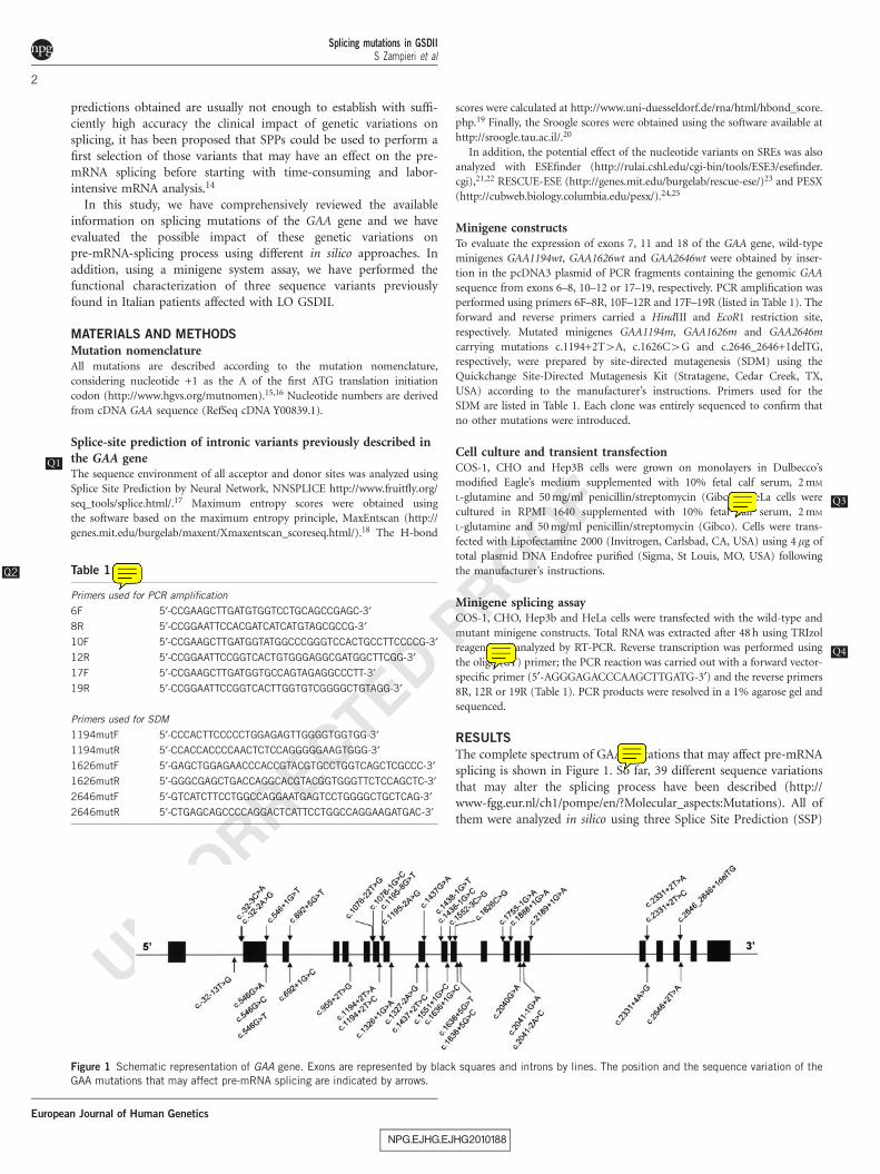
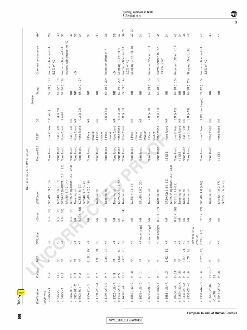
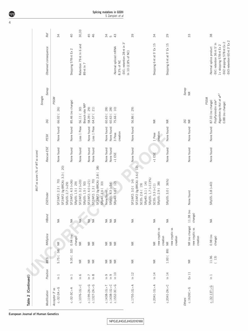
The complete spectrum of GAA mutations that may affect pre-mRNAsplicing is shown in Figure 1. So far, 39 different sequence variationsthat may alter the splicing process have been described (http://www-fgg.eur.nl/ch1/pompe/en/?Molecular_aspects:Mutations). All ofthem were analyzed in silico using three Splice Site Prediction (SSP)
NPG_EJHG_EJHG2010188
Table 1Q2
Primers used for PCR amplification
6F 5¢-CCGAAGCTTGATGTGGTCCTGCAGCCGAGC-3¢8R 5¢-CCGGAATTCCACGATCATCATGTAGCGCCG-3¢10F 5¢-CCGAAGCTTGATGGTATGGCCCGGGTCCACTGCCTTCCCCG-3¢12R 5¢-CCGGAATTCCGGTCACTGTGGGAGGCGATGGCTTCGG-3¢17F 5¢-CCGAAGCTTGATGGTGCCAGTAGAGGCCCTT-3¢19R 5¢-CCGGAATTCCGGTCACTTGGTGTCGGGGCTGTAGG-3¢
Primers used for SDM
1194mutF 5¢-CCCACTTCCCCCTGGAGAGTTGGGGTGGTGG-3¢1194mutR 5¢-CCACCACCCCAACTCTCCAGGGGGAAGTGGG-3¢1626mutF 5¢-GAGCTGGAGAACCCACCGTACGTGCCTGGTCAGCTCGCCC-3¢1626mutR 5¢-GGGCGAGCTGACCAGGCACGTACGGTGGGTTCTCCAGCTC-3¢2646mutF 5¢-GTCATCTTCCTGGCCAGGAATGAGTCCTGGGGCTGCTCAG-3¢2646mutR 5¢-CTGAGCAGCCCCAGGACTCATTCCTGGCCAGGAAGATGAC-3¢
Figure 1 Schematic representation of GAA gene. Exons are represented by black squares and introns by lines. The position and the sequence variation of the
GAA mutations that may affect pre-mRNA splicing are indicated by arrows.
Splicing mutations in GSDIIS Zampieri et al
2
European Journal of Human Genetics
UNCORRECTED PROOF
NPG_EJHG_EJHG2010188
Table
2Q
5
MU
Tss
scor
e(%
ofW
Tss
scor
e)
Sro
ogle
Mod
ifica
tion
Pos
itio
nM
ES
NN
Splice
HB
ond
ES
Efinder
Res
cue-
ES
EPE
SX
DG
Sen
epO
bse
rved
conse
quen
ceR
ef
Don
or5
¢ss
c.5
46
G4
AEx
2N
RN
R5.8
(�38
)SR
p40
:3
.9(�
10
)N
one
found
Los
s2
Pes
e2.2
(+6
1)
57.6
0(�
17
)N
orm
alsp
lice
dm
RN
A
6.3
%of
NC
29
c.5
46
G4
CEx
2N
RN
R5.8
(�38
)SR
p40
:3
.5(�
19
)N
one
found
Los
s2
Pes
e�
2.3
(+5
9)
56.3
2(�
19
)3
0
c.5
46
G4
TEx
2N
RN
R5.8
(�38
)SF2/A
F2
(Ig-
BR
CA
):2
.3(�
23
)
SR
p40
:3
.7(�
14
)
Non
efo
und
Non
efo
und�
2(+
64)
57.0
4(�
18
)N
orm
alsp
lice
dm
RN
A
reduce
dw
ith
resp
ect
toN
C
31
c.5
46
+1G4
TIn
2N
RN
R5.8
(�38
)SF2/A
F2
(Ig-
BR
CA
):3
.7(+
23
)N
one
found
Los
s1
Pes
eN
RN
R3
2
c.6
92
+1G4
CIn
3N
RN
RN
RN
one
found
Non
efo
und
Non
efo
und
NR
NR
r.0
33
c.6
92
+5G4
TIn
3N
RN
R5.1
0(�
58)
SC
35
:4
.5(+
32)
SR
p40
:4
.7(+
88)
Non
efo
und
Non
efo
und�
0.3
(+9
2)
58.2
2(�
17
)3
4
c.9
55
+2T4
GIn
51.1
9(�
87)
NR
NR
SR
p40
:2
.2(�
19
)N
one
found
1Pes
e
crea
tion
NR
NR
34
c.1
19
4+
2T4
AIn
71.9
1(�
81)
NR
NR
Non
efo
und
Non
efo
und
4Pes
e
crea
tion
NR
NR
33
c.1
19
4+
2T4
CIn
72.3
4(�
77)
NR
NR
Non
efo
und
Non
efo
und
3Pes
e
crea
tion
�3.5
(+5
1)
69.1
9(�
20
)R
eten
tion
89
nt
In7
35
c.1
32
6+
1G4
AIn
8N
RN
RN
RN
one
found
Non
efo
und
Non
efo
und
NR
NR
r.0
36
c.1
43
7+
2T4
CIn
91.0
0(�
89)
NR
NR
SF2/A
F2:
3.2
(+8
8)
Non
efo
und
Non
efo
und�
4.4
(+4
8)
67.3
9(�
20
)Ski
ppin
g11
1nt
Ex
93
7
c.1
43
7G4
AEx
92.6
7(�
70)
NR
9.3
(�34
)N
one
found
Non
efo
und
Non
efo
und�
3.8
(+5
5)
72.5
9(�
14
)N
orm
alsp
lice
dm
RN
A
1.2
%of
NC
34,3
5
c.1
55
1+
1G4
CIn
10
NR
NR
NR
SC
35
:4
.0(+
14)
Non
efo
und
1Pes
e
crea
tion
NR
NR
Ski
ppin
g11
4nt
Ex
10
37–3
9
c.1
63
6+
1G4
CIn
11
NR
NR
(no
chan
ge)
NR
SR
p40
:2
.0(�
31
)N
one
found
1Pes
e
crea
tion
NR
NR
34
c.1
63
6+
5G4
CIn
11
NR
NR
(no
chan
ge)
NR
Non
efo
und
Non
efo
und
1Pes
e
crea
tion
�1.0
(+6
8)
62.9
9(�
16
)R
eten
tion
957
nt
In11
40
c.1
63
6+
5G4
TIn
11
NR
NR
(no
chan
ge)
8.3
0(�
27)
Non
efo
und
Non
efo
und
Non
efo
und�
0.9
(+7
1)
66.4
8(�
12
)N
orm
alsp
lice
dm
RN
A
13
.7%
ofN
C
34
c.1
88
8+
1G4
AIn
13
1.4
2(�
85)
NR
NR
SF2/A
F2:
3.8
(+6
5)
SF2/A
F2
(Ig-
BR
CA
):3
.2(+
33
)
+2
ESE
Non
efo
und
NR
NR
34
c.2
04
0G4
AEx
14
NR
NR
8.9
0(�
26)
SC
35
:3
.7(+
12)
Non
efo
und
Los
s2
Pes
e�
3.8
(+4
5)
64.1
8(�
16
)R
eten
tion
190
nt
In14
41
c.2
18
9+
1G4
AIn
15
NR
NR
NR
Non
efo
und
+1
ESE
Non
efo
und
NR
NR
34
c.2
33
1+
2T4
AIn
16
3.2
7(�
71)
NR
NR
Non
efo
und
Non
efo
und
Los
s1
Pes
eN
RN
R3
4
c.2
33
1+
2T4
CIn
16
3.7
0(�
68)
NR
new
cryp
tic
ss
crea
tion
,
NR
Non
efo
und
Non
efo
und
Los
s1
Pes
e�
3.8
(+4
9)
68.2
8(�
20
)Ski
ppin
g16
nt
Ex
16
42
c.2
33
1+
4A4
GIn
16
8.2
7(�
28)
0.2
8(�
75)
12
.5(�
22)
SR
p40
:1
.8(+
40)
Non
efo
und
Los
s1
Pes
e�
7.0
0(n
och
ange
)7
2.4
2(�
15
)N
orm
alsp
lice
dm
RN
A
3.6
%of
NC
43
c.2
64
6_2
646
+1
del
TG
Ex
18
–In
18
NR
NR
NR
Non
efo
und
Non
efo
und
Non
efo
und
NR
NR
33
c.2
64
6+
2T4
AIn
18
NR
NR
NR
SR
p40
:4
.0(+
67)
SR
p55
:2
.6(+
86)
+1
ESE
Non
efo
und
NR
NR
44
Splicing mutations in GSDIIS Zampieri et al
3
European Journal of Human Genetics
UNCORRECTED PROOF
NPG_EJHG_EJHG2010188
Table
2(C
ontinued
)
MU
Tss
scor
e(%
ofW
Tss
scor
e)
Sro
ogle
Mod
ifica
tion
Pos
itio
nM
ES
NN
Splice
HB
ond
ES
Efinder
Res
cue-
ES
EPE
SX
DG
Sen
epO
bse
rved
conse
quen
ceR
ef
Acc
epto
r3
¢ss
PSSM
c.-3
2-2
A4
GIn
15.7
5(�
58
)N
RN
ASF2/A
F2:
2.3
(�70
)
SF2/A
F2
(Ig-
BR
CA
):3
.3(�
20
)
SR
p55
:2
.7(+
29)
Non
efo
und
Non
efo
und
66
.02
(�26
)3
4
c.-3
2-3
C4
AIn
19.3
5(�
32
)0.9
4(n
o
chan
ge)
NA
SF2/A
F2:
4.5
(+36
)
SR
p55
:1
.5(�
29
)
Non
efo
und
Non
efo
und
85
.46
(no
chan
ge)
Ski
ppin
g57
8nt
Ex
24
0
c.10
76-1
G4
CIn
6N
RN
RN
ASF2/A
F2:
3.2
(+33
)
SR
p55
:1
.5(�
50
)
Non
efo
und
Los
s1
Pes
e56
.11
(�30
)
Bra
nch
site
NR
a
Ret
ention
79
nt
In6
and
89
nt
In7
30,3
3
c.11
95-2
A4
GIn
7N
RN
RN
ASF2/A
F2:
4.0
(+21
)N
one
found
Non
efo
und
58
.39
(�29
)4
5
c.13
27-2
A4
GIn
8N
RN
RN
ASF2/A
F2:
1.3
(�70
)
SF2/A
F2
(Ig-
BR
CA
):2
.8(�
38
)
SR
p40
:2
.6(�
33
)
Non
efo
und
Los
s1
Pes
e64
.57
(�21
)4
6
c.14
38-1
G4
TIn
9N
RN
RN
AN
one
found
Non
efo
und
Non
efo
und
60
.63
(�28
)3
4
c.14
38-1
G4
CIn
9N
RN
RN
ASR
p55
:4
.3(+
34)
Non
efo
und
Non
efo
und
60
.63
(�28
)5
c.15
52-3
C4
GIn
10
NR
NR
NA
SR
p40
:3
.0(�
12
)+
1ES
E3
Pes
e
crea
tion
75
.66
(�10
)-N
orm
alsp
lice
dm
RN
A
8.3
%of
NC
-SV1:
rete
ntion
28
nt
in3
¢In
10
(2.8
%of
NC
)
43
c.17
55-1
G4
AIn
12
NR
NR
NA
SF2/A
F2:
3.0
(�14
)
SF2/A
F2
(Ig-
BR
CA
):3
.6(�
12
)
SC
35
:2
.6(�
19
)
SR
p40
:3
.1(�
14
)
SR
p55
:1
.7(+
113
%)
Non
efo
und
Non
efo
und
56
.86
(�29
)3
9
c.20
41-1
G4
AIn
14
NR
NR
new
cryp
tic
ss
crea
tion
NA
SC
35
:3
.3(+
11
)
SR
p55
:2
.9(�
38
)
+1
ES
E1
Pes
e
crea
tion
NR
Ski
ppin
g6
nt
at5
¢Ex
15
34
c.20
41-2
A4
CIn
14
1.0
0(�
89
)N
R
new
cryp
tic
ss
crea
tion
NA
SR
p55
:3
.0(�
36
%)
Non
efo
und
Non
efo
und
NR
Ski
ppin
g6
nt
at5
¢Ex
15
29
Oth
ers
DG
Sen
ep
c.16
26C4
GE
x1
1N
RN
R(n
och
ange
)
new
cryp
tic
ss
crea
tion
11
.38
(no
chan
ge)
Non
efo
und
Non
efo
und
Non
efo
und
NR
NR
33
PSSM
c.-3
2-1
3T4
GIn
111
.96
(�13)
0.9
8(n
o
chan
ge)
NA
SR
p55
:3
.8(+
40)
Non
efo
und
Non
efo
und
87
.03
(no
chan
ge)
Pol
yPyr
imid
insi
tea
Alg
orithm
by
Kol
etal
47
0.8
8(n
och
ange
)
-Nor
mal
splice
pro
duct
-SV1
rete
nti
on3
6nt
5¢I
n
1+
skip
pin
g57
8nt
Ex
2
-SV2
skip
pin
g57
8nt
Ex
2
-SV3
rete
nti
on60
nt3
¢Ex
2
38
Splicing mutations in GSDIIS Zampieri et al
4
European Journal of Human Genetics
UNCORRECTED PROOF
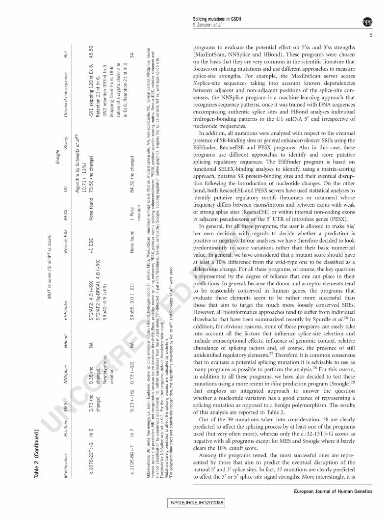
programs to evaluate the potential effect on 5¢ss and 3¢ss strengths(MaxEntScan, NNSplice and HBond). These programs were chosenon the basis that they are very common in the scientific literature thatfocuses on splicing mutations and use different approaches to measuresplice-site strengths. For example, the MaxEntScan server scores5¢splice-site sequences taking into account known dependenciesbetween adjacent and non-adjacent positions of the splice-site con-sensus, the NNSplice program is a machine-learning approach thatrecognizes sequence patterns, once it was trained with DNA sequencesencompassing authentic splice sites and HBond analyses individualhydrogen-bonding patterns to the U1 snRNA 5¢ end irrespective ofnucleotide frequencies.
In addition, all mutations were analyzed with respect to the eventualpresence of SR-binding sites or general enhancer/silencer SREs using theESEfinder, RescueESE and PESX programs. Also in this case, theseprograms use different approaches to identify and score putativesplicing regulatory sequences. The ESEfinder program is based onfunctional SELEX-binding analyses to identify, using a matrix-scoringapproach, putative SR protein-binding sites and their eventual disrup-tion following the introduction of nucleotide changes. On the otherhand, both RescueESE and PESX servers have used statistical analyses toidentify putative regulatory motifs (hexamers or octamers) whosefrequency differs between exons/introns and between exons with weakor strong splice sites (RescueESE) or within internal non-coding exonsvs adjacent pseudoexons or the 5¢ UTR of intronless genes (PESX).
In general, for all these programs, the user is allowed to make his/her own decision with regards to decide whether a prediction ispositive or negative. In our analyses, we have therefore decided to lookpredominantly to score variations rather than their basic numericalvalue. In general, we have considered that a mutant score should haveat least a 10% difference from the wild-type one to be classified as adeleterious change. For all these programs, of course, the key questionis represented by the degree of reliance that one can place in theirpredictions. In general, because the donor and acceptor elements tendto be reasonably conserved in human genes, the programs thatevaluate these elements seem to be rather more successful thanthose that aim to target the much more loosely conserved SREs.However, all bioinformatics approaches tend to suffer from individualdrawbacks that have been summarized recently by Spurdle et al.26 Inaddition, for obvious reasons, none of these programs can easily takeinto account all the factors that influence splice-site selection andinclude transcriptional effects, influence of genomic context, relativeabundance of splicing factors and, of course, the presence of stillunidentified regulatory elements.27 Therefore, it is common consensusthat to evaluate a potential splicing mutation it is advisable to use asmany programs as possible to perform the analysis.28 For this reason,in addition to all these programs, we have also decided to test thesemutations using a more recent in silico prediction program (Sroogle)20
that employs an integrated approach to answer the questionwhether a nucleotide variation has a good chance of representing asplicing mutation as opposed to a benign polymorphism. The resultsof this analysis are reported in Table 2.
Out of the 39 mutations taken into consideration, 38 are clearlypredicted to affect the splicing process by at least one of the programsused (but very often more), whereas only the c.-32-13T4G scores asnegative with all programs except for MES and Sroogle where it barelyclears the 10% cutoff score.
Among the programs tested, the most successful ones are repre-sented by those that aim to predict the eventual disruption of thenatural 5¢ and 3¢ splice sites. In fact, 37 mutations are clearly predictedto affect the 3¢ or 5¢ splice-site signal strengths. More interestingly, it is
NPG_EJHG_EJHG2010188
Table
2(C
ontinued
)
MU
Tss
scor
e(%
ofW
Tss
scor
e)
Sro
ogle
Mod
ifica
tion
Pos
itio
nM
ES
NN
Splice
HB
ond
ES
Efinder
Res
cue-
ES
EPE
SX
DG
Sen
epO
bse
rved
conse
quen
ceR
ef
Alg
orithm
by
Sch
war
tzet
al4
8
10
.71
(�1
3%
)
c.10
76
-22T4
GIn
63.7
3(n
o
chan
ge)
0.3
8(n
o
chan
ge)
New
cryp
tic
ss
crea
tion
NA
SF2/A
F2:
4.9
(+6
9)
SF2/A
F2
(Ig-
BR
CA
):4
.8(+
55)
SR
p40
:4
.9(+
69)
+1
ES
EN
one
found
79
.56
(no
chan
ge)
-SV1
skip
pin
g12
0nt
Ex
6.
Ret
ention
21
nt
In6
.
-SV2
rete
nti
on3
93
nt
In5.
Ski
ppin
g4
5nt
Ex
6.
Utili-
zati
onof
acr
yptic
don
orsi
te
inEx
6.R
eten
tion
21
ntIn
6
49
,50
c.11
95
-8G4
TIn
79.1
3(+
16)
0.7
3(+
62)
NA
SR
p55
:3
.0(�
21
)N
one
found
1Pes
e
crea
tion
84
.32
(no
chan
ge)
34
Abbre
viat
ions:
DG
,del
tafr
eeen
ergy
;E
x,ex
on;
Ese
finder
,ex
onic
splici
ng
enhan
cer
finder
;H
Bon
d,
hyd
roge
nbon
d;
In,
intr
on;
MES
,M
axE
ntS
can
(max
imum
entr
opy
scan
);M
ut
ss,
muta
nt
splice
site
;N
A,
non
-applica
ble
;N
C,
nor
mal
contr
ol;
NN
Splice
,neu
ral
net
wor
ksp
lice
site
pre
dic
tion
;N
R,
not
reco
gniz
able
bec
ause
bel
owdet
ection
thre
shol
d;
Pes
e,puta
tive
exon
icsp
lici
ng
enhan
cer;
PES
X,
puta
tive
exon
icsp
lici
ng
enhan
cer/si
lence
r;P
SS
M,
pos
itio
n-s
pec
ific
scor
ing
mat
rix;
Res
cue-
ESE
,re
lative
enhan
cer
and
sile
nce
rcl
assi
fica
tion
by
unan
imou
sen
rich
men
t;r.0,
mR
NA
tran
scribed
from
the
muta
nt
alle
lenon
-det
ecte
din
pat
ient’s
fibro
bla
sts;
Sen
ep,
senep
athy;
Sro
ogle
,sp
lici
ng
regu
lation
online
grap
hic
alen
gine;
SV,
splice
varian
t;W
Tss
,w
ild-t
ype
splice
site
.Thre
shol
dfo
rN
NSplice
was
set
at0
.1.
For
the
other
pro
gram
s,def
ault
thre
shol
ds
wer
euse
d.
Muta
tion
sbar
ely
pre
dic
ted
toaf
fect
the
splici
ng
pro
cess
are
indic
ated
by
under
line.
a For
pol
ypyr
imid
ine
trac
tan
dbra
nch
-site
reco
gnitio
n,
the
algo
rith
ms
dev
elop
edby
Kol
etal
47
and
Sch
war
tzet
al4
8w
ere
use
d.
Splicing mutations in GSDIIS Zampieri et al
5
European Journal of Human Genetics
UNCORRECTED PROOF
worthy to point out that with possibly just one exception (c.-32-3C4A), where MaxEntScan performs apparently better, in all othercases, the integrated Sroogle program performs in the same way as anyof the individual programsQ6 . In some cases, such as c.1636+5G4C andc.1636+5G4T, it even performs better than the MaxEntScan andNNSplice programs.
We also looked at whether the programs were able to predict thedegree of changes correctly. However, no correlation between thedegree of score change and the consequence on the mRNA splicingobserved in vivo was found.
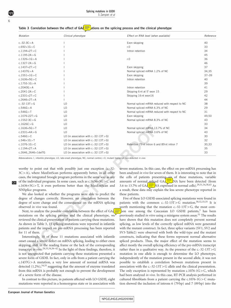
Next, to analyze the possible correlation between the effect of GAAmutations on the splicing process and the clinical phenotype, wereviewed the clinical presentation of patients carrying these mutations.As shown in Table 3, 15 splicing mutations were reported in infantilepatients and the impact on pre-mRNA processing has been reportedfor 11 of them.
Interestingly, 10 of these 11 mutations associated with infantileonset caused a severe defect on mRNA splicing, leading to either exonskipping, shift in the reading frame or the lack of the correspondingtranscript in vivo.29,33,34,36–42 In the light of these findings, therefore, itis not unexpected that patients carrying these mutations presented asevere form of GSDII. In fact, only in cells from a patient carrying thec.1437G4A mutation, a very low amount of normal mRNA wasdetected (1.2%),34,35 suggesting that the amount of enzyme translatedfrom this mRNA is probably not enough to prevent the developmentof a severe form of the disease.
In contrast to this picture, in patients affected with LO GSDII, eightmutations were reported in a homozygous state or in association with
severe mutations. In this case, the effect on pre-mRNA processing hasbeen analyzed in vivo for seven of them. It is interesting to note that inthe cells of patients presenting six of these mutations, variableamounts of normal spliced GAA mRNA have been detected (from3.6 to 13.7% of GAA mRNA expressed in normal cells).29,31,34,38,43 Asa result, these data may explain the less severe phenotype reported inthese patients.
Five of these LO GSDII-associated splicing mutations were found inpatients with the common c.-32-13T4G mutation.30,32,33,35 It isworth mentioning that the mutation c.-32-13T4G, the most com-mon one among the Caucasian LO GSDII patients,3 has beenpreviously studied in vitro using a minigene system assay.51 The resultshave shown that this mutation does not completely prevent normalsplicing, as low levels of the correctly spliced mRNA were generatedwith the mutant construct. In fact, three splice variants (SV1, SV2 andSV3-Table2) were observed with both the wild-type and the mutantconstructs, indicating that these forms represent normal alternativespliced products. Thus, the major effect of the mutation seems toaffect mostly the overall splicing efficiency of the pre-mRNA transcriptrather than in a qualitative way. As the presence of the c.-32-13T4Gmutation in one allele is enough to determine the LO phenotype,independently of the mutation present in the second allele, it was notpossible to establish a correlation between mutations present inassociation with the c.-32-13T4G allele and the clinical presentation.The only exception is represented by mutation c.1076-1G4C, whichhad been analyzed in vivo. In this case, RT-PCR analysis performed incultured fibroblasts from a patient carrying the c.1076-1G4C muta-tion showed the inclusion of introns 6 (79 bp) and 7 (89 bp) into the
NPG_EJHG_EJHG2010188
Table 3 Correlation between the effect of GAA mutations on the splicing process and the clinical phenotype
Mutation Clinical phenotype Effect on RNA level (when available) Reference
c.-32-3C4A I Exon skipping 40
c.692+1G4C I r.0 33
c.1194+2T4C I Intron retention 34
c.1195-2A4G I 45
c.1326+1G4A I r.0 36
c.1327-2A4G I 46
c.1437+2T4C I Exon skipping 37
c.1437G4A I Normal spliced mRNA 1.2% of NC 34,35
c.1551+1G4C I Exon skipping 37–39
c.1636+5G4C I Intron retention 40
c.1755-1G4A I 39
c.2040G4A I Intron retention 41
c.2041-2A4C I Skipping 6 nt at 5¢ exon 15 29
c.2331+2T4C I Skipping 16nt exon16 42
c.2646+2T4A I 44
c.-32-13T4G LO Normal spliced mRNA reduced with respect to NC 38
c.546G4A LO Normal spliced mRNA 6.3% of NC 29
c.546G4T LO Normal spliced mRNA reduced with respect to NC 31
c.1076-22T4G LO Exon skipping 49,50
c.1552-3C4G LO Normal spliced mRNA 8.3% of NC 43
c.1626C4G LO 33
c.1636+5G4T LO Normal spliced mRNA 13.7% of NC 34
c.2331+4A4G LO Normal spliced mRNA 3.6% of NC 43
c.546G4C LO (in association with c.-32-13T4G) 30
c.546+1G4T LO (in association with c.-32-13T4G) 32
c.1076-1G4C LO (in association with c.-32-13T4G) Retention 79 nt intron 6 and 89 nt intron 7 30,33
c.1194+2T4A LO (in association with c.-32-13T4G) 35
c.2646_2646+1delTG LO (in association with c.-32-13T4G) 33
Abbreviations: I, infantile phenotype; LO, late-onset phenotype; NC, normal control; r.0, mutant transcript non-detected in vivo.
Splicing mutations in GSDIIS Zampieri et al
6
European Journal of Human Genetics

UNCORRECTED PROOF
transcribed mRNA. If we consider the fact that the c.1076-1G4Cmutation has also been found in homozygosis in infantile GSDIIpatients,30 these data indicate that c.1076-1G4C could be classified asa severe mutation.
To complete this analysis of GAA-splicing mutations, it should alsobe noted that in a previous study, we have characterized the mutationprofile of the GAA gene in 40 Italian patients with LO GSDII.33
Overall, five mutations that might have affected the splicing processwere found. However, as RNA and/or cells from patients carryingthree of them (c.1626C4G, c.1194+2T4A and c.2646_2646+1delTG)were unavailable, their deleterious effect could not be confirmed. Thesequence variation c.1626C4G was found in the homozygous state
and did not disrupt the reading frame and codon usage, whereasmutations c.1194+2T4A and c.2646_2646+1delTG affected theconsensus 5¢ splice donor sites of exons 7 and 18, respectively. SeveralSPPs (Table 2) clearly predicted that mutation c.1626C4G wouldcreate a novel donor site, which would have caused the exclusion of11 bp of exon 11, whereas the mutated sequences c.1194+2T4A andc.2646_2646+1delTG would no longer be recognized as donor sites.Therefore, to test the predicted effects of these mutations on GAA pre-mRNA splicing, we have now performed a functional splicing assay.
As shown in Figure 2, cells transfected with the mutant constructsbearing these mutations produced aberrant transcripts in all cases.However, it is worth noting that in cells transfected with the
NPG_EJHG_EJHG2010188
Figure 2 Schematic representation of the GAA regions affected by splicing mutations. Mutations c.1194+2T4A, c.1626C4G and c.2646_2646+1delTG
(panels a1, b1 and c1, respectively) are highlighted in bold. RT-PCR analysis of the GAA mRNA in cells transfected with wt and minigenes containing
mutations c.1194+2T4A, c.1626C4G and c.2646_2646+1delTG (panels a2, b2 and c2, respectively) usingQ7 a forward vector-specific primer that amplified
only the minigene product.
Splicing mutations in GSDIIS Zampieri et al
7
European Journal of Human Genetics
UNCORRECTED PROOF
GAA1626m construct, a low amount of a transcript similar in size tothe normal one was also present. As the wild-type acceptor and donorsites of intron 11 seem not to be abolished, this data suggest that evenin the presence of this mutation, a low amount of wild-type transcriptwould still be produced.
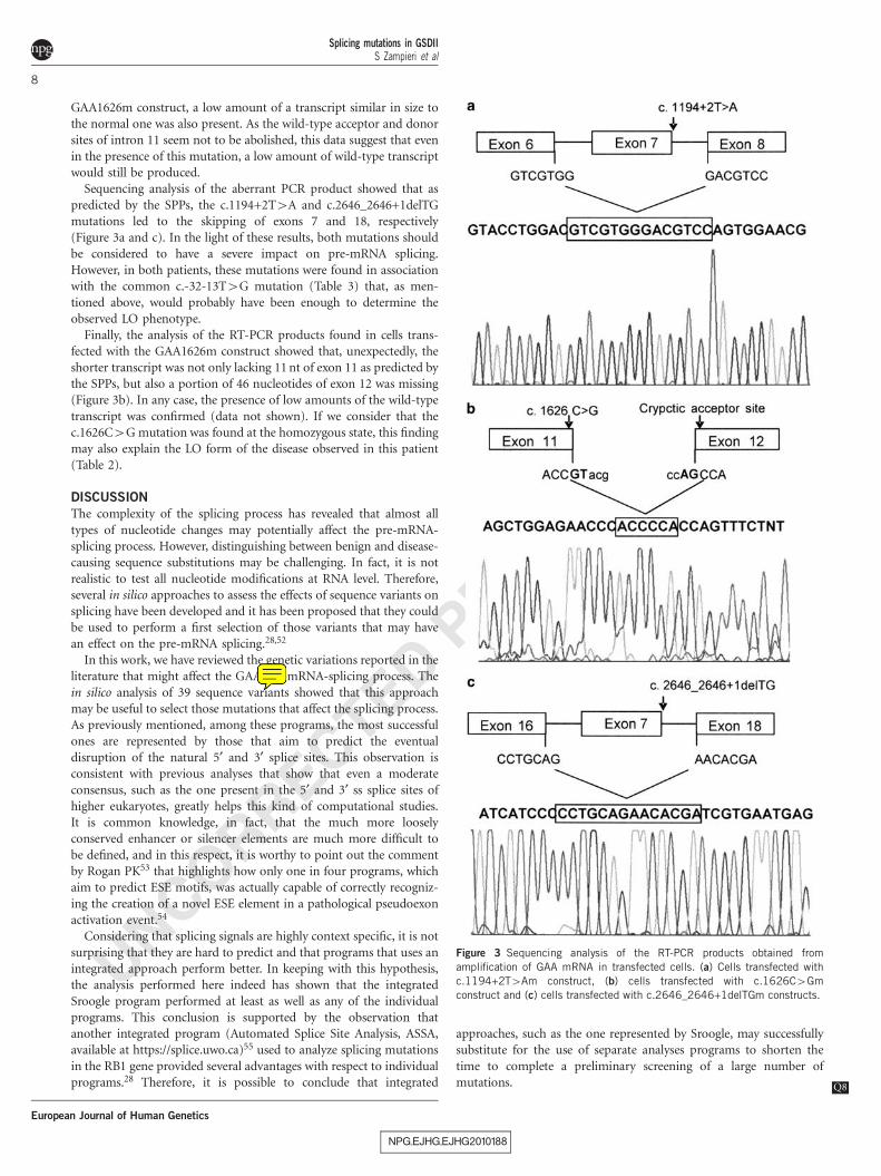
Sequencing analysis of the aberrant PCR product showed that aspredicted by the SPPs, the c.1194+2T4A and c.2646_2646+1delTGmutations led to the skipping of exons 7 and 18, respectively(Figure 3a and c). In the light of these results, both mutations shouldbe considered to have a severe impact on pre-mRNA splicing.However, in both patients, these mutations were found in associationwith the common c.-32-13T4G mutation (Table 3) that, as men-tioned above, would probably have been enough to determine theobserved LO phenotype.
Finally, the analysis of the RT-PCR products found in cells trans-fected with the GAA1626m construct showed that, unexpectedly, theshorter transcript was not only lacking 11 nt of exon 11 as predicted bythe SPPs, but also a portion of 46 nucleotides of exon 12 was missing(Figure 3b). In any case, the presence of low amounts of the wild-typetranscript was confirmed (data not shown). If we consider that thec.1626C4G mutation was found at the homozygous state, this findingmay also explain the LO form of the disease observed in this patient(Table 2).
DISCUSSION
The complexity of the splicing process has revealed that almost alltypes of nucleotide changes may potentially affect the pre-mRNA-splicing process. However, distinguishing between benign and disease-causing sequence substitutions may be challenging. In fact, it is notrealistic to test all nucleotide modifications at RNA level. Therefore,several in silico approaches to assess the effects of sequence variants onsplicing have been developed and it has been proposed that they couldbe used to perform a first selection of those variants that may havean effect on the pre-mRNA splicing.28,52
In this work, we have reviewed the genetic variations reported in theliterature that might affect the GAA pre-mRNA-splicing process. Thein silico analysis of 39 sequence variants showed that this approachmay be useful to select those mutations that affect the splicing process.As previously mentioned, among these programs, the most successfulones are represented by those that aim to predict the eventualdisruption of the natural 5¢ and 3¢ splice sites. This observation isconsistent with previous analyses that show that even a moderateconsensus, such as the one present in the 5¢ and 3¢ ss splice sites ofhigher eukaryotes, greatly helps this kind of computational studies.It is common knowledge, in fact, that the much more looselyconserved enhancer or silencer elements are much more difficult tobe defined, and in this respect, it is worthy to point out the commentby Rogan PK53 that highlights how only one in four programs, whichaim to predict ESE motifs, was actually capable of correctly recogniz-ing the creation of a novel ESE element in a pathological pseudoexonactivation event.54
Considering that splicing signals are highly context specific, it is notsurprising that they are hard to predict and that programs that uses anintegrated approach perform better. In keeping with this hypothesis,the analysis performed here indeed has shown that the integratedSroogle program performed at least as well as any of the individualprograms. This conclusion is supported by the observation thatanother integrated program (Automated Splice Site Analysis, ASSA,available at https://splice.uwo.ca)55 used to analyze splicing mutationsin the RB1 gene provided several advantages with respect to individualprograms.28 Therefore, it is possible to conclude that integrated
approaches, such as the one represented by Sroogle, may successfullysubstitute for the use of separate analyses programs to shorten thetime to complete a preliminary screening of a large number ofmutations Q8.
NPG_EJHG_EJHG2010188
Figure 3 Sequencing analysis of the RT-PCR products obtained from
amplification of GAA mRNA in transfected cells. (a) Cells transfected with
c.1194+2T4Am construct, (b) cells transfected with c.1626C4Gm
construct and (c) cells transfected with c.2646_2646+1delTGm constructs.
Splicing mutations in GSDIIS Zampieri et al
8
European Journal of Human Genetics
UNCORRECTED PROOF
There are also some cases, however, where in silico analysis did notperform so well. Not surprisingly, these cases include mutationsthat occur more distant to the splice site (ie, c.-32-13T4G andc.1195-8G4T). However, in the case of c.1195-8G4T mutation, theuse of the ESEfinder and PESX programs might have substituted forthe relative inability of the other softwares to detect the splicing-modifying potential of this mutations. In the case of the c.-32-13T4Gsubstitution, even using a very favorable degree of score change (10%),it was barely predicted to affect the splicing process. There are severalreasons that may account for this failure but the fact that thismutation still allows a certain amount of normal splicing (as discussedabove) most probably places it in the limit of the detection thresholdof these bioinformatics programs. Taken together, therefore, B97% ofthe GAA-splicing mutations taken in consideration in this analysiscould have been clearly predicted to have an impact on the splicingprocess by in silico analysis alone.
The remarkable heterogeneity of mutations found in GSDII patientsmakes it difficult to correlate the genotype with the phenotype.However, the results reviewed here show a good correlation betweenthe impact of the mutation on the splicing process and the clinicalphenotype (ie, the more severe the impact on the splicing process,the more likely the presence of an infantile form of the disease andvice versa).
Finally, using a minigene assay, we confirmed the pathogenic effectof the three sequence variants found in Italian patients affected withGSDII. All these three mutations were predicted to affect the splicingprocess by SPPs. However, in the case of mutation c.1626C4G,although several SPPs predicted that the mutation would create anovel donor site, which would cause the exclusion of 11 bp of exon 11,the functional analysis showed that this mutation not only creates anew donor site as predicted but also causes the activation of a crypticsplice site localized in exon 12, downstream of the normal acceptorsite of exon 11.
In conclusion, this study has shown that in silico analysis representsa useful tool to select mutations that affect the splicing process of theacid a-glucosidase. This type of analysis is quite straightforward andreliable. However, it is worth to highlight the importance of functionalstudies for the correct evaluation of sequences’ variations. In fact, acomprehensive analysis of the mechanism by which a sequence variantaffect the mRNA splicing is crucial to analyze possible correlationsbetween the mutation and the clinical phenotype, and to evaluate thefeasibility of using emerging splicing-based therapeutic approaches.
CONFLICT OF INTEREST
The authors declare no conflict of interest.
ACKNOWLEDGEMENTS
This work was supported by a grant from the Italian Health Ministry (Italia-
USA RF526 D/47) and by a European community grant (EURASNET-LSHG-
CT-2005-518238).
1 Hoefsloot LH, Hoogeveen-Westerveld M, Reuser AJ, Oostra BA: Characterization of thehuman lysosomal alpha-glucosidase gene. Biochem J 1990a; 272: 493–497.
2 Martiniuk F, Bodkin M, Tzall S, Hirschhorn R, Hirschhorn R: Isolation and partialcharacterization of the structural gene for human acid alpha glucosidase. DNA Cell Biol1991; 10: 283–292.
3 Hirschhorn R, Reuser AJJ: Glycogen storage disease type II: acid a-glucosidase (acidmaltase) deficiency; in Scriver CR, Beaudet AL, Sly WS, Valle D (eds): The Metabolicand Molecular Basis of Inherited Disease. New York: McGraw-Hill, 2001, Vol 3,pp 3389–3420.
4 Moreland RJ, Jin X, Zhang XK et al: Lysosomal acid alpha-glucosidase consists of fourdifferent peptides processed from a single chain precursor. J Biol Chem 2005; 280:6780–6791.
5 Raben N, Plotz P, Byrne BJ: Acid alpha-glucosidase deficiency (glycogenosis type II,Pompe disease). Curr Mol Med 2002; 2: 145–166.
6 Van den Hout HM, Hop W, van Diggelen OP et al: The natural course of infantilePompe’s disease: 20 original cases compared with 133 cases from the literature.Pediatrics 2003; 112: 332–340.
7 Kishnani PS, Howell RR: Pompe disease in infants and children. J Pediatr 2004; 144
(5 Suppl): S35–S43.8 Hagemans ML, Janssens AC, Winkel LP et al: Late-onset Pompe disease primarily
affects quality of life in physical health domains. Neurology 2004; 63: 1688–1692.9 Hagemans ML, Winkel LP, Van Doorn PA et al: Clinical manifestation and natural
course of late-onset Pompe’s disease in 54 Dutch patients. Brain 2005; 128 (Part 3):671–677.
10 Tazi J, Bakkour N, Stamm S: Alternative splicing and disease. Biochim Biophys Acta2009; 1792: 14–26.
11 Wang GS, Cooper TA: Splicing in disease: disruption of the splicing code and thedecoding machinery. Nat Rev Genet 2007; 8: 749–761.
12 Baralle D, Lucassen A, Buratti E: Missed threads. The impact of pre-mRNA splicingdefects on clinical practice. Review. EMBO Rep 2009; 10: 810–816.
13 Vibe-Pedersen K, Kornblihtt AR, Baralle FE: Expression of a human alpha-globin/fibronectin gene hybrid generates two mRNAs by alternative splicing. EMBO J 1984; 3:2511–2516.
14 Vreeswijk MP, Kraan JN, van der Klift HM et al: Intronic variants in BRCA1 and BRCA2that affect RNA splicing can be reliably selected by splice-site prediction programs.Hum Mutat 2009; 30: 107–114.
15 den Dunnen JT, Antonarakis SE: Mutation nomenclature extensions and suggestions todescribe complex mutations: a discussion. Hum Mutat 2000; 15: 7–12.
16 den Dunnen JT, Paalman MH: Standardizing mutation nomenclature: why bother?Hum Mutat 2003; 22: 181–182.
17 Reese MG, Eeckman FH, Kulp D, Haussler D: Improved splice site detection in Genie.J Comput Biol 1997; 4: 311–323.
18 Yeo G, Burge CB: Maximum entropy modeling of short sequence motifs with applica-tions to RNA splicing signals. J Comput Biol 2004; 11: 377–394.
19 Freund M, Asang C, Kammler S et al: A novel approach to describe a U1 snRNAbinding site. Nucleic Acids Res 2003; 31: 6963–6975.
20 Schwartz S, Hall E, Ast G: SROOGLE: webserver for integrative, user-friendlyvisualization of splicing signals. Nucleic Acids Res 2009; 37 (Web Server issue):W189–W192.
21 Smith PJ, Zhang C, Wang J, Chew SL, Zhang MQ, Krainer AR: An increased specificityscore matrix for the prediction of SF2/ASF-specific exonic splicing enhancers.Hum Mol Genet 2006; 15: 2490–2508.
22 Cartegni L, Wang J, Zhu Z, Zhang MQ, Krainer AR: ESEfinder: a web resource toidentify exonic splicing enhancers. Nucleic Acids Res 2003; 3: 3568–3571.
23 Fairbrother WG, Yeh RF, Sharp PA, Burge CB: Predictive identification of exonicsplicing enhancers in human genes. Science 2002; 297: 1007–1013.
24 Zhang XH, Chasin LA: Computational definition of sequence motifs governing consti-tutive exon splicing. Genes Dev 2004; 18: 1241–1250.
25 Zhang XH, Kangsamaksin T, Chao MS, Banerjee JK, Chasin LA: Exon inclusionis dependent on predictable exonic splicing enhancers. Mol Cell Biol 2005; 25:7323–7332.
26 Spurdle AB, Couch FJ, Hogervorst FB, Radice P, Sinilnikova OM, IARC UnclassifiedGenetic Variants Working Group: Prediction and assessment of splicing alterations:implications for clinical testing. Hum Mutat 2008; 29: 1304–1313.
27 Buratti E, Baralle M, Baralle FE: Defective splicing, disease and therapy: searchingfor master checkpoints in exon definition. Nucleic Acids Res 2006; 34: 3494–3510.
28 Houdayer C, Dehainault C, Mattler C et al: Evaluation of in silico splice toolsfor decision-making in molecular diagnosis. Hum Mutat 2008; 29: 975–982.
29 Hermans MM, van Leenen D, Kroos MA et al: Twenty-two novel mutations in thelysosomal alpha-glucosidase gene (GAA) underscore the genotype-phenotype correla-tion in glycogen storage disease type II. Hum Mutat 2004; 23: 47–56.
30 Gort L, Coll MJ, Chabas A: Glycogen storage disease type II in Spanishpatients: high frequency of c.1076-1G4C mutation. Mol Genet Metab 2007; 92:183–187.
31 Maimaiti M, Takahashi S, Okajima K et al: Silent exonic mutation in the acid-alpha-glycosidase gene that causes glycogen storage disease type II by affecting mRNAsplicing. J Hum Genet 2009; 54: 493–496.
32 Nascimbeni AC, Fanin M, Tasca E, Angelini C: Molecular pathology and enzymeprocessing in various phenotypes of acid maltase deficiency. Neurology 2008; 19:617–626.
33 Montalvo AL, Bembi B, Donnarumma M et al: Mutation profile of the GAA gene in 40Italian patients with late onset glycogen storage disease type II. Hum Mutat 2006; 27:999–1006.
34 Kroos M, Pomponio RJ, van Vliet L et al: GAA Database Consortium. Update of thePompe disease mutation database with 107 sequence variants and a format for severityrating. Hum Mutat 2008; 29: E13–E26.
35 Wan L, Lee CC, Hsu CM et al: Identification of eight novel mutations of the acid alpha-glucosidase gene causing the infantile or juvenile form of glycogen storage diseasetype II. J Neurol 2008; 255: 831–838.
36 Raben N, Lee E, Lee L, Hirschhorn R, Plotz PH: Novel mutations in African Americanpatients with glycogen storage disease Type II. Mutations in brief no. 209. Online. HumMutat 1999; 13: 83–84.
NPG_EJHG_EJHG2010188
Splicing mutations in GSDIIS Zampieri et al
9
European Journal of Human Genetics
UNCORRECTED PROOF
37 Stroppiano M, Bonuccelli G, Corsolini F, Filocamo M: Aberrant splicing at catalyticsite as cause of infantile onset glycogen storage disease type II (GSDII): molecularidentification of a novel IVS9 (+2GT-GC) in combination with rare IVS10(+1GT-CT). Am J Med Genet 2001; 101: 55–58.
38 Huie ML, Chen AS, Tsujino S et al: Aberrant splicing in adult onset glycogen storagedisease type II (GSDII): molecular identification of an IVS1 (-13T-G) mutation in amajority of patients and a novel IVS10 (+1GT-CT) mutation. Hum Mol Genet 1994;3: 2231–2236.
39 Palmer RE, Amartino HM, Niizawa G, Blanco M, Pomponio RJ, Chamoles NA:Pompe disease (glycogen storage disease type II) in Argentineans: clinical manifesta-tions and identification of 9 novel mutations. Neuromuscul Disord 2007; 17:16–22.
40 McCready ME, Carson NL, Chakraborty P et al: Development of a clinical assay fordetection of GAA mutations and characterization of the GAA mutation spectrum in aCanadian cohort of individuals with glycogen storage disease, type II. Mol Genet Metab2007; 92: 325–335.
41 Hermans MM, van Leenen D, Kroos MA, Reuser AJ: Mutation detection in glycogenstorage-disease type II by RT-PCR and automated sequencing. Biochem Bioph ResCommun 1997; 241: 414–418.
42 Fernandez-Hojas R, Huie ML, Navarro C et al: Identification of six novel mutations inthe acid alpha-glucosidase gene in three Spanish patients with infantile onsetglycogen storage disease type II (Pompe disease). Neuromuscul Disord 2002; 12:159–166.
43 Pittis MG, Donnarumma M, Montalvo AL et al: Molecular and functional characteriza-tion of eight novel GAA mutations in Italian infants with Pompe disease. Hum Mutat2008; 29: E27–E36.
44 Oba-Shinjo SM, da Silva R, Andrade FG et al: Pompe disease in a Brazilianseries: clinical and molecular analyses with identification of nine new mutations.J Neurol 2009; 256: 1881–1890.
45 Hamdan MA, Almalik MH, Mirghani HM: Early administration of enzyme replacementtherapy for Pompe disease: short-term follow-up results Q9. J Inherit Metab Dis 2008;e-pub ahead of print 12 December 2008.
46 Kroos M, Manta P, Mavridou I et al: Seven cases of Pompe disease from Greece.J Inherit Metab Dis 2006; 29: 556–563.
47 Kol G, Lev-Maor G, Ast G: Human-mouse comparative analysis reveals thatbranch-site plasticity contributes to splicing regulation. Hum Mol Genet 2005; 14:1559–1568.
48 Schwartz SH, Silva J, Burstein D, Pupko T, Eyras E, Ast G: Large-scale comparativeanalysis of splicing signals and their corresponding splicing factors in eukaryotes.Genome Res 2008; 18: 88–103.
49 Adams EM, Becker JA, Griffith L, Segal A, Plotz PH, Raben N: Glycogenosis type II: ajuvenile-specific mutation with an unusual splicing pattern and a shared mutation inAfrican Americans. Hum Mutat 1997; 10: 128–134.
50 Vorgerd M, Burwinkel B, Reichmann H, Malin JP, Kilimann MW: Adult-onset glycogenstorage disease type II: phenotypic and allelic heterogeneity in German patients.Neurogenetics 1998; 1: 205–211.
51 Raben N, Nichols RC, Martiniuk F, Plotz PH: A model of mRNA splicing in adultlysosomal storage disease (glycogenosis type II). Hum Mol Genet 1996; 5: 995–1000.
52 Baralle D, Baralle M: Splicing in action: assessing disease causing sequence changes.J Med Genet 2005; 42: 737–748.
53 Rogan PK: Deeper understanding of unclassified intronic variants and ESEs.Hum Mutat 2010; 31: V.
54 Homolova K, Zavadakova P, Doktor TK, Schroeder LD, Kozich V, Andresen BS: The deepintronic c.903+469T4C mutation in the MTRR gene creates an SF2/ASF bindingexonic splicing enhancer, which leads to pseudoexon activation and causes the cblEtype of homocystinuria. Hum Mutat 2010; 31: 437–444.
55 Nalla VK, Rogan PK: Automated splicing mutation analysis by information theory. HumMutat 2005; 25: 334–342.
NPG_EJHG_EJHG2010188
Splicing mutations in GSDIIS Zampieri et al
10
European Journal of Human Genetics
Copyright © 2022 FDOKUMEN