A Promoter for the First Nine Genes of the Escherichia coli mra Cluster of Cell Division and Cell...
10
JOURNAL OF BACTERIOLOGY, 0021-9193/97/$04.0010 Sept. 1997, p. 5802–5811 Vol. 179, No. 18 Copyright © 1997, American Society for Microbiology A Promoter for the First Nine Genes of the Escherichia coli mra Cluster of Cell Division and Cell Envelope Biosynthesis Genes, Including ftsI and ftsW HIROSHI HARA, 1 * SEIICHI YASUDA, 1 KENSUKE HORIUCHI, 1 ² AND JAMES T. PARK 2 National Institute of Genetics, Mishima, Shizuoka-ken 411, Japan, 1 and Department of Molecular Biology and Microbiology, Tufts University Health Sciences Campus, Boston, Massachusetts 02111 2 Received 14 April 1997/Accepted 12 July 1997 We constructed a null allele of the ftsI gene encoding penicillin-binding protein 3 of Escherichia coli. It caused blockage of septation and loss of viability when expression of an extrachromosomal copy of ftsI was repressed, providing a final proof that ftsI is an essential cell division gene. In order to complement this null allele, the ftsI gene cloned on a single-copy mini-F plasmid required a region 1.9 kb upstream, which was found to contain a promoter sequence that could direct expression of a promoterless lacZ gene on a mini-F plasmid. This promoter sequence lies at the beginning of the mra cluster in the 2 min region of the E. coli chromosome, a cluster of 16 genes which, except for the first 2, are known to be involved in cell division and cell envelope biosynthesis. Disruption of this promoter, named the mra promoter, on the chromosome by inserting the lac promoter led to cell lysis in the absence of a lac inducer. The defect was complemented by a plasmid carrying a chromosomal fragment ranging from the mra promoter to ftsW, the fifth gene downstream of ftsI, but not by a plasmid lacking ftsW. Although several potential promoter sequences in this region of the mra cluster have been reported, we conclude that the promoter identified in this study is required for the first nine genes of the cluster to be fully expressed. Many of the genes involved in the cell division process in Escherichia coli are found localized in a few clusters on the chromosome. The best characterized of the clusters is located at 2 min. It has been designated the mra (for murein A) cluster (11, 36, 41). It contains at least six cell division genes (fts genes), together with seven murein biosynthetic genes (mur genes, mraY, and ddl) and a lipopolysaccharide biosynthetic gene (envA) (see Fig. 1). Among them is the ftsI (also named pbpB) gene, which codes for penicillin-binding protein 3 (PBP 3). PBP 3 is a cytoplasmic membrane protein that functions as DD-transpeptidase for the formation of a septum of the murein sacculus and is a principal lethal target of b-lactam antibiotics (1, 16, 26). Analyses of thermosensitive cell division mutants showing thermolabile penicillin binding of PBP 3 and their revertants indicated that PBP 3 is an essential cell division component (48), although addition of sucrose suppressed the division defect but not the PBP 3 thermolability. The transpep- tidase activity is carried by the C-terminal penicillin-binding domain of PBP 3, while the function of the N-terminal domain is still controversial (16, 26, 50, 57). We initiated an attempt to create a new class of ftsI mutants in the hope of understanding the function of the N-terminal domain of PBP 3, especially in terms of its interaction with other cell division proteins, and realized that a null allele of the ftsI gene on the chromosome would be very useful to charac- terize mutant alleles newly created on plasmids. To eliminate a possible effect of copy number, we decided to clone the mutant alleles on single-copy mini-F vectors rather than on multicopy vectors. In this study, we constructed a deletion- insertion allele of ftsI and tried to complement it with wild-type ftsI on a mini-F plasmid. Such a study led to identification of a promoter sequence 1.9 kb upstream required for ftsI expres- sion. This is much farther upstream than previously presumed. We show that this promoter is necessary not only for the genes up to ftsI but also for five more genes downstream of ftsI, up to ftsW, to be fully expressed. (A preliminary report on part of this work was presented at a Federation of European Microbiological Societies Sympo- sium on Bacterial Growth and Lysis [21].) MATERIALS AND METHODS Bacterial strains and culture media. The bacterial strains used were deriva- tives of Escherichia coli K-12 and are described in Table 1. They were grown in buffered L broth-glucose-thymine (BLGT) medium (22) (buffered salt solution [100-fold concentrated] was further modified to include Na 2 SO 4 [14.2 g], NH 4 H 2 PO 4 [115 g], Na 2 HPO 4 z 12H 2 O [179 g], and K 2 HPO 4 [365 g] per liter), in L broth containing 10 mM glucose, or in M9 medium (40). The following antibiotics were used at the indicated concentrations (in micrograms per milli- liter) for multicopy and single-copy resistance genes, respectively: ampicillin, 100 and 20; chloramphenicol, 50 and 10; kanamycin, 50 and 20; and tetracycline, 12.5 and 5. Cell growth was monitored with a Klett-Summerson colorimeter equipped with a no. 54 filter. Plasmids. Most of the plasmids constructed in this study are illustrated in Fig. 1, 4, and 5. Mini-F plasmid vectors used were pMF3 (34), pSY396, pHR277, and pFZY1DH. The vector pSY396 was constructed by replacing a 5.4-kb EcoRI- BamHI fragment of pMF3 that contains a staphylococcal blaZ fragment and a part of F DNA with a 1.9-kb EcoRI-BamHI fragment of pKC7 (44) that contains the kanamycin resistance (neo) gene. In this vector there seems to be no tran- scription of significant strength flowing into the EcoRI-HindIII cloning region, whereas pMF3 contains the promoter of the truncated cadA gene in the blaZ fragment that drives transcription proceeding beyond the EcoRI cloning site (see Discussion). pHR277 carries a 1.2-kb PstI-KpnI lacI q fragment of pMJR1560 (52) cloned into the BamHI site of pSY396 with the pUC19 multiple cloning sites (MCSs) as linkers. pFZY1DH is a derivative of pFZY1 (31) with its unique HindIII site destroyed by the filling reaction of T4 DNA polymerase. The ftsI-containing chromosomal fragments cloned in this work (see Fig. 1) were obtained from pLC26-6 (54), pMS316 (24), and pGY100 (a gift from Guy Dufford). In pMS316, a 2.6-kb PvuII fragment containing ftsI was cloned into the BamHI site of pACYC184, with the BamHI site filled in before ligation, thus regenerating a BamHI site on either side of ftsI. In constructing pGY100, an XbaI site was created a little upstream of a putative ribosome-binding site for ftsI by modifying nucleotides (nt) 599 to 602 (see Fig. 4 of reference 42, for nucle- * Corresponding author. Mailing address: National Institute of Ge- netics, Mishima, Shizuoka-ken 411, Japan. Phone: 81-559-81-6758. Fax: 81-559-81-6762. E-mail: [email protected]. ² Present address: The Rockefeller University, New York, NY 10021. 5802 on February 14, 2015 by guest http://jb.asm.org/ Downloaded from
-
Upload
independent -
Category
Documents
-
view
5 -
download
0
Transcript of A Promoter for the First Nine Genes of the Escherichia coli mra Cluster of Cell Division and Cell...

JOURNAL OF BACTERIOLOGY,0021-9193/97/$04.0010
Sept. 1997, p. 5802–5811 Vol. 179, No. 18
Copyright © 1997, American Society for Microbiology
A Promoter for the First Nine Genes of the Escherichia colimra Cluster of Cell Division and Cell Envelope Biosynthesis
Genes, Including ftsI and ftsWHIROSHI HARA,1* SEIICHI YASUDA,1 KENSUKE HORIUCHI,1† AND JAMES T. PARK2
National Institute of Genetics, Mishima, Shizuoka-ken 411, Japan,1 and Department of Molecular Biology andMicrobiology, Tufts University Health Sciences Campus, Boston, Massachusetts 021112
Received 14 April 1997/Accepted 12 July 1997
We constructed a null allele of the ftsI gene encoding penicillin-binding protein 3 of Escherichia coli. It causedblockage of septation and loss of viability when expression of an extrachromosomal copy of ftsI was repressed,providing a final proof that ftsI is an essential cell division gene. In order to complement this null allele, theftsI gene cloned on a single-copy mini-F plasmid required a region 1.9 kb upstream, which was found to containa promoter sequence that could direct expression of a promoterless lacZ gene on a mini-F plasmid. Thispromoter sequence lies at the beginning of the mra cluster in the 2 min region of the E. coli chromosome, acluster of 16 genes which, except for the first 2, are known to be involved in cell division and cell envelopebiosynthesis. Disruption of this promoter, named the mra promoter, on the chromosome by inserting the lacpromoter led to cell lysis in the absence of a lac inducer. The defect was complemented by a plasmid carryinga chromosomal fragment ranging from the mra promoter to ftsW, the fifth gene downstream of ftsI, but not bya plasmid lacking ftsW. Although several potential promoter sequences in this region of the mra cluster havebeen reported, we conclude that the promoter identified in this study is required for the first nine genes of thecluster to be fully expressed.
Many of the genes involved in the cell division process inEscherichia coli are found localized in a few clusters on thechromosome. The best characterized of the clusters is locatedat 2 min. It has been designated the mra (for murein A) cluster(11, 36, 41). It contains at least six cell division genes (ftsgenes), together with seven murein biosynthetic genes (murgenes, mraY, and ddl) and a lipopolysaccharide biosyntheticgene (envA) (see Fig. 1). Among them is the ftsI (also namedpbpB) gene, which codes for penicillin-binding protein 3 (PBP3). PBP 3 is a cytoplasmic membrane protein that functions asDD-transpeptidase for the formation of a septum of the mureinsacculus and is a principal lethal target of b-lactam antibiotics(1, 16, 26). Analyses of thermosensitive cell division mutantsshowing thermolabile penicillin binding of PBP 3 and theirrevertants indicated that PBP 3 is an essential cell divisioncomponent (48), although addition of sucrose suppressed thedivision defect but not the PBP 3 thermolability. The transpep-tidase activity is carried by the C-terminal penicillin-bindingdomain of PBP 3, while the function of the N-terminal domainis still controversial (16, 26, 50, 57).
We initiated an attempt to create a new class of ftsI mutantsin the hope of understanding the function of the N-terminaldomain of PBP 3, especially in terms of its interaction withother cell division proteins, and realized that a null allele of theftsI gene on the chromosome would be very useful to charac-terize mutant alleles newly created on plasmids. To eliminatea possible effect of copy number, we decided to clone themutant alleles on single-copy mini-F vectors rather than onmulticopy vectors. In this study, we constructed a deletion-insertion allele of ftsI and tried to complement it with wild-type
ftsI on a mini-F plasmid. Such a study led to identification of apromoter sequence 1.9 kb upstream required for ftsI expres-sion. This is much farther upstream than previously presumed.We show that this promoter is necessary not only for the genesup to ftsI but also for five more genes downstream of ftsI, up toftsW, to be fully expressed.
(A preliminary report on part of this work was presented ata Federation of European Microbiological Societies Sympo-sium on Bacterial Growth and Lysis [21].)
MATERIALS AND METHODS
Bacterial strains and culture media. The bacterial strains used were deriva-tives of Escherichia coli K-12 and are described in Table 1. They were grown inbuffered L broth-glucose-thymine (BLGT) medium (22) (buffered salt solution[100-fold concentrated] was further modified to include Na2SO4 [14.2 g],NH4H2PO4 [115 g], Na2HPO4 z 12H2O [179 g], and K2HPO4 [365 g] per liter),in L broth containing 10 mM glucose, or in M9 medium (40). The followingantibiotics were used at the indicated concentrations (in micrograms per milli-liter) for multicopy and single-copy resistance genes, respectively: ampicillin, 100and 20; chloramphenicol, 50 and 10; kanamycin, 50 and 20; and tetracycline, 12.5and 5. Cell growth was monitored with a Klett-Summerson colorimeter equippedwith a no. 54 filter.
Plasmids. Most of the plasmids constructed in this study are illustrated in Fig.1, 4, and 5. Mini-F plasmid vectors used were pMF3 (34), pSY396, pHR277, andpFZY1DH. The vector pSY396 was constructed by replacing a 5.4-kb EcoRI-BamHI fragment of pMF3 that contains a staphylococcal blaZ fragment and apart of F DNA with a 1.9-kb EcoRI-BamHI fragment of pKC7 (44) that containsthe kanamycin resistance (neo) gene. In this vector there seems to be no tran-scription of significant strength flowing into the EcoRI-HindIII cloning region,whereas pMF3 contains the promoter of the truncated cadA gene in the blaZfragment that drives transcription proceeding beyond the EcoRI cloning site (seeDiscussion). pHR277 carries a 1.2-kb PstI-KpnI lacIq fragment of pMJR1560(52) cloned into the BamHI site of pSY396 with the pUC19 multiple cloning sites(MCSs) as linkers. pFZY1DH is a derivative of pFZY1 (31) with its uniqueHindIII site destroyed by the filling reaction of T4 DNA polymerase.
The ftsI-containing chromosomal fragments cloned in this work (see Fig. 1)were obtained from pLC26-6 (54), pMS316 (24), and pGY100 (a gift from GuyDufford). In pMS316, a 2.6-kb PvuII fragment containing ftsI was cloned into theBamHI site of pACYC184, with the BamHI site filled in before ligation, thusregenerating a BamHI site on either side of ftsI. In constructing pGY100, anXbaI site was created a little upstream of a putative ribosome-binding site for ftsIby modifying nucleotides (nt) 599 to 602 (see Fig. 4 of reference 42, for nucle-
* Corresponding author. Mailing address: National Institute of Ge-netics, Mishima, Shizuoka-ken 411, Japan. Phone: 81-559-81-6758.Fax: 81-559-81-6762. E-mail: [email protected].
† Present address: The Rockefeller University, New York, NY10021.
5802
on February 14, 2015 by guest
http://jb.asm.org/
Dow
nloaded from
otide numbering) by oligonucleotide-directed mutagenesis, and the XbaI-PvuIIftsI fragment was then cloned into the XbaI-BamHI region (with a BamHI siteregenerated as in pMS316) of a pIN-III vector (35).
For construction of pHR377 (see Fig. 1) and for promoter activity assays (seeFig. 4), a DNA fragment containing the promoter region flanked by the appro-priate restriction sites was chemically synthesized in both strands. The ftsI geneand the pKC7 neo gene that were sandwiched by linkers derived from pUC18 andpUC19 MCSs were inserted into the synthetic promoter and cloned on pSY396(pHR377) and pFZY1DH (pHR351 and pHR352), respectively.
Chromosomal fragments of the 2-min region cloned in this work (see Fig. 5)were obtained from cosmid 83-2 (a gift from Teru Ogura), which carries an;47-kb chromosomal fragment (genomic address of about kb 75.5 to 122.5according to Rudd [45]) on vector pHSG439 (5). A derivative of pSY396 that hassynthetic polylinker cloning sites (HindIII-AatII-PstI-NotI-Bsu36I-EcoRI) inplace of the HindIII-EcoRI fragment was a vector in pHR416, pHR426,pHR427, and pHR439. In pHR432, a 4.9-kb EcoRI fragment containing murD,ftsW, murG, and a 59 part of murC was inserted into the EcoRI site of pHR311(see Fig. 1). The internal fragments of this EcoRI fragment were displaced by anV-Tc interposon (14) and inserted into pHR311 to construct pHR477, pHR478,and pHR479; in these plasmids, murC is 39-terminally truncated and separatedfrom the mra promoter-driven transcription by a terminator in the V interposon.In pHR485, the ftsW gene was cloned downstream of the spectinomycin-strep-tomycin resistance (aadA) gene on pGB2 (7) so that it was transcribed togetherwith aadA.
Construction of a null allele of ftsI and replacement of the mra promoter witha lac promoter on the chromosome. The construction of a null allele and re-placement of the mra promoter with a lac promoter were done by integrationinto and resolution from the chromosome of a ColE1-type plasmid in polA(Ts)strains. A deletion-insertion mutation of ftsI (DftsI::cat) was first constructed onpHR4 (see Fig. 1), a pBR322 derivative (Apr) that carries ftsI and its flankingregion of about 4 kb on either side. In place of the MluI-NarI fragment (corre-sponding to amino acids 43 to 543 out of 588 residues of the precursor PBP 3)within ftsI, a 1.3-kb HaeII fragment of pACYC184 containing the chloramphen-icol resistance (cat) gene was inserted, with all the cut sites having been filled inbefore being ligated. The cat gene was placed in the same orientation as ftsI.JE7931 [Hfr polA(Ts)] was transformed with the resulting plasmid at 30°C andthen grown at 42°C in the presence of ampicillin and chloramphenicol, selectingfor integration of the plasmid into the chromosome by homologous recombina-tion around ftsI. The integrated plasmid was transferred to N1126 [F2 polA(Ts)
str] at 42°C by conjugation with selection for Apr Cmr Smr, and the exconjugantwas subsequently grown at 30°C to promote excision, which should result inDftsI::cat on the chromosome being complemented by the wild-type ftsI on theplasmid or vice versa, depending on the site of the recombination in the excisionevent. Phage P1 lysate was prepared and used to transduce JE7936 (leu::Tn10)harboring pHR223 (see Fig. 1), an ftsI-carrying pACYC177 derivative (Kmr), toLeu1. The transductants were tested for drug resistance, and one of the Tcs Cmr
Aps Kmr transductants, which should have received the chromosomal DftsI::cat,was used for further experiments.
The mra promoter identified in the present study was replaced by the lacpromoter (Pmra::Plac) (see Fig. 5), first on a plasmid that carries an 8.5-kbchromosomal fragment between the AatII site 1.2 kb upstream of the mrapromoter and the EcoRI site within mraY. The vector was a pBR322 derivativethat had the 1.2-kb ClaI-NarI fragment deleted. Into the HindIII site within themra promoter, a DNA fragment containing the lac promoter was inserted. The3-kb HindIII fragment was composed of the cat gene followed by two transcrip-tional terminators of the rrnB operon, the lacIq gene (placed in the orientationopposite to the following lac promoter), and the lac promoter. The 59 part of catwas derived from pBR325 (2), with the 39 part followed by the terminators frompKK232-8 (6), lacIq from pMJR1560, and the lac promoter from pUC19. TheHindIII site at the upstream end of the fragment was created by inserting asynthetic, palindromic oligonucleotide into the AatII site of pBR325. Pmra::Placwas then crossed into the chromosome essentially in the same way as forDftsI::cat except that isopropyl-thio-b-D-galactoside (IPTG) was included in me-dia throughout the procedure. The plasmid carrying Pmra::Plac was integratedinto the chromosome of JE7966 [Hfr polA(Ts) leu::Tn10] and transferred toN1126 together with leu::Tn10. Chromosomally integrated Pmra::Plac was P1transduced to W3110 by selecting for Tcr and then testing for Cmr.
Genetical and recombinant DNA procedures. The genetical and recombinantDNA procedures used were essentially based on the methods of Miller (40) andSambrook et al. (46). In transductions, P1kc was used except for recA1, which wastransduced by P1vir that had gone through MM146 for generations and couldproduce high-titer lysates of recA strains (a gift from Carol Kumamoto). Oligo-nucleotides were synthesized with a Cyclone Plus DNA Synthesizer (Millipore).The b-galactosidase (b-Gal) assay method and the unit definition used were asdescribed by Koop et al. (31). Computer analyses of nucleotide sequences wereperformed by using the GENETYX program (Software Development Co.).
PBP assay. Assays for PBPs with benzyl[14C]penicillin (Amersham) were per-formed as described previously (53). JE7611, an ftsI730(Ts) strain whose PBP 3
TABLE 1. Bacterial strains used in this study
Strain Relevant genotype and/or phenotype Source or reference
DH5 recA1 endA1 hsdR17 Laboratory collectionJM109 recA1 endA1 hsdR17/F9 lacIq Laboratory collectionP4X8 HfrP4X Laboratory collectionN1126 polA12(Ts) lacZ str Laboratory collectionJE7931 HfrP4X polA12(Ts) Sms P4X8 3 N1126BW7261 leu-63::Tn10 61W3110 Wild type Laboratory collectionJE7936 W3110 leu-63::Tn10 P1(BW7261) 3 W3110JE7941b W3110 DftsI::cata Leu1 DftsI::cata transductant of JE7936; see textJE7942c Wild type Transductant isogenic to JE7941; see textMM146 MC4100 srl::Tn10 recA1 32JE7943b W3110 DftsI::cata srl::Tn10 recA1 P1(MM146) 3 JE7941JE7944c W3110 srl::Tn10 recA1 P1(MM146) 3 JE7942BW5660 srlC300::Tn10 61JE7845b W3110 DftsI::cata srlC300::Tn10 P1(BW5660) 3 JE7941JE7946c W3110 srlC300::Tn10 P1(BW5660) 3 JE7942LC248 recA1 Laboratory collectionJE7947b W3110 DftsI::cata recA1 P1(LC248) 3 JE7945JE7948c W3110 recA1 P1(LC248) 3 JE7946MC1061-5 DlacX74 31JE7611 ftsI730(Ts) recA1 42JE7966 HfrP4X polA12(Ts) leu-63::Tn10 Sms P1(BW7261) 3 JE7931JE7967d W3110 Pmra::Plac
e leu-63::Tn10 Tcr Pmra::Place transductant of W3110; see text
JE7968d W3110 Pmra::Place P1(W3110) 3 JE7967
JE7969d W3110 Pmra::Place srlC300::Tn10 P1(BW5660) 3 JE7968
JE7970d W3110 Pmra::Place recA1 P1(LC248) 3 JE7969
a The internal MluI-NarI fragment within the ftsI gene was displaced with the cat gene of the same orientation (Fig. 1) (see text).b This DftsI::cat strain exists only when it has an additional copy of ftsI. During the construction of recA1 derivatives, the mini-F plasmid pHR296 (Apr) (Fig. 1) was
used. Other mini-F plasmids (Kmr) were introduced in place of pHR296 by transformation.c This strain carried the same plasmids during construction as its isogenic DftsI::cat strain did.d This strain requires a lac inducer for growth.e The mra promoter was disrupted by inserting the lac promoter, which was preceded by cat, rrnB transcriptional terminators, and lacIq (Fig. 5) (see text).
VOL. 179, 1997 A PROMOTER FOR E. COLI DIVISION AND ENVELOPE GENES 5803
on February 14, 2015 by guest
http://jb.asm.org/
Dow
nloaded from

does not bind penicillin in vitro even at temperatures permissive for growth (53),was used as a host of plasmids. Radioactive protein bands in a sodium dodecylsulfate-polyacrylamide gel were detected with the BAS2000 (Fuji Photo FilmCo.) phosphorimaging system.
RESULTS
ftsI is essential for cell division and viability. A null allele offtsI, in which almost all of the coding region was deleted andinstead the chloramphenicol acetyltransferase (cat) gene wasinserted in the same orientation as ftsI, was constructed asdescribed in Materials and Methods, first in a strain carrying
additional copies of ftsI on a pACYC177 derivative (pHR223)(Fig. 1). Phage P1 grown on this DftsI::cat strain was used toinfect JE7936, a leu::Tn10 strain harboring either no plasmidor pHR4, an ftsI-carrying pBR322 derivative (Fig. 1), andLeu1 transductants were selected. The ftsI null allele (Cmr)was cotransduced with leu to the pHR4-harboring recipient ata high frequency, as expected from the close linkage ofDftsI::cat and leu, but not to the recipient with no extrachro-mosomal ftsI (Table 2). One of the DftsI::cat transductantsharboring pHR4 was then used as a donor in a similar trans-duction experiment, in which the recipient was a strain condi-
FIG. 1. Plasmids carrying the ftsI gene. Only the cloned chromosomal regions are shown (filled bars). The long open bar in pHR295 represents DNA derived fromthe pUC18 MCS and the PvuII-XbaI region containing the lac promoter of a derivative of pUC19 with the HindIII, SphI, and PstI restriction sites deleted. A short,open bar represents a small insertion created by the filling reaction of T4 DNA polymerase (pHR317) and polylinkers derived from pUC18, pUC19, pUC-4K (59), orsynthetic oligonucleotides (pHR295, pHR296, pHR297, and pHR377). Open arrowheads denote the direction of the promoters on the vectors (pHR223, pHR296, andpHR297) or of the lac promoter of a pUC19 origin joined to ftsI in the cloning procedure (pHR295). The orientation and approximate sizes of the mra cluster genesare shown at the top. The thick bracket with a T beneath it represents a r-independent transcriptional terminator. Parentheses surrounding enzyme pairs indicate theligation of blunt or filled-in ends. (Pv3)B and B(4Pv) stand for PvuII sites converted to BamHI sites by ligating with the BamHI-cut and filled-in ends. The maps aredrawn to scale except for some regions crowded with restriction sites. The cat fragment in DftsI::cat is drawn not in its own size but in the size of the fragment it displaced.Cloned fragments in pHR379 and pHR377 are in the same orientation as in other pSY396-based plasmids, with the proximal side of the mra cluster joined toward theHindIII site of the vector. A dotted line in pHR377 indicates that the synthetic mra promoter sequence and the ftsI gene were directly connected. Oligonucleotides usedin the construction of pHR377 were synthesized in both strands and are shown only in the upper strand, in uppercase letters. Bases in lowercase letters are shown toindicate the EcoRI restriction sites joined to the vector. Complementation of chromosomal DftsI::cat and PBP 3 synthesis were tested as described in the footnotes toTable 2 and 3 and the legend to Fig. 3, respectively. NT, not tested; oligo, oligonucleotide. Abbreviations for restriction sites (only relevant sites are shown): A, AatII;B, BamHI; E, EcoRI; H, HindIII; Hp, HpaI; M, MluI; N, NarI; Pv, PvuII; Sa, SalI; Sc, ScaI; X, XbaI.
5804 HARA ET AL. J. BACTERIOL.
on February 14, 2015 by guest
http://jb.asm.org/
Dow
nloaded from
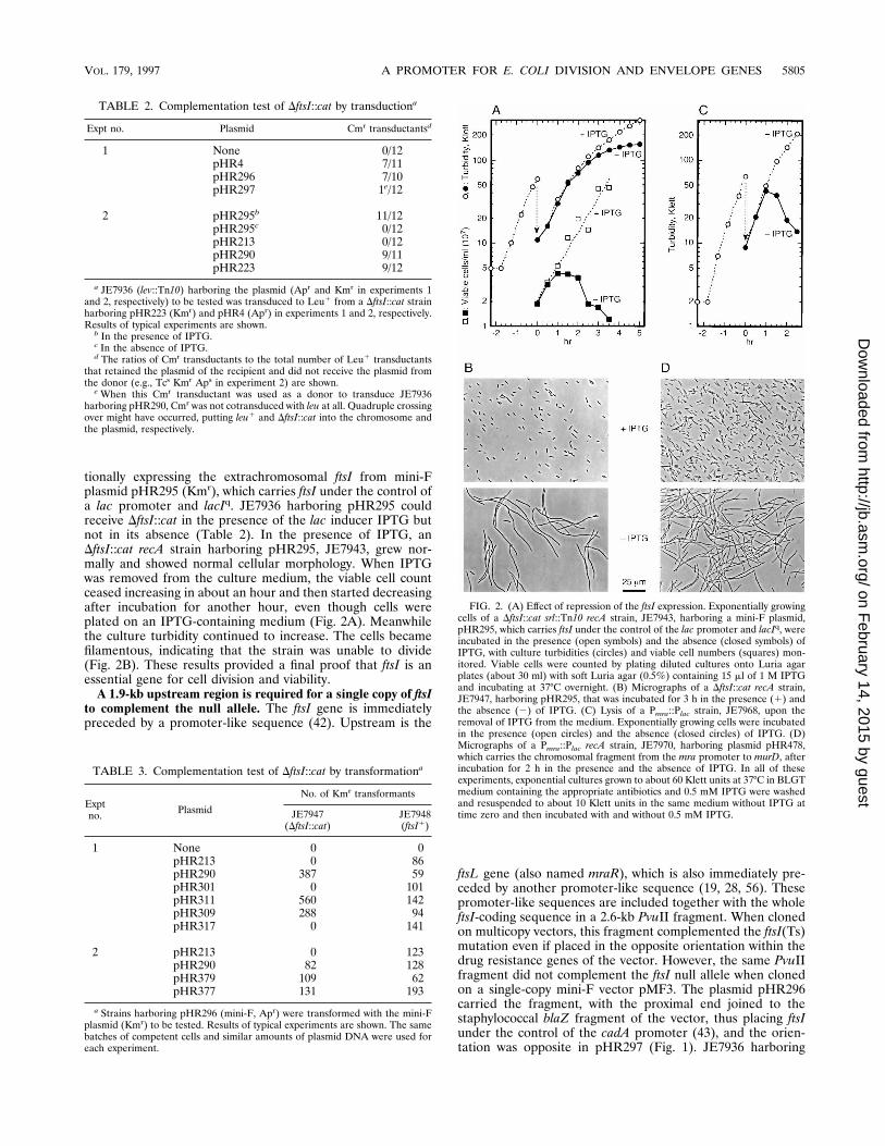
tionally expressing the extrachromosomal ftsI from mini-Fplasmid pHR295 (Kmr), which carries ftsI under the control ofa lac promoter and lacIq. JE7936 harboring pHR295 couldreceive DftsI::cat in the presence of the lac inducer IPTG butnot in its absence (Table 2). In the presence of IPTG, anDftsI::cat recA strain harboring pHR295, JE7943, grew nor-mally and showed normal cellular morphology. When IPTGwas removed from the culture medium, the viable cell countceased increasing in about an hour and then started decreasingafter incubation for another hour, even though cells wereplated on an IPTG-containing medium (Fig. 2A). Meanwhilethe culture turbidity continued to increase. The cells becamefilamentous, indicating that the strain was unable to divide(Fig. 2B). These results provided a final proof that ftsI is anessential gene for cell division and viability.
A 1.9-kb upstream region is required for a single copy of ftsIto complement the null allele. The ftsI gene is immediatelypreceded by a promoter-like sequence (42). Upstream is the
ftsL gene (also named mraR), which is also immediately pre-ceded by another promoter-like sequence (19, 28, 56). Thesepromoter-like sequences are included together with the wholeftsI-coding sequence in a 2.6-kb PvuII fragment. When clonedon multicopy vectors, this fragment complemented the ftsI(Ts)mutation even if placed in the opposite orientation within thedrug resistance genes of the vector. However, the same PvuIIfragment did not complement the ftsI null allele when clonedon a single-copy mini-F vector pMF3. The plasmid pHR296carried the fragment, with the proximal end joined to thestaphylococcal blaZ fragment of the vector, thus placing ftsIunder the control of the cadA promoter (43), and the orien-tation was opposite in pHR297 (Fig. 1). JE7936 harboring
FIG. 2. (A) Effect of repression of the ftsI expression. Exponentially growingcells of a DftsI::cat srl::Tn10 recA strain, JE7943, harboring a mini-F plasmid,pHR295, which carries ftsI under the control of the lac promoter and lacIq, wereincubated in the presence (open symbols) and the absence (closed symbols) ofIPTG, with culture turbidities (circles) and viable cell numbers (squares) mon-itored. Viable cells were counted by plating diluted cultures onto Luria agarplates (about 30 ml) with soft Luria agar (0.5%) containing 15 ml of 1 M IPTGand incubating at 37°C overnight. (B) Micrographs of a DftsI::cat recA strain,JE7947, harboring pHR295, that was incubated for 3 h in the presence (1) andthe absence (2) of IPTG. (C) Lysis of a Pmra::Plac strain, JE7968, upon theremoval of IPTG from the medium. Exponentially growing cells were incubatedin the presence (open circles) and the absence (closed circles) of IPTG. (D)Micrographs of a Pmra::Plac recA strain, JE7970, harboring plasmid pHR478,which carries the chromosomal fragment from the mra promoter to murD, afterincubation for 2 h in the presence and the absence of IPTG. In all of theseexperiments, exponential cultures grown to about 60 Klett units at 37°C in BLGTmedium containing the appropriate antibiotics and 0.5 mM IPTG were washedand resuspended to about 10 Klett units in the same medium without IPTG attime zero and then incubated with and without 0.5 mM IPTG.
TABLE 2. Complementation test of DftsI::cat by transductiona
Expt no. Plasmid Cmr transductantsd
1 None 0/12pHR4 7/11pHR296 7/10pHR297 1e/12
2 pHR295b 11/12pHR295c 0/12pHR213 0/12pHR290 9/11pHR223 9/12
a JE7936 (lev::Tn10) harboring the plasmid (Apr and Kmr in experiments 1and 2, respectively) to be tested was transduced to Leu1 from a DftsI::cat strainharboring pHR223 (Kmr) and pHR4 (Apr) in experiments 1 and 2, respectively.Results of typical experiments are shown.
b In the presence of IPTG.c In the absence of IPTG.d The ratios of Cmr transductants to the total number of Leu1 transductants
that retained the plasmid of the recipient and did not receive the plasmid fromthe donor (e.g., Tcs Kmr Aps in experiment 2) are shown.
e When this Cmr transductant was used as a donor to transduce JE7936harboring pHR290, Cmr was not cotransduced with leu at all. Quadruple crossingover might have occurred, putting leu1 and DftsI::cat into the chromosome andthe plasmid, respectively.
TABLE 3. Complementation test of DftsI::cat by transformationa
Exptno. Plasmid
No. of Kmr transformants
JE7947(DftsI::cat)
JE7948(ftsI1)
1 None 0 0pHR213 0 86pHR290 387 59pHR301 0 101pHR311 560 142pHR309 288 94pHR317 0 141
2 pHR213 0 123pHR290 82 128pHR379 109 62pHR377 131 193
a Strains harboring pHR296 (mini-F, Apr) were transformed with the mini-Fplasmid (Kmr) to be tested. Results of typical experiments are shown. The samebatches of competent cells and similar amounts of plasmid DNA were used foreach experiment.
VOL. 179, 1997 A PROMOTER FOR E. COLI DIVISION AND ENVELOPE GENES 5805
on February 14, 2015 by guest
http://jb.asm.org/
Dow
nloaded from
either of these plasmids was transduced to Leu1 from anDftsI::cat strain harboring pHR223, and it was found thatpHR296 enabled the host to receive the null allele whereaspHR297 did not (Table 2). Complementation tests of mini-Fplasmids were also tried by transforming JE7611 [ftsI(Ts)], butthe results were not clear because of the thermosensitivity ofthe F replicon (51).
Since the promoter-like sequences immediately upstream offtsI in the 2.6-kb PvuII fragment did not allow complementa-tion of the null mutation, we next tested pHR213 (Fig. 1),which carries a 7.3-kb HindIII-EcoRI fragment containing ftsIon the mini-F vector pSY396. The fragment includes 1.9 kb ofDNA upstream of ftsI, yet DftsI::cat could not be cotransducedwith leu to a pHR213-harboring strain from a pHR4-harboringstrain (Table 2). The chromosomal insert of pHR213 did, how-ever, complement the ftsI(Ts) mutation when recloned ontopBR322. We then cloned into pSY396 a longer chromosomalfragment, starting at another HindIII site that is farther up-stream by 1.8 kb (pHR290) (Fig. 1). A transduction experimentshowed that pHR290 complemented the disrupted ftsI (Table2). The complementation ability of Kmr mini-F plasmids wasalso tested by transformation of JE7947 (DftsI::cat recA) har-boring pHR296 (Apr). Since mini-F plasmids are incompatiblewith each other, pHR296 should have been displaced in Kmr
transformants. As shown in Table 3, pHR290 could displacepHR296 in a DftsI::cat strain, whereas pHR213 could not; bothcould displace pHR296 in JE7948, an isogenic ftsI1 strain.Hence, the region further upstream of the 1.9-kb-upstreamHindIII site is necessary for a single-copy ftsI to complementthe null allele.
The chromosomal fragment cloned into pHR290 was short-ened from the upstream HindIII site by 0.7 kb (to the AatII site[pHR311]) and by 1.6 kb (to the HpaI site [pHR309]) (Fig. 1).These mini-F plasmids complemented DftsI::cat, as shown bytransformation experiments (Table 3). Thus, the region neces-sary for the complementation extends at most up to the HpaIsite 2.1 kb upstream of ftsI. Since the upstream region is intacton the chromosome in the DftsI::cat strain, there seems to be asequence required in cis. The inversion of the upstream 1.8-kbHindIII fragment in pHR290 (pHR301) (Fig. 1) resulted in theloss of complementation ability (Table 3), indicating that theorientation of the upstream region or the contiguity at theHindIII site at 1.9 kb upstream or both were essential. Wedisrupted this HindIII site in pHR309 by performing the fillingreaction of T4 DNA polymerase followed by ligation, thusintroducing a 4-bp insertion, which was confirmed by creationof a new NheI site. A test by transformation (Table 3) showedthat the resultant mini-F plasmid pHR317 did not complementDftsI::cat (Table 3). Therefore, this HindIII site seems to bewithin the essential sequence.
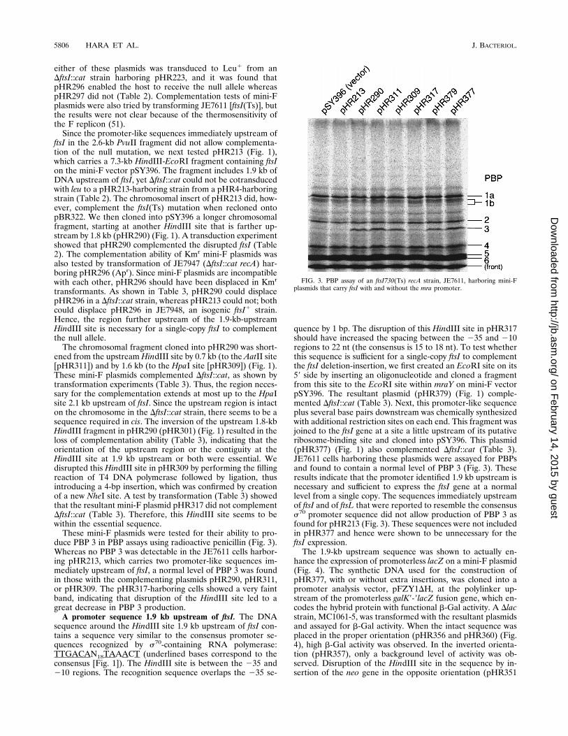
These mini-F plasmids were tested for their ability to pro-duce PBP 3 in PBP assays using radioactive penicillin (Fig. 3).Whereas no PBP 3 was detectable in the JE7611 cells harbor-ing pHR213, which carries two promoter-like sequences im-mediately upstream of ftsI, a normal level of PBP 3 was foundin those with the complementing plasmids pHR290, pHR311,or pHR309. The pHR317-harboring cells showed a very faintband, indicating that disruption of the HindIII site led to agreat decrease in PBP 3 production.
A promoter sequence 1.9 kb upstream of ftsI. The DNAsequence around the HindIII site 1.9 kb upstream of ftsI con-tains a sequence very similar to the consensus promoter se-quences recognized by s70-containing RNA polymerase:TTGACAN18TAAACT (underlined bases correspond to theconsensus [Fig. 1]). The HindIII site is between the 235 and210 regions. The recognition sequence overlaps the 235 se-
quence by 1 bp. The disruption of this HindIII site in pHR317should have increased the spacing between the 235 and 210regions to 22 nt (the consensus is 15 to 18 nt). To test whetherthis sequence is sufficient for a single-copy ftsI to complementthe ftsI deletion-insertion, we first created an EcoRI site on its59 side by inserting an oligonucleotide and cloned a fragmentfrom this site to the EcoRI site within mraY on mini-F vectorpSY396. The resultant plasmid (pHR379) (Fig. 1) comple-mented DftsI::cat (Table 3). Next, this promoter-like sequenceplus several base pairs downstream was chemically synthesizedwith additional restriction sites on each end. This fragment wasjoined to the ftsI gene at a site a little upstream of its putativeribosome-binding site and cloned into pSY396. This plasmid(pHR377) (Fig. 1) also complemented DftsI::cat (Table 3).JE7611 cells harboring these plasmids were assayed for PBPsand found to contain a normal level of PBP 3 (Fig. 3). Theseresults indicate that the promoter identified 1.9 kb upstream isnecessary and sufficient to express the ftsI gene at a normallevel from a single copy. The sequences immediately upstreamof ftsI and of ftsL that were reported to resemble the consensuss70 promoter sequence did not allow production of PBP 3 asfound for pHR213 (Fig. 3). These sequences were not includedin pHR377 and hence were shown to be unnecessary for theftsI expression.
The 1.9-kb upstream sequence was shown to actually en-hance the expression of promoterless lacZ on a mini-F plasmid(Fig. 4). The synthetic DNA used for the construction ofpHR377, with or without extra insertions, was cloned into apromoter analysis vector, pFZY1DH, at the polylinker up-stream of the promoterless galK9-9lacZ fusion gene, which en-codes the hybrid protein with functional b-Gal activity. A Dlacstrain, MC1061-5, was transformed with the resultant plasmidsand assayed for b-Gal activity. When the intact sequence wasplaced in the proper orientation (pHR356 and pHR360) (Fig.4), high b-Gal activity was observed. In the inverted orienta-tion (pHR357), only a background level of activity was ob-served. Disruption of the HindIII site in the sequence by in-sertion of the neo gene in the opposite orientation (pHR351
FIG. 3. PBP assay of an ftsI730(Ts) recA strain, JE7611, harboring mini-Fplasmids that carry ftsI with and without the mra promoter.
5806 HARA ET AL. J. BACTERIOL.
on February 14, 2015 by guest
http://jb.asm.org/
Dow
nloaded from

and pHR366) or of 4 bp created by filling and religating as inpHR317 (pHR358) abolished the b-Gal activity.
The promoter sequence we have identified here forms anoperon of ftsI together with at least the three open readingframes (ORFs) upstream, that is, a 456-base ORF which im-mediately follows the promoter and is temporarily named orfCin the features section of EMBL, GenBank, and DDBJ nucle-otide sequence database entry EC2MIN, a gene coding for a35-kDa protein (18), and ftsL (Fig. 1). Hereafter in this work,the promoter will be referred to as the mra promoter becauseit is located at the beginning of the mra cluster.
The mra promoter is responsible for expression of the fivenext genes downstream of ftsI, i.e., murE, murF, mraY, murD,and ftsW. The question naturally arises as to whether the mrapromoter drives more of the 16 contiguous genes that exist inthe mra cluster without a recognizable transcriptional termi-nator. In an experiment done in parallel to the construction ofa DftsI::cat allele on the chromosome, we attempted to replacethe chromosomal ftsI with another deletion-insertion mutant(DftsI::catopp), in which cat was substituted for the same inter-nal segment of ftsI as in DftsI::cat but was inserted in theopposite orientation to ftsI. A pHR4 derivative carryingDftsI::catopp underwent integration into and resolution fromthe chromosome in the polA(Ts) background (see Materialsand Methods), and P1 transduction to a leu::Tn10 strain har-boring pHR290, selecting for Leu1, was tried. Among 16 trans-ductants examined, none was Cmr. In a similar transductionexperiment, DftsI::cat having cat in the same orientation as ftsIwas found in 4 out of 15 Leu1 transductants, indicating that,after integration and resolution of the DftsI::cat-carrying plas-mid, nearly half of the polA(Ts) cells had retained the dis-rupted gene on the chromosome. Such a discrepancy betweenDftsI::cat and DftsI::catopp can be explained if the cat insertioninto ftsI in the opposite orientation gives a polar effect on
essential genes beyond murF that are not covered by pHR4 inthe transductional donor or by pHR290 in the recipient. Wesuspected that transcription originating from the mra promotermight proceed along the entire cluster to the only known ter-minator just downstream of envA (3) and be at least partlyresponsible for expression of all the genes in the cluster (seeDiscussion).
To examine how far into the mra cluster the function of thepromoter at its beginning was required, we disrupted the pro-moter on the chromosome at the HindIII site and inserted thelac promoter (Fig. 5) as described in Materials and Methods.The inserted DNA fragment was composed of the cat genefollowed by the rrnB transcriptional terminators, the lacIq genein the opposite orientation, and the lac promoter in the sameorientation. In the resultant Pmra::Plac strain, the genes nor-mally dependent on the mra promoter for their expressionwere expected to depend on the lac promoter. The Pmra::Placstrain JE7968 did not grow at all in the absence of IPTG. Whenthe cells grown in the presence of IPTG were deprived of thelac inducer, they started to lyse after about an hour (Fig. 2C).The loss of PBP 3 (Fig. 2A and B) or FtsL (19) would cause cellfilamentation, not lysis. The lysis was probably due to repressedexpression of one or more of the downstream murein biosyn-thetic genes. Two upstream genes, the gene coding for the35-kDa protein and orfC, would also be repressed, but theyhave been found not to be essential for growth and viability(20).
The growth defect in the absence of IPTG of Pmra::Plac recAstrain JE7970 was not corrected by pHR311 (Fig. 5), whichcovers as much of the cluster as do pHR4 and pHR290. Theseplasmids did not support construction of chromosomalDftsI::catopp as described above. When the 21-kb AatII frag-ment containing the entire cluster was cloned into a mini-Fvector, pSY396 (pHR416) (Fig. 5), and introduced into
FIG. 4. b-Gal assays of mra promoter activity. The synthetic oligonucleotides used were the same as those used in the construction of pHR377 (Fig. 1). Open arrowswith neo and the open bar with AGCT indicate the neo fragment inserted into the HindIII site in the orientation opposite to the mra promoter and a small insertioncreated by the filling reaction of T4 DNA polymerase, respectively. Abbreviations for restriction site are as given in the legend to Fig. 1. b-Gal activities (shown inarbitrary units [arb. units]) are averages of the measurements from three transformants.
VOL. 179, 1997 A PROMOTER FOR E. COLI DIVISION AND ENVELOPE GENES 5807
on February 14, 2015 by guest
http://jb.asm.org/
Dow
nloaded from

JE7970, the transformant grew normally in the absence ofIPTG. The chromosomal fragment on pHR416 was shortenedfrom the cluster’s distal end so that envA, ftsZ, and ftsA geneswere deleted one by one (pHR426, pHR427, and pHR439)(Fig. 5). Although a long transcript covering the entire clusterwas suspected at first, these distal genes proved to be unnec-essary to complement the growth defect of JE7970. Next, the13-kb chromosomal fragment ranging from the AatII site up-stream of the mra promoter to the EcoRI site within murC,thus covering up to murG, was cloned into pSY396 (pHR431)(Fig. 5) and was also found to correct the IPTG dependence ofJE7970. When a 1.0-kb SmaI fragment within murG was de-leted from pHR431 and replaced with an V-Tc interposon(14), the resultant plasmid pHR479 (Fig. 5) still corrected theIPTG dependence. The murG gene codes for N-acetylglu-cosaminyl transferase, essential for murein synthesis (39). Fi-nally, the complementation ability was abolished when thedeletion was extended upward to within ftsW (pHR478) or towithin murD (pHR477) (Fig. 5). When deprived of IPTG,JE7970 harboring pHR478 did not lyse but grew into long,filamentous cells (Fig. 2D), indicative of the repressed expres-sion of ftsW, whose defect is known to cause cell division block(28, 29). This division defect was complemented by additionalintroduction of pHR485 (Fig. 5), a plasmid expressing ftsWunder the promoter for the aminoglycoside 39-adenyltrans-ferase (aadA) gene of the vector. Therefore, extrachromo-somal expression of genes up to ftsW, the fifth gene down-stream of ftsI, was necessary to complement the defect due todisruption of the chromosomal mra promoter. We concludethat the mra promoter is required for these first nine genes ofthe cluster to be fully expressed.
The growth defect of JE7970 harboring pHR477 was not
corrected by pHR485, and the cells harboring these two plas-mids lysed upon removal of IPTG. Neither of the plasmidscarried murD, and its repression seemed responsible for thelysis. Although no mutation in the murD gene has been previ-ously described, a lysis phenotype is very likely, since the murDgene product, the D-glutamic acid-adding enzyme (38), servesan essential function in the murein biosynthetic pathway.
DISCUSSION
We identified a promoter at the beginning of the mra cluster,a cluster of cell division and cell envelope biosynthesis genes inthe 2-min region of the E. coli chromosome. Displacement ofthis mra promoter with the lac promoter (Pmra::Plac) resultedin cell lysis in the absence of a lac inducer, and the defect wascomplemented by plasmids containing chromosomal frag-ments from the mra promoter to at least ftsW, the ninth geneof the cluster. With a plasmid containing up to the eighth genebut not the intact ftsW, the Pmra::Plac strain showed a celldivision defect, which was corrected by the addition of a plas-mid expressing ftsW. We thus conclude that the mra promoterwas required for full expression of the first nine genes, up toftsW, of the cluster. Although the length of the transcript fromthe mra promoter has not been determined, it seems probablethat transcription proceeds for more than 10 kb, to ftsW andbeyond. All the genes from orfC through envA are tightlyclustered in the same orientation without a transcriptionalterminator.
Dai and Lutkenhaus (8) reported that a null allele of ftsZwas not complemented by a l transducing phage that carried 6kb of DNA upstream of ftsZ, including promoters locatedwithin ftsA and ddl (65), but was complemented by F9104,
FIG. 5. Plasmids carrying chromosomal fragments of the 2-min region. Only the cloned chromosomal regions are shown (filled bars). Short, open bars representpolylinkers derived from synthetic oligonucleotides (pHR416, pHR426, pHR427, and pHR439) and from the V-Tc interposon (pHR485). The open arrowhead inpHR485 denotes the direction of the promoter on the vector. The orientation and approximate sizes of the mra cluster genes and the structure of Pmra::Plac are shownat the top. The thick brackets with T’s beneath them represent r-independent transcriptional terminators. Parentheses surrounding enzyme pairs indicate the ligationof compatible cohesive ends [(Eg/Nt)] or of blunt or filled-in ends. (Ap3)Nt stands for the ApaLI-cut end that was first ligated to a NotI linker (59-dGCGGCCGC-39)and then joined to the NotI site. The maps are drawn to scale except for the polylinkers and Pmra::Plac. V-Tc interposons (broken bars) are drawn not in their own sizesbut in the sizes of the respective fragments they displaced. Complementation of chromosomal Pmra::Plac was tested by cross-brushing a Pmra::Plac recA strain, JE7970,transformed with the respective plasmids against a brush of 200 mM IPTG on Luria agar plates and incubating at 37°C overnight. Abbreviations for restriction sites(only relevant sites are shown): A, AatII; Ap, ApaLI; B, BamHI; Bs, Bsu36I; E, EcoRI; Eg, EagI; Ev, EcoRV; H, HindIII; Nt, NotI; P, PstI; Pv, PvuII; S, SmaI; Sn, SnaBI.
5808 HARA ET AL. J. BACTERIOL.
on February 14, 2015 by guest
http://jb.asm.org/
Dow
nloaded from

which carried a much larger chromosomal insert, including thewhole of the mra cluster. They suggested that a portion of ftsZexpression was due to a promoter(s) located more than 6 kbupstream. We first suspected that the mra promoter might bethe upstream promoter that they had postulated to be requiredfor full expression of ftsZ. However, the Pmra::Plac strain car-rying the mra promoter-ftsW region on a plasmid grew nor-mally in the absence of IPTG, without extrachromosomal ex-pression of genes further downstream, including ftsZ,indicating that these genes could be expressed independentlyof the mra promoter. There might be basal-level transcriptionfrom the lac promoter even in the absence of an inducer, butsuch transcription was unlikely to be sufficient for expression ofthe downstream genes, considering the existence of lacIq in thePmra::Plac strain and inclusion of glucose in media used in theexperiments, both leading to tighter repression (4). Actually,the uninduced lac promoter in the Pmra::Plac strain could notsupport sufficient expression of the proximal ftsL, ftsI, and ftsWgenes, whose products have been shown to be very minorcomponents of the cell (13, 19, 29, 49).
The murG gene coding for N-acetylglucosaminyl transferaseis essential, as indicated by an murG(Am) supF(Ts) strainwhich lyses at the restrictive temperature (39). Coding framesof ftsW and murG are overlapped by 1 base or separated by 23bases, depending on the two possible initiation codons ofmurG, and a promoter(s) for the murG gene should be some-where in the orfC-ftsW region, possibly in the just-precedingftsW gene. Analysis of the DNA sequence reveals a promoter-like sequence TTGGCGN17TACGCT (nt 15203 to 15231 ofnucleotide sequence database entry EC2MIN [underlinedbases correspond to the consensus]) towards the 59 end of ftsW(nt 14933 to 16174). The upstream endpoint of the clonedfragment in l16-2, a transducing phage that contained ftsZ butfailed to complement an ftsZ null allele, was in the midst offtsW (8), and therefore a promoter(s) responsible for at least aportion of expression of genes from murG to ftsZ seemed to liein the 59 half of ftsW or further upstream.
There appear to be many potential promoter sequences inthe mra cluster. The envA gene has its own promoter located inthe intergenic space between ftsZ and envA, which seems suf-ficient for its expression (3, 8). The ability of the promoters inthe ddl-ftsA region to regulate ftsQAZ expression has beenstudied (e.g., see references 15 and 47). Mengin-Lecreulx et al.(38) suggested the existence of several promoters in the ftsI-murD region. Although we have shown in the present studythat the mra promoter is necessary for expression of genes upto ftsW, it is not known if the promoter alone is sufficient forexpression of all of these genes. Distal genes might be partlydependent on the potential promoters in the ftsI-murD region,whose activities are yet to be examined. These promoters, ifreally active, possibly contribute to expression of murG and itsdownstream genes, too. Several promoter-like sequences havebeen described for the orfC-ftsL region (19, 28, 42) (also de-scribed in the features section of nucleotide sequence databaseentry EC2MIN). However, mini-F plasmid pHR213, whichcontains all of these sequences, did not lead to detectableproduction of PBP 3. Thus, these sequences seemed to havelittle if any promoter activity, at least under the conditionsused, although the possibility was not excluded that they mightplay important roles in subtle adjustments under conditionsnot tested in the present study. There have been interestingreports for the 35-kDa protein gene-ftsL region, a sequencethat has high homology with the LexA repressor-binding se-quence (28) and a negative control by mreB of the ftsI expres-sion (60). It is not known whether there is any effect of the SOSresponse on expression of the mra cluster genes or whether the
negative control by mreB is transcriptional or posttranscrip-tional. Between the mra promoter and the shl gene upstream(33) is a 0.6-kb silent region that contains no ORF of signifi-cant length. It is interesting if there is any regulatory elementaffecting the mra cluster expression. However, no characteris-tic sequence has yet been identified in this silent region. As forthe amount of PBP 3, no fluctuation was observed during thecell division cycle (63), whereas a downregulation under thedirect or indirect influence of rpoS was reported at the entryinto stationary phase (12). Further detailed analyses are nec-essary to elucidate the regulatory mechanism of the complexmultipromoter system in the mra cluster.
PBP 3 and PBP 2 are members of class B high-molecular-mass PBPs (16). PBP 3 is thought to function in conjunctionwith FtsW for septal murein synthesis, which is analogous toPBP 2’s function in cell elongation and maintenance of the rodshape in conjunction with RodA (27), a protein that is struc-turally very similar to FtsW (25). Under the control of the mrapromoter, ftsI and ftsW are in a single transcriptional unit, asare pbpA, the structural gene coding for PBP 2, and rodA (37).This may be convenient for the bacterium to coordinate ex-pression of these pairs of cooperating genes. In Bacillus subtilis,however, spoVD (9) and spoVE (25), which code for the ho-mologs of E. coli PBP 3 and FtsW, respectively, required forspore cortex synthesis, seem to be transcribed separately (55),although they are within a gene cluster at 133 to 135° on thechromosome that is homologous to the E. coli mra cluster. ThepbpB gene coding for PBP 2B, another homolog of E. coli PBP3 that functions during vegetative growth and asymmetricspore septum formation (64) is also within the cluster at 133 to135° but is transcribed separately from spoVD and spoVE (10).An ORF named ipa-42d, whose predicted product is highlysimilar to FtsW and RodA (17) and possibly functions in con-junction with PBP 2B, is well separated from the cluster at 133to 135°.
In B. subtilis and also in Haemophilus influenzae, the geneshomologous to the E. coli mra cluster genes are found clus-tered on their chromosomes, and the order of homologousgenes is highly conserved among these three species. This isstriking, considering that the conservation of the order of morethan two homologous genes is very rare (62). Such exceptionalconservation may not be explained simply by assuming con-straints on the transcriptional combinations of the functionallyhomologous genes during evolution.
All the genes with known functions in this cluster are essen-tial for cell division and/or envelope synthesis. Although no E.coli mutant defective in murD has been isolated, it was shownin the present study that repression of murD in the Pmra::Placstrain with the other mra promoter-dependent genes expressedfrom plasmids resulted in cell lysis, indicating that the gene isindispensable. Functions are not known for the first two genesof the cluster, orfC and the gene coding for a 35-kDa protein,except for the existence of an S-adenosylmethionine-bindingmotif in the latter (30). These genes are well conserved in thehomologous clusters of B. subtilis and H. influenzae. They areconserved even on the chromosome of Mycoplasma genitaliumtogether with ftsZ, while the other genes of the cluster areabsent from this murein-free organism. Hence, the first twogenes have been assumed to play some important roles relatedto cell division. However, a PvuII fragment that covers themost parts of these genes could be deleted from the E. colichromosome and displaced by an antibiotic resistance gene ofthe same orientation with no apparent defect in growth ordivision (20). The B. subtilis homologs of these two genes haverecently been shown to be nonessential (10).
Multicopy plasmids that carry ftsI and its upstream region
VOL. 179, 1997 A PROMOTER FOR E. COLI DIVISION AND ENVELOPE GENES 5809
on February 14, 2015 by guest
http://jb.asm.org/
Dow
nloaded from
but not the mra promoter in the opposite orientation withindrug resistance genes complemented the ftsI(Ts) mutation.This could be a cumulative effect of very weak promoters athigh copy numbers but is more likely due to transcriptionalreadthrough from another promoter(s) on the vector. By usingsingle-copy mini-F vectors, one can eliminate a possible effectof high copy numbers. The mini-F vector pMF3 (34) has apromoter, originally for the cadA gene (43), in its antibioticresistance gene (blaZ) fragment. The transcription from itflows into the EcoRI cloning site. When cloned into this EcoRIsite, the PvuII fragment containing ftsI but not the mra pro-moter did not complement the ftsI null allele unless placed inthe same orientation under the control of the cadA promoter.Only in this orientation was complementation observed alsofor the prc gene (23). Thus, only negligible if any transcriptionseems to extend out of the F replicon fragment beyond theEcoRI site. In pSY396, another mini-F vector used in this study(see Materials and Methods), the antibiotic resistance gene(neo) fragment does not seem to contain a promoter thatdirects transcription flowing into the EcoRI-HindIII cloningregion, because pHR213 did not lead to production of a de-tectable amount of PBP 3; in pHR213, the ftsI-containingHindIII-EcoRI fragment was cloned with the HindIII sitewithin the mra promoter sequence joined to the HindIII site ofthe neo fragment of pSY396. As described above for pMF3, notranscription proceeds into the EcoRI-HindIII region from theF replicon fragment. Thus, a gene(s) cloned in this region ofpSY396 is not subjected to transcriptional read-through fromeither side. This is useful in examining promoter function at asingle copy in vivo.
We discovered the existence of the mra promoter by testingthe ability of ftsI-containing fragments cloned on pSY396 tocomplement a chromosomal null allele of ftsI (DftsI::cat) inwhich a large internal fragment of the gene was deleted andthe antibiotic resistance gene (cat) fragment was inserted inthe same orientation. The deletion-insertion mutation with thecat fragment in the opposite orientation (DftsI::catopp) couldnot be crossed into the chromosome even in the presence ofplasmids that allowed the crossing into the chromosome ofDftsI::cat. This is probably because transcription of the cat genein DftsI::catopp counteracts transcription from the mra pro-moter and diminishes expression of the downstream essentialgenes that were not covered by the plasmids used in the ex-periment. One should be aware that such polar effects canoccur in gene disruption experiments.
The DftsI::cat strain carrying an extrachromosomal copy offtsI under the control of the lac promoter was dependent on alac inducer for cell division and viability, confirming the essen-tial function of PBP 3. When incubated for 2 h or longer afterremoval of a lac inducer from the medium, cells were stillgrowing into longer and longer filaments, but most cells be-came unable to form colonies on an IPTG-containing agarplate. This seems to indicate that they could not restore theability to divide even if ftsI expression was resumed, althoughthere is another possibility, namely, that filamentous cells wereeasily killed during the plating procedure. In normally dividingcells, PBP 3 is thought to interact with many other proteinsinvolved in cell division to form a multimolecular complex.Depletion of PBP 3 might lead to formation of abortive com-plexes lacking this essential protein, which might be incapableof reactivation by incorporating PBP 3 afterwards. This couldcause unrepairable damage to the cell. Genetic analyses havesuggested interaction of PBP 3 with other cell division pro-teins, including FtsA and FtsZ, direct interaction with thelatter being just recently demonstrated by new methodologies(58). For a thorough understanding of the bacterial cell divi-
sion process, elucidation of interactions of division proteins aswell as of transcriptional regulations of their genes will berequired.
ACKNOWLEDGMENTS
This work was supported, in part, by grant AI05090 from the Na-tional Institutes of Health of the United States and by grants-in-aid forscientific research (A) 02404088 and (C) 05680601 from the Ministryof Education, Science, Sports, and Culture of Japan.
We are grateful to Atsushi Higashitani and Yukinobu Nishimura forcontinuous encouragement and stimulating discussion and to AtsukoAoyagi for excellent technical assistance. We thank Juan A. Ayala,Suzanne Bourgeois, Guy Dufford, Joachim Frey, Carol Kumamoto,Yuthana Limpa-Amara, Michael Malamy, Dominique Mengin-Lec-reulx, Teru Ogura, Debabrata RayChaudhuri, Masaaki Wachi, andHidemi Watanabe for helpful advice, bacterial strains, plasmids, cos-mids, and phages.
REFERENCES
1. Adam, M., C. Damblon, M. Jamin, W. Zorzi, V. Dusart, M. Galleni, A. ElKharroubi, G. Piras, B. G. Spratt, W. Keck, J. Coyette, J.-M. Ghuysen, M.Nguyen-Disteche, and J.-M. Frere. 1991. Acyltransferase activities of thehigh-molecular-mass essential penicillin-binding proteins. Biochem. J. 279:601–604.
2. Balbas, P., X. Soberon, E. Merino, M. Zurita, H. Lomeli, F. Valle, N. Flores,and F. Bolivar. 1986. Plasmid vector pBR322 and its special-purpose deriv-atives—a review. Gene 50:3–40.
3. Beall, B., and J. Lutkenhaus. 1987. Sequence analysis, transcriptional orga-nization, and insertional mutagenesis of the envA gene of Escherichia coli. J.Bacteriol. 169:5408–5415.
4. Beckwith, J. 1987. The lactose operon, p. 1444–1452. In F. C. Neidhardt, J. L.Ingraham, K. B. Low, B. Magasanik, M. Schaechter, and H. E. Umbarger(ed.), Escherichia coli and Salmonella typhimurium: cellular and molecularbiology. American Society for Microbiology, Washington, D.C.
5. Brady, G., H. M. Jantzen, H. U. Bernard, R. Brown, G. Schuts, and T.Hashimoto-Gotoh. 1984. New cosmid vectors developed for eukaryotic DNAcloning. Gene 27:223–232.
6. Brosius, J. 1984. Plasmid vectors for the selection of promoters. Gene27:151–160.
7. Churchward, G., D. Belin, and Y. Nagamine. 1984. A pSC101-derived plas-mid which shows no sequence homology to other commonly used cloningvectors. Gene 31:165–171.
8. Dai, K., and J. Lutkenhaus. 1991. ftsZ is an essential cell division gene inEscherichia coli. J. Bacteriol. 173:3500–3506.
9. Daniel, R. A., S. Drake, C. E. Buchanan, R. Scholle, and J. Errington. 1994.The Bacillus subtilis spoVD gene encodes a mother-cell-specific penicillin-binding protein required for spore morphogenesis. J. Mol. Biol. 235:209–220.
10. Daniel, R. A., A. M. Williams, and J. Errington. 1996. A complex four-geneoperon containing essential cell division gene pbpB in Bacillus subtilis. J.Bacteriol. 178:2423–2350.
11. Donachie, W. D. 1993. The cell cycle of Escherichia coli. Annu. Rev. Micro-biol. 47:199–230.
12. Dougherty, T. J., and M. J. Pucci. 1994. Penicillin-binding proteins areregulated by rpoS during transitions in growth states of Escherichia coli.Antimicrob. Agents Chemother. 38:205–210.
13. Dougherty, T. J., K. Kennedy, R. E. Kessler, and M. J. Pucci. 1996. Directquantitation of the number of individual penicillin-binding proteins per cellin Escherichia coli. J. Bacteriol. 178:6110–6115.
14. Fellay, R., J. Fray, and H. Krisch. 1987. Interposon mutagenesis of soil andwater bacteria: a family of DNA fragments designed for in vitro insertionalmutagenesis of Gram-negative bacteria. Gene 52:147–154.
15. Garrido, T., M. Sanchez, P. Palacios, M. Aldea, and M. Vicente. 1993.Transcription of ftsZ oscillates during the cell cycle of Escherichia coli.EMBO J. 12:3957–3965.
16. Ghuysen, J.-M. 1994. Molecular structures of penicillin-binding proteins andb-lactamases. Trends Microbiol. 2:372–380.
17. Glaser, P., F. Kunst, M. Arnaud, M.-P. Coudart, W. Gonzales, M.-F. Hullo,M. Ionescu, B. Lubochinsky, L. Marcelino, I. Moszer, E. Presecan, M.Santana, E. Schneider, J. Schweizer, A. Vertes, G. Rapoport, and A.Danchin. 1993. Bacillus subtilis genome project: cloning and sequencing ofthe 97 kb region from 325° to 333°. Mol. Microbiol. 10:371–384.
18. Gomez, M. J., B. Fluoret, J. van Heijenoort, and J. A. Ayala. 1990. Nucleo-tide sequence of the regulatory region of the gene pbpB of Escherichia coli.Nucleic Acids Res. 18:2813.
19. Guzman, L.-M., J. J. Barondess, and J. Beckwith. 1992. FtsL, an essentialcytoplasmic membrane protein involved in cell division in Escherichia coli. J.Bacteriol. 174:7716–7728.
20. Hara, H., and K. Horiuchi. Unpublished data.
5810 HARA ET AL. J. BACTERIOL.
on February 14, 2015 by guest
http://jb.asm.org/
Dow
nloaded from
21. Hara, H., and J. T. Park. 1993. A far upstream region is required forexpression of the ftsI gene coding for penicillin-binding protein 3 of Esche-richia coli, p. 303–308. In M. A. de Pedro, J.-V. Holtje, and W. Loffelhardt(ed.), Bacterial growth and lysis: metabolism and structure of the bacterialsacculus. Plenum Press, New York, N.Y.
22. Hara, H., Y. Nishimura, J. Kato, H. Suzuki, H. Nagasawa, A. Suzuki, and Y.Hirota. 1989. Genetic analyses of processing involving C-terminal cleavage inpenicillin-binding protein 3 of Escherichia coli. J. Bacteriol. 171:5882–5889.
23. Hara, H., Y. Yamamoto, A. Higashitani, H. Suzuki, and Y. Nishimura. 1991.Cloning, mapping, and characterization of the Escherichia coli prc gene,which is involved in C-terminal processing of penicillin-binding protein 3. J.Bacteriol. 173:4799–4813.
24. Houba-Herin, N., H. Hara, M. Inouye, and Y. Hirota. 1985. Binding ofpenicillin to thiol-penicillin-binding protein 3 of Escherichia coli: identifica-tion of its active site. Mol. Gen. Genet. 201:499–504.
25. Ikeda, M., T. Sato, M. Wachi, H. K. Jung, F. Ishino, Y. Kobayashi, and M.Matsuhashi. 1989. Structural similarity among Escherichia coli FtsW andRodA proteins and Bacillus subtilis SpoVE protein, which function in celldivision, cell elongation, and spore formation, respectively. J. Bacteriol.171:6375–6378.
26. Ishino, F., and M. Matsuhashi. 1981. Peptidoglycan synthetic enzyme activ-ities of highly purified penicillin-binding protein 3 in Escherichia coli: aseptum-forming reaction sequence. Biochem. Biophys. Res. Commun. 101:905–911.
27. Ishino, F., W. Park, S. Tomioka, S. Tamaki, I. Takase, K. Kunugita, H.Matsuzawa, S. Asoh, T. Ohta, B. J. Spratt, and M. Matsuhashi. 1986.Peptidoglycan synthetic activities in membranes of Escherichia coli caused byoverproduction of penicillin-binding protein 2 and RodA protein. J. Biol.Chem. 261:7024–7031.
28. Ishino, F., H. K. Jung, M. Ikeda, M. Doi, M. Wachi, and M. Matsuhashi.1989. New mutations fts-36, lts-33, and ftsW clustered in the mra region of theEscherichia coli chromosome induce thermosensitive cell growth and divi-sion. J. Bacteriol. 171:5523–5530.
29. Khattar, M. M., K. J. Begg, and W. D. Donachie. 1994. Identification ofFtsW and characterization of a new ftsW division mutant of Escherichia coli.J. Bacteriol. 176:7140–7147.
30. Koonin, E. V., R. L. Tatusov, and K. E. Rudd. 1995. Sequence similarityanalysis of Escherichia coli proteins: functional and evolutionary implica-tions. Proc. Natl. Acad. Sci. USA 92:11921–11925.
31. Koop, A. H., M. E. Hartley, and S. Bourgeois. 1987. A low-copy-numbervector utilizing b-galactosidase for the analysis of gene control elements.Gene 52:245–256.
32. Kumamoto, C. A., and J. Beckwith. 1985. Evidence for specificity at an earlystep in protein export in Escherichia coli. J. Bacteriol. 163:267–274.
33. Leclerc, G., G. Noel, and G. R. Drapeau. 1990. Molecular cloning, nucleotidesequence, and expression of shl, a new gene in the 2-minute region of thegenetic map of Escherichia coli. J. Bacteriol. 172:4696–4700.
34. Manis, J. J., and B. C. Kline. 1977. Restriction endonuclease mapping andmutagenesis of the F sex factor replication region. Mol. Gen. Genet. 152:175–182.
35. Masui, Y., J. Coleman, and M. Inouye. 1983. Multipurpose expression clon-ing vehicles in Escherichia coli, p. 15–32. In M. Inouye (ed.), Experimentalmanipulation of gene expression. Academic Press, New York, N.Y.
36. Matsuhashi, M., M. Wachi, and F. Ishino. 1990. Machinery for cell growthand division: penicillin-binding proteins and other proteins. Res. Microbiol.141:89–103.
37. Matsuzawa, H., S. Asoh, K. Kunai, K. Muraiso, A. Takasuga, and T. Ohta.1989. Nucleotide sequence of the rodA gene, responsible for the rod shapeof Escherichia coli: rodA and the pbpA gene, encoding penicillin-bindingprotein 2, constitute the rodA operon. J. Bacteriol. 171:558–560.
38. Mengin-Lecreulx, D., C. Parquet, L. R. Desviat, J. Pla, B. Flouret, J. A.Ayala, and J. van Heijenoort. 1989. Organization of the murF-murG regionof Escherichia coli: identification of the murD gene encoding the D-glutamic-acid-adding enzyme. J. Bacteriol. 171:6126–6134.
39. Mengin-Lecreulx, D., L. Texier, M. Rousseau, and J. van Heijenoort. 1991.The murG gene of Escherichia coli codes for the UDP-N-acetylglucosamine:N-acetylmuramyl-(pentapeptide) pyrophosphoryl-undecaprenol N-acetyl-glucosamine transferase involved in the membrane steps of peptidoglycansynthesis. J. Bacteriol. 173:4625–4636.
40. Miller, J. H., ed. 1992. A short course in bacterial genetics: a laboratorymanual and handbook for Escherichia coli and related bacteria. Cold SpringHarbor Laboratory Press, Cold Spring Harbor, N.Y.
41. Miyakawa, T., H. Matsuzawa, M. Matsuhashi, and Y. Sugino. 1972. Cell wall
peptidoglycan mutants of Escherichia coli K-12: existence of two clusters ofgenes, mra and mrb, for cell wall peptidoglycan biosynthesis. J. Bacteriol.112:950–958.
42. Nakamura, M., I. N. Maruyama, M. Soma, J. Kato, H. Suzuki, and Y.Hirota. 1983. On the process of cellular division in Escherichia coli: nucle-otide sequence of the gene for penicillin-binding protein 3. Mol. Gen. Genet.191:1–9.
43. Nucifora, G., L. Chu, T. K. Misra, and S. Silver. 1989. Cadmium resistancefrom Staphylococcus aureus plasmid pI258 cadA gene results from a cadmi-um-efflux ATPase. Proc. Natl. Acad. Sci. USA 86:3544–3548.
44. Rao, R. N., and S. G. Rogers. 1979. Plasmid pKC7: a vector containing tenrestriction endonuclease sites suitable for cloning DNA segments. Gene7:79–82.
45. Rudd, K. E. 1992. Alignment of E. coli DNA sequences to a revised, inte-grated genomic restriction map, p. 2.3–2.43. In J. H. Miller (ed.), A shortcourse in bacterial genetics: a laboratory manual and handbook for Esche-richia coli and related bacteria. Cold Spring Harbor Laboratory Press, ColdSpring Harbor, N.Y.
46. Sambrook, J., E. F. Fritsch, and T. Maniatis. 1989. Molecular cloning: alaboratory manual, 2nd ed. Cold Spring Harbor Laboratory Press, ColdSpring Harbor, N.Y.
47. Sitnikov, D. M., J. B. Schineller, and T. O. Baldwin. 1996. Control of celldivision in Escherichia coli: regulation of transcription of ftsQA involves bothrpoS and SdiA-mediated autoinduction. Proc. Natl. Acad. Sci. USA 93:336–341.
48. Spratt, B. G. 1977. Temperature-sensitive cell division mutants of Esche-richia coli with thermolabile penicillin-binding proteins. J. Bacteriol. 131:293–305.
49. Spratt, B. G. 1977. Properties of the penicillin-binding proteins of Esche-richia coli K12. Eur. J. Biochem. 72:341–352.
50. Spratt, B. G., and K. D. Cromie. 1988. Penicillin-binding proteins of gram-negative bacteria. Rev. Infect. Dis. 10:699–711.
51. Stadler, J., and E. A. Adelberg. 1972. Temperature dependence of sex-factormaintenance in Escherichia coli K-12. J. Bacteriol. 109:447–449.
52. Stark, M. J. R. 1987. Multicopy expression vectors carrying the lac repressorgene for regulated high-level expression of genes in Escherichia coli. Gene51:255–267.
53. Suzuki, H., Y. Nishimura, and Y. Hirota. 1978. On the process of cellulardivision in Escherichia coli: a series of mutants of E. coli altered in thepenicillin-binding proteins. Proc. Natl. Acad. Sci. USA 75:664–668.
54. Takeda, Y., A. Nishimura, Y. Nishimura, M. Yamada, S. Yasuda, H. Suzuki,and Y. Hirota. 1981. Synthetic ColE1 plasmids carrying genes for penicillin-binding proteins in Escherichia coli. Plasmid 6:86–98.
55. Theeragool, G., A. Miyao, K. Yamada, T. Sato, and Y. Kobayashi. 1993. Invivo expression of the Bacillus subtilis spoVE gene. J. Bacteriol. 175:4071–4080.
56. Ueki, M., M. Wachi, H. K. Jung, F. Ishino, and M. Matsuhashi. 1992.Escherichia coli mraR gene involved in cell growth and division. J. Bacteriol.174:7841–7843.
57. van Heijenoort, Y., M. Gomez, M. Derrien, J. Ayala, and J. van Heijenoort.1992. Membrane intermediates in the peptidoglycan metabolism of Esche-richia coli: possible roles of PBP 1b and PBP 3. J. Bacteriol. 174:3549–3557.
58. Vicente, M., and J. Errington. 1996. Structure, function and controls inmicrobial division. Mol. Microbiol. 20:1–7.
59. Vieira, J., and J. Messing. 1982. The pUC plasmids, an M13mp7-derivedsystem for insertion mutagenesis and sequencing with synthetic universalprimers. Gene 19:259–268.
60. Wachi, M., and M. Matsuhashi. 1989. Negative control of cell division bymreB, a gene that functions in determining the rod shape of Escherichia colicells. J. Bacteriol. 171:3123–3127.
61. Wanner, B. 1986. Novel regulatory mutants of the phosphate regulon inEscherichia coli K-12. J. Mol. Biol. 191:39–58.
62. Watanabe, H., T. Itoh, H. Mori, and T. Gojobori. 1997. Genome plasticity asa paradigm of eubacteria evolution. J. Mol. Evol. 44:S57–S64.
63. Wientjes, F. B., T. J. M. Olijhoek, U. Schwarz, and N. Nanninga. 1983.Labeling pattern of major penicillin-binding proteins of Escherichia coliduring the division cycle. J. Bacteriol. 153:1287–1293.
64. Yanouri, A., R. A. Daniel, J. Errington, and C. E. Buchanan. 1993. Cloningand sequencing of the cell division gene pbpB, which encodes penicillin-binding protein 2B in Bacillus subtilis. J. Bacteriol. 175:7604–7616.
65. Yi, Q.-M., S. Rockenbach, J. E. Ward, and J. Lutkenhaus. 1991. Structureand expression of the cell division genes ftsQ, ftsA, and ftsZ. J. Mol. Biol.184:399–412.
VOL. 179, 1997 A PROMOTER FOR E. COLI DIVISION AND ENVELOPE GENES 5811
on February 14, 2015 by guest
http://jb.asm.org/
Dow
nloaded from