An automated prognosis system for estrogen hormone status assessment in breast cancer tissue samples
Upload
independentCategory
view
1download
0
Cell Stem Cell
Short Article
Methylation of Cancer-Stem-Cell-AssociatedWnt Target Genes Predicts Poor Prognosisin Colorectal Cancer PatientsFelipe de Sousa E Melo,1 Selcuk Colak,1 Joyce Buikhuisen,1 Jan Koster,2 Kate Cameron,1 Joan H. de Jong,1
Jurriaan B. Tuynman,1 Pramudita R. Prasetyanti,1 Evelyn Fessler,1 Saskia P. van den Bergh,1 Hans Rodermond,1
Evelien Dekker,3 Chris M. van der Loos,4 Steven T. Pals,4 Marc J. van de Vijver,4 Rogier Versteeg,2 Dick J. Richel,5
Louis Vermeulen,1,6 and Jan Paul Medema1,6,*1Laboratory of Experimental Oncology and Radiobiology, Center for Experimental Molecular Medicine2Department of Oncogenomics3Department of Gastroenterology and Hepatology4Department of Pathology5Department of Medical Oncology
Academic Medical Center, Meibergdreef 9, 1105AZ Amsterdam, The Netherlands6These authors contributed equally to this work
*Correspondence: [email protected] 10.1016/j.stem.2011.10.008
SUMMARY
Gene signatures derived from cancer stem cells(CSCs) predict tumor recurrence for many forms ofcancer. Here, we derived a gene signature for colo-rectal CSCs defined by high Wnt signaling activity,which in agreement with previous observations pre-dicts poor prognosis. Surprisingly, however, wefound that elevated expression of Wnt targets wasactually associated with good prognosis, whilepatient tumors with low expression of Wnt targetgenes segregated with immature stem cell signa-tures. We discovered that several Wnt target genes,including ASCL2 and LGR5, become silenced byCpG island methylation during progression of tumor-igenesis, and that their re-expression was associ-ated with reduced tumor growth. Taken together,our data show that promoter methylation of Wnttarget genes is a strong predictor for recurrence ofcolorectal cancer, and suggest that CSC gene signa-tures, rather than reflecting CSC numbers, mayreflect differentiation status of the malignant tissue.
INTRODUCTION
Colorectal cancer (CRC) is a major contributor to cancer-related
death. Although patients with local disease (stage I and II) have
a favorable prognosis, a small fraction of these patients will inev-
itably develop a recurrence after intentionally curative surgery
(Edge and Compton, 2010). Identifying these poor-prognosis
patients will allow optimized selection of individuals that would
benefit from adjuvant chemotherapy. Exciting new insights in
tumor biology might facilitate this selection; of particular interest
in this respect is the recent discovery of cancer stem cells (CSCs)
in CRC, a subset of cells that are defined by their capacity to
476 Cell Stem Cell 9, 476–485, November 4, 2011 ª2011 Elsevier Inc
transplant the human malignancy to immuno-compromised
mice (O’Brien et al., 2007; Ricci-Vitiani et al., 2007). CSCs share
many features with normal intestinal stem cells (ISCs) (Medema
and Vermeulen, 2011), and can be identified based on cell
surface markers, such as CD133 (Todaro et al., 2007; Vermeulen
et al., 2008), or functionally, using high Wnt-signaling activity
levels (Vermeulen et al., 2010). Importantly, CSCs are suggested
to fuel tumor growth and as such are hypothesized to cause
tumor recurrence and metastasis. Accordingly, recent reports
address the relation between stem cells, CSCs, and patient
prognosis in different malignancies, including CRC (Liu et al.,
2007). For example, in a recent study by Merlos-Suarez et al.
(2011) a mouse ISC signature has been derived by identifying
genes that are highly expressed in EphB2high ISCs compared
with that of the more differentiated epithelial cells. This profile
encompasses known ISC markers, such as the Wnt target
gene LGR5, and is strongly associated with both CRC disease
stage and the occurrence of tumor relapse and metastasis
formation. This has led to the suggestion that an increased
number of CSCs is predictive for prognosis.
Here we show that CSC-derived gene signatures can indeed
predict tumor recurrence. However, the positive association is
not due to expression of specific CSC and CSC-associated
Wnt target genes, which rather inversely correlate with prog-
nosis. The subset of patients identified in this manner display
decreased expression of a wide range of ISC/Wnt target genes.
This is not due to decreased Wnt pathway activity, but is a result
of selective promoter methylation. Moreover, Wnt target methyl-
ation levels, by themselves, can be used to effectively identify
patients at risk of recurrence, and re-expression of these meth-
ylated genes lowers tumorigenicity in vitro and in vivo.
RESULTS
ISC and CSC Profiles Predict Poor Prognosis in CRCPreviously we have shown that colon-CSCs can be identified in
primary human CRC using Wnt signaling intensity levels and can
.

Cell Stem Cell
Predicting Colorectal Cancer Relapse
be isolated by employing a Wnt reporter construct (TOP-GFP,
Figure 1A) (Vermeulen et al., 2010). Gene expression profiling
of the TOP-GFPhigh human colon-CSCs indicated high expres-
sion of the stem cell marker LGR5, as well as several other
Wnt targets (APCDD1, LEF1), while typical intestinal differentia-
tion markers (e.g., MUC2 and FABP1) displayed low expression
(Figure 1A). Based on two primary isolated spheroid cultures, we
generated a colon-CSC signature comprising 187 genes most
differentially expressed between the CSCs and the more differ-
entiated cells (Table S1A, available online). This profile was
subsequently validated in several independent freshly isolated
CRCs (Figure S1A, available online).
Crucially, by employing gene set enrichment analysis (GSEA)
(Subramanian et al., 2005), we found that this colon-CSC gene
expression signature was intimately associated with disease
recurrence in a set of 90 stage II CRC patients that underwent
intentionally curative surgery at our institute (AMC-AJCCII-90,
see Table S1B for patient characteristics), a finding we con-
firmed in an unrelated, publically available data set (Figure 1B
and Figure S1B). Similar results were obtained using two inde-
pendent ISC profiles that have previously been shown to relate
to disease recurrence, lending strength to the validity of both
the CSC signature and our patient set (Figure 1B and Figure S1B)
(Merlos-Suarez et al., 2011). Using a simple rank-sum approach,
we stratified the AMC-AJCCII-90 patients into two groups, which
further established the prognostic power of the CSC profile
because it revealed that especially early relapses in stage II
CRC patients were characterized by a strong resemblance to
the CSC signature (Figure 1C, see Experimental Procedures
for details). Importantly, however, the nature and biological impli-
cation of this correlation remain unclear.
CSC-Associated Wnt Target Genes Inversely Correlatewith PrognosisIn an attempt to analyze the biological mechanism behind the
prognostic power of CSC profiles in more depth and to identify
genes that are most predictive regarding tumor relapse and
metastasis formation, we employed a cluster analysis. Unsuper-
vised K-means clustering of the AMC-AJCCII-90 set using
the TOP-GFPhigh/CSC-derived gene expression profile resulted
in two distinct patient groups with a significant difference in
relapse-free survival, as the Kaplan-Meier curve illustrates (Fig-
ure 1D). Gene tree analysis revealed that segregation of these
two clusters is accompanied by generation of two major
subgroups of genes. The majority of genes were upregulated
in the patient cluster that was associated with poor prognosis
(blue). However, a clearly distinct subset of genes, at the bottom
region of the gene tree, inversely correlated with disease relapse
(Figure 1D). To our surprise this gene cluster contained many
well-known Wnt target genes of which the expression is inti-
mately linked to ISCs and CSCs. (For a list of genes and their
differential expression between the clusters, see Tables S1C
and S1D). Similar results were obtained by employing different
stem cell signatures, including the LGR5� and EphB2 ISC signa-
tures that have been described previously (Figures S1C and
S1D) (Merlos-Suarez et al., 2011). In all cases high expression
of the Wnt-driven ISC marker genes present in the different
signatures, like ASCL2, LGR5, AXIN2, and APCDD1, were
associated with the favorable prognosis cluster (Figure 1D and
Cel
Figure S1C and S1D). We confirmed the expression level differ-
ences between the two groups by qPCR (Figure S1E) and found
that expression was not related to oncogenic mutations present
within the different patients (Figure S2A). Multivariate Cox
regression analysis indicated the profile to be an independent
prognostic factor that was much more predictive than the pres-
ence of different mutations (Figure S2A). Strikingly, we also ob-
served that the expression of Wnt target genes was not a simple
reflection of patient-to-patient variation in Wnt signaling activity
as measured by nuclear localized b-catenin levels (Figure 1F).
Intrigued by this counterintuitive finding, which indicates that
high Wnt target gene expression is linked to favorable rather
than poor prognosis, we repeated this analysis with a more
defined set of Wnt target genes, previously identified by overex-
pression of dominant-negative TCF4 (dnTCF4) in CRC cell lines
(van de Wetering et al., 2002). The clear majority of genes in
this dnTCF4 signature are also markedly lower expressed in
the poor-prognosis patient cluster (Figure 1E). Even single
Wnt target genes, including the validated ISC markers EPHB2
(Merlos-Suarez et al., 2011), LGR5 (Barker et al., 2007), and
ASCL2 (van der Flier et al., 2009), but also more general Wnt
targets such as AXIN2 and APCDD1, can identify poor-prog-
nosis patients based on their low expression levels both in our
patient set and in publically available data sets (Figure S2B
and results not shown).
The finding that high expression of genes intimately associ-
ated with the CSC phenotype is associated with good prognosis
in CRC immediately challenges the conventional interpretation
as to why (cancer-) stem-cell-associated profiles define poor
prognosis in cancer. Mostly it is believed that association with
a CSC profile reflects the number of CSC-like cells in the malig-
nancy. However, when we use FACS staining to define the frac-
tion of cells positive for CD133 in several freshly isolated colon
cancer specimens, which so far is the best studied and validated
means to identify colon-CSCs, we could not correlate the
number of CSCs to the overall expression of CSC-associated
Wnt target genes within these tumors (Figure S2C). In addition,
the fraction of CRC cells positive for nuclear b-catenin, which
has been used before as a trait to identify colon-CSCs, also
does not correlate significantly with Wnt target gene expression
in our patient data set (Figure S2D). These findings both indicate
that CSC numbers in CRC are not causal determinants in the
patient stratification obtained with the CSC-associated expres-
sion signature. More importantly, the lack of correlation between
nuclear b-catenin levels and Wnt target gene expression indi-
cates that additional regulatory mechanisms are in place to
regulate Wnt target gene expression.
CSC-Associated Wnt Targets Are Downregulatedduring ProgressionIn order to better understand why CSC-associated Wnt targets
are inversely correlated with prognosis, we determined the
expression of five Wnt target genes at multiple stages during
the adenoma-carcinoma sequence. As expected, comparison
of normal tissue with adenoma tissue revealed a marked
increase in expression of most Wnt target genes, in line with
the notion that activation of the Wnt cascade is the initiating
event in CRC development (Figure 2A) (Morin et al., 1997). It is
well accepted that further genetic and epigenetic alterations in
l Stem Cell 9, 476–485, November 4, 2011 ª2011 Elsevier Inc. 477
A
TOP-GFP High
TOP-GFP LowTOP-GFP
Transduction
B
0 1000 2000 3000 4000
20
40
60
80
100
CSC signature
Follow up (days)
Su
rv
ival (p
ro
b.)
p 6.7e-03
WntHigh
WntLow
D
CSC signature/AMC-AJCCII-90
Lgr5
Lef1
Apcdd1
0 1000 2000 3000 4000
20
40
60
80
100
Follow up (days)
Su
rv
ival (p
ro
b.) CSC signature Low
CSC signature High
p 0.01
-2.0
0.17
2.0
CSC signature
0 25 50
0
0.15
0.30
Rec
No
ISC-EphB2CSC signature
0.15
0
0.30
Rec
0 25 50
ES
No
0.30
0 25 50
0
0.15
Rec
No
Rank list (x1000)
Lgr5
ES
C
E
p 0.013
0 1000 2000 3000 4000
20
40
60
80
100
dnTCF4
Follow up (days)
Su
rv
ival (p
ro
b.) Wnt
High
WntLow
AMC-AJCCII-90
vs recurrenceGeneset ES NES FDR
CSC signature 0.36 1.67 0.0038ISC-EphB2 0.35 1.69 <0.001Lgr5 0.32 1.74 <0.001
Hig
h
Wnt
Low
Wnt
0
20
40
60
80
100
p 0.16
% N
uc. p
ositiv
ity
dnTCF4 signature/AMC-AJCCII-90
Ascl2
Axin2
Lgr5
F WntLowWnt
High
Co34
Co78
Co20
Co52
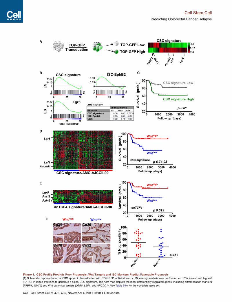
Figure 1. CSC Profile Predicts Poor Prognosis; Wnt Targets and ISC Markers Predict Favorable Prognosis
(A) Schematic representation of CSC spheroid transduction with TOP-GFP lentiviral vector. Microarray analysis was performed on 10% lowest and highest
TOP-GFP sorted fractions to generate a colon-CSC signature. The heat map depicts the most differentially regulated genes, including differentiation markers
(FABP1, MUC2) and Wnt canonical targets (LGR5, LEF1, and APCDD1). See Table S1A for the complete gene set.
Cell Stem Cell
Predicting Colorectal Cancer Relapse
478 Cell Stem Cell 9, 476–485, November 4, 2011 ª2011 Elsevier Inc.

Cell Stem Cell
Predicting Colorectal Cancer Relapse
the premalignant adenoma tissue mark the transition to an inva-
sively growing CRC (Fearon and Vogelstein, 1990). However,
evaluation of Wnt target gene expression in CRC samples strik-
ingly indicated a downregulation in the majority of patients
compared with the adenoma stage (Figure 2A). This was also
observed in an independent data set containing normal,
adenoma, and carcinoma samples (Figures S3A and S3B). The
relevance of this suppressed Wnt target gene expression was
immediately evident: patients that developed a tumor relapse
displayed the lowest expression levels (Figure 2A, red triangles).
This confirms our findings that a lowWnt target gene expression
profile is related to poor prognosis. In addition, we obtained
evidence that CRC tissue characterized by a low Wnt target
expression signature demonstrates other characteristics of
advanced disease as well. In this respect we analyzed the asso-
ciation of the patients in theWnt target-low cluster with immature
stem cell signatures (based on SOX2, OCT4, andNanog targets),
which have been used previously to determine disease grade
and prognosis in cancer patients (Figure S3C) (Ben-Porath
et al., 2008). GSEA showed a clear correlation between the
Wnt target-low cluster and these signatures (Figure S3D). This
correlation was not detected when a direct comparison was
made between the expression of pluripotency genes in the
Wnt target high clusters with that of low clusters (Figure S3D),
but the association with immature stem cell signatures does
suggest that segregation based on the CSC signature reflects
a more immature trait of the malignant tissue in the poor-prog-
nosis cluster. In agreement, the association of the Wnt target-
low cluster with this molecular immature fingerprint is also
reflected by a significant enrichment for tumors presenting with
a poorly differentiated histology (Figure 2B).
CSC-Associated Wnt Target Genes Are Subjectto Methylation-Dependent RegulationAlthough the above indicates that the tumors that cluster in the
Wnt-low group have a more immature phenotype, this does
not provide insight into the apparent discrepancy between the
observed lowWnt target gene expression and the lack of reduc-
tion in nuclear b-catenin localization (Figure S2). To explain this
conundrum we sought to determine whether epigenetic regula-
tory mechanisms might act to regulate Wnt target gene expres-
sion and thereby promote progression. Methylation is involved in
many biological processes, including stem cell maintenance and
cancer progression. In addition, CRC development is accompa-
nied by global changes in methylation status of a plethora of
genes (Morin et al., 1997; Rai et al., 2010). A subtype of CRC,
the CpG Island Methylation Phenotype (CIMP), has even been
(B) GSEA reveals a strong relationship between CSC signature and tumor relap
ISC-EphB2 signatures associate with recurrence in our set. ES, enrichment scor
(C) Kaplan-Meier graph (relapse-free survival) based on overall adherence to the
mental Procedures for details).
(D and E) K-means clustering analysis of CRC samples from the AMC-AJCCII-90
(E) representing a defined set of Wnt target genes. Several canonical Wnt target
survival is drawn for each corresponding signature. Note that the poor-prognosis
also Figures S1C and S1D for similar conclusions based on the LGR5� and ISC
(F) Representative b-catenin stainings are shown for two patients in both the
Automated scoring of nuclear b-catenin fractions in patients from the AMC-AJC
In (D–F), blue represents Wnt-target-low cluster (WntLow); red, Wnt-target-high c
Cel
identified that is characterized by extensive methylation (Toyota
et al., 1999). Several natural Wnt inhibitors, such as AXIN2 and
SFRP1, have previously been shown to be methylated in CRC
(Koinuma et al., 2006; Suzuki et al., 2004). We therefore analyzed
the DNA methylation status of Wnt target genes first in a series
of CRC cell lines. Methylation-specific PCR analysis revealed
that several Wnt target genes including LGR5, APCDD1,
DKK1, andASCL2 are, to a different extent, methylated in a panel
of CRC cell lines, suggesting epigenetic silencing of these genes
(Figure 2C). In agreement, treatment of CRC cells with the
demethylating agent 5-Azacytidine (5-Aza) resulted in marked
upregulation of these genes specifically in those cell lines where
methylation was evident (Figure 2D). For instance, APCDD1
expression was enhanced by 5-Aza in the lines where APCDD1
promoter methylation was clearly detectable. These data
enforce the notion that Wnt targets are regulated, at least in
part, by a mechanism that involves promoter methylation in
CRC cell lines.
Functional Relevance of Wnt Target Gene MethylationNext we determined the functional relevance of this methylation-
dependent silencing of Wnt target genes. Treatment of CRC
cell lines with a demethylating agent resulted in markedly
decreased clonogenicity of these lines (Figure 3A). Importantly,
this was a general observation; primary isolated colon-CSC
cultures treated with 5-Aza also demonstrated significantly lower
clonogenicity as determined by limiting dilution analysis, sug-
gesting that the fraction of CSCs in these cultures decreased
(Figure 3B). Also, in an in vivo model system in which primary
CSC-induced xenograft tumors were growing subcutaneously,
we observed that 5-Aza treatment resulted in markedly sup-
pressed tumor growth (Figure 3C). Importantly, analysis of these
5-Aza-treated xenografts confirmed the efficacy of 5-Aza on
re-expression of the Wnt target genes in vivo (Figure 3D), vali-
dating the methylation-dependent regulation of Wnt target
genes in this in vivo model as well. To strengthen our hypothesis
that re-expression of Wnt target genes has important func-
tional consequences, we analyzed the effect of 5-Aza on Wnt
activity levels. In this light it is important to realize that several
of these repressed target genes normally serve as feedback
inhibitors of the same pathway and as such could repress the
activity of the Wnt pathway. In agreement, in several CRC cell
lines, 5-Aza-mediated expression of genes previously sup-
pressed by methylation resulted in decreased Wnt activity
(Figure 3E).
Although these data are suggestive for a role of Wnt target
gene methylation in CRC, the effects of 5-Aza are rather generic,
se in the AMC-AJCCII-90 patient set. Also, previously described LGR5� and
e; NES, normalized enrichment score; FDR, false discovery rate.
TOP-GFPhigh/CSC profile as identified by gene ranking analysis (see Experi-
patient set according to the colon-CSC signature (D) and the dnTCF4 signature
genes are denoted for each signature. Kaplan-Meier analysis on relapse-free
cluster (blue) is associated with low expression of indicated Wnt targets (see
-EphB2 signatures). p values are calculated with the log-rank test.
Wnt-target-High (WntHigh; red) and Wnt-target-Low (WntLow; blue) clusters.
CII-90 set is shown. Scale bars represent 200 mm.
luster (WntHigh).
l Stem Cell 9, 476–485, November 4, 2011 ª2011 Elsevier Inc. 479

Rec
No Rec
Norm
al
Adenom
a
CRC
0.0
0.5
1.0
1.5
2.0
mR
NA
re
l. e
xp
.
Lgr5
Norm
al
Adenom
a
CRC
0
2
4
6 Apcdd1
Norm
al
Adenom
a
CRC
0.0
0.5
1.0
1.5 Axin2
B
Norm
al
Adenom
a
CRC
0
1
2
3
3
6 Ascl2
Lgr5 Apcdd1
Lgr5 Dkk1Ascl2
HCT15
HCT116
SW620
SW837
Colo205
U M U MU MU M
Apcdd1
HCT15
HCT116
SW
620
SW
837
Colo
205
0
2
4
6
2000
4000
6000
HCT15
HCT116
SW
620
SW
837
Colo
205
0
2
4
6
mR
NA
re
l. exp
.
HCT15
HCT116
SW
620
SW
837
Colo
205
0
1
2
3
4
5
100
200
300
400
HCT15
HCT116
SW
620
SW
837
Colo
205
0
2
4
6
8
Dkk1
Ascl2
A
D
Norm
al
Adenom
a
CRC
0.0
0.5
1.0
1.5
2.0
N.D
Dkk1
Control
Aza
HCT15
HCT116
SW
620
SW
837
Colo
205
0
1
2
3
4 Axin2
Poor
Good/M
od.
0.0
0.2
0.4
0.6
0.8
1.0
WntLow
WntHigh
Fra
ctio
n
p 8.6-6
C
Figure 2. Wnt Target Genes Are Regulated by Methylation in CRC
(A) Relative expression of established Wnt target genes (LGR5, APCDD1, ASCL2, DKK1, and AXIN2) in normal, adenoma, and CRC tissue. Patients in the CRC
group that developed a recurrence are highlighted in red. Horizontal line indicates mean value.
Cell Stem Cell
Predicting Colorectal Cancer Relapse
480 Cell Stem Cell 9, 476–485, November 4, 2011 ª2011 Elsevier Inc.

Cell Stem Cell
Predicting Colorectal Cancer Relapse
affecting all methylated CpG islands, and can therefore not be
considered specific for Wnt target genes. We therefore ques-
tioned whether growth inhibition could be achieved directly by
specific re-expression of methylated Wnt target genes. Indeed,
we observed that re-expression of either AXIN2 or APCDD1,
both methylated in CRC, was sufficient to decrease Wnt sig-
naling levels (Figure 3F). This is not only observed in CRC cell
lines, but confirmed in a primary CSC culture as well (Figure 3F,
right bars, Co100). This Wnt activity modulation directly
suggests a potential functional explanation of why suppression
of a large set of Wnt target genes occurs during the adenoma
to carcinoma sequence and why Wnt target gene inactivation
could be associated with poor prognosis.
Wnt Target GeneMethylation Predicts Prognosis in CRCSo far our data indicate that low expression of CSC-associated
Wnt target genes is related to poor prognosis and that methyla-
tion-dependent downregulation of these genes has a functionally
relevant impact on the clonogenicity of CRC cells. However, it is
unclear whether in-patient material suppression of a stem-cell-
associated Wnt expression program is also dependent on meth-
ylation. To this end we determined the relative methylation levels
of the Wnt target genes in our AMC-AJCCII-90 patient set by
either methylation-specific PCR or bisulphite sequencing (Fig-
ure 4). Intriguingly, also in the tumors from our patient cohort,
low Wnt target gene expression was associated with increased
methylation of the promoter regions of these genes (Figure 4A).
That is, in the tumors that cluster in theWnt-low group (Figure 4A,
right black bars), the fraction of DNAmethylation in the APCDD1,
ASCL2, AXIN2, DKK1, and LGR5 promoter regions is much
higher as compared with that of the tumors that cluster in the
Wnt-high group. Moreover, tumors in the Wnt-high group that
did show methylation were, in several cases, derived from
patients that eventually developed recurrences or metastases,
as indicated by the asterisks in Figure 4A, suggesting that meth-
ylation of Wnt target genes is an even better marker for recur-
rences. In agreement, we found that methylation levels of a small
subset of these CSC-associated Wnt target genes resulted in
a highly predictive association with disease recurrence and
metastasis using unsupervised cluster analysis based on the
relative methylation levels (Figures 4B and 4C). Indeed, all recur-
rences cluster within the Wnt target gene methylation high
group. These findings lend further support to the idea that meth-
ylation-dependent tuning of the Wnt expression program is
related to disease progression and increased risk for recurrent
disease.
DISCUSSION
Our findings have three major implications for the use and
interpretation of (cancer-) stem-cell-associated profiles in risk
stratification of CRC.
(B) The Wnt-target-Low (WntLow) cluster of patients is enriched in patients
Chi-square test.
(C) Methylation-specific PCR for Wnt target genes LGR5, APCDD1, ASCL2, and
(D) Relative expression of indicated Wnt target genes following 48 hr demethy
standard deviations.
Cel
First, we confirm previous observations that ISC signatures
can predict recurrence of CRC (Merlos-Suarez et al., 2011) and
extend these findings with a novel colon-CSC-derived signature
that has similar predictive properties, as might be expected
based on the partial overlap between the various stem cell signa-
tures. In particular both ISC and CSC signatures were character-
ized by a clear enrichment in Wnt target genes, consistent with
the major role of the Wnt cascade in both ISC and colon-CSC
biology.
Second, to our surprise, our data unequivocally show that
expression of many well-defined canonical Wnt target genes,
including prominent ISC markers, was inversely correlated with
prognosis. This unexpected finding, which was verified in mul-
tiple patient sets, significantly changes the conclusions of earlier
studies of stem-cell-derived predictive signatures (Merlos-
Suarez et al., 2011). In previous reports the association of a
(cancer) stem cell signature with poor prognosis is often attrib-
uted to a relative high number of CSCs present in the malignant
tissue (Merlos-Suarez et al., 2011; Pardal et al., 2003; Charafe-
Jauffret et al., 2009), which was thought to enhance the chance
of CSCs shedding from the primary tumor. Indeed, adherence to
an ISC profile of individual CRCs, but also similarity of breast
cancers to a breast-CSC signature, was translated into an
increased risk of metastasis and tumor recurrence (Liu et al.,
2007;Merlos-Suarez et al., 2011). It is important to realize though
that CSC numbers in primary tumors are on average suggested
to constitute a minority of the tumor cells. It therefore appears
rather unlikely that gene profiling of a complete tumor specimen
would yield detailed information on a small minority of the cells.
This would only be feasible when stem cell genes in the signa-
tures are unique to the CSC population and additionally highly
expressed. Instead, it is apparent from our data that these signa-
tures identify poor-prognosis patients despite the presence of
key (cancer) stem cell markers and canonical Wnt target genes,
which we find to inversely correlate with tumor relapse and
disease stage in CRC. Indeed, deletion of Wnt targets from the
CSC signature improves the association with malignancies
harboring a poor prognosis as predicted (Figure S1F).We believe
our data indicate that CSC signatures identify tumors with a rela-
tively immature signature as suggested by the association with
a poorly differentiated histology. In agreement, progression of
disease seems to be accompanied by adapting amore primitive,
immature expression program defined by SOX2, OCT4, and
Nanog signatures, as opposed to the more intestinal-tissue-
specific stem cell signature with genes such as ASCL2 and
LGR5. The fact that this association is strongest with the Wnt-
target low cluster (Figure S3C), which has the poorest prognosis,
substantiates this hypothesis. Whether this is directly related to
methylation of ISC/CSC-associated Wnt target genes or occurs
in parallel is unclear at this point, but suppression of the Wnt
target expression profile during the adenoma-carcinoma
sequence clearly links thisWnt regulation to disease progression
displaying a poorly differentiated morphology. p value was calculated by
DKK1 in a panel of CRC lines. U, unmethylated; M, methylated.
lating treatment with 5-Aza in CRC cell lines. Confidence intervals represent
l Stem Cell 9, 476–485, November 4, 2011 ª2011 Elsevier Inc. 481
A B
E
C D
0
50
100
150
200
0 10 20 30
0
5
10
15
20
Time
Control
Aza
Lgr5
Apcdd1
Ascl2
Dkk1
Axin
2
0
1
2
3
4
5
15
20
25
mR
NA
re
l.e
xp
. Control
Aza
# o
f c
lo
nes
Co
ntro
lA
za
Rel. T
um
or size
0.1
1
10
100
Clo
ne freq
uen
cy (%
)0.01
0.1
1
0.1
1
10
100
Control
Aza
Control
Axin2
Apcdd1
HCT116
SW
620
SW
837
DLD1
0.25
0.5
1
2
TO
P/F
OP
F
HCT15
HCT15
HCT116
SW
620
SW
837
DLD1
Co100
0.0
0.5
1.0
1.5
TO
P/F
OP
Control
Aza
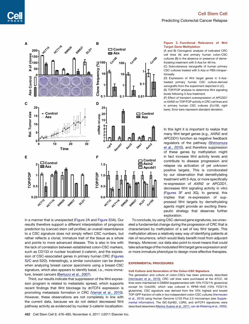
Figure 3. Functional Relevance of Wnt
Target Gene Methylation
(A and B) Clonogenic analysis of indicated CRC
cell lines (A) and primary human colon-CSC
cultures (B) in the absence or presence of deme-
thylating treatment with 5-Aza for 48 hrs.
(C) Subcutaneous xenografts of human primary
CSC cultures treated with 5-Aza or PBS intraper-
itoneally.
(D) Expression of Wnt target genes in 5-Aza-
treated primary human CSC culture-derived
xenografts from the experiment depicted in (C).
(E) TOP/FOP analysis to determine Wnt signaling
levels following 5-Aza treatment.
(F) Effect of transient overexpression of APCDD1
orAXIN2 on TOP/FOP activity in CRCcell lines and
in primary human CSC cultures (Co100, right
bars). Error bars represent standard deviation.
Cell Stem Cell
Predicting Colorectal Cancer Relapse
in a manner that is unexpected (Figure 2A and Figure S3A). Our
results therefore support a different interpretation of prognosis
prediction by (cancer) stem cell profiles; an overall resemblance
to a CSC signature does not simply reflect CSC numbers, but
rather reflects a clonal, immature trait of the tissue as a whole
and points to more advanced disease. This is also in line with
the lack of correlation between established colon-CSC markers,
such as CD133 or nuclear localized b-catenin, and the expres-
sion of CSC-associated genes in primary human CRC (Figures
S2C and S2D). Interestingly, a similar conclusion can be drawn
when analyzing breast cancer specimens using a breast-CSC
signature, which also appears to identify basal, i.e., more imma-
ture, breast cancers (Bertucci et al., 2007).
Third, our results indicate that suppression of the Wnt expres-
sion program is related to metastatic spread, which supports
recent findings that Wnt blockage by dnTCF4 expression is
promoting metastasis formation in CRC (Varnat et al., 2010).
However, these observations are not completely in line with
the current data, because we do not detect decreased Wnt
pathway activity as evidenced by nuclear b-catenin localization.
482 Cell Stem Cell 9, 476–485, November 4, 2011 ª2011 Elsevier Inc.
In this light it is important to realize that
many Wnt target genes (e.g., AXIN2 and
APCDD1) function as negative feedback
regulators of the pathway (Shimomura
et al., 2010), and therefore suppression
of these genes by methylation might
in fact increase Wnt activity levels and
contribute to disease progression and
relapse via activation of yet unknown
positive targets. This is corroborated
by our observation that demethylating
treatment with 5-Aza, or more specifically
re-expression of AXIN2 or APCDD1,
decreases Wnt signaling activity in vitro
(Figures 3F and 3G). In general, this
implies that re-expression of sup-
pressed Wnt targets by demethylating
agents might provide an exciting thera-
peutic strategy that deserves further
exploration.
To conclude, by usingCSC-derivedgene signatures,we unrav-
eled a fundamental change during the progression of CRC that is
characterized by methylation of a set of key Wnt targets. This
methylation allows a relatively easy way of identifying patients at
risk of recurrence, which would likely benefit most from adjuvant
therapy. Moreover, our data also point to novel means that could
takeadvantageof themodulatedWnt target geneexpressionand/
or more immature phenotype to design more effective therapies.
EXPERIMENTAL PROCEDURES
Cell Culture and Generation of the Colon-CSC Signature
The generation and culture of colon-CSCs has been previously described
(Vermeulen et al., 2010). CRC cell lines were purchased at the ATCC. All
lines were maintained in DMEM (supplemented with 10% FCS/1% glutamine)
except for Colo205, which was cultured in RPMI-1640 (10% FCS/1%
glutamine). CSC signature was derived from the 10% highest and lowest
TOP-GFP fraction of cells in two independent colon-CSC cultures (Vermeulen
et al., 2010) using Human Genome U133 Plus 2.0 microarrays (see Supple-
mental Information). The ISC-EphB2, LGR5, and dnTCF4 signatures were
described elsewhere (Merlos-Suarez et al., 2011; van deWetering et al., 2002).

0
20
40
60
80
0
20
40
60
80
100
0
20
40
60
0
20
40
60
80
100
94
19
98
95
70
34
49
65
89
78
92
38
39
11
56
52
26
04
54
20
03
75
87
53
0
5
10
15
0 500 1000 1500 2000
0
20
40
60
80
100
Low Methylation
High Methylationp 1.2-03
Follow up (days)
Su
rv
ival (
pro
b.)
*
*
* *
** *
*
*
*
*
*
** *
*
*
*
**
**
*
*
** *
**
*
*
*
***
* *
*
*
*
Me
th
yla
tio
n (
%)
WntHigh
WntLow
A
C
Ascl2
Dkk1
Apcdd1
Axin2
Lgr5
*
*
*
*
*
Lo
w
Hig
h
-+
Lgr5
Ascl2
Axin2
Dkk1
Apcdd1
53
75
03
92
87
20
54
04
26
89
11
52
34
56
98
39
19
70
38
49
94
95
78
65
Hig
h
Lo
w
Hig
h
Hig
h
Hig
h
Hig
h
Hig
h
Hig
h
Hig
h
Hig
h
Hig
h
Hig
h
Hig
h
Hig
h
Lo
w
Lo
w
Lo
w
Lo
w
Lo
w
Lo
w
Lo
w
Hig
h
- - - - - - - - -- - - + + - + ++ + + - +
Pat. No.
Wnt Cluster
Rec/meta
B
Figure 4. Methylation of Wnt Target Genes Identifies Poor-Prognosis Patients
(A) Percentage of CpG island methylation in the promoter region of the indicated Wnt target genes for a subset of patients from the AMC-AJCCII-90 set.
(B) Unsupervised cluster analysis using the methylation levels (ranking) of Wnt target genes reveals two clusters. Patient number, High or LowWnt-target cluster,
and recurrence are indicated. Colors depict rank order of methylation level within the patient set for each gene: green, lowly methylated; red, highly methylated;
grey, data not available.
(C) Kaplan-Meier curve depicting the two different patient groups as identified in (B). p value is calculated with the log-rank test.
Cell Stem Cell
Predicting Colorectal Cancer Relapse
Patient Cohorts
Two different CRC patient series were used for this study. The first one con-
sisted of 90 AJCC stage II CRC patients that underwent intentionally curative
surgery in the Academic Medical Center in Amsterdam, The Netherlands in the
years 1997–2006 (AMC-AJCCII-90). Extensive medical records are kept of
these patients and long-term clinical follow-up is available for the large
majority. Both paraffin-embedded and fresh frozen tissue is available from
all these patients for analysis, which was used to derive gene expression
profiles. The second patient set is composed of two merged cohorts that
form a metacohort of 345 CRC patients and has been described elsewhere
(Merlos-Suarez et al., 2011). In addition, a separate panel of normal and
adenoma fresh-frozen tissues was obtained in the AMC and these observa-
tions were validated with a publically available data set (Galamb et al., 2008).
Clustering and Survival Analysis
Unsupervised K-means cluster analysis and Kaplan-Meier survival curves
were generated in the different expression data sets with the different gene
signatures using the software package R2 (http://r2.amc.nl), a web-based mi-
croarray analysis application developed by J.K. (data not shown). For single-
Cel
gene survival prediction, the median expression value of each gene was
used as a cutoff to generate two groups of 45 patients having either a low or
high relative expression. p value was calculated using the log-rank test. For
cluster analysis (Euclidian Distance, average linkage in the MultiExperiment
Viewer package v4.5, www.TM4.org) of methylation levels of Wnt target
gene sets, we used the rank value for each individual patient for each gene.
Prediction Power of Signature and Multivariate Analysis
For the predictive power of the CSC signature, we selected the 134 upregu-
lated genes. Every individual gene was ranked according to their expression
in each patient. The rank score for all the genes per patient was summed to
define the rank for each patient. A high expression of a gene is translated
into a low rank score. Patients that have an overall high expression of the
genes in the signature have an overall low rank score and therefore are highly
associated with the CSC profile. A Kaplan-Meier survival curve was generated
to plot the relapse-free survival of patients having a high (n = 30) versus low
(n = 60) correlation with the CSC profile. For multivariate analysis, the Cox
proportional hazard model was used. All p values are two-sided. Statistical
analysis was performed in SPSS.
l Stem Cell 9, 476–485, November 4, 2011 ª2011 Elsevier Inc. 483
Cell Stem Cell
Predicting Colorectal Cancer Relapse
In Vivo 5-Aza Treatment
Murine experiments were performed in accordance with the ethical committee
of the AMC. For transplantation of CSCs, 5,000 cells suspended in 100 ml of
PBS/BSA admixed with Matrigel at a 1:1 ratio were injected subcutaneously
into nude mice (Hsd:Athymic Nude/Nude) (Harlan). When tumors were
palpable, mice were injected i.p. with 5-Aza (5 mg/kg) or PBS vehicle. Injec-
tions were performed every 2 days.
b-catenin Staining and Spectral Imaging Quantification
Paraffin-embedded primary human specimens were stained with anti-b-cate-
nin (Transduction Labs) and then incubated with anti-mouse-HRP (Powervi-
son) (1:1). Multispectral data sets from slides stained for b-catenin were
acquired using a Nuance camera system (Caliper Life Science, Hopkinton,
MA) from 420–720 nm at intervals of 20 nm. To analyze the frequency of hema-
toxylin-b-catenin colocalization, spectral data sets were analyzed with the
tissue segmentation and machine-learning Inform 1.2 software (Caliper Life
Science), similar to methods described elsewhere (Al-Kofahi et al., 2011).
The software was trained to recognize tumor and stroma areas, and next, all
spectral data sets were segmented into these two tissue categories. All data
sets were analyzed using the nuclear algorithm, scoring the percentage of
b-catenin positive nuclei out of all nuclei in the tumor tissue category.
RNA, PCR, and Methylation Analysis
For all methods on RNA isolation, PCR, and mutation analysis, and for all
primer sequences, please see the Supplemental Information.
ACCESSION NUMBERS
The GenBank accession number for the CSC signature is GSE33112: gene
expression in CSC cultures identified by Wnt signaling levels. The GenBank
accession number for the patient dataset is GSE33113: AMC colon cancer
AJCCII.
SUPPLEMENTAL INFORMATION
Supplemental Information for this article includes three figures, one table, and
Supplemental Experimental Procedures and can be found with this article
online at doi:10.1016/j.stem.2011.10.008.
ACKNOWLEDGMENTS
The authors would like to specially thankM. Oud for the tumor slides, and prof.
T.M. van Gulik, prof. W.A. Bemelman, and Dr. P.J. Tanis for the primary tumor
samples. Prof. S. Repping we would like to thank for critically reading the
manuscript. This work was supported by a VICI grant from the Netherlands
Organisation for Scientific Research and a Dutch Cancer Society grant
(2009-4416) (to J.P.M.), and an Academisch Medisch Centrum (AMC) fellow-
ship (to L.V. and F.d.S.E.M.). The TOP-GFP construct was a kind gift of
Dr. L. Ailles. The pMX-Axin2-ires-CD8 was kindly provided by Prof. H. Mano.
Received: May 26, 2011
Revised: September 19, 2011
Accepted: October 18, 2011
Published: November 3, 2011
REFERENCES
Al-Kofahi, Y., Lassoued, W., Grama, K., Nath, S.K., Zhu, J., Oueslati, R.,
Feldman, M., Lee, W.M., and Roysam, B. (2011). Cell-based quantification of
molecular biomarkers in histopathology specimens. Histopathology 59, 40–54.
Barker, N., van Es, J.H., Kuipers, J., Kujala, P., van den Born, M., Cozijnsen,
M., Haegebarth, A., Korving, J., Begthel, H., Peters, P.J., and Clevers, H.
(2007). Identification of stem cells in small intestine and colon by marker
gene Lgr5. Nature 449, 1003–1007.
Ben-Porath, I., Thomson, M.W., Carey, V.J., Ge, R., Bell, G.W., Regev, A., and
Weinberg, R.A. (2008). An embryonic stem cell-like gene expression signature
in poorly differentiated aggressive human tumors. Nat. Genet. 40, 499–507.
484 Cell Stem Cell 9, 476–485, November 4, 2011 ª2011 Elsevier Inc
Bertucci, F., Cervera, N., and Birnbaum, D. (2007). A gene signature in breast
cancer. N. Engl. J. Med. 356, 1887–1888.
Charafe-Jauffret, E., Ginestier, C., Iovino, F., Wicinski, J., Cervera, N., Finetti,
P., Hur, M.H., Diebel, M.E.,Monville, F., Dutcher, J., et al. (2009). Breast cancer
cell lines contain functional cancer stem cells with metastatic capacity and
a distinct molecular signature. Cancer Res. 69, 1302–1313.
Edge, S.B., and Compton, C.C. (2010). The American Joint Committee on
Cancer: the 7th edition of the AJCC cancer staging manual and the future of
TNM. Ann. Surg. Oncol. 17, 1471–1474.
Fearon, E.R., and Vogelstein, B. (1990). A genetic model for colorectal tumor-
igenesis. Cell 61, 759–767.
Galamb, O., Gyorffy, B., Sipos, F., Spisak, S., Nemeth, A.M., Miheller, P.,
Tulassay, Z., Dinya, E., and Molnar, B. (2008). Inflammation, adenoma and
cancer: objective classification of colon biopsy specimens with gene expres-
sion signature. Dis. Markers 25, 1–16.
Koinuma, K., Yamashita, Y., Liu, W., Hatanaka, H., Kurashina, K., Wada, T.,
Takada, S., Kaneda, R., Choi, Y.L., Fujiwara, S.I., et al. (2006). Epigenetic
silencing of AXIN2 in colorectal carcinoma with microsatellite instability.
Oncogene 25, 139–146.
Liu, R., Wang, X., Chen, G.Y., Dalerba, P., Gurney, A., Hoey, T., Sherlock, G.,
Lewicki, J., Shedden, K., andClarke, M.F. (2007). The prognostic role of a gene
signature from tumorigenic breast-cancer cells. N. Engl. J. Med. 356, 217–226.
Medema, J.P., and Vermeulen, L. (2011). Microenvironmental regulation of
stem cells in intestinal homeostasis and cancer. Nature 474, 318–326.
Merlos-Suarez, A., Barriga, F.M., Jung, P., Iglesias, M., Cespedes, M.V.,
Rossell, D., Sevillano, M., Hernando-Momblona, X., da Silva-Diz, V., Munoz,
P., et al. (2011). The intestinal stem cell signature identifies colorectal cancer
stem cells and predicts disease relapse. Cell Stem Cell 8, 511–524.
Morin, P.J., Sparks, A.B., Korinek, V., Barker, N., Clevers, H., Vogelstein, B.,
and Kinzler, K.W. (1997). Activation of beta-catenin-Tcf signaling in colon
cancer by mutations in beta-catenin or APC. Science 275, 1787–1790.
O’Brien, C.A., Pollett, A., Gallinger, S., and Dick, J.E. (2007). A human colon
cancer cell capable of initiating tumour growth in immunodeficient mice.
Nature 445, 106–110.
Pardal, R., Clarke, M.F., and Morrison, S.J. (2003). Applying the principles of
stem-cell biology to cancer. Nat. Rev. Cancer 3, 895–902.
Rai, K., Sarkar, S., Broadbent, T.J., Voas, M., Grossmann, K.F., Nadauld, L.D.,
Dehghanizadeh, S., Hagos, F.T., Li, Y., Toth, R.K., et al. (2010). DNA demethy-
lase activity maintains intestinal cells in an undifferentiated state following loss
of APC. Cell 142, 930–942.
Ricci-Vitiani, L., Lombardi, D.G., Pilozzi, E., Biffoni, M., Todaro, M., Peschle,
C., and De Maria, R. (2007). Identification and expansion of human colon-
cancer-initiating cells. Nature 445, 111–115.
Shimomura, Y., Agalliu, D., Vonica, A., Luria, V., Wajid, M., Baumer, A., Belli,
S., Petukhova, L., Schinzel, A., Brivanlou, A.H., et al. (2010). APCDD1 is a novel
Wnt inhibitor mutated in hereditary hypotrichosis simplex. Nature 464, 1043–
1047.
Subramanian, A., Tamayo, P., Mootha, V.K., Mukherjee, S., Ebert, B.L.,
Gillette, M.A., Paulovich, A., Pomeroy, S.L., Golub, T.R., Lander, E.S., and
Mesirov, J.P. (2005). Gene set enrichment analysis: a knowledge-based
approach for interpreting genome-wide expression profiles. Proc. Natl.
Acad. Sci. USA 102, 15545–15550.
Suzuki, H., Watkins, D.N., Jair, K.W., Schuebel, K.E., Markowitz, S.D., Chen,
W.D., Pretlow, T.P., Yang, B., Akiyama, Y., Van Engeland, M., et al. (2004).
Epigenetic inactivation of SFRP genes allows constitutive WNT signaling in
colorectal cancer. Nat. Genet. 36, 417–422.
Todaro, M., Alea, M.P., Di Stefano, A.B., Cammareri, P., Vermeulen, L., Iovino,
F., Tripodo, C., Russo, A., Gulotta, G., Medema, J.P., and Stassi, G. (2007).
Colon cancer stem cells dictate tumor growth and resist cell death by produc-
tion of interleukin-4. Cell Stem Cell 1, 389–402.
Toyota, M., Ahuja, N., Ohe-Toyota, M., Herman, J.G., Baylin, S.B., and Issa,
J.P. (1999). CpG island methylator phenotype in colorectal cancer. Proc.
Natl. Acad. Sci. USA 96, 8681–8686.
.

Cell Stem Cell
Predicting Colorectal Cancer Relapse
van de Wetering, M., Sancho, E., Verweij, C., de Lau, W., Oving, I., Hurlstone,
A., van der Horn, K., Batlle, E., Coudreuse, D., Haramis, A.P., et al. (2002).
The beta-catenin/TCF-4 complex imposes a crypt progenitor phenotype on
colorectal cancer cells. Cell 111, 241–250.
van der Flier, L.G., van Gijn, M.E., Hatzis, P., Kujala, P., Haegebarth, A.,
Stange, D.E., Begthel, H., van den Born, M., Guryev, V., Oving, I., et al.
(2009). Transcription factor achaete scute-like 2 controls intestinal stem cell
fate. Cell 136, 903–912.
Varnat, F., Siegl-Cachedenier, I., Malerba, M., Gervaz, P., and Ruiz i Altaba, A.
(2010). Loss of WNT-TCF addiction and enhancement of HH-GLI1 signalling
Cel
define the metastatic transition of human colon carcinomas. EMBO Mol.
Med. 2, 440–457.
Vermeulen, L., Todaro, M., de SousaMello, F., Sprick, M.R., Kemper, K., Perez
Alea, M., Richel, D.J., Stassi, G., and Medema, J.P. (2008). Single-cell cloning
of colon cancer stem cells reveals a multi-lineage differentiation capacity.
Proc. Natl. Acad. Sci. USA 105, 13427–13432.
Vermeulen, L., De Sousa E Melo, F., van der Heijden, M., Cameron, K., de
Jong, J.H., Borovski, T., Tuynman, J.B., Todaro, M., Merz, C., Rodermond,
H., et al. (2010). Wnt activity defines colon cancer stem cells and is regulated
by the microenvironment. Nat. Cell Biol. 12, 468–476.
l Stem Cell 9, 476–485, November 4, 2011 ª2011 Elsevier Inc. 485
Copyright © 2022 FDOKUMEN