Myb-Related Schizosaccharomyces pombe cdc5p Is Structurally and Functionally Conserved in Eukaryotes

c-Myb and Bcl-x Overexpression Predicts PoorPrognosis in Colorectal Cancer
Clinical and Experimental Findings
Annamaria Biroccio,* Barbara Benassi,*Igea D’Agnano,* Carmen D’Angelo,*Simonetta Buglioni,† Marcella Mottolese,†
Andrea Ricciotti,‡ Gennaro Citro,*Maurizio Cosimelli,‡ Robert G. Ramsay,§
Bruno Calabretta,¶\ and Gabriella Zupi*From the Experimental Chemotherapy Laboratory,* the Pathology
Department,† and the Surgical Department,‡ Regina Elena
Cancer Institute, Rome, Italy; the Peter MacCallum Cancer
Institute,§ Melbourne, Australia; the Department of Microbiology
and Immunology,¶ Kimmel Cancer Institute, Thomas Jefferson
University, Philadelphia, Pennsylvania; and the Department of
Biomedical Science,\ University of Modena, Modena, Italy
The aim of this study was twofold: to assess the rela-tionship between c-Myb and Bcl-x expression and toevaluate the prognostic significance of their expres-sion in colorectal carcinoma (CRC) patients. Analysisof tumors from 91 CRC patients for expression ofc-Myb and Bcl-x revealed a significant relationshipbetween these two proteins. Kaplan-Meier’s analysisshowed an increased risk of relapse and death inpatients whose tumor specimens displayed highc-Myb levels and Bcl-x positivity. Similar results werealso observed excluding Dukes’ D patients. Molecularanalysis using three c-Myb-overexpressing LoVoclones indicated that c-Myb overexpression was ac-companied by up-regulation of Bcl-xL protein andmRNA. Tumors originating from these clones injectedin nude mice were significantly larger than thoseformed in mice injected with parental or vector-trans-fected LoVo cells. Moreover, tumors derived from pa-rental and control vector-transfected but not fromc-Myb-overexpressing LoVo cells showed high fre-quency of apoptotic cells. These results provide directevidence of an association between c-Myb and Bcl-xexpression and suggest that expression of both mol-ecules might be a useful prognostic marker in CRC.(Am J Pathol 2001, 158:1289–1299)
Colorectal carcinoma (CRC) is one of the most commonmalignancies in the western world. Several clinical, bio-logical, and genetic parameters have been used to as-
sess the prognosis and to help the clinician in optimizingtherapies for CRC patients. Studies indicate that the mostimportant prognostic variable is the tumor stage.1 How-ever, patients who are apparently at the same patholog-ical stage often have different outcomes. Alteration inDNA content and high tumor proliferative activity seem topredict adverse outcome in CRC,2,3 although a lack ofcorrelation has been also reported.4 The role of somecellular oncogenes and tumor suppressor genes in clin-ical aggressiveness of CRC has been also studied. Pointmutations of the p53 and K-ras tumor genes occur in;50% of CRCs and have been associated with a poorprognosis.5–8 However, available data are again contro-versial.9,10 Thus, recent efforts have focused on identify-ing new prognostic factors that accurately predict theclinical outcome of CRC patients with the goal of provid-ing a rational approach for planning specific therapies.
c-Myb is a 75- to 80-kd transcription factor11,12 ex-pressed in immature and transformed hematopoieticcells. Hematopoiesis depends on c-Myb expression forthe expansion of most cell lineages,13 whereas a declinein c-Myb expression can be a prelude to differentiation.12
c-Myb is also expressed in nonhematopoietic tissues ofchickens, mice, and humans, with high levels of tran-script and protein in the gastrointestinal tract.14–17 c-Mybexpression increases from colonic normal mucosathrough premalignant polyps up to colonic tumors.18 Celllines derived from colon tumors also express c-Myb andseem to depend on c-Myb expression for growth.15,19,20
Deregulated expression of c-Myb inhibits growth arrestand accelerates apoptosis of myeloid cells, consistentwith the involvement of c-Myb in the regulation of apo-ptotic process.21 Indeed, overexpression of c-Myb pro-tects T lymphocytes from apoptosis induced by growth
Supported by grants from Italian Association for Cancer Research (AIRC),Italy-USA Project on Therapy of Tumors (to G. Z.), NIH (to B. C.), in partby a Research Fellowship from the National Health and Medical Councilof Australia (to R. G. R.); and by a fellowship from Italian Foundation forCancer Research (FIRC) (to B. B. and S. B.).
Igea D’Agnano’s present address: Institute of Biomedical Technology,CNR, Rome, Italy.
Accepted for publication January 9, 2001.
Address reprint requests to G. Zupi, Laboratory of Experimental Che-motherapy, Experimental Research Center, Regina Elena Cancer Insti-tute, Via delle Messi d’Oro 156, 00158, Rome, Italy. E-mail: [email protected].
American Journal of Pathology, Vol. 158, No. 4, April 2001
Copyright © American Society for Investigative Pathology
1289

factor deprivation or dexamethasone treatment, and it isaccompanied by enhanced Bcl-2 expression, dependenton activation of the Bcl-2 promoter.22 In colon cells, de-creased c-Myb expression during the commitment todifferentiation and apoptosis is accompanied by a de-crease in Bcl-2 levels.23 Moreover, Bcl-2 expression isreduced and apoptosis is increased in colonic epitheliumof embryos with a disrupted c-myb gene.24 The role of theanti-apoptotic Bcl-2 in CRC patients remains unclear.Several authors have found a lack of Bcl-2 expression inCRC25 and we recently found that only 30% of CRCpatients studied were positive for Bcl-2 and that Bcl-2expression predicted a better clinical outcome.26 Otherauthors found no prognostic significance of Bcl-2 expres-sion in CRC.27,28 On the other hand, the anti-apoptoticprotein Bcl-xL seems to play a major role in colorectaltumorigenesis and progression. A shift from expressionof Bcl-2 to Bcl-xL has been demonstrated during progres-sion of colorectal tumors,29 and significant Bcl-xL over-expression has been found in the majority of CRC pa-tients when compared with the corresponding normalcolonic tissue.30
Here we show that the expression of c-Myb correlateswith that of Bcl-x and that the levels of these two proteinsprovide a reliable predictor of survival in CRC patients.Moreover, analysis of a human colon carcinoma cell line,transfected with the human c-myb cDNA, demonstratedthat overexpression of c-Myb up-regulates Bcl-xL andincreases tumorigenesis of colon carcinoma cells by in-hibiting the apoptotic process.
Materials and Methods
Patient Characteristics
Ninety-one patients surgically treated for CRC (70 colonand 21 rectum cancers) at the Regina Elena CancerInstitute between 1990 and 1998 have been included inthis study. Multiple representative samples were col-lected from the tumor.
Tumor tissues were pathologically staged according tothe Dukes’ classification as follows: 5 stage A (5.5%), 40stage B (43.9%), 19 stage C (20.9%), and 27 stage D(29.7%). Tumors were categorized according to theWorld Health Organization classification, as well differen-tiated (G1; four cases, 4.4%), moderately-differentiated(G2; 65 cases, 71.4%), and poorly-differentiated (G3; 22cases, 24.2%).
Flow Cytometry (FCM)
Frozen tissue specimens were mechanically desegre-gated and cell suspensions were fixed at 4°C with asolution of acetone-methanol (1:4, v/v). Indirect immuno-fluorescence was performed using the anti-c-Myb pri-mary monoclonal antibody (mAb) 1.1 (diluted 1:20).31 Afluorescein-isothiocyanate rabbit anti-mouse (DAKO,Glostrup, Denmark) was used as secondary antibodyand the negative control was obtained by omitting theprimary antibody. Specificity for the c-Myb protein of the
mAb 1.1 in FCM was tested using resting and phytohe-magglutinin-activated (Sigma Aldrich, Milano, Italy) lym-phocytes. Resting lymphocytes were negative for c-Mybprotein expression, whereas ;50% of lymphocytes ex-posed to 2 mg/ml of phytohemagglutinin for 72 hourswere c-Myb-positive. Suspended cells were incubatedwith antibodies, stained for DNA content determination ina solution containing 75 kU/ml RNase A and 50 mg/mlpropidium iodide, and then analyzed using a FACScancytofluorimeter (Becton Dickinson, San Jose, CA). Linearpropidium-iodide red fluorescence was monitoredthrough a LP 620 filter and logarithmic green fluores-cence from fluorescein isothiocyanate-labeled anti-c-Myb antibody was measured through a BP 530/15 filter.Debris, damaged cells, and doublets were excluded bygating on a forward and side-scatter dot plot. At least30,000 events per sample were stored as list mode dataand the monodimensional or bidimensional FCM distribu-tions were analyzed using Lysis II C32 Becton Dickinsonsoftware. Each tumor was analyzed in parallel with nor-mal mucosa of the same patients. In each sample thepercentage of c-Myb-positive cells was calculated by
Figure 1. Two-parameter FCM analysis in four representative samples ofnormal mucosa (NM) and primary tumor (T). The number reported repre-sents the percentage of c-Myb-positive cells. The threshold (�) was set to thenegative control of each sample. Windows 1 and 2 indicate the diploid andaneuploid cell populations, respectively.
1290 Biroccio et alAJP April 2001, Vol. 158, No. 4

subtracting a background of 2 to 5% from the positive celldistribution. The cut-off of c-Myb and percent S-phase,calculated on the median value of tumor specimens,were 58 and 20.1, respectively.
Primary Antibodies and Immunohistochemistry
Immunoreactivity for Bcl-x protein was detected by usingaffinity-purified polyclonal antibody S-18 (Santa Cruz Bio-technology, Santa Cruz, CA), diluted 1:100, raisedagainst a peptide mapping at the N terminus of humanbcl-x gene (identical to corresponding mouse sequence).Bcl-2 protein expression was evaluated using mAb 124(Dakopatts, Copenhagen, Denmark), diluted 1:20. Immu-nohistochemical staining was performed on 5-mm thicksections on silane-treated (APES; Sigma, St. Louis, MO)slides from routinely fixed paraffin-embedded blocks.The deparaffinized and rehydrated sections were pre-treated twice in a microwave oven at 750 W for 5 minutesin citrate buffer and incubated for 60 minutes at roomtemperature with primary antibodies. The reaction wasvisualized using a streptavidin-biotin immunoperoxidasesystem (LSAB 2 kit; DAKO, Milan, Italy) and 3-amino-9-ethyl carbazole solution (DAKO) as chromogenic sub-strate. Sections were then slightly counterstained withMayer’s hematoxylin and mounted in aqueous mountingmedium (Glycergel, DAKO). Sections without primary an-tibodies served as negative controls.
Bcl-x and Bcl-2 expression was determined semiquan-titatively by assessing the whole tumor section by twoinvestigators (MM and SB), who were blinded to theclinical data at the time of interpretation. Tumors wereclassified, according to Sinicrope and colleagues32 withminor modifications, into four categories and assigned toone of the four: 0, ,5%; 1, 5 to 25%; 2, 25 to 50%; 3,.50%. The staining intensity was scored as follows: 0,completely negative; 11, weak; 21, moderate; and 31,strong. For tumors showing heterogeneous staining, thepredominant pattern was taken into account for scoring.
The percentage of positive tumor cells and staining in-tensity were multiplied to produce a weighted score foreach case. Cases with weighted scores #1 were definedas negative; otherwise they were defined as positive.
Cells and Transfection Experiments
Human colon carcinoma LoVo cells were grown inHAM’S-F12 (Bio-Whittaker, Walkersville, MD) supple-mented with 10% fetal calf serum, 2 mmol/L L-glutamine,and antibiotics.
LoVo cells (1 3 106/200 ml) were transfected by elec-troporation (960 mF, 200 V, Gene Pulser; Bio-Rad, Milano,Italy) with the expression vector pcDNA3c-myb carryingthe c-myb gene and the gene for the resistance to neo-mycin (G418). c-Myb full-length cDNA was obtained bydigesting the plasmid LXSNc-myb22 with EcoRV, by end-blunting and by cloning it into pcDNA3 vector (CMVpromoter). pcDNA3c-myb constructs were screened bysequencing and plasmids with c-myb gene in sense ori-entation were chosen for the experiments. TransfectedLoVo cells were grown in G418-containing medium (0.8mg/ml; Life Technologies, Inc., Gaithersburg, MD) to ob-tain individual clones. Two weeks later, clones were ex-panded and screened for c-Myb expression by Westernblot analysis.
Western Blotting
Western blot and detection were performed as previouslyreported33 using 40 mg of total proteins loaded from eachsample on denaturing 10% sodium dodecyl sulfate-poly-acrylamide gel electrophoresis. Anti-c-Myb mAb 1.131
was used at 1:1,000 dilution; anti-Bcl-x (S-18, Santa Cruz)polyclonal antibody and nonspecific purified IgG wereused at 1:500 dilution. To check the amount of proteinstransferred to nitrocellulose membrane, heat shock pro-tein (HSP) was used as control and detected by ananti-human HSP 72/73 mAb (Ab-1, clone W27; Calbio-chem, Cambridge, MA). The relative amounts of the
Table 1. Relationship between c-Myb and Bcl-x Expression and Biopathological Variables
Variables No. of cases c-Myb .58% (%) P value* Bcl-x-positive (%) P value*
G1 G2 69 63.8 59.4G3 22 72.7 0.4401 81.8 0.0500Dukes’ A† 5 40.0 20.0Dukes’ B 40 67.5 60.0Dukes’ C 19 63.2 63.2Dukes’ D 27 70.4 0.6057 81.5 0.0435Diploidy 23 34.7 34.8Aneuploidy 68 76.5 0.0003 75.0 0.0005% S # 20.1% 37 62.2 56.7% S . 20.1% 34 70.6 0.4534 67.6 0.3450Bcl-2-negative 60 70.0 70.0Bcl-2-positive 31 58.1 0.2549 54.8 0.1511Bcl-x-negative 37 13.3Bcl-x-positive 54 86.7 0.0001
*P value: Pearson’s chi-square test.†Dukes’ stage was dichotomized as follows: CD versus AB.
c-Myb and Bcl-x in Colorectal Carcinoma 1291AJP April 2001, Vol. 158, No. 4
transferred proteins were quantified by scanning the au-toradiographic films with a gel densitometer scanner(Bio-Rad) and then normalized to the related HSP 72/73amounts.
Northern Blotting
Total RNA was isolated by Trizol (Life Technologies, Inc.)following a standard protocol, and 30 mg was size-frac-
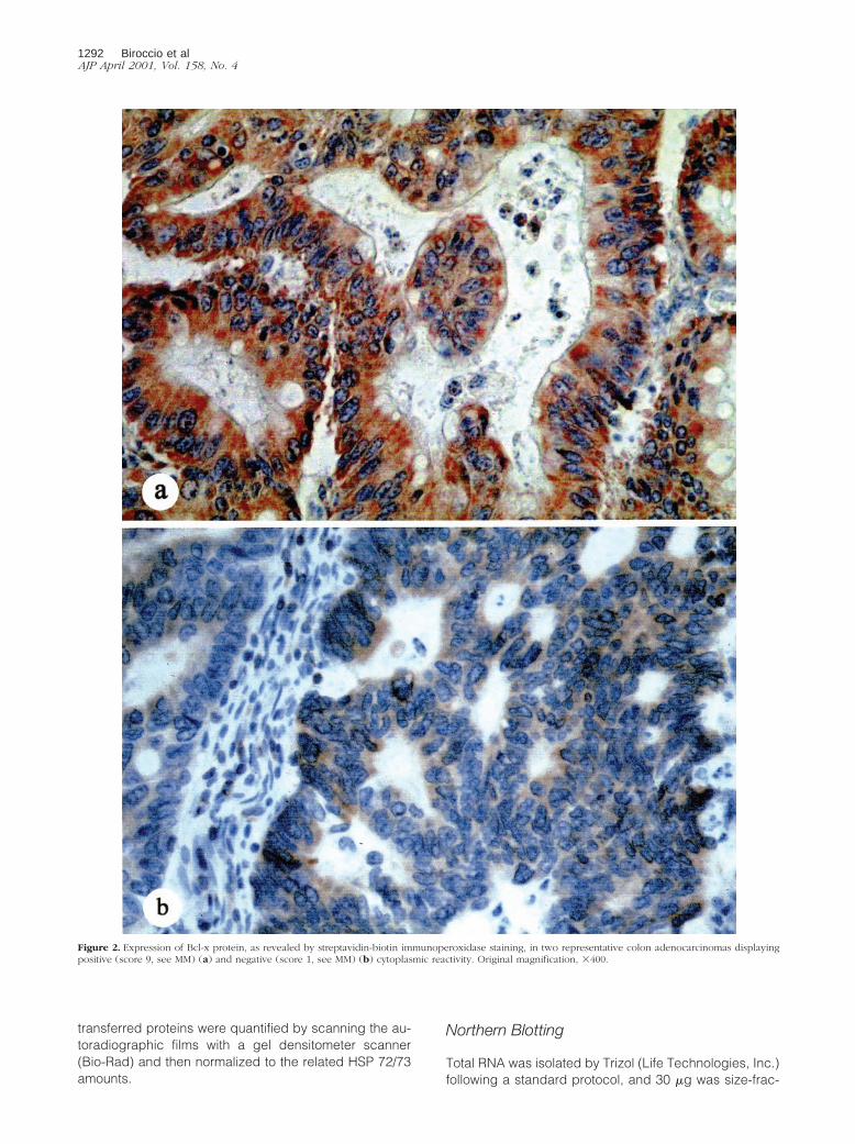
Figure 2. Expression of Bcl-x protein, as revealed by streptavidin-biotin immunoperoxidase staining, in two representative colon adenocarcinomas displayingpositive (score 9, see MM) (a) and negative (score 1, see MM) (b) cytoplasmic reactivity. Original magnification, 3400.
1292 Biroccio et alAJP April 2001, Vol. 158, No. 4

tionated on denaturing formaldehyde agarose gel, blot-ted onto nylon filter, and hybridized with a 920-bp frag-ment cut from a human bcl-xL plasmid. The filter wasexposed to an autoradiographic film for 3 days. Afterstripping (0.1% sodium dodecyl sulfate for 5 minutes at100°C) the filter was reprobed with the glyceraldehyde-3-phosphate dehydrogenase probe to assess equiva-lence of RNA loading and transfer.
In Vitro and In Vivo Cell Growth
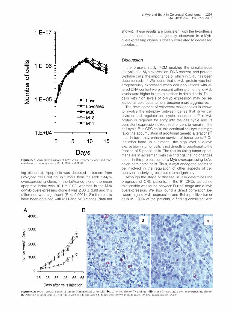
The growth of LoVo parental line, one neo clone (LoVo/neo) and three c-Myb-overexpressing clones (M11, M18,and M30) was assessed by seeding 8 3 104 cells in60-mm Petri dishes (Nunc, Mascia Brunelli, Milano, Italy).Cell counts (Coulter Counter model ZM, Kontron) andviability (trypan blue dye exclusion) were determineddaily, from day 1 to day 15 of culture.
CD-1 male nude (nu/nu) mice, 6 to 8 weeks old and 22 to24 g in body weight, were purchased from Charles RiverLaboratories, Calco, Italy. All procedures involving animalsand their care have been previously described and were inaccordance with institutional guidelines in compliance withnational and international laws and policies.34 Mice wereinjected intramuscularly with parental LoVo cells, LoVo/neo
clone or each of c-Myb-overexpressing clones at 2 3 106 oftumor cells/mouse. Eight mice for each group were usedand tumor mass was monitored daily.34
Terminal Deoxynucleotide Transferase-Mediated dUTP Nick-End Labeling (TUNEL)
Apoptotic activity on sections of the murine tumors wasanalyzed by TUNEL with a commercial kit (ApopDetekin situ apoptosis detection kit; Enzo Diagnostic, NewYork, NY), following the manufacturer’s instructions.Briefly, formalin-fixed, paraffin-embedded tissue sec-tions (4 mm) were deparaffinized with xylene andstripped of proteins by incubation with 25 mg/ml pro-teinase K for 1 hour at room temperature before theTUNEL reaction. The incorporated Bio-16-dUTP wasthen stained with streptavidin-biotinylated horseradishperoxidase complex, AEC (3-amino-9-ethyl carbazole)and hematoxylin. Apoptotic cells had red nuclear stain-ing. Omission of the terminal deoxynucleotide trans-ferase enzyme in the TUNEL reaction was used asnegative control and resulted in no staining.
Three mice for each group were sacrificed at day 50after cell injection. The percentage of apoptotic cells
Figure 3. DFS and OS (81 cases) for patients with CRC categorized according to the c-Myb (a and b) and Bcl-x expression (c and d). Survival curves weregenerated according to the Kaplan-Meier method; statistical comparisons were made using the log-rank method.
c-Myb and Bcl-x in Colorectal Carcinoma 1293AJP April 2001, Vol. 158, No. 4

was determined by light microscopic examination ofsections at 3400. For each slide, five high-power fieldsof nonnecrotic tumor areas were examined and 300cells in each field were counted. For each tumor, fourdifferent sections were evaluated. Results were ex-pressed as mean 6 SD of the samples analyzed. Theapoptotic index was calculated as number of apoptoticcells per 100 cells.
Statistical Analyses
Association between variables was tested by the Pear-son’s chi-square test. The TUNEL results were expressedas mean 6 SD of the samples analyzed. Student’s t-testwas used to evaluate statistical significance. Disease-free survival (DFS) and overall survival (OS) curves wereestimated by the Kaplan-Meier product-limit method; thelog-rank test was used to assess differences betweensubgroups. A P value , 0.05 was considered significant.The BMDP statistical package program (BMDP, Los An-geles, CA) was used for analysis.
Results
Expression of c-Myb Oncoprotein in ColorectalTumor and Its Relationship with Biological andClinicopathological Variables
c-Myb levels were monitored in 91 colorectal tumor sam-ples using FCM. This methodology permits the simulta-neous evaluation of more than one biological parameterand might allow to accurately calculate the percentage ofc-Myb-positive cells in heterogeneous cell populations.Thus, c-Myb expression, DNA ploidy, and percent S-phase were determined in each tumor sample and com-pared to those of the corresponding normal mucosa.
Two-parameter FCM analysis (c-Myb expression andDNA content) in four representative samples of normalmucosa and primary tumor demonstrates that c-Myb isdifferentially expressed in the same neoplastic samplecontaining diploid and aneuploid cell populations (Figure1). The expression of c-Myb was relatively low in cellswith a diploid DNA content (window 1), whereas an in-crease in c-Myb positivity was observed in cells with
Figure 4. DFS and OS (54 cases obtained by excluding the Dukes’ D patients) for patients with CRC categorized according to the c-Myb (a and b) and Bcl-xexpression (c and d). Survival curves were generated according to the Kaplan-Meier method; statistical comparisons were made using the log-rank method.
1294 Biroccio et alAJP April 2001, Vol. 158, No. 4

aneuploid DNA content (window 2). Because these re-sults suggested that there was a relationship betweenaneuploidy and c-Myb overexpression, the associationbetween c-Myb expression, ploidy, and percent S-phasecells was evaluated in each tumor sample.
Table 1 shows the statistical evaluation of the relation-ship between clinicopathological and biological vari-ables. Tumor cell differentiation and Dukes’ stage did notcorrelate with c-Myb overexpression, whereas a signifi-cant relationship between c-Myb overexpression and an-euploidy (P 5 0.0003) was observed. On the other hand,expression of c-Myb, a cell cycle-related gene, did notcorrelate with the percentage of cells in S phase (P 50.4534). In fact, the number of cases with more or less20.1% S-phase cells was similar regardless the degree ofc-Myb expression.
On the basis of previous results demonstrating a rela-tionship between Bcl-2 and c-Myb,22,23 and data thatreport an elevated expression of Bcl-x in primaryCRC,29,30 we studied the correlation between c-Myb,Bcl-2, and Bcl-x in this series of patients. Bcl-2 and Bcl-ximmunoreactivity was determined by immunohistochem-
istry on paraffin-embedded sections (Table 1). Althoughthe expression of c-Myb did not correlate with that ofBcl-2 (P 5 0.2549), it was directly related to that of Bcl-x(P , 0.0001). In fact the majority of tumors expressinghigh c-Myb levels were positive for Bcl-x (;87%, Figure2a), whereas tumors expressing low c-Myb levels werenegative for Bcl-x (;93%, Figure 2b). In addition, tumorspositive for Bcl-x staining were associated with aneuploidCRC (P 5 0.0115) and Dukes’ stage. Pearson chi-squaretest revealed a borderline correlation between Bcl-x pos-itivity and tumor grading.
Prognostic Role of c-Myb and Bcl-x
To evaluate the prognostic impact of c-Myb and Bcl-x,patients who died within 30 days after surgery or thosewho died from noncancer-related causes were excludedfrom DFS and OS Kaplan-Meier analysis. This selectionpermitted us to analyze 81 cases, with a median fol-low-up of 50 months (range, 24 to 77 months), for DFSand OS, respectively. In this series of patients, 33 re-
Figure 5. DFS and OS (71 cases) for patients with CRC categorized according to the combinations of c-Myb and Bcl-x expression (a and b) and DFS and OS (49cases obtained by excluding Dukes’ D patients) for patients with CRC categorized according to the combinations of c-Myb and Bcl-x expression (c and d). Survivalcurves were generated according to the Kaplan-Meier method; statistical comparisons were made using the log-rank method.
c-Myb and Bcl-x in Colorectal Carcinoma 1295AJP April 2001, Vol. 158, No. 4

lapses were recorded and 30 patients died from cancer-related causes.
Kaplan-Meier curves, stratified for c-Myb and Bcl-xexpression, showed a significantly longer DFS and OS inpatients with low c-Myb expression (Figure 3, a and b)and in patients with Bcl-x-negative tumors (Figure 3, cand d). To better investigate the predictive value of c-Myb and Bcl-x in patients not presenting distant metas-tases, DFS and OS Kaplan-Meier analysis was performedexcluding the Dukes’ D patients. As shown in Figure 4c-Myb low-expressing and Bcl-x-negative patients had asignificant increased DFS and OS benefit compared tothose with high c-Myb expression and Bcl-x-positive.
On the basis of these results, we evaluated the impactof the combination of these two factors on DFS and OS,analyzing the two groups of patients bearing high c-Mybexpression/Bcl-x-positive and low c-Myb expression/Bcl-x-negative tumors. As shown in Figure 5, a and b, theresults obtained in the entire series of patients (Dukes’ Ato D) provide statistically significant evidence that theconcomitant expression of c-Myb and Bcl-x identifies agroup of patients with a higher probability of relapsingand dying. Similar results were observed excludingDukes’ D patients (Figure 5, c and d).
Expression of Bcl-xL Protein and mRNA in LoVoCells and c-Myb Transfectants
To examine the potential relationship between c-Myb andBcl-xL in tumor cells, the human colon cancer cell lineLoVo was transfected with a full-length c-myb cDNAdriven by a constitutively active promoter. LoVo cellswere selected as low c-Myb-expressing cells from apanel of five colon carcinoma lines (Colo205, HT29,LoVo, SW, DLD-1) (data not shown). Figure 6 showsdifferent c-Myb protein levels in the parental LoVo cells,LoVo/neo clone (the vector control) and in some of thetransfectants. Clones M11, M18, and M30, which show a5- to 10-fold increase in c-Myb expression compared tothe LoVo/neo clone, were used for further experiments.
To verify whether c-Myb modulates Bcl-xL protein andmRNA levels, Western and Northern blot analysis of thedifferent cell lines were performed. Figure 7A showsWestern blot analysis of Bcl-xL protein in LoVo/neo con-trol clone and three clones overexpressing c-Myb (M11,M18, and M30). Compared to vector-transfected LoVocells, c-Myb-transfected cells show a twofold to threefoldincrease in Bcl-xL protein. Bcl-xL protein corresponds tothe lower band (29 kd) in the top panel of the figurebecause the top prominent 35 kd is a nonspecific bandrecognized by a nonspecific rabbit IgG (middle panel). Atwofold to threefold increase in bcl-xL mRNA was alsoobserved in M11, M18, and M30 clones (Figure 7B). TheLoVo parental cells showed the same level of Bcl-xL
expression observed in the LoVo/neo clone (data notshown).
Effect of c-Myb Expression on In Vitro and InVivo Growth
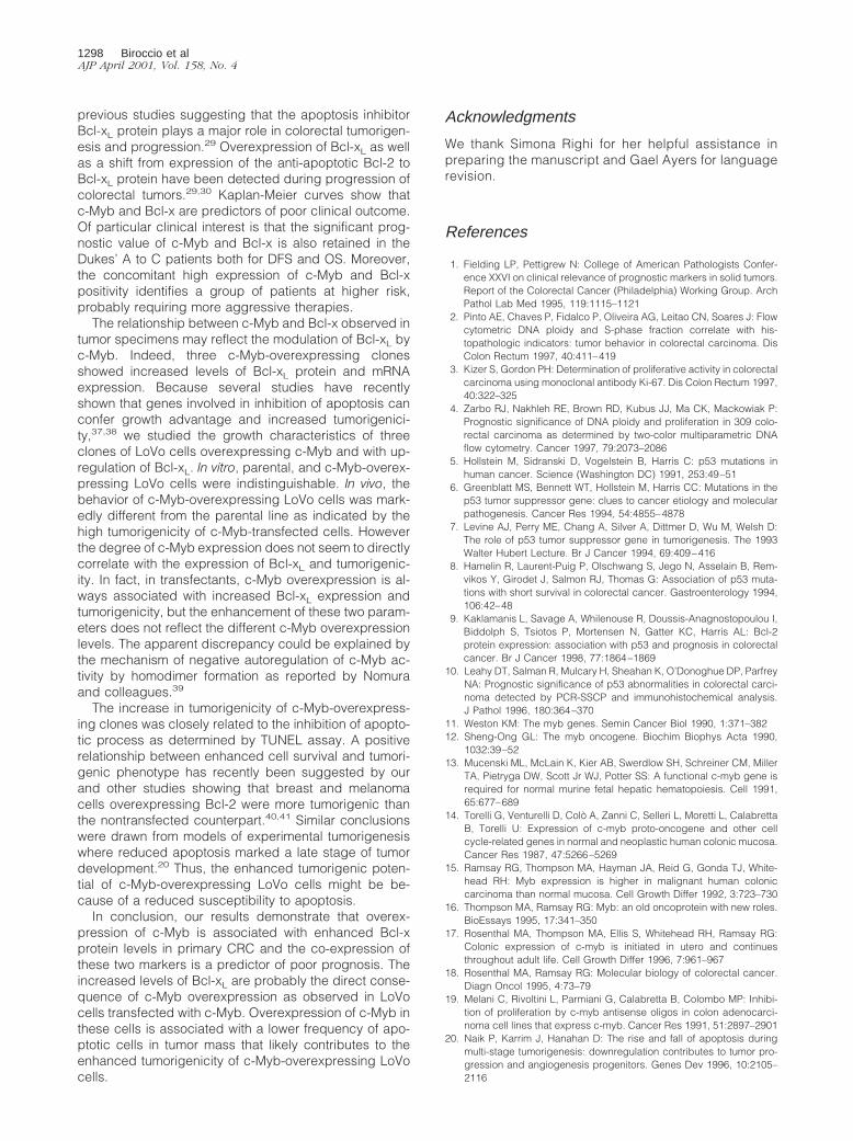
c-Myb-overexpressing LoVo cells were analyzed for invitro and in vivo growth characteristics. The in vitro growthcurves of the c-Myb overexpressing M11, M18, and M30transfectants were similar to those of the parental cellsand the LoVo/neo clone (Figure 8). Similar results wereobtained by performing the growth curves in mediumsupplemented with 1% FCS (data not shown). Thus, over-expression of c-Myb protein did not affect proliferation ofLoVo cells. By contrast, in vivo growth of c-Myb-overex-pressing LoVo cells was different (Figure 9A). The tumormass of the c-Myb-overexpressing clones was largerthan control tumors, even at a late stage of tumor growth;ie, 60 days after cell injection the weight of tumors orig-inating from LoVo and LoVo/neo cells was ;1,200 mgcompared to the 3,000 mg from c-Myb-overexpressingclones. In addition, 75% of mice injected with parentalcells or with the LoVo/neo control clone developed tu-mors, whereas tumors formed in all mice injected with theM11, M18, or M30 c-Myb transfectants.
To assess whether the different tumor growth could berelated to differences in apoptosis rate, a TUNEL assayfor detection of DNA fragmentation caused by apoptosis,was performed 50 days after cell injection. Figure 9Bshows TUNEL staining in tumor section of LoVo/neo con-trol clone (a) and one representative c-Myb-overexpress-
Figure 6. Western blot analysis of c-Myb expression in LoVo parental line,LoVo/neo control clone, and several c-Myb transfectants (M11, M15, M18,and M30). HL60 cells was used as positive control. Expression of HSP 72/73was measured as control of equal proteins loading.
Figure 7. A: Cell lysates from the LoVo/neo clone, and M18, M30, and M11c-Myb-overexpressing clones were loaded on sodium dodecyl sulfate-poly-acrylamide gel electrophoresis, blotted, and incubated with rabbit anti-Bcl-xpolyclonal antibody (top), nonspecific purified rabbit IgG (middle) andmouse anti-HSP mAb (bottom). Expression of HSP 72/73 was measured ascontrol of equal proteins loading. B: Northern blot analysis of bcl-xL expres-sion in LoVo/neo clone, M18, M30, and M11 c-Myb-overexpressing clones.Glyceraldehyde-3-phosphate dehydrogenase was measured as control ofequal RNA loading.
1296 Biroccio et alAJP April 2001, Vol. 158, No. 4
ing clone (b). Apoptosis was detected in tumors fromLoVo/neo cells but not in tumors from the M30 c-Myb-overexpressing clone. In the LoVo/neo clone, the meanapoptotic index was 10.1 6 2.02, whereas in the M30c-Myb-overexpressing clone it was 2.36 6 0.98 and thisdifference was significant (P , 0.0001). Similar resultshave been obtained with M11 and M18 clones (data not
shown). These results are consistent with the hypothesisthat the increased tumorigenicity observed in c-Myb-overexpressing clones is closely correlated to decreasedapoptosis.
Discussion
In the present study, FCM enabled the simultaneousanalysis of c-Myb expression, DNA content, and percentS-phase cells, the importance of which in CRC has beendocumented.2,15 We found that c-Myb protein was het-erogeneously expressed when cell populations with al-tered DNA content were present within a tumor, ie, c-Myblevels were higher in aneuploid than in diploid cells. Thus,cells with high levels of c-Myb expression may be se-lected as colorectal tumors become more aggressive.
The development of colorectal malignancies is knownto involve the interplay between genes that drive celldivision and regulate cell cycle checkpoints.35 c-Mybprotein is required for entry into the cell cycle and itspersistent expression is required for cells to remain in thecell cycle.16 In CRC cells, this continual cell cycling mightfavor the accumulation of additional genetic alterations28
that, in turn, may enhance survival of tumor cells.36 Onthe other hand, in our model, the high level of c-Mybexpression in tumor cells is not directly proportional to thefraction of S-phase cells. The results using tumor speci-mens are in agreement with the findings that no changesoccur in the proliferation of c-Myb-overexpressing LoVocolon carcinoma cells. Thus, c-myb oncogene seems tobe involved in the regulation of other aspects of cellbehavior underlying colorectal tumorigenicity.
Although the stage of disease usually determines theprognosis of CRC patients, in the 91 CRCs tested norelationship was found between Dukes’ stage and c-Myboverexpression. We also found a direct correlation be-tween high c-Myb expression and Bcl-x-positive tumorcells in ;90% of the patients, a finding consistent with
Figure 8. In vitro growth curves of LoVo cells, LoVo/neo clone, and threec-Myb-overexpressing clones (M11, M18, and M30).
Figure 9. A: In vivo growth curves of tumors from injected LoVo cells (F), LoVo/neo clone (ƒ), and M11 (f), M18 (E), M30 (Œ) c-Myb-overexpressing clones.B: Detection of apoptosis (TUNEL) in LoVo/neo (a) and M30 (b) tumor cells grown in nude mice. Original magnification, 3200.
c-Myb and Bcl-x in Colorectal Carcinoma 1297AJP April 2001, Vol. 158, No. 4
previous studies suggesting that the apoptosis inhibitorBcl-xL protein plays a major role in colorectal tumorigen-esis and progression.29 Overexpression of Bcl-xL as wellas a shift from expression of the anti-apoptotic Bcl-2 toBcl-xL protein have been detected during progression ofcolorectal tumors.29,30 Kaplan-Meier curves show thatc-Myb and Bcl-x are predictors of poor clinical outcome.Of particular clinical interest is that the significant prog-nostic value of c-Myb and Bcl-x is also retained in theDukes’ A to C patients both for DFS and OS. Moreover,the concomitant high expression of c-Myb and Bcl-xpositivity identifies a group of patients at higher risk,probably requiring more aggressive therapies.
The relationship between c-Myb and Bcl-x observed intumor specimens may reflect the modulation of Bcl-xL byc-Myb. Indeed, three c-Myb-overexpressing clonesshowed increased levels of Bcl-xL protein and mRNAexpression. Because several studies have recentlyshown that genes involved in inhibition of apoptosis canconfer growth advantage and increased tumorigenici-ty,37,38 we studied the growth characteristics of threeclones of LoVo cells overexpressing c-Myb and with up-regulation of Bcl-xL. In vitro, parental, and c-Myb-overex-pressing LoVo cells were indistinguishable. In vivo, thebehavior of c-Myb-overexpressing LoVo cells was mark-edly different from the parental line as indicated by thehigh tumorigenicity of c-Myb-transfected cells. Howeverthe degree of c-Myb expression does not seem to directlycorrelate with the expression of Bcl-xL and tumorigenic-ity. In fact, in transfectants, c-Myb overexpression is al-ways associated with increased Bcl-xL expression andtumorigenicity, but the enhancement of these two param-eters does not reflect the different c-Myb overexpressionlevels. The apparent discrepancy could be explained bythe mechanism of negative autoregulation of c-Myb ac-tivity by homodimer formation as reported by Nomuraand colleagues.39
The increase in tumorigenicity of c-Myb-overexpress-ing clones was closely related to the inhibition of apopto-tic process as determined by TUNEL assay. A positiverelationship between enhanced cell survival and tumori-genic phenotype has recently been suggested by ourand other studies showing that breast and melanomacells overexpressing Bcl-2 were more tumorigenic thanthe nontransfected counterpart.40,41 Similar conclusionswere drawn from models of experimental tumorigenesiswhere reduced apoptosis marked a late stage of tumordevelopment.20 Thus, the enhanced tumorigenic poten-tial of c-Myb-overexpressing LoVo cells might be be-cause of a reduced susceptibility to apoptosis.
In conclusion, our results demonstrate that overex-pression of c-Myb is associated with enhanced Bcl-xprotein levels in primary CRC and the co-expression ofthese two markers is a predictor of poor prognosis. Theincreased levels of Bcl-xL are probably the direct conse-quence of c-Myb overexpression as observed in LoVocells transfected with c-Myb. Overexpression of c-Myb inthese cells is associated with a lower frequency of apo-ptotic cells in tumor mass that likely contributes to theenhanced tumorigenicity of c-Myb-overexpressing LoVocells.
Acknowledgments
We thank Simona Righi for her helpful assistance inpreparing the manuscript and Gael Ayers for languagerevision.
References
1. Fielding LP, Pettigrew N: College of American Pathologists Confer-ence XXVI on clinical relevance of prognostic markers in solid tumors.Report of the Colorectal Cancer (Philadelphia) Working Group. ArchPathol Lab Med 1995, 119:1115–1121
2. Pinto AE, Chaves P, Fidalco P, Oliveira AG, Leitao CN, Soares J: Flowcytometric DNA ploidy and S-phase fraction correlate with his-topathologic indicators: tumor behavior in colorectal carcinoma. DisColon Rectum 1997, 40:411–419
3. Kizer S, Gordon PH: Determination of proliferative activity in colorectalcarcinoma using monoclonal antibody Ki-67. Dis Colon Rectum 1997,40:322–325
4. Zarbo RJ, Nakhleh RE, Brown RD, Kubus JJ, Ma CK, Mackowiak P:Prognostic significance of DNA ploidy and proliferation in 309 colo-rectal carcinoma as determined by two-color multiparametric DNAflow cytometry. Cancer 1997, 79:2073–2086
5. Hollstein M, Sidranski D, Vogelstein B, Harris C: p53 mutations inhuman cancer. Science (Washington DC) 1991, 253:49–51
6. Greenblatt MS, Bennett WT, Hollstein M, Harris CC: Mutations in thep53 tumor suppressor gene: clues to cancer etiology and molecularpathogenesis. Cancer Res 1994, 54:4855–4878
7. Levine AJ, Perry ME, Chang A, Silver A, Dittmer D, Wu M, Welsh D:The role of p53 tumor suppressor gene in tumorigenesis. The 1993Walter Hubert Lecture. Br J Cancer 1994, 69:409–416
8. Hamelin R, Laurent-Puig P, Olschwang S, Jego N, Asselain B, Rem-vikos Y, Girodet J, Salmon RJ, Thomas G: Association of p53 muta-tions with short survival in colorectal cancer. Gastroenterology 1994,106:42–48
9. Kaklamanis L, Savage A, Whilenouse R, Doussis-Anagnostopoulou I,Biddolph S, Tsiotos P, Mortensen N, Gatter KC, Harris AL: Bcl-2protein expression: association with p53 and prognosis in colorectalcancer. Br J Cancer 1998, 77:1864–1869
10. Leahy DT, Salman R, Mulcary H, Sheahan K, O’Donoghue DP, ParfreyNA: Prognostic significance of p53 abnormalities in colorectal carci-noma detected by PCR-SSCP and immunohistochemical analysis.J Pathol 1996, 180:364–370
11. Weston KM: The myb genes. Semin Cancer Biol 1990, 1:371–38212. Sheng-Ong GL: The myb oncogene. Biochim Biophys Acta 1990,
1032:39–5213. Mucenski ML, McLain K, Kier AB, Swerdlow SH, Schreiner CM, Miller
TA, Pietryga DW, Scott Jr WJ, Potter SS: A functional c-myb gene isrequired for normal murine fetal hepatic hematopoiesis. Cell 1991,65:677–689
14. Torelli G, Venturelli D, Colo A, Zanni C, Selleri L, Moretti L, CalabrettaB, Torelli U: Expression of c-myb proto-oncogene and other cellcycle-related genes in normal and neoplastic human colonic mucosa.Cancer Res 1987, 47:5266–5269
15. Ramsay RG, Thompson MA, Hayman JA, Reid G, Gonda TJ, White-head RH: Myb expression is higher in malignant human coloniccarcinoma than normal mucosa. Cell Growth Differ 1992, 3:723–730
16. Thompson MA, Ramsay RG: Myb: an old oncoprotein with new roles.BioEssays 1995, 17:341–350
17. Rosenthal MA, Thompson MA, Ellis S, Whitehead RH, Ramsay RG:Colonic expression of c-myb is initiated in utero and continuesthroughout adult life. Cell Growth Differ 1996, 7:961–967
18. Rosenthal MA, Ramsay RG: Molecular biology of colorectal cancer.Diagn Oncol 1995, 4:73–79
19. Melani C, Rivoltini L, Parmiani G, Calabretta B, Colombo MP: Inhibi-tion of proliferation by c-myb antisense oligos in colon adenocarci-noma cell lines that express c-myb. Cancer Res 1991, 51:2897–2901
20. Naik P, Karrim J, Hanahan D: The rise and fall of apoptosis duringmulti-stage tumorigenesis: downregulation contributes to tumor pro-gression and angiogenesis progenitors. Genes Dev 1996, 10:2105–2116
1298 Biroccio et alAJP April 2001, Vol. 158, No. 4
21. Selvakumaran M, Lin HK, Sjin RT, Reed JC, Liebermann DA, HoffmanB: The novel primary response gene MyD118 and the proto-onco-genes myb, myc, and bcl-2 modulate transforming growth factor beta1-induced apoptosis of myeloid leukemia cells. Mol Cell Biol 1994,14:2352–2360
22. Salomoni P, Perrotti D, Martinez R, Franceschi C, Calabretta B: Re-sistance to apoptosis in CTLL-2 cells constitutively expressing c-mybis associated with induction of Bcl-2 expression and Myb-dependentregulation of Bcl-2 promoter activity. Proc Natl Acad Sci USA 1997,94:3296–3301
23. Thompson MA, Rosenthal MA, Ellis SL, Friend A, Zorbas M, White-head R, Ramsay R: c-Myb down-regulation is associated with coloncell differentiation, apoptosis, and decrease Bcl-2 expression. Can-cer Res 1998, 58:5168–5175
24. Zorbas M, Sicurella C, Bertoncello I, Venter D, Ellis S, Mucenski ML,Ramsay RG: c-Myb is critical for murine colon development. Onco-gene 1999, 18:5821–5830
25. Sinicrope FA, Hart J, Michelassi F, Lee JJ: Prognostic value of bcl-2oncoprotein expression in stage II colon carcinoma. Clin Cancer Res1995, 1:1103–1110
26. Buglioni S, D’Agnano I, Cosimelli M, Vasselli S, D’Angelo C, TedescoM, Zupi G, Mottolese M: Evaluation of multiple bio-pathological fac-tors in colorectal adenocarcinomas: independent prognostic role ofp53 and Bcl-2. Int J Cancer 1999, 84:545–552
27. Bosari S, Monechini L, Graziani D, Lee AKC, Murray JJ, Coggi G,Viale G: Bcl-2 oncoprotein in colorectal hyperplastic adenomas andadenocarcinomas. Hum Pathol 1995, 26:534–540
28. Fearon ER, Vogelstein B: A genetic model for colorectal tumorigen-esis. Cell 1990, 61:759–767
29. Krajewska M, Moss SF, Krajewsky S, Song K, Holt PR, Reed JC:Elevated expression of Bcl-x and reduced Bak in primary colorectaladenocarcinomas. Cancer Res 1996, 56:2422–2427
30. Maurer CA, Friess H, Buhler SS, Wahl BR, Graber H, Zimmermann A,Buchler MW: Apoptosis inhibiting factor Bcl-xL might be the crucialmember of the Bcl-2 gene family in colorectal cancer. Dig Dis Sci1998, 43:2641–2648
31. Ramsay R, Ishii S, Nishina Y, Soe G, Gonda T: Characterization ofalternate and truncated forms of murine c-myb proteins. OncogeneRes 1989, 4:259–269
32. Sinicrope FA, Ruan SB, Cleary KR, Stephens C, Lee JJ, Levin B: bcl-2and p53 oncoprotein expression during colorectal tumorigenesis.Cancer Res 1995, 55:237–241
33. Del Bufalo D, Biroccio A, Soddu S, Laudonio N, D’Angelo C, SacchiA, Zupi G: Lonidamine induces apoptosis in drug-resistant cellsindependently of the p53 gene. J Clin Invest 1996, 98:1165–1173
34. Citro G, D’Agnano I, Leonetti C, Perini R, Bucci B, Zon G, CalabrettaB, Zupi G: c-myc antisense oligodeoxynucleotides enhance the effi-cacy of cisplatin in melanoma chemotherapy in vitro and in vivo innude mice. Cancer Res 1998, 58:283–289
35. Taylor D, Badiani P, Weston K: A dominant interfering Myb mutantcauses apoptosis in T cells. Genes Dev 1996, 10:2732–2744
36. Frampton J, Ramquist T, Grafm T: V-Myb of E26 leukemia virusupregulates Bcl-2 and suppresses apoptosis in myeloid cells. GenesDev 1996, 10:2720–2731
37. Oka K, Qi B, Yutsudo M: Tumorigenic conversion resulting frominhibition of apoptosis in a nontumorigenic HeLa-derived hybrid cellline. Cancer Res 1999, 59:1816–1819
38. Hashimoto S, Koji T, Kohara N, Kanematsu T, Nakane PK: Frequencyof apoptosis relates inversely to invasiveness and metastatic activityin human colorectal cancer. Virchows Arch 1997, 431:241–248
39. Nomura T, Sakai N, Sarai A, Sudo T, Kanei-Ishii C, Ramsay RG, FavierD, Gonda TJ, Ishii S: Negative autoregulation of c-Myb activity byhomodimer formation through the leucine zipper. J Biol Chem 1993,268:21914–21923
40. Del Bufalo D, Biroccio A, Leonetti C, Zupi G: Bcl-2 overexpressionenhances the metastatic potential of a human breast cancer line.FASEB J 1997, 11:947–953
41. Takaoka A, Adachi M, Okuda H, Sato S, Yawata A, Hinoda Y,Takayama S, Reed JC, Imai K: Anti-cell death activity promotespulmonary metastasis of melanoma cells. Oncogene 1997, 14:2941–2977
c-Myb and Bcl-x in Colorectal Carcinoma 1299AJP April 2001, Vol. 158, No. 4
Copyright © 2022 FDOKUMEN