Myb promotes centriole amplification and later steps of the multiciliogenesis program
10
RESEARCH ARTICLE 1 Development 140, 0000-0000 (2013) doi:10.1242/dev.094102 © 2013. Published by The Company of Biologists Ltd INTRODUCTION Cilia are microtubule-based organelles that project from almost every vertebrate cell (Gerdes et al., 2009). A primary cilium is nucleated from the mother centriole of the centrosome during interphase of the cell cycle. It contains nine peripheral and no central microtubule doublets (9+0) and functions in intercellular signaling and mechanosensing (Gerdes et al., 2009). Many cells modify their cilium, generating a wide diversity of cilium types. Cells of the embryonic node form a 9+0 motile cilium that generates fluid flow to determine left-right asymmetry (Nonaka et al., 1998), whereas hair cells of the inner ear form a non-motile cilium with two central microtubules (9+2) that transduces sound (Nayak et al., 2007), and sperm have a 9+2 motile cilium that propels them through the reproductive tract (Fisch and Dupuis Williams, 2011). This diversity of cilium types is reflected in the wide range of human diseases caused by ciliary defects (Davis and Katsanis, 2012). One of the most striking ciliary specializations is the formation of multicilia, in which cells generate hundreds of 9+2 motile cilia, the coordinated beating of which generates fluid flow over the tissue. Multiciliated cells (MCCs) that line the airways are part of an essential defense mechanism that moves inhaled pathogens and debris out of the lungs (Knowles and Boucher, 2002). MCCs are also found in other tissues including the brain ependyma where they promote cerebrospinal fluid flow (Del Bigio, 2010), the fallopian tubes where they propel the egg outward (Hagiwara et al., 2004), and in the skin of Xenopus laevis embryos (Deblandre et al., 1999). Each motile cilium is nucleated by a specialized centriole called the basal body (Sorokin, 1968; Hagiwara et al., 2004). Centrioles along with pericentriolar material form the centrosome, which organizes the interphase microtubule skeleton and mitotic spindle. Centriole duplication is tightly regulated and coordinated with DNA synthesis in the S phase of cycling cells (Hinchcliffe and Sluder, 2001; Tsou and Stearns, 2006). Extraneous centrioles can lead to genomic instability and aberrant cell signaling (Mahjoub and Stearns, 2012). MCCs somehow escape this regulation to generate hundreds of centrioles and basal bodies per cell (Sorokin, 1968), which dock at the apical cell surface and initiate a ciliary axoneme (Fig. 1A). Although the transcriptional program that controls primary cilium formation has begun to be elucidated (Chu et al., 2010; Piasecki et al., 2010; Thomas et al., 2010), much less is known about the transcriptional programs that control multiciliogenesis and other ciliary specializations. The best understood ciliary transcription factor is forkhead box J1 (FOXJ1), which is expressed and required in all cells with motile cilia (Brody et al., 2000; Gomperts et al., 2004; Pan et al., 2007; Yu et al., 2008). Recently, a pathway that controls MCC development was identified in mouse airway epithelium and X. laevis epidermis. Downregulation of Notch signaling in MCC precursor cells (Tsao et al., 2009; Morimoto et al., 2010) leads to induction of multicilin (Mcin; Mcidas – Mouse Genome Informatics), which functions upstream of centriole amplification and induction of Foxj1 (Stubbs et al., 2012). Although MCIN expression is necessary and sufficient for MCC formation 1 Department of Biochemistry and Howard Hughes Medical Institute, Stanford University School of Medicine, Stanford, CA 94305, USA. 2 Department of Pathology, Stanford University School of Medicine, Stanford, CA 94305, USA. 3 Molecular Neurobiology Laboratory, The Salk Institute for Biological Studies, San Diego, CA 92186, USA. 4 Department of Neurological Surgery and Institute for Regeneration Medicine, University of California, San Francisco, San Francisco, CA 94122, USA. 5 Department of Genetics, Stanford University School of Medicine, Stanford, CA 94305, USA. 6 Department of Biology, Stanford University, Stanford, CA 94305, USA. *Author for correspondence ([email protected]) This is an Open Access article distributed under the terms of the Creative Commons Attribution License (http://creativecommons.org/licenses/by/3.0), which permits unrestricted use, distribution and reproduction in any medium provided that the original work is properly attributed. Accepted 8 August 2013 SUMMARY The transcriptional control of primary cilium formation and ciliary motility are beginning to be understood, but little is known about the transcriptional programs that control cilium number and other structural and functional specializations. One of the most intriguing ciliary specializations occurs in multiciliated cells (MCCs), which amplify their centrioles to nucleate hundreds of cilia per cell, instead of the usual monocilium. Here we report that the transcription factor MYB, which promotes S phase and drives cycling of a variety of progenitor cells, is expressed in postmitotic epithelial cells of the mouse airways and ependyma destined to become MCCs. MYB is expressed early in multiciliogenesis, as progenitors exit the cell cycle and amplify their centrioles, then switches off as MCCs mature. Conditional inactivation of Myb in the developing airways blocks or delays centriole amplification and expression of FOXJ1, a transcription factor that controls centriole docking and ciliary motility, and airways fail to become fully ciliated. We provide evidence that MYB acts in a conserved pathway downstream of Notch signaling and multicilin, a protein related to the S-phase regulator geminin, and upstream of FOXJ1. MYB can activate endogenous Foxj1 expression and stimulate a cotransfected Foxj1 reporter in heterologous cells, and it can drive the complete multiciliogenesis program in Xenopus embryonic epidermis. We conclude that MYB has an early, crucial and conserved role in multiciliogenesis, and propose that it promotes a novel S-like phase in which centriole amplification occurs uncoupled from DNA synthesis, and then drives later steps of multiciliogenesis through induction of Foxj1. KEY WORDS: Myb, Multiciliogenesis, Centriole amplification Myb promotes centriole amplification and later steps of the multiciliogenesis program Fraser E. Tan 1 , Eszter K. Vladar 2 , Lina Ma 3 , Luis C. Fuentealba 4 , Ramona Hoh 5,6 , F. Hernán Espinoza 1 , Jeffrey D. Axelrod 2 , Arturo Alvarez-Buylla 4 , Tim Stearns 5,6 , Chris Kintner 3 and Mark A. Krasnow 1, * Development Development ePress. Posted online 18 September 2013
-
Upload
independent -
Category
Documents
-
view
3 -
download
0
Transcript of Myb promotes centriole amplification and later steps of the multiciliogenesis program

RESEARCH ARTICLE 1
Development 140, 0000-0000 (2013) doi:10.1242/dev.094102© 2013. Published by The Company of Biologists Ltd
INTRODUCTIONCilia are microtubule-based organelles that project from almostevery vertebrate cell (Gerdes et al., 2009). A primary cilium isnucleated from the mother centriole of the centrosome duringinterphase of the cell cycle. It contains nine peripheral and no centralmicrotubule doublets (9+0) and functions in intercellular signalingand mechanosensing (Gerdes et al., 2009). Many cells modify theircilium, generating a wide diversity of cilium types. Cells of theembryonic node form a 9+0 motile cilium that generates fluid flowto determine left-right asymmetry (Nonaka et al., 1998), whereashair cells of the inner ear form a non-motile cilium with two centralmicrotubules (9+2) that transduces sound (Nayak et al., 2007), andsperm have a 9+2 motile cilium that propels them through thereproductive tract (Fisch and Dupuis Williams, 2011). This diversityof cilium types is reflected in the wide range of human diseasescaused by ciliary defects (Davis and Katsanis, 2012).
One of the most striking ciliary specializations is the formationof multicilia, in which cells generate hundreds of 9+2 motile cilia,the coordinated beating of which generates fluid flow over the
tissue. Multiciliated cells (MCCs) that line the airways are part ofan essential defense mechanism that moves inhaled pathogens anddebris out of the lungs (Knowles and Boucher, 2002). MCCs arealso found in other tissues including the brain ependyma where theypromote cerebrospinal fluid flow (Del Bigio, 2010), the fallopiantubes where they propel the egg outward (Hagiwara et al., 2004),and in the skin of Xenopus laevis embryos (Deblandre et al., 1999).
Each motile cilium is nucleated by a specialized centriole calledthe basal body (Sorokin, 1968; Hagiwara et al., 2004). Centriolesalong with pericentriolar material form the centrosome, whichorganizes the interphase microtubule skeleton and mitotic spindle.Centriole duplication is tightly regulated and coordinated with DNAsynthesis in the S phase of cycling cells (Hinchcliffe and Sluder, 2001;Tsou and Stearns, 2006). Extraneous centrioles can lead to genomicinstability and aberrant cell signaling (Mahjoub and Stearns, 2012).MCCs somehow escape this regulation to generate hundreds ofcentrioles and basal bodies per cell (Sorokin, 1968), which dock at theapical cell surface and initiate a ciliary axoneme (Fig. 1A).
Although the transcriptional program that controls primary ciliumformation has begun to be elucidated (Chu et al., 2010; Piasecki etal., 2010; Thomas et al., 2010), much less is known about thetranscriptional programs that control multiciliogenesis and otherciliary specializations. The best understood ciliary transcriptionfactor is forkhead box J1 (FOXJ1), which is expressed and requiredin all cells with motile cilia (Brody et al., 2000; Gomperts et al.,2004; Pan et al., 2007; Yu et al., 2008). Recently, a pathway thatcontrols MCC development was identified in mouse airwayepithelium and X. laevis epidermis. Downregulation of Notchsignaling in MCC precursor cells (Tsao et al., 2009; Morimoto et al.,2010) leads to induction of multicilin (Mcin; Mcidas – MouseGenome Informatics), which functions upstream of centrioleamplification and induction of Foxj1 (Stubbs et al., 2012). AlthoughMCIN expression is necessary and sufficient for MCC formation
1Department of Biochemistry and Howard Hughes Medical Institute, StanfordUniversity School of Medicine, Stanford, CA 94305, USA. 2Department of Pathology,Stanford University School of Medicine, Stanford, CA 94305, USA. 3MolecularNeurobiology Laboratory, The Salk Institute for Biological Studies, San Diego, CA92186, USA. 4Department of Neurological Surgery and Institute for RegenerationMedicine, University of California, San Francisco, San Francisco, CA 94122, USA.5Department of Genetics, Stanford University School of Medicine, Stanford, CA94305, USA. 6Department of Biology, Stanford University, Stanford, CA 94305, USA.
*Author for correspondence ([email protected])
This is an Open Access article distributed under the terms of the Creative Commons AttributionLicense (http://creativecommons.org/licenses/by/3.0), which permits unrestricted use, distributionand reproduction in any medium provided that the original work is properly attributed.
Accepted 8 August 2013
SUMMARYThe transcriptional control of primary cilium formation and ciliary motility are beginning to be understood, but little is known aboutthe transcriptional programs that control cilium number and other structural and functional specializations. One of the most intriguingciliary specializations occurs in multiciliated cells (MCCs), which amplify their centrioles to nucleate hundreds of cilia per cell, insteadof the usual monocilium. Here we report that the transcription factor MYB, which promotes S phase and drives cycling of a varietyof progenitor cells, is expressed in postmitotic epithelial cells of the mouse airways and ependyma destined to become MCCs. MYBis expressed early in multiciliogenesis, as progenitors exit the cell cycle and amplify their centrioles, then switches off as MCCs mature.Conditional inactivation of Myb in the developing airways blocks or delays centriole amplification and expression of FOXJ1, atranscription factor that controls centriole docking and ciliary motility, and airways fail to become fully ciliated. We provide evidencethat MYB acts in a conserved pathway downstream of Notch signaling and multicilin, a protein related to the S-phase regulatorgeminin, and upstream of FOXJ1. MYB can activate endogenous Foxj1 expression and stimulate a cotransfected Foxj1 reporter inheterologous cells, and it can drive the complete multiciliogenesis program in Xenopus embryonic epidermis. We conclude that MYBhas an early, crucial and conserved role in multiciliogenesis, and propose that it promotes a novel S-like phase in which centrioleamplification occurs uncoupled from DNA synthesis, and then drives later steps of multiciliogenesis through induction of Foxj1.
KEY WORDS: Myb, Multiciliogenesis, Centriole amplification
Myb promotes centriole amplification and later steps of themulticiliogenesis programFraser E. Tan1, Eszter K. Vladar2, Lina Ma3, Luis C. Fuentealba4, Ramona Hoh5,6, F. Hernán Espinoza1, Jeffrey D. Axelrod2, Arturo Alvarez-Buylla4, Tim Stearns5,6, Chris Kintner3 and Mark A. Krasnow1,*
Dev
elop
men
t
Development ePress. Posted online 18 September 2013

2
(Stubbs et al., 2012), how this novel nuclear protein controlscentriole amplification and later steps in the multiciliogenesisprogram is not known.
Here we identify the product of the myeloblastosis proto-oncogene Myb (c-Myb) as a crucial early transcription factor in themulticiliogenesis program. MYB belongs to a family oftranscription factors involved in cell cycle regulation and progenitorcell proliferation (Ramsay and Gonda, 2008; Lipsick, 2010). Mybl2(b-Myb) is ubiquitously expressed and is required for the G1/Stransition (Bessa et al., 2001), whereas Mybl1 (a-Myb) (Toscani etal., 1997) and Myb show restricted expression. Myb is required forthe self-renewal of hematopoietic stem cells (Lieu and Reddy, 2009)and promotes the proliferation of progenitor/stem cells in the colon(Malaterre et al., 2007) and brain (Malaterre et al., 2008; Ramsayand Gonda, 2008). Misregulation of Myb has been implicated incancers of the blood, gut, lung and many other tissues (Ramsay andGonda, 2008).
In contrast to its role in proliferative progenitor cells, we foundthat Myb is expressed in developing MCCs of the mouse lung justafter they exit the cell cycle and initiate multiciliogenesis.Conditional inactivation of Myb delays or blocks the initiation ofcentriole amplification and FOXJ1 expression, and airways do notbecome fully ciliated. We provide evidence that MYB functions asa transcription factor in a conserved multiciliogenesis programdownstream of Notch signaling and MCIN and upstream of FOXJ1,and we propose a model that reconciles its postmitotic function inthis program with its well-established role in proliferatingprogenitor cells.
MATERIALS AND METHODSMiceTimed pregnant CD1 female mice were obtained from Charles RiversLaboratories. Noon of the day that a vaginal plug was observed wasconsidered day (E) 0.5 of gestation. Mybfl/fl mice, in which exon II is flankedby loxP sites, were crossed to an HPRT-Cre strain (Tang et al., 2002) toobtain a null allele (Myb–) that produces no detectable protein (Bender et al.,2004). We did not detect any differences between Mybfl/fl and Mybfl/– mice(either alone or when crossed to Cre driver alleles) and so used theminterchangeably. The Shh driver line (ShhCre) (Harfe et al., 2004), whichhas a Cre-GFP knock-in at the Shh locus that is active in the lung epitheliumthroughout development (Harris et al., 2006), and the Nkx2.1 driver line(Nkx2.1-CreTg) (Xu et al., 2008), which has an Nkx2.1 BAC transgene thatexpresses Cre in the lung epithelium during development (Tiozzo et al.,2009), have been described. The Cre reporter line R26-mTmG (Muzumdaret al., 2007) labels cells with membrane-bound GFP following Crerecombination. Foxj1 null mice (Foxj1−/−), in which exon I is deleted,produce no detectable mRNA or protein (Brody et al., 2000). All controlanimals were littermates of experimental animals. All procedures involvinganimals were approved by the Institutional Animal Care and Use Committeein accordance with established guidelines for animal care.
Lung fixation and sectioningEmbryonic lungs were dissected, fixed in 4% paraformaldehyde (PFA) pH7 for 1 hour at 4°C, equilibrated in 30% sucrose/PBS for 30 minutes at 4°C,embedded in OCT compound (Sakura Finetek) and stored at −80°C.Postnatal lungs were inflated intratracheally with cold 4% PFA, removedfrom the body cavity, submerged in 4% PFA for 1 hour at 4°C, washed inPBS overnight at 4°C and equilibrated in 30% sucrose/PBS overnight at4°C. Individual lobes were embedded and stored at −80°C. Tissue blockswere sectioned on a Leica CM3050 S cryostat.
Lung in situ hybridization of Myb and other transcription factorsA literature search using PubMed and Google Scholar was conducted of1971 known and predicted mouse transcription factor genes for expressiondata linking any of the genes to the lung (F.E.T., PhD Thesis, Stanford
University, CA, USA, 2011). The lung developmental expression patternsof selected genes were examined by in situ hybridization using probesprepared from sequence-verified I.M.A.G.E. clones (Open Biosystems,Research Genetics); I.M.A.G.E. clone 695394 was used for Myb. Genes notin the I.M.A.G.E. collection were amplified from E15.5 lung and braincDNA (RETROscript Kit, Life Technologies), cloned into pCR4-BLUNT(Life Technologies) and sequence verified. Amplicons containing the insertand flanking RNA polymerase promoters were used as templates for in vitrotranscription of digoxigenin-labeled antisense probes (Roche) (Sambrookand Russell, 2001).
Cryosections (20 μm) of E13.5, E15.5 and E17.5 lungs were mountedon a single Superfrost Plus glass slide (Thermo Fisher Scientific) and storedat −80°C. Mounted sections were thawed, postfixed in 4% PFA, acetylatedfor 10 minutes, rinsed in DEPC-treated PBS and then incubated in DEPC-treated 5× SSC. Sections were pretreated in hybridization solution for2 hours then incubated with probe {400 ng/ml in hybridization solution[50% formamide, DEPC-treated 5× SSC, 4× Denhardt’s solution (50×Denhardt’s solution is 0.1% Ficoll 400, 0.1% polyvinylpyrrolidone, 0.1%BSA, w/v)], 400 ng/ml salmon or herring sperm DNA, 200 ng/ml yeasttRNA in DEPC-treated water} at 58°C for 2 days. Sections were washedthree times for 1 hour each at 65°C in 0.2× SSC, then incubated withalkaline phosphatase-conjugated anti-digoxigenin Fab fragments diluted1:5000 in 1% Blocking Reagent (Roche) at 4°C overnight. After washing,sections were incubated with NBT/BCIP (Roche) diluted 1:50 in APreaction buffer (0.1 M Tris-Cl pH 9.5, 50 mM MgCl2, 0.1 M NaCl) for upto 6 hours, mounted in Aqua-Poly/Mount (Polysciences) and visualizedunder brightfield optics on a Zeiss Axiophot microscope equipped with anAxioCam HRC camera.
Lung immunostainingImmunostaining was performed on 10 μm cryosections of E11.5 throughE17.5 lungs. Tissue sections were washed in PBST (PBS with 0.1% Tween20), blocked for 1 hour with 10% goat serum in PBS, and incubated withprimary antibodies diluted in 1.5% goat serum at 4°C overnight. Afterwashing, Alexa Fluor-conjugated secondary antibodies (Life Technologies)diluted 1:500 in 1.5% goat serum were applied for 1 hour at roomtemperature. Slides were mounted with Vectashield containing DAPI(Vector Laboratories) for nuclear labeling. To quench endogenous reporters,stained tissues from transgenic animals carrying the mTmG reporter wereascended and descended through a graded methanol/PBS series aftersecondary antibody incubations. Images were captured on a Leica TCS SP2SE confocal microscope.
Primary antibodies were mouse monoclonal anti-MYB (clone D-7, SantaCruz Biotechnology; used at 1:500 dilution), mouse monoclonal anti-FOXJ1 (clone 2A5, eBioscience; 1:500), chicken anti-GFP (Abcam,ab13970; 1:500), rabbit anti-RFP (Rockland, Gilbertsville, PA, USA, 600-401-379; 1:250), mouse monoclonal anti-Ki67 (BD Biosciences, 550609;1:500), rabbit anti-Ki67 (Vector Laboratories, VP-K451; 1:500), mousemonoclonal anti-acetylated tubulin (Sigma, T6793; 1:1000), rat anti-cadherin 1 (Life Technologies, 13-1900; 1:1000) and mouse monoclonalanti-pericentrin (BD Biosciences, 611814; 1:500). Isotype-specific anti-mouse secondary antibodies were used to simultaneously detect multiplemouse monoclonal primary antibodies. Cells were counted with CellProfiler(Carpenter et al., 2006).
Mouse tracheal epithelial cell cultureMouse tracheal epithelial cell (MTEC) cultures were prepared as described(You et al., 2002; Vladar and Stearns, 2007). Briefly, epithelial cellsobtained from adult mouse tracheas by Pronase (Roche) digestion weregrown to confluence on Transwell-Clear filters (Corning) at 37°C/5% CO2,then switched to an air-liquid interface (ALI) to induce differentiation ofMCCs and other tracheal epithelial cell types. Immunostaining wasperformed as described (Vladar and Stearns, 2007) and specimens wereimaged on a Leica TCS SP2 or SP5 confocal microscope. RNA expressionlevels of Myb family transcription factors were determined by DNAmicroarray analysis of FACS-sorted MTEC cultures prepared from FOXJ1-GFP transgenic mice (Hoh et al., 2012). For Notch inhibition, MTECs were
RESEARCH ARTICLE Development 140 (20)
Dev
elop
men
t

treated with 1 μM DAPT (Sigma) from ALI −1 day to ALI +3 days and cellcounts were performed with the Nucleus Counter plug-in in ImageJ (NIH).
For lentivirus-mediated gene transfer into MTECs, epitope-taggedrecombinant MCIN (Stubbs et al., 2012) and other lentiviruses wereprepared and MTECs were infected as described (Vladar and Stearns,2007). The mouse Myb mRNA coding region from pcRM-Myb (from DrTimothy Bender, University of Virginia, Charlottesville, VA, USA) wascloned into pLVX-tdTomato-C1 (Life Technologies) to generate an N-terminal tdTomato-MYB fusion construct that was subcloned intopLentiPGK to create pLenti-td-Tomato-MYB for virus production (Vladarand Stearns, 2007). Cell counts were performed on filters from at least twoindependent infections.
Brain immunostainingDissected brain tissues were incubated with primary and secondaryantibodies in 10% goat serum in PBS/1% Triton X-100 for 48 hours at 4°Cas described (Mirzadeh et al., 2008; Mirzadeh et al., 2010). After staining,ventricular walls were dissected from underlying parenchyma as slivers oftissue 200-300 μm in thickness and mounted. Confocal images werecaptured on a Leica SP5 microscope.
RNA microinjection of Xenopus embryosX. laevis embryos were obtained following in vitro fertilization (Sive et al.,1998). Embryos were injected at the two-cell stage with in vitro synthesizedcapped mRNAs (typically 1-5 ng/embryo) (Turner and Weintraub, 1994)encoding MCIN-HGR, activated Notch (ICD), membrane RFP (mRFP) andHyls1-GFP to mark centrioles as described (Stubbs et al., 2012). X. laevisMyb was inserted into the CS2 vector to generate a myc-tagged Mybconstruct (MT-Myb) (supplementary material Table S1).
To analyze the effects of MYB overexpression, embryos were injected atthe two-cell stage with MT-Myb RNA (0.5 pg/embryo) along with RNAencoding mRFP (0.1 pg/embryo) and Hysl1-GFP (0.5 pg/embryo). Controlembryos were injected with just mRFP and Hyls1-GFP. At stage 28, theembryos were fixed, immunostained with anti-acetylated tubulin antibodyand imaged by confocal microscopy as described (Stubbs et al., 2012). Tworandom fields of the flank of four to seven embryos were imaged and scoredfor MCCs (multiple basal bodies and acetylated tubulin staining), proton-secreting cells (small apical domain) or outer cells (remaining cells).
For quantitative (q) RT-PCR analysis of gene expression, embryos wereinjected at the two-cell stage with ICD RNA or with both ICD (0.2pg/embryo) and MCIN-HGR RNA (0.08 pg/embryo). Animal caps weredissected at stage 10, incubated until stage 11, and then treated withdexamethasone (10 μM) to induce MCIN activity. Total RNA was harvested6 hours later. cDNA templates were generated from 3 μg RNA usingSuperscript III reverse transcriptase (Life Technologies). qPCR reactionswere performed in triplicate using an ABI Prism 7900HT thermal cycler(Life Technologies) and primers for Foxj1 and Myb, and normalized withornithine decarboxylase (Odc) as an internal control (supplementarymaterial Table S2). qPCR data were analyzed with Sequence DetectionSystem (SDS, Applied Biosystems) software.
Cotransfection assayA 1 kb region upstream of the human FOXJ1 translation start site (hg19,chr17:74136502-74137569; nucleotides −876 to +162 with respect to thetranscription initiation site) was cloned by PCR upstream of the codingsequence for firefly luciferase in the pGL4.20 vector (Promega) to createpGL4.20-Foxj1. This fragment includes the −872 to +108 region upstreamof the transcription start site used to generate the Foxj1-GFP mouse line(Ostrowski et al., 2003). Human MYB (clone ID 6069320) and FOXJ1(clone ID 5171637) coding sequences were obtained from Open Biosystems(Thermo Fisher Scientific) and cloned into the pCDH-CMV-MCS-EF1-PuroGreen (pCDH) vector (Systems Bioscience) to generate pCDH-Myband pCDH-Foxj1. The MCIN expression vector was described previously(Stubbs et al., 2012).
HEK293T cells (5×10⁴ cells in 100 μl per well) were transfected usingFuGENE6 (Promega) in a 96-well plate with a total of 50 ng DNA (6:1FuGENE6:DNA ratio) consisting of 15 ng pGL4.20-Foxj1 DNA, a total of32.5 ng transcription factor DNA (empty pCDH vector, pCDH-MYB,
pCDH-MCIN, pCDH-Foxj1) and 2.5 ng plasmid containing a thymidinekinase promoter-driven Renilla luciferase (pRL-TK). Cells were incubatedat 37°C/5% CO2 for 3 days, then lyzed and assayed for firefly and Renillaluciferase activity using the Dual-Glo Luciferase Assay System (Promega)in a Synergy H1 hybrid multi-mode microplate reader (Biotek, Winooski,VT, USA). Three or four independent transfections were performed intriplicate, and the ratio of firefly to Renilla luciferase signal was normalizedto the values obtained for empty pCDH vector.
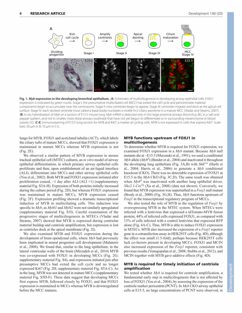
RESULTSMyb is expressed in postmitotic cells of thedeveloping bronchial airway epitheliumIn an in situ hybridization screen for transcription factors expressedduring Mus musculus (mouse) lung development (see Materials andmethods), we found that Myb was expressed in a ‘salt and pepper’pattern in the large diameter proximal airway epithelium, but not inthe small distal epithelial bud tips or surrounding mesenchyme(Fig. 1B). Expression initiated at E13.5 and expanded down thebronchial tree as branching morphogenesis progressed, but neverextended into the undifferentiated portion of the distal airwayepithelium. The density of cells expressing Myb peaked at ~E16.5and began to decline by E17.5 (supplementary material Fig. S1) andwas completely absent by early postnatal life (data not shown).
Myb is expressed in cycling progenitor cells of the blood, colonand brain. Such cell cycle phase-specific expression could accountfor the ‘salt and pepper’ appearance of Myb transcripts in thedeveloping bronchial epithelium. To test this, we labeled E14.5through E17.5 lungs for MYB and Ki67 (MKI67 – Mouse GenomeInformatics), a cell cycle marker that is absent only in quiescent(G0) cells (Scholzen and Gerdes, 2000). Surprisingly, there wasalmost no colocalization between MYB and Ki67 expression (0-2%colocalization, n>98 cells scored at each stage), indicating thatMYB is expressed in cells that have exited the cell cycle (Fig. 1C-E; supplementary material Table S2). Thus, in contrast to othertissues, MYB is expressed in postmitotic cells in the developingbronchial epithelium.
MYB is expressed early and transiently indeveloping MCCsThe MYB expression pattern is reminiscent of markers ofdifferentiating cell types such as Clara cells and MCCs, which areinterspersed in the bronchial epithelium. These cell types appearduring lung development in a proximal-to-distal wave ofdifferentiation, in which cells in the larger proximal airways havebegun differentiation before those in the smaller distal airways. Todetermine whether MYB is expressed in developing secretory Claracells or MCCs, we co-stained E17.5 airways for MYB and eithersecretoglobin 1a1 (SCGB1A1), a canonical Clara cell marker, orFOXJ1, which labels MCCs. MYB expression did not overlap withthat of SCGB1A1 (Fig. 2A). However, most MYB-expressing cellsalso expressed FOXJ1 (Fig. 2B), indicating that MYB is expressedin developing MCCs of the lung.
We compared the temporal expression patterns of MYB andFOXJ1 in the developing bronchial epithelium from E13.5 throughE17.5 (Fig. 2C; supplementary material Fig. S2). Expression of bothMYB and FOXJ1 initiated between E13.5 and E14.5, and thenumber of cells expressing MYB and/or FOXJ1 increased over thenext 2 days. However, whereas the number of FOXJ1-expressingcells continued to increase through E17.5, the number of MYB-expressing cells began to decline (Fig. 2C). This suggests that MYBis expressed early and turns off at later stages of themulticiliogenesis program. Indeed, triple immunostaining of E17.5
3RESEARCH ARTICLEMYB promotes multiciliogenesis
Dev
elop
men
t
4
lungs for MYB, FOXJ1 and acetylated tubulin (ACT), which labelsthe ciliary tufts of mature MCCs, showed that FOXJ1 expression ismaintained in mature MCCs whereas MYB expression is not(Fig. 2E).
We observed a similar pattern of MYB expression in mousetracheal epithelial cell (MTEC) cultures, an in vitro model of airwayepithelial differentiation, in which primary airway epithelial cellsproliferate and then, upon establishment of an air-liquid interface(ALI), differentiate into MCCs and other airway epithelial cells(You et al., 2002). Both MYB and FOXJ1 expression initiated afterproliferation ceased, ~1 day after ALI (ALI +1) (supplementarymaterial Fig. S3A-H). Expression of both proteins initially increasedduring the culture period (Fig. 2D), but whereas FOXJ1 expressionwas maintained in mature MCCs, MYB expression ceased(Fig. 2F). Expression profiling showed a dramatic transcriptionalinduction of MYB in multiciliating cells. This induction wasspecific to Myb, as Mybl1 and Mybl2 were not similarly upregulated(supplementary material Fig. S3I). Careful examination of theprogressive stages of multiciliogenesis in MTECs (Vladar andStearns, 2007) showed that MYB is expressed during centriolarmaterial buildup and centriole amplification, but expression is lostas centrioles dock at the apical membrane (Fig. 2I).
We also examined MYB and FOXJ1 expression during thedevelopment of brain ependymal cells, where Myb had previouslybeen implicated in neural progenitor cell development (Malaterreet al., 2008). We found that, similar to the lung epithelium, in thelateral ventricular walls of the brain (Mirzadeh et al., 2010) MYBwas co-expressed with FOXJ1 in developing MCCs (Fig. 2G;supplementary material Fig. S4), and expression initiated just afterpresumptive MCCs had exited the cell cycle and no longerexpressed Ki67 (Fig. 2H; supplementary material Fig. S5A-C). Asin the lung, MYB was not detected in mature MCCs (supplementarymaterial Fig. S5D-F). These data suggest that developing MCCsfirst express MYB, followed closely by FOXJ1, and that FOXJ1expression is maintained in MCCs whereas MYB is downregulatedbefore the MCC matures.
MYB functions upstream of FOXJ1 inmulticiliogenesisTo determine whether MYB is required for FOXJ1 expression, weexamined FOXJ1 expression in a Myb mutant. Because Myb nullmutants die at ~E15.5 (Mucenski et al., 1991), we used a conditionalMyb allele (Mybfl) (Bender et al., 2004) and inactivated it throughoutthe developing lung epithelium (Fig. 3A,B) with ShhCre (Harfe etal., 2004; Harris et al., 2006) to generate a Myb conditionalknockout (CKO). There was no detectable expression of FOXJ1 atE15.5 in the Myb CKO (Fig. 3C,D). The same result was obtainedwhen Mybfl was inactivated with another lung epithelial driver,Nkx2.1-CreTg (Xu et al., 2008) (data not shown). Conversely, wefound that MYB expression was unperturbed in a Foxj1 null mutant(Brody et al., 2000) (Fig. 3G,H). Thus, Myb functions upstream ofFoxj1 in the transcriptional regulatory program of MCCs.
We also tested the role of MYB in the regulation of Foxj1 byoverexpressing MYB in the MTEC system. When MTECs wereinfected with a lentivirus that expressed a tdTomato-MYB fusionprotein, 44% of infected cells expressed FOXJ1, as compared with13% of cells infected with a control lentivirus that expressed RFPalone (Fig. 4A-C). Thus, MYB is able to induce FOXJ1 expressionin MTECs. MYB also increased the expression of a Foxj1 reportergene in a cotransfection assay in HEK293T cells (Fig. 4D), althoughthe effect was small (1.5-fold), perhaps because HEK293T cellslack co-factors present in developing MCCs. FOXJ1 and MCINalso increased expression of the Foxj1 reporter, consistent withprevious results (Venugopalan et al., 2008; Stubbs et al., 2012), andMCIN together with MYB gave additive effects (Fig. 4D).
MYB is required for timely initiation of centrioleamplificationWe tested whether Myb is required for centriole amplification, afundamental early step in multiciliogenesis that is not affected byloss of FOXJ1 (You et al., 2004), by assessing the expression of thecentriole marker pericentrin (PCNT). In Myb CKO airway epithelialcells at E15.5, no large concentrations of PCNT were observed, in
RESEARCH ARTICLE Development 140 (20)
Fig. 1. Myb expression in the developing bronchial epithelium. (A) Schematic of multiciliogenesis in developing airway epithelial cells. FOXJ1expression is indicated by green nuclei. Stage I: the presumptive multiciliated cell (MCC) has exited the cell cycle and pericentriolar materialcomponents begin to accumulate near the centrosome. Stage II: new centrioles begin to appear. Stage III: centrioles migrate and dock at the apical cellsurface. Stage IV: each docked centriole (now called a basal body) nucleates a motile 9+2 ciliary axoneme in a mature MCC (Vladar and Stearns, 2007).(B) In situ hybridization of Myb on a section of E15.5 mouse lung. Myb mRNA is detected only in the large proximal airways (bronchus, Br), in a ‘salt andpepper’ pattern, and not in smaller, more distal airways (outlined) that have not yet begun to differentiate or in surrounding mesenchyme or bloodvessels (V). (C-E) Immunostaining of E15.5 lung section for MYB and Ki67, a marker of cycling cells. MYB is not expressed in cells that express Ki67. Scalebars: 50 μm in B; 10 μm in C-E.
Dev
elop
men
t

contrast to the amplified centrioles found at this stage in normalMCCs (Fig. 3E,F). Only one or two puncta of PCNT were seen,resembling those in cells with the standard pair of centrioles. Thus,Myb is required for the timely initiation of centriole amplificationduring multiciliogenesis.
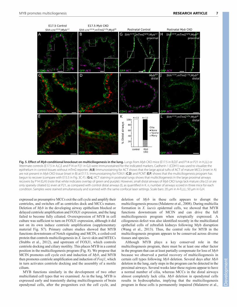
The multiciliogenesis program can partiallyrecover from loss of MybAt E17.5, the major airways are normally well ciliated. When weexamined Myb CKO epithelium at E17.5, however, no mature ciliawere detectable by ACT staining (Fig. 5A,B). This indicates adramatic arrest of the multiciliogenesis program, as observed 2 daysearlier with the early MCC markers (Fig. 3E,F). However, FOXJ1expression (Fig. 5C,D) and multiple centrioles (Fig. 5E,F) were nowdetectable in the large airways at levels indistinguishable from thecontrol, implying that the multiciliogenesis program had begun torecover. When we examined Myb CKO airways at postnatal day (P)14 and P21, we found that, at both stages, large proximal airwayswere ciliated at levels similar to control tissues (Fig. 5G,H).However, the small diameter distal airways were sparsely ciliated,if at all (Fig. 5I-L). Thus, recovery of the multiciliogenesis programin the absence of Myb is incomplete, at least in the distal airways.
Likewise, we found that in MTECs generated from Myb CKO mice,MCC differentiation is reduced early but partially recovers at latertime points (supplementary material Fig. S8).
Conservation of MYB in the multiciliogenesispathwayMcin controls centriole amplification, FOXJ1 expression andmulticiliogenesis (Stubbs et al., 2012). To determine whether Mcincontrols MYB expression, we examined the expression of MYB inMTECs expressing myc-tagged MCIN (MCIN-myc) or myc-taggeddominant-negative MCIN (DN MCIN-myc) (Fig. 6A-C). Theformer causes infected MTECs to become multiciliated, whereasthe latter blocks multiciliogenesis (Stubbs et al., 2012). We foundthat 80% of MCIN-myc-expressing cells expressed MYB, whereasonly 5% of control cells infected with the control lentivirusexpressed MYB (Fig. 6A,D; supplementary material Fig. S6). Inparallel experiments with DN-MCIN-myc, almost no infected cells(<2%) expressed MYB (Fig. 6D). Thus, Myb lies downstream ofMcin in MTECs.
Notch signaling controls Mcin expression in MTECs (Stubbs etal., 2012). To determine whether Myb expression is also controlledby Notch signaling, MYB expression was examined in MTECs
5RESEARCH ARTICLEMYB promotes multiciliogenesis
Fig. 2. MYB is expressed early and transiently during multiciliogenesis in the lung and brain. (A,B) Immunostaining of E17.5 mouse lung sectionsfor MYB and either SCGB1A1 (Clara cells) or FOXJ1 (MCCs). MYB is not detected in SCGB1A1-expressing cells (boxed region, insets in A) but many MYB-positive cells also express FOXJ1 (boxed region, insets in B). (C) Sections of E14.5 through E17.5 lungs were stained for MYB, FOXJ1 and SOX2 and thenumber of epithelial cells (marked by SOX2) expressing MYB only, FOXJ1 only, or both MYB and FOXJ1 were counted. Total SOX2-positive cells scoredwas >1000 per age. (D) Mouse tracheal epithelial cell (MTEC) cultures were immunostained for MYB and FOXJ1 at the times indicated after exposure toan air-liquid interface (ALI) to promote differentiation. The number of cells that were MYB+, FOXJ1+ or double-positive were counted. Similar to in vivo,the FOXJ1+ population continually increased during culturing, whereas the percentage of MYB+ and double-positive cells initially increased and thendeclined. Total cells scored was >100 per time point. (E) Immunostaining of E17.5 lung section for MYB, FOXJ1 and acetylated tubulin (ACT, cilia marker).MYB is seen in developing (upper inset) but not mature (lower inset) MCCs. (F) Immunostaining of ALI +8 days MTEC cultures for MYB and ACT. MYB isnot detected in mature MCCs. Arrowheads (E,F), ciliary tufts of mature MCCs. (G,H) Whole-mount ventricles of P2 mouse brain stained for FOXJ1 andMYB (G) or Ki67 and MYB (H). (I) Stage-specific expression of MYB during multiciliogenesis in MTECs. ALI +6 days MTECs were stained for MYB andpericentrin (PCNT), a centriole marker, and cells were staged as described in Fig. 1A. MYB is detected at stages I and II, before and during centrioleamplification, but not during stages III or IV. Dotted lines show cell outline. Scale bars: 10 μm in A,B,E-H; 5 μm in I.
Dev
elop
men
t

6
treated with the Notch inhibitor DAPT. This increased the numberof MYB-expressing cells at early time points in culture (ALI +1 andALI +2) (supplementary material Fig. S9). We conclude that Myb
lies downstream of Notch and Mcin but upstream of Foxj1 duringMCC differentiation in MTECs (Fig. 8).
MCCs similar to those of mammalian airways line the embryonicepidermis of X. laevis, interspersed with other epidermal cell types(Fig. 7B) (Dubaissi and Papalopulu, 2011). We tested whether Mybfunction is conserved in the X. laevis multiciliogenesis program byinjecting X. laevis embryos at the two-cell stage with mRNAencoding the Notch intracellular domain (ICD), which inhibitsMCC differentiation, or with mRNAs encoding ICD and MCIN-HGR, an inducible form of MCIN that restores MCC differentiation.qPCR demonstrated a 4-fold increase in Myb transcript levels in theICD+MCIN-HGR-injected animal caps as compared with ICD-injected animal caps (Fig. 7A). Thus, as in mouse airways, Myb isdownstream of Mcin in X. laevis multiciliogenesis.
We examined the effects of exogenous MYB in X. laevis byinjecting two-cell stage embryos with Myb mRNA and assessingthe epidermal cell type distribution at stage 28. The epidermis ofembryos injected with Myb mRNA had twice as many MCCs ascontrol, whereas other cell types slightly decreased (Fig. 7B-D).Thus, Myb can drive the complete multiciliogenesis program in X.laevis epidermis.
DISCUSSIONWe have shown that Myb is a crucial early transcription factor inthe multiciliogenesis program of lung development. Myb is
RESEARCH ARTICLE Development 140 (20)
Fig. 3. Effect of Myb deletion on initiation of FOXJ1 expression in thelung. (A-F) Mice carrying a conditional allele of Myb (Mybfl) and a Crerecombination reporter (mTmG) were crossed to mice carrying Shh-Cre orNkx2.1-Cre transgenes to selectively delete Myb from the developingairway epithelium (Myb CKO), and lungs from E15.5 control (Myb+/fl) andMyb CKO mice were immunostained for the proteins indicated. (A,B) MYBimmunostaining confirms loss of MYB expression in Myb CKO tissue. (C,D)FOXJ1 expression is not detected in Myb CKO tissue. (E,F) Largeconcentrations of PCNT, indicative of ciliating cells with amplifiedcentrioles (inset in E), are not observed in Myb CKO tissue (inset in F).PCNT staining of centrosomes (arrowheads) is seen in both control andMyb CKO non-MCCs. (G,H) Lungs from E15.5 Foxj1 control heterozygousand homozygous knockout (Foxj1−/−) mice immunostained for MYB. MYBis expressed normally in the absence of Foxj1. Scale bars: 10 μm.
Fig. 4. Effect of MYB on FOXJ1 expression in MTECs and a HEK293Tcell cotransfection assay. (A,B) MTECs infected with a lentivirusexpressing either a tdTomato-MYB fusion protein (tdT-MYB) or RFP alonewere harvested at ALI +4 days and stained for RFP (A) or tdTomato (B) andFOXJ1. Scale bar: 20 μm. (C) Quantification of the percentage of infectedcells that express FOXJ1. *P<0.0001 by chi-square test. (D) Luciferaseactivity driven by the Foxj1 reporter illustrated (nucleotides −876 to +162with respect to the Foxj1 transcription initiation site) cotransfected intohuman HEK293T cells with pCDH empty vector (-) or pCDH constructsexpressing the indicated proteins. Reporter activity is normalized to thevector-only (pCDH) control. *P<0.05, **P<0.003; n.s., not significant(Student’s t-test). Error bars indicate s.e.m.
Dev
elop
men
t
expressed as presumptive MCCs exit the cell cycle and amplify theircentrioles, and switches off as centrioles dock and MCCs mature.Deletion of Myb in the developing airway epithelium blocked ordelayed centriole amplification and FOXJ1 expression, and the lungfailed to become fully ciliated. Overexpression of MYB in cellculture was sufficient to turn on FOXJ1 expression, although it didnot on its own induce centriole amplification (supplementarymaterial Fig. S7). Primary culture studies showed that MYBfunctions downstream of Notch signaling and MCIN, a coiled-coilprotein that controls multiciliogenesis in X. laevis skin and MTECs(Stubbs et al., 2012), and upstream of FOXJ1, which controlscentriole docking and ciliary motility. This places MYB in a centralposition in the multiciliogenesis program (Fig. 8). We propose thatMCIN promotes cell cycle exit and induction of Myb, and MYBthen promotes centriole amplification and induction of Foxj1, whichin turn activates centriole docking and the assembly of a motilecilium.
MYB functions similarly in the development of two othermulticiliated cell types that we examined. As in the lung, MYB isexpressed early and transiently during multiciliogenesis of brainependymal cells, after the progenitors exit the cell cycle, and
deletion of Myb in these cells appears to disrupt themulticiliogenesis process (Malaterre et al., 2008). During multiciliaformation in X. laevis epidermal cells, we showed that MYBfunctions downstream of MCIN and can drive the fullmulticiliogenesis program when ectopically expressed. Aciliogenesis defect was also identified recently in the multiciliatedepithelial cells of zebrafish kidneys following Myb disruption(Wang et al., 2013). Thus, the central role for MYB in themulticiliogenesis program appears to be conserved across diversetissues and species.
Although MYB plays a key conserved role in themulticiliogenesis program, there must be at least one other factorin the program that can at least partially compensate for loss of Mybbecause we observed a partial recovery of multiciliogenesis incertain cell types following Myb deletion. Several days after Mybdeletion in the lung, early steps in the program can be detected in theproximal airways. Several weeks later these regions appear to havea normal number of cilia, whereas MCCs in the distal airwaysalmost completely lack cilia. Myb deletion in ependymal cellsresults in hydrocephalus, implying that the multiciliogenesisprogram in these cells is permanently impaired (Malaterre et al.,
7RESEARCH ARTICLEMYB promotes multiciliogenesis
Fig. 5. Effect of Myb conditional knockout on multiciliogenesis in the lung. Lungs from Myb CKO mice (E17.5 in B,D,F and P14 or P21 in H,J,L) orlittermate controls (E17.5 in A,C,E and P14 or P21 in G,I) were immunostained for the indicated markers. Cadherin 1 (CDH1) was used to visualize theepithelium in control tissues without mTmG reporter. (A,B) Immunostaining for ACT shows that the large apical tufts of ACT of mature MCCs (inset in A)are not present in Myb CKO tissue (inset in B) at E17.5. Immunostaining for FOXJ1 (C,D) and PCNT (E,F) shows that the multiciliogenesis program hasbegun to recover (compare with E15.5 in Fig. 3C-F). (G-L) ACT staining in postnatal lungs shows that multiciliogenesis in the large proximal airwaysrecovers by P14 (G,H) (note that white indicates overlap of green and purple). However, small distal airways of Myb CKO lungs lack mature cilia (J) or areonly sparsely ciliated (L) even at P21, as compared with control distal airways (I), as quantified in K. n, number of airways scored in three mice for eachcondition. Samples were stained simultaneously and scanned with the same confocal laser settings. Scale bars: 20 μm in A-F,I,J,L; 50 μm in G,H.
Dev
elop
men
t

8
2008). One possibility is that MCIN contributes to thiscompensatory activity because it can induce Foxj1 expression inthe apparent absence of MYB, at least in cotransfection experiments(Fig. 4D, Fig. 8). It will be important to identify the auxiliaryfactor(s) and to determine how MCIN, which lacks a DNA-bindingdomain (Stubbs et al., 2012), can induce Myb and Foxj1 expression,and to identify direct targets of MYB, in addition to Foxj1, in themulticiliogenesis program.
There is an apparent paradox in the proposed role for MYB inpromoting the differentiation of MCCs and its classical function inhematopoietic and other stem and progenitor cells, where MYBpromotes S phase and cell cycling; indeed, loss of Myb typically
leads to progenitor cell differentiation and depletion (Malaterre etal., 2007; Malaterre et al., 2008; Lieu and Reddy, 2009). Thesecontrasting roles of MYB can be reconciled if its role inmulticiliogenesis is actually a variant of its cell cycle role. Duringa normal S phase, both chromosomes and centrioles are duplicated;these processes typically begin at the onset of S phase (Hinchcliffeand Sluder, 2001). Cells in G0 do not generate new chromosomesor centrosomes. We speculate that during multiciliogenesis MYBpromotes an S phase-like state, which we refer to as S*, duringwhich centriole amplification occurs but not the eponymoushallmark of S phase – DNA synthesis. Interestingly, the mousehomologue of MCIN called IDAS (Stubbs et al., 2012) and anotherrelated coiled-coil domain protein named Geminin have both beenimplicated in the regulation of S phase (Pefani et al., 2011). In ourmodel, DNA synthesis must be specifically repressed in S*. Anintriguing candidate for this function is cyclin A1, which is a tissue-restricted homolog of the ubiquitous S-phase cyclin, cyclin A2,which is expressed at unusually high levels in MCCs (Hoh et al.,2012). Cyclin A2 prevents replication complexes from binding toorigins of replication that have already fired (Coverley et al., 2002).Perhaps cyclin A1 functions in a similar manner to inactivatereplication complexes in MCCs.
In the last 15 years, much progress has been made inunderstanding the transcriptional program that drives canonicalprimary cilium formation. RFX3, a transcription factor thatapparently functions in all cilia, drives expression of crucialintraflagellar transport components and controls the expression ofproteins found at the basal body and the transition zone (Chu et al.,2010; Piasecki et al., 2010; Thomas et al., 2010). We propose thatcells deploy specific transcriptional modules that modify this basalciliogenic program to form different types of cilia (supplementarymaterial Fig. S10). Such modules govern cilium number and otherstructural attributes found across multiple cell and tissue types. Forexample, ciliary motility is governed by FOXJ1, which is expressedand required in all cells with motile cilia (Brody et al., 2000; Yu etal., 2008). Our work identifies MYB as a crucial regulator ofcentriole amplification and multiciliogenesis in multiple tissues andspecies. We presume that there is at least one other, as yetunidentified, transcription factor and associated transcriptionalmodule that determines the 9+2 axoneme structure instead of thestandard 9+0. These transcriptional modules may be coupled; forexample, deployment of Myb and the multiplicity module alsoinduces Foxj1 and ciliary motility and a 9+2 axonemal structure
RESEARCH ARTICLE Development 140 (20)
Fig. 6. Effect of the multiciliogenesis regulator multicilin (MCIN) onMYB expression in MTECs. (A-C) MTECs were infected with a controllentivirus expressing GFP (A), a myc-tagged wild-type MCIN (B) ordominant-negative MCIN (DN-MCIN) (C), and stained for MYB and GFP ormyc at ALI +4 days. (D) Quantification of the percentage of infected cellsthat express MYB. *P<0.001 by chi-square test. For examples of infectedcells, see supplementary material Fig. S6. Scale bar: 10 μm.
Fig. 7. Myb acts downstream of Mcin and can promote multiciliogenesis in Xenopus laevis embryonic epidermis. (A) Two-cell stage X. laevisembryos were injected with Notch intracellular domain (ICD) mRNA alone to repress multiciliogenesis or together with MCIN (ICD/MCIN-HGR) toinduce it, and Foxj1 and Myb mRNA levels were measured by qRT-PCR, normalized to Odc mRNA levels as an internal control. Values are mean ± s.d. (B-D) Two-cell stage embryos were injected with control (B) or Myb mRNA (C) and Hysl-GFP mRNA to visualize centrioles and membrane RFP mRNA tovisualize cell boundaries and stained for acetylated tubulin (ACT) to label cilia. Scale bar: 10 μm. (D) Quantification of B and C. Note that the percentageof MCCs is increased in Myb mRNA-injected embryos, whereas the percentage of outer cells (OCs) and proton-secreting cells (PSCs) declines slightly.P<0.05 for all three cell types by Student’s t-test. Error bars indicate s.d. D
evel
opm
ent

(supplementary material Fig. S10). Evolution appears to have‘mixed and matched’ these ciliary diversity modules in differentciliated cells (supplementary material Fig. S10) and then furthermodified some ciliary programs for tissue- or cell type-specificpurposes, such as the sensory cilium in Drosophila neurons(Newton et al., 2012) and the light-sensing cilium of photoreceptors(Gerdes et al., 2009). Elucidating the full transcriptional programsthat control cilium number and diversification will also help us tounderstand and hopefully treat the many distinct ciliopathies thatresult from alterations in these programs.
AcknowledgementsWe thank Dr Jennifer Stubbs (Salk Institute) for initiating the Xenopus Mybexperiments; Dr Gray Camp (Stanford University) for help with bioinformatics;Drs Timothy Bender (University of Virginia) and Steven Brody (WashingtonUniversity) for mouse lines; Dr Peter Lee for help with CellProfiler; andmembers of the M.A.K. lab and Dr Joe Lipsick (Stanford University) for expertadvice and comments on the manuscript.
FundingThis work was supported by the National Institutes of Health (NIH) [ProgenitorCell Biology Consortium grant 5U01HL099995 to M.A.K., R01 GM096021 toC.K., and Cellular and Molecular Biology Training program NIH 5T32GM07276 to F.E.T.] and fellowships from the A. P. Giannini Foundation (E.K.V.),Howard Hughes Medical Institute fellowship of the Helen Hay WhitneyFoundation (L.C.F.), and the Damon Runyon Cancer Research Foundation andthe Parker B. Francis Foundation (F.H.E.). M.A.K. is an investigator of theHoward Hughes Medical Institute. Deposited in PMC for immediate release.
Competing interests statementThe authors declare no competing financial interests.
Author contributionsF.E.T., E.K.V. and M.A.K. conceived and analyzed the experiments. F.E.T. andE.K.V. designed and executed experiments. L.M. and C.K. performed X. laevisexperiments and L.C.F. performed ependymal experiments. R.H. and T.S.performed microarray analysis of MTECs. F.H.E. compiled the annotated list ofmouse transcription factors. F.E.T. and M.A.K. wrote the manuscript. Allauthors discussed results and edited the manuscript. J.D.A. supported thework of E.K.V.; A.A.-B. supported the work of L.C.F.
Supplementary materialSupplementary material available online athttp://dev.biologists.org/lookup/suppl/doi:10.1242/dev.094102/-/DC1
ReferencesBender, T. P., Kremer, C. S., Kraus, M., Buch, T. and Rajewsky, K. (2004).
Critical functions for c-Myb at three checkpoints during thymocytedevelopment. Nat. Immunol. 5, 721-729.
Bessa, M., Joaquin, M., Tavner, F., Saville, M. K. and Watson, R. J. (2001).Regulation of the cell cycle by B-Myb. Blood Cells Mol. Dis. 27, 416-421.
Brody, S. L., Yan, X. H., Wuerffel, M. K., Song, S. K. and Shapiro, S. D. (2000).Ciliogenesis and left-right axis defects in forkhead factor HFH-4-null mice. Am.J. Respir. Cell Mol. Biol. 23, 45-51.
Carpenter, A. E., Jones, T. R., Lamprecht, M. R., Clarke, C., Kang, I. H., Friman,O., Guertin, D. A., Chang, J. H., Lindquist, R. A., Moffat, J. et al. (2006).CellProfiler: image analysis software for identifying and quantifying cellphenotypes. Genome Biol. 7, R100.
Chu, J. S. C., Baillie, D. L. and Chen, N. (2010). Convergent evolution of RFXtranscription factors and ciliary genes predated the origin of metazoans. BMCEvol. Biol. 10, 130.
Coverley, D., Laman, H. and Laskey, R. A. (2002). Distinct roles for cyclins E andA during DNA replication complex assembly and activation. Nat. Cell Biol. 4,523-528.
Davis, E. E. and Katsanis, N. (2012). The ciliopathies: a transitional model intosystems biology of human genetic disease. Curr. Opin. Genet. Dev. 22, 290-303.
Deblandre, G. A., Wettstein, D. A., Koyano-Nakagawa, N. and Kintner, C.(1999). A two-step mechanism generates the spacing pattern of the ciliatedcells in the skin of Xenopus embryos. Development 126, 4715-4728.
Del Bigio, M. R. (2010). Ependymal cells: biology and pathology. ActaNeuropathol. 119, 55-73.
Dubaissi, E. and Papalopulu, N. (2011). Embryonic frog epidermis: a model forthe study of cell-cell interactions in the development of mucociliary disease.Dis. Model. Mech. 4, 179-192.
Fisch, C. and Dupuis-Williams, P. (2011). Ultrastructure of cilia and flagella –back to the future! Biol. Cell 103, 249-270.
Gerdes, J. M., Davis, E. E. and Katsanis, N. (2009). The vertebrate primarycilium in development, homeostasis, and disease. Cell 137, 32-45.
Gomperts, B. N., Gong-Cooper, X. and Hackett, B. P. (2004). Foxj1 regulatesbasal body anchoring to the cytoskeleton of ciliated pulmonary epithelialcells. J. Cell Sci. 117, 1329-1337.
Hagiwara, H., Ohwada, N. and Takata, K. (2004). Cell biology of normal andabnormal ciliogenesis in the ciliated epithelium. Int. Rev. Cytol. 234, 101-141.
Harfe, B. D., Scherz, P. J., Nissim, S., Tian, H., McMahon, A. P. and Tabin, C. J.(2004). Evidence for an expansion-based temporal Shh gradient in specifyingvertebrate digit identities. Cell 118, 517-528.
Harris, K. S., Zhang, Z., McManus, M. T., Harfe, B. D. and Sun, X. (2006). Dicerfunction is essential for lung epithelium morphogenesis. Proc. Natl. Acad. Sci.USA 103, 2208-2213.
Hinchcliffe, E. H. and Sluder, G. (2001). ‘It takes two to tango’: understandinghow centrosome duplication is regulated throughout the cell cycle. Genes Dev.15, 1167-1181.
Hoh, R. A., Stowe, T. R., Turk, E. and Stearns, T. (2012). Transcriptional programof ciliated epithelial cells reveals new cilium and centrosome components andlinks to human disease. PLoS ONE 7, e52166.
Knowles, M. R. and Boucher, R. C. (2002). Mucus clearance as a primary innatedefense mechanism for mammalian airways. J. Clin. Invest. 109, 571-577.
Lieu, Y. K. and Reddy, E. P. (2009). Conditional c-myb knockout in adulthematopoietic stem cells leads to loss of self-renewal due to impairedproliferation and accelerated differentiation. Proc. Natl. Acad. Sci. USA 106,21689-21694.
Lipsick, J. S. (2010). The C-MYB story – is it definitive? Proc. Natl. Acad. Sci. USA107, 17067-17068.
Mahjoub, M. R. and Stearns, T. (2012). Supernumerary centrosomes nucleateextra cilia and compromise primary cilium signaling. Curr. Biol. 22, 1628-1634.
Malaterre, J., Carpinelli, M., Ernst, M., Alexander, W., Cooke, M., Sutton, S.,Dworkin, S., Heath, J. K., Frampton, J., McArthur, G. et al. (2007). c-Myb isrequired for progenitor cell homeostasis in colonic crypts. Proc. Natl. Acad. Sci.USA 104, 3829-3834.
Malaterre, J., Mantamadiotis, T., Dworkin, S., Lightowler, S., Yang, Q.,Ransome, M. I., Turnley, A. M., Nichols, N. R., Emambokus, N. R.,Frampton, J. et al. (2008). c-Myb is required for neural progenitor cellproliferation and maintenance of the neural stem cell niche in adult brain.Stem Cells 26, 173-181.
Mirzadeh, Z., Merkle, F. T., Soriano-Navarro, M., Garcia-Verdugo, J. M. andAlvarez-Buylla, A. (2008). Neural stem cells confer unique pinwheelarchitecture to the ventricular surface in neurogenic regions of the adult brain.Cell Stem Cell 3, 265-278.
Mirzadeh, Z., Doetsch, F., Sawamoto, K., Wichterle, H. and Alvarez-Buylla,A. (2010). The subventricular zone en-face: wholemount staining andependymal flow. J. Vis. Exp. 39, e1938.
9RESEARCH ARTICLEMYB promotes multiciliogenesis
Fig. 8. Proposed molecular pathwaygoverning multiciliogenesis. MYB andother transcriptional regulatorsassociated with each step in the programare shown along with the regulatoryrelationships among them, together withthe Notch signaling pathway thatcontrols initiation of the program. Thedashed line indicates that there is anauxiliary pathway that can partiallybypass the requirement for MYB, at leastin some MCCs.
Dev
elop
men
t

10
Morimoto, M., Liu, Z., Cheng, H. T., Winters, N., Bader, D. and Kopan, R.(2010). Canonical Notch signaling in the developing lung is required fordetermination of arterial smooth muscle cells and selection of Clara versusciliated cell fate. J. Cell Sci. 123, 213-224.
Mucenski, M. L., McLain, K., Kier, A. B., Swerdlow, S. H., Schreiner, C. M.,Miller, T. A., Pietryga, D. W., Scott, W. J., Jr and Potter, S. S. (1991). Afunctional c-myb gene is required for normal murine fetal hepatichematopoiesis. Cell 65, 677-689.
Muzumdar, M. D., Tasic, B., Miyamichi, K., Li, L. and Luo, L. (2007). A globaldouble-fluorescent Cre reporter mouse. Genesis 45, 593-605.
Nayak, G. D., Ratnayaka, H. S. K., Goodyear, R. J. and Richardson, G. P.(2007). Development of the hair bundle and mechanotransduction. Int. J. Dev.Biol. 51, 597-608.
Newton, F. G., zur Lage, P. I., Karak, S., Moore, D. J., Göpfert, M. C. andJarman, A. P. (2012). Forkhead transcription factor Fd3F cooperates with Rfxto regulate a gene expression program for mechanosensory ciliaspecialization. Dev. Cell 22, 1221-1233.
Nonaka, S., Tanaka, Y., Okada, Y., Takeda, S., Harada, A., Kanai, Y., Kido, M.and Hirokawa, N. (1998). Randomization of left-right asymmetry due to lossof nodal cilia generating leftward flow of extraembryonic fluid in mice lackingKIF3B motor protein. Cell 95, 829-837.
Ostrowski, L. E., Hutchins, J. R., Zakel, K. and O’Neal, W. K. (2003). Targetingexpression of a transgene to the airway surface epithelium using a ciliatedcell-specific promoter. Mol. Ther. 8, 637-645.
Pan, J., You, Y., Huang, T. and Brody, S. L. (2007). RhoA-mediated apical actinenrichment is required for ciliogenesis and promoted by Foxj1. J. Cell Sci. 120,1868-1876.
Pefani, D. E., Dimaki, M., Spella, M., Karantzelis, N., Mitsiki, E., Kyrousi, C.,Symeonidou, I. E., Perrakis, A., Taraviras, S. and Lygerou, Z. (2011). Idas, a novel phylogenetically conserved geminin-related protein, binds to geminin and is required for cell cycle progression. J. Biol. Chem. 286, 23234-23246.
Piasecki, B. P., Burghoorn, J. and Swoboda, P. (2010). Regulatory Factor X(RFX)-mediated transcriptional rewiring of ciliary genes in animals. Proc. Natl.Acad. Sci. USA 107, 12969-12974.
Ramsay, R. G. and Gonda, T. J. (2008). MYB function in normal and cancer cells.Nat. Rev. Cancer 8, 523-534.
Sambrook, J. and Russell, D. W. (2001). Molecular Cloning: A Laboratory Manual.Cold Spring Harbor, NY: Cold Spring Harbor Laboratory Press.
Scholzen, T. and Gerdes, J. (2000). The Ki-67 protein: from the known and theunknown. J. Cell. Physiol. 182, 311-322.
Sive, H. L., Grainger, R. M. and Harland, R. M. (1998). Early Development ofXenopus Laevis: A Laboratory Manual. Plainsview, NY: Cold Spring HarborLaboratory Press.
Sorokin, S. P. (1968). Centriole formation and ciliogenesis. Aspen EmphysemaConf. 11, 213-216.
Stubbs, J. L., Vladar, E. K., Axelrod, J. D. and Kintner, C. (2012). Multicilinpromotes centriole assembly and ciliogenesis during multiciliate celldifferentiation. Nat. Cell Biol. 14, 140-147.
Tang, S. H., Silva, F. J., Tsark, W. M. and Mann, J. R. (2002). A Cre/loxP-deletertransgenic line in mouse strain 129S1/SvImJ. Genesis 32, 199-202.
Thomas, J., Morlé, L., Soulavie, F., Laurençon, A., Sagnol, S. and Durand, B.(2010). Transcriptional control of genes involved in ciliogenesis: a first step inmaking cilia. Biol. Cell 102, 499-513.
Tiozzo, C., De Langhe, S., Yu, M., Londhe, V. A., Carraro, G., Li, M., Li, C.,Xing, Y., Anderson, S., Borok, Z. et al. (2009). Deletion of Pten expands lungepithelial progenitor pools and confers resistance to airway injury. Am. J. Respir.Crit. Care Med. 180, 701-712.
Toscani, A., Mettus, R. V., Coupland, R., Simpkins, H., Litvin, J., Orth, J.,Hatton, K. S. and Reddy, E. P. (1997). Arrest of spermatogenesis anddefective breast development in mice lacking A-myb. Nature 386, 713-717.
Tsao, P. N., Vasconcelos, M., Izvolsky, K. I., Qian, J., Lu, J. and Cardoso, W. V.(2009). Notch signaling controls the balance of ciliated and secretory cell fatesin developing airways. Development 136, 2297-2307.
Tsou, M. F. and Stearns, T. (2006). Mechanism limiting centrosome duplicationto once per cell cycle. Nature 442, 947-951.
Turner, D. L. and Weintraub, H. (1994). Expression of achaete-scute homolog 3in Xenopus embryos converts ectodermal cells to a neural fate. Genes Dev. 8,1434-1447.
Venugopalan, S. R., Amen, M. A., Wang, J., Wong, L., Cavender, A. C.,D’Souza, R. N., Akerlund, M., Brody, S. L., Hjalt, T. A. and Amendt, B. A.(2008). Novel expression and transcriptional regulation of FoxJ1 during oro-facial morphogenesis. Hum. Mol. Genet. 17, 3643-3654.
Vladar, E. K. and Stearns, T. (2007). Molecular characterization of centrioleassembly in ciliated epithelial cells. J. Cell Biol. 178, 31-42.
Wang, L., Fu, C., Fan, H., Du, T., Dong, M., Chen, Y., Jin, Y., Zhou, Y., Deng, M.,Gu, A. et al. (2013). miR-34b regulates multiciliogenesis during organformation in zebrafish. Development 140, 2755-2764.
Xu, Q., Tam, M. and Anderson, S. A. (2008). Fate mapping Nkx2.1-lineage cellsin the mouse telencephalon. J. Comp. Neurol. 506, 16-29.
You, Y., Richer, E. J., Huang, T. and Brody, S. L. (2002). Growth anddifferentiation of mouse tracheal epithelial cells: selection of a proliferativepopulation. Am. J. Physiol. 283, L1315-L1321.
You, Y., Huang, T., Richer, E. J., Schmidt, J. E., Zabner, J., Borok, Z. and Brody,S. L. (2004). Role of f-box factor foxj1 in differentiation of ciliated airwayepithelial cells. Am. J. Physiol. 286, L650-L657.
Yu, X., Ng, C. P., Habacher, H. and Roy, S. (2008). Foxj1 transcription factors aremaster regulators of the motile ciliogenic program. Nat. Genet. 40, 1445-1453.
RESEARCH ARTICLE Development 140 (20)
Dev
elop
men
t