CDK5RAP2 Regulates Centriole Engagement and Cohesion in Mice
22
CDK5RAP2 Regulates Centriole Engagement and Cohesion in Mice Jose A. Barrera 1 , Ling-Rong Kao 2 , Robert E. Hammer 3,4 , Joachim Seemann 5 , Jannon L. Fuchs 6 , and Timothy L. Megraw 2,* 1 Department of Pediatrics, The University of Texas Southwestern Medical Center at Dallas, Dallas, TX 75390-9063, USA 2 Department of Biomedical Sciences, College of Medicine, Florida State University, Tallahassee, FL 32306-4300, USA 3 Department of Biochemistry, The University of Texas Southwestern Medical Center at Dallas, Dallas, TX 75390-9051, USA 4 The Cecil and Ida Green Center for Reproductive Biology Sciences, The University of Texas Southwestern Medical Center at Dallas, Dallas, TX 75390-9051, USA 5 Department of Cell Biology, The University of Texas Southwestern Medical Center at Dallas, Dallas, TX 75390-9039, USA 6 Department of Biological Sciences, University of North Texas, Denton, TX 76203, USA SUMMARY Centriole duplication occurs once per cell cycle, ensuring that each cell contains two centrosomes, each containing a mother-daughter pair of tightly engaged centrioles at mitotic entry. Loss of the tight engagement between mother and daughter centrioles appears to license the next round of centriole duplication. However, the molecular mechanisms regulating this process remain largely unknown. Mutations in CDK5RAP2, which encodes a centrosomal protein, cause autosomal recessive primary microcephaly (MCPH) in humans. Here we show that CDK5RAP2 loss of function in mice causes centriole amplification with a preponderance of single, unpaired centrioles and increased numbers of daughter-daughter centriole pairs. These results indicate that CDK5RAP2 is required to maintain centriole engagement and cohesion, thereby restricting centriole replication. Early in mitosis, amplified centrosomes assemble multipolar spindles in CDK5RAP2 mutant cells. Moreover, both mother and daughter centrioles are amplified, and the excess mother centrioles template multiple primary cilia in CDK5RAP2 mutant cells. INTRODUCTION Centrosomes perform diverse and critical functions. Perturbations in centrosome function form the etiological basis for a growing number of human diseases (Nigg and Raff, 2009). The mammalian centrosome consists of a centriole pair surrounded by pericentriolar material (PCM), a proteinaceous matrix that supports microtubule nucleation, © 2010 Elsevier Inc. All rights reserved. * Contact: Tim Megraw [email protected], Phone: 850-645-9271, Fax: 850-644-5781. Publisher's Disclaimer: This is a PDF file of an unedited manuscript that has been accepted for publication. As a service to our customers we are providing this early version of the manuscript. The manuscript will undergo copyediting, typesetting, and review of the resulting proof before it is published in its final citable form. Please note that during the production process errors may be discovered which could affect the content, and all legal disclaimers that apply to the journal pertain. NIH Public Access Author Manuscript Dev Cell. Author manuscript; available in PMC 2011 June 15. Published in final edited form as: Dev Cell. 2010 June 15; 18(6): 913–926. doi:10.1016/j.devcel.2010.05.017. NIH-PA Author Manuscript NIH-PA Author Manuscript NIH-PA Author Manuscript
-
Upload
independent -
Category
Documents
-
view
0 -
download
0
Transcript of CDK5RAP2 Regulates Centriole Engagement and Cohesion in Mice

CDK5RAP2 Regulates Centriole Engagement and Cohesion inMice
Jose A. Barrera1, Ling-Rong Kao2, Robert E. Hammer3,4, Joachim Seemann5, Jannon L.Fuchs6, and Timothy L. Megraw2,*
1Department of Pediatrics, The University of Texas Southwestern Medical Center at Dallas,Dallas, TX 75390-9063, USA2Department of Biomedical Sciences, College of Medicine, Florida State University, Tallahassee,FL 32306-4300, USA3Department of Biochemistry, The University of Texas Southwestern Medical Center at Dallas,Dallas, TX 75390-9051, USA4The Cecil and Ida Green Center for Reproductive Biology Sciences, The University of TexasSouthwestern Medical Center at Dallas, Dallas, TX 75390-9051, USA5Department of Cell Biology, The University of Texas Southwestern Medical Center at Dallas,Dallas, TX 75390-9039, USA6Department of Biological Sciences, University of North Texas, Denton, TX 76203, USA
SUMMARYCentriole duplication occurs once per cell cycle, ensuring that each cell contains two centrosomes,each containing a mother-daughter pair of tightly engaged centrioles at mitotic entry. Loss of thetight engagement between mother and daughter centrioles appears to license the next round ofcentriole duplication. However, the molecular mechanisms regulating this process remain largelyunknown. Mutations in CDK5RAP2, which encodes a centrosomal protein, cause autosomalrecessive primary microcephaly (MCPH) in humans. Here we show that CDK5RAP2 loss offunction in mice causes centriole amplification with a preponderance of single, unpaired centriolesand increased numbers of daughter-daughter centriole pairs. These results indicate thatCDK5RAP2 is required to maintain centriole engagement and cohesion, thereby restrictingcentriole replication. Early in mitosis, amplified centrosomes assemble multipolar spindles inCDK5RAP2 mutant cells. Moreover, both mother and daughter centrioles are amplified, and theexcess mother centrioles template multiple primary cilia in CDK5RAP2 mutant cells.
INTRODUCTIONCentrosomes perform diverse and critical functions. Perturbations in centrosome functionform the etiological basis for a growing number of human diseases (Nigg and Raff, 2009).The mammalian centrosome consists of a centriole pair surrounded by pericentriolarmaterial (PCM), a proteinaceous matrix that supports microtubule nucleation,
© 2010 Elsevier Inc. All rights reserved.*Contact: Tim Megraw [email protected], Phone: 850-645-9271, Fax: 850-644-5781.Publisher's Disclaimer: This is a PDF file of an unedited manuscript that has been accepted for publication. As a service to ourcustomers we are providing this early version of the manuscript. The manuscript will undergo copyediting, typesetting, and review ofthe resulting proof before it is published in its final citable form. Please note that during the production process errors may bediscovered which could affect the content, and all legal disclaimers that apply to the journal pertain.
NIH Public AccessAuthor ManuscriptDev Cell. Author manuscript; available in PMC 2011 June 15.
Published in final edited form as:Dev Cell. 2010 June 15; 18(6): 913–926. doi:10.1016/j.devcel.2010.05.017.
NIH
-PA Author Manuscript
NIH
-PA Author Manuscript
NIH
-PA Author Manuscript
polymerization and stability (Doxsey, 2001; Bornens, 2002; Luders and Stearns, 2007).Centrosomes contain two structurally distinct centrioles, a mature “mother” centriole,distinguished by distal and subdistal appendages, and a “daughter” centriole that lacks theseappendages (Vorobjev and Chentsov Yu, 1982; Paintrand et al., 1992). In the absence ofcentrioles, the PCM becomes unstable and dispersed (Bobinnec et al., 1998). Althoughtypical centrosomes consist of centriole pairs, a single centriole can also assemble PCM.Therefore, centriole numbers define centrosome numbers within cells.
The centriole duplication cycle is tightly regulated to ensure that duplication occurs onlyonce per cell cycle (Tsou and Stearns, 2006a; Azimzadeh and Bornens, 2007; Bettencourt-Dias and Glover, 2007; Nigg, 2007). Deregulation of the duplication cycle can lead tocentrosome amplification, thereby increasing the frequency of multipolar spindles and likelycontributing to aneuploidy due to errors in chromosome segregation (Brinkley, 2001; Pihanand Doxsey, 2003; Nigg, 2006; Ganem et al., 2009). The formation of multipolar spindleswhen centrosomes amplify is offset by a centrosome clustering mechanism that suppressessuch occurrences (Sluder et al., 1997; Quintyne et al., 2005; Basto et al., 2008; Kwon et al.,2008; Yang et al., 2008). The mitotic checkpoint is activated in cells with multiplecentrosomes (Basto et al., 2008; Kwon et al., 2008; Yang et al., 2008; Ganem et al., 2009),yet aberrant microtubule attachments to kinetochores can still occur prior to clustering(Ganem et al., 2009).
Centriole biogenesis is a highly orchestrated process that culminates in the organization oftriplet microtubule blades into an elegant 9-fold rotationally symmetric cylinder(Bettencourt-Dias and Glover, 2007; Strnad and Gonczy, 2008). The proteins andmechanisms involved in restricting centriole duplication to once per cell cycle have recentlybegun to be elucidated (Tsou and Stearns, 2006a; Nigg, 2007; Strnad and Gonczy, 2008).After cell division, a cell inherits a pair of disengaged but cohered centrioles (Nigg, 2007).Centriole cohesion is the tethering of centriole pairs by cohesion fibers during interphase(Bahe et al., 2005; Yang et al., 2006). In G1, the mother centriole initiates formation of theprimary cilium (Ishikawa et al., 2005; Bettencourt-Dias and Glover, 2007). In S-phase, eachcentriole templates the assembly of a single daughter centriole, which grows from itsproximal base and remains tightly bound, or “engaged” until disengagement occurs atmitosis. During G2, the engaged centriole pairs remain tethered by cohesion. Cohesion islost at mitotic onset coincident with centrosome separation in preparation for mitotic spindleassembly (Faragher and Fry, 2003). The mother-daughter centriole pairs, however, remainengaged until anaphase of the ensuing mitosis. At the metaphase to anaphase transition,centriole disengagement requires the activation of the protease separase (Tsou and Stearns,2006b). According to current models, the loss of centriole engagement is the licensing stepthat allows centrioles to undergo a single round of replication (Tsou and Stearns, 2006b, a;Nigg, 2007).
CDK5RAP2 is a resident centrosome protein and an ortholog of Drosophila centrosomin(CNN) (Megraw et al., 2001). Homozygous mutations in CDK5RAP2, microcephalin(MCPH1), abnormal spindle-like microcephaly associated (ASPM), centromere associatedprotein J (SAS4/CPAP/CENPJ), and SCL/TAL1 interrupting locus (STIL) cause autosomalrecessive primary microcephaly (MCPH, [MIM 251200]), a condition characterized by theoverall reduction of brain size (Bond et al., 2002; Jackson et al., 2002; Trimborn et al., 2004;Bond et al., 2005; Kumar et al., 2009). All five of the mapped MCPH genes encodecentrosomal proteins, implicating a critical role for the centrosome in brain development(Bond et al., 2005; Zhong et al., 2005; Zhong et al., 2006; Pfaff et al., 2007). In addition, theessential centrosomal protein pericentrin is linked to Seckel syndrome [MIM 210600] andmicrocephalic osteodysplastic primordial dwarfism type II (MOPD2, [MIM 210720])(Griffith et al., 2008; Rauch et al., 2008). Like MCPH, Seckel syndrome and MOPD2 are
Barrera et al. Page 2
Dev Cell. Author manuscript; available in PMC 2011 June 15.
NIH
-PA Author Manuscript
NIH
-PA Author Manuscript
NIH
-PA Author Manuscript

associated with reduced brain size, suggesting a common role for these centrosome proteinsin related processes during development.
In this study we show that centrioles are amplified in loss-of-function CDK5RAP2 mutantcells. Consequently, CDK5RAP2 mutant mouse embryonic fibroblasts (MEFs) frequentlydisplayed multipolar spindles and were delayed in mitosis. In addition, the excess mothercentrioles in CDK5RAP2 mutant MEFs promoted assembly of multiple primary cilia. Instrong loss-of-function CDK5RAP2 mutant MEFs, centrioles were disengaged and lost thenormal paired configuration. We propose that CDK5RAP2 is required to maintain centrioleengagement and cohesion. As centriole engagement is a key step in licensing centriolereplication, CDK5RAP2 is therefore a negative regulator of centriole licensing. Thus,CDK5RAP2 restricts centriole duplication by maintaining centriole engagement.
RESULTSCentrosomal levels of CDK5RAP2 change with the cell cycle
CDK5RAP2 is a member of the centrosomin family of proteins, conserved amongeukaryotes from yeast to humans. The founding member of this family, centrosomin (CNN)is required for mitotic centrosome function in Drosophila melanogaster (Megraw et al.,2001; Mahoney et al., 2006). The yeast S. pombe ortholog, Mto1p, is similarly required forMTOC functions (Sawin et al., 2004; Venkatram et al., 2004; Zimmerman and Chang,2005). CDK5RAP2 shares homology with two domains in Drosophila CNN, CNN Motifs 1and 2 (Figure 1A). In flies, CNN is essential for the formation of the PCM; it is recruited tocentrosomes at mitotic onset and maintains centrosomal localization throughout mitosis untilits dissociation during cytokinesis (Li and Kaufman, 1996). We examined the subcellularlocalization and dynamics of CDK5RAP2 to investigate its relationship with thecentrosome.
To examine subcellular localization and dynamics of CDK5RAP2 during the cell cycle,antibodies were produced against the amino-terminal end of mouse CDK5RAP2 (Figure1A). Immunostaining showed that CDK5RAP2 is a centrosomal protein (Figure 1B),consistent with previous reports (Bond et al., 2005; Graser et al., 2007; Fong et al., 2008),and localizes to centrosomes independent of microtubules (Figure 2E). GFP-CDK5RAP2also localized to centrosomes in transiently transfected NIH-3T3 mouse fibroblasts (FigureS1). Next, changes in CDK5RAP2 levels at centrosomes during the cell cycle wereexamined in NIH-3T3 cells (Figure 1B). Levels of CDK5RAP2 were relatively low atinterphase centrosomes, increased at mitotic prophase, and remained high throughoutmitosis until telophase, when signal dropped to interphase levels (Figure 1B). WhileCDK5RAP2 is a centriolar resident (Graser et al., 2007; Fong et al., 2008), the results hereshow that CDK5RAP2 accumulates at centrosomes in mitosis, consistent with CDK5RAP2localization to the PCM, which grows at mitosis (Palazzo et al., 2000). Thus, CDK5RAP2 isa centrosomal protein whose centrosomal levels are regulated in a cell cycle-dependentmanner. Because CDK5RAP2 impacts human health, we next sought to examine thefunction of CDK5RAP2 in mammalian cells by generating two mouse models forCDK5RAP2 deficiency.
Generation of CDK5RAP2 mutant miceCDK5RAP2 mutant mice were derived using two Bay Genomics embryonic stem cellclones, RRU031 and RRF465, harboring transposon splice-trap insertions within introns 3and 12 of the CDK5RAP2 locus, respectively. The splice-trap vector used to generate theCDK5RAP2 mutations contains a superior splice site upstream of a “β-Geo” fusion cassette,a fusion between β-galactosidase and neomycin phosphotransferase (Stryke et al., 2003).
Barrera et al. Page 3
Dev Cell. Author manuscript; available in PMC 2011 June 15.
NIH
-PA Author Manuscript
NIH
-PA Author Manuscript
NIH
-PA Author Manuscript

In CDK5RAP2Gt(RRU031)Byg/Gt(RRU031)Byg and CDK5RAP2Gt(RRF465)Byg/Gt(RRF465)Byg
mutant mice (hereafter referred to as CDK5RAP2RRU031/RRU031 andCDK5RAP2RRF465/RRF465), the splice traps result in translation of the first 64 and 435 aminoacids of CDK5RAP2, respectively, fused to β-Geo (Figure 2A-D). The truncated proteinsexpressed from these CDK5RAP2 alleles are similar to the predicted protein products of thehuman CDK5RAP2 mutations, S81X and E385fsX4, which result in truncations of 81 and389 amino acids, respectively (Figures 2A and C) (Bond et al., 2005). Homozygous mutantCDK5RAP2 mice (RRU031 and RRF465) were viable and born at expected Mendelianratios. Female CDK5RAP2RRF465/RRF465 mice were fertile (n=5). Males, however, showedvariable fertility, with 2 out of 6 mice showing infertility (n=6). Neither group ofCDK5RAP2 mutant mice showed evidence of reduced brain size (Figure S2A and data notshown).
We characterized and compared CDK5RAP2 expression between the two mutants usingaffinity-purified CDK5RAP2 antibody. The region used to raise antibodies was retained inthe CDK5RAP2RRF465/RRF465 mutant fusion protein (Figures 1A and 2B). Immunoblots ofwhole cell lysates showed that CDK5RAP2+/+ MEFs expressed full-length protein,heterozygous CDK5RAP2+/RRF465 MEFs expressed full-length and mutant fusion proteins,and homozygous CDK5RAP2RRF465/RRF465 MEFs expressed only the CDK5RAP2 mutantfusion protein (Figure 2B). On the other hand, homozygous CDK5RAP2RRU031/RRU031
MEFs expressed full-length CDK5RAP2 at ~7% of wild type levels (Figure 2D). Therefore,the RRU031 mutation is leaky and hypomorphic. These results reveal differences in therelative strengths of the two splice trap mutations, and establish a mouse mutant model ofMCPH and of the centrosomin family of centrosomal proteins.
The centrosome phenotypes described below support the observations that theCDK5RAP2RRU031/RRU031 mutation is hypomorphic, expressing a low amount of full-lengthCDK5RAP2, and the CDK5RAP2RRF465/RRF465 mutation is more severe.
Centrosome disruptions in CDK5RAP2 mutant cellsTo investigate CDK5RAP2 functions in centrosome structure and regulation we establishedMEF primary cell cultures from embryonic day E14.5 embryos. TheCDK5RAP2RRF465/RRF465 protein was stable (Figure 2B), and retained centrosomelocalization (Figure 2E,F). From these data we conclude that the first 435 amino acids ofCDK5RAP2 are sufficient for centrosomal localization. However, full-length CDK5RAP2and the mutant fusion protein in CDK5RAP2RRF465/RRF465 MEFs exhibited differentpatterns of localization at centrosomes. Full-length CDK5RAP2 localized to centrioles andalso to fibrous projections that emanated from the centrosome (Figure 2E,F). These PCMfibers were not microtubules or microtubule-dependent, as localization of CDK5RAP2 tothese structures persisted upon microtubule disassembly with nocodazole (Figures 2E andS2B). The CDK5RAP2RRF465/RRF465 protein failed to localize to PCM fibers, yet retainedcentriole localization (Figures 2E,F). Thus, the normal PCM architecture of centrosomeswas disrupted in CDK5RAP2RRF465/RRF465 MEFs.
We examined the impact of CDK5RAP2RRF465/RRF465 on the localization of othercentrosome proteins, and found that rootletin, a cohesion fiber protein (Bahe et al., 2005),co-localized with CDK5RAP2 at PCM fibers in CDK5RAP2+/+ MEFs. Again, nocodazoletreatment demonstrated that co-localization of CDK5RAP2 and rootletin at PCM fibers wasnot microtubule-dependent (Figure S2B). However, rootletin localization to PCM fibers wasdisrupted in CDK5RAP2RRF465/RRF465 MEFs and, to a lesser and variable degree, also inCDK5RAP2RRU031/RRU031 MEFs (Figure 2F,G). While rootletin localization to PCM fiberswas completely disrupted in CDK5RAP2RRF465/RRF465 MEFs, the incidence of weakrootletin signal at PCM fibers increased more than 2-fold in CDK5RAP2RRU031/RRU031
Barrera et al. Page 4
Dev Cell. Author manuscript; available in PMC 2011 June 15.
NIH
-PA Author Manuscript
NIH
-PA Author Manuscript
NIH
-PA Author Manuscript

MEFs (CDK5RAP2+/+: 28% vs. CDK5RAP2RRU031/RRU031: 66%) (Figure 2G,H). Theseresults show that CDK5RAP2 regulates assembly of a cohesion fiber protein, consistent withthe requirement for CDK5RAP2 in centrosome cohesion (Figures 2I and S3B and (Graser etal., 2007)). Interestingly, PCM fibers were not prominent in all mouse cells; centrosomes inNIH-3T3 cells did not display these structures as elaborately (Figure 1B). Nevertheless,disruption of rootletin localization shows that centrosome structure is altered in CDK5RAP2mutant MEFs. In contrast, CDK5RAP2 mutant MEFs showed normal centrosomallocalization of cenexin/ODF2, centrobin, centrin, γ-tubulin, pericentrin, TOG, aurora-A, andEB1 (Figures 3A, 5A, 7B and S5A; others not shown).
CDK5RAP2 restricts centriole duplicationCentrosome amplification was a prominent feature of CDK5RAP2RRF465/RRF465 MEFs. Co-staining for γ-tubulin to label PCM and centrin to label centrioles showed thatCDK5RAP2+/+ MEFs contained one or two pairs of centrin puncta, representing normalcentriole complements in G1 or G2, respectively (Figure 3A). In contrast, a high percentageof CDK5RAP2RRF465/RRF465 MEFs had greater than two centrin and γ-tubulin puncta,showing that these cells had amplified centrioles (Figure 3A and see below).
CDK5RAP2RRF465/RRF465 cells had a greater than 4-fold increase in the occurrence of threecentrioles (CDK5RAP2+/+: 2.5% vs. CDK5RAP2RRF465/RRF465: 11.2%), and a greater than5-fold increase in cells with more than four centrioles (CDK5RAP2+/+: 4.2% vs.CDK5RAP2RRF465/RRF465: 22.0%). A reciprocal decrease in cells with two centriolesoccurred in CDK5RAP2RRF465/RRF465 MEFs (CDK5RAP2+/+: 85.0% vs.CDK5RAP2RRF465/RRF465: 49.8%) (Figure 3B). Heterozygous RRF465 MEFs were similarto CDK5RAP2+/+ MEFs; only 2% of cells had greater than four centrioles (data not shown).Therefore, the RRF465 gene trap allele is not a dominant mutation in CDK5RAP2. Overall,33.2% of CDK5RAP2RRF465/RRF465 MEFs, exhibited amplified centrioles, compared to only6.7% of CDK5RAP2+/+ MEFs. Thus, CDK5RAP2RRF465/RRF465 MEFs have an alteredcentriole replication cycle that results in centriole amplification. In contrast, no centrosomeamplification was observed in CDK5RAP2RRU031/RRU031 MEFs, an indication that only asmall fraction of full-length CDK5RAP2 is sufficient to restrict centriole replication (FigureS3A).
Centrosome amplification was also observed in vivo in fetal CDK5RAP2RRF465/RRF465 mice.We counted centrosome numbers in sections of E14.5 fetal brains by immunofluorescencestaining for pericentrin and DNA to identify centrosomes and nuclei, respectively.Centrosomes were amplified in cells of the fetal frontal cortex in vivo compared to wild-typesiblings (Figure 3C), indicating that centrosome amplification occurs in vivo in thedeveloping cerebral cortex of CDK5RAP2RRF465/RRF465 mice.
In CDK5RAP2RRF465/RRF465 MEFs centriole amplification could arise as an indirect effecton the cell cycle. To assess cell cycle block or delay, MEF cell cycle profiles were analyzedusing flow cytometry. Cell cycle profiles from CDK5RAP2+/+ andCDK5RAP2RRF465/RRF465 cultures were indistinguishable (Figure S3C,D), indicating thatcentriole amplification in CDK5RAP2RRF465/RRF465 MEFs is not due to an overt indirecteffect on the cell cycle. Nevertheless, wild type and CDK5RAP2 mutant sibling MEFs haddifferent growth capacities. Whereas CDK5RAP2+/+ MEFs proliferated to passage 15 (P15),in contrast, CDK5RAP2RRF465/RRF465 MEFs slowed or arrested between P8 and P10, andafter P4 a higher percentage of cells were needed to seed a culture. Following P8-P10,CDK5RAP2RRF465/RRF465 MEFs remained viable but would no longer grow to confluence.The underlying cause of this premature senescence and its link to centriole amplification isunclear. However, overall, these data show that CDK5RAP2RRF465/RRF465 MEFs have an
Barrera et al. Page 5
Dev Cell. Author manuscript; available in PMC 2011 June 15.
NIH
-PA Author Manuscript
NIH
-PA Author Manuscript
NIH
-PA Author Manuscript
altered centriole replication cycle, uncoupled from the cell cycle, which results in centrioleamplification.
Centriole amplification is associated with increased nuclear sizeAnother salient phenotype of CDK5RAP2RRF465/RRF465 MEFs was the presence of enlargednuclei (Figure 3A). Frequently, the nuclear diameter was increased approximately 3-fold(Figure S4). Quantification of nuclear size revealed a small but significant increase in theaverage nuclear area of CDK5RAP2RRF465/RRF465 cells (CDK5RAP2+/+: 230.3 μm2 vs.CDK5RAP2RRF465/RRF465: 262 μm2). However, the mean nuclear area was significantlylarger, 528 μm2, among CDK5RAP2RRF465/RRF465 MEFs with amplified centrioles (Figure4A). Since centriole amplification and increased DNA content would coincide if cytokinesisfailed, we measured DNA content between CDK5RAP2+/+ and CDK5RAP2RRF465/RRF465
MEFs. DNA analysis by flow cytometry revealed no increase of polyploid cells inCDK5RAP2RRF465/RRF465 MEFs, nor did we observe an increase of binucleate cells toindicate cytokinesis defects. In addition, we compared the fluorescence signal of the nuclearstain, DAPI, between CDK5RAP2+/+ and CDK5RAP2RRF465/RRF465 MEFs with amplifiedcentrosomes, and compared this to wild-type binucleate cells as a polyploid control.Binucleate cells had approximately 2-fold increase in DAPI signal compared toCDK5RAP2+/+ or CDK5RAP2RRF465/RRF465 MEFs with amplified centrosomes (Figure 4B).Therefore, we conclude that CDK5RAP2RRF465/RRF465 MEFs exhibit nuclear enlargementwithout increased DNA content, and that increased nuclear size correlates with centrioleamplification. While nuclear size correlated with centriole amplification, this was notspecific to CDK5RAP2 mutant cells and was seen in the wild type MEFs upon the relativelyrare centriole amplification event (Figure 4B).
Centrioles lose engagement and cohesion in CDK5RAP2 mutant MEFsIn addition to centriole amplification, CDK5RAP2RRF465/RRF465 MEFs showed asignificantly higher incidence of single centrioles (Figure 3A,D and Table 1). This is incontrast to wild type cells where centrioles are predominantly configured as pairs.Centrosome structure within MEFs was examined with immunofluorescence microscopy,using antibodies against the centriole marker centrin and PCM marker γ-tubulin. Typical ofnormal cells, CDK5RAP2+/+ MEFs had two centrioles in close proximity that eachassembled PCM in G1, and two engaged pairs of centrioles, where each pair assembledPCM in G2 (Figure 3A). In contrast, CDK5RAP2RRF465/RRF465 MEFs contained severaldifferent centriole configurations including paired centrioles, single centrioles and clustersof three or more centrioles (Figure 3A).
We quantified these effects by counting mother and daughter centrioles immunostained forthe mother centriole marker cenexin/ODF2 and γ-tubulin or centrin as general centriolemarkers, and with cenexin plus centrobin as a mother and daughter centriole combination(Figure 5A and S5A). In this analysis, the paired configuration, including one mother andone daughter centriole, was used to describe “normal” centrosomes. The incidence of MEFscontaining one or more singlet centrioles increased more than 4-fold in theCDK5RAP2RRF465/RRF465 mutant relative to wild type (CDK5RAP2+/+: 9.7% vs.CDK5RAP2RRF465/RRF465: 40.8%) (Figure 3D) (Table 1 summarizes all centrioleconfigurations). The presence of singlet centrioles indicated that both centriole engagementand cohesion failed. Normally, late in G2, loss of centrosome cohesion occurs whencentrosomes separate prior to mitotic spindle formation.
A role for CDK5RAP2 in cohesion was previously reported by siRNA knockdown, yet nocentriole amplification or disengagement was observed (Graser et al., 2007). We wonderedwhether CDK5RAP2RRU031/RRU031 MEFs also displayed loss of centriole cohesion since the
Barrera et al. Page 6
Dev Cell. Author manuscript; available in PMC 2011 June 15.
NIH
-PA Author Manuscript
NIH
-PA Author Manuscript
NIH
-PA Author Manuscript

low level of full-length protein in this mutant may mimic siRNA knockdown. Indeed,centrosome splitting increased by more than 70% in CDK5RAP2RRU031/RRU031 MEFs(Figures 2I and S3B). We conclude that RRU031 is a weak loss of function CDK5RAP2mutation due to the low expression level of full-length CDK5RAP2. Moreover, we concludethat the hypomorphic phenotype of CDK5RAP2 is loss of cohesion, whereas a strong loss offunction results in loss of cohesion and also loss of centriole engagement.
CDK5RAP2RRF465/RRF465 MEFs possessed multiple mother centrioles and, sinceCDK5RAP2RRF465/RRF465 MEFs exhibited no irregularities in cell cycle profile (FigureS3C,D), we attributed this to the accumulation of mother centrioles following centrioleamplification (Figure 5A,B). The majority of CDK5RAP2+/+ MEFs contained one (89.3%)or two (8.2%) mother centrioles, reflecting the G1/S and G2 stages of the cell cycle,respectively. In contrast, fewer CDK5RAP2RRF465/RRF465 MEFs contained one mothercentriole (62.5%), while significantly more cells contained two mother centrioles (26.3%).In addition, mother centriole numbers in excess of two were increased more than 4-fold inCDK5RAP2RRF465/RRF465 MEFs (CDK5RAP2+/+: 2.3% vs. CDK5RAP2RRF465/RRF465:10.0%) (Figure 5B). Since the cell cycle was unaffected, the increased number ofCDK5RAP2RRF465/RRF465 cells with two mother centrioles must represent a substantialfraction of cells in G1 with excess mother centrioles. With this consideration, we infer thatapproximately 28.1% of CDK5RAP2RRF465/RRF465 MEFs have amplified mother centrioles.Daughter centriole numbers exceeded mother centrioles (Figure 5C), consistent with ourmodel that centriole engagement is lost and multiple rounds of daughter synthesis occur ineach cell cycle (Figure 3E, see below). Thus, the prevalence of single centrioles andamplified daughter centrioles in CDK5RAP2RRF465/RRF465 cells indicates that CDK5RAP2restricts centriole duplication by maintaining centriole engagement.
Another frequent centriole configuration seen in CDK5RAP2RRF465/RRF465 MEFs was“daughter-daughter” pairs (Table 1). Centriole maturation, monitored here by recruitment ofcenexin/ODF2 to mother centrioles, occurs late in G2 phase (Lange and Gull, 1995).Therefore, the population of “daughter-daughter” centriole pairs, normally seen only in cellstransiting between S and G2, will increase if centriole engagement fails and daughtercentrioles reduplicate (Figure 3E). CDK5RAP2RRF465/RRF465 MEFs showed anapproximately 5-fold increase in daughter-daughter pairs (CDK5RAP2+/+: 2.3% ± 0.4% vs.CDK5RAP2RRF465/RRF465: 11.2% ± 1.2%, mean ± SEM, p<0.05). Thus,CDK5RAP2RRF465/RRF465 centrioles lose engagement and become re-licensed to duplicate,as evidenced not only by the presence of singlet centrioles but also by the increased numbersof daughter-daughter pairs (see model, Figure 3E).
Amplified mother centrioles template ectopic primary cilia in CDK5RAP2RRF465/RRF465
MEFsNew centrioles form in S-phase and mature into mother centrioles in G2 phase of thefollowing cell cycle. Most vertebrate cells contain a single non-motile primary cilium(Bettencourt-Dias and Glover, 2007; Nigg and Raff, 2009). In G1 the mother centriolemigrates to the plasma membrane, becomes a basal body, and templates assembly of theprimary cilium (Ishikawa et al., 2005). Centriole amplification led us to hypothesize that thesupernumerary mother centrioles in CDK5RAP2RRF465/RRF465 MEFs (Figure 5A-C) couldresult in the assembly of ectopic primary cilia.
We asked whether the amplification of mother centrioles in CDK5RAP2RRF465/RRF465 MEFswas associated with changes in the proficiency and frequency of primary cilium assembly.When cultured in serum-deprived medium, many cells assemble a primary cilium uponentering G0, a prolonged resting state. We subjected MEFs to serum starvation andimmunostained with anti-acetylated α-tubulin antibody to label axoneme microtubules, and
Barrera et al. Page 7
Dev Cell. Author manuscript; available in PMC 2011 June 15.
NIH
-PA Author Manuscript
NIH
-PA Author Manuscript
NIH
-PA Author Manuscript

with antibodies to γ-tubulin to label the centrioles/basal bodies (Figure 5D). CDK5RAP2+/+
and CDK5RAP2RRF465/RRF465 MEFs were equally proficient at primary cilium formation(CDK5RAP2+/+: 71.2% vs. CDK5RAP2RRF465/RRF465: 74.0%) (Figure 5E). CDK5RAP2+/+
MEFs formed a single primary cilium templated from one of two interphase centrioles(Figure 5D,F). In contrast, CDK5RAP2RRF465/RRF465 MEFs with amplified centriolesformed multiple primary cilia (Figure 5D,F). The incidence of multiple primary ciliaincreased more than 7-fold in CDK5RAP2RRF465/RRF465 MEFs (CDK5RAP2+/+: 4.7% vs.CDK5RAP2RRF465/RRF465: 33.3%; Figure 5F), and was consistent with the frequency ofcentriole amplification (Figure 3B). Immunostaining for additional cilium markers,polyglutamylated tubulin and polaris/IFT88, axonemal and intraflagellar transportcomponents of primary cilia, respectively, confirmed the assembly of multiple cilia inmutant cells (Figure S5B,C). These data reveal a propensity for mammalian cells to formmore than one primary cilium.
CDK5RAP2RRF465/RRF465 MEFs assemble multipolar spindlesThe presence of supernumerary centrosomes is a potentially dangerous condition at mitosisbecause centrosomes are dominant MTOCs, and therefore have the potential to organizemicrotubules aberrantly into the spindle apparatus, make inappropriate kinetochoreattachments, and assemble multipolar spindles. The transient occurrence of multipolarspindles in cells with amplified centrosomes can cause chromosomal instability by theformation in inappropriate merotelic kinetochore-microtubule attachments prior tocentrosome clustering (Ganem et al., 2009). Thus, spindle multipolarity results in theinappropriate segregation of chromosomes, leading to aneuploidy with increased cell death,but also a heightened propensity for tumorigenesis (Brinkley, 2001; Pihan and Doxsey,2003; Nigg, 2006; Ganem et al., 2009). Centrosome clustering at spindle poles assuresspindle bipolarity in cells with more than two centrosomes (Quintyne et al., 2005; Basto etal., 2008; Kwon et al., 2008; Yang et al., 2008). Multipolar spindles were defined here asone or more MTOC’s positioned greater than 45° away from the major axis of the spindle,as determined from spindle microtubules and the configuration of aligned chromosomes(Figure 6A). We found that CDK5RAP2RRF465/RRF465 MEFs with two centrosomes formedbipolar spindles normally, as did CDK5RAP2+/+ MEFs. However, 51% of mitoticCDK5RAP2RRF465/RRF465 MEFs with supernumerary centrosomes formed multipolarspindles, while the remaining 49% assembled a bipolar, or pseudo-bipolar, spindle (Figure6B). Since ~30% of mutant MEFs had amplified centrioles, this amounts to approximately15% of total mitotic CDK5RAP2RRF465/RRF465 MEFs with multipolar spindles. Thus,centriole amplification in CDK5RAP2RRF465/RRF465 MEFs can lead to multipolar spindleformation. However, aberrant anaphase or telophase stage cells were not observed, anindication that multipolar spindles are resolved into bipolar spindles before anaphase onset.
Centrosome clustering is accomplished during a delay in mitosis due to activation of thespindle checkpoint by amplified centrosomes, allowing the cell time to correct spindleassembly defects (Basto et al., 2008; Kwon et al., 2008; Yang et al., 2008). Using live cellimaging, we observed that the timing of anaphase onset was prolonged by approximately48% in CDK5RAP2RRF465/RRF465 MEFs compared to CDK5RAP2+/+ MEFs (Figure 6C).These results are consistent with the idea that CDK5RAP2RRF465/RRF465 MEFs accomplishspindle bipolarity by prolonging their stay in metaphase through checkpoint activation,providing time for supernumerary centrosomes to cluster.
DiscussionThese data show that CDK5RAP2 maintains centriole engagement and cohesion. Sincecentriole disengagement is the licensing step that initiates centriole replication (Tsou andStearns, 2006b, a; Nigg, 2007), the loss of CDK5RAP2 activity results in centriole
Barrera et al. Page 8
Dev Cell. Author manuscript; available in PMC 2011 June 15.
NIH
-PA Author Manuscript
NIH
-PA Author Manuscript
NIH
-PA Author Manuscript

amplification due to the failure to maintain engagement of mother-daughter pairs.CDK5RAP2 is therefore a negative regulator of centriole licensing. The amplified centriolesin CDK5RAP2RRF465/RRF465 MEFs impact not only mitotic spindle assembly but alsopromote the ectopic assembly of multiple primary cilia, templated by the excess mothercentrioles that accumulate in these cells.
CDK5RAP2’s role in centriole engagement and cohesionThe presence of single centrioles is an uncommon occurrence in wild type cells becausemother-daughter centriole pairs remain engaged following duplication until anaphase (Tsouand Stearns, 2006b, a; Nigg, 2007). Following disengagement, the centriole pair normallyremains in close proximity through cohesion, which persists from G1 through the G2/Mtransition. The scattering of singlet centrioles and the centriole amplification inCDK5RAP2RRF465/RRF465 MEFs show that engagement and cohesion both requireCDK5RAP2 function. Moreover, from the two mutants we have generated, onehypomorphic and one strong, it is evident that centrosome cohesion is more sensitive todisruption of CDK5RAP2 than is engagement. Thus, even the low (approximately 7% ofendogenous) level of CDK5RAP2 expression from the leaky allele was sufficient tomaintain centriole engagement.
The prevailing model for control of centriole replication underscores disengagement as theinitiating step for centriole licensing: the step in the cell cycle that authorizes a single roundof centriole replication (Tsou and Stearns, 2006b, a; Nigg, 2007). Disengagement isactivated by separase at the metaphase-to-anaphase transition (Tsou and Stearns, 2006b).The molecules required to maintain engagement of centrioles, thus preventing re-licensingand restricting duplication, are not known. Our data indicate that CDK5RAP2 is required torestrict centriole replication by maintaining mother-daughter centrioles in the engaged state.The prevalence of singlet centrioles and excess daughter-daughter pairs strongly implicatesdisengagement combined with re-licensing as mechanisms underlying centrioleamplification in CDK5RAP2RRF465/RRF465 MEFs (see model, Figure 3E).
Since separase initiates disengagement, it is possible that CDK5RAP2 is a target of thisprotease or that its localization or activity is inhibited by separase. We tested CDK5RAP2 asa separase substrate using in vitro translated human CDK5RAP2 with purified humanseparase, but detected no apparent cleavage of CDK5RAP2 (data not shown). Despite thisnegative result we cannot rule out the possibility that additional factors, post-translationalmodifications, or its centriolar context are needed for separase to target CDK5RAP2. Theextent of the relationship between these two engagement regulators remains to bedetermined.
An alternative hypothesis for the amplification of centrioles in CDK5RAP2RRF465/RRF465
MEFs is unrestricted de novo centriole biogenesis. In contrast to the canonical “templated”assembly of daughters from mothers, centrioles can form in cells that lack any centrioles byde novo assembly (Marshall et al., 2001; Khodjakov et al., 2002; La Terra et al., 2005;Uetake et al., 2007). However, the presence of even one centriole in a cell suppresses denovo centriole assembly (La Terra et al., 2005). Since centrioles are present inCDK5RAP2RRF465/RRF465 MEFs during amplification we favor the loss of engagement/re-licensing model as proposed above. However, we cannot exclude the possibility thatCDK5RAP2 suppresses de novo centriole formation.
How CDK5RAP2 regulates engagement and cohesion is unclear, but it is likely that thesetwo centriole-binding processes are related at the molecular level, as demonstrated by theCDK5RAP2 mutant phenotypes. The displacement of rootletin, a structural component ofcohesion fibers (Bahe et al., 2005), from its normal localization pattern at CDK5RAP2
Barrera et al. Page 9
Dev Cell. Author manuscript; available in PMC 2011 June 15.
NIH
-PA Author Manuscript
NIH
-PA Author Manuscript
NIH
-PA Author Manuscript

mutant centrosomes is consistent with the role for CDK5RAP2 in maintaining cohesion, aspresented here and reported recently by RNAi knockdown in cell culture (Graser et al.,2007). That the RNAi depletion of CDK5RAP2 did not produce the single centrioles andcentriole amplification phenotypes reported here is likely due to the partial depletion ofCDK5RAP2 by siRNAs. This supposition is strongly supported by our results with the weakCDK5RAP2 mutation in CDK5RAP2RRU031/RRU031 MEFs. Thus, partial loss of CDK5RAP2function results in loss of cohesion, while strong loss of function additionally results in lossof engagement.
Another recent study, again targeting CDK5RAP2 by RNAi, reported that depletion ofCDK5RAP2 disrupted mitotic centrosome MTOC activity and γ-tubulin recruitment (Fonget al., 2008), similar to the phenotype of cnn mutant Drosophila cells (Megraw et al., 2001).In contrast, we observed no apparent effect on mitotic centrosome MTOC activity or γ-tubulin recruitment to centrosomes in CDK5RAP2RRF465/RRF465 MEFs. Nevertheless, thepossibility remains that such a function is retained by the first 435 amino acids ofCDK5RAP2, which are still present in the RRF465 truncation mutant. It is also possible thatoff-target affects contributed to the phenotypes seen by Fong et al. Myomegalin/PDE4DIP,the other centrosomin ortholog in mammals (Verde et al., 2001), might work redundantlywith CDK5RAP2 in mitotic centrosome assembly and could have been an off-target in thoseexperiments. Since antibodies against mouse myomegalin were unavailable, we were unableto examine its expression or the effect, if any, of CDK5RAP2 mutations on myomegalinlocalization. It remains possible that myomegalin shares redundant functions withCDK5RAP2 in mitotic centrosome function, similar to the defined role for centrosomin inDrosophila.
CDK5RAP2 mutant miceThe CDK5RAP2 mutant mice described here showed no overt defects in somatic growth,body weight, adult behavior, or female fertility. Male fertility was mixed, with about 33% ofmales showing reduced fertility in CDK5RAP2RRF465/RRF465 mice. Male infertility might bea genetic background effect, a possibility that will be tested via backcrosses to the C57BL/6and 129Ola strains. Brain size and gross morphology appeared to be normal, indicating thatdisruption of CDK5RAP2 does not cause microcephaly in mice. It is possible that in mice,unlike in humans, the development of the cerebral cortex is not CDK5RAP2-dependent. Inone scenario, mice may have redundancy in CDK5RAP2 function, and compensatorymechanism(s) might account for the lack of an obvious brain phenotype in these mutants.Myomegalin might play this role. Alternatively, CDK5RAP2 may be required in humans topopulate the cerebral cortex with the significantly larger number of neurons (1011) than existin the mouse (107); human brain development was proposed to involve an extra set of neuralprogenitors (Fish et al., 2008). Alternatively, other differences in cerebral cortexdevelopment between mouse and human may account for the different requirements forCDK5RAP2. Yet another possibility is that strain background effects masked phenotypicpenetrance in the mouse mutants reported here.
The physiological consequences of the plethora of cellular defects we describe, includingmultiple centrosomes in interphase and mitotic cells, and excess numbers of primary cilia inCDK5RAP2RRF465/RRF465 mice are at present unknown. The establishment of these mousemutants will enable deeper investigations into the mechanisms by which the organism“copes” with these centrosome-based aberrations during development and in the adult. It isan intriguing possibility that centrosome amplification underlies the etiology of humanMCPH. Since the primary cilium, once thought to be a cell vestige with no function, hasemerged as a key signaling “antenna” for the cell (Singla and Reiter, 2006), the amplifiedcilia could produce altered responses to intercellular signaling the affect cell division, cellfates or cell migration. An alternative mechanism might be a primary defect in neural stem
Barrera et al. Page 10
Dev Cell. Author manuscript; available in PMC 2011 June 15.
NIH
-PA Author Manuscript
NIH
-PA Author Manuscript
NIH
-PA Author Manuscript

cell divisions caused by the presence of multipolar spindles. Early in the development of thecerebral cortex, ventricular neural stem cells divide symmetrically to expand the progenitorpool; neurons arise somewhat later from asymmetric cell divisions of ventricular stem cellsand from symmetric divisions of intermediate cortical progenitor cells (Fish et al., 2006;Buchman and Tsai, 2007; Fish et al., 2008). The formation of multipolar spindles, eventransiently prior to anaphase, could alter the control of symmetric and asymmetric divisions,thereby affecting neural cell fates.
Materials and MethodsMouse Genetics
Chimeras were generated by injection of embryonic stem cells from clones RRF465 andRRU031 (Bay Genomics, San Francisco, CA, USA) into host blastocysts of C57BL/6J miceand then transferred into uteri of pseudo-pregnant C57BL/6J females. Male chimeras weremated with C57BL/6J females and F1 progeny were subsequently intercrossed. The β-Geoinsertion site was mapped by Southern blotting and PCR analysis. Splice trap transposonsites were mapped to sites approximately 8.8 and 11.1 kbp from the 5′ ends of exons 3 and12 in CDK5RAP2RRU031 and CDK5RAP2RRF465 mutant mouse lines. Genotyping was doneby PCR. Sequencing of these alleles revealed that, while the CDK5RAP2RRF465 line had anintact transposon, the CDK5RAP2RRU031 line had a 133 bp deletion at the 5′ end of thetransposon. Mouse protocols were approved by IACUC committees at UT Southwestern andFlorida State University.
MEF Derivation and cultureA heterozygous cross was observed for the copulatory plug (day E0.5). MEFs were thenderived from gestation day E14.5. After removal of the head and internal organs, embryoswere rinsed in phosphate-buffered saline (PBS) and incubated in 0.05% trypsin at 4°Covernight. Embryos were then mechanically dissociated with a 1ml pipette and allowed tosettle. Cells from single embryos were plated onto a 100 mm culture dish (Grainer Bio-One,Frickenhausen, Germany) with DMEM containing 10% FBS, 100 U/ml penicillin and 100μg/ml streptomycin and incubated at 37°C in a 5% CO2 and 5% O2-humidifier chamber.Cell culture reagents were purchased from GIBCO-BRL (Invitrogen, Carlsbad, CA, USA)except where noted. Plating after dissociation was considered passage 0 (P0) and the firstreplating 2 days later was P1. All experiments were performed using cells between passagesP4 and P8, on sibling MEFs cultured side by side. MEF genotypes were confirmed by PCR.
Cell Culture and TreatmentsNIH-3T3 cells were cultured in DMEM containing 10% FBS, 100 U/ml penicillin and 100μg/ml streptomycin and incubated at 37°C in a 5% CO2-humidifier chamber. Cell culturereagents were purchased from GIBCO-BRL (Invitrogen, Carlsbad, CA, USA) except wherenoted. Serum starvation to induce cilium formation was accomplished by culturing cells inDMEM containing 0.5% FBS, 100 U/ml penicillin and 100 μg/ml streptomycin andincubated for 36 hours. Cells were cultured in complete medium containing 30 μMNocodazole (Sigma, St. Louis, MO, USA) for 2.5 hours to achieve microtubuledepolymerization. MEFs were synchronized by double-thymidine block by incubatingfreshly plated cells for 12 hours in complete medium containing 2 mM thymidine (Sigma),released for 16 hours in complete medium, and then replated and incubated 12 hours incomplete medium containing 2 mM thymidine. Cells were finally released for 8 hoursbefore fixation to examine mitotic cells.
Barrera et al. Page 11
Dev Cell. Author manuscript; available in PMC 2011 June 15.
NIH
-PA Author Manuscript
NIH
-PA Author Manuscript
NIH
-PA Author Manuscript

Immunostaining and MicroscopyCells were seeded onto 4 well slides and incubated overnight. Next, cells were rinsed briefly(2 seconds) in PBS and fixed in −20°C methanol for 10 minutes. After fixation cells werewashed 5 min in PBS and primary antibodies (see Supplemental Experimental Methods)added in staining solution (5 mg/ml BSA, 0.1% Saponin, and 0.1% Triton X-100) for 1hour, thereafter cells were washed 3 times 5 min in PBS and secondary antibody conjugatesto Alexa 488 or 546 (Invitrogen, Carlsbad, CA, USA) were applied at 1:500 dilution for 1hour. Three 5 min washes in PBS preceded the mounting of slides. Cells were imaged with aLeica TCS SP2 confocal microscope (Leica, Wetzlar, Germany) with a 63X/NA1.4 oilimmersion objective, or a Zeiss Axioskop microscope (Carl Zeiss, Inc., Oberkochen,Germany) with a 63X/NA1.4 oil immersion objective and a Coolsnap FX CCD camera(Photometrics, Tucson, AZ, USA) with Metamorph software (Molecular Devices,Downingtown, PA, USA). Where noted, imageJ (Rasband, W.S., ImageJ, U.S. NationalInstitutes of Health, Bethesda, MD, USA) was used for data analysis.
Tissue Sectioning and MicroscopyA CDK5RAP2+/RRF465 timed pregnant female that was mated with a heterozygous male wasdeeply anesthetized (12.5 mg/ml Tribromoethanol [Avertin], administered at 250 mg/kg,i.p.) followed by cervical dislocation. Embryonic brains were recovered, post-fixedovernight in 4% Paraformaldehyde in 0.1M phosphate buffer, cryoprotected in 30% sucrosein 0.1M phosphate buffer, and frozen in −80°C 2-methylbutane. Frozen 30-μm thicksections were immunostained on-slide. The tissue was incubated in primary antibodiesovernight, washed, and then incubated with secondary antibodies for two hours. Sectionswere imaged as described above. For adult brain sections, CDK5RAP2+/+ andCDK5RAP2RRF465/RRF465 mice and brains were processed as above, fixed overnight in 4%Paraformaldehyde in PBS and embedded in paraffin. Mounted 20μm thick coronal sectionswere stained 1 minute in Harris Hematoxylin (American Master Tech Inc., Lodi, CA, USA),washed 1 minute in water, rinsed in Scott’s Solution, stained 1 minute in Eosin Y stain(American Master Tech Inc.), and dehydrated in ethanol (95% followed by 100% ethanol),quickly rinsed in Xylene and coverslip mounted with Permount (Fisher Scientific, Fairlawn,NJ, USA)
Statistical AnalysisStatistical analyses were performed using Prism 5 (GraphPad Software, Inc., San Diego,CA). All statistical tests were two-tailed and were considered to be statistically significant atp<0.05. An * denotes p<0.05 and ** denotes p<0.01. In all cases, error bars represent thestandard error of the means (SEM).
Supplementary MaterialRefer to Web version on PubMed Central for supplementary material.
AcknowledgmentsWe thank J. L. Salisbury, K. Lee, Eugene Xu, Brad Shuster, C. Janke, and K. Rhee for providing antibodies. Wealso thank Hui Zou for performing the separase assay against hCDK5RAP2, Bill Snell for helpful discussions, andtwo anonymous reviewers for comments that led to significant improvements of this work. This work wassupported by a grant from the National Institutes of Health (NIH) (GM068756) to T. L. Megraw, and an NIHPharmacological Sciences Training Grant to J. A. Barrera (GM007062).
Barrera et al. Page 12
Dev Cell. Author manuscript; available in PMC 2011 June 15.
NIH
-PA Author Manuscript
NIH
-PA Author Manuscript
NIH
-PA Author Manuscript

ReferencesAzimzadeh J, Bornens M. Structure and duplication of the centrosome. J Cell Sci. 2007; 120:2139–
2142. [PubMed: 17591686]Bahe S, Stierhof YD, Wilkinson CJ, Leiss F, Nigg EA. Rootletin forms centriole-associated filaments
and functions in centrosome cohesion. J Cell Biol. 2005; 171:27–33. [PubMed: 16203858]Basto R, Brunk K, Vinadogrova T, Peel N, Franz A, Khodjakov A, Raff JW. Centrosome
amplification can initiate tumorigenesis in flies. Cell. 2008; 133:1032–1042. [PubMed: 18555779]Bettencourt-Dias M, Glover DM. Centrosome biogenesis and function: centrosomics brings new
understanding. Nat Rev Mol Cell Biol. 2007; 8:451–463. [PubMed: 17505520]Bobinnec Y, Khodjakov A, Mir LM, Rieder CL, Edde B, Bornens M. Centriole disassembly in vivo
and its effect on centrosome structure and function in vertebrate cells. J Cell Biol. 1998; 143:1575–1589. [PubMed: 9852152]
Bond J, Roberts E, Mochida GH, Hampshire DJ, Scott S, Askham JM, Springell K, Mahadevan M,Crow YJ, Markham AF, et al. ASPM is a major determinant of cerebral cortical size. Nat Genet.2002; 32:316–320. [PubMed: 12355089]
Bond J, Roberts E, Springell K, Lizarraga SB, Scott S, Higgins J, Hampshire DJ, Morrison EE, LealGF, Silva EO, et al. A centrosomal mechanism involving CDK5RAP2 and CENPJ controls brainsize. Nat Genet. 2005; 37:353–355. [PubMed: 15793586]
Bornens M. Centrosome composition and microtubule anchoring mechanisms. Curr Opin Cell Biol.2002; 14:25–34. [PubMed: 11792541]
Brinkley BR. Managing the centrosome numbers game: from chaos to stability in cancer cell division.Trends Cell Biol. 2001; 11:18–21. [PubMed: 11146294]
Buchman JJ, Tsai LH. Spindle regulation in neural precursors of flies and mammals. Nature reviews.2007; 8:89–100.
Doxsey SJ. Centrosomes as command centres for cellular control. Nat Cell Biol. 2001; 3:E105–108.[PubMed: 11331889]
Faragher AJ, Fry AM. Nek2A kinase stimulates centrosome disjunction and is required for formationof bipolar mitotic spindles. Mol Biol Cell. 2003; 14:2876–2889. [PubMed: 12857871]
Fish JL, Dehay C, Kennedy H, Huttner WB. Making bigger brains-the evolution of neural-progenitor-cell division. J Cell Sci. 2008; 121:2783–2793. [PubMed: 18716282]
Fish JL, Kosodo Y, Enard W, Paabo S, Huttner WB. Aspm specifically maintains symmetricproliferative divisions of neuroepithelial cells. Proc Natl Acad Sci U S A. 2006; 103:10438–10443. [PubMed: 16798874]
Fong KW, Choi YK, Rattner JB, Qi RZ. CDK5RAP2 Is a Pericentriolar Protein That Functions inCentrosomal Attachment of the {gamma}-Tubulin Ring Complex. Mol Biol Cell. 2008; 19:115–125. [PubMed: 17959831]
Ganem NJ, Godinho SA, Pellman D. A mechanism linking extra centrosomes to chromosomalinstability. Nature. 2009; 460:278–282. [PubMed: 19506557]
Graser S, Stierhof YD, Nigg EA. Cep68 and Cep215 (Cdk5rap2) are required for centrosomecohesion. J Cell Sci. 2007; 120:4321–4331. [PubMed: 18042621]
Griffith E, Walker S, Martin CA, Vagnarelli P, Stiff T, Vernay B, Al Sanna N, Saggar A, Hamel B,Earnshaw WC, et al. Mutations in pericentrin cause Seckel syndrome with defective ATR-dependent DNA damage signaling. Nat Genet. 2008; 40:232–236. [PubMed: 18157127]
Ishikawa H, Kubo A, Tsukita S. Odf2-deficient mother centrioles lack distal/subdistal appendages andthe ability to generate primary cilia. Nat Cell Biol. 2005; 7:517–524. [PubMed: 15852003]
Jackson AP, Eastwood H, Bell SM, Adu J, Toomes C, Carr IM, Roberts E, Hampshire DJ, Crow YJ,Mighell AJ, et al. Identification of microcephalin, a protein implicated in determining the size ofthe human brain. Am J Hum Genet. 2002; 71:136–142. [PubMed: 12046007]
Khodjakov A, Rieder CL, Sluder G, Cassels G, Sibon O, Wang CL. De novo formation of centrosomesin vertebrate cells arrested during S phase. J Cell Biol. 2002; 158:1171–1181. [PubMed:12356862]
Barrera et al. Page 13
Dev Cell. Author manuscript; available in PMC 2011 June 15.
NIH
-PA Author Manuscript
NIH
-PA Author Manuscript
NIH
-PA Author Manuscript
Kumar A, Girimaji SC, Duvvari MR, Blanton SH. Mutations in STIL, encoding a pericentriolar andcentrosomal protein, cause primary microcephaly. Am J Hum Genet. 2009; 84:286–290. [PubMed:19215732]
Kwon M, Godinho SA, Chandhok NS, Ganem NJ, Azioune A, Thery M, Pellman D. Mechanisms tosuppress multipolar divisions in cancer cells with extra centrosomes. Genes Dev. 2008; 22:2189–2203. [PubMed: 18662975]
La Terra S, English CN, Hergert P, McEwen BF, Sluder G, Khodjakov A. The de novo centrioleassembly pathway in HeLa cells: cell cycle progression and centriole assembly/maturation. J CellBiol. 2005; 168:713–722. [PubMed: 15738265]
Lange BM, Gull K. A molecular marker for centriole maturation in the mammalian cell cycle. J CellBiol. 1995; 130:919–927. [PubMed: 7642707]
Li K, Kaufman TC. The homeotic target gene centrosomin encodes an essential centrosomalcomponent. Cell. 1996; 85:585–596. [PubMed: 8653793]
Luders J, Stearns T. Microtubule-organizing centres: a re-evaluation. Nat Rev Mol Cell Biol. 2007;8:161–167. [PubMed: 17245416]
Mahoney NM, Goshima G, Douglass AD, Vale RD. Making microtubules and mitotic spindles in cellswithout functional centrosomes. Curr Biol. 2006; 16:564–569. [PubMed: 16546079]
Marshall WF, Vucica Y, Rosenbaum JL. Kinetics and regulation of de novo centriole assembly.Implications for the mechanism of centriole duplication. Curr Biol. 2001; 11:308–317. [PubMed:11267867]
Megraw TL, Kao LR, Kaufman TC. Zygotic development without functional mitotic centrosomes.Curr Biol. 2001; 11:116–120. [PubMed: 11231128]
Nigg EA. Origins and consequences of centrosome aberrations in human cancers. Int J Cancer. 2006;119:2717–2723. [PubMed: 17016823]
Nigg EA. Centrosome duplication: of rules and licenses. Trends Cell Biol. 2007; 17:215–221.[PubMed: 17383880]
Nigg EA, Raff JW. Centrioles, centrosomes, and cilia in health and disease. Cell. 2009; 139:663–678.[PubMed: 19914163]
Paintrand M, Moudjou M, Delacroix H, Bornens M. Centrosome organization and centriolearchitecture: their sensitivity to divalent cations. J Struct Biol. 1992; 108:107–128. [PubMed:1486002]
Palazzo RE, Vogel JM, Schnackenberg BJ, Hull DR, Wu X. Centrosome maturation. Curr Top DevBiol. 2000; 49:449–470. [PubMed: 11005031]
Pfaff KL, Straub CT, Chiang K, Bear DM, Zhou Y, Zon LI. The zebra fish cassiopeia mutant revealsthat SIL is required for mitotic spindle organization. Mol Cell Biol. 2007; 27:5887–5897.[PubMed: 17576815]
Pihan G, Doxsey SJ. Mutations and aneuploidy: co-conspirators in cancer? Cancer Cell. 2003; 4:89–94. [PubMed: 12957283]
Quintyne NJ, Reing JE, Hoffelder DR, Gollin SM, Saunders WS. Spindle multipolarity is prevented bycentrosomal clustering. Science. 2005; 307:127–129. [PubMed: 15637283]
Rauch A, Thiel CT, Schindler D, Wick U, Crow YJ, Ekici AB, van Essen AJ, Goecke TO, Al-GazaliL, Chrzanowska KH, et al. Mutations in the pericentrin (PCNT) gene cause primordial dwarfism.Science. 2008; 319:816–819. [PubMed: 18174396]
Sawin KE, Lourenco PC, Snaith HA. Microtubule nucleation at non-spindle pole body microtubule-organizing centers requires fission yeast centrosomin-related protein mod20p. Curr Biol. 2004;14:763–775. [PubMed: 15120067]
Singla V, Reiter JF. The primary cilium as the cell’s antenna: signaling at a sensory organelle. Science.2006; 313:629–633. [PubMed: 16888132]
Sluder G, Thompson EA, Miller FJ, Hayes J, Rieder CL. The checkpoint control for anaphase onsetdoes not monitor excess numbers of spindle poles or bipolar spindle symmetry. J Cell Sci. 1997;110(Pt 4):421–429. [PubMed: 9067594]
Strnad P, Gonczy P. Mechanisms of procentriole formation. Trends Cell Biol. 2008; 18:389–396.[PubMed: 18620859]
Barrera et al. Page 14
Dev Cell. Author manuscript; available in PMC 2011 June 15.
NIH
-PA Author Manuscript
NIH
-PA Author Manuscript
NIH
-PA Author Manuscript

Stryke D, Kawamoto M, Huang CC, Johns SJ, King LA, Harper CA, Meng EC, Lee RE, Yee A,L’Italien L, et al. BayGenomics: a resource of insertional mutations in mouse embryonic stemcells. Nucleic Acids Res. 2003; 31:278–281. [PubMed: 12520002]
Trimborn M, Bell SM, Felix C, Rashid Y, Jafri H, Griffiths PD, Neumann LM, Krebs A, Reis A,Sperling K, et al. Mutations in microcephalin cause aberrant regulation of chromosomecondensation. Am J Hum Genet. 2004; 75:261–266. [PubMed: 15199523]
Tsou MF, Stearns T. Controlling centrosome number: licenses and blocks. Curr Opin Cell Biol. 2006a;18:74–78. [PubMed: 16361091]
Tsou MF, Stearns T. Mechanism limiting centrosome duplication to once per cell cycle. Nature.2006b; 442:947–951. [PubMed: 16862117]
Uetake Y, Loncarek J, Nordberg JJ, English CN, La Terra S, Khodjakov A, Sluder G. Cell cycleprogression and de novo centriole assembly after centrosomal removal in untransformed humancells. J Cell Biol. 2007; 176:173–182. [PubMed: 17227892]
Venkatram S, Tasto JJ, Feoktistova A, Jennings JL, Link AJ, Gould KL. Identification andcharacterization of two novel proteins affecting fission yeast gamma-tubulin complex function.Mol Biol Cell. 2004; 15:2287–2301. [PubMed: 15004232]
Verde I, Pahlke G, Salanova M, Zhang G, Wang S, Coletti D, Onuffer J, Jin SL, Conti M. Myomegalinis a novel protein of the golgi/centrosome that interacts with a cyclic nucleotide phosphodiesterase.J Biol Chem. 2001; 276:11189–11198. [PubMed: 11134006]
Vorobjev IA, Chentsov Yu S. Centrioles in the cell cycle. I. Epithelial cells. J Cell Biol. 1982; 93:938–949. [PubMed: 7119006]
Yang J, Adamian M, Li T. Rootletin interacts with C-Nap1 and may function as a physical linkerbetween the pair of centrioles/basal bodies in cells. Mol Biol Cell. 2006; 17:1033–1040. [PubMed:16339073]
Yang Z, Loncarek J, Khodjakov A, Rieder CL. Extra centrosomes and/or chromosomes prolongmitosis in human cells. Nat Cell Biol. 2008; 10:748–751. [PubMed: 18469805]
Zhong X, Liu L, Zhao A, Pfeifer GP, Xu X. The abnormal spindle-like, microcephaly-associated(ASPM) gene encodes a centrosomal protein. Cell Cycle. 2005; 4:1227–1229. [PubMed:16123590]
Zhong X, Pfeifer GP, Xu X. Microcephalin encodes a centrosomal protein. Cell Cycle. 2006; 5:457–458. [PubMed: 16479174]
Zimmerman S, Chang F. Effects of {gamma}-tubulin complex proteins on microtubule nucleation andcatastrophe in fission yeast. Mol Biol Cell. 2005; 16:2719–2733. [PubMed: 15772152]
Barrera et al. Page 15
Dev Cell. Author manuscript; available in PMC 2011 June 15.
NIH
-PA Author Manuscript
NIH
-PA Author Manuscript
NIH
-PA Author Manuscript
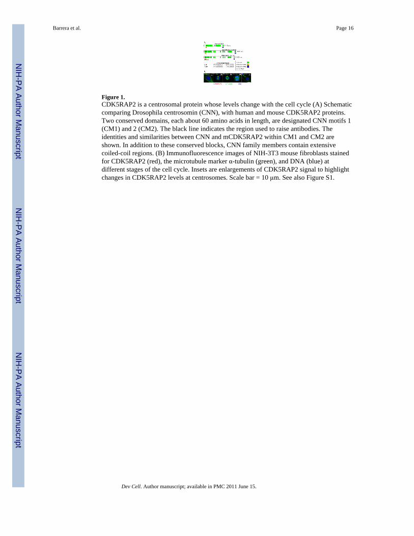
Figure 1.CDK5RAP2 is a centrosomal protein whose levels change with the cell cycle (A) Schematiccomparing Drosophila centrosomin (CNN), with human and mouse CDK5RAP2 proteins.Two conserved domains, each about 60 amino acids in length, are designated CNN motifs 1(CM1) and 2 (CM2). The black line indicates the region used to raise antibodies. Theidentities and similarities between CNN and mCDK5RAP2 within CM1 and CM2 areshown. In addition to these conserved blocks, CNN family members contain extensivecoiled-coil regions. (B) Immunofluorescence images of NIH-3T3 mouse fibroblasts stainedfor CDK5RAP2 (red), the microtubule marker α-tubulin (green), and DNA (blue) atdifferent stages of the cell cycle. Insets are enlargements of CDK5RAP2 signal to highlightchanges in CDK5RAP2 levels at centrosomes. Scale bar = 10 μm. See also Figure S1.
Barrera et al. Page 16
Dev Cell. Author manuscript; available in PMC 2011 June 15.
NIH
-PA Author Manuscript
NIH
-PA Author Manuscript
NIH
-PA Author Manuscript

Figure 2.Centrosome disruption in CDK5RAP2 mutant MEFs(A,C) The truncated CDK5RAP2 products expressed in CDK5RAP2RRF465/RRF465 (A) andCDK5RAP2RRU031/RRU031 (C) mutant mice are similar to the two mapped human mutations.Schematics show the protein fragments predicted to result from the human mutations,E385fsX4 and S81X (Bond et al., 2005), and the CDK5RAP2/β-GEO mutant fusion proteinsexpressed in CDK5RAP2RRF465/RRF465 and CDK5RAP2RRU031/RRU031 mice.(B,D) Western blots of CDK5RAP2 from whole cell lysates collected from sibling MEFcultures of the indicated genotypes. The positions of the wild type and fusion proteinsexpressed in the RRF465 mutant (B), and the relative expression level of wild typeCDK5RAP2 in the RRU031 mutant (D) are indicated. α-tubulin was probed as a loadingcontrol.(E) Nocodazole-treated CDK5RAP2+/+ and CDK5RAP2RRF465/RRF465 MEFs stained forCDK5RAP2 (red), α-tubulin (green), and DNA (blue).(F) CDK5RAP2+/+ and CDK5RAP2RRF465/RRF465 MEFs stained for CDK5RAP2 (red), thecohesion fiber protein rootletin (green), and DNA (blue).(G) Images of wild type and CDK5RAP2 mutant MEFs stained for γ-tubulin (red), thecohesion fiber protein rootletin (green), and DNA (blue).
(H) Comparison between strong PCM fibers (CDK5RAP2+/+: 72% ± vs.CDK5RAP2RRU031/RRU031: 34% ± 2%, p<0.05) and weak PCM fibers (CDK5RAP2+/+: 28%
± vs. CDK5RAP2RRU031/RRU031: 66% ± , p<0.05). The CDK5RAP2RRU031/RRU031 MEFin (G) is an example of weak PCM fibers. n = 50 cells total from two independent cell lines.(I). Centrosome splitting was scored when centrosomes were >2μm apart (CDK5RAP2+/+:27.4% ± 3.6% vs. CDK5RAP2RRU031/RRU031: 47.2% ± 6.4%, p<0.05). n = 110 cells totalfrom five independent experiments.Insets are enlargements of the centrosome region and PCM fibers. Scale bar = 10 μm. Seealso Figure S2.
Barrera et al. Page 17
Dev Cell. Author manuscript; available in PMC 2011 June 15.
NIH
-PA Author Manuscript
NIH
-PA Author Manuscript
NIH
-PA Author Manuscript
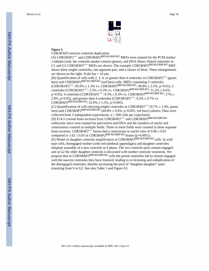
Figure 3.CDK5RAP2 restricts centriole duplication(A) CDK5RAP2+/+ and CDK5RAP2RRF465/RRF465 MEFs were stained for the PCM markerγ-tubulin (red), the centriole marker centrin (green), and DNA (blue). Paired centrioles inG1 and G2 CDK5RAP2+/+ MEFs are shown. The example CDK5RAP2RRF465/RRF465 MEFshows three singlet centrioles, one apparent pair, and a cluster of three. Three enlargementsare shown on the right. Scale bar = 10 μm.(B) Quantification of cells with 2, 3, 4, or greater than 4 centrioles in CDK5RAP2+/+ (greenbars) and CDK5RAP2RRF465/RRF465 (red bars) cells. MEFs containing 2 centrioles(CDK5RAP2+/+: 85.0% ± 1.3% vs. CDK5RAP2RRF465/RRF465: 49.8% ± 2.5%, p<0.01), 3centrioles (CDK5RAP2+/+: 2.5% ± 0.3% vs. CDK5RAP2RRF465/RRF465: 11.2% ± 0.6%,p<0.05), 4 centrioles (CDK5RAP2+/+: 8.3% ± 0.3% vs. CDK5RAP2RRF465/RRF465: 17% ±2.8%, p>0.05), and greater than 4 centrioles (CDK5RAP2+/+: 4.2% ± 0.7% vs.CDK5RAP2RRF465/RRF465: 22.0% ± 1.5%, p<0.005).(C) Quantification of cells showing singlet centrioles in CDK5RAP2+/+ (9.7% ± 1.4%, greenbars) and CDK5RAP2RRF465/RRF465 (40.8% ± 0.6%, p<0.005, red bars) cultures. Data werecollected from 3 independent experiments, n = 200 cells per experiment.(D) E14.5 coronal brain sections from CDK5RAP2+/+ and CDK5RAP2RRF465/RRF465
embryonic mice were stained for pericentrin and DNA and the numbers of nuclei andcentrosomes counted in multiple fields. Three or more fields were counted in three separatebrain sections. CDK5RAP2+/+ brains had a centrosome to nuclei ratio of 0.96 ± 0.01compared to 1.62 ± 0.05 in CDK5RAP2RRF465/RRF465 brains (p=0.0051).(E) Model of daughter centriole amplification in CDK5RAP2RRF465/RRF465 cells. In wildtype cells, disengaged mother (with red subdistal appendages) and daughter centriolestemplate assembly of a new centriole in S phase. The two centriole pairs remain engagedand at G2 the older daughter centriole is decorated with mother centriole vestments. Wepropose that in CDK5RAP2RRF465/RRF465 cells the parent centrioles fail to remain engagedwith the nascent centrioles they have fostered, leading to re-licensing and reduplication ofthe disengaged centrioles, thereby increasing the pool of “daughter-daughter” pairstransiting from S to G2. See also Table 1 and Figure S3.
Barrera et al. Page 18
Dev Cell. Author manuscript; available in PMC 2011 June 15.
NIH
-PA Author Manuscript
NIH
-PA Author Manuscript
NIH
-PA Author Manuscript

Figure 4.CDK5RAP2RRF465/RRF465 MEFs with amplified centrioles have enlarged nuclei (A) Nucleiwere stained with DAPI, and nuclear area (μm2) was measured from micrographs(CDK5RAP2+/+: 230.3 μm2 ± 5.6 μm2 vs. CDK5RAP2RRF465/RRF465: 262.0 μm2 ± 9.5 μm2,p<0.05); n ≥ 200 cells per experiment. CDK5RAP2RRF465/RRF465 MEFs with amplifiedcentrioles predominate this phenotype, with an area of 528.0 μm2 ± 13.4 μm2; n = 50 cellsper experiment. Data were collected from 3 independent experiments.(B) DAPI signal (arbitrary units) was compared between CDK5RAP2+/+ andCDK5RAP2RRF465/RRF465 MEFs with one or two centrosomes (CDK5RAP2+/+: 3.9 × 10⁶ ±0.1 × 10⁶, filled square vs. CDK5RAP2RRF465/RRF465: 4.0 × 10⁶ ± 0.3 × 10⁶, filled triangle)and MEFs with greater than two centrosomes (CDK5RAP2+/+: 7.2 × 10⁶ ± 1.2 × 10⁶, opencircle vs. CDK5RAP2RRF465/RRF465: 8.5 × 10⁶ ± 0.8 × 10⁶, open triangle) and these werethen compared to wild type binucleate cells (15.0 × 10⁶ ± 0.7 × 10⁶, open square); n = 10and 11 for wild type binucleate cells and CDK5RAP2+/+ MEFs with one or twocentrosomes, respectively and n ≥20 for all other samples. See also Figure S4.
Barrera et al. Page 19
Dev Cell. Author manuscript; available in PMC 2011 June 15.
NIH
-PA Author Manuscript
NIH
-PA Author Manuscript
NIH
-PA Author Manuscript

Figure 5.Supernumerary mother centrioles and primary cilia in CDK5RAP2RRF465/RRF465 MEFs(A) CDK5RAP2+/+ and CDK5RAP2RRF465/RRF465 MEFs stained with the daughter centriole-specific marker centrobin (left, red in merged panel), the mother centriole-specific markercenexin/ODF2 (middle, green in merge), and DNA (blue in merge). Insets showenlargements of the centriole region.(B) Percentages of cells with 0, 1, 2, or greater than 2 mother centrioles in CDK5RAP2+/+
(green bars) and CDK5RAP2RRF465/RRF465 (red bars) cells. MEFs containing 0 mothercentrioles (CDK5RAP2+/+: 0.2% ± 0.2% vs. CDK5RAP2RRF465/RRF465: 1.2% ± 0.7%,p>0.05), 1 mother centriole (CDK5RAP2+/+: 89.3% ± 0.7% vs. CDK5RAP2RRF465/RRF465:62.5% ± 0.5%, p<0.001), 2 mother centrioles (CDK5RAP2+/+: 8.2% ± 0.6% vs.CDK5RAP2RRF465/RRF465: 26.3% ± 0.9%, p<0.0005), or more than 2 mother centrioles(CDK5RAP2+/+: 2.3% ± 0.7% vs. CDK5RAP2RRF465/RRF465: 10.0% ± 1.0%, p<0.005).(C) Dot plot of the average number of mother (CDK5RAP2+/+: 1.3 ± 0.05 vs.CDK5RAP2RRF465/RRF465: 1.6 ± 0.2) and daughter (CDK5RAP2+/+: 1.3 ± 0.1 vs.CDK5RAP2RRF465/RRF465: 2.4 ± 0.15) centrioles per cell. Data were collected from threeindependent experiments, 90 cells total.(D) CDK5RAP2+/+ and CDK5RAP2RRF465/RRF465 MEFs stained for γ-tubulin (left, red inmerged panel), the cilium axoneme marker acetylated α-tubulin (middle, green in merge),and DNA (blue in merge). Image in bottom panel shows four cilia in a cell with eightcentrioles.(E) The percent of cells that formed primary cilia in CDK5RAP2+/+ (green bars) andCDK5RAP2RRF465/RRF465 (red bars) MEFs (CDK5RAP2+/+: 71.2% ± 3.6% vs.CDK5RAP2RRF465/RRF465: 74.0% ± 0.9%, p>0.05).(F) The percent of MEFs forming a single primary cilium (CDK5RAP2+/+: 95.3% ± 0.8%vs. CDK5RAP2RRF465/RRF465: 66.7% ± 2.4%, p<0.005) or more than one primary cilium(CDK5RAP2+/+, 4.7% ± 0.8% vs. CDK5RAP2RRF465/RRF465, 33.3% ± 2.4%, p<0.005). For(B), (E) and (F), data were collected from three independent experiments, n = 200 cells perexperiment (except (F), where n = 150 cells with primary cilia per experiment). Scale bar =10 μm. MEFs in (C-F) were blocked in G1 by serum starvation. See also Figure S5
Barrera et al. Page 20
Dev Cell. Author manuscript; available in PMC 2011 June 15.
NIH
-PA Author Manuscript
NIH
-PA Author Manuscript
NIH
-PA Author Manuscript

Figure 6.Multipolar spindles form in CDK5RAP2RRF465/RRF465 MEFs.(A) Schematic outlining the criteria used to designate bipolar and multipolar spindleformation. To qualify as multipolar, spindles had an MTOC offset by greater than 45° fromthe dominant spindle axis as determined from spindle microtubules and DNA alignment.(B) Mitotic figures immunostained for γ-tubulin (1st row, red in merged panel), α-tubulin(2nd row, green in merge), and DNA (3rd row, blue in merge). In CDK5RAP2RRF465/RRF465
mitotic MEFs with multiple centrosomes, 51% of mitotic spindles were multipolar (n = 43).Arrows indicate examples of the excess spindle poles, and arrowheads indicatechromosomes configured improperly on the metaphase plate. Scale bar = 10 μm.(C) Scatter plot showing the timing from nuclear envelope breakdown (NEBD) to anaphaseonset in CDK5RAP2+/+ and CDK5RAP2RRF465/RRF465 MEFs. In a small population ofCDK5RAP2RRF465/RRF465 MEFs, the time between NEBD and anaphase onset wasprolonged, with an approximately 8 min, or 47.7%, increase in the time spent to reachanaphase (CDK5RAP2+/+: 17.2 min. ± 0.7 min. vs. CDK5RAP2RRF465/RRF465: 25.4 min. ±2.1 min., p<0.0001); n ≥ 25.
Barrera et al. Page 21
Dev Cell. Author manuscript; available in PMC 2011 June 15.
NIH
-PA Author Manuscript
NIH
-PA Author Manuscript
NIH
-PA Author Manuscript

NIH
-PA Author Manuscript
NIH
-PA Author Manuscript
NIH
-PA Author Manuscript
Barrera et al. Page 22
Table 1Centriole configurations
Dev Cell. Author manuscript; available in PMC 2011 June 15.