Land degradation - JRC Publications Repository - European ...
Upload
khangminh22Category
view
3download
0
A possible functional link between RNA
degradation and transcription in Bacillus subtilis
Dissertation
for the award of the degree
“Doctor of Philosophy”
Division of Mathematics and Natural Sciences
of the Georg-August-University Göttingen
within the program “Microbiology and Biochemistry”
of the Georg-August University School of Science (GAUSS)
submitted by
Martin Benda
from Prague (Czech Republic)
Göttingen 2020


Thesis Committee
Prof. Dr. Jörg Stülke (Supervisor and 1st Reviewer)
Institute for Microbiology and Genetics, Department of General Microbiology, Georg-August-University
Göttingen
Prof. Dr. Rolf Daniel (2nd Reviewer)
Institute for Microbiology and Genetics, Department of Genomic and Applied Microbiology, Georg-August-
University Göttingen
Prof. Dr. Fabian M. Commichau
Institute for Biotechnology, Department of Synthetic Microbiology, Brandenburg University of Technology
Cottbus-Senftenberg
Additional members of the Examination Board
Prof. Dr. Markus Bohnsack
Department of Molecular Biology, University Medical Center Göttingen
Prof. Dr. Patrick Cramer
Department of Molecular Biology, Max Planck Institute for Biophysical Chemistry, Göttingen
Prof. Dr. Stefanie Pöggeler
Institute for Microbiology and Genetics, Department of Genetics of Eukaryotic Microorganisms, Georg-
August-University Göttingen
Date of oral examination: September 17th, 2020

I hereby declare that this Ph.D. thesis entitled “A possible functional link between RNA
degradation and transcription in Bacillus subtilis” has been written independently and
with no other sources and aids than quoted.
Martin Benda

Acknowledgements
First of all, I would like to express my deep thanks to my supervisor Prof. Dr. Jörg Stülke for
the great guidance during this work. He put his full trust in me and gave me the opportunity to
work on this intricate, but engaging topic of RNA degradation. I could always turn to him with
questions, I have been encouraged in moments of despair and supported in developing my own
scientific ideas.
My immense thanks go to Prof. Dr. Fabian Commichau and Prof. Dr. Rolf Daniel for being
members of my Thesis Advisory Committee and for broadening our view of the topic with
meaningful comments and ideas. I would also like to thank Dr. Katrin Gunka who introduced me
both to the project and to the lab and made the transition to a new working environment easier
for me. I thank G2L Göttingen and especially Dr. Anja Poehlein for all the sequencing performed
and discussion of the results. Many thanks go to Julia Busse and Gabriele Beyer for their help with
the experimental work. Importantly, huge thanks go to Silvia Carillo-Castellón, all the work on this
thesis would have been much more difficult without a constant supply of sterile tips, tubes or
freshly prepared media.
I would also like to thank my students Simon Wölfel, Jonas Jennrich, Leon Daniau, Melin
Güzel, Fabian Fiedler and Maxim Wintergoller, not only for their contribution to this thesis, but
mainly for the great time I had working with all of them. I learned many new skills during that
time and realized, quite unexpectedly, how much I like teaching.
My huge thanks go to all former and current members of the Stülke, Commichau and
Rismondo labs I have met here. There were many of you and yet everyone kind, helpful in the
laboratory and creating warm and friendly atmosphere not only in the laboratory but also
outside.
I would also like to express my gratitude to Dr. Libor Krásný and all members of his lab. I
learned the essential basics of laboratory and scientific work during my undergraduate time there
and when this doctoral thesis turned its way towards his expertise, I received warm and great
support again.
Finally and above all, I would like to thank my wife Klára for the daily unconditional support
in all aspects of life. She always stands by my side and supports me, even if it meant moving
abroad to a completely new environment and brought inconvenience to her own career. Without
such a great support, I would have never been able to complete this thesis. Big thanks go also to
my parents and my whole family, they have always been there for me since my first steps and
only thanks to that I got up all the way to this thesis.


I
Table of contents
Table of contents ......................................................................................................................................... I
List of abbreviations .................................................................................................................................. III
Summary .................................................................................................................................................... V
1 Introduction ....................................................................................................................................... 1
1.1 mRNA degradation and RNA degradosomes in bacteria ............................................................... 2
1.2 mRNA degradation and degradosome-like network of B. subtilis ................................................. 6
1.2.1 RNase Y .............................................................................................................................. 9
1.2.2 RNases J1 and J2............................................................................................................... 11
1.2.3 Polynucleotide phosphorylase (PNPase) ........................................................................... 13
1.2.4 CshA, a DEAD-box RNA helicase ........................................................................................ 14
1.2.5 Enolase and phosphofructokinase .................................................................................... 15
1.3 Essentiality and RNase Y ........................................................................................................... 16
1.4 Natural competence in B. subtilis .............................................................................................. 17
1.5 Aims of this thesis ..................................................................................................................... 19
2 Quasi-essentiality of RNase Y in Bacillus subtilis is caused by its critical role in the control of mRNA
homeostasis .............................................................................................................................................. 20
3 The YtrBCDEF ABC transporter is involved in the control of social activities in Bacillus subtilis ........ 46
4 Discussion......................................................................................................................................... 63
4.1 Suppressor mutant screen revealed initiation of bulk mRNA degradation as the pivotal function
of RNase Y ............................................................................................................................................. 63
4.2 Analysis of the rny suppressor mutants brings new insights into the regulation of the RNA
polymerase ........................................................................................................................................... 70
4.3 Loss of RNase Y leads to phenotypic effects independent of the total mRNA accumulation........ 73
5 References........................................................................................................................................ 78
6 Appendix ........................................................................................................................................ 101
6.1 Supplementary material.......................................................................................................... 101
6.2 Bacterial strains, plasmids and oligonucleotides ...................................................................... 110
7 Curriculum Vitae............................................................................................................................. 123

II

III
List of abbreviations
General
A adenosine
ABC ATP-binding cassette
Aco aconitase
AMP adenosine monophosphate
aphA3 kanamycin resistance gene
Asn asparagine
ATP adenosine triphosphate
B. Bacillus
C cytosine
C. Caulobacter
cat chloramphenicol resistance gene
CTD C-terminal domain
DLN degradosome-like network
DNA deoxyribonucleic acid
E. Escherichia
e. g. exempli gratia (for example)
Eno enolase
ermC erythromycin resistance gene
et al. et alia (and others)
FDR false discovery rate
Fig. figure
fwd forward
G guanosine
Glu glutamic acid
H. Helicobacter
i. e. id est (that is)
IPTG isopropyl β-D-1-thiogalactopyranoside
kan kanamycin
LB lysogeny broth
Leu leucine
LFH long flanking homology
mRNA messenger RNA
nt nucleotide
NTD N-terminal domain
OD optical density
Orn oligoribonuclease
P phosphate
P promoter
PNPase polynucleotide phosphorylase
PCR polymerase chain reaction
PFK phosphofructokinase
pH power of hydrogen
RBS ribosomal binding site
rev reverse
RNA ribonucleic acid
RNase ribonuclease
rRNA ribosomal RNA
S. Staphylococcus
S. Streptococcus
spc spectinomycin resistance gene
sRNA small RNA
SRP signal recognition particle
T thymine
Term. transcriptional terminator
tRNA transfer RNA
Trp tryptophan
Tyr tyrosine
vs. versus
Δ deletion
Prefixes
k kilo
m milli
µ micro
n nano

IV
Units
°C degree Celsius
bp base pair
g gram
g standard gravity
h hour
l liter
min minute
mol mol
M molar
s second
V
Summary
Cellular levels of RNA depend on the rate of its synthesis and degradation. While synthesis is
performed by RNA polymerase conserved in all domains of life, the enzymes responsible for RNA
degradation are more unique even among organisms from the same domain. In the best studied
bacterium, the gram-negative Escherichia coli, RNA degradation is achieved through a protein
complex called RNA degradosome, which is assembled around the essential endoribonuclease
RNase E. However, RNase E is not present in the gram-positive model organism Bacillus subtilis.
Instead, an enzyme called RNase Y (rny) has been proposed as its functional counterpart
responsible for the initiation of RNA degradation. Nevertheless, unlike RNase E of E. coli, it can be
deleted from the genome, leaving an open question of its true significance and function. This
project was designed to get a deeper understanding of the crucial process of RNA degradation in
B. subtilis and of the role RNase Y plays there. Although RNase Y is dispensable for survival, the
rny gene deletion leads to detrimental phenotypic effects, including filamentous growth, impaired
cellular morphology or defects in the development of genetic competence and sporulation. The
rny mutant strain also lyses rapidly and subsequently suppressor colonies appear. Using this
natural force of suppressor evolution, we could demonstrate that no other RNase can take over
the tasks of RNase Y. Conversely, all identified mutations were aimed to reduce RNA synthesis.
This was achieved either by inactivation of transcription factors in conjunction with duplication of
core RNA polymerase genes, which results in decreased number of correctly assembled RNA
polymerase complexes, or, if the first suppressing mechanism was prevented, by mutations
occurring directly in the RNA polymerase core genes, leading to orders of magnitude decrease in
transcription. The fact that the mutations always affect RNA synthesis, a process on the opposite
side of RNA life to the one RNase Y acts, suggest close collaboration of RNase Y with the RNA
polymerase in establishing stable equilibrium between RNA synthesis and degradation. While the
suppressor mutant analysis helped to identify the pivotal function of RNase Y, it did not
necessarily provide an explanation for all the phenotypes associated with the deletion of the rny
gene. In an attempt to better understand such phenotypes, RNA-sequencing analysis revealed
global remodeling of gene expression in the rny strain. Furthermore, a screening system to
recognize the reasons for the loss of genetic competence was established and helped to decipher
the reasons for the loss of competence in the rny mutant as well as in other strains, among them
in the ytrA mutant overexpressing putative ABC transporter YtrBCDEF. This was shown to act in
remodeling of the cell wall thickness, which hampers development of genetic competence as well
as other lifestyles of B. subtilis. The possible influence of a disordered cell wall is also discussed as
a potential reason for the loss of competence in the rny mutant.

VI
1
1 Introduction
All organisms are dependent on their ability to adapt to the surrounding environments
and to use the available resources for their survival and reproduction. Due to their small size,
bacteria are extremely vulnerable to changing environmental conditions and are therefore
equipped with remarkable abilities to accommodate to the changing and challenging conditions.
These abilities include short generation time, fast evolution, rapid modulation of gene expression
or differentiation into specific cell types.
Crucial for fast adaptation is to regulate the amount and/or activity of proteins. This could
be done either directly on the protein level or indirectly by modulating levels of messenger RNA
(mRNA). The cellular level of mRNA is determined by the rate of its synthesis and degradation.
Synthesis of mRNA is performed by a multi-subunit enzyme called RNA polymerase in process of
transcription, which is subject to strict control and regulation. However, this control has a delayed
onset of action and therefore mRNA levels must be also controlled by its degradation.
Degradation of mRNA is thus one of the main mechanisms by which protein synthesis is regulated
in all domains of life, since timely degradation of no longer necessary mRNAs is important to save
energetic costs of translation and to release ribonucleotides for new rounds of condition adjusted
transcription.
In conjunction with short generation time and fast adaptation, also half-lives of bacterial
mRNAs are short, ranging from seconds to tens of minutes, with majority of transcripts from
model bacterial organism Escherichia coli and Bacillus subtilis having mRNA half-lives shorter than
8 minutes (Hambraeus et al., 2003; Bernstein et al., 2004).
The enzymes responsible for the RNA degradation are called ribonucleases (RNases) and
can be divided into two main groups (endo- and exo-ribonucleases) based on their mode of
action. Endoribonucleases cleave RNA internally, while exoribonucleases attack the RNA molecule
from its 5′ or 3′ ends. Whereas some RNases do have a very narrow substrate specificity and act
on a limited number of transcripts, others are responsible for a broad degradation of cellular
mRNAs. Those ribonucleases are often localized into multi-enzyme complexes to achieve high
degree of effective cooperation. Such protein complexes can be found in all domains of life, as
exosomes in eukaryotes and archaea (Mitchell et al., 1997; Evguenieva-Hackenberg et al., 2014),
or as so-called RNA degradosomes in bacteria. These complexes have already been found in many
bacterial species and will be further described in the following chapter.

2
1.1 mRNA degradation and RNA degradosomes in bacteria
Degradation of mRNA is generally a very fast process once it starts, so it is the initial
cleavage event which determines the degradation rate (Laalami et al., 2014). In theory, RNA
degradation could be initiated by three different ways, by exoribonucleolytic degradation from
either the 3′ or the 5′ end of RNA molecule or by internal endoribonucleolytic cleavage. However,
mRNAs are often equipped with protective structures to prevent premature and uncontrolled
degradation. The 3′ ends are usually protected from the action of exoribonucleases by secondary
stem loop structures, moreover degradation from the 3′ end would be energetically very
inefficient process, since the degradation would proceed in opposite direction than translation,
thus leading to creation of truncated proteins (Laalami et al., 2014). The 5′ ends are mainly
protected by a triphosphate group, although there is an increasing evidence about presence of
other 5′ end protecting molecules such as nicotinamide adenine dinucleotide (NAD) (Cahová et
al., 2015; Frindert et al., 2018). Therefore, due to the above-mentioned protections, initiation by
exoribonuclease accounts only for minority of transcripts and it is the endoribonucleolytic attack,
which usually initiates the degradation pathway (Mohanty and Kushner, 2018).
The endoribonuclease responsible for the initial cleavage in the best studied model
organism E. coli is called RNase E. This enzyme is capable to initiate RNA degradation by direct
endoribonucleolytic cleavage of single stranded mRNAs protected both on the 5′ and 3′ ends;
however, this is the case only for some transcripts. Activity of RNase E, although it is an
endoribonuclease, is in fact also affected by the phosphorylation state of the 5′ end, as RNase E
was shown in vitro to preferentially cleave transcripts with monophosphorylated 5′ ends, which
rarely occur in nature (Mackie, 1998). In order to overcome this problem, E. coli is equipped with
an additional enzymatic activity that alters the phosphorylation state of the 5' end and creates
monophosphorylated RNA molecules, thus facilitating the initial cleavage by RNase E. We can
therefore define two different pathways by which the degradation is initiated, the 5′ end
dependent pathway and the 5′ end independent pathway (see Fig. 1).
In the first case, the 5′ end dependent pathway is initiated by cleavage of two phosphates
from the 5′ end, which leads to creation of 5′ monophosphorylated RNA molecule. An enzyme
called RppH was traditionally thought to be responsible for this dephosphorylation (Deana et al.,
2008). However, recent studies suggested that the dephosphorylation is a sequential process and
that RppH can efficiently catalyze only the second reaction from diphosphate to monophosphate,
leaving a possibility that another, as yet undiscovered enzyme, may be involved in this pathway
(Luciano et al., 2017). When a 5′ monophosphorylated RNA molecule is created, the presence of
the monophosphate group stimulates endoribonucleolytic activity of RNase E, leading to creation

3
of two fragments. The first fragment does no longer have a stem loop structure on the 3′ end and
therefore could be easily degraded by 3′-to-5′ directed exoribonucleases like polynucleotide
phosphorylase (PNPase). The second fragment is, thanks to its monophosphorylated 5′ end,
a great substrate for further cleavage by RNase E. The whole RNA is this way gradually degraded
up to di-nucleotides, which are then degraded into the individual nucleotides reusable in new
round of transcription by an enzyme called Oligoribonuclease (Orn) (Kim et al., 2019).
The second pathway, 5′ end independent or sometimes also called direct entry pathway,
is initiated by cleavage by RNase E. In this case RNase E directly accesses and cleaves an internal
site of the mRNA molecule independently from the phosphorylation state of its 5′ end. Although
this pathway seemed to be less likely due to the in vitro preference of RNase E for
5′ monophosphorylated RNAs, in reality it was shown to be the major initiating pathway in vivo in
E. coli (Mackie, 1998; Clarke et al., 2014). The endoribonucleolytic cleavage here results again in
two fragments, the first one contains the original 5′ end, but does no longer have a stem loop
structure on the 3′ end and therefore, as in the 5′ end dependent pathway, is accessible for
degradation by 3′–5′ directed exoribonucleases. The second fragment, on the other hand, still
contains a stem loop structure on the 3′ end, but is monophosphorylated on its 5′ end and
therefore more susceptible for further cleavage events by RNase E. The RNA molecule is this way
again further fragmented until dinucleotides are produced and degraded by Orn (Kim et al.,
2019).
Figure 1: Schematic depiction of mRNA degradation pathways in E. coli (A) In the 5′ end dependent pathway, pyrophosphate is first removed from the RNA molecule by RppH (dark green) and possible other enzyme (light green), monophosphorylated 5′ end activates RNase E (red), in further steps PNPase (blue) degrades RNA from the 3′ end. Finally, degradation of dinucleotides is achieved by Orn (orange). (B) In the 5′ end independent pathway, degradation is initiated directly by cleavage of RNase E, followed by actions of PNPase and Orn as described in A.
4
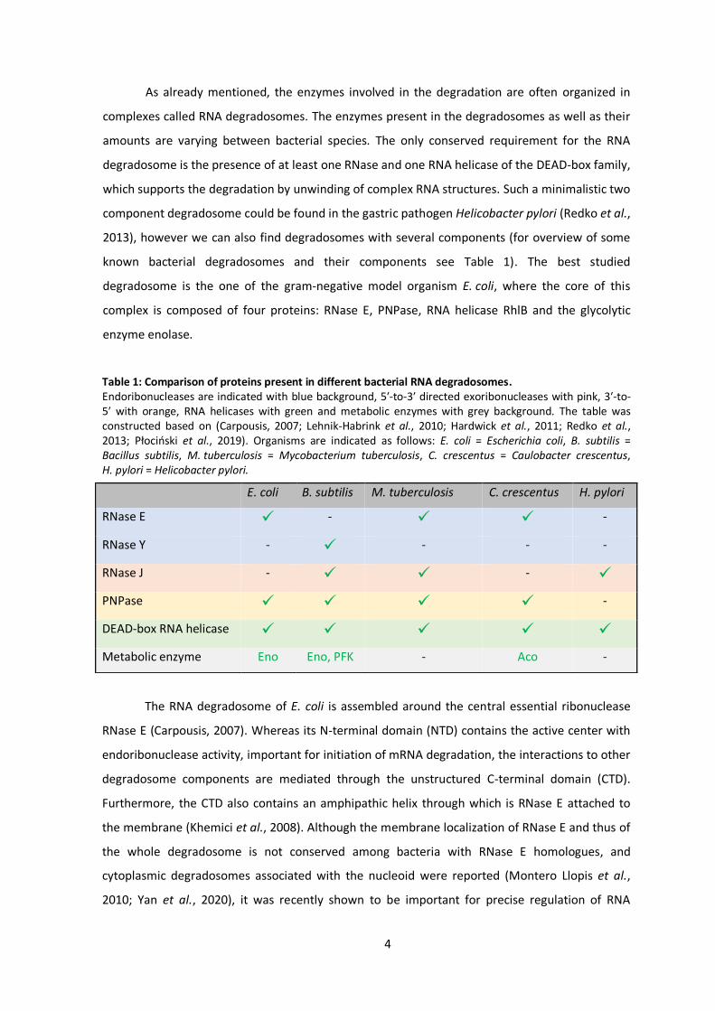
As already mentioned, the enzymes involved in the degradation are often organized in
complexes called RNA degradosomes. The enzymes present in the degradosomes as well as their
amounts are varying between bacterial species. The only conserved requirement for the RNA
degradosome is the presence of at least one RNase and one RNA helicase of the DEAD-box family,
which supports the degradation by unwinding of complex RNA structures. Such a minimalistic two
component degradosome could be found in the gastric pathogen Helicobacter pylori (Redko et al.,
2013), however we can also find degradosomes with several components (for overview of some
known bacterial degradosomes and their components see Table 1). The best studied
degradosome is the one of the gram-negative model organism E. coli, where the core of this
complex is composed of four proteins: RNase E, PNPase, RNA helicase RhlB and the glycolytic
enzyme enolase.
Table 1: Comparison of proteins present in different bacterial RNA degradosomes. Endoribonucleases are indicated with blue background, 5′-to-3′ directed exoribonucleases with pink, 3′-to-5′ with orange, RNA helicases with green and metabolic enzymes with grey background. The table was constructed based on (Carpousis, 2007; Lehnik-Habrink et al., 2010; Hardwick et al., 2011; Redko et al., 2013; Płociński et al., 2019). Organisms are indicated as follows: E. coli = Escherichia coli, B. subtilis = Bacillus subtilis, M. tuberculosis = Mycobacterium tuberculosis, C. crescentus = Caulobacter crescentus, H. pylori = Helicobacter pylori.
The RNA degradosome of E. coli is assembled around the central essential ribonuclease
RNase E (Carpousis, 2007). Whereas its N-terminal domain (NTD) contains the active center with
endoribonuclease activity, important for initiation of mRNA degradation, the interactions to other
degradosome components are mediated through the unstructured C-terminal domain (CTD).
Furthermore, the CTD also contains an amphipathic helix through which is RNase E attached to
the membrane (Khemici et al., 2008). Although the membrane localization of RNase E and thus of
the whole degradosome is not conserved among bacteria with RNase E homologues, and
cytoplasmic degradosomes associated with the nucleoid were reported (Montero Llopis et al.,
2010; Yan et al., 2020), it was recently shown to be important for precise regulation of RNA
E. coli B. subtilis M. tuberculosis C. crescentus H. pylori
RNase E ✓ - ✓ ✓ -
RNase Y - ✓ - - -
RNase J - ✓ ✓ - ✓
PNPase ✓ ✓ ✓ ✓ -
DEAD-box RNA helicase ✓ ✓ ✓ ✓ ✓
Metabolic enzyme Eno Eno, PFK - Aco -
5
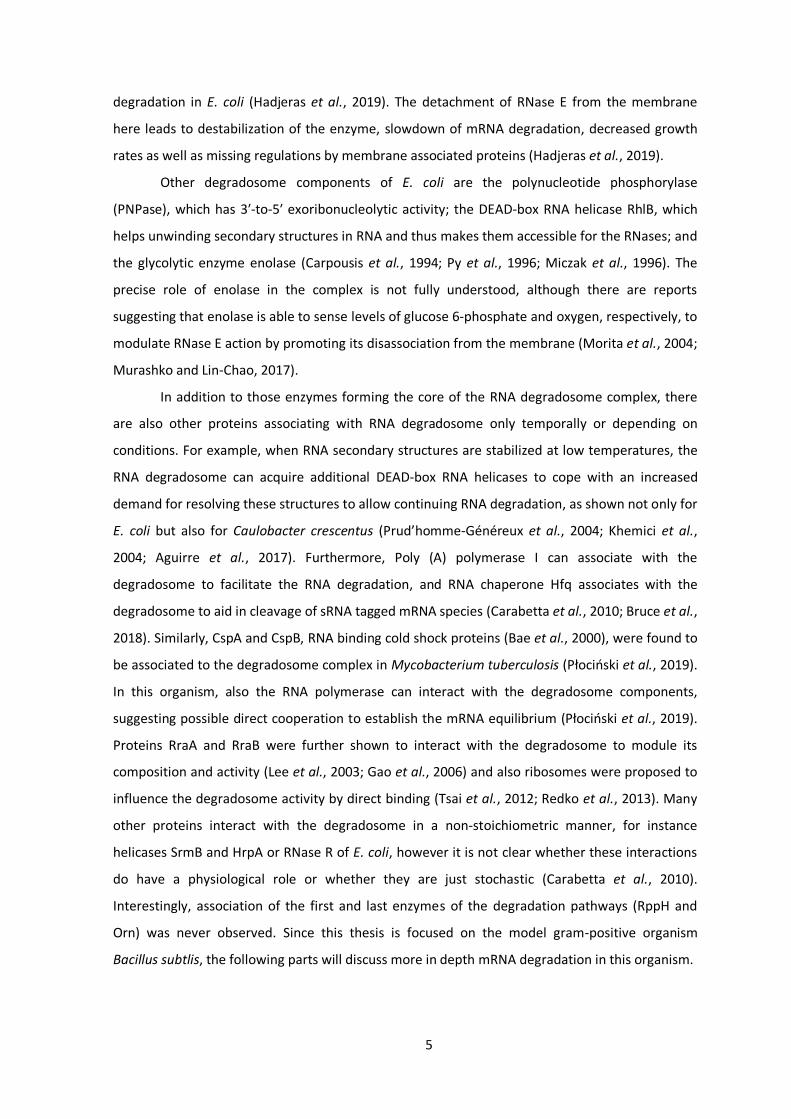
degradation in E. coli (Hadjeras et al., 2019). The detachment of RNase E from the membrane
here leads to destabilization of the enzyme, slowdown of mRNA degradation, decreased growth
rates as well as missing regulations by membrane associated proteins (Hadjeras et al., 2019).
Other degradosome components of E. coli are the polynucleotide phosphorylase
(PNPase), which has 3′-to-5′ exoribonucleolytic activity; the DEAD-box RNA helicase RhlB, which
helps unwinding secondary structures in RNA and thus makes them accessible for the RNases; and
the glycolytic enzyme enolase (Carpousis et al., 1994; Py et al., 1996; Miczak et al., 1996). The
precise role of enolase in the complex is not fully understood, although there are reports
suggesting that enolase is able to sense levels of glucose 6‐phosphate and oxygen, respectively, to
modulate RNase E action by promoting its disassociation from the membrane (Morita et al., 2004;
Murashko and Lin-Chao, 2017).
In addition to those enzymes forming the core of the RNA degradosome complex, there
are also other proteins associating with RNA degradosome only temporally or depending on
conditions. For example, when RNA secondary structures are stabilized at low temperatures, the
RNA degradosome can acquire additional DEAD-box RNA helicases to cope with an increased
demand for resolving these structures to allow continuing RNA degradation, as shown not only for
E. coli but also for Caulobacter crescentus (Prud’homme-Généreux et al., 2004; Khemici et al.,
2004; Aguirre et al., 2017). Furthermore, Poly (A) polymerase I can associate with the
degradosome to facilitate the RNA degradation, and RNA chaperone Hfq associates with the
degradosome to aid in cleavage of sRNA tagged mRNA species (Carabetta et al., 2010; Bruce et al.,
2018). Similarly, CspA and CspB, RNA binding cold shock proteins (Bae et al., 2000), were found to
be associated to the degradosome complex in Mycobacterium tuberculosis (Płociński et al., 2019).
In this organism, also the RNA polymerase can interact with the degradosome components,
suggesting possible direct cooperation to establish the mRNA equilibrium (Płociński et al., 2019).
Proteins RraA and RraB were further shown to interact with the degradosome to module its
composition and activity (Lee et al., 2003; Gao et al., 2006) and also ribosomes were proposed to
influence the degradosome activity by direct binding (Tsai et al., 2012; Redko et al., 2013). Many
other proteins interact with the degradosome in a non-stoichiometric manner, for instance
helicases SrmB and HrpA or RNase R of E. coli, however it is not clear whether these interactions
do have a physiological role or whether they are just stochastic (Carabetta et al., 2010).
Interestingly, association of the first and last enzymes of the degradation pathways (RppH and
Orn) was never observed. Since this thesis is focused on the model gram-positive organism
Bacillus subtlis, the following parts will discuss more in depth mRNA degradation in this organism.

6
1.2 mRNA degradation and degradosome-like network of B. subtilis
Due to the general importance of mRNA processing and degradation, it could be assumed
that the key components are highly conserved among individual bacteria species. It was therefore
surprising, that the gram-positive model organism B. subtilis does not contain any homolog of
RNase E, the central enzyme of mRNA degradation in E. coli. This also brought a question of
whether there is an RNA degradosome in B. subtilis and if so, what does it look like?
This question was later addressed by the discovery of an enzyme called RNase Y
(Commichau et al., 2009; Shahbabian et al., 2009). Although RNase Y does not have any sequence
homology to RNase E of E. coli, it was proposed to be the scaffolding protein of B. subtilis RNA
degradosome based on interactions with other RNases, RNA helicase and glycolytic enzymes
(Commichau et al., 2009; Lehnik-Habrink et al., 2010). Except these interactions, RNase Y has also
other striking functional similarities to RNase E of E. coli, since it also possesses endoribonuclease
activity and is localized to the cytoplasmic membrane (Shahbabian et al., 2009; Cascante-Estepa
et al., 2016). Apparently, the key players of the mRNA degradation process have evolved
independently to fulfill very similar roles in the cells. This is further supported by the fact that the
essential RNase E of E. coli could be substituted with RNase Y of B. subtilis (Tamura et al., 2017).
The proposed RNA degradosome complex of B. subtilis built around central RNase Y (see
Fig. 2) is further composed of two other RNases showing endoribonuclease activity in vitro, the
paralogues proteins RNases J1 and J2 (Even et al., 2005). In addition, those two RNases were also
shown to have 5′-to-3′ directed exoribonuclease activity, which is an activity completely missing in
E. coli (Mathy et al., 2007). Furthermore, the proposed RNA degradosome contains 3′-to-5′
directed exoribonuclease PNPase and a DEAD-box RNA helicase called CshA. Like the
degradosome of E. coli, also this one contains the glycolytic enzyme enolase and on top of that
another glycolytic enzyme, phosphofructokinase. Their role in the complex, however, remains
mysterious.
In contrast to the RNA degradosome of E. coli, the degradosome of B. subtilis was never
successfully purified as a complex and interactions between the individual components were only
shown via bacterial-two hybrid studies or cross-linking pull down experiments (Coburn et al.,
1999; Worrall et al., 2008; Commichau et al., 2009; Lehnik-Habrink et al., 2011a). In combination
with data showing that the degradosome components localize mainly in the cytoplasm and do not
co-localize with RNase Y at the membrane (Cascante-Estepa et al., 2016), the existence of true
degradosome in B. subtilis is questioned. Hence, recent literature is rather talking about
degradosome-like network (DLN), since the interactions are probably just transient and highly
dynamic (Durand and Condon, 2018).

7
Initiation of mRNA degradation in B. subtilis can also occur by different pathways that are
similar to those from E. coli (see Fig. 3). The 5′ end dependent pathway starts with
dephosphorylation of RNA molecule by a phosphohydrolase also called RppH, although this does
not have a high degree of homology to the one from E. coli. RppH of B. subtilis can efficiently
remove phosphates step by step as orthophosphates and thus, in contrast to E. coli, there is no
need for additional enzymes (Richards et al., 2011). Nevertheless, there are reports about other
enzymes capable of 5′ end dephosphorylation, which might be involved in this pathway as well
(Frindert et al., 2019). The dephosphorylation step is followed either by complete
exoribonucleolytic degradation of RNA by RNase J1 in 5′-to-3′ direction (5′ end dependent exo-
pathway) or by endoribonucleolytic cleavage by RNase Y (5′ end dependent endo-pathway), which
has also preference for substrates with 5′ monophosphates (Shahbabian et al., 2009; Richards et
al., 2011). Fragments created by RNase Y cleavage could be then rapidly degraded by action of
exoribonucleases RNase J1 and PNPase. The final degradation step is not done by Orn enzyme as
in E. coli, instead B. subtilis has at least two so-called nanoRNases encoded by the genes nrnA and
nrnB, which were shown to degrade short oligoribonucleotides up to 5 nt long from the 3′ end.
However, some capacity to complete the decay of RNA was also found in RNase J1 itself and 3′-to-
5′ exoribonuclease YhaM, so it is possible that this function in B. subtilis is redundantly distributed
among various enzymes (Mechold et al., 2007; Fang et al., 2009).
Figure 2: The proposed RNA degradosome complex of B. subtilis The complex is anchored to the membrane through the N-terminus of RNase Y, which also serves as a scaffold for the other components, complex of RNases J1/J2, PNPase, DEAD-box RNA helicase CshA and glycolytic enzymes enolase (Eno) and phosphofructokinase (PfkA). Modified from (Cho, 2017) and (Wölfel, 2018).

8
Similarly to E. coli, B. subtilis can also initiate RNA degradation by a 5′ end independent
pathway. Despite the fact that RNase Y has preference for 5′ monophosphorylated substrates, it
was shown to efficiently initiate degradation of ermC mRNA regardless of the 5′ end
phosphorylation state (Shahbabian et al., 2009; Yao et al., 2011). Taken together, the repertoire
of degradation pathways is extended in the gram-positive model organism by the action of 5′-to-
3′ directed exoribonuclease RNase J1.
An obvious question which might appear is why there is no pathway initiating mRNA
decay from the 3′ end? Although mRNAs are generally protected by stem loop structures at this
terminus as already discussed, especially considering collaboration of the PNPase with RNA
helicase present in the degradosome, this protective structure does not necessarily have to be a
complete obstacle for such a pathway. Results obtained in previous studies, however, suggest
that this is not the case, since absence of PNPase does not lead to strong global effect on gene
expression and pnpA deletion strain accumulates only degradation fragments and not full length
transcripts, as would be expected if PNPase is involved in the decay initiation (Luttinger et al.,
1996; Oussenko et al., 2005). Therefore this possible initiation pathway seems to play only a
minor role, if any, possibly in degradation of transcripts with Rho dependent terminators, which
are rare in B. subtilis (Ingham et al., 1999; Liu et al., 2016).
Figure 3: Schematic depiction of mRNA degradation pathways in B. subtilis (A) In the 5′ end dependent exo-pathway, two orthophosphates are first removed from the RNA molecule by RppH (green scissors), monophosphorylated 5′ end activates RNases J1/J2 (green) to degrade the RNA exoribonucleolytically, followed by the degradation of short RNA fragments by nanoRNases (orange) (B) In the 5′ end dependent endo-pathway, RppH creates monophosphorylated 5′ end, which activates RNase Y (purple scissors) for endoribonuclease cleavage, in further steps PNPase (blue) degrades RNA from the 3′ end and complex of the RNases J1/J2 from the 5′ end. Finally, short RNA fragments are degraded by nanoRNAses. (C) In the 5′ end independent pathway, RNase Y cleaves the transcript internally without a requirement for removal of phosphates from the 5′ end, this cleavage is followed by action of exoribonucleases as in B.
9
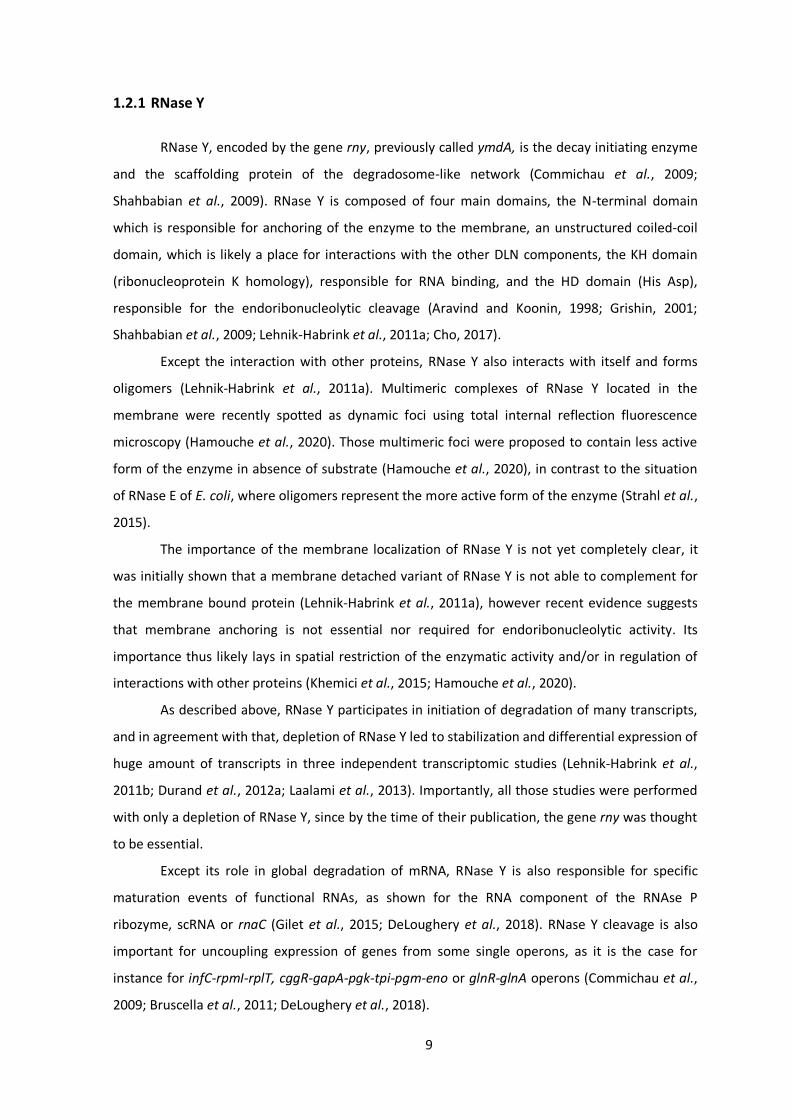
1.2.1 RNase Y
RNase Y, encoded by the gene rny, previously called ymdA, is the decay initiating enzyme
and the scaffolding protein of the degradosome-like network (Commichau et al., 2009;
Shahbabian et al., 2009). RNase Y is composed of four main domains, the N-terminal domain
which is responsible for anchoring of the enzyme to the membrane, an unstructured coiled-coil
domain, which is likely a place for interactions with the other DLN components, the KH domain
(ribonucleoprotein K homology), responsible for RNA binding, and the HD domain (His Asp),
responsible for the endoribonucleolytic cleavage (Aravind and Koonin, 1998; Grishin, 2001;
Shahbabian et al., 2009; Lehnik-Habrink et al., 2011a; Cho, 2017).
Except the interaction with other proteins, RNase Y also interacts with itself and forms
oligomers (Lehnik-Habrink et al., 2011a). Multimeric complexes of RNase Y located in the
membrane were recently spotted as dynamic foci using total internal reflection fluorescence
microscopy (Hamouche et al., 2020). Those multimeric foci were proposed to contain less active
form of the enzyme in absence of substrate (Hamouche et al., 2020), in contrast to the situation
of RNase E of E. coli, where oligomers represent the more active form of the enzyme (Strahl et al.,
2015).
The importance of the membrane localization of RNase Y is not yet completely clear, it
was initially shown that a membrane detached variant of RNase Y is not able to complement for
the membrane bound protein (Lehnik-Habrink et al., 2011a), however recent evidence suggests
that membrane anchoring is not essential nor required for endoribonucleolytic activity. Its
importance thus likely lays in spatial restriction of the enzymatic activity and/or in regulation of
interactions with other proteins (Khemici et al., 2015; Hamouche et al., 2020).
As described above, RNase Y participates in initiation of degradation of many transcripts,
and in agreement with that, depletion of RNase Y led to stabilization and differential expression of
huge amount of transcripts in three independent transcriptomic studies (Lehnik-Habrink et al.,
2011b; Durand et al., 2012a; Laalami et al., 2013). Importantly, all those studies were performed
with only a depletion of RNase Y, since by the time of their publication, the gene rny was thought
to be essential.
Except its role in global degradation of mRNA, RNase Y is also responsible for specific
maturation events of functional RNAs, as shown for the RNA component of the RNAse P
ribozyme, scRNA or rnaC (Gilet et al., 2015; DeLoughery et al., 2018). RNase Y cleavage is also
important for uncoupling expression of genes from some single operons, as it is the case for
instance for infC-rpmI-rplT, cggR-gapA-pgk-tpi-pgm-eno or glnR-glnA operons (Commichau et al.,
2009; Bruscella et al., 2011; DeLoughery et al., 2018).

10
As already mentioned, the rny gene was thought for a long time to be essential, however,
in 2013 it was deleted by Figaro and coworkers and this was later reproduced in another study
(Figaro et al., 2013; Koo et al., 2017). Nevertheless, deletion of rny gene leads to severe
phenotypic defects. Colonies are small and smooth, quickly lysing and forming suppressor
mutants (see Fig. 4). The doubling times are more than doubled as compared to the wild type, cell
separation is impaired, so the rny mutant cells grow in chains (see Fig. 5). Furthermore, the strain
is cold sensitive, its peptidoglycan layer is disordered, and also sporulation and development of
genetic competence are abolished (Figaro et al., 2013).
RNase Y is an endoribonuclease with a preference for 5′ monophosphorylated ends
(Shahbabian et al., 2009). However, it is a matter of discussion, whether there is any sequence
specificity for RNase Y cleavage events. In related organisms, preferential cleavage downstream of
guanosine was reported both for Staphylococcus aureus and Streptococcus pyogenes (Khemici et
al., 2015; Broglia et al., 2020). Furthermore, presence of double stranded secondary structure 6 nt
downstream of the cleavage site was reported to be decisive for cleavage of saePQRS operon
mRNA in S. aureus (Marincola and Wolz, 2017). Concerning RNase Y from B. subtilis, no sequence
preference for guanosine was identified so far, on the other hand presence of secondary structure
might be the determinant also for the B. subtilis enzyme, as it was shown for S-
adenosylmethionine riboswitches, where RNase Y cleaves 6 nt downstream from the riboswitch
aptamer structure (Shahbabian et al., 2009). Nevertheless, such a structural requirement was not
identified in a whole transcriptome approach and might be specific only for certain transcripts
(DeLoughery et al., 2018).
Except the proteins proposed to be part of the degradosome-like network, RNase Y also
interacts with three additional proteins (YlbF, YmcA and YaaT) that form the so called Y-complex.
Figure 4: Colony morphology and suppressor formation of the rny mutant (A) Comparison of colony morphology of wild type strain 168 and deletion mutant of rny gene. Plates were grown for 2 days 37°C. All images were taken at the same magnification. (B) Suppressor mutants appear on the surface of lysing Δrny colonies. The picture was taken after 12 days of incubation at 37°C.

11
This complex is necessary for RNase Y cleavage (DeLoughery et al., 2016) and involved in the
majority of known cleavage events. However, the phenotypes connected with the deletion of
enzymes from this complex are far less severe than those of rny deletion, so the complex likely
acts as a sort of specificity factor involved in some cleavage events. However, any sequence or
other determinant of its action is yet to be discovered (DeLoughery et al., 2018). Although the
mode of action of the Y-complex is not clear, recent studies suggest that the complex modulates
self-association of RNase Y and thereby its activity (Hamouche et al., 2020).
1.2.2 RNases J1 and J2
RNases J1 and J2 (encoded by the genes rnjA and rnjB) are paralogous proteins originally
discovered during the search for possible functional homologs of RNase E in gram-positive
bacteria thanks to their endoribonuclease activity in vitro (Even et al., 2005). However, later
studies demonstrated that RNase J1 has unique bifunctional properties, since except the
endoribonuclease activity it was also shown to degrade RNA exoribonucleolytically in 5′-to-3′
direction. This is an activity that was at the time of the discovery thought to be absent from the
bacterial domain of life (Mathy et al., 2007). Later on, the exoribonuclease activity was proposed
to be the main one for RNase J1, based on the structural data showing that accommodation of a
substrate for endoribonuclease cleavage into the active center is physically impossible without
further conformational changes (Newman et al., 2011).
Figure 5: Phenotypic comparison of individual cells and their cell walls between wild type and Δrny The upper panel shows light microscopy images of wild type strain 168 (left) and Δrny cell morphology (right). The lower panel shows transmission electron microscopy of the altered cell wall of Δrny (right) comparing to wild type strain 168 (left). (pg) – peptidoglycan layer, (m) – cellular membrane, (r) – ribosomes, (b) – base of the peptidoglycan layer. Modified from (Figaro et al., 2013).

12
After the discovery of RNase J in B. subtlis, this enzyme was found to be conserved in
different, mainly gram-positive bacterial species, but orthologues of RNase J could be also found
in some archaea (Even et al., 2005; Clouet-d’Orval et al., 2018). This is striking since there are no
homologs outside of bacteria for RNases Y and E, the two degradation initiating enzymes in
B. subtilis and E. coli, respectively.
Both RNases J1 and J2 are able to cleave substrates endoribonucleolytically in vitro with
equal specificity and efficiency (Even et al., 2005), however the exoribonuclease activity of RNase
J2 is about 100 times weaker than of RNase J1 (Mathy et al., 2010). That brings a question of
RNase J2 relevance in vivo, especially since deletion of rnjB gene does not lead to a significant
phenotypic effect in B. subtilis. Since RNases J1 and J2 form a heterotetrametric complex in vivo
(Mathy et al., 2010; Newman et al., 2011) it is possible that the main role of RNase J2 lays in
altering cleavage site preferences of the J1/J2 complex, which was shown to be different
comparing to preferences of RNase J1 and RNase J2 alone (Mathy et al., 2010). The assumption
that the ribonuclease activity is not the main role of RNase J2 is further supported by the fact that
in S. aureus, where deletion of both genes for RNases J1 and J2 leads to strong phenotypic effects,
only active site mutation of RNase J1 leads to the same phenotypes as deletion, whereas it is not
the case for active site mutations of RNase J2 (Linder et al., 2014).
Similar to RNases E and Y, activity of RNase J1 is also affected by the phosphorylation
state of the 5′ end of its substrates, with preference for monophosphorylated RNAs (Mathy et al.,
2007). RNase J1 is directly responsible for maturation of the 5′ end of 16S rRNA (Britton et al.,
2007) and also for some specific cleavage events, as for instance cleavage of the yflS mRNA
(Durand et al., 2017). It was also shown to participate in the turnover of the trp leader sequence
and both maturation and degradation of hbs mRNA (Deikus et al., 2008; Daou-Chabo et al., 2009;
Deikus and Bechhofer, 2009). Although it is able to initiate mRNA degradation following 5′ end
dephosphorylation (see Fig. 3), the global relevance of this pathway seems to be rather small, as
assumed from non-altered global mRNA stability in double mutant lacking both RNases J1 and J2
(Even et al., 2005; Laalami et al., 2014). On the other hand, the role of RNase J1 in subsequent
steps of mRNA degradation, following initial cleavage by RNase Y, seems to be crucial, since
depletion of RNases J1 and J2 influences abundance of hundreds of transcripts (Mäder et al.,
2008; Durand et al., 2012a).
Corresponding to its important role in RNA degradation, the rnjA gene was for a long time
thought to be essential, and although it could be later deleted from the genome, its deletion leads
to similar phenotypic effects as deletion of rny (Figaro et al., 2013). Thanks to the mutual
interaction of RNase J1 with RNase Y, PNPase and phosphofructokinase (PFK), RNases J1 and J2
are proposed to be part of the degradosome-like network, although RNase J2 interacts only with

13
RNase J1 (Commichau et al., 2009). Localization studies revealed that RNase J1 is mainly localized
around the nucleoid (Cascante-Estepa et al., 2016), suggesting more pleiotropic role of RNase J1
in the cell than just being part of the degradosome-like network. Indeed, in agreement with the
nucleoid localization, latest finding suggested its role in recovering of stalled RNA polymerases
(Šiková et al., 2020).
1.2.3 Polynucleotide phosphorylase (PNPase)
PNPase is one of the four 3′-to-5′ exoribonucleases encoded in the genome of B. subtilis,
together with RNase R, RNase PH and YhaM, and seems to be the most important one for the
global mRNA degradation. This is based on the observation that accumulation of 5′ end precursors
is not compensated by the other enzymes in a pnpA mutant (Oussenko et al., 2005; Liu et al.,
2014). Furthermore, transcriptomic analysis showed that degradation of about 10% of transcripts
is fully dependent on action of this 3′-to-5′ exoribonuclease (Liu et al., 2014). Relevance of this
enzyme for global mRNA degradation is even supported by the fact that PNPase was found to
interact with other components of so-called degradosome-like network of B. subtilis (Commichau
et al., 2009).
Unlike other components of the degradosome-like network, PNPase is widely conserved
across bacterial species as well as eukaryotic organelles (Lin-Chao et al., 2007). Except its 3′-to-5′
exoribonuclease activity, PNPase can also reverse the reaction and is able to polymerase RNA by
addition of unspecific polyA tails on the 3′ ends of RNA molecules. In fact, this is the activity it was
initially discovered for (Grunberg-Manago et al., 1956; Mohanty and Kushner, 2000).
Although PNPase is required for degradation of some specific transcripts, its activity was
shown to be blocked by the presence of secondary structures on the RNA, which likely limits its
role in the mRNA decay to downstream path after initial endoribonucleolytic cleavage (Farr et al.,
1999). Initiation of mRNA degradation by PNPase itself is thus limited to few exceptional
transcripts with Rho dependent terminators, as shown for slrA mRNA (Liu et al., 2016). PNPase is
also involved in maturation processes of some tRNAs (Bechhofer and Deutscher, 2019).
In addition to the role in RNA degradation, also other functions within the cell were
proposed for PNPase, since PNPase can also degrade DNA molecules and the substrate specificity
(DNA vs. RNA) is supposed to be determined by the energetic status of the cell. Furthermore
PNPase is likely involved in double stranded break repair and homologous recombination
processes, where its degradative and polymerizing activities are required to cooperate with RecN
and RecA proteins (Cardenas et al., 2009; Cardenas et al., 2011).

14
Deletion of pnpA gene is possible, however absence of PNPase leads to some phenotypic
effects similar to those observed for the rny and rnjA mutants, i.e. strongly decreased
transformation rates, growth in long filaments of cells, extremely poor growth at cold
temperatures or increased sensitivity to tetracycline (Luttinger et al., 1996; Wang and Bechhofer,
1996; Figaro et al., 2013).
1.2.4 CshA, a DEAD-box RNA helicase
Another component of the degradosome-like network is a DEAD-box RNA helicase called
CshA (cold shock helicase-like protein A). This was initially described as a cold-shock response RNA
helicase, since its expression seemed to be increased in low temperatures (Beckering et al., 2002;
Hunger et al., 2006). However, later studies showed that cshA is expressed stably at different
temperatures, media, as well as growth stages (Lehnik-Habrink et al., 2010; Nicolas et al., 2012).
Despite this condition independent expression, the role of CshA seems to be indeed more
important at low temperatures under 22°C, as could be judged from the impaired growth of the
deletion mutant and curly phenotype reminiscent of the phenotpyes from mutants of other DLN
components genes (for Δrny, see Fig. 5) (Lehnik-Habrink et al., 2013; Figaro et al., 2013). The
reason for the increased need for CshA during cold likely lies in the fact that under cold
temperatures RNA secondary structures are more stable and therefore unwinding of these
complex RNA structures is of higher importance.
DEAD-box helicases are in general composed of two RecA like domains consisting of 12
sequence motifs responsible for binding of ATP and RNA, respectively, and for subsequent
remodeling of the RNA at the expanse of an ATP molecule (Linder and Jankowsky, 2011). Although
most of the DEAD-box helicases are monomeric, CshA of B. subtilis forms a homodimer, which
likely aids the enzyme to stay associated with the RNA molecule during multiple cycles of ATP
hydrolysis. This can then result in an effective unwinding of RNA target providing substrate for
action of RNA degrading enzymes, as it was shown for CshA of closely related organism
Geobacillus stearothermophilus (Lehnik-Habrink et al., 2010; Huen et al., 2017).
CshA was proposed to be member of the DLN based on its interactions with RNase Y,
PNPase, enolase and phosphofructokinase (Lehnik-Habrink et al., 2010). Except its general role in
RNA degradation, CshA is also required for correct rRNA processing and thereby also ribosome
biogenesis. Furthermore, deletion of cshA specifically affects expression of more than 200 genes
(Lehnik-Habrink et al., 2013).
Interestingly, CshA was recently shown to be involved in activation of some alternative
sigma factors. CshA is in the presence of glucose acetylated on two lysine residues and this

15
acetylation seems to be crucial for σM and σX activation. Although the exact mechanisms is not
known, this effect is independent from the presence of RNase Y, which provides another evidence
for a broader role of CshA in B. subtilis physiology (Ogura and Asai, 2016). This is even supported
by the fact that CshA was also found to be associated with the RNA polymerase, where it could,
for instance, stimulate expression from alternative sigma factor promoters (Delumeau et al.,
2011).
In addition to CshA, other RNA helicases from the DEAD-box family are also present in the
genome of B. subtilis. Despite the fact that these genes had been likely evolved by duplication,
overexpression of the individual RNA helicases cannot complement for each other suggesting very
specific role for each RNA helicase (González-Gutiérrez et al., 2018). Whether the other helicases
except CshA also play a role in RNA degradation is not yet clear, however it is possible that one or
more of them associates with the complex in condition dependent manner in analogy to similar
situation in E. coli (Prud’homme-Généreux et al., 2004; Lehnik-Habrink et al., 2010).
1.2.5 Enolase and phosphofructokinase
The last two components of the degradosome-like network of B. subtilis are the glycolytic
enzymes enolase (Eno) and phosphofructokinase (PFK), which were found both to interact with
other DLN components as well as with each other (Commichau et al., 2009; Lehnik-Habrink et al.,
2010; Lehnik-Habrink et al., 2011a; Newman et al., 2012). These two enzymes have a known role
in glycolysis, where PFK phosphorylates fructose-6-phosphate to fructose-1,6-bisphosphate and
enolase catalyzes conversion of 2-phosphoglycerate to phosphoenolpyruvate. In agreement with
their main role outside of the RNA degradation, both are localized in the cytoplasm, with enolase
aggregating at cell poles of some cells (Cascante-Estepa et al., 2016; El Najjar et al., 2018). Enolase
is also part of the degradosome in E. coli and generally metabolic enzymes seem to be conserved
among most of the RNA degradation machines (see 1.1). Nevertheless, the roles of metabolic
enzymes in RNA degradation and specifically of Eno and PFK in the degradosome-like network of
B. subtilis are rather unclear. Based on some initial studies about the role of enolase in the RNA
degradosome of E. coli, it is likely that these enzymes can monitor the energetic status of the cell
and adjust RNA degradation accordingly (Morita et al., 2004; Murashko and Lin-Chao, 2017).
However, simple control of RNA degradation based on the energetic status of the cells would be
much easier through direct binding of regulatory molecules (e.g. ATP, (p)ppGpp, c-di-AMP) to the
RNA degrading enzymes, therefore the role of these glycolytic enzymes in the DLN is presumably
more complex and will need further investigation in the future (Cho, 2017).
16
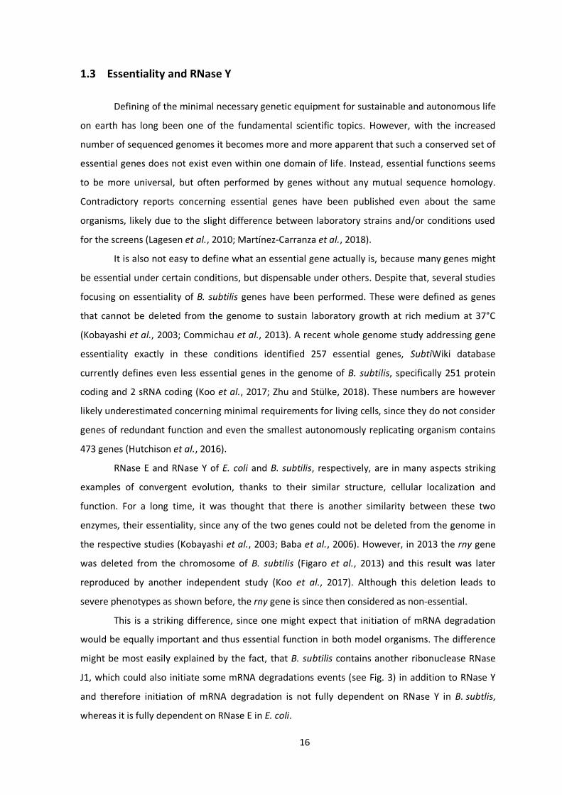
1.3 Essentiality and RNase Y
Defining of the minimal necessary genetic equipment for sustainable and autonomous life
on earth has long been one of the fundamental scientific topics. However, with the increased
number of sequenced genomes it becomes more and more apparent that such a conserved set of
essential genes does not exist even within one domain of life. Instead, essential functions seems
to be more universal, but often performed by genes without any mutual sequence homology.
Contradictory reports concerning essential genes have been published even about the same
organisms, likely due to the slight difference between laboratory strains and/or conditions used
for the screens (Lagesen et al., 2010; Martínez-Carranza et al., 2018).
It is also not easy to define what an essential gene actually is, because many genes might
be essential under certain conditions, but dispensable under others. Despite that, several studies
focusing on essentiality of B. subtilis genes have been performed. These were defined as genes
that cannot be deleted from the genome to sustain laboratory growth at rich medium at 37°C
(Kobayashi et al., 2003; Commichau et al., 2013). A recent whole genome study addressing gene
essentiality exactly in these conditions identified 257 essential genes, SubtiWiki database
currently defines even less essential genes in the genome of B. subtilis, specifically 251 protein
coding and 2 sRNA coding (Koo et al., 2017; Zhu and Stülke, 2018). These numbers are however
likely underestimated concerning minimal requirements for living cells, since they do not consider
genes of redundant function and even the smallest autonomously replicating organism contains
473 genes (Hutchison et al., 2016).
RNase E and RNase Y of E. coli and B. subtilis, respectively, are in many aspects striking
examples of convergent evolution, thanks to their similar structure, cellular localization and
function. For a long time, it was thought that there is another similarity between these two
enzymes, their essentiality, since any of the two genes could not be deleted from the genome in
the respective studies (Kobayashi et al., 2003; Baba et al., 2006). However, in 2013 the rny gene
was deleted from the chromosome of B. subtilis (Figaro et al., 2013) and this result was later
reproduced by another independent study (Koo et al., 2017). Although this deletion leads to
severe phenotypes as shown before, the rny gene is since then considered as non-essential.
This is a striking difference, since one might expect that initiation of mRNA degradation
would be equally important and thus essential function in both model organisms. The difference
might be most easily explained by the fact, that B. subtilis contains another ribonuclease RNase
J1, which could also initiate some mRNA degradations events (see Fig. 3) in addition to RNase Y
and therefore initiation of mRNA degradation is not fully dependent on RNase Y in B. subtlis,
whereas it is fully dependent on RNase E in E. coli.

17
Regardless of the fact, that deletion of rny gene is possible, this leads to severe
phenotypic defects and genomic instability (see Fig.4) suggesting that although not completely
essential, it is inevitable for the rny strain to undergo further genetic adjustments for stable life.
This is interestingly not the case for some even closely related organisms as Streptococcus
pyogenes or Staphylococcus aureus (Marincola et al., 2012; Chen et al., 2013) bringing up an
question, why is deletion of RNase Y so harmful for B. subtilis. This has not yet been discovered
and thus it remains possible that these phenotypes are caused because an essential cleavage
event is missing, as found for instance for RNase III which is essential due to its cleavage of
prophage encoded toxins (Durand et al., 2012b) or due to some general effect on total levels of
multiple mRNA species.
1.4 Natural competence in B. subtilis
Loss of competence is not only a problem for the cellular survival in its natural habitat, but
also major obstacle for the laboratory work. Since this thesis is focused on RNase Y and the
response of the cell to its absence, it is important to note that rny mutant strain has lost its ability
to become competent (Figaro et al., 2013; Koo et al., 2017). That does not only bring a slowdown
during the experimental work, but also a question why?
Competence of B. subtilis is evolved in a subpopulation of cells in response to increased
cellular density and nutritional starvation. This is fully dependent on the levels of the master
transcription regulator ComK (van Sinderen et al., 1995). Its expression is regulated in response to
extra- and intra-cellular signals by various regulators on the level of gene expression, mRNA
stability, as well as protein stability and only those cells, where ComK levels reach certain
threshold become competent in an all or nothing scenario thanks to a ComK auto activation loop
(Serror and Sonenshein, 1996; Turgay et al., 1998; Hoa et al., 2002; Hamoen et al., 2003b; Gamba
et al., 2015).
There are various mechanism translating the signals into molecular responses. The cellular
density is for instance sensed by the quorum sensing ComPA two component system, which can
respond to the levels of the ComX pheromone (Weinrauch et al., 1990; Magnuson et al., 1994).
Nutritional limitation is sensed by the transcription regulator CodY, which responds to levels of
GTP and branched‐chain amino acids (Serror and Sonenshein, 1996; Shivers and Sonenshein,
2004).
Interestingly, also other transcription regulators play a role in activation of competence
(for instance Spo0A) and they are often shared between competence and development of other
social behaviors in B. subtilis, like sporulation or biofilm formation (for review see (López et al.,
18
2009). When the master regulator ComK is present in sufficient amount, it activates expression of
more than 100 genes responsible for the DNA uptake and the recombination itself (Berka et al.,
2002; Hamoen et al., 2002; Ogura et al., 2002; Boonstra et al., 2020).
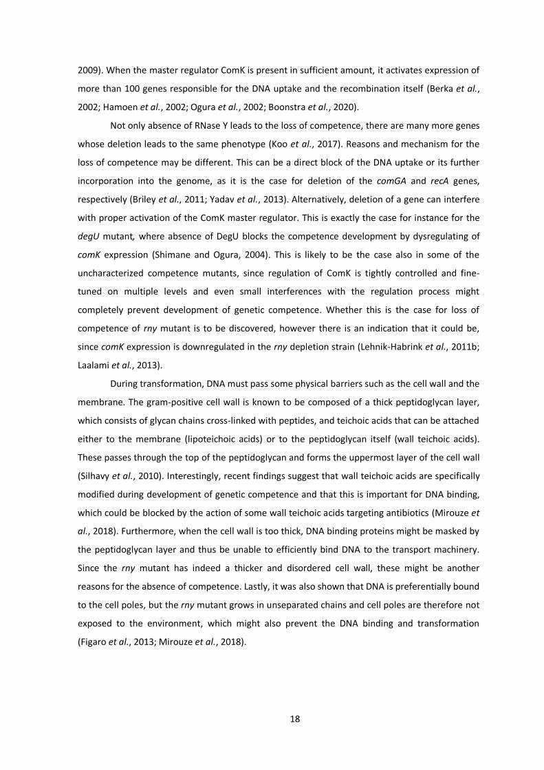
Not only absence of RNase Y leads to the loss of competence, there are many more genes
whose deletion leads to the same phenotype (Koo et al., 2017). Reasons and mechanism for the
loss of competence may be different. This can be a direct block of the DNA uptake or its further
incorporation into the genome, as it is the case for deletion of the comGA and recA genes,
respectively (Briley et al., 2011; Yadav et al., 2013). Alternatively, deletion of a gene can interfere
with proper activation of the ComK master regulator. This is exactly the case for instance for the
degU mutant, where absence of DegU blocks the competence development by dysregulating of
comK expression (Shimane and Ogura, 2004). This is likely to be the case also in some of the
uncharacterized competence mutants, since regulation of ComK is tightly controlled and fine-
tuned on multiple levels and even small interferences with the regulation process might
completely prevent development of genetic competence. Whether this is the case for loss of
competence of rny mutant is to be discovered, however there is an indication that it could be,
since comK expression is downregulated in the rny depletion strain (Lehnik-Habrink et al., 2011b;
Laalami et al., 2013).
During transformation, DNA must pass some physical barriers such as the cell wall and the
membrane. The gram-positive cell wall is known to be composed of a thick peptidoglycan layer,
which consists of glycan chains cross-linked with peptides, and teichoic acids that can be attached
either to the membrane (lipoteichoic acids) or to the peptidoglycan itself (wall teichoic acids).
These passes through the top of the peptidoglycan and forms the uppermost layer of the cell wall
(Silhavy et al., 2010). Interestingly, recent findings suggest that wall teichoic acids are specifically
modified during development of genetic competence and that this is important for DNA binding,
which could be blocked by the action of some wall teichoic acids targeting antibiotics (Mirouze et
al., 2018). Furthermore, when the cell wall is too thick, DNA binding proteins might be masked by
the peptidoglycan layer and thus be unable to efficiently bind DNA to the transport machinery.
Since the rny mutant has indeed a thicker and disordered cell wall, these might be another
reasons for the absence of competence. Lastly, it was also shown that DNA is preferentially bound
to the cell poles, but the rny mutant grows in unseparated chains and cell poles are therefore not
exposed to the environment, which might also prevent the DNA binding and transformation
(Figaro et al., 2013; Mirouze et al., 2018).

19
1.5 Aims of this thesis
Turnover of mRNA is a key regulatory process in all domains of life. RNase Y is the enzyme
initiating this process in the well-studied model organism B. subtilis, yet it could be deleted from
the genome and therefore is, by definition, considered not to be essential. However, such a
deletion leads to severe phenotypes affecting many cellular processes and to high genetic
instability. In the presented work the essentiality of RNase Y and reasons for the deleterious
phenotypes are addressed.
Analysis of suppressor mutants is used to identify the maintenance of equilibrium
between RNA synthesis and degradation as the quasi-essential function missing in the rny mutant.
Furthermore, speed of evolutionary forces and natural selection between variants present in a
bacterial population is shown. Subsequent transcriptomic analysis is used to confirm the
enormous influence of RNase Y on B. subtilis physiology and to reveal possible causes for some
specific rny related phenotypes.
In addition, a new experimental set up is established to assess the reasons for the loss of
genetic competence not only in the rny mutant strain, but also in some other previously
uncharacterized competence mutants of B. subtilis. This way, the reason for the loss of
competence as well as other social behaviors of the mutant overproducing unknown ABC
transporter YtrBCDEF is described and further investigated.

20
2 Quasi-essentiality of RNase Y in Bacillus subtilis is caused by its
critical role in the control of mRNA homeostasis
The results of this chapter are published in the following pre-print:
Benda, M., Woelfel, S., Gunka, K., Klumpp, S., Poehlein, A., Kálalová, D., Šanderová, H., Daniel,
R., Krásný, L. and Stülke, J. (2020) Quasi-essentiality of RNase Y in Bacillus subtilis is caused by its
critical role in the control of mRNA homeostasis. bioRxiv 2020.05.20.106237.
Author contribution:
MB, SW and KG constructed the strains, evolved suppressors and assessed growth. SW performed
CRISPR genome editing. MB purified the RNA polymerase, performed in vitro transcription assays
and the evolution experiment. DK and HŠ constructed pBSURNAP. AP and RD sequenced the
genomes. MB analyzed the sequences. MB, AP and RD performed transcriptome analyses. SK
build the RNA polymerase composition model. MB, KG, LK and JS designed the study. MB, LK and
JS wrote the manuscript.

21
Abstract
RNA turnover is essential in all domains of life. The endonuclease RNase Y (rny) is one of
the key components involved in RNA metabolism of the model organism Bacillus subtilis.
Essentiality of RNase Y has been a matter of discussion, since deletion of the rny gene is possible,
but leads to severe phenotypic effects. In this work, we demonstrate that the rny mutant strain
rapidly evolves suppressor mutations to at least partially alleviate these defects. All suppressor
mutants had acquired a duplication of an about 60 kb long genomic region encompassing genes
for all three core subunits of the RNA polymerase – α, β, β′. When the duplication of the RNA
polymerase genes was prevented by relocation of the rpoA gene in the B. subtilis genome, all
suppressor mutants carried distinct single point mutations in evolutionary conserved regions of
genes coding either for the β or β’ subunits of the RNA polymerase that were not tolerated by
wild type bacteria. In vitro transcription assays with the mutated polymerase variants showed a
severe decrease in transcription efficiency. Altogether, our results suggest a tight cooperation
between RNase Y and the RNA polymerase to establish an optimal RNA homeostasis in B. subtilis
cells.
Introduction
Among all organisms, bacteria are the ones multiplying most rapidly. Under optimal
conditions, the model bacteria Escherichia coli and Bacillus subtilis have generation times of 20 to
30 minutes. On the other hand, bacteria are exposed to a variety of changing environmental
conditions, and due to their small size, the impact of environmental changes is particularly severe
for bacterial cells. To adapt to these potentially rapidly changing conditions, bacteria have evolved
a huge arsenal of systems to sense and respond to the environment. Especially in the competition
between microorganisms, it is crucial that these responses are both rapid and productive.
However, while regulatory events may be very rapid, there is an element of retardation in the
system, and this is the stability of mRNA and protein molecules. If the continued activity of a
protein may become harmful to the bacteria, it is important not only to prevent expression of the
corresponding gene but also to take two important measures: (i) switch off the protein’s activity
and (ii) degrade the mRNA to exclude further production of the protein. The inactivation or even
degradation of proteins is well documented in the model bacteria. For example, in both E. coli and
B. subtilis the uptake of toxic ammonium is limited by a regulatory interaction of the ammonium
transporter with GlnK, a regulatory protein of the PII family (Coutts et al., 2002; Detsch and
Stülke, 2003). Similarly, the uptake of potentially toxic potassium can be prevented by inhibition

22
of potassium transporters at high environmental potassium concentrations, either by the second
messenger cyclic di-AMP or by interaction with a dedicated modified signal transduction protein,
PtsN (Lee et al., 2007; Corrigan et al., 2013; Gundlach et al., 2019). To prevent the accumulation
of potentially harmful mRNAs, bacteria rely on a very fast mRNA turnover. Indeed, in E. coli and B.
subtilis more than 80% of all transcripts have average half-lives of less than 8 minutes, as
compared to about 30 minutes and 10 hours in yeast or human cells, respectively (Hambraeus et
al., 2003; Yang et al., 2003; Bernstein et al., 2004; Geisberg et al., 2014). Thus, the mRNA turnover
is much faster than the generation time. The high mRNA turnover rate in bacteria contributes to
the fast adaptation even in rapidly growing cells. The rapid mRNA turnover is therefore a major
factor to resolve the apparent growth speed-adaptation trade-off.
RNases are the key elements to achieve the rapid mRNA turnover in bacteria. Theses
enzymes can degrade bulk mRNA in a rather unspecific manner, just depending on the
accessibility of the RNA molecules as well as perform highly specific cleavages that serve to
process an RNA molecule to its mature form. In all organisms, RNA degradation involves an
interplay of endo- and exoribonucleases as well as other proteins such as RNA helicases that
resolve secondary structures (Lehnik-Habrink et al., 2012; Durand et al., 2015; Redder, 2018;
Tejada-Arranz et al., 2020). Often, these proteins form a complex called the RNA degradosome. In
E. coli, the RNA degradosome is organized around the essential endoribonuclease RNase E
(Carpousis, 2007; Mackie, 2013). RNase E consists of two parts, the N-terminal endoribonuclease
domain that harbors the enzymatic activity and the C-terminal macromolecular interaction
domain that serves as the scaffold for the degradosome components and is responsible for the
binding of RNase E to the cell membrane (Khemici et al., 2008; Mackie, 2013). As mentioned
above, RNase E is essential for viability of the bacteria. An analysis of the contributions of the two
parts of RNase E to its essentiality revealed that the enzymatically active N-terminal domain is
essential whereas the C-terminal interaction domain is dispensable (Kido et al., 1996). This
suggests that the endoribonucleolytic attack on mRNA molecules is the essential function of
RNase E, whereas the interaction with other degradosome components is not required for
viability. This conclusion is supported by the fact, that the other components of the E. coli
degradosome are also dispensable (Carpousis, 2007).
RNase E is widespread in proteobacteria, cyanobacteria, and actinobacteria, but absent
from many firmicutes, -proteobacteria, or from bacteria of the Deinococcus-Thermus class.
However, an efficient RNA-degrading machinery is important also for these bacteria to allow both
rapid growth and adaptation. Indeed, these bacteria possess a different endoribonuclease, RNase
Y (Commichau et al., 2009; Shahbabian et al., 2009). A depletion of RNase Y results in a two-fold
increase of the average mRNA half-life in B. subtilis (Shahbabian et al., 2009). Similar to RNase E,

23
RNase Y is a membrane protein, and it is capable of interacting with several proteins involved in
RNA degradation. Among these proteins are the 5′‐to‐3′ exoribonunclease RNase J1,
polynucleotide phosphorylase, the RNA helicase CshA, the glycolytic proteins enolase and
phosphofructokinase, and a protein complex composed of YaaT, YlbF, and YmcA (Commichau et
al., 2009; Shahbabian et al., 2009; Lehnik-Habrink et al., 2011a; Newman et al., 2012; DeLoughery
et al., 2016; Salvo et al., 2016). Many of these interactions are likely to be transient as judged
from the distinct localization of RNase Y and its interaction partners in the cell membrane and in
the cytoplasm, respectively (Cascante-Estepa et al., 2016).
We are interested in the identification of the essential cellular components that are
required for the viability of B. subtilis cells with the aim to construct strains that harbor only the
minimal set of genes to fulfill the essential cellular functions (Commichau et al., 2013; Reuß et al.,
2016; Reuß et al., 2017). For B. subtilis, RNase Y and RNase J1 were originally described as being
essential (Kobayashi et al., 2003; Hunt et al., 2006; Mathy et al., 2007; Commichau et al., 2009;
Shahbabian et al., 2009). Interestingly, these two RNases are also present in the most genome-
reduced independently viable organism, Mycoplasma mycoides JCVI-syn3.0 (Hutchison et al.,
2016). Both RNase J1 and RNase Y are involved in the processing and degradation of a large
number of RNA molecules in B. subtilis (Mäder et al., 2008; Lehnik-Habrink et al., 2011b)(Durand
et al., 2012a; Laalami et al., 2013; DeLoughery et al., 2018). However, more recent studies
demonstrated the possibility to delete the rnjA and rny genes, encoding the two RNases (Figaro et
al., 2013; Šiková et al., 2020) and the dispensability of RNase Y was confirmed in a global
approach to inactivate all genes of B. subtilis (Koo et al., 2017).
Comprehensive knowledge on essential genes and functions is the key to construct viable
minimal genomes. By definition, essential genes cannot be individually deleted in a wild type
genetic background under standard growth conditions (Commichau et al., 2013). In this study, we
have addressed the essentiality of RNase Y in B. subtilis. While the rny gene could indeed be
deleted, this was accompanied by the rapid acquisition of suppressor mutations that affect the
transcription apparatus. We demonstrate that a strongly reduced transcription activity is required
to allow stable growth of B. subtilis in the absence of RNase Y. Our results suggest that the
accumulation of mRNA that cannot be degraded is the growth-limiting factor in strains lacking
RNase Y.
24
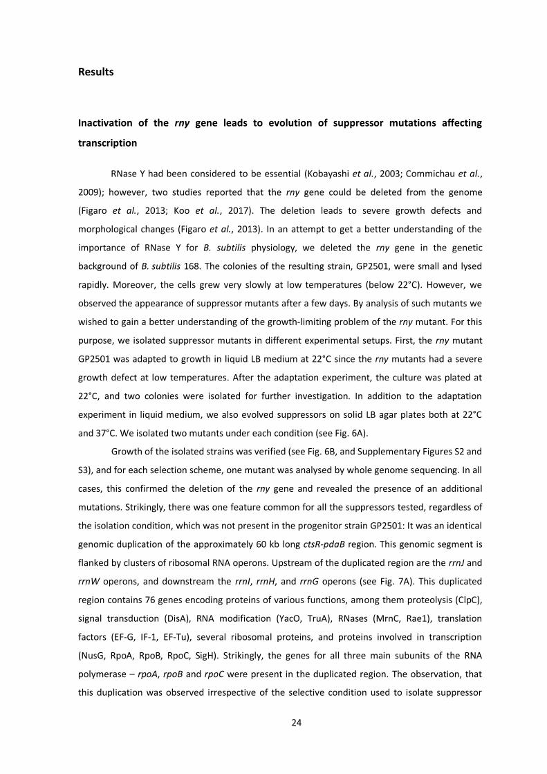
Results
Inactivation of the rny gene leads to evolution of suppressor mutations affecting
transcription
RNase Y had been considered to be essential (Kobayashi et al., 2003; Commichau et al.,
2009); however, two studies reported that the rny gene could be deleted from the genome
(Figaro et al., 2013; Koo et al., 2017). The deletion leads to severe growth defects and
morphological changes (Figaro et al., 2013). In an attempt to get a better understanding of the
importance of RNase Y for B. subtilis physiology, we deleted the rny gene in the genetic
background of B. subtilis 168. The colonies of the resulting strain, GP2501, were small and lysed
rapidly. Moreover, the cells grew very slowly at low temperatures (below 22°C). However, we
observed the appearance of suppressor mutants after a few days. By analysis of such mutants we
wished to gain a better understanding of the growth-limiting problem of the rny mutant. For this
purpose, we isolated suppressor mutants in different experimental setups. First, the rny mutant
GP2501 was adapted to growth in liquid LB medium at 22°C since the rny mutants had a severe
growth defect at low temperatures. After the adaptation experiment, the culture was plated at
22°C, and two colonies were isolated for further investigation. In addition to the adaptation
experiment in liquid medium, we also evolved suppressors on solid LB agar plates both at 22°C
and 37°C. We isolated two mutants under each condition (see Fig. 6A).
Growth of the isolated strains was verified (see Fig. 6B, and Supplementary Figures S2 and
S3), and for each selection scheme, one mutant was analysed by whole genome sequencing. In all
cases, this confirmed the deletion of the rny gene and revealed the presence of an additional
mutations. Strikingly, there was one feature common for all the suppressors tested, regardless of
the isolation condition, which was not present in the progenitor strain GP2501: It was an identical
genomic duplication of the approximately 60 kb long ctsR-pdaB region. This genomic segment is
flanked by clusters of ribosomal RNA operons. Upstream of the duplicated region are the rrnJ and
rrnW operons, and downstream the rrnI, rrnH, and rrnG operons (see Fig. 7A). This duplicated
region contains 76 genes encoding proteins of various functions, among them proteolysis (ClpC),
signal transduction (DisA), RNA modification (YacO, TruA), RNases (MrnC, Rae1), translation
factors (EF-G, IF-1, EF-Tu), several ribosomal proteins, and proteins involved in transcription
(NusG, RpoA, RpoB, RpoC, SigH). Strikingly, the genes for all three main subunits of the RNA
polymerase – rpoA, rpoB and rpoC were present in the duplicated region. The observation, that
this duplication was observed irrespective of the selective condition used to isolate suppressor

25
mutants suggests that this duplication is relevant to overcome the poor growth associated with
the loss of RNase Y. However, in addition, for each selection scheme we found additional
mutations that affect genes involved in transcription.
For the selection in liquid medium at 22°C, the suppressor mutant GP2503 had a point
mutation that resulted in an amino acid substitution (S125L) in the greA gene encoding a
transcription elongation factor (Kusuya et al., 2011). For the other suppressor mutant (GP2504)
isolated under the same selective conditions, we sequenced the greA gene to test whether it had
also acquired a mutation in this gene. Indeed, we found a different mutation in greA, resulting in
the introduction of a premature stop codon after E56. Moreover, we evolved two additional
suppressor mutants applying this adaptive scenario, and both contained frameshift mutations in
greA that resulted in premature stop codons after amino acid 23 and 137 (GP2539 and GP2538,
respectively; see Table S3).
The strain isolated on LB plates at 22°C (GP2637) had a deletion of the skin element, an
amino acid substitution (Y55N) in the AdeR activator protein (Lin et al., 2012), and a short internal
deletion in the rpoE gene encoding the subunit of RNA polymerase, which resulted in a
frameshift after residue G66 (Juang and Helmann, 1994; Rabatinová et al., 2013). For the second
mutant isolated at 22°C (GP3210), we re-sequenced the adeR and rpoE genes. While the adeR
gene was identical to the wild type, we found an insertion of an adenine residue after position 87
of rpoE, resulting in a frameshift after 29 amino acids and premature stop codon after 38 amino
acids. Therefore, the rpoE but not the adeR mutation is likely to be required for the suppressor
phenotype.
Figure 6: Suppressors of rny show increased growth at 22°C (A) Schematic depiction of different single nucleotide polymorphisms identified in the initial suppressor screen and their overlap with the duplication of ctsR-pdaB region. (B) Serial drop dilutions comparing growth of the wild type strain 168, the rny mutant GP2501, its greA suppressors (GP2503, greA (Ser125Leu) (rrnW-rrnI)2; GP2504, greA (Glu57Stop)) and the rny greA double mutant GP2628 on LB-agar plate at 22°C. The picture was taken after 2 days of incubation.

26
The suppressor evolved at 37°C on LB plates (GP2636) contained a mutation resulting in
the introduction of a premature stop at the eighth codon of the cspD gene encoding an RNA
binding protein which has transcription antitermination activity in E. coli (Graumann et al., 1997;
Bae et al., 2000). Sanger sequencing of the second suppressor isolated under the same condition
(GP2678) also identified a mutation affecting cspD, but this time in its ribosomal binding site
(GGAGGA → GGAAGA).
Taken together, the duplication of the ctsR-pdaB genomic region was
accompanied by specific additional suppressor mutation affecting transcription in every single
suppressor mutant analysed. These mutations result in the inactivation of the greA gene in liquid
medium at 22°C, whereas the selective pressure on agar plates at 22°C and 37°C was directed at
the inactivation of the RNA polymerase subunit RpoE or the RNA binding protein CspD,
respectively (see Fig. 6A). It is therefore tempting to speculate that the inactivation of these genes
combined with the ctsR-pdaB genomic duplication is causative for the suppression.
In order to test whether the inactivation of the greA, rpoE, or cspD genes alone is
sufficient for the suppression of the rny mutant strain, we constructed the corresponding double
mutants. As both rny and greA mutants are defective in genetic competence (Koo et al., 2017),
the greA rny double mutant was obtained by transforming the wild type strain 168 with DNA
molecules specifying both deletions simultaneously (see Table S3). For the greA and rpoE
deletions, the double mutants did not phenocopy the original suppressor mutants, instead the
gene deletions conferred only partial suppression (see Fig. 6B for the rny greA double mutant
GP2628, and Supplementary Figure S2 for the rny rpoE double mutant GP3217). In the case of the
rny cspD double mutant GP2615, complete suppression was observed (see Supplementary Figure
S3). However, we cannot exclude that the mutant had already acquired the duplication of the
ctsR-pdaB genomic region. Thus, we conclude that the suppression depends on both, the
duplication of the ctsR-pdaB region and the concomitant mutations that inactivate genes involved
in transcription.
Transcriptome analysis of the rny mutant and a suppressor strain
As mentioned above, the deletion of greA allowed only partial suppression of the growth
defect caused by the loss of RNase Y. However, the rny greA double mutant GP2628 eventually
gave rise to a better suppressing mutant, GP2518. Whole genome sequencing of this strain
revealed that in addition to the greA deletion it had only acquired the duplication of the ctsR-
pdaB genomic region. Again, this highlights the relevance of the combination of the greA deletion
and the ctsR-pdaB duplication for suppression.
27
To get insights into the global consequences of the suppressing mutations, we compared
the transcriptomes of the wild type strain 168, the rny mutant GP2501, and the suppressor
mutant GP2518 by RNA-Seq analysis. We identified 1,102 genes (corresponding to about 25% of
all genes of B. subtilis) with at least two-fold differential expression in the Δrny strain GP2501 as
compared to the wild type 168. It should be noted that the number of differentially expressed
genes is likely to be underestimated, since about 50% of all genes are not or only very poorly
expressed during vegetative growth (Rasmussen et al., 2009; Reuß et al., 2017). The rny gene is
encoded within an operon with the ymdB gene (Diethmaier et al., 2011); however, there was no
polar effect on the expression of ymdB, suggesting that the observed changes are a direct result
of the loss of RNase Y.
From the dataset mentioned above, 587 and 515 genes were down- and upregulated,
respectively, in the rny strain. The most severe difference (more than 100-fold decrease) was
observed for the yxkC gene. This gene codes for protein of unknown function and is part of the σD
regulon (Serizawa et al., 2004). Interestingly, 14 out of the 30 most strongly downregulated genes
are σD dependent (see Supplementary Table S1). This may be the result of the reduced expression
of the sigD gene itself. Since σD controls the expression of many genes responsible for motility as
well as peptidoglycan autolysins (lytA, lytB,lytC, lytD and lytF) this reduced expression of target
genes might cause the disordered cell wall of the rny deletion strain (Figaro et al., 2013). Among
the most strongly upregulated genes (see Supplementary Table S1), many are members of the
general stress response factor σB regulon. Another set of upregulated genes is controlled by the
sporulation specific sigma factors σF and σG, whose genes are also more than 4-fold upregulated.
This is especially striking taking into an account that the rny mutant strain is not able to form
spores (Figaro et al., 2013).
Importantly, we wanted to test whether the suppressor mutant had restored a wild type-
like expression of genes that were affected by the loss of RNase Y. We found 461 genes with
differential expression between the suppressor mutant GP2518 and the rny mutant GP2501. Of
these, however, only some were returned towards the expression levels of the wild type (176
genes, see Supplementary Table S2), while for others, the mRNA levels were even more distant
from the wild type. In total 115 genes upregulated in the rny strain showed reduced expression in
the suppressor mutant. On the other hand, also 61 genes which were downregulated in the rny
mutant, had increased their expression again in the suppressor mutant GP2518 (see
Supplementary Table S2). Among these genes with restored expression, four (murAA, tagA, tagB,
ywpB) are essential, and only the expression of ywpB encoding an enzyme of fatty acid
biosynthesis is 2.4-fold reduced in the rny mutant. This weak regulation suggests that fatty acid
biosynthesis is not the growth-limiting factor for the rny mutant. In contrast, many of these genes

28
with (partially) restored expression belong to prophage PBSX or are required for rather specific
metabolic pathways. In conclusion, the evaluation of the genes which had their expression
restored as a result of the suppressing mutations did not give a clear clue to the reason of
suppression.
Genomic separation of the genes encoding the core subunits of RNA polymerase
As mentioned above, the region duplicated in all suppressor mutants contained genes
encoding RNA modification enzymes, translation factors, ribosomal proteins, RNases, and
proteins involved in transcription. MrnC and Rae1 are RNase Mini-III required for the maturation
of 23S rRNA and ribosome-associated A site endoribonuclease, respectively (Redko et al., 2008;
Leroy et al., 2017). As our suppressor screen identified additional mutations related to
transcription, we assumed that the translation-specific RNases encoded in this region might not
be relevant for the suppression of the rny deletion. Therefore, we hypothesized that the
duplication of the genes encoding the main three subunits of RNA polymerase made a major
contribution to the selective advantage provided by the duplication.
To test the idea that simultaneous duplication of all three genes for the RNA polymerase
core subunits is the key for the suppression of the loss of RNase Y, we decided to interfere with
this possibility. The duplicated region is located between two highly conserved rrn gene clusters
which may facilitate the duplication event (see Fig. 7A). Therefore, we attempted to separate the
core RNA polymerase genes by relocating the rpoA gene out of this genomic region flanked by the
Figure 7: Genomic organization of the duplicated genomic region (A) Schematic representation of the first 180 kb of the B. subtilis chromosome. The orange box indicates the duplicated region in the suppressors of rny strain GP2501. rRNA operons are depicted as green rectangles, RNA polymerase genes rpoA, rpoB, rpoC as blue arrows, the ctsR and pdaB genes are shown in yellow and red, respectively. (B) Chromosomal relocation of the rpoA gene. For the colour code, see above; the relocated rpoA is shown as a purple arrow.

29
rrn operons. We assumed that if RNA polymerase was indeed the key to the original suppression,
such a duplication would not be likely in the new background with relocated rpoA, since
simultaneous duplication of all three RNA polymerase subunit genes would be disabled there. For
this purpose, the rpoA gene kept under the control of its natural promoter PrpsJ was placed
between the dgk and yaaH genes, and the original copy of rpoA was deleted (see Fig. 7B,
Experimental procedures for details). We then compared the growth of the wild type strain 168
and the strain with the relocated rpoA GP2903 using a drop-dilution assay. No differences were
observed, thus excluding a possible negative impact of the rpoA relocation on B. subtilis
physiology (see Fig. S4).
Strain GP2903 was then used to delete the rny gene, and to isolate suppressor mutants.
Indeed, even with the genomically separated RNA polymerase genes, suppressor mutations
appeared upon the deletion of the rny gene encoding RNase Y. There were three possibilities for
the outcome of the experiment. First, the same genomic region as in the original suppressors
might duplicate thus falsifying our hypothesis that the simultaneous duplication of all three genes
encoding the core subunits of RNA polymerase is required for suppression. Second, both regions
containing the rpoA and rpoBC genes might be duplicated. Third, in the new genetic background
completely new suppressing mutations might evolve. Two of these suppressor mutants were
subjected to whole genome sequencing. None of them had the duplication of the ctsR-pdaB
region as in the original suppressors. Similarly, none of the mutants had the two regions
containing the rpoA and the rpoBC genes duplicated. Instead, both mutants had point mutations
in the RNA polymerase subunit genes that resulted in amino acid substitutions (GP2912: RpoC,
R88H; GP2913: RpoB, G1054C; see Table S3). A mutation affecting RNA polymerase was also
evolved in one strain (GP2915) not subjected to whole genome sequencing. In this case, the
mutation resulted in an amino acid substitution (G45D) in RpoC.
An analysis of the localization of the amino acid substitutions in RpoB and RpoC revealed
that they all affect highly conserved amino acid residues (see Fig. 8A). G1054 of RpoB and G45 of
RpoC are universally conserved in RNA polymerases in all domains of life, and R88 of RpoC is
conserved in the bacterial proteins. This high conservation underlines the importance of these
residues for RNA polymerase function. The mutations G45D and R88H in RpoC affect the N-
terminal β’ zipper and the zinc-finger like motif of the β′ subunit, respectively, that are required
for the processivity of the elongating RNA polymerase (Nudler et al., 1996; Nudler, 2009). G1054C
in RpoB is located in the C-terminal domain of the β subunit that is involved in transcription
termination (Clerget et al., 1995). In the three-dimensional structure of RNA polymerase, these
regions of the β and β′ subunits are located in close vicinity opposite to each other in the region of

30
the RNA exit channel which guides newly transcribed RNA out of the enzyme (see Fig. 8B; Nudler,
2009), and they are both in direct contact with DNA (Nudler et al., 1996).
The fact that several independent mutations affecting RNA polymerase were obtained in
the suppressor screen strongly supports the idea that RNA polymerase is the key for the
suppression. As the mutations affect highly conserved residues, they are likely to compromise the
enzyme’s activity. Based on the structural information, the mutations might weaken RNA
polymerase-nucleic acid interactions and therefore, destabilize the transcription elongation
complex which may result in increased
premature termination and reduced RNA polymerase processivity. However, RNA polymerase is
essential, therefore the mutations cannot inactivate the protein completely.
Figure 8: Suppressor mutations in RNA polymerase localize to evolutionary conserved regions (A) Multiple sequence alignment of RpoB and RpoC sequences from various species, the numbering of amino acid residues is based on the B. subtilis sequence. The positions of mutations are indicated with red double head arrows, conserved cysteines involved in Zn-finger formation are shown in red. Logos were created as described (98). Abbreviations: B. subtilis, Bacillus subtilis; E. coli, Escherichia coli; M. tuberculosis, Mycobacterium tuberculosis; T. thermophilus, Thermus thermophilus; M. genitalium, Mycoplasma genitalium; S. acidocaldarius, Sulfolobus acidocaldarius; H. sapiens, Homo sapiens. (B) Localization of the mutations (indicated as red spheres) in the RNA polymerase shown at their corresponding position in the structure of T. thermophilus (PDB ID: 1IW7; 99). The two α subunits are shown in dark red and violet, respectively, the ß subunit is shown in dark blue, ß’ in cyan, ω in gold and the σ subunit is shown in grey. The image was created using UCSF Chimera (Pettersen et al., 2004).

31
Establishing the rpoB and rpoC mutations in wild type background
Based on the essentiality of transcription, we expected that the mutations in rpoB and
rpoC that we have identified in the suppressor screen with the rny mutant and genomically
separated RNA polymerase genes might adjust some of the properties of RNA polymerase. To
study the consequences of these mutations for the RNA polymerase and hence also for the
physiology of B. subtilis, we decided to introduce one of them (RpoC-R88H) into the wild type
background of B. subtilis 168. For this purpose, the CRISPR/Cas9 system designed for use in B.
subtilis was employed (Altenbuchner, 2016). As a control, we used the same procedure to
introduce a mutation in the rae1 gene, which is located nearby on the chromosome. Although this
system readily allowed the introduction of a frameshift mutation (introduction of an extra T after
32 bp) in rae1 (strain GP2901), we failed to isolate genome-edited clones expressing the RpoC-
R88H variant in multiple attempts. This failure to construct the RpoC-R88H variant in the wild type
background suggests that the properties of the protein are altered in a way that is incompatible
with the presence of an intact RNA degradation machine.
Mutated RNA polymerases have highly decreased activity in vitro
Since our attempts to study the effect of the mutations in vivo failed, we decided to test
the properties of the mutant RNA polymerases using in vitro transcription. B. subtilis RNA
polymerase is usually purified from a strain expressing His-tagged RpoC (Qi and Hulett, 1998).
However, the loss of competence of the rny mutant and the lethality of the rpoC mutation in the
wild type background prevented the construction of a corresponding strain. To solve this
problem, we used an approach to purify B. subtilis RNA polymerase from E. coli that had been
successful before for RNA polymerase of Mycobacterium smegmatis (Kouba et al., 2019). Briefly,
plasmid pBSURNAP containing genes rpoA, rpoB, rpoC, rpoE, rpoY, and rpoZ for the RNA
polymerase subunits under control of an IPTG inducible promoter was constructed in a way that
each individual gene for a subunit could be cleaved out using unique restriction sites and replaced
with its mutant counterpart, yielding pGP2181 (RpoC-R88H) and pGP2182 (RpoB-G1054C) (for
details of the construction, see Experimental procedures). The variant RNA polymerases were
expressed in E. coli BL21 and purified via affinity chromatography and subsequent size exclusion
chromatography.
We purified the wild type and two mutant RNA polymerases (RpoC-R88H and RpoB-
G1054C) and assessed their activity by in vitro transcription on three different templates,
containing well-studied promoters of the veg and ilvB genes and the P1 promoter of the rrnB

32
operon (Krásný and Gourse, 2004; Krásný et al., 2008). In agreement with previous results with
wild type RNA polymerase (Sojka et al., 2011), this enzyme performed well on all three substrates.
In contrast, the mutated variants of RNA polymerase exhibited a drastic decrease of transcription
activity on all three promoters; for the RpoB-G1054C variant the transcripts were only barely
detectable (Fig. 9A).
On many promoters, including the P1 promoter of the rrnB operon, B. subtilis RNA
polymerase is sensitive to the concentration of the first transcribed nucleotide both in vitro and in
vivo (Krásný and Gourse, 2004). This prompted us to compare the response of the wild type and
the RpoC-R88H variant RNA polymerases to different concentrations of GTP, the initiation NTP for
the rrnB P1 transcript. As described before, transcription with the wild type enzyme increased
gradually in response to the GTP concentration (Krásný and Gourse, 2004). In contrast, the
mutated variant was saturated with a relatively low GTP concentration, suggesting that this
important regulatory mechanism is not functional here (see Fig. 9B).
Taken together, our results suggest that a reprogramming of the properties of RNA
polymerase as indicated by a substantial reduction in RNA polymerase activity and its altered
ability to be regulated by iNTPs allows the suppressor mutants to overcome the loss of RNase Y.
Figure 9: Comparison of transcriptional activity between RNA polymerase variants (A) The RNA polymerase variants (64 nM) were reconstituted with saturating concentrations of σA (1:10). Holoenzymes were used to initiate transcription on three promoters as indicated. A representative image from three independent experiments is shown. (B) Transcription from the rrnB P1 promoter in dependence on increasing concentration of iNTP (GTP). The intensity of the transcripts generated by RNA polymerase containing RpoC-R88H was adjusted for better visibility. The relative activity of this mutant RNA polymerase was 2.5% of the wild type RNA polymerase at 2,000 μM GTP. The graph shows average of two replicates normalized for maximal transcription of each polymerase (set as 1).
33
A pre-existing duplication of the genomic region containing rpoA and rpoBC is fixed in
response to the deletion of rny
The screen for suppressor mutations that facilitate growth of strains lacking RNase Y
yielded two classes of mutants: the first set harboured mutations in genes involved in
transcription (greA, rpoE, or cspD) in addition to a duplication of the chromosomal region
encoding the core subunits of RNA polymerase. The second class had point mutations affecting
the β or β′ subunits of RNA polymerase that result in strongly decreased transcription activity. At
a first glance, these results seem to be conflicting. Considering RNA degradation as the function of
RNase Y, it seemed plausible that the selective pressure caused by deletion of rny should result in
alleviating the stress from mRNA accumulation. This seems to be the case in the second class of
suppressors (see above), whereas the logic behind the duplication seems to be less obvious.
Importantly, this duplication was always accompanied by one of the other aforementioned
mutations affecting transcription. In an attempt to determine the order of the evolutionary
events in these suppressors we established a method to detect the presence of the duplication
without whole genome sequencing. For this, we made use of a pair of oligonucleotides that binds
to the pdaB and ctsR genes giving a product of about 10 kb, if the region is duplicated or amplified
but no product in the absence of duplication or amplification (see Fig. 10A). This PCR product was
very prominent for the strain GP2636 that is known to carry the duplication. However a band was
also observed in the wild type strain 168, indicating that the duplication is present in a part of the
population independent from the selective pressure exerted by the rny deletion (Fig. 10B).
It is well-established that genomic duplications or amplifications occur frequently in
bacterial populations, even in the absence of selective pressure (Andersson and Hughes, 2009). In
Salmonella typhimurium, rrn operons have been shown to be a hotspot of gene duplications or
amplifications (Anderson and Roth, 1981). Since evolution of such a genomic duplication is
dependent on homologous recombination, we performed the PCR also on the recA mutant
GP2542, which is defective in homologous recombination and thus unable to amplify
chromosomal regions (Dormeyer et al., 2017; Reuß et al., 2019). Indeed, in this case we did not
obtain even a faint band. Interestingly, the genomic duplication can also be observed in cells
having the core subunits of RNA polymerase at distinct genomic regions (GP2903). For the derived
suppressor mutant GP2912 that carries a point mutation in rpoC, the band indicating the presence
of the duplication was also detectable by PCR analysis although the duplication could not be
detected by genome sequencing. This apparent discrepancy is most easily resolved by assuming
that the duplication was present only in a small subpopulation (as observed for the wild type
strain) and therefore only detectable by the very sensitive PCR assay.
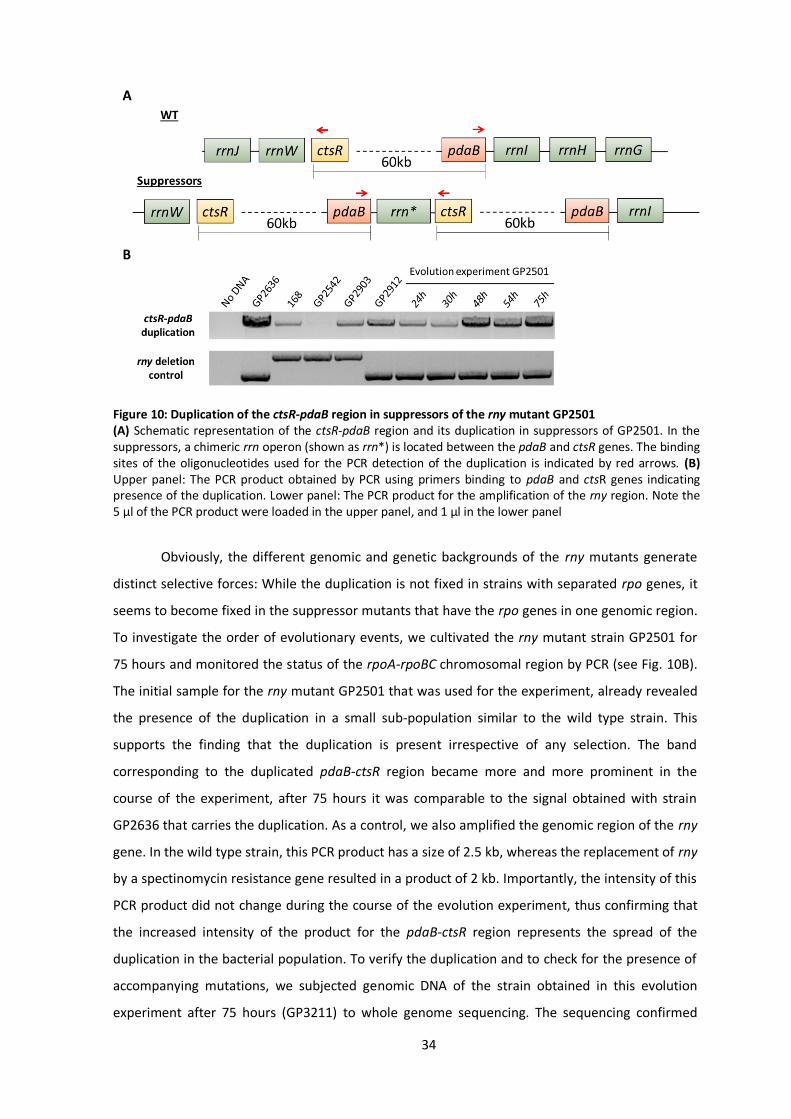
34
Obviously, the different genomic and genetic backgrounds of the rny mutants generate
distinct selective forces: While the duplication is not fixed in strains with separated rpo genes, it
seems to become fixed in the suppressor mutants that have the rpo genes in one genomic region.
To investigate the order of evolutionary events, we cultivated the rny mutant strain GP2501 for
75 hours and monitored the status of the rpoA-rpoBC chromosomal region by PCR (see Fig. 10B).
The initial sample for the rny mutant GP2501 that was used for the experiment, already revealed
the presence of the duplication in a small sub-population similar to the wild type strain. This
supports the finding that the duplication is present irrespective of any selection. The band
corresponding to the duplicated pdaB-ctsR region became more and more prominent in the
course of the experiment, after 75 hours it was comparable to the signal obtained with strain
GP2636 that carries the duplication. As a control, we also amplified the genomic region of the rny
gene. In the wild type strain, this PCR product has a size of 2.5 kb, whereas the replacement of rny
by a spectinomycin resistance gene resulted in a product of 2 kb. Importantly, the intensity of this
PCR product did not change during the course of the evolution experiment, thus confirming that
the increased intensity of the product for the pdaB-ctsR region represents the spread of the
duplication in the bacterial population. To verify the duplication and to check for the presence of
accompanying mutations, we subjected genomic DNA of the strain obtained in this evolution
experiment after 75 hours (GP3211) to whole genome sequencing. The sequencing confirmed
Figure 10: Duplication of the ctsR-pdaB region in suppressors of the rny mutant GP2501 (A) Schematic representation of the ctsR-pdaB region and its duplication in suppressors of GP2501. In the suppressors, a chimeric rrn operon (shown as rrn*) is located between the pdaB and ctsR genes. The binding sites of the oligonucleotides used for the PCR detection of the duplication is indicated by red arrows. (B) Upper panel: The PCR product obtained by PCR using primers binding to pdaB and ctsR genes indicating presence of the duplication. Lower panel: The PCR product for the amplification of the rny region. Note the 5 µl of the PCR product were loaded in the upper panel, and 1 µl in the lower panel
35
presence of the duplication, but did not reveal any additional suppressor mutation. Based on this
result, we can assume that upon deletion of rny the bacteria first fixed the duplication of the
pdaB-ctsR region and then, later, may acquire the point mutations affecting greA, rpoE, or cspD.
Perturbing stoichiometry of transcription complexes reduces RNA polymerase activity
In the investigation of suppressor mutants we have found suppressor mutants that
exhibited severely reduced RNA polymerase activity as well as suppressor mutants with increased
copy number of core RNA polymerase subunit genes. In the latter mutants, one might expect that
the increased copy number of RNA polymerase core subunit genes would result even in increased
transcription, apparently in contradiction to the other set of suppressors. However, the outcome
of gene duplication may just be the opposite: The RNA polymerase is a complex multi-protein
machine that contains several important proteins in addition to the core subunits. As these
factors, including the sigma factor and other subunits like RpoE, RpoY and RpoZ (Juang and
Helmann, 1994; Doherty et al., 2010; Delumeau et al., 2011; Rabatinová et al., 2013; Keller et al.,
2014) as well as transcription factors like GreA and NusA (Davies et al., 2005; Kusuya et al., 2011)
bind to the RNA polymerase via the core subunits, the perturbation of the normal evolved
equilibrium between the RNA polymerase core subunits and transcription factors is likely to result
in the formation of abortive incomplete complexes that are not fully active in transcription. To
obtain a quantitative estimate for the formation of incomplete complexes, we turned to
modelling.
We estimated the stoichiometry of the complexes in the wild type from proteomic mass
fractions of the components (Reuß et al., 2017), calculating the number ratio of the subunit or
transcription factor to the core RNA polymerase. These data indicate that GreA and the RpoZ
subunit are in excess of core RNA polymerase, but not NusA, σA as well as the RpoE and RpoY
subunits (Fig. 11A). Since σA is needed during initiation of transcription and NusA during
elongation, we make the simplifying assumption that these two factors bind to the core RNA
polymerase subsequently with NusA replacing σA during transcription elongation, such that only
one of them is present in the complex and their numbers can effectively be summed up (O’Reilly
et al., 2020). Taken together, their number is only slightly smaller than that of core RNA
polymerases (90%). This means that, in the wild type, 90% of all core RNA polymerases can form a
complete complex including GreA, RpoZ and either σA or NusA depending on the stage of
transcription.
This fraction is strongly reduced if the core subunits are duplicated relative to the other
subunits: To see that we make the assumption that the small subunits and transcription factors

36
bind to the core RNA polymerase independently of each other. Upon duplication, the core RNA
polymerase is in excess of all subunits and factors and thus a variety of partial complexes can be
formed. The probability that a complex contains a specific set of factors is obtained by
interpreting the stoichiometric ratio of a subunit to core as the probability that a core RNA
polymerase will bind the subunit. The combinatorics of those probabilities give the fractions of
the various complexes. For a complete complex consisting of core RNA polymerase, GreA, RpoZ
and A/NusA, this leads to 0.6 x 0.85 x (0.15 + 0.3) ≈ 0.23, indicating that a duplication of the core
subunits may result in a reduction of the fraction of complete complexes down to 23% of the core
RNA polymerases in contrast to 90% in the wild type strain. This will result in reduced
transcription activity even if there are twice as many core RNA polymerases than in the wild type
since a variety of incomplete complexes containing different subsets of the subunits and
transcription factors are formed (Fig. 11B). In the same way, we can estimate the fraction of
complexes that contain the RpoE and RpoY subunits in addition. These complexes make up only
59% of all core RNA polymerases already in the wild type and their fraction is reduced down to 8%
upon core duplication. Thus, a duplication of the core subunit genes is indeed expected to result
in a strong decrease of the transcription activity.
Figure 11: The duplication of the genes for core RNA polymerase is likely to result in the formation of incomplete RNA polymerase complexes (A) Relative abundance/stoichiometry of RNA polymerase subunits and associated factors from
proteomics data (Reuß et al., 2017). (B) Fractions of core RNA polymerase in different complete (green) and incomplete (grey) complexes estimated based on the relative abundance in (A) for the wild type and for the core duplication strain, where the relative abundance of core subunits is doubled compared to all other subunits.
37
Discussion
RNases E and Y are the main players in RNA degradation in E. coli and B. subtilis,
respectively. Recently, it has been estimated that about 86% of all bacteria contain either RNase E
or RNase Y (or, sometimes, both) supporting the broad relevance of these two enzymes (Tejada-
Arranz et al., 2020). While RNase E of E. coli is essential (Hammarlöf et al., 2015), conflicting
results concerning the essentiality of RNase Y have been published (Kobayashi et al., 2003; Hunt
et al., 2006; Commichau et al., 2009; Figaro et al., 2013; Koo et al., 2017). In this study, we have
examined the properties of B. subtilis mutants lacking RNase Y due to deletion of the
corresponding rny gene. We observed that the rny mutant grew poorly, and rapidly acquired
secondary mutations that suppressed, at least partially, the growth defect caused by the deletion
of the rny gene. Thus, we conclude that RNase Y is in fact quasi-essential (Hutchison et al., 2016)
for B. subtilis, since the mutant cannot be stably propagated on complex medium without
acquiring suppressor mutations.
A lot of effort has been devoted to the understanding of the reason(s) of the (quasi)-
essentiality of RNases E and Y for E. coli and B. subtilis, respectively. Initially, it was assumed that
the essentiality is caused by the involvement of these RNases in one or more key essential
processing event(s) that may affect the mRNAs of essential genes as has been found for B. subtilis
RNase III and E. coli RNase P (Lehnik-Habrink et al., 2011b; Durand et al., 2012a; Durand et al.,
2012b; Laalami et al., 2013; Mohanty et al., 2020). However, such a target was never identified.
Instead, different conclusions were drawn from suppressor studies with E. coli rne mutants
lacking RNase E: some studies reported suppression by the inactivation or overexpression of
distinct genes, such as deaD encoding a DEAD-box RNA helicase and ppsA encoding
phosphoenolpyruvate synthetase, respectively (Tamura et al., 2012; Tamura et al., 2016). In
addition, the processing and degradation of the essential stable RNAs, such as tRNAs and rRNAs
was shown to be an essential function of RNase E (Sulthana et al., 2016). Yet another study
suggested that mRNA turnover is the growth-limiting factor of the E. coli rne mutant (Hammarlöf
et al., 2015). The results presented here lend strong support to the idea that the main task of
RNase Y in B. subtilis is the control of intracellular mRNA concentration via the initiation of mRNA
degradation. The transcriptome analysis with the rny mutant and a suppressor mutant revealed
that only a limited number of genes shows restored expression in the suppressor mutant.
Moreover, most of these genes are part of the prophage PBSX or encode very specific metabolic
functions. In addition, irrespective of the conditions used in the different suppressor screens, we
identified a coherent set of mutations that resulted in improved growth of the B. subtilis rny
mutant. The initial mutants carry a duplication of the chromosomal region that contains the genes

38
for the core subunits of RNA polymerase (RpoA, RpoB, RpoC) and point mutations in greA, rpoE,
and cspD that all affect transcription. If this duplication was prevented by genomically separating
the RNA polymerase genes, we found suppressor mutants affecting the core subunits of RNA
polymerase which result in strongly compromised transcription activity. Taken together, these
findings suggest that the (quasi)-essentiality of RNases E and Y is related to their general function
in initiating mRNA turnover rather than to the processing of specific RNA species. This idea is
further supported by two lines of evidence: First, mutations that mimic a stringent response and
therefore reduce RNA polymerase activity suppressed the growth defect of an rne mutant, and
second, artificial expression of RNase Y or of the ribonucleases RNase J1 or J2 from B. subtilis
partially suppressed the E. coli strain lacking RNase E, but only under specific growth conditions
(Tamura et al., 2017; Himabindu and Anupama, 2017).
With the initiation of global mRNA degradation as the (quasi)-essential function of RNases
E and Y in E. coli and B. subtilis, respectively, one might expect that the overexpression of other
RNases might compensate for their loss. By analogy, such a compensation has been observed for
the essential DNA topoisomerase I of B. subtilis, which could be replaced by overexpression of
topoisomerase IV (Reuß et al., 2019). However, in all the seven suppressor mutants analyzed by
whole genome sequencing (see Table S3), we never observed a mutation affecting any of the
known RNases of B. subtilis. Similarly, no such compensatory mutations resulting from
overexpression of other cognate RNases have been found in suppressor screens for E. coli RNase
E. While RNase Y does not have a paralog in B. subtilis, E. coli possesses the two related RNases E
and G. However, not even the overexpression of RNase G allowed growth of an E. coli rne mutant
(Deana and Belasco, 2004; Chung et al., 2010) suggesting that RNase G has a much more narrow
function than RNase E and that none of the other RNases in either bacterium is capable of
initiating global mRNA degradation. Interestingly, as mentioned above, RNase J1 could partially
replace RNase E in E. coli (Tamura et al., 2017), whereas it is not able to replace RNase Y in B.
subtilis. This difference could be due to the fact that RNase J1 provides an additional pathway to
initiate mRNA degradation in B. subtilis, which is not naturally present in E. coli. This idea is
further supported by the observation that a B. subtilis strain lacking both RNases Y and J1 could
never be constructed (Figaro et al., 2013).
An interesting result of this study was the apparent contradiction between the isolation of
suppressor mutants with increased copy number of core RNA polymerase subunit genes in one
setup, intuitively suggesting increased transcription activity, and the isolation of mutants that
exhibited severely reduced RNA polymerase activity in the other setup. We therefore tested with
a theoretical model whether duplication of the core subunits leads to abortive incomplete
complexes, as the composition of the RNA polymerase complex might be perturbed by the

39
duplication of the core. The calculations indicate that most (90%) of the core RNA polymerases in
the wild type are associated with GreA and RpoZ as well as either sigma or NusA, depending on
their stage in the transcription process, while upon core subunit gene duplication, the fraction of
complete complexes, i.e. complexes associated to all these factors, is strongly reduced (to 23%).
Thus, the model shows that perturbing the stoichiometry of the transcription machinery results in
a strong reduction of the fraction of core RNA polymerases that assemble a complete complex. As
a consequence, a duplication of the core subunit genes is indeed expected to result in a strong
decrease of the transcription activity, resolving the apparent contradiction.
In each organism, an optimal trade-off between RNA synthesis and degradation must be
adjusted to allow optimal growth. Obviously, the loss of the major RNA decay-initiating enzyme
will bring this adjustment out of equilibrium. This idea is supported by the observation that
reduced RNA degradation in B. subtilis is accompanied by the acquisition of mutations that
strongly reduce transcription activity of the RNA polymerase. Actually, the reduction of activity
was so strong that it was not tolerated in a wild type strain with normal RNA degradation. This
indicates that the suppressor mutants have reached a new stable equilibrium between RNA
synthesis and degradation, which, however, is not optimal as judged from the reduced growth
rates of the suppressor mutants as compared to the wild type strain. It has already been noticed
that generation times and RNA stability are directly related (Yang et al., 2003; Rustad et al., 2013).
This implies that a stable genetic system requires a balance between transcription and RNA
degradation to achieve a specific growth rate. In bacteria, rapid growth requires high transcription
rates accompanied by rapid RNA degradation. The association between RNA polymerase and
components of the RNA degrading machinery, as shown for B. subtilis and Mycobacterium
tuberculosis might be a factor to achieve this coupling between RNA synthesis and degradation
(Delumeau et al., 2011; Płociński et al., 2019).
In conclusion, our study suggests that the initiation of mRNA degradation to keep the
equilibrium between RNA synthesis and degradation is the function of RNase Y that makes it
quasi-essential for B. subtilis. In addition to RNase Y, RNase J1 is also quasi-essential for this
bacterium. In the future, it will be interesting to understand the reasons behind the critical role of
this enzyme as well in order to get a more comprehensive picture of the physiology of RNA
metabolism.
40
Experimental procedures
Bacterial strains, plasmids and growth conditions
All B. subtilis strains used in this study are listed in Table S3. All strains are derived from
the laboratory strain 168 (trpC2). B. subtilis and E. coli cells were grown in Lysogeny Broth (LB
medium; Sambrook et al., 1989). LB plates were prepared by addition of 17 g Bacto agar/l (Difco)
to LB (Sambrook et al., 1989). The plasmids are listed in Table S4. Oligonucleotides are listed in
Table S5.
DNA manipulation and genome sequencing
B. subtilis was transformed with plasmids, genomic DNA or PCR products according to the
two-step protocol (Sambrook et al., 1989; Kunst and Rapoport, 1995). Transformants were
selected on LB plates containing erythromycin (2 µg/ml) plus lincomycin (25 µg/ml),
chloramphenicol (5 µg/ml), kanamycin (10 µg/ml), or spectinomycin (250 µg/ml). Competent cells
of E. coli were prepared and transformed following the standard procedure (Sambrook et al.,
1989) and selected on LB plates containing kanamycin (50 µg/ml). S7 Fusion DNA polymerase
(Mobidiag, Espoo, Finland) was used as recommended by the manufacturer. DNA fragments were
purified using the QIAquick PCR Purification Kit (Qiagen, Hilden, Germany). DNA sequences were
determined by the dideoxy chain termination method (Sambrook et al., 1989). Chromosomal DNA
from B. subtilis was isolated using the peqGOLD Bacterial DNA Kit (Peqlab, Erlangen, Germany). To
identify the mutations in the suppressor mutant strains GP2503, GP2518, GP2636, GP2637,
GP2912, GP2913, and GP3211 (see Table S3), the genomic DNA was subjected to whole-genome
sequencing. Concentration and purity of the isolated DNA was first checked with a Nanodrop ND-
1000 (PeqLab, Erlangen, Germany) and the precise concentration was determined using the
Qubit® dsDNA HS Assay Kit as recommended by the manufacturer (Life Technologies GmbH,
Darmstadt, Germany). Illumina shotgun libraries were prepared using the Nextera XT DNA Sample
Preparation Kit and subsequently sequenced on a MiSeq system with the reagent kit v3 with 600
cycles (Illumina, San Diego, CA, USA) as recommended by the manufacturer. The reads were
mapped on the reference genome of B. subtilis 168 (GenBank accession number: NC_000964)
(Barbe et al., 2009). Mapping of the reads was performed using the Geneious software package
(Biomatters Ltd., New Zealand) (Kearse et al., 2012). Frequently occurring hitchhiker mutations
(Reuß et al., 2019) and silent mutations were omitted from the screen. The resulting genome
sequences were compared to that of our in-house wild type strain. Single nucleotide

41
polymorphisms were considered as significant when the total coverage depth exceeded 25 reads
with a variant frequency of ≥90%. All identified mutations were verified by PCR amplification and
Sanger sequencing. Copy numbers of amplified genomic regions were determined by dividing the
mean coverage of the amplified regions by the mean coverage of the remaining genome as
described previously (Dormeyer et al., 2017; Reuß et al., 2019).
Construction of deletion mutants
Deletion of the rny, rpoA, and cspD genes was achieved by transformation with PCR
products constructed using oligonucleotides to amplify DNA fragments flanking the target genes
and intervening antibiotic resistance cassettes as described previously (Youngman, 1990;
Guérout-Fleury et al., 1995; Wach, 1996). The identity of the modified genomic regions was
verified by DNA sequencing.
Chromosomal relocation of the rpoA gene
To construct a strain in which the genes for the core subunits of RNA polymerase are
genomically separated, we decided to place the rpoA gene between the dgk and yaaH genes, and
then to delete the original copy of the gene. First, the rpoA gene was fused in a PCR reaction with
its cognate promoter and a chloramphenicol resistance gene at the 5′ and 3′ ends, respectively. In
addition, the amplified dgk and yaaH genes were fused to this construct to direct the integration
of the construct to the dgk-yaaH locus. The fusion of PCR products was achieved by overlapping
primers. The final product was then used to transform B. subtilis 168. Correct insertion was
verified by PCR amplification and sequencing. The resulting strain was B. subtilis GP2902. In the
second step, the original rpoA gene was replaced by a kanamycin resistance gene as described
above, leading to strain GP2903.
Genome editing
Introduction of genetic changes in genes for RNA polymerase subunit RpoC or the non-
essential RNase Rae1 at their native locus was attempted using CRISPR editing as described
(Altenbuchner, 2016). Briefly, oligonucleotides encoding a 20 nucleotide gRNA with flanking BsaI
sites and a repair fragment carrying mutations of interest with flanking SfiI restriction sites were
cloned sequentially into vector pJOE8999 (Altenbuchner, 2016). The resulting plasmids pGP2825
and pGP2826 were used to transform recipient B. subtilis strain 168 and cells were plated on 10
μg/ml kanamycin plates with 0.2% mannose. Transformation was carried out at 30°C since
42
replication of pJOE8999 derivatives is temperature-sensitive. The transformants were patched on
LB agar plates and incubated at the non-permissive temperature of 50°C. The loss of the vector
was verified by the inability of the bacteria to grow on kanamycin plates. The presence of the
desired mutation in rae1 or rpoC was checked via Sanger sequencing. While the desired mutation
could be introduced into the rae1 gene, this was not the case for rpoC.
Construction of the expression vector pBSURNAP
To facilitate the purification of different variants of B. subtilis RNA polymerase, we
expressed and purified the core subunits of the RNA polymerase and the sigma factor separately
in E. coli. For the expression of the core subunits, we cloned the corresponding B. subtilis genes
into the backbone of a pET28a derivative as follows. The pRMS4 vector (a pET28a derivative,
Kouba et al., 2019) containing Mycobacterium smegmatis RNA polymerase core subunit genes
was used as a template to create an analogous vector containing the genes rpoA, rpoZ, rpoE, rpoY,
and rpoBC. The construct was designed to allow removal/substitution of each gene via unique
restriction sites (see Fig. S1). DNA encoding rpoA, rpoZ, rpoE and rpoY genes was cloned as one
single fragment (purchased as Gene Art Strings from Invitrogen) via XbaI and NotI restriction sites.
The rpoB and rpoC genes were amplified by PCR using genomic DNA of B. subtilis 168 as a
template and inserted into the plasmid via NotI and NcoI or NcoI and KpnI restriction sites,
respectively. The rpoC gene was inserted with a sequence encoding a 8xHis tag on the 3′ end. The
cloned construct was verified by DNA sequencing. The final vector, pBSURNAP, encodes a
polycistronic transcript for expression of all six RNA polymerase core subunits. Expression is
driven from an IPTG-inducible T7 RNAP-dependent promoter. Each gene is preceded by a Shine-
Dalgarno sequence (AGGAG) except for rpoC. RpoB-RpoC are expressed as one fused protein
connected by a short linker (9 amino acid residues) to decrease the possibility that E. coli subunits
would mix with B. subtilis subunits as done previously for RNA polymerase from Mycobacterium
bovis (Czyz et al., 2014). The full sequence of pBSURNAP has been deposited in GenBank under
Accession No. MT459825. The mutant alleles of rpoB and rpoC were amplified from the mutant
strains GP2913 and GP2912 and introduced into pBSURNAP by replacing the wild type alleles as
NotI/NcoI and NcoI/KpnI fragments, respectively. The resulting plasmids were pGP2181 (RpoC-
R88H) and pGP2182 (RpoB-G1054C).

43
Purification of B. subtilis RNA polymerase from E. coli cells
For purification, E. coli BL21 carrying pBSURNAP or the plasmids specifying the mutant
alleles was cultivated in LB medium containing kanamycin (50 µg/ml). Expression was induced by
the addition of IPTG (final concentration 0.3 mM) to logarithmically growing cultures (OD600
between 0.6 and 0.8), and cultivation was continued for three hours. Cells were harvested and
the pellets from 1 l of culture medium were washed in 50 ml buffer P (300 mM NaCl, 50 mM
Na2HPO4, 3 mM β-mercaptoethanol, 1 mM PMSF, 5% glycerol) and the pellets were resuspended
in 30 ml of the same buffer. Cells were lysed using a HTU DIGI-F Press (18,000 p.s.i., 138,000 kPa,
two passes, G. Heinemann, Germany). After lysis, the crude extracts were centrifuged at 41,000 x
g for 30 min at 4°C, and the RNA polymerase was purified from the supernatant via the His-tagged
RpoC as described (Qi and Hulett, 1998). The RNA polymerase-containing fractions were pooled
and further purified by size exclusion chromatography. For this purpose, the complex was applied
onto a HiLoad 16/600 Superdex 200 column (GE Healthcare) in buffer P. The buffer was filtered
(0.2 µm filters) prior to protein separation on an Äkta Purifier (GE Healthcare). The fractions
containing RNA polymerase were pooled and dialyzed against RNA polymerase storage buffer (50
mM Tris–HCl, pH 8.0, 3 mM β-mercaptoethanol, 0.15 M NaCl, 50% glycerol, 1:1,000). The purified
RNA polymerase was stored at -20°C.
The housekeeping sigma factor σA was overproduced from plasmid pCD2 (Chang and Doi,
1990) and purified as described (Juang and Helmann, 1994).
In vitro transcription assays
Multiple round transcription assays were performed as described previously
(Wiedermannová et al., 2014), unless stated otherwise. Initiation competent RNA polymerase was
reconstituted using the core enzyme and saturating concentration of σA in dilution buffer (50 mM
Tris-HCl, pH 8.0, 0.1 M NaCl, 50% glycerol) for 10 min at 30°C. Assays were carried out in 10 μl
with 64 nM RNA polymerase holoenzyme and 100 ng plasmid DNA templates in transcription
buffer containing 40 mM Tris-HCl (pH 8.0), 10 mM MgCl2, 1 mM dithiothreitol (DTT), 0.1 mg/ml
bovine serum albumin (BSA), 150 mM NaCl, and NTPs (200 μM ATP, 2,000 μM GTP, 200 μM CTP,
10 μM UTP plus 2 μM of radiolabeled [α-32P]-UTP). The samples were preheated for 10 min at
37°C. The reaction was started by the addition of RNA polymerase and allowed to proceed for 20
min (30 min in the case of iNTP-sensing experiments) at 37°C. Subsequently, the reaction was
stopped by the addition of 10 μl of formamide stop solution (95% formamide, 20 mM EDTA, pH
8.0). The samples were loaded onto 7M urea-7% polyacrylamide gels. The gels were dried and
44
exposed to Fuji MS phosphor storage screens, scanned with a Molecular Imager FX (BIORAD) and
analyzed with Quantity One program (BIORAD).
Transcriptome analysis
Cells were grown in LB medium at 37°C to an OD600 of 0.5 to 0.6. 5 ml samples of the
cultures were added to 10 ml RNA-protect (Qiagen) and allowed to incubate for 5 minutes at
room temperature, followed by centrifugation at 5,000 x g for 10 min at 4°C. Pellets were quickly
frozen in liquid nitrogen and stored at −80°C. A total of three independent biological replicates
were included. The harvested pellets were resuspended in 800 µl RLT buffer (RNeasy Mini Kit,
Qiagen) with β-mercaptoethanol (10 µl/ml) and cell lysis was performed using a laboratory ball
mill. Subsequently 400 µl RLT buffer with β-mercaptoethanol (10 µl/ml) and 1,200 µl 96 % [v/v]
ethanol were added. For RNA isolation, the RNeasy Mini Kit (Qiagen) was used as recommended
by the manufacturer, but instead of RW1 buffer RWT buffer (Qiagen) was used to facilitate the
isolation of RNAs smaller 200 nt. To determine the RNA integrity number (RIN) the isolated RNA
was run on an Agilent Bioanalyzer 2100 using an Agilent RNA 6000 Nano Kit as recommended by
the manufacturer (Agilent Technologies, Waldbronn, Germany). Remaining genomic DNA was
removed by digesting with TURBO DNase (Invitrogen, ThermoFischer Scientific, Paisley, United
Kingdom). The Pan-Prokaryozes riboPOOL kit v1 (siTOOLS BIOTECH, Planegg/Martinsried,
Germany) was used to reduce the amount of rRNA-derived sequences. For sequencing, the
strand-specific cDNA libraries were constructed with a NEBNext Ultra II directional RNA library
preparation kit for Illumina (New England BioLabs, Frankfurt am Main, Germany). To assess
quality and size of the libraries, samples were run on an Agilent Bioanalyzer 2100 using an Agilent
High Sensitivity DNA Kit (Agilent Technologies, Waldbronn, Germany). Concentration of the
libraries were determined using the Qubit® dsDNA HS Assay Kit as recommended by the
manufacturer (Life Technologies GmbH, Darmstadt, Germany). Sequencing was performed by
using the HiSeq4000 instrument (Illumina Inc., San Diego, CA, USA) using the HiSeq 3000/4000 SR
Cluster Kit for cluster generation and the HiSeq 3000/4000 SBS Kit (50 cycles) for sequencing in
the single-end mode and running 1x 50 cycles. Between 12.623.708 and 16.865.134 raw reads
were generated for the samples. For quality filtering and removing of remaining adaptor
sequences, Trimmomatic-0.39 (Bolger et al., 2014) and a cutoff phred-33 score of 15 were used.
The mapping of the remaining sequences was performed with the Bowtie (version 2) program
(Langmead and Salzberg, 2012) using the implemented end-to-end mode, which requires that the
entire read aligns from one end to the other. First, surviving reads were mapped against a
database consisting of tRNA and rRNA sequences of B. subtilis 168 and unaligned reads were

45
subsequently mapped against the genome of B. subtilis 168. Differential expression analyses were
performed with the BaySeq program (Mortazavi et al., 2008). Genes with fold change in
expression of ≥2.0 or ≤ -2.0, a likelihood value of ≥0.9, and an adjusted P value of ≤0.05 (the P
value was corrected by the false discovery rate [FDR] on the basis of the Benjamini-Hochberg
procedure) were considered differentially expressed. The raw reads have been deposited in the
National Center for Biotechnology Information's (NCBI) Sequence Read Archive (SRA) under
accession no. SRP274247. Functional and regulation information on the differentially expressed
genes was obtained from the SubtiWiki database (Zhu and Stülke, 2018).
Model for subunit composition of RNA polymerase
To test whether the duplication of RNA polymerase core genes can result in incomplete
RNA polymerase complexes, a model for complex composition was built based on the following
assumptions: (i) Every core RNA polymerase will bind a copy of each component that is available
in excess of core. (ii) Other components are allocated to the core RNA polymerases randomly and
independently of each other (with exception of A and NusA). (iii) The probability that such a
subunit or transcription factor is associated with core RNA polymerase is estimated by the ratio of
the number of molecules of that subunit to the number of cores. The latter ratios are calculated
from proteomic mass fractions (Reuß et al., 2017) and the numbers of amino acids in the different
proteins. The amount of core RNA polymerase is estimated by the subunit (the subunit is
present at approximately 2:1 ratio as expected from the stoichiometry of core, ’ is slightly in
excess of the other two subunits in these data) (Reuß et al., 2017). A and NusA are treated as
binding subsequently during the initiation and elongation stage of transcription with NusA
replacing A during the transition to elongation, thus their numbers are added. The probabilities
for core RNA polymerase to form specific complexes are then obtained by combinatorial
multiplication of these probabilities.
Acknowledgements
We are grateful to Alžbeta Rabatinová, Victoria Keidel, Janek Meißner and Fabian
Commichau for providing strains and to Gabriele Beyer, Melanie Heinemann and Sarah Teresa
Schüßler for technical support. We thank Jonas Jennrich for helpful discussions.
46
3 The YtrBCDEF ABC transporter is involved in the control of social
activities in Bacillus subtilis
The results of this chapter are published in the following pre-print:
Benda, M., Schulz, L. M., Rismondo, J. and Stülke, J. (2020) The YtrBCDEF ABC transporter is
involved in the control of social activities in Bacillus subtilis. bioRxiv 2020.07.24.219923.
Author contribution:
MB constructed the strains. MB performed transformation experiments, biofilm assay and
fluorescence microscopy. LMS performed electron microscopy. MB, LMS, JR and JS analyzed the
data. MB and JS designed the study and wrote the manuscript.

47
Abstract
Bacillus subtilis develops genetic competence for the uptake of foreign DNA when cells enter the
stationary phase and a high cell density is reached. These signals are integrated by the
competence transcription factor ComK which is subject to transcriptional, post-transcriptional and
post-translational regulation. Many proteins are involved the development of competence, both
to control ComK activity and to mediate DNA uptake. However, for many proteins, the precise
function they play in competence development is unknown. In this study, we have tested whether
proteins required for genetic transformation play a role in the activation of ComK or rather
downstream of competence gene expression. While these possibilities could be distinguished for
most of the tested factors, two proteins (PNPase and the transcription factor YtrA) are required
both for full ComK activity and for the downstream processes of DNA uptake and integration.
Further analyses of the role of the transcription factor YtrA for the competence development
revealed that the constitutive expression of the YtrBCDEF ABC transporter in the ytrA mutant
causes the loss of genetic competence. Moreover, constitutive expression of this ABC transporter
also interferes with biofilm formation. Since the ytrGABCDEF operon is induced by cell wall-
targeting antibiotics, we tested the cell wall properties upon overexpression of the ABC
transporter and observed an increased thickness of the cell wall. The composition and properties
of the cell wall are important for competence development and biofilm formation, suggesting,
that the increased cell wall thickness as a result of YtrBCDEF overexpression causes the observed
phenotypes.

48
Introduction
The gram-positive model bacterium Bacillus subtilis has evolved many different ways to
survive harsh environmental conditions, i. e. it can form highly resistant spores, secrete toxins to
kill and cannibalize neighboring cells, form resistant macroscopic biofilms or become competent
for transformation (reviewed in (López and Kolter, 2010).
Development of genetic competence is a strategy, which allows bacterial cells to take up
foreign DNA from the environment in order to extend the genetic variability of the population.
Competence is developed during the transition from exponential to stationary phase of growth as
a response to increased cell density and nutrient limitation. In B. subtilis, genetic competence is
developed in a bistable manner, meaning that only about 10-20% of the cells of a population
change their physiological characteristics and become competent for transformation, leaving the
rest of the population non-competent in an all or nothing scenario (Haijema et al., 2001; Maamar
and Dubnau, 2005). Whether a specific cell becomes competent or not depends on the level of
the master regulator ComK (van Sinderen et al., 1995), whose cellular amount is tightly controlled
by a complex network of regulators acting on the transcriptional, post-transcriptional as well as
on post-translational levels (for a detailed overview see (Maier, 2020).
Transcription of the comK gene is controlled by three repressor proteins, Rok, CodY, and
AbrB (Serror and Sonenshein, 1996; Hoa et al., 2002; Hamoen et al., 2003a), moreover, comK
transcription is activated by the transcriptional regulator DegU (Hamoen et al., 2000). Another
important player for comK regulation is Spo0A-P, which controls the levels of the AbrB repressor
and additionally supports activation of ComK expression by antagonizing Rok (Hahn et al., 1995;
Mirouze et al., 2012). The presence of phosphorylated Spo0A directly links competence to other
lifestyles, since Spo0A-P is also involved in pathways leading to sporulation or biofilm formation
(Aguilar et al., 2010). When ComK expression reaches a certain threshold, it binds its own
promoter region to further increase its own expression, thereby creating a positive feedback loop
which leads to full activation of competence (Maamar and Dubnau, 2005; Smits et al., 2005).
ComK levels are also controlled post-transcriptionally by the Kre protein, which
destabilizes the comK mRNA (Gamba et al., 2015). Post-translational regulation is achieved
through the adapter protein MecA, which sequesters ComK and directs it towards degradation by
the ClpCP protease (Turgay et al., 1998). During competence, this degradation is prevented by a
small protein, ComS, that is expressed in response to quorum sensing (Nakano et al., 1991).
ComK activates expression of more than 100 genes (Berka et al., 2002; Hamoen et al.,
2002; Ogura et al., 2002; Boonstra et al., 2020). Whereas a clear role in competence development
49
has been assigned to many of the ComK regulon members, the roles of some ComK-dependent
genes remain unclear. Similarly, many single deletion mutant strains were identified as
competence deficient, and for many of them the reasons for this deficiency are obvious.
However, there are still many single deletion mutants deficient in genetic competence, in which
the reason for the loss of competence remains unknown. Typical examples for this are various
RNases, namely RNase Y, RNase J1, PNPase or nanoRNase A (Luttinger et al., 1996; Figaro et al.,
2013; our unpublished results). Recently, a library of single knock outs of B. subtilis genes was
screened for various phenotypes, including competence development (Koo et al., 2017). This
screen revealed 21 mutants with completely abolished competence. Out of those, 16 are known
to be involved in the control of the ComK master regulator, DNA uptake or genetic recombination.
However, in case of the other 5 competence-defective strains the logical link to competence is not
obvious.
Here, we have focused on some of these factors to investigate their role in genetic
competence in more detail. We took advantage of the fact that artificial overexpression of ComK
and ComS significantly increases transformation efficiency independently of traditional ComK and
ComS regulations (Rahmer et al., 2015). This allows the identification of genes that are involved in
competence development due to a function in ComK expression or for other specific reasons
downstream of ComK activity. We identified the ytrGABCDEF operon as an important player for
B. subtilis differentiation, since its constitutive expression does not only completely block
competence by a so far unknown mechanism, but also affects the proper development of other
lifestyles of B. subtilis. We discuss the role of thicker cell walls upon overexpression of the
proteins encoded by the ytrGABCDEF operon as the reason for competence and biofilm defects.
Results
ComK-dependent and –independent functions of proteins required for the development
of genetic competence
Genetic work with B. subtilis is facilitated by the development of genetic competence, a
process that depends on a large number of factors. While the specific contribution of many
proteins to the development of competence is well understood, this requirement has not been
studied for many other factors. In particular, several RNases (RNase Y, RNase J1, PNPase and
nanoRNase A) are required for competence, and the corresponding mutants have lost the ability
to be become naturally competent (Luttinger et al., 1996; Figaro et al., 2013; our unpublished

50
results). We are interested in the reasons for the loss of competence in these mutant strains, as
well as in other single gene deletion mutants which are impaired in the development of natural
competence for unknown reasons (Koo et al., 2017). Therefore, we first tested the roles of the
aforementioned RNases (encoded by the rny, rnjA, pnpA, and nrnA genes) as well as of the
transcription elongation factor GreA, the metalloprotease FtsH and the transcription factor YtrA
(Koo et al., 2017) for the development of genetic competence. For this purpose, we compared the
transformation efficiencies of the corresponding mutant strains to that of a wild type strain. We
have included two controls to all experiments, i. e. comEC and degU mutants. Both mutants have
completely lost genetic competence, however for different reasons. The ComEC protein is directly
responsible for the transport of the DNA molecule across the cytoplasmic membrane. Loss of
ComEC blocks competence, but it should not affect the global regulation of competence
development and expression of other competence factors (Draskovic and Dubnau, 2005). In
contrast, DegU is a transcription factor required for the expression of the key regulator of
competence, ComK, and thus indirectly also for the expression of all other competence genes
(Hamoen et al., 2000; Shimane and Ogura, 2004). Our analysis confirmed the significant decrease
in transformation efficiency for all tested strains (see Table 2). For five out of the seven strains, as
well as the two control strains competence was abolished completely, whereas transformation of
strains GP2155 (ΔnrnA) and GP1748 (ΔpnpA) was possible, but severely impaired as compared to
the wild type strain. This result confirms the implication of these genes in the development of
genetic competence.
The proteins that are required for genetic competence might play a more general role in
the control of expression of the competence regulon (as known for the regulators that govern
ComK expression and stability, e. g. the control protein DegU), or they may have a more specific
role in competence development such as the control protein ComEC. To distinguish between
these possibilities, we introduced the mutations into a strain that allows inducible overexpression
of the comK and comS genes. The overexpression of comK and comS allows transformation in rich
medium and hence facilitates the transformation of some competence mutants (Rahmer et al.,
2015). For this purpose, we first constructed strains that contain mannitol inducible comK and
comS genes fused to resistance cassettes (GP2618 and GP2620, for details see Experimental
procedures). Subsequently, we deleted our target genes in this genetic background and assayed
transformation efficiency after induction of comKS expression (for details see Experimental
procedures). In contrast to the strain with wild type comK expression, the transformation
efficiency of the degU mutant was now similar to the isogenic wild type strain. This suggests that
DegU affects competence only by its role in comK expression and that DegU is no longer required
in the strain with inducible comKS expression. In contrast, the comEC mutant was even in this case
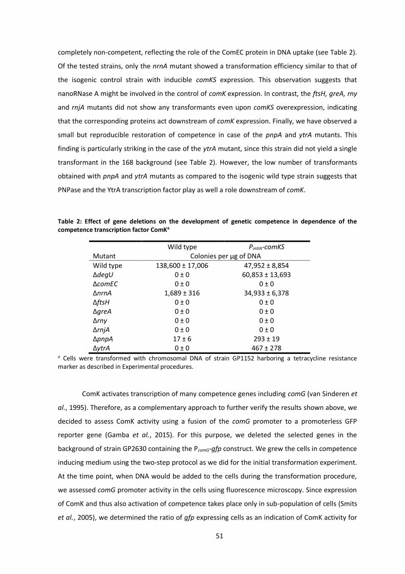
51
completely non-competent, reflecting the role of the ComEC protein in DNA uptake (see Table 2).
Of the tested strains, only the nrnA mutant showed a transformation efficiency similar to that of
the isogenic control strain with inducible comKS expression. This observation suggests that
nanoRNase A might be involved in the control of comK expression. In contrast, the ftsH, greA, rny
and rnjA mutants did not show any transformants even upon comKS overexpression, indicating
that the corresponding proteins act downstream of comK expression. Finally, we have observed a
small but reproducible restoration of competence in case of the pnpA and ytrA mutants. This
finding is particularly striking in the case of the ytrA mutant, since this strain did not yield a single
transformant in the 168 background (see Table 2). However, the low number of transformants
obtained with pnpA and ytrA mutants as compared to the isogenic wild type strain suggests that
PNPase and the YtrA transcription factor play as well a role downstream of comK.
Table 2: Effect of gene deletions on the development of genetic competence in dependence of the competence transcription factor ComKa
Wild type PmtlA-comKS Mutant Colonies per µg of DNA
Wild type 138,600 ± 17,006 47,952 ± 8,854 ΔdegU 0 ± 0 60,853 ± 13,693 ΔcomEC 0 ± 0 0 ± 0 ΔnrnA 1,689 ± 316 34,933 ± 6,378 ΔftsH 0 ± 0 0 ± 0 ΔgreA 0 ± 0 0 ± 0 Δrny 0 ± 0 0 ± 0 ΔrnjA 0 ± 0 0 ± 0 ΔpnpA 17 ± 6 293 ± 19 ΔytrA 0 ± 0 467 ± 278
a Cells were transformed with chromosomal DNA of strain GP1152 harboring a tetracycline resistance marker as described in Experimental procedures.
ComK activates transcription of many competence genes including comG (van Sinderen et
al., 1995). Therefore, as a complementary approach to further verify the results shown above, we
decided to assess ComK activity using a fusion of the comG promoter to a promoterless GFP
reporter gene (Gamba et al., 2015). For this purpose, we deleted the selected genes in the
background of strain GP2630 containing the PcomG-gfp construct. We grew the cells in competence
inducing medium using the two-step protocol as we did for the initial transformation experiment.
At the time point, when DNA would be added to the cells during the transformation procedure,
we assessed comG promoter activity in the cells using fluorescence microscopy. Since expression
of ComK and thus also activation of competence takes place only in sub-population of cells (Smits
et al., 2005), we determined the ratio of gfp expressing cells as an indication of ComK activity for
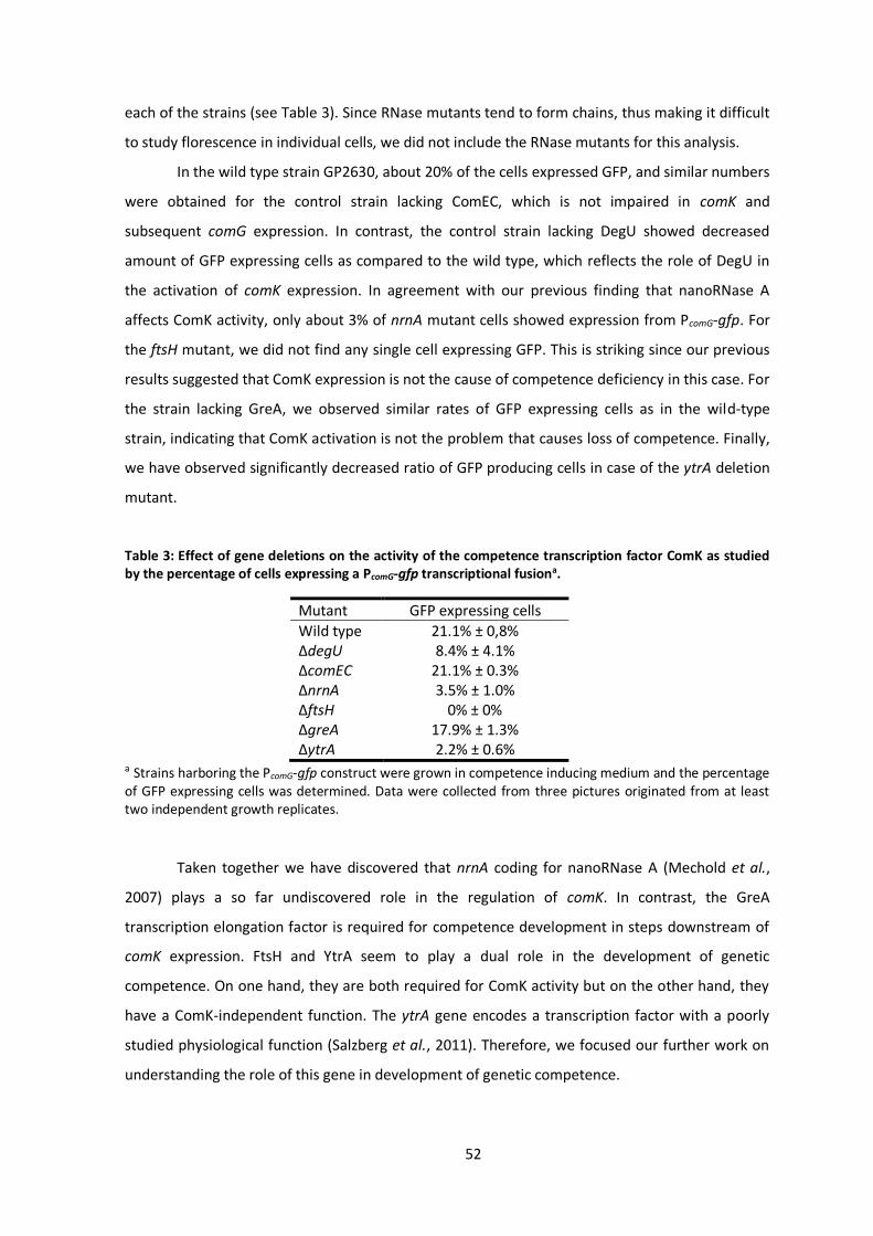
52
each of the strains (see Table 3). Since RNase mutants tend to form chains, thus making it difficult
to study florescence in individual cells, we did not include the RNase mutants for this analysis.
In the wild type strain GP2630, about 20% of the cells expressed GFP, and similar numbers
were obtained for the control strain lacking ComEC, which is not impaired in comK and
subsequent comG expression. In contrast, the control strain lacking DegU showed decreased
amount of GFP expressing cells as compared to the wild type, which reflects the role of DegU in
the activation of comK expression. In agreement with our previous finding that nanoRNase A
affects ComK activity, only about 3% of nrnA mutant cells showed expression from PcomG-gfp. For
the ftsH mutant, we did not find any single cell expressing GFP. This is striking since our previous
results suggested that ComK expression is not the cause of competence deficiency in this case. For
the strain lacking GreA, we observed similar rates of GFP expressing cells as in the wild-type
strain, indicating that ComK activation is not the problem that causes loss of competence. Finally,
we have observed significantly decreased ratio of GFP producing cells in case of the ytrA deletion
mutant.
Table 3: Effect of gene deletions on the activity of the competence transcription factor ComK as studied by the percentage of cells expressing a PcomG-gfp transcriptional fusiona.
Mutant GFP expressing cells
Wild type 21.1% ± 0,8% ΔdegU 8.4% ± 4.1% ΔcomEC 21.1% ± 0.3% ΔnrnA 3.5% ± 1.0% ΔftsH 0% ± 0% ΔgreA 17.9% ± 1.3% ΔytrA 2.2% ± 0.6%
a Strains harboring the PcomG-gfp construct were grown in competence inducing medium and the percentage of GFP expressing cells was determined. Data were collected from three pictures originated from at least two independent growth replicates.
Taken together we have discovered that nrnA coding for nanoRNase A (Mechold et al.,
2007) plays a so far undiscovered role in the regulation of comK. In contrast, the GreA
transcription elongation factor is required for competence development in steps downstream of
comK expression. FtsH and YtrA seem to play a dual role in the development of genetic
competence. On one hand, they are both required for ComK activity but on the other hand, they
have a ComK-independent function. The ytrA gene encodes a transcription factor with a poorly
studied physiological function (Salzberg et al., 2011). Therefore, we focused our further work on
understanding the role of this gene in development of genetic competence.
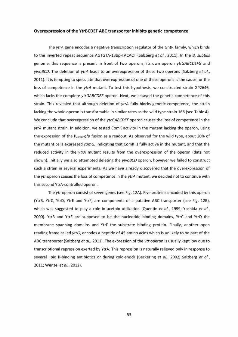
53
Overexpression of the YtrBCDEF ABC transporter inhibits genetic competence
The ytrA gene encodes a negative transcription regulator of the GntR family, which binds
to the inverted repeat sequence AGTGTA-13bp-TACACT (Salzberg et al., 2011). In the B. subtilis
genome, this sequence is present in front of two operons, its own operon ytrGABCDEFG and
ywoBCD. The deletion of ytrA leads to an overexpression of these two operons (Salzberg et al.,
2011). It is tempting to speculate that overexpression of one of these operons is the cause for the
loss of competence in the ytrA mutant. To test this hypothesis, we constructed strain GP2646,
which lacks the complete ytrGABCDEF operon. Next, we assayed the genetic competence of this
strain. This revealed that although deletion of ytrA fully blocks genetic competence, the strain
lacking the whole operon is transformable in similar rates as the wild type strain 168 (see Table 4).
We conclude that overexpression of the ytrGABCDEF operon causes the loss of competence in the
ytrA mutant strain. In addition, we tested ComK activity in the mutant lacking the operon, using
the expression of the PcomG-gfp fusion as a readout. As observed for the wild type, about 20% of
the mutant cells expressed comG, indicating that ComK is fully active in the mutant, and that the
reduced activity in the ytrA mutant results from the overexpression of the operon (data not
shown). Initially we also attempted deleting the ywoBCD operon, however we failed to construct
such a strain in several experiments. As we have already discovered that the overexpression of
the ytr operon causes the loss of competence in the ytrA mutant, we decided not to continue with
this second YtrA-controlled operon.
The ytr operon consist of seven genes (see Fig. 12A). Five proteins encoded by this operon
(YtrB, YtrC, YtrD, YtrE and YtrF) are components of a putative ABC transporter (see Fig. 12B),
which was suggested to play a role in acetoin utilization (Quentin et al., 1999; Yoshida et al.,
2000). YtrB and YtrE are supposed to be the nucleotide binding domains, YtrC and YtrD the
membrane spanning domains and YtrF the substrate binding protein. Finally, another open
reading frame called ytrG, encodes a peptide of 45 amino acids which is unlikely to be part of the
ABC transporter (Salzberg et al., 2011). The expression of the ytr operon is usually kept low due to
transcriptional repression exerted by YtrA. This repression is naturally relieved only in response to
several lipid II-binding antibiotics or during cold-shock (Beckering et al., 2002; Salzberg et al.,
2011; Wenzel et al., 2012).
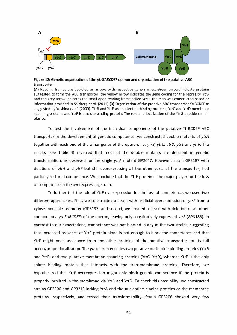
54
To test the involvement of the individual components of the putative YtrBCDEF ABC
transporter in the development of genetic competence, we constructed double mutants of ytrA
together with each one of the other genes of the operon, i.e. ytrB, ytrC, ytrD, ytrE and ytrF. The
results (see Table 4) revealed that most of the double mutants are deficient in genetic
transformation, as observed for the single ytrA mutant GP2647. However, strain GP3187 with
deletions of ytrA and ytrF but still overexpressing all the other parts of the transporter, had
partially restored competence. We conclude that the YtrF protein is the major player for the loss
of competence in the overexpressing strain.
To further test the role of YtrF overexpression for the loss of competence, we used two
different approaches. First, we constructed a strain with artificial overexpression of ytrF from a
xylose inducible promoter (GP3197) and second, we created a strain with deletion of all other
components (ytrGABCDEF) of the operon, leaving only constitutively expressed ytrF (GP3186). In
contrast to our expectations, competence was not blocked in any of the two strains, suggesting
that increased presence of YtrF protein alone is not enough to block the competence and that
YtrF might need assistance from the other proteins of the putative transporter for its full
action/proper localization. The ytr operon encodes two putative nucleotide binding proteins (YtrB
and YtrE) and two putative membrane spanning proteins (YtrC, YtrD), whereas YtrF is the only
solute binding protein that interacts with the transmembrane proteins. Therefore, we
hypothesized that YtrF overexpression might only block genetic competence if the protein is
properly localized in the membrane via YtrC and YtrD. To check this possibility, we constructed
strains GP3206 and GP3213 lacking YtrA and the nucleotide binding proteins or the membrane
proteins, respectively, and tested their transformability. Strain GP3206 showed very few
Figure 12: Genetic organization of the ytrGABCDEF operon and organization of the putative ABC transporter (A) Reading frames are depicted as arrows with respective gene names. Green arrows indicate proteins suggested to form the ABC transporter; the yellow arrow indicates the gene coding for the repressor YtrA and the grey arrow indicates the small open reading frame called ytrG. The map was constructed based on information provided in Salzberg et al. (2011) (B) Organization of the putative ABC transporter YtrBCDEF as suggested by Yoshida et al. (2000). YtrB and YtrE are nucleotide binding proteins, YtrC and YtrD membrane spanning proteins and YtrF is a solute binding protein. The role and localization of the YtrG peptide remain elusive.

55
transformants, suggesting that the presence of nucleotide binding proteins is not important to
block competence. In contrast, strain GP3213 gave rise to many transformants. We thus conclude
that the overexpression of the solute binding protein YtrF in conjunction with the membrane
proteins YtrC and YtrD is responsible for the block of competence indicating that indeed the
proper function of YtrF, which depends on YtrC and YtrD, is crucial for the phenotype.
Table 4: Effect of gene deletions in the ytrGABCDEF operon on the development of genetic competencea.
Mutant Colonies per µg of DNA
Wild type 138,600 ± 17,006 ΔytrGABCDEF 114,733 ± 14,408 ΔytrA 0 ± 0 ΔytrAB 0 ± 0 ΔytrAC 0 ± 0 ΔytrAD 24 ± 2 ΔytrAE 137 ± 51 ΔytrAF 10,180 ± 549 Pxyl-ytrF 137,533 ± 26,595 ΔytrGABCDE 108,467 ± 14,836 ΔytrABE 309 ± 88 ΔytrACD 45,467 ± 10,799
a Cells were transformed with chromosomal DNA of strain GP1152 harboring a tetracycline resistance marker as described in Experimental procedures
Overexpression of the ytrGABCDEF operon leads to defect in biofilm formation
B. subtilis can employ various lifestyles which are tightly interconnected through
regulatory proteins (López et al., 2009). Therefore, we anticipated that the overexpression of YtrF
might also affect other lifestyles of B. subtilis. Indeed, it was previously shown that the ytrA
mutant has a reduced sporulation efficiency (Koo et al., 2017). We thus decided to examine the
effect of the ytrA deletion on biofilm formation. To that end, we first deleted the ytrA gene or the
whole ytrGABCDEF operon from the biofilm-forming strain DK1042 (Konkol et al., 2013). We then
tested the biofilm formation of the resulting strains on biofilm inducing MSgg agar (Branda et al.,
2001). As expected, the wild type strain DK1042 formed structured colonies that are indicative of
biofilm formation. In contrast, the negative control GP2559 (a ymdB mutant that is known to be
defective in biofilm formation, Kampf et al., 2018) formed completely smooth colonies. The
biofilm formed by the ytrA mutant GP3212 was less structured, more translucent and with only
some tiny wrinkles on its surface, indicating that biofilm formation was inhibited but not fully
abolished upon loss of YtrA. In contrast, strain GP3207 lacking the complete ytrGABCDEF operon
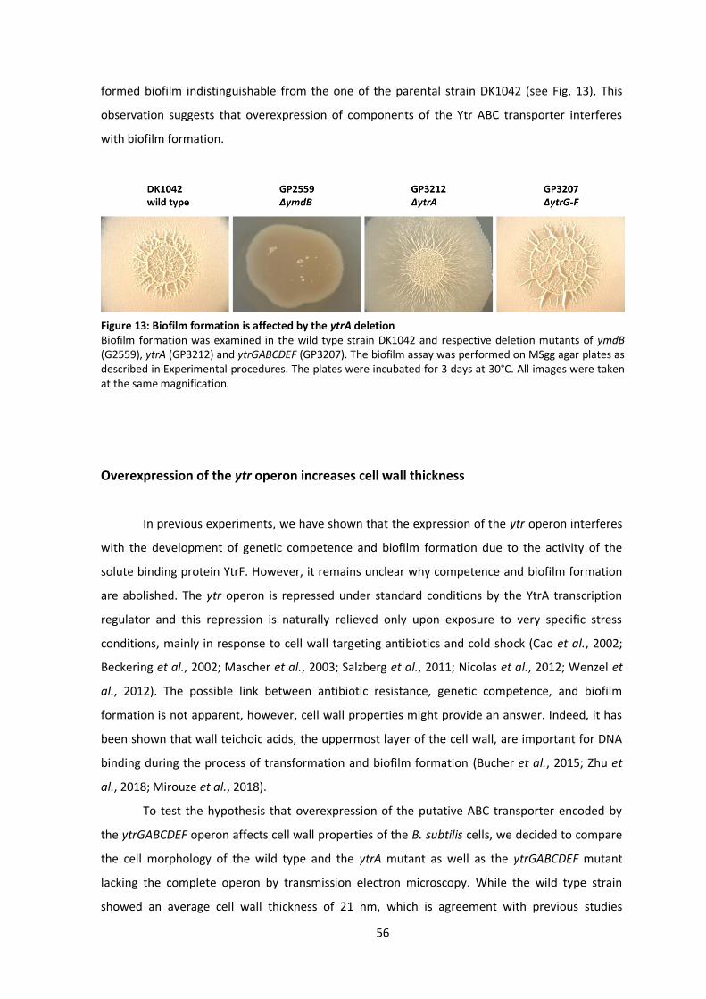
56
formed biofilm indistinguishable from the one of the parental strain DK1042 (see Fig. 13). This
observation suggests that overexpression of components of the Ytr ABC transporter interferes
with biofilm formation.
Overexpression of the ytr operon increases cell wall thickness
In previous experiments, we have shown that the expression of the ytr operon interferes
with the development of genetic competence and biofilm formation due to the activity of the
solute binding protein YtrF. However, it remains unclear why competence and biofilm formation
are abolished. The ytr operon is repressed under standard conditions by the YtrA transcription
regulator and this repression is naturally relieved only upon exposure to very specific stress
conditions, mainly in response to cell wall targeting antibiotics and cold shock (Cao et al., 2002;
Beckering et al., 2002; Mascher et al., 2003; Salzberg et al., 2011; Nicolas et al., 2012; Wenzel et
al., 2012). The possible link between antibiotic resistance, genetic competence, and biofilm
formation is not apparent, however, cell wall properties might provide an answer. Indeed, it has
been shown that wall teichoic acids, the uppermost layer of the cell wall, are important for DNA
binding during the process of transformation and biofilm formation (Bucher et al., 2015; Zhu et
al., 2018; Mirouze et al., 2018).
To test the hypothesis that overexpression of the putative ABC transporter encoded by
the ytrGABCDEF operon affects cell wall properties of the B. subtilis cells, we decided to compare
the cell morphology of the wild type and the ytrA mutant as well as the ytrGABCDEF mutant
lacking the complete operon by transmission electron microscopy. While the wild type strain
showed an average cell wall thickness of 21 nm, which is agreement with previous studies
Figure 13: Biofilm formation is affected by the ytrA deletion Biofilm formation was examined in the wild type strain DK1042 and respective deletion mutants of ymdB (G2559), ytrA (GP3212) and ytrGABCDEF (GP3207). The biofilm assay was performed on MSgg agar plates as described in Experimental procedures. The plates were incubated for 3 days at 30°C. All images were taken at the same magnification.
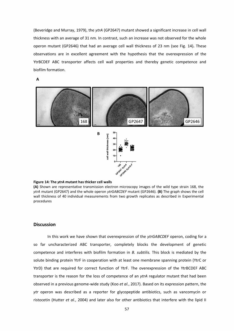
57
(Beveridge and Murray, 1979), the ytrA (GP2647) mutant showed a significant increase in cell wall
thickness with an average of 31 nm. In contrast, such an increase was not observed for the whole
operon mutant (GP2646) that had an average cell wall thickness of 23 nm (see Fig. 14). These
observations are in excellent agreement with the hypothesis that the overexpression of the
YtrBCDEF ABC transporter affects cell wall properties and thereby genetic competence and
biofilm formation.
Discussion
In this work we have shown that overexpression of the ytrGABCDEF operon, coding for a
so far uncharacterized ABC transporter, completely blocks the development of genetic
competence and interferes with biofilm formation in B. subtilis. This block is mediated by the
solute binding protein YtrF in cooperation with at least one membrane spanning protein (YtrC or
YtrD) that are required for correct function of YtrF. The overexpression of the YtrBCDEF ABC
transporter is the reason for the loss of competence of an ytrA regulator mutant that had been
observed in a previous genome-wide study (Koo et al., 2017). Based on its expression pattern, the
ytr operon was described as a reporter for glycopeptide antibiotics, such as vancomycin or
ristocetin (Hutter et al., 2004) and later also for other antibiotics that interfere with the lipid II
Figure 14: The ytrA mutant has thicker cell walls (A) Shown are representative transmission electron microscopy images of the wild type strain 168, the ytrA mutant (GP2647) and the whole operon ytrGABCDEF mutant (GP2646). (B) The graph shows the cell wall thickness of 40 individual measurements from two growth replicates as described in Experimental procedures
58
cycle, such as nisin (Wenzel et al., 2012). Whether this induction of ytrGABCDEF expression leads
to an increased resistance towards those antibiotics is not clear, but recent results indicate that it
does at least in case of nisin (J. Bandow, personal communication).
Based on the partial restoration of genetic competence of the ytrA mutant upon ComKS
overexpression, one might expect that the loss of YtrA and the concomitant overexpression of the
ABC transporter somehow interferes with competence development upstream of ComK
activation. However, competence is developed in an all or nothing scenario, and cells in which the
ComK levels reach a certain threshold should become competent (Haijema et al., 2001; Maamar
and Dubnau, 2005). Our observation that comKS overexpression restores competence of the ytrA
mutant only partially suggests that ComK levels are not the only factor that limits competence of
the ytrA mutant. If the ytrA deletion would interfere with ComK activation, one would then expect
wild type like competence upon overexpression of ComK which was not the case. Why does ComK
then restore the competence at all? The DNA uptake apparatus must be adapted to cell wall
thickness in order to ensure that the extracellular DNA can reach the ComG/ComE DNA transport
complex. Due to the increased cell wall thickness upon overexpression of the YtrBCDEF ABC
transporter, the DNA probably has problems to get in contact with the ComG pili. Overexpression
of ComK will then result in the increased production of DNA-binding ComG on the cell surface of
all cells of the population (comparing to about 10% in the wild-type strain transformed with the
classical two-step protocol). This would simply increase the probability that foreign DNA reaches
the DNA uptake machinery in some cells, which then leads to the appearance of only a few
transformants as observed in our study. On the other hand, the results obtained by fluorescence
microscopy revealed a decreased transcription from the ComK dependent comG promoter in the
ytrA mutant. However, this expression is expected to be wild type-like if the action of YtrBCEDF
ABC transporter would not interfere with ComK activity and only block DNA uptake as a result of
the remodeled cell wall as suggested above. Again, the disorganized cell wall might be
responsible, since ComK expression is induced by the detection of extracellular quorum-sensing
signals (both ComXPA and Rap-Phr systems) and this induction depends on the accessibility of the
sensor domains for the pheromones which might be impaired in the strain with altered cell wall
composition.
In addition to the loss of genetic competence, it was previously shown that the ytrA
deletion leads to decreased sporulation efficiency (Koo et al., 2017) and we have shown that it
also affects biofilm formation. Considering the changed cell wall properties, this is in agreement
with previous studies which showed hampered biofilm formation upon disruption of cell wall
biosynthesis (Bucher et al., 2015; Zhu et al., 2018). Taken together, we conclude that the

59
overexpression of the YtrBCDEF ABC transporter upon deletion of ytrA plays a pleiotropic role in
the control of alternative lifestyles of B. subtilis.
Our results demonstrate that the YtrBCDEF ABC transporter is involved in the control of
cell wall homeostasis, but it is not yet clear how this is achieved. An easy explanation would be
that the system exports molecules necessary for cell wall synthesis, however, based on the
presence of the solute binding protein YtrF and on the critical role of this protein in preventing
genetic competence, it can be assumed that the ABC transporter rather acts as an importer.
However, YtrBCDEF may not act as a transporter at all and simply modulate the activity of other
enzymes that participate in cell wall metabolism. Strikingly, YtrF is a member of the same protein
family as FtsX, which is known to activate the cell wall hydrolase CwlO (Meisner et al., 2013).
Future work will need to address the precise mechanism by which the YtrBCDEF ABC transporter
interferes with cell wall synthesis.
Experimental procedures
Bacterial strains and growth conditions
The B. subtilis strains used in this study are listed in Table S3. Lysogeny broth (LB,
Sambrook et al., 1989) was used to grow E. coli and B. subtilis. When required, media were
supplemented with antibiotics at the following concentrations: ampicillin 100 µg ml-1 (for E. coli)
and chloramphenicol 5 µg ml-1, kanamycin 10 µg ml-1, spectinomycin 250 µg ml-1, tetracycline 12.5
µg ml-1, and erythromycin 2 µg ml-1 plus lincomycin 25 µg ml-1 (for B. subtilis). For agar plates, 15 g
l-1 Bacto agar (Difco) was added.
DNA manipulation and strain construction
S7 Fusion DNA polymerase (Mobidiag, Espoo, Finland) was used as recommended by the
manufacturer. DNA fragments were purified using the QIAquick PCR Purification Kit (Qiagen,
Hilden, Germany). DNA sequences were determined by the dideoxy chain termination method
(Sambrook et al., 1989). Chromosomal DNA from B. subtilis was isolated using the peqGOLD
Bacterial DNA Kit (Peqlab, Erlangen, Germany) and plasmids were purified from E. coli using
NucleoSpin Plasmid Kit (Macherey-Nagel, Düren, Germany). Deletion of the degU, comEC, ftsH,
greA, ytrA, nrnA, and ytrF genes as well as ytrCD, ytrG-ytrE, and ytrGABCDEF regions was achieved
by transformation with PCR products constructed using oligonucleotides (see Table S5) to amplify
DNA fragments flanking the target genes and intervening antibiotic resistance cassettes as
described previously (Youngman, 1990; Guérout-Fleury et al., 1995; Wach, 1996). The identity of

60
the modified genomic regions was verified by DNA sequencing. To construct the strains (GP2618
and GP2620) harbouring the PmtlA-comKS cassette coupled to the antibiotic resistance gene, we
have first amplified the PmtlA-comKS from the strain PG10 (Reuß et al., 2017) as well as the
resistance genes from pDG646 and pGEM-cat, respectively (Youngman, 1990; Guérout-Fleury et
al., 1995) and the genes flanking the intended integration site, i. e. yvcA and hisI from B. subtilis
168. Subsequently, those DNA fragments were fused in another PCR reaction thanks to the
overlapping primers. The final product was used to transform B. subtilis 168. Correct insertion was
verified by PCR amplification and sequencing. Markerless deletions of ytrB, ytrC, ytrD and ytrE
genes were performed using pDR244 plasmid as described (Koo et al., 2017). In short, strains
BKE30450, BKE30440, BKE30430 and BKE30420 were transformed with plasmid pDR244 and
transformants were selected on LB agar plates supplemented with spectinomycin at 30°C.
Transformants were then streaked on plain LB agar plates and incubated at 42°C to cure the
plasmid, which contains a thermo-sensitive origin of replication. Single colonies were then
screened for spectinomycin and erythromycin/lincomycin sensitivity. Markerless deletion was
confirmed by PCR with primers flanking the deletion site. Created strains GP3188, GP3189,
GP3190 and GP 3191 were used for subsequent deletion of the ytrA gene. This was done either by
transformation with PCR product as described above or by transformation with genomic DNA of
the ytrA deletion strain (in case of GP3195 construction). Deletion of the ytrA gene and
preservation of selected markerless deletions were confirmed via PCR. To construct GP3206, PCR
product containing erythromycin resistance in place of ytrA and ytrB genes was amplified from
GP3193 and transformed to GP3191.
Transformation of B. subtilis strains
Transformation experiments were conducted based on the two-step protocol as
described previously (Kunst and Rapoport, 1995). Briefly, cells were grown at 37°C at 200 rpm in
10 ml MNGE medium containing 2% glucose, 0.2% potassium glutamate, 100 mM potassium
phosphate buffer (pH 7), 3.4 mM trisodiumcitrate, 3 mM MgSO4, 42 µM ferric ammonium citrate,
0.24 mM L-tryptophan and 0.1% casein hydrolysate. During the transition from exponential to
stationary phase, the culture was diluted with another 10 ml of MNGE medium (without casein
hydrolysate) and incubated for 1 h at 37°C with shaking. In case of strain GP3187, 0.5% xylose was
added to both media. Afterwards, 250 ng of chromosomal DNA was added to 400 µl of cells and
incubated for 30 minutes at 37°C. One hundred microliter of Expression mix (2.5% yeast extract,
2.5% casein hydrolysate, 1.22mM tryptophan) was added and cells were allowed to grow for 1h at
37°C, before spreading onto selective LB plates containing appropriate antibiotics.
61
Transformation of strains harboring comK and comS expressed from the mannitol
inducible promotor (PmtlA) was performed based on (Rahmer et al., 2015). Briefly, an overnight
culture was diluted in 5 ml LB to an initial OD600 of 0.1 and incubated at 37°C at 200 rpm. After 90
minutes incubation, 5 ml of fresh LB containing mannitol (1%) and MgCl2 (5 mM) were added and
the bacterial culture was incubated for an additional 90 minutes. The cells were then pelleted by
centrifugation for 10 minutes at 2,000 x g and the pellet was re-suspended in the same amount of
fresh LB medium, 1 ml aliquots were distributed into 1.5 ml reaction tubes and 250 ng of
chromosomal DNA was added to each of them. The cell suspension was incubated for 1 h at 37°C
and transformants were selected on LB plates as described above.
Plasmid construction
All plasmids used in this study are listed in Table S4. Escherichia coli DH5 (Sambrook et
al., 1989) was used for plasmid constructions and transformation using standard techniques
(Sambrook et al., 1989). To express the B. subtilis protein YtrF under the control of a xylose
inducible promotor, we cloned the ytrF gene into the backbone of pGP888 via the XbaI and KpnI
sites (Diethmaier et al., 2011).
Biofilm assay
To analyse biofilm formation, selected strains were grown in LB medium to an OD600 of
about 0.5 to 0.8 and 10 μl of the culture were spotted onto MSgg agar plates (Branda et al., 2001).
Plates were incubated for 3 days at 30°C.
Fluorescence microscopy
For fluorescence microscopy imaging, B. subtilis cultures were grown in 10 ml MNGE
medium till the transition from exponential to stationary phase and then diluted with another 10
ml of MNGE medium as described for the transformation experiments (see above). 5 μl of cells
were pipetted on microscope slides coated with a thin layer of 1% agarose and covered with a
cover glass. Fluorescence images were obtained with the AxioImager M2 fluorescence
microscope, equipped with digital camera AxioCam MRm and AxioVision Rel 4.8 software for
image processing and an EC Plan-NEOFLUAR 100X/1.3 objective (Carl Zeiss, Göttingen, Germany).
Filter set 38 (BP 470/40, FT 495, BP 525/50; Carl Zeiss) was applied for GFP detection. Ratio of GFP
expressing cells to the total number of cells was determined by manual examination from three

62
independent randomly selected pictures originated from at least two independent growth
replicates.
Transmission electron microscopy
To examine cell wall thickness of B. subtilis strains, cells were prepared for Transmission
Electron Microscopy (TEM) as previously described (Rincón-Tomás et al., 2020). An overnight
culture was inoculated to an OD600 of 0.05 in 30 ml MNGE medium and grown to an OD600 of 0.6 ±
0.1 at 37°C and 200 rpm. Cells were centrifuged for 10 minutes at 4,000 rpm to obtain a 100 µl
cell pellet, which was then washed twice in phosphate-buffered saline (PBS, 127 mM NaCl, 2.7
mM KCl, 10 mM Na2HPO4, 1.8 mM KH2PO4, pH 7.4) and fixed overnight in 2.5% (w/v)
glutaraldehyde at 4°C. Cells were then mixed with 1.5% (w/v, final concentration) molten Bacto-
Agar (in PBS) and the resulting agar block was cut to pieces of 1 mm3. A dehydration series was
performed (15% aqueous ethanol solution for 15 minutes, 30%, 50%, 70% and 95% for 30 minutes
and 100% for 2 x 30 minutes) at 0°C, followed by an incubation step in 66% LR white resin mixture
(v/v, in ethanol) (Plano, Wletzlar, Germany) for 2 hours at room temperature and embedment in
100% LR-White solution overnight at 4°C. One agar piece was transferred to a gelatin capsule
filled with fresh LR-white resin, which was subsequently polymerized at 55°C for 24 hours. A
milling tool (TM 60, Fa. Reichert & Jung, Vienna, Austria) was used to shape the gelatin capsule
into a truncated pyramid. An ultramicrotome (Reichert Utralcut E, Leica Microsystems, Wetzlar,
Germany) and a diamond knife were used to obtain ultrathin sections (80 nm) of the samples. The
resulting sections were mounted onto mesh specimen Grids (Plano, Wetzlar, Germany) and
stained with 4% (w/v) uranyl acetate solution (pH 7.0) for 10 minutes. Microscopy was performed
in a Joel JEM 1011 transmission electron microscope (Joel Germany GmbH, Freising, Germany) at
80 kV. Images were taken at a magnification of 30,000 and recorded with a Gatan Orius SC1000
CCD camera (Gatan, Munich, Germany). For each replicate, 20 cells were photographed and cell
wall thickness was measured at three different locations using ImageJ software (Rueden et al.,
2017).
Acknowledgements
We wish to thank Julia Busse, Melin Güzel and Leon Daniau for the help with some experiments.
We are grateful to Josef Altenbuchner, Jan Gundlach, Leendert Hamoen, Daniel Kearns, Daniel
Reuss, Sarah Wilcken, and the Bacillus Genetic Sock Center for providing B. subtilis strains. We
thank Dr. Michael Hoppert for providing access to the Transmission Electron Microscope. This
research received funding from the Deutsche Forschungsgemeinschaft via SFB860.

63
4 Discussion
4.1 Suppressor mutant screen revealed initiation of bulk mRNA degradation as
the pivotal function of RNase Y
This thesis is focused on RNase Y and the effect of the rny gene deletion on B. subtilis
physiology. The rny gene was for a long time considered to be essential, but it could be later
deleted from the genome in the study of Figaro et al. (2013). The authors of this study managed
to delete the rny gene in genetic backgrounds of four different B. subtilis strains commonly used
in the laboratories around the world and thus concluded that requirement for second-site
suppressor mutations is rather unlikely. However, the rny mutant shows deformed cellular
morphology, forms small and smooth colonies and has significantly decreased growth rate as
compared to the wild type strain. Taken together, the rny deletion strain is far away from the
optimal growth of B. subtilis and thus has a huge space for improvements of its properties
through suppressor mutations. Indeed, although we were able to verify that it is possible to
introduce an rny deletion into different strains of B. subtilis, we have observed that the rny
mutant does lyse rather quickly followed by the appearance of suppressor colonies.
As already mentioned in the introduction, essential genes were defined as those whose
deletion prevents growth under standard laboratory conditions; from that point of view the rny
gene cannot be regarded as essential (Kobayashi et al., 2003; Commichau et al., 2013). However,
dividing genes into only two groups of essential and non-essential genes is probably not the most
appropriate. There are differences in the importance for cell growth even between the genes that
would be traditionally marked as essential. This was recently evaluated in a study where the
authors measured the time for which bacteria can continue to grow after disruption of a
particular essential gene and thereby managed ordered the essential genes by their importance
(Gallagher et al., 2020). In light of this study, essentiality should not be considered as yes or no
question, but rather as a scale ranging from genes whose deletion does not cause any
disadvantage to genes whose inactivation leads to immediate cell death. Since the rny deletion
does not allow for robust growth and has to be compensated by second site suppressor
mutations, we believe that the rny gene should be very close to the upper boundary on such a
scale and therefore we decided to label this gene as a quasi-essential, also in accordance with the
definition from Hutchison et al. (2016). This brings the question about the reason(s) for this quasi-
essentiality and about the main cellular functions of the enzyme.
There are several possible reasons for the pivotal role of RNase Y. Firstly, there might be a
specific essential transcript that needs to be processed by the RNase. Indeed, it was previously
64
shown that RNase Y is involved in the maturation of two essential sRNAs, small cytoplasmic RNA
(scRNA) coding for the ribonucleic components of the signal recognition particle (SRP), and rnpB
that encodes the ribozyme component of RNase P. Essentiality of scRNA lays in the role of SRP in
co-translational trafficking of proteins to/across the cytoplasmic membrane and scRNA itself is
responsible for translation arrest during this process by interaction with 23S RNA (Beckert et al.,
2015; Tsirigotaki et al., 2017). RNase P is responsible for maturation of the 5′ end of tRNAs
(Guerrier-Takada et al., 1983). Given that both RNAs are essential, it would be tempting to
speculate that the essentiality of RNase Y lays in the absence of their respective processing
events. However, scRNA was shown to be functional even in its unprocessed form (Beckert et al.,
2015) and RNase P processing has alternative, although less efficient pathways, that are RNase Y
independent (Gilet et al., 2015). Therefore, the possibility that absence of processing of those two
functional RNA molecules is the reason for RNase Y quasi-essentiality seems to be rather unlikely.
Except the already known essential targets, another option is that absence of processing
of some so far unidentified target of RNase Y stands behind the detrimental phenotypes and
quasi-essentiality of the rny gene. This was, by analogy, shown for RNase III, which is essential
thanks to its cleavage event in a prophage encoded toxin-antitoxin system (Durand et al., 2012b),
or for RNase Z, which is responsible for tRNA processing (Pellegrini et al., 2003). To identify such a
specific target, we decided to use the force of natural genetic selection and thus analyzed several
of the suppressor colonies, popping up on the plates after the lyses of the rny mutant strain. We
took several different colonies evolved at different conditions, to be able to identify whether the
selection uses general mechanism or is condition-specific. Analysis of suppressor mutants has
previously helped to uncover interconnections of metabolic pathways, important protein residues
as well as reasons for essentiality of the signaling molecule c-di-AMP (Gundlach et al., 2017;
Tödter et al., 2017; Osaka et al., 2020) and we thus hoped this approach to give us a better insight
into the most important functions of RNase Y.
The suppressor mutant analysis identified single nucleotide polymorphisms in the genes
greA, rpoE and cspD, coding for transcription elongation factor (Kusuya et al., 2011), the RNA
polymerase subunit (Juang and Helmann, 1994; Rabatinová et al., 2013) and an RNA chaperone
(Graumann et al., 1997), in dependence of isolation conditons. These three genes does not seem
to play a crucial role for B. subtilis and thus it is unlikely, that they would be directly responsible
for the rny mutant quassi-essentiality. On the other hand, they share a common function related
to transcription, a process which is on the other side of RNA life span than the degradation
initiated by RNase Y. Next to those mutations, we observed an interesting phenomenon present
in all of the suppressors. That was a duplication of the 60 kb long fragment located between the
ribosomal operons rrnW and rrnI. Similar duplications of a larger genomic regions encompassed

65
by ribosomal operons were noticed already 40 year ago for the gram-positive organisms E. coli
and Salmonella typhimurium, since the highly similar rRNA coding operons are ideal platform for
homologous recombination events (Hill et al., 1977; Anderson and Roth, 1981). Already back then
in the study on S. typhimurium the authors remarked that the duplicated region contains all genes
coding for the core subunits of the RNA polymerase and suggested that their duplication might
have a major influence on the cellular physiology (Anderson and Roth, 1981). The situation seems
to be similar in the gram-positive Bacillus subtilis, where the three genes for core subunits of the
RNA polymerase are also present within this duplicated region we observed here.
With such a knowledge in mind and in relation to the point mutation found in
transcription related genes, we assumed that the simultaneous duplication of the genes for the
RNA polymerase core might be responsible for the observed suppression of the rny gene deletion.
We were able to confirm this hypothesis with our next experiment (see Fig. 7), in which we
deleted the rny gene in such a genetic background, where the three core polymerase genes are
no longer present at the same genetic locus in between the rrnW and rrnI ribosomal operons and
thus cannot be easily duplicated simultaneously. However, the new genetic composition of the
RNA polymerase genes did not prevent the suppressor formation of the rny mutant completely.
Even in this background the rny mutant formed suppressors extensively which allowed us to
analyze this second class of suppressors. All of them carried single nucleotide polymorphisms
directly in the genes coding for the RpoB and RpoC subunits of the RNA polymerase, leading to
huge decrease in the transcriptional activity as we observed in subsequent in vitro transcription
assays (see Fig. 9). The suppressing mechanism of the first class of suppressors containing RNA
polymerase genes duplication in conjunction with transcription factors mutations seem to be less
obvious, however according to the mathematic model presented in chapter 2 it is also assumed to
decrease transcription rates significantly.Taken together the results from the suppressor screen
suggested that there is not a single one specific transcript, whose degradation/processing would
be the key function of RNase Y. Since RNase Y is the enzyme responsible for initiation of the
degradation of the majority of transcripts and the global mRNA half-lives are doubled in the rny
depletion strain (Shahbabian et al., 2009; Lehnik-Habrink et al., 2011b), we concluded that it is
likely the global role in degradation of bulk mRNA which stands behind the quasi-essentiality of
RNase Y. The rny deletion likely leads to a never-ending accumulation of total mRNA (see Fig. 15),
which results in high energy consumption and high degree of cellular stress. It would be logical if
the suppressors would therefore either try to increase RNA degradation or decrease RNA
synthesis, which seems to be the case for both classes of the isolated suppressors.

66

67
As judged from the absence of RNA degradation affecting suppressor mutations, the cells
lacking RNase Y are apparently unable to increase the RNA degradation and thereby decrease the
average half-lives back towards the situation in the wild type strain. Hence, the suppressor
mutants had to use an alternative approach and limit RNA transcription. This way the average
mRNA half-lives should stay increased as compared to the wild type strain, but thanks to the
decreased transcription, the total RNA should not accumulate to such a huge extent, establishing
a new stable equilibrium between the RNA synthesis and degradation and improving the
energetic status of the cell. We had observed two alternative ways how the suppressor of the rny
mutant achieve this. In the first class of suppressors, the core RNA polymerase genes are
duplicated which, in conjunction with additional mutations in transcription related genes (greA,
cspD, rpoE). This duplication leads to a decreased likelihood of proper RNA polymerase assembly
and thereby decreased transcription. In the second class of suppressors harboring point
mutations in the genes coding for core RNA polymerase subunits, we could clearly show that the
transcriptions rates are significantly diminished.
Further experiments will be necessary to fully confirm this conclusion, however it might
not be easy to find the proper experimental setup. The best approach would be to use either DNA
microarrays or RNA-sequencing and analyze the transcriptomes of the wild-type, the rny mutant
Figure 15: Model of RNA synthesis and degradation in wild type, the rny mutant and its suppressors In the case of wild type strain 168 (A), RNA molecules (red lines) are quickly transcribed from DNA template (black line) by the multi-subunit RNA polymerase composed of two subunits α (grey), subunit β (light blue), β′ (dark blue), ω (light orange), δ (green), ε (violet) and during process of initiation also subunit σ (light green). RNA is subsequently rapidly degraded as a result of initial cleavage by RNase Y (purple), followed by exoribonucleolytic degradation by other degradosome-like network components PNPase (blue) and RNases J1 and J2 (light and dark green). This leads to balanced RNA equilibrium where majority of transcripts have half-lives of 2-7 minutes as observed by Hambraeus et al. (2003). In the rny mutant (B) RNA synthesis by the RNA polymerase proceeds as fast as in the wild type case, while the degradation is affected by the absence of RNase Y and is achieved only to a limited extent due to the activity of other RNases, probably mainly RNase J1. This RNA degradation defect leads to about two-fold increased half-lives (approx. 4-14 minutes) as expected based on results from Shahbabian et al., (2009) and thereby to accumulation of total mRNA in the cell, which causes stress that the cells try to alleviate through formation of suppressors. In the suppressor mutants of the first class, e. g. strain GP2637 (C), RNA synthesis is affected by duplication of core RNA polymerase subunits (α, β, β′). This leads to a significantly reduced likelihood that all subunits interact properly, and the number of fully functional RNA polymerase complexes is lower. Transcription is also further reduced by the presence of other mutations (indicated by red cross) in additional transcription factors, in the particular case of GP2637 the small RNA polymerase subunit δ. Although the mRNA half-lives remain the same as in the case of the rny mutant, the total amount of mRNA molecules is reduced back towards the situation in the wild type strain due to the decreased transcription rates, thus the strain reaches a new stable equilibrium between the RNA synthesis and degradation. In the second class of suppressors, e. g. strain GP2912 (D), the slowdown in transcription is achieved directly through mutations (indicated by yellow asterisk) in the core subunits β or β′, which leads to reduced transcription rates. Similarly as in the case of first class suppressors, the mRNA half-lives remain the same as in the case of the rny mutant, but the number of mRNA molecules is lower and this way the strain finds a new stable equilibrium between the RNA synthesis and degradation.
For simplicity, the glycolytic enzymes which were proposed to be part of the B. subtilis degradosome-like network were omitted from this figure.

68
and its respective suppressor strains at various time intervals after rifampicin treatment. This
rifampicin based approach coupled with DNA microarrays was previously used to determine
mRNA half-lives in the wild type strain (Hambraeus et al., 2003) and would provide detailed
information about both the speed of RNA decay as well as about the total mRNA abundance and
abundance of individual transcripts. While such an experiment would certainly help to validate
the conclusions drawn in this thesis and could provide further insights into the RNA metabolism in
these strains, its performance in relevant triplicates would require dozens of different samples
assessed by the transcriptomic approach, which seems to be excessive and unfeasible for routine
laboratory work. As an alternative to this global approach, Northern blot assay or qRT-PCR
analyses may be performed on selected genes. This was in past used for example to assess the
roles of 3′-to-5′ directed exoribonucleases in B. subtilis (Oussenko et al., 2005). However, such an
approach always brings the risk that the results will be biased by the gene selection. It might be
therefore at least interesting to see whether the bulk mRNA half-lives are indeed the same
between the rny mutant and its suppressors. This could be potentially measured by pulse-labeling
RNA with [3H]uridine as was done previously for instance to determine bulk mRNA decay rates in
the pnpA mutant (Wang and Bechhofer, 1996).
Overall, we have concluded that it is the initiation of bulk mRNA degradation through the
endoribonuclease cleavage that is the pivotal function of the RNase Y and that is required to keep
the RNA synthesis and degradation in a constant equilibrium. This finding is also in agreement
with the evidence from E. coli, where similar conclusions were drawn about the essentiality of
RNase E (Hammarlöf et al., 2015).
Interestingly, in any of the two suppressor screens we did not observe any mutations
affecting the behavior of other RNases or components of the degradosome-like network
(e.g. CshA). This suggests that RNase Y plays an important role, which cannot be easily substituted
by any other enzyme encoded in the genome of B. subtilis. It would be, however, interesting to
see, whether it is possible to replace the activity of RNase Y with some RNase of other group, for
instance RNase E of E. coli. In the opposite direction, it was already shown that RNase Y can
substitute the essential RNase E of E. coli, although the resulting strain was only able to grow on a
minimal medium and does not reach wild type like growth rates (Tamura et al., 2017). It would be
thus interesting to test whether this interchangeability is bidirectional.
Except B. subtilis, RNase Y was extensively studied also in its two relatives S. aureus and
S. pyogenes and is present in many other bacteria, including the gram-negative organisms Borrelia
burgdorferi or Thermatoga maritima. Despite the fact that the homologs of RNase Y can be found
in multiple bacterial species, their roles in cell physiology seem to be different. Interestingly, even
the homologs of RNase Y present in closely related organism of the Firmicutes phylum seem to
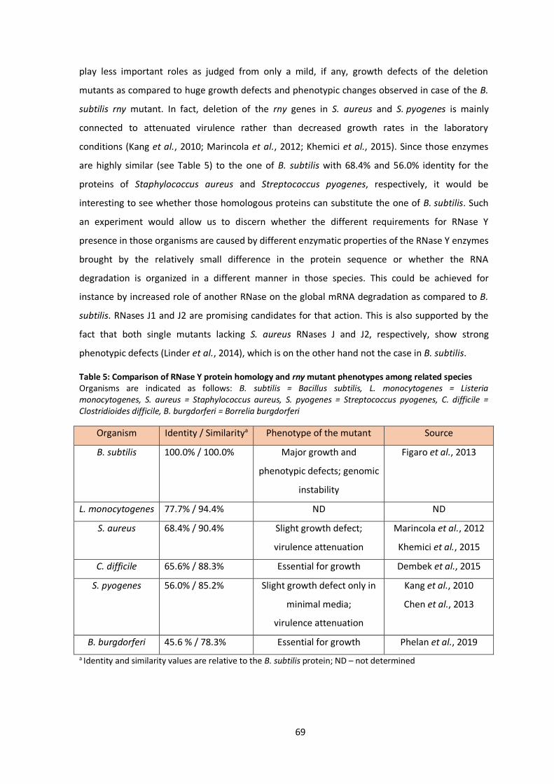
69
play less important roles as judged from only a mild, if any, growth defects of the deletion
mutants as compared to huge growth defects and phenotypic changes observed in case of the B.
subtilis rny mutant. In fact, deletion of the rny genes in S. aureus and S. pyogenes is mainly
connected to attenuated virulence rather than decreased growth rates in the laboratory
conditions (Kang et al., 2010; Marincola et al., 2012; Khemici et al., 2015). Since those enzymes
are highly similar (see Table 5) to the one of B. subtilis with 68.4% and 56.0% identity for the
proteins of Staphylococcus aureus and Streptococcus pyogenes, respectively, it would be
interesting to see whether those homologous proteins can substitute the one of B. subtilis. Such
an experiment would allow us to discern whether the different requirements for RNase Y
presence in those organisms are caused by different enzymatic properties of the RNase Y enzymes
brought by the relatively small difference in the protein sequence or whether the RNA
degradation is organized in a different manner in those species. This could be achieved for
instance by increased role of another RNase on the global mRNA degradation as compared to B.
subtilis. RNases J1 and J2 are promising candidates for that action. This is also supported by the
fact that both single mutants lacking S. aureus RNases J and J2, respectively, show strong
phenotypic defects (Linder et al., 2014), which is on the other hand not the case in B. subtilis.
Table 5: Comparison of RNase Y protein homology and rny mutant phenotypes among related species Organisms are indicated as follows: B. subtilis = Bacillus subtilis, L. monocytogenes = Listeria monocytogenes, S. aureus = Staphylococcus aureus, S. pyogenes = Streptococcus pyogenes, C. difficile = Clostridioides difficile, B. burgdorferi = Borrelia burgdorferi
Organism Identity / Similaritya Phenotype of the mutant Source
B. subtilis 100.0% / 100.0% Major growth and
phenotypic defects; genomic
instability
Figaro et al., 2013
L. monocytogenes 77.7% / 94.4% ND ND
S. aureus 68.4% / 90.4% Slight growth defect;
virulence attenuation
Marincola et al., 2012
Khemici et al., 2015
C. difficile 65.6% / 88.3% Essential for growth Dembek et al., 2015
S. pyogenes 56.0% / 85.2%
Slight growth defect only in
minimal media;
virulence attenuation
Kang et al., 2010
Chen et al., 2013
B. burgdorferi 45.6 % / 78.3% Essential for growth Phelan et al., 2019
a Identity and similarity values are relative to the B. subtilis protein; ND – not determined

70
Another important variable to be addressed in future is the expression rate of RNase Y
necessary for stable growth. For its functional counterpart RNase E of E. coli, the presence of only
10-20% of wild type levels of the protein is sufficient to sustain normal growth and this reduction
in RNase E quantity does not lead to major phenotypic effects (Jain et al., 2002). Such an
information is unfortunately missing for RNase Y of B. subtilis. Although transcriptomic studies
with the inducible promoter based depletion of RNase Y were performed, their experimental
design does not allow us to calculate precisely the protein amount requirement for sustainable
stable growth in wild type-like rates (Lehnik-Habrink et al., 2011b; Laalami et al., 2013).
It was previously suggested that mRNA turnover and generation time are correlated
(Rustad et al., 2013). Although this seems to be the truth for the best studied model organisms
among the domains of life (Bernstein et al., 2002; Hambraeus et al., 2003; Yang et al., 2003;
Geisberg et al., 2014), there are exceptions breaking this concept. For instance the slowly growing
cyanobacterium of the genus Prochlorococcus has a very short mRNA half-lives with average of
2,3 minutes, although it divides only once per day (Steglich et al., 2010). Furthermore, the
generation times of B. subtilis, S. aureus and S. pyogenes are not that significantly different (Gera
and McIver, 2013; Missiakas and Schneewind, 2013) to explain the difference in phenotypes of
the respective mutants. In addition, the rny mutant of S. pyogenes shows decreased growth rates
only in minimal medium where the generation times are longer, however, if the hypothesis about
generation time RNA stability correlation is correct, one would expect more severe phenotypes in
rich media with shorter doubling times.
4.2 Analysis of the rny suppressor mutants brings new insights into the
regulation of the RNA polymerase
Taken into an account the very strong difference in the activity of the RNA polymerase
variants in in vitro transcription assays, which was 200 fold for the RpoC-R88H variant as
compared to the wild type polymerase and not even quantifiable for the RpoB-G1054C variant,
we can ask ourselves whether such a huge decrease in RNA polymerase activity really occurs in
vivo. Although even just 2-fold increase in the mRNA half-lives is apparently enough to get the
RNA synthesis/degradation rate significantly out of equilibrium (Shahbabian et al., 2009), the
more than 200-fold drop in transcription activity still seems to be too excessive. Although further
experimental evidence will be needed to fully address this question, the decrease in transcription
rates is likely milder in vivo. In the gram-negative model organism E. coli it is well established that
the levels of RpoB and RpoC subunits of the RNA polymerase are subject to an auto-regulation on
multiple levels (Dennis et al., 1985; Meek and Hayward, 1986). Whether the RNA polymerase
71
subunits are subject to a similar auto-regulation also in B. subtlis has never been addressed.
Nevertheless, the presence of such auto-regulatory mechanism seems to be probable, not just as
a rational explanation for the huge drop in in vitro transcriptional activity, but also judged from
the increased protein quantity of the RNA polymerase RpoB subunit in the strains containing the
RNA polymerase core mutations (both RpoB-G1054C and RpoC-R88H), which we have observed
during Western-Blot experiments (data not shown).
In contrast to E. coli, where the rpoB and rpoC genes are part of a multicistronic operon
together with ribosomal proteins, the B. subtilis rpoB and rpoC genes form just a bicistronic
operon. This rpoBC operon is, however, preceded by a more than 200 bp long 5′ UTR which could
have an influence on the rpoBC expression. Interestingly, a study published in the course of this
thesis shown that RNase Y cleaves within this UTR to create an alternative 5′ end of the rpoBC
transcript (DeLoughery et al., 2018), giving rise to a possibility that RNase Y is responsible for post-
transcriptional regulation of rpoBC expression in B. subtilis. Such an observation also sparked the
attractive speculation that the absence of this cleavage by RNase Y is the reason for the formation
of suppressor mutation affecting the RNA polymerase in response to the rny deletion. That would
falsify our previous conclusion about the pivotal function of RNase Y laying in the initiation of bulk
mRNA degradation. However, such a possibility seems to be rather unlikely, since we did not
observe any difference in the ß-galactosidase expression between the PrpoB-lacZ fusions containing
or lacking the RNase Y cleavage site. Such a results suggests that the loss of the RNase Y cleavage
site did not affect the expression of rpoBC genes.
Although our aforementioned model clearly show that the probability of assembly of the
whole RNAP complex is lower when core subunits are duplicated (see Fig. 11), there is one factor
which was for calculation simplicity left out during the model construction, but might play a role
in the suppression mechanism, and this is the presence of alternative sigma factors. The
housekeeping factor σA was the only sigma factor considered in the model, however, there are
also 18 alternative sigma factors in B. subtilis. They are known to have lower affinity for the core
than the housekeeping σA, which under normal circumstances contributes to the low expression
of the genes under their control (Österberg et al., 2011). However, the alternative sigma factors
may be favored in the situation with increased amount of uncomplete RNA polymerase
complexes lacking some of the minor subunits. This was already shown on the example of rpoZ
mutant in other organisms, which showed increased proportion of transcription dependent on
alternative sigma factors (Geertz et al., 2011; Gunnelius et al., 2014). Hence, it is possible that the
effect of the core duplication might not only lead to decrease of the overall transcription, but also
increase the proportion of transcripts from promoters controlled by the alternative sigma factors.
72
This would together account for the positive effect on the physiology of the rny suppressors, since
alternative sigma factors are mainly involved in transcription of stress related genes which might
help to combat the phenotypes caused by the rny deletion.
Whether this is really the case and alternative sigma factors play a role in the suppression
has to be assessed in future. On one hand, one might expect that cells that need increased
transcription of genes dependent on alternative sigma factors would simply upregulate
expression of the sigma factor for instance by promotor up mutations. However, on the other
hand, a simultaneous decrease of σA dependent transcription and increase in transcription from
promoters controlled by multiple alternative sigma factors together might be most easily
achieved by the duplication observed in our study, which is also supported by the finding that
genomic amplifications are the easiest and most often occurring suppressing mutations in
B. subtilis cells (Dormeyer et al., 2017; Reuß et al., 2019). One possible way to test the hypothesis
about the alternative sigma factors involvement would be to introduce deletion of the rny gene
into B. subtilis strain which was, on the other hand, proposed to have increased transcription
activity from promoters dependent on the housekeeping sigma factor σA. That was shown for
example for strains with rifampicin resistance variants of RpoB (Inaoka et al., 2004). If the
hypothesis is correct, the rny deletion in such a background should lead to even more detrimental
phenotype or obstacles in formation of suppressor mutations.
This thesis also brings strong support to the assumption that cold shock proteins, and
especially CspD, actually are transcription factors. This can be deduced from the finding of cspD
affecting mutations in the one class of suppressors next to the mutations in genes for the known
transcription factor greA and the RNA polymerase subunit rpoE. This assumption is further
supported by the evidence that CspB and CspD are localized around the nucleoid in transcription
dependent manner (Weber et al., 2001). Despite its name, cspD is expressed stably at variety of
conditions (Nicolas et al., 2012) and its role in transcription would be in agreement with the role
of the homologous cold-shock proteins in the gram-negative model organism E. coli, for which an
anti-termination activity was proposed (Bae et al., 2000). Whether CspD and other so-called cold
shock proteins in B. subtilis also act as anti-terminator proteins or whether their role in
transcription is different has to be subject of further investigations.
Another interesting finding this thesis brings about the cspD gene is the fact, that the
suppressors with inactivated cspD gene seem to be genetically stable (see Fig. S3), in contrast to
the progenitor rny mutant as well as the other suppressors evolved under different selection
scenarios that do provide a growth benefit, but do not lead to complete genetic stabilization. It is
not completely clear whether this genetic stabilization upon cspD inactivation is specific to the rny
mutant background or whether CspD plays some general role in the cellular ability to evolve

73
mutations. Preliminary results obtained on that topic in our laboratory suggest, that this is rather
rny specific, since double deletion strain of cspD and cspB is forming suppressor extensively
(Faßhauer and Stülke, unpublished). However, what is the exact link between RNase Y, CspD and
the genome stabilization remains unclear.
4.3 Loss of RNase Y leads to phenotypic effects independent of the total mRNA
accumulation
Whereas the total mRNA accumulation is likely the key problem the cells are facing upon
the rny deletion, it does not explain all of the phenotypes observed in the rny mutant. There are
probably additional reasons for some minor, less detrimental, phenotypes which could be
connected to changed expression of specific genes. In order to get a better understanding of all
the changes that occur upon the rny deletion we have used a transcriptomic approach. The wild
type, the rny mutant and one of its suppressors were subjected to RNA-sequencing of transcripts
present in the exponential phase of growth in rich medium. In agreement with previous studies
(Lehnik-Habrink et al., 2011b; Durand et al., 2012a; Laalami et al., 2013), we could clearly see that
the absence of RNase Y leads to a global remodeling of mRNAs abundances, since expression of
1102 genes was at least two-fold different from the expression in the wild type strain, which
means that 26% of all genes from the from the genome of B. subtilis are affected by the deletion.
Besides, our screen undoubtedly did not identify all genes effected by the absence of RNase Y,
since some genes with increased false discovery rates were excluded from the analysis and not all
genes are expressed during the conditions chosen for this experiment, in fact only about 50% of
all genes are transcribed during exponential growth in LB medium (Rasmussen et al., 2009).
Therefore, we can conclude that loss of RNase Y leads to global change of gene expression and
influences abundance of majority of transcripts.
Since the rny mutant has severely impaired growth, we also cannot exclude the
possibility, that differential expression of some genes which we observed is rather influenced by
the growth-rate dependent regulation than directly by the rny deletion (Klumpp et al., 2009;
Yubero and Poyatos, 2020).
Generally, we can divide the affected genes into two groups, those affected directly by
the absence of RNase Y and those where the differential is expression is caused indirectly. For a
direct effect, one would expect that the loss of a specific cleavage leads to a stabilization of
certain transcripts and destabilization of others. This is exactly the case of cggR-gapA operon. It
was previously shown that RNase Y cleaves between cggR and gapA genes which leads to
destabilization of cggR transcript (Commichau et al., 2009). Indeed, and also in agreement with
74
previous transcriptomic studies, cggR had more than 7-fold higher abundance in the rny mutant
as compared to the wild type (Lehnik-Habrink et al., 2011b; DeLoughery et al., 2018). Similarly,
also expression of the rpsO transcript is destabilized by RNase Y cleavage and thus its abundance
should be increased in the rny mutant, which was indeed the case in our study (Yao and
Bechhofer, 2010).
However, for many genes, the expression may be affected indirectly, for instance as a
consequence of differential expression of their regulators. This seems to be exactly the case if we
consider the regulation by alternative sigma factors, where the σD dependent genes are mostly
downregulated, likely in response to downregulation of sigD gene itself, while the σB dependent
genes are mainly upregulated, again probably due to sigB gene upregulation. These changes in
the regulation of expression of alternative sigma factors may also not be a direct effect related to
loss of RNase Y cleavage, but rather can be triggered by the overall stress that rny deletion exerts
(Figaro et al., 2013), since especially transcription of σB dependent genes is known to be part of
the general stress response (Price et al., 2001).
An interesting example of the sigma factor dependency is the case of the yvyC operon.
This is an operon preceded by σD dependent promoter composed of 5 genes related to flagellar
assembly yvyC, fliD, fliS, fliT, smiA and hpf gene, coding for ribosome dimerization protein (Nicolas
et al., 2012; Akanuma et al., 2016). All the first five genes of the operon are downregulated in
response to sigD downregulation, however, the last gene, hpf, is not. This is likely the case
because, except being part of the whole σD dependent transcription unit, hpf is also transcribed
from two other promoters, dependent on σB and σH, respectively (Drzewiecki et al., 1998).
In conjunction with our initial task addressed mainly in the suppressor mutant screen, we
also tried to identify transcripts whose differential expression in the rny mutant would return to
the wild type levels in the suppressing strain to alleviate the growth defects of the rny mutant. To
that end we also analyzed the transcriptome of the suppressor strain GP2518. This strain had also
a much-altered gene expression as compared both to the wild type (1168 differentially expressed
genes), but also to the rny mutant. There are more than 150 transcripts that actually indeed
returned towards the wild type levels in the suppressor. Given how large this group is, it is
unlikely that the return of a single transcript level would be the key for the suppression observed.
Already previous studies of transcriptomic effects of RNase Y depletion did not manage to identify
specific targets standing behind the crucial role of RNase Y for B. subtilis physiology (Laalami et al.,
2013), supporting our previous conclusion that the role in regulation of global mRNA homeostasis
is the main task of RNase Y.
Previously, the only available transcriptomic data about the influence of RNase Y were
obtained from depletion strains. Nevertheless, in parallel to this work, DeLoughery et al. (2018)

75
published RNA-sequencing data of the rny deletion mutant and we thus wished to see, to what
extent our data correlate. Our studies agreed in most cases for which data in sufficient quality
were available in both studies, however not in all of them. For 54 genes (out of 1102 differentially
expressed in our study) we observed an opposite effect in the two studies as compared to the
respective wild-type levels. This discrepancy can by caused by the differences in experimental
setups, since we have harvested the cells in higher OD600 than DeLoughery et al. and wild-wild
type strain NCIB3610 was used in the other study and not laboratory wild type 168 as in our case.
Interestingly, however, 38 out of those 54 genes, for which the expression data between our
studies did not match, were also identified in our screen as genes whose expression returned
towards the wild-type levels in the suppressor strain. It is therefore tempting to speculate that the
rny mutant used by DeLoughery at el. had already acquired second site suppressor mutation(s) in
the course of their experiment. This would not be so surprising given the incredible speed rny
mutant forms suppressors and especially fixes the ctsR-pdaB duplication (see Fig. 10).
This is another noteworthy observation of this thesis. We have observed that deletion of
the rny gene in the background of wild type 168 always leads to the maintenance of the ctsR-
pdaB duplication, which is, however, naturally present also in a small part of the wild type
population. It was already previously shown in gram-positive bacteria that stochastic duplications
of chromosomal segments occur with a frequency ranging from 10-6 to 10-2 per cell per
generation. Hence a standard population cultivated in the laboratory always contain cells
harboring some genomic duplication and it is just a matter of probability, whether such a
duplication brings any advantage to the cells and thus becomes dominant in the majority of the
population (Pettersson et al., 2009; Tomanek et al., 2020). It was proposed by Romero and
Palacios that such gene amplifications should not be considered as mutations, but rather as a
dynamic state of the genome related to its fast adaptation preparedness for changing
environmental conditions (Romero and Palacios, 1997). Apparently, these findings are valid also
for the gram-positive B. subtilis. In fact, it took only 48 hours of growth inoculated with single
colony for this ctsR-pdaB duplication to be maintained by the majority of the population,
supporting the previous findings that duplications are a significantly faster mode of adaptation
than other genome modifications, such as promoters up mutations, for example (Dormeyer et al.,
2017; Reuß et al., 2019; Tomanek et al., 2020).
In correlation with the sigma factor dependency, as already suggested, we have noticed
interesting patterns that might explain some of the observed phenotypes of the rny mutant. For
instance, the downregulation of the sigD gene might explain the long chain phenotype, since it
was shown that σD OFF cells grow in long chains of sessile cells (Kearns and Losick, 2005). Under
the σD control are also genes coding for five peptidoglycan autolysins (lytA, lytB,lytC, lytD and lytF)

76
that play a major role in cell separation and motility (Chen et al., 2009). Since they were indeed all
significantly downregulated in our transcriptomic analysis, it is tempting to speculate that their
downregulation is responsible for the disordered peptidoglycan as observed in the rny mutant
(Figaro et al., 2013). In an attempt to confirm such a hypothesis, it might be interesting to see,
whether an artificial overexpression of either the autolysin genes, or of the sigD gene, would
revert the phenotype of disordered peptidoglycan and growth in chains and possibly indirectly
also affect other phenotypes observed in the rny mutant.
One such a phenotype which complicated the work in the laboratory and slowed down
the progress of this project is the loss of genetic competence. To possibly speed up the progress,
we decided to take a closer look at this phenomenon in the second part of this thesis. Initially we
hypothesized that the loss of competence in the rny mutant strain may be a consequence of
decreased expression of comK, the competence master regulator (van Sinderen et al., 1995). This
was supported also by the transcriptomic data obtained in previous studies as well as in this thesis
(Lehnik-Habrink et al., 2011b; Laalami et al., 2013). Furthermore RNase Y is employed in
maturation of sRNA called rnaC, which is responsible for maintaining levels of AbrB,
transcriptional repressor of comK (Mars et al., 2015; DeLoughery et al., 2018). On top of that, the
mecA transcript which encodes a protein responsible for ComK proteolytic degradation was
shown to be more abundant in the rny mutant (DeLoughery et al., 2018). All these results
together therefore suggested that the dysregulation of ComK levels through the aforementioned
mechanisms could be behind the loss of competence in the rny mutant.
To test this possibility, we constructed a strain with overexpression of the comK and comS
genes, comS encodes small adaptor protein which sequesters MecA-ClpCP complex and thereby
prevents ComK degradation (Turgay et al., 1998; Prepiak and Dubnau, 2007), and introduced the
rny deletion into such a background. If the competence deficiency of the rny mutant was really
caused by the decreased expression of comK, transformation rates should be restored in this new
background. However, this was not the case and the rny mutant did not give rise to a single
transformant colony even upon comKS overexpression.
Having such a screening system in hand, we then decided to test some other genes,
whose deletion also lead to the loss of genetic competence. This way we could show that
nanoRNase A encoded by the gene nrnA is involved in the regulation of competence master
regulator ComK by so far undiscovered mechanism, or exclude the role of transcription factor
GreA in the ComK regulation (van Sinderen et al., 1995; Mechold et al., 2007; Kusuya et al., 2011).
These experiments also aroused our interested in the previously poorly characterized ABC
transporter YtrBCDEF (Yoshida et al., 2000; Salzberg et al., 2011). Expression of this transporter is
controlled by the transcription repressor YtrA, whose deletion then leads to a loss of competence.
77
It was previously shown that this repression is in the wild type strain relieved only upon very
specific conditions related to cell wall attacking antibiotics (Salzberg et al., 2011; Wenzel et al.,
2012).
Based on this we built and later confirmed the hypothesis that the expression of the
YtrBCDEF transporter interferes with cell wall homeostasis and leads to increased cell wall
thickness (see Fig. 14). We also suggested that such an interference with the cell wall properties
can lead not only to a loss of genetic competence, but affect biofilm formation and sporulation
(Koo et al., 2017) This data can in return shed light also on the reasons for the competence
deficiency in the strain lacking RNase Y, since also this strain shows thicker and top of that highly
disorganized peptidoglycan layer (Figaro et al., 2013), likely as a result of downregulated
expression of autolysins as suggested above. By analogy to the situation in the ytrA mutant, it is
very much possible that the DNA binding proteins simply does not reach out of the peptidoglycan
layer to get in contact with the DNA molecule and that this steric hindrance is the main reason for
the impossibility to transform the rny mutant. Another possibility, which is also connected to the
function of autolysins, is that the chain growth prevents DNA binding, since DNA was shown to be
bound to cell poles during the process of transformation and those are not free in the chain-
growing cells of the rny mutant (Hahn et al., 2005; Kidane and Graumann, 2005).
Taken together, this thesis brings an evidence about a highly dynamic system constantly
looking for an optimal equilibrium between the cellular processes of RNA synthesis and
degradation, which is severely affected in the absence of RNase Y. In addition to the general role
in global mRNA degradation, loss of RNase Y is also shown to effect directly or indirectly the
expression of the majority of transcripts and some of them are suggested to provide explanation
to some of the phenotypes connected with the deletion of the rny gene.

78
5 References
Aguilar, C., Vlamakis, H., Guzman, A., Losick, R. and Kolter, R. (2010) KinD is a checkpoint protein
linking spore formation to extracellular-matrix production in Bacillus subtilis biofilms. MBio 1:
e00035–10.
Aguirre, A. A., Vicente, A. M., Hardwick, S. W., Alvelos, D. M., Mazzon, R. R., Luisi, B. F. and
Marques, M. V. (2017) Association of the cold shock DEAD-box RNA helicase RhlE to the RNA
degradosome in Caulobacter crescentus. J. Bacteriol. 199: e00135-17.
Akanuma, G., Kazo, Y., Tagami, K., Hiraoka, H., Yano, K., Suzuki, S. et al. (2016) Ribosome
dimerization is essential for the efficient regrowth of Bacillus subtilis. Microbiology 162: 448–458.
Altenbuchner, J. (2016) Editing of the Bacillus subtilis genome by the CRISPR-Cas9 system. Appl.
Environ. Microbiol. 82: 5421–5427.
Anderson, P. and Roth, J. (1981) Spontaneous tandem genetic duplications in Salmonella
typhimurium arise by unequal recombination between rRNA (rrn) cistrons. Proc. Natl. Acad. Sci. U.
S. A. 78: 3113–3117.
Andersson, D. I. and Hughes, D. (2009) Gene amplification and adaptive evolution in bacteria.
Annu. Rev. Genet. 43: 167–195.
Aravind, L. and Koonin, E. V. (1998) The HD domain defines a new superfamily of metal-
dependent phosphohydrolases. Trends Biochem. Sci. 23: 469–472.
Baba, T., Ara, T., Hasegawa, M., Takai, Y., Okumura, Y., Baba, M. et al. (2006) Construction of
Escherichia coli K-12 in-frame, single-gene knockout mutants: The Keio collection. Mol. Syst. Biol.
2: 2006.0008.
Bae, W., Xia, B., Inouye, M. and Severinov, K. (2000) Escherichia coli CspA-family RNA chaperones
are transcription antiterminators. Proc. Natl. Acad. Sci. U. S. A. 97: 7784–7789.
Barbe, V., Cruveiller, S., Kunst, F., Lenoble, P., Meurice, G., Sekowska, A. et al. (2009) From a
consortium sequence to a unified sequence: The Bacillus subtilis 168 reference genome a decade
later. Microbiology 155: 1758–1775.
Bechhofer, D. H. and Deutscher, M. P. (2019) Bacterial ribonucleases and their roles in RNA
metabolism. Crit. Rev. Biochem. Mol. Biol. 54: 242–300.
Beckering, C. L., Steil, L., Weber, M. H. W., Völker, U. and Marahiel, M. A. (2002) Genomewide
transcriptional analysis of the cold shock response in Bacillus subtilis. J. Bacteriol. 184: 6395–6402.
79
Beckert, B., Kedrov, A., Sohmen, D., Kempf, G., Wild, K., Sinning, I. et al. (2015) Translational
arrest by a prokaryotic signal recognition particle is mediated by RNA interactions. Nat. Struct.
Mol. Biol. 22: 767–773.
Berka, R. M., Hahn, J., Albano, M., Draskovic, I., Persuh, M., Cui, X. et al. (2002) Microarray
analysis of the Bacillus subtilis K-state: Genome-wide expression changes dependent on ComK.
Mol. Microbiol. 43: 1331–1345.
Bernstein, J. A., Khodursky, A. B., Lin, P. H., Lin-Chao, S. and Cohen, S. N. (2002) Global analysis
of mRNA decay and abundance in Escherichia coli at single-gene resolution using two-color
fluorescent DNA microarrays. Proc. Natl. Acad. Sci. U. S. A. 99: 9697–9702.
Bernstein, J. A., Lin, P. H., Cohen, S. N. and Lin-Chao, S. (2004) Global analysis of Escherichia coli
RNA degradosome function using DNA microarrays. Proc. Natl. Acad. Sci. 101: 2758–2763.
Beveridge, T. J. and Murray, R. G. E. (1979) How thick is the Bacillus subtilis cell wall? Curr.
Microbiol. 2: 1–4.
Bolger, A. M., Lohse, M. and Usadel, B. (2014) Trimmomatic: A flexible trimmer for Illumina
sequence data. Bioinformatics 30: 2114–2120.
Boonstra, M., Schaffer, M., Sousa, J., Morawska, L., Holsappel, S., Hildebrandt, P. et al. (2020)
Analyses of competent and non‐competent subpopulations of Bacillus subtilis reveal yhfW, yhxC
and ncRNAs as novel players in competence. Environ. Microbiol. 22: 2312–2328.
Branda, S. S., González-Pastor, J. E., Ben-Yehuda, S., Losick, R. and Kolter, R. (2001) Fruiting body
formation by Bacillus subtilis. Proc. Natl. Acad. Sci. U. S. A. 98: 11621–11626.
Briley, K., Dorsey-Oresto, A., Prepiak, P., Dias, M. J., Mann, J. M. and Dubnau, D. (2011) The
secretion ATPase ComGA is required for the binding and transport of transforming DNA. Mol.
Microbiol. 81: 818–830.
Britton, R. A., Wen, T., Schaefer, L., Pellegrini, O., Uicker, W. C., Mathy, N. et al. (2007)
Maturation of the 5′ end of Bacillus subtilis 16S rRNA by the essential ribonuclease YkqC/RNase
J1. Mol. Microbiol. 63: 127–138.
Broglia, L., Lécrivain, A. L., Renault, T. T., Hahnke, K., Ahmed-Begrich, R., Rhun, A. Le and
Charpentier, E. (2020) An RNA-seq based comparative approach reveals the transcriptome-wide
interplay between 3′-to-5′ exoRNases and RNase Y. Nat. Commun. 11: 1587.
Bruce, H. A., Du, D., Matak-Vinkovic, D., Bandyra, K. J., Broadhurst, R. W., Martin, E. et al.
(2018) Analysis of the natively unstructured RNA/protein-recognition core in the Escherichia coli
80
RNA degradosome and its interactions with regulatory RNA/Hfq complexes. Nucleic Acids Res. 46:
387–402.
Bruscella, P., Shahbabian, K., Laalami, S. and Putzer, H. (2011) RNase Y is responsible for
uncoupling the expression of translation factor IF3 from that of the ribosomal proteins L35 and
L20 in Bacillus subtilis. Mol. Microbiol. 81: 1526–1541.
Bucher, T., Oppenheimer-Shaanan, Y., Savidor, A., Bloom-Ackermann, Z. and Kolodkin-Gal, I.
(2015) Disturbance of the bacterial cell wall specifically interferes with biofilm formation. Environ.
Microbiol. Rep. 7: 990–1004.
Cahová, H., Winz, M. L., Höfer, K., Nübel, G. and Jäschke, A. (2015) NAD captureSeq indicates
NAD as a bacterial cap for a subset of regulatory RNAs. Nature 519: 374–377.
Cao, M., Wang, T., Ye, R. and Helmann, J. D. (2002) Antibiotics that inhibit cell wall biosynthesis
induce expression of the Bacillus subtilis σW and σM regulons. Mol. Microbiol. 45: 1267–1276.
Carabetta, V. J., Silhavy, T. J. and Cristea, I. M. (2010) The response regulator SprE (RssB) is
required for maintaining poly(A) polymerase I-degradosome association during stationary phase.
J. Bacteriol. 192: 3713–3721.
Cardenas, P. P., Carrasco, B., Sanchez, H., Deikus, G., Bechhofer, D. H. and Alonso, J. C. (2009)
Bacillus subtilis polynucleotide phosphorylase 3’-to-5’ DNase activity is involved in DNA repair.
Nucleic Acids Res. 37: 4157–4169.
Cardenas, P. P., Carzaniga, T., Zangrossi, S., Briani, F., Garcia-Tirado, E., Dehò, G. and Alonso, J.
C. (2011) Polynucleotide phosphorylase exonuclease and polymerase activities on single-stranded
DNA ends are modulated by RecN, SsbA and RecA proteins. Nucleic Acids Res. 39: 9250–9261.
Carpousis, A. J., Houwe, G. Van, Ehretsmann, C. and Krisch, H. M. (1994) Copurification of E. coli
RNAase E and PNPase: Evidence for a specific association between two enzymes important in RNA
processing and degradation. Cell 76: 889–900.
Carpousis, A. J. (2007) The RNA degradosome of Escherichia coli: An mRNA-degrading machine
assembled on RNase E. Annu. Rev. Microbiol. 61: 71–87.
Cascante-Estepa, N., Gunka, K. and Stülke, J. (2016) Localization of components of the RNA-
degrading machine in Bacillus subtilis. Front. Microbiol. 7: 1492.
Chang, B. Y. and Doi, R. H. (1990) Overproduction, purification, and characterization of Bacillus
subtilis RNA polymerase σA factor. J. Bacteriol. 172: 3257–3263.
81
Chen, R., Guttenplan, S. B., Blair, K. M. and Kearns, D. B. (2009) Role of the σD-dependent
autolysins in Bacillus subtilis population heterogeneity. J. Bacteriol. 191: 5775–5784.
Chen, Z., Itzek, A., Malke, H., Ferretti, J. J. and Kreth, J. (2013) Multiple roles of RNase Y in
Streptococcus pyogenes mRNA processing and degradation. J. Bacteriol. 195: 2585–2594.
Cho, K. H. (2017) The structure and function of the gram-positive bacterial RNA degradosome.
Front. Microbiol. 8: 154.
Chung, D. H., Min, Z., Wang, B. C. and Kushner, S. R. (2010) Single amino acid changes in the
predicted RNase H domain of Escherichia coli RNase G lead to complementation of RNase E
deletion mutants. RNA 16: 1371–1385.
Clarke, J. E., Kime, L., Romero A., D. and McDowall, K. J. (2014) Direct entry by RNase E is a major
pathway for the degradation and processing of RNA in Escherichia coli. Nucleic Acids Res. 42:
11733–11751.
Clerget, M., Jin, D. J. and Weisberg, R. A. (1995) A zinc-binding region in the β′ subunit of RNA
polymerase is involved in antitermination of early transcription of phage HK022. J. Mol. Biol 248:
768–780.
Clouet-d’Orval, B., Batista, M., Bouvier, M., Quentin, Y., Fichant, G., Marchfelder, A. and Maier,
L. K. (2018) Insights into RNA-processing pathways and associated RNA-degrading enzymes in
Archaea. FEMS Microbiol. Rev. 42: 579–613.
Coburn, G. A., Miao, X., Briant, D. J. and Mackie, G. A. (1999) Reconstitution of a minimal RNA
degradosome demonstrates functional coordination between a 3′ exonuclease and a DEAD-box
RNA helicase. Genes Dev. 13: 2594–2603.
Commichau, F. M., Rothe, F. M., Herzberg, C., Wagner, E., Hellwig, D., Lehnik-Habrink, M. et al.
(2009) Novel activities of glycolytic enzymes in Bacillus subtilis: interactions with essential
proteins involved in mRNA processing. Mol. Cell. Proteomics 8: 1350–1360.
Commichau, F. M., Pietack, N. and Stülke, J. (2013) Essential genes in Bacillus subtilis: A re-
evaluation after ten years. Mol. Biosyst. 9: 1068–1075.
Corrigan, R. M., Campeotto, I., Jeganathan, T., Roelofs, K. G., Lee, V. T. and Gründling, A. (2013)
Systematic identification of conserved bacterial c-di-AMP receptor proteins. Proc. Natl. Acad. Sci.
U. S. A. 110: 9084–9089.
Coutts, G., Thomas, G., Blakey, D. and Merrick, M. (2002) Membrane sequestration of the signal
transduction protein GlnK by the ammonium transporter AmtB. EMBO J. 21: 536–545.

82
Czyz, A., Mooney, R. A., Iaconi, A. and Landick, R. (2014) Mycobacterial RNA polymerase requires
a U-tract at intrinsic terminators and is aided by NusG at suboptimal terminators. MBio 5: e00931-
14.
Daou-Chabo, R., Mathy, N., Bénard, L. and Condon, C. (2009) Ribosomes initiating translation of
the hbs mRNA protect it from 5′-to-3′ exoribonucleolytic degradation by RNase J1. Mol. Microbiol.
71: 1538–1550.
Davies, K. M., Dedman, A. J., Horck, S. van and Lewis, P. J. (2005) The NusA:RNA polymerase
ratio is increased at sites of rRNA synthesis in Bacillus subtilis. Mol. Microbiol. 57: 366–379.
Deana, A. and Belasco, J. G. (2004) The function of RNase G in Escherichia coli is constrained by its
amino and carboxyl termini. Mol. Microbiol. 51: 1205–1217.
Deana, A., Celesnik, H. and Belasco, J. G. (2008) The bacterial enzyme RppH triggers messenger
RNA degradation by 5′ pyrophosphate removal. Nature 451: 355–358.
Deikus, G., Condon, C. and Bechhofer, D. H. (2008) Role of Bacillus subtilis RNase J1
endonuclease and 5′-exonuclease activities in trp leader RNA turnover. J. Biol. Chem. 283: 17158–
17167.
Deikus, G. and Bechhofer, D. H. (2009) Bacillus subtilis trp leader RNA. RNase J1 endonuclease
cleavage specificity and PNPase processing. J. Biol. Chem. 284: 26394–26401.
DeLoughery, A., Dengler, V., Chai, Y. and Losick, R. (2016) Biofilm formation by Bacillus subtilis
requires an endoribonuclease-containing multisubunit complex that controls mRNA levels for the
matrix gene repressor SinR. Mol. Microbiol. 99: 425–437.
DeLoughery, A., Lalanne, J. B., Losick, R. and Li, G. W. (2018) Maturation of polycistronic mRNAs
by the enribonuclease RNase Y and its associated Y-complex in Bacillus subtilis. Proc. Natl. Acad.
Sci. U. S. A. 115: E5585–E5594.
Delumeau, O., Lecointe, F., Muntel, J., Guillot, A., Guédon, E., Monnet, V. et al. (2011) The
dynamic protein partnership of RNA polymerase in Bacillus subtilis. Proteomics 11: 2992–3001.
Dembek, M., Barquist, L., Boinett, C. J., Cain, A. K., Mayho, M., Lawley, T. D. et al. (2015) High-
throughput analysis of gene essentiality and sporulation in Clostridium difficile. MBio 6: e02383.
Dennis, P. P., Nene, V. and Glass, R. E. (1985) Autogenous posttranscriptional regulation of RNA
Polymerase ß and ß′ subunit synthesis in Escherichia coli. J. Bacteriol. 161: 803–806.
Detsch, C. and Stülke, J. (2003) Ammonium utilization in Bacillus subtilis: Transport and regulatory
functions of NrgA and NrgB. Microbiology 149: 3289–3297.

83
Diethmaier, C., Pietack, N., Gunka, K., Wrede, C., Lehnik-Habrink, M., Herzberg, C. et al. (2011) A
novel factor controlling bistability in Bacillus subtilis: The Ymdb protein affects flagellin expression
and biofilm formation. J. Bacteriol. 193: 5997–6007.
Doherty, G. P., Fogg, M. J., Wilkinson, A. J. and Lewis, P. J. (2010) Small subunits of RNA
polymerase: Localization, levels and implications for core enzyme composition. Microbiology 156:
3532–3543.
Dormeyer, M., Lübke, A. L., Müller, P., Lentes, S., Reuß, D. R., Thürmer, A. et al. (2017)
Hierarchical mutational events compensate for glutamate auxotrophy of a Bacillus subtilis gltC
mutant. Environ. Microbiol. Rep. 9: 279–289.
Draskovic, I. and Dubnau, D. (2005) Biogenesis of a putative channel protein, ComEC, required for
DNA uptake: Membrane topology, oligomerization and formation of disulphide bonds. Mol.
Microbiol. 55: 881–896.
Drzewiecki, K., Eymann, C., Mittenhuber, G. and Hecker, M. (1998) The yvyD gene of Bacillus
subtilis is under dual control of σ(B) and σ(H). J. Bacteriol. 180: 6674–6680.
Durand, S., Gilet, L., Bessières, P., Nicolas, P. and Condon, C. (2012a) Three essential
ribonucleases-RNase Y, J1, and III-control the abundance of a majority of Bacillus subtilis mRNAs.
PLoS Genet. 8: e1002520.
Durand, S., Gilet, L. and Condon, C. (2012b) The essential function of B. subtilis RNase III is to
silence foreign toxin genes. PLoS Genet. 8: e1003181.
Durand, S., Tomasini, A., Braun, F., Condon, C. and Romby, P. (2015) sRNA and mRNA turnover in
gram-positive bacteria. FEMS Microbiol. Rev. 39: 316–330.
Durand, S., Braun, F., Helfer, A. C., Romby, P. and Condon, C. (2017) sRNA-mediated activation of
gene expression by inhibition of 5’-3’ exonucleolytic mRNA degradation. Elife 6: e23602.
Durand, S. and Condon, C. (2018) RNases and helicases in Gram-positive bacteria. Microbiol.
Spectr. 6: RWR-0003-2017.
Even, S., Pellegrini, O., Zig, L., Labas, V., Vinh, J., Bréchemmier-Baey, D. and Putzer, H. (2005)
Ribonucleases J1 and J2: Two novel endoribonucleases in B. subtilis with functional homology to
E.coli RNase E. Nucleic Acids Res. 33: 2141–2152.
Evguenieva-Hackenberg, E., Hou, L., Glaeser, S. and Klug, G. (2014) Structure and function of the
archaeal exosome. Wiley Interdiscip. Rev. RNA 5: 623–635.

84
Fang, M., Zeisberg, W.-M., Condon, C., Ogryzko, V., Danchin, A. and Mechold, U. (2009)
Degradation of nanoRNA is performed by multiple redundant RNases in Bacillus subtilis. Nucleic
Acids Res. 37: 5114–5125.
Farr, G. A., Oussenko, I. A. and Bechhofer, D. H. (1999) Protection against 3’-to-5’ RNA decay in
Bacillus subtilis. J. Bacteriol. 181: 7323–7330.
Figaro, S., Durand, S., Gilet, L., Cayet, N., Sachse, M. and Condon, C. (2013) Bacillus subtilis
mutants with knockouts of the genes encoding ribonucleases RNase Y and RNase J1 are viable,
with major defects in cell morphology, sporulation, and competence. J. Bacteriol. 195: 2340–
2348.
Flórez, L. A., Gunka, K., Polanía, R., Tholen, S. and Stülke, J. (2011) SPABBATS: A pathway-
discovery method based on Boolean satisfiability that facilitates the characterization of
suppressor mutants. BMC Syst. Biol. 5: 5.
Frindert, J., Zhang, Y., Nübel, G., Kahloon, M., Kolmar, L., Hotz-Wagenblatt, A. et al. (2018)
Identification, biosynthesis, and decapping of NAD-Capped RNAs in B. subtilis. Cell Rep. 24: 1890–
1901.
Frindert, J., Kahloon, M. A., Zhang, Y., Ahmed, Y. L., Sinning, I. and Jäschke, A. (2019) YvcI from
Bacillus subtilis has in vitro RNA pyrophosphohydrolase activity. J. Biol. Chem. 294: 19967–19977.
Gallagher, L. A., Bailey, J. and Manoil, C. (2020) Ranking essential bacterial processes by speed of
mutant death. Proc. Natl. Acad. Sci. U. S. A. 202001507.
Gamba, P., Jonker, M. J. and Hamoen, L. W. (2015) A novel feedback loop that controls bimodal
expression of genetic competence. PLoS Genet. 11: e1005047.
Gao, J., Lee, K., Zhao, M., Qiu, J., Zhan, X., Saxena, A. et al. (2006) Differential modulation of E.
coli mRNA abundance by inhibitory proteins that alter the composition of the degradosome. Mol.
Microbiol. 61: 394–406.
Geertz, M., Travers, A., Mehandziska, S., Sobetzko, P., Janga, S. C., Shimamoto, N. and
Muskhelishvilia, G. (2011) Structural coupling between RNA polymerase composition and DNA
supercoiling in coordinating transcription: A global role for the omega subunit? MBio 2: e00034-
11.
Geisberg, J. V., Moqtaderi, Z., Fan, X., Ozsolak, F. and Struhl, K. (2014) Global analysis of mRNA
isoform half-lives reveals stabilizing and destabilizing elements in yeast. Cell 156: 812–824.
85
Gera, K. and McIver, K. S. (2013) Laboratory Growth and Maintenance of Streptococcus pyogenes
(The Group A Streptococcus, GAS). Curr Protoc Microbiol. 30: 9D.2.1-9D.2.13.
Gilet, L., Dichiara, J. M., Figaro, S., Bechhofer, D. H. and Condon, C. (2015) Small stable RNA
maturation and turnover in Bacillus subtilis. Mol. Microbiol. 95: 270–282.
González-Gutiérrez, J. A., Díaz-Jiménez, D. F., Vargas-Pérez, I., Guillén-Solís, G., Stülke, J. and
Olmedo-Álvarez, G. (2018) The DEAD-box RNA helicases of Bacillus subtilis as a model to evaluate
genetic compensation among duplicate genes. Front. Microbiol. 9: 2261.
Graumann, P., Wendrich, T. M., Weber, M. H. W., Schröder, K. and Marahiel, M. A. (1997) A
family of cold shock proteins in Bacillus subtilis is essential for cellular growth and for efficient
protein synthesis at optimal and low temperatures. Mol. Microbiol. 25: 741–756.
Grishin, N. V (2001) KH domain: one motif, two folds. Nucleic Acids Res. 29: 638–643.
Grunberg-Manago, M., Ortiz, P. J. and Ochoa, S. (1956) Enzymic synthesis of polynucleotides I.
polynucleotide phosphorylase of Azotobacter vinelandii. Biochim. Biophys. Acta - Gen. Subj. 20:
269–285.
Guérout-Fleury, A. M., Shazand, K., Frandsen, N. and Stragier, P. (1995) Antibiotic-resistance
cassettes for Bacillus subtilis. Gene 167: 335–336.
Guerrier-Takada, C., Gardiner, K., Marsh, T., Pace, N. and Altman, S. (1983) The RNA moiety of
ribonuclease P is the catalytic subunit of the enzyme. Cell 35: 849–857.
Gundlach, J., Herzberg, C., Kaever, V., Gunka, K., Hoffmann, T., Weiß, M. et al. (2017) Control of
potassium homeostasis is an essential function of the second messenger cyclic di-AMP in Bacillus
subtilis. Sci. Signal. 10: eaal3011.
Gundlach, J., Krüger, L., Herzberg, C., Turdiev, A., Poehlein, A., Tascón, I. et al. (2019) Sustained
sensing in potassium homeostasis: Cyclic di-AMP controls potassium uptake by KimA at the levels
of expression and activity. J. Biol. Chem. 294: 9605–9614.
Gunnelius, L., Hakkila, K., Kurkela, J., Wada, H., Tyystjärvi, E. and Tyystjärvi, T. (2014) The omega
subunit of the RNA polymerase core directs transcription efficiency in cyanobacteria. Nucleic Acids
Res. 42: 4606–4614.
Hadjeras, L., Poljak, L., Bouvier, M., Morin‐Ogier, Q., Canal, I., Cocaign‐Bousquet, M. et al.
(2019) Detachment of the RNA degradosome from the inner membrane of Escherichia coli results
in a global slowdown of mRNA degradation, proteolysis of RNase E and increased turnover of
ribosome‐free transcripts. Mol. Microbiol. 111: 1715–1731.

86
Hahn, J., Roggiani, M. and Dubnau, D. (1995) The major role of Spo0A in genetic competence is
to downregulate abrB, an essential competence gene. J. Bacteriol. 177: 3601–3605.
Hahn, J., Maier, B., Haijema, B. J., Sheetz, M. and Dubnau, D. (2005) Transformation proteins and
DNA uptake localize to the cell poles in Bacillus subtilis. Cell 122: 59–71.
Haijema, B.-J., Hahn, J., Haynes, J. and Dubnau, D. (2001) A ComGA-dependent checkpoint limits
growth during the escape from competence. Mol. Microbiol. 40: 52–64.
Hambraeus, G., Wachenfeldt, C. von and Hederstedt, L. (2003) Genome-wide survey of mRNA
half-lives in Bacillus subtilis identifies extremely stable mRNAs. Mol. Genet. Genomics 269: 706–
714.
Hammarlöf, D. L., Bergman, J. M., Garmendia, E. and Hughes, D. (2015) Turnover of mRNAs is
one of the essential functions of RNase E. Mol. Microbiol. 98: 34–45.
Hamoen, L. W., Werkhoven, A. F. Van, Venema, G. and Dubnau, D. (2000) The pleiotropic
response regulator DegU functions as a priming protein in competence development in Bacillus
subtilis. Proc. Natl. Acad. Sci. U. S. A. 97: 9246–9251.
Hamoen, L. W., Smits, W. K., Jong, A. de, Holsappel, S. and Kuipers, O. P. (2002) Improving the
predictive value of the competence transcription factor (ComK) binding site in Bacillus subtilis
using a genomic approach. Nucleic Acids Res. 30: 5517–5528.
Hamoen, L. W., Kausche, D., Marahiel, M. A., Sinderen, D. van, Venema, G. and Serror, P.
(2003a) The Bacillus subtilis transition state regulator AbrB binds to the −35 promoter region of
comK. FEMS Microbiol. Lett. 218: 299–304.
Hamoen, L. W., Venema, G. and Kuipers, O. P. (2003b) Controlling competence in Bacillus
subtilis: shared use of regulators. Microbiology 149: 9–17.
Hamouche, L., Billaudeau, C., Rocca, A., Chastanet, A., Ngo, S., Laalami, S. and Putzer, H. (2020)
Dynamic membrane localization of RNase Y in Bacillus subtilis. MBio 11: e03337-19.
Hardwick, S. W., Chan, V. S. Y., Broadhurst, R. W. and Luisi, B. F. (2011) An RNA degradosome
assembly in Caulobacter crescentus. Nucleic Acids Res. 39: 1449–1459.
Hill, C. W., Grafstrom, R. H., Harnish, B. W. and Hillman, B. S. (1977) Tandem duplications
resulting from recombination between ribosomal RNA genes in Escherichia coli. J. Mol. Biol. 116:
407–428.
Himabindu, P. and Anupama, K. (2017) Decreased expression of stable RNA can alleviate the
lethality associated with RNase E deficiency in Escherichia coli. J. Bacteriol. 199: e00724-16.

87
Hoa, T. T., Tortosa, P., Albano, M. and Dubnau, D. (2002) Rok (YkuW) regulates genetic
competence in Bacillus subtilis by directly repressing comK. Mol. Microbiol. 43: 15–26.
Huen, J., Lin, C. L., Golzarroshan, B., Yi, W. L., Yang, W. Z. and Yuan, H. S. (2017) Structural
insights into a unique dimeric DEAD-box helicase CshA that promotes RNA decay. Structure 25:
469–481.
Hunger, K., Beckering, C. L., Wiegeshoff, F., Graumann, P. L. and Marahiel, M. A. (2006) Cold-
induced putative DEAD-box RNA helicases CshA and CshB are essential for cold adaptation and
interact with cold shock protein B in Bacillus subtilis. J. Bacteriol. 188: 240–248.
Hunt, A., Rawlins, J. P., Thomaides, H. B. and Errington, J. (2006) Functional analysis of 11
putative essential genes in Bacillus subtilis. Microbiology 152: 2895–2907.
Hutchison, C. A., Chuang, R.-Y., Noskov, V. N., Assad-Garcia, N., Deerinck, T. J., Ellisman, M. H. et
al. (2016) Design and synthesis of a minimal bacterial genome. Science 351: aad6253.
Hutter, B., Fischer, C., Jacobi, A., Schaab, C. and Loferer, H. (2004) Panel of Bacillus subtilis
reporter strains indicative of various modes of action. Antimicrob. Agents Chemother. 48: 2588–
2594.
Inaoka, T., Takahashi, K., Yada, H., Yoshida, M. and Ochi, K. (2004) RNA Polymerase mutation
activates the production of a dormant antibiotic 3,3′-neotrehalosadiamine via an autoinduction
mechanism in Bacillus subtilis. J. Biol. Chem. 279: 3885–3892.
Ingham, C. J., Dennis, J. and Furneaux, P. A. (1999) Autogenous regulation of transcription
termination factor Rho and the requirement for Nus factors in Bacillus subtilis. Mol. Microbiol. 31:
651–663.
Jain, C., Deana, A. and Belasco, J. G. (2002) Consequences of RNase E scarcity in Escherichia coli.
Mol. Microbiol. 43: 1053–1064.
Juang, Y. L. and Helmann, J. D. (1994) The δ subunit of Bacillus subtilis RNA polymerase: An
allosteric effector of the initiation and core-recycling phases of transcription. J. Mol. Biol. 239: 1–
14.
Kampf, J., Gerwig, J., Kruse, K., Cleverley, R., Dormeyer, M., Grünberger, A. et al. (2018)
Selective pressure for biofilm formation in Bacillus subtilis: Differential effect of mutations in the
master regulator SinR on bistability. MBio 9: e01464-18.

88
Kang, S. O., Caparon, M. G. and Cho, K. H. (2010) Virulence gene regulation by CvfA, a putative
RNase: the CvfA-enolase complex in Streptococcus pyogenes links nutritional stress, growth-phase
control, and virulence gene expression. Infect. Immun. 78: 2754–2767.
Kearns, D. B. and Losick, R. (2005) Cell population heterogeneity during growth of Bacillus
subtilis. Genes Dev. 19: 3083–3094.
Kearse, M., Moir, R., Wilson, A., Stones-Havas, S., Cheung, M., Sturrock, S. et al. (2012) Geneious
Basic: An integrated and extendable desktop software platform for the organization and analysis
of sequence data. Bioinformatics 28: 1647–1649.
Keller, A. N., Yang, X., Wiedermannová, J., Delumeau, O., Krásný, L. and Lewis, P. J. (2014) ε, a
new subunit of RNA polymerase found in gram-positive bacteria. J. Bacteriol. 196: 3622–3632.
Khemici, V., Toesca, I., Poljak, L., Vanzo, N. F. and Carpousis, A. J. (2004) The RNase E of
Escherichia coli has at least two binding sites for DEAD-box RNA helicases: functional replacement
of RhlB by RhlE. Mol. Microbiol. 54: 1422–1430.
Khemici, V., Poljak, L., Luisi, B. F. and Carpousis, A. J. (2008) The RNase E of Escherichia coli is a
membrane-binding protein. Mol. Microbiol. 70: 799–813.
Khemici, V., Prados, J., Linder, P., Redder, P. and Linder, P. (2015) Decay-initiating
endoribonucleolytic cleavage by RNase Y is kept under tight control via sequence preference and
sub-cellular localisation. PLOS Genet. 11: e1005577.
Kidane, D. and Graumann, P. L. (2005) Intracellular protein and DNA dynamics in competent
Bacillus subtilis cells. Cell 122: 73–84.
Kido, M., Yamanaka, K., Mitani, T., Niki, H., Ogura, T. and Hiraga, S. (1996) RNase E polypeptides
lacking a carboxyl-terminal half suppress a mukB mutation in Escherichia coli. J. Bacteriol. 178:
3917–3925.
Kim, S. K., Lormand, J. D., Weiss, C. A., Eger, K. A., Turdiev, H., Turdiev, A. et al. (2019) A
dedicated diribonucleotidase resolves a key bottleneck for the terminal step of RNA degradation.
Elife 8: e46313.
Klumpp, S., Zhang, Z. and Hwa, T. (2009) Growth-rate dependent global effects on gene
expression in bacteria. Cell 139: 1366–1375.
Kobayashi, K., Ehrlich, S. D., Albertini, A., Amati, G., Andersen, K. K., Arnaud, M. et al. (2003)
Essential Bacillus subtilis genes. Proc. Natl. Acad. Sci. U. S. A. 100: 4678–4683.
89
Konkol, M. A., Blair, K. M. and Kearns, D. B. (2013) Plasmid-encoded ComI inhibits competence in
the ancestral 3610 strain of Bacillus subtilis. J. Bacteriol. 195: 4085–4093.
Koo, B. M., Kritikos, G., Farelli, J. D., Todor, H., Tong, K., Kimsey, H. et al. (2017) Construction
and analysis of two genome-scale deletion libraries for Bacillus subtilis. Cell Syst. 4: 291–305.
Kouba, T., Pospíšil, J., Hnilicová, J., Šanderová, H., Barvík, I. and Krásný, L. (2019) The core and
holoenzyme forms of RNA polymerase from Mycobacterium smegmatis. J. Bacteriol. 201: e00583-
18.
Krásný, L. and Gourse, R. L. (2004) An alternative strategy for bacterial ribosome synthesis:
Bacillus subtilis rRNA transcription regulation. EMBO J. 23: 4473–4483.
Krásný, L., Tišerová, H., Jonák, J., Rejman, D. and Šanderová, H. (2008) The identity of the
transcription +1 position is crucial for changes in gene expression in response to amino acid
starvation in Bacillus subtilis. Mol. Microbiol. 69: 42–54.
Kunst, F. and Rapoport, G. (1995) Salt stress is an environmental signal affecting degradative
enzyme synthesis in Bacillus subtilis. J. Bacteriol. 177: 2403–2407.
Kusuya, Y., Kurokawa, K., Ishikawa, S., Ogasawara, N. and Oshima, T. (2011) Transcription factor
GreA contributes to resolving promoter-proximal pausing of RNA polymerase in Bacillus subtilis
cells. J. Bacteriol. 193: 3090–3099.
Laalami, S., Bessières, P., Rocca, A., Zig, L., Nicolas, P. and Putzer, H. (2013) Bacillus subtilis
RNase Y activity in vivo analysed by tiling microarrays. PLoS One 8: e54062.
Laalami, S., Zig, L. and Putzer, H. (2014) Initiation of mRNA decay in bacteria. Cell. Mol. Life Sci.
71: 1799–1828.
Lagesen, K., Ussery, D. W. and Wassenaar, T. M. (2010) Genome update: the 1000th genome - a
cautionary tale. Microbiology 156: 603–608.
Langmead, B. and Salzberg, S. (2012) Fast gapped-read alignment with Bowtie 2. Nat. Methods 9:
357–359.
Lee, C. R., Cho, S. H., Yoon, M. J., Peterkofsky, A. and Seok, Y. J. (2007) Escherichia coli enzyme
IIANtr regulates the K+ transporter TrkA. Proc. Natl. Acad. Sci. U. S. A. 104: 4124–4129.
Lee, K., Zhan, X., Gao, J., Qiu, J., Feng, Y., Meganathan, R. et al. (2003) RraA: a protein inhibitor
of RNase E activity that globally modulates RNA abundance in E. coli. Cell 114: 623–634.
90
Lehnik-Habrink, M., Pförtner, H., Rempeters, L., Pietack, N., Herzberg, C. and Stülke, J. (2010)
The RNA degradosome in Bacillus subtilis: identification of CshA as the major RNA helicase in the
multiprotein complex. Mol. Microbiol. 77: 958–971.
Lehnik-Habrink, M., Newman, J., Rothe, F. M., Solovyova, A. S., Rodrigues, C., Herzberg, C. et al.
(2011a) RNase Y in Bacillus subtilis: a natively disordered protein that is the functional equivalent
of RNase E from Escherichia coli. J. Bacteriol. 193: 5431–5441.
Lehnik-Habrink, M., Schaffer, M., Mäder, U., Diethmaier, C., Herzberg, C. and Stülke, J. (2011b)
RNA processing in Bacillus subtilis: identification of targets of the essential RNase Y. Mol.
Microbiol. 81: 1459–1473.
Lehnik-Habrink, M., Lewis, R. J., Mäder, U. and Stülke, J. (2012) RNA degradation in Bacillus
subtilis: an interplay of essential endo- and exoribonucleases. Mol. Microbiol. 84: 1005–1017.
Lehnik-Habrink, M., Rempeters, L., Kovács, Á. T., Wrede, C., Baierlein, C., Krebber, H. et al.
(2013) DEAD-box RNA helicases in Bacillus subtilis have multiple functions and act independently
from each other. J. Bacteriol. 195: 534–544.
Leroy, M., Piton, J., Gilet, L., Pellegrini, O., Proux, C., Coppée, J.-Y. et al. (2017) Rae1/YacP, a new
endoribonuclease involved in ribosome-dependent mRNA decay in Bacillus subtilis. EMBO J. 36:
1167–1181.
Lin-Chao, S., Chiou, N.-T. and Schuster, G. (2007) The PNPase, exosome and RNA helicases as the
building components of evolutionarily-conserved RNA degradation machines. J. Biomed. Sci. 14:
523–532.
Lin, T. H., Wei, G. T., Su, C. C. and Shaw, G. C. (2012) AdeR, a PucR-Type transcription factor,
activates expression of L-Alanine dehydrogenase and is required for sporulation of Bacillus
subtilis. J. Bacteriol. 194: 4995–5001.
Linder, P. and Jankowsky, E. (2011) From unwinding to clamping - the DEAD box RNA helicase
family. Nat. Rev. Mol. Cell Biol. 12: 505–516.
Linder, P., Lemeille, S. and Redder, P. (2014) Transcriptome-wide analyses of 5′-Ends in RNase J
mutants of a gram-positive pathogen reveal a role in RNA maturation, regulation and
degradation. PLoS Genet. 10: e1004207.
Liu, B., Deikus, G., Bree, A., Durand, S., Kearns, D. B. and Bechhofer, D. H. (2014) Global analysis
of mRNA decay intermediates in Bacillus subtilis wild-type and polynucleotide phosphorylase-
deletion strains. Mol. Microbiol. 94: 41–55.
91
Liu, B., Kearns, D. B. and Bechhofer, D. H. (2016) Expression of multiple Bacillus subtilis genes is
controlled by decay of slrA mRNA from Rho-dependent 3′ ends. Nucleic Acids Res. 44: 3364–3372.
López, D., Vlamakis, H. and Kolter, R. (2009) Generation of multiple cell types in Bacillus subtilis.
FEMS Microbiol. Rev. 33: 152–163.
López, D. and Kolter, R. (2010) Extracellular signals that define distinct and coexisting cell fates in
Bacillus subtilis. FEMS Microbiol. Rev. 34: 134–149.
Luciano, D. J., Vasilyev, N., Richards, J., Serganov, A. and Belasco, J. G. (2017) A novel RNA
phosphorylation state enables 5′ end-dependent degradation in Escherichia coli. Mol. Cell 67: 44–
54.
Luttinger, A., Hahn, J. and Dubnau, D. (1996) Polynucleotide phosphorylase is necessary for
competence development in Bacillus subtilis. Mol. Microbiol. 19: 343–356.
Maamar, H. and Dubnau, D. (2005) Bistability in the Bacillus subtilis K-state (competence) system
requires a positive feedback loop. Mol. Microbiol. 56: 615–624.
Mackie, G. A. (1998) Ribonuclease E is a 5’-end-dependent endonuclease. Nature 395: 720–723.
Mackie, G. A. (2013) RNase E: At the interface of bacterial RNA processing and decay. Nat. Rev.
Microbiol. 11: 45–57.
Mäder, U., Zig, L., Kretschmer, J., Homuth, G. and Putzer, H. (2008) mRNA processing by RNases
J1 and J2 affects Bacillus subtilis gene expression on a global scale. Mol. Microbiol. 70: 183–196.
Magnuson, R., Solomon, J. and Grossman, A. D. (1994) Biochemical and genetic characterization
of a competence pheromone from B. subtilis. Cell 77: 207–216.
Maier, B. (2020) Competence and transformation in Bacillus subtilis. Curr. Issues Mol. Biol. 37: 57–
76.
Marincola, G., Schäfer, T., Behler, J., Bernhardt, J., Ohlsen, K., Goerke, C. and Wolz, C. (2012)
RNase Y of Staphylococcus aureus and its role in the activation of virulence genes. Mol. Microbiol.
85: 817–832.
Marincola, G. and Wolz, C. (2017) Downstream element determines RNase Y cleavage of the
saePQRS operon in Staphylococcus aureus. Nucleic Acids Res. 45: 5980–5994.
Mars, R. A. T., Nicolas, P., Ciccolini, M., Reilman, E., Reder, A., Schaffer, M. et al. (2015) Small
regulatory RNA-Induced growth rate heterogeneity of Bacillus subtilis. PLOS Genet. 11: e1005046.
Martínez-Carranza, E., Barajas, H., Alcaraz, L. D., Servín-González, L., Ponce-Soto, G. Y. and

92
Soberón-Chávez, G. (2018) Variability of bacterial essential genes among closely related bacteria:
The case of Escherichia coli. Front. Microbiol. 9: 1059.
Mascher, T., Margulis, N. G., Wang, T., Ye, R. W. and Helmann, J. D. (2003) Cell wall stress
responses in Bacillus subtilis: The regulatory network of the bacitracin stimulon. Mol. Microbiol.
50: 1591–1604.
Mathy, N., Bénard, L., Pellegrini, O., Daou, R., Wen, T. and Condon, C. (2007) 5′-to-3′
exoribonuclease activity in bacteria: role of RNase J1 in rRNA maturation and 5′ stability of mRNA.
Cell 129: 681–692.
Mathy, N., Hébert, A., Mervelet, P., Bénard, L., Dorléans, A., Li De La Sierra-Gallay, I. et al.
(2010) Bacillus subtilis ribonucleases J1 and J2 form a complex with altered enzyme behaviour.
Mol. Microbiol. 75: 489–498.
Mechold, U., Fang, G., Ngo, S., Ogryzko, V. and Danchin, A. (2007) YtqI from Bacillus subtilis has
both oligoribonuclease and pAp-phosphatase activity. Nucleic Acids Res. 35: 4552–4561.
Meek, D. W. and Hayward, R. S. (1986) Direct evidence for autogenous regulation of the
Escherichia coli genes rpoBC in vivo. MGG Mol. Gen. Genet. 202: 500–508.
Meisner, J., Montero Llopis, P., Sham, L. T., Garner, E., Bernhardt, T. G. and Rudner, D. Z. (2013)
FtsEX is required for CwlO peptidoglycan hydrolase activity during cell wall elongation in Bacillus
subtilis. Mol. Microbiol. 89: 1069–1083.
Miczak, A., Kaberdin, V. R., Wei, C. L. and Lin-Chao, S. (1996) Proteins associated with RNase E in
a multicomponent ribonucleolytic complex. Proc. Natl. Acad. Sci. U. S. A. 93: 3865–3869.
Mirouze, N., Desai, Y., Raj, A. and Dubnau, D. (2012) Spo0A~P imposes a temporal gate for the
bimodal expression of competence in Bacillus subtilis. PLoS Genet. 8: e1002586.
Mirouze, N., Ferret, C., Cornilleau, C. and Carballido-López, R. (2018) Antibiotic sensitivity reveals
that wall teichoic acids mediate DNA binding during competence in Bacillus subtilis. Nat.
Commun. 9: 5072.
Missiakas, D. M. and Schneewind, O. (2013) Growth and Laboratory Maintenance of
Staphylococcus aureus. Curr Protoc Microbiol. Chapter 9: Unit-9C.1.
Mitchell, P., Petfalski, E., Shevchenko, A., Mann, M. and Tollervey, D. (1997) The exosome: A
conserved eukaryotic RNA processing complex containing multiple 3′→5′ exoribonucleases. Cell
91: 457–466.
93
Mohanty, B. K. and Kushner, S. R. (2000) Polynucleotide phosphorylase functions both as a 3’ →
5’ exonuclease and a poly(A) polymerase in Escherichia coli. Proc. Natl. Acad. Sci. U. S. A. 97:
11966–11971.
Mohanty, B. K. and Kushner, S. R. (2018) Enzymes involved in post-transcriptional RNA
metabolism in gram-negative bacteria. Microbiol. Spectr. 6: RWR-0011-2017.
Mohanty, B. K., Agrawal, A. and Kushner, S. R. (2020) Generation of pre-tRNAs from polycistronic
operons is the essential function of RNase P in Escherichia coli. Nucleic Acids Res. 48: 2564–2578.
Montero Llopis, P., Jackson, A. F., Sliusarenko, O., Surovtsev, I., Heinritz, J., Emonet, T. and
Jacobs-Wagner, C. (2010) Spatial organization of the flow of genetic information in bacteria.
Nature 466: 77–81.
Morita, T., Kawamoto, H., Mizota, T., Inada, T. and Aiba, H. (2004) Enolase in the RNA
degradosome plays a crucial role in the rapid decay of glucose transporter mRNA in the response
to phosphosugar stress in Escherichia coli. Mol. Microbiol. 54: 1063–1075.
Mortazavi, A., Williams, B. A., McCue, K., Schaeffer, L. and Wold, B. (2008) Mapping and
quantifying mammalian transcriptomes by RNA-Seq. Nat. Methods 5: 621–628.
Murashko, O. N. and Lin-Chao, S. (2017) Escherichia coli responds to environmental changes
using enolasic degradosomes and stabilized DicF sRNA to alter cellular morphology. Proc. Natl.
Acad. Sci. 114: E8025–E8034.
Najjar, N. El, Andari, J. El, Kaimer, C., Fritz, G., Rösch, T. C. and Graumann, P. L. (2018) Single
molecule tracking of DNA translocases in Bacillus subtilis reveals strikingly different dynamics of
SftA, SpoIIIE, and FtsA. Appl. Environ. Microbiol. 84: e02610-17.
Nakano, M. M., Xia, L. and Zuber, P. (1991) Transcription initiation region of the srfA operon,
which is controlled by the comP-comA signal transduction system in Bacillus subtilis. J. Bacteriol.
173: 5487–5493.
Newman, J. A., Hewitt, L., Rodrigues, C., Solovyova, A., Harwood, C. R. and Lewis, R. J. (2011)
Unusual, dual endo- and exonuclease activity in the degradosome explained by crystal structure
analysis of RNase J1. Structure 19: 1241–1251.
Newman, J. A., Hewitt, L., Rodrigues, C., Solovyova, A. S., Harwood, C. R. and Lewis, R. J. (2012)
Dissection of the network of interactions that links RNA processing with glycolysis in the Bacillus
subtilis degradosome. J. Mol. Biol. 416: 121–136.

94
Nicolas, P., Mäder, U., Dervyn, E., Rochat, T., Leduc, A., Pigeonneau, N. et al. (2012) Condition-
dependent transcriptome reveals high-level regulatory architecture in Bacillus subtilis. Science
335: 1103–1106.
Nudler, E., Avetissova, E., Markovtsov, V. and Goldfarb, A. (1996) Transcription processivity:
protein-DNA interactions holding together the elongation complex. Science 273: 211–217.
Nudler, E. (2009) RNA Polymerase Active Center: The Molecular Engine of Transcription. Annu.
Rev. Biochem. 78: 335–361.
O’Reilly, F. J., Xue, L., Graziadei, A., Sinn, L., Lenz, S., Tegunov, D. et al. (2020) In-cell architecture
of an actively transcribing-translating expressome. Science 369: 554–557.
Ogura, M., Yamaguchi, H., Kobayashi, K., Ogasawara, N., Fujita, Y. and Tanaka, T. (2002) Whole-
genome analysis of genes regulated by the Bacillus subtilis competence transcription factor ComK.
J. Bacteriol. 184: 2344–2351.
Ogura, M. and Asai, K. (2016) Glucose induces ECF sigma factor genes, sigX and sigM,
independent of cognate anti-sigma factors through acetylation of CshA in Bacillus subtilis. Front.
Microbiol. 7: 1918.
Osaka, N., Kanesaki, Y., Watanabe, M., Watanabe, S., Chibazakura, T., Takada, H. et al. (2020)
Novel (p)ppGpp0 suppressor mutations reveal an unexpected link between methionine catabolism
and GTP synthesis in Bacillus subtilis. Mol. Microbiol. 113: 1155–1169.
Österberg, S., Peso-Santos, T. del and Shingler, V. (2011) Regulation of alternative sigma factor
use. Annu. Rev. Microbiol. 65: 37–55.
Oussenko, I. A., Abe, T., Ujiie, H., Muto, A. and Bechhofer, D. H. (2005) Participation of 3′-to-5′
exoribonucleases in the turnover of Bacillus subtilis mRNA. J. Bacteriol. 187: 2758–2767.
Pellegrini, O., Nezzar, J., Marchfelder, A., Putzer, H. and Condon, C. (2003) Endonucleolytic
processing of CCA-less tRNA precursors by RNase Z in Bacillus subtilis. EMBO J. 22: 4534–4543.
Pettersen, E. F., Goddard, T. D., Huang, C. C., Couch, G. S., Greenblatt, D. M., Meng, E. C. and
Ferrin, T. E. (2004) UCSF Chimera - A visualization system for exploratory research and analysis. J.
Comput. Chem. 25: 1605–1612.
Pettersson, M. E., Song, A. E., Ae, S., Andersson, D. I. and Berg, O. G. (2009) Evolution of new
gene functions: simulation and analysis of the amplification model. Genetica 135: 309–324.

95
Phelan, J. P., Kern, A., Ramsey, M. E., Lundt, M. E., Sharma, B., Lin, T. et al. (2019) Genome-wide
screen identifies novel genes required for Borrelia burgdorferi survival in its Ixodes tick vector.
PloS Pathog. 15: e1007644.
Płociński, P., Macios, M., Houghton, J., Niemiec, E., Płocińska, R., Brzostek, A. et al. (2019)
Proteomic and transcriptomic experiments reveal an essential role of RNA degradosome
complexes in shaping the transcriptome of Mycobacterium tuberculosis. Nucleic Acids Res. 47:
5892–5905.
Prepiak, P. and Dubnau, D. (2007) A peptide signal for adapter protein-mediated degradation by
the AAA+ protease ClpCP. Mol. Cell 26: 639–647.
Price, C. W., Fawcett, P., Cérémonie, H., Su, N., Murphy, C. K. and Youngman, P. (2001) Genome-
wide analysis of the general stress response in Bacillus subtilis. Mol. Microbiol. 41: 757–774.
Prud’homme-Généreux, A., Beran, R. K., Iost, I., Ramey, C. S., Mackie, G. A. and Simons, R. W.
(2004) Physical and functional interactions among RNase E, polynucleotide phosphorylase and the
cold-shock protein, CsdA: Evidence for a “cold shock degradosome.” Mol. Microbiol. 54: 1409–
1421.
Py, B., Higgins, C. F., Krischt, H. M. and Carpousist, A. J. (1996) A DEAD-box RNA helicase in the
Escherichia coli RNA degradosome. Nature 381: 169–172.
Qi, Y. and Hulett, F. M. (1998) PhoP~P and RNA polymerase σA holoenzyme are sufficient for
transcription of Pho regulon promoters in Bacillus subtilis: PhoP~P activator sites within the
coding region stimulate transcription in vitro. Mol. Microbiol. 28: 1187–1197.
Quentin, Y., Fichant, G. and Denizot, F. (1999) Inventory, assembly and analysis of Bacillus subtilis
ABC transport systems. J. Mol. Biol. 287: 467–484.
Rabatinová, A., Šanderová, H., Matějčková, J. J., Korelusová, J., Sojka, L., Barvík, I. et al. (2013)
The δ subunit of RNA polymerase is required for rapid changes in gene expression and
competitive fitness of the cell. J. Bacteriol. 195: 2603–2611.
Rahmer, R., Heravi, K. M. and Altenbuchner, J. (2015) Construction of a super-competent Bacillus
subtilis 168 using the PmtlA-comKS inducible cassette. Front. Microbiol. 6: 1431.
Rasmussen, S., Nielsen, H. B. and Jarmer, H. (2009) The transcriptionally active regions in the
genome of Bacillus subtilis. Mol. Microbiol. 73: 1043–1057.
Redder, P. (2018) Molecular and genetic interactions of the RNA degradation machineries in
Firmicute bacteria. Wiley Interdiscip. Rev. RNA 9: e1460.

96
Redko, Y., Bechhofer, D. H. and Condon, C. (2008) Mini-III, an unusual member of the RNase III
family of enzymes, catalyses 23S ribosomal RNA maturation in B. subtilis. Mol. Microbiol. 68:
1096–1106.
Redko, Y., Aubert, S., Stachowicz, A., Lenormand, P., Namane, A., Darfeuille, F. et al. (2013) A
minimal bacterial RNase J-based degradosome is associated with translating ribosomes. Nucleic
Acids Res. 41: 288–301.
Reuß, D. R., Commichau, F. M., Gundlach, J., Zhu, B. and Stülke, J. (2016) The blueprint of a
minimal cell: MiniBacillus. Microbiol. Mol. Biol. Rev. 80: 955–987.
Reuß, D. R., Altenbuchner, J., Mäder, U., Rath, H., Ischebeck, T., Sappa, P. K. et al. (2017) Large-
scale reduction of the Bacillus subtilis genome: Consequences for the transcriptional network,
resource allocation, and metabolism. Genome Res. 27: 289–299.
Reuß, D. R., Faßhauer, P., Mroch, P. J., Ul-Haq, I., Koo, B.-M., Pöhlein, A. et al. (2019)
Topoisomerase IV can functionally replace all type 1A topoisomerases in Bacillus subtilis. Nucleic
Acids Res. 47: 5231–5242.
Richards, J., Liu, Q., Pellegrini, O., Celesnik, H., Yao, S., Bechhofer, D. H. et al. (2011) An RNA
pyrophosphohydrolase triggers 5′-exonucleolytic degradation of mRNA in Bacillus subtilis. Mol.
Cell 43: 940–949.
Rincón-Tomás, B., González, F. J., Somoza, L., Sauter, K., Madureira, P., Medialdea, T. et al.
(2020) Siboglinidae tubes as an additional niche for microbial communities in the Gulf of Cádiz—A
microscopical appraisal. Microorganisms 8: 367.
Romero, D. and Palacios, R. (1997) Gene amplification and genomic plasticity in prokaryotes.
Annu. Rev. Genet. 31: 91–111.
Ross, W., Thompson, J. F., Newlands, J. T. and Gourse, R. L. (1990) E.coli Fis protein activates
ribosomal RNA transcription in vitro and in vivo. EMBO J. 9: 3733–3742.
Rueden, C. T., Schindelin, J., Hiner, M. C., DeZonia, B. E., Walter, A. E., Arena, E. T. and Eliceiri, K.
W. (2017) ImageJ2: ImageJ for the next generation of scientific image data. BMC Bioinformatics
18: 529.
Rustad, T. R., Minch, K. J., Brabant, W., Winkler, J. K., Reiss, D. J., Baliga, N. S. and Sherman, D.
R. (2013) Global analysis of mRNA stability in Mycobacterium tuberculosis. Nucleic Acids Res. 41:
509–517.

97
Salvo, E., Alabi, S., Liu, B., Schlessinger, A. and Bechhofer, D. H. (2016) Interaction of Bacillus
subtilis polynucleotide phosphorylase and RNase Y: STRUCTURAL MAPPING AND EFFECT ON
mRNA TURNOVER. J. Biol. Chem. 291: 6655–6663.
Salzberg, L. I., Luo, Y., Hachmann, A.-B., Mascher, T. and Helmann, J. D. (2011) The Bacillus
subtilis GntR family repressor YtrA responds to cell wall antibiotics. J. Bacteriol. 193: 5793–5801.
Sambrook, J., Fritsch, E. F. and Maniatis, T. (1989) Molecular cloning: a laboratory manual. 2nd
ed. Cold Spring Harbor Laboratory, Cold Spring Harbor, N.Y. 1989
Serizawa, M., Yamamoto, H., Yamaguchi, H., Fujita, Y., Kobayashi, K., Ogasawara, N. and
Sekiguchi, J. (2004) Systematic analysis of SigD-regulated genes in Bacillus subtilis by DNA
microarray and Northern blotting analyses. Gene 329: 125–136.
Serror, P. and Sonenshein, A. L. (1996) CodY is required for nutritional repression of Bacillus
subtilis genetic competence. J. Bacteriol. 178: 5910–5915.
Shahbabian, K., Jamalli, A., Zig, L. and Putzer, H. (2009) RNase Y, a novel endoribonuclease,
initiates riboswitch turnover in Bacillus subtilis. EMBO J. 28: 3523–3533.
Shimane, K. and Ogura, M. (2004) Mutational analysis of the helix-turn-helix region of Bacillus
subtilis response regulator DegU, and identification of cis-acting sequences for DegU in the aprE
and comK promoters. J. Biochem. 136: 387–397.
Shivers, R. P. and Sonenshein, A. L. (2004) Activation of the Bacillus subtilis global regulator CodY
by direct interaction with branched-chain amino acids. Mol. Microbiol. 53: 599–611.
Šiková, M., Wiedermannová, J., Převorovský, M., Barvík, I., Sudzinová, P., Kofroňová, O. et al.
(2020) The torpedo effect in Bacillus subtilis: RNase J1 resolves stalled transcription complexes.
EMBO J. 39: e102500.
Silhavy, T. J., Kahne, D. and Walker, S. (2010) The bacterial cell envelope. Cold Spring Harb.
Perspect. Biol. 2: a000414.
Sinderen, D. van, Luttinger, A., Kong, L., Dubnau, D., Venema, G. and Hamoen, L. (1995) comK
encodes the competence transcription factor, the key regulatory protein for competence
development in Bacillus subtilis. Mol. Microbiol. 15: 455–462.
Smits, W. K., Eschevins, C. C., Susanna, K. A., Bron, S., Kuipers, O. P. and Hamoen, L. W. (2005)
Stripping Bacillus: ComK auto-stimulation is responsible for the bistable response in competence
development. Mol. Microbiol. 56: 604–614.

98
Sojka, L., Kouba, T., Barvík, I., Šanderová, H., Maderová, Z., Jonák, J. and Krásný, L. (2011) Rapid
changes in gene expression: DNA determinants of promoter regulation by the concentration of
the transcription initiating NTP in Bacillus subtilis. Nucleic Acids Res. 39: 4598–4611.
Steglich, C., Lindell, D., Futschik, M., Rector, T., Steen, R. and Chisholm, S. W. (2010) Short RNA
half-lives in the slow-growing marine cyanobacterium Prochlorococcus. Genome Biol. 11: R54.
Strahl, H., Turlan, C., Khalid, S., Bond, P. J., Kebalo, J.-M., Peyron, P. et al. (2015) Membrane
recognition and dynamics of the RNA degradosome. PLOS Genet. 11: e1004961.
Sulthana, S., Basturea, G. N. and Deutscher, M. P. (2016) Elucidation of pathways of ribosomal
RNA degradation: An essential role for RNase E. RNA 22: 1163–1171.
Tamura, M., Kers, J. A. and Cohen, S. N. (2012) Second-site suppression of RNase E essentiality by
mutation of the deaD RNA helicase in Escherichia coli. J. Bacteriol. 194: 1919–1926.
Tamura, M., Honda, N., Fujimoto, H., Cohen, S. N. and Kato, A. (2016) PpsA-mediated alternative
pathway to complement RNase E essentiality in Escherichia coli. Arch. Microbiol. 198: 409–421.
Tamura, M., Kageyama, D., Honda, N., Fujimoto, H. and Kato, A. (2017) Enzymatic activity
necessary to restore the lethality due to Escherichia coli RNase E deficiency is distributed among
bacteria lacking RNase E homologues. PLoS One 12: e0177915.
Tejada-Arranz, A., Crécy-Lagard, V. de and Reuse, H. de (2020) Bacterial RNA Degradosomes:
Molecular machines under tight control. Trends Biochem. Sci. 45: 42–57.
Tödter, D., Gunka, K. and Stülke, J. (2017) The highly conserved Asp23 family protein YqhY plays a
role in lipid biosynthesis in Bacillus subtilis. Front. Microbiol. 8: 883.
Tomanek, I., Grah, R., Lagator, M., Andersson, A. M. C., Bollback, J. P., Tkačik, G. and Guet, C. C.
(2020) Gene amplification as a form of population-level gene expression regulation. Nat. Ecol.
Evol. .
Tsai, Y. C., Du, D., Domínguez-Malfavón, L., Dimastrogiovanni, D., Cross, J., Callaghan, A. J. et al.
(2012) Recognition of the 70S ribosome and polysome by the RNA degradosome in Escherichia
coli. Nucleic Acids Res. 40: 10417–10431.
Tsirigotaki, A., Geyter, J. De, Šoštarić, N., Economou, A. and Karamanou, S. (2017) Protein export
through the bacterial Sec pathway. Nat. Rev. Microbiol. 15: 21–36.
Turgay, K., Hahn, J., Burghoorn, J. and Dubnau, D. (1998) Competence in Bacillus subtilis is
controlled by regulated proteolysis of a transcription factor. EMBO J. 17: 6730–6738.

99
Wach, A. (1996) PCR-synthesis of marker cassettes with long flanking homology regions for gene
disruptions in S. cerevisiae. Yeast 12: 259–265.
Wang, W. and Bechhofer, D. H. (1996) Properties of a Bacillus subtilis polynucleotide
phosphorylase deletion strain. J. Bacteriol. 178: 2375–2382.
Weber, M. H., Volkov, A. V, Fricke, I., Marahiel, M. A. and Graumann, P. L. (2001) Localization of
cold shock proteins to cytosolic spaces surrounding nucleoids in Bacillus subtilis depends on active
transcription. J. Bacteriol. 183: 6435–6443.
Weinrauch, Y., Penchev, R., Dubnau, E., Smith, E. and Dubnau, D. (1990) A Bacillus subtilis
regulatory gene product for genetic competence and sporulation resembles sensor protein
members of the bacterial two-component signal-transduction systems. Genes Dev. 4: 860–872.
Wenzel, M., Kohl, B., Münch, D., Raatschen, N., Albada, H. B., Hamoen, L. et al. (2012)
Proteomic response of Bacillus subtilis to lantibiotics reflects differences in interaction with the
cytoplasmic membrane. Antimicrob. Agents Chemother. 56: 5749–5757.
Wiedermannová, J., Sudzinová, P., Kovaľ, T., Rabatinová, A., Šanderova, H., Ramaniuk, O. et al.
(2014) Characterization of HelD, an interacting partner of RNA polymerase from Bacillus subtilis.
Nucleic Acids Res. 42: 5151–5163.
Wölfel, S. (2018) Characterization of a 60 kb genome amplification in Bacillus subtilis Δrny
suppressor mutants. Master thesis. University of Göttingen.
Worrall, J. A. R., Górna, M., Crump, N. T., Phillips, L. G., Tuck, A. C., Price, A. J. et al. (2008)
Reconstitution and analysis of the multienzyme Escherichia coli RNA degradosome. J. Mol. Biol.
382: 870–883.
Yadav, T., Carrasco, B., Hejna, J., Suzuki, Y., Takeyasu, K. and Alonso, J. C. (2013) Bacillus subtilis
DprA recruits RecA onto single-stranded DNA and mediates annealing of complementary strands
coated by SsbB and SsbA. J. Biol. Chem. 288: 22437–22450.
Yan, H., Qin, X., Wang, L. and Chen, W. (2020) Both enolase and the DEAD-box RNA helicase CrhB
can form complexes with RNase E in Anabaena sp. PCC 7120. Appl. Environ. Microbiol. 86:
e00425-20.
Yang, E., Nimwegen, E. van, Zavolan, M., Rajewsky, N., Schroeder, M., Magnasco, M. and
Darnell, J. E. (2003) Decay rates of human mRNAs: Correlation with functional characteristics and
sequence attributes. Genome Res. 13: 1863–1872.

100
Yao, S. and Bechhofer, D. H. (2010) Initiation of decay of Bacillus subtilis rpsO mRNA by
endoribonuclease RNase Y. J. Bacteriol. 192: 3279–3286.
Yao, S., Richards, J., Belasco, J. G. and Bechhofer, D. H. (2011) Decay of a model mRNA in Bacillus
subtilis by a combination of RNase J1 5′ exonuclease and RNase Y endonuclease activities. J.
Bacteriol. 193: 6384–6386.
Yoshida, K.-I., Fujita, Y. and Dusko Ehrlich, A. S. (2000) An Operon for a putative ATP-binding
cassette transport system involved in acetoin utilization of Bacillus subtilis. J. Bacteriol. 182: 5454–
5461.
Youngman, P. (1990) Use of transposons and integrational vectors for mutagenesis and
construction of gene fusions in Bacillus subtilis. In Molecular biological methods for Bacillus.
Harwood, C.R. (ed.), Chichester: Wiley, pp. 221–266
Yubero, P. and Poyatos, J. F. (2020) The impact of global transcriptional regulation on bacterial
gene order. iScience 23: 101029.
Zhu, B. and Stülke, J. (2018) SubtiWiki in 2018: From genes and proteins to functional network
annotation of the model organism Bacillus subtilis. Nucleic Acids Res. 46: D743–D748.
Zhu, X., Liu, D., Singh, A. K., Drolia, R., Bai, X., Tenguria, S. and Bhunia, A. K. (2018) Tunicamycin
mediated inhibition of wall teichoic acid affects Staphylococcus aureus and Listeria
monocytogenes cell morphology, biofilm formation and virulence. Front. Microbiol. 9: 1352.

101
6 Appendix
6.1 Supplementary material
Figure S1: Plasmid map of pBSURNAP used for the expression of the core RNA polymerase.
Figure S2: Suppressors of rny with mutations in rpoE and the rny rpoE double mutant show improved growth at 22°C, but not at 37°C. Serial drop dilutions comparing growth of the wild type 168, the rny mutant (GP2501), its derived suppressor mutants evolved at LB agar plates at 22°C (GP2637 and GP3210) and the rny rpoE double mutant (GP3217). The pictures were taken after 3 days of incubation at 22°C and 1 day of incubation at 37°C, respectively.
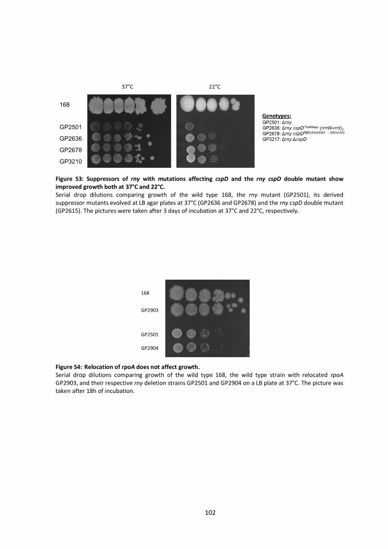
102
Figure S3: Suppressors of rny with mutations affecting cspD and the rny cspD double mutant show improved growth both at 37°C and 22°C. Serial drop dilutions comparing growth of the wild type 168, the rny mutant (GP2501), its derived suppressor mutants evolved at LB agar plates at 37°C (GP2636 and GP2678) and the rny cspD double mutant (GP2615). The pictures were taken after 3 days of incubation at 37°C and 22°C, respectively.
Figure S4: Relocation of rpoA does not affect growth. Serial drop dilutions comparing growth of the wild type 168, the wild type strain with relocated rpoA GP2903, and their respective rny deletion strains GP2501 and GP2904 on a LB plate at 37°C. The picture was taken after 18h of incubation.

103
Table S1. Effect of the rny deletion on the expression of B. subtilis genes and operons. All operons that exhibited an at least eight-fold change upon deletion of rny are shown (and relevant sigma factor genes). In case of differential expression within one operon, the genes not in bold did not met this 8-fold criteria.
Transcription unit Function a Regulation b Fold changes
mRNAs with increased amount in the rny mutant
yxkC unknown SigD, TnrA 0.010
epr minor extracellular serine protease, involved in control of swarming motility SigD, Spo0A, SinR, DegU, ScoC 0.013
yfmT-S vanillin dehydrogenase/soluble chemotaxis receptor SigD 0.022
motA-B H+-coupled MotA-MotB flagellar stator SigD 0.023
hemAT soluble chemotaxis receptor, heme-containing O2 sensor protein SigD 0.026
lytF major autolysin SigD, SinR, SlrR 0.031
hag flagellin protein SigD, CodY, ScoC, CsrA 0.031
glpT-Q glycerol-3-phosphate permease and diesterase GlpP, PhoP, CcpA 0.036
pyrR-P-B-C-AA-AB-K-D-F-E pyrimidine biosynthesis PyrR 0.047
pgdS gamma-DL-glutamyl hydrolase SigD 0.049
pstS-C-A-BA-BB high-affinity phosphate uptake PhoP 0.056
artP-Q-R high affinity arginine ABC transporter YlxR 0.056
yvbX putative glycoside hydrolase 0.065
yvbJ unknown 0.065
lip extracellular lipase AbrB 0.067
yxeK-snaB-yxeM-N-O-sndB-yxeQ N-acetylcysteine deacetylase CymR 0.068
tlpA-mcpA membrane-bound chemotaxis receptor, methyl-accepting chemotaxis protein SigD, AbrB 0.079
yocC-D unknown 0.083
ctaO heme O synthase (minor enzyme) AbrB 0.083
lytA-B-C autolysins SigD, SinR, YvrHb, SlrR 0.090
tlpC membrane-bound chemotaxis receptor, methyl-accepting chemotaxis protein SigD 0.090
mntA-B-C-D manganese ABC transporter MntR 0.093
tuaA/2-A/1-B-C-D-E-F-G-H biosynthesis of teichuronic acid PhoP, SigF 0.096
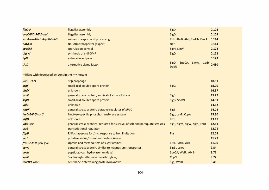
104
flhO-P flagellar assembly SigD 0.102
yvyC-fliD-S-T-A-hpf flagellar assembly SigD 0.109
sunA-sunT-bdbA-yolJ-bdbB sublancin export and processing Rok, AbrB, Abh, YvrHb, DnaA 0.114
natA-B Na+ ABC transporter (export) NatR 0.114
spo0M sporulation control SigH, SigW 0.122
dgcW synthesis of c-di-GMP SigD 0.122
lipB extracellular lipase 0.123
sigD alternative sigma factor SigD, Spo0A, SwrA, CodY, DegU
0.430
mRNAs with decreased amount in the rny mutant
yonP -O-N SPβ prophage 18.51
sspF small acid-soluble spore protein SigG 18.00
yhdX unknown 16.37
ysnF general stress protein, survival of ethanol stress SigB 15.22
sspB small acid-soluble spore protein SigG, SpoVT 14.93
yukJ unknown 14.52
nhaX general stress protein, putative regulator of nhaC SigB 14.03
levD-E-F-G-sacC fructose-specific phosphotransferase system SigL, LevR, CcpA 13.30
yhfH unknown YlxR 13.17
yjbC-spx general stress proteins, required for survival of salt and paraquate stresses SigB, SigM, SigW, SigX, PerR 12.81
ytzE transcriptional regulator 12.21
fbpB RNA chaperone for fsrA, response to iron limitation Fur 12.03
yrzF putative serine/threonine-protein kinase 11.73
frlB-O-N-M-frlD-yurJ Uptake and metabolisms of sugar amines FrlR, CodY, YlxR 11.00
corA general stress protein, similar to magnesium transporter SigB , LexA 9.84
yocH peptidoglycan hydrolase (amidase) Spo0A, WalR, AbrB 9.76
speD S-adenosylmethionine decarboxylase, CcpN 9.72
mreBH-ykpC cell shape-determining protein/unknown SigI, WalR 9.48

105
slp small peptidoglycan-associated lipoprotein 9.36
yrhH similar to methyltransferase SigW,SigM,SigX 9.05
tlrB 23S rRNA (guanine-N(1)-)-methyltransferase 9.03
yuzA general stress protein SigB, SigG 8.61
rsfA Regulator of SigF-dependent transcription SigF, SigG 8.57
bsrA-yrvM 6S RNA/ tRNA modification enzyme 8.55
yqhB similar to magnesium exporter, general stress protein SigB, LexA 8.42
sigF sporulation-specific sigma factor SigH, SigF, SigG, AbrB, SinR, Spo0A
4.23
sigG sporulation-specific sigma factor SigF, SigG, AbrB, SinR, Spo0A 4.68 a Information was taken from SubtiWiki database (Zhu and Stülke, 2018)
b The housekeeping sigma factor SigA is not listed as a regulator
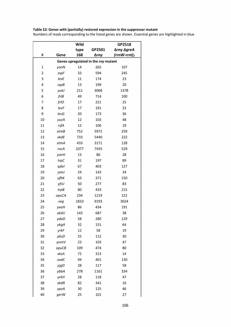
106
Table S2: Genes with (partially) restored expression in the suppressor mutant Numbers of reads corresponding to the listed genes are shown. Essential genes are highlighted in blue
# Gene
Wild type 168
GP2501 Δrny
GP2518 Δrny ΔgreA (rrnW-rrnI)2
Genes upregulated in the rny mutant
1 yonN 14 265 107
2 sspF 33 594 245
3 levE 11 174 23
4 sspB 13 199 20
5 yukJ 211 3068 1378
6 frlB 49 714 100
7 frlO 17 221 25
8 levF 17 191 23
9 levG 20 173 36
10 yuzA 12 103 48
11 rsfA 12 106 19
12 xtmB 752 5972 259
13 xkdE 733 5440 222
14 xtmA 433 3171 128
15 rocA 1077 7435 529
16 yonH 13 86 28
17 trpC 31 197 89
18 qdoI 67 403 127
19 yonJ 24 143 34
20 yfhK 63 371 150
21 yfiU 50 277 83
22 trpB 80 433 215
23 opuCA 234 1219 222
24 veg 1810 9193 3024
25 yxaH 86 434 191
26 xkdU 143 687 38
27 ydaD 58 280 129
28 ykgA 32 151 64
29 yrkF 12 58 19
30 pksD 25 112 30
31 yomV 23 103 47
32 opuCB 109 474 80
33 xkzA 72 313 14
34 oxdC 94 401 130
35 yjgD 28 117 58
36 ybbA 278 1161 334
37 yrkH 28 118 47
38 xkdR 82 341 16
39 ypzA 30 125 46
40 gerW 25 101 27
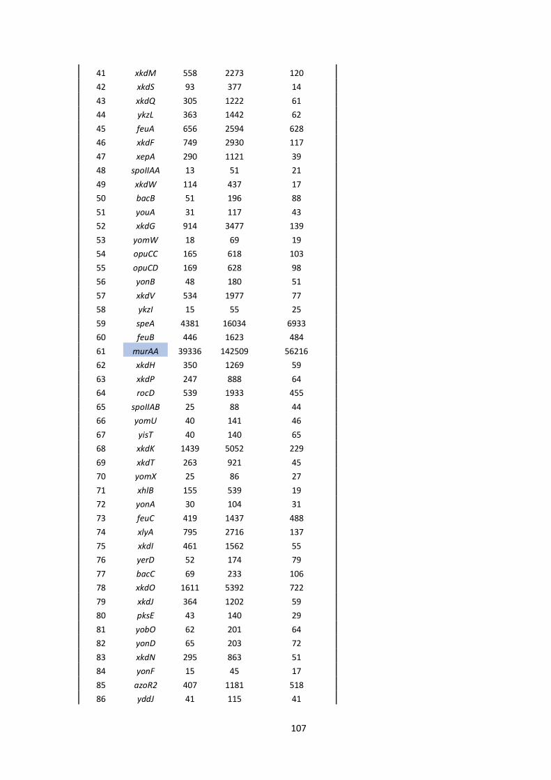
107
41 xkdM 558 2273 120
42 xkdS 93 377 14
43 xkdQ 305 1222 61
44 ykzL 363 1442 62
45 feuA 656 2594 628
46 xkdF 749 2930 117
47 xepA 290 1121 39
48 spoIIAA 13 51 21
49 xkdW 114 437 17
50 bacB 51 196 88
51 youA 31 117 43
52 xkdG 914 3477 139
53 yomW 18 69 19
54 opuCC 165 618 103
55 opuCD 169 628 98
56 yonB 48 180 51
57 xkdV 534 1977 77
58 ykzI 15 55 25
59 speA 4381 16034 6933
60 feuB 446 1623 484
61 murAA 39336 142509 56216
62 xkdH 350 1269 59
63 xkdP 247 888 64
64 rocD 539 1933 455
65 spoIIAB 25 88 44
66 yomU 40 141 46
67 yisT 40 140 65
68 xkdK 1439 5052 229
69 xkdT 263 921 45
70 yomX 25 86 27
71 xhlB 155 539 19
72 yonA 30 104 31
73 feuC 419 1437 488
74 xlyA 795 2716 137
75 xkdI 461 1562 55
76 yerD 52 174 79
77 bacC 69 233 106
78 xkdO 1611 5392 722
79 xkdJ 364 1202 59
80 pksE 43 140 29
81 yobO 62 201 64
82 yonD 65 203 72
83 xkdN 295 863 51
84 yonF 15 45 17
85 azoR2 407 1181 518
86 yddJ 41 115 41

108
87 yonC 26 73 21
88 yorG 37 104 30
89 gmuA 277 766 53
90 yorF 30 83 14
91 yisK 335 922 408
92 rocB 119 323 56
93 ykzM 239 646 35
94 yonO 16 43 12
95 spoIIT 4930 13052 5899
96 gmuD 1313 3471 215
97 gltB 936 2468 987
98 xhlA 248 651 21
99 yomE 18 46 12
100 yybF 464 1212 436
101 yonE 35 87 41
102 tagB 1918 4643 1720
103 yosP 51 121 38
104 tagA 2932 6995 2221
105 nrdEB 40 94 38
106 yqgY 282 659 325
107 spoVG 2028 4548 1825
108 cwlS 222 495 118
109 bacD 193 429 195
110 yomM 16 36 16
111 bdhA 5374 11288 2499
112 opuD 2318 4837 2384
113 galM 80 165 79
114 gmuR 749 1533 212
115 yorI 24 48 22
Genes downregulated in the rny mutant
1 artP 489 27 57
2 sndB 836 68 265
3 yvyC 447 39 94
4 fliD 4264 374 777
5 yxeQ 935 103 319
6 spo0M 7850 954 2678
7 epsD 185 25 52
8 cydA 83 13 81
9 nrgA 404 63 359
10 xpt 378 68 141
11 epsN 61 12 28
12 ywlD 232 45 132
13 yxeR 1218 237 478
14 yteJ 2911 579 1208
15 qdoR 1982 436 1148
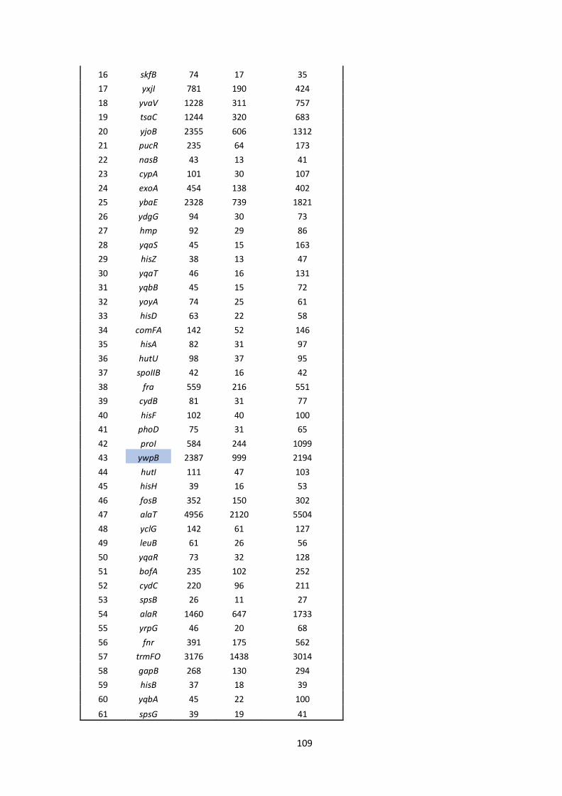
109
16 skfB 74 17 35
17 yxjI 781 190 424
18 yvaV 1228 311 757
19 tsaC 1244 320 683
20 yjoB 2355 606 1312
21 pucR 235 64 173
22 nasB 43 13 41
23 cypA 101 30 107
24 exoA 454 138 402
25 ybaE 2328 739 1821
26 ydgG 94 30 73
27 hmp 92 29 86
28 yqaS 45 15 163
29 hisZ 38 13 47
30 yqaT 46 16 131
31 yqbB 45 15 72
32 yoyA 74 25 61
33 hisD 63 22 58
34 comFA 142 52 146
35 hisA 82 31 97
36 hutU 98 37 95
37 spoIIB 42 16 42
38 fra 559 216 551
39 cydB 81 31 77
40 hisF 102 40 100
41 phoD 75 31 65
42 proI 584 244 1099
43 ywpB 2387 999 2194
44 hutI 111 47 103
45 hisH 39 16 53
46 fosB 352 150 302
47 alaT 4956 2120 5504
48 yclG 142 61 127
49 leuB 61 26 56
50 yqaR 73 32 128
51 bofA 235 102 252
52 cydC 220 96 211
53 spsB 26 11 27
54 alaR 1460 647 1733
55 yrpG 46 20 68
56 fnr 391 175 562
57 trmFO 3176 1438 3014
58 gapB 268 130 294
59 hisB 37 18 39
60 yqbA 45 22 100
61 spsG 39 19 41

110
6.2 Bacterial strains, plasmids and oligonucleotides
Table S3: Bacterial strains used in this study
Strain Genotype c Source a
B. subtilis
168 trpC2 Laboratory collection
BSB1 Wild type Nicolas et al., 2012
BKE30420 trpC2 ∆ytrE::ermC Koo et al., 2017
BKE30430 trpC2 ∆ytrD::ermC Koo et al., 2017
BKE30440 trpC2 ∆ytrC::ermC Koo et al., 2017
BKE30450 trpC2 ∆ytrB::ermC Koo et al., 2017
BP351 trpC2 ΔgreA::cat F. Commichau
CCB434 ∆rnjA::spc Figaro et al., 2013
CCB441 Δrny::spc Figaro et al., 2013
DK1042 comIQ12L Konkol et al., 2013
LK633 MO1099 rpoE::aphA3 amyE::mls Rabatinová et al., 2013
LK1098 ΔrpoE::aphA3 LK633 → BSB1
PG389 amyE::PcomG-lacZ-gfp-cat Gamba et al., 2015
PG10 b yvcA::(PmtlA-comKS) Reuß et al., 2017
GP811 trpC2 ∆gudB::cat rocG::Tn10 spc amyE::(gltA-lacZ
aphA3) ∆ansR::tet
Flórez et al., 2011
GP1152 trpC2 ∆ansR::tetR GP811→ 168
GP1748 trpC2 ∆pnpA::aphA3 Cascante-Estepa et al., 2016
GP2155 trpC2 ∆nrnA::aphA3 LFH → 168
GP2501 d trpC2 ∆rny::spc CCB441 → 168
GP2503 d trpC2 Δrny::spc greA (C374T – Ser125Leu) (rrnW-rrnI)2 Evolution of GP2501 at 22°C
GP2504 trpC2 Δrny::spc greA (G169T – Glu57Stop) Evolution of GP2501 at 22°C
GP2506 trpC2 ∆rnjA::spc CCB434 → 168
GP2518 d trpC2 ΔgreA::cat Δrny::spc (rrnW-rrnI)2 Evolution of GP2628 on LB
agar at 37°C
GP2524 trpC2 Δrny::ermC LFH → 168
GP2525 trpC2 greA-3xflag spc pGP2542 → 168
GP2529 trpC2 Δrny::ermC greA-3xflag spc GP2524 → GP2525
GP2538 trpC2 Δrny::ermC greA (Insertion A406)-3xflag spc Evolution of GP2529 at 22°C
GP2539 trpC2 Δrny::ermC greA (Deletion A66)-3xflag spc Evolution of GP2529 at 22°C
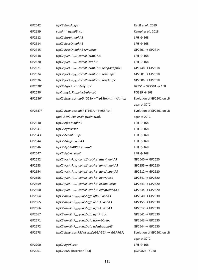
111
GP2542 trpC2 ΔrecA::spc Reuß et al., 2019
GP2559 comIQ12L ∆ymdB::cat Kampf et al., 2018
GP2612 trpC2 ∆greA::aphA3 LFH → 168
GP2614 trpC2 ΔcspD::aphA3 LFH → 168
GP2615 trpC2 ΔcspD::aphA3 Δrny::spc GP2501 → GP2614
GP2618 trpC2 yvcA-PmtlA-comKS-ermC-hisI LFH → 168
GP2620 trpC2 yvcA-PmtlA-comKS-cat-hisI LFH → 168
GP2621 trpC2 yvcA-PmtlA-comKS-ermC-hisI ∆pnpA::aphA3 GP1748 → GP2618
GP2624 trpC2 yvcA-PmtlA-comKS-ermC-hisI ∆rny::spc GP2501 → GP2618
GP2626 trpC2 yvcA-PmtlA-comKS-ermC-hisI ∆rnjA::spc GP2506 → GP2618
GP2628 d trpC2 ΔgreA::cat Δrny::spc BP351 + GP2501 → 168
GP2630 trpC amyE::PcomG-lacZ-gfp-cat PG389 → 168
GP2636 d trpC2 Δrny::spc cspD (G23A – Trp8Stop) (rrnW-rrnI)2 Evolution of GP2501 on LB
agar at 37°C
GP2637 d trpC2 Δrny::spc adeR (T163A – Tyr55Asn)
rpoE-Δ199-208 Δskin (rrnW-rrnI)2
Evolution of GP2501 on LB
agar at 22°C
GP2640 trpC2 ∆ftsH::aphA3 LFH → 168
GP2641 trpC2 ∆ytrA::spc LFH → 168
GP2643 trpC2 ∆comEC::spc LFH → 168
GP2644 trpC2 ∆degU::aphA3 LFH → 168
GP2646 trpC2 ∆ytrGABCDEF::ermC LFH → 168
GP2647 trpC2 ∆ytrA::ermC LFH → 168
GP2652 trpC2 yvcA-PmtlA-comKS-cat-hisI ∆ftsH::aphA3 GP2640 → GP2620
GP2653 trpC2 yvcA-PmtlA-comKS-cat-hisI ∆nrnA::aphA3 GP2155 → GP2620
GP2654 trpC2 yvcA-PmtlA-comKS-cat-hisI ∆greA::aphA3 GP2612 → GP2620
GP2655 trpC2 yvcA-PmtlA-comKS-cat-hisI ∆ytrA::spc GP2641 → GP2620
GP2659 trpC2 yvcA-PmtlA-comKS-cat-hisI ∆comEC::spc GP2643 → GP2620
GP2660 trpC2 yvcA-PmtlA-comKS-cat-hisI ∆degU::aphA3 GP2644 → GP2620
GP2664 trpC2 amyE::PcomG-lacZ-gfp ∆ftsH::aphA3 GP2640 → GP2630
GP2665 trpC2 amyE::PcomG-lacZ-gfp ∆nrnA::aphA3 GP2155 → GP2630
GP2666 trpC2 amyE::PcomG-lacZ-gfp ∆greA::aphA3 GP2612 → GP2630
GP2667 trpC2 amyE::PcomG-lacZ-gfp ∆ytrA::spc GP2641 → GP2630
GP2671 trpC2 amyE::PcomG-lacZ-gfp ∆comEC::spc GP2643 → GP2630
GP2672 trpC2 amyE::PcomG-lacZ-gfp ∆degU::aphA3 GP2644 → GP2630
GP2678 trpC2 Δrny::spc RBS of cspD(GGAGGA → GGAAGA) Evolution of GP2501 on LB
agar at 37°C
GP2700 trpC2 ∆ytrF::cat LFH → 168
GP2901 trpC2 rae1 (insertion T33) pGP2826 → 168

112
GP2902 trpC2 dgk-rpoA-cat-yaaH LFH → 168
GP2903 trpC2 dgk-rpoA-cat-yaaH ΔrpoA::aphA3 LFH → 2902
GP2904 trpC2 dgk-rpoA-cat-yaaH ΔrpoA::aphA3 Δrny::spc GP2501 → GP2903
GP2907 trpC2 raeI Palf4- gfp-ermC sigH LFH → 168
GP2909
trpC2 dgk-rpoA-cat-yaaH ΔrpoA::aphA3 (rae1 Palf4-
gfp-ermC sigH)
GP2907 → GP2903
GP2910 trpC2 dgk-rpoA-cat-yaaH ΔrpoA::aphA3 (rae1 Palf4-
gfp-ermC sigH) Δrny::spc
GP2501 → 2909
GP2912 d
trpC2 dgk-rpoA-cat-yaaH ΔrpoA::aphA3 Δrny::spc rpoC
(G263A – Arg88His) ∆skin trnSL-Val1 (bp55T -> C)
Evolution of GP2904 on LB
agar at 37°C
GP2913 d trpC2 dgk-rpoA-cat-yaaH ΔrpoA::aphA3 (rae1 Palf4-
gfp-ermC sigH) Δrny::spc rpoB (G3160T – Gly1054Cys)
∆skin
Evolution of GP2910 on LB
agar at 37°C
GP2915
trpC2 dgk-rpoA-cat-yaaH ΔrpoA::aphA3 (rae1 Palf4-
gfp-ermC sigH) Δrny::spc rpoC (G134A – Gly45Asp)
Evolution of GP2910 on LB
agar at 37°C
GP3186 trpC2 ∆ytrGABCDE::ermC LFH → 168
GP3187 trpC2 ∆ytrF::cat ∆ytrA::ermC GP2647 → GP2700
GP3188 trpC2 ∆ytrB pDR244 → BKE30450
GP3189 trpC2 ∆ytrC pDR244 → BKE30440
GP3190 trpC2 ∆ytrD pDR244 → BKE30430
GP3191 trpC2 ∆ytrE pDR244 → BKE30420
GP3193 trpC2 ∆ytrA::ermC ∆ytrB LFH → GP3188
GP3194 trpC2 ∆ytrA::ermC ∆ytrC LFH → GP3189
GP3195 trpC2 ∆ytrA::ermC ∆ytrD GP2647 → GP3190
GP3196 trpC2 ∆ytrA::ermC ∆ytrE LFH → GP3191
GP3197 trpC2 ganA::PxylA-ytrF-aphA3 pGP2184 → 168
GP3200 trpC2 amyE::PcomG-lacZ-gfp-cat ytrGABCDEF::ermC GP2646 → GP2630
GP3205 trpC2 ∆ytrCD::cat LFH → 168
GP3206 trpC2 ∆ytrA::ermC ∆ytrB ∆ytrE LFH → GP3188
GP3207 comIQ12L ∆ytrGABCDEF::ermC GP2646 → DK1042
GP3210 trpC2 Δrny::spc rpoE (Insertion A88) Evolution of GP2501 on LB
agar at 22°C
GP3211d trpC2 Δrny::spc (rrnW-rrnI)2 Evolution of GP2501 at 37°C
GP3212 comIQ12L ∆ytrA::spc GP2641 → DK1042
GP3213 trpC2 ∆ytrA::spc ∆ytrCD:cat GP2641 → GP3205
GP3216 trpC2 ΔrpoE::aphA3 LK1098 → 168
GP3217 trpC2 ΔrpoE::aphA3 Δrny::spc GP2501 → GP3216
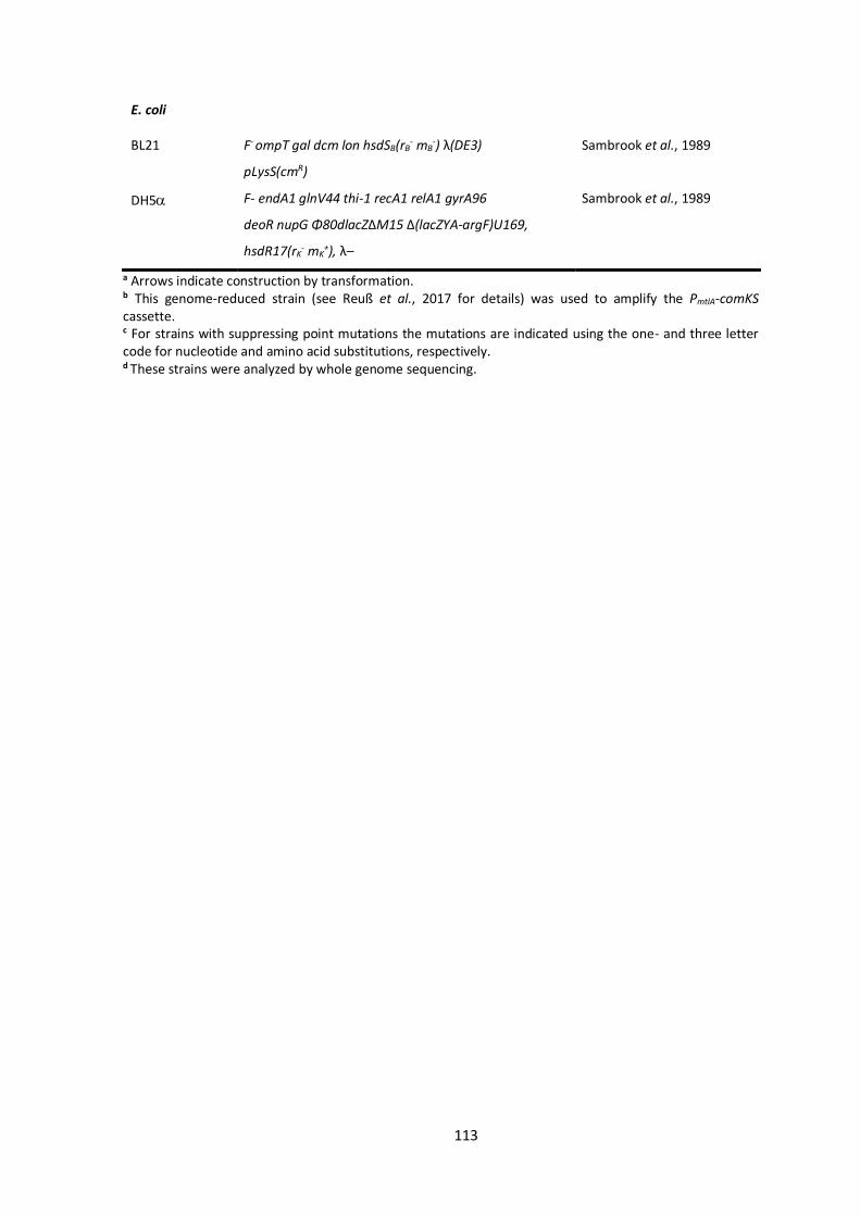
113
E. coli
BL21 F- ompT gal dcm lon hsdSB(rB- mB
-) λ(DE3)
pLysS(cmR)
Sambrook et al., 1989
DH5 F- endA1 glnV44 thi-1 recA1 relA1 gyrA96
deoR nupG Φ80dlacZΔM15 Δ(lacZYA-argF)U169,
hsdR17(rK- mK
+), λ–
Sambrook et al., 1989
a Arrows indicate construction by transformation. b This genome-reduced strain (see Reuß et al., 2017 for details) was used to amplify the PmtlA-comKS cassette. c For strains with suppressing point mutations the mutations are indicated using the one- and three letter code for nucleotide and amino acid substitutions, respectively. d These strains were analyzed by whole genome sequencing.
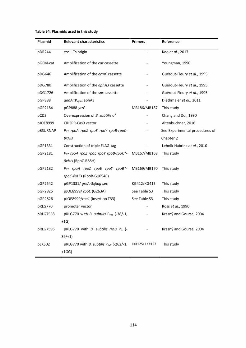
114
Table S4: Plasmids used in this study
Plasmid Relevant characteristics Primers Reference
pDR244 cre + Ts origin - Koo et al., 2017
pGEM-cat Amplification of the cat cassette - Youngman, 1990
pDG646 Amplification of the ermC cassette - Guérout-Fleury et al., 1995
pDG780 Amplification of the aphA3 cassette - Guérout-Fleury et al., 1995
pDG1726 Amplification of the spc cassette - Guérout-Fleury et al., 1995
pGP888 ganA::PxylA; aphA3 - Diethmaier et al., 2011
pGP2184 pGP888-ytrF MB186/MB187 This study
pCD2 Overexpression of B. subtilis σA - Chang and Doi, 1990
pJOE8999 CRISPR-Cas9 vector - Altenbuchner, 2016
pBSURNAP PT7 rpoA rpoZ rpoE rpoY rpoB-rpoC-
8xHis
- See Experimental procedures of
Chapter 2
pGP1331 Construction of triple FLAG-tag - Lehnik-Habrink et al., 2010
pGP2181 PT7 rpoA rpoZ rpoE rpoY rpoB-rpoC*-
8xHis (RpoC-R88H)
MB167/MB168 This study
pGP2182 PT7 rpoA rpoZ rpoE rpoY rpoB*-
rpoC-8xHis (RpoB-G1054C)
MB169/MB170 This study
pGP2542 pGP1331/ greA-3xflag spc KG412/KG413 This study
pGP2825 pJOE8999/ rpoC (G263A) See Table S3 This study
pGP2826 pJOE8999/rea1 (insertion T33) See Table S3 This study
pRLG770 promoter vector - Ross et al., 1990
pRLG7558 pRLG770 with B. subtilis Pveg (-38/-1,
+1G)
- Krásný and Gourse, 2004
pRLG7596 pRLG770 with B. subtilis rrnB P1 (-
39/+1)
- Krásný and Gourse, 2004
pLK502 pRLG770 with B. subtilis PilvB (-262/-1,
+1GG)
LK#125/ LK#127 This study
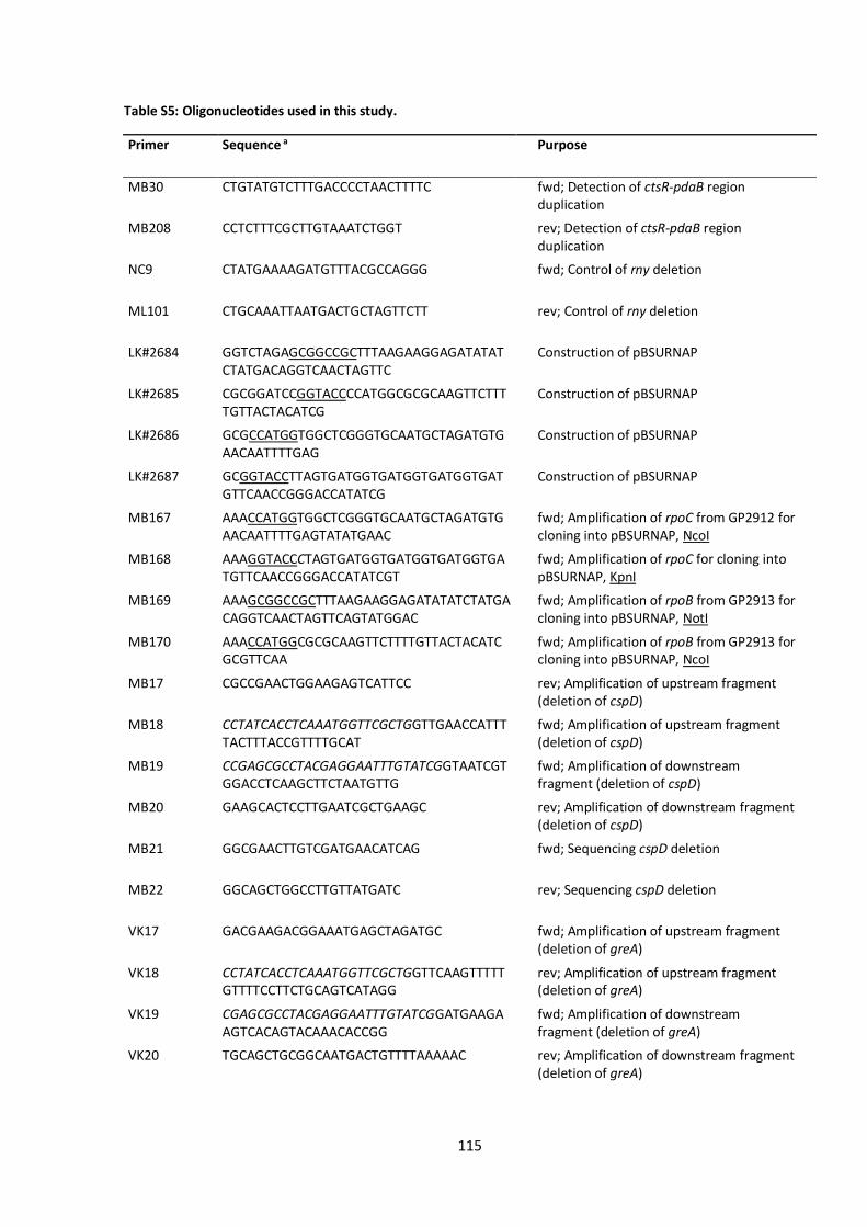
115
Table S5: Oligonucleotides used in this study.
Primer Sequence a Purpose
MB30 CTGTATGTCTTTGACCCCTAACTTTTC fwd; Detection of ctsR-pdaB region duplication
MB208 CCTCTTTCGCTTGTAAATCTGGT rev; Detection of ctsR-pdaB region duplication
NC9 CTATGAAAAGATGTTTACGCCAGGG
fwd; Control of rny deletion
ML101 CTGCAAATTAATGACTGCTAGTTCTT
rev; Control of rny deletion
LK#2684 GGTCTAGAGCGGCCGCTTTAAGAAGGAGATATATCTATGACAGGTCAACTAGTTC
Construction of pBSURNAP
LK#2685 CGCGGATCCGGTACCCCATGGCGCGCAAGTTCTTTTGTTACTACATCG
Construction of pBSURNAP
LK#2686 GCGCCATGGTGGCTCGGGTGCAATGCTAGATGTGAACAATTTTGAG
Construction of pBSURNAP
LK#2687 GCGGTACCTTAGTGATGGTGATGGTGATGGTGATGTTCAACCGGGACCATATCG
Construction of pBSURNAP
MB167 AAACCATGGTGGCTCGGGTGCAATGCTAGATGTGAACAATTTTGAGTATATGAAC
fwd; Amplification of rpoC from GP2912 for cloning into pBSURNAP, NcoI
MB168 AAAGGTACCCTAGTGATGGTGATGGTGATGGTGATGTTCAACCGGGACCATATCGT
fwd; Amplification of rpoC for cloning into pBSURNAP, KpnI
MB169 AAAGCGGCCGCTTTAAGAAGGAGATATATCTATGACAGGTCAACTAGTTCAGTATGGAC
fwd; Amplification of rpoB from GP2913 for cloning into pBSURNAP, NotI
MB170 AAACCATGGCGCGCAAGTTCTTTTGTTACTACATCGCGTTCAA
fwd; Amplification of rpoB from GP2913 for cloning into pBSURNAP, NcoI
MB17 CGCCGAACTGGAAGAGTCATTCC rev; Amplification of upstream fragment (deletion of cspD)
MB18 CCTATCACCTCAAATGGTTCGCTGGTTGAACCATTTTACTTTACCGTTTTGCAT
fwd; Amplification of upstream fragment (deletion of cspD)
MB19 CCGAGCGCCTACGAGGAATTTGTATCGGTAATCGTGGACCTCAAGCTTCTAATGTTG
fwd; Amplification of downstream fragment (deletion of cspD)
MB20 GAAGCACTCCTTGAATCGCTGAAGC rev; Amplification of downstream fragment (deletion of cspD)
MB21 GGCGAACTTGTCGATGAACATCAG fwd; Sequencing cspD deletion
MB22 GGCAGCTGGCCTTGTTATGATC rev; Sequencing cspD deletion
VK17 GACGAAGACGGAAATGAGCTAGATGC fwd; Amplification of upstream fragment (deletion of greA)
VK18 CCTATCACCTCAAATGGTTCGCTGGTTCAAGTTTTTGTTTTCCTTCTGCAGTCATAGG
rev; Amplification of upstream fragment (deletion of greA)
VK19 CGAGCGCCTACGAGGAATTTGTATCGGATGAAGAAGTCACAGTACAAACACCGG
fwd; Amplification of downstream fragment (deletion of greA)
VK20 TGCAGCTGCGGCAATGACTGTTTTAAAAAC rev; Amplification of downstream fragment (deletion of greA)

116
VK21 GGCTTAGTGCTGAATTATGATGAAGATACAC fwd; Sequencing greA deletion
VK22 GTGCCTTTGTCGTCCCCCGG rev; Sequencing greA deletion
ML47 5′-GAAGAATCTGCTTACACATACATCG fwd; Amplification of upstream fragment (deletion of rny)
KG409 GACTGTGTTTTATATTTTTCTCGTTCATACTTTCACCTCCTCTTGCTATGAACT
rev; Amplification of upstream fragment (deletion of rny)
KG410 CCGAGCGCCTACGAGGAATTTGTATCGAGTGATGCGCTAAGCATCACTTTATTTTTTTG
fwd; Amplification of downstream fragment (deletion of rny)
NP60 GCAGACACATACTCTCCCACTTTTACACTGCTGACAT
rev; Amplification of downstream fragment (deletion of rny)
KG411 ATGAACGAGAAAAATATAAAACACAGTC fwd; Amplification of ermC cassette (deletion of rny)
CZ68 CGATACAAATTCCTCGTAGGCGCTCGGTTACTTATTAAATAATTTATAGCTATTG
rev; Amplification of ermC cassette (deletion of rny)
KG414 GTCGGTTCATCACAAAAAGCGCTGAT fwd; Sequencing rny deletion
NP61 AGTATTGGTACACACATGAGATTTTCCTGTTAG
rev; Sequencing rny deletion
NC16 CTGCCACTGAATTTGGACTCG
rev; Sequencing rny deletion
JN420 CCTATCACCTCAAATGGTTCGCTGCGCACATGTCTATGTAAGATAATCGT
rev; Amplification of upstream fragment (deletion of nrnA)
JN421 GGGATCGAAGTGCTTCCCG fwd; Amplification of upstream fragment (deletion of nrnA)
JN422 CCGAGCGCCTACGAGGAATTTGTATCGGCTGGGATGAAGCTGATCGTA
fwd; Amplification of downstream fragment (deletion of nrnA)
JN423 GCGGCATACTCGAAGGCA rev; Amplification of downstream fragment (deletion of nrnA)
JN424 GACCAAAAATCCCGTCACGG fwd; Sequencing nrnA deletion
JN425 GCTTGCCAACCGGTTAAAAATATG rev; Sequencing nrnA deletion
MB31 CTGCGTATATCTGCTTCGAAATCCTTC fwd; Amplification of upstream fragment (integration of PmtlA-comKS)
MB32 TAAAAATAAAAAGCTAGCGGGGATCCCAAGTCAAAACCGAGTCTCATTTCCTATTTATCC
rev; Amplification of upstream fragment (integration of PmtlA-comKS)
MB33 CTTGGGATCCCCGCTAGCTTTTTATTTTTA fwd; Amplification of PmtlA-comKS for its insertion into yvcA-hisI locus
MB34 CCTATCACCTCAAATGGTTCGCTGCGGAGGATTTCGTGCCGGTTGATTA
rev; Amplification of PmtlA-comKS for its insertion into yvcA-hisI locus
MB35 CCGAGCGCCTACGAGGAATTTGTATCG GCCGGCTAGCACCCAATATAAATCTAAAT
fwd; Amplification of downstream fragment (integration of PmtlA-comKS)
MB36 GTGCTGACACTTGCGTATATGAACAAG rev; Amplification of downstream fragment (integration of PmtlA-comKS)
MB37 GTAAACTCCTTTGTAGCCTCATACTGAC fwd; Sequencing PmtlA-comKS insertion

117
MB38 GAATGTGAGATGAAACAGGCAGATGAAC rev; Sequencing PmtlA-comKS insertion
MB43 CTTGATAGATACTTTCCATCCTCCGG fwd; Sequencing PmtlA-comKS insertion
MB44 CCCTACACTTTCTTCGACAAGACCC fwd; Sequencing PmtlA-comKS insertion
MB60 GCTGATGAAACGGCAGTGCT fwd; Amplification of upstream fragment (deletion of ftsH)
MB61 CCTATCACCTCAAATGGTTCGCTGTCCTTACCTCCTCCCACAG
rev; Amplification of upstream fragment (deletion of ftsH)
MB62 CCGAGCGCCTACGAGGAATTTGTATCGAAGACGATACGAAAGAGTAATTCGC
fwd; Amplification of downstream fragment (deletion of ftsH)
MB63 CTCCTATACACTTCCTACGCGG rev; Amplification of downstream fragment (deletion of ftsH)
MB64 GGGCTGAAGGTGGTCAAATC fwd; Sequencing ftsH deletion
MB65 CATATCAGTCGTTCTCGCTGCA rev; Sequencing ftsH deletion
MB66 CATCGGTCCGGTTTCCAGCA fwd; Amplification of upstream fragment (deletion of ytrA)
MB67 CCTATCACCTCAAATGGTTCGCTGGGGTGTTGAGCTTCTTGGATC
rev; Amplification of upstream fragment (deletion of ytrA)
MB68 CCGAGCGCCTACGAGGAATTTGTATCGGCTGATGTGAAGGGAGGCAA
fwd; Amplification of downstream fragment (deletion of ytrA)
MB69 GGCGATCAAGACACCCTTGA rev; Amplification of downstream fragment (deletion of ytrA)
MB70 GATGTACTTGCCGTCCTTCCA fwd; Sequencing ytrA deletion
MB71 ACCCGGCACCCAGTTGATAT rev; Sequencing ytrA deletion
MB72 AGGGGACAGAGTATCTCAGGCA fwd; Amplification of upstream fragment (deletion of comEC)
MB73 CCTATCACCTCAAATGGTTCGCTGCGCATTCATCACACGTAGCTC
rev; Amplification of upstream fragment (deletion of comEC)
MB74 CCGAGCGCCTACGAGGAATTTGTATCGAAAGACTGCCGAGAAATCAGCA
fwd; Amplification of downstream fragment (deletion of comEC)
MB75 TCTCCAATAAACGTGCAGAGCTT rev; Amplification of downstream fragment (deletion of comEC)
MB76 AACAACGACGAGTCAAACGAAACAA fwd; Sequencing comEC deletion
MB77 CTCTGTTCGTTTTCGGTTGACG rev; Sequencing comEC deletion
MB78 AACCGTTTATCCGAGGTCAGCC fwd; Amplification of upstream fragment (deletion of degU)
MB79 CCTATCACCTCAAATGGTTCGCTGGTTTACTTTAGTCACAAGCCACGC
rev; Amplification of upstream fragment (deletion of degU)
MB80 CCGAGCGCCTACGAGGAATTTGTATCGGGCTGGGTAGAAATGAGATAGTA
fwd; Amplification of downstream fragment (deletion of degU)
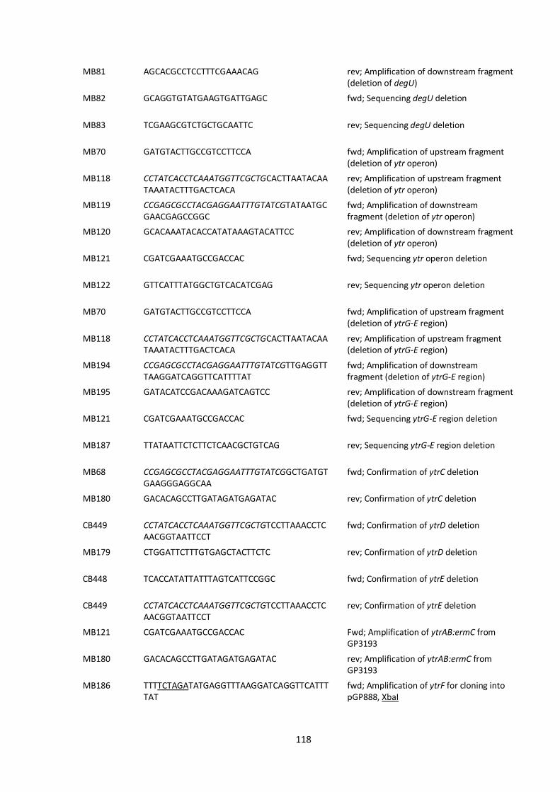
118
MB81 AGCACGCCTCCTTTCGAAACAG rev; Amplification of downstream fragment (deletion of degU)
MB82 GCAGGTGTATGAAGTGATTGAGC fwd; Sequencing degU deletion
MB83 TCGAAGCGTCTGCTGCAATTC rev; Sequencing degU deletion
MB70 GATGTACTTGCCGTCCTTCCA fwd; Amplification of upstream fragment (deletion of ytr operon)
MB118 CCTATCACCTCAAATGGTTCGCTGCACTTAATACAATAAATACTTTGACTCACA
rev; Amplification of upstream fragment (deletion of ytr operon)
MB119 CCGAGCGCCTACGAGGAATTTGTATCGTATAATGCGAACGAGCCGGC
fwd; Amplification of downstream fragment (deletion of ytr operon)
MB120 GCACAAATACACCATATAAAGTACATTCC rev; Amplification of downstream fragment (deletion of ytr operon)
MB121 CGATCGAAATGCCGACCAC fwd; Sequencing ytr operon deletion
MB122 GTTCATTTATGGCTGTCACATCGAG rev; Sequencing ytr operon deletion
MB70 GATGTACTTGCCGTCCTTCCA fwd; Amplification of upstream fragment (deletion of ytrG-E region)
MB118 CCTATCACCTCAAATGGTTCGCTGCACTTAATACAATAAATACTTTGACTCACA
rev; Amplification of upstream fragment (deletion of ytrG-E region)
MB194 CCGAGCGCCTACGAGGAATTTGTATCGTTGAGGTTTAAGGATCAGGTTCATTTTAT
fwd; Amplification of downstream fragment (deletion of ytrG-E region)
MB195 GATACATCCGACAAAGATCAGTCC
rev; Amplification of downstream fragment (deletion of ytrG-E region)
MB121 CGATCGAAATGCCGACCAC fwd; Sequencing ytrG-E region deletion
MB187 TTATAATTCTCTTCTCAACGCTGTCAG rev; Sequencing ytrG-E region deletion
MB68 CCGAGCGCCTACGAGGAATTTGTATCGGCTGATGTGAAGGGAGGCAA
fwd; Confirmation of ytrC deletion
MB180 GACACAGCCTTGATAGATGAGATAC rev; Confirmation of ytrC deletion
CB449 CCTATCACCTCAAATGGTTCGCTGTCCTTAAACCTCAACGGTAATTCCT
fwd; Confirmation of ytrD deletion
MB179 CTGGATTCTTTGTGAGCTACTTCTC rev; Confirmation of ytrD deletion
CB448 TCACCATATTATTTAGTCATTCCGGC fwd; Confirmation of ytrE deletion
CB449 CCTATCACCTCAAATGGTTCGCTGTCCTTAAACCTCAACGGTAATTCCT
rev; Confirmation of ytrE deletion
MB121 CGATCGAAATGCCGACCAC Fwd; Amplification of ytrAB:ermC from GP3193
MB180 GACACAGCCTTGATAGATGAGATAC rev; Amplification of ytrAB:ermC from GP3193
MB186 TTTTCTAGATATGAGGTTTAAGGATCAGGTTCATTTTAT
fwd; Amplification of ytrF for cloning into pGP888, XbaI

119
MB187 ATTGGTACCTTATAATTCTCTTCTCAACGCTGTCAG rev; Amplification of ytrF for cloning into pGP888, KpnI
MB198 GCGGCAGCTGTCAAAAGC fwd; Amplification of upstream fragment (deletion of ytrCD)
MB199 CCTATCACCTCAAATGGTTCGCTGCTACCATCTCCGCTTCCCTC
rev; Amplification of upstream fragment (deletion of ytrCD)
MB200 CCGAGCGCCTACGAGGAATTTGTATCGGTAAGGGAGAGAGAACATATGATTG
fwd; Amplification of downstream fragment (deletion of ytrCD)
MB201 CTCCTTCCTTGCCCATTACG rev; Amplification of downstream fragment (deletion of ytrCD)
MB202 CACTATGCAGGGGTTGAGCT fwd; Sequencing ytrCD deletion
MB203 GTTTGGTTCATACACTTGCGTTC rev; Sequencing ytrCD deletion
CB448 TCACCATATTATTTAGTCATTCCGGC fwd; Amplification of upstream fragment (deletion of ytrF)
CB449 CCTATCACCTCAAATGGTTCGCTGTCCTTAAACCTCAACGGTAATTCCT
rev; Amplification of upstream fragment (deletion of ytrF)
CB450 CGAGCGCCTACGAGGAATTTGTATCGTTATAATGCGAACGAGCCGGCT
fwd; Amplification of downstream fragment (deletion of ytrF)
CB451 TCCCATGTTTTCAAGCTTTTATAAAACG rev; Amplification of downstream fragment (deletion of ytrF)
CB452 ACCTCGAGATCCTTTTTGGCG fwd; Sequencing ytrF deletion
CB453 TGCTAAGCGATGCCGTGCT rev; Sequencing ytrF deletion
SW17 GACATTGTCCCTTTATCAGC fwd; Amplification of upstream fragment (insertion of rpoA)
SW18 GGGGTGTGAGCTGAATTCCTGCTGTCTGATCAATTTAATG
rev; Amplification of upstream fragment (insertion of rpoA)
SW19 CCGAGCGCCTACGAGGAATTTGTATCGCCCCCATGAAAAAAAGAC
fwd; Amplification of downstream fragment (insertion of rpoA)
SW20 CGAATCAAATGCTTATTTGG rev; Amplification of downstream fragment (insertion of rpoA)
SW21 GAATTCAGCTCACACCCC fwd; Amplification of PrpsJ
SW40 TGGTTTTTCAATCTCGATCATTATTTTCCCTCCTTTTC
rev; Amplification of PrpsJ
SW23 ATGATCGAGATTGAAAAACCA fwd; Amplification of rpoA
SW24 CCTATCACCTCAAATGGTTCGCTGTCAATCGTCTTTGCGAAG
rev; Amplification of rpoA
SW25 GATCATAATCTTCAATGCGAAG fwd; Sequencing rpoA insertion
SW27 GAACAACCACAAATGACATC rev; Sequencing rpoA insertion
SW28 GTGATCTGTGAAAATCCAAAG fwd; Amplification of upstream fragment (deletion of rpoA)
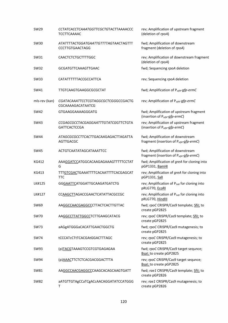
120
SW29 CCTATCACCTCAAATGGTTCGCTGTACTTAAAACCCTCCTTCAAAAC
rev; Amplification of upstream fragment (deletion of rpoA)
SW30 ATATTTTACTGGATGAATTGTTTTAGTAACTAGTTTCCCTTGTGAACTAGG
fwd; Amplification of downstream fragment (deletion of rpoA)
SW31 CAACTCTCTGCTTTTGGC rev; Amplification of downstream fragment (deletion of rpoA)
SW32 GCGATGTTCAAAGTTGAAC fwd; Sequencing rpoA deletion
SW33 CATATTTTTTACCGCCATTCA rev; Sequencing rpoA deletion
SW41 TTGTCAAGTGAAGGCGCGCTAT
fwd; Amplification of Palf4-gfp-ermC
mls-rev (kan) CGATACAAATTCCTCGTAGGCGCTCGGGCCGACTGCGCAAAAGACATAATCG
rev; Amplification of Palf4-gfp-ermC
SW42 GTGAAGGAAAAGGGATG fwd; Amplification of upstream fragment (insertion of Palf4-gfp-ermC)
SW43 CCGAGCGCCTACGAGGAATTTGTATCGGTTCTGTAGATTCACTCCGA
rev; Amplification of upstream fragment (insertion of Palf4-gfp-ermC)
SW44 ATAGCGCGCCTTCACTTGACAAGAGACTTAGATTAAGTTGACGC
fwd; Amplification of downstream fragment (insertion of Palf4-gfp-ermC)
SW45 ACTGTCAATATAGCATAAATTCC fwd; Amplification of downstream fragment (insertion of Palf4-gfp-ermC)
KG412 AAAGGATCCATGGCACAAGAGAAAGTTTTTCCTATG
fwd; Amplification of greA for cloning into pGP1331, BamHI
KG413 TTTGTCGACTGAAATTTTCACAATTTTCACGAGCATTTC
rev; Amplification of greA for cloning into pGP1331, SalI
LK#125 GGGAATTCATGGATTGCAAGATGATCTG rev; Amplification of PilvB for cloning into pRLG770, EcoRI
LK#127 CCAAGCTTAGACCGAACTCATATTACGCCGC rev; Amplification of PilvB for cloning into pRLG770, HindIII
SW69 AAGGCCAACGAGGCCCTTACTCACTTGTTAC fwd; rpoC CRISPR/Cas9 template; SfiI; to create pGP2825
SW70 AAGGCCTTATTGGCCTCTTGAAGCATACG rev; rpoC CRISPR/Cas9 template; SfiI; to create pGP2825
SW73 aAGgATGGGaCACATTGAACTGGCTG fwd; rpoC CRISPR/Cas9 mutagenesis; to create pGP2825
SW74 tCCCATcCTtTCACGAtGGACTTTAGC rev; rpoC CRISPR/Cas9 mutagenesis; to create pGP2825
SW93 (p)TACGTAAAGTCCGTCGTGAGAGAA
fwd; rpoC CRISPR/Cas9 target sequnce; BsaI; to create pGP2825
SW94 (p)AAACTTCTCTCACGACGGACTTTA
rev; rpoC CRISPR/Cas9 target sequnce; BsaI; to create pGP2825
SW81 AAGGCCAACGAGGCCCAAGCACAGCAAGTGATT fwd; rae1 CRISPR/Cas9 template; SfiI; to create pGP2826
SW82 aATGTTGTAgCCaTCgACcAACAGGATATCCATGGGT
rev; rae1 CRISPR/Cas9 mutagenesis; to create pGP2826
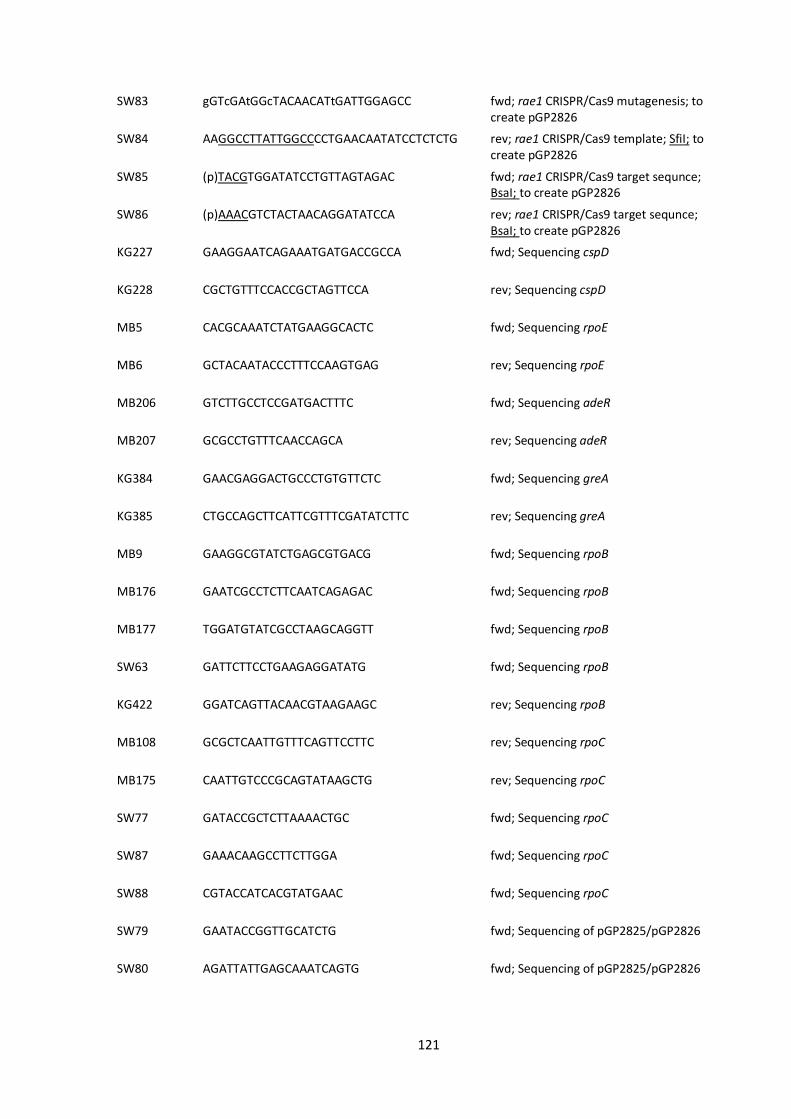
121
SW83 gGTcGAtGGcTACAACATtGATTGGAGCC
fwd; rae1 CRISPR/Cas9 mutagenesis; to create pGP2826
SW84 AAGGCCTTATTGGCCCCTGAACAATATCCTCTCTG
rev; rae1 CRISPR/Cas9 template; SfiI; to create pGP2826
SW85 (p)TACGTGGATATCCTGTTAGTAGAC fwd; rae1 CRISPR/Cas9 target sequnce; BsaI; to create pGP2826
SW86 (p)AAACGTCTACTAACAGGATATCCA rev; rae1 CRISPR/Cas9 target sequnce; BsaI; to create pGP2826
KG227 GAAGGAATCAGAAATGATGACCGCCA fwd; Sequencing cspD
KG228 CGCTGTTTCCACCGCTAGTTCCA rev; Sequencing cspD
MB5 CACGCAAATCTATGAAGGCACTC fwd; Sequencing rpoE
MB6 GCTACAATACCCTTTCCAAGTGAG rev; Sequencing rpoE
MB206 GTCTTGCCTCCGATGACTTTC fwd; Sequencing adeR
MB207 GCGCCTGTTTCAACCAGCA rev; Sequencing adeR
KG384 GAACGAGGACTGCCCTGTGTTCTC fwd; Sequencing greA
KG385 CTGCCAGCTTCATTCGTTTCGATATCTTC rev; Sequencing greA
MB9 GAAGGCGTATCTGAGCGTGACG fwd; Sequencing rpoB
MB176 GAATCGCCTCTTCAATCAGAGAC fwd; Sequencing rpoB
MB177 TGGATGTATCGCCTAAGCAGGTT fwd; Sequencing rpoB
SW63 GATTCTTCCTGAAGAGGATATG fwd; Sequencing rpoB
KG422 GGATCAGTTACAACGTAAGAAGC rev; Sequencing rpoB
MB108 GCGCTCAATTGTTTCAGTTCCTTC rev; Sequencing rpoC
MB175 CAATTGTCCCGCAGTATAAGCTG rev; Sequencing rpoC
SW77 GATACCGCTCTTAAAACTGC fwd; Sequencing rpoC
SW87 GAAACAAGCCTTCTTGGA fwd; Sequencing rpoC
SW88 CGTACCATCACGTATGAAC fwd; Sequencing rpoC
SW79 GAATACCGGTTGCATCTG fwd; Sequencing of pGP2825/pGP2826
SW80 AGATTATTGAGCAAATCAGTG fwd; Sequencing of pGP2825/pGP2826
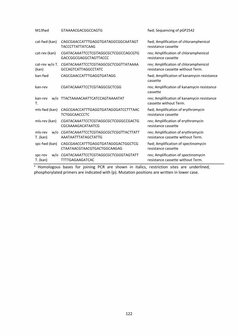
122
M13fwd GTAAAACGACGGCCAGTG fwd; Sequencing of pGP2542
cat-fwd (kan) CAGCGAACCATTTGAGGTGATAGGCGGCAATAGTTACCCTTATTATCAAG
fwd; Amplification of chloramphenicol resistance cassette
cat-rev (kan) CGATACAAATTCCTCGTAGGCGCTCGGCCAGCGTGGACCGGCGAGGCTAGTTACCC
rev; Amplification of chloramphenicol resistance cassette
cat-rev w/o T. (kan)
CGATACAAATTCCTCGTAGGCGCTCGGTTATAAAAGCCAGTCATTAGGCCTATC
rev; Amplification of chloramphenicol resistance cassette without Term.
kan-fwd CAGCGAACCATTTGAGGTGATAGG
fwd; Amplification of kanamycin resistance cassette
kan-rev CGATACAAATTCCTCGTAGGCGCTCGG rev; Amplification of kanamycin resistance cassette
kan-rev w/o T.
TTACTAAAACAATTCATCCAGTAAAATAT
rev; Amplification of kanamycin resistance cassette without Term.
mls-fwd (kan) CAGCGAACCATTTGAGGTGATAGGGATCCTTTAACTCTGGCAACCCTC
fwd; Amplification of erythromycin resistance cassette
mls-rev (kan) CGATACAAATTCCTCGTAGGCGCTCGGGCCGACTGCGCAAAAGACATAATCG
rev; Amplification of erythromycin resistance cassette
mls-rev w/o T. (kan)
CGATACAAATTCCTCGTAGGCGCTCGGTTACTTATTAAATAATTTATAGCTATTG
rev; Amplification of erythromycin resistance cassette without Term.
spc-fwd (kan) CAGCGAACCATTTGAGGTGATAGGGACTGGCTCGCTAATAACGTAACGTGACTGGCAAGAG
fwd; Amplification of spectinomycin resistance cassette
spc-rev w/o T. (kan)
CGATACAAATTCCTCGTAGGCGCTCGGGTAGTATTTTTTGAGAAGATCAC
rev; Amplification of spectinomycin resistance cassette without Term.
a Homologous bases for joining PCR are shown in italics, restriction sites are underlined, phosphorylated primers are indicated with (p). Mutation positions are written in lower case.
123
7 Curriculum Vitae
Personal information
Name: Martin Benda
Date of birth: 5th April 1992
Place of birth: Prague, Czech Republic
Nationality: Czech
Contact: mb.martin.benda[at]gmail.com
Education
• 2017 – 2020: PhD studies at the GGNB program Microbiology and Biochemistry
Georg August University of Göttingen (Germany)
Title of thesis: “A possible functional link between RNA degradation and transcription in
Bacillus subtilis”
Supervisor: Prof. Dr. Jörg Stülke
• 2014 – 2016: Master studies in Genetics, Molecular Biology and Virology
Charles University in Prague (Czech Republic), Faculty of Science
Specialization: Molecular Biology and Genetics of Prokaryotes
Title of thesis: Transcription regulation by sigma factors in Bacillus subtilis
Supervisor: Libor Krásný PhD.
• 2011 – 2014: Bachelor studies in Molecular Biology and Biochemistry of Organisms
Charles University in Prague (Czech Republic), Faculty of Science
Title of thesis: Regulation of bacterial transcription by alternative sigma factors
Supervisor: Libor Krásný PhD.
Copyright © 2022 FDOKUMEN