
and Nup214- dependent nuclear export of proteins - eDiss
124
Analysis of CRM1- and Nup214- dependent nuclear export of proteins Dissertation for the award of the degree “Doctor rerum naturalium” Division of Mathematics and Natural Sciences of the Georg-August-University Göttingen submitted by Stephanie Roloff from Prenzlau Göttingen 2012
-
Upload
khangminh22 -
Category
Documents
-
view
3 -
download
0
Transcript of and Nup214- dependent nuclear export of proteins - eDiss

1
Analysis of CRM1- and Nup214- dependent
nuclear export of proteins
Dissertation for the award of the degree
“Doctor rerum naturalium”
Division of Mathematics and Natural Sciences
of the Georg-August-University Göttingen
submitted by
Stephanie Roloff
from Prenzlau
Göttingen 2012
Member of the Thesis Committee (First Reviewer):
Prof. Dr. Ralph H. Kehlenbach
Department of Biochemistry I
Center for Biochemistry and Molecular Cell Biology
Georg-August University, Goettingen
Member of the Thesis Committee (Second Reviewer):
Prof. Dr. Peter Rehling
Department of Biochemistry II
Center for Biochemistry and Molecular Cell Biology
Georg-August University, Goettingen
Member of the Thesis Committee:
Prof. Dr. Ralf Ficner
Dept. of Molecular Structural Biology
Institute for Microbiology and Genetics
Georg-August University, Goettingen
DATE OF ORAL EXAMINATION:

Statutory declaration:
I declare that this thesis has been written entirely by myself with no other sources and aids than
explicitly quoted.
This thesis was not submitted in the same or in a substantially similar version to any other authority to
achieve an academic grading and was not published elsewhere.
Stephanie Roloff

To Science
1
Abstract Nuclear pore complexes (NPCs) are giant units of the nuclear envelope, mediating directional
transport into and out of the nucleus. Nucleoporins are the main components of the NPC, and one third
of them contain phenylalanine-glycine (FG) repeats. They are believed to serve as binding sites for
transport receptors of the karyopherin family. CRM1 is the main export receptor in eukaryotic cells. It
has a ring-like structure that is composed of HEAT (Huntingtin Elongation Factor A Subunit TOR)
repeats (HRs) and exports various cargo proteins containing a leucine-rich nuclear export signal
(NES). In the nucleus, the export complex assembles together with the small GTPase Ran in its GTP-
bound state. On its way through the nuclear pore, the trimeric export complex interacts with several
nucleoporins, among them Nup214. Nup214 is located at the cytoplasmic side of the NPC and its FG-
rich C-terminus interacts with CRM1 in a RanGTP-dependent manner, providing a terminal docking
site for CRM1-RanGTP-cargo complexes in the late steps of nuclear export. This interaction also
indicates a specific role for Nup214 in nuclear export, at least for some cargos. Whereas the CRM1-
binding region in Nup214 is known, the CRM1-binding sites for Nup214 have not been identified so
far. In this work, we analyzed the localization and the export potential of CRM1-fragments, mutants
and chimeras, derived from H. sapiens and C. elegans CRM1. Whereas CRM1-fragments did not
associate with the nuclear envelope and were not able to mediate export in mammalian cells, we found
one CRM1-chimera which was located at the nuclear envelope and was able to transport NES-cargos
through the nuclear pore. From that we conclude that association of CRM1 with the nuclear pore
correlates with the ability to promote nuclear export. By comparing several chimeras with respect to
their localization and functionality, we hypothesize that Nup214 binding occurs between HRs 9-11 of
CRM1.
Transport receptors bind preferentially to FGs in nucleoporins. For CRM1 this had not been analyzed
in detail. Here we show that CRM1 specifically binds to the FGs in Nup214. Moreover, we could
narrow down the Nup214 region for CRM1-binding. By analyzing short Nup214-fragments and
mutants, we identified a prominent FG-motif in Nup214, comprising 20 amino acid residues. This
motif interacts with CRM1 in a RanGTP-dependent manner, with enhanced binding in the presence of
an NES-cargo. Two other Nup214 regions downstream of this motif contribute to the stability of this
interaction.

Table of contents
2
Table of contents
ABSTRACT ................................................................................................................ 1
1 INTRODUCTION ................................................................................................ 5
1.1 The nuclear pore complex ..................................................................................................................... 5
1.2 Transport receptors and regulators ..................................................................................................... 7 1.2.1 The GTPase Ran ................................................................................................................................. 7
1.2.2 Basic nuclear import ........................................................................................................................... 8
1.2.3 Nuclear export ................................................................................................................................... 10
1.2.3.1 The structure of CRM1 ............................................................................................................ 11
1.2.3.2 CRM1-mediated nuclear export .............................................................................................. 12
1.3 The translocation of transport complexes through the nuclear pore .............................................. 14
1.4 Nup214 .................................................................................................................................................. 15
1.5 Aim of the work ................................................................................................................................... 16
2 MATERIAL AND METHODS ........................................................................... 17
2.1 Material ................................................................................................................................................ 17 2.1.1 Technical equipment and software .................................................................................................... 17
2.1.2 Consumables ..................................................................................................................................... 18
2.1.3 Kits .................................................................................................................................................... 19
2.1.4 Chemicals, reagents and enzymes ..................................................................................................... 19
2.1.5 Buffers, stock solutions and media ................................................................................................... 21
2.1.6 Cell lines ........................................................................................................................................... 24
2.1.7 Antibodies ......................................................................................................................................... 25
2.1.8 Oligonucleotides ............................................................................................................................... 26
2.1.9 Vectors and plasmids ........................................................................................................................ 29
2.1.10 Proteins ............................................................................................................................................. 32
2.2 Biochemical methods ........................................................................................................................... 33 2.2.1 Separation and detection of proteins ................................................................................................. 33
2.2.2 Protein preparation ............................................................................................................................ 35
2.2.3 In vitro binding assays with GST-tagged proteins ............................................................................ 36
2.2.4 Affinity purification of CRM1 antibodies ......................................................................................... 37
2.3 Cell biology methods ............................................................................................................................ 39 2.3.1 Cell culture ........................................................................................................................................ 39
2.3.1.1 Transfection of DNA in mammalian cells ............................................................................... 39
2.3.2 In vitro transport assays .................................................................................................................... 40
2.3.3 Detection of proteins by indirect immunofluorescence .................................................................... 41
2.3.3.1 Quantification of GFP-substrate distribution ........................................................................... 41
Table of contents
3
2.4 Molecular biology methods ................................................................................................................. 42 2.4.1 Polymerase chain reaction (Mullis, 1990) ......................................................................................... 42
2.4.2 Transformation and purification of plasmid DNA ............................................................................ 42
2.4.3 Agarose gel electrophoresis .............................................................................................................. 44
2.4.4 Restriction of DNA by endonucleases .............................................................................................. 44
2.4.5 Ligation of DNA fragments .............................................................................................................. 44
2.4.6 Oligonucleotide cloning .................................................................................................................... 45
2.4.7 DNA-sequencing ............................................................................................................................... 45
3 RESULTS .......................................................................................................... 46
3.1 In vivo analysis of CRM1, CRM1-fragments and chimeras ............................................................. 46
3.1.1 Distribution of nuclear export cargos after Leptomycin B treatment of HeLa cells ......................... 47 3.1.2 The CRM1 mutant C528S can overcome the LMB effect on several export cargos ........................ 48
3.1.3 CRM1-fragments are not able to mediate nuclear export of proteins ............................................... 50
3.1.3.1 Localization of CRM1-truncations .......................................................................................... 50
3.1.3.2 Only full-length CRM1 is able to mediate export through the nuclear pore ........................... 52
3.1.4 CRM1-chimeras as an alternative approach to study export capabilities in vivo .............................. 53
3.1.4.1 C. elegans CRM1 promotes export of a simple GFP-NES, but is unable to export more
complex cargos out of the nucleus .......................................................................................... 53
3.1.4.2 CRM1-chimeras differ in their localization ............................................................................. 55
3.1.4.3 The export capability of CRM1-chimeras correlates with their localization in HeLa cells ..... 57
3.1.4.4 The deletion of an outer surface loop between HEAT repeat 8B and 9A of CRM1 does not
affect nuclear export ................................................................................................................ 60
3.2 Determination of a minimal region in Nup214 required for CRM1-binding ................................. 62 3.2.1 Function of Nup214 C-terminal fragments in nuclear export ........................................................... 62
3.2.1.1 Analysis of C-terminal Nup214-fragments in vivo .................................................................. 64
3.2.1.2 Accumulation of CRM1 in the nucleus upon overexpression of Nup214-fragments .............. 66
3.2.1.3 Nup214 1916-2033 shows an inhibitory effect on CRM1-dependent nuclear export of
GFP-NFAT in vitro ................................................................................................................. 67
3.2.2 Analysis of a highly conserved FG-motif in Nup214 ....................................................................... 69
3.2.2.1 Nup214 aa 1916-2033 contains a CRM1-binding site ............................................................. 69
3.2.2.2 The mutation of the FGs in the FG-motif in Nup214 aa 1916-2033 leads to severe
CRM1-binding defects ............................................................................................................ 70
3.2.2.3 The inhibition of nuclear export is abrogated in Nup214 aa 1916-2033 a1 mutant in vivo
and in vitro............................................................................................................................... 73
3.2.2.4 Analysis of the Nup214 FG-motif in Nup214 aa 1859-2090 .................................................. 75
3.3.3 Refined search for CRM1-binding sites in Nup214 .......................................................................... 77
3.2.3.1 The FG-motif in Nup214 requires aa 1991-2033 to form a stable complex with CRM1
and RanGTP ............................................................................................................................ 77
3.2.3.2 Elongation of Nup214 C-terminal fragments leads to increasing functionality in nuclear
export ....................................................................................................................................... 79
Table of contents
4
4 DISCUSSION .................................................................................................... 81
4.1 Analysis of CRM1-fragments and -chimeras .................................................................................... 81
4.2 Analysis of the CRM1-binding region in Nup214 ............................................................................. 85
4.3 Outlook ................................................................................................................................................. 88
5 REFERENCES .................................................................................................. 89
6 APPENDIX ........................................................................................................ 98
6.1 Alingments ............................................................................................................................................ 98
6.2 Code for amino acids ......................................................................................................................... 109
6.3 Construction of CRM1-chimeras ..................................................................................................... 110
6.4 Quantifications ................................................................................................................................... 112
6.5 Abbreviations ..................................................................................................................................... 114
ACKNOWLEDGEMENTS ..................................................................................... 118
CURRICULUM VITAE
1 Introduction
5
1 Introduction
In eukaryotic cells, a mechanism is required that allows transport of substrates between the nucleus
and the cytoplasm according to the needs of the cell. The nuclear envelope is interspersed with special
protein complexes. These complexes, named nuclear pore complexes (NPCs), provide the basis for
directional transport to and out of the nucleus. All macromolecules that leave or enter the nucleus have
to pass these complexes. These transport processes are strictly regulated so that most of the cargos
cannot freely pass these NPCs. Transport receptors mediate their transport, thus ensuring the substrate
specificity of nuclear entry and exit.
1.1 The nuclear pore complex
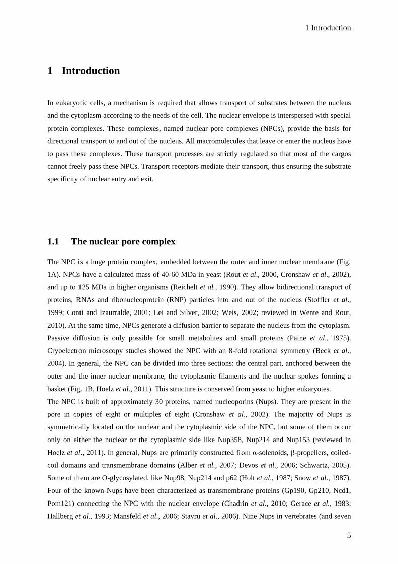
The NPC is a huge protein complex, embedded between the outer and inner nuclear membrane (Fig.
1A). NPCs have a calculated mass of 40-60 MDa in yeast (Rout et al., 2000, Cronshaw et al., 2002),
and up to 125 MDa in higher organisms (Reichelt et al., 1990). They allow bidirectional transport of
proteins, RNAs and ribonucleoprotein (RNP) particles into and out of the nucleus (Stoffler et al.,
1999; Conti and Izaurralde, 2001; Lei and Silver, 2002; Weis, 2002; reviewed in Wente and Rout,
2010). At the same time, NPCs generate a diffusion barrier to separate the nucleus from the cytoplasm.
Passive diffusion is only possible for small metabolites and small proteins (Paine et al., 1975).
Cryoelectron microscopy studies showed the NPC with an 8-fold rotational symmetry (Beck et al.,
2004). In general, the NPC can be divided into three sections: the central part, anchored between the
outer and the inner nuclear membrane, the cytoplasmic filaments and the nuclear spokes forming a
basket (Fig. 1B, Hoelz et al., 2011). This structure is conserved from yeast to higher eukaryotes.
The NPC is built of approximately 30 proteins, named nucleoporins (Nups). They are present in the
pore in copies of eight or multiples of eight (Cronshaw et al., 2002). The majority of Nups is
symmetrically located on the nuclear and the cytoplasmic side of the NPC, but some of them occur
only on either the nuclear or the cytoplasmic side like Nup358, Nup214 and Nup153 (reviewed in
Hoelz et al., 2011). In general, Nups are primarily constructed from α-solenoids, β-propellers, coiled-
coil domains and transmembrane domains (Alber et al., 2007; Devos et al., 2006; Schwartz, 2005).
Some of them are O-glycosylated, like Nup98, Nup214 and p62 (Holt et al., 1987; Snow et al., 1987).
Four of the known Nups have been characterized as transmembrane proteins (Gp190, Gp210, Ncd1,
Pom121) connecting the NPC with the nuclear envelope (Chadrin et al., 2010; Gerace et al., 1983;
Hallberg et al., 1993; Mansfeld et al., 2006; Stavru et al., 2006). Nine Nups in vertebrates (and seven

1 Introduction
6
in yeast) act as coat proteins of the NPC (Lutzmann et al., 2002, 2005; Siniossoglou et al., 2000).
Together, they form a subcomplex (Nup84 complex in yeast; Nup107-160 complex in human) and
deletion of any of its members, e.g. Nup75, Nup107 and Nup160 (Siniossoglou et al., 1996) has severe
effects on the architecture and the function of the NPC. The adaptor Nups (seven in yeast, and nine in
vertebrates) associate with transmembrane Nups, coat proteins and also with the channel proteins
(Fig. 1B; reviewed in Hoelz et al., 2011,). Interestingly, only the channel proteins, the Nups of the
nuclear basket and the cytoplasmic filaments contain phenylalanine-glycine (FG) repeats, e.g. Nup358,
p62, Nup153 or Nup214. These FG-repeat regions are unstructured and provide binding sites for
transport receptors (Denning et al., 2003; Isgro and Schulten, 2007a/b; Macara, 2001; Peters, 2005).
Typically, they are made up of 4-48 GLFG, FxFG, PxFG or SxFG, separated by spacers of variable
length (Cronshaw et al., 2002; Rout et al., 2000).
(A) (B)
Fig. 1 The nuclear pore complex. (A) Pictures from the outer nuclear membrane (upper panel; ONM)
and inner nuclear membrane (lower panel; INM) of Xenopus laevis oocytes, analyzed with field emission
scanning electron microscopy (unpublished pictures, prepared in collaboration with the group of V. Cordes,
MPI Göttingen). (B) Schematic model of the NPC, embedded in the nuclear envelope (Hoelz et al., 2011).
ONM
INM
1 Introduction
7
1.2 Transport receptors and regulators
Most of the known transport receptors belong to the importin β superfamily, also known as
karyopherins (reviewed in Wente and Rout, 2010). They have a molecular mass of 90-145 KDa and
are evolutionary conserved. Until today, approximately 20 different transport receptors are known.
Depending on directionality of nuclear transport they are summarized as importins (transport of
substrates into the nucleus) or exportins (transport of substrates out of the nucleus). They all consist of
HEAT repeat motifs (HEAT = Huntingtin Elongation Factor A Subunit TOR, Andrade et al., 1995).
One HEAT repeat motif consists of two antiparallel α-helices which are connected by a loop of
variable length. This slightly clockwise twisted connection of up to 20 HEAT repeats leads to a
superhelical structure of the whole protein (reviewed in Cook et al., 2007).
Active transport of proteins mediated by transport receptors is only possible if the cargo contains a
recognition signal. This is either a nuclear localization signal (NLS), recognized by importins, or a
nuclear export signal (NES), recognized by exportins.
Karyopherins do not only have a cargo recognition site, they also have binding domains for Nups
(Bayliss et al., 2000/2002) and for the small GTPase Ran (Rexach and Blobel, 1995).
1.2.1 The GTPase Ran
The directionality of nuclear transport is regulated by the small GTPase Ran. It is a member of the
Ras-related superfamily and besides its role in nuclear export it is also involved in mitotic processes in
eukaryotes (reviewed in Clarke and Chuanmao, 2008). Transport receptors bind Ran only in its GTP
bound form. This requires GDP-GTP conversion of Ran, mediated by accessory proteins which are
located exclusively in either the nucleus or the cytoplasm (Fig. 2). In the nucleus, the chromatin bound
RanGEF (Ran guanosine-nucleotide exchange factor), also called RCC1 (regulator of chromosome
condensation 1), exchanges GDP to GTP (Bischoff and Postingl, 1991a). In the cytoplasm, the
RanGTPase activating protein (RanGAP) stimulates the intrinsic GTPase activity of Ran (Becker et
al., 1995; Bischoff et al., 1995; Seewald et al., 2002) with the help of the Ran binding protein 1
(RanBP1). RanGAP is a soluble cytoplasmic protein, but it is also associated with Nup358 (Mahajan
et al. 1997; Matunis et al. 1996; Weis 2007), coordinating recycling of importin β (Hutten et al.,
2008). Due to cytoplasmic RanGAP and nuclear RCC1, there is a low concentration of RanGTP in the
cytoplasm and a high concentration in the nucleus (Izaurralde et al., 1997). The nuclear transport
factor 2 (NTF2) is closing the Ran-cycle, binding to RanGDP and mediating its import into the
nucleus (Moore and Blobel, 1994; Ribbeck et al., 1998).
1 Introduction
8
In general, RanGTP decreases the affinity of import receptors for their import cargos, but increases the
affinity of export receptors for their export proteins. Due to the high concentration of RanGTP in the
nucleus, incoming import complexes dissociate, and formation of export complexes is promoted
(reviewed in Wente and Rout, 2010).
1.2.2 Basic nuclear import
Nuclear import is mediated by a variety of import receptors. Among them, transportin, importin 5, 7,
9, 11 and 13 are receptors which are recognized by different import substrates. One of the best
characterized import pathway is the importin α/β mediated import of cargos containing a classical
NLS (cNLS). In general, the NLS-motif K(K/R)X(K/R) presents the required sequence for nuclear
import of cargos by importin α/β (Conti et al., 2000; Fontes et al., 2000; Hodel et al., 2001). The
cNLS can be monopartite (Kalderon et al., 1984), but many proteins also carry a more complex
bipartite NLS consisting of two clusters of basic amino acids, which are separated by a spacer of about
ten amino acids (Dingwall et al., 1982; Robbins et al., 1988). In the cytoplasm, importin α acts as an
adaptor protein, recognizing the cNLS of the import-cargo and connecting it to importin β via its IBB
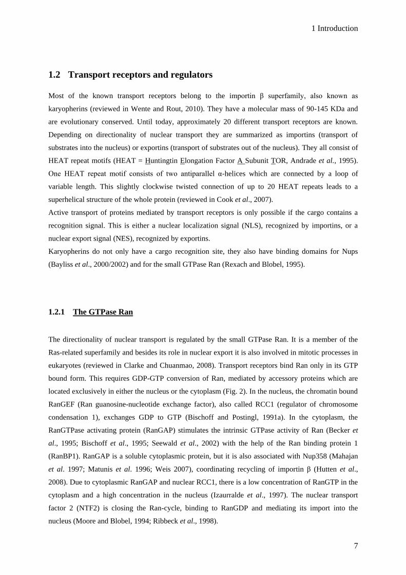
Fig. 2 The Ran-cycle. RanGTP, bound to an export receptor leaves the nucleus. In the cytoplasm,
RanGAP and RanBP1 mediate hydrolysis of RanGTP to RanGDP. Bound to NTF2, RanGDP is transported
back to the nucleus where RCC1 catalyzes the exchange of GTP to GDP.
R-GTP
R-GTP R-GDP
NTF2
NTF2
RCC1R-GDP
Ran
GAP
BP1 Pi
R-GTP
R-GTP R-GDP
NTF2
NTF2
RCC1R-GDP
Ran
GAP
BP1 Pi
Nucleus
Cytoplasm

1 Introduction
9
Fig. 3 Nuclear import of proteins and recycling of import receptors. The importin α/β complex (Imp) recognizes proteins with a cNLS. After passage through the nuclear pore, the complex disassembles in
the presence of RanGTP (R-GTP). The import receptor-RanGTP complex is transported back to the cytoplasm,
where RanGTP is immediately hydrolyzed to RanGDP (R-GDP).
R-GTP
R-GDP
NLSImp
NLSImp
NLSImp
NLS
ImpR-GTP
ImpR-GTP
Imp
R-GTP
R-GDP
NLSImp NLSImp
NLSImp
NLSImp NLSImp
NLS
ImpR-GTP
ImpR-GTP
Imp
(importin β binding) domain (Fig. 3; Görlich et al., 1995a, b). After passage of the import complex
through the NPC, Ran binds to the outer convex surface of the import receptor, thereby displacing the
import cargo from the inner concave surface of the receptor. Importin β, bound to RanGTP, is
transported back to the cytoplasm (Rexach and Blobel, 1995). Nuclear export of importin α is
mediated by the export receptor CAS (Kutay et al., 1997a).
Importin β is known to interact with the FG-rich Nup358, the major component of the cytoplasmic
filaments (Saitoh et al., 1996). In a current model, this interaction leads to enrichment of the import
receptor at the nuclear pore and thus promoting nuclear import in mammalian cells (Hamada et al.,
2011, Wälde et al., 2012).
Another well characterized import receptor is transportin. Instead of a cNLS, it recognizes substrates
with a so called M9 sequence, which is glycine-rich and deficient in basic amino acids (Siomi et al.,
1995; Nakielny et al., 1996). In crystal structure analysis, another transportin binding motif was
identified, consisting of positively charged amino acids and a R/K/HX (2-5)/PY-motif (Lee et al.,
2006).
Cytoplasm
Nucleus
Cytoplasm

1 Introduction
10
1.2.3 Nuclear export
So far, there are eight nuclear export receptors of the karyopherin family known (Table 1), and most of
them act as transport receptors for specific substrates, e.g. CAS, which exclusively transports
importin α to the cytoplasm. Besides transport of proteins, they also mediate transport of RNAs, either
by direct interaction with RNA or with the help of adaptor proteins (Ohno et al., 2000; Arts et al.,
1998; Kutay et al., 1998; reviewed in Wente and Rout, 2010). They also transport viral RNAs to the
cytoplasm (reviewed in Carmody and Wente, 2009). The export receptor TAP, which forms a
heterodimer with the cofactor p15 (Mex67/Mtr2 complex in yeast), exports mRNAs to the cytoplasm
(Lutzmann et al., 2005; Santos-Rosa et al., 1998; Segref et al., 1997). TAP is an exception among
transport receptors as it does not belong to the karyopherin family, but has cargo- and nucleoporin-
binding sites and is shuttling between the nucleus and the cytoplasm. The TAP-p15 complex binds
directly to the FG-repeats of the cytoplasmically located Nup214 (Fribourg et al., 2001).
Export receptors Substrates
Exportin 1 (CRM1, Xpo1) NES-Substrates (Fornerod et al., 1997b; Fukuda et al., 1997)
CAS Importin α (Kutay et al., 1997a)
Exportin-t tRNAs (Arts et al., 1998; Kutay et al., 1998)
Exportin 4 eIF5A (Lipowski et al., 2000); Smad3 (Kurisaki et al., 2006)
Exportin 5 microRNA precursors (Kim, 2004; Zheng and Cullen, 2004)
Exportin 6 Profilin, actin (Stüven et al., 2003)
Exportin 7 p50Rho-GAP, 14-3-3σ (Mingot et al., 2004)
Importin 13 eIF-1 A (Mingot et al., 2001)
Table 1 List of known export receptors. (reviewed in Wente and Rout, 2010)
Like import receptors, also export receptors possess specific cargo recognition sites. These recognition
sites are well studied for CRM1 (Chromosome region maintenance 1), the main export receptor in
vertebrates and in yeast. CRM1 was initially discovered in Schizosaccharomyces pombe (Adachi and
Yanagida, 1989) and it recognizes proteins with a so-called leucine-rich NES. The NES was originally
identified in the HIV-Rev protein (Fischer et al., 1995) and the protein kinase inhibitor (Wen et al.,
1995). The consensus sequence consists of hydrophobic amino acids (Φ) with a defined interval of
other amino acids (X) between: Φ1 X2-3 Φ
2 X2-3 Φ
3 X Φ
4. Classic NESs are commonly defined as
leucine-rich NESs, but also isoleucine, valine, phenylalanine or methionine can be present at the
hydrophobic positions in the consensus sequence (Bogerd et al., 1996; Zhang et al., 1998; Kosugi et
al., 2008).

1 Introduction
11
Recent structural analysis of different NES peptides revealed a fifth hydrophobic amino acid being
involved in CRM1 recognition (Φ0 X Φ
1 X2-3 Φ
2 X2-3Φ
3 X Φ
4; Güttler et al., 2010). So far, several
hundred cargos have been described to be exported via the NES in a CRM1-dependent manner
(NESbase 1.0, la Cour et al., 2003).
1.2.3.1 The structure of CRM1
There is no structure available for CRM1 alone, but recently, the structure of CRM1 together with an
export cargo and RanGTP has been solved (Dong et al., 2009; Monecke et al., 2009). There, CRM1
consists of 21 HEAT (HR) repeat motifs which are arranged in a ring-like structure (Fig. 4). RanGTP
is present in the middle of this structure and is associated to CRM1 via many binding sites. The most
prominent Ran interaction surface is an acidic loop between HR 9A and 9B, but also regions within
HR 1-4, 7, 8, 13, 15, 17, 18 and 19 interact with RanGTP. Snurportin 1 (SPN1), which is shown as an
export cargo, is an m3G-cap receptor and imports m3G-capped uridine-rich small nuclear RNPs
(U snRNPs; Huber et al., 1998). SPN1 is recycled back to the cytoplasm with the help of CRM1
(Paraskeva et al., 1999). It binds via its NES to a hydrophobic cleft of CRM1 that is built by HR 11A
and 12A and additionally interacts with HR 14A, 15A and 16A.
Fig. 4 Crystal structure of the trimeric export complex. CRM1 is a ring-like protein (shown
in rainbow-colours; blue - N-terminus, red - C-terminus). RanGTP (light brown-coloured) binds to the
inner concave surface of CRM1, interacting with several HEAT repeats. The export cargo SPN1 (lilac-
coloured) binds to the outer convex surface of CRM1 (picture taken from Monecke et al., 2009).

1 Introduction
12
As there are many more CRM1-dependent export cargos with different globular structures, there is no
unique binding mechanism, but all of these cargo proteins have a NES in common. Although the NES
differs from protein to protein (Güttler et al., 2010), it exclusively binds to the hydrophobic cleft of
CRM1. Because of that, CRM1-mediated nuclear export can be selectively inhibited by Leptomycin B
(LMB). LMB, an unsaturated fatty acid, covalently modifies the cysteine 528 in CRM1 (Kudo et al.,
1999), which is present in the hydrophobic cleft. As a consequence, export cargos cannot bind to
LMB-modified CRM1 with their NES, and the formation of a functional export complex is prevented.
1.2.3.2 CRM1-mediated nuclear export
CRM1 mediates export of various NES-cargos, but also other substrates like the 60S preribosomal
subunit (Gadal et al., 2001) or several RNAs (rRNAs, U snRNAs, viral mRNAs and some cellular
mRNAs; Johnson et al., 2002; Kimura et al., 2004; Zeiner et al., 2003; reviewed in Siddiqui and
Borden, 2012). There, CRM1 does not interact directly with RNAs, but uses adaptor proteins
containing an NES (reviewed in Rodriguez et al., 2004). In general, NES-cargos have a very low
affinity to CRM1, even in the presence of RanGTP (Askjaer et al., 1999). SPN1, which is shown
above in the crystal structure, is an exception, as it can bind to CRM1 even in the absence of RanGTP
(Paraskeva et al., 1999). To date, it remains unclear whether the presence of RanGTP influences the
conformation of CRM1. A model (Monecke et al., 2009) suggests a relaxed conformation of CRM1 in
the cytoplasm and a strained conformation in the nucleus, thus modulating the accessibility of the
hydrophobic cleft for export cargos. On the other hand, X-ray scattering and electron microscopy
studies suggest that the conformation of CRM1 does not change (Dong et al., 2009; Fukuhara et al.,
2004; Petosa et al., 2004).
Basically, the trimeric export complex is a result of cooperative binding of RanGTP and the export
cargo to CRM1 (Fig. 5; Fornerod et al., 1997b). The formation of this complex seems to be the rate-
limiting factor as nuclear export is stimulated in the presence of a Ran mutant with increased affinity
to CRM1 (Kehlenbach et al., 2001). Recent studies showed that the very C-terminal part of human
CRM1 consists of a short α-helix containing four acidic amino acid residues which modulate the
affinity of CRM1 to NES-cargos (Dong et al., 2009; Fox et al., 2011). This helix adopts different
positions in the CRM1 molecule depending on RanGTP presence or absence, thus providing a further
regulatory mechanism of cargo binding.
Once bound to RanGTP, the HEAT 9 loop in CRM1 becomes an ordered β-hairpin, interacting with
HRs 12-15. Together with the C-terminal acidic α-helix, the HEAT 9 loop plays a central role in
modulation of NES-cargo binding (Fox et al., 2011). The Ran binding protein 3 (RanBP3) supports
the complex formation by providing sufficient amounts of RanGTP (Müller et al., 1998; Nemergut et
1 Introduction
13
CRM1
R-GTPNES
CRM1
R-GTPNES
CRM1
R-GTPNES
Ran
GAP
BP1
R-GDP
NESCRM1
BP3
CRM1
R-GTPNES
CRM1
R-GTPNES
CRM1
R-GTPNES
Ran
GAP
BP1
R-GDP
NESCRM1
CRM1
R-GTPNES
CRM1
R-GTPNES
CRM1
R-GTPNES
CRM1
R-GTPNES
Ran
GAP
BP1
R-GDP
NESCRM1
BP3
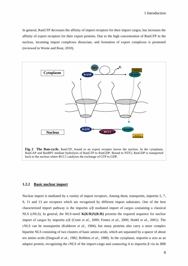
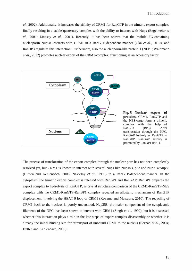
Fig. 5 Nuclear export of
proteins. CRM1, RanGTP and
the NES-cargo form a trimeric
complex with the help of
RanBP3 (BP3). After
translocation through the NPC,
RanGAP hydrolyzes RanGTP to
RanGDP. RanGAP activity is
promoted by RanBP1 (BP1).
al., 2002). Additionally, it increases the affinity of CRM1 for RanGTP in the trimeric export complex,
finally resulting in a stable quaternary complex with the ability to interact with Nups (Engelmeier et
al., 2001; Lindsay et al., 2001). Recently, it has been shown that the mobile FG-containing
nucleoporin Nup98 interacts with CRM1 in a RanGTP-dependent manner (Oka et al., 2010), and
RanBP3 regulates this interaction. Furthermore, also the nucleoporin-like protein 1 (NLP1; Waldmann
et al., 2012) promotes nuclear export of the CRM1-complex, functioning as an accessory factor.
The process of translocation of the export complex through the nuclear pore has not been completely
resolved yet, but CRM1 is known to interact with several Nups like Nup153, p62 and Nup214/Nup88
(Hutten and Kehlenbach, 2006; Nakielny et al., 1999) in a RanGTP-dependent manner. In the
cytoplasm, the trimeric export complex is released with RanBP1 and RanGAP. RanBP1 prepares the
export complex to hydrolysis of RanGTP, as crystal structure comparison of the CRM1-RanGTP-NES
complex with the CRM1-RanGTP-RanBP1 complex revealed an allosteric mechanism of RanGTP
displacement, involving the HEAT 9 loop of CRM1 (Koyama and Matsuura, 2010). The recycling of
CRM1 back to the nucleus is poorly understood. Nup358, the major component of the cytoplasmic
filaments of the NPC, has been shown to interact with CRM1 (Singh et al., 1999), but it is discussed
whether this interaction plays a role in the late steps of export complex disassembly or whether it is
already the initial binding site for retransport of unbound CRM1 to the nucleus (Bernad et al., 2004;
Hutten and Kehlenbach, 2006).
Cytoplasm
Nucleus

1 Introduction
14
1.3 The translocation of transport complexes through the nuclear pore
To date, it is not exactly known how transport complexes translocate through the nuclear pore. Several
studies showed that transport receptors interact with FG-rich Nups (Bayliss et al., 1999/2000/2002;
Fornerod et al, 1997a; Fribourg et al., 2001; Saitoh et al., 1996). Generally, it is believed that this
interaction occurs via the FGs of Nups. Based on this, several models exist, which explain the efficient
passage of transport receptor-cargo complexes through the nuclear pore.
In the oily-spaghetti-model (Macara, 2001), the NPC is an open structure, in which FG containing
repeat Nups behave like loose "oily spaghetti". The open central channel has a diameter of 10 nm
(Keminer and Peters, 1999). The FG-spaghetti would form an approximately 7 nm layer around this
tube. Transport receptors are able to interact with the FGs, moving randomly within the nuclear pore
for a short distance. This "hopping" from FG to FG results in a facilitated diffusive movement through
the nuclear pore. The diameter of the central tube would be big enough to allow sufficient translo-
cation of many transport complexes at the same time.
The selective phase-model (Ribbeck and Görlich, 2001) suggests that FG-rich Nups form a meshwork
in the central channel of the NPC, providing a permeability barrier for molecules. Transport receptors
interact with the FGs, thus becoming part of the meshwork. Recent studies showed that complexes
containing transport receptor domains, which can interact with FGs, can translocate extremely fast into
an FG-hydrogel, whereas the passage of molecules without this receptor domain is clearly restricted
(Frey and Görlich, 2006/2007).
The “virtual gate”-model considers entropic factors of macromolecules (Rout et al., 2003). In general,
molecules having a defined energy state can move freely in the cell. The NPC restricts this movement,
because the FG-Nups form a dense meshwork in the channel. The molecules require a higher energy
state to reach a "transition state", which enables them to cross the NPC. Small molecules can afford
this energy, but for bigger molecules the energy would be too high. Transport receptors bind to FG-
Nups, which results in decrease of the energy costs. Thus, transport receptors can reach the "transition
state" and move through the nuclear pore.
The forest-model combines the selective phase- and the virtual gate- model (Yamada et al., 2010).
Additionally, Nups are categorized in different structures. The analysis of topology, dimensions,
stoichiometry, interactivity and approximate anchoring of the FG-Nups to the wall of NPC leads to a
"forest"-like landscape of the nuclear pore channel. FG-Nup domains can either occur as extended,
relaxed or collapsed coils, where the relaxed and extended coils are more dynamic than the collapsed
coils. Depending on the charge of these domains, they are more attractive (low charge content) or
repulsive (high charge content). Certain Nups only have collapsed coils, therefore looking like a
"shrub". Other Nups are anchored to the wall of the NPC via extended coils and additionally can have
collapsed coils sitting on the extended coil, resulting in a "tree"-like structure. The "trees" extend to
the centre of the NPC channel, thereby creating a "transporter" zone (zone 1). There, transport
1 Introduction
15
receptors with large substrates can move. The "shrubs" do not extend to the centre of the channel, but
create another zone (zone 2) near the conduit of the NPC, where small molecules and transport
receptors with small substrates can translocate. In this model, zone 1 functions as a permeability
barrier, as described by the selective phase-model, and zone 2 allows passage of molecules with
corresponding loss of entropic energy as described in the “virtual gate”-model.
1.4 Nup214
Human Nup214, also known as CAN, was firstly identified in leukemia-associated chromosomal
translocations involving the SET and DEK genes (von Lindern et al., 1992a, b). In T-cells of patients
suffering from acute lymphoblastic leukaemia, a third translocation was found, which generates a
fusion between Nup214 and the constitutive tyrosine kinase ABL (Abelson murine leukemia viral
oncogene homolog 1; Grauxl et al., 2004). The activity of the Nup214-ABL fusion is dependent on its
tethering to the nuclear pore complex (de Keersmaecker et al., 2008).
Nup214 is an essential nucleoporin, as depletion in mice leads to strong mRNA export defects and to
lethality (van Deursen et al., 1996). It is located at the cytoplasmic side of the NPC (Kraemer et al.,
1994), and transmission electron microscopy (TEM) and field emission in-lense scanning electron
microscopy (FEISEM) studies revealed that Nup214 is located near the central channel of the pore
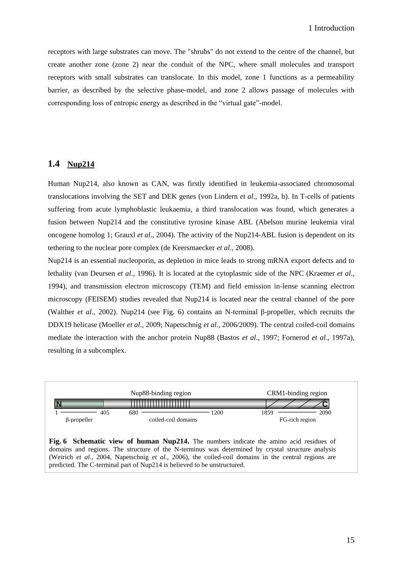
(Walther et al., 2002). Nup214 (see Fig. 6) contains an N-terminal β-propeller, which recruits the
DDX19 helicase (Moeller et al., 2009; Napetschnig et al., 2006/2009). The central coiled-coil domains
mediate the interaction with the anchor protein Nup88 (Bastos et al., 1997; Fornerod et al., 1997a),
resulting in a subcomplex.
Nup88-binding region CRM1-binding region
1 405 680 1200 1859 2090
β-propeller coiled-coil domains FG-rich region
Fig. 6 Schematic view of human Nup214. The numbers indicate the amino acid residues of
domains and regions. The structure of the N-terminus was determined by crystal structure analysis
(Weirich et al., 2004, Napetschnig et al., 2006), the coiled-coil domains in the central regions are
predicted. The C-terminal part of Nup214 is believed to be unstructured.
N C
1 Introduction
16
Deletion of Nup88 leads to a loss of Nup214 at the nuclear pore, and vice versa (Bernad et al., 2004;
Hutten and Kehlenbach, 2006). By contrast, other Nups like p62, Nup153 or Nup358 are not affected
upon Nup214 depletion (Hutten and Kehlenbach, 2006).
The FG-rich region in the C-terminus of Nup214 was identified as the binding site for CRM1, and this
binding is dependent on RanGTP (Fornerod et al., 1996). Furthermore, this binding is increased in the
presence of an export substrate (Kehlenbach et al., 1999). Nup214 is not only associated to CRM1-
mediated export, it is also involved in nuclear import of certain molecules. Nup214 exposes a docking
site for nucleocapsids of large viruses and facilitates their import into the nucleus independent of
transport receptors (Trotman et al., 2001). Furthermore, Nup214 has been shown to play a role in
nuclear import of the transcription factors Smad2 (Xu et al., 2002), Stat1 (Marg et al., 2004) and
NF-κB (Xylourgidis et al., 2006).
The role of the CRM1-Nup214 interaction is highly discussed. The lack of Nup88 causes
mislocalization of CRM1 (Roth et al., 2003), and it is suggested that the Nup214-88 complex is the
terminal docking site for CRM1 in nuclear export in vitro (Fornerod et al., 1996; Kehlenbach et al.,
1999). Strikingly, only a few substrates have been identified, which are Nup214- and CRM1-
dependent. Nup214 depletion leads to impaired mediated nuclear export of NFAT (nuclear factor of
activated T-cells), the viral protein HIV-Rev (Hutten and Kehlenbach, 2006) and the 60S preribosomal
subunit via the adaptor protein Nmd3 (Bernad et al., 2006). In yeast, the C-terminus of Nup214 is not
required at all for CRM1-dependent nuclear export of shuttling reporter proteins (Zeitler and Weis,
2004).
1.5 Aim of the work
The strong binding of the FG-containing Nup214 C-terminus to CRM1 suggests a general role for
Nup214 in nuclear export of proteins. Whereas the region at the C-terminal end of Nup214 is known
to bind CRM1, the region(s) in CRM1 for interaction with Nup214 (and other Nups) has not been
identified so far. The identification of these sites would help to elucidate the role of CRM1-
nucleoporin interaction, e.g. for translocation of export complexes through the nuclear pore or their
disassembly in the cytoplasm. For this reason, we perform microscopic studies with CRM1 fragments
and chimeras and investigate their association with the nuclear envelope and their functionality in
nuclear export.
The FG-containing Nup214 region that binds to CRM1 comprises 230 amino acids. It is known for
other karyopherins that they bind to the FGs in nucleoporins, but for CRM1 this can only be assumed.
With Nup214 FG-mutants, we want to investigate whether the FGs in Nup214 are responsible for
CRM1 interaction. To narrow down the CRM1-binding region in Nup214, we combine binding
studies with in vivo assays, using short Nup214 fragments.
Nucleus

2 Material and methods
17
2 Material and methods
2.1 Material
2.1.1 Technical equipment and software
Technical equipment Company
Centrifuge Avanti J-30I Beckman Coulter
Documentation system LAS 3000 Fujifilm
Emulsi flex-C5 Avestin
FACS Canto™II BD Biosciences
Film developing machine Curix 60 AGFA
Fluorescence microscope Axioskop 2 Zeiss
Rotors JA 30.50Ti, SX4750, TLA 100.3 Beckman Coulter
SE260 Small Format Vertical Electrophoresis
System Hoefer
Sequencer, Genetic Analyser 3100 Applied Biosystems
Table centrifuges 5415 D, 5415 R, 5424 Eppendorf
Thermocycler Gene Amp PCR 2400 Perkin Elmer
Thermocycler Primus MWG Biotech
Thermocycler Tprofessional Gradient Biometra
Thermocycler Tprofessional Standard Biometra
Ultracentrifuge OptimaMax Beckman Coulter
2 Material and methods
18
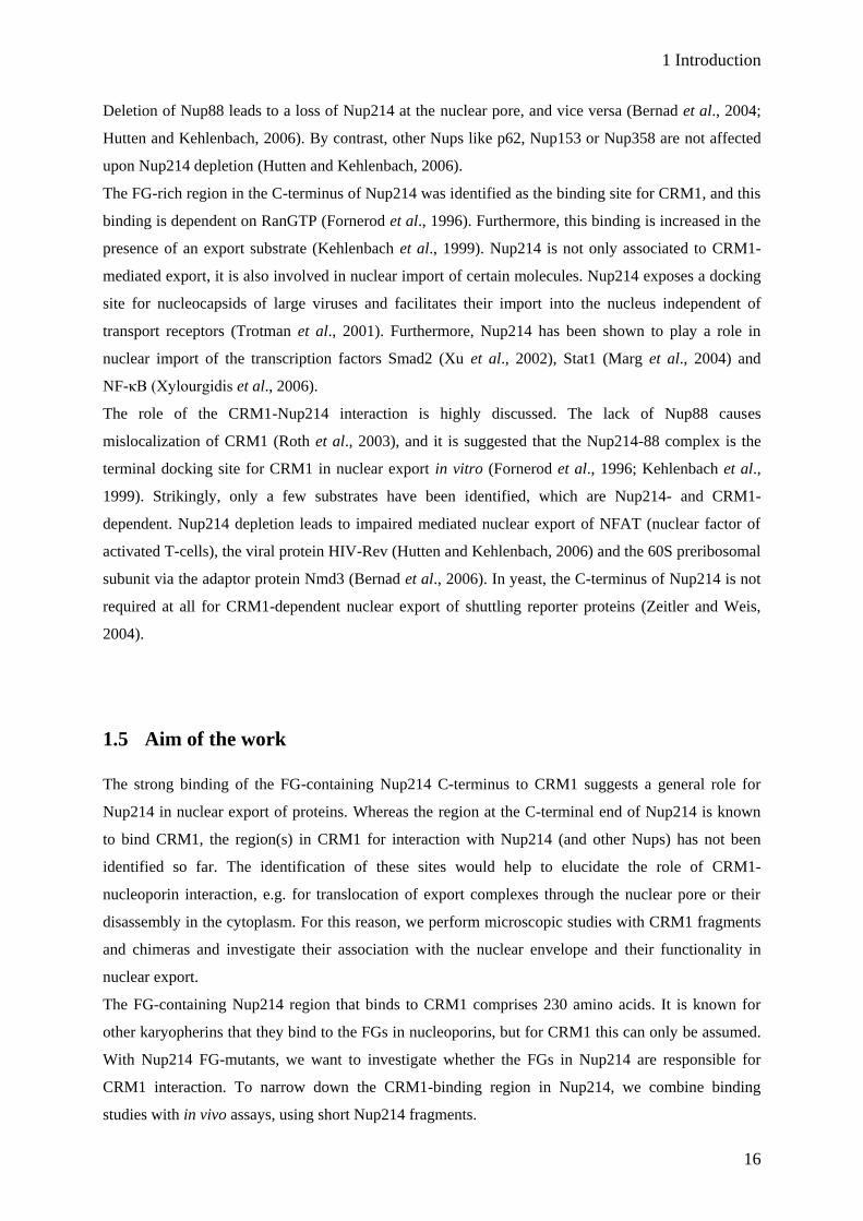
Software Company/Origin
AxioVision (LE) Rel. 4.5 Carl Zeiss
BLAST http://blast.ncbi.nlm.nih.gov/
Chromas Lite 2.01 Technelysium
Image Reader LAS 3000 Fuji
Ligation calculator http://www.insilico.uni-duesseldorf.de/Lig_Input.html
Oligonucleotide properties calculator http://www.basic.northwestern.edu/biotools/
oligocalc.html
Photoshop 6.0 Adobe
Vector NTI Invitrogen
2.1.2 Consumables
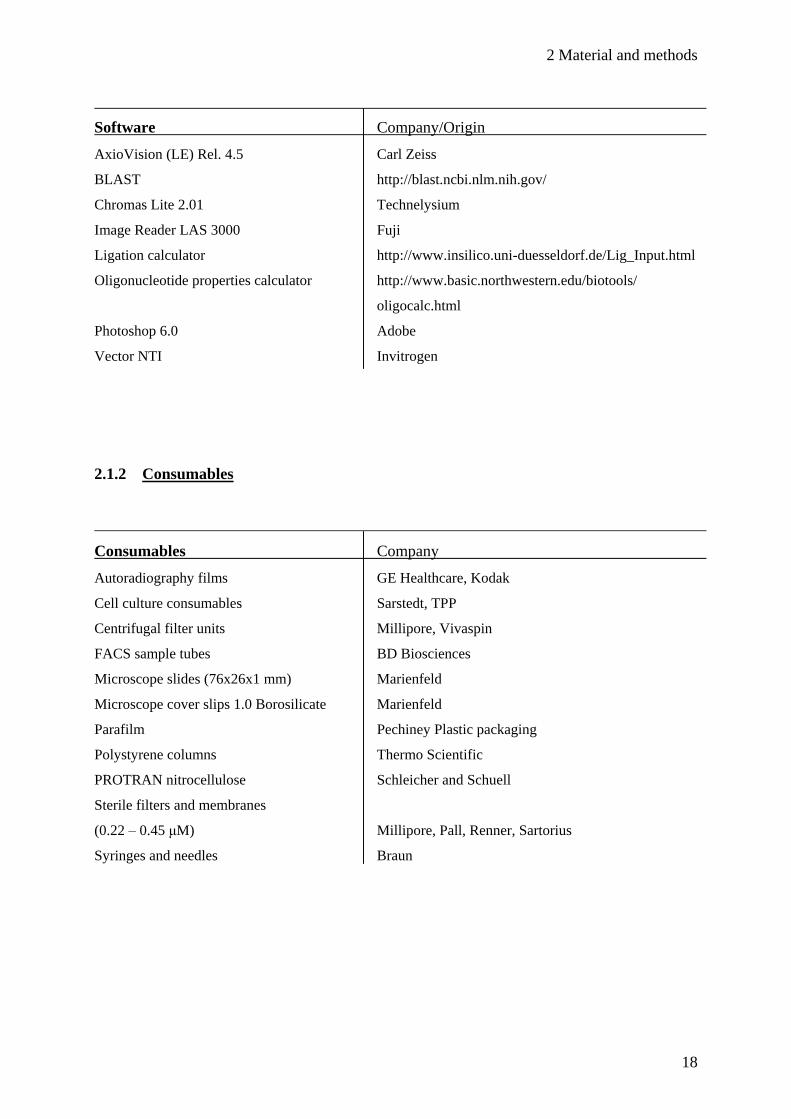
Consumables Company
Autoradiography films GE Healthcare, Kodak
Cell culture consumables Sarstedt, TPP
Centrifugal filter units Millipore, Vivaspin
FACS sample tubes BD Biosciences
Microscope slides (76x26x1 mm) Marienfeld
Microscope cover slips 1.0 Borosilicate Marienfeld
Parafilm Pechiney Plastic packaging
Polystyrene columns Thermo Scientific
PROTRAN nitrocellulose Schleicher and Schuell
Sterile filters and membranes
(0.22 – 0.45 μM) Millipore, Pall, Renner, Sartorius
Syringes and needles Braun

2 Material and methods
19
2.1.3 Kits
Kits Company
BigDye Terminator v1.1 cycle sequencing kit Applied Biosystems
NucleoBond® PC100, PC500 MACHEREY-NAGEL
NucleoSpin® Extract II MACHEREY-NAGEL
2.1.4 Chemicals, reagents and enzymes
Standard chemicals and reagents were obtained from AppliChem (Darmstadt), FLUKA (Buchs,
Switzerland), Merck (Darmstadt), Carl Roth (Karlsruhe), SERVA (Heidelberg) and Sigma Aldrich
(Taufkirchen). Specific reagents are listed below.
Reagent Origin
acrylamide (30%) Amersham
aprotinin Biomol
ATP Sigma Aldrich
BSA, fraction V PAA
calf intestinal phosphatise Fermentas
ImmobilonTM Western Millipore
creatine phosphate Calbiochem
creatine phosphate kinase Calbiochem
Cyanogen bromide-activated Sepharose® 4B Sigma Aldrich
digitonin Calbiochem
dimethylsulfoxide Applichem
dithiothreitol AppliChem
DMEM (high glucose) Gibco
DNA ladder, 1kb Fermentas
dNTPs Fermentas
FCS Gibco
Fluorescent mounting medium Dako
Freundsches Adjuvant incomplete Sigma Aldrich

2 Material and methods
20
Reagent Origin
glutamine Gibco
GDP, GTP Sigma Aldrich
Glutathione sepharoseTM High Performance GE Healthcare
Hoechst 33258 Sigma Aldrich
ionomycin Sigma Aldrich
IPTG Fermentas
leptomycin B Alexis Biochemicals
leupeptin Biomol
Ni-NTA agarose Qiagen
penicillin PAA
pepstatin Biomol
Phusion 530L Polymerase New England Biolabs
PMSF Sigma Aldrich
poly-L-Lysine Sigma Aldrich
protein assay reagent BIO-RAD
protein ladder PAGE ruler Fermentas
restriction enzymes Fermentas
RNase A Applichem
SERVA DNA stain G SERVA
sequencing mix and buffer Applied Biosystems
siRNA oligonucleotides Applied Biosystems
streptomycin PAA
T4 DNA Ligase Fermentas
T4 polynucleotide kinase Fermentas
Titermax® Gold Sigma Aldrich
trichostatin A Sigma Aldrich
trypane blue Fluka
trypsin Gibco
Vent polymerase New England Biolabs
WGA Sigma Aldrich

2 Material and methods
21
2.1.5 Buffers, stock solutions and media
All buffers and media were prepared using deionized water and autoclaved at 121 °C for 20 minutes.
Buffers were titrated with NaOH, HCL or KOH (only for HEPES buffers). Solutions with heat-
sensitive substances were sterilized using a 0.2 μm sterile filter.
Buffers
annealing buffer (2x) 100 mM potassium acetate, 30 mM HEPES pH 7.4, 2 mM
magnesium acetate
colloidal coomassie dye 0.1% (v/v) brilliant blue G-250, 2% (w/v) ortho-phosphoric
acid, 10% (w/v) ammonium sulphate, 20 % methanol
colloidal coomassie fixing solution 40% (v/v) ethanol, 10% (v/v) acetic acid
coomassie dye 10% (v/v) acetic acid, 0.025% (w/v) brilliant blue G-250
coomassie fixing solution 45% (v/v) methanol, 10% (v/v) acetic acid
10x DNA loading dye 50% (v/v) glycerol, 0.1% (w/v) bromphenol blue, 0.1 % (w/v)
xylencyanol
GST buffer 50 mM Tris-HCl (pH 6.8), 300 mM NaCl, 0.25 mM EDTA,
1 mM MgCl2, aprotinin, leupeptin, pepstatin, (1µg/ml each),
0.1 mM PMSF
2x HEPES buffered saline (HBS) 50 mM HEPES (pH 6.98), 250 mM NaCl, 1.5 mM Na2HPO4
10x Laemmli running buffer 250 mM Tris, 1.92 M glycine, 0.1 % (w/v) SDS
Nup214 buffer 50 mM Tris-HCl (pH 6.8), 300 mM NaCl, 10 % glycerol,
4 mM β-mercaptoethanol, 1 mM MgCl2, aprotinin, leupeptin,
pepstatin, (1µg/ml each), 0.1 mM PMSF
P1 buffer 50 mM Tris-HCl (pH 8), 10 mM EDTA, 100 μg/ml RNase A
P2 buffer 200 mM NaOH, 1% (v/v) SDS
P3 buffer 3 M KOAc (pH 5.3)

2 Material and methods
22
Buffers
phosphate buffered saline (PBS) 140 mM NaCl, 2.5 mM KCl, 10 mM Na2HPO4, KH2PO4
(pH 7.5)
PBS Tween 140 mM NaCl, 2.5 mM KCl, 10 mM Na2HPO4,
KH2PO4 (pH 7.5) + 0.1% (v/v) Tween20
Ponceau S 0.5% (w/v) Ponceau S, 1% (v/v) acetic acid
SDS-PAGE loading buffer (4x) 4% (w/v) SDS, 125 mM Tris pH 6.8, 10% (v/v) glycerol,
0.02% (w/v) bromphenol blue, 10% (v/v) β-mercaptoethanol
50x TAE 2 M Tris, 0.05 M EDTA pH 8, 5.8% acetic acid
1x TE 10 mM Tris pH 7.5, 1 mM EDTA
TFB I 100 mM RbCl, 15% (v/v) glycerol, 0.5 mM LiCl (pH 5.8)
TFB II 10 mM MOPS (pH 7), 10 mM RbCl, 75 mM CaCl2,
15% (v/v) glycerol
Transport buffer (TB) 20 mM HEPES pH 7.3, 110 mM KOAc, 2 mM Mg(OAc)
1 mM EGTA
Tris buffer 50 mM Tris-HCl (pH 7.4), 200 mM NaCl, 1 mM MgCl2,
5% glycerol, aprotinin, leupeptin, pepstatin, (1µg/ml each)
Western blot buffer (WB) 192 mM glycine, 25 mM Tris, 0.05% (v/v) SDS, 20% (v/v)
methanol
2 Material and methods
23
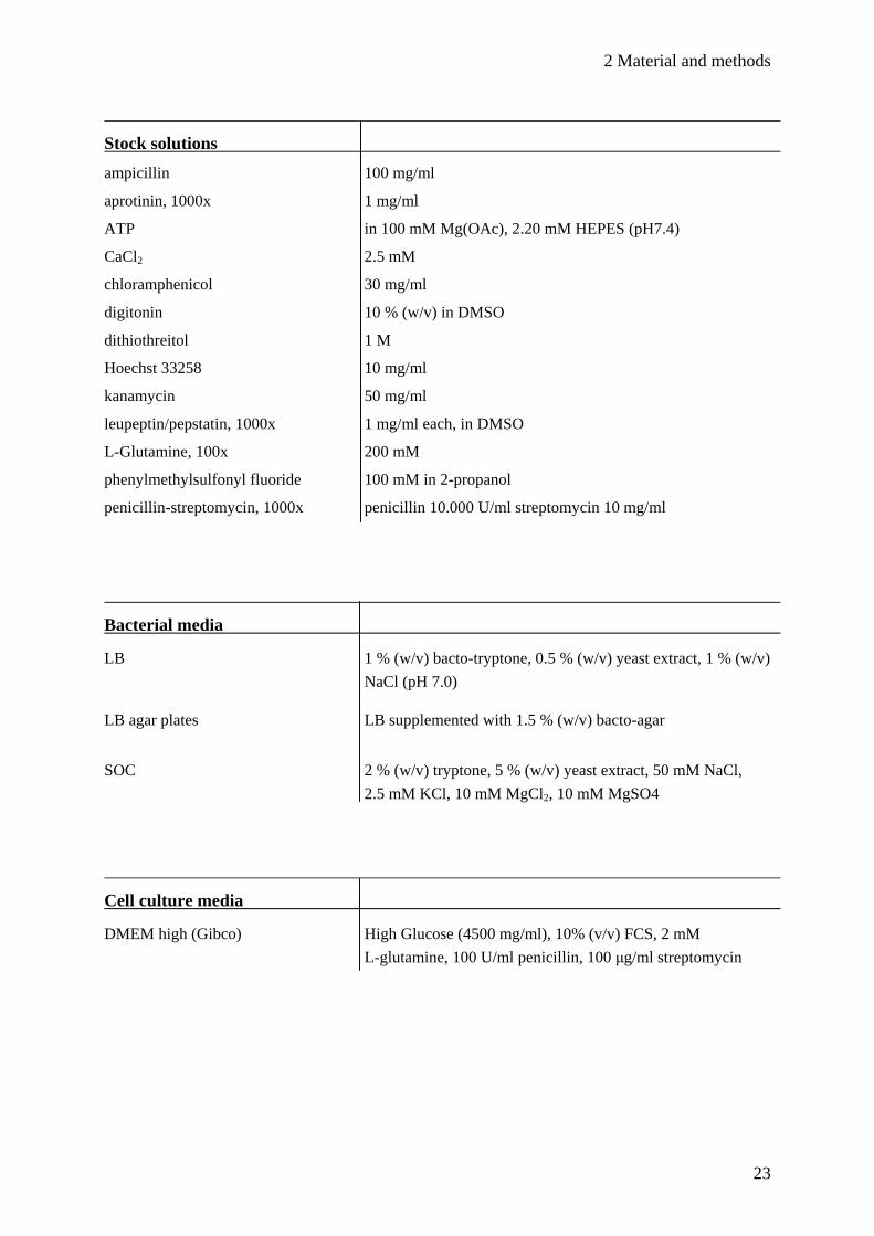
Stock solutions
ampicillin 100 mg/ml
aprotinin, 1000x 1 mg/ml
ATP in 100 mM Mg(OAc), 2.20 mM HEPES (pH7.4)
CaCl2 2.5 mM
chloramphenicol 30 mg/ml
digitonin 10 % (w/v) in DMSO
dithiothreitol 1 M
Hoechst 33258 10 mg/ml
kanamycin 50 mg/ml
leupeptin/pepstatin, 1000x 1 mg/ml each, in DMSO
L-Glutamine, 100x 200 mM
phenylmethylsulfonyl fluoride 100 mM in 2-propanol
penicillin-streptomycin, 1000x penicillin 10.000 U/ml streptomycin 10 mg/ml
Bacterial media
LB 1 % (w/v) bacto-tryptone, 0.5 % (w/v) yeast extract, 1 % (w/v)
NaCl (pH 7.0)
LB agar plates LB supplemented with 1.5 % (w/v) bacto-agar
SOC 2 % (w/v) tryptone, 5 % (w/v) yeast extract, 50 mM NaCl,
2.5 mM KCl, 10 mM MgCl2, 10 mM MgSO4
Cell culture media
DMEM high (Gibco) High Glucose (4500 mg/ml), 10% (v/v) FCS, 2 mM
L-glutamine, 100 U/ml penicillin, 100 μg/ml streptomycin
2 Material and methods
24
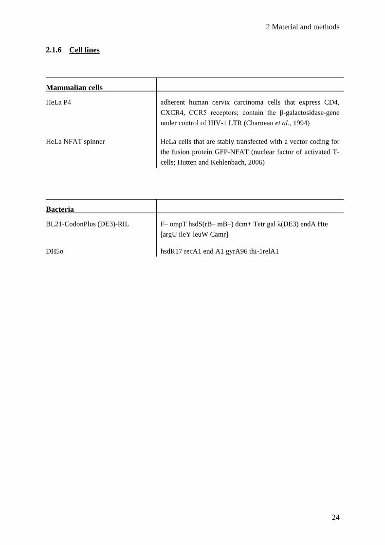
2.1.6 Cell lines
Mammalian cells
HeLa P4 adherent human cervix carcinoma cells that express CD4,
CXCR4, CCR5 receptors; contain the β-galactosidase-gene
under control of HIV-1 LTR (Charneau et al., 1994)
HeLa NFAT spinner HeLa cells that are stably transfected with a vector coding for
the fusion protein GFP-NFAT (nuclear factor of activated T-
cells; Hutten and Kehlenbach, 2006)
Bacteria
BL21-CodonPlus (DE3)-RIL F– ompT hsdS(rB– mB–) dcm+ Tetr gal λ(DE3) endA Hte
[argU ileY leuW Camr]
DH5α hsdR17 recA1 end A1 gyrA96 thi-1relA1

2 Material and methods
25
2.1.7 Antibodies
Primary antibodies
Name Species Origin Dilution Application
anti CRM1 goat R. Kehlenbach/S. Roloff 1:1000 WB, IF
anti HA rabbit Sigma Aldrich 1:500 WB, IF
anti His (penta) mouse Qiagen 1:1000 WB, IF
anti myc 9E10 mouse Serotec (WB)/Santa Cruz (IF) 1:5000/1:200 WB, IF
anti Nup214 #53 rabbit Hutten and Kehlenbach, 2006 1:200 WB
anti Nup214 #55 rabbit Hutten and Kehlenbach, 2006 1:500 IF
anti Ran mouse BD Transduction Laboratories 1:5000 WB, IF
Secondary antibodies
Name Species Origin Dilution Application
anti goat Alexa Fluor 488 donkey Molecular probes 1:1000 IF
anti mouse Alexa Fluor 594 donkey Molecular probes 1:1000 IF
anti rabbit Alexa Fluor 594 donkey Molecular probes 1:1000 IF
HRP anti goat donkey Dianova 1:5000 WB
HRP anti mouse donkey Dianova 1:5000 WB
HRP anti rabbit donkey Dianova 1:5000 WB
2 Material and methods
26
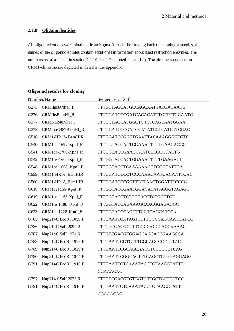
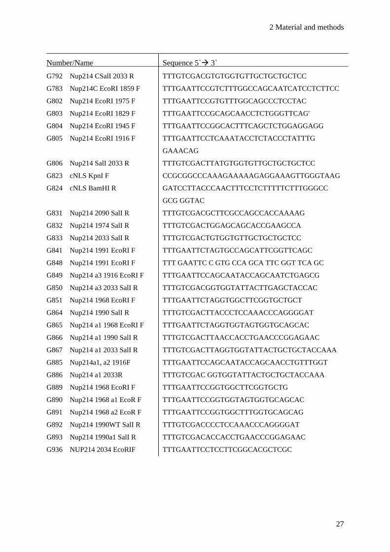
2.1.8 Oligonucleotides
All oligonucleotides were obtained from Sigma Aldrich. For tracing back the cloning strategies, the
names of the oligonucleotides contain additional information about used restriction enzymes. The
numbers are also listed in section 2.1.10 (see “Generated plasmids”). The cloning strategies for
CRM1-chimeras are depicted in detail in the appendix.
Oligonucleotides for cloning
Number/Name Sequence 5` 3`
G275 CRMIhs39NheI_F TTTGCTAGCATGCCAGCAATTATGACAATG
G276 CRMIhsBamHI_R TTTGGATCCCGATCACACATTTCTTCTGGAATC
G277 CRMIce248NheI_F TTTGCTAGCATGGCTGTCTCAGCAATGGAA
G278 CRMI ce3487BamHI_R TTTGGATCCCGACGCATATCCTCATCTTCCAC
G516 CRM1-HR13- BamHIR TTTGGATCCGGCTGAATTACAAAGGGGTGTC
G540 CRM1ce-1697-KpnI_F TTTGGTACCACTGGAAATTTGTGAAGACGG
G541 CRM1ce-1700-KpnI_R TTTGGTACCGAAGGAATCTCGGGTACTG
G542 CRM1hs-1668-KpnI_F TTTGGTACCACTGGAAATTTCTGAAGACT
G548 CRM1hs-1668_KpnI_R TTTGGTACCTCAAAAAACGTGGGTATTGA
G559 CRM1-HR16_BamHIR TTTGGATCCCGTGGGAAACAATGAGAATTGAC
G560 CRM1-HR18_BamHIR TTTGGATCCCGGTTGTTAACTGGATTTCCCG
G618 CRM1ce1166-KpnI_R TTTGGTACCGAATGGACATATACGGTAGAGC
G619 CRM1hs-1165-KpnI_F TTTGGTACCTCTGGTACCTCTGCCTCT
G622 CRM1hs 1188_KpnI_R TTTGGTACCAGAAAGCAACGGAGAGGC
G623 CRM1ce 1228-KpnI_F TTTGGTACCCAGGTTCGTGAGCATCCA
G785 Nup214C EcoRI 1859 F TTTGAATTCATAGTCTTTGGCCAGCAATCATCC
G786 Nup214C SalI 2090 R TTTGTCGACGGCTTCGCCAGCCACCAAAAC
G787 Nup214C SalI 1974 R TTTGTCGACGTGGAGCAGCACCGAAGCCA
G788 Nup214C EcoRI 1975 F TTTGAATTCGTGTTTGGCAGCCCTCCTAC
G789 Nup214C EcoRI 1829 F TTTGAATTCGCAGCAACCTCTGGGTTCAG
G790 Nup214C EcoRI 1945 F TTTGAATTCGGCACTTTCAGCTCTGGAGGAGG
G791 Nup214C EcoRI 1916 F TTTGAATTCTCAAATACCTCTAACCTATTT
GGAAACAG
G792 Nup214 CSalI 2033 R TTTGTCGACGTGTGGTGTTGCTGCTGCTCC
G791 Nup214C EcoRI 1916 F TTTGAATTCTCAAATACCTCTAACCTATTT
GGAAACAG
2 Material and methods
27
Number/Name Sequence 5` 3`
G792 Nup214 CSalI 2033 R TTTGTCGACGTGTGGTGTTGCTGCTGCTCC
G783 Nup214C EcoRI 1859 F TTTGAATTCCGTCTTTGGCCAGCAATCATCCTCTTCC
G802 Nup214 EcoRI 1975 F TTTGAATTCCGTGTTTGGCAGCCCTCCTAC
G803 Nup214 EcoRI 1829 F TTTGAATTCCGCAGCAACCTCTGGGTTCAG'
G804 Nup214 EcoRI 1945 F TTTGAATTCCGGCACTTTCAGCTCTGGAGGAGG
G805 Nup214 EcoRI 1916 F TTTGAATTCCTCAAATACCTCTACCCTATTTG
GAAACAG
G806 Nup214 SalI 2033 R TTTGTCGACTTATGTGGTGTTGCTGCTGCTCC
G823 cNLS KpnI F CCGCGGCCCAAAGAAAAAGAGGAAAGTTGGGTAAG
G824 cNLS BamHI R GATCCTTACCCAACTTTCCTCTTTTTCTTTGGGCC
GCG GGTAC
G831 Nup214 2090 SalI R TTTGTCGACGCTTCGCCAGCCACCAAAAG
G832 Nup214 1974 SalI R TTTGTCGACTGGAGCAGCACCGAAGCCA
G833 Nup214 2033 SalI R TTTGTCGACTGTGGTGTTGCTGCTGCTCC
G841 Nup214 1991 EcoRI F TTTGAATTCTAGTGCCAGCATTCGGTTCAGC
G848 Nup214 1991 EcoRI F TTT GAATTC C GTG CCA GCA TTC GGT TCA GC
G849 Nup214 a3 1916 EcoRI F TTTGAATTCCAGCAATACCAGCAATCTGAGCG
G850 Nup214 a3 2033 SalI R TTTGTCGACGGTGGTATTACTTGAGCTACCAC
G851 Nup214 1968 EcoRI F TTTGAATTCTAGGTGGCTTCGGTGCTGCT
G864 Nup214 1990 SalI R TTTGTCGACTTACCCTCCAAACCCAGGGGAT
G865 Nup214 a1 1968 EcoRI F TTTGAATTCTAGGTGGTAGTGGTGCAGCAC
G866 Nup214 a1 1990 SalI R TTTGTCGACTTAACCACCTGAACCCGGAGAAC
G867 Nup214 a1 2033 SalI R TTTGTCGACTTAGGTGGTATTACTGCTGCTACCAAA
G885 Nup214a1, a2 1916F TTTGAATTCCAGCAATACCAGCAACCTGTTTGGT
G886 Nup214 a1 2033R TTTGTCGAC GGTGGTATTACTGCTGCTACCAAA
G889 Nup214 1968 EcoRI F TTTGAATTCCGGTGGCTTCGGTGCTG
G890 Nup214 1968 a1 EcoR F TTTGAATTCCGGTGGTAGTGGTGCAGCAC
G891 Nup214 1968 a2 EcoR F TTTGAATTCCGGTGGCTTTGGTGCAGCAG
G892 Nup214 1990WT SalI R TTTGTCGACCCCTCCAAACCCAGGGGAT
G893 Nup214 1990a1 SalI R TTTGTCGACACCACCTGAACCCGGAGAAC
G936 NUP214 2034 EcoRIF TTTGAATTCCTCCTTCGGCACGCTCGC

2 Material and methods
28
Oligonucleotides for mutagenesis
Number/Name Sequence 5`3`
G651 CRM1 HS-loop del_F KpnI TGAACTCTATAGAGAGGGTACC (∆) CCCAGG
AGACAGCTAT
G652 CRM1 HS-loop del_R KpnI ATAGCTGTCTCCTGGGGGTACC (∆) CTCTC
TATAGAGTTCA
Oligonucleotides for sequencing
Number/Name Sequence 5`3`
G379 CRM1-ce-501F TCATCCAACACAAATGGAAGT
G380 CRM1-ce-400R CGACATGAGGATTTGGTTAGC
G381 CRM1-ce-1501F CTACCGTGAATACCTCTCGCA
G382 CRM1-ce-1001F AACTTCCTTTCGCTTGAAGTG
G383 CRM1-ce-1501F CTACCGTGAATACCTCTCGCA
G384 CRM1-ce-2001F AGACTCATGAAGGTGTACAGG
G385 CRM1-ce-2501F AATCCATTGGTAAAGACGATG
G386 CRM1-ce-3001F TGACAAGATCGCTCAACCATT
G387 CRM1-hs-39F ATGCCAGCAATTATGACAATG
G388 CRM1-hs-200R CTTTAAATGTGTCAGTACTTC
G389 CRM1-hs-501F GGAGCAAGTAGGACCAGCGAA
G390 CRM1-hs-1001F TCTCAGTTTGTTTCTCTGCAC
G391 CRM1-hs-1501F AGTGGTCATGGAAAAATTTGA
G392 CRM1-hs-2001F GAAAAGTACATGTTACTCCCT
G393 CRM1-hs-2495F ACCTCAAATATTTGAATGCTG
G394 CRM1-hs-3001F ACCTACAAGATGCTCAAGTAA
G525 T7-Promotor TAATACGACTCACTATAGGG
G545 pmCherry_seq_f CGCTGAGGTCAAGACCACC
G546 pmCherry_seq_r CGAAGGGCAGGGGGCCACC
2 Material and methods
29
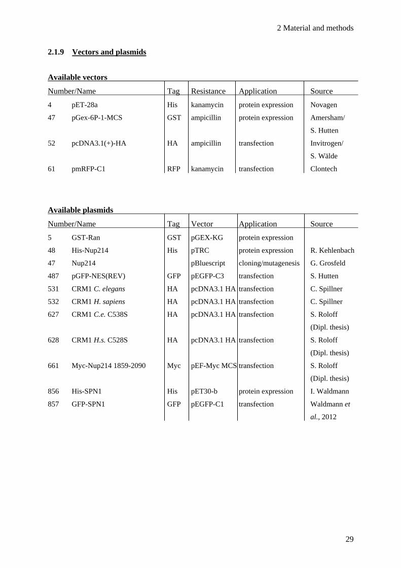
2.1.9 Vectors and plasmids
Available vectors
Number/Name Tag Resistance Application Source
4 pET-28a His kanamycin protein expression Novagen
47 pGex-6P-1-MCS GST ampicillin protein expression Amersham/
S. Hutten
52 pcDNA3.1(+)-HA HA ampicillin transfection Invitrogen/
S. Wälde
61 pmRFP-C1 RFP kanamycin transfection Clontech
Available plasmids
Number/Name Tag Vector Application Source
5 GST-Ran GST pGEX-KG protein expression
48 His-Nup214 His pTRC protein expression R. Kehlenbach
47 Nup214 pBluescript cloning/mutagenesis G. Grosfeld
487 pGFP-NES(REV) GFP pEGFP-C3 transfection S. Hutten
531 CRM1 C. elegans HA pcDNA3.1 HA transfection C. Spillner
532 CRM1 H. sapiens HA pcDNA3.1 HA transfection C. Spillner
627 CRM1 C.e. C538S HA pcDNA3.1 HA transfection S. Roloff
(Dipl. thesis)
628 CRM1 H.s. C528S HA pcDNA3.1 HA transfection S. Roloff
(Dipl. thesis)
661 Myc-Nup214 1859-2090 Myc pEF-Myc MCS transfection S. Roloff
(Dipl. thesis)
856 His-SPN1 His pET30-b protein expression I. Waldmann
857 GFP-SPN1 GFP pEGFP-C1 transfection Waldmann et
al., 2012

2 Material and methods
30
Purchased plasmids
Number/Name Tag Vector Application Source
949 Nup214 aa1916-2033 a1 no pMA cloning life technologies
950 Nup214 aa1916-2033 a2 no pMA cloning life technologies
951 Nup214 aa1916-2033 a3 no pMA cloning life technologies
1025 Nup214 aa1859-2090 a1 no pMA-T cloning life technologies
1036 Nup214 aa1859-2090 a3 no pMA-T cloning life technologies
Generated plasmids
Number/Name Tag Vector Application Cloning
694 CRM1 aa 1-709 C528S HA pcDNA3.1 HA transfection G275/G516
713 CRM1 aa 1-861 C528S HA pcDNA3.1 HA transfection G275/G559
714 CRM1 aa 1-970 C528S HA pcDNA3.1 HA transfection G275/G560
721 CRM1 H.s./C.e. I C528S HA pcDNA3.1 HA transfection G275/G548/G540/
G529
722 CRM1 C.e./H.s. I C538S HA pcDNA3.1 HA transfection G277/G541/G542/
G276
743 CRM1 C.e./H.s. II C528S HA pcDNA3.1 HA transfection G277/G618/G619/
G276
744 CRM1 H.s./C.e. II C538S HA pcDNA3.1 HA transfection G275/G622/G623/
G529
749 CRM1 H.s. Δloop C528S HA pcDNA3.1 HA transfection Mutagenesis; G651/
G652
943 His-Nup214 aa1859-2090 His pET28-a protein expr. G785/G786
944 His-Nup214 aa1859-1974 His pET28-a protein expr. G785/G787
945 His-Nup214 aa1975-2090 His pET28-a protein expr. G788/G786
946 His-Nup214 aa1829-1974 His pET28-a protein expr. G789/G787
947 His-Nup214 aa1945-2090 His pET28-a protein expr. G790/786
948 His Nup214 aa1916-2033 His pET28-a protein expr. G791/G792
987 RFP-cNLS RFP pmRFP-C1 transfection Oligo annealing;
G823/G824
988 RFP-Nup214 1859-2090 cNLS RFP pmRFP-C1 transfection G783/G831
989 RFP-Nup214 1859-1974 cNLS RFP pmRFP-C1 transfection G783/G832
990 RFP-Nup214 1975-2090 cNLS RFP pmRFP-C1 transfection G802/G831
991 RFP-Nup214 1829-1974 cNLS RFP pmRFP-C1 transfection G803/G832

2 Material and methods
31
Number/Name Tag Resistance Application Cloning
992 RFP-Nup214 1945-2090 cNLS RFP pmRFP-C1 transfection G804/G831
993 RFP-Nup214 1916-2033 cNLS RFP pmRFP-C1 transfection G805/G833
994 RFP-Nup214 1916-2033- RFP pmRFP-C1 transfection G885/G886
cNLS a1
995 RFP-Nup214 1916-2033 RFP pmRFP-C1 transfection G885/G886
cNLS a2
996 RFP-Nup214 1916-2033 RFP pmRFP-C1 transfection G849/G850
cNLS a3
997 His-Nup214 1916-2033 a1 His pET28-a protein expr. from 949 via EcoRI/
SalI
998 His-Nup2141916-2033 a2 His pET28-a protein expr. from 950 via EcoRI/
SalI
999 His-Nup214 1916-2033 a3 His pET28-a protein expr. from 951 via EcoRI/
SalI
1000 GST-Nup214 1991-2033 GST pGEX-6P-1 protein expr. G841/G806
1007 GST-Nup214 1968-1990 WT GST pGEX-6P-1 protein expr. G851/G864
1008 GST-Nup214 1968-1990 a1 GST pGEX-6P-1 protein expr. G865/G866
1009 GST-Nup214 1968-2033 WT GST pGEX-6P-1 protein expr. G851/G806
1010 GST-Nup214 1968-2033 a1 GST pGEX-6P-1 protein expr. G865/G867
1011 RFP-Nup214 1968-2033 WT RFP pmRFP-C1 transfection G889/G833
1012 RFP-Nup214 1968-2033 a1 RFP pmRFP-C1 transfection G890/G886
1030 RFPNup214 1859-2090 - RFP pmRFP-C1 transfection from 1025 via EcoRI/
cNLS a1 SalI
1031 RFP-Nup214 1916-1990- RFP pmRFP-C1 transfection G805/G892
cNLS WT
1032 RFP-Nup214 1968-1990- RFP pmRFP-C1 transfection G890/G893
cNLS a1
1033 RFP-Nup214 1968-1990- RFP pmRFP-C1 transfection G889/G892
cNLS WT
1034 RFP-Nup214 1991-2033cNLS RFP pmRFP-C1 transfection G848/G833
1035 RFP-Nup214 1991-2090cNLS RFP pmRFP-C1 transfection G848/G831
1038 RFP-Nup214 1916-1990cNLS RFP pmRFP-C1 transfection G805/G892
1039 RFP-Nup214 2034-2090cNLS RFP pmRFP-C1 transfection G936/G831
2 Material and methods
32
2.1.10 Proteins
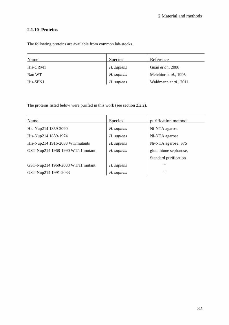
The following proteins are available from common lab-stocks.
Name Species Reference
His-CRM1 H. sapiens Guan et al., 2000
Ran WT H. sapiens Melchior et al., 1995
His-SPN1 H. sapiens Waldmann et al., 2011
The proteins listed below were purifed in this work (see section 2.2.2).
Name Species purification method
His-Nup214 1859-2090 H. sapiens Ni-NTA agarose
His-Nup214 1859-1974 H. sapiens Ni-NTA agarose
His-Nup214 1916-2033 WT/mutants H. sapiens Ni-NTA agarose, S75
GST-Nup214 1968-1990 WT/a1 mutant H. sapiens glutathione sepharose,
Standard purification
GST-Nup214 1968-2033 WT/a1 mutant H. sapiens “
GST-Nup214 1991-2033 H. sapiens “
2 Material and methods
33
2.2 Biochemical methods
2.2.1 Separation and detection of proteins
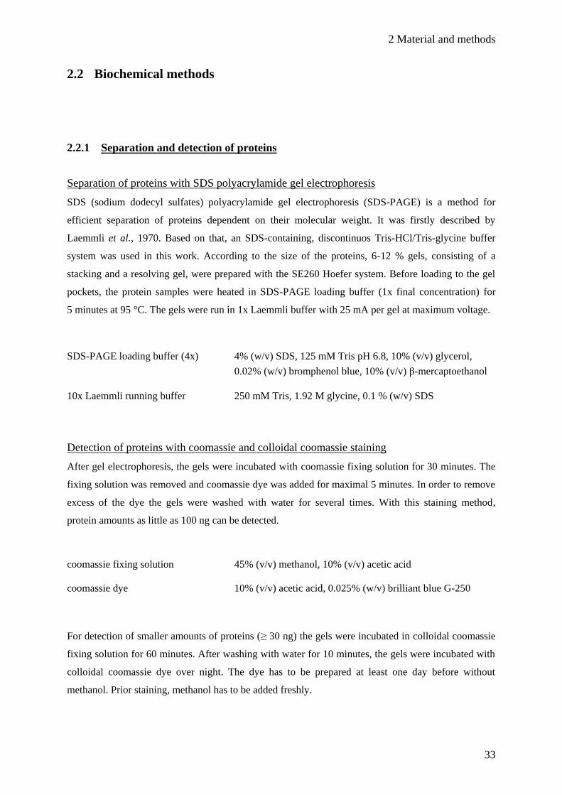
Separation of proteins with SDS polyacrylamide gel electrophoresis
SDS (sodium dodecyl sulfates) polyacrylamide gel electrophoresis (SDS-PAGE) is a method for
efficient separation of proteins dependent on their molecular weight. It was firstly described by
Laemmli et al., 1970. Based on that, an SDS-containing, discontinuos Tris-HCl/Tris-glycine buffer
system was used in this work. According to the size of the proteins, 6-12 % gels, consisting of a
stacking and a resolving gel, were prepared with the SE260 Hoefer system. Before loading to the gel
pockets, the protein samples were heated in SDS-PAGE loading buffer (1x final concentration) for
5 minutes at 95 °C. The gels were run in 1x Laemmli buffer with 25 mA per gel at maximum voltage.
SDS-PAGE loading buffer (4x) 4% (w/v) SDS, 125 mM Tris pH 6.8, 10% (v/v) glycerol,
0.02% (w/v) bromphenol blue, 10% (v/v) β-mercaptoethanol
10x Laemmli running buffer 250 mM Tris, 1.92 M glycine, 0.1 % (w/v) SDS
Detection of proteins with coomassie and colloidal coomassie staining
After gel electrophoresis, the gels were incubated with coomassie fixing solution for 30 minutes. The
fixing solution was removed and coomassie dye was added for maximal 5 minutes. In order to remove
excess of the dye the gels were washed with water for several times. With this staining method,
protein amounts as little as 100 ng can be detected.
coomassie fixing solution 45% (v/v) methanol, 10% (v/v) acetic acid
coomassie dye 10% (v/v) acetic acid, 0.025% (w/v) brilliant blue G-250
For detection of smaller amounts of proteins (≥ 30 ng) the gels were incubated in colloidal coomassie
fixing solution for 60 minutes. After washing with water for 10 minutes, the gels were incubated with
colloidal coomassie dye over night. The dye has to be prepared at least one day before without
methanol. Prior staining, methanol has to be added freshly.

2 Material and methods
34
Colloidal coomassie fixing solution 40 % (v/v) ethanol, 10% (v/v) acetic acid
Colloidal coomassie dye 0.1% (v/v) brilliant blue G-250, 2% (w/v) orthophosphoric
acid, 10% (w/v) ammonium sulphate, 20 % (v/v) methanol
Transfer of proteins to nitrocellulose (Western blot) followed by immunological detection
Dependent on the molecular weight of the analyzed proteins, either the semi-dry or the wet blot
technique was applied. The SDS gel was placed directly onto a wet nitrocellulose membrane
(equilibrated with Western blot buffer). For stabilization, two whatman papers (also soaked with
Western blot buffer) were packed on each side. Depending on the method, this package was either
clamped into a wet-blot chamber, filled with buffer, or laid into a semi-dry chamber. The wet blot
chamber was run at 400 mA for 1.5-2 hours. The semi-dry chamber was run at 200 mA per gel for 1.5
hours.
The transfer efficiency was analyzed by reversible staining of the nitrocellulose membrane with
Ponceau S solution. The membrane was incubated with the solution for 5 minutes, followed by
washing with 1 % acetic acid, until red protein bands become visible.
For immunological detection of single proteins, the nitrocellulose was first blocked with 3% milk in
PBS-Tween buffer for 30 minutes. The primary antibodies was diluted in the same buffer and added to
the membrane. After incubation over night at 4 °C, the membrane was washed three times with PBS-
Tween buffer without milk. The secondary antibody, coupled with horseradish peroxidase and directed
against the species of the primary antibody, was diluted in PBS-Tween buffer with 3% milk and the
membrane was incubated for two hours at room temperature. The membrane was washed for three
times, followed by addition of chemiluminescent reagents (Millipore). Depending on the expected
signal, the membrane was incubated for maximal five minutes. For detection of the signals,
autoradiography films were used and developed with an automatic developer machine. Alternatively,
the signals were directly detected with a digital documentation system (LAS-3000, Fuji).
Western blot buffer 192 mM glycine, 25 mM Tris, 0.05% (v/v) SDS, 20% (v/v) methanol
PBS Tween 140 mM NaCl, 2.5 mM KCl, 10 mM Na2HPO4, KH2PO4 (pH 7.5),
0.1% (v/v) Tween20
2 Material and methods
35
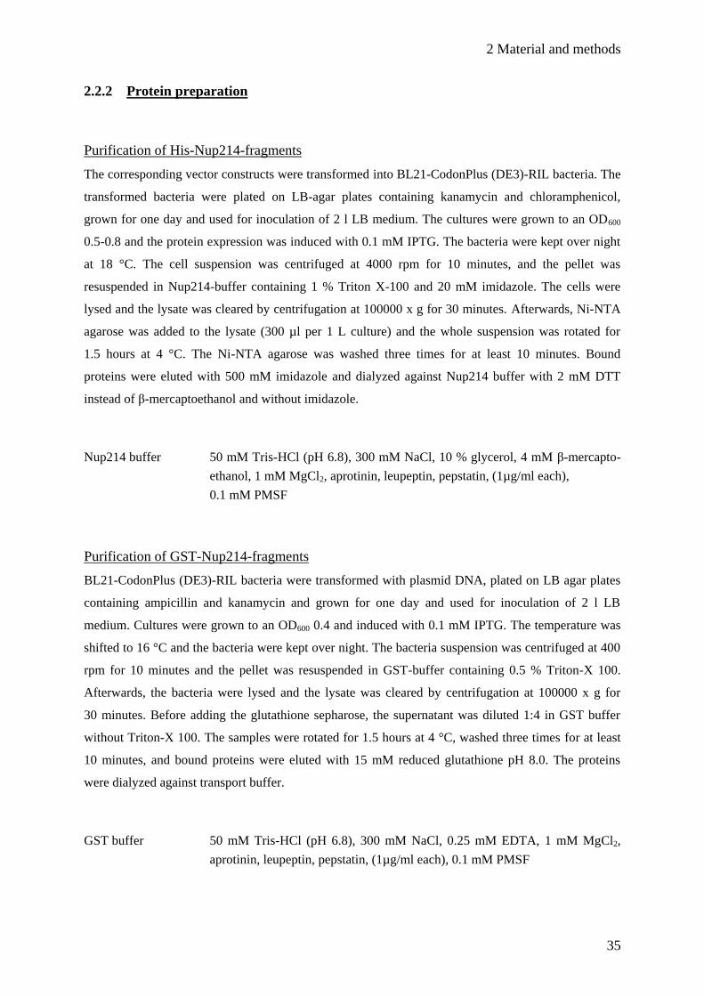
2.2.2 Protein preparation
Purification of His-Nup214-fragments
The corresponding vector constructs were transformed into BL21-CodonPlus (DE3)-RIL bacteria. The
transformed bacteria were plated on LB-agar plates containing kanamycin and chloramphenicol,
grown for one day and used for inoculation of 2 l LB medium. The cultures were grown to an OD600
0.5-0.8 and the protein expression was induced with 0.1 mM IPTG. The bacteria were kept over night
at 18 °C. The cell suspension was centrifuged at 4000 rpm for 10 minutes, and the pellet was
resuspended in Nup214-buffer containing 1 % Triton X-100 and 20 mM imidazole. The cells were
lysed and the lysate was cleared by centrifugation at 100000 x g for 30 minutes. Afterwards, Ni-NTA
agarose was added to the lysate (300 µl per 1 L culture) and the whole suspension was rotated for
1.5 hours at 4 °C. The Ni-NTA agarose was washed three times for at least 10 minutes. Bound
proteins were eluted with 500 mM imidazole and dialyzed against Nup214 buffer with 2 mM DTT
instead of β-mercaptoethanol and without imidazole.
Nup214 buffer 50 mM Tris-HCl (pH 6.8), 300 mM NaCl, 10 % glycerol, 4 mM β-mercapto-
ethanol, 1 mM MgCl2, aprotinin, leupeptin, pepstatin, (1µg/ml each),
0.1 mM PMSF
Purification of GST-Nup214-fragments
BL21-CodonPlus (DE3)-RIL bacteria were transformed with plasmid DNA, plated on LB agar plates
containing ampicillin and kanamycin and grown for one day and used for inoculation of 2 l LB
medium. Cultures were grown to an OD600 0.4 and induced with 0.1 mM IPTG. The temperature was
shifted to 16 °C and the bacteria were kept over night. The bacteria suspension was centrifuged at 400
rpm for 10 minutes and the pellet was resuspended in GST-buffer containing 0.5 % Triton-X 100.
Afterwards, the bacteria were lysed and the lysate was cleared by centrifugation at 100000 x g for
30 minutes. Before adding the glutathione sepharose, the supernatant was diluted 1:4 in GST buffer
without Triton-X 100. The samples were rotated for 1.5 hours at 4 °C, washed three times for at least
10 minutes, and bound proteins were eluted with 15 mM reduced glutathione pH 8.0. The proteins
were dialyzed against transport buffer.
GST buffer 50 mM Tris-HCl (pH 6.8), 300 mM NaCl, 0.25 mM EDTA, 1 mM MgCl2,
aprotinin, leupeptin, pepstatin, (1µg/ml each), 0.1 mM PMSF

2 Material and methods
36
2.2.3 In vitro binding assays with GST-tagged proteins
All binding studies were performed in Tris buffer. Per sample, 5 µg of GST-tagged protein were
immobilized for one hour on 20 µl glutathione sepharose beads (equilibrated before in Tris buffer).
Unbound proteins were removed by washing the beads for three times, and unspecific binding sites
were blocked by adding Tris buffer containing 20 mg/ml BSA for 20 minutes. After that, the proteins
to be investigated were added and the whole suspension was incubated for an hour. The sepharose
beads were washed three times and then the supernatant was completely taken off by using a gel
loading tip which was gently pressed before with a forceps. Finally, 50 µl of 2x sample buffer was
added to the beads and the samples were heated for 5 minutes at 95 °C.
Tris buffer 50 mM Tris-HCl (pH 7.4), 200 mM NaCl, 1 mM MgCl2, 5 % glycerol,
aprotinin, leupeptin, pepstatin, (1µg/ml each)
Loading of Ran with GDP/GTP (Kehlenbach et al., 1999):
Per sample, 10 µg Ran were incubated for 30 minutes at room temperature together with 4.5 mM
EDTA and 10 mM GDP/GTP in Tris-buffer (150 µl final volume). Afterwards, 30 mM MgCl2 was
added, followed by incubation for 15 minutes on ice.
Pulldown assays with GST-Nup214-fragments:
Immobilized GST-Nup214-fragments (400 nM) were incubated with 960 nM RanGDP/GTP, 255 nM
His-SPN1 and 108 nM His-CRM1 in a final volume of 400 µl. Bound proteins were analyzed by SDS-
PAGE and coomassie staining as described above.
Pulldown assays with GST-Ran:
Before immobilizing GST-Ran (500 nM) to glutathione sepharose, it was loaded with either GDP or
GTP. 108 nM His-CRM1, 160 nM His-Nup214-fragments and 255 nM His-SPN1 (or 2500 nM NES
peptide) were added. For detection of His-tagged proteins, a Western blot using a His-antibody was
performed. Detection of small His-Nup214-fragments bound to CRM1 is also possible by colloidal
coomassie staining.
2 Material and methods
37
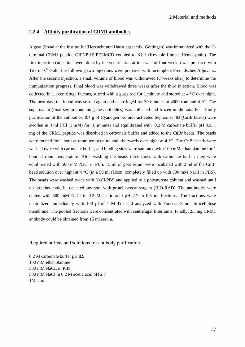
2.2.4 Affinity purification of CRM1 antibodies
A goat (bread at the Institut für Tierzucht und Haustiergenetik, Göttingen) was immunized with the C-
terminal CRM1 peptide GIFNPHEIPEEMCD coupled to KLH (Keyhole Limpet Hemacyanin). The
first injection (injections were done by the veterinarian at intervals of four weeks) was prepared with
Titermax® Gold, the following two injections were prepared with incomplete Freundsches Adjuvanz.
After the second injection, a small volume of blood was withdrawed (3 weeks after) to determine the
immunization progress. Final blood was withdrawed three weeks after the third injection. Blood was
collected in 1 l centrifuge falcons, stirred with a glass rod for 1 minute and stored at 4 °C over night.
The next day, the blood was stirred again and centrifuged for 30 minutes at 4000 rpm and 4 °C. The
supernatant (final serum containing the antibodies) was collected and frozen in aliquots. For affinity
purification of the antibodies, 0.4 g of Cyanogen bromide-activated Sepharose 4B (CnBr beads) were
swollen in 3 ml HCl (1 mM) for 10 minutes and equilibrated with 0.2 M carbonate buffer pH 8.9. 1
mg of the CRM1 peptide was dissolved in carbonate buffer and added to the CnBr beads. The beads
were rotated for 1 hour at room temperature and afterwards over night at 4 °C. The CnBr beads were
washed twice with carbonate buffer, and binding sites were saturated with 100 mM ethanolamine for 1
hour at room temperature. After washing the beads three times with carbonate buffer, they were
equilibrated with 500 mM NaCl in PBS. 15 ml of goat serum were incubated with 2 ml of the CnBr
bead solution over night at 4 °C (in a 50 ml falcon, completely filled up with 500 mM NaCl in PBS).
The beads were washed twice with NaCl/PBS and applied to a polystyrene column and washed until
no proteins could be detected anymore with protein assay reagent (BIO-RAD). The antibodies were
eluted with 500 mM NaCl in 0.2 M acetic acid pH 2.7 in 0.5 ml fractions. The fractions were
neutralized immediately with 100 µl of 1 M Tris and analyzed with Ponceau-S on nitrocellulose
membrane. The pooled fractions were concentrated with centrifugal filter units. Finally, 2.5 mg CRM1
antibody could be obtained from 15 ml serum.
Required buffers and solutions for antibody purification
0.2 M carbonate buffer pH 8.9
100 mM ethanolamine
500 mM NaCL in PBS
500 mM NaCl in 0.2 M acetic acid pH 2.7
1M Tris

2 Material and methods
38
For characterization of the CRM1-antibody, HeLa cells and recombinant His-CRM1 were analyzed.
Endogenous CRM1 could be detected in immunofluorescence as well as in Western blot (Fig 7A and
B). The antibody recognized 1 ng of His-CRM1. The side bands appearing in the cell lysate sample
could derive from degradation of CRM1.
(A) (B)
DNA
endogenous
CRM1
1 2 5 ng
kDa His-CRM1 CL
CRM1
Fig. 7 Characterization of purified CRM1 antibody. (A) HeLa cells were fixed,
stained for endogenous CRM1 and analyzed by immunofluorescence microscopy. The
antibody was diluted 1:5000, corresponding to 40 ng of antibody per slide. (B) 1, 2 or 5 ng of
recombinant His-CRM1 or lysate from HeLa cells (50000 cells; CL) was analyzed by
Western blot (antibody dilution 1:5000; film exposure time 5 seconds).
200
150
120
100
85
70
60
50
40
2 Material and methods
39
2.3 Cell biology methods
2.3.1 Cell culture
HeLa P4 cells were grown at 37 °C and 5% CO2 on 10 cm diameter plates in Dulbecco’s modified
Eagle Medium (DMEM) containing 10% fetal calf serum (FCS), 100 U/ml penicillin, 100 µg/ml
streptomycin and 2 mM L-glutamine. For transferring to other plates or to 24-wells containing glass
cover slips, the cells were washed with PBS, detached from the plate with trypsin at 37 °C and diluted
in fresh medium.
To improve adherence of the cells, glass coverslips were coated with poly L-lysine. After washing the
coverslips in isopropanol, they were dried and placed on parafilm. 0.01% (v/v) poly-L-lysine (in
sterile water) was dropped onto the coverslips. After 20 minutes, they were washed with sterile water,
dried and exposed to UV light for 3 minutes.
2.3.1.1 Transfection of DNA in mammalian cells
Transfections were performed with the calcium phosphate method (Ausubel et al., 1994). HeLa P4
cells were grown on cover slips in 24-well plates in 500 µl medium until they reached 40 %
confluence. The amount of plasmid DNA per well did not exceed 1 µg, in the case of GFP-NC2β, not
more than 0.1 µg was used. 20 µl CaCl2 (250 µM) was added and the tube was gently vortexed for
5 seconds. 20 µl of 2x HEPES buffer was added, the mixture was vortexed for 10 seconds at the
highest level, incubated for 20-30 minutes at room temperature and the solution was added to the cells.
After transfection, cells were grown for 20-24 hours (Nup214-fragments) or for 48 hours (HA-CRM1
constructs).
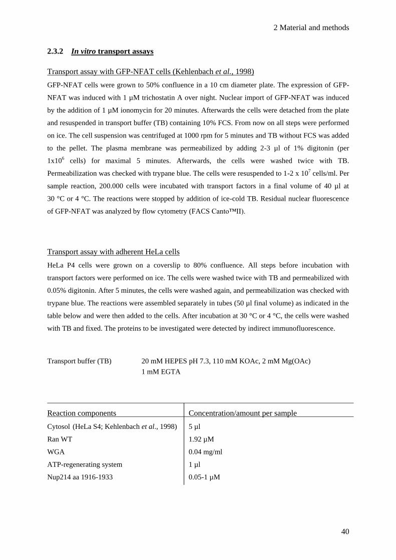
2 Material and methods
40
2.3.2 In vitro transport assays
Transport assay with GFP-NFAT cells (Kehlenbach et al., 1998)
GFP-NFAT cells were grown to 50% confluence in a 10 cm diameter plate. The expression of GFP-
NFAT was induced with 1 µM trichostatin A over night. Nuclear import of GFP-NFAT was induced
by the addition of 1 µM ionomycin for 20 minutes. Afterwards the cells were detached from the plate
and resuspended in transport buffer (TB) containing 10% FCS. From now on all steps were performed
on ice. The cell suspension was centrifuged at 1000 rpm for 5 minutes and TB without FCS was added
to the pellet. The plasma membrane was permeabilized by adding 2-3 µl of 1% digitonin (per
1x10⁶ cells) for maximal 5 minutes. Afterwards, the cells were washed twice with TB.
Permeabilization was checked with trypane blue. The cells were resuspended to 1-2 x 10⁷ cells/ml. Per
sample reaction, 200.000 cells were incubated with transport factors in a final volume of 40 µl at
30 °C or 4 °C. The reactions were stopped by addition of ice-cold TB. Residual nuclear fluorescence
of GFP-NFAT was analyzed by flow cytometry (FACS Canto™II).
Transport assay with adherent HeLa cells
HeLa P4 cells were grown on a coverslip to 80% confluence. All steps before incubation with
transport factors were performed on ice. The cells were washed twice with TB and permeabilized with
0.05% digitonin. After 5 minutes, the cells were washed again, and permeabilization was checked with
trypane blue. The reactions were assembled separately in tubes (50 µl final volume) as indicated in the
table below and were then added to the cells. After incubation at 30 °C or 4 °C, the cells were washed
with TB and fixed. The proteins to be investigated were detected by indirect immunofluorescence.
Transport buffer (TB) 20 mM HEPES pH 7.3, 110 mM KOAc, 2 mM Mg(OAc)
1 mM EGTA
Reaction components Concentration/amount per sample
Cytosol (HeLa S4; Kehlenbach et al., 1998) 5 µl
Ran WT 1.92 µM
WGA 0.04 mg/ml
ATP-regenerating system 1 µl
Nup214 aa 1916-1933 0.05-1 µM
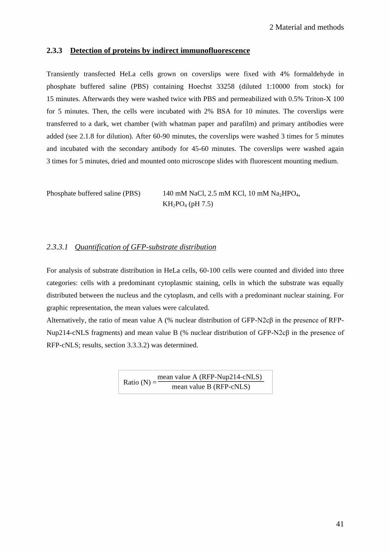
2 Material and methods
41
2.3.3 Detection of proteins by indirect immunofluorescence
Transiently transfected HeLa cells grown on coverslips were fixed with 4% formaldehyde in
phosphate buffered saline (PBS) containing Hoechst 33258 (diluted 1:10000 from stock) for
15 minutes. Afterwards they were washed twice with PBS and permeabilized with 0.5% Triton-X 100
for 5 minutes. Then, the cells were incubated with 2% BSA for 10 minutes. The coverslips were
transferred to a dark, wet chamber (with whatman paper and parafilm) and primary antibodies were
added (see 2.1.8 for dilution). After 60-90 minutes, the coverslips were washed 3 times for 5 minutes
and incubated with the secondary antibody for 45-60 minutes. The coverslips were washed again
3 times for 5 minutes, dried and mounted onto microscope slides with fluorescent mounting medium.
Phosphate buffered saline (PBS) 140 mM NaCl, 2.5 mM KCl, 10 mM Na2HPO4,
KH2PO4 (pH 7.5)
2.3.3.1 Quantification of GFP-substrate distribution
For analysis of substrate distribution in HeLa cells, 60-100 cells were counted and divided into three
categories: cells with a predominant cytoplasmic staining, cells in which the substrate was equally
distributed between the nucleus and the cytoplasm, and cells with a predominant nuclear staining. For
graphic representation, the mean values were calculated.
Alternatively, the ratio of mean value A (% nuclear distribution of GFP-N2cβ in the presence of RFP-
Nup214-cNLS fragments) and mean value B (% nuclear distribution of GFP-N2cβ in the presence of
RFP-cNLS; results, section 3.3.3.2) was determined.
mean value A (RFP-Nup214-cNLS)
mean value B (RFP-cNLS)
Ratio (N) =
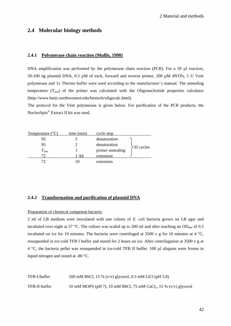
2 Material and methods
42
2.4 Molecular biology methods
2.4.1 Polymerase chain reaction (Mullis, 1990)
DNA amplification was performed by the polymerase chain reaction (PCR). For a 50 µl reaction,
50-100 ng plasmid DNA, 0.5 µM of each, forward and reverse primer, 200 µM dNTPs, 1 U Vent
polymerase and 1x Thermo buffer were used according to the manufacturer`s manual. The annealing
temperature (Tann) of the primer was calculated with the Oligonucleotide properties calculator
(http://www.basic.northwestern.edu/biotools/oligocalc.html).
The protocol for the Vent polymerase is given below. For purification of the PCR products, the
NucleoSpin® Extract II kit was used.
Temperature (°C) time (min) cycle step
95 5 denaturation
95 2 denaturation
Tann. 1 primer annealing
72 1 /kb extension
72 10 extension
2.4.2 Transformation and purification of plasmid DNA
Preparation of chemical competent bacteria
2 ml of LB medium were inoculated with one colony of E. coli bacteria grown on LB agar and
incubated over night at 37 °C. The culture was scaled up to 200 ml and after reaching an OD600 of 0.5
incubated on ice for 10 minutes. The bacteria were centrifuged at 3500 x g for 10 minutes at 4 °C,
resuspended in ice-cold TFB I buffer and stored for 2 hours on ice. After centrifugation at 3500 x g at
4 °C, the bacteria pellet was resuspended in ice-cold TFB II buffer. 100 µl aliquots were frozen in
liquid nitrogen and stored at -80 °C.
TFB-I-buffer 100 mM RbCl, 15 % (v/v) glycerol, 0.5 mM LiCl (pH 5.8)
TFB-II-buffer 10 mM MOPS (pH 7), 10 mM RbCl, 75 mM CaCl2, 15 % (v/v) glycerol
35 cycles

2 Material and methods
43
Transformation of chemical competent bacteria
100 µl of chemical competent E. coli DH5α bacteria were thawed on ice and 1 µg of plasmid DNA
was added. After incubation on ice, the bacteria were set to 42 °C for 60-90 seconds and transferred on
ice. After 2 minutes, the bacteria were supplied with 400 ml SOC medium and shaked gently for at
least 30 minutes at 37 °C. 50 µl of the whole suspension was plated on LB agarose containing
ampicillin or kanamycin.
Midi preparation
100-200 ml LB culture was inoculated with a single colony from an LB agar plate at 37 °C over night.
The bacteria were centrifuged for 10 minutes at 4000 rpm. For purification of the plasmid DNA, the
NucleoBond® PC100 or PC500 kit was used.
Mini preparation
Single colonies from an agar plate were transferred to 3 ml LB medium and grown over night at 37 °C.
1.5 ml of the culture was centrifuged for 5 minutes at 13000 rpm, and the bacteria pellet was
resuspended in 100 µl P1 buffer. The cells were lysed with 200 µl P2 buffer for 5 minutes at room
temperature. After neutralization of the suspension with 150 µl P3 buffer for 15 minutes on ice, the
lysate was centrifuged for 15 minutes at 4 °C. The supernatant containing the plasmid DNA was
collected, and 100% (v/v) ethanol was added to precipitate the DNA. After centrifugation for
15 minutes at 4 °C, the supernatant was discarded, the pellet was washed with 70% (v/v) ethanol, dried
and dissolved in TE buffer pH 7.5. Plasmid DNA was stored at -20 °C.
P1 buffer 50 mM Tris-HCl (pH 8), 10 mM EDTA, 100 μg/ml RNase A
P2 buffer 200 mM NaOH, 1% (v/v) SDS
P3 buffer 3 M KOAc (pH 5.3)
1x TE buffer 10 mM Tris pH 7.5, 1 mM EDTA

2 Material and methods
44
2.4.3 Agarose gel electrophoresis
Depending on the size of the DNA fragments, 0.8- 2.0 % agarose gels were prepared with 1x TAE
buffer and additional DNA stain (SERVA DNA stain G, diluted 1:100000). Restricted plasmid DNA
or PCR products were supplied with 1x DNA sample buffer and loaded on the agarose gel and
80-130 V was applied, depending on the size of the agarose gel. Afterwards, the gel was analyzed with
UV light.
50x TAE buffer 2 M Tris, 0.05 M EDTA pH 8, 5.8% acetic acid
2.4.4 Restriction of DNA by endonucleases
Restriction of plasmid DNA and DNA fragments was performed according to the manufacturer´s
manual. For analytical and preparative digestion, 2-4 µg plasmid DNA were incubated with
appropriate amounts of enzymes and buffers in a final volume of 20 µl at 37 °C for 1-2 hours. DNA
fragments were separated and analyzed by agarose gel electrophoresis. For ligation, DNA was cut out
from the agarose gel with a scalpel and purified with the NucleoSpin® Extract II kit.
2.4.5 Ligation of DNA fragments
Purified vector DNA and the insert DNA were added to the 10 µl ligation reaction in a 1:3 ratio. In
general, 10 ng vector DNA were used. The corresponding amounts of the insert DNA were calculated
with a ligation calculator (http://www.insilico.uni-duesseldorf.de/Lig_Input.html). The sample
reaction, containing 1x ligase buffer, 0.5 µM ATP and 0.5 µl T4 ligase (Fermentas), was incubated
over night at 16 °C. The complete sample was transformed into E. coli DH5α.
2 Material and methods
45
2.4.6 Oligonucleotide cloning
For insertion of short DNA fragments up to 60 bp into a vector, complementary oligonucleotides with
overhanging restriction sites were ordered. These restriction sites fit accurately into a vector digested
with the corresponding enzymes. 100 µM of the oligonucleotide were incubated in 50 µl annealing
buffer for 3 minutes at 95 °C. After that, the mixture was cooled down slowly and was incubated for
1 hour at 37 °C. In contrast to a PCR product, the annealed oligonucleotides are unphosphorylated. To
allow efficient ligation with the vector, 5 µl of the annealed oligonucleotide were incubated with
10 U T4 polynucleotide kinase and 200 µM ATP for 30 minutes at 37 °C. The reaction was stopped by
heating the sample for 10 minutes at 65 °C.
Annealing buffer (2x) 100 mM potassium acetate, 30 mM HEPES pH 7.4, 2 mM
magnesium acetate
2.4.7 DNA-sequencing
Sequencing of DNA was performed by either myGATC (Konstanz) or at the GZMB by using a
sequence analyzer (Genetic Analyzer 3100; Applied Biosystems). For the latter, the sequencing
reactions were prepared in the laboratory. 200-400 ng plasmid DNA, 10 pmol primers, 1 µl
sequencing mix, 2 µl sequencing buffer and sterile water were mixed in 10 µl final volume. Plasmid
DNA was amplified in a PCR program as listed below. Afterwards, 1 µl of each, 3 M Na(OAc) pH 5.2
and 125 mM EDTA, and 50 µl ice-cold ethanol (100%) was added and incubated for 5 minutes at
room temperature. The samples were centrifuged for 15 minutes at 13000 rpm and 4 °C and the
supernatant was removed. The pellet was washed once with 70 % (v/v) ethanol, dried and dissolved in
15 µl formamide.
PCR cycle protocol Temperature (°C) time (s) cycle step
96 120 denaturation
99 10 denaturation
55 15 primer annealing
60 240 extension
25 cycles

3 Results
46
Fig 8. Scheme of CRM1 structure. In this view, CRM1 consists of an N-terminal CRIME-domain,
which corresponds to the Ran (switch II) -binding region in transportin 1 and importin β (Fornerod et al.,
1997). The central conserved region (in accordance to the current model) contains the RanGTP-dependent
NES recognition site. The numbers in the C-terminal region represent the tandem HEAT repeats 14-19
(Petosa et al., 2004).
3 Results
3.1 In vivo analysis of CRM1, CRM1-fragments and chimeras
At the onset of the work for this thesis, CRM1-fragments were constructed and analyzed to investigate
the function of distinct regions. The CRM1 structure was only available for the C-terminus, which is
composed of HEAT repeat motifs (Petosa et al., 2004, see Fig. 8). The Ran binding region to CRM1
was not exactly determined and was based on sequence similarities to other transport factors like
importin β or transportin (CRIME domain; CRM1, IMportin β, Etcetera; Chook and Blobel, 1999). As
the function for the C-terminal part of CRM1 was not known, we used CRM1-fragments that were
truncated at the C-terminus. During the course of this project, the crystal structure of the trimeric
export complex was solved (Monecke et al., 2009, see introduction), showing CRM1 as a ring-like
protein. To maintain the ternary structure of full-length CRM1, chimeras consisting of H. sapiens
(Homo sapiens) and C. elegans (Caenorhabditis elegans) CRM1 were constructed and analyzed.
C. elegans CRM1 is 60% homologous to H. sapiens CRM1. Alignments revealed that especially the
NES-binding region is almost identical to that of human CRM1. Furthermore, C. elegans CRM1 is
able to interact with human RanGTP and an NES-peptide (Roloff, Diploma thesis). Concluded from
that, CRM1-chimeras consisting of H. sapiens and C. elegans CRM1 should fulfil all requirements for
the assembly of the trimeric export complex in the nucleus of mammalian cells. One striking
difference to H. sapiens CRM1 is the inability of C. elegans CRM1 to interact with human nucleo-
porins (Roloff, 2008, Diploma thesis).
3 Results
47
3.1.1 Distribution of nuclear export cargos after Leptomycin B treatment of HeLa cells
Leptomycin B (LMB) is an unsaturated fatty acid with a terminal δ-lactone ring. It covalently modifies
cysteine 528 in CRM1, thus preventing cargo binding. As a consequence, the trimeric export complex
cannot assemble, and export cargos remain in the nucleus. In order to identify suitable export cargos
for further analysis, HeLa cells were transfected with plasmids coding for different proteins and
treated with LMB. This approach had already been used for GFP-NES (Roloff, 2008, Diploma thesis),
and was now extended for quantification of GFP-SPN1 (snurportin 1), GFP-NC2β (negative cofactor
2β), GFP-Nmd3 and HA-DDX3. SPN1 is an m3G-cap receptor and imports m3G-capped uridine-rich
small nuclear RNPs (U snRNPs; Huber et al., 1998). It has an importin β binding (IBB) domain but it
does not share the structural similarity to the importin α arm repeat domain. SPN1 is recycled back to
the cytoplasm with the help of CRM1 in a trimeric export complex (Paraskeva et al., 1999). The
transcription factor NC2ß forms a heterodimer with NC2α and regulates the polymerase II
transcription machinery by either repression or activation of transcription (Geisberg et al., 2001; Cang
et al., 2002). NC2β contains a classical nuclear localization signal (NLS) as well as an NES
comparable to other known export cargos. Furthermore, it interacts with importin α/ß and CRM1 and
is sensitive to LMB treatment (Kahle et al., 2009). Nmd3 acts as an adapter protein between the 60S
preribosomal subunit and CRM1 (Thomas and Kutay, 2003; Trotta et al., 2003). It also contains basic
and non-basic regions (but no classical NLS) mediating nuclear import. DDX3 belongs to the DEAD
(D-E-A-D = Asp-Glu-Ala-Asp) box RNA helicase family. It facilitates nuclear export of HIV RNAs
via CRM1 and also shuttles back to the nucleus (Yedavalli et al., 2004).
After transfection and LMB treatment, HeLa cells were fixed and analyzed by immunofluorescence
microscopy. In untreated cells, GFP-NES was located in the cytoplasm, but in cells treated with LMB
it accumulated in the nucleus (see Fig. 9A). As the construct does not contain an NLS, it is still able to
diffuse freely in the cell. Hence, there was also a slight cytoplasmic signal for GFP-NES after LMB
treatment (see arrow). The distribution of the other tested proteins is shown in Fig. 9B. As expected,
localization of all of them was affected by LMB, though the extent of accumulation differed from
protein to protein. In nearly all cells, GFP-NES, GFP-NC2β and GFP-SPN1 were located in the
nucleus, whereas there was only a mild effect of LMB on GFP-Nmd3 (approximately 50% of the cells
showed nuclear accumulation). The amount of cells with cytoplasmic HA-DDX3 was reduced from
100% to only 65% after LMB treatment.
As GFP-NES, GFP-NC2β and GFP-SPN1 showed the strongest LMB-effect, they were chosen for
further analysis of CRM1 and also used in the second part of this thesis (section 3.2).
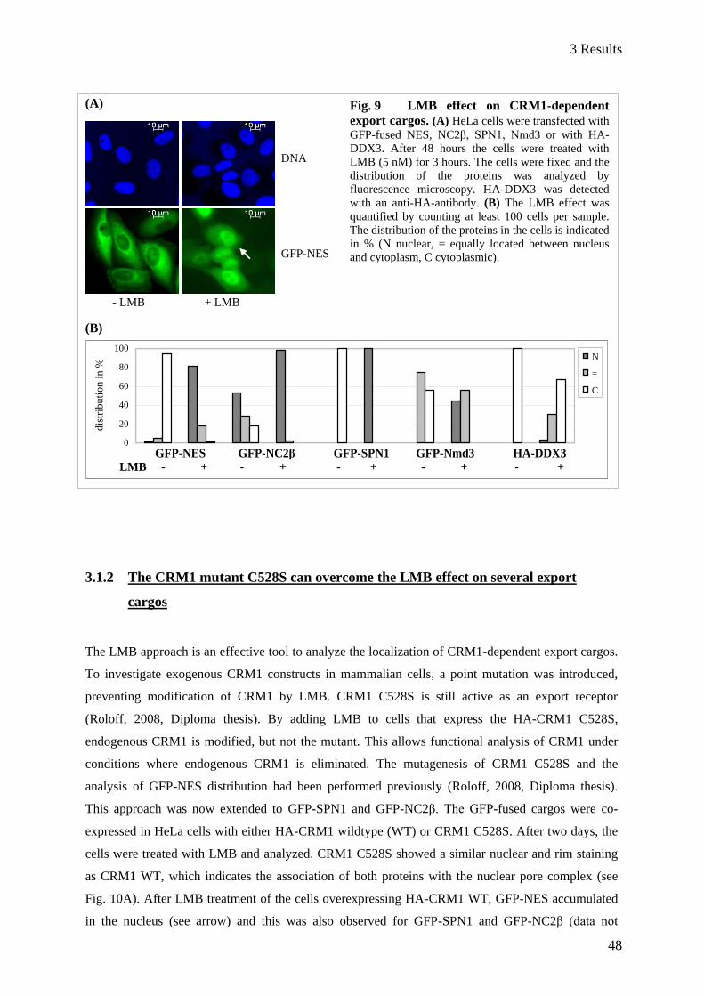
3 Results
48
DNA
GFP-NES
- LMB + LMB
(A)
(B)
3.1.2 The CRM1 mutant C528S can overcome the LMB effect on several export
cargos
The LMB approach is an effective tool to analyze the localization of CRM1-dependent export cargos.
To investigate exogenous CRM1 constructs in mammalian cells, a point mutation was introduced,
preventing modification of CRM1 by LMB. CRM1 C528S is still active as an export receptor
(Roloff, 2008, Diploma thesis). By adding LMB to cells that express the HA-CRM1 C528S,
endogenous CRM1 is modified, but not the mutant. This allows functional analysis of CRM1 under
conditions where endogenous CRM1 is eliminated. The mutagenesis of CRM1 C528S and the
analysis of GFP-NES distribution had been performed previously (Roloff, 2008, Diploma thesis).
This approach was now extended to GFP-SPN1 and GFP-NC2β. The GFP-fused cargos were co-
expressed in HeLa cells with either HA-CRM1 wildtype (WT) or CRM1 C528S. After two days, the
cells were treated with LMB and analyzed. CRM1 C528S showed a similar nuclear and rim staining
as CRM1 WT, which indicates the association of both proteins with the nuclear pore complex (see
Fig. 10A). After LMB treatment of the cells overexpressing HA-CRM1 WT, GFP-NES accumulated
in the nucleus (see arrow) and this was also observed for GFP-SPN1 and GFP-NC2β (data not
0
20
40
60
80
100
dis
trib
uti
on
in
%
N
=
C
Fig. 9 LMB effect on CRM1-dependent
export cargos. (A) HeLa cells were transfected with
GFP-fused NES, NC2β, SPN1, Nmd3 or with HA-
DDX3. After 48 hours the cells were treated with
LMB (5 nM) for 3 hours. The cells were fixed and the
distribution of the proteins was analyzed by
fluorescence microscopy. HA-DDX3 was detected
with an anti-HA-antibody. (B) The LMB effect was
quantified by counting at least 100 cells per sample.
The distribution of the proteins in the cells is indicated
in % (N nuclear, = equally located between nucleus
and cytoplasm, C cytoplasmic).
GFP-NES GFP-NC2β GFP-SPN1 GFP-Nmd3 HA-DDX3
LMB - + - + - + - + - +

3 Results
49
DNA
HA-CRM1
GFP-NES
CRM1 WT CRM1 C528S
Fig. 10 CRM1 C528S allows nuclear export of CRM1-dependent cargos
upon LMB treatment. HeLa cells were cotransfected with plasmids coding for
GFP-tagged NES, SPN1 or NC2β and HA-CRM1 (wildtype WT or the C528S
mutant). After 48 hours, the cells were treated with LMB (5 nM) for 3 hours.
Afterwards, the cells were fixed and stained for HA-CRM1 using an HA-antibody.
The localization of GFP-NES in HA-CRM1 overexpressing HeLa cells is shown in
(A). A part of these data is taken from Roloff, 2008 (Diploma thesis). The localization
of the GFP-tagged proteins was quantified by counting at least 60 cells expressing
HA-CRM1 and is given in % (N nuclear, = equally distributed between nucleus and
cytoplasm, C cytoplasmic) (B). Error bars indicate the standard deviation from the
mean of three independent experiments.
shown). Not only endogenous CRM1 is modified by LMB but also the transfected HA-CRM1. In the
presence of HA-CRM1 C528S, GFP-NES was located in the cytoplasm, and this localization was
nearly unaffected after LMB treatment (Fig. 10A, right panel, see arrow; and Fig. 10B).
(A)
(B)
Basically, HA-CRM1 C528S had the same effect on localization of GFP-SPN1 and GFP-NC2β. GFP-
SPN1 was cytoplasmic in all HeLa cells co-expressing the HA-CRM1 mutant, and after LMB
0
20
40
60
80
100
dis
trib
uti
on i
n %
N
=
C
- LMB + LMB - LMB + LMB
GFP-NES GFP-SPN1 GFP-NC2β
LMB - + - + - +
3 Results
50
treatment the cargo was still in the cytoplasm (Fig. 10B). Without LMB, GFP-NC2β was mainly
cytoplasmic (80% of the cells) in the presence of the CRM1 mutant. In cells treated with LMB, GFP-
NC2β distributed mostly equally between the nucleus and the cytoplasm. Compared to localization of
GFP-NC2β alone (Fig. 9B), it becomes clear that CRM1 is rate-limiting for nuclear export of the
cargo. This has previously been shown for artificial reporter substrates (Waldmann et al., 2012).
Together, the CRM1 C528S mutant is functional, and combined with LMB it is an effective tool for
analysis of CRM1 variants.
3.1.3 CRM1-fragments are not able to mediate nuclear export of proteins
CRM1-fragments containing the C528S mutation were constructed and analyzed in order to specify
regions for interaction partners of CRM1 and to understand which regions in CRM1 are important for
its function in nuclear export. According to Petosa et al. (2004), the N-terminal region of CRM1 was
thought to have the Ran-binding capability and the central region was implicated in export cargo
binding, but the function of the C-terminus of CRM1 remained unclear. Moreover, the binding
region(s) for nucleoporins like Nup214 in CRM1 are not known.
3.1.3.1 Localization of CRM1-truncations
Full-length CRM1 associates with the nuclear pore, but it was not known where CRM1-fragments
would localize in the cell. To test this, plasmids coding for CRM1-fragments containing the C528S
mutation were constructed (Fig. 11A) and used for transfection of HeLa cells. After two days, the
cells were fixed and stained (Fig. 11B). Whereas CRM1 containing HEAT repeats (HR) 1-13 showed
a considerable amount of cytoplasmic staining (see asterisk), CRM1 HR 1-16 and 1-18 were almost
completely in the nucleus with a clear boundary to the cytoplasm. This suggests that the ability of
CRM1 to locate to the nucleus increases with the number of HEAT repeats. However, only full-length
CRM1 showed a rim staining around the nuclear envelope (see arrow). This points to the association
of full-length CRM1 with the nuclear pore and thus, to the interaction of CRM1 with FG-rich
nucleoporins like Nup214, Nup153 or p62. This led to the assumption that the tested CRM1-
fragments might not be able to interact with nucleoporins. In coimmunoprecipitation experiments the
binding between endogenous nucleoporins and the HA-CRM1-truncations was analyzed but no
interaction could be detected, in contrast to full-length HA-CRM1 (data not shown). As RanGTP
enhances the association of CRM1 to nucleoporins, the binding of these fragments to nucleoporins
could be indirectly affected due to disturbed Ran interaction. Hence, the binding regions for

3 Results
51
Fig. 11 Only full-length CRM1 is concentrated at the nuclear pore. (A) Schematic view of CRM1-truncations. The CRM1-fragments used in this work were
constructed according to the C-terminal structure that was available at that time (Petosa et
al., 2004). The numbers in the bars represent amino acid boundaries. HR 1-13, 1-16 and 1-18
indicate the number of the residual N-terminal HEAT repeats. (B) HeLa cells were
transfected with full-length or C-terminal truncated HA-CRM1. After 48 hours, the cells
were fixed and stained using an HA- antibody. The cells were analyzed by
immunofluorescence microscopy. CRM1 1-709 shows considerable amounts of cytoplasmic
staining (see asterisk). The arrow indicates a rim staining that is only present in cells
overexpressing full-length CRM1.
nucleoporins could be located at the N-terminal or central part of CRM1 but cannot be detected by in
vitro binding studies, using CRM1-fragments.
(A)
(B)
1 970
1 1071
HR 1-13 HR 1-16 HR 1-18
full-length
C528S
CRM1 1-709 CRM1 1-861 CRM1 1-970 CRM1 fl
HA-CRM1
DNA
*
1 709
1 861
10 µm

3 Results
52
Fig. 12 C-terminal CRM1-truncations are not able to mediate nuclear export. HeLa cells were cotransfected with plasmids coding for GFP-NES and HA-CRM1-
fragments. After 48 hours, the cells were treated with LMB (5 nM) for 3 hours. Afterwards,
the cells were fixed, stained for HA-CRM1 using an HA-antibody and analyzed by
immunofluorescence microscopy. Because of low transfection efficiency of the CRM1-
fragments, the localization of GFP-NES was not quantified.
+ LMB + LMB + LMB + LMB
3.1.3.2 Only full-length CRM1 is able to mediate export through the nuclear pore
In order to test the export capability of the HA-CRM1 C528S fragments, HeLa cells were
cotransfected with plasmids coding for GFP-NES and the CRM1-fragments as indicated in Fig 11A.
After two days, the cells were treated with LMB. Only full-length CRM1 was able to transport GFP-
NES out of the nucleus (Fig. 12, see arrow) whereas in all cells overexpressing CRM1-fragments,
GFP-NES accumulated in the nucleus. As already mentioned in the section above, the C-terminus of
CRM1 might be required for proper ring closure. In turn, a disturbed ring structure of CRM1 could be
the reason for the inability of CRM1-fragments to mediate nuclear export.
DNA
HA-CRM1
C528S
GFP-NES
1-709 1-861 1-970 full length CRM1
10 µm

3 Results
53
3.1.4 CRM1-chimeras as an alternative approach to study export capabilities in vivo
The previously solved structure of the CRM1-RanGTP-cargo complex (Monecke et al., 2009),
showing CRM1 as a ring-like protein, indicated why CRM1-fragments are not functional in nuclear
export. The structure revealed a complex binding of RanGTP to several HEAT repeats at the N- and
at the C-terminus of CRM1. Regarding this, RanGTP binding to CRM1-fragments lacking the
C-terminus might indeed be affected. Hence, there might be problems with forming a functional
trimeric export complex and also binding of CRM1 to nucleoporins could be disturbed. To avoid such
problems, full-length chimeras consisting of H. sapiens and C. elegans CRM1 were constructed
accordingly to the secondary structure. The usage of H. sapiens/C. elegans CRM1-chimeras allows
the following assumptions:
1. The ring structure of CRM1 stays preserved.
2. The cooperativity of CRM1-Ran binding is not disturbed.
3. The CRM1-binding and the release of the export cargo can occur.
3.1.4.1 C. elegans CRM1 promotes export of a simple GFP-NES, but is unable to export
more complex cargos out of the nucleus
To test whether C. elegans CRM1 is functional in nuclear export in human cells, HeLa cells were
cotransfected with plasmids coding for C. elegans HA-CRM1 C538S (corresponding to the C528S
mutation in H. sapiens CRM1) and GFP-NES, GFP-SPN1 or GFP-NC2β, respectively. After two
days, the cells were treated with LMB. The localization of GFP-NES in HeLa cells in the presence of
C. elegans CRM1 was already investigated in Roloff, 2008 (Diploma thesis). The quantification of
GFP-NES distribution was completed in this work and extended to other GFP-substrates. In cells
expressing C. elegans HA-CRM1 mutant, all tested GFP-cargos were mostly located in the cytoplasm
(Fig. 13). After LMB treatment, GFP-NES distributed equally between the nucleus and the cytoplasm
in 80% of the cells. Thus, C. elegans CRM1 promotes export of GFP-NES. Strikingly, export of more
complex cargos was not supported by C. elegans CRM1, as GFP-SPN1 and GFP-NC2β accumulated
in all cells overexpressing C. elegans CRM1 after LMB treatment. In cells expressing H. sapiens
CRM1 C528S (see Fig. 10A and B), LMB had only a very weak effect on the distribution of all
investigated cargos. By contrast, the LMB effect in cells expressing C. elegans CRM1 was mild on
GFP-NES, but very strong on distribution of GFP-SPN1 and NC2β. We know from the crystal
structure of the trimeric export complex that SPN1 binds to the NES-groove, but also to other sites in

3 Results
54
Fig. 13 C. elegans CRM1 promotes nuclear export of GFP-
NES, but not of GFP-SPN1 and GFP-NC2β. HeLa cells were
cotransfected with plasmids coding for C. elegans HA-CRM1 C538S
and GFP-NES, GFP-SPN1 or GFP-NC2β, respectively. After 48 hours,
the cells were treated with LMB (5 nM) for 3 hours. Afterwards, the
cells were fixed, stained for HA-CRM1 using an HA-antibody and
analyzed by immunofluorescence microscopy. The localization of the
GFP-substrates in HA-CRM1 expressing cells is given in %. At least 60
cells were counted (N nuclear, = equally distributed between nucleus and
cytoplasm, C cytoplasmic).
CRM1. Probably C. elegans CRM1 is able to form a trimeric export complex with the NES, but is
lacking other binding sites for SPN1. The binding mechanism of NC2β to CRM1 is not known.
Aside from that, coimmunoprecipitation experiments showed that C. elegans CRM1 is not able to
interact with the human nucleoporins Nup214 and p62 (Roloff, 2008, Diploma thesis). This might be
another reason why the export capability is restricted for C. elegans CRM1 in mammalian cells.
Based on these data, the usage of chimeras consisting of human and C. elegans CRM1 could give
more detailed information about regions in H. sapiens CRM1 that are required for nuclear export of
proteins.
0
20
40
60
80
100
dis
trib
uti
on
in
%
N
=
C
GFP-NES GFP-SPN1 GFP-NC2β
LMB - + - + - +
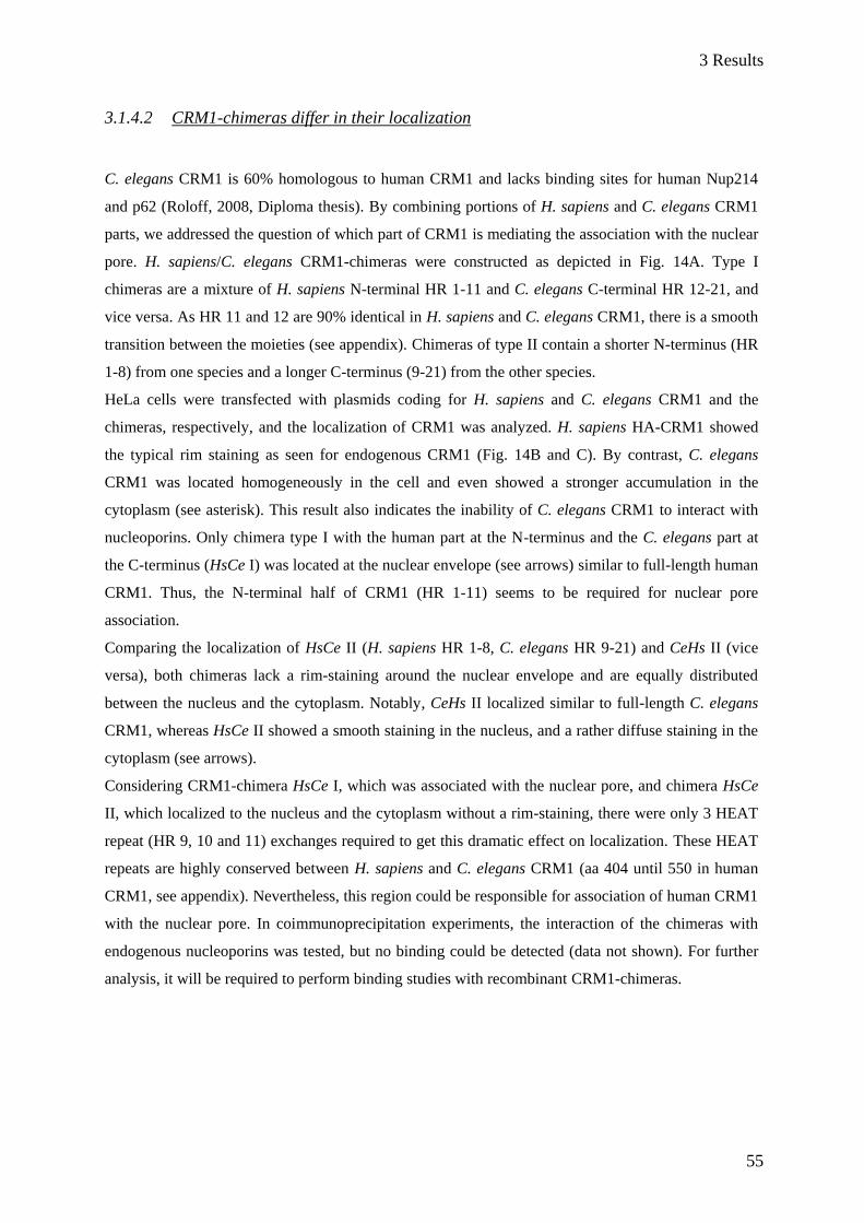
3 Results
55
3.1.4.2 CRM1-chimeras differ in their localization
C. elegans CRM1 is 60% homologous to human CRM1 and lacks binding sites for human Nup214
and p62 (Roloff, 2008, Diploma thesis). By combining portions of H. sapiens and C. elegans CRM1
parts, we addressed the question of which part of CRM1 is mediating the association with the nuclear
pore. H. sapiens/C. elegans CRM1-chimeras were constructed as depicted in Fig. 14A. Type I
chimeras are a mixture of H. sapiens N-terminal HR 1-11 and C. elegans C-terminal HR 12-21, and
vice versa. As HR 11 and 12 are 90% identical in H. sapiens and C. elegans CRM1, there is a smooth
transition between the moieties (see appendix). Chimeras of type II contain a shorter N-terminus (HR
1-8) from one species and a longer C-terminus (9-21) from the other species.
HeLa cells were transfected with plasmids coding for H. sapiens and C. elegans CRM1 and the
chimeras, respectively, and the localization of CRM1 was analyzed. H. sapiens HA-CRM1 showed
the typical rim staining as seen for endogenous CRM1 (Fig. 14B and C). By contrast, C. elegans
CRM1 was located homogeneously in the cell and even showed a stronger accumulation in the
cytoplasm (see asterisk). This result also indicates the inability of C. elegans CRM1 to interact with
nucleoporins. Only chimera type I with the human part at the N-terminus and the C. elegans part at
the C-terminus (HsCe I) was located at the nuclear envelope (see arrows) similar to full-length human
CRM1. Thus, the N-terminal half of CRM1 (HR 1-11) seems to be required for nuclear pore
association.
Comparing the localization of HsCe II (H. sapiens HR 1-8, C. elegans HR 9-21) and CeHs II (vice
versa), both chimeras lack a rim-staining around the nuclear envelope and are equally distributed
between the nucleus and the cytoplasm. Notably, CeHs II localized similar to full-length C. elegans
CRM1, whereas HsCe II showed a smooth staining in the nucleus, and a rather diffuse staining in the
cytoplasm (see arrows).
Considering CRM1-chimera HsCe I, which was associated with the nuclear pore, and chimera HsCe
II, which localized to the nucleus and the cytoplasm without a rim-staining, there were only 3 HEAT
repeat (HR 9, 10 and 11) exchanges required to get this dramatic effect on localization. These HEAT
repeats are highly conserved between H. sapiens and C. elegans CRM1 (aa 404 until 550 in human
CRM1, see appendix). Nevertheless, this region could be responsible for association of human CRM1
with the nuclear pore. In coimmunoprecipitation experiments, the interaction of the chimeras with
endogenous nucleoporins was tested, but no binding could be detected (data not shown). For further
analysis, it will be required to perform binding studies with recombinant CRM1-chimeras.
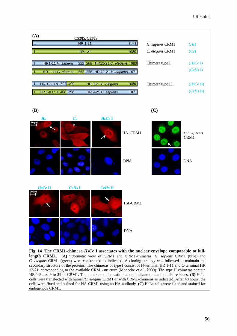
3 Results
56
HA-CRM1
DNA
Hs Ce HsCe I
HA- HA- CRM1 endogenous
CRM1
DNA DNA
HR 12-21 H. sapiens HR 1-11 C. elegans
HR 1-8 H.s.
HR1-11 H. sapiens
HR1-21
HR 1-21
HR 9-21 C. elegans
HR12-21 C. elegans
HR 9-21 H. sapiens HR 1-8 C. e.
H. sapiens CRM1 (Hs)
C. elegans CRM1 (Ce)
Chimera type I (HsCe I)
(CeHs I)
Chimera type II (HsCe II)
(CeHs II)
1 1071
1 1080
1 555 566 1080
1 565 556 1071
1 397 409 1080
1 408 398 1071
C528S/C538S
10 µM
HsCe II CeHs I CeHs II
10 µM
(A)
(B) (C)
Fig. 14 The CRM1-chimera HsCe I associates with the nuclear envelope comparable to full-
length CRM1. (A) Schematic view of CRM1 and CRM1-chimeras. H. sapiens CRM1 (blue) and
C. elegans CRM1 (green) were constructed as indicated. A cloning strategy was followed to maintain the
secondary structure of the proteins. The chimeras of type I consist of N-terminal HR 1-11 and C-terminal HR
12-21, corresponding to the available CRM1-structure (Monecke et al., 2009). The type II chimeras contain
HR 1-8 and 9 to 21 of CRM1. The numbers underneath the bars indicate the amino acid residues. (B) HeLa
cells were transfected with human/C. elegans CRM1 or with CRM1-chimeras as indicated. After 48 hours, the
cells were fixed and stained for HA-CRM1 using an HA-antibody. (C) HeLa cells were fixed and stained for
endogenous CRM1.
*
*

3 Results
57
3.1.4.3 The export capability of CRM1-chimeras correlates with their localization in HeLa
cells
To investigate the functionality of the CRM1-chimeras (containing the C528S/C538S mutation), they
were cotransfected with plasmids coding for the GFP-fused substrates NES, SPN1 and NC2β in HeLa
cells. After two days, the cells were treated with LMB to inhibit endogenous CRM1 and analyzed by
immunofluorescence microscopy. In cells expressing functional HA-CRM1, LMB is expected to have
no or very mild effects in the localization of cargo-proteins, since the exogenous CRM1 is insensitive
to the drug. The localization of GFP-NES was cytoplasmic in HeLa cells co-expressing CRM1-
chimera HsCe I, and after LMB treatment GFP-NES was still cytoplasmic in 70 % of the cells (Fig.
15A and B). The cytoplasmic localization of GFP-SPN1 was not affected at all in LMB-treated cells.
Strikingly, GFP-NC2β accumulated in the nucleus in the presence of chimera HsCe I after LMB
treatment, suggesting a specific binding mechanism to CRM1 as described for SPN1. In contrast to
CRM1-chimera HsCe I, all tested GFP-cargos accumulated in the nucleus in the presence of the
chimera CeHs I and LMB treatment, suggesting that the N-terminal portion of CRM1 is required for
CRM1-functions in mammalian cells.
LMB-treated HeLa cells overexpressing chimeras type II (Fig. 16A and B) showed equal distribution
of GFP-NES between the nucleus and the cytoplasm (and nuclear GFP-NC2β, see appendix, Fig. S1).
Therefore, these chimeras are clearly restricted in their export capability. Interestingly, in LMB-
treated cells overexpressing the CRM1-chimera HsCe II, the export cargo had a similar distribution
pattern like the chimera itself (see asterisk).
In general, the localization of the chimeras correlates with their ability to promote nuclear export.
Based on this, the association with the nuclear envelope could be required for CRM1 to fulfill its
function as an export receptor in mammalian cells. With respect to cargo interaction, the C-terminus
of C. elegans CRM1 (HR 1-11) can substitute for the C-terminus of H. sapiens CRM1 for SPN1 or a
simple NES in mammalian cells, but not for NC2β.
In summary, only the CRM1-chimera HsCe I associated with the nuclear envelope and was able to
promote nuclear export in mammalian cells for a subset of proteins (Table 2). The ability for
association with the nuclear envelope (NE) of this chimera got lost by the exchange of H. sapiens
HRs 9-11 into C. elegans CRM1 HRs (HsCe I HsCe II), but this could not be reconstituted by
extension of the human part of CRM1 C-terminally (CeHs I CeHs II). Taken together, HRs 9-11 of
CRM1 seem to be required for nuclear pore association, but there must be additional regions in
human CRM1 HR 1-7 that are required for functional nuclear export.
%

3 Results
58
10 µM
Fig. 15 The N-terminus of human
CRM1 is required for efficient nuclear
export. HeLa cells were cotransfected with
plasmids coding for HA-CRM1-chimeras type
I as indicated in Fig. 11A and GFP-NES,
GFP-SPN1 or GFP-NC2β. After 48 hours, the
cells were treated with LMB (5 nM) for 3
hours. Afterwards, the cells were fixed,
stained for HA-CRM using an HA-antibody
and analyzed by immunofluorescence
microscopy. (A) represents the typical
distribution of GFP-NES co-expressed with
CRM1-chimeras type I. (B) For quantifi-
cation, at least 60 cells were counted per
sample. The distribution of the tested proteins
GFP-NES, GFP-SPN1 and GFP-NC2β is
given in % (N nuclear, = equally distributed,
C cytoplasmic). Error bars indicate the
variation from the mean of two different
experiments.
(A) Chimeras type I
(B)
(B)
HR1-11 C. elegans HR12-21 H. sapiens
DNA
HA-CRM1
HsCe I (C528S)
GFP-NES
HR1-11 H. sapiens HR12-21 C. elegans
%
10 µM 10 µM
- LMB + LMB - LMB + LMB
0
20
40
60
80
100
dis
trib
uti
on
in
% N
=
C
GFP-NES GFP-SPN1 GFP-
NC2β
+ HsCe I (C528S) LMB - + - + - +
0
20
40
60
80
100
dis
trib
uti
on
in
% N
=
C
+ CeHs I (C538S) LMB - + - + - +
GFP-NES GFP-SPN1 GFP-NC2β
DNA
HA-CRM1
CeHs I (C538S)
GFP-NES

3 Results
59
Fig. 16 CRM1-chimeras type II are restricted in their export capability. HeLa cells were
cotransfected plasmids for CRM1-chimeras type II as indicated in Fig. 11A and GFP-NES. After 48 hours,
the cells were treated with LMB (5 nM) for 3 hours. Afterwards, the cells were fixed, stained for HA-CRM
using an HA-antibody and analyzed by immunofluorescence microscopy. The pictures in (A) represent the
typical distribution of GFP-NES co-expressed with CRM1-chimeras type II. (B) For quantification, at least
60 cells were counted per sample. The distribution of GFP-NES is given in % (N nuclear, = equally
distributed, C cytoplasmic). Error bars indicate the variation from the mean of two different experiments.
(A)
(B)
CRM1
Localization
Rim around the
nucleus
Promoted nuclear export of
NES SPN1 NC2β
H. sapiens N + + + +
C. elegans N/C - + - -
HsCe I (HR 1-11/12-21) N + + + -
CeHs I (HR 1-11/12-21) N/C - - - -
HsCe II (HR 1-8/9-21) N/C - - - -
CeHs II (HR 1-8/9-21) N/C - -/+ - -
Table 2 Summary of analyzed CRM1-chimeras and export substrates.
HR 1-8 H. sapiens HR9-21 C. elegans HR1-8 C. elegans HR9-21 H. sapiens
- LMB + LMB - LMB + LMB
DNA
HA-CRM1
HsCe II (C538S)
* GFP-NES
DNA
HA-CRM1
CeHs II (C528S)
GFP-NES
10 µM 10 µM
0
20
40
60
80
100
dis
trib
uti
on i
n % N
=
C
0
20
40
60
80
100
dis
trib
uti
on
in
% N
=
C
GFP-NES GFP-NES
+ HsCe II (C538S) LMB - +
+ CeHs II (C528S)
LMB - +
3 Results
60
CRM1 H. sapiens
CRM1 D. melanogaster
CRM1 C. elegans
8B
9A
CRM1 H. sapiens
CRM1 D. melanogaster
CRM1 C. elegans
Fig. 17 CRM1 contains a non-conservative loop
between HEAT repeat 8B and 9A. The alignment
reveals an unconserved sequence between HR 8B and 9A
(marked with the black frame). The structure on the left hand
(modified from Monecke et al., 2009) shows this sequence
as a flexible loop.
3.1.4.4 The deletion of an outer surface loop between HEAT repeat 8B and 9A of CRM1
does not affect nuclear export
The differences in localization and functionality observed for the CRM1-chimeras led to the
assumption that the responsible region for that is located in HR 9-11. The alignment of H. sapiens
CRM1, C. elegans CRM1 and also D. melanogaster (Drosophila melanogaster) CRM1 revealed an
unconserved sequence directly before HR 9 (Fig. 17, black frame). In the CRM1-structure, this
sequence extends from the molecule as a flexible loop (Monecke et al., 2009).
To investigate this region in CRM1, a mutant lacking aa 384-403 was constructed (containing the
C528S mutation). A plasmid coding for this CRM1 mutant (named CRM1 C528SΔloop) was
transfected into HeLa cells and the location of GFP-NES and GFP-NC2β was analyzed in co-
expressing cells after LMB treatment. For better comparison, the distribution of the GFP-proteins
alone or in the presence of CRM1 C528S or CRM1 C528SΔloop is summarized in Fig. 18B and C.
Like WT CRM1, the C528SΔloop mutant localized to the nucleus and at the nuclear envelope. GFP-
NES localization was cytoplasmic after LMB treatment (Fig. 18A), as it was observed for cells co-
expressing CRM1 C528S. Moreover, overexpressed CRM1 C528SΔloop promoted nuclear export of
NC2β in untreated cells, as it has been described for CRM1 containing the loop (summarized in Fig.
18C, also compare Fig. 9B and 10B). Thus the loop between HR 8B and 9A is not required for export
of proteins.
loop -A-
-B-

3 Results
61
Fig. 18 CRM1 C528SΔloop is able to export cargos out of the nucleus. HeLa cells were
transfected with plasmids coding for HA-CRM1 C528S mutant or with HA-CRM1 C528S Δloop together
with GFP-NES (A, B) or GFP-NC2β (C). After 48 hours, the cells were treated with LMB (5 nM) for 3
hours, fixed, stained for HA-CRM1 and analyzed by immunofluorescence microscopy. The distribution was
quantified by counting 60-100 cells per sample.
0
20
40
60
80
100
+ LMB + LMB + LMB
GFP-Nc2ß +CRM1 C528S + CRM1 C528S
Δloop
GFP-Nc2ß +CRM1 C528S + CRM1 C528S
Δloop
dis
trib
uti
on
in
% N
=
C
(A)
(B)
(C)
Regarding the data obtained from CRM1-chimeras and the loop deletion mutant, we assume that one
or more regions in HR 9-11 in human CRM1 are required for interaction with nucleoporins and thus
are also necessary for the function of CRM1 as an export receptor. These regions should differ
between H. sapiens and C. elegans CRM1, as C. elegans CRM1 cannot interact with human
nucleoporins. To prove this assumption, it will be required to create and analyze CRM1 with
10 µM
CRM1 C528S
DNA Δloop GFP-NES
- LMB
+ LMB
0
20
40
60
80
100
+ LMB + LMB + LMB
GFP-NES + CRM1 C528S + CRM1 C528S
Δloop
GFP-NES + CRM1 C528S + CRM1 C528S
Δloop
dis
trib
uti
on
in
% N
=
C
GFP-NES
GFP-NC2β
10 µM
3 Results
62
mutations in this region and more CRM1-chimeras with step-wise addition of human HEAT repeats in
this region (e.g. chimeras 1-9/10-21 or 1-10/11-21).
3.2 Determination of a minimal region in Nup214 required for CRM1-
binding
Little is known about nucleoporin-CRM1 interaction in general. So far, the amino acid residues
1859-2090 of Nup214 have been identified to bind CRM1 in a RanGTP-dependent manner (Fornerod
et al., 1996). Furthermore, the FG-repeats of Nup214 are assumed to be responsible for this binding,
but so far there is no evidence for that. Here, we analyzed the binding of Nup214 aa 1859-2090 to
CRM1 by using short Nup214-fragments and -mutants. To narrow down the Nup214-CRM1-
interaction site, in vivo analysis was combined with binding assays.
3.2.1 Function of Nup214 C-terminal fragments in nuclear export
Overexpression of Myc-Nup214 aa 1859-2090 cells led to apoptosis, growth defects, to nuclear
accumulation of CRM1 and also to nuclear accumulation of Poly (A)+ RNA (Boer et al., 1998). This
led to the hypothesis that CRM1 accumulates in the nucleus because of its strong binding to
C-terminal Nup214, and hence, would not be available for export cargos. We now set out to analyze
the localization of other CRM1-dependent export cargos in cells overexpressing C-terminal Myc-
Nup214. HeLa cells were transfected with plasmids coding for GFP-NES, -NC2β, -SPN1, -Nmd3 or
-TFIIAα and additionally with a plasmid coding for C-terminal Myc-Nup214. After 24 hours, the cells
were analyzed by immunofluorescence microscopy. Without C-terminal Myc-Nup214, most of the
GFP-cargos were located in the cytoplasm (Fig. 19A). GFP-Nmd3 distributed equally between the
nucleus and the cytoplasm. However, all tested cargos accumulated in the nucleus in the presence of
C-terminal Nup214 (Fig. 19B). In cells weakly expressing Myc-Nup214 aa 1859-2090, cargos also
distributed equally between the nucleus and the cytoplasm (see arrow).
Nuclear accumulation of the tested cargos further supports the assumption that endogenous CRM1 is
held back in the nucleus in the presence of the exogenous Nup214 C-terminus. As a consequence,
proteins that are imported remain in the nucleus. As this approach worked very well for the tested
export cargos, it was also used for analysis of shorter Nup214-fragments to narrow down the Nup214-
CRM1-interaction site.

3 Results
63
(A)
(B)
10 µM
DNA
GFP
NES NC2β SPN1 Nmd3 TFIIAα 10 µM
NES NC2β SPN1 Nmd3 TFIIAα
Fig. 19 Nuclear export inhibition by Myc-Nup214 1859-2090. (A) HeLa cells were
transfected with plasmids coding for GFP-NES, -NC2β, -SPN1, -Nmd3 or -TFIIa and (B)
additionally with a plasmid coding for Myc-Nup214 aa 1859-2090. After 24 hours, the cells
were fixed, stained for Myc-Nup214 using a Myc-antibody and analyzed by immuno-
fluorescence microscopy.
DNA
Myc-Nup214
1859-2090
GFP
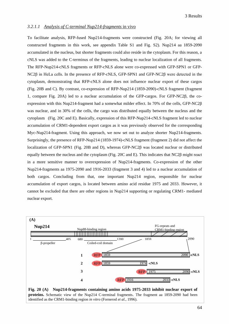
3 Results
64
3.2.1.1 Analysis of C-terminal Nup214-fragments in vivo
To facilitate analysis, RFP-fused Nup214-fragments were constructed (Fig. 20A; for viewing all
constructed fragments in this work, see appendix Table S1 and Fig. S2). Nup214 aa 1859-2090
accumulated in the nucleus, but shorter fragments could also reside in the cytoplasm. For this reason, a
cNLS was added to the C-terminus of the fragments, leading to nuclear localization of all fragments.
The RFP-Nup214-cNLS fragments or RFP-cNLS alone were co-expressed with GFP-SPN1 or GFP-
NC2β in HeLa cells. In the presence of RFP-cNLS, GFP-SPN1 and GFP-NC2β were detected in the
cytoplasm, demonstrating that RFP-cNLS alone does not influence nuclear export of these cargos
(Fig. 20B and C). By contrast, co-expression of RFP-Nup214 (1859-2090)-cNLS fragment (fragment
1, compare Fig. 20A) led to a nuclear accumulation of the GFP-cargos. For GFP-NC2β, the co-
expression with this Nup214-fragment had a somewhat milder effect. In 70% of the cells, GFP-NC2β
was nuclear, and in 30% of the cells, the cargo was distributed equally between the nucleus and the
cytoplasm (Fig. 20C and E). Basically, expression of this RFP-Nup214-cNLS fragment led to nuclear
accumulation of CRM1-dependent export cargos as it was previously observed for the corresponding
Myc-Nup214-fragment. Using this approach, we now set out to analyze shorter Nup214-fragments.
Surprisingly, the presence of RFP-Nup214 (1859-1974)-cNLS fragment (fragment 2) did not affect the
localization of GFP-SPN1 (Fig. 20B and D), whereas GFP-NC2β was located nuclear or distributed
equally between the nucleus and the cytoplasm (Fig. 20C and E). This indicates that NC2β might react
in a more sensitive manner to overexpression of Nup214-fragments. Co-expression of the other
Nup214-fragments aa 1975-2090 and 1916-2033 (fragment 3 and 4) led to a nuclear accumulation of
both cargos. Concluding from that, one important Nup214 region, responsible for nuclear
accumulation of export cargos, is located between amino acid residue 1975 and 2033. However, it
cannot be excluded that there are other regions in Nup214 supporting or regulating CRM1- mediated
nuclear export.
(A)
1
2
3
4
1859 2090 -cNLS
Nup88-binding region FG-repeats and
CRM1-binding region
β-propeller Coiled-coil domain
1 405 680 1200 1859 2090
Nup214
RFP
1859 1974 -cNLS
1975 2090 -cNLS
1916 2033 -cNLS
RFP
RFP
RFP
Fig. 20 (A) Nup214-fragments containing amino acids 1975-2033 inhibit nuclear export of
proteins. Schematic view of the Nup214 C-terminal fragments. The fragment aa 1859-2090 had been
identified as the CRM1-binding region in vitro (Fornerod et al., 1996).
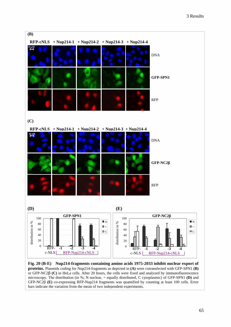
3 Results
65
10 µM
0
20
40
60
80
100
dis
trib
uti
on
in
%
N
=
C
(B)
(C)
(D) (E)
GFP-SPN1
10 µM
10 µM
DNA
GFP-NC2β
RFP
RFP-cNLS + Nup214-1 + Nup214-2 + Nup214-3 + Nup214-4
10 µM
DNA
GFP-SPN1
RFP
0
20
40
60
80
100
dis
trib
uti
on
in
%
N
=
C
GFP-SPN1 GFP-NC2β
RFP- -1 -2 -3 -4
c-NLS RFP-Nup214-cNLS
RFP-cNLS + Nup214-1 + Nup214-2 + Nup214-3 + Nup214-4
Fig. 20 (B-E) Nup214-fragments containing amino acids 1975-2033 inhibit nuclear export of
proteins. Plasmids coding for Nup214-fragments as depicted in (A) were cotransfected with GFP-SPN1 (B)
or GFP-NC2β (C) in HeLa cells. After 20 hours, the cells were fixed and analyzed by immunofluorescence
microscopy. The distribution (in %; N nuclear, = equally distributed, C cytoplasmic) of GFP-SPN1 (D) and
GFP-NC2β (E) co-expressing RFP-Nup214 fragments was quantified by counting at least 100 cells. Error
bars indicate the variation from the mean of two independent experiments.
RFP- -1 -2 -3 -4
c-NLS RFP-Nup214-cNLS

3 Results
66
3.2.1.2 Accumulation of CRM1 in the nucleus upon overexpression of Nup214-fragments
Overexpression of Nup214 aa 1859-2090 inhibits CRM1-dependent nuclear export and also leads to
nuclear accumulation of CRM1 (Boer et al., 1998). Based on that, shorter Nup214-fragments that
inhibit nuclear export should also lead to accumulation of CRM1 in the nucleus. In order to test this,
HeLa cells were transfected with Nup214-fragments as indicated in Fig. 20A. Except of RFP-Nup214
(1859-1974)-cNLS (fragment 2), all other fragments led to enrichment of CRM1 in the nucleus
(Fig. 21). Cells overexpressing Nup214-fragments 1, 3 and 4 were often smaller than untransfected
cells and also occurred pairwise. This indicates an arrest of the cells in the G0 phase and reduced cell
growth as it has been described for Myc-Nup214 aa 1859-2090 (Boer et al., 1998).
These results show that the inhibitory effect of Nup214-fragments on nuclear export correlates with
retention of CRM1 in the nucleus.
Fig. 21 Nuclear accumulation of CRM1 in the presence of export
inhibiting Nup214 fragments. HeLa cells were transfected with plasmids
coding for RFP-Nup214-fragments as indicated in Fig 22A. After 20 hours,
cells were fixed, stained for endogenous CRM1 and analyzed by
immunofluorescence microscopy.
DNA
RFP-Nup214-
cNLS
CRM1
Nup214-1 Nup214-2 Nup214-3 Nup214-4 10 µM
3 Results
67
3.2.1.3 Nup214 1916-2033 shows an inhibitory effect on CRM1-dependent nuclear export of
GFP-NFAT in vitro
Formally, the observed accumulation of export cargos in HeLa cells could be due to altered nuclear
import. For this reason, a Nup214-fragment was bacterially expressed and its effects were tested in an
in vitro system using digitonin-permeabilized HeLa cells stably expressing GFP-NFAT (Kehlenbach
et al., 1998). The transport of the shuttling transcription factor GFP-NFAT (nuclear factor of activated
T-cells) is regulated by phosphorylation/dephosphorylation. NFAT contains two cNLSs (Beals et al.,
1997a) and a NES, recognized by CRM1 (Kehlenbach et al., 1998). Treatment of GFP-NFAT cells
with the calcium ionophore ionomycin leads to dephosphorylation of the cNLS by calcineurin. Hence,
NFAT becomes available for the importin α/β complex and is imported into the nucleus. Once inside
the nucleus, the glycogen synthase kinase 3 (GSK-3) phosphorylates the cNLS and NFAT is exported
into the cytoplasm (Beals et al., 1997b). For the assay, nuclear import of GFP-NFAT was induced and
the cells were permeabilized with digitonin, allowing permeabilization of the cell membrane while the
nuclear membrane is kept intact. After washing out the cytosol, external transport factors were added.
As a control, the cells were additionally incubated with the import substrate BSA-Cy5-cNLS. Hence,
it is possible to analyze import and export in the same cell. The transport reaction was started by
shifting the temperature of the samples to 30 °C. Afterwards, nuclear fluorescence of BSA-Cy5-cNLS
and GFP-NFAT was analyzed by flow cytometry. Nuclear fluorescence of BSA-Cy5-cNLS (Fig. 22A)
increased in the presence of cytosol and Ran (mean = 0.43) compared to the buffer reaction (mean =
0.26). Thus, nuclear import of BSA-Cy5-cNLS could take place under these conditions. In cells
treated with buffer, nuclear fluorescence of GFP-NFAT was very high (Fig. 22B; mean = 7.2).
Treatment of the cells with cytosol led to a decrease in nuclear fluorescence (mean = 4.2), which was
further decreased by additional Ran (mean = 1.5). With this, nuclear export of GFP NFAT was clearly
stimulated compared to the buffer reaction. The cells were also treated with wheat germ agglutinin
(WGA) which blocks the nuclear pore (Finlay et al., 1987). The high nuclear fluorescence of GFP-
NFAT indicates that the nuclear envelope of the permeabilized cells was intact. Nuclear fluorescence
of GFP-NFAT was also high after incubation of the cells with all transport factors at 4 °C, showing the
specificity of the transport reaction at 30 °C. The nuclear fluorescence of GFP-NFAT, or BSA-Cy5-
cNLS, respectively, was unaltered by increasing amounts of GST. However, increasing concentrations
of His-Nup214 (aa 1916-2033) led to increasing nuclear fluorescence of GFP-NFAT (see red frame).
In contrast to that, nuclear fluorescence of Cy5-BSA-cNLS was unaffected in the presence of the
Nup214-fragment. Together, Nup214 aa 1916-2033 inhibits nuclear export, and does not affect
nuclear import.

3 Results
68
(A)
(B)
We also analyzed the localization of CRM1 and Ran in the presence of recombinant Nup214 in
adherent cells. The cells were permeabilized with digitonin, washed and supplied with cytosol, Ran, an
ATP-regenerating system, and additionally with Nup214 aa 1916-2033. After incubation at 30° C, the
cells were analyzed by immunofluorescence microscopy (Fig. 23). Cells treated with cytosol, Ran and
ATP hardly showed any staining for CRM1 or Ran, whereas in cells incubated with additional
His-Nup214-fragment, CRM1 and Ran became clearly nuclear. This suggests that CRM1 was bound
to His-Nup214 in a complex with Ran.
GST
concentration (nM)
Nup214
GST Nup214
concentration (nM)
Fig. 22 Inhibition of nuclear export of GFP-NFAT by His-Nup214 aa 1916-2033 in vitro. Stable GFP-NFAT HeLa cells were permeabilized with digitonin and subjected to export reactions in
vitro. BSA-Cy5-cNLS and cytosol, Ran and different concentrations of GST (control) or His-Nup214 aa
1916-2033 were added as indicated. The nuclear fluorescence of BSA-Cy5-cNLS (A) and GFP-NFAT
(B) was analyzed by flow cytometry.
Nuclear import
Nuclear export
nucl
ear
flu
ore
scen
ceof
GF
P-N
FA
T (
mea
n)
nucle
ar
fluore
scence
of
BS
A-C
y5-c
NLS
(m
ean)

3 Results
69
Taken together, Nup214 aa 1916-2033 leads to accumulation of CRM1 and Ran in HeLa cells and
therefore inhibits nuclear export of CRM1-dependent cargos. This supports the idea that there is a
CRM1-binding region within Nup214 aa 1916-2033.
3.2.2 Analysis of a highly conserved FG-motif in Nup214
The investigation of Nup214-fragments in HeLa cells indicated that in the C-terminal part aa 1859-
2090 some regions are required for nuclear export inhibition and nuclear retention of CRM1, whereas
others are not. Based on the previous results, we hypothesized that Nup214-fragments which lead to
nuclear retention of CRM1 and CRM1-dependent cargo proteins in vivo would also bind directly to
CRM1 in vitro. To analyze putative CRM1-binding regions in Nup214, we combined functional
assays with binding studies by using short Nup214-fragments and -mutants.
3.2.2.1 Nup214 aa 1916-2033 contains a CRM1-binding site
CRM1 is known to bind together with RanGTP and a NES-cargo as a trimeric export complex to
Nup214 aa 1859-2090 (Fornerod et al., 1996; Kehlenbach et al., 1999). The binding of shorter
Nup214-fragments was now investigated in pull down assays. His-Nup214 C-terminal fragments,
corresponding to the tested RFP-cNLS fragments, were constructed and purified (Fig. 24A). His-
Nup214 aa 1975-2090 was also expressed in bacteria, but unfortunately it was insoluble.
GST-Ran either loaded with GDP or GTP was used as a bait. His-CRM1, an NES peptide (Askjaer et
al., 1999) and the Nup214-fragments were added and complex formation was analyzed by Western
blot. Strikingly, Nup214 (aa 1859-1974) did not bind to CRM1, whereas Nup214 (aa 1916-2033) did
(Fig. 24B). Furthermore, binding of CRM1 to the latter was increased in the presence of RanGTP,
which is consistent with data obtained from C-terminal Nup214, which had been identified as the
CRM1-binding site (aa 1859-2090; Fornerod et al., 1996). Interestingly, binding ability of Nup214 to
His-Nup214
1916-2033
DNA CRM1 Ran Fig. 23 Accumulation of CRM1
and Ran in the presence of His-
Nup214 (aa 1916-2033). HeLa cells
were permeabilized and incubated with
cytosol, Ran and an ATP regenerating
system and with or without His-Nup214
aa 1916-2033 (1 µM). Afterwards the
cells were fixed, stained for CRM1 and
Ran and analyzed by immuno-
fluorescence microscopy.
10 µM

3 Results
70
CRM1 correlated with nuclear export inhibition and CRM1-accumulation in the nucleus (compare
Fig. 21). From this we conclude that endogenous CRM1 accumulates in the nucleus in mammalian
cells due to direct binding to overexpressed Nup214-fragments.
(A)
(B)
3.2.2.2 The mutation of the FGs in the FG-motif in Nup214 aa 1916-2033 leads to severe
CRM1-binding defects
To narrow down the CRM1-binding site in Nup214 aa 1916-2033, we searched for prominent regions
in the C-terminal region which might contribute to CRM1-binding. An alignment of the amino acid
sequence of Nup214 revealed a highly conserved region (Fig. 25A). It consists of four phenylalanine-
glycines (FGs) with an identical distance from each other, and four prolines with a defined interval
between the FGs. It is still not known in which way CRM1 binds to Nup214, but it is assumed that the
binding occurs via the FGs, as it was observed for other transport receptors like importin β (Bayliss et
al., 2002). Based on this, mutants of Nup214 aa 1916-2033 (Fig. 25B) were analyzed with respect to
their binding capabilities to CRM1. In Nup214 mutant a1, the four FGs in the highly conserved region
are replaced by serin-glycines (SGs). Mutant a2 contains alanines instead of prolines in this region. In
mutant a3 all FGs in the whole amino acid sequence 1916-2033 are replaced by SGs.
GTP GDP GTP GDP
His-CRM1
Nup214 1916-2033
GST-Ran (ponceau)
GST (ponceau)
Input
10% Input
10% His-CRM1
Nup214 1859-1974
GST-Ran (ponceau)
GST (ponceau)
+ NES + NES
1859 1974 -His
1916 2033 -His
His-
His-
1859 2090
His-CRM1
GST-
RanGTP
NES
peptide +
--- Nup214
Fig. 24 Nup214-fragments differ in their CRM1-binding capability. (A) Schematic view of
His-tagged Nup214-fragments and an illustration of the performed binding study in (B). GST (2.5 µg),
GST-RanGDP or GST-RanGTP (5 µg) was immobilized on glutathione sepharose beads and incubated
with His-CRM1 (5 µg) and an NES peptide (2500 nM, Askjaer et al., 1999). To the trimeric export
complex, His-Nup214-fragments (1 µg) aa 1859-1974 (left panel) or aa 1916-2033 (right panel) were
added, respectively. CRM1 and Nup214 were detected by Western blot using anti-His-antibodies.
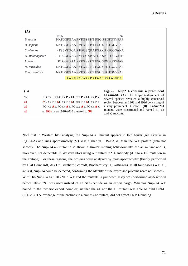
3 Results
71
WT FG xx P x FG xx P x FG x x P x FG xx P x
a1 SG xx P x SG xx P x SG x x P x SG xx P x
a2 FG xx A x FG xx A x FG x x A x FG xx A x
a3 all FGs in aa 1916-2033 mutated to SG
Fig. 25 Nup214 contains a prominent
FG-motif. (A) The Nup214-alignment of
several species revealed a highly conserved
region between aa 1968 and 1990 consisting of
a very prominent FG-motif. (B) His-Nup214
mutants were constructed and named a1, a2
and a3 mutants.
(A)
(B)
Note that in Western blot analysis, the Nup214 a1 mutant appears in two bands (see asterisk in
Fig. 26A) and runs approximately 2-3 kDa higher in SDS-PAGE than the WT protein (data not
shown). The Nup214 a3 mutant also shows a similar running behaviour like the a1 mutant and is,
moreover, not detectable in Western blots using our anti-Nup214 antibody (due to a FG mutation in
the epitope). For these reasons, the proteins were analyzed by mass-spectrometry (kindly performed
by Olaf Bernhardt, AG Dr. Bernhard Schmidt, Biochemistry II, Göttingen). In all four cases (WT, a1,
a2, a3), Nup214 could be detected, confirming the identity of the expressed proteins (data not shown).
With His-Nup214 aa 1916-2033 WT and the mutants, a pulldown assay was performed as described
before. His-SPN1 was used instead of an NES-peptide as an export cargo. Whereas Nup214 WT
bound to the trimeric export complex, neither the a1 nor the a3 mutant was able to bind CRM1
(Fig. 26). The exchange of the prolines to alanines (a2 mutant) did not affect CRM1-binding.
B. taurus
H. sapiens
C. elegans
D. melanogaster
X. laevis
M. musculus
R. norwegicus
1965 1992
NKTGGFG AA P VFG S P P T FGG S PGFGGVPAF
NKTGGFG AA P VFG S P P T FGG S PG FGGVPAF
- TS SVFG GA P KFG S QP A FGAK P - FGGGANA
T TPGGFG AK P VFGG S P A FGA S PT FGGGATF
TKTGGFG AA P VFG S P P T FGG S PG FGGS PAF
NKTGGFG AA P VFG S P P T FGG S PG FGGVPAF
NKTGGFG AA P VFG S P P T FGG S PG FGGVPAF
FG x x P xFG x x P x FG x x Px FG xx P x

3 Results
72
As His-Nup214 aa 1916-2033 a1 and a3 mutant were not able to bind CRM1 in vitro, we asked
whether the localization of endogenous CRM1 would change in cells overexpressing the Nup214
mutants. To address this question, the a1, a2 and a3 mutations were introduced into RFP-Nup214 (aa
1916-2033)-cNLS. The mutants were overexpressed in HeLa cells and the localization of endogenous
CRM1 was analyzed (Fig. 27). In the presence of Nup214 aa 1916-2033 WT, CRM1 accumulated in
the nucleus, as it was previously observed (Fig. 21, fragment 4). The a2 mutant also retained CRM1 in
the nucleus, indicating that the prolines in the FG-motif are not required for nuclear accumulation of
CRM1. Surprisingly, in the presence of Nup214 a1 mutant CRM1 accumulated in the nucleus to a
certain degree (see arrow), though this accumulation was not as strong as observed for WT Nup214.
This suggests that not only these 4 FGs are involved in CRM1 nuclear accumulation. With respect to
CRM1-binding, there might be other regions in Nup214 aa 1916-2033. This idea was further
supported by the observation that the mutation of all FGs in Nup214 aa 1916-2033 (mutant a3)
abolished nuclear retention of CRM1. It can be excluded that FGs per se are responsible for nuclear
accumulation of CRM1, as Nup214 1859-1974, a fragment that contains eight FGs, did not affect
CRM1 localization (see Fig 21, fragment 2). From these data, we concluded that the 4 FGs in the
conserved region are essential for CRM1 interaction with Nup214 aa 1916-2033 in vitro.
Input Input Input Input
10 % 10 % 10 % 10 %
His-CRM1
His-SPN1
* His-Nup214 (aa 1916-2033)
GST-Ran (ponceau)
GDP GTP GDP GTP GDP GTP GDP GTP
WT a1 a2 a3
Fig. 26 CRM1-binding is abrogated in the Nup214 a1 mutant aa 1916-2033. GST-RanGDP or
GST-RanGTP (5 µg) was immobilized on glutathione sepharose beads and incubated with His-CRM1 (5 µg),
His-SPN1 (5 µg) and His-Nup214 1916-2033 WT or with a1, a2 or a3 mutant (1 µg), respectively. SPN1,
CRM1 and Nup214 were detected by Western blot using anti-His-antibodies. The antibody recognizes two
bands for the Nup214 a1 mutant (see asterisk).

3 Results
73
DNA
RFP
CRM1
3.2.2.3 The inhibition of nuclear export is abrogated in Nup214 aa 1916-2033 a1 mutant in
vivo and in vitro
To analyze whether the Nup214 aa 1916-2033 a1, a2 and a3 mutants inhibit nuclear export, HeLa cells
were cotransfected with plasmids coding for RFP-Nup214-cNLS aa 1916-2033 WT or a1, a2, a3
mutant and GFP-SPN1 or GFP-NC2β (Fig. 28A and B). Apart from Nup214 WT, the a2 mutant led to
nuclear accumulation of GFP-SPN1, whereas the a1 and a3 mutant showed no effect on the
localization of the cargo. Interestingly, the distribution of GFP-NC2β was mildly affected in the
presence of Nup214 a1 mutant, as it was more equally distributed between the nucleus and the
cytoplasm compared to cells expressing RFP-cNLS. Only the mutation of all FGs in Nup214 could
completely restore the cytoplasmic localization of GFP-NC2β. Thus, effects on localization of NC2β
correlated with nuclear accumulation of CRM1 in the presence of the a1- and a3- mutant.
WT a1 a2 a3
RFP-Nup214 (aa 1916-2033)-cNLS
Fig. 27 C-terminal Nup214
lacking all FGs does not cause
nuclear accumulation of CRM1. HeLa cells were transfected with
plasmids coding for RFP-Nup214 aa
1916-2033 WT or a1, a2 or a3
mutant, respectively. After 20 hours,
the cells were fixed, stained for
endogenous CRM1 and analyzed by
fluorescence microscopy. In all cells
expressing Nup214 WT or a2 mutant,
endogenous CRM1 was reduced from
the cytoplasm.
10 µM

3 Results
74
DNA
RFP
GFP-
NC2β
DNA
RFP
GFP-SPN1
0
20
40
60
80
100
dis
trib
uti
on
in
% N
=
C
Fig. 28 Nuclear export can take place in the presence of Nup214 1916-2033 a1
mutant in vivo. HeLa cells were cotransfected with plasmids coding for RFP-Nup214 aa
1916-2033 WT or a1, a2 or a3 mutant and GFP-SPN1 (A) or GFP-NC2β (B), respectively.
After 20 hours, the cells were fixed and analyzed by immuno-fluorescence microscopy. The
distribution (in %; N nuclear, = equally distributed, C cytoplasmic) of GFP-SPN1 and GFP-
NC2β in cells expressing RFP-Nup214-cNLS aa 1916-2033 WT or a1, a2, a3 mutant was
quantified by counting at least 100 cells. Error bars indicate the variation from the mean of
two independent experiments.
(A)
(B)
10 µM
0
20
40
60
80
100
dis
trib
uti
on
in
% N
=
C
WT a1 a2 a3
RFP-cNLS RFP-Nup214 (1916-2033)-cNLS
WT a1 a2 a3
RFP-cNLS RFP-Nup214 (1916-2033)-cNLS
RFP- WT a1 a2 a3
c-NLS RFP-Nup214 1916-2033-cNLS
GFP-SPN1
GFP-NC2β
10 µM
10 µM
RFP- WT a1 a2 a3
c-NLS RFP-Nup214 1916-2033-cNLS
GFP-NC2β
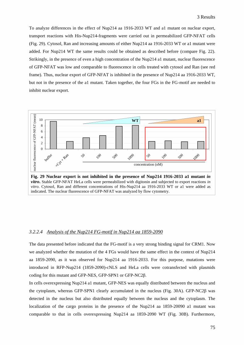
3 Results
75
0
2
4
6
8
10
buffer
+Cyt
+ R
an 50 100
500
1000 50 10
050
010
00
To analyze differences in the effect of Nup214 aa 1916-2033 WT and a1 mutant on nuclear export,
transport reactions with His-Nup214-fragments were carried out in permeabilized GFP-NFAT cells
(Fig. 29). Cytosol, Ran and increasing amounts of either Nup214 aa 1916-2033 WT or a1 mutant were
added. For Nup214 WT the same results could be obtained as described before (compare Fig. 22).
Strikingly, in the presence of even a high concentration of the Nup214 a1 mutant, nuclear fluorescence
of GFP-NFAT was low and comparable to fluorescence in cells treated with cytosol and Ran (see red
frame). Thus, nuclear export of GFP-NFAT is inhibited in the presence of Nup214 aa 1916-2033 WT,
but not in the presence of the a1 mutant. Taken together, the four FGs in the FG-motif are needed to
inhibit nuclear export.
3.2.2.4 Analysis of the Nup214 FG-motif in Nup214 aa 1859-2090
The data presented before indicated that the FG-motif is a very strong binding signal for CRM1. Now
we analyzed whether the mutation of the 4 FGs would have the same effect in the context of Nup214
aa 1859-2090, as it was observed for Nup214 aa 1916-2033. For this purpose, mutations were
introduced in RFP-Nup214 (1859-2090)-cNLS and HeLa cells were cotransfected with plasmids
coding for this mutant and GFP-NES, GFP-SPN1 or GFP-NC2β.
In cells overexpressing Nup214 a1 mutant, GFP-NES was equally distributed between the nucleus and
the cytoplasm, whereas GFP-SPN1 clearly accumulated in the nucleus (Fig. 30A). GFP-NC2β was
detected in the nucleus but also distributed equally between the nucleus and the cytoplasm. The
localization of the cargo proteins in the presence of the Nup214 aa 1859-20090 a1 mutant was
comparable to that in cells overexpressing Nup214 aa 1859-2090 WT (Fig. 30B). Furthermore,
% %
WT a1
concentration (nM)
Fig. 29 Nuclear export is not inhibited in the presence of Nup214 1916-2033 a1 mutant in
vitro. Stable GFP-NFAT HeLa cells were permeabilized with digitonin and subjected to export reactions in
vitro. Cytosol, Ran and different concentrations of His-Nup214 aa 1916-2033 WT or a1 were added as
indicated. The nuclear fluorescence of GFP-NFAT was analyzed by flow cytometry.
nucl
ear
flu
ore
scen
ceof
GF
P-N
FA
T (
mea
n)

3 Results
76
Fig. 30 Export cargos accumulate in the nucleus
in the presence of Nup214 1859-2090 a1 mutant. (A) HeLa cells were cotransfected with plasmids coding
for RFP-Nup214 1859-2090 a1 mutant and GFP-NES,
GFP-SPN1 or GFP-NC2β. After 20 hours the cells were
fixed and analyzed by immunofluorescence microscopy.
(B) The distribution (in %; N nuclear, = equally
distributed, C cytoplasmic) of GFP-SPN1 and GFP-NC2β
was quantified by counting at least 100 cells. Bars
indicate the variation from the mean of two independent
experiments. (C) Nup214 a1 mutant over-expressing
HeLa cells were fixed and stained for endogenous CRM1.
endogenous CRM1 accumulated in the nucleus in the presence of RFP-Nup214 1859-2090-cNLS a1
mutant (Fig. 30C), as it was observed for Nup214 aa 1859-2090 WT (Fig. 21, fragment 1).
Thus, in Nup214 aa 1859-2090, the FGs in the FG-motif are not sufficient to affect nuclear export.
The retention of CRM1 in the nucleus in the presence of the Nup214 a1 mutant indicates that this
mutant can still bind CRM1. This further supports the idea that there are other regions in the C-
terminus of Nup214, which are able to interact with CRM1.
(A) (C)
(B)
DNA
RFP-Nup214
1859-2090-cNLS
a1 mutant
GFP
DNA
RFP-Nup214
1958-2090-cNLS
a1 mutant
endogenous
CRM1
GFP-NES GFP-SPN1 GFP-NC2β
%
0
20
40
60
80
100
dis
trib
uti
on
in
%
N
=
C
0
20
40
60
80
100
dis
trib
uti
on
in
%
N
=
C
RFP- WT a1
cNLS Nup214 1859-2090
GFP-SPN1
GFP-NC2β
RFP- WT a1
cNLS Nup214 1859-2090

3 Results
77
3.3.3 Refined search for CRM1-binding sites in Nup214
The collected data so far indicated a CRM1-binding region in Nup214 aa 1975-2033, whereas the
tested Nup214-fragment aa 1859-1974 is dispensable for CRM1 interaction. Furthermore, Nup214 aa
1975-2033 also contains the main part of the highly conserved FG-motif (aa 1970-1990). However, a
Nup214 aa 1859-2090 mutant containing SGs instead of FGs in this motif behaves like Nup214 WT.
The FG-motif seems to be an important platform for CRM1, but taken together, our data point to at
least two CRM1-binding sites in Nup214. In this section, the very last part of the Nup214 C-terminal
region (aa 1968-2090) was investigated by analyzing and comparing very short Nup214-fragments.
3.2.3.1 The FG-motif in Nup214 requires aa 1991-2033 to form a stable complex with
CRM1 and RanGTP
In order to identify other regions in Nup214 which bind to CRM1, GST-Nup214-fragments (WT or a1
mutant) were constructed and purified as depicted in Fig. 31A. Furthermore, RanGTP- and substrate-
dependent binding of CRM1 to Nup214 was investigated.
Nup214 aa 1859-2090 interacts with CRM1 in a RanGTP-dependent manner (Fornerod et al., 1996,
Kehlenbach et al., 1999). In the first approach, Ran-dependent binding of CRM1 to the shorter GST-
Nup214-fragments was analyzed. The fragments as indicated were bound to glutathione sepharose
beads and CRM1, SPN1 and Ran, loaded either with GDP or GTP, were added. Gst-Nup214 aa 1968-
1990 WT (the FG-motif) and a1 mutant were not able to bind CRM1 in the presence of RanGTP
(Fig. 31B). Strikingly, GST-Nup214 aa 1968-2033 WT bound the CRM1-RanGTP-SPN1-export
complex, whereas GST-Nup214 aa 1968-2033 a1 mutant did not bind to any of the components (see
red frame). Notably, Nup214 aa 1991-2033 alone was not able to bind to the export complex. Taken
together, Nup214 aa 1991-2033 is required for interaction of CRM1 with the FG-motif.
Binding of CRM1 to Nup214 aa 1859-2090 is enhanced in the presence of an export cargo
(Kehlenbach et al., 1999). In the second approach, CRM1 and RanGTP binding to the shorter
Nup214-fragments were analyzed in the presence or the absence of the cargo SPN1 (Fig. 31C).
Basically, only GST-Nup214 aa 1968-2033 WT could bind CRM1 (see red frame), as it was observed
in the approach described before. This binding increased in the presence of SPN1. Thus, enhanced
binding of CRM1 to Nup214 aa 1859-2090 in the presence of an export cargo is also observed for the
short Nup214-fragment aa 1968-2033.
Taken together, these results give evidence for the importance of the FG-motif for CRM1-interaction
in a RanGTP- and export cargo- dependent manner. However, Nup214 aa 1991-2033 strongly
contributes to this binding.
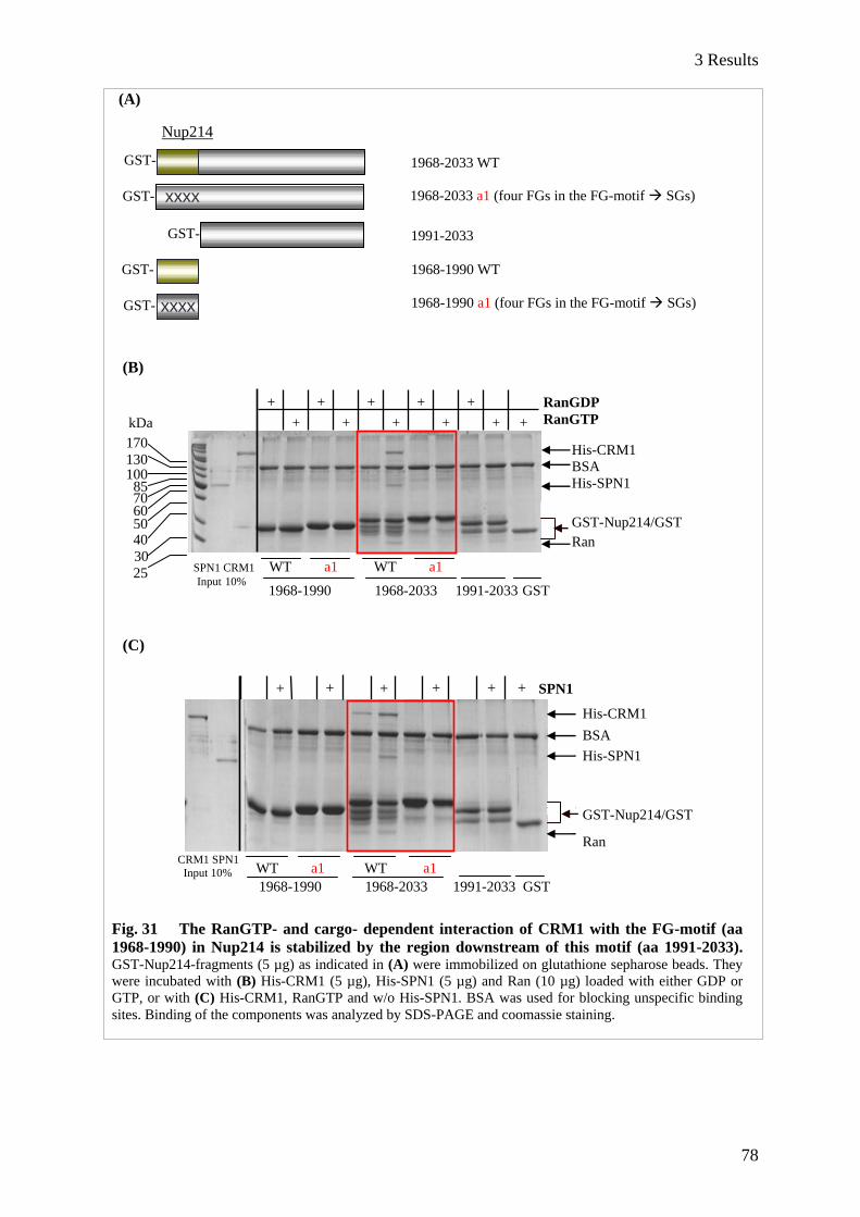
3 Results
78
(A)
(B)
kDa
(C)
WT a1 WT a1
1968-1990 1968-2033 1991-2033 GST
+ + + + + + SPN1
CRM1 SPN1
Input 10%
His-CRM1
BSA
His-SPN1
GST-Nup214/GST Ran
GST- 1968-2033 a1 (four FGs in the FG-motif SGs)
GST- 1991-2033
GST-
GST-
xxxx
1968-1990 WT
1968-1990 a1 (four FGs in the FG-motif SGs)
Nup214
GST- 1968-2033 WT
xxxx
1968-1990 1968-2033 1991-2033 GST
+ + + + + +
+ + + + +
170
130 100 85 70 60 50 40
30
25
RanGDP
RanGTP
His-CRM1
BSA
His-SPN1
GST-Nup214/GST
Ran
Fig. 31 The RanGTP- and cargo- dependent interaction of CRM1 with the FG-motif (aa
1968-1990) in Nup214 is stabilized by the region downstream of this motif (aa 1991-2033). GST-Nup214-fragments (5 µg) as indicated in (A) were immobilized on glutathione sepharose beads. They
were incubated with (B) His-CRM1 (5 µg), His-SPN1 (5 µg) and Ran (10 µg) loaded with either GDP or
GTP, or with (C) His-CRM1, RanGTP and w/o His-SPN1. BSA was used for blocking unspecific binding
sites. Binding of the components was analyzed by SDS-PAGE and coomassie staining.
SPN1 CRM1 WT a1 WT a1 Input 10%
3 Results
79
3.2.3.2 Elongation of Nup214 C-terminal fragments leads to increasing functionality in
nuclear export
In order to test which regions around the FG-motif (aa 1968-1990) in Nup214 affect nuclear export of
proteins, RFP-cNLS Nup214-fragments were constructed as indicated in Fig. 32A. GFP-NC2β was
chosen as a substrate because, compared to SPN1 (see appendix, Fig. S2), it reacts more sensitive to
overexpression of Nup214-fragments. HeLa cells were cotransfected with plasmids coding for RFP-
Nup214-cNLS and GFP-NC2β, and export inhibition was analyzed by immunofluorescence
microscopy. First, the cells were grouped as usual into cells with nuclear (N) or cytoplasmic (C)
located GFP-NC2β or cells with GFP-NC2β distributed equally between the nucleus and the
cytoplasm (=). For better comparison of the effects of the Nup214-fragments on GFP-NC2β, the ratio
of the nuclear distribution of GFP-NC2β in RFP-Nup214-cNLS expressing cells and the nuclear
distribution of GFP-NC2β in RFP-cNLS expressing cells was determined.
In the presence of the RFP-Nup214-cNLS fragments aa 1859-2090, 1975-2090 and 1916-2033, the
number of cells with nuclear located GFP-NC2β increased 5-8 fold compared to RFP-cNLS alone
(Fig. 32B). Notably, Nup214 aa 1859-2090 and 1975-2090 showed the strongest export inhibition
effect, whereas Nup214 aa 1859-1974 did not influence the localization of GFP-NC2β. Stepwise
truncation of Nup214 aa 1975-2090 (fragments aa 1991-2090 and 2034-2090; see group I in Fig. 32A,
also underlined in Fig. 32B) led to a stepwise decrease of cells showing nuclear GFP-NC2β (from 7 to
2 fold). Notably, the lack of the FG-motif in Nup214 aa 1991-2090 already influenced localization of
the cargo, and a further shortening to aa 2034-2090 led to comparable amounts of cells with nuclear
located GFP-NC2β in the presence of RFP-cNLS alone. Interestingly, for GFP-SPN1, Nup214 aa
1991-2090 was the only fragment lacking the FG-motif and inhibiting nuclear export at the same time
(see appendix, Fig. S2), and truncation of this fragment at the N-terminus (lack of aa 1991-2033)
completely abolished export inhibition of GFP-SPN1.
In principle, the same effect on nuclear export of GFP-NC2β was observed for N-terminal truncations
of Nup214 aa 1916-2033 (group II in Fig 32A and B; Nup214 aa 1968-2033 and 1991-2033).
However, Nup214 aa 1916-1990, compared to RFP-cNLS alone, did not affect the localization of
GFP-NC2β although it contains the FG-motif. On the other hand, Nup214 aa 1968-2033, also
containing the FG-motif, led to a clear nuclear accumulation of GFP-NC2β, though the effect was
weaker compared to the Nup214-fragment aa 1916-2033. The FG-motif alone (aa 1968-1990) had no
effect on nuclear export of the cargo. Together, the in vivo data confirmed the results obtained from
the in vitro pulldown assay (compare Fig. 31).
In summary, there are regions in Nup214 contributing to the function of the FG-motif (aa 1968-1990).
These regions are located mainly downstream of this motif. The data strongly suggest that the region
between aa 1991 and 2033 plays the most prominent role in supporting CRM1-binding to the FG-
motif. Nevertheless, also the region before (aa 1916-1967) seems to influence functionality of the FG-

3 Results
80
motif, as removing this region from Nup214 aa 1916-2033 (resulting in Nup214 aa 1968-2033) led to
decreased nuclear accumulation of GFP-NC2β.
(A)
(B)
Altogether, we identified a major CRM1-binding site in Nup214 which is also responsible for nuclear
export inhibition of cargo proteins. This FG-motif in Nup214 additionally requires aa 1991-2033 for a
stable interaction with CRM1. Nup214 aa 1991-2090, which does not contain the FG-motif, inhibited
nuclear export of GFP-NC2β even more efficiently compared to Nup214 aa 1968-2033, which does
contain the FG-motif. When removing aa 1991-2033 from Nup214 aa 1991-2090, nuclear export
inhibition was completely abrogated. Thus, Nup214 aa 1991-2033 not only contributes to the function
of the FG-motif in aa 1968-1990, but also to the function of an unidentified CRM1-binding region in
Nup214 aa 2034-2090.
0
2
4
6
8
10
RFP-
cNLS
1859-
2090
1859-
1974
1975-
2090
1991-
2090
2034-
2090
1968-
1990
1916-
1990
1916-
2033
1968-
2033
1991-
2033
Rat
io R
FP
-Nup
21
4-c
NL
S/
RF
P-c
NL
S
Nup214 aa
1859-2090
1859-1974
1975-2090
1991-2090 I
2034-2090
1968-1990
1916-1990
1916-2033
1968-2033 II
1991-2033
I II
GFP-NC2β
RFP-Nup214-cNLS
FG-motif
Fig. 32 Effect of short Nup214-fragments on nuclear accumulation of GFP-NC2β. (A) RFP-
Nup214-cNLS fragments were constructed as depicted. (B) HeLa cells were cotransfected with plasmids
coding for RFP-Nup214-cNLS-Fragments and GFP-NC2β. After 20 hours the cells were fixed and
distribution of GFP-NC2β was quantified by counting at least 60 cells. The ratio of the nuclear distribution of
GFP-NC2β in the presence of RFP-Nup214-cNLS and in the presence of RFP-cNLS was determined. The
bars indicate the variation from the mean of two independent experiments.

4 Discussion
81
4 Discussion
4.1 Analysis of CRM1-fragments and -chimeras
Inhibition of CRM1 by LMB as an alternative approach to CRM1 RNA interference
RNA interference is a common method to investigate the effect of proteins in eukaryotic cells and also
in whole organisms (Fire et al., 1998). This system takes advantage of the protective mechanism of
cells against viruses or transposons (Waterhouse et al., 2001). Eukaryotic cells or organisms are
exposed to small RNAs with sequences complementary to the mRNA transcript of the protein to be
depleted. However, off-target effects are common, because already a match of 11-15 nucleotides can
lead to unspecific gene suppression (Jackson et al., 2003). Furthermore, the interferon-system can be
activated and protein biosynthesis can be inhibited via activation of the RNA-dependent protein kinase
PKR and phosphorylation of the transcription factor eIF2 (eukaryotic initiation factor 2; Levin et al.,
1978). Additionally, RNase L is induced, resulting in degradation of mRNA and apoptosis (Pandey et
al., 2004). Together, the effects, which are observed upon depletion of proteins of interest, always
have to be analyzed with respect to specificity. In the case of CRM1, other problems could potentially
arise upon RNA interference. CRM1 is the main export receptor and transports hundreds of cargos out
of the nucleus (la Cour et al., 2003). Hence, depletion of CRM1 by RNA interference would cause
mislocalization of many proteins, finally causing a lot of side effects. The usage of the LMB-method
allows cells to grow under normal conditions. Unlike the siRNA treatment, which takes several days,
LMB is added to cells for only a few hours, minimizing potential side effects. Furthermore, LMB is a
selective inhibitor, as it only modifies CRM1 (Kudo et al., 1999) by binding covalently to the
hydrophobic cleft, that is also used by the NES-sequences of CRM1-dependent cargos.
In analogy to RNA interference experiments, where siRNA resistant mutants can be expressed in cells
upon depletion of endogenous proteins, CRM1, containing a point mutation in the NES-binding cleft,
can be overexpressed together with the cargo protein of interest. LMB covalently modifies C528 in
CRM1, and the mutation of this amino acid residue leads to LMB resistant, but still functional CRM1.
The big advantage of this approach is switching off endogenous CRM1, but at the same time giving
the cells the chance to use exogenous CRM1 C528S. Indeed, in LMB-treated HeLa cells, that
overexpress CRM1 C528S, the localization of the tested NES-cargos could be rescued. Together, the
LMB approach is an efficient tool to analyze CRM1-dependent localization of proteins. We set out to
use this mutation in CRM1-fragments and chimeras. This enabled us to investigate the function of
distinct regions in CRM1, without having background effects produced by endogenous CRM1.
4 Discussion
82
Nucleoporin binding and export capability of CRM1-fragments and -chimeras
Based on our previous knowledge of domains in CRM1, their assigned functions and the available
crystal structure (Petosa et al., 2004), the N-terminus of CRM1 was responsible for Ran binding, and
the central part was binding to NES-cargos (Nazari and Dargemont, 1999). The function of the
C-terminal half of CRM1 was not known. To analyze this region, we constructed HA-CRM1-
fragments lacking C-terminal HEAT repeats. As CRM1 interacts with several nucleoporins like
Nup153, p62 and also with Nup214 in a RanGTP-dependent manner (Fornerod et al., 1996;
Kehlenbach et al., 1999), we assumed that nucleoporin-binding could occur in the C-terminal region
and performed coimmunoprecipitation experiments with the CRM1-fragments. Although endogenous
CRM1 could be coimmunoprecipitated with HA-Nup214 (Fornerod et al. 1996), we had problems to
coimmunoprecipitate endogenous Nup214 with HA-CRM1-fragments. In general, the expression level
of HA-CRM1 constructs is low in HeLa cells. Endogenous CRM1, present in higher amounts in total
cell lysates, could saturate the binding sites in endogenous Nup214 molecules and therefore compete
with HA-CRM1. We therefore resorted to a microscopic analysis and showed that none of the tested
CRM1 fragments associated with the nuclear envelope. This correlated with their inability to export
cargos out of the nucleus. During our work on CRM1-fragments, the crystal structure of CRM1 in a
trimeric export complex was solved (Dong et al., 2009; Monecke et al., 2009), showing CRM1 as a
ring-like protein. RanGTP is located in the cavity of this ring, connected to several HRs of CRM1. In
CRM1-fragments, this ring structure might be disturbed, thus affecting RanGTP-binding. Due to this,
export complex formation as well as the interaction with nucleoporins could be affected. Besides
problems in nucleoporin-interaction that might occur with CRM1-fragments, it is also unclear whether
the release of the export cargo via RanBP1 into the cytoplasm is possible. The acidic α-helix at the
very C-terminus of CRM1, which modulates the affinity of CRM1 to NES-cargos (Dong et al., 2009;
Fox et al., 2011), is missing in all CRM1-fragments. Both, NES-cargo and RanBP1, affect RanGTP
binding to CRM1.
The inability of CRM1-fragments to associate with the nuclear envelope emphasizes the specific role
of RanGTP in CRM1-nucleoporin interaction. The function of this interaction is to facilitate
translocation of transport complexes through the nuclear pore, and the translocation process itself has
been discussed in several models (Macara, 2001; Ribbeck and Görlich, 2001; Rout et al., 2003;
Yamada et al., 2010). To date, the structure of unbound CRM1 is not known but it is presumed that
due to RanGTP-binding, CRM1 undergoes conformational changes, additionally promoting export
cargo binding (Monecke et al., 2009). It is also not known, whether binding of nucleoporins influences
the conformation of CRM1.
To overcome problems of RanGTP- /cargo- binding to CRM1, translocation through the nuclear pore
and release of the export complex in the cytoplasm, we decided to continue our studies with H.
sapiens/C. elegans CRM1-chimeras. There is no crystal structure available for C. elegans CRM1, but
we assume that it is present as a ring-like protein with similar binding properties for an NES-cargo,

4 Discussion
83
RanGTP and RanBP1, too. One striking difference between C. elegans CRM1 and human CRM1 is
the inability to interact with human nucleoporins (S. Roloff, 2008, Diploma thesis). Because of this,
C. elegans CRM1 should not be able to fully mediate export through the nuclear pore. A GFP-NES
was exported by C. elegans CRM1 to a certain extent. By contrast, C. elegans CRM1 was neither able
to export human GFP-SPN1 nor GFP-NC2β. The residual export activity, however, showed that
C. elegans CRM1 can interact with human RanGTP as well as with the GFP-NES reporter. The crystal
structure of the CRM1-RanGTP-SPN1-complex (Monecke et al., 2009) indicates more than one
binding site for SPN1 on CRM1. Thus, C. elegans CRM1 is presumably not able to fulfil all required
binding conditions for SPN1. Besides the lack of appropriate nucleoporin binding sites in C. elegans
CRM1, its minimal export capability for a GFP-NES in human cells might be due to the fact that this
NES interacts only with the hydrophobic NES-cleft and with nothing else. The reason for nuclear
accumulation of GFP-NC2β remains unclear, as the binding mechanism to CRM1 is not known. Due
to the restricted export capability and inability to interact with human nucleoporins, C. elegans CRM1
was a suitable candidate for analysis of CRM1-chimeras.
Among our chimeras, we found one that acted similar to human CRM1 in mammalian cells. Chimera
HsCe I (human HR 1-11 and C. elegans HR 12-21) was located in the nucleus and at the nuclear
envelope and could mediate export of at least a subset of cargo proteins. GFP-NES and GFP-SPN1
could be efficiently exported, so we can conclude that HsCe I contains all binding regions for SPN1.
Furthermore, the chimera-export complex must contain nucleoporin binding regions and must also be
able to disassemble in the cytoplasm. Based on our approaches, we cannot say whether this chimera
and human CRM1 have similar affinities for interacting export factors, but we see that HsCe I is
functional in mammalian cells. NC2β, by contrast, accumulated in the nucleus in the presence of
HsCe I under conditions where endogenous CRM1 is inhibited by LMB, indicating a binding
mechanism to CRM1, different from that of GFP-SPN1 or GFP-NES. Obviously, these binding
properties are not present in the C. elegans CRM1 part. Hence, important NC2β binding regions might
be found in the C-terminus of human CRM1. Strikingly, the chimera CeHs I (with C. elegans CRM1
in the N-terminal half and H. sapiens CRM1 at the C-terminal half) was clearly restricted in its export
capability as none of the tested cargos could be exported. When we assume that important binding
regions for NC2β are located in the C-terminus of CRM1, the export complex should be assembled
with this chimera without difficulties. This would also be true for SPN1, which interacts with human
CRM1 HR 14A, 15A and 16A (Monecke et al., 2009). As CeHs I did not show a rim-staining around
the nucleus and neither could mediate nuclear export of the tested cargos, we assume that association
with the nuclear pore is related to the ability of CRM1 to mediate export of cargos into the cytoplasm.
Assuming a proper ring closure and the ability to interact with human RanGTP and an NES-cargo,
missing binding regions for nucleoporins could explain the export restriction for this chimera. Hence,
binding sites for human nucleoporins like Nup214, the terminal docking site for export complexes
(Fornerod et al., 1996; Kehlenbach et al., 1999), are likely present in the N-terminal portion of CRM1.

4 Discussion
84
Our assumption is further supported by identified nucleoporin-binding sites in importin β, also located
in the N-terminus (Kutay et al., 1997).
Consistent with their inability to associate to the nuclear envelope, none of the chimeras type II could
mediate export in mammalian cells. CeHs II (C. elegans HR 1-8 and human HR 9-21) and HsCe II
were equally distributed in the nucleus and the cytoplasm. In both chimeras putative nucleoporin
binding sites are missing or affected. Comparing CRM1 chimeras HsCe I and HsCe II, the exchange
of H. sapiens HRs 9-11 into C. elegans HRs was sufficient to abrogate association with the nuclear
envelope. From this, we conclude that HRs 9-11 of human CRM1 are important for interaction with
nucleoporins. We could not detect CRM1-chimera - nucleoporin interaction in coimmuno-
precipitation experiments. For HsCe I, we hypothesize a RanGTP-dependent binding to nucleoporins.
The composition of this chimera would indicate nucleoporin interaction at the N-terminal part of
human CRM1. However, we cannot exclude a decreased affinity of this chimera for RanGTP, which
would explain the lack of nucleoporin binding in coimmunoprecipitation. Additionally, the exchange
of human CRM1 regions into C. elegans CRM1 parts could lead to structural or conformational
changes. Considering these aspects, a potential binding to nucleoporins could not be seen because
available endogenous nucleoporins would mostly bind to endogenous human CRM1. To further
investigate HR 9-11 in CRM1, binding studies with recombinant CRM1-chimeras have to be
performed.
Nup214 interacts with full-length human CRM1 in a RanGTP-dependent manner (Fornerod et al.,
1996) and this binding is enhanced in the presence of an NES (Kehlenbach et al., 1999), indicating a
cooperative binding mechanism of all four components. When we assume an interaction of Nup214
with HRs 9-11 in CRM1, this cooperativity can be explained by the close proximity of Nup214-
binding to the Ran- and cargo- relevant regions in CRM1. In Fig. 33, a model is depicted, in which
Nup214-binding involves several HEAT repeat regions nearby the RanGTP- and cargo- binding sites
of CRM1. Such a Nup214- interaction would specify the CRM1-RanGTP-cargo export complex as
such in the nuclear pore. The function of this interaction will be discussed in the following section.
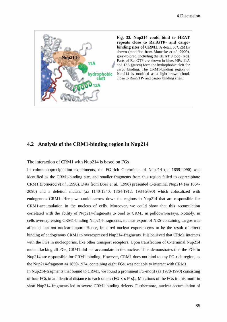
4 Discussion
85
4.2 Analysis of the CRM1-binding region in Nup214
The interaction of CRM1 with Nup214 is based on FGs
In coimmunoprecipitation experiments, the FG-rich C-terminus of Nup214 (aa 1859-2090) was
identified as the CRM1-binding site, and smaller fragments from this region failed to coprecipitate
CRM1 (Fornerod et al., 1996). Data from Boer et al. (1998) presented C-terminal Nup214 (aa 1864-
2090) and a deletion mutant (aa 1140-1340, 1864-1912, 1984-2090) which colocalized with
endogenous CRM1. Here, we could narrow down the regions in Nup214 that are responsible for
CRM1-accumulation in the nucleus of cells. Moreover, we could show that this accumulation
correlated with the ability of Nup214-fragments to bind to CRM1 in pulldown-assays. Notably, in
cells overexpressing CRM1-binding Nup214-fragments, nuclear export of NES-containing cargos was
affected. but not nuclear import. Hence, impaired nuclear export seems to be the result of direct
binding of endogenous CRM1 to overexpressed Nup214-fragments. It is believed that CRM1 interacts
with the FGs in nucleoporins, like other transport receptors. Upon transfection of C-terminal Nup214
mutant lacking all FGs, CRM1 did not accumulate in the nucleus. This demonstrates that the FGs in
Nup214 are responsible for CRM1-binding. However, CRM1 does not bind to any FG-rich region, as
the Nup214-fragment aa 1859-1974, containing eight FGs, was not able to interact with CRM1.
In Nup214-fragments that bound to CRM1, we found a prominent FG-motif (aa 1970-1990) consisting
of four FGs in an identical distance to each other: (FG x x P x)4. Mutations of the FGs in this motif in
short Nup214-fragments led to severe CRM1-binding defects. Furthermore, nuclear accumulation of
*
Fig. 33. Nup214 could bind to HEAT
repeats close to RanGTP- and cargo-
binding sites of CRM1. A detail of CRM1is
shown (modified from Monecke et al., 2009),
grey-colored, including the HEAT 9 loop (red).
Parts of RanGTP are shown in blue. HRs 11A
and 12A (green) form the hydrophobic cleft for
cargo binding. The CRM1-binding region of
Nup214 is modeled as a light-brown cloud,
close to RanGTP- and cargo- binding sites.
--Nup214--

4 Discussion
86
NES-
cargo
CRM1 and export cargos was abrogated. In the context of the longer Nup214 C-terminal region
(aa 1859-2090), mutation of the FG-motif caused nuclear accumulation of CRM1 similar to Nup214
WT, suggesting that this mutant also binds CRM1. In combination with the results we got from other
short Nup214-fragments, we conclude that not only the FG-motif binds to CRM1, but that there are
two other binding sites in Nup214, stabilizing this interaction. Nup214 aa 1991-2033 contributes to
CRM1-binding to the FG-motif but also to an unidentified region between aa 2034 and 2090 (Fig 34).
The smallest fragment lacking the FG- motif but inhibiting nuclear export was Nup214 aa 1991-2090.
Importin β has been shown to bind via several regions to nucleoporins (Bayliss et al., 2000;
Kutay et al., 1997b). Also in the nuclear import factor p97 more than one nucleoporin-binding site was
found (Chi and Adam, 1997). Together with our results obtained from Nup214 fragments, also in
CRM1 more than one binding region for Nup214 must be present.
The role of the FGs in the terminal steps of nuclear export
The cytoplasmically located Nup214 serves as a terminal docking site in the late steps of nuclear
export. RanBP1 releases CRM1 from Nup214 (Kehlenbach et al., 1999), indicating a role of Nup214
preparing the export complex for the following releasing steps by RanGAP. RanBP1 is a soluble
protein, and RanGAP can be soluble, but is also associated to Nup358 (Mahajan et al. 1997; Matunis
et al. 1996; Weis 2007). Due to the strong binding to Nup214, export complexes containing CRM1
could be concentrated at the nuclear pore, thus allowing efficient disassembly by RanBP1 and
RanGAP.
For importin β it is known that its binding pockets for FGs have different binding properties and
sensitivities to RanGTP (Otsuka et al., 2008). Consequently, the impact of RanGTP on distinct FG-
binding regions in CRM1 could also modulate the affinity of CRM1 for Nup214. Moreover, RanGTP-
dependent binding of FGs in Nup214 to CRM1 could induce conformational changes in the export
complex, which might be crucial for the following releasing steps. The identification of the FG-motif
with its contributing C-terminal FGs of Nup214 strongly suggests a defined binding pattern for
Nup214 XXX - - - ---
1859 1970 1990 2033 2090
RanGTP
Fig. 34 Nup214 binds with at
least three regions to CRM1. The trimeric export complex (shown
in blue colours) interacts with several
regions of Nup214. The FG-motif (aa
1970-1990; yellow) binds to CRM1.
Nup214 aa 1991-2033 (orange) and
another unidentified region (XXX)
stabilize this interaction.
CRM1 HR 9-11
4 Discussion
87
CRM1. This might be important for a correct orientation of the export complex at the cytoplasmic side
of the NPC, thus allowing the presentation of distinct CRM1 regions to releasing factors like RanGAP.
The FG-motif also contains prolines which have an identical distance to each other. Prolines have the
potential to cause nicks in α-helices. FG-regions in Nups are intrinsically unstructured (Allen et al.,
2002; Denning et al., 2003; Patel et al., 2007), and we do not know which kind of structure the FG-
motif would have when bound to CRM1. Thus, it is unclear what function these prolines could have.
Nup214-fragments where the prolines are exchanged for alanines in the FG-motif can still bind to
CRM1, and export cargos also accumulate in the presence of this mutant.
Controversially to all observations concerning the function of Nup214 in nuclear export, namely to be
involved in releasing the cargo from the CRM1 export complex, only a few cargos have been shown to
be CRM1- and Nup214- dependent. They have been identified via overexpression in Nup214 depleted
cells (Bernad et al., 2006; Hutten and Kehlenbach, 2006), and an accumulation of these proteins in the
nucleus had been observed. However, cargos that are nuclear per se, due to a stronger import signal
compared to the export signal, cannot be investigated with this method. Consequently, alternative
ways have to be found to analyze the impact of Nup214 on CRM1-dependent cargos. In this work,
nuclear accumulation of CRM1, due to short overexpressed Nup214-fragments, led to problems in cell
growth. This had also been described for the complete FG-containing C-terminal part of Nup214 aa
1859-2090 (Boer et al., 1998). Moreover, depletion of Nup214 leads to reduced cell growth, too
(Hutten and Kehlenbach, 2006). Overexpression of the Nup214-Nup88 complex reduces CRM1 from
the nuclear pore, resulting in impaired nuclear export (Xylourgidis et al., 2006). All these findings
imply a general role of Nup214-CRM1 interaction in nuclear export. The identification of the Nup214-
binding site in CRM1 and of export cargos that are dependent on both, Nup214 and CRM1, would
help to elucidate the role of Nup214 in CRM1-mediated nuclear export.
4 Discussion
88
4.3 Outlook
To test whether the binding of Nup214 occurs in HRs 9-11 in CRM1, we have to optimize protein
purification of CRM1-chimeras and do binding assays with them. Furthermore, the construction of
triple chimeras (e.g. H.sapiens HR 1-8/C. elegans HR 9-11/H. sapiens HR 12-21) can give us further
insight into the function of certain HRs in vivo.
The FG-motif in Nup214 seems to be the most important binding site for CRM1, and at least two
distinct regions in Nup214 contribute to this interaction. For identification of the binding sites,
Nup214-fragments with mutations in selected FG-areas will be constructed and analyzed in vivo and in
vitro. Cross-linking of suitable Nup214-fragments with CRM1 should be performed to identify the
CRM1-binding region for Nup214. The ultimate goal is to obtain co-crystals of Nup214-fragments and
the CRM1-export complex and to determine their structure by X-ray crystallography. Furthermore,
Nup214- and CRM1- dependent export cargos will have to be systematically investigated, e.g. by
screening for cytoplasmic proteins that accumulate in the nucleus upon depletion of Nup214 (similar
to the approach that has been used for Nup358 (Wälde et al., 2011)).
The elucidation of the role for CRM1-Nup214 interaction would also help to get more insight into the
function of other nucleoporins in nuclear transport. The uncoverage of similarities or differences in
transport receptor-nucleoporin interaction might lead to a better understanding, how transport
complexes translocate through the nuclear pore.
5 References
89
5 References Adachi, Y. and M. Yanagida. 1989. Higher order chromosome structure is affected by cold-sensitive mutations in
Schizosaccharomyces pombe gen CRM1+ which encodes a 115-kD protein preferentially localized in the nucleus
and its periphery. J.Cell.Biol. 108:1195-1207.
Alber, F., S. Dokudovskaya, L.M. Veenhoff, W. Zhang, J. Kipper, D. Devos, A. Suprapto,O. Karni-Schmidt, R.
Williams, B.T. Chait, M.P. Rout and A. Sali. 2007. Determining the architectures of macromolecular
assemblies. Nature. 450:683-94.
Allen, N. P., S. S. Patel and M. Rexach. 2002. Deciphering networks of protein interactions at the nuclear pore complex.
Mol. Cell. Proteomics. 1:930–946.
Andrade, M.A., and P. Bork. 1995. HEAT repeats in the Huntington's disease protein. Nat Genet. 11:115-6.
Arts, G.J., M. Fornerod and I.W. Mattaj. 1998. Identification of a nuclear export receptor for tRNA. Curr Biol. 8:305-314.
Askjaer, P., A. Bachi, M. Wilm, F. R. Bischoff, D. L. Weeks, V. Ogniewski, M. Ohno, C. Niehrs, J. Kjems, I. W.
Mattaj and M. Fornerod. 1999. RanGTP-regulated interactions of CRM1 with nucleoporins and a shuttling
DEAD-box helicase. Mol. Cell. Biol. 19:6276-6285.
Ausubel, F.M., R. Brent, R.E. Kingston, D.D. Moore, J.G. Seidman, J.A. Smith and K. Struhl. 1994. Current
protocols in molecular biology. New York: Greene. Publishing Associates and Wiley-Interscience.
Bayliss, R., K, Ribbeck, D, Akin, H.M. Kent, C.M. Feldherr, D. Görlich and M. Stewart. 1999. Interaction between
NTF2 and xFxFG-containing nucleoporins is required to mediate nuclear import of RanGDP. J. Mol. Biol.
293:579-593.
Bayliss R, T. Littlewood and M. Stewart. 2000. Structural basis for the interaction between FxFG nucleoporin repeats and
importin-beta in nuclear trafficking. Cell 102:99–108.
Bayliss R, T, Littlewood, L.A. Strawn, S.R. Wente and M. Stewart. 2002. GLFG and FxFG nucleoporins bind to
overlapping sites on importin-beta. J. Biol. Chem. 277:50597–606.
Bastos, R., L. Ribas de Pouplana, M. Enarson, K. Bodoor and B. Burke. 1997. Nup84, a novel nucleoporin that is
associated with CAN/Nup214 on the cytoplasmic face of the nuclear pore complex. J Cell Biol. 137:989-1000.
Beals, C.R., N.A. Clipstone, S.N. Ho and G.R. Crabtree. 1997a. Nuclear localization of NF-ATc by a calcineurin-
dependent, cyclosporin-sensitive intramolecular interaction. Genes Dev 11:824–834.
Beals, C.R., C.M. Sheridan, C.W. Turck, P. Gardner and G.R. Crabtree. 1997b. Nuclear export of NF-ATc enhanced
by glycogen synthase kinase-3. Science 275:1930–1934.
Beck, M., F. Forster, M. Ecke, J.M. Plitzko, F. Melchior, G. Gerisch, W. Baumeister and O. Medalia. 2004. Nuclear
Pore Complex Structure and Dynamics Revealed by Cryoelectron Tomography. Science. 306:1387–1390.
Becker, J., F. Melchior, V. Gerke, F.R. Bischoff, H. Ponstingl and A. Wittinghofer. 1995. RNA1 encodes a GTPase-
activating protein specific for Gsp1p, the Ran/TC4 homologue of Saccharomyces cerevisiae J. Biol. Chem.
270:11860–11865.
Ben-Efraim, I. and L. Gerace. 2001. Gradient of increasing affinity of importin beta for nucleoporins along the pathway of
nuclear import. J Cell Biol. 152:411-417.
Bernad, R., H. van der Velde, M. Fornerod and H. Pickersgill. 2004. Nup358/RanBP2 attaches to the nuclear pore
complex via association with Nup88 and Nup214/CAN and plays a supporting role in CRM1-mediated nuclear
protein export. Mol. Cell. Biol. 24:2373–2384.
Bernad, R., D. Engelsma, H. Sanderson, H. Pickersgill and M. Fornerod. 2006. The Nup214-Nup88 nucleoporin
subcomplex is required for CRM1-mediated 60 S preribosomal nuclear export. J Biol Chem. 28:19378-86.
Bischoff, F. R. and H. Ponstingl. 1991. Catalysis of guanine nucleotide exchange on Ran by the mitotic regulator RCC1.
Nature. 354: 80-82.
5 References
90
Bischoff, F. R., H. Krebber, E. Smirnova, W. Dong and H. Ponstingl. 1995. Co-activation of RanGTPase and inhibition
of GTP dissociation by Ran–GTP binding protein RanBP1. EMBO J. 14:705-715.
Boer, J., J. Bonten-Surtel and G. Grosveld. 1998. Overexpression of the Nucleoporin CAN/NUP214 Induces Growth
Arrest, Nucleocytoplasmic Transport Defects, and Apoptosis. Mol. Cell. Biol. 18:1236-1247.
Bogerd, H.P., R.A. Fridell, R.E. Benson, J. Hua and B.R. Cullen. 1996. Protein sequence requirements for function of
the human T-cell leukemia virus type 1 Rex nuclear export signal delineated by a novel in vivo randomization-
selection assay. Mol. Cell. Biol. 16:4207–4214.
Cang, Y. and G. Prelich. 2002. Direct stimulation of transcription by negative cofactor 2 (NC2) through TATA binding
protein (TBP). PNAS. 99 (20):12727-12732.
Carmody, S.R. and S.R.Wente. 2009. mRNA nuclear export at a glance. J Cell Sci 122:1933–1937.
Chadrin, A., B. Hess, M. San Roman, X. Gatti,, B. Lombard, D. Loew, Y. Barral, B. Palancade and V. Doye. 2010.
Pom33, a novel transmembrane nucleoporin required for proper nuclear pore complex distribution. J. Cell Biol.
189:795-811.
Charneau, P., G. Mirambeau, P. Roux, S. Paulous, H. Buc and F. Clavel. 1994. HIV-1 reversetranscription. A
termination step at the center of the genome. J Mol Biol.241:651-62.
Clarke, P. and Z. Chuanmao. 2008. Spatial and temporal coordination of mitosis by Ran GTPase. Nat. Rev. Mol. Cell Bio.
9:464-477
Chi, N.C. and S.A. Adam. 1997. Functional domains in nuclear import factor p97 for binding the nuclear localization
sequence receptor and the nuclear pore. Mol Biol Cell. 8(6):945-956.
Chook, Y.M., and G. Blobel. 1999. Structure of the nuclear transport complex karyopherin-beta2-RanGppNHp. Nature.
399: 230–237.
Conti, E. and J. Kuriyan. 2000. Crystallographic analysis of the specific yet versatile recognition of distinct nuclear
localization signals by karyopherin α. Structure. 8:329-38.
Conti, E. and E. Izaurralde. 2001. Nucleocytoplasmic transport enters the atomic age. Curr Opin Cell Biol 13: 310-9.
Cook, A., F. Bono, M. Jinek and E. Conti. (2007). Structural biology of nucleocytoplasmic transport. Annu Rev
Biochem 76: 647–671
la Cour, T., R. Gupta, K. Rapacki, K. Skriver, F.M. Poulsen and S. Brunak. 2003. NESbase version 1.0, a database of
Nuclear Export Signals. Nucleic Acids Res. 31(1):393-396.
Cronshaw, J.M., A.N. Krutchinsky, W. Zhang, B.T. Chait and M.J. Matunis. 2002. Proteomic analysis of the
mammalian nuclear pore complex. J Cell Biol. 158:915-27.
Denning, D.P., S.S. Patel, V. Uversky, A.L. Fink and M. Rexach. 2003. Disorder in the nuclear pore complex: the FG-
repeat regions of nucleoporins are natively unfolded. Proc Natl Acad Sci U S A. 100:2450-5.
van Deursen, J., J. Boer, L. Kasper and G. Grosveld. 1996. G2 arrest and impaired nucleocytoplasmic transport in mouse
embryos lacking the proto-oncogene CAN/Nup214. EMBO J. 15(20):5574–5583.
Devos, D., S. Dokudovskaya, R. Williams, F. Alber and N. Eswar. 2006. Simple fold composition and modular
architecture of the nuclear pore complex. Proc. Natl. Acad. Sci. USA 103:2172-77.
Dingwall, C., S.V. Sharnick and R.A. Laskey. 1982. A polypeptide domain that specifies migration of nucleoplasmin into
the nucleus. Cell. 30:449-458.
Dong, X., A. Biswas and Y.M. Chook. 2009. Structural basis for assembly and disassembly of the CRM1 nuclear export
complex. Nat. Struct. Mol. Biol. 16:558-560.
Englmeier, L., M. Fornerod, F.R. Bischoff, C. Petosa and I.W. Mattaj. 2001. RanBP3 influences interactions between
CRM1 and its nuclear protein export substrates. EMBO Rep 2: 926–932.
Fahrenkrog, B., J. Koser and U. Aebi. 2004. The nuclear pore complex: a jack of all trades?
Trends Biochem Sci. 29:175-82.
5 References
91
Finlay, D.R., D.D. Newmeyer, T.M. Price and D.J. Forbes. 1987. Inhibition of in vitro nuclear transport by a lectin that
binds to nuclear pores. J. Cell Biol. 104, 189–200.
Fischer, U., J. Huber, W.C. Boelens, I.W. Mattaj and R. Lührmann. 1995. The HIV-1 Rev activation domain is a
nuclear export signal that accesses an export pathway used by specific cellular RNAs. Cell. 82:475-83.
Fire A, S. Xu, M.K. Montgomery, S.A. Kostas, S.E. Driver and C.C. Mello. 1998. Potent and specific genetic
interference by double-stranded RNA in Caenorhabditis elegans. Nature 391:806-11.
Fontes, M.R., T. Teh and B. Kobe. 2000. Structural basis of recognition of monopartite and bipartite nuclear localization
sequences by mammalian importin α. J Mol Biol. 297:1183-94.
Fornerod, M., J. Boer, S. van Baal, H. Morreau and G. Grosveld. 1996. Interaction of cellular proteins with the
leukemia specific fusion proteins DEK-CAN and SET-CAN and their normal counterpart, the nucleoporin CAN.
Oncogene 13:1801–1809.
Fornerod, M., J. van Deursen, S. van Baal, A. Reynolds, D. Davis, K. G. Murti, J. Fransen and G. Grosveld. 1997a.
The human homologue of yeast CRM1 is in a dynamic subcomplex with CAN/Nup214 and a novel nuclear pore
component Nup88. EMBO J. 16:807–816.
Fornerod, M., M. Ohno, M. Yoshida and I. W. Mattaj. 1997b. CRM1 is an export receptor for leucine-rich nuclear
export signals. Cell 90:1051-1060.
Fox, A.M., D. Ciziene, S.H. McLaughlin and M. Stewart. 2011. Electrostatic Interactions Involving the Extreme C
Terminus of Nuclear Export Factor CRM1 Modulate Its Affinity for Cargo. Biol Chem. 286(33): 29325–29335.
Frey, S., R. P. Richter and D. Görlich. 2006. FG-rich repeats of nuclear pore proteins form a three-dimensional meshwork
with hydrogel-like properties. Science 314: 815–817.
Frey, S. and D. Görlich. 2007. A Saturated FG-Repeat Hydrogel Can Reproduce the Permeability Properties of Nuclear
Pore Complexes.Cell 130:512-523.
Fribourg, S., I.C. Braun, E. Izaurralde and E. Conti. 2001. Structural basis for the recognition of a nucleoporin FG
repeat by the NTF2-like domain of the TAP/p15 mRNA nuclear export factor. Mol. Cell 8:645–56.
Fukuda, M., L. Gotoh, M. Adachi, Y. Gotoh and E. Nishida. 1997: A novel regulatory mechanism in the mitogen-
activated protein (MAP) kinase cascade. Role of nuclear export signal of MAP kinase kinase. J. Biol. Chem. 272
(5): 32642-32648.
Gadal, O., D. Strauss, J. Kessl, B. Trumpower, D. Tollervey and E. Hurt. 2001. Nuclear export of 60s ribosomal
subunits depends on Xpo1p and requires a nuclear export sequence-containing factor, Nmd3p, that associates with
the large subunit protein Rpl10p. Mol. Cell. Biol. 21:3405–3415.
Geisberg, J. V., F.C. Holstege, R.A. Young and K. Struhl. 2001. Yeast NC2 associates with the RNA polymerase II
preinitiation complex and selectively affects transcription in vivo. Mol Cell Biol, 21(8):2736-42.
Gerace, L., Y. Ottaviano and C. Kondor-Koch. 1982. Identification of a major polypeptide of the nuclear pore complex.
J. Cell Biol. 95:826-837.
Görlich, D., S. Kostka, R. Kraft, C. Dingwall, R.A. Laskey, E. Hartmann and S. Prehn. 1995a. Two different subunits
of importin cooperate to recognize nuclear localization signals and bind them to the nuclear envelope. Curr. Biol.
5:383-392.
Görlich, D., F. Vogel, A.D. Mills, E. Hartmann and R.A. Laskey. 1995b. Distinct functions for the two importin subunits
in nuclear protein import. Nature. 377:246-8.
Grauxl, C., J. Cools, C. Melotte1, H. Quentmeier, A. Ferrando, R. Levine, J.R. Vermeesch1, M. Stul1, B. Dutta, N.
Boeckx, A. Bosly, P. Heimann, A. Uyttebroeck, N. Mentens, R. Somers, R.A.F. MacLeod, H.G. Drexler,
A.T. Look, D.G. Gilliland, L. Michaux, P. Vandenberghe, I. Wlodarska, P. Marynen and A. Hagemeijer.
2004. Fusion of NUP214 to ABL1 on amplified episomes in T-cell acute lymphoblastic leukaemia. Nature.
36:1084-1089.
5 References
92
Guan, T., R. H. Kehlenbach, E. C. Schirmer, A. Kehlenbach, F. Fan, B. E. Clurman, N. Arnheim,
L. Gerace. 2000.
Nup50, a Nucleoplasmically Oriented Nucleoporin with a Role in Nuclear Protein Export. Mol Cell Biol. 20:5619-
5630.
Güttler, T., T. Madl, P. Neumann, D. Deichsel, L. Corsini, T. Monecke, R. Ficner, M. Sattler and D. Görlich. 2010.
NES consensus redefined by structures of PKI-type and Rev-type nuclear export signals bound to CRM1. Nature.
17:1367-1377.
Hallberg, E., R.W. Wozniak and G. Blobel. 1993. An integral membrane protein of the pore membrane domain of the
nuclear envelope contains a nucleoporin-like region. J. Cell Biol. 122:513-521.
Hamada, M., A. Haeger, K.B. Jeganathan, J.H. van Ree, L. Malureanu, S. Wälde, J. Joseph, R.H. Kehlenbach and
J.M. van Deursen. 2011. Ran-dependent docking of importin-beta to RanBP2/Nup358 filaments is essential for
protein import and cell viability. J. Cell Biol. 194:597-612.
Hilliard, M., C. Frohnert, C. Spillner, A. Nath, T. Lampe, S. Marcone, D. Fitzgerald and R.H. Kehlenbach. 2010. The
anti-inflammatory prostaglandin 15-Deoxy-D12,14PGJ2 inhibits CRM1-dependent nuclear protein export. J. Biol.
Chem. 285:22202-22210.
Hodel, M.R., A.H. Corbett and A.E. Hodel. 2001. Dissection of a nuclear localization signal. J Biol Chem. 276:1317-25.
Hoelz, A., E.W. Debler and G. Blobel. 2011. The Structure of the Nuclear Pore Complex. Annu. Rev. Biochem.80:613-643.
Hofmann, W., B. Reichart, A. Ewald, E. Müller, I. Schmitt, R.H. Stauber, F. Lottspeich, B.M. Jokusch, U. Scheer, J.
Hauber and M.C. Dabauvalle. 2001. Cofactor Requirements for Nuclear Export of Rev Response Element
(Rre)–And Constitutive Transport Element (Cte)–Containing Retroviral Rnas. J Cell Biol. 152(5):895-910.
Holt, G.D., C.M. Snow, A. Senior, R.S. Haltiwanger, L. Gerace and G.W. Hart. 1987. Nuclear pore complex
glycoproteins contain cytoplasmically disposed O-linked N-acetylglucosamine.J Cell Biol. 104:1157-64.
Huber, J., U. Cronshagen, M. Kadokura, C. Marshallsay, T. Wada, M. Sekine and R. Lührmann. 1998. Snurportin 1,
an m3G-cap-specific nuclear import receptor with a novel domain structure. EMBO (Eur. Mol. Biol. Organ.) J.
17:4114–4126.
Hutten, S. and R. H. Kehlenbach. 2006. Nup214 Is Required for CRM1-Dependent Nuclear Protein Export In Vivo. Mol.
Cell. Biol. 28:6772–6785.
Hutten, S., A. Flotho, F. Melchior and R. H. Kehlenbach. 2008. The Nup358-RanGAP Complex Is Required for Efficient
Importin α/β-dependent Nuclear Import. Mol Biol Cell. 19: 2300-2310.
Isgro, T.A. and K. Schulten. 2007a. Cse1p-binding dynamics reveal a binding pattern for FG-repeat nucleoporins on
transport receptors.Structure 15: 977–991.
Isgro, T.A. and K. Schulten. 2007b. Association of nuclear pore FG-repeat domains to NTF2 import and export
complexes. J. Mol.Biol. 366: 330–345.
Izaurralde, E., U. Kutay, C. von Kobbe, I.W. Mattaj and D. Görlich. 1997. The asymmetric distribution of the
constituents of the Ran system is essential for transport into and out of the nucleus. EMBO J. 16:6535-6547.
Jackson, A.L., S.R. Bartz, J. Schelter, S.V. Kobayashi, J. Burchard, M. Mao, B. Li, G. Cavet and P.S. Linsley. 2003.
Expression profiling reveals off-target gene regulation by RNAi. Nat Biotechnol. 21:635-7.
Johnson, A.W., E. Lund and J. Dahlberg. 2002. Nuclear export of ribosomal subunits. Trends Biochem. Sci.27:580-
585.
Kahle, J., E. Piaia. S. Neimanis, M. Meisterernst and D. Doenecke. 2009. Regulation of Nuclear Import and Export of
Negative Cofactor 2. J Biol Chem. 284:9382-9393
Kalderon, D., W.D. Richardson, A.F. Markham and A.E. Smith. 1984. Sequence requirements for nuclear location of
simian virus 40 large T antigen. Nature. 311:33-8.
De Keersmaecker, J.L. R. Rocnik, B.H. Bernad, D. Lee, O. Leeman. H. Gielen, C. Verachtert, S. Folens, K. Munck,
P. Marynen, M. Fornerod and J. Cools. 2008. Kinase Activation and Transformation by NUP214-ABL1 Is
Dependent on the Context of the Nuclear Pore. Mol. Cell. 1(31): 134-142.

5 References
93
Kehlenbach, R.H., A. Dickmanns and L. Gerace. 1998. Nucleocytoplasmic Shuttling Factors Including Ran and CRM1
Mediate Nuclear Export of NFAT In Vitro. J. Cell. Biol. 141:863-874.
Kehlenbach, R.H., A. Dickmanns, A. Kehlenbach, T. Guan and L. Gerace. 1999. A role for RanBP1 in the release of
CRM1 from the nuclear pore complex in a terminal step of nuclear export. J. Cell Biol. 145:645-657.
Kehlenbach, R.H., R. Assheuer, A. Kehlenbach, J. Becker and L. Gerace. 2001. Stimulation of Nuclear Export and
Inhibition of Nuclear Import by a Ran Mutant Deficient in Binding to Ran-binding Protein 1 .J. Biol. Chem.
276:14524-14531.
Keminer O. and R. Peters. 1999. Permeability of single nuclear pores. Biophys. J. 77:217–228.
Kim, V.N. 2004. MicroRNA precursors in motion: exportin-5 mediates their nuclear export. Trends Cell Biol. 14:156–159.
Kosugi, S., M. Hasebe, M. Tomita and H. Yanagawa. 2008. Nuclear export signal consensus sequences defined using a
localization-based yeast selection system. Traffic 9:2053–2062.
Kraemer, D., R.W. Wozniak, G. Blobel and A. Radu. 1994. The human CAN protein, a putative oncogene product
associated with myeloid leukemogenesis, is a nuclear pore complex protein that faces the cytoplasm. Proc Natl
Acad Sci U S A. 91:1519-23.
Kudo, N., N. Matsumori, H. Taoka, D. Fujiwara, E. S. Schreiner, B. Wolff, M. Yoshida and S. Horinouchi. 1999.
Leptomycin B inactivates CRM1/exportin 1 by covalent modification at a cysteine residue in the central conserved
region. Cell Biol. 96:9112-9117.
Kurisaki, A., K. Kurisaki, M. Kowanetz, H. Sugino, Y. Yoneda, C.H. Heldin and A. Moustakas. 2006. The Mechanism
of Nuclear Export of Smad3 Involves Exportin 4 and Ran. Mol. Cell Biol. 26(4):1318-1332.
Kutay, U., F.R. Bischoff, S. Kostka, R. Kraft and D. Görlich. 1997a. Export of importin alpha from the nucleus is
mediated by a specific nuclear transport factor. Cell 90: 1061-1071.
Kutay, U., E. Izaurralde, F.R. Bischoff, I.W. Mattaj and D. Görlich. 1997b. Dominant-negative mutants of importin-β
block multiple pathways of import and export through the nuclear pore complex. EMBO. 16:1153-1163.
Kutay, U., G. Lipowsky, E. Izaurralde, F.R. Bischoff, P. Schwarzmaier, E. Hartmann and D. Görlich. 1998.
Identification of a tRNA-specific nuclear export receptor. Mol. Cell 1 (3): 359-369.
Laemmli, U.K. 1970. Cleavage of structural proteins during assembly of the head of bacteriophage T4. Nature 227:680–
685.
Lee, J.B., A.E. Cansizoglu, K.E. Süel, T.H. Louis, Z. Zhang and Y.M. Chook. 2006. Rules for Nuclear Localization
Sequence Recognition by Karyopherinβ2. Cell. 126: 543-558.
Lei, E.P. and P.A. Silver. 2002. Protein and RNA export from the nucleus. Dev Cell. 2:261-72.
Levin, D. and I.M. London. 1978. Regulation of protein synthesis: activation by double-stranded RNA of a protein kinase
that phosphorylates eukaryotic initiation factor 2. Proc Natl Acad Sci U S A. 75:1121-5.
von Lindern, M., M. Fornerod, S. van Baal, M. Jaegle, T. de Wit, A. Buijs and G. Grosveld. 1992. The translocation
(6;9), associated with a specific subtype of acute myeloid leukemia, results in the fusion of two genes, dek and can,
and the expression of a chimeric, leukemia-specific dek-can mRNA.Mol. Cell. Biol. 12:1687–1697.
von Lindern, M., S. van Baal, J. Wiegant, A. Raap, A. Hagemeijer and G. Grosveld. 1992. Can, a putative oncogene
associated with myeloid leukemogenesis, may be activated by fusion of its 3' half to different genes:
characterization of the set gene. Mol Cell Biol. 12:3346-55.
Lindsay, M. E., J. M. Holaska, K. Welch, B. M. Paschal and I. G. Macara. 2001.Ran-Binding Protein 3 Is a Cofactor for
CRM1-mediated Nuclear Protein Export. J Cell Biol. 153: 1391-1402.
Lipowsky, G., F. Bischoff, P. Schwarzmaier, R. Kraft, S. Kostka, E. Hartmann, U. Kutay and D. Görlich. 2000.
Exportin 4: a mediator of a novel nuclear export pathway in higher eukaryotes. EMBO J. 19: 4362-4371.
Lutzmann M, R. Kunze, A. Buerer, U. Aebi and E. Hurt. 2002. Modular self-assembly of a Y-shaped multiprotein
complex from seven nucleoporins. EMBO J. 21:387–97.
5 References
94
Lutzmann, M., R, Kunze, K. Stangl, P. Stelter, K.F. Tóth, B. Böttcher and E. Hurt. 2005. Reconstitution of Nup157
and Nup145N into the Nup84 complex. J Biol Chem 280: 18442–18451.
Macara, I.G. 2001. Transport into and out of the nucleus. Microbiol Mol Biol Rev. 65:570-94.
Mahajan, R., C. Delphin, T. Guan, L. Gerace and F. Melchior. 1997. A Small Ubiquitin-Related Polypeptide Involved
in Targeting RanGAP1 to Nuclear Pore Complex Protein RanBP2. Cell. 88:97-107.
Mansfeld, J., S. Güttinger, L.A. Hawryluk-Gara, N. Panté M. Mall, V. Galy, U. Haselmann, P. Mühlhäusser, R.W.
Wozniak, I.W. Mattaj, U. Kutay and W. Antonin. 2006. The conserved transmembrane nucleoporin NDC1 is
required for nuclear pore complex assembly in vertebratecells. Mol. Cell 22: 93–103
Marg, A. Y. Shan, T. Meyer, T. Meissner, M. Brandenburg and U. Vinkemeier. 2004. Nucleocytoplasmic distribution
by nucleoporins Nup153 and Nup214 and CRM1-dependent nuclear export controll the subcellular distribution of
latent Stat1. J. Cell. Biol. 165(6):823-833.
Matunis, M.J., E. Coutavas and G. Blobel. 1996. A novel ubiquitin-like modification modulates the partitioning of the
Ran-GTPase-activating protein RanGAP1 between the cytosol and the nuclear pore complex. J Cell Biol.
135:1457-70.
Melchior, F., D.J. Sweet and L. Gerace. 1995. Analysis of Ran/TC4 function in nuclear protein import. Methods Enzymol.
257:279–291.
Mingot, J.M., S. Kostka, R. Kraft, E. Hartmann and D. Görlich. 2001. Importin 13: a novel mediator of nuclear import
and export. Embo J. 20:3685-94.
Mingot, J.M., M.T. Bohnsack, U. Jäkle and D. Görlich. 2004. Exportin 7 defines a novel general nuclear export pathway.
EMBO J. 23(16): 3227–3236.
von Moeller, H., C. Basquin and E.Conti. 2009. The mRNA export protein DBP5 binds RNA and the cytoplasmic
nucleoporin NUP214 in a mutually exclusive manner. Nat. Struct. Biol. 16:247-254.
Monecke, T., T. Güttler, P. Neumann, A. Dickmanns, D. Görlich and R. Ficner. 2009. Crystal Structure of the Nuclear
Export Receptor CRM1 in Complex with Snurportin1 and RanGTP. Science. 324: 1087-1091.
Moore, M.S. and G. Blobel. 1994. Purification of a Ran-interacting protein that is required for protein import into the
nucleus. Proc. Natl Acad. Sci. USA, 91:10212-10216.
Morinaga, Y., T. Franceschini, S. Inouye and M. Inouye. 1984. Improvement Of Oligonucleotide-Directed Site-Specific
Mutagenesis Using Double-Stranded Plasmid DNA. Nat. Biotechnol. 2: 636 – 639.
Mueller, L., V.C, Cordes, F.R. Bischoff and H. Ponstingl. 1998. Human RanBP3, a group of nuclear RanGTP binding
proteins. FEBS Lett 427: 330–336.
Mullis, K.B. 1990. Target amplification for DNA analysis by the polymerase chain reaction. Ann Biol Clin (Paris). 48:579-
82.
Nakielny, S., M.C. Siomi, H. Siomi, W.M. Michael, V. Pollard, and G. Dreyfuss. 1996. Transportin: nuclear transport
receptor of a novel nuclear protein import pathway. Exp Cell Res. 229:261-6.
Nakielny, S., S. Shaikh, B. Burke and G. Dreyfuss. 1999. Nup153 is an M9-containing mobile nucleoporin with a novel
Ran-binding domain. EMBO J. 18:1982-1995.
Napetschnig, J., G. Blobel and A. Hoelz. 2006. Crystal structure of the N-terminal domain of the human protooncogene
Nup214/CAN. PNAS. (6) 104: 1783-1788.
Napetschnig, J., S.A. Kassube, E.W. Debler, R.W. Wong, G. Blobel and A. Hoelz. 2009. Structural and functional
analysis of the interaction between the nucleoporinNup214 and the DEAD-box helicase Ddx19. PNAS.
106:3089-3094.
Nazari, B.O. and C. Dargemont. 1999. Domains of Crm1 Involved in the Formation of Crm1, RanGTP, and Leucine-Rich
Nuclear Export Sequences Trimeric Complex. Exp Cell Res. 252:236-241.

5 References
95
Nemergut, M.E., M.E. Lindsay, A.M. Brownawell and I.G. Macara. 2002. Ran-binding Protein 3 Links CRM1 to the
Ran Guanine Nucleotide Exchange Factor. J. Biol. Chem., 277:17385-17388.
Ohno, M., A. Segref, A. Bachi, M. Wilm and I.W, Mattaj. 2000. PHAX, a mediator of U snRNA nuclear export whose
activity is regulated by phosphorylation. Cell. 101:187-198.
Oka, M., M. Asally, Y, Yasuda, Y. Ogawa, T. Tachibana and Y. Yoneda. 2010. The Mobile FG Nucleoporin Is a
Cofactor for Crm1-dependent Protein Export. Mol. Biol. Cell. 21:1885-1896.
Otsuka, S., S. Iwasaka, Y.Yoneda, K. Takeyasu and S.H. Yoshimura. 2008. Individual binding pockets of importin-_ for
FG-nucleoporins have different binding properties and different sensitivities to RanGTP. PNAS. 105(42): 16101–
16106.
Paine, P.L., L.C. Moore and S.B. Horowitz. 1975. Nuclear envelope permeability. Nature.254:109-114.
Pandey, M., G.D. Bajaj and P.C. Rath. 2004. Induction of the interferon-inducible RNA-degrading enzyme, RNase L, by
stress-inducing agents in the human cervical carcinoma cells. RNA Biol. 1:21-7.
Paraskeva, E., E. Izaurralde, F. R. Bischoff, J. Huber, U. Kutay, E. Hartmann, R. Lührmann and D. Görlich. 1999.
CRM1-mediated Recycling of Snurportin 1 to the Cytoplasm. J. Cell Biol. 145:255-264.
Patel, S. S., B. J. Belmont and M. F. Rexach. 2007. Natively unfolded nucleoporins gate protein diffusion across the
nuclear pore complex. Cell. 129:83–96.
Peters, R. 2005. Translocation through the nuclear pore complex: selectivity and speed by reduction-of-dimensionality.
Traffic 6: 421–427.
Petosa, C., G. Schoehn, P. Askjaer, U. Bauer, M. Moulin, U. Steuerwald, M. Soler-Lo´ pez, F. Baudin, I. W. Mattaj
and C.W. Müller. 2004. Architecture of CRM1/Exportin1 Suggests How Cooperativity Is Achieved during
Formation of a Nuclear Export Complex. Mol. Cell.16:761–775.
Reichelt, R., A. Holzenburg, E.L. Buhle Jr, M. Jarnik, A. Engel and Aebi, U. 1990. Correlation between structure
and mass distribution of the nuclear pore complex and of distinct pore complex components. J. Cell Biol.110, 883-
894.
Rexach, M. and G. Blobel. 1995. Protein import into nuclei: association and dissociation reactions involving transport
substrate, transport factors, and nucleoporins. Cell. 83:683-92.
Ribbeck, K., G. Lipowsky, H.M. Kent, M. Stewart and D. Görlich. 1998. NTF2 mediates nuclear import of Ran. Embo
J. 17:6587-98.
Ribbeck, K. and D. Görlich. 2001. Kinetic analysis of translocation through nuclear pore complexes. Embo J. 20:1320-30.
Robbins, J., S.M. Dilworth, R.A. Laskey and C. Dingwall. 1988. Two interdependent basic domains in nucleoplasmic
nuclear targeting sequence: identification of a class of bipartide nuclear targeting sequence. Cell. 64:615-623.
Rodriguez, M.S., C. Dargemont and F. Stutz. 2004. Nuclear export of RNA. Biol. Cell. 96: 639-655.
Roloff, S. 2008. Die Rolle von Nup214 und seiner FG-repeats im CRM1-vermittelten Kernexport. Diploma thesis.
Roth, P., N, Xylourgidis, N. Sabri, A. Uv, M. Fornerod and C. Samakovlis. 2003. The Drosophila nucleoporin DNup88
localizes DNup214 and CRM1 on the nuclear envelope and attenuates NES-mediated nuclear export. J. Cell Biol.
163:701-706.
Rout, M.P., J.D. Aitchison, A. Suprapto, K. Hjertaas, Y. Zhao and B.T. Chait. 2000. The yeast nuclear pore complex:
composition, architecture, and transport mechanism. J. Cell Biol. 148:635–651.
Rout, M.P., J.D. Aitchison, M.O. Magnasco and B.T. Chait. 2003. Virtual gating and nuclear transport: the hole picture.
Trends Cell Biol. 13:622-8.
Sabri, N., P. Roth, N. Xylourgidis, F. Sadeghifar, J. Adler and C. Samakovlis. 2007. Distinct functions of the
Drosophila Nup153 and Nup214 FG domains in nuclear protein transport. J. Cell Biol. 178: 557 - 565.
Saitoh, H., C.A. Cooke, W.H. Burgess, W.C. Earnshaw and M. Dasso. 1996. Direct and indirect association of the small
GTPase ran with nuclear pore proteins and soluble transport factors: studies in Xenopus laevis egg extracts. Mol.
Biol. Cell. 7 (9): 1319-34.

5 References
96
Santos-Rosa, H., H. Moreno, G. Simos, A. Segref, B. Fahrenkrog, N. Panté and E. Hurt. 1998. Nuclear mRNA export
requires complex formation between Mex67p and Mtr2p at the nuclear pores. Mol. Cell. Biol. 18:6826–6838.
Schwartz, T. U. 2005. Modularity within the architecture of the nuclear pore complex. Curr. Opin. Struct. Biol.15:221-226
Seewald, M.J., C. Korner, A. Wittinghofer and I.R. Vetter. 2002. RanGAP mediates GTP hydrolysis without an
arginine finger. Nature 415:662-666.
Segref, A., K. Sharma, V. Doye, A. Hellwig, J. Huber, R. Lührmann and E. Hurt. 1997. Mex67p, a novel factor for
nuclear mRNA export, binds to both poly(A)+ RNA and nuclear pores. EMBO J 16:3256–3271.
Siddiqui, N. and K.L.B. Lorden. 2012. mRNA expott and cancer. WIREs RNA. 3:13-25.
Singh, B.B., H.H. Patel, R. Roepman, D. Schick and P.A. Ferreira. 1999. The zinc finger cluster domain of RanBP2 is a
specific docking site for the nuclear exprot factor exportin-1. J. Biol. Chem. 274:37370-37378.
Siniossoglou, S., C. Wimmer, M. Rieger, V. Doye, H. Tekotte, C. Weise, S. Emig, A. Segref and E.C Hurt. 1996. A
novel complex of nucleoporins, which includes Sec13p and a Sec13p homolog, is essential for normal nuclear
pores. Cell. 84:265–75.
Siniossoglou S, M. Lutzmann, H. Santos-Rosa, K. Leonard, S. Mueller, U. Aebi and E. Hurt. 2000. Structure and
assembly of the Nup84p complex. J. Cell Biol. 149:41–5.
Siomi, H. and G. Dreyfuss. 1995. A nuclear localization domain in the hnRNP A1 protein. J. Cell. Biol. 129:551-60.
Snow, C.M., A. Senior and L. Gerace. 1987. Monoclonal antibodies identify a group of nuclearpore complex
glycoproteins. J. Cell. Biol. 104:1143-56.
Stavru, F., B.B. Hulsmann, A. Spang, E. Hartmann, V.C. Cordes and D. Görlich. 2006. NDC1: a crucial membrane-
integral nucleoporin of metazoan nuclear pore complexes. J. Cell Biol. 173:509-519.
Stoffler, D., B. Fahrenkrog, and U. Aebi. 1999. The nuclear pore complex: from molecular architecture to functional
dynamics. Curr. Opin. Cell Biol.11:391–401.
Stüven, T., E. Hartmann and D. Görlich. 2003. Exportin 6: a novel nuclear export receptor that is specific for
profilin.actin complexes. EMBO J. 22(21):5928-40.
Titov, A. and G. Blobel. 1999. The Karyopherin Kap122p/Pdr6p Imports Both Subunits of the Transcription Factor Iia
into the Nucleus. J. Cell. Biol. 147(2):235-246.
Thomas, F and U. Kutay. 2003. Biogenesis and nuclear export of ribosomal subunits in higher eukaryotes depend on the
CRM1 export pathway. J. Cell Sci. 116:2409-2419.
Trotman, L.C., N. Moosberger, M. Fornerod, R.P. Stidwill and U.F. Greber. 2001. Import of adenovirus DNA involves
the nuclear pore complex receptor CAN/Nup214 and histone H1. Nat. Cell Biol. 3:1092-1100.
Trotta, R., E. Lund, L. Kahan, A.W. Johnson and J.E. Dahlberg. 2003. Coordinated nuclear export of 60S ribosomal
subunits and NMD3 in vertebrates. EMBO J. 22(11): 2841–2851.
Wälde, S., K. Thakar, S. Hutten, C. Spillner, A. Nath, U. Rothbauer, S. Wiemann and R.H. Kehlenbach. 2012. The
nucleoporin Nup358/RanBP2 promotes nuclear import in a cargo- and transport receptor-specific manner. Traffic.
13:218-233.
Waldmann, I., C. Spillner and R.H. Kehlenbach. 2012. The nucleoporin like protein NLP1/hCG1 promotes CRM1-
dependent nuclear protein export. J. Cell Science. 125:144-154.
Walther, T.C., H.S. Pickersgill, V.C. Cordes, M.W. Goldberg, T.D. Allen, I.W. Mattaj and M. Fornerod. 2002. The
cytoplasmic filaments of the nuclear pore complex are dispensable for selective nuclear protein import. J. Cell.
Biol. 158:63-77.
Waterhouse, P.M., M.B. Wang and T. Lough. 2001. Gene silencing as an adaptive defence against viruses. Nature.
411:834-42.
Weideman, C.A., R.C. Netter, L.R. Benjamin, J.J. McAllister, L.A. Schmiedekamp, R.A. Coleman and B.F. Pugh.
1997. Dynamic interplay of TFIIA, TBP and TATA DNA. J. Mol. Biol. 271:61-75.

5 References
97
Weirich, C.S., J.P. Erzberger, J.M. Berger and K. Weis. 2004. The N-terminal domain of Nup159 forms a beta-propeller
that functions in mRNA export by tethering the helicase Dbp5 to the nuclear pore. Mol. Cell 16:749-760.
Weis, K. 2002. Nucleocytoplasmic transport: cargo trafficking across the border. Curr. Opin. Cell Biol. 14:328–335.
Weis, K. 2007. The nuclear pore complex: oily spaghetti or gummy bear? Cell 130(3): 405-407.
Wen, W., J.L. Meinkoth, R.Y. Tsien and S.S. Taylor. 1995. Identification of a signal for rapid export of proteins from
the nucleus. Cell. 82:463-473.
Wente, S.R. and P. Rout. 2010. The Nuclear Pore Complex and Nuclear Transport. Cold Spring Harb Perspect. Biol.
2:a000562.
Xie J., M. Collart, M. Lemaire, G. Stelzer and M. Meisterernst. 2000. A single point mutation in TFIIA suppresses NC2
requirement in vivo. EMBO J. 19(4):672-682.
Xu, L., Y. Kang, S. Cöl and J. Massaguè. 2002. Smad2 Nucelocytoplasmic Shuttling by Nuceloporins CAN/Nup214 and
Nup153 Feeds TGFβ Signaling Complexes in the Cytoplasm and Nucleus. Mol. Cell. 10:271-282.
Xylourgidis, N., P. Roth, N. Sabri, V. Tsarouhas and C. Samakovlis. 2006. The nucleoporin Nup214 sequesters CRM1
at the nuclear rim and modulates NF{kappa}B activation in Drosophila. J. Cell Sci. 119: 4409 - 4419.
Yamada, J., J.L. Phillips, S. Patel, G. Goldfien, A. Calestagne-Morelli, H. Huang, R. Reza, J. Acheson, V.V.
Krishnan, S. Newsam, A. Gopinathan, E.Y. Lau, M.E. Colvin, V.N. Uversky and M.F. Rexach. 2010. A
bimodal distribution of two distinct categories of intrinsically-disordered structures with separate functions in
FG-nucleoporins. Mol Cell Proteomics. M000035-MCP201.
Yedavalli, V.S.,C. Neuveut, Y.H. Chi,, L. Kleiman and K.T. Jeang. 2004. Requirement of DDX3 DEAD box RNA
helicase for HIV-1 Rev-RRE export function. Cell. 119: 381–92.
Yokomori, K., M.P. Zeidler, J.L. Chen, C.P. Verrijzer, M. Mlodzik and R. Tjian. 1994. Drosophila TFIIA directs
cooperative DNA binding with TBP and mediates transcriptional activation. Genes Dev. 8:2313-2323.
Zeiner, G.M., N.R. Sturm, and D.A. Campbell. 2003. Exportin 1 Mediates Nuclear Export of the Kinetoplastid Spliced
Leader RNA. Eukaryot. Cell.2:222-230.
Zeitler, B. and K. Weis. 2004. The FG-repeat asymmetry of the nuclear pore complex is dispensable for bulk
nucleocytoplasmic transport in vivo. J Cell Biol. 167:583-90.
Zeng, Y. and B.R. Cullen. 2004. Structural requirements for pre-microRNA binding and nuclear export by exportin 5.
Nucleic Acids Res. 32:4776–4785.
Zhang, M.J. and A.I. Dayton. 1998. Tolerance of diverse amino acid substitutions at conserved positions in the nuclear
export signal (NES) of HIV-1 Rev. Biochem. Biophys. Res. Commun. 243:113–116.
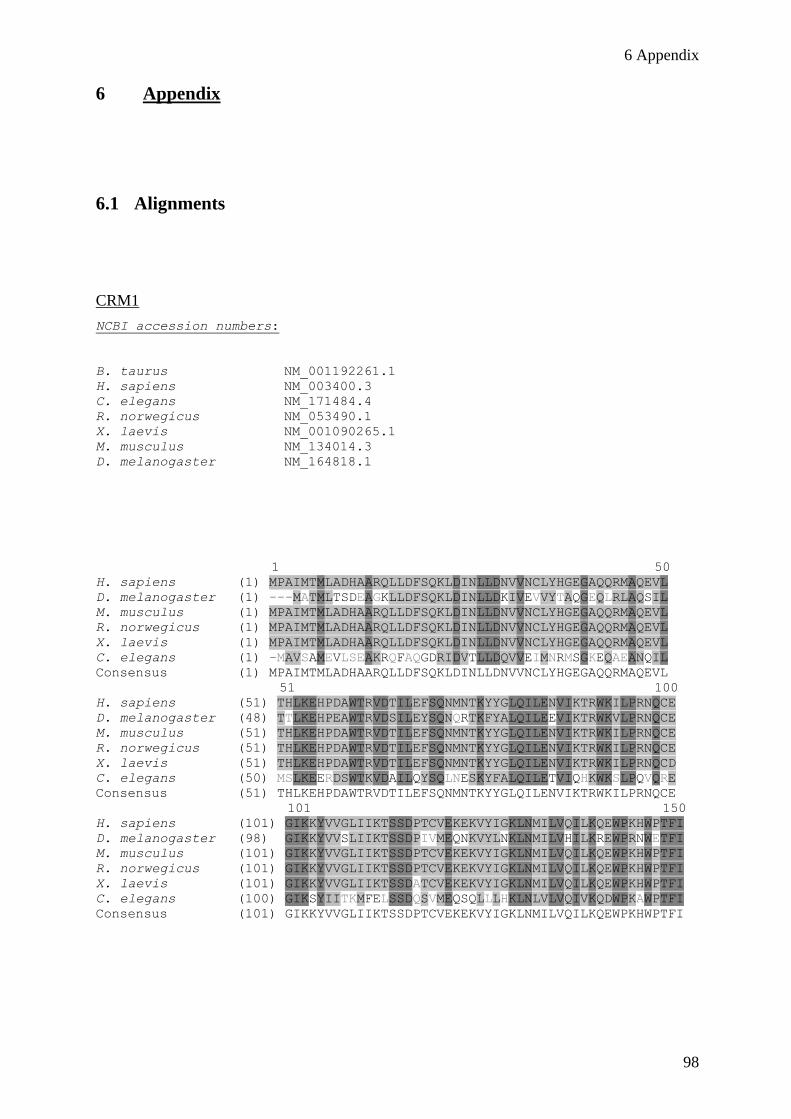
6 Appendix
98
6 Appendix
6.1 Alignments
CRM1
NCBI accession numbers:
B. taurus NM_001192261.1
H. sapiens NM_003400.3
C. elegans NM_171484.4
R. norwegicus NM_053490.1
X. laevis NM_001090265.1
M. musculus NM_134014.3
D. melanogaster NM_164818.1
1 50
H. sapiens (1) MPAIMTMLADHAARQLLDFSQKLDINLLDNVVNCLYHGEGAQQRMAQEVL
D. melanogaster (1) ---MATMLTSDEAGKLLDFSQKLDINLLDKIVEVVYTAQGEQLRLAQSIL
M. musculus (1) MPAIMTMLADHAARQLLDFSQKLDINLLDNVVNCLYHGEGAQQRMAQEVL
R. norwegicus (1) MPAIMTMLADHAARQLLDFSQKLDINLLDNVVNCLYHGEGAQQRMAQEVL
X. laevis (1) MPAIMTMLADHAARQLLDFSQKLDINLLDNVVNCLYHGEGAQQRMAQEVL
C. elegans (1) -MAVSAMEVLSEAKRQFAQGDRIDVTLLDQVVEIMNRMSGKEQAEANQIL
Consensus (1) MPAIMTMLADHAARQLLDFSQKLDINLLDNVVNCLYHGEGAQQRMAQEVL
51 100
H. sapiens (51) THLKEHPDAWTRVDTILEFSQNMNTKYYGLQILENVIKTRWKILPRNQCE
D. melanogaster (48) TTLKEHPEAWTRVDSILEYSQNQRTKFYALQILEEVIKTRWKVLPRNQCE
M. musculus (51) THLKEHPDAWTRVDTILEFSQNMNTKYYGLQILENVIKTRWKILPRNQCE
R. norwegicus (51) THLKEHPDAWTRVDTILEFSQNMNTKYYGLQILENVIKTRWKILPRNQCE
X. laevis (51) THLKEHPDAWTRVDTILEFSQNMNTKYYGLQILENVIKTRWKILPRNQCD
C. elegans (50) MSLKEERDSWTKVDAILQYSQLNESKYFALQILETVIQHKWKSLPQVQRE
Consensus (51) THLKEHPDAWTRVDTILEFSQNMNTKYYGLQILENVIKTRWKILPRNQCE
101 150
H. sapiens (101) GIKKYVVGLIIKTSSDPTCVEKEKVYIGKLNMILVQILKQEWPKHWPTFI
D. melanogaster (98) GIKKYVVSLIIKTSSDPIVMEQNKVYLNKLNMILVHILKREWPRNWETFI
M. musculus (101) GIKKYVVGLIIKTSSDPTCVEKEKVYIGKLNMILVQILKQEWPKHWPTFI
R. norwegicus (101) GIKKYVVGLIIKTSSDPTCVEKEKVYIGKLNMILVQILKQEWPKHWPTFI
X. laevis (101) GIKKYVVGLIIKTSSDATCVEKEKVYIGKLNMILVQILKQEWPKHWPTFI
C. elegans (100) GIKSYIITKMFELSSDQSVMEQSQLLLHKLNLVLVQIVKQDWPKAWPTFI
Consensus (101) GIKKYVVGLIIKTSSDPTCVEKEKVYIGKLNMILVQILKQEWPKHWPTFI
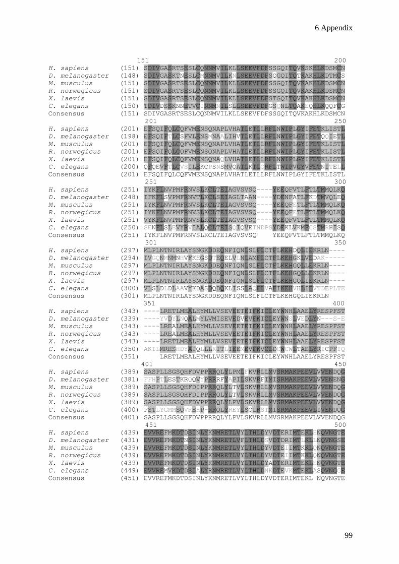
6 Appendix
99
151 200
H. sapiens (151) SDIVGASRTSESLCQNNMVILKLLSEEVFDFSSGQITQVKSKHLKDSMCN
D. melanogaster (148) SDIVGASKTNESLCMNNMVILKNLSEEVFDFSQGQITQTKAKHLKDTMCS
M. musculus (151) SDIVGASRTSESLCQNNMVILKLLSEEVFDFSSGQITQVKAKHLKDSMCN
R. norwegicus (151) SDIVGASRTSESLCQNNMVILKLLSEEVFDFSSGQITQVKAKHLKDSMCN
X. laevis (151) SDIVGASRTSESLCQNNMVILKLLSEEVFDFSTGQITQVKAKHLKDSMCN
C. elegans (150) TDIVDSSKNNETVCINNMNILSLLSEEVFDFGSQNLTQAKEQHLKQQFCG
Consensus (151) SDIVGASRTSESLCQNNMVILKLLSEEVFDFSSGQITQVKAKHLKDSMCN
201 250
H. sapiens (201) EFSQIFQLCQFVMENSQNAPLVHATLETLLRFLNWIPLGYIFETKLISTL
D. melanogaster (198) EFSQIFTLCSFVLENSMNAALIHVTLETLLRFLNWIPLGYIFETQQIETL
M. musculus (201) EFSQIFQLCQFVMENSQNAPLVHATLETLLRFLNWIPLGYIFETKLISTL
R. norwegicus (201) EFSQIFQLCQFVMENSQNAPLVHATLETLLRFLNWIPLGYIFETKLISTL
X. laevis (201) EFSQIFQLCQFVMENSQNAQLVHATLETLLRFLNWIPLGYIFETKLISTL
C. elegans (200) QFQEVFTLCVSILEKCPSNSMVQATLKTLQRFLTWIPVGYVFETNITELL
Consensus (201) EFSQIFQLCQFVMENSQNAPLVHATLETLLRFLNWIPLGYIFETKLISTL
251 300
H. sapiens (251) IYKFLNVPMFRNVSLKCLTEIAGVSVSQ----YEEQFVTLFTLTMMQLKQ
D. melanogaster (248) IFKFLSVPMFRNVTLKCLSEIAGLTAAN----YDENFATLFKDTMVQLEQ
M. musculus (251) IYKFLNVPMFRNVSLKCLTEIAGVSVSQ----YEEQFETLFTLTMMQLKQ
R. norwegicus (251) IYKFLNVPMFRNVSLKCLTEIAGVSVSQ----YEEQFETLFTLTMMQLKQ
X. laevis (251) VYKFLNVPMFRNVSLKCLTEIAGVSVSQ----YEEQFVTLFTLTMMQLKQ
C. elegans (250) SENFLSLEVYRVIALQCLTEISQIQVETNDPSYDEKLVKMFCSTMRHISQ
Consensus (251) IYKFLNVPMFRNVSLKCLTEIAGVSVSQ YEEQFVTLFTLTMMQLKQ
301 350
H. sapiens (297) MLPLNTNIRLAYSNGKDDEQNFIQNLSLFLCTFLKEHDQLIEKRLN----
D. melanogaster (294) IVGQNMNMNHVFKHGSDTEQELVLNLAMFLCTFLKEHGKLVEDAK-----
M. musculus (297) MLPLNTNIRLAYSNGKDDEQNFIQNLSLFLCTFLKEHGQLLEKRLN----
R. norwegicus (297) MLPLNTNIRLAYSNGKDDEQNFIQNLSLFLCTFLKEHGQLLEKRLN----
X. laevis (297) MLPLNTNIRLAYSNGKDDEQNFIQNLSLFLCTFLKEHGQLIEKRLN----
C. elegans (300) VLSLDLDLAAVYKDASDQDQKLISSLAQFLVAFIKEHVHLIEVTDEPLTE
Consensus (301) MLPLNTNIRLAYSNGKDDEQNFIQNLSLFLCTFLKEHGQLIEKRLN
351 400
H. sapiens (343) ----LRETLMEALHYMLLVSEVEETEIFKICLEYWNHLAAELYRESPFST
D. melanogaster (339) ----YVDYLNQALMYLVMISEVEDVEVFKICLEYWNSLVEDLYN---S-E
M. musculus (343) ----LREALMEALHYMLLVSEVEETEIFKICLEYWNHLAAELYRESPFST
R. norwegicus (343) ----LREALMEALHYMLLVSEVEETEIFKICLEYWNHLAAELYRESPFST
X. laevis (343) ----LRETLMEALHYMLLVSEVEETEIFKICLEYWNHLAAELYRESPFST
C. elegans (350) AKILMRESHDYAIQLLLKITLIEEMEVFKVCLDCWCWLTAELYRICPFIQ
Consensus (351) LRETLMEALHYMLLVSEVEETEIFKICLEYWNHLAAELYRESPFST
401 450
H. sapiens (389) SASPLLSGSQHFDVPPRRQLYLPMLFKVRLLMVSRMAKPEEVLVVENDQG
D. melanogaster (381) FFHPTLESTKRQQVYPRRRFYAPILSKVRFIMISRMAKPEEVLVVENENG
M. musculus (389) SASPLLSGSQHFDIPPRRQLYLTVLSKVRLLMVSRMAKPEEVLVVENDQG
R. norwegicus (389) SASPLLSGSQHFDIPPRRQLYLTVLSKVRLLMVSRMAKPEEVLVVENDQG
X. laevis (389) SASPLLSGSQHFDVPPRRQLYLPVLSKVRLLMVSRMAKPEEVLVVENDQG
C. elegans (400) PSTLYGMMSQVREHP-RRQLYREYLSQLRSTMISRMAKPEEVLIVENDQG
Consensus (401) SASPLLSGSQHFDVPPRRQLYLPVLSKVRLLMVSRMAKPEEVLVVENDQG
451 500
H. sapiens (439) EVVREFMKDTDSINLYKNMRETLVYLTHLDYVDTERIMTEKLHNQVNGTE
D. melanogaster (431) EVVREFMKDTNSINLYKNMRETLVFLTHLDSVDTDRIMTLKLLNQVNGSE
M. musculus (439) EVVREFMKDTDSINLYKNMRETLVYLTHLDYVDTEIIMTKKLQNQVNGTE
R. norwegicus (439) EVVREFMKDTDSINLYKNMRETLVYLTHLDYVDTEIIMTKKLQNQVNGTE
X. laevis (439) EVVREFMKDTDSINLYKNMRETLVYLTHLDYADTERIMTEKLHNQVNGTE
C. elegans (449) EVVREMVKDTDSIALYRNMRETLVYLTHLDNKDTEVKMTEKLASQVNGGE
Consensus (451) EVVREFMKDTDSINLYKNMRETLVYLTHLDYVDTERIMTEKL NQVNGTE
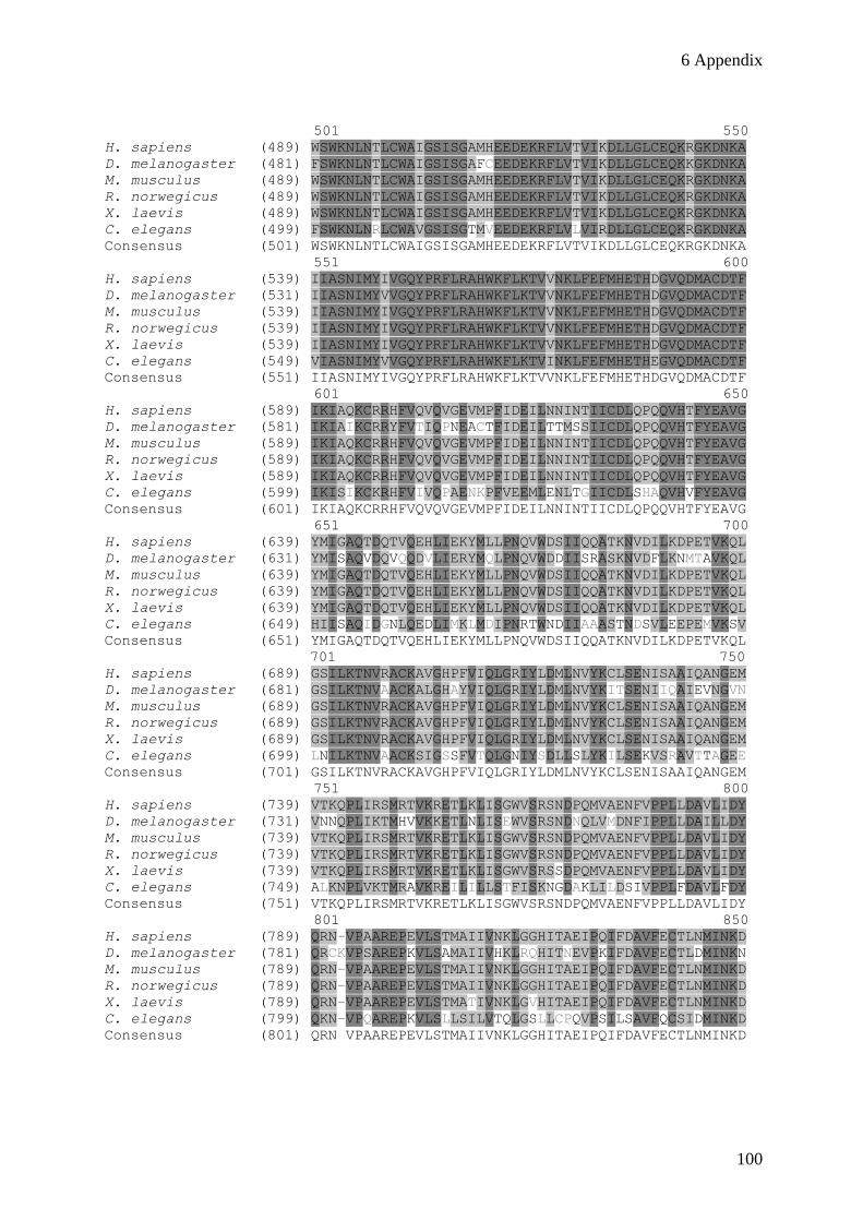
6 Appendix
100
501 550
H. sapiens (489) WSWKNLNTLCWAIGSISGAMHEEDEKRFLVTVIKDLLGLCEQKRGKDNKA
D. melanogaster (481) FSWKNLNTLCWAIGSISGAFCEEDEKRFLVTVIKDLLGLCEQKKGKDNKA
M. musculus (489) WSWKNLNTLCWAIGSISGAMHEEDEKRFLVTVIKDLLGLCEQKRGKDNKA
R. norwegicus (489) WSWKNLNTLCWAIGSISGAMHEEDEKRFLVTVIKDLLGLCEQKRGKDNKA
X. laevis (489) WSWKNLNTLCWAIGSISGAMHEEDEKRFLVTVIKDLLGLCEQKRGKDNKA
C. elegans (499) FSWKNLNRLCWAVGSISGTMVEEDEKRFLVLVIRDLLGLCEQKRGKDNKA
Consensus (501) WSWKNLNTLCWAIGSISGAMHEEDEKRFLVTVIKDLLGLCEQKRGKDNKA
551 600
H. sapiens (539) IIASNIMYIVGQYPRFLRAHWKFLKTVVNKLFEFMHETHDGVQDMACDTF
D. melanogaster (531) IIASNIMYVVGQYPRFLRAHWKFLKTVVNKLFEFMHETHDGVQDMACDTF
M. musculus (539) IIASNIMYIVGQYPRFLRAHWKFLKTVVNKLFEFMHETHDGVQDMACDTF
R. norwegicus (539) IIASNIMYIVGQYPRFLRAHWKFLKTVVNKLFEFMHETHDGVQDMACDTF
X. laevis (539) IIASNIMYIVGQYPRFLRAHWKFLKTVVNKLFEFMHETHDGVQDMACDTF
C. elegans (549) VIASNIMYVVGQYPRFLRAHWKFLKTVINKLFEFMHETHEGVQDMACDTF
Consensus (551) IIASNIMYIVGQYPRFLRAHWKFLKTVVNKLFEFMHETHDGVQDMACDTF
601 650
H. sapiens (589) IKIAQKCRRHFVQVQVGEVMPFIDEILNNINTIICDLQPQQVHTFYEAVG
D. melanogaster (581) IKIAIKCRRYFVTIQPNEACTFIDEILTTMSSIICDLQPQQVHTFYEAVG
M. musculus (589) IKIAQKCRRHFVQVQVGEVMPFIDEILNNINTIICDLQPQQVHTFYEAVG
R. norwegicus (589) IKIAQKCRRHFVQVQVGEVMPFIDEILNNINTIICDLQPQQVHTFYEAVG
X. laevis (589) IKIAQKCRRHFVQVQVGEVMPFIDEILNNINTIICDLQPQQVHTFYEAVG
C. elegans (599) IKISIKCKRHFVIVQPAENKPFVEEMLENLTGIICDLSHAQVHVFYEAVG
Consensus (601) IKIAQKCRRHFVQVQVGEVMPFIDEILNNINTIICDLQPQQVHTFYEAVG
651 700
H. sapiens (639) YMIGAQTDQTVQEHLIEKYMLLPNQVWDSIIQQATKNVDILKDPETVKQL
D. melanogaster (631) YMISAQVDQVQQDVLIERYMQLPNQVWDDIISRASKNVDFLKNMTAVKQL
M. musculus (639) YMIGAQTDQTVQEHLIEKYMLLPNQVWDSIIQQATKNVDILKDPETVKQL
R. norwegicus (639) YMIGAQTDQTVQEHLIEKYMLLPNQVWDSIIQQATKNVDILKDPETVKQL
X. laevis (639) YMIGAQTDQTVQEHLIEKYMLLPNQVWDSIIQQATKNVDILKDPETVKQL
C. elegans (649) HIISAQIDGNLQEDLIMKLMDIPNRTWNDIIAAASTNDSVLEEPEMVKSV
Consensus (651) YMIGAQTDQTVQEHLIEKYMLLPNQVWDSIIQQATKNVDILKDPETVKQL
701 750
H. sapiens (689) GSILKTNVRACKAVGHPFVIQLGRIYLDMLNVYKCLSENISAAIQANGEM
D. melanogaster (681) GSILKTNVAACKALGHAYVIQLGRIYLDMLNVYKITSENIIQAIEVNGVN
M. musculus (689) GSILKTNVRACKAVGHPFVIQLGRIYLDMLNVYKCLSENISAAIQANGEM
R. norwegicus (689) GSILKTNVRACKAVGHPFVIQLGRIYLDMLNVYKCLSENISAAIQANGEM
X. laevis (689) GSILKTNVRACKAVGHPFVIQLGRIYLDMLNVYKCLSENISAAIQANGEM
C. elegans (699) LNILKTNVAACKSIGSSFVTQLGNIYSDLLSLYKILSEKVSRAVTTAGEE
Consensus (701) GSILKTNVRACKAVGHPFVIQLGRIYLDMLNVYKCLSENISAAIQANGEM
751 800
H. sapiens (739) VTKQPLIRSMRTVKRETLKLISGWVSRSNDPQMVAENFVPPLLDAVLIDY
D. melanogaster (731) VNNQPLIKTMHVVKKETLNLISEWVSRSNDNQLVMDNFIPPLLDAILLDY
M. musculus (739) VTKQPLIRSMRTVKRETLKLISGWVSRSNDPQMVAENFVPPLLDAVLIDY
R. norwegicus (739) VTKQPLIRSMRTVKRETLKLISGWVSRSNDPQMVAENFVPPLLDAVLIDY
X. laevis (739) VTKQPLIRSMRTVKRETLKLISGWVSRSSDPQMVAENFVPPLLDAVLIDY
C. elegans (749) ALKNPLVKTMRAVKREILILLSTFISKNGDAKLILDSIVPPLFDAVLFDY
Consensus (751) VTKQPLIRSMRTVKRETLKLISGWVSRSNDPQMVAENFVPPLLDAVLIDY
801 850
H. sapiens (789) QRN-VPAAREPEVLSTMAIIVNKLGGHITAEIPQIFDAVFECTLNMINKD
D. melanogaster (781) QRCKVPSAREPKVLSAMAIIVHKLRQHITNEVPKIFDAVFECTLDMINKN
M. musculus (789) QRN-VPAAREPEVLSTMAIIVNKLGGHITAEIPQIFDAVFECTLNMINKD
R. norwegicus (789) QRN-VPAAREPEVLSTMAIIVNKLGGHITAEIPQIFDAVFECTLNMINKD
X. laevis (789) QRN-VPAAREPEVLSTMATIVNKLGVHITAEIPQIFDAVFECTLNMINKD
C. elegans (799) QKN-VPQAREPKVLSLLSILVTQLGSLLCPQVPSILSAVFQCSIDMINKD
Consensus (801) QRN VPAAREPEVLSTMAIIVNKLGGHITAEIPQIFDAVFECTLNMINKD
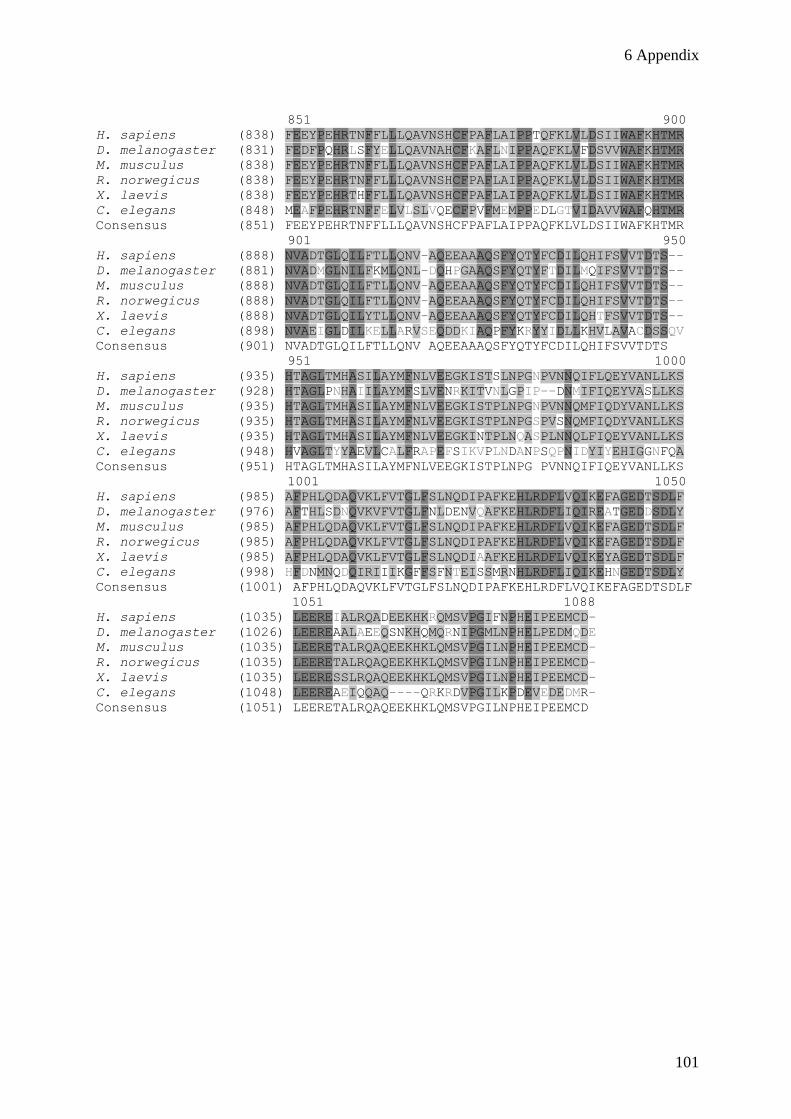
6 Appendix
101
851 900
H. sapiens (838) FEEYPEHRTNFFLLLQAVNSHCFPAFLAIPPTQFKLVLDSIIWAFKHTMR
D. melanogaster (831) FEDFPQHRLSFYELLQAVNAHCFKAFLNIPPAQFKLVFDSVVWAFKHTMR
M. musculus (838) FEEYPEHRTNFFLLLQAVNSHCFPAFLAIPPAQFKLVLDSIIWAFKHTMR
R. norwegicus (838) FEEYPEHRTNFFLLLQAVNSHCFPAFLAIPPAQFKLVLDSIIWAFKHTMR
X. laevis (838) FEEYPEHRTHFFLLLQAVNSHCFPAFLAIPPAQFKLVLDSIIWAFKHTMR
C. elegans (848) MEAFPEHRTNFFELVLSLVQECFPVFMEMPPEDLGTVIDAVVWAFQHTMR
Consensus (851) FEEYPEHRTNFFLLLQAVNSHCFPAFLAIPPAQFKLVLDSIIWAFKHTMR
901 950
H. sapiens (888) NVADTGLQILFTLLQNV-AQEEAAAQSFYQTYFCDILQHIFSVVTDTS--
D. melanogaster (881) NVADMGLNILFKMLQNL-DQHPGAAQSFYQTYFTDILMQIFSVVTDTS--
M. musculus (888) NVADTGLQILFTLLQNV-AQEEAAAQSFYQTYFCDILQHIFSVVTDTS--
R. norwegicus (888) NVADTGLQILFTLLQNV-AQEEAAAQSFYQTYFCDILQHIFSVVTDTS--
X. laevis (888) NVADTGLQILYTLLQNV-AQEEAAAQSFYQTYFCDILQHTFSVVTDTS--
C. elegans (898) NVAEIGLDILKELLARVSEQDDKIAQPFYKRYYIDLLKHVLAVACDSSQV
Consensus (901) NVADTGLQILFTLLQNV AQEEAAAQSFYQTYFCDILQHIFSVVTDTS
951 1000
H. sapiens (935) HTAGLTMHASILAYMFNLVEEGKISTSLNPGNPVNNQIFLQEYVANLLKS
D. melanogaster (928) HTAGLPNHAIILAYMFSLVENRKITVNLGPIP--DNMIFIQEYVASLLKS
M. musculus (935) HTAGLTMHASILAYMFNLVEEGKISTPLNPGNPVNNQMFIQDYVANLLKS
R. norwegicus (935) HTAGLTMHASILAYMFNLVEEGKISTPLNPGSPVSNQMFIQDYVANLLKS
X. laevis (935) HTAGLTMHASILAYMFNLVEEGKINTPLNQASPLNNQLFIQEYVANLLKS
C. elegans (948) HVAGLTYYAEVLCALFRAPEFSIKVPLNDANPSQPNIDYIYEHIGGNFQA
Consensus (951) HTAGLTMHASILAYMFNLVEEGKISTPLNPG PVNNQIFIQEYVANLLKS
1001 1050
H. sapiens (985) AFPHLQDAQVKLFVTGLFSLNQDIPAFKEHLRDFLVQIKEFAGEDTSDLF
D. melanogaster (976) AFTHLSDNQVKVFVTGLFNLDENVQAFKEHLRDFLIQIREATGEDDSDLY
M. musculus (985) AFPHLQDAQVKLFVTGLFSLNQDIPAFKEHLRDFLVQIKEFAGEDTSDLF
R. norwegicus (985) AFPHLQDAQVKLFVTGLFSLNQDIPAFKEHLRDFLVQIKEFAGEDTSDLF
X. laevis (985) AFPHLQDAQVKLFVTGLFSLNQDIAAFKEHLRDFLVQIKEYAGEDTSDLF
C. elegans (998) HFDNMNQDQIRIIIKGFFSFNTEISSMRNHLRDFLIQIKEHNGEDTSDLY
Consensus (1001) AFPHLQDAQVKLFVTGLFSLNQDIPAFKEHLRDFLVQIKEFAGEDTSDLF
1051 1088
H. sapiens (1035) LEEREIALRQADEEKHKRQMSVPGIFNPHEIPEEMCD-
D. melanogaster (1026) LEEREAALAEEQSNKHQMQRNIPGMLNPHELPEDMQDE
M. musculus (1035) LEERETALRQAQEEKHKLQMSVPGILNPHEIPEEMCD-
R. norwegicus (1035) LEERETALRQAQEEKHKLQMSVPGILNPHEIPEEMCD-
X. laevis (1035) LEERESSLRQAQEEKHKLQMSVPGILNPHEIPEEMCD-
C. elegans (1048) LEEREAEIQQAQ----QRKRDVPGILKPDEVEDEDMR-
Consensus (1051) LEERETALRQAQEEKHKLQMSVPGILNPHEIPEEMCD
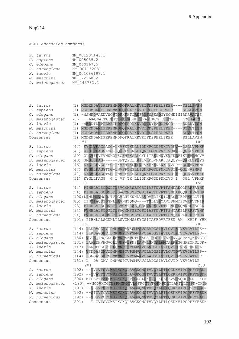
6 Appendix
102
Nup214
NCBI accession numbers:
B. taurus NM_001205443.1
H. sapiens NM_005085.2
C. elegans NM_060167.5
R. norwegicus NM_001162031 X. laevis NM_001086197.1 M. musculus NM_172268.2 D. melanogaster NM_143782.2
1 50
B. taurus (1) MGDEMDAVIPEREMKDFQFRALKKVRIFDSPEELPKER----SSLLSVSN
H. sapiens (1) MGDEMDAMIPEREMKDFQFRALKKVRIFDSPEELPKER----SSLLAVSN
C. elegans (1) -MSNEDVAEDVSQVTDFHFHTCRKFRLFSSKSDGYSQNEINIRNRVATSS
D. melanogaster (1) ---MAQNAPDCEDTQDFQFKLHHKIVAFKKCG-EPRSN----VNLLAVSS
X. laevis (1) -MEDDTDLPPERETKDFQFRQLKKVRLFDYPADLPKQR----SNLLVISN
M. musculus (1) MGDEMDAMIPEREMKDFQFRALKKVRIFDSPEELPKER----SSVLTISN
R. norwegicus (1) MGDEMDAMIPEREMKDFQFRALKKVRIFDSPEELPKER----SSVLTVSN
Consensus (1) MGDEMDAMIPEREMKDFQFRALKKVRIFDSPEELPKER SSLLAVSN
51 100
B. taurus (47) KYGLVFAGGASG-LHVFSTKNLLIQNKPGDDPNKIVDKV--QSLLVPMKF
H. sapiens (47) KYGLVFAGGASG-LQIFPTKNLLIQNKPGDDPNKIVDKV--QGLLVPMKF
C. elegans (50) QLGVTFVTVNSNQLSCFHTKSLLGYKITRENMNVEVTDLPIKTIRLHGVV
D. melanogaster (43) SRGLLFAG------SPTQPELKVIIVKDLVNAKSTAQQP--QARLVPLPS
X. laevis (46) KYGLLFVGGFMG-LKVFHTKDILVTVKPKENANKTVVGP--QGIHVPMNS
M. musculus (47) KYGMLFAGGTNG-LNVFPTKSLLIQNKPGDDPNKIVDTI—QGLNVPMKF
R. norwegicus (47) KYGMLFAGGTNG-LNVFPTKSLLIQNKPGDDPNKIVDTI--QGLNVPMKF
Consensus (51) KYGLLFAGG G L VF TK LLIQNKPGDDPNKIVD I QGL VPMKF
101 150
B. taurus (94) PIHHLALSCDNLTLSVCMMSSEYGSIIAFFDVRTFSNEAKQQKRPFAYHK
H. sapiens (94) PIHHLALSCDNLTLSACMMSSEYGSIIAFFDVRTFSNEAKQQKRPFAYHK
C. elegans (100)LINDMGVNSDGTVLGVLHTKNNDVSVDVFDIKKICTSSSIEPFKPLCTTR
D. melanogaster (85) IPNYIACSSDGNLLAVNHTQNG----TSLLSIYAVLSFMTPDVRPVYNIR
X. laevis (93) PIHHLALSSDNLTLSVCMTSAEQGSSVSFYDVRTLLNESKQNKMPFASCK
M. musculus (94) PVHHLALSCDSLTLSACMMSSEYGSIIAFFDVRTFSNQAKPLKRPFTYHK
R. norwegicus (94) PVHHLALSCDNLTLSACMMSSEYGSIIAFFDVRTFSNQAKPLKRPFTYHK
Consensus (101) PIHHLALSCDNLTLSVCMMSSEYGSIIAFFDVRTFSN AK KRPF YHK
151 200
B. taurus (144) LLKDAGGLVIDMKWNPAVSSMVAVCLADGSIAVLQVTETVKVCATLPS--
H. sapiens (144) LLKDAGGMVIDMKWNPTVPSMVAVCLADGSIAVLQVTETVKVCATLPS--
C. elegans (150) VGTEQINQGSCLEWNPAFPDTFAASSTDRSILVAKINVQSPANQKLVGIG
D. melanogaster (131) LAAEDHVHGVQLLWNPVLPNSLAVVLSNGALAMYALKEGGNFEMHSLDK-
X. laevis (143) LLRDPSSSVTDLQWNPTLPSMVAVCLSDGSISVLQVTDTVSVFANLPA--
M. musculus (144) VSNDASGMVNDMKWNPTVPSMVAVCLADGSISVLQVTDVVKVCATLPP--
R. norwegicus (144) LSNGASGMVNDMKWNPTVPSMAAVCLADGSISVLQVTDVVKVCATLPP--
Consensus (151) L DA GMV DMKWNPTVPSMVAVCLADGSIAVLQVTD VKVCATLP
201 250
B. taurus (192) --TVGVTSVCWSPKGKQLAVGKQNGTVVQYLPTLQEKKVIPCPPFYEADH
H. sapiens (192) --TVAVTSVCWSPKGKQLAVGKQNGTVVQYLPTLQEKKVIPCPPFYESDH
C. elegans (200) KFGAVTTAISWSPKGKQLTIGDSLGKIVQLKPELEVVRSQHGPEN--KPN
D. melanogaster (180) --NQQVKCGCWSPKGKQIVLGFPGGTVKQFKPDLTLAKTLLCPPN-IHDA
X. laevis (191) --TLGVTSVCWSPKGKQLAVGKQNGTVVQYLPSLQEKKVIPCPSFYDSDN
M. musculus (192) --STGVTCVCWSPKGKQLAVGKQNGTVVQYLPTLQEKKVIPCPPFYESDH
R. norwegicus (192) --SSGVTCVCWSPKGKQLAVGKQNGTVVQYLPTLQEKKVIPCPPFYEADH
Consensus (201) T GVTSVCWSPKGKQLAVGKQNGTVVQYLPTLQEKKVIPCPPFYESDH

6 Appendix
103
251 300
B. taurus (240) PVRVLDVLWIGTYEFTIVYAAADGTLETAPDVVMALLPKKEEKHPEVFMN
H. sapiens (240) PVRVLDVLWIGTYVFAIVYAAADGTLETSPDVVMALLPKKEEKHPEIFVN
C. elegans (248) YGRITGLCWLATTEWLVSLENG--------TDQDAYLMRCKKDKPTEWIQ
D. melanogaster (227) PFDTIAIQWLSTFQFAVIFLQHGEDCSPSLYILNAPKAG-----APSYIN
X. laevis (239) PVKVLDVLWLSTYVFTVVYAAADGSLEASPQLVIVTLPKKEDKRAERFLN
M. musculus (240) PVRVLDVLWIGTYVFTIVYAGADGTLETCPDVVMALLPKKEEKHPEIFVN
R. norwegicus (240) PVRVLDVLWIGTYVFTIVYAGADGALETCPDVVMALLPKKEEKHPEIFVN
Consensus (251) PVRVLDVLWIGTYVFTIVYAAADGTLETSPDVVMALLPKKEEKHPEIFVN
301 350
B. taurus (290) FMEPCYGSC---TERQHRYYLSYIEEWDLVLAASAASTEVSILARQSS--
H. sapiens (290) FMEPCYGSC---TERQHHYYLSYIEEWDLVLAASAASTEVSILARQSD--
C. elegans (290) FHELSYSSSKWPLPPQLFPATQLLVDWNVVIVGNSKTSEISTVGKRDD--
D. melanogaster (272) YYDICYSMN---GPRNHQFVFSHVPQWNLLLVVSANGVEVGIMRSTEAGD
X. laevis (289) FTETCYSIC---SERQHHFFLNYIEDWEILLAASAASVDVGVIARPPD--
M. musculus (290) FMEPCYSSC---TERQHHYYLSYIEEWDLVLAASAASTEVSILARQND--
R. norwegicus (290) FMEPCYSSC---TERQHHYYLSYIEEWDLVLAASAASTEVSILARQSD--
Consensus (301) FMEPCYSSC TERQHHYYLSYIEEWDLVLAASAASTEVSILARQ D
351 400
B. taurus (335) QNSWESWLLEDSSRAELPVTDKSDDSLPMGVAIDYTNQVEITITEE--KT
H. sapiens (335) QINWESWLLEDSSRAELPVTDKSDDSLPMGVVVDYTNQVEITISDE--KT
C. elegans (338) --WQTWVPVEGES-IYLPTTSSGKDTVPIGVAVDRSMTDEVLLNPDGSQR
D. melanogaster (319) TPAWQQLTLLDEARIEMPLSEDKDETFPLGFAFDTSTTHQLTINE---KK
X. laevis (334) QVGWEQWLLEDSSRAEMPMTENNDDTLPMGVALDYTCQLEVFISES--QI
M. musculus (335) QTNWESWLLEDSSRAELPVTDKSDDSLPMGVAIDYTNEVEVTINEE--KT
R. norwegicus (335) QTNWESWVLEDSSRAELPVTDKSDDSLPVGVAIDYTNEVEVAINEE--KT
Consensus (351) Q WESWLLEDSSRAELPVTDKSDDSLPMGVAIDYTN VEVTINEE KT
401 450
B. taurus (383) LPPAPVLMLLSTDGVLCPFYMINQNPGVRSLLRTPERLSLEGERQPKSSG
H. sapiens (383) LPPAPVLMLLSTDGVLCPFYMINQNPGVKSLIKTPERLSLEGERQPKSPG
C. elegans (385) HRPSPLVLCLTNDGILTAHHIISTFAAHIPCQMSS---------------
D. melanogaster (366) LQTMPMVHVLSSDGELLSFNFLNVLPTAVSVCSPP---------------
X. laevis (382) LPPVPVLLLLSTDGVLCPFHVVNLNQGVKPLTTSPEQLSLDGEREMKVVG
M. musculus (383) LPPAPVLLLLSTDGVLCPFYMINQNPGVRSLIKTLELISTEGERQPKSSG
R. norwegicus (383) LPPAPVLLLLSTDGVLCPFYMINQNPGVRSLIKTLELISAEGERQPKSSG
Consensus (401) LPPAPVLLLLSTDGVLCPFYMINQNPGVRSLIKTPE LS EGERQPKS G
451 500
B. taurus (433) -------SIPTTPTSPQAPQKLDVPAAAAPASVLPSSQAAPTSTFSLP--
H. sapiens (433) -------STPTTPTSSQAPQKLDASAAAAPASLPPSSPAAPIATFSLLPA
C. elegans (420) ------------------------------------QN------------
D. melanogaster (401) ---P-----PVADTSGQFKPLNMLLASEEEEQPAWAAS--PSKAPAATPA
X. laevis (432) GTAVSTPPAPLTSVSAPAPPASAAPRSAAPPPYPFGLSTASSGAPTPVLN
M. musculus (433) -------SFPGTPSSPQAPQNLDAPATASSPLPPVS--AAPTSTFPMPSA
R. norwegicus (433) -------SFPSTPPSPQAPQNLDAPATASSPLPPVS--AAPTATFPLPSA
Consensus (451) S P TPTS QAPQ LDAPASAA P S AAPTATF L A
501 550
B. taurus (474) ---SAVFSFGSSSLKSSGSVPGEAPLSSSGSDGAKAALVSGTSPFPFAPP
H. sapiens (476) GGAPTVFSFGSSSLKSSATVTGEPPSYSSGSDSSKAAPGPGPSTFSFVPP
C. elegans (422) ---------------------------------------LAINDLKKLQF
D. melanogaster (441) ASSDISFAFTPNTVTSTPAPSKDKQ----------------PSLFSGFGA
X. laevis (482) PPASLAPAATPTKTTSQPAAAATSIFQPAGPAAGSLQP-PSLPAFSFSSA
M. musculus (474) AGSPSVFSFGPSSFKSSASVTGEPPLYPTGSDSSRAAPGSGTSTFSFAPP
R. norwegicus (474) GGGPSVFSFGPSPFKS-SSIPGEPPPYPTGSDSSRAAPASGTPTFSFAPP
Consensus (501) A A VFSFGPSS KSS SV GE P SGSDSSKAAP G S FSFAPP
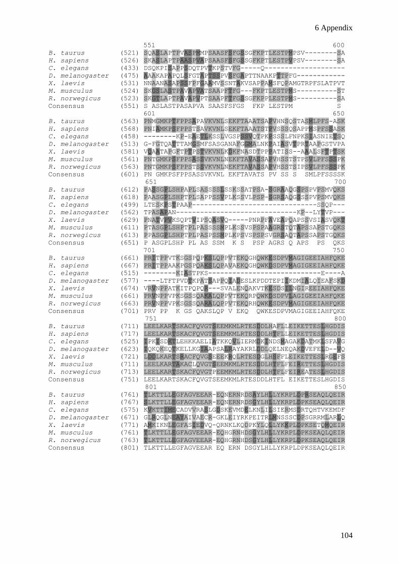
6 Appendix
104
551 600
B. taurus (521) SQASLAPTPVASPMMPSAASFSFGSSGFKPTLESTPMPSV--------SA
H. sapiens (526) SKASLAPTPAASPVAPSAASFSFGSSGFKPTLESTPVPSV--------SA
C. elegans (433) DSQKPISAPPSDQTPVTKPSTVFG-----Q--------------------
D. melanogaster (475) AAAKAPAPQLSFGTAPTSSPVSFGAPTTNAAKPTTPFG------------
X. laevis (531) NNAANASAPSSFPFGAAMVSSNTAKVSAPPAMSFQPAMGTRPFSLATPVT
M. musculus (524) SKGSLASTPAVAPVATSAAPFTFG---FKPTLESTPMS----------ST
R. norwegicus (523) SKGTLAPTPAVAPVPTSAAPFTFGSSGFKPPLESTPMS----------SA
Consensus (551) S ASLASTPASAPVA SAASFSFGS FKP LESTPM S
601 650
B. taurus (563) PNMGMKPTFPPSAPAVKVNLSEKFTAAATSAPVHNSQSTASMLPFS-ASK
H. sapiens (568) PNIAMKPSFPPSTSAVKVNLSEKFTAAATSTPVSSSQSAPPMSPFSSASK
C. elegans (458) --------KP-EAETLKSSLVGSPSSVQTPKPSSSLFNPKSIASNIETSQ
D. melanogaster (513) G-FGTQATTTAMGSMFSASGANAFGGMALNKPAIASVTPRTAAPGSTVPA
X. laevis (581) VQAATAPGFTPTPSTVKVNLKDKFNASDTPPPATISS--AAALSFTPTSK
M. musculus (561) PNTGMKPSFPPSASSVKVNLNEKFTAVASSAPVHSSTSTPSVLPFSSSPK
R. norwegicus (563) PNTGMKPSFPPSTSSVKVNLNEKFTAVASSAPVHSSTSIPSVLPFSSSPK
Consensus (601) PN GMKPSFPPSASSVKVNL EKFTAVATS PV SS S SMLPFSSSSK
651 700
B. taurus (612) PASSGPLSHPAPLSASSSSLSSKSSATPSA-SGRAAQGSPSPVPSMVQKS
H. sapiens (618) PAASGPLSHPTPLSAPPSSVPLKSSVLPSP-SGRSAQGSSSPVPSMVQKS
C. elegans (499) LTESKPSTPAAP-------------------------------SSQP---
D. melanogaster (562) TPASAPAN------------------------------KP--LYTVP---
X. laevis (629) PNATVPVKSQPTVIPSQASVQ-----PNRPFAVEAPQAPSSVSIASVQKT
M. musculus (611) PTASGPLSHPTPLPASSSSMPLKSSVSPSPAAGRSTQTAPSSAPSTGQKS
R. norwegicus (613) PPASGPLSHPTPLPASPSSMPLKPSVSPSPSVGRSAQTAPSSAPSTGQKS
Consensus (651) P ASGPLSHP PL AS SSM K S PSP AGRS Q APS PS QKS
701 750
B. taurus (661) PRITPPVTKSGSPQPKSLQPPVTEKQGHQWKESDPVMAGIGEEIAHFQKE
H. sapiens (667) PRITPPAAKPGSPQAKSLQPAVAEKQGHQWKDSDPVMAGIGEEIAHFQKE
C. elegans (515) --------KIASTPKS----------------------------E----A
D. melanogaster (577) ----LTFTPVDTKPATSAPPQIADESLKPDDTEPIIKDMIALQIEAFSKD
X. laevis (674) VRVNPPATKITPQPQR---SVALENQAKVTKESDSILNGIREEIAHFQKE
M. musculus (661) PRVNPPVPKSGSSQAKALQPPVTEKQRPQWKDSDPVLAGIGEEIAHFQKE
R. norwegicus (663) PRVNPPVPKSGSSQARALQPPVTEKQRHQWKESDPVMAGIGEEIAHFQKE
Consensus (701) PRV PP K GS QAKSLQP V EKQ QWKESDPVMAGIGEEIAHFQKE
751 800
B. taurus (711) LEELKARTSKACFQVGTSEEMKMLRTESDDLHAFLLEIKETTESLHGDIS
H. sapiens (717) LEELKARTSKACFQVGTSEEMKMLRTESDDLHTFLLEIKETTESLHGDIS
C. elegans (525) IPKISDKTLEHKKAELIATKKQVLIERMDKINDSMAGAKDATMKLSFAVG
D. melanogaster (623) IQKQKEQTKELLKGIAAPSALRAYAKRLDDLQELNEQAKDVEFELD--VQ
X. laevis (721) LDDLKARTSRACFQVGSEEEKRQLRTESDGLHSFFLEIKETTESLRGEFS
M. musculus (711) LEELKARTAKACLQVGTSEEMKMLRTESDDLHTFLFEIRETTESLHGDIS
R. norwegicus (713) LEELKARTSKACFQVGTPEEMKMLRTESDDLHTFLFEIREATESLHGDIS
Consensus (751) LEELKARTSKACFQVGTSEEMKMLRTESDDLHTFL EIKETTESLHGDIS
801 850
B. taurus (761) TLKTTLLEGFAGVEEAR-EQNERNRDSAYLHLLYKRPLDPRSEAQLQEIR
H. sapiens (767) SLKTTLLEGFAGVEEAR-EQNERNRDSGYLHLLYKRPLDPKSEAQLQEIR
C. elegans (575) KVKTTIMECADVVRASLGDSKEVMDELKNLILSIERMSDRTQHTVKEMDF
D. melanogaster (671) GLRQGLNEAYAIVAECR-GKLEIYRKPEITRLMNSSSCDPSGRRMLARLQ
X. laevis (771) AMKIKNLEGFASIEDVQ-QRNKLKQDPKYLQLLYKKPLDPKSETQMQEIR
M. musculus (761) TLKTTLLEGFAGVEEAR-EQHGRNHDSGYLHLLYKRPLDPKSEAQLQEIR
R. norwegicus (763) TLKTTLLEGFAGVEEAR-EQHGRNHDSGYLHLLYKRPLDPKSEAQLQEIR
Consensus (801) TLKTTLLEGFAGVEEAR EQ ERN DSGYLHLLYKRPLDPKSEAQLQEIR

6 Appendix
105
851 900
B. taurus (810) RLHQYVKFAVQDVNDVLDLEWDRHLEQKKK--QKRLLVPERETLFNTLAN
H. sapiens (816) RLHQYVKFAVQDVNDVLDLEWDQHLEQKKK--QRHLLVPERETLFNTLAN
C. elegans (625) EIDEKMELVAGVEDGNQVLEKLRNMSETEKLMRFNKLETAADLLNGKYEE
D. melanogaster (720) SYVAANEAQLRLAQQHVDLQWEQFQDVVRRNSKSRMHMPCLEGIYQRLTR
X. laevis (820) RLNQYVKNAVQDVNDVLDLEWDQYLEEKQK--KKGIIIPERETLFNSLAN
M. musculus (810) RLHQYVKFAVQDVNDVLDLEWDRHLEQKKR--QRRLIVPERETLFNTLAN
R. norwegicus (812) RLHQYVKFAVQDVNDVLDLEWDRHLEQKKR--QRRLIVPERETLFNTLAN
Consensus (851) RLHQYVKFAVQDVNDVLDLEWDRHLEQKKK QRRLIVPERETLFNTLAN
901 950
B. taurus (858) NREIINQQRKRLNHLVDSLQQLRLYNQTSQWSLPLDVPSQSTTHSFDSDL
H. sapiens (864) NREIINQQRKRLNHLVDSLQQLRLYKQTSLWSLSSAVPSQSSIHSFDSDL
C. elegans (675) CSDLIKKLRMSLS-------------------------------------
D. melanogaster (770) LQNLTSNQR-----------------------------------------
X. laevis (868) HQEIINQQRPKLEQLVENLQKLRLYNQISQWNVP-----DSSTKSFDVEL
M. musculus (858) NREIINQQRKRLNQLVDSLQQLRLYNHTAPWSLPSALSTQSNSHSFDSDL
R. norwegicus (860) NREIINQQRKRLNHLVDSLQQLRLYNQTSPWNRPSTLSTQSDTPSFDSDL
Consensus (901) NREIINQQRKRLN LVDSLQQLRLYNQTS W LP L SQS T SFDSDL
951 1000
B. taurus (908) ESLRNALLKTTLEAHPKPLPKVPAKLSPVKQAQLRNFLAKRKTPPVRSTA
H. sapiens (914) ESLCNALLKTTIESHTKSLPKVPAKLSPMKQAQLRNFLAKRKTPPVRSTA
C. elegans (688) ---------------EKES-----------------LRKQAILSPLRLSS
D. melanogaster (779) ---------------IVQN----------------NIKSKLKERGLLQAA
X. laevis (913) ENMQKTLSQTAIDTQTKPQAKLPAKISPVKQSQLRNFLSKRKTPPVRSLA
M. musculus (908) E----CLLKTTIESHTKPSPRVPGKLSPAKQAQLRNFLAKRKTPPVRSTA
R. norwegicus (910) E----HLLKTTIESHTKPSPRVPGKLSPAKQAQLRNFLAKRKTPPVRSTA
Consensus (951) E LLKTTIESHTKP PKVPAKLSP KQAQLRNFLAKRKTPPVRSTA
1001 1050
B. taurus (958) PASLSRSAFLSQRYYEDLDEASSTSSISQSLESEDTRTSCK---------
H. sapiens (964) PASLSRSAFLSQRYYEDLDEVSSTSSVSQSLESEDARTSCK---------
C. elegans (706) NLNQLRS-------------------------------------------
D. melanogaster (798) LLDQEKSRTRTNEAVDTLTDSILTMSLSQVVDSNAAKLTRER--------
X. laevis (963) PANLSRSAFLAPSFFEDLDDVSSTSSLSDMADNDNRNPPPKEIER-----
M. musculus (954) PASLSRSAFLSQRYYEDLDEGSSASSVAQPLEGEDARPTCTSVAQPLEGE
R. norwegicus (956) PASLSRSAFLSQRYYEDLDEGSSASSVAHPLEGEDARPTPT---------
Consensus (1001)PASLSRSAFLSQRYYEDLDE SSTSSVSQ LE EDAR T
1051 1100
B. taurus (999) -------DDDAVVQVPRHAPVVRTPSIQPSLLPQATPFAKSHLIHGSQ-G
H. sapiens (1005)-------DDEAVVQAPRHAPVVRTPSIQPSLLPHAAPFAKSHLVHGSSPG
C. elegans (713) ---------------------------------------------GSETE
D. melanogaster (840) -----------------------------------------------LQK
X. laevis (1008)--QETPPPESTPVRVPKHAPVARTTSVQPGLGTASLPFQS-----GLHPA
M. musculus (1004)DAQPICKEEEAVVPVPRHAPVVRTPSIQPSLLPQSMPFAKPHLIHSSSPA
R. norwegicus (997) -----CREEEAVVPVPRHAPVVRTPSVQPSLLPQAMPFAKPHLIHSSSPA
Consensus (1051) DEEAVV VPRHAPVVRTPSIQPSLLP A PFAK HLIHGS PA
1101 1150
B. taurus (1041) MMGTSMSTPASKIIPQGADSTMLATKTVKHGAPGPSHPISAPQAAAAAAL
H. sapiens (1048 VMGTSVATSASKIIPQGADSTMLATKTVKHGAPSPSHPISAPQAAAAAAL
C. elegans (718) LALKVMRNVSKIIMDTREQIQRTELEFVRFQRDVKFQNFKKGKENLNFTQ
D. melanogaster (843) IRNIVQLQKINVICPQRPDRVGLKSEVILETKRRAEQIKRAAAKPATANK
X. laevis (1051 TSTPVAPSQSIRVIPQGADSTMLATKTVKHGAPN----ITAAQKAAVAAM
M. musculus (1054) VMSSAVSTSATKVIPQGADSTMLATKTVKHGAPGPSHTVAAPQAAAAAAL
R. norwegicus (1042) AMSSSISTSATKVIPQGADTTMLATKTVKHGAPGPSHTVAAPQAAAAAAL
Consensus (1101) VM SSMST ASKIIPQGADSTMLATKTVKHGAP PSH IAAPQAAAAAAL

6 Appendix
106
1151 1200
B. taurus (1091) RRQMASQAPAVSTLTESTLKNVPQVVNVQELKNNAT--APSAAVGSSGPY
H. sapiens (1098) RRQMASQAPAVNTLTESTLKNVPQVVNVQELKNNPA--TPSTAMGSSVPY
C. elegans (768) PLEMSSLDGDAP--------------------------------------
D. melanogaster (893) YTQAAVAPPSPP----DVAPT---------------------------P
X. laevis (1097) RRQTASQIPAAS-LTESTLQTVPQVVNVKELKNNGPGPTIPTVIGPTVPQ
M. musculus (1104) RRQMASQAPAMSTLTESTLKTVPQVVNVQELRSNPS--PPSAAMGSAVQH
R. norwegicus (1092) RRQMASQAPAVSTLTESTLKNVPQVVNVQELKSNPS--PPSAAMGSSGPH
Consensus (1151) RRQMASQAPAVSTLTESTLK VPQVVNVQELK N PS AMGSS PH
1201 1250
B. taurus (1139) SAAKTPHPVLTPAPANQAKQGSLINALK-----PSGPPPASSQLSSGDKA
H. sapiens (1146) STAKTPHPVLTPVAANQAKQGSLINSLK-----PSGPTPASGQLSSGDKA
C. elegans (780) --QGK---SLTDAESIKVRQALVNQIQK--------R-------------
D. melanogaster (911) AVAPMPQATVTVAPPLPKPMPSIPSVVE------KPG-------------
X. laevis (1146) SAAQVIHQVLATVGSVSARQAAPAAPLKNPPASASSIAPQTWQGSAPNKP
M. musculus (1152) SAAKTPHAVLTPVANSQAKQGSLINSFK-----PSGPTAASCQLSSGDKA
R. norwegicus (1140) SAAKAPHAVLTPVAASQAKQGSLINSFK-----PSGPTPVSCQLSSGDKA
Consensus (1201) SAAK PH VLTPVAA QAKQGSLINSLK PSGP P S QLSSGDKA
1251 1300
B. taurus (1184) S--VATKIETAVTSTPSTVG-QFSKPFSFS-SGTGFNFGIVTPTPSSNFT
H. sapiens (1191) S--GTAKIETAVTSTPSASG-QFSKPFSFSPSGTGFNFGIITPTPSSNFT
C. elegans (804) --------------GIVKTRNVIVESYKKSENSAAMKNDLLDTSNLSNAI
D. melanogaster (942) --------------VPTHPTTPVATPFSFSQSIPFVKTSTVTPTTNTVTP
X. laevis (1196) AAQAIPKSDPSASQAPAPSVSQVNKPVSFSPAAGGFSFSNVTSAPVTSAL
M. musculus (1197) VGQGTAKTESAATSTPSAAG-QLNKPFSFA-SPGTFTFGTITPTPSSSFT
R. norwegicus (1185) VGPGTAKTESPVTSTPSTG--QLNKPFSFA-SPGTFTFGTITPAPSSSFT
Consensus (1251) G K ESA TSTPS QL KPFSFS S GGF FG ITPTPSS FT
1301 1350
B. taurus (1230) –ATQGSAPPTKESSQLDAFSFGGGGKPFYETVPESLPSTGTMTSGSTGA
H. sapiens (1238) --AAQGATPSTKESSQPDAFSSGGGSKPSYEAIPESSPPS-----GITSA
C. elegans (840) LKLSMTPRRVMPSSSLFSASPSTPSTKSDAATQADEPPIVKTVVVTVESP
D. melanogaster (978) G-EAAKPGLSIFGGSTISSFSFGGGAAKSALSFGTGSPAVAAPTPKPNPL
X. laevis (1246) GSSSAGCAATARDSNQASSYMFGGTGKSLGSEGSFSFASLKPAS------
M. musculus (1245) --ATPGAGPPTKEPTQLEAFSFGGGGKPFSEAIPGNSPATGATSAPSTSV
R. norwegicus (1232) --ATPGAAPPTKESNQLEAFSFGGGGKPFCETIPGNPPATGPPSAPSTSV
Consensus (1301) AT GAAP TKESSQLDAFSFGGGGKP ETIP PATG S TS
1351 1400
B. taurus (1278) AGATPGESAPSSSRPAAPSGISLSTPAGKLEAPPSKLGELLFPSSLAGET
H. sapiens (1281) SNTTPGEPAASSSRPVAPSGTALSTTSSKLETPPSKLGELLFPSSLAGET
C. elegans (890) AKPIASAPAVSSPLIKLNTTTATTTMTTPKVTVPKEEANKTQDQKPIIST
D. melanogaster (1027) SAVEKPTPEPTKPK-----EQKAAESKEFKAVQPETE-ESKVPQKPKAET
X. laevis (1290) SSSSSSVVEPTMSKPSVVTAASTTATVTSTTAASSKPGEGLFQGFSGGET
M. musculus (1293) TAASLEDSAPSSSKPAAPPETTVSSASSKLETPPSKLGELLFPSSLAGET
R. norwegicus (1280) TTTSLEDSAPTSSK-PATPETPVSTASSKLETPPSKLGELLFPSSLAGET
Consensus (1351) S S D APSSSKP A S TALST SSKLETPPSKLGELLFPSSLAGET
1401 1450
B. taurus (1328) LGSFSGLRVGQADEPTKPASKAPSPGVTSAGAQPTKTPAAPSGFSSPSPA
H. sapiens (1331) LGSFSGLRVGQADDSTKPTNKASS--TSLTSTQPTKTSGVPSGFNFTAPP
C. elegans (940) PASSSIFSSGSLFGTKTQTPLVSK--------------------------
D. melanogaster (1071) ENKSFGFGGFTGTGGTVGNTSSSP-------------------FSFGGLG
X. laevis (1340) LGSFSGLRVGQADEASKVEVAKTP----TA-AQPVKLPSNPVLFSFAGAP
M. musculus (1343) LGSFSGLRVGQAEDSTKPVSKASS--TNLAGAQPAK----PSGVSFPNTS
R. norwegicus (1329) LGSFSGLRVGQAEESTKPVSKASS--TNLAGAQPAK----PSGFSFPAAS
Consensus (1401) LGSFSGLRVGQADESTKP SKASS A AQP K PSGFSF A
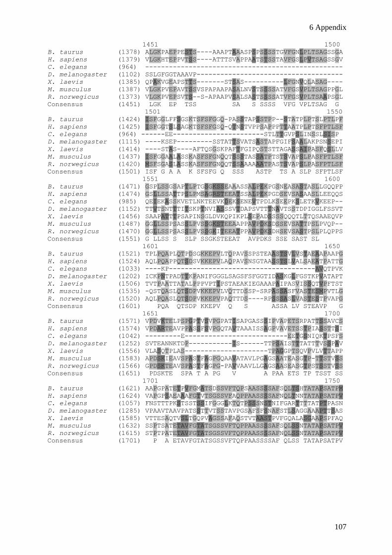
6 Appendix
107
1451 1500
B. taurus (1378) ALGKPAEPPESTS----AAAPTAAASPSPSSSSTGVFGNLPLTSAGSSGA
H. sapiens (1379) VLGKHTEPPVTSS----ATTTSVAPPAATSTSSTAVFGSLPVTSAGSSGV
C. elegans (964) --------------------------------------------------
D. melanogaster (1102) SSLGFGGTAAAVP-------------------------------------
X. laevis (1385) QPAKVGEAPSTTS-------STSAS----------LFGNVQLASAG----
M. musculus (1387) VLGKPVEPAVTSSVSPAPAAPASALNVSTSSSSATVFGSVPLTSAGPPGL
R. norwegicus (1373) VLGKPVEPSVTS--S-APAAPVSALSASTSSSSATVFGSVPLTSAAPSGL
Consensus (1451) LGK EP TSS SA S SSSS VFG VPLTSAG G
1501 1550
B. taurus (1424) ISFGGLFFSGSKTSFSFGGQ-PASSTAPSSTPP--STATPLPTSLPTLPF
H. sapiens (1425) ISFGGTSLSAGKTSFSFGSQ-QTNSTVPPSAPPPTTAATPLPTSFPTLSF
C. elegans (964) ------EE-----------------------STLTTGVPSLINSSLSISP
D. melanogaster (1115) ----KSEP---------SSTATTSVATSASTAPFGIFSAALAKPSNSEPI
X. laevis (1414) ----STAS----AFTQSGSKPAFTFGIPQSTSTTAGASSAIPASFQSLLV
M. musculus (1437) ISFGGAALASSKASFSFGNQQTSSSTASSATPTSTSVAPSLPASFPTLSF
R. norwegicus (1420) MSFSGASLASSKASFSFGNQQTSSAAAAAATPASTSVAPSLPASFPTLSF
Consensus (1501) ISF G A A K SFSFG Q SSS ASTP TS A SLP SFPTLSF
1551 1600
B. taurus (1471) GSPLSSGSAPTLPTGSGKSSEAAASSALPEKPGSNEASASTASLLGQQPP
H. sapiens (1474) GSLLSSATTPSLPMSAGRSTEEATSSALPEKPGDSEVSASAASLLEEQQS
C. elegans (985) QEIEKASSKVETLNKTEEVKDEKSENEVTPDLKSEEPKSLETKVKEEP--
D. melanogaster (1152) TTVTSNTTTITSKPTNVIASSSVTDAPSVTTTNAVTSSTDPIGGLFSSVT
X. laevis (1456) SAAPATTTPSAPINSGLDVKQPIKPLSEPADSSSSQQQTLTTQSAAEQVP
M. musculus (1487) GGLLSSPSASSLPVSSGKSTEEAAPPAVPDKSDSSEVSATTPSLPVQP--
R. norwegicus (1470) GGLLSSPSASSLPVSSGKITEEATPPAVPDKSDHSEVSASTPSLPLQPPS
Consensus (1551) G LLSS S SLP SSGKSTEEAT AVPDKS SSE SAST SL
1601 1650
B. taurus (1521) TPLPQAPLQTPDSGKKEPVLTQPAVSSPSTEAASTSVLVSTAEAAPAAPG
H. sapiens (1524) AQLPQAPPQTSDSVKKEPVLAQPAVSNSGTAASSTSLVALSAEATPATTG
C. elegans (1033) ----KP-------------------------------------AVQTPVK
D. melanogaster (1202) ICKPNTPADTTKPANIFGGGLSAGSFSFGGTIDASKGLFGSTKPVATAPT
X. laevis (1506) TVTPAATTATALPPPVPTIPSTAEAKIEGAAAPAIPASVISSQTVPFTST
M. musculus (1535) -QSTQASLQTSDPVKKEPVLVQTTDSSP-SRPASSASFVASTESMPVTLG
R. norwegicus (1520) AQLPQASLQTSDPVKKEPVPAQTTDS----RPSSSASLVASTESTPVAPG
Consensus (1601) PQA QTSDP KKEPV Q S ASSA LV STEAVP G
1651 1700
B. taurus (1571) VPDVKTELPSPGPTVSVPGPATISAPGASSSIPVAPETSRPATTSSAVCS
H. sapiens (1574) VPDARTEAVPPASSFSVPGQTAVTAAAISSAGPVAVETSSTPIASSTTSI
C. elegans (1042) ---------E--------------------------EETGSNIQKTPSFS
D. melanogaster (1252) SVTEANNKTDP-----------IS------TTPSAISTTTATTTVSSPAV
X. laevis (1556) VLASQTPLAS---------------------TPAGGPTSQVPVLVTTAPP
M. musculus (1583) APDSKIEAVSPASTFAGPGQAAVATAVLPGAGSAATEASGTP-TTSTVSS
R. norwegicus (1566) GPDSKTEAVSPASTFAGPG-PAVVAAVLLGAGSAASEASGTPSTSSTVSS
Consensus (1651) PDSKTE SPA T A PG V A PAA ETS TP TSST SS
1701 1750
B. taurus (1621) AAPGPATETPVFGMATSDSSVFTQPSAASSSSAFSQLTSHTATAPSATPM
H. sapiens (1624) VAPGPSAEAAAFGTVTSGSSVFAQPPAASSSSAFNQLTNNTATAPSATPV
C. elegans (1057) FNSTTTPKSTSSTSSIFGGGLKTQTPSSSNSTNIFGARTTTTATPTPASN
D. melanogaster (1285) VPAAVTAAVPATSSTTVTSSTAVPGSAFSFSNAFSTLSAGGAAAPTTSAS
X. laevis (1585) VTTESAQTVSLTGQPVAGSSAFAQSTVTAASTPVFGQALASGAAPSPFAQ
M. musculus (1632) SSPTSATETAVFGTATSGSSVFTQPPAASSSSAFSQLSSNTATAPSATPV
R. norwegicus (1615) STPTPATETAVFGTATSGSSVFTQPPAASSSSAFNQLSSNTATAPSATPV
Consensus (1701) P A ETAVFGTATSGSSVFTQPPAASSSSAF QLSS TATAPSATPV

6 Appendix
108
1751 1800
B. taurus (1671) FGHVAASTAPSLFGQQTGSMASTAAAAPQVSSSGFGSPAFGTSTPGVFGQ
H. sapiens (1674) FGQVAASTAPSLFGQQTGSTASTAAATPQVSSSGFSSPAFGTTAPGVFGQ
C. elegans (1107) TSSIFGG-------------------------------------------
D. melanogaster (1335) APLAAKSP----------TATSTGNNSSNSVFGGGFAVATSTAAPVASPF
X. laevis (1635) PTSSSVS------------TSANSSTGFGTSAFGATG------GNGGFGQ
M. musculus (1682) FGQVAAS------------ITSTAAATPQASSSGFGSPAFGASAPGVFGQ
R. norwegicus (1665) FGQVAAS------------IASTAAATPQASSSGFGSPAFGASAPGVFGQ
Consensus (1751) FG VAAS ASTAAATPQ SSSGF SPAFG SAPGVFGQ
1801 1850
B. taurus (1721) PGFGQAPAFGQSTSSPASSFSFSQPGFSSVPAFGQSVSSTPTSTSGNVFG
H. sapiens (1724) TTFGQASVFGQSASSAASVFSFSQPGFSSVPAFGQPASSTPTSTSGSVFG
C. elegans (1114) ----------GSKAASSPFGSFGQAGCQPAKTSNPATSTASVTFSFNTGA
D. melanogaster (1375) QSAAKSPVSSANIFGSIPKAETSVFGGATTAPSNTTAAATPDAPPAGLFA
X. laevis (1667) PSFGQAPLWKGPATS-QSTLPFSQPTFGTQPAFGQPAASTATSSAGSLFG
M. musculus (1720) TAFGQTPAFGQATSSPASGFSFSQPGFSSVPAFGQSVSSTPASTSANVFG
R. norwegicus (1703) TAFGQSPAFGQATSSPASGFSFSQPGFSSVPAFGQSVSSTPTSTSANVFG
Consensus (1801) AFGQAP FGQS SS AS FSFSQPGFSSVPAFGQS SSTPTSTSANVFG
1851 1900
B. taurus (1771) ASSSTSSSSSFSFGQSS-TNNGGGLFGQNNPPAFGQSPGFGQ-GGSVFGG
H. sapiens (1774) AASSTSSSSSFSFGQSS-PNTGGGLFGQSNAPAFGQSPGFGQ-GGSVFGG
C. elegans (1154) TSASAKPAGFGSFGAGASAKPSSVFGGSVTAPTVPNVDDGMEDDSMANGG
D. melanogaster (1425) SAAISNPSPFGSPTTRAPASGGNIFGQAVKPSVFGQPAQAGDSGGSIFGG
X. laevis (1716) CTSSASSFSFGQASNTSGTSTSGVLFGQSSAPVFGQSAAFPQ-AAPAFGS
M. musculus (1770) ATSSTSSPGSFSFGQAS-TNTGGTLFGQNNPPAFGQSPGFGQ-GSSVFGG
R. norwegicus (1753) ATSSTSSPGSFSFGQAS-TNTGGTLFGQNNPPAFGQSPGFGQ-GGSVFGS
Consensus (1851) ATSSTSSS SFSFGQAS TNTGG LFGQ NPPAFGQSPGFGQ GGSVFGG
1901 1950
B. taurus (1819) TSAATTTASSSGFSFCQASGFGSSNTGSLFGQAASTSGTVFGQQSSSSSG
H. sapiens (1822) TSAATTTAATSGFSFCQASGFGSSNTGSVFGQAASTGGIVFGQQSSSSSG
C. elegans (1204) G-------------------------------------------------
D. melanogaster (1475) GSASTPFGSS-------S-IFGGGNTQGAVGAPAAGSTSIFGQKVFGQSS
X. laevis (1765) ASVSTTTTAS--FGFGQPAGFASGTSGSLFNPSQSGSTSVFGQQPASSSG
M. musculus (1818) TSATTSTAAPSGFSFCQASGFGSSNTGSVFGQAANTGGSVFGQ-SSTSSG
R. norwegicus (1801) TSATTSTAAPSGFSFCQASGFGSSNTGSVFGQAASSGGSVFGQ-SSTSSG
Consensus (1901) TSASTTTAASSGFSFCQASGFGSSNTGSVFGQAAST GSVFGQ SSSSSG
1951 2000
B. taurus (1869) SVFGSGN-AGRGGGFFSGLGGKPSQDAANKNPFSSASG-GFGSTATPNTS
H. sapiens (1872) SVFGSGN-TGRGGGFFSGLGGKPSQDAANKNPFSSASG-GFGSTATSNTS
C. elegans (1205) -----S------GGFMSGLGNARTSNTSGGNNPFAPKTSTGTSASSS--S
D. melanogaster (1517) AAAPAAG-----GNIFSNPVGSPQ--ASPFGGGGNSIFGSPATAPPASGG
X. laevis (1813) GLFGAGSGGASTVGLFSGLGAKPSQEAANKNPFGSPGSSGFGSAGASNSS
M. musculus (1867) GVFGSGN-ATRGGGFFSGLGGKPSQDAANKNPFSSAGG-GFGSTAAPNTS
R. norwegicus (1850) SVFGSGN-ATRGGGFFSGLGGKPSQDAANKNPFSSASG-GFGSTPTPNTS
Consensus (1951) SVFGSGN A RGGGFFSGLGGKPSQDAANKNPFSSA G GFGSTATSNTS
2001 2050
B. taurus (1917) NLFGN-SGAKTFGGFASSSFGDQKPAGTFSSGGG-SVASQGFGFSTP---
H. sapiens (1920) NLFGN-SGAKTFGGFASSSFGEQKPTGTFSSGGG-SVASQGFGFSSP---
C. elegans (1242) WLFGG--------GGNQQQQQQQKPSFSFNTAG--SSAQQASAPATG---
D. melanogaster (1560) SIFGGGSSSGGFGSFTQTTPAQGGFGGGFGQGGGGSVAQTGFGSPQAPQQ
X. laevis (1863) NLFGN-SGAKAFGFGG--TSFGDKPSATFSAGG--SVASQGFSFNSP---
M. musculus (1915) NLFGN-SGAKTFGGFGSSSFGEQKPAGTFSSGGG-SVASQGFGFSTP---
R. norwegicus (1898) NLFGN-SGAKTFGGFG-SSFGEQKPAGTFSSGGG-SVASQGFGFSTP---
Consensus (2001) NLFGN SGAKTFGGFG SSFGEQKPAGTFSSGGG SVASQGFGFSTP

6 Appendix
109
2051 2100
B. taurus (1962) --NKTGGFGAAPVFGSPPTFGGSPGFGGVPAFGSAPAFTSPLGSTGGKVF
H. sapiens (1965) --NKTGGFGAAPVFGSPPTFGGSPGFGGVPAFGSAPAFTSPLGSTGGKVF
C. elegans (1279) ---TSSVFGGAPKFGSQPAFGAKP-FGGGANAGLSKNASIFGGATSSTTN
D. melanogaster (1610) QTTTPGGFGAKPVFGGSPAFGASPTFGGGATFGSPKGFGGFGGASPVASP
X. laevis (1905) --TKTGGFGAAPVFGSPPTFGGSPGFGGSPAFGTAAAFSNTLGSTGGKVF
M. musculus (1960) --NKTGGFGAAPVFGSPPTFGGSPGFGGVPAFGSAPAFTSPLGSTGGKVF
R. norwegicus (1942) --NKTGGFGAAPVFGSPPTFGGSPGFGGVPAFGSAPAFTSPLGSTGGKVF
Consensus (2051) NKTGGFGAAPVFGSPPTFGGSPGFGGVPAFGSAPAFTSPLGSTGGKVF
2101 2150
B. taurus (2010) GEGTAAASAGGFGFGSSSSTTSFGTLASQNAPTFGSLSQQSSGFGSQSGG
H. sapiens (2013) GEGTAAASAGGFGFGSSSNTTSFGTLASQNAPTFGSLSQQTSGFGTQSSG
C. elegans (1325) NPATGGFAQFASGQKTSSLFGGGATPQTNTSIFGGGANTTPAPTSSVFGG
D. melanogaster (1660) PPFGAAAKPAQGNIFETLGGQESGLSFGNLAQTGNSNAQKPAFGGSSFMN
X. laevis (1953) GEGTSAATTGGFGFGSNSSTAAFGSLATQNTPTFGSISQQSPGFGGQSSG
M. musculus (2008) GEGTAAASAGGFGFGSSGNTASFGTLASQNAPTFGSLSQQTSGFGTPSSG
R. norwegicus (1990) GEGTAAASAGGFGFGSSSNTASFGTLASQNAPTFGSLSQQTSGFGTPSSG
Consensus (2101) GEGTAAASAGGFGFGSSS TASFGTLASQNAPTFGSLSQQTSGFGS SSG
2151 2185
B. taurus (2060) FSGFGSS-------GGGFTFGSSNSSVQGFGGWRS
H. sapiens (2063) FSGFGSG-------TGGFSFGSNNSSVQGFGGWRS
C. elegans (1375) GASANAN------------KP------TSFTSWR-
D. melanogaster (1710) YR---------------------------------
X. laevis (2003) FSGFGAGPGAAAGNTGGFGFGVSNPTSPGFGCWRS
M. musculus (2058) FAGFGSG-------TGAFTFGSSNSSVQGFGGWRS
R. norwegicus (2040) FSGFGSG-------TGAFTFGSSNSSVQGFGGWRS
Consensus (2151) FSGFGSG TGGFTFGSSNSSVQGFGGWRS
6.2 Code for amino acids
A Ala alanine Q Gln glutamine
C Cys cysteine R Arg arginine
D Asp aspartate S Ser serine
E Glu glutamate T Thr threonine
F Phe phenylalanine V Val valine
G Gly glycine W Trp tryptophane
H His histidine Y Tyr tyrosine
I Iso isoleucine
K Lys lysine
L Leu leucine
M Met methionine
N Asn asparagine
P Pro proline
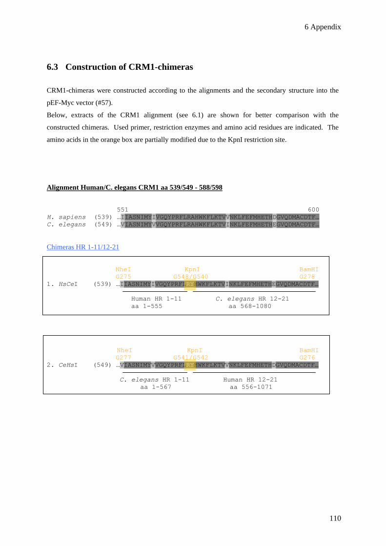
6 Appendix
110
6.3 Construction of CRM1-chimeras
CRM1-chimeras were constructed according to the alignments and the secondary structure into the
pEF-Myc vector (#57).
Below, extracts of the CRM1 alignment (see 6.1) are shown for better comparison with the
constructed chimeras. Used primer, restriction enzymes and amino acid residues are indicated. The
amino acids in the orange box are partially modified due to the KpnI restriction site.
Alignment Human/C. elegans CRM1 aa 539/549 - 588/598
551 600
H. sapiens (539) …IIASNIMYIVGQYPRFLRAHWKFLKTVVNKLFEFMHETHDGVQDMACDTF…
C. elegans (549) …VIASNIMYVVGQYPRFLRAHWKFLKTVINKLFEFMHETHEGVQDMACDTF…
Chimeras HR 1-11/12-21
NheI KpnI BamHI
G275 G548/G540 G278
1. HsCeI (539) …IIASNIMYIVGQYPRFLRYHWKFLKTVINKLFEFMHETHEGVQDMACDTF…
Human HR 1-11 C. elegans HR 12-21
aa 1-555 aa 568-1080
NheI KpnI BamHI
G277 G541/G542 G276
2. CeHsI (549) …VIASNIMYVVGQYPRFLRYHWKFLKTVVNKLFEFMHETHDGVQDMACDTF…
C. elegans HR 1-11 Human HR 12-21
aa 1-567 aa 556-1071
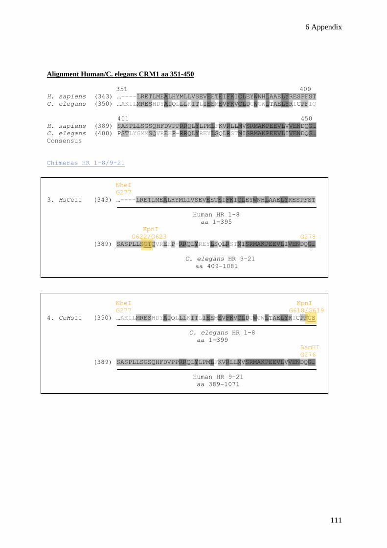
6 Appendix
111
Alignment Human/C. elegans CRM1 aa 351-450
351 400
H. sapiens (343) …----LRETLMEALHYMLLVSEVEETEIFKICLEYWNHLAAELYRESPFST
C. elegans (350) …AKILMRESHDYAIQLLLKITLIEEMEVFKVCLDCWCWLTAELYRICPFIQ
401 450
H. sapiens (389) SASPLLSGSQHFDVPPRRQLYLPMLFKVRLLMVSRMAKPEEVLVVENDQG…
C. elegans (400) PSTLYGMMSQVREHP-RRQLYREYLSQLRSTMISRMAKPEEVLIVENDQG…
Consensus
Chimeras HR 1-8/9-21
NheI
G277
3. HsCeII (343) …----LRETLMEALHYMLLVSEVEETEIFKICLEYWNHLAAELYRESPFST
Human HR 1-8
aa 1-395
KpnI
G622/G623 G278
(389) SASPLLSGTQVREHP-RRQLYREYLSQLRSTMISRMAKPEEVLIVENDQG…
C. elegans HR 9-21
aa 409-1081
NheI KpnI
G277 G618/G619
4. CeHsII (350) …AKILMRESHDYAIQLLLKITLIEEMEVFKVCLDCWCWLTAELYRICPFGS
C. elegans HR 1-8
aa 1-399
BamHI
G276
(389) SASPLLSGSQHFDVPPRRQLYLPMLFKVRLLMVSRMAKPEEVLVVENDQG…
Human HR 9-21
aa 389-1071
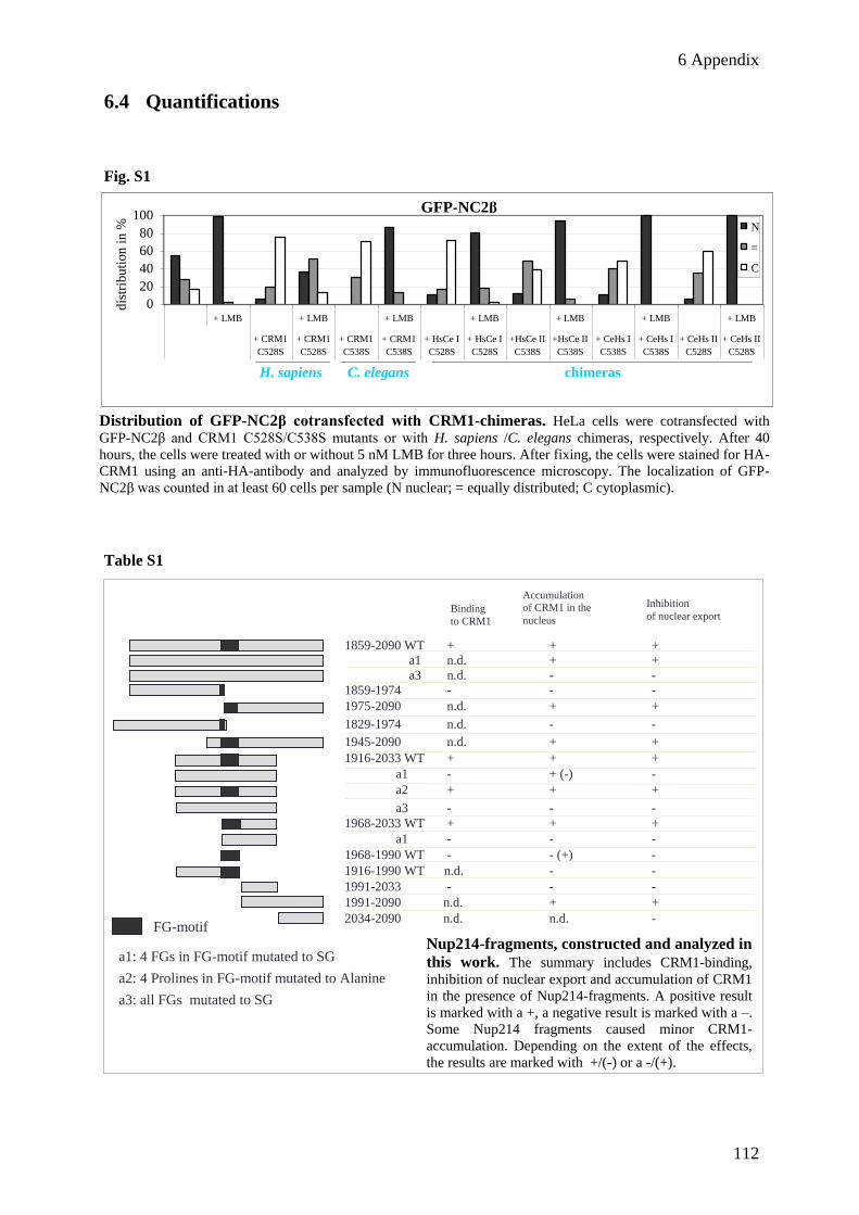
6 Appendix
112
6.4 Quantifications
Fig. S1
Table S1
1859-2090 WT + + +
a1 n.d. + +
a3 n.d. - -
1859-1974 - - -
1975-2090 n.d. + +
1829-1974 n.d. - -
1945-2090 n.d. + +
1916-2033 WT + + +
a1 - + (-) -
a2 + + +
a3 - - -
1968-2033 WT + + +
a1 - - -
1968-1990 WT - - (+) -
1916-1990 WT n.d. - -
1991-2033 - - -
1991-2090 n.d. + +
2034-2090 n.d. n.d. -
Binding
to CRM1
Accumulation
of CRM1 in the
nucleus
Inhibition
of nuclear export
FG-motif
a1: 4 FGs in FG-motif mutated to SG
a2: 4 Prolines in FG-motif mutated to Alanine
a3: all FGs mutated to SG
GFP-NC2β GFP-SPN1
0
20
40
60
80
100
+ LMB + LMB + LMB + LMB + LMB + LMB + LMB
+ CRM1
C528S
+ CRM1
C528S
+ CRM1
C538S
+ CRM1
C538S
+ HsCe I
C528S
+ HsCe I
C528S
+HsCe II
C538S
+HsCe II
C538S
+ CeHs I
C538S
+ CeHs I
C538S
+ CeHs II
C528S
+ CeHs II
C528S
dis
trib
uti
on
in
% N
=
C
H. sapiens C. elegans chimeras
GFP-NC2β
Distribution of GFP-NC2β cotransfected with CRM1-chimeras. HeLa cells were cotransfected with
GFP-NC2β and CRM1 C528S/C538S mutants or with H. sapiens /C. elegans chimeras, respectively. After 40
hours, the cells were treated with or without 5 nM LMB for three hours. After fixing, the cells were stained for HA-
CRM1 using an anti-HA-antibody and analyzed by immunofluorescence microscopy. The localization of GFP-
NC2β was counted in at least 60 cells per sample (N nuclear; = equally distributed; C cytoplasmic).
Nup214-fragments, constructed and analyzed in
this work. The summary includes CRM1-binding,
inhibition of nuclear export and accumulation of CRM1
in the presence of Nup214-fragments. A positive result
is marked with a +, a negative result is marked with a –.
Some Nup214 fragments caused minor CRM1-
accumulation. Depending on the extent of the effects,
the results are marked with +/(-) or a -/(+).

6 Appendix
113
GF
P-S
PN
1
0
20
40
60
80
100
RF
P-
cNL
S
1859-
2090
1859-
2090
a1
1859-
2090
a3
1859-
1974
1975-
2090
1829-
1974
1945-
2090
1916-
2033
WT
1916-
2033
a1
1916-
2033
a2
1916-
2033
a3
1968-
2033
WT
1968-
2033
a1
1968-
2033
a3
1968-
1990
WT
1916-
1990
WT
1991-
2033
1991-
2090
2034-
2090
N = C
GF
P-N
c2β
0
20
40
60
80
100
RF
P-
cNL
S
1859-
2090
1859-
2090
a1
1859-
2090
a3
1859-
1974
1975-
2090
1829-
1974
1945-
2090
1916-
2033
WT
1916-
2033
a1
1916-
2033
a2
1916-
2033
a3
1968-
2033
WT
1968-
2033
a1
1968-
2033
a3
1968-
1990
WT
1916-
1990
WT
1991-
2033
1991-
2090
2034-
2090
N = C
RF
P-N
up
21
4-c
NL
S
RF
P-N
up
21
4-c
NL
S
distributionin %distributionin %
*
*
*
*
*
*
* *
*
*
*
*
*
*
*
* *
*
Nu
clea
r exp
ort
in
hib
itio
n o
f p
rote
ins
in t
he p
rese
nce
of
Nu
p2
14
fra
gm
ents
. T
he
eff
ect
of
Nup
21
4 f
rag
ments
on n
ucl
ear
exp
ort
of
GF
P-S
PN
1an
d
GF
P-N
c2β
is s
um
mari
zed
. H
eL
ace
lls
wer
e tr
ansf
ecte
dw
ith
pla
smid
s co
din
g f
or
the
const
ruct
s as
ind
icat
ed a
nd
GF
P-S
PN
1 o
r G
FP
-Nc2
β.
Aft
er 2
0 h
ours
,
the
cel
ls w
ere
fix
ed a
nd
anal
yze
d b
y i
mm
uno
fluo
resc
ence.
The
loca
liza
tio
n o
f G
FP
-Nc2
βw
as c
ounte
d i
n a
t le
ast
10
0 c
ells
per
sam
ple
(N
>c
nu
clear
; N
=C
equal
ly d
istr
ibute
d;
n<
C c
yto
pla
smic
).E
rro
r b
ars
ind
icat
e th
e st
and
ard
dev
iati
on f
rom
the
mean
of
thre
e in
dep
end
ent
ex
per
iments
or
the
var
iati
on f
rom
th
e
mean
fro
m t
wo
ind
epend
ent
exp
erim
ents
(se
e ast
eris
ks)
.
Fig S2

6 Appendix
114
6.5 Abbreviations
Abbreviations
A adenine (DNA and RNA)
aa amino acid
ABL1 Abelson murine leukemia viral oncogene homolog 1
ATP adenosine-5-triphosphate
bp basepair
B. taurus Bos taurus
BSA bovine serum albumine
C cytoplasmic
C cytosine (DNA and RNA)
C. elegans/Ce Caenorhabditis elegans
CRIME CRM1, IMportin β, Etcetera
CRM1 chromosome region maintenance 1
C-terminus carboxy terminus
Da dalton
D. melanogaster Drosophila melanogaster
DMEM Dulbecco's modified eagles medium
DMSO dimethyl sulfoxide
DNA desoxyribonucleic acid
dNTP 2’-desoxynucleoside-5’-triphosphate
DTT dithiothreitol
ECL enhanced chemical luminescence
E. coli Escherichia coli
EDTA ethylenediaminetetraacetic acid
eIF2 eukaryotic initiation factor 2
EtOH ethanol
FG-repeat phenylalanine glycine repeat
FCS fetal calf serum
G guanine (DNA and RNA)
GAP GTPase-activating protein
GDP guanosine-5’-diphosphate
GEF guanine nucleotide exchange factor
GFP green fluorescent protein
GSK-3 glycogen synthase kinase 3
GST glutathione-S-transferase
GTP guanosine-5’-triphosphate
GTPase GTP-hydrolase

6 Appendix
115
Abbreviations
GZMB Göttinger Zentrum für Molekulare Biowissenschaften
HEAT Huntingtin Elongation Factor A Subunit TOR
HCl hydrochloric acid
HEPES [4-(2-hydroxyethyl)-1-piperazine]ethanesulfonic
His histidine tag
HIV human immunodeficiency virus
HR HEAT repeat motif
H. sapiens /Hs Homo sapiens
IBB importin β binding
IF immunofluorescence
INM inner nuclear membrane
IPTG isopropyl-β-D-thiogalactopyranoside
KLH Keyhole Limpet Hemacyanin
LB Luria-Bertani
LMB Leptomycin B
LTR long terminal repeat
MCS multiple cloning site
M. musculus Mus musculus
N nuclear
nct nucleotide
NC2 negative cofactor
NES nuclear export signal
NFAT nuclear factor of activated T-cells
cNLS classical nuclear localization signal
No number
NPC nuclear pore complex
N-terminus amino terminus
Nup nucleoporin
ONM outer nuclear membrane
PAGE polyacrylamide gel electrophoresis
PBS phosphate buffered saline
PCR polymerase chain reaction
pH negative common logarithm of the proton
PMSF phenylmethylsulphonyl fluoride
Ran Ras-related nuclear protein
RCC1 Regulator of chromatin condensation 1
R. norwegicus Rattus norwegicus
RFP red fluorescent protein

6 Appendix
116
Abbreviations
Ran Ras related nuclear protein
RanBP1 Ran binding protein 1
RCC1 regulator of chromosome condensation 1
RNA ribonucleic acid
RNP ribonucleoprotein
rpm rotations per minute
SDS sodium dodecyl sulphate
SV40 Simian virus 40
siRNA small interfering RNA
SPN1 Snurportin 1
T thymine (DNA and RNA)
TB transport buffer
TAE Tris /Acetate /EDTA
TBP TATA binding protein
TE Tris /EDTA
TFIIA Transcription factor II A
Triton X-100 4-octylphenol polyethoxylate
Tween20 polyoxyethylene (20) sorbitan monolaurate
U uracile, in context of RNA
UV ultraviolet
v/v volume per volume
WGA wheat germ agglutinin
WB Western blot
WT wild type
w/o with or without
w/v weight per volume
X. laevis Xenopus laevis
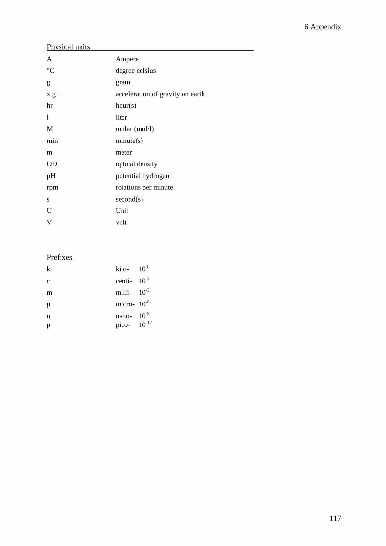
6 Appendix
117
Physical units
A Ampere
°C degree celsius
g gram
x g acceleration of gravity on earth
hr hour(s)
l liter
M molar (mol/l)
min minute(s)
m meter
OD optical density
pH potential hydrogen
rpm rotations per minute
s second(s)
U Unit
V volt
Prefixes
k kilo- 103
c centi- 10-2
m milli- 10-3
μ micro- 10-6
n nano- 10-9
p pico- 10-12

118
Acknowledgements
Nun, fast ganz zum Schluss dieser Arbeit (aber das soll nicht bedeuten, dass es nicht weniger wichtig
ist) möchte ich einigen Menschen danken, die mich während meiner Arbeit fortwährend unterstützt
haben.
Zuallererst einmal danke ich meinem Doktorvater Ralph Kehlenbach für die Bereitstellung dieses
herausfordernden, wenn auch nicht immer ganz einfachen, Projektes. Bei Problemen oder Fragen hast
Du immer Zeit gefunden und geholfen, dieses Projekt mit konstruktiven Lösungen in die richtige
Richtung zu lenken. An dieser Stelle möchte ich mich auch bei meinen weiteren Thesis-Komitee
Mitgliedern, Peter Rehling und Ralf Ficner bedanken, die großes Interesse an diesem Projekt gezeigt
haben und es stets mit vielen Ideen bereichert haben.
Bedanken möchte ich mich auch bei meinen Laborkollegen Christiane Spillner, Annegreth Nath,
Cornelia Frohnert, Ketan Thakar, Sarah Port, Inga Waldmann und auch bei der Arbeitsgruppe
Schwappach für die entspannte Laboratmosphäre und super Unterstützung, wenn es um methodische
oder projektbezogene Fragen ging. Christiane, die Arbeit mit Dir im letzten halben Jahr dieser Arbeit
haben mir soviel Spaß gemacht, und ich bewundere Deinen unermüdlichen Einsatz, Deine
Motivation, ohne Murren auch das 381. slide auszuzählen und immer noch guter Dinge zu sein.
Danken möchte ich auch Olaf Bernhard aus der AG B. Schmidt für die Massenspektrometrie-
analysen.
Ein besonderer Dank gilt auch meinem Partner Volker Henschel, der mit seiner besonderen Art, mich
zu motivieren, kritisieren und aufzubauen, immer unterstützt hat. Auch meiner Familie, die immer für
mich da ist und mich liebevoll während dieser Arbeit unterstützt hat, möchte ich danken.
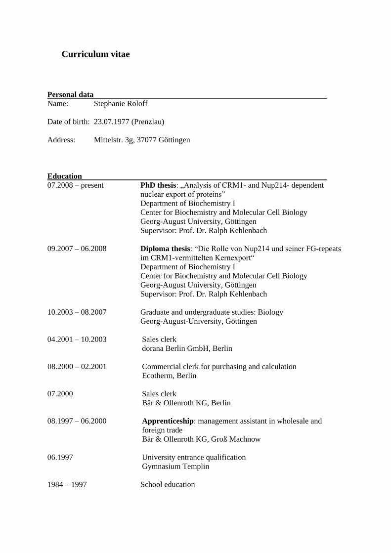
Curriculum vitae
Personal data
Name: Stephanie Roloff
Date of birth: 23.07.1977 (Prenzlau)
Address: Mittelstr. 3g, 37077 Göttingen
Education
07.2008 – present PhD thesis: „Analysis of CRM1- and Nup214- dependent
nuclear export of proteins”
Department of Biochemistry I
Center for Biochemistry and Molecular Cell Biology
Georg-August University, Göttingen
Supervisor: Prof. Dr. Ralph Kehlenbach
09.2007 – 06.2008 Diploma thesis: “Die Rolle von Nup214 und seiner FG-repeats
im CRM1-vermittelten Kernexport“
Department of Biochemistry I
Center for Biochemistry and Molecular Cell Biology
Georg-August University, Göttingen
Supervisor: Prof. Dr. Ralph Kehlenbach
10.2003 – 08.2007 Graduate and undergraduate studies: Biology
Georg-August-University, Göttingen
04.2001 – 10.2003 Sales clerk
dorana Berlin GmbH, Berlin
08.2000 – 02.2001 Commercial clerk for purchasing and calculation
Ecotherm, Berlin
07.2000 Sales clerk
Bär & Ollenroth KG, Berlin
08.1997 – 06.2000 Apprenticeship: management assistant in wholesale and
foreign trade
Bär & Ollenroth KG, Groß Machnow
06.1997 University entrance qualification
Gymnasium Templin
1984 – 1997 School education