Topoisomerase I and RecQL1 Function in Epstein-Barr Virus Lytic Reactivation
Upload
vikramunivCategory
view
1download
0
Biochimica et Biophysica Acta (BBA) - Biomembranes, 1788, 2, 2009, 538-550
A peptide derived from the putative transmembrane domain in the tail region of E. coli toxin hemolysin E assembles in phospholipid membrane and exhibits lytic activity to human red blood cells: plausible implications in the toxic activity of the protein Sharada Prasad Yadav, Aqeel Ahmad, Brijesh Kumar Pandey, Dharamsheela$, Neeta Asthana, Richa Verma, Raj Kamal Tripathi$ and Jimut Kanti Ghosh* Molecular and Structural Biology Division and Toxicology Division$, Central Drug Research Institute, Lucknow–226001, India #This is CDRI communication number 6813 *To whom correspondence should be addressed Tel: 091-522-2612411-18 (Ext.-4282); Fax: 091-522-2623405; E-mail: [email protected] Abstract
Hemolysin E (HlyE), a pore-forming protein-toxin and a potential virulence factor of Escherichia coli, exhibits cytotoxic activity to mammalian cells. However, very little is known about how the different individual segments contribute in the toxic activity of the protein. Toward this end, the role of a 33-residue segment comprising the amino acid region 88 to 120, which contains the putative transmembrane domain in the tail region of HlyE has been addressed in the toxic activity of the protein-toxin by characterizing the related wild type and mutant peptides and the whole protein. Along with the 33-residue wild type peptide, H-88, two mutants of the same size were synthesized; in one mutant a conserved valine at 89th position was replaced by aspartic acid and in the other both glycine and valine at the 88th and 89th positions were substituted by aspartic acid residues. These mutations were also incorporated in the whole toxin HlyE. Results showed that only H-88 but not its mutants permeabilized both lipid vesicles and human red blood cells (hRBCs). Interestingly, while H-88 exhibited a moderate lytic activity to human red blood cells, the mutants were not active. Drastic reduction in the depolarization of hRBCs and hemolytic activity of the whole toxin HlyE was also observed as a result of the same double and single amino acid substitution in it. The results indicate an important role of the amino
Biochimica et Biophysica Acta (BBA) - Biomembranes, 1788, 2, 2009, 538-550
acid segment 88-120, containing the putative transmembrane domain of the tail region of the toxin in the toxic activity of hemolysin E. Key words: Pore-forming toxin; Escherichia coli Hemolysin E; toxicity of hemolysin E; Putative transmembrane segment; Hemolysin E mutant; lipid-peptide interaction; protein-dissection. Introduction
Gram-negative bacteria often produce a variety of cytolytic toxins, which enable the microorganisms to successfully infect the host including humans and other animals [1-3]. However, the exact mode of actions and involvement of the majority of these cytolytic toxins in the pathogenesis of infections are not clearly understood. A number of cytolysins isolated from Gram-negative bacteria form pores in the cytoplasmic membranes of eukaryotic cells [4-5]. Disease causing strains of Escherichia coli (E. coli) produce several pore-forming toxins. Of these, α-hemolysin (HlyA) is found in E. coli strains, which cause extra-intestinal infections, while enterohemorrhagic E. coli (EHEC) hemolysin is produced by EHEC strains of serogroup O157 [2,6]. The nonpathogenic strain, E. coli K-12, does not contain the gene cluster required for the production and secretion of α-hemolysin or the related EHEC hemolysin and is non-hemolytic under normal condition. However, a variant of E. coli K-12 can also express a hemolytic or cytolytic phenotype under certain conditions. This induction of cytolytic activity in the laboratory strain, E. coli K-12 has been shown to be due to the synthesis of a toxin, named as hemolysin E (HlyE) or silent hemolysin A ¶Abbreviations Used: HlyE, hemolysin E; TFE, Trifluoroethanol; NBD-fluoride, 4-fluoro-7-nitrobenz-2-oxa-1, 3-diazole; Rho, Tetramethylrhodamine; PC, Egg phosphatidylcholine; PG, Egg phosphatidylglycerol; Chol, Cholesterol; a.a., amino acid; hRBC, human red blood cell (SheA) or cytolysin A (ClyA) by different groups [7,8]. E. coli, expressing hemolysin E can cause lysis of red blood cells from different species, including humans, horse, sheep, goat and hen [8-11]. The expression of hlyE gene in E. coli K-12 is silenced by the nucleoid protein histone-like nucleoid-structuring protein (H-NS) [12] while it is activated in the presence of overproduction of SlyA, MprA, HlyX or fumarate and nitrate reduction regulator (FNR) [8,9,12,13]. The expression of hemolysin E has also been found in clinical isolates of E. coli [14]. Furthermore, purified HlyE and HlyE-expressing E. coli exhibit cytotoxic and apoptogenic activities towards human and murine macrophages, human peripheral monocytes and HeLa cells [15]. Recent studies have shown that hemolysin E after secretion into the periplasm of E. coli, is packaged in its outer membrane vesicles in the active form and from there transported into the target mammalian cells [16].
Biochimica et Biophysica Acta (BBA) - Biomembranes, 1788, 2, 2009, 538-550
Crystal structure of hemolysin E has been solved at 2.0 Å resolution, which indicated that it belongs to a new family of toxin structures [17]. A structure-function study showed that the deletion of 37 amino acids from the αG region, at the C-terminal of the toxin rendered it non-hemolytic [18]. An amphipathic, conserved leucine zipper motif from hemolysin E has been identified and characterized [19-21], which suggested a possible structural and functional role of this motif in the toxin. However, it is still not known how hemolysin E exhibits cytotoxic activity to eukaryotic cells for example lyses human red blood cells. The literature shows that for a number of proteins belonging to several families like transcription factors, fusion proteins of enveloped virus and pore-forming toxins it has been possible to identify small segments that show certain activity of the corresponding whole protein. However, for hemolysin E no such segment with hemolytic activity of the toxin was known. With a goal to understand the contribution of different conserved hemolysin E segments to the membrane-interaction and functional activity of the whole protein, we have synthesized and characterized a 33-residue peptide (H-88, amino acid 88-120), which contains a putative transmembrane domain [13,17], located in the tail region of the protein along with two mutants of the same size. In one of them (Mu1-H-88), a conserved valine at 89th position was substituted by an aspartic acid while in the other glycine and valine positioned at the 88th and 89th positions were replaced by two aspartic acid residues. The lytic activity of E. coli containing the hemolysin E plasmid with the latter mutations was significantly reduced [9]. Therefore, it was of interest to look into the effect of the same mutation on the activity of the H-88 segment. In the first mutant the purpose was to look into the effect of the substitution of the single amino acid positioned in the putative transmembrane segment. Furthermore, in order to look into the effect of these single and double amino acid substitutions on the cytotoxic activity of the whole toxin, site directed mutagenesis was performed in the hlyE plasmid to prepare both the mutant proteins with the above amino acid substitutions. The wild type and mutant proteins were purified and then characterized along with the synthetic peptides. Results showed that H-88 permeabilized both phospholipid vesicles and human red blood cells (hRBCs), and more interestingly lysed them like the whole protein though with lesser efficiency. Although the amino acid substitutions were done at the N-terminal of the selected segment, the mutant peptides did not exhibit any appreciable pore-forming activity in lipid vesicles or hRBCs and showed no lytic activity toward the hRBCs. Hemolysin E mutants with the same mutations as in the peptide showed drastically reduced hemolytic activity and permeabilized the human red blood cells negligibly as compared to the wild type protein. The results have been discussed in terms of the plausible role of the H-88 segment containing the conserved putative transmembrane region in toxic activity of the protein toxin HlyE.
Biochimica et Biophysica Acta (BBA) - Biomembranes, 1788, 2, 2009, 538-550
Materials and Methods Materials
Rink amide MBHA resin (loading capacity, 0.63 mmol/g) and all the N-α Fmoc and necessary side-chain protected amino acids were purchased from Novabiochem, Switzerland. Coupling reagents for peptide synthesis like 1-hydroxybenzotriazole (HOBT), di-isopropylcarbodiimide (DIC), 1,1,3,3-tetramethyluronium tetrafluoroborate (TBTU) and N, N’-diisopropylethylamine (DIPEA) were purchased from Sigma, USA. Dichloromethane, N, N’ dimethylformamide (DMF) and piperidine were of standard grades and procured from reputed local companies like Merck Limited, Mumbai, Thomas Baker, Mumbai and Spectrochem Pvt. Ltd., Mumbai respectively. Acetonitrile (HPLC grade) was procured from Merck, India while trifluoroacetic acid (TFA), trifluoroethanol (TFE) and sodium dodecyl sulfate (SDS) were purchased from Sigma. Egg phosphatidylcholine (PC) and egg phosphatidylglycerol (PG) were obtained from Northern Lipids Inc., Canada while cholesterol (Chol) was purchased from Sigma. NBD-fluoride (4-fluoro-7-nitrobenz-2-oxa-1, 3-diazole) and tetramethylrhodamine succinimidyl ester were procured from Molecular probes (Eugene, OR) whereas calcein was purchased from Sigma and used without any further purification. Rests of the reagents were of analytical grade and procured locally; buffers were prepared in milli Q water. Peptide Synthesis, Fluorescent labeling and Purification
All the peptides were synthesized manually on solid phase. Stepwise solid phase syntheses were carried out on rink amide MBHA resin (0.15 mmole) utilizing the standard Fmoc chemistry and employing DIC/HOBT or TBTU/HOBT/DIPEA coupling procedure [22] as already reported [19,23]. De-protection of α-amino group and the coupling of amino acids were checked by the Kaiser test [24] for primary amines. After the synthesis, each peptide was cleaved from the resin with simultaneous de-protection of side chains by treatment with a mixture of TFA/phenol/thioanisole/1,2 ethanedithiol/water (82.5:5:5:2.5:5 v/v) for 6-7 hours. Labeling at the N-terminus of a peptide with NBD or rhodamine was achieved by standard procedures as reported [25,26,19]. After sufficient labeling, the resins were washed with DMF and DCM in order to remove the un-reacted probe. The peptides were cleaved from the resin as above and precipitated with dry ether. All the labeled and unlabeled peptides were purified by reversed-phase HPLC on an analytical Vydac C4 column using a linear gradient of 0-80% acetonitrile in 45 min with a flow rate of 0.8 ml/min. Both acetonitrile and water contained 0.05% TFA. The purified peptides were ~95% homogeneous as shown by HPLC. Experimental mass of the peptides as detected by ES-MS analysis corresponded very close to their theoretical values. Generation of double and point mutation in hlyE
Site directed mutagenesis was done in order to substitute Glycine at 88th and Valine at 89th position by Asparatic acid residues in HlyE. This was
Biochimica et Biophysica Acta (BBA) - Biomembranes, 1788, 2, 2009, 538-550
accomplished by introducing mutations in the internal forward FPIHlymE (5` AT GAA TGG TGT GAT GAT GCG ACG CA 3`) and reverse primer RPIHlymE (5` TG CGT CGC ATC ATC ACA CCA TTC AT 3`) as underlined. The PCR was done using pGS-1111 (construct of pGEX-KG and hlyE) as the template, which was isolated from the engineered E. coli JM109 cells (a gift from Prof. Jefrey Green, University of Sheffield, U.K.) carrying the same plasmid using two sets of primer combinations, forward outer FPOHlymE (5`CCA TGG CTG AAA TCG TTG CAG A 3`) and internal reverse RPIHlymE and internal forward FPIHlymE and outer reverse RPOHlymE (5`GTC GAC TCA GAC TTC AGG TAC CTC AAA G 3`). The amplified products were gel eluted, quantified and mixed together in equal proportion and further used as template to amplify the full-length hlyE gene using primer set FPOHlymE and RPOHlymE. The amplified hlyE was gel eluted and cloned in pDrive (Quiagen PCR cloning kit), which was subcloned in the expression vector pGEX-KG at NcoI/SalI site as pDKJ1. The positive clone was sequenced on ABI sequencer (Supplementary Figure 1) and aligned with the original hlyE sequence using Gene Tool software.
The same procedure was followed to introduce the point mutation to replace valine at 89th position by a single aspartic acid residue as mentioned above except the internal primers set used for introducing mutation was replaced by FPIHlymVD (5` AT GAA TGG TGT GGT GAT GCG ACG CA 3`) and RPIHlymVD(5` TG CGT CGC ATC ACC ACA CCA TTC AT 3`). The mutated gene was subcloned in the expression vector pGEX-KG at NcoI/SalI site as pDKJ2 and sequenced (Supplementary Fig. 2) as before. Expression of Hemolysin E and its mutants and their purification
The mutated plasmids were transformed into E. coli JM109 cells by a standard procedure. Hemolysin E and its mutants were expressed and purified as reported earlier [18,21]. E. coli JM109 cells either carrying the pGS-1111 plasmid, which contains the expression vector of GST-hemolysin E or the mutated plasmids for the expression vectors GST-G88V89DHlyE and GST-V89DHlyE were grown in LB broth for three hours at 37 0C before induction of the expression of the fusion proteins by addition of isopropyl-1-thio-β-D-galactopyranoside (100 μg/ml). After a further four hours of incubation at 370C bacteria were collected by centrifugation at 7000 r.p.m at 40C. The bacterial pellet was suspended in 10 mM Tris-HCl, pH 8.0 containing 10 mM benzamidine and 0.1 mM phenylmethylsulfonyl fluoride. The above bacterial suspension was then sonicated for the disruption of the bacterial membrane followed by centrifugation at 12000 r.p.m at 40C. The supernatant was taken and loaded on a GSH-Sepharose column equilibrated with 25 mM Tris-HCl, pH 6.8 containing 100 mM NaCl and 2.5 mM CaCl2. After washing with 10 volumes of the same buffer, HlyE was released by the thrombin treatment (5 units for 16h at 25 0C). Concentrations of the proteins were estimated with the help of the Lowry method [27]. Preparation of small unilamellar vesicles (SUVs)
Biochimica et Biophysica Acta (BBA) - Biomembranes, 1788, 2, 2009, 538-550
SUVs of either PC/cholesterol (8:1 w/w) or PC/PG (1:1 w/w) were
prepared by a standard procedure [28,19] using a bath-type sonicator. The lipid concentration was determined by phosphorus estimation [29]. Circular dichroism (CD) experiments
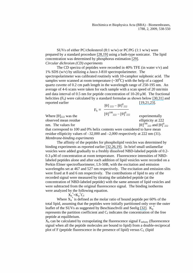
The CD spectra of peptides were recorded in 40% TFE (in water v/v) and 1% SDS (w/v) by utilizing a Jasco J-810 spectropolarimeter. The spectropolarimeter was calibrated routinely with 10-camphor sulphonic acid. The samples were scanned at room temperature (~300C) with the help of a capped quartz cuvette of 0.2 cm path length in the wavelength range of 250-195 nm. An average of 4-6 scans were taken for each sample with a scan speed of 20 nm/min and data interval of 0.5 nm for peptide concentration of 10-20 μM. The fractional helicities (Fh) were calculated by a standard formulae as shown below [30,31] and reported earlier [19,21,23]. Where [θ]222 was the experimentally observed mean residue ellipticity at 222
[θ]100222 and [θ]0
222 nm. The values for that correspond to 100 and 0% helix contents were considered to have mean residue ellipticity values of –32,000 and –2,000 respectively at 222 nm (31). Membrane-binding experiments
The affinity of the peptides for phospholipid vesicles was determined by binding experiments as reported earlier [32,26,19]. In brief small unilamellar vesicles were added gradually to a freshly dissolved NBD-labeled peptide of 0.2-0.3 μM of concentration at room temperature. Fluorescence intensities of NBD-labeled peptides alone and after each addition of lipid vesicles were recorded on a Perkin Elmer spectrofluorimeter, LS-50B, with the excitation and emission wavelengths set at 467 and 527 nm respectively. The excitation and emission slits were fixed at 8 and 6 nm respectively. The contributions of lipid to any of the recorded signal were measured by titrating the unlabeled peptide (at the concentration of NBD-labeled peptide) with the same amount of lipid vesicles and were subtracted from the original fluorescence signal. The binding isotherms were analyzed by the following equation.
Xb*=Kp
*Cf Where Xb
* is defined as the molar ratio of bound peptide per 60% of the total lipid, assuming that the peptides were initially partitioned only over the outer leaflet of the SUVs as suggested by Beschiaschvili and Seelig [32]. Kp
* represents the partition coefficient and Cf indicates the concentration of the free peptide at equilibrium. Xb can be calculated by extrapolating the fluorescence signal Finfinity (fluorescence signal when all the peptide molecules are bound to lipid) from a double-reciprocal plot of F (peptide fluorescence in the presence of lipid) versus CL (lipid
[θ] 222 − [θ]0222
[θ]100222 − [θ]0
222
Fh =
Biochimica et Biophysica Acta (BBA) - Biomembranes, 1788, 2, 2009, 538-550
concentration). Fraction of peptide bound (fb) was determined by the following equation. fb=(F-F0)/(Finfinity-F0) Where F is the fluorescence of the peptide when it is bound to lipid and F0 is the fluorescence of the peptide in its unbound state. When fb is known, Cf can easily be calculated for each concentration of the lipid. Kp* can easily be determined from the slope of the plot of Xb
* and Cf. Partition coefficient of each of the peptides was determined as the average of the values obtained from two-three independent experiments as described previously [19,20]. Fluorescence resonance energy transfer experiments
Assembly of H-88 and the designed mutants in phospholipid vesicles was detected with the help of fluorescence energy transfer experiments using their NBD- and Rho- labeled analogs as energy-donor and acceptor respectively. The excitation wavelength was set at 467 nm and emission range at 500 to 600 nm. Desired amount of the NBD-labeled peptide was taken in a fluorimeter cuvette. Sufficient amount of the phospholipid vesicles ([lipid]/[peptide] ~ 2500) were added to the NBD-labeled peptide to ensure that the peptides were bound to the membrane. Now Rho-labeled acceptor peptide was added to the donor peptide-lipid complex. Energy transfer from the donor to acceptor was determined by subtracting the acceptor fluorescence in the presence of lipid and unlabeled donor from the fluorescence signal obtained in the presence of donor, acceptor and lipid vesicles. The efficiency of energy transfer (E) was determined by the decrease in donor’s fluorescence in the presence of the acceptor as reported earlier [33,26,19]. The percentage of energy transfer was calculated by the following equation. E = {(ID0 – IDA)/ID0} x 100 Where ID0 and IDA are the fluorescence intensities of the NBD-labeled donor peptide in the absence and presence of the Rho-labeled acceptor peptide respectively at the emission maxima of the donor after correcting the light scattering of the lipid vesicles and emission of the acceptor. Detection of pore-forming activity of the peptides in lipid vesicle
Peptide-induced release of calcein from calcein-entrapped lipid vesicles is often employed to detect the pore-forming activity of proteins and peptides. Calcein-entrapped lipid vesicles were prepared with a self-quenching concentration (60 mM) of the dye in 10 mM HEPES at pH 7.4 as reported before [34,35,19]. Peptide-induced release of calcein from the lipid vesicles was monitored by the increase in fluorescence due to the dilution of the dye from its self-quenched concentration. Fluorescence was monitored at room temperature with excitation and emission wavelengths fixed at 490 and 520 nm respectively. Pore-forming activity of the peptides, indicated by the calcein release was measured by the fluorescence recovery as defined by: Ft = [(It - I0)/(If - I0)] x 100 %
Biochimica et Biophysica Acta (BBA) - Biomembranes, 1788, 2, 2009, 538-550
Where It=the observed fluorescence after the addition of a peptide at time t (~60 sec after the addition of the peptide), I0 = the fluorescence before the addition of peptide and If = the total fluorescence, which was determined after the addition of triton X-100 (0.1% final concentration) to the dye-entrapped vesicle suspension. Assay of hemolytic activity of the peptides and proteins
Hemolytic activity of H-88 and its mutants was assayed against human red blood cells in PBS [9,10,19]. Briefly, fresh human red blood cells (hRBCs) that were collected in the presence of an anti-coagulant from a healthy volunteer were washed three times in PBS. Freshly dissolved peptides in dimethyl sulphoxide (DMSO) at desired concentrations were added to the suspension of red blood cells (final density ~5 x 10⁸ cells/ml, counted with help of a LEICA DM 5000 Microscope) in PBS and incubated at 37 0C for two hours. It is to be mentioned that hemolytic activity of H-88 reached the maximum value at ~ 50 min and it remained the same even up to 2 hours. The samples were then centrifuged for 10 min at 2000 r.p.m. and the release of hemoglobin was monitored by measuring the absorbance (Asample) of the supernatant at 540 nm. For negative and positive controls hRBC in PBS (Ablank) and in 0.2% (final concentration v/v) Triton X-100 (Atriton) were used respectively. The percentage of hemolysis was calculated according to the following equation.
Percentage of hemolysis = [(Asample-Ablank)/(Atriton-Ablank)] x 100 One hemolytic unit was defined as the amount of peptide, which caused 50% lysis of the hRBCs (~5 x 10⁸ cells/ml) after an incubation of two hours at 370C [9]. Osmotic protection assay
In order to determine the equivalent diameter of the pores formed by the synthetic putative transmembrane segment on hRBCs, hemolytic activity of H-88 was examined in the presence of osmotic protectors as described previously [36,37] with 3% (final concentration, in v/v) hRBCs. Human erythrocytes, prepared in the same way as the previous experiment were suspended in PBS and incubated with one of the following osmotic protectors at 30 mM concentration at 37 0C. Sucrose, raffinose, Poly (ethylene glycol) (PEG) 1450, PEG 2000, PEG 3000 and PEG 3350 of diameters 0.9, 1.3, 2.2, 2.5, 3.2 and 3.4 nm [36-39] respectively were used in this experiment as osmotic protectors. After 30 min of incubation to each of the protector-treated erythrocytes, H-88 was added. H-88 induced hemolysis was determined after another incubation of 50 min at 37 0C by recording the absorbances of the supernatants at 540 nm as the previous experiment. Hemolytic peptides form pores onto hRBCs, which cause the release of its cytoplasmic contents and cell death. Osmotic protectors tend to fill this pore and prevent the release of cytoplasmic content from hRBC. However, to fill the pore the osmotic protector must have a size comparable to the pore, which is the basis of osmotic protection assay. Thus when the size of the osmotic protector match with the size of pore formed by the peptides onto the hRBC, inhibition of the hemolytic activity of the peptides is observed. Accordingly, by observing
Biochimica et Biophysica Acta (BBA) - Biomembranes, 1788, 2, 2009, 538-550
inhibition of hemolysis in the presence of osmotic protectors a probable diameter of the pore formed by a hemolytic peptide onto the hRBC is determined. Human red blood cell membrane depolarization assay
Peptide-induced depolarization of the hRBCs was determined by its efficacy to dissipate the membrane potential across the hRBCs [40]. Normally a cell is hyperpolarized means it has a potential gradient across the cell membrane. The potential sensitive dye, DiS-C3-5 tends to accumulate in hyperpolarized cells, and its fluorescence get quenched. Fresh human red blood cells (hRBCs) were collected in the presence of an anti-coagulant from a healthy volunteer and washed three times in PBS and were incubated with diS-C3-5 dye for 1hr with a final cell density of 0.5x 10⁸ cells/ml. When the fluorescence level (excitation and emission wavelengths were set 620 and 670 nm respectively) became stable, different amounts of each of the peptides were added to record the peptide-induced membrane depolarization of the hRBCs. Peptides capable of forming pores onto hRBC membranes, depolarize them by dissipation of membrane potential, which is associated with an increase in fluorescence of the dye. Membrane depolarization as measured by the fluorescence recovery is defined by the same equation as used to determine the peptide-induced calcein release, which has been described before already. However, in this case If, the total fluorescence was determined just after addition of diS-C3-5 to hRBCs; It, the observed fluorescence after the addition of a peptide to hRBCs which were already incubated for 1hr with diS-C3-5 dye and I0 is the steady fluorescence level after one hr incubation of hRBCs with the dye. Detection of damage of hRBC membrane organization in the presence of the HlyE proteins and H-88 peptides by flow cytometry
Alteration in morphology or organization of the lipid bilayer of human red blood cells is often probed by annexin V-FITC staining [41]. Therefore, in order to detect any damage of membrane organization of hRBC in the presence of these peptides and proteins the cells were stained by annexin V-FITC following the treatment of the cells with these molecules. Fluorescence of the cells after the treatment the probe was analyzed by using a Becton Dickinson FACSCalibur flow cytometer and CellQuest Pro software as reported earlier [21]. The exitation and emission wavelengths of annexin V-FITC were set at 488 nm and 530 nm respectively. Results
Hemolysin E is a recently identified pore-forming toxin in E. coli. Like other pore-forming toxins, [1,2,42] membrane-interaction and assembly therein are important steps associated with its function. However, the segment(s) that participates in the interaction with the target cell membrane and contributes to the toxic activity is not clearly known. In order to evaluate the potential role of the putative transmembrane segment, located in the tail region of hemolysin E, a 33-
Biochimica et Biophysica Acta (BBA) - Biomembranes, 1788, 2, 2009, 538-550
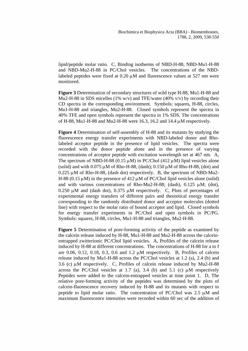
residue peptide comprising the amino acid region 88-120 was synthesized along with the two mutant peptides of the same size. Figure 1A shows the schematic location of the putative transmembrane domains [13] and two identified and partially characterized heptad repeats [19]. Figure 1B shows the sequence alignment of the corresponding amino acid region of the proteins of hemolysin E family, which indicates that a considerable number of amino acids belonging to all four proteins have identical sequences. Figure 1C, depicts the amino acid sequences of the unlabeled and labeled peptides used in this study. The synthetic segments that have been employed in the this study have been marked in different colors in the crystal structure (down loaded from the PDB) of HlyE (Supplementary Fig. 3). Peptides containing the putative transmembrane segment bound to phospholipid membrane
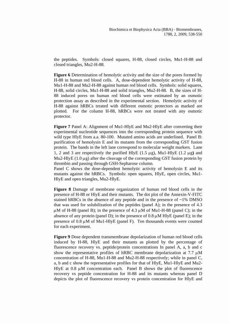
Since hemolysin E is a pore-forming toxin, it was of interest to study the membrane-interaction of the peptides that contain the putative transmembrane domain. In order to study the membrane-binding ability of these peptides, their NBD-labeled versions and small unilamellar lipid vesicles (SUV) with different lipid composition were used. Zwitterionic PC/Chol and negatively charged PC/PG lipid vesicles are often employed as mimetics of mammalian and bacterial membrane respectively (33,25,19,23). The dependence of NBD-fluorescence on the dielectric constant of the medium has been employed widely to study the membrane-interaction of proteins and peptides by attaching the probe onto these molecules [43,44,26,19]. In the presence of either zwitterionic PC/Chol or negatively charged PC/PG lipid vesicles, emission maxima of NBD-labeled H-88 and mutant H-88 peptides moved from ~542 nm to shorter wavelength (~527±1.0 nm), concomitant with significant increase in fluorescence (Figure 2A). The result indicates the relocation of the probe in the hydrophobic environment as a result of binding of the peptides to phospholipid vesicles [45,19]. Figure 2B shows the plots of fluorescence of NBD-labeled H-88, Mu1-H-88, Mu2-H-88 with respect to the lipid/peptide molar ratio when the peptides were treated with increasing amount of PC/Chol lipid vesicles. NBD-labeled H-88 and mutants in the presence of increasing PC/PG vesicles showed a similar extent of fluorescence change and thus exhibited plots of comparable shapes as that in the presence of PC/Chol vesicles and therefore not presented. The plots show a gradual increase in NBD-fluorescence with increase in lipid concentration, indicating the progressive binding of the NBD-labeled peptide molecules to lipid vesicles. Binding isotherms (Figure 2C) were generated by plotting Xb
* with respect to Cf as has been described in the experimental section and also reported earlier [19]. Partition coefficients of the NBD-labeled peptides to the phospholipid vesicles were estimated from the slope of the binding isotherms. NBD-labeled H-88, Mu1-H-88 and Mu2-H-88 exhibited an appreciable affinity for PC/Chol lipid vesicles and the calculated partition coefficients were 1.50 (±0.1) x 104, 1.29 (±0.1) x 104 and 1.02 (±0.1) x 104 M-1 respectively. The nature of the binding
Biochimica et Biophysica Acta (BBA) - Biomembranes, 1788, 2, 2009, 538-550
isotherms also provides an idea about the assembly of a peptide in membrane. The binding isotherms of NBD-labeled mutants in PC/Chol lipid vesicles were mostly linear in nature (Figure 2C), suggesting that the binding of these peptides to PC/Chol vesicles was a simple adhesion process and no large aggregates of the peptide was formed. However, the binding isotherm of NBD-labeled H-88 in PC/Chol vesicles bent downward appreciably and deviated from linearity. As suggested earlier, this kind of curve indicates the co-operativety in binding of peptides to membrane [46,25,19]. Thus, the binding isotherms revealed that only NBD-labeled H-88 but not the mutant formed large aggregates in zwitterionic lipid vesicles. Both H-88 and its mutants adopted significant helical structure in membrane mimetic environments
The secondary structures of the peptides were determined with the help of their CD spectra in the membrane-mimetic environment, 1% SDS and in the helix stabilizing 40% TFE (v/v) in water. All the peptides displayed CD spectra with minima at 208 and 222 nm, characteristic of α-helical secondary structure as shown in Figure 3. The mean residual ellipticity values at 222 nm of the CD spectra of H-88 in 40% TFE and 1% SDS were 25,257 and 20,771 which corresponded to 77% and 62% helical structures respectively. For Mu1-H-88 mean residue ellipticity values at 222 nm in 40% TFE and 1% SDS were 20,556 and 15,262 which were equivalent to helicities of 62% and 44% respectively. The Mu2-H-88 exhibited mean residue ellipticity values of 13,404 and 13,021 in 40% TFE and 1% SDS respectively which corresponded to the helix contents of 38% and 36.7%. The CD data suggest that the substitution of valine or glycine and valine by aspartic acid to some extent reduced the helical structure of the peptide derived from the putative transmembrane segment. The H-88 peptide containing the putative transmembrane domain in the tail region of hemolysin E assembled in phospholipid membrane
In order to look into the possibility of an involvement of this putative transmembrane segment in the assembly of hemolysin E, energy transfer experiments of H-88 and its mutants were performed with their NBD-and Rho-labeled analogs that have been employed as energy donor and acceptor respectively. It is to be mentioned that the shape of the binding isotherms can detect only large aggregates but not the self-association of a polypeptide to form small sized bundles [26]. Appreciable energy transfer from an NBD-labeled donor peptide to a Rho-labeled acceptor one occurs when they are in close proximity to each other. Figure 4A shows the typical profiles of energy transfer experiments when increasing amounts of Rho-labeled H-88 were added to membrane-bound (zwitterionic PC/Chol) NBD-labeled H-88. It is very much evident that donor’s fluorescence progressively decreased as a result of increase in acceptor’s concentration, suggesting self-association between H-88 peptide molecules in the zwitterionic lipid vesicles. Also, a significant fluorescence energy transfer was observed between NBD- and Rho-labeled H-88 peptide
Biochimica et Biophysica Acta (BBA) - Biomembranes, 1788, 2, 2009, 538-550
molecules when they were bound to the negatively charged PC/PG lipid vesicles (profiles not shown, data presented in Figure 4C). However, when Rho-H-88 was replaced with Rho-labeled H-88 mutants or a rhodamine labeled scrambled peptide (Rho-Scr-198) derived from the amino acid region 198-234 of HlyE, no significant decrease in NBD-H-88 fluorescence was observed (profiles not shown), which indicated that the H-88 peptide molecules self-assembled in the phospholipid vesicles with a sequence specificity. In another control experiment instead of Rho-H-88 increasing amount of unlabelled H-88 was added to the membrane-bound (either PC/PG or PC/Chol vesicles) NBD-H-88. However, fluorescence of NBD-H-88 was unchanged (profiles not shown) indicating that the decrease in NBD-H-88 fluorescence in the presence of Rho-H-88 in phospholipid vesicles occurs due to energy transfer from NBD-labeled peptide to Rho-labeled peptide but not due to non-specific aggregation of NBD-labeled peptide molecules. Very similar energy transfer experiments were also performed with NBD- and Rho-labeled mutant peptides. Interestingly, the decrease in NBD-fluorescence as a result of fluorescence energy transfer was much less in case of Mu1-H-88 (profiles not shown but data presented in Figure 4C) or Mu2-H-88 (Figure 4B) when they were bound to the PC/Chol vesicles indicating that the mutants didn’t assemble appreciably in the zwitterionic membrane. Similar results were obtained when the mutants were bound to the negatively charged PC/PG lipid vesicles and therefore profiles are not presented but the data are shown in Figure 4C. In order to further check whether the observed energy transfer was due to the assembly of the peptide molecules or not, the percentages of energy transfer with different pairs of energy donor and acceptor were compared with that of the randomly distributed energy donor and acceptor as reported earlier [47,33,26,19]. Figure 4C clearly indicates that the energy transfer efficiencies between NBD-H-88 and Rho-H-88 either in PC/Chol or PC/PG vesicles are much above the level of energy transfer that takes place when a donor acceptor randomly comes close to each other just due to Brownian motion. However, energy transfer efficiencies between NBD-Mu1-H-88 and Rho-Mu1-H-88 and NBD-Mu2-H-88 and Rho-Mu2-H-88 in zwitterionic or negatively charged vesicles were either below or only marginally above the random distribution level indicating much weaker self-association. The results further suggest that the substitution of the conserved valine or glycine and valine by single or double aspartic acid residues significantly disturbed the assembly of the segment containing the putative transmembrane region in phospholipid membrane. H-88 but not the mutant peptides formed pores in phospholipid vesicles
The segment, involved with the pore-forming activity of the toxin is not known. In order to detect any possible contribution of this segment to the pore-forming activity of HlyE, peptide-induced calcein release from the calcein entrapped zwitterionic and negatively charged lipid vesicles were examined in the presence of H-88 and its mutants. Panels A, B and C of Figure 5 show the typical profiles of calcein release experiments with increasing concentrations of H-88,
Biochimica et Biophysica Acta (BBA) - Biomembranes, 1788, 2, 2009, 538-550
Mu1-H-88 and Mu2-H-88 respectively in zwitterionic PC/Chol lipid vesicles. An increase in fluorescence after the addition of a peptide to calcein-entrapped lipid vesicles was observed when the dilution of the probe occurs as a result of its release through the pores formed by the peptide. Appreciable increase in fluorescence was detected in a very short time (~60 sec) in the presence of increasing amount of H-88 indicating that the peptide containing the putative transmembrane segment can trigger the release of liposome-encapsulated calcein in a concentration-dependent manner probably by forming pores in PC/Chol lipid vesicles. However, the mutant peptides were almost inactive as evident from the negligible increase in calcein fluorescence after its addition. The pore-forming activity of H-88 and its mutants has been expressed in terms of the percentage of fluorescence recovery induced by the individual peptides (Figure 5D). H-88 also induced the release of calcein in a dose-dependent manner from the calcein entrapped PC/PG vesicles, which indicates the pore-forming activity of the peptide in the negatively charged lipid vesicles (Supplementary Fig. 4). However, like in the zwitterionic lipid vesicles the mutants Mu1-H-88 and Mu2-H-88 were significantly less active than the wild type H-88 (Supplementary Fig. 4) in the negatively charged lipid vesicles also. NBD- and Rho-labeled H-88 peptide also induced appreciable calcein release (~90% of the unlabeled peptide) from the calcein-entrapped zwitterionic or negatively charged lipid vesicles (data not shown) as its unlabeled version suggesting that the labeling of this peptide didn’t have significant effect on its functional activity. Altogether the results indicate the pore-forming activity of the peptide derived from the putative transmembrane segment in the tail region but not its mutants in the lipid vesicles. H-88, the peptide containing the putative trans-membrane segment of hemolysin E, exhibited lytic activity towards the human red blood cells
Hemolysin E exhibits cytotoxic activity. It can lyse red blood cells from a variety of species including human. In order to look into the possibility of involvement of H-88 segment in the cytotoxic activity of HlyE, hemolytic activity of H-88 and the designed mutants against the hRBCs was examined. As shown in Figure 6A, H-88 exhibited appreciable hemolytic activity towards human red blood cells. While H-88 showed ~ 60% hemolytic activity at ~ 8 μM, the mutants (Mu1-H-88 & Mu2-H-88) did not exhibit any appreciable activity. Also the hemolytic activity of the peptides H-205, H-198, H-167, H-179, H-130 and H-115, derived from the protein of the amino acid region 205-234, 198-234, 167-197, 179-209, 130-157 and 115-136 respectively were checked, which didn’t show any detectable activity (data not shown). Since H-88 (molecular weight 3678 Da) exhibited 50% hemolytic activity at ~ 7.2 μM, one hemolytic unit for this peptide corresponded to 26.5 μg. Thus the specific activity of H-88 was about 38 hemolytic units/mg against human red blood cells. However, the specific activity of hemolysin E was reported to be ~ 200 hemolytic units/mg against horse red blood cells [9]. Considering the activity of HlyE against hRBCs is ~70% to that of horse red blood cells, [11] the activity of H-88 is only ~ one
Biochimica et Biophysica Acta (BBA) - Biomembranes, 1788, 2, 2009, 538-550
forth of the whole protein as measured in hemolytic units/mg. Although the observed activity of H-88 was much less than the whole protein, its sequence specific hemolytic activity probably points towards an involvement of this segment in HlyE in maintaining the cytotoxic activity of protein. To the best of our knowledge this is the first report on the hemolytic activity of a peptide derived from hemolysin E. H-88 formed pores of diameter of 2.5-3.4 nm on human red blood cells
Osmotic protectors are often employed in order to determine the size of the pores formed by the peptides/proteins on red blood cells [48,36,37]. As shown in Figure 6B, sucrose, raffinose, and PEG 1450 showed insignificant effect on the hemolytic activity of H-88 to hRBCs. However, an inhibition of hemolytic activity of H-88 occurred in the presence of PEG-2000 (diameter 2.5 nm), which was further pronounced in the presence of PEG-3000 (diameter 3.2 nm). PEG-3350 (diameter 3.4 nm) caused the maximum inhibition of hemolytic activity of H-88. The results suggest a probable pore diameter of 2.5-3.4 nm induced by H-88 on the human red blood cells.
When the mixture of PEGs (2000, 3000 and 3350) and hRBC was washed with PBS to remove the PEGs and the human red blood cells were further incubated with H-88, the hemolytic activity of the peptide was observed. The data indicated that PEG-2000, 3000 and 3350 acted just as osmotic protectors but not as the inhibitors of binding of the peptide to hRBC membrane. The same mutations done in the H-88 peptide also significantly affected the toxicity of whole HlyE
It was also investigated how the mutations, which totally abrogated the hemolytic activity of H-88 peptide influence the hemolytic activity of the whole protein toxin, HlyE. E. coli containing gst-hlyE plasmid was kindly provided by Prof. Jefrey Green, University of Sheffield, U.K. Site directed mutagenesis was performed in order make gst-hlyE constructs having the desired mutations. The over expression and purification of hemolysin E and the two mutant proteins from the fusion protein GST-hemolysin E was achieved as described earlier [18]. Figure 7A, shows the purified HlyE and its two mutant proteins’ bands after GST-HlyE cleavage by thrombin and passing through GSH-Sepharose column. Hemolytic activity of the HlyE and two mutant proteins were measured in a similar way as the H-88 peptides. Figure 7B shows that hemolytic activity of both the mutants with single and double amino acid substitutions were drastically reduced as compared to the activity of wild type HlyE. Thus the data suggested that these mutations not only affected the hemolytic activity of the 33-residue H-88 peptide but also abrogated the hemolytic activity of the whole protein. This significant reduction in the hemolytic activity of HlyE as a result of double and single amino acid substitution in the putative transmembrane segment probably raise the possibility of a role of this segment in the toxic activity of protein. Hemolysin E and H-88 but not the mutant proteins or peptides induced the damages in the membrane organization of human red blood cells
Biochimica et Biophysica Acta (BBA) - Biomembranes, 1788, 2, 2009, 538-550
In order to further look into the cytotoxic activity of H-88 and the effect of
the amino acid substitution on the cytotoxicity of both the peptide and protein against human red blood cells, annexin V-FITC staining of hRBCs after the treatment of these peptides and proteins were carried out. Hemolysin E altered the membrane organization of hRBCs as evident by the significant staining of the cells after hemolysin E treatment (Figure 8B) as was reported earlier also [21]. Interestingly, hRBCs was also appreciably stained by annexin V-FITC after the treatment of the wild type H-88 indicating the ability of the peptide to damage the membrane organization of the cells (Figure 8E). However, when the cells were treated with either of the mutant proteins or peptides staining of the hRBC decreased appreciably as compared to their wild type counter parts (Figure 8, panels C and F). The data suggested that mutations significantly impaired the ability of H-88 or HlyE to damage the membrane organization of human red blood cells. Since mutant HlyE or Mu2-H-88 with two amino acid substitutions exhibited similar activity as the single mutant, the data of only single mutant was presented. Mutations either in HlyE or H-88 significantly affected their permeability toward the human red blood cells
Hemolysin E is a pore-forming toxin and our previous experiments suggested that H-88 forms pores in lipid vesicles. Therefore, in order to investigate why the mutant proteins or peptides exhibit significantly reduced hemolytic activity as compared to their wild type counter part, the peptide and protein induced permeability of human red blood cells was measured. For this purpose the efficacy of the peptides and proteins to dissipate the diffusion potential across the hRBC membrane was determined by a standard procedure. As shown in the panels A to D of Figure 9 both H-88 and HlyE induced significant membrane depolarization in hRBCs indicating their ability to permeabilize these cell membranes. However, both mutant peptides and proteins induced comparatively much less depolarization of the hRBC membrane showing a significant effect of these mutations on the ability of either HlyE or H-88 to permeabilize the human red blood cells.
Discussion The results depict the characterization of a 33-residue wild type peptide (H-88), which contains the putative transmembrane segment (a.a. 89-101) [13] in the tail region of E. coli toxin hemolysin E and two mutated peptides of the same size. The results also showed the effect of the same mutations done in the H-88 peptide on the cytotoxic activity of hemolysin E. The data showed that the wild type peptide bound to both zwitterionic and negatively charged phospholipid vesicles (Figure 2), self-assembled (Figure 4) and formed pore therein (Figure 5). Interestingly, the H-88 peptide also permeabilized hRBC membrane (Figure 9), and importantly lysed them (Figure 6). However, significant changes were observed in the properties of the synthetic H-88 segment following the
Biochimica et Biophysica Acta (BBA) - Biomembranes, 1788, 2, 2009, 538-550
substitutions of glycine and valine residues at 88th and 89th positions respectively to aspartic acid residues. Even the substitution of only valine to aspartic acid residue at 89th position caused the similar results. The mutant peptides although bound to phospholipid vesicles assembled weakly in the lipid vesicles, did not form pores in the lipid vesicles (Figure 5) or human red blood cells (Figure 9) and also exhibited no lytic activity to the hRBCs (Figure 6). Interestingly, the two purified mutant proteins with double amino acid substitutions as well as the point mutation exhibited dramatically reduced cytotoxic activity towards the human red blood cells (Figure 7) as was observed in case of the H-88 peptide. Annexin V-FITC staining of hRBCs following the treatment of the cells with peptides and proteins showed that mutations either in HlyE or H-88 significantly abrogated their ability to damage the membrane organization of these cells (Figure 8). It is to be mentioned that according to a previous report [9] although the intact bacteria bearing G88V89D HlyE exhibited negligible hemolytic activity, the sonicated bacteria showed ~ 25% activity as compared to the bacteria having the wild type hlyE plasmid. However, as mentioned already in our experiments performed with the prurified proteins having the mutations of the same amino acids exhibited insignificant hemolytic activity. The reasons behind these differences in the activity are not clear although the differences in the experimental procedures could contribute in it. Considering the surface charge of outer leaflet of eukaryotic/human red blood cell membrane and bacterial membrane, experiments were performed with zwitterionic, PC/Chol and negatively charged, PC/PG lipid vesicles respectively. Experiments with NBD-labeled analogs clearly suggested that although H-88 and both the mutants bound to zwitterionic or negatively charged phospholipid vesicles (Figure 2), only the wild type peptide but not the mutants self-assembled therein. This is to mention that H-88 exhibited partion coefficient of the same order of magnitude as other well known membrane-active cytotoxic peptides like paradaxin and melittin. For example, paradaxin in PC/Chol (9:1 w/w) lipid vesicles exhibited a partition coefficient of 3.3 ±.4 x 104 [25], while melittin in PC/PG (9:1 w/w) and in DMPC (dimyristoyl phosphatidylcholine) lipid vesicles exhibited partition coefficients of 4.5 ± 0.6 x 10⁴ and 6.0 x 10⁴ respectively [32,49]. H-88 not only formed pores in the zwitterionic lipid vesicles but also depolarized hRBC membrane (Figure 9) indicating towards a probable role of the H-88 segment in the target cell membrane interaction and cytotoxic activity of hemolysin E. However, the most important result of this study was the lysis of hRBCs by H-88 (Figure 6). Both the mutants were totally inactive in lysing the human red blood cells. Equally interesting observation was the drastic reduction in the cytotoxic activity of the whole protein as a result of the same amino acid substitutions in it as in the peptide (Figure 7). Even a point mutation done in the putative transmembrane segment of the tail region of HlyE showed dramatic effect on its hemolytic activity against the hRBC as was observed in case of the
Biochimica et Biophysica Acta (BBA) - Biomembranes, 1788, 2, 2009, 538-550
double amino acid substitutions. Detailed structural characterization of the HlyE and its mutants will be performed separately. However, the result presented here indicated that these amino acid substitutions impaired the HlyE-induced depolarization of hRBC membrane or HlyE-induced damage of membrane organization of hRBC (Figures 8 and 9), which could result in the loss of its hemolytic activity against these cells. The results of the H-88 peptide and the effect of mutations in HlyE together probably suggest a role of the amino acid segment 88-120, which contains the putative transmembrane segment of the tail region in the toxic activity of the protein.
Also the peptides derived from the several other amino acid regions like 115-136, 130-157, 167-197, 179-209, 205-234 and 198-234 were not at all active probably pointing out that hemolytic activity of hemolysin E originates from a specific segment(s) of the protein. It is to be mentioned that amino acid regions 115-136, 130-157 and 205-234 mostly comprise amphipathic sequences; the 167-197 and 179-209 segments consist of mainly hydrophobic amino acids derived from the β-tongue region, which overlaps with the other putative transmembrane domain of hemolysin E while 198-234 segment contains a leucine zipper motif and a part of β-tongue region. Similar to the hemolysin E-derived H-88, hemolytic activity of a peptide derived from the RTX family toxin alpha hemolysin was reported earlier [50]. However, since H-88 exhibited only ~ one fourth activity (measured in hemolytic unit/mg) of the whole protein, it probably indicates the direct or indirect involvement of other segments also in maintaining the lytic activity of hemolysin E against the human red blood cells and/or differences in the folding of H-88 peptide with HlyE therein. Considering the wide difference in the molecular weights of H-88 and HlyE, their hemolytic activity have been expressed in terms of hemolytic activity units per milligram. Hemolytic activity assay with osmotic protectors revealed that the diameter of the H-88 induced pore on hRBCs was ~2.5-3.4 nm. It is to be mentioned that the electron microscopic studies [17] proposed a mean pore size of 5.0 nm diameter for hemolysin E while studies with osmotic protectors suggested a pore-diameter of 2.5 to 3.0 nm [7,9,10]. It is not known how H-88 forms pores of diameter of 2.5-3.4 nm on the hRBCs, which was close to that of HlyE, when determined from the osmotic protection assays. However, there are pore-forming peptide and small protein, which form pores of diameter in the similar range on erythrocyte membrane or on lipid vesicles. For example, melittin, a 26-residue bee venom peptide, has been reported to form pores of diameter ~2.0-3.0 nm in erythrocytes [51] and 1.3-2.4 nm [52] and 2.5-3.0 [53] in lipid vesicles while amoebapore A, a 77-residue protein from protozoan parasite Entamoeba histolytica, forms pores of diameter 1.3- 2.2 nm on lipid bilayer [54]. One of the unique characteristics of this protein-toxin is the presence of two putative transmembrane domains [13,9,17]. According to the crystal structure [17] one of them is located near the head region and the other is located in the opposite pole, i.e. in the tail region. Recently, a peptide corresponding to
Biochimica et Biophysica Acta (BBA) - Biomembranes, 1788, 2, 2009, 538-550
an amphipathic leucine zipper like motif near the head region of HlyE has been shown to bind to both zwitterionic and negatively charged phospholipid vesicles [19]. Taking into account that H-88, derived from the tail region of HlyE binds to phospholipid vesicles, it is evident that HlyE possesses membrane-interacting segments in both of its poles like that is found in case of the enveloped virus fusion proteins. However, implications of the presence of membrane-interacting segments in both the poles of HlyE are not clear. It is interesting to note that a point mutation even at the N-terminal of the H-88 peptide (Mu1-H-88) significantly impaired the assembly, pore-forming and hemolytic activity of the peptide. Also a critical look at the amino acid sequence of the whole peptide reveals the presence of more hydrophobic amino acids at the N-terminal of the peptide as compared to its C-terminal. Thus probably the result suggest a role of the N-terminal hydrophobic stretch of H-88 segment in its pore-forming lytic assembly. In the proposed model of hemolysin E pore by Wallace et al., [17] the individual protein molecule assembles in oligomers with minor conformational change. However, the very recent models of oligomeric HlyE-pore complex are associated with large structural changes in the ClyA monomer [55,56]. The precise membrane-interacting or pore-forming segment(s) of hemolysin E is not clearly known, though the proposed models indicate that the β-tongue region of HlyE could be the lipid-interacting segment [55,56]. The H-88 segment under the present investigation comprises the hydrophobic putative transmembrane segment from the tail region is one of the segments that has the potential to interact with the target cell membrane [13,9]. In the recently proposed model [55] of ClyA pore, the significant increase in its total length and structural change will put this segment in a pole of hemolysin E, thus making it more compatible for membrane-interaction. Site-directed mutagenesis [9] studies showed the loss of activity of HlyE following the mutation in the β-tongue of the molecule indicating a crucial role of this segment in the molecule. However, it is not clear whether these mutations directly affect the activity of the pore-forming segment of HlyE or alter the conformation of the protein, which could also impair the toxic activity of the protein. As mentioned before that our studies indicated that two peptides (H-167 and H-179) with overlapping sequences containing the β-tongue region didn’t show any lytic activity towards human red blood cells. An extended leucine zipper peptide (H-198) with addition of a small part of the transmembrane domain-2 (TM-2) although permeabilized the zwitterionic lipid vesicles better than its shorter version but did not exhibit any lytic activity toward the hRBCs [20]. Probably, all the membrane-interacting segments of HlyE may not have the primary amino acid sequence to lyse a target cell although could participate in the interaction with the target cell membrane and/or assembly there in. There are many such instances in the literature. For example, in case of viral fusion proteins only a small segment is involved with the direct fusion event with the target cell
Biochimica et Biophysica Acta (BBA) - Biomembranes, 1788, 2, 2009, 538-550
membrane but there are other important segments like heptad repeats which play crucial role in maintaining fusogenic conformation and membrane interaction of the molecule [57,58,59]. Site directed mutagenesis in both the fusion peptide as well as heptad repeat regions equally affect the fusogenic activity of the protein. The present observations on the ability of H-88 segment to permeabilize and lyse the human red blood cells with sequence specificity could argue in favor of a direct role of the amino acid segment 88-120 in the cytotoxic activity of HlyE against these cells. Conclusion This report depicts a detailed characterization of an important segment of HlyE by studies on both synthetic peptides and the whole protein. In summary, the results suggest that probably the amino acid segment (a.a. 88-120) containing the putative transmembrane segment of the tail region could play an important role in maintaining the toxic activity and/or interaction with target cell membrane of HlyE. The mutations done in the putative transmembrane segment of the protein also abrogated the hemolytic activity of the toxin, which further strengthens a plausible role of this segment in the cytotoxic activity of the protein. Perhaps, the homologous segments in the other toxins of hemolysin E family may also play important structural and functional roles. Also, the results support the growing notion that it is possible to identify a small segment from a membrane-protein, which exhibits a functional activity of the whole protein at least partly. Appendix A. Supplementary data Chromatograms showing the partial nucleotide sequence of the Mu1-hlyE and Mu2-hlyE have been presented in Supplementary Figure 1 and Supplementary Figure 2 respectively. Crystal structure of HlyE with segments used in this study marked in different colors has been shown in Supplementary Fig. 3; while calcein release induced by H-88 and its mutants in PC/PG lipd vesicles has been shown in Supplementary Fig. 4. Acknowledgements This work was supported by a Department of Science & Technology (DST), Government of India sponsored project, No. SP/SO/BB-19/2003. We are very much grateful to Prof. Jefrey Green, University of Sheffield, U. K. for kindly providing us the clone of GST-hemolysin E construct. We thankfully acknowledge Dr. Vishal Trivedi is for his assistance in purifying the wild type protein. The authors thank to the Head, Sophisticated Analytical Instrument Facility (SAIF), CDRI and to Dr. B. Kundu for assistance in recording the ES-Ms and LC-MS spectra respectively. The authors are also thankful to Dr. J. K. Saxena, the Head, Biochemistry Division, for allowing them to use the lyophilizer. The authors are extremely thankful to A. L. Vishwakarma from SAIF for recoding the flow cytometry profiles. Dr. Amogh A. Sahasrabuddhe is acknowledged for his help in counting the hRBCs by using a LEICA DM 5000
Biochimica et Biophysica Acta (BBA) - Biomembranes, 1788, 2, 2009, 538-550
Microscope. S.P.Y., N.A., B.K.P., A. A and R. V. acknowledge the receipt of fellowships from CSIR, India. J. P. Srivastava is acknowledged thankfully for his technical assistance.
Biochimica et Biophysica Acta (BBA) - Biomembranes, 1788, 2, 2009, 538-550
References [1] L. Abrami, M. Fivaz, F.G. van der Goot, Adventures of a pore-forming toxin at the target cell surface, Trends Microbiol. 8 (2000) 168-172. [2] S. Bhakdi, A. Bayey, A. Valeva, I. Walev, B. Walker, M. Kehoe, M. Palmer, Staphylococcal alpha-toxin, streptolysin-O, and Escherichia coli hemolysin: prototypes of pore-forming bacterial cytolysins, Arch. Microbiol. 165 (1996) 73-79. [3] P. Stanley, V. Koronakis, C. Hughes, Acylation of Escherichia coli hemolysin: a unique protein lipidation mechanism underlying toxin function, Mol. Biol. Rev. 62 (1998) 309-333. [4] V. Braun, R. Schonherr, S. Hobbie, Enterobacterial hemolysins: activation, secretion and pore formation, Trends Microbiol. 1 (1993), 211-216. [5] G.E. Rowe, R.A. Welch, Assays of hemolytic toxins, Methods Enzymol. 1235 (1994) 657-667. [6] R.A. Welch, C. Forestier, A. Lobo, S. Pellete, Jr.,W Thomas, G. Rowe, The synthesis and function of the Escherichia coli hemolysin and related RTX exotoxins, FEMS Microbiol. Immunol. 5 (1992), 29-36. [7] A. Ludwig, C. Tengel, S. Bauer, A. Bubert, R. Benz, H.J. Mollenkopf, W. Goebel, SlyA, a regulatory protein from Salmonella typhimurium, induces a haemolytic and pore-forming protein in Escherichia coli, Mol. Gen. Genet. 249 (1995), 474-486. [8] J. Oscarsson, Y. Mizunoe, B.E. Uhlin, D.J. Haydon, Induction of haemolytic activity in Escherichia coli by the slyA gene product. Mol. Microbiol., 20 (1996) 191-199. [9] J. Oscarsson, Y. Mizunoe, L. Li, L. Lai, A. Wieslander, B.E. Uhlin, Molecular analysis of the cytolytic protein ClyA (SheA) from Escherichia coli, Mol. Microbiol. 32 (1999) 1226-1238. [10] A. Ludwig, S. Bauer, R. Benz, B. Bergmann, W. Goebel, Analysis of the SlyA-controlled expression, subcellular localization and pore-forming activity of a 34 kDa haemolysin (ClyA) from Escherichia coli K-12, Mol. Microbiol. 31 (1999) 557-567. [11] J. Oscarsson, M. Westermark, S. Löfdahl, B. Olsen, H. Palmgren, Y. Mizunoe, S.N. Wai, B.E. Uhlin, Characterization of a pore-forming cytotoxin expressed by Salmonella enterica serovars typhi and paratyphi A, Infect. Immun. 70 (2002) 5759-5769. [12] M. Westermark, J. Oscarsson, Y. Mizunoe, J. Urbonaviciene, B.E. Uhlin, Silencing and activation of ClyA cytotoxin expression in Escherichia coli, J. Bacteriol. 182 (2000) 6347-6357. [13] F.J. Del Castillo, S.C. Leal, F. Moreno, I. del Castillo, The Escherichia coli K-12 sheA gene encodes a 34-kDa secreted haemolysin, Mol. Microbiol. 25 (1997) 107-115.
Biochimica et Biophysica Acta (BBA) - Biomembranes, 1788, 2, 2009, 538-550
[14] A. Ludwig, C. von Rhein, S. Bauer, C. Huttinger, W. Goebel, Molecular analysis of cytolysin A (ClyA) in pathogenic Escherichia coli strains, J. Bacteriol. 186 (2004), 5311-5320. [15] X.H. Lai, I. Arencibia, A, Johansson, S.N. Wai, J. Oscarsson, S. Kalfas, K.G. Sundqvist, Y. Mizunoe, A. Sjostedt, B.E. Uhlin, Cytocidal and apoptotic effects of the ClyA protein from Escherichia coli on primary and cultured monocytes and macrophages, Infect. Immun., 68 (2000), 4363-4367. [16] S.N. Wai, B. Lindmark, T. Soderblom, A. Takeda, M. Westermark, J. Oscarsson, J. Jass, A. Richter-Dahlfors, Y. Mizunoe, B.E. Uhlin, Vesicle-mediated export and assembly of pore-forming oligomers of the enterobacterial ClyA cytotoxin. Cell, 115 (2003) 25-35. [17] A.J. Wallace, T.J. Stillman, A. Atkins, S.J. Jamieson, P.A. Bullough, J. Green, P.J. Artymiuk, E. coli hemolysin E (HlyE, ClyA, SheA): X-ray crystal structure of the toxin and observation of membrane pores by electron microscopy. Cell, 100 (2000) 265-276. [18] A. Atkin, N.R. Wyborn, A.J. Wallace, T.J. Stillman, L.K. Black, A.B. Fielding, M., Hisakado, P.J. Artymiuk, J. Green, Structure-function relationships of a novel bacterial toxin, hemolysin E. The role of alpha G, J. Biol. Chem. 275 (2000) 41150-41155. [19] S.P. Yadav, B. Kundu, and J.K. Ghosh, Identification and characterization of an amphipathic leucine zipper-like motif in Escherichia coli toxin hemolysin E. Plausible role in the assembly and membrane destabilization. J. Biol. Chem. 278 (2003) 51023-51034. [20] S.P. Yadav, A. Ahmad, J.K. Ghosh, Addition of a small hydrophobic segment from the head region to an amphipathic leucine zipper like motif of E. coli toxin hemolysin E enhances the peptide-induced permeability of zwitterionic lipid vesicles. Biochim. Biophys. Acta 1768 (2007) 1574-1582. [21] S.P. Yadav, A. Ahmad, B.K. Pandey, R. Verma, J.K. Ghosh, Inhibition of lytic activity of Escherichia coli toxin hemolysin E against human red blood cells by a leucine zipper peptide and understanding the underlying mechanism, Biochemistry 47 (2008), 2134-2142. [22] G. B Fields, R. L. Noble, Solid phase peptide synthesis utilizing 9-fluorenylmethoxycarbonyl amino acids, Int. J. Pep. Prot. Res., 35 (1990) 161-214. [23] N. Asthana, S.P. Yadav, and J.K. Ghosh, Dissection of antibacterial and toxic activity of melittin: a leucine zipper motif plays a crucial role in determining its hemolytic activity but not antibacterial activity, J. Biol. Chem. 279 (2004) 55042-51050. [24] E. Kaiser, R.L. Colescott, C.D. Bossinger, and P. Cook, Color test for detection of free terminal amino groups in the solid-phase synthesis of peptides, Anal Biochem. 34 (1970) 595-598. [25] D. Rapaport, and Y. Shai, Interaction of fluorescently labeled pardaxin and its analogues with lipid bilayers, J. Biol. Chem, 266 (1991) 23769-23775.
Biochimica et Biophysica Acta (BBA) - Biomembranes, 1788, 2, 2009, 538-550
[26] J.K. Ghosh, M. Ovadia, and Y. Shai, A leucine zipper motif in the ectodomain of Sendai virus fusion protein assembles in solution and in membranes and specifically binds biologically active peptides and the virus, Biochemistry 36 (1997) 15451-15462. [27] O.H. Lowry, N.J. Rosebrough, A.L. Farr, R.J. Randall, Protein measurement with the Folin phenol reagent, J. Biol. Chem. 193 (1951) 265-275. [28] J.K. Ghosh, Y. Shai, A peptide derived from a conserved domain of Sendai virus fusion protein inhibits virus-cell fusion. A plausible mode of action. J. Biol. Chem. 273 (1998) 7252-7259. [29] G.R. Bartlett, Phosphorus assay in column chromatography, J. Biol. Chem. 234 (1959) 466-468. [30] N. Greenfield, G.D. Fasman, Computed circular dichroism spectra for the evaluation of protein conformation, Biochemistry 8 (1969) 4108-4116. [31] C.S. Wu, K. Ikeda, J.T Yang, Ordered conformation of polypeptides and proteins in acidic dodecyl sulfate solution, Biochemistry 20 (1981) 566-570. [32] G. Beschiaschvili, J. Seelig, Melittin binding to mixed phosphatidylglycerol/phosphatidylcholine membranes, Biochemistry 29 (1990) 52-58. [33] E. Gazit, Y. Shai, Structural characterization, membrane interaction, and specific assembly within phospholipid membranes of hydrophobic segments from Bacillus thuringiensis var. israelensis cytolytic toxin, Biochemistry 32 (1993) 12363-12371. [34] D.K. Struck, D. Hoekastra, R.E. Pagano, Use of resonance energy transfer to monitor membrane fusion, Biochemistry 20 (1981) 4093-4099. [35] T.M. Allen, L.G. Cleland, Serum-induced leakage of liposome contents, Biochim. Biophys. Acta 597 (1980) 418-426. [36] D. Chen, R.M. Kini, R. Yuen, H.E. Khoo, Haemolytic activity of stonustoxin from stonefish (Synanceja horrida) venom: pore formation and the role of cationic amino acid residues, Biochem. J. 325, 685-691. [37] S. Lange, E. Kauschke, W. Mohrig, E.L. Cooper, Biochemical characteristics of Eiseniapore, a pore-forming protein in the coelomic fluid of earthworms, Eur. J. Biochem. 262 (1999) 547-556. [38] S. Kuga, Pore size distribution analysis of gelsubstances by size exclusion chromatography. J. Chromatogr. 206 (1981) 449-461. [39] P.G. Merzlyak, L.N. Yuldasheva, C.G. C.M.M. Rodrigues, O.V. Krasilnikov, S.M. Bezrukov, Polymeric nonelectrolytes to probe pore geometry: application to the alpha-toxin transmembrane channel, Biophys. J. 77 (1999) 3023-3033. [40] N. Papo, A. Braunstein, Z. Eshhar, Y. Shai, Suppression of human prostate tumor growth in mice by a cytolytic D-, L-amino Acid Peptide: membrane lysis, increased necrosis, and inhibition of prostate-specific antigen secretion, Cancer Res. 64 (2004) 5779-5786.
Biochimica et Biophysica Acta (BBA) - Biomembranes, 1788, 2, 2009, 538-550
[41] F.A. Kuypers, R.A. Lewis, M. Hua, M.A. Mary Ann Schott, D. Discher, J.D. Ernst B.H. Lubin, Detection of altered membrane phospholipid asymmetry in subpopulations of human red blood cells using fluorescently labeled annexin V. Blood 87 (1996) 1179-1187. [42] H. Schmidt, L. Beutin, H. Karch, Molecular analysis of the plasmid-encoded hemolysin of Escherichia coli O157:H7 strain EDL 933. Infect. Immun. 63 (1995) 1055-1061. [43] R. Kenner, A. Aboderin, A new fluorescent probe for protein and nucleoprotein conformation. Binding of 7-(p-methoxybenzylamino)-4-nitrobenzoxadiazole to bovine trypsinogen and bacterial ribosomes, Biochemistry 10 (1971) 4433-4440. [44] G. Bailin, J. R. Huang, Fluorescence changes of 7-chloro-4-nitrobenzo-2-oxa-1,3-diazole bound to thiol groups of gizzard myosin, FEBS Lett. 259 (1990) 254-256. [45] K. Rajarathnam, J. Hochman, M. Schindler, S. Ferguson-Miller, Synthesis, location, and lateral mobility of fluorescently labeled ubiquinone 10 in mitochondrial and artificial membranes, Biochemistry 28 (1989) 3168-3176. [46] V. Rizzo, S. Stankowski, G. Schwarz, Alamethicin incorporation in lipid bilayers: a thermodynamic study, Biochemistry 26, (1987) 2751-2759. [47] B.K. Fung, L. Stryer, Surface density determination in membranes by fluorescence energy transfer, Biochemistry, 17 (1978) 5241-5248. [48] S. Bhakdi, J. Tranum-Jensen, Mechanism of complement cytolysis and the concept of channel-forming proteins, Philos. Trans. R. Soc. London ser. B. Biol. Sci. 306 (1984) 311-324. [49] S. Stankowski G. Schwarz, Electrostatics of a peptide at a membrane/water interface. The pH dependence of melittin association with lipid vesicles, Biochim Biophys Acta. 1025 (1990) 164-172. [50] R.L Oropeza-Wekerle, S. Muller, J.P., Briand, R. Benz, A. Schmid, W. Goebel, Haemolysin-derived synthetic peptides with pore-forming and haemolytic activity, Mol. Microbiol., 6 (1992) 115-121. [51] T. Katsu, C. Ninomiya, M. Kuroko, H. Kobayashi, T. Hirota, Y. Fujita, Action mechanism of amphipathic peptides gramicidin S and melittin on erythrocyte membrane, Biochim. Biophys. Acta, 939 (1988) 57-63. [52] K. Matsuzaki, S. Yoneyama, K. Miyajima, Pore formation and translocation of melittin, Biophys. J. 73 (1997) 831-838. [53] A.S. Ladokhin, M.E. Selsted, S.H. White, Sizing membrane pores in lipid vesicles by leakage of co-encapsulated markers: pore formation by melittin, Biophys. J. 72 (1997) 1762-1766. [54] T. Gutsmann, B. Rieckenes, H. Bruhn, A. Wiese, U. Seydel, M. Leippe, Interaction of amoebapores and NK-lysin with symmetric phospholipid and asymmetric lipopolysaccharide/phospholipid bilayers, Biochemistry 42 (2003) 9804-9812.
Biochimica et Biophysica Acta (BBA) - Biomembranes, 1788, 2, 2009, 538-550
[55] N. Eifler, M. Vetsch, M.Gregorini, P. Ringler, M. Chami, A. Philipsen, A. Fritz, S.A. Muller, R. Glockshuber, A.Engel, U. Grauschopf, Cytotoxin ClyA from Escherichia coli assembles to a 13-meric pore independent of its redox-state. EMBO J. 25 (2006) 2652-2661. [56] S.B.Tzokov, N.R.Wyborn, T.J. Stillman, S. Jamieson, N. Czudnochowski, P.J., Artymiuk, J.Green, P.A. Bullough, Structure of the hemolysin E (HlyE, ClyA, and SheA) channel in its membrane-bound form. J. Biol. Chem. 281 (2006) 23042-23049. [57] J W Dubay, S. J. Roberts, B. Brody, E Hunter, Mutations in the leucine zipper of the human immunodeficiency virus type 1 transmembrane glycoprotein affect fusion and infectivity. J Virol. 66 (1992) 4748–4756. [58] J. N. Reitter, T. Sergel T. G. Morrison, Mutational analysis of the leucine zipper motif in the Newcastle disease virus fusion protein. J. Virol. 69 (1995) 5995–6004. [59] R. Buckland, E. Malvoisin, P. Beauverger F. Wild, A leucine zipper structure present in the measles virus fusion protein is not required for its tetramerization but is essential for fusion. J. Gen. Virol. 73 (1992), 1703–1707.
Figure Legends Figure 1 Schematic representation, sequence alignment and amino acid sequences of H-88, its mutants and other peptides used in this study. A, schematic representation of hemolysin E and the localization of H-88 segment in it. The TM1 (putative transmembrane domain 1, a.a. 89-101, Del Castillo et al., 1997; Wallace et al., 2000) and other important segments like the TM2 (a.a. 177-203; Wallace et al., 2000) and two heptad repeats at a.a. 130-157 and 205-234 have been shown as marked. B, sequence alignment of H-88 (a.a. 88-120) derived from hemolysin E of E.coli with homologous regions of hemolysin sequences of avian E. coli (EcAv), Salmonella typhi (Styphi) and Shigella flexneri (Shflex). Identical amino acids have been marked as bold letters. C, Designations and sequences of the peptides used in this study. The mutated amino acid is marked as underlined. Figure 2 Detection of binding and binding affinity of H-88, Mu1-H-88 and Mu2-H-88 to different kinds of phospholipid membrane by employing their NBD-labeled analogs. A, the recording of the fluorescence emission spectra of NBD-labeled H-88 and its mutants in the absence and presence of the phospholipid vesicles in PBS. Squares, round boxes and triangles represent H-88, Mu1-H-88 and Mu2-H-88 respectively. Open symbols, 0.20 μM of NBD-labeled peptides in PBS; plus centered symbols, NBD-labeled peptides in the presence of 412 μM of PC/PG and Closed symbols, NBD-labeled peptides in the presence of 412 μM of PC/Chol lipid vesicles. B, Plots of fluorescence of NBD-labeled H-88, Mu1-H-88 and Mu2-H-88 with increasing amount of PC/Chol lipid vesicles as marked by
Biochimica et Biophysica Acta (BBA) - Biomembranes, 1788, 2, 2009, 538-550
lipid/peptide molar ratio. C, Binding isotherms of NBD-H-88, NBD-Mu1-H-88 and NBD-Mu2-H-88 in PC/Chol vesicles. The concentrations of the NBD-labeled peptides were fixed at 0.20 μM and fluorescence values at 527 nm were monitored. Figure 3 Determination of secondary structures of wild type H-88, Mu1-H-88 and Mu2-H-88 in SDS micelles (1% w/v) and TFE/water (40% v/v) by recording their CD spectra in the corresponding environment. Symbols: squares, H-88, circles, Mu1-H-88 and triangles, Mu2-H-88. Closed symbols represent the spectra in 40% TFE and open symbols represent the spectra in 1% SDS. The concentrations of H-88, Mu1-H-88 and Mu2-H-88 were 16.3, 16.2 and 14.4 μM respectively. Figure 4 Determination of self-assembly of H-88 and its mutants by studying the fluorescence energy transfer experiments with NBD-labeled donor and Rho-labeled acceptor peptide in the presence of lipid vesicles. The spectra were recorded with the donor peptide alone and in the presence of varying concentrations of acceptor peptide with excitation wavelength set at 467 nm. A, The spectrum of NBD-H-88 (0.15 μM) in PC/Chol (412 μM) lipid vesicles alone (solid) and with 0.075 μM of Rho-H-88, (dash); 0.150 μM of Rho-H-88, (dot) and 0.225 μM of Rho-H-88, (dash dot) respectively. B, the spectrum of NBD-Mu2-H-88 (0.15 μM) in the presence of 412 μM of PC/Chol lipid vesicles alone (solid) and with various concentrations of Rho-Mu2-H-88; (dash), 0.125 μM; (dot), 0.250 μM and (dash dot), 0.375 μM respectively. C, Plots of percentages of experimental energy transfers of different pairs and theoretical energy transfer corresponding to the randomly distributed donor and acceptor molecules (dotted line) with respect to the molar ratio of bound acceptor and lipid. Closed symbols for energy transfer experiments in PC/Chol and open symbols in PC/PG. Symbols: squares, H-88, circles, Mu1-H-88 and triangles, Mu2-H-88. Figure 5 Determination of pore-forming activity of the peptide as examined by the calcein release induced by H-88, Mu1-H-88 and Mu2-H-88 across the calcein-entrapped zwitterionic PC/Chol lipid vesicles. A, Profiles of the calcein release induced by H-88 at different concentrations. The concentrations of H-88 for a to f are 0.06, 0.12, 0.18, 0.3, 0.6 and 1.2 μM respectively. B, Profiles of calcein release induced by Mu1-H-88 across the PC/Chol vesicles at 1.2 (a), 2.4 (b) and 3.6 (c) μM respectively. C, Profiles of calcein release induced by Mu2-H-88 across the PC/Chol vesicles at 1.7 (a), 3.4 (b) and 5.1 (c) μM respectively Peptides were added to the calcein-entrapped vesicles at time point 1. D, The relative pore-forming activity of the peptides was determined by the plots of calcein-fluorescence recovery induced by H-88 and its mutants with respect to peptide to lipid molar ratio. The concentration of PC/Chol was 2.5 μM and maximum fluorescence intensities were recorded within 60 sec of the addition of
Biochimica et Biophysica Acta (BBA) - Biomembranes, 1788, 2, 2009, 538-550
the peptides. Symbols: closed squares, H-88, closed circles, Mu1-H-88 and closed triangles, Mu2-H-88. Figure 6 Determination of hemolytic activity and the size of the pores formed by H-88 in human red blood cells. A, dose-dependent hemolytic activity of H-88, Mu1-H-88 and Mu2-H-88 against human red blood cells. Symbols: solid squares, H-88, solid circles, Mu1-H-88 and solid triangles, Mu2-H-88. B, the sizes of H-88 induced pores on human red blood cells were estimated by an osmotic protection assay as described in the experimental section. Hemolytic activity of H-88 against hRBCs treated with different osmotic protectors as marked are plotted. For the column H-88, hRBCs were not treated with any osmotic protector. Figure 7 Panel A: Alignment of Mu1-HlyE and Mu2-HlyE after converting their experimental nucleotide sequences into the corresponding protein sequence with wild type HlyE from a.a. 80-100. Mutated amino acids are underlined. Panel B: purification of hemolysin E and its mutants from the corresponding GST fusion protein. The bands in the left lane correspond to molecular weight markers. Lane 1, 2 and 3 are respectively the purified HlyE (1.5 μg), Mu1-HlyE (1.2 μg) and Mu2-HlyE (1.0 μg) after the cleavage of the corresponding GST fusion protein by thrombin and passing through GSH-Sepharose column. Panel C shows the dose-dependent hemolytic activity of hemolysin E and its mutants against the hRBCs. Symbols: open squares, HlyE, open circles, Mu1-HlyE and open triangles, Mu2-HlyE. Figure 8 Damage of membrane organization of human red blood cells in the presence of H-88 or HlyE and their mutants. The dot plot of the Annexin-V-FITC stained hRBCs in the absence of any peptide and in the presence of ~1% DMSO that was used for solubilization of the peptides (panel A); in the presence of 4.3 μM of H-88 (panel B); in the presence of 4.3 μM of Mu1-H-88 (panel C); in the absence of any protein (panel D); in the presence of 0.8 μM HlyE (panel E); in the presence of 0.8 μM of Mu1-HlyE (panel F). Ten thousands events were counted for each experiment. Figure 9 Dose dependent transmembrane depolarization of human red blood cells induced by H-88, HlyE and their mutants as plotted by the percentage of fluorescence recovery vs. peptide/protein concentrations In panel A, a, b and c show the representative profiles of hRBC membrane depolarization at 7.7 μM concentration of H-88, Mu1-H-88 and Mu2-H-88 respectively; while in panel C, a, b and c show the representative profiles for that of HlyE, Mu1-HlyE and Mu2-HlyE at 0.8 μM concentration each. Panel B shows the plot of fluorescence recovery vs peptide concentration for H-88 and its mutants whereas panel D depicts the plot of fluorescence recovery vs protein concentration for HlyE and
Biochimica et Biophysica Acta (BBA) - Biomembranes, 1788, 2, 2009, 538-550
the mutants. Panel B-symbols: solid squares, H-88; solid circles, Mu1-H-88 and solid triangles, Mu2-H-88 whereas for panel D symbols: open squares, HlyE; open circles, Mu1-HlyE and open triangles, Mu2-HlyE.

Biochimica et Biophysica Acta (BBA) - Biomembranes, 1788, 2, 2009, 538-550
Biochimica et Biophysica Acta (BBA) - Biomembranes, 1788, 2, 2009, 538-550

Biochimica et Biophysica Acta (BBA) - Biomembranes, 1788, 2, 2009, 538-550
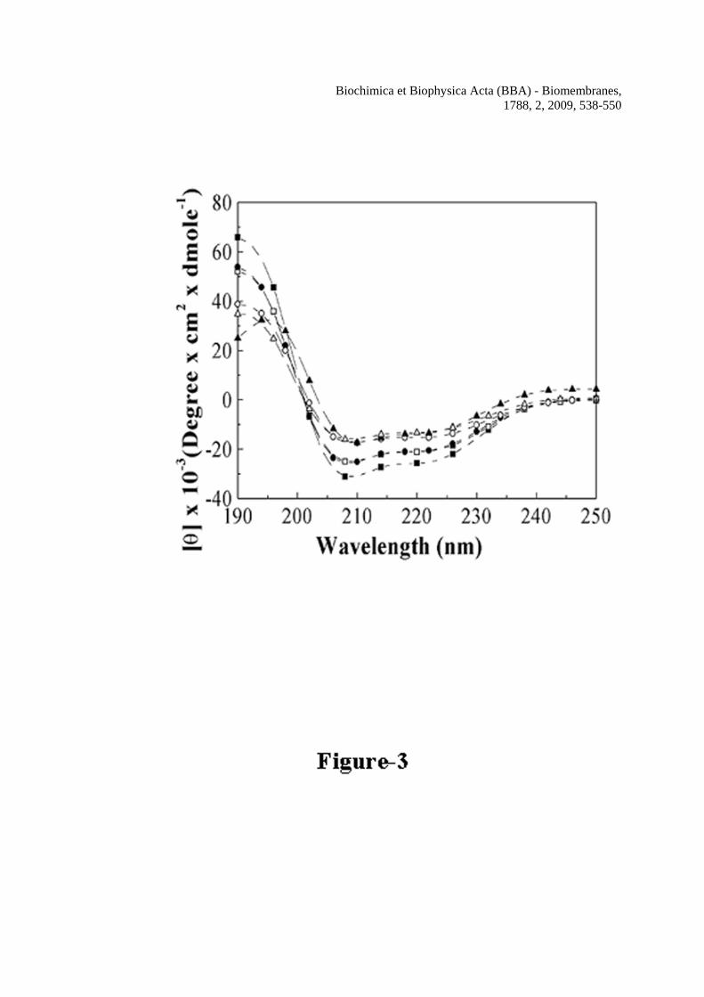
Biochimica et Biophysica Acta (BBA) - Biomembranes, 1788, 2, 2009, 538-550
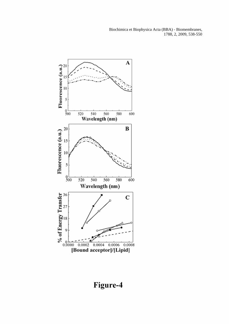
Biochimica et Biophysica Acta (BBA) - Biomembranes, 1788, 2, 2009, 538-550
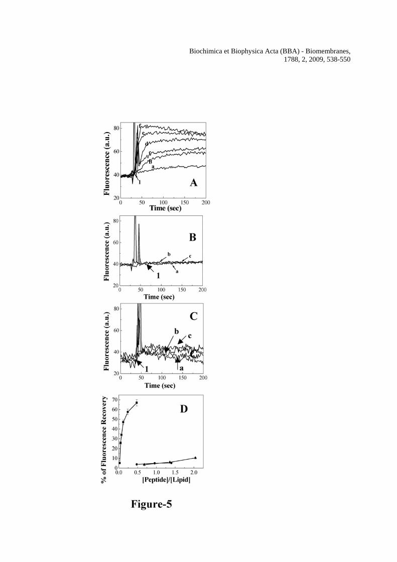
Biochimica et Biophysica Acta (BBA) - Biomembranes, 1788, 2, 2009, 538-550

Biochimica et Biophysica Acta (BBA) - Biomembranes, 1788, 2, 2009, 538-550
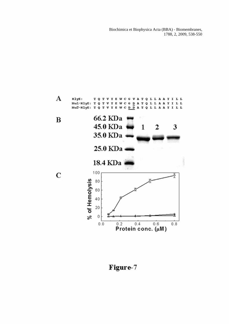
Biochimica et Biophysica Acta (BBA) - Biomembranes, 1788, 2, 2009, 538-550
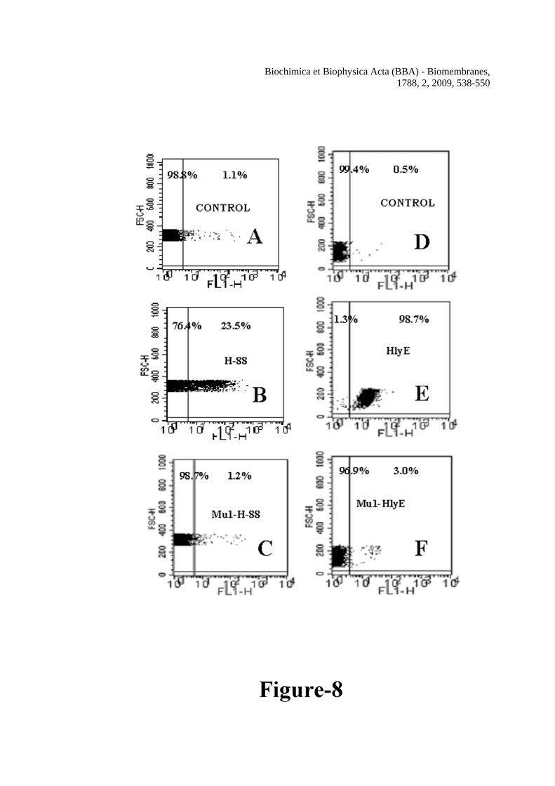
Biochimica et Biophysica Acta (BBA) - Biomembranes, 1788, 2, 2009, 538-550
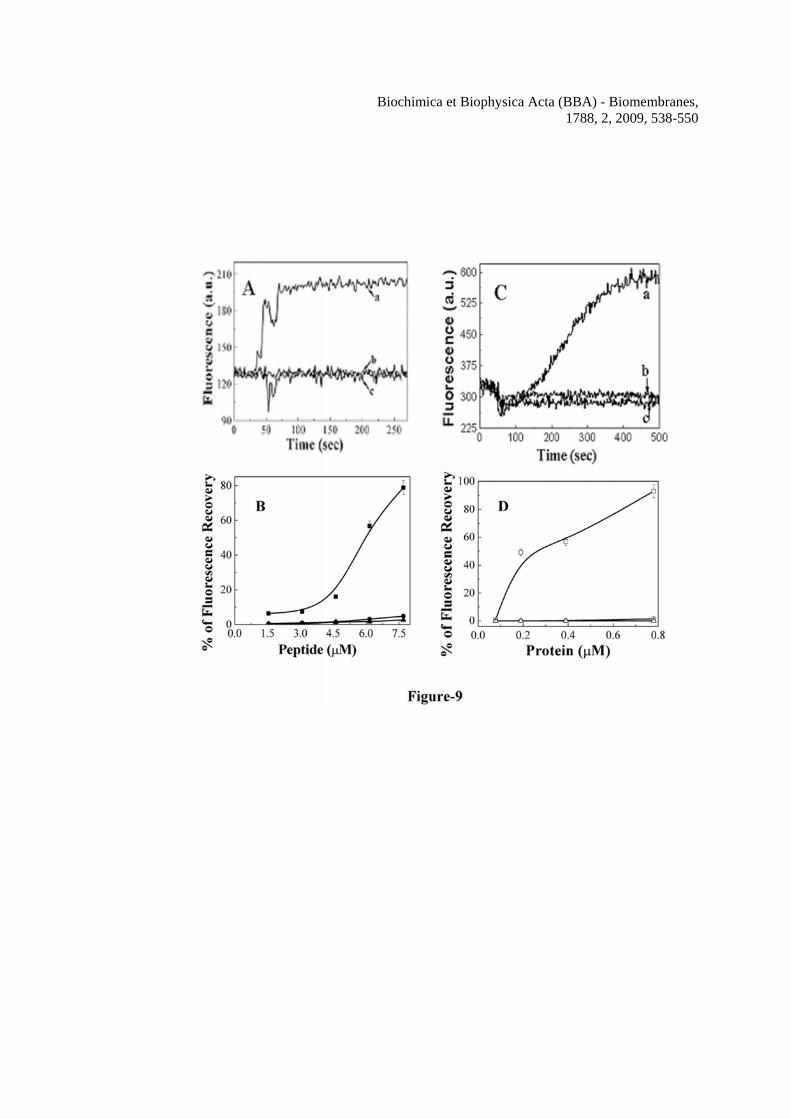
Biochimica et Biophysica Acta (BBA) - Biomembranes, 1788, 2, 2009, 538-550
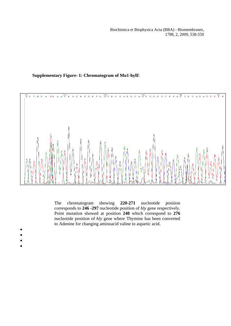
Biochimica et Biophysica Acta (BBA) - Biomembranes, 1788, 2, 2009, 538-550
Supplementary Figure- 1: Chromatogram of Mu1-hylE
The chromatogram showing 220-271 nucleotide position corresponds to 246 -297 nucleotide position of hly gene respectively. Point mutation showed at position 240 which correspond to 276 nucleotide position of hly gene where Thymine has been converted to Adenine for changing aminoacid valine to aspartic acid.
• • • •
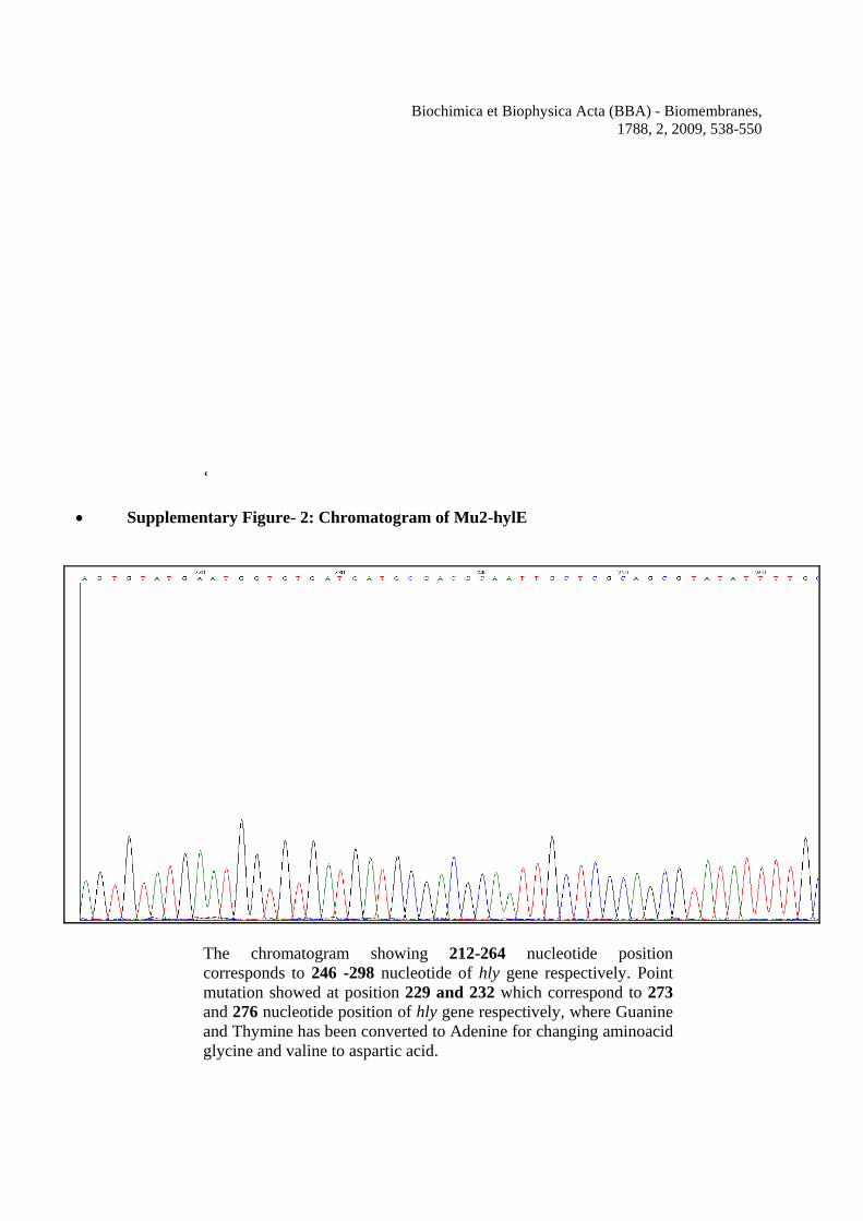
Biochimica et Biophysica Acta (BBA) - Biomembranes, 1788, 2, 2009, 538-550
‘
• Supplementary Figure- 2: Chromatogram of Mu2-hylE
The chromatogram showing 212-264 nucleotide position corresponds to 246 -298 nucleotide of hly gene respectively. Point mutation showed at position 229 and 232 which correspond to 273 and 276 nucleotide position of hly gene respectively, where Guanine and Thymine has been converted to Adenine for changing aminoacid glycine and valine to aspartic acid.
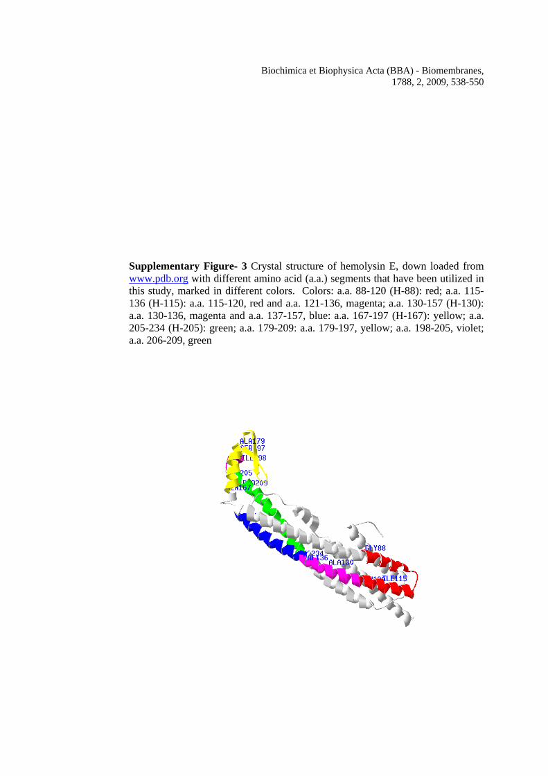
Biochimica et Biophysica Acta (BBA) - Biomembranes, 1788, 2, 2009, 538-550
Supplementary Figure- 3 Crystal structure of hemolysin E, down loaded from www.pdb.org with different amino acid (a.a.) segments that have been utilized in this study, marked in different colors. Colors: a.a. 88-120 (H-88): red; a.a. 115-136 (H-115): a.a. 115-120, red and a.a. 121-136, magenta; a.a. 130-157 (H-130): a.a. 130-136, magenta and a.a. 137-157, blue: a.a. 167-197 (H-167): yellow; a.a. 205-234 (H-205): green; a.a. 179-209: a.a. 179-197, yellow; a.a. 198-205, violet; a.a. 206-209, green
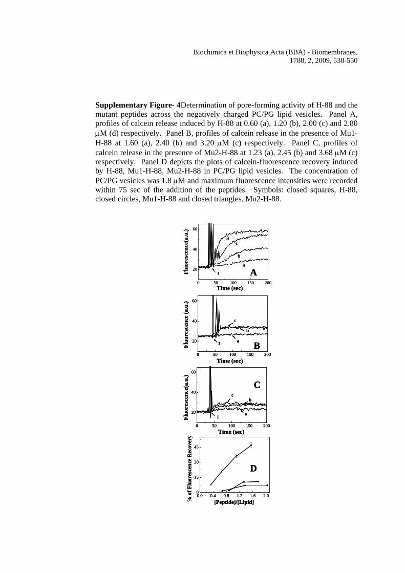
Biochimica et Biophysica Acta (BBA) - Biomembranes, 1788, 2, 2009, 538-550
Supplementary Figure- 4Determination of pore-forming activity of H-88 and the mutant peptides across the negatively charged PC/PG lipid vesicles. Panel A, profiles of calcein release induced by H-88 at 0.60 (a), 1.20 (b), 2.00 (c) and 2.80 μM (d) respectively. Panel B, profiles of calcein release in the presence of Mu1-H-88 at 1.60 (a), 2.40 (b) and 3.20 μM (c) respectively. Panel C, profiles of calcein release in the presence of Mu2-H-88 at 1.23 (a), 2.45 (b) and 3.68 μM (c) respectively. Panel D depicts the plots of calcein-fluorescence recovery induced by H-88, Mu1-H-88, Mu2-H-88 in PC/PG lipid vesicles. The concentration of PC/PG vesicles was 1.8 μM and maximum fluorescence intensities were recorded within 75 sec of the addition of the peptides. Symbols: closed squares, H-88, closed circles, Mu1-H-88 and closed triangles, Mu2-H-88.
0 50 100 150 200
20
40
60
1
dc
b
a
Time (sec)
Fluo
resc
ence
(a.u
.)
0 50 100 150 200
20
40
60
c
b
a1
Time (sec)
Fluo
resc
ence
(a.u
.)
0 50 100 150 200
20
40
60
1
cb
a
Time (sec)
Fluo
resc
ence
(a.u
.)
0.0 0.4 0.8 1.2 1.6 2.00
15
30
45
[Peptide]/[Lipid]% o
f Flu
ores
cenc
e R
ecov
ery
A
B
C
D
0 50 100 150 200
20
40
60
1
dc
b
a
Time (sec)
Fluo
resc
ence
(a.u
.)
0 50 100 150 200
20
40
60
c
b
a1
Time (sec)
Fluo
resc
ence
(a.u
.)
0 50 100 150 200
20
40
60
1
cb
a
Time (sec)
Fluo
resc
ence
(a.u
.)
0.0 0.4 0.8 1.2 1.6 2.00
15
30
45
[Peptide]/[Lipid]% o
f Flu
ores
cenc
e R
ecov
ery
A
B
C
D
0 50 100 150 200
20
40
60
c
b
a1
Time (sec)
Fluo
resc
ence
(a.u
.)
0 50 100 150 200
20
40
60
1
cb
a
Time (sec)
Fluo
resc
ence
(a.u
.)
0.0 0.4 0.8 1.2 1.6 2.00
15
30
45
[Peptide]/[Lipid]% o
f Flu
ores
cenc
e R
ecov
ery
A
B
C
D
Copyright © 2022 FDOKUMEN