A Panel of Trypanosoma brucei Strains Tagged with Blue and Red-Shifted Luciferases for...
16
A Panel of Trypanosoma brucei Strains Tagged with Blue and Red-Shifted Luciferases for Bioluminescent Imaging in Murine Infection Models Nick Van Reet 1 *, He ´le ` ne Van de Vyver 2 , Patient Pati Pyana 1,3 , Anne Marie Van der Linden 4 , Philippe Bu ¨ scher 1 1 Department of Biomedical Sciences, Institute of Tropical Medicine, Antwerp, Belgium, 2 Institute of Medical Microbiology, University Hospital of Mu ¨ nster, Mu ¨ nster, Germany, 3 De ´partement de Parasitologie, Institut National de Recherche Biome ´dicale, Kinshasa Gombe, Democratic Republic of the Congo, 4 Faculty of Pharmaceutical, Biomedical and Veterinary Sciences, Bio-Imaging Lab, Department of Biomedical Sciences, University of Antwerp, Wilrijk, Belgium Abstract Background: Genetic engineering with luciferase reporter genes allows monitoring Trypanosoma brucei (T.b.) infections in mice by in vivo bioluminescence imaging (BLI). Until recently, luminescent T.b. models were based on Renilla luciferase (RLuc) activity. Our study aimed at evaluating red-shifted luciferases for in vivo BLI in a set of diverse T.b. strains of all three subspecies, including some recently isolated from human patients. Methodology/Principal findings: We transfected T.b. brucei, T.b. rhodesiense and T.b. gambiense strains with either RLuc, click beetle red (CBR) or Photinus pyralis RE9 (PpyRE9) luciferase and characterised their in vitro luciferase activity, growth profile and drug sensitivity, and their potential for in vivo BLI. Compared to RLuc, the red-shifted luciferases, CBR and PpyRE9, allow tracking of T.b. brucei AnTaR 1 trypanosomes with higher details on tissue distribution, and PpyRE9 allows detection of the parasites with a sensitivity of at least one order of magnitude higher than CBR luciferase. With CBR-tagged T.b. gambiense LiTaR1, T.b. rhodesiense RUMPHI and T.b. gambiense 348 BT in an acute, subacute and chronic infection model respectively, we observed differences in parasite tropism for murine tissues during in vivo BLI. Ex vivo BLI on the brain confirmed central nervous system infection by all luminescent strains of T.b. brucei AnTaR 1, T.b. rhodesiense RUMPHI and T.b. gambiense 348 BT. Conclusions/Significance: We established a genetically and phenotypically diverse collection of bioluminescent T.b. brucei, T.b. gambiense and T.b. rhodesiense strains, including drug resistant strains. For in vivo BLI monitoring of murine infections, we recommend trypanosome strains transfected with red-shifted luciferase reporter genes, such as CBR and PpyRE9. Red- shifted luciferases can be detected with a higher sensitivity in vivo and at the same time they improve the spatial resolution of the parasites in the entire body due to the better kinetics of their substrate D-luciferin. Citation: Van Reet N, Van de Vyver H, Pyana PP, Van der Linden AM, Bu ¨ scher P (2014) A Panel of Trypanosoma brucei Strains Tagged with Blue and Red-Shifted Luciferases for Bioluminescent Imaging in Murine Infection Models. PLoS Negl Trop Dis 8(8): e3054. doi:10.1371/journal.pntd.0003054 Editor: Jayne Raper, New York University School of Medicine, United States of America Received March 7, 2014; Accepted June 17, 2014; Published August 21, 2014 Copyright: ß 2014 Van Reet et al. This is an open-access article distributed under the terms of the Creative Commons Attribution License, which permits unrestricted use, distribution, and reproduction in any medium, provided the original author and source are credited. Data Availability: The authors confirm that all data underlying the findings are fully available without restriction. Funding: This work was supported by grants of the Fonds voor Wetenschappelijk Onderzoek (1.5.147.09N and G.0229.10 N). The funders had no role in study design, data collection and analysis, decision to publish, or preparation of the manuscript. Competing Interests: The authors have declared that no competing interests exist. * Email: [email protected] Introduction African trypanosomes pose a threat to millions of humans and animals in sub-Saharan Africa. Only two species readily infect humans and both are subspecies of Trypanosoma brucei (T.b.). T.b. gambiense is responsible for the chronic form of human African trypanosomiasis (HAT) in West and Central Africa and accounts for more than 97% of the near 10,000 sleeping sickness patients who are diagnosed and treated annually [1]. T.b. rhodesiense causes a more acute form of HAT in South and East Africa, only differing from a third non human-infective subspecies, T.b. brucei, by a single gene, SRA, that confers resistance against human serum [2]. All T.b. subspecies are transmitted by the bite of tsetse flies (Glossina spss). Upon injection into a vertebrate host, parasites multiply locally in the lesion and cause a blood, lymph and tissue infection, also called first stage disease. Later, the parasites invade the central nervous system (CNS) initiating the second stage of the disease. Untreated infections almost invariably have a fatal outcome that occurs after weeks to months in rhodesiense HAT and months to years in gambiense HAT [3]. Treatment is subspecies- and stage-specific [1]. First stage gambiense and rhodesiense HAT are treated with pentamidine and suramin respectively. The first line treatment for second stage gambiense HAT consists of nifurtimox-eflornithine combination therapy (NECT) while second stage rhodesiense HAT is still treated with melarsoprol. All current drugs used to treat HAT are toxic [4]. For obvious reasons, basic and applied research on trypano- somes and HAT, including drug resistance studies, highly benefit PLOS Neglected Tropical Diseases | www.plosntds.org 1 August 2014 | Volume 8 | Issue 8 | e3054
-
Upload
independent -
Category
Documents
-
view
0 -
download
0
Transcript of A Panel of Trypanosoma brucei Strains Tagged with Blue and Red-Shifted Luciferases for...

A Panel of Trypanosoma brucei Strains Tagged with Blueand Red-Shifted Luciferases for Bioluminescent Imagingin Murine Infection ModelsNick Van Reet1*, Helene Van de Vyver2, Patient Pati Pyana1,3, Anne Marie Van der Linden4,
Philippe Buscher1
1 Department of Biomedical Sciences, Institute of Tropical Medicine, Antwerp, Belgium, 2 Institute of Medical Microbiology, University Hospital of Munster, Munster,
Germany, 3 Departement de Parasitologie, Institut National de Recherche Biomedicale, Kinshasa Gombe, Democratic Republic of the Congo, 4 Faculty of Pharmaceutical,
Biomedical and Veterinary Sciences, Bio-Imaging Lab, Department of Biomedical Sciences, University of Antwerp, Wilrijk, Belgium
Abstract
Background: Genetic engineering with luciferase reporter genes allows monitoring Trypanosoma brucei (T.b.) infections inmice by in vivo bioluminescence imaging (BLI). Until recently, luminescent T.b. models were based on Renilla luciferase(RLuc) activity. Our study aimed at evaluating red-shifted luciferases for in vivo BLI in a set of diverse T.b. strains of all threesubspecies, including some recently isolated from human patients.
Methodology/Principal findings: We transfected T.b. brucei, T.b. rhodesiense and T.b. gambiense strains with either RLuc,click beetle red (CBR) or Photinus pyralis RE9 (PpyRE9) luciferase and characterised their in vitro luciferase activity, growthprofile and drug sensitivity, and their potential for in vivo BLI. Compared to RLuc, the red-shifted luciferases, CBR andPpyRE9, allow tracking of T.b. brucei AnTaR 1 trypanosomes with higher details on tissue distribution, and PpyRE9 allowsdetection of the parasites with a sensitivity of at least one order of magnitude higher than CBR luciferase. With CBR-taggedT.b. gambiense LiTaR1, T.b. rhodesiense RUMPHI and T.b. gambiense 348 BT in an acute, subacute and chronic infection modelrespectively, we observed differences in parasite tropism for murine tissues during in vivo BLI. Ex vivo BLI on the brainconfirmed central nervous system infection by all luminescent strains of T.b. brucei AnTaR 1, T.b. rhodesiense RUMPHI andT.b. gambiense 348 BT.
Conclusions/Significance: We established a genetically and phenotypically diverse collection of bioluminescent T.b. brucei,T.b. gambiense and T.b. rhodesiense strains, including drug resistant strains. For in vivo BLI monitoring of murine infections,we recommend trypanosome strains transfected with red-shifted luciferase reporter genes, such as CBR and PpyRE9. Red-shifted luciferases can be detected with a higher sensitivity in vivo and at the same time they improve the spatial resolutionof the parasites in the entire body due to the better kinetics of their substrate D-luciferin.
Citation: Van Reet N, Van de Vyver H, Pyana PP, Van der Linden AM, Buscher P (2014) A Panel of Trypanosoma brucei Strains Tagged with Blue and Red-ShiftedLuciferases for Bioluminescent Imaging in Murine Infection Models. PLoS Negl Trop Dis 8(8): e3054. doi:10.1371/journal.pntd.0003054
Editor: Jayne Raper, New York University School of Medicine, United States of America
Received March 7, 2014; Accepted June 17, 2014; Published August 21, 2014
Copyright: � 2014 Van Reet et al. This is an open-access article distributed under the terms of the Creative Commons Attribution License, which permitsunrestricted use, distribution, and reproduction in any medium, provided the original author and source are credited.
Data Availability: The authors confirm that all data underlying the findings are fully available without restriction.
Funding: This work was supported by grants of the Fonds voor Wetenschappelijk Onderzoek (1.5.147.09N and G.0229.10 N). The funders had no role in studydesign, data collection and analysis, decision to publish, or preparation of the manuscript.
Competing Interests: The authors have declared that no competing interests exist.
* Email: [email protected]
Introduction
African trypanosomes pose a threat to millions of humans and
animals in sub-Saharan Africa. Only two species readily infect
humans and both are subspecies of Trypanosoma brucei (T.b.). T.b.gambiense is responsible for the chronic form of human African
trypanosomiasis (HAT) in West and Central Africa and accounts
for more than 97% of the near 10,000 sleeping sickness patients
who are diagnosed and treated annually [1]. T.b. rhodesiensecauses a more acute form of HAT in South and East Africa, only
differing from a third non human-infective subspecies, T.b. brucei,by a single gene, SRA, that confers resistance against human
serum [2]. All T.b. subspecies are transmitted by the bite of tsetse
flies (Glossina spss). Upon injection into a vertebrate host, parasites
multiply locally in the lesion and cause a blood, lymph and tissue
infection, also called first stage disease. Later, the parasites invade
the central nervous system (CNS) initiating the second stage of the
disease. Untreated infections almost invariably have a fatal
outcome that occurs after weeks to months in rhodesiense HAT
and months to years in gambiense HAT [3]. Treatment is
subspecies- and stage-specific [1]. First stage gambiense and
rhodesiense HAT are treated with pentamidine and suramin
respectively. The first line treatment for second stage gambienseHAT consists of nifurtimox-eflornithine combination therapy
(NECT) while second stage rhodesiense HAT is still treated with
melarsoprol. All current drugs used to treat HAT are toxic [4].
For obvious reasons, basic and applied research on trypano-
somes and HAT, including drug resistance studies, highly benefit
PLOS Neglected Tropical Diseases | www.plosntds.org 1 August 2014 | Volume 8 | Issue 8 | e3054

from the availability of T.b. rhodesiense and T.b. gambiense strains
that have been recently isolated from patients with known
treatment outcome and that along their isolation underwent only
few in vivo and/or in vitro passages [5–8]. To this end,
bloodstream form trypanosomes can be isolated from diverse
patient specimens such as blood, lymph or cerebrospinal fluid
(CSF). Generally, T.b. brucei and T.b. rhodesiense can be easily
isolated in classical laboratory rodents such as mice and rats [9].
T.b. gambiense, on the other hand, is very difficult to isolate and
often requires either susceptible rodent species [9–12] or severely
immune-suppressed or –deprived hosts [13,14]. Seldomly, isola-
tion of bloodstream form T.b. gambiense parasites has been
achieved by direct inoculation of in vitro medium containing
feeder layer cells [5,15].
Apart from isolation of trypanosome strains, rodent models for
HAT are considered to be informative because they can reproduce
the invasion of the central nervous system (CNS) [16,17]. Thus,
these rodent models are highly relevant for investigating drug
discovery, drug resistance and treatment failure [18,19]. Much like
the clinical diversity seen in HAT [1], rodent models also reveal a
very broad spectrum of pathology resulting from infection with
diverse strains of the human pathogenic subspecies of T.b. [5,20–
27]. Genetic engineering of parasites has made it possible to
monitor infections in living animals using biophotonic techniques,
such as in vivo fluorescence and bioluminescence imaging [28,29].
In vivo bioluminescent imaging (BLI) allows the tracking of
luciferase-modified cells in living animals over time without the
need to sacrifice them. This technique has been applied to study
the spatio-temporal distribution of T.b. brucei and T.b. gambienseparasites in murine models and lead to the discovery of a testis
tropism of T.b. brucei AnTaR 1, where the parasites are less
accessible for trypanocides [30]. BLI also revealed that different
T.b. gambiense strains can induce a variety of infections in mice,
ranging from chronic to silent, thus mimicking the clinical diversity
that is observed in HAT [5]. The former models made use of
RLuc as the bioluminescent reporter [31], which, upon oxidation
of its substrate coelenterazine, emits blue light (peak luminescence
at 480 nm) that is readily absorbed by blood and other tissues.
Alternative luciferases emitting light with longer wavelengths exist,
such as luciferase enzymes from fireflies, click beetles and railroad
worms that all use D-luciferin as substrate [32,33]. Firefly
luciferase (FLuc) appeared to sort into the glycosome of T.b.possibly disturbing ATP/ADP equilibrium upon activation [34].
Recently the well characterised T.b. brucei GVR 35 strain was
modified with a firefly luciferase variant (LUC2), that emits yellow
light (560 nm), and proved to be useful to shorten the follow-up
period in studies on drugs that reach the CNS during the infection
[29]. Also, studies with LUC2 modified T. vivax have shown that
this trypanosome species, like T.b., invades tissues including the
CNS [35]. However, luciferases that emit light beyond 600 nm are
potentially even more useful for in vivo imaging due to the fact
that transmission of light through animal tissue increases greatly
above this wavelength [36]. The synthetic click beetle red
luciferase (CBR) from Pyrophorus plagiophtalamus and the
thermostabilised PpyRE9 luciferase from Photinus pyralis both
emit light around 617 nm [37]. These red-shifted luciferases have
shown a potential to better resolve signals from deeper tissues than
the original FLuc or to emit more stable luminescence than the
LUC2 variant [38,39]. Very recently it was shown that PpyRE9-
tagged T.b. brucei GVR 35 parasites allow improved detection in
BLI over LUC2-tagged T.b. brucei GVR 35 models [40].
In the present study we explored the use of red-shifted firefly
luciferases CBR and PpyRE9 for bioluminescent imaging of all
three subspecies of T.b., including the human pathogens, in
comparison with the existing RLuc strains in our collection. This
collection consists of a set of four genetically and phenotypically
very different trypanosome strains (Table 1). T.b. brucei AnTaR 1
is a pleomorphic strain from Uganda that causes a sub-acute
infection in various mouse and rat strains [41,42]. T.b. rhodesienseRUMPHI is a recently isolated strain from Malawi, which
underwent only a few in vivo and in vitro passages and originates
from an area with less virulent T.b. rhodesiense than those
circulating in Uganda [43,44]. T.b. gambiense LiTaR 1 is a well
characterised virulent strain that is used for the production of
diagnostic antigens in the card agglutination test for trypanoso-
miasis (CATT) and in the recently developed HAT-Sero-K-SeT
[45,46]. This strain is extensively passaged in vivo and produces
an acute monomorphic infection in rodents. In contrast, T.b.gambiense 348 BT was isolated in the HAT focus of Mbuji-Mayi in
the Democratic Republic of the Congo (DRC) [7], where a very
high relapse rate after melarsoprol treatment has been observed
that may be related to the presence of a chimeric aquaglycer-
oporin 2/3 gene [47,48].
We focus this report on the in vitro drug sensitivity, the in vitrogrowth characteristics and the in vivo virulence in mice, assessed
through bioluminescence imaging of either RLuc, CBR and
PpyRE9 luciferase activity, of the four bioluminescent T.b. strains.
Materials and Methods
Ethics statementThis study was approved by the Veterinary Ethics Committee of
the Institute of Tropical Medicine, Antwerp, Belgium (protocol
BM2012-1 and BM2013-5) and the Veterinary Ethics Committee
of the University of Antwerp, Belgium (protocol BPI-EAT). It
adheres to the European Commission Recommendation on
guidelines for the accommodation and care of animals used for
experimental and other scientific purposes (18 June 2007, 2007/
526/EG) and the Belgian National law on the protection of
Author Summary
Research on African trypanosomes heavily relies on rodentinfection models. One way to reduce the number oflaboratory rodents used in each experiment and effectivelyfollow the progression of the infection in the same animalsis to use genetically modified trypanosomes that allowmonitoring of the infection over time with biolumines-cence technology, without having to sacrifice the animalsat multiple time points. In this study, we were able toestablish a collection of bioluminescent strains of all threesubspecies of Trypanosoma brucei (T.b.), including T.b.gambiense and T.b. rhodesiense that cause human Africantrypanosomiasis (HAT) or sleeping sickness. Making use ofbioluminescence assays, we demonstrate the diversity ofour collection in terms of in vitro and in vivo growth, drugsensitivity and in vivo parasite distribution, includingcentral nervous system tropism. Growth characteristicsand drug sensitivity are not affected by the geneticmodification with luciferase reporter genes. Trypanosomestrains transfected with red-shifted luciferase reportergenes have several advantages compared to the corre-sponding blue luciferase modified strains. Red light is lessabsorbed in the blood than blue light, which should leadto higher sensitivity of detection. Furthermore, thesubstrates that drive the light reaction are better distrib-uted through the body for the red luciferase than for theblue luciferase, which greatly improves spatial resolutionof the infection.
Blue and Red Luminescent Trypanosoma brucei spp
PLOS Neglected Tropical Diseases | www.plosntds.org 2 August 2014 | Volume 8 | Issue 8 | e3054

animals under experiment. The parasite strains included in this
study belong to the cryobank of the World Health Collaboration
Center for Research and Training on Human African Trypano-
somiasis Diagnostics at the Institute of Tropical Medicine in
Antwerp, Belgium.
Trypanozoon strains and in vitro cultureThe axenic in vitro culture of monomorphic and pleomorphic
bloodstream form trypanosome populations in HMI-9 has been
described elsewhere [49,50]. The original host, the year and
country of isolation, the number of in vivo passages and the
medium for in vitro culture of T.b. brucei AnTaR 1, T.b.rhodesiense RUMPHI and the T. b. gambiense strains LiTaR 1
and 348 BT are described in Table 1. The propagation of the
bloodstream form in vivo in rodents, the adaptation in vitro to an
HMI-9 based culture medium and the molecular confirmation of
their taxonomic identity have been described previously [7,51,52].
Iscove’s modified Dulbecco’s medium powder (IMDM) and foetal
calf serum (FCS; heat-inactivated; EU approved; South American
origin) were purchased from Invitrogen (Carlsbad, USA). For invitro assays, medium was prepared from IMDM without phenol
red (Invitrogen) and without addition of antibiotics. All other
culture media ingredients were from Sigma–Aldrich (St. Louis,
MO, USA). Briefly, strains were isolated from first peak
parasitaemia in mice, cultured in HMI-9 based medium contain-
ing 1,1% methylcellulose and 15% foetal bovine serum with or
without 5% heat-inactivated human serum until adaptation [51].
All strains were adapted to medium without methylcellulose before
transfection as previously described [52]. Strains were cultivated in
500 ml of medium in a 48-well plate at densities between 103–106
cells ml21 and maintained in logarithmic growth phase by
subpassages at appropriate dilutions after 24 to 72 hours of
incubation at 37uC and 5% CO2. Cultures were monitored by
phase contrast inverted microscopy. Cell counting was performed
in disposable counting chambers (Uriglass, Menarini Diagnostics,
Belgium). For larger cell preparations, the cultures were stepwise
scaled up to 40 ml in 25 cm2 flasks, by addition of four (for T.b.gambiense) to nine (for T.b. rhodesiense/brucei) volumes of fresh
medium once the parasites reached a density of 56105 cells ml21.
For long term storage, cells were concentrated tenfold from log
phase cultures in 90% medium with 10% glycerol and frozen
stepwise to 240uC at 1uC/min using a programmable cryogenic
freezing device (MiniCool MP40, Air Liquide, Belgium) where-
after they were kept in liquid nitrogen.
Overexpression of reporter genesThe promoterless vector pHD309 was used for constitutive
expression of foreign genes in trypanosomes [5,30,52]. For
overexpression of a single reporter gene, the pHD309 plasmid
was cut with BamHI and HindIII and PCR products were fused
using In-Fusion Cloning and transformed in Fusion Blue cells
according to the manufacturer’s recommendations (Clontech,
Takara Bio, Japan). The cDNA sequences of the reporter genes
were amplified from their donor plasmids using gradient PCR and
a proofreading polymerase (Deep VentR, New England Biolabs,
UK). All primers (Biolegio, Nijmegen, The Netherlands) contained
a cDNA specific sequence and a 59 extension of 15 nucleotides
specific to the place of integration, containing the restriction site
and sequence overlap with the vector as required for the In-Fusion
Cloning reaction (Table S1). Trypanosomes that have been
electroporated (Gene Pulse Xcell, Bio-Rad, USA) with a NotI
linearised plasmid can afterwards be selected with hygromycin to
obtain stable recombinants. The transfection and selection of
trypanosome strains that express RLuc has been described earlier
Ta
ble
1.
List
of
T.b
.st
rain
san
dth
eir
his
tory
.
Ta
xo
nS
tra
inO
rig
ina
lh
ost
Co
un
try
an
dH
AT
focu
sY
ea
rP
ass
ag
es
inro
de
nt
Invi
tro
me
diu
mR
efe
ren
ce
T.b
.b
ruce
iA
nT
aR1
bu
shb
uck
Ug
and
a,B
uso
ga
19
66
10
HM
I[7
7]
T.b
.rh
od
esie
nse
RU
MP
HI
hu
man
Mal
awi,
Ru
mp
hi
20
07
3H
MI
[51
]
T.b
.g
am
bie
nse
LiT
aR1
hu
man
Co
ted
’Ivo
ire
19
67
14
HM
I+h
um
anse
rum
[78
]
T.b
.g
am
bie
nse
34
8B
Th
um
anD
RC
,M
bu
ji-M
ayi
20
07
3H
MI+
hu
man
seru
m[5
1]
do
i:10
.13
71
/jo
urn
al.p
ntd
.00
03
05
4.t
00
1
Blue and Red Luminescent Trypanosoma brucei spp
PLOS Neglected Tropical Diseases | www.plosntds.org 3 August 2014 | Volume 8 | Issue 8 | e3054

[30,52]. Recombinant populations were maintained in 1 mg ml21
hygromycin for T.b. gambiense strains and 5 mg ml21 for T.b.brucei and T.b. rhodesiense strains for over three weeks after
transfection upon which the most resistant populations were
cryopreserved and used for further analysis of luciferase activity.
The selection antibiotic was no longer added to the in vitrocultures during luciferase activity and drug sensitivity testing.
In vitro luciferase activityTo measure the luminescent activity of the RLuc-modified
strains, the EnduRen Live Cell assay (Promega, Madison, USA)
was used with a final EnduRen concentration of 6 mM. Sixty mM
EnduRen stock solution in DMSO was diluted 1:1000 into HMI-9
medium without phenol red. Five ml of this solution were
transferred to a well of a white opaque 1/2 area 96-well plate
(Perkin Elmer, Waltham, MA, USA) and 45 ml of a trypanosome
suspension were added. The plate was incubated for at least one
hour in a 5% CO2 incubator at 37uC. After measurement of RLuc
activity with EnduRen, the amount of ATP was measured in the
same sample by adding 50 ml of CellTiter Glo reagent (Promega),
to create a luminescent multiplex viability assay as described
previously [52]. To measure the luminescent activity of the firefly
luciferases, the ONE-Glo Luciferase reagent (Promega) was
reconstituted as described by the manufacturer and 20 ml of this
assay solution were added to 20 ml of a trypanosome suspension in
HMI-9 in an opaque white 1/2 area 96-well plate (Perkin Elmer).
A separate aliquot of 20 ml of the trypanosome suspension was
used to measure ATP luminescence using the CellTiter-Glo
reagent. No centrifugation or wash steps were required in any of
the protocols. All luminescent measurements were performed after
2 minutes of shaking and the number of counts was integrated by
sampling over a 1 second period (CPS), every minute for
10 minutes using WorkOut software from Victor X3 plate reader
(Perkin Elmer). The CPS values were divided by the CPS value of
HMI-9 medium (fold change), plotted against the trypanosome cell
density and a linear regression was calculated in GraphPad
(Prism). The threshold for detection was defined as a fold change
.3. The relative activity in each clone was calculated as the ratio
of the CPS in the luciferase assay (EnduRen, for RLuc or ONE-
Glo, for CBR and P9) over the CPS in the luminescent cell
viability assay (CellTiter Glo). The means of the clones of the red-
shifted luciferases with the highest relative activity and the mean
doubling time of the wild-type and their luminescent population(s)
were compared using one-way analysis of variance (ANOVA) with
Bonferroni post-hoc test in GraphPad (Prism).
IC50 drug sensitivityEflornithine (Sanofi Aventis, Paris, France) and hygromycin B
(Sigma) were prepared as 10 mg ml21 stock solutions in distilled
water. Melarsoprol (Sanofi Aventis), suramin (Bayer, Leverkusen,
Germany), pentamidine isethionate (Sanofi Aventis) and nifurti-
mox (Sigma) were stored as 10 mg ml21 stock solutions in DMSO.
Dophanil powder (Docpharma, Hoeilaart, Belgium), containing
455 mg diminazene diaceturate and 555 mg antipyrine per gram,
was prepared as a 10 mg ml21 diminazene diaceturate solution in
DMSO. A method to measure the IC50 values of compounds in
96-well plates was performed as described elsewhere [53].
Threefold drug dilutions in duplicate were made in HMI-9
medium to allow testing in final drug concentrations ranging from
100 to 0.14 mg ml21 for eflornithine and hygromycin, from 50 to
0.07 mg ml21 for nifurtimox, from 10 to 0.014 mg ml21 suramin
and from 500 to 0.7 ng ml21 for diminazene diaceturate,
melarsoprol and pentamidine with 56103 cells ml21 in a total
volume of 200 ml. Next, the plate was incubated for 72 hours at
37uC with 5% CO2 followed by addition of 20 ml of resazurin
(Sigma; 12.5 mg in 100 ml PBS). After a further 24 h incubation
at 37uC with 5% CO2, fluorescence was measured (excitation
l= 560 nm; emission l= 590 nm) with a VictorX3 multimodal
plate reader using top reading (Perkin Elmer) [54]. The results
were expressed as the percent reduction in parasite viability
compared to the parasite viability in control wells without drugs.
The 50% inhibitory concentration (IC50) was calculated using
non-linear regression and compared between groups with one-way
ANOVA and Bonferroni post-hoc test in GraphPad (Prism).
In vivo luciferase activityFor experiments with T.b. brucei AnTaR 1, T.b. gambiense
LiTaR 1 and T.b. rhodesiense RUMPHI, female OF-1 mice
(2563 g) in groups of 3 were infected intraperitoneally (IP) with
26104 parasites (from culture medium). Every group was tested at
days 1, 4, 7, 18 and 26 post-infection. For experiments with T.b.gambiense 348 BT, female OF-1 mice (3065 g) in groups of 3,
treated or not treated with 200 mg/kg cyclophosphamide (CPA)
IP (Endoxan, Baxter, Lessing, Belgium) 2 days pre-infection, were
infected with 26105 parasites (from infected mouse blood). These
groups were tested at days 1, 3, 7, 11, 43 and, for CBR only, also
on day 60 post-infection. Before each BLI recording, animals were
weighed and anaesthetised by inhalation of 5% isoflurane (Isoflo,
USP) for induction and 2% isoflurane for maintenance in 100% 02
at a flow rate of 1000 ml min21. While under anaesthesia, mice
were injected IP with 10 ml kg21 body weight of a 15 mg ml21 D-
luciferin (ViviGlo, D-luciferin potassium salt, Promega) in
phosphate buffered saline pH 7.4 (PBS) or 1 mg ml21 ViviRen
(Promega) in PBS with 0,1% bovine serum albumin (BSA) [55].
Two to five minutes after injection of the substrate, a ten to fifteen
minute image acquisition was made on an in vivo bioluminescence
imager (Photon Imager, Biospace, France). During the imaging
session, the animal was placed on its back on a heated mat (39uC)
to maintain body temperature. After each session, the parasitae-
mia was estimated using the matching method on 30 fields,
allowing a detection limit of 105 cells ml21 in whole blood [56].
The BLI data were analysed by dividing the images of the mice in
3 rectangular shaped regions of interest (ROI); covering the
abdomen (12.3 cm2), the thorax (6.1 cm2) and the head (2.9 cm2)
(Figure 1). The radiance in each ROI was obtained from a 30 to
60 second period within the plateau phase of luminescence and
expressed in photons per second per square centimetre per
steradian (ph s21 cm22 sr21) in M3 Vision (Biospace, France). In
the non-infected controls ViviRen was injected first, followed by a
washout period of at least 4 hours before D-luciferin
Figure 1. Definition of ROI. BLI data were analysed in function of 3ROIs; (A) abdomen, (B) thorax and (C) head.doi:10.1371/journal.pntd.0003054.g001
Blue and Red Luminescent Trypanosoma brucei spp
PLOS Neglected Tropical Diseases | www.plosntds.org 4 August 2014 | Volume 8 | Issue 8 | e3054
administration. The threshold for detection was defined as a .3
fold change in radiance compared to non-infected controls.
Ex vivo luciferase activityAt day 43 and day 60 for T.b. gambiense 348 BT and at day 26 for
T.b. brucei AnTaR 1 and T.b. rhodesiense RUMPHI, mice were
transcardially perfused, under Nembutal anaesthesia (60 mg kg21 in
PBS, IP), with 50 ml of phosphate buffered saline glucose (PBSG;
10 mM phosphate pH 7.4, 0.9% NaCl and 1% glucose) at a flow
rate of 5 ml min21 to rinse the vascular compartments of
trypanosomes. The spleen was excised and weighed while the brains
(without dura mater and arachnoid) were removed, washed in 10 ml
of PBSG and incubated in a 24-well plate in 1 ml of PBSG
containing either 1.5 mg ml21 D-luciferin or 0,1 mg ml21 ViviRen.
After 5 minutes delay, a BLI recording was made for 10 minutes.
The BLI data were analysed by drawing a ROI around the
circumference of the well. The radiance (ph s21 cm22 sr21) was
expressed as fold change over the average values of the non-infected
control brains for each substrate as described above.
Results
A panel of blue and red luminescent trypanosomesRLuc luciferase expressing clones of T.b. brucei AnTaR 1 and
T.b. gambiense 348 BT were available from previous studies
[30,52]. CBR luciferase was integrated in these strains as well as in
T.b. gambiense LiTaR 1 and T.b. rhodesiense RUMHPI. The
PpyRE9 luciferase (P9), a promising red-shifted luciferase for invivo imaging, was only integrated in T.b. brucei AnTaR 1 and was
not yet tested in the other strains. Out of four clones of T.b. bruceiAnTaR 1 transfected with pHD P9 (AnTaR 1 P9), ten clones of
T.b. brucei AnTaR 1 transfected with pHD CBR (AnTaR 1 CBR),
3 clones of T.b. rhodesiense RUMPHI transfected with pHD CBR
(RUMPHI CBR), 7 clones of T.b. gambiense LiTaR 1 transfected
with pHD CBR (LiTaR 1 CBR) and 2 clones of T.b. gambiense348 BT transfected with pHD CBR (348 BT CBR) that were
simultaneously tested, we identified for each strain and luciferase
reporter combination, the clone with the highest relative luciferase
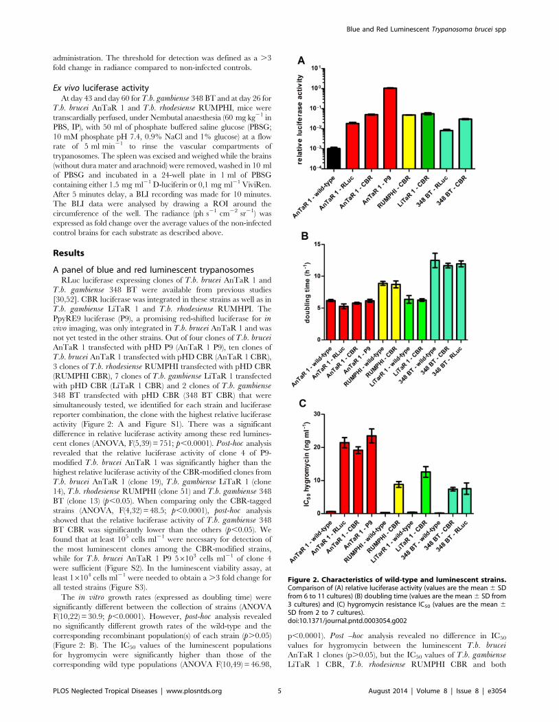
activity (Figure 2: A and Figure S1). There was a significant
difference in relative luciferase activity among these red lumines-
cent clones (ANOVA, F(5,39) = 751; p,0.0001). Post-hoc analysis
revealed that the relative luciferase activity of clone 4 of P9-
modified T.b. brucei AnTaR 1 was significantly higher than the
highest relative luciferase activity of the CBR-modified clones from
T.b. brucei AnTaR 1 (clone 19), T.b. gambiense LiTaR 1 (clone
14), T.b. rhodesiense RUMPHI (clone 51) and T.b. gambiense 348
BT (clone 13) (p,0.05). When comparing only the CBR-tagged
strains (ANOVA, F(4,32) = 48.5; p,0.0001), post-hoc analysis
showed that the relative luciferase activity of T.b. gambiense 348
BT CBR was significantly lower than the others (p,0.05). We
found that at least 105 cells ml21 were necessary for detection of
the most luminescent clones among the CBR-modified strains,
while for T.b. brucei AnTaR 1 P9 56103 cells ml21 of clone 4
were sufficient (Figure S2). In the luminescent viability assay, at
least 16104 cells ml21 were needed to obtain a .3 fold change for
all tested strains (Figure S3).
The in vitro growth rates (expressed as doubling time) were
significantly different between the collection of strains (ANOVA
F(10,22) = 30.9; p,0.0001). However, post-hoc analysis revealed
no significantly different growth rates of the wild-type and the
corresponding recombinant population(s) of each strain (p.0.05)
(Figure 2: B). The IC50 values of the luminescent populations
for hygromycin were significantly higher than those of the
corresponding wild type populations (ANOVA F(10,49) = 46.98,
p,0.0001). Post –hoc analysis revealed no difference in IC50
values for hygromycin between the luminescent T.b. bruceiAnTaR 1 clones (p.0.05), but the IC50 values of T.b. gambienseLiTaR 1 CBR, T.b. rhodesiense RUMPHI CBR and both
Figure 2. Characteristics of wild-type and luminescent strains.Comparison of (A) relative luciferase activity (values are the mean 6 SDfrom 6 to 11 cultures) (B) doubling time (values are the mean 6 SD from3 cultures) and (C) hygromycin resistance IC50 (values are the mean 6SD from 2 to 7 cultures).doi:10.1371/journal.pntd.0003054.g002
Blue and Red Luminescent Trypanosoma brucei spp
PLOS Neglected Tropical Diseases | www.plosntds.org 5 August 2014 | Volume 8 | Issue 8 | e3054

Figure 3. Drug sensitivity profiles of wild-type and luminescent strains. IC50 (mean 6 SD) of wild-type and luminescent strains against (A)eflornithine (values are the mean 6 SD from 4 to 9 cultures), (B) nifurtimox (values are the mean 6 SD from 4 to 9 cultures), (C) diminazenediaceturate (values are the mean 6 SD from 2 cultures), (D) melarsoprol (values are the mean 6 SD from 4 to 9 cultures), (E) suramin (values are themean 6 SD from 4 to 9 cultures) and (F) pentamidine isethionate (values are the mean 6 SD from 2 cultures).doi:10.1371/journal.pntd.0003054.g003
Blue and Red Luminescent Trypanosoma brucei spp
PLOS Neglected Tropical Diseases | www.plosntds.org 6 August 2014 | Volume 8 | Issue 8 | e3054

CBR- and RLuc-modified T.b. gambiense 348 BT strains were
significantly lower than those of the luminescent T.b. bruceiAnTaR 1 populations (p,0.05) (Figure 2: C).
IC50 values of the wild-type and recombinant strains fortrypanocides
The drug sensitivity profiles of all wild-type and luminescent
strains were compared against a set of trypanocides (eflornithine,
nifurtimox, diminazene diaceturate, melarsoprol, suramin and
pentamidine isethionate) to test if the luminescent modifications
induced differences in IC50 value. For each drug, ANOVA found
differences between the IC50 values from the collection of strains
(for eflornithine F(10,66) = 183, p,0.0001; for nifurtimox
F(10,66) = 37, p,0.0001; for diminazene diaceturate
F(10,11) = 112.3, p,0.0001; for melarsoprol F(10,63) = 33, p,
0.0001; for suramin F(10,68) = 26.22, p,0.001 and for
Figure 4. Quantification of BLI data of OF-1 mice infected with luminescent T.b. brucei, T.b. rhodesiense and T.b. gambiense. OF-1 mice(n = 3) were infected with T.b. brucei AnTaR 1 wild-type, T.b. brucei AnTaR 1 RLuc, T.b. brucei AnTaR 1 CBR, T.b. brucei AnTaR 1 P9, T.b. rhodesienseRUMPHI CBR, T.b. gambiense LiTaR 1 CBR and their luminescence was measured at 1, 4, 7, 18 and 26 post-infection in BLI using their respectivesubstrates. The BLI data were divided in 3 ROIs; (A) abdomen, (B) thorax and (C) head and expressed as ph s21 cm22 sr21.doi:10.1371/journal.pntd.0003054.g004
Blue and Red Luminescent Trypanosoma brucei spp
PLOS Neglected Tropical Diseases | www.plosntds.org 7 August 2014 | Volume 8 | Issue 8 | e3054

pentamidine F(6,7) = 105, p,0.0001). Post-hoc analysis did not
reveal significant differences between the IC50 values of the wild-
type and the corresponding luminescent population(s) of each
strain (for each drug, p.0.05). However, there were substantial
differences between the different strains for each of the different
drugs as represented in Figure 3. For eflornithine, all T.b. bruceiAnTaR 1 populations had significantly higher IC50 values than all
the other populations and all T.b. rhodesiense RUMPHI
populations had significantly higher IC50 values than T.b.gambiense populations (p,0.05) (Figure 3: A). For nifurtimox,
the IC50 values of all T.b. brucei AnTaR 1 populations were
significantly higher, while all IC50 values of T.b. gambiense 348 BT
were significantly lower than all IC50 values of T.b. rhodesienseRUMPHI and T.b. gambiense LiTaR 1 populations (p,0.05)
(Figure 3: B). For diminazene diaceturate, IC50 values of all T.b.brucei AnTaR 1 and T.b. gambiense 348BT were significantly
different and were significantly higher IC50 than of all T.b.rhodesiense RUMPHI and T.b. gambiense LiTaR 1 populations
(p,0.05) (Figure 3: C). For melarsoprol, IC50 values of all T.b.brucei AnTaR 1 and T.b. gambiense 348 BT populations were
higher than those of T.b. gambiense LiTaR 1 and T.b. rhodesienseRUMPHI (Figure 3: D). Strikingly, the IC50 values of all T.b.
Figure 5. Visualisation of BLI data of OF-1 mice infected with T.b. brucei, T.b. rhodesiense and T.b. gambiense. OF-1 mice (n = 3) wereinfected with T.b. brucei AnTaR 1 RLuc (A–D), T.b. brucei AnTaR 1 CBR (E–H), T.b. brucei AnTaR 1 P9 (I–L), T.b. rhodesiense RUMPHI CBR (M–P), T.b.gambiense LiTaR 1 CBR (Q–R) and their luminescence was measured in BLI using their respective substrates. Rows represent measurements at 1, 4, 18and 26 days post-infection.doi:10.1371/journal.pntd.0003054.g005
Blue and Red Luminescent Trypanosoma brucei spp
PLOS Neglected Tropical Diseases | www.plosntds.org 8 August 2014 | Volume 8 | Issue 8 | e3054

gambiense 348 BT for suramin were about tenfold higher than of
all the other strains (p,0.05) (Figure 3: E). Also for pentamidine,
the IC50 values of all T.b. gambiense 348 BT populations were
much higher than those of T.b. brucei AnTaR 1, while the IC50
values of T.b. gambiense LiTaR and T.b. rhodesiense RUMPHI
were below the lower threshold (,0.70 ng ml21) (p,0.05)
(Figure 3: F).
Infections with the different luciferase-tagged T.b. bruceiAnTaR 1 trypanosomes
The sensitivity of RLuc, CBR and PpyRE9 luciferase detection
during in vivo BLI of a murine infection was assessed with T.b.brucei AnTaR 1. Throughout the infection, very high parasitemia
between 107 and 108.4 cells ml21 was observed for the wild-type
strain and for the RLuc-, CBR- and P9-modified clones. Mice
Figure 6. Quantification of BLI data in untreated and CPA-treated mice infected with T.b. gambiense. OF-1 mice (n = 3) untreated or CPA-treated were infected with T.b. gambiense 348 BT RLuc and T.b. gambiense 348 BT CBR and their luminescence was measured at 1, 3, 7, 11, 43 (forRLuc) and 60 days post-infection (for CBR) using their respective substrates. The BLI data were divided in 3 ROIs; (A) abdomen, (B) thorax and (C) headand expressed as ph s21 cm22 sr21.doi:10.1371/journal.pntd.0003054.g006
Blue and Red Luminescent Trypanosoma brucei spp
PLOS Neglected Tropical Diseases | www.plosntds.org 9 August 2014 | Volume 8 | Issue 8 | e3054

Blue and Red Luminescent Trypanosoma brucei spp
PLOS Neglected Tropical Diseases | www.plosntds.org 10 August 2014 | Volume 8 | Issue 8 | e3054

infected with wild-type and recombinant T.b. brucei AnTaR 1
parasites showed increased body weight gain. The spleen weight of
T.b. brucei AnTaR 1 infected mice that survived until day 26 post-
infection varied from 1 to 2 gram, roughly tenfold the spleen
weight of uninfected mice, indicating severe splenomegaly.
For the BLI experiments, the background luminescence of each
luciferase substrate was measured in uninfected mice and was at least
four times higher for ViviRen (1622864826 ph s21 cm22 sr21)
than for D-luciferin (46226927 ph s21 cm22 sr21) in the abdom-
inal ROI, with lesser differences in the thoracic ROI (ViviRen:
40646873 and D-luciferin: 37436927 ph s21 cm22 sr21) and in
the head ROI (ViviRen :37726789 and D-luciferin: 31316
459 ph s21 cm22 sr21). The in vivo luciferase activities in function
of ROI and days post-infection are represented in Figure 4 and
visualised in Figure 5. At day 1 post-infection, when parasitaemia is
not yet detectable by microscopy, the BLI signal is already above
threshold in the abdominal region of some mice infected with the
CBR strain (Figure 5: E) and in all compartments of all mice infected
with the P9 strain (Figure 5: I). No signal above background could be
detected in mice infected with RLuc-tagged parasites (Figure 5: A).
At day 4 post-infection, the RLuc-infected mice (Figure 5: B) were
100 fold more luminescent in the abdominal region and at least 10
fold more luminescent in the thorax and the head region than wild-
type-infected mice (Figure 4). Mice infected with trypanosomes
expressing red luciferases were much more luminescent with a fold
change of 5000 in the abdomen and 1000 in the thorax and head for
CBR-infected mice and a fold change of almost 100000 in the
abdomen and of 10000 in the thorax and head for the P9-infected
mice (Figure 4 and Figure 5: F and J).
Interestingly, the difference in sensitivity (as defined by higher
radiance values) between CBR and P9 was not associated with a
different distribution of the trypanosomes in the body. No more
visual detail could be obtained from the higher luminescence of P9
compared to CBR-tagged parasites (Figure 5: F and J, G and K).
BLI pictures often revealed the contours of lymph nodes, but in
most cases the signal consisted of a superimposed surface covering
multiple organs in the body, especially in the abdomen. With RLuc-
tagged parasites, less information was obtained. Luminescence was
often only visible in the abdominal region (Figure 5: B, C and D).
After day 7 post-infection, data were too limited for analysis due to
differences between the strains in survival of mice and parasitaemia.
The median survival time was 26 days for mice infected with wild-
type; 26 days with RLuc; 26 days with CBR and 11 days with P9,
with deaths in all groups occurring first at day 11 post-infection (3
animals) and later between day 18 and 25 post-infection (4 animals).
Infections with CBR-tagged trypanosomes of thedifferent T.b. subspecies
T.b. gambiense LiTaR 1 killed mice in 5 days without any overt
signs of pathology. The trypanosomes were monomorphic and at
day 4 post-infection, hours before death, the parasitaemia
approached 108,7 cells ml21. In contrast, T.b. rhodesienseRUMPHI infection was not fatal up to day 26 post-infection,
which marked the end of the experiment. Throughout the
infection, the parasitaemia of T.b. rhodesiense RUMPHI varied
between 26106 and 56107 cells ml21 and was markedly lower
than that of T.b. brucei AnTaR 1 and T.b. gambiense LiTaR 1.
One mouse showed a delay in reaching the first peak of
parasitemia (Figure 5: M–O). Although mice infected with
RUMPHI did show signs of lethargy, splenomegaly was less
pronounced than in mice infected with T.b. brucei AnTaR 1.
Figure 7. Visualisation of BLI data in untreated and CPA-treated OF-1 mice infected with T.b. gambiense. OF-1 mice (n = 3) were infectedwith T.b. gambiense 348 BT RLuc without (A–E) or with CPA treatment (F–J) and their luminescence was measured with ViviRen or were infected withT.b. gambiense 348 BT CBR without (K–P) or with (Q–V) CPA treatment and their luminescence was measured with D-luciferin. Rows representmeasurements at 1, 3, 7, 11, 43 (for RLuc) and 60 days post-infection (only CBR). In square P one mouse woke up from anesthesia just before the endof acquisition.doi:10.1371/journal.pntd.0003054.g007
Figure 8. Visualisation of ex vivo brain BLI data obtained from mice infected with different luminescent strains. At 26 days post-infection for T.b. brucei AnTaR 1 and T.b. rhodesiense RUMPHI and at 43 and 60 days post-infection for T.b. gambiense 348 BT brains were extractedand immersed in PBSG and substrate. (A–B) T.b. gambiense 348 BT CBR, CPA-treated, with D-luciferin; (C) T.b. gambiense 348 BT CBR, untreated, withD-luciferin ;(D) uninfected with D-luciferin; (E) T.b. gambiense 348 BT RLuc, CPA-treated with ViviRen; (F–H) T.b. rhodesiense RUMPHI CBR with D-luciferin; (I) T.b. brucei AnTaR 1 CBR with D-luciferin; (J) T.b. brucei AnTaR 1 P9 with D-luciferin.doi:10.1371/journal.pntd.0003054.g008
Blue and Red Luminescent Trypanosoma brucei spp
PLOS Neglected Tropical Diseases | www.plosntds.org 11 August 2014 | Volume 8 | Issue 8 | e3054

When we compared the in vivo luciferase activity in mice infected
with CBR-tagged T.b. brucei AnTaR 1, T.b. rhodesienseRUMPHI and T.b. gambiense LiTaR 1, it appeared that on day
1 post-infection, when none of the infected mice showed
microscopically detectable parasitaemia, all strains could be
detected with BLI in the abdominal region of some infected mice
(Figure 5: E, M and Q). At day 4 post-infection, all three strains
generated a comparable BLI signal in the abdomen and thorax
(Figure 4: A and B). The signal from the head was highest in mice
infected with T.b. gambiense LiTaR 1, followed by mice infected
with T.b. brucei AnTaR 1 and with T.b. rhodesiense RUMPHI
(Figure 4: C and Figure 5: F, N and R). Later during the infection,
all mice infected with T.b. gambiense LiTaR 1 and 2 of 3 mice
infected with T.b. brucei AnTaR 1 died while all mice infected
with T.b. rhodesiense RUMPHI survived until the end of the
experiment at day 26 post-infection with steadily increasing BLI
signals in the head ROI (Figure 5: O and P).
Effect of immune suppression on infection with blue andred T.b. gambiense 348 BT
RLuc- and CBR-modified parasites of T.b. gambiense 348 BT
were injected in BALB/c that underwent weekly CPA treatment.
When these animals became parasitologically positive after 14 to
20 days (a total of 2–5 trypanosomes in 30 fields), their blood was
injected into CPA-treated OF-1 mice. The peak of parasitaemia
occurred within one week and again, blood containing the
parasites was injected in 2 CPA-treated OF-1 mice. The first
peak of parasitaemia in the latter mice reached 108,1 cells ml21
and blood was diluted with PBSG to 106 cells ml21 and used to
infect three CPA-treated OF-1 mice and three untreated OF-1
mice. In the untreated mice, parasitaemia remained undetectable
in all RLuc-infected mice and in two CBR-infected mice; only one
CBR-infected mouse was once positive at day 4 post-infection. In
the CPA-treated mice, both RLuc and CBR infections gave rise to
detectable parasitaemia that increased until 11 days post-infection
where after the mice became only sporadically positive. At the end
of the experiment we recorded mild splenomegaly in all mice that
became parasitologically positive. The in vivo luciferase activity in
function of ROI and days post infection are represented in
Figure 6 and visualised in Figure 7. As observed in the BLI
experiment with T.b. brucei AnTaR 1, the T.b. gambiense 348 BT
RLuc-modified parasites were less informative than the CBR-
modified parasites. With BLI, the signal of the RLuc-modified
trypanosomes was below threshold during the whole infection
period in untreated mice (Figure 7: A–E), but in CPA-treated
animals, the T.b. gambiense 348 BT RLuc-modified parasites were
detectable at day 1 and day 3 post-infection in the abdomen
(Figure 7: F and G) and at day 7 also in the thorax and the head of
some mice (Figure 7: H). In contrast, in both CPA-treated and
untreated mice, T.b. gambiense 348 BT CBR-tagged parasites
were detectable at day 1 post-infection in the abdomen, and at day
4 post-infection also in the thorax and the head (Figure 7: K–V).
At day 7 post-infection, the T.b. gambiense 348 BT CBR-modified
parasites became undetectable in the CPA-untreated mice
(Figure 7: M–P). In the CPA-treated mice we were able to track
the infection in all animals and in all compartments until day 43
post-infection, yet at day 60 post-infection the signal decreased
below the detection threshold (Figure 7: V).
Ex vivo luminescence in the brainAt day 26 post-infection for T.b. brucei AnTaR 1 and T.b.
rhodesiense RUMPHI and at day 43 and day 60 post-infection for
T.b. gambiense 348 BT, the surviving animals were sacrificed for
ex vivo BLI quantification of the parasites in the brain. The ventral
portion of the brain was the most informative for BLI data
(Figure 8). The brains from the animals infected with CBR-tagged
strains (Figure 8: A–C, I) showed 50 to 100 fold higher
luminescence than brains of uninfected mice (Figure 8: D,
representative image measured with D-luciferin). In the brain of
the mouse infected with the T.b. brucei P9-tagged strain (Figure 8:
J), the luminescence was even about 1000 fold higher. In the case
of infection with T.b. gambiense 348 RLuc and T.b. brucei AnTaR
RLuc, luminescence was detectable at the circumference of the
brain rather than in the brain itself (Figure 8: E, image of T.b.gambiense 348 BT RLuc measured with ViviRen) and similar to
the recording made from the brain of the T.b. brucei AnTaR P9
infected mouse (Figure 8: J), light radiated into the PBSG medium.
However, we did not check whether we could detect free
trypanosomes in the surrounding liquid or on the dorsal portion
of the brain. In case of infections with CBR and P9 tagged
trypanosomes and with D-luciferin as substrate, BLI signals
emanated from all over the brain, but the densest spots were
often observed in the olfactory bulbs, in the ventral anterior
hypothalamic region including the suprachiasmatic nucleus, and
in the cerebellum, as well as in the pituitary gland. The brains of
two CPA-untreated animals infected with the T.b. gambiense 348
BT CBR as well as all untreated mice infected with T.b. gambiense348 BT RLuc remained negative in BLI. As stated above, these
animals also remained aparasitaemic in blood.
Discussion
The aim of the present study was to assemble a set of genetically
and phenotypically diverse bioluminescent T.b. strains and to
assess the optimal reporter system (enzyme and substrate) for invivo bioluminescent imaging of murine infections.
Are red-shifted luciferases more advantageous tomonitor tagged trypanosomes in vivo?
In previous studies, we used RLuc as the reporter gene in
bioluminescent models of T.b. [5,30,52]. For the current study, we
opted to replace RLuc for in vivo imaging by a red-shifted firefly
luciferase reporter for several reasons. Coelenterazine, used for
RLuc activity detection, has rather unfavourable kinetics in vivo.
For example, it is thought not to pass the blood-brain barrier due
to the abundance of P-glycoprotein pumps in brain vascular
endothelium, whose efflux activity restricts access of coelenterazine
to the parenchyma unless there is severe dysfunction of the blood-
brain barrier [57]. Although this might be interesting for studying
advanced neurological trypanosomiasis models, it does not reflect
the precise timing of CNS infection by the parasite since
trypanosomes have been observed in CNS before tight junctions
are disrupted [58]. Novel variants of coelenterazine, such as
ViViRen, are more resistant against auto-oxidation in serum, but
similarly to coelenterazine, the eventual BLI signal highly depends
on the route of their administration [59]. Furthermore, all
coelenterazine variants are more expensive than D-luciferin. Also,
in contrast to coelenterazine variants, the distribution of D-
luciferin in vivo is fairly well characterised and optimised protocols
for administration and anaesthesia are available [60]. Therefore,
we did not extend our research into red-shifted Renilla luciferases
that have been described recently, neither did we compare
different administration routes [61]. We prefer IP injection which
is the most practical and appropriate for D-luciferin, the substrate
of all firefly and beetle luciferases, including the red-shifted
variants such as CBR and PpyRE9 [37].
When reporter genes are to be compared, one should use the
same vector background and the same assay to standardise
Blue and Red Luminescent Trypanosoma brucei spp
PLOS Neglected Tropical Diseases | www.plosntds.org 12 August 2014 | Volume 8 | Issue 8 | e3054

expression and activity measurement [62]. We expressed the
different reporter genes, RLuc, CBR and PpyRE9 in the same
trypanosomal expression vector, pHD309. This vector can be
integrated in the b-tubulin locus of trypanosomes and is one of the
few known expression vectors that has been proven successful in
T.b. gambiense through simple electroporation. Genome data show
that this locus consists of multiple tandem repeats, thus allowing
multiple integrations, while among different T.b. strains, a wide
variation in b-tubulin copy numbers has been reported [63,64]. In
our strains we did not assess the number of b-tubulin copy numbers
or the number of reporter gene copies integrated in this locus but
rather used hygromycin resistance to select modified clones of the
same strain with equal IC50 values for comparison. Using this
approach, our study confirms the higher catalytic activity of P9
compared to CBR in T.b. brucei AnTaR 1, both in vitro and in vivowith 10 to 20-fold higher signals generated by P9 as previously
described by Branchini et al [37]. However, it should be noted that
higher expression of reporter genes in T.b. can also be obtained by
modifying the expression vector for RNA polymerase I dependent
transcription and by replacing the 59 and 39 untranslated regions
flanking the reporter gene with sequences that flank highly
expressed genes, as demonstrated in McLatchie et al [40].
One of the limitations of this study is that we did not correlate the
in vivo BLI data with precise quantification of the parasites, e.g. by
real-time RT-PCR. However, we microscopically estimated para-
sitaemia in blood to allow comparison between luminescent clones
of the same strain early in infection (day 4 to day 7). Furthermore,
for CBR-modified T.b. brucei AnTaR 1, T.b. rhodesiense RUMPHI
and T.b. gambiense LiTaR 1, clones were selected with the same
level of luciferase activity per living cell, making differences in the invivo BLI responses dependent on parasitaemia and distribution in
organs. However, we did not verify whether the expression of the
reporter genes remained similar during the infection. At least invitro testing did not reveal changes in hygromycin resistance or
luciferase activity during a 6 week period in culture.
With an inoculum of 26104 to 26105 parasites, the red
luciferase trypanosomes can be tracked from day 1 post-infection,
although the sensitivity of detection was higher with P9 than with
CBR. This higher sensitivity of P9 is important to confirm
infection prior to treatment where treatment is to be given very
early after infection. Later in the infection, the added detail gained
from higher sensitivity of P9 becomes less important since the low
background signal inherent to in vivo luminescence imaging
allows similar localisation of the trypanosomes within mice
infected with CBR-modified trypanosomes, as previously reported
by Close et al for FLuc and Lux comparison [65]. Compared to P9
and CBR, signals from RLuc modified trypanosomes are mainly
from the abdominal region, which can be explained by the poor
absorbance of ViviRen after IP administration. In contrast with
their poor performance in vivo, protected coelenterazine com-
pounds, like EnduRen (Promega), perform very well in vitro as was
demonstrated in a luminescent multiplexed viability assay
developed to monitor the response of RLuc-modified trypano-
somes for compound screening [52].
Is our collection of modified trypanosomesphenotypically diverse?
Our collection of luminescent trypanosomes contains strains
with very diverse infection outcome. Although less virulent than
T.b. gambiense LiTaR 1, T.b. brucei AnTaR 1 strains induce a
subacute infection. The sustained high parasitaemia in the mice
might take its toll on the cardiovascular, hepatic or splenic
physiology causing some animals to die early in infection. The
occurrence of hepatosplenomegaly is an indication of high
virulence in T.b. brucei and T. evansi strains [66,67]. Other
studies record a less virulent phenotype of T.b. brucei AnTaR 1
that may be related to the number of passages in mice or in vitroor to the rodent species or breed. Our data suggest that the
integration of the expression plasmid did not change the growth
phenotype, as reflected by the in vitro doubling times of the wild-
type and the recombinant strains. Because of this high lethality in
mice, it would be advantageous to use rats as host, which are
known to control the infection longer than mice [68,69].
Unfortunately, in vivo imaging on larger rodents does not yet
seem very efficient [70].
The T.b. gambiense LiTaR 1 strain induces the same high
parasitaemia levels and early lethality as the monomorphic T.b.brucei Lister 427 strain [30]. In our in vivo experiments, the mice
did not survive the first peak of parasitaemia. The T.b. gambienseLiTaR 1 has undergone numerous passages in vivo and has
become monomorphic and highly virulent and thus very different
from wild-type strains that cause chronic gambiense sleeping
sickness. The investigation of T.b. gambiense LiTaR 1 is important
since laboratory accidents have shown that this strains is very
virulent also in humans [71].
T.b. rhodesiense RUMPHI induces lower peak parasitaemia, does
not show the survival bottleneck that characterises T.b. brucei AnTaR
1, but still, survival is limited to approximately one month. This strain
is fully tsetse transmissible in G. morsitans morsitans (personal
communication; Dr. Jan Van den Abbeele) and under pressure of
normal human serum in vitro, the serum resistance associated (SRA)
protein expressing phenotype can easily be obtained (data not shown).
All experiments in the present study were performed without normal
human serum pressure. It is not known if SRA expression would alter
the virulence phenotype in the rodent model.
In contrast to the other strains in our luminescent collection,
T.b. gambiense 348 BT mimics very well the chronic phenotype of
classical gambiense sleeping sickness in humans. Although both
RLuc and CBR modified parasites were visible in BLI early after
infection of immunosuppressed mice, the BLI signal strongly
decreased later during the infection reflecting progression to a
chronic infection. Such chronic infections, similarly to what is
observed in humans, are characterised by the absence of
detectable parasites in blood but by presence of the parasites in
the CNS. With T.b. brucei, such chronic infections with parasites
only present in the CNS can only be obtained by subcurative
treatment of the mice [17]. In our in vivo experiments, some
animals infected with luminescent T.b. gambiense 348 BT
remained negative in BLI. This may correspond to the silent
infection phenotype of T.b. gambiense, in which trypanosomes are
present in very low numbers and can only be traced back by
histopathological or molecular assays [5]. In addition, our
experiments show that with the same parasite strain a variety of
infection phenotypes can be encountered in different individuals
from the same outbred mouse strain, in casu OF-1 [20,21].
With all pleomorphic parasite strains, the CNS infection was
confirmed ex vivo. The sites that were found infected are not
different from those previously described with other T.b. gambienseand T.b. brucei strains [5,29,41,72–74]. It should be noted that we
cannot report on the presence of the trypanosomes in the dura
mater or the subarchnoid space because this tissue was removed
during brain extraction. Furthermore, our sample size and time of
brain extraction did not allow description of differences in CNS
invasion between the different trypanosome strains.
Next to their diversity in virulence phenotype, the biolumines-
cent trypanosome strains display a wide diversity in drug sensitivity
phenotype. T.b. brucei AnTaR 1 is less sensitive to eflornithine,
nifurtimox and diminazene diaceturate than the human infective
Blue and Red Luminescent Trypanosoma brucei spp
PLOS Neglected Tropical Diseases | www.plosntds.org 13 August 2014 | Volume 8 | Issue 8 | e3054

strains. Among the latter, T.b. gambiense 348 BT shows a
particularly interesting phenotype. Compared to the other strains,
it is less sensitive to melarsoprol, pentamidine and, surprisingly, to
suramin. This strain originates from the HAT focus of Mbuji-Mayi
in East Kasai, DRC where melarsoprol treatment failure rates of
about 40% have been reported [47]. Other strains isolated from
the same HAT focus have been found to be cross-resistant to
melarsoprol and pentamidine, a phenotype that was linked to a
chimaeric aquaglyceroporin 2/3 gene in their genome [48].
Sequencing confirmed the presence of the same mutation in T.b.gambiense 348 BT (personal communication, Dr. Maser Pascal). A
lower susceptibility of T.b. gambiense than T.b. brucei to suramin
has been described previously, but an even higher resistance has
been described in T. evansi [53,54]. T.b. rhodesiense RUMPHI is
less sensitive to eflornithine than the two T.b. gambiense strains in
our collection but is fully sensitive to pentamidine. This finding is
consistent with recent evidence that first stage rhodesiense HAT
patients can be cured with pentamidine, a drug that is much less
toxic than suramin [75,76]. Yet, suramin is still the first line
treatment for first stage rhodesiense HAT [1].
Concluding remarksWe established a very diverse collection of bioluminescent T.b.
brucei, T.b. gambiense and T.b. rhodesiense strains that is now
available for in vitro and in vivo studies on trypanosomiasis
research. For in vivo monitoring of murine infections, we
recommend the use of trypanosome strains transfected with red-
shifted luciferases, such as PpyRE9, above the blue RLuc. Recently
it was shown that PpyRE9 was also advantageous to monitor disease
progression with T.b. brucei GVR35. However, the study of T.b.gambiense field strains is far more relevant for sleeping sickness
rodent models, because this parasite subspecies causes more than
95% of the cases of HAT and severely differs in virulence from T.b.brucei, causing more chronic and protracted infections in murine
models. Furthermore, we modified a T.b. gambiense strain that
harbours the AQP2/3 chimaeric gene, which was recently shown to
be responsible for pentamidine and melarsoprol cross-resistance in
field isolates and quite possibly contributes to the high treatment
failure rates seen in the DRC and Southern Sudan.
Supporting Information
Figure S1 Relative luciferase activity of wild-type and red-
shifted luciferase modified trypanosomes. Relative luciferase
activity (mean of 2–8 repetitions 6 SD) of the firefly luciferase
modified clones of several strains (T.b. brucei AnTaR 1, T.b.
rhodesiense RUMPHI, T.b. gambiense LiTaR 1 and T.b.gambiense 348 BT).
(TIF)
Figure S2 ONE-Glo luciferase activity in wild-type and red-
shifted luciferase modified trypanosomes. ONE-Glo luciferase
activity expressed as signal to background in function of cell
density (cells ml21) (T.b. brucei AnTaR 1, T.b. rhodesienseRUMPHI, T.b. gambiense LiTaR 1 and T.b. gambiense 348
BT). Horizontal dotted line represents a fold change of 3. Vertical
dotted lines mark the cell density necessary for detection at this
threshold (56103 cells ml21 for T.b. brucei AnTaR 1 P9 and
approximately 105 cells ml21 for all CBR clones).
(TIF)
Figure S3 CellTiter-Glo luciferase activity in wild-type and red-
shifted luciferase modified trypanosomes. CellTiter-Glo luciferase
activity expressed as signal to background in function of cell
density (cells ml21) (T.b. brucei AnTaR, T.b. rhodesienseRUMPHI, T.b. gambiense LiTaR and T.b. gambiense 348BT).
Horizontal dotted line represents a fold change of 3. The vertical
dotted line marks the cell density necessary for detection at this
threshold (approximately 104 cells ml21).
(TIF)
Table S1 List of primer and cDNA sequences and the resulting
expression vector.
(DOCX)
Acknowledgments
The pHD309 vector was a kind gift from Dr. George Cross (Rockefeller
University, USA). The PpyRE9 cDNA was a kind gift from Dr. Bruce
Branchini (Connecticut University, USA). The wild-type T.b. rhodesienseRUMPHI was cyclically transmitted through G. m. morsitans by Dr. Van
Den Abbeele Jan (ITM, Belgium). The AQP2/3 locus of strain T.b.gambiense 348 BT was sequenced by Dr. Maser Pascal and Dr. Fabrice
Graff (STPHI, Switzerland). The authors like to thank Caroline
Guglielmetti (UA, Belgium), Shana Robbrecht (KaHo, Gent), Maarten
Van Den Bogaerde (ITM, Belgium) and Jeroen Swiers (ITM, Belgium) for
excellent assistance in bioimaging, molecular biology, animal care and
cryobiology.
Author Contributions
Conceived and designed the experiments: NVR PB. Performed the
experiments: NVR PPP HVdV. Analyzed the data: NVR HVdV PB.
Contributed reagents/materials/analysis tools: AMVdL PB. Wrote the
paper: NVR HVdV PPP AMVdL PB.
References
1. World Health Organization (2013) Control and surveillance of human African
trypanosomiasis. 984. http://apps.who.int/iris/bitstream/10665/95732/1/
9789241209847_eng.pdf
2. Xong HV, Vanhamme L, Chamekh M, Chimfwembe CE, Van Den Abbeele J
et al. (1998) A VSG expression site-associated gene confers resistance to human
serum in Trypanosoma rhodesiense. Cell 95: 839–846.
3. Checchi F, Filipe JAN, Haydon DT, Chandramohan D, Chappuis F (2008)
Estimates of the duration of the early and late stage of gambiense sleeping
sickness. BMC Infect Dis 8: doi:10.1186/1471-2334-8-16.
4. Bacchi CJ (2009) Chemotherapy of human African trypanosomiasis. Interdiscip
Perspect Infect Dis 2009: 195040.
5. Giroud C, Ottones F, Coustou V, Dacheux D, Biteau N et al. (2009) Murine
models for Trypanosoma brucei gambiense disease progression-from silent to
chronic infections and early brain tropism. PLoS Negl Trop Dis 3: e509.
6. Maina NW, Oberle M, Otieno C, Kunz C, Maeser P et al. (2007) Isolation and
propagation of Trypanosoma brucei gambiense from sleeping sickness patients in
south Sudan. Trans R Soc Trop Med Hyg 101: 540–546.
7. Pyana PP, Ngay Lukusa I, Mumba Ngoyi D, Van Reet N, Kaiser M et al. (2011)
Isolation of Trypanosoma brucei gambiense from cured and relapsed sleeping
sickness patients and adaptation to laboratory mice. PLoS Negl Trop Dis 5:
e1025.
8. Kibona SN, Matemba L, Kaboya JS, Lubega GW (2006) Drug-resistance of
Trypanosoma b. rhodesiense isolates from Tanzania. Trop Med Int Health 11:
144–155.
9. World Health Organization (1986) Epidemiology and control of African
trypanosomiasis. WHO Technical Report Series No 739.
10. Molyneux DH (1973) Isolation of Trypanosoma (Trypanozoon) brucei gambiensein rabbits by the intratesticular inoculation technique. Ann Trop Med Parasitol
67: 391–397.
11. Wery M, Weyn J, Mpela NN, Colaert J (1977) Isolement de souches de
T.gambiense au Zaıre et leur adaptation aux animaux de laboratoire. Ann Soc
Belg Med Trop 57: 425–437.
12. Buscher P, Kahremere Bin Shamamba S, Mumba D, Pyana P, Baelmans R
et al. (2005) Susceptibility of Grammomys surdaster thicket rats for
Trypanosoma brucei gambiense infection. Trop Med Int Health 10: 850–85
5.
13. Babiker EA, Le Ray D (1981) Adaptation of low virulence stocks of Trypanosomabrucei gambiense to rat and mouse. Ann Soc Belg Med Trop 61: 15–29.
Blue and Red Luminescent Trypanosoma brucei spp
PLOS Neglected Tropical Diseases | www.plosntds.org 14 August 2014 | Volume 8 | Issue 8 | e3054

14. Inoue N, Narumi D, Mbati PA, Hirumi K, Situakibanza NT et al. (1998)Susceptibility of severe combined immuno-deficient (SCID) mice to Trypano-soma brucei gambiense and T. b. rhodesiense. Trop Med Int Health 3: 408–412.
15. Zweygarth E, Kaminsky R (1990) Direct in vitro isolation of Trypanosoma bruceibrucei and T. b. evansi from disease hosts with low parasitaemia. Trop Med
Parasitol 41: 56–58.
16. Masocha W, Kristensson K (2012) Passage of parasites across the blood-brain
barrier. Virulence 3: 202–212.
17. Jennings FW, Whitelaw DD, Holmes PH, Chizyuka HGB, Urquhart GM (1979)The brain as a source of relapsing Trypanosoma brucei infection in mice after
chemotherapy. J Parasitol 9: 381–384.
18. Poltera AA (1985) Pathology of human African trypanosomiasis with reference
to experimental African trypanosomiasis and infections of the central nervous
system. Br Med Bull 41: 169–174.
19. Jennings FW, Hunter CA, Kennedy PGE, Murray M (1993) Chemotherapy of
Trypanosoma brucei infection of the central nervous system: the use of a rapidchemotherapeutic regimen and the development of post-treatment encephalop-
athies. Trans R Soc Trop Med Hyg 87: 224–226.
20. Beckers A, Wery M, Van Marck E, Gigase P (1981) Experimental infections oflaboratory rodents with recently isolated stocks of Trypanosoma bruceigambiense. 1. Parasitological investigations. Z Parasitenkd 64: 285–296.
21. Van Marck EA, Gigase PL, Beckers A, Wery M (1981) Experimental infectionsof laboratory rodents with recently isolated stocks of Trypanosoma bruceigambiense. 2. Histopathological investigations. Z Parasitenkd 64: 187–193.
22. Mulumba PM, Wery M (1983) Experimental infection with two stocks of
Trypanosoma brucei gambiense. Contrib Microbiol Immunol 7: 120–129.
23. Poltera AA (1983) Chemotherapy, relapses and CNS in experimental Africantrypanosomiasis. Contrib Microbiol Immunol 7: 155–164.
24. Holzmuller P, Biron DG, Courtois P, Koffi M, Bras-Goncalves R et al. (2008)Virulence and pathogenicity patterns of Trypanosoma brucei gambiense field
isolates in experimentally infected mouse: differences in host immune response
modulation by secretome and proteomics. Microbes Infect 10: 79–86.
25. Smith DH, Bailey JW (1997) Human African trypanosomiasis in south-eastern
Uganda: clinical diversity and isoenzyme profiles. Ann Trop Med Parasitol 91:851–856.
26. Gibson WC (1986) Will the real Trypanosoma b. gambiense please stand up?
Parasitol Today 2: 255–257.
27. Goodhead I, Capewell P, Bailey W, Beament T, Chance M et al. (2013) Whole-
genome sequencing of Trypanosoma brucei reveals introgression between
subspecies that is associated with virulence. mBio 4: 1–8.
28. Andreu N, Zelmer A, Wiles S (2011) Noninvasive biophotonic imaging for
studies of infectious disease. FEMS Microbiol Rev 35: 360–394.
29. Myburgh E, Coles JA, Ritchie R, Kennedy PG, McLatchie AP et al. (2013) Invivo imaging of trypanosome-brain interactions and development of a rapid
screening test for drugs against CNS stage trypanosomiasis. PLoS Negl Trop Dis7: e2384.
30. Claes F, Vodnala SK, Van Reet N, Boucher N, Lunden-Miguel H et al. (2009)Bioluminescent imaging of Trypanosoma brucei shows preferential testis
dissemination which may hamper drug efficacy in sleeping sickness patients.
PLoS Negl Trop Dis 3: e486.
31. Bhaumik S, Lewis XZ, Gambhir SS (2004) Optical imaging of Renilla luciferase,
synthetic Renilla luciferase, and firefly luciferase reporter gene expression inliving mice. J Biomed Opt 9: 578–586.
32. Wood KV, Lam YA, McElroy WD (1989) Introduction to beetle luciferases and
their applications. J Biolum Chemilum 4: 289–301.
33. Hastings JW (1996) Chemistries and colors of bioluminescent reactions: a review.
Gene 173: 5–11.
34. Sommer JM, Cheng QL, Keller GA, Wang CC (1992) In vivo import of fireflyluciferase into the glycosomes of Trypanosoma brucei and mutational analysis of
the C-terminal targeting signal. Mol Biol Cell 3: 749–759.
35. D’Archivio S, Cosson A, Medina M, Lang T, Minoprio P et al. (2013) Non-
invasive in vivo study of the Trypanosoma vivax infectious process consolidates
the brain commitment in late infections. PLoS Negl Trop Dis 7: e1976.
36. Doyle TC, Burns SM, Contag CH (2004) In vivo bioluminescence imaging for
integrated studies of infection. Cell Microbiol 6: 303–317.
37. Branchini BR, Ablamsky DM, Davis AL, Southworth TL, Butler B et al. (2010)
Red-emitting luciferases for bioluminescence reporter and imaging applications.
Anal Biochem 396: 290–297.
38. Miloud T, Henrich C, Hammerling GJ (2007) Quantitative comparison of click
beetle and firefly luciferases for in vivo bioluminescence imaging. J Biomed Opt12: 054018.
39. Liang Y, Walczak P, Bulte JW (2012) Comparison of red-shifted firefly luciferase
Ppy RE9 and conventional Luc2 as bioluminescence imaging reporter genes forin vivo imaging of stem cells. J Biomed Opt 17: 016004.
40. McLatchie AP, Burrell-Saward H, Myburgh E, Lewis MD, Ward TH et al.(2013) Highly sensitive in vivo imaging of Trypanosoma brucei expressing ‘‘red-
shifted’’ luciferase. PLoS Negl Trop Dis 7: e2571.
41. Schultzberg M, Ambatsis M, Samuelsson E-B, Kristensson K, Van Meirvenne N(1988) Spread of Trypanosoma brucei to the nervous system: early attack on
circumventricular organs and sensory ganglia. J Neurosci Res 21: 56–61.
42. Keita M, Bouteille B, Enanga B, Vallat J-M, Dumas M (1997) Trypanosomabrucei brucei: a long-term model of human African trypanosomiasis in mice,
meningo-encephalitis, astrocytosis, and neurological disorders. Exp Parasitol 85:183–192.
43. Rickman LR (1974) Investigations into an outbreak of human trypanosomiasis inthe lower Luangwa Valley, Eastern Province, Zambia. East Afr Med J 51: 467–
487.
44. MacLean L, Chisi JE, Odiit M, Gibson WC, Ferris V et al. (2004) Severity ofhuman African trypanosomiasis in East-Africa is associated with geographic
location, parasite genotype and host inflammatory cytokines response profile.
Infect Immun 72: 7040–7044.
45. Magnus E, Vervoort T, Van Meirvenne N (1978) A card-agglutination test with
stained trypanosomes (C.A.T.T.) for the serological diagnosis of T.b.gambiensetrypanosomiasis. Ann Soc Belg Med Trop 58: 169–176.
46. Buscher P, Gilleman Q, Lejon V (2013) Novel rapid diagnostic test for sleeping
sickness. N Engl J Med 368: 1069–1070.
47. Mumba Ngoyi D, Lejon V, Pyana P, Boelaert M, Ilunga M et al. (2010) How toshorten patient follow-up after treatment for Trypanosoma brucei gambiensesleeping sickness? J Infect Dis 201: 453–463.
48. Graf FE, Ludin P, Wenzler T, Kaiser M, Brun R et al. (2013) Aquaporin 2mutations in Trypanosoma brucei gambiense field isolates concur with decreased
susceptibility to pentamidine and melarsoprol. PLoS Negl Trop Dis 7: e2475.
49. Hirumi H, Hirumi K (1989) Continuous cultivation of Trypanosoma bruceiblood stream forms in a medium containing a low concentration of serum
protein without feeder cell layers. J Parasitol 75: 985–989.
50. McCulloch R, Vassella E, Burton P, Boshart M, Barry JD (2004) Transforma-
tion of monomorphic and pleomorphic Trypanosoma brucei. Methods Mol Biol
262: 53–86.
51. Van Reet N, Pyana PP, Deborggraeve S, Buscher P, Claes F (2011)
Trypanosoma brucei gambiense: HMI-9 medium containing methylcellulose
and human serum supports the continuous axenic in vitro propagation of thebloodstream form. Exp Parasitol 128: 285–290.
52. Van Reet N, Pyana PP, Roge S, Claes F, Buscher P (2013) Luminescent
multiplex viability assay for Trypanosoma brucei gambiense. Parasit Vectors 6:207.
53. Gillingwater K, Buscher P, Brun R (2007) Establishment of a panel of referenceTrypanosoma evansi and Trypanosoma equiperdum strains for drug screening.
Vet Parasitol 148: 114–121.
54. Raz B, Iten M, Grether-Buhler Y, Kaminsky R, Brun R (1997) The Alamar Blueassay to determine drug sensitivity of African trypanosomes (T.b. rhodesiense and
T.b. gambiense) in vitro. Acta Trop 68: 139–147.
55. Heikkila JE, Vaha-Koskela MJ, Ruotsalainen JJ, Martikainen MW, StanfordMM et al. (2010) Intravenously administered alphavirus vector VA7 eradicates
orthotopic human glioma xenografts in nude mice. PLoS ONE 5: e8603.
56. Herbert WJ, Lumsden WHR (1976) Trypanosoma brucei: A rapid ‘‘matching’’method for estimating the host’s parasitemia. Exp Parasitol 40: 427–431.
57. Pichler A, Prior JL, Piwnica-Worms D (2004) Imaging reversal of multidrug
resistance in living mice with bioluminescence: MDR1 P-glycoprotein transportscoelenterazine. Proc Natl Acad Sci USA 101: 1702–1707.
58. Mulenga C, Mhlanga JD, Kristensson K, Robertson BD (2001) Trypanosomabrucei brucei crosses the blood-brain barrier while tight junction proteins arepreserved in a rat chronic disease model. Neuropathol Appl Neurobiol 27: 77–
85.
59. Otto-Duessel M, Khankaldyyan V, Gonzalez-Gomez I, Jensen MC, Laug WE etal. (2006) In vivo testing of Renilla luciferase substrate analogs in an orthotopic
murine model of human glioblastoma. Mol Imaging 5: 57–64.
60. Aswendt M, Adamczak J, Couillard-Despres S, Hoehn M (2013) Boosting
bioluminescence neuroimaging: an optimized protocol for brain studies. PLoS
ONE 8: e55662.
61. Loening AM, Dragulescu-Andrasi A, Gambhir SS (2010) A red-shifted Renillaluciferase for transient reporter-gene expression. Nature Meth 7: 5–6.
62. Gil JS, Machado HB, Herschman HR (2012) A method to rapidly andaccurately compare the relative efficacies of non-invasive imaging reporter genes
in a mouse model and its application to luciferase reporters. Mol Imag Biol 14:
462–471.
63. Jackson AP, Vaughan S, Gull K (2006) Evolution of tubulin gene arrays in
Trypanosomatid parasites: genomic restructuring in Leishmania. BMC Geno-mics 7: 261.
64. Kanmogne GD, Bailey M, Gibson WC (1997) Wide variation in DNA content
among isolates of Trypanosoma brucei ssp. Acta Trop 63: 75–87.
65. Close DM, Xu T, Sayler GS, Ripp S (2011) In vivo bioluminescent imaging(BLI): noninvasive visualization and interrogation of biological processes in living
animals. Sensors 11: 180–206.
66. Morrison LJ, Tait A, McLellan S, Sweeney L, Turner CM et al. (2009) A majorgenetic locus in Trypanosoma brucei is a determinant of host pathology. PLoS
Negl Trop Dis 3: e557.
67. Mekata H, Konnai S, Mingala CN, Abes NS, Gutierrez CA et al. (2013)Isolation, cloning, and pathologic analysis of Trypanosoma evansi field isolates.
Parasitol Res 112: 1513–1521.
68. Darsaud A, Bourdon L, Chevrier C, Keita M, Bouteille B et al. (2003) Clinical
follow-up in the rat experimental model of African trypanosomiasis. Experi-
mental Biology and Medicine 228: 1355–1362.
69. Chevrier C, Canini F, Darsaud A, Cespuglio R, Buguet A et al. (2005) Clinical
assessment of the entry into neurological state in rat experimental African
trypanosomiasis. Acta Trop 95: 33–39.
70. Song HT, Jordan EK, Lewis BK, Liu W, Ganjei J et al. (2009) Rat model of
metastatic breast cancer monitored by MRI at 3 tesla and bioluminescence
imaging with histological correlation. J Travel Med 7: 88.
Blue and Red Luminescent Trypanosoma brucei spp
PLOS Neglected Tropical Diseases | www.plosntds.org 15 August 2014 | Volume 8 | Issue 8 | e3054

71. Herwaldt BL (2001) Laboratory-acquired parasitic infections from accidental
exposures. Clin Microbiol Rev 14: 659–688.72. Van Marck EAE, Le Ray D, Beckers A, Jacob W, Wery M et al. (1981) Light
and electron microscope studies on extravascular Trypanosoma brucei gambiensein the brain of chronically infected rodents. Ann Soc Belg Med Trop 61: 57–78.
73. Chirimwami B, Van Marck EA, Brucher JM, Wery M, Gigase PL (1988)
Progression of central nervous system lesions in the rat infected with Trypanosomabrucei gambiense. A light microscopic study. Ann Soc Belg Med Trop 68: 205–218.
74. Poltera AA, Hochmann A, Rudin W, Lambert PH (1980) Trypanosoma bruceibrucei: a model for cerebral trypanosomiasis in mice - an immunological,histological and electronmicroscopic study. Clin Exp Immunol 40: 496–507.
75. Urech K, Neumayr A, Blum J (2011) Sleeping sickness in travelers - do they
really sleep? PLoS Negl Trop Dis 5: e1358.76. Simarro PP, Franco JR, Cecchi G, Paone M, Diarra A et al. (2012) Human
African trypanosomiasis in non-endemic countries (2000–2010). J Travel Med
19: 135.77. Le Ray D, Barry JD, Easton C, Vickerman K (1977) First tsetse fly transmission
of the ‘‘AnTat’’ serodeme of Trypanosoma brucei. Ann Soc Belg Med Trop 57:369–381.
78. Van Meirvenne N, Magnus E, Vervoort T (1977) Comparisons of variable
antigenic types produced by trypanosome strains of the subgenus Trypanozoon.Ann Soc Belg Med Trop 57: 409–423.
Blue and Red Luminescent Trypanosoma brucei spp
PLOS Neglected Tropical Diseases | www.plosntds.org 16 August 2014 | Volume 8 | Issue 8 | e3054