Fluorescence-tagged monolignols: Synthesis, and application to studying in vitro lignification
10
Published: March 16, 2011 r2011 American Chemical Society 1752 dx.doi.org/10.1021/bm200136x | Biomacromolecules 2011, 12, 1752–1761 ARTICLE pubs.acs.org/Biomac Fluorescence-Tagged Monolignols: Synthesis, and Application to Studying In Vitro Lignification Yuki Tobimatsu,* ,† Christy L. Davidson, † John H. Grabber, ‡ and John Ralph* ,†,§ † Department of Biochemistry, University of Wisconsin-Madison, Enzyme Institute, 1710 University Avenue, Madison, Wisconsin 53726, United States ‡ United States Dairy Forage Research Center, USDA-ARS, 1925 Linden Drive West, Madison, Wisconsin 53706, United States § Great Lakes Bioenergy Research Center, University of Wisconsin-Madison, Wisconsin 53706, United States b S Supporting Information ’ INTRODUCTION Lignins are aromatic cell wall polymers produced by the oxidative polymerization of monolignols, principally coniferyl alcohol (CA, Figure 1) and sinapyl alcohol, with typically minor amounts of p-coumaryl alcohol. The lignin polymers are most abundant in vessels, tracheids, and fibrous tissues in vascular plants where they bind, strengthen, and waterproof cell walls to provide mechanical support, enhance water transport, and help ward off plant pests. The biosynthesis and the chemical and mechanical properties of lignin have attracted significant research attention mainly because lignin is a limiting factor in numerous agro-industrial processes such as chemical pulping, forage digest- ibility, and the processing of lignocellulosic plant biomass into liquid biofuels. 1,2 Lignification is a dynamic and complex process involving biosynthesis of monolignols inside the cell, translocation of monolignols to the apoplast, and polymerization of monolignols to form lignin. Recent studies have largely unveiled the genes, enzymes, and metabolites involved in the lignin biosynthetic pathway. 3,4 Immunochemical localization of these enzymes suggested that the monolignol synthesis occurs in the cytosol. 5,6 The chemical process of lignin polymerization has been well- established, both by structural investigations on natural and synthetic lignin polymers and by mechanistic studies of in vitro lignin polymerization; monolignols undergo oxidative combina- torial radical coupling initiated by peroxidases or laccases located in cell walls. 7,8 Genetic engineering approaches have successfully modified lignin content, composition, or both in various plant species. 9,10 Despite these advances, little is known about the mechanism of monolignol translocation from the cytosol to the cell wall. 11 Recent studies suggest that plant membrane proteins, including ATP-binding cassette-like (ABC) transporters, are involved in monolignol transport across the plasmalemma, 1214 but other work indicates that unaided diffusion of monolignols across the plasmalemma is plausible. 15 In this context, a lack of useful methods to visualize the subcellular localization and transport of monolignols is a major obstacle to fully understanding mono- lignol biosynthesis in plants. In addition, whereas many perox- idases and laccases capable of oxidizing monolignols have been isolated from developing xylem tissues, 16,17 the specific isozymes involved in lignin polymerization are not yet clear. 1,11 Both types of enzymes belong to large gene families in which the individual isozymes have significantly overlapping activities. 18,19 Although Received: January 31, 2011 Revised: March 6, 2011 ABSTRACT: Fluorescence-tagged coniferyl alcohols, coniferyl alcohol γ-coupled by ethylenediamine spacers to dimethylamino- coumarin or nitrobenzofuran fluorophores, were tested as photoprobes to study the oxidase-mediated polymerization of monolignols. The fluorescent coniferyl alcohol derivatives read- ily underwent peroxidase-catalyzed in vitro copolymerization with coniferyl alcohol to yield fluorescent dehydrogenation polymers, the backbone polymers of which were structurally indistinguishable from polymers formed solely from coniferyl alcohol. To illustrate the use of the photoprobes, we successfully monitored in real time the complexation of coniferyl alcohol with horseradish apoperoxidase by F€ orster resonance energy transfer (FRET) using the protein-tryptophan near the active site and a dimethylaminocoumarin moiety as donor and acceptor fluorophores. Furthermore, mixtures of fluorescence-tagged and normal coniferyl alcohols readily diffused into isolated maize cell walls and reacted with wall-bound peroxidases to form in muro artificial lignins that could be visualized by fluorescence microscopy. Thus we anticipate that fluorescence-tagged monolignols will be useful for in vitro and in vivo studies of cell wall lignification.
Transcript of Fluorescence-tagged monolignols: Synthesis, and application to studying in vitro lignification

Published: March 16, 2011
r 2011 American Chemical Society 1752 dx.doi.org/10.1021/bm200136x | Biomacromolecules 2011, 12, 1752–1761
ARTICLE
pubs.acs.org/Biomac
Fluorescence-Tagged Monolignols: Synthesis, and Applicationto Studying In Vitro LignificationYuki Tobimatsu,*,† Christy L. Davidson,† John H. Grabber,‡ and John Ralph*,†,§
†Department of Biochemistry, University of Wisconsin-Madison, Enzyme Institute, 1710 University Avenue, Madison,Wisconsin 53726, United States‡United States Dairy Forage Research Center, USDA-ARS, 1925 Linden Drive West, Madison, Wisconsin 53706, United States§Great Lakes Bioenergy Research Center, University of Wisconsin-Madison, Wisconsin 53706, United States
bS Supporting Information
’ INTRODUCTION
Lignins are aromatic cell wall polymers produced by theoxidative polymerization of monolignols, principally coniferylalcohol (CA, Figure 1) and sinapyl alcohol, with typically minoramounts of p-coumaryl alcohol. The lignin polymers are mostabundant in vessels, tracheids, and fibrous tissues in vascularplants where they bind, strengthen, and waterproof cell walls toprovide mechanical support, enhance water transport, and helpward off plant pests. The biosynthesis and the chemical andmechanical properties of lignin have attracted significant researchattention mainly because lignin is a limiting factor in numerousagro-industrial processes such as chemical pulping, forage digest-ibility, and the processing of lignocellulosic plant biomass intoliquid biofuels.1,2
Lignification is a dynamic and complex process involvingbiosynthesis of monolignols inside the cell, translocation ofmonolignols to the apoplast, and polymerization of monolignolsto form lignin. Recent studies have largely unveiled the genes,enzymes, and metabolites involved in the lignin biosyntheticpathway.3,4 Immunochemical localization of these enzymessuggested that the monolignol synthesis occurs in the cytosol.5,6
The chemical process of lignin polymerization has been well-established, both by structural investigations on natural andsynthetic lignin polymers and by mechanistic studies of in vitro
lignin polymerization; monolignols undergo oxidative combina-torial radical coupling initiated by peroxidases or laccases locatedin cell walls.7,8 Genetic engineering approaches have successfullymodified lignin content, composition, or both in various plantspecies.9,10
Despite these advances, little is known about the mechanismof monolignol translocation from the cytosol to the cell wall.11
Recent studies suggest that plant membrane proteins, includingATP-binding cassette-like (ABC) transporters, are involved inmonolignol transport across the plasmalemma,12�14 but otherwork indicates that unaided diffusion of monolignols across theplasmalemma is plausible.15 In this context, a lack of usefulmethods to visualize the subcellular localization and transport ofmonolignols is a major obstacle to fully understanding mono-lignol biosynthesis in plants. In addition, whereas many perox-idases and laccases capable of oxidizing monolignols have beenisolated from developing xylem tissues,16,17 the specific isozymesinvolved in lignin polymerization are not yet clear.1,11 Both typesof enzymes belong to large gene families in which the individualisozymes have significantly overlapping activities.18,19 Although
Received: January 31, 2011Revised: March 6, 2011
ABSTRACT: Fluorescence-tagged coniferyl alcohols, coniferylalcohol γ-coupled by ethylenediamine spacers to dimethylamino-coumarin or nitrobenzofuran fluorophores, were tested asphotoprobes to study the oxidase-mediated polymerization ofmonolignols. The fluorescent coniferyl alcohol derivatives read-ily underwent peroxidase-catalyzed in vitro copolymerizationwith coniferyl alcohol to yield fluorescent dehydrogenationpolymers, the backbone polymers of which were structurallyindistinguishable from polymers formed solely from coniferylalcohol. To illustrate the use of the photoprobes, we successfullymonitored in real time the complexation of coniferyl alcohol with horseradish apoperoxidase by F€orster resonance energy transfer(FRET) using the protein-tryptophan near the active site and a dimethylaminocoumarin moiety as donor and acceptorfluorophores. Furthermore, mixtures of fluorescence-tagged and normal coniferyl alcohols readily diffused into isolated maizecell walls and reacted with wall-bound peroxidases to form in muro artificial lignins that could be visualized by fluorescencemicroscopy. Thus we anticipate that fluorescence-tagged monolignols will be useful for in vitro and in vivo studies of cell walllignification.

1753 dx.doi.org/10.1021/bm200136x |Biomacromolecules 2011, 12, 1752–1761
Biomacromolecules ARTICLE
several peroxidases are expressed in lignifying cells and some cangenerate lignin ectopically in planta upon overexpression,20,1,21
their involvement in lignin polymerization still needs to beunambiguously demonstrated.
Fluorescence-based techniques have long been recognized aspowerful tools in studies of biological processes. Whenmoleculesof interest are tagged with fluorescent probes, their movementand interactions in vitro or in vivo can be tracked in a highly sensi-tive manner using various spectroscopic techniques, such as F€orsterresonance energy transfer (FRET),22,23 fluorescence anisotropy,24,25
and fluorescence correlation spectroscopy.26,22 In addition, sub-cellular localization of fluorescence-tagged molecules in in vitroand in vivo biological systems can be visualized by advancedmicroscopic imaging techniques.27�29 These techniques are at-tractive for probingmonolignol�protein interactions and possiblyfor visualizing monolignol transport and polymerization at thecellular level. For such applications, biologically active monolignolanalogues tagged with fluorophores are required. Herein, wereport synthetic protocols for preparing fluorescent dimethyl-aminocoumarin- (DMAC) and nitrobenzofuran- (NBD) taggedanalogues of CA and demonstrate their compatibility with normalmonolignols for forming synthetic lignins (dehydrogenation poly-mers, DHPs) via horseradish peroxidase (HRP). Finally, the
potential applications of fluorescent monolignol probes are illu-strated by monitoring the complexation of CA with horseradishapoperoxidase (apoHRP) by FRET and by artificially lignifyingmaize cell walls for fluorescence microscopy.
’EXPERIMENTAL SECTION
General. 7-Dimethylaminocoumarin ethylenediamine 6 (trifluoro-acetate salt),30,31 N-(7-nitrobenz-2-oxa-1,3,diazol-4-yl)ethylenediamine8 (trifluoroacetate salt),32 and CA33 were synthesized according toliterature methods. HRP (Type II, 200-300 U) was from Sigma-Aldrich(Milwaukee, WI), apoHRP was from Calzyme (San Luis Obispo, CA),and other chemicals were purchased from Sigma-Aldrich or FisherScientific (Atlanta, GA) and were used as received.Measurements. NMR spectra were acquired on a Bruker Biospin
(Billerica, MA) AVANCE 500 (500 MHz) spectrometer fitted with acryogenically cooled 5 mm TCI gradient probe with inverse geometry(proton coils closest to the sample), and spectral processing used Bruker’sTopspin 2.1 software. The central solvent peaks were used as internalreference [δH/δC: acetone, 2.04/29.8; dimethyl sulfoxide (DMSO),2.49/39.5]. The standard Bruker implementations of 1D and 2D(gradient-selected COSY, HSQC, and HMBC) NMR experiments wereused for routine structural assignments of newly synthesized compounds.Adiabatic 2D-HSQC experiments (hsqcetgpsisp2.2) for synthetic lignin
Figure 1. Synthetic scheme for dimethyaminocoumarin-tagged coniferyl alcohol (CADMAC) and nitrobenzofuran-tagged coniferyl alcohol(CANBD). Conditions: (a) dihydropyran, PPTS, CH2Cl2; (b) NaBH4, EtOAc; (c) ethyl bromoacetate, KH, THF; (d) LiOH, EtOH-H2O (9:1);(e) EDCI, DMAP, DMF; (f) HCl aq; (g) EDCI, DMAP, DMF; (h) HCl aq.

1754 dx.doi.org/10.1021/bm200136x |Biomacromolecules 2011, 12, 1752–1761
Biomacromolecules ARTICLE
(dehydrogenation polymer, DHP) samples were carried out using thefollowing parameters: acquired from 10 to 0 ppm in F2 (1H) with 1998data points (acquisition time 200 ms), 200 to 0 ppm in F1 (13C) with400 increments (F1 acquisition time 8 ms) of 64 scans with a 1.5 sinterscan delay; the d24 delay was set to 0.89 ms (1/8J, J: 140 Hz).Processing used typical matched Gaussian apodization in F2 andsquared cosine-bell and one level of linear prediction (32 coefficients)in F1. Ultraviolet�visible (UV�vis) absorption spectra were recordedon a Shimadzu BioSpec-nano spectrophotometer (Shimadzu, Kyoto,Japan) equipped with a quartz cell adapter. Fluorescence spectroscopywas conducted with a PTI QuantaMaster model C-60/2000 spectro-fluorometer (Photon Technology, Lawrenceville, NJ) at 25 ( 0.1 �Cand data acquisitions including spectra collections used PTI Felix32 orFelixGX software (Photon Technology). Fluorescence quantum yields(Φf) were determined according to the method described in theliterature34 using anthracene (λem = 350 nm, Φf = 0.27 in EtOH,η = 1.36) or fluoresceine (λem = 450 nm,Φf = 0.92 in 0.1 N NaOH aq,η = 1.33) as standards. Gel permeation chromatography (GPC) wasperformed on a Shimadzu LC-20A LC system (Shimadzu, Kyoto, Japan)equipped with a photodiode array (PDA) detector (SPD-M20A;Shimadzu) using the following conditions: column: TSK gel R-M þR-2500 (Tosoh, Tokyo, Japan); eluent: 0.1 M LiBr in dimethylforma-mide (DMF); flow rate: 0.5 mL min�1; column oven temperature:40 �C; sample detection: PDA response at 280 nm. The molecularweight calibration was via polystyrene standards. The data acquisitionand computation used LCsolution version 1.25 software (Shimadzu).Fluorescence microscopy was performed with an Olympus BX60epifluorescence microscope (Olympus Optical, Tokyo, Japan) equippedwith an Olympus DP70 digital camera. Olympus U-MWU (excitation,330�385 nm; emission, 410�430 nm) andChroma 41020 narrow bandGFP filter cubes (excitation, 480 nm; emission, 505�535 nm) (ChromaTechnology, Brattleboro, VT) were used for the blue and green channels,respectively.3-[3-Methoxy-4-[(tetrahydropyran-2-yl)oxy]phenyl-2-propenal
(2). Pyridinium p-toluenesulfonate (PPTS) (350 mg, 0.0014 mol)was added to a solution of coniferaldehyde (4-hydroxy-3-methoxycinnam-aldehyde) 1 (5.0 g, 0.028 mol) and 3,4-dihydro-2H-pyran (5.1 mL,0.056 mol) in anhydrous dichloromethane (40 mL) (Figure 1). After beingstirred at room temperature for 3 h, distilled water was added, and theproduct was extracted with ethyl acetate (200 mL). The organic layer waswashed with brine (3 � 50 mL), dried over sodium sulfate, andevaporated under reduced pressure to give a solid residue, which wasrecrystallized from ethanol to afford compound 2 as a yellowish solid, 6.0g, 82% yield. 1H NMR (acetone-d6): δ = 1.54�1.70, 1.82�1.86,1.94�2.01 (6H, m, THP-H2, -H3, and -H4), 3.54�3.58 (1H, m, THP-H5), 3.82�3.87 (1H, m, THP-H50), 3.90 (3H, s, OMe), 5.52 (1H, br s,THP-H1), 6.70 (1H, dd, J = 15.9, 7.8 Hz, Hβ), 7.18 (1H, d, J = 8.3 Hz,H5), 7.23 (1H, d, J = 8.3 Hz, H6), 7.39 (1H, s, H2), 7.59 (1H, d,J = 15.9 Hz, HR), 9.65 (1H, d, J = 7.8 Hz, Hγ). 13C NMR (acetone-d6):δ = 19.31 (THP-C3), 25.85 (THP-C4), 30.87 (THP-C2), 56.31 (OMe),62.35 (THP-C5), 97.54 (THP-C1), 112.11 (C2), 117.54 (C5), 123.81(C6), 127.88 (Cβ), 129.32 (C1), 150.31 (C3), 151.39 (C4), 153.59(CR), 193.90 (Cγ). HR-MS (ESI) calcd for C15H18NaO4 [(MþNa)þ]:285.1098; found: 285.1096.3-[3-Methoxy-4-[(tetrahydropyran-2-yl)oxy]phenyl-2-
propenol (3). To a solution of compound 2 (5.6 g, 0.021mol) in ethylacetate (100 mL), sodium borohydride (1.6 g, 0.042 mol) was added andstirred at room temperature for 16 h. The reaction was quenched withsaturated aqueous ammonium chloride solution and extracted with ethylacetate. The organic layer was dried over sodium sulfate and evaporatedunder reduced pressure to give a residue, which was purified by silica-gelchromatography to afford compound 3 as a colorless oil, 4.8 g, 85% yield.1H NMR (acetone-d6): δ = 1.52�1.67, 1.79�1.84, 1.94�2.00 (6H, m,THP-H2, -H3, and -H4), 3.50�3.55 (1H, m, THP-H5a), 3.84 (3H, s,
OMe), 3.86�3.95 (1H, m, THP-H5b), 4.20 (2H, t, J = 5.4 Hz, Hγ), 5.37(1H, t, J= 7.1Hz, THP-H1), 6.28 (1H, dt, J= 5.4, 15.9Hz,Hβ), 6.52 (1H,d, J= 15.9Hz, HR), 6.89 (1H, dd, J = 2.0, 8.3Hz, H6), 7.04 (1H, d, J= 8.3,H5), 7.07 (1H, d, J = 2.0, H2). 13C NMR (acetone-d6): δ = 19.44 (THP-C3), 25.99 (THP-C4), 31.03 (THP-C2), 56.15 (OMe), 62.21 (THP-C5), 63.24 (Cγ), 97.98 (THP-C1), 110.89 (C2), 118.63 (C5), 119.91(C6), 129.38 (Cβ), 129.87 (CR), 132.76 (C1), 146.85 (C4), 151.44(C3). HR-MS (ESI) calcd for C15H20NaO4 [(M þ Na)þ]: 287.1254;found: 287.1241.Ethyl 3-{3-Methoxy-4-[(tetrahydropyran-2-yl)oxy]phenyl-
2-propen-1-yl}oxy Acetate (4). Compound 3 (2.6 g, 0.01 mol) inanhydrous tetrahydrofuran (10 mL) was added to a suspension ofpotassium hydride (50% in mineral oil, 1.2 g, 0.015 mol) in anhydroustetrahydrofuran (10 mL), followed by the addition of ethyl bromoacetate(1.7 mL, 0.015 mol) at 0 �C. After 30 min of stirring, the reaction mixturewas warmed to 40 �C and kept under stirring at that temperature for 15 h.The reaction was quenched by the addition of aqueous ammoniumchloride solution (50 mL) at 0 �C and then extracted with ethyl acetate(100 mL). The organic layer was washed with brine, dried over sodiumsulfate, and evaporated under reduced pressure to give yellowish oil, whichwas purified by silica-gel chromatography to give compound 4 as acolorless oil, 2.3 g, 65% yield. 1H NMR (acetone-d6): δ = 1.22 (3H,t, J = 7.2, CH3CH2�), 1.51�1.67, 1.78�1.85, 1.91�2.02 (6H, m,THP-H2, -H3, and -H4), 3.49�3.56 (1H, m, THP H5a), 3.84 (3H, s,OMe), 3.90 (1H, t, J= 10.5, THPH5b), 4.10 (2H, s,H10), 4.14 (2H, q, J=7.2, CH3CH2-), 4.19 (2H, d, J = 6.2, Hγ), 5.38 (1H, br s, THP H1), 6.24(1H, dt, J = 6.3, 15.9, Hβ), 6.57 (1H, d, J = 15.9, HR), 6.92 (1H, d, J = 8.2,H6), 7.05 (1H, d, J = 8.2, H5), 7.12 (1H, s, H2). 13C NMR (acetone-d6):δ = 14.45 (CH3CH2�), 19.39 (THP-C3), 25.94 (THP-C4), 30.98(THP-C2), 56.12 (OMe), 60.84 (CH3CH2-), 62.19 (THP-C5), 67.53(C10), 72.25 (Cγ), 97.84 (THP-C1), 110.87 (C2), 118.40 (C5), 120.25(C6), 124.78 (Cβ), 132.06 (C1), 133.17 (CR), 147.16 (C4), 151.37 (C3)170.84 (C20). HR-MS (ESI) calcd for C19H26NaO6 [(M þ Na)þ]:373.1622; found: 373.1631.3-{3-Methoxy-4-[(tetrahydropyran-2-yl)oxy]phenyl-2-
propen-1-yl}oxy Acetic Acid (5). To a solution of compound 4(1.9 g, 0.0054 mol) in EtOH�water (9:1 v/v, 20 ml), lithium hydroxidemonohydrate (1.1 g, 0.027 mol) was added at 0 �C. After 1 h of stirring,the reaction mixture was carefully neutralized with acetic acid andextracted with i-PrOH�CH2Cl2 (2:1, v/v, 100 mL) over brine. Theorganic layer was dried over sodium sulfate and evaporated under reducedpressure to give compound 5 as a pale yellow oil, which was purified bysilica-gel chromatography to a colorless oil, 1.6 g, 89% yield. 1H NMR(acetone-d6/D2O, 9:1, v/v): δ = 1.56�1.73, 1.84�1.98, 1.99�2.10 (6H,m, THP-H2, -H3, and -H4), 3.56�3.63 (1H, m, THP-H5a), 3.87 (3H, s,OMe), 3.97 (1H, t, J= 10.3, THP-H5b), 4.14 (2H, s,H10), 4.14 (2H, q, J=7.1, H30), 4.26 (2H, d, J = 5.2, Hγ), 5.41 (1H, br s, THP-H1), 6.15 (1H,dt, J = 5.2, 15.8, Hβ), 6.56 (1H, d, J = 15.8, HR), 6.90 (1H, d, J = 8.2, H6),6.95 (1H, s, H2), 7.07 (1H, d, J = 8.2, H5). 13C NMR (acetone-d6/D2O,9:1, v/v):δ = 18.76 (THP-C3), 25.15 (THPC4), 30.22 (THPC2), 55.99(OMe), 62.16 (THP-C5), 66.57 (C10), 72.19 (Cγ), 97.33 (THP-C1),109.86 (C2), 117.34 (C5), 119.88 (C6), 124.45 (Cβ), 130.56 (C1),134.29 (CR), 146.32 (C4), 150.13 (C3) 173.88 (C20). HR-MS (ESI)calcd for C17H22O6 [(M � H)�]: 321.1333; found: 321.1323.Dimethyaminocoumarin-Tagged Coniferyl Alcohol
(CADMAC). To a solution of compound 5 (540 mg, 1.7 mmol),7-dimethylaminocoumarin ethylenediamine trifluoroacetate 6 (810 mg,2.0 mmol), and N-ethyl-N0-(3-dimethylaminopropyl)carbodiimide hy-drochloride (EDCI) (390 mg, 2.0 mmol) in anhydrous N,N-dimethyl-formamide (DMF) (10 mL) was added 4-dimethylaminopyridine(DMAP) (100 mg, 0.9 mmol) at room temperature. After being stirredfor 12 h, 0.1 N aq HCl was added at 0 �C, and the reaction mixture wasstirred for an additional 10 min. The reaction mixture was extractedwith chloroform (200 mL), washed with saturated sodium bicarbonate
1755 dx.doi.org/10.1021/bm200136x |Biomacromolecules 2011, 12, 1752–1761
Biomacromolecules ARTICLE
aqueous solution (100 mL), dried over sodium sulfate, and evapo-rated under reduced pressure. Purification by silica-gel chromatog-raphy yielded CADMAC as a yellowish solid, 720 mg, 84% yield.1H NMR (acetone-d6): δ = 3.05 (6H, s, NMe), 3.32�3.35 (4H, m,H120 and H130), 3.64 (2H, s, H90), 3.82 (2H, s, H160), 3.84 (3H, s,OMe), 4.09 (2H, d, J = 6.2, Hγ), 6.02 (1H, s, H30), 6.16 (1H, dt, J =6.2, 15.9, Hβ), 6.47 (1H, d, J = 2.6, H80), 6.54 (1H, d, J = 15.9, HR),6.68 (1H, dd, J = 2.6, 9.0, H60), 6.76 (1H, d, J = 8.1, H5), 6.88 (1H, dd,J = 1.9, 8.1, H6), 7.09 (1H, d, J = 1.9, H2), 7.56 (1H, d, J = 9.0, H50).13C NMR (acetone-d6): δ = 39.34 (C130), 40.13 (NMe), 40.29(C120), 40.35 (C9), 56.15 (OMe), 69.89 (C160), 72.66 (Cγ), 98.50(C80), 109.44 (C60), 109.66 (C2), 110.11 (C4a0), 111.06 (C30), 115.75(C5), 121.01 (C6), 123.22 (Cβ), 126.82 (C50), 129.60 (C1), 133.82(CR), 147.56 (C4), 148.43 (C3), 151.19 (C40), 153.95 (C70),156.86 (C1a0), 161.49 (C20), 169.18 (C100), 170.60 (C150). HR-MS (ESI) calcd for C27H31N3NaO7 [(MþNa)þ]: 532.2055; found:532.2049.Nitrobenzofuran-Tagged Coniferyl Alcohol (CANBD). To a
solution of compound 5 (540 mg, 1.7 mmol),N-(7-nitrobenz-2-oxa-1,3,diazol-4-yl)ethylenediamine trifluoroacetate 8 (680 mg, 2.0 mmol) andEDCI (390 mg, 2.0 mmol) in anhydrous DMF (10 mL) was addedDMAP (100 mg, 0.9 mmol) at room temperature. After being stirredfor 12 h, 0.1 N HCl aq was added at 0 �C, and the reaction mixturewas stirred for an additional 10 min. The reaction mixture was extractedwith ethyl acetate (200 mL), washed with saturated aqueous sodiumbicarbonate solution (100 mL), dried over sodium sulfate, and evapo-rated under reduced pressure. Purification by silica-gel chromatographyyielded CANBD as an orange powder, 620 mg, 82% yield. 1H NMR(acetone-d6): δ = 3.71�3.79 (2H, m, H120) 3.79�3.84 (2H, m, H110),3.89 (3H, s, OMe), 3.99 (2H, s, H150), 4.19 (2H, d, J = 6.4, Hγ), 6.19(1H, dt, J = 6.4, 15.9, Hβ), 6.53 (1H, d, J = 8.8, H50), 6.56 (1H, d, J =15.9, HR), 6.79 (1H, d, J = 8.1, H5), 6.89 (1H, dd, J = 1.8, 8.1, H6), 7.08(1H, d, J = 1.8, H2), 8.57 (1H, d, J = 8.8, H60). 13CNMR (acetone-d6): δ =38.10 (C110), 45.10 (C120), 56.12 (OMe), 69.88 (C150), 72.78 (Cγ),99.53 (C50), 110.03 (C2), 115.76 (C5), 120.93 (C6), 122.96 (Cβ), 123.55(C70), 129.39 (C10), 134.19 (CR), 137.83 (C60), 145.05 (C80), 145.47(C30), 145.80 (C40), 147.62 (C4), 148.40 (C3), 171.73 (C140). HR-MS(ESI) calcd for C20H22N5O7 [(M þ H)þ]: 444.1514; found: 444.1493.HRP-Catalyzed Dehydrogenative Polymerization. Fluores-
cence-tagged CA (CANBD or CADMAC, 0.075 mmol) and CA(0.425 mmol) in 240 mL of acetone/sodium phosphate buffer (0.1 M,pH 6.5) (1:9, v/v) and a separate solution of hydrogen peroxide(0.6 mmol) in 240 mL of water were added by peristaltic pump over a20 h period at 25 �C to 60 mL of buffer containing HRP (2.5 mg). Thereaction mixture was further stirred for 4 h; then, the precipitate wascollected by centrifugation (10 000g, 15 min), washed with ultrapure water(100 mL � 3), and lyophilized to afford DHPs.FRET-Based Binding Study.The complexation of CADMAC and
apoHRP was monitored by FRET using a PTI QuantaMaster modelC-60/2000 spectrofluorometer (Photon Technology). The fluores-cence emission spectra were recorded between 305 and 600 nm using4 nm excitation and emission slits. We set the excitation wavelength to295 nm to excite selectively tryptophan residues in the protein.35,22 Theconcentration of apoHRP in sodium phosphate buffer (0.1 M, pH 7.4)was determined by absorption spectrometry using a molar extinctioncoefficient of 20 000M�1 cm�1 at 280 nm.36 The solution (2500 μL) of10 μM apoHRP in sodium phosphate buffer (0.1 M, pH 7.4) was placedin a 1 cm quartz cuvette and set in the spectrometer at 25( 0.1 �C. Thereaction was initiated by the addition of 150 μL of buffer solutioncontaining 50 μM CADMAC, 0�100 μM CA, and 10 μM apoHRP tothe cuvette, and emission spectra were periodically recorded until nochange in fluorescence intensity was observed.Preparation of Artificially Lignified Maize Cell Walls. Pri-
mary nonlignified cell walls were isolated from maize cell suspensions
(Zea mays L. cv. Black Mexican), as previously described.37 Freshlyprepared, fully hydrated cell walls (40 g,∼1.0 g dry weight) were stirredin 100 mL of water with 4 mM CaCl2 (pH 5.2) and artificially lignifiedvia in situ peroxidases adding separate solutions of monolignols andhydrogen peroxide via a peristaltic pump over a 20 h period at 25 �C.Monolignols (100 mg CA and 5 mg CADMAC or CANBD) wereprepared in 50 mL of 10% (v/v) dioxane/water, and hydrogen peroxide(30%, 70 μL, ∼1.1 equiv) was prepared in 50 mL of water. Followingadditions, cell walls were stirred for an additional 4 h and then collectedby centrifugation (3700g, 15 min). The cell walls were then washed twiceby resuspending the pellet in 180 mL of water, followed by centrifugation(3700g, 15min). Cell walls were then resuspended inwater (1:1 v/v), andan aliquot was removed for fluorescence microscopy. The remaining cellwalls were then further washed with 9:1 (v/v) acetone/water, oven-driedat 55 �C, and subjected to a gel-state NMR38 and Klason lignin39 analyses.
’RESULTS AND DISCUSSION
Chemistry.When designing fluorescent monolignols, we anti-cipated that γ-attachment of fluorophores would have minimaleffects on the enzyme-mediated oxidation and coupling reactions ofCA. As evidence, our group established that acylated (acetylated,p-hydroxybenzylated, and p-coumaroylated) lignins in some plantspecies are derived from γ-acylated monolignols.40,41 Moreover,our in vitro lignification studies demonstrated that γ-acylation42
and γ-glucosylation43,44 of monolignols do not particularly perturbkey radical coupling reactions; some postcoupling rearomatizationreactions are, however, altered during lignification, producing noveltetrahydrofuran products in the lignin polymer.40,42 For selection offluorophores, special prudence was paid to select molecular struc-tures that would remain intact during lignification becauseperoxidase isozymes can form radicals and modify a wide varietyof phenolic and aniline derivatives.19,45 Following these consid-erations, two commonly used fluorophores, DMAC and NBD,were chosen for this study. Finally, the introduction of anethylenediamine spacer was thought to be desirable to improveaccessibility and flexibility of the CA moiety after conjugationwith the rigid and bulky fluorophores and to increase the watersolubility of the final conjugates.The synthesis started by protecting the phenolic hydroxyl
group of commercially available coniferaldehyde 1 as a tetrahydro-pyranyl (THP) ether 2 in 82% yield. Hydride reduction of thecinnamaldehyde group gave THP-protected CA 3 in 85% yield.The free γ-hydroxyl group was then deprotonated with potas-sium hydride and alkylated with ethyl bromoacetate to give ester4 in 65% yield, which was hydrolyzed with aq LiOH to give carb-oxymethylated CA 5 in 80% yield. DMAC and NBD fluoro-phores bearing ethylenediamine linkers, compounds 630,31
and 8,32 were synthesized according to literature methods.For the final assembly of the target fluorescence-tagged CAs,
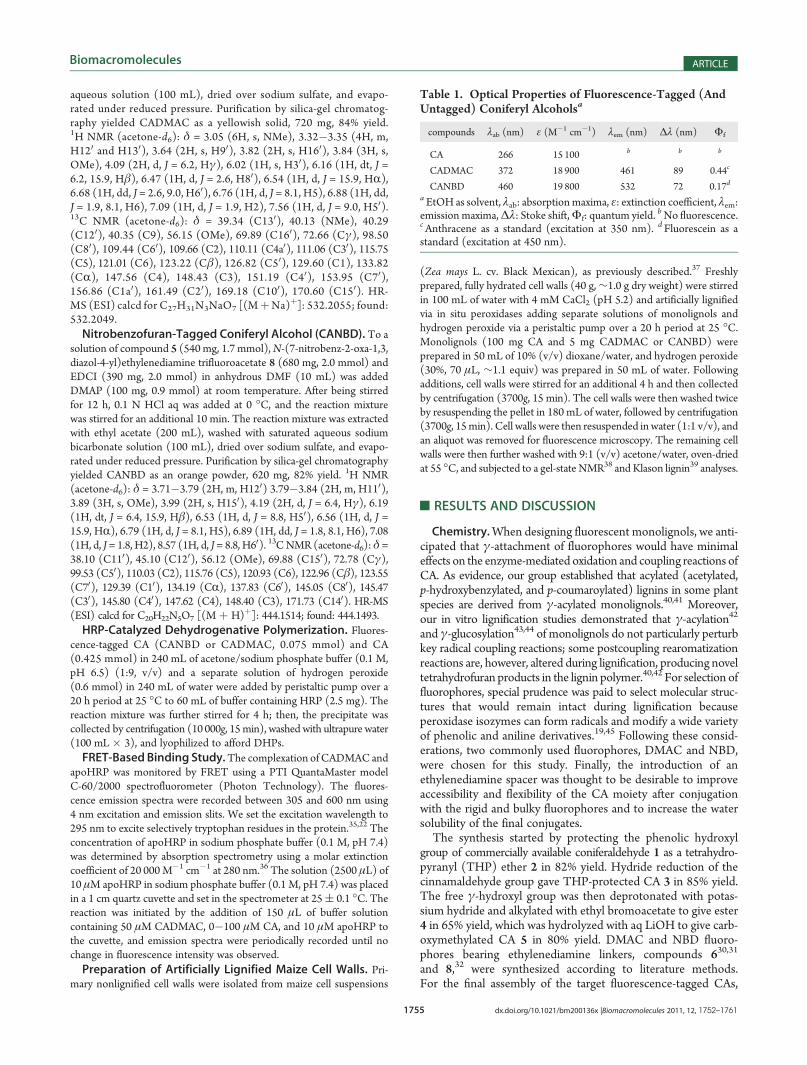
Table 1. Optical Properties of Fluorescence-Tagged (AndUntagged) Coniferyl Alcoholsa
compounds λab (nm) ε (M�1 cm�1) λem (nm) Δλ (nm) Φf
CA 266 15 100 b b b
CADMAC 372 18 900 461 89 0.44c
CANBD 460 19 800 532 72 0.17d
a EtOH as solvent, λab: absorptionmaxima, ε: extinction coefficient, λem:emission maxima,Δλ: Stoke shift,Φf: quantum yield. bNo fluorescence.cAnthracene as a standard (excitation at 350 nm). d Fluorescein as astandard (excitation at 450 nm).

1756 dx.doi.org/10.1021/bm200136x |Biomacromolecules 2011, 12, 1752–1761
Biomacromolecules ARTICLE
EDCI-promoted amide couplings of compound 5 with fluoro-phores 6 and 8 were carried out in the presence of DMAP atroom temperature. After confirming complete conversion ofcompound 5 by thin layer chromatography, THP groups wereremoved by acidification with aq HCl at 0 �C, yielding the desiredCADMAC and CANBD fluorescent compounds in 89 and 83%yields, respectively. Optical properties of CADMAC and CANBDwere verified by UV�vis absorption and fluorescence spectroscopy(Table 1, Figure S1 in the Supporting Information). As expected,CADMAC exhibited a strong bright-blue fluorescence under UV-Alight, and CANBD exhibited a green fluorescence under blue light,whereas untagged CA did not show any visible fluorescence.HRP-Catalyzed Dehydrogenative Polymerization. It is im-
portant to determine whether fluorescence-tagged CAs arecompatible with H-abstraction by peroxidase and the subsequentradical coupling reactions of lignification. For this purpose,we produced synthetic lignins (DHPs) using a conventionalHRP/H2O2 system, in which CADMAC and CANBD were usedas low-levelmonomer substitutes (15mol%) forCA. Product yields,averagemolecularweights, and dispersity data forDHPs formedwithCADMAC and CANBD were quite similar to DHPs formed withCA (Table 2). The degree of polymerization for the DHPs averaged13 (based on the molecular weight of CA), which is comparable toliterature values for conventional DHPs.46 DHP structural composi-tions were determined by 2D HSQC-NMR. Major lignin interunitlinkages inDHPs formed with CADMAC andCANBDwere typicalβ�O�4-, β�5-, and β�β-substructures,38 and their distributionswere similar to those in the DHP formed only with CA (Figure S2,Supporting Information). The inclusion of fluorescence-tagged CAswas clearly demonstrated by the presence of fluorophore signals inthe aromatic region (Figure 2). These data indicate that fluorescentCAs were incorporated into lignin polymers with an intact fluoro-phore moiety. As previously demonstrated for γ-acylated and γ-glucosylated monolignols,40,42�44 the polymerization of CADMACand CANBD logically produces several tetrahydrofuran-type β�β-linked subunits because of their lack of a free γ�OH for internaltrapping of the bis-quinone methide intermediates formed via β�β-coupling. NMR evidence of these structures in the present DHPs isnot yet conclusive because of low levels (if any) of such couplingproducts. Optical properties of the DHPs were verified by UV�visand fluorescence spectroscopy. The optical spectra of DHPs dis-played characteristic absorption and emission signals from thefluorophores, further supporting successful incorporation of fluores-cent CAs into the lignin polymers (Figure 3, Table 2). TheCA-DHPwasweakly fluorogenic, as reported.47,48 Lignin autofluorescencewas,however, negligibly small relative to DHPs with fluorescence-taggedCAs, and this was easily verified visually and by spectroscopicmethods (Figure 3B, Table 2).
Monolignol-Apoperoxidase Interactions Probed by FRET.In principle, fluorescence-tagged CAs can be used in a variety offluorescence assays to study the molecular interactions ofmonolignols. To examine this possibility, we explored thefeasibility of a FRET-based protocol for monitoring the interac-tions between monolignols and HRP, a model peroxidase widelyused for in vitro lignin polymerization studies. HRP has a singletryptophan residue (Trp117) that is predicted to be locatedwithin 12�18 Å of the active site for peroxidation,49,50 and thiscan serve as an intrinsic fluorescence probe for FRET studies.Because FRET efficiency depends on the inverse sixth power ofthe distance between donor and acceptor fluorophores,51 mon-itoring fluorescence signals from the protein-tryptophan donorand the fluorescent CA acceptor can, in principle, reveal theprocess of HRP-monolignol complexation. In the present study,apoHRP (a heme-free HRP) was chosen instead of native HRPbecause the absence of hematoporphyrin facilitates the observa-tion of the tryptophan fluorescence.52 Upon excitation at 295 nm,the fluorescence spectrum of apoHRP exhibited a characteristicemission at 329 nm for the excited state of tryptophan. TheDMAC fluorophore was confirmed as an appropriate energyacceptor because its absorption spectrum overlapped with theemission spectrum of tryptophan in apoHRP (Figure 4A). Afterexposure of CADMAC in the apoHRP buffer solution (apoHRP/CADMAC ratio ≈ 3.3), the complex formation was evident; thetryptophan excitation gradually decreased along with risingCADMAC emission (Figure 4B), indicating that the energytransferred from excited tryptophan sensitized the proximateDMAC probe. Moreover, the DMAC emission was blue-shiftedas the complexation proceeded. Because the emission maximumof CADMAC is shifted from 482 nm in phosphate buffer to460 nm in EtOH (Figure 4A), the blue shift observed during thereaction suggests that the probe experiences a less polar environ-ment in the active site. It has been proposed that the binding siteof peroxidase is located in a hydrophobic protein interior.53,54
Therefore, the blue-shifted DMAC emission supports the con-tention that CADMAC is captured in the hydrophobic bindingpocket of apoHRP (Figure 5). Accordingly, the time course ofCADMAC-apoHRP complexation in the absence and presence ofCA was successfully traced by monitoring the fluorescenceintensity ratio at 329 and 460 nm (Figure 6). Nonlabeled CAeffectively suppressed FRET generation, clearly indicating thatCA competes with CADMAC for the apoHRP binding site. Wealso monitored the reaction of apoHRP with an aminocoumarinderivative without a monolignol moiety (tert-butoxycarbonylaminoderivative from compound 6). In this case, neither FRET signalsnor blue-shifted DMAC emission was significant (Figure S3 inthe Supporting Information), indicating negligible nonspecific
Table 2. Horseradish Peroxidase-Catalyzed Polymerization of Fluorescence-Tagged (and Untagged) Coniferyl Alcohols
DHP monomera yield (weight%) Mnb Mw
b PDb
optical propertiesc
λab (nm) λem (nm) FI
CADMAC-DHP CA & CADMAC 89 2300 6860 2.98 362 450 1
CANBD-DHP CA & CANBD 88 2220 7900 3.56 460 531 0.514
CA-DHP CA only 86 2330 8130 3.49 333 380 0.008aCADMAC and CANBD were copolymerized with CA at 15 mol % feed. bDetermined by GPC, polystyrene as standards, Mn: number-averagemolecular weight,Mw: weight-average molecular weight, PD: polydispersity (Mw/Mn).
cDioxane�water (95:5, v/v) as solvent, λab: absorption maxima(>300 nm), λem: emission maxima, FI: relative fluorescence intensity based on emission peak integral under same conditions (50 μg/mL indioxane�H2O 95:5, excited at the corresponding absorption maxima).

1757 dx.doi.org/10.1021/bm200136x |Biomacromolecules 2011, 12, 1752–1761
Biomacromolecules ARTICLE
Figure 2. Aromatic regions of 2D 13C�1H correlation (HSQC) NMR spectra of synthetic lignins (DHPs) produced from (A) 85% coniferyl alcoholwith 15% dimethylaminocoumarin-tagged coniferyl alcohol (CADMAC-DHP), (B) 85% coniferyl alcohol with 15% nitrobenzofuran-tagged coniferylalcohol (CANBD-DHP), and (C) 100% coniferyl alcohol (CADHP).

1758 dx.doi.org/10.1021/bm200136x |Biomacromolecules 2011, 12, 1752–1761
Biomacromolecules ARTICLE
absorption of the probe onto the protein surface. These resultsconfirm that the affinity of the monolignol moiety for the protein-binding site is the driving force for CADMAC-apoHRP complexa-tion observed in this work. The differing specificities of peroxidasesare related to the accessibility of protein-binding sites bysubstrates,55,56 and this has mainly been examined by computermodeling.53,54 This, however,must be confirmed experimentally; ourdata demonstrate the utility of the fluorescence-tagged monolignolsfor monitoring monolignol-peroxidase interactions.Artificially LignifiedMaize CellWalls (CW-DHPs). Finally, to
test the use of fluorescence-tagged CAs for microscopy, wepolymerized CA with a small amount (5% by weight) offluorescence-tagged CAs into nonlignified maize primary wallsvia wall-bound peroxidases and exogenously supplied H2O2.Previous studies have demonstrated that the structure anddistribution of artificial lignins formed by this model systemclosely mimic natural lignification in grasses.57 The successfulformation of artificial lignins was confirmed by gel-state2D-HSQC NMR of whole cell walls (Figure S4 in the Support-ing Information). Klason lignin contents of CW-DHPs from
CADMAC and CANBD and without fluorescent monomers(only CA) were similar, averaging 12.0, 12.6, and 11.8%,respectively. As expected, CADMAC and CANBD labeledCW-DHPs and produced a blue and green fluorescence(Figure 7A,B), whereas autofluorescence from CW-DHPs pre-pared without fluorescent CAs or with DMAC and NBDderivatives without monolignol moieties was not significantlyvisible under identical microscopic conditions (Figure 7C1,C2 andFigure S5 in the Supporting Information). The fluorescence fromCADMAC and CANBD was observed throughout the cell wall,
Figure 3. (A) Normalized UV�vis absorption spectra of syntheticlignins (DHPs). (B) Fluorescence emission spectra of DHPs underidentical conditions (50 μg/mL in dioxane/water 95/5 v/v excitation atthe corresponding absorptionmaxima listed in Table 2). Insert is a pictureof DHP solutions under irradiation by UV light (365 nm). CADMAC-DHP: DHP prepared from 85% coniferyl alcohol with 15% dimethyl-aminocoumarin-tagged coniferyl alcohol; CANBD-DHP: DHP preparedfrom 85% coniferyl alcohol with 15% nitrobenzofuran-tagged coniferylalcohol; CA-DHP: DHP prepared from 100% coniferyl alcohol.
Figure 4. (A) Absorption and fluorescence emission spectra of apo-peroxidase fromhorseradish (apoHRP) anddimethyaminocoumarin-taggedconiferyl alcohol (CADMAC). (B) Fluorescence emission spectrafollowing the time-course of the complexation of apoHRP andCADMAC.
Figure 5. Schematic presentation of the complexation of dimethyami-nocoumarin-tagged coniferyl alcohol (CADMAC) with apoperoxidasefrom horseradish (apoHRP).

1759 dx.doi.org/10.1021/bm200136x |Biomacromolecules 2011, 12, 1752–1761
Biomacromolecules ARTICLE
which apparently has a single thin (<2.0 μm) layer, as previouslyobserved for CW-DHPs produced by this system.37Consequently,the data presented here suggest that fluorescent monolignolprobes polymerized into lignin can be selectively visualizedin plant cell walls with minimal interference from cell wallautofluorescence.
’CONCLUSIONS
In summary, we have developed a strategy for synthesizingnovel CA derivatives tagged with DMAC andNBD fluorophores.In principle, our approach can be applied to synthesize other
fluorescent monolignols, such as p-coumaryl and sinapyl alcoholderivatives. Their application in fluorescence-based molecularinteraction studies was demonstrated by using FRET for sensingthe monolignol-apoperoxidase binding process. In addition,biomimetic in vitro lignification studies revealed the compat-ibility of fluorescence-tagged CAs with lignin polymerization;they act as substrates for peroxidase and are integrally cross-coupled to lignin polymers, generating bright fluorescence easilyseen in the presence of the autofluorescence from lignin andother cell wall components. Therefore, these fluorescence-taggedCAs might be useful for visualizing the transport and depositionof monolignols in biological systems. Overall, we anticipate thatfluorescence-tagged monolignols can be used in a variety of invitro and in vivo studies aimed at understanding lignificationprocesses in plants.
’ASSOCIATED CONTENT
bS Supporting Information. UV�vis and fluorescencespectra of CADMAC, CANBD, and CA; aliphatic regions from2DHSQCNMR spectra of DHPs; FRET-based binding assay ofthe reaction of a DMAC derivative with apoHRP; gel-state 2DHSQC NMR spectra of CW-DHPs; and the full set of micro-scopic images of CW-DHPs. This material is available free ofcharge via the Internet at http://pubs.acs.org.
’AUTHOR INFORMATION
Corresponding Author*E-mail: [email protected]; [email protected].
’ACKNOWLEDGMENT
We thank Prof. George H. Reed and Dr. Darrell M. McCaslinfor assistance with fluorescence spectroscopy that was performedat UW Biophysics Instrumentation Facility, Dr. Sarah Swansonfor fluorescence microscopic imaging that was performed at UWPlant Imaging Center, and Drs. Hoon Kim and Fachuang Lu fortheir assistance with NMR spectroscopy. Y.T. was supported by aPostdoctoral Fellowship for Research Abroad provided by theJapan Society for the Promotion of Science. We gratefullyacknowledge partial funding from a University of WisconsinGraduate School Vilas Associate Award, the U.S. DOE GreatLakes Bioenergy Research Center (DOE Office of Science BERDE-FC02-07ER64494), and Stanford’s Global Climate andEnergy Project.
’REFERENCES
(1) Boerjan, W.; Ralph, J.; Baucher, M. Lignin biosynthesis. Annu.Rev. Plant Biol. 2003, 54, 519–546.
(2) Vanholme, R.; Demedts, B.; Morreel, K.; Ralph, J.; Boerjan, W.Lignin biosynthesis and structure. Plant Physiol. 2010, 153, 895–905.
(3) Chiang, V. L. Monolignol biosynthesis and genetic engineeringof lignin in trees, a review. Environ. Chem. Lett. 2006, 4, 143–146.
(4) Umezawa, T. The cinnamate/monolignol pathway. Phytochem.Rev. 2010, 9, 1–17.
(5) Smith, C. G.; Rodgers, M. W.; Zimmerlin, A.; Ferdinando, D.;Bolwell, G. P. Tissue and subcellular immunolocalization of enzymes oflignin synthesis in differentiating and wounded hypocotyl tissue ofFrench bean (Phaseolus vulgaris L.). Planta 1994, 192, 155–64.
(6) Takabe, K.; Takeuchi, M.; Sato, T.; Ito, M.; Fujita, M. Immuno-cytochemical localization of enzymes involved in lignification of the cellwall. J. Plant Res. 2001, 114, 509–515.
Figure 7. Epifluorescence microscopic images of artificially lignifiedmaize cell walls (CW-DHPs). (A) CW-DHP lignified with dimethyl-aminocoumarin-tagged coniferyl alcohol observed via the blue channel(excitation, 330�385 nm; emission, 410�430 nm). (B) CW-DHPlignified with nitrobenzofuran-tagged coniferyl alcohol observed viathe green channel (excitation, 480 nm; emission, 505�535 nm).(C) CW-DHP lignified only with untagged coniferyl alcohol observedvia the blue (C1) and green (C2) channels.
Figure 6. Time course of apoperoxidase and dimethyaminocoumarin-tagged coniferyl alcohol (CADMAC) emission ratio (I460/I329) duringtheir complexation in the absence and presence of coniferyl alcohol (CA).

1760 dx.doi.org/10.1021/bm200136x |Biomacromolecules 2011, 12, 1752–1761
Biomacromolecules ARTICLE
(7) Ralph, J.; Lundquist, K.; Brunow, G.; Lu, F.; Kim, H.; Schatz,P. F.; Marita, J. M.; Hatfield, R. D.; Ralph, S. A.; Christensen, J. H.;Boerjan, W. Lignins: natural polymers from oxidative coupling of4-hydroxyphenylpropanoids. Phytochem. Rev. 2004, 3, 29–60.(8) Ralph, J.; Brunow, G.; Harris, P. J.; Dixon, R. A.; Schatz, P. F.;
Boerjan, W. Lignification: are lignins biosynthesized via simple combi-natorial chemistry or via proteinaceous control and template replication?Recent Adv. Polyphenol Res. 2008, 1, 36–66.(9) Vanholme, R.; Morreel, K.; Ralph, J.; Boerjan, W. Lignin
engineering. Curr. Opin. Plant Biol. 2008, 11, 278–285.(10) Weng, J.-K.; Li, X.; Bonawitz, N. D.; Chapple, C. Emerging
strategies of lignin engineering and degradation for cellulosic biofuelproduction. Curr. Opin. Biotechnol. 2008, 19, 166–172.(11) Li, X.; Chapple, C. Understanding lignification: challenges
beyond monolignol biosynthesis. Plant Physiol. 2010, 154, 449–452.(12) Ehlting, J.; Mattheus, N.; Aeschliman, D. S.; Li, E.; Hamberger,
B.; Cullis, I. F.; Zhuang, J.; Kaneda, M.; Mansfield, S. D.; Samuels, L.;Ritland, K.; Ellis, B. E.; Bohlmann, J.; Douglas, C. J. Global transcriptprofiling of primary stems from Arabidopsis thaliana identifies candidategenes for missing links in lignin biosynthesis and transcriptionalregulators of fiber differentiation. Plant J. 2005, 42, 618–640.(13) Kaneda, M.; Rensing, K. H.; Wong, J. C. T.; Banno, B.;
Mansfield, S. D.; Samuels, A. L. Tracking monolignols duringwood development in lodgepole pine. Plant Physiol. 2008, 147,1750–1760.(14) Miao, Y.-C.; Liu, C.-J. ATP-binding cassette-like transporters
are involved in the transport of lignin precursors across plasma andvacuolar membranes. Proc. Natl. Acad. Sci. U.S.A. 2010, 107, 22728–22733.(15) Boija, E.; Johansson, G. Interactions between model mem-
branes and lignin-related compounds studied by immobilized liposomechromatography. Biochim. Biophys. Acta, Biomembr. 2006, 1758,620–626.(16) Barcelo, A. R.; Ros, L. V. G.; Carrasco, A. E. Looking for syringyl
peroxidases. Trends Plant Sci. 2007, 12, 486–491.(17) Fagerstedt, K. V.; Kukkola, E. M.; Koistinen, V. V. T.; Takahashi,
J.; Marjamaa, K. Cell wall lignin is polymerized by class III secretableplant peroxidases in Norway spruce. J. Integr. Plant Biol. 2010, 52,186–194.(18) Hiraga, S.; Sasaki, K.; Ito, H.; Ohashi, Y.; Matsui, H. A large
family of class III plant peroxidases. Plant Cell Physiol. 2001, 42,462–468.(19) Veitch, N. C. Horseradish peroxidase: a modern view of a
classic enzyme. Phytochemistry 2004, 65, 249–259.(20) Blee Kristopher, A.; Choi Joon,W.; O’Connell Ann, P.; Schuch,
W.; Lewis Norman, G.; Bolwell, G. P. A lignin-specific peroxidase intobacco whose antisense suppression leads to vascular tissue modifica-tion. Phytochemistry 2003, 64, 163–76.(21) Li, Y.; Kajita, S.; Kawai, S.; Katayama, Y.; Morohoshi, N. Down-
regulation of an anionic peroxidase in transgenic aspen and its effect onlignin characteristics. J. Plant Res. 2003, 116, 175–182.(22) Royer, C. A. Probing protein folding and conformational
transitions with fluorescence. Chem. Rev. 2006, 106, 1769–1784.(23) Loura, L. M. S.; Prieto, M.; Fernandes, F. Quantification of
protein-lipid selectivity using FRET. Eur. Biophys. J. 2010, 39, 565–578.(24) Gradinaru, C. C.; Marushchak, D. O.; Samim, M.; Krull, U. J.
Fluorescence anisotropy: from single molecules to live cells. Analyst2010, 135, 452–459.(25) Yengo, C. M.; Berger, C. L. Fluorescence anisotropy and
resonance energy transfer: powerful tools for measuring real timeprotein dynamics in a physiological environment.Curr. Opin. Pharmacol.2010, 10, 731–737.(26) Hess, S. T.; Huang, S.; Heikal, A. A.; Webb, W. W. Biological
and chemical applications of fluorescence correlation spectroscopy: areview. Biochemistry 2002, 41, 697–705.(27) Watson, P.; Jones Arwyn, T.; Stephens David, J. Intracellular
trafficking pathways and drug delivery: fluorescence imaging of livingand fixed cells. Adv. Drug. Deliv. Rev. 2005, 57, 43–61.
(28) Lavis, L. D.; Chao, T.-Y.; Raines, R. T. Fluorogenic label forbiomolecular imaging. ACS Chem. Biol. 2006, 1, 252–260.
(29) McIntosh, A. L.; Atshaves, B. P.; Huang, H.; Gallegos, A. M.;Kier, A. B.; Schroeder, F. Fluorescence techniques using dehydroergos-terol to study cholesterol trafficking. Lipids 2008, 43, 1185–1208.
(30) Joulli�e, M. M.; Leonard, M. S.; Portonovo, P.; Liang, B.; Ding,X.; La Clair, J. J. Chemical defense in ascidians of theDidemnidae Family.Bioconjugate Chem. 2003, 14, 30–37.
(31) Sandler, J. S.; Fenical, W.; Gulledge, B. M.; Chamberlin, A. R.;La Clair, J. J. Fluorescent profiling of natural product producers. J. Am.Chem. Soc. 2005, 127, 9320–9321.
(32) Cotte, A.; Bader, B.; Kuhlmann, J.; Waldmann, H. Synthesis ofthe N-terminal lipohexapeptide of human Galpha O-protein and fluor-escent-labeled analogs for biological studies. Chem.—Eur. J. 1999,5, 922–936.
(33) Kim, H.; Ralph, J. Simplified preparation of coniferyl andsinapyl alcohols. J. Agric. Food Chem. 2005, 53, 3693–3695.
(34) Fery-Forgues, S.; Lavabre, D. Are fluorescence quantum yieldsso tricky to measure? A demonstration using familiar stationery pro-ducts. J. Chem. Educ. 1999, 76, 1260–1264.
(35) Haque,M. E.; Debnath, D.; Basak, S.; Chakrabarti, A. Structuralchanges of horseradish peroxidase in presence of low concentrations ofurea. Eur. J. Biochem. 1999, 259, 269–274.
(36) Tamura, M.; Asakura, T.; Yonetani, T. Heme-modificationstudies on horseradish peroxidase. Biochim. Biophys. Acta 1972, 268,292–304.
(37) Grabber, J. H.; Ralph, J.; Hatfield, R. D.; Quideau, S.; Kuster, T.;Pell, A. N. Dehydrogenation polymer-cell wall complexes as a model forlignified grass walls. J. Agric. Food Chem. 1996, 44, 1453–1459.
(38) Kim, H.; Ralph, J. Solution-state 2D NMR of ball-milled plantcell wall gels in DMSO-d6/pyridine-d5. Org. Biomol. Chem. 2010,8, 576–591.
(39) Theander, O.; Westerlund, E. A. Studies on dietary fiber. 3.Improved procedures for analysis of dietary fiber. J. Agric. Food Chem.1986, 34, 330–6.
(40) Lu, F.; Ralph, J. Novel tetrahydrofuran structures derived fromβ-β-coupling reactions involving sinapyl acetate in kenaf lignins. Org.Biomol. Chem. 2008, 6, 3681–3694.
(41) Martinez, A. T.; Rencoret, J.; Marques, G.; Gutierrez, A.; Ibarra,D.; Jimenez-Barbero, J.; del Rio, J. C. Monolignol acylation and ligninstructure in some nonwoody plants: a 2D NMR study. Phytochemistry2008, 69, 2831–2843.
(42) Lu, F.; Ralph, J.; Morreel, K.; Messens, E.; Boerjan, W. Prepara-tion and relevance of a cross-coupling product between sinapylalcohol and sinapyl p-hydroxybenzoate. Org. Biomol. Chem. 2004, 2,2888–2890.
(43) Tobimatsu, Y.; Takano, T.; Kamitakahara, H.; Nakatsubo, F.Studies on the dehydrogenative polymerizations of monolignolβ-glycosides. Part 3: Horseradish peroxidase-catalyzed polymerizationsof triandrin and isosyringin. J. Wood Chem. Technol. 2008, 28, 69–83.
(44) Tobimatsu, Y.; Takano, T.; Kamitakahara, H.; Nakatsubo, F.Studies on the dehydrogenative polymerizations (DHPs) of monolignolβ-glycosides: part 4. Horseradish peroxidase-catalyzed copolymeri-zation of isoconiferin and isosyringin. Holzforschung 2008, 62,495–500.
(45) Kadokawa, J.-I.; Kobayashi, S. Polymer synthesis by enzymaticcatalysis. Curr. Opin. Chem. Biol. 2010, 14, 145–153.
(46) Tobimatsu, Y.; Takano, T.; Kamitakahara, H.; Nakatsubo, F.Studies on the dehydrogenative polymerizations of monolignolβ-glycosides. Part 2: Horseradish peroxidase-catalyzed dehydrogenativepolymerization of isoconiferin. Holzforschung 2006, 60, 513–518.
(47) Albinsson, B.; Li, S.; Lundquist, K.; Stomberg, R. The origin oflignin fluorescence. J. Mol. Struct. 1999, 508, 19–27.
(48) Djikanovic, D.; Kalauzi, A.; Jeremic, M.; Micic, M.; Radotic, K.Deconvolution of fluorescence spectra: contribution to the structuralanalysis of complex molecules. Colloids Surf., B 2007, 54, 188–192.
(49) Das, T. K.; Mazumdar, S. pH-induced conformational pertur-bation in horseradish peroxidase: picosecond tryptophan fluorescence

1761 dx.doi.org/10.1021/bm200136x |Biomacromolecules 2011, 12, 1752–1761
Biomacromolecules ARTICLE
studies on native and cyanide-modified enzymes. Eur. J. Biochem. 1995,227, 823–828.(50) Lasagna, M.; Gratton, E.; Jameson, D. M.; Brunet, J. E. Apo-
horseradish peroxidase unfolding and refolding: intrinsic tryptophanfluorescence studies. Biophys. J. 1999, 76, 443–450.(51) Van DerMeer, B. W.; Coker, G., III; Chen, S. S. Y. In Resonance
Energy Transfer: Theory and Data; VCH Publisher: New York, 1994.(52) Brunet, J. E.; Vargas, V.; Gratton, E.; Jameson, D. M. Hydro-
dynamics of horseradish peroxidase revealed by global analysis ofmultiple fluorescence probes. Biophys. J. 1994, 66, 446–453.(53) Østergaard, L.; Teilum, K.; Mirza, O.; Mattsson, O.; Petersen,
M.; Welinder, K. G.; Mundy, J.; Gajhede, M.; Henriksen, A. ArabidopsisATP A2 peroxidase. Expression and high-resolution structure of a plantperoxidase with implications for lignification. Plant Mol. Biol. 2000,44, 231–243.(54) Nielsen, K. L.; Indiani, C.; Henriksen, A.; Feis, A.; Becucci, M.;
Gajhede, M.; Smulevich, G.; Welinder, K. G. Differential activity andstructure of highly similar peroxidases. Spectroscopic, crystallographic,and enzymatic analyses of lignifying Arabidopsis thaliana peroxidase A2and horseradish peroxidase A2. Biochemistry 2001, 40, 11013–11021.(55) Kobayashi, T.; Taguchi, H.; Shigematsu, M.; Tanahashi, M.
Substituent effects of 3,5-disubstituted p-coumaryl alcohols on theiroxidation using horseradish peroxidase-H2O2 as the oxidant. J. Wood Sci.2005, 51, 607–614.(56) Sasaki, S.; Nonaka, D.; Wariishi, H.; Tsutsumi, Y.; Kondo, R.
Role of Tyr residues on the protein surface of cationic cell-wall-peroxidase (CWPO-C) from poplar: potential oxidation sites foroxidative polymerization of lignin. Phytochemistry 2008, 69, 348–55.(57) Grabber, J. H. How do lignin composition, structure, and cross-
linking affect degradability? A review of cell wall model studies. Crop Sci.2005, 45, 820–831.