A Metric Study of Three Types of Artificial Cranial Modification from North-Central Peru
18
A Metric Study of Three Types of Artificial Cranial Modification from North-Central Peru EMMA POMEROY, a * JAY T. STOCK, a SONIA R. ZAKRZEWSKI b AND MARTA MIRAZO ´ N LAHR a a Leverhulme Centre for Human Evolutionary Studies, Department of Biological Anthropology, University of Cambridge, Fitzwilliam Street, Cambridge, CB2 1QH, UK b Department of Archaeology, University of Southampton, Avenue Campus, Highfield, Southampton, SO17 1BF, UK ABSTRACT Artificial cranial modification (ACM) involves the alteration of cranial vault shape by cultural means, and is performed during infancy while the cranial bones remain soft and malleable. The direction of normal cranial growth is altered through the application of external forces. In this study, three types of ACM from north-central Peru (posterior flattening, bilobed and circumferential) were analysed using standard craniometric techniques. The aim was to determine the effects of these forms of ACM on craniofacial morphology, and the extent to which different types of ACM could be distinguished from one another and unmodified crania on the basis of these measurements. Significant differences between artificially modified and unmodified crania, and between different types of ACM, were demonstrated in cranial vault shape for all types. Significant differences in facial morphology were found only in the bilobed group compared with the unmodified crania. Canonical variates analysis (discriminant analysis) confirmed that major differences between modification types and unmodified crania were in measurements and angles of the cranial vault. While the results show some similarities to previous studies, they add to the variability in the patterns and extent of differences documented to date. It is suggested, based on these results and visual observations, that interpopulation variation in ACM within major modification categories may explain some of the variability in results between studies, an explanation which has previously received insufficient recognition but which remains to be tested since varied methodology between studies may also be a contributory factor. While previous studies have often sought to generalise about the effects of ACM, the examination of the differences between populations even within major ACM categories may offer new insight into cultural variation in modification techniques between populations and the nature of craniofacial development. Copyright ß 2009 John Wiley & Sons, Ltd. Key words: artificial cranial modification; craniometrics; Peru Introduction Anatomically, artificial cranial modification (ACM) is ‘the product of the dynamic distortion of the normal vectors of infantile neurocranial growth through the agency of externally applied forces’ (Gerszten, 1993: p. 87). The restriction of cranial growth in certain directions results in compensatory growth in other less restricted or unrestricted directions (Aufderheide & Rodrı´guez- Martı ´n, 1998) and thus alteration of cranial shape. The direction of growth, rather than its International Journal of Osteoarchaeology Int. J. Osteoarchaeol. (2009) Published online in Wiley InterScience (www.interscience.wiley.com) DOI: 10.1002/oa.1044 * Correspondence to: Leverhulme Centre for Human Evolutionary Studies, Department of Biological Anthropology, University of Cambridge, Fitzwilliam Street, Cambridge, CB2 1QH, UK. e-mail: [email protected] Copyright # 2009 John Wiley & Sons, Ltd. Received 28 January 2008 Revised 30 September 2008 Accepted 2 October 2008
Transcript of A Metric Study of Three Types of Artificial Cranial Modification from North-Central Peru
International Journal of OsteoarchaeologyInt. J. Osteoarchaeol. (2009)Published online in Wiley InterScience
044
(www.interscience.wiley.com) DOI: 10.1002/oa.1* Correspondence to: LeverhStudies, Department of BioCambridge, Fitzwilliam Streee-mail: emma.pomeroy@cant
Copyright # 2009 Joh
A Metric Study of Three Types ofArtificial Cranial Modification fromNorth-Central Peru
EMMA POMEROY,a* JAY T. STOCK,a
SONIA R. ZAKRZEWSKIb AND MARTA MIRAZON LAHRa
a Leverhulme Centre for Human Evolutionary Studies, Department of Biological
Anthropology, University of Cambridge, Fitzwilliam Street, Cambridge, CB2 1QH, UKb Department of Archaeology, University of Southampton, Avenue Campus, Highfield,
Southampton, SO17 1BF, UK
ABSTRACT Artificial cranial modification (ACM) involves the alteration of cranial vault shape by culturalmeans, and is performed during infancy while the cranial bones remain soft and malleable.The direction of normal cranial growth is altered through the application of external forces. Inthis study, three types of ACM from north-central Peru (posterior flattening, bilobed andcircumferential) were analysed using standard craniometric techniques. The aim was todetermine the effects of these forms of ACM on craniofacial morphology, and the extent towhich different types of ACM could be distinguished from one another and unmodified craniaon the basis of these measurements. Significant differences between artificially modified andunmodified crania, and between different types of ACM, were demonstrated in cranial vaultshape for all types. Significant differences in facial morphology were found only in the bilobedgroup compared with the unmodified crania. Canonical variates analysis (discriminantanalysis) confirmed that major differences between modification types and unmodified craniawere in measurements and angles of the cranial vault. While the results show some similaritiesto previous studies, they add to the variability in the patterns and extent of differencesdocumented to date. It is suggested, based on these results and visual observations, thatinterpopulation variation in ACM within major modification categories may explain some of thevariability in results between studies, an explanation which has previously received insufficientrecognition but which remains to be tested since varied methodology between studies mayalso be a contributory factor. While previous studies have often sought to generalise about theeffects of ACM, the examination of the differences between populations even within majorACM categories may offer new insight into cultural variation in modification techniquesbetween populations and the nature of craniofacial development. Copyright � 2009 JohnWiley & Sons, Ltd.
Key words: artificial cranial modification; craniometrics; Peru
Introduction
Anatomically, artificial cranial modification(ACM) is ‘the product of the dynamic distortion
ulme Centre for Human Evolutionarylogical Anthropology, University oft, Cambridge, CB2 1QH, UK.ab.net
n Wiley & Sons, Ltd.
of the normal vectors of infantile neurocranialgrowth through the agency of externally appliedforces’ (Gerszten, 1993: p. 87). The restriction ofcranial growth in certain directions results incompensatory growth in other less restricted orunrestricted directions (Aufderheide & Rodrıguez-Martın, 1998) and thus alteration of cranial shape.The direction of growth, rather than its
Received 28 January 2008Revised 30 September 2008Accepted 2 October 2008
E. Pomeroy et al.
magnitude, is affected (Moss, 1958) and the brainachieves a volume comparable to unmodifiedcrania (Aufderheide & Rodrıguez-Martın, 1998).ACM has had a surprisingly broad distribution
throughout the world, being found on everyinhabited continent at some stage in the past (e.g.Hrdlicka, 1922; Dingwall, 1931; Blackwood &Danby, 1955; Brown, 1981; Anton & Weinstein,1999; Ozbek, 2001). It has been suggested for theDynasty of Akhenaten in ancient Egypt (Snorra-son, 1946), and was common in certain districtsof 19th century France (Dingwall, 1931). Theearliest certain cases of ACM come from LatePalaeolithic Australia and China (Brothwell,1975; Brown, 1981; Clark et al., 2007).To produce permanent effects, ACM must be
performed during infancy when the cranial bonesare malleable and when the trajectory of growthcan be controlled (Dingwall, 1931; Torres-Rouff,2002). It is therefore not a feature acquiredthrough individual choice (Torres-Rouff, 2002),but rather that of parents or carers. An exceptionmight be cradle-boarding or laying the infantwith its head on a hard surface for long periods,which unintentionally flattens the occipitalregion (Dingwall, 1931; Moss, 1958; Mason,1887). Methods of intentionally modifying craniaare more varied and the effects generally morepronounced. Cloth or other material may beused, sometimes with boards, pads, bags of earthor clay or even special headdresses (Dingwall,1931; Allison et al., 1981).The suggested motives for intentional ACM
are varied, but not mutually exclusive. ACM maysymbolise social identity, as some Spanishchroniclers suggested for the Andean region(Aufderheide & Rodrıguez-Martın, 1998). It canserve as both a permanent symbol of intra-groupsolidarity and of inter-group cultural differences(Torres-Rouff, 2002). As such, it may be useful inarchaeological studies of inter-group interactions(e.g. Torres-Rouff, 2002). Cultural notions ofattractiveness (Hatt, 1915; Dingwall, 1931;Blackwood & Danby, 1955) and supposed healthbenefits (Hatt, 1915) have also been cited asreasons for the practice, although such explana-tions have not generally received strong supportin subsequent work. However, Blackwood &Danby (1955) present ethnographic evidencefrom Melanesia that ACM was considered to
Copyright # 2009 John Wiley & Sons, Ltd.
increase an individual’s physical attractiveness.This provides valuable insight to a practice thathad largely died out worldwide during the 20thcentury
The purpose of this study is twofold: (1) toexamine metrically the effects of different typesof ACM on morphology in order to assess theway in which they can be distinguishedmetrically; (2) to investigate the extent to whichdifferent forms of cranial modification influencefacial morphology. The cranium may be con-sidered a functional matrix comprising numerousseries of interacting functional units (Moss, 1958;Anton, 1989). Growth restriction in the cranialvault therefore indirectly affects growth in otherareas such as the cranial base and face (seeBlackwood & Danby, 1955; Moss, 1958; Bjork &Bjork, 1964; Schendel et al., 1980; Brown, 1981;Anton, 1989; Cheverud et al., 1992; Kohn et al.,1993; Cocilovo & Costa-Junquiera, 2001; Rhode& Arriaza, 2006; and experimental work on rats,Puciarelli, 1978). Kohn et al. (1993) haveproduced a useful geometric model of the effectsof ACM on the cranial base and face and the wayin which they interact. The neurocranium isrepresented by a square and the face by anattached triangle (when viewing the craniumsuperiorly). The extent of this interdependencebetween different cranial elements is a matter ofcontroversy which metric studies have attemptedto address (Anton, 1989).
Several such studies have been undertaken (e.g.Blackwood & Danby, 1955; McNeill & Newton,1965; Schendel et al., 1980; Brown, 1981; Anton,1989; Cheverud et al. 1992; Kohn et al., 1993;Cocilovo & Costa-Junqueira, 2001; Rhode &Arriaza, 2006) but the number of populationsanalysed is small and several studies includematerial from the same collections. Comparisonsbetween studies are limited by the variety oftechniques used, which include finite elementscaling analysis (Cheverud et al., 1992; Kohn et al.,1993), and measurements taken from the craniadirectly and/or X-rays (e.g. Moss, 1958; McNeill& Newton, 1965; Anton, 1989). However, thesestudies have generally confirmed and quantifiedthe visually observed effects of ACM, and high-lighted changes in the shape of the cranial base andface, although the precise differences observedand their extent even within modification types
Int. J. Osteoarchaeol. (2009)DOI: 10.1002/oa
Artificial Cranial Modification in North-Central Peru
vary between studies. It is argued here that thismay relate to interpopulation differences in themodification process, even where apparentlysimilar types of modification are performed.
Materials and methods
The crania studied are derived from four sites innorth-central Peru (Figure 1). Pitakilla, Paucarmasand Tuquillo are located on an approximatetransect from the Andes to the coast. The samplefrom Huallamarca represents another coastalpopulation, from Lima.Pitakilla occupies a long natural rock shelter
overlooking the archaeological site of Gotush-jirka, at about 3650m altitude in the CordilleraBlanca. The crania were from 10 machayes(communal mortuary structures) of which onlyone remains standing, and a natural tunnel andcrevice in the rock shelter. All were lying on thesurface. The site dates to the Early IntermediatePeriod (AD 200–600) and Middle Horizon (AD600–1100) (Herrera, 2005).The necropolis at Paucarmas is located on the
southern margin of the upper Rıo Loco in theCordillera Negra at about 3375m altitude. Itconsists of at least 22 machayes distributed along ahorizontal kilometre of slope. The crania studiedwere from two subterranean structures locatedbeneath large rock outcrops. The human boneslay on the surface within the structures andprobably date from the Late Intermediate Period(AD 1100–1400) (Herrera, 2005). Crania werestudied on site at both Pitakilla and Paucarmas.The Tuquillo sample is from a heavily looted
Late Intermediate Period cemetery on theTuquillo peninsula, north of the Huarmey valley.The sample, Registry Number 20040, is from asurface collection conducted by Lorenzo Samaniegoin July 1978. There is no published informationon the collection (which is stored at the MaxUhle Museum at Sechin, near Casma).The fourth sample was from the huaca at
Huallamarca, situated in the San Isidro district ofLima. This ceremonial adobe platform dates tothe Lima culture (AD 200–600), but was used inlater periods for burials. The sample consists ofeight specimens excavated at the huaca, dating to
Copyright # 2009 John Wiley & Sons, Ltd.
the Late Intermediate Period and Late Horizon(AD 1400–1532).
Classification of crania
There are many different classification systemsfor ACM.Many use broad categories, contrasting‘fronto-occipital deformation’ (Cheverud et al.,1992; Clark et al., 2007) or ‘antero-posteriordeformation’ (McNeill & Newton, 1965; Anton,1989; Rhode & Arriaza, 2006) with ‘circumfer-ential deformation’, or ‘vertical’ versus ‘oblique’occipital orientation (Moss, 1958). However,such systems may mask interpopulation varia-bility in cranial form. Although some researchersavoid (Allison et al. 1981) or discourage (Black-wood & Danby, 1955) naming types, it is useful tofacilitate discussion.Three types of ACM were recorded in this
study. The first, ‘posterior flattening’, is charac-terised visually by flattening of the occipitalregion and an increase in breadth in the parietalregion, while the frontal bone is unaffected(Figure 2a). Posterior flattening in our sample mayhave been unintentional, as it was highly variablein extent, and frequently mild and asymmetrical.This would be consistent with the effects ofcradle practices (Neumann, 1942). This type hasreceived little attention in craniometric studies(except Ewing, 1950; Moss, 1958). Anton (1989)suggests that generally this type of modification isnot included within ACM, but it is included herebecause it is the product of cultural practices withpotentially intentional effects (Dingwall, 1931),and may have implications for studies attemptingto examine relationships between populationsusing standard craniometric techniques. Reichlen(1982) suggests that posterior flattening wasunknown in the Peruvian highlands before theInca period, but the sample studied here does notsupport this.The second group was termed ‘bilobed’ (e.g.
Dingwall, 1931), referring to the two distinctlobes formed in the parietal region on either sideof the sagittal suture (Figure 2b). This was clearlydistinguished from posterior flattening by thepresence of frontal modification and a cleardepression along the line of the sagittal suture.Growth is restricted antero-posteriorly and
Int. J. Osteoarchaeol. (2009)DOI: 10.1002/oa
Figure 1. Map of north-central Peru.
E. Pomeroy et al.
compensatory growth occurs laterally and verti-cally. The direction of restriction is similar to theposterior flattening type, but more marked andaffects the frontal bone as well as the occipital.Some cases in the literature (e.g. Dingwall, 1931)are far more marked than any studied hereimplying substantial interpopulation variation.This type of modification was often found incoastal populations in pre-Columbian Peru (Ding-wall, 1931; Bjork & Bjork, 1965; Reichlen, 1982)and was probably produced using a headdressencircling the vault with an additional band alongthe line of the sagittal suture (Weiss, 1961; Allisonet al., 1981). Although a depression along the
Copyright # 2009 John Wiley & Sons, Ltd.
sagittal suture may occur naturally, its extent,combination with frontal modification and thegeographical limitation of this character toindividuals from a single site (Tuquillo) indicatethat this is a consequence of this particularmethod of ACM. Variability in bilobed modifi-cation was high in terms of modification of thefrontal, angulation of the occipital, and severity(as illustrated in Figure 2b). The occurrence ofthese different features showed no consistentpattern, so they were studied as a single group.
The third group was termed ‘circumferential’and characterised by narrowing and postero-superior elongation of the cranial vault as a result
Int. J. Osteoarchaeol. (2009)DOI: 10.1002/oa
Figure 2. Categories of artificial cranial modification, demonstrating variability in resulting cranial shape within thesamples: (a) posterior flattening, (b) bilobed and (c) circumferential.
Artificial Cranial Modification in North-Central Peru
of binding the cranium around its circumferencewith fabric or other material (Figure 2c). In pre-Columbian Peru this type of modification wasoften (though not exclusively) found in highlandpopulations (Dingwall, 1931; Bjork & Bjork, 1964;Hoshower et al., 1995). Again, there is consider-
Copyright # 2009 John Wiley & Sons, Ltd.
able interpopulation variability in the severity ofmodification, and resulting cranial shape within(Figure 2c) and between populations (see e.g.Weiss, 1961).The unmodified group consisted of crania with
no indications of ACM. The use of comparative
Int. J. Osteoarchaeol. (2009)DOI: 10.1002/oa
E. Pomeroy et al.
samples from populations other than that fromwhich modified crania are derived has beencriticised (Cheverud et al., 1992; Kohn et al.,1993). Although it would be ideal to use craniaderived from single populations to minimise theeffects of interpopulation genetic or environ-mental differences, this was not possible due tosmall sample sizes. Hence unmodified crania fromall sites were pooled to produce a singleunmodified group.Current understanding of temporal and geo-
graphical craniometric variation in Peru is some-what limited due to the small number of previousstudies (Ross et al., 2008), perhaps in part becauseACM is relatively common in Peruvian popu-lations. A recent study of coastal and highlandpopulations from central and northern Peru hasdemonstrated some differences in cranial shape,but not overall size, between populations (Rosset al., 2008). Coastal populations are characterisedby lower, longer cranial vaults than highlandpopulations (Newman, 1943; Ross et al., 2008)and postcranial morphology also shows adistinction between highland and coastal popu-lations (Weinstein, 2005), while there is morpho-logical similarity within these regions even overrelatively large areas. This may reflect thecombined effects of genetic drift, environmentaladaptation (particularly for postcranial morpho-logy) and dietary influences. However, Ericksen(1962) observed a temporal reduction in craniallength in a highland population from Cajamarcawhich might have been a reflection of increasedgene flow with the coast over time. This may beof particular relevance to the populations examinedin the current study which weremainly fairly late indate. Archaeological evidence for the degree ofinteraction between coastal and highland popu-
Table 1. Sample sizes and origin
Group
Pitakilla Paucarmas
Unmodified 11 13Posterior flattening 32 0Bilobed 0 0Circumferential 0 11Total 43 24
Copyright # 2009 John Wiley & Sons, Ltd.
lations is controversial (Weinstein, 2005) butsuggests some cultural interaction (and perhapspotentially gene flow) between the coast and thehighlands in north-central Peru at this time,probably through trade networks for ceramicsand animals/animal products (Lane, 2007). Thuswhile the use of a combined unmodified sample inthis study is not ideal, it seems that the degree ofmorphological similarity between populations,particularly within the highlands or coastrespectively, and evidence suggestive of a degreeof gene flow between these regions, justifies theuse of a mixed unmodified sample in this case.
Table 1 shows the number of crania in eachgroup and the sites from which they derive. Itshould be noted that sample sizes are small,particularly for the circumferential group, andvaried for individual measurements due to varyingpreservation. This should be borne in mind whenexamining the results presented here.
Measurements
Forty-four measurements were taken, as definedby Howells (1973) and one of the authors(MML), 11 cranial angles (after Howells, 1973)and six indices (after Bass, 1995) were calculatedfrom the measurements. Sex was determined onthe basis of cranial morphology (since associatedpelves were available only at Huallamarca) usingstandard methods (Bass, 1995; White & Folkens,2000), with final sex determination classified asmale, female or indeterminate. To ensure that thesex distribution was not statistically significantbetween the different groups, a Fisher’s exact test
Number of crania
Tuquillo Huallamarca Total
14 8 461 0 33
19 0 190 0 11
34 8 109
Int. J. Osteoarchaeol. (2009)DOI: 10.1002/oa
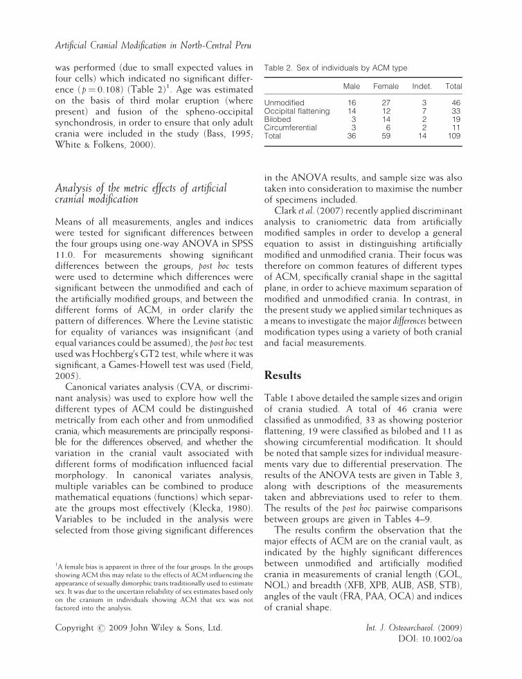
Table 2. Sex of individuals by ACM type
Male Female Indet. Total
Unmodified 16 27 3 46Occipital flattening 14 12 7 33Bilobed 3 14 2 19Circumferential 3 6 2 11Total 36 59 14 109
Artificial Cranial Modification in North-Central Peru
was performed (due to small expected values infour cells) which indicated no significant differ-ence ( p¼ 0.108) (Table 2)1. Age was estimatedon the basis of third molar eruption (wherepresent) and fusion of the spheno-occipitalsynchondrosis, in order to ensure that only adultcrania were included in the study (Bass, 1995;White & Folkens, 2000).
Analysis of the metric effects of artificialcranial modification
Means of all measurements, angles and indiceswere tested for significant differences betweenthe four groups using one-way ANOVA in SPSS11.0. For measurements showing significantdifferences between the groups, post hoc testswere used to determine which differences weresignificant between the unmodified and each ofthe artificially modified groups, and between thedifferent forms of ACM, in order clarify thepattern of differences. Where the Levine statisticfor equality of variances was insignificant (andequal variances could be assumed), the post hoc testused wasHochberg’s GT2 test, while where it wassignificant, a Games-Howell test was used (Field,2005).Canonical variates analysis (CVA, or discrimi-
nant analysis) was used to explore how well thedifferent types of ACM could be distinguishedmetrically from each other and from unmodifiedcrania; which measurements are principally responsi-ble for the differences observed; and whether thevariation in the cranial vault associated withdifferent forms of modification influenced facialmorphology. In canonical variates analysis,multiple variables can be combined to producemathematical equations (functions) which separ-ate the groups most effectively (Klecka, 1980).Variables to be included in the analysis wereselected from those giving significant differences
1A female bias is apparent in three of the four groups. In the groupsshowing ACM this may relate to the effects of ACM influencing theappearance of sexually dimorphic traits traditionally used to estimatesex. It was due to the uncertain reliability of sex estimates based onlyon the cranium in individuals showing ACM that sex was notfactored into the analysis.
Copyright # 2009 John Wiley & Sons, Ltd.
in the ANOVA results, and sample size was alsotaken into consideration to maximise the numberof specimens included.Clark et al. (2007) recently applied discriminant
analysis to craniometric data from artificiallymodified samples in order to develop a generalequation to assist in distinguishing artificiallymodified and unmodified crania. Their focus wastherefore on common features of different typesof ACM, specifically cranial shape in the sagittalplane, in order to achieve maximum separation ofmodified and unmodified crania. In contrast, inthe present study we applied similar techniques asa means to investigate the major differences betweenmodification types using a variety of both cranialand facial measurements.
Results
Table 1 above detailed the sample sizes and originof crania studied. A total of 46 crania wereclassified as unmodified, 33 as showing posteriorflattening, 19 were classified as bilobed and 11 asshowing circumferential modification. It shouldbe noted that sample sizes for individual measure-ments vary due to differential preservation. Theresults of the ANOVA tests are given in Table 3,along with descriptions of the measurementstaken and abbreviations used to refer to them.The results of the post hoc pairwise comparisonsbetween groups are given in Tables 4–9.The results confirm the observation that the
major effects of ACM are on the cranial vault, asindicated by the highly significant differencesbetween unmodified and artificially modifiedcrania in measurements of cranial length (GOL,NOL) and breadth (XFB, XPB, AUB, ASB, STB),angles of the vault (FRA, PAA, OCA) and indicesof cranial shape.
Int. J. Osteoarchaeol. (2009)DOI: 10.1002/oa

E. Pomeroy et al.
This analysis also allows quantification of theobserved effects of ACM. The posteriorly flat-tened sample has significantly shorter vaults thanthe unmodified sample (GOL is reduced by 4.7%,NOL by 4.9%). The breadth (XPB) is greater by4.9%. Whilst frontal and occipital angles areunaffected, the parietal angle in posteriorlyflattened crania is more acute. Bilobed modifi-cation also results in shortened vaults (GOL andNOL are reduced by 7.5% and 7.0% respectively)and increased breadth (11.2% greater XPB)relative to the unmodified sample. By contrast,circumferential modification exhibits significantlygreater cranial length (NOL) by 4.2%, and a5.5% lower breadth (XPB) compared with theunmodified group, and significantly longer frontaland occipital cords (FRC, OCC) and frontalflattening (FRA). Surprisingly, no significant diffe-rence in GOL was seen between these samples.ACM also affects the morphology of the cranial
vault in less visually obvious ways. With posteriorflattening, greater cranial breadth was alsoobserved in the frontal (XFB, STB) and temporal(AUB) areas. The difference in vault breadth isgreater closer to the source of growth restriction(i.e. in the posterior region) than anteriorly(frontal region). Similarly, bilobed modificationshows a significantly higher average maximumfrontal breadth (XFB), and greater breadth in thetemporal region. Since it is thought that bilobedmodification is effected by applying pressure tothe frontal and posterior regions of the neuro-cranium, it is interesting that this effect is so muchless than on the parietal breadth. Pressure may begreatest on the posterior part of the cranium, or itmay be more plastic (being less constrained byfacial development) allowing more compensatorygrowth in this region.The results also imply that in posteriorly
flattened and bilobed crania, the majority ofcompensatory growth in the neurocraniumoccurs in the mediolateral plane rather than ina supero-inferior direction. There are no signifi-cant differences in cranial height (BBH) betweenunmodified and posteriorly flattened or bilobedcrania, yet they show highly significant differ-ences from unmodified crania in cranial breadthacross the parietals, frontal and temporals.Although the modifying apparatus which issuggested to produce bilobed modification (see
Copyright # 2009 John Wiley & Sons, Ltd.
Weiss, 1961; Allison et al., 1981) might restrictheight growth along the sagittal suture andfrontal; no such height-restrictive effects shouldexist for the posteriorly flattened crania. It ispossible that there is an increase in height whichis not reflected in the measurement taken (basi-bregmatic height, BBH), since the work of Anton(1989) and McNeill & Newton (1965) on ‘antero-posterior’ modification imply that there is heightincrease mainly posterior to bregma. Alterna-tively, small sample size due to frequent damageto the cranial base may have prevented differ-ences from attaining statistical significancebetween the groups.
Unexpectedly, circumferential modificationalso showed no significant difference in cranialheight. However, the visually marked heightincrease is principally posterior to the point ofmeasurement, which may account for this result.Small sample size may also contribute.
Comparisons of different types of ACM high-light the contrasts described above in the direc-tion of restriction and lateral growth between theposteriorly flattened and bilobed crania on theone hand, and the circumferentially modifiedcrania on the other. The suggestion that theeffects of posterior flattening are less severe thanbilobed modification is supported, with theformer showing a smaller decrease in cranial lengthand a smaller increase in cranial breadth acrossthe parietals, frontal and temporals. However, thedifferences in the effects on breadth were onlystatistically significant between these groups forparietal breadth. Bilobed modification showssignificantly greater frontal and occipital flatten-ing in the sagittal plane compared with posteriorflattening, again reflecting the greater severity ofmodification in the bilobed group and differencesin the method used to produce the modification.
Only bilobed modification shows significantdifferences from unmodified crania in facialmorphology, namely a significantly higher meanfronto-maxillary breadth (FMB) and less prog-nathism (indicated by SSS). The posteriorlyflattened group shows significantly greater facialflatness in the orbital area (DKA) than thebilobed group, and the posteriorly flattenedgreater than the circumferential group. Thecircumferential group shows significantly greaterlower-facial flatness than the bilobed group.
Int. J. Osteoarchaeol. (2009)DOI: 10.1002/oa
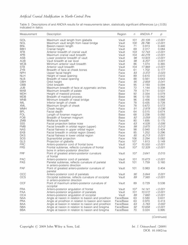
Table 3. Descriptions of and ANOVA results for all measurements taken, statistically significant differences (p� 0.05)indicated in italics
Measurement Description Region n ANOVA F p
GOL Maximum vault length from glabella Vault 101 20.109 <0.001NOL Maximum vault length from nasal bridge Vault 100 26.796 <0.001BNL Basion-nasion length Face 71 0.913 0.440BBH Cranial height Vault 69 2.317 0.084XFB Anterior breadth of cranial vault Vault 103 12.743 <0.001XPB Maximum cranial vault breadth Vault 104 44.279 <0.001ASB Lower posterior breadth of vault Vault 98 13.923 <0.001AUB Vault breadth at ear level Vault 98 6.207 0.001WCB Minimum anterior vault breadth Vault 86 1.074 0.365STB Anterior vault breadth Vault 104 17.969 <0.001ZYB Breadth of face at cheek bones Face 61 1.702 0.177NPH Upper facial height Face 83 3.372 0.023NLH Height of nasal opening Face 89 0.610 0.610NLB Breadth of nasal opening Face 88 0.561 0.643OBH Orbit height Face 95 2.859 0.041OBB Orbit breadth Face 95 2.193 0.094JUB Maximum breadth of face at zygomatic arches Face 72 1.144 0.338MAB Maximum breadth of palate Face 79 0.741 0.531MDH Height of mastoid process Base 92 2.334 0.079MDB Breadth of mastoid process Base 96 0.130 0.942WNB Minimum breadth of nasal bridge Face 96 2.456 0.068IML Inferior length of cheek Face 78 0.435 0.728XML Maximum length of cheek Face 78 0.672 0.572WMH Cheek height Face 91 2.979 0.036BPL Facial depth Face 64 0.515 0.674FOL Length of foramen magnum Base 68 2.238 0.092FOB Breadth of foramen magnum Base 82 3.059 0.033ZMB Midfacial breadth Face 80 1.695 0.175SSS Facial projection below nose Face 63 4.528 0.006FMB Facial breadth in orbital region (upper) Face 99 3.875 0.012NAS Facial flatness in upper orbital region Face 98 0.940 0.424EKB Facial breadth in orbital region (lower) Face 85 1.252 0.296DKS Facial flatness in lower orbital region Face 85 6.213 0.001SOS Supraorbital projection Face 106 2.294 0.082GLS Projection of glabella Face 107 1.813 0.149FRC Antero-posterior cord of frontal bone Vault 107 15.555 <0.001FRS Frontal subtense, reflects curvature of frontal
bone in antero-posterior directionVault 107 12.329 <0.001
FRF Point of greatest antero-posterior curvatureof frontal
Vault 107 3.641 0.015
PAC Antero-posterior cord of parietals Vault 101 15.673 <0.001PAS Parietal subtense, reflects curvature of parietal
in antero-posterio directionVault 101 1.759 0.160
PAF Point of greatest antero-posterior curvature ofparietal
Vault 101 0.983 0.404
OCC Antero-posterior cord of parietals Vault 90 5.844 0.001OCS Occipital subtense, reflects curvature of occipital
in antero-posterior directionVault 89 7.565 <0.001
OCF Point of maximum antero-posterior curvature ofoccipital
Vault 89 0.729 0.538
FRA Antero-posterior angulation of frontal Vault 107 14.141 <0.001PAA Antero-posterior angulation of parietals Vault 101 10.572 <0.001OCA Antero-posterior angulation of occipital Vault 89 7.660 <0.001NAA Angle at nasion in relation to basion and prosthion Face/Base 63 0.580 0.631PRA Angle at prosthion in relation to basion and nasion Face/Base 63 0.970 0.413BAA Angle at basion in relation to nasion and prosthion Face/Base 63 5.783 0.002NBA Angle at nasion in relation to basion and bregma Face/Base 92 10.658 <0.001BBA Angle at basion in relation to nasion and bregma Face/Base 70 0.024 0.995
(Continues)
Copyright # 2009 John Wiley & Sons, Ltd. Int. J. Osteoarchaeol. (2009)DOI: 10.1002/oa
Artificial Cranial Modification in North-Central Peru
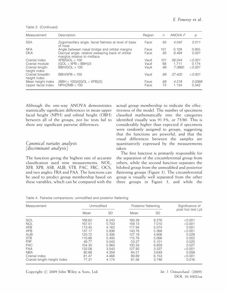
Table 3. (Continued)
Measurement Description Region n ANOVA F p
SSA Zygomaxillary angle, facial flatness at level of baseof nose
Face 65 4.042 0.011
NFA Angle between nasal bridge and orbital margins Face 101 0.109 0.955DKA Dacryal angle, relative sweeping back of orbital
margins relative to midfaceFace 85 6.494 0.001
Cranial index XPB/GOL� 100 Vault 101 58.044 <0.001Cranial module (GOLþXPBþBBH)/3 Vault 68 1.711 0.174Cranial length-height index
BBH/GOL� 100 Vault 68 7.3860 <0.001
Cranial breadth-height index
BBH/XPB� 100 Vault 69 37.420 <0.001
Mean height index (BBH� 100)/([GOLþXPB]/2) Face 68 4.518 0.0068Upper facial index NPH/ZMB� 100 Face 73 1.134 0.342
E. Pomeroy et al.
Although the one-way ANOVA demonstratesstatistically significant differences in mean upperfacial height (NPH) and orbital height (OBH)between all of the groups, post hoc tests fail toshow any significant pairwise differences.
Canonical variates analysis(discriminant analysis)
The function giving the highest rate of accurateclassification used nine measurements: NOL,XFB, XPB, ASB, AUB, STB, PAC, FRC, OCS;and two angles: FRA and PAA. The functions canbe used to predict group membership based onthese variables, which can be compared with the
Table 4. Pairwise comparisons: unmodified and posterior fl
Measurement Unmodified
Mean SD
GOL 168.62 6.243NOL 167.51 5.703XFB 113.45 4.163XPB 137.17 5.938AUB 123.72 5.306STB 110.86 5.493FRF 49.77 5.043PAC 104.30 5.983PAA 132.08 3.543NBA 80.88 4.359Cranial index 81.47 4.468Cranial length–height index 77.31 4.174
Copyright # 2009 John Wiley & Sons, Ltd.
actual group membership to indicate the effec-tiveness of the model. The number of specimensclassified mathematically into the categoriesidentified visually was 91.3%, or 73/80. This isconsiderably higher than expected if specimenswere randomly assigned to groups, suggestingthat the functions are powerful, and that thevisual differences between the samples arequantitatively expressed by the measurementstaken.
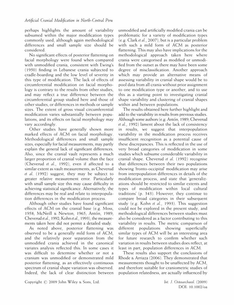
The first function is primarily responsible forthe separation of the circumferential group fromothers, while the second function separates thebilobed group from the unmodified and posteriorflattening groups (Figure 3). The circumferentialgroup is visually well separated from the otherthree groups in Figure 3, and while the
attening
Posterior flattening Significance ofpost hoc test (p)
Mean SD
160.39 8.276 <0.001159.13 7.010 <0.001117.94 5.074 0.001143.76 5.368 <0.001127.19 4.806 0.029115.76 5.068 0.00253.27 5.101 0.020
100.33 6.859 0.027127.92 3.337 <0.00184.21 3.649 0.00889.89 6.153 <0.00181.56 3.746 0.016
Int. J. Osteoarchaeol. (2009)DOI: 10.1002/oa
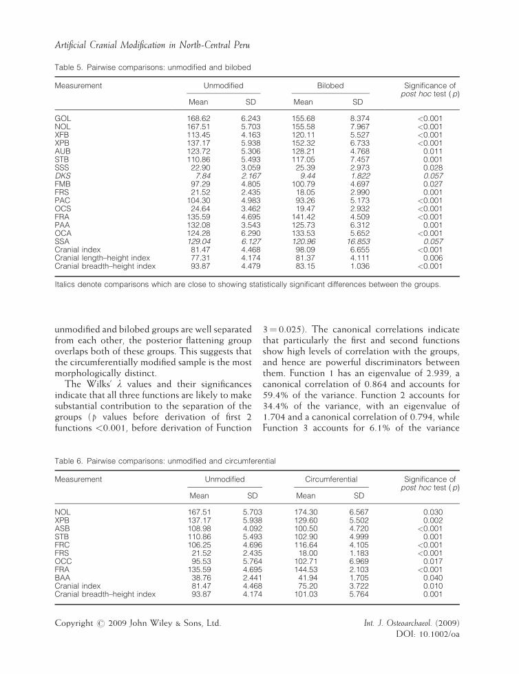
Table 5. Pairwise comparisons: unmodified and bilobed
Measurement Unmodified Bilobed Significance ofpost hoc test (p)
Mean SD Mean SD
GOL 168.62 6.243 155.68 8.374 <0.001NOL 167.51 5.703 155.58 7.967 <0.001XFB 113.45 4.163 120.11 5.527 <0.001XPB 137.17 5.938 152.32 6.733 <0.001AUB 123.72 5.306 128.21 4.768 0.011STB 110.86 5.493 117.05 7.457 0.001SSS 22.90 3.059 25.39 2.973 0.028DKS 7.84 2.167 9.44 1.822 0.057FMB 97.29 4.805 100.79 4.697 0.027FRS 21.52 2.435 18.05 2.990 0.001PAC 104.30 4.983 93.26 5.173 <0.001OCS 24.64 3.462 19.47 2.932 <0.001FRA 135.59 4.695 141.42 4.509 <0.001PAA 132.08 3.543 125.73 6.312 0.001OCA 124.28 6.290 133.53 5.652 <0.001SSA 129.04 6.127 120.96 16.853 0.057Cranial index 81.47 4.468 98.09 6.655 <0.001Cranial length–height index 77.31 4.174 81.37 4.111 0.006Cranial breadth–height index 93.87 4.479 83.15 1.036 <0.001
Italics denote comparisons which are close to showing statistically significant differences between the groups.
Artificial Cranial Modification in North-Central Peru
unmodified and bilobed groups are well separatedfrom each other, the posterior flattening groupoverlaps both of these groups. This suggests thatthe circumferentially modified sample is the mostmorphologically distinct.The Wilks’ l values and their significances
indicate that all three functions are likely to makesubstantial contribution to the separation of thegroups ( p values before derivation of first 2functions <0.001, before derivation of Function
Table 6. Pairwise comparisons: unmodified and circumfere
Measurement Unmodified
Mean SD
NOL 167.51 5.703XPB 137.17 5.938ASB 108.98 4.092STB 110.86 5.493FRC 106.25 4.696FRS 21.52 2.435OCC 95.53 5.764FRA 135.59 4.695BAA 38.76 2.441Cranial index 81.47 4.468Cranial breadth–height index 93.87 4.174
Copyright # 2009 John Wiley & Sons, Ltd.
3¼ 0.025). The canonical correlations indicatethat particularly the first and second functionsshow high levels of correlation with the groups,and hence are powerful discriminators betweenthem. Function 1 has an eigenvalue of 2.939, acanonical correlation of 0.864 and accounts for59.4% of the variance. Function 2 accounts for34.4% of the variance, with an eigenvalue of1.704 and a canonical correlation of 0.794, whileFunction 3 accounts for 6.1% of the variance
ntial
Circumferential Significance ofpost hoc test (p)
Mean SD
174.30 6.567 0.030129.60 5.502 0.002100.50 4.720 <0.001102.90 4.999 0.001116.64 4.105 <0.00118.00 1.183 <0.001
102.71 6.969 0.017144.53 2.103 <0.00141.94 1.705 0.04075.20 3.722 0.010
101.03 5.764 0.001
Int. J. Osteoarchaeol. (2009)DOI: 10.1002/oa
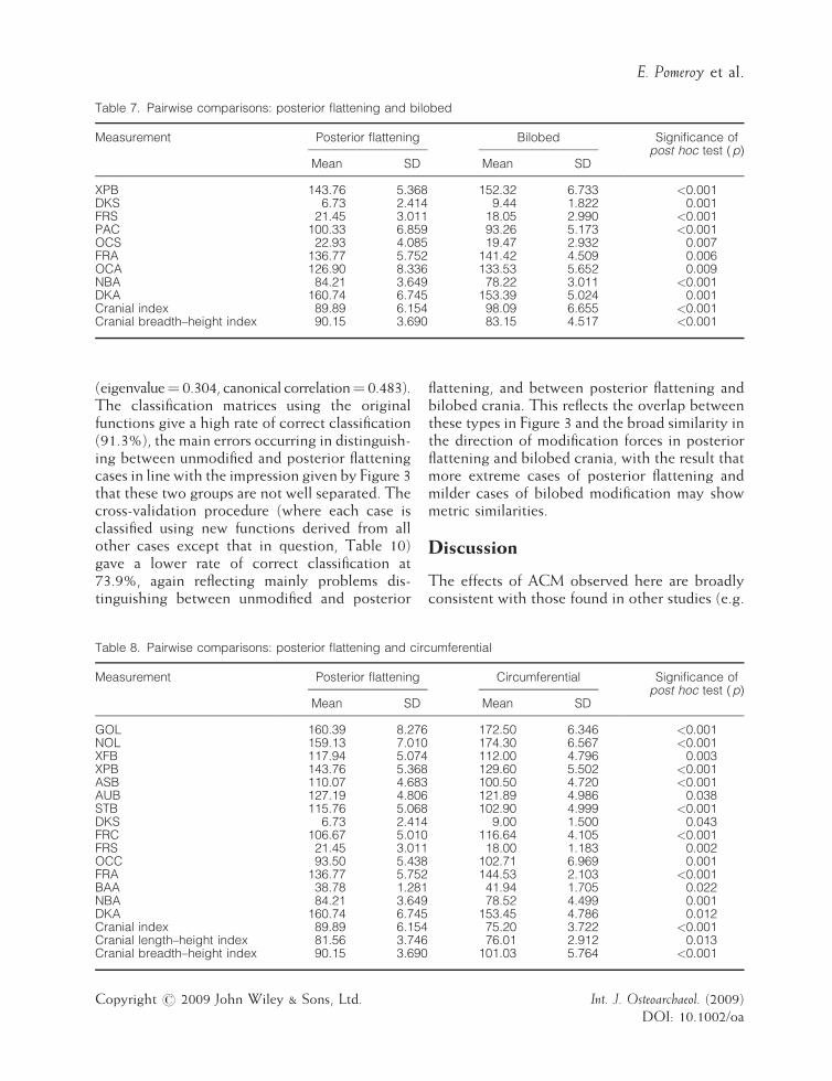
Table 7. Pairwise comparisons: posterior flattening and bilobed
Measurement Posterior flattening Bilobed Significance ofpost hoc test (p)
Mean SD Mean SD
XPB 143.76 5.368 152.32 6.733 <0.001DKS 6.73 2.414 9.44 1.822 0.001FRS 21.45 3.011 18.05 2.990 <0.001PAC 100.33 6.859 93.26 5.173 <0.001OCS 22.93 4.085 19.47 2.932 0.007FRA 136.77 5.752 141.42 4.509 0.006OCA 126.90 8.336 133.53 5.652 0.009NBA 84.21 3.649 78.22 3.011 <0.001DKA 160.74 6.745 153.39 5.024 0.001Cranial index 89.89 6.154 98.09 6.655 <0.001Cranial breadth–height index 90.15 3.690 83.15 4.517 <0.001
E. Pomeroy et al.
(eigenvalue¼ 0.304, canonical correlation¼ 0.483).The classification matrices using the originalfunctions give a high rate of correct classification(91.3%), the main errors occurring in distinguish-ing between unmodified and posterior flatteningcases in line with the impression given by Figure 3that these two groups are not well separated. Thecross-validation procedure (where each case isclassified using new functions derived from allother cases except that in question, Table 10)gave a lower rate of correct classification at73.9%, again reflecting mainly problems dis-tinguishing between unmodified and posterior
Table 8. Pairwise comparisons: posterior flattening and circ
Measurement Posterior flattening
Mean SD
GOL 160.39 8.276NOL 159.13 7.010XFB 117.94 5.074XPB 143.76 5.368ASB 110.07 4.683AUB 127.19 4.806STB 115.76 5.068DKS 6.73 2.414FRC 106.67 5.010FRS 21.45 3.011OCC 93.50 5.438FRA 136.77 5.752BAA 38.78 1.281NBA 84.21 3.649DKA 160.74 6.745Cranial index 89.89 6.154Cranial length–height index 81.56 3.746Cranial breadth–height index 90.15 3.690
Copyright # 2009 John Wiley & Sons, Ltd.
flattening, and between posterior flattening andbilobed crania. This reflects the overlap betweenthese types in Figure 3 and the broad similarity inthe direction of modification forces in posteriorflattening and bilobed crania, with the result thatmore extreme cases of posterior flattening andmilder cases of bilobed modification may showmetric similarities.
Discussion
The effects of ACM observed here are broadlyconsistent with those found in other studies (e.g.
umferential
Circumferential Significance ofpost hoc test (p)
Mean SD
172.50 6.346 <0.001174.30 6.567 <0.001112.00 4.796 0.003129.60 5.502 <0.001100.50 4.720 <0.001121.89 4.986 0.038102.90 4.999 <0.001
9.00 1.500 0.043116.64 4.105 <0.00118.00 1.183 0.002
102.71 6.969 0.001144.53 2.103 <0.00141.94 1.705 0.02278.52 4.499 0.001
153.45 4.786 0.01275.20 3.722 <0.00176.01 2.912 0.013
101.03 5.764 <0.001
Int. J. Osteoarchaeol. (2009)DOI: 10.1002/oa
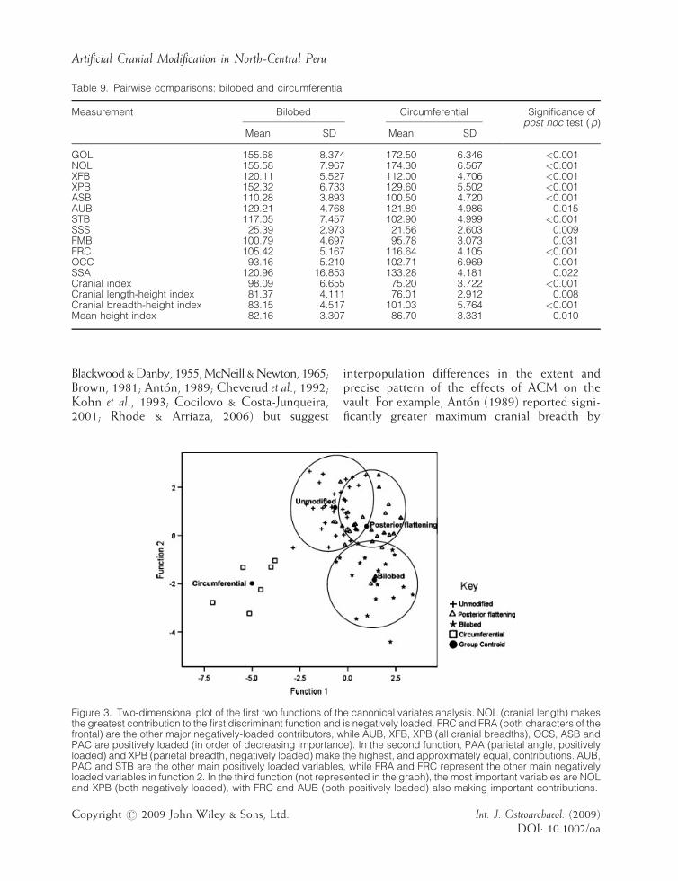
Table 9. Pairwise comparisons: bilobed and circumferential
Measurement Bilobed Circumferential Significance ofpost hoc test (p)
Mean SD Mean SD
GOL 155.68 8.374 172.50 6.346 <0.001NOL 155.58 7.967 174.30 6.567 <0.001XFB 120.11 5.527 112.00 4.706 <0.001XPB 152.32 6.733 129.60 5.502 <0.001ASB 110.28 3.893 100.50 4.720 <0.001AUB 129.21 4.768 121.89 4.986 0.015STB 117.05 7.457 102.90 4.999 <0.001SSS 25.39 2.973 21.56 2.603 0.009FMB 100.79 4.697 95.78 3.073 0.031FRC 105.42 5.167 116.64 4.105 <0.001OCC 93.16 5.210 102.71 6.969 0.001SSA 120.96 16.853 133.28 4.181 0.022Cranial index 98.09 6.655 75.20 3.722 <0.001Cranial length-height index 81.37 4.111 76.01 2.912 0.008Cranial breadth-height index 83.15 4.517 101.03 5.764 <0.001Mean height index 82.16 3.307 86.70 3.331 0.010
Artificial Cranial Modification in North-Central Peru
Blackwood&Danby, 1955;McNeill &Newton, 1965;Brown, 1981; Anton, 1989; Cheverud et al., 1992;Kohn et al., 1993; Cocilovo & Costa-Junqueira,2001; Rhode & Arriaza, 2006) but suggest
Figure 3. Two-dimensional plot of the first two functions of ththe greatest contribution to the first discriminant function andfrontal) are the other major negatively-loaded contributors, wPAC are positively loaded (in order of decreasing importancloaded) and XPB (parietal breadth, negatively loaded) makePAC and STB are the other main positively loaded variablesloaded variables in function 2. In the third function (not represand XPB (both negatively loaded), with FRC and AUB (both
Copyright # 2009 John Wiley & Sons, Ltd.
interpopulation differences in the extent andprecise pattern of the effects of ACM on thevault. For example, Anton (1989) reported signi-ficantly greater maximum cranial breadth by
e canonical variates analysis. NOL (cranial length) makesis negatively loaded. FRC and FRA (both characters of thehile AUB, XFB, XPB (all cranial breadths), OCS, ASB ande). In the second function, PAA (parietal angle, positivelythe highest, and approximately equal, contributions. AUB,, while FRA and FRC represent the other main negativelyented in the graph), the most important variables are NOLpositively loaded) also making important contributions.
Int. J. Osteoarchaeol. (2009)DOI: 10.1002/oa
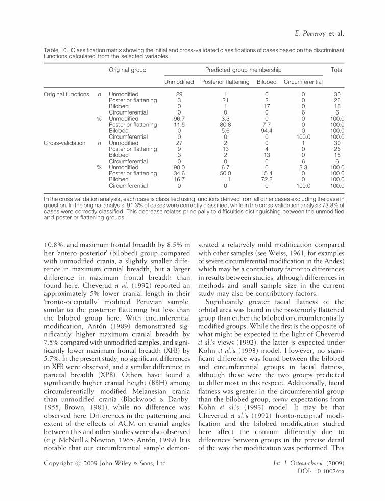
Table 10. Classificationmatrix showing the initial and cross-validated classifications of cases based on the discriminantfunctions calculated from the selected variables
Original group Predicted group membership Total
Unmodified Posterior flattening Bilobed Circumferential
Original functions n Unmodified 29 1 0 0 30Posterior flattening 3 21 2 0 26Bilobed 0 1 17 0 18Circumferential 0 0 0 6 6
% Unmodified 96.7 3.3 0 0 100.0Posterior flattening 11.5 80.8 7.7 0 100.0Bilobed 0 5.6 94.4 0 100.0Circumferential 0 0 0 100.0 100.0
Cross-validation n Unmodified 27 2 0 1 30Posterior flattening 9 13 4 0 26Bilobed 3 2 13 0 18Circumferential 0 0 0 6 6
% Unmodified 90.0 6.7 0 3.3 100.0Posterior flattening 34.6 50.0 15.4 0 100.0Bilobed 16.7 11.1 72.2 0 100.0Circumferential 0 0 0 100.0 100.0
In the cross validation analysis, each case is classified using functions derived from all other cases excluding the case inquestion. In the original analysis, 91.3% of cases were correctly classified, while in the cross-validation analysis 73.8% ofcases were correctly classified. This decrease relates principally to difficulties distinguishing between the unmodifiedand posterior flattening groups.
E. Pomeroy et al.
10.8%, and maximum frontal breadth by 8.5% inher ‘antero-posterior’ (bilobed) group comparedwith unmodified crania, a slightly smaller diffe-rence in maximum cranial breadth, but a largerdifference in maximum frontal breadth thanfound here. Cheverud et al. (1992) reported anapproximately 5% lower cranial length in their‘fronto-occipitally’ modified Peruvian sample,similar to the posterior flattening but less thanthe bilobed group here. With circumferentialmodification, Anton (1989) demonstrated sig-nificantly higher maximum cranial breadth by7.5% comparedwith unmodified samples, and signi-ficantly lower maximum frontal breadth (XFB) by5.7%. In the present study, no significant differencesin XFB were observed, and a similar difference inparietal breadth (XPB). Others have found asignificantly higher cranial height (BBH) amongcircumferentially modified Melanesian craniathan unmodified crania (Blackwood & Danby,1955; Brown, 1981), while no difference wasobserved here. Differences in the patterning andextent of the effects of ACM on cranial anglesbetween this and other studies were also observed(e.g. McNeill & Newton, 1965; Anton, 1989). It isnotable that our circumferential sample demon-
Copyright # 2009 John Wiley & Sons, Ltd.
strated a relatively mild modification comparedwith other samples (see Weiss, 1961, for examplesof severe circumferential modification in the Andes)which may be a contributory factor to differencesin results between studies, although differences inmethods and small sample size in the currentstudy may also be contributory factors.
Significantly greater facial flatness of theorbital area was found in the posteriorly flattenedgroup than either the bilobed or circumferentiallymodified groups. While the first is the opposite ofwhat might be expected in the light of Cheverudet al.’s views (1992), the latter is expected underKohn et al.’s (1993) model. However, no signi-ficant difference was found between the bilobedand circumferential groups in facial flatness,although these were the two groups predictedto differ most in this respect. Additionally, facialflatness was greater in the circumferential groupthan the bilobed group, contra expectations fromKohn et al.’s (1993) model. It may be thatCheverud et al.’s (1992) ‘fronto-occipital’ modi-fication and the bilobed modification studiedhere affect the cranium differently due todifferences between groups in the precise detailof the way the modification was performed. This
Int. J. Osteoarchaeol. (2009)DOI: 10.1002/oa
Artificial Cranial Modification in North-Central Peru
perhaps highlights the amount of variabilitysubsumed within the major modification typescommonly used, although again methodologicaldifferences and small sample size should beconsidered.No significant effects of posterior flattening on
facial morphology were found when comparedwith unmodified crania, consistent with Ewing’s(1950) finding in Lebanese crania subjected tocradle-boarding and the low level of severity inthis type of modification. The lack of effects ofcircumferential modification on facial morpho-logy is contrary to the results from other studies,and may reflect a true difference between thecircumferential group studied here and those ofother studies, or differences in methods or samplesizes. The extent of gross visual circumferentialmodification varies substantially between popu-lations, and its effects on facial morphology mayvary accordingly.Other studies have generally shown more
marked effects of ACM on facial morphology.Methodological differences and small samplesizes, especially for facial measurements, may partlyexplain the general lack of significant differences.Also, since the cranial vault represents a muchlarger proportion of cranial volume than the face(Cheverud et al., 1992), even if affected to asimilar extent as vault measurements, as Cheverudet al. (1992) suggest, they may be subject togreater relative measurement error. Particularlywith small sample size this may cause difficulty inachieving statistical significance. Alternatively, thedifferences may be real and relate to interpopula-tion differences in the modification process.Although other studies have found significant
effects of ACM on the cranial base (e.g. Moss,1958; McNeill & Newton, 1965; Anton, 1989;Cheverud et al., 1992;Kohn et al., 1993), themeasure-ments taken here did not permit a detailed study.As noted above, posterior flattening was
observed to be a generally mild form of ACM,and the relatively poor separation from theunmodified crania achieved in the canonicalvariates analysis reflected this. In some cases itwas difficult to determine whether or not acranium was unmodified or demonstrated mildposterior flattening, as an effectively continuousspectrum of cranial shape variation was observed.Indeed, the lack of clear distinction between
Copyright # 2009 John Wiley & Sons, Ltd.
unmodified and artificially modified crania can beproblematic for a variety of modification types(e.g. Clark et al., 2007), but is a particular problemwith such a mild form of ACM as posteriorflattening. This may also have implications for themethodological approach taken here wherecrania were categorised as modified or unmodi-fied from the outset as there may have been somedegree of misclassification. Another approachwhich may provide an alternative means ofassessing variability in cranial shape would be topool data from all crania without prior assignmentto one modification type or another, and to usethis as a starting point to investigating cranialshape variability and clustering of cranial shapeswithin and between populations.The results obtained here further highlight and
add to the variability in results from previous studies.Although some authors (e.g. Anton, 1989;Cheverudet al., 1992) lament about the lack of consistencyin results, we suggest that interpopulationvariability in the modification process receivesinsufficient recognition as a potential cause ofthese discrepancies. This is reflected in the use ofvery broad categories of modification in somestudies which subsume considerable variability incranial shape. Cheverud et al. (1992) recognisethat differences between their two populationsshowing ‘fronto-occipital’ modification may arisefrom interpopulation differences in details of themodification process, and state that ‘generaliz-ations should be restricted to similar extents andtypes of modification within local culturaltraditions’ (p. 343). However, they continue tocompare broad categories in their subsequentstudy (e.g. Kohn et al., 1993). This suggestioncould not be explored in the present study, andmethodological differences between studies mustalso be considered as a factor contributing to thisvariability in results. The metric comparison ofdifferent populations showing superficiallysimilar types of ACM will be an interesting areafor future research to confirm whether suchvariation in results between studies does reflect, atleast in part, population differences in ACM.These results also support the conclusions of
Rhode & Arriaza (2006). They demonstrated thatmeasurements thought to be unaffected by ACM,and therefore suitable for craniometric studies ofpopulation relatedness, are actually influenced by
Int. J. Osteoarchaeol. (2009)DOI: 10.1002/oa
E. Pomeroy et al.
some types of ACM. Ten measurements (BPL,NPH, OBH, OBB, WCB, NLH, NLB, ZYB, MABand maxillo-alveolar length, not measured here)had been suggested to be suitable for this purposeas they were unaffected by ACM in someArgentinean groups (see Rhode & Arriaza,2006). Of these measurements, they found thatonly facial depth (BPL) and nasal breadth (NLB)were unaffected in both sexes observed in asample of Chilean crania. A different pattern ofresults was obtained here (only orbital height,OBH, showed significant differences), supportingtheir assertion that these measurements aredifferentially affected by ACM according tothe population studied.It should be remembered that any significant
differences in cranial dimensions between thedifferent types of modification could also relate inpart to body size differences between thepopulations. However, it was not possible totest for such differences due to the relatively smallnumbers of unmodified crania from the individualsites and a lack of associated postcrania in whichto examine potential body size differences. This isan area for further investigation which this andprevious comparative studies involving differentmodification types and populations have not yetfully addressed.This and previous studies have shown that
ACM can have significant effects on the cranialvault, base and face. Future investigations shouldfocus on variability resulting from apparentlysimilar modification types. This will potentiallyrefine our knowledge of the interactions betweenvarious elements of the cranium during growthbeyond the information already gleaned fromprevious studies. The differences might resultfrom small differences in the modification processitself, such as the position, intensity and durationof the application of the apparatus, or from otherenvironmental or cultural factors which remainafter the process of modification or which alter itseffects. These factors are a component of thevariation seen within populations showing agiven modification type (Blackwood & Danby,1955). Variation within broad modification types(e.g. Weiss, 1961) and even within populationscan be considerable, as observed in this study andhighlighted by Blackwood & Danby (1955).Variation between geographically close popu-
Copyright # 2009 John Wiley & Sons, Ltd.
lations practising apparently the same type ofmodification may itself be of interest in terms ofthe degree to which practices varied on a localbasis, and the implications this might have forinteraction between such groups (Hoshower et al.,1995).
Conclusion
Metric analysis was successfully used to charac-terise the effects of ACM on cranial vault andfacial shape compared with unmodified crania.Measurements may be used to distinguishbetween different types of ACM. Although thepatterns recorded broadly agreed with those ofother studies, they differed in detail. Thissuggests that there may be considerable inter-population differences in the precise effects ofvisually similar types of modification, a factorwhich warrants further investigation and greaterrecognition as a potentially contributory factor todifferences in results between craniometricstudies of ACM.
Acknowledgements
The research was made possible by the grantsfrom the Hanley Fund and the Johnian SocietyTravel Exhibition (St. Johns College, Cambridge);the Cambridge University Worts’ TravellingScholars Fund and the H. E. Durham Fund (King’sCollege, Cambridge).
The authors thank Alex Herrera, Kevin Lane,Camilo Dolorier Torres, Timoteo, Don Panchoand family, Felipe Livora, Pedro and Fraser Sturtfor access to samples, help and assistance at thesites, and preparation of the figures. The authorsalso thank the editor and two anonymousreviewers for helpful feedback which substan-tially improved the manuscript.
References
Allison MJ, Gerszten E, Munizaga J, Santoro C,Focacci G. 1981. La practica de la deformacioncraneana entre los pueblos Andinos Precolombinos.Chungara, (Arica, Chile) 7: 238–260.
Int. J. Osteoarchaeol. (2009)DOI: 10.1002/oa
Artificial Cranial Modification in North-Central Peru
Anton SC. 1989. Intentional cranial vault deformationand induced changes of the cranial base and face.American Journal of Physical Anthropology 79: 253–267.
Anton SC,Weinstein KJ. 1999. Artificial cranial defor-mation and fossil Australians revisited. Journal ofHuman Evolution 36: 195–209.
Aufderheide AC, Rodrıguez-Martın C. 1998. The Cam-bridge Encyclopedia of Human Paleopathology. CambridgeUniversity Press: Cambridge.
Bass WM. 1995. Human Osteology: A Laboratory and FieldManual, (4th edn). Missouri Archaeological SocietyInc: Special Publication 2.
Bjork A, Bjork L. 1964. Artificial deformation andcraniofacial asymmetry in ancient Peruvians. Journalof Dental Research 43: 353–362.
Blackwood B, Danby PM. 1955. A study of artificialcranial deformation in New Britain. Journal of theRoyal Anthropological Institute 85: 173–192.
Brothwell D. 1975. Possible evidence of a culturalpractice affecting head growth in some Late Pleis-tocene East Asian and Australasian populations.Journal of Archaeological Science 2: 75–77.
Brown P. 1981. Artificial cranial deformation: a com-ponent in the variation in Pleistocene AustralianAboriginal crania. Archaeology in Oceania 16: 156–167.
Cheverud JM, Kohn LAP, Konigsberg LW, Leigh SR.1992. Effects of fronto-occipital artificial cranialvault modification on the cranial base and face.American Journal of Physical Anthropology 88: 323–345.
Clark JL, Dobson SD, Anton SA, Hawks J, Hunley KL,Wolpoff MH. 2007. Identifying artificiallydeformed crania. International Journal of Osteoarchaeol-ogy 17: 596–607.
Cocilovo JA, Costa-Junqueira MA. 2001.La deformacion artificial en el Perıodo Arcaıco deArica. Latin American Antiquity 12: 203–214.
Dingwall EJ. 1931. Artificial Cranial Deformation: A Con-tribution to the Study of Ethnic Mutilations. John Bale,Sons and Danielsson: London.
Ericksen MF. 1962. Undeformed pre-Columbian cra-nia from the north sierra of Peru. American Journal ofPhysical Anthropology 20: 209–222.
Ewing FJ. 1950. Hyperbrachycephaly as influenced bycultural conditioning. Peabody Museum Papers 23:1–99.
Field A. 2005. Discovering Statistics Using SPSS. (2ndedn). Sage Publications Ltd: London.
Gerszten PC. 1993. An investigation into the practiceof cranial deformation among pre-Columbianpeoples of northern Chile. International Journal ofOsteoarchaeology 3: 87–98.
Copyright # 2009 John Wiley & Sons, Ltd.
Hatt G. 1915. Artificial moulding of the infant’s headamong the Scandinavian Lapps. American Anthropol-ogist 17: 245–256.
Herrera A. 2005. Territory and Identity in the Pre-ColumbianAndes of North-central Peru. PhD Thesis, University ofCambridge.
Hrdlicka A. 1922. Aymara deformation in America.American Journal of Physical Anthropology 5: 400.
Hoshower LM, Buikstra JE, Goldstein PS, WebsterAD. 1995. Artificial cranial deformation at the OmoM10 site: a Tiwanaku complex from the MoqueguaValley, Peru. Latin American Antiquity 6: 145–164.
Howells WW. 1973. Cranial variation in man. PeabodyMuseum Papers 67: 1–529.
Klecka WR. 1980. Discriminant Analysis. Sage UniversityPapers Series: Quantitative Applications in the SocialSciences, No. 07-019. Sage: London.
Kohn LAP, Leigh SR, Jacobs SC, Cheverud JM. 1993.Effects of annular cranial vault modification on thecranial base and face. American Journal of PhysicalAnthropology 90: 147–168.
Lane K. 2007. The state they were in: community,continuity and change in the north-central Andes,1000AD–1608AD. In Socialising Complexity: Structure,Integration and Power, Kohring S, andWynne-Jones S(eds.) Oxbow Books: Oxford; 76–98.
Mason OT. 1887. Indian cradles and head flattening.Science 9: 617–620.
McNeill RW, Newton GN. 1965. Cranial basemorphology in association with intentional cranialvault deformation. American Journal of Physical Anthro-pology 23: 241–254.
Moss ML. 1958. The pathogenesis of artificial cranialdeformation. American Journal of Physical Anthropology16: 269–286.
Neumann GK. 1942. Types of artificial cranial defor-mation in the eastern United States. American Anti-quity 7: 306–310.
Newman MT. 1943. A metric study of undeformedIndian crania from Peru. American Journal of PhysicalAnthropology 1: 21–45.
Ozbek M. 2001. Cranial deformation in a subadultsample from Degirmentepe (Chalcolithic, Turkey).American Journal of Physical Anthropology 115: 238–244.
Puciarelli HM. 1978. The influence of experimentaldeformation on craniofacial development in rats.American Journal of Physical Anthropology 48: 455–461.
Reichlen P. 1982. Contribucion al estudios de lasantiguas tecnicas de deformacion cefalica en la costadel Peru. Revisto del Museo Nacional (Lima) 46: 381–403.
Rhode MP, Arriaza BT. 2006. Influence of cranial defor-mation on facial morphology among prehistoric
Int. J. Osteoarchaeol. (2009)DOI: 10.1002/oa
E. Pomeroy et al.
south-central Andean populations. American Journalof Physical Anthropology 130: 462–470.
Ross AH, Ubelaker DH, Guillen S. 2008. Cranio-metric patterning within ancient Peru. Latin AmericanAntiquity 19: 158–166.
Schendel SA, Walker G, Kamisugi A. 1980. Hawaiiancraniofacial morphometrics: average Mokapuanskull, artificial cranial deformation and the ‘rocker’mandible. American Journal of Physical Anthropology 52:491–500.
Snorrason E. 1946. Cranial deformation in the reign ofAkhenaton. Bulletin of the History of Medicine 20: 601–610.
Copyright # 2009 John Wiley & Sons, Ltd.
Torres-Rouff C. 2002. Cranial vault modification andethnicity in middle horizon San Pedro de Atacama,Chile. Current Anthropology 43: 163–171.
Weinstein KJ. 2005. Body proportions in ancientAndeans from high and low altitudes. AmericanJournal of Physical Anthropology 128: 569–585.
Weiss P. 1961. Osteologıa Cultural: Practicas Cefalicas. 2daParte: Tipologıa de las Deformaciones Cefalicas—EstudioCultural de los Tipos Cetalicos y de Algunas EnfermedadesOseas. Universidad Nacional Mayor de San Marcos,Lima.
White TD, Folkens PA. 2000. Human Osteology, (2ndedn). Academic Press: London.
Int. J. Osteoarchaeol. (2009)DOI: 10.1002/oa