A giant pliosaurid skull from the late Jurassic of England
34
A Giant Pliosaurid Skull from the Late Jurassic of England Roger B. J. Benson 1 *, Mark Evans 2 , Adam S. Smith 3 , Judyth Sassoon 4 , Scott Moore-Faye 5 , Hilary F. Ketchum 6,7 , Richard Forrest 8 1 Department of Earth Sciences, University of Oxford, Oxford, United Kingdom, 2 New Walk Museum and Art Gallery, Leicester Arts and Museums Service, Leicester, United Kingdom, 3 Nottingham Natural History Museum, Nottingham, United Kingdom, 4 School of Earth Sciences, University of Bristol, Bristol, United Kingdom, 5 Wavecut Platform Ltd., Sevenoaks, United Kingdom, 6 University Museum of Zoology, University of Cambridge, Cambridge, United Kingdom, 7 Sedgwick Museum of Earth Sciences, University of Cambridge, Cambridge, United Kingdom, 8 Radcliffe-on-Trent, Nottinghamshire, United Kingdom Abstract Pliosaurids were a long-lived and cosmopolitan group of marine predators that spanned 110 million years and occupied the upper tiers of marine ecosystems from the Middle Jurassic until the early Late Cretaceous. A well-preserved giant pliosaurid skull from the Late Jurassic Kimmeridge Clay Formation of Dorset, United Kingdom, represents a new species, Pliosaurus kevani. This specimen is described in detail, and the taxonomy and systematics of Late Jurassic pliosaurids is revised. We name two additional new species, Pliosaurus carpenteri and Pliosaurus westburyensis, based on previously described relatively complete, well-preserved remains. Most or all Late Jurassic pliosaurids represent a globally distributed monophyletic group (the genus Pliosaurus, excluding ‘Pliosaurus’ andrewsi). Despite its high species diversity, and geographically widespread, temporally extensive occurrence, Pliosaurus shows relatively less morphological and ecological variation than is seen in earlier, multi-genus pliosaurid assemblages such as that of the Middle Jurassic Oxford Clay Formation. It also shows less ecological variation than the pliosaurid-like Cretaceous clade Polycotylidae. Species of Pliosaurus had robust skulls, large body sizes (with skull lengths of 1.7–2.1 metres), and trihedral or subtrihedral teeth suggesting macropredaceous habits. Our data support a trend of decreasing length of the mandibular symphysis through Late Jurassic time, as previously suggested. This may be correlated with increasing adaptation to feeding on large prey. Maximum body size of pliosaurids increased from their first appearance in the Early Jurassic until the Early Cretaceous (skull lengths up to 2360 mm). However, some reduction occurred before their final extinction in the early Late Cretaceous (skull lengths up to 1750 mm). Citation: Benson RBJ, Evans M, Smith AS, Sassoon J, Moore-Faye S, et al. (2013) A Giant Pliosaurid Skull from the Late Jurassic of England. PLoS ONE 8(5): e65989. doi:10.1371/journal.pone.0065989 Editor: Richard J Butler, Ludwig-Maximilians-Universita ¨t Mu ¨ nchen, Germany Received March 7, 2013; Accepted April 26, 2013; Published May 31, 2013 Copyright: ß 2013 Benson et al. This is an open-access article distributed under the terms of the Creative Commons Attribution License, which permits unrestricted use, distribution, and reproduction in any medium, provided the original author and source are credited. Funding: The Heritage Lottery Fund Collecting Cultures programme (http://www.hlf.org.uk) and Dorset (http://www.dorsetforyou.com/county) and Devon (http://www.devon.gov.uk/) County Councils provided funding for purchase of DORCM G.13,675. The funders had no role in study design, data collection and analysis, decision to publish, or preparation of the manuscript. Competing Interests: SM-F is director of Wavecut Platform Ltd., which received payment for preparation of DORCM 13,675. This did not influence the authors’ objectivity or the validity of research, analysis, and interpretations in this study. This does not alter the authors’ adherence to all the PLOS ONE policies on sharing data and materials. * E-mail: [email protected] Introduction Pliosaurids were Mesozoic ocean predators, some of which achieved giant sizes .12 metres long [1–3]. They had a global distribution and spanned c. 110 million years, from the Early Jurassic until the early Late Cretaceous (e.g., [4–14]). However, their fossils are best known from the Late Jurassic Oxford and Kimmeridge Clay formations of England (e.g., [15–17]). Pliosaur- ids form part of a larger clade of marine reptiles, Plesiosauria, characterised by highly plastic body proportions, and including extremely long-necked taxa such as Elasmosaurus, as well as short- necked, large-skulled taxa informally termed ‘pliosauromorphs’ [18–19]. Early pliosaurid evolution shows a transition from plesio- morphic, intermediate body proportions and a small skull in the earliest Early Jurassic taxa [13], [19] to Middle Jurassic taxa with ‘pliosauromorph’ body proportions and piscivorous (gracile, longirostine skulls, many slender teeth) or macropredaceous habits (robust longirostrine or brevirostrine skulls with robust teeth) (e.g., [20–25]). These Middle Jurassic and younger taxa belong to an easily recognisable clade called Thalassophonea [26]. Pliosaurid diversity declined in the Late Jurassic, leaving only macropredac- eous forms [26]. The latest pliosaurids are from the early Late Cretaceous [14], [27], and they may have been made extinct by the appearance of large-bodied mosasauroids as competitors in the Middle Turonian [14]. The first pliosaurid fossil discoveries were from the English, Late Jurassic Kimmeridge Clay Formation. In 1822, Conybeare figured vertebrae from near Weymouth and mentioned similar remains from Headington Pits, near Oxford ([28]:plate 22, figs 4–8). Later, in 1824, he also mentioned the fragmentary remains of a large- bodied, short necked plesiosaurian in William Buckland’s collec- tion, from Market Rasen in Lincolnshire ([29]:p. 389 ‘‘Market Raisin’’) and provisionally suggested the name Plesiosaurus giganteus (a nomen oblitum) for short-necked plesiosaurians in general. In 1841, Owen erected the subgenus ‘Pleiosaurus’ for a new species Plesiosaurus (Pleiosaurus) brachydeirus [30]. Owen mentioned two specimens, the Market Rasen skeleton (OXFUM (Oxford University Museum of Natural History, Oxford, United Kingdom) J.9245, J.9247–J.9301, J.10453, comprising a partial skull and PLOS ONE | www.plosone.org 1 May 2013 | Volume 8 | Issue 5 | e65989
-
Upload
plesiosauria -
Category
Documents
-
view
2 -
download
0
Transcript of A giant pliosaurid skull from the late Jurassic of England

A Giant Pliosaurid Skull from the Late Jurassic of EnglandRoger B. J. Benson1*, Mark Evans2, Adam S. Smith3, Judyth Sassoon4, Scott Moore-Faye5,
Hilary F. Ketchum6,7, Richard Forrest8
1 Department of Earth Sciences, University of Oxford, Oxford, United Kingdom, 2 New Walk Museum and Art Gallery, Leicester Arts and Museums Service, Leicester, United
Kingdom, 3 Nottingham Natural History Museum, Nottingham, United Kingdom, 4 School of Earth Sciences, University of Bristol, Bristol, United Kingdom, 5 Wavecut
Platform Ltd., Sevenoaks, United Kingdom, 6 University Museum of Zoology, University of Cambridge, Cambridge, United Kingdom, 7 Sedgwick Museum of Earth
Sciences, University of Cambridge, Cambridge, United Kingdom, 8 Radcliffe-on-Trent, Nottinghamshire, United Kingdom
Abstract
Pliosaurids were a long-lived and cosmopolitan group of marine predators that spanned 110 million years and occupied theupper tiers of marine ecosystems from the Middle Jurassic until the early Late Cretaceous. A well-preserved giant pliosauridskull from the Late Jurassic Kimmeridge Clay Formation of Dorset, United Kingdom, represents a new species, Pliosauruskevani. This specimen is described in detail, and the taxonomy and systematics of Late Jurassic pliosaurids is revised. Wename two additional new species, Pliosaurus carpenteri and Pliosaurus westburyensis, based on previously describedrelatively complete, well-preserved remains. Most or all Late Jurassic pliosaurids represent a globally distributedmonophyletic group (the genus Pliosaurus, excluding ‘Pliosaurus’ andrewsi). Despite its high species diversity, andgeographically widespread, temporally extensive occurrence, Pliosaurus shows relatively less morphological and ecologicalvariation than is seen in earlier, multi-genus pliosaurid assemblages such as that of the Middle Jurassic Oxford ClayFormation. It also shows less ecological variation than the pliosaurid-like Cretaceous clade Polycotylidae. Species ofPliosaurus had robust skulls, large body sizes (with skull lengths of 1.7–2.1 metres), and trihedral or subtrihedral teethsuggesting macropredaceous habits. Our data support a trend of decreasing length of the mandibular symphysis throughLate Jurassic time, as previously suggested. This may be correlated with increasing adaptation to feeding on large prey.Maximum body size of pliosaurids increased from their first appearance in the Early Jurassic until the Early Cretaceous (skulllengths up to 2360 mm). However, some reduction occurred before their final extinction in the early Late Cretaceous (skulllengths up to 1750 mm).
Citation: Benson RBJ, Evans M, Smith AS, Sassoon J, Moore-Faye S, et al. (2013) A Giant Pliosaurid Skull from the Late Jurassic of England. PLoS ONE 8(5): e65989.doi:10.1371/journal.pone.0065989
Editor: Richard J Butler, Ludwig-Maximilians-Universitat Munchen, Germany
Received March 7, 2013; Accepted April 26, 2013; Published May 31, 2013
Copyright: � 2013 Benson et al. This is an open-access article distributed under the terms of the Creative Commons Attribution License, which permitsunrestricted use, distribution, and reproduction in any medium, provided the original author and source are credited.
Funding: The Heritage Lottery Fund Collecting Cultures programme (http://www.hlf.org.uk) and Dorset (http://www.dorsetforyou.com/county) and Devon(http://www.devon.gov.uk/) County Councils provided funding for purchase of DORCM G.13,675. The funders had no role in study design, data collection andanalysis, decision to publish, or preparation of the manuscript.
Competing Interests: SM-F is director of Wavecut Platform Ltd., which received payment for preparation of DORCM 13,675. This did not influence the authors’objectivity or the validity of research, analysis, and interpretations in this study. This does not alter the authors’ adherence to all the PLOS ONE policies on sharingdata and materials.
* E-mail: [email protected]
Introduction
Pliosaurids were Mesozoic ocean predators, some of which
achieved giant sizes .12 metres long [1–3]. They had a global
distribution and spanned c. 110 million years, from the Early
Jurassic until the early Late Cretaceous (e.g., [4–14]). However,
their fossils are best known from the Late Jurassic Oxford and
Kimmeridge Clay formations of England (e.g., [15–17]). Pliosaur-
ids form part of a larger clade of marine reptiles, Plesiosauria,
characterised by highly plastic body proportions, and including
extremely long-necked taxa such as Elasmosaurus, as well as short-
necked, large-skulled taxa informally termed ‘pliosauromorphs’
[18–19].
Early pliosaurid evolution shows a transition from plesio-
morphic, intermediate body proportions and a small skull in the
earliest Early Jurassic taxa [13], [19] to Middle Jurassic taxa with
‘pliosauromorph’ body proportions and piscivorous (gracile,
longirostine skulls, many slender teeth) or macropredaceous habits
(robust longirostrine or brevirostrine skulls with robust teeth) (e.g.,
[20–25]). These Middle Jurassic and younger taxa belong to an
easily recognisable clade called Thalassophonea [26]. Pliosaurid
diversity declined in the Late Jurassic, leaving only macropredac-
eous forms [26]. The latest pliosaurids are from the early Late
Cretaceous [14], [27], and they may have been made extinct by
the appearance of large-bodied mosasauroids as competitors in the
Middle Turonian [14].
The first pliosaurid fossil discoveries were from the English, Late
Jurassic Kimmeridge Clay Formation. In 1822, Conybeare figured
vertebrae from near Weymouth and mentioned similar remains
from Headington Pits, near Oxford ([28]:plate 22, figs 4–8). Later,
in 1824, he also mentioned the fragmentary remains of a large-
bodied, short necked plesiosaurian in William Buckland’s collec-
tion, from Market Rasen in Lincolnshire ([29]:p. 389 ‘‘Market
Raisin’’) and provisionally suggested the name Plesiosaurus giganteus
(a nomen oblitum) for short-necked plesiosaurians in general. In
1841, Owen erected the subgenus ‘Pleiosaurus’ for a new species
Plesiosaurus (Pleiosaurus) brachydeirus [30]. Owen mentioned two
specimens, the Market Rasen skeleton (OXFUM (Oxford
University Museum of Natural History, Oxford, United Kingdom)
J.9245, J.9247–J.9301, J.10453, comprising a partial skull and
PLOS ONE | www.plosone.org 1 May 2013 | Volume 8 | Issue 5 | e65989

postcrania) and a tooth from the Kimmeridge Clay Formation of
Shotover, near Oxford ([30]: p. 282–285, plate 68, figs 5–50). One
year later, in 1842, Owen emended the subgenus ‘Pleiosaurus’ to the
genus Pliosaurus ([31]:p. 60–64), which has been followed by all
subsequent authors (e.g., [15], [17], [32–34]) except Phillips in
1871 [35]. Owen [31] stated that Pliosaurus brachydeirus was
intended as the name for the Market Rasen specimen (OXFUM
J.9245 etc.), and described additional fragmentary remains and
isolated bones and teeth from the Kimmeridge Clay Formation at
various British localities.
Since these early reports, in addition to further discoveries from
the United Kingdom, Late Jurassic pliosaurid remains have been
Figure 1. Locality and horizon of Pliosaurus kevani n. sp. DORCM G.13,675. Maps of the United Kingdom (A), Dorset area (B), and OsmingtonBay (D) showing the locality of the specimen, indicated by a pliosaurid silhouette. The stratigraphic context of the find (C), the approximate level ofthe Wyke Siltstone bed is marked by a pliosaurid silhouette and the stratigraphic section at Black Head was given in full by ([63]:fig. 6). The mountedskull of DORCM G.13,675 in left lateral view (E). Scale bars equal 500 m (D) and 500 mm (E).doi:10.1371/journal.pone.0065989.g001
Pliosaurus
PLOS ONE | www.plosone.org 2 May 2013 | Volume 8 | Issue 5 | e65989

collected from France [36], North America [4], [37], Russia [7–9],
[38–40], Mexico [2], Cuba [12] and Svalbard [3], including
several valid species. In spite of the global distribution of these
finds, they document an ecologically and taxonomically conser-
vative assemblage of large-bodied (adult specimens suggest body
lengths estimated from 10–12 metres; e.g., [2–3]), robust skulled,
long-snouted pliosaurids with distinctive, trihedral teeth [17], [26],
[34] (a gracile, longirostrine ecotype from Italy may be early Late
Jurassic or late Middle Jurassic in age [41]). Although various
taxonomists have recognised multiple Late Jurassic pliosaurid
genera (e.g., [9], [12], [17], [40], [42–43]), in this paper we suggest
that most, perhaps all, specimens form a monophyletic group
representing a single genus, Pliosaurus. This was also suggested for
Kimmeridgian–Tithonian taxa by Knutsen [34], and contrasts
sharply with the high genus-level and ecological diversity of
pliosaurids seen in the late Middle Jurassic (Callovian) Peterbor-
ough Member of the Oxford Clay Formation [16], [23–24], [26],
[44]. The morphologically and ecologically restricted nature of
Late Jurassic (and younger [26]) pliosaurids also contrasts with the
variety of the pliosaurid-like Cretaceous clade Polycotylidae.
Polycotylids are especially abundant in the Late Cretaceous and
include primarily longirostrine forms with gracile snouts and either
slender, widely spaced, isodont teeth indicative of piscivory, or a
slightly more robust snout and dentition suggesting intermediate
levels of macropredation [45–49].
Since the early pliosaurid discoveries described above, numer-
ous additional pliosaurid remains have been reported from the
Kimmeridge Clay Formation. Many are isolated bones and teeth
or skeletal fragments (e.g., [31], [43], [50–51]). However, others
comprise more complete cranial remains, some with associated
postcrania [15], [42], [52–55], and partial postcranial skeletons
[56–57]. Although many species of Kimmeridge Clay Formation
pliosaurid have been erected, only three have recently been
considered valid [17], [34], [54]: Pliosaurus brachydeirus, P.
brachyspondylus and P. macromerus. Of these, the name-bearing type
specimens of P. brachyspondylus (Owen, 1840) [58] (a neotype) and
P. macromerus Phillips, 1871 [35] (a lectotype) are single vertebrae
[17], [52], [57]. These vertebrae were considered undiagnostic in
the most recent taxonomic appraisal of Pliosaurus [34]. In
consequence, even the more complete and informative pliosaurid
skulls and skeletons are in taxonomic limbo [34], [42], [51], [54–
55]. Thus, substantial work remains to understand pliosaurid
diversity and evolution in the Kimmeridge Clay Formation.
In this paper we report DORCM (Dorset County Museum,
Dorchester, United Kingdom) G.13,675, a new, near-complete,
pliosaurid skull from the Kimmeridge Clay Formation of
Osmington Bay, Dorset, United Kingdom (the locality is shown
in Fig. 1). Distinctive anatomical features suggest that DORCM
G.13,675 represents a new species of Pliosaurus, which we name
Pliosaurus kevani n. sp. (see Systematic Palaeontology). The discovery of
P. kevani presents an opportunity to clarify the cranial anatomy and
taxonomy of Pliosaurus. We present a complete description of this
specimen, and taxonomic reappraisal of the genus in which we
provide differential diagnoses and species names for the most
complete specimens in order to stabilize the taxonomy of
Pliosaurus.
Methods
CollectionDORCM G.13,675 was collected over a period of eight years as
pieces up to 60 kg in mass weathered out of the sea-cliff. Most
were collected by Kevan Sheehan, the owner of a small cafe
overlooking the sea at Osmington Mills during daily walks along
the foreshore. No permits were required for the collection of most
of these pieces, which occurred in loose, fallen blocks in the
intertidal zone. Other parts were collected in situ from privately
owned land, and were purchased from the land owner. They were
first identified as a pliosaurid skull by Richard Edmonds, Earth
Sciences Manager for Dorset and East Devon Coast World
Heritage Site Team. Steve Etches, a well-known local collector of
Kimmeridge Clay Formation fossils identified the stratigraphic
horizon from which the specimen was eroding and assisted in
recovering some posterior parts of the skull. The specimen was
then purchased with funding secured by David Tucker of Dorset
County Council’s museum service from the Heritage Lottery Fund
Collecting Cultures programme and Dorset and Devon county
councils.
The impressive size and completeness of DORCM G.13,675
has generated significant media coverage. Its acquisition was
announced publicly in October 2009. Some additional pieces
came to light. These were donated by Patrick Clarke and
purchased from Shirley Swaine. DORCM G.13,675 was prepared
and mounted by S. M.-F. (see Specimen Preparation), and went on
display in Dorchester County Museum in July 2011 with an
official opening by Sir David Attenborough.
Specimen preparationPreparation of DORCM G.13,675 was undertaken between
March 2010 and March 2011. The bulk of the matrix was
removed using a modified Chicago pneumatic air pen. On areas
where the matrix was particularly thick or where it was obvious
that the underlying bone lay as a continuous sheet, a series of thin
5 mm deep slots were cut at 90 degrees to each other with a
40 mm diamond blade and the resulting blocks were chipped off
using either an airpen or chisel. An airbrasive unit equipped with
50 micron aluminium oxide abrasive powder was used to remove
the remaining matrix from the surface of the bone. The broken
surface produced by the airpen made it difficult to identify the
encrusting epifauna, so a 100 mm angle grinder fitted with a
continuous diamond blade was used on these areas to grind away
the matrix instead. The encrusting oysters appeared as a series of
white circles within the grey matirx. This method proved very
effective, because once the encrusting epifauna had been identified
and delimited it was possible to finish the preparation of these
areas with the airpen and airbrasive unit. The average mass of the
sections making up the lower jaws was around 20 kg. The mass of
the sections comprising the skull ranged from 15 kg upwards.
Over 30 kg of matrix was removed from the block containing the
orbits, after which this block weighed 50 kg. Load bearing breaks
were bonded with the epoxy resin adhesive Araladite 2012.
Preparation of the lower jaws took 200 hours and a further
365 hours were needed to complete preparation of the skull.
Nomenclatural actsThe electronic edition of this article conforms to the require-
ments of the amended International Code of Zoological Nomen-
clature, and hence the new names contained herein are available
under that Code from the electronic edition of this article. This
published work and the nomenclatural acts it contains have been
registered in ZooBank, the online registration system for the
ICZN. The ZooBank LSIDs (Life Science Identifiers) can be
resolved and the associated information viewed through any
standard web browser by appending the LSID to the prefix
"http://zoobank.org/". The LSID for this publication is: urn:lsid:
zoobank.org:pub:D3EE687F-52BF-4111-9203-280BFAA40F63.
The electronic edition of this work was published in a journal
Pliosaurus
PLOS ONE | www.plosone.org 3 May 2013 | Volume 8 | Issue 5 | e65989

with an ISSN, and has been archived and is available from the
following digital repositories: PubMed Central and LOCKSS.
Results
Systematic palaeontologySauropterygia Owen, 1860 [59]
Plesiosauria de Blainville, 1835 [60]
Pliosauridae Seeley, 1874 [61]
Thalassophonea Benson & Druckenmiller, 2013 [26]
Genus Pliosaurus Owen, 1841 [30]
1824 Plesiosaurus Conybeare; Conybeare 1824 [29]:p. 389 (as
Plesiosaurus giganteus, a nomen oblitum)
1841 Plesiosaurus (Pleiosaurus) Owen; Owen 1841 ([30]:p. 282-
285, plate 68, figs 5–50)
1842 Pliosaurus Owen; Owen 1842 ([31]:p. 60–65)
1871 Pleiosaurus Owen; Phillips 1871 ([35]:p. 316–318, 341–366)
1959 Stretosaurus Tarlo; Tarlo 1959 ([57]:p. 40) (subjective junior
synonym)
1989 Liopleurodon Sauvage; Halstead 1989 ([42]:p. 38) (as
Liopleurodon macromerus)
Type species. Pliosaurus brachydeirus Owen, 1841 [30]
Other included valid species. Pliosaurus kevani n. sp.;
Pliosaurus westburyensis n. sp.; Pliosaurus carpenteri n. sp.;
Pliosaurus rossicus Novozhilov, 1948a [7]; Pliosaurus funkei
Knutsen et al., 2012 [3].
Diagnosis. Pliosaurids possessing seven autapomorphies: (1)
trihedral or subtrihedral teeth (although similar teeth are also
present in Gallardosaurus iturraldei from the Oxfordian of Cuba [12],
which may or may not be referable to Pliosaurus; see Phylogenetic
analysis); (2) anterior end of premaxilla–maxilla contact on lateral
surface of snout deeply interdigitating with an anteroposteriorly
‘zig-zagging’ appearance; (3) occipital condyle lacking notochordal
pit, but scored by several, irregularly-arranged grooves; (4) first
(mesialmost) premaxillary alveolus reduced to approximately half
or less the diameter of the second alveolus (although an even
smaller, perhaps vestigial, first alveolus may be present in some
Cretaceous pliosaurids [27]); (5) long posteroventral process of the
jugal ventrally underlaps the squamosal; (6) dorsal surface of
surangular mediolaterally broad, as in other thalassophonean
pliosaurids, but inclined to face dorsolaterally (except in Pliosaurus
carpenteri n. sp.) and bounded laterally by an anteroposteriorly
oriented groove, unlike in other pliosaurids (this groove is absent in
P. carpenteri and an immature specimen proposed as the ‘neotype’
of Pliosaurus brachyspondylus by Knutsen [34], CAMSM (Sedgwick
Museum of Earth Sciences, Cambridge, United Kingdom)
J.35991); (7) proximal surfaces of radius and tibia markedly
convex in large individuals (possibly controlled by ontogeny and
absent in immature specimens such as CAMSM J.35991 and the
holotype of Pliosaurus brachydeirus).
Remarks. Several authors have noted that Plesiosaurus
(Pleiosaurus) Owen, 1841 [30] (a subgenus) is the original spelling
of Pliosaurus (a genus) (e.g., [34]). Although ‘Pleiosaurus’ is the
correct original spelling (in the sense of Article 32 of the ICZN), as
far as we know, no authors have used this spelling since Phillips in
1871 [35]. Instead, ‘Pliosaurus’, is in prevailing usage (e.g., [17],
[34], [42–43], [54–55]), and should be preserved (Article 33.3.1 of
the ICZN).
The taxonomy of Pliosaurus was reviewed by Tarlo in 1960 [17]
and Knutsen in 2012 [34], both establishing that several historic
taxa are based on undiagnostic type materials and are nomina dubia.
We do not repeat all such details here, and concur with many of
the statements of Knutsen [34]. For example, observation of
taxonomically important anatomical variation among Pliosaurus
specimens with intermediate counts of mandibular symphysial
alveoli (8–10) causes us to agree that Pliosaurus portentificus Noe et
al., 2004 [43] is a nomen dubium referrable to Pliosaurus indet [34].
We also agree that diagnostic features of Pliosaurus irgisensis
(Novozhilov, 1948) [7] have yet to be established and that its
type specimen requires redescription. This taxon should be
considered a nomen dubium, and its type specimen referred to
Thalassophonea indet. However, several differences do exist
between our assessment of Pliosaurus taxonomy and that of
Knutsen [34]. These differences are explained here and in the
sections below.
The type specimens of several nominal taxa are based on
specifically undiagnostic remains and represent nomina dubia. For
example Pliosaurus brachyspondylus (Owen, 1840) [58], based on a
neotypic cervical vertebra (CAMSM J.29564 [52]), and Pliosaurus
macromerus (Phillips, 1871) [35], based on a lectotypic cervical
vertebra (OXFUM J.10441 [57]). Knutsen [34] proposed
replacement type specimens for these species, pending a petition
to the ICZN that has not yet been made (E. M. Knutsen, pers.
comm. March 2013). Although we consider the proposed
replacement types of these historical taxa to be diagnostic and
distinct from the species proposed in the current work, until the
appeal is made, P. brachyspondylus and P. macromerus are nomina dubia.
Their current name-bearing type specimens should be considered
Thalassophonea indet.
Knutsen [34] suggested possible synonymy between Pliosaurus
macromerus and P. rossicus based on the presence of only six
symphysial and five premaxillary alveoli in both [7], [17], [34],
[40]. He also proposed NHMUK (Natural History Museum,
London, United Kingdom) PV OR 39362 as the replacement type
of P. macromerus, pending a petition to the ICZN [34]. NHMUK
PV OR 39362 is a partial skull, first described in 1869 [15].
However, although NHMUK PV OR 39362 was said by Knutsen
[34] and earlier authors [17], [43], [57] to have approximately six
symphysial alveoli, in fact seven are present as preserved, and
some mesial alveoli are missing. Furthermore, we estimate that a
total of nine were present prior to breakage (pers. obs., NHMUK
PV OR 39362). Although another specimen referred to P.
macromerus, OXFUM J.10454, does have a short symphysis
containing only six alveoli ([17]:plate 22, fig. 5), the marked
difference in symphysial tooth counts indicates that OXFUM
J.10454 is distinct from NHMUK PV OR 39362. Thus, we
consider P. rossicus to be a valid species of Pliosaurus, based on the
presence of a short symphysis containing only six alveoli, and
provisionally refer OXFUM J.10454 to P. ?rossicus on the basis of
this feature.
Specimens referred to Pliosaurus indet. In addition to the
holotype of Pliosaurus portentificus (discussed above and in [34]),
several other specimens can be referred to Pliosaurus but are not
diagnostic at the species level. Many of these are isolated trihedral
teeth (e.g., [30], [50]). Isolated cervical vertebrae (e.g., [28–29],
[31]) are not diagnostic except to Pliosauridae indet. We do not
discuss all fragmentary material in detail here, but focus on key
specimens.
BHN (Musee-sur-Mer, Boulogne, France) 2R.370, a mandible
from the Kimmeridgian of Moulin-Wibert quarry, Boulonnais,
France [36], [62] is referred to Pliosaurus based on possession of a
broad, dorsolaterally facing surangular fossa, bounded laterally by
a fossa and ridge. This specimen was originally referred to
Pliosaurus grandis [62] and later to P. brachyspondylus by [36], based
on its count of 9–10 symphysial alveoli. However, Pliosaurus
brachyspondylus is currently a nomen dubium, and it is not clear that
when its taxonomy is clarified [34], that intermediate symphysial
alveolar counts (of 8–10 alveoli) will be useful in species
Pliosaurus
PLOS ONE | www.plosone.org 4 May 2013 | Volume 8 | Issue 5 | e65989

determination within Pliosaurus. We note that Pliosaurus carpenteri n.
sp. has a similar count of symphysial alveoli (nine) to that proposed
for P. brachyspondylus [17], [34], [43], but differs from CAMSM
J.35991, the proposed replacement type of P. brachyspondylus [34],
and from BHN 2R.370, in possessing an autapomorphic
morphology of the surangular (see the Diagnosis of P. carpenteri).
Thus, BHN 2R.370 cannot be identified to species level based on
currently available information and should be considered Pliosaurus
indet. A similar situation pertains to SEKC.K1.2 (Steve Etches
Kimmeridge collection ( = Museum of Jurassic Marine Life)), a
mandible with eight symphysial alveoli referred to Liopleurodon
macromerus by Clarke & Etches [53] and Pliosaurus portentificus by
Noe et al. [43]. SEKC.K1.2 should be considered as Pliosaurus
indet. unless other information on its morphology becomes
available.
Pliosaurus brachydeirus Owen, 1841 [30]1841 Plesiosaurus (Pleiosaurus) brachydeirus Owen; Owen 1841
([30]:pp. 282–285, pl. 68, figs 5–50)
1842 Pliosaurus brachydeirus Owen; Owen 1842 ([31]:p. 64)
1871 Pleiosaurus brachydeirus Owen; Phillips 1871 ([35]:pp. 341–
354, figs 134–135)
Holotype and only specimen. OXFUM, J.9245, J.9247–
J.9301, J.10453, a partial skull and postcranial skeleton.
Locality and horizon. Rasenia cymodoce Biozone [34] (Lower
Kimmeridgian) of the Kimmeridge Clay Formation of Market
Rasen, Lincolnshire, United Kingdom.
Diagnosis. Species of Pliosaurus with the following unique
character combination: high dentary alveolar count including 24
postsymphysial alveoli (.35 total) and an estimated total count of
36–37; high count of symphysial dentary alveoli (.11), estimated
as 12–13; fully trihedral teeth; mediolateral expansion of
premaxilla and maxillary caniniform region relatively slight; six
closely-spaced premaxillary alveoli; distalmost premaxillary alve-
olus similar in size to more mesial alveoli (i.e. non-‘anisodont’ or
non-‘heterodont’ premaxillary dentition); diastema present be-
tween maxillary and premaxillary alveolar rows; premaxilla–
parietal suture located level with the anterior region of the orbit;
broad, low, anteroposteriorly oriented ridge on ventral surfaces of
cervical centra; epipodials with flat proximal articular surfaces
(although this may result from the subadult ontogenetic status of
the holotype and only specimen).
Pliosaurus kevani n. sp.urn:lsid:zoobank.org:act:39B2679D-B3FD-4DBA-B5F1-
196294DB03D0
Holotype and only specimen. DORCM G.13,675, a skull
(Figs 1–22)
Etymology. Species named after Kevan Sheehan, the main
collector of DORCM G.13,675. The name also serves as a tribute
to the underestimated and undervalued Kevans of this world.
Locality and horizon. Wyke Siltstone bed (Rasenia cymodoce
Zone, Lower Kimmeridgian), Kimmeridge Clay Formation,
Ancholme Group of Osmington Bay (UK Ordnance Survey grid
reference SY 372520 81930; Global Positioning System WGS84:
50u 389 110 N 2u 239 230 W), Dorset, United Kingdom (Fig. 1).
The stratigraphic section at Black Head was given by ([63]:fig. 6).
Tentatively referred specimen. CAMSM J.35990 is most
of a postcranial skeleton, originally referred to Stretosaurus
macromerus [17], [56–57]. It was found at Stretham, southwest of
Ely in Cambridgeshire, probably from the Lower Kimmeridgian
Aulacostephanus mutabilis Zone [34]. This specimen is significant
because relatively complete postcranial data are available,
although only fragments of the skull remain. CAMSM J.35990
differs from most specimens of Pliosaurus in possessing subtrihedral
teeth, which are otherwise present only definitely in Pliosaurus kevani
n. sp., and possibly also in Gallardosaurus iturraldei from the
Oxfordian of Cuba (M.E. pers. obs.; see below). Because of the
paucity of preserved postcrania in several other species of
Pliosaurus, especially P. kevani, which is known only from a skull,
CAMSM J.35990 cannot be confidently diagnosed as a distinct
species, or referred to an existing species with certainty. However,
we provisionally refer it to Pliosaurus cf. kevani based on the
presence of subtrihedral teeth and very large body size.
A subtrihedral tooth from the Kimmeridge Clay Formation of
Ely, Cambridgeshire, UK is also referred to Pliosaurus cf. kevani
(LEICT (New Walk Museum and Art Gallery, Leicester, United
Kingdom) G418.1965.108).
Diagnosis. Species of Pliosaurus with four autapomorphies,
which are absent in all other species of Pliosaurus: (1) subrectan-
gular sheet of the maxilla extends anteriorly on alveolar surface of
the premaxilla to contact the distalmost premaxillary alveolus —
in other species of Pliosaurus an interdigitating premaxilla-maxilla
suture is located mid-way between the mesialmost maxillary and
distalmost premaxillary alveoli; (2) pineal foramen surrounded by
a raised rim — in other thalassophonean pliosaurids, including
other species of Pliosaurus, a shallow fossa containing anteropos-
teriorly oriented grooves/ridges extends anteriorly from the pineal
foramen; (3) mesial postsymphysial dentary alveoli everted to face
dorsolaterally — in other species the dentary alveoli all face
dorsally; (4) lateral surface of the mandible dorsoventrally concave
posteriorly — in other thalassophonean pliosaurids the lateral
surface of the postedentary bones is flat or weakly convex. P. kevani
also possesses the following unique character combination: high
dentary alveolar count including 22 postsymphysial alveoli (.28
total) and an estimated total count of 36–37; high count of
symphysial dentary alveoli (.6), estimated as 14–15; teeth
subtrihedral, possessing a suboval cross-section with only a slightly
flattened labial surface bearing only sparse enamel ridges;
pronounced mediolateral expansion of caniniform regions of the
premaxilla and maxilla; six closely-spaced premaxillary alveoli;
distalmost premaxillary alveolus reduced compared to more mesial
alveoli (i.e. anisodont [ = ’heterodont’] premaxillary dentition);
premaxilla–parietal suture located level with the anterior region of
the orbit. Because only the skull of P. kevani is known, the condition
of postcranial characters that vary among other species of
Pliosaurus cannot be determined.
Remark. Pliosaurus kevani is described in detail later in this
paper.
Pliosaurus westburyensis n. sp.urn:lsid:zoobank.org:act:DF34CD25-6F48-4C08-ACFF-
D617FA81F5C6
1993 Pliosaurus brachyspondylus Owen; Taylor & Cruickshank
1993 ([54]:p. 401, figs 3–11)
2012 Pliosaurus sp.; Sassoon et al. 2012 ([55]:p. 769, figs 19D–F)
2012 Pliosaurus sp.; Knutsen 2012 ([34]:p. 265, figs 2A, 4D, 5E–
F)
Holotype. BRSMG (Bristol City Museum and Art Gallery,
Bristol, United Kingdom) Cc332, a skull and postcranial
fragments.
Etymology. Species named after the town of Westbury near
which BRSMG Cc332 was found.
Locality and horizon. Subdivision E5 [64] of the Aulacoste-
phanus euxodus Biozone (Upper Kimmeridgian), one metre below
the Crussoliceras Limestone of the Kimmeridge Clay Formation of
Westbury Clay Pit, Wiltshire, United Kingdom.
Pliosaurus
PLOS ONE | www.plosone.org 5 May 2013 | Volume 8 | Issue 5 | e65989

Diagnosis. Species of Pliosaurus with three autapomorphies:
(1) premaxillary alveoli widely spaced, with interalveolar walls
approximately half the anteroposterior length of a single alveolus;
(2) a long, sheet-like process of the maxilla extends posteromedial
to the anterolateral part of the maxilla–frontal contact medial to
the external naris. This process of the maxilla terminates just
anterior to orbital midlength ([65]:fig. A1); (3) premaxilla–parietal
suture located around orbital midlength. P. westburyensis also
possesses the following unique character combination: low dentary
alveolar count including only 18 postsymphysial alveoli (the
symphysis is missing so a full count is not possible); teeth fully
trihedral, possessing a flat, anteroposteriorly broad labial surface
lacking enamel ridges; mediolateral expansion of premaxilla and
maxillary caniniform region relatively slight; six premaxillary
alveoli; distalmost premaxillary alveolus similar in size to more
mesial alveoli (i.e. lacks anisodont premaxillary dentition); space
between maxillary and premaxillary alveolar rows comparable to
other interalveolar spaces (i.e. diastema absent); cervical centra
lacking ventral ridge.
Remarks. Sassoon et al. observed several differences between
the holotype of Pliosaurus westburyensis (BRSMG Cc332 [54]) and
that of P. carpenteri n. sp. (BRSMG Cd6172) in the snout, parietal
crest, and alveolar count [55]. They suggested these differences
represented intraspecific variation, with these specimens possibly
Figure 2. Cranium of Pliosaurus kevani n. sp. DORCM G.13,675 in dorsal view (A–B). In line drawing (B) dark grey tone represents brokenbone surface, mid grey represents matrix, and light grey represents tooth or artificial restoration. Abbreviations: bat, basal tuber; en, external naris;exoc-opis, exoccipital-opisthotic; fr, frontal; jug, jugal; lac, ‘lacrimal’; mx, maxilla; pal, palatine; par parietal; pmx, premaxilla; po, postorbital; pofr,postfrontal; prfr, prefrontal; pt, pterygoid; rug, rugose eminence; soc (l), left portion of supraoccipital; soc (r), right portion of supraoccipital; sq,squamosal. Ssdoi:10.1371/journal.pone.0065989.g002
Pliosaurus
PLOS ONE | www.plosone.org 6 May 2013 | Volume 8 | Issue 5 | e65989

being sexual dimorphs. However, although these specimens are
from close stratigraphic levels of the same quarry, the differences
between them are relatively great when seen in the context of
specimens from other localities, and warrant specific distinction. In
the snout, the wide alveolar spacing of BRSMG Cc332 is unique
and is an autapomorphy of P. westburyensis. The narrow snout of P.
westburyensis, which shows relatively little lateral expansion of the
canniniform regions of the premaxilla and maxilla, is shared with
Figure 3. Reconstruction of the skull of Pliosaurus kevani n. sp. In right lateral (A), dorsal (B), and ventral (C) views. Abbreviations: ang, angular;art, articular; den, dentary; en, external naris; exoc-opis, exoccipital-opisthotic; in, internal naris; jug, jugal; fr, frontal; lac, ‘lacrimal’; mx, maxilla; occ,occipital condyle; pal, palatine; palp, palpebral; par, parietal; pifor, pineal foramen; pmx, premaxilla; po, postorbital; pofr, postfrontal; prfr, prefrontal;proo, prootic; ps, parasphenoid; pt, pterygoid; sa, surangular; soc, supraoccipital; sq, squamosal; vom, vomer. Total skull length is approximately 2metres.doi:10.1371/journal.pone.0065989.g003
Pliosaurus
PLOS ONE | www.plosone.org 7 May 2013 | Volume 8 | Issue 5 | e65989

some species, including Pliosaurus brachydeirus, but differs from
others including P. carpenteri and P. kevani. The dorsally high,
anteroposteriorly extensive parietal crest of P. westburyensis differs
from the low crest of P. carpenteri, but other Pliosaurus specimens do
not preserve the crest so comparisons cannot be made.
Sassoon et al. measured the position of the parietal–premaxilla
contact as a proportion of skull length in both BRSMG Cc332 and
Cd6172 and found they had similar measurements [55]. However,
its position compared to other cranial landmarks may be
autapomorphic in P. westburyensis: the anteriormost point of the
parietal–premaxilla contact is posterior to orbital midlength (pers.
obs. BRSMG Cc332 [54]). In contrast, the contact extends
anterior to orbital midlength in other thalassophoneans [65],
including Pliosaurus kevani (Figs 2–3), and likely also in P. carpenteri,
although damage to the orbits and interorbital region obscures the
condition slightly in P. carpenteri.
Sassoon et al. also stated that BRSMG Cc332 and Cd6172 had
different dentary and maxillary alveolar counts [55]. However,
both have 18 postsymphysial dentary alveoli (the maxillary
alveolar count can only be estimated imprecisely in Cd 6172
[55], and the mandibular symphysis is not preserved in BRSMG
Cc332, but seems likely to have contained a similar number of
alveoli to that in BRSMG Cd6172, which has nine [55]).
Pliosaurus carpenteri n. sp.urn:lsid:zoobank.org:act:9F12DB6D-EF17-41C8-AB76-
8221A854ED8A
Figure 4. Premaxilla of Pliosaurus kevani n. sp. DORCM G.13,675 and contacting portions of the maxilla. In dorsal (A), left lateral (B–C),ventral (D–E), and anterior (F) views. In line drawings (C, E) dark grey tone represents broken bone surface, mid grey represents matrix, and light greyrepresents tooth or artificial restoration. Abbreviations: for, foramen; mx, maxilla; mx1, first maxillary alveolus; pdp, paradental plates; pmx, premaxilla;pmx6, sixth premaxillary alveolus; pmx-mx, premaxilla-maxilla contact; pmx-pmx, premaxilla-premaxilla contact; vom, vomer. Scale bar equals100 mmdoi:10.1371/journal.pone.0065989.g004
Pliosaurus
PLOS ONE | www.plosone.org 8 May 2013 | Volume 8 | Issue 5 | e65989

2012 Pliosaurus sp.; Sassoon et al. 2012 ([55]:p. 746, figs 2–18,
19A–C)
2012 Pliosaurus sp.; Knutsen 2012 ([34]:p. 265, fig. 3)
Holotype and only specimen. BRSMG Cd6172.
Etymology. Species named after Simon Carpenter, who
discovered and collected BRSMG Cd6172.
Locality and horizon. Subdivision E4 [64] of the Aulacoste-
phanus euxodus Biozone (Upper Kimmeridgian), seven metres below
the Crussoliceras Limestone) of the Kimmeridge Clay Formation of
Westbury Clay Pit, Wiltshire, United Kingdom.
Diagnosis. Species of Pliosaurus possessing a single autapo-
morphy: the dorsal surface of the surangular lacks any fossa (unlike
in thalassophonean pliosaurids other than CAMSM J.35991 [52],
Figure 5. Cranium of Pliosaurus kevani n. sp. DORCM G.13,675 in ventral view. In line drawing (B) dark grey tone represents broken bonesurface, mid grey represents matrix, and light grey represents tooth or artificial restoration. Abbreviations: bat, basal tuber; cfps, cultriform process ofthe parasphenoid; ecto, ectopterygoid; exoc-opis, exoccipital-opisthotic; in, internal naris; jug, jugal; mx, maxilla; mx19, 19th maxillary alveolus; occ,occipital condyle; pal, palatine; pal-pt, palatine-pterygoid contact; palf, palatal fenestra; par, parietal; pmx, premaxilla; ps, parasphenoid; pt, pterygoid;soc (l), left portion of supraoccipital; sof, suborbital fenestra; sq, squamosal; vom, vomer; vom-pt, vomer-pterygoid contact. Scale bar equals 500 mm.doi:10.1371/journal.pone.0065989.g005
Pliosaurus
PLOS ONE | www.plosone.org 9 May 2013 | Volume 8 | Issue 5 | e65989
the proposed ‘neotype’ of P. brachyspondylus [34]), and faces dorsally
— in other specimens of Pliosaurus it is inclined to face
dorsolaterally. P. carpenteri also possesses the following unique
character combination: low dentary alveolar count including only
18 postsymphysial alveoli, and a total count of 27; intermediate
low count of syphysial alveoli (nine); teeth fully trihedral,
possessing a flat, anteroposteriorly broad labial surface lacking
enamel ridges; mediolateral expansion of caniniform regions of the
premaxilla and maxilla relatively pronounced (although this may
have been enhanced by ventral crushing); six closely-spaced
premaxillary alveoli; distalmost premaxillary alveolus reduced
compared to more mesial alveoli (i.e. anisodont premaxillary
dentition); diastema present between maxillary and premaxillary
alveolar rows; premaxilla–parietal suture located level with the
anterior region of the orbit; cervical centra lacking ventral ridge;
epipodials with highly convex proximal surfaces.
The cranium of Pliosaurus kevani n. sp.The skull of Pliosaurus kevani is large (1995 mm long on the
dorsal midline) and longirostrine, with a preorbital portion
Figure 6. Maxilla of Pliosaurus kevani n. sp. DORCM G.13,675 and contacting bones. Schematic (A) showing the anterior and posteriorportions of the maxilla figured in (B–E). Anterior portion in left dorsolateral (B) and ventral (D) views, and posterior portion in left dorsolateral (C) andright dorsolateral (E) views. Abbreviations: en, external naris; fr, frontal; fr-pmx, frontal, premaxilla contact; mx, maxilla; mx-pmx, maxilla-premaxillacontact; lac, ‘lacrimal’; lac-mx, ‘lacrimal’-maxilla contact; pdp, paradental plates; pmx, premaxilla; pmx-pmx, premaxilla-premaxilla contact; vom,vomer; vom-mx, vomer-maxilla contact. Scale bars equal 100 mm.doi:10.1371/journal.pone.0065989.g006
Pliosaurus
PLOS ONE | www.plosone.org 10 May 2013 | Volume 8 | Issue 5 | e65989

1130 mm long, thus comprising 57% of the skull length (Fig. 2). A
full cranial reconstruction is shown in Figure 3. The temporal
region is transversely broad (730 mm) relative to its length
(postorbital length = 520 mm; temporal fossa length = 670).
The skull has been slightly crushed dorsoventrally, especially
immediately anterior to the orbits. The postorbital portion of the
skull is rotated slightly dorsally. The snout and dorsal surfaces of
the skull are complete. However, the suborbital and subtemporal
bars, and the basicranium and palate posterior to the vomer-
pterygoid contact, are only partly preserved. Some of the palatal
elements have been broken and pulled apart either anteroposte-
riorly or mediolaterally, and the ventral portions of the squamosal-
quadrate unit, which formed the mandibular condyles, are
missing.
Premaxilla. The body of the premaxilla, which forms the
anterior part of the snout, has a tooth-bearing ventral portion that
measures 300 mm anteroposteriorly. It is dorsoventrally low and
mediolaterally broad (215 mm) (Fig. 4). Six premaxillary alveoli
are present, of which the first (mesialmost) alveolus is highly
reduced, with a minimum diameter (28 mm) approximately half
that of the third alveolus (58 mm). Substantial reduction of the first
premaxillary alveolus is a synapomorphy of Late Jurassic and
younger pliosaurids (Pliosaurus + Brachaucheninae [26]:character
140, [27]), and also occurs in most plesiosauroids (e.g., [66]).
However, this alveolus in less reduced in Pliosaurus than it is in
brachaucheninines [27], so the condition in Pliosaurus is tentatively
considered to be autapomorphic.
The third–fifth premaxillary alveoli of DORCM G.13,675 are
the largest, demonstrating the presence of an anisodont premax-
illary dentition (‘heterodont’ is often used to describe this condition
in plesiosaurians, but ‘anisodont’ is more appropriate because the
teeth vary only in size, not morphology). This is similar to the
condition in some Kimmeridge Clay Formation pliosaurids,
which, like DORCM G.13,675, have a reduced distalmost
premaxillary alveolus (e.g., [55]). However, it differs from others,
in which the distalmost premaxillary alveolus is only slightly
smaller than more mesial alveoli (e.g., [54]) (Table 1). The
Figure 7. Context of the ‘lacrimal’ of Pliosaurus kevani n. sp. DORCM G.13,675. Left orbit and antorbital region in dorsolateral view (A), theunidentified bone fragment attached anterior to the orbit by matrix is not the same as the suborbital bar fragment in (B–D). Portion of possiblesuborbital bar fragment in medial or lateral (B, D) and dorsal (C) views. Abbreviations: jug, jugal; lac, ‘lacrimal’; ?lac-?jug, possible ‘lacrimal’-jugalcontact; mx, maxilla; mx-lac, maxilla-’lacrimal’ contact; pifor, pineal foramen; sob, suborbital bar. Scale bars equal 100 mm (A) and 50 mm (B–D).doi:10.1371/journal.pone.0065989.g007
Figure 8. Anterior portion of the right suborbital bar ofPliosaurus kevani n. sp. DORCM G.13,675. In medial (A) and lateral(B) views. Abbreviations: jug, jugal; po, postorbital; sq, squamosal; sq-jug, squamosal-jugal contact. Scale bar equals 100 mm.doi:10.1371/journal.pone.0065989.g008
Pliosaurus
PLOS ONE | www.plosone.org 11 May 2013 | Volume 8 | Issue 5 | e65989

premaxilla of DORCM G.13,675 is transversely expanded to
accommodate the large third–fifth alveoli. Thus, its outline in
dorsal view has pronouncedly convex lateral margins, resulting in
a ‘spatulate’ appearance (Figs 2–5), the prominence of which is
also variable among Kimmeridge Clay Formation pliosaurids
(Table 1). A transversely narrow ‘rostral constriction’ separates the
expanded region of the premaxilla from the maxilla (Fig. 2). The
premaxillary dentition is separated from the maxillary dentition by
a smooth, edentulous region of compact bone forming a diastema
subequal to (left) or greater than (right) the diameter of the
distalmost premaxillary alveolus. This diastema is formed by a
subrectangular, sheet-like anterior extension of the maxilla, which
contacts the posterior margin of the distalmost ( = sixth) pre-
maxilary alveolus. This extension of the maxilla is absent in all
other pliosaurids (e.g., [16], [23–24], [54–55]), and is an
autapomorphy of Pliosaurus kevani n. sp. (Fig. 4). The premaxillary
alveoli of DORCM G.13,675 are otherwise closely spaced, divided
only by the lateral extensions of rugose, triangular paradental
plates. A deep, anteroposteriorly oriented groove separates the
paradental plates from the central platform that bears the
interdigitating midline contact of the premaxillae. This platform
is mediolaterally narrow anteriorly, where it contributes to the
posteromedial margin of the first premaxillary alveolus. The
platform bifurcates posterior to the fourth alveolus, forming paired
posterolateral extensions that contact the maxillae posteriorly. The
recess between these posterolateral extensions accommodates the
anterior process of the vomer. Three foramina penetrate the
premaxillary-vomerine contact: an anterior midline foramen at the
level of the fourth premaxillary alveolus, and paired lateral
foramina level with the fifth premaxillary alveolus. Smooth
channels extend anterolaterally from the lateral foramina, incising
the posterolateral extensions of the central platform.
Figure 9. Suspensorium of Pliosaurus kevani n. sp. DORCM G.13,675 in posterior view. Posterior view of entire suspensorium (A), of the leftventral ramus (B), and of the squamosal-squamosal contact (C). Abbreviations: bat, basal tuber; occ, occipital condyle. Scales bar equal 100 mm (A)and 50 mm (B–C).doi:10.1371/journal.pone.0065989.g009
Figure 10. Left orbit and postorbital region of Pliosaurus kevanin. sp. DORCM G.13,675 showing parietal crest in anterolateralview. Abbreviations: exoc, exoccipital-opisthotic; pifor, pineal foramen;parcr, parietal crest; soc (l), left portion of supraoccipital; soc (r), rightportion of supraoccipital. Scale bar equals 100 mm.doi:10.1371/journal.pone.0065989.g010
Pliosaurus
PLOS ONE | www.plosone.org 12 May 2013 | Volume 8 | Issue 5 | e65989

The dorsal and lateral surfaces of the premaxillae are highly
fractured, conferring an artefactual rugose appearance (Fig. 4).
They bear numerous foramina, especially anteriorly and laterally.
On the dorsal surface, the premaxillary midline suture is weakly
sinuous anteriorly, but becomes straight posterior to the rostral
constriction. The dorsal surface of the snout is mediolaterally
convex, except where it has been crushed ventrally, posteriorly.
This crushing has preferentially affected the maxillae, causing
them to be depressed either side of the posterodorsal processes of
the premaxilla, resulting in the appearance of a prominent,
anteroposteriorly oriented midline ridge. However, this is artefac-
tual: in fact no dorsomedian ridge was originally present. The
lateral margins of the posterodorsal processes of the premaxillae
form straight, continuous lines that extend posterodorsally,
separating the premaxillae from the maxillae anteriorly (anterior
to the external naris) and from the frontals posteriorly. The
conjoined posterodorsal processes of the premaxillae extend far
posteriorly, forming a broad, deeply interdigitating contact with
the parietals adjacent to the anterior orbit margin. Because the
premaxillae contact the parietals, the frontals are excluded from
the midline in dorsal view (Figs 2–3), as in other thalassophonean
pliosaurids and derived members of Rhomaleosauridae, Lepto-
cleidia and Elasmosauridae (e.g., [16], [65], [67–69]). However,
among these taxa, pliosaurids are unique in possessing a posterior
termination of the premaxilla that is mediolaterally broad and
interdigitating (contra [13], who mistakenly said that this also
occurred in cryptoclidids), and they differ from most other taxa in
the anterior position of the premaxilla–parietal contact, located
anterior to orbital midlength [65], as in DORCM G.13,675. This
suture is apomorphically located further posteriorly in Pliosaurus
westburyensis (Table 1) ([54]:fig. 4).
Maxilla. The maxillae form the lateral surfaces of the snout
(Figs 2–3, 5). They continue posteriorly in the suborbital region,
ventral to the ‘lacrimal’ and jugal. However, because this region is
broken, their posterior extent cannot be determined. The left
maxilla, as preserved up to the anterior orbit margin, bears 20
alveoli, and the right bears 19 because it is slightly less complete
(Fig. 5). The maxillary dentition is anisodont; for example, the
mediolateral diameter of the first (mesialmost) maxillary alveolus
(27 mm) is approximately half the diameter of the second
Figure 11. Palate of Pliosaurus kevani n. sp. DORCM G.13,675 in the region of the internal naris, seen in ventral view. Schematic (A)showing the portion of the palate figured in (B–C). (C) is at greater magnification than (B). Abbreviations: for, foramina; in, internal naris; pal, palatine;pal-max, palatine-maxilla contact; vom, vomer; vom-pal, vomer-palatine contact; vom-pt, vomer-pterygoid contact. Scale bars equal 100 mm.doi:10.1371/journal.pone.0065989.g011
Pliosaurus
PLOS ONE | www.plosone.org 13 May 2013 | Volume 8 | Issue 5 | e65989

(51 mm). The body of the maxilla is expanded laterally to
accommodate the fourth–sixth alveoli, which are the largest
(Figs 5–6). Posterior to these, successive maxillary alveoli are
smaller. As in the premaxilla, the medial walls of the maxillary
alveoli are defined by rugose, subtriangular paradental plates. An
anteroposteriorly oriented groove containing replacement alveoli
separates these plates from the horizontal palatal shelf of the
maxilla, which contacts the lateral elements of the palate (vomer
and palatine) medially. Several irregularly distributed foramina of
varying sizes penetrate the maxilla-vomer and maxilla-palatine
contacts (Figs 5–6; these two sutures form a continuous line
parallel to the tooth row). The internal naris is identified as the
largest of these foramina, and is located at the intersection of all
three bones, at the level of the eleventh maxillary alveolus. In
Pliosaurus westburyensis and Pliosaurus carpenteri the internal naris is
located at the level of the ninth maxillary alveolus (BRSMG
Cd6172 [55] and BRSMG Cc332, pers. obs.; contra [54]), and in
NHMUK PV OR 39362 it is located at the level of the seventh
maxillary alveolus. The presence of additional foramina on the
maxilla–vomer and maxilla–palatine sutures, anterior and poste-
rior to the internal naris, is unique to thalassophonean pliosaurids
among Plesiosauria [24], [54] ([26]:character 69).
The maxilla–premaxilla suture of DORCM G.13,675 is
expressed on the ventral and external (‘external’ = dorsal and
lateral) surfaces of the snout. Externally the suture originates at the
level of the rostral constriction, where it is deeply interdigitating,
with a ‘zig-zag’ appearance in lateral view (Fig. 4), and trends
posterodorsally. The presence of a deeply interdigitating anterior
portion of the premaxilla–maxilla suture is a unique synapomor-
phy of Pliosaurus ([26]:character 24). Posterior to this, the
premaxilla–maxilla suture becomes weakly sinuous, and the
medial edge of the maxilla dorsally overlaps the premaxilla
(Fig. 6). This overlap has been accentuated by ventral crushing of
the snout, especially in the posterior half of the preorbital region. A
posteromedial extension of the maxilla extends medial to the
external naris, and contacts an anterolateral extension of the
frontal, thus excluding the premaxilla from the external naris
(Figs 2–3), as occurs in pliosaurids and leptocleidians (e.g., [5],
[16], [22], [45], [67], [70]). The maxilla–frontal contact of
DORCM G.13,675 is deeply interdigitating and trends medially.
The posteromedial extension of the maxilla is divided into three
prong-like processes by anteroposteriorly oriented fissures, which
are most clearly visible on the left side (Fig. 6). They are somewhat
obscured by damage on the right side, but at least two such
processes are clearly present there (Fig. 6). These processes
Figure 12. Posterior palate of Pliosaurus kevani n. sp. DORCM G.13,675. Schematic (A) showing the portion of the palate figured in (B–D).Posterior portion of palate in ventral view with various regions shown at magnification (B–D). Abbreviations: cfps, cultriform process of theparasphenoid; piv, posterior interpterygoid vacuity; ps-pt, parasphenoid-pterygoid contact; sof, suborbital fenestra. Scale bars equal 200 mm (B) and100 mm (C–D).doi:10.1371/journal.pone.0065989.g012
Pliosaurus
PLOS ONE | www.plosone.org 14 May 2013 | Volume 8 | Issue 5 | e65989

Figure 13. Bones of the otic capsule of Pliosaurus kevani n. sp. DORCM G.13,675. Schematic (A) showing the portions of the otic capsulefigured in (B–D). Right portion of the supraoccipital in posterior (B), right ventrolateral (C) and right posterolateral (D) views. Schematic (G) showingthe portions of the otic capsule figured in (E–F). Left exoccipital-opisthotic and articulated left portion of the supraoccipital in anteromedial (E) andventral (F) views. Abbreviations: amp, ampullary recess in opisthotic; exoc, exoccipital; exoc-opis, exoccipital-opisthotic; exoc-soc, exoccipital-supraoccipital contact; exof, exoccipital facet of the supraoccipital; formag, supraoccipital portion of the foramen magnum; opis, opisthotic; paf,parietal facet of the supraoccipital; pop, parocipital process of the opisthotic; proof, prootic facet; pvc, posterior vertical canal; soc (l), left portion of
Pliosaurus
PLOS ONE | www.plosone.org 15 May 2013 | Volume 8 | Issue 5 | e65989

terminate posteriorly around one-quarter of the length of the
external naris. Fissures dividing the posteromedial process of the
maxilla into prong-like processes are also present in the well-
preserved skull of the holotype of Pliosaurus westburyensis (BRSMG
Cc 332) ([65]:fig. A1), and in brachauchenines (e.g., National
Museum of Natural History, Smithsonian Institution, Washington
D.C., USA 2361 ([65]:fig. A2); Queensland Museum, Brisbane,
Australia (QM) F51291). However, in brachaucheninines the
posteromedial process of the maxilla extends posteriorly past the
external naris ([27], [65]:character 15), unlike in most Jurassic
pliosaurids, including DORCM G.13,675. In P. westburyensis, only
the most medial prong of the posteromedial process extends
posteriorly past the naris ([65]:fig. A1).
The external nares are relatively large and slightly dorsoven-
trally crushed, oval openings, anteroposteriorly long (left, 116 mm;
right, 118 mm) and mediolaterally narrow (left, 38.5 mm; right
24 mm). They are located slightly posterior to the level of the
internal nares, as in some other large-skulled plesiosaurians (e.g.,
[71]).
‘Lacrimal’. The presence of a neomorphic ossification
forming the anteroventral margin of the orbit, and informally
termed the ‘lacrimal’ [5], [16], [72], is a unique synapomorphy of
Pliosauridae [22], [24], [65], [68] ([67] and [73] observed the
same morphology but interpreted it as an anterior extension of the
jugal). The homology of this element is uncertain, but because the
lacrimal is primitively absent in plesiosaurs and other sauropter-
ygians, the ossification in pliosaurids is probably neomorphic and
not a direct homologue of the lacrimal of other tetrapods. In
DORCM G.13,675 the anterior margin of the ‘lacrimal’ (i.e. the
‘lacrimal’-maxilla suture) is visible on both sides of the skull
(Fig. 7A), and its morphology shows that the maxilla continues
ventrally under the ‘lacrimal’, forming the alveolar margin of the
cranium (Figs 3, 5). Because the suborbital bar is broken on both
sides of the skull, much of this region cannot be observed.
However, a preserved bone fragment may represent the dorsal
portion of one of the suborbital bars (Fig. 7B–D). This fragment
bears a strongly interdigitating, subvertical suture that may
represent the ‘lacrimal’-jugal contact, which is located at
approximately orbital midlength in other pliosaurids [5], [16],
the supraoccipital; soc (r), right portion of the supraoccipital; sq, squamosal. Scale bar equals 100 mm.doi:10.1371/journal.pone.0065989.g013
Figure 14. Right exoccipital-opisthotic of Pliosaurus kevani n. sp. DORCM G.13,675. In anterior (A), dorsal (B), posterior (C), ventral (D),anteroventral (E), and medial (F) views. Abbreviations: bof (exoc), exoccipital portion of the basioccipital facet; exoc, exoccipital; exoc-opis,exoccipital-opisthotic suture; opis, opisthotic; pop (opis), paroccipital process; pvc, posterior vertical canal; socf, supraoccipital facet of the exoccipital-opisthotic; XII, canal for the hypoglossal nerve opening into the metotic/jugular canal. Scale bar equals 50 mm (A–D) or 25 mm (E–F).doi:10.1371/journal.pone.0065989.g014
Pliosaurus
PLOS ONE | www.plosone.org 16 May 2013 | Volume 8 | Issue 5 | e65989

[24], [72]. Because of its ‘bar-like’ morphology, this bone fragment
must have formed part of either the suborbital or subtemporal bar.
Because the subtemporal bars are preserved articulated, our
interpretation of the fragment as part of the suborbital bar is most
plausible.
Prefrontal. The interorbital skull roof is abraded, but some
sutures are visible, allowing recognition of an ossification identified
as the prefrontal, and possibly a ‘palpebral’ ossification on the
lateral surface of the prefrontal. This region has a convex lateral
margin that embays the anterodorsal orbit margin (Figs 2–3; its
prominence has been reduced by abrasion), as occurs in other
thalassophonean pliosaurids and leptocleidids [5], [16], [26], [54],
[70], [74]. A similar embayment of the orbital margin, attributed
to the frontal, has been described in well-preserved polycotylid
skulls [75] ([67]:character 24). In the pliosaurid Peloneustes, this
projection into the orbit is formed by a separate ossification on the
lateral surface of the prefrontal, informally termed the ‘palpebral’
[24]. However, the palpebral-prefrontal suture is only visible in
subadults and juveniles (it is closed in adults). Due to abrasion in
DORCM G.13,675, it is difficult to determine the presence or
absence of this suture, but the gross morphological similarity of
this region in DORCM G.13,675, Peloneustes and other thalasso-
phonean pliosaurids suggests homology.
The prefrontal contacts the ‘lacrimal’ anteroventrally, around
orbital midheight, in a subhorizontal suture that is partly obscured
by a break between preserved skull portions on the right side, and
concealed by a disarticulated bone fragment on the left side. Part
of the prefrontal-maxilla contact is also recognisable, indicating
that the prefrontal did not extend anteriorly to contact the external
naris, unlike in many plesiosauroids, brachaucheninine pliosaurids
[5], [27] and possibly Liopleurodon [68].
Frontal. Because of poor preservation, many sutures of the
frontal could not be recognised. However, the preserved
morphology suggests that the exposure of the frontal on the dorsal
surface of the skull is anteroposteriorly elongate (Fig. 2), bounded
medially by the premaxilla, posteriorly by the parietal and
postfrontal, laterally by the prefrontal, and anterolaterally by the
maxilla, as in other pliosaurids (e.g., [16], [24], [27], [54], [72]).
We could not determine whether the frontal extended laterally
between the prefrontal and postfrontal, thus contributing to the
dorsal margin of the orbit, or was excluded from the orbit by
prefrontal-postfrontal contact.
Postfrontal. The postfrontal forms the dorsal portion of the
postorbital bar (Fig. 2). It contacts the frontal anteromedially,
parietal medially, and postorbital ventrolaterally. The postfrontal-
postorbital suture on the lateral surface of the postorbital bar
extends posteriorly from a point located at approximately two-
thirds the dorsoventral height of the orbit. Close to the posterior
margin of the postorbital bar, this suture inflects posteroventrally
to contact an angular tubercle on the posterior surface of the bar.
The postorbital bar is anteroposteriorly narrow in lateral view. It
extends medially as a broad, anterodorsally inclined sheet that
forms the anterior wall of the temporal fossa (and posterior wall of
the orbital cavity), and contacts the parietal medially (Fig. 2).
Postorbital. The left postorbital is almost complete, although
its ventral portion is damaged. The postorbital forms the ventral
portion of the postorbital bar, and is anteroposteriorly narrow
dorsally, but expands ventrally, contacting the jugal (anteroven-
trally) and squamosal (posteroventrally) (Fig. 2). The postorbital-
jugal and postorbital-squamosal sutures form a continuous, non-
interdigitating contact, which has a ventrally convex trace in
lateral view. This suture originates at the posteroventral margin of
the orbit and continues a short distance posterior to the postorbital
bar, defining the ventral margin of the short posteroventral process
of the postorbital (Figs 2, 8).
Jugal. Because both suborbital bars are damaged, the anterior
portion of the jugal is incompletely known (although its anterior
contact with the ‘lacrimal’ may be preserved in a bone fragment
described above; Fig. 7B–D). The posterior portion of each jugal is
preserved in articulation with the squamosal (posteriorly) and
postorbital (dorsally) (Figs 2, 8). The ventral surface of the
posterior half of the jugal is well preserved and smooth, lacking an
articular surface for the maxilla. This indicates that the maxilla
terminated anterior to this preserved region of the jugal, at the
level of the postorbital bar or more anteriorly. A maxilla-
squamosal contact was clearly thus absent.
Figure 15. Posterior part of the basicranium of Pliosaurus kevani n. sp. DORCM G.13,675. In ventral (A) and posterior (B) views.Abbreviations: bat, basal tuber; occ, occipital condyle; par, parietal; ps, parasphenoid; pt, pterygoid. Scale bar equals 100 mm.doi:10.1371/journal.pone.0065989.g015
Pliosaurus
PLOS ONE | www.plosone.org 17 May 2013 | Volume 8 | Issue 5 | e65989

The jugal-squamosal contact is deeply interdigitating (Fig. 8). It
is subvertical dorsally, where it originates just posterior to the level
of the postorbital bar. From here it curves posteroventrally,
defining the dorsal margin of a prominent, ‘prong-like’ postero-
ventral process of the jugal, which forms most of the ventral
surface of the subtemporal bar (Fig. 8). This process is absent in
most other pliosaurids, including Peloneustes [24] and Brachauchenius
[27]. However, it is present in Pliosaurus westburyensis (BRSMG
Cc332), although it was not figured in [54]. The presence of a long
posteroventral process of the jugal may be an autapomorphy of
Pliosaurus, although its presence cannot be determined in many
specimens.
Squamosal. The squamosal is a triradiate bone (Figs 2–3, 5).
It comprises an anterior ramus that contacts the jugal and forms
most of the temporal bar, a ventral ramus, which articulates with
the quadrate, and a dorsomedial ramus that contacts the midline
and forms the posterior margin of the temporal fossa, as in all
plesiosaurians (e.g., [76–78]). The midline suture of the dorsome-
dial rami is either closed dorsally, or difficult to observe due to
damage. However, it is visible ventrally, where it is deeply
interdigitating mediolaterally (Fig. 2). The cross section of the
anterior ramus of the squamosal ( = temporal bar) is mediolaterally
narrow and dorsoventrally broad (82 mm), less than half the
height of the orbit as preserved, and substantially less than that if
dorsoventral crushing of the orbit is accounted for. The
Figure 16. Left mandible of Pliosaurus kevani n. sp. DORCM G.13,675. In lateral (A–B) and medial (C–D) views. In line drawings (B, D) dark greytone represents broken bone surface, mid grey represents matrix, and light grey represents tooth or artificial restoration. Abbreviations: ang, angular;ang-art, angular-articular contact; art, articular; cor, coronoid; den, dentary; for, foramina or foramen; gle, glenoid; neo, possible neomorphicossification; pra, prearticular; retrart, retroarticular process; spl, splenial; sur, surangular. Scale bar equals 500 mm.doi:10.1371/journal.pone.0065989.g016
Pliosaurus
PLOS ONE | www.plosone.org 18 May 2013 | Volume 8 | Issue 5 | e65989
subtemporal bar has a rounded ventral surface, but a sharp dorsal
surface. In lateral view, the subtemporal bar arches dorsally above
the level of the maxillary tooth row and mandibular glenoid, as in
non-xenopsarian plesiosaurians. This is evident from our recon-
struction (Fig. 3). However, it is not immediately apparent when
studying the specimen because dorsoventral crushing has obscured
the morphology (Fig. 1).
The cross section of the dorsomedial ramus of the squamosal is
anteroposteriorly narrow and dorsoventrally broad for most of its
length (Fig. 2), as in other non-brachaucheninine thalassopho-
neans [5], [16], [24], [26–27]. It becomes anteroposteriorly
thicker, and dorsoventrally lower at its contact with the parietal.
The squamosal–parietal contact is complex. Each squamosal
forms a thin, anteriorly directed sheet that overlaps the
dorsolateral surface of the parietal. Each squamosal also forms a
ventral sheet that underlaps the parietal (Fig. 5). Thus, the
posterior portion of the parietal is enclosed both dorsally and
ventrally by the squamosal and only the parietal crest is exposed
on the dorsal surface, and a small rugose midline eminence of the
parietal is exposed on the ventral surface (Figs. 3, 5).
The posterior surface of the conjoined squamosals forms a
mediolaterally broad convexity that projects posteriorly (Fig. 2).
This differs from the mediolaterally narrow, but prominent
‘squamosal bulb’ of many plesiosaurians (e.g., [67–68]), including
some pliosaurids such as Thalassiodracon, Hauffiosaurus and Peloneustes
(e.g., [13], [16], [22]). However, it is similar to the condition in
other Late Jurassic pliosaurids and brachaucheninines (BRSMG
Cc332, Cd6172 pers. obs. and [5], [9], [12], [27]). A pronounced,
irregular depression on the posterior surface in this region of
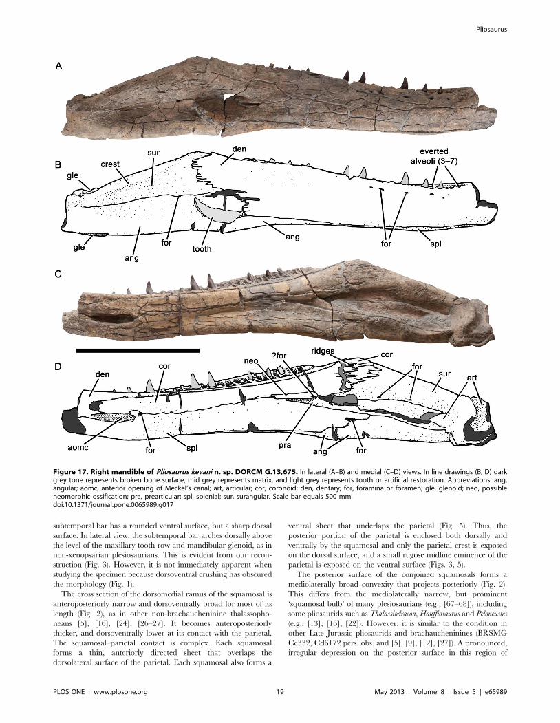
Figure 17. Right mandible of Pliosaurus kevani n. sp. DORCM G.13,675. In lateral (A–B) and medial (C–D) views. In line drawings (B, D) darkgrey tone represents broken bone surface, mid grey represents matrix, and light grey represents tooth or artificial restoration. Abbreviations: ang,angular; aomc, anterior opening of Meckel’s canal; art, articular; cor, coronoid; den, dentary; for, foramina or foramen; gle, glenoid; neo, possibleneomorphic ossification; pra, prearticular; spl, splenial; sur, surangular. Scale bar equals 500 mm.doi:10.1371/journal.pone.0065989.g017
Pliosaurus
PLOS ONE | www.plosone.org 19 May 2013 | Volume 8 | Issue 5 | e65989
DORCM G.13,675 could be a pathology, a bite mark, or a bone
surface degraded during biostratinomy (Fig. 9).
The ventral ramus of the squamosal bears the dorsal portion of
the quadrate and is broken ventrally. Because of encrusting
organisms and possible sutural fusion, the locations of sutures
between the squamosal, quadrate and pterygoid cannot be
determined. The posterior surface of the ventral ramus of each
squamosal bears a mound-like, rugose eminence, bounded
dorsolaterally by a slight ridge that extends dorsomedially along
the posterior surface of the squamosal arch (Figs 2, 9). The ventral
half of the posterior surface of the quadrate is vertical and curves
anterodorsally.
Parietal. The parietal forms the central portion of the
temporal region, contacting the interorbital skull roof anteriorly,
and the dorsomedial rami of the squamosals posteriorly (Fig. 2). As
in many Late Jurassic and younger plesiosaurians, the parietal
midline suture is closed. The pineal foramen is located adjacent to
the anterior part of the temporal fossa. It is surrounded by a raised
rim and has a suboval outline 57 mm long anteroposteriorly and
23 mm wide mediolaterally (Fig. 10). This differs from the
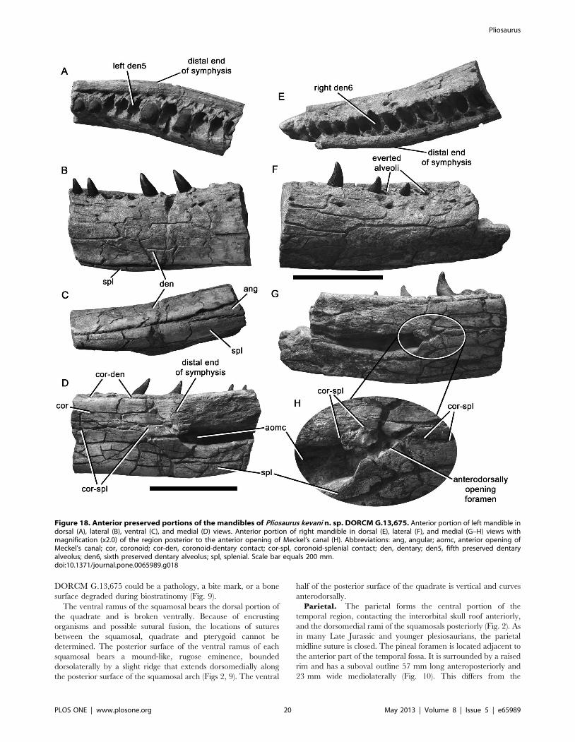
Figure 18. Anterior preserved portions of the mandibles of Pliosaurus kevani n. sp. DORCM G.13,675. Anterior portion of left mandible indorsal (A), lateral (B), ventral (C), and medial (D) views. Anterior portion of right mandible in dorsal (E), lateral (F), and medial (G–H) views withmagnification (x2.0) of the region posterior to the anterior opening of Meckel’s canal (H). Abbreviations: ang, angular; aomc, anterior opening ofMeckel’s canal; cor, coronoid; cor-den, coronoid-dentary contact; cor-spl, coronoid-splenial contact; den, dentary; den5, fifth preserved dentaryalveolus; den6, sixth preserved dentary alveolus; spl, splenial. Scale bar equals 200 mm.doi:10.1371/journal.pone.0065989.g018
Pliosaurus
PLOS ONE | www.plosone.org 20 May 2013 | Volume 8 | Issue 5 | e65989

condition in all other thalassophonean pliosaurids, in which only
the posterior margin of the pineal foramen has a raised rim, and
the anterior margin opens into an anteroposteriorly elongate
depression containing longitudinal ridges and grooves [5], [16],
[24] ([65]:character 37).
The parietal crest of DORCM G.13,675 extends posteriorly
from the pineal foramen. It is transversely narrow and dorsoven-
trally deep, and rises dorsally past the level of the skull roof
(Fig. 10), resulting in a dorsally convex outline in lateral view, as in
other thalassophoneans. In the anterior half of the temporal fossa,
the parietal is mediolaterally narrowest, extends ventrally to form
the dorsolateral walls of the endocranial cavity, and may have
formed a ligamentous attachment with the epipterygoid ventrally
(which is not preserved) as in other pliosaurids [24], [67]. More
posteriorly, the parietal expands mediolaterally to form a roof over
the occiput (the occipital condyle is inset anteriorly, far under this
roof). The parietal attains maximum mediolateral width (42 mm)
posteriorly, equal to more than half of the total skull width
(73 mm). This great proportional width (,0.5 times the skull
width or greater) is also present on other Late Jurassic pliosaurids
Figure 19. Central portions of the mandibles of Pliosaurus kevani n. sp. DORCM G.13,675. Central portion of left mandible in dorsal (A) andmedial (B) views. Central portion of right mandible in dorsal (C) and medial (D–E) views, showing an enlarged view of the possibly neomorphicossification (E). Abbreviations: den-cor, dentary-coronoid contact; for, foramen; neo, possibly neomorphic ossification; pdp, paradental plates; pra,prearticular; spl, splenial; spl-cor, splenial-coronoid contact. Scale bars equal 200 mm (A–D) and 100 mm (E).doi:10.1371/journal.pone.0065989.g019
Pliosaurus
PLOS ONE | www.plosone.org 21 May 2013 | Volume 8 | Issue 5 | e65989

(e.g., BRSMG Cc 332 and [12]), brachaucheninines [27], [75],
and convergently in leptocleidians [66], [70], [74–76] ([26]:char-
acter 49). In other plesiosaurians, including Middle Jurassic
pliosaurids, the posterior part of the parietal is narrower (e.g.,
[16], [24]).
The parietal of DORCM G.13,675 has been crushed ventrally.
The epipterygoids and prootics are not preserved, and the
supraoccipital is preserved in two pieces in the left temporal fossa
(explained below). Thus, the original relationships of these bones
with the parietal are difficult to determine. However, a flat,
posteroventrally facing surface located posteriorly on the ventral
surface of the parietal anterior to the otic region may have
articulated with the prootic.
Ectopterygoid. A possible fragment of the left ectopterygoid
is preserved dorsal to the fragmentary left pterygoid lateral ramus
(Fig. 5). The ectopterygoid might therefore have overlapped the
pterygoid at least in this region. Sutures defining the right
ectopterygoid cannot be determined.
Vomer. The vomer forms a single midline element. Its
mediolateral width tapers anteriorly. Although the vomer is
generally well-preserved, it is broken at various points along its
length, and on the midline posteriorly (Figs 4D–E, 5, 6C). It
extends anteriorly to the level of the fourth–fifth premaxillary
alveoli, and posteriorly to the level of the fourteenth maxillary
alveolus, where it contacts the anterior extension of the pterygoids
in a mediolaterally broad, deeply interdigitating suture (Fig. 5), as
in pliosaurids and most rhomaleosaurids (e.g., [13], [22], [67–69]).
The vomer-palatine suture curves posteromedially from the
posteromedial margin of the internal naris, and is also deeply
interdigitating (Figs 5, 11). However, the vomer-maxilla and
vomer-premaxilla contacts, which form a continuous line extend-
ing anteriorly from the anterolateral margin of the external naris,
are only weakly sinuous (posteriorly) and form a loose butt joint
(anteriorly) (Figs 4D–E, 5, 6C).
Palatine. The palatines are paired elements that form the
lateral portions of the palate medial to the maxillae and lateral to
the pterygoids (Figs 5, 11). The left palatine is more complete,
although its posterior portion has been broken and shifted
posteriorly (Fig. 11). The posterior portion of the right palatine
has been broken and rotated dorsolaterally. The palatine extends
from the internal naris anteriorly, to at least orbital midlength
posteriorly, where it is broken. The palatine-pterygoid suture is
sinuous anteriorly and interdigitatng posteriorly. It extends
posterolaterally from the intersection of the palatine, vomer and
Figure 20. Posterior portions of the mandibles of Pliosaurus kevani n. sp. DORCM G.13,675. Posterior portion of the left mandible in dorsal(A) and lateral (B) views. Posterior portion of the right mandible in dorsal (C), lateral (D), posterodorsomedial (E), and medial (F, G, H) views withmagnifications (x2.0) of the coronoid-surangular contact (G) and foramen at the splenial-angular contact (H). Abbreviations: ang, angular; cor,coronoid; cor-sur, coronoid-surangular contact; for, foramen or foramina; gle, glenoid; pra, prearticular; pra-ang, prearticular-angular contact; retrart,retroarticular process; spl, splenial; sur, surangular; sur-ang, surangular-angular contact. Scale bars equal 200 mm (A–D, F) and 100 mm (E).doi:10.1371/journal.pone.0065989.g020
Pliosaurus
PLOS ONE | www.plosone.org 22 May 2013 | Volume 8 | Issue 5 | e65989

pterygoid, approximately parallel to the lateral surface of the skull
(Fig. 5). A narrow, elongate notch between the posterior part of the
left palatine and the left maxilla represents the anterior end of a
suborbital fenestra (Fig. 5). Both palatines are pierced by a number
of large foramina.
Pterygoid. The pterygoids form most of the posterior palate,
and although they are only partially preserved (Fig. 5), it is clear
that they followed the typical plesiosaurian pattern in possessing
anterior, lateral, and posterior rami. Most of the anterior ramus is
preserved on the left side, but is broken at approximately orbital
midlength. The right anterior ramus is more fragmentary. The
lateral ramus is partially preserved on the right, although its
posterior edge, which would have formed the anterior margin of
the subtemporal fossa, is broken. A small notch indents its anterior
edge, and may have formed a small palatal fenestra between the
pterygoid and palatine (Fig. 5). The posterior rami, which would
have underplated the basicranium ventrally, are largely broken,
although an anterior portion is preserved on the right (Figs 5, 12).
Figure 21. Teeth of Pliosaurus kevani n. sp. DORCM G.13,675. Large tooth in mesial or distal (A, C), labial (B), lingual (D), and apical (E) views.Small tooth in labial (F), mesial or distal (G), and lingual (H) views. Scale bars equal 20 mm.doi:10.1371/journal.pone.0065989.g021
Pliosaurus
PLOS ONE | www.plosone.org 23 May 2013 | Volume 8 | Issue 5 | e65989
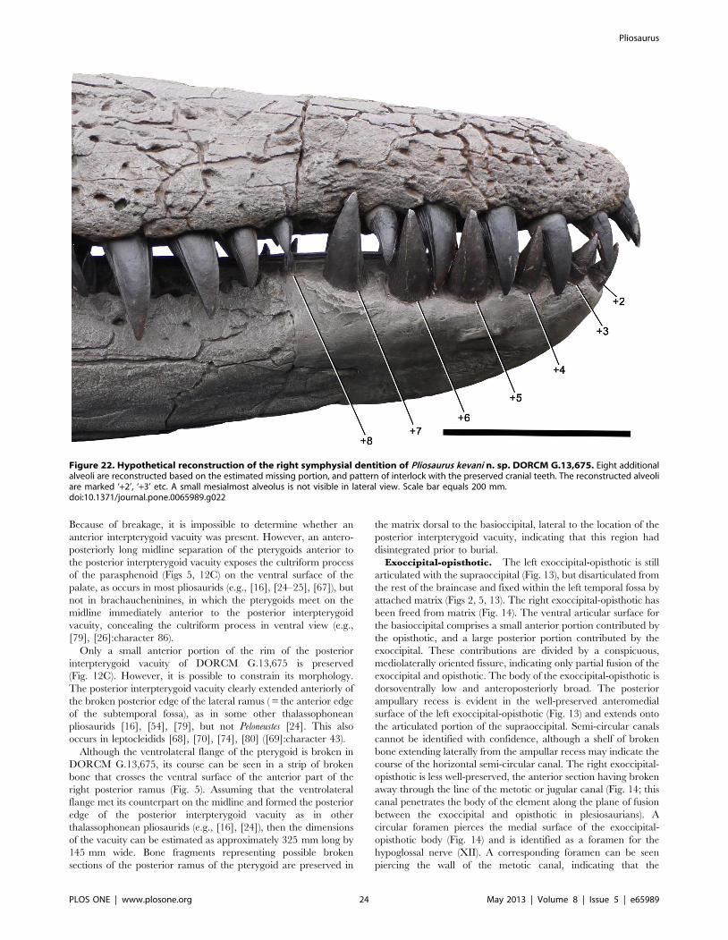
Because of breakage, it is impossible to determine whether an
anterior interpterygoid vacuity was present. However, an antero-
posteriorly long midline separation of the pterygoids anterior to
the posterior interpterygoid vacuity exposes the cultriform process
of the parasphenoid (Figs 5, 12C) on the ventral surface of the
palate, as occurs in most pliosaurids (e.g., [16], [24–25], [67]), but
not in brachaucheninines, in which the pterygoids meet on the
midline immediately anterior to the posterior interpterygoid
vacuity, concealing the cultriform process in ventral view (e.g.,
[79], [26]:character 86).
Only a small anterior portion of the rim of the posterior
interpterygoid vacuity of DORCM G.13,675 is preserved
(Fig. 12C). However, it is possible to constrain its morphology.
The posterior interpterygoid vacuity clearly extended anteriorly of
the broken posterior edge of the lateral ramus ( = the anterior edge
of the subtemporal fossa), as in some other thalassophonean
pliosaurids [16], [54], [79], but not Peloneustes [24]. This also
occurs in leptocleidids [68], [70], [74], [80] ([69]:character 43).
Although the ventrolateral flange of the pterygoid is broken in
DORCM G.13,675, its course can be seen in a strip of broken
bone that crosses the ventral surface of the anterior part of the
right posterior ramus (Fig. 5). Assuming that the ventrolateral
flange met its counterpart on the midline and formed the posterior
edge of the posterior interpterygoid vacuity as in other
thalassophonean pliosaurids (e.g., [16], [24]), then the dimensions
of the vacuity can be estimated as approximately 325 mm long by
145 mm wide. Bone fragments representing possible broken
sections of the posterior ramus of the pterygoid are preserved in
the matrix dorsal to the basioccipital, lateral to the location of the
posterior interpterygoid vacuity, indicating that this region had
disintegrated prior to burial.
Exoccipital-opisthotic. The left exoccipital-opisthotic is still
articulated with the supraoccipital (Fig. 13), but disarticulated from
the rest of the braincase and fixed within the left temporal fossa by
attached matrix (Figs 2, 5, 13). The right exoccipital-opisthotic has
been freed from matrix (Fig. 14). The ventral articular surface for
the basioccipital comprises a small anterior portion contributed by
the opisthotic, and a large posterior portion contributed by the
exoccipital. These contributions are divided by a conspicuous,
mediolaterally oriented fissure, indicating only partial fusion of the
exoccipital and opisthotic. The body of the exoccipital-opisthotic is
dorsoventrally low and anteroposteriorly broad. The posterior
ampullary recess is evident in the well-preserved anteromedial
surface of the left exoccipital-opisthotic (Fig. 13) and extends onto
the articulated portion of the supraoccipital. Semi-circular canals
cannot be identified with confidence, although a shelf of broken
bone extending laterally from the ampullar recess may indicate the
course of the horizontal semi-circular canal. The right exoccipital-
opisthotic is less well-preserved, the anterior section having broken
away through the line of the metotic or jugular canal (Fig. 14; this
canal penetrates the body of the element along the plane of fusion
between the exoccipital and opisthotic in plesiosaurians). A
circular foramen pierces the medial surface of the exoccipital-
opisthotic body (Fig. 14) and is identified as a foramen for the
hypoglossal nerve (XII). A corresponding foramen can be seen
piercing the wall of the metotic canal, indicating that the
Figure 22. Hypothetical reconstruction of the right symphysial dentition of Pliosaurus kevani n. sp. DORCM G.13,675. Eight additionalalveoli are reconstructed based on the estimated missing portion, and pattern of interlock with the preserved cranial teeth. The reconstructed alveoliare marked ‘+2’, ‘+3’ etc. A small mesialmost alveolus is not visible in lateral view. Scale bar equals 200 mm.doi:10.1371/journal.pone.0065989.g022
Pliosaurus
PLOS ONE | www.plosone.org 24 May 2013 | Volume 8 | Issue 5 | e65989

hypoglossal nerve exited the exoccipital-opisthotic via the metotic
canal. The right exoccipital-opisthotic is broken anterior to the
hypoglossal foramen, but an additional smaller foramen can be
seen piercing the medial surface of the more complete left
exoccipital-opisthotic. Thus the exoccipital-opisthotic body is
penetrated by two hypoglossal foramina and the metotic foramen
medially, whereas laterally there is a single common foramen, as in
pliosaurids and rhomaleosaurids [13], [16], [24], [81]. This is
unlike the situation in most plesiosauroids, in which multiple
foramina exit laterally (e.g., [73], [82–85]). The supraoccipital
facet can be seen in the right exoccipital-opisthotic of DORCM
G.13,675, with a clear suture separating a small, triangular
exoccipital contribution from a larger anterior opisthotic contri-
bution (Fig. 14). The paroccipital process is elongate, with a
dorsoventrally broad, spatulate distal half for articulation with the
suspensorium (Fig. 14). A rugose ridge extends medially on the
posterior surface of the exoccipital between the proximal end of
the paroccipital process and the edge of the foramen magnum.
This exoccipital flange has previously been called the ‘facet like a
zygapophysis’ [16] or ‘atlas-axis articulating facet’ [81].
Supraoccipital. The supraoccipital is broken and preserved
in two portions disarticulated and embedded in matrix remaining
Table 1. Selected measurements and observations on Late Jurassic pliosaurid specimens, arranged stratigraphically (olderspecimens are shown lower in the table).
Alveolar counts
A B Premaxilla Maxilla Dentary Symphysial C D E
Tithonian
Pliosaurus rossicus PIN 304and [7–9], [40]
? Trihedral 5 ? ? 6 ? ? ?
Upper Kimmeridgian or LowerTithonian
P. elegans or A. autissidorensis Biozone
NHMUK PV OR 39362 and[15]
1730 ? 5 .23 .25 (27e) .7 (9e) No Yes ?
Upper Kimmeridgian
A. autissidorensis Biozone
Pliosaurus indet. SEKC.K1.2and [53]
2000 ? ? ? 29e 8 ? ? ?
A. autissidorensis or A. euxodus Biozone
Pliosaurus ?rossicus OXFUMJ.10454 and [17], [57]
2875r ? ? ? 30e 6 ? ? ?
A. euxodus Biozone
Pliosaurus ?brachyspondylusCAMSM J.35991 and [52]
1200* (small) Trihedral ? ? 29 9 ? ? Flat*
Pliosaurus carpenteri BRSMGCd6172 and [55]
1800 Trihedral 6 .17 27 9 Yes ? Convex
Pliosaurus westburyensisBRSMG Cc332 and [54]
1600 Trihedral .4 25 .20 .2 No No ?
A. mutabilis or A. euxodus Biozone
Pliosaurus ‘portentificus’CAMSM J.46380 and [43]
? (big) ? ? ? .12 8 ? ? ?
A. mutabilis Biozone
Pliosaurus cf. kevani CAMSMJ.35990 and [56–57]
? (big) Subtrihedral ? ? ? ? ? ? Convex
Lower Kimmeridgian
Rasenia cymodoce Biozone
Pliosaurus indet. BHN 2R.370and [36]
2050 ? ? ? 29 11 ? ? ?
Pliosaurus brachydeirusOXFUM J.9245
1320i* Trihedral .4 .16 .35 (36e) .11 (12e) No ? Flat*
Pliosaurus kevani DORCMG.13,675
2045 Subtrihedral 6 .20 . 28 (37e) .6 (15e) Yes Yes ?
Oxfordian
Gallardosaurus iturraldeiMNHNCu P3005 and [12]
? (small)* ?Subtrihedral ? ? ? ? ? No ?
Stratigraphic data is from [34]. Abbreviations: e, estimated; r, measurement based on reconstruction; *, measurement or observation of a juvenile specimen. Columnscontain data on: A, mandible length; B, tooth morphology; C, presence of anisodont premaxillary dentition (reduced distal alveolus); D, presence of a ventrallongitudinal ridge on the parasphenoid; E, morphology of the proximal surface of the radius or tibia. Measurements are in millimetres (mm).doi:10.1371/journal.pone.0065989.t001
Pliosaurus
PLOS ONE | www.plosone.org 25 May 2013 | Volume 8 | Issue 5 | e65989

within the left temporal fenestra. The left portion of the
supraoccipital is preserved articulated with the left exoccipital–
opisthotic (Figs 2, 13D–E). The right is on its own, dorsal to the left
portion (Figs 2, 13A–C). The supraoccipital of P. kevani is similar to
that of Peloneustes [16], [24]. Its posterior surface is mediolaterally
convex and encloses only a small dorsal portion of the foramen
magnum (Fig. 13A–B). The parietal contact is planar and
horizontal, and the lateral surfaces slope ventrolaterally
(Fig. 13A–B). The opening of the posterior vertical canal of the
labyrinth is exposed on the ventrally-facing surface for contact
with the right exoccipital-opisthotic (Fig. 13C).
Basioccipital. The basioccipital forms the posterior part of
the basicranium and bears the occipital condyle (Figs 5, 9, 15). The
condyle is large (107 mm wide mediolaterally, 104 mm high
dorsoventrally), subcircular, and lacks a well-defined neck to
separate it from the basioccipital body (Fig. 15). The dorsolateral
surfaces of the condyle are embayed by the exoccipital facets. The
condyle’s surface is marked by a number of coarse grooves, but it
lacks a notochordal pit. This morphology also occurs in other Late
Jurassic pliosaurids (pers. obs. BRSMG Cc332, Cd6172, and [3]),
except Gallardosaurus, which has a smooth condyle with a well-
defined notochordal pit [12], as in Middle Jurassic pliosaurids
[16], [24].
In DORCM G.13,675, only the posterior portion of the
basicranium is preserved, and is highly abraded. This abrasion has
almost completely eroded the pterygoids, revealing the morphol-
ogy of the dorsally overlying basicranial bones. The abrasion, and
associated fracturing, makes it difficult to interpret and to
differentiate basicranial elements. A small, diamond-shaped
platform is present on the ventral surface of the basioccipital, just
anterior to the occipital condyle. This structure, often called the
‘ventral plate’ or ‘ventral process’ probably contacts portions of the
parabasisphenoid anteriorly. Well-developed basal tubera project
laterally from the body of the basioccipital (Figs 9, 15). These
contact the posterolateral processes of the parasphenoid anteriorly
and laterally, but no evidence of any more extensive contact is
preserved. The basal tubera of most other Late Jurassic pliosaurids
also project laterally or only slightly ventrolaterally (BRSMG
Cc332, Cd6172; OXFUM J.92451/4), but in brachaucheninines,
Gallardosaurus, and Middle Jurassic pliosaurids they project further
ventrolaterally so their articular surfaces for the pterygoids are
located ventral to the occipital condyle [6], [12], [16], [24].
Parabasisphenoid. The basisphenoid and parasphenoid are
often conjoined in plesiosaurians, and the line of fusion between
them can be difficult to discern. The combined parabasisphenoid
forms the anterior part of the basicranium, and extends anteriorly
into the palate as the cultriform process. (Figs 5, 12) The central
portion of the parabasisphenoid, which divides the posterior
interpterygoid vacuity, is broken and missing in DORCM
G.13,675. Thus, of the parabasisphenoid, only the cultriform
process and posterior fragments are preserved. The cultriform
process extends far anteriorly on the palate, tapering along its
length. Its base is mediolaterally constricted.
The anterior end of the preserved posterior portion of the
parabasisphenoid bears a low, abraded midline ridge (Fig. 15).
This suggests that the ventral surface of the parabasisphenoid bore
a median ridge, as in Middle Jurassic [16], [24] and some Late
Jurassic pliosaurids (NHMUK PV OR 39362), but unlike in
BRSMG Cc332 [54], Gallardosaurus [12], and brachaucheninines
[6], [27], [79], in which the ventral surface of the parabasi-
sphenoid lacks a ventral keel.
The cross-section of the parabasisphenoid of DORCM
G.13,675 is visible on the broken anterior surface of the
basicranium, and shows a dorsal area of highly cancellous bone.
This may represent a poorly-ossified basisphenoid contribution to
the parabasisphenoid and is seen in some other pliosaurid
specimens (e.g., LEICT G418.1956.58.1.4). The portion visible
in ventral view consists of denser bone with longitudinal
trabeculae, forming the ventral and ventrolateral surfaces of the
parabasisphenoid, and likely represents the parasphenoid. This
structure continues posteriorly on the ventral surface of the
basicranium, contacting the ventral plate of the basioccipital, and
producing posterolateral processes that contact the basal tubera.
The mandible of Pliosaurus kevani n. sp.Both mandibles are well-preserved, but most of the symphysis is
missing (Figs 16–17). The total missing symphysial length can be
estimated as approximately 400 mm, by comparison of the
preserved length from the posterior margin of the glenoid (right
side = 1660 mm; left side = 1625 mm) with the length of the
cranium measured from the quadrate to the snout tip (right side =
2060 mm; left side = 2030 mm). Compared to its anteroposterior
length, the mandible seems dorsoventrally deep (maximum
dorsoventral height = 320 mm at the coronoid eminence), with
an estimated length to maximum depth ratio of 6.25.
The posterior portion of the mandibular symphysis is preserved,
as determined by anterior inflection of the medial surface, which
becomes planar and rugose, and bears the anterior opening of
Meckel’s canal around midheight. The mandibles are not
coossified in this posterior symphysial region. Furthermore, the
lateral surfaces of both rami were heavily encrusted with epifauna
prior to preparation, while their medial surfaces were almost free
of encrustation. This suggests that in both rami rested in the
sediment with their lateral surfaces facing upwards. This may
indicate that the symphysis was not firmly joined anteriorly, and
was thus similar to some brachaucheninines (e.g., [27]) but unlike
other Jurassic pliosaurids (e.g., [16–17], [23–24], [43], [55]).
Alternatively however, and perhaps more likely, the symphysis
may have been broken prior to burial.
The lateral surface of the mandible is weakly convex
dorsoventrally for most of its length. However, posteriorly (where
it is formed by the surangular dorsally and angular ventrally), the
lateral surface is concave (Figs 16–17).
Dentary. The dentary forms much of the body of the
mandible anteriorly (Figs 16–17). It is dorsoventrally deep
compared to its mediolateral width. The medial surface of the
dentary is covered by the coronoid dorsally and splenial ventrally.
Together, the coronoid and splenial form the medial wall of
Meckel’s canal, which excavates the body of the dentary and opens
anteriorly in the posterior region of the symphysis (Figs 16–18),
between the anterior ends of the splenial and coronoid. The lateral
surface of the dentary is weakly convex dorsoventrally, and bears a
row of unevenly spaced, anteroposteriorly elongate foramina
dorsally. The dentary continues posteriorly as a thin sheet of bone
with crenulated edges. This sheet bifurcates posteriorly into two
lobes, a posterodorsal lobe that covers the lateral surface of the
surangular, and a posteroventral lobe that covers the angular
(Figs 16–17). The posterodorsal lobe extends furthest posteriorly,
to the level of the coronoid eminence.
As in the maxilla and premaxilla, the dentary alveoli are
bounded medially by low, subtriangular paradental plates formed
from rugose bone. These plates extend medially to divide the
alveoli (Figs 18–19). Replacement alveoli are visible medial to the
bases of the paradental plates. The left dentary preserves 27
alveoli, of which five are located within the symphysis. The right
dentary, which is slightly more complete, preserves 28 alveoli, of
which six are symphysial. Most alveoli open dorsally, as is usual in
other plesiosaurians. However, the occlusal surface of the dentary
Pliosaurus
PLOS ONE | www.plosone.org 26 May 2013 | Volume 8 | Issue 5 | e65989

is inclined to face dorsolaterally in the posterior region of the
symphysis, so that the fifth–seventh alveoli (as counted from the
preserved anterior end of the right dentary) open dorsolaterally
(Figs 16–18). This was not observed in any other pliosaurid during
the present study and is an autapomorphy of Pliosaurus kevani.
Splenial. The splenial is an anteroposteriorly elongate sheet
of bone that covers the medial surface of the mandible ventrally
(Figs 16–17). It extends far anteriorly to form the posterior portion
of the mandibular symphysis, as in many pliosaurids and other
intermediate- and long-snouted plesiosaurians (e.g., [65], [67–69]).
It tapers abruptly posteriorly, becoming dorsoventrally narrow and
terminating just posterior to the level of the coronoid eminence.
Here, the splenial is supported ventrally by a medial buttress of the
angular that forms the ventral floor of Meckel’s canal. A small,
anteroposteriorly elongate foramen penetrates the splenial-angular
contact, entering Meckel’s canal (Figs 16–17, 20).
Coronoid. The coronoid is an anteroposteriorly elongate
sheet of bone that covers the medial surface of the mandible
dorsally (Figs 16–17). It extends anteriorly to form the posterior
portion of the mandibular symphysis, as in many pliosaurids and
other longirostrine plesiosaurians (e.g., [22–24], [45–47], [65],
[68], [86]). It extends posterior to the level of the coronoid
eminence, where it overlaps the lateral surface of the surangular
and terminates in a deeply crenulated, dorsoventrally trending
edge (Figs 16–17, 20).
Surangular. The surangular forms the dorsal part of the
body of the mandible posteriorly (Figs 16–17, 20). Its lateral and
dorsal surfaces are covered anteriorly by the dentary, and its
medial surface is covered anteriorly by the coronoid. Thus, the
anterior extent of the surangular within the mandible cannot be
determined. Posteriorly, the surangular contacts the articular,
which forms the mandibular glenoid. The dorsal portion of the
surangular anterior to the glenoid is transversely expanded and
bears a broad, mediolaterally convex, dorsolaterally facing surface
that is excavated laterally by an anteroposteriorly elongate fossa
with a sharp lateral rim (Fig. 20). The presence of a mediolaterally
broad dorsal surface of the surangular is shared with other
thalassophonean pliosaurids ([68]:character 87). However, where-
as in Middle Jurassic taxa and the Late Jurassic holotype of P.
carpenteri (BRSMG Cd6172) the resulting broad surface faces
dorsally [23–24], in most Late Jurassic specimens, including
DORCM G.13,675 and the mandible of the holotype of Pliosaurus
brachydeirus (OXFUM J.9245) it is inclined to face dorsolaterally
(BRSMG Cc332; CAMSM J.29561; NHMUK PV OR 39362;
OXFUM J.10454). Furthermore, in Late Jurassic specimens
except for CAMSM J.29561, the lateral fossa and rim are present,
as in DORCM G.13,675.
The surangular of DORCM G.13,675 makes an anteroposte-
riorly oriented contact with the angular ventrally (Figs 16–17, 20).
The plane of this contact is subhorizontal, so it is expressed at the
same dorsoventral level both medially and laterally. The
surangular–angular contact is penetrated by two oval foramina
(Figs 16–17, 20).
Angular. The angular forms the ventral part of the body of
the mandible posteriorly. It extends anteriorly within the
mandible, where it is covered medially by the splenial and
laterally by the dentary. However, it remains exposed on the
ventral surface of the mandible as a long anterior process dividing
the splenial and dentary until just posterior to the mandibular
symphysis (Fig. 18). The angular extends posteriorly ventral and
lateral to the glenoid and forms part of the retroarticular process
(on which its suture with the articular is not visible, and was
possibly closed) (Figs 16–17, 20). The left retroarticular process is
well-preserved. It is posterodorsally inclined and anteroposteriorly
short, subequal to the length of the glenoid. It has a suboval cross
section and is expanded slightly towards its posterior end, which is
abraded and measures 95 mm dorsoventrally and 75 mm
mediolaterally.
Knutsen [34] identified four morphotypes of the retroarticular
process in Late Jurassic pliosaurids. The main criterion for
recognising these is the shape of the outline in lateral view. The
retroarticular process of DORCM G.13,675 seen in lateral view
has a ventral surface that curves smoothly posterodorsally, and a
dorsal surface that is approximately horizontal (Fig. 20B). The
process is mediolaterally much narrower than the glenoid, as in all
the morphotypes except for ‘morphotype II’ [34]. However, the
morphology does not correspond exactly to any of the morpho-
types, and we note that differences in preservation (i.e. deforma-
tion and completeness) and ontogenetic stage (the process seems to
grow posteriorly through ontogeny) complicate the situation
further.
Prearticular. The prearticular is a dorsoventrally narrow,
anteroposteriorly elongate, band-like bone (Figs 16–17, 20). It
originates just anterior to the glenoid, contacting the anteromedial
surface of the articular. The prearticular is supported ventrally by
the medial buttress of the angular, forming the ventromedial wall
of Meckel’s canal, which is only partly enclosed in this region (it is
open dorsolaterally).
Neomorphic mandibular element. A broken, anteroposte-
riorly oriented band of bone is present on the medial surface of the
mandible level with the posterior part of the tooth row (Figs 16C–
D, 17C–D, 19B). Examination of cross-sections demonstrates that
this is not a portion of the prearticular, and it is tentatively
identified as a neomorphic ossification, possibly an analogue of the
additional coronoid bone present in some early amniotes.
Alternatively, this ossification could be directly homologous with
an additional coronoid, in which case an additional coronoid must
have been present in more basal pliosaurids and earlier
plesiosaurians, but thus far have been overlooked. Whatever its
identity, this ossification in DORCM G.13,675 encloses a smooth,
rounded rim posteriorly. This is especially well preserved on the
right side, and is suggestive of a foramen through which Meckel’s
canal might have made the transition to being fully enclosed
anteriorly to being only partly enclosed posteriorly. A similar
opening is present in Peloneustes (LEICT G418.1956.33; collected
by Phillips in 1923 from the Peterborough Member of King’s
Dyke brick pit, near Peterborough, United Kingdom). However,
in Peloneustes it seems to be formed by the coronoid, and evidence
for a separate neomorphic ossification is absent.
Articular. The articular forms the mandibular glenoid and
perhaps a portion of the retroarticular process. However, this is
difficult to determine because the angular-articular suture is only
visible medially, immediately ventral to the glenoid. The glenoid
faces dorsomedially, and consists of an anteroposteriorly long
lateral cotyle, and shorter medial cotyle, that are confluent,
forming a single, mediolaterally broad depression (Figs 16–17, 20).
The dentition of Pliosaurus kevani n. sp.Several teeth are preserved attached to the skull or deep within
the alveoli. Four have been prepared free. The two best preserved
are shown in Figure 21, representing a large tooth from the mesial
half of the snout (Fig. 21A–E) and a small, distal ‘ratchet’ tooth
(Fig 21F–H). All teeth are conical, curved, have approximately
circular cross sections with a slightly flattened labial surface ( =
‘subtrihedral’). Comparison with other pliosaurids suggests the
teeth curved lingually. The teeth of DORCM G.13,675 bear
numerous coarse, apicobasally oriented ridges. Because the tooth
apices are broken, it is impossible to say how far these ridges
Pliosaurus
PLOS ONE | www.plosone.org 27 May 2013 | Volume 8 | Issue 5 | e65989
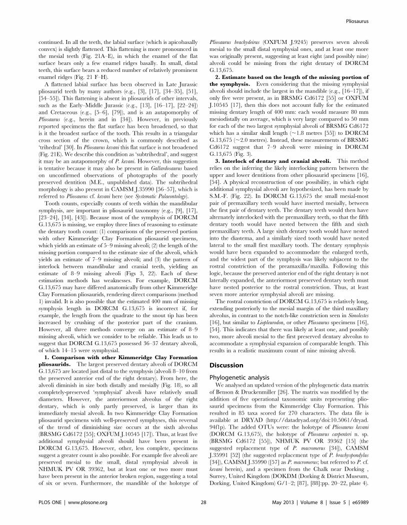
continued. In all the teeth, the labial surface (which is apicobasally
convex) is slightly flattened. This flattening is more pronounced in
the mesial teeth (Fig. 21A–E), in which the enamel of the flat
surface bears only a few enamel ridges basally. In small, distal
teeth, this surface bears a reduced number of relatively prominent
enamel ridges (Fig. 21 F–H).
A flattened labial surface has been observed in Late Jurassic
pliosaurid teeth by many authors (e.g., [3], [17], [34–35], [51],
[54–55]). This flattening is absent in pliosaurids of other intervals,
such as the Early–Middle Jurassic (e.g., [13], [16–17], [22–24])
and Cretaceous (e.g., [5–6], [79]), and is an autapomorphy of
Pliosaurus (e.g., herein and in [34]). However, in previously
reported specimens the flat surface has been broadened, so that
is it the broadest surface of the tooth. This results in a triangular
cross section of the crown, which is commonly described as
‘trihedral’ [30]. In Pliosaurus kevani this flat surface is not broadened
(Fig. 21E). We describe this condition as ‘subtrihedral’, and suggest
it may be an autapomorphy of P. kevani. However, this suggestion
is tentative because it may also be present in Gallardosaurus based
on unconfirmed observations of photographs of the poorly
preserved dentition (M.E., unpublished data). The subtrihedral
morphology is also present in CAMSM J.35990 [56–57], which is
referred to Pliosaurus cf. kevani here (see Systematic Palaeontology).
Tooth counts, especially counts of teeth within the mandibular
symphysis, are important in pliosaurid taxonomy (e.g., [9], [17],
[23–24], [34], [43]). Because most of the symphysis of DORCM
G.13,675 is missing, we employ three lines of reasoning to estimate
the dentary tooth count: (1) comparisons of the preserved portion
with other Kimmeridge Clay Formation pliosaurid specimens,
which yields an estimate of 5–9 missing alveoli; (2) the length of the
missing portion compared to the estimate size of the alveoli, which
yields an estimate of 7–9 missing alveoli; and (3) the pattern of
interlock between mandibular and cranial teeth, yielding an
estimate of 8–9 missing alveoli (Figs 3, 22). Each of these
estimation methods has weaknesses. For example, DORCM
G.13,675 may have differed anatomically from other Kimmeridge
Clay Formation pliosaurids, rendering direct comparisons (method
1) invalid. It is also possible that the estimated 400 mm of missing
symphysis length in DORCM G.13,675 is incorrect if, for
example, the length from the quadrate to the snout tip has been
increased by crushing of the posterior part of the cranium.
However, all three methods converge on an estimate of 8–9
missing alveoli, which we consider to be reliable. This leads us to
suggest that DORCM G.13,675 possessed 36–37 dentary alveoli,
of which 14–15 were symphysial.
1. Comparison with other Kimmeridge Clay Formation
pliosaurids. The largest preserved dentary alveoli of DORCM
G.13,675 are located just distal to the symphysis (alveoli 8–10 from
the preserved anterior end of the right dentary). From here, the
alveoli diminish in size both distally and mesially (Fig. 18), so all
completely-preserved ‘symphysial’ alveoli have relatively small
diameters. However, the anteriormost alveolus of the right
dentary, which is only partly preserved, is larger than its
immediately mesial alveoli. In two Kimmeridge Clay Formation
pliosaurid specimens with well-preserved symphyses, this reversal
of the trend of diminishing size occurs at the sixth alveolus
(BRSMG Cd6172 [55]; OXFUM J.10545 [17]). Thus, at least five
additional symphysial alveoli should have been present in
DORCM G.13,675. However, other, less complete, specimens
suggest a greater count is also possible. For example five alveoli are
preserved mesial to the small, distal symphysial alveoli in
NHMUK PV OR 39362, but at least one or two more must
have been present in the anterior broken region, suggesting a total
of six or seven. Furthermore, the mandible of the holotype of
Pliosaurus brachydeirus (OXFUM J.9245) preserves seven alveoli
mesial to the small distal symphysial ones, and at least one more
was originally present, suggesting at least eight (and possibly nine)
alveoli could be missing from the right dentary of DORCM
G.13,675.
2. Estimate based on the length of the missing portion of
the symphysis. Even considering that the missing symphysial
alveoli should include the largest in the mandible (e.g., [16–17]), if
only five were present, as in BRSMG Cd6172 [55] or OXFUM
J.10545 [17], then this does not account fully for the estimated
missing dentary length of 400 mm: each would measure 80 mm
mesiodistally on average, which is very large compared to 50 mm
for each of the two largest symphysial alveoli of BRSMG Cd6172
which has a similar skull length (,1.8 metres [55]) to DORCM
G.13,675 (,2.0 metres). Instead, these measurements of BRSMG
Cd6172 suggest that 7–9 alveoli were missing in DORCM
G.13,675 (Fig. 3).
3. Interlock of dentary and cranial alveoli. This method
relies on the inferring the likely interlocking pattern between the
upper and lower dentitions from other pliosaurid specimens [16],
[54]. A physical reconstruction of one possibility, in which eight
additional symphysial alveoli are hypothesized, has been made by
S.M.-F. (Fig. 22). In DORCM G.13,675 the small mesial-most
pair of premaxillary teeth would have inserted mesially, between
the first pair of dentary teeth. The dentary teeth would then have
alternately interlocked with the premaxillary teeth, so that the fifth
dentary tooth would have nested between the fifth and sixth
premaxillary teeth. A large sixth dentary tooth would have nested
into the diastema, and a similarly sized tooth would have nested
lateral to the small first maxillary tooth. The dentary symphysis
would have been expanded to accommodate the enlarged teeth,
and the widest part of the symphysis was likely subjacent to the
rostral constriction of the preamaxilla/maxilla. Following this
logic, because the preserved anterior end of the right dentary is not
laterally expanded, the anteriormost preserved dentary teeth must
have nested posterior to the rostral constriction. Thus, at least
seven more anterior symphysial alveoli are missing.
The rostral constriction of DORCM G.13,675 is relatively long,
extending posteriorly to the mesial margin of the third maxillary
alveolus, in contrast to the notch-like constriction seen in Simolestes
[16], but similar to Liopleurodon, or other Pliosaurus specimens [16],
[54]. This indicates that there was likely at least one, and possibly
two, more alveoli mesial to the first preserved dentary alveolus to
accommodate a symphysial expansion of comparable length. This
results in a realistic maximum count of nine missing alveoli.
Discussion
Phylogenetic analysisWe analysed an updated version of the phylogenetic data matrix
of Benson & Druckenmiller [26]. The matrix was modified by the
addition of five operational taxonomic units representing plio-
saurid specimens from the Kimmeridge Clay Formation. This
resulted in 85 taxa scored for 270 characters. The data file is
available at DRYAD (http://datadryad.org/doi:10.5061/dryad.
94f1p). The added OTUs were: the holotype of Pliosaurus kevani
(DORCM G.13,675), the holotype of Pliosaurus carpenteri n. sp.
(BRSMG Cd6172 [55]), NHMUK PV OR 39362 [15] (the
suggested replacement type of P. macromerus [34]), CAMSM
J.35991 [52] (the suggested replacement type of P. brachyspondylus
[34]), CAMSM J.35990 ([57] as P. macromerus; but referred to P. cf.
kevani herein), and a specimen from the Chalk near Dorking ,
Surrey, United Kingdom (DOKDM (Dorking & District Museum,
Dorking, United Kingdom) G/1–2; [87], [88]:pp. 20–22, plate 4).
Pliosaurus
PLOS ONE | www.plosone.org 28 May 2013 | Volume 8 | Issue 5 | e65989

DOKDM G/1–2 was first reported by Owen in 1860 ([87], as
Polyptychodon interruptus) as being from the Lower Chalk at Dorking,
and in the collection of Mr G. Cubitt. Owen later described it
again as being in Cubitt’s collection, but instead this time coming
from a ‘railway tunnel through the chalk formations near Frome,
Somersetshire’ ([88]:p. 20). The Frome reference is likely to have
been in error; George Cubitt became Lord Ashcombe, his estate
was at Denbies, near Dorking, and the specimen is curated at
DOKDM, in Dorking. Chalk was quarried in and around Dorking
from the West Melbury Marly Chalk, Zig Zag Chalk, Holywell
Nodular Chalk and New Pit Chalk formations [89–90] of
Cenomanian–early Turonian age. Owen’s [87] use of the term
‘Lower Chalk’ is likely to have been intended in the traditional
sense common in the 19th century. This included only the
traditional Chalk Marl and Grey Chalk and so only represents
Cenomanian strata [90].
Two characters in the analysis were modified from their usage
in Benson & Druckenmiller [26] by the addition of a state (‘2’):
Character 63. Notochordal pit on occipital condyle: absent (0);
present (1); occipital condyle scored by multiple pits and deep
grooves (2).
Character 139. Cross-sectional shape of teeth in anterior half of
tooth row: round or sub-rounded (0); sub-triangular (1; =
trihedral); intermediate between states 0 and 1, with a flattened
labial surface, but this surface not substantially expanded
anteroposteriorly (2; = subtrihedral).
Tree searches were performed in PAUP* 4.0b10 for Macintosh
[91]. Initial exploration for shortest-length tree islands was
conducted using four independent randomisations of the Parsi-
mony Ratchet [92] implemented by PAUPRat [93]. The resulting
subset of most parsimonious trees (MPTs) was then used as the
starting point for TBR (tree bisection and reconnection) branch
swapping. ‘Wildcard’ taxa were identified by inspection of the
Adams consensus and pruned from the set of most parsimonious
cladograms, which were then used to construct a strict reduced
consensus [94].
Our search recovered .130000 shortest-length trees 1336 steps
long. The strict consensus of these trees (Fig. 23) differed from that
of Benson & Druckenmiller [26] in two respects. Firstly, within
Xenopsaria, several early Cretaceous taxa have slightly different or
less resolved positions: Gronausaurus, the ‘Speeton clay plesiosaur-
ian’, Wapuskanectes, Callawayasaurus and Eromangasaurus form a basal
elasmosaurid polytomy and the leptocleidians Brancasaurus [83]
and Vectocleidus [66], previously found as leptocleidids [26], were
found as basal leptocleidians. Brancasaurus was found outside of the
clade comprising Leptocleididae + Polycotylidae. Vectocleidus was
found in a polytomy with Leptocleididae and Polycotylidae. This
change likely arose from minor scoring amendments and
instability of relationships among Early Cretaceous plesiosauroids.
Secondly, most members the thalassophonean clades Brachau-
cheninae and Pliosaurus formed an unresolved polytomy. The
exception was a clade comprising two sister taxon pairings:
CAMSM J.35990 + Pliosaurus kevani and Pliosaurus funkei + Pliosaurus
carpenteri. This polytomy results from the uncertain phylogenetic
positions of a few ‘wildcard’ taxa, which occupy multiple positions
among the set of shortest-length trees. Pruning these taxa from the
trees results in a reduction of the number of unique topologies and
an increase in resolution of the strict consensus (a ‘strict reduced
consensus’ [94]). Thus, deletion of Pliosaurus irgisensis results in
resolution of a monophyletic Brachaucheninae in the strict
reduced consensus (although relationships within Brachaucheni-
nae remained unresolved). Subsequent deletion of QM F51291
results in resolution of Kronosaurus queenslandicus (MCZ 1285) as the
sister taxon of an unresolved polytomy comprising three species of
Brachauchenius plus DOKDM G/1–2 (‘Polyptychodon interruptus’
according to [87–88]), which we suggest should be referred to
Brachauchenius indet. Finally, deletion of Pliosaurus rossicus and
Gallardosaurus, in addition to Pliosaurus irgisensis, results in resolution
of a monophyletic Pliosaurus (excluding ‘Pliosaurus’ andrewsi). The
strict reduced consensus after deletion of Pliosaurus irgisensis,
Pliosaurus rossicus, Gallardosaurus, and QM F51291 is shown in
Figure 23B.
Body size of Pliosaurus kevani, and pliosaurid body sizeevolution
The skull of Pliosaurus kevani is very large, measuring 1995 mm
along the dorsal midline of the cranium and with a mandibular
ramus length of 2045 mm excluding the retroarticular process.
This is the largest substantially complete Kimmeridge Clay
Formation pliosaurid skull (Table 1). However, comparably large
individuals have been reported on the basis of more fragmentary
material (see Table 1; and explained further below).
Maximum body size of pliosaurids seems to have increased from
their first occurrences in the Early Jurassic, until the Early
Cretaceous (Fig. 24). Lower Jurassic pliosaurids are relatively
small, with maximum skull lengths of 180 mm in the earliest
Jurassic (Hettangian) taxon Thalassiodracon [13], and 680 mm in
the late Early Jurassic (Toarcian) Hauffiosaurus longirostris [86].
Plesiosaurian maximum body sizes generally increased during the
Early Jurassic, across multiple clades [95]. However, pliosaurid
maximum sizes continued to increase after this (Fig. 24). The
largest Middle Jurassic pliosaurid, Liopleurodon ferox from the
Callovian Oxford Clay Formation, has a skull length of
1540 mm [16]. By contrast, most Late Jurassic pliosaurid skulls
measure 1700–2100 mm long (Table 1 and below), and some
Early Cretaceous pliosaurids were even larger than this ([1], [6],
[96] reported skull lengths of at least 2360 mm). However, Late
Cretaceous pliosaurids seem to have been smaller than those of the
Late Jurassic (maximum skull length of 1750 mm [14]). The
Cretaceous plesiosauroid clade Polycotylidae, which independent-
ly evolved pliosaurid-like body proportions [18–19], never attained
sizes similar to the largest pliosaurids. Instead, the largest
polycotylids had skulls approximately 1000 mm long [45–47] in
Dolichorhynchops bonneri, Eopolycotylus, and Polycotylus. This is
congruent with the observation that most polycotylids were
piscivorous, with long, narrow snouts and gracile, widely-spaced,
approximately isodont teeth. Although some polycotylids show
slightly more robust, anisodont dentitions, they never evolved
especially robust snouts, unlike those of pliosaurids.
At first glance, the Late Jurassic pliosaurid assemblage seems not
to consist only of large-bodied species. Some specimens are smaller
than 1700–2100 mm long, including the holotype of Pliosaurus
brachydeirus (OXFUM J.9245), Knutsen’s [34] proposed ‘neotype’
of P. brachyspondylus (CAMSM J.35991 [52]), and Gallardosaurus
iturraldei [12] (Table 1). However, these specimens show juvenile
features, indicating their small size is the result of incomplete
growth, and suggesting that Late Jurassic pliosaurid body size
diversity may have been low. These juvenile features include
incomplete fusion of the atlas-axis complex (pers. obs. of CAMSM
J.35991; and [12]) and possibly incomplete ossification of the
proximal convexities of the radius and tibia (CAMSM J.35991;
OXFUM J.9245). The cervical and dorsal vertebrae of these
specimens preserve disarticulated centra and neural spines,
indicating unfused neurocentral sutures, commonly also interpret-
ed as a juvenile feature in reptiles. They also have dorsoventrally
short neural spines, with incompletely ossified dorsal ends. This is
also indicative of ontogenetic immaturity in sauropterygians [48].
However, the neurocentral sutures remain unfused in very large
Pliosaurus
PLOS ONE | www.plosone.org 29 May 2013 | Volume 8 | Issue 5 | e65989

thalassophonean individuals, including Late Jurassic specimens
such as CAMSM J.35990 [57], the holotype of Pliosaurus carpenteri
(pers. obs. of BRSMG Cd6172) and the large Mexican specimen
[2]. Thus, it is possible that vertebral sutural fusion was extremely
delayed or never occurred in thalassophoneans. If so, vertebral
sutural fusion cannot be used as a reliable indicator of ontogenetic
stage in Thalassophonea (contra [2]). Alternatively, it is possible that
despite their large body sizes, these specimen do represent
juveniles, and truly adult specimens have yet to be discovered.
Knutsen et al. [3] found a strong relationship between skull
length and the mediolateral width of the occipital condyle in Late
Jurassic and Cretaceous pliosaurids. Pliosaurus funkei is from the
Figure 23. Phylogenetic position of Pliosaurus kevani n. sp. DORCM G.13,675. Strict (A) and strict reduced (B) consensus topologies of.130000 shortest length trees 1336 steps long resulting from analysis of our matrix of 85 taxa and 270 characters modified from [26]. Shading is usedto indicate major clades, Pliosauridae is enclosed by a grey rectangle, and Pliosaurus kevani n. sp. is indicated by an arrow.doi:10.1371/journal.pone.0065989.g023
Pliosaurus
PLOS ONE | www.plosone.org 30 May 2013 | Volume 8 | Issue 5 | e65989

Tithonian (Late Jurassic) of Svalbard. The largest of two
individuals of P. funkei, PMO (University of Oslo Natural History
Museum, Oslo, Norway) 214.136 [3] has an occipital condyle that
is 150 mm wide and 105 mm high dorsoventrally. Based on his
regression line, Knutsen et al. [3] predicted an original skull length
around 2190 mm for PMO 214.136. A slightly lower value around
2040 mm was predicted from the dimensions of cervical centra. It
is difficult to compare the size of P. kevani with that of P. funkei
because these taxa are known from different parts of the skeleton.
The occipital condyle of P. kevani is approximately as high
dorsoventrally (104 mm) as that of P. funkei (105 mm). However, it
is narrower mediolaterally (107 mm compared to 150 mm in P.
funkei [3]). This suggests either that P. kevani is slightly smaller, that
P. funkei had an unusually broad occipital condyle, or that the
occipital condyle of P. funkei was taphonomically distorted. Either
way, it seems that both individuals were of similar size with skull
lengths close to 2 metres.
Other large individuals of Late Jurassic pliosaurids were
reported by Tarlo [57] (CAMSM J.35990) and Buchy et al. [2].
The body size of these specimens are difficult to compare to taxa
known only from skulls such as P. kevani. Although they reinforce
the point that many Late Jurassic pliosaurids were large-bodied,
they are not suggestive of individuals much larger than P. kevani or
P. funkei. OXFUM J.10454, a highly reconstructed fragmentary
pliosaurid skull from Cumnor near Oxford, has a reconstructed
total length of 2875 mm [57]. Tarlo [57] suggested that this
specimen would have originally been more than 3000 mm long
and represented the largest pliosaurid ever recorded. However, the
total length cannot be determined and more detailed study is
required to establish whether it actually represents an individual
substantially larger than other Late Jurassic specimens.
White [6] referred MCZ 1285 from the Early Cretaceous of
Australia to Kronosaurus queenslandicus. He estimated a skull length of
3720 mm based on comparison with Liopleurodon (as ‘Pliosaurus
ferox’). The total length of this individual, for which most of the
vertebral column was known (and now highly restored with
plaster) was stated as 12800 mm by Romer & Lewis [1]. Based on
the dimensions of its cervical vertebrae, Knutsen et al. [3]
estimated the skull length of MCZ 1285 as around 2190 mm.
However, this approach employs ‘average’ cervical centrum width,
which is taken to mean the average of those preserved, and may be
imprecise depending on the pattern of size change along the
cervical series. The occipital condyle of MCZ 1285 is 180 mm
wide mediolaterally and 125 mm high dorsoventrally (pers. obs.
R.B.J.B.), approximately 25% wider and higher than that of P.
funkei. Using the regression line of Knutsen et al. [3], this suggests a
skull length of 2850 mm.
Although the cranial length of MCZ 1285 can only be estimated
imprecisely, another Cretaceous specimen, the holotype of
Kronosaurus boyacensis from the Aptian of Colombia, is more
complete [96]. K. boyacensis has a midline skull length of 2360 mm
and a snout-vent length of 7250 mm. Unfortunately, the tail is
missing, preventing comparisons with the estimated total body
length of MCZ 1285 provided by Romer & Lewis [1] (12800 mm).
However, individual middle–posterior dorsal vertebrae of K.
boyacensis range from 117–138 mm [96], comparable or only
slightly less than 114–145 mm in MCZ 1285 [1]. These dorsal
centrum lengths, as well as the skull lengths, of Early Cretaceous
pliosaurids, are greater than those of the largest Late Jurassic
specimens (dorsal centra range from 100–121 mm in Pliosaurus cf.
kevani; CAMSM J.35990 [57]; most dorsal centra are in the range
97–116 mm in Pliosaurus funkei PMO 214.135, only two are greater
at 135 and 142 mm [3]). However, the limbs of these Cretaceous
specimens seem to be comparable in length, or shorter than, those
of the largest Late Jurassic specimens. For example, the humerus is
799 mm long and femur 977 mm long in K. boyacensis [96] and the
femur is 1060 mm long in MCZ 1285 [1], whereas the humerus is
840 mm long and the femur is 960 mm long in CAMSM J.35990
[57], and the humerus 1000 mm long in Pliosaurus funkei [3]. In
reconciliation of these observations, Knutsen et al. [3] showed that
Early Cretaceous pliosaurids had proportionally short limbs, and
that P. funkei in particular, has proportionally long limbs compared
to other large-bodied pliosaurids. In general, the proportional limb
length might decline with increasing body size in pliosauromorphs
[18–19]. Thus, limb lengths might represent the weakest proxy for
body size in pliosaurids.
Evolution of Late Jurassic pliosauridsLack of phylogenetic resolution within Pliosaurus and the
incomplete nature of many specimens makes it difficult to
Figure 24. Maximum skull length of Jurassic–Cretaceous pliosaurids. Silhouettes are only approximately indicative of body size andproportions.doi:10.1371/journal.pone.0065989.g024
Pliosaurus
PLOS ONE | www.plosone.org 31 May 2013 | Volume 8 | Issue 5 | e65989

understand the evolution of Late Jurassic pliosaurids. However, we
agree with Knutsen’s [34] observation that stratigraphically older
specimens, including Pliosaurus kevani, have generally longer
mandibular symphyses (containing up to 15 alveoli), and younger
specimens have fewer (containing as few as six alveoli; e.g., [7],
[17], [40]). A reduction also occurs in total premaxillary and
mandibular tooth counts (maxillary tooth counts are not confi-
dently known for many specimens), and subtrihedral teeth only
occur among the earliest Kimmeridgian (and perhaps in
Oxfordian) specimens (Table 1). Some previous authors referred
Late Jurassic pliosaurid species with short symphyses to the genus
Liopleurodon (eg. [42]). Noe et al. [43] explained that there was no
good evidence for referral to Liopleurodon, but believed that a
second, currently unnamed genus was instead present in the
Kimmeridge Clay Formation. Our review of the material,
phylogenetic results, and observations of low morphological
variation among Kimmeridge Clay Formation and other Late
Jurassic pliosaurids [26] support a monophyletic group of
Kimmeridge Clay Formation pliosaurids, and suggest that there
is little basis for defining multiple genera. Therefore, we refer all
currently known Kimmeridge Clay Formation pliosaurids to
Pliosaurus.
Liopleurodon, known primarily from the Callovian (Middle
Jurassic) Oxford Clay Formation, has a mandibular symphysis
comparable in length to some Pliosaurus specimens (containing 5–7
alveoli [16–17]). However, three observations show that this
referral of Late Jurassic specimens to Liopleurodon is erroneous: (1)
the stratigraphic gap between the Middle Jurassic Liopleurodon and
the stratigraphically younger species of Pliosaurus that show the
short symphysis (Pliosaurus rossicus, including OXFUM J.10454:
Late Kimmeridgian–Tithonian [9], [34]); (2) the absence in
Liopleurodon of many autapomorphies uniting Late Jurassic
pliosaurids within Pliosaurus (see Systematic Palaeontology); and (3)
our phylogenetic results (above) suggest that a short symphysis
arose convergently in Liopleurodon and Pliosaurus.
Thalassophonean pliosaurids were primitively longirostrine with
long mandibular symphyses, suggestive of a piscivorous mode of
life (e.g., [23]). The independent evolution of short mandibular
symphyses in the largest Jurassic genera (i.e. Liopleurodon [16] and
Pliosaurus [34]; and also in Simolestes [16]) suggests a functional
correlation between macropredatory lifestyles and a short man-
dibular symphysis. Finite element modelling and beam theory
approaches to understanding the mechanical implications of
symphysis length in crocodiles indicates that short symphyses
perform better under loads used for feeding on large prey [97].
This suggests that symphysis shortening in pliosaurids was an
adaptation for macropredation, and is consistent with the
appearance of other correlates of macropredation such as trihedral
teeth [20] (in Pliosaurus) and a strongly anisodont dentition in these
taxa.
Acknowledgments
We thank Kevan Sheehan, main collector of the specimen, and Patrick
Clarke and Shirley Swain for additional parts. The purchase of DORCM
G.13,675 was secured by David Tucker of Dorset County Council’s
museum service. Richard Edmonds contributed to securing the specimen,
and gave considerable assistance enabling and promoting our research.
Jenny Cripps and volunteers of the DORCM provided access to DORCM
G.13,675 for study. Steve Etches helped to identify the stratigraphic
horizon of the specimen and Ramues Gallois provided advice on
Kimmeridge Clay Formation stratigraphy. The specimen was prepared
and mounted by Scott Moore-Fay of Wavecut Platforms Ltd, who also
made the cast and reconstruction of the mandibular symphysis shown in
Fig. 22. Simon Powell captured and assembled the composite photographs
shown in Figs 2, 5 and 16–17. Sandra Chapman (NHMUK), Roger
Vaughan (BRSMG), Matthew Riley and Dan Pemberton (CAMSM), Eliza
Howlett and Paul Jeffery (OXFUM) and Manuel Iturralde-Vinent (Museo
Nacional de Historia Natural de Cuba, Havana, Cuba) provided access to
comparative material examined during the study. F. Robin O’Keefe and
Patrick Druckenmiller provided helpful reviews of this manuscript..
Author Contributions
Conceived and designed the experiments: RBJB ME RF HFK SM-F JS
ASS. Performed the experiments: RBJB ME RF HFK SM-F JS ASS.
Analyzed the data: RBJB ME. Contributed reagents/materials/analysis
tools: RBJB SM-F. Wrote the paper: RBJB ME RF HFK SM-F JS ASS.
References
1. Romer AS, Lewis AD (1959) A mounted skeleton of the giant plesiosaurKronosaurus. Breviora 112: 1–15.
2. Buchy M-C, Frey E, Stinnesbeck W, Lopez-Oliva JG (2003) First occurrence ofa gigantic pliosaurid plesiosaur in the late Jurassic (Kimmeridgian) of Mexico.
Bulletin de la Societe Geologique de France 174: 271–278.
3. Knutsen EM, Druckenmiller PS, Hurum JH (2012) A new species of Pliosaurus
(Sauropterygia: Plesiosauria) from the middle Volgian of central Spitsbergen,
Norway. Norwegian Journal of Geology 92: 235–258.
4. Knight WC (1898) Some new Jurassic vertebrates from Wyoming, second paper.
American Journal of Science (series 4) 5: 378–381.
5. Williston SW (1907) The skull of Brachauchenius, with observations on therelationships of the plesiosaurs. Proceedings of the National Museum 32(1540):
477–493.
6. White TE (1935) On the skull of Kronosaurus queenslandicus Longman. Occasional
Papers of the Boston Society of Natural History 8: 219–228.
7. Novozhilov NI (1948) [Two new pliosaurs from the lower Volgian beds,Povolzhe (right bank of Volga)]. Doklady Akademii Nauk SSSR 60: 115–118 [in
Russian].
8. Novozhilov NI (1948) [On some peculiarities of the construction of the parietal
bones in the Pliosauridae]. Doklady Akademii Nauk SSSR 60: 285–288 [inRussian].
9. Storrs GW, Arkhangel’skii MS, Efimov VM (2000) Mesozoic marine reptiles of
Russia and other former Soviet Republics. In: Benton MJ, Shishkin MA, UnwinDM, Kurochkin EN, eds. The Age of Dinosaurs in Russia and Mongolia.
Cambridge: Cambridge University Press. pp. 187–210.
10. Hampe O (2005) Considerations on a Brachauchenius skeleton (Pliosauroidea)
from the lower Paja Formation (late Barremian) of Villa de Leyva area(Colombia). Mitteilungen aus dem Museum fur Naturkunde in Berlin,
Geowissenschaftliche Reihe 8: 37–51.
11. Albright LB III, Gillette DD, Titus AL (2007) Plesiosaurs from the Upper
Cretaceous (Cenomanian–Turonian) Tropic Shale of southern Utah, part 1:
new records of the pliosaur Brachauchenius lucasi. Journal of VertebratePaleontology 27: 31–40.
12. Gasparini Z (2009) A new Oxfordian pliosaurid (Plesiosauria, Pliosauridae) in
the Caribbean Seaway. Palaeontology 52: 661–669.
13. Benson RBJ, Bates KT, Johnson MR, Withers PJ (2011) Cranial anatomy ofThalassiodracon hawkinsii (Reptilia, Plesiosauria) from the Early Jurassic of
Somerset, United Kingdom. Journal of Vertebrate Paleontology 31: 562–574.
14. Schumacher BA (2011) A ‘woollgari-zone mosasaur’ (Squamata; Mosasauridae)from the Carlile Shale (lower Middle Turonian) of central Kansas and the
stratigraphic overlap of early mosasaurs and pliosaurid plesiosaurs. Transactions
of the Kansas Academy of Science 114: 1–14.
15. Owen R (1869) Monograph on the British Fossil Reptilia from the Kimmeridge
Clay III. Palaeontographical Society Monographs 22(98)[for 1868]: 1–12.
16. Andrews CW (1913) A Descriptive Catalogue of the Marine Reptiles of theOxford Clay based on the Leeds collection in the British Museum (Natural
History), London, part II. London: British Museum (Natural History). xxiv +206 p.
17. Tarlo LB (1960) A review of the Upper Jurassic pliosaurs. Bulletin of the British
Museum (Natural History), geology series 4: 147–189.
18. O’Keefe FR (2002) The evolution of plesiosaur and pliosaur morphotypes in the
Plesiosauria (Reptilia: Sauropterygia). Paleobiology 28: 101–112.
19. O’Keefe FR, Carrano MT (2005) Correlated trends in the evolution of the
plesiosaur locomotor system. Paleobiology 31: 656–675.
20. Massare JA (1987) Tooth morphology and prey preference of Mesozoic marinereptiles. Journal of Vertebrate Paleontology 7: 121–137.
21. Massare JA (1997) Introduction. In: Callaway JM, Nicholls E, eds. Ancient
Marine Reptiles. San Diego: Academic Press. pp. 401–521.
22. Benson RBJ, Ketchum HF, Noe LF, Gomez-Perez M (2011) New informationon Hauffiosaurus (Reptilia, Plesiosauria) based on a new species from the Alum
Shale Member (lower Toarcian: Lower Jurassic) of Yorkshire, UK. Palaeontol-
ogy 54: 547–571.
Pliosaurus
PLOS ONE | www.plosone.org 32 May 2013 | Volume 8 | Issue 5 | e65989

23. Ketchum HF, Benson RBJ (2011) A new pliosaurid (Sauropterygia, Plesiosauria)from the Oxford Clay Formation (Middle Jurassic, Callovian) of England:
evidence for a gracile, longirostrine grade of Early-Middle Jurassic pliosaurids.Special Papers in Palaeontology 86: 109–129.
24. Ketchum HF, Benson RBJ (2011) The cranial anatomy and taxonomy ofPeloneustes philarchus (Sauropterygia, Pliosauridae) from the Peterborough
Member (Callovian, Middle Jurassic) of the United Kingdom. Palaeontology
54: 639–665.
25. Vincent P (2011) A re-examination of Hauffiosaurus zanoni, a pliosauroid from the
Toarcian (Early Jurassic) of Germany. Journal of Vertebrate Paleontology 31:340–351.
26. Benson RBJ, Druckenmiller PS (2013) Faunal turnover of marine tetrapodsduring the Jurassic–Cretaceous transition. Biological Reviews (doi:10.1111/
brv.12038).
27. Schumacher BA, Carpenter K, Everhart MJ. A new Cretaceous pliosaurid
(Reptilia, Plesiosauria) from the Carlile Shale (Middle Turonian) of Russell
County, Kansas. Journal of Vertebrate Paleontology. In press.
28. Conybeare WD (1822) Additional notes on the fossil genera Ichthyosaurus and
Plesiosaurus. Transactions of the Geological Society of London (series 2) 1: 103–123.
29. Conybeare WD (1824) On the discovery of an almost perfect skeleton of thePlesiosaurus. Transactions of the Geological Society of London (series 2) 1: 381–
389.
30. Owen R (1841) Odontography. London: Hippolyte Bailliere. 655 p.
31. Owen R (1842) Report on British fossil reptiles, part 2. Report of the BritishAssociation for the Advancement of Science 11[for 1841]: 60–204.
32. Lydekker R (1889) Catalogue of Fossil Reptilia and Amphibia in the BritishMuseum (Natural History), part II. London: Longmans & Co. 307 p.
33. Lydekker R (1890) Contributions to our knowledge of the dinosaurs of theWealden and the sauropterygians of the Purbeck and Oxford Clay. Quarterly
Journal of the Geological Society 46: 36–53.
34. Knutsen EM (2012) A taxonomic revision of the genus Pliosaurus (Owen, 1841a)
Owen, 1841b. Norwegian Journal of Geology 92: 259–276.
35. Phillips J (1871) Geology of Oxford and the Valley of the Thames. Oxford:
Oxford University Press. 523 p.
36. Bardet N, Mazin JM, Martin M (1993) Une mandibule de Pliosaurus
brachyspondylus (Reptilia, Sauropterygia) dans le Kimmeridgien du Boulonnais
(France). Neues Jahrbuch fur Geologie und Palaontologie Monatshefte 1993:385–392.
37. Wahl WR, Ross M, Massare J (2007) Rediscovery of Wilbur Knight’sMegalneusaurus rex site: new material from an old pit. Paludicola 6: 94–104.
38. Novozhilov NI (1964) Order Sauropterygia. In: Orlov JA, ed. OsnovyPaleontologii. Moscow: Izdeltetsva Nauka. pp. 309–332.
39. Rozhdestvensky A (1947) A new finding of a huge pliosaur from the Trans-Volgaregion. Comptes Rendus (Doklady) de l’Academie des Sciences de l’URSS 56:
197–199.
40. Halstead LB (1971) Liopleurodon rossicus (Novozhilov)—a pliosaur from the lower
Volgian of the Moscow Basin. Palaeontology 14: 566–570.
41. Cau A, Fanti F (in press) A pliosaurid (Reptilia, Plesiosauria) from the Rosso
Ammonitico Veronese Formation of Italy. Acta Palaeontologica Polonica. doi:
10.4202/app.2012.0117
42. Halstead LB (1989) Plesiosaur locomotion. Journal of the Geological Society,
London 146: 37–40.
43. Noe LF, Smith DTJ, Walton DI (2004) A new species of Kimmeridgian pliosaur
(Reptilia; Sauropterygia) and its bearing on the nomenclature of Liopleurodon
macromerus. Proceedings of the Geologists’ Association 115: 13–24.
44. Cruickshank ARI, Martill DM, Noe LF (1996) A pliosaur (Reptilia,Sauropterygia) exhibiting pachyostosis from the Middle Jurassic of England.
Journal of the Geological Society, London 153: 873–879.
45. Albright LB III, Gillette DD, Titus AL (2007) Plesiosaurs from the Upper
Cretaceous (Cenomanian–Turonian) Tropic Shale of southern Utah, part 2:
Polycotylidae. Journal of Vertebrate Paleontology 27: 41–58.
46. Schumacher BA (2007) A new polycotylid plesiosaur (Reptilia; Sauropterygia)
from the Greenhorn Limestone (Upper Cretaceous; lower upper Cenomanian),Black Hills, South Dakota. Special Papers of the Geological Society of America
427: 133–146.
47. O’Keefe FR (2008) Cranial anatomy and taxonomy of Dolichorhynchops bonneri
new combination, a polycotylid (Sauropterygia: Plesiosauria) from the PierreShale of Wyoming and South Dakota. Journal of Vertebrate Paleontology 28:
664–676.
48. O’Keefe FR, Chiappe LM (2011) Viviparity and K-selected life history in a
Mesozoic marine plesiosaur (Reptilia, Sauropterygia). Science 333: 870–873.
49. Schmeisser-McKean R (2012) A new species of polycotylid plesiosaur (Reptilia:
Sauropterygia) from the Lower Turonian of Utah: extending the stratigraphic
range of Dolichorhynchops. Cretaceous Research 34: 184–199.
50. Owen R (1861) The Reptilia of the Kimmeridge Clay I. Palaeontographical
Society Monographs 8(57)[for 1859]: 15–16.
51. Owen R (1863) Monograph on the British Fossil Reptilia from the Kimmeridge
Clay II. Palaeontographical Society Monographs 14(61) [for 1860]: 27–28.
52. Tarlo LB (1959) Pliosaurus brachyspondylus (Owen) from the Kimmeridge Clay.
Palaeontology 1: 283–291.
53. Clarke J, Etches S (1992) Predation amongst Jurassic marine reptiles.
Proceedings of the Dorset Natural History and Archaelogical Society 113:202–205.
54. Taylor MA, Cruickshank ARI (1993) Cranial anatomy and functional
morphology of Pliosaurus brachyspondylus (Reptilia: Plesiosauria) from the Upper
Jurassic of Westbury, Wiltshire. Philosophical Transactions of the Royal Society
of London B 341: 399–418.
55. Sassoon JS, Noe LF, Benton MJ (2012) Cranial anatomy, taxonomic
implications and palaeopathology of an Upper Jurassic pliosaur (Reptilia:
Sauropterygia) from Westbury, Wiltshire, UK. Palaeontology 55: 743–773.
56. Tarlo LB (1957) The scapula of Pliosaurus macromerus Phillips. Palaeontology 1:
36–37.
57. Tarlo LB (1959) Stretosaurus gen. nov., a giant pliosaur from the Kimmeridge
Clay. Palaeontology 2: 39–55.
58. Owen R (1840) Report on British fossil reptiles, part 1. Report of the British
Association for the Advancement of Science 9[for 1839]: 43–126.
59. Owen R (1860) On the orders of fossil and Recent Reptilia, and their
distribution through time. Report of the British Association for the Advancement
of Science 29: 153–166.
60. de Blainville HD (1835) Description de quelques especes de reptiles de la
Californie, precede de l’analyse d’un systeme general d’Erpetologie et
d’Amphibiologie. Nouvelles Annales du Museum National d’Histoire Naturelle,
Paris 4: 233–296.
61. Seeley HG (1874) Note on some of the generic modifications of the plesiosaurian
pectoral arch. Quarterly Journal of the Geological Society of London 30: 436–
449.
62. Sauvage HE (1902) Note sur quelques reptiles du Jurassique superieur du
Boulonnais. Bulletin de la Societe Academique de l‘Arrondissement de
Boulogne-sur-Mer 6: 380–398.
63. Cox BM, Gallois RW (1981) The stratigraphy of the Kimmeridge Clay of the
Dorset type area and its correlation with some other Kimmeridgian sequences.
Report of the Institute of Geological Sciences 80/4: 1–44.
64. Birkelund T, Callomon JH, Clausen CK, Nøhr Hansen H, Salinas I (1983) The
Lower Kimmeridge Clay at Westbury, Wiltshire, England. Proceedings of the
Geologists’ Association 94: 289–309.
65. Ketchum HF, Benson RBJ (2010) Global interrelationships of Plesiosauria
(Reptilia, Sauropterygia) and the pivotal role of taxon sampling in determining
the outcome of phylogenetic analyses. Biological Reviews 85: 361–392.
66. Benson RBJ, Ketchum HF, Naish D, Turner LE (2012) A new leptocleidid
(Sauropterygia, Plesiosauria) from the Vectis Formation (early Barremian–early
Aptian; Early Cretaceous) of the Isle of Wight and the evolution of
Leptocleididae, a controversial clade. Journal of Systematic Palaeontology 11:
233–250.
67. O’Keefe FR (2001) A cladistic analysis and taxonomic revision of the
Plesiosauria (Reptilia: Sauropterygia). Acta Zoologica Fennica 213: 1–63.
68. Druckenmiller PS, Russell AP (2008) A phylogeny of Plesiosauria (Sauropter-
ygia) and its bearing on the systematic status of Leptocleidus Andrews, 1922.
Zootaxa 1863: 1–120.
69. Smith AS, Dyke GJ (2008) The skull of the giant predatory pliosaur
Rhomaleosaurus cramptoni: implications for plesiosaur phylogenetics. Naturwis-
senschaften 95: 975–980.
70. Druckenmiller PS, Russell AP (2008) Skeletal anatomy of an exceptionally
complete specimen of a new genus of plesiosaur from the Early Cretaceous (early
Albian) of northeastern Alberta, Canada. Palaeontographica Abteilung A 283:
1–33.
71. Cruickshank ARI, Small PG, Taylor MA (1991) Dorsal nostrils and
hydrodynamically driven underwater olfaction in plesiosaurs. Nature 352: 62–
64.
72. Linder H (1913) Beitrage zur kenntnis der Plesiosaurier-Gattungen Peloneustes
und Pliosaurus, nebst anhang: uber die beiden ersten Halswirbel der Plesiosaurier.
Geologische und Palaontologische Abhandlungen 11: 339–409.
73. Carpenter K (1997) Comparative cranial anatomy of two North American
Cretaceous plesiosaurs. In: Callaway JM, Nicholls E, eds. Ancient Marine
Reptiles. San Diego: Academic Press. pp. 191–213.
74. Cruickshank ARI (1997) A Lower Cretaceous pliosauroid from South Africa.
Annals of the South African Museum 105: 207–226.
75. Carpenter K (1996) A review of short-necked plesiosaurs from the Cretaceous of
the Western Interior, North America. Neues Jahrbuch fur Geologie und
Palaontologie, Abhandlungen 201: 259–287.
76. Sato T (2005) A new polycotylid plesiosaur (Reptilia: Sauropterygia) from the
Upper Cretaceous Bearpaw Formation in Saskatchewan, Canada. Journal of
Paleontology 79: 969–980.
77. Sues H-D (1987) Postcranial skeleton of Pistosaurus and interrelationships of the
Sauropterygia (Diapsida). Zoological Journal of the Linnean Society 90: 109–
132.
78. Rieppel O (1997) Revision of the Sauropterygian reptile genus Cymatosaurus v.
Fritsch, 1894, and the relationships of Germanosaurus Nopcsa, 1928, from the
Middle Triassic of Europe. Fieldiana Geology, new series 36: 1–38.
79. Williston SW (1903) North American plesiosaurs, Part 1. Field Columbian
Museum Publication 73, geological series 2(1): 1–77.
80. Kear BP, Schroeder NI, Lee MSY (2006) An archaic crested plesiosaur in opal
from the Lower Cretaceous high-latitude deposits of Australia. Biology Letters 2:
615–619.
81. Noe LF, Liston J, Evans M (2003) The first relatively complete exoccipital-
opisthotic from the braincase of the Callovian pliosaur, Liopleurodon. Geological
Magazine 140: 479–486.
Pliosaurus
PLOS ONE | www.plosone.org 33 May 2013 | Volume 8 | Issue 5 | e65989
82. Andrews CW (1910) A descriptive catalogue of the marine reptiles of the Oxford
Clay based on the Leeds collection in the British Museum (Natural History),
London, part I. London: British Museum (Natural History). xvii + 205 p.
83. Wegner T (1914) Brancasaurus brancai n. g. n. sp., ein Elasmosauridae aus dem
Wealden Westfalens. In: Schoendorf F, ed. Branca-Festschrift. Leipzig: Verlag
von Gebruder Borntraeger. pp. 235–305.
84. Brown DS (1981) The English Upper Jurassic Plesiosauroidea (Reptilia) and a
review of the phylogeny and classification of the Plesiosauria. Bulletin of the
British Museum (Natural History), geology series 35: 253–347.
85. Sato T, Wu X-C, Tirabasso A, Bloskie P (2011) Braincase of a polycotylid
plesiosaur (Reptilia: Sauropterygia) from the Upper Cretaceous of Manitoba,
Canada. Journal of Vertebrate Paleontology 31: 313–329.
86. White TE (1940) Holotype of Plesiosaurus longirostris Blake and classification of the
plesiosaurs. Journal of Paleontology 14: 451–467.
87. Owen R (1860) Note on some remains of Polyptychodon from Dorking. Quarterly
Journal of the Geological Society 16: 262–263.
88. Owen R (1861) The Reptilia of the Cretaceous Formations, supplement III.
Palaeontographical Society Monographs 12(52)[for 1858]: 1–25.
89. Dines HG, Edmunds FH, Dewey H, Stubblefield CJ, Chatwin CP (1933) The
Geology of the Country around Reigate and Dorking: Explanation of One-Inch
Geological Sheet 286, new series. London: Her Majesty’s Stationery Office.
204 p.
90. Hopson PM (2005) A stratigraphical framework for the Upper Cretaceous Chalk
of England and Scotland, with statements on the Chalk of Northern Ireland andthe UK offshore sector. British Geological Survey Research Report RR/05/01:
1–102.
91. Swofford D (2002) PAUP*. Phylogenetic analysis using parsimony (* and othermethods). Version 4.0b10. Sunderland, Massachusetts: Sinauer Associates.
92. Nixon KC (1999) The parsimony ratchet, a new method for rapid parsimonyanalysis. Cladistics 15: 407–414.
93. Sikes DS, Lewis PO (2001) Beta software, version 1. PAUPRat: PAUP
implementation of the parsimony ratchet. Distributed by the authors. Storrs:Department of Ecology and Evolutionary Biology, University of Connecticut.
94. Wilkinson M (2003) Missing entries and multiple trees: instability, relationships,and support in parsimony analysis. Journal of Vertebrate Paleontology 23: 311–
323.95. Benson RBJ, Evans M, Druckenmiller PS (2013) High diversity, low disparity
and small body size in plesiosaurs (Reptilia, Sauropterygia) from the Triassic–
Jurassic boundary. PloS ONE 7(3): e31838. doi:10.1371/journal.pone.003183896. Hampe O (1992) Ein großwuchsiger Pliosauride (Reptilia: Plesiosauria) aus der
Unterkreide (oberes Aptium) von Kolumbien. Courier ForschungsinstitutSenckenberg 145: 1–32.
97. Walmsley CW, Smits PD, Quayle MR, McCurry MR, Richards HS, et al.
(2013) Why the long face? The mechanics of mandibular symphysis proportionsin crocodiles. PLoS ONE 8(1): e53873. doi:10.1371/journal.pone.0053873
Pliosaurus
PLOS ONE | www.plosone.org 34 May 2013 | Volume 8 | Issue 5 | e65989