A genome-wide association study of aging
15
This article appeared in a journal published by Elsevier. The attached copy is furnished to the author for internal non-commercial research and education use, including for instruction at the authors institution and sharing with colleagues. Other uses, including reproduction and distribution, or selling or licensing copies, or posting to personal, institutional or third party websites are prohibited. In most cases authors are permitted to post their version of the article (e.g. in Word or Tex form) to their personal website or institutional repository. Authors requiring further information regarding Elsevier’s archiving and manuscript policies are encouraged to visit: http://www.elsevier.com/copyright
Transcript of A genome-wide association study of aging

This article appeared in a journal published by Elsevier. The attachedcopy is furnished to the author for internal non-commercial researchand education use, including for instruction at the authors institution
and sharing with colleagues.
Other uses, including reproduction and distribution, or selling orlicensing copies, or posting to personal, institutional or third party
websites are prohibited.
In most cases authors are permitted to post their version of thearticle (e.g. in Word or Tex form) to their personal website orinstitutional repository. Authors requiring further information
regarding Elsevier’s archiving and manuscript policies areencouraged to visit:
http://www.elsevier.com/copyright

Author's personal copy
A genome-wide association study of aging
Stefan Waltera,b,1, Gil Atzmonc,d,e,1, Ellen W. Demerathf,1, Melissa E. Garciag,1,Robert C. Kaplanh,1, Meena Kumarii,1, Kathryn L. Lunettaj,1, Yuri Milaneschik,1,
Toshiko Tanakak,1, Gregory J. Tranahl,1, Uwe Völkerm,1, Lei Yun,1, Alice Arnoldo,Emelia J. Benjaminp,q, Reiner Biffarr, Aron S. Buchmann, Eric Boerwinkles, David Coupert,
Philip L. De Jageru, Denis A. Evansv, Tamara B. Harrisg, Wolfgang Hoffmannw,x,Albert Hofmanb, David Karasiky, Douglas P. Kiely, Thomas Kocherr, Maris Kuningasb,
Lenore J. Launerg, Kurt K. Lohmanz, Pamela L. Lutseyf, Johan Mackenbacha,Kristin Marcianteaa, Bruce M. Psatyaa,bb, Eric M. Reimancc, Jerome I. Rotterdd,
Sudha Seshadrip,q, Michelle D. Shardellee, Albert V. Smithff, Cornelia van Duijnb,Jeremy Walstongg, M. Carola Zillikenshh, Stefania Bandinelliii,2, Sebastian E. Baumeisterw,2,
David A. Bennettn,2, Luigi Ferruccijj,2, Vilmundur Gudnasonff,2, Mika Kivimakii,2,Yongmei Liuz,2, Joanne M. Murabitop,q,2, Anne B. Newmankk,2, Henning Tiemeierb,ll,2,*,
Nora Franceschinimm,2
a Department of Public Health, Erasmus Medical Center, Rotterdam, The Netherlandsb Department of Epidemiology, Erasmus Medical Center, Rotterdam, The Netherlands
c Institute for Aging Research and the Diabetes Research Center, Albert Einstein College of Medicine, Bronx, NY, USAd Department of Medicine, Albert Einstein College of Medicine, Bronx, NY, USAe Department of Genetic, Albert Einstein College of Medicine, Bronx, NY, USA
f Division of Epidemiology and Community Health, School of Public Health, University of Minnesota, Minneapolis, MN, USAg Laboratory of Epidemiology, Demography, and Biometry, National Institute on Aging, National Institutes of Health, Bethesda, MD, USA
h Department of Epidemiology and Population Health, Albert Einstein College of Medicine, Bronx, NY, USAi Department of Epidemiology and Public Health, University College London, London, UKj Department of Biostatistics, Boston University School of Public Health, Boston, MA, USA
k Clinical Research Branch, National Institute on Aging, Baltimore, MD, USAl California Pacific Medical Center, San Francisco, CA, USA
m Interfaculty Institute for Genetics and Functional Genomics, Ernst-Moritz-Arndt-University Greifswald, Greifswald, Germanyn Rush Alzheimer’s Disease Center, Rush University Medical Center, Chicago, IL, USA
o Department of Biostatistics, University of Washington, Seattle, WA, USAp Sections of General Internal Medicine, Preventive Medicine, Cardiology, and Neurology, Department of Medicine, Boston University School of
Medicine, Boston, MA, USAq The National Heart Lung and Blood Institute’s Framingham Heart Study, Framingham, MA, USA
r Dental School, Ernst-Moritz-Arndt-University Greifswald, Greifswald, Germanys Human Genetics Center, University of Texas Health Science Center at Houston, Houston, TX, USA
t Department of Biostatistics, Gillings School of Global Public Health, University of North Carolina, Chapel Hill, NC, USAu Program in Translational NeuroPsychiatric Genomics, Department of Neurology, Brigham and Women’s Hospital, Harvard Medical School, Boston,
MA, USAv Rush Institute for Healthy Aging, Rush University Medical Center, Chicago, IL, USA
w Institute of Community Medicine, Ernst-Moritz-Arndt-University Greifswald, Greifswald, Germanyx Center for Integrated Dementia Care Research (CIDC), a scientific cooperation between the Universities and University Hospitals of Rostock and
Greifswald, and the German Center for Neurodegenerative Disease (DZNE), Bonn, Germanyy Institute for Aging Research, Hebrew SeniorLife and Harvard Medical School, Boston, MA, USA
z Center for Human Genomics, Department of Epidemiology and Prevention, Wake Forest University School of Medicine, Winston-Salem, NC, USAaa Cardiovascular Health Research Unit, Departments of Medicine, Epidemiology and Health Services, University of Washington, Seattle, WA, USA
bb Group Health Research Unit, Group Health Cooperative, Seattle, WA, USAcc Neurogenomics Division, The Translational Genomics Research Institute, Banner Alzheimer’s Institute, Phoenix, AZ, USA
dd Medical Genetics Institute, Cedars-Sinai Medical Center, Los Angeles, CA, USA
Neurobiology of Aging 32 (2011) 2109.e15–2109.e28
www.elsevier.com/locate/neuaging
0197-4580/$ – see front matter © 2011 Elsevier Inc. All rights reserved.doi:10.1016/j.neurobiolaging.2011.05.026

Author's personal copy
ee Epidemiology and Public Health, University of Maryland, MD, USAff Icelandic Heart Association, Kópavogur, Iceland
gg Johns Hopkins University School of Medicine, Division of Geriatric Medicine and Gerontology, Baltimore, MD, USAhh Department of Internal Medicine, Erasmus Medical Center, Rotterdam, The Netherlands
ii Geriatric Unit, Azienda Sanitaria Firenze (ASF), Florence, Italyjj Clinical Research Branch, National Institute on Aging, Baltimore, MD, USA
kk Graduate School of Public Health, University of Pittsburgh, Pittsburgh, PA, USAll Department of Child and Adolescent Psychiatry, Erasmus Medical Center, Rotterdam, The Netherlands
mm Department of Epidemiology, Gillings School of Global Public Health, University of North Carolina, Chapel Hill, NC, USA
Received 7 January 2011; received in revised form 14 April 2011; accepted 30 May 2011
Abstract
Human longevity and healthy aging show moderate heritability (20%–50%). We conducted a meta-analysis of genome-wide associationstudies from 9 studies from the Cohorts for Heart and Aging Research in Genomic Epidemiology Consortium for 2 outcomes: (1) all-causemortality, and (2) survival free of major disease or death. No single nucleotide polymorphism (SNP) was a genome-wide significantpredictor of either outcome (p � 5 � 10�8). We found 14 independent SNPs that predicted risk of death, and 8 SNPs that predictedevent-free survival (p � 10�5). These SNPs are in or near genes that are highly expressed in the brain (HECW2, HIP1, BIN2, GRIA1), genesinvolved in neural development and function (KCNQ4, LMO4, GRIA1, NETO1) and autophagy (ATG4C), and genes that are associated withrisk of various diseases including cancer and Alzheimer’s disease. In addition to considerable overlap between the traits, pathway andnetwork analysis corroborated these findings. These findings indicate that variation in genes involved in neurological processes may be animportant factor in regulating aging free of major disease and achieving longevity.© 2011 Elsevier Inc. All rights reserved.
Keywords: Genome-wide association analysis; Mortality; Disease-free survival; Longevity; Aging; Brain aging
1. Introduction
The recent, remarkable extension of life expectancy islargely attributed to the postponement of mortality at oldage (Vaupel, 1997, 2010). The years of life gained in theolder population residing in developed nations are a successstory of public health measures and improved health care. Inaddition to such external factors, longevity and healthyaging consistently show a modest heritability between 20%and 50% and aging-associated genetic research may providefurther insights into the mechanisms of aging (Herskind etal., 1996; McGue et al., 1993; Reed and Dick, 2003). It hasbeen postulated that genes involved in pathways associatedwith aging identified in animal models, such as insulin-likegrowth factor (IGF)-insulin signaling, regulation of lipopro-tein metabolism, the mTOR pathway, and the oxidativestress response may also influence survival to old or evenexceptionally old age in humans (Christensen et al., 2006;Kenyon, 2010; Vellai et al., 2003). However, in humans,common variants within genes involved in these pathwayshave not been consistently associated with lifespan (Chris-
tensen et al., 2006; Kenyon, 2010; Kuningas et al., 2008;Vijg and Suh, 2005).
The lack of success in the identification of genes relatedto aging in humans may be due to the complexity of thephenotype. One approach to investigate aging and longevityis to compare frequencies of genetic variants between no-nagenarians or centenarians and the general population.This approach led to the discovery of an association be-tween APOE (Deelen et al., 2011; Ewbank, 2007; Gerdes etal., 2000) and more recently FOXO3A (Anselmi et al., 2009;Flachsbart et al., 2009; Li et al., 2009a; Pawlikowska et al.,2009; Willcox et al., 2008) and human aging and longevity.However, a recent genome-wide association study (GWAS)of individuals reaching the age of 90 or older failed toidentify genome-wide significant variants (Newman et al.,2010).
Prospective follow-up studies with a continuous outcomesuch as time to death are more powerful than case-controlanalyses. A study of time to death simultaneously addressesthe effects of genetic variation related to life span, theprogression toward death, and disease-specific mortality.This design has been successfully applied in animal models(Finch and Ruvkun, 2001; Kenyon, 2010) and also in hu-man genetics research of blood pressure (Levy et al., 2009;Newton-Cheh et al., 2009; van Rijn et al., 2007), a trait withheritability similar to longevity, where examination of acontinuous outcome has been more successful in identifyinggenetic loci than studies that have solely used hypertension
1 These authors contributed equally to this work.2 These authors are joint senior authors on this work.
* Corresponding author at: Erasmus Medical Center Rotterdam, Depart-ment of Epidemiology & Biostatistics, P.O. Box 1738, 3000 DR Rotter-dam, Netherlands. Tel.: �31 10 70 32183; fax: �31 10 70 44657. (H.Tiemeier).
E-mail address: [email protected] (H. Tiemeier).
2109.e16 S. Walter et al. / Neurobiology of Aging 32 (2011) 2109.e15–2109.e28

Author's personal copy
as a dichotomous trait. Frailty and survival free of diseasehave been suggested as more promising phenotypes forstudies of aging because mortality is a very heterogeneousoutcome caused by multiple chronic conditions (Vijg andSuh, 2005).
This study addresses the genetics of aging in a broad,sequential way using data from cohort studies participatingin the Cohorts for Heart and Aging Research in GenomicEpidemiology (CHARGE) consortium. First, we aimed toidentify single nucleotide polymorphism (SNPs) associatedwith all cause mortality (time to death) in a hypothesis-freeGWAS in approximately 25,000 unselected persons of Eu-ropean ancestry. Second, we performed GWAS of time toevent, defined by major incident events (myocardial infarc-tion, heart failure, stroke, dementia, hip fracture, or cancer)or death, as an alternative phenotype for healthy aging. Last,we analyzed the SNPs along with their respective mostlikely associated genes identified in the GWAS meta-anal-yses to identify pathways and networks associated withaging and longevity.
2. Methods
2.1. Participants
The participants are of recent European ancestry andstem from cohorts of the CHARGE Consortium (Psaty etal., 2009). All cohorts are follow-up studies periodicallyassessing the health and vital status of their participants.Although some of the cohorts included multiple ethnicgroups, only data from self-reported Caucasians were used.In addition, population structure was assessed using princi-pal components in each CHARGE study and outliers wereremoved. Any remaining within-study structure was ad-justed for using appropriate methods (Price et al., 2006). Allparticipants included in this analysis were at least 55 yearsof age at the time of blood draw for DNA and providedwritten informed consent. A brief description of each pop-ulation is given in the Supplementary Information.
2.2. Phenotype
We conducted a survival analysis, adjusted for age atbaseline and sex, to model continuous time to death or endof follow-up in 25,007 participants (deceased “cases” �8444; mean follow-up time � 10.6 [SD 5.4] years) thatwere older than 55 years at baseline. As research demon-strated that the likelihood of incident disease is geneticallydetermined, we defined a second phenotype: survival free ofmajor disease or mortality (Atzmon et al., 2004; Lunetta etal., 2007; Vijg and Suh, 2005). The outcome was defined astime to the first of the following adjudicated events: myo-cardial infarction, heart failure, stroke, dementia, hip frac-ture, cancer, or death. For this analysis, participants atbaseline were older than 55 years of age and free of any ofthe aforementioned conditions. Inclusion in the study re-quired complete follow-up information on mortality and at
least 4 out of 6 of the health conditions. Genome-wideinformation on polymorphisms was available for 16,995participants free of disease at the beginning of the study.These participants were followed for 8.8 (SD 5.7) years andwe registered 7314 major events.
2.3. Genotyping and imputation
As different genotyping platforms were used across stud-ies, we imputed to 2.5 million SNPs using the HapMap 22CEU (Build 36) genotyped samples as a reference. Fordetails on the study-specific quality control procedures forgenotyping and imputation please consult SupplementaryTable S1.
2.4. Statistical analysis
We used the semiparametric Cox proportional hazard tomodel time to event for both phenotypes in each study.Follow-up time since baseline was used as time scale. Anadditive genetic model was used in this analysis. We sub-sequently combined the individual study results in a meta-analysis using a fixed effects model that combined thestudy-specific regression parameters and standard errors us-ing inverse variance weighting. We included SNPs that hada minor allele frequency (MAF) of at least 1% and animputation quality ratio (de Bakker et al., 2008) (equivalentto the MaCH r2 statistic; Li et al., 2009b) of at least 0.3. Thestudy-specific inflation factors (�GC) were computed usingthe set of chi-square statistics used for the meta-analysis foreach study. The inflation factor is computed as the medianof all chi-square statistics divided by the expected median ofthe statistics (approximately 0.456) for a chi-square distri-bution with 1 degree of freedom. SNP associations at p � 5� 10�8 were considered to be genome-wide significant.SNPs with p � 5 � 10�5 were considered suggestiveassociations. The combined meta-analysis hazard ratio (HR)can be interpreted as the increase in the risk of dying orhaving a major event during follow-up per additional copyof the coded allele. Power analysis revealed 80% statisticalpower to detect SNPs with a minor allele frequency of 5%and relative risk of 1.10 using a nominal significance levelof 0.05 (Supplementary Table S2).
In addition, we incorporated gene annotation informa-tion, a technique that has successfully been applied in thefield of aging research (de Magalhaes et al., 2009a, 2010).Protein ANalysis THrough Evolutionary Relationships(PANTHER; Mi et al., 2007; Thomas et al., 2003) andIngenuity Pathway Analysis (IPA) (www.ingenuity.com)were used for identification and classification of networks,pathways, biological processes, and molecular functions ofthe genes identified in this study. For both phenotypeswe generated lists of candidate genes. These genes were theclosest reference genes to the SNPs associated with theoutcome at p � 1 � 10�3. PANTHER compares these genelists to the reference list using the binomial test for eachmolecular function, biological process, or pathway term.
2109.e17S. Walter et al. / Neurobiology of Aging 32 (2011) 2109.e15–2109.e28

Author's personal copy
IPA builds networks by searching the Ingenuity PathwaysKnowledge Base for interactions between the identifiedgenes and all other gene objects stored in the knowledgebase.
3. Results
We conducted a meta-analysis of GWAS on time todeath adjusted for baseline age and sex in participants ofEuropean origin, 55 years of age or older from 9 longitu-dinal cohort studies participating in the CHARGE Consor-tium (Psaty et al., 2009). In total, we observed 8444 deaths(mean age at death: 81.1, SD 8.4) in 25,007 participants(55% female) after an average follow-up of 10.6 (SD 5.4)years. Descriptive characteristics of participants and Man-hattan plots showing genome wide p-values for associationare displayed in the Supplementary data (SupplementaryFig. S1, and Supplementary Tables S3 and S4). The quan-tile-quantile plot (Q-Q plot) of observed versus expectedp-values showed only a small deviation from the null hy-pothesis, indicating no significant population stratification(Fig. 1a, �GC � 1.066). Although there were no genome-wide significant findings (p � 5 � 10�8), 14 independentSNPs were associated with time to death at a suggestivethreshold of p � 1 � 10�5 (Table 1). Among these SNPs,rs4936894 (chromosome 11, near the von Willebrand factorA domain containing 5A gene [VWA5A]) had the strongestassociation with time to death (p � 3.4 � 10�7). We soughtreplication for 5 of the 14 top SNPs with the strongestassociation with time to death in 4 independent samples(n � 10,411, deaths � 1295) of the same ancestry. None ofthe SNPs were consistently associated with time to death ata nominally significant level of p � 0.05 across all replica-tion samples (Supplementary Tables S5–S8). In the com-bined meta-analysis of the discovery and replication studiesonly rs1425609 in the vicinity of otolin-1 (OTOL1) showeda stronger association (1.61 � 10�6).
Likewise, no genome-wide significant findings wereidentified in the time to event analysis following 16,995participants free of disease at baseline and registering 7314events over an average of 8.8 (SD 5.7) years of follow-up(Table 2). Events included incident myocardial infarction,heart failure, stroke, dementia, hip fracture, and cancer ordeath. The Q-Q plot (Fig. 1a, �GC � 1.019) showed noevidence of inflation of type I error. In total, there were 8independent SNPs associated with event-free survival atp � 10�5. The SNP with the strongest association wasrs10412199 (chromosome 19, p � 3.02 � 10�6), which isin close proximity to ataxia, cerebellar, Cayman type (AT-CAY). Additional descriptive information including defini-tions of each event and association results with p � 10�4
are provided in Supplementary Figure S2, and Supplemen-tary Tables S9–S12.
As both phenotypes may provide different but compli-mentary information about the aging process, we evaluated
the overlap between their association results (Table 3). In-terpretation of the overlap between the phenotypes requirescaution as both phenotypes are correlated, nevertheless ithelps to focus on specific loci and put them into the contextof aging. From the 14 loci passing the prespecified, sugges-tive threshold of p � 1 � 10�5 in the time to death analysis,5 had corresponding SNPs within 500 kilo base pairs dis-tance, in linkage disequilibrium (LD; r2 � 0.1) associatedwith p � 1 � 10�4 and the same overall direction of theeffect in the time to event analysis. These 5 regions were inthe vicinity of the following genes: OTOL1 (3q26.1), bridg-ing integrator 2 (BIN2, 12q13), ATG4 autophagy related 4homolog C (ATG4C, 1p31.3), origin recognition complex,
Fig. 1. (a) Quantile-quantile (Q-Q) plot after meta-analysis for time todeath. (b) Quantile-quantile (Q-Q) plot after meta-analysis for time toevent.
2109.e18 S. Walter et al. / Neurobiology of Aging 32 (2011) 2109.e15–2109.e28

Author's personal copy
Tab
le1
Top
14SN
Ps(p
-val
ue�
10-5
)fo
rtim
eto
deat
hra
nked
byp-
valu
e,fr
omm
eta-
anal
ysis
of9
coho
rtsa
Num
ber
SNP
Chr
Posi
tion
Clo
sest
refe
renc
ege
neD
ista
nce
from
clos
est
gene
Cod
edal
lele
Non
code
dal
lele
Freq
uenc
yco
ded
alle
leH
Rp-
valu
eSt
udy
effe
ctdi
rect
ion
bN
umbe
rof
supp
ortin
gSN
Ps
1rs
4936
894
1112
3522
703
VW
A5A
123
AG
0.22
61.
113.
38E
-07
��
��
-��
-�22
42
rs14
2560
93
1641
6468
9O
TO
L1
1,46
0,26
5A
G0.
381
0.92
1.46
E-0
6—
----
--39
93
rs76
6903
1249
9901
01B
IN2
14,1
04A
G0.
834
0.90
1.61
E-0
6—
----
---�
----
74
rs12
0426
401
6313
9384
AT
G4C
36,7
47T
C0.
284
1.09
1.71
E-0
6�
��
�-�
-�-
195
rs17
1492
277
7507
3485
HIP
172
,141
TG
0.95
90.
793.
56E
-06
�??
----
----
-�-?
06
rs31
2859
19
1367
4194
0C
OL
5A1
68,4
68A
G0.
754
0.92
3.64
E-0
6—
----
--20
7rs
1158
2903
187
6186
42L
MO
434
,804
AC
0.15
01.
123.
94E
-06
��
-��
��
��
388
rs48
5069
52
1968
6150
4H
EC
W2
89,2
83A
G0.
766
1.09
4.62
E-0
6�
��
��
��
��
959
rs10
2590
867
1036
8024
8O
RC
5L44
,549
TG
0.68
61.
085.
16E
-06
��
��
��
-��
7210
rs27
6925
51
4101
7941
KC
NQ
443
29T
C0.
374
1.08
5.17
E-0
6�
��
��
�-�
�95
11rs
1729
1546
626
6068
1L
OC
3401
5635
,472
AG
0.95
70.
827.
65E
-06
�?-
----
--8
12rs
1260
6100
1869
1029
67N
ET
O1
417,
177
TC
0.20
21.
118.
72E
-06
�??
-��
��
-4
13rs
1274
214
1112
2979
741
GR
AM
D1B
18,9
87T
C0.
500
0.93
8.87
E-0
6—
----
--42
14rs
1081
1679
922
2470
1SM
AR
CA
241
,080
TC
0.33
01.
089.
53E
-06
��
��
��
��
�37
n�
25,0
07pa
rtic
ipan
tsw
ith84
44de
aths
,on
lySN
Psw
ithM
AF
�3%
are
pres
ente
d.p-
valu
esar
efo
rth
ein
vers
eva
rian
ce-w
eigh
ted
met
a-an
alys
is.
Dis
tanc
esto
gene
sar
egi
ven
inba
sepa
irs.
Posi
tion
isfo
rN
CB
IB
uild
36.H
Rs
are
for
each
addi
tiona
lco
ded
alle
le.N
umbe
rof
supp
ortin
gSN
Psis
the
num
ber
ofSN
Psw
ithin
500
kilo
base
pair
sof
the
top
SNP
that
are
inL
Dw
ithth
eto
pSN
Pin
the
Hap
Map
CE
Ure
leas
e22
(r2
��
0.10
)an
dha
veas
soci
atio
np-
valu
e�
0.05
.K
ey:
Chr
,ch
rom
osom
e;L
D,
linka
gedi
sequ
ilibr
ium
;M
AF,
min
oral
lele
freq
uenc
y;SN
P,si
ngle
nucl
eotid
epo
lym
orph
ism
.a
For
info
rmat
ion
onal
lSN
Pas
soci
atio
nsw
ithp-
valu
e�
10-4
see
Supp
lem
enta
ryT
able
S2.
bSt
udy-
spec
ific
info
rmat
ion
ispr
esen
ted
inth
eor
der:
RS,
CH
S,FH
S,A
RIC
,AG
ES,
HA
BC
,BL
SA,I
nCH
IAN
TI,
SHIP
;“�
”�
code
dal
lele
incr
ease
sri
skof
mor
talit
y,“-
”�
code
dal
lele
decr
ease
sri
skof
mor
talit
y,“?
”�
not
test
ed.
2109.e19S. Walter et al. / Neurobiology of Aging 32 (2011) 2109.e15–2109.e28

Author's personal copy
subunit 5-like (ORC5L, 7q22.1), and potassium voltage-gated channel, KQT-like subfamily, member 4 (KCNQ4,1p34). Similarly, in the time to event analysis 3 of the 8 topSNPs showed considerable overlap and the same directionof effect in the time to death analysis. These SNPs wereclose to the following genes: MDS1 and EVI1 complexlocus (MECOM, 3q24-q28), succinate-CoA ligase, ADP-forming, beta subunit (SUCLA2, 13q12.2-q13.3), and ST3beta-galactoside alpha-2,3-sialyltransferase 3 (ST3GAL3,1p34.1).
Finally, we evaluated candidate genes for aging by iden-tification and classification of networks, pathways, biolog-ical processes, and molecular functions. The candidategenes were derived from the meta-analyses of GWAS andincluded the reference genes closest to the SNPs associatedwith p � 1 � 10�3 (time to death: 862 genes, time to event:704 genes). We used PANTHER (Mi et al., 2007; Thomaset al., 2003, 2006) and IPA software (www.ingenuity.com)for these analyses. PANTHER compares these gene lists tothe reference list using the binomial test for each molecularfunction, biological process, or pathway term. IPA buildsnetworks by searching the Ingenuity Pathways KnowledgeBase for interactions between the identified genes and allother gene objects stored in the knowledge base.
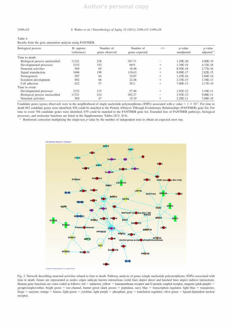
For the analysis of time to death, the relevant biologicalprocesses overrepresented in the PANTHER analysis weredevelopmental processes, neuronal activities, signal transduc-tion, neurogenesis, ectoderm development, and cell adhesion.For the analysis of time to incident event, developmental pro-cesses and neuronal activities were overrepresented amongother biological process (Table 4). The analyses also high-lighted the Wnt signaling pathway. The Wnt signaling path-way is ubiquitous and known to be involved in cancer butalso plays an important role in the early stages of thedevelopment of the central nervous system, in synapticformation by axon guidance, and in modulating fibrosisduring muscle repair scored high in both traits under study(Brack et al., 2007; Inestrosa and Arenas, 2010; Keeble etal., 2006; Ulloa and Martí, 2010). For extended tables seeSupplementary Tables S13 and S14. In addition, Ingenuityidentified 1 network with p � 10�31 containing 26 genesinvolved in processes related to nervous system develop-ment and function for the analysis of time to death (Fig. 2)and 1 network with p � 10�40 containing 28 genes involvedin cellular function and development for time to event(Supplementary Fig. S3).
IPA analysis highlighted the following genes associatedwith the time to death trait: NTRK2 (neurotrophic tyrosinekinase, receptor, type 2)—a member of the neurotrophictyrosine receptor kinase family. This kinase is a membrane-bound receptor that, upon neurotrophin binding, phospho-rylates itself and members of the mitogen-activated proteinkinase (MAPK) pathway. Signaling through this kinaseleads to cell differentiation. Second in line were NCAM1(neural cell adhesion molecule 1)—a cytoskeletal bindingT
able
2T
op8
SNP
(p-v
alue
�10
-5)
asso
ciat
ions
from
met
a-an
alys
isof
8co
hort
sfo
rtim
eto
even
t,ra
nked
byp-
valu
e(n
�16
,995
with
7314
even
ts)
Num
ber
SNP
Chr
Posi
tion
Clo
sest
refe
renc
eG
ene
Dis
tanc
e(b
p)fr
omcl
oses
tge
neC
oded
alle
leN
onco
ded
alle
leFr
eque
ncy
code
dal
lele
HR
p-va
lue
Stud
yef
fect
dire
ctio
na
Num
ber
ofsu
ppor
ting
SNPs
1rs
1041
2199
1938
7877
1A
TC
AY
307
AG
0.33
0.91
3.02
E-0
6–?
-��
--6
2rs
1685
2912
317
0169
370
ME
CO
M11
4610
TC
0.08
1.18
3.37
E-0
6�
��
�-�
-�72
3rs
8001
976
1347
2857
23SU
CL
A2
1290
69T
C0.
441.
093.
43E
-06
��
�-�
�--
173
4rs
1116
2963
180
5071
69E
LT
D1
1262
086
TC
0.63
1.09
4.15
E-0
6�
��
��
��
�40
5rs
4764
043
1214
0067
49G
RIN
2B17
570
TC
0.08
1.17
6.10
E-0
6�
��
��
��
�2
6rs
3112
530
515
2619
870
GR
IA1
2306
28A
G0.
080.
856.
79E
-06
—--
----
-�--
-13
07
rs10
2024
972
2379
3563
3C
OL
6A3
3823
3A
C0.
140.
898.
22E
-06
—--
---
368
rs23
6772
51
4398
8415
ST3G
AL
342
611
TC
0.42
1.08
9.31
E-0
6�
��
��
�-
�11
9
p-va
lues
are
for
the
inve
rse
vari
ance
-wei
ghte
dm
eta-
anal
ysis
.Dis
tanc
esto
gene
sar
egi
ven
inba
sepa
irs.
Posi
tion
isfo
rN
CB
IB
uild
36.H
Rs
are
for
each
addi
tiona
lcod
edal
lele
.Num
ber
ofsu
ppor
ting
SNPs
isth
enu
mbe
rof
SNPs
with
in50
0ki
loba
sepa
irof
the
top
SNP
that
are
inL
Dw
ithth
eto
pSN
Pin
the
Hap
Map
CE
Ure
leas
e22
(r2
�0.
10)
and
have
asso
ciat
ion
p-va
lue
�0.
05.
For
info
rmat
ion
onal
lSN
Pas
soci
atio
nsw
ithp-
valu
e�
10-4
see
Supp
lem
enta
ryT
able
S12.
Key
:bp
,ba
sepa
ir;
Chr
,ch
rom
osom
e;H
R,
haza
rdra
tio;
LD
,lin
kage
dise
quili
briu
m;
SNP,
sing
lenu
cleo
tide
poly
mor
phis
m.
aSt
udy-
spec
ific
info
rmat
ion
ispr
esen
ted
inth
eor
der:
RS,
CH
S,FH
S,A
RIC
,AG
ES,
HA
BC
,BL
SA,I
nCH
IAN
TI;
“�”
�co
ded
alle
lein
crea
ses
risk
ofev
ent;
“-”
�co
ded
alle
lede
crea
ses
risk
ofev
ent;
“?”
�no
tte
sted
.
2109.e20 S. Walter et al. / Neurobiology of Aging 32 (2011) 2109.e15–2109.e28

Author's personal copy
Tab
le3
Ove
rlap
betw
een
the
asso
ciat
ions
oftim
eto
deat
han
dtim
eto
even
ta
Top
hit
SNP
Chr
Clo
sest
refe
renc
ege
neT
ime
tode
ath
Tim
eto
even
tT
opSN
Psfr
omtim
eto
deat
h(t
ime
toev
ent)
anal
ysis
asso
ciat
edw
ithdi
ffer
ent
p-va
lues
intim
eto
even
t(t
ime
tode
ath)
anal
ysis
pE
ffec
tp
Eff
ect
Tot
alp
�0.
05p
�0.
05p
�0.
01p
�0.
001
p�
0.00
01
Tim
eto
deat
h1
rs14
2560
93
OT
OL
11.
46E
-06
�0.
0057
04�
1119
693
235
132
3722
2rs
7669
0312
BIN
21.
61E
-06
�0.
0131
5�
3727
45
01
3rs
1204
2640
1A
TG
4C1.
71E
-06
�0.
0370
1�
9360
194
010
4rs
1158
2903
1L
MO
43.
94E
-06
�0.
7336
�13
391
812
211
5rs
1025
9086
7O
RC
5L5.
16E
-06
�0.
0326
6�
239
154
5621
44
6rs
2769
255
1K
CN
Q4
5.17
E-0
6�
0.01
322
�28
715
168
567
57
rs17
2915
466
LO
C34
0156
7.65
E-0
6�
0.01
624
�29
199
10
08
rs12
6061
0018
NE
TO
18.
72E
-06
�0.
0285
3�
2316
52
00
9rs
1274
214
11G
RA
MD
1B8.
87E
-06
�0.
0567
�10
139
2817
170
Tim
eto
even
t1
rs16
8529
123
ME
CO
M0.
0058
9�
3.37
E-0
6�
169
6749
492
22
rs80
0197
613
SUC
LA
20.
0147
3�
3.43
E-0
6�
433
198
9146
5939
3rs
4764
043
12G
RIN
2B0.
0017
�6.
10E
-06
�45
422
10
04
rs10
2024
972
CO
L6A
30.
0003
5�
8.22
E-0
6�
135
8327
129
45
rs23
6772
51
ST3G
AL
30.
0274
�9.
31E
-06
�45
931
756
3731
18
p-va
lues
are
for
the
inve
rse
vari
ance
-wei
ghte
dm
eta-
anal
ysis
.Tot
alre
pres
ents
the
num
ber
ofSN
Psin
time
tode
ath
(tim
eto
even
t)an
alys
isw
ithin
500
kilo
base
pair
ofSN
Psfr
omth
etim
eto
even
t(t
ime
tode
ath)
anal
ysis
that
are
inL
Dw
ithth
eto
pSN
Psfr
omth
etim
eto
deat
h(t
ime
toev
ent)
anal
ysis
inth
eH
apM
apC
EU
rele
ase
22(r
2�
0.10
)an
dha
veas
soci
atio
np-
valu
e�
0.05
.K
ey:
Chr
,ch
rom
osom
e;E
ffec
t,m
eta-
anal
ysis
dire
ctio
nof
effe
ct;
LD
,lin
kage
dise
quili
briu
m;
SNP,
sing
lenu
cleo
tide
poly
mor
phis
m.
aO
nly
SNPs
that
wer
eno
min
ally
sign
ifica
nt(p
�0.
05)
for
both
trai
tsar
esh
own.
2109.e21S. Walter et al. / Neurobiology of Aging 32 (2011) 2109.e15–2109.e28
Author's personal copy
Table 4Results from the gene annotation analysis using PANTHER
Biological process H. sapiens(reference)
Number ofgenes observed
Number ofgenes expected
�/� p-valueunadjusted
p-valueadjusted a
Time to death:Biological process unclassified 11321 238 367.71 � 1.29E-20 4.00E-19Developmental processes 2152 152 69.9 � 1.39E-19 4.32E-18Neuronal activities 569 65 18.48 � 8.94E-18 2.77E-16Signal transduction 3406 199 110.63 � 9.09E-17 2.82E-15Neurogenesis 587 64 19.07 � 1.43E-16 2.84E-14Ectoderm development 692 68 22.48 � 2.33E-15 3.38E-13Cell adhesion 622 57 20.2 � 7.00E-12 2.17E-10
Time to event:Developmental processes 2152 115 57.46 � 1.02E-12 3.16E-11Biological process unclassified 11321 214 302.27 � 2.93E-12 9.08E-11Neuronal activities 569 47 15.19 � 2.28E-11 7.08E-10
Candidate genes (genes observed) were in the neighborhood of single nucleotide polymorphisms (SNPs) associated with p value � 1 � 10-3. For time todeath 862 candidate genes were identified; 826 could be matched to the Protein ANalysis THrough Evolutionary Relationships (PANTHER) gene list. Fortime to event 704 candidate genes were identified; 679 could be matched to the PANTHER gene list. Extended lists of PANTHER pathways, biologicalprocesses, and molecular functions are listed in the Supplementary Tables (S13, S14).
a Bonferroni correction multiplying the single-test p-value by the number of independent tests to obtain an expected error rate.
Fig. 2. Network describing neuronal activities related to time to death. Pathway analysis of genes (single nucleotide polymorphisms; SNPs) associated withtime to death. Genes are represented as nodes; edges indicate known interactions (solid lines depict direct and hatched lines depict indirect interaction).Human gene functions are color-coded as follows: red � unknown, yellow � transmembrane receptor and G protein coupled receptor, magenta (pink-purple) �group/complex/other, bright green � ion channel, hunter green (dark green) � peptidase, navy blue � transcription regulator, light blue � transporter,beige � enzyme, orange � kinase, light green � cytokine, light purple � phosphate, gray � translation regulator, olive green � ligand-dependent nuclearreceptor.
2109.e22 S. Walter et al. / Neurobiology of Aging 32 (2011) 2109.e15–2109.e28
Author's personal copy
protein, GRID2 (glutamate receptor, ionotropic, delta 2)—arelatively new member of the family of ionotropic gluta-mate receptors which are the predominant excitatory neu-rotransmitter receptors in the mammalian brain, and have arole in neuronal apoptotic death, and RIMS1 (regulatingsynaptic membrane exocytosis 1), which regulates synapticvesicle exocytosis and may be part of the protein scaffold ofthe cell.
Among the genes that were highlighted through the IPAanalysis in the analysis of time to event was MYC (v-mycmyelocytomatosis viral oncogene homolog)—a multifunc-tional, nuclear phosphoprotein that plays a role in cell cycleprogression, apoptosis, and cellular transformation. MYCfunctions as a transcription factor that regulates transcrip-tion of specific target genes. Second in line were E2F1 (E2Ftranscription factor 1), EGFR (epidermal growth factor re-ceptor), and CEBPA (CCAAT/enhancer binding protein [C/EBP], alpha). EF21, a transcription factor, plays a crucialrole in the control of cell cycle and action of tumor sup-pressor proteins can mediate both cell proliferation andp53-dependent/independent apoptosis. EGFR is a trans-membrane glycoprotein that serves as a receptor for mem-bers of the epidermal growth factor family and supports cellproliferation. CEBP-Alpha, a bZIP transcription factor, canbind as a homodimer to certain promoters and enhancers.CEBPA also forms heterodimers with the related proteinsCEBP-beta and CEBP-gamma and modulates the expres-sion of leptin, interacts with CDK2 and CDK4, and therebyinhibits these kinases and causes growth arrest in culturedcells.
4. Discussion
In our analyses of over 25,000 individuals of 55 yearsand older followed for an average of 11 years, we did notidentify genome-wide significant associations for all-causemortality and survival free of major diseases. However,both traits highlighted loci with suggestive significance thatwere in the neighborhood of genes related to neural regu-lation. In addition, our pathway and network analyses iden-tified an enrichment of genes associated with cellular andneural development and function, and cell communicationthat may contribute to variation in human aging. Braindevelopment might be responsible for the creation of redun-dancy in brain circuitry, which is associated with functionalreserve and resiliency. Brain function regulates most of thecompensatory strategy supporting maintenance of homeo-static equilibrium. Both of these processes are essential tohealthy aging and longevity.
Several explanations are possible for the lack of genome-wide significant findings. First, mortality is arguably 1 ofthe most complex phenotypes, and several trajectories to-ward extreme old age have been identified (Evert et al.,2003). Multiple genes could mediate the aging process butwould have their effects through numerous different patho-
physiological processes and diseases that act as intermediatefactors on the pathway to death (de Magalhaes et al., 2010).Therefore, any common variation in genes associated withaging probably has a small effect.
Second, the largely negative findings of this and otherstudies contrast with the intriguing animal studies of lon-gevity. Very large effects of single genes on lifespan haveindeed been observed in laboratory animals, but humansoften have several homologues of these genes which mightsignificantly differ in function or compensate for mutatedgenes through redundant mechanisms (Kuningas et al.,2008). This could explain why our top findings did notinclude genes in these pathways found in animal models.Animal models also represent genetically homogenous pop-ulations and are exposed to controlled environmental influ-ences. The lack of replication of animal model findings inhumans suggests that the use of knockout animals may notprovide the optimal approach to understanding the variationin survival in humans as interactions with environmentalfactors may obscure the associations and prevent the iden-tification of loci in humans.
Third, our study is based on common genetic variantsand therefore we cannot exclude effects due to low fre-quency and rare variants (� 5%) or due to the presence ofstructural variation, such as copy number polymorphisms.Our discovery set may lack the power to identify all therelevant loci, even though we had sufficient power to detectcommon SNPs (minor allele frequency � 5% or more) witha relative risk of 1.10 (Supplementary Table S2).
Last, we cannot exclude that phenotypic heterogeneityinfluenced our findings. While all cohorts had prospectivelycollected information on major health events and diagnoses,heterogeneity in the methods of assessment and classifica-tion might have limited the ability to identify true effects.
Complex diseases may result from the effects of a largenumber of low frequency variants, with substantial allelicheterogeneity at disease-causing loci (Pritchard, 2001;Pritchard and Cox, 2002; Swarbrick and Vaisse, 2003).Theoretical modeling that incorporates mutation, randomgenetic drift, and purifying selection suggests that many ofthe variants that affect complex traits may be in the 1%–5%frequency range (Pritchard, 2001). Indeed, sequencing ofcandidate genes in an attempt to capture such low frequencyvariants, has led to the identification of rare variants withmodest effects on body mass index (Ahituv et al., 2007;Challis et al., 2002; Cone, 2000), triglyceride levels (Romeoet al., 2007), high-density lipoprotein (HDL; Cohen et al.,2004; Romeo et al., 2007) and low-density lipoprotein(LDL) cholesterol levels (Cohen et al., 2005, 2006; Ko-towski et al., 2006).
It is impossible to determine the functional variant of agene by GWAS. Moreover, we cannot conclude from thelocation of an SNP that this variation is involved in theexpression of the closest gene. However, our top resultssuggested a possible role of genes involved in neurological
2109.e23S. Walter et al. / Neurobiology of Aging 32 (2011) 2109.e15–2109.e28

Author's personal copy
processes in human longevity and aging. Ten of the 22suggestive associations identified in our analyses are in ornear genes that are highly expressed in the brain (HECW2[Rotin and Kumar, 2009], HIP1 [Blanpied et al., 2003],BIN2, GRIA1), were previously related to the regulation ofneuronal excitability and plasticity (KCNQ4 [Van Eyken etal., 2006], LMO4 [Joshi et al., 2009; Leuba et al., 2004],GRIA1), and the maintenance of neural circuitry and syn-aptic plasticity (NETO1), or are associated with neurologi-cal diseases such as Alzheimer’s disease (LMO4 [Leuba etal., 2004], BIN2, GRIA1, GRIN2B), and amyotrophic lat-eral sclerosis (GRIN2B). In addition, 6 of the 22 SNPs werein close proximity to genes associated with other pheno-types of aging such as autophagy (ATG4C [Kenyon, 2010]),cancer (ATG4C [Maiuri et al., 2009], HIP1 [Bradley et al.,2007], HECW2 [Rotin and Kumar, 2009], VWA5A [Zhou etal., 2009], MECOM), and mitochondrial depletion syn-drome (SUCLA2). Notably, BIN2, ATG4C, KCNQ4,MECOM, and SUCLA2 showed associations with both traitsin our study.
Using the expression quantitative trait loci (eQTL)browser (eqtl.uchicago.edu/cgi-bin/gbrowse/eqtl/) we de-tected eQTL associated with HIP1, COL5A1, LOC340156,and SMARCA2 in time to death only.
Interestingly, SNPs known to be associated with longev-ity and disease in the neighborhood of APOE (Deelen et al.,2011) or FOXO3A (Flachsbart et al., 2009; Willcox et al.,2008) only reached nominal significance (results notshown). These genes were originally identified in studies ofcentenarians; it is possible that our study of cohorts com-prised of individuals from the general populations did nothave sufficient statistical power to identify these genes withcertainty. (Tan et al., 2008).
While meta-analysis of GWAS has the power to detectsmall changes of allele frequencies between groups with theanalyzed trait, true association signals may not be revealedbased on a stringent genome-wide significance threshold.This situation, although limiting false positive findings, per-forms poorly in identifying false negatives as they may fallbelow the threshold. Network analyses using a less stringentsignificance threshold do not amend the overall negativefinding of this study. However, it is well-recognized thatwithin the many associations that failed to attain this levelof significance lie true positive associations. Network anal-yses can provide useful information exploring multiple geneeffects and their interactions.
In fact the interpretation of most GWAS results is diffi-cult because individual results may involve many seeminglyunrelated genes. Because PANTHER and IPA are built ondifferent conceptual approaches, database sources and dif-ferent pathway classifications, they can be seen as comple-mentary approaches. Our pathway and network analyseshighlighted neuronal activities and organism developmentalprocesses as major biological processes involved aging. Inaddition, it highlighted Wnt signaling and showed that those
genes that were involved in most pathways indeed hadsubstantial effects within the analyzed trait. NTRK2 (Rico etal., 2002), NCAM1 (Rutishauser et al., 1988), GRID2 (Hiraiet al., 2003), and RIMS1 (Johnson et al., 2003; Schoch et al.,2002) are associated with neuronal development and diseasepathways that were highlighted in the analysis of time todeath. MYC (Cole, 1986; Goga et al., 2007), E2F1 (Nevins,2001), EGFR (Wang et al., 2004), and CEBPA (Ménard etal., 2002; Wang et al., 2001) are associated with “cancer,”“cell function,” and “development” pathways.
Few if any of the top hits from the GWAS wereinvolved in common pathways of aging, typically ad-dressed in candidate gene studies. For example, there wasno specific evidence for genes involved in IGF-insulinsignaling. However, this negative finding cannot be in-terpreted as evidence against the importance of IGF-insulin signaling, as well as other processes such asinflammation, oxidative stress, cellular damage and re-pair, growth hormone, and cell proliferation in aging.Moreover, it is possible that polymorphisms in relatedgenes have an effect in the oldest old, who were repre-sented by fewer numbers in our study population suchthat our study design would be underpowered to detect it.It is also conceivable that the neurological pathwaysidentified by our analysis interact with the known candi-date genes involved in aging (Bishop et al., 2010; Finchand Ruvkun, 2001). It is feasible that the traditional agingpathways are hierarchically controlled by neurons andthat the brain might be the location coordinating physi-ological changes (Bishop et al., 2010; Finch and Ruvkun,2001). Because neurons are particularly susceptible todamage caused by reactive oxygen species, limitations incellular maintenance and repair might reinforce thesepathways and accelerate aging (Finch and Ruvkun,2001). An increased ability of neuronal cells to prevent orrepair oxidative damage might result in beneficial hor-monal signaling, otherwise deregulated with age, thusdelaying the onset of age-related disease and directlyregulating cognitive aging and life span (Bishop et al.,2010; Cutler and Mattson, 2006; de Magalhães and Sand-berg, 2005).
In conclusion, our analysis did provide suggestive evi-dence that aging is under neuronal control. Unfortunately,we have no relevant tissue or expression experiment avail-able to further underscore or validate our findings. Futureinvestigations of changes of gene expression with age atcellular and population levels are warranted.
Disclosure statement
The authors declare that no competing interests exist.All participants included in this analysis provided written
informed consent.
2109.e24 S. Walter et al. / Neurobiology of Aging 32 (2011) 2109.e15–2109.e28
Author's personal copy
Replication Samples
Whitehall II: Whitehall II has been supported by grantsfrom the Medical Research Council; Economic and SocialResearch Council; British Heart Foundation; Health andSafety Executive; Department of Health; National HeartLung and Blood Institute (HL36310), US NIH NationalInstitute on Aging (AG13196), US NIH Agency for HealthCare Policy Research (HS06516); and the John D. andCatherine T. MacArthur Foundation Research Networks onSuccessful Midlife Development and Socio-economic Sta-tus and Health.
English Longitudinal Study of Aging: Samples from theEnglish Longitudinal Study of Ageing (ELSA) DNA Re-pository (EDNAR), received support under a grant(AG1764406S1) awarded by the National Institute on Age-ing (NIA). ELSA was developed by a team of researchersbased at the National Centre for Social Research, UniversityCollege London and the Institute of Fiscal Studies. The datawere collected by the National Centre for Social Research.
Religious Order Study: Grants P30AG10161,R01AG15819, and R01AG30146 from the National Insti-tute on Aging, and the Translation Genomics ResearchInstitute.
Memory and Aging Project: Grants R01AG17917 andR01AG15819 from the National Instiute on Aging, and theTranslation Genomics Research Institute.
The Longevity Consortium, funded by the National In-stitute of Aging, grant number U19 AG023122, providedadministrative resources to CHARGE investigators for thisphenotype as well as scientific opportunity funds for denovo genotyping of the 5 selected SNPs in the ELSA andWhitehall II replication studies.
Acknowledgements
Rotterdam Study: The Rotterdam Study is supported byNetherlands Genomics Initiative/Netherlands Consortiumfor Healthy Aging (050-060-810); Netherlands Organisa-tion for Scientific Research (NWO) (904-61-090, 904-61-193, 480-04-004, 400-05-717, SPI 56-464-1419, 175.010.2005.011, 911-03-012 and 017.106.370); Netspar—Livinglonger for a good health; the Erasmus Medical Center, andErasmus University in Rotterdam; the Netherlands Organi-zation for Health Research and Development, the Nether-lands Research Institute for Diseases in the Elderly; theDutch Ministry of Education, Culture and Science, and theMinistry for Health, Welfare and Sports; the EuropeanCommission; and the Municipality of Rotterdam, the Neth-erlands.
Cardiovascular Health Study: The CHS research re-ported in this article was supported by contract numbersN01-HC-85079 through N01-HC-85086, N01-HC-35129,N01 HC-15103, N01 HC-55222, N01-HC-75150, N01-HC-45133, grant numbers U01 HL080295 and R01 HL087652from the National Heart, Lung, and Blood Institute, the
National Institute of Aging R01 AG031890 with additionalcontribution from the National Institute of NeurologicalDisorders and Stroke. A full list of principal CHS investi-gators and institutions can be found at www.chs-nhlbi.org/pi.htm. DNA handling and genotyping was supported inpart by National Center for Research Resources grant M01-RR00425 to the Cedars-Sinai General Clinical ResearchCenter Genotyping core and National Institute of Diabetesand Digestive and Kidney Diseases grant DK063491 to theSouthern California Diabetes Endocrinology Research Cen-ter.
Framingham Heart Study: Phenotype-genotype analy-ses were supported by the National Institute of Aginggrant number R01AG029451 (PI: JMM). The Framing-ham Heart Study of the National Heart Lung and BloodInstitute of the National Institutes of Health and BostonUniversity School of Medicine were supported by theNational Heart, Lung and Blood Institute’s FraminghamHeart Study Contract No. N01-HC-25195 and its contractwith Affymetrix, Inc., for genotyping services (ContractNo. N02-HL-6-4278). Analyses reflect intellectual inputand resource development from the Framingham HeartStudy investigators participating in the SNP Health As-sociation Resource (SHARe) project. A portion of thisresearch was conducted using the Linux Cluster for Ge-netic Analysis (LinGA-II) funded by the Robert DawsonEvans Endowment of the Department of Medicine atBoston University School of Medicine and Boston Med-ical Center. Dr. Kiel’s effort as well as all hip fracturedata from the Framingham Osteoporosis Study was sup-ported by a grant from the National Institute of Arthritis,Musculoskeletal and Skin Diseases and the National In-stitute on Aging; R01 AR/AG 41398. This research wasadditionally supported by the following grants:AG033193, AG081220, NS17950, P30AG013846, 1R01-AG028321.
Atherosclerosis Risk in Communities Study: The Ath-erosclerosis Risk in Communities Study is carried out as acollaborative study supported by National Heart, Lung, andBlood Institute contracts N01-HC-55015, N01-HC-55016,N01-HC-55018, N01-HC-55019, N01-HC-55020, N01-HC-55021, and N01-HC-55022. The authors thank the staffand participants of the ARIC study for their importantcontributions.
Age, Gene/Environment Susceptibility -ReykjavikStudy: The Age, Gene/Environment Susceptibility-Reykja-vik Study is funded by NIH contract N01-AG-12100, theNIA Intramural Research Program, Hjartavernd (the Icelan-dic Heart Association), and the Althingi (the Icelandic Par-liament). Genotyping was conducted at the NIA IRP Lab-oratory of Neurogenetics.
Invecchiare nel Chianti: The InCHIANTI study baseline(1998–2000) was supported as a “targeted project”(ICS110.1/RF97.71) by the Italian Ministry of Health and in
2109.e25S. Walter et al. / Neurobiology of Aging 32 (2011) 2109.e15–2109.e28

Author's personal copy
part by the U.S. National Institute on Aging (Contracts: 263MD 9164 and 263 MD 821336).
Baltimore Longitudinal Study of Aging: The BLSA wassupported in part by the Intramural Research Program of theNIH, National Institute on Aging. A portion of that supportwas through an R&D contract with MedStar Research In-stitute.
Health, Aging and Body Composition: This research issupported in part by the Intramural Research Program of theNIH, National Institute on Aging. This research was sup-ported by NIA contracts N01AG62101, N01AG62103,N01AG62106 and NIA grant 1R03AG032498-01. The ge-nome-wide association study was funded by NIA grant1R01AG032098-01A1 to Wake Forest University HealthSciences and genotyping services were provided by theCenter for Inherited Disease Research (CIDR). CIDR isfully funded through a federal contract from the NationalInstitutes of Health to The Johns Hopkins University, con-tract number HHSN268200782096C.
Study of Health in Pomerania: SHIP is part of the Com-munity Medicine Research net of the University of Greif-swald, Germany, which is funded by the Federal Ministry ofEducation and Research (grants no. 01ZZ9603, 01ZZ0103,and 01ZZ0403), the Ministry of Cultural Affairs as well asthe Social Ministry of the Federal State of Mecklenburg-West Pomerania. Genome-wide data have been supportedby the Federal Ministry of Education and Research (grantno. 03ZIK012) and a joint grant from Siemens Healthcare,Erlangen, Germany and the Federal State of Mecklenburg,West Pomerania. The University of Greifswald is a memberof the “Center of Knowledge Interchange” program of theSiemens AG.
Appendix. Supplementary data
Supplementary data associated with this article can befound, in the online version, at doi:10.1016/j.neurobiolaging.2011.05.026.
References
Ahituv, N., Kavaslar, N., Schackwitz, W., Ustaszewska, A., Martin, J.,Hebert, S., Doelle, H., Ersoy, B., Kryukov, G., Schmidt, S., Yosef, N.,Ruppin, E., Sharan, R., Vaisse, C., Sunyaev, S., Dent, R., Cohen, J.,McPherson, R., Pennacchio, L.A., 2007. Medical sequencing at theextremes of human body mass. Am. J. Hum. Genet. 80, 779–791.
Anselmi, C.V., Malovini, A., Roncarati, R., Novelli, V., Villa, F., Con-dorelli, G., Bellazzi, R., Puca, A.A., 2009. Association of the FOXO3Alocus with extreme longevity in a southern Italian centenarian study.Rejuvenation Res. 12, 95–104.
Atzmon, G., Schechter, C., Greiner, W., Davidson, D., Rennert, G., Bar-zilai, N., 2004. Clinical phenotype of families with longevity. J. Am.Geriatr. Soc. 52, 274–277.
Bishop, N.A., Lu, T., Yankner, B.A., 2010. Neural mechanisms of ageingand cognitive decline. Nature 464, 529–535.
Blanpied, T.A., Scott, D.B., Ehlers, M.D., 2003. Age-related regulation ofdendritic endocytosis associated with altered clathrin dynamics. Neu-robiol. Aging 24, 1095–1104.
Brack, A.S., Conboy, M.J., Roy, S., Lee, M., Kuo, C.J., Keller, C., Rando,T.A., 2007. Increased Wnt signaling during aging alters muscle stemcell fate and increases fibrosis. Science 317, 807–810.
Bradley, S.V., Holland, E.C., Liu, G.Y., Thomas, D., Hyun, T.S., Ross,T.S., 2007. Huntingtin interacting protein 1 is a novel brain tumormarker that associates with epidermal growth factor receptor. CancerRes. 67, 3609–3615.
Challis, B.G., Pritchard, L.E., Creemers, J.W., Delplanque, J., Keogh, J.M.,Luan, J., Wareham, N.J., Yeo, G.S., Bhattacharyya, S., Froguel, P.,White, A., Farooqi, I.S., O’Rahilly, S., 2002. A missense mutationdisrupting a dibasic prohormone processing site in pro-opiomelanocor-tin (POMC) increases susceptibility to early-onset obesity through anovel molecular mechanism. Hum. Mol. Genet. 11, 1997–2004.
Christensen, K., Johnson, T.E., Vaupel, J.W., 2006. The quest for geneticdeterminants of human longevity: challenges and insights. Nat. Rev.Genet. 7, 436–448.
Cohen, J., Pertsemlidis, A., Kotowski, I.K., Graham, R., Garcia, C.K.,Hobbs, H.H., 2005. Low LDL cholesterol in individuals of Africandescent resulting from frequent nonsense mutations in PCSK9. Nat.Genet. 37, 161–165.
Cohen, J.C., Kiss, R.S., Pertsemlidis, A., Marcel, Y.L., McPherson, R.,Hobbs, H.H., 2004. Multiple rare alleles contribute to low plasmalevels of HDL cholesterol. Science 305, 869–872.
Cohen, J.C., Pertsemlidis, A., Fahmi, S., Esmail, S., Vega, G.L., Grundy,S.M., Hobbs, H.H., 2006. Multiple rare variants in NPC1L1 associatedwith reduced sterol absorption and plasma low-density lipoproteinlevels. Proc. Natl. Acad. Sci. U. S. A. 103, 1810–1815.
Cole, M.D., 1986. The myc oncogene: its role in transformation anddifferentiation. Annu. Rev. Genet. 20, 361–384.
Cone, R.D., 2000. Haploinsufficiency of the melanocortin-4 receptor: partof a thrifty genotype? J. Clin. Invest. 106, 185–187.
Cutler, R.G., Mattson, M.P., 2006. The adversities of aging. Ageing Res.Rev. 5, 221–238.
de Bakker, P.I., Ferreira, M.A., Jia, X., Neale, B.M., Raychaudhuri, S.,Voight, B.F., 2008. Practical aspects of imputation-driven meta-anal-ysis of genome-wide association studies. Hum. Mol. Genet. 17, R122–R128.
de Magalhaes, J.P., Budovsky, A., Lehmann, G., Costa, J., Li, Y., Fraifeld,V., Church, G.M., 2009a. The Human Ageing Genomic Resources:online databases and tools for biogerontologists. Aging Cell 8, 65–72.
de Magalhaes, J.P., Finch, C.E., Janssens, G., 2010. Next-generation se-quencing in aging research: Emerging applications, problems, pitfallsand possible solutions. Ageing Res Rev 9, 315–323.
de Magalhães, J.P., Sandberg, A., 2005. Cognitive aging as an extension ofbrain development: a model linking learning, brain plasticity, andneurodegeneration. Mech. Ageing Dev. 126, 1026–1033.
Deelen, J., Beekman, M., Uh, H.W., Helmer, Q., Kuningas, M., Christian-sen, L., Kremer, D., van de Breggen, R., Suchiman, H.E., Lakenberg,N., van den Akker, E.B., Passtoors, W.M., Tiemeier, H., van Heemst,D., de Craen, A.J., Rivadeneira, F., de Geus, E.J., Perola, M., van derOuderaa, F.J., Gunn, D.A., Boomsma, D.I., Uitterlinden, A.G., Chris-tensen, K., van Duijn, C.M., Heijmans, B.T., Houwing-Duistermaat,J.J., Westendorp, R.G., Slagboom, P.E., 2011. Genome-wide associa-tion study identifies a single major locus contributing to survival intoold age; the APOE locus revisited. Aging Cell doi:10.1111/j.1474-9726.2011.00705.x.
Evert, J., Lawler, E., Bogan, H., Perls, T., 2003. Morbidity profiles ofcentenarians: survivors, delayers, and escapers. J. Gerontol. A Biol.Sci. Med. Sci. 58, 232–237.
Ewbank, D.C., 2007. Differences in the association between apolipoproteinE genotype and mortality across populations. J. Gerontol. A Biol. Sci.Med. Sci. 62, 899–907.
Finch, C.E., Ruvkun, G., 2001. The genetics of aging. Annu. Rev. Genom-ics Hum. Genet. Genet. 2, 435–462.
Flachsbart, F., Caliebe, A., Kleindorp, R., Blanché, H., von Eller-Eberstein,H., Nikolaus, S., Schreiber, S., Nebel, A., 2009. Association of
2109.e26 S. Walter et al. / Neurobiology of Aging 32 (2011) 2109.e15–2109.e28

Author's personal copy
FOXO3A variation with human longevity confirmed in Germancentenarians. Proc. Natl. Acad. Sci. U. S. A. 106, 2700–2705.
Gerdes, L.U., Jeune, B., Ranberg, K.A., Nybo, H., Vaupel, J.W., 2000.Estimation of apolipoprotein E genotype-specific relative mortalityrisks from the distribution of genotypes in centenarians and middle-aged men: apolipoprotein E gene is a “frailty gene,” not a “longevitygene.Genet. Epidemiol. 19, 202–210.
Goga, A., Yang, D., Tward, A.D., Morgan, D.O., Bishop, J.M., 2007.Inhibition of CDK1 as a potential therapy for tumors over-expressingMYC. Nat. Med. 13, 820–827.
Herskind, A.M., McGue, M., Holm, N.V., Sørensen, T.I., Harvald, B.,Vaupel, J.W., 1996. The heritability of human longevity: a population-based study of 2872 Danish twin pairs born 1870–1900. Hum. Genet.97, 319–323.
Hirai, H., Launey, T., Mikawa, S., Torashima, T., Yanagihara, D., Kasaura,T., Miyamoto, A., Yuzaki, M., 2003. New role of delta2-glutamatereceptors in AMPA receptor trafficking and cerebellar function. Nat.Neurosci. 6, 869–876.
Inestrosa, N.C., Arenas, E., 2010. Emerging roles of Wnts in the adultnervous system. Nat. Rev. Neurosci. 11, 77–86.
Johnson, S., Halford, S., Morris, A.G., Patel, R.J., Wilkie, S.E., Hardcastle,A.J., Moore, A.T., Zhang, K., Hunt, D.M., 2003. Genomic organisationand alternative splicing of human RIM1, a gene implicated in auto-somal dominant cone-rod dystrophy (CORD7). Genomics 81, 304–314.
Joshi, K., Lee, S., Lee, B., Lee, J.W., Lee, S.K., 2009. LMO4 controls thebalance between excitatory and inhibitory spinal V2 interneurons. Neu-ron 61, 839–851.
Keeble, T.R., Halford, M.M., Seaman, C., Kee, N., Macheda, M., Ander-son, R.B., Stacker, S.A., Cooper, H.M., 2006. The Wnt receptor Ryk isrequired for Wnt5a-mediated axon guidance on the contralateral side ofthe corpus callosum. J. Neurosci. 26, 5840–5848.
Kenyon, C.J., 2010. The genetics of ageing. Nature 464, 504–512.Kotowski, I.K., Pertsemlidis, A., Luke, A., Cooper, R.S., Vega, G.L.,
Cohen, J.C., Hobbs, H.H., 2006. A spectrum of PCSK9 alleles con-tributes to plasma levels of low-density lipoprotein cholesterol. Am. J.Hum. Genet. 78, 410–422.
Kuningas, M., Mooijaart, S.P., van Heemst, D., Zwaan, B.J., Slagboom,P.E., Westendorp, R.G., 2008. Genes encoding longevity: from modelorganisms to humans. Aging Cell 7, 270–280.
Leuba, G., Vernay, A., Vu, D., Walzer, C., Belloir, B., Kraftsik, R., Bouras,C., Savioz, A., 2004. Differential expression of LMO4 protein inAlzheimer’s disease. Neuropathol. Appl. Neurobiol. 30, 57–69.
Levy, D., Ehret, G.B., Rice, K., Verwoert, G.C., Launer, L.J., Dehghan, A.,Glazer, N.L., Morrison, A.C., Johnson, A.D., Aspelund, T., Aulchenko,Y., Lumley, T., Kottgen, A., Vasan, R.S., Rivadeneira, F., Eiriksdottir,G., Guo, X., Arking, D.E., Mitchell, G.F., Mattace-Raso, F.U., Smith,A.V., Taylor, K., Scharpf, R.B., Hwang, S.J., Sijbrands, E.J., Bis, J.,Harris, T.B., Ganesh, S.K., O’Donnell, C.J., Hofman, A., Rotter, J.I.,Coresh, J., Benjamin, E.J., Uitterlinden, A.G., Heiss, G., Fox, C.S.,Witteman, J.C., Boerwinkle, E., Wang, T.J., Gudnason, V., Larson,M.G., Chakravarti, A., Psaty, B.M., van Duijn, C.M., 2009. Genome-wide association study of blood pressure and hypertension. Nat Genet41, 677–687.
Li, Y., Wang, W.J., Cao, H., Lu, J., Wu, C., Hu, F.Y., Guo, J., Zhao, L.,Yang, F., Zhang, Y.X., Li, W., Zheng, G.Y., Cui, H., Chen, X., Zhu, Z.,He, H., Dong, B., Mo, X., Zeng, Y., Tian, X.L., 2009a. Geneticassociation of FOXO1A and FOXO3A with longevity trait in HanChinese populations. Hum. Mol. Genet. 18, 4897–4904.
Li, Y., Willer, C., Sanna, S., Abecasis, G., 2009b. Genotype imputation.Annu. Rev. Genomics Hum. Genet. 10, 387–406.
Lunetta, K.L., D’Agostino, R.B., Sr., Karasik, D., Benjamin, E.J., Guo,C.Y., Govindaraju, R., Kiel, D.P., Kelly-Hayes, M., Massaro, J.M.,Pencina, M.J., Seshadri, S., Murabito, J.M., 2007. Genetic correlates oflongevity and selected age-related phenotypes: a genome-wide associ-
ation study in the Framingham Study. BMC Med. Genet. 8 suppl 1,S13.
Maiuri, M.C., Tasdemir, E., Criollo, A., Morselli, E., Vicencio, J.M.,Carnuccio, R., Kroemer, G., 2009. Control of autophagy by oncogenesand tumor suppressor genes. Cell Death Differ. 16, 87–93.
McGue, M., Vaupel, J.W., Holm, N., Harvald, B., 1993. Longevity ismoderately heritable in a sample of Danish twins born 1870–1880. J.Gerontol. 48, B237–B244.
Ménard, C., Hein, P., Paquin, A., Savelson, A., Yang, X.M., Lederfein, D.,Barnabé-Heider, F., Mir, A.A., Sterneck, E., Peterson, A.C., Johnson,P.F., Vinson, C., Miller, F.D., 2002. An essential role for a MEK-C/EBP pathway during growth factor-regulated cortical neurogenesis.Neuron 36, 597–610.
Mi, H., Guo, N., Kejariwal, A., Thomas, P.D., 2007. PANTHER version 6:protein sequence and function evolution data with expanded represen-tation of biological pathways. Nucleic Acids Res. 35(Database issue),D247–D252.
Nevins, J.R., 2001. The Rb/E2F pathway and cancer. Hum. Mol. Genet. 10,699–703.
Newman, A.B., Walter, S., Lunetta, K.L., Garcia, M.E., Slagboom, P.E.,Christensen, K., Arnold, A.M., Aspelund, T., Aulchenko, Y.S., Benja-min, E.J., Christiansen, L., D’Agostino, R.B., Sr., Fitzpatrick, A.L.,Franceschini, N., Glazer, N.L., Gudnason, V., Hofman, A., Kaplan, R.,Karasik, D., Kelly-Hayes, M., Kiel, D.P., Launer, L.J., Marciante,K.D., Massaro, J.M., Miljkovic, I., Nalls, M.A., Hernandez, D., Psaty,B.M., Rivadeneira, F., Rotter, J., Seshadri, S., Smith, A.V., Taylor,K.D., Tiemeier, H., Uh, H.W., Uitterlinden, A.G., Vaupel, J.W., Wal-ston, J., Westendorp, R.G., Harris, T.B., Lumley, T., van Duijn, C.M.,Murabito, J.M., 2010. A meta-analysis of four genome-wide associa-tion studies of survival to age 90 years or older: The Cohorts for Heartand Aging Research in Genomic Epidemiology Consortium. J. Geron-tol. A Biol. Sci. Med. Sci. 65, 478–487.
Newton-Cheh, C., Johnson, T., Gateva, V., Tobin, M.D., Bochud, M., Coin,L., Najjar, S.S., Zhao, J.H., Heath, S.C., Eyheramendy, S., Papadakis, K.,Voight, B.F., Scott, L.J., Zhang, F., Farrall, M., Tanaka, T., Wallace, C.,Chambers, J.C., Khaw, K.T., Nilsson, P., van der Harst, P., Polidoro, S.,Grobbee, D.E., Onland-Moret, N.C., Bots, M.L., Wain, L.V., Elliott, K.S.,Teumer, A., Luan, J., Lucas, G., Kuusisto, J., Burton, P.R., Hadley, D.,McArdle, W.L., Brown, M., Dominiczak, A., Newhouse, S.J., Samani,N.J., Webster, J., Zeggini, E., Beckmann, J.S., Bergmann, S., Lim, N.,Song, K., Vollenweider, P., Waeber, G., Waterworth, D.M., Yuan, X.,Groop, L., Orho-Melander, M., Allione, A., Di Gregorio, A., Guarrera, S.,Panico, S., Ricceri, F., Romanazzi, V., Sacerdote, C., Vineis, P., Barroso,I., Sandhu, M.S., Luben, R.N., Crawford, G.J., Jousilahti, P., Perola, M.,Boehnke, M., Bonnycastle, L.L., Collins, F.S., Jackson, A.U., Mohlke,K.L., Stringham, H.M., Valle, T.T., Willer, C.J., Bergman, R.N., Morken,M.A., Doring, A., Gieger, C., Illig, T., Meitinger, T., Org, E., Pfeufer, A.,Wichmann, H.E., Kathiresan, S., Marrugat, J., O’Donnell, C.J., Schwartz,S.M., Siscovick, D.S., Subirana, I., Freimer, N.B., Hartikainen, A.L.,McCarthy, M.I., O’Reilly, P.F., Peltonen, L., Pouta, A., de Jong, P.E.,Snieder, H., van Gilst, W.H., Clarke, R., Goel, A., Hamsten, A., Peden,J.F., Seedorf, U., Syvanen, A.C., Tognoni, G., Lakatta, E.G., Sanna, S.,Scheet, P., Schlessinger, D., Scuteri, A., Dorr, M., Ernst, F., Felix, S.B.,Homuth, G., Lorbeer, R., Reffelmann, T., Rettig, R., Volker, U., Galan, P.,Gut, I.G., Hercberg, S., Lathrop, G.M., Zelenika, D., Deloukas, P.,Soranzo, N., Williams, F.M., Zhai, G., Salomaa, V., Laakso, M., Elosua,R., Forouhi, N.G., Volzke, H., Uiterwaal, C.S., van der Schouw, Y.T.,Numans, M.E., Matullo, G., Navis, G., Berglund, G., Bingham, S.A.,Kooner, J.S., Connell, J.M., Bandinelli, S., Ferrucci, L., Watkins, H.,Spector, T.D., Tuomilehto, J., Altshuler, D., Strachan, D.P., Laan, M.,Meneton, P., Wareham, N.J., Uda, M., Jarvelin, M.R., Mooser, V., Me-lander, O., Loos, R.J., Elliott, P., Abecasis, G.R., Caulfield, M., Munroe,P.B., 2009. Genome-wide association study identifies eight loci associatedwith blood pressure. Nat. Genet. 41, 666–676.
Pawlikowska, L., Hu, D., Huntsman, S., Sung, A., Chu, C., Chen, J.,Joyner, A.H., Schork, N.J., Hsueh, W.C., Reiner, A.P., Psaty, B.M.,
2109.e27S. Walter et al. / Neurobiology of Aging 32 (2011) 2109.e15–2109.e28
Author's personal copy
Atzmon, G., Barzilai, N., Cummings, S.R., Browner, W.S., Kwok,P.Y., Ziv, E., Study of Osteoporotic Fractures, 2009. Association ofcommon genetic variation in the insulin/IGF1 signaling pathway withhuman longevity. Aging Cell 8, 460–472.
Price, A.L., Patterson, N.J., Plenge, R.M., Weinblatt, M.E., Shadick, N.A.,Reich, D., 2006. Principal components analysis corrects for stratifica-tion in genome-wide association studies. Nat. Genet. 38, 904–909.
Pritchard, J.K., 2001. Are rare variants responsible for susceptibility tocomplex diseases? Am. J. Hum. Genet. 69, 124–137.
Pritchard, J.K., Cox, N.J., 2002. The allelic architecture of human diseasegenes: common disease-common variant. . .or not? Hum. Mol. Genet.11, 2417–2423.
Psaty, B.M., O’Donnell, C.J., Gudnason, V., Lunetta, K.L., Folsom, A.R.,Rotter, J.I., Uitterlinden, A.G., Harris, T.B., Witteman, J.C., Boer-winkle, E., CHARGE Consortium, 2009. Cohorts for Heart and AgingResearch in Genomic Epidemiology (CHARGE) Consortium: Designof prospective meta-analyses of genome-wide association studies from5 cohorts. Circ. Cardiovasc. Genet. 2,73–80.
Reed, T., Dick, D.M., 2003. Heritability and validity of healthy physicalaging (wellness) in elderly male twins. Twin Res. 6, 227–234.
Rico, B., Xu, B., Reichardt, L.F., 2002. TrkB receptor signaling is requiredfor establishment of GABAergic synapses in the cerebellum. Nat.Neurosci. 5, 225–233.
Romeo, S., Pennacchio, L.A., Fu, Y., Boerwinkle, E., Tybjaerg-Hansen,A., Hobbs, H.H., Cohen, J.C., 2007. Population-based resequencing ofANGPTL4 uncovers variations that reduce triglycerides and increaseHDL. Nat. Genet. 39, 513–516.
Rotin, D., Kumar, S., 2009. Physiological functions of the HECT family ofubiquitin ligases. Nat. Rev. Mol. Cell Biol. 10, 398–409.
Rutishauser, U., Acheson, A., Hall, A.K., Mann, D.M., Sunshine, J., 1988.The neural cell adhesion molecule (NCAM) as a regulator of cell-cellinteractions. Science 240, 53–57.
Schoch, S., Castillo, P.E., Jo, T., Mukherjee, K., Geppert, M., Wang, Y.,Schmitz, F., Malenka, R.C., Südhof, T.C., 2002. RIM1alpha forms aprotein scaffold for regulating neurotransmitter release at the activezone. Nature 415, 321–326.
Swarbrick, M.M., Vaisse, C., 2003. Emerging trends in the search forgenetic variants predisposing to human obesity. Curr. Opin. Clin. Nutr.Metab. Care 6, 369–375.
Tan, Q., Zhao, J.H., Zhang, D., Kruse, T.A., Christensen, K., 2008. Powerfor genetic association study of human longevity using the case-controldesign. Am. J. Epidemiol. 168, 890–896.
Thomas, P.D., Campbell, M.J., Kejariwal, A., Mi, H., Karlak, B., Daver-man, R., Diemer, K., Muruganujan, A., Narechania, A., 2003. PAN-
THER: a library of protein families and subfamilies indexed byfunction. Genome Res. 13, 2129–2141.
Thomas, P.D., Kejariwal, A., Guo, N., Mi, H., Campbell, M.J., Muruganu-jan, A., Lazareva-Ulitsky, B., 2006. Applications for protein sequence-function evolution data: mRNA/protein expression analysis and codingSNP scoring tools. Nucleic Acids Res. 34(Web Server issue), W645–650.
Ulloa, F., Martí, E., 2010. Wnt won the war: antagonistic role of Wnt overShh controls dorso-ventral patterning of the vertebrate neural tube.Dev. Dyn. 239, 69–76.
Van Eyken, E., Van Laer, L., Fransen, E., Topsakal, V., Lemkens, N.,Laureys, W., Nelissen, N., Vandevelde, A., Wienker, T., Van DeHeyning, P., Van Camp, G., 2006. KCNQ4: a gene for age-relatedhearing impairment? Hum. Mutat. 27, 1007–1016.
van Rijn, M.J., Schut, A.F., Aulchenko, Y.S., Deinum, J., Sayed-Taba-tabaei, F.A., Yazdanpanah, M., Isaacs, A., Axenovich, T.I.,Zorkoltseva, I.V., Zillikens, M.C., Pols, H.A., Witteman, J.C., Oostra,B.A., van Duijn, C.M., 2007. Heritability of blood pressure traits andthe genetic contribution to blood pressure variance explained by fourblood-pressure-related genes. J. Hypertens. 25, 565–570.
Vaupel, J.W., 1997. The remarkable improvements in survival at olderages. Philos. Trans. R. Soc. Lond. B Biol. Sci. 352, 1799–1804.
Vaupel, J.W., 2010. Biodemography of human ageing. Nature 464, 536–542.
Vellai, T., Takacs-Vellai, K., Zhang, Y., Kovacs, A.L., Orosz, L., Muller,F., 2003. Genetics: influence of TOR kinase on lifespan in C. elegans.Nature 426, 620.
Vijg, J., Suh, Y., 2005. Genetics of longevity and aging. Annu. Rev. Med.56, 193–212.
Wang, H., Iakova, P., Wilde, M., Welm, A., Goode, T., Roesler, W.J.,Timchenko, N.A., 2001. C/EBPalpha arrests cell proliferation throughdirect inhibition of Cdk2 and Cdk4. Mol. Cell 8, 817–828.
Wang, K., Yamamoto, H., Chin, J.R., Werb, Z., Vu, T.H., 2004. Epidermalgrowth factor receptor-deficient mice have delayed primary endochon-dral ossification because of defective osteoclast recruitment. J. Biol.Chem. 279, 53848–53856.
Willcox, B.J., Donlon, T.A., He, Q., Chen, R., Grove, J.S., Yano, K.,Masaki, K.H., Willcox, D.C., Rodriguez, B., Curb, J.D., 2008.FOXO3A genotype is strongly associated with human longevity. Proc.Natl. Acad. Sci. U. S. A. 105, 13987–13992.
Zhou, Y.Q., Chen, S.L., Ju, J.Y., Shen, L., Liu, Y., Zhen, S., Lv, N., He,Z.G., Zhu, L.P., Lv, N., He, Z.G., Zhu, L.P, 2009. Tumor suppressorfunction of BCSC-1 in nasopharyngeal carcinoma. Cancer Sci. 100,1817–1822.
2109.e28 S. Walter et al. / Neurobiology of Aging 32 (2011) 2109.e15–2109.e28