A double-blind, placebo-controlled, randomized clinical study of the effects of vardenafil on human...
7
Double-blind placebo-controlled randomized clinical trial on the efficacy of Aerosal 1 in the treatment of sub-obstructive adenotonsillar hypertrophy and related diseases §,§§ Matteo Gelardi a, *, Lucia Iannuzzi a , Antonio Greco Miani b , Simone Cazzaniga c , Luigi Naldi c,d , Concetta De Luca a , Nicola Quaranta a a Section of Otolaryngology, Department of Basic Medical Science, Neuroscience and Sensory Organs, University of Bari, Italy b Paediatrics ASL BA, Bari, Italy c Centro Studi GISED, Fondazione per la Ricerca Ospedale Maggiore, Bergamo, Italy d Unita ` di Dermatologia, Ospedali Riuniti di Bergamo, Bergamo, Italy 1. Introduction Symptomatic adenotonsillar hyperthrophy (ATH) is a frequent cause of obstructive syndromes ascribable to mechanic obstruction in the oropharynx and resulting upper aerodigestive tract encum- brance [1]. The syndrome, which usually affects children aged 3–10 years, is characterized by middle ear, nasal passages, paranasal sinus symptoms, voice and swallowing disorders, poor sleep quality, and occasionally facial dysmorphisms and dental malocclusion [2–7]. ATH has a typical onset after the third year of life with symptoms progressively worsening with a peak age incidence between 4 and 8 years [8]. International Journal of Pediatric Otorhinolaryngology 77 (2013) 1818–1824 A R T I C L E I N F O Article history: Received 30 November 2012 Received in revised form 7 August 2013 Accepted 9 August 2013 Available online 22 August 2013 Keywords: Adenotonsillar hyperthrophy Otitis media Sleep disorders Nasal cytology Halotherapy Aerosal A B S T R A C T Background: Adenotonsillar hypertrophy (ATH) is a frequent cause of upper airways obstructive syndromes associated to middle ear and paranasal sinuses disorders, swallowing and voice disorders, sleep quality disorders, and occasionally facial dysmorphisms. ATH treatment is essentially based on a number of medical–surgical aids including nasal irrigation with topical antibiotics and corticosteroids and/or treatment with systemic corticosteroids, immunoregulators, thermal treatments, adenotonsil- lectomy, etc. Objectives: The aim of the present study is to assess the efficacy of Aerosal 1 halotherapy in the treatment of sub-obstructive adenotonsillar disease and correlated conditions compared to placebo treatment. Methods: A total of 45 patients with sub-obstructive adenotonsillar hypertrophy were randomized to receive either Aerosal 1 halotherapy or placebo for 10 treatment sessions. The main outcome was a reduction greater than or equal to 25% from the baseline of the degree of adenoid and/or tonsillar hypertrophy. Results: In the intention-to-treat analysis, a reduction of the degree of adenoid and/or tonsillar hypertrophy 25% from baseline after 10 therapy sessions was found in 44.4% of the patients in the halotherapy arm and in 22.2% of the patients in the placebo arm (P = 0.204). Among the secondary outcomes, the reduction of hearing loss after 10 treatment sessions in the halotherapy arm was higher than the placebo arm (P = 0.018) as well as the time-dependent analysis showed significantly improved peak pressure in the Aerosal 1 group (P = 0.038). No side effects were reported during the trial. In addition, the therapy was well accepted by the young patients who considered it as a time for play rather than a therapy. Conclusions: Aerosal 1 halotherapy can be considered a viable adjunct, albeit not a replacement, to conventional medical treatment of sub-obstructive adenotonsillar syndrome and related conditions. Further research is however needed to improve ATH treatment. ß 2013 The Authors. Published by Elsevier Ireland Ltd. All rights reserved. § This is an open-access article distributed under the terms of the Creative Commons Attribution-NonCommercial-ShareAlike License, which permits non- commercial use, distribution, and reproduction in any medium, provided the original author and source are credited. §§ Trial registration number: NCT01574885 (ClinicalTrials.gov Identifier). * Corresponding author at: Otolaryngology, University of Bari, Piazza G. Cesare n8 11, 70124 Bari, Italy. Tel.: +39 0805023966; fax: +39 0805593315. E-mail addresses: [email protected] (M. Gelardi), [email protected] (L. Iannuzzi), [email protected] (A. Greco Miani), [email protected] (S. Cazzaniga), [email protected] (L. Naldi), [email protected] (C. De Luca). Contents lists available at ScienceDirect International Journal of Pediatric Otorhinolaryngology jo ur n al ho m ep ag e: ww w.els evier .c om /lo cat e/ijp o r l 0165-5876/$ – see front matter ß 2013 The Authors. Published by Elsevier Ireland Ltd. All rights reserved. http://dx.doi.org/10.1016/j.ijporl.2013.08.013
Transcript of A double-blind, placebo-controlled, randomized clinical study of the effects of vardenafil on human...

International Journal of Pediatric Otorhinolaryngology 77 (2013) 1818–1824
Double-blind placebo-controlled randomized clinical trial on theefficacy of Aerosal1 in the treatment of sub-obstructive adenotonsillarhypertrophy and related diseases§,§§
Matteo Gelardi a,*, Lucia Iannuzzi a, Antonio Greco Miani b, Simone Cazzaniga c,Luigi Naldi c,d, Concetta De Luca a, Nicola Quaranta a
a Section of Otolaryngology, Department of Basic Medical Science, Neuroscience and Sensory Organs, University of Bari, Italyb Paediatrics ASL BA, Bari, Italyc Centro Studi GISED, Fondazione per la Ricerca Ospedale Maggiore, Bergamo, Italyd Unita di Dermatologia, Ospedali Riuniti di Bergamo, Bergamo, Italy
A R T I C L E I N F O
Article history:
Received 30 November 2012
Received in revised form 7 August 2013
Accepted 9 August 2013
Available online 22 August 2013
Keywords:
Adenotonsillar hyperthrophy
Otitis media
Sleep disorders
Nasal cytology
Halotherapy
Aerosal
A B S T R A C T
Background: Adenotonsillar hypertrophy (ATH) is a frequent cause of upper airways obstructive
syndromes associated to middle ear and paranasal sinuses disorders, swallowing and voice disorders,
sleep quality disorders, and occasionally facial dysmorphisms. ATH treatment is essentially based on a
number of medical–surgical aids including nasal irrigation with topical antibiotics and corticosteroids
and/or treatment with systemic corticosteroids, immunoregulators, thermal treatments, adenotonsil-
lectomy, etc.
Objectives: The aim of the present study is to assess the efficacy of Aerosal1 halotherapy in the treatment
of sub-obstructive adenotonsillar disease and correlated conditions compared to placebo treatment.
Methods: A total of 45 patients with sub-obstructive adenotonsillar hypertrophy were randomized to
receive either Aerosal1 halotherapy or placebo for 10 treatment sessions. The main outcome was a
reduction greater than or equal to 25% from the baseline of the degree of adenoid and/or tonsillar
hypertrophy.
Results: In the intention-to-treat analysis, a reduction of the degree of adenoid and/or tonsillar
hypertrophy �25% from baseline after 10 therapy sessions was found in 44.4% of the patients in the
halotherapy arm and in 22.2% of the patients in the placebo arm (P = 0.204). Among the secondary
outcomes, the reduction of hearing loss after 10 treatment sessions in the halotherapy arm was higher
than the placebo arm (P = 0.018) as well as the time-dependent analysis showed significantly improved
peak pressure in the Aerosal1 group (P = 0.038). No side effects were reported during the trial. In
addition, the therapy was well accepted by the young patients who considered it as a time for play rather
than a therapy.
Conclusions: Aerosal1 halotherapy can be considered a viable adjunct, albeit not a replacement, to
conventional medical treatment of sub-obstructive adenotonsillar syndrome and related conditions.
Further research is however needed to improve ATH treatment.
� 2013 The Authors. Published by Elsevier Ireland Ltd. All rights reserved.
Contents lists available at ScienceDirect
International Journal of Pediatric Otorhinolaryngology
jo ur n al ho m ep ag e: ww w.els evier . c om / lo cat e/ i jp o r l
§ This is an open-access article distributed under the terms of the Creative
Commons Attribution-NonCommercial-ShareAlike License, which permits non-
commercial use, distribution, and reproduction in any medium, provided the
original author and source are credited.§§ Trial registration number: NCT01574885 (ClinicalTrials.gov Identifier).
* Corresponding author at: Otolaryngology, University of Bari, Piazza G. Cesare n811, 70124 Bari, Italy. Tel.: +39 0805023966; fax: +39 0805593315.
E-mail addresses: [email protected] (M. Gelardi), [email protected]
(L. Iannuzzi), [email protected] (A. Greco Miani),
[email protected] (S. Cazzaniga), [email protected] (L. Naldi),
[email protected] (C. De Luca).
0165-5876/$ – see front matter � 2013 The Authors. Published by Elsevier Ireland Ltd
http://dx.doi.org/10.1016/j.ijporl.2013.08.013
1. Introduction
Symptomatic adenotonsillar hyperthrophy (ATH) is a frequentcause of obstructive syndromes ascribable to mechanic obstructionin the oropharynx and resulting upper aerodigestive tract encum-brance [1]. The syndrome, which usually affects children aged 3–10years, is characterized by middle ear, nasal passages, paranasal sinussymptoms, voice and swallowing disorders, poor sleep quality, andoccasionally facial dysmorphisms and dental malocclusion [2–7].
ATH has a typical onset after the third year of life withsymptoms progressively worsening with a peak age incidencebetween 4 and 8 years [8].
. All rights reserved.

Fig. 1. ‘‘Salt Clinic’’. 3D design: reception/welcome area (a); waiting room with
children’s recreation area (b); ‘‘Aerosal1’’ halotherapy room (c). ENT Care Unit (d).
Cabinet containing the Dry Salt Aerosol Generator – University General Hospital –
Bari (Italy).
Fig. 2. ‘‘Aerosal’’1 Haloterapy Salt Room where children are always highly
compliant as they consider treatment sessions as opportunities for play and
recreation. ENT Care Unit–University General Hospital – Bari (Italy).
M. Gelardi et al. / International Journal of Pediatric Otorhinolaryngology 77 (2013) 1818–1824 1819
The management of this condition has changed dramaticallyover the last few years thanks to technological advances indiagnostic criteria specificity that once used to rely almostexclusively on rather empirical and vague clinical parameters[9,10]. As a result, it has been possible to define a more accuratenosologic picture of ATH which has allowed for a more targetedtherapeutic strategy essentially based on the use of severaltherapeutic aids (topical antibiotics and corticosteroids to clearnasal passages and/or systemic corticosteroids, etc.) [11–19]. Inthis connection, in the past few years several investigators havestudied the beneficial effects of salt (halotherapy from the Greekword for salt, halos) on a number of respiratory system conditions(rhinosinusitis, allergic rhinitis, otitis, bronchitis, and asthma) [20–23] as well as on some dermatological pathologies (atopicdermatitis and psoriasis) [24–26]. Halotherapy is based on anon pharmacological approach as it relies on the release ofmicronized medical sodium chloride into an indoor climate-controlled environment. The release is meant to recreate theconditions occurring in nature in salt mines and caves. Occasion-ally a small amount of micronized iodine is added to mimic theexperience of being on a real naturally occurring seashore. Salttherapy has been practised in old salt mines of Central and EasternEurope for centuries where it is still common being considered afull-fledged medical treatment.
The aim of the present study was to assess the efficacy ofAerosal1 in the treatment of sub-obstructive adenotonsillarhypertrophy and correlated disease versus placebo treatment.
2. Materials and methods
2.1. Patients
Patients were recruited from the Department of Otolaryngology(ENT) of Bari University General Hospital after approval had beenobtained by the institutional ethics committee. Inclusion criteriawere as follows: age range: 4–12 years; genders: both; pathology:sub-obstructive adenoid hypertrophy lasting from at least sixmonths and associated with sleep-disordered breathing (respira-tory pauses or sleep apnea) and/or recurrent serous otitis media;suspension for over 3 months from the start of the study of anyimmunosuppressive treatments (cyclosporin and systemic ster-oids). Exclusion criteria: patients with acute bronchopulmonarydisease, tuberculosis, severe hypertension, hyperthyroidism,cancer (chemotherapy), intoxication, heart failure, bronchialasthma or iodine allergy. Patients were still allowed to use topicaltherapy with nasal washings and topical steroids.
2.2. Technical specifications of salt room ‘‘Aerosal1’’
2.2.1. Salt room
Both walls and ceiling of the multilayer sea wood salt room(2.30 mt. � 2.90 mt. � 2.20 mt.) are completely covered with ESCO(European Salt Company) type certified-origin iodized rock salt.The floor, which is also made of multilayer sea wood, is coveredwith about 500 kg of RESIMAX type certified-origin rock salt(Figs. 1 and 2).
The room environment is not contaminated with pathogenicmicroorganisms (as certified by SAS 901 measurements). Patientscan settle into comfortable chairs inside the room where the drysalt aerosol is blown through a PVC pipe (described below). Acentrifugal extractor fan (air flow rate 90 m3/h), placed on the sideopposite to the PVC pipe ensures a number of complete changes ofair in full compliance with requirements in terms of CO2 ppmvalues, i.e. <750 ppm. Also temperature and humidity are kept atconstant values ranging between 20 8C and 24 8C and 44% and 60%,respectively (TESTO 435-41 Digital Multimeter measurements).
2.2.2. Dry salt aerosol generator
The dry salt aerosol generator is encased in a cabinet placedoutside of, albeit contiguous to, the salt room (Fig. 1d). A standardamount of NaCl (salt sachet) is fed into the dry salt aerosolgenerator to be blown into the salt room in the form of aerosolthrough a PVC (polyvinyl chloride) connector. The size of NaCl,micronized particles ranges from 0.23 to 20 mm (data collected byportable laser aerosol spectrometer Model 1.109 with GRIMM1
technology). Particle density ranges from 20 to 35 mg/m3 and iskept constant over time thanks to an electronic system.
2.2.3. Salt sachet: salt features
The salt sachet contains 30 g of NaCl, 20 g of micronized RG(Reagent Grade) salt (according to Ph Eur Current Edition), and 10 gof non micronized ESCO iodized feed salt to prevent aggregationand keep an appropriate level of iodine exposure.
2.3. Clinical and instrumental evaluation
After collection of medical history, all the patients underwentclinical and instrumental exams as follows: ENT visit withinspection of the oropharyngeal tract and tonsillar hypertrophystaging (08–48) [27], flexible fiberscope nasal endoscopy (ENT 2000

M. Gelardi et al. / International Journal of Pediatric Otorhinolaryngology 77 (2013) 1818–18241820
flexible Ø 3.4 mm fibroscope – Vision Sciences1, USA) to assess thedegree of adenoidal hypertrophy [10]. Pure tone audiometry wasperformed in a sound-proofed cabin using pure tones (250 msduration, 25 ms rise/fall time, 50% duty cycle) at octave frequenciesfrom 125 Hz to 8000 Hz with a maximum intensity of 120 dB SPLwith an Amplaid 309 clinical audiometer (Amplaid, Milan, Italy).Tympanometric measurements were performed using a 220 Hzprobe tone with an Amplaid 770 clinical tympanometer (Amplaid,Milan, Italy). Air conduction pure tone average was obtained by themean of thresholds at 0.5, 1, 2 and 4 kHz. Tympanograms wereclassified according to Jerger in types A, B and C [28]. Nasalcytology was performed by anterior rhinoscopy, using a nasalspeculum and good lighting. Scrapings of the nasal mucosa werecollected from the middle portion of the inferior turbinate, using aRhino-Probe1 [29]. Samples were placed on a glass slide, fixed byair drying and then stained with the May-Grunwald Giemsa (MGG)method (Carlo Erba1, Milan, Italy) [30]. Cell counts, bacterial andfungal analysis were carried out by a semi-quantitative grading, asproposed by Meltzer and Jalowayski [31]. The semiquantitativeevaluation of the biofilms [32] was performed by counting thenumber of infectious spots (ISs) in 50 microscopic fields, always ata 1000� magnification (oil immersion). Sleep evaluation wascarried out overnight by means of wrist-worn pulse oximetersWrist 0x2
TM Model 3150. The parameters studied were: basalSpO2%; event data index (adjusted index, 1 h–1) and time (%) withSpO2 value below 95%. It was decided to use pulse oximetry [33]instead of ‘‘gold standard’’ polysomnography [34] to studypatients’ sleep patterns as the former makes overnight studieseasier for patients at home (the protocol envisaged three suchstudies in three months, two of them with only a 15-day interval).In addition, important guidelines [35,36] do indicate pulseoximetry as a method with a high positive predictive value ofOSASs (97%) [37].
2.4. Study design
After having given their written informed consent all theeligible patients were randomized on a 3:2 basis to receive eitherAerosal1 or placebo. Central stratified blocked randomizationusing telephone was adopted with patients, investigators andoutcomes assessor being all blinded to randomization rules.Aerosal1 treatment consisted of 10 daily sessions (5 sessions forweek) of micronized iodized salt (sodium chloride) – with theaddition of iodine – inhalation in a chamber reproducing themicroclimate of a natural salt cave. Each daily session lasted30 min. Treatment with placebo comparator was performed in thesame way as halotherapy but with no salt release in the room. Allpatients underwent a complete clinical evaluation at baseline, atthe end of therapy period (10 sessions) and 3 months after the endof treatment (follow-up).
2.5. Outcome measures
The primary outcome measure, evaluated both after 10 sessionsof therapy and at the 3-month follow-up, was an adenoid and/ortonsillar hypertrophy reduction �25% from baseline as assessed bythe physician on a standardized four-point rating scale. Secondaryoutcome measures included instrumental assessments: anyreductions in terms of adenoid and/or tonsillar hypertrophydegree; any reductions of hearing loss �10 dB of the 4-frequency(0.5, 1, 2 and 4 kHz) pure tone average, as well as any othersignificant gain; any improvements in of tympanometric values,i.e. transition from type B curve to type C/A curve or from type Ccurve to type A curve for both sides; any change in tympanogrampeak pressure (daPa); any changes in pulse-oximetric values(increase in SpO2% mean levels, reduction of the event data index
(adjusted index, 1 h–1), reduction of sleep time percentage withSpO2 levels <95%; any reductions of the main inflammatoryimmune cells (neutrophils, eosinophils, and mast cells) as assessedby nasal cytology. The number of side effects reported eitherduring treatment period or after the end of treatment, if suspectedto be related with this latter, was also included in secondaryoutcome measures.
2.6. Statistical analysis
Data were presented as medians with ranges and/or inter-quartile ranges (IQRs), or numbers with percentages. Baselinevariables and changes in outcomes were compared betweengroups by using the Mann–Whitney U test for continuous data andFisher’s exact test for categorical ones. Overall time-dependentvariations in primary and secondary outcomes were evaluated byFriedman’s test for repeated measures with Page’s test for trend intime variations. An intention-to-treat approach was adopted inprimary analyses. This approach considered patients withdrawnprematurely from the study as treatment failures in the two studyarms. Intention-to-treat analysis was then complemented by per-protocol analyses which considered only those patients who hadcompleted the study period. In the study design phase it had beencalculated that a total of 64 patients would be needed for the studyto have a 40% success rate in terms of primary outcome in thehalotherapy group as against 10% in the placebo group (a = 0.05,b = 0.20). Statistical analysis was carried out by using MATLABsoftware (MathWorks, Natick, MA, USA). Two-sided P-values<0.05 were considered to indicate statistical significance in alltests.
3. Results
Between February 2012 and March 2012, 49 patients werescreened, 45 of whom (24 boys and 21 girls, average age 6 years)underwent randomization. The reason for exclusion was ageoutside the study inclusion criteria age range (n = 4). Recruitmenthalted prematurely due to technical and legal issues related to thecertification of the device. The baseline characteristics ofrandomized patients in the two arms of the study are given inTable 1. One patient, randomized to the placebo group, withdrewafter the first week of treatment for an episode of acute tonsillitisrequiring antibiotic treatment, and one patient, randomized to theAerosal1 arm, withdrew during the follow-up period for exacer-bation of upper airways symptoms. Both arms were matched inbaseline characteristics (data not shown).
3.1. Effectiveness
Fig. 3 shows the distribution of variations of measurementsfrom baseline in both arms after 10 treatment sessions in terms of(i) reduction of the degree of adenoid and/or tonsillar hypertrophy;(ii) reduction of hearing loss and (iii) tympanometry improvement.All outcome measures and their departure from baseline at the endof treatment sessions and at the 3-month follow up are reported inTable 2. Assuming intention-to-treat analysis as a reference, areduction of the degree of adenoid and/or tonsillar hypertrophy�25% from baseline after 10 therapy sessions was found in 44.4% ofthe patients in the halotherapy arm and in 22.2% of the patients inthe placebo arm (P = 0.204). These results increased to 59.3% and38.9%, respectively at the 3-month follow up (P = 0.231). Othersubstantial changes in adenoid or tonsillar hypertrophy were notfound to have a statistical significance at any other point in time.
Among secondary outcomes hearing loss reduction was foundto be significant (P = 0.018) after 10 treatment sessions in thehalotherapy arm compared to the placebo arm, even though the
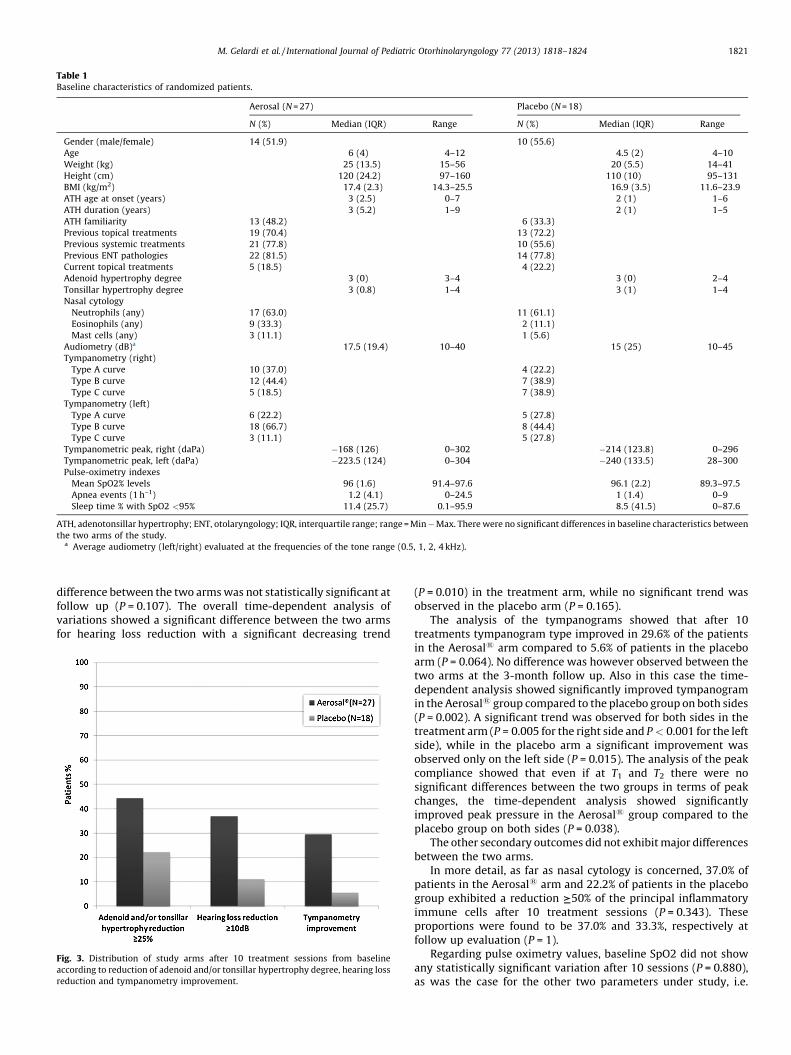
Table 1Baseline characteristics of randomized patients.
Aerosal (N = 27) Placebo (N = 18)
N (%) Median (IQR) Range N (%) Median (IQR) Range
Gender (male/female) 14 (51.9) 10 (55.6)
Age 6 (4) 4–12 4.5 (2) 4–10
Weight (kg) 25 (13.5) 15–56 20 (5.5) 14–41
Height (cm) 120 (24.2) 97–160 110 (10) 95–131
BMI (kg/m2) 17.4 (2.3) 14.3–25.5 16.9 (3.5) 11.6–23.9
ATH age at onset (years) 3 (2.5) 0–7 2 (1) 1–6
ATH duration (years) 3 (5.2) 1–9 2 (1) 1–5
ATH familiarity 13 (48.2) 6 (33.3)
Previous topical treatments 19 (70.4) 13 (72.2)
Previous systemic treatments 21 (77.8) 10 (55.6)
Previous ENT pathologies 22 (81.5) 14 (77.8)
Current topical treatments 5 (18.5) 4 (22.2)
Adenoid hypertrophy degree 3 (0) 3–4 3 (0) 2–4
Tonsillar hypertrophy degree 3 (0.8) 1–4 3 (1) 1–4
Nasal cytology
Neutrophils (any) 17 (63.0) 11 (61.1)
Eosinophils (any) 9 (33.3) 2 (11.1)
Mast cells (any) 3 (11.1) 1 (5.6)
Audiometry (dB)a 17.5 (19.4) 10–40 15 (25) 10–45
Tympanometry (right)
Type A curve 10 (37.0) 4 (22.2)
Type B curve 12 (44.4) 7 (38.9)
Type C curve 5 (18.5) 7 (38.9)
Tympanometry (left)
Type A curve 6 (22.2) 5 (27.8)
Type B curve 18 (66.7) 8 (44.4)
Type C curve 3 (11.1) 5 (27.8)
Tympanometric peak, right (daPa) �168 (126) 0–302 �214 (123.8) 0–296
Tympanometric peak, left (daPa) �223.5 (124) 0–304 �240 (133.5) 28–300
Pulse-oximetry indexes
Mean SpO2% levels 96 (1.6) 91.4–97.6 96.1 (2.2) 89.3–97.5
Apnea events (1 h–1) 1.2 (4.1) 0–24.5 1 (1.4) 0–9
Sleep time % with SpO2 <95% 11.4 (25.7) 0.1–95.9 8.5 (41.5) 0–87.6
ATH, adenotonsillar hypertrophy; ENT, otolaryngology; IQR, interquartile range; range = Min � Max. There were no significant differences in baseline characteristics between
the two arms of the study.a Average audiometry (left/right) evaluated at the frequencies of the tone range (0.5, 1, 2, 4 kHz).
M. Gelardi et al. / International Journal of Pediatric Otorhinolaryngology 77 (2013) 1818–1824 1821
difference between the two arms was not statistically significant atfollow up (P = 0.107). The overall time-dependent analysis ofvariations showed a significant difference between the two armsfor hearing loss reduction with a significant decreasing trend
Fig. 3. Distribution of study arms after 10 treatment sessions from baseline
according to reduction of adenoid and/or tonsillar hypertrophy degree, hearing loss
reduction and tympanometry improvement.
(P = 0.010) in the treatment arm, while no significant trend wasobserved in the placebo arm (P = 0.165).
The analysis of the tympanograms showed that after 10treatments tympanogram type improved in 29.6% of the patientsin the Aerosal1 arm compared to 5.6% of patients in the placeboarm (P = 0.064). No difference was however observed between thetwo arms at the 3-month follow up. Also in this case the time-dependent analysis showed significantly improved tympanogramin the Aerosal1 group compared to the placebo group on both sides(P = 0.002). A significant trend was observed for both sides in thetreatment arm (P = 0.005 for the right side and P < 0.001 for the leftside), while in the placebo arm a significant improvement wasobserved only on the left side (P = 0.015). The analysis of the peakcompliance showed that even if at T1 and T2 there were nosignificant differences between the two groups in terms of peakchanges, the time-dependent analysis showed significantlyimproved peak pressure in the Aerosal1 group compared to theplacebo group on both sides (P = 0.038).
The other secondary outcomes did not exhibit major differencesbetween the two arms.
In more detail, as far as nasal cytology is concerned, 37.0% ofpatients in the Aerosal1 arm and 22.2% of patients in the placebogroup exhibited a reduction I50% of the principal inflammatoryimmune cells after 10 treatment sessions (P = 0.343). Theseproportions were found to be 37.0% and 33.3%, respectively atfollow up evaluation (P = 1).
Regarding pulse oximetry values, baseline SpO2 did not showany statistically significant variation after 10 sessions (P = 0.880),as was the case for the other two parameters under study, i.e.

Table 2Intention-to-treat analysis of primary and secondary outcomes at T1 (10 sessions of therapy) and T2 (3 months from the end of treatment) compared to baseline.
Aerosal (N = 27) Placebo (N = 18) P-valuea
N (%) Median (IQR) N (%) Median (IQR)
T1
Reduction of adenoid hypertrophy degree (%) 0 (25) 0 (0) 0.734
Reduction �25% 8 (29.6) 3 (16.7) 0.482
Reduction of tonsillar hypertrophy degree (%) 0 (31.2) 0 (0) 0.197
Reduction �25% 9 (33.3) 3 (16.7) 0.308
Reduction of adenoid and/or tonsillar
hypertrophy degree �25% 12 (44.4) 4 (22.2) 0.204
Hearing loss reductionb (dB) 5 (13.8) 0 (10) 0.018
Reduction �10 dB 10 (37.0) 2 (11.1) 0.086
Tympanometry improvementc 8 (29.6) 1 (5.6) 0.064
Tympanometric peak pressure change (daPa) �21.5 (79.5) �1 (74) 0.141
Nasal cytology reductiond �50% 10 (37.0) 4 (22.2) 0.343
Increase of mean SpO2% levels �0.1 (1.9) 0.1 (2.8) 0.880
Reduction of apnea events (1 h–1) �0.5 (4.9) 0 (1.4) 0.372
Reduction of sleep time % with SpO2 <95% 3.6 (14.6) �2.5 (57.4) 0.424
T2
Reduction of adenoid hypertrophy degree (%) 0 (18.8) 0 (25) 0.967
Reduction �25% 7 (25.9) 6 (33.3) 0.739
Reduction of tonsillar hypertrophy degree (%) 0 (31.2) 0 (0) 0.070
Reduction �25% 11 (40.7) 3 (16.7) 0.111
Reduction of adenoid and/or tonsillar hypertrophy degree �25% 16 (59.3) 7 (38.9) 0.231
Hearing loss reductionb (dB) 2.5 (14.4) 0 (10) 0.107
Reduction �10 dB 10 (37.0) 4 (22.2) 0.343
Tympanometry improvementc 9 (33.3) 3 (16.7) 0.308
Tympanometric peak pressure change (daPa) �60.5 (104.4) �44 (78.5) 0.509
Nasal cytology reductiond �50% 10 (37.0) 6 (33.3) 1
Increase of mean SpO2% levels 0.2 (2.2) 0.2 (2) 0.772
Reduction of apnea events (1 h–1) 0.2 (2.8) �0.9 (2.1) 0.102
Reduction of sleep time % with SpO2 <95% 0.4 (28.5) 0.3 (22.2) 0.917
IQR, interquartile range.a Mann–Whitney U-test for continuous variables, Fisher’s exact test for categorical variables.b Reduction of average audiometry (left/right) evaluated at the frequencies of the tone range (0.5, 1, 2, 4 kHz).c Improvement was defined as defined as the passage from type B curve to type C/A curve or from type C curve to type A curve for both ears sides.d Any reduction of principal inflammatory immune cells (neutrophils, eosinophils, and mast cells).
Table 3Salt room microclimate features.
Size of iodized NaCl particles released 0.23–20 mm
Particles density 35–50 mg/m3
Air exchange 90 m3/h
CO2 ppm <750
Temperature 20–24 8CHumidity 44–60%
M. Gelardi et al. / International Journal of Pediatric Otorhinolaryngology 77 (2013) 1818–18241822
reduction of the event data index (P = 0.372), and reduction ofsleep time with SpO2 <95% (P = 0.424). Pulse oximetry valuesremained mostly unchanged at follow up.
The results of the per-protocol analysis did confirm the mainfindings and were generally overlapping intention-to-treat out-comes (data not shown).
4. Discussion
The growing prevalence of conditions (both allergic andinfectious) affecting the upper airways has stimulated a wholeseries of studies on topical treatments in a view to reducing theside effects of systemic treatments and improving clinical responsein terms of improvement of nasal symptoms [38–40].
The latest guidelines issued by EPOS 2012 [41] on obstructiveand infectious nasal sinus disease include among therapeutic aids(antibiotics, topical steroids, and topical decongestants) also nasalsaline irrigation, thus emphasizing the crucial role of thistreatment in reducing nasal congestion and mucopurulentdischarge by a washing process that restores mucociliary clearanceand prevents both locoregional (otitis, rhinosinusitis) and distantinflammation (rhinobronchial syndrome, bronchitis, pneumonia,asthma, etc.) [42–44].
In addition, in the last few years some literature studies [20–26]have reported a new therapeutic-preventive role for sodiumchloride in what has come to be called ‘‘halotherapy’’ and in theapplications of this latter in the different branches of medicine, inparticular respiratory and dermatological disease. As a matter offact, for hundreds of years salt has been recognized as an agent totreat respiratory and skin conditions. The history of using salt cavesfor healing (speleotherapy from Greek speleos = cave and therapy)
different ailments by assimilating dust-like salt particles goes backto ancient times. These caves used for therapeutic purposes are stillin use in many Central and Eastern European countries includingAustria (Solzbad-Salzetnan), Romania (Sieged), Poland (Wieliczka,one of the UNESCO World Heritage Sites), Azerbaijan (Nakhiche-van), Kirgizia (Chon-Tous), Russia (Berezniki-Pern), and Ukraine(Solotvino-Carpathians e Artiomovsk-Donietsk).
The possibility of recreating the microclimate (Table 3) of thesecaves in a room has given a new impulse to studies and researchefforts on the potential therapeutic effects of this treatment.
As far as the present study is concerned, the first finding hasbeen the absence of adverse effects. None of the children enrolledin the study exhibited episodes of respiratory distress (dyspnea,bronchial hyperactivity, asthma), skin itch or eyes disorders, bothduring treatment and in the hours immediately after treatment. Inaddition, a high compliance to treatment has been observed aschildren did not consider their HT sessions as a therapy, but ratheras a time for play or recreation as they spent their 30-min sessionsplaying, watching TV (cartoons, wildlife shows, etc.) (Fig. 2). Onlytwo children withdrew from the study; one of them (in the placeboarm) withdrew during the first week of halotherapy for an episode
M. Gelardi et al. / International Journal of Pediatric Otorhinolaryngology 77 (2013) 1818–1824 1823
of acute tonsillitis with high fever, the other dropped out in thefollow-up period for increased adenotonsillar hypertrophy associ-ated with sleep respiratory disorders with an indication foradenotonsillectomy. Being specific to the natural course of theconditions in question, these episodes have not been considered asadverse events connected to the halotherapeutic treatment.
However the most interesting aspects emerging from thepresent study have been those related to the assessment of the realimpact of halotherapy on both the lymphatic component(adenotonsillar component) and co-morbidities, namely earconditions and sleep disorders. Actually, the numerous studiesconducted so far on halotherapy have mainly been focused onlower airways conditions (cystc fibrosis, bronchitis, and asthma[21–23]).
Our study has highlighted a �25% reduction of the adenoton-sillar tissue in 44.4% of the patients treated versus 22.2% of theplacebo controls. In our view, far from being statistically significant(P = 0.204), this finding has a clinical value that deserves furtherstudy. This pattern has also been confirmed by the pulse-oximetricdata that, far from being statistically significant, have shown adecreased event data index (adjusted index) as well as a reductionof the sleep time percentage with SpO2 <95%. Based on our resultsit is possible to calculate that approximately 140 patients (70 ineach arm) would be needed to show a significant reduction of theadenotonsillar tissue as expressed in the primary outcome. It istherefore unlikely that the loss of power due to the reducednumber of patients enrolled in the study (45) compared to theplanned number (64) had a significant impact on our findings. Thereduction of some clinical and endoscopic parameters also in thecontrol group should however be justified by the fact that the veryyoung patients actually spent their time in a ‘‘salted’’ environmentwhere their same manipulation of salt released microparticles ofsodium chloride available for inhalation.
Among secondary end-points, end-of-treatment improvementof hearing loss has been found to be statistically significant in thehalotherapy group (P = 0.018) compared to the control group. Thestatistical analysis has demonstrated a significant improvement ofboth tympanogram and hearing loss in the Aerosal1 group.
The treatment of otitis media with effusion (OME) is stillcontroversial today. While this condition has a high likelihood of aspontaneous recovery [45], so far no medical therapy has beenshown to be effective to treat OME, as indicated by recent reviews[46–49]. The presence of a control group in our study does rule outthe possibility that the improvements observed are linked only to aspontaneous recovery from the disease. Even though theeffectiveness of sodium chloride in OME treatment has neverbeen reported in literature studies, the potential mechanisms ofaction could be ascribed to decongestion of nasal passages andtubaric orifice respiratory mucosa as well as to a restored muco-ciliary clearance that would favor middle ear aeration-drainingmechanisms. This assumption is substantiated by literaturestudies that report the efficacy of those treatments targeted toimprove middle ear ventilation. Perera et al. reported someevidence that autoinflation devices may be of benefit in the short-term in treating children with otitis media with effusion [50].Similar results were reported in a group of children affected byOME treated with swallowing and auto-inflation exercises,including Valsalva maneuver [51]. Finally even if the improvementof hearing threshold was on average of only 5 dB, it is important toremember that the pre-treatment threshold was on averagebetween 15 and 17.5 dB, therefore a 5 dB change together with achange in tympanogram is clinically relevant. A longer treatmentcould however further improve the hearing thresholds.
No statistically significant difference has been found in terms ofsleep quality and nasal immunophlogosis parameters. Recent workon bronchial immunophlogosis confirms these findings [52].
5. Conclusions
Haloterapy accounts for a relatively new, completely naturaltherapeutic remedy which does not call for any pharmacologicaladministration and is based on the healing capacities of natural saltmicronized and released into an indoor environment by means ofspecific techniques. Halotherapy administered by Aerosal1 systemhas been shown to have a statistically significant effect on OME.Aerosal1 halotherapy has also been found to be partially effectivein reducing adenotonsillar hypertrophy. The beneficial effects ofthe treatment in question have been shown for some ‘‘time-dependent’’ parameters, therefore additional studies should beconducted in a view to defining treatment modalities likely toresult in a better clinical response. New double-blind, placebo-controlled, randomized clinical studies should also be performedon more complex conditions including asthma, cystic fibrosis,chronic pulmonary disease and dermatological conditions.
In addition to being a safe treatment, the Aerosal1 Haloterapysystem has been well accepted and tolerated by our young patientswho experienced their halotherapeutic sessions as a time for playand recreation and not as a real medical treatment. ThereforeAerosal1 Haloterapy might constitute a valuable adjunct (and nota replacement) to current orthodox medical treatment ofadenotonsillar disease and correlated conditions.
Funding
This study has been funded by a TECNOSUN srl scholarship.
Conflict of interest
None declared.
References
[1] R. Arens, C.L. Marcus, Pathophysiology of upper airway obstruction: a develop-mental perspective, Sleep 27 (1 August (5)) (2004) 997–1019.
[2] S.Z. Toros, H. Nos eri, C.K. Ertugay, S. Kulekci, T.E. Habes oglu, G. Kılıcoglu, G. Yılmaz,E. Egeli, Adenotonsillar hypertrophy: does it correlate with obstructive symptomsin children? Int. J. Pediatr. Otorhinolaryngol. 74 (November (11)) (2010) 1316–1319.
[3] D. Gozal, L. Kheirandish-Gozal, O.S. Capdevila, E. Dayyat, E. Kheirandish, Preva-lence of recurrent otitis media in habitually snoring school-aged children, SleepMed. 9 (July (5)) (2008) 549–554.
[4] S. Hammaren-Malmi, H. Saxen, J. Tarkkanen, P.S. Mattila, Adenoidectomy does notsignificantly reduce the incidence of otitis media in conjunction with the insertionof tympanostomy tubes in children who are younger than 4 years: a randomizedtrial, Pediatrics 116 (2005) 185–189.
[5] R.M. Ray, C.M. Bower, Pediatric obstructive sleep apnea: the year in review, Curr.Opin. Otolaryngol. Head Neck Surg. 13 (December (6)) (2005) 360–365.
[6] A. Pac, A. Karadag, H. Kurtaran, D. Aktas, Comparison of cardiac function andvalvular damage in children with and without adenotonsillar hypertrophy, Int. J.Pediatr. Otorhinolaryngol. 69 (April (4)) (2005) 527–532.
[7] J.B. Sousa, W.T. Anselmo-Lima, F.C. Valera, A.J. Gallego, .MA. Matsumoto, Cepha-lometric assessment of the mandibular growth pattern in mouth-breathingchildren, Int. J. Pediatr. Otorhinolaryngol. 69 (March (3)) (2005) 311–317.
[8] S.R. Gonzalez Rivera, J. Coromina Isern, C. Gay Escoda, Respiratory orofacial andocclusion disorders associated with adenotonsillar hypertrophy, An. Otorrinolar-ingol. Ibero Am. 31 (3) (2004) 265–282.
[9] A.E. Zautner, Adenotonsillar disease, Recent Pat. Inflamm. Allergy Drug Discov. 6(May (2)) (2012) 121–129.
[10] P. Cassano, M. Gelardi, M. Cassano, M.L. Fiorella, R. Fiorella, Adenoid tissuerhinopharyngeal obstruction grading based on fiberendoscopic findings: a novelapproach to therapeutic management, Int. J. Pediatr. Otorhinolaryngol. 67 (2003)1303–1309.
[11] A. Mlynarek, M.A. Tewfik, A. Hagr, J.J. Manoukian, M.-D. Schloss, T.L. Tewfik,Lateral neck radiography versus direct video rhinoscopy in assessing adenoid size,J. Otolaryngol. 33 (2004) 360–365.
[12] M. Berlucchi, D. Salsi, L. Valetti, G. Parrinello, P. Nicolai, The role of mometasonefuroate acqueous nasal spray in the treatment of adenoidal hypertrophy in thepediatric age group: preliminary results of a prospective, randomised study,Pediatrics 119 (2007) e1392–e1397.
[13] M. Berlucchi, L. Valetti, G. Parrinello, P. Nicolai, Longterm follow-up of childrenundergoing topical intranasal steroid therapy for adenoidal hypertrophy, Int. J.Pediatr. Otorhinolaryngol. 72 (2008) 1171–1175.
M. Gelardi et al. / International Journal of Pediatric Otorhinolaryngology 77 (2013) 1818–18241824
[14] S. Cengel, M.U. Akyol, The role of topical nasal steroids in the treatment of childrenwith otitis media with effusion and/or adenoid hypertrophy, Int. J. Ped. Otorhi-nolaryngol. 70 (2006) 639–645.
[15] C.S. Derkay, D.H. Darrow, C. Welch, J.T. Sinacori, Post-tonsillectomy morbidityand quality of life in pediatric patients with obstructive tonsils and adenoid:microdebrider vs electrocautery, Otolaryngol. Head Neck Surg. 134 (2006)114–120.
[16] G. Scadding, Non-surgical treatment of adenoidal hypertrophy: the role of treat-ing IgE-mediated inflammation, Pediatr. Allergy Immunol. 21 (December (8))(2010) 1095–1106.
[17] M. Gelardi, A. Mezzoli, M.L. Fiorella, M. Carbonara, M. Di Gioacchino, G. Ciprandi,Nasal irrigation with lavonase as ancillary treatment of acute rhinosinusitis: apilot study, J. Biol. Regul. Homeost. Agents 23 (April–June (2)) (2009) 79–84.
[18] M. Costantino, [The rhinogenic deafness and SPA therapy: clinical–experimentalstudy], Clin. Ter. 159 (September–October (5)) (2008) 311–315.
[19] J.L. Fauquert, A. Labbe, Treatment of respiratory and ORL diseases with mineralwaters in children, Pediatrie 45 (11) (1990) 769–774.
[20] A.V. Chervinskaya, N.A. Zilber, Halotherapy for treatment of respiratory diseases,J. Aerosol Med. 8 (1995) 221–232.
[21] J. Hedman, T. Hugg, J. Sandell, T. Haahtela, The effect of salt chamber treatment onbronchial hyperresponsiveness in asthmatics, Allergy 61 (2006) 605–610.
[22] L.M. Abdrakhmanova, U.R. Farkhutdinov, R.R. Farkhutdinov, Effectiveness ofhalotherapy of chronic bronchitis patients, Vopr. Kurortol. Fizioter. Lech. Fiz.Kult. (November–December (6)) (2000) 21–24.
[23] R.A. Chernenkov, E.A. Chernenkova, G.V. Zhukov, The use o fan artificial microcli-mate chambre in the treatment of patients with chronic obstructive lung diseases,Vopr. Kurortol. Fizioter. Lech. Fiz. Kult. (July–August (4)) (1997) 19–21.
[24] E.A. Puryshev, The efficacy of speleotherapy in atopic dermatitis in children, Vopr.Kurortol. Fizioter. Lech. Fiz. Kult. 4 (July–August) (1994) 34–35 (in Russian).
[25] D. Ben-Amatai, M. David, Climatotherapy at the dead Sea for pediatric onsetpsoriasis vulgaris, Pediatr. Dermatol. 26 (2009) 102–104.
[26] A.V. Chervinskaya, Prospects of halotherapy in sanatorium-and-spa dermatologyand cosmetology, Resort bulletin 3 (36) (2006) 74–75.
[27] L. Brodsky, Modern assessment of tonsils and adenoids, Pediatr. Clin. North Am.36 (1989) 1551–1569.
[28] J. Jerger, Clinical experience with impedance audiometry, Arch Otolaryngol. 92(October (4)) (1970) 311–324.
[29] M. Gelardi, Atlas of Nasal Cytology, Edi Ermes, Milan, Italy, 2012.[30] M. Gelardi, M.L. Fiorella, C. Russo, R. Fiorella, G. Ciprandi, Role of nasal cytology,
Int. J. Immunopathol. Pharmacol. 23 (January–March (1 Suppl)) (2010) 45–49.[31] E.O. Meltzer, A.A. Jalowayski, Nasal cytology in clinical practice, Am. J. Rhinol. 2
(1988) 47–54.[32] M. Gelardi, G. Passalacqua, M.L. Fiorella, A. Mosca, N. Quaranta, Nasal cytology:
the infectious spot, an expression of a morphological-chromatic biofilm, Eur. J.Clin. Microbiol. Infect. Dis. 30 (September (9)) (2011) 1105–1109.
[33] R.T. Brouillette, A. Morielli, A. Leimanis, K.A. Waters, R. Luciano, F.-M. Ducharme,Nocturnal pulse oximetry as an abbreviated testing modality for pediatric ob-structive sleep apnea, Pediatrics 105 (2000) 405–412.
[34] N. Traeger, B. Scultz, A.N. Pollock, T. Mason, C.L. Marcus, R. Arens, Polysomno-graphic values in children 2–9 years old: additional data and review of theliterature, Paediatr. Pulmonol. 40 (2005) 22–30.
[35] R.F. Baugh, S.M. Archer, R.B. Mitchell, R.M. Rosenfeld, R. Amin, J.J. Burns, D.H.Darrow, T. Giordano, R.S. Litman, K.K. Li, M.E. Mannix, R.H. Schwartz, G. Setzen,E.R. Wald, E. Wall, G. Sandberg, M.M. Patel, Clinical practice guideline: tonsillec-tomy in children. American Academy of Otolaryngology-Head and Neck SurgeryFoundation, Otolaryngol. Head Neck Surg. 144 (January (1 Suppl)) (2011) S1–S30.
[36] M.P. Villa, L. Brunetti, O. Bruni, F. Cirignotta, P. Cozza, G. Donzelli, L. Ferini Strambi,L. Levrini, S. Mondini, L. Nespoli, L. Nosetti, J. Pagani, M. Zucconi, Guidelines for the
diagnosis of childhood obstructive sleep apnea syndrome, Minerva Pediatr. 56(June (3)) (2004) 239–253.
[37] G.M. Nixon, A.S. Kermack, G.M. Davis, J.J. Manoukian, K.A. Brown, R.T. Brouillette,Planning adenotonsillectomy in children with obstructive sleep apnea: the role ofovernight oximetry, Pediatrics 113 (2004) 19–25.
[38] I.Y. Wong, S.E. Soh, S.Y. Chng, L.P. Shek, D.Y. Goh, H.P. Van Bever, B.W. Lee,Compliance with topical nasal medication – an evaluation in children withrhinitis, Pediatr. Allergy Immunol. 21 (December (8)) (2010) 1146–1150.
[39] D.W. Kennedy, As the inflammatory nature of chronic rhinosinusitis (CRS) hasbecome increasingly recognized, the use of steroids, both systemic and topical, aspart of the disease management has significantly increased, Int. Forum AllergyRhinol. 2 (March–April (2)) (2012) 93–94.
[40] N.D. Adappa, C.C. Wei, J.N. Palmer, Nasal irrigation with or without drugs: theevidence, Curr. Opin. Otolaryngol. Head Neck Surg. 20 (February (1)) (2012) 53–57.
[41] W.J. Fokkens, V.J. Lund, J. Mullol, C. Bachert, I. Alobid, F. Baroody, N. Cohen, A. Cervin,R. Douglas, P. Gevaert, C. Georgalas, H. Goossens, R. Harvey, P. Hellings, C. Hopkins,N. Jones, G. Joos, L. Kalogjera, B. Kern, M. Kowalski, D. Price, H. Riechelmann, R.Schlosser, B. Senior, M. Thomas, E. Toskala, R. Voegels, Y. Wang de, P.J. Wormald,EPOS 2012: European position paper on rhinosinusitis and nasal polyps 2012. Asummary for otorhinolaryngologists, Rhinology 50 (March (1)) (2012) 1–12.
[42] E. Daviskas, S.D. Anderson, I. Gonda, S. Eberl, S. Meikle, J.P. Seale, G. Bautovich,Inhalation of hypertonic saline aerosol enhances mucociliary clearance in asth-matic and healthy subjects, Eur. Respir. J. 9 (1996) 725–732.
[43] M. Gelardi, A. Mezzoli, M.L. Fiorella, M. Carbonara, M. Di Gioacchino, G. Ciprandi,Nasal irrigation with Lavonase as ancillary treatment of acute rhinosinusitis: apilot study, J. Biol. Regul. Homeost. Agents 23 (2) (2009) 79–84.
[44] M.R. Elkins, M. Robinson, B.R. Rose, C. Harbour, C.P. Moriarty, G.B. Marks, et al., Acontrolled trial of long-term inhale hypertonic saline in patients with cysticfibrosis, New Eng. J. Med. 354 (2006) 229–240.
[45] I. Williamson, S. Benge, S. Barton, S. Petrou, L. Letley, N. Fasey, M. Haggard, P. Little,Topical intranasal corticosteroids in 4-11 year old children with persistentbilateral otitis media with effusion in primary care: double blind randomisedplacebo controlled trial, Br Med. J. 339 (Dec 16) (2009) b4984.
[46] G. Griffin, C.A. Flynn, R.E. Bailey, J.K. Schultz, Antihistamines and/or decongestantsfor otitis media with effusion (OME) in children, Cochrane Database Syst. Rev. 9(Issue 9) (2011).
[47] S.A. Simpson, R. Lewis, J. van der Voort, C.C. Butler, Oral or topical nasal steroidsfor hearing loss associated with otitis media with effusion in children, CochraneDatabase Syst. Rev. 5 (Issue 5) (2011).
[48] A. van Zon, G.J. van der Heijden, T.M.A. van Dongen, M.J. Burton, A.G.M. Schilder,Antibiotics for otitis media with effusion in children, Cochrane Database Syst. Rev.9 (Issue 9) (2012).
[49] R.M. Rosenfeld, L. Culpepper, K.J. Doyle, K.M. Grundfast, A. Hoberman, M.A. Kenna,A.S. Lieberthal, M. Mahoney, R.A. Wahl, C.R. Woods Jr., B. Yawn, AmericanAcademy of Pediatrics Subcommittee on Otitis Media with Effusion, AmericanAcademy of Family Physicians, American Academy of Otolaryngology – Head,Neck Surgery, Clinical practice guideline: otitis media with effusion, Otolaryngol.Head Neck Surg. 130 (May (5 Suppl)) (2004) S95–S118.
[50] R. Perera, J. Haynes, P.P. Glasziou, C.J. Heneghan, Autoinflation for hearing lossassociated with otitis media with effusion, Cochrane Database Syst. Rev. 4 (Issue4) (2006).
[51] L. D’Alatri, P.M. Picciotti, M.R. Marchese, A. Fiorita, Alternative treatment for otitismedia with effusion: eustachian tube rehabilitation, Acta Otorhinolaryngol. Ital.32 (February) (2012) 32–40.
[52] J. Sandell, J. Hedman, K. Saarinen, T. Haahtela, Salt chamber treatment is ineffec-tive in treating eosinophilic inflammation in asthma, Allergy 4 (January) (2013)68–70.