Three small, cryptic plasmids from Aeromonas salmonicida subsp. salmonicida A449
Upload
independentCategory
view
0download
0
A cryptic sensor for HIV-1 activates antiviral innate immunity indendritic cells
Nicolas Manel1,5, Brandon Hogstad1,4, Yaming Wang2, David E. Levy2, Derya Unutmaz3,and Dan R. Littman1,3,4
1Molecular Pathogenesis Program, The Kimmel Center for Biology and Medicine of the SkirballInstitute, New York University School of Medicine, New York, NY 10016, USA2Department of Pathology and New York University Cancer Institute, New York University Schoolof Medicine, New York, NY 10016, USA3Departments of Microbiology and Pathology, New York University School of Medicine, NewYork, NY 10016, USA4Howard Hughes Medical Institute, New York University School of Medicine, New York, NY10016, USA5CNRS-UMR5535, Institut de Génétique Moléculaire de Montpellier, Université Montpellier I andII, Montpellier, France
AbstractDendritic cells (DC) serve a key function in host defense, linking innate detection of microbes tothe activation of pathogen-specific adaptive immune responses(1,2). Whether there is cell-intrinsicrecognition of HIV-1 by host innate pattern-recognition receptors and subsequent coupling toantiviral T cell responses is not yet known(3). DC are largely resistant to infection with HIV-1(4),but facilitate infection of co-cultured T-helper cells through a process of trans-enhancement(5,6).We show here that, when DC resistance to infection is circumvented(7,8), HIV-1 induces DCmaturation, an antiviral type I interferon response and activation of T cells. This innate response isdependent on the interaction of newly-synthesized HIV-1 capsid (CA) with cellular cyclophilin A(CypA) and the subsequent activation of the transcription factor IRF3. Because the peptidyl-prolylisomerase CypA also interacts with CA to promote HIV-1 infectivity, our results suggest that CAconformation has evolved under opposing selective pressures for infectivity versus furtiveness.Thus, a cell intrinsic sensor for HIV-1 exists in DC and mediates an antiviral immune response,but it is not typically engaged due to absence of DC infection. The virulence of HIV-1 may berelated to evasion of this response, whose manipulation may be necessary to generate an effectiveHIV-1 vaccine.
Exposure of monocyte-derived DC (MDDC) to GFP-encoding HIV-1 pseudotyped withVSV-G (HIVGFP(G)) (MOI 1–2, see Supplemental Table 1) resulted in little infection and
Users may view, print, copy, download and text and data- mine the content in such documents, for the purposes of academic research,subject always to the full Conditions of use: http://www.nature.com/authors/editorial_policies/license.html#terms
AUTHOR CONTRIBUTIONSN.M. and D.R.L. designed the study and wrote the manuscript. N.M. performed the experiments and analyses. B.H. provided technicalhelp. D.U. provided expertise and contributed to experiments with human T cell proliferation assays. D.E.L. provided expertise inidentifying the interferon response. D.E.L. and Y.W. designed the quantitative bioassay for interferons and Y.W. performed the assay.All authors discussed results and edited the manuscript.
DATA DEPOSITIONMicroarray data has been deposited in the NCBI GEO database under the accession number GSE22589.
NIH Public AccessAuthor ManuscriptNature. Author manuscript; available in PMC 2011 March 9.
Published in final edited form as:Nature. 2010 September 9; 467(7312): 214–217. doi:10.1038/nature09337.
NIH
-PA Author Manuscript
NIH
-PA Author Manuscript
NIH
-PA Author Manuscript
absence of cell activation, as monitored by expression of CD86, CD80, CD38, and CD83(Fig. 1a and Supplemental Fig. 2a). Likewise, VSV-G-pseudotyped SIVmac239 virus-likeparticles (SIVVLP(G)) had no effect DC activation. In contrast, co-infection of MDDC withHIVGFP(G) and SIVVLP(G), which provides Vpx-mediated relief of restriction to HIV-1replication(8), resulted in GFP expression in more than 85% of the cells as well as up-regulation of CD86 and other activation markers after 48h (Fig. 1a and Supplemental Fig.2a). Entry of both virions into the cytoplasm was required (Supplemental Fig. 2b), andactivation occurred only beyond a variable threshold of infection (Supplemental Fig. 2c, 3a).A virus that expressed all accessory proteins and a CCR5-tropic replication competent virusalso infected MDDC in the presence of SIVVLP(G) and induced expression of CD86(Supplemental Fig. 3b, 4a). Expression of a Vpx-Vpr fusion protein in packaging cellsrescued the ability of HIVGFP(G) to productively infect MDDC and to induce CD86 up-regulation, indicating that Vpx is the only SIVVLP component required for infection withHIV-1 (Supplemental Fig. 4b). Thus, MDDC carry an intact mechanism of activationfollowing HIV-1 infection.
As observed with MDDC, infection of primary peripheral blood CD11c+ DC withHIVGFP(G) did not result in detectable expression of GFP (Supplemental Fig. 4c).However, CD11c+ DC were infected with HIVGFP(G) in the presence of SIVVLP(G),resulting in up-regulation of CD86 in a proportion of cells similar to that observed afterincubation with poly(I:C).
Genome-wide expression profiling demonstrated induction of a type I interferon responsefollowing co-infection, but not following infection with either HIV-1 or SIV particles(Supplemental Fig. 5a, 5b, 5c). After infection, expression of interferon-regulated genes wasdelayed as compared to the response to LPS (Supplemental Fig. 5d). Accordingly, STAT1phosphorylation was present at 2h after LPS treatment, but only at 22h after infection (Fig.1b). Type I interferon was produced by infected MDDC over the course of 48 hours(Supplemental Fig. 6a), but was not detected following infection of CD4+ T cells (Fig. 1c),293T cells, and THP-1 cells (Fig. 1d), despite the ability of those cells to produce type I IFNafter other viral innate stimuli. Blocking antibodies against IFNβ, but not IFNα, reducedexpression of the activation markers on MDDC (Supplemental Fig. 6b, 6c). Furtherneutralization of type I and type III IFN did not improve the inhibition (data not shown).Together, these results suggest that CD86 induction is mainly due to the production ofsoluble type I IFNβ, in accordance with observations in murine DC(9).
Next, we sought to determine which step of the viral replication cycle is required for MDDCactivation. Inhibitors of HIV-1 reverse transcriptase (AZT) and integrase (Raltegravir)inhibited transduction efficiency and MDDC activation only when added during the first 24hours (Supplemental Fig. 7a) and had no effect on LPS- or poly(I:C)-induced CD86 up-regulation (data not shown). These results suggested that DC activation is induced afterintegration. There was no activation of MDDC after infection with an HIV-1-based vectordevoid of viral protein-coding sequences (LKO1gfp) (Supplemental Fig. 7b). We thereforeintroduced mutations in the packaged HIVGFP genome, and evaluated activation of infectedMDDC. Inactivation of Rev, required for nuclear export of unspliced viral RNA(10), andabrogation of Gag expression prevented MDDC activation (Fig. 2a), but mutation of thePTAP sequence in p6, required for viral budding(11), and treatment with HIV-1 proteaseinhibitors (Supplemental Fig. 7c) had no effect. In the absence of SIVVLP(G), intracellularCA from incoming viral particles failed to induce CD86 expression (Supplemental Fig. 8).These results suggested that newly synthesized GagPol is required for DC activation, whichis consistent with the delayed induction of the type I interferon response. We next tested apanel of viruses with CA mutations(12) for ability to induce the innate response in MDDC.These mutants were defective for DC infection (Supplemental Fig. 9) and were thus partially
Manel et al. Page 2
Nature. Author manuscript; available in PMC 2011 March 9.
NIH
-PA Author Manuscript
NIH
-PA Author Manuscript
NIH
-PA Author Manuscript

rescued by co-transfection of wild type viral proteins in the packaging cells. The T54A/N57A and Q63A/Q67A mutants induced CD86 expression despite reduced infectivitycompared to wild-type virus (Fig. 2b and Supplemental Fig. 10). In contrast, infection withthe G89V mutant, which is compromised for CA binding to cyclophilin A (CypA), apeptidyl-prolyl isomerase required for optimal HIV-1 infectivity(13), resulted insubstantially reduced CD86 expression at similar levels of infection.
Treatment with Cyclosporin A (CsA), which disrupts the interaction between CypA andCA(14), prevented MDDC activation following infection with HIVGFP(G) andSIVVLP(G), but not following treatment with LPS (Fig. 2c). Because CsA also inhibitsinfection with HIV-1, we assessed its effect when administered at different times afterinfection of MDDC. When CsA was added as late as 12h following infection, it preventedup-regulation of CD86 despite highly efficient infection and expression of CA(Supplemental Fig. 11).
To study the role of CypA and other host genes in innate immune signaling followingproductive infection of MDDC with HIV-1, we employed an RNAi approach using shRNAlentiviral vectors (that also express GFP)(15) along with SIVVLP(G). Knockdown of PPIAmarkedly reduced expression of its product, CypA, and prevented CD86 up-regulationfollowing infection with HIV-1 (HDVIRESRFP(G), encoding the reporter RFP), but notfollowing treatment with LPS (Fig. 2d and Supplemental Fig. 12a). The interaction betweenCypA and newly synthesized CA is therefore essential for the innate response of MDDC toHIV-1.
Type I interferon responses following infection with multiple viruses requires thephosphorylation, dimerization, and nuclear translocation of IRF3(16). Productive infectionof MDDC with HIV-1 resulted in CsA-sensitive nuclear accumulation of phosphorylatedIRF3 (Fig. 3a). Knock-down of IRF3 in MDDC abrogated the induction of CD86 uponinfection with HIV-1 and, as expected, following treatment with LPS or poly(I:C)), but notafter treatment with curdlan, indicating that IRF3 knockdown did not lead to an intrinsicdefect in CD86 expression (Fig. 3b and Supplemental Fig. 12b). IRF3 knockdown, as wellas CypA knockdown, also increased the threshold at which virus induced CD86 and CD38(Supplemental Fig. 12c, 12d).
To determine if productive infection and subsequent activation of MDDC influencesantiviral adaptive immunity, we first examined whether HIV-infected DC could activateHIV-1 Gag-specific CD4+ and CD8+ T cell clones. In the presence of MDDC incubatedwith HIV-1 alone, low levels of IFNγ were detected (17). In contrast, MDDC infected withHIVGFP(G) and SIVVLP(G) stimulated a high proportion of MHC class I and class IIrestricted T cell clones to produce IFNγ (Supplemental Fig. 13a). Maturation induced byunrelated TLR ligands coupled with abortive HIV infection was not sufficient for MDDC topotently stimulate HIV antigen-specific T cells (Supplemental Fig. 13b).
To directly measure the contribution of co-stimulation to T cell activation, we examined thepolyclonal proliferation of naïve CD4+ T cells in response to infected DC in the presence ofsub-optimal concentrations of anti-CD3 antibody(18–20). Under these conditions, T cellsthat were co-cultured with productively infected and activated MDDC proliferated throughmultiple cell cycles whereas T cells cultured with the abortively infected or uninfectedMDDC had little proliferation (Fig. 4a and Supplemental Fig. 14). We next examined theeffect of SCY, a non-immunosuppressive CsA analog(21,22) that, unlike CsA, does nothave any direct effect on activation or proliferation of T cells(21) (Supplemental Fig. 15a).SCY inhibited DC activation induced by HIVGFP(G) similarly to CsA at similar levels ofinfection. DC treated with SCY or the RT inhibitor AZT at the time of HIVGFP(G) and
Manel et al. Page 3
Nature. Author manuscript; available in PMC 2011 March 9.
NIH
-PA Author Manuscript
NIH
-PA Author Manuscript
NIH
-PA Author Manuscript

SIVVLP(G) infection showed a reduced ability to induce proliferation (Fig. 4b andSupplemental Fig. 15b and 16), as did DC infected with the G89V CA mutant(Supplemental Fig. 17). These results are consistent with a requirement for interaction ofnewly-synthesized CA with CypA in the induction of DC co-stimulatory activity.
Trans-enhancement by MDDC of CD4+ T cell infection with a CCR5-tropic virus encodingGFP was inhibited if the DC were previously infected with HIV-1. The inhibition wasrelieved by neutralizing antibody against IFNβ, indicating that the innate response to HIV-1in DC restricts infection of surrounding T cells (Figure 4c) and suggesting that activation ofsuch response may also limit infection in vivo.
Our results show that, in contrast to CD4+ T cells, human dendritic cells have intrinsicmachinery for responding to infection with HIV-1 and for activating antiviral defenses andadaptive immunity (Supplemental Fig. 1). However, they are unlikely to do so effectively ininfected individuals because HIV-1 fails to replicate in DCs. HIV-2, which is notpandemic(23), encodes Vpx and has the potential to infect and activate MDDC in a CypA-dependent manner (Supplemental Fig. 18), which is consistent with the reported ability ofHIV-2 CA to bind human CypA(24,25). The finding that newly-synthesized CA is requiredto induce DC activation through a pathway involving CypA and IRF3 implicates anintracellular viral protein, in addition to viral nucleic acids, among the type I interferon-inducing pathogen-associated molecular patterns (PAMPs)(26) and constitutes the firstdescription of a cell-intrinsic recognition mechanism of retroviruses(26). It will be importantto determine whether the mechanism described herein contributes to control of the viral loadin individuals infected with HIV-2, as well as in HIV-1-infected long-term non-progressorsor “elite controllers”(27). A better mechanistic understanding of this DC-intrinsic signalingpathway may also inform HIV vaccine development.
METHODS SUMMARYMonocytes were isolated and incubated with GM-CSF and IL-4 to induce dendritic celldifferentiation. Pseudotyped viruses and virus-like particles were produced by transienttransfection of 293FT cells using TransIT-293 (Mirus). Infections were performed byincubating 105 MDDCs in 96 well U bottom plates in the presence of 8 µg/ml polybrene.Cell surface staining of activation markers was performed 48h after infection. shRNAvectors carrying GFP were transduced into fresh monocytes together with SIVVLP(G) anddendritic cell differentiation was induced. More than 90% of cells were routinely transducedand cells were challenged at day 4 with HDVIRESRFP(G) or other control PAMPs.
METHODSCells
GHOST(28) and 293FT (Invitrogen) cells were cultured in DMEM, 10% fetal bovine serum(FBS) (HyClone) and antibiotics. PBMC were isolated from IRB-approved buffy coats fromnormal donors. CD14+ cells were isolated by double positive selection with anti-humanCD14 magnetic beads (Miltenyii). Purity was at least 99%. CD14+ cells were cultured inRPMI, 10% FBS, antibiotics and HEPES in the presence of recombinant human GM-CSF at10 ng/ml and IL-4 at 50 ng/ml (eBioscience). Fresh media was added at day 3, and cellswere stimulated or infected at day 4. To isolate CD11c+ DC, CD14-depleted PBMC werefurther depleted using biotin-labeled antibodies against CD3, CD16, CD19 and CD56 andstreptavidin magnetic beads (Miltenyi). The negative fraction was stained and sorted on aFACSAria (BD Biosciences) as CD3−CD14−CD16− CD19−CD20−CD56−HLA-DR+CD11c+ (Supplemental Table 2). Purity was at least 98%. Total CD4+ T cells were
Manel et al. Page 4
Nature. Author manuscript; available in PMC 2011 March 9.
NIH
-PA Author Manuscript
NIH
-PA Author Manuscript
NIH
-PA Author Manuscript

isolated using human CD4 magnetic beads (Miltenyii). Naïve CD4+ T cells were furthersorted as CD4+CD25−CD45RA+CD45RO−.
T cell clonesT cell clones were expanded as previously described (29). The clone DR4LI15(LGLNKIVRMYSPTSI) was obtained from Nina Bhardwaj and the clones B81TL9(TPQDLNTML) and B14DA9 (DRFYKTLRA) were obtained from Bruce Walker.
ReagentsLPS (lipopoylsaccharide) and poly(I:C) (polyriboinosinic:polyribocytidylic acid) were fromSigma and were used at 1 µg/ml and 10 µg/ml, respectively. Curdlan (CM-Curdlan) wasform Wako and was used at 1 µg/ml. Cyclosporin A and FK506 were from Calbiochem.AZT (Zidovudine), RAL (Raltegravir), LPV (Lopinavir), SQV (Saquinavir), TPV(Tipranavir) were obtained through the NIH AIDS Research & Reference Reagent Program.SCY (SCYX00011867717) is similar to SCY-635 and was a generous gift from Scynexis.
Infection and stimulationAt day 4 of MDDC differentiation, cells were harvested, counted and resuspended in theirown media at a concentration of one million/ml with 5 µg/ml polybrene and 100 µl werealiquoted in round bottom 96-well plates. For infection, 50 µl of media or SIVVLP(G) wasfirst added. 100 µl of media or dilutions of various HIV-1-derived viral preparations werethen added. SQV, TPV, AZT and RAL were added at 10 µM. Neutralizing anti-IFNα andanti-IFNβ were added at 20 µg/ml. B18R (eBioscience) was added at 0.2 µg/ml. IL28RA-Fc(R&D Systems) was added at 1 µg/ml. Neutralizing anti-IFNAR was added at 1 µg/ml. ForshRNA-transduced DC, cells were harvested, counted and resuspended in fresh mediacontaining GM-CSF, IL-4 and 1 µg/ml polybrene at a concentration of one million/ml. 100µl were aliquoted in round bottom 96-well plate and 100 µl of media or virus was added.
Western blot analysisCells were lysed in 1% NP-40, 50 mM Tris pH 8, 120 mM NaCl, 4 mM EDTA, 50 mMNaF, 1 mM NA3VO4 and a protease inhibitor cocktail (Roche). Total lysates were resolvedon SDS-PAGE, transferred to PVDF membranes and probed with primary antibodies andcorresponding HRP-conjugated secondary antibodies (GE Healthcare).
Cytoplasmic and nuclear fractionationMDDC were harvested at 24 hours after infection or treatment. 4×10⁶ cells were washedonce with room temperature PBS, gently pelleted and resuspended in 400 µl of coldcytoplasmic lysis buffer (CL buffer) containing 10 mM Hepes pH 7.9, 10 mM sodiumpotassium, 1.5 mM magnesium chloride, 1 mM sodium orthovanadate, 2 mM sodiumpyrophosphate, 2 mM sodium β-glycerophosphate, 5 mM sodium fluoride, complete EDTA-free protease inhibitor cocktail (Roche) and phosphatase inhibitor cocktail (SIGMA P2850).Cells in cold CL buffer were immediately pelleted at 4°C, supernatant was discarded, 40 µlCL was added, and buffer and cells were gently resuspended by slow pipetting and softflicking and left on ice for 15 minutes. 2.5 µl of 10% NP-40 was added and cells were lysedby gentle flicking. Nuclei were pelleted at 13,000 rpm for 5 minutes at 4°C. 40 µl ofsupernatant was harvested and saved as the cytoplasmic fraction, and remaining liquid wasdiscarded. 40 µl of cold nuclear lysis buffer (NL buffer) containing 420 mM sodiumchloride, 20 mM HEPES pH7.9, 1.5 mM magnesium chloride, 0.2 mM EDTA, 25%glycerol and protease and phosphatase inhibitors as in CL buffer was added. Nuclei wereresuspended by vigorous flicking and incubated on ice for 15 minutes, with occasionalflicking. Nuclei were vortexed for 10 seconds and sonicated for 10 minutes in a 4°C bath
Manel et al. Page 5
Nature. Author manuscript; available in PMC 2011 March 9.
NIH
-PA Author Manuscript
NIH
-PA Author Manuscript
NIH
-PA Author Manuscript

sonicator (30 seconds on, 30 seconds off). The nuclear lysate was cleared by centrifugationat 13,000rpm for 5 minutes at 4°C, and the resulting supernatant was saved as the nuclearextract. Western blot loading buffer with dithiothreitol was added to the cytoplasmic andnuclear extracts, and the samples were heated at 70°C for 15 minutes. 10 µl of each samplewere run on a 7.5% SDS-PAGE gel and transferred to PVDF membrane (Roche).Membranes were blocked with 5% non-fat dry milk in TBS containing 0.1% Tween-20(TBST) and probed with primary antibody overnight while rocking at 4°C, washed six timesfor 5 minutes with TBST, probed with secondary HRP-conjugated antibody (GE Healthcare)for one hour at room temperature, washed six times for 5 minutes in TBST, and incubatedwith ECL reagents (Pierce Pico or Pierce Femto). Chemiluminescence signal was visualizedusing Kodak film.
Quantitative Bioassay for Interferons293FT and THP-1 were infected with HIVGFP(G) and SIVVLP(G) or transfected withpolyI:C or total RNA from New Castle Disease (NDV)-infected A549 cells harvested inTrizol (Invitrogen) 8 hours after infection using lipofectamine 2000 (Invitrogen). NDV viralstock was produced by inoculating 10-day-old embryonated chicken eggs (Charles River).CD4+ T cells were expanded with 5 µg/ml Phytohemagglutinin-L (Sigma) and 10 U/mlhuman IL-2 for 4 days and infected with 100 HA units/ml of Sendai virus (Charles River) orinfected with HIVGFP(G) and SIVVLP(G). Media was replaced after 24 hours and culturesupernatants were harvested after another 24 hours. Cell culture supernatants were UV-irradiated to inactivate traces of Sendai virus. Supernatants were assayed for interferonactivity using a recombinant COS-1 cell line, which carries a luciferase reporter containingmultiple repeats of interferon-stimulated response element (ISRE). In brief, the reporter cellswere exposed to cell culture supernatants for 8 hours to overnight, and assayed for luciferaseactivities, which were then translated to interferon activities by using a standard curvegenerated from a serial dilution of human interferon alpha 2a.
Microarray analysisMDDC were infected with HIVGFP(G), SIVVLP(G), both or treated with LPS. Cells wereharvested after 48h and a subset was analyzed by flow cytometry. RNA was prepared withTRIZOL and microarray data generation was done using standard protocols on HumanGenome U133A 2.0 arrays (Affymetrix). Microarray analysis was performed using theBioconductor package in R and Genespring GX10 (Agilent). Probes were filtered based onat least a 2-fold change in expression and p<0.05. Promoter analysis was performed usingPRIMA(30) in EXPANDER(31). TLR Pathway analysis was performed using SPIKE(32).
qPCRqPCR analysis was performed as described(33) using the standard curve method or the ΔCtmethod (for primer sets, see Table 3).
PlasmidsHIVGFP, which is env−vpu−vpr−vif−nef−, with GFP in place of nef, has already beendescribed (34). HIVGFP ΔRev was generated by mutating the start codon of Rev. HIVGFPPTAP− was generated by mutating PTAP to LIRL. HIVGFP D52N is a point mutation ofprotease. HIVGFP ΔGag was generated by inserting a stop codon after 7 amino acids ofGag. VPX-VPR fusion protein was generated by fusing SIVmac251 Vpx with HIV-1 NL4-3Vpr using the linker ANYAAAAAAADPS in pIRES2-EGFP (Clontech). LKO1gfp wasgenerated by replacing the puroR open-reading frame in pLKO1puro(35) with the EGFPcoding region. shRNAs were designed as described previously (Table 4), except that apartial mir30 sequence "CTGTGAAGCCACAGATGGG " was used for the loop. shRNAs
Manel et al. Page 6
Nature. Author manuscript; available in PMC 2011 March 9.
NIH
-PA Author Manuscript
NIH
-PA Author Manuscript
NIH
-PA Author Manuscript
were then cloned as described (35). T54A/N57A, Q63AQ67A, G89V and the parental vectorpLaiΔEnv-GFP3 are env−nef−, with GFP in place of nef, and were previously described(12). HDVIRESRFP was described elsewhere(36). HIV-2 ROD9 Δenv GFP was generatedfrom a HIV-2 Δenv construct(37) by inserting the GFP coding sequence in nef, thusdisrupting nef. All plasmid DNA were prepared with Invitrogen HiPure plasmid kit. PlasmidDNA did not induce DC maturation, and viral producing cells were washed after DNAtransfection.
Virus productionViral particles were produced by transfection of 293FT cells with 3 µg DNA and 8 µlTransIT-293 (Mirus Bio); for shRNA vectors, we used 0.4 µg CMV-VSVG, 1 µg pCMV-ΔR8.91 and 1.6 µg shRNA; for SIVVLP(G), 0.4 µg CMV-VSVG and 2.6 µg pSIV3+ (38);for HIVGFP(G), 0.4 µg CMV-VSVG and 2.6 µg HIVGFP; for HIV2 ROD9 Δenv GFP(G),0.4 µg CMV-VSVG and 2.6 µg HIV2 ROD9 Δenv GFP; for NL4-3-delta-EEGFP, 0.4 µgpCMV-VSVG and 2.6 µg pNL4-3-deltaE-EGFP(39). HIVGFP ΔRev, HIVGFP PTAP−
were produced with 0.4 µg CMV-VSVG, 0.5 µg pCMV-ΔR8.91 and 2.1 µg HIV plasmid.HIVGFP ΔGag and CA mutants were produced with 0.4 µg CMV-VSVG, 1 µg pCMV-ΔR8.91 and 1.6 µg HIV plasmid. R5-GFP is NL4-3 encoding for the BAL envelope andGFP in nef (34,36). One day after transfection, media was removed, cells were washed outonce, and fresh media was added. Viral supernatants were harvested one day later andfiltered at 0.45 µM. In some experiments, p24 concentration was measured by p24 enzyme-linked immunosorbent assay (ELISA).
RNAiSynthetic siRNA can be delivered in MDDC by electroporation, but this is highly andrapidly toxic and renders difficult the interpretation of dendritic cell activation, which can bealtered by the presence of apoptotic or necrotic cells. In addition, we have not been able toachieve significant knock-down using synthetic siRNA with lipid-based reagents in MDDC.In fact, fluorescently-labeled siRNA appeared to be simply endocytosed with all the lipid-based reagents that we tested (data not shown). We thus utilized shRNA vectors. Using thismethod, we routinely obtained >90% transduction efficiency, alleviating the need for cellsorting or selection. Five million freshly isolated CD14+ cells were cultured in 5 ml ofmedia containing GM-CSF, IL-4, and 1 µg/ml polybrene. One ml of SIVVLP(G)supernatant and 2.5 ml of shRNA vector supernatant were added to cells. At days 1 and 3, 2ml of fresh media was added. At day 4, cells were transduced at more than 96% based onGFP expression and were used for further infections and stimulation as above.
HIV-specific T cell clone stimulation48h after infection of MDDC, 105 rested HIV-specific T cell clones were added in thepresence of GolgiStop (BD Biosciences). Where indicated, T cells were activated with 50ng/ml PMA (Sigma) and 0.5 µg/ml ionomycin (Sigma). Cells were incubated for 6 hoursand processed for intracellular staining(33).
Naïve T cell proliferation assay48h after infection, naïve T cells were labeled with CFSE (eBioscience) as descriedpreviously (18). DC were infected with dilutions of SIVVLP(G) and pLaiΔEnv-GFP3(G)WT (indicated as HIVGFP(G)) or G89V. AZT was added at the time of infection and SCYwas added from 3 to 8 hours after infection. 48 hours after infection, half of the DC wasprocessed for surface staining and cytometry. The other half was washed with media andresuspended in fresh media without cytokines. 20,000 T cells were mixed with DC at a DCto T ratio of 1:5 and 1:15. Cells were stimulated by dilutions of anti-CD3 (OKT3 hybridoma
Manel et al. Page 7
Nature. Author manuscript; available in PMC 2011 March 9.
NIH
-PA Author Manuscript
NIH
-PA Author Manuscript
NIH
-PA Author Manuscript

supernatant, approximately 1–100 ng/ml) in a total volume of 150 to 200 µl in round bottom96-well plates. Cells were harvested and analyzed by flow cytometry at day 4 or day 5postactivation.
Trans-enhancement105 Day 4 MDDC were infected with dilutions of HDVIRESRFP(G) and SIVVLP(G) in 96well round bottom plates in the presence of 5 µg/ml polybrene. Type I IFN neutralizingantibodies and recombinant proteins were maintained throughout the experiment in somesamples. Media was replaced after 24 hours. Another 24 hours later, half of the cells wereprocessed for surface staining and RFP and CD86 expression were measured by flowcytometry. The other half was mixed with a preparation of replication competent R5-GFPand 5×10⁵ CD4+ T cells at day 4 post-activation with PHA-L (Sigma) and IL-2, in theabsence of polybrene. GFP expression in CD4+ T cells was measured another 48 hours later.
Supplementary MaterialRefer to Web version on PubMed Central for supplementary material.
AcknowledgmentsWe thank members of the Littman laboratory for valuable discussions and critical reading of the manuscript, andTakeshi Egawa, Elke Kurz, Xing Gong, and Lina Kozyhaya for assistance with experiments. We thank SamHopkins at Scynexis for the generous gift of the SCY compound, Jiri Zavadil and the genomic core facility of NYUfor performing the array studies, Michael Emerman, Masahiro Yamashita, and Wes Sundquist for HIV constructsand critical reading of the manuscript, Bruce Walker, Alicja Piechocka-Trocha, Douglas Kwon, Seanna M. Vine,Nina Bardhwaj and Elizabeth Miller for T cell clones and reagents, and Susan Schwab and Ruslan Medzhitov forcritical reading of the manuscript. N.M. thanks Marc Sitbon, Naomi Taylor and Jean-Marie Blanchard forcontinuous support. The work was supported sequentially by EMBO and Cancer Research Institute fellowships(N.M.), by the Institut National de la Santé et de la Recherche Médicale (N.M.), by the Howard Hughes MedicalInstitute (D.R.L.), the Helen and Martin Kimmel Center for Biology and Medicine (D.R.L.), and National Institutesof Health grants AI33856 (D.R.L.), AI28900 (D.E.L.), U54AI57168 (D.E.L.) and R01AI065303 (D.U.).
REFERENCES1. Steinman RM, Hemmi H. Dendritic cells: translating innate to adaptive immunity. Current topics in
microbiology and immunology. 2006; 311:17–58. [PubMed: 17048704]
2. Takeuchi O, Akira S. Innate immunity to virus infection. Immunological reviews. 2009; 227(1):75–86. [PubMed: 19120477]
3. Stetson DB, Ko JS, Heidmann T, Medzhitov R. Trex1 prevents cell-intrinsic initiation ofautoimmunity. Cell. 2008; 134(4):587–598. [PubMed: 18724932]
4. Negre D, et al. Characterization of novel safe lentiviral vectors derived from simianimmunodeficiency virus (SIVmac251) that efficiently transduce mature human dendritic cells. Genetherapy. 2000; 7(19):1613–1623. [PubMed: 11083469]
5. Cameron PU, et al. Dendritic cells exposed to human immunodeficiency virus type-1 transmit avigorous cytopathic infection to CD4+ T cells. Science (New York, N.Y. 1992; 257(5068):383–387.
6. Kwon DS, Gregorio G, Bitton N, Hendrickson WA, Littman DR. DC-SIGN-mediatedinternalization of HIV is required for trans-enhancement of T cell infection. Immunity. 2002; 16(1):135–144. [PubMed: 11825572]
7. Mangeot PE, et al. High levels of transduction of human dendritic cells with optimized SIV vectors.Mol Ther. 2002; 5(3):283–290. [PubMed: 11863418]
8. Goujon C, et al. With a little help from a friend: increasing HIV transduction of monocyte-deriveddendritic cells with virion-like particles of SIV(MAC). Gene therapy. 2006; 13(12):991–994.[PubMed: 16525481]
Manel et al. Page 8
Nature. Author manuscript; available in PMC 2011 March 9.
NIH
-PA Author Manuscript
NIH
-PA Author Manuscript
NIH
-PA Author Manuscript
9. Honda K, et al. Selective contribution of IFN-alpha/beta signaling to the maturation of dendriticcells induced by double-stranded RNA or viral infection. Proc Natl Acad Sci U S A. 2003; 100(19):10872–10877. [PubMed: 12960379]
10. Malim MH, Hauber J, Fenrick R, Cullen BR. Immunodeficiency virus rev trans-activatormodulates the expression of the viral regulatory genes. Nature. 1988; 335(6186):181–183.[PubMed: 3412474]
11. Demirov DG, Orenstein JM, Freed EO. The late domain of human immunodeficiency virus type 1p6 promotes virus release in a cell type-dependent manner. Journal of virology. 2002; 76(1):105–117. [PubMed: 11739676]
12. Yamashita M, Perez O, Hope TJ, Emerman M. Evidence for direct involvement of the capsidprotein in HIV infection of nondividing cells. PLoS pathogens. 2007; 3(10):1502–1510. [PubMed:17967060]
13. Yoo S, et al. Molecular recognition in the HIV-1 capsid/cyclophilin A complex. Journal ofmolecular biology. 1997; 269(5):780–795. [PubMed: 9223641]
14. Luban J, Bossolt KL, Franke EK, Kalpana GV, Goff SP. Human immunodeficiency virus type 1Gag protein binds to cyclophilins A and B. Cell. 1993; 73(6):1067–1078. [PubMed: 8513493]
15. Boggiano C, Manel N, Littman DR. Dendritic cell-mediated trans-enhancement of humanimmunodeficiency virus type 1 infectivity is independent of DC-SIGN. Journal of virology. 2007;81(5):2519–2523. [PubMed: 17182696]
16. Sato M, et al. Distinct and essential roles of transcription factors IRF-3 and IRF-7 in response toviruses for IFN-alpha/beta gene induction. Immunity. 2000; 13(4):539–548. [PubMed: 11070172]
17. Moris A, et al. Dendritic cells and HIV-specific CD4+ T cells: HIV antigen presentation, T-cellactivation, and viral transfer. Blood. 2006; 108(5):1643–1651. [PubMed: 16675708]
18. Antons AK, Wang R, Kalams SA, Unutmaz D. Suppression of HIV-specific and allogeneic T cellactivation by human regulatory T cells is dependent on the strength of signals. PLoS One. 2008;3(8):e2952. [PubMed: 18698349]
19. Gett AV, Sallusto F, Lanzavecchia A, Geginat J. T cell fitness determined by signal strength.Nature immunology. 2003; 4(4):355–360. [PubMed: 12640450]
20. Langenkamp A, et al. T cell priming by dendritic cells: thresholds for proliferation, differentiationand death and intraclonal functional diversification. Eur J Immunol. 2002; 32(7):2046–2054.[PubMed: 12115626]
21. Hopkins S, et al. SCY-635, a novel nonimmunosuppressive analog of cyclosporine that exhibitspotent inhibition of hepatitis C virus RNA replication in vitro. Antimicrob Agents Chemother.54(2):660–672. [PubMed: 19933795]
22. Chatterji U, et al. The isomerase active site of cyclophilin A is critical for hepatitis C virusreplication. J Biol Chem. 2009; 284(25):16998–17005. [PubMed: 19380579]
23. de Silva TI, Cotten M, Rowland-Jones SL. HIV-2: the forgotten AIDS virus. Trends Microbiol.2008; 16(12):588–595. [PubMed: 18964021]
24. Neagu MR, et al. Potent inhibition of HIV-1 by TRIM5-cyclophilin fusion proteins engineeredfrom human components. The Journal of clinical investigation. 2009; 119(10):3035–3047.[PubMed: 19741300]
25. Price AJ, et al. Active site remodeling switches HIV specificity of antiretroviral TRIMCyp. NatStruct Mol Biol. 2009; 16(10):1036–1042. [PubMed: 19767750]
26. Janeway CA Jr. Approaching the asymptote? Evolution and revolution in immunology. ColdSpring Harb Symp Quant Biol. 1989; 54(Pt 1):1–13. [PubMed: 2700931]
27. Kosmrlj A, et al. Effects of thymic selection of the T-cell repertoire on HLA class[thinsp]I-associated control of HIV infection. Nature. advance online publication.
ADDITIONAL METHODS REFERENCES28. Morner A, et al. Primary human immunodeficiency virus type 2 (HIV-2) isolates, like HIV-1
isolates, frequently use CCR5 but show promiscuity in coreceptor usage. Journal of virology.1999; 73(3):2343–2349. [PubMed: 9971817]
Manel et al. Page 9
Nature. Author manuscript; available in PMC 2011 March 9.
NIH
-PA Author Manuscript
NIH
-PA Author Manuscript
NIH
-PA Author Manuscript
29. Fonteneau JF, et al. Generation of high quantities of viral and tumor-specific human CD4+ andCD8+ T-cell clones using peptide pulsed mature dendritic cells. J Immunol Methods. 2001;258(1–2):111–126. [PubMed: 11684128]
30. Elkon R, Linhart C, Sharan R, Shamir R, Shiloh Y. Genome-wide in silico identification oftranscriptional regulators controlling the cell cycle in human cells. Genome research. 2003; 13(5):773–780. [PubMed: 12727897]
31. Shamir R, et al. EXPANDER--an integrative program suite for microarray data analysis. BMCbioinformatics. 2005; 6:232. [PubMed: 16176576]
32. Elkon R, et al. SPIKE--a database, visualization and analysis tool of cellular signaling pathways.BMC bioinformatics. 2008; 9:110. [PubMed: 18289391]
33. Manel N, Unutmaz D, Littman DR. The differentiation of human T(H)-17 cells requirestransforming growth factor-beta and induction of the nuclear receptor RORgammat. Natureimmunology. 2008; 9(6):641–649. [PubMed: 18454151]
34. Unutmaz D, KewalRamani VN, Marmon S, Littman DR. Cytokine signals are sufficient for HIV-1infection of resting human T lymphocytes. The Journal of experimental medicine. 1999; 189(11):1735–1746. [PubMed: 10359577]
35. Moffat J, et al. A lentiviral RNAi library for human and mouse genes applied to an arrayed viralhigh-content screen. Cell. 2006; 124(6):1283–1298. [PubMed: 16564017]
36. Oswald-Richter K, et al. HIV infection of naturally occurring and genetically reprogrammedhuman regulatory T-cells. PLoS Biol. 2004; 2(7):E198. [PubMed: 15252446]
37. Griffin SD, Allen JF, Lever AM. The major human immunodeficiency virus type 2 (HIV-2)packaging signal is present on all HIV-2 RNA species: cotranslational RNA encapsidation andlimitation of Gag protein confer specificity. Journal of virology. 2001; 75(24):12058–12069.[PubMed: 11711596]
38. Mangeot PE, et al. Development of minimal lentivirus vectors derived from simianimmunodeficiency virus (SIVmac251) and their use for gene transfer into human dendritic cells.Journal of virology. 2000; 74(18):8307–8315. [PubMed: 10954529]
39. Zhang H, et al. Novel single-cell-level phenotypic assay for residual drug susceptibility andreduced replication capacity of drug-resistant human immunodeficiency virus type 1. Journal ofvirology. 2004; 78(4):1718–1729. [PubMed: 14747537]
Manel et al. Page 10
Nature. Author manuscript; available in PMC 2011 March 9.
NIH
-PA Author Manuscript
NIH
-PA Author Manuscript
NIH
-PA Author Manuscript
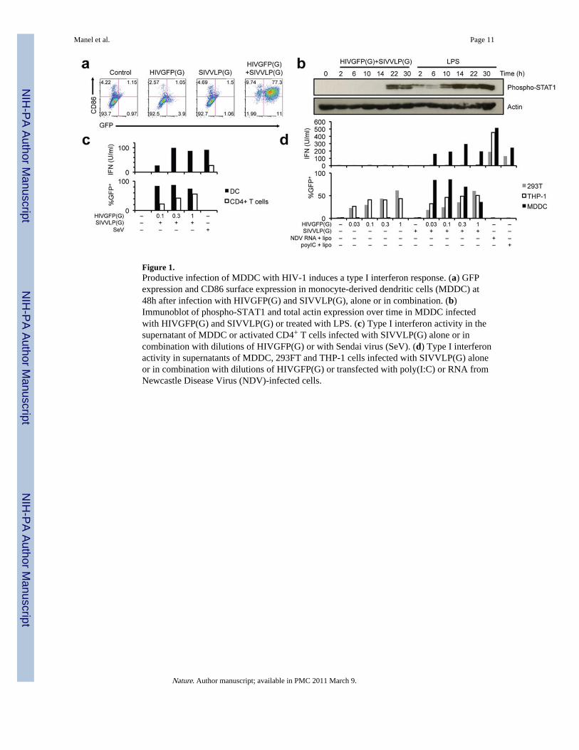
Figure 1.Productive infection of MDDC with HIV-1 induces a type I interferon response. (a) GFPexpression and CD86 surface expression in monocyte-derived dendritic cells (MDDC) at48h after infection with HIVGFP(G) and SIVVLP(G), alone or in combination. (b)Immunoblot of phospho-STAT1 and total actin expression over time in MDDC infectedwith HIVGFP(G) and SIVVLP(G) or treated with LPS. (c) Type I interferon activity in thesupernatant of MDDC or activated CD4+ T cells infected with SIVVLP(G) alone or incombination with dilutions of HIVGFP(G) or with Sendai virus (SeV). (d) Type I interferonactivity in supernatants of MDDC, 293FT and THP-1 cells infected with SIVVLP(G) aloneor in combination with dilutions of HIVGFP(G) or transfected with poly(I:C) or RNA fromNewcastle Disease Virus (NDV)-infected cells.
Manel et al. Page 11
Nature. Author manuscript; available in PMC 2011 March 9.
NIH
-PA Author Manuscript
NIH
-PA Author Manuscript
NIH
-PA Author Manuscript

Figure 2.Dendritic cell activation requires cyclophilin A interaction with newly-synthesized HIV-1capsid. (a) GFP and CD86 expression in MDDC infected with HIVGFP(G) or its mutants,ΔRev, ΔGag or PTAP− in the presence of SIVVLP(G). HIVGFP was rescued in all cases byco-expression of wild-type proteins in packaging cells. (b) Effect of CA mutations onproportion of GFP+ infected MDDC that express CD86. MDDC were infected with seriallydiluted wild-type (WT) pLaiΔEnv-GFP3(G) or CA mutants T54A/N57A, Q63A/Q67A orG89V, in the presence of SIVVLP(G). CA mutant infectivity was rescued by co-expressionof wild-type proteins in packaging cells. * p < 0.026 (n=9). (c) Effect of Cyclosporin A andFK506 on expression of CD86 in MDDC infected with HIVGFP(G) and SIVVLP(G) orafter treatment with LPS. CsA and FK506 target the calcineurin pathway but FK506 doesnot bind to CypA. (d) Expression of GFP, RFP and CD86 in HIV-infected cells followingCypA knock-down by RNAi. MDDC were transduced with GFP-encoding control shRNAvector or a shRNA vector targeting the CypA-encoding PPIA, in the presence ofSIVVLP(G), and subsequently challenged with HDVIRESRFP(G) or treated with LPS.Right panel: cells are gated on GFP+ populations shown in the left panels. Experiments wereperformed on a total of at least 6 donors, except (c) that was performed on 4 donors.
Manel et al. Page 12
Nature. Author manuscript; available in PMC 2011 March 9.
NIH
-PA Author Manuscript
NIH
-PA Author Manuscript
NIH
-PA Author Manuscript
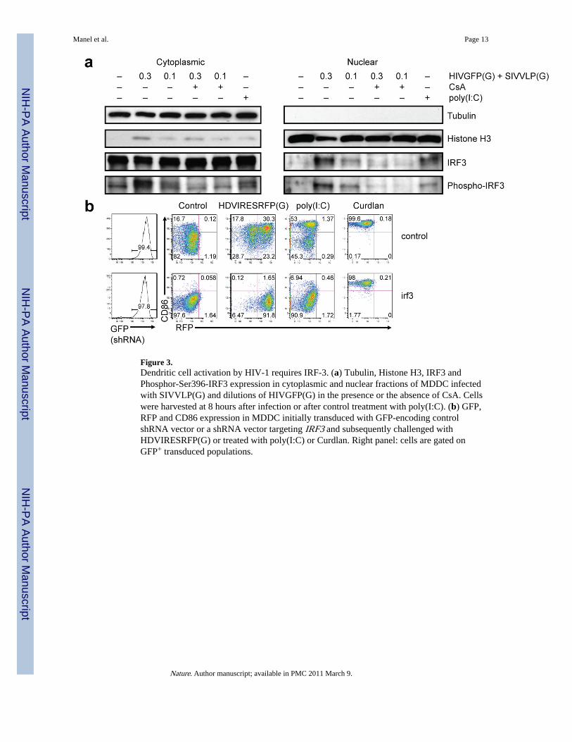
Figure 3.Dendritic cell activation by HIV-1 requires IRF-3. (a) Tubulin, Histone H3, IRF3 andPhosphor-Ser396-IRF3 expression in cytoplasmic and nuclear fractions of MDDC infectedwith SIVVLP(G) and dilutions of HIVGFP(G) in the presence or the absence of CsA. Cellswere harvested at 8 hours after infection or after control treatment with poly(I:C). (b) GFP,RFP and CD86 expression in MDDC initially transduced with GFP-encoding controlshRNA vector or a shRNA vector targeting IRF3 and subsequently challenged withHDVIRESRFP(G) or treated with poly(I:C) or Curdlan. Right panel: cells are gated onGFP+ transduced populations.
Manel et al. Page 13
Nature. Author manuscript; available in PMC 2011 March 9.
NIH
-PA Author Manuscript
NIH
-PA Author Manuscript
NIH
-PA Author Manuscript
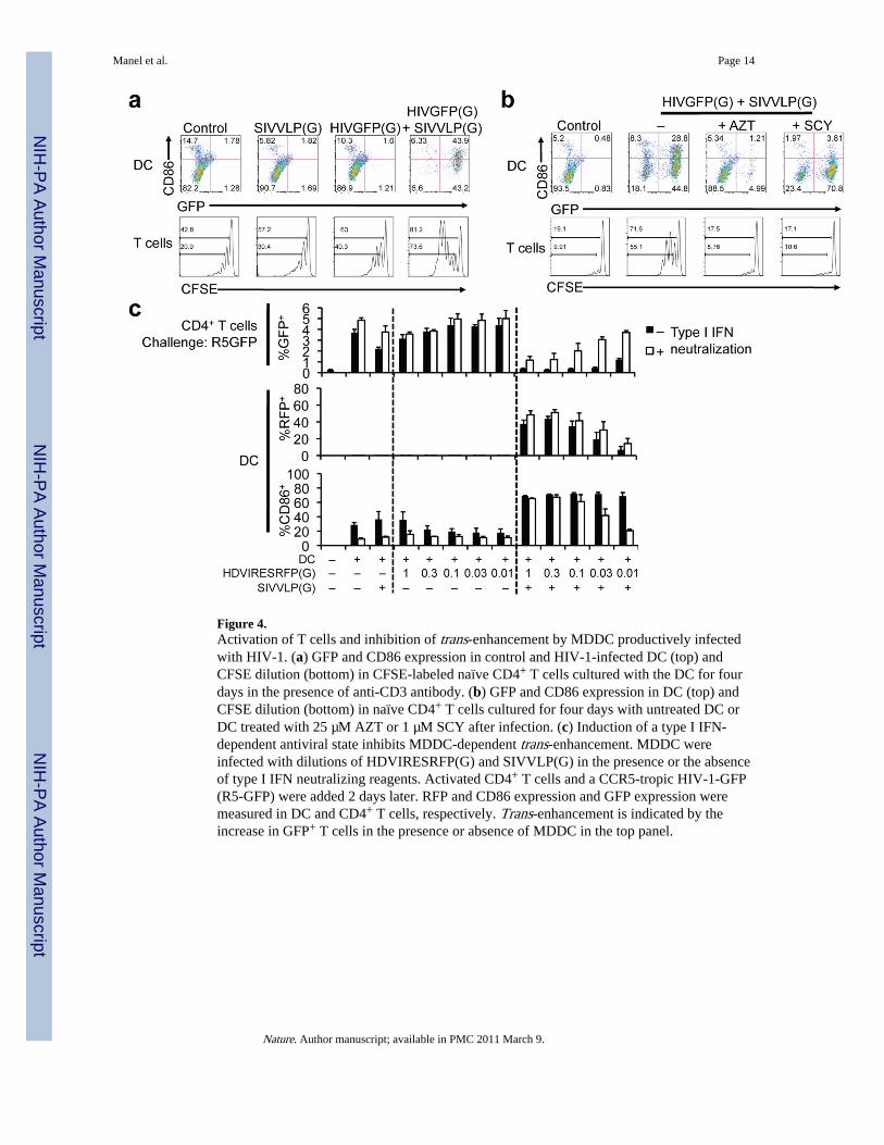
Figure 4.Activation of T cells and inhibition of trans-enhancement by MDDC productively infectedwith HIV-1. (a) GFP and CD86 expression in control and HIV-1-infected DC (top) andCFSE dilution (bottom) in CFSE-labeled naïve CD4+ T cells cultured with the DC for fourdays in the presence of anti-CD3 antibody. (b) GFP and CD86 expression in DC (top) andCFSE dilution (bottom) in naïve CD4+ T cells cultured for four days with untreated DC orDC treated with 25 µM AZT or 1 µM SCY after infection. (c) Induction of a type I IFN-dependent antiviral state inhibits MDDC-dependent trans-enhancement. MDDC wereinfected with dilutions of HDVIRESRFP(G) and SIVVLP(G) in the presence or the absenceof type I IFN neutralizing reagents. Activated CD4+ T cells and a CCR5-tropic HIV-1-GFP(R5-GFP) were added 2 days later. RFP and CD86 expression and GFP expression weremeasured in DC and CD4+ T cells, respectively. Trans-enhancement is indicated by theincrease in GFP+ T cells in the presence or absence of MDDC in the top panel.
Manel et al. Page 14
Nature. Author manuscript; available in PMC 2011 March 9.
NIH
-PA Author Manuscript
NIH
-PA Author Manuscript
NIH
-PA Author Manuscript
Copyright © 2022 FDOKUMEN