Project Manual For: The Bonneville Renovation - Idaho Falls, ID
Upload
khangminh22Category
view
2download
0
J. Cardiac Arrythmias, São Paulo, v32, n1, pp. 5-5 , Jan-Mar, 2019 5
The year of 2019 brought us unforeseeable surprises, new challenges, new goals, new
motivations. This is how we see the process going.
As of April, we made changes in the editorial staff. Ana Marlene F. Morais assumed
the executive editor of this journal, bringing her expertise and extensive experience. As a
member of the board of the Brazilian Association of Scientific Editors (ABEC Brasil),
she enjoys respect and broad knowledge in the area of scientific publishing. She began her
work with a series of suggestions that meet the expectations of technical and scientific
aggrandizement of the journal. The first one, and that has already materialized in this
first edition of this year, is the adequacy of formatting of the journal to the requirements
of indexation to important international databases. The second one also made concrete
in this edition, is the already ambitious transformation of the journal into a vehicle
published in English and entitled Journal of Cardiac Arrhythmias ( JCA). In fact, more
than that, from now on, the JCA will become a bilingual journal, published in Portuguese
and English. This, in addition to bringing necessary adaptations to future other indexing,
brings respectability to the journal and provides international visibility by allowing easy
access to your content in any country. This is our goal: to make JCA a vehicle of scientific
information that is respectable and attractive for good publications, access to all the
community of researchers lacking a journal in our specialized area in cardiac arrhythmias.
Happy reading!
A Constant Process of RenovationUm Processo Constante de Renovação
J. Tarcísio Medeiros de Vasconcelos
1.Centro Avançado de Ritmologia e Eletrofi siologia – São Paulo/SP – Brazil.
*Correspondence author: [email protected]
ORCID: Vasconcelos JTM https://orcid.org/0000-0002-5152-2648
EDITORIALhttps://doi.org/10.24207/jac.v32i1.995_IN
6 J. Cardiac Arrythmias, São Paulo, v32, n1, pp. 6-9 , Jan-Mar, 2019
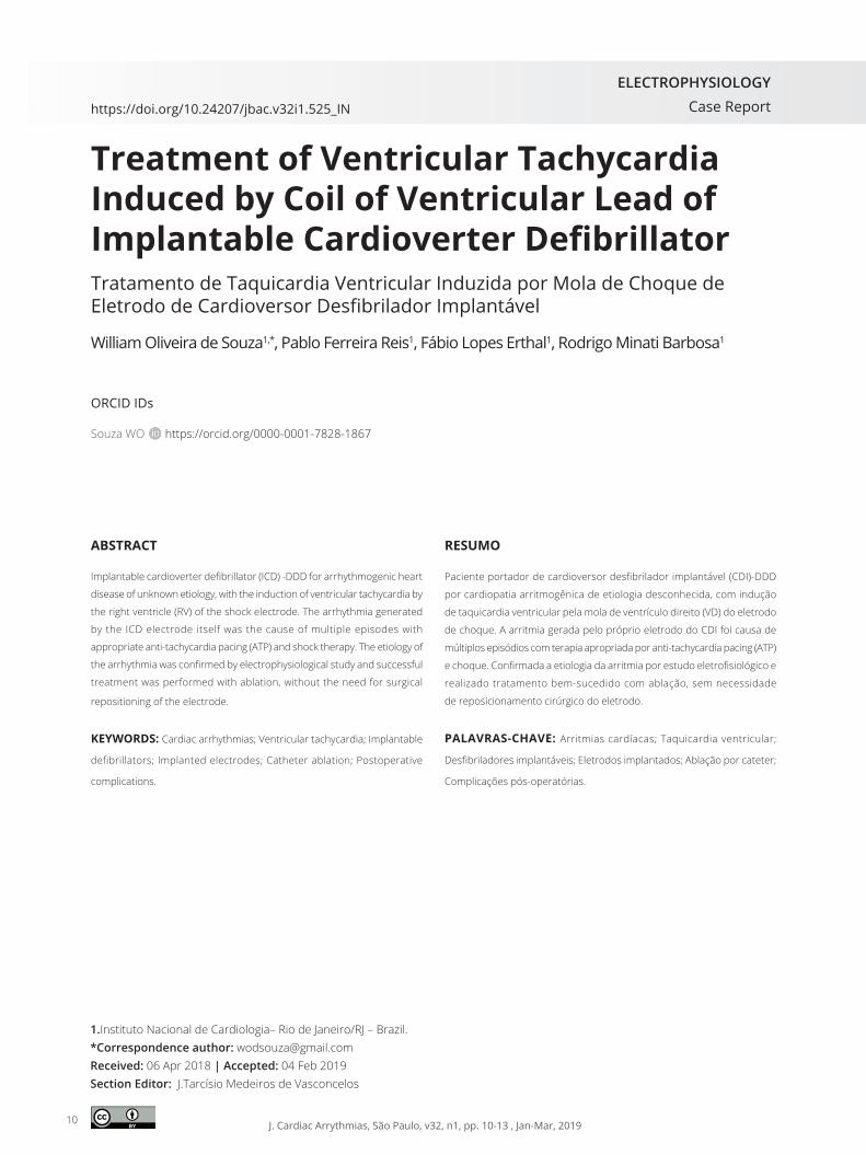
Cryoablation of the para-Hisian regionCrioablação da Região Para-Hissiana
Leonardo Rezende de Siqueira1,*, Nilson Araujo de Oliveira Junior1, Olga Ferreira de Souza1,Rodrigo Periquito Cosenza2, Martha Valéria Tavares Pinheiro1, Angelina Camiletti3
ELECTROPHYSIOLOGYOriginal article
1.Hospital Universitário Clementino Fraga Filho – Rio de Janeiro/RJ – Brazil.2.Hospital Federal da Lagoa – Rio de Janeiro/RJ – Brazil.3.Hospital Quinta D’Or – Rio de Janeiro/RJ – Brazil.*Correspondence author: [email protected]: 15 Apr 2018 | Accepted: 31 Jan 2019Section Editor: J. Tarcísio Medeiros de Vasconcelos
https://doi.org/10.24207/jac.v32i1.528_IN
ORCID IDs
Siqueira LR https://orcid.org/0000-0003-1206-0513
Oliveira Junior NA https://orcid.org/0000-0002-9964-6332
Souza OF https://orcid.org/0000-0001-8722-7504
Siqueira LR
Oliveira Junior NA
Souza OF
ABSTRACT
Basis: the ablation of the para-Hisian region is a challenge due to the risk of inadvertent lesion of a bundle of His. Cryoablation, due to its slower progression, allows interruption of the application in case of signs of undesired lesions and catheter adhesion during the applications, which has made cryoablation the ideal method for these patients. Objectives: to demonstrate the results of an initial series of patients referred for cryoablation of para-Hisian pathways. Patients and methods: From April 2015 to August 2017, 13 patients were referred for cryoablation due to the necessity for a para-Hisian approach detected in previous ablation procedures. Of the 13 patients, seven were submitted a radiofrequency ablation attempt (RF) and presented failure or recurrence, fi ve performed only electrophysiological studies, and no ablation was attempted, and one was indicated primarily. The mean age was 32 ± 16 years. Eleven patients had manifest anomalous pathways (APs), one hidden and one nodal reentrant tachycardia (NRT) with a transient atrioventricular block (AB) during RF. A cycle of 4 minutes followed by one more cycle in case of a positive result. Results: Of the 13 patients, 11 had an acute success in eliminating the accessory pathway. One patient had multiple accessory pathways, one right side, and one left side. In this patient, it was possible only the ablation of the left pathway. In all others, it was observed exuberant Hisian potential at the point of application with success. The patient with NRT was ablated in the M region without intercurrences. Four applications were required on average to eliminate the accessory pathway successfully. The mean local temperature was -74 ºC. In fi ve patients, the occurrence of third-degree right branch block (RBB) was observed. In one patient, early application of RBB was interrupted and the bonus application was not applied. This was the only acutely successful patient who presented clinical recurrence. Transient AB was not observed in any patient. No complications were observed. Conclusion: Cryoablation of para-Hisian pathways and NRTs in regions surrounding the His was an eff ective method for treatment inthis population of patients refractory or refused for RF treatment. The occurrence of acute RBB does not seem to be a criterion for the interruption of applications.
KEYWORDS: Cryoablation; Para-Hisian; Antero-septal pathways; Cryomapping; Cryotherapy.
RESUMO Fundamentos: A ablação da região para-Hissiana é um desafi o devido ao risco de lesão inadvertida do feixe de His. A crioablação, pela sua progressão mais lenta, permite a interrupção da aplicação em caso de sinais de lesões indesejadas e adesividade do cateter durante as aplicações, o que tem tornado a crioablação o método ideal para esses pacientes. Objetivos: Demonstrar os resultados de uma série inicial de pacientes encaminhados para crioablação de vias para-hissianas. Pacientes e métodos: De abril de 2015 a agosto de 2017, 13 pacientes foram encaminhados para crioablação devido à necessidade de abordagem para-hissiana detectada em procedimentos prévios de ablação. Dos 13 pacientes, sete foram submetidos à tentativa de ablação por radiofrequência (RF) e apresentaram insucesso ou recidiva, cinco realizaram apenas estudos eletrofi siológicos, não sendo tentada a ablação, e um foi indicado primariamente. A idade média era 32 ± 16 anos. Onze pacientes tinham vias anômalas (VAs) manifestas, um oculta e um taquicardia por reentrada nodal (TRN) com sinais de bloqueio atrioventricular (AV) transitório durante RF. Aplicava-se um ciclo de 4 minutos seguido de mais um ciclo em caso de resultado positivo. Resultados: Dos 13 pacientes, 11 apresentaram sucesso agudo em eliminar a via acessória. Um paciente tinha múltiplas vias acessórias, sendo uma lateral direita e uma lateral esquerda. Nesse paciente foi possível apenas a ablação da via esquerda. Em todos os demais foi observado exuberante potencial hissiano no ponto de aplicação com sucesso. O paciente com TRN foi ablacionado na região M sem intercorrências. Foram necessárias quatro aplicações em média para eliminação da via acessória com sucesso. A temperatura local média foi de -74 ºC. Em cinco pacientes foi observada a ocorrência de bloqueio do ramo direito (BRD) de terceiro grau. Em um paciente foi interrompida a aplicação precocemente pelo BRD e não foi realizada a aplicação de bônus. Esse foi o único paciente com sucesso agudo que apresentou recidiva clínica. Em nenhum paciente foi observado BAV transitório. Não foram observadas complicações. Conclusão: A crioablação de vias para-hissianas e TRN em regiões mais circunvizinhas do His foi um método efi caz para tratamento nessa população de pacientes refratários ou recusados para tratamento por RF. A ocorrência de BRD agudo não parece um critério para interrupção das aplicações.
PALAVRAS-CHAVE: Crioablação; Para-Hissiana; Vias ântero-septais; Criomapeamento; Crioterapia.
Cosenza RP https://orcid.org/0000-0002-2914-1048
Pinheiro MVT https://orcid.org/0000-0003-1144-9252
Camiletti A https://orcid.org/0000-0002-7867-6794

J. Cardiac Arrythmias, São Paulo, v32, n1, pp. 6-9 , Jan-Mar, 2019 7
Siqueira LR, Oliveira Junior NA, Souza OF, Cosenza RP, Pinheiro MVT, Camiletti A
INTRODUCTION
Incidence of accessory pathways was estimated to be three-four per 1.000 live births. The anomalous (AP) or accessory pathways are muscle bundles that electrically connect the atrium to the ipsilateral ventricle, permitting abnormal electrical conduction despite physiological atrioventricular nodal (AV) conduction. It is believed that they are formed by failure during the embryonic segmentation of the cardiac tube that forms atrium and ventricles, causing muscular connection that goes beyond the fibrous valvar ring. APs can be located in the mitral annulus (65% of cases) or in the tricuspid annulus. It is estimated that 25% of the pathways are septal and about 10% are positioned in the anteroseptal region near the bundle of His.1
Surgical treatment of accessory pathways by open surgery and subsequent electrofulguration ablation were rapidly replaced by the development of radiofrequency ablation (RF) in 1987. The high success rates and low risk of complications, when compared to previous techniques, promoted the great dissemination of the method and support the current guidelines for the treatment of pre-excitation syndromes. The failures of the procedure relate mainly to the unfavorable location of the accessory pathway in a minority of patients. The para-Hisian location is related in large series to a risk of up to 20% of total VA block development during the release of the RF pulse.
The RF current is an alternating high frequency unipolar electric current between the distal electrode of the ablation catheter and a large surface indifferent electrode positioned in contact with the patient’s skin. The tissue lesion is caused by the resistive heating of the tissue in contact with the tip of the catheter and probably also the direct electrical effect. The produced cellular aggression can install quickly and irreversibly but tends to be deeper and more stable, depending on the time of application, temperature and power reached and the contact of the catheter with the tissue.
Cryoablation was initially developed to replace surgical resection of tumors. The first catheters for cryoablation of arrhythmias began to be produced in the 1990s.3 Catheters with 4 and 6 mm tip are currently used for point ablation and balloon catheters for isolation of pulmonary veins.
To produce catheter cooling, liquid nitrogen is pumped into the catheter, with internal evaporation occurring at
its tip, which lowers the local temperature to –80 oC. Before producing irreversible lesion, there is the possibility of cryomapping. Cryomapping consists of cooling the tissue to –30 oC for up to 60 seconds, creating a totally reversible lesion. A great advantage of the technique is the cryoadhesion phenomenon, which promotes great stability of the catheter when cooled, with freezing of the tissue and electrode tip in the contact region.4
The cold cell lesion is composed of three phases: freezing, inflammatory and hemorrhagic, and the replacement of acute fibrosis lesion. The freezing phase generates cell death by the mitochondrial lesion. The inflammatory phase occurs within the first 48 hours of application. Local fibrosis occurs between the first and the 24th week. The extracellular matrix remains intact and there is little or no endothelial lesion, which reduces the risk of thromboembolism. Cryoablation lesion does not increase after the end of the application, which may occur with RF lesion
The described characteristics of cold ablation rapidly promoted studies with nodal reentry tachycardia ablation (NRT), accessory pathway mediated tachycardias and atrial fibrillation that revealed a high cure rate and safety of the method, comparable to RF ablation. The use of para-Hisian ablation has aroused special interest in the theoretical possibility of avoiding irreversible damage to the driving system and increasing the success rates of these procedures. Several series of cases of cold ablation of para-Hisian pathways have been described.
It describes in this article the consecutive initial series of patients submitted to cryoablation of the para-Hisian region.
METHODS
A series of 13 consecutive cases of patients submitted to cryoablation for para-Hisian pathways from April 2015 to August 2017.
Thirteen patients were referred for cryoablation due to the necessity for a para-Hisian approach detected in previous ablation procedures.
Of the 13 patients, seven were submitted RF ablation attempt and failed or relapsed, five performed only electrophysiological studies and no ablation was attempted, and one was indicated primarily for cryoablation based on

8 J. Cardiac Arrythmias, São Paulo, v32, n1, pp. 6-9 , Jan-Mar, 2019
Cryoablation of the para-Hisian region
the presumed location of the accessory pathway based on the electrocardiogram (EKG ). Th e mean age was 32 ± 16 years.
Eleven patients had a manifest anomalous pathway of ventricular pre-excitation to the EKG, one had an anomalous occult pathway and one NRT AP with transient AP block signs during a previous RF ablation procedure.
The procedures were performed under sedation guided by an anesthesiologist. Th e right femoral access was used. A cryo cycle of 4 minutes followed by another cycle of 4 minutes in case of a positive result. In 12 of the 13 patients, the applications were complete. Patient follow-up time was eight to 36 months.
RESULTS
Th e Figs. 1 to 3 show the obtained results. Of the 13 patients, 11 had an acute success in eliminating the accessory pathway. One patient actually had multiple accessory pathways, one right side, and one left side. In this patient, it was possible only the ablation of the left pathway. In all others, it was observed exuberant Hisian potential at the point of application with success.
Th e patient with NRT was submitted to ablation in the M region without intercurrences with the disappearance of the nodal shift. Th ere was no induction of active junctional rhythm during cryoablation. After the fi rst application, slow pathway ablation was identifi ed and the second application of 4 minutes was performed.
It took 4 applications on average to eliminate conduction by the accessory pathway. Th e average local temperature was –74°C. In fi ve patients, the occurrence of third-degree right branch block (RBB) was observed during the application.
Th e mean duration of the procedures was 52 minutes. Th e mean scan time was 6 minutes.
In one patient, the RBB application was stopped early (130 seconds) and the booster application was not perfor-med. Th e return of ventricular pre-excitation was observed 14 days after the procedure. Th is was the only acutely suc-cessful patient who presented recurrent pre-excitation or recurrence of palpitation symptoms.
No transient AP block of any degree was observed in any patient. Measurements of the HV intervals did
Figure 1. Local potential before application of cryoablation.Figure 3. Hisian potential at the cryoablation site after ventricular pre-excitation disappeared.
Figure 2. Disappearance of pre-excitation during cryoablation.

J. Cardiac Arrythmias, São Paulo, v32, n1, pp. 6-9 , Jan-Mar, 2019 9
Siqueira LR, Oliveira Junior NA, Souza OF, Cosenza RP, Pinheiro MVT, Camiletti A
not reveal abnormal values after the applications. No complications were observed in the short and medium term. The patients did not a present recurrence of tachycardia or ventricular pre-excitation or AP block at follow-up.
DISCUSSION
Para-Hisian accessory pathways have always been a challenge for electrophysiology in the era of RF ablation because of the risk of irreversible AP node damage during RF pulse release. The AP node lesion can install quickly without warning signs.
Several RF mapping and ablation techniques have been described in an attempt to increase the success of ablation and decrease the risk of the procedure in the right anteroseptal pathways. Mapping and ablation of the right coronary cusp, use of electroanatomic mapping and energy release with low power, approach by superior access (jugular or subclavian) and use of magnetic navigation system were published in case reports.
The cryoablation procedure is very similar to the RF ablation procedure in relation to the access pathway,
sedation and the use of radioscopy. The handling of the cryoablation catheter is very similar to that of the RF catheter, which makes the technique easy to perform for licensed electrophysiologists.
Cryoablation has proven to be an effective and safe method in arrhythmias in which the region to be injured is contiguous to the bundle of His. The technique proved safe even in children and with high rates of long-term cure in several series of patients reported in the literature. The cryoablation procedure is similar to the RF ablation pattern with respect to femoral venous access and the use of radioscopy.
There are no direct published comparative studies of RF ablation and cryoablation. Our initial case series shows a high success rate and safety of cryoablation in para-Hisian accessory pathways in accordance with current literature.
AUTHORS’ CONTRIBUTION
All the authors contributed equally to this article.
1. Yildirim I, Karagöz T, Ertuğrul I, Karagöz AH. Özer S. Efficacy and safety of cryoablation of parahissian accessory pathways in children: A Single Institution Study. Pacing Clin Electrophysiol. 2013;36(12)1495-502. https://doi.org/10.1111/pace.12268
2. Cay S, Aras D, Topaloglu S, Ozcan F, Ozeke O. Various routes and techniques for ablation of parahisian bypass tracts. Int J Cardiol. 2016;223:217. https://doi.org/10.1016/j.ijcard.2016.08.257
3. Liao Z, Zhan X, Wu S. Successful radiofrequency ablation
of a parahisian accessory pathway from the right coronary
cusp. Int J Cardiol. 2015;186:41-2. https://doi.org/10.1016/
j.ijcard.2015.03.231
4. Zeljko HM, Yue A. Europace. 2015;17(11):1707. https://doi.
org/10.1093/europace/euv254
REFERENCES
10 J. Cardiac Arrythmias, São Paulo, v32, n1, pp. 10-13 , Jan-Mar, 2019
Treatment of Ventricular Tachycardia Induced by Coil of Ventricular Lead of Implantable Cardioverter Defi brillatorTratamento de Taquicardia Ventricular Induzida por Mola de Choque de Eletrodo de Cardioversor Desfi brilador Implantável
William Oliveira de Souza1,*, Pablo Ferreira Reis1, Fábio Lopes Erthal1, Rodrigo Minati Barbosa1
ELECTROPHYSIOLOGYCase Report
1.Instituto Nacional de Cardiologia– Rio de Janeiro/RJ – Brazil.*Correspondence author: [email protected]: 06 Apr 2018 | Accepted: 04 Feb 2019Section Editor: J.Tarcísio Medeiros de Vasconcelos
https://doi.org/10.24207/jbac.v32i1.525_IN
ORCID IDs
Souza WO https://orcid.org/0000-0001-7828-1867
ABSTRACT
Implantable cardioverter defi brillator (ICD) -DDD for arrhythmogenic heart
disease of unknown etiology, with the induction of ventricular tachycardia by
the right ventricle (RV) of the shock electrode. The arrhythmia generated
by the ICD electrode itself was the cause of multiple episodes with
appropriate anti-tachycardia pacing (ATP) and shock therapy. The etiology of
the arrhythmia was confi rmed by electrophysiological study and successful
treatment was performed with ablation, without the need for surgical
repositioning of the electrode.
KEYWORDS: Cardiac arrhythmias; Ventricular tachycardia; Implantable
defibrillators; Implanted electrodes; Catheter ablation; Postoperative
complications.
RESUMO
Paciente portador de cardioversor desfi brilador implantável (CDI)-DDD
por cardiopatia arritmogênica de etiologia desconhecida, com indução
de taquicardia ventricular pela mola de ventrículo direito (VD) do eletrodo
de choque. A arritmia gerada pelo próprio eletrodo do CDI foi causa de
múltiplos episódios com terapia apropriada por anti-tachycardia pacing (ATP)
e choque. Confi rmada a etiologia da arritmia por estudo eletrofi siológico e
realizado tratamento bem-sucedido com ablação, sem necessidade
de reposicionamento cirúrgico do eletrodo.
PALAVRAS-CHAVE: Arritmias cardíacas; Taquicardia ventricular;
Desfi briladores implantáveis; Eletrodos implantados; Ablação por cateter;
Complicações pós-operatórias.
J. Cardiac Arrythmias, São Paulo, v32, n1, pp. 10-13 , Jan-Mar, 2019 11
Treatment of Ventricular Tachycardia induced by Coil of Ventricular Lead of Implantable Cardioverter Defibrillator
INTRODUCTION
An interesting case is reported by the induction of ventricular arrhythmia by the device that aims to treat it. The treatment was performed with radiofrequency catheter ablation, without the need for surgical repositioning of the electrode.
METHODS
The patient is followed up at the Arrhythmia Service of the Instituto Nacional de Cardiologia (National Institute of Cardiology), in the state of Rio de Janeiro. The case was set up with a review of records, electrophysiological studies, and anamnesis. Bibliographical review1-3. Search in the PubMed database with the terms «implantable cardioverter defibrillator lead complications», «icd lead replacement», «icd lead-induced arrhythmia», «icd coil induced arrhythmia» did not return similar cases until 04/05/2018.
CASE REPORT
Patient ACJ, male, 45 years old in 2010, attended in a public emergency care unit with sustained monomorphic ventricular tachycardia (SMVT) and hemodynamic instability, being treated with electrical cardioversion (ECV) and the event classified as analogous to aborted sudden death. Referred to specialized service of the Sistema Único de Saúde-SUS (Unified Health System). In April 2011, he was admitted to the Arrhythmia Service of the Instituto Nacional de Cardiologia and was hospitalized for further investigation. The examinations at the time showed echocardiogram with the absence of structural heart disease, normal coronariography, ergometric test with ventricular bigeminism at the peak of the effort (9.6 mets) and two 24-hour Holter exams with several episodes of nonsustained ventricular tachycardia (NSVT). Was chosen an electrophysiological study with SMVT re-induction, an initially stable cycle of 290 ms (207 bpm), with acceleration and instability after anti-tachycardia pacing (ATP) with a need for ECV. In the same hospitalization, the patient was implanted with an implantable cardioverter defibrillator (ICD) - DDD
with a double-spring shock electrode (Biotronik Lumax 340 DRT, Biotronik Linox SD 65/16 shock electrode, Briotronik Setrox S53 atrium electrode).
In December 2011, the patient was re-admitted with an electrical storm picture by multiple SMVT. Interrogation of the device demonstrated that all therapies were appropriate. After adjustment of drugs and cessation of arrhythmias, the patient was discharged. At the time, the possibility of an electrophysiological study and ablation (EPS/ABL) was questioned, depending on the posterior clinical evaluation. However, it evolved without new arrhythmias until the generation unit was exhausted in 2014, is submitted to the exchange of this (Medtronic Virtuoso II DR), maintaining the electrodes.
In February 2017, the patient sought care due to a shock. Interrogation of the device revealed about 200 episodes of tachyarrhythmia, most of NSVT, a minority of SMVT interrupted with ATPs and one episode of failure in successive ATPs and appropriate shock. New EPS/ABL was scheduled within one week and the drugs were adjusted, but the patient progressed without any arrhythmia episode, and the procedure for observation was canceled.
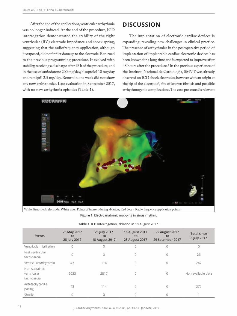
In August of the same year, the patient returned with> 2,000 episodes of NSVT and SMVT. All sustained arrhythmias were discontinued ATPs. New echocardiogram maintained the absence of identifiable structural heart disease. It was then submitted to EPS/ABL with electroanatomic mapping. Prior to the procedure, ICD therapies were deactivated and the unit reprogrammed to VVI mode 30 bpm (pacing suppression), and the patient was in sinus rhythm throughout the procedure. Voltage map showed an absence of endocardial scars. EPS easily induced NSVT and SMVT, with a cycle of 315 ms (190 bpm), hemodynamic stability and interruption with ATP. The electroanatomic mapping observed a region of greater precocity attached to the proximal portion of the RV shock spring. In this region, precocity, mesodiastolic potential and 12/12 similarity in the pace mapping maneuver were observed. This region was determined as the focus of the presented arrhythmia, radiofrequency applications juxtaposed to the shock spring endocardially on both sides of it as a «rail», with a higher concentration of applications in the medial and proximal aspect of the spring (Fig.1).
12 J. Cardiac Arrythmias, São Paulo, v32, n1, pp. 10-13 , Jan-Mar, 2019
Souza WO, Reis PF, Erthal FL, Barbosa RM
After the end of the applications, ventricular arrhythmia was no longer induced. At the end of the procedure, ICD interrogation demonstrated the stability of the right ventricular (RV) electrode impedance and shock spring, suggesting that the radiofrequency application, although juxtaposed, did not inflict damage to the electrode. Returned to the previous programming procedure. It evolved with stability, receiving a discharge after 48 h of the procedure, and in the use of amiodarone 200 mg/day, bisoprolol 10 mg/day and ramipril 2.5 mg/day. Return in one week did not show any new arrhythmias. Last evaluation in September 2017, with no new arrhythmia episodes (Table 1).
DISCUSSION
The implantation of electronic cardiac devices is expanding, revealing new challenges in clinical practice. The presence of arrhythmias in the postoperative period of implantation of implantable cardiac electronic devices has been known for a long time and is expected to improve after 48 hours after the procedure.4 In the previous experience of the Instituto Nacional de Cardiologia, SMVT was already observed on ICD shock electrodes, however with an origin at the tip of the electrode5, site of known fibrosis and possible arrhythmogenic complications. The case presented is relevant
Table 1. ICD Interrogation, ablation in 18 August 2017.
Events26 May 2017
to 28 July 2017
28 July 2017 to
18 August 2017
18 August 2017 to
25 August 2017
25 August 2017 to
29 Setember 2017
Total since 8 July 2017
Ventricular fibrillation 0 0 0 0 0
Fast ventricular tachycardia
0 0 0 0 26
Ventricular tachycardia 43 114 0 0 247
Non-sustained ventricular tachycardia
2033 2817 0 0 Non-available data
Anti-tachycardia pacing
43 114 0 0 272
Shocks 0 0 0 0 1
Figure 1. Electroanatomic mapping in sinus rhythm.
White line: shock electrode; White dots: Points of interest during ablation; Red dots = Radio frequency application points.

J. Cardiac Arrythmias, São Paulo, v32, n1, pp. 10-13 , Jan-Mar, 2019 13
Treatment of Ventricular Tachycardia induced by Coil of Ventricular Lead of Implantable Cardioverter Defibrillator
for the documentation of arrhythmia caused by the contact of the shock spring with the endocardium. The presence of precocity to the electroanatomic mapping, as well as the successful treatment with ablation in the target region, strongly suggests the origin of this arrhythmia in the RV shock spring. It is not clear whether the arrhythmia, in this case, was caused only by the presence of contact of the shock spring with the endocardium or if the patient›s previous arrhythmogenic condition contributed to its induction, since the etiology of the arrhythmia that led to the index event in 2010 and the electrical storm in 2011 was not defined. It is important to point out that the SMVT morphologies obtained in the EPS of 2011 and 2017 are different in cycles and morphologies, suggesting distant foci.
Radiofrequency catheter ablation was effective in suppressing the occurrence of repeated NSVT episodes and inappropriate therapies, without the need for surgical repositioning of the shock electrode.
ACKNOWLEDGMENTS
To Prof. Bernardo Rangel Tura, for the review of the article.
AUTHORS’ CONTRIBUTION
All the authors contributed equally to this article.
1. Ellenbogen KA, Neal Kay G, Lau CP. Clinical cardiac pacing, defibrillation, and resynchronization therapy. 3a ed. Philadelphia: Saunders; 2007.
2. Melo CS. Tratado de estimulação cardíaca artificial. 5a ed. Tamboré: Manole; 2015.
3. Al-Khatib SM, Stevenson WG, Ackerman MJ, Bryant WJ, Callans DJ, Curtis AB, et al. 2017 AHA/ACC/HRS guideline for management of patients with ventricular arrhythmias and the prevention of
sudden cardiac death. Circulation. 2018;138(13):e272-e391.
https://doi.org/10.1161/CIR.0000000000000549
4. Brito Junior HL, Gauch PRA, Oliveira AS de. Arritmias
induzidas por marcapasso cardíaco. JBAC. 1990;3(3):88-93.
5. Souza WO, Seifert MMS, Pinho DL, Nascimento EAD, Saad
EB, et al. Anais do 28o Congresso de Cardiologia da Socerj;
3-5 de Agosto de 2011; Rio de Janeiro.
REFERENCES
14 J. Cardiac Arrythmias, São Paulo, v32, n1, pp. 14-16 , Jan-Mar, 2019
What is the Diagnosis?
CLINICAL ARRHYTHMIAChallengehttps://doi.org/10.24207/jac.v32i1.996_IN
CASE PRESENTATION
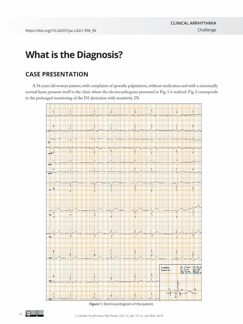
A 54 years old woman patient, with complaints of sporadic palpitations, without medication and with a structurally normal heart, presents itself in the clinic where the electrocardiogram presented in Fig. 1 is realized. Fig. 2 corresponds to the prolonged monitoring of the D2 derivation with sensitivity 2N.
Figure 1. Electrocardiogram of the patient.

J. Cardiac Arrythmias, São Paulo, v32, n1, pp. 14-16 , Jan-Mar, 2019 15
What is the Diagnosis?
RESPOSTA
On the 12 derivations electrocardiogram (EKG) (Fig. 1), a sinus rhythm can be visualized with atrial bigeminism, and the atrial extrasystole presents a long coupling interval in relation to the previous P wave. For a better understanding of the case, monitoring with long D2 and 2N sensitivity was realized (Fig. 2). In this monitoring, the atrial ectopias (*in Fig. 2) begin to show coupling variations in relation to the previous signal wave P (3 and 4) and begin an alternation with an ectopic atrial rhythm (5 and 6) that presents the same morphology of the extrasystolic beats, suggesting that the origin is the same in both situations. Still in Fig. 2, observing the sequence (5) of ectopic beats, rhythmicity with a frequency of 880 ms is observed interrupted by an absence of ectopic beat (** in Fig. 2) followed by sinus rhythm with a frequency lower than of the ectopic focus. In sequence 6, an ectopic beat is followed by sinus beat, returning to the ectopic rhythm. Due to the rhythmicity of the ectopias, it is observed that in some moments this shows loss of atrial capture, which evidences a phenomenon of parasystole with intermittent output block.
Parasystole arises due to the existence of one or more cardiac cells with automatic properties protected from the basic rhythm by an input block. In this case, this is observed because the sinus beat does not restart the ectopic focus cycle (Fig. 2). � e dominant pacemaker is unable to excite this area. Concomitantly, there is a variable and intermittent output block, which prevents the depolarizing impulse there originating from reaching the underlying musculature on several occasions. It is also possible to suggest that the focus of the parasystole is in the right atrium region, the appearance of the P wave morphology of the ectopic focus with that of the P w ave in sinus rhythm, but with a lower amplitude in the inferior leads, suggesting a location below the node.1
Figure 2. Prolonged monitoring of the D2 derivation with sensitivity 2N.
Explanatory diagram of the electrophysiological phenomenon during the above beats
A
Junction
Ectopia

16 J. Cardiac Arrythmias, São Paulo, v32, n1, pp. 14-16 , Jan-Mar, 2019
Durval Jr J, Godinho J, Padilha J
1.Centro Universitário São Lucas – Porto Velho/RO – Brazil.2.Real e Benemérita Associação Portuguesa de Benefi cência São Paulo – São Paulo/SP – Brazil.3.Centro Avançado de Ritmologia e Eletrofi siologia – São Paulo/SP – Brazil.4.Centro Universitário UNINOVAFAPI – Teresina/PI – Brazil.5.Universidade Federal de São Paulo – São Paulo/SP – Brazil.*Correspondence author: [email protected]
Durval Jr J https://orcid.org/0000-0002-9484-0013
Godinho J https://orcid.org/0000-0001-5681-0924
Padilha J https://orcid.org/0000-0002-8761-6013
Durval Jr J
Godinho J
Padilha J
João Durval Jr.1,2,3*, Jardel Godinho2,3,4, Jaqueline Padilha5
1. Josephson ME; Wellens JJH.Tachycardias: Mechanisms, diagnosis, treatment. Philadelphia, PA: Lea & Febiger; 1984 p.287-30.
REFERENCES
AUTHORS
Case kindly given by Prof. Dr. José Tarcísio Medeiros de Vasconcelos from Centro Avançado de Ritmologia e Eletro� siologia (C.A.R.E.), São Paulo/SP, Brazil.
ACKNOWLEDGMENTS

J. Cardiac Arrythmias, São Paulo, v32, n1, pp. 17-24 , Jan-Mar, 2019 17
Puncture of the Axillary Vein for the Implant for Electronic Cardiac DevicesPunção da Veia Axilar para o Implante de Dispositivos Cardíacos Eletrônicos
Vagner Rossato Pegoraro1,*, E duardo Rodrigues Bento Costa1, Luiz Fernando FagundesGouvea Filho1, Beatriz Tose Costa Paiva2
ARTIFICIAL HEART STIMULATIONReview Article
1.CardioRitmo – Clínica de Arritmias Cardíacas – São José dos Campos/SP – Brazil.2.REGIOMED Klinikum – Coburg – Germany.*Correspondence author: [email protected]: 18 Feb 2018 | Accepted: 09 Jul 2018Section Editor: J. Tarcisio Medeiros de Vasconcelos
https://doi.org/10.24207/jac.v32i1.511_IN
ORCID IDs
Pegoraro VR https://orcid.org/0000-0003-3448-320X
Costa ERB https://orcid.org/0000-0002-3342-5369
Gouvea Filho LFF https://orcid.org/0000-0002-5199-1233
Paiva BTC https://orcid.org/0000-0002-2516-1770
Pegoraro VR
Costa ERB
Gouvea Filho LFF
Paiva BTC
ABSTRACT
Introduction: The obtaining of venous access for implantation of implantable
electronic cardiac devices (IECDs) has been traditionally made by intra-
thoracic subclavian vein puncture (SVP) or cephalic vein phlebotomy (CVP).
Evidence indicates, however, the increased risk of short-term and long-term
complications with SVP due to the fact that it is intrathoracic access and
the risk of compression of the electrodes by the costoclavicular ligament,
leading to diff erent types of defects. CVP, in turn, has been associated
with a failure rate that reaches 45%. Axillary vein puncture (AVP) has been
described in the literature and is presented here as an alternative to the
two techniques mentioned. Methods: A PubMed survey was conducted
on articles that mention the AVP, SVP and CVP techniques and compare
them to the immediate, short and long term results and success rates for
obtaining venous access. Emphasis was placed on comparisons between
the various AVP techniques. Conclusion: The AVP technique for obtaining
venous access presents some variations among the diff erent authors. It has
CVP-like safety, success rates comparable to those of the subclavian vein,
and better medium and long term results for electrode function.
KEYWORDS: Axillary vein puncture; Cephalic vein phlebotomy; Subclavian
vein puncture; Complications with pacemaker implantation.
RESUMO
Introdução: A obtenção do acesso venoso para implante de dispositivos cardíacos eletrônicos implantáveis (DCEIs) tem sido tradicionalmente feita por meio da punção da veia subclávia intratorácica (PVS) ou por flebotomia da veia cefálica (FVC). Evidências apontam, entretanto, para o risco aumentado de complicações a curto e longo prazos com a PVS pelo fato de ser um acesso intratorácico e pelo risco de compressão dos eletrodos pelo ligamento costoclavicular, levando a diferentes tipos de defeitos. A FVC, por sua vez, tem sido associada à taxa de insucesso que chega a 45%. A punção da veia axilar (PVA) tem sido descrita na literatura e é apresentada, aqui, como alternativa às duas técnicas mencionadas. Métodos: Realizou-se uma pesquisa pelo PubMed sobre artigos que mencionam as técnicas de PVA, PVS e FVC e que as comparam quanto aos resultados imediatos, a curto e longo prazos e taxas de sucesso para a obtenção do acesso venoso. Deu-se ênfase às comparações entre as diversas técnicas de PVA. Conclusão: A técnica de PVA para obtenção do acesso venoso apresenta algumas variações entre os diversos autores. Ela tem segurança semelhante à da FVC, taxas de sucesso comparáveis às da veia subclávia e melhores
resultados a médio e a longo prazos para a função dos eletrodos.
PALAVRAS-CHAVE: Punção da veia axilar; Flebotomia da veia cefálica;
Punção da veia subclávia; Complicações com implante de marcapassos.

18 J. Cardiac Arrythmias, São Paulo, v32, n1, pp. 17-24 , Jan-Mar, 2019
Pegoraro VR, Costa ERB, Gouvea Filho LFF, Paiva BTC
INTRODUCTION
Obtaining venous access for implantation of implantable cardiac devices (ICDs) is an essential part of the procedure. The choice of puncture technique should take into account factors such as the chance of success, the risks of immediate and future complications and the time required to obtain them. Several techniques have been described, all with their particularities and limitations. The intrathoracic subclavian vein puncture (SVP) technique was introduced by Littleford et al.1, in 1979. It was widely accepted because it is fast, easy to learn, and has high success rates. Thus, it has been the most widely used electrode implant method in the world2-4. In Brazil, this is also the most used venous access, followed by cephalic vein phlebotomy (CVP). Subclavian access, however, is associated with a greater risk of both immediate complications - pneumothorax, hemothorax, arterial puncture, brachial plexus injury - and late - insulation defects, electrode fractures, capture losses, abnormal impedances and sensing failures5,6. CVP, although quite safe, has been less and less used due to the failure rate that varies from 15 to 45%7.In this work, we will review the axillary vein puncture technique (AVP), presenting the similarities and variations between the different authors, as well as their respective success rates, and compare it with the other techniques.
METHODS
A PubMed survey was conducted on articles that mention AVP techniques. Those who described the AVP techniques or those who compared them to those of SVP or CVP were selected for immediate, short- and long-term results, and success rates for obtaining venous accesses. The survey covers articles published between 1979 and 2017. Emphasis was placed on comparisons between the various AVP techniques.
AVP
The axillary vein originates from the junction of the cephalic and basilic veins. It extends to the lower margin of the first rib where it continues as the subclavian vein ending with its junction with the internal jugular8.
AVP can be performed using contrast venography, contrast-free fluoroscopy, ultrasonography, or even anatomical landmarks only.
For fluoroscopy-guided AVP, data from venography studies that evaluate the usual path of the axillary vein are used. One demonstrated that the axillary vein runs parallel to the deltopectoral sulcus (DPS) between one finger (1.85 cm) and one finger and a half (2.8 cm) more medially and follows its course towards the most prominent point of the clavicle (MPPC)9.
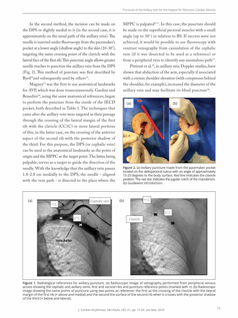
This MPPC approximately corresponds to the crossing of the clavicle with the lateral margin of the first rib10,11. The axillary vein in its course parallel to the DPS also passes over the anterior body of the second rib, at the point where it crosses over the posterior shadow of the third rib (lateral radiological limit of the rib cage). Thus, with fluoroscopy, the needle can be directed to one of these two points from the pacemaker pocket (Fig. 1).
To reach these points, several authors have used varied techniques that can be generally grouped into two methods. In the first one, it begins by making the incision to the IECD pocket below (1.5-2 cm) and parallels to the clavicle, with this extending to the DPS. Then, the puncture needle is coupled to a syringe and puncture is performed from the IECD pocket. The tip of the needle is placed from the IECD pocket under fluoroscopy on the first rib, with an initial angle of approximately 60o (steep angle) in relation to the body surface (BS). The needle is then advanced and if it passes from the rib margin it is partially withdrawn and reintroduced with a greater angle (which can reach 90o ) so that it is always seen on the first rib while it is advanced. From the moment it touches the rib, aspiration begins at the same time the needle is slowly drawn back. If blood cannot be aspirated, the process is repeated a little more laterally or medially, always with the needle on the radiological image of the first rib. The same technique can be used with the needle directed to a second target: the second rib body at the point where it intersects with the posterior shade of the third rib - which leads to more lateral puncture of the vein. Care should be taken that the needle always points to the anterior arch of the target rib since the inadvertent choice of a posterior arch may cause the needle to cross the intercostal muscles and the puncture result in a pneumothorax12,13.
J. Cardiac Arrythmias, São Paulo, v32, n1, pp. 17-24 , Jan-Mar, 2019 19
Puncture of the Axillary Vein for the Implant for Electronic Cardiac Devices
In the second method, the incision can be made on the DPS or slightly medial to it (in the second case, it is approximately on the usual path of the axillary vein). Th e needle is inserted under fl uoroscopy from the pacemaker’s pocket at a lower angle (shallow angle) to the skin (10-30°),targeting the same crossing point of the clavicle with the lateral face of the fi rst rib. Th is puncture angle allows greater needle reaches to puncture the axillary vein from the DPS (Fig. 2). Th is method of puncture was fi rst described by Byrd10and subsequently used by others14.
Magney15 was the first to use anatomical landmarks for AVP, which was done transcutaneously. Gardini and Benedini16, using the same anatomical references, began to perform the puncture from the inside of the IECD pocket, both described in Table 1. The techniques that came after the axillary vein were targeted in their passage through the crossing of the lateral margin of the first rib with the clavicle (CC1C) or more lateral portions of this, in the latter case, on the crossing of the anterior aspect of the second rib with the posterior shadow of the third. For this purpose, the DPS (or cephalic vein) can be used as the anatomical landmarks as the point of origin and the MPPC as the target point. The latter, being palpable, serves as a target to guide the direction of the needle. With the knowledge that the axillary vein passes 1.8-2.8 cm medially to the DPS, the needle - aligned with the vein path - is directed to the place where the
MPPC is palpated9,17. In this case, the puncture should be made on the superficial pectoral muscles with a small angle (up to 30o ) in relation to BS. If success were not achieved, it would be possible to use fluoroscopy with contrast venography from cannulation of the cephalic vein (if it was dissected to be used as a reference) or from a peripheral vein to identify any anomalous path17.
Pittiruti et al.18, in axillary vein Doppler studies, have shown that abduction of the arm, especially if associated with a certain shoulder elevation (with compresses behind the shoulder, for example), increased the diameter of theaxillary vein and may facilitate its blind puncture18.
Axillary vein
Figure 1. Radiological references for axillary puncture. (a) Radioscopic image of venography performed from peripheral venousaccess showing the cephalic and axillary veins, fi rst and second ribs and puncture reference points (marked with ×). (b) Radioscopic image showing the same points of puncture using two points as reference: the fi rst as the crossing of the clavicle with the lateral margin of the fi rst rib (× above and medial) and the second the surface of the second rib when it crosses with the posterior shadow of the third (× below and lateral).
Figure 2. (a) Axillary puncture made from the pacemaker pocketlocated on the deltopectoral sulcus with an angle of approximately15-20 degrees to the body surface. Red line indicates the clavicle position. The red star indicates the jugular notch of the manubrium. (b) Guidewire introduction.
(a) (b)
1a C.1a C.
2a C.
2a C.
(a) (b)Cephalic vein
Clavicle
20 J. Cardiac Arrythmias, São Paulo, v32, n1, pp. 17-24 , Jan-Mar, 2019
Pegoraro VR, Costa ERB, Gouvea Filho LFF, Paiva BTC
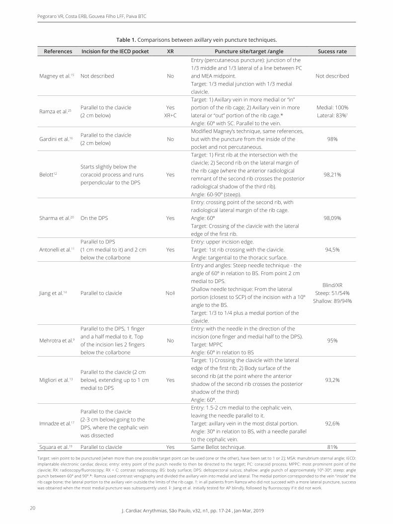
Table 1. Comparisons between axillary vein puncture techniques.
References Incision for the IECD pocket XR Puncture site/target /angle Sucess rate
Magney et al.15 Not described No
Entry (percutaneous puncture): junction of the 1/3 middle and 1/3 lateral of a line between PC and MEA midpoint.Target: 1/3 medial junction with 1/3 medial clavicle.
Not described
Ramza et al.25Parallel to the clavicle (2 cm below)
YesXR+C
Target: 1) Axillary vein in more medial or “in” portion of the rib cage; 2) Axillary vein in more lateral or “out” portion of the rib cage.*Angle: 60º with SC. Parallel to the vein.
Medial: 100%Lateral: 83%†
Gardini et al.16Parallel to the clavicle (2 cm below)
NoModified Magney’s technique, same references, but with the puncture from the inside of the pocket and not percutaneous.
98%
Belott12
Starts slightly below the coracoid process and runs perpendicular to the DPS
Yes
Target: 1) First rib at the intersection with the clavicle; 2) Second rib on the lateral margin of the rib cage (where the anterior radiological remnant of the second rib crosses the posterior radiological shadow of the third rib).Angle: 60-90º (steep).
98,21%
Sharma et al.20 On the DPS Yes
Entry: crossing point of the second rib, with radiological lateral margin of the rib cage. Angle: 60ºTarget: Crossing of the clavicle with the lateral edge of the first rib.
98,09%
Antonelli et al.11
Parallel to DPS (1 cm medial to it) and 2 cm below the collarbone
YesEntry: upper incision edge.Target: 1st rib crossing with the clavicle. Angle: tangential to the thoracic surface.
94,5%
Jiang et al.14 Parallel to clavicle No‡
Entry and angles: Steep needle technique - the angle of 60º in relation to BS. From point 2 cm medial to DPS.Shallow needle technique: From the lateral portion (closest to SCP) of the incision with a 10º angle to the BS.Target: 1/3 to 1/4 plus a medial portion of the clavicle.
Blind/XRSteep: 51/54%
Shallow: 89/94%
Mehrotra et al.9
Parallel to the DPS, 1 finger and a half medial to it. Top of the incision lies 2 fingers below the collarbone
No
Entry: with the needle in the direction of the incision (one finger and medial half to the DPS).Target: MPPCAngle: 60º in relation to BS
95%
Migliori et al.13
Parallel to the clavicle (2 cm below), extending up to 1 cm medial to DPS
Yes
Target: 1) Crossing the clavicle with the lateral edge of the first rib; 2) Body surface of the second rib (at the point where the anterior shadow of the second rib crosses the posterior shadow of the third)Angle: 60º.
93,2%
Imnadze et al.17
Parallel to the clavicle (2-3 cm below) going to the DPS, where the cephalic vein was dissected
Entry: 1.5-2 cm medial to the cephalic vein, leaving the needle parallel to it.Target: axillary vein in the most distal portion.Angle: 30º in relation to BS, with a needle parallel to the cephalic vein.
92,6%
Squara et al.19 Parallel to clavicle Yes Same Bellot technique. 81%
Target: vein point to be punctured [when more than one possible target point can be used (one or the other), have been set to 1 or 2]; MSA: manubrium-sternal angle; IECD: implantable electronic cardiac device; entry: entry point of the punch needle to then be directed to the target; PC: coracoid process; MPPC: most prominent point of the clavicle; RX: radioscopy/fluoroscopy; RX + C: contrast radioscopy; BS: body surface; DPS: deltopectoral sulcus; shallow: angle punch of approximately 10º-30º; steep: angle punch between 60º and 90º.*: Ramza used contrast venography and divided the axillary vein into medial and lateral. The medial portion corresponded to the vein “inside” the rib cage bone; the lateral portion to the axillary vein outside the limits of the rib cage. †: in all patients from Ramza who did not succeed with a more lateral puncture, success was obtained when the most medial puncture was subsequently used. ‡: Jiang et al. initially tested for AP blindly, followed by fluoroscopy if it did not work.
J. Cardiac Arrythmias, São Paulo, v32, n1, pp. 17-24 , Jan-Mar, 2019 21
Puncture of the Axillary Vein for the Implant for Electronic Cardiac Devices
DISCUSSION
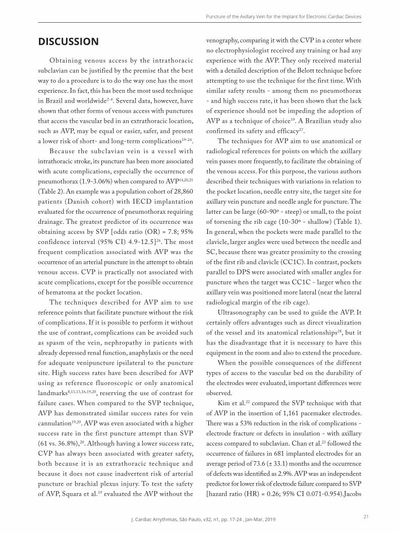
Obtaining venous access by the intrathoracic subclavian can be justified by the premise that the best way to do a procedure is to do the way one has the most experience. In fact, this has been the most used technique in Brazil and worldwide2-4. Several data, however, have shown that other forms of venous access with punctures that access the vascular bed in an extrathoracic location, such as AVP, may be equal or easier, safer, and present a lower risk of short- and long-term complications19-24.
Because the subc lavian vein is a vessel with intrathoracic stroke, its puncture has been more associated with acute complications, especially the occurrence of pneumothorax (1.9-3.06%) when compared to AVP14,20,25 (Table 2). An example was a population cohort of 28,860 patients (Danish cohort) with IECD implantation evaluated for the occurrence of pneumothorax requiring drainage. The greatest predictor of its occurrence was obtaining access by SVP [odds ratio (OR) = 7.8; 95% confidence interval (95% CI) 4.9-12.5]26. The most frequent complication associated with AVP was the occurrence of an arterial puncture in the attempt to obtain venous access. CVP is practically not associated with acute complications, except for the possible occurrence of hematoma at the pocket location.
The techniques described for AVP aim to use reference points that facilitate puncture without the risk of complications. If it is possible to perform it without the use of contrast, complications can be avoided such as spasm of the vein, nephropathy in patients with already depressed renal function, anaphylaxis or the need for adequate venipuncture ipsilateral to the puncture site. High success rates have been described for AVP using as reference fluoroscopic or only anatomical landmarks8,11,13,16,19,20, reserving the use of contrast for failure cases. When compared to the SVP technique, AVP has demonstrated similar success rates for vein cannulation19,20. AVP was even associated with a higher success rate in the first puncture attempt than SVP (61 vs. 36.8%),20. Although having a lower success rate, CVP has always been associated with greater safety, both because it is an extrathoracic technique and because it does not cause inadvertent risk of arterial puncture or brachial plexus injury. To test the safety of AVP, Squara et al.19 evaluated the AVP without the
venography, comparing it with the CVP in a center where no electrophysiologist received any training or had any experience with the AVP. They only received material with a detailed description of the Belott technique before attempting to use the technique for the first time. With similar safety results - among them no pneumothorax - and high success rate, it has been shown that the lack of experience should not be impeding the adoption of AVP as a technique of choice19. A Brazilian study also confirmed its safety and efficacy27.
The techniques for AVP aim to use anatomical or radiological references for points on which the axillary vein passes more frequently, to facilitate the obtaining of the venous access. For this purpose, the various authors described their techniques with variations in relation to the pocket location, needle entry site, the target site for axillary vein puncture and needle angle for puncture. The latter can be large (60-90º - steep) or small, to the point of torsening the rib cage (10-30º - shallow) (Table 1). In general, when the pockets were made parallel to the clavicle, larger angles were used between the needle and SC, because there was greater proximity to the crossing of the first rib and clavicle (CC1C). In contrast, pockets parallel to DPS were associated with smaller angles for puncture when the target was CC1C - larger when the axillary vein was positioned more lateral (near the lateral radiological margin of the rib cage).
Ultrasonography can be used to guide the AVP. It certainly offers advantages such as direct visualization of the vessel and its anatomical relationships28, but it has the disadvantage that it is necessary to have this equipment in the room and also to extend the procedure.
When the possible consequences of the different types of access to the vascular bed on the durability of the electrodes were evaluated, important differences were observed.
Kim et al.22 compared the SVP technique with that of AVP in the insertion of 1,161 pacemaker electrodes. There was a 53% reduction in the risk of complications - electrode fracture or defects in insulation - with axillary access compared to subclavian. Chan et al.23 followed the occurrence of failures in 681 implanted electrodes for an average period of 73.6 (± 33.1) months and the occurrence of defects was identified as 2.9%. AVP was an independent predictor for lower risk of electrode failure compared to SVP [hazard ratio (HR) = 0.26; 95% CI 0.071-0.954).Jacobs

22 J. Cardiac Arrythmias, São Paulo, v32, n1, pp. 17-24 , Jan-Mar, 2019
Pegoraro VR, Costa ERB, Gouvea Filho LFF, Paiva BTC
et al.24 made an even more detailed evaluation of defective electrodes extracted with the use of electrical tests, light microscopy, electron microscopy, and tests to evaluate the pressure on the electrodes. The analysis of the electrodes by specialists showed that the occurrence of pressure in the costoclavicular transition was responsible for the greater incidence of defects when the venous access was subclavian and suggested a more lateral approach, such as the use of the axillary vein, as a preventive for these complications.
CONCLUSIONS
The AVP technique has been described by several authors and presents some variations. It is a valuable alternative for obtaining venous access, presenting similar safety to CVP
Table 2. Comparison between techniques for obtaining vascular access.
References Patients(n)
Pneumothorax(%)
Hemothorax(%)
Arterial puncture
(%)
Pocket bruise
(%)
Brachial plexus injury
(%)
Limb thrombosis
(%)
Success(%)
Axillary veinSharma20 202 0.00 0.00 ND 4.40 ND ND 98.00Antonelli11 182 0.00 0.00 3.30 0.00 0.00 ND 100.00Imnadze17 108 0.00 0.00 4.60 ND 0.00 ND 92.60Jiang shallow14* 460 0.00 ND 7.50 0.50 0.00 ND 94.00Jiang steep14* 140 0.00 0.00 7.90 0.00 1.30 ND 54.00Migliori13 103 0.00 0.00 2.00 ND ND ND 100.00Byrd10 213 0.00 0.00 ND ND ND ND 98.00Saad27 241 0.00 0.00 5.00 ND ND 0.40 100.00Mehrotra9 20 5.00 ND ND ND ND ND 95.00Ramza25 50 0.00 0.00 8.10 ND ND ND 98.00Squara19 37 0.00 0.00 ND 2.70 5.40 ND 81.00
Subclavian veinSharma20 98 3.06 ND ND 4.00 ND ND 96.90Aggarwal29 1.047 1.80 ND 2.70 ND ND ND NDChauhan30 1.892 0.6† ND ND 0.50 ND ND NDLitleford1 164 2.40 ND ND 1.20 ND ND 91.70Marinoni31 1.220 0.30 ND ND ND ND ND NDKirkfeldt26 12.260 0.66† ND ND ND ND ND NDEberhardt32 1.100 1.1‡ ND ND ND ND ND NDFiorista33 101 3.00 ND ND ND 4.30 ND NDHess34 171 0.00 0.00 ND ND 0.00 ND ND
Cephalic veinChauhan30 157 0.00 0.00 ND 2.60 ND ND NDSquara19 37 0.00 0.00 ND 5.40 0.00 0.00 75.70Kircanski35 44 0.00 0.00 0.00 0.00 0.00 0.00 90.10Parsonnet36 148 0.67 ND ND ND ND ND ND
ND: not described, unspecified or without separation of values between groups compared; *: Jiang shallow and steep are part of the same work but represent different axillary vein
access techniques.†: authors who defined as the occurrence of pneumothorax only the cases requiring drainage. Cases without drainage are not included; ‡: cases of pneumothorax
requiring drainage in which the implanted pacemaker was a double chamber.
(even in the learning phase), success rates comparable to those of the subclavian vein and better medium and long term results for the function of the electrodes.
AUTHORS’ CONTRIBUTION
Methodology, Pegoraro VR, Costa ERB and Gouvea Filho LFF; Investigation, Pegoraro VR, Paiva BTC and Gouvea Filho LFF; Writing first version, Pegoraro VR and Paiva BTC; Writing - Review & Editing, Pegoraro VR and Costa ERB; Supervision,Costa ERB.
ACKNOWLEDGMENTS
To Dr. Eduardo R. B. Costa for guiding us in this work.

J. Cardiac Arrythmias, São Paulo, v32, n1, pp. 17-24 , Jan-Mar, 2019 23
Puncture of the Axillary Vein for the Implant for Electronic Cardiac Devices
1. Littleford PO, Parsonnet V, Spector SD. Method for the rapid and atraumatic insertion of permanent endocardial pacemaker electrodes through the subclavian vein. Am J Cardiol. 1979;43(5):980-2. https://doi.org/10.1016/0002-9149(79)90363-1
2. Bernstein AD, Parsonnet V. Survey of cardiac pacing in the United States in 1989. Am J Cardiol. 1992;69(4):331-8. https://doi.org/10.1016/0002-9149(92)90229-R
3. Bernstein AD, Parsonnet V. Survey of cardiac pacing and defibrillation in the United States in 1993. Am J Cardiol. 1996;78(2):187-96.
4. Mond HG, Proclemer A. The 11th world survey of cardiac pacing and implantable cardioverter-defibrillators: calendar year 2009 – A World Society of Arrhythmias project. Pacing Clin Electrophysiol. 2011;34(8):1013-27. https://doi.org/10.1111/j.1540-8159.2011.03150.x.
5. Kleemann T, Becker T, Doenges K, Vater M, Senges J, Schneider S, et al. Annual rate of transvenous defibrillation lead defects in implantable cardioverter-defibrillators over a period of >10 years. Circulation. 2007;115(19):2474-80. https://doi.org/10.1161/CIRCULATIONAHA.106.663807
6. Dorwarth U, Frey B, Dugas M, Matis T, Fiek M, Schmoeckel M, et al. Transvenous defibrillation leads: high incidence of failure during long-term follow-up. J Cardiovasc Electrophysiol. 2003;14(1):38-43. https://doi.org/10.1046/j.1540-8167.2003.02305.x
7. Calkins H, Ramza BM, Brinker J, Atiga W, Donahue K, Nsah E, et al. Prospective randomized comparison of the safety and effectiveness of placement of endocardial pacemaker and defibrillator leads using the extrathoracic subclavian vein guided by contrast venography versus the cephalic approach. Pacing Clin Electrophysiol. 2001;24(4 Pt 1):456-64.
8. Jiang M, Mao JL, He B. Clinical definition of the axillary vein and experience with blind axillary puncture. Int J Cardiol. 2012;159(3):243-5. https://doi.org/10.1016/j.ijcard.2012.05.089
9. Mehrotra S, Rohit MK. Prospective study to develop surface landmarks for blind axillary vein puncture for permanent pacemaker and defibrillator lead implantation and compare it to available contrast venography guided technique. Indian Heart J. 2015;67(2):136-40. https://doi.org/10.1016/j.ihj.2015.04.007
10. Byrd CL. Clinical experience with the extrathoracic introducer insertion technique. Pacing Clin Electrophysiol. 1993;16(9):1781-4. https://doi.org/10.1111/j.1540-8159.1993.tb01810.x
11. Antonelli D, Feldman A, Freedberg NA, Turgeman Y. Axillary vein puncture without contrast venography for pacemaker and defibrillator leads implantation. Pacing Clin Electrophysiol. 2013;36(9):1107-10. https://doi.org/10.1111/pace.12181
12. Belott P. How to access the axillary vein. Heart Rhythm. 2006;3(3):366-9. https://doi.org/10.1016/j.hrthm.2005.10.031
13. Migliore F, Siciliano M, De Lazzari M, Ferretto S, Valle CD, Zorzi A, et al. Axillary vein puncture using fluoroscopic landmarks: a safe and effective approach for implantable cardioverter
defibrillator leads. J Interv Card Electrophysiol. 2015;43(3):263-7. https://doi.org/10.1007/s10840-015-0011-7
14. Jiang M, Gong XR, Zhou SH, Pu J, Mao JL, He B. A comparison of steep and shallow needle trajectories in blind axillary vein puncture. Pacing Clin Electrophysiol. 2013;36(9):1150-5. https://doi.org/10.1111/pace.1215
15. Magney JE, Staplin DH, Flynn DM, Hunter DW. A new approach to percutaneous subclavian venipuncture to avoid lead fracture or central venous catheter occlusion. Pacing Clin Electrophysiol. 1993;16(11):2133-42. https://doi.org/10.1111/j.1540-8159.1993.tb.01018.x
16. Gardini A, Benedini G. Blind extrathoracic subclavian venipuncture for pacemaker implant: a 3-year experience in 250 patients. Pacing Clin Electrophysiol. 1998;21(11 Pt 2):2304-8.
17. Imnadze G, Awad K, Wolff E, Amberger J, Franz N, Thale J, et al. A novel method of axillary venipuncture using the cephalic vein as a sole anatomic landmark. The Can J Cardiol. 2015;31(8):1067-9. https://doi.org/10.1016/j.cjca.2015.02.021
18. Pittiruti M, Biasucci DG, La Greca A, Pizza A, Scoppettuolo G. How to make the axillary vein larger? Effect of 90 degrees abduction of the arm to facilitate ultrasound-guided axillary vein puncture. J Crit Care. 2016;33:38-41. https://doi.org/10.1016/j.jcrc.2015.12.018
19. Squara F, Tomi J, Scarlatti D, Theodore G, Moceri P, Ferrari E. Self-taught axillary vein access without venography for pacemaker implantation: prospective randomized comparison with the cephalic vein access. Europace. 2017;19(12):2001-6. https://doi.org/10.1093/europace%2Feuw363
20. Sharma G, Senguttuvan NB, Thachil A, Leong D, Naik N, Yadav R, et al. A comparison of lead placement through the subclavian vein technique with fluoroscopy-guided axillary vein technique for permanent pacemaker insertion. Can J Cardiol. 2012;28(5):542-6. https://doi.org/10.1016/j.cjca.2012.02.019
21. Holubec T, Ursprung G, Schonrath F, Caliskan E, Steffel J, Falk V, et al. Does implantation technique influence lead failure? Acta Cardiol. 2015;70(5):581-6. https://doi.org/10.2143/AC.70.5.3110519
22. Kim KH, Park KM, Nam GB, Kim DK, Oh M, Choi H, et al. Comparison of the axillary venous approach and subclavian venous approach for efficacy of permanent pacemaker implantation. 8-Year follow-up results. Circulation. 2014;78(4):865-71. https://doi.org/10.1253/circj.CJ-13-0884
23. Chan NY, Kwong NP, Cheong AP. Venous access and long-term pacemaker lead failure: comparing contrast-guided axillary vein puncture with subclavian puncture and cephalic cutdown. Europace. 2017;19(7):1193-7. https://doi.org/10.1093/europace/euw147
24. Jacobs DM, Fink AS, Miller RP, Anderson WR, McVenes RD, Lessar JF, et al. Anatomical and morphological evaluation of pacemaker lead compression. Pacing Clin Electrophysiol. 1993;16(3 Pt 1):434-44.
25. Ramza BM, Rosenthal L, Hui R, Nsah E, Savader S, Lawrence JH, et al. Safety and effectiveness of placement of pacemaker
REFERENCES

24 J. Cardiac Arrythmias, São Paulo, v32, n1, pp. 17-24 , Jan-Mar, 2019
Pegoraro VR, Costa ERB, Gouvea Filho LFF, Paiva BTC
and defibrillator leads in the axillary vein guided by contrast venography. Am J Cardiol. 1997;80(7):892-6. https://doi.org/10.1016/s0002-9149(97)00542-0
26. Kirkfeldt RE, Johansen JB, Nohr EA, Moller M, Arnsbo P, Nielsen JC. Pneumothorax in cardiac pacing: a population-based cohort study of 28,860 Danish patients. Europace. 2012;14(8):1132-8. https://doi.org/10.1093/europace/eus054
27. Saad EBFF, Veronese F, Maldonado P, Camanho LE. Uso do acesso venoso axilar para implante de eletrodos de marcapassos e desfibriladores. Relampa. 2006;19(4):259-99.
28. Orihashi K, Imai K, Sato K, Hamamoto M, Okada K, Sueda T. Extrathoracic subclavian venipuncture under ultrasound guidance. Circ J. 2005;69(9):1111-5. https://doi.org/10.1253/circj.69.1111
29. Aggarwal RK, Connelly DT, Ray SG, Ball J, Charles RG. Early complications of permanent pacemaker implantation: no difference between dual and single chamber systems. Br Heart J. 1995;73(6):571-5. https://doi.org/10.1136/hrt.73.6.571.
30. Chauhan A, Grace AA, Newell SA, Stone DL, Shapiro LM, Schofield PM, et al. Early complications after dual chamber versus single chamber pacemaker implantation. Pacing Clin Electrophysiol. 1994;17(11 Pt 2):2012-5.
31. Marinoni G, Broglia P, Bruno N, Perotti R, Bosatra C, Montemartini C. Percutaneous approach in the use of subclavian vein in pacemaker implantation. Giornale italiano di cardiologia. 1994;24(6):685-9.
32. Eberhardt F, Bode F, Bonnemeier H, Boguschewski F, Schlei M, Peters W, et al. Long term complications in single and dual chamber pacing are influenced by surgical experience and patient morbidity. Heart. 2005;91(4):500-6. https://doi.org/10.1136/hrt.2003.025411
33. Fiorista F, Lazari M, Marzegalli M, Piane C, Cotti R, Casazza F, et al. Use of the subclavian vein for permanent cardiac stimulation. Arch Inst Cardiol Mex. 1986;56(4):309-13.
34. Hess DS, Gertz EW, Morady F, Scheinman M, Sudduth BK. Permanent pacemaker implantation in the cardiac catheterization laboratory: the subclavian vein approach. Cathet Cardiovasc Diagn. 1982;8(5):453-8
35. Kircanski B, Vasic D, Savic D, Stojanov P. Low incidence of complications after cephalic vein cutdown for pacemaker lead implantation in children weighing less than 10 kilograms: A single-center experience with long-term follow-up. Heart Rhythm. 2015;12(8):1820-6. https://doi.org/10.1016/j.hrthm.2015.04.025
36. Parsonnet V, Roelke M. The cephalic vein cutdown versus subclavian puncture for pacemaker/ICD lead implantation. Pacing Clin Electrophysiol. 1999;22(5):695-7.
J. Cardiac Arrythmias, São Paulo, v32, n1, pp. 25-29 , Jan-Mar, 2019 25
Occurrence of Subclinical Atrial Fibrillation in the Follow-up of Patients with Cardiac PacemakersOcorrência de Fibrilação Atrial Subclínica no Acompanhamento de Pacientes Portadores de Marcapasso Cardíaco
Luis Fernando Spagnuolo Brunello1,*, Gustavo Andrade de Figueiredo1, Leonardo Andrade Mulinari1
1.Universidade Federal do Paraná – Hospital de Clínicas – Serviço de Cirurgia Cardiovascular – Curitiba/PR – Brazil.*Correspondence author: [email protected]: 09 Oct 2018 | Accepted: 06 May 2019Section Editor: J. Tarcísio Medeiros de Vasconcelos
ORCID IDs
Brunello LFS https://orcid.org/0000-0001-7717-5835
Figueiredo GA https://orcid.org/0000-0003-1907-1637
Mulinari LA https://orcid.org/ 0000-0001-7138-9912
HEART STIMULATIONOriginal Articlehttps://doi.org/10.24207/jac.v32i1.003_IN
ABSTRACT
Objective: Cardiac pacemaker records atrial fi brillation (AF). This condition
can cause serious hemodynamic consequences to patients, who should be
assisted by a cardiologist. This study aimed to document and investigate, in
a tertiary hospital, the prevalence of subclinical AF in patients with a cardiac
pacemaker. Methods: Between July 2015 and April 2016, 196 patients with
pacemakers were attended on an outpatient basis. Of these, 60 had cardiac
arrhythmias recorded by the pacemaker and were invited to participate
in the study. Data collection was done through a structured interview
containing four questions: gender, age, follow-up with cardiologist and use
of anticoagulants. Results: Subclinical AF was recorded in 35 (17.8%) of
the total of 196 patients. Of these 35, 16 (45.7%) did not follow a regular
cardiology service and 29 (82.8%) did not use anticoagulant medication.
No statistically signifi cant relationships were found between age, follow
up with a cardiologist, and presence or absence of subclinical AF in the
patients studied. Conclusion: A signifi cant portion of outpatient patients
with pacemakers have AF recorded by the device. However, although
essential, almost half of these do not proceed with the clinical follow-up with
cardiologist and less than a fi fth with AF makes use of anticoagulant therapy.
KEYWORDS: Artifi cial pacemaker; Atrial fi brillation; Cardiac arrhythmias;
Hospital outpatient clinic
RESUMO Objetivo: O marcapasso cardíaco registra a fi brilação atrial (FA). Essa condição pode causar graves consequências hemodinâmicas aos pacientes, que devem ser assistidos por médico cardiologista. Este estudo objetivou documentar e investigar, em um hospital terciário, a prevalência de FA subclínica em portadores de marcapasso cardíaco. Métodos: Entre julho de 2015 e abril de 2016, foram atendidos 196 pacientes portadores de marca-passo em caráter ambulatorial. Desses, 60 apresentaram arritmias cardíacas registradas pelo marcapasso e foram convidados a participar do estudo. A coleta de dados foi feita por meio de entrevista estruturada contendo quatro questões: sexo, idade, acompanhamento com cardiologista e uso de anticoagulantes. Resultados: Foi registrada FA subclínica em 35 (17,8%) do total de 196 pacientes. Desses 35, 16 (45,7%) não realizavam acompanhamento regular em serviço de cardiologia e 29 (82,8%) não faziam uso de medicamento anticoagulante. Não foram encontradas relações estatisticamente signifi cativas entre idade, acompanhamento com cardiologista e presença ou ausência da FA subclínica nos pacientes estudados. Conclusão: Uma parcela signifi cativa dos pacientes portadores de marcapasso atendidos ambulatorialmente tem FA registrada pelo dispositivo. No entanto, ainda que essencial, quase metade desses não faz acompanhamento clínico com cardiologista e menos de um quinto com FA faz uso de terapia anticoagulante.
PALAVRAS-CHAVE: Marcapasso artifi cial; Fibrilação atrial; Arritmias
cardíacas; Ambulatório hospitalar.
26 J. Cardiac Arrythmias, São Paulo, v32, n1, pp. 25-29 , Jan-Mar, 2019
Brunello LFS, Figueiredo GA, Mulinari LA
INTRODUCTION
Atrial fibrillation (AF) is the most common sustained cardiac arrhythmia currently; affects approximately 33 million people worldwide1 with an estimated prevalence of 1 to 4% in adults, and may be paroxysmal2. Its clinical importance lies in the fact that most patients have little or no specific symptoms of this arrhythmia3, besides being associated with several hemodynamic complications and increased morbidity and mortality in patients who has it2. Therefore, its early detection - although difficult most of the time - is useful for monitoring and adequate management of patients in order to avoid secondary complications2,4.
The diagnosis and monitoring of subclinical AF - its asymptomatic form - is preferably performed from electrocardiogram4; however, in patients with a cardiac pacemaker, it is possible to diagnose it by the analysis of the recording and storage of data of the device5. Pacemakers not only perform their function of identifying and correcting problems in cardiac electrical stimulation but are also able to accurately record abnormal cardiac events, registering the day, time and duration6. Studies point to the prevalence rate of subclinical AF around 10% in patients with cardiac pacemaker6,7 and may vary up to 55.3% for those aged 65 or over5,7.
It is known that all patients with a cardiac pacemaker who had AF recorded in their device should have a regular clinical follow-up with a cardiologist for control and monitoring8. Treatment under oral anticoagulation is indicated for the absolute majority of AF patients, except in cases where the hemorrhagic risks outweigh the benefits of preventing thromboembolic complications of arrhythmia4,8. However, there are few international studies that evaluate the adequate management of patients with AF with cardiac pacing, and there is no recent Brazilian study that evaluates this scenario in the patients treated by the Sistema Único de Saude (SUS).
Due to the necessity and importance of adequate clinical follow-up of these patients, this study aims to raise the prevalence of atrial arrhythmias and subclinical AF in patients with cardiac pacemakers, as well as to investigate how many of them undergo regular clinical follow-up with a cardiologist and use anticoagulant medications.
METHODS
This is an observational study with a quantitative and descriptive approach. 196 patients with a cardiac pacemaker in the period between July 2015 and April 2016 were attended on an outpatient basis. Of these, 60 patients had cardiac arrhythmias recorded by the pacemaker and were invited to participate in the study.
The setting of the study was the Pacemaker Ambulatory of Hospital de Clínicas (HC) of the Universidade Federal do Paraná (UFPR), which is part of the Cardiovascular Surgery Service and is located in the Ambulatory Medical Service (SAM 2) of the hospital. Outpatient care takes place on the first Monday of each month and is assisted by pacemaker technicians representing the different brands of devices used by the service.
To the 60 patients selected, the objectives of the research were explained and presented the Term of Free and Informed Consent, signed by these and the researchers. Then, the researchers applied a structured interview, which contained two sociodemographic questions (sex and age) and two clinical questions about regular follow-up with a cardiologist and use of anticoagulant drugs.
The collected data were recorded and organized in Excel® software table (Microsoft, 2013) and statistical analysis performed by R (R Core Team, 2015; version 3.2.3) software. Absolute and relative frequencies were obtained from the following data: the presence of arrhythmic event; the presence of subclinical AF; gender; follow-up with a cardiologist; and use of anticoagulants.
For statistical evaluation, the results were submitted to Fischer’s exact test, when qualitative and dichotomous variables, and logistic regression test, when there was a need to predict cause-effect relationships between two variables. Mean, median, minimum, maximum, and standard deviation of the variable age were also obtained, also submitted to Student›s t-test for comparison of paired samples. The reference value for the p-value of 5% was considered as a determinant of the statistical significance of the sample results.
The research was approved by the Ethics and Research Committee of the UFPR HC (CAAE 44183615.7.0000.0096), and the consolidated opinion was issued on May 9, 2015.

J. Cardiac Arrythmias, São Paulo, v32, n1, pp. 25-29 , Jan-Mar, 2019 27
Occurrence of Subclinical Atrial Fibrillation in the Follow-up of Patients with Cardiac Pacemakers
RESULTS
Arrhythmic events were recorded in 60 (30.6%) of 196 patients who went through the outpatient clinic in the period analyzed. Of these 60, 25 (41.6%) had exclusively ventricular arrhythmias, 16 (26.6%) AF associated with ventricular arrhythmias and 19 (31.6%) had exclusively AF. Therefore, in relation to the 196 patients, 35 (17.8%) had AF (Table 1).
Among the study participants, 38 (63.3%) were male and 22 (36.6%) were female. With regard to the disease studied (subclinical AF) and the gender of the participants, it was observed that the chance of subclinical AF in female patients is 3.77 times that of male patients [odds ratio (OR) = 3, 77; 95% confidence interval (95% CI) 1.21-13.42; p = 0.0277]. The mean age of the patients was 68.1 ± 12.1 years; however, there were no statistically significant relationships between the age of the patients and the presence or absence of subclinical AF (p = 0.5876).
More than half of the 60 patients studied (53.3%, n = 32) had regular follow-up with a cardiologist, and among the 35 patients with subclinical AF, 18 (51.4%) underwent cardiac monitoring. Of the 25 patients who have exclusively ventricular arrhythmias, 14 (56%) follow up with a cardiologist and the other 11 (44%) do not (Fig. 1). There was no statistically significant difference between the two groups (subclinical AF and exclusively ventricular arrhythmias) for regular follow-up with cardiologists (p = 0.7964).
Of the 60 patients studied, 14 (23.3%) used anticoagulants. Of the 35 patients with subclinical AF, 29 (82.8%) did not use drugs of this c lass (Fig. 2). Taking into account the follow-up with a
cardiologist, it was observed that 16 patients with subclinical AF, in addition to not taking anticoagulant drugs, did not proceeded with medical follow up (45.7%).
Table 1. Clinic and sociodemographic characteristics of the study group.
PatientsWith cardiac arrhythmias
(n = 60)
With subclinical atrial fibrillation
(n = 35)
No subclinical atrial fibrillation
(n = 25)
Gender
Male [n (%)] 38 (63.3) 18 (51.4) 20 (80.0)
Female [n (%)] 22 (36.6) 17 (48.5) 5 (20.0)
Age (mean ± Standard deviation) 68 ± 12.1 67.4 ± 13.1 69.2 ± 10.6
Ventricular arrhythmias [n (%)] 41 (68.3) 16 (45.7) 25 (100.0)
Cardiological follow-up [n (%)] 32 (53.3) 18 (51.4) 14 (56.0)
Use of oral anticoagulants [n (%)] 14 (23.3) 6 (17.1) 8 (32.0)
40 No
Yes
30
20
10
0Present AbsentSubclinical atrial fibrillation
Num
ber
of p
atie
nts
Present
Num
ber
of p
atie
nts
Absent
40 No
Yes
30
20
10
0
Subclinical atrial fibrillation
Figure 1. Follow up with a cardiologist.
Figure 2. Use of oral anticoagulant drugs.

28 J. Cardiac Arrythmias, São Paulo, v32, n1, pp. 25-29 , Jan-Mar, 2019
Brunello LFS, Figueiredo GA, Mulinari LA
DISCUSSION
The main results of this research highlight the high prevalence (30.6%) of occurrence of cardiac arrhythmias in addition to the underlying diseases that indicated implantation of a pacemaker in the patients observed. Cardiac arrhythmias recorded by pacemakers - also called «events» - are broken down in telemetry and can be archived by the technicians and then analyzed by the cardiac surgeon in charge.
AF, in its subclinical form, is responsible for more than half of the arrhythmias in the studied group. It is associated with a 2.5-fold increased risk of possible hemodynamic complications, such as cerebrovascular accident (stroke) and systemic embolism7. The rate of subclinical AF observed in the patients in this study was 17.8%, whereas the literature presents variable rates: some studies6,7 rates close to 10%, while others5,9 found rates close to 40%. This observed difference may be related to the data collection time. Healey et al.7 followed patients for a period of three months and obtained a rate of 10.1%, while Cabrera et al.9 followed patients for a period of 5.5 years and obtained a rate of 36.9%. Another important factor that may influence the rate of occurrence of AF is the criterion of patient selection, since male, Caucasian, smokers, obese patients with a previous history of cardiovascular problems are at greater risk of developing AF1.
In contrast, this research demonstrated a greater chance of AF occurring in female patients, which contrasts with results presented in the literature. Schnabel et al.10 risk factors for the development of AF, one being the male gender. However, there are studies that found no significant relationship between gender and the occurrence of AF in patients with pacemakers11-13.
Still in relation to the sociodemographic data analyzed, a predominance of the elderly in the study group (mean age 68 years) was observed, although no significant relationship was found between this aspect and the presence or absence of subclinical AF. It is known that age above 60 years is one of the risk factors for the development of AF10and other studies were able to relate this information to AF detected by implantable devices5-7,9,14.
One variable studied in this study, of great clinical importance, was the regular follow-up of cardiac
arrhythmias in a cardiologist doctor. The guidelines point to the need for follow-up and clinical cardiological management for those with cardiac arrhythmias, whether atrial or ventricular arrhythmias15,16. The cardiologist is responsible for investigating and choosing the ideal therapy for the control and attenuation of possible arrhythmic symptoms, in order to provide a better quality of life for patients with these conditions, as well as to prevent related complications15,16.
The present study found that approximately half of the patients with subclinical AF (48.6%) do not have a regular cardiological follow-up. A similar relationship was observed in the group of patients who presented ventricular arrhythmias; of these, only 56% follow up with a cardiologist. Therefore, there was a lack of information on this group about the importance of having a cardiological follow-up, as well as the difficulty of access to cardiologists by the patients in this study.
The vast majority (82.8%) of the patients who presented subclinical AF did not use anticoagulant drugs. A similar result is highlighted by Healey et al.5, which portray an unfavorable scenario for patients who need to use this type of drug; the authors emphasize that anticoagulants are prescribed for less than a quarter of patients who have asymptomatic AF5. Nevertheless, another study that exclusively evaluates the beginning of anticoagulant treatment in patients with AF emphasizes that in more than 77% of the patients observed there would be a need for anticoagulant drug therapy, however, in only 61% of these it is performed6.
It should be noted that a group of patients studied in this research, besides not having regular follow-up with a cardiologist, does not use anticoagulant drugs. This group denoted the rate of 45.7% among patients with subclinical AF and was considered the most vulnerable to the thromboembolic complications of this asymptomatic disease.
The main limitation of this study is its short period of data collection (10 months) and, consequently, the small sample size used for statistical calculations. Therefore, new research is needed to analyze the variables studied in a larger number of patients, in order to increase the accuracy of the calculated results

J. Cardiac Arrythmias, São Paulo, v32, n1, pp. 25-29 , Jan-Mar, 2019 29
Occurrence of Subclinical Atrial Fibrillation in the Follow-up of Patients with Cardiac Pacemakers
CONCLUSION
A significant portion of the patients with pacemakers treated ambulatorily in HC-UFPR have AF recorded by the device. However, although primordial, almost half of these patients do not undergo clinical follow-up with a cardiologist and less than one-fifth of patients with AF undergoing anticoagulant therapy. New studies that increase the number of patients studied are necessary to increase the statistical accuracy.
AUTHORS’ CONTRIBUTIONS
Conceptualization, Mulinari, LA; Methodology, Mulinari LA, Brunello LFS and Figueiredo, GA; Research, Mulinari LA, Brunello LFS and Figueiredo, GA; Writing, Mulinari L A, Brunello LFS and Figueiredo GA; First version, Mulinari LA, Brunello LFS and Figueiredo GA; Review, Mulinari LA, Brunello, LFS and Figueiredo, GA; Supervision, Mulinari, LA.
1. Ball J, Carrington MJ, McMurray JJV, Stewart S. Atrial fibrillation: profile and burden of an evolving epidemic in the 21st century. Int J Cardiol. 2013;167(5):1807-24. https://doi.org/10.1016/j.ijcard.2012.12.093
2. Rahman F, Kwan GF, Benjamin EJ. Global epidemiology of atrial fibrillation. Nat Rev Cardiol. 2014;11(11):639-54. https://doi.org/10.1038/nrcardio.2014.118
3. Israel CW, Grönefeld G, Ehrlich JR, Li Y-G, Hohnloser SH. Long-term risk of recurrent atrial fibrillation as documented by an implantable monitoring device. J Am Coll Cardiol. 2004;43(1):47-52. https://doi.org/10.1016/j.jacc.2003.08.027
4. Camm AJ, Kirchhof P, Lip GYH, Schotten U, Savelieva I, Ernst S, et al. Guidelines for the management of atrial fibrillation: The Task Force for the Management of Atrial Fibrillation of the European Society of Cardiology (ESC). Eur Heart J. 2010;31(19):2369-429. https://doi.org/10.1093/eurheartj/ehq278
5. Healey JS, Martin JL, Duncan A, Connolly SJ, Ha AH, Morillo CA, et al. Pacemaker-detected atrial fibrillation in patients with pacemakers: prevalence, predictors, and current use of oral anticoagulation. Can J Cardiol. 2013;29(2):224-8. https://doi.org/10.1016/j.cjca.2012.08.019
6. Yedlapati N, Fisher JD. Pacemaker diagnostics in atrial fibrillation: limited usefulness for therapy initiation in a pacemaker practice. Pacing Clin Electrophysiol. 2014;37(9):1189-97. https://doi.org/10.1111/pace.12392
7. Healey JS, Connolly SJ, Gold MR, Israel CW, Van Gelder IC, Capucci A, et al. Subclinical atrial fibrillation and the risk of stroke. N Engl J Med. 2012;366(2):120-9. https://doi.org/10.1056/NEJMoa1105575
8. January CT, Wann LS, Alpert JS, Calkins H, Cleveland Jr JC, Cigarroa JE, et al. 2014 AHA/ACC/HRS Guideline for the Management of Patients With Atrial Fibrillation: Executive summary. J Am Coll Cardiol. 2014;(212):1-57. https://doi.org/10.1161/CIR.0000000000000041
9. Cabrera S, Mercé J, Castro R de, Aguirre C, Carmona A, Pinedo M, et al. Pacemaker clinic: An opportunity to detect silent atrial fibrillation and improve antithrombotic treatment. Europace. 2011;13(11):1574-9. https://doi.org/10.1093/europace/eur160
10. Schnabel RB, Sullivan LM, Levy D, Pencina MJ, Massaro JM, D’Agostino RB, et al. Development of a risk score for atrial fibrillation (Framingham Heart Study): a community-based cohort study. Lancet. 2009;373(9665):739-45. https://doi.org/10.1016/S0140-6736(09)60443-8
11. Mabo P, Victor F, Bazin P, Ahres S, Babuty D, Costa A da, et al. A randomized trial of long-term remote monitoring of pacemaker recipients (The COMPAS trial). Eur Heart J. 2012;33(9):1105-11. https://doi.org/10.1093/eurheartj/ehr419
12. Glotzer TV, Daoud EG, Wyse DG, Singer DE, Ezekowitz MD, Hilker C, et al. The relationship between daily atrial tachyarrhythmia burden from implantable device diagnostics and stroke risk the trends study. Circ Arrhythmia Electrophysiol. 2009;2(5):474-80. https://doi.org/10.1161/CIRCEP.109.849638
13. Defaye P, Leclercq J-F, Guilleman D, Scanu P, Hazard J-R, Fatemi M, et al. Contributions of high resolution electrograms memorized by DDDR pacemakers in the interpretation of arrhythmic events. Pacing Clin Electrophysiol. 2003;26 (1 Pt 2):214-20. https://doi.org/10.1046/j.1460-9592.2003. 00019.x
14. Kaufman ES, Israel CW, Nair GM, Armaganijan L, Divakaramenon S, Mairesse GH, et al. Positive predictive value of device-detected atrial high-rate episodes at different rates and durations: an analysis from ASSERT. Hear Rhythm. 2012;9(8):1241-6. https://doi.org/10.1016/j.hrthm.2012.03.017
15. Priori SG, Blomström-Lundqvist C, Mazzanti A, Blom N, Borggrefe M, Camm J, et al. 2015 ESC Guidelines for the management of patients with ventricular arrhythmias and the prevention of sudden cardiac death. Eur Heart J. 2015;36(41):2793-867. https://doi.org/10.1093/eurheartj/ehv316
16. Page RL, Joglar JA, Caldwell MA, Calkins H, Conti JB, Deal BJ, et al. 2015 ACC/AHA/HRS Guideline for the Management of Adult Patients With Supraventricular Tachycardia. Circulation. 2015;64(21). https://doi.org/10.1161/CIR.0000000000000311
REFERENCES

30 J. Cardiac Arrythmias, São Paulo, v32, n1, pp. 30-37 , Jan-Mar, 2019
Impact of DDD and VVIR Stimulation Modes on Functional Capacity and Quality of Life of Chagasic PatientsImpacto dos Modos de Estimulação DDD e VVIR na Capacidade Funcional e Qualidade de Vida de Pacientes Chagásicos
Débora Rodrigues Santana1, Geraldo Paulino Santana Filho1, Zander Bastos Rocha1, Antonio Malan Cavalcanti Lima2, Max Weyler Nery3, Salvador Rassi4, Giulliano Gardenghi3,*
ARTIFICIAL HEART STIMULATION
Original Article
1.Santa Casa de Misericórdia de Goiânia – Goiânia/GO – Brazil.2.Pontifícia Universidade de Goiás – Goiânia/GO – Brazil.3.Hospital ENCORE – Aparecida de Goiânia/GO – Brazil.4.Universidade Federal de Goiás – Hospital das Clínicas – Serviço de Cardiologia – Goiânia/GO – Brazil.*Correspondence author: [email protected]: 03 Jul 2018 | Accepted: 04 Feb 2019Section Editor: J. Tarcísio Medeiros de Vasconcelos
https://doi.org/10.24207/jac.v32i1.533_IN
ORCID IDs
Santana DR https://orcid.org/0000-0003-0659-8755
Santana Filho GP https://orcid.org/0000-0002-3224-4435
Rocha ZB https://orcid.org/0000-0002-1413-753X
Lima AMC https://orcid.org/0000-0002-7252-9056
Santana DR
Santana Filho GP
Rocha ZB
Lima AMC
Nery MW https://orcid.org/0000-0002-2944-6220
Rassi S https://orcid.org/0000-0001-7210-6486
Gardenghi G https://orcid.org/0000-0002-8763-561X
ABSTRACT
Introduction: Atrioventricular stimulation provides hemodynamic benefi ts over the isolated ventricular rate, but this advantage is not completely established in chagasic patients with systolic dysfunction. Objectives: To evaluate the infl uence of DDD and VVIR stimulation modes on functional capacity, quality of life (QoL) and laboratory abnormalities of a natriuretic peptide in chagasic patients with ventricular dysfunction submitted to pacemaker implantation.Methods: Twenty patients (55% male) with a mean age of 62.7 (± 9.9 years) and a mean ejection fraction of 41.8% (± 2.8) were prospectively studied. Alternately, patients received pacing in the DDD and VVIR modes for a period of three months under each schedule. The minimum percentage of ventricular pacing was 80%. After each period, the patient was submitted to the six-minute walk test (6MWT), QOL assessment by the Minnesota Living with Heart Failure Questionnaire (MLHFQ) and the Assay of QUAlity of life and RELated events (AQUAREL). Laboratory evaluation was performed with the N-terminal fraction of the brain natriuretic peptide (N-terminal pro b-type natriuretic peptide - NT-proBNP). Results: The mean distance walked on the 6MWT in the DDD and VVIR modes were 390.60 (± 52.71) and 396.30 (± 52.71) meters respectively (p = 0.160). Results of lower QOL were found, considering the physical domain of the MLHFQ (p = 0.03)and the domains of effort dyspnea (p = 0.05) and arrhythmia(p <0.001) of the AQUAREL with the VVIR mode. NT-proBNP levels increased signifi cantly with stimulation in VVIR mode (p < 0.001). Conclusion: After three months of stimulation with the VVIR mode, there was worsening of the QoL of the chagasic patients and increase of the levels of NT-proBNP (clinical trial record: ReBEc RBR-53x476)
KEYWORDS: Chagas disease; Pacemaker; Quality of life.
RESUMO
Introdução: A estimulação atrioventricular propicia benefícios hemodinâmicos em relação à ventricular isolada, mas essa vantagem não está completamente estabelecida em pacientes chagásicos com disfunção sistólica. Objetivo: Avaliar a infl uência dos modos de estimulação DDD e VVIR na capacidade funcional, qualidade de vida (QV) e alterações laboratoriais de peptídeo natriurético em pacientes chagásicos com disfunção ventricular submetidos a implante de marcapasso. Métodos: Estudaram-se prospectivamente 20 pacientes (55% do sexo masculino) com média de idade de 62,7(± 9,9 anos) e média da fração de ejeção de 41,8% (± 2,8). Alternadamente, os pacientes receberam a estimulação nos modos DDD e VVIR por um período de três meses sob cada programação. O mínimo percentual de estimulação ventricular admitido foi de 80%. Após cada período, o paciente foi submetido ao teste de caminhada de seis minutos (TC6M), avaliação de QV pelo Minnesota Living with Heart Failure Questionnaire (MLHFQ) e pelo Assesment of QUAlity of life and RELated events (AQUAREL). A avaliação laboratorial foi realizada com a dosagem da fração N-terminal do peptídeo natriurético cerebral (N-terminal pro b-type natriuretic peptide – NT-proBNP). Resultados: A média da distância percorrida no TC6M nos modos DDD e VVIR foram respectivamente 390,60 (± 52,71) e 396,30 (± 52,71) metros (p = 0,160). Verifi caram-se resultados de QV inferiores, considerando o domínio físico do MLHFQ (p = 0,03) e os domínios dispneia de esforço(p = 0,05) e arritmia (p < 0,001) do AQUAREL, com o modo VVIR. Os níveis de NT-proBNP aumentaram significativamente com a estimulação nomodo VVIR (p < 0,001). Conclusão: Após três meses de estimulação com o modo VVIR, houve piora da QV dos pacientes chagásicos e aumento dos níveis de NT-proBNP (registro de ensaio clínico: ReBEc RBR-53x476).
PALAVRAS-CHAVE: Doença de Chagas; Marcapasso; Qualidade de vida.

J. Cardiac Arrythmias, São Paulo, v32, n1, pp. 30-37 , Jan-Mar, 2019 31
Santana DR, Santana Filho GP, Rocha ZB, Lima AMC, Nery MW, Rassi S, Gardenghi G
INTRODUCTION
Despite the decline in the number of new cases of Chagas› disease (CHD), its endemic condition and chronicity sustain it as an important health problem in South America1. Recent data show that 20 to 30% of the millions of HIV positive people manifest, every year, some form of cardiomyopathy2,3. The involvement of the cardiac excito-conductive system by Trypanosoma cruzi remains the second most frequently involved etiology in conventional pacemaker implantation in Brazil4. The important reduction of mortality in this population consecrated the artificial stimulation as a treatment of choice of bradycardia5.
The structural and electronic evolution of pacemakers aroused interest in detecting additional benefits such as quality of life (QoL). Atrioventricular (AV) pacing has been shown to be superior to unicameral pacing, which has therefore been deferred if sinus activity is present6. However, the evidence for QoL and functional capacity are based on populations without CHD and with preserved ejection fraction. Little information correlates the results of QOL and functional capacity with the use of specific tools for chagasic patients with pacemakers.
OBJECTIVE
To assess the effects of artificial cardiac pacing modes on QOL and functional capacity of chagasic patients with total AV block undergoing pacemaker implantation and to detect their influence on the levels of the N-terminal fraction of the cerebral natriuretic peptide (N-terminal pro b-type natriuretic peptide - NT-proBNP).
METHODS
This is a prospective, controlled clinical study performed in Santa Casa de Misericórdia de Goiânia (SCMG), state of Goiás, in patients submitted to double-chamber pacemaker implantation, from July 2014 to April 2015. After the approval of the study by the SCMG Ethics Committee (CAAE: 32745114.4.0000.5081),
patients were evaluated at the return visit that occurs, on average, 10 days post-pacemaker implant.
Included in the inclusion criteria were: serological confirmation of CHD, verification of total AV block by electrocardiogram or Holter prior to implantation, and left ventricular ejection fraction of less than 50% by the Simpson method. The detection of atrial fibrillation at any time of follow-up, clinically important chronic obstructive pulmonary disease, coronary artery disease (with prior diagnosis or angina pectoris), presence of ambulation limiting condition, cardiac insufficiency functional class (FC) IV of the New York Heart Association (NYHA) and artificial ventricular pacing of less than 80% were considered exclusion criteria.
Eligible patients were allocated in chronological order of the implant in groups A or B to avoid the learning effect in the evaluations. The participants of group A had their mode of stimulation initially programmed in DDD and those of group B in VVIR mode. After 90 days, they were submitted to the first evaluation that consisted of the application of the QOL questionnaires; six-minute walk test (6MWT); and blood sample collection for NT-proBNP dosing. Afterward, the patients were reprogrammed to the other mode of stimulation, according to the group to which they belonged. After 90 days, the second evaluation was performed and the patients were referred to the pacemaker outpatient clinic for the final decision of the assistant team regarding the definitive pacing mode.
The 6MWT was used as a functional evaluation i n s t r u m e n t a n d p e r f o r m e d a c c o rd i n g t o t h e recommendations of the American Thoracic Society (ATS)7. Two questionnaires were applied in both planned assessments: the Minnesota Living with Heart Failure Questionnaire (MLHFQ) and the Assay of QUAlity of life and RELated events (AQUAREL).
The MLHFQ was used in the transcultural version published and validated in Portuguese, which encompasses the emotional and physical domains in 21 questions that, together, result in the total score in which larger values correspond to the better aspect of QoL8. The AQUAREL questionnaire consists of 20 questions directly linked to aspects relevant to patients with pacemakers, distributed in three domains: chest discomfort; effort dyspnea; and arrhythmia. NT-proBNP was measured by the Cardiac® NT-proBNP (Roche®) test.
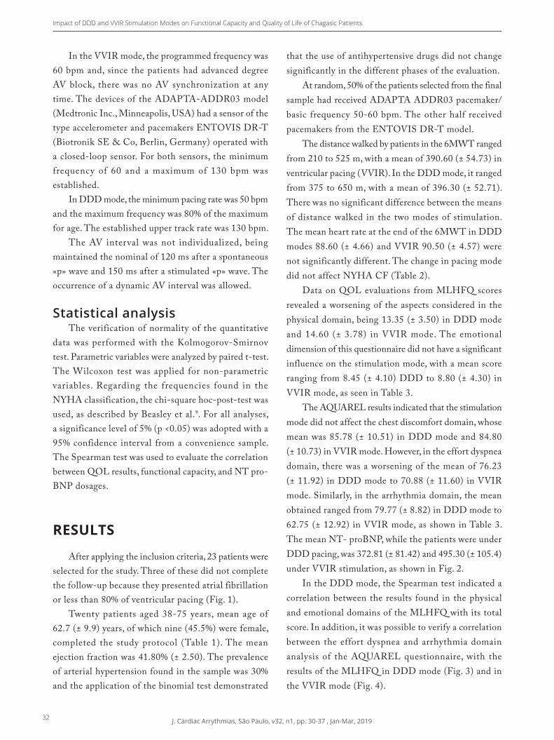
32 J. Cardiac Arrythmias, São Paulo, v32, n1, pp. 30-37 , Jan-Mar, 2019
Impact of DDD and VVIR Stimulation Modes on Functional Capacity and Quality of Life of Chagasic Patients
In the VVIR mode, the programmed frequency was 60 bpm and, since the patients had advanced degree AV block, there was no AV synchronization at any time. The devices of the ADAPTA-ADDR03 model (Medtronic Inc., Minneapolis, USA) had a sensor of the type accelerometer and pacemakers ENTOVIS DR-T (Biotronik SE & Co, Berlin, Germany) operated with a closed-loop sensor. For both sensors, the minimum frequency of 60 and a maximum of 130 bpm was established.
In DDD mode, the minimum pacing rate was 50 bpm and the maximum frequency was 80% of the maximum for age. The established upper track rate was 130 bpm.
The AV interval was not individualized, being maintained the nominal of 120 ms after a spontaneous «p» wave and 150 ms after a stimulated «p» wave. The occurrence of a dynamic AV interval was allowed.
Statistical analysisThe verification of normality of the quantitative
data was performed with the Kolmogorov-Smirnov test. Parametric variables were analyzed by paired t-test. The Wilcoxon test was applied for non-parametric variables. Regarding the frequencies found in the NYHA classification, the chi-square hoc-post-test was used, as described by Beasley et al.9. For all analyses, a significance level of 5% (p <0.05) was adopted with a 95% confidence interval from a convenience sample. The Spearman test was used to evaluate the correlation between QOL results, functional capacity, and NT pro-BNP dosages.
RESULTS
After applying the inclusion criteria, 23 patients were selected for the study. Three of these did not complete the follow-up because they presented atrial fibrillation or less than 80% of ventricular pacing (Fig. 1).
Twenty patients aged 38-75 years, mean age of 62.7 (± 9.9) years, of which nine (45.5%) were female, completed the study protocol (Table 1). The mean ejection fraction was 41.80% (± 2.50). The prevalence of arterial hypertension found in the sample was 30% and the application of the binomial test demonstrated
that the use of antihypertensive drugs did not change significantly in the different phases of the evaluation.
At random, 50% of the patients selected from the final sample had received ADAPTA ADDR03 pacemaker/basic frequency 50-60 bpm. The other half received pacemakers from the ENTOVIS DR-T model.
The distance walked by patients in the 6MWT ranged from 210 to 525 m, with a mean of 390.60 (± 54.73) in ventricular pacing (VVIR). In the DDD mode, it ranged from 375 to 650 m, with a mean of 396.30 (± 52.71). There was no significant difference between the means of distance walked in the two modes of stimulation. The mean heart rate at the end of the 6MWT in DDD modes 88.60 (± 4.66) and VVIR 90.50 (± 4.57) were not significantly different. The change in pacing mode did not affect NYHA CF (Table 2).
Data on QOL evaluations from MLHFQ scores revealed a worsening of the aspects considered in the physical domain, being 13.35 (± 3.50) in DDD mode and 14.60 (± 3.78) in VVIR mode. The emotional dimension of this questionnaire did not have a significant influence on the stimulation mode, with a mean score ranging from 8.45 (± 4.10) DDD to 8.80 (± 4.30) in VVIR mode, as seen in Table 3.
The AQUAREL results indicated that the stimulation mode did not affect the chest discomfort domain, whose mean was 85.78 (± 10.51) in DDD mode and 84.80 (± 10.73) in VVIR mode. However, in the effort dyspnea domain, there was a worsening of the mean of 76.23 (± 11.92) in DDD mode to 70.88 (± 11.60) in VVIR mode. Similarly, in the arrhythmia domain, the mean obtained ranged from 79.77 (± 8.82) in DDD mode to 62.75 (± 12.92) in VVIR mode, as shown in Table 3. The mean NT- proBNP, while the patients were under DDD pacing, was 372.81 (± 81.42) and 495.30 (± 105.4) under VVIR stimulation, as shown in Fig. 2.
In the DDD mode, the Spearman test indicated a correlation between the results found in the physical and emotional domains of the MLHFQ with its total score. In addition, it was possible to verify a correlation between the effort dyspnea and arrhythmia domain analysis of the AQUAREL questionnaire, with the results of the MLHFQ in DDD mode (Fig. 3) and in the VVIR mode (Fig. 4).
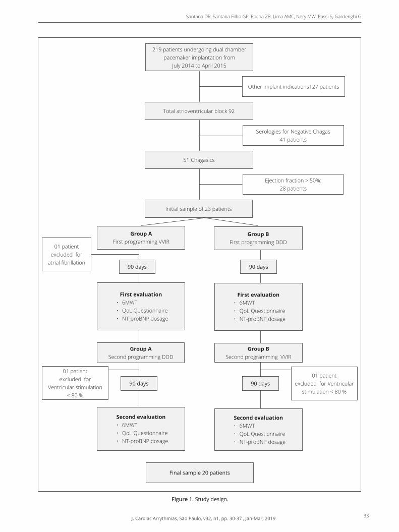
J. Cardiac Arrythmias, São Paulo, v32, n1, pp. 30-37 , Jan-Mar, 2019 33
Santana DR, Santana Filho GP, Rocha ZB, Lima AMC, Nery MW, Rassi S, Gardenghi G
Figure 1. Study design.
Group AFirst programming VVIR
Second evaluation• 6MWT• QoL Questionnaire• NT-proBNP dosage
Second evaluation• 6MWT• QoL Questionnaire• NT-proBNP dosage
Final sample 20 patients
Other implant indications127 patients
Serologies for Negative Chagas 41 patients
Ejection fraction > 50%: 28 patients
01 patient excluded for
atrial fibrillation
01 patient excluded for Ventricular
stimulation < 80 %
01 patient excluded for
Ventricular stimulation < 80 %
219 patients undergoing dual chamber pacemaker implantation from
July 2014 to April 2015
Total atrioventricular block 92
51 Chagasics
Initial sample of 23 patients
Group ASecond programming DDD
First evaluation• 6MWT• QoL Questionnaire• NT-proBNP dosage
90 days
90 days
90 days
90 days
Group BFirst programming DDD
Group BSecond programming VVIR
First evaluation• 6MWT• QoL Questionnaire• NT-proBNP dosage
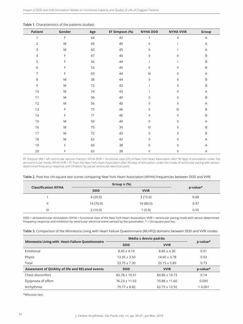
34 J. Cardiac Arrythmias, São Paulo, v32, n1, pp. 30-37 , Jan-Mar, 2019
Impact of DDD and VVIR Stimulation Modes on Functional Capacity and Quality of Life of Chagasic Patients
Table 1. Characteristics of the patients studied.
Patient Gender Age EF Simpson (%) NYHA DDD NYHA VVIR Group
1 F 64 42 I II A
2 M 60 40 II I A
3 M 60 45 II I A
4 F 47 40 II II B
5 F 56 44 I I B
6 F 53 40 II II B
7 F 69 44 III II A
8 M 38 44 II II B
9 M 72 42 I II B
10 M 74 43 I II A
11 M 56 40 II II B
12 M 56 40 II II A
13 F 75 46 II III B
14 F 71 46 II II B
15 M 59 40 II II A
16 M 75 39 III II B
17 M 72 43 II II B
18 M 62 42 II II A
19 F 69 38 II II A
20 F 65 38 II II A EF Simpson (%) = left ventricular ejection fraction; NYHA DDD = functional class (CF) of New York Heart Association after 90 days of stimulation under the atrioventricular mode; NYHA VVIR = FC from the New York Heart Association after 90 days of stimulation under the mode of ventricular pacing with sensor-determined frequency response and inhibition by paced ventricular electrical event.
Table 2. Post-hoc chi-square test scores comparing New York Heart Association (NYHA) frequencies between DDD and VVIR.
Classification NYHAGroup n (%)
p-value*DDD VVIR
I 4 (20.0) 3 (15.0) 0.68
II 14 (70.0) 16 (80.0) 0.47
III 2 (10.0) 1 (5.0) 0.55 DDD = atrioventricular stimulation; NYHA = functional class of the New York Heart Association; VVIR = ventricular pacing mode with sensor-determined frequency response and inhibition by ventricular electrical event sensed by the pacemaker; * = chi-square post hoc.
Table 3. Comparison of the Minnesota Living with Heart Failure Questionnaire (MLHFQ) domains between DDD and VVIR modes.
Minnesota Living with Heart Failure QuestionnaireMédia ± desvio padrão
p-value*DDD VVIR
Emotional 8.45 ± 4.10 8.80 ± 4.30 0.51
Physic 13.35 ± 3.50 14.60 ± 3.78 0.03
Total 33.75 ± 7.30 33.15 ± 5.89 0.73
Assesment of QUAlity of life and RELated events DDD VVIR p-value*
Chest discomfort 85.78 ± 10.51 84.80 ± 10.73 0.14
Dyspnoea of effort 76.23 ± 11.92 70.88 ± 11.60 0.005
Arrhythmia 79.77 ± 8.82 62.75 ± 12.92 < 0.001 *Wilcoxon test.
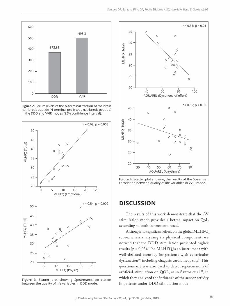
J. Cardiac Arrythmias, São Paulo, v32, n1, pp. 30-37 , Jan-Mar, 2019 35
Santana DR, Santana Filho GP, Rocha ZB, Lima AMC, Nery MW, Rassi S, Gardenghi G
DISCUSSION
The results of this work demonstrate that the AV stimulation mode provides a better impact on QoL according to both instruments used.
Although no significant effect on the global MLHFQ score, when analyzing its physical component, we noticed that the DDD stimulation presented higher results (p = 0.03). The MLHFQ is an instrument with well-defined accuracy for patients with ventricular dysfunction10, including chagasic cardiomyopathy8. This questionnaire was also used to detect repercussions of artificial stimulation on QOL, as in Santos et al.11, in which they analyzed the influence of the sensor activity in patients under DDD stimulation mode.
Figure 4. Scatter plot showing the results of the Spearman correlation between quality of life variables in VVIR mode.
Figure 2. Serum levels of the N-terminal fraction of the brain natriuretic peptide (N-terminal pro b-type natriuretic peptide) in the DDD and VVIR modes (95% confidence interval).
600
500
400
300
200
100
DDR VVIR
495,3
372,81
0
50
45
40
30
25
200 5 10 15
MLHFQ (Emotional)20 25
MLH
FQ (T
otal
)
35
50r = 0.54; p = 0.002
r = 0.62; p = 0.003
45
40
30
25
209 12 15 18
MLHFQ (Physic)21
MLH
FQ (T
otal
)
35
45
40
30
25
2040 50
AQUAREL (Dyspnoea of effort)80 100
MLH
FQ (T
otal
)
35
r = 0,52; p = 0,02
r = 0,53; p = 0,01
45
40
30
25
2030 40 50 60 70 80
AQUAREL (Arrythmia)
MLH
FQ (T
otal
)
35
Figure 3. Scatter plot showing Spearman›s correlation between the quality of life variables in DDD mode.
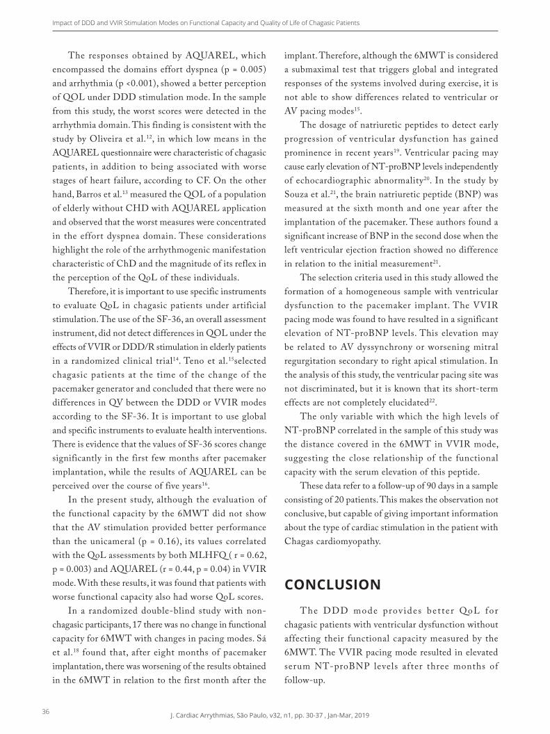
36 J. Cardiac Arrythmias, São Paulo, v32, n1, pp. 30-37 , Jan-Mar, 2019
Impact of DDD and VVIR Stimulation Modes on Functional Capacity and Quality of Life of Chagasic Patients
The responses obtained by AQUAREL, which encompassed the domains effort dyspnea (p = 0.005) and arrhythmia (p <0.001), showed a better perception of QOL under DDD stimulation mode. In the sample from this study, the worst scores were detected in the arrhythmia domain. This finding is consistent with the study by Oliveira et al.12, in which low means in the AQUAREL questionnaire were characteristic of chagasic patients, in addition to being associated with worse stages of heart failure, according to CF. On the other hand, Barros et al.13 measured the QOL of a population of elderly without CHD with AQUAREL application and observed that the worst measures were concentrated in the effort dyspnea domain. These considerations highlight the role of the arrhythmogenic manifestation characteristic of ChD and the magnitude of its reflex in the perception of the QoL of these individuals.
Therefore, it is important to use specific instruments to evaluate QoL in chagasic patients under artificial stimulation. The use of the SF-36, an overall assessment instrument, did not detect differences in QOL under the effects of VVIR or DDD/R stimulation in elderly patients in a randomized clinical trial14. Teno et al.15selected chagasic patients at the time of the change of the pacemaker generator and concluded that there were no differences in QV between the DDD or VVIR modes according to the SF-36. It is important to use global and specific instruments to evaluate health interventions. There is evidence that the values of SF-36 scores change significantly in the first few months after pacemaker implantation, while the results of AQUAREL can be perceived over the course of five years16.
In the present study, although the evaluation of the functional capacity by the 6MWT did not show that the AV stimulation provided better performance than the unicameral (p = 0.16), its values correlated with the QoL assessments by both MLHFQ ( r = 0.62, p = 0.003) and AQUAREL (r = 0.44, p = 0.04) in VVIR mode. With these results, it was found that patients with worse functional capacity also had worse QoL scores.
In a randomized double-blind study with non-chagasic participants, 17 there was no change in functional capacity for 6MWT with changes in pacing modes. Sá et al.18 found that, after eight months of pacemaker implantation, there was worsening of the results obtained in the 6MWT in relation to the first month after the
implant. Therefore, although the 6MWT is considered a submaximal test that triggers global and integrated responses of the systems involved during exercise, it is not able to show differences related to ventricular or AV pacing modes15.
The dosage of natriuretic peptides to detect early progression of ventricular dysfunction has gained prominence in recent years19. Ventricular pacing may cause early elevation of NT-proBNP levels independently of echocardiographic abnormality20. In the study by Souza et al.21, the brain natriuretic peptide (BNP) was measured at the sixth month and one year after the implantation of the pacemaker. These authors found a significant increase of BNP in the second dose when the left ventricular ejection fraction showed no difference in relation to the initial measurement21.
The selection criteria used in this study allowed the formation of a homogeneous sample with ventricular dysfunction to the pacemaker implant. The VVIR pacing mode was found to have resulted in a significant elevation of NT-proBNP levels. This elevation may be related to AV dyssynchrony or worsening mitral regurgitation secondary to right apical stimulation. In the analysis of this study, the ventricular pacing site was not discriminated, but it is known that its short-term effects are not completely elucidated22.
The only variable with which the high levels of NT-proBNP correlated in the sample of this study was the distance covered in the 6MWT in VVIR mode, suggesting the close relationship of the functional capacity with the serum elevation of this peptide.
These data refer to a follow-up of 90 days in a sample consisting of 20 patients. This makes the observation not conclusive, but capable of giving important information about the type of cardiac stimulation in the patient with Chagas cardiomyopathy.
CONCLUSION
The DDD mode prov ides be t te r QoL for chagasic patients with ventricular dysfunction without affecting their functional capacity measured by the 6MWT. The VVIR pacing mode resulted in elevated serum NT-proBNP levels af ter three months of follow-up.

J. Cardiac Arrythmias, São Paulo, v32, n1, pp. 30-37 , Jan-Mar, 2019 37
Santana DR, Santana Filho GP, Rocha ZB, Lima AMC, Nery MW, Rassi S, Gardenghi G
AUTHORS’ CONTRIBUTION
Conceptualization, Santana DR and Rassi S; Methodology, Santana DR; Lima AMC and Rassi S; Research, Santana DR; Santana Filho GP and Rocha ZB;
Writing - First version, Santana DR and Rassi S; Writing - Review & Edition, Santana DR; Rassi S; Nery MW and Gardenghi G; Resources, Santana DR; Supervision, Rassi S.
1. Rassi AJ, Rassi A, Marin-Neto J. Chagas disease. Lancet. 2010;375(9723):1388-402. https://doi.org/10.1016/S0140-6736(10)60061-X
2. World Health Organization. Chagas disease in Latin America: an epidemiological update based on 2010 estimates. Weekly Epidemiol Rec. 2015;90(6):33-43.
3. Sabino EC, Ribeiro AL, Salemi VMC, Di Lorenzo Oliveira C, Antunes AP, Menezes MM, et al. Ten-year incidence of Chagas cardiomyopathy among asymptomatic Trypanosoma cruzi seropositive former blood donors. Circulation. 2013;127(10):1105-15. https://doi.org/10.1161/circulationaha.112.123612
4. Pachon JC, Pereira W, Batista Junior WD, Pachón JPM, Pachón CEI, Vargas RNA, et al. RBM - Registro Brasileiro de Marcapassos, Ressincronizadores e Desfibriladores. Relampa. 2013;26(1):39-49.
5. Rassi A, Rassi Jr A, Faria GHDC. História natural do bloqueio atrioventricular total de etiologia chagásica. Arq Bras Cardiol. 1992;59(Suppl II):191.
6. Martinelli Filho M, Zimerman LI. Diretrizes brasileiras de dispositivos cardíacos eletrônicos implantáveis (DCEI). Arq Bras Cardiol. 2007;89(6):e210-38.
7. American Thoracic Association. ATS statement: guidelines for the six-minute walking test. Am J Resp Critical Care Med. 2002;166(1):111-7. https://doi.org/10.1164/ajrccm.166.1.at1102
8. Carvalho VO, Guimarães GV, Carrara D, Bacal F, Bocchi EA. Validação da versão em português do Minnesota Living with Heart Failure Questionnaire. Arq Bras Cardiol. 2009;93:39-44. https://doi.org/ 10.1590/S0066-782X2009000700008
9. Beasley TM, Schumacker RE. Multiple regression approach to analyzing contingency tables: post hoc and planned comparison procedures. J Exp Educ. 1995;64(1):79-93. https://doi.org/10.1080/00220973.1995.9943797
10. Rector TS, Kubo SH, Cohn JN. Validity of the Minnesota Living with Heart Failure Questionnaire as a measure of therapeutic response to enalapril or placebo. Am J Cardiol. 1993;71(12):1106-7.
11. Santos JRC. Marcapassos dupla-câmara com resposta de frequência melhoram a qualidade de vida dos pacientes? Relampa. 2002;15(4):201-5.
12. Oliveira BG, Abreu MNS, Abreu CDG, Rocha MOC, Ribeiro AL. Health-related quality of life in patients with Chagas disease. Rev Soc Bras Med Trop. 2011;44:150-6. https://doi.org/10.1590/S0037-86822011005000002
13. Barros RTD, Carvalho SMRD, Silva MAM, Borges JBC. Avaliação de aspectos da qualidade de vida em pacientes pós-implante de marcapasso cardíaco. Rev Bras Cir
Cardiovasc. 2014:37-44. https://doi.org/10.5935/1678-9741.20140009
14. Lamas GA, Orav EJ, Stambler BS, Ellenbogen KA, Sgarbossa EB, Huang SKS, et al. Quality of life and clinical outcomes in elderly patients treated with ventricular pacing as compared with dual-chamber pacing. NEJM. 1998;338(16):1097-104. https://doi.org/10.1056/NEJM199804163381602
15. Teno LAC, Costa R, Martinelli Filho M, Castilho FCT, Ruiz I. Mudança do modo VVI para DDD (“upgrade”) na troca eletiva do gerador de pulsos: análise comparativa clínico-funcional. Arq Bras Cardiol. 2007;88(2):128-33. https://doi.org/10.1590/S0066-782X2007000200001
16. Udo EO, van Hemel NM, Zuithoff NP, Nijboer H, Taks W, Doevendans PA, et al. Long term quality-of-life in patients with bradycardia pacemaker implantation. Inter J Cardiol. 2013;168(3):2159-63. https://doi.org/10.1016/j.ijcard.2013.01.253
17. Ouali S, Neffeti E, Ghoul K, Hammas S, Kacem S, Gribaa R, et al. DDD versus VVIR pacing in patients, ages 70 and over, with complete heart block. Pacing Clin Electrophysiol. 2010;33(5):583-9. https://doi.org/10.1111/j.1540-8159.2009.02636.x
18. Sá LAB, Rassi S, Batista MAL. Efeitos da estimulação ventricular convencional em pacientes com função ventricular normal. Arq Bras Cardiol. 2009;93:167-73. https://doi.org/10.1590/S0066-782X2009000800016
19. Porapakkham P, Porapakkham P, Zimmet H, Billah B, Krum H. B-type natriuretic peptide-guided heart failure therapy: a meta-analysis. Arch Intern Med. 2010;170(6):507-14. https://doi.org/10.1001/archinternmed.2010.35
20. Naegeli B, Kurz DJ, Koller D, Straumann E, Furrer M, Maurer D, et al. Single-chamber ventricular pacing increases markers of left ventricular dysfunction compared with dual-chamber pacing. Europace. 2007;9(3):194-9. https://doi.org/10.1093/europace/eul186
21. Souza SF, Nascimento B, Nunes MCP, da Silva JLP, Carvalho VT, Beaton AZ, et al. Effect of pacemaker site on B-type natriuretic peptide levels, and left ventricular function in a population with high prevalence of Chagas disease. Inter J Cardiol. 2015;190:315-8. https://doi.org/10.1016/j.ijcard.2015.04.127
22. Nikoo MH, Ghaedian MM, Kafi M, Fakhrpour A, Jorat MV, Pakfetrat M, et al. Effects of right ventricular septal versus apical pacing on plasma natriuretic peptide levels. J Cardiovasc Dis Res. 2011;2(2):104-9. https://doi.org/10.4103%2F0975-3583.83036
REFERENCES
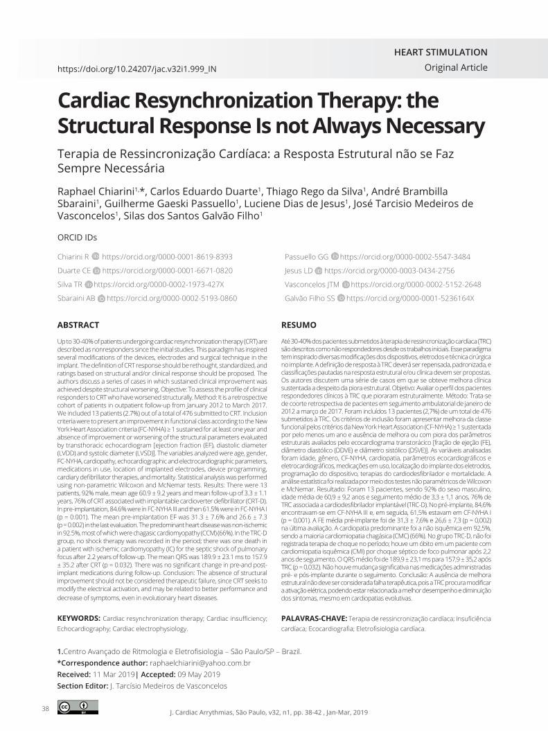
38 J. Cardiac Arrythmias, São Paulo, v32, n1, pp. 38-42 , Jan-Mar, 2019
Cardiac Resynchronization Therapy: the Structural Response Is not Always NecessaryTerapia de Ressincronização Cardíaca: a Resposta Estrutural não se Faz Sempre Necessária
Raphael Chiarini1,*, Carlos Eduardo Duarte1, Thiago Rego da Silva1, André Brambilla Sbaraini1, Guilherme Gaeski Passuello1, Luciene Dias de Jesus1, José Tarcisio Medeiros de Vasconcelos1, Silas dos Santos Galvão Filho1
HEART STIMULATIONOriginal Article
1.Centro Avançado de Ritmologia e Eletrofi siologia – São Paulo/SP – Brazil.*Correspondence author: [email protected]: 11 Mar 2019| Accepted: 09 May 2019Section Editor: J. Tarcísio Medeiros de Vasconcelos
https://doi.org/10.24207/jac.v32i1.999_IN
ORCID IDs
Chiarini R https://orcid.org/0000-0001-8619-8393
Duarte CE https://orcid.org/0000-0001-6671-0820
Silva TR https://orcid.org/0000-0002-1973-427X
Sbaraini AB https://orcid.org/0000-0002-5193-0860
Chiarini R https://orcid.
Duarte CE https://orcid.
Silva TR https://orcid.
Sbaraini AB https://orcid.
Passuello GG https://orcid.org/0000-0002-5547-3484
Jesus LD https://orcid.org/0000-0003-0434-2756
Vasconcelos JTM https://orcid.org/0000-0002-5152-2648
Galvão Filho SS https://orcid.org/0000-0001-5236164X
ABSTRACT
Up to 30-40% of patients undergoing cardiac resynchronization therapy (CRT) are described as nonresponders since the initial studies. This paradigm has inspired several modifi cations of the devices, electrodes and surgical technique in the implant. The defi nition of CRT response should be rethought, standardized, and ratings based on structural and/or clinical response should be proposed. The authors discuss a series of cases in which sustained clinical improvement was achieved despite structural worsening. Objective: To assess the profi le of clinical responders to CRT who have worsened structurally. Method: It is a retrospective cohort of patients in outpatient follow-up from January 2012 to March 2017. We included 13 patients (2.7%) out of a total of 476 submitted to CRT. Inclusion criteria were to present an improvement in functional class according to the New York Heart Association criteria (FC-NYHA) ≥ 1 sustained for at least one year and absence of improvement or worsening of the structural parameters evaluated by transthoracic echocardiogram [ejection fraction (EF), diastolic diameter (LVDD) and systolic diameter (LVSD)]. The variables analyzed were age, gender, FC-NYHA, cardiopathy, echocardiographic and electrocardiographic parameters, medications in use, location of implanted electrodes, device programming, cardiary defi brillator therapies, and mortality. Statistical analysis was performed using non-parametric Wilcoxon and McNemar tests. Results: There were 13 patients, 92% male, mean age 60.9 ± 9.2 years and mean follow-up of 3.3 ± 1.1 years, 76% of CRT associated with implantable cardioverter defi brillator (CRT-D). In pre-implantation, 84.6% were in FC-NYHA III and then 61.5% were in FC-NYHA I(p = 0.001). The mean pre-implantation EF was 31.3 ± 7.6% and 26.6 ± 7.3(p = 0.002) in the last evaluation. The predominant heart disease was non-ischemic in 92.5%, most of which were chagasic cardiomyopathy (CCM) (66%). In the TRC-D group, no shock therapy was recorded in the period; there was one death in a patient with ischemic cardiomyopathy (IC) for the septic shock of pulmonary focus after 2.2 years of follow-up. The mean QRS was 189.9 ± 23.1 ms to 157.9 ± 35.2 after CRT (p = 0.032). There was no signifi cant change in pre-and post-implant medications during follow-up. Conclusion: The absence of structural improvement should not be considered therapeutic failure, since CRT seeks to modify the electrical activation, and may be related to better performance and decrease of symptoms, even in evolutionary heart diseases.
KEYWORDS: Cardiac resynchronization therapy; Cardiac insuffi ciency; Echocardiography; Cardiac electrophysiology.
RESUMO Até 30-40% dos pacientes submetidos à terapia de ressincronização cardíaca (TRC) são descritos como não respondedores desde os trabalhos iniciais. Esse paradigma tem inspirado diversas modifi cações dos dispositivos, eletrodos e técnica cirúrgica no implante. A defi nição de resposta à TRC deverá ser repensada, padronizada, e classifi cações pautadas na resposta estrutural e/ou clínica devem ser propostas. Os autores discutem uma série de casos em que se obteve melhora clínica sustentada a despeito da piora estrutural. Objetivo: Avaliar o perfi l dos pacientes respondedores clínicos à TRC que pioraram estruturalmente. Método: Trata-se de coorte retrospectiva de pacientes em seguimento ambulatorial de janeiro de 2012 a março de 2017. Foram incluídos 13 pacientes (2,7%) de um total de 476 submetidos à TRC. Os critérios de inclusão foram apresentar melhora da classe funcional pelos critérios da New York Heart Association (CF-NYHA) ≥ 1 sustentada por pelo menos um ano e ausência de melhora ou com piora dos parâmetros estruturais avaliados pelo ecocardiograma transtorácico [fração de ejeção (FE), diâmetro diastólico (DDVE) e diâmetro sistólico (DSVE)]. As variáveis analisadas foram idade, gênero, CF-NYHA, cardiopatia, parâmetros ecocardiográfi cos e eletrocardiográfi cos, medicações em uso, localização do implante dos eletrodos, programação do dispositivo, terapias do cardiodesfi brilador e mortalidade. A análise estatística foi realizada por meio dos testes não paramétricos de Wilcoxon e McNemar. Resultado: Foram 13 pacientes, sendo 92% do sexo masculino, idade média de 60,9 ± 9,2 anos e seguimento médio de 3,3 ± 1,1 anos, 76% de TRC associada a cardiodesfi brilador implantável (TRC-D). No pré-implante, 84,6% encontravam-se em CF-NYHA III e, em seguida, 61,5% estavam em CF-NYHA I (p = 0,001). A FE média pré-implante foi de 31,3 ± 7,6% e 26,6 ± 7,3 (p = 0,002) na última avaliação. A cardiopatia predominante foi a não isquêmica em 92,5%, sendo a maioria cardiomiopatia chagásica (CMC) (66%). No grupo TRC-D, não foi registrada terapia de choque no período; houve um óbito em um paciente com cardiomiopatia isquêmica (CMI) por choque séptico de foco pulmonar após 2,2 anos de seguimento. O QRS médio foi de 189,9 ± 23,1 ms para 157,9 ± 35,2 após TRC (p = 0,032). Não houve mudança signifi cativa nas medicações administradas pré- e pós-implante durante o seguimento. Conclusão: A ausência de melhora estrutural não deve ser considerada falha terapêutica, pois a TRC procura modifi car a ativação elétrica, podendo estar relacionada a melhor desempenho e diminuição dos sintomas, mesmo em cardiopatias evolutivas.
PALAVRAS-CHAVE: Terapia de ressincronização cardíaca; Insufi ciência cardíaca; Ecocardiografi a; Eletrofi siologia cardíaca.
J. Cardiac Arrythmias, São Paulo, v32, n1, pp. 38-42 , Jan-Mar, 2019 39
Cardiac Resynchronization Therapy: the Structural Response Is not Always Necessary
INTRODUCTION
Cardiac pacing began to be thought of as adjuvant therapy in the treatment of cardiac insufficiency (CI) refractory to pharmacological treatment in the mid-1990s. The theoretical and experimental basis of this application is the correction of delayed left ventricular (LV) contraction in patients with IC that present complete left bundle branch block (LBBB). In these cases, with the advent of imaging exams such as echocardiography, it can be confirmed that, in addition to the delay in electrical activation, there is mechanical dyssynchrony of the ventricle, decreasing cardiac efficiency, and may promote cardiac remodeling in the short and long term. Important randomized studies supported implant guidelines adopted in Brazil and by the leading scientific societies in the United States and Europe (American College of Cardiology (ACC), American Heart Association (AHA), Heart Rhythm Society (HRS) and European Society of Cardiology (ESC)], being recommended as a recommendation class I and level A evidence of cardiac resynchronization therapy (CRT) for NYHA II and III patients, QRS duration ≥ 150 ms and LBBB, provided that with optimized drug therapy, sinus rhythm and EF ≤ 35%8-11. Although there are no important randomized studies, it is now possible to identify other groups of patients with a high probability of positive response to CRT, setting guidelines as indication class I or II with a level of evidence B8-11. Despite the technical advances of devices, electrodes, implant surgery, and development of complementary methods, more recent studies still classify about 30% of patients as non-responders to CRT despite the lack of consensus in the definition of CRT response1-8. The present study analyzed a retrospective cohort of patients submitted to CRT, evaluating the profile of clinical responders to therapy, seeking to identify factors associated with the mechanism of action that results in clinical improvement without the presence of structural improvement.
METHODS
Retrospective study of a cohort of follow-up patients at the Advanced Center of Ritology and Electrophysiology (Centro Avançado de Ritmologia e Eletrofisiologia-CARE), in the state of São Paulo, submitted to the CRT from January 2012 to March 2017.
A g e , g e n d e r , F C - N Y H A , c a r d i o p a t h y, echocardiographic parameters, and pre and post-CRT electrocardiographic parameters, medications in use, electrode implant localization, device programming, cardi-defibrillator therapies, and mortality were analyzed. The criteria for an indication of CRT and outpatient follow-up obeyed the institutional protocol that follows the Brazilian Guidelines for Implantable Electronic Cardiac Devices.
The criterion used to define exclusively a clinical response to CRT was: improvement of the SF by NYHA criteria ≥ 1 sustained for a minimum period of 12 months after CRT, without improvement or worsening of the structural parameters in the transthoracic echocardiogram.
Exclusion criteria were any increase in left ventricular ejection fraction (EF) and/or reduction of diastolic and/or final systolic diameters evaluated by transthoracic echocardiography or unsupported clinical improvement for a minimum period of 12 months.
Statistical analysisContinuous variables were expressed in means ±
standard deviation (SD) and categorical variables in percentages. Statistical analysis was performed using non-parametric Wilcoxon and McNemar tests. The representation was made by the mean, SD and quartiles (Tables 1, 2 and 3).
RESULTS
A total of 476 sequential charts of patients submitted to CRT-related surgical intervention from January 2012 to March 2017 were selected. Thirteen patients (2.7%) met the inclusion criteria, most were male (92%), with a mean age of 60.9 ± 9.2 years. Before implantation, FC-NYHA was III in 84.6%, IV in 7.6% and II in 7.6%; Mean EF of the LV was 31.3 ± 7.9%, the mean systolic diameter (LVSD) was 53.2 ± 5.1 mm and the mean diastolic diameter (LVDD) was 65.5 ± 5.9. Baseline heart disease was distributed in: Chagas cardiomyopathy (CCC) in 61%; hypertensive (HiCM) in 15%; dilated (DCM) in 8%; ischemic (ICM) in 8%; and hypertrophic (HCM) in 8%. Pre-implantation sinus rhythm was present in 76% of the patients, with a mean QRS of 189.9 ± 29.1 ms, with an LBBB pattern being identified in 61.5%, of which 25% were associated with axis deviation for left. Right
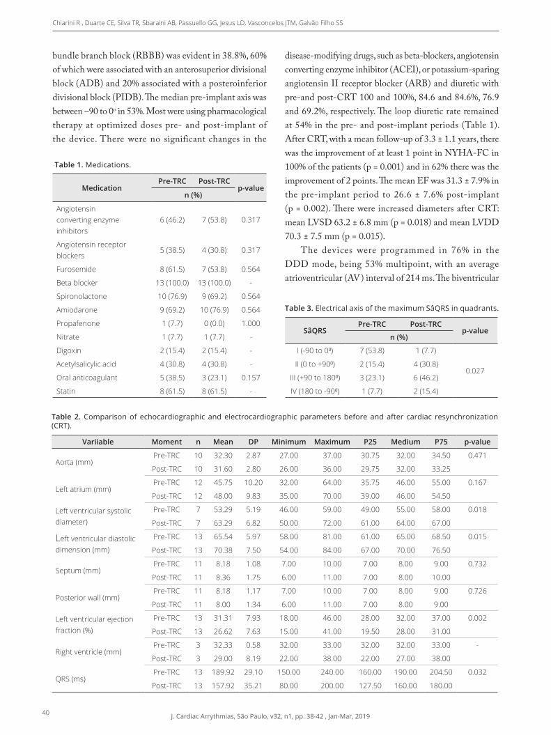
40 J. Cardiac Arrythmias, São Paulo, v32, n1, pp. 38-42 , Jan-Mar, 2019
Chiarini R , Duarte CE, Silva TR, Sbaraini AB, Passuello GG, Jesus LD, Vasconcelos JTM, Galvão Filho SS
bundle branch block (RBBB) was evident in 38.8%, 60% of which were associated with an anterosuperior divisional block (ADB) and 20% associated with a posteroinferior divisional block (PIDB). The median pre-implant axis was between –90 to 0o in 53%. Most were using pharmacological therapy at optimized doses pre- and post-implant of the device. There were no significant changes in the
disease-modifying drugs, such as beta-blockers, angiotensin converting enzyme inhibitor (ACEI), or potassium-sparing angiotensin II receptor blocker (ARB) and diuretic with pre-and post-CRT 100 and 100%, 84.6 and 84.6%, 76.9 and 69.2%, respectively. The loop diuretic rate remained at 54% in the pre- and post-implant periods (Table 1). After CRT, with a mean follow-up of 3.3 ± 1.1 years, there was the improvement of at least 1 point in NYHA-FC in 100% of the patients (p = 0.001) and in 62% there was the improvement of 2 points. The mean EF was 31.3 ± 7.9% in the pre-implant period to 26.6 ± 7.6% post-implant (p = 0.002). There were increased diameters after CRT: mean LVSD 63.2 ± 6.8 mm (p = 0.018) and mean LVDD 70.3 ± 7.5 mm (p = 0.015).
The devices were programmed in 76% in the DDD mode, being 53% multipoint, with an average atrioventricular (AV) interval of 214 ms. The biventricular
Table 1. Medications.
MedicationPre-TRC Post-TRC
p-valuen (%)
Angiotensin converting enzyme inhibitors
6 (46.2) 7 (53.8) 0.317
Angiotensin receptor blockers
5 (38.5) 4 (30.8) 0.317
Furosemide 8 (61.5) 7 (53.8) 0.564
Beta blocker 13 (100.0) 13 (100.0) -
Spironolactone 10 (76.9) 9 (69.2) 0.564
Amiodarone 9 (69.2) 10 (76.9) 0.564
Propafenone 1 (7.7) 0 (0.0) 1.000
Nitrate 1 (7.7) 1 (7.7) -
Digoxin 2 (15.4) 2 (15.4) -
Acetylsalicylic acid 4 (30.8) 4 (30.8) -
Oral anticoagulant 5 (38.5) 3 (23.1) 0.157
Statin 8 (61.5) 8 (61.5) -
Table 2. Comparison of echocardiographic and electrocardiographic parameters before and after cardiac resynchronization (CRT).
Variiable Moment n Mean DP Minimum Maximum P25 Medium P75 p-value
Aorta (mm)Pre-TRC 10 32.30 2.87 27.00 37.00 30.75 32.00 34.50 0.471
Post-TRC 10 31.60 2.80 26.00 36.00 29.75 32.00 33.25
Left atrium (mm)Pre-TRC 12 45.75 10.20 32.00 64.00 35.75 46.00 55.00 0.167
Post-TRC 12 48.00 9.83 35.00 70.00 39.00 46.00 54.50
Left ventricular systolic diameter)
Pre-TRC 7 53.29 5.19 46.00 59.00 49.00 55.00 58.00 0.018
Post-TRC 7 63.29 6.82 50.00 72.00 61.00 64.00 67.00
Left ventricular diastolic dimension (mm)
Pre-TRC 13 65.54 5.97 58.00 81.00 61.00 65.00 68.50 0.015
Post-TRC 13 70.38 7.50 54.00 84.00 67.00 70.00 76.50
Septum (mm)Pre-TRC 11 8.18 1.08 7.00 10.00 7.00 8.00 9.00 0.732
Post-TRC 11 8.36 1.75 6.00 11.00 7.00 8.00 10.00
Posterior wall (mm)Pre-TRC 11 8.18 1.17 7.00 10.00 7.00 8.00 9.00 0.726
Post-TRC 11 8.00 1.34 6.00 11.00 7.00 8.00 9.00
Left ventricular ejection fraction (%)
Pre-TRC 13 31.31 7.93 18.00 46.00 28.00 32.00 37.00 0.002
Post-TRC 13 26.62 7.63 15.00 41.00 19.50 28.00 31.00
Right ventricle (mm)Pre-TRC 3 32.33 0.58 32.00 33.00 32.00 32.00 33.00 -
Post-TRC 3 29.00 8.19 22.00 38.00 22.00 27.00 38.00
QRS (ms)Pre-TRC 13 189.92 29.10 150.00 240.00 160.00 190.00 204.50 0.032
Post-TRC 13 157.92 35.21 80.00 200.00 127.50 160.00 180.00
Table 3. Electrical axis of the maximum SâQRS in quadrants.
SâQRSPre-TRC Post-TRC
p-valuen (%)
I (-90 to 0⁰) 7 (53.8) 1 (7.7)
0.027II (0 to +90⁰) 2 (15.4) 4 (30.8)
III (+90 to 180⁰) 3 (23.1) 6 (46.2)
IV (180 to -90⁰) 1 (7.7) 2 (15.4)

J. Cardiac Arrythmias, São Paulo, v32, n1, pp. 38-42 , Jan-Mar, 2019 41
Cardiac Resynchronization Therapy: the Structural Response Is not Always Necessary
pacing rate was 98.7%, with the LV-RV pacing pattern 61.5%, RV-LV 23%, and simultaneous 15.5%. The QRS under biventricular pacing was 187.1 ± 25.9 ms with ADB in 7% and PIDB in 46%, mean axis under stimulation between +90 and + 180o in 43% and between 0 and + 90o in 30%. Regarding the position of the electrodes, the right ventricle was superior to the septal-basal, super-septal-medial and mid-septal-apical positions in 38, 23 and 38% of the implants, respectively. The LV electrode in mid-latero-basal position in 23%, supero-latero-basal 15%; medial-latero-medial 15% infero-latero-apical 15%, infero-latero-medial 15% and superior-antero-basal 15%. There were no records of cardiac defibrillator shock therapies. There was one death in a patient with IMC related to pulmonary focus septic shock with 2.2 years of CRT (Tables 1 and 2).
DISCUSSION
This study discusses the standardization of CRT response criteria and proposes analysis from both the structural and clinical perspectives, questioning the real need to restrict the indication of CRT to those who have established predictors of structural response, such as the presence of LBBB and QRS> 150 ms in patients with DCM.
It should be remembered that in the onset of CRT, with the study MUSTIC15 was analyzed exactly that the functional capacity was improved, the quality of life and the patient›s preference for the mode of stimulation, with LV stimulation bound or not. Restricting CRT to the point of structural improvement would deny its greatest benefit to those with evolutionary structural cardiomyopathy.
Question: Can driving disorders be the cause of CI or just an aggravating factor? In the MADIT-CRT study25, in women with mild IC symptoms, female gender, absence of infarction, LBBB, QRS> 150 ms, body mass index (BMI) <30 kg / m2 and reduced left atrial volume were predictors of normalization of parameters and clinical improvement before CRT (super-responders). In these cases CRT is very likely to act on the pathophysiology of ventricular dysfunction, restoring normalization.
In this context, we discussed whether the clear change in the direction of activation (Table 3) and the search for correction of conduction disturbance were determinants
of improvement in clinical performance even in those whose etiology was of evolutionary cardiomyopathies, such as Chagas› disease.
The major criticism of these results is the deficiency of other parametric clinical evaluations. On MUSTIC15, in addition to FC-NYHA, the 6-minute walk test, the peak O2 consumption (VO2 peak), and the Minnesota quality of life score were analyzed. Clinical evaluation of this study was limited to FC-NYHA. Regarding echocardiographic references, a deficiency is present in the evaluation of pre-and post-implant mitral regurgitation, since it could be a determinant of response. It is believed that the reduction of mitral regurgitation before CRT may lead to increased pressure load in the LV, resulting in increased diameters with worsening of global EF, but generating a greater anterograde flow through the LV exit pathway, resulting in clinical improvement. The actual understanding of the clinical improvement of this group of patients should encourage prospective studies to study this effect.
CONCLUSION
The absence of structural improvement should not be considered therapeutic failure. Electrical dyssynchrony can lead to changes in the sense of activation, causing slower and disharmonious contractions, resulting in hemodynamic performance deficit. The CRT seeks to accelerate and harmonize electrical activation, correcting driving disorders that worsen cardiac performance, and may promote clinical and/or structural improvement.
AUTHORS’ CONTRIBUTION
Conceptualization, Chiarini R; Duarte CE; Vasconcelos JTM de; Galvão Filho SS. Methodology, Chiarini R; Duarte CE; Vasconcelos JTM de; Galvão Filho SS; Research, Chiarini R; Duarte CE; Silva TR da; Sbaraini AB; Passuello GG; Jesus LD de; Vasconcelos JTM de; Galvão Filho SS; Writing - First version, Chiarini R; Passuello GG; Silva TR da; Writing - Review & Editing, Chiarini R; Duarte CE; Acquisition of Financing, Duarte CE; Resources, Chiarini R; Duarte CE; Supervision, Duarte CE
42 J. Cardiac Arrythmias, São Paulo, v32, n1, pp. 38-42 , Jan-Mar, 2019
Chiarini R , Duarte CE, Silva TR, Sbaraini AB, Passuello GG, Jesus LD, Vasconcelos JTM, Galvão Filho SS
1. Fuganti CJ, Melo CS, Moraes Jr AV, Pachon-Mateos JC, Pereira WL, Galvão Filho SS, et al. Diretriz: – Terapia de ressincronização cardíaca. Relampa. 2015;28(Suppl 2):S26-S40.
2. Chung ES, Leon AR, Tavazzi L, Sun JP, Nihoyannopoulos P, Merlino J, et al. Results of the predictors of response to CRT (PROSPECT) trial. Circulation. 2008;117(20):2608-16. https://doi.org/10.1161/CIRCULATIONAHA.107.743120
3. Rocha EA, Moreira FT, Quidute AR, Abreu JS, Lima WO, Rodrigues CR, et al. Quem são os super-respondedores à terapia de ressincronização cardíaca? Int J Cardiovasc Sci. 2017;30(1):61-9. https://doi.org/10.5935/2359-4802.20170020
4. Tracy CM, Epstein AE, Darbar D, Dimarco JP, Dunbar SB, Estes NA 3rd, et al. 2012 ACCF/AHA/HRS focused update of the 2008 guidelines for device-based therapy of cardiac rhythm abnormalities: a report of the American College of Cardiology Foundation/American Heart Association Task Force on Practice Guidelines. J Am Coll Cardiol. 2012;60(14):1297-313. https://doi.org/10.1161/CIR.0b013e3182618569
5. Yu CM, Bleeker GB, Fung JW, Schalij MJ, Zhang Q, van der Wall EE, et al. Left ventricular reverse remodeling but not clinical improvement predicts long-term survival after cardiac resynchronization therapy. Circulation. 2005;112(11):1580-6. https://doi.org/10.1161/CIRCULATIONAHA.105.538272
6. Rao RK, Kumar UN, Schafer J, Viloria E, De Lurgio D, Foster E. Reduced ventricular volumes and improved systolic function with cardiac resynchronization therapy: a randomized trial comparing simultaneous biventricular pacing, sequential biventricular pacing, and left ventricular pacing. Circulation. 2007;115(16):2136-44. https://doi.org/10.1161/CIRCULATIONAHA.106.634444
7. Leclercq C, Gras D, Le Helloco A, Nicol L, Daubert C. Hemodynamic importance of preserving the normal sequence of ventricular activation in permanent cardiac pacing. Am Heart J 1995;129:1133-41. https://doi.org/10.1016/0002-8703(95)90394-1
8. Tracy CM, Epstein AE, Darbar D, DiMarco JP, Dunbar SB, Estes NAM. 2012 ACCF/AHA/HRS focused update of the 2008 guidelines for device-based therapy of cardiac rhythm abnormalities. A report of the American College of Cardiology Foundation/American Heart Association Task Force on Practice Guidelines and the Heart Rhythm Society. Circulation. 2012;126:1784-1800. https://doi.org/10.1161/CIR.0b013e3182618569
9. Comitê Coordenador da Diretriz de Insuficiência Cardíaca. Diretriz brasileira de insuficiência cardíaca crônica e aguda. Arq Bras Cardiol. 2018;111(3):436-539. https://doi.org/10.5935/abc.20180190
10. The Task Force on cardiac pacing and resynchronization therapy of the European Society of Cardiology (ESC). ESC Guidelines on cardiac pacing and cardiac resynchronization therapy. Eur Heart J. 2013;34:2281-2329. https://doi.org/10.1093/eurheartj/eht150
11. The Task Force for the diagnosis and treatment of acute and chronic heart failure of the European Society of Cardiology (ESC). ESC Guidelines for the diagnosis and treatment of acute and chronic heart failure. Eur Heart J. 2016;37:2129-2200. https://doi.org/10.1093/eurheartj/ehw128.
12. Martinelli Filho M, Peixoto GL, Siqueira SF, Martins SAM, Nishioka SAD, Costa R, et al. Terapia de ressincronização cardíaca na cardiomiopatia chagásica crônica: boa resposta clínica e pior prognóstico. Relampa. 2013;26(1):33-8.
13. Garillo R, Salgado Melo C, Pachón Mateos CTC, Silva Jr. O, Leite GMS, Carvalho EIJ. Pacientes não responsivos à terapia de ressincronização cardíaca. Relampa. 2007;20(3):167-74.
14. Menezes Junior AS, Stival WN, Lopes ISP. Indicações da terapia de ressincronização cardíaca: discussão baseada em estudos recentes. Relampa. 2014;27(1):34-9.
15. Linde C, Leclercq C, Rex S, Garrigue S, Lavergne T, Cazeau S, et al. Long-term benefits of BiV pacing in heart failure. JACC. 2002;40(1):111-8. https://doi.org/10.1016/S0735-1097(02)01932-0
16. Linde C, Ellenbogen K, McAlister FA. CRT: clinical trials, guidelines, and target populations. Heart Rhythm. 2012;9(Suppl 8):S3-S13. https://doi.org/10.1016/j.hrthm.2012.04.026
17. Rocha EA. Analysis of cardiac dyssynchrony. Arq Bras Cardiol. 2018;111(4):616-7. https://doi.org/10.5935/abc.20180200
18. Leyva F, Nisam S, Auricchio A. 20 years of cardiac resynchronization therapy. JACC. 2014;64(10):1047-58. https://doi.org/10.1016/j.jacc.2014.06.1178
19. Papelbaum B. Galvão Filho SS, Vasconcelos JTM, Duarte CE, Chiarini R Numata BK, et al. Punção do óstio do seio coronariano ocluído para implante de eletrodo ventricular esquerdo: uma nova alternativa. Rev Soc Cardiol Estado de São Paulo. 2018;28(4):489-91. https://doi.org/10.29381/0103-8559/20182804489-91
20. Nascimento EA, Wiefels Reis CC, Ribeiro FB, Alves CR, Silva EN, Ribeiro ML, et al. Relação entre dissincronismo elétrico e mecânico em pacientes Submetidos a TRC com implante do eletrodo de VE orientado pela cintilografia GATED SPECT. Arq Bras Cardiol. 2018;111(4):607-15. https://doi.org/10.5935/abc.20180159
21. Birnie et al. Incidence of AF with adaptive CR. Heart Rhythm. 2017;14(12). https://doi.org/10.1016/j.hrthm.2017.08.017
22. Linde C, Abraham WT, Gold MR, Daubert JC, Tang ASL, Young JB, et al. Predictors of CRT clinical response. Eur J Heart Failure. 2017:19(8):1056-63. https://doi.org/10.1002/ejhf.795
23. Melo CS, Silva Júnior LM, Vazquez BP, Oliveira JC, Salerno HD, Lage JS. Evidências atuais para indicação da terapia de ressincronização cardíaca. Relampa. 2013;26(3):151-61.
24. Rocha EA, Pereira FTM, Quidute ARP, Abreu JS, Lima JWO, Sobrinho CRMR, et al. Quem são os super-respondedores à ressincronização. Int J Cardiovasc Sci. 2017;30(1):61-9. https://doi.org/10.5935/2359-4802.20170020
25. Hsu JC, Solomon SD, Bourgoun M, McNitt S, Goldenberg I, Klein H, et al; MADIT-CRT Executive Committee. Predictors of super-response to cardiac resynchronization therapy and associated improvement in clinical outcome: the MADIT-CRT (multicenter automatic defibrillator implantation trial with cardiac resynchronization therapy) study. J Am Coll Cardiol. 2012;59(25):2366-73. https://doi.org/10.1016/j.jacc.2012.01.065.
REFERENCES
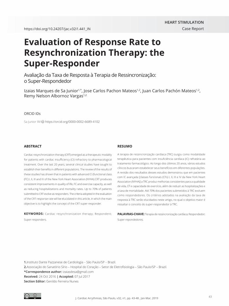
J. Cardiac Arrythmias, São Paulo, v32, n1, pp. 43-48 , Jan-Mar, 2019 43
Evaluation of Response Rate to Resynchronization Therapy: theSuper-ResponderAvaliação da Taxa de Resposta à Terapia de Ressincronização:o Super-Respondedor
Izaias Marques de Sa Junior1,*, Jose Carlos Pachon Mateos1,2, Juan Carlos Pachón Mateos1,2, Remy Nelson Albornoz Vargas1,2
HEART STIMULATIONCase Report
1.Instituto Dante Pazzanese de Cardiologia – São Paulo/SP – Brazil.2.Associação do Sanatório Sírio – Hospital do Coração – Setor de Eletrofi siologia – São Paulo/SP – Brazil.*Correspondence author: [email protected]: 24 Oct 2016 | Accepted: 07 Jul 2017Section Editor: Genildo Ferreira Nunes
https://doi.org/10.24207/jac.v32i1.441_IN
ORCID IDs
Sa Junior IM https://orcid.org/0000-0002-6689-4102Sa Junior IM
ABSTRACT
Cardiac resynchronization therapy (CRT) emerged as a therapeutic modality
for patients with cardiac insuffi ciency (CI) refractory to pharmacological
treatment. Over the last 20 years, several clinical studies have sought to
establish their benefi ts in diff erent populations. The review of the results of
these studies has shown that in patients with advanced CI (functional class
(FC) I, II, III and IV of the New York Heart Association (NYHA) CRT produces
consistent improvements in quality of life, FC and exercise capacity, as well
as reducing hospitalizations and mortality rates. Up to 70% of patients
submitted to CRT evolve as responders. The criteria adopted in the evaluation
of the CRT response rate will be elucidated in this article, in which the main
objective is to highlight the concept of the CRT super-responder.
KEYWORDS: Cardiac resynchronization therapy; Respondent;
Super-responders.
RESUMO
A terapia de ressincronização cardíaca (TRC) surgiu como modalidade
terapêutica para pacientes com insufi ciência cardíaca (IC) refratária ao
tratamento farmacológico. Ao longo dos últimos 20 anos, vários estudos
clínicos buscaram estabelecer seus benefícios em diferentes populações.
A revisão dos resultados desses estudos demonstrou que em pacientes
com IC avançada [classes funcionais (CFs) I, II, III e IV da New York Heart
Association (NYHA)] a TRC produz melhorias consistentes para a qualidade
de vida, CF e capacidade de exercício, além de reduzir as hospitalizações e
a taxa de mortalidade. Até 70% dos pacientes submetidos à TRC evoluem
como respondedores. Os critérios adotados na avaliação da taxa de
resposta à TRC serão elucidados neste artigo, no qual o objetivo maior é
ressaltar o conceito do super-respondedor à TRC.
PALAVRAS-CHAVE: Terapia de ressincronização cardíaca; Respondedor;
Super-repondedores.

44 J. Cardiac Arrythmias, São Paulo, v32, n1, pp. 43-48 , Jan-Mar, 2019
Sa Junior IM, Mateos JCP, Mateos JCP, Vargas RNA
INTRODUCTION
Cardiac insufficiency (CI) represents a major public health problem worldwide. In Europe and the United States, it represents an important cause of hospitalization in patients aged 65 and over1,2. This disease has a great social, economic and, above all, human impact, since it imposes important physical limitations on patients, resulting in early retirements, with high governmental costs3.
Despite advances in available pharmacological therapies, they do not fully meet the needs of patients with heart failure4. Non-pharmacological therapies, such as mechanical left ventricular assist devices and cardiac transplantation, are reserved for a small group of patients with CI5,6. In addition to these, cardiac resynchronization therapy (CRT) and implantable cardioverter defibrillators appeared as a good option for patients with CI refractory to drug treatment4-7.
The pathophysiological basis of CRT is the presence of dyssynchrony associated with conduction disturbance. With the implantation of electrodes in the right atrium (RA), left ventricle (LV) and right ventricle (RV) and adequate device programming, atrioventricular and ventricle-ventricular resynchronization would be achieved and expected patient improvement. CRT is indicated in patients with CI with conduction disorder, mainly with left bundle branch block morphology (LBBB), functional classes (FC) II, III, IV NYHA, and optimized clinical treatment8,9.
The CRT response rate historically approaches 70% of cases. With improved patient selection, ie QRS duration> 150 ms and LBBB morphology10, absence of fibrosis by magnetic resonance imaging (MRI) in the posterolateral region11, as well as total fibrosis load <20%12, improvement of the surgical technique, adequate programming of the device, a recent and considerable increase in the response rate to CRT was observed.
The following is the case of a patient considered to be a CRT super-responder. We will also discuss the important variables in the selection of these patients, as well as the criteria to be considered super-responders.
CASE REPORT
MJS, 72 years old, born in the state of Paraíba, a patient with CI since 2010, without other comorbidities.
Admitted in a tertiary hospital in 2015 for optimization and investigation of CI etiology.
In this period, FC III, doses of carvedilol, enalapril, and furosemide were optimized. The patient had a significant improvement in FC, now FC II. All the necessary exams were done to elucidate the etiology of CI (Chagas, echocardiogram, MRI of the heart, myocardial scintigraphy), and, as definitive diagnosis, idiopathic CI.
Because he was an LBBB patient and still symptomatic despite clinical treatment, he was referred to the pacemaker team to decide on CRT. Based on the current evidence, we opted for the implantation of the cardiac resynchronizer, performed 12/13/2015.
Five essential steps for success in using CRT as a treatment modality were defined. They were followed strictly.
Patient selectionAccording to the current evidence, HF with ejection
fraction <35% and LBBB with QRS duration> 150 ms may be one of the only indications considered to be FC IA de CRT8,9. In the present case, those criteria were satisfied.
Surgical techniqueThe cardiac dyssynchrony generated by LBBB has
the slowest and most delayed region on the conduction in the posterolateral wall of the LV13. This should be the implant site of the LV electrode. The cardiac vein of choice should be the one that is directed to the posterolateral region of the LV (Fig. 1). In addition, performing an MRI of the pre-implanted heart discards the presence of fibrosis in this region, thus avoiding possible stimulation in a non-viable region11. In the present case, all such care was observed.
X-ray and comparison of electroencephalograms (ECGs) (<one month postoperatively)
The correct position of the electrodes, especially the LV coronary sinus electrode, can be verified in the posterolateral region (Fig. 2). In the case of a dislocation, it should be surgically resubmitted for repositioning. In the present case, the LV electrode was stable in the postoperative period.
In addition, a considerable reduction in pre and post-implant QRS duration provides good prospects regarding
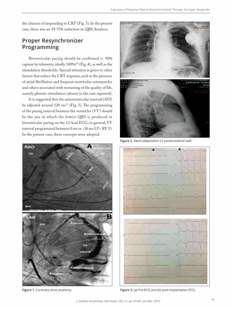
J. Cardiac Arrythmias, São Paulo, v32, n1, pp. 43-48 , Jan-Mar, 2019 45
Evaluation of Response Rate to Resynchronization Therapy: the Super-Responder
Figure 1. Coronary sinus anatomy.
Figure 2. Electrodeposition LV posterolateral wall.
Figure 3. (a) Pre-ECG and (b) post-implantation ECG.
the chances of responding to CRT (Fig. 3). In the present case, there was an 18.75% reduction in QRS duration.
Proper Resynchronizer Programming
Biventricular pacing should be confirmed (> 90% capture by telemetry, ideally 100%)14 (Fig. 4), as well as the stimulation thresholds. Special attention is given to other factors that reduce the CRT response, such as the presence of atrial fibrillation and frequent ventricular extrasystoles and others associated with worsening of the quality of life, namely phrenic stimulation (absent in the case reported).
It is suggested that the atrioventricular interval (AVI) be adjusted around 120 ms15 (Fig. 5). The programming of the pacing interval between the ventricles (VV) should be the one in which the lowest QRS is produced in biventricular pacing on the 12-lead ECG, in general, VV interval programmed between 0 ms or -30 ms LV> RV 5). In the present case, these concepts were adopted.

46 J. Cardiac Arrythmias, São Paulo, v32, n1, pp. 43-48 , Jan-Mar, 2019
Sa Junior IM, Mateos JCP, Mateos JCP, Vargas RNA
Figure 4. 100% LV capture.
Figure 5. IAV 120MS and VV-30MS.
Table 1. Parameters and cut-off points used in the super-responder definition.
Parameters Cut-Off points
Ventricular Function19 > 50% and functional recovery
Ventricular Function20 > 2× the baseline function or > 45% and > 1 Functional Class NYHA
End-Systolic Volume21 Reduction > 30%
End-Dyastolic Volume21 Reduction > 20%
Ventricular Function21 Increase > 10%
Volume Sistólico Final22 Reduction > 30%
Ventricular Function23> 50%, reduction End-Systolic Volume > 25% and > 1 Functional class NYHA
Ventricular Function24 Increase > 20% Source: Adapted from Steffel e Ruschitzka25; NYHA = New York Heart Association
Evaluation of the CRT response rate
Finally, it will be assessed whether our patient actually responded to CRT. According to the main studies, six months would be enough time to have this answer. By means of clinical (FC), echocardiographic criteria [ventricular function (VF), end-systolic volume (ESV)] and others we can conclude about this. In the present case, after evaluation, it was concluded that it is a super-responder. From the echocardiographic data, the VF of 28% was 55% (normalization), the ESV from 173 to 79 mL, a reduction of 54.4%, reaching LV reverse remodeling. Finally, FC II was for NYHA FC I, remaining practically asymptomatic.
DISCUSSION
Concept of super-responder to CRT
Patients without structural heart disease and with LBBB when compared to those without structural heart disease and without LBBB present, on average, 7% less of the VF, considering both within the range of normality16. In about three years, approximately 16% of patients with normal heart and LBBB may develop with CI17. This condition will be defined as a primary electrical cardiopathy18-20.
When we work on the cause of heart disease in question, we generally have the best rates of response to treatment. It is believed that these patients are the ones with the highest CRT response rate - the super-responders. It is clear that many other patients, with the most diverse etiologies of CI, can progress with LBBB and benefit from CRT, but it is believed that response rates are more modest.
Although there was no consensus regarding the best criterion to be considered in the evaluation of CRT response rate, clinical parameters with improvement of at least one of the FC NYHA, 10 to 15% increase in the six-minute walk test, elevation of 10 to 15% of peak oxygen consumption (VO2 peak) in ergospirometry, improvement of the Minnesota life questionnaire, and echocardiographic findings, such as improvement of LVEF> 5% and reduction of ESV greater than 10 to 15%, are the most used. Other criteria, such as improved blood pressure and natriuretic peptide reduction, may also be considered.
Likewise, because there is no consensus in the medical literature regarding the concept of the super-responder, some of the main echocardiographic criteria used in the large studies on the subject are listed in Table 1.
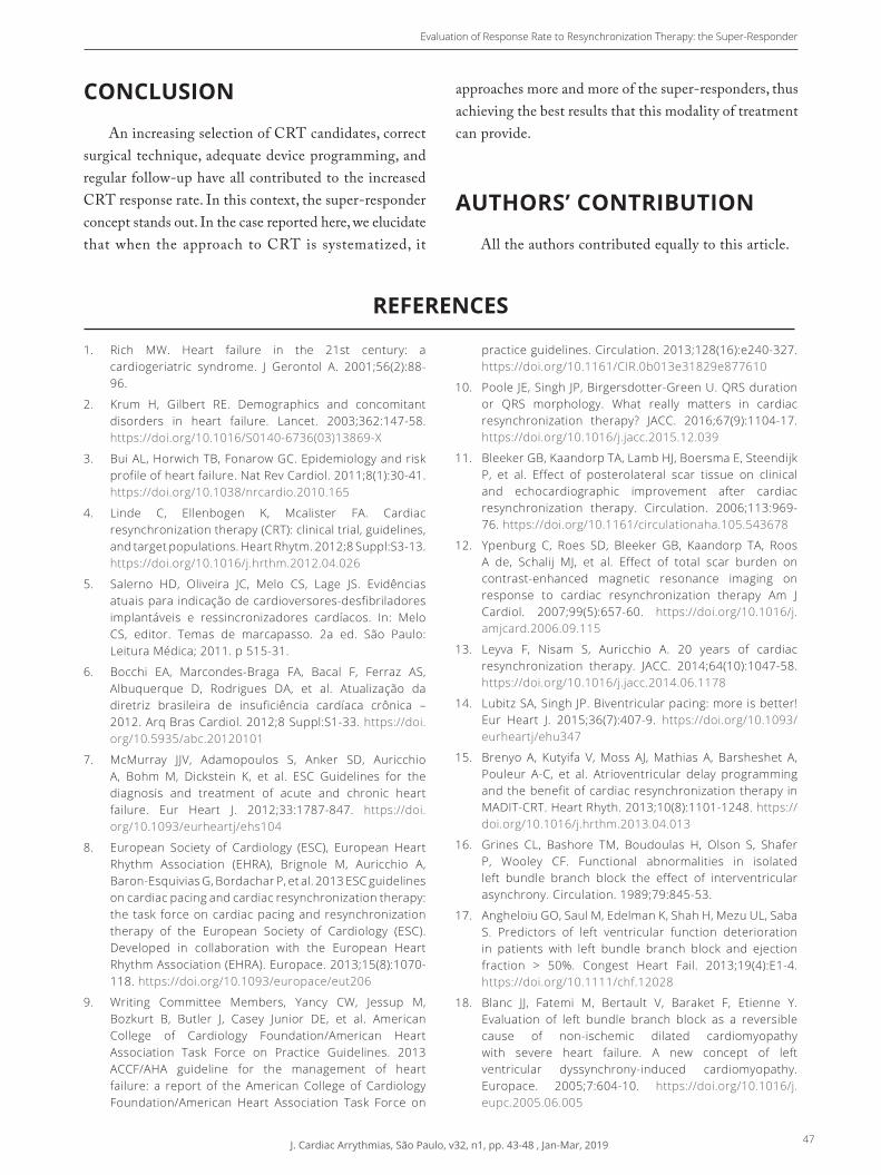
J. Cardiac Arrythmias, São Paulo, v32, n1, pp. 43-48 , Jan-Mar, 2019 47
Evaluation of Response Rate to Resynchronization Therapy: the Super-Responder
1. Rich MW. Heart failure in the 21st century: a cardiogeriatric syndrome. J Gerontol A. 2001;56(2):88-96.
2. Krum H, Gilbert RE. Demographics and concomitant disorders in heart failure. Lancet. 2003;362:147-58. https://doi.org/10.1016/S0140-6736(03)13869-X
3. Bui AL, Horwich TB, Fonarow GC. Epidemiology and risk profile of heart failure. Nat Rev Cardiol. 2011;8(1):30-41. https://doi.org/10.1038/nrcardio.2010.165
4. Linde C, Ellenbogen K, Mcalister FA. Cardiac resynchronization therapy (CRT): clinical trial, guidelines, and target populations. Heart Rhytm. 2012;8 Suppl:S3-13. https://doi.org/10.1016/j.hrthm.2012.04.026
5. Salerno HD, Oliveira JC, Melo CS, Lage JS. Evidências atuais para indicação de cardioversores-desfibriladores implantáveis e ressincronizadores cardíacos. In: Melo CS, editor. Temas de marcapasso. 2a ed. São Paulo: Leitura Médica; 2011. p 515-31.
6. Bocchi EA, Marcondes-Braga FA, Bacal F, Ferraz AS, Albuquerque D, Rodrigues DA, et al. Atualização da diretriz brasileira de insuficiência cardíaca crônica – 2012. Arq Bras Cardiol. 2012;8 Suppl:S1-33. https://doi.org/10.5935/abc.20120101
7. McMurray JJV, Adamopoulos S, Anker SD, Auricchio A, Bohm M, Dickstein K, et al. ESC Guidelines for the diagnosis and treatment of acute and chronic heart failure. Eur Heart J. 2012;33:1787-847. https://doi.org/10.1093/eurheartj/ehs104
8. European Society of Cardiology (ESC), European Heart Rhythm Association (EHRA), Brignole M, Auricchio A, Baron-Esquivias G, Bordachar P, et al. 2013 ESC guidelines on cardiac pacing and cardiac resynchronization therapy: the task force on cardiac pacing and resynchronization therapy of the European Society of Cardiology (ESC). Developed in collaboration with the European Heart Rhythm Association (EHRA). Europace. 2013;15(8):1070-118. https://doi.org/10.1093/europace/eut206
9. Writing Committee Members, Yancy CW, Jessup M, Bozkurt B, Butler J, Casey Junior DE, et al. American College of Cardiology Foundation/American Heart Association Task Force on Practice Guidelines. 2013 ACCF/AHA guideline for the management of heart failure: a report of the American College of Cardiology Foundation/American Heart Association Task Force on
practice guidelines. Circulation. 2013;128(16):e240-327. https://doi.org/10.1161/CIR.0b013e31829e877610
10. Poole JE, Singh JP, Birgersdotter-Green U. QRS duration or QRS morphology. What really matters in cardiac resynchronization therapy? JACC. 2016;67(9):1104-17. https://doi.org/10.1016/j.jacc.2015.12.039
11. Bleeker GB, Kaandorp TA, Lamb HJ, Boersma E, Steendijk P, et al. Effect of posterolateral scar tissue on clinical and echocardiographic improvement after cardiac resynchronization therapy. Circulation. 2006;113:969-76. https://doi.org/10.1161/circulationaha.105.543678
12. Ypenburg C, Roes SD, Bleeker GB, Kaandorp TA, Roos A de, Schalij MJ, et al. Effect of total scar burden on contrast-enhanced magnetic resonance imaging on response to cardiac resynchronization therapy Am J Cardiol. 2007;99(5):657-60. https://doi.org/10.1016/j.amjcard.2006.09.115
13. Leyva F, Nisam S, Auricchio A. 20 years of cardiac resynchronization therapy. JACC. 2014;64(10):1047-58. https://doi.org/10.1016/j.jacc.2014.06.1178
14. Lubitz SA, Singh JP. Biventricular pacing: more is better! Eur Heart J. 2015;36(7):407-9. https://doi.org/10.1093/eurheartj/ehu347
15. Brenyo A, Kutyifa V, Moss AJ, Mathias A, Barsheshet A, Pouleur A-C, et al. Atrioventricular delay programming and the benefit of cardiac resynchronization therapy in MADIT-CRT. Heart Rhyth. 2013;10(8):1101-1248. https://doi.org/10.1016/j.hrthm.2013.04.013
16. Grines CL, Bashore TM, Boudoulas H, Olson S, Shafer P, Wooley CF. Functional abnormalities in isolated left bundle branch block the effect of interventricular asynchrony. Circulation. 1989;79:845-53.
17. Angheloiu GO, Saul M, Edelman K, Shah H, Mezu UL, Saba S. Predictors of left ventricular function deterioration in patients with left bundle branch block and ejection fraction > 50%. Congest Heart Fail. 2013;19(4):E1-4. https://doi.org/10.1111/chf.12028
18. Blanc JJ, Fatemi M, Bertault V, Baraket F, Etienne Y. Evaluation of left bundle branch block as a reversible cause of non-ischemic dilated cardiomyopathy with severe heart failure. A new concept of left ventricular dyssynchrony-induced cardiomyopathy. Europace. 2005;7:604-10. https://doi.org/10.1016/j.eupc.2005.06.005
REFERENCES
CONCLUSION
An increasing selection of CRT candidates, correct surgical technique, adequate device programming, and regular follow-up have all contributed to the increased CRT response rate. In this context, the super-responder concept stands out. In the case reported here, we elucidate that when the approach to CRT is systematized, it
approaches more and more of the super-responders, thus achieving the best results that this modality of treatment can provide.
AUTHORS’ CONTRIBUTION
All the authors contributed equally to this article.

48 J. Cardiac Arrythmias, São Paulo, v32, n1, pp. 43-48 , Jan-Mar, 2019
Sa Junior IM, Mateos JCP, Mateos JCP, Vargas RNA
19. Castellant P, Fatemi M, Orhan E, Etienne Y, Blanc JJ. Patients with non-ischaemic dilated cardiomyopathy and hyper-responders to cardiac resynchronization therapy: characteristics and long-term evolution. Europace. 2009;11(3):350-5. https://doi.org/10.1093/europace/eup035.
20. Antonio N, Teixeira R, Coelho L, Lourenço C, Monteiro P, Ventura M, et al. Identification of ‘super-responders’ to cardiac resynchronization therapy: the importance of symptom duration and left ventricular geometry. Europace. 2009;11(3):343-9. https://doi.org/10.1093/europace/eup038
21. Steffel J, Milosevic G, Hürlimann A, Krasniqi, Namdar M, Ruschitzka F, et al. Characteristics and long-term outcome of echocardiographic super-responders to cardiac resynchronisation therapy: ‘real world’ experience from a single tertiary care centre. Heart. 2011;97(20):1668-74. https://doi.org/ 0.1136/heartjnl-2011-300222
22. Ypenburg C, van Bommel RJ, Borleffs CJ, Bleeker GB, Boersma E, Schalij MJ, et al. Long-term prognosis after cardiac resynchronization therapy is related to the extent of left ventricular reverse remodeling at midterm follow- up. J Am Coll Cardiol. 2009;53(6)483-90. https://doi.org/10.1016/j.jacc.2008.10.032
23. Reant P, Zaroui A, Donal E, Mignot A, Bordachar P, Deplagne A, et al. Identification and characterization of super-responders after cardiac resynchronization therapy. Am J Cardiol. 2010;105(9):1327-35. https://doi.org/10.1016/j.amjcard.2009.12.058
24. Rickard J, Kumbhani DJ, Popovic Z, Verhaert D, Manne M, Sraow D, et al. Characterization of super-response to cardiac resynchronization therapy. Heart Rhythm. 2010;(7):885-9. https://doi.org/10.1016/j.hrthm.2010.04.005
25. Steffel J; Ruschitzka F. Superresponse to cardiac resynchronization therapy. Circulation. 2014;130(1):87-90. https://doi.org/10.1161/circulationaha.113.006124
J. Cardiac Arrythmias, São Paulo, v32, n1, pp. 49-51 , Jan-Mar, 2019 49
Persistence of Left Superior Vena Cava in Patient submitted a Pacemaker ImplantationPersistência de Veia Cava Superior Esquerda em Paciente Submetido a Implante de Marca-Passo
Sara Carneiro Vicente Bueno1,*, Arnaldo Duarte Lourenço1
ARTIFICIAL HEART STIMULATION
Case Report
1.HospitalAnaCosta–SetordeEstimulaçãoCardíacaArtificial–Santos/SP–Brazil.*Correspondence author: [email protected]: 16 May 2018 | Accepted: 04 Feb 2019Section Editor: J.TarcísioMedeirosdeVasconcelos
https://doi.org/10.24207/jac.v32i1.531_IN
ORCID IDs
BuenoSCVhttps://orcid.org/0000-0001-6638-2492
Lourenço AD https://orcid.org/0000-0003-3582-6534
BuenoSCV
Lourenço AD
ABSTRACT
Thepersistenceoftheleftsuperiorvenacava(PLSVC)isthemostfrequentthoracicvenousmalformation,howeverrare,andwithausuallyaccidentaldiagnosis.Recurringly,itsgreatestsuspicionisrealizedintheintraoperativeactrelatedtoatechnicaldifficultyinperformingtheprocedure,evenbymoreexperiencedprofessionals.Casereport:patientis77yearsold,woman,chagasic,submittedadefinitivepacemakerimplant,beingthediagnosisofpersistenceleftsuperiorvenacavarealizedduringthesurgicalprocedure.Thepatientdidnotpresentintercurrenceduringtheprocedureanddevelopedwithsubstantialclinicalimprovementaftertheimplantationoftheelectroniccardiacdevice,andremainingasymptomatic.
KEYWORDS: Superiorvenacava;Vascularmalformations;Artificial
pacemaker.
RESUMO
Apersistênciadaveiacavasuperioresquerda(PCVSE)éamalformaçãovenosatorácicamaisfrequente,porémraraecomdiagnósticohabitualmenteacidental.Recorrentemente,suamaiorsuspeitaérealizadanoatointraoperatóriorelacionadoaumadificuldadetécnicanarealizaçãodoprocedimento,mesmoporprofissionaismaisexperientes.Relatodecaso: Pacientede77anos,sexofeminino,chagásica,submetidaaimplantedemarcapassodefinitivo,sendoodiagnósticodepersistênciadeveiacavasuperioresquerdarealizadoduranteoprocedimentocirúrgico.Apacientenãoapresentouintercorrênciaduranteoprocedimentoeevoluiucommelhoraclínicasubstancialapósoimplantedodispositivocardíacoeletrônico,mantendo-seassintomática.
PALAVRAS-CHAVE: Veiacavasuperior;Malformaçõesvasculares;
Marcapassoartificial.
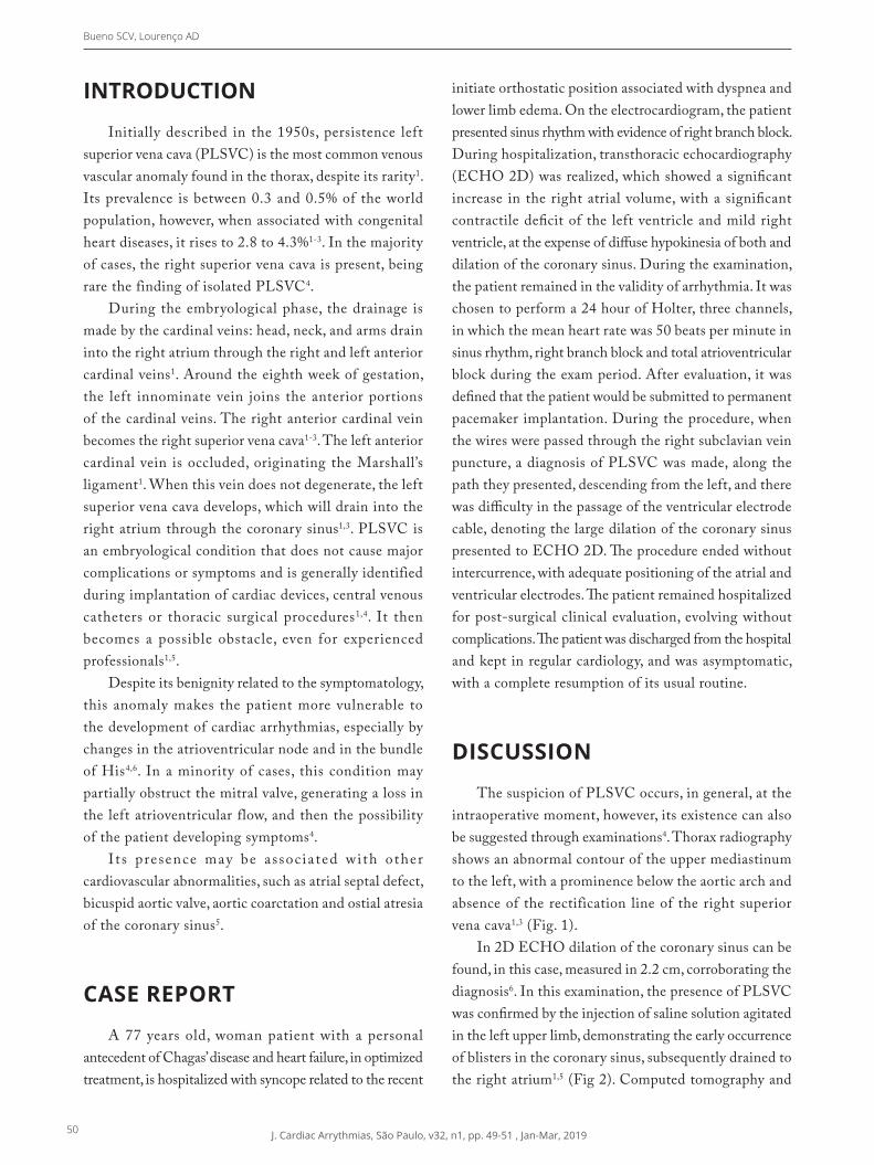
50 J. Cardiac Arrythmias, São Paulo, v32, n1, pp. 49-51 , Jan-Mar, 2019
Bueno SCV, Lourenço AD
INTRODUCTION
Initially described in the 1950s, persistence left superior vena cava (PLSVC) is the most common venous vascular anomaly found in the thorax, despite its rarity1. Its prevalence is between 0.3 and 0.5% of the world population, however, when associated with congenital heart diseases, it rises to 2.8 to 4.3%1-3. In the majority of cases, the right superior vena cava is present, being rare the finding of isolated PLSVC4.
During the embryological phase, the drainage is made by the cardinal veins: head, neck, and arms drain into the right atrium through the right and left anterior cardinal veins1. Around the eighth week of gestation, the left innominate vein joins the anterior portions of the cardinal veins. The right anterior cardinal vein becomes the right superior vena cava1-3. The left anterior cardinal vein is occluded, originating the Marshall’s ligament1. When this vein does not degenerate, the left superior vena cava develops, which will drain into the right atrium through the coronary sinus1,3. PLSVC is an embryological condition that does not cause major complications or symptoms and is generally identified during implantation of cardiac devices, central venous catheters or thoracic surgical procedures1,4. It then becomes a possible obstacle, even for experienced professionals1,5.
Despite its benignity related to the symptomatology, this anomaly makes the patient more vulnerable to the development of cardiac arrhythmias, especially by changes in the atrioventricular node and in the bundle of His4,6. In a minority of cases, this condition may partially obstruct the mitral valve, generating a loss in the left atrioventricular flow, and then the possibility of the patient developing symptoms4.
I t s presence may be assoc ia ted wi th other cardiovascular abnormalities, such as atrial septal defect, bicuspid aortic valve, aortic coarctation and ostial atresia of the coronary sinus5.
CASE REPORT
A 77 years old, woman patient with a personal antecedent of Chagas’ disease and heart failure, in optimized treatment, is hospitalized with syncope related to the recent
initiate orthostatic position associated with dyspnea and lower limb edema. On the electrocardiogram, the patient presented sinus rhythm with evidence of right branch block. During hospitalization, transthoracic echocardiography (ECHO 2D) was realized, which showed a significant increase in the right atrial volume, with a significant contractile deficit of the left ventricle and mild right ventricle, at the expense of diffuse hypokinesia of both and dilation of the coronary sinus. During the examination, the patient remained in the validity of arrhythmia. It was chosen to perform a 24 hour of Holter, three channels, in which the mean heart rate was 50 beats per minute in sinus rhythm, right branch block and total atrioventricular block during the exam period. After evaluation, it was defined that the patient would be submitted to permanent pacemaker implantation. During the procedure, when the wires were passed through the right subclavian vein puncture, a diagnosis of PLSVC was made, along the path they presented, descending from the left, and there was difficulty in the passage of the ventricular electrode cable, denoting the large dilation of the coronary sinus presented to ECHO 2D. The procedure ended without intercurrence, with adequate positioning of the atrial and ventricular electrodes. The patient remained hospitalized for post-surgical clinical evaluation, evolving without complications. The patient was discharged from the hospital and kept in regular cardiology, and was asymptomatic, with a complete resumption of its usual routine.
DISCUSSION
The suspicion of PLSVC occurs, in general, at the intraoperative moment, however, its existence can also be suggested through examinations4. Thorax radiography shows an abnormal contour of the upper mediastinum to the left, with a prominence below the aortic arch and absence of the rectification line of the right superior vena cava1,3 (Fig. 1).
In 2D ECHO dilation of the coronary sinus can be found, in this case, measured in 2.2 cm, corroborating the diagnosis6. In this examination, the presence of PLSVC was confirmed by the injection of saline solution agitated in the left upper limb, demonstrating the early occurrence of blisters in the coronary sinus, subsequently drained to the right atrium1,5 (Fig 2). Computed tomography and
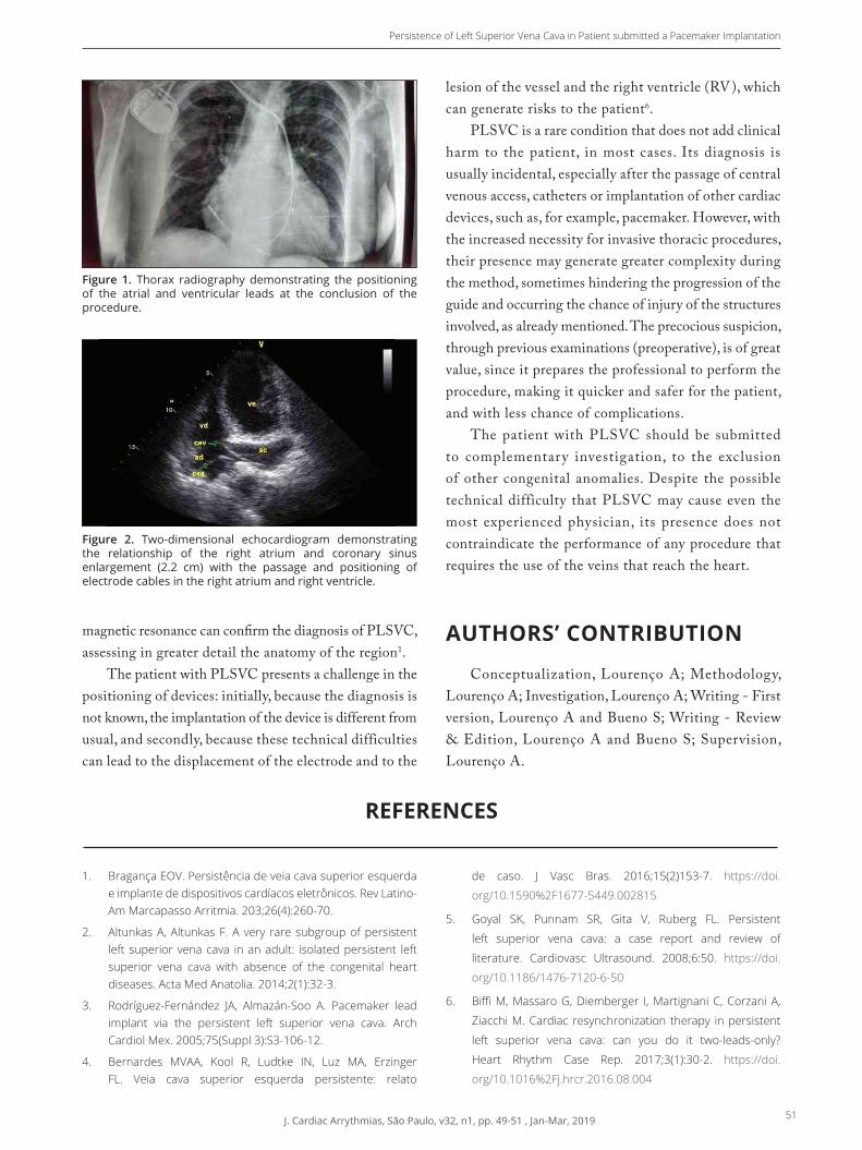
J. Cardiac Arrythmias, São Paulo, v32, n1, pp. 49-51 , Jan-Mar, 2019 51
Persistence of Left Superior Vena Cava in Patient submitted a Pacemaker Implantation
magnetic resonance can confirm the diagnosis of PLSVC, assessing in greater detail the anatomy of the region1.
The patient with PLSVC presents a challenge in the positioning of devices: initially, because the diagnosis is not known, the implantation of the device is different from usual, and secondly, because these technical difficulties can lead to the displacement of the electrode and to the
lesion of the vessel and the right ventricle (RV), which can generate risks to the patient6.
PLSVC is a rare condition that does not add clinical harm to the patient, in most cases. Its diagnosis is usually incidental, especially after the passage of central venous access, catheters or implantation of other cardiac devices, such as, for example, pacemaker. However, with the increased necessity for invasive thoracic procedures, their presence may generate greater complexity during the method, sometimes hindering the progression of the guide and occurring the chance of injury of the structures involved, as already mentioned. The precocious suspicion, through previous examinations (preoperative), is of great value, since it prepares the professional to perform the procedure, making it quicker and safer for the patient, and with less chance of complications.
The patient with PLSVC should be submitted to complementary investigation, to the exclusion of other congenital anomalies. Despite the possible technical difficulty that PLSVC may cause even the most experienced physician, its presence does not contraindicate the performance of any procedure that requires the use of the veins that reach the heart.
AUTHORS’ CONTRIBUTION
Conceptualization, Lourenço A; Methodology, Lourenço A; Investigation, Lourenço A; Writing - First version, Lourenço A and Bueno S; Writing - Review & Edition, Lourenço A and Bueno S; Supervision, Lourenço A.
Figure 1. Thorax radiography demonstrating the positioning of the atrial and ventricular leads at the conclusion of the procedure.
Figure 2. Two-dimensional echocardiogram demonstrating the relationship of the right atrium and coronary sinus enlargement (2.2 cm) with the passage and positioning of electrode cables in the right atrium and right ventricle.
1. BragançaEOV.Persistênciadeveiacavasuperioresquerdaeimplantededispositivoscardíacoseletrônicos.RevLatino-AmMarcapassoArritmia.203;26(4):260-70.
2. AltunkasA,AltunkasF.Avery raresubgroupofpersistentleft superior venacava inanadult: isolatedpersistent leftsuperior vena cava with absence of the congenital heartdiseases.ActaMedAnatolia.2014;2(1):32-3.
3. Rodríguez-Fernández JA, Almazán-Soo A. Pacemaker leadimplant via the persistent left superior vena cava. ArchCardiolMex.2005;75(Suppl3):S3-106-12.
4. Bernardes MVAA, Kool R, Ludtke IN, Luz MA, ErzingerFL. Veia cava superior esquerda persistente: relato
de caso. J Vasc Bras. 2016;15(2)153-7. https://doi.
org/10.1590%2F1677-5449.002815
5. Goyal SK, Punnam SR, Gita V, Ruberg FL. Persistent
left superior vena cava: a case report and review of
literature. Cardiovasc Ultrasound. 2008;6:50. https://doi.
org/10.1186/1476-7120-6-50
6. BiffiM,MassaroG,Diemberger I,MartignaniC,CorzaniA,
ZiacchiM.Cardiac resynchronization therapy inpersistent
left superior vena cava: can you do it two-leads-only?
Heart Rhythm Case Rep. 2017;3(1):30-2. https://doi.
org/10.1016%2Fj.hrcr.2016.08.004
REFERENCES

52 J. Cardiac Arrythmias, São Paulo, v32, n1, pp. 52-54, Jan-Mar, 2019
What is the Diagnosis?
ARTIFICIAL HEART STIMULATIONChallengehttps://doi.org/10.24207/jac.v32i1.998_IN
CASE PRESENTATION
Patient YB, 50 years old, female, with hypothyroidism, severe left ventricular dysfunction, uncompressed left ventricle and poorly tolerated ventricular tachycardia, submitted to implantation of a dual chamber implantable cardioverter de� brillator (ICD) in February 2012 (generator Secura DR Medtronic, 4076 Medtronic atrial electrode, and Sprint Quattro 6947 Medtronic ventricular electrode). Returns asymptomatic nine months after the implant for routine evaluation.
� e initial interrogation evidenced electrophysiological measures. � e device alert highlighted 700 episodes of short VV (intervals between QRS complex senses less than 200 ms) (Fig. 1). � ese episodes usually represent electrode noise, excessive sensitivity (double QRS count or cross-sensitivity of atrial events in the ventricle) or electromagnetic interference.
� e telemetry of the device revealed episodes of cross-sensitivity through the ventricular channel of both sensed and stimulated atrial events (Fig 2). Such behavior is not usual in devices with correctly positioned bipolar electrodes. On chest radiography (Fig. 3), it was possible to con� rm the adequate positioning of the atrial, ventricular and shock-spring electrodes.
However, after careful evaluation of the programming, it was observed that the ventricular sensitivity was programmed in a tip to coil, which increases the detection antenna and the possibility of cross-sensitivity, once the detection � eld becomes larger (Fig. 4). � us, it was decided to program the sensitivity of the ventricle in bipolar, correcting the sensitivity dysfunction that motivated the short VV alarm. � is correction is important because, given sinus tachycardia, this behavior could cause an inappropriate shock.
Parameter summary
Mode AAIR ↔DDDR Lower rate 60 bpm Paced AV 180 ms
Mode Switch 171 bpm Upper track 130 bpm Sensed AV 150 ms
Upper sensor 120 bpm
Detection Rates Therapies
AT/AF Monitor > 171 bpm All Rx Off
VF On > 222 bpm ATP During Charging 35J × 6
FVT via VT 176-222 bpm Burst(1), Burst(1), 20J, 35J × 6
VT On 162-222 bpm All Rx Off
Enhancements On: AF/Afl , Sinus Tach, VT Monitor
Measured P/R Wave 1.5mV 4.4 mV
Capture Threshold Atiral/Ventricular 0.375V @ 0.4ms
Observations
Sensing issue: 700 short V-V intervals since 18 June 2011 35:05
Check for double-couted R waves or lead fracture.
Figure 1. Electrophysiological measures and initial interrogation of the implantable cardioverter-defi brillator.

J. Cardiac Arrythmias, São Paulo, v32, n1, pp. 52-54, Jan-Mar, 2019 53
What is the diagnosis?
Figure 5. Change in the programmed sensitivity pattern of the right ventricle from tip to coil to bipolar, correcting ross-sensitivity. Patient in follow-up for more than four years.
Figure 2. Cross-sensitivity through the ventricular channel of both sensed and stimulated atrial events.
Figure 3. Chest X-ray PA/profi le showing the correct positioning of the electrodes.
Figure 4. Bipolar sensitivity (above) and tip to coil (below). In this confi guration, since the proximal portion (positive pole) of the right ventricular shock spring is close to the atrioventricular ring, this form of sensitivity increases the chance of cross-sensitivity of atrial eventsin the ventricular channel.
Device: Secura DR D234DRG
Serial Number: PZc614420S
Final: Session Summary
Change this session Session start Current Value
FVT Rx 3 Energy 10 J 20 J
FVT Rx 4 Energy 20 J 35 K
RV Sense Polarity Tip to Coil Bipolar

54 J. Cardiac Arrythmias, São Paulo, v32, n1, pp. 52-54, Jan-Mar, 2019
Baggio Junior JM, Mosquera JAP, Gomes LGF, Gali WL, Sarabanda AVL
1.Instituto de Cardiologia do Distrito Federal – Brasília/DF – Brazil.*Correspondence author: [email protected]
1. Ellenbogen KA, Wilkoff BL, Kay GN, Lau CP, Auricchio A. Clinical cardiac pacing defi brillation, and resynchronization therapy. 5a ed. Philadelphia: Elsevier; 2017. https://doi.org/10.1016/c2014-0-01314-7
2. Barold SS, Stroobandt X, Sinnaeve AF. Cardiac pacemakers step by step. Chichester: Wiley-Blackwell; 2004.
REFERENCES
AUTHORS
ANSWER
In patients with ICDs and low sensitivity thresholds (<5 mV), the increase of the detection antenna by programming, converting the operation of a true bipolar electrode into integrated bipolar, is an important resource. However, it should be noted that by increasing the detection antenna of these devices, patients are exposed to a greater risk of excessive sensitivity of cardiac and non-cardiac events that occur outside the ventricles.
Gali WL https://orcid.org/0000-0002-8116-3009
Sarabanda AVL https://orcid.org/0000-0001-8053-4499
Gali WL
Sarabanda AVL
Jose Mario Baggio Junior1,*, Joubert Ariel Pereira Mosquera1, Luis Gustavo Ferreira Gomes1, Wagner Luis Gali1, Alvaro Valentim Lima Sarabanda1
J. Cardiac Arrythmias, São Paulo, v32, n1, pp. 55-59, Jan-Mar, 2019 55
RHYTHM UPDATES
https://doi.org/10.24207/jac.v32i1.997_IN
Section Editor: Bruno PapelbaumPapelbaum B https://orcid.org/0000-0001-7154-70011.Centro Avançado de Ritmologia e Eletrofi siologia – São Paulo/SP – Brazil.E-mail: [email protected]
https://orcid.org/0000-0001-7154-7001
Clinical outcomes of cardiac resynchronization therapy with and without defi brillator in elderly patients with heart failure*
Cardiac resynchronization therapy (CRT) is well established for individuals optimized for heart failure (HF) with New York Heart Association (NYHA) functional class II, III and IV ambulatory, left ventricular ejection fraction (LVEF) ≤ 35% and prolonged duration of QRS, with several studies demonstrating the benefits of CRT in morbimortality. There is, however, still a gap between patients who should receive CRT associated with cardioverter defibrillator (ICD) for primary prevention, with most receiving more CRTD than CRTP. While elderly patients were underrepresented in studies that validated this therapy, recent literature suggests that these patients achieve similar benefits of CRT when compared to young patients in morbimortality. The study in question was performed comparing CRTD to CRTP in elderly patients since the arrhythmic mortality in this age group is overcome by non-arrhythmic death. This is a study realized in Sao Bonifacio Hospital, Winnipeg, Canada, between 2007 and 2017. All patients over 75 years old were included (new implants or exchanges). Of 170 patients, the majority (112) made a new implant, with 58 patients submitted to the exchange of the device; there was more implantation of CRTD (128) versus CRTP (42), and in the CRT group the majority (104) was by primary prevention, with a mean follow-up of 2.8 years. The mean age was 79 (IQR 77-81), with 37% having LVEF <20%. There were more men than women (141 versus 29), more pronounced in the CRTD group than CRTP (88% men versus 67% men, respectively; p <0.001). Patients in the CRTD group had more ischemic etiology (87 versus 48%, respectively, p < 0.001) and those in the CRTP group were older than those in the CRTD group (81 years versus 79 years, respectively, p <0.001). The primary outcome occurred in 47/128 (36.7%) in the CRTD group and 12 of 42 (28.6%) in the CRTP group, and the Kaplan Meier curve showed no difference in the survival curves in both prevention indications primary or secondary, after three years (p = 0.69). The 1:1 propensity score identified 27 CRTD and 27 CRTP, coinciding for age, gender, Charlson comorbidity score, the implant (primary versus secondary) indication and date of an implant. There was no difference in survival from the Kaplan Meier analysis in a 3-year follow-up between CRTD versus CRTP. In both univariate and multivariate analyses, chronic kidney disease and ischemic cardiomyopathy were predictors of higher and lower mortality, respectively, and were not predictors of mortality age, gender, Charlson score or decision to implant CRTD versus CRTP. Regarding hospitalization, the univariate predictors were ischemic cardiomyopathy (hazard ratio (HR) 0.45; 95% confidence interval (95% CI) 0.22-0.92; (HR 3.34, 95% CI 1.68-6.62, p <0.001), as well as secondary prevention (HR 2.28, 95% CI 1.09-4.77, p = 0.029), and in the multivariate analysis of chronic
Synopsis of Most Relevant Articles on Cardiac Arrhythmias

56 J. Cardiac Arrythmias, São Paulo, v32, n1, pp. 55-59, Jan-Mar, 2019
Papelbaum B
kidney disease (HR 3.55, 95% CI 1.72-7.35, p <0.001) and secondary prevention (HR 3.05, 95% CI 1.36-6.84, p = 0.007) remained as independent predictors. In the ICD therapies, appropriate therapy was not a predictor of mortality (HR 0.99, 95% CI 0.40-2.42, p = 0.973). The authors conclude that there is no significant difference in mortality between patients with CRTD and CRTP and more than 75 years of HF and optimized medical therapy submitted to primary prevention and that in patients with CRTD, secondary prevention conferred a higher risk of hospitalization , individuals with chronic kidney disease have a higher risk of mortality in both CRTD and CRTP, and it is possible to evaluate CRTP in this population with significant comorbidities.
*Christie S, Hiebert B, Seifer CM, Khoo C. Clinical outcomes of cardiac resynchronization therapy with and without a defibrillator in elderly patients with heart failure. Journal of Arrhytmia. 2019;35:61-69. https://doi.org/10.1002/joa3.12131
Survival after cardiac resynchronization therapy: results from 50.084 implants*
Randomized controlled trials (RCTs) have shown that cardiac resynchronization therapy (CRT) prolongs survival and reduces morbidity in selected patients with heart failure (HF), left ventricular (LV) dysfunction and enlarged QRS complex; On this basis, CRT is accepted as a standard treatment for HF. Because RCTs express the results in terms of absolute or relative risk reduction, which quantifies the efficacy of the treatment, patients question how much survival it will have. For this, it will be considered the relative survival (RS), defined as survival observed divided by the expected survival in the general population, a concept already well developed in the field of cancer and most commonly used in cardiovascular diseases. This was a non-randomized, retrospective study exploring total mortality after the first CRT implant; the sample included patients in England between January 2009 and September 2017, both CRTP and CRTD being evaluated. The primary outcome was mortality; survival time based on observed mortality was defined as the duration between the date of the implant and that of death. The secondary outcome was expected survival, calculated according to national life expectancy tables. As for comorbidities, patients were analyzed on the history of hypertension, diabetes mellitus, chronic kidney disease or myocardial infarction prior to implantation. The Charlson comorbidity index (CCI) was used and categorized as: no comorbidity (CCI = 0), mild (CCI = 1), moderate (CCI = 2) and severe (CCI ≥ 3). Over a period of 8.8 years, 50.084 patients were submitted CRT [CRTD: 25,273 (50.5%), CRTP: 24.811 (49.5%)]. As in 2014, after a change in the CRT guidelines, there was an increase in the proportion of CRTD implants and significant statistical differences (p < 0.001) were observed in relation to the baseline characteristics. Patients with CRTD had a history of ischemic disease (67.2 versus 56.0%) and less hypertension (57.6 versus 58.8%) or chronic kidney disease (12.4 versus 15.8%) (all p < 0.001). Mean follow-up of 2.7 years (interquartile range 1.3 - 4.8 years), 14.108 (28.1%) patients died, 5.975 (23.6%, 8.2 per 100 people-years) after CRTD and 8.133 (32.8% , 11.1 per 100 people-years) after CRTP; in the Kaplan Meier curve, CRTD was more associated with lower mortality than CRTP (log rank p < 0.001). After multivariate analysis, mortality was lower after CRTD [hazard ratio adjusted (HRa) 0.85; 95% confidence interval (95% CI 0.82-0.88) after adjustment for age, gender, history of comorbidity, CCI, history of ischemic heart disease and year of implantation. In univariate analysis of age, mortality increased with age (≥ 80 years; HR 4.37; 95% CI 4.07 - 4.68 compared to < 60 years) and the majority were men (37.511 (74.9%); p <0.001] with higher mortality in men (HR 1.41, 95% CI 1.36-1.47) after univariate analysis. For etiology, in patients with ischemic heart disease, CRTD was associated with lower mortality (HRa 0.83; 95% CI 0.80-0.87) and excessive mortality (HRa 0.79; 95% CI 0.74-0.84) compared to CRTP. This was the largest study of long-term outcomes in the real world population of patients submitted to CRT, RS quantification being a unique aspect that expresses

J. Cardiac Arrythmias, São Paulo, v32, n1, pp. 55-59, Jan-Mar, 2019 57
Rhythm Updates
how long a patient is expected to survive after CRT. The authors conclude that RS was higher in young patients, women, and no history of ischemic heart disease, diabetes or chronic kidney disease. CRTD was associated with higher RS than CRTP in patients with or without ischemic heart disease, and comorbidities were more associated with worse outcomes.
*Leyfa F, Zegard A, Okafor O, Bono J de, McNulty D, Ahmed A, et al. Survival after cardiac resynchronization therapy: results from 50,084 implantations. EP Europace. 2019;21(5):754-62. https://doi.org/10.1093/europace/euy267
Risk factors for atrioventricular block after transcatheter aortic valve implantation: single-center analysis including assessment of aortic calcifications and follow-up*
Transcatheter aortic valve implantation (TAVI) has emerged as an alternative to valve replacement surgery (VRS) in cases of severe aortic stenosis in patients with intermediate or high surgical risk and those who are inoperable. The occurrence of conduction disorders requiring definitive pacemaker implantation (DP) after the procedure is not uncommon, represents an important clinical event, its incidence remains high in TAVI versus VRS, the presence of aortic calcification had a greater association with atrioventricular block (AVB) and the necessity for DP as a consequence of mechanical trauma to the bundle of His. The aim of the study was, therefore, to assess the risk factors for total AVB (AVBT) after TAVI in a cohort of a single large center, using a multi-parameter approach, taking into account aortic valve calcification based on multidetector computed tomography (multidetector computed tomography – MDCT) enhanced by contrast. Retrospective procedures were performed retrospectively between July 2009 and October 2016, with severe aortic stenosis, according to international guidelines, and exclusion of bicuspid aortic valve, pure aortic regurgitation and valve ring diameter >30 mm. During this period, 707 patients were submitted to TAVI and, after exclusion of those with valvular valve procedures and previous pacemaker patients, 585 patients were eligible for the study. Due to the factors described in the study, some cases were excluded, leaving 470 patients for analysis. Most patients received expansive balloon prosthesis: Edwards SapienXT, n=157; Edwards Sapien3, n= 185; Medtronic CoreValve, n=27; Medtronic CoreValve EvolutR, n=12; Medtronic Engager, n=5; e Symetis Accurate, n = 84. To avoid bias, the analysis was performed on SapienXT and Sapien3 prostheses (n = 342). The following intraoperative and hospital outcomes were recorded: intermittent or permanent high-grade AVB (AVBT or Mobitz II); a necessity for DP; and new or worsened intraventricular conduction disturbance, including right branch block (RBB), left branch block and left anterior hemiblock. The pacemaker implant was performed in the case of symptomatic bradycardia or high-grade AVBs lasting up to seven days. Patients were classified into three groups, according to the outset of AVB (either, transient/reversible or permanent/irreversible). The study population consisted of 342 consecutive patients; of these, 14 (4%) presented transient/reversible AVB, while 26 (7.6%) had permanent/irreversible AVB. Compared to those without AVB, patients with transient AVB had a higher incidence of post-dilatation of the balloon, while those with permanent AVB had a higher incidence of percutaneous coronary intervention (PCI), RBB and Q waves on baseline electrocardiogram (EKG). Regarding calcification, the cusp with the highest amount of calcium was the non-coronary (CNC), above and below the basal plane. Univariate and multivariate analyses identified independent predictors associated with a transient and permanent high degree of AVB; RBB on baseline EKG, calcium volume below the CNC in the left ventricular outflow tract, prior PCI and excess size (overestimated) were more associated with permanent AVB. On the other side, calcium volume below the right coronary cusp and dilation of the balloon after implantation were associated with transient AVB. The mean follow-up was 21.2 ± 18 months; Transient AVB showed a trend towards lower survival in the first year after valve implantation, but without statistical significance. It was possible to interrogate the pacemakers after 12 months in 14 of the 26 patients; Of these, seven (50%) had ventricular pacing percentage > 95%, while the
58 J. Cardiac Arrythmias, São Paulo, v32, n1, pp. 55-59, Jan-Mar, 2019
Papelbaum B
other percentage <1%. The authors conclude that the preoperative assessment of valve calcification prior to prosthesis implantation may help to predict mechanical stress in the bundle of His and the subsequent risk of DP, calcification below the CNC is associated with irreversible AVB, and, finally, patients with reversible AVB should be followed up more briefly.
*Pollari F, Grobmann I, Vogt F, Klinsnik JM, Cuomo M, Schwab J, et al. Risk factors for atrioventricular block after transcatheter aortic valve implantation: a single-centre analysis including assessment of aortic calcifications and follow-up. EP Europace. 2019;21(5):787-95. https://doi.org/10.1093/europace/euy316
Antibacterial envelope to prevent infection in implantable device*
It is estimated that 1.5 million patients receive implantable electronic cardiac devices (IECDs) and, despite prevention, the infection continues as an important complication associated with the important morbidity, mortality, and costs of the health system. There is little evidence about different prophylaxis strategies other than preoperative antibiotic use. The study in question evaluated the use of an envelope (TYRX Absorbable Antibacterial Envelope, Medtronic) absorbable in nine weeks, multifilament in terms of efficacy and safety in reducing infection. The envelope has the ability to stabilize IECD in the subcutaneous space and release the antibiotics rifampicin and minocycline. The Worldwide Randomized Antibiotic Envelope Infection Prevention Trial (WRAP-IT) study was a multicenter, randomized, controlled, prospective, single-blind, post-marketing, interventionist, comparing infection incidence at 12 months in those who received the envelope versus those who did not receive it. Diverse devices (CRTP, CRTD, ICD, and pacemaker) were used and follow-up occurred every six months to a minimum of 12 months.
The study has one primary and three secondary outcomes. The primary outcome was the occurrence of infection in 12 months (superficial cellulitis in the space with incision dehiscence, erosion or purulent drainage, deep or space incision infection, persistent bacteremia or endocarditis) leading to the withdrawal of the system, invasive procedure, long therapy term with antibiotic or death. Secondary outcomes were complications related to the procedure or to the system, minor infection at 12 months and major infection regardless of when it occurred. Seven thousand and seventy-five patients were enrolled, 6.983 of whom were randomized, with 3.495 assigned to receive the envelope and 3.488 not to receive, with recruitment from January 2015 to July 2017. The characteristics of the groups were balanced, except for a higher percentage of patients on immunosuppressive therapy in the control group and with a mean age of 70.1 ± 12.5 years; 28.3% were women. Follow-up occurred for 20.7 ± 8.5 months, with 89.4% of patients completing 12 months; during this period, 181 system reviews occurred in 153 patients in the envelope group and 229 in 186 patients in the control group [annual rate 0.06 and 0.07, respectively; rate ratio 0.79; 95% confidence interval (95% CI) 0.65-0.96]. At 12 months, there were 30 major infections in 25 patients in the envelope group and 45 in 42 patients in the control group (Kaplan-Meier estimated event rate at 12 months 0.7 and 1.2%, respectively, hazard ratio 0.60, 95% CI 0.36 -0.98, p = 0.04). Regarding the first major infection in each patient, 17 were endocarditis or bacteremia and 50 were space infections; of the 36 microorganisms identified, 23 were Staphylococcus bacteria. The authors conclude that the use of the antibacterial envelope resulted in a 40% lower incidence of IECD infection when compared to the strategies currently used and that patients using the envelope had no further complications related to the device or procedure.
*Tarakji KG, Mittal S, Kennergren C, Corey R, Poole JE, Schloss E, et al. Antibacterial envelope to prevent cardiac implantable device infection. N Engl J Med. 2019;380:1895-1905. https://doi.org/10.1056/NEJMoa1901111
J. Cardiac Arrythmias, São Paulo, v32, n1, pp. 55-59, Jan-Mar, 2019 59
Rhythm Updates
Gender differences in frequency and rhythm control for atrial fibrillation*
Atrial fibrillation (AF) is the most common sustained arrhythmia and is associated with a substantial increase in morbimortality. Several studies have shown differences in several aspects related to gender, including age at diagnosis, clinical manifestations, management, and prognosis; these differences may dictate approaches in patients and translate different outcomes as a tendency to treat women more conservatively and less aggressively than men. The article, therefore, reviews gender-related disparities in patients with AF, discusses therapeutic options, and specifically refers to differences in access to treatment, success rates, and potential treatment-related complications. FA is rare in pre-menopausal women, suggesting a protective effect of female hormones, and the BiomarCaRE study showed occurrence 10 years later when compared to men. The risk of mortality is > 3.5 times in both genders, but recent studies have shown that AF is an important risk factor for cardiovascular disease and death in women. In the Women’s Health Initiative (WHI) Observational Study, a prospective observational study of 93.676 post-menopausal women (mean age 63 years) followed by 11.5 years, elevated levels of physical activity were associated with lower rates of AF and modified the association between obesity and AF. Regarding the clinical presentation, gender-related differences were compared in the Euro Observational Research Program on Atrial Fibrillation (EORP-AF) in 3.119 patients; women were more symptomatic and had more palpitations (80 versus 69%, respectively, p < 0.0001), as well as more symptomatic episodes of AF. In treatment, although its present more symptoms, women receive fewer interventions for rhythm control, with fewer referrals for catheter ablation, but receive, in contrast, more antiarrhythmic medications. It is therefore subject to more drug complications and is referred for ablation with older age and longer arrhythmia, which increases the arrhythmogenic substrate. Takigawa et al. reported that sinus rhythm was similarly maintained between men and women in the first intervention (56.4 versus 59.3% in men up to five years of age, p = 0.24), but was significantly lower in women after the last catheter ablation (76.5 versus 81.3% in men up to 5 years, p = 0.007) and women had more triggers outside the pulmonary veins (p <0.05) versus men. As women have less participation in clinical studies on AF, future studies will be important to improve expected outcomes in this population, especially for catheter ablation. The authors conclude that low adherence, emotional stress, hormonal changes, and sleep quality may interfere with AF symptoms, but gender-specific differences have not yet been studied.
*Weberndorfer V, Beinart R, Ricciardi D, Ector J, Mahfoud M, Szeplaki G, et al. Sex differences in rate and rhythm control for atrial fibrillation. EP Europace. 2019;21(5):690-7. https://doi.org/10.1093/europace/euy295
Copyright © 2022 FDOKUMEN