A bird’s-eye view of post-translational modifications in the spliceosome and their roles in...
10
A bird’s-eye view of post-translational modifications in the spliceosome and their roles in spliceosome dynamicsw Susannah L. McKay and Tracy L. Johnson* Received 10th February 2010, Accepted 2nd June 2010 DOI: 10.1039/c002828b Pre-mRNA splicing, the removal of noncoding intron sequences from the pre-mRNA, is a critical reaction in eukaryotic gene expression. Pre-mRNA splicing is carried out by a remarkable macromolecular machine, the spliceosome, which undergoes dynamic rearrangements of its RNA and protein components to assemble its catalytic center. While significant progress has been made in describing the ‘‘moving parts’’ of this machine, the mechanisms by which spliceosomal proteins mediate the ordered rearrangements within the spliceosome remain elusive. Here we explore recent evidence from proteomics studies revealing extensive post-translational modification of splicing factors. While the functional significance of most of these modifications remains to be characterized, we describe recent studies in which the roles of specific post-translational modifications of splicing factors have been characterized. These examples illustrate the importance of post-translational modifications in spliceosome dynamics Introduction In eukaryotes, removal of introns from pre-mRNA is carried out by a large, dynamic macromolecular machine called the spliceosome. Splice site recognition, spliceosome assembly, and catalysis of the two transesterification steps are achieved by the coordinated activities of five spliceosomal snRNAs (U1, U2, U4, U5, and U6) and their associated proteins. Biochemical studies over the last 30 years have revealed much about the protein components of the spliceosomal snRNPs (small nuclear ribonucleoproteins) and their stepwise assembly to form the catalytically active spliceosome (reviewed in ref. 1–4 and illustrated in Fig. 1). Despite substantial advances in understanding the stepwise assembly of the spliceosome, the complex and highly dynamic nature of the splicing reaction still presents significant challenges to understanding the mechanistic details of splicing regulation. Here we explore recent evidence from proteomics studies revealing extensive post-translational modification of splicing factors. Although the functional significance of many modifica- tions remains to be elucidated, we describe recent examples of post-translational modifications for which specific regulated steps have been identified. These studies provide exciting insights into the likely functions of post-translational modifications in regulating the elegant choreography of the spliceosome. Post-translational modifications regulate dynamic processes Post-translational modifications (PTMs) such as phosphoryla- tion, ubiquitination, acetylation, and O-GlcNAcylation work Division of Biological Sciences, Molecular Biology Section MC-0377, 9500 Gilman Drive, La Jolla, CA 92093-0377, USA. E-mail: [email protected]; Fax: +1 (858) 822-1505; Tel: +1 (858) 822-4768 w Electronic supplementary information (ESI) available: Supple- mentary Table 1. Summary of post-translationally modified non-SR splicing factors across species. See DOI: 10.1039/c002828b Susannah L. McKay Susy McKay is currently a PhD candidate in the lab of Prof. Tracy Johnson at the University of California at San Diego. She completed her BA at Rice University (2000) and taught math and science in the Peace Corps, Vanuatu (2000–2003). Susy’s current research interests are in understanding the molecular mechanisms that regulate pre- mRNA splicing, particularly phosphorylation. Tracy L. Johnson Tracy Johnson is an assistant professor at the University of California, at San Diego, where her laboratory studies mechanisms of pre-mRNA splicing and the coordination of RNA processing with trans- cription. In 2006 she received the Presidential Early Career Award for Scientists and Engineers. Dr Johnson was a Jane Coffin Childs Post- doctoral Fellow at the California Institute of Technology and received her PhD from the University of California, Berkeley. This journal is c The Royal Society of Chemistry 2010 Mol. BioSyst., 2010, 6, 2093–2102 | 2093 REVIEW www.rsc.org/molecularbiosystems | Molecular BioSystems Published on 29 July 2010. Downloaded by University of California - Berkeley on 22/09/2014 16:03:29. View Article Online / Journal Homepage / Table of Contents for this issue
Transcript of A bird’s-eye view of post-translational modifications in the spliceosome and their roles in...

A bird’s-eye view of post-translational modifications in the spliceosome
and their roles in spliceosome dynamicsw
Susannah L. McKay and Tracy L. Johnson*
Received 10th February 2010, Accepted 2nd June 2010
DOI: 10.1039/c002828b
Pre-mRNA splicing, the removal of noncoding intron sequences from the pre-mRNA, is a
critical reaction in eukaryotic gene expression. Pre-mRNA splicing is carried out by a remarkable
macromolecular machine, the spliceosome, which undergoes dynamic rearrangements of its RNA
and protein components to assemble its catalytic center. While significant progress has been made
in describing the ‘‘moving parts’’ of this machine, the mechanisms by which spliceosomal proteins
mediate the ordered rearrangements within the spliceosome remain elusive. Here we explore
recent evidence from proteomics studies revealing extensive post-translational modification
of splicing factors. While the functional significance of most of these modifications remains
to be characterized, we describe recent studies in which the roles of specific post-translational
modifications of splicing factors have been characterized. These examples illustrate the
importance of post-translational modifications in spliceosome dynamics
Introduction
In eukaryotes, removal of introns from pre-mRNA is carried
out by a large, dynamic macromolecular machine called the
spliceosome. Splice site recognition, spliceosome assembly,
and catalysis of the two transesterification steps are achieved
by the coordinated activities of five spliceosomal snRNAs
(U1, U2, U4, U5, and U6) and their associated proteins.
Biochemical studies over the last 30 years have revealed much
about the protein components of the spliceosomal snRNPs
(small nuclear ribonucleoproteins) and their stepwise assembly
to form the catalytically active spliceosome (reviewed in
ref. 1–4 and illustrated in Fig. 1). Despite substantial advances
in understanding the stepwise assembly of the spliceosome, the
complex and highly dynamic nature of the splicing reaction
still presents significant challenges to understanding the
mechanistic details of splicing regulation.
Here we explore recent evidence from proteomics studies
revealing extensive post-translational modification of splicing
factors. Although the functional significance of many modifica-
tions remains to be elucidated, we describe recent examples of
post-translational modifications for which specific regulated
steps have been identified. These studies provide exciting insights
into the likely functions of post-translational modifications in
regulating the elegant choreography of the spliceosome.
Post-translational modifications regulate
dynamic processes
Post-translational modifications (PTMs) such as phosphoryla-
tion, ubiquitination, acetylation, and O-GlcNAcylation work
Division of Biological Sciences, Molecular Biology Section MC-0377,9500 Gilman Drive, La Jolla, CA 92093-0377, USA.E-mail: [email protected]; Fax: +1 (858) 822-1505;Tel: +1 (858) 822-4768w Electronic supplementary information (ESI) available: Supple-mentary Table 1. Summary of post-translationally modified non-SRsplicing factors across species. See DOI: 10.1039/c002828b
Susannah L. McKay
Susy McKay is currently aPhD candidate in the lab ofProf. Tracy Johnson at theUniversity of California atSan Diego. She completedher BA at Rice University(2000) and taught math andscience in the Peace Corps,Vanuatu (2000–2003). Susy’scurrent research interests arein understanding the molecularmechanisms that regulate pre-mRNA splicing, particularlyphosphorylation.
Tracy L. Johnson
Tracy Johnson is an assistantprofessor at the University ofCalifornia, at San Diego,where her laboratory studiesmechanisms of pre-mRNAsplicing and the coordinationof RNA processing with trans-cription. In 2006 she receivedthe Presidential Early CareerAward for Scientists andEngineers. Dr Johnson was aJane Coffin Childs Post-doctoral Fellow at the CaliforniaInstitute of Technology andreceived her PhD from theUniversity of California,Berkeley.
This journal is �c The Royal Society of Chemistry 2010 Mol. BioSyst., 2010, 6, 2093–2102 | 2093
REVIEW www.rsc.org/molecularbiosystems | Molecular BioSystems
Publ
ishe
d on
29
July
201
0. D
ownl
oade
d by
Uni
vers
ity o
f C
alif
orni
a -
Ber
kele
y on
22/
09/2
014
16:0
3:29
. View Article Online / Journal Homepage / Table of Contents for this issue
to fine tune nearly every cellular process by causing changes in
a protein’s activity, cellular localization, and interactions with
other factors. PTMs may contribute to the splicing cycle and
splicing fidelity by facilitating the physical rearrangements of
the spliceosome, controlling the timing of these rearrange-
ments, regulating splicing factor activities, or altering the
mRNP composition assembled on a particular transcript.
A recent report nicely illustrates the ways in which a
particular PTM—ubiquitination—regulates the timing of
spliceosome dynamics in budding yeast (Saccharomyces cerevisiae).
During spliceosome assembly, the U4/U6�U5 tri-snRNP
associates with the pre-mRNA, whereupon the spliceosome
is ‘‘activated,’’ in part by the release of the U4 snRNP (Fig. 2).
A member of the DExD/H box family of proteins, Brr2,
facilitates this process by unwinding the U4/U6 RNA duplex.
Like Brr2, each of the eight DExD/H box proteins that have
been shown to play a crucial role in splicing hydrolyzes ATP to
drive energy-rich RNA–RNA and RNA–protein rearrange-
ments (reviewed in ref. 5); hence, their activities are tightly
regulated. Bellare et al. discovered that the ability of Brr2 to
unwind U4/U6 is suppressed by ubiquitin, most likely when it
is conjugated to the highly conserved U5 snRNP protein,
Prp8.6,7 It is thought that when Prp8 ubiquitination is disrupted
(or when the ubiquitin moiety is occluded), Brr2 activity is no
longer suppressed, and this activity leads to tri-snRNP
disassembly.7 Misregulation of Brr2 causes premature unwinding
of U4/U6 and aborts the splicing pathway. Since Brr2 is
also involved in a later step in splicing—U2/U6 unwinding—
during spliceosome disassembly,8 it is possible that this later
step is also regulated by Brr2 ubiquitination. This example
highlights the importance of ubiquitination in regulating
spliceosome dynamics via its effects on both the physical and
the temporal interactions of splicing factors.
Large-scale proteome analysis enables a better
understanding of PTMs in splicing
As this example illustrates, it is crucial to understand how
PTMs contribute to splicing regulation, but the splicing reac-
tion presents particular difficulties for identifying PTMs and
their effects. The spliceosome is highly dynamic, as are most
PTMs. Capturing a splicing factor in a particular modified
state is difficult, given the transient nature of splicing inter-
mediates and of the modifications themselves. Furthermore,
identification of the enzyme responsible for a particular modifi-
cation is difficult, as its association with the spliceosome is likely
to be transient. Lastly, as will be discussed below in more detail,
some PTMs may be specific to particular environmental
conditions.
The advent of proteomics and systems biology presents an
opportunity to address some of the complex issues presented
by the splicing reaction and machinery. Large-scale -omics
investigations involve unbiased global analyses of cellular
responses. Subsequent systems-style integration of these data
sets allows for better understanding of the underlying biology.
In the field of pre-mRNA splicing, proteomics studies have
been used to determine the protein composition of each
spliceosomal complex, thus providing valuable information
about the structural rearrangements that occur at each step of
the splicing cycle.9–19 These studies have been further informed
by large-scale approaches to understanding spliceosome
transitional states using drugs that block PTMs and reveal
the compositions of intermediate complexes.20 The other
proteomic studies have used mass spectrometry (MS) analysis
to identify PTM substrates, including splicing factors, many of
which are catalogued in the online database UniProt.21 The
results of these studies are presented in Supplementary
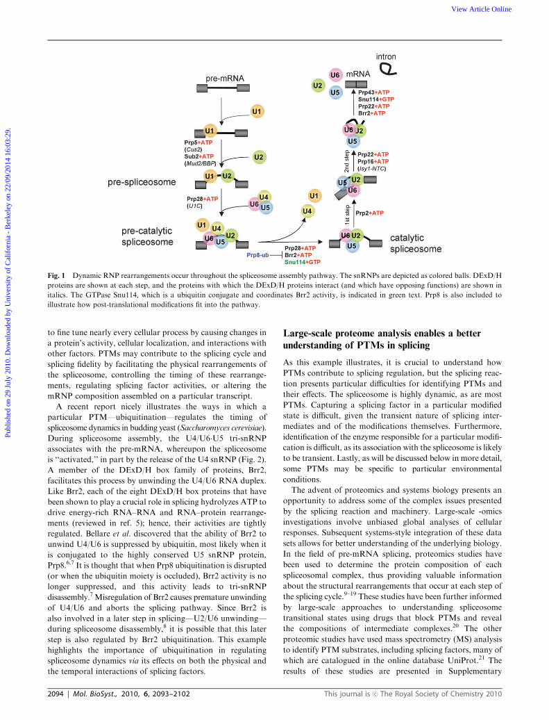
Fig. 1 Dynamic RNP rearrangements occur throughout the spliceosome assembly pathway. The snRNPs are depicted as colored balls. DExD/H
proteins are shown at each step, and the proteins with which the DExD/H proteins interact (and which have opposing functions) are shown in
italics. The GTPase Snu114, which is a ubiquitin conjugate and coordinates Brr2 activity, is indicated in green text. Prp8 is also included to
illustrate how post-translational modifications fit into the pathway.
2094 | Mol. BioSyst., 2010, 6, 2093–2102 This journal is �c The Royal Society of Chemistry 2010
Publ
ishe
d on
29
July
201
0. D
ownl
oade
d by
Uni
vers
ity o
f C
alif
orni
a -
Ber
kele
y on
22/
09/2
014
16:0
3:29
. View Article Online

Table 1w,106–156 and the evidence linking each one to splicing is
described below.
Phosphorylation
Reversible protein phosphorylation is used as a regulatory mecha-
nism in nearly all cellular processes. Dynamic phosphorylation
events are achieved via the interplay of protein kinases and
phosphatases that promote phosphate addition and removal,
respectively. Phosphorylation of proteins within complexes is
of particular importance since it alters physical interactions
(by disrupting some ionic interactions and facilitating others),
protein stability, and enzymatic activities.22
Phosphorylation regulates many of the dynamic inter-
actions that occur throughout spliceosome assembly, splicing
catalysis, and spliceosome disassembly. Early work pointed to
a role for cycles of protein phosphorylation and dephosphoryl-
ation by demonstrating that splicing cannot proceed in vitro in
the presence of phosphatases or phosphatase inhibitors.23
Indeed, there are splicing factors representing each step in
the splicing pathway that are phosphorylated, suggesting that
phosphorylation and dephosphorylation are important
throughout the splicing cycle (Supplementary Table 1w).Although phosphorylation is sure to play a role in guiding
spliceosome dynamics, only a handful of the 100+ non-SR
splicing factors have been reported as phosphorylation sub-
strates and even fewer have been described at the mechanistic
level. Since there are a number of excellent reviews of SR
protein phosphorylation,24–29 these will not be discussed here.
One clear example of how dynamic phosphorylation and
dephosphorylation modulates the activity of a single factor is
the U2 snRNP protein SAP155. Phosphorylation of mammalian
SAP155 occurs prior to or concomitant with splicing catalysis.30
SAP155 is then dephosphorylated during the second step of
splicing by the PP1 or PP2A phosphatase or both.31 Recruit-
ment of PP1 to SAP155 is mediated at least in part by NIPP1
(nuclear inhibitor of protein phosphatase 1), which recognizes
and binds to hyperphosphorylated SAP155 and stimulates its
dephosphorylation.32
Analysis of proteomic data leads to predictions about how
phosphorylation affects discrete steps in splicing, including
commitment complex formation and pre-spliceosome forma-
tion. For example, during commitment complex formation,
the yeast branchpoint binding protein (BBP) and its partner
Mud2 (SF1 and U2AF65, respectively, in humans) bind the
branchpoint region of the pre-mRNA and form a bridging
interaction with the U1 snRNP bound to the 50SS.33,34 Sub-
sequent pre-spliceosome formation requires the exchange of
the U2 snRNP for BBP/SF1 and Mud2/U2AF65 at the
branchpoint.33 Large-scale analyses have revealed that each
of these factors is phosphorylated, a result which raises the
possibility that the role of these factors in formation of commit-
ment complexes and pre-spliceosomes might be regulated by
their modification.35,36 Indeed, genetic interactions between
Mud2 and the PP1 phosphatase suggest that the cycle of
Fig. 2 Ubiquitination regulates Brr2’s role in formation of a catalytically active spliceosome. During the splicing reaction, formation of the
catalytic spliceosome is facilitated by rearrangements resulting in tri-snRNP disassembly and U4 snRNP release. Ubiquitin negatively regulates
spliceosome activation by suppressing Brr2-mediated U4/U6 unwinding. Once the ubiquitin moiety is removed or occluded, Brr2 activity is no
longer suppressed and facilitates U4 snRNP dissociation.
This journal is �c The Royal Society of Chemistry 2010 Mol. BioSyst., 2010, 6, 2093–2102 | 2095
Publ
ishe
d on
29
July
201
0. D
ownl
oade
d by
Uni
vers
ity o
f C
alif
orni
a -
Ber
kele
y on
22/
09/2
014
16:0
3:29
. View Article Online

phosphorylation and dephosphorylation of one or all of these
proteins may play a role in this step.37,38
Regulated phosphorylation of splicing factors may also
be a mechanism by which the activities of specific proteins
are regulated in response to cellular conditions. For instance,
in two large-scale studies examining cell-cycle specific
phosphorylation events, Cus2, the yeast U2 snRNP factor
important for facilitating proper U2 snRNA folding,39 was
found to be phosphorylated, a finding that raises the possibility
that Cus2 has roles in splicing that are cell-cycle dependent.
Mutations in Cus2 that suppress the deleterious effect caused
by misfolded U2 snRNA lie in close proximity to a putative
CKII site within the protein’s acidic domain. This domain is
also extensively phosphorylated in the mammalian homolog of
Cus2, Tat-SF1, a fact which further suggests that phosphoryl-
ation within this domain is important for Cus2 activity.40–45
Moreover, since recent studies illustrate that, under different
environmental conditions, the splicing of a certain subset of
genes requires the activity of different splicing factors,46 it is
likely that conditions rendered by progression through the cell-
cycle also require specific splicing factor activities (modulated
by PTMs) to mediate the appropriate responses.
These studies highlight two outstanding questions about
the role of phosphorylation in splicing; namely, how does
phosphorylation affect protein activities such as RNA binding
and protein–protein interactions? And what mechanisms does
the spliceosome employ to ensure that protein phosphoryla-
tion occurs at the right time? Clearly, new approaches will
need to be employed to determine the global effects of
phosphorylation on splicing.
Ubiquitination
Ubiquitination involves the covalent attachment of the small,
conserved peptide ubiquitin (ub) to a target protein, almost
exclusively at a lysine residue. Ubiquitin addition is catalyzed
by an enzymatic cascade involving an E1 ub-activating enzyme,
followed by the activities of an E2 ub-conjugating enzyme and
an E3 ub ligase (reviewed in ref. 47). This process is reversible
as a result of the activities of deubiquitinating enzymes
(reviewed in ref. 38 and 48). Monoubiquitination has emerged
as an important signaling mechanism separate from the
well-characterized role of polyubiquitination in proteolysis
(reviewed in ref. 49). Monoubiquitination of both histone
and non-histone protein targets plays a role in regulating
a number of important reactions, including transcription,
DNA repair, signal transduction, and receptor internalization
(reviewed in ref. 50). Ubiquitination, in general, has been
shown to regulate protein–protein interactions and often
serves as a signal for subsequent phosphorylation events.51
In addition to recent studies suggesting that ubiquitination
of Prp8 regulates tri-snRNP disassembly,6,7 there are other
indications that ubiquitin or ubiquitin-like proteins (such as
Sumo and Hub1) play additional roles in splicing. These
moieties, and the enzymes responsible for their addition/
removal, co-purify with splicing factors.16,52 Interestingly, a
number of splicing factors that exhibit genetic and physical
interactions with one another contain motifs similar to those
found in the ubiquitination machinery. The presence of these
motifs suggests that the splicing factors themselves are directly
involved in ubiquitination events. The formation of the pre-
spliceosome and the biogenesis of snRNPs involved in this
step appear to be regulated by ubiquitination and deubiquiti-
nation. For example, Prp19, an essential member of the
Nineteen Complex (NTC) that, along with the tri-snRNP,
associates with the pre-mRNA for formation of the pre-
catalytic spliceosome contains an E3 ligase U-box domain
and exhibits ubiquitin ligase activity in vitro.53 Mutations
within this U-box domain disrupt the NTC complex and
produce splicing defects.54–56 Additionally, Sad1, an essential
factor required for U4/U6 biogenesis and tri-snRNP addition,
contains a C-terminal hydrolase (UCH) domain, which is
typically associated with ubiquitin cleavage.57,58 These findings
suggest that splicing factors may directly contribute to cycles
of ubiquitination and deubiquitination to facilitate progression
of the splicing reaction and that the targets of their activities are
likely to be other splicing factors. Consistent with this idea,
several splicing factors have been identified as ubiquitin con-
jugates including Snu114, the U5 snRNPGTPase protein which
coordinates Brr2 activity (Supplementary Table 1w).59,60
Snu114 exhibits genetic interactions with Prp19 and Sad1,58 a
fact which presents the possibility that the Snu114 ubiquiti-
nation state is modulated by the activities of these proteins.
Phosphorylation marks are known to promote or inhibit
subsequent ubiquitination events by altering E3 ligase binding
sites.51 This mechanism of sequential modification may serve
to calibrate ubiquitination events by directing E3 ligase binding,
which normally occurs with little preference for the primary
sequence. The highly ordered process of spliceosomal rearrange-
ment is well-suited to such a tightly coordinated series of
modifications and leads to the prediction that splicing factors
are multiply modified. Indeed, the yeast factors Prp19, Prp43,
Snu114, Sad1, Prp8, Cbp80, and Hsh155 have been shown to
be both phosphorylated and ubiquitinated (Supplementary
Table 1w), and there are likely to be other examples. It is
unclear if the addition of these modifications is coordinated or
if they serve as completely independent marks, but it is easy to
imagine the implications that this type of coordinated modifi-
cation may have. Prp8 regulation may be an instance of
coordinated modification since Prp8 contains many of these
modifications, acts throughout splicing, and participates in a
large number of sequential and coordinated RNA–RNA and
RNA–protein rearrangements.61 Further studies are needed to
address this model directly.
Acetylation
Protein acetylation at lysine residues is a dynamic modification
whose function in transcription and chromatin regulation has
long been of interest. Lysine acetylation and deacetylation are
catalyzed by a group of enzymes known as lysine acetyltrans-
ferases (KATs) and lysine deacetylases (KDACs). Acetylation
of non-histone targets is known to affect protein stability,
protein–DNA interactions, sub-cellular localization, and trans-
criptional activity (reviewed in ref. 62). Recent proteomics
studies have identified acetyl marks on a plethora of proteins
involved in diverse cellular processes, including factors involved
in splicing.63–66
2096 | Mol. BioSyst., 2010, 6, 2093–2102 This journal is �c The Royal Society of Chemistry 2010
Publ
ishe
d on
29
July
201
0. D
ownl
oade
d by
Uni
vers
ity o
f C
alif
orni
a -
Ber
kele
y on
22/
09/2
014
16:0
3:29
. View Article Online

In addition to large-scale studies identifying the targets of
acetylation, biochemical studies in mammalian cells have
shown that splicing factors that are the targets of acetylation
also associate with acetylation/deacetylation machinery. For
example, the U2 snRNP factor SAP130 associates with the
KAT complex STAGA67 and is the target of acetylation.64
Likewise, the RNA helicase p68, which facilitates spliceosome
formation by destabilizing the U1/50SS interaction, associates
with KDAC168 and is acetylated.64 However, it remains to be
determined whether these splicing factors are the actual targets
of the enzymes with which they associate.
A more direct role for acetylation in regulating splicing was
suggested by Kuhn et al., who demonstrated that both KAT
and KDAC inhibitors can affect splicing in vitro.20 This study
used three KAT and three KDAC small molecule inhibitors to
block splicing at distinct steps. The composition of the stalled
spliceosomes was then analyzed by mass spectrometry. These
studies identified unique splicing complexes that accumulated
only in the absence of proper acetylation or deacetylation. For
instance, the KAT inhibitor anacardic acid blocked splicing at
a complex lacking several U1 snRNP, SR, and NTC proteins,
a result which suggests that inhibiting acetylation prevents the
stable association of these proteins. Even though in the
absence of KAT inhibition these studies could not confirm
acetylation of the missing proteins using anti-acetyl antibodies
or radioactively labeled acetyl coenzyme A, the majority of the
proteins absent from these stalled complexes, including both
SR and non-SR proteins, have been identified as acetylation
substrates in a large-scale study of proteins acetylated in vivo
including FBP11, S164, SPF31, hPRP5, SRp40, SRp55, SC35,
SRm160, SRm300, Prp19, and Cdc5.64 Further characteriza-
tion of specific acetylation events using mutational analysis of
acetylation substrates will be important for elucidating direct
roles for acetylation in splicing.
The well-characterized role for KATs and KDACs in
regulating chromatin dynamics presents the exciting possibility
that lysine acetylation in vivo functions to coordinate trans-
cription and splicing. Indeed, Gunderson and Johnson have
shown that the acetyltransferase activity of the yeast KAT
Gcn5 regulates co-transcriptional association of the U2
snRNP with the nascent transcript.69 The spatial proximity
of the KATs and the splicing factors during co-transcriptional
splicing may allow for acetylation of histones or splicing
factors or both to affect co-transcriptional spliceosome assembly
and the activities of specific splicing factors.
Glycosylation
Protein glycosylation—the addition of saccharides to proteins—
has been shown to regulate a variety of cellular processes
including protein folding, cell signaling, cell adhesion, and
protein stability (reviewed in ref. 70 and 71). The addition of
O-linked N-acetylglucosamine (O-GlcNAc) to serine and
threonine residues has been shown to occur on numerous
cytosolic and nuclear proteins. Like other PTMs, this modifi-
cation is dynamic, with the level of the O-GlcNAc modulated
by the opposing activities of O-GlcNAc transferases and
b-N-acetylglucosaminidases.70,72 This reversible modification
affects protein–protein and protein–DNA interactions, protein
stability and activity, and cell signaling cascades.73 Given the
diverse functional regulatory roles of O-GlcNAc modification,
it is possible that this modification also plays a role in splicing
dynamics.
Large-scale analyses have identified several splicing factors
as modified by O-GlcNAc. SF3b3 (SAP130) was identified in a
large-scale screen for O-GlcNAc-modified proteins from
HeLa cells.73 Several yeast splicing factors were identified in
a large-scale study using lectins that specifically bind GlcNAc,
including Slu7, Prp19, Npl3, and Isy1.74,75 It remains to be
determined whether O-GlycNAcylation of these mammalian
and yeast splicing factors is important for their activities in
splicing. Interestingly, O-GlcNAc modifies residues that
can also be phosphorylated, and O-GlcNAc transferase
stably associates with the phosphatase PP1, associations that
suggest that this modification may directly compete with
phosphorylation.70,71 Indeed, several factors are both phos-
phorylated and O-GlycNAcylated (Supplementary Table 1w),a fact which opens the possibility that there may be a regulated
exchange of phosphates for O-GlcNAc on these proteins.
Determining the specific role of O-GlcNAc transferases in
splicing will yield valuable insights into the roles of this
modification in spliceosome dynamics.
General roles for post-translational modifications of
splicing factors
In the next two sections we consider how PTMs may con-
tribute to two important aspects of splicing. First we describe
how modification of DExD/H box proteins may regulate the
timing and fidelity of dynamic rearrangements that occur
during splicing. Second, we describe how post-translational
modifications of splicing factors may allow splicing to be
responsive to changes in the environment.
DExD/H proteins that guide splicing dynamics and
fidelity are post-translationally modified
As described above, there are a number of examples illustrating
that PTM of splicing factors can regulate the dynamic inter-
actions that occur throughout the splicing pathway. Of parti-
cular interest are the eight conserved DExD/H-box proteins
that drive many spliceosomal rearrangements and enforce
splicing fidelity (Fig. 1). This class of proteins is characterized
by the presence of a DExD/H motif along with six or seven
other conserved motifs that determine their NTP-binding,
NTP-hydrolysis, RNA-binding, and unwinding activities
(reviewed in ref. 76). This key group of spliceosomal proteins
hydrolyzes ATP to catalyze a number of both RNA–RNA and
RNA–protein rearrangements that take place throughout the
splicing cycle.77,78 The functional consequence of these activities
is to progress, pause, or abort the splicing pathway,79 making
DExD/H box proteins major splicing ‘‘decision-makers.’’
Recent reports indicate that post-translational modification
of splicing factors is at least one mode of regulating this class
of proteins.
Mammalian PRP28, a likely homolog of the yeast U5
snRNP DExD/H protein required for exchanging U6 snRNA
for U1 snRNA at the 50SS,5 must be phosphorylated by
This journal is �c The Royal Society of Chemistry 2010 Mol. BioSyst., 2010, 6, 2093–2102 | 2097
Publ
ishe
d on
29
July
201
0. D
ownl
oade
d by
Uni
vers
ity o
f C
alif
orni
a -
Ber
kele
y on
22/
09/2
014
16:0
3:29
. View Article Online

SRPK2 for its stable association with the tri-snRNP and
formation of the pre-catalytic complex.80 Although it is not
known precisely which protein–protein or protein–RNA
interactions are altered by PRP28 phosphorylation, PRP28
phosphorylation appears to promote physical interactions
that allow for stable association of the tri-snRNP and pre-
spliceosome. Furthermore, proteomics studies show that
PRP28 is underrepresented in catalytic spliceosomes,11
suggesting that PRP28 dissociates from the spliceosome before
catalysis, perhaps as the authors suggest, mediated by its
dephosphorylation.
The revelation that PRP28 must be phosphorylated to
stably associate with the tri-snRNP raises the question of
whether PTM is a general mechanism for regulating the
activity of DExD/H box proteins and their influence on
splicing fidelity. A compelling model of splicing fidelity posits
that each transition along the splicing pathway represents two
kinetically competing conformations that must be stabilized or
destabilized to advance or abort the splicing pathway.81 To
this end, the dynamics of splicing often involve the pairing of
opposing factors that interact to ensure proper timing and
fidelity of the splicing reaction. The DExD/H box proteins
play a crucial role in these rearrangements. For instance,
Prp16 activity is crucial for the transition between the
first and second steps of splicing.82,83 The activity of Prp16
is opposed by Isy1, a member of the Nineteen Complex
(NTC), which acts to stabilize the first step conformation
of the spliceosome.84 Perturbation of either Prp16 or Isy1
decreases splicing fidelity, a result which suggests that the
interaction of these proteins regulates the kinetics of the first to
second step transition. Similar antagonistic pairs involving
DExD/H box proteins have been identified, including Sub2
and Mud2/BBP,33,85 Prp5 and Cus2,86,87 Brr2 and the U4/U6
helix,88–90 and Prp28 and U1C,91 where the interactions
mediate substrate rearrangements that are necessary for
splicing to progress (Fig. 1). It is likely that the other DExD/H
proteins (Prp2, Prp22, and Prp43) employ similar mechanisms to
ensure proper timing and fidelity of splicing.39,92
Despite the obvious importance of DExD/H proteins in the
splicing reaction, the mechanisms that guide their specificity and
activities are not well understood. We envision a model of
spliceosome assembly whereby the DExD/H proteins associate
with RNP subcomplexes and are poised to catalyze rearrange-
ments when the correct conformations are achieved. PTMs on
these DExD/H proteins may trigger their catalytic activities or
PTMs on other auxiliary proteins may regulate their interactions
with the DExD/H proteins. Indeed, post-translational modifica-
tions have been identified for all of the spliceosomal DExD/H box
proteins and their partner proteins (Supplementary Table 1w).93–96
A possible mechanism for mediating the rapid
splicing changes that occur in response to
extracellular conditions
Competitive fitness and cell survival depend on a cell’s
ability to swiftly respond to changes in the environment. Post-
translational modification of spliceosomal proteins provides
an efficient method for making splicing responsive to
cellular conditions. Indeed, in metazoans, cell signaling affects
alternative splicing by producing changes in SR protein
phosphorylation in response to environmental cues.97,98 The
question remains whether general splicing factors are also
modified in response to environmental changes.
Two recent studies in budding yeast suggest global splicing
patterns are uniquely sensitive to changes in specific splicing
factors and changes in environmental conditions. Splicing-
sensitive microarrays were used to demonstrate that mutation
of core splicing factors produces different splicing profiles, a
finding which suggests that splicing of each transcript is
sensitive to the activities of specific proteins.99 Moreover, yeast
cells exposed to changes in environmental conditions undergo
rapid (within 2 minutes) changes in their genome-wide splicing
profiles that are unique to the stimuli.46 Taken together, these
studies produce an intriguing model that, in response to
environmental changes, rapid PTMs alter the activities of
specific splicing factors to modulate their roles in removing
specific introns.
For example, not only is Prp8 ubiquitinated (as described
above), but it also contains a JAB/MNP-like domain, which is
implicated in binding ubiquitin. Upon amino acid starvation,
two different alleles of Prp8 with mutations that flank the
protein’s JAB/MNP-like domain produce different splicing
profiles.99 Mutations of the JAB/MNP-like domain reduce
both ubiquitin binding and U4/U6-U5 tri-snRNP levels,
reductions that demonstrate that ubiquitin contributes to
Prp8’s role in splicing.6,7 These data raise the possibility that
modification within this portion of the protein contributes to
substrate specificity and implicates ubiquitin binding by Prp8
as important in splicing responses to the environment. While
this hypothesis remains to be tested, it will be interesting to
determine whether the same stress conditions used in this
study produce changes in the levels of ubiquitin-bound Prp8
and U4/U6-U5 tri-snRNP. Furthermore, it will be interesting
to determine if ubiquitin-mediated intramolecular interactions
in Prp8 contribute to its activity.
Future directions
Post-translational modifications have proven to be an impor-
tant mechanism for regulating the highly dynamic interactions
that guide the splicing cycle and ensure splicing fidelity. While
proteomic studies have provided valuable information about
PTM substrates, the next important steps toward understanding
the role of PTMs in splicing must involve elucidating the
functional consequences of these modifications. Outstanding
questions to be addressed include determining which PTM
events are required for each step of the splicing cycle, how
combinations of modifications affect splicing factor activity,
and under what conditions specific modifications are required.
Although current detection technologies and certain biochemical
aspects of PTMs present specific challenges in achieving these
three goals, integrating the current data sets with new proteo-
mics data that become available will be key to advancing our
understanding of global splicing dynamics.
It is also important to note the likelihood that some PTMs
identified in proteomics studies are not important for the
target’s role in splicing. In addition to regulating a protein’s
2098 | Mol. BioSyst., 2010, 6, 2093–2102 This journal is �c The Royal Society of Chemistry 2010
Publ
ishe
d on
29
July
201
0. D
ownl
oade
d by
Uni
vers
ity o
f C
alif
orni
a -
Ber
kele
y on
22/
09/2
014
16:0
3:29
. View Article Online

function, PTMs may mark a protein for proper folding, degra-
dation, or trafficking. Additionally, in the case of proteins that
play multiple roles in gene expression, PTMs may be important
for other, non-splicing functions of the proteins. It is also
possible that some PTMs are nonspecific. For example, recent
studies of the evolution of protein phosphorylation sites
indicate that some phosphorylation events occur nonspecifically
on disordered protein surfaces.100 Because many kinases are
promiscuous, nonspecific modifications may occur, particularly
on proteins that are complexed with a true target protein
(i.e., are in close proximity with the kinase). These sites
probably arise from random mutations and are not conserved
until they acquire a function. As discussed below, specific
experimental design considerations will facilitate determining
which PTMs are important for regulating the splicing cycle.
Challenge 1: revealing PTM dynamics during
splicing
Although proteomic studies have greatly increased the number
of identified spliceosomal proteins that are post-translationally
modified by improving techniques for both enriching modified
peptides and detecting them in mixed preparations, this number
is still likely to be an underestimate. Furthermore, current MS
methods are limited in the reproducibility of the data produced.
However, new technologies like multiple reaction monitoring
MS can be used to facilitate reliable, quantitative results with
greater coverage.101–104 In addition to the technical challenges
of various detection methods, the biology of PTMs presents
challenges for identifying and characterizing PTMs during
highly dynamic processes like splicing. Modifications are tran-
sient and modified peptides are likely to be in low abundance
compared to their unmodified counterparts.
To study the timing of PTMs during the splicing cycle on a
global scale, it will be necessary to combine proteomic analyses
with methods that enrich for proteins specific to each step. For
example, conditional alleles of individual splicing factors enrich
for particular complexes under non-permissive temperatures.
Because splicing is blocked, some PTMs may be stabilized and
will provide information about the timing of particular splicing
factor modifications. Furthermore, small molecules that inhibit
specific modifications can be used to block spliceosomal rear-
rangements at specific intermediates, and mass spectrometry can
complement these in vitro studies by identifying which proteins
are modified at each step or within each complex. Improved
methods for stabilizing PTMs, enrichment methods, and improve-
ments in detection technologies will all likely contribute to an
expanded set of PTMs and their role in splicing.
It is equally important to identify the enzymes responsible
for catalyzing PTM addition and removal. However, these
enzymes may associate only transiently with their substrates,
thus decreasing the likelihood of their detection. Employing
techniques such as in vivo crosslinking may enable the capture
of such transient interactions in future studies.
Challenge 2: elucidating the PTM code
In addition to the dynamic changes in PTMs during the
splicing cycle, it is apparent that some splicing factors are
multiply modified, a fact which raises the question of whether
these modifications are coordinated. It is known that such
spatial and temporal crosstalk between PTMs is an important
aspect of regulation. Roles for processive phosphoryla-
tion, crosstalk between multiple modifications within a single
protein, and regulated stepwise-protein modification have
already been established as important for protein function.
Recognition of particular splice sites is guided by an
‘‘mRNP code,’’ where the concerted activities of multiple
splicing factors dictate splice site utilization.105 Similarly,
splicing may be additionally regulated by a ‘‘PTM code’’—
where crosstalk between modifications within a single protein
or multiple proteins modulates spliceosomal rearrangements
and stepwise progression of the splicing cycle. Within a single
protein there are typically several potential modification sites,
some of which can be the target of more than one type
of modification. Furthermore, crosstalk can exist between
phosphorylation, ubiquitination, acetylation, and O-GlcNA-
cylation. For instance, phosphorylation can promote ubiquiti-
nation by creating a recognition signal for E3 ligase binding;42,51
ubiquitination can, in turn, stimulate lysine acetylation, while
acetylation can inhibit ubiquitination.51 Likewise, O-GlcNA-
cylation occurs at the same serine/threonine residues available
for the phosphorylation and can, in fact, inhibit phosphoryl-
ation of surrounding residues.51 Regulation of the activity of a
specific splicing factor may involve multiple modifications that
guide its involvement in sequential steps in splicing. Similarly,
proteins containing PTM-recognition domains that bind
different types of modifications can act as dual recognition
systems, binding only when its interacting partners are properly
modified. Prp19, for example, is ubiquitinated, O-GlcNAcy-
lated, and acetylated.35,55,64,66,74 It will be interesting to deter-
mine whether the addition of these modifications is coordinated
and how each modification, whether independently or in com-
bination, defines Prp19 activity.
Challenge 3: from ’omics to mechanism: determining
the functional consequences of PTMs on splicing
The abundance of proteomics and genomics data has added
breadth to our current knowledge of splicing factor modifica-
tions and how splicing changes under different conditions.
However, the differences in the ways in which these data sets
were prepared—different growth conditions, sample prepara-
tions, mutations, and detection methodologies—have made
it difficult to integrate the data. Addressing the functional
consequences of splicing factor PTMs requires a systems
approach combining proteomics and genomics with analysis
of individual factors.
As suggested by genome-wide splicing changes in S. cerevisiae
in response to environmental changes, it is possible that
certain PTMs occur only under a narrow set of conditions
that trigger the activity of specific splicing factors or the
splicing of specific transcripts.46 Likewise, particular tran-
scripts may require a modified splicing factor to facilitate their
splicing, while others may not. The splicing-specific micro-
array has proven to be a powerful tool for identifying splicing
changes under specific conditions. Such arrays can be used in
combination with PTM inhibitors, mutations in the PTM
This journal is �c The Royal Society of Chemistry 2010 Mol. BioSyst., 2010, 6, 2093–2102 | 2099
Publ
ishe
d on
29
July
201
0. D
ownl
oade
d by
Uni
vers
ity o
f C
alif
orni
a -
Ber
kele
y on
22/
09/2
014
16:0
3:29
. View Article Online

machinery, or mutations at splicing factor modification sites to
identify how changes in PTM profiles produce concomitant
changes in splicing. Additionally, the resulting data sets can be
compared to identify conditions and mutations that produce
overlapping effects on splicing and may reveal factors that act
in concert under such conditions.
Conclusion
The spliceosome is an exquisitely coordinated macromolecular
machine. The revelation that many splicing factors are post-
translationally modified strongly suggests that many of the
dynamic rearrangements that occur during the splicing cycle
are regulated, in part, by the addition and removal of these
PTMs. Understanding how the network of PTMs functions in
regulating splicing will be the next exciting step toward under-
standing the coordinated mechanisms of spliceosome dynamics.
Acknowledgements
We would like to thank Dr Stephen Rader, Julia Claggett,
Felizza Gunderson, and Dr Mary Fae McKay, for critical
reading of the manuscript; Patricia Tu for her work on Table
S1 (ESIw); Herve Tiriac for work on Fig. S1; and other
members of the Johnson lab for useful comments and sugges-
tions. This work was supported by an NSF CAREER award
to T.L.J. (MCB-0448010) and an NSF predoctoral fellowship
to S.L.M.
References
1 S. Valadkhan, Curr. Opin. Struct. Biol., 2007, 17, 310–315.2 A. J. Matlin and M. J. Moore, Adv. Exp. Med. Biol., 2007, 623,14–35.
3 M. C. Wahl, C. L. Will and R. Luhrmann, Cell (Cambridge,Mass.), 2009, 136, 701–718.
4 D. J. Smith, C. C. Query and M. M. Konarska, Mol. Cell, 2008,30, 657–666.
5 J. P. Staley and C. Guthrie, Mol. Cell, 1999, 3, 55–64.6 P. Bellare, A. K. Kutach, A. K. Rines, C. Guthrie andE. J. Sontheimer, RNA, 2006, 12, 292–302.
7 P. Bellare, E. C. Small, X. Huang, J. A. Wohlschlegel, J. P. Staleyand E. J. Sontheimer, Nat. Struct. Mol. Biol., 2008, 15, 444–451.
8 E. C. Small, S. R. Leggett, A. A. Winans and J. P. Staley, Mol.Cell, 2006, 23, 389–399.
9 S. E. Behrens and R. Luhrmann, Genes Dev., 1991, 5, 1439–1452.10 N. Behzadnia, M. M. Golas, K. Hartmuth, B. Sander,
B. Kastner, J. Deckert, P. Dube, C. L. Will, H. Urlaub,H. Stark and R. Luhrmann, EMBO J., 2007, 26, 1737–1748.
11 S. Bessonov, M. Anokhina, C. L. Will, H. Urlaub andR. Luhrmann, Nature, 2008, 452, 846–850.
12 Y. I. Chen, R. E. Moore, H. Y. Ge, M. K. Young, T. D. Lee andS. W. Stevens, Nucleic Acids Res., 2007, 35, 3928–3944.
13 N. Herold, C. L. Will, E. Wolf, B. Kastner, H. Urlauband R. Luhrmann, Mol. Cell. Biol., 2009, 29, 281–301.
14 M. S. Jurica, L. J. Licklider, S. R. Gygi, N. Grigorieff andM. J. Moore, RNA, 2002, 8, 426–439.
15 E. Kuhn-Holsken, C. Lenz, B. Sander, R. Luhrmann andH. Urlaub, RNA, 2005, 11, 1915–1930.
16 J. Rappsilber, U. Ryder, A. I. Lamond and M. Mann, GenomeRes., 2002, 12, 1231–1245.
17 S. W. Stevens, D. E. Ryan, H. Y. Ge, R. E. Moore, M. K. Young,T. D. Lee and J. Abelson, Mol. Cell, 2002, 9, 31–44.
18 Z. Warkocki, P. Odenwalder, J. Schmitzova, F. Platzmann,H. Stark, H. Urlaub, R. Ficner, P. Fabrizio and R. Luhrmann,Nat. Struct. Mol. Biol., 2009, 16, 1237–1243.
19 Z. Zhou, L. J. Licklider, S. P. Gygi and R. Reed, Nature, 2002,419, 182–185.
20 A. N. Kuhn, M. A. van Santen, A. Schwienhorst, H. Urlaub andR. Luhrmann, RNA, 2009, 15, 153–175.
21 E. Jain, A. Bairoch, S. Duvaud, I. Phan, N. Redaschi,B. E. Suzek, M. J. Martin, P. McGarvey and E. Gasteiger,Infrastructure for the life sciences: design and implementationof the UniProt website, BMC Bioinformatics, 2009, 10, 136.
22 P. Cohen, Trends Biochem. Sci., 2000, 25, 596–601.23 J. E. Mermoud, P. T. Cohen and A. I. Lamond, EMBO J., 1994,
13, 5679–5688.24 B. J. Blencowe, J. A. Bowman, S. McCracken and E. Rosonina,
Biochem. Cell Biol., 1999, 77, 277–291.25 J. Valcarcel and M. R. Green, Trends Biochem. Sci., 1996, 21,
296–301.26 R. Fluhr, Curr. Top. Microbiol. Immunol., 2008, 326, 119–138.27 M. Hagiwara, Biochim. Biophys. Acta, Proteins Proteomics, 2005,
1754, 324–331.28 D. F. Stojdl and J. C. Bell, Biochem. Cell Biol., 1999, 77, 293–298.29 S. A. Tenenbaum and J. Aguirre-Ghiso, Mol. Cell, 2005, 20,
499–501.30 C. Wang, K. Chua, W. Seghezzi, E. Lees, O. Gozani and R. Reed,
Genes Dev., 1998, 12, 1409–1414.31 Y. Shi, B. Reddy and J. L. Manley, Mol. Cell, 2006, 23, 819–829.32 N. Tanuma, S. E. Kim, M. Beullens, Y. Tsubaki, S. Mitsuhashi,
M. Nomura, T. Kawamura, K. Isono, H. Koseki, M. Sato,M. Bollen, K. Kikuchi and H. Shima, J. Biol. Chem., 2008, 283,35805–35814.
33 A. L. Kistler and C. Guthrie, Genes Dev., 2001, 15, 42–49.34 J. Fleckner, M. Zhang, J. Valcarcel and M. R. Green, Genes Dev.,
1997, 11, 1864–1872.35 C. P. Albuquerque, M. B. Smolka, S. H. Payne, V. Bafna, J. Eng
and H. Zhou, Mol. Cell. Proteomics, 2008, 7, 1389–1396.36 M. B. Smolka, C. P. Albuquerque, S. H. Chen and H. Zhou,
Proc. Natl. Acad. Sci. U. S. A., 2007, 104, 10364–10369.37 G. M. Wilmes, M. Bergkessel, S. Bandyopadhyay, M. Shales,
H. Braberg, G. Cagney, S. R. Collins, G. B. Whitworth,T. L. Kress, J. S. Weissman, T. Ideker, C. Guthrie andN. J. Krogan, Mol. Cell, 2008, 32, 735–746.
38 A. Hershko and A. Ciechanover, Annu. Rev. Biochem., 1998, 67,425–479.
39 R. J. Perriman and M. Ares, Jr., Genes Dev., 2007, 21, 811–820.40 D. Yan, R. Perriman, H. Igel, K. J. Howe, M. Neville and
M. Ares, Jr., Mol. Cell. Biol., 1998, 18, 5000–5009.41 B. Wang, R. Malik, E. A. Nigg and R. Korner, Anal. Chem.,
2008, 80, 9526–9533.42 J. V. Olsen, B. Blagoev, F. Gnad, B. Macek, C. Kumar,
P. Mortensen and M. Mann, Cell (Cambridge, Mass.), 2006,127, 635–648.
43 N. Dephoure, C. Zhou, J. Villen, S. A. Beausoleil,C. E. Bakalarski, S. J. Elledge and S. P. Gygi, Proc. Natl. Acad.Sci. U. S. A., 2008, 105, 10762–10767.
44 S. Matsuoka, B. A. Ballif, A. Smogorzewska, E. R. McDonald,3rd, K. E. Hurov, J. Luo, C. E. Bakalarski, Z. Zhao, N. Solimini,Y. Lerenthal, Y. Shiloh, S. P. Gygi and S. J. Elledge, Science,2007, 316, 1160–1166.
45 X. Li, S. A. Gerber, A. D. Rudner, S. A. Beausoleil, W. Haas,J. Villen, J. E. Elias and S. P. Gygi, J. Proteome Res., 2007, 6,1190–1197.
46 J. A. Pleiss, G. B. Whitworth, M. Bergkessel and C. Guthrie,Mol.Cell, 2007, 27, 928–937.
47 C. M. Pickart and M. J. Eddins, Biochim. Biophys. Acta, Mol.Cell Res., 2004, 1695, 55–72.
48 A. Y. Amerik and M. Hochstrasser, Biochim. Biophys. Acta, Mol.Cell Res., 2004, 1695, 189–207.
49 S. Sigismund, S. Polo and P. P. Di Fiore, Curr. Top. Microbiol.Immunol., 2004, 286, 149–185.
50 R. L. Welchman, C. Gordon and R. J. Mayer,Nat. Rev. Mol. CellBiol., 2005, 6, 599–609.
51 T. Hunter, Mol. Cell, 2007, 28, 730–738.52 E. M. Makarov, O. V. Makarova, H. Urlaub, M. Gentzel,
C. L. Will, M. Wilm and R. Luhrmann, Science, 2002, 298,2205–2208.
53 S. Hatakeyama, M. Yada, M. Matsumoto, N. Ishida andK. I. Nakayama, J. Biol. Chem., 2001, 276, 33111–33120.
2100 | Mol. BioSyst., 2010, 6, 2093–2102 This journal is �c The Royal Society of Chemistry 2010
Publ
ishe
d on
29
July
201
0. D
ownl
oade
d by
Uni
vers
ity o
f C
alif
orni
a -
Ber
kele
y on
22/
09/2
014
16:0
3:29
. View Article Online

54 M. D. Ohi and K. L. Gould, RNA, 2002, 8, 798–815.55 M. D. Ohi, C. W. Vander Kooi, J. A. Rosenberg, W. J. Chazin
and K. L. Gould, Nat. Struct. Biol., 2003, 10, 250–255.56 M. D. Ohi, C. W. Vander Kooi, J. A. Rosenberg, L. Ren,
J. P. Hirsch, W. J. Chazin, T. Walz and K. L. Gould, Mol. Cell.Biol., 2005, 25, 451–460.
57 S. Hunter, R. Apweiler, T. K. Attwood, A. Bairoch, A. Bateman,D. Binns, P. Bork, U. Das, L. Daugherty, L. Duquenne,R. D. Finn, J. Gough, D. Haft, N. Hulo, D. Kahn, E. Kelly,A. Laugraud, I. Letunic, D. Lonsdale, R. Lopez, M. Madera,J. Maslen, C. McAnulla, J. McDowall, J. Mistry, A. Mitchell,N. Mulder, D. Natale, C. Orengo, A. F. Quinn, J. D. Selengut,C. J. Sigrist, M. Thimma, P. D. Thomas, F. Valentin, D. Wilson,C. H. Wu and C. Yeats,Nucleic Acids Res., 2009, 37, D211–D215.
58 T. J. Brenner and C. Guthrie, Genetics, 2005, 170, 1063–1080.59 C. Tagwerker, K. Flick, M. Cui, C. Guerrero, Y. Dou, B. Auer,
P. Baldi, L. Huang and P. Kaiser, Mol. Cell. Proteomics, 2006, 5,737–748.
60 J. Peng, D. Schwartz, J. E. Elias, C. C. Thoreen, D. Cheng,G. Marsischky, J. Roelofs, D. Finley and S. P. Gygi, Nat.Biotechnol., 2003, 21, 921–926.
61 R. J. Grainger and J. D. Beggs, RNA, 2005, 11, 533–557.62 T. Kouzarides, EMBO J., 2000, 19, 1176–1179.63 A. Basu, K. L. Rose, J. Zhang, R. C. Beavis, B. Ueberheide,
B. A. Garcia, B. Chait, Y. Zhao, D. F. Hunt, E. Segal, C. D. Allisand S. B. Hake, Proc. Natl. Acad. Sci. U. S. A., 2009, 106,13785–13790.
64 C. Choudhary, C. Kumar, F. Gnad, M. L. Nielsen, M. Rehman,T. C. Walther, J. V. Olsen and M. Mann, Science, 2009, 325,834–840.
65 S. C. Kim, R. Sprung, Y. Chen, Y. Xu, H. Ball, J. Pei, T. Cheng,Y. Kho, H. Xiao, L. Xiao, N. V. Grishin, M. White, X. J. Yangand Y. Zhao, Mol. Cell, 2006, 23, 607–618.
66 Y. Y. Lin, J. Y. Lu, J. Zhang, W. Walter, W. Dang, J. Wan,S. C. Tao, J. Qian, Y. Zhao, J. D. Boeke, S. L. Berger and H. Zhu,Cell (Cambridge, Mass.), 2009, 136, 1073–1084.
67 E. Martinez, V. B. Palhan, A. Tjernberg, E. S. Lymar,A. M. Gamper, T. K. Kundu, B. T. Chait and R. G. Roeder,Mol. Cell. Biol., 2001, 21, 6782–6795.
68 Z. R. Liu, Mol. Cell. Biol., 2002, 22, 5443–5450.69 F. Q. Gunderson and T. L. Johnson, PLoS Genet., 2009, 5,
e1000682.70 G. W. Hart, M. P. Housley and C. Slawson, Nature, 2007, 446,
1017–1022.71 L. Wells and G. W. Hart, FEBS Lett., 2003, 546, 154–158.72 R. Hurtado-Guerrero, H. C. Dorfmueller and D. M. van Aalten,
Curr. Opin. Struct. Biol., 2008, 18, 551–557.73 A. Nandi, R. Sprung, D. K. Barma, Y. Zhao, S. C. Kim,
J. R. Falck and Y. Zhao, Anal. Chem., 2006, 78, 452–458.74 D. M. Gelperin, M. A. White, M. L. Wilkinson, Y. Kon,
L. A. Kung, K. J. Wise, N. Lopez-Hoyo, L. Jiang, S. Piccirillo,H. Yu, M. Gerstein, M. E. Dumont, E. M. Phizicky, M. Snyderand E. J. Grayhack, Genes Dev., 2005, 19, 2816–2826.
75 L. A. Kung, S. C. Tao, J. Qian, M. G. Smith, M. Snyder andH. Zhu, Mol. Syst. Biol., 2009, 5, 308.
76 P. Linder, N. K. Tanner and J. Banroques, Trends Biochem. Sci.,2001, 26, 339–341.
77 J. P. Staley and C. Guthrie, Cell (Cambridge, Mass.), 1998, 92,315–326.
78 J. Hamm and A. I. Lamond, Curr. Biol., 1998, 8, R532–R534.79 M. M. Konarska and C. C. Query, Genes Dev., 2005, 19,
2255–2260.80 R. Mathew, K. Hartmuth, S. Mohlmann, H. Urlaub, R. Ficner
and R. Luhrmann, Nat. Struct. Mol. Biol., 2008, 15, 435–443.81 C. C. Query and M. M. Konarska, Mol. Cell, 2004, 14, 343–354.82 B. Schwer and C. Guthrie, EMBO J., 1992, 11, 5033–5039.83 B. Schwer and C. Guthrie, Nature, 1991, 349, 494–499.84 T. Villa and C. Guthrie, Genes Dev., 2005, 19, 1894–1904.85 Q. Wang, L. Zhang, B. Lynn and B. C. Rymond, Nucleic Acids
Res., 2008, 36, 2787–2798.86 R. Perriman, I. Barta, G. K. Voeltz, J. Abelson and M. Ares, Jr.,
Proc. Natl. Acad. Sci. U. S. A., 2003, 100, 13857–13862.87 R. Perriman and M. Ares, Jr., Genes Dev., 2000, 14, 97–107.88 D. H. Kim and J. J. Rossi, RNA, 1999, 5, 959–971.89 P. L. Raghunathan and C. Guthrie, Curr. Biol., 1998, 8, 847–855.
90 R. W. van Nues and J. D. Beggs, Genetics, 2001, 157,1451–1467.
91 J. Y. Chen, L. Stands, J. P. Staley, R. R. Jackups, Jr., L. J. Latusand T. H. Chang, Mol. Cell, 2001, 7, 227–232.
92 B. Rutz and B. Seraphin, RNA, 1999, 5, 819–831.93 J. Roy, K. Kim, J. R. Maddock, J. G. Anthony and
J. L. Woolford, Jr., RNA, 1995, 1, 375–390.94 N. Tanaka, A. Aronova and B. Schwer, Genes Dev., 2007, 21,
2312–2325.95 R. T. Tsai, R. H. Fu, F. L. Yeh, C. K. Tseng, Y. C. Lin,
Y. H. Huang and S. C. Cheng, Genes Dev., 2005, 19, 2991–3003.96 R. T. Tsai, C. K. Tseng, P. J. Lee, H. C. Chen, R. H. Fu,
K. J. Chang, F. L. Yeh and S. C. Cheng, Mol. Cell. Biol., 2007,27, 8027–8037.
97 C. Shin, Y. Feng and J. L. Manley, Nature, 2004, 427, 553–558.98 C. Shin and J. L. Manley, Nat. Rev. Mol. Cell Biol., 2004, 5,
727–738.99 J. A. Pleiss, G. B. Whitworth, M. Bergkessel and C. Guthrie,
PLoS Biol., 2007, 5, e90.100 L. J. Holt, B. B. Tuch, J. Villen, A. D. Johnson, S. P. Gygi and
D. O. Morgan, Science, 2009, 325, 1682–1686.101 D. M. Cox, F. Zhong, M. Du, E. Duchoslav, T. Sakuma and
J. C. McDermott, J. Biomol. Tech., 2005, 16, 83–90.102 C. B. Gocke, H. Yu and J. Kang, J. Biol. Chem., 2005, 280,
5004–5012.103 S. Mollah, I. E. Wertz, Q. Phung, D. Arnott, V. M. Dixit and
J. R. Lill, Rapid Commun. Mass Spectrom., 2007, 21, 3357–3364.104 R. D. Unwin, J. R. Griffiths and A. D. Whetton, Nat. Protoc.,
2009, 4, 870–877.105 S. Stamm, J. Biol. Chem., 2008, 283, 1223–1227.106 S. A. Beausoleil, M. Jedrychowski, D. Schwartz, J. E. Elias,
J. Villen, J. Li, M. A. Cohn, L. C. Cantley and S. P. Gygi, Proc.Natl. Acad. Sci. U. S. A., 2004, 101, 12130–12135.
107 S. A. Beausoleil, J. Villen, S. A. Gerber, J. Rush and S. P. Gygi,Nat. Biotechnol., 2006, 24, 1285–1292.
108 F. Giorgianni, Y. Zhao, D. M. Desiderio and S. Beranova-Giorgianni, Electrophoresis, 2007, 28, 2027–2034.
109 H. Molina, D. M. Horn, N. Tang, S. Mathivanan and A. Pandey,Proc. Natl. Acad. Sci. U. S. A., 2007, 104, 2199–2204.
110 H. Shu, S. Chen, Q. Bi, M. Mumby and D. L. Brekken,Mol. Cell.Proteomics, 2004, 3, 279–286.
111 J. C. Smith, M. A. Duchesne, P. Tozzi, M. Ethier and D. Figeys,J. Proteome Res., 2007, 6, 3174–3186.
112 S. M. Sweet, C. M. Bailey, D. L. Cunningham, J. K. Heath andH. J. Cooper, Mol. Cell. Proteomics, 2009, 8, 904–912.
113 M. Trost, L. English, S. Lemieux, M. Courcelles, M. Desjardinsand P. Thibault, Immunity, 2009, 30, 143–154.
114 J. Villen, S. A. Beausoleil, S. A. Gerber and S. P. Gygi, Proc. Natl.Acad. Sci. U. S. A., 2007, 104, 1488–1493.
115 S. Zanivan, F. Gnad, S. A. Wickstrom, T. Geiger, B. Macek,J. Cox, R. Fassler and M. Mann, J. Proteome Res., 2008, 7,5314–5326.
116 J. Peng and D. Cheng, Methods Enzymol., 2005, 399, 367–381.117 K. F. Wilson, W. J. Wu and R. A. Cerione, J. Biol. Chem., 2000,
275, 37307–37310.118 G. T. Cantin, W. Yi, B. Lu, S. K. Park, T. Xu, J. D. Lee and
J. R. Yates, 3rd, J. Proteome Res., 2008, 7, 1346–1351.119 K. Gevaert, A. Staes, J. Van Damme, S. De Groot, K. Hugelier,
H. Demol, L. Martens, M. Goethals and J. Vandekerckhove,Proteomics, 2005, 5, 3589–3599.
120 K. Imami, N. Sugiyama, Y. Kyono, M. Tomita and Y. Ishihama,Anal. Sci., 2008, 24, 161–166.
121 L. R. Yu, Z. Zhu, K. C. Chan, H. J. Issaq, D. S. Dimitrov andT. D. Veenstra, J. Proteome Res., 2007, 6, 4150–4162.
122 A. Chi, C. Huttenhower, L. Y. Geer, J. J. Coon, J. E. Syka,D. L. Bai, J. Shabanowitz, D. J. Burke, O. G. Troyanskaya andD. F. Hunt, Proc. Natl. Acad. Sci. U. S. A., 2007, 104, 2193–2198.
123 L. M. Brill, A. R. Salomon, S. B. Ficarro, M. Mukherji,M. Stettler-Gill and E. C. Peters, Anal. Chem., 2004, 76,2763–2772.
124 H. Daub, J. V. Olsen, M. Bairlein, F. Gnad, F. S. Oppermann,R. Korner, Z. Greff, G. Keri, O. Stemmann and M. Mann, Mol.Cell, 2008, 31, 438–448.
125 S. Gauci, A. O. Helbig, M. Slijper, J. Krijgsveld, A. J. Heck andS. Mohammed, Anal. Chem., 2009, 81, 4493–4501.
This journal is �c The Royal Society of Chemistry 2010 Mol. BioSyst., 2010, 6, 2093–2102 | 2101
Publ
ishe
d on
29
July
201
0. D
ownl
oade
d by
Uni
vers
ity o
f C
alif
orni
a -
Ber
kele
y on
22/
09/2
014
16:0
3:29
. View Article Online

126 V. Mayya, D. H. Lundgren, S. I. Hwang, K. Rezaul, L. Wu,J. K. Eng, V. Rodionov and D. K. Han, Sci. Signaling, 2009, 2, ra46.
127 K. Rikova, A. Guo, Q. Zeng, A. Possemato, J. Yu, H. Haack,J. Nardone, K. Lee, C. Reeves, Y. Li, Y. Hu, Z. Tan, M. Stokes,L. Sullivan, J. Mitchell, R. Wetzel, J. Macneill, J. M. Ren, J. Yuan,C. E. Bakalarski, J. Villen, J. M. Kornhauser, B. Smith, D. Li,X. Zhou, S. P. Gygi, T. L. Gu, R. D. Polakiewicz, J. Rush andM. J. Comb, Cell (Cambridge, Mass.), 2007, 131, 1190–1203.
128 J. T. Wilson-Grady, J. Villen and S. P. Gygi, J. Proteome Res.,2008, 7, 1088–1097.
129 C. Guerrero, C. Tagwerker, P. Kaiser and L. Huang, Mol. Cell.Proteomics, 2006, 5, 366–378.
130 B. Soufi, C. D. Kelstrup, G. Stoehr, F. Frohlich, T. C. Waltherand J. V. Olsen, Mol. BioSyst., 2009, 5, 1337–1346.
131 J. Rush, A. Moritz, K. A. Lee, A. Guo, V. L. Goss, E. J. Spek,H. Zhang, X. M. Zha, R. D. Polakiewicz and M. J. Comb, Nat.Biotechnol., 2005, 23, 94–101.
132 A. C. Vertegaal, S. C. Ogg, E. Jaffray, M. S. Rodriguez,R. T. Hay, J. S. Andersen, M. Mann and A. I. Lamond,J. Biol. Chem., 2004, 279, 33791–33798.
133 A. Gruhler, J. V. Olsen, S. Mohammed, P. Mortensen,N. J. Faergeman, M. Mann and O. N. Jensen, Mol. Cell.Proteomics, 2005, 4, 310–327.
134 M. Carrascal, D. Ovelleiro, V. Casas, M. Gay and J. Abian,J. Proteome Res., 2008, 7, 5167–5176.
135 G. Han, M. Ye, H. Zhou, X. Jiang, S. Feng, X. Jiang, R. Tian,D. Wan, H. Zou and J. Gu, Proteomics, 2008, 8, 1346–1361.
136 K. Licht, J. Medenbach, R. Luhrmann, C. Kambach andA. Bindereif, RNA, 2008, 14, 1532–1538.
137 J. Dai, W. H. Jin, Q. H. Sheng, C. H. Shieh, J. R. Wu andR. Zeng, J. Proteome Res., 2007, 6, 250–262.
138 R. Chen, X. Jiang, D. Sun, G. Han, F. Wang, M. Ye, L. Wangand H. Zou, J. Proteome Res., 2009, 8, 651–661.
139 A. Boudrez, M. Beullens, E. Waelkens, W. Stalmans andM. Bollen, J. Biol. Chem., 2002, 277, 31834–31841.
140 J. E. Kim, S. R. Tannenbaum and F. M. White, J. Proteome Res.,2005, 4, 1339–1346.
141 B. A. Ballif, J. Villen, S. A. Beausoleil, D. Schwartz andS. P. Gygi, Mol. Cell. Proteomics, 2004, 3, 1093–1101.
142 M. Nousiainen, H. H. Sillje, G. Sauer, E. A. Nigg and R. Korner,Proc. Natl. Acad. Sci. U. S. A., 2006, 103, 5391–5396.
143 J. Ptacek, G. Devgan, G. Michaud, H. Zhu, X. Zhu, J. Fasolo,H. Guo, G. Jona, A. Breitkreutz, R. Sopko, R. R. McCartney,M. C. Schmidt, N. Rachidi, S. J. Lee, A. S. Mah, L. Meng,M. J. Stark, D. F. Stern, C. De Virgilio, M. Tyers, B. Andrews,M. Gerstein, B. Schweitzer, P. F. Predki and M. Snyder, Nature,2005, 438, 679–684.
144 J. M. Lizcano, O. Goransson, R. Toth, M. Deak, N. A. Morrice,J. Boudeau, S. A. Hawley, L. Udd, T. P. Makela, D. G. Hardieand D. R. Alessi, EMBO J., 2004, 23, 833–843.
145 L. Y. Tang, N. Deng, L. S. Wang, J. Dai, Z. L. Wang, X. S. Jiang,S. J. Li, L. Li, Q. H. Sheng, D. Q. Wu, L. Li and R. Zeng, Mol.Cell. Proteomics, 2007, 6, 1952–1967.
146 Y. Wang, D. Du, L. Fang, G. Yang, C. Zhang, R. Zeng,A. Ullrich, F. Lottspeich and Z. Chen, EMBO J., 2006, 25,5058–5070.
147 W. A. Tao, B. Wollscheid, R. O’Brien, J. K. Eng, X. J. Li,B. Bodenmiller, J. D. Watts, L. Hood and R. Aebersold, Nat.Methods, 2005, 2, 591–598.
148 S. B. Ficarro, M. L. McCleland, P. T. Stukenberg, D. J. Burke,M. M. Ross, J. Shabanowitz, D. F. Hunt and F. M. White, Nat.Biotechnol., 2002, 20, 301–305.
149 W. Zhou, J. J. Ryan and H. Zhou, J. Biol. Chem., 2004, 279,32262–32268.
150 R. P. Munton, R. Tweedie-Cullen, M. Livingstone-Zatchej, F. Weinandy, M. Waidelich, D. Longo, P. Gehrig,F. Potthast, D. Rutishauser, B. Gerrits, C. Panse,R. Schlapbach and I. M. Mansuy, Mol. Cell. Proteomics, 2007,6, 283–293.
151 S. F. Dagher and X. D. Fu, RNA, 2001, 7, 1284–1297.152 T. H. Heibeck, S. J. Ding, L. K. Opresko, R. Zhao,
A. A. Schepmoes, F. Yang, A. V. Tolmachev, M. E. Monroe,D. G. Camp, 2nd, R. D. Smith, H. S. Wiley and W. J. Qian,J. Proteome Res., 2009, 8, 3852–3861.
153 J. C. Trinidad, C. G. Specht, A. Thalhammer, R. Schoepfer andA. L. Burlingame, Mol. Cell. Proteomics, 2006, 5, 914–922.
154 H. Zhou, M. Ye, J. Dong, G. Han, X. Jiang, R. Wu and H. Zou,J. Proteome Res., 2008, 7, 3957–3967.
155 S. Beranova-Giorgianni, Y. Zhao, D. M. Desiderio andF. Giorgianni, Pituitary, 2006, 9, 109–120.
156 N. T. Seyfried, P. Xu, D. M. Duong, D. Cheng, J. Hanfelt andJ. Peng, Anal. Chem., 2008, 80(11), 4161–4169.
2102 | Mol. BioSyst., 2010, 6, 2093–2102 This journal is �c The Royal Society of Chemistry 2010
Publ
ishe
d on
29
July
201
0. D
ownl
oade
d by
Uni
vers
ity o
f C
alif
orni
a -
Ber
kele
y on
22/
09/2
014
16:0
3:29
. View Article Online