2011 Etravirine cross resistance, AIDS 2011
11
Copyright © Lippincott Williams & Wilkins. Unauthorized reproduction of this article is prohibited. Research Letters AIDS 2011, 25:1123–1133 Selection of nonnucleoside reverse transcriptase inhibitor-associated mutations in HIV-1 subtype C: evidence of etravirine cross-resistance Ujjwal Neogi a,b,c , Anita Shet a,b , Ranjani Shamsundar a and Maria L. Ekstrand a,d Prevalence of etravirine genotypic resistance was assessed among 92 HIV-1C-infected patients failing nevirapine and efavirenz-based regimens from a cohort of 552 Indian patients. Overall, prevalence of etravirine cross-resistance identified using the Tibotec Weighted Score was 41% (31.5% interme- diately-resistant and 9.8% fully-resistant). The most frequently described nonnucleoside reverse tran- scriptase inhibitor-associated mutations included Y181 (35.9%), K101 (20.7%), G190 (17.4%), and V108 (15.2%). The resistant group demonstrated higher viral load (P U 0.01) and longer duration of antiretroviral treatment (P U 0.03) compared with the susceptible group. The low genetic barrier to development of resistance to first generation nonnucleoside reverse transcriptase inhibitors (NNRTIs) is compounded by cross-resistance across the class that makes sequential therapy with the NNRTIs therapeutically inappropriate. Current first-line NNRTI in most resource-constrained regions includes nevirapine, except in cases of intolerance or potential drug interaction when efavirenz is used [1]. Etravirine, a new generation NNRTI (TMC-125, Intelence; Tibotec Pharmaceuticals Ltd, Turnhoutseweg 30, 2340 Beerse, Belgium), was approved by US Food and Drug Administration for use in antiretroviral-experienced adults with resistance to first-line NNRTIs. Etravirine resistance-associated mutations (RAMs) in reverse tran- scriptase gene were identified, which are as follows: V90I, A98G, L100I, K101E/P/H, V106I, V179D/F, Y181C/ I/V, G190A/S, E138A, V179T, and M230L [2–4]. The Tibotec Weighted Score was proposed with 17 etravirine RAMs and assigned differential weights based upon the impact on clinical response [5]. Alternatively, the Monogram Weighted Score included 30 etravirine RAMs based on the genotypic and phenotypic inter- relationship [6]. Etravirine cross-resistance may be influenced by the prevailing HIV-1 subtype [7,8]. With a worldwide prevalence of 50% [9], and prevalence in India of 96%, HIV-1C undoubtedly has a significant impact on the evolution of the HIV epidemic globally. This study reports the selection of NNRTI RAMs and etravirine cross-resistance patterns among HIV-1C- infected patients failing first-line antiretroviral therapy (ART). Among a total of 552 participants participating in a 2-year longitudinal cohort study [10], 18% (n ¼ 101) with detectable viremia were assessed for presence of drug RAMs during their baseline visit [11]. Drug resistance genotyping was successfully done from 92 plasma samples from failing patients (viral load >1000 copies/ml) using a validated in-house method [12]. Drug-resistant strains previously reported from India (n ¼ 429) from patients failing first-line ARTwere included as a second group in this study [13–19]. A third group of 1122 global HIV-1C sequences were obtained from HIVseq Program (http:// hivdb.stanford.edu/; accessed 13 August 2010) reported from patients worldwide with a history of NNRTI drug treatment. Indian sequences and duplicates were excluded from global subtype C sequences. NNRTI DRMs in all these sequences were analyzed. Etravirine resistance was evaluated by Tibotec Etravirine Weighted Genotype Score [5]. Statistical analysis was performed in SPSS, version 11.5 (SPSS inc., Chicago, IL, USA). Plasma virus was successfully genotyped in 92 failing patients; their mean age was 39.6 years (SD 10.2 years) and 67% were men, similar to the complete cohort. Among the 92 patients, 77% used nevirapine; 12% used efavirenz, and 10% changed from an initial nevirapine- based regimen to an efavirenz-based regimen for clinical reasons. The mean duration of nevirapine and efavirenz exposure was 23 and 14 months, respectively. The overall prevalence of etravirine resistance was 41% (38 of 92). Single etravirine RAMs were seen in 13% and two etravirine RAMs were seen in 33% of the strains. Eleven percent (10 of 92) of strains harbored three or more etravirine RAMs. The Tibotec Weighted Score identified 58.7% of the strains to be susceptible to etravirine, whereas 31.5 and 9.8% displayed intermediate resistance and resistance, respectively. Alternative scoring methods showed comparable patterns (39% of strains had a monogram weighted score 4) indicating that a significant percentage of isolates had reduced efficacy to etravirine. Genotypic analysis predicted that 41.6% (30 of 72) of samples from nevirapine-experienced and 9.1% (one of 11) from efavirenz-experienced patients were cross resistant to etravirine. The maximum level of cross- resistance (77.8%, seven of nine) was observed in those patients who had exposure of both the drugs. The most frequently described RAMs included amino acid ISSN 0269-9370 Q 2011 Wolters Kluwer Health | Lippincott Williams & Wilkins 1123
-
Upload
independent -
Category
Documents
-
view
1 -
download
0
Transcript of 2011 Etravirine cross resistance, AIDS 2011

C
Researc
h LettersAIDS 2011, 25:1123–1133
Selection of nonnucleoside reverse transcriptaseinhibitor-associated mutations in HIV-1 subtype C:evidence of etravirine cross-resistance
Ujjwal Neogia,b,c, Anita Sheta,b, Ranjani Shamsundara
and Maria L. Ekstranda,d
Prevalence of etravirine genotypic resistance was
assessed among 92 HIV-1C-infected patients failing
nevirapine and efavirenz-based regimens from a
cohort of 552 Indian patients. Overall, prevalence
of etravirine cross-resistance identified using the
Tibotec Weighted Score was 41% (31.5% interme-
diately-resistant and 9.8% fully-resistant). The most
frequently described nonnucleoside reverse tran-
scriptase inhibitor-associated mutations included
Y181 (35.9%), K101 (20.7%), G190 (17.4%), and
V108 (15.2%). The resistant group demonstrated
higher viral load (P U 0.01) and longer duration of
antiretroviral treatment (P U 0.03) compared with
the susceptible group.
The low genetic barrier to development of resistance tofirst generation nonnucleoside reverse transcriptase
inhibitors (NNRTIs) is compounded by cross-resistanceacross the class that makes sequential therapy with theNNRTIs therapeutically inappropriate. Current first-lineNNRTI in most resource-constrained regions includesnevirapine, except in cases of intolerance or potentialdrug interaction when efavirenz is used [1]. Etravirine, anew generation NNRTI (TMC-125, Intelence; TibotecPharmaceuticals Ltd, Turnhoutseweg 30, 2340 Beerse,Belgium), was approved by US Food and DrugAdministration for use in antiretroviral-experiencedadults with resistance to first-line NNRTIs. Etravirineresistance-associated mutations (RAMs) in reverse tran-scriptase gene were identified, which are as follows: V90I,A98G, L100I, K101E/P/H, V106I, V179D/F, Y181C/I/V, G190A/S, E138A, V179T, and M230L [2–4]. TheTibotec Weighted Score was proposed with 17 etravirineRAMs and assigned differential weights based upon theimpact on clinical response [5]. Alternatively, theMonogram Weighted Score included 30 etravirineRAMs based on the genotypic and phenotypic inter-relationship [6]. Etravirine cross-resistance may beinfluenced by the prevailing HIV-1 subtype [7,8]. Witha worldwide prevalence of 50% [9], and prevalence inIndia of 96%, HIV-1C undoubtedly has a significantimpact on the evolution of the HIV epidemic globally.This study reports the selection of NNRTI RAMs andetravirine cross-resistance patterns among HIV-1C-opyright © Lippincott Williams & Wilkins. UnauthISSN 0269-9370 Q 2011 Wolters Kluwer Hea
infected patients failing first-line antiretroviral therapy(ART).
Among a total of 552 participants participating in a 2-yearlongitudinal cohort study [10], 18% (n¼ 101) withdetectable viremia were assessed for presence of drugRAMs during their baseline visit [11]. Drug resistancegenotyping was successfully done from 92 plasma samplesfrom failing patients (viral load >1000 copies/ml) using avalidated in-house method [12]. Drug-resistant strainspreviously reported from India (n¼ 429) from patientsfailing first-line ARTwere included as a second group inthis study [13–19]. A third group of 1122 global HIV-1Csequences were obtained from HIVseq Program (http://hivdb.stanford.edu/; accessed 13 August 2010) reportedfrom patients worldwide with a history of NNRTI drugtreatment. Indian sequences and duplicates were excludedfrom global subtype C sequences. NNRTI DRMs in allthese sequences were analyzed. Etravirine resistance wasevaluated by Tibotec Etravirine Weighted GenotypeScore [5]. Statistical analysis was performed in SPSS,version 11.5 (SPSS inc., Chicago, IL, USA).
Plasma virus was successfully genotyped in 92 failingpatients; their mean age was 39.6 years (SD 10.2 years)and 67% were men, similar to the complete cohort.Among the 92 patients, 77% used nevirapine; 12% usedefavirenz, and 10% changed from an initial nevirapine-based regimen to an efavirenz-based regimen for clinicalreasons. The mean duration of nevirapine and efavirenzexposure was 23 and 14 months, respectively.
The overall prevalence of etravirine resistance was 41%(38 of 92). Single etravirine RAMs were seen in 13% andtwo etravirine RAMs were seen in 33% of the strains.Eleven percent (10 of 92) of strains harbored three ormore etravirine RAMs. The Tibotec Weighted Scoreidentified 58.7% of the strains to be susceptible toetravirine, whereas 31.5 and 9.8% displayed intermediateresistance and resistance, respectively. Alternative scoringmethods showed comparable patterns (39% of strains hada monogram weighted score �4) indicating that asignificant percentage of isolates had reduced efficacyto etravirine.
Genotypic analysis predicted that 41.6% (30 of 72) ofsamples from nevirapine-experienced and 9.1% (one of11) from efavirenz-experienced patients were crossresistant to etravirine. The maximum level of cross-resistance (77.8%, seven of nine) was observed in thosepatients who had exposure of both the drugs. The mostfrequently described RAMs included amino acid
orized reproduction of this article is prohibited.lth | Lippincott Williams & Wilkins 1123

Co
1124 AIDS 2011, Vol 25 No 8
substitutions at positions Y181 (35.9%), K101 (20.7%),G190 (17.4%), and V108 (15.2%). Similar trends wereobserved in sequences reported previously from India(n¼ 429); however, among global subtype C sequences,K103N was the most frequent RAM (Fig. 1).
Compared to patients with susceptible virus, those whoharbored etravirine-resistant virus were more likely tohave been on ART for a longer duration (P¼ 0.03) and tohave higher viral load (P¼ 0.01) (Supplementary digitalcontent 1; http://links.lww.com/QAD/A126). Therewas no significant difference in age, CD4 cell count, timesince diagnosis, or self-reported adherence in the lastmonth measured by Visual Analogue Scale between thetwo groups.
Our report highlights the high prevalence of etravirinecross-resistance (41%) among the patients infected withHIV-1C viruses and failing first-generation NNRTI-based regimens in India. Etravirine RAMs have also beendescribed in ART-naive patients from France, Mali, andIndia [20,21]. Our finding of etravirine resistance ishigher than among HIV-infected patients harboringsubtype B in UK (11.5%) and Spain (18.7%) [22,23]. Asimilar study from Thailand found 56% etravirine cross-resistance in HIV-1 CRF01_AE strains [24].
The high prevalence of Y181 and K101 found in oursetting is also seen in other places where nevirapine iswidely used as first-line NNRTI. Similar trends havebeen observed in patients with CRF01_AE strains fromThailand (50% Y181C/I/Vand 18.7% K101E/H/P) [24]and the UK (17% Y181C in those failing efavirenz and
pyright © Lippincott Williams & Wilkins. Unautho
40
35
30
25
20
15
10
5
0
V90 A98 L100 K101 K103 V106 V108 E138
Indian HIV-1C strains (n = 521)
Mut
atio
n fr
eque
ncy
(%)
NNRTI-asso
Fig. 1. Selection of nonnucleoside reverse transcriptase inhibitoharboring HIV-1 subtype C viruses. Higher frequencies of nonnuclmutations are present in residues Y181, K101, G190, and V108previously reported sequences) compared to global subtype C sequeUniversity HIV Drug resistance database (http://hivdb.stanford.edu
40.5% Y181C were in those failing nevirapine) [25], thuslending credence to the conclusion that Y181C isparticularly selected during prolonged exposure to afailing nevirapine-containing regimen [11].
The association between etravirine resistance and higherviral loads in the study cohort may be reflective of thelonger duration on poorly suppressive regimens experi-enced by these patients [26]. In settings like India whereroutine viral load monitoring is not a part of standard ofcare, the second-line ART regimens have to be designedwith caution when including NNRTI drugs. As over 50%of failing isolates are susceptible to etravirine, it can beused as salvage therapy among those patients failing first-generation NNRTI-based regimens. Patients with highlevel of etravirine RAMs were also more likely to havetenofovir-associated mutations [27], which may raisechallenges in designing an effective second-line regimenin resource-constrained settings like India. The presenceof cross-resistance also highlights the need for developingeffective and sustainable adherence interventions thattarget local adherence patterns and barriers in order tokeep the limited first-line ARTagents effective for as longas possible [10].
In summary, our analysis highlights the high level ofetravirine cross-resistance in a cohort of ART-experi-enced patients failing NNRTI-containing first-linetherapy in India. The pattern of NNRTI mutations innevirapine-exposed patients also suggests the possiblebenefit of reconsidering the use of nevirapine in favor ofefavirenz as first-line NNRTI choice in resource-constrained settings.
rized reproduction of this article is prohibited.
V179 Y181 Y188 G190 H121 P225 F227 M230
Global HIV-1C strains (n = 1122)
ciated mutations
r mutations in antiretroviral therapy-experienced patientseoside reverse transcriptase inhibitor (NNRTI) drug resistancein Indian sequences (n¼521, 92 primary isolates and 429nces (n¼1122) obtained from HIVseq Program from Stanford/; accessed on 13 August 2010).

C
Research Letters 1125
Acknowledgements
The authors would like to thank the Prerana study team fortheir excellent field work, Karthika Arumugam, for herhelp with statistical analysis, and Dr Prabhakar of Bowringand Lady Curzon Hospital for his help with patientrecruitment. We dedicate this paper to the Prerana studyparticipants who so generously contributed their time tohelp us better understand issues in ART adherence in thissetting. The study was approved by the Committee forHuman Research at University of California, SanFrancisco, USA and the Institutional Ethical ReviewBoard St John’s Medical College and Hospital, Bangalore,India.
aSt John’s National Academy of Health Sciences,Bangalore, India, bDivision of Global Health, cDepart-ment of Medicine, Huddinge, Karolinska Institutet,Stockholm, Sweden, and dDepartment of Medicine,Center for AIDS Prevention Studies, University ofCalifornia, San Francisco, San Francisco, California,USA.
Correspondence to Maria L. Ekstrand, PhD, Universityof California, San Francisco, 50 Beale Street, Suite-1300,San Francisco, CA 94105, USA.E-mail: [email protected]
Received: 11 November 2010; revised: 16 February2011; accepted: 24 February 2011.
References
1. Joly V, Yeni P. Nonnucleoside reverse transcriptase inhibitors.Ann Med Intern (Paris) 2000; 151:260–267.
2. Madruga JV, Cahn P, Grinsztejn B, Haubrich R, Lalezari J, MillsA, et al. Efficacy and safety of TMC125 (etravirine) in treatment-experienced HIV-1-infected patients in DUET-1: 24-week resultsfrom a randomised, double-blind, placebo-controlled trial. Lan-cet 2007; 370:29–38.
3. Lazzarin A, Campbell T, Clotet B, Johnson M, Katlama C, MollA, et al. Efficacy and safety of TMC125 (etravirine) in treat-ment-experienced HIV-1-infected patients in DUET-2: 24-week results from a randomised, double-blind, placebo-con-trolled trial. Lancet 2007; 370:39–48.
4. Johnson VA, Brun-Vezinet F, Clotet B, Gunthard HF, KuritzkesDR, Pillay D, et al. Update of the drug resistance mutations inHIV-1. Top HIV Med 2008; 16:138–145.
5. Vingerhoets J, Tambuyzer L, Azijn H, Hoogstoel A, Nijs S,Peeters M, et al. Resistance profile of etravirine: combinedanalysis of baseline genotypic and phenotypic data from therandomized, controlled phase III clinical studies. AIDS 2010;24:503–514.
6. Benhamida J, Chappey C, Coakley E, Parkin NT. HIV-1 geno-type algorithms for prediction of etravirine susceptibility: novelmutations and weighting factors identified through correla-tions to phenotype. Antivir Ther 2008; 13:A142.
7. Martinez-Cajas JL, Pant-Pai N, Klein MB, Wainberg MA. Role ofgenetic diversity amongst HIV-1 non-B subtypes in drug re-sistance: a systematic review of virologic and biochemicalevidence. AIDS Rev 2008; 10:212–223.
8. Kosakovsky Pond SL, Smith DM. Are all subtypes createdequal? The effectiveness of antiretroviral therapy against non-subtype B HIV-1. Clin Infect Dis 2009; 48:1306–1309.
9. Taylor BS, Sobieszczyk ME, McCutchan FE, Hammer SM. Thechallenge of HIV-1 subtype diversity. N Engl J Med 2008;358:1590–1602.
opyright © Lippincott Williams & Wilkins. Unauth
10. Ekstrand ML, Chandy S, Heylen E, Steward W, Singh G. Devel-oping useful highly active antiretroviral therapy adherencemeasures for India: the Prerana study. J Acquir Immune DeficSyndr 2010; 53:415–416.
11. Ekstrand ML, Shet A, Chandy S, Singh G, Shamsundar R,Madhavan V, et al. Suboptimal adherence associated withvirologic failure and resistance mutations among patients on1st line HAART in Bangalore, India [abstract]. XVIII Interna-tional AIDS Conference, Vienna, Austria; 18–23 July 2010.(abstract no. WEAB0202).
12. Saravanan S, Vidya M, Balakrishnan P, Kumarasamy N,Solomon SS, Solomon S, et al. Evaluation of two human im-munodeficiency virus-1 genotyping systems: ViroSeqTM 2.0and in-house methods. J Virol Methods 2009; 159:211–216.
13. Sen S, Tripathy SP, Patil AA, Chimanpure VM, Paranjape RP.High prevalence of human immunodeficiency virus type 1 drugresistance mutations in antiretroviral treatment-experiencedpatients from Pune, India. AIDS Res Hum Retroviruses 2007;23:1303–1308.
14. Sen S, Tripathy SP, Chimanpure VM, Patil AA, Bagul RD,Paranjape RP. Human immunodeficiency virus type 1 drugresistance mutations in peripheral blood mononuclear cellproviral DNA among antiretroviral treatment-naive and treat-ment-experienced patients from Pune, India. AIDS Res HumRetroviruses 2007; 23:489–497.
15. Deshpande A, Jauvin V, Magnin N, Pinson P, Faure M,Masquelier B, et al. Resistance mutations in subtype C HIVtype 1 isolates from Indian patients of Mumbai receiving NRTIsplus NNRTIs and experiencing a treatment failure: resistanceto AR. AIDS Res Hum Retroviruses 2007; 23:335–340.
16. Vidya M, Saravanan S, Uma S, Kumarasamy N, Sunil SS, KantorR, et al. Genotypic HIV type-1 drug resistance among patientswith immunological failure to first-line antiretroviral therapyin south India. Antivir Ther 2009; 14:1005–1009.
17. Kandathil AJ, Kannangai R, Verghese VP, Pulimood SA, RupaliP, Sridharan G, et al. Drug resistant mutations detected bygenotypic drug resistance testing in patients failing therapy inclade C HIV-1 infected individuals from India. Indian J MedMicrobiol 2009; 27:231–236.
18. Rajesh L, Karunaianantham R, Narayanan PR, Swaminathan S.Antiretroviral drug-resistant mutations at baseline and at timeof failure of antiretroviral therapy in HIV type 1-coinfected TBpatients. AIDS Res Hum Retroviruses 2009; 25:1179–1185.
19. Choudhury SD, Choudhury AK, Kalra R, Andrabi R, Wig N,Biswas A, et al. Anti retroviral drug resistant mutations in thereverse transcriptase gene of HIV-1 isolates from northernIndian patients: a follow up study. Arch Virol 2010; 155:563–569.
20. Maiga AI, Descamps D, Morand-Joubert L, Malet I, Derache A,Cisse M, et al. Resistance-associated mutations to etravirine(TMC-125) in antiretroviral-naive patients infected with non-BHIV-1 subtypes. Antimicrob Agents Chemother 2010; 54:728–733.
21. Neogi U, Prarthana BS, Gupta S, D’souza G, De Costa A,Kuttiatt VS, et al. Naturally occurring polymorphisms andprimary drug resistance profile among antiretroviral-naıveindividuals in Bangalore, India. AIDS Res Hum Retroviruses2010; 26:1097–1101.
22. Scott C, Grover D, Nelson M. Is there a role for etravirine inpatients with nonnucleoside reverse transcriptase inhibitorresistance? AIDS 2008; 22:989–992.
23. Poveda E, Anta L, Blanco JL, Perez-Elias MJ, Gracia F, Leal M,et al. Etravirine resistance associated mutations in HIV-infectedpatients falling efavirenz or nevirapine in the Spanish antire-troviral resistance database. AIDS 2010; 24:469–471.
24. Manosuthi W, Butler DM, Chantratita W, Sukasem C, RichmanDD, Smith DM. Patients infected with HIV type 1 subtypeCRF01_AE and failing first-line nevirapine- and efavirenz-based regimens demonstrate considerable cross-resistance toetravirine. AIDS Res Hum Retroviruses 2010; 26:609–611.
25. Richman DD, Havlir D, Corbeil J, Looney D, Ignacio C, SpectorSA, et al. Nevirapine resistance mutations of human immuno-deficiency virus type 1 selected during therapy. J Virol 1994;68:1660–1666.
26. Lapadula G, Calabresi A, Castelnuovo F, Costarelli S, Quiros-Roldan E, Paraninfo G, et al. Prevalence and risk factors foretravirine resistance among patients failing on nonnucleosidereverse transcriptase inhibitors. Antivir Ther 2008; 13:601–605.
orized reproduction of this article is prohibited.

Co
1126 AIDS 2011, Vol 25 No 8
27. Bunupuradah T, Ananworanich J, Chetchotisakd P, KantipongP, Jirajariyavet S, Sirivichayakul S, et al. Prevalence and pre-dictors of etravirine resistance in Thai HIV-infected adultsfailing first-line NNRTI-based regimens [abstract]. XVIII Inter-national AIDS Conference, Vienna, Austria; 18–23 July 2010.(Abstract no. MOPDB104).
DOI:10.1097/QAD.0b013e328346269f
HIV-1 decreases the levels of neurotrophins inhuman lymphocytes
Valeriya Avdoshinaa, Alfredo Garzino-Demob, AlessiaBachisa, Maria C.G. Monacoc, Pauline M. Makid,Rochelle E. Tractenberge, Chenglong Liuf, Mary A.Youngf and Italo Mocchettia
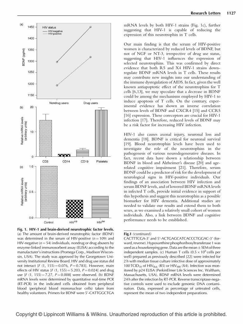
Neurotrophins control cell survival. Therefore, weexamined whether HIV-1 reduces neurotrophinlevels. Serum of HIV-positive individuals exhibitedlower concentrations of brain-derived neuro-trophic factor (BDNF), but not of other neurotro-phins, than HIV-negative individuals. In addition,R5 and X4 strains of HIV-1 decreased BDNFexpression in T cells. Our results support the hypo-thesis that reduced levels of BDNF may be a riskfactor for T-cell apoptosis and for neurologicalcomplications associated with HIV-1 infection.
Neurotrophins [1,2] are produced by immune organsand immunocompetent cells, including T cells [3] and
macrophages [4], and are believed to play a role in variousfunctions of the immune system, including lymphocyteproliferation [5,6]. Little is known about the effect ofHIV-1 on neurotrophin levels. Loss of neurotrophinexpression may impair the immune system and promoteAIDS. In this study, we investigated whether HIV-1reduces serum concentration of the neurotrophins andsought to establish a correlation between HIV infectionand neurotrophin expression in T cells.Serum levels of brain-derived neurotrophic factor(BDNF) were measured by an enzyme-linked immuno-sorbent assay in human samples collected between 1994and 2007 at the Washington, District of Columbia site ofthe Women’s Interagency HIV Study [7,8]. Becauseapproximately 50% of these individuals were polydrugabusers, mainly cocaine, methamphetamine and heroin, atwo-way analysis of variance (ANOVA) was used toexamine a potential interaction between HIV-1 and druguse and to examine each factor independently. HIV-positive individuals exhibited significantly lower levels ofBDNF compared with HIV-negative controls (Fig. 1a).Drug use significantly affected BDNF levels such that theamount of BDNF in the serum of HIV-positive drugusers were higher than in HIV-positive nondrug users(Fig. 1a), suggesting that polydrug use may affect serum
pyright © Lippincott Williams & Wilkins. Unautho
BDNF levels in HIV-1-positive individuals. There was nointeraction between drug use and serostatus on BDNFlevels (P> 0.33).
Drugs of abuse [9] or HIV-1 may influence the expressionof other neurotrophins. To test this hypothesis, wemeasured nerve growth factor (NGF) and neurotrophin-3 (NT-3) levels in the same samples. The two-wayANOVAs analyzing associations of HIV status and druguse on NGF (P¼ 0.516) and NT-3 (P¼ 0.382) were notstatistically significant, and no evidence of interactionbetween HIV and drug use was observed for eitheroutcome. Although we found a tendency toward loweraverage NGF levels in the serum of HIV-positiveindividuals compared with controls, the effect was notsignificant (P¼ 0.89) nor did polydrug use affect NGFlevels (data not shown). Results for NT-3 levels weresimilarly not statistically significant (data not shown).
The reduction of BDNF observed in HIV-1-positiveindividuals could be due to single nucleotide poly-morphisms (SNPs) that alter intracellular packaging andsecretion of BDNF [10]. rs6265 is a polymorphism in theBDNF gene that produces an amino acid substitutionof valine to methionine in codon 66 (Val66Met);rs56164415 is located in the fifth of the seven noncodingexons of the BDNF gene [11] and appears to bemoderately associated with substance abuse [12]. There-fore, these SNPs, either alone or in combination, mightlead to a reduction in serum BDNF levels. To testthis hypothesis, we examined the frequency of thesepolymorphisms in the same cohort, using DNA from thesame sample of individuals. There was no significantdifference in frequency of alleles in HIV individuals ascompared with HIV-negative controls (rs6265, P¼ 0.83;rs56164415, P¼ 0.72). Therefore, mutation of theBDNF gene does not appear to account for differencein the levels of BDNF in these individuals.
Contributing factors that may account for the decrease inserum BDNF in HIV-positive individuals are not easilydefined. BDNF and other neurotrophins are produced byimmune organs and immunocompetent cells [13], as wellas platelets [14]. Thus, a decrease in the number ofplatelets may explain the lower levels of BDNF in HIV-1-positive individuals. To determine whether BDNF fromplatelets constitutes a significant fraction of serum BDNF,we examined which blood cell type expresses BDNF. Wefound that platelets and T cells exhibited comparablelevels of BDNF expression (Fig. 1b). Thus, plateletsaccount for only for a fraction of serum BDNF.Nevertheless, to more directly examine the effect ofHIV-1 on BDNF, we examined the ability of HIV-1 todecrease BDNF expression in T cells. T lymphocyteswere prepared from healthy donors and were infectedwith X4 (IIIB) or R5 (BaL) HIVs. BDNF mRNA levelswere then quantified 24 h after the infection. Weobserved an approximately 50% decrease in BDNF
rized reproduction of this article is prohibited.
Copyright © Lippincott Williams & Wilkins. Unauth
Research Letters 1127
Fig. 1. HIV-1 and brain-derived neurotrophic factor levels.(a) The amount of brain-derived neurotrophic factor (BDNF)was determined in the serum of HIV-positive (n¼109) andHIV-negative (n¼ 54) individuals, nondrug or drug abusers byenzyme-linked immunosorbent assay (ELISA) according to themanufacturer’s instructions (Promega Corp., Madison Wiscon-sin, USA). The study was approved by the Georgetown Uni-versity Institutional Review Board. HIV and drug use status didnot interact [F (1, 155)¼0.076, P¼0.783]. However, maineffects of HIV status [F (1, 155)¼5.203, P¼ 0.024] and druguse [F (1, 155)¼ 7.27, P¼0.008] were observed. (b) BDNFmRNA levels were determined by quantitative real-time PCR(RT-PCR) in the indicated cells obtained from peripheralblood (peripheral blood mononuclear cells) taken fromhealthy volunteers. Primers for BDNF were 5’-CATTGGCTGA
mRNA levels by both HIV-1 strains (Fig. 1c), furthersuggesting that HIV-1 is capable of reducing theexpression of this neurotrophin in T cells.
Our main finding is that the serum of HIV-positivewomen is characterized by reduced levels of BDNF, butnot of NGF or NT-3, irrespective of drug use status,suggesting that HIV-1 influences the expression ofselected neurotrophins. This was confirmed by directevidence that both R5 and X4 HIV-1 strains down-regulate BDNF mRNA levels in T cells. These resultsmay contribute new insights into our understanding ofthe immune dysregulation of AIDS. In fact, given the wellknown antiapoptotic effect of the neurotrophins for Tcells [6,13], we may speculate that a decrease in BDNFcould be among the mechanisms employed by HIV-1 toinduce apoptosis of T cells. On the contrary, exper-imental evidence has shown an inverse correlationbetween levels of BDNF and CXCR4 [15] and CCR5[16] expression. These coreceptors are crucial for HIV-1infection [17]. Therefore, reduced levels of BDNF maybe a risk factor for increasing HIV infection.
HIV-1 also causes axonal injury, neuronal loss anddementia [18]. BDNF is critical for neuronal survival[19]. Blood neurotrophin levels have been used toinvestigate the role of the neurotrophins in thepathogenesis of various neurodegenerative diseases. Infact, recent data have shown a relationship betweenBDNF in blood and Alzheimer’s disease [20] and age-related cognitive impairment [21]. Therefore, serumBDNF could be a predictor of risk for the development ofneurological signs in HIV-positive individuals. Ourfindings of an association between HIV infection andserum BDNF levels, and of lowered BDNF mRNA levelsin infected T cells, provide initial evidence in support ofthis hypothesis and suggest this neurotrophin as a possiblebiomarker for HIV dementia. Additional studies areneeded to validate our results and extend them to bothsexes, as we examined a relatively small cohort of womenindividuals. Also, a link between BDNF and cognitiveperformance needs to be established.
orized reproduction of this article is prohibited.
Fig.1 (continued)CACTTTCGA-3’ and 5’-ACTGAGCATCACCCTGGAC-3’ (for-ward, reverse). Hypoxanthine phosphoribosyltransferase 1 wasused as a housekeepinggene.Data are themean� SEM of threeindependent samples. (c) Human T cells (0.5�106 cells perwell) prepared as previously described [22] were infected for2 h with median tissue culture infective dose of approximately100TCID50 of HIVBaL (R5) or HIVIIIB (X4). Infection was mon-itored by p24 ELISA (PerkinElmer Life Sciences Inc. Waltham,Massachusetts, USA). BDNF mRNA levels were determined24 h after the infection by RT-PCR. Reverse transcriptase-nega-tive controls were used to exclude genomic DNA contami-nation. Data, expressed as percentage of untreated cells,represent the mean of two independent preparations.

Co
1128 AIDS 2011, Vol 25 No 8
Acknowledgements
This study is supported by HHS grants DA026174 (I.M.),NS066842 (A.G.-D.). Women’s Interagency HIV Studyis funded by UO1-AI-35004, UO1-AI-31834, UO1-AI-34994, UO1-AI-34989, UO1-AI-34993, UO1-AI-42590, UO1-HD-32632 and UL1 RR024131.
aDepartment of Neuroscience, Georgetown UniversityMedical Center, Washington, District of Columbia,bDepartment of Microbiology and Immunology, In-stitute of Human Virology, University of Maryland,Baltimore, cLaboratory of Molecular Medicine andNeuroscience, National Institute of Neurological Dis-orders and Stroke/National Institute of Health, Bethes-da, Maryland, dDepartments of Psychiatry andPsychology, University of Illinois at Chicago, Chicago,Illinois, eDepartment of Neurology, and fDepartmentof Medicine, Georgetown University Medical Center,Washington, District of Columbia, USA.
Correspondence to Italo Mocchetti, PhD, GeorgetownUniversity Medical Center, New Research BuildingWP13, 3970 Reservoir Road NW, Washington, DC20057, USA.E-mail: [email protected]
Received: 6 January 2011; revised: 7 February 2011;accepted: 4 March 2011.
References
1. Laurenzi MA, Barbany G, Timmusk T, Lindgren JA, Persson H.Expression of mRNA encoding neurotrophins and neurotrophinreceptors in rat thymus, spleen tissue and immunocompetentcells. Regulation of neurotrophin-4 mRNA expression bymitogens and leukotriene B4. Eur J Biochem 1994; 223:733–741.
2. Artico M, Bronzetti E, Felici LM, Alicino V, Ionta B, Bronzetti B,et al. Neurotrophins and their receptors in human lingualtonsil: an immunohistochemical analysis. Oncol Rep 2008;20:1201–1206.
3. Kerschensteiner M, Gallmeier E, Behrens L, Leal VV, Misgeld T,Klinkert WE, et al. Activated human T cells, B cells, andmonocytes produce brain-derived neurotrophic factor in vitroand in inflammatory brain lesions: a neuroprotective role ofinflammation? J Exp Med 1999; 189:865–870.
4. Elkabes S, DiCicco-Bloom EM, Black IB. Brain microglia/macrophages express neurotrophins that selectively regulatemicroglial proliferation and function. J Neurosci 1996;16:2508–2521.
5. Aloe L, Bracci-Laudiero L, Micera A, Tirassa P. Nerve growthfactor and the immune system. In: Mocchetti I, editor. Neuro-biology of the neurotrophins. Johnson City: Graham FP; 2005.pp. 237–253.
6. Garcia-Suarez O, Blanco-Gelaz MA, Lopez ML, Germana A,Cabo R, Diaz-Esnal B, et al. Massive lymphocyte apoptosis inthe thymus of functionally deficient TrkB mice. J Neuroimmu-nol 2002; 129:25–34.
7. Barkan SE, Melnick SL, Preston-Martin S, Weber K, Kalish LA,Miotti P, et al. The Women’s Interagency HIV Study.WIHS Collaborative Study Group. Epidemiology 1998; 9:117–125.
8. Bacon MC, von Wyl V, Alden C, Sharp G, Robison E, Hessol N,et al. The Women’s Interagency HIV Study: an observationalcohort brings clinical sciences to the bench. Clin Diagn LabImmunol 2005; 12:1013–1019.
pyright © Lippincott Williams & Wilkins. Unautho
9. Angelucci F, Ricci V, Pomponi M, Conte G, Mathe AA, AttilioTonali P, Bria P. Chronic heroin and cocaine abuse is associatedwith decreased serum concentrations of the nerve growthfactor and brain-derived neurotrophic factor. J Psychopharma-col 2007; 21:820–825.
10. Chen ZY, Patel PD, Sant G, Meng CX, Teng KK, Hempstead BL,Lee FS. Variant brain-derived neurotrophic factor (BDNF)(Met66) alters the intracellular trafficking and activity-depen-dent secretion of wild-type BDNF in neurosecretory cells andcortical neurons. J Neurosci 2004; 24:4401–4411.
11. Kunugi H, Ueki A, Otsuka M, Isse K, Hirasawa H, Kato N, et al.A novel polymorphism of the brain-derived neurotrophicfactor (BDNF) gene associated with late-onset Alzheimer’sdisease. Mol Psychiatry 2001; 6:83–86.
12. Liu QR, Walther D, Drgon T, Polesskaya O, Lesnick TG, StrainKJ, et al. Human brain derived neurotrophic factor (BDNF)genes, splicing patterns, and assessments of associations withsubstance abuse and Parkinson’s disease. Am J Med Genet BNeuropsychiatr Genet 2005; 134:93–103.
13. Vega JA, Garcia-Suarez O, Hannestad J, Perez-Perez M, Ger-mana A. Neurotrophins and the immune system. J Anat 2003;203:1–19.
14. Yamamoto H, Gurney ME. Human platelets contain brain-derived neurotrophic factor. J Neurosci 1990; 10:3469–3478.
15. Nosheny RL, Amhed F, Yakovlev AG, Meyer EM, Ren K,Tessarollo L, Mocchetti I. Brain-derived neurotrophic factorprevents the nigrostriatal degeneration induced by humanimmunodeficiency virus-1 glycoprotein 120 in vivo. Eur JNeurosci 2007; 25:2275–2284.
16. Ahmed F, Tessarollo L, Thiele C, Mocchetti I. Brain-derivedneurotrophic factor modulates expression of chemokine re-ceptors in the brain.. Brain Res 2008; 1227:1–11.
17. Berger EA, Murphy PM, Farber JM. Chemokine receptors asHIV-1 coreceptors: roles in viral entry, tropism, and disease.Annu Rev Immunol 1999; 17:657–700.
18. Price RW. Neurological complications of HIV infection. Lancet1996; 348:445–452.
19. Reichardt LF. Neurotrophin-regulated signalling pathways. Phi-los Trans R Soc Lond B Biol Sci 2006; 361:1545–1564.
20. Laske C, Stransky E, Leyhe T, Eschweiler GW, Maetzler W,Wittorf A, et al. BDNF serum and CSF concentrations inAlzheimer’s disease, normal pressure hydrocephalus andhealthy controls. J Psychiatr Res 2007; 41:387–394.
21. Komulainen P, Pedersen M, Hanninen T, Bruunsgaard H, LakkaTA, Kivipelto M, et al. BDNF is a novel marker of cognitivefunction in ageing women: the DR’s EXTRA Study. NeurobiolLearn Mem 2008; 90:596–603.
22. Garzino-Demo A, Moss RB, Margolick JB, Cleghorn F, Sill A,Blattner WA, et al. Spontaneous and antigen-induced produc-tion of HIV-inhibitory beta-chemokines are associated withAIDS-free status. Proc Natl Acad Sci USA 1999; 96:11986–11991.
DOI:10.1097/QAD.0b013e32834671b3
High-sensitivity C-reactive protein levels fall duringstatin therapy in HIV-infected patients receivingritonavir-boosted protease inhibitors
Elisabeth Aslangula,b, Soraya Fellahic,d,e, Lambert K.Assoumouf,g, Jean-Philippe Bastardc,d,e, JacquelineCapeauc,d,e and Dominique Costagliolaf,g,h
HIV-infected patients are at an increased risk ofdeveloping cardiovascular disease. Elevated levelsof C-reactive protein (CRP) are associated with anincreased risk of cardiovascular disease in thegeneral population and are reduced by statintherapy. We examined the effect of pravastatin
rized reproduction of this article is prohibited.

C
Research Letters 1129
and rosuvastatin on CRP levels in 58 dyslipidemic
HIV-infected patients. A 45-day course of either
statin reduced the median CRP level from 3.0 to
2.4mg/l (P <0.001) with no correlation with
changes in lipid parameters.
Cardiovascular disease is more frequent in HIV-infectedpatients than in the general population, possibly owing to
lipid disorders, viral infection, inflammation and anti-retroviral therapy (ART), especially ritonavir (RTV)-boosted protease inhibitors [1–3]. Elevated levels of high-sensitivity C-reactive protein (hsCRP), a marker ofpersistent inflammation, are linked to an increased risk ofcardiovascular events in the general population [4],whereas elevated hsCRP levels in HIV-infected patientsare associated with a higher incidence of myocardialinfarction [5] and death [6,7]. In the general population,rosuvastatin has been shown to reduce hsCRP levels byabout one third and also to lower the risk of death andcardiovascular events [8]. Several studies have reportedthat hsCRP levels are higher in HIV-infected patientsthan those in the general population [9,10]. The role ofcombination ART (cART) is discussed [11–13]. ElevatedCRP levels have also been linked to other cardiovascularrisk factors, such as high low-density lipoprotein (LDL)levels, low high-density lipoprotein levels and smoking,in addition to ART [14]. The aim of this study was toexamine changes in levels of hsCRP, soluble tumornecrosis factor-a receptors (TNFRs) and the endothelialmarkers intercellular adhesion molecule-1 (ICAM-1) andvascular cell adhesion molecule-1 (VCAM-1) after a 45-day course of rosuvastatin or pravastatin in dyslipidemicHIV-infected patients participating in the AgenceNationale de Recherche sur le SIDA (ANRS126)VIHstatine trial; all the patients had good viral controlon a RTV-boosted protease inhibitor regimen [15].The VIHstatine randomized, double-blind, multicentertrial (NCT00117494) was designed to assess the impact ofa 45-day course of rosuvastatin 10 mg per day orpravastatin 40 mg per day on lipid values in dyslipidemic(LDL> 4.1 mmol/l) HIV-infected patients receivingRTV-boosted protease inhibitors [15]. The presentsubstudy focused on patients for whom frozen sampleswere available both at baseline and after the 45-day courseof statin therapy, and who had a baseline CRP valuebelow 10 mg/l, as values above 10 mg/l are suggestive ofother inflammatory processes, as indicated by theAmerican Heart Association [16]. Fifty-eight of the 83patients enrolled in the VIHstatine trial were eligible forthis substudy and were equally distributed between thetwo statin arms.
HsCRP was measured by immunonephelometry onan IMMAGE analyzer (Beckman-Coulter, Villepinte,France). sTNFR1, sTNFR2, ICAM-1 and VCAM-1levels were measured with commercial ELISA kits from
opyright © Lippincott Williams & Wilkins. Unauth
R&D Systems (Oxford, UK), using the manufacturer’sprotocols.
Results are reported as median [interquartile range(IQR)]. Baseline hsCRP, sTNFR1, sTNFR2, ICAM-1and VCAM-1 levels, and changes between baseline andday 45, were compared between the two statin arms byusing the Mann–Whitney nonparametric test, whereaschanges between baseline and day 45 were comparedwith the nonparametric paired Wilcoxon test. Corre-lations between changes in parameters were tested withSpearman’s nonparametric test. All reported P-valuesare two tailed. The Bonferroni rule was used to takemultiplicity issues into account: we used nine Mann–Whitney tests and nine Wilcoxon tests, yielding thesignificance threshold at P-value less than 0.0055, and 30Spearman correlation tests, yielding the significancethreshold at P-value less than 0.0017. The SPSS softwarepackage version 13.0 for Windows (SPSS Inc., Chicago,Illinois, USA) and SAS statistical software version 9.1(SAS Institute Inc., Cary, North Carolina, USA) wereused for all analyses.
The patients were mainly men (74%) and white (91%) andhad a median age of 49 years (IQR 42–56). Plasma HIV-1-RNA levels were below 400 copies/ml in 90% ofpatients and the median CD4 cell count was 490 cells/ml(IQR 314–704). The median duration of cART was9 years (IQR 5–13). Baseline values of lipid, inflam-mation and endothelial parameters are reported inTable 1. After 45 days of statin therapy, the medianchange in the hsCRP concentration was �20% overall(�0.6 mg/l, P< 0.001), and, respectively,�22 and�16%in the pravastatin and rosuvastatin groups (P¼ 0.932).There was no significant change in sTNFR1 andsTNFR2 levels or in ICAM-1 and VCAM-1 levels(Table 1).
The LDL-cholesterol level fell by a median of 19% in thepravastatin group and 37% in the rosuvastatin group(P< 0.001 for difference between groups), whereastriglyceride levels fell by, respectively, 3% and 26%(P¼ 0.008 for difference between groups), values similarto those obtained in the original VIHstatine trial [15]. Asshown in Table 1, LDL-cholesterol, total cholesterol andtriglyceride levels also fell significantly in the entiresubstudy. There was no correlation between the change inthe hsCRP level and changes in the markers of lipid,endothelial and inflammatory status [LDL-cholesterol(r¼�0.071, P¼ 0.598); total cholesterol (r¼�0.176,P¼ 0.188); and triglycerides (r¼�0.273, P¼ 0.038)].
This is the first study of the effect of statins oninflammatory status in HIV-infected patients on effectiveART. We observed a reduction in the CRP level, but notin the levels of sTNFR1 and sTNFR2, two otherinflammatory markers. It is interesting to note thatsTNFR1 and sTNFR2 levels were reported to fall in
orized reproduction of this article is prohibited.

Copyright © Lippincott Williams & Wilkins. Unautho
1130 AIDS 2011, Vol 25 No 8
Tab
le1.
Lipid
,in
flam
mat
ory
and
endoth
elia
lm
arke
rsat
bas
elin
ean
daf
ter
45
day
sof
stat
inth
erap
yby
trea
tmen
tgr
oup
and
ove
rall.
Pra
vast
atin
Rosu
vast
atin
P-v
alue
a
Tota
l
P-v
alueb
nD
ay0
Per
centa
geof
chan
geat
day
45
nD
ay0
Per
centa
geof
chan
geat
day
45
Pra
vast
atin
vs.
rosu
vast
atin
Day
0Per
centa
geof
chan
geat
day
45
Day
0vs
.day
45
Tota
lch
ole
ster
ol
(mm
ol/
l),
med
ian
(IQ
R)
29
7.1
4(6
.35
–8.0
1)
�14%
(�21
to�
1)
29
7.3
2(6
.42
–7.9
4)
�29%
(�32
to�
17)
<0.0
01
7.2
8(6
.38
–7.9
1)
�20%
(�29
to�
11)
<0.0
01
LDL
chole
ster
ol
(mm
ol/
l),
med
ian
(IQ
R)
29
4.8
1(4
.23
–5.6
9)
�19%
(�29
to0)
29
4.9
4(4
.27
–5.8
9)
�37%
(�42
to�
19)
<0.0
01
4.9
0(4
.24
–5.7
9)
�28%
(�37
to�
13)
<0.0
01
HD
Lch
ole
ster
ol
(mm
ol/
l),
med
ian
(IQ
R)
29
1.4
3(1
.17
–1.6
5)
0%
(�6
to12)
29
1.3
1(1
.15
–1.5
6)
8%
(�2
to13)
0.2
49
1.3
5(1
.17
–1.6
1)
3%
(�4
to13)
0.0
29
Tri
glyc
erid
es(m
mol/
l),
med
ian
(IQ
R)
29
2.0
7(1
.65
–2.6
4)
�3%
(�23
to11)
29
2.8
2(1
.90
–3.4
5)
�26%
(�41
to�
9)
0.0
08
2.3
3(1
.72
–3.4
3)
�16%
(�33
to0)
<0.0
01
Hig
h-s
ensi
tivi
tyC
-rea
ctiv
epro
tein
(mg/
l),
med
ian
(IQ
R)
29
2.8
(1.7
–4.0
)�
22%
(�41
to23)
29
3.2
(2.0
–4.7
)�
16%
(�54
to11)
0.9
32
3.0
(1.9
–4.5
)�
20%
(�44
to17)
0.0
01
Solu
ble
tum
or
nec
rosi
sfa
ctor-
are
cepto
r(s
TN
FR)1
(ng/
ml)
,m
edia
n(I
QR
)27
1235
(1011
–1661)
�6%
(�15
to8)
28
1469
(1309
–1665)
�5%
(�11
to8)
0.5
01
1411
(1122
–1661)
�6%
(�15
to8)
0.1
40
sTN
FR2
(ng/
ml)
,m
edia
n(I
QR
)27
2124
(1810
–2615)
�3%
(�8
to10)
28
2717
(2159
–3045)
�4%
(�11
to12)
0.4
90
2332
(1935
–2823)
�3%
(�8
to11)
0.7
38
Inte
rcel
lula
rad
hes
ion
mole
cule
-1(n
g/m
l),
med
ian
(IQ
R)
24
265
(218
–296)
4%
(�5
to14)
27
295
(228
–393)
3%
(�3
to10)
0.4
97
279
(220
–339)
3%
(�4
to12)
0.0
44
Vas
cula
rce
llad
hes
ion
mole
cule
-1(n
g/m
l),
med
ian
(IQ
R)
27
866
(747
–1069)
�1%
(�10
to8)
29
900
(753
–1168)
�4%
(�13
to8)
0.6
76
885
(749
–1079)
�3%
(�11
to7)
0.1
51
HD
L,hig
h-d
ensi
tyli
popro
tein
;IQ
R,
inte
rquar
tile
range
;LD
L,lo
w-d
ensi
tyli
popro
tein
.aM
ann
–W
hit
ney
test
.bPai
red
Wil
coxo
nte
st.
HIV-infected patients starting first-line ART, whereasCRP levels were unaffected [12]. Together, these datasuggest that these markers reflect different phenomena:sTNFRs may reflect control of HIV infection, whereasCRP elevation could result from other proinflammatorymechanisms not resolved by the control of viral load.
We show here that pravastatin 40 mg per day androsuvastatin 10 mg per day induce similar significant fallsin hsCRP, even during brief administration. In addition,the fall in hsCRP did not correlate with the improvementin lipid parameters or with modifications in endothelialstatus, as reflected by ICAM-1 and VCAM-1 serumconcentrations. The fact that the effect of a statin on thelevel of hsCRP do not correlate with its effect on LDL-cholesterol is already known [17]; this may explain why,although rosuvastatin and pravastatin did not have thesame effect on lipid parameters in this trial, their effect wassimilar on the hsCRP level. The impact of statin onimmune activation, which has been recently shown inHIV-naive patients, might be the mechanism by whichthe hsCRP level was influenced [18].
In the general population, statins induce a dose-dependent fall in hsCRP levels [19]. HsCRP continuesto fall during the course of statin treatment, especially athigher doses [20]. In the JUPITER (Justification for theUse of Statins in Prevention: an Intervention TrialEvaluating Rosuvastatin) trial, 20 mg per day rosuvastatinreduced hsCRP levels by 37% after 1.9 years of treatment[8]. Thus, the fall in CRP observed here in HIV-infectedpatients during short-term statin therapy could beaccentuated during long-term treatment.
In conclusion, a 45-day course of rosuvastatin orpravastatin reduced not only lipid levels but also hsCRPlevels in HIV-infected patients treated with RTV-boostedprotease inhibitors. Clinical trials are warranted todetermine the potential benefit of long-term statintreatment on the risk of cardiovascular events and death inat-risk HIV-infected patients.
Acknowledgements
The authors thank Agence Nationale de Recherche sur leSIDA for funding, and Lydie Hossou and Sandra Raabonfor their excellent technical support.
Conception and design of the substudy was performed byE.A., J.-P.B., J.C. and D.C.
Provision of study materials or patients was conducted byE.A., S.F., J.-P.B. and J.C.
Statistical analysis was performed by L.K.A. and D.C.
rized reproduction of this article is prohibited.

C
Research Letters 1131
Interpretation of the data was performed E.A., S.F.,L.K.A., J.-P.B., J.C. and D.C.
Drafting of the article was done by E.A., L.K.A., J.-P.B.,J.C. and D.C.
Critical revision of the article for important intellectualcontent and final approval of the article were performedE.A., S.F., L.K.A., J.-P.B., J.C. and D.C.
aUniversite Paris Descartes, bAP-HP, Hotel Dieu,Service de Medecine Interne, cAP-HP, Hopital Tenon,Service de Biochimie et Hormonologie, dINSERMU938, CDR Saint-Antoine, eUPMC Univ Paris 06,UMR-S938, fINSERM U943, gUPMC Univ Paris 06,UMR-S943, and hAP-HP, Hopital Pitie-Salpetriere,Service de Maladies Infectieuses et Tropicales, Paris,France.
Correspondence to Dominique Costagliola, INSERMU943, 56 Boulevard Vincent Auriol, BP 335, 75625Paris Cedex 13, France.Tel: +33 1 4216 4282;e-mail: [email protected].
Received: 7 February 2011; revised: 10 March 2011;accepted: 14 March 2011.
References
1. Lang S, Mary-Krause M, Cotte L, Gilquin J, Partisani M, Simon A,et al. Increased risk of myocardial infarction in HIV-infectedpatients in France, relative to the general population. AIDS2010; 24:1228–1230.
2. Martinez E, Larrousse M, Gatell JM. Cardiovascular disease andHIV infection: host, virus, or drugs? Curr Opin Infect Dis 2009;22:28–34.
3. Triant VA, Lee H, Hadigan C, Grinspoon SK. Increased acutemyocardial infarction rates and cardiovascular risk factorsamong patients with human immunodeficiency virus disease.J Clin Endocrinol Metab 2007; 92:2506–2512.
4. Ridker P. Inflammatory biomarkers and risks of myocardialinfarction, stroke, diabetes, and total mortality: implicationsfor longevity. Nutr Rev 2007; 65 (12 Pt 2):S253–S259.
5. Triant VA, Regan S, Lee H, Sax PE, Meigs JB, Grinspoon SK.Association of immunologic and virologic factors with myo-cardial infarction rates in a US healthcare system. J AcquirImmune Defic Syndr 2010; 55:615–619.
6. Kuller LH, Tracy R, Belloso W, De Wit S, Drummond F, LaneHC, et al. Inflammatory and coagulation biomarkers and mor-tality in patients with HIV infection. PLoS Med 2008; 5:e203.
7. Tien PC, Choi AI, Zolopa AR, Benson C, Tracy R, Scherzer R,et al. Inflammation and mortality in HIV-infected adults: ana-lysis of the FRAM study cohort. J Acquir Immune Defic Syndr2010; 55:316–322.
8. Ridker PM, Danielson E, Fonseca FA, Genest J, Gotto AM Jr,Kastelein JJ, et al. Rosuvastatin to prevent vascular events inmen and women with elevated C-reactive protein. N Engl J Med2008; 359:2195–2207.
9. Hsue PY, Hunt PW, Schnell A, Kalapus SC, Hoh R, Ganz P, et al.Role of viral replication, antiretroviral therapy, and immuno-deficiency in HIV-associated atherosclerosis. AIDS 2009;23:1059–1067.
10. Neuhaus J, Jacobs DR Jr, Baker JV, Calmy A, Duprez D, La RosaA, et al. Markers of inflammation, coagulation, and renalfunction are elevated in adults with HIV infection. J InfectDis 2010; 201:1788–1795.
opyright © Lippincott Williams & Wilkins. Unauth
11. Baker JV, Neuhaus J, Duprez D, Kuller LH, Tracy R, BellosoWH, et al. Changes in inflammatory and coagulation biomar-kers: a randomized comparison of immediate versus deferredantiretroviral therapy in patients with HIV infection. J AcquirImmune Defic Syndr 2011; 56:36–43.
12. Brown TT, Tassiopoulos K, Bosch RJ, Shikuma C, McComseyGA. Association between systemic inflammation and incidentdiabetes in HIV-infected patients after initiation of antiretro-viral therapy. Diabetes Care 2010; 33:2244–2249.
13. Smith KY, Patel P, Fine D, Bellos N, Sloan L, Lackey P, et al.Randomized, double-blind, placebo-matched, multicenter trialof abacavir/lamivudine or tenofovir/emtricitabine with lopi-navir/ritonavir for initial HIV treatment. AIDS 2009; 23:1547–1556.
14. Masia M, Bernal E, Padilla S, Graells ML, Jarrin I, Almenar MV,et al. The role of C-reactive protein as a marker for cardiovas-cular risk associated with antiretroviral therapy in HIV-in-fected patients. Atherosclerosis 2007; 195:167–171.
15. Aslangul E, Assoumou L, Bittar R, Valantin MA, Kalmykova O,Peytavin G, et al. Rosuvastatin versus pravastatin in dyslipi-demic HIV-1-infected patients receiving protease inhibitors: arandomized trial. AIDS 2010; 24:77–83.
16. Pearson TA, Mensah GA, Alexander RW, Anderson JL, CannonRO 3rd, Criqui M, et al. Markers of inflammation and cardi-ovascular disease: application to clinical and public healthpractice – a statement for healthcare professionals from theCenters for Disease Control and Prevention and the AmericanHeart Association. Circulation 2003; 107:499–511.
17. Carr A. Statins as anti-inflammatory therapy in HIV disease?J Infect Dis 2011; 203:751–752.
18. Ganesan A, Crum-Cianflone N, Higgins J, Qin J, Rehm J, MetcalfJ, et al. High dose atorvastatin decreases cellular markers ofimmune activation without affecting HIV-1 RNA levels: resultsof a double-blind randomized placebo controlled clinical trial.J Infect Dis 2011; 203:756–764.
19. Ridker PM, Danielson E, Fonseca FA, Genest J, Gotto AM Jr,Kastelein JJ, et al. Reduction in C-reactive protein and LDLcholesterol and cardiovascular event rates after initiation ofrosuvastatin: a prospective study of the JUPITER trial. Lancet2009; 373:1175–1182.
20. Bonnet J, McPherson R, Tedgui A, Simoneau D, Nozza A,Martineau P, Davignon J. Comparative effects of 10-mg versus80-mg atorvastatin on high-sensitivity C-reactive protein inpatients with stable coronary artery disease: results of the CAP(Comparative Atorvastatin Pleiotropic effects) study. Clin Ther2008; 30:2298–2313.
DOI:10.1097/QAD.0b013e328346be29
Impact of IL28B polymorphisms on response topeginterferon and ribavirin in HIV–hepatitis Cvirus-coinfected patients with prior nonresponseor relapse
Pablo Labargaa, Pablo Barreiroa, Jose A. Mirab,Eugenia Vispoa, Norma Rallona, Karin Neukamb,Angela Camachoc, Antonio Caruzd, Sonia Rodriguez-Novoaa, Javier Pinillae, Antonio Riveroc, Jose M.Benitoa, Juan A. Pinedab and Vincent Sorianoa
IL28B polymorphisms predict treatment responsein chronic hepatitis C. However, no informationexists in prior treatment failures. A total of 62HIV/hepatitis C virus (HCV) patients who completedretreatment with peginterferon-a/ribavirin wereexamined, of whom 25 (40%) had been cured.Predictors of response [odds ratio, OR (95% con-fidence interval, CI)] were HCV genotypes 2/3[16.1 (2.7–90.9)], prior relapse [9.6 (1.5–62.4)]
orized reproduction of this article is prohibited.

Co
1132 AIDS 2011, Vol 25 No 8
and ribavirin plasma trough concentrations atweek 4 [4.9 (1.3–18.4)]. IL28B-CC only predictedresponse in prior nonresponders carrying HCVgenotypes 1/4 [25.1 (1.9–337)].
While awaiting for the arrival of new direct hepatitis Cvirus (HCV) antivirals, the accelerated course of liver
fibrosis in HIV/HCV-coinfected individuals [1] makestreatment of chronic hepatitis C a priority [2], beingpatients who have failed interferon (IFN)a-based therapyin the past no exception. Besides the strong influence ofHCV genotypes, the chances of a sustained virologicalresponse (SVR) after treatment rechallenge seem tomostly depend on patient’s characteristics rather than viralfactors, that is, extent of liver fibrosis or ribavirin (RBV)plasma exposure [3]. Single nucleotide polymorphisms(SNPs) nearby the IL28B gene are currently known to bestrong predictors of response to first-line pegIFNa–RBVtherapy in both HCV-monoinfected [4–6] and HCV/HIV-coinfected individuals [7,8]. At this time, the impactof IL28B variants on treatment rechallenge is unknown.We have assessed the influence of IL28B rs12979860SNPs in 62 HIV/HCV-coinfected patients who receiveda second course of therapy with pegIFNa-2a (180 mg/week) and RBV (1000–1200 mg/day) for 48 weeks, afterhaving failed to suboptimal IFNa-based regimens in thepast (i.e., conventional IFNa with/without RBV orpegIFNa and fixed 800 mg/day RBV dosing). Partici-pants without a decline of more than 2 logs in serumHCV-RNA at week 12 or with serum HCV-RNA morethan 10 IU/ml at week 24 were considered as virologicalfailures and discontinued therapy [3]. Likewise, partici-pants who showed HCV-RNA rebound after disconti-nuing treatment with undetectable viremia were con-sidered as relapsers. Plasma HCV-RNA was measuredusing a real-time PCR assay (lower limit of detection of10 IU/ml). HCV genotyping was performed using acommercial RT-PCR hybridization [9]. Plasma troughconcentrations of RBV were measured at week 4 usinghigh-performance liquid chromatography (HPLC) [10].The IL28B rs12979860 SNP was examined in peripheralblood mononuclear cells using the 50 nuclease assay withallele-specific TaqMan probes (ABI TaqMan allelic dis-crimination kit) and ABI7900HT Sequence DetectionSystem (Applied Biosystems, Carlsbad, California, USA)[11].
In the study population, mean age was 43 years, mostwere men (82%) and were former injection drug users(IDUs; 97%); active alcohol abuse was rare (8%) and mostpatients were on antiretroviral therapy (94%), withundetectable plasma HIV-RNA (95%) and mean CD4cell counts of 657 cells/ml. Most participants had serumHCV-RNA levels more than 500 000 IU/ml (73%) andwere infected with HCV genotypes 1 or 4 (76%). Morethan a half of patients had advanced liver fibrosis (53%)and had failed to pegIFNa and low-dose RBV (58%).
pyright © Lippincott Williams & Wilkins. Unautho
Nonresponse (63%) was the most frequent type ofvirological failure to first hepatitis C therapy, being HCVrelapse recognized in only 21% of cases. In the remaining16%, the prior course of therapy had been prematurelyinterrupted due to toxicity. Overall, 47% of patients hadthe IL28B rs12979860 CC genotype.
A total of 25 (40%, by on-treatment analysis) attainedSVR after completion of pegIFNa–RBV retreatment.Patients who achieved SVR had lower baseline serumHCV-RNA (5.8 vs. 6.2 log IU/ml, P¼ 0.06) and wereless frequently infected with HCV genotype 1 or 4 (48%vs. 95%, P< 0.01) than failures. The likelihood ofachieving SVR was significantly greater in prior relapsersthan in nonresponders (85% vs. 31%; P< 0.001).Participants carrying IL28B CC more likely attainedSVR than non-CC carriers (57% vs. 24%, respectively;P¼ 0.006). However, when the population was split outaccording to HCV genotype, the impact of IL28B onSVR was only seen in HCV genotypes 1 or 4 carriers(44% for CC vs. 14% for non-CC, P¼ 0.02), being notrecognized in participants infected with HCV genotype 2or 3 (82% SVR for CC vs. 100% for non-CC, P¼ 0.36).Patients who attained sustained HCV clearance hadgreater mean RBV plasma trough concentrations at week4 of therapy than patients who failed therapy (2.41 vs.1.75 mg/ml; P¼ 0.02). The best discriminatory RBVthreshold was 2.0 mg/ml, which displayed a positivepredictive value of 69% and a negative predictive value of70% for SVR (P¼ 0.02).
Two models for the multivariate analysis were builtconsidering or not RBV plasma trough concentrations atweek 4 among the predictors of SVR. HCV genotype 2or 3, relapse after prior IFNa-based therapy, and RBVplasma concentrations were all associated with SVR(Table 1). Interestingly, the impact of IL28B polymorph-isms on SVR was only recognized in the subset of patientsmore difficult to treat, namely those infected with HCVgenotypes 1 or 4 and with true nonresponse to a firstcourse of therapy. In the multivariate analysis for thissubpopulation, adjusting for sex, use of antiretroviraltherapy, serum HCV-RNA levels, and liver fibrosisstaging, the subset of patients carrying the CC genotypehad a higher likelihood of response than CT/TT carriers[odds ratio (OR), 25.07 (95% confidence interval, CI1.86–337), P¼ 0.01]. Moreover, in these patients, RBVplasma trough concentrations at week 4 did not predictSVR [OR, 2.79 (95% CI 0.58–13.03), P¼ 0.2].
The finding of a restricted influence of the favorableIL28B genotype in patients with history of truenonresponse instead of relapsers is in line with therecognition by others of a strong association betweenIL28B rs12979860 SNPs and early viral kinetics ontherapy but not with prevention of viral rebound uponcompletion of treatment [12]. In contrast, RBV plasmatrough concentrations predicted SVR to re-treatment of
rized reproduction of this article is prohibited.

C
Research Letters 1133
Table 1. Predictors of sustained virological response in the study population (multivariate analysis).
OR (95% CI) P
Considering RBV Not considering RBV
Male sex 7.66 (0.13–456) 0.33 2.88 (0.38–21.69) 0.30Under HAART 0.07 (0.003–1.51) 0.09 0.09 (0.007–1.09) 0.06HCV-RNA <500 000 IU/ml 0.54 (0.03–9.70) 0.68 2.61 (0.53–12.79) 0.24HCV genotype 2–3 vs. 1–4 52.63 (2.35–1000) 0.01 16.13 (2.75–90.91) 0.002Advanced liver fibrosis 0.51 (0.06–4.57) 0.55 0.41 (0.01–1.75) 0.23Prior relapse 13.01 (0.61–275) 0.09 9.65 (1.49–62.44) 0.02IL28B rs12979860 CC vs. CT/TT 2.02 (0.18–23.12) 0.57 2.51 (0.54–11.69) 0.24RBV (per mg/ml) 4.92 (1.31–18.45) 0.02 –
RBV represents ribavirin plasma trough concentration. CI, confidence interval; HAART, highly active antiretroviral therapy; HCV, hepatitis C virus; OR, odds ratio.
hepatitis C in the subset of patients with prior HCVrelapse and/or HCV genotypes 2 or 3. In them, RBVtrough concentrations more than 2 mg/ml were associ-ated with SVR [OR, 11.67 (95% CI 0.92–147),P¼ 0.06] with almost statistical significance in themultivariate analysis.
In summary, re-treatment of chronic hepatitis C in HIV–HCV-coinfected patients must ensure optimal RBVexposure, especially in prior relapsers and/or in patientsinfected with HCV genotype 2 or 3. In contrast, in priortrue nonresponders infected with HCV genotype 1 or 4,which is the most prevalent and difficult-to-treat popula-tion, optimization of RBV exposure seems to have littleimpact on SVR, while a favorable IL28B genotype plays amajor role in the outcome of re-treatment.
Acknowledgements
The present work was supported by grants fromFundacion Investigacion y Educacion en SIDA (IES),the European NEAT project, Red de Investigacion enSIDA (RIS, FIS-RD06/0006), Agencia Laın Entralgo,Instituto de Salud Carlos III (Rıo Hortega, ref. CM009)and Fundacion para la Investigacion y la Prevencion delSIDA en Espana (FIPSE, 360799/09). V.S. and P.L. arerecipients of intensification grants from Agencia LainEntralgo, Comunidad Autonoma de Madrid. J.A.P. isrecipient of an intensification grant from FundacionProgreso y Salud, Consejerıa de Salud de la Junta deAndalucıa (AI-0021).
All authors declare no conflict of interest.
aHospital Carlos III, Madrid, bHospital Valme, Seville,cHospital Reina Sofıa, Cordoba, dUniversidad de Jaen,Jaen, and eHospital San Pedro, Logrono, Spain.
Correspondence to Dr Pablo Barreiro, Department ofInfectious Diseases, Hospital Carlos III, Calle SinesioDelgado 10, Madrid 28029, Spain.Tel: +34 91 4532500; fax: +34 91 7336614;e-mail: [email protected]
opyright © Lippincott Williams & Wilkins. Unauth
Received: 16 February 2011; revised: 10 March 2011;accepted: 24 March 2011.
References
1. Benhamou Y, Bochet M, Di Martino V, Charlotte F, Azria F,Coutellier A, et al. Liver fibrosis progression in HIV and hepa-titis C virus coinfected patients. The Multivirc Group. Hepa-tology 1999; 30:1054–1058.
2. Soriano V, Puoti M, Sulkowski M, Mauss S, Cacoub P, CargnelA, et al. Care of patients coinfected with HIV and hepatitis Cvirus: 2007 updated recommendations from the HCV-HIVInternational Panel. AIDS 2007; 21:1073–1089.
3. Labarga P, Vispo E, Barreiro P, Rodrıguez-Novoa S, Pinilla J,Morello J, et al. Rate and predictors of success in the retreat-ment of chronic hepatitis C virus in HIV/hepatitis C viruscoinfected patients with prior nonresponse or relapse. J AcquirImmune Defic Syndr 2010; 53:364–368.
4. Ge D, Fellay J, Thompson A, Simon J, Shianna K, Urban T, et al.Genetic variation in IL28B predicts hepatitis C treatment-induced viral clearance. Nature 2009; 461:399–401.
5. Suppiah V, Moldovan M, Ahlenstiel G, Berg T, Weltman M,Abate M, et al. IL28B is associated with response to chronichepatitis C interferon-alpha and ribavirin therapy. Nat Genet2009; 41:1100–1104.
6. Tanaka Y, Nishida N, Sugiyama M, Kurosaki M, Matsuura K,Sakamoto N, et al. Genome-wide association of IL28B withresponse to pegylated interferon-alpha and ribavirin therapyfor chronic hepatitis C. Nat Genet 2009; 41:1105–1109.
7. Rallon N, Naggie S, Benito J, Medrano J, Restrepo C, GoldsteinD, et al. Association of a single nucleotide polymorphism nearinterleukin-28B with response to hepatitis C therapy in HIV/hepatitis C virus coinfected patients. AIDS 2010; 24:F23–29.
8. Pineda J, Caruz A, Rivero A, Neukam K, Salas I, Camacho A,et al. Prediction of response to pegylated interferon plusribavirin by IL28B gene variation in patients coinfected withHIV and hepatitis C virus. Clin Infect Dis 2010; 51:788–795.
9. Chevaliez S, Bouvier-Alias M, Brillet R, Pawlotsky JM. HepatitisC virus (HCV) genotype 1 subtype identification in new HCVdrug development and future clinical practice. PLoS One 2009;4:e8209.
10. Morello J, Rodriguez-Novoa S, Cantillano A, Gonzalez-PardoG, Jimenez-Nacher I, Soriano V. Measurement of ribavirinplasma concentrations by high-performance liquid chromato-graphy using a novel solid-phase extraction method in patientstreated for chronic hepatitis C. Ther Drug Monit 2007; 29:802–806.
11. Livak K. Allelic discrimination using fluorogenic probes and the50 nuclease assay. Genet Anal 1999; 14:143–149.
12. McCarthy J, Li J, Thompson A, Suchindran S, Lao X, Patel K, et al.Replicated association between an IL28B gene variant and asustained response to pegylated interferon and ribavirin. Gas-troenterology 2010; 138:2307–2314.
DOI:10.1097/QAD.0b013e3283471d83
orized reproduction of this article is prohibited.