2-deoxyglucose induces beta-APP overexpression, tau hyperphosphorylation and expansion of the...
12
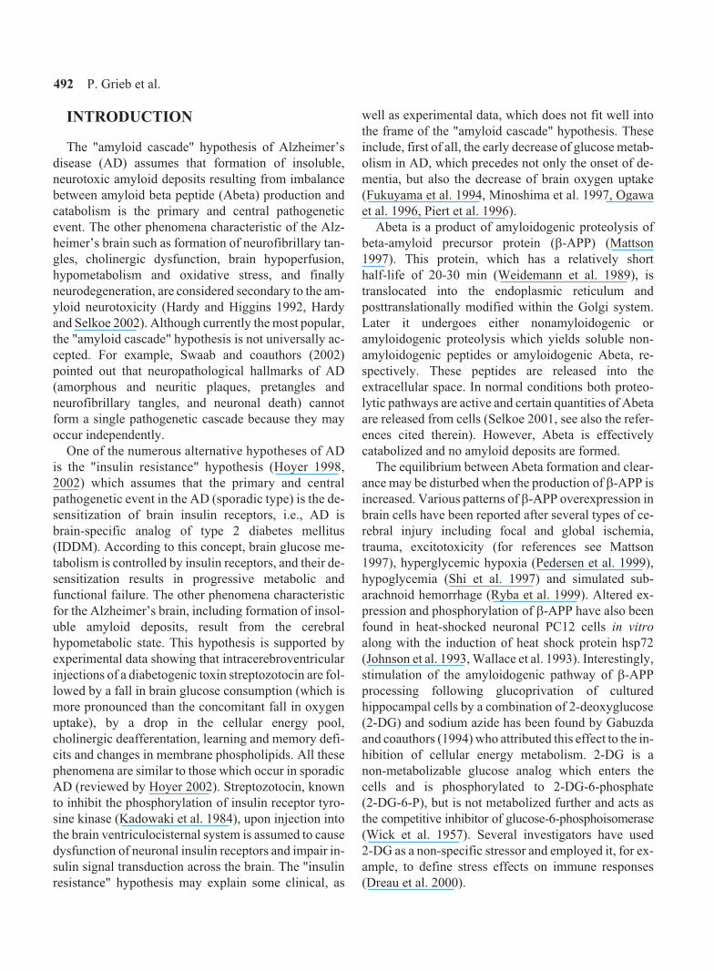
Acta Neurobiol Exp 2004, 64: 491-502 The correspondence should be addressed to P. Grieb, Email: [email protected] 2-Deoxyglucose induces b-APP overexpression, tau hyperphosphorylation and expansion of the trans-part of the Golgi complex in rat cerebral cortex Pawel Grieb 1 , Wanda Gordon-Krajcer 2 , Ma³gorzata Frontczak-Baniewicz 3 , Micha³ Walski 3 , Miroslaw S. Ryba 1 , Tomasz Kryczka 1 , Michal Fiedorowicz 1 , Piotr Kulinowski 4 , Zenon Su³ek 4 , Katarzyna Majcher 4 and Andrzej Jasiñski 4 1 Laboratory of Experimental Pharmacology, 2 Department of Neurochemistry, 3 Laboratory of Cell Ultrastructure, M. Mossakowski Medical Research Centre, Polish Academy of Sciences, Warsaw, Poland; 4 Department of Magnetic Resonance, H. Niewodniczañski Institute of Nuclear Physics, Polish Academy of Sciences, Krakow, Poland Abstract. The effects of a single intraperitoneal injection of a non-metabolizable glucose analog 2-deoxyglucose (2-DG, 500 mg/kg) on the levels of b-APP expression, and phosphorylated and unphosphorylated tau protein in the rat cerebral cortex were investigated. The effects of 2-DG on the ultrastructure of cortical neurons with particular emphasis on the morphology of the Golgi apparatus, and on brain bioenergetics assessed by in vivo 31 P-MRS technique were also evaluated. Seven and a half hours after injection of 2-deoxyglucose a significant increase in brain cortex b-APP expression, increased tau phosphorylation, and a marked relative expansion of the trans- part of the Golgi intracellular secretory pathway in cortical neurons has been found. The changes of b-APP expression and tau phosphorylation appeared within 1 h after 2-DG application and continued for at least 24 h. However, brain 31 P resonance spectra remained unchanged for up to 7.5 h after 2-DG. It is suggested that the increase of b-APP expression represents a response of brain tissues to 2-DG-evoked biochemical stress, while tau hyperphosphorylation and the change in Golgi morphology may be secondary phenomena. Key words: 2-deoxyglucose, beta-amyloid precursor protein, tau protein, Golgi apparatus, phosphorous magnetic resonance spectroscopy, brain cortex NEU OBIOLOGI E EXPE IMENT LIS
-
Upload
independent -
Category
Documents
-
view
3 -
download
0
Transcript of 2-deoxyglucose induces beta-APP overexpression, tau hyperphosphorylation and expansion of the...

Acta Neurobiol Exp 2004, 64: 491-502
The correspondence should be
addressed to P. Grieb,
Email: [email protected]
2-Deoxyglucose induces �-APP
overexpression, tau
hyperphosphorylation and expansion
of the trans-part of the Golgi
complex in rat cerebral cortex
Pawel Grieb1, Wanda Gordon-Krajcer
2, Ma³gorzata
Frontczak-Baniewicz3, Micha³ Walski
3, Miroslaw S. Ryba
1,
Tomasz Kryczka1, Michal Fiedorowicz
1, Piotr Kulinowski
4,
Zenon Su³ek4, Katarzyna Majcher
4and Andrzej Jasiñski
4
1Laboratory of Experimental Pharmacology,
2Department of Neurochemistry,
3Laboratory of Cell Ultrastructure, M. Mossakowski Medical Research
Centre, Polish Academy of Sciences, Warsaw, Poland;4Department
of Magnetic Resonance, H. Niewodniczañski Institute of Nuclear Physics,
Polish Academy of Sciences, Krakow, Poland
Abstract. The effects of a single intraperitoneal injection of a non-metabolizable
glucose analog 2-deoxyglucose (2-DG, 500 mg/kg) on the levels of �-APP
expression, and phosphorylated and unphosphorylated tau protein in the rat
cerebral cortex were investigated. The effects of 2-DG on the ultrastructure
of cortical neurons with particular emphasis on the morphology of the Golgi
apparatus, and on brain bioenergetics assessed by in vivo31
P-MRS technique
were also evaluated. Seven and a half hours after injection of 2-deoxyglucose
a significant increase in brain cortex �-APP expression, increased tau
phosphorylation, and a marked relative expansion of the trans- part of the
Golgi intracellular secretory pathway in cortical neurons has been found.
The changes of �-APP expression and tau phosphorylation appeared within
1 h after 2-DG application and continued for at least 24 h. However, brain31
P
resonance spectra remained unchanged for up to 7.5 h after 2-DG. It is
suggested that the increase of �-APP expression represents a response
of brain tissues to 2-DG-evoked biochemical stress, while tau hyperphosphorylation
and the change in Golgi morphology may be secondary phenomena.
Key words: 2-deoxyglucose, beta-amyloid precursor protein, tau protein,
Golgi apparatus, phosphorous magnetic resonance spectroscopy, brain cortex
NEU OBIOLOGI E
EXPE IMENT LIS
INTRODUCTION
The "amyloid cascade" hypothesis of Alzheimer’s
disease (AD) assumes that formation of insoluble,
neurotoxic amyloid deposits resulting from imbalance
between amyloid beta peptide (Abeta) production and
catabolism is the primary and central pathogenetic
event. The other phenomena characteristic of the Alz-
heimer’s brain such as formation of neurofibrillary tan-
gles, cholinergic dysfunction, brain hypoperfusion,
hypometabolism and oxidative stress, and finally
neurodegeneration, are considered secondary to the am-
yloid neurotoxicity (Hardy and Higgins 1992, Hardy
and Selkoe 2002). Although currently the most popular,
the "amyloid cascade" hypothesis is not universally ac-
cepted. For example, Swaab and coauthors (2002)
pointed out that neuropathological hallmarks of AD
(amorphous and neuritic plaques, pretangles and
neurofibrillary tangles, and neuronal death) cannot
form a single pathogenetic cascade because they may
occur independently.
One of the numerous alternative hypotheses of AD
is the "insulin resistance" hypothesis (Hoyer 1998,
2002) which assumes that the primary and central
pathogenetic event in the AD (sporadic type) is the de-
sensitization of brain insulin receptors, i.e., AD is
brain-specific analog of type 2 diabetes mellitus
(IDDM). According to this concept, brain glucose me-
tabolism is controlled by insulin receptors, and their de-
sensitization results in progressive metabolic and
functional failure. The other phenomena characteristic
for the Alzheimer’s brain, including formation of insol-
uble amyloid deposits, result from the cerebral
hypometabolic state. This hypothesis is supported by
experimental data showing that intracerebroventricular
injections of a diabetogenic toxin streptozotocin are fol-
lowed by a fall in brain glucose consumption (which is
more pronounced than the concomitant fall in oxygen
uptake), by a drop in the cellular energy pool,
cholinergic deafferentation, learning and memory defi-
cits and changes in membrane phospholipids. All these
phenomena are similar to those which occur in sporadic
AD (reviewed by Hoyer 2002). Streptozotocin, known
to inhibit the phosphorylation of insulin receptor tyro-
sine kinase (Kadowaki et al. 1984), upon injection into
the brain ventriculocisternal system is assumed to cause
dysfunction of neuronal insulin receptors and impair in-
sulin signal transduction across the brain. The "insulin
resistance" hypothesis may explain some clinical, as
well as experimental data, which does not fit well into
the frame of the "amyloid cascade" hypothesis. These
include, first of all, the early decrease of glucose metab-
olism in AD, which precedes not only the onset of de-
mentia, but also the decrease of brain oxygen uptake
(Fukuyama et al. 1994, Minoshima et al. 1997, Ogawa
et al. 1996, Piert et al. 1996).
Abeta is a product of amyloidogenic proteolysis of
beta-amyloid precursor protein (�-APP) (Mattson
1997). This protein� which has a relatively short
half-life of 20-30 min (Weidemann et al. 1989), is
translocated into the endoplasmic reticulum and
posttranslationally modified within the Golgi system.
Later it undergoes either nonamyloidogenic or
amyloidogenic proteolysis which yields soluble non-
amyloidogenic peptides or amyloidogenic Abeta, re-
spectively. These peptides are released into the
extracellular space. In normal conditions both proteo-
lytic pathways are active and certain quantities of Abeta
are released from cells (Selkoe 2001, see also the refer-
ences cited therein). However, Abeta is effectively
catabolized and no amyloid deposits are formed.
The equilibrium between Abeta formation and clear-
ance may be disturbed when the production of �-APP is
increased. Various patterns of �-APP overexpression in
brain cells have been reported after several types of ce-
rebral injury including focal and global ischemia,
trauma, excitotoxicity (for references see Mattson
1997), hyperglycemic hypoxia (Pedersen et al. 1999),
hypoglycemia (Shi et al. 1997) and simulated sub-
arachnoid hemorrhage (Ryba et al. 1999). Altered ex-
pression and phosphorylation of �-APP have also been
found in heat-shocked neuronal PC12 cells in vitro
along with the induction of heat shock protein hsp72
(Johnson et al. 1993, Wallace et al. 1993). Interestingly,
stimulation of the amyloidogenic pathway of �-APP
processing following glucoprivation of cultured
hippocampal cells by a combination of 2-deoxyglucose
(2-DG) and sodium azide has been found by Gabuzda
and coauthors (1994) who attributed this effect to the in-
hibition of cellular energy metabolism. 2-DG is a
non-metabolizable glucose analog which enters the
cells and is phosphorylated to 2-DG-6-phosphate
(2-DG-6-P), but is not metabolized further and acts as
the competitive inhibitor of glucose-6-phosphoisomerase
(Wick et al. 1957). Several investigators have used
2-DG as a non-specific stressor and employed it, for ex-
ample, to define stress effects on immune responses
(Dreau et al. 2000).
492 P. Grieb et al.
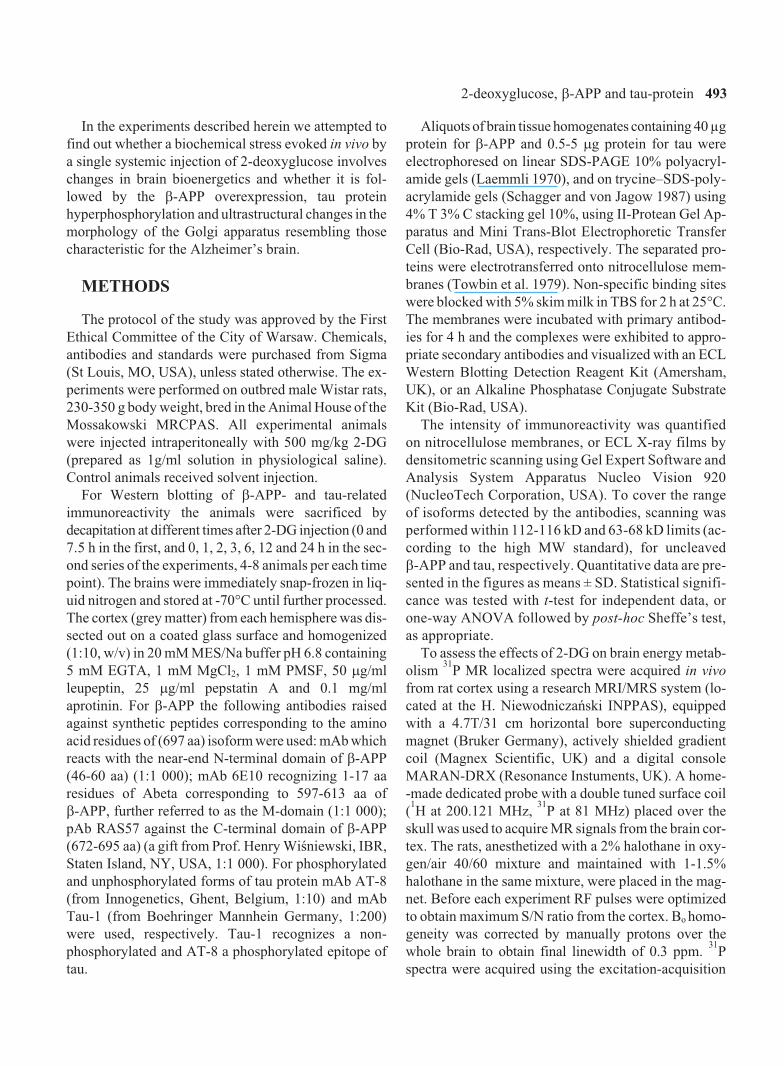
In the experiments described herein we attempted to
find out whether a biochemical stress evoked in vivo by
a single systemic injection of 2-deoxyglucose involves
changes in brain bioenergetics and whether it is fol-
lowed by the �-APP overexpression, tau protein
hyperphosphorylation and ultrastructural changes in the
morphology of the Golgi apparatus resembling those
characteristic for the Alzheimer’s brain.
METHODS
The protocol of the study was approved by the First
Ethical Committee of the City of Warsaw. Chemicals,
antibodies and standards were purchased from Sigma
(St Louis, MO, USA), unless stated otherwise. The ex-
periments were performed on outbred male Wistar rats,
230-350 g body weight, bred in the Animal House of the
Mossakowski MRCPAS. All experimental animals
were injected intraperitoneally with 500 mg/kg 2-DG
(prepared as 1g/ml solution in physiological saline).
Control animals received solvent injection.
For Western blotting of �-APP- and tau-related
immunoreactivity the animals were sacrificed by
decapitation at different times after 2-DG injection (0 and
7.5 h in the first, and 0, 1, 2, 3, 6, 12 and 24 h in the sec-
ond series of the experiments, 4-8 animals per each time
point). The brains were immediately snap-frozen in liq-
uid nitrogen and stored at -70�C until further processed.
The cortex (grey matter) from each hemisphere was dis-
sected out on a coated glass surface and homogenized
(1:10, w/v) in 20 mM MES/Na buffer pH 6.8 containing
5 mM EGTA, 1 mM MgCl2, 1 mM PMSF, 50 �g/ml
leupeptin, 25 �g/ml pepstatin A and 0.1 mg/ml
aprotinin. For �-APP the following antibodies raised
against synthetic peptides corresponding to the amino
acid residues of (697 aa) isoform were used: mAb which
reacts with the near-end N-terminal domain of �-APP
(46-60 aa) (1:1 000); mAb 6E10 recognizing 1-17 aa
residues of Abeta corresponding to 597-613 aa of
�-APP, further referred to as the M-domain (1:1 000);
pAb RAS57 against the C-terminal domain of �-APP
(672-695 aa) (a gift from Prof. Henry Wiœniewski, IBR,
Staten Island, NY, USA, 1:1 000). For phosphorylated
and unphosphorylated forms of tau protein mAb AT-8
(from Innogenetics, Ghent, Belgium, 1:10) and mAb
Tau-1 (from Boehringer Mannhein Germany, 1:200)
were used, respectively. Tau-1 recognizes a non-
phosphorylated and AT-8 a phosphorylated epitope of
tau.
Aliquots of brain tissue homogenates containing 40 µg
protein for �-APP and 0.5-5 �g protein for tau were
electrophoresed on linear SDS-PAGE 10% polyacryl-
amide gels (Laemmli 1970), and on trycine–SDS-poly-
acrylamide gels (Schagger and von Jagow 1987) using
4% T 3% C stacking gel 10%, using II-Protean Gel Ap-
paratus and Mini Trans-Blot Electrophoretic Transfer
Cell (Bio-Rad, USA), respectively. The separated pro-
teins were electrotransferred onto nitrocellulose mem-
branes (Towbin et al. 1979). Non-specific binding sites
were blocked with 5% skim milk in TBS for 2 h at 25�C.
The membranes were incubated with primary antibod-
ies for 4 h and the complexes were exhibited to appro-
priate secondary antibodies and visualized with an ECL
Western Blotting Detection Reagent Kit (Amersham,
UK), or an Alkaline Phosphatase Conjugate Substrate
Kit (Bio-Rad, USA).
The intensity of immunoreactivity was quantified
on nitrocellulose membranes, or ECL X-ray films by
densitometric scanning using Gel Expert Software and
Analysis System Apparatus Nucleo Vision 920
(NucleoTech Corporation, USA). To cover the range
of isoforms detected by the antibodies, scanning was
performed within 112-116 kD and 63-68 kD limits (ac-
cording to the high MW standard), for uncleaved
�-APP and tau, respectively. Quantitative data are pre-
sented in the figures as means ± SD. Statistical signifi-
cance was tested with t-test for independent data, or
one-way ANOVA followed by post-hoc Sheffe’s test,
as appropriate.
To assess the effects of 2-DG on brain energy metab-
olism31
P MR localized spectra were acquired in vivo
from rat cortex using a research MRI/MRS system (lo-
cated at the H. Niewodniczañski INPPAS), equipped
with a 4.7T/31 cm horizontal bore superconducting
magnet (Bruker Germany), actively shielded gradient
coil (Magnex Scientific, UK) and a digital console
MARAN-DRX (Resonance Instuments, UK). A home-
-made dedicated probe with a double tuned surface coil
(1H at 200.121 MHz,
31P at 81 MHz) placed over the
skull was used to acquire MR signals from the brain cor-
tex. The rats, anesthetized with a 2% halothane in oxy-
gen/air 40/60 mixture and maintained with 1-1.5%
halothane in the same mixture, were placed in the mag-
net. Before each experiment RF pulses were optimized
to obtain maximum S/N ratio from the cortex. Bo homo-
geneity was corrected by manually protons over the
whole brain to obtain final linewidth of 0.3 ppm.31
P
spectra were acquired using the excitation-acquisition
2-deoxyglucose, �-APP and tau-protein 493
method just prior to, and then every 6 minutes after
2-DG injection, up to 7.5 h (or prior to death). A glass
pellet of 3 mm inner diameter containing 0.5 mM PPA
water solution, placed on the skull surface in the centre
of the surface coil, served as an external standard. The
MRUI graphical user interface package and the
VARPRO non-linear fitting algorithm were used to pro-
cess the acquired FID signals.
Brain tissue sampling for electron microscopy was
performed under ketamine (70 mg/kg) and xylazine
(20 mg/ml) anesthesia 7.5 h after injection of 2-DG or
vehicle. The animals (4 2-DG treated and 3 vehi-
cle-treated) were perfused via the left heart ventricle
with a mixture of 2% paraformaldehyde and 2.5%
glutaraldehyde in 0.1 M cacodylate buffer, pH 7.4 at
20�C. Tissues sampled from the fronto-parietal lobe of
cerebral cortex were fixed in the same solution for 20 h,
postfixed in a mixture of 1% OsO4 and 0.8% K4[Fe(CN)6],
and processed for transmission electron microscopy.
After dehydration in series of ethanol and propylene ox-
ide, tissue specimens were embedded in Spurr resin. Se-
rial ultrathin (50 nm) sections were examined with a
JEM 1200EX electron microscope. In representative
populations of neuronal cells, the average area of the
whole Golgi apparatus as well as its -cis (proximal) and
-trans (distal) parts was also determined, as described in
detail in the accompanying paper (Grieb et al. 2004).
Significance of differences between morphometric data
for neurons from control and 2-DG-treated animals
were evaluated with the Mann-Whitney U-test.
RESULTS
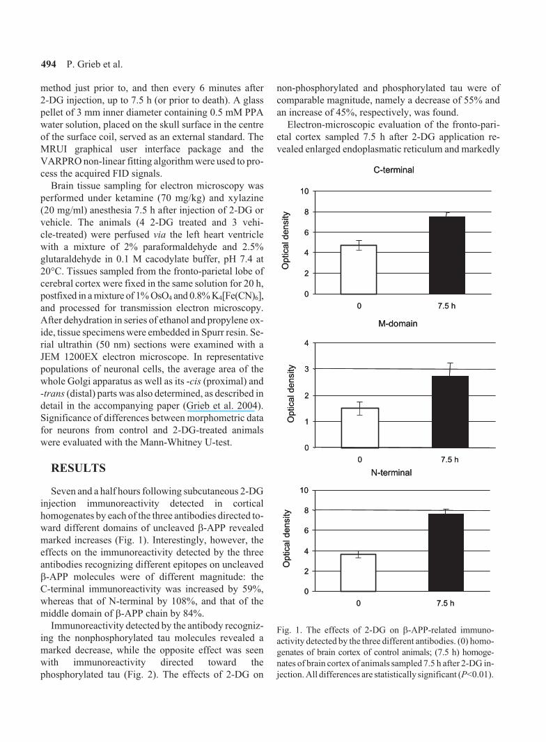
Seven and a half hours following subcutaneous 2-DG
injection immunoreactivity detected in cortical
homogenates by each of the three antibodies directed to-
ward different domains of uncleaved �-APP revealed
marked increases (Fig. 1). Interestingly, however, the
effects on the immunoreactivity detected by the three
antibodies recognizing different epitopes on uncleaved
�-APP molecules were of different magnitude: the
C-terminal immunoreactivity was increased by 59%,
whereas that of N-terminal by 108%, and that of the
middle domain of �-APP chain by 84%.
Immunoreactivity detected by the antibody recogniz-
ing the nonphosphorylated tau molecules revealed a
marked decrease, while the opposite effect was seen
with immunoreactivity directed toward the
phosphorylated tau (Fig. 2). The effects of 2-DG on
non-phosphorylated and phosphorylated tau were of
comparable magnitude, namely a decrease of 55% and
an increase of 45%, respectively, was found.
Electron-microscopic evaluation of the fronto-pari-
etal cortex sampled 7.5 h after 2-DG application re-
vealed enlarged endoplasmatic reticulum and markedly
C-terminal
0
2
4
6
8
10
0 7.5 h
Op
tica
ld
en
sity
C-terminal
0
2
4
6
8
10
0 7.5 h
Op
tica
ld
en
sity
M-domain
0
1
2
3
4
0 7.5 h
Op
tica
ld
en
sity
M-domain
0
1
2
3
4
0 7.5 h
Op
tica
ld
en
sity
N-terminal
0
2
4
6
8
10
0 7.5 h
Op
tica
ld
en
sity
N-terminal
0
2
4
6
8
10
0 7.5 h
Op
tica
ld
en
sity
Fig. 1. The effects of 2-DG on �-APP-related immuno-
activity detected by the three different antibodies. (0) homo-
genates of brain cortex of control animals; (7.5 h) homoge-
nates of brain cortex of animals sampled 7.5 h after 2-DG in-
jection. All differences are statistically significant (P<0.01).
494 P. Grieb et al.

expanded trans- part of the Golgi system in neuronal
cells, while mitochondria were not affected (Fig. 3). For
comparison, a micrograph of a cortical neuron from a
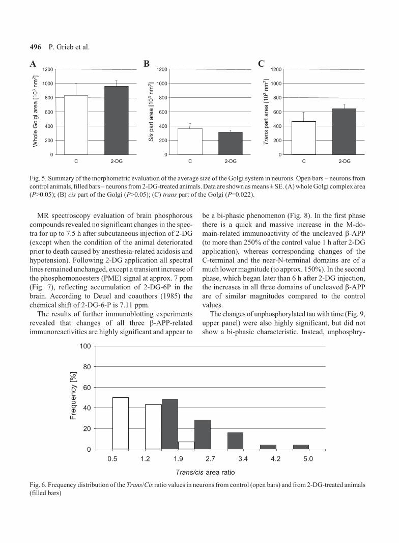
vehicle-injected animal is also shown (Fig. 4). Quantita-
tive morphometric analysis of the Golgi apparatus in
neurons of control and 2-DG-treated animals showed
that neither the average size of the whole Golgi com-
plex, nor the average size of its cis- (i.e., proximal) part
were significantly affected by the treatment (Fig. 5).
However, in neurons from 2-DG-treated animals the
size of the trans- (i.e., the distal) part of the Golgi was
significantly increased (Fig. 5), and this resulted in
markedly higher values of the trans/cis area ratio of the
Golgi in most cells (Fig. 6).
tau non-phosphorylated
0
2
4
6
8
10
0 7.5 h
Opticaldensity
tau phosphorylated
0
2
4
6
8
10
0 7.5 h
Opticaldensity
Fig. 2. The effects of 2-DG on nonphosphorylated (upper
panel) and phosphorylated (lower panel) tau immuno-
reactivity. (0) homogenates of brain cortex of control ani-
mals, (7.5 h) homogenates of brain cortex of animals sampled
7.5 h after 2-DG injection. The differences are statistically
significant (P<0.01).
Fig. 3. Electron micrograph of a cortical neuron from a
2-DG-treated rat. Mitochondria are unaffected, but expanded
trans-Golgi complex is clearly seen. Magnification, � 25 000.
Fig. 4. Electron micrograph of a cortical neuron from a vehi-
cle-treated rat. Magnification, � 20 000.
2-deoxyglucose, �-APP and tau-protein 495
MR spectroscopy evaluation of brain phosphorous
compounds revealed no significant changes in the spec-
tra for up to 7.5 h after subcutaneous injection of 2-DG
(except when the condition of the animal deteriorated
prior to death caused by anesthesia-related acidosis and
hypotension). Following 2-DG application all spectral
lines remained unchanged, except a transient increase of
the phosphomonoesters (PME) signal at approx. 7 ppm
(Fig. 7), reflecting accumulation of 2-DG-6P in the
brain. According to Deuel and coauthors (1985) the
chemical shift of 2-DG-6-P is 7.11 ppm.
The results of further immunoblotting experiments
revealed that changes of all three �-APP-related
immunoreactivities are highly significant and appear to
be a bi-phasic phenomenon (Fig. 8). In the first phase
there is a quick and massive increase in the M-do-
main-related immunoactivity of the uncleaved �-APP
(to more than 250% of the control value 1 h after 2-DG
application), whereas corresponding changes of the
C-terminal and the near-N-terminal domains are of a
much lower magnitude (to approx. 150%). In the second
phase, which began later than 6 h after 2-DG injection,
the increases in all three domains of uncleaved �-APP
are of similar magnitudes compared to the control
values.
The changes of unphosphorylated tau with time (Fig. 9,
upper panel) were also highly significant, but did not
show a bi-phasic characteristic. Instead, unphosphry-
0
200
400
600
800
1000
1200
C 2-DG C 2-DG C 2-DG
Whole
Golg
iare
a[1
03
nm
2]
Tra
ns
part
are
a[1
03
nm
2]
Sis
part
are
a[1
03
nm
2]
0
200
400
600
800
1000
1200
0
200
400
600
800
1000
1200
Fig. 5. Summary of the morphometric evaluation of the average size of the Golgi system in neurons. Open bars – neurons from
control animals, filled bars – neurons from 2-DG-treated animals. Data are shown as means ± SE. (A) whole Golgi complex area
(P>0.05); (B) cis part of the Golgi (P>0.05); (C) trans part of the Golgi (P=0.022).
Trans/cis area ratio
0
20
40
60
80
100
Fre
qu
en
cy
[%]
5.01.20.5 3.4 4.22.71.9
Fig. 6. Frequency distribution of the Trans/Cis ratio values in neurons from control (open bars) and from 2-DG-treated animals
(filled bars)
496 P. Grieb et al.
BA C

Pi
(4.3...5.5 ppm)
Pi
(4.3...5.5 ppm)
PCr(0 ppm)
PCr(0 ppm)
PME
PDE�NTP
(-7.6 ppm)
�NTP(-7.6 ppm)
�NTP(-16.1 ppm)
�NTP(-16.1 ppm)
�NTP(-2.5 ppm)
�NTP(-2.5 ppm)PPA
(21.8 ppm)
PPA(21.8 ppm)
Fig. 7. Brain31
P spectra acquired just prior to (upper panel) and approximately 2 h after the injection of 2-DG (lower panel).
(PME) phosphomonoesters; (Pi) inorganic phosphates; (PDE) phosphodiesters; (PCr) phosphocreatine; (NTP) nucleoside
triphosphates.
2-deoxyglucose, �-APP and tau-protein 497
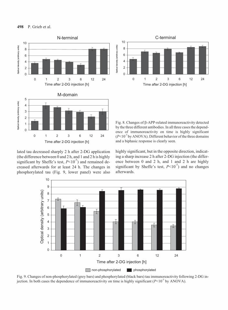
lated tau decreased sharply 2 h after 2-DG application
(the difference between 0 and 2 h, and 1 and 2 h is highly
significant by Sheffe’s test, P<10-5
) and remained de-
creased afterwards for at least 24 h. The changes in
phosphorylated tau (Fig. 9, lower panel) were also
highly significant, but in the opposite direction, indicat-
ing a sharp increase 2 h after 2-DG injection (the differ-
ence between 0 and 2 h, and 1 and 2 h are highly
significant by Sheffe’s test, P<10-5
) and no changes
afterwards.
N-terminal
0
2
4
6
8
10
0 1 2 3 6 12 24
Time after 2-DG injection [h]
Op
tic
ald
en
sit
y(a
rbit
rary
un
its
)
M-domain
0
1
2
3
4
5
0 1 2 3 6 12 24
Time after 2-DG injection [h]
Op
tic
ald
en
sit
y(a
rbit
rary
un
its
)
Fig. 8. Changes of �-APP-related immunoreactivity detected
by the three different antibodies. In all three cases the depend-
ence of immunoreactivity on time is highly significant
(P<10-5
by ANOVA). Different behavior of the three domains
and a biphasic response is clearly seen.
0
1
2
3
4
5
6
7
8
9
10
0 1 2 3 6 12 24
Time after 2-DG injection [h]
Opticaldensity
(arb
itra
ryunits)
non-phosphorylated phosphorylated
C-terminal
0
2
4
6
8
10
0 1 2 3 6 12 24
Time after 2-DG injection [h]
Op
tic
ald
en
sit
y(a
rbit
rary
un
its
)
Fig. 9. Changes of non-phosphorylated (grey bars) and phosphorylated (black bars) tau immunoreactivity following 2-DG in-
jection. In both cases the dependence of immunoreactivity on time is highly significant (P<10-5
by ANOVA).
498 P. Grieb et al.
DISCUSSION
The results of the present study indicate that systemic
application of 2-DG produces a quick and significant in-
crease in the uncleaved �-APP-related immuno-
reactivities which may be interpreted as the evidence for
increased �-APP expression at the protein level. How-
ever, the changes of the immunoreactivity detected by
each of the three antibodies recognizing different
epitopes on the uncleaved �-APP molecule were of dif-
ferent magnitudes. Similar disparity between the in-
creases of different domains of �-APP has been observed
previously in response to simulated subarachnoid haem-
orrhage (Ryba et al. 1999). This phenomenon may reflect
differences in posttranslational modifications of the
amino acid residues in the domains detected by particular
antibodies. �-APP undergoes numerous modifications
affecting side amino acid residues, including phospho-
rylations at various sites (Alpin et al. 1996, 1997, Gandy
et al. 1988). All the antibodies used in the present study
have been raised toward synthetic peptides. If post-
translationally modified amino acid residues are located
within, or close to the epitopes detected by the antibodies,
modified (e.g., phosphorylated) protein molecules may
not be detected, or sensitivity for their detection may be
decreased. It is worth to noting that the increase of M-do-
main-related immunoreactivity was always higher com-
pared to the other two, which is in agreement with the fact
that the 597-613 aa epitope of uncleaved �-APP, referred
to as the M-domain, is not posttranslationally modified.
As mentioned in the introduction, increased �-APP ex-
pression in brain tissue has been found following a vari-
ety of insults. Physiological function of this protein
remains poorly understood despite tremendous interest
stimulated by the "amyloid cascade" hypothesis of AD
etiology. There is, however, some evidence that �-APP
and some of its nascent peptides may positively influence
neurons. �-APP was able to enhance viability of the neu-
rons in vitro (Perez et al. 1997), and soluble products of
�-APP cleavage displayed neuroprotective activity in the
in vivo brain ischemia model (Smith-Swintosky et al.
1994). Interestingly, following postischemic reperfusion
the overexpression of �-APP and heat-shock proteins
have been reported in the same brain regions, although
mostly in distinct neurons (Tomimoto et al. 1994). In that
study �-APP accumulated in neurons on the margin of
the regions destined to die, but the majority of these neu-
rons seemed to survive after ischemic insult (Tomimoto
et al. 1994). On the other hand, 2-DG induced an increase
in the level of the stress protein heat-shock protein 70
(HSP 70) in striatal cells in vivo, and 2-DG treatment in-
duced HSP70 in cultured neurons (Yu and Mattson
1999). Repeated injections of 2-DG (dose 100 mg/kg/day)
induced increased levels of the stress-responsive proteins
GRP78 and HSP70 in brain neurons (Lee et al. 1999), and
(dose 150 mg/kg/day) enhanced ischemia-evoked brain
tolerance to seizures (Rejdak et al. 2001). The results of
the present study add 2-DG-induced biochemical stress
to the list of insults which result in �-APP overexpression
in brain tissues. Increased �-APP may reflect yet another
aspect of an unspecific response of brain tissues to stress-
ful conditions. One may speculate that overexpression
of this protein may help neurons survive and be in-
volved in the development of brain tolerance to noxious
stimuli.
The effects of 2-DG on brain tissue �-APP expres-
sion observed in the present experiments cannot be at-
tributed to the failure of cellular bioenergetics. While
such an explanation certainly holds true in the case of
the in vitro study of Gabuzda and coauthors (1984), it
doesn’t seem to be of value as an explanation of the re-
sults obtained in vivo. Although the effects of repeated
administration of lower doses of 2-DG described in
some studies were similar to those of dietary (caloric)
restriction (Yu and Mattson 1999) or intermittent fast-
ing (Wan et al. 2003), this can be attributed to the sys-
temic stress response evoked by 2-DG rather than to
insufficient glucose supply and metabolism in the brain.
Our results show that a single dose of 500 mg/kg 2-DG
given intravenously does not significantly affect in vivo
brain tissue31
P NMR spectra. This is in agreement with
the data of Horton and coauthors (1973) who have
found no fall in brain ATP following a single dose of
3 g/kg 2-DG, although these authors have noted a fall in
brain phosphocreatine. Apparently, although 2-DG en-
ters brain and is phosphorylated, and 2-DG-6-P (which
is not metabolized by glucose-6-phosphoisomerase)
may transiently restrict glucose utilization, this effect is
short lasting and, as far as the energy balance of brain
tissue is concerned, appears to be fully compensated ei-
ther by some other available sources of energy, or by de-
creased energy expenditure of brain cells.
We have also found a significant and sustained in-
crease of the phosphorylated form of tau protein follow-
ing 2-DG injection, which was counterbalanced by a
decrease of unphosphorylated tau. The link between
�-APP overexpression and tau phosphorylation may be
provided by glycogen synthase kinase 3beta
2-deoxyglucose, �-APP and tau-protein 499
(GSK3beta), the enzyme implicated in a variety of
intracellular receptor-mediated processes including
transduction of insulin signaling (Grimes and Jope
2001). GSK3beta activity has been implicated in virtu-
ally all aspects of Alzheimer’s pathology, including
pro-amyloidogenic phosphorylation of the carboxy-do-
main of �-APP (Alpin et al. 1996), tau hyper-
phosphorylation (Lee et al. 2003, Lovestone et al. 1994)
and disassembly of neuronal Golgi apparatus (Elyaman
et al. 2002). Its unique feature is that it is constitutively
active in resting conditions. One may consider the pos-
sibility that 2-DG produces hypoinsulinemia which ac-
tivates GSK3beta and hyperphosphorylates tau. Indeed,
prolonged treatment of rats with lower doses of 2-DG
has been recently found to cause a marked decrease in
plasma insulin (Wan et al. 2003). However, Pascoe and
coauthors (1989), using an experimental design identi-
cal to that of the present study, found that i.v. infusion of
500 mg/kg 2-DG to male Wistar rats resulted in the ap-
proximately 3-fold increase in plasma insulin level.
A recent finding that Abeta is a direct competitive in-
hibitor of insulin binding to its receptor binding and in-
sulin receptor autophosphorylation (Xie et al. 2002)
may link overexpression of �-APP directly to the activa-
tion of GSK3beta. One may speculate that over-
expression of �-APP, which is a primary brain cellular
response to 2-DG-evoked stress, shifts the balance be-
tween Abeta production and removal toward the in-
crease of Abeta concentration outside brain cells. The
next step would be the competition of Abeta with insulin
at the insulin receptors, impaired transduction of the in-
sulin signaling and the activation of GSK3beta, to create
a vicious cycle.
The changes in Golgi morphology which we have
found following 2-DG injection may also be driven by
the same mechanism. It is worth noting that a similar
morphological picture (i.e., the apparent relative expan-
sion of the trans- part of the Golgi complex) has been pre-
viously found in our lab in brain neurons of rats subjected
to acute insulin-induced hypoglycemia (Wierzba-
-Bobrowicz 1980, 1984 – unpublished doctoral thesis).
These observations are in agreement with findings of
the others. Agardh and coauthors (1981) have noted that
the pathogenesis of cell damage in hypoglycemia is dif-
ferent from that in hypoxia-ischemia and indicated that
other mechanisms than energy failure must contribute
to neuronal cell damage in the brain. Also, according to
Simon and coauthors (1986), the neuropathology of rat
hippocampal neurons at the EM level following
hypoglycemia was markedly different from that seen
following status epilepticus and ischemia, the main dif-
ference being that microvacuoles were rarely seen and,
when present, their ultrastructural correlate was swollen
Golgi apparatus, not dilated mitochondria.
Neither the changes in Golgi morphology produced
by intravenous injection of 2-DG, nor similar changes
reported previously following hypoglycemia, seem to
have a direct relation to brain tissues energy failure. It is
also unlikely that they are related to hyperinsulinemia
which would rather be expected to dephosphorylate tau
protein in the brain. It is well known that starvation de-
creases insulin in blood, and Yanagisawa and coauthors
(1999) have shown that starving induces tau hyper-
phosphorylation in the mouse brain. On the other hand,
similar but even more pronounced enlargement of the
trans compartment of the Golgi has been found follow-
ing intracerebroventricular injections of streptozotocin,
a toxin which desensitizes brain insulin receptors (Grieb
et al. 2004). It is tempting to speculate that all these
functional and ultrastructural effects may be related ei-
ther to stimulation of �-APP synthesis and Abeta forma-
tion, modulation of GSK3beta activity, or an interplay
between these two mechanisms at the level of brain in-
sulin receptors.
CONCLUSION
A single dose of 2-deoxyglucose (500 mg/kg i.p.) in-
duces in brain cortex of rats a bi-phasic �-APP
overexpression, a hyperphosphorylation of tau protein,
and a marked expansion of the trans part of the neuronal
Golgi apparatus, but does not appreciably influence
brain energy compounds.
ACKNOWLEDGEMENT
The study was supported by the State Committee for
Scientific Research (KBN) project No. 4P05A 041 17
granted to the late professor Miroslaw J. Mossakowski,
to whom the authors are indebted for his creativity and
expertise in the early stage of the work.
REFERENCES
Agardh CD, Kalimo H, Olsson Y, Siesjo BK (1981)
Hypoglycemic brain injury: metabolic and structural find-
ings in rat cerebellar cortex during profound insulin-in-
duced hypoglycemia and in the recovery period following
500 P. Grieb et al.

glucose administration. J Cereb Blood Flow Metab 1:
71-84.
Alpin AE, Gibb GM, Jacobsen JS, Gallo J-M, Anderton BH
(1996) In vitro phosphorylation of cytoplasmic domain of
the amyloid precursor protein by glycogen synthase
kinase-3�. J Neurochem 67: 699-707.
Aplin AE, Jacobsen JS, Anderton BH, Gallo JM (1997) Ef-
fect of increased glycogen synthase kinase-3 activity upon
the maturation of the amyloid precursor protein in
transfected cells. Neuroreport 8: 639-643.
Deuel RK, Yue GM, Sherman WR, Schickner DJ, Ackerman
JJH (1985) Monitoring the time course of cerebral
deoxyglucose metabolism by31
P nuclear magnetic spec-
troscopy. Science 228: 1329-1331.
Dreau D, Foster M, Morton DS, Fowler N, Kinney K,
Sonnenfeld G (2000) Immune alterations in three mouse
strains following 2-deoxy-D-glucose administration.
Physiol Behav 70: 513-520.
Elyaman W, Yardin C, Hugon J (2002) Involvement of glyco-
gen synthase kinase-3� and tau phosphorylation in
neuronal Golgi disassembly. J Neurochem 81: 870-880.
Fukuyama H, Ogawa M, Yamauchi H, Yamaguchi S, Kimura
J, Yonekura Y, Konishi J (1994) Altered cerebral energy
metabolism in Alzheimer's disease: a PET study. J Nucl
Med 35: 1-6.
Gabuzda D, Busciglio J, Chen L, Matsudaira P, Yankner B
(1994) Inhibition of energy metabolism alters the process-
ing of amyloid precursor protein and induces a potentially
amyloidogenic derivative. J Biol Chem. 269: 13623-13628.
Gandy S, Czernik A, Greengard P (1988) Phosphorylation of
Alzheimer disease amyloid precursor peptide by protein
kinase C and Ca2+
/calmodulin-dependent protein kinase II.
Proc Natl Acad Sci U S A 85: 6218-6221.
Grieb P, Kryczka T, Fiedorowicz M, Frontczak-Baniewicz M,
Walski M (2004) Expansion of the Golgi apparatus in rat ce-
rebral cortex following intracerebroventricular injections of
streptozotocin. Acta Neurobiol Exp (Wars) 64: 481-489.
Grimes CA, Jope RS (2001) The multifaceted roles of glyco-
gen synthase kinase 3� in cellular signalling. Prog
Neurobiol 65: 391-426.
Hardy J, Higgins GA (1992) Alzheimer’s disease: The amy-
loid hypothesis. Science 256: 1124-1125.
Hardy J, Selkoe DJ (2002) The amyloid hypothesis of Alzhei-
mer’s disease: progress and problems on the road to thera-
peutics. Science 297: 353-356.
Horton RW, Meldrum BS, Bachelard HS (1973) Enzymic
and cerebral metabolic effects of 2-deoxy-D-glucose. J
Neurochem 21: 507-520.
Hoyer S (1998) Is sporadic Alzheimer disease the brain type
of non-insulin dependent diabetes mellitus. A challenging
hypothesis. J Neural Transm 105: 415-422.
Hoyer S (2002) The brain insulin signal transduction system
and sporadic (type II) Alzheimer disease: an update. J Neu-
ral Transm 109: 341-360.
Johnson G, Refolo LM, Merril CR, Wallace W (1993) Al-
tered expression and phosphorylation of amyloid precur-
sor protein in heat shocked neuronal PC12 cells. Brain Res
Mol Brain Res 19: 140-148.
Kadowaki T, Kasaga M, Akamuma Y, Ezaki O, Takaku F
(1984) Decreased autophosphorylation of the insulin re-
ceptor-kinase in streptozotocin diabetic rats. J Biol Chem
259: 14208-14216.
Laemmli UK (1970) Cleavage of structural proteins during
the assembly of the head of bacteriophage T4. Nature 227:
680-685.
Lee CW, Lau KF, Miller CC, Shaw PC (2003) Glycogen
synthase kinase-3 beta-mediated tau phosphorylation in
cultured cell lines. Neuroreport 14: 257-560.
Lee J, Bruce-Keller AJ, Kruman Y, Chan SL, Mattson MP
(1999) 2-Deoxy-D-glucose protects hippocampal neurons
against excitotoxic and oxidative injury: evidence for the
involvement of stress proteins. J Neurosci Res 57: 48-61.
Lovestone S, Reynolds CH, Latimer D, Davis DR, Anderton
BH, Gallo JM, Hanger D, Mulot S, Marquardt B, Stabel S,
et al (1994) Alzheimer’s disease-like phosphorylation of
the microtubule-associated protein tau by glycogen
synthase kinase-3 in transfected mammalian cells. Curr
Biol 4: 1077-1086.
Mattson MP (1997) Cellular actions of �-Amyloid Precursor
Protein and its soluble and fibrillogenic derivatives.
Physiol Rev 77: 1081-1132.
Minoshima S, Giordani B, Berent S, Frey KA, Foster NL,
Kuhl DE (1997) Metabolic reduction in the posterior
cingulate cortex in very early Alzheimer’s disease. Ann
Neurol 42: 85-94.
Ogawa M, Fukuyama H, Ouchi Y, Yamauchi H, Kimura J
(1996) Altered energy metabolism in Alzheimer’s disease.
J Neurol Sci 139: 78-82.
Pascoe WS, Smythe GA, Storlie LH (1989) 2-Deoxy-D-glu-
cose-induced hyperglycemia: role for direct sympathetic
nervous system activation of liver glucose output. Brain
Res 505: 23-28.
Pedersen WA, Culmsee C, Ziegler D, Herman JP, Mattson
MP (1999) Aberrant stress response associated with severe
hypoglycemia in a transgenic mouse model of Alzheimer’s
disease. J Mol Neurosci 13: 159-165.
Perez RG, Zheng H, Van der Ploeg LH, Koo EH (1997) The
beta-amyloid precursor protein of Alzheimer’s disease en-
hances neuron viability and modulates neuronal polarity. J
Neurosci 17: 9407-9414.
Piert M, Koeppe RA, Giordani B, Berent S, Kuhl DE (1996)
Diminished glucose transport and phosphorylation in Alz-
heimer’s disease determined by dynamic FDG-PET. J
Nucl Med 37: 201-208.
Rejdak K, Rejdak R, Sieklucka-Dziuba M, Stelmasiak Z,
Grieb P (2001) 2-Deoxyglucose enhances hypoxic toler-
ance evoked by transient incomplete brain ischemia. Epi-
lepsy Res 43: 271-278.
2-deoxyglucose, �-APP and tau-protein 501
Ryba MS, Gordon-Krajcer W, Walski M, Chalimoniuk M,
Chrapusta SJ (1999) Hydroxylamine attenuates the effects
of simulated subarachnoid hemorrhage in the rat brain and
improves neurological outcome. Brain Res 850: 225-233.
Schagger H, von Jagow G (1987) Tricine-sodium dodecyl
sulphate polyacrylamide gel electrophoresis for the sepa-
ration of protein in the range from 1 to 100 kDa. Anal
Biochem 166: 638-661.
Selkoe DJ (2001) Alzheimer’s disease: genes, proteins, and
therapy. Physiol Rev 81: 741-766.
Shi J, Xiang Y, Simpkins JW (1997) Hypoglycemia enhances
the expression of mRNA encoding beta-amyloid precursor
protein in rat primary cortical astroglial cells. Brain Res
772: 247-251.
Simon RP, Schmidley JW, Swan JH, Meldrum BS (1986)
Neuronal alterations in hippocampus following severe hy-
poglycaemia: a light microscopic and ultrastructural study
in the rat. Neuropathol Appl Neurobiol 12: 11-26.
Smith-Swintosky VL, Pettigrew LC, Craddock SD, Culwell
AR, Rydel RE, Mattson MP (1994) Secreted forms of
beta-amyloid precursor protein protect against ischemic
brain injury. J Neurochem 63: 781-784.
Swaab DF, Dubelaar EJ, Hofman MA, Scherder EJ, van
Someren EJ, Verwer RW (2002) Brain aging and Alzhei-
mer’s disease: use it or lose it. Prog Brain Res 138:343-373.
Tomimoto H, Wakita H, Akiguchi I, Nakamura S, Kimura J
(1994) Temporal profiles of accumulation of amyloid
beta/A4 protein precursor in the gerbil after graded
ischemic stress. J Cereb Blood Flow Metab 14: 565-573.
Towbin H, Staehelin T, Gordon J (1979) Electrophoretic
transfer of proteins from polyacrylamide gels to
nitrocellulose sheets: procedure and some applications.
Proc Natl Acad Sci U S A 76: 4350-4354.
Wallace W, Johnson G, Sugar J, Merril CR, Refolo LM
(1993) Reversible phosphorylation of tau to form A68 in
heat-shocked neuronal PC12 cells. Brain Res Mol Brain
Res 19: 149-155.
Wan R, Camandola S, Mattson MP (2003) Intermittent fast-
ing and dietary supplementation with 2-deoxy-D-glucose
improve functional and metabolic cardiovascular risk fac-
tors in rats. FASEB J 17:1133-1134.
Weidemann A, Konig G, Bunke D, Fischer P, Salbaum JM,
Masters CL, Beyeruther K (1989) Identification,
biogenesis and localization of precursors of Alzheimer’s
disease A4 amyloid protein. Cell 57: 115-126.
Wick AN, Drury DR, Nakada HI, Wolfe JB (1957) Localiza-
tion of the primary metabolic block produced by
2-deoxyglucose. J Biol Chem 224: 963-969.
Wierzba-Bobrowicz T (1980) Electron-microscopic picture
of rat brain in acute experimental hypoglycemia (in Pol-
ish). Neuropatol Pol 18: 289-300.
Xie L., Helmerhorst E, Taddei K, Plewright B, Van
Bronswijk W, Martins R (2002) Alzheimer’s beta-amyloid
peptides compete for insulin binding to the insulin recep-
tor. J Neurosci 22: RC221.
Yanagisawa M, Planel E, Ishiguro K, Fujita SC (1999) Star-
vation induces tau hyperphosphorylation in mouse brain:
implications for Alzheimer’s disease. FEBS Lett 461:
329-333.
Yu ZF, Mattson MP (1999) Dietary restriction and
2-deoxyglucose administration reduce focal ischemic
brain damage and improve behavioral outcome: evidence
for a preconditioning mechanism. J Neurosci Res 57:
830-839.
Received 10 August 2003, accepted 6 September 2004
502 P. Grieb et al.