رصبلا بصعلا سايق ي لا يبط يع مادختساب نيغلابلا ...
55
SUDAN UNIVERSITY OF SCIENCES & TECHNOLOGY COLLEGE OF GRADUATE STUDIES Measurement of Normal Optic Nerve for Adult Sudanese Using Computed Tomography البصرس العصب قيا ي ال طبي عيستخدامالغين بايين البي السودان لدشعة المقطعية المحوسبة اA thesis Submitted for Partial Fulfillments the Requirements of M.Sc. Radiological Imaging Diagnosis By; Shihabaldin Ibrahim Mohamed Ahmed SUPERVISOR: D. IKHLAS ABDALAZEEZ 2017
-
Upload
khangminh22 -
Category
Documents
-
view
5 -
download
0
Transcript of رصبلا بصعلا سايق ي لا يبط يع مادختساب نيغلابلا ...

SUDAN UNIVERSITY OF SCIENCES & TECHNOLOGY
COLLEGE OF GRADUATE STUDIES
Measurement of Normal Optic Nerve for Adult Sudanese Using
Computed Tomography
لدي السودانيين البالغين باستخدام عيطبيال يقياس العصب البصر
االشعة المقطعية المحوسبة
A thesis Submitted for Partial Fulfillments the Requirements of
M.Sc. Radiological Imaging Diagnosis
By;
Shihabaldin Ibrahim Mohamed Ahmed
SUPERVISOR:
D. IKHLAS ABDALAZEEZ
2017

II
يتعالقال
بسم هللا الرحمن الرحيم
ره منازل لتعلموا عدد السـنين والحس ) ا هو الذي جعل الشمس ضياء والقمر نورا وقد
ل األيات لقوم يعلمون ( ما خلق هللا ذلك إال بالحق يفص
صدق هللا العظيم
(5سورة يونس االية )

III
Dedication
To my Father
To my mother for her moral support and all the work she did to get me where
I am today.
To my wife and my daughter Retaj
To my lovely sisters and brothers for their kind support.
To my friends who stood beside me and supported me.
Thanks for all

IV
ACKNWOLEDGMENTS
I thank almighty God for giving me the strength, courage and determination in conducting this study, despite all difficulties. I would like to thank gratefully my supervisor
Dr. Ikhlas Abdelaziz
Phrases may not cover what I mean to show, but a word must be penned to those who helped me and guided me through the way and to those who intended to help me accomplish this work, it’s because of their patience and splendid character I reached this far.

V
Abstract:
This study was conducted to find out the mean optic nerve
length and width in Sudanese population using CT.
Number of 100 patients CT orbit and brain was performed for
Sudanese patient there ages between 20–80 years old, were 49
males and 51 females.
The result after data analysis was found the overall mean of optic
nerve length was [5.0052±.43099] cm, and mean of optic nerve
width [.3971±.10012] cm. with minimum length [4.00cm], and
maximum length [6.23 cm].with minimum width [.23cm], and
maximum width [.65 cm].
There is a correlation between Also there is correlation between
the optic nerve length and the patient weight as weight increase
the length increase.
The optic nerve (length &width) slightly higher in males.
There is no relation between optic nerve width &length and the
tribe.

VI
خالصة البحث
ي أجريت هذه الدراسة لقياس متوسط طول و عرض العصب البصري الطبيعي ف
ريض التصوير لعدد مائة مالمواطن السوداني باستخدام االشعة المقطعية. وقد أجري
ما بين باستخدام األشعة المقطعية للعين والمخ لمواطنين سودانيين تراوحت اعمارهم
من النساء . 51من الرجال و 49عاما منهم 80الي ال 20ال
ب البصري المتوسط الكلي لطول العص -وكانت النتيجة بعد تحليل البيانات كاالتي :
+_ 3,971,سم( و متوسط العرض كان) ز99430+_ 5,0052كان )
, 23سم( واقل عرض )6,23سم( واعلى طول )4,00,سم( و اقل طول )10012
,سم(65سم( واعلي عرض )
كانت هنالك عالقة ما بين طول العصب البصري ووزن المريض كلما زاد وزن
المريض زاد طول العصب البصري .
طول جال وليس هنالك عالقة بينطول وعرض العصب البصري اطول نسبيا في الر
وعرض العصب البصري بالقبيلة

VII
List of Contents
No. Content Page no.
Chapter One
1.1 Introduction 1
1.2 Research problem 1
1.3 General objective 2
1.4 Specific objective 2
1.5 Research content 2
Chapter Two
2.1 Anatomy of the Eye 3
2.2 The optic nerve 3
2.3 The retina 16
2.4 Physiology of the Eye 17
2.5 Pathology of the Eye 17
2.6 Previous studies 27
Chapter Three
Materials and Methods 30
3.1 Materials 30
3.2 Methods 32
Chapter Four
Results 34
Chapter Five
5.1 Discussion 39
5.2 Conclusion 40
5.3 Recommendation 41
References 42
Appendix 44
VIII
List of Figures
Fig no. Figure title Page no.
2.1 Shows anatomy of optic nerve 3
2.2 Int aorbital parte of optic nerve 8
2.3 Visual pathway 9
2.4 Blood supply of optic nerve 11
3.1 Shows Toshiba CT machine 16
scanner
31
3.2 Shows neou soft CT machine 128
scanner
31
3.3 Axial CT orbit shows optic nerve 33
3.4 Axial CT orbit shows measurement
length of optic nerve
33
4'1 Area distribution 35
4.2 Shows comparsion between males &
female s optic nerve length diameter
36
4.3 Correlation between optic nerve
length and weight
38
4.4 Correlation between optic nerve
width and age
38
5.1 Sagittal CT orbit shows
measurement length of optic nerve
45
5.2 Coronal CT orbit shows
measurement width of optic nerve
45

IX
List of Tables
Table no. Title Page no.
4.1 Gender distribution 34
4.2 Area distribution 34
4.3 Descriptive statistics 35
4.4 Group statistics 36
4.5 correlation statistics 37

X

1
Chapter One
1.1Introduction
The optic nerve it is the sensory nerve of the retina .Its fibers
originate in the ganglion layer and converge on the posterior part
of the eye ball. The nerve passes backwards through the orbit and
optic canal into the middle cranial fossa where it unites with the
nerve of Opposite side of the optic chiasma (Longman-2003 )
Optic Nerve, or the second cranial nerve has four portions,
they are : intra cranial , intracanalicular , intra orbital and
intraocular
Although we speak of the optic nerve ,it is very important
to realis that it is really no nerve at all ,but essentially a fiber tract
joining two portions of is uncontrollable .They are: It is an
outgrowth of the brain , Its fibers possess no neurolemmal cells , It
is surrounded by the meninges ,unlike any peripheral nerve and
both the primary and secondary neurons are in the retina.(Agarwal-
2008)
CT and MRI has great value in measurements of the optic nerve
1.2 Research problem
There is no comprehensive anthropometric study on normal
measurement of optic nerve and therefore it was thought pertinent
to undertake present study to evaluate the optic nerve measurement
in different Sudanese tribes.
2
1.3 General Objective
To measure normal optic nerve in different Sudanese tribes
using CT in order to find new index for Sudanese.
1.4 Specific objectives
To evaluate the optic nerve diameter in axial, sagittal and
coronal CT image.
To correlate the finding with patient age, gender.
To compare the optic nerve measurement in different
Sudanese tribes.
1.5 Research content :
The study contains five chapters, chapter one consisted of
introduction that contain an idea about the eye and the optic nerve
in Addition to Research problem, Objectives. Chapter two
includes the literature review and previous studies . Chapter
three describes the material and methods. Chapter four includes the
result, chapter five includes the discussion, conclusion ,
recommendations references and appendix.
3
Chapter Two
Theoretical Background and Literature Review
2.1 Anatomy of the Eye
When you look at a person eye you see only small part of the
whole eye. Three layers of tissue form the eye ball: the sclera, the
choroids, and the retina. The white of the eye is part of front
surface of the sclera. The other part of the front surface of the
sclera is called the cornea and is sometime spoken of as the
window of the eye because of its transparency .At casual glance,
however, it dose not look transparent colored part of the eye. A
mucous membrane known as the conjunctiva is kept moist by tears
formed in the lacrimal gland located in the upper lateral portion of
the orbit.(Longman et al ,2003)
The middle layer of the eye ball ,the choroid, contains a dark
pigment to prevent the scattering of incoming light rays. Two
involuntary muscles make up to front part of the choroids .One is
the iris, the colored structure seen through the cornea, and the other
is the ciliary muscle. the black center of the iris is really a hole in
the this doughnut-shape muscle ,it is pupil of the eye. So me of the
fibers of the iris are arranged like spokes in the wheel .When they
contract, the pupils dilate ,letting in more light rays . Other fibers
are circular. When they contract, the pupils constrict, letting fewer
light rays. Normally, the pupils constrict in bright light and dilate
in dim light. When we look at distant objects, the ciliary muscle is
relaxed, and the lens has only a slightly curved shape. To focus on
near objects, however, the ciliary muscle contract. As it contracts,
it pulls the choroids coat forward toward the lens, thus causing the
lens to bulge and curve even more. Most of us become more

4
farsighted as we grow older and lose the ability to focus on close
objects because our lenses lose their elasticity and con no longer
bulge enough to bring near objects into focus. Presbyopia or old
sightedness is the name for this condition . The retina or innermost
layer of the eyeball contains microscopic receptor cells, called rods
and cones because of their shapes. Dim light can stimulate the
rods, but fairly bright light is necessary to stimulate the cones. In
other words, rods are the receptors for night vision and cones for
daytime vision. There are three kinds of cones; each is sensitive to
a different color: red, green, or blue. Scattered throughout the
central portion of the retina, these three types of cones allow us to
distinguish between different colors (longman,2003)
2.2.1 The Optic Nerve
The optic nerve is sensory nerve of the retina. It is fibers originate
in the ganglion layer and converge on the posterior part of the eye
ball. The nerve passes backwards through the orbit and optic
canal into the middle cranial fossa where it unites with the
nerve of opposite side of the o optic chiasma. (Longman et al
2003).
5
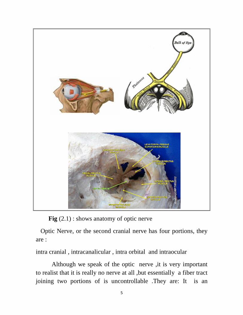
Fig (2.1) : shows anatomy of optic nerve
Optic Nerve, or the second cranial nerve has four portions, they
are :
intra cranial , intracanalicular , intra orbital and intraocular
Although we speak of the optic nerve ,it is very important
to realist that it is really no nerve at all ,but essentially a fiber tract
joining two portions of is uncontrollable .They are: It is an

6
outgrowth of the brain , Its fibers possess no neurolemmal cells , It
is surrounded by the ménages ,unlike any peripheral nerve and
both the primary and secondary neurons are in the retina.(Agarwal-
2008)
2.2.2 Intracranial Portion
Optic nerve unsheathed in pia runs as a flattened band from the
anterior lateral angle of the somewhat quadrilateral optic chiasma .
It runs forwards and The laterally and slightly downwards to the
optic foramen . (Agarwal-2008) ).
2.2.3 Intracanalicular Portion
At its entry into the optic canal, it receives a covering of
arachnoid mater and since the dura mater is prolonged through the
canal as aperiosteum, the nerve is in fact from here onwards
surrounded by all three meninges and also of course, the
cerebrospinal fluid. It traverses the optic canal and enters the
orbit.( Agarwal-2008) .
2.2.4 Intraorbital Portion
As a rounded cord, it now runs forwards and slightly laterally
and downwards in a somewhat sinuous manner to allow for ocular
movements and is continued into the back.( Agarwal-2008) .
2.2.5 Intraocular Portion
It then enters the eyeball just above and 3 mm medial to the
posterior pole.
(Agarwal-2008).

7
2.2.6 Relation
2.2.6.1 Intracranial Portion
The nerve lies at first above the diaphragm sellae, which covers
the pituitary body. Between the two nerves in front of the chiasma
is atriangular space in which is a variable portion of the pituitary,
covered by the diaphragm sellae. Above the nerve is the anterior
perforated substance, the medial root of the olfactory tract and the
anterior cerebral artery, which crosses superiorly to reach its
medial side. The internal carotid artery is at first below and then
lateral. ( Agarwal-2008).
2.2.6.2 Intracanalicular Portion
The pia forms a sheath closely adherent to the nerve . The dura
constitutes the periosteal lining to the canal and at its orbitalend
splits to become continuous on the one hand with the periorbita
and on the other hand with the dura of the optic nerve. The
ophthalmic artery crosses below the nerve in the dural sheath to its
lateral side. It leaves the dura at or near the anterior end of the
canal. Thus, the internal carotid artery is to some extent tied to the
dural sheath by its ophthalmic branch and it is also indirectly
attached to the optic nerve by the adherence of the sheaths and by
branches to the nerve from the ophthalmic artery. Medial to the
optic nerve is the sphenoidal air sinus or a posterior ethmoidal
sinus, from which it may be separated by a thin plate of bone only.
This provides the explanation of retrobulbar neuritis following a
sinus infection. ( Agarwal-2008).
8
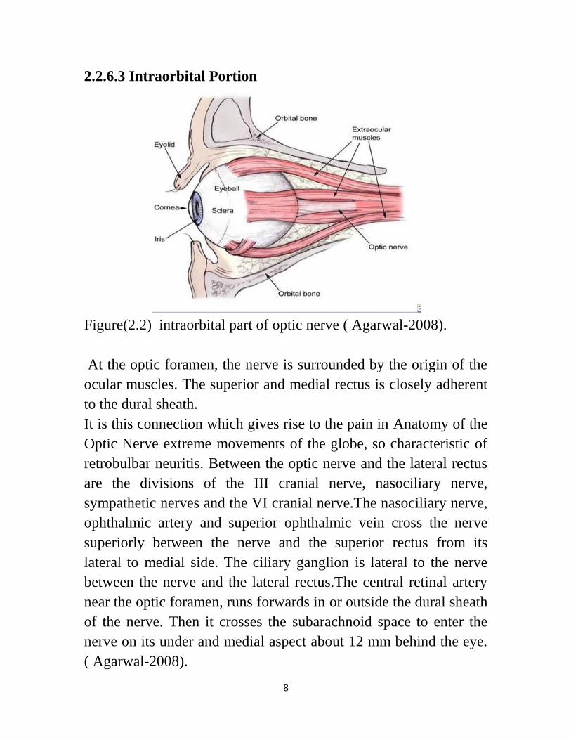
2.2.6.3 Intraorbital Portion
Figure(2.2) intraorbital part of optic nerve ( Agarwal-2008).
At the optic foramen, the nerve is surrounded by the origin of the
ocular muscles. The superior and medial rectus is closely adherent
to the dural sheath.
It is this connection which gives rise to the pain in Anatomy of the
Optic Nerve extreme movements of the globe, so characteristic of
retrobulbar neuritis. Between the optic nerve and the lateral rectus
are the divisions of the III cranial nerve, nasociliary nerve,
sympathetic nerves and the VI cranial nerve.The nasociliary nerve,
ophthalmic artery and superior ophthalmic vein cross the nerve
superiorly between the nerve and the superior rectus from its
lateral to medial side. The ciliary ganglion is lateral to the nerve
between the nerve and the lateral rectus.The central retinal artery
near the optic foramen, runs forwards in or outside the dural sheath
of the nerve. Then it crosses the subarachnoid space to enter the
nerve on its under and medial aspect about 12 mm behind the eye.
( Agarwal-2008).
9
2.2.6.4 Intraocular Portion
The intraocular portion passes through the sclera and choroid and
finally appears in the eye as the optic disk . The intraocular portion
of the optic nerve head has an average diameter of 1.5 mm.
( Agarwal-2008).
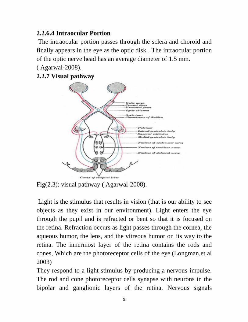
2.2.7 Visual pathway
Fig(2.3): visual pathway ( Agarwal-2008).
Light is the stimulus that results in vision (that is our ability to see
objects as they exist in our environment). Light enters the eye
through the pupil and is refracted or bent so that it is focused on
the retina. Refraction occurs as light passes through the cornea, the
aqueous humor, the lens, and the vitreous humor on its way to the
retina. The innermost layer of the retina contains the rods and
cones, Which are the photoreceptor cells of the eye.(Longman,et al
2003)
They respond to a light stimulus by producing a nervous impulse.
The rod and cone photoreceptor cells synapse with neurons in the
bipolar and ganglionic layers of the retina. Nervous signals

10
eventually leave the retina and exit the eye through the optic nerve
on the posterior surface of the eyeball. No rods or cones are
present in the area of the retina where the optic nerve fibers exit.
The result is a " blind spot" known as the optic disc.(Longman,et
al 2003).
After leaving the eye, the optic nerves enter the brain and travel to
the visual cortex of the occipital l lobe. In this area of the brain,
visual interpretation of the nervous impulses that were generated
by light stimuli in the rods and cones of the retina result in
"seeing".(Longman,et al 2003).
2.2.8 Blind spot
The beginning of the optic nerve in the retina is called the optic
nerve head or optic disc. Since there are no photoreceptors (cones
and rods) in the optic nerve head, this area of the retina cannot
respond to light stimulation. As a result, it is known as the “blind
spot,” and everybody has one in each reason we normally do not
notice our blind spots is because, when both eyes are open, the
blind spot of one eye corresponds to retina that is seeing properly
in the other eye .(montogomery-1998)
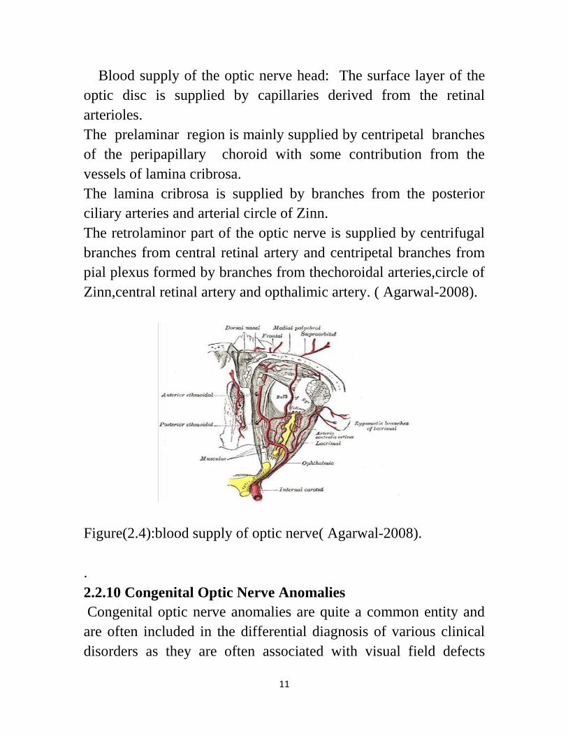
2.2.9 Blood supply of optic nerve
The visual pathway is mainly supplied by pail net work of the
vessels except the orbital part of optic nerve which also supplied
by and axial system derived from the central artery of retina .The
pial plexus around different parts of the visual pathway gets
contribution from different arteries.
visual pathway is supplied by:Pial plexus , Calcarian A , Posterior
cerebral A and Anterior choridal A
11
Blood supply of the optic nerve head: The surface layer of the
optic disc is supplied by capillaries derived from the retinal
arterioles.
The prelaminar region is mainly supplied by centripetal branches
of the peripapillary choroid with some contribution from the
vessels of lamina cribrosa.
The lamina cribrosa is supplied by branches from the posterior
ciliary arteries and arterial circle of Zinn.
The retrolaminor part of the optic nerve is supplied by centrifugal
branches from central retinal artery and centripetal branches from
pial plexus formed by branches from thechoroidal arteries,circle of
Zinn,central retinal artery and opthalimic artery. ( Agarwal-2008).
Figure(2.4):blood supply of optic nerve( Agarwal-2008).
.
2.2.10 Congenital Optic Nerve Anomalies
Congenital optic nerve anomalies are quite a common entity and
are often included in the differential diagnosis of various clinical
disorders as they are often associated with visual field defects

12
central nervous system (CNS) malformations and other ocular
abnormalities. Athorough knowledge of the embryological
development of the optic nerve entails a better understanding of the
development of optic nerve anomalies. ( Agarwal-2008).
2.2.10.1 Aplasia
Aplasia is a rare anomaly characterized clinically by a total blind
eye with an afferent pupillary defect, an absent optic disk and an
absent retinal vasculature. ( Agarwal-2008).
2.2.10.2 Hypoplasia
The optic disk hypoplasia is a sporadic condition. The affected can
bemicro-ophthalmic or of normal size and usually exhibit a wide
range of visual impairment from normal vision to severe visual
loss with strabismus or nystagmus in bilateral cases. Visual acuity
is determined primarily by the integrity of the papillomacular
bundle. The visual fields show a localized defect. The disk is
surrounded by a peripapillary halo, bordered by a dark pigment
ring called as the double ring sign. Retinal vascular tortuosity is
commonly seen. ( Agarwal-2008).
2.2.10.3 Bergmister’s Papillae
The glial sheath of Bergmister envelops the posterior third of the
hyaloid artery it begins to atrophy about the seventh month of
gestation, even before the main vessel atrophies. The extent of the
atrophy is below the surface of tee disk. If the atrophy at the disk is
less complete a tuft of glial tissue may be seen thorough out the
life over the optic disk called the Bergmister’s papillae. ( Agarwal-
2008).
13
2.2.10.4 Myelineated Nerve Fibers
Medullation or myelineation of the optic nerve begins in the fetal
lifefrom the lateral geniculate body towards the globe. Normally
the myelination is completed shortly after birth at which time the
myelinsheath reaches the posterior aspect of the lamina cribrosa.
The medullated fibers may be seen starting from the disk and
extending towards the periphery . Fundus examination shows
irregular feather-like patches which may or may not obscure the
retinal blood vessels. Rarely, isolated peripheral patches of
myelination may also occur. Myelination of the nerve fibers results
in visual field defects. Myopia, coloboma, polycoria, keratoconus,
oxycephaly and neurofibromatosis have been associated with
myelineated nerve fibers. ( Agarwal-2008).
2.2.10.5 Optic Nerve Head Drusen
Deposition of hyaline like calcified material within the substance
of the optic nerve head. Optic nerve head drusen can binherited or
can be associated with heredodegenerative conditions like retinitis
pigmentosa or angioid streaks or can be following long standing
papilledema, papillitis, vascular occlusions. Clinically has an
irregular, nodular, mulberry like appearance ofthe surface of the
disk. The physiological cup can be absent but venous pulsation is
present there can be abnormal tortous, anomalously branching
vessels. Disk can be pallor some times but will have irregular
margins. They vary greatly in size, shape and number, smaller ones
often coalesce to form larger ones .The differential diagnosis of
optic disk drusen includes papilledema with which it is often
confused.
Patients with disk drusen usually remain asymptomatic although
few cases have been reported to develop peripapillary and macular
hemorrhage. Drusens may directly compress the nerve fibers

14
within the disk and cause various visual field defects like
enlargement of the blind spot, arcuate scotoma or peripheral field
constriction. Centralvisual acuity is usually good. ( Agarwal-2008).
2.2.10.6 Colboma of optic disk
A coloboma of the optic disk results from incomplete closure of
the embryonic fissure. The fissure initially closes in the middle and
then extends anteriorly and posteriorly until a small crescent at the
posterior pole remains open. When the lips of the fissure fail to
fuse, typical colobomas result. The coloboma of the optic nerve
may occuralone or may be associated with coloboma of the iris,
retina, choroid or ciliary body. ( Agarwal-2008).
2.2.11 Optic Nerve Tumors
Tumors of the optic nerve are relatively rare lesions. However,
these lesions have significant risk for visual morbidity as well as
other problems related to the central nervous system (CNS). Optic
nerve glioma (astrocytoma) is the most common intrinsic tumor of
the optic nerve. Juvenile pilocytic astrocytomas are by far the most
common optic nerve tumor of children. Malignant gliomas of the
optic nerve occur much less frequently and are seen in adults.
Meningiomas of the optic nerve sheath are the second largest
group of tumors which may affect the optic nerve and occur more
commonly in adults. Lastly, secondary tumors of the optic nerve
may arise from direct invasion from intraocular malignancies,
meninges, adjacent structures, as well as distant metastases.
( Agarwal-2008).

15
2.2.11.1 Optic nerve glioma
Gliomas (juvenile pilocytic astrocytoma) are the most important
optic nerve tumor of children, accounting for 65 percent of all
intrinsic optic nerve tumors. Gliomas are benign neoplasms arising
from the neuroglia (astrocytes and oligodendrocytes). The majority
of optic nerve gliomas are of astrocytic origin. However, a few rare
optic nerve gliomas arise from oligodendrocytes. The descriptive
term juvenile pilocytic astrocytoma is often used to describe this
low-grade glioma. Gliomas grow slowly, but can spread under the
dura to invade local structures. Patients typically present before the
age of 20 with progressive visual loss, proptosis, and disk pallor
with or without papilledema. Management includes observation,
radiation, and surgery.
Gross Appearance: Optic nerve gliomas are typically contained
within the dura . The dura is stretched and thin, but usually intact.
Typical gliomas appea tan to dusky red from the vascular
congestion within the tumor. Orbital gliomas are characteristically
fusiform, with the borders of the tumor difficult to delineate. Thus,
it is helpful to obtain cross-sections at the end of each specimen.
Gliomas may also invade the arachnoid and pia, and extend
through the subdural space. This pattern occurs more often in
neurofibromatosis patients.The nerve itself may be of normal
thickness, but the overall diameter may be increased because of the
perineural component. Cross-sections through the middle portion
show the whitish nerve enlarged and surrounded by a cuff of
arachnoidal tissue, which is then covered by stretched dura.
( Agarwal-2008).
2.2.11.2 Optic nerve meningiomas
Meningiomas are the second most common optic nerve tumor,
after gliomas. Meningiomas are benign neoplasms arising from

16
meningothelial cells typically in the arachnoid. Patients generally
are middle-aged adults and present with decreased vision, visual
field loss, proptosis, disk atrophy, disk swelling, and later on
optociliary shunt vessels. Meningiomas grow slowly, but are
invasive and infiltrate surrounding structures. Management
includes conservative monitoring, radiotherapy, and surgery .
( Agarwal-2008).
2.3 The Retina
The retina is the innermost layer of the eye (the tunica intima or
internal tunic) and is comparable to the film inside of a camera.It is
composed of nerve tissue which senses the light entering the eye
.(Montgomery,1998)
This complex system of nerves sends impulses through the optic
Nerve back to the brain, which translates these messages into
images that we see.That is, we “see” with our brains; our eyes
merely collect the information to do so The retina is composed of
10 layers, from the outside (nearest the blood vessel enriched
choroid) to the inside (nearest the gelatinous vitreous humor)
Pigmented epithelium, photoreceptors, bacillary layer (outer and
inner segments of cone and rod photoreceptors), external (outer)
limiting membrane, outer nuclear (cell bodies of cones and
rods),outer plexiform (cone and rod axons, horizontal cell
dendrites, bipolar dendrites), inner nuclear (nuclei of horizontal
cells, bipolar cells, amacrine cells, and Müller cells), inner
plexiform (axons of bipolar cells and amacrine cells, dendrites of
ganglion cells), ganglion cells (nuclei of ganglion cells and
displaced amacrine cells), nerve fiber layer (axons from ganglion
cells traversing the retina to leave the eye at the optic disc), and
internal limiting membrane (separates the retina from the
vitreous).(Montgomery,1998).
17
Beneath the pigmented epithelium of the retina are these layers,
from the outside (furthest from the retina) to the inside (closest to
the retina): sclera (white part of the eye) ,large choroidal blood
vessels,choriocapilaris Bruch’s membrane (separates the
pigmented epithelium of the retina from the
choroid)(Montgomery,1998).
2-4 Physiologyof the Eye
Process of vision: light waves from an object (such as a tree) enter
the eye first through the cornea, which is the clear dome at the
front of the eye.It is like a window that allows light to enter the
eye.The light then progresses through the pupil, the circular
opening in the center of the colored iris.(Montgomery,1998).
Fluctuations in the intensity of incoming light change the size of
the eye’s pupil.As the light entering the eye becomes brighter, the
pupil will constrict (get smaller), due to the pupillary light
response .As the entering light becomes dimmer, the pupil will
dilate (get larger).(Montgomery,1998). Initially, the light waves
are bent or converged first by the cornea and then further by the
crystalline lens (located immediately behind the iris and the pupil),
to a nodal point (N) located immediately behind the back surface
of the lens.At that point, the image becomes reversed (turned
backwards)andinverted (turned upside-down).(Montgomery,1998).
2.5 Pathology of the Eye
2.5.1 Optic Atrophy:
Optic atrophy of the optic disc (visible to an eye doctor looking
inside the eye) is the result of degeneration of the nerve fibers of
the optic nerve and optic tract. It can be congenital (usually
hereditary) or acquired.(Montgomer,1998)

18
If acquired, it can be due to vascular disturbances (occlusions of
the central retinal vein or artery or arteriosclerotic changes within
the optic nerve itself), may be secondary to degenerative retinal
disease (e.g., optic neuritis or papilledema), may be a result of
pressure against the optic nerve, or may be related to metabolic
diseases (e.g., diabetes), trauma, glaucoma, or toxicity (to
alcohol,tobacco, or other poisons).(Montgomery,1998).
2.5.2 Optic neuritis:
Optic neuritis is an inflammation of the optic nerve.It may affect
the part of the nerve and disc within the eyeball (papillitis) or the
portion behind the eyeball (retrobulbar optic neuritis, causing pain
with eye movement).It also includes degeneration or
demyelinization of the optic nerve.There will be no visible changes
in the optic nerve head (disc) unless some optic atrophy has
occurred. (Montgomery ,1998).
The condition is unilateral rather than bilateral.If the nerve head is
involved, it is slightly elevated, and pupillary response in that eye
is sluggish.There usually is a marked but temporary decrease in
visionfor several days or weeks, and there is pain in the eye when
it is moved. Single episodes generally do not result in optic atrophy
nor in permanent vision loss; however, multiple episodes can
result in both.(Montgomery,1998)
2.5.3 pilledema:
Papilledema is edema or swelling of the optic disc (papilla), most
commonly due to an increase in intracranial pressure (often from a
tumor), malignant hypertension, or thrombosis of the central retinal
vein.The condition usually is bilateral, the nerve head is very
elevated and swollen, and pupil response typically is
normal.(Montgomery,1998).

19
Vision is not affected initially (although there is an enlargement of
the blind spot), and there is no pain upon eye movement.
Secondary optic atrophy and permanent vision loss can occur if
the primary cause of the papilledema is left
untreated.(Montgomery,1998).
2.5.4 Ischemic optic neuropathy:
Ischemic optic neuropathy is a severely blinding disease resulting
from loss of the arterial blood supply to the optic nerve (usually in
one
eye), as a result of occlusive disorders of the nutrient arteries
.Optic
neuropathy is divided into anterior, which causes a pale edema of
the optic disc, and posterior, in which the optic disc is not swollen
and the abnormality occurs between the eyeball and the optic
chiasm. (Montgomery,1998)
Ischemic anterior optic neuropathy usually causes a loss of vision
that may be sudden or occur over several days. Ischemic posterior
optic neuropathy is uncommon, and the diagnosis depends largely
upon exclusion of other causes, chiefly stroke and brain tumor
.(Montgomery,1998)
2.5.5 Glaucoma:
Glaucoma is an insidious disease which damages the optic nerve,
typically because the “intraocular pressure” (IOP) is higher than
the retinal ganglion cells can tolerate. This eventually results in the
death of the ganglion cells and their axons, which comprise the
optic nerve. Thus, less visual impulses are able to reach the brai.
(Montgomery,1998)

20
In advanced glaucoma, the visual field in the peripheral retina is
decreas or lost, leaving vision in the central retina (macular area)
intact.This results in “tunnel vision.”Elevated eye pressure occurs
when too much aqueous fluid enters the eye and not enough of the
aqueous fluid is leaving the eye.Eye pressure can be measured by
performing a “tonometry” test. (Montgomery,1998)
Normally, fluid enters the eye by seeping out of the blood vessels
in the ciliary body. This fluid eventually make its way past the
crystalline lens, through the pupil(the central opening in the iris),
and into the irido-corneal angle, the anatomical angle formed
where the iris and the cornea come together. Then the fluid passes
through the trabecular meshwork in the angle and leaves the eye,
via the canal of Schlemm.(Montgomery,1998)
If the rate of aqueous fluid is entering the eye is too great, or if the
trabecular meshwork “drain” gets clogged (for instance, with
debris or cells) so that the fluidis not leaving the eye quickly
enough, the pressure builds up in what is known as “open angle
glaucoma.”It is more common with increasing age
(Montgomery,1998).
Open angle glaucoma, which tends to be a chronic and painless
condition, also can be caused when the posterior portion of the iris,
surrounding the pupil, somehow adheres to the anterior surface of
the lens (creating a “pupillary block”).This can prevent intraocular
fluid from passing through the pupil into the anterior chamber.
( Montgomery,1998).
On the other hand, if the angle between and iris and the cornea is
too narrow, or is even closed, then the fluid backs up because it
cannot flow out of the eye properly. This causes an increased
intraocular pressure in what is known as “closed angle glaucoma.”

21
Typically, there is an acute (sudden), painful onset.It can be
accompanied by the appearance of rainbow-colored rings around
white lights.(Montgomery,1998).
An internal pressure more than that which the eye can tolerate
can deform the lamina cribrosa, the small cartilaginous section of
the sclera at the back of the eye through which the optic nerve
passes.
A deformed lamina cribrosa seems to “pinch” nerve fibers passing
Through it, eventually causing axon death. Untreated glaucoma
eventually leads to optic atrophy and
blindness.(Montgomery,1998)
Eye pressure is measured by using a “tonometer” (with the test
being called “tonometry”), and the standard tonometer generally is
considered to be the “Goldmann tonometer.”The normal range of
intraocular pressure (IOP) is 10mm Hg to 21mm Hg, with an
average of about 16mm Hg. Typically, eyes with intraocular
pressure
measurements of 21mm Hg or higher, using a Goldmann
tonometer, are considered to be “ocular hypertensive” and are
suspect for glaucoma.(Montgomery,1998).
However, although glaucoma typically is associated with
elevated IOP, the amount of pressure which will cause glaucoma
varies 17from eye to eye and person to person.Many people with
glaucoma actually have IOP’s in the normal range (“low tension”
glaucoma), possibly indicating that their lamina cribrosas are too
weak to withstand even normal amounts of pressure.
Conversely, many people with IOP’s which would be considered
high have no evidence of glaucomatous damage
(Montgomery,1998)
Glaucomatous changes in the optic disk (optic nerve head) usually
Can be detected over time. If the optic cup within the optic disk

22
increases in size over a period of months or years, if nothing is
observed anywhere around the nerve head rim, and/or if an
asymmetry is observed between the optic cups of the two eyes,
then that person may be considered to be a “glaucoma suspect” .In
glaucoma, optic nerve rim atrophy and/or notching, with a
corresponding visual field decrease, usually will occur in this
order:
Visual field loss, caused by optic nerve damage, is measured by
using a “visual field analyzer” or “perimeter,” especially by
measuring and comparing changes over time. The procedure is
known as 18 “perimetry.”Most field loss due to glaucoma usually
is not even measurable until 25% to 40%of the optic nerve’s axons
have been destroyed. ( Montgomery,1998).
Studies seem to show that the first fibers to die are the larger
fibers, which primarily carry form and motion information, rather
than the smaller fibers, which primarily detect light. Therefore,
pattern Discrimination perimetry (PDP), which requires detection
of both form and motion, may be a better test for early glaucoma
than conventional perimetry, which requires detection of spots of
light .(Montgomery,1998).
In pattern discrimination perimetry (PDP), various locations of the
retina are stimulated with a checkerboard pattern on a background
of randomly moving dots. The more random the dot movements,
the more difficult it is to continue to perceive the checkerboard
pattern.
Even a normal eye eventually will not be able to see the
checkerboard when the dot movement is random enough.
(Montgomery,1998).
The more advanced the stage of glaucomatous nerve damage, the
less “noisy” the dots need to be for the checkerboard pattern to be

23
indistinguishable from the background of moving dots.In effect,
the PDP seems to be more sensitive than a standard perimeter in
detecting early glaucomatous visual field losses.(
Montgomery,1998).
Typically, the elevated pressure in open angle glaucoma can be
controlled using glaucoma medications, which either decrease the
production of aqueous fluid or else increase its outflow from the
eye.
However, closed angle glaucoma often requires surgical
intervention to be controlled.( Montgomery,1998).
2.5.6 Cataract:
Normally, all the layers of the crystalline lens are clear, and light
Passes through it unobstructed. However, with age or due to
certain systemic diseases, as well as with a cumulative absorption
of ultraviolet radiation over many years, the lens material can
become cloudy, yellow, brown, and even opaque. Anything in the
lens which obstructs entering light is referred to as a “cataract.”(
Montgomery,1998).
More than 50% of people over the age of 60have some form of
acataract .It has been said that if one lives long enough, he/she will
develop a cataract. Even some infants are born with a “congenital”
cataract which, if left untreated, can cause permanent
visualimpairment or blindness, even if the cataract is removed
years later.(Montgomery,1998).
It is not possible to remove a primary cataract without irreparably
Damaging the crystalline lens within which the cataract is
contained.
A laser cannot be used successfully to remove a cataract, except as
described later (in the case of a secondary cataract).Therefore,
cataract surgery involves removing most or all of the lens of the
24
eye and replacing it with an artificial “intraocular lens” or “lens
implant,” made of a hard plastic (polymethyl methacrylate or
PMMA), silicone, acrylic, or hydro gel material
.(Montgomer,1998).
An “extracapsular” cataract extraction (ECCE) is the routine
type of cataract removal.In an ECCE procedure, an opening is
made in the front of the lens capsule. Through this opening, the
lens nucleus is removed, either as a whole or by dissolving it into
tiny pieces and vacuuming out the pieces, a procedure called
“phacoemulsification.”Next, the lens cortex also is sucked out,
leaving the lens capsule in place, and into the lens capsule is
inserted the artificial lens implant.(Montgomery,1998)
Prior to the 1980’s, the entire crystalline lens was removed in a
cataract surgery, called an “intracapsular” cataract extraction
(ICCE). Usually, this was performed using “cryoextraction,” where
acryoprobe froze the entire lens, permitting its complete removal.
Now, in the unusual case of an intracapsular lens extraction, or
ICCE, the implant lens is placed in front of the iris, rather than
behind it, because there is no lens capsule to hold the implant in
place. Rarely is this procedure done anymore.( Montgomery,1998).
Approximately 1-2% of post-cataract extraction patients develop
swelling in the area of the retina responsible for central vision (the
macula).This swelling occurs in cystoids spaces, and is referred to
as cystoids macular edema. After an initial improvement following
surgery, these patients subsequently will describe blurred vision.
Cystoids macular edema can occur as early as days, or as late as
several years, following surgery. Treatment options include
observation, topical therapy, periocular injections, and surgery.(
Montgomery,1998).
occuring carotenoids in the crystalline lens—lutein and
25
zeaxanthin (molecular cousins of beta carotene and vitamin A)—
have been shown to reduce the risk of cataracts. These pigments
act as antioxidants within the lens, inhibiting the formation of free
radicals, which can damage lenticular material and contribute to
the development of cataracts( Montgomery,1998)
Thus, it may be that the greater the amount of antioxidants such
as lutein and zeaxanthin in the system, the less the risk of cataract
formation.These two antioxidants are found particularly in yellow
fruits and in green leafy vegetables (especially xanthophyll-rich
vegetables such as spinach, kale, collard greens, and broccoli), in
eggs, and as nutritional supplements ( Montgomery,1998
2.5.7 Retinal detachment (RD):
Normally, with age, the vitreousgel collapses and detaches from
the retina—an event known as a posterior vitreous detachment.
Occasionally, however, the vitreous membrane pulls on and creates
a tear in the retina. Vitreous fluid can seep into or beneath the
retina, detaching it from the pigmented epithelium
underneath.Montgomery,1998).When a retinal detachment occurs,
a shower of floaters may be Observed by the person experiencing
the detachment. These are Thousands of blood cells being liberated
from a tiny blood vessel which has been broken due to the retinal
tear or detachment.Sometimes the floaters are described as being
like a “shower of pepper” before the eyes.(Montgomery,1998).
Sudden flashes of light, as well as a “web” or “veil” in front or else
in the periphery of the eye, also may appear in conjunction with
the onset of floaters. The retinal tear and subsequent detachment
must be repaired as soon as possible, usually with one of these
procedures: sealing it using an argon laser (“photocoagulation”),
freezing it (“cryotherapy” or “cryopexy”), securing it, after
cryotherapy, with a tiny belt around the equator of the eye (“scleral

26
buckle” surgery), injecting a gas bubble into the eye (in
conjunction with photocoagulation or cryopexy) so that the bubble
rests against
The hole or tear (“pneumatic retinopexy”), requiring the person to
keep his/her head in the sameposition for several days, or
removing the vitreousgel and filling the eye with a gas bubble or
silicon oil(“vitrectomy”). (Montgomery,1998).
2.5.8 Retinitis pigmentosa (RP):
One of the most devastating conditions affecting the rods is
“retinitis pigmentosa,” an inherited disorder in which the rods
gradually degenerate. With time, night vision is severely affected.
Eventually, all peripheral vision will continue to be destroyed, to
the point where only central or “tunnel” vision
remains.(Montgomery,1998)
There is no known treatment. However, since blue and ultraviolet
light may make aggravate the condition, amber-colored glasses
with an ultraviolet absorption coating, worn during the day, may
slow down the disease process.(Montgomery,1998).
Studies have shown that retinitis pigmentosa is caused by
mutations in the rhodopsin gene, the peripherin gene, and possibly
in other genes within the rod.Mutations in the peripherin gene also
may be the cause of another devastating retinal disorder: “macular
dystrophy.”(Montgomery,1998)

27
2.6 Previous studies
Benevento, eltal2011studied Optic Nerve Measurements in
Normal Human Eyes by MRI and they used Coronal MRI imaging
of normal human eyes it's showedan average ONSD range of 4.0 –
6.0 with SD 0.5mm, andan average OND range of 2.6 –4.0 with
SD 0.3mm.there rangeis consistent with published data on the
ONSD. However, we are not aware of any published data on the
OND.
Newcombe, eltal 2008 Used T2-weighted magnetic resonance
imaging of the optic nerve sheath to detect raised intracranial and
they 35 results that the Measurement of ONSD was possible in
95% of cases. The ONSD was significantly greater in TBI patients
with raised ICP (>20 mmHg;±6.31, 0.50 mm, 19 measures) than in
those with ICP of 20mmHg or less (± 5.29,0.48 mm, 26 measures;
P <0.0001) or in healthy volunteers (± 5.08,0.52 mm; P < 0.0001).
There was a significant relationship between ONSD and ICP (r
=0.71, P < 0.0001). Enlarged ONSD was a robust predictor of
raised ICP (area under the receiver operating characteristic curve =
0.94), with a best cut-off of 5.82 mm, corresponding to a negative
predictive value of 92%, and to a value of 100% when ONSD was
less than 5.30 mm.
Brex, eltal 2011describe an MRI technique for quantifying optic
nerve atrophy resulting from a single episode of unilateral optic
neuritis. They imaged 71patients, with a median time since onset
of optic neuritis of 17months (range 3–17months), using a coronal-
oblique fat-saturated -echo fast fluid-attenuated inversion-recovery
(sTE fFLAIR) sequence. The mean cross-sectional area of the
intraorbital portion of the optic nerves was calculated by a blinded
observer from five consecutive 3mm slices from the orbital apex
forwards using a semiautomated contouring technique and
compared with data from 71controls. The mean optic nerve area

28
was 7711mm1 in the affected eye of the patients, 7111mm1in the
contralateral eye (P = .1..0compared to the affected eye) and 7111
mm1in contro ls (P = .1.3compared to the affected eyes). There
wasa significant negative correlation between disease duration and
the size of the affected optic nerve (r =–.1.1, P = .1.71). The
measurement coefficient of variation was 811%. The sTE fFLAIR
sequence enables measurement of optic nerve area with sufficient
reproducibility to show optic nerve atrophy following a single
episode of unilateral optic neuritis. The correlation of increasing
optic nerve atrophy withdisease duration would be consistent with
ongoing axonal loss in a persistently demyelinated lesion, or
Wallerian degeneration following
axonal damage during the acute inflammatory phase.
Hickman, eltal 2009 investigate optic neuritis as a model for
atrophy in multiple sclerosis (MS) lesion they performed serial
magnetic resonance imaging (MRI) on 7.patients with a history of
optic neuritis using a fat saturated short-echo fast fluid-attenuated
inversion recovery (sTE fFLAIR) sequence. The first study was
performed a median of 711.months after the onset of optic neuritis
and the second 7year later. Using a computer-assisted contouring
technique, a blinded observer calculated the mean area of the intra-
orbital optic nerves. The mean area of affected optic nerves
decreased over 7 year by .11mm1from 7717to 7.11mm1(p=.1.7).
Poor visual acuity and decreased visual-evoked potential (VEP)
amplitude were associated with atrophy. These findings suggest
that atrophy is a feature of focal demyelinating lesions, it may
evolve over several years, and may have functional significance.
Optic neuritis provides a model to study the effect of inflammatory
demyelination the ability to accurately measure visual function and
to visualize and measure the optic nerves using magnetic
resonance imaging.

29
Newman , eltal 2009 evaluate the utility of measuring the optic
nerve sheath diameter in children with shunted hydrocephalus,
suspected of having raised intracranial pressure used 13children
with shunted hydrocephalus were examined, six had well
controlled ICP, 71 however manifested symptoms suggestive of
intracranial hypertension. A clinical history was taken from all
patients and their parents or carers. The shunt valve was examined
clinically, and signs of raised intracranial pressure were sought.
Ultrasound examination was performed in both eyes to measure
the optic nerve sheath diameters 3mm behind the globe. These
measurements were compared with control data obtained from
7.137children who attended the radiology department for unrelated
renal ultrasoundexamination1Control data are the upper limit of
normal for optic nerve sheath diameter is 81.mm (measured 3mm
behind the globe) in patients over 7year of age, and 81.mm in
children less than 7year of age. Those patients with functioning
ventriculoperitoneal shunts had a mean optic nerve sheath diameter
of 111(SD .1.) mm; those with raised intracranial pressure had a
mean optic nerve sheath diameter of .10(.10) mm (p<.1...7). These
results confirm that optic nerve sheath diameters in excess of the
control data are strongly suggestive of raised intracranial
pressure138.

30
Chapter Three
Materials and Methods
The study has been carried out during the period from October to
December 2016 ,on hundred (n=100) Sudanese males and females
patients who presented to the clinical department of the
Computerized Tomography (CT) in two hospital in Khartoum city
of Sudan in AL-Ribat Teaching Hospital and Yastabshroon
Hospital . The100-study group included 49 men and 51 women
with their aged being between 20-80 years . Evaluation of CT
images of the brain and orbital area of the patients with no
previous trauma in this area , no chronic diseases of the optic nerve
or surgeries interfering with orbital.
Data was collected through Questionnaire and patients
interviewing system ,prior to imaging; where personal
demographic notes ; age , sex , weight and tribal .and collected
randomly according to our inclusion criteria, and statistical
analysis was performed using Statistical Package for Social
Sciences (SPSS).
3.1 Materials :
3.1.1 Study population
The total sample of patients were (100 ) there ages between 20–
80 years old all were under CT orbit and brain.
Patient with optic nerve disorder or orbit disorder or diabetic
patient
3.1.2 Machine
TOSHIBA Emotion 16 CT scanner in Yastabshiroon Hospital and
Neou Soft 128 CT scanner in Ribat University Hospital ( Omer
Sawi Complex ) .
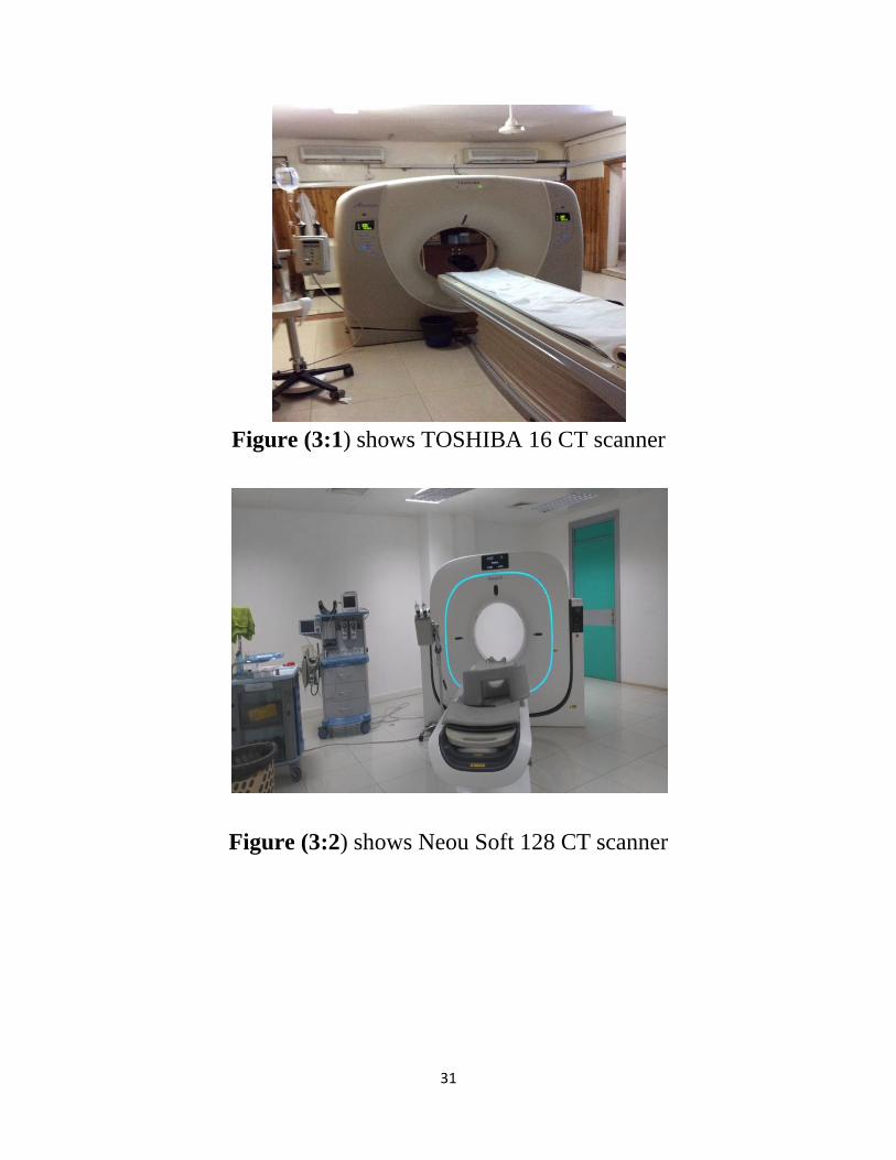
31
Figure (3:1) shows TOSHIBA 16 CT scanner
Figure (3:2) shows Neou Soft 128 CT scanner

32
3.2 Methods:
3.2.1 Protocol Used:
The patients lies supine on the examination couch . Both
orbits are examined using head holder . The patients are positioned
so that the longitudinal alignment light lies in the midline , and the
horizontal alignment light passes through the orbits . Straps and
foam pads are used for immobilization .
Anteroposterior and lateral scout view taken to show scanning
range and slice thickness.
3.2.2 Measurement method:
optic nerve Length was measured in two steps; firstly identenify
the lens of eye in the axial cut to indentify the site of the optic
chiasm , secondly identify the optic nerve and chiasma length that
the optic nerve go through it. .
The length of optic nerve was measured in axial cuts and sagittal
cut. Taken from the posterior part of the eye ball to the optic
chiasma in the base of skull. and the widths of the optic nerve
measured from the area between the two border of optic canal in
coronal cuts and axial cuts.
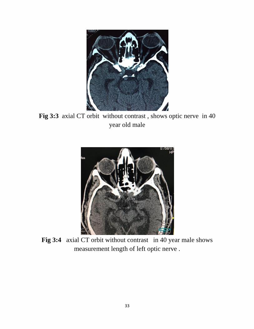
33
Fig 3:3 axial CT orbit without contrast , shows optic nerve in 40
year old male
Fig 3:4 axial CT orbit without contrast in 40 year male shows
measurement length of left optic nerve .

34
Chapter Four
Results:-
Table (4.1): gender distribution
Gender
Freque
ncy
Percen
t
Valid
Percent
Cumulati
ve
Percent
Vali
d
Fema
le
51 51.0 51.0 51.0
Male 49 49.0 49.0 100.0
Total 100 100.0 100.0
Table (4.2): Area distribution:
Area
Freque
ncy
Percen
t
Valid
Percent
Cumulati
ve
Percent
Vali
d
Nort
h
24 24.0 24.0 24.0
Sout
h
18 18.0 18.0 42.0
East 24 24.0 24.0 66.0
West 12 12.0 12.0 78.0
Midd
le
22 22.0 22.0 100.0

35
Gender
Freque
ncy
Percen
t
Valid
Percent
Cumulati
ve
Percent
Vali
d
Fema
le
51 51.0 51.0 51.0
Male 49 49.0 49.0 100.0
Total 100 100.0 100.0
Figure (4.1):Area distribution
Table (4.3):Descriptive statistics
N
Minimu
m
Maximu
m Mean
Std.
Deviation
Age 100 20 94 44.83 19.697
Weight 100 52 112 79.46 14.181
Length 100 4.00 6.23 5.0052 .43099
Width 100 .23 .65 .3971 .10012
36
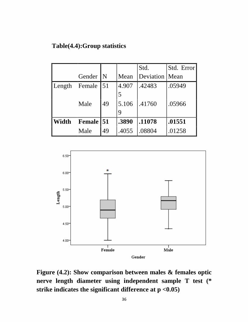
Table(4.4):Group statistics
Gender N Mean
Std.
Deviation
Std. Error
Mean
Length Female 51 4.907
5
.42483 .05949
Male 49 5.106
9
.41760 .05966
Width Female 51 .3890 .11078 .01551
Male 49 .4055 .08804 .01258
Figure (4.2): Show comparison between males & females optic
nerve length diameter using independent sample T test (*
strike indicates the significant difference at p <0.05)
37
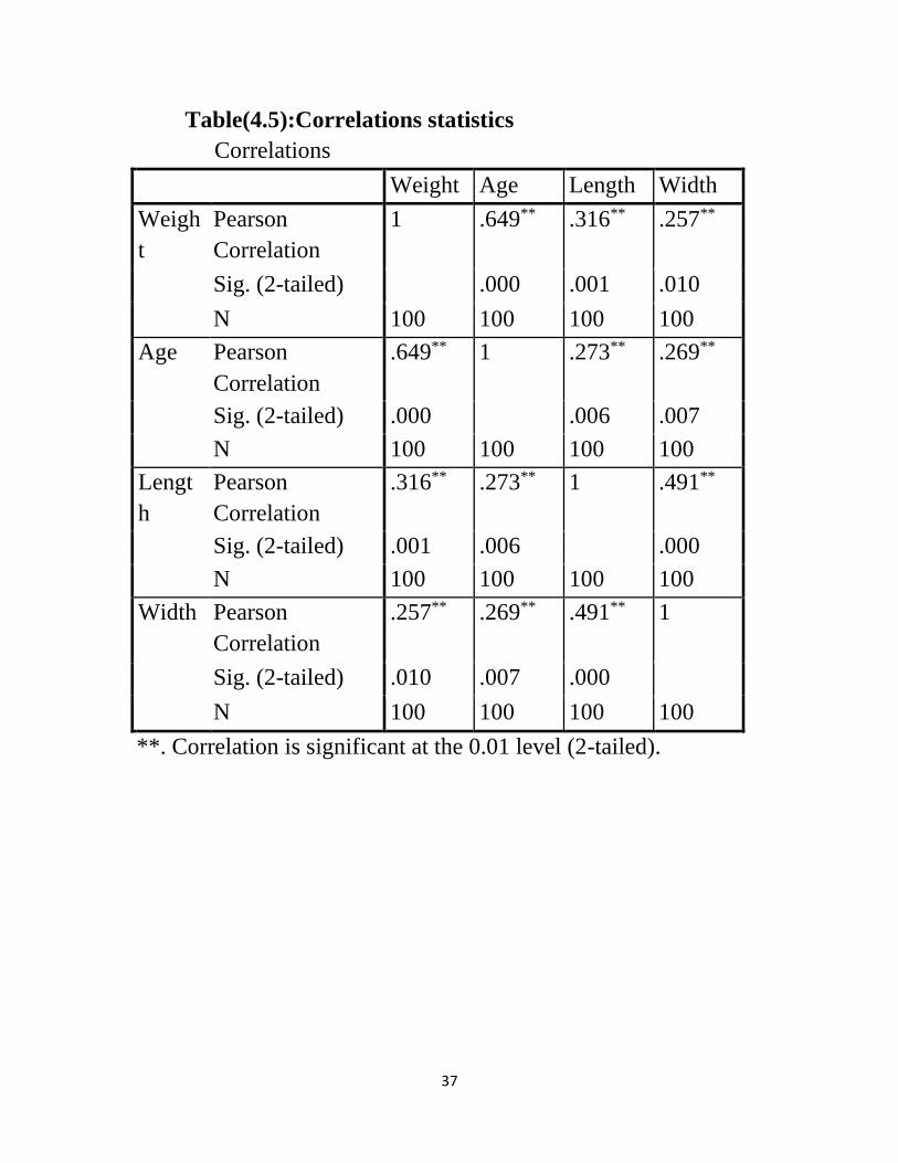
Table(4.5):Correlations statistics
Correlations
Weight Age Length Width
Weigh
t
Pearson
Correlation
1 .649** .316** .257**
Sig. (2-tailed) .000 .001 .010
N 100 100 100 100
Age Pearson
Correlation
.649** 1 .273** .269**
Sig. (2-tailed) .000 .006 .007
N 100 100 100 100
Lengt
h
Pearson
Correlation
.316** .273** 1 .491**
Sig. (2-tailed) .001 .006 .000
N 100 100 100 100
Width Pearson
Correlation
.257** .269** .491** 1
Sig. (2-tailed) .010 .007 .000
N 100 100 100 100
**. Correlation is significant at the 0.01 level (2-tailed).

38
Figure(4.3):correlation between optic nerve length and weight
Figure(4.4):correlation between optic nerve width and age

39
Chapter Five
Discussion, Conclusion and Recommendations
5.1 Discussion
Number of 100 patients CT orbit and brain was performed for
Sudanese patient there ages between 20–80 years old, were 49
males and 51 females.
The result after data analysis was found the overall mean of optic
nerve length was [5.0052±.43099] cm, and mean of optic nerve
width [.3971±.10012] cm. with minimum length [4.00cm], and
maximum length [6.23 cm]. with minimum width [.23cm], and
maximum width [.65 cm].
The result of this studies (minimum length [4.00cm], and
maximum length [6.23 cm].) similar to result obtained by MRI in
(Benvento , eltal 2001studies optic nerve measurements in normal
human eyes by MRI (minimum length {4.00cm} and maximum
length {6.00cm} .
From these results there is correlation between the width of
optic nerve and age, as age increase the width increase.
Also there is correlation between the optic nerve length and the
patient wieght as weight increase the length increase.
The mean optic nerve length for male [5.1069±.41760] cm, is
slightly higher than mean optic nerve length for female
[4.9075±.42483] cm .
The mean optic nerve width for male [.4055±.08804] cm, is
slightly higher than mean optic nerve width for female
[.3890±.11078] cm . There is no relation between optic nerve
width &length and the tribe.

40
5.2 Conclusion
using CT This study was conducted to find out the mean
optic nerve length and width in Sudanese population.
The overall mean of optic nerve length in 100 Sudanese
patient was [5.0052±.43099] cm, and mean of optic nerve
width [.3971±.10012] cm.
There is a correlation between Also there is correlation
between the optic nerve length and the patient wieght as
weight increase the length increase.
The optic nerve (length &width)slightly higher in males.
There is no relation between optic nerve width &length and
the tribe.

41
5.3 Recommendation
The minimum length of optic nerve must not exceed
[4.00cm], and maximum length [6.23 cm].
The minimum width of optic nerve must not exceed [.23cm],
and maximum width [.65 cm].
Is always important to know the reference value for optic
nerve diameters in Sudanese population, to state the
pathology and abnormality easily.
The age ,gender and weight are important parameters that
affect the optic nerve length and width.
I recommend for next studies to correlate the optic nerve
diameter with diabetic pateints.
Also I recommend using a larger sample size, from
difference state of the country. Finally I recommend for next
studies to use MRI in measuring the optic nerve for
Sudanese population.

42
References:
Amar Agrawal , Athiya Agrawal , 2008 , Manual of Neuro -
ophthalmology ,Ajanta ,third ed .
Catherine Westbrook ,2008 ,hand book of MRI technique ,
Cambridge ,UK ,third ed .
Suznne Henwood , 1999 ,clinical CT for technique and practice ,
Cambridge University 1 e
D G MacManus NMR Research Unit , Institute of Neurology ,
University College London , Queen Square , London , WC
2011N3BG , UK.
FJ Newcombe,1 Jonathan P Coles,1Maria Giulia Abate,1Iain E
Perkes,1Peter JA Hutchinson , 3Jo G Outtrim, 1 Dot A Chatfield,1
and David K Menonl Virginia 2008.
J,D, Benevento ,J.P.S. Garcia ,A.B. Baxter ,P.M.T. Garcia, R.A.
Holliday and R.B. Rosen Ophthalmology , Radiology , New York
Eye & Ear Infirmary , New York , N Y Ophthalmology , New
York Medical College , Valhalla , NY 2011 .
N J Scolding Institute of clinical Neurology , Frenchay Hospital ,
Bristol BS 20111 16LE , UK .
P A Brex NMR Rsearch Unit , Institute of Neurosciences
,University College London , Queen Square , W C 20111 N3BG ,
UK .
P Y , UK D H Miller NMR Research Unit , Institute of Neurology
, University Collage London , Queen Square , London 2011 .

43
S .J . Hickman SJ , Kapoor R , Jones SJ, Altmann DR , Plant GT ,
Miller DR .Corticosteroids do not prevent optic nerve atrophy
following optic neuritis , J Neural Neurosurg Psychiatry .2009 .
S . J .Hickman SJ , P . A . Brex , C . M .H . Brierley , N . C .
Silver , G .J .Barker , N . J , Scolding , D . A . S , Composton , I .F
. Moseley , G . T . Plant , D .H .
Miller NMR Research Unit , Institute of Neurology , University
College London , Queen Square , Kondon , W C 2009 .
Shlomo Melmed Kenneth S .Polonsky , P .Reed Larsen , Henery
M . Kronenberg , 2011 , Williams Textbook of Endocinology (12
th edition ) .
Ted Montgomery , 1998 , Anatomy , Physiology and Pathology of
human eye , California .
Welsey Longman , 2003 ,human anatomy and physiology , 6th ed
San , Francisco .

44
Appendex
SUDAN UNIVERSITY OF SCIENCES & TECHNOLOGY
COLLEGE OF GRADUATE STUDIES
M S c Diagnostic Radiologic Technology
Research About:
Measurement of Normal Optic Nerve for Adult Sudanese Using
Computed Tomography
The data collection questioner
No Age Gender wieght tribe Axial mm
sagittal coronal
1
2
3
4
5
6
7
8
9
10
11
12
13
14
15
Figure 3:5 sagittal CT orbit without contrast shows optic

45
o
Figure 1 sagittal CT orbit without contrast shows measurement
of length optic nerve of left eye
Figure 2 coronal CT orbits without contrast in 40 years male
shows measurement of width optic nerve of left eye