Young Age is not Associated with Increased Local Recurrence for DCIS Treated by Breast-conserving...
18
Young Age is Not Associated with Increased Local Recurrence for DCIS Treated by Breast-Conserving Surgery and Radiation Aruna Turaka, M.D. * , Gary M Freedman, M.D. * , Tianyu Li, M.S. † , Penny R Anderson, M.D. * , Ramona Swaby, M.D. ø , Nicos Nicolaou, M.D. * , Lori Goldstein, M.D. ø , Elin R. Sigurdson, M.D., Ph.D. ‡ , and Richard J. Bleicher, M.D. ‡ * Radiation Oncology, Fox Chase Cancer Center, Philadelphia, PA † Biostatistics, Fox Chase Cancer Center, Philadelphia, PA ‡ Surgical Oncology, Fox Chase Cancer Center, Philadelphia, PA ø Medical Oncology, Fox Chase Cancer Center, Philadelphia, PA Abstract Background—We report local recurrence (LR) after breast-conserving surgery and radiation (BCS + RT) for ductal carcinoma in situ (DCIS) to determine outcomes for patients aged ≤ 40 years compared with older women. Methods—The study included 440 women with DCIS treated from 1978 to 2007. All patients received whole-breast radiotherapy with a boost in 95% of cases. Demographics, characteristics, surgical and adjuvant treatments were analyzed for an effect on LR. Results—Median age was 56.5 years with 24 patients aged ≤ 40. Median DCIS size was 0.8 cm. Re-excision was required in 62% of patients, and in 75% of those aged ≤ 40. Tamoxifen was used in 22%, but only 1 patient aged ≤ 40. Median follow-up was 6.8 years. Actuarial LR was 7% (95% confidence interval of 4–11%) at 10 years and 8% (5–14%) at 15 years. There was no difference in LR by age (p=0.76). Conclusions—The long-term risk of LR after BCS + RT for DCIS is low, even in patients ≤ 40 years. This may be due to patient selection for small size, high utilization of re-excision and radiation boost. Young age may be a smaller contributor to LR risk in DCIS than previously suggested. Keywords Breast cancer; radiation therapy; DCIS; young age INTRODUCTION Ductal carcinoma in situ (DCIS) of the breast has increased in the United States from 3% of new breast cancer cases in 1980 to greater than 20% in 2003 (1). This corresponded to an increase in the age-adjusted rate of DCIS from 2.4 to 27.7 per 100,000 women between 1981 and 2001 after the advent of the screening mammography (2–5). Mastectomy was initially the standard of care for DCIS but breast-conserving surgery and radiation (BCS + Correspondence to: Gary M. Freedman, M.D.Department of Radiation Oncology Fox Chase Cancer Center 333 Cottman Avenue Philadelphia, PA 19111 Phone: 215-728-3016 Fax: 215-214-1629 [email protected]. Presented in part at the scientific session of the 50 th annual meeting of the American Society for Therapeutic Radiology and Oncology in Boston, MA, September 21-25, 2008. NIH Public Access Author Manuscript J Surg Oncol. Author manuscript; available in PMC 2010 September 25. Published in final edited form as: J Surg Oncol. 2009 July 1; 100(1): 25–31. doi:10.1002/jso.21284. NIH-PA Author Manuscript NIH-PA Author Manuscript NIH-PA Author Manuscript
-
Upload
independent -
Category
Documents
-
view
4 -
download
0
Transcript of Young Age is not Associated with Increased Local Recurrence for DCIS Treated by Breast-conserving...

Young Age is Not Associated with Increased Local Recurrencefor DCIS Treated by Breast-Conserving Surgery and Radiation
Aruna Turaka, M.D.*, Gary M Freedman, M.D.*, Tianyu Li, M.S.†, Penny R Anderson, M.D.*,Ramona Swaby, M.D.ø, Nicos Nicolaou, M.D.*, Lori Goldstein, M.D.ø, Elin R. Sigurdson,M.D., Ph.D.‡, and Richard J. Bleicher, M.D.‡* Radiation Oncology, Fox Chase Cancer Center, Philadelphia, PA† Biostatistics, Fox Chase Cancer Center, Philadelphia, PA‡ Surgical Oncology, Fox Chase Cancer Center, Philadelphia, PAø Medical Oncology, Fox Chase Cancer Center, Philadelphia, PA
AbstractBackground—We report local recurrence (LR) after breast-conserving surgery and radiation(BCS + RT) for ductal carcinoma in situ (DCIS) to determine outcomes for patients aged ≤ 40years compared with older women.
Methods—The study included 440 women with DCIS treated from 1978 to 2007. All patientsreceived whole-breast radiotherapy with a boost in 95% of cases. Demographics, characteristics,surgical and adjuvant treatments were analyzed for an effect on LR.
Results—Median age was 56.5 years with 24 patients aged ≤ 40. Median DCIS size was 0.8 cm.Re-excision was required in 62% of patients, and in 75% of those aged ≤ 40. Tamoxifen was usedin 22%, but only 1 patient aged ≤ 40. Median follow-up was 6.8 years. Actuarial LR was 7%(95% confidence interval of 4–11%) at 10 years and 8% (5–14%) at 15 years. There was nodifference in LR by age (p=0.76).
Conclusions—The long-term risk of LR after BCS + RT for DCIS is low, even in patients ≤ 40years. This may be due to patient selection for small size, high utilization of re-excision andradiation boost. Young age may be a smaller contributor to LR risk in DCIS than previouslysuggested.
KeywordsBreast cancer; radiation therapy; DCIS; young age
INTRODUCTIONDuctal carcinoma in situ (DCIS) of the breast has increased in the United States from 3% ofnew breast cancer cases in 1980 to greater than 20% in 2003 (1). This corresponded to anincrease in the age-adjusted rate of DCIS from 2.4 to 27.7 per 100,000 women between1981 and 2001 after the advent of the screening mammography (2–5). Mastectomy wasinitially the standard of care for DCIS but breast-conserving surgery and radiation (BCS +
Correspondence to: Gary M. Freedman, M.D.Department of Radiation Oncology Fox Chase Cancer Center 333 Cottman AvenuePhiladelphia, PA 19111 Phone: 215-728-3016 Fax: 215-214-1629 [email protected] in part at the scientific session of the 50th annual meeting of the American Society for Therapeutic Radiology and Oncologyin Boston, MA, September 21-25, 2008.
NIH Public AccessAuthor ManuscriptJ Surg Oncol. Author manuscript; available in PMC 2010 September 25.
Published in final edited form as:J Surg Oncol. 2009 July 1; 100(1): 25–31. doi:10.1002/jso.21284.
NIH
-PA Author Manuscript
NIH
-PA Author Manuscript
NIH
-PA Author Manuscript

RT) therapy are now also standard practice for many of these patients (2,6–9) . The use oftamoxifen for DCIS has also increased after the reporting of National Surgical AdjuvantBreast and Bowel Project (NSABP) B-24 (10–12) which demonstrated that 5 years ofTamoxifen is effective in reducing the risk of invasive or non-invasive ipsilateral andcontralateral tumors after radiation.
Proper management of DCIS is critical to prevent local recurrence (LR) and progression toinvasive breast cancer, as 50% of recurrent tumors are invasive (13). Previously identifiedpredictors of LR after BCS + RT include margin status, size of DCIS, nuclear grade,necrosis, mammographic presentation, histologic subtype, volume of excised specimens anddistance from the nipple (14–18) .
The importance of age in the management of DCIS is controversial with no consensus on itsimpact or on the definition of “young”. Young patient age in DCIS has been variouslydefined as </= 35, </=40, or </=45 years of age and the risk of LR after DCIS in youngpatients has been reported to be approximately 25–30% (19–22). This is significantly higherthan the long term LR risk of approximately 15% in other studies (23,24) consistingpredominantly of women over 40. One explanation may be the presence of adversepathologic features in younger patients (25) though treatment-related factors that appear tobe of particular importance in younger women include margin status and use of a boost(21,26). We therefore reviewed the long-term results with BCS + RT in young patients withDCIS with a special attention to the interaction of age with other known prognostic factorsfor LR.
METHODSThere were 573 patients with DCIS seen at the Fox Chase Cancer Center with a diagnosis ofDCIS between September 1998 and October 2007. The study population consists of 440consecutive women with DCIS treated with BCS + RT during that interval. Inclusion criteriaconsisted of clinically node negative patients with DCIS (AJCC 6th Ed. Stage 0) (27),treated with a completed course of radiation at Fox Chase Cancer Center. A prospectivelycollected Institutional Review Board-approved, Health Insurance Portability andAcountability Act-compliant institutional database that tracks breast cancer patients fordemographics, tumor characterstics, and treatment related information was reviewed.Patients were excluded if they were male, had microinvasion, underwent mastectomy, had adiagnosis of Paget’s disease of the nipple, or underwent conservative surgery withoutradiation.
All patients underwent a history and physical examination, baseline mammogram andpathologic diagnosis consisting of fine needle aspiration, core needle biopsy or excisionalbiopsy. A definitive surgical excision was performed in all patients with or without a re-excision when needed for surgical margins. All outside pathology slides were reviewed atFox Chase Cancer Center at the time of initial radiotherapy consultation. All patientsunderwent postoperative radiation. The radiotherapy technique consisted of treatment to theentire breast to a dose of 46 –50 Gy delivered in 1.8 – 2 Gy fractions over a period of 4.5–5weeks. The median whole breast dose was 50 Gy. A 6 MV linear accelerator was used formajority of patients. Women with large breasts or separation greater than 21–22 cm wereoften treated with higher energy photons (10 or 15 MV) in 33% of cases. A boost to theprimary site and scar was used in 95.7% of patients, almost all with electrons (or 192Ir andexternal beam) for an additional 10 – 18 Gy.
Study end points including LR, regional recurrence, distant metastasis, and overall survivalwere analyzed based upon established risk factors. Patient characteristics included age at
Turaka et al. Page 2
J Surg Oncol. Author manuscript; available in PMC 2010 September 25.
NIH
-PA Author Manuscript
NIH
-PA Author Manuscript
NIH
-PA Author Manuscript
diagnosis, menopausal status, race, and family history of breast cancer. Tumorcharacteristics included primary tumor location, pathological tumor size, hormonal receptorstatus, histological type, grade and necrosis. Treatment characteristics included use of re-excision, radiotherapy dose, fractionation schedule, use of and type of boost, and use oftamoxifen. Kaplan-Meier analysis was used for estimates of LR and survival. The log-ranktest was used to assess the significance between variables. All the statistical analyses wereperformed in statistical package for the SAS version 9.0 (SAS Institute, Cary, NC).Statistically significant differences were confirmed by p values of ≤ 0.05.
RESULTSPatient characteristics are listed in Table 1. The median age was 56.5 years (range 31–91years). There were 24 patients ≤ 40 years of age (5.5%), 169 patients were 41–54, 169patients were 55–69 and 78 patients were over 70 years of age. 294 patients (66.7%) werepostmenopausal, 119 (27.1%) were premenopausal and 27 (6.2%) were perimenopausal.African-Americans comprised 7.3% while 90.4% were white and others were 2.5%. Positivefamily history of breast cancer was seen in 168 patients (38.3%), 105 with a history in onerelative and 62 with more than 2 affected relatives. In 377 patients (85.9%) the diagnosiswas made based on mammogram only, in 4.8% by only physical examination, 9.3% by bothmammogram and physical examination. The most common mammographic finding waspresence of microcalcifications without associated mass (N=336, 76.5%). 43 patients hadmass without calcifications and in 30 patients both mass and microcalcifications wasnoticed. In 54.9% the tumor was located in the outer quadrant, 17.5% inner quadrant, 21.4%central and in 6.4% subareolar region.
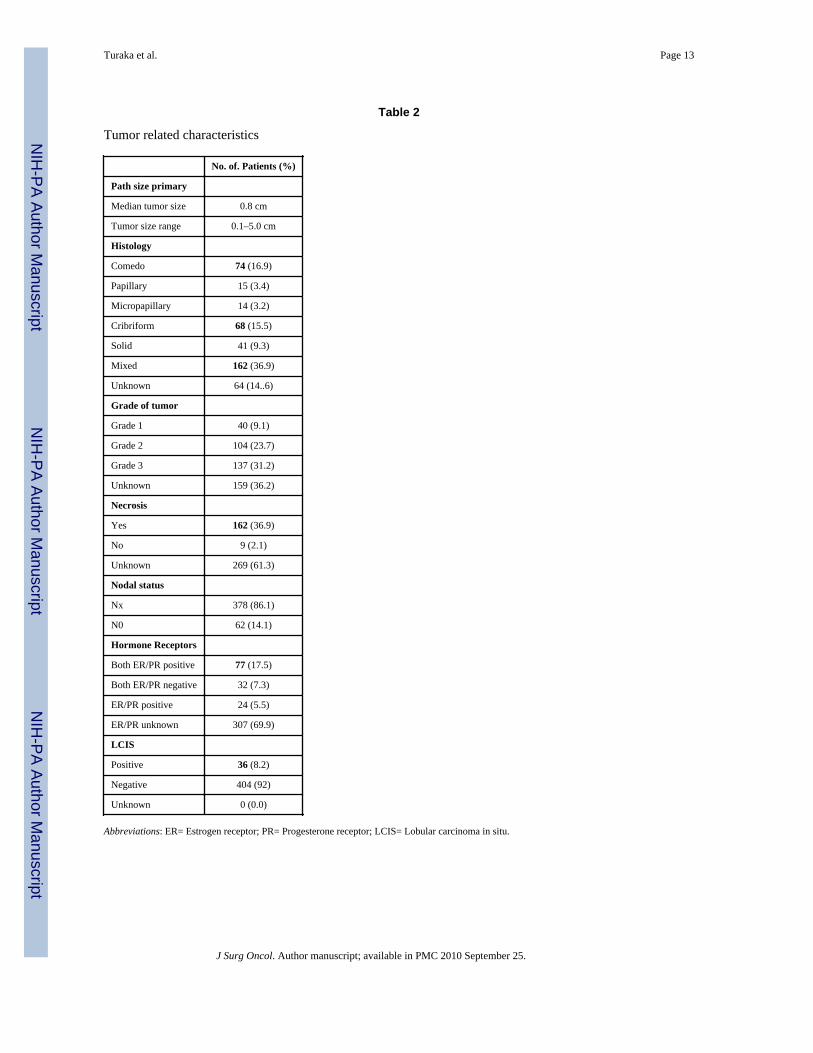
DCIS characteristics are shown in Table 2. The predominant histological pattern was mixedin 36.9%, comedo in 16.9%, cribriform in 15.5%, solid in 9.3%, papillary in 3.4%,micropapillary in 3.2% and not reported in 14.6%. Nuclear grade was assessed in 281patients; 63.86% (9.1% Grade 1, 23.7% Grade 2, 31.2% Grade 3). Necrosis was recorded aspresent in 162. The median tumor size was 8 mm (range 0.1–5.0 mm). 77 patients were bothER and PR positive (17.5%) and 24 were ER/PR positive (5.5%) and negative in 32 patients(7.3%). LCIS was seen in 36 (8.2%) patients.
Treatment-related characteristics are shown in Table 3. A re-excision was performed in61.7% overall, and in 75% for patients age ≤ 40. The final margins of re-excision werenegative (>2 mm) for 342 patients (77.9%), positive for 13 patients (3%), close in 49(11.2%), LCIS only in 6 (1.4%) and unknown in 30 (7%). 62 patients underwent axillarydissection, all of which were negative. A post biopsy mammogram was obtained prior toinitiation of radiation in 240 patients (54.7%). In 45 patients there were residualmicrocalcification, and it was negative in 195 patients. 23/45 patients with residualmicrocalcification had reexcision, and 22 did not. 198 patients did not have a postbiopsymammogram and in 2 patients it was unknown. Tamoxifen was given in 96 patients(21.9%).
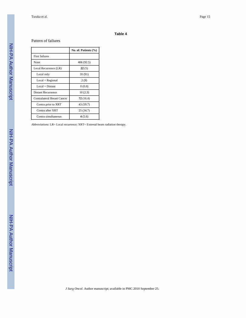
The median follow up was 6.8 years (range 0.2–24). Patterns of recurrence are shown inTable 4. There were 22 local recurrences (5%), two with simultaneous regional nodalrecurrence. None of the patients had distant metastases at the time of LR. 7/22 (32%) of thelocal recurrences were invasive cancers. The interval to LR was 71 months (range: 7–247months).
Pathologic size did not appear to correlate with the risk of LR. The median size of tumor inpatients without LR was 0.8 cm (range: 0.1–5 cm) compared to with LR 0.7 cm (range: 0.4 –2.4 cm), (p=0.5). There was no apparent correlation between architectural pattern and the
Turaka et al. Page 3
J Surg Oncol. Author manuscript; available in PMC 2010 September 25.
NIH
-PA Author Manuscript
NIH
-PA Author Manuscript
NIH
-PA Author Manuscript
risk of a LR. 1 out of 74 of these patients with comedo histology recurred, 2/68 withcribriform histology, compared to 18 of 296 with non-comedo/cribriform histology (5 LRwere in patients in whose histologic subtype was not recorded). Re-excision was required in271 patients (61.7%) either for a therapeutic resection after excisional biopsy or due tomargin status. 102 of these patients had a focally positive reexcision margin, 160 had anegative margin, and 9 had LCIS. 15 LR occurred in patients who had a reexcision (5.5%,15/271) and 5 had distant metastases. 8 LR occurred in patients whose reexcisiondemonstrated no residual tumor, and there were 7 LR in the setting of a positive reexcision.There were 14 LR and one locoregional recurrence in 198 patients who had no postbiopsymammogram, compared with 3 patients of 240 with a postbiopsy mammogram who had LR.
10 patients (2.3%) ultimately developed distant metastases. 8 of these patients died ofdisease and 2 patients were alive with disease (one had 79 months, second had 2 months offollowup). The 10 and 15 year actuarial cause-specific survival are 98% and 93%. 39patients died of other causes at a median of 9.9 years after their treatment. The 10- year and15- year actuarial overall survival rates are 93% and 92% respectively. 74 patients (16.2%)developed contralateral breast cancer. 44 patients received prior radiation therapy, 26developed after radiotherapy and 4 had simultaneous contralateral tumors. The median timeto develop contralateral breast tumor in 26 patients was 66.5 months (range: 3–196 months).The histology of the contralateral primary was invasive ductal cancer and DCIS. 60/74patients were alive without disease. 39 contralateral breast cancer occurred in patients with apositive family history and 35 had negative family history. 19/74 patients were treated withprior tamoxifen and in 4/26 who developed contralateral tumor, tamoxifen was used.
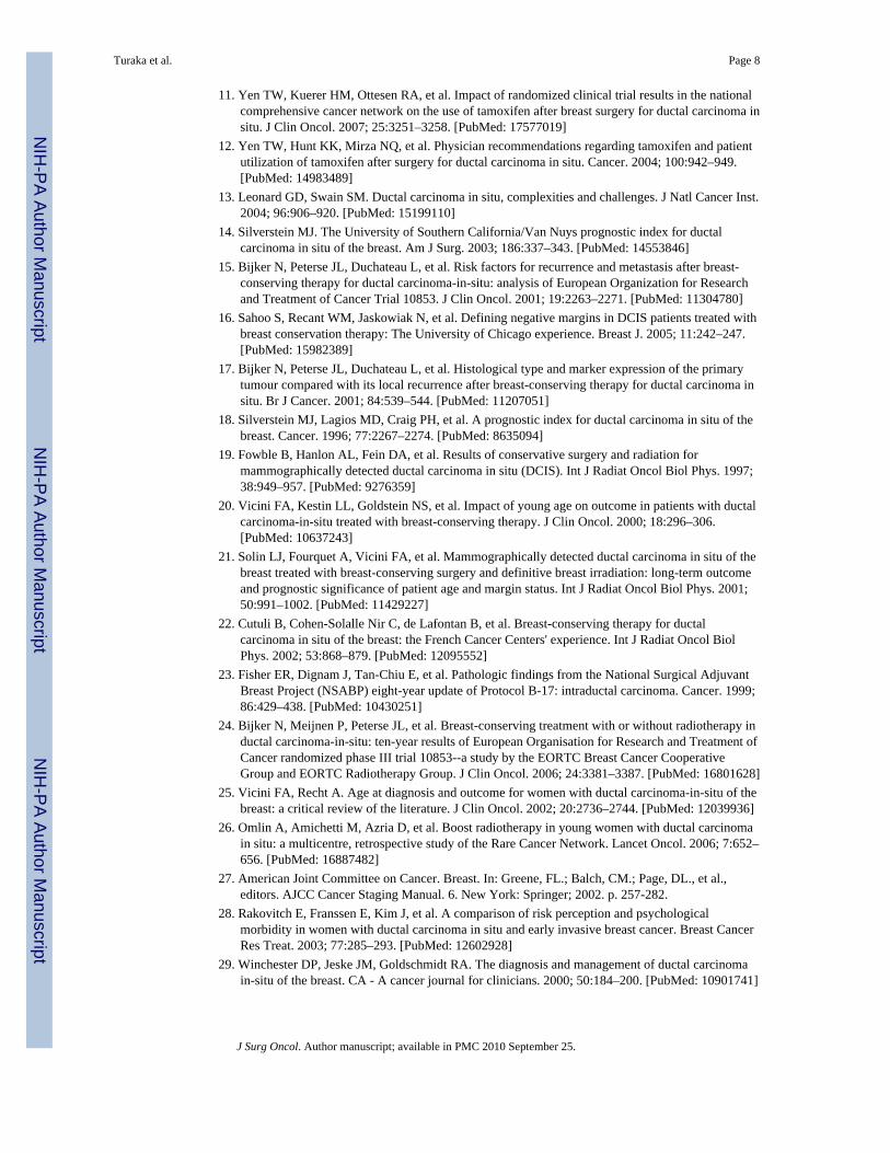
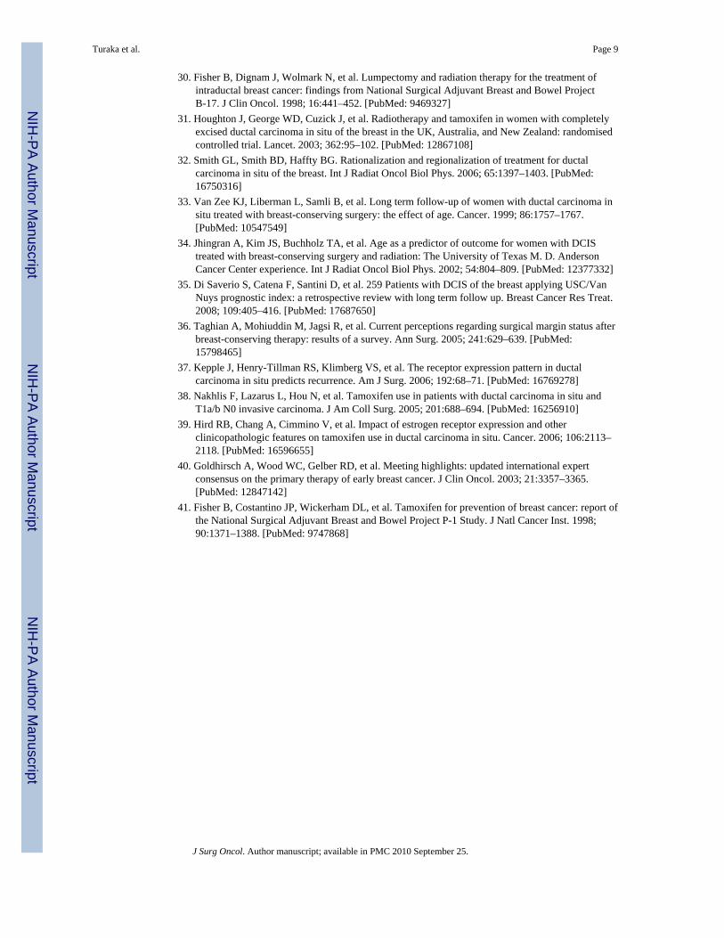
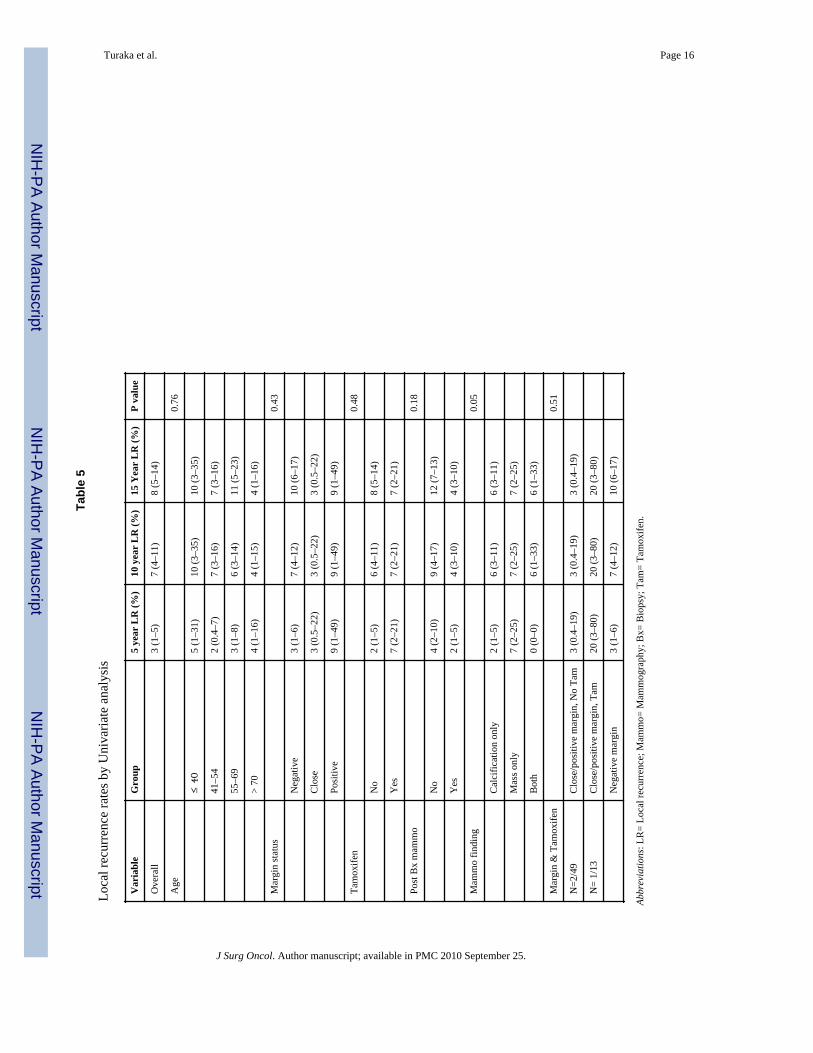
Actuarial outcome for breast recurrence is presented in Table 5. The 10 year and 15 yearactuarial rates of LR for all patients are 7% (4–11) and 8% (5–14) respectively (Fig. 1).There was no difference in LR by age (p=0.76), as shown in Figure 2. The LR rates at 15years were 10% (3–35%) in patient’s ≤ 40 years, 7% (3–16%) 41–54, 11% (5–23%) 55–69,and 4% (1–15%) =70 years. There were no significant differences in 15 year LR in patientswith close 3% (0.5–22), positive 9% (1–49), or negative 10 (6–17) margins (0.43). Post-biopsy mammogram (p=0.18), and use of tamoxifen (p=0.48) had no effect on LR. In thesubgroup of patients with close or positive margins, 10-year LR with tamoxifen (n=13) was20% (3–80) compared with 3% (0.4–19) without tamoxifen (n=49) (p=0.51).
There were 21 patients who developed an invasive LR recurrence underwent mastectomyalone without nodal staging and one had local excision only. The histology of recurrencewas colloid carcinoma (4 patients), invasive ductal and ductal carcinoma in situ (7 patients),DCIS only (9 patients), invasive lobular in patient and unknown in one patient. The size ofrecurrence was 0.9 cm (range: 0.3–2.5 cm). All the recurrences were detectedmammographically, and 4 also had a palpable mass, 2 had bloody nipple discharge. Themedian interval to recurrence was 71 months (range: 7 to 247 months). Ten of 22 LR werein a separate quadrant from the primary. At the time of mastectomy, 9 patients had negativeaxillary dissection and 12 patients had no axillary dissection. One patient receivedchemotherapy and one patient had both adjuvant chemotherapy and tamoxifen. With amedian followup of 62 months years after salvage mastectomy, 18 of 22 patients remainalive without evidence of disease, 1 died of disease and 3 died without evidence of disease.
DISCUSSIONThe prevention of LR (invasive and noninvasive) is a primary goal of the treatment forDCIS. Women with DCIS can experience serious concerns and psychological distress aswomen with invasive cancer despite their excellent prognosis with near 100% five-yeardisease-specific survival rates (28). Although DCIS was originally detected predominantly
Turaka et al. Page 4
J Surg Oncol. Author manuscript; available in PMC 2010 September 25.
NIH
-PA Author Manuscript
NIH
-PA Author Manuscript
NIH
-PA Author Manuscript
by physical examination as a mass (29), screening and treatment for DCIS have changed asmammographic screening detects smaller areas of nonpalpable DCIS amenable to BCS.BCS + RT has now become a standard option to mastectomy, with adjuvant tamoxifen oraromatase inhibitors postoperatively to reduce the incidence of recurrence (9,10,15,30–32).
There are several randomized trials proving that the addition of radiation therapy decreasesLR rates. National Surgical Adjuvant Breast and Bowel Project (NSABP) trial B-17 (30),conducted by Fisher et al compared lumpectomy with lumpectomy plus radiation,demonstrating reduction in invasive and noninvasive LR at 8 years of followup (non-invasive LR 13.4% to 8.2%; invasive LR 13.4% to 3.9%). The cumulative incidence of LRof any type was reduced from 26.8% to 12.1% with the addition of radiation (absoluterecurrence rate at 5 years: 21% in control arm and 10% in radiotherapy arm). In theEuropean Organisation for Research and Treatment of Cancer (EORTC) 10853 trial (15,24)by Bijker et al, with the addition of RT, the risk of LR reduced by 47% at 10 year follow up(absolute recurrence rate at 4 years: 16% and 9% respectively). The disease-free survivalrate was 74% in patients treated with BCS alone compared with 85% in women treated withBCS + RT. In the United Kingdom Coordinating Committee on Cancer Research(UKCCCR) trial (31), at a median follow up of 52.6 months, the LR rates were 4.8% withRT and 13.7% without RT. There was absolute reduction in risk of LR of 8.9% in the RTarm. Age is thought to possibly have contributed to the lower rates in the UKCCR trial asthe cohort was slightly older by comparison, with 90.5% of patients having been ≥50 yearsof age, although this is by no means certain. In an international collaborative multi-institutional study reported by Solin et al (21), 422 patients with DCIS were treatedprospectively with BCS + RT. The 10 year rate of LR was 31% in patients aged ≤ 39 years,13% in 40–49 year age, 8% for 50–59 years and 6% for age ≥ 60 years (p= 0.0001). Patientage ≤ 39 years and positive margins of resection were independently associated withincreased risk of LR (p= 0.0006 and p= 0.023, respectively).
Younger patient age has been associated in retrospective studies with a higher risk of LRcompared to older women when treated with BCS and RT (Table 6). Silverstein et al (14,18)felt the need to modify the original Van Nuys prognostic index by adding age (14) due tothis association. The relative LR rate was 2.3 in patients 40–60 years of age compared with3.2 in patients <40 years (p = 0.001). The disease-free survival was statistically significantfor age (p ≤ 0.01) along with other predictive factors such as tumor size, margin width andpathologic classification. In a retrospective review by Van Zee et al (33), at median followup of 74 months, there were 33 local recurrences (crude rate, 21%) in young women. Theactuarial LR rate was 16% at 6 years and the actuarial 6-year LR rates were 10.8%, 14%,and 47.2% in patients aged = 70, 40–69 and <40 years, respectively (p = 0.047).Radiotherapy reduced the risk of LR for each age subgroup. In patients ≤ 39 years, the 6year actuarial rate of LR in patients with BCS alone was 59% compared to 39% in thosetreated with RT, while for patients 40–69, the rates were 19% and 8%, and for ≥ 70 years,the rates were 14% and 0% respectively. Vicini et al (20) reported that younger patientshave greater risk of LR independent of other risk factors (5 and 10 year recurrence rateswere 21%, 26% in <45 years and 7%, 9% in >45 yr). Jhingran et al (34) reported an actuarialrate of LR-free survival at 5 and 10 years of 96% and 88% at 63 months. There was nodifference in incidence of true recurrence/marginal miss at 5 and 10 years in youngerpatients ( ≤ 40 years: 5%, 5%) compared with older patients (≥ 40 years: 3%, 4%), p= 0.39.Analysis of 259 patients of DCIS by Di Saverio et al (35) showed that age was not found tobe significant factor influencing the LR rates. At a mean follow up 130 months, LR wasseen in 21 patients (8%). The actuarial rates at 10 years were 89.3%, 90.7% and 88% foreach group according to age < 40, 40–40, and > 60 years, respectively (p=NS).
Turaka et al. Page 5
J Surg Oncol. Author manuscript; available in PMC 2010 September 25.
NIH
-PA Author Manuscript
NIH
-PA Author Manuscript
NIH
-PA Author Manuscript

The cause of the association of young age with LR may be due to association with otheradverse pathologic factors. A literature review by Vicini and Recht (25) studied theinfluence of age at diagnosis on characteristics and outcomes for women with DCIS.Younger patients contained grade 3 tumors, those of comedo subtype and those havinghigher maximum distance of intraductal spread through the breast, with higher residualtumor burdens when compared with the older patients. Younger patients treated with BCS +RT had a significantly higher rate of LR than older patients especially for invasive LR.There was inconsistency among different studies in defining the age limit for youngerwomen ranging from ≤ 35 or ≤ 50 years. They concluded that this age group requires carefulattention to selection and surgical technique but young age should not be a contraindicationto BCS + RT.
The potential limitations of our study include that the data is from a single institution and aretrospective analysis. Our study of younger patients age ≤ 40 years is limited by smallnumbers. We did not find a significant difference by age in LR, but to detect such adifference would require an extremely large cohort. Patients with mastectomy are notincluded in the analysis. There may have been other factors of patient selection at work aswell, such as preference for mastectomy in patients who were tested for BRCA positivity.We did not see a statistical difference in LR by use of post-biopsy mammograms, althoughas a negative margin was defined as 2 mm and not tumor on ink, the effect of postexcisionalmammography may have been minimized. Despite this, we still advocate re-imaging of apatient prior to radiation to confirm that all calcifications have been completely removed forassurance of the adequacy of resection. Similarly, with widely varying opinions on thedefinition of a clear margin (36) and without prospective randomized data comparing theimpact of margins on DCIS LR, we continue to recommend re-excision when margins orpositive or close, as defined by our institutional standard.
The use of a boost in DCIS was standard in our experience, although it does not have thesame level of randomized evidence as in invasive breast cancer. The total radiotherapy doseused in all randomized trials of DCIS was 50 Gy to the whole breast. A boost to the tumorbed was not considered in these studies. There was an emphasis on boost in a subset ofpatients especially young patients aged ≤ 40 years in a retrospective study by Omlin et al(26) to estimate the effect of boost radiotherapy on LR. The disease-free survival at 10 yearswas 46% for patients without radiotherapy, 72% for those with radiotherapy without boostand 86% for those given radiotherapy and boost (p <0.0001). The LR-free rates for patientsaged 39 years or younger was 63% compared to 81% for those aged 40–45 years (Hazardratio: 1.00, p= 0.010). The routine use of a boost in our study may have contributed to thelow risk of LR seen in our young patients.
In our study, Tamoxifen was given in 95 patients (21.6%) but we did not detect a significantdifference in LR. Prospective data is mixed regarding the magnitude of the benefit oftamoxifen in the setting of DCIS. Receptor expression is important in DCIS for predictingthe patients who will benefit from the addition of tamoxifen in the adjuvant setting (10,37–39). Estrogen receptors (ER) are highly expressed in 50–60% of patients with DCIS. Thelevels of ER expression are higher in DCIS lesions with less aggressive histologic features,such as low grade, increasing differentiation and absence of necrosis, than in those withmore aggressive features (13). In 2003, the St Gallen International Consensus Panelendorsed the use of tamoxifen in hormone- receptor positive DCIS (40) and the role oftamoxifen has been evaluated in different series (10,11,13,31,38,39,41). The use oftamoxifen has increased from 24% to 46% in a study by Yen (11) et al before and after theyear 1999. 60% were offered tamoxifen and 54% chose to take the drug. In another study byNaklis (38) et al, tamoxifen was offered to 91% of DCIS patients and the accepted by 63%.In the NSABP B-24 study (10), (Fisher et al) patients were randomized to treatment with
Turaka et al. Page 6
J Surg Oncol. Author manuscript; available in PMC 2010 September 25.
NIH
-PA Author Manuscript
NIH
-PA Author Manuscript
NIH
-PA Author Manuscript

lumpectomy, RT, and placebo or lumpectomy, RT, and tamoxifen and there was reductionin breast cancer events at 5 years (13.4% vs. 8.2%, p= 0.0009). Younger patients (≤ 49years) were at higher risk for LR than older patients (LR were 16% vs 6.5% in placebogroup, 11% vs 5.2% with Tamoxifen, p= 0.09). Tamoxifen administration resulted in a 38%reduction in LR in women younger than 50 years and a 22% reduction in women older than50 years. There was little benefit with the addition of tamoxifen in the UKCCCR trial (31).The differnce was not significant statistically on subgroup analysis (10% in observation arm,6% with tamoxifen alone, 1% in RT alone and 1% with RT and tamoxifen). In patients ≤ 50years, the LR rates were 18% in those treated with tamoxifen and 26% without tamoxifen(p= 0.19). Tamoxifen also did not significantly reduce the incidence of LR in the UnitedKingdom/Australia New Zealand ramdomized trial (31).
CONCLUSIONSWe report very low risks of LR after BCS + RT for DCIS, even in young patients ≤ 40years. There was no apparent difference in LR rates by age, margins, or use of tamoxifen.This may be due to strict patient selection for size and margins, a high utilization of re-excision, and radiation boost. This data may better allow teams of mutispecialty physiciansto communicate risks and benefits of breast conservation with their patients, particularlythose of young age. Study of newer predictors of biologic behaviour may be helpful inassessing which of these patients are at increased risk for progression to invasive cancer.
AcknowledgmentsThe authors thank Cindy Rosser for her collection and management of the data for the study population.
References1. Jemal A, Murray T, Samuels A, et al. Cancer statistics, 2003. CA Cancer J Clin. 2003; 53:5–26.
[PubMed: 12568441]2. Ernster VL, Barclay J, Kerlikowske K, et al. Incidence of and treatment for ductal carcinoma in situ
of the breast. Jama. 1996; 275:913–918. [PubMed: 8598618]3. Sumner WE 3rd, Koniaris LG, Snell SE, et al. Results of 23,810 cases of ductal carcinoma-in-situ.
Ann Surg Oncol. 2007; 14:1638–1643. [PubMed: 17245612]4. Freedman GM, Anderson PR, Goldstein LJ, et al. Routine mammography is associated with earlier
stage disease and greater eligibility for breast conservation in breast carcinoma patients age 40 yearsand older. Cancer. 2003; 98:918–925. [PubMed: 12942557]
5. Tabar L, Chen HH, Fagerberg G, et al. Recent results from the Swedish Two-County Trial: theeffects of age, histologic type, and mode of detection on the efficacy of breast cancer screening. JNatl Cancer Inst Monogr. 1997:43–47. [PubMed: 9709274]
6. Bleicher RJ, Abrahamse P, Hawley ST, et al. The influence of age on the breast surgery decision-making process. Ann Surg Oncol. 2008; 15:854–862. [PubMed: 18058182]
7. Baxter NN, Virnig BA, Durham SB, et al. Trends in the treatment of ductal carcinoma in situ of thebreast. J Natl Cancer Inst. 2004; 96:443–448. [PubMed: 15026469]
8. Morrow M, White J, Moughan J, et al. Factors predicting the use of breast-conserving therapy instage I and II breast carcinoma. J Clin Oncol. 2001; 19:2254–2262. [PubMed: 11304779]
9. Fisher ER, Leeming R, Anderson S, et al. Conservative management of intraductal carcinoma(DCIS) of the breast. Collaborating NSABP investigators. J Surg Oncol. 1991; 47:139–147.[PubMed: 1649353]
10. Fisher B, Dignam J, Wolmark N, et al. Tamoxifen in treatment of intraductal breast cancer:National Surgical Adjuvant Breast and Bowel Project B-24 randomised controlled trial. Lancet.1999; 353:1993–2000. [PubMed: 10376613]
Turaka et al. Page 7
J Surg Oncol. Author manuscript; available in PMC 2010 September 25.
NIH
-PA Author Manuscript
NIH
-PA Author Manuscript
NIH
-PA Author Manuscript
11. Yen TW, Kuerer HM, Ottesen RA, et al. Impact of randomized clinical trial results in the nationalcomprehensive cancer network on the use of tamoxifen after breast surgery for ductal carcinoma insitu. J Clin Oncol. 2007; 25:3251–3258. [PubMed: 17577019]
12. Yen TW, Hunt KK, Mirza NQ, et al. Physician recommendations regarding tamoxifen and patientutilization of tamoxifen after surgery for ductal carcinoma in situ. Cancer. 2004; 100:942–949.[PubMed: 14983489]
13. Leonard GD, Swain SM. Ductal carcinoma in situ, complexities and challenges. J Natl Cancer Inst.2004; 96:906–920. [PubMed: 15199110]
14. Silverstein MJ. The University of Southern California/Van Nuys prognostic index for ductalcarcinoma in situ of the breast. Am J Surg. 2003; 186:337–343. [PubMed: 14553846]
15. Bijker N, Peterse JL, Duchateau L, et al. Risk factors for recurrence and metastasis after breast-conserving therapy for ductal carcinoma-in-situ: analysis of European Organization for Researchand Treatment of Cancer Trial 10853. J Clin Oncol. 2001; 19:2263–2271. [PubMed: 11304780]
16. Sahoo S, Recant WM, Jaskowiak N, et al. Defining negative margins in DCIS patients treated withbreast conservation therapy: The University of Chicago experience. Breast J. 2005; 11:242–247.[PubMed: 15982389]
17. Bijker N, Peterse JL, Duchateau L, et al. Histological type and marker expression of the primarytumour compared with its local recurrence after breast-conserving therapy for ductal carcinoma insitu. Br J Cancer. 2001; 84:539–544. [PubMed: 11207051]
18. Silverstein MJ, Lagios MD, Craig PH, et al. A prognostic index for ductal carcinoma in situ of thebreast. Cancer. 1996; 77:2267–2274. [PubMed: 8635094]
19. Fowble B, Hanlon AL, Fein DA, et al. Results of conservative surgery and radiation formammographically detected ductal carcinoma in situ (DCIS). Int J Radiat Oncol Biol Phys. 1997;38:949–957. [PubMed: 9276359]
20. Vicini FA, Kestin LL, Goldstein NS, et al. Impact of young age on outcome in patients with ductalcarcinoma-in-situ treated with breast-conserving therapy. J Clin Oncol. 2000; 18:296–306.[PubMed: 10637243]
21. Solin LJ, Fourquet A, Vicini FA, et al. Mammographically detected ductal carcinoma in situ of thebreast treated with breast-conserving surgery and definitive breast irradiation: long-term outcomeand prognostic significance of patient age and margin status. Int J Radiat Oncol Biol Phys. 2001;50:991–1002. [PubMed: 11429227]
22. Cutuli B, Cohen-Solalle Nir C, de Lafontan B, et al. Breast-conserving therapy for ductalcarcinoma in situ of the breast: the French Cancer Centers' experience. Int J Radiat Oncol BiolPhys. 2002; 53:868–879. [PubMed: 12095552]
23. Fisher ER, Dignam J, Tan-Chiu E, et al. Pathologic findings from the National Surgical AdjuvantBreast Project (NSABP) eight-year update of Protocol B-17: intraductal carcinoma. Cancer. 1999;86:429–438. [PubMed: 10430251]
24. Bijker N, Meijnen P, Peterse JL, et al. Breast-conserving treatment with or without radiotherapy inductal carcinoma-in-situ: ten-year results of European Organisation for Research and Treatment ofCancer randomized phase III trial 10853--a study by the EORTC Breast Cancer CooperativeGroup and EORTC Radiotherapy Group. J Clin Oncol. 2006; 24:3381–3387. [PubMed: 16801628]
25. Vicini FA, Recht A. Age at diagnosis and outcome for women with ductal carcinoma-in-situ of thebreast: a critical review of the literature. J Clin Oncol. 2002; 20:2736–2744. [PubMed: 12039936]
26. Omlin A, Amichetti M, Azria D, et al. Boost radiotherapy in young women with ductal carcinomain situ: a multicentre, retrospective study of the Rare Cancer Network. Lancet Oncol. 2006; 7:652–656. [PubMed: 16887482]
27. American Joint Committee on Cancer. Breast. In: Greene, FL.; Balch, CM.; Page, DL., et al.,editors. AJCC Cancer Staging Manual. 6. New York: Springer; 2002. p. 257-282.
28. Rakovitch E, Franssen E, Kim J, et al. A comparison of risk perception and psychologicalmorbidity in women with ductal carcinoma in situ and early invasive breast cancer. Breast CancerRes Treat. 2003; 77:285–293. [PubMed: 12602928]
29. Winchester DP, Jeske JM, Goldschmidt RA. The diagnosis and management of ductal carcinomain-situ of the breast. CA - A cancer journal for clinicians. 2000; 50:184–200. [PubMed: 10901741]
Turaka et al. Page 8
J Surg Oncol. Author manuscript; available in PMC 2010 September 25.
NIH
-PA Author Manuscript
NIH
-PA Author Manuscript
NIH
-PA Author Manuscript
30. Fisher B, Dignam J, Wolmark N, et al. Lumpectomy and radiation therapy for the treatment ofintraductal breast cancer: findings from National Surgical Adjuvant Breast and Bowel ProjectB-17. J Clin Oncol. 1998; 16:441–452. [PubMed: 9469327]
31. Houghton J, George WD, Cuzick J, et al. Radiotherapy and tamoxifen in women with completelyexcised ductal carcinoma in situ of the breast in the UK, Australia, and New Zealand: randomisedcontrolled trial. Lancet. 2003; 362:95–102. [PubMed: 12867108]
32. Smith GL, Smith BD, Haffty BG. Rationalization and regionalization of treatment for ductalcarcinoma in situ of the breast. Int J Radiat Oncol Biol Phys. 2006; 65:1397–1403. [PubMed:16750316]
33. Van Zee KJ, Liberman L, Samli B, et al. Long term follow-up of women with ductal carcinoma insitu treated with breast-conserving surgery: the effect of age. Cancer. 1999; 86:1757–1767.[PubMed: 10547549]
34. Jhingran A, Kim JS, Buchholz TA, et al. Age as a predictor of outcome for women with DCIStreated with breast-conserving surgery and radiation: The University of Texas M. D. AndersonCancer Center experience. Int J Radiat Oncol Biol Phys. 2002; 54:804–809. [PubMed: 12377332]
35. Di Saverio S, Catena F, Santini D, et al. 259 Patients with DCIS of the breast applying USC/VanNuys prognostic index: a retrospective review with long term follow up. Breast Cancer Res Treat.2008; 109:405–416. [PubMed: 17687650]
36. Taghian A, Mohiuddin M, Jagsi R, et al. Current perceptions regarding surgical margin status afterbreast-conserving therapy: results of a survey. Ann Surg. 2005; 241:629–639. [PubMed:15798465]
37. Kepple J, Henry-Tillman RS, Klimberg VS, et al. The receptor expression pattern in ductalcarcinoma in situ predicts recurrence. Am J Surg. 2006; 192:68–71. [PubMed: 16769278]
38. Nakhlis F, Lazarus L, Hou N, et al. Tamoxifen use in patients with ductal carcinoma in situ andT1a/b N0 invasive carcinoma. J Am Coll Surg. 2005; 201:688–694. [PubMed: 16256910]
39. Hird RB, Chang A, Cimmino V, et al. Impact of estrogen receptor expression and otherclinicopathologic features on tamoxifen use in ductal carcinoma in situ. Cancer. 2006; 106:2113–2118. [PubMed: 16596655]
40. Goldhirsch A, Wood WC, Gelber RD, et al. Meeting highlights: updated international expertconsensus on the primary therapy of early breast cancer. J Clin Oncol. 2003; 21:3357–3365.[PubMed: 12847142]
41. Fisher B, Costantino JP, Wickerham DL, et al. Tamoxifen for prevention of breast cancer: report ofthe National Surgical Adjuvant Breast and Bowel Project P-1 Study. J Natl Cancer Inst. 1998;90:1371–1388. [PubMed: 9747868]
Turaka et al. Page 9
J Surg Oncol. Author manuscript; available in PMC 2010 September 25.
NIH
-PA Author Manuscript
NIH
-PA Author Manuscript
NIH
-PA Author Manuscript
Figure 1.15-year rates of local recurrence for 440 patients with DCIS
Turaka et al. Page 10
J Surg Oncol. Author manuscript; available in PMC 2010 September 25.
NIH
-PA Author Manuscript
NIH
-PA Author Manuscript
NIH
-PA Author Manuscript
Figure 2.15-year rates of local recurrence by age
Turaka et al. Page 11
J Surg Oncol. Author manuscript; available in PMC 2010 September 25.
NIH
-PA Author Manuscript
NIH
-PA Author Manuscript
NIH
-PA Author Manuscript

NIH
-PA Author Manuscript
NIH
-PA Author Manuscript
NIH
-PA Author Manuscript
Turaka et al. Page 12
Table 1
Patient and tumor related characteristics of Mammographically detected DCIS
No. of. Patients (%)
Total 440
Age (years) Median 56.5
< 41 24 (5.5)
41–54 169 (38.5)
55–69 169 (38.5)
> 70 78 (17.8)
Menopausal status
Pre 119 (27.1)
Peri 27 (6.2)
Post 294 (67)
Race
White 397 (90.4)
Black 32 (7.3)
Others 11 (2.5)
Family history
Negative 18 (4.1)
Positive 168 (38.3)
1 relative 105 (62.9)
>2 relatives 62 (37.1)
Mammographic findings
Calcification only 336 (76.5)
Mass 43 (9.8)
Mass and calcifications 30 (6.8)
Location primary
Outer 241 (54.9)
Inner 77 (17.5)
Central 94 (21.4)
Subareolar 28 (6.4)
Method of detection
Mammogram + PE 41 (9.3 )
Mammogram only 377 (85.9)
PE only 21 (4.8)
Other (MRI/CT) 1 (0.2)
Abbreviations: DICS= Ductal carcinoma in situ; PE= Physical examination; MRI= Magnetic resonance imaging; CT: Computed tomography.
J Surg Oncol. Author manuscript; available in PMC 2010 September 25.
NIH
-PA Author Manuscript
NIH
-PA Author Manuscript
NIH
-PA Author Manuscript
Turaka et al. Page 13
Table 2
Tumor related characteristics
No. of. Patients (%)
Path size primary
Median tumor size 0.8 cm
Tumor size range 0.1–5.0 cm
Histology
Comedo 74 (16.9)
Papillary 15 (3.4)
Micropapillary 14 (3.2)
Cribriform 68 (15.5)
Solid 41 (9.3)
Mixed 162 (36.9)
Unknown 64 (14..6)
Grade of tumor
Grade 1 40 (9.1)
Grade 2 104 (23.7)
Grade 3 137 (31.2)
Unknown 159 (36.2)
Necrosis
Yes 162 (36.9)
No 9 (2.1)
Unknown 269 (61.3)
Nodal status
Nx 378 (86.1)
N0 62 (14.1)
Hormone Receptors
Both ER/PR positive 77 (17.5)
Both ER/PR negative 32 (7.3)
ER/PR positive 24 (5.5)
ER/PR unknown 307 (69.9)
LCIS
Positive 36 (8.2)
Negative 404 (92)
Unknown 0 (0.0)
Abbreviations: ER= Estrogen receptor; PR= Progesterone receptor; LCIS= Lobular carcinoma in situ.
J Surg Oncol. Author manuscript; available in PMC 2010 September 25.

NIH
-PA Author Manuscript
NIH
-PA Author Manuscript
NIH
-PA Author Manuscript
Turaka et al. Page 14
Table 3
Treatment related characteristics
No. of. Patients (%)
Re-excision
Yes 271 (61.7)
No 169 (38.5)
Final margin status
Negative 342 (77.9)
Close 49 (11.2)
Positive 13 (3)
LCIS only 6 (1.4)
Unknown 30 (7)
Radiotherapy
Total dose primary (median dose), tangents 5000 cGy (200–5040)
Boost , median dose-range 1000 (1000–1800)
Yes 420 (95.7)
No 20 (4.6)
Post Bx Mammogram
Yes 240 (54.7)
No 198 (45.1)
Unknown 2 (0.5)
Adjuvant treatment
Tamoxifen 96 (21.9)
No systemic therapy 342 (77.9)
Chemotherapy only 1 (0.2)
Chemotherapy/Tamoxifen 1 (0.2)
Abbreviations: cGy= Centi gray; LCIS= Lobular carcinoma in situ; Bx= Biopsy.
J Surg Oncol. Author manuscript; available in PMC 2010 September 25.
NIH
-PA Author Manuscript
NIH
-PA Author Manuscript
NIH
-PA Author Manuscript
Turaka et al. Page 15
Table 4
Pattern of failures
No. of. Patients (%)
First failures
None 406 (92.5)
Local Recurrence (LR) 22 (5)
Local only 20 (91)
Local + Regional 2 (9)
Local + Distant 0 (0.0)
Distant Recurrence 10 (2.3)
Contralateral Breast Cancer 72 (16.4)
Contra prior to XRT 43 (59.7)
Contra after XRT 25 (34.7)
Contra simultaneous 4 (5.6)
Abbreviations: LR= Local recurrence; XRT= External beam radiation therapy.
J Surg Oncol. Author manuscript; available in PMC 2010 September 25.
NIH
-PA Author Manuscript
NIH
-PA Author Manuscript
NIH
-PA Author Manuscript
Turaka et al. Page 16
Tabl
e 5
Loca
l rec
urre
nce
rate
s by
Uni
varia
te a
naly
sis
Var
iabl
eG
roup
5 ye
ar L
R (%
)10
yea
r L
R (%
)15
Yea
r L
R (%
)P
valu
e
Ove
rall
3 (1
–5)
7 (4
–11)
8 (5
–14)
Age
0.76
≤ 40
5 (1
–31)
10 (3
–35)
10 (3
–35)
41–5
42
(0.4
–7)
7 (3
–16)
7 (3
–16)
55–6
93
(1–8
)6
(3–1
4)11
(5–2
3)
> 70
4 (1
–16)
4 (1
–15)
4 (1
–16)
Mar
gin
stat
us0.
43
Neg
ativ
e3
(1–6
)7
(4–1
2)10
(6–1
7)
Clo
se3
(0.5
–22)
3 (0
.5–2
2)3
(0.5
–22)
Posi
tive
9 (1
–49)
9 (1
–49)
9 (1
–49)
Tam
oxife
n0.
48
No
2 (1
–5)
6 (4
–11)
8 (5
–14)
Yes
7 (2
–21)
7 (2
–21)
7 (2
–21)
Post
Bx
mam
mo
0.18
No
4 (2
–10)
9 (4
–17)
12 (7
–13)
Yes
2 (1
–5)
4 (3
–10)
4 (3
–10)
Mam
mo
findi
ng0.
05
Cal
cific
atio
n on
ly2
(1–5
)6
(3–1
1)6
(3–1
1)
Mas
s onl
y7
(2–2
5)7
(2–2
5)7
(2–2
5)
Bot
h0
(0–0
)6
(1–3
3)6
(1–3
3)
Mar
gin
& T
amox
ifen
0.51
N=2
/49
Clo
se/p
ositi
ve m
argi
n, N
o Ta
m3
(0.4
–19)
3 (0
.4–1
9)3
(0.4
–19)
N=
1/13
Clo
se/p
ositi
ve m
argi
n, T
am20
(3–8
0)20
(3–8
0)20
(3–8
0)
Neg
ativ
e m
argi
n3
(1–6
)7
(4–1
2)10
(6–1
7)
Abbr
evia
tions
: LR
= Lo
cal r
ecur
renc
e; M
amm
o= M
amm
ogra
phy;
Bx=
Bio
psy;
Tam
= Ta
mox
ifen.
J Surg Oncol. Author manuscript; available in PMC 2010 September 25.

NIH
-PA Author Manuscript
NIH
-PA Author Manuscript
NIH
-PA Author Manuscript
Turaka et al. Page 17
Tabl
e 6
Tabl
e sh
owin
g ris
k of
recu
rren
ce a
fter W
E+X
RT
in D
CIS
Aut
hor
Yea
rN
o. o
f. pa
tient
sA
geIB
TR
Tim
e in
terv
al
Fow
ble
1997
8≤4
025
%5
year
s
Fish
er19
9913
7≤4
915
%8
year
s
Van
Zee
1999
15<
4033
%6
year
s
Vic
ini
2000
31<
4524
%10
yea
rs
Szel
i-Ste
vens
2000
44<
509.
1%8.
7 ye
ars
Vic
ini
2002
31<
4521
%7
year
s
Solin
2001
31<
4031
%10
yea
r
Rod
rigue
s20
029
≤35
0 %
8 ye
ars
Cut
uli
2002
8<
4029
%7
year
s
Jhin
gran
2002
20≤4
05%
5 ye
ars
Om
lin20
0610
8≤3
928
%10
yea
rs
Bijk
er20
0665
≤40
34%
10 y
ears
Di S
aver
io20
0822
< 40
10.7
, 0.3
, 12%
10 y
ears
Abbr
evia
tions
: WE=
Wid
e ex
cisi
on; X
RT=
Ext
erna
l bea
m ra
diat
ion
ther
apy;
DC
IS=
Duc
tal c
arci
nom
a in
situ
; IB
TR=
Ipsi
late
ral B
reas
t Tum
or R
ecur
renc
e.
J Surg Oncol. Author manuscript; available in PMC 2010 September 25.

NIH
-PA Author Manuscript
NIH
-PA Author Manuscript
NIH
-PA Author Manuscript
Turaka et al. Page 18
Tabl
e 7
Ris
k of
recu
rren
ce a
fter W
E+X
RT+
Tam
oxife
n in
DC
IS
Aut
hor
Yea
rN
o. o
f. pa
tient
sA
geIB
TR
Tim
e in
terv
al
Fish
er19
9960
2≤
4913
.2%
6 ye
ars
Hou
ghto
n20
0316
0<
5013
%4.
4 ye
ars
Abbr
evia
tions
: As i
n ta
ble
6.
J Surg Oncol. Author manuscript; available in PMC 2010 September 25.