Application of Blind Deconvolution Denoising in Failure Prognosis
Upload
khangminh22Category
view
1download
0
HAL Id: hal-02468510https://hal.archives-ouvertes.fr/hal-02468510
Submitted on 22 Oct 2021
HAL is a multi-disciplinary open accessarchive for the deposit and dissemination of sci-entific research documents, whether they are pub-lished or not. The documents may come fromteaching and research institutions in France orabroad, or from public or private research centers.
L’archive ouverte pluridisciplinaire HAL, estdestinée au dépôt et à la diffusion de documentsscientifiques de niveau recherche, publiés ou non,émanant des établissements d’enseignement et derecherche français ou étrangers, des laboratoirespublics ou privés.
Distributed under a Creative Commons Attribution - NonCommercial| 4.0 InternationalLicense
Patterns of recurrence and prognosis in locally advancedFIGO stage IB2 to IIB cervical cancer: Retrospective
multicentre study from the FRANCOGYN groupTiphaine de Foucher, Sofiane Bendifallah, Lobna Ouldamer, Alexandre
Bricou, Vincent Lavoué, Justine Varinot, Geoffroy Canlorbe, XavierCarcopino, Emilie Raimond, Laurie Monnier, et al.
To cite this version:Tiphaine de Foucher, Sofiane Bendifallah, Lobna Ouldamer, Alexandre Bricou, Vincent Lavoué, etal.. Patterns of recurrence and prognosis in locally advanced FIGO stage IB2 to IIB cervical cancer:Retrospective multicentre study from the FRANCOGYN group. EJSO - European Journal of SurgicalOncology, WB Saunders, 2019, 45 (4), pp.659-665. �10.1016/j.ejso.2018.11.014�. �hal-02468510�
1
Patterns of recurrence and prognosis in locally advanced FIGO stage IB2 to IIB
cervical cancer: retrospective multicentre study from the FRANCOGYN Group.
Short version of title: Recurrence and prognosis for locally advanced cervical cancer
Tiphaine de FOUCHER (M.D), Sofiane BENDIFALLAH (M.D, PhD) 1,2, Lobna OULDAMER
(M.D, PhD) 3, Alexandre BRICOU (M.D.) 4, Vincent LAVOUE (M.D, PhD) 5, Justine
VARINOT (M.D) 6, Geoffroy CANLORBE (M.D) 1, Xavier CARCOPINO (M.D, PhD) 7,
Emilie RAIMOND (M.D) 8, Laurie MONNIER (M.D) 1, Olivier GRAESSLIN (M.D, PhD) 8,
Cyril TOUBOUL (M.D, PhD) 9, Pierre COLLINET (M.D, PhD) 10, Marie-Emmanuelle
NEVEU (M.D) 11, Cyrille HUCHON (M.D, PhD) 12, Emile DARAÏ (M.D, PhD) 1,13, Marcos
BALLESTER (M.D, PhD) 1,13
For the Groupe de Recherche FRANCOGYN , FRANCE.
1. Department of Gynaecology and Obstetrics, Tenon University Hospital, Assistance
Publique des Hôpitaux de Paris (AP-HP), University Pierre and Marie Curie, Paris 6,
Institut Universitaire de Cancérologie (IUC), 4 rue de la Chine, 75020 Paris France.
2. INSERM UMR_S_707, "Epidemiology, Information Systems, Modeling", University
Pierre and Marie Curie, Paris 6, 27 rue de Chaligny, 75012 Paris, France.
3 Department of Obstetrics and Gynaecology, Centre Hospitalier Régional
Universitaire de Tours, Hôpital Bretonneau, 2 bd Tonnellée, 37000 Tours, France.
4 Department of Obstetrics, Gynecology and Reproductive Medicine, Hôpitaux
Universitaires Paris Seine Saint-Denis, Assistance Publique-Hôpitaux de Paris, av du
14 juillet, 93140 Bondy, France.
5 CRLCC Eugène-Marquis, service de gynécologie, CHU de Rennes, université de
Rennes 1, av de la bataille Flandres-Dunkerque, 35000 Rennes, France.
6 Department of Pathology, Tenon University Hospital, Assistance Publique des
Hôpitaux de Paris (AP-HP), University Pierre and Marie Curie, Paris 6, 4 rue de la
Chine, 75020 Paris, France.
7 Department of Obstetrics and Gynaecology, Hôpital Nord, APHM, Aix-Marseille
University (AMU), CNRS, IRD, chemin des Bourrely, 13105 Marseille, France.
8 Department of Obstetrics and Gynaecology, Institute Alix de Champagne University
© 2018 published by Elsevier. This manuscript is made available under the CC BY NC user licensehttps://creativecommons.org/licenses/by-nc/4.0/
Version of Record: https://www.sciencedirect.com/science/article/pii/S0748798318320122Manuscript_b38f0042c921fe850e0d0a621a4f6c5c
2
Hospital, 45 rue Cognacq Jay, 51100 Reims, France.
9 Department of Obstetrics and Gynecology, Centre Hospitalier Intercommunal,
Créteil, 40 av de Verdun, 94000 Créteil, France.
10 Department of Gynecologic Surgery, Jeanne de Flandre Hospital, CHU de Lille, av
Eugène Avinée, 59000 Lille, France.
11 Department of Gynecology and Obstetrics, Kremlin-Bicêtre University Hospital, 78
rue du Général Leclerc, 94170 Le Kremlin-Bicêtre, France.
12 Department of Obstetrics and Gynecology, Poissy-St Germain Hospital, 10 rue du
champ Gaillard, 78000 Poissy, France.
13 INSERM UMR_S_938, University Pierre et Marie Curie, Paris 6, 27 rue de Chaligny,
75012 Paris, France.
Conflicts of interest: the authors report no conflict of interest.
Funding: No external funding source
Corresponding author : Sofiane BENDIFALLAH, MD, PhD
Service de Gynécologie-Obstétrique
Hôpital Tenon, 4 rue de la Chine, 75020 Paris, France
Phone: + 33 1 56 01 68 76 Fax: + 33 1 56 01 60 62
E-mail: [email protected]
Word counts : abstract : 231, main text : 2838.
3
Abbreviations:
BMI: body mass index
CC: cervical cancer
CCRT: concomitant chemo-radiotherapy
CIs: confidence intervals
DR: distant recurrence
FIGO: International Federation of Gynecology and Obstetrics
HT: first radical hysterectomy
INCa: French National Institue of Cancer
LACC: locally advanced cervical cancer
LN: lymph node
LRR: local regional recurrence
LVSI: lympho-vascular space invasion
MRI: Magnetic Resonance Imaging
OS: Overall survival
PAL: para-aortic lymph node dissection
PET-CT: Positron Emission Tomography–Computed Tomography
RFS: recurrence free survival
VBT: vaginal brachytherapy
4
ABSTRACT
Introduction: Evidence-based data describing patterns of recurrence and prognosis
in women with FIGO stage IB2 to IIB locally advanced cervical cancer (LACC) are
scarce. The purpose of this study was to analyse patterns of recurrence in LACC and
their correlation with prognosis, depending on FIGO stage, lymph node (LN) status
and treatment modalities. The endpoints of this study were the type of recurrence
(locoregional or distant, and time to recurrence), the recurrence free survival, the
overall survival and the cumulative incidence for both locoregional and distant
recurrence.
Materials and Methods: Data of women with FIGO stage IB2 to IIB CC treated
between April 1996 and May 2016 were retrospectively abstracted from nine French
institutions.
Results: The median follow-up for the 501 women included was 35.6 months.
Recurrences were observed in 158 (31.5%), with a mean time to recurrence of 20.7
months. Women with IIB CC had poorer prognosis, lower 3-year RFS and higher 3-
year cumulative incidence of both locoregional and distant recurrences. Women
with positive or unknown LN status had poorer prognosis with higher 3-year
cumulative incidence of distant recurrence. Women who underwent concomitant
chemo-radiotherapy +/- vaginal brachytherapy had poorer prognosis, with lower 3-
year RFS and higher 3-year cumulative incidence of distant recurrence.
Conclusions: Recurrence location and time to recurrence differ widely depending on
the FIGO stage, LN status and treatment modalities, with potential impact on follow-
up modalities and therapeutic approaches.
Key words: locally advanced cervical cancer, recurrence, pattern, survival
5
INTRODUCTION
Cervical cancer (CC) is the fourth most common cancer in women, with an
estimated worldwide incidence of 527 600 and a death rate of 265 700 reported in
2015 [1). In women with FIGO (International Federation of Gynecology and
Obstetrics) stage IB2 to IIB locally advanced cervical cancer (LACC), recurrences have
been reported to occur in 15% to 40%, with a 5-year recurrence-free survival (RFS)
averaging from 50% to 70% [2][3][4][5][6].
It is now well established that most events in CC are observed within 2 years
following the diagnosis [7]. Recurrences after primary treatment are often located in
the true pelvis, but other locations including distant recurrences or peritoneal
carcinomatosis can also be observed underlining the prognostic heterogeneity of the
disease [8][9][10][11]. To reduce this heterogeneity, several epidemiological,
histological and treatment prognostic factors for recurrence and survival have been
reported and are currently used to define optimal CC management [12][13]. In this
setting, different models have been developed based on the prognostic factors to
predict recurrence and survival in LACC [6][14][15][16]. However, to date, few
evidence-based data are available about patterns of recurrence (location, timing
from initial treatment) and prognosis for FIGO stage IB2 to IIB CC. As a result, LACC
management guidelines from various countries are heterogeneous, especially
concerning: (i) the prognostic and therapeutic value of surgical nodal staging, (ii) the
optimal treatment modalities, and (iii) the rationale of post-operative follow-up
[17][18][19][20][21].
The aim of the study was to analyse pattern of recurrence in women with
LACC based on a large retrospective French multicentre database. Analyses were
stratified according to FIGO stage, lymph node (LN) status and treatment modalities.
MATERIAL AND METHODS
1. Study population
The data of women with histologically proven FIGO stage IB2 to IIB CC treated
between April 1996 and May 2016 were retrospectively abstracted from nine
institutions with prospectively maintained CC databases in France (Tenon University
6
Hospital, Tours University Hospital, Creteil University Hospital, Reims University
Hospital, Rennes University Hospital, Jeanne de Flandre University Hospital, Poissy
University Hospital, Jean Verdier Hospital and Marseille North University Hospital).
All the women had given written consent to participate in the study, and the
research protocol was approved by the Institutional Review Board of the Collège
National des Gynécologues et Obstétriciens Français (CEROG 2016-GYN-0502).
All enrolled women underwent pre-operative workup including history,
physical examination, cervical biopsy, Magnetic Resonance Imaging (MRI) and
Positron Emission Tomography–Computed Tomography (PET-CT), if indicated
according to the FIGO stage and the period of treatment. Cystoscopy and/or
proctoscopy were performed if there was a suspicion of bladder or rectal
involvement after clinical examination or on MRI.
Clinical, surgical and pathological data as well as details of adjuvant therapies
were collected: the woman’s age, body mass index (BMI; calculated as weight in
kilograms divided by the square of height in meters), surgical procedure (type of
hysterectomy and/or LN staging), FIGO stage, final pathological analysis (histological
type, tumour grade, tumour size and lympho-vascular space invasion (LVSI) status),
treatment modalities and prognosis (recurrence, death). All women were classified
according to the 2009 FIGO classification [22].
2. Therapeutic management
Therapeutic management was decided on by a multidisciplinary committee
on an individual basis, according to the French National Institue of Cancer (INCa)
guidelines [21], depending on FIGO stage and results of PET-CT when available.
When indicated, laparoscopic LN staging was performed including para-aortic (PAL)
+/- pelvic lymph node dissection. We applied the following definitions to describe
the LN status: women were considered as node positive when they had metastatic
LNs on PET-CT or after surgical nodal staging; as node negative when they had
disease free LN on surgical nodal staging; and as nodal status unknown if neither
PET-CT nor surgical staging had been performed or with negative PET-CT but without
surgical nodal staging.
Clinical follow-up consisted of physical examinations and the use of imaging
7
techniques according to the findings. Follow-up sessions were conducted every 3
months for the first 2 years, every 6 months for the following 3 years, and once a
year thereafter.
3. Definition and classification of recurrence
Recurrent disease was assessed by physical examination, imaging techniques
and biopsy when feasible.
According to a previous report [23], we applied the following definition to
describe the patterns of recurrence: local regional recurrence (LRR) was defined
recurrences including (i) the cervix, (ii) the vaginal vault, (iii) pelvic LNs, and (iv) the
pelvic walls. Distant recurrence (DR) could include (i) metastatic spread to organs
whatever the location, (ii) peritoneal carcinomatosis and (iii) extra pelvic LN
involvement.
RFS was defined as the time from the date of primary treatment to any CC
recurrence and was censored at the date of last follow-up or death without
recurrence. Overall survival (OS) was defined as the time from primary treatment to
death as a result of any cause.
4. Statistical analysis
The women’s characteristics, and tumour and treatment characteristics were
analysed using Chi-square statistics or Fisher’s exact test for categorical variables
and the t-test or analysis of variance (ANOVA) for continuous variables. Kaplan-
Meier estimates were used to estimate the event-time distributions, and log-rank
test was used to compare the differences among the different groups in terms of RFS
and OS. The kernel-smoothed hazard functions of RFS were estimated on the basis of
the method described by Cardoso et al. [24]. Time to the first CC recurrence for a
specific site was evaluated by cumulative incidence analysis (Gray's test) and
competing risk regression analysis to estimate sub- distribution hazard ratios (HRs)
and 95% confidence intervals (CIs). Analysis was stratified according to FIGO stage,
LN status and treatment modalities. Values of p < 0.05 were considered to denote
significant differences. Data were managed with an Excel database (Microsoft,
Redmond, WA, USA) and analysed using the R 2·15 software, available online.
8
RESULTS
1. Epidemiological and surgical characteristics of the population.
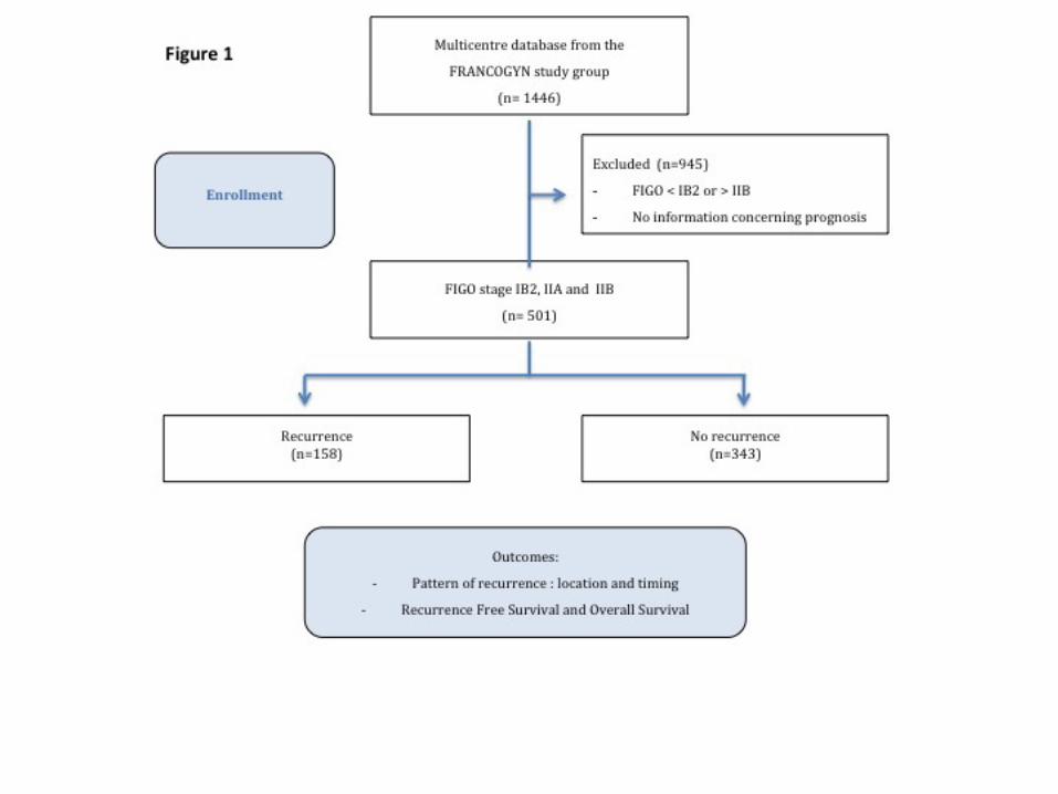
During the study period, 501 women with FIGO stage IB2 to IIB LACC were
documented as having received treatment. Recurrences were observed in 158
(31.5%): 72 were LRR and 86 DR (among which 20 were both LRR and DR). The study
flow chart is shown in Figure 1. Epidemiological and histological characteristics of the
population are reported in Table 1.
2. OS and RFS according to FIGO stage, LN status and treatment modalities.
The median follow-up was 35.6 months (1.1-146.5 months). In the whole
population, the respective 3-year OS and RFS were 83.5% (95% CI, 79.8-87.3) and
67.4% (95% CI, 63.1 – 72.1), p<0,05. The respective 3-year OS according to
recurrence location were 66.1% (95% CI, 54.4-80.3) for LRR and 56.1% (95% CI, 45.2-
69.6) for DR.
The respective 3-year RFS for women with FIGO stages IB2, IIA and IIB were
79.1% (95% CI, 69.5 – 89.9), 79.1% (95% CI, 70.2 – 89.1) and 62.0% (95% CI, 56.6 –
68.0), respectively, (p < 0.001). No difference was found in 3-year OS according to
FIGO stage.
The respective 3-year OS were 87.1% (95% CI, 82.4 – 92.0), 78.7% (95% CI, 71.9
– 86.1) and 81.7% (95% CI, 72.4 – 92.3), for node negative women, node positive
women and those with unknown LN status, respectively (p < 0.001). The respective
3-year RFS were 76.8 % (95% CI, 71.1- 82.9), 58.4% (95% CI, 50.7 – 67.3), and 61.8 %
(95% CI, 51.2 – 74.5), for node negative women, node positive women and those
with unknown LN status, respectively (p < 0.0001).
The respective 3-year OS were 90.7% (95% CI, 82.3 – 99.9), 85.2% (95% CI, 80.1
– 90.7) and 79.1% (95% CI, 73.0 – 85.8), for women who underwent a first radical
hysterectomy (HT) +/- concomitant chemo-radiotherapy (CCRT) +/- vaginal
brachytherapy (VBT), CCRT +/- VBT followed by radical HT and CCRT +/- VBT,
respectively (p<0.01). The respective 3-year RFS were 75.8% (95% CI, 64.9 – 88.5),
74.3 % (95% CI, 68.1- 81.1) and 59.2 % (95% CI, 52.3 – 65.6), for women who
underwent a first radical HT +/- CCRT +/- VBT, CCRT +/- VBT followed by radical HT
9
and CCRT +/- VBT, respectively (p < 0.0001).
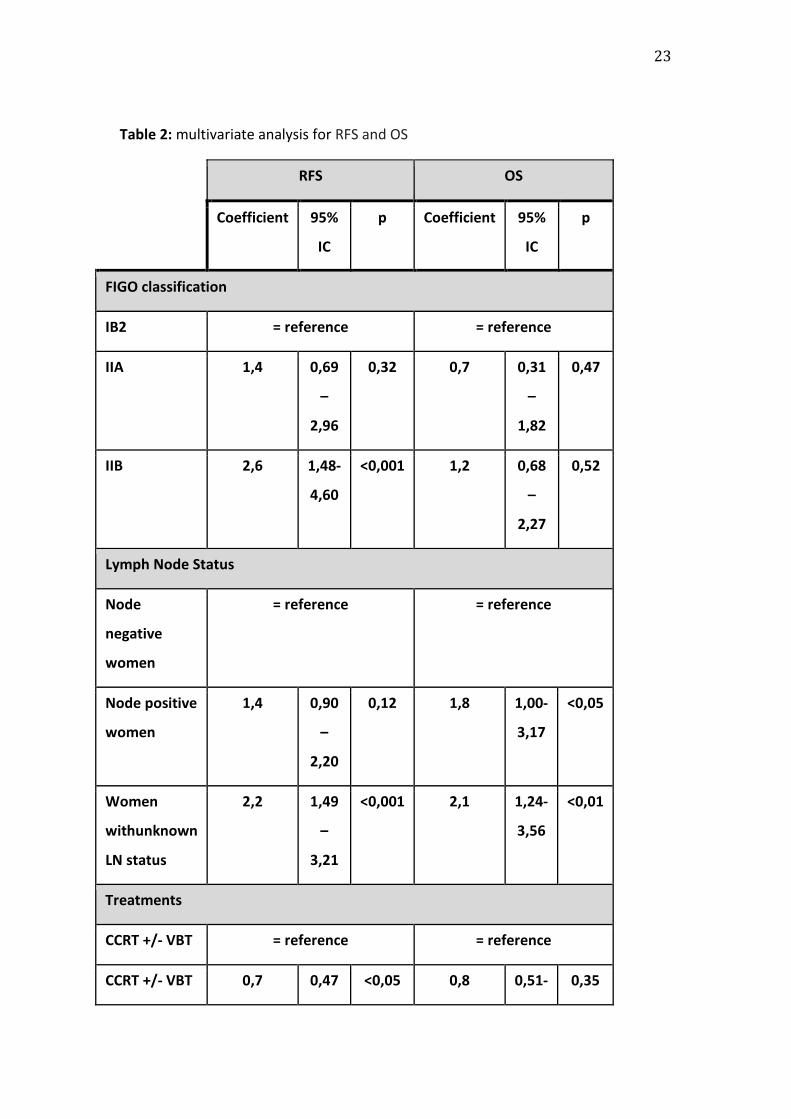
As reported in Table 2, after multivariate analysis FIGO stage and LN status
remain significant independent risk factors for recurrence.
3. Cumulative incidence for LRR and DR according to FIGO stage, LN status and
treatment.
Overall, recurrences were observed in 158 of the 501 women (31.5%). The
mean time to recurrence was 20.72 months (3-122months). The 3-year cumulative
incidences of LRR and DR in the whole population were 16.1% and 18.8%,
respectively.
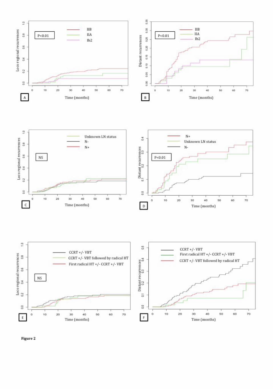
The 3-year cumulative incidence rates and their distribution according to
FIGO stage, LN status and treatment modalities are reported in Figure 2 ( A, B, C, D,
E, F). Women with FIGO stage IIB CC had a higher 3-year cumulative incidence for
both LRR and DR than women with FIGO stage IIA or IB2 CC (19.4% and 22.4% vs.
12.5% and 9.6%, and 7.4% and 13.4% respectively, p<0.01). Node positive women
and women with unknown LN status had a higher 3-year cumulative incidence of DR
than node negative women (29.1% and 25.1% vs. 10.7%, p<0.001). No significant
difference in LRR was observed according to LN status. Women treated with CCRT
+/- VBT had a higher 3-year cumulative incidence of DR than women treated with
CCRT+/-VBT followed by radical HT or first radical HT +/- CCRT +/- VBT (27.8% (95%
CI, 20.3 – 34.6), 13.9% (95% CI, 8.4 - 19) and 7.1 % (95% CI, 1 – 13.6), p<0.001). No
significant difference in LRR was observed according to treatment modalities.
DISCUSSION
This study reports specific site and time patterns of first recurrence for LACC
according to the main prognostic factors (i.e. FIGO stage and LN status) and
treatment modalities. Our results confirm that FIGO stage IIB, positive LNs as well as
unknown LN status are major prognostic factors which affect the time to first
recurrence and site as well as the HR for recurrence. Such results are of interest to
adapt long-term clinical follow-up and as a starting point to improve therapeutic
management of LACC.
LN status is known to be a major prognostic factor for all stages of CC [5].
10
However, the prognostic impact of surgical LN staging in the standard management
of FIGO stage IB2 to IIB CC remains to be proved, as published results concerning the
superiority of surgical over radiological staging are contradictory [25] [26]. For
example, Lai et al. found no benefit of surgical staging in a randomized controlled
trial comparing surgical to clinical staging. In contrast, Gold et al. found that surgical
exclusion of positive para-aortic LNs in patients with LACC who received
chemoradiation had a significant prognostic impact compared with radiographic
exclusion. In our study, 60% of the women underwent radiological nodal staging and
72% surgical nodal staging, highlighting the considerable heterogeneity in practice
within the same country. We found that women without optimal LN staging (i.e. with
neither radiological nor surgical LN staging or with negative radiological staging and
no surgical staging) had poorer 3-year RFS and OS, close to that of node positive
women (61.8% vs. 58.4% and 81.7% vs. 78.7%). We found no significant difference in
LRR according to LN status, but a significantly higher rate of DR among node positive
women and women without optimal LN staging than among node negative women
(29.1% and 25.1% vs. 10.7%)(p < 0.001). For women with positive nodes, these
results are consistent with literature [13]. Our results are of importance for women
with unknown LN status, as surgical staging is currently considered a key procedure
to plan the extent of radiation therapy (RT) strategy in France. Thus, we can
hypothesize that some of the women without optimal LN staging were undertreated,
explaining such a poor prognosis. In this setting, several authors have suggested
performing systematic CCRT in all women with LACC, with a prophylactic extension
of the radiation field to the para-aortic area regardless of the staging [26][27]. This
latter option must be questioned in the light of our results, especially for women
who did not undergo optimal nodal staging.
Although it is commonly accepted that the treatment of LACC is primarily based on
CCRT [21], treatment guidelines are inconsistent [17][18][19][20][21] and the place
of radical HT remains to be determined [28][29]. In our study, women treated with
exclusive CCRT +/- VBT had poorer prognosis (with a lower 3-year RFS and a higher 3-
year cumulative incidence of DR) than women who underwent either first radical HT
+/- CCRT +/- VBT or CCRT +/- VBT followed by radical rather than completion HT.
Nevertheless, these results should be interpreted in the light of the LN status as
11
French guidelines recommend exclusive CCRT +/- VBT for node positive women
while completion HT is restricted to patients with negative lymph node status.
Among the 222 women treated by exclusive CCRT +/- VBT, 97 (43.9%) had positive
LNs and 47 (21.2%) had unknown LN status while among the 279 women who
underwent other treatment modalities, 59 (21.1%) had positive LNs and 38 (13.6%)
had unknown LN status. A meta-analysis studying the impact of adjuvant
hysterectomy in patients with LACC treated with CCRTshowed no improval on OS,
although hysterectomy seemed to reduce the risk of recurrence. Moreover, the
author recomanded that routine use of hysterectomy should be avoided due to
significant morbidity in these patients [30]. Legge F et al., after analyzing the
patterns of recurrence and their association with clinical outcome in LACC patients
submitted to primary chemoradiation followed by radical surgery, found most of the
recurrences were outside the irradiated field (57.3%). This is consistent with our
results, as we observed 56,9% of DR. Among the parameters of recurrence
associated with RFS they bring out, only secondary radical surgery retains an
independent predictive role in reducing the risk of death (p=0.037) [31]. Hence,
future randomized trials are needed to clearly define the place of first intention
radical HT and of completion HT in the treatment of FIGO stage IB2 to IIB CC.
A better knowledge of stage IB2 to IIB patterns of recurrence is also needed to
adapt follow-up modalities as, to date, post-treatment monitoring programmes
differ widely from country to country [32]. Current international guidelines
recommend physical examination every 3-6 months for 2 years, then every 6 months
or annually. Imaging studies are recommended when clinically indicated, testing for
serum tumour markers is optional, and the frequency of the pap-smear test is
controversial [18][19][20][21]. However, clinical examination alone seems to be
insufficient as several authors report the low performance of clinical monitoring to
detect asymptomatic recurrences. This suggests a need of prospective cost-
effectiveness studies as well as multilticentre randomized clinical trials to compare
various follow-up policies [8][33][34]. Furthermore, there is limited evidence of a
significant impact on survival of systematic regular CT scans or MRI for asymptomatic
patients [35][36] [37] [39] [40]. Even though PET-CT has been shown to be useful in
the diagnosis of recurrent CC, especially in the case of unexplained elevation of
12
serum tumour markers without evidence of recurrent disease on conventional
workup [41], the exact indications of this exam in the setting of CC recurrence are
yet to be defined. However, no study to date has focused on the follow-up
modalities for women at increased risk of recurrence. Our results suggest that closer
monitoring including systematic imaging procedures would be of benefit to these
patients, especially for those with positive or unknown LN status.
The strengths of our study lie in its multicentre nature and the large number of
women included, but some limits deserve to be mentioned. First, we cannot exclude
an inherent bias linked its retrospective multicenter nature. Indeed, the guidelines
changed during the period of data collection and PET-CT was introduced which
incurred modification to the management strategy over the years. In the following
retrospective long-term study especially concerning the surgical strategy of pelvic
lymphadenectomy is not homogeneous. However, all included women were treated
in regional referral centres applying French/European guidelines after systematic
multidisciplinary committee approval. Second, there are considerable differences in
describing the recurrences from study to study which could also bias the comparison
of reported rates. Based on previous reports, we opted to combine cervical, vaginal
vault, pelvic LN, and pelvic wall recurrences as LRR, and organ metastasis, peritoneal
carcinomatosis and extra-pelvic LN recurrences as DR [23]. Third, even if our results
allowed us to describe patterns of recurrences in LACC, they should be carefully
analysed beyond 3 years before drawing any firm conclusion. Finally, our results are
consistant with previous report. Indeed, Rose et al. and Ferrandina et al. also
suggested that clinical stage and LN status were significantly associated with clinical
outcomes [15] [42].
CONCLUSION
Our results show that the patterns of recurrence in women with LACC differ
widely in terms of time to recurrence and site, depending on the FIGO stage, LN
status and treatment modalities. We hypothesize that characterizing and
understanding the behaviour of these tumours may have profound implications on
treatment options and follow-up modalities. Thus our results could give rise to
improved monitoring programmes and should be taken into account when designing

13
future therapeutic approaches.
14
REFERENCES
1. Ferlay J, Soerjomataram I, Dikshit R, Eser S, Mathers C, Rebelo M, et al. Cancer
incidence and mortality worldwide: sources, methods and major patterns in
GLOBOCAN 2012. Int J Cancer. 1 mars 2015;136(5):E359-386.
2. Delgado G, Bundy B, Zaino R, Sevin BU, Creasman WT, Major F. Prospective
surgical-pathological study of disease-free interval in patients with stage IB
squamous cell carcinoma of the cervix: a Gynecologic Oncology Group study.
Gynecol Oncol. sept 1990;38(3):352�7.
3. Inoue T, Morita K. The prognostic significance of number of positive nodes in
cervical carcinoma stages IB, IIA, and IIB. Cancer. 1 mai 1990;65(9):1923�7.
4. Takeda N, Sakuragi N, Takeda M, Okamoto K, Kuwabara M, Negishi H, et al.
Multivariate analysis of histopathologic prognostic factors for invasive cervical
cancer treated with radical hysterectomy and systematic retroperitoneal
lymphadenectomy. Acta Obstet Gynecol Scand. déc 2002;81(12):1144�51.
5. Quinn MA, Benedet JL, Odicino F, Maisonneuve P, Beller U, Creasman WT, et al.
Carcinoma of the cervix uteri. FIGO 26th Annual Report on the Results of
Treatment in Gynecological Cancer. Int J Gynaecol Obstet Off Organ Int Fed
Gynaecol Obstet. nov 2006;95 Suppl 1:S43-103.
6. Polterauer S, Grimm C, Hofstetter G, Concin N, Natter C, Sturdza A, et al.
Nomogram prediction for overall survival of patients diagnosed with cervical
cancer. Br J Cancer. 4 sept 2012;107(6):918�24.
7. Webb MJ, Symmonds RE. Site of recurrence of cervical cancer after radical
hysterectomy. Am J Obstet Gynecol. 1 déc 1980;138(7 Pt 1):813�7.
8. Ansink A, de Barros Lopes A, Naik R, Monaghan JM. Recurrent stage IB cervical
carcinoma: evaluation of the effectiveness of routine follow up surveillance. Br
J Obstet Gynaecol. nov 1996;103(11):1156�8.
15
9. Landoni F, Colombo A, Milani R, Placa F, Zanagnolo V, Mangioni C. Randomized
study between radical surgery and radiotherapy for the treatment of stage IB-
IIA cervical cancer: 20-year update. J Gynecol Oncol. mai 2017;28(3):e34.
10. Lavoué V, Voguet L, Bertel C, Mesbah H, Williaume D, Laguerre B, et al. [Place
of surgery before and after concurrent chemoradiotherapy for locally advanced
cervical carcinoma: A retrospective study of 102 cases]. J Gynecol Obstet Biol
Reprod (Paris). févr 2011;40(1):11�21.
11. Carcopino X, Houvenaeghel G, Buttarelli M, Esterni B, Tallet A, Goncalves A, et
al. Equivalent survival in patients with advanced stage IB-II and III-IVA cervical
cancer treated by adjuvant surgery following chemoradiotherapy. Eur J Surg
Oncol J Eur Soc Surg Oncol Br Assoc Surg Oncol. mai 2008;34(5):569�75.
12. Burghardt E, Baltzer J, Tulusan AH, Haas J. Results of surgical treatment of 1028
cervical cancers studied with volumetry. Cancer. 1 août 1992;70(3):648�55.
13. Stehman FB, Bundy BN, DiSaia PJ, Keys HM, Larson JE, Fowler WC. Carcinoma of
the cervix treated with radiation therapy. I. A multi-variate analysis of
prognostic variables in the Gynecologic Oncology Group. Cancer. 1 juin
1991;67(11):2776�85.
14. Shim S-H, Lee S-W, Park J-Y, Kim YS, Kim D-Y, Kim J-H, et al. Risk assessment
model for overall survival in patients with locally advanced cervical cancer
treated with definitive concurrent chemoradiotherapy. Gynecol Oncol. janv
2013;128(1):54�9.
15. Rose PG, Java J, Whitney CW, Stehman FB, Lanciano R, Thomas GM, et al.
Nomograms Predicting Progression-Free Survival, Overall Survival, and Pelvic
Recurrence in Locally Advanced Cervical Cancer Developed From an Analysis of
Identifiable Prognostic Factors in Patients From NRG Oncology/Gynecologic
Oncology Group Randomized Trials of Chemoradiotherapy. J Clin Oncol. 1 juill
2015;33(19):2136.
16
16. Yoon WS, Yang DS, Lee JA, Lee NK, Park YJ, Kim CY, et al. Validation of
Nomograms for Survival and Metastases after Hysterectomy and Adjuvant
Therapy in Uterine Cervical Cancer with Risk Factors. BioMed Res Int.
2017;2017:2917925.
17. Koh W-J, Greer BE, Abu-Rustum NR, Apte SM, Campos SM, Chan J, et al.
Cervical cancer. J Natl Compr Cancer Netw JNCCN. 1 mars 2013;11(3):320�43.
18. PDQ Adult Treatment Editorial Board. Cervical Cancer Treatment (PDQ®):
Health Professional Version. In: PDQ Cancer Information Summaries [Internet].
Bethesda (MD): National Cancer Institute (US); 2002 [cité 12 avr 2017].
Disponible sur: http://www.ncbi.nlm.nih.gov/books/NBK66058/
19. Colombo N, Creutzberg C, Amant F, Bosse T, González-Martín A, Ledermann J,
et al. ESMO-ESGO-ESTRO Consensus Conference on Endometrial Cancer:
diagnosis, treatment and follow-up. Ann Oncol. 1 janv 2016;27(1):16�41.
20. Lim MC, Lee M, Shim SH, Nam EJ, Lee JY, Kim HJ, et al. Practice guidelines for
management of cervical cancer in Korea: a Korean Society of Gynecologic
Oncology Consensus Statement. J Gynecol Oncol. mai 2017;28(3):e22.
21. Haute Autorité de Santé - ALD n° 30 - Cancer invasif du col utérin [Internet].
[cité 12 avr 2017]. Disponible sur: http://www.has-
sante.fr/portail/jcms/c_922973/fr/ald-n-30-cancer-invasif-du-col-uterin
22. Pecorelli S, Zigliani L, Odicino F. Revised FIGO staging for carcinoma of the
cervix. Int J Gynaecol Obstet Off Organ Int Fed Gynaecol Obstet. mai
2009;105(2):107�8.
23. Yang K, Park W, Huh SJ, Bae D-S, Kim B-G, Lee J-W. Clinical outcomes in patients
treated with radiotherapy after surgery for cervical cancer. Radiat Oncol J
Radiat Oncol J [Internet]. 12 déc 2016 [cité 24 mars 2017]; Disponible sur:
http://www.e-roj.org/journal/view.php?doi=10.3857/roj.2016.01893
17
24. Metzger-Filho O, Sun Z, Viale G, Price KN, Crivellari D, Snyder RD, et al. Patterns
of Recurrence and Outcome According to Breast Cancer Subtypes in Lymph
Node–Negative Disease: Results From International Breast Cancer Study Group
Trials VIII and IX. J Clin Oncol. 1 sept 2013;31(25):3083�90.
25. Lai C-H, Huang K-G, Hong J i-Hong, Lee C-L, Chou H-H, Chang T-C, et al.
Randomized trial of surgical staging (extraperitoneal or laparoscopic) versus
clinical staging in locally advanced cervical cancer☆. Gynecol Oncol. 1 avr
2003;89(1):160�7.
26. Gold MA, Tian C, Whitney CW, Rose PG, Lanciano R. Surgical versus
radiographic determination of para-aortic lymph node metastases before
chemoradiation for locally advanced cervical carcinoma: a Gynecologic
Oncology Group Study. Cancer. 1 mai 2008;112(9):1954�63.
27. Leblanc E, Narducci F, Frumovitz M, Lesoin A, Castelain B, Baranzelli MC, et al.
Therapeutic value of pretherapeutic extraperitoneal laparoscopic staging of
locally advanced cervical carcinoma. Gynecol Oncol. mai 2007;105(2):304�11.
28. Morice P, Rouanet P, Rey A, Romestaing P, Houvenaeghel G, Boulanger JC, et al.
Results of the GYNECO 02 study, an FNCLCC phase III trial comparing
hysterectomy with no hysterectomy in patients with a (clinical and radiological)
complete response after chemoradiation therapy for stage IB2 or II cervical
cancer. The Oncologist. 2012;17(1):64�71.
29. Lèguevaque P, Motton S, Delannes M, Querleu D, Soulé-Tholy M, Tap G, et al.
Completion surgery or not after concurrent chemoradiotherapy for locally
advanced cervical cancer? Eur J Obstet Gynecol Reprod Biol. avr
2011;155(2):188�92.
30. Shim SH, Kim SN, Chae SH, Kim JE, Lee SJ. Impact of adjuvant hysterectomy on
prognosis in patients with locally advanced cervical cancer treated with
concurrent chemoradiotherapy: a meta-analysis. J Gynecol Oncol. mars
2018;29(2):e25.
18
31. Legge F, Chiantera V, Macchia G, Fagotti A, Fanfani F, Ercoli A, et al. Clinical
outcome of recurrent locally advanced cervical cancer (LACC) submitted to
primary multimodality therapies. Gynecol Oncol. juill 2015;138(1):83�8.
32. Bodurka-Bevers D, Morris M, Eifel PJ, Levenback C, Bevers MW, Lucas KR, et al.
Posttherapy surveillance of women with cervical cancer: an outcomes analysis.
Gynecol Oncol. août 2000;78(2):187�93.
33. Olaitan A, Murdoch J, Anderson R, James J, Graham J, Barley V. A critical
evaluation of current protocols for the follow-up of women treated for
gynecological malignancies: a pilot study. Int J Gynecol Cancer Off J Int Gynecol
Cancer Soc. oct 2001;11(5):349�53.
34. Zola P, Fuso L, Mazzola S, Piovano E, Perotto S, Gadducci A, et al. Could follow-
up different modalities play a role in asymptomatic cervical cancer relapses
diagnosis? An Italian multicenter retrospective analysis. Gynecol Oncol. oct
2007;107(1 Suppl 1):S150-154.
35. Zanagnolo V, Minig LA, Gadducci A, Maggino T, Sartori E, Zola P, et al.
Surveillance procedures for patients for cervical carcinoma: a review of the
literature. Int J Gynecol Cancer Off J Int Gynecol Cancer Soc. avr
2009;19(3):306�13.
36. Hricak H, Powell CB, Yu KK, Washington E, Subak LL, Stern JL, et al. Invasive
cervical carcinoma: role of MR imaging in pretreatment work-up--cost
minimization and diagnostic efficacy analysis. Radiology. févr
1996;198(2):403�9.
37. Jeong YY, Kang HK, Chung TW, Seo JJ, Park JG. Uterine cervical carcinoma after
therapy: CT and MR imaging findings. Radiogr Rev Publ Radiol Soc N Am Inc.
août 2003;23(4):969�81; discussion 981.
38. Schwartz LB, Panageas E, Lange R, Rizzo J, Comite F, McCarthy S. Female pelvis:
impact of MR imaging on treatment decisions and net cost analysis. Radiology.
juill 1994;192(1):55�60.
19
39. Yamashita Y, Harada M, Torashima M, Takahashi M, Miyazaki K, Tanaka N, et al.
Dynamic MR imaging of recurrent postoperative cervical cancer. J Magn Reson
Imaging JMRI. févr 1996;6(1):167�71.
40. Choi JI, Kim SH, Seong CK, Sim JS, Lee HJ, Do KH. Recurrent uterine cervical
carcinoma: spectrum of imaging findings. Korean J Radiol. déc
2000;1(4):198�207.
41. Chung HH, Kim S-K, Kim TH, Lee S, Kang KW, Kim J-Y, et al. Clinical impact of
FDG-PET imaging in post-therapy surveillance of uterine cervical cancer: from
diagnosis to prognosis. Gynecol Oncol. oct 2006;103(1):165�70.
42. Ferrandina G, Legge F, Fagotti A, Fanfani F, Distefano M, Morganti A, et al.
Preoperative concomitant chemoradiotherapy in locally advanced cervical
cancer: safety, outcome, and prognostic measures. Gynecol Oncol. oct
2007;107(1 Suppl 1):S127-132.
20
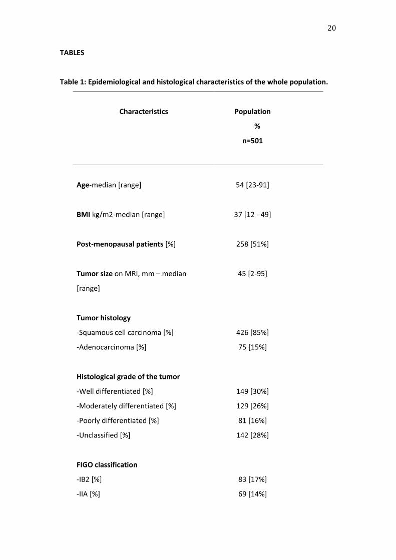
TABLES
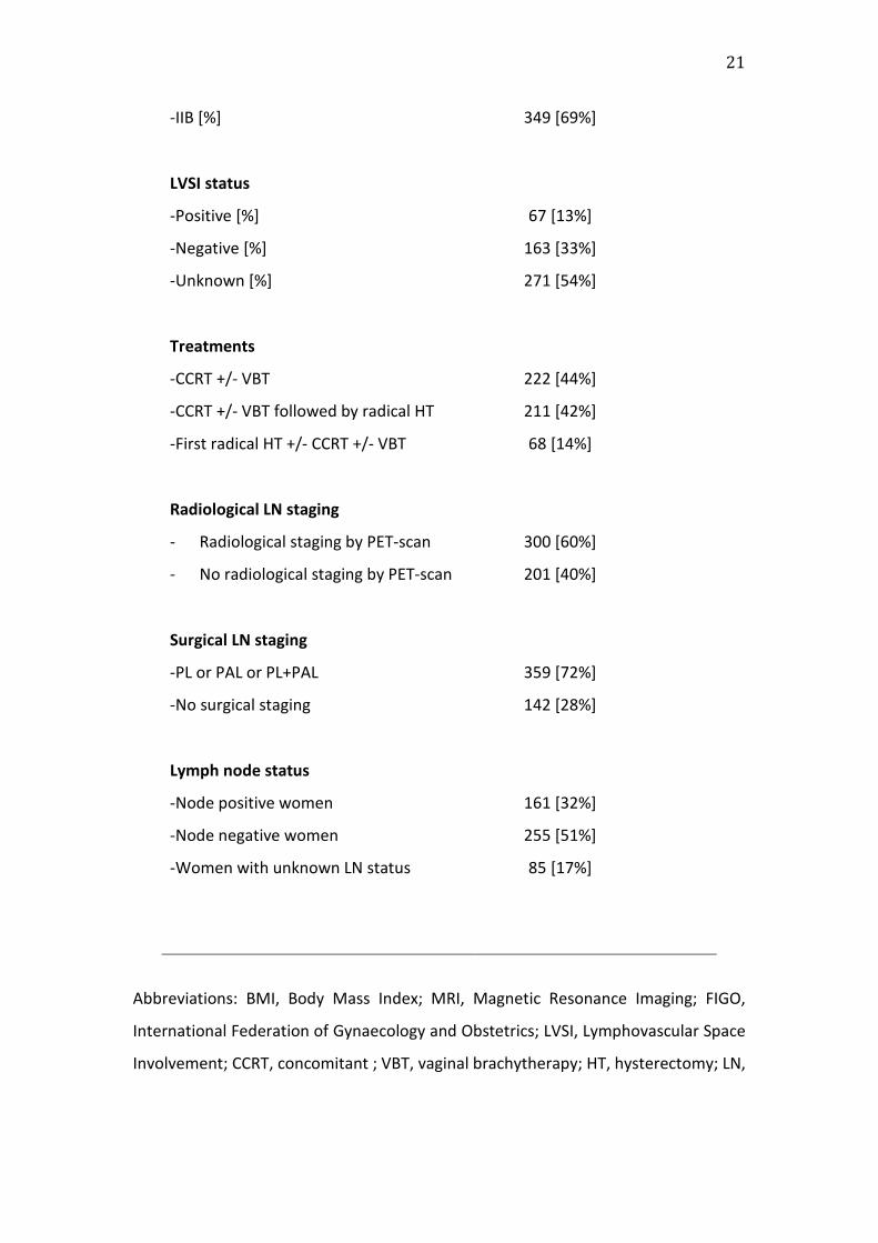
Table 1: Epidemiological and histological characteristics of the whole population.
Characteristics
Population
%
n=501
Age-median [range]
54 [23-91]
BMI kg/m2-median [range]
37 [12 - 49]
Post-menopausal patients [%]
258 [51%]
Tumor size on MRI, mm – median
[range]
45 [2-95]
Tumor histology
-Squamous cell carcinoma [%]
-Adenocarcinoma [%]
426 [85%]
75 [15%]
Histological grade of the tumor
-Well differentiated [%]
-Moderately differentiated [%]
-Poorly differentiated [%]
-Unclassified [%]
149 [30%]
129 [26%]
81 [16%]
142 [28%]
FIGO classification
-IB2 [%]
-IIA [%]
83 [17%]
69 [14%]
21
-IIB [%]
349 [69%]
LVSI status
-Positive [%]
-Negative [%]
-Unknown [%]
67 [13%]
163 [33%]
271 [54%]
Treatments
-CCRT +/- VBT
-CCRT +/- VBT followed by radical HT
-First radical HT +/- CCRT +/- VBT
222 [44%]
211 [42%]
68 [14%]
Radiological LN staging
- Radiological staging by PET-scan
- No radiological staging by PET-scan
300 [60%]
201 [40%]
Surgical LN staging
-PL or PAL or PL+PAL
-No surgical staging
359 [72%]
142 [28%]
Lymph node status
-Node positive women
-Node negative women
-Women with unknown LN status
161 [32%]
255 [51%]
85 [17%]
Abbreviations: BMI, Body Mass Index; MRI, Magnetic Resonance Imaging; FIGO,
International Federation of Gynaecology and Obstetrics; LVSI, Lymphovascular Space
Involvement; CCRT, concomitant ; VBT, vaginal brachytherapy; HT, hysterectomy; LN,

22
lymph node; PET, Positron Emisson Tomography; PL, pelvic lymphadenectomy; PAL,
para-aortic lymphadenectomy.
23
Table 2: multivariate analysis for RFS and OS
RFS OS
Coefficient 95%
IC
p Coefficient 95%
IC
p
FIGO classification
IB2 = reference = reference
IIA 1,4 0,69
–
2,96
0,32 0,7 0,31
–
1,82
0,47
IIB 2,6 1,48-
4,60
<0,001 1,2 0,68
–
2,27
0,52
Lymph Node Status
Node
negative
women
= reference = reference
Node positive
women
1,4 0,90
–
2,20
0,12 1,8 1,00-
3,17
<0,05
Women
withunknown
LN status
2,2 1,49
–
3,21
<0,001 2,1 1,24-
3,56
<0,01
Treatments
CCRT +/- VBT = reference = reference
CCRT +/- VBT 0,7 0,47 <0,05 0,8 0,51- 0,35
24
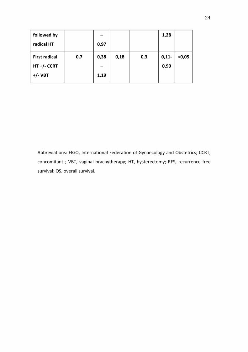
Abbreviations: FIGO, International Federation of Gynaecology and Obstetrics; CCRT,
concomitant ; VBT, vaginal brachytherapy; HT, hysterectomy; RFS, recurrence free
survival; OS, overall survival.
followed by
radical HT
–
0,97
1,28
First radical
HT +/- CCRT
+/- VBT
0,7 0,38
–
1,19
0,18 0,3 0,11-
0,90
<0,05
25
FIGURES
Figure 1: Flow Chart of the study
Abbreviations: FIGO, International Federation of Gynaecology and Obstetrics.
Figure 2 A: Three-year cumulative incidence rates of loco regional recurrences
according to FIGO stage
Figure 2 B: Three-year cumulative incidence rates of distant recurrences according to
FIGO stage
Figure 2 C: Three-year cumulative incidence rates of loco regional recurrences
according to LN status
Figure 2 D: Three-year cumulative incidence rates of distant recurrences according
to LN status
Figure 2 E: Three-year cumulative incidence rates of loco regional recurrences
according to treatment modalities
Figure 2 F: Three-year cumulative incidence rates of distant recurrences according to
treatment modalities
Abbreviations: FIGO, International Federation of Gynaecology and Obstetrics; LN,
lymph node; N+, Node positive women; N-, Node negative women; CCRT,
concomitant ; VBT, vaginal brachytherapy; HT, hysterectomy
Copyright © 2022 FDOKUMEN