What Are the Complications, Success and Survival ... - MDPI
15
Healthcare 2022, 10, 835. https://doi.org/10.3390/healthcare10050835 www.mdpi.com/journal/healthcare Systematic Review What Are the Complications, Success and Survival Rates for Autotransplanted Teeth? An Overview of Systematic Reviews and Metanalyses Ashutosh Kumar Singh 1 , Nikita Khanal 2 , Nisha Acharya 3 , Md Riasat Hasan 4, * and Takashi Saito 4 1 Department of Oral & Maxillofacial Surgery, Tribhuvan UniversityTeaching Hospital, Maharajgunj Medical Campus, Institute of Medicine, Kathmandu 44600, Nepal; [email protected] 2 Dental Surgeon, Ek EK Paila Foundation, Kathmandu 44600, Nepal; [email protected] 3 Department of Conservative Dentistry and Endodontics, Tribhuvan UniversityTeaching Hospital, Maharajgunj Medical Campus, Institute of Medicine, Kathmandu 44600, Nepal; [email protected] 4 Division of Clinical Cariology and Endodontology, Department of Oral Rehabilitation, School of Dentistry, Health Sciences University of Hokkaido, Ishikari-Tobetsu 061-0293, Japan; [email protected] * Correspondence: [email protected]; Tel./Fax: +81-133-23-1129 Abstract: Background: Autotransplantation is the surgical repositioning of a tooth within the same patient. It can be thought of as the controlled avulsion and re-implantation of a tooth and can be a viable alternative to other dental rehabilitation options. This review aimed to evaluate the survival rate (SR), major complications such as ankylosis rate (AR) and infection-related root resorption (RR), and overall success and failure rate (FR) in autotransplanted teeth. Methods: Six databases were accessed up to January 2021 to obtain all systematic reviews and meta-analyses (SRs and MAs). Study selection: After title and abstract reading, data extraction was performed from eligible SRs. The methodological quality was calculated for the included SRs using the risk of bias in systematic reviews (ROBIS) tool. Results: Six SRs were included in this review. The overall failure rate ranged from as low as 2.0% to 10.32%. The 1-year survival was very high (97.4–98.0%). The 5-year survival rate ranged from 81 to 98.2%. Major complications of AR ranged from 1.2 to 6.2%, and RR ranged from 2.1 to 10.4%. Conclusion: The overall findings from these SR and MA are promising; however, all the SRs include only single-arm prospective or retrospective studies, the SRs are of overall low methodological quality, and for the heterogeneity of the included SRs, well-designed comparative studies with a long-term follow-up are recommended. Keywords: autotransplantation; survival rate; success rate; complications; resorption 1. Introduction Autogenous tooth transplantation (ATT), or autotransplantation, is the surgical movement in one individual of a vital or endodontically treated tooth from its original location in the mouth to another site [1,2]. Tooth loss as a result of dental caries along with trauma is the most common indication, especially when mandibular and maxillary first molars are involved. Autogenous tooth transplantation was first documented in 1954 by M.L. Hale [3,4]. The major principles of his technique are still followed today. Initial results suggested only a 50% success rate, and there was little widespread acceptance of the technique [5,6]. Recent developments in the understanding of the nature of the periodontal ligament and cementum and the need for careful atraumatic extractions, use of systemic antibiotics, splinting techniques, and endodontic therapy, have led to a considerable improvement in the success rate and an increase in popularity [7–12]. The science of autotransplantation has progressed, as evidenced by the high success rates reported in studies over the last Citation: Singh, A.K.; Khanal, N.; Acharya, N.; Hasan, M.R.; Saito, T. What Are the Complications, Success and Survival Rates for Autotransplanted Teeth? An Overview of Systematic Reviews and Metanalyses. Healthcare 2022, 10, 835. https://doi.org/10.3390/ healthcare10050835 Academic Editor(s): Takahiro Kanno and Shintaro Sukegawa Received: 6 April 2022 Accepted: 29 April 2022 Published: 1 May 2022 Publisher’s Note: MDPI stays neu- tral with regard to jurisdictional claims in published maps and institu- tional affiliations. Copyright: © 2022 by the authors. Li- censee MDPI, Basel, Switzerland. This article is an open access article distributed under the terms and con- ditions of the Creative Commons At- tribution (CC BY) license (https://cre- ativecommons.org/licenses/by/4.0/).
-
Upload
khangminh22 -
Category
Documents
-
view
3 -
download
0
Transcript of What Are the Complications, Success and Survival ... - MDPI
Healthcare 2022, 10, 835. https://doi.org/10.3390/healthcare10050835 www.mdpi.com/journal/healthcare
Systematic Review
What Are the Complications, Success and Survival Rates for Autotransplanted Teeth? An Overview of Systematic Reviews and Metanalyses Ashutosh Kumar Singh 1, Nikita Khanal 2, Nisha Acharya 3, Md Riasat Hasan 4,* and Takashi Saito 4
1 Department of Oral & Maxillofacial Surgery, Tribhuvan UniversityTeaching Hospital, Maharajgunj Medical Campus, Institute of Medicine, Kathmandu 44600, Nepal; [email protected]
2 Dental Surgeon, Ek EK Paila Foundation, Kathmandu 44600, Nepal; [email protected] 3 Department of Conservative Dentistry and Endodontics, Tribhuvan UniversityTeaching Hospital,
Maharajgunj Medical Campus, Institute of Medicine, Kathmandu 44600, Nepal; [email protected]
4 Division of Clinical Cariology and Endodontology, Department of Oral Rehabilitation, School of Dentistry, Health Sciences University of Hokkaido, Ishikari-Tobetsu 061-0293, Japan; [email protected]
* Correspondence: [email protected]; Tel./Fax: +81-133-23-1129
Abstract: Background: Autotransplantation is the surgical repositioning of a tooth within the same patient. It can be thought of as the controlled avulsion and re-implantation of a tooth and can be a viable alternative to other dental rehabilitation options. This review aimed to evaluate the survival rate (SR), major complications such as ankylosis rate (AR) and infection-related root resorption (RR), and overall success and failure rate (FR) in autotransplanted teeth. Methods: Six databases were accessed up to January 2021 to obtain all systematic reviews and meta-analyses (SRs and MAs). Study selection: After title and abstract reading, data extraction was performed from eligible SRs. The methodological quality was calculated for the included SRs using the risk of bias in systematic reviews (ROBIS) tool. Results: Six SRs were included in this review. The overall failure rate ranged from as low as 2.0% to 10.32%. The 1-year survival was very high (97.4–98.0%). The 5-year survival rate ranged from 81 to 98.2%. Major complications of AR ranged from 1.2 to 6.2%, and RR ranged from 2.1 to 10.4%. Conclusion: The overall findings from these SR and MA are promising; however, all the SRs include only single-arm prospective or retrospective studies, the SRs are of overall low methodological quality, and for the heterogeneity of the included SRs, well-designed comparative studies with a long-term follow-up are recommended.
Keywords: autotransplantation; survival rate; success rate; complications; resorption
1. Introduction Autogenous tooth transplantation (ATT), or autotransplantation, is the surgical
movement in one individual of a vital or endodontically treated tooth from its original location in the mouth to another site [1,2]. Tooth loss as a result of dental caries along with trauma is the most common indication, especially when mandibular and maxillary first molars are involved.
Autogenous tooth transplantation was first documented in 1954 by M.L. Hale [3,4]. The major principles of his technique are still followed today. Initial results suggested only a 50% success rate, and there was little widespread acceptance of the technique [5,6]. Recent developments in the understanding of the nature of the periodontal ligament and cementum and the need for careful atraumatic extractions, use of systemic antibiotics, splinting techniques, and endodontic therapy, have led to a considerable improvement in the success rate and an increase in popularity [7–12]. The science of autotransplantation has progressed, as evidenced by the high success rates reported in studies over the last
Citation: Singh, A.K.; Khanal, N.;
Acharya, N.; Hasan, M.R.; Saito, T.
What Are the Complications,
Success and Survival Rates for
Autotransplanted Teeth? An
Overview of Systematic Reviews
and Metanalyses. Healthcare 2022, 10,
835. https://doi.org/10.3390/
healthcare10050835
Academic Editor(s): Takahiro Kanno
and Shintaro Sukegawa
Received: 6 April 2022
Accepted: 29 April 2022
Published: 1 May 2022
Publisher’s Note: MDPI stays neu-
tral with regard to jurisdictional
claims in published maps and institu-
tional affiliations.
Copyright: © 2022 by the authors. Li-
censee MDPI, Basel, Switzerland.
This article is an open access article
distributed under the terms and con-
ditions of the Creative Commons At-
tribution (CC BY) license (https://cre-
ativecommons.org/licenses/by/4.0/).
Healthcare 2022, 10, 835 2 of 15
decade [13–23]. These studies demonstrate that an autotransplantation is a viable option for tooth replacement for carefully selected patients [24–28].
Even though ATT is now considered a viable solution for tooth replacement, there are many prognostic factors that affect the overall success and survival of these teeth. Stage of root completion, type of donor and recipient tooth, operative technique and han-dling of the donor’s tooth, recipient site preparation, use of perioperative systemic antibi-otics, and adjunctive procedures such as root surface treatment, ex vivo root canal treat-ment (RCT), type and duration of splinting are some of the factors that have been reported to affect the prognosis [29]. There is a need to analyze the literature and present contem-porary and cumulative evidence regarding these prognostic indicators that could poten-tially alleviate the complications and failure associated with ATT.
Several systematic reviews (SR) and meta-analyses (MA) were conducted, but varied conclusions were often reported [29–34]; therefore, a review of SRs and MAs should con-solidate the survival rate (SR), major complications such as ankylosis rate (AR) and infec-tion-related root resorption (RR), and overall success and failure rate (FR) in autotrans-planted teeth. This should help clinicians decide and counsel the patients regarding the choice of autogenous transplantation. The purpose of this paper was to systematically re-view all SRs and MAs that have reported the overall FR, SR, AR, and RR, present the overall evidence and point out deficiencies in the existing body of evidence.
2. Materials and Methods An overview of systematic reviews was conducted following the PRISMA statement,
and the protocol was registered on PROSPERO (CRD42021256798). The PICOS criteria used for study selection was:
Population: patients who underwent autotransplantation (anterior or posterior, open apex vs. closed apex) of any tooth for any indication (caries vs. trauma).
Intervention: autotransplantation of tooth. Comparison: none (Not applicable for outcome and survival analysis review). Outcomes: survival rate (defined clinically as no more than grade 1 mobility and not
associated with any pathology); inflammation related external root resorption; ankylosis. Setting: systematic reviews with meta-analyses of primary studies with the selected
outcomes of autotransplantation.
2.1. Search Strategy The PubMed, Cochrane Library, Google Scholar, clinicaltrial.gov, and Ovid Embase
databases were considered up to May 2020. English language publications with no re-striction on publication date were applied. The PubMed search strategy is provided in Table 1. This strategy was modified and used for all other databases. A reference list of included studies was also searched, and experts were counseled regarding any valuable missed studies.
Table 1. Search Strategy for PubMed which was modified and used for other databases.
Query Filters Search Details Results
(teeth OR tooth) AND (((au-totransplant) OR (autotrans-plant *)) OR (autotransplan-
tation))
Systematic Review
((“teeth s”[All Fields] OR “teeths”[All Fields] OR “tooth”[MeSH Terms] OR “tooth”[All
Fields] OR “teeth”[All Fields] OR “tooth s”[All Fields] OR “tooths”[All Fields] OR (“teeth s”[All Fields] OR “teeths”[All Fields] OR
“tooth”[MeSH Terms] OR “tooth”[All Fields] OR “teeth”[All Fields] OR “tooth s”[All Fields]
OR “tooths”[All Fields])) AND (“auto-grafts”[MeSH Terms] OR “autografts”[All Fields] OR “autotransplant”[All Fields] OR
“autotransplants”[All Fields] OR
28

Healthcare 2022, 10, 835 3 of 15
“autotransplanted”[All Fields] OR “autotrans-planting”[All Fields] OR “autotransplant *”[All Fields] OR (“autotransplantion”[All Fields] OR
“transplantation, autologous”[MeSH Terms] OR (“transplantation”[All Fields] AND “autol-ogous”[All Fields]) OR “autologous transplan-
tation”[All Fields] OR “autotransplanta-tion”[All Fields] OR “autotransplantations”[All
Fields]))) AND (systematicreview[Filter])
2.2. Review Selection Two investigators (A.K.S. and N.K.) independently conducted the electronic search.
Titles and abstracts were screened, assessing the selected reviews in parallel for eligibility. When missing information persisted, full-text reading was established before the final de-cision. Any discrepancies between the two authors were discussed and judged by a third author (NA). The inclusion criteria concerned systematic reviews and meta-analysis that allowed for the extraction of data on the survival rate (SR), major complications such as the ankylosis rate (AR) and infection-related root resorption (RR), and overall failure rate (FR) in autotransplanted teeth. The exclusion criteria were narrative and scoping reviews as well as systematic reviews without statistical analysis.
2.3. Data Extraction The same two investigators (AKS and NK) independently conducted the data extrac-
tion from the eligible reviews. The recorded information was the following: author, pub-lication date, study design (SR or MA), number of included studies, country of the in-cluded studies, number and type of included donor and recipient’s tooth, methodological data, quality assessment of the primary studies, outcomes, review results, and author’s conclusion. Any discrepancy was solved by consultation with a third investigator (NA).
2.4. Analysis of Methodological Quality Two investigators (AKS and NA) analyzed the methodological quality of each SR,
calculating the score using ROBIS tool. The ROBIS tool was used to provide a tabular graphical display of results [35].
3. Results 3.1. Search Results and Review Selection
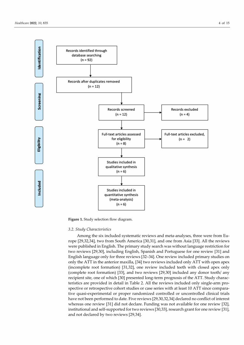
The electronic search was performed until January 2022, producing a total of 92 rec-ords from six different databases: PubMed, n = 28; Cochrane Library, n = 12; Google Scholar, n = 16, Embase, n = 36, Clinicaltrial.gov, n = 0. Titles and abstracts were screened after the removal of duplicates, and a total of 12 potentially significant records were as-sessed. Eight full-text articles were identified for eligibility. After full-text reading, two studies were excluded because they did not meet the inclusion criteria. A list of all the excluded studies is provided in the flow diagram showing the SR selection (Figure 1), and, finally, six SRs were included in this review. All six SRs also conducted MAs.
Healthcare 2022, 10, 835 4 of 15
Figure 1. Study selection flow diagram.
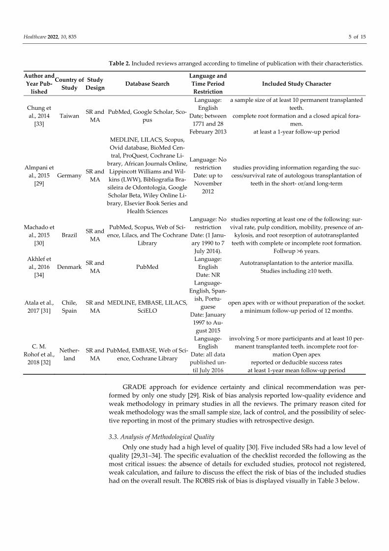
3.2. Study Characteristics Among the six included systematic reviews and meta-analyses, three were from Eu-
rope [29,32,34], two from South America [30,31], and one from Asia [33]. All the reviews were published in English. The primary study search was without language restriction for two reviews [29,30], including English, Spanish and Portuguese for one review [31] and English language only for three reviews [32–34]. One review included primary studies on only the ATT in the anterior maxilla, [34] two reviews included only ATT with open apex (incomplete root formation) [31,32], one review included teeth with closed apex only (complete root formation) [33], and two reviews [29,30] included any donor tooth/ any recipient site, one of which [30] presented long-term prognosis of the ATT. Study charac-teristics are provided in detail in Table 2. All the reviews included only single-arm pro-spective or retrospective cohort studies or case series with at least 10 ATT since compara-tive quasi-experimental or proper randomized controlled or uncontrolled clinical trials have not been performed to date. Five reviews [29,30,32,34] declared no conflict of interest whereas one review [31] did not declare. Funding was not available for one review [32], institutional and self-supported for two reviews [30,33], research grant for one review [31], and not declared by two reviews [29,34].
Healthcare 2022, 10, 835 5 of 15
Table 2. Included reviews arranged according to timeline of publication with their characteristics.
Author and Year Pub-
lished
Country of Study
Study Design Database Search
Language and Time Period Restriction
Included Study Character
Chung et al., 2014
[33] Taiwan
SR and MA
PubMed, Google Scholar, Sco-pus
Language: English
Date; between 1771 and 28
February 2013
a sample size of at least 10 permanent transplanted teeth.
complete root formation and a closed apical fora-men.
at least a 1-year follow-up period
Almpani et al., 2015
[29] Germany
SR and MA
MEDLINE, LILACS, Scopus, Ovid database, BioMed Cen-tral, ProQuest, Cochrane Li-
brary, African Journals Online, Lippincott Williams and Wil-kins (LWW), Bibliografia Bra-sileira de Odontologia, Google Scholar Beta, Wiley Online Li-brary, Elsevier Book Series and
Health Sciences
Language: No restriction Date: up to November
2012
studies providing information regarding the suc-cess/survival rate of autologous transplantation of
teeth in the short- or/and long-term
Machado et al., 2015
[30] Brazil
SR and MA
PubMed, Scopus, Web of Sci-ence, Lilacs, and The Cochrane
Library
Language: No restriction
Date: (1 Janu-ary 1990 to 7 July 2014).
studies reporting at least one of the following: sur-vival rate, pulp condition, mobility, presence of an-
kylosis, and root resorption of autotransplanted teeth with complete or incomplete root formation.
Follwup >6 years. Akhlef et al., 2016
[34] Denmark
SR and MA
PubMed Language:
English Date: NR
Autotransplantation to the anterior maxilla. Studies including ≥10 teeth.
Atala et al., 2017 [31]
Chile, Spain
SR and MA
MEDLINE, EMBASE, LILACS, SciELO
Language-English, Span-
ish, Portu-guese
Date: January 1997 to Au-gust 2015
open apex with or without preparation of the socket. a minimum follow-up period of 12 months.
C. M. Rohof et al.,
2018 [32]
Nether-land
SR and MA
PubMed, EMBASE, Web of Sci-ence, Cochrane Library
Language-English
Date: all data published un-til July 2016
involving 5 or more participants and at least 10 per-manent transplanted teeth. incomplete root for-
mation Open apex reported or deducible success rates
at least 1-year mean follow-up period
GRADE approach for evidence certainty and clinical recommendation was per-formed by only one study [29]. Risk of bias analysis reported low-quality evidence and weak methodology in primary studies in all the reviews. The primary reason cited for weak methodology was the small sample size, lack of control, and the possibility of selec-tive reporting in most of the primary studies with retrospective design.
3.3. Analysis of Methodological Quality Only one study had a high level of quality [30]. Five included SRs had a low level of
quality [29,31–34]. The specific evaluation of the checklist recorded the following as the most critical issues: the absence of details for excluded studies, protocol not registered, weak calculation, and failure to discuss the effect the risk of bias of the included studies had on the overall result. The ROBIS risk of bias is displayed visually in Table 3 below.

Healthcare 2022, 10, 835 6 of 15
Table 3. ROBIS Results for risk of bias analysis in the included systematic reviews.
Review
Phase 2 Phase 3
1. Study Eligi-bility Criteria
2. Identification and Selection of Studies
3. Data Collection and Study Ap-
praisal
4. Synthesis and Findings
Risk of Bias in the Review
Machado et al.[30] Evelyn Rohof et al.[32]
Chung et al.[33] ? Atala-Avecado et al.[31] ?
Almpani et al.[29] ? ? Akhlef et al.[34] ?
= low risk; = high risk; ? = unclear risk.
3.4. Success and Failure Rate In the MA by Chung et al. [33], the summary estimate of the annual FR was 2.0%
[95% confidence interval (CI): 1.2–3.2%], based on 25 studies. The reported percentages of survival from individual studies fell within a wide range of 30–100%. Some studies also presented the 1-year SR. All reported 1-year SRs were >88%. Based on the meta-analysis, they found that the estimated FR of autotransplanted teeth with complete root formation was only 2.0%; the estimated 1-year and 5-year SRs were 98.0% and 90.5%, respectively. In the MA by Almpani et al. [29], the pooled FR in the included studies was found to be 7.8% (95% CI: 4.7–10.9%), based on 15 studies. In the MA by Machado et al. [30], the sur-vival rate was mentioned in four studies and ranged from 75.3% to 91%. The meta-analy-sis showed a significant effect size of 81% (95% CI: 73.8–86.6%) (p < 0.0001). Heterogeneity among the studies was low. In the MA by Atala-Acevedo et al. [31], seventeen studies were included as evidence to determine the success rate of autotransplantation with an open apex. The success rate of the studies was 89.68% (95% CI 86.77 to 92.59%). The suc-cess rate was high, although the heterogeneity of 64.6% was substantial. The autotrans-plantation survival rate in the 15 studies included was 98.21% (95% CI, 96.99 to 99.44), with a low heterogeneity of 25.3%. In the MA by Akhlef et al. [34], survival rates ranged between 93% and 100% (weighted mean: 96.7%, median: 100%) after from 9 months to 22 years of observation (median: 8.75 years), based on 11 studies.
In the MA by Rohof et al. [32], the weighted estimated yearly success rate was 96.6% (95% CI, 94.8–97.8). No heterogeneity was found (Q = 8.24; p = 0.99; I2 = 0.0%), based on 23 studies. The survival rate after 1 year was reported in 26 articles, with the average weighted survival rate of 97.4% (95% CI, 96.2–98.2%). No heterogeneity was found across these studies (Q = 13.66; p = 0.98; I2 = 0.0%). The survival rate after 5 years was reported in 11 articles, with the average weighted survival rate of 97.8% (95% CI, 95.0–99.0%). The data on 5-year survival showed 19.6% heterogeneity (Q = 12.4; p = 0.26), which can be considered low. The survival rate after 10 years was reported in six articles, with the av-erage weighted survival rate of 96.3% (95% CI, 89.8–98.7%). The heterogeneity was 56.8%, which can be considered substantial (Q = 11.6; p = 0.04). The weighted estimated survival rate per year was 98.2% (95% CI, 96.4–99.1%). No heterogeneity was found (Q = 6.2; p = 0.99; I2 = 0.0%).
3.5. Ankylosis Rate In the MA by Chung et al. [33], the results showed that the estimated first-year AR
was 1.2% (95% CI: 0.5–3.2%). However, two aberrant studies demonstrated 100% ARs. Seven studies did not report the occurrence of ankylosis. In the MA by Almpani et al. [29], the pooled AR in the included studies was found to be 76.2% (95% CI: 4.5–7.8%), based on 11 studies. In the MA by Machado et al. [30], four studies reported the percentage of re-placement resorption (ankylosis) in transplanted teeth, ranging from 4.2% to 18.2%. The

Healthcare 2022, 10, 835 7 of 15
meta-analysis showed high heterogeneity among studies, and after the sensitivity analy-sis, the heterogeneity decreased considerably, and a significant effect size of 4.8% was ob-served (p < 0.0001). In the MA by Rohof et al. [32], the weighted estimated ankylosis per year was 2.0% (95% CI, 1.1–3.7%).
3.6. Infection-Related Root Resorption Rate In the MA by Chung et al. [33], the estimated first-year RR from 25 included studies
was only 2.1%. Two of the included studies presented an RR of >50%. On the other hand, they considered four studies that did not report the occurrence of infection-related root resorption as no occurrence. In the MA by Almpani et al. [29], the pooled RR in the in-cluded studies was found to be 10.4% (95% CI: 7.0–13.7%), based on 19 studies. In the MA by Machado et al. [30], when surface resorption, inflammatory resorption, and external root resorption were considered together in the meta-analysis, a significant effect size of 19% was observed (p < 0.0001). The heterogeneity among studies, nevertheless, was ex-tremely high, and after the sensitivity analysis, the heterogeneity decreased considerably, and a significant effect size of 4% was observed (p < 0.0001). In the MA by Rohof et al. [32], the weighted root resorption per year was 2.9% (95% CI, 1.5–5.5%).
3.7. Other Reported Outcomes In the MA by Almpani et al. [29], hypermobility was 8% (95% CI: 4.1, 11.9) based on
eight studies, pulp necrosis was 34.3% (95% CI: 21.1, 47.4) based on 10 studies, and pulp obliteration was 53.4% (95% CI: 28.3, 78.5) based on five studies. In the MA by Machado et al. [30], all studies reported the pulp condition of the transplanted teeth. In the MA by Rohof et al. [32], the weighted pulp necrosis per year was 3.3% (95% CI, 1.9–5.6%). The study outcomes and results are provided in detail in Table 3.
3.8. Subgroup Analysis Based on Factors Could Potentially Mediate the Prognosis of ATT Chung et al. [33] reported that the estimated FR was higher in the absence of SA,
suture splinting, wire splinting ≤ 14 days, and posterior donors. The estimated RR was higher in the absence of SA, endodontic treatment within post-operative 14 days, and an-terior/premolar donors [33]. The estimated AR was higher with wire splinting and pre-molar donors. The stage of development of the root and the autotransplantation receptor site showed no statistically significant differences [33]. CM Rohof et al. reported a higher success and survival rate with the maxillary recipient site compared to the mandible site and premolar recipient site compared to the molar recipient site. They also reported higher success and survival rates with premolar donors than with molar donors. Ankylo-sis rate and root resorptions were higher in molars compared to premolar donors, but the pulp necrosis rate was higher with the premolars [32]. Almpani et al. reported lower fail-ure rates with open apex compared to closed apex and suture splinting compared to wire splinting. The calculated NNT is seven; for every seven ATT with a suture splint, one failed transplant could be prevented compared to transplants with a wire-composite splint. For root development, the calculated NNT is six; for every seven ATT with open apex, one failed transplant could be prevented compared to transplants with closed apex [29]. Atala et al. reported a higher survival rate of teeth at stages three and four of root completion compared to stages one, two, and five (closed apex), premolars compared to molars, and in the maxilla compared to the mandible. Ankylosis was also found to be an indicator of the failure of the transplant [31]. The subgroup analysis is tabulated in table 4.

Healthcare 2022, 10, 835 8 of 15
Table 4. Details of subgroup analysis and their results based on prognostic factors in the included reviews.
Author and Year Published
Number of In-cluded Studies
Subgroup Analysis Results and Conclusion
Chung et al., 2014 [33]
26 studies
systemic antibiotics (SAs), endo-dontic and splinting modalities and donor tooth morphology
Systemic Antibiotics: FR (IRR = 2.5, 95% CI: 0.9–7.2) and Root resorption (IRR = 1.4, 95% CI: 0.2–8.9)
Endodontic treatment: FR (IRR = 1.0, 95% CI: 0.2–5.2) and Root resorption (IRR = 2.0, 95% CI: 0.2–9.3)
Splinting: FR (IRR = 0.8, 95% CI: 0.1–5.5)
Splinting ≥14 days vs. Splinting <14 days FR (IRR = 0.4, 95% CI: 0.1–2.0)
Wire splinting vs. Suture splinting AR (IRR = 3.0, 95% CI: 0.0–607.9)
Donor tooth: Annual Failure rate anterior (0.6% (95% CI: 0.2–2.3%), premolar 1.6% (95% CI
0.3–9.1%), and molar donors was 3.3% (95% CI: 2.4–4.7%) 1-Year Survival rate anterior, 99.4% (95% CI: 97.7–99.8%), premolar 98.4% (95% CI: 90.9–99.7%) and molar donors was 96.7% (95% CI: 95.3–97.6%)
5-Year Survival rate anterior, 96.9% (95% CI: 89.1–99.2%), premolar 92.3% (95% CI: 62.1–98.6%) and molar donors was 84.3% (95% CI: 78.7–88.6%)
Almpani et al., 2015 [29] 38 studies
Open apex vs. Closed apex Splinting
Donor tooth
Open apex vs. closed apex: FR (RR: 0.3; 95% CI: 0.2–0.6) Wire splint vs. suture splint
FR (RR: 3.7; 95% CI: 1.1–12.6) NSD for donor tooth, patient age (>20 vs. <20), gender, recipient site and surgi-
cal technique.
Atala-Acevedo et al., 2016 [31]
17 studies
Donor tooth, Recipient site, stage of root for-
mation.
Donor tooth (Premolar vs. Molar) FR (OR, 0.46; 95% CI, 0.25 to 0.84) Recipient (Maxilla vs. Mandible) FR (OR, 0.38; 95% CI, 0.09 to 1.60)
C. M. Rohof et al., 2018 [32]
32 studies
donor tooth type recipient site,
root development, splinting procedure,
splinting duration, orthodontic procedure, and antibiotic regimen.
Survival rate: Premolar as recipient site (98.6%), Molar (97.3%)
Premolar donor (98.4%), Molar (97.2%) Success rate:
As recipient site; Incisor (98.5%), Canine (97.7%), Premolar (97.8%), Molar (95.1%)
Maxillary recipient site (98.5%) vs. Mandibular recipient site (97.3%) Ankylosis rate:
Premolar donor (1.9%), Molar donor(2.2%) Root Resorption
Premolar donor (1.5%), Molar donor (5%) Pulp Necrosis:
Premolar donor (4.4%), Molar donor (2.5%) Footnotes: NSD = No significant difference, FR = Failure rate; RR = Risk ratio, IRR = Incident risk ratio; AR = ankylosis rate.
3.9. Publication Bias Analysis of publication bias was performed in only two of the SR and MAs. Machado
et al., in their Funnel plot analysis, showed a tendency toward the publication of studies with high survival rates [30]. Almpani et al. reported Funnel plot asymmetry for all out-comes; the Eggers test was significant for FR, RR, and pulp necrosis outcomes [29].

Healthcare 2022, 10, 835 9 of 15
3.10. Analysis of Primary Studies Overlap in the Reviews The overlap of primary studies was analyzed using a citation matrix where the rows
represent the primary studies and columns represent the reviews [36,37]. A total of 91 primary studies were included in the reviews. Three studies were overlapping in four reviews, eight studies were overlapping in three reviews, 22 studies were overlapping in two reviews, and 58 studies did not overlap. The citation matrix is presented in Table 5. The corrected coverage area was calculated to quantify the overlap using the formula: CCA = N-r/rc-r, where N = total number of included studies, including double counting (total check marks), r = total unique primary studies (number of rows), and c = total re-views (number of columns). The calculated CCA was 0.08, which means insignificant overlap (CCA 1–5 is slight, 6–10 is moderate, 11–15 is high, and >15 is very high overlap). Not surprisingly, fifteen primary studies are overlapping in the review by Atala et al. and Evelyn Rohof et al., as both these reviews include ATT with open apex only. The calcu-lated CCA for these two similar reviews is also slight (0.33). Hence, the overestimation of the effect is potentially low when combining these reviews due to only a slight primary study overlap.
Table 5. Citation matrix and overlap of primary studies in the included reviews.
Primary Studies Akhlef 2017 Almpani 2015 Atala 2016 Chung 2014 Evelyn Rohof
2018 Machado 2016
Size of Over-lap
1. Andreasen et al., 1990 X 1 2. Bowden et al., 1990 X 1
3. Czochrowska et al., 2000 X X X X 4 4. Gilijamse et al., 2016 X 1
5. Kristerson and Lagerström, 1991 X X X 3 6. Kugelberg et al., 1994 X X X 3
7. Kvint et al., 2010 X 1 8. Mendoza-Mendoza et al., 2012 X X X X 4
9. Slagsvold et al., 1978 X X 2 10. Stange et al., 2016 X 1 11. Tanaka et al., 2008 X X X 3
12. Ahlberg et al., 1983 X X 2 13. Akiyama et al., 1998 X 1
14. Akkocaoglu and Kasaboglu X 1 15. Altonen et al., 1978 X 1
16. Andreasen et al., 1990 X X X 3 17. Andreasen et al., 1990 X X 2 18. Andreasen et al., 1990 X X 2 19. Andreasen et al., 1990 X X X 3 20. Arikan et al. 32 2008 X X 2
21. Azaz et al., 1978 X X 2 22. Bauss et al., 2002 X X 2 23. Bauss et al., 2008 X 1 24. Bauss et al., 2004 X 1 25. Bauss et al., 2004 X 1 26. Bauss et al., 2005 X 1 27. Bauss et al., 2008 X 1 28. Bauss et al., 2008 X 1
29. Bauss and Kiliaridis 2009 X 1 30. Eliasson et al., 1988 X X 2
31. Kahnberg 1987 X 1 32. Kristerson 1985 X X X 3
33. Kristerson et al., 1991 X X 2 34. Lagerstron and Kristerson 1986 X 1
35. Lundberg and Isaksson 1996 X X X 3 36. Marques- Ferreira et al., 2011 X 1
37. Mejare et al., 2004 X X 2 38. Myrlund et al., 2004 X X 2
39. Nethander et al., 1988 X 1 40. Nethander 1994 X 1

Healthcare 2022, 10, 835 10 of 15
41. Nethander 1998 X X 2 42. Ploder et al., 2001 X 1 43. Pogrel et al., 1987 X 1
44. Reich 2008 X 1 45. Sagne et al., 1986 X 1 46. Sobhi et al., 2003 X 1 47. Sugai et al., 2010 X X 2
48. Thomson et al., 1984 X X 2 49. Yan et al., 2010 X X X X 4 50. Bauss et al., 2002 X 1 51. Bauss et al., 2004 X 1 52. Bauss et al., 2003 X 1
53. Czochrowska et al.,2000 X X 2 54. Denys et al., 2013 X 1
55. Dıaz et al., 2014 (17) X 1 56. Isa-Kara et al., 2011 X X X 3 57. Josefsson et al., 1999 X X 2
58. Kallu et al., 2005 X 1 59. Mertens et al., 2014 X 1 60. Nagori et al., 2014 X X 2 61. Naranjo et al., 2002 X 1 62. Plakwicz et al., 2013 X X 2
63. Schutz et al., 2013 X X 2 64. Vilhjalmsson et al., 2011 X X 2
65. Forssell and Oksala (1986) X 1 66. Gault and Warocquier-Clerout (2002) X 1
67. Hovinga (1969) X 1 68. Masif and Youseff (1977) X 1
69. Moss (1968) X 1 70. Niimi et al. (2011) X 1 71. Patel et al. (2011) X 1
72. Reade et al. (1973) X 1 73. Schatz and Joho (1993) X 1
74. Sange and Thilander (1990) X 1 75. Schwartz et al. (1985) X 1
76. Wang et al. (2007) X 1 77. Watanabe et al. (2010) X X 2
78. Borring-Møller et al.1979 X 1 79. de Carvalho et al. (2014) X 1
80. Díaz et al., 2008 X 1 81. Gonnissen et al 2010 X X 2
82. Hernandez and Cuestascarner 1988 X 1 83. Jonsson and Sigurdsson 2004 X 1
84. Marcusson and Lilja-Karlander 1996 X 1 85. Mertens et al., 2016 X 1
86. Mensink and van Merkesteyn 2010 X 1 87. Nagori et al. (2014) X 1
88. Schatz and Joho 1992 X 1 89. Shahbazian et al., 2013 X 1
90. Paulsen and Andreasen (1998) X 1 91. Paulsen et al. (1995) X 1
X = included in the review.
4. Discussion This review aimed to answer the question: “What is the survival and success/failure
rate of autotransplanted tooth, and what is the rate of major complications like AR and RR?” Six SRs and MAs were included in this study, but their methodological heterogene-ity and the slight overlap of studies precluded an umbrella meta-analysis. In order to as-sess the quality of each SR, the AMSTAR-2 tool and ROBIS tool were used. The qualitative analysis of the included SRs allowed for the SR, FR, AR, and RR to be summarized. The overall failure rate ranged from as low as 2.0% to 10.32%. The 1-year survival was very high (97.4–98.0%). The 5-year survival rate ranged from 81 to 98.2%. Major complications

Healthcare 2022, 10, 835 11 of 15
of AR ranged from 1.2 to 6.2%, and RR ranged from 2.1 to 10.4%. Considerable heteroge-neity exists across SRs, with differences in terms of donor tooth, recipient site, and the stage of root formation. In this current review, high methodological quality was assessed for only one SR [30], whereas five others were of low quality [29,31–34]. The evidence from included reviews was undermined by the absence of studies directly comparing various morphology, areas, modality, and perioperative techniques for ATT, as only single-arm studies were available for analysis. All the reviews have conceded this limitation in their discussion and have provided the explanation that it will be unethical to perform an RCT knowingly when one treatment is supposed to be superior to the other; thus, comparative controlled trials were not feasible. Thus, we should be cautious, as suggested by the re-view authors, that the findings should be interpreted in the light of the limitations under which the data were aggregated and compared, as only an indirect comparison was pos-sible from multiple single-arm cohorts, which were then post hoc considered as reference and comparator group for statistical analysis. For this same reason, we did not perform an umbrella meta-analysis as this would further increase the bias of performing indirect comparison among groups that come from disparate studies and are not matched for con-founders.
Based on the subgroup analysis of different donor tooth and recipient sites, evidence suggests that anterior donor and recipient sites have a higher survival rate compared to molars. This has been attributed to the ease with which the single-rooted anterior donors can be atraumatically harvested, causing less damage to the PDL, which is one of the most important prognostic factors for the success of the transplant. Maxillary transplants were found to have higher survival than mandibular ones, probably because of highly porous alveolar bone and abundant blood supply, as well as less direct occlusal loading of max-illary teeth compared to mandibular teeth. The results of subgroup analysis based on var-ious techniques utilized along with ATT hint towards the use of suture splinting rather than wire splinting, performing ATT using donor tooth with open apex, performing ex vivo RCT, or RCT within 14 days of transplant, and using perioperative systemic antibi-otics. Teeth with open-apex have been reported to allow neurovascular ingrowth after transplantation which might explain their post-transplant vitality and better survival rate. Wire-splinting is more rigid than suture splinting, thus allowing lesser micro-motion of the ATT in the post-operative procedure. Though it may provide more stability, the lack of physiological stimulus may prevent the optimum level of tissue signaling from initiat-ing adaptation of the ATT in the recipient site and may lead to avascular necrosis due to rigid pressure applied by the wire splint. Another interesting finding is higher survival of teeth where RCT was initiated within 14 days, and systemic antibiotics were used. Anti-biotics definitely would have reduced the incidence of infection post-ATT, which may lead to periodontal tissue destruction and loosening of transplant. The prompt initiation of RCT may help to prevent pulpal infection and necrosis, which, if uncontrolled, is one of the main reasons for root resorption and ATT failure.
Autotransplantation, or the process of moving a tooth from one site to another in the same person, is not a new technique in the history of dentistry but has varying success and failure rates in the dental literature [3,37–39]. This surgical method can be used to replace missing, avulsed, traumatized, or grossly decayed, unrestorable teeth with high survival rates (91.3%), particularly in adults or children [40–42]. Ironically this treatment option is grossly overlooked, and the use of dental implants or fixed prostheses is widely used instead. Dental implants are not a good option for patients until the completion of growth, which is 17–19 years for girls and 19–21 years for boys [43]. However, loss of a tooth is more prevalent in young adults due to trauma or caries. Autotransplantation could hence be a very good alternative for these patients, with lots of advantages, such as preservation and normal functioning of periodontal ligament tissues and alveolar bone volume, along with proprioception, better esthetics, and higher resistance to occlusal load [16]. Moreover, even in the case of external root resorption or replacement resorption, the bone and soft tissue volume will be preserved, favoring subsequent implant placement
Healthcare 2022, 10, 835 12 of 15
[16,44]. Now that it is established that the success and survival rate of ATT is overalls high, and AR and RR are considerably low, direct comparative prospective studies, preferably randomized and blinded, should be performed to evaluate the factors that can further decrease complications and failure rate, and increase overall survival over a long period of time.
Recently, various methods have been incorporated into ATT to increase its success and survival rates. One such method is the use of 3D-printed guiding templates/replicas and GBR, which significantly reduces the extra-oral time of the tooth, resulting in less drying and trauma for the PDL cells [45–53]. Additionally, ATT can be performed by cre-ating artificial tooth sockets with the use of cone-beam computed tomography (CBCT) and 3D-printed donor tooth replicas. The incorporation of this modern technology in the ATT makes this procedure less technique-sensitive, reduces extra-oral time and iatrogenic damage, predicts a good prognosis, and facilitates a successful outcome [45,49,54–56]. However, occlusion reduction, splinting, and root canal treatment are still indicated to reduce RR.
It has also been observed that, with proper case selection and appropriate treatment, ATT can be successfully performed with fully formed third molars as donors in both im-mediate extractions as well as surgically created sockets [57]. Previously, external inflam-matory or internal root resorption was considered to be a major reason for the failure of the ATT tooth [58]. However, this kind of resorption can easily be controlled with the timely initiation of endodontic therapy, or even after the initiation of resorption, endo-dontic treatment and long-term calcium hydroxide dressings were found to treat these conditions successfully, without any signs of mobility, pocket, radiolucency, tenderness to percussion, or any other pathology [59]. Similarly, a minimal amount of trauma and extra-oral time are crucial for preserving intact and viable periodontal cells, which aid in the healing of the transplanted teeth without root resorption, especially the replacement resorption/ankylosis [60]. Thus, the horizon of the scope of autotransplantation is becom-ing broader, with many possibilities, newer advancements, and a very predictable and favorable outcome. The literature search suggests that autotransplantation is a very cost-effective, viable, immediate treatment option and a biological solution that can be pro-vided to adolescent patients with a missing tooth when a suitable donor tooth is present, with a high success rate [28].
4.1. Limitations and Strengths of this Overview This overview of reviews has some limitations. We included all the autotransplanted
teeth for any indication to accrue a cumulative complication and survival rate, but that also means that we have evidence from essentially heterogeneous sources. There is no considerable difference in the technique of autotransplantation, but the clinical protocol, systemic antibiotics, and local procedures to increase viability differed among the primary studies included in the reviews. The included reviews were found to have low methodo-logical quality due to the exclusion of important steps from PRISMA guidelines, and the meta-analyses were all based on indirect group comparisons from disparate studies that were not matched for confounders. Our literature search included results from only six databases and the English language only and did not include gray literature, pre-prints, or commercial reports, which might have added to the evidence if available. The primary study overlap was low between the reviews; thus, we can say that at least the cumulative results are not overestimated, although indirectness of comparison brings down the level of confidence in the results.
4.2. Implications for Practice and Research Based on the comprehensive and cumulative evidence from available systematic re-
views and meta-analyses, the success rate (SR) of the autotransplanted tooth shows prom-ising results, above 90%, even with long-term follow-up and a survival rate up to 98%. In the era of implant-driven dentistry, preserving natural teeth is more economical,
Healthcare 2022, 10, 835 13 of 15
biological, and esthetic. Natural teeth with viable periodontium are more physiological than a dental implant, which is similar to ankylosed teeth. However, the success of auto-transplantation depends upon various prognostic factors, such as the age of the patient, stage of root development at the time of transplantation, splinting method and duration of transplanted teeth, amount of trauma perceived by the PDL at the time of extraction, administration of systemic antibiotics, initiation of endodontic treatment, compliance of the patient, etc. Thus, these factors determine the success and failure, with either root re-sorption or replacement resorption, of the transplanted tooth. Hence, more studies are recommended on various prognostic indicators to obtain accurate protocols for autotrans-plantation.
5. Conclusions The overall findings from these SR and MAs are promising. The autotransplanted
tooth has a low failure rate, high survival rate, and low complication rate, but the overall evidence is of low to moderate certainty, and all the SRs are based on only single-arm prospective or retrospective studies. Further research on the evaluation of prognostic in-dicators and factors that affect the success of ATT is recommended.
Author Contributions: A.K.S.: Conceptualization, Methodology, Writing—Original draft prepara-tion. N.K.: Data curation, Writing—Original draft preparation. N.A.: Writing—Reviewing and Ed-iting, Visualization. M.R.H.: Supervision, Conceptualization, Writing—Reviewing and Editing, T.S.: Supervision, Writing—Reviewing and Editing. All authors have read and agreed to the published version of the manuscript.
Funding: This work was supported by a Grant-in-Aid for Early-Career Scientists from the Japanese Society for the Promotion of Science KAKENHI-PROJECT (grant number 20K18541).
Institutional Review Board Statement: Not applicable.
Informed Consent Statement: Not applicable.
Data Availability Statement: Medline (PubMed) and Scopus databases.
Conflicts of Interest: The authors declare no conflict of interest.
References 1. Parvini, P.; Obreja, K.; Trimpou, G.; Mahmud, S.; Sader, R. Autotransplantation of teeth. Int. J. Esthet Dent. 2018, 13, 274–282. 2. Park, J.H.; Tai, K.; Hayashi, D. Tooth Autotransplantation as a Treatment Option: A Review. J. Clin. Pediatr. Dent. 2010, 35, 129–
135. https://doi.org/10.17796/jcpd.35.2.97816254u2140x88. 3. Amos, M.J.; Day, P.F.; Littlewood, S.J. Autotransplantation of Teeth: An Overview. Dent. Update 2009, 36, 102–113.
https://doi.org/10.12968/denu.2009.36.2.102. 4. Schmidt, S.K.; Cleverly, D.G. Tooth autotransplantation: An overview and case study. Northwest Dent. 2012, 91, 29–33. 5. Thomas, S.; Turner, S.R.; Sandy, J.R. Autotransplantation of Teeth: Is There a Role? Br. J. Orthod. 1998, 25, 275–282.
https://doi.org/10.1093/ortho.25.4.275. 6. Schwartz, O.; Bergmann, P.; Klausen, B. Autotransplantation of human teeth. A life-table analysis of prognostic factors. Int. J.
Oral Surg. 1985, 14, 245–258. https://doi.org/10.1016/s0300-9785(85)80036-3. 7. Fernandes, S.; Habibullah, M.A.; Nalam, G.S.; Nair, P.P. Think before you extract—A case of tooth autotransplantation. BMJ
Case Rep. 2011, 2011, https://doi.org/10.1136/bcr.06.2011.4380. 8. Herrera-Gimbernat, D.; Recio-Lora, C.; Torres-Lagares, D.; Romero-Ruiz, M.; Pérez, J.L.G. Current state of dental autotrans-
plantation. Med. Oral Patol. Oral Cir. Bucal 2011, 16, e948–e952. https://doi.org/10.4317/medoral.17267. 9. Cross, D.; El-Angbawi, A.; McLaughlin, P.; Keightley, A.; Brocklebank, L.; Whitters, J.; McKerlie, R.; Cross, L.; Welbury, R.
Developments in autotransplantation of teeth. J. Surg. 2013, 11, 49–55. https://doi.org/10.1016/j.surge.2012.10.003. 10. Plotino, G.; Sans, F.A.; Duggal, M.S.; Grande, N.M.; Krastl, G.; Nagendrababu, V.; Gambarini, G. European Society of Endodon-
tology position statement: Surgical extrusion, intentional replantation and tooth autotransplantation: European Society of Endo-dontology developed by. Int. Endod. J. 2021, 54, 655–659. https://doi.org/10.1111/iej.13456.
11. Tsukiboshi, M. Autotransplantation of teeth: Requirements for predictable success. Dent. Traumatol. 2002, 18, 157–180. https://doi.org/10.1034/j.1600-9657.2002.00118.x.
12. Tsukiboshi, M.; Yamauchi, N.; Tsukiboshi, Y. Long-term outcomes of autotransplantation of teeth: A case series. Dent. Trau-matol. 2019, 35, 358–367. https://doi.org/10.1111/edt.12495.

Healthcare 2022, 10, 835 14 of 15
13. De Freitas Coutinho, N.B.; Nunes, F.C.; Intra, J.B.G.; Roldi, A.; de Jesus-Soares, A.; Coelho, M.S.; Frozoni, M. Success, Survival Rate, and Soft Tissue Esthetic of Tooth Autotransplantation. J. Endod. 2021, 47, 391–396. https://doi.org/10.1016/j.joen.2020.11.013.
14. Raabe, C.; Bornstein, M.M.; Ducommun, J.; Sendi, P.; von Arx, T.; Janner, S.F.M. A retrospective analysis of autotransplanted teeth including an evaluation of a novel surgical technique. Clin. Oral Investig. 2021, 25, 3513–3525. https://doi.org/10.1007/s00784-020-03673-y.
15. Mertens, B.; Boukari, A.; Tenenbaum, H. Long-term follow up of post-surgical tooth autotransplantation: A retrospective study. J. Investig. Clin. Dent. 2014, 7, 207–214. https://doi.org/10.1111/jicd.12126.
16. Ong, D.C.; Itskovich, Y.; Dance, G. Autotransplantation: A viable treatment option for adolescent patients with significantly compromised teeth. Aust. Dent. J. 2016, 61, 396–407. https://doi.org/10.1111/adj.12420.
17. Nagori, S.A.; Bhutia, O.; Roychoudhury, A.; Pandey, R.M. Immediate autotransplantation of third molars: An experience of 57 cases. Oral Surg. Oral Med. Oral Pathol. Oral Radiol. 2014, 118, 400–407. https://doi.org/10.1016/j.oooo.2014.05.011.
18. Boschini, L.; Melillo, M.; Berton, F. Long term survival of mature autotransplanted teeth: A retrospective single center analysis. J. Dent. 2020, 98, 103371. https://doi.org/10.1016/j.jdent.2020.103371.
19. Juslin, J.; Jääsaari, P.; Teerijoki-Oksa, T.; Suominen, A.; Thorén, H. Survival of Autotransplanted Teeth with Open Apices: A Retrospective Cohort Study. J. Oral Maxillofac. Surg. 2020, 78, 902.e1–902.e9. https://doi.org/10.1016/j.joms.2020.02.018.
20. Ashurko, I.; Vlasova, I.; Yaremchuk, P.; Bystrova, O. Autotransplantation of teeth as an alternative to dental implantation. BMJ Case Rep. 2020, 13, e234889. https://doi.org/10.1136/bcr-2020-234889.
21. Yang, S.; Jung, B.-Y.; Pang, N.-S. Outcomes of autotransplanted teeth and prognostic factors: A 10-year retrospective study. Clin. Oral Investig. 2018, 23, 87–98. https://doi.org/10.1007/s00784-018-2412-3.
22. Abela, S.; Murtadha, L.; Bister, D.; Andiappan, M.; Kwok, J. Survival probability of dental autotransplantation of 366 teeth over 34 years within a hospital setting in the United Kingdom. Eur. J. Orthod. 2019, 41, 551–556. https://doi.org/10.1093/ejo/cjz012.
23. van Westerveld, K.; Verweij, J.; Toxopeus, E.; Fiocco, M.; Mensink, G.; van Merkesteyn, J. Long-term outcomes 1–20 years after autotransplantation of teeth: Clinical and radiographic evaluation of 66 premolars and 8 molars. Br. J. Oral Maxillofac. Surg. 2019, 57, 666–671. https://doi.org/10.1016/j.bjoms.2019.06.006.
24. Armstrong, L.; O’reilly, C.; Ahmed, B. Autotransplantation of third molars: A literature review and preliminary protocols. Br. Dent. J. 2020, 228, 247–251. https://doi.org/10.1038/s41415-020-1264-9.
25. Czochrowska, E.M.; Plakwicz, P. Autotransplantation and healing. Am. J. Orthod. Dentofac. Orthop. 2019, 156, 299–300. https://doi.org/10.1016/j.ajodo.2019.06.007.
26. Czochrowska, E.M. “Off the beaten track” solutions: Autotransplantation of teeth. Int. J. Esthet. Dent. 2018, 13, 234–239. 27. Martin, K.; Nathwani, S.; Bunyan, R. Autotransplantation of teeth: An evidence-based approach. Br. Dent. J. 2018, 224, 861–864.
https://doi.org/10.1038/sj.bdj.2018.432. 28. Verweij, J.; Anssari Moin, D.; Mensink, G.; Wismeijer, D.; van Merkesteyn, J.P.R. Autotransplantation 2.0. Considerations, re-
sults and the latest techniques. Ned. Tijdschr. Voor Tandheelkd. 2016, 123, 348–353. https://doi.org/10.5177/ntvt.2016.07/08.16119. 29. Almpani, K.; Papageorgiou, S.N.; Papadopoulos, M.A. Autotransplantation of teeth in humans: A systematic review and meta-
analysis. Clin. Oral Investig. 2015, 19, 1157–1179. https://doi.org/10.1007/s00784-015-1473-9. 30. Machado, L.; Nascimento, R.D.; Ferreira, D.; Mattos, C.; Vilella, O. Long-term prognosis of tooth autotransplantation: A sys-
tematic review and meta-analysis. Int. J. Oral Maxillofac. Surg. 2016, 45, 610–617. https://doi.org/10.1016/j.ijom.2015.11.010. 31. Atala-Acevedo, C.; Abarca, J.; Martínez-Zapata, M.J.; Díaz, J.; Olate, S.; Zaror, C. Success Rate of Autotransplantation of Teeth
with an Open Apex: Systematic Review and Meta-Analysis. J. Oral Maxillofac. Surg. 2017, 75, 35–50. https://doi.org/10.1016/j.joms.2016.09.010.
32. Rohof, E.C.M.; Kerdijk, W.; Jansma, J.; Livas, C.; Ren, Y. Autotransplantation of teeth with incomplete root formation: A sys-tematic review and meta-analysis. Clin. Oral Investig. 2018, 22, 1613–1624. https://doi.org/10.1007/s00784-018-2408-z.
33. Chung, W.-C.; Tu, Y.-K.; Lin, Y.-H.; Lu, H.-K. Outcomes of autotransplanted teeth with complete root formation: A systematic review and meta-analysis. J. Clin. Periodontol. 2014, 41, 412–423. https://doi.org/10.1111/jcpe.12228.
34. Akhlef, Y.; Schwartz, O.; Andreasen, J.O.; Jensen, S.S. Autotransplantation of teeth to the anterior maxilla: A systematic review of survival and success, aesthetic presentation and patient-reported outcome. Dent. Traumatol. 2017, 34, 20–27. https://doi.org/10.1111/edt.12379.
35. Whiting, P.; Savović, J.; Higgins, J.P.; Caldwell, D.M.; Reeves, B.C.; Shea, B.; Davies, P.; Kleijnen, J.; Churchill, R. ROBIS: A new tool to assess risk of bias in systematic reviews was developed. J. Clin. Epidemiol. 2016, 69, 225–234. doi:10.1016/j.jclinepi.2015.06.005
36. Lunny, C.; Pieper, D.; Thabet, P.; Kanji, S. Managing overlap of primary study results across systematic reviews: practical con-siderations for authors of overviews of reviews. BMC Med. Res. Methodol. 2021, 21, 140. https://doi.org/10.1186/s12874-021-01269-y
37. Hennessy, E.A.; Blair, T.J. Examining overlap of included studies in meta-reviews: Guidance for using the corrected covered area index. Res. Synth. Methods 2020, 11, 134–145. doi:10.1002/jrsm.1390
38. Kallu, R.; Vinckier, F.; Politis, C.; Mwalili, S.; Willems, G. Tooth transplantations: A descriptive retrospective study. Int. J. Oral Maxillofac. Surg. 2005, 34, 745–755. https://doi.org/10.1016/j.ijom.2005.03.009.
39. Mendes, R.A.; Rocha, G. Mandibular third molar autotransplantation—literature review with clinical cases. J. Can. Dent. Assoc. 2004, 70, 761–766.
Healthcare 2022, 10, 835 15 of 15
40. Abu Tair, J.A.; Rahhal, A. Tooth autotransplantation in orthodontic patients. J. Contemp. Dent. Pract. 2010, 11, 63–70. 41. Kvint, S.; Lindsten, R.; Magnusson, A.; Nilsson, P.; Bjerklin, K. Autotransplantation of Teeth in 215 Patients. A follow-up study.
Angle Orthod. 2010, 80, 446–451. https://doi.org/10.2319/062509-354.1. 42. Yoshino, K.; Kariya, N.; Namura, D.; Noji, I.; Mitsuhashi, K.; Kimura, H.; Fukuda, A.; Kikukawa, I.; Hayashi, T.; Yamazaki, N.;
et al. Influence of age on tooth autotransplantation with complete root formation. J. Oral Rehabil. 2013, 40, 112–118. https://doi.org/10.1111/joor.12012.
43. Gilijamse, M.; Baart, J.A.; Wolff, J.; Sándor, G.K.; Forouzanfar, T. Tooth autotransplantation in the anterior maxilla and mandi-ble: Retrospective results in young patients. Oral Surg. Oral Med. Oral Pathol. Oral Radiol. 2016, 122, e187–e192. https://doi.org/10.1016/j.oooo.2016.06.013.
44. Ong, D.C.; Dance, G. Posterior tooth autotransplantation: A case series. Aust. Dent. J. 2021, 66, 85–95. https://doi.org/10.1111/adj.12757.
45. Moin, D.A.; Derksen, W.; Verweij, J.; van Merkesteyn, R.; Wismeijer, D. A Novel Approach for Computer-Assisted Template-Guided Autotransplantation of Teeth with Custom 3D Designed/Printed Surgical Tooling. An Ex Vivo Proof of Concept. J. Oral Maxillofac. Surg. 2016, 74, 895–902. https://doi.org/10.1016/j.joms.2016.01.033.
46. Verweij, J.; Anssari Moin, D.; Mensink, G.; Wismeijer, D.; van Merkesteyn, J.P.R. Replacing heavily damaged molars with the use of 3D-techniques. Ned. Tijdschr. Voor Tandheelkd. 2018, 125, 21–26. https://doi.org/10.5177/ntvt.2018.01.17186.
47. Dreizin, D.; Nam, A.J.; Hirsch, J.; Bernstein, M. New and emerging patient-centered CT imaging and image-guided treatment paradigms for maxillofacial trauma. Emerg. Radiol. 2018, 25, 533–545. https://doi.org/10.1007/s10140-018-1616-9.
48. Al-Rimawi, A.; Ezeldeen, M.; Schneider, D.; Politis, C.; Jacobs, R. 3D Printed Temporary Veneer Restoring Autotransplanted Teeth in Children: Design and Concept Validation Ex Vivo. Int. J. Environ. Res. Public Health 2019, 16, 496. https://doi.org/10.3390/ijerph16030496.
49. Kamio, T.; Kato, H. Autotransplantation of Impacted Third Molar Using 3D Printing Technology: A Case Report. Bull. Tokyo Dent. Coll. 2019, 60, 193–199. https://doi.org/10.2209/tdcpublication.2018-0058.
50. Curtis, J.M.T.; Foster, E.C.; Ananth, S.; Eckhardt, C.; Knox, J.; Alvarez, A.G.; Newton, R. Autotransplantation of a surgically removed canine using a customised 3D-printed surgical template. J. Orthod. 2020, 47, 82–90. https://doi.org/10.1177/1465312519891738.
51. Cousley, R.R.J.; Gibbons, A.; Nayler, J. A 3D printed surgical analogue to reduce donor tooth trauma during autotransplanta-tion. J. Orthod. 2017, 44, 287–293. https://doi.org/10.1080/14653125.2017.1371960.
52. Sokolowski, A.A.; Sokolowski, A.A.; Kammerhofer, J.; Madreiter-Sokolowski, C.T.; Payer, M.; Koller, M.; Jakse, N.; Wegscheider, A. Accuracy assess-ment of 3D-printed tooth replicas. Int. J. Comput. Dent. 2019, 22, 321–329.
53. Kim, K.; Choi, H.; Pang, N. Clinical application of 3D technology for tooth autotransplantation: A case report. Aust. Endod. J. 2019, 45, 122–128. https://doi.org/10.1111/aej.12260.
54. Shahbazian, M.; Jacobs, R.; Wyatt, J.; Willems, G.; Pattijn, V.; Dhoore, E.; van Lierde, C.; Vinckier, F. Accuracy and surgical feasibility of a CBCT-based stereolithographic surgical guide aiding autotransplantation of teeth: In vitro validation. J. Oral Rehabil. 2010, 37, 854–859. https://doi.org/10.1111/j.1365-2842.2010.02107.x.
55. Ezeldeen, M.; Wyatt, J.; Al-Rimawi, A.; Coucke, W.; Shaheen, E.; Lambrichts, I.; Willems, G.; Politis, C.; Jacobs, R. Use of CBCT Guidance for Tooth Autotransplantation in Children. J. Dent. Res. 2019, 98, 406–413. https://doi.org/10.1177/0022034519828701.
56. Chen, J.-M.; Wu, Y.; He, P.-T.; Xie, F.-P.; Liu, H.-H.; Lin, L.-S. Application of computer virtual design to assist the accuracy of sock-et preparation during tooth autotransplantation. Shanghai Kou Qiang Yi Xue 2020, 29, 65–68.
57. Yu, H.; Jia, P.; Lv, Z.; Qiu, L. Autotransplantation of third molars with completely formed roots into surgically created sockets and fresh extraction sockets: A 10-year comparative study. Int. J. Oral Maxillofac. Surg. 2017, 46, 531–538. https://doi.org/10.1016/j.ijom.2016.12.007.
58. Ahlberg, K.; Bystedt, H.; Eliasson, S.; Odenrick, L. Long-Term Evaluation of Autotransplanted Maxillary Canines with Com-pleted Root Formation. Acta Odontol. Scand. 1983, 41, 23–31. https://doi.org/10.3109/00016358309162300.
59. Herrera, H.; Herrera, H.; Leonardo, M.R.; Paula-Silva, F.W.; da Silva, L.A.B. Treatment of external inflammatory root resorption after autogenous tooth transplantation: Case report. Oral Surg. Oral Med. Oral Pathol. Oral Radiol. Endod. 2006, 102, e51–e54. https://doi.org/10.1016/j.tripleo.2006.05.027.
60. Andreasen, J. Periodontal healing after replantation and autotransplantation of incisors in monkeys. Int. J. Oral Surg. 1981, 10, 54–61. https://doi.org/10.1016/s0300-9785(81)80008-7.