Long-term success and survival rates of autogenously transplanted canines
9
Long-term success and survival rates of autogenously transplanted canines H. Gonnissen, a C. Politis, a,b S. Schepers, a,c I. Lambrichts, a L. Vrielinck, b Y. Sun, b J. Schuermans, b Diepenbeek, Genk, and Gent, Belgium HASSELT UNIVERSITY, ST. JOHN’S HOSPITAL, AND GENT UNIVERSITY Objectives. The objectives of this study were to determine the long-term survival and success rates of autotransplanted canines and to investigate the influence of various parameters on the long-term success rate. Study design. Fifty-nine patients (73 transplanted canines) volunteered to participate in this study. The mean follow-up time was 11 years. Different parameters that could influence the outcome of transplantation were examined in the patient files. Each transplanted canine was clinically and radiologically evaluated. Logistic regression analyses were performed. Results. The survival rate was 75.3%, because 18 transplanted teeth were lost before examination. The success rate for all transplanted teeth was 57.5%, because 42 transplanted teeth were evaluated as clinically successful. The most significant parameter in determining the success rate of autotransplantation was age at transplantation (P .0429). Conclusion. Autotransplantation of impacted canines may have a successful outcome 11 years after transplantation. The success rate increases when performing the transplantation at a younger age. (Oral Surg Oral Med Oral Pathol Oral Radiol Endod 2010;110:570-578) Autogenous tooth transplantation can be defined as the surgical movement of a tooth from one position in the mouth to another in the same individual. 1 The maxillary canine is the most frequently ectopic tooth in the anterior part of the mouth. The incidence of impacted maxillary canines varies from 1% to 3%. 2 The mandibular canine is less frequently impacted. The traditional treatment for ectopically positioned canines is surgical exposure and orthodontic realignment. How- ever, when the position of the canine is not such that orthodontic treatment would be feasible, autotransplan- tation can be considered. 3 Because impacted canines are a particular challenge in children and adolescents, the transplanted tooth should preferably adapt to growth and developmental changes in the oral region. In addition, the tooth should have the potential for long-term, even lifelong, survival. However, the liter- ature contains only a few long-term follow-up studies of transplanted canines. The aim of the present study was to determine the long-term survival and success rates of autotrans- planted canines. In addition, the influence of various preoperative parameters, such as age, gender, root de- velopment, and others, on the long-term success rate was investigated. MATERIALS AND METHODS Patients A total of 133 patients had undergone canine trans- plantation during the period of 1995 to 2002. Consid- erable efforts were made to contact these patients, because of the long period since their operations. Fifty- nine patients with a total of 73 teeth volunteered to participate in this study. Thirty-four patients (41 teeth) were male and 25 patients (32 teeth) were female (Table I). The mean age at the time of transplantation was 20.7 years (range 11-46 years), and the mean follow-up period was 11 years (range 6-14 years). All transplantations were performed by the same experi- enced oral surgeon, according to the strict protocol of the oral and maxillofacial surgery (OMS) department in the St. John’s Hospital, Genk. Out of the 73 trans- planted teeth, 67 teeth were maxillary impacted canines and only 3 were mandibular impacted canines. Two teeth were molars from the mandible transplanted to a canine position in the maxilla. Various pre- and peri- operative parameters were examined in patient files (Table II). Moorrees et al.’s classification for the de- velopmental stages of roots was used as a basis to estimate the stage of root development of each trans- planted tooth at the time of transplantation. 4 For this purpose, previous analog radiographs were observed. It a Faculty of Medicine, Hasselt University. b Oral and Maxillofacial Surgery, St. John’s Hospital. c Oral and Maxillofacial Surgery, Faculty of Medicine, Gent University. Received for publication Aug 24, 2009; returned for revision Feb 10, 2010; accepted for publication Feb 24, 2010. 1079-2104/$ - see front matter © 2010 Mosby, Inc. All rights reserved. doi:10.1016/j.tripleo.2010.02.039 570
Transcript of Long-term success and survival rates of autogenously transplanted canines

Long-term success and survival rates of autogenouslytransplanted caninesH. Gonnissen,a C. Politis,a,b S. Schepers,a,c I. Lambrichts,a L. Vrielinck,b Y. Sun,b
J. Schuermans,b Diepenbeek, Genk, and Gent, BelgiumHASSELT UNIVERSITY, ST. JOHN’S HOSPITAL, AND GENT UNIVERSITY
Objectives. The objectives of this study were to determine the long-term survival and success rates of autotransplantedcanines and to investigate the influence of various parameters on the long-term success rate.Study design. Fifty-nine patients (73 transplanted canines) volunteered to participate in this study. The mean follow-uptime was 11 years. Different parameters that could influence the outcome of transplantation were examined in thepatient files. Each transplanted canine was clinically and radiologically evaluated. Logistic regression analyses wereperformed.Results. The survival rate was 75.3%, because 18 transplanted teeth were lost before examination. The success ratefor all transplanted teeth was 57.5%, because 42 transplanted teeth were evaluated as clinically successful. The mostsignificant parameter in determining the success rate of autotransplantation was age at transplantation (P � .0429).Conclusion. Autotransplantation of impacted canines may have a successful outcome 11 years after transplantation.The success rate increases when performing the transplantation at a younger age. (Oral Surg Oral Med Oral Pathol
Oral Radiol Endod 2010;110:570-578)Autogenous tooth transplantation can be defined as thesurgical movement of a tooth from one position in themouth to another in the same individual.1
The maxillary canine is the most frequently ectopictooth in the anterior part of the mouth. The incidence ofimpacted maxillary canines varies from �1% to 3%.2
The mandibular canine is less frequently impacted. Thetraditional treatment for ectopically positioned caninesis surgical exposure and orthodontic realignment. How-ever, when the position of the canine is not such thatorthodontic treatment would be feasible, autotransplan-tation can be considered.3 Because impacted caninesare a particular challenge in children and adolescents,the transplanted tooth should preferably adapt togrowth and developmental changes in the oral region.In addition, the tooth should have the potential forlong-term, even lifelong, survival. However, the liter-ature contains only a few long-term follow-up studiesof transplanted canines.
The aim of the present study was to determine thelong-term survival and success rates of autotrans-planted canines. In addition, the influence of various
aFaculty of Medicine, Hasselt University.bOral and Maxillofacial Surgery, St. John’s Hospital.cOral and Maxillofacial Surgery, Faculty of Medicine, GentUniversity.Received for publication Aug 24, 2009; returned for revision Feb 10,2010; accepted for publication Feb 24, 2010.1079-2104/$ - see front matter© 2010 Mosby, Inc. All rights reserved.
doi:10.1016/j.tripleo.2010.02.039570
preoperative parameters, such as age, gender, root de-velopment, and others, on the long-term success ratewas investigated.
MATERIALS AND METHODSPatients
A total of 133 patients had undergone canine trans-plantation during the period of 1995 to 2002. Consid-erable efforts were made to contact these patients,because of the long period since their operations. Fifty-nine patients with a total of 73 teeth volunteered toparticipate in this study. Thirty-four patients (41 teeth)were male and 25 patients (32 teeth) were female(Table I). The mean age at the time of transplantationwas 20.7 years (range 11-46 years), and the meanfollow-up period was 11 years (range 6-14 years). Alltransplantations were performed by the same experi-enced oral surgeon, according to the strict protocol ofthe oral and maxillofacial surgery (OMS) department inthe St. John’s Hospital, Genk. Out of the 73 trans-planted teeth, 67 teeth were maxillary impacted caninesand only 3 were mandibular impacted canines. Twoteeth were molars from the mandible transplanted to acanine position in the maxilla. Various pre- and peri-operative parameters were examined in patient files(Table II). Moorrees et al.’s classification for the de-velopmental stages of roots was used as a basis toestimate the stage of root development of each trans-planted tooth at the time of transplantation.4 For this
purpose, previous analog radiographs were observed. It
OOOOEVolume 110, Number 5 Gonnissen et al. 571
was not always possible to document the condition ofthe root and apex of impacted canines on these radio-graphs, because of superimposition of anatomic struc-
Table I. Number of patients, number of transplantedteeth, and age at time of transplantation subdivided bygender
n No. transplanted teeth Age, mean (SD)
Male 34 41 21.3 (9.9)Female 25 32 19.9 (9.2)Total 59 73 20.7 (9.6)
Table II. Pre- and perioperative parameters whichcould influence the success rate of transplantation
Preoperative parameter n
Position of the caninePalatal 66Labial 5
Previous surgical exposureYes 7No 64
Sufficient space for transplantationYes 68No 5
Stage of root developmentOne-half to three-fourths 3More than three-fourths 16Complete 36Missing 18
Condition of the apexOpen 17Closed 38Missing 18
Apical anomalyCurved apex 20No curved apex 53
Ankylosis of the transplanted toothYes 18No 55
Uni-/bilateral transplantationUnilateral 59Bilateral 14
Root resorption in adjacent teethYes 14No 59
Damage of the periodontal ligamentYes 12No 33Missing 28
FixationOrthodontic wire 56Trauma splint 17
InfraocclusionYes 41No (occlusion) 4Missing 28
tures.
Indication of transplantationTransplantation of canines was indicated when a
surgical exposure seemed to be technically too difficult,when orthodontic realignment after surgical exposurefailed, or when the patient refused the option of orth-odontic realignment with sufficient space for transplan-tation of the canine.
Surgical procedureAll transplantations were performed by the same
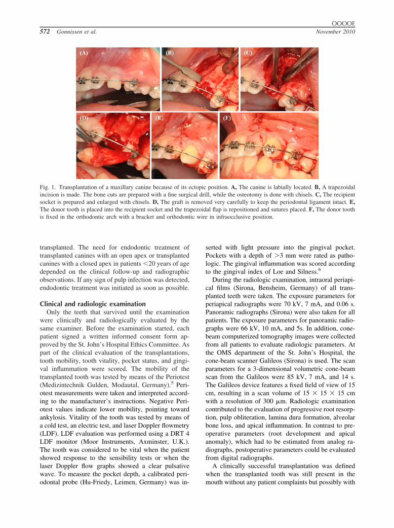
experienced oral surgeon, according to the strict proto-col of the OMS department of St. John’s Hospital.Before a surgical approach was made, a radiographicexamination had already revealed the location of theimpacted canine. Furthermore, the graft and the recip-ient site dimensions were checked for compatiblity. InFig. 1, the surgical procedure is shown for a maxillarycanine in labial deviation. The surgical procedure dif-fered according to the site of impaction, but the generalapproach is described here.
First, the surgical sites were disinfected and a localanaesthetic was injected. The remaining primary ca-nine, if it was still present, was extracted, and a trape-zoidal incision was made that ensured intact mesial,distal, and palatal gingiva at the graft site. To preparethe recipient socket, an osteotomy was performed usinga surgical bur and chisels. The tooth roots that weremesial and distal to the osteotomy site must remainintact. If the recipient site was too small for the do-nor tooth, the socket area was expanded by looseningthe vestibular and palatal bone plates with chisels. Thesocket for the graft should be slightly larger than thegraft. Next, the crown of the impacted canine wasexposed and the tooth removed with a periosteal ele-vator. The donor tooth was extracted slowly and asatraumatically as possible. It is important that the in-strument contacts only the crown and not the rootsurface so as not to damage the periodontal ligament(PDL). Next, the donor tooth was placed into the re-cipient socket without any pressure. Obstacles in thesocket wall were removed as encountered. The trape-zoidal flap was repositioned and sutures were placed.To optimize reattachment and block bacterial invasion,the gingival flap must close tightly around the donortooth. Finally, the donor tooth was splinted with asuture splint or with an acid-etch composite and orth-odontic wire. The occlusion was checked to ensure thatthe transplanted tooth had no traumatic contact with itsantagonists at the occlusal plane (infraocclusive posi-tion).
Criteria for endodontic treatmentEndodontic treatment was performed in patients �20
years of age who had a canine with a closed apex
ic wire
OOOOE572 Gonnissen et al. November 2010
transplanted. The need for endodontic treatment oftransplanted canines with an open apex or transplantedcanines with a closed apex in patients �20 years of agedepended on the clinical follow-up and radiographicobservations. If any sign of pulp infection was detected,endodontic treatment was initiated as soon as possible.
Clinical and radiologic examinationOnly the teeth that survived until the examination
were clinically and radiologically evaluated by thesame examiner. Before the examination started, eachpatient signed a written informed consent form ap-proved by the St. John’s Hospital Ethics Committee. Aspart of the clinical evaluation of the transplantations,tooth mobility, tooth vitality, pocket status, and gingi-val inflammation were scored. The mobility of thetransplanted tooth was tested by means of the Periotest(Medizintechnik Gulden, Modautal, Germany).5 Peri-otest measurements were taken and interpreted accord-ing to the manufacturer’s instructions. Negative Peri-otest values indicate lower mobility, pointing towardankylosis. Vitality of the tooth was tested by means ofa cold test, an electric test, and laser Doppler flowmetry(LDF). LDF evaluation was performed using a DRT 4LDF monitor (Moor Instruments, Axminster, U.K.).The tooth was considered to be vital when the patientshowed response to the sensibility tests or when thelaser Doppler flow graphs showed a clear pulsativewave. To measure the pocket depth, a calibrated peri-
Fig. 1. Transplantation of a maxillary canine because of its eincision is made. The bone cuts are prepared with a fine surgisocket is prepared and enlarged with chisels. D, The graft isThe donor tooth is placed into the recipient socket and the trais fixed in the orthodontic arch with a bracket and orthodont
odontal probe (Hu-Friedy, Leimen, Germany) was in-
serted with light pressure into the gingival pocket.Pockets with a depth of �3 mm were rated as patho-logic. The gingival inflammation was scored accordingto the gingival index of Loe and Silness.6
During the radiologic examination, intraoral periapi-cal films (Sirona, Bensheim, Germany) of all trans-planted teeth were taken. The exposure parameters forperiapical radiographs were 70 kV, 7 mA, and 0.06 s.Panoramic radiographs (Sirona) were also taken for allpatients. The exposure parameters for panoramic radio-graphs were 66 kV, 10 mA, and 5s. In addition, cone-beam computerized tomography images were collectedfrom all patients to evaluate radiologic parameters. Atthe OMS department of the St. John’s Hospital, thecone-beam scanner Galileos (Sirona) is used. The scanparameters for a 3-dimensional volumetric cone-beamscan from the Galileos were 85 kV, 7 mA, and 14 s.The Galileos device features a fixed field of view of 15cm, resulting in a scan volume of 15 � 15 � 15 cmwith a resolution of 300 �m. Radiologic examinationcontributed to the evaluation of progressive root resorp-tion, pulp obliteration, lamina dura formation, alveolarbone loss, and apical inflammation. In contrast to pre-operative parameters (root development and apicalanomaly), which had to be estimated from analog ra-diographs, postoperative parameters could be evaluatedfrom digital radiographs.
A clinically successful transplantation was definedwhen the transplanted tooth was still present in the
position. A, The canine is labially located. B, A trapezoidalll, while the osteotomy is done with chisels. C, The recipiented very carefully to keep the periodontal ligament intact. E,al flap is repositioned and sutures placed. F, The donor tooth
in infraocclusive position.
ctopiccal driremovpezoid
mouth without any patient complaints but possibly with

OOOOEVolume 110, Number 5 Gonnissen et al. 573
transient root resorption or endodontic treatment. Whenthe transplanted canine showed progressive root resorp-tion, pathologic pockets, extensive loss of alveolarbone, or an apical inflammation, the transplantation wasdefined as clinically unsuccessful, even if it remainedpresent in the patient without any subjective com-plaints. The success rate was calculated as the percent-age of clinically successful transplantations relative tothe number of transplantations. The survival rate wasdefined as the percentage of transplanted teeth stillpresent at the examination relative to the total numberof teeth that were transplanted.
StatisticsLogistic regression analyses were performed to de-
termine the influence of the various preoperative pa-rameters on the success rate of autotransplantation ofcanines. Parameters found to be significantly associatedwith the success rate on univariate logistic regression(P � .25) or those thought to be important on logicaland medical grounds were entered into a multivariatelogistic regression model. To compare the models intheir goodness of fit, Akaike and Bayesian informationcriteria were used. Fitting the models was performed oncomplete observations. Mixed models were used totake into account that some patients had �1 caninetransplanted.
To estimate the interexaminer variation, a secondexaminer evaluated all radiographs. Simple � statisticswere computed to determine the interexaminer agree-ment. The Kaplan-Meier method was used to estimatethe survival probability of autogenously transplantedcanines over a period of 14 years.
Statistic analyses were performed with SAS (version9.2) and S-plus (version 6.2). Significance was definedas P � .05.
RESULTSSurvival rate
The survival rate was 75.3%, because 18 trans-planted teeth were lost before examination. One trans-plant was lost 6 months after surgery because of adelayed endodontic treatment. The other transplantedteeth were lost 4 (n � 2), 5 (n � 1), 6 (n � 1), 7 (n �5), 8 (n � 3), 9 (n � 1), 10 (n � 1), 11 (n � 1), and 12years (n � 2) after surgery. Figure 2 represents theKaplan-Meier risk curve for the overall survival rateover 14 years. The most common reason for failure wasprogressive root resorption, which caused the loss of 9teeth.
Clinical and radiologic examinationA total of 55 transplanted teeth were clinically and
radiologically examined during this study.
Tooth mobility and tooth vitality. Overall, 35 trans-planted teeth (63.6%) showed negative Periotest values,meaning that the teeth lost mobility. On the other hand,2 teeth (3.6%) displayed Periotest values higher thanthe normal values and thus were clinically mobile. Theremaining 18 teeth (32.7%) had normal Periotestvalues.
Endodontic treatment was needed in 22 teeth(40.0%) after transplantation. Tooth vitality was exam-ined only in teeth that were not endodontically treated(33 teeth). Only 1 transplanted tooth (3.0%) showed apositive result for the cold test, and 4 transplanted teeth(12.1%) showed a positive result for the electric pulptest. In addition, the LDF graphs illustrated a clearpulsatility for 25 teeth (75.8%) out of the 33 teethwithout endodontic treatment.
Pocket depth and inflammation. Overall, 11 trans-planted teeth (20.0%) showed an unacceptable (�3mm) clinical pocket depth. All other transplanted teethhad an acceptable pocket depth of �3 mm.
Thirteen transplanted teeth (23.6%) showed moder-ate inflammation, meaning that the gingiva bled whileprobing the pocket depth.
Resorption. Twenty-one transplanted teeth (38.2%)showed some sign of root resorption, and 34 teeth(61.8%) showed no resorption at all. Nineteen trans-planted teeth (90.5%) showed external root resorption,and only 2 teeth (9.5%) showed internal root resorption.One specific radiologic case of a transplanted canine inthe right upper jaw is presented in Fig. 3. Figure 3, A,illustrates replacement resorption of the transplantedcanine on a periapical radiograph. Cross-sections of acone-beam volumetric scan through the same trans-planted canine are shown in Fig. 3, B and C, where theentrance of the alveolar bone in the tooth becomes
Fig. 2. Kaplan-Meier estimation describing the probability ofsurvival for a follow-up period of 14 years.
clear.

the to
OOOOE574 Gonnissen et al. November 2010
Pulp obliteration and lamina dura formation. In 14transplanted teeth (25.4%) no important changes in thepulp chamber were detected. Other teeth, excludingendodontic treated teeth, showed a reduction in size ora complete obliteration of the pulp chamber.
In total, 29 transplanted teeth (52.7%) showed anintact lamina dura. In 15 transplanted teeth (27.3%),however, no lamina dura could be detected on theradiographs. The remaining 11 teeth (20.0%) weremissing only a part of the lamina dura.
Alveolar bone loss and apical inflammation. Out ofthe 55 transplanted teeth that were examined, 6 (10.9%)showed extensive alveolar bone loss after transplanta-tion. In addition, 3 (5.4%) exhibited an apical inflam-mation.
Success rateBased on the clinical and radiologic examination, the
success rate for all transplanted teeth in this study was57.5%, because 42 transplanted teeth were evaluated asclinically successful. Two radiologic cases of trans-planted canines are presented in Fig. 4 and Fig. 5.
Univariate analysis. The influence of each parameterseparately on the long-term success outcome of 73transplanted teeth is shown in Table III. Two parame-ters were found to be statistically significant regardingsuccess rates in this study. The success rate signifi-cantly decreased when the age at time of transplantation
Fig. 3. A, Periapical radiograph of a transplanted canine in tCross-sections of a cone-beam volumetric scan through the sacross-section (B) shows the entrance of the alveolar bone in
increased (odds ratio [OR] 0.95, 95% confidence inter-
val [CI] 0.90-0.99; P � .0287). The success rate ofteeth that were ankylosed at the time of transplantationwas 33.3%, whereas it was 65.4% for teeth that werenot ankylosed. This difference was statistically signif-icant (OR 0.26, 95% CI 0.08-0.81; P � .0168).
All other preoperative parameters had no significantinfluence on the long-term success rate (Table III). Thesuccess rate of canine autotransplantation for male pa-tients was 51.2%, whereas it was 65.6% for femalepatients (P � .2148). Although the success rate ofautotransplantations performed on palatial impacted ca-nines was 57.6%, it was 40% for autotransplantationperformed on labial impacted canines (P � .4469).Only 5 impacted canines were located labially. Thesuccess rate of impacted canines that were previouslysurgical exposed was 42.9% compared with 59.1% fornonsurgically exposed canines (P � .4118). Only 7 ofthe 73 transplanted teeth were previously surgicallyexposed. The success rate was lower for teeth trans-planted in a too-small diastema (40%) than for trans-planted teeth that had enough space (58.8%). However,this difference was not statistically significant (P �.4144). No significant difference in success rate wasfound between teeth transplanted at a different rootdevelopment stage (P � .4998). For teeth transplantedwith an open apex, the success rate was 70.6%, whichwas not significantly higher than the success rate(60.5%) for teeth transplanted with a closed apex (P �
t upper jaw showing replacement resorption (arrow). B, C,nsplanted canine showing root resorption (arrows). The first
oth.
he righme tra
.4693). In addition, the success rates for impacted ca-

d of 9
functi
OOOOEVolume 110, Number 5 Gonnissen et al. 575
nines with a curved apex and impacted canines withouta curved apex were not significantly different (P �.4250). Uni- and bilateral transplantations showed al-most the same success rates, respectively 57.8% and
Fig. 4. Successful transplantation of an impacted canine in twere taken before surgery (A, B) and after a follow-up perio
Fig. 5. Transplantation of bilateral impacted canines after ugraph was taken before surgery (A) and at follow-up 12 yetransplantation, show the transplanted upper right canine defias unsuccessful due to periodontal defect (D). Both teeth are
57.1% (P � .9574). The success rate of transplantation
of teeth that caused root resorption to adjacent teethwas 71.4%, whereas it was 54.2% for teeth that causedno root resorption (P � .2339). When the PDL was notdamaged during surgery, 62.7% of the transplantations
t upper jaw (circled). Panoramic and periapical radiographsyears (C, D).
ssful orthodontic realignment (circled). A panoramic radio-r transplantation (B). Periapical radiographs, 12 years aftersuccessful (C) and the transplanted upper left canine definedonal and not causing complaints by the patient.
he righ
nsuccears aftened as
were successful compared with a success rate of 50%

OOOOE576 Gonnissen et al. November 2010
when the PDL was damaged (P � .4643). For patientswho had orthodontics, 62.5% of the transplants weresuccessful, and the success rate for transplanted teethfixed with a splint was 41.1% (P � .1209). The successrate for teeth placed in infraocclusion after transplan-tation was 58.5%, and it was 25% for teeth placed inocclusion. This difference was not statistically signifi-
Table III. Variables which could have an influence onthe overall success rate of transplantation, as derivedfrom the univariate analysis
Covariate (parameter) OR (95% CI)Univariate
P value
PatientAge at transplantation 0.95 (0.90-0.99) .0312Gender
Female 1.00 (ref.) .2148Male 0.55 (0.21-1.43)
Transplantation areaPosition
Palatinal 1.00 (ref.) .4469Labial 0.49 (0.08-3.14)
Surgical exposureNo 1.00 (ref.) .4118Yes 0.52 (0.11-2.51)
SpaceNo 1.00 (ref.) .4144Yes 2.14 (0.34-13.67)
Transplanted canineRoot development
Complete 1.00 (ref.) .4998More than three-fourths 2.14 (0.58-7.95)One-half to three-fourths 1.43 (0.12-17.23)
ApexOpen 1.00 (ref.) .4693Closed 0.64 (0.19-2.18)
Curved apexNo 1.00 (ref.) .4250Yes 1.54 (0.53-4.46)
AnkylosisNo 1.00 (ref.) .0168Yes 0.26 (0.08-0.81)
Unilateral 1.00 (ref.) .9574Bilateral 0.97 (0.37-2.53)Adjacent teeth
Root resorptionNo 1.00 (ref.) .2339Yes 2.11 (0.59-7.49)
Pre-/perioperativeDamage of periodontalligament
No 1.00 (ref.) .4643Yes 0.62 (0.17-2.22)
FixationSplint 1.00 (ref.) .1209Orthodontics 2.38 (0.79-7.20)
Occlusion 1.00 (ref.) .1935Infraocclusion 4.24 (0.41-44.27)
OR, Odds ratio; CI, confidence interval.
cant (P � .1935).
Multivariate analysis. After the prognostic signifi-cance of each parameter was determined by univariateanalysis, assessment of the relative importance of theparameters in explaining the success rate after caninetransplantation was established by multivariate logisticregression analysis. The statistical model that best fittedto the data contained 3 preoperative parameters ascovariates (Table IV). The significant parameter in de-termining the success rate of autotransplantation wasage at the time of transplantation (P � .0429). Morespecifically, the age at transplantation had a negativecorrelation with the success rate (OR 0.98, 95% CI0.96-0.99). The higher the age at transplantation, thelower the success rate (Fig. 6). Other parameters werenot significant, including root development (P � .7542)and condition of the apex (P � .5444).
DISCUSSIONThe survival rate of transplanted canines in this study
was 75.3%. It appears from other studies that toothsurvival after autotransplantation of canines rangesfrom 86% to 100%.7 This dissimilarity can be ex-plained by the difference in follow-up period. An im-portant factor in tooth survival assessment is the lengthof the observation period.7 The follow-up period in thepresent study ranged from 6 to 14 years, with a mean of
Fig. 6. Probability of success as function of age at transplan-tation. The probability of success decreased when the age attime of transplantation increased.
Table IV. The final model, derived from the multivar-iate analysis, containing 3 parameters, with the age attransplantation being predictive for determining thesuccess rate after autotransplantation of canines
Covariate (parameter) OR 95% CI P value
Age at transplantation 0.98 0.96-0.99 .0429Root development .7542
More than three-fourths 1.18 0.74-1.89One-half to three-fourths 1.08 0.60-2.37Closed apex 1.16 0.72-1.88 .5444
OR, Odds ratio; CI, confidence interval.
11 years. In contrast, the main observation period in the

OOOOEVolume 110, Number 5 Gonnissen et al. 577
other studies was only 5 years or even less. Further-more, it has been proven that with increasing time aftertransplantation, significantly more root resorption isfound.7 Because progressive root resorption was themain reason for tooth loss in the present study, thiscould be the explanation of why fewer transplantedteeth survive for 11 years than for 5 years.
When comparing the success rate of this study withother success rates in literature, it is important to con-sider the difference in criteria for success, because thereare no common success criteria. A remarkable differ-ence with other studies is that in the present studytransient root resorption was not considered as a crite-rion for unsuccessful transplantation, because even incases of eventual failure, autotransplanted teeth may beretained for considerable lengths of time, providing thepatient with an esthetic and functional solution.8
The success rate of transplanted teeth in this study,57.5%, is lower than the success rate of transplantationreported in other studies (�90%).9-11 This can be ex-plained by the fact that most studies included all toothtypes in their calculation of the success rate of thetransplantation procedure. However, some studies alsomention the specific success rate for canine transplants.Pogrel12 found a success rate of 62% for the transplan-tation of maxillary canines after a follow-up of �2years. In the study by Kallu et al.,13 the success rate forcanines was 51% with a mean follow-up of 3.8 years.These rates are similar to the success rate found in thepresent study, though our results indicate the successrate 11 years after surgery. There are multiple reasonswhy the success rate of canines is lower than thesuccess rate of other tooth types. Impaction of thecanines is an important factor that influences the out-come of transplantation.8 The accessibility of the toothduring surgery is lower, so that the chance of damagingthe root surface while extracting is greater. In addition,the PDL in impacted teeth is thinner than in teeth thatare functioning normally.14 Furthermore, transplanta-tions of impacted canines to their normal position arecalled transalveolar transplantations which have thedrawback of possibly not having a preformed alveolusfor positioning the donor tooth. It is already proven thatautotransplantation is more successful if the donortooth is transplanted to an extraction site with PDLattachment still present in the recipient socket. There-fore, success rates of teeth transplanted into artificiallyformed sockets are lower than those of teeth trans-planted into extracted sockets.8,15
The significant parameter in determining the successrate of autotransplantation was the age at transplanta-tion. More specifically, the age at time of transplanta-tion had a negative correlation with the success rate,
meaning that the younger the patient, the higher thesuccess rate. This result may be explained by betterhealing capacity in younger patients. On univariatelogistic regression analysis, the presence or absence ofankylosis was also associated with the success rate ofautotransplantation. Ankylosis is one of the possiblecauses of impacted canines.16,17 During eruption, theroot of the canine is replaced by bone and thereforethe canine gets stuck and remains impacted. Onmultiple logistic regression analysis, we failed todemonstrate that this preoperative parameter was adefinite predictor for the success rate of canine auto-transplantation. In the clinical setting, it is worthwhileto keep in mind the possibility of a lower long-termsuccess rate if the impacted canine was ankylosed.
Based on the literature, the maintenance of healthyPDL cells is an important consideration for a successfultooth transplant.8,18 The correlation between damage tothe PDL and the success rate of transplantation couldnot be verified in the present study. A possible expla-nation could be that during extraction, there is alwayssome degree of damage to the PDL and it is difficult todetermine the exact degree of PDL damage. It is notpossible to be sure that the entire PDL is vital orundamaged after extraction of the donor tooth. Fromthe literature, it is also known that root shape andmaturity of the root structure of the donor tooth areinfluencing factors for the prognosis of autotransplan-tation.7,8,19 However, a lower success rate due to acurved apex could not be established in the presentstudy. In addition, no significant difference in successrate was found between the various root developmentalstages. The main reason that these and possibly otherpreoperative parameters were not found to have a sig-nificant influence on the success rate of canine trans-plantation is because of missing data and unbalanceddata. Owing to the retrospective nature of this study, themain limitation was the availability of information inthe medical files when so many parameters were takeninto consideration.
CONCLUSIONThe present study demonstrated a survival rate of
75.3% 11 years after transplantation of impacted ca-nines. The age at transplantation was found to be animportant prognostic factor. A success rate of 57.5% islow but acceptable, considering that transplantationwas indicated when surgical exposure seemed to be toodifficult or when orthodontic realignment after surgicalexposure failed. Weighted against extraction, the op-tion of transplantation of the canine is a valuable tech-nique with an acceptable long-term prognosis. In thefuture, these results could be improved by adding morepatients and collecting preoperative parameters as com-
pletely as possible.
OOOOE578 Gonnissen et al. November 2010
REFERENCES1. Howlander M, Begum S, Naulakha D. Autogenous tooth trans-
plantation from ectopic position: a case report and review fromliterature. Bangladesh Coll Phys Surg 2006;24:79-85.
2. Chaushu S, Chaushu G, Becker A. The use of panoramic radio-graphs to localize displaced maxillary canines. Oral Surg OralMed Oral Pathol Oral Radiol Endod 1999;88:511-6.
3. Kalavritinos MK, Kaisaris P. Autogenous transplantation of im-pacted canines. Hel Orthod Rev 2006;9:47-60.
4. Moorrees CF, Fanning EA, Hunt EE Jr. Age variation of formationstages for ten permanent teeth. J Dent Res 1963;42:1490-502.
5. Schulte W, Lukas D. The Periotest method. Int Dent J 1992;42:433-40.
6. Loe H, Silness J. Periodontal disease in pregnancy. I. Prevalenceand severity. Acta Odontol Scand 1963;21:533-51.
7. Andreasen J. Atlas of replantation and transplantation of teeth.Copenhagen: Mediglobe; 1990.
8. Tsukiboshi M. Autotransplantation of teeth. Chicago: Quin-tessence; 2001.
9. Akkocaoglu M, Kasaboglu O. Success rate of autotransplantedteeth without stabilisation by splints: a long-term clinical andradiological follow-up. Br J Oral Maxillofac Surg 2005;43:31-5.
10. Czochrowska EM, Stenvik A, Bjercke B, Zachrisson BU. Out-come of tooth transplantation: survival and success rates 17-41years posttreatment. Am J Orthod Dentofacial Orthop 2002;121:110-9; quiz 93.
11. Tanaka T, Deguchi T, Kageyama T, Kanomi R, Inoue M, FoongKW. Autotransplantation of 28 premolar donor teeth in 24 orth-
odontic patients. Angle Orthod 2008;78:12-9.12. Pogrel MA. Evaluation of over 400 autogenous tooth transplants.J Oral Maxillofac Surg 1987;45:205-11.
13. Kallu R, Vinckier F, Politis C, Mwalili S, Willems G. Toothtransplantations: a descriptive retrospective study. Int J OralMaxillofac Surg 2005;34:745-55.
14. Ioannidou E, Makris GP. Twelve-year follow-up of an autoge-nous mandibular canine transplant. Oral Surg Oral Med OralPathol Oral Radiol Endod 2003;96:582-90.
15. Tsukiboshi M. Autotransplantation of teeth: requirements forpredictable success. Dent Traumatol 2002;18:157-80.
16. Alberto PL. Management of the impacted canine and secondmolar. Oral Maxillofac Surg Clin North Am 2007;19:59-68.
17. Becker A. The orthodontic treatment of impacted teeth. Jerusa-lem: Informa Healthcare; 2007.
18. Kim E, Jung JY, Cha IH, Kum KY, Lee SJ. Evaluation of theprognosis and causes of failure in 182 cases of autogenous toothtransplantation. Oral Surg Oral Med Oral Pathol Oral RadiolEndod 2005;100:112-9.
19. Schwartz O, Bergmann P, Klausen B. Autotransplantation ofhuman teeth. A life-table analysis of prognostic factors. Int J OralSurg 1985;14:245-58.
Reprint requests:
Prof. Dr. Constantinus PolitisHead, Department Oral Maxillofacial SurgerySt. John’s HospitalSchiepse Bos 63600 GenkBelgium
[email protected]