Surfactant and Surfactant-Polymer Flooding for Enhanced Oil Recovery
Upload
independentCategory
view
0download
0
Journal of Applied Microbiology 1999, 86, 80–90
Viscosinamide, a new cyclic depsipeptide with surfactant andantifungal properties produced by Pseudomonas fluorescensDR54
T.H. Nielsen 1, C. Christophersen 2, U. Anthoni 2 and J. So�rensen 1
1Section of Genetics and Microbiology, Department of Ecology, The Royal Veterinary and Agricultural University,Frederiksberg, and 2Marine Chemistry Section, The H. C. Ošrsted Institute, University of Copenhagen, Copenhagen,Denmark
6965/11/98: received 13 November 1998, revised 31 March 1999 and accepted 7 April 1999
T.H. NIELSEN, C. CHRISTOPHERSEN, I . ANTHONI AND J. SOšRENSEN. 1999. Pseudomonasfluorescens DR54 showed antagonistic properties against plant pathogenic Pythium ultimumand Rhizoctonia solani both in vitro and in planta. Antifungal activity was extractablefrom spent growth media, and fractionation by semi-preparative HPLC resulted in isolationof an active compound, which was identified as a new bacterial cyclic lipodepsipeptide,viscosinamide, using 1D and 2D 1H-, 13C-NMR and mass spectrometry. The new antibiotichas biosurfactant properties but differs from the known biosurfactant, viscosin, bycontaining glutamine rather than glutamate at the amino acid position 2 (AA2). No viscosinproduction was observed, however, when Ps. fluorescens DR54 was cultured in mediaenriched with glutamate. In vitro tests showed that purified viscosinamide also reduced fungalgrowth and aerial mycelium development of both P. ultimum and R. solani.Viscosinamide production by Ps. fluorescens DR54 was tightly coupled to cell proliferation inthe batch cultures, as the viscosinamide produced per cell mass unit approached aconstant value. In batch cultures with variable initial C, N or P nutrient levels, there wereno indications of elevated viscosinamide production during starvation or maintenanceof the cultures in stationary phase. Analysis of cellular fractions and spent growthmedia showed that a major fraction of the viscosinamide produced remained bound tothe cell membrane of Ps. fluorescens DR54. The isolation, determination ofstructure and production characteristics of the new compound with both biosurfactantand antibiotic properties have promising perspectives for the application of Ps.fluorescens DR54 in biological control.
INTRODUCTION
Fluorescent pseudomonads often show antagonistic activityagainst fungal root pathogens (Weller 1988). However,screening for strains to be used for biocontrol has usuallybeen carried out using in vitro agar plate tests. It is knownthat this protocol may not always detect strains which willantagonize the pathogens in situ (Keel et al. 1992). A possibleway to increase the recovery of useful isolates may be to select
Correspondence to: Dr T.H. Nielsen, Section of Genetics and Microbiology,Department of Ecology, The Royal Veterinary and AgriculturalUniversity, Thorvaldsensvej 40, DK-1871 Frederiksberg C (Copenhagen),Denmark (e-mail: [email protected]).
© 1999 The Society for Applied Microbiology
for strains showing medium-independent in vitro antagonism(Nielsen et al. 1998b). Three Pseudomonas fluorescens strainsisolated by such a protocol showed antagonistic activityagainst two root pathogenic fungi, Pythium ultimum and Rhi-zoctonia solani, both in in vitro and in planta experiments withsugar beet seedlings (Nielsen et al. 1998b). Subsequent testssuggested that the antagonistic activity by the highly activeisolate, Ps. fluorescens strain DR54, was at least partiallycaused by production of an unknown antibiotic (Nielsen et al.1998b).
Several Pseudomonas antibiotics are known (Dowling andO’Gara 1994) and their antagonistic activity against variouspathogenic fungi has been well documented (Weller 1988).
VISCOSINAMIDE, A NEW ANTIFUNGAL DEPSIPEPTIDE 81
As suggested by Shanahan et al. (1992b), antibiotics againstpathogenic fungi may be grouped into phenazines (Guru-siddaiah et al. 1986; Thomashow et al. 1990), pyo compounds(Takeda 1958), pyrrols (Howell and Stipanovic 1979), andother compounds such as phloroglucinols (Keel et al. 1992;Nowak-Thompson et al. 1994). Antibiotics with surfactantproperties (Asaka and Shoda 1996; Stanghellini and Miller1997) have also been found.
Various parameters influence antibiotic production inPseudomonas spp. e.g. pH (Shanahan et al. 1992b), tem-perature (Bolton and Elliot 1989; Shanahan et al. 1992b),oxygen concentration (Bencini et al. 1983; Messenger andTurner 1983; Shanahan et al. 1992b), surface attachment(Shanahan et al. 1992b), substrate composition and con-centration (Bencini et al. 1983; Messenger and Turner 1983;James and Gutterson 1986; Shanahan et al. 1992b; Stephens etal. 1993), trace element concentrations (Gross 1985; Sliningerand Jackson 1992) and antibiotic precursors (Gorman andLively 1967).
In batch cultures, the pseudomonads may show growth-phase dependent antibiotic production (Shanahan et al.1992a). In a previous study, Ps. fluorescens strain DR54 wasshown to produce variable amounts of an unknown antibioticin cultures with different carbon sources (Nielsen et al.1998a). It was also found that the purified antibiotic exhibitedantifungal activity and specific stress responses in Pythiumultimum and Rhizoctonia solani (Thrane et al. in press). Byvital fluorescent staining of fungal mycelium, it was thusevident that the antibiotic resulted in increased branching,swelling and septation of hyphae, and reduced intracellularactivity. The changes were suggested to be caused by alteredintracellular ion (H¦ and Ca2¦) contents, possible due tochannel formation in the cell membrane (Thrane et al. inpress). In the present study, the chemical identification ofviscosinamide, which is a new cyclic lipodepsipeptide withboth biosurfactant and antibiotic properties, is reported. Theviscosinamide production in Ps. fluorescens DR54 as affectedby different growth stages in glucose minimal medium, andthe importance of C, N and P limitations in this medium, isalso reported.
MATERIALS AND METHODS
Purification and structural analysis of a newPseudomonas antibiotic
Pseudomonas fluorescens strain DR54 was isolated from thesugar beet rhizosphere (Nielsen et al. 1998b) and identifiedto Ps. fluorescens biovar I according to Palleroni (1984). StrainDR54 was initially found to show antibiotic production onPotato Dextrose Agar (PDA; Difco) (Nielsen et al. 1998b).To determine the chemical structure, a large aliquot of active
© 1999 The Society for Applied Microbiology, Journal of Applied Microbiology 87, 80–90
compound was obtained after growth (7 d, 20 °C) of Ps. flu-orescens DR54 on 200 PDA agar plates. The agar was homo-genized and extracted twice with 600ml of a mixture ofethyl acetate containing 6ml formic acid. The solvent wassubsequently evaporated by vacuum centrifugation and theresidue was dissolved in 4ml methanol. The active compoundwas purified by semi-preparative HPLC with automatic frac-tion collection, using a LiChroCART 250–10 HPLC-Car-tridge with LiChrosphere 100 RP-18 (10mm) (Merck) (20 °C)as a preparative column. Samples of 200ml were injected andeluted in a gradient from 80% methanol in Milli-Q water(Millipore, MA, USA) increasing to 100% methanol over a 20min period (flow rate 3ml min−1). Approximately 450mmolantibiotic could be purified from the 200 PDA agar plates.
NMR spectra were recorded on a Varian 400 FT-NMRspectrometer (Varian NMR Instruments, Palo Alto, Cali-fornia, USA) at 400·0 and 100·6MHz for 1H- and 13C-NMRspectra, respectively, using samples in hexadeuterio-dimethylsulfoxide (DMSO-d6).
1H,1H correlated spec-troscopy (COSY) was performed using pulse gradientmultiquantum-filtered, phase-sensitive analysis. Rotatingframe NOESY (ROESY) data were determined using a mix-ing time of 200 ms. Heteronuclear multiple quantum coher-ence (HMQC) data were recorded with pulse gradients andreverse detection and optimized for JCH 140Hz (single bond)and 7Hz (multiple bond), respectively. FAB-MS spectrawere determined on a JEOL JMS-HX/HX110A tandemmass spectrometer in a meta-nitrobenzylic alcohol(NBA)-gly-cerol-thioglycerol 1 : 1: 2 matrix containing 1% trifluoroaceticacid. Circular dichroism (CD) spectra were measured on aJASCO J-70 spectropolarimeter (JASCO International Co.Ltd, Tokyo, Japan).
A Hofmann degradation (Fieser et al. 1981) was performedby addition of a solution of iodobenzene bis(trifluoroacetate)(1·3mmol, 560mg) in acetonitrile/water 1 : 1 (70ml) to a solu-tion of viscosinamide (1·3mmol, 1·45mg) in aceto-nitrile/water 1 : 1 (300ml). The mixture was then left for 5 hat room temperature protected against light. The reactionmixture was lyophilized after addition of 0·01 M HCl (1·8ml)and the reaction product subjected to FAB-MS.
Antifungal properties of purified antibiotic
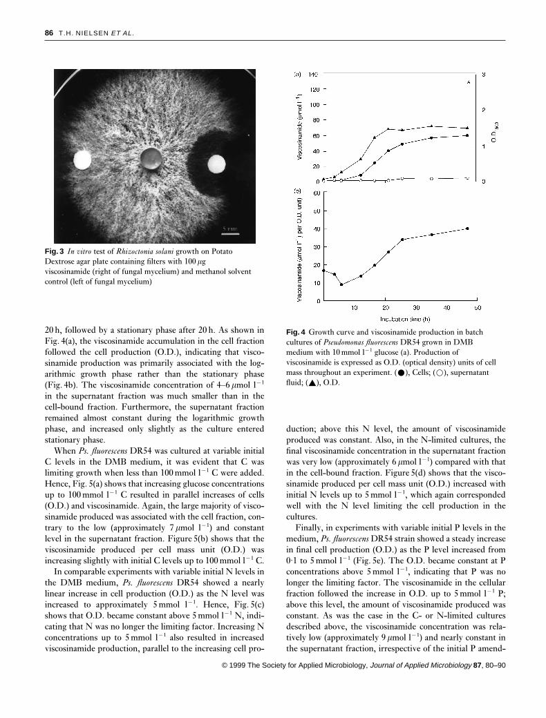
The plant pathogenic Basidiomycete Rhizoctonia solani KuhnAG-4 was isolated from a diseased sugar beet root (DaniscoSeed, Holeby, Denmark) and maintained as described byThrane et al. (in press). The antifungal activity of visco-sinamide was tested against the fungus using in vitro tests. Asample of 100mg purified compound in methanol was appliedto a sterile glass fibre filter (5mm diameter). The methanolsolvent was evaporated in the hood for 15min and the filterwas placed 1·5 cm from Rhizoctonia solani mycelium inocu-

82 T.H. NIELSEN ET AL.
lated on a Potato Dextrose Agar plate (PDA; Difco) (Nielsenet al. 1998b). A filter treated with pure methanol (HPLCgrade) was used as control. After incubation at 27 °C for30 h, the fungal growth was photographed with a Nikon F70camera with a Sigma zoom, 28–80, Macro using a Fujicolour100 ISO film.
Production of antibiotic in liquid media
Prior to all growth experiments in liquid media, the strainwas pre-cultured in Davis Minimal Broth (DMB; 30mmoll−1 K2HPO4, 14mmol l−1 KH2PO4, 0·4mmol l−1 MgSO4,7·6mmol l−1 (NH4)2SO4, 60mmol l−1 C (glucose), and 1mll−1 trace element solution, pH 7·3. The trace element solutioncontained 20mg CoCl2.6H20, 30mg H3BO3, 10mgZnSO4.7H20, 1mg CuCl2.2H2O, 2mg NiCl2.6H2O, 3mgNaMoO4.2H2O, 10mg FeSO4.7H2O and 2·6mg MnSO4.H2Odissolved in 1 litre Milli-Q water. Unless specified, all cul-tures were grown in 50ml Capsenberg glass flasks on a rotaryshaker (150 rev min−1) at 20 °C for 2 d, before cells wereharvested by centrifugation (7000 g, 10min, 4 °C), washedtwice and dissolved in phosphate-buffered saline (PBS;25mmol l−1 phosphate buffer, 125mmol l−1 NaCl, pH 7·4).In the following experiments, cells from the washed pre-culture were inoculated at an optical idensity (O.D.) of 0·1 at600 nm.
To determine the antibiotic production during differentgrowth stages in the batch cultures, Ps. fluorescens DR54was inoculated in fresh DMB medium with 60mmol l−1 C(glucose) in a 140 ml Erlenmeyer flask with glass inva-ginations, designed to facilitate high aeration of the culture.The cultures were incubated at 20 °C under shaking and 9mlsub-samples were collected at defined time intervals. A 6ml aliquot of the sub-sample was transferred into a 9 mlpolyethylene vial for subsequent extraction and analysis(described below). The remaining 3ml sub-sample was usedto determine optical density (O.D.).
To determine the antibiotic production during C, N or Pnutrient limitation in the cultures, Ps. fluorescens DR54 waspre-cultured as described above, washed twice and dissolvedin DMB without C, N or P. The effect of C limitation wassubsequently studied by inoculation of Ps. fluorescens DR54in 50ml vials containing 20ml DMB with 15, 30, 45, 60,90, 120 or 150mmol l−1 C (glucose) and 15mmol l−1 N((NH4)2SO4). Similarly, the influence of N limitation wasdetermined in DMB containing 60mmol l−1 C (glucose) with0·1, 0·5, 1, 2·5, 5, 7·5 or 15mmol l−1 N ((NH4)2SO4). Finally,P limitation was studied in DMB prepared with 10mmol l−1
Hepes buffer (pH 7·3) rather than phosphate buffer. Thismedium contained 60mmol l−1 C (glucose), 15mmol l−1 N((NH4)2SO4) and 0·5, 1, 2, 3, 4, 5 or 10mmol l−1 P (K2HPO4).All cultures were incubated under shaking (150 rev min−1)
© 1999 The Society for Applied Microbiology, Journal of Applied Microbiology 87, 80–90
at 20 °C for 2 d and subsequently sub-sampled for O.D.determinations and extractions of viscosinamide from thecells and supernatant fluid as described below. Con-centrations of antibiotic accumulated in both the supernatantfluid and cells were expressed in mmol l−1 of culture. Allexperiments were performed in triplicate.
Extraction and analysis of antibiotic in cell andsupernatant fractions
Cell and supernatant fractions of the cultures were separatedby centrifugation at 7000 g for 10min at 4 °C. Antibiotic wasextracted from the cell fraction (pellet) with 1ml acetone,which was subsequently evaporated under an N2 flow, andthe extract was dissolved in methanol for HPLC analysis.The supernatant fraction was extracted twice using a 1·5volume of ethyl acetate containing 0·1% formic acid in thefirst extraction. After evaporation of the solvent, the extractwas dissolved in methanol. Prior to HPLC analysis, impurit-ies were precipitated by centrifugation at 15 000 g for 10minat 4 °C. The amount of antibiotic produced per millilitre oforiginal culture was calculated from the HPLC analysis ofextractant from supernatant and cell fractions.
The efficacy of the extraction procedure was tested byadding purified antibiotic dissolved in 300ml methanoldirectly into 7ml of DMB media, spent culture supernatantfluid or Ps. fluorescens DR54 culture. These additionsincreased the antibiotic concentrations by 17·9mmol l−1 and37·6mmol l−1. All samples were vortexed and left for 1 hbefore they were extracted as described above. Before theculture-amended sample was extracted, cells and supernatantfractions were separated by centrifugation and extracted asdescribed above. To determine whether the methanol solventinfluenced the extraction efficacy, samples amended with300ml HPLC-grade methanol were included for comparison.
To determine the cellular localization of antibiotic in Ps.fluorescens DR54, cells were washed twice and concentratedto a higher cell density. Sub-samples of 3ml cell suspensionwere first sonicated for 1min interval on ice (Sanyo MSE150W ultra sonic disintegrator, probe diameter 9·5mm,10mm amplitude, Kontram, Denmark) during a total of10min. Unbroken cells were removed by centrifugation at7000 g for 10min at 4 °C, and the supernatant fluid containingboth membranes and cytoplasm fractions were transferredinto Corex tubes (Du Pont Company, Delaware, USA) beforeall samples were centrifuged at 50 000 g for 10min at 4 °C.The supernatant fluid from each sample was subsequentlyremoved and extracted twice with a mixture of 9ml ethylacetate and 0·1ml formic acid. The membrane pellet wasextracted with 1ml acetone, which was evaporated before thepellet material was re-dissolved in 0·2ml methanol for HPLCanalysis as described below.
VISCOSINAMIDE, A NEW ANTIFUNGAL DEPSIPEPTIDE 83
Analysis of antibiotic in the extracts was performed usinga Hewlett Packard 1100 Series HPLC with a LiChroCART250–4 HPLC-Cartridge containing LiChrosphere 100 RP-18 Merck (5mm) column kept at 40 °C. The samples wereanalysed in a gradient of 80% acetonitrile and 20% 0·1%o-phosphoric acid (0–5min), and 80–99% acetonitrile (5–15min), 99% acetonitrile (15–18·5min) and 99–80% (18·5–20min), respectively, run at 1ml min−1. Standards of purifiedantibiotic were included. A diode array detector measuredabsorption at 2102 8 nM. Chromatograms were analysedusing the Hewlett-Packard HPLC 3D ChemStation softwareHewlett-Packard GmbH, Waldbronn, Germany.
RESULTS
Structural analysis and detection of a new antibiotic,viscosinamide
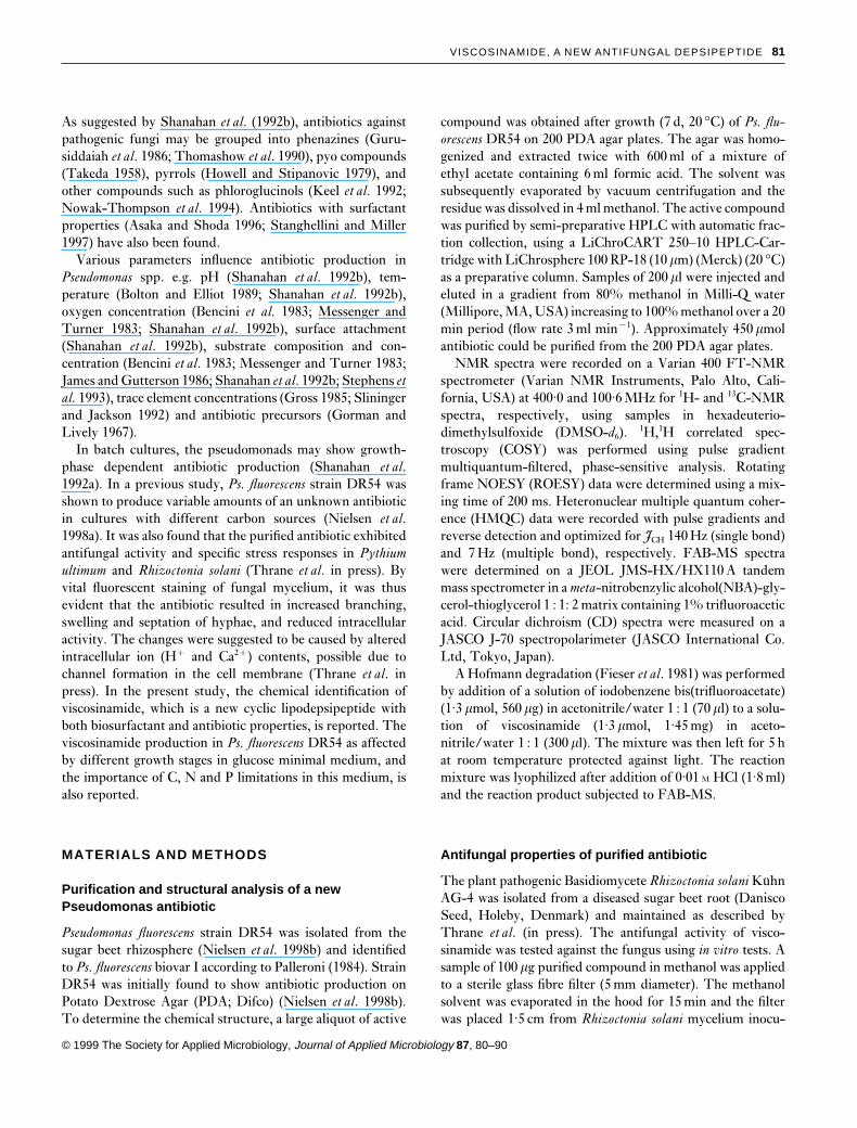
Figure 1 shows the structure of the new Pseudomonas anti-biotic as inferred from the FAB-MS pseudomolecular ionsat m/z 1125·69 (M¦H)¦ and 1147·68 (M¦Na)¦. Themolecular composition, C54H96N10O15, represents the amideof the known Pseudomonas surfactant, viscosin (Hiramotoet al. 1970). Viscosinamide has a glutamine residue at aminoacid position 2 (AA2), where a glutamic acid residue is foundin viscosin. In routine HPLC analysis, viscosin and visco-sinamide both showed absorption at 210 nm and could there-fore not be distinguished by UV-VIS spectra. However, thenew viscosinamide compound could be chromatographicallyseparated from viscosin owing to the slightly longer retentiontime (data not shown). The FAB-MS data further displayedsignals from fragment ions representing loss of a hyd-roxydecanoyl moiety, acylated Leu, acylated Gln-Leu, acy-lated and dehydrated Thr-Gln-Leu, and acylated Val-dehydrated Thr-Gln-Leu as depicted in Fig. 1(b). This find-ing is analogous to the recorded fragmentation of the Pseudo-monas surfactant, massetolide F (Gerard et al. 1997), andrepresents fragmentation of an acylated linear peptide formedby a ring opening between the protonated C-terminal Ileand the Thr leaving the unsaturated dehydrated Thr in thepeptide chain.
The 1H- and 13C-NMR data were compatible with thisstructure. A pivotal point in the structure elucidation ofviscosinamide was the appearance in comparison with visco-sin, a perturbed glutamine residue with the g-carbonyl groupat dC 173·9, and two broadened singlets at dH 6·78 and 7·24,respectively. These signals showed a weak coupling in theCOSY spectrum and a weak cross peak in a ROESY experi-ment. The signal at dH 7·24 exhibited two bond coupling tothe dC signal at 173·9 and the one at dH 6·78 to the samecarbonyl and to the Gln-g methylene carbon at dC 31·4 bearingthe protons appearing at dH 2·09 (dd, J 14·5 and 6·5Hz). Asa result of extensive overlap in proton signals, it was not
© 1999 The Society for Applied Microbiology, Journal of Applied Microbiology 87, 80–90
entirely possible to assign the individual signals. The nine a-carbon atoms and the attached protons appear at dC 51·5 and53·0 (dH 4·20), 56·4, 55·1, 51·2, 51·1 (4·30), 55·3 (4·33), 56·4(Thr, 4·45, dd, 14·5, 6·5Hz) and 59·1 (Val, 3·99, dd). The 3-hydroxydodecanoyl moiety appear at dC 43·6 (C-2, dH 2·24,ddd), 67·6 (C-3, 3·81, dt), 37·1 (C-4, 1·35) and dH 4·61 (OH,d, J 5·5Hz). Except for the signals mentioned above, thefollowing signals are diagnostic because of their position inotherwise unoccupied parts of the spectrum: Gln residue Cb
35·9 (1·88, 1·74), Thr Cb 69·7 (5·00 dq), Cg 16·5 (1·05, d) andSer OH 4·82, dd, 13·7, 5·5Hz.
Confirmation of the exact structure was achieved from thefact that viscosinamide could be transformed to the cor-responding depsipeptide containing 2,4-diaminobutyric acidinstead of glutamine. The transformation was effected bytreatment of viscosinamide with bis(trifluoroacetoxy)iodobenzene, giving rise to a Hofmann rearrangement result-ing in elimination of the acid carbonyl. The product wascharacterized by FAB-MS displaying the pseudomolecularion at m/z 1097·64 (M¦H)¦. The calculated exact molec-ular mass of the Hofmann degradation product,C53H96N10O14, is 1096·71. The absolute stereochemistry wasdeemed identical to the one determined for viscosin based onthe almost identical positive Cotton effect CD curves for thetwo compounds. Viscosin CD [EtOH, c� 0·196] nm (Do)210 (24·7), 196 (−78·8) compared with viscosinamide[EtOH, c� 0·300]nm (Do) 210 (27·7), 196 (−92·2).
In view of the structural similarity between viscosin andviscosinamide, experiments were carried out to test whetherPs. fluorescens DR54 was able to produce viscosin when cul-tured in DMB media supplemented with 250mg l−1
L-glu-tamic acid. It was found, however, that the strain was unableto incorporate L-glutamic acid into the cyclic depsipeptide atthe AA2 position and was unable to produce viscosin (datanot shown).
Extraction and antifungal properties ofviscosinamide
The extraction protocol for viscosinamide in liquid bacterialcultures was tested by adding purified viscosinamide (dis-solved in methanol) at two concentration levels. The additionwas made to both fresh DMB medium, spent DMB mediumand a stationary phase bacterial culture, which was later sep-arated into cell and supernatant fractions. In Table 1, thetotal extractable pool of viscosinamide is shown, togetherwith percentage recoveries for these treatments. The recoveryof added viscosinamide was always very high and addition ofthe methanol solvent did not change the amount of extractableviscosinamide. Hence, it was anticipated that the extractionprotocol was valid for all the produced or added viscosinamidepools in the bacterial cultures.
During the recovery experiments, it was noted that when
84 T.H. NIELSEN ET AL.
Fig. 1 Chemical structure (a) and FAB-MS fragmentation (b) of the new compound viscosinamide
viscosinamide was added to a culture of Ps. fluorescens DR54,most of the compound was subsequently extracted from thecell fraction, while the supernatant fluid concentrationincreased only slightly (Table 1). To determine whether the
© 1999 The Society for Applied Microbiology, Journal of Applied Microbiology 87, 80–90
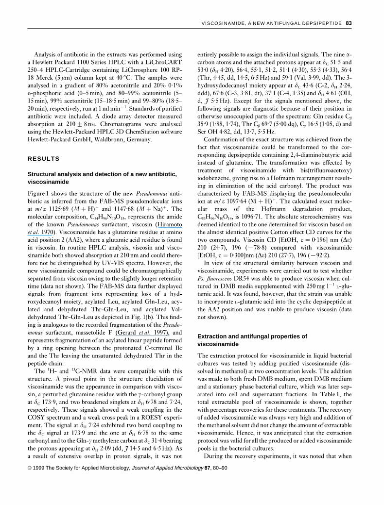
produced viscosinamide was bound to specific cell fractions,Ps. fluorescens DR54 was cultured to stationary phase in DMBmedium with 60mmol l−1 C (glucose), and cells were sep-arated from the supernatant fluid. Figure 2 shows that 76%
VISCOSINAMIDE, A NEW ANTIFUNGAL DEPSIPEPTIDE 85
Table 1 Ectraction efficiency for viscosinamide from control medium, medium with methanol and medium with added 17·9 or 37·6mmol l−1, viscosinamide in methanol—––––––––––––––––––––––––––––––––––––––––––––––––––––––––––––––––––––––––––––––––––––––––––––––––––––––––––––––––
¦Methanol ¦Methanol¦20·1 mmol l−1 ¦42·3 mmol l−1
Control* ¦Methanol viscosinamide viscosinamideFraction (mmol l−1) (mmol l−1) (mmol l−1)† (mmol l−1)†—––––––––––––––––––––––––––––––––––––––––––––––––––––––––––––––––––––––––––––––––––––––––––––––––––––––––––––––––Fresh medium 0 0 18·0 2 0·8 (101) 39·0 2 1·6 (104)Supernatant fluid 4·1 2 1·0 4·2 2 0·1 21·52 1·1 (97) 39·9 2 1·5 (95)Stationary phase culture‡ 65·1 2 0·2 65·4 2 0·9 82·42 0·3 (96) 102·9 2 3·8 (100)Cells from stationary phase culture 60·8 2 0·8 60·2 2 0·8 76·02 1·0 (88) 96·3 2 3·6 (96)Supernatant fluid from stationary phase culture 4·4 2 0·6 5·2 2 0·1 6·4 2 0·9 (7) 6·6 2 0·4 (4)—––––––––––––––––––––––––––––––––––––––––––––––––––––––––––––––––––––––––––––––––––––––––––––––––––––––––––––––––
Viscosinamide extraction was tested in fresh and spent DMB media, and in stationary phase culture which was separated into cells andsupernatant fluid before extraction. Viscosinamide recovery was calculated using the medium with added methanol as reference.* Values are means (n � 3) 2 standard deviation.† Viscosinamide recovery (%) is given in parantheses.‡ Results are calculated by adding results from cells and supernatant fluid.
Fig. 2 Localization of viscosinamide inculture (a) and cell fractions (b) instationary phase cultures ofPseudomonas fluorescens DR54 grown inDMB medium with 10 mmol l−1
glucose. (n � 3 2 S.D.)
of the viscosinamide in the culture was located in the cellfraction and the remaining 24% was extracted from the spentgrowth medium. The cellular localization of viscosinamideproduced in Ps. fluorescens DR54 was further confirmed bysonicating the cells and separating the cell membrane andcytoplasm sub-fractions by centrifugation. Following thistreatment, 68% of the detected viscosinamide was locatedin the precipitating cell membranes, while 32% was in thecytoplasm (Fig. 2).
When the fungus Rhizoctonia solani was challenged with
© 1999 The Society for Applied Microbiology, Journal of Applied Microbiology 87, 80–90
viscosinamide, hyphal growth was reduced near the visco-sinamide-containing filter but not the control filter (Fig. 3).Aerial mycelium was lacking in a 5–6mm zone around theviscosinamide source.
Viscosinamide production in liquid cultures of Ps.fluorescens DR54
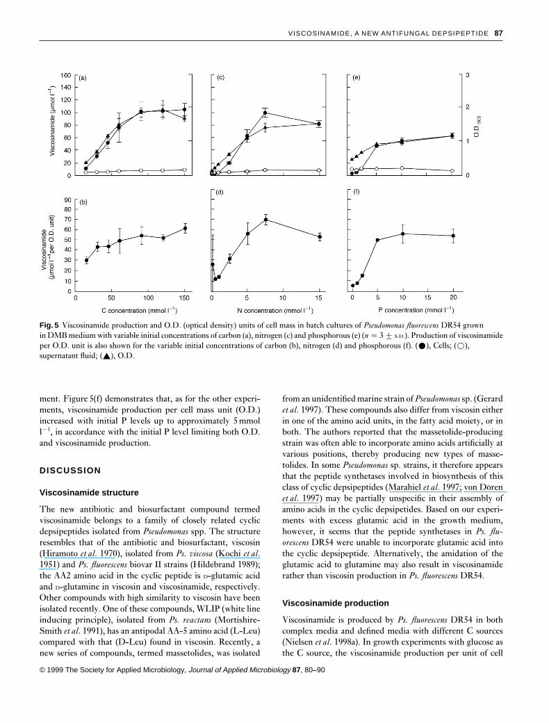
In DMB medium with 10mmol l−1 glucose, Ps. fluorescensDR54 exhibited exponential growth during the initial 15–
86 T.H. NIELSEN ET AL.
Fig. 3 In vitro test of Rhizoctonia solani growth on PotatoDextrose agar plate containing filters with 100 mgviscosinamide (right of fungal mycelium) and methanol solventcontrol (left of fungal mycelium)
20 h, followed by a stationary phase after 20 h. As shown inFig. 4(a), the viscosinamide accumulation in the cell fractionfollowed the cell production (O.D.), indicating that visco-sinamide production was primarily associated with the log-arithmic growth phase rather than the stationary phase(Fig. 4b). The viscosinamide concentration of 4–6mmol l−1
in the supernatant fraction was much smaller than in thecell-bound fraction. Furthermore, the supernatant fractionremained almost constant during the logarithmic growthphase, and increased only slightly as the culture enteredstationary phase.
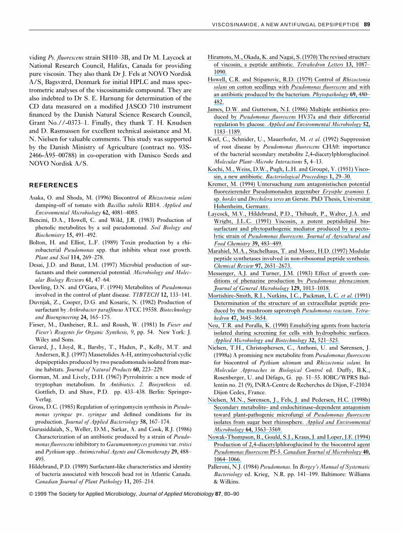
When Ps. fluorescens DR54 was cultured at variable initialC levels in the DMB medium, it was evident that C waslimiting growth when less than 100mmol l−1 C were added.Hence, Fig. 5(a) shows that increasing glucose concentrationsup to 100mmol l−1 C resulted in parallel increases of cells(O.D.) and viscosinamide. Again, the large majority of visco-sinamide produced was associated with the cell fraction, con-trary to the low (approximately 7mmol l−1) and constantlevel in the supernatant fraction. Figure 5(b) shows that theviscosinamide produced per cell mass unit (O.D.) wasincreasing slightly with initial C levels up to 100mmol l−1 C.
In comparable experiments with variable initial N levels inthe DMB medium, Ps. fluorescens DR54 showed a nearlylinear increase in cell production (O.D.) as the N level wasincreased to approximately 5mmol l−1. Hence, Fig. 5(c)shows that O.D. became constant above 5mmol l−1 N, indi-cating that N was no longer the limiting factor. Increasing Nconcentrations up to 5mmol l−1 also resulted in increasedviscosinamide production, parallel to the increasing cell pro-
© 1999 The Society for Applied Microbiology, Journal of Applied Microbiology 87, 80–90
Fig. 4 Growth curve and viscosinamide production in batchcultures of Pseudomonas fluorescens DR54 grown in DMBmedium with 10 mmol l−1 glucose (a). Production ofviscosinamide is expressed as O.D. (optical density) units of cellmass throughout an experiment. (ž), Cells; (�), supernatantfluid; (R), O.D.
duction; above this N level, the amount of viscosinamideproduced was constant. Also, in the N-limited cultures, thefinal viscosinamide concentration in the supernatant fractionwas very low (approximately 6mmol l−1) compared with thatin the cell-bound fraction. Figure 5(d) shows that the visco-sinamide produced per cell mass unit (O.D.) increased withinitial N levels up to 5mmol l−1, which again correspondedwell with the N level limiting the cell production in thecultures.
Finally, in experiments with variable initial P levels in themedium, Ps. fluorescens DR54 strain showed a steady increasein final cell production (O.D.) as the P level increased from0·1 to 5mmol l−1 (Fig. 5e). The O.D. became constant at Pconcentrations above 5mmol l−1, indicating that P was nolonger the limiting factor. The viscosinamide in the cellularfraction followed the increase in O.D. up to 5mmol l−1 P;above this level, the amount of viscosinamide produced wasconstant. As was the case in the C- or N-limited culturesdescribed above, the viscosinamide concentration was rela-tively low (approximately 9mmol l−1) and nearly constant inthe supernatant fraction, irrespective of the initial P amend-
VISCOSINAMIDE, A NEW ANTIFUNGAL DEPSIPEPTIDE 87
Fig. 5 Viscosinamide production and O.D. (optical density) units of cell mass in batch cultures of Pseudomonas fluorescens DR54 grownin DMB medium with variable initial concentrations of carbon (a), nitrogen (c) and phosphorous (e) (n � 3 2 S.D.). Production of viscosinamideper O.D. unit is also shown for the variable initial concentrations of carbon (b), nitrogen (d) and phosphorous (f). (ž), Cells; (�),supernatant fluid; (R), O.D.
ment. Figure 5(f) demonstrates that, as for the other experi-ments, viscosinamide production per cell mass unit (O.D.)increased with initial P levels up to approximately 5mmoll−1, in accordance with the initial P level limiting both O.D.and viscosinamide production.
DISCUSSION
Viscosinamide structure
The new antibiotic and biosurfactant compound termedviscosinamide belongs to a family of closely related cyclicdepsipeptides isolated from Pseudomonas spp. The structureresembles that of the antibiotic and biosurfactant, viscosin(Hiramoto et al. 1970), isolated from Ps. viscosa (Kochi et al.1951) and Ps. fluorescens biovar II strains (Hildebrand 1989);the AA2 amino acid in the cyclic peptide is D-glutamic acidand D-glutamine in viscosin and viscosinamide, respectively.Other compounds with high similarity to viscosin have beenisolated recently. One of these compounds, WLIP (white lineinducing principle), isolated from Ps. reactans (Mortishire-Smith et al. 1991), has an antipodal AA-5 amino acid (L-Leu)compared with that (D-Leu) found in viscosin. Recently, anew series of compounds, termed massetolides, was isolated
© 1999 The Society for Applied Microbiology, Journal of Applied Microbiology 87, 80–90
from an unidentified marine strain of Pseudomonas sp. (Gerardet al. 1997). These compounds also differ from viscosin eitherin one of the amino acid units, in the fatty acid moiety, or inboth. The authors reported that the massetolide-producingstrain was often able to incorporate amino acids artificially atvarious positions, thereby producing new types of masse-tolides. In some Pseudomonas sp. strains, it therefore appearsthat the peptide synthetases involved in biosynthesis of thisclass of cyclic depsipeptides (Marahiel et al. 1997; von Dorenet al. 1997) may be partially unspecific in their assembly ofamino acids in the cyclic depsipetides. Based on our experi-ments with excess glutamic acid in the growth medium,however, it seems that the peptide synthetases in Ps. flu-orescens DR54 were unable to incorporate glutamic acid intothe cyclic depsipeptide. Alternatively, the amidation of theglutamic acid to glutamine may also result in viscosinamiderather than viscosin production in Ps. fluorescens DR54.
Viscosinamide production
Viscosinamide is produced by Ps. fluorescens DR54 in bothcomplex media and defined media with different C sources(Nielsen et al. 1998a). In growth experiments with glucose asthe C source, the viscosinamide production per unit of cell

88 T.H. NIELSEN ET AL.
mass increased throughout the logarithmic growth phase, buteventually stopped when the culture entered stationary phase.Hence, the growth experiment with glucose verified thatviscosinamide production was tightly coupled to cell pro-liferation in the logarithmic growth phase. In similar experi-ments with sucrose as the sole C source, Ps. fluorescens DR54also showed an increasing viscosinamide production parallelto the production of cell mass (Nielsen et al. 1998a). In acomplex PDB medium (Difco), the viscosinamide producedper unit of cell mass also became constant during growth(Nielsen et al. 1998a). In all these experiments, viscosinamideproduction by Ps. fluorescens DR54 thus seemed to be associ-ated with cell proliferation and appeared not to be linked tocessation of growth as the cultures entered stationary phase.However, the actual amount of viscosinamide accumulatedper unit of cell mass depended strongly on the specific growthmedium (Nielsen et al. 1998a).
The results obtained in the cultures with varying C, Nor P additions gave no indication of elevated viscosinamideproduction in the growth phase when the cells experiencedthe stress conditions of nutrient limitation. This differs fromthe situation usually encountered in the production of metab-olites such as biosurfactants (Desai and Banat 1997) andantibiotics (Shanahan et al. 1992a). Viscosinamide may thusbe considered to be a primary rather than a secondary metab-olite, as has also been observed for other biosurfactantmolecules, where production may indeed be linked to cellproliferation and growth rather than starvation or stress con-ditions (Persson et al. 1988; Desai and Banat 1997).
Viscosinamide characteristics and possible role inthe environment
The small differences in the primary chemical structure ofviscosinamide and several other cyclic depsipeptides led to acomparison of such properties as solubility, membrane affin-ity and biosurfactant properties, and antimicrobial action ofthe compounds. Viscosinamide displayed a low solubility inpure water (below 2mmol l−1, data not shown) comparedwith viscosin (9mmol l−1), but high solubility in methanol.The viscosinamide concentrations in the medium supernatantfluids (Fig. 4a,c,d) were thus higher than the solubility deter-mined in pure water. A likely explanation for a high apparentsolubility in the medium supernatant fluids resulted fromearlier observations with viscosin, in which Ps. fluorescensstrain SH10–3B was found to be able to accumulate viscosinin the surrounding medium at concentrations of 450mmoll−1, far exceeding the theoretical solubility. However, this wasprobably due to binding of viscosin to acidic polysaccharidesexcreted in the culture (Laycock et al. 1991).
To test the biosurfactant properties of viscosinamide, asimple drop assay (Hildebrand 1989) was used. It was foundthat both pure viscosinamide and cells of the viscosinamide-
© 1999 The Society for Applied Microbiology, Journal of Applied Microbiology 87, 80–90
producing Ps. fluorescens DR54 were able to change the sur-face tension of sterile distilled water (data not shown). Theresults were similar, when the tests were carried out withpure viscosin and cells of the viscosin-producing Ps. flu-orescens strains W44 (Kremer 1994) and SH10–3B (Hil-debrand 1989).
Significant accumulations of viscosinamide were indeedfound in the cell fraction of Ps. fluorescens DR54, and rela-tively small amounts of the compound were released to thesupernatant fluid during the growth experiments. A similarcellular binding has been indicated for viscosin-like com-pounds (Persson et al. 1988) and other biosurfactants(Duvnjak et al. 1982). As viscosinamide accumulated in thecell membrane, it seems likely that one of its physiologicalroles is to act as a biosurfactant, aiding the organism in surfacecolonization and utilization of surface-bound substrates. Ithas been suggested that such amphilic properties of bio-surfactants enable bacteria to condition both their cell sur-faces and their substratum of growth (Neu and Poralla 1990),and it is therefore hypothesized that production of vis-cosinamide in Ps. fluorescens DR54 could be important duringsurface colonization of plant roots and surfaces of soilparticles.
The characteristic features of a biosurfactant may also beinvolved in membrane binding and inhibition of growth offungal pathogens (Stanghellini and Miller 1997). Macro-scopic observations of viscosinamide-challenged R. solaniclearly suggested that the plant pathogen was significantlyinhibited by the compound (Fig. 3). Similar observations havebeen made with another plant pathogenic fungus Pythiumultimum (data not shown). Microscopic observations showedhyphal swellings when R. solani was challenged with eitherpure viscosinamide or whole cells of the viscosinamide-pro-ducing Ps. fluorescens DR54 (Thrane et al. in press). Theseobservations were similar to those made for viscosin whenchallenging the plant pathogenic fungus, Drechslera teres,where it gave rise to swollen hyphae in germinating conidia(Kremer 1994). Using vital fluorescent stains, it is furtherproposed that viscosinamide affects the mycelium of thefungus by forming ion channels across the cell membrane(Thrane et al. in press). Our results thus demonstrate thatviscosinamide exhibits a dual function as both a biosurfactantand an antibiotic, and whether both traits are involved insurface attachment and fungal antagonism on the plant rootsurface will be the focus of future studies.
ACKNOWLEDGEMENTS
The authors thank Dr K. Poralla at Mikrobiologie/Biotechnologie im Botanischen Institut, Eberhard-Karls-Universitat Tubingen, Germany for providing Ps. fluorescensstrain W44, Dr P. Hildebrand at the Agriculture CanadaResearch Station, Kentville, Nova Scotia, Canada for pro-
VISCOSINAMIDE, A NEW ANTIFUNGAL DEPSIPEPTIDE 89
viding Ps. fluorescens strain SH10–3B, and Dr M. Laycock atNational Research Council, Halifax, Canada for providingpure viscosin. They also thank Dr J. Fels at NOVO NordiskA/S, Bagsv%rd, Denmark for initial HPLC and mass spec-trometric analyses of the viscosinamide compound. They arealso indebted to Dr S. E. Harnung for determination of theCD data measured on a modified JASCO 710 instrumentfinanced by the Danish Natural Science Research Council,Grant No.//-0373–1. Finally, they thank T. H. Knudsenand D. Rasmussen for excellent technical assistance and M.N. Nielsen for valuable comments. This study was supportedby the Danish Ministry of Agriculture (contract no. 93S-2466-A95–00788) in co-operation with Danisco Seeds andNOVO Nordisk A/S.
REFERENCES
Asaka, O. and Shoda, M. (1996) Biocontrol of Rhizoctonia solanidamping-off of tomato with Bacillus subtilis RB14. Applied andEnvironmental Microbiology 62, 4081–4085.
Bencini, D.A., Howell, C. and Wild, J.R. (1983) Production ofphenolic metabolites by a soil pseudomonad. Soil Biology andBiochemistry 15, 491–492.
Bolton, H. and Elliot, L.F. (1989) Toxin production by a rhi-zobacterial Pseudomonas spp. that inhibits wheat root growth.Plant and Soil 114, 269–278.
Desai, J.D. and Banat, I.M. (1997) Microbial production of sur-factants and their commercial potential. Microbiology and Molec-ular Biology Reviews 61, 47–64.
Dowling, D.N. and O’Gara, F. (1994) Metabolites of Pseudomonasinvolved in the control of plant disease. TIBTECH 12, 133–141.
Duvnjak, Z., Cooper, D.G. and Kosaric, N. (1982) Production ofsurfactant by Arthrobacter paraffineus ATCC 19558. Biotechnologyand Bioengineering 24, 165–175.
Fieser, M., Danheiser, R.L. and Roush, W. (1981) In Fieser andFieser’s Reagents for Organic Synthesis, 9, pp. 54. New York: J.Wiley and Sons.
Gerard, J., Lloyd, R., Barsby, T., Haden, P., Kelly, M.T. andAndersen, R.J. (1997) Massetolides A-H, antimycobacterial cyclicdepsipeptides produced by two pseudomonads isolated from mar-ine habitats. Journal of Natural Products 60, 223–229.
Gorman, M. and Lively, D.H. (1967) Pyrrolnitrin: a new mode oftryptophan metabolism. In Antibiotics. 2. Biosynthesis ed.Gottlieb, D. and Shaw, P.D. pp. 433–438. Berlin: Springer-Verlag.
Gross, D.C. (1985) Regulation of syringomycin synthesis in Pseudo-monas syringae pv. syringae and defined conditions for itsproduction. Journal of Applied Bacteriology 58, 167–174.
Gurusiddaiah, S., Weller, D.M., Sarkar, A. and Cook, R.J. (1986)Characterization of an antibiotic produced by a strain of Pseudo-monas fluorescens inhibitory to Gaeumannomyces graminis var. triticiand Pythium spp. Antimicrobial Agents and Chemotherapy 29, 488–495.
Hildebrand, P.D. (1989) Surfactant-like characteristics and identityof bacteria associated with broccoli head rot in Atlantic Canada.Canadian Journal of Plant Pathology 11, 205–214.
© 1999 The Society for Applied Microbiology, Journal of Applied Microbiology 87, 80–90
Hiramoto, M., Okada, K. and Nagai, S. (1970) The revised structureof viscosin, a peptide antibiotic. Tetrahedron Letters 13, 1087–1090.
Howell, C.R. and Stipanovic, R.D. (1979) Control of Rhizoctoniasolani on cotton seedlings with Pseudomonas fluorescens and withan antibiotic produced by the bacterium. Phytopathology 69, 480–482.
James, D.W. and Gutterson, N.I. (1986) Multiple antibiotics pro-duced by Pseudomonas fluorescens HV37a and their differentialregulation by glucose. Applied and Environmental Microbiology 52,1183–1189.
Keel, C., Schnider, U., Mauerhofer, M. et al. (1992) Suppressionof root disease by Pseudomonas fluorescens CHA0: importanceof the bacterial secondary metabolite 2,4-diacetylphloroglucinol.Molecular Plant–Microbe Interactions 5, 4–13.
Kochi, M., Weiss, D.W., Pugh, L.H. and Groupe, V. (1951) Visco-sin, a new antibiotic. Bacteriological Proceedings 1, 29–30.
Kremer, M. (1994) Untersuchung zum antagonistischen potentialfluorezierender Pseudomonaden gegenuber Erysiphe graminis f.sp. hordei und Drechslera teres an Gerste. PhD Thesis, UniversitatHohenheim, Germany.
Laycock, M.V., Hildebrand, P.D., Thibault, P., Walter, J.A. andWright, J.L.C. (1991) Viscosin, a potent peptidolipid bio-surfactant and phytopathogenic mediator produced by a pecto-lytic strain of Pseudomonas fluorescens. Journal of Agricultural andFood Chemistry 39, 483–489.
Marahiel, M.A., Stachelhaus, T. and Mootz, H.D. (1997) Modularpeptide synthetases involved in non-ribosomal peptide synthesis.Chemical Review 97, 2651–2673.
Messenger, A.J. and Turner, J.M. (1983) Effect of growth con-ditions of phenazine production by Pseudomonas phenazinium.Journal of General Microbiology 129, 1013–1018.
Mortishire-Smith, R.J., Nutkins, J.C., Packman, L.C. et al. (1991)Determination of the structure of an extracellular peptide pro-duced by the mushroom saprotroph Pseudomonas reactans. Tetra-hedron 47, 3645–3654.
Neu, T.R. and Poralla, K. (1990) Emulsifying agents from bacteriaisolated during screening for cells with hydrophobic surfaces.Applied Microbiology and Biotechnology 32, 521–525.
Nielsen, T.H., Christophersen, C., Anthoni, U. and So�rensen, J.(1998a) A promising new metabolite from Pseudomonas fluorescensfor biocontrol of Pythium ultimum and Rhizoctonia solani. InMolecular Approaches in Biological Control ed. Duffy, B.K.,Rosenberger, U. and Defago, G. pp. 51–55. IOBC/WPRS Bul-lentin no. 21 (9), INRA-Centre de Recherches de Dijon, F-21034Dijon Cedex, France.
Nielsen, M.N., So�rensen, J., Fels, J. and Pedersen, H.C. (1998b)Secondary metabolite- and endochitinase-dependent antagonismtoward plant-pathogenic microfungi of Pseudomonas fluorescensisolates from sugar beet rhizosphere. Applied and EnvironmentalMicrobiology 64, 3563–3569.
Nowak-Thompson, B., Gould, S.J., Kraus, J. and Loper, J.E. (1994)Production of 2,4-diacetylphloroglucinol by the biocontrol agentPseudomonas fluorescens Pf-5. Canadian Journal of Microbiology 40,1064–1066.
Palleroni, N.J. (1984) Pseudomonas. In Bergey’s Manual of SystematicBacteriology ed. Krieg, N.R. pp. 141–199. Baltimore: Williams& Wilkins.
90 T.H. NIELSEN ET AL.
Persson, A., Osterberg, E. and Dostalek, M. (1988) Biosurfactantproduction by Pseudomonas fluorescens 378: growth and productcharacteristics. Applied Microbiology and Biotechnology 29, 1–4.
Shanahan, P., Borro, A., O’Gara, A. and Glennon, J.D. (1992a)Isolation, trace enrichment and liquid chromatographic analysisof diacetylphloroglucinol in culture and soil samples using UVand amperometric detection. Journal of Chromatography 606, 171–177.
Shanahan, P., O’Sullivan, D.J., Simpson, P., Glennon, J.D. andO’Gara, F. (1992b) Isolation of 2,4-diacetylphloroglucinol froma fluorescent pseudomonad and investigation of physiological par-ameters influencing its production. Applied and EnvironmentalMicrobiology 58, 353–358.
Slininger, J.P. and Jackson, M.A. (1992) Nutritional factors reg-ulating growth and accumulation of phenazine 1-carboxylic acidby Pseudomonas fluorescens 2–79. Applied Microbiology and Biotech-nology 37, 388–392.
Stanghellini, M.E. and Miller, R.M. (1997) Biosurfactants; theiridentity and potential efficacy in the biological control of zoosporicplant pathogens. Plant Disease 81, 4–12.
© 1999 The Society for Applied Microbiology, Journal of Applied Microbiology 87, 80–90
Stephens, P.M., Crowley, J.J. and O’Connel, C.O. (1993) Selectionof pseudomonad strains inhibiting Pythium ultimum on sugar beetseeds in soil. Soil Biology and Biochemistry 25, 1283–1288.
Takeda, R. (1958) Structure of a new antibiotic, pyoluteorin. Journalof American Chemical Society 80, 4749–4750.
Thomashow, L.S., Weller, D.M., Bonsall, R.F. and Pierson, L.S.III (1990) Production of antibiotic phenazine-1-carboxylic acidby fluorescent Pseudomonas species in the rhizosphere of wheat.Applied and Environmental Microbiology 56, 908–912.
Thrane, C., Olsson, S., Nielsen, T.H. and So�rensen, J. (FEMSMicrobiology Ecology, in press) The use of vital fluorescentprobes for detection of stress in the fungi Pythium ultimum andRhizoctonia solani challenged with viscosinamide from Pseudo-monas fluorescens DR54.
von Doren, H., Keller, U., Vater, J. and Zocher, R. (1997) Multi-functional peptide synthetases. Chemical Review 97, 2675–2705.
Weller, D.M. (1988) Biological control of soilborne plant pathogensin the rhizosphere with bacteria. Annual Review of Phytopathology26, 379–407.
Copyright © 2022 FDOKUMEN