INTRA-ORAL DELIVERY SYSTEM FOR ANTIFUNGAL ...
307
INTRA-ORAL DELIVERY SYSTEM FOR ANTIFUNGAL RELEASE A thesis submitted to The University of Manchester for the degree of Doctor of Philosophy in the Faculty of Medical and Human Sciences 2012 NESREEN SALIM School of Dentistry
-
Upload
khangminh22 -
Category
Documents
-
view
0 -
download
0
Transcript of INTRA-ORAL DELIVERY SYSTEM FOR ANTIFUNGAL ...

INTRA-ORAL DELIVERY SYSTEM FOR
ANTIFUNGAL RELEASE
A thesis submitted to The University of Manchester for
the degree of Doctor of Philosophy
in the Faculty of Medical and Human Sciences
2012
NESREEN SALIM
School of Dentistry

List of Contents Page 2
LIST OF CONTENTS
LIST OF CONTENTS ..................................................................................................................... 2
LIST OF TABLES ............................................................................................................................ 9
LIST OF FIGURES ....................................................................................................................... 11
LIST OF EQUATIONS.................................................................................................................15
LIST OF ABBREVIATIONS ....................................................................................................... 16
LIST OF APPENDICES ............................................................................................................... 19
ABSTRACT ................................................................................................................................... 20
DECLARATION ........................................................................................................................... 21
COPYRIGHT STATEMENT ...................................................................................................... 22
THE AUTHOR ............................................................................................................................. 23
DEDICATION ............................................................................................................................... 25
ACKNOWLEDGEMENTS ........................................................................................................... 26
CHAPTER 1 .................................................................................................................................. 28
REVIEW OF THE LITERATURE ............................................................................................. 28
1.1 Introduction .................................................................................................................................. 29
1.2 Oral candidosis ............................................................................................................................ 32
1.2.1 Definition and description ............................................................................................... 32
1.2.2 Organisms involved in oral candidosis ....................................................................... 33
1.2.3 Predisposing factors .......................................................................................................... 35
1.2.3.1 Local factors .................................................................................................................. 36
1.2.3.2 Systemic factors ........................................................................................................... 37
1.2.4 Clinical presentation of oral candidosis ..................................................................... 38
1.2.4.1 Pseudomembranous candidosis ........................................................................... 39
1.2.4.2 Erythematous candidosis ........................................................................................ 40
1.2.4.3 Hyperplastic candidosis ........................................................................................... 40
1.2.4.4 Denture-induced candidosis ................................................................................... 40
1.3 Denture-induced candidosis ................................................................................................... 41
1.3.1 Definition and characteristics ........................................................................................ 41

List of Contents Page 3
1.3.2 Classification of denture-induced candidosis .......................................................... 42
1.3.3 Aetiological factors of denture-induced candidosis .............................................. 42
1.3.3.1 Denture factors ............................................................................................................ 43
1.3.3.2 Infective factors ........................................................................................................... 44
1.3.3.3 Allergy ............................................................................................................................. 45
1.3.3.4 Dietary factors and hormone deficiency ............................................................ 45
1.3.3.5 Systemic factors ........................................................................................................... 46
1.3.3.6 Antibiotic therapy ....................................................................................................... 46
1.3.3.7 Miscellaneous factors ................................................................................................ 47
1.3.4 Diagnosis of denture-induced candidosis ................................................................. 47
1.4 Candida albicans .......................................................................................................................... 49
1.4.1 Definition and characteristics ........................................................................................ 49
1.4.2 The virulence mechanisms and the factors affecting distribution of Candida
in the oral cavity ............................................................................................................................. 51
1.4.2.1 Adherence ...................................................................................................................... 51
I.4.2.2 Saliva ................................................................................................................................. 52
1.4.2.3 Growth at different pHs ............................................................................................ 53
1.4.2.4 Nutritional and Metabolic factors ......................................................................... 53
1.4.2.5 Necrotic factors ............................................................................................................ 53
1.4.2.6 Mannanoproteins (cell surface polysaccharide) ............................................ 53
1.4.2.7 Cell surface hydrophobicity .................................................................................... 54
1.4.2.8 Oral bacteria .................................................................................................................. 54
1.4.2.9 Multiple cell forms ...................................................................................................... 55
1.4.2.10 Phenotypicity ............................................................................................................. 55
1.5 Denture plaque biofilms ........................................................................................................... 57
1.5.1 Definition ................................................................................................................................ 57
1.5.2 Candida biofilms .................................................................................................................. 57
1.5.2.1 Formation and characterisation of C. albicans biofilms ............................... 58
1.5.2.2 Role of morphogenesis in C. albicans biofilm formation ............................. 59
1.5.2.3 - biofilms resistance ................................................................................................... 59
1.6 Management of denture-induced candidosis ................................................................... 62
1.6.1 Treatment modalities directed toward oral mucosa ............................................ 63
1.6.1.1 Antifungal agents ........................................................................................................ 63

List of Contents Page 4
1.6.1.2 Antimicrobial agents .................................................................................................. 70
1.6.2 Treatment modalities directed toward the denture base ................................... 72
1.6.2.1 Hygienic measures and denture cleansers ....................................................... 72
1.6.2.2 Modification of surface properties ....................................................................... 75
1.6.2.3 Prosthetic treatment .................................................................................................. 75
1.6.2.4 Microwave energy as a disinfectant .................................................................... 76
1.6.2.5 Diode laser irradiation .............................................................................................. 77
1.6.3 Treatment modalities directed toward both oral mucosa and denture base78
1.6.3.1 Intra-oral delivery of therapeutic agents .......................................................... 78
1.7 Spectrophotometer .................................................................................................................... 85
1.7.1 Introduction .......................................................................................................................... 85
1.7.2 The principle of spectroscopic process ...................................................................... 86
1.7.3 Instrumentation .................................................................................................................. 87
1.7.4 Calculations and analysis ................................................................................................. 88
1.7.5 The spectrum ........................................................................................................................ 89
1.7.6 Practical application of UV spectrophotometry ...................................................... 90
1.8 Properties of PEM/THFM acrylic system .......................................................................... 92
1.8.1 Testing procedures of serviceability of acrylic liners ........................................... 93
1.8.1.1 Shear bond strength ................................................................................................... 93
1.8.1.2 Water absorption ........................................................................................................ 95
1.8.1.3 Degree of conversion ................................................................................................. 96
1.8.1.4 Colour stability ............................................................................................................. 97
1.9 Summary ...................................................................................................................................... 102
CHAPTER 2 ................................................................................................................................ 107
STATEMENT OF THE PROBLEM, AIMS & OBJECTIVES & WORK PLAN ................. 107
2.1 Statement of the problem ...................................................................................................... 108
2.2 Aims and Objectives ................................................................................................................. 111
CHAPTER 3 ................................................................................................................................ 113
METHODOLOGIES ................................................................................................................... 113
3.1 Introduction ................................................................................................................................ 114

List of Contents Page 5
3.2 Methods ........................................................................................................................................ 114
3.2.1 Broth Microdilution method ......................................................................................... 114
3.2.1.1 Preparation of drug dilution ................................................................................. 115
3.2.1.2 Preparation of inoculum ........................................................................................ 117
3.2.1.3 Inoculation of assay plates .................................................................................... 117
3.2.1.4 Quality control ........................................................................................................... 117
3.2.1.5 Results interpretation ............................................................................................. 118
3.2.2 Bioassay method ............................................................................................................... 119
3.2.2.1 Preparation of stock solutions and drug standards .................................... 119
3.2.2.2 Quality control ........................................................................................................... 120
3.2.2.3 Preparation of inoculum ........................................................................................ 121
3.2.2.4 Preparation of bioassay plates ............................................................................. 121
3.2.2.5 Calculations and interpretation .......................................................................... 122
3.2.2.6 Validation ..................................................................................................................... 124
3.2.3 Biofilm quantification methods ................................................................................... 124
3.2.3.1 Organisms and inoculum preparation .............................................................. 125
3.2.3.2 Biofilm formation ...................................................................................................... 126
3.2.3.3 XTT reduction assay................................................................................................. 127
3.2.3.4 Biofilm biomass quantification ............................................................................ 128
3.2.4 Time-kill test ....................................................................................................................... 129
3.2.4.1 Disc preparation and incubation ........................................................................ 129
3.2.4.2 Microorganisms and inoculum preparation ................................................... 129
3.2.4.3 Time-kill studies ........................................................................................................ 130
3.2.4.4 Analysis ......................................................................................................................... 130
3.2.5 Degree of conversion measurement .......................................................................... 131
3.2.5.1 FTIR test ....................................................................................................................... 131
3.2.5.2 Results interpretation and analysis ................................................................... 132
3.2.6 UV Spectrophotometry ................................................................................................... 133
CHAPTER 4 ................................................................................................................................ 136
Chlorhexidine is a highly effective topical broad-spectrum agent against
Candida species ....................................................................................................................... 136

List of Contents Page 6
4.1 Abstract ......................................................................................................................................... 137
4.2 Introduction ................................................................................................................................ 138
4.3 Materials & Methods ................................................................................................................ 141
4.4 Results ........................................................................................................................................... 144
4.5 Discussion .................................................................................................................................... 151
4.6 Conclusions ................................................................................................................................. 154
4.7 Funding ......................................................................................................................................... 155
CHAPTER 5 ................................................................................................................................ 156
Fungicidal amounts of antifungals are released from impregnated denture
lining material for up to 28 days ..................................................................................... 156
5.1 Abstract ......................................................................................................................................... 157
5.2 Introduction ................................................................................................................................ 158
5.3 Materials and methods ........................................................................................................... 161
5.4 Results ........................................................................................................................................... 165
5.5 Discussion .................................................................................................................................... 170
5.6 Conclusions ................................................................................................................................. 173
5.7 Acknowledgements .................................................................................................................. 174
CHAPTER 6 ................................................................................................................................ 175
Candidacidal effect of fluconazole and chlorhexidine released from acrylic
polymer...................................................................................................................................... 175
6.1 Abstract ......................................................................................................................................... 176
6.2 Introduction ................................................................................................................................ 178
6.3 Materials and methods ........................................................................................................... 180
6.4 Results ........................................................................................................................................... 184
6.5 Discussion .................................................................................................................................... 188
6.6 Conclusions ................................................................................................................................. 191
CHAPTER 7 ................................................................................................................................ 192
Chlorhexidine impregnated PEM/THFM polymer exhibits superior activity to
fluconazole against Candida albicans biofilm formation ......................................... 192

List of Contents Page 7
7.1 Abstract ......................................................................................................................................... 193
7.2 Introduction ................................................................................................................................ 194
7.3 Materials and methods ........................................................................................................... 197
7.4 Results ........................................................................................................................................... 201
7.5 Discussion .................................................................................................................................... 204
7.6 Conclusion ................................................................................................................................... 207
CHAPTER 8 ................................................................................................................................ 208
Impregnation with antimicrobials challenge bonding properties and water
sorption behaviour of an acrylic liner ............................................................................ 208
8.1 Abstract ......................................................................................................................................... 209
8.2 Introduction ................................................................................................................................ 210
8.3 Materials and methods ........................................................................................................... 213
8.4 Results ........................................................................................................................................... 218
8.5 Discussion .................................................................................................................................... 221
8.6 Conclusions ................................................................................................................................. 225
CHAPTER 9 ................................................................................................................................ 226
Impregnation with antimicrobials has an impact on degree of conversion and
colour stability of acrylic liner .......................................................................................... 226
9.1 Abstract ......................................................................................................................................... 227
9.2 Introduction ................................................................................................................................ 228
9.3 Materials and methods ........................................................................................................... 231
9.4 Results ........................................................................................................................................... 236
9.5 Discussion .................................................................................................................................... 241
9.6 Conclusions ................................................................................................................................. 245
CHAPTER 10 ............................................................................................................................. 246
General discussion, Conclusions and Suggestions for future research ............... 246
10.1 General discussion ................................................................................................................. 247
10.2 Meanings and Implications ................................................................................................. 258
10.3 Strengths and Limitations ................................................................................................... 261

List of Contents Page 8
10.4 Conclusions ............................................................................................................................... 264
10.5 Suggestions for future research ........................................................................................ 266
REFERENCES ............................................................................................................................. 267
APPENDICES ............................................................................................................................. 290
APPENDIX - I ...................................................................................................................................... 290
APPENDIX - II ..................................................................................................................................... 293
APPENDIX - III ................................................................................................................................... 294
APPENDIX - IV ................................................................................................................................... 301
60, 116
Word Count

List of Tables Page 9
LIST OF TABLES
Table 1.1 The principal fungi that may infect the oral cavity............................ 35 Table 1.2 Predisposing factors in oral candidosis................................................. 38 Table 1.3 Classification of different clinical presentations of oral
candidosis............................................................................................................
39
Table 3.1 Absorbance values for different known concentrations of chlorhexidine......................................................................................................
134
Table 3.2 Absorbance values for different known concentrations of fluconazole....................................................................................................... ....
135
Table 4.1 Chlorhexidine geometric mean MIC results for 79 Candida
isolates belonging to 8 different species at 24 h and 48 h incubation............................................................................................................
144 Table 4.2 Cumulative percentage of isolates for each species of Candida
inhibited at each concentration in broth microdilution series A. Susceptibility of Candida species to chlorhexidine by MICa at 80 % after 48 h..................................................................................................
146 B. Susceptibility of Candida species to fluconazole by MICa at
50 % after 48 h..................................................................................................
147 Table 5.1 The cumulative amount (mean ± SD; in mg) of pure
fluconazole (FLUp), fluconazole from capsules (FLUc) or chlorhexidine (CHX) released into the distilled water from the acrylic discs during 28-day incubation measured at 6 different time points. Differences between groups were statistically significant at all time points (P≤0.001)...................................................
166 Table 8.1 Groups of the shear bond study: control, chlorhexidine (CHX),
fluconazole pure (FLUp) and fluconazole from capsules (FLUc)....................................................................................................................
214 Table 8.2 Mean (SD) of shear bond strengths (MPa) for all discs: Group 1
(tested immediately) and Group 2 water-stored discs (water immersion for 28 days). These groups consisting of control discs without impregnation, discs impregnated with pure fluconazole (FLUp), chlorhexidine (CHX), and fluconazole from capsules (FLUc) (different upper case alphabets in rows indicate significant difference at P≤0.05)..............................................
218 Table 9.1 Means (SD) of degree of conversion of the tested groups:
Control, chlorhexidine impregnated group (CHX), fluconazole pure impregnated group (FLUp) and fluconazole from capsules impregnated groups (FLUc).........................................................................
236 Table 9.2 The mean colour changes (ΔΕ*ab) of the study groups and the
standard deviation. The values in NBS units were calculated

List of Tables Page 10
[∆E*ab (in CIE) ×0.92].................................................................................... 238 Table 9.3 National Bureau of Standard rating (NBS)............................................ 238

List of Figures Page 11
LIST OF FIGURES
Figure 1.1 Curve of damage-response framework................................................... 34 Figure 1.2 Dimorphic nature in Candida: Blastospore form (a), Hyphal
form (b)........................................................................................................
50 Figure 1.3 SEM image of a mature C. albicans biofilm (48 h).............................. 58 Figure 1.4 The different mechanisms of resistance in C. albicans..................... 61 Figure 1.5 Mechanism of action of different antifungal agents.......................... 68 Figure 1.6 Schematic diagram showing the principle of controlled-release
drug delivery system.......................................................................................
78 Figure 1.7 The difference in the achieved drug levels between the
conventional treatments and the controlled drug delivery systems..................................................................................................................
79 Figure 1.8 Electromagnetic spectrum............................................................................ 85 Figure 1.9 Typical UV-Vis spectrophotometer........................................................... 86 Figure 1.10 Basic components of spectrophotometer.............................................. 87 Figure 1.11 Sample and reference beam intensities.................................................. 88 Figure 1.12 Sample graphical output of Spectrometer............................................. 90 Figure 1.13 Munsell Colour System.................................................................................. 98 Figure 1.14 Colour chart of the Tristimulus System.................................................. 100 Figure 1.15 The CIE L*a*b* Colour System.................................................................... 101 Figure 1.16 2-D and 3-D chemical structure of THFM (a), PEM (b).................... 105 Figure 1.17 2-D and 3-D chemical structure of fluconazole (a),
chlorhexidine (b).............................................................................................. 106
Figure 2.1 A flow chart showing the work plan......................................................... 112 Figure 3.1 The diagram shows the fluconazole and chlorhexidine dilution
series, the numbers highlighted in bold represent the concentrations used in the microtitre plates (a). A blank microtitre plate showing the wells where the prepared concentrations to be dispensed (b)..........................................................
116
Figure 3.2 Chlorhexidine microtitre plate showing clear wells where growth was inhibited and hazy wells with visible growth after 48 h incubation..................................................................................................
118
Figure 3.3 Photographs of bioassay plates showing the wells where the leachates and the standards to be dispensed (a), and the inhibition zones formed by the diluted leachates of chlorhexidine (b)..............................................................................................
122
Figure 3.4 A representative view to show the known concentrations and the inhibition zone measurements (a), and the standard curve that was used to calculate the unknown drug concentrations [pink column in (a)]. Internal standards in this example passed the quality control requirement [within 20 % of their known concentrations (a)]..........................................................................................
123

List of Figures Page 12
Figure 3.5 Schematic diagram for the discs to be investigated by XTT (orange) and CV (purple) after different incubation periods (2, 7, 14, 21, 28 days).............................................................................................
126
Figure 3.6 A photograph showing a microtitre plate with Crystal violet stain (a) and XTT solution (b) collected for different biofilms.....
128
Figure 3.7 Photograph representing fourier transform infrared spectro-photometer connected to PC, showing the detector crystal against which the samples were distributed (arrow)......................
132
Figure 3.8 Full FTIR spectrum of the uncured specimen from 4000 cm-1 to 400 cm-1 and a closer view of the area of interest (spectrum from 1790 cm-1 to 1505 cm-1)(a), FTIR spectrum of the cured specimen and a closer view of the area of interest (spectrum from 1790 cm-1 to 1505 cm-1)(b)...............................................................
133
Figure 3.9 Chlorhexidine standard curve..................................................................... 134 Figure 3.10 Fluconazole standard curve......................................................................... 135 . Figure 4.1 Scatterplot showing the relationship between fluconazole and
chlorhexidine MICs obtained with 79 isolates of Candida species (rs=0.039, P=0.733). Each number in the graph represents the number of isolates with a particular chlorhexidine MIC value and the corresponding fluconazole MIC value..............................................................................................................
148
Figure 4.2 Distribution of MICs (mg/L) and the GM means ( ) of 79 Candida isolates comprising 8 Candida species for chlorhexidine (a) and fluconazole (b). The hatched area represents the normal salivary concentration for each agent with normal dosing regimens, twice chlorhexidine rinsing daily and single oral dose of 100 mg of fluconazole..........................
149
Figure 5.1 Pof drug Proportion of drug leached from discs impregnated with
chlorhexidine (CHX), fluconazole from capsules (FLUc) and pure fluconazole (FLUp) during the 28-day incubation. Cumulative percentage presented............................................................
166
Figure 5.2 MICs (mg/L) of 46 isolates (left y-axis) and the amount drugs released from discs impregnated with fluconazole powder from capsules (a) pure fluconazole (b) and chlorhexidine (c) during the 28-day incubation (right y-axis)..........................................
167
Figure 5.3 A photog A photograph of a representative bioassay plate showing growth inhibition zones formed by diluted leachates of chlorhexidine (a) and fluconazole (FLUc) (b) discs in distilled water. A random template was used for well selection. Positive controls in 1A wells, negative controls e.g. in 2C (b) and 1D (b), THFM controls in 4E (b) and 3B (b) and PEM controls in 6B (b) and 6E (b)..............................................................................................
168
Figure 5.4 The reproducibility of the bioassay was assessed by re-analysing 30 % of the samples of each group (CHX, FLUc, FLUp). A Bland and Altman plot of 27 repeated measurements (9 each group) shows minimal variation between results and

List of Figures Page 13
high reproducibility. Each dot represents the mean concentration of two measurements against the difference between the two measurements. 95 % Confidence Intervals for the first measurements were 4.93-10.84 and were 4.69-10.24 for the repeated measurements.................................................................
169
Figure 6.1 Representative time-kill curve plots for C. albicans ATCC
90028 (a), C. albicans F/2511 (b), and C. glabrata F/4023 (c) of leachates of different time intervals of chlorhexidine impregnated discs. The MIC of all isolates was 6.25 mg/L for chlorhexidine.....................................................................................................
185
Figure 6.2 Representative time-kill curve plots for C. albicans ATCC 90028 (a), C. albicans F/2511 (b), and C. glabrata F/4023 (c) of leachates of different time intervals of pure fluconazole impregnated discs. The MIC of C. albicans ATCC 90028 for pure fluconazole was 0.25 mg/L while the MIC of C. albicans F/2511 and C. glabrata F/4023 was 128 mg/L...................................................
186
Figure 6.3 Representative time-kill curve plots for C. albicans ATCC 90028 (a), C. albicans F/2511 (b), and C. glabrata F/4023 (c) of leachates of different time intervals of fluconazole from capsules impregnated discs. The MIC of C. albicans 90028 for fluconazole from capsules was 0.25 mg/L while the MIC of C. albicans F/2511 and C. glabrata F/4023 was 128 mg/L............
187
Figure 7.1 Level of cell metabolism as measured by XTT-assay in
C. albicans (ATCC 90028) biofilms grown on chlorhexidine impregnated acrylic discs, fluconazole impregnated discs or control discs after 2-28 days incubation. The colour intensity of formazan salt produced by sessile cells constituting the biofilm is directly correlated to cellular metabolic activity of the biofilm. Bars represent the mean of five parallel discs............
202
Figure 7.2 Quantity of biomass as measured by crystal violet staining in C. albicans (ATCC 90028) biofilms grown on chlorhexidine impregnated acrylic discs or fluconazole impregnated discs or control discs after 2-28 days incubation. The results presented as OD where the biofilm biomass is proportional to the absorbance values. Bars represent the mean of five parallel discs........................................................................................................................
203
Figure 8.1 A photograph showing the heat cure acrylic substrate inside
the cylinder and the cured PEM/THFM specimen attached to the acrylic substrate and showing the adhesive bond failure after subjected to shear bond test............................................................
215
Figure 8.2 A bar chart showing the mean shear bond strength (standard deviation) under two different conditions: 1) the specimens stored dry at room temperature (Group 1) to be tested after 24 h, and it comprises 4 groups: control discs, discs impregnated with chlorhexidine (CHX), pure fluconazole

List of Figures Page 14
(FLUp) or fluconazole from capsules (FLUc). 2) The specimens were further immersed in water at 37 °C for 28 days after 24 h setting (Group 2) and it consists of the same sub-groups of discs. Horizontal capped lines indicate significant difference between the dry stored discs and their corresponding water stored discs (P≤0.05)......................................................................................
219
Figure 8.3 Percentage mass change of control discs, discs impregnated with chlorhexidine (CHX), fluconazole from capsules (FLUc) and pure fluconazole (FLUp) immersed in distilled water for 6 months (510.5 min1/2)..............................................................................
220
Figure 8.4 Molecular structure of fluconazole (a) and chlorhexidine (b).... 222 Figure 9.1 A representative spectrum of the cured acrylic for all tested
groups: control (a), fluconazole pure impregnated group (FLUp) (b), chlorhexidine impregnated group (CHX) (c) and fluconazole from capsules impregnated groups (FLUc) (d). The carbonyl peak corresponds to (C=O) (internal standard) where (C=C) corresponds to the aliphatic peak................................................
237
Figure 9.2 The three colour values before (L*1, a*1, b*1) and after water immersion for 28 days (L*2, a*2, b*2) of acrylic discs without antifungal impregnation (CTR), acrylic discs impregnated with chlorhexidine (CHX), pure fluconazole (FLUp), or fluconazole from capsules (FLUc). Horizontal lines above bars represent significant difference between paired groups P≤0.05.....................
239
Figure 10.1 Schematic diagram showing the leaching mechanism over
time in the polymeric delivery system impregnated with pure fluconazole (a), fluconazole from capsules (b), chlorhexidine (c)............................................................................................................................
252
Figure 10.2 The calculated amount (mg) of pure fluconazole (FLUp), fluconazole from capsules (FLUc) or chlorhexidine (CHX) to be released from denture with average surface area during 28-day treatment period. The straight lines represent the normal recommended dosing regimens, twice chlorhexidine rinsing daily (=40 mg) and single oral dose of fluconazole (=100 mg)............................................................................................................
260

List of Equations Page 15
LIST OF EQUATIONS
Equation 1.1 Calculation of Transmittance................................................................... 89
Equation 1.2 Calculation of Absorbance......................................................................... 89
Equation 1.3 Beer-Lambert Law........................................................................................ 89
Equation 1.4 Calculation of SBS.......................................................................................... 95
Equation 1.5 Calculation of Mass Change...................................................................... 96
Equation 1.6 Calculation of ∆E*.......................................................................................... 100
Equation 3.1 Calculation of DC %...................................................................................... 132
Equation 8.1 Calculation of SBS.......................................................................................... 216
Equation 8.2 Calculation of Mass Change...................................................................... 217
Equation 9.1 Calculation of DC %...................................................................................... 233
Equation 9.2 Calculation of ∆E*.......................................................................................... 234
Equation 9.3 Calculation of NBS Unit............................................................................... 234

List of Abbreviations Page 16
LIST OF ABBREVIATIONS
A Area
ADA American Dental Association
ATCC American Type Culture Collection
BEC Buccal Epithelial Cells
BMD Broth Microdilution
Candida spp. Candida species
C. albicans Candida albicans
CFU Colony Forming Units
CHX Chlorhexidine
CLSI Clinical Laboratory Standard Institute
CV Crystal Violet
DC Degree of Conversion
DMSO DiMethyl SulphOxide
F Force
FLUc Fluconazole powder from capsules
FLUp Fluconazole pure
FTIR Fourier Transform InfraRed Spectroscopy
h Hour
HPLC High Performance Liquid Chromatography
HIV Human Immunodeficiency Virus
I0 Intensity of reference beam
I Intensity of sample beam

List of Abbreviations Page 17
MIC Minimum Inhibitory Concentration
min Minute
mg Milligram
ml Millilitre
µl Microlitre
mm Millimetre
MPa Megapascal
MOPS Morpholinepropanesulfonic acid
N Newton
NMR Nuclear Magnetic Resonance
OD Optical Density
PAFE Post Antifungal Effect
PBS Phosphate buffered saline
PEM Poly (ethyl methacrylate)
PEM/THFM Poly (ethyl methacrylate) and Tetrahydrofurfuryl methacrylate
PMMA Poly (methyl methacrylate)
R Pearson’s correlation coefficient
r Simple correlation coefficient
rs Spearman correlation coefficient
S Second
SBS Shear Bond Strength
SD Standard Deviation
SEM Scanning Electron Microscopy
UV Ultraviolet

List of Abbreviations Page 18
VIS Visible
W Watt
YNBG Yeast Nitrogen Base with Glucose
∆E Change in colour
°C Centigrade
§ Section
2-D Two-dimensions
3-D Three-dimensions

List of Appendices Page 19
LIST OF APPENDICES
Appendix I Protocol for in vivo study 290
Appendix II MIC values of FLUp against MIC values of FLUc for different Candida species
293
Appendix III Publication 1 (Fungicidal amounts of antifungals are released from impregnated denture lining material for up to 28 days)
294
Appendix IV Publication 2 (Impregnation with antimicrobials challenge bonding properties and water sorption behaviour of an acrylic liner)
301

Abstract Page 20
ABSTRACT
Background: The placement of removable dental prostheses produces significant changes in the oral environment that may lead to adverse effects on the integrity of the oral tissues. Denture-induced candidosis, caused by candidal infection of the palatal mucosa, is the most frequent complication (40 %) in removable denture wearers. It predominantly affects immunosuppressed and medically compromised patients. In these high-risk patients the oral cavity may provide a source for Candida causing systemic infection. Oral candidosis has become a significant challenge in patients with persisting risk factors and a recurrent need for antifungal treatment. In addition, denture-induced candidosis is a mixed biofilm infection which provides multiple challenges for its management. Moreover, the persistent fungal colonisation on the fitting surfaces of denture often leads to cross infection and recurrence of mucosal lesions. These considerations highlight the clear need for new effective antifungal treatment modalities. Aims: The aims of this project were to establish a polymeric delivery device based on denture base lining polymer, poly (ethyl methacrylate) and tetrahydrofurfuryl methacrylate (PEM/THFM), for sustained delivery of antifungal agents [chlorhexidine (CHX) and fluconazole (FLU)], for the use in the treatment of denture-induced candidosis and to test the serviceability of the lining under investigation. Methods: A broth microdilution method was used to assess the spectrum of activity of the antifungal agents (CHX powder and FLU powder in two formulations pure and from capsules) against wide range of Candida species. Bioassay method and spectrophotometry were used to evaluate the efficiency of the PEM/THFM denture liner to release the impregnated antifungal agents and to quantify the released concentrations. Bioassay, time-kill studies and biofilm assays were used to verify the antifungal activity of the released antifungal agents. Shear bond test, water absorption, colorimetery and Fourier Transform Infrared Spectroscopy were used to test clinically important physical and mechanical properties for the impregnated liner. Results: It was found that CHX has broad-spectrum antifungal activity also among Candida species highly resistant to FLU. Both CHX and FLU became readily leached from PEM/THFM polymer up to 4 weeks in microbiologically effective concentrations. CHX demonstrated superior antifungal efficacy against planktonic and biofilm lifestyle of Candida compared to FLU. Findings show that the impregnation with antifungal agents has affected all tested properties (shear bond strength, water absorption, degree of conversion and colour stability) but these changes are comparable to other long-term lining materials and are within acceptable ranges. Conclusions: These findings indicate the feasibility of introducing an efficient treatment modality for candidal infections, especially denture-induced candidosis. A polymeric system containing CHX or FLU could assume a very promising treatment option as the drug is effective and directed to the site of pathology. Moreover, the distinct efficacy of CHX against C. albicans biofilms is a promising outcome to overcome the side effects of conventional antifungal agents and their reduced efficacy against biofilm formation.

Declaration Page 21
DECLARATION
No portion of the work referred to in the thesis has been submitted in support of an
application for another degree or qualification of this or any other university or other
institute of learning.
Nesreen Salim 2012

Copyright Statement Page 22
COPYRIGHT STATEMENT
i. The author of this thesis (including any appendices and/or schedules to this thesis)
owns any copyright in it (the “Copyright”) and s/he has given The University of
Manchester the right to use such Copyright for any administrative, promotional,
educational and/or teaching purposes.
ii. Copies of this thesis, either in full or in extracts, may be made only in accordance
with the regulations of the John Rylands University Library of Manchester. Details of
these regulations may be obtained from the Librarian. This page must form part of
any such copies made.
iii. The ownership of any patents, designs, trade marks and any and all other
intellectual property rights except for the Copyright (the “Intellectual Property
Right”) and any reproductions of copyright works, for example graphs and tables
(“Reproductions”), which may be described in this thesis, may not be owned by the
other and may be owned by third parties. Such Intellectual Properties Rights and
Reproductions cannot and must not be made available for use without the prior
written permission of the owner(s) of the relevant Intellectual Property Rights
and/or Reproductions.
iv. Further information on the conditions under which disclosure, publication and
exploitation of this thesis, the Copyright and any Intellectual Property Rights and/or
Reproductions described in it may take place is available from the Head of School of
Dentistry.

The author Page 23
THE AUTHOR
I graduated from The University of Jordan in 2003, gaining a BDS with a GPA of 3.7
out of 4 (Excellent). I worked as a teaching assistant at The University of Jordan
between 2004 and 2007. After that I enrolled in a one year full-time MSc Fixed and
Removable Prosthodontics program offered by The University of Manchester in 2007. I
finished the degree with Distinction. Then I enrolled in a four-year clinical PhD (Doctor of
Clinical Dental Science in Fixed and Removable Prosthodontics) in 2008. In 2010 I won the
Friends of the Hebrew University Prize for the best oral presentation for research in the
postgraduate presentation day at the School of Dentistry. I am a reviewer for Dental
Materials. During the PhD I attended several scientific meetings:
British Society of Prosthodontics meeting, in York in April 2009.
British Society of Prosthodontics meeting, in Stirling in March 2010.
British Society of Prosthodontics meeting, in Liverpool in April 2012.
British Society for Medical Mycology, Cardiff, in April 2012.
In addition, some of the research work is to be presented in the following meeting:
A poster presentation titled ‘’Anti-biofilm activity of antifungal-impregnated
denture material’’ was accepted and will be presented at the PER/IADR
coming meeting in Helsinki in September 12-15/2012.

The author Page 24
In addition, I have published the following papers during my studies:
Salim N, Moore C, Silikas N, Satterthwaite J.D and Rautemaa R. (2012).
Fungicidal amounts of antifungals are released from impregnated denture
lining material for up to 28 days. Journal of Dentistry, 40, 506-512.
Salim N, Silikas N, Satterthwaite J.D and Rautemaa R. (2012). Impregnation
with antimicrobials challenge bonding properties and water sorption
behaviour of an acrylic liner. Journal of Dentistry, 40, 693-699.
Darwish RM, Amin WM, Al-Ali MH and Salem NA. (2011). Study of the elution
of fluconazole from a self-polymerizing acrylic resin and its activity against
resistant Candida albicans. Journal of Materials Science: Materials in Medicine,
22: 1885-1890.
Amin WM, Al-Ali MH, Salim NA and Al-Tarawneh SK. (2009). A new form of
intra-oral delivery of antifungal drugs for the treatment of denture-induced
oral candidosis. European Journal of Dentistry, 3: 257-266.
Amin WM, Kassab AM and Salim NA. (2008). Incisal edge abrasion caused by
an unusual eating habit. The Internet Journal of Dental Science, 6, Number 1.
I have also submitted other papers to a variety of scientific journals.

Dedication Page 25
DEDICATION
IN THE NAME OF ALLAH &
His Blessings
The all knowing, The most wise
I would like to dedicate this work to the soul of my father, who was the cornerstone
of my life, the light of my eyes, the spring of my ambitions. My father, you are and will
remain the source of my persistence and progress.
I would like to dedicate this work to my mother, the spring of love that was very
supportive and encouraging during all stages of this work to excel towards the best.
I would like to dedicate this work to my husband, who was the forefront to my
achievements and without his incessant support, patience and valuable guidance this
work cannot be tackled. There could not be a husband any thoughtful and benevolent
more than you. This thesis is also dedicated to my dearly child Haneen.
This thesis is also dedicated to the stars of my life, brothers and sisters; Abdullah,
Mohmmed, Ahmed, Mahmood, Ali and Njat, for their continuous support and
tremendous encouragement.
I would like to dedicate this work to my dear friends Katayoon Azizi, Suad Othman
Dua’ Jaber and Noura Nour who were with me step by step leading me down the path
of even more valuable achievements by their constant support.
Finally I would also like to dedicate this work to all those people who suffered or are
still suffering from injustice, aggression, physical or emotional hardships. Their pain
is hoped to end one day.

Acknowledgements Page 26
ACKNOWLEDGEMENTS
By coming to the end of this scientific journey; first, I thank God, the most merciful
and most gracious for edifying my mind by cognition and acquaintance.
I would like to express my warmest thanks to my supervisors, Dr Riina Rautemaa,
Dr Nick Silikas and Dr Julian Satterthwaite, without whom I would never have
undertaken this project. The help, continued support and enthusiastic
encouragement they have given me at every stage of this work and especially with
guidance on the drafts of this thesis have been invaluable.
I am totally indebted to Dr Caroline Moore for her tremendous efforts, kind help and
guidance in her capacity and even beyond. Her insights through my study were very
helpful. The tremendous patience and efforts she showed during my laboratory work
are really appreciated.
In addition, I would like to thank my advisor Dr Antony Roberts for his interest and
advice.
I also would like to thank Prof. Malcom Richardson for the language support and the
critical comments he offered to me throughout writing my thesis.
I am especially grateful to all the members of Reference Mycology Laboratory in
Manchester for their hard work and exceptional enthusiasm. I also would like to
extend my thanks to Mr Brian Daber for his invaluable help in the lab work and to
Mrs Shena Reynolds, Mrs Rose-Marie Parr and Mrs Margaret Stockburger for their
help and support throughout my study.

Acknowledgements Page 27
My thanks are also extended to Dr Craig Barclay, Prof. Nick Grey, Dr Joanne Cunliffe
and other faculty members, nurses and supporting staff of the Prosthodontics
department for their help and assistance throughout my clinical training.
I would like to show my utmost sincere appreciation and indebted acknowledgement
to my husband for all the help and assistance he offered to me throughout the years,
for his unconditional support, for his constant presence and incentive, for his
exceptional understanding. He made it a very enjoyable and amazing experience.
I would like to thank Prof. Wala Amin (Lecturer in Prosthodontic Department in The
University of Jordan) for the considerable advice, support and critical comments.
I would like to thank my friends Suad Othman, Noura Nour, Nehal Hani, Ruida Al-
Shali, Eman Abu Hajar and Fatema Ashibi, who made my experience in Manchester
wonderful and exceptional.
Finally, I am grateful to The University of Jordan for awarding me a studentship.

Chapter 1 Page 28
CHAPTER 1
REVIEW OF THE LITERATURE

Chapter 1 Page 29
1.1 Introduction
The popular belief that edentulism will decline markedly in the future is based on
epidemiologic studies (Steele et al., 2000; Muller et al., 2007). This decline is
counteracted by demographic changes such as the increase in size and age of the
older population and consequently the need and demand for complete dentures will
increase over the next two decades as the number of older adults increases
(Muller et al., 2007). In addition, the prevalence of tooth loss increases with age and
the prosthetic need increases accordingly (Walter et al., 2001). Consequently there is
a growing concern about denture-related mucosal pathologies (Budtz-Jorgensen,
1981; Jainkittivong et al., 2010) and, hence, providing efficient treatment of such
conditions is significant.
Placement of a removable prosthesis in the oral cavity produces significant changes
in the oral environment that may lead to adverse effects on the integrity of the oral
tissues (Dorey et al., 1985). Mucosal reactions may result from mechanical irritation
from dentures, plaque accumulation on dentures, or allergic reaction to denture base
materials (Budtz-Jorgensen, 1981; Coelho et al., 2004; Freitas et al., 2008).
The adverse direct effects of wearing complete dentures include resorption of
residual ridges and also pathological changes of the oral mucosa in the form of
denture-induced candidosis, flabby ridges, angular cheilitis, denture hyperplasia,
burning mouth syndrome and traumatic ulcers (Dorey et al., 1985; Jainkittivong et al.,
2010). These adverse consequences often result in patient discomfort, unstable
occlusion, and insufficient masticatory function (Dorey et al., 1985; Jainkittivong
et al., 2010). In addition systemic or general diseases may alter the tissue response

Chapter 1 Page 30
resulting in oral lesions, so these lesions may indicate a serious underlying disease
(Budtz-Jorgensen, 1981; Khatibi et al., 2011).
Denture-induced candidosis (Candida-associated denture stomatitis, denture-induced
stomatitis), characterised by candidal infection of the palatal mucosa, is the most
frequent complication of wearing complete or partial dentures (Budtz-Jorgensen,
1981; Jainkittivong et al., 2010). The exact prevalence of denture-induced candidosis
is unclear; it has been reported in 45-70 % of complete denture wearers (Figueiral et
al., 2007; Dagistan et al., 2009). Moreover, denture-induced candidosis is the most
common clinical presentation of oral candidosis (Samaranayake et al., 2009). This has
driven research towards this research area, which has focused attention on effective
means to control these lesions (Cawson, 1963; Giuliana et al., 1997; Dhir et al., 2007).
Denture-induced candidosis is described as an inflammatory process of the mucosa of
denture-bearing tissues. This condition is almost invariably asymptomatic (Budtz-
Jorgensen, 1974), usually affects the hard palate (Budtz-Jorgensen, 1981) and
frequently is associated with angular cheilitis and glossitis (Ritchie et al., 1969;
Budtz-Jorgensen and Bertram, 1970a). Denture-induced candidosis is seen more
frequently among women than men (Arendorf and Walker, 1980). There are a
number of factors that can give rise to denture-induced candidosis such as trauma,
allergy, and dietary factors. It is a multifactorial disease with Candida albicans (C.
albicans) being the primary aetiological agent (Olsen, 1974;
Coco et al., 2008).
Denture-induced candidosis may heal partially or completely after topical antifungal
treatment, but the incidence of relapse is high (Budtz-Jorgensen and Bertram, 1970b;
Budtz-Jorgensen, 1974; Cross et al., 2004). The existing conventional methods of

Chapter 1 Page 31
delivering drugs into intra-oral sites (such as topical application of miconazole gel,
using nystatin suspension, chlorhexidine mouth wash) for the treatment of conditions
of oral mucosa are inefficient (Budtz-Jorgensen, 1990a; Ellepola and Samaranayake,
2001). This is mainly due to the washing effect of saliva and oral musculature, which
reduces the availability of the drug below the optimal therapeutic concentration
(Ellepola and Samaranayake, 2001; Samaranayake et al., 2009). Also,
a satisfactory outcome depends on patient compliance as frequent dose application is
required which may lead to sub-optimal dosing (Samaranayake et al., 2009).
Dentists are frequently the first medical professionals to examine patients who have
oral mucosal changes (Khatibi et al., 2011). Consequently, they are responsible for
recognising the signs of disease, transforming them into a diagnosis, and planning
the therapy, so it is important that the examination is carried out by a clinician who
has adequate medical knowledge (Budtz-Jorgensen, 1981). Unfortunately, 30 % of
doctors prescribe nystatin for oral candidosis without oral examination and only 9 %
of doctors are aware that wearing a denture is a risk factor for oral candidal infection.
This can result in recurrent candidal infection as a result of incorrect diagnosis or not
considering important risk factors (Morgan et al., 2001).

Chapter 1 Page 32
1.2 Oral candidosis
1.2.1 Definition and description
Oral candidosis comprises a group of diseases that are associated with candidal
infection. Furthermore it is by far the most common human fungal infection and
manifests in a variety of clinical presentations (McIntyre, 2001; Akpan and Morgan,
2002). Oral candidosis may range from localised infections to acute systemic invasive
diseases, both immunocompetent and immunocompromised individuals can be
affected (McIntyre, 2001). In addition, it may present as a secondary infection
superimposed on another medical condition (Budtz-Jorgensen et al., 1975; Akpan and
Morgan, 2002). It can be a marker of immunosuppression and is therefore often
referred to as “disease of the diseased” (McIntyre, 2001). Treatment of fungal
infections necessitates removing or alleviating the predisposing condition (Rautemaa
and Ramage, 2011). Importantly, the rate of opportunistic candidal infection has
increased markedly among hospitalised patients (Fisher-Hoch and Hutwagner,
1995): this increase in the incidence is due to the increasing number of
immunosuppressed patients and is associated with a high overall mortality rate
(Fraser et al., 1992; Andes et al., 2009; Lopez-Martínez, 2010). Oral candidosis is a
superficial infection; however, if treated ineffectively in immunocompromised
patients, it may lead to invasive systemic infection and consequently increase the
mortality rate (Gautam et al., 2010).

Chapter 1 Page 33
1.2.2 Organisms involved in oral candidosis
Oral candidal species are part of the normal oral flora in 18 % of the normal healthy
population (Cannon and Chaffin, 1999), with C. albicans being the most dominant
species (Akpan and Morgan, 2002; Coco et al., 2008). However, large variations are
found in relation to the age, general health and dental health of the population
studied (Akpan and Morgan, 2002). Candida species are opportunistic pathogens
which cause disease when there is a disturbance in host-commensal balance
(Ghannoum et al., 2010; Rautemaa and Ramage, 2011). The change from
commensalism to parasitism is most probably due to alteration in the oral
environment of the host rather than the alteration of the yeast itself
(Budtz-Jorgensen, 1974; Akpan and Morgan, 2002; Ghannoum et al., 2010).
Opportunistic pathogens incite disease in hosts whose local or systemic immune
attributes have been impaired and the defence mechanisms have been damaged or
innately dysfunctional (Nater et al., 1978; Ghannoum et al., 2010). Casadevall and
Pirofiski (2003) showed that host-microorganism interaction determines the
microbial pathogenesis outcome and the relevant host damage using damage-
response curve (Figure 1.1).

Chapter 1 Page 34
Figure 1.1 Curve of damage-response framework (after Casadevall and Pirofski, 2003)
Commonly a mixture of Candida species can be isolated from oral candidal lesions,
C. albicans, C. glabrata and C. tropicalis have been isolated from these lesions
(Budtz-Jorgensen et al., 1975; Akpan and Morgan, 2002) (Table 1.1). C. albicans has a
main role in the development of denture-induced candidosis (Cawson, 1963;
Olsen, 1974; Akpan and Morgan, 2002; Coco et al., 2008) but other species including
C. glabrata, C. krusei, C. parapsilosis, C. dubliniensis, C. tropicalis, C. kefyr and
C. guilliermondii have been increasingly isolated in immunocompromised patients
(Sullivan et al., 1995; Rautemaa et al., 2006). Repeated and prolonged azole antifungal
treatment has been related to the presence of these non-albicans Candida species
(Cuéllar-Cruz et al., 2012), which are resistant to commonly used azole antifungal
agents and are considered second or third most common cause of systemic

Chapter 1 Page 35
candidosis (Delgado et al., 2009; Cuéllar-Cruz et al., 2012). Other studies have
suggested that bacteria may also play a role in the disease (Budtz-Jorgensen and
Theilade, 1983; Koopmans et al., 1988).
Table 1.1 The principal fungi that may infect the oral cavity (Akpan and Morgan, 2002)
1.2.3 Predisposing factors
It is generally known that candidal infection does not affect healthy
immunocompetent individuals (Cannon and Chaffin, 1999; McIntyre, 2001).
Therefore, the presence of predisposing factors, breaching the immune defence
mechanisms either at local or systemic level is essential for the development of the
infection (Akpan and Morgan, 2002). A number of predisposing factors may result in
the development of oral candidosis; all these factors affect the host-commensal
balance, allowing the proliferation of the candidal organisms (McIntyre, 2001). These
factors may be short-term or long-term in nature; this nature has significant impact
Candida species
Candida albicans
Candida glabrata
Candida tropicals
Candida krusei
Candida guilliermondii
Candida parapsilosis
Candida dubliniensis

Chapter 1 Page 36
on the duration and the prognosis of the infection (these factors and others are
discussed in more details in § 1.3.3).
1.2.3.1 Local factors
Short-term local predisposing factors include poor oral hygiene and topical steroid
administration that can disturb the normal balance of oral flora and result in
dissemination of the infection (Ritchie et al., 1969; Budtz-Jorgensen, 1990a;
Rautemaa and Ramage, 2011). Dietary factors play important role in the oral
homeostasis (Ritchie et al., 1969). A high carbohydrate intake provides a good source
of nourishment for Candida (Ritchie et al., 1969).
Long-term local predisposing factors include mechanical irritation from dentures or
any prostheses or faulty occlusion may result in breakdown of the integrity of the oral
mucosa and provide a good opportunity for the Candida to affect the injured tissues
(Figueiral et al., 2007). In addition, xerostomia results in reduced salivary flow and
predisposes the oral tissues to candidal infection (Ettinger, 1996). Local mucosal
lesions (such as lichen planus) and smoking are also considered predisposing factors
(Zeng et al., 2009; Rautemaa and Ramage, 2011). Dental problems such as
periodontal pocketing, gingivitis and retained roots have been related to increased
carriage of Candida leading to infection (Wang et al., 2006). Radiotherapy following
malignancy is associated with an increased risk of oral candidosis (Dorko et al., 2001)
because it has a direct negative effect on the rate of cellular turnover in the oral
mucous membrane and the salivary flow (Ettinger, 1996; Deng et al., 2010).

Chapter 1 Page 37
1.2.3.2 Systemic factors
Use of antibiotics (especially broad spectrum) is considered a short-term systemic
factor that can alter the normal balance of oral flora and result in spreading of
infection (Ritchie et al., 1969; Budtz-Jorgensen, 1990a; Soysa et al., 2008). Humoral
and cell-mediated immunity deficiencies (e.g. diabetes mellitus, AIDS) have been
identified as a long-term systemic predisposing factor (Dorocka-Bobkowska et al.,
1996; Akpan and Morgan, 2002). It has been estimated that 90 % of AIDS patients
develop oral candidosis (Akpan and Morgan, 2002). Other well known systemic
factors include malignancy, malnutrition and physiological factors such as extreme
ages and pregnancy (McIntyre, 2001): these factors are associated with imbalance in
host-commensal relationship leading to infection. Deficiency states (iron, vit. B12,
folate) are also associated with a lower level of host response (Ritchie et al., 1969;
Rautemaa and Ramage, 2011), and gastro-oesophageal reflux disease could be a
predisposing factor because it provides a favourable acidic growth environment for
Candida species (Prusiski et al., 2002). All predisposing factors are summarised in
Table 1.2.

Chapter 1 Page 38
Table 1.2 Predisposing factors in oral candidosis (Siikala, 2011)
Local predisposing factors
Systemic predisposing factors
Short-term Long-term
Short-term Long-term
Poor oral hygiene Trauma (ill-fitting
denture)
Broad-spectrum antibiotics
Physiological factors (age, pregnancy)
Topical steroids Smoking
Dietary factors (deficiency state)
Dietary factors (deficiency state)
Dietary factors (high carbohydrate intake)
Radiation
Immune defect (AIDS)
Xerostomia
Malignancy (leukemia)
Local mucosal lesions and dental problems
Endocrine (diabetes mellitus)
1.2.4 Clinical presentation of oral candidosis
Many classifications have been suggested to describe the clinical forms of oral
candidosis, the first classification of oral candidosis was proposed by Lehner (1966).
He defined two major subdivisions:
Acute, including pseudomembranous and atrophic candidosis.
Chronic, including atrophic (Denture sore mouth) and hyperplastic candidosis.
The currently accepted classifications are presented in Table 1.3. Another common
classification has been used, in which the lesions are divided into three main
categories of acute, chronic, and mucocutaneous (Lynch, 1994). Acute candidosis is
further subdivided into pseudomembranous and atrophic forms, and chronic

Chapter 1 Page 39
candidosis subdivided into atrophic and hyperplastic forms. Mucocutaneous
candidosis can be presented as localised, familial, or syndrome related (Lynch, 1994).
Table 1.3 Classification of different clinical presentations of oral candidosis (Holmstrup and Axell, 1990; Samaranayake, 1991)
Acute Chronic Candida-associated lesions
Pseudomembranous Hyperplastic Denture-induced stomatitis
Erythematous - Nodular Angular cheilitis
- Plaque-like Median rhomboid glossitis
Erythematous
1.2.4.1 Pseudomembranous candidosis
Pseudomembranous candidosis (or thrush) is characteristically an acute infection,
even though many lesions are chronic in nature. The clinical presentation for this
lesion is very characteristic; non-adherent creamy white patches, easily wiped with
gauze to leave an underlying erythematous mucosa (McIntyre, 2001). Apart from
neonates, who have no immunity to Candida species, thrush indicates an
immunosuppression status or a local disturbance in oral flora, such as that caused by
xerostomia, antibiotic treatment or corticosteroids. It is a feature in many immune
defects, especially leukaemia and HIV disease. It can be an early feature of HIV
infection (Budtz-Jorgensen, 1990a; Samaranayake, 1992). Thrush can affect any oral
site, typically the palate or upper buccal vestibule posteriorly. Confirmation may be
obtained by a smear biopsy.

Chapter 1 Page 40
1.2.4.2 Erythematous candidosis
Erythematous candidosis (previously known as antibiotic sore mouth) is associated
with corticosteroids and broad-spectrum antibiotics. Erythematous candidosis
resulting from the prescription of broad-spectrum antibiotics is the only oral
candidosis where pain is a common symptom (McIntyre, 2001). Clinically,
erythematous areas are seen on the dorsum of the tongue, palate or buccal mucosa
and it can be associated with angular stomatitis.
1.2.4.3 Hyperplastic candidosis
Hyperplastic candidosis (also known as candidal leukoplakia) is a chronic white
raised discrete lesion. Hyperplastic candidosis is a premalignant condition and it is
indistinguishable from leukoplakia due to other causes. Therefore, biopsy is
important in the management of this condition. The lesion may present as a bilateral
homogenous or speckled lesion, which does not rub off. It usually affects commissural
areas or rarely near the surface of the tongue (McIntyre, 2001).
1.2.4.4 Denture-induced candidosis
Denture-induced candidosis (or denture-induced stomatitis, or chronic atrophic
candidosis, or denture sore mouth) is the most common clinical presentation of oral
candidosis (Budtz-Jorgensen et al., 1975; Akpan and Morgan, 2002), and is usually
associated with wearing prosthesis but many other factors are associated
(Ritchie et al., 1969; Samaranayake et al., 2009; Jainkittivong et al., 2010). Typically,
the lesions appear as an asymptomatic, erythematous mucositis limited to the
denture-bearing mucosa (Budtz-Jorgensen, 1974; Figueiral et al., 2007).

Chapter 1 Page 41
1.3 Denture-induced candidosis
1.3.1 Definition and characteristics
Denture-induced candidosis is a term used to describe certain pathologic changes in
the oral mucosa of denture-bearing tissues (Budtz-Jorgensen, 1974, 1981). It presents
as a chronic bright diffuse erythema limited to the denture-bearing area in both jaws,
but more frequently in the maxilla (Budtz-Jorgensen, 1981), as the upper dentures
are in close contact with the mucosa and tend to be more conducive to the
accumulation of plaque on the fitting surface. Denture-induced candidosis is a
common inflammatory condition with high prevalence in denture wearing population
(Budtz-Jorgensen et al., 1975; Figueiral et al., 2007). It has been documented that
denture-induced candidosis affects 45-70 % of denture wearers (Figueiral et al.,
2007). Women are the most commonly affected (Cawson, 1963; Arendorf and Walker,
1980; Figueiral et al., 2007).
The condition is usually symptomless apart from occasional soreness if complicated
by thrush (Budtz-Jorgensen, 1974), consequently “Denture-induced candidosis” and
“Chronic atrophic candidosis” have been suggested as more appropriate descriptions
of the condition than “Denture sore mouth” as soreness is seldom a complaint
(Newton, 1962). However, in association with denture-induced candidosis patients
may suffer angular stomatitis (Cawson, 1963) and/or median rhomboid glossitis
(Lynch, 1994).

Chapter 1 Page 42
1.3.2 Classification of denture-induced candidosis
Denture-induced candidosis can be manifested in a variety of clinical forms and can
be categorised clinically according to Newton’s classification (Newton, 1962), which
classified this condition into three categories:
Type 1, characterised by pin-point hyperaemia. It shows small areas of
inflammation in an otherwise normal tissue, which is usually found around the
orifices of the ducts of the palatal mucous glands. It is the most common type
(Kulak-Ozkan et al., 2002; Figueiral et al., 2007).
Type 2, characterised by diffuse hyperaemia. A generalised inflammation of
the entire denture-bearing area can be detected. The surface of the mucosa is
smooth and slight trauma may be sufficient to cause bleeding.
Type 3, presented as hyperplastic nodular reaction of the palatal mucosa
which may be present over the entire denture–bearing area but is more
commonly restricted to the central areas and is particularly found under relief
areas or suction discs.
1.3.3 Aetiology factors of denture-induced candidosis
Denture-induced candidosis is a multifactorial condition and no primary aetiological
cause has been identified (Ritchie et al., 1969; Figueiral et al., 2007). However,
various factors are thought to be particularly significant:
denture factors including continuous denture wearing and lack of hygiene
(including reaction to denture plaque).
infective factors.
allergic reactions to denture base materials.

Chapter 1 Page 43
dietary factors (including resultant haematological deficiencies).
systemic factors and deterioration in the general state of health (including
predisposing factors).
antibiotic therapy.
miscellaneous factors.
1.3.3.1 Denture factors
Many factors related to the prosthesis have been identified as risk factors for the
development of denture-induced candidosis. These factors include trauma caused by
ill-fitting dentures, unsatisfactory occlusion and articulation, and lack of oral and
denture hygiene. There is good evidence that Newton's type I denture-induced
candidosis is caused by trauma alone (Budtz-Jorgensen and Bertram, 1970a). It is
assumed that pin-point hyperaemia in the palate (type I) is due to occlusion of the
salivary ducts by a close fitting denture (Newton, 1962). In a study comparing
denture adjustment versus antifungal therapy, lesions healed by proper adjustment
of the dentures while antifungal therapy was ineffective (Budtz-Jorgensen and
Bertram, 1970b). Several studies have provided evidence that denture-induced
candidosis is present more frequently in patients with poor fitting dentures with a
non-balanced occlusion (Figueiral et al., 2007; Emami et al., 2008). Continuous
denture wearing has been suggested to cause denture-induced candidosis due to local
injury (Nyquist, 1953), but it might also expose the mucosa to denture plaque for a
longer time or alter the composition of this plaque (Kulak-Ozkan et al., 2002).
Poor oral and denture hygiene is frequently identified as a local aetiological factor
(Fisher and Rashid, 1952; Budtz-Jorgensen and Bertram, 1970a; Rautemaa and
Ramage, 2011). Bergendal (1982) found that patients with denture-induced

Chapter 1 Page 44
candidosis have more maxillary denture plaque than those with healthy mucosa. It
has been found that an improvement in the level of oral and denture hygiene using
mechanical or chemical means results in significant reduction in the number of
patients with candidosis (Budtz-Jorgensen, 1979; Grimoud et al., 2005). These
findings indicate that poor oral and denture hygiene is an important predisposing
condition for denture-induced candidosis.
1.3.3.2 Infective factors
The infective causes including Candida and some bacterial genera have been reported
to play an important role in denture-induced candidosis (Budtz-Jorgensen et al.,
1983; Akpan and Morgan, 2002), but Candida species, mainly C. albicans are the ones
most frequently involved (Davenport, 1970; Budtz-Jorgensen and Bertram, 1970a;
Coco et al., 2008). Cahn (1936) was the first to report the presence of Candida in
denture-induced stomatitis lesions. The causal relationship has subsequently been
supported by mycological and immunological studies (Budtz-Jorgensen and Bertram,
1970a; Coco et al., 2008). The infection is primarily due to an overgrowth and
colonisation of Candida species under the fitting surface of dentures (Budtz-
Jorgensen and Bertram, 1970a). Budtz-Jorgensen and Bertram (1970b) found that the
localised simple inflammation of the maxillary denture-bearing mucosa (type I) was
caused by trauma, whereas the generalised simple (type II) and the granular
inflammation (type III) more readily could be due to candidal infection, as hyphal
structures were isolated in palatal smears consistently only among type II and III
patients, although trauma may be a significant predisposing factor (Budtz-Jorgensen
and Bertram, 1970a).

Chapter 1 Page 45
1.3.3.3 Allergy
Rattner (1936) was the first to report allergic reactions to denture base material.
Further case reports reinforcing this finding have been presented (e.g. Bradford,
1948; Crissey, 1965; Barclay et al., 1999). Fisher (1956) has found that the incidence
of these reactions is low and allergy to denture base materials rarely occurs. Nyquist
(1952) in his study was not able to find cases of allergic reaction in 248 patients with
denture-induced candidosis. Furthermore, a significant release of acrylic monomer
will take place in new dentures only, and will be temporary. This fact rules out the
irritating effect of residual monomer as a cause of denture-induced candidosis
(Smith and Bains, 1956). In addition, the diagnosis is difficult to establish and
cooperation is needed between the dermatologists and the dentists to diagnose these
lesions (Kaaber et al., 1979).
1.3.3.4 Dietary factors and hormone deficiency
A diverse array of nutritional factors including high carbohydrate intake, iron
deficiency, folic acid and vitamins deficiency have been identified as contributory
factors of oral candidal infection (Shuttleworth and Gibbs, 1960; Ritchie et al., 1969;
Samaranayake, 1986; Paillaud et al., 2004). Therefore it is not surprising that
denture-induced candidosis patients have been found to be suffering from nutritional
deficiencies (Budtz-Jorgensen, 1978). Iron deficiency has been reported to be an
important factor to increase the susceptibility to Candida infection (Rose, 1968) and a
higher incidence of folate deficiency in denture-induced candidosis patients has been
reported than in a control groups (Samaranayake and McFarlane, 1981). Nutritional
deficiencies such as amino acids, iron and certain vitamins of the B complex are
believed to reduce the resistance of the oral mucosa (Samaranayake, 1986). It has

Chapter 1 Page 46
been shown that dietary supplements of proteins and minerals increase tolerance to
dentures and resolve the inflammation (Deeley, 1965).
1.3.3.5 Systemic factors
A number of systemic diseases and treatment with various drugs may increase the
susceptibility to oral candidosis (Knight and Fletcher, 1971; Akpan and Morgan,
2002). These factors include endocrine disturbances (diabetes mellitus) (Dorocka-
Bobkowska et al., 1996; Dorko et al., 2001), immunodeficiency states (Samaranayake,
1992), nutritional deficiencies (iron deficiency, high carbohydrate intake), malignant
diseases (leukemia) (Deng et al., 2010), and drugs such as antibiotics, and steroids
(Knight and Fletcher, 1971; Budtz-Jorgensen, 1975). In these patients symptoms in
association with denture-induced candidosis are usually pronounced (Ettinger,
1996). A wide range of other systemic diseases also play a secondary role due to the
large number of therapeutic drugs that cause xerostomia, which is itself a
predisposing factor (Ettinger, 1996).
1.3.3.6 Antibiotic therapy
It is well established that antibiotics increase susceptibility to infection with
C. albicans (Ritchie et al., 1969; Soysa et al., 2008; Rautemaa and Ramage, 2011).
Administration of oral antibiotics will disturb the oral flora by allowing an
overgrowth of resistant organisms (Ritchie et al., 1969). Knight and Fletcher (1971)
suggested that antibiotics stimulate the growth of C. albicans by killing nutrient–
competing bacteria, and hence an adequate amount of glucose would be available for
the more slowly growing Candida species. It has been documented that prolonged
antibiotic therapy in some medical problems (such as chronic urinary tract infection)
increases the risk of development of denture-induced candidosis (Bergendal, 1982).

Chapter 1 Page 47
1.3.3.7 Miscellaneous factors
Psychological problems were detected in the majority of patients with denture-
induced candidosis sampled by Nater et al (1978) using a personality questionnaire.
Self-prosthetic treatment with home relining materials can also contribute to
denture-induced candidosis (Welker, 1977).
1.3.4 Diagnosis of denture-induced candidosis
C. albicans is by far the most commonly isolated Candida species in denture-induced
candidosis (Coco et al., 2008). However, non-albicans Candida species have become
increasingly associated with oral candidosis especially in immunocompromised
patients (high-risk) (Bagg et al., 2003; Coco et al., 2008). This highlights the
importance of using accurate microbiological diagnostic methods (Rautemaa and
Ramage, 2011). Quantitative estimates of the overgrowth of yeasts on the mucosa and
the fitting surface of the denture either by culture or by microscopy of oral smears is
needed to establish the diagnosis (Budtz-Jorgensen, 1974). Conventional sampling
methods for oral Candida are swabbing, or scraping of the tissues, suction of saliva, or
rinsing the mouth. The most valuable tools in the diagnosis of oral candidosis are
semi-quantitative cultures and oral smears (Budtz-Jorgensen, 1990b). Biopsy
(a microscopic examination of tissue sections) is required if premalignant or
malignant changes of the epithelium are suspected. Since agglutinating, haem
agglutinating, precipitating, and immunofluorescent antibodies are found in people
without signs of Candida infection, serologic tests for Candida are not diagnostic
methods, and the raise in the antibody response in patients with oral candidosis is
only moderate. However, such tests may be a prognostic instrument and a valuable

Chapter 1 Page 48
tool in patients with severe oral candidosis who respond poorly to antifungal therapy
(Budtz-Jorgensen, 1990b).
For the collected samples to be representative, it is important to sample an adjacent
non-renewing surface in addition to the affected area (Rautemaa et al., 2006). Oral
rinsing is a helpful mean to indicate the intra-oral fungal load (McIntyre, 2001;
Coco et al., 2008). Direct sonication of the denture is a valuable method which
remarkably improves both quantification and diagnosis of candidal infection
(Coco et al., 2008). Haematological investigations are also important to assess any
underlying predisposing factors such as deficiency of iron, vitamin B12 or folate
(McIntyre, 2001). In patients with a localised simple type of denture-induced
candidosis, microbiological examinations are not essential as these lesions do not
appear to be caused by C. albicans (Budtz-Jorgensen, 1974). In patients with
recurrent candidal infection or high-risk patients, species level identification using
different standard biochemical methods and susceptibility testing is crucial. These
tests are important to identify if resistant species are involved which is important to
decide an appropriate antifungal treatment (Rautemaa and Ramage, 2011).

Chapter 1 Page 49
1.4 Candida albicans
1.4.1 Definition and characteristics
Candida albicans is part of the normal flora of the oral cavity but is present in small
numbers in healthy individuals (Cannon and Chaffin, 1999). However, it is regarded
as the principal aetiological factor in denture-induced candidosis (Cawson, 1963;
Budtz-Jorgensen, 1974; Coco et al., 2008). Candida has been isolated from over 80 %
of patients suffering from denture-induced candidosis (Figueiral et al., 2007). The
increasing incidence of infections caused by C. albicans highlights the complexity of
diagnosing and treating candidosis in the immunocompromised patient (Fisher-Hoch
and Hutwagner, 1995; Rautemaa and Ramage, 2011). Overcoming these challenges
requires a good understanding of host-candidal interactions. An important
characteristic of these interactions is the virulence attributes of the fungus
(Cutler, 1991).
Candida is a eukaryotic organism with a cell wall that lies external to the plasma
membrane. There is a nuclear pore complex within the nuclear membrane. The
plasma membrane contains large quantities of sterols, usually ergosterol
(McCullough et al., 1996). C. albicans is an obligate associate of human beings with a
dimorphic nature (Calderone and Braun, 1991). It is dimorphic because it has two
forms, it can proliferate into either a yeast form or a hyphal form. Switching
between these two forms depends on a complex interaction of external and internal
factors (Figure 1.2) (Calderone and Braun, 1991). However, yeast and hyphal
cells are not the only morphological states of C. albicans. Other morphological states
can form in response to certain environmental and genetic conditions for example,

Chapter 1 Page 50
the opaque form required for mating, the pseudohyphal cell, and the chlamydospore
(Whiteway and Bachewich, 2007).
(a) (b)
Figure 1.2 Dimorphic nature in Candida: Blastospore form (a), Hyphal form (b)
Candida multiplies by mitotic cell division or budding. When the bud has grown to an
optimal size, nuclear division occurs and a septum is formed between the two cells.
Multiple cells divided by septa will form hyphae (Webb et al., 1998b). It forms soft
cream-coloured colonies when grown under aerobic conditions. Growth is usually
detected within 48-72 h; the ability of yeasts to grow at 37 °C is an important
characteristic to be considered in their identification from clinical specimens
(Webb et al., 1998b).
The most densely site populated with C. albicans is the dorsum of the tongue. Other
oral sites such as mucosa, palate and plaque-covered tooth surfaces are colonised
secondarily (Arendorf and Walker, 1980). It has also been shown that the labial
vestibular sulcus is the most important primary reservoir for Candida and especially

Chapter 1 Page 51
C. albicans (Rautemaa et al., 2006). In denture wearers, the denture fitting surface is
the primary and the most important source of C. albicans (Coco et al., 2008).
1.4.2 The virulence mechanisms and the factors affecting distribution of
Candida in the oral cavity
Fungal virulence and pathogenicity depends on specific virulence factors and
mechanisms that have the capability to breach the host defence mechanisms
(van Burik and Magee, 2001). Identification of virulence factors has received
particular attention, this interest based on the fact that these factors contribute to the
pathogenicity of the organism (McCullough et al., 1996; van Burik and Magee, 2001).
The identification of these factors is highly important for the development of effective
therapeutic strategies (Dhir et al., 2007).
1.4.2.1 Adherence
Candida can resist the clearing mechanisms of the host by adherence to a target
surfaces. Adherence is a very important prerequisite in the pathogenesis of oral
candidal infections (McCourtie and Douglas, 1984; Hoffman and Haidaris, 1993;
He et al., 2006). It involves two phases: the initial adherence is not specific and results
from the free-surface energies of the surfaces and the Candida. The second phase of
adhesion is more specific and it involves particular adhesion-receptor interactions.
The specific interaction between Candida and a host requires interaction between
Candida cell ligands and host cell receptors (Calderone and Braun, 1991).
The protein portion of the mannanoproteins adhesion of Candida interacts
with N-acetylglucosamine containing surface glycoproteins of epithelial cells
(Calderone and Braun, 1991). This stage is necessary for the tight binding of the yeast

Chapter 1 Page 52
to a specific substrate that facilitates colonisation and invasion (Samaranayake et al.,
1980).
There are also other factors affecting the adherence of yeast to surfaces, including the
type of material (Okita et al., 1991), surface roughness (Radford et al., 1998) and
consumption of a carbohydrate-rich diet (cultivation media) (Samaranayake et al.,
1980). In addition, the morphologic form of Candida has an effect; hyphal forms
adhere more significantly than that of the blastospore forms (Samaranayake and
MacFarlane, 1982).
I.4.2.2 Saliva
It has been reported that saliva reduces the adhesion of C. albicans to acrylic;
however, serum enhances its adhesion (Samaranayake et al., 1980; Rautemaa and
Ramage, 2011). It has been shown that preconditioning of biomaterials with
serum enhances biofilm formation (Frade and Arthington-Skaggs, 2011) and this
may explain the role of trauma as an important aetiological factor in
denture-induced candidosis (Nyquist, 1952). Another study suggested that
mannanoproteins on the surface of C. albicans have receptors in salivary proteins or
mucin (Nikawa and Hamada, 1990), and mucin-containing saliva promotes adhesion
of Candida to acrylic dentures (Hoffman and Haidaris, 1993). Mixed saliva has been
found to reduce the in vitro adherence of C. albicans (Samaranayake et al., 1980).
These results support the work of Olsen and Haanaes (1977), who found increased
yeast colonisation of upper acrylic plates in monkeys with reduced salivary flow.
Reduced salivary flow rate and reduced secretion of antimicrobial proteins into saliva
are known risk factors for oral candidal infections (Ueta et al., 2000).

Chapter 1 Page 53
1.4.2.3 Growth at different pHs
A low pH environment favours Candida growth and colonisation (Webb et al., 1998b;
Sherman et al., 2002); however, C. albicans can grow both in acidic and basic media
and can adapt to environments of diverse pH (Buffo et al., 1984). This ability to
tolerate a wide range of pH is an important virulence factor. C. albicans has two
pH-regulated genes, PHR2 and PHR1, the former expressed at acidic pH and the latter
at neutral and basic pH (Muhlschlegel and Fonzi, 1997).
1.4.2.4 Nutritional and Metabolic factors
In order to thrive in the host, fungi need to be able to carry out biosynthetic and
metabolic reactions. Experiments of auxotrophic mutants of C. albicans (auxotrophs
require a nutrient that the host does not require) have pointed out that the inability
to synthesise purines, pyrimidines, or haem significantly diminishes virulence and
pathogenicity (Kirsch and Whitney, 1991).
1.4.2.5 Necrotic factors
Extracellular proteinase has been identified as a potential necrotic factor
(Staib, 1969). The production of such enzymatic necrotic factor results in a breach of
the structural barriers of host cells during infection. C. albicans is known to secrete
phospholipases, which have a role in pathogenicity, and isolates associated with high
mortality rates in mice have the highest phospholipase activity (Ibrahim et al., 1995).
1.4.2.6 Mannanoproteins (cell surface polysaccharide)
It has been established that the cell wall of C. albicans is composed mainly of
mannanoproteins and glucans and a small amount of chitin (Bishop et al., 1960).
These mannanoproteins have an important role in the interaction between Candida

Chapter 1 Page 54
and host cell receptors, which is mainly located in the outer layer of the yeast cell wall
(Calderone and Braun, 1991).
1.4.2.7 Cell surface hydrophobicity
Changes in hydrophobicity and hydrophilicity of C. albicans are thought to be due to
changes in the external cell wall protein (Hazen et al., 1990). It has been shown that
hydrophobic C. albicans cells can bind abundantly and diffusely to host tissues free of
macrophages, while hydrophilic cells bind to regions with macrophages (Hazen et al.,
1991). This results in removing of hydrophilic cells by phagocytosis but not
hydrophobic cells, which can then flourish and colonise epithelial surfaces. It has
been postulated that London-Van der Waals forces (hydrophobic molecular forces)
are responsible for adhesion of Candida to plastic surfaces. In addition, it has been
documented that the adhesion of Candida spp. to hydrophobic surfaces is more
rapid than to hydrophilic ones (Frade and Arthington-Skaggs, 2011). The ability of
Candida to adhere to polymeric surfaces may give it direct access to human hosts
(Klotz et al., 1985; He et al., 2006).
1.4.2.8 Oral bacteria
The presence of bacteria is a contributing factor in the colonisation of Candida strains
in the oral cavity and plays a complex role in regulating the adhesion of Candida
(Samaranayake et al., 1980). It has been shown that adhesion of C. albicans to acrylic
surfaces is enhanced when the yeast is incubated simultaneously with Streptococcus
mutans (Branting et al., 1989), as well as with Streptococcus salivarius (Samaranayake
et al., 1980).

Chapter 1 Page 55
1.4.2.9 Multiple cell forms
Dimorphic fungi regulate their cellular morphology in response to environmental
conditions. For example, blastospore form of C. albicans prevails in rich media,
whereas hyphae which composed of elongated cells attached end-to-end form in
response to starvation, serum, and other conditions (Land et al., 1975). A variety of
environmental changes, including a shift from an aerobic to a fermentative
metabolism or growth on particular compounds such as N-acetyl glucosamine, cause
C. albicans to switch from yeast to filamentous growth. This switching is accompanied
by changes in carbohydrate metabolism and an interruption of electron transfer
within the cell (Land et al., 1975). Temperature and pH can both regulate C. albicans
dimorphism (Land et al., 1975).
The hyphal form of C. albicans is associated with its invasiveness (Kimura and
Pearsall, 1980; Buffo et al., 1984; Calderone and Braun, 1991). In addition, there is a
strong relation between germ tube formation and increased adhesion of C. albicans to
buccal epithelial cells, the hyphae of Candida enhance adhesion to human mucosal
cells (Kimura and Pearsall, 1980).
1.4.2.10 Phenotypicity
Some C. albicans strains are capable of undergoing phenotypic changes (regulation of
expression of gene activity without alteration of genetic structure), which result in a
change in colonial morphology. This can be advantageous to the yeast, particularly in
relation to being able to tolerate antifungal therapy (McCullough et al., 1996). An
example of phenotypicity is white-opaque phenotypes. White colonies almost
exclusively contain classical yeast cells whereas opaque colonies contain bean-shaped
cells and differ from white cells in a variety of ways, including surface properties,

Chapter 1 Page 56
gene expression, temperature sensitivity and pathogenicity (Kennedy et al., 1988).
White cells can switch to opaque cells within one generation, and can adhere more
significantly to buccal epithelial cells (BEC) than the latter (Kennedy et al., 1988). The
white phenotype is more virulent in a systemic mouse model of infection, whereas
opaque cells are more effective in colonisation in a mouse cutaneous infection model
(Kvaal et al., 1999).

Chapter 1 Page 57
1.5 Denture plaque biofilms
1.5.1 Definition
Denture plaque biofilm represents a protected community of microbial growth
that allows survival in a hostile environment (Ramage et al., 2005). It is a structured
community of microorganisms with increased cell density surrounded by a
self-produced polymeric matrix that is adherent to an inert or living surface
(Costerton et al., 1999). Ultrastructural studies have revealed that denture plaque is a
thick material composed of cocci, rod-shaped bacteria, desquamated epithelial cells
and yeasts (Radford and Radford, 1993). It is now known that the majority of
microorganisms live in a biofilm community (Costerton et al., 1999), rather than
planktonic lifestyle (free-floating).
1.5.2 Candida biofilms
Fungi, like bacteria, are also capable of forming biofilms (Ell, 1996; Chandra et al.,
2001a; Ramage et al., 2005). C. albicans is by far the most common fungal species
associated with biofilm formation (Douglas, 2002). Cells within biofilms have the
ability to withstand host immune defences and antifungal therapy (Chandra et al.,
2001a) and biofilm formation on medical and dental devices can cause device failure
and provide a source for future continuing infections (Ell, 1996; Douglas, 2002). The
detailed structure of C. albicans biofilms was first examined by scanning electron
microscopy (SEM) (Hawser and Douglas, 1994). Figure 1.3 shows SEM image of a
mature C. albicans biofilm. These biofilms are composed of yeast, hyphal, and
pseudohyphal elements.

Chapter 1 Page 58
Figure 1.3 SEM image of a mature C. albicans biofilm (48 h)
1.5.2.1 Formation and characterisation of C. albicans biofilms
Candida has an exceptional ability to adhere to artificial surfaces and prostheses
(He et al., 2006). Initial attachment of yeast cells is followed, after 3 to 6 h, by germ
tube formation. Fully mature biofilms, produced after incubation for 24 to 48 h,
consist of a dense network of yeasts, hyphae and pseudohyphae (Hawser and
Douglas, 1994). The initial focal attachment of individual cells to a substratum is
closely followed by cell division and biofilm development. Mature Candida biofilms
exhibit extensive spatial heterogeneity and form a complex three-dimensional
structure (Chandra et al., 2001a). The biofilms are a potential source of fungal
infection and need to be cleaned and interrupted thoroughly (Coco et al., 2008;
Rautemaa and Ramage, 2011).

Chapter 1 Page 59
1.5.2.2 Role of morphogenesis in C. albicans biofilm formation
It has been demonstrated that hyphae are essential elements to provide the structural
integrity of biofilms and give the multilayered complex structure of mature biofilms
(Baillie and Douglas, 1999). It is possible that dimorphism has a role in the
development of biofilms; however, Watamoto et al (2009) showed that mutants
lacking the capability to form hyphae were resistant to azole antifungal agents. These
findings may indicate that filamintation is not the major determinant of antifungal
resistance in Candida biofilm.
1.5.2.3 Candida biofilms resistance
Resistance to antifungal agents is the insensitivity of microorganisms to that
antifungal agent. In vitro, it is determined using minimum inhibitory concentrations
(MIC) that is the lowest concentration of an antifungal agent required to inhibit the
visible growth of microorganism (Clinical Laboratory Standard Institute, 2008a). The
susceptibilities of Candida species to different antifungal agents vary significantly
(Versalovic, 2011). Generally, C. albicans strains are susceptible to most commonly
used antifungal agents (Versalovic, 2011). Candida species, such as
C. glabrata and C. krusei, are inherently resistant to commonly used azoles
(Coco et al., 2008; Delgado et al., 2009).
It has been shown that the structure of Candida biofilm is highly associated with a
dramatic increase in levels of resistance to the most widely used antifungal agents
(Ramage et al., 2002a), and biofilm cells are up to 1000 times more resistant to
antimicrobial drugs than planktonic cells (Seneviratne et al., 2008). High MICs are
commonly reported for polyenes and azole antifungal agents in Candida biofilms
(Ramage et al., 2002b; Seneviratne et al., 2008). The nature of biofilm architecture,

Chapter 1 Page 60
altered gene expression and the physiological attributes of biofilm organisms give an
intrinsic resistance to antimicrobial agents (Douglas, 2002). The mechanisms of
biofilm resistance to antimicrobial agents are poorly understood and it is thought to
be multifactorial (Ramage et al., 2002a).
This resistance is thought to be related to the following mechanisms (Figure 1.4):
Delayed penetration of the antimicrobial agent through the biofilm matrix,
because of the high density of cells within the biofilm and the matrix around
the biofilm (Al-Fattani and Douglas, 2006; Seneviratne et al., 2008).
Altered growth rate of biofilm organisms (Kumamoto, 2002). Biofilm cells
grow significantly more slowly than planktonic cells. Consequently, the cells
take up antimicrobial agents more gradually.
Physiological changes due to the biofilm mode of growth (Gilbert et al., 1997)
in relation to nutrient and interactions between matrices.
The expression of resistance genes, particularly those encoding efflux pumps
(Ramage et al., 2002a). This leads to upregulation of drug transporters leading
to active export of the antimicrobial from the cell.
Mutations in the genes encoding the drug receptors in the cell which leads to
failure in the interaction (Lopez-Ribot et al., 1998).
Upregulation of the gene encoding the drug receptors in the cell (Lopez-Ribot
et al., 1998).

Chapter 1 Page 61
Figure 1.4 The different mechanisms of resistance in C. albicans (Siikala, 2011)

Chapter 1 Page 62
1.6 Management of denture-induced candidosis
The high incidence of oral candidosis appears to be due to the multiplicity of
predisposing factors (which facilitates switching of the commensal presence of
Candida to a pathogenic one), and the increasing number of immunosuppressed
patients (Ellepola and Samaranayake, 2001; Lopez-Martínez, 2010), so it is important
to manage these conditions effectively. Moreover, as oral candidosis is mainly
a biofilm infection; effective treatment requires disruption of the biofilm
(Rautemaa and Ramage, 2011). In addition, predisposing factors should be addressed
to prevent the recurrence of fungal infections. Variable treatment protocols have
been suggested for the treatment of oral candidosis (Samaranayake et al., 2009;
Rautemaa and Ramage, 2011). Although denture-induced candidosis could be treated
by methods targeted solely towards the oral mucosa, denture-induced candidosis is
associated with a proliferation of Candida which is mainly within plaque on the
denture rather than in the palatal mucosa (Davenport, 1970). It has therefore been
suggested that treatment should be directed to the denture rather than the mucosa.
Cracks within the denture represent a yeast reservoir, which are difficult to be
removed by mechanical brushing (Ramage et al., 2004). For successful treatment it is
essential to eradicate the yeast from both the oral tissues as well as from the denture
base (Kulak et al., 1994; Ramage et al., 2004).

Chapter 1 Page 63
1.6.1 Treatment modalities directed toward oral mucosa
1.6.1.1 Antifungal agents
Fungi and the human host are both eukaryotes. Therefore, drugs that inhibit protein,
RNA, and DNA synthesis are potentially toxic for the host and thus antimycotic agents
have limited potential targets (White et al., 1998). The efficacy of antifungal
treatment is well documented (Ellepola and Samaranayake, 2000a). A number of
effective antifungal agents, which can be administered either topically or
systemically, are available for management of oral candidosis (Graber, 1994). Four
classes of antifungal compounds are currently available for clinical use, the polyenes,
the azoles, the allylamines and the echinocandins. The first three classes target
ergosterol synthesis (major sterol in fungal cell membrane), whereas the
echinocandins target nucleic acid synthesis (Versalovic, 2011).
The first line treatment for oral candidosis should be topical in healthy patients with
uncomplicated infection and no risk for systemic spread (Ellepola and Samaranayake,
2000a). Systemic treatment is indicated in complicated oral candidosis in
immunocompromised patients in addition to topical treatment (Akpan and Morgan,
2002). Patients with repeated or prolonged courses of antifungals should be treated
with antifungal agents that have low risk for development of resistant strains
(Johnson et al., 1995). Consequently, the choice of the treatment depends on the
complexity of the infection, the immunological status of the patient and the chronicity
of the predisposing factors (Rautemaa and Ramage, 2011).

Chapter 1 Page 64
A) Polyene antifungal agents
Amphotericin B and nystatin, the first clinically available and effective antifungal
agents, are common topical antifungal agents (Budtz-Jorgensen and Lombardi, 1996;
Ellepola and Samaranayake, 2000a). Although significant resistance in yeasts has
been reported in isolates from patients with prolonged neutropenia, fungal resistance
to these agents is rare (Graber, 1994; White et al., 1998).
Amphotericin B
Amphotericin was discovered in the 1950s (Donovick et al., 1956). It inhibits fungal
growth through its interaction with ergosterol (the main sterol in fungal membrane).
This interaction results in channel formation which, in turn, leads to loss of the
membrane selective permeability, leakage of cellular components and cell death
(Ellepola and Samaranayake, 2000a). Their selectivity is based upon the difference in
sterol composition of the yeast and host cells; that is, these polyenes have a higher
affinity for yeast ergosterol than for host cholesterol (Ellepola and Samaranayake,
2000a). Amphotericin can be administered topically and intravenously. It has a
fungistatic or fungicidal effect depending on its concentration with broad-spectrum
activity. Unfortunately one of the most common side effects of systemic amphotericin
B is nephrotoxicity, although new lipid-associated formulations of amphotericin B
have been introduced in an attempt to alleviate the agent toxicity (Versalovic, 2011).
Nystatin
Nystatin is the most popular agent for treating superficial infections with Candida
albicans (Cawson, 1963; Ellepola and Samaranayake, 1999; Versalovic, 2011), and it
is an effective agent in type II and III candidal infections (Budtz-Jorgensen and
Bertram, 1970b). It has a mode of action similar to that of amphotericin B

Chapter 1 Page 65
(White et al., 1998). Amphotericin B and nystatin have unpleasant taste which results
in less patient compliance (Martin et al., 1986). However, despite the proven in vitro
efficacy of nystatin against C. albicans, treatment with this drug may fail particularly
in patients with HIV/AIDS (Nualwan, 1976; Pons et al., 1997). It has been proven that
nystatin is very effective antifungal agent and the explanation for the failure of
therapy is poor patient compliance, especially with the gel form, although when
provided in pastille form it effectively cures the infection (Martin et al., 1986).
B) Azole antifungal agents
Azoles are also commonly used for the management of oral candidosis. Azole-
containing antifungals all interfere with sterol synthesis and are classified as
ergosterol-biosynthesis inhibitors. They act by interfering with synthesis of fungal
cell membranes by inhibiting cytochrome P-450 enzymes (14 ∝-demethylase) (gene
ERG 11), which convert 14 ∝-methylsterols to ergosterol in the cell membrane
(Versalovic, 2011). This blockage results in depletion of ergosterol and accumulation
of 14 ∝-methylsterols that may account for reduction in Candida adhesion to buccal
epithelial cells when exposed to fluconazole (Zepelin et al., 2002). The fungal
cytochrome P-450 system is more sensitive to antifungal drugs than the cytochrome
P-450 of humans. This fact explains the selective action of these drugs against fungi
(Shepherd et al., 1985). Several resistance mechanisms have been reported, including
upregulation of multidrug efflux transporter genes; upregulation of ERG11 gene that
encodes 14 ∝-demethylase (Figure 1.4) (Pfaller et al., 2010).
Fluconazole
Fluconazole efficacy in treating oral candidosis has been investigated and good
clinical outcomes have been reported (Lumbreras et al., 1996; Ellepola and

Chapter 1 Page 66
Samaranayake, 2000a; Koray et al., 2005). Reduction in candidal adhesion to buccal
epithelial cells (BEC) is observed after treatment with fluconazole. As discussed
previously (§ 1.4.2.1), the adhesion of fungi to host mucosal surfaces is a prerequisite
for colonisation and infection (Samaranayake and MacFarlane, 1982). Although
fluconazole reduces the adhesion of C. albicans (Zepelin et al., 2002), resistance has
been reported in HIV patients (Zepelin et al., 2002). Prolonged and repeated exposure
to low-dose fluconazole is associated with resistance development (Johnson et al.,
1995; Rautemaa and Ramage, 2011). Fluconazole is well tolerated and adverse effects
are mild and subjective. They include nausea, headache, abdominal discomfort,
diarrhoea and vomiting (Ellepola and Samaranayake, 2000a). Fluconazole should be
mainly used in uncomplicated infections with high enough doses to reach effective
therapeutic levels (Rautemaa and Ramage, 2011).
Itraconazole
Itraconazole has broader spectrum activity compared to fluconazole. It is widely used
in the treatment of superficial fungal infections as well as for systemic infections
(Versalovic, 2011) and resistance is rarely documented. It is well tolerated with few
side effects.
Miconazole
Miconazole is a synthetic antifungal agent. It inhibits ergosterol synthesis and
accumulates reactive oxygen species and peroxide molecules, leading to cell death
(Laudenbach and Epstein, 2009). It exhibits several drug interactions and therefore it
is not appropriate for patients using multiple medications but rather for healthy
patients with uncomplicated infection. Development of resistance is a common
finding with repeated use of miconazole (Rautemaa et al., 2008).

Chapter 1 Page 67
Clotrimazole
Clotrimazole has a fugistatic effect. It binds to fungal cell membranes affecting
permeability and leading to loss of vital intracellular elements (Laudenbach and
Epstein, 2009). It is effective against superficial oral candidal infections. It is
particularly useful in treating angular cheilitis due to its action against Candida and
also Staphylococci (Ellepola and Samaranayake, 2000a).
Ketoconazole
Ketoconazole has broad spectrum activity and was the first orally available azole
(Ellepola and Samaranayake, 2000a). Its use is limited due to its hepatotoxic effects.
Posaconazole and Voriconazole
Both posaconazole and voriconazole are second generation azoles with a broad
spectrum of activity and higher efficiency compared to other azole antifungal agents
(Ellepola and Samaranayake, 2000a). They are well tolerated and resistance has not
been reported (Versalovic, 2011). Their use should be restricted to
immunocompromised patients and patients with refractory infections resistant to
other antifungal agents because both agents require level monitoring and as
a result they are more technically demanding and are also not cost effective
(Versalovic, 2011).
Following brief exposure to antifungal agents, suppression of yeast growth persists,
and this effect is termed post antifungal effect (PAFE) (Ellepola and Samaranayake,
1998). The main significance of this effect is the impact that it may have on the dosage
regimens of therapeutic agents (Ellepola and Samaranayake, 1998). Antifungal agents
inducing longer post antifungal effect can be administered with more spaced dosing

Chapter 1 Page 68
intervals, with the same efficacy and with fewer adverse effects (Ellepola and
Samaranayake, 1998). It has been shown in many studies that Candida biofilms
are resistant to conventional antifungal treatments including amphotericin B,
fluconazole, itraconazole and ketoconazole (Chandra et al., 2001a; Chandra et al.,
2001b; Ramage et al., 2002a; Ramage et al., 2002b). Resistance to fluconazole is
common in HIV patients (White et al., 1998). The suggested mechanism, through
which the resistance can result, is the increased efflux of the drug by pumps on the
cell membrane that remove the antifungal drug out of the cell (Ramage et al., 2002a).
It has been reported that these biofilms are up to 4000 times more resistant to
fluconazole when compared to planktonic cells (Hawser and Douglas, 1994). Figure
1.5 shows the mechanism of action of different antifungal agents.
Figure 1.5 Mechanism of action of different antifungal agents

Chapter 1 Page 69
C) Echinocandins
The demand for new effective therapies other than conventional ones has been
developed. A vast array of new antifungal agents such as caspofungin, anidulafungin
and micafungin are now available. These agents have shown activity against
fluconazole-resistant C. albicans (Ramage et al., 2002b). Antibiofilm activity has been
demonstrated with echinocandins in vitro (Ramage et al., 2002b; Ramage et al., 2011).
These agents are only available in intravenous formulations and they are very
expensive, limiting their use to systemic and invasive candidal infections. The effect of
combination of antifungal agents has been examined for example, antagonistic effect
of high fluconazole doses with caspofungin was observed (Bachmann et al., 2003).

Chapter 1 Page 70
1.6.1.2 Antimicrobial agents
It has been shown that chlorhexidine gluconate has broad-spectrum antimicrobial
activity including Candida (Giuliana et al., 1999; Ellepola and Samaranayake, 2001).
It is a safe oral antiseptic with well documented antifungal and antibacterial activity
in vitro and in vivo, as well as anti-plaque effects (Epstein, 1990; Lal et al., 1992).
It has been shown to be an effective against C. albicans biofilms and is able to reduce
the biofilm viability by 80 %-100 % (Lamfon et al., 2004; Ramage et al., 2011).
Moreover, chlorhexidine shows superior efficacy compared to azole antifungal agents
(Ellepola and Samaranayake, 2001; Ramage et al., 2011). Therefore, data support the
use of chlorhexidine mouth rinses as an alternative or adjunct to conventional
antifungals in the management of oral candidosis (Giuliana et al., 1997; Koray et al.,
2005; Ramage et al., 2011).
Chlorhexidine has an equivalent or superior antifungal effect to that of fluconazole
with fewer potential complications (Koray et al., 2005; Ramage et al., 2011).
Conservative intervention has been advised to be employed by using oral mouth
rinses to prevent the adverse effects of systemic drugs used for treatment of oral
candidosis (Koray et al., 2005). Chlorhexidine in 0.2 % concentration mouth rinse
has been successfully used in the treatment of denture-induced candidosis
(Ellepola and Samaranayake, 2001), so it is effective at low concentrations.
Chlorhexidine affects Candida by various mechanisms. It reduces the adherence of
C. albicans to BEC (Jones et al., 1997). It has been demonstrated that chlorhexidine
has the ability to suppress candidal hyphae formation (Ellepola and Samaranayake,
2000b). Another important factor that contributes in candidal pathogenicity is the

Chapter 1 Page 71
cell surface hydrophobicity of Candida (Hazen et al., 1991) which reduces following
treatment with chlorhexidine (Anil et al., 2001).
The crucial feature of chlorhexidine is its substantivity in the oral cavity which
prolongs its therapeutic effect (Bonesvoll et al., 1974); it binds to salivary pellicles as
well as hard tissues in the oral cavity, resulting in chlorhexidine titres in saliva for
12 h or more after rinsing. About 30 % of chlorhexidine may be retained in the mouth
for 24 h after 1min rinsing (Bonesvoll et al., 1974). In addition, it has a post antifungal
effect, which results in suppression of fungal growth after removal of the agent
(Ellepola and Samaranayake, 2000b). Chlorhexidine has been used in combination
with other antifungal agents. It has been indicated that treatment with fluconazole
plus chlorhexidine has a synergistic effect (Kulak et al., 1994). Further, the incidence
of oral candidosis was shown to decrease significantly when chlorhexidine was used
as prophylactic agent in a group of neoplastic patients (Ferretti et al., 1990). Topical
use of chlorhexidine may have some side effects. The most common ones include
superficial staining of the teeth and other oral surfaces, an increase in calculus
formation, and an alteration in taste perception (Helms et al., 1995; Gürgan et al.,
2006) but these unwanted effects are reversible and well tolerated (Rushton, 1977).
Conflicting data have been reported concerning cytotoxicity effect of chlorhexidine. In
vitro studies have reported that chlorhexidine results in DNA damage in leukocytes,
BEC and human alveolar cells at clinically relevant concentrations (Ribeiro et al.,
2004; Cabral and Fernandes, 2007). However, clinical experience and in vivo studies
have shown a beneficial healing effect in different mucosal and periodontal
conditions (Brägger et al., 1994; Lang et al., 1994).

Chapter 1 Page 72
Other mouth rinses (such as cetylpyridinium chloride) have demonstrated in vitro
antifungal properties; however more clinical trials are needed to evaluate efficiency
in vivo (Giuliana et al., 1997). Aqueous garlic extract has also been shown to have
inhibitory effects in vitro on the adhesion of Candida to BECs, in addition
treatment of blastospores with the extract reduced the ability to form germ tubes
(Ghannoum, 1990).
1.6.2 Treatment modalities directed toward the denture base
Although systemic dosage of antifungals may be effective against mucosal lesions,
they offer no solution to a Candida infested denture fitting surface (Budtz-Jorgensen
and Bertram, 1970b). Persistent fungal presence on the fitting surface of a denture
often leads to cross infection and recurrence of the mucosal lesions (Nualwan, 1976).
C. albicans has been found on both hard denture base acrylic resins and silicone-
based resilient liner materials in vitro (Allison and Douglas, 1973; He et al., 2006).
However, adhesion of C. albicans to denture base acrylics is less than to the silicone
liners (Okita et al., 1991; Nevzatoglu et al., 2007).
1.6.2.1 Hygienic measures and denture cleansers
Poor denture hygiene is a predisposing factor to candidal infection and poor oral
hygiene increases Candida colonisation (Grimoud et al., 2005; Coco et al., 2008). As a
result, hygienic care of the fitting surface of the denture would seem to be an
appropriate treatment strategy (Fisher and Rashid, 1952; Grimoud et al., 2005) and
improvement of the oral hygiene is a basic management for oral candidal infections
before imparking more invasive therapeutic options (Rautemaa and Ramage, 2011).
However, denture biofilm control that uses only brushing is not as effective as
chemical cleansing in removing denture plaque and also in preventing

Chapter 1 Page 73
denture-induced candidosis (Kulak et al., 1997), and cleaning with water alone is the
least effective method compared to all other hygienic protocols (Harrison et al.,
2004).
Mechanical cleaning of dentures is an effective mean to maintain good denture
hygiene (Kulak et al., 1997). However patients usually find difficulty to maintain the
denture hygiene, so it is crucial to use chemical cleansing agents for disinfection
(Budtz-Jorgensen, 1979). Chemical cleansing is more effective in reducing prosthesis
plaque and in preventing denture-induced candidosis in comparison with mechanical
cleaning (Kulak et al., 1997). Denture cleansers have been studied to identify the ideal
cleanser (Budtz-Jorgensen, 1979). The ideal denture cleansers should be easy to use,
bactericidal and fungicidal, efficient in removing organic and inorganic deposits and
safe to dentures (Neill, 1968).
Denture cleansers can be divided into two groups:
Paste type cleansers: they contain abrasive agents which significantly roughen
the denture base material, resulting in increased plaque accumulation
(Harrison et al., 2004), but they provide an alternative to immersion cleaning
for those patients who are unwilling to leave their dentures out at night
(Neill, 1968). These products causes damage to acrylic resin and thus only
those with minimum abrasiveness should be used (Neill, 1968).
Immersion type cleansers: they are the most suitable for denture base
material due to their low abrasiveness and high effectiveness in removing
organic debris (Harrison et al., 2004), and accordingly less plaque retention.

Chapter 1 Page 74
This type mainly includes peroxide and sodium hypochlorite and it has been
found to be effective against C. albicans (Rudd et al., 1984).
Sodium hypochlorite is effective in removing plaque (Kulak et al., 1997). Webb et al
(1995) reported that sodium hypochlorite in a concentration below the MIC reduces
the adhesive abilities of Candida species. Therefore, it can be used as an effective
antifungal denture soak in cases of denture-induced candidosis. Sodium hypochlorite
has a fungicidal effect at 0.02 % concentration (Webb et al., 2005). Moreover,
disinfection of soft lining materials in dilute sodium hypochlorite is more effective
than exposure to microwave energy (Baysan et al., 1998). However, the limitation of
using this cleanser is its corrosive effect on metals (Budtz-Jorgensen, 1979), and it
may deteriorate soft lining materials if used incorrectly (Nikawa et al., 2003).
Although chemical disinfection has been largely recommended, it has been
demonstrated that the hardness, strength and colour stability of denture base
materials can be significantly affected by disinfectant solutions (Asad et al., 1992,
1993).
Immersion of dentures in 0.2 % chlorhexidine has shown an anti-inflammatory effect
in patients with denture-induced candidosis (Olsen, 1975) and the emergence of
resistance to chlorhexidine has not been reported clinically (Thurmond et al., 1991).
Chlorhexidine and nystatin are also effective in treating candidal inflammation when
used as denture disinfectants (Nualwan, 1976). Other agents have also been used, for
example, combining coconut soap (a natural antibacterial soap mainly used for
cleaning and hand washing) and 0.5 % sodium hypochlorite has significantly reduced
denture-induced candidosis (Barnabe et al., 2004).

Chapter 1 Page 75
1.6.2.2 Modification of surface properties
Altering the chemical composition of the denture fitting surface may reduce microbial
adhesion and biofilm formation (Dhir et al., 2007). Modifying poly (ethyl urethane)
surfaces by adding 6 % polyethylene oxide results in inhibition of biofilm formation
and prevention of C. albicans adhesion (Chandra et al., 2005). In addition different
surface finishes affect the adherence of C. albicans, with greater adherence to the
rougher surface (Radford et al., 1998; Frade and Arthington-Skaggs, 2011; Li et al.,
2012). Moreover, glazing the fitting surface of the denture would reduce the
microbial count versus the untreated surface (Monsenego, 2000).
Enamel pellicle contains histatins (antimicrobial peptide), IgA, amylase, mucin and
stathrins. In contrast, statherins and histatin are absent in denture pellicle which is
partly responsible for the lack of anionic charge in denture bases, and as a result
enhances the adhesion of C. albicans to these surfaces (Edgerton and Levine, 1992). In
addition, modification of denture base resins by increasing the amount of methacrylic
acid in place of methyl methacrylate monomer during processing results in a
decrease in adhesion of C. albicans (Park et al., 2003). Adding phosphate-containing
monomer (anionic charge) to denture bases also inhibits adhesion of C. albicans and
evaluation of the physical properties of a modified denture resin at 10 % has been
found to be within the American Dental Association (ADA) specifications and
appropriate for denture fabrication (Dhir et al., 2007).
1.6.2.3 Prosthetic treatment
Fabrication of new well-fitting dentures has been suggested as a solution for
denture-induced candidosis on the basis that trauma is an important aetiological
factor in this condition (Nyquist, 1952; Emami et al., 2008). Removing trauma,

Chapter 1 Page 76
by correcting faulty occlusion and providing balanced occlusion, has been
demonstrated to resolve localised simple denture-induced stomatitis lesions
(type I, § 1.3.2) but Candida induced lesions (type II & type III, § 1.3.2) are not
resolved by prosthetic treatment (Budtz-Jorgensen and Bertram, 1970b). In contrast,
Richie et al (1969) reported a positive effect of prosthetic treatment whatever the
cause of the inflammation (trauma or Candida). Healing following using relining with
tissue conditioners was reported, possibly because it distributes the load more evenly
and reduces the trauma (Budtz-Jorgensen and Bertram, 1970b). However, denture
lining materials needs to be changed regularly (Budtz-Jorgensen and Bertram,
1970b), because it enhances candidal growth more than acrylic surfaces (Bulada et
al., 2004). Furthermore, providing lower implant overdentures may reduce oral
mucosal trauma and control denture-induced candidosis, because more stability
and consistent biting force vectors can be offered by implant retained denture
(Emami et al., 2008).
1.6.2.4 Microwave energy as a disinfectant
C. albicans can penetrate into the fitting surface of acrylic denture resin and between
its polymeric chains (Levin, 1973), resulting in difficulty in eradication of the yeast by
mechanical (Chan et al., 1991) as well as chemical cleaning methods (Keng and Lim,
1996). The use of microwave energy to disinfect dentures has been suggested to
overcome the drawbacks of chemical disinfection (Dixon et al., 1999), as chemical
disinfection can affect hardness, flexural strength and colour stability (Asad et al.,
1992; Neppelenbroek et al., 2005) as well as promoting tarnish and corrosion of
denture metal components and bleaching of acrylic resin (Neill, 1968; Shen et al.,
1989). It has been proven that microwave denture disinfection is effective in treating

Chapter 1 Page 77
denture-induced candidosis (Webb et al., 2005). The thermal effect of microwave
exposure is thought to cause destruction of microorganisms and microwave
irradiation promotes structural alteration in the cell wall of C. albicans (Campanha
et al., 2007). While microwave exposure of 10 min (604 W) results in dimensional
changes to denture, 6 min exposure (331 W) was found to be sufficient for
disinfection while maintaining dimensional stability (Thomas and Webb, 1995). The
superiority of microwave disinfection over chemical disinfection has been
demonstrated (Webb et al., 1998a; Banting and Hill, 2001) and it is considered as an
effective and quick alternative to sodium hypochlorite because of the clinical
disadvantages of the latter. However, microwave energy disinfection does not
overcome the problem of metal components of the denture when using a
conventional microwave oven (Baysan et al., 1998) and disinfection of soft lining
material by soaking in sodium hypochlorite proved to be more effective than
exposure to microwave energy (Rohrer and Bulard, 1985; Baysan et al., 1998).
1.6.2.5 Diode laser irradiation
The ability of low-power lasers to destroy fungi and bacteria has been reported
(Wilson and Mia, 1993). These results indicate that low-power lasers may be a
promising treatment modality for denture-induced candidosis (Maver-Biscanin et al.,
2004). This treatment may eradicate or reduce the fungal load on the fitting surface of
the denture and as a result reduces the recurrence rate of this condition (Wilson and
Mia, 1993).

Chapter 1 Page 78
1.6.3 Treatment modalities directed toward both oral mucosa and denture base
1.6.3.1 Intra-oral delivery of therapeutic agents
Biomaterial chemical alteration, such as antimicrobial impregnation into acrylic, aims
to improve the fungal control and to resist biofilm formation (Cuéllar-Cruz et al.,
2012). The feasibility of using drug delivery systems by incorporation of antifungal or
antimicrobial agents in denture acrylic resin or soft liners has been investigated
(Quinn, 1985; Schneid, 1992; Truhlar et al., 1994; Patel et al., 2001; Amin et al., 2009;
Ryalat et al., 2011) (Figure 1.6). The use of polymerised acrylic as carriers for drugs
orally (Douglas, 1977; Amin et al., 2009) is a method to extend the duration of
effective therapy. Similarly, soft liners have been used as carriers for antifungal drugs
in treating denture-induced candidosis (Douglas and Walker, 1973; Schneid, 1992;
Chow et al., 1999).
Figure 1.6 Schematic diagram showing the principle of controlled-release drug delivery system

Chapter 1 Page 79
The idea of using controlled-release drug systems is to release the drug at a specified
rate from a protected reservoir and to maintain an ideal therapeutic level over the
required time period for treatment (Brook and van Noort, 1984). Many advantages
have been reported for controlled release systems; they increase patient convenience
and reduce the need for rigid compliance with frequent dosing regimes. Direct
delivery of a drug to the site of infection also reduces the systemic effect. Further, the
peaks and troughs of conventional therapy are eliminated, and replaced by
continuous delivery and availability of the drug, thus the resultant risks of sub-
therapeutic or toxic drug levels are also reduced, while the constant level results in
improved therapeutic outcome (Brook and van Noort, 1984) (Figure 1.7).
Figure 1.7 The difference in the achieved drug levels between the conventional treatments and the controlled drug delivery systems

Chapter 1 Page 80
Different combinations of polymers impregnated with antifungal and/or
antimicrobial agents have been studied. Antifungal agents, impregnated into a tissue
conditioner, result in inhibition of candidal growth, demonstrating the release of the
drugs from the liner (Schneid, 1992). Ammonium quaternary compounds can be
incorporated in the resin powder and polymerised with acrylic denture base. The
resultant polymeric system has inherent antiseptic properties both fungistatic and
bactericidal and seems a promising management of denture-induced candidosis
(Pesci-Bardon et al., 2004).
Owing to the proven antifungal efficacy of chlorhexidine there has been considerable
interest in the application of this agent, through carriers and slow-release polymeric
systems (Amin et al., 2009; Ryalat et al., 2011). The feasibility of establishing
intra-oral controlled-release delivery system for chlorhexidine was investigated using
different carriers (Lamb and Martin, 1983; Brook et al., 1986). Varnishes
(Balanyk and Sandham, 1985), and ethyl cellulose films (Friedman and Golomb, 1982;
Hirshfeld et al., 1984) have been investigated to be used as delivery systems for
chlorhexidine. Cold and heat cured acrylics containing chlorhexidine have been used
as carriers and assessed for their potential to release chlorhexidine (Amin et al.,
2009; Ryalat et al., 2011). The release of chlorhexidine out of impregnated polymeric
delivery systems can be detected for more than 100 days and the polymerisation of
the acrylic does not seem to affect the efficacy of chlorhexidine (Addy, 1981)
However, the physical properties of the impregnated resin are affected due to the
presence of chlorhexidine particles, which may dissolve and result in porosity in the
acrylic base (Addy and Handley, 1981). This effect can be neglected by employing

Chapter 1 Page 81
these materials as temporary measures (e.g. relining or rebasing materials) in
existing prostheses.
Delivering chlorhexidine over many weeks using these systems reduces
patient involvement in drug administration and increases patient satisfaction
and prevents the local side effects of chlorhexidine because of the targeted delivery
(Friedman and Golomb, 1982). These formulations have fewer side effects compared
with the conventional forms, due to the continual presence of the drug at the site of
infection, and less amount of drug is needed to achieve the therapeutic effect
(Brook and van Noort, 1984). In addition, an intra-oral delivery system for
chlorhexidine could be with high benefit for physically or mentally compromised
patients (Mirth et al., 1989). Sustained topical release of chlorhexidine has been
reported by Riggs et al. (2000), who used a self-curing system based on poly (ethyl
methacrylate) and tetrahydrofurfuryl methacrylate polymeric system (PEM/THFM).
Many factors should be considered to design a controlled-release drug delivery
system for intra-oral use (Brook and van Noort, 1984):
Polymers used to produce the delivery devices should be biocompatible and
safe.
The acrylic delivery device should be well tolerated by the patient,
inexpensive, and easy to use in the mouth.
Devices must be stable in oral environment with very low solubility.
Adjustment of release rate in the device should be possible by minor alteration
in the design.

Chapter 1 Page 82
Monolithic delivery device (impregnate the drug with the polymer) is favoured
over a reservoir delivery device (an inert diffusion barrier surrounding solid
core of drug).
Release rates must be constant and predictable; the release should be
consistent with the clinical needs and within the recommended ranges.
To improve drug release, high water uptake of the delivery system permits
drug to leach out of the material.
PEM/THFM polymeric system has a superior drug release characteristic compared
with methyl methacrylate and n-butyl methacrylate based systems (Patel et al.,
1994). This system is ductile in nature, which is a good characteristic for prostheses
(Patel and Braden, 1991c). Moreover, the same system has been reported to enjoy
excellent biological properties (Pearson et al., 1986; Wyre and Downes, 2000). In
addition, the suitability of PEM/THFM as a delivery system seems to be related to the
high water uptake of this acrylic system: this would account for enhanced release of
the medicament (Riggs et al., 2000).
Some of the previously discussed studies that investigated the possibility of
introducing a system for intra-oral delivery of therapeutic agents employed
chlorhexidine for the treatment of denture-induced candidosis. Chlorhexidine
impregnated acrylics of a suggested polymeric delivery system were soaked in
distilled water and the eluates were tested for the presence of chlorhexidine. The
methods of detecting the released drug varied, both in type and sensitivity, among
investigators. Addy (1981) used spectrophotometric and agar diffusion
measurements to detect chlorhexidine leaching; Riggs et al (2000) employed a proton
nuclear magnetic resonance spectroscopy (NMR) for the same purpose, whereas
Chapter 1 Page 83
Patel et al (2001) used ultraviolet spectrometry in their measurements to detect
chlorhexidine release. Ryalat et al (2011) demonstrated that chlorhexidine could be
released steadily from self-cured poly (methyl methacrylate) acrylic throughout the
test period using high performance liquid chromatography (HPLC).
Few studies investigated whether the concentration of the released chlorhexidine
into the surrounding medium was effective microbiologically. Patel et al (2001) used
a turbidimetric measurement of candidal growth after exposure to an experimental
drug release device both with and without chlorhexidine. It was found that the
chlorhexidine-containing test samples inhibited candidal growth in vitro. The number
of surviving Colony Forming Unit/ml (CFU/ml) of culture medium was substantially
reduced for the test sample compared to controls. Another study used a well diffusion
test to check the antifungal efficiency of the leachates, where chlorhexidine leachates
have shown larger inhibition zones compared to fluconazole (Amin et al., 2009).
Different methods can be applied to assess the presence and the amount of the
released material under study. Examination using the UV spectra (Ultra-Violet
spectrophotometer) of the solution is one method that can be used (Patel et al., 2001).
Another universal type of analytical procedures that can be applied with high
efficiency is HPLC. It is used for a wide range of applications and offers significant
advantages in the analysis of pharmaceutical formulations, biological fluids, synthetic
and natural polymers, a variety of inorganic substances and trace element
contaminants (Braithwaite and Smith, 1996). However, HPLC is technically
demanding (Amin et al., 2009; Andes et al., 2009), whereas spectrophotometry is easy
to apply and provides reliable results of drug concentrations in solutions
(Wilson and Wilson, 1993).

Chapter 1 Page 84
Bioassay is a microbiological assay used to measure the concentration or potency of
antimicrobial agents against a suitable microbe under standard conditions by
measuring the area of the zone of inhibition by a test substance and comparing with
that of standards with known concentrations (Zuluaga et al., 2009). Such a method
has particular advantages, it is quantitative and qualitative method as it estimates
both the concentration and the potency, accurate, rapid and cost-effective
(Andes et al., 2009).

Chapter 1 Page 85
1.7 Spectrophotometer
1.7.1 Introduction
Spectrometry is a technique used to measure the concentration or amount of a given
material. The instrument that performs these measurements is a spectrophotometer
or spectrograph (James, 2007). A spectrometer (spectrograph or spectroscope) is an
optical instrument used to measure properties of light over a specific portion of the
electromagnetic spectrum typically used in spectroscopic analysis. The measured
variable is most often the intensity of the light. The independent variable is usually
the light wavelength, normally expressed in nanometers. A spectrometer is used in
spectroscopy for producing spectral lines and measuring their wavelengths and
intensities (Ingle and Crouch, 1988). Spectrometers operate over a very wide range of
wavelengths, from gamma rays and X-rays into the far infrared (IR) (Skoog et al.,
1997). Figure 1.8 demonstrates the electromagnetic spectrum.
Figure 1.8 Electromagnetic spectrum

Chapter 1 Page 86
1.7.2 The principle of spectroscopic process
A light source (usually either a UV deuterium lamp or a VIS tungsten or
tungsten/halogen lamp) is used to create two identical beams of light. A prism or
diffraction grating is used to separate the beam of light from a visible (VIS) and/or UV
light source into its component wavelengths (James, 2007). This light passes through
a monochromator to select a single wavelength. One of these beams passes through
the sample itself and the other beam passes through the reference cell, which usually
contains everything that is in the sample to be measured except for the molecules
under investigation (James, 2007).
To summarise, UV and visible spectrometers measure the amount of ultraviolet and
visible light transmitted or absorbed by a sample placed in the spectrometer. The
wavelength at which a chemical absorbs light is a function of its electronic structure
and the intensity of the light absorption is related to the amount of the chemical
between the light source and the detector (concentration), so a UV/VIS spectrum can
be used to identify different chemical species (Ingle and Crouch, 1988) (Figure 1.9).
Figure 1.9 Typical UV-Vis spectrophotometer

Chapter 1 Page 87
1.7.3 Instrumentation
Most modern UV spectrometers consist of the following (Skoog et al., 1997; James,
2007) (Figure1.10):
Light sources: two sources are required to illuminate the entire UV-VIS band,
the lamps that illuminate the entire band of UV or VIS light are:
Deuterium lamp – covers the UV (200-330 nm)
Tungsten lamp – covers the VIS (330-700 nm)
Monochromator (grating or prism): this gradually changes the small bands of
radiation: these radiations to be sent to beam splitter.
Beam splitter: this sends a separate band to a cell container.
Cuvette: is a transparent container, containing the sample and the reference
solution. It can be made of plastic, glass or quartz. Only quartz is transparent
in the full 200-700 nm range; plastic and glass are only suitable for visible
spectra.
Detector: this measures the difference between the transmitted light through
the sample versus the reference light and sends this information to the
recorder which can be a computer and/or a printer.
Figure 1.10 Basic components of spectrophotometer

Chapter 1 Page 88
1.7.4 Calculations and analysis
The reference beam in the spectrometer travels from the light source to the detector
without interacting with the sample. The sample beam interacts with the sample
exposing it to ultraviolet light of continuously changing wavelength. When the
emitted wavelength corresponds to the energy level which promotes an electron to a
higher molecular orbital, energy is absorbed. The detector records the ratio between
reference and sample beam intensities (I0/I) (Ingle and Crouch, 1988). At the
wavelength where the sample absorbs a large amount of light, the detector receives a
very weak sample beam. The intensities of these light beams are then calculated by
electronic detectors and compared. The intensity of the reference beam, which should
have suffered little or no light absorption, is defined as I0. The intensity of the sample
beam is defined as I (Skoog et al., 1997). The ratio of reference beam and sample
beam intensities sent to computer (Figure 1.11).
Figure 1.11 Sample and reference beam intensities

Chapter 1 Page 89
The transmittance (T) is defined as the ratio of the intensity of light that gets through
the sample cell over the intensity of light that goes through the reference cell
(Ingle and Crouch, 1988).
Eq...(1.1)
The absorbance (A) or Optical Density is defined as
Eq...(1.2)
The absorbance of a solution increases as attenuation of the beam increases, thus
absorbance is directly proportional to the path length, l, and the concentration, c, of
the absorbing sample. The Beer-Lambert law gives the relationship between
concentration, absorbance and transmitted light intensity:
Eq...(1.3)
Where ε is called the extinction coefficient, l is the path length of the sample cell, and c
is the concentration.
1.7.5 The spectrum
The typical layout of the spectrum output is demonstrated in Figure 1.12, it consists
of two axes (James, 2007). The x-axis of the spectrum is in wavelength; 200-330 nm
for UV, 200-700 for UV-VIS determinations, the peak maxima are simply reported as a
numerical list of “lamba max” values or lmax and the y-axis of the spectrum is in
absorbance, A (Ingle and Crouch, 1988).

Chapter 1 Page 90
Figure 1.12 Sample graphical output of spectrometer
1.7.6 Practical application of UV spectrophotometry
UV spectrophotometry is a well known precise analytical quantitative instrument
(James, 2007). It has been used for many applications (Wilson and Wilson, 1993;
Patel et al., 2001). Using the absorbance curves of known concentrations the
concentration of unknown materials can be predicted (Patel et al., 2001). In addition,
it can be used to evaluate the proper irradiation wavelengths for photochemical
experiments (via lmax and molar absorptivity), or the design of UV resistant paints
and coatings (Skoog et al., 1997). It is most useful in combination with NMR and IR
data to reveal unique electronic features that may be ambiguous in those methods
(Ingle and Crouch, 1988). The most common use of UV spectrophotometry is as a
detection device for HPLC; since UV spectrophotometry is used for solution phase

Chapter 1 Page 91
samples versus a reference solvent this is easily incorporated into liquid
chromatography (LC) design (Skoog et al., 1997). Although UV spectrophotometry
was the first organic spectral method to be used, it is rarely used as a primary method
for structure determination (Ingle and Crouch, 1988).

Chapter 1 Page 92
1.8 Properties of PEM/THFM acrylic system
Denture liners are widely used to relief pain and to improve the fit between the
denture surface and the underlying mucosa (Mack, 1989; Anil et al., 1999). In
addition, these liners can be used as drug carriers by being impregnated with
antifungal agents (Schneid, 1992; Truhlar et al., 1994; Chow et al., 1999). Denture
liners can be provisional or permanent, acrylic or silicone based, and can be
chemically or heat cured (Ergun and Nagas, 2007). Provisional liners are those lining
materials that are designed for intra-oral use for up to 30 days (Ergun and Nagas,
2007). Acrylic based lining materials are basically composed of a polymer powder
and a liquid of methacrylate monomer (Ergun and Nagas, 2007). PEM/THFM is an
autopolymerising acrylic resin which has been suggested as a drug carrier in vitro
with promising results (Patel et al., 2001). Although this system has been suggested
to be used for a short-term clinical service, it should fulfil certain standards to
optimise its clinical performance (Patel et al., 2001).
Ideally, a denture liner should fulfil certain criteria to function effectively within the
required frame time. They should satisfy several physical, mechanical, biological and
aesthetic requirements. However, materials deteriorate with aging after being
exposed to environmental conditions and incorporation of antifungals into liners can
reduce the mechanical and physical properties of the liner and affect its longevity
(Addy and Handley, 1981; Faber et al., 2005). As a result it is highly important to test
the physico-mechanical properties of these impregnated liners to assess clinical
performance and serviceability (Aydin et al., 1999).

Chapter 1 Page 93
1.8.1 Testing procedures of serviceability of acrylic liners
The functionality of prosthetic materials can be tested via their mechanical properties
under different simulated conditioning environments (Mese and Guzel, 2008). Such
mechanical testing determines the appropriateness of the tested material for clinical
service. Moreover, physical properties such as water absorption and colour stability
are important properties to evaluate the longevity of dental liners (Anil et al., 1999;
Wei et al., 2011). Furthermore, assessment of prosthetic material compatibility or
toxicity by considering the residual monomer content in the acrylic is crucial to prove
its clinical suitability (Vallittu et al., 1995; Bural et al., 2011).
Important clinical properties to be investigated to determine the durability of denture
liners (such as PEM/THFM) for short-term use are thoroughly described in the next
section.
1.8.1.1 Shear bond strength
Denture liner materials can only be successful if a satisfactory bond between the liner
and the denture base can be achieved (Aydin et al., 1999; Mese and Guzel, 2008).
A durable bond is highly important to optimise the clinical performance of the liners
for the desired service time (Maeda et al., 2012). Bond failure between the liner and
the denture is a serious problem that renders the liner useless (Aydin et al., 1999).
This failure could be due to structural differences between the two different materials
(Aydin et al., 1999).
The durability of the bond between the liner and the denture base is the most
important factor for successful use, as the bond should be serviceable for the desired
treatment time period (Aydin et al., 1999). Various tests have been employed to

Chapter 1 Page 94
assess the bond strength of denture liners; Maeda et al (2012) evaluated bond
strength of eight denture liners using a peeling test; Kulak-Ozkan et al (2003) applied
a tensile test to measure the bond strength of 6 denture materials; another study has
investigated the bond strength using shear bond test (Hatamleh and Watts, 2010).
All the above tests have been criticised. The peel test, despite its similarity to the
clinical forces, is highly influenced by compliance and thickness of materials; and
bond failures being mainly cohesive within peeled materials (Omer, 1994; McCabe et
al., 2002). The shear bond test is argued to concentrate stresses at the margins, and it
is affected by the selected deformation rate and the specimen thickness
(Al-Athel and Jagger, 1996). The tensile test is an inherently sensitive method, and
reflects the tensile strength of the material itself rather than its bonding to the
substrate (Stamatacos-Mercer and Hottel, 2005). Furthermore, tensile bond strength
values reported for identical materials varied significantly depending on the
condition and surface area of specimens (Stamatacos-Mercer and Hottel, 2005).
Accordingly, there is no single test that can represent all acting forces in clinical
situations and all have their inherent disadvantages. However, the shear bond test
can be related to the loads that affect the lining materials during function and can be
clinically relevant (Hatamleh and Watts, 2010).

Chapter 1 Page 95
The shear bond (SBS) can be calculated according to the following equation:
Eq...(1.4)
Where F stands for maximum force to failure expressed in (N), A is the area of
attachment expressed in (mm2); SBS represents the shear bond strength (MPa).
Other mechanical properties e.g. flexural strength and Young’s modulus have been
studied for PEM/THFM polymeric systems and are favourable for use as a temporary
material (Patel and Braden, 1991c).
1.8.1.2 Water absorption
Although water absorption can dramatically affect dimensional stability, bond
strength and colour stability, its importance is often overlooked (Ergun and Nagas,
2007; Mese and Guzel, 2008). When the liner absorbs water, it swells and stresses
will build up at the bond interface (Aydin et al., 1999). Moreover, water absorption
enhances the elution of leachable particles within the matrix (Wei et al., 2011).
However, this can be a benefit in drug carrier liners to enhance drug release
(Riggs et al., 2000).
Water absorption can be monitored by placing acrylic liner specimens of known
dimensions into distilled water at constant temperature. The percentage absorption
can be calculated as a function of time (Riggs et al., 1999) using the following
measurements:
m1: The mass at the baseline, m2: recorded mass at any given time, m3: The mass at
equilibrium. The percentage of mass change (Mg) can be calculated by:

Chapter 1 Page 96
Eq...(1.5)
Few studies have evaluated the water absorption characteristics of PEM/THFM
polymeric systems per se. Riggs et al (1999) investigated the behaviour of PEM/THFM
in water and found that the water uptake is high: up to 70 % over 3 years.
Additionally, higher water absorption was reported by Riggs et al (2000) after
impregnation of PEM/THFM with chlorhexidine. This unique water absorption
capability is a critical attribute for this system to be used as a delivery system with
enhanced release (Riggs et al., 2000).
1.8.1.3 Degree of conversion
Ideally, the polymerisation reaction of acrylic resins should result in conversion of all
monomer into polymer (Vallittu et al., 1995). In practice some of the monomer does
not convert resulting in residual monomer which might leach into human saliva
(Baker et al., 1988). The level of residual monomer is related to the degree of
conversion (DC) which can be achieved using chemical activator in autopolymerising
acrylic resin (Vallittu et al., 1995; Vallittu, 1996). DC is a critical parameter that
influences the biocompatibility and the mechanical and physical properties of dental
polymers (Baker et al., 1988; Karmaker et al., 1997; Boeckler et al., 2008) and
assessing the DC is a valuable mean to evaluate the mechanical properties of a
polymer and its clinical performance (Stansbury and Dickens, 2001).
DC can be determined by measuring the percentage of aliphatic bonds (C=C)
that have been converted into single bonds to form an extended network

Chapter 1 Page 97
polymer (Stansbury and Dickens, 2001). The extent of polymerisation can be affected
by many factors: powder-to-liquid ratio, mixing method and resin composition
(Duray et al., 1997). Moreover, it has been shown that adding chlorhexidine to
PEM/THFM hinders the polymerisation and higher levels of residual monomer are
leached compared to the parent polymer (Riggs et al., 2000).
Few studies have been carried out to assess biocompatibility of PEM/THFM systems.
Riggs et al. (2000) showed that residual monomer was detected in leachates from
PEM/THFM acrylic polymer. Pearson et al (1986) have shown that this acrylic system
is non-irritant and well tolerated by dental pulp tissue and it has a potential to
support bone and cartilage repair (Wyre and Downes, 2000).
Fourier transform infrared (FTIR) spectrometer is a widely used non-destructive
molecular spectroscopic method (Duray et al., 1997). It measures the interaction of a
material with the emitted infrared beam (Celina et al., 1997) and has proved to be a
robust instrument for identification and monitoring setting reactions of a wide range
of different chemical compounds. Many advantages have been reported; it is a
universal technique which can be used for any material, a wide range of information
can be obtained, and it is fast, inexpensive and easy to use (Guerra et al., 1996;
Stansbury and Dickens, 2001).
1.8.1.4 Colour stability
Light represents a narrow band in the electromagnetic spectrum (400-700 nm) which
is the only detectable range by human eye (Joiner, 2004). Within this visible range the
shortest wavelength (400 nm) corresponds to blue/violet colour and the longest
wavelength (700 nm) corresponds to deep red. Perception of colour is related to the

Chapter 1 Page 98
physical interaction of light energy with the object to be viewed and the individual
observer, however, colour perception is subjective and varies according to age, mood
of observer and the range of wavelengths of light source (Joiner, 2004). Consequently,
universal colour quantifying methods are highly needed.
A number of colour scales have been evolved in attempt to communicate colours
universally (Sproull, 1973). The Munsell colour system describes colours in three
dimensions: hue (colour family), chroma (colour saturation) and value (lightness)
(Figure 1.13) (Joiner, 2004).
Figure 1.13 Munsell Colour System (Nimeroff and Judd, 1968)
Hue is the ordinary colour name, and is divided into ten categories according to the
Munsell colour system; five main hues (Red, Yellow, Green, Blue, and Purple) and five
intermediate hues. Chroma defines the purity of a colour, with lower chroma colours

Chapter 1 Page 99
being less pure (more washed out). Saturated colours such as red, yellow, green, blue,
and purple are highly chromatic colours (in their highest purity). However, non-
saturated colours are the ones that are mixed with white, black or grey. In a scale of
saturation from 0 to 100 %, for instance, achromatic colours (black, white, greys)
have 0 % saturation; while the main spectral colours have 100 % saturation (they are
the purest visible colours). Value (Lightness) indicates whether the colour appears to
emit more or less light and may also be referred to the brightness of the colour and it
ranges from black to white (Nimeroff and Judd, 1968).
Other colour identification systems have been developed based on these distinct
colour variables such as the tristimulus system and CIE L*a*b* colour system which
are the most popular colourimetric systems (O'Brien et al., 1997).
The tristimulus colour system
The tristimulus system is based on the three primary colours, red, green and blue
against which visually perceived colours are mapped. The three variables are
expressed as X, Y, and Z, respectively, and are called tristimulus values. These values
specify the colour and the visually perceived reflectance, and are represented so that
the Y value equals the sample's reflectivity when visually compared to a standard
white surface by a standard (average) viewer under average daylight. The tristimulus
values can also be used to determine the dominant spectral wavelength (which is
related to the hue) of a given sample. A standard chromacity diagram has been
approved by the Commission Internationale d’Éclairage (CIE) and it is basically based
on x, y, and z values (Figure 1.14). X, y, and z values can be calculated as follows:
x = X/(X + Y + Z), y = Y/(X + Y + Z), and z = Z/(X + Y + Z) (Nimeroff and Judd, 1968).

Chapter 1 Page 100
Figure 1.14 Colour chart of the Tristimulus System (Nimeroff and Judd, 1968)
The CIE L*a*b* Colour System
This system describes three colour values of L*, a* and b*, to facilitate classifying
colour differences numerically. It describes 3-dimensions of the colour (L*a*b*) in the
colour space (Figure 1.15). The L* parameter reflects the degree of lightness and
darkness which ranges from black (0) to white (100), whereas a* and b* are
chromacity which indicate red or green chroma (+a* = red, -a* = green) and yellow or
blue chroma (+b* = yellow, -b* = blue). The colour changes, ∆E*, were calculated from
the three colour values L*, a* and b* according to the following formula:
Eq...(1.6)

Chapter 1 Page 101
Where:
(L*1, a*1, b*1) the values before aging
(L*2, a*2, b*2) the values after aging
Figure 1.15 The CIE L*a*b* Colour System
The major advantage of the CIE L*a*b* system is that the colour differences can be
expressed numerically and can be transmitted into visual perception and clinical
significance. For colour analysis, the total reflected light at each wavelength in the
visible spectrum (400-700 nm) is measured and recorded. The total amount of
reflected light, dominant wavelength (actual colour) and excitation purity (chroma)
are calculated in accordance with the CIE chromatography diagram, using a standard
white light source defined as the spectral function of a 100 W incandescent lamp at a
colour temperature of 2800 Kelvin (Nimeroff and Judd, 1968).
.

Chapter 1 Page 102
1.9 Summary
Denture-induced candidosis is the most frequent clinical presentation of oral candidal
infections, characterised by an inflamed mucosa under upper dentures. It is a
multifactorial condition and commonly affecting elderly denture wearers and
especially a serious problem in immunocompromised patients. C. albicans is the main
species associated with oral candidosis. However, other Candida species have been
isolated, such as C. glabrata, which may play an important role in the pathogenesis of
oral candidosis. The presence of non-albicans Candida species is a main concern
because they are often resistant to conventional antifungal treatment. Topical and
systemic antifungal treatment options are available including the classic polyenes,
azole antimycotics and the promising recently used echinocandins. Despite the
availability of a wide range of treatment options, therapeutic failures are commonly
documented.
Biofilm is the most common form of growth of yeasts in nature. Of all human microbial
infections, over 65 % relate to biofilm. Moreover, 80 % of microorganisms live in
organised complex structured communities (biofilm) (Costerton et al., 1999). Oral
candidosis is a mixed biofilm infection which provides further challenges for its
management (Ramage et al., 2005). Biofilm lifestyle is commonly associated with
poor drug penetration and antimicrobial recalcitrance as well as resistance for host
defence mechanisms (Cowen et al., 2002; Ramage et al., 2005).
Fluconazole is commonly used to treat denture-induced candidosis as it has a broad
antifungal activity. It is well tolerated with few side effects; however, emergence of
resistance is the main clinical concern as well as the reduced efficiency against

Chapter 1 Page 103
non-albicans Candida species such as C. glabrata and candidal biofilms (Rautemaa
and Ramage, 2011). Interestingly, topical alternative therapeutic agents such as
chlorhexidine have proven exceptional efficiency against Candida species
(Redding et al., 2009; Rautemaa and Ramage, 2011). In addition, chlorhexidine has
unique anti-biofilm activity with low MIC values (Lamfon et al., 2004; Redding et al.,
2009).
One of the main issues in topical oral treatment is the washing effect of the saliva and
the musculature which reduce the availability of the drugs to sub-therapeutic levels.
Moreover, a rigid patient compliance for frequent drug administration is paramount
to achieve optimal outcome. A local delivery system is an attractive option to
maintain therapeutic drug levels at the site of pathology. It is more convenient for the
patient as it is a self-release system. Delivery systems have been studied extensively
using different polymers impregnated with different therapeutic agents and
promising results have been reported.
A self-cured PEM/THFM polymeric system is a promising delivery system. Its
biocompatibility, water uptake properties and the negligible impact on the activity of
the impregnated drugs are the main attributes characterising this polymeric system.
It may be possible to add a new therapeutic modality to the available antifungal
treatment options by establishing an antimicrobial delivery device. The overall aim of
this research is to identify the potential efficiency of this therapeutic option and to
accurately identify its suitability and durability in an oral simulating environment
over the required treatment period. A PEM/THFM polymeric system impregnated
with chlorhexidine or fluconazole is to be investigated. Figures 1.16 and 1.17 show

Chapter 1 Page 104
the 2-D and 3-D chemical structures of tetrahydrofurfuryl methacrylate (THFM), poly
ethyl methacrylate (PEM), fluconazole and chlorhexidine.
In order to establish an effective antimicrobial delivery device it is necessary to:
Release the impregnated antimicrobials in adequate therapeutic
concentrations during the intended treatment duration.
The released concentrations should be microbiologically effective against wide
range of commonly associated Candida species.
The antimicrobial delivery device should be effective against both the
planktonic and the biofilm candidal lifestyle.
The impact of the drug impregnation on the mechanical and physical
properties of the delivery polymeric system should be minimal and within
acceptable ranges.

Chapter 1 Page 105
a.
O
O
O
CH3
CH2
b.
Figure 1.16 2-D and 3-D chemical structure of THFM (a), PEM (b) (produced using Advanced Chemistry Development simulator software, version 11.01)

Chapter 1 Page 106
a.
NN
N N N
N
OH
F
F
b.
NHNHNH
NH
NH
NH
NH
NH
NHNH
Cl
Cl
Figure 1.17 2-D and 3-D chemical structure of fluconazole (a), chlorhexidine (b) (produced using Advanced Chemistry Development simulator software, version 11.01)

Chapter 2 Page 107
CHAPTER 2
STATEMENT OF THE PROBLEM,
AIMS & OBJECTIVES & WORK PLAN

Chapter 2 Page 108
2.1 Statement of the problem
Denture-induced candidosis is a common disease in elderly denture wearers with a
prevalence of 45-70 % (Figueiral et al., 2007; Dagistan et al., 2009). It is a
multifactorial disease and Candida albicans is the principal causative agent. Other
Candida species such as C. glabrata are commonly identified especially in medically
compromised patients (Coco et al., 2008; Rautemaa and Ramage, 2011). Many
therapeutic modalities are available ranging from denture disinfection to systemic
antifungal therapy (Webb et al., 1998c; Uludamar et al., 2011). Despite this the
recurrence rate of denture-induced candidosis is high (Lal et al., 1992; Cross et al.,
2004). It has been suggested that this is due to poor access of the antifungals onto the
fitting surface, their poor penetration into the microbial biofilm on the porous
denture material, as well as their rapid clearance by saliva and tongue movements
(Addy and Fugit, 1989; Cross et al., 2004; Ramage et al., 2005; Seneviratne et al.,
2008).
Being the “disease of the diseased”, oral candidosis predominantly affects
immunosuppressed and medically compromised patients (Coco et al., 2008). In these
high-risk patients, the oral cavity may provide a source of systemic candidal infection
(Rautemaa and Ramage, 2011). Oral candidosis has become a significant challenge in
patients with persisting risk factors and a recurrent need for antifungal treatment
(Rautemaa and Ramage, 2011). In particular, repeated courses of fluconazole
have been shown to form a risk for persistent colonisation with microbiologically
and clinically resistant Candida (Johnson et al., 1995). Oral candidosis is a
mixed biofilm infection which provides further challenges for its management

Chapter 2 Page 109
(Ramage et al., 2005). Biofilm lifestyle is commonly associated with poor drug
penetration and antimicrobial resistance (Cowen et al., 2002; Ramage et al., 2005).
A number of antifungal agents can be used for the management of fungal infections
(Samaranayake et al., 2009). However, the choice of antifungals suitable for the
treatment of oral candidosis is limited (Rautemaa and Ramage, 2011). Fluconazole is
a widely used systemic antifungal agent which is well tolerated with low toxicity and
mild side effects (Samaranayake et al., 2009). However, in elderly patients with
reduced saliva production, there is a risk for inappropriately low therapeutic drug
levels in the oral cavity and emergence of resistance (Siikala et al., 2010). Moreover,
non-albicans Candida species such as C. glabrata and C. krusei intrinsically resistant
to fluconazole are common causes of oral candidosis (Heimdahl and Nord, 1990;
Bagg et al., 2003; Coco et al., 2008). In addition, its penetration into candidal biofilm
is poor leading to low drug concentrations which is a potential risk of selection and
development of resistant strains and it does not offer any cure to the Candida
infested the fitting surface of the denture (Ramage et al., 2002a).
Nystatin is a highly effective topical antifungal with few drug interactions; however,
its four-times daily dosage is a significant challenge for patient compliance
(Blomgren et al., 1998; Su et al., 2008). Echinocandins are highly effective agents
against Candida and Candida biofilms (Ramage et al., 2002b); however, its
availability only as intravenous formulations and its high cost negate its use for
treatment of oral candidosis (Rautemaa and Ramage, 2011).
The persistent fungal presence on the fitting surface of dentures often leads to cross
infection and recurrence of the mucosal lesions. In addition, the number of medically

Chapter 2 Page 110
compromised patients, who are in a high risk of infection and limited treatment
options, is increasing. Moreover, denture-induced candidosis is a mixed biofilm
infection which provides multiple challenges for its management. Based on these
facts, developing new biomaterials with antimicrobial properties is of
unquestionable importance.

Chapter 2 Page 111
2.2 Aims and Objectives
The aims of the present research were to establish whether a polymeric delivery
device based on auto-polymerising denture base lining polymer, poly (ethyl
methacrylate) and tetrahydrofurfuryl methacrylate (PEM/THFM), for sustained
delivery of antifungal agents (chlorhexidine and fluconazole), could be developed for
the use in the treatment of denture-induced candidosis. Additionally, the
serviceability of the lining under study has been examined (through in vitro studies).
The specific objectives were to:
investigate the antifungal activity of chlorhexidine against a large number of
Candida species, and compare its activity against fluconazole.
evaluate the efficiency of the PEM/THFM denture liner to release the
impregnated antifungal agents (quantitative assessment).
verify the antifungal activity of the released antifungal agents (qualitative
assessment) against planktonic and biofilm lifestyles.
assess a range of clinically important physical and mechanical properties for
the impregnated liner. Those properties are shear bond strength, water
absorption, degree of conversion and colour stability.
This thesis is structured to contain an overall Introduction, Aims and Objectives and
Methods sections followed by seven chapters with specific themes and an overall
Discussion and Summary.
A flow chart showing the work plan is presented diagrammatically in Figure 2.1:

Chapter 2 Page 112
CH
8
CH
7
CH
6
CH
4
CH
5
CH
9
Experimental work
Aims
Chlorhexidine is a highly effective topical broad-spectrum agent
against Candida species
Fungicidal amounts of antifungals
are released from impregnated
denture lining material for up to
28 days
Candidacidal effect of fluconazole
and chlorhexidine released from
acrylic polymer
Chlorhexidine impregnated
PEM/THFM polymer exhibits
superior activity to fluconazole
against Candida albicans biofilm
formation
Impregnation with antimicrobials
challenge bonding properties and
water sorption behaviour of an
acrylic liner
The effect of antifungals
incorporation on degree of
conversion and colour stability of
acrylic liner
Preliminary testing of the
antifungal agents’
efficiency
Quantitative assessment
of the drug delivery
system
Qualitative assessment
of the drug delivery
system
Properties &
characterisation of the
drug delivery system
Figure 2.1 A flow chart showing the work plan

Chapter 3 Page 113
CHAPTER 3
METHODOLOGIES

Chapter 3 Page 114
3.1 Introduction
Six in vitro studies were carried out using a range of standard techniques. The
required paper format of the chapters (four to nine) did not allow an adequate
description of the methodologies used. The methods and techniques employed are
presented comprehensively in this chapter. Shear bonding, water absorption and
colour stability are thoroughly detailed in their respective chapters.
3.2 Methods
3.2.1 Broth microdilution method
The broth microdilution (BMD) method as described in the CLSI M27-A3 document
(Clinical Laboratory Standard Institute), used in this study, has been proven to
produce comparable results to those obtained by the macrodilution method (Clinical
Laboratory Standard Institute, 2008a). BMD has many advantages over
macrodilution method; it is easier to perform, faster and more economic; therefore
its use is highly preferable (Clinical Laboratory Standard Institute, 2008a).
Susceptibility testing generates a range of MIC values that can be categorised based
on the in vitro response of an organism to an antimicrobial agent at levels
corresponding to that achievable with a normally prescribed dose (Clinical
Laboratory Standard Institute, 2008a). Antimicrobial susceptibility test
interpretative categories are: 1) susceptible category: isolates are inhibited by
normally attainable concentrations of the antimicrobial agent; 2) susceptible-dose
dependent category: isolates are inhibited by a higher than normal dosage of the
antimicrobial agent; 3) resistant category: isolates are not inhibited by the achievable

Chapter 3 Page 115
concentrations of the antimicrobial agent (Clinical Laboratory Standard Institute,
2008a).
In addition, quality control procedures and techniques were used in all
microbiological investigations to ensure accuracy and reproducibility.
3.2.1.1 Preparation of drug dilution
Both pure fluconazole (FLUp; Pfizer, Kent, UK) and chlorhexidine (CHX) (Sigma-
Aldrich, Dorset, UK) were tested. Stock solutions of FLUp (4096 mg/L) and CHX
(10,000 mg/L) were prepared by dissolving 0.0123 and 0.0306 g in 3 ml DMSO
(dimethyl sulphoxide; Sigma-Aldrich) respectively. The stock solutions were diluted in
double strength RPMI-1640 with 2 % glucose in total (Sigma-Aldrich), by 1/2 for FLUp
and 1/100 for CHX to give solutions of 2048 mg/L and 100 mg/L for FLUp and CHX
respectively. A dilution range was prepared by serial double diluting the 4096 mg/L
FLUp stock to 0.125 and 100 mg/L CHX stock to 0.2 mg/L (Figure 3.1a). The drug
dilutions were dispensed into appropriate wells in microtitre assay plates (100 µl for
each well) (Appleton Woods Limited, Birmingham, UK). Column 11 was the drug-free
positive control (100 µl media and 100 µl inoculum) and column 12 was the inoculum-
free negative control that acted as the medium sterility control (100 µl media and
100 µl sterile distilled water) (Figure 3.1b). Fluconazole from capsules (FLUc; Pfizer)
was also tested for susceptibility and it was found that 2.5 times the quantity of FLUc
compared to FLUp was required in order to give equivalent efficacy (Appendix II).
For simplicity only CHX and FLUp is described in this chapter.

Chapter 3 Page 116
(a)
Fluconazole dilution series
4096 2048 1024 512 256 128 64 32 16 8
0.125 0.25 0.5 1 2 4
Chlorhexidine dilution series
10,000 5000 2500 1250 625 312.5 156 78 39 20
100 50 25 12.5 6.25 3.125 1.56 0.78 0.39 0.2
(b)
Figure 3.1 The diagram shows the fluconazole and chlorhexidine dilution series, the numbers highlighted in bold represent the concentrations used in the microtitre plates (a). A blank microtitre plate showing the wells where the prepared concentrations to be dispensed (b)

Chapter 3 Page 117
3.2.1.2 Preparation of inoculum
A single colony from the agar plate was picked off for each isolate to be tested using a
sterile loop and suspended in 2 ml of sterile distilled water and vortexed. The
turbidity of each suspension was examined visually and adjusted to equal
0.5 McFarland Standard (1-5 × 10⁶ cells/ml). The inoculum was further diluted 1:10
to give a final suspension of 1-5 × 10⁵ cells/ml to be used for colony counts. A further
dilution by 1:100 in sterile water to give a suspension of 1-5 × 10³ cells/ml was used
in the assay and for colony counts and purity check.
3.2.1.3 Inoculation of assay plates
For each isolate 100 µl of the inoculum (1-5 × 10³ cells/ml) was added into all the
wells (1-11) in the appropriate row (one row for each isolate). The drug ranges were
0.125-2048 for FLUp and 0.1-50 for CHX. Distilled sterile water (100 µl) was added
to column 12. Then, all microtitre plates were incubated for 48 h at 37 °C with
readings at 24 h and 48 h. For purity check a 10 µl loopful of 1-5 × 10⁵ cells/ml
suspensions as well as 10 µl of 1-5 × 10³ cells/ml suspensions were spread onto half
a blood agar and half a Sabouraud agar (Oxoid, Basingstoke, UK): all purity plates
were incubated for 48 h at 37 °C.
3.2.1.4 Quality control
An isolate with a known MIC value to FLUp was set up with each batch of test
isolates. Candida krusei ATCC 6258 was used in all experiments: it has a MIC value to
FLUp of 16-64 mg/L. Purity plates, as described previously in § 3.2.1.3, ensured the
quality control procedure.

Chapter 3 Page 118
3.2.1.5 Results interpretation
The MICs were read at an optical density (OD) of 490 nm by a spectrophotometer
(BMG Labtech, Aylesbury, UK) at 24 h incubation (Figure 3.2). The plates were
re-incubated for a further 24 h and MICs were re-read. For fluconazole, the minimum
inhibitory concentration (MIC) was the lowest drug concentration that reduced the
OD490 by 50 % compared to the drug-free control. For chlorhexidine, the MIC was the
lowest drug concentration that prevented any discernible growth (80 %). The purity
plates were examined to ensure that yeast suspensions were not contaminated and
were the correct concentration (purity plates of 1-5 × 10³ cfu/ml should yield
between 10 and 50 colonies).
Figure 3.2 Chlorhexidine microtitre plate showing clear wells where growth was inhibited and hazy wells with visible growth after 48 h incubation

Chapter 3 Page 119
3.2.2 Bioassay method
Bioassay was used in this study to measure the concentration of CHX, FLUp and FLUc
in the leachates of the impregnated discs by means of measuring and comparing the
zone of inhibition of the tested samples with that of standards with known drug
concentrations against susceptible microbes under standard condition.
3.2.2.1 Preparation of stock solutions and drug standards
A-Stock solution preparation
Stock solutions of chlorhexidine (Achx) and fluconazole (Aflu) were prepared (1000
mg/L) by dissolving pure chlorhexidine (0.0031 g, 98 % purity) or fluconazole (0.0033
g, 90 %) powder in 3 ml of water in separate glass universals.
Stock solutions were diluted to obtain working concentrations:
100 mg/L - labelled as concentration Bflu (100 µl of 1000 mg/L drug stock and
900 µl of water) for fluconazole
800 mg/L- labelled as concentration Bchx (500 µl of 1000 mg/L drug stock and
125 µl of water) for chlorhexidine
B- Drug Standards preparation
The following standards were prepared: 6.25, 12.5, 25, 50, 100 mg/L for FLU and
100, 200, 400, 500, 600, 800 mg/L for CHX. These were prepared by adding sterile
water to the concentrated stock solutions. These concentrations were prepared as
follows:
Fluconazole Standards:
100 mg/L (B) add 100 µl of concentration Aflu to 900 µl sterile water

Chapter 3 Page 120
50 mg/L (C) add 500 µl of concentration B to 500 µl sterile water
25 mg/L (D) add 500 µl of concentration C to 500 µl sterile water
12.5 mg/L (E) add 500 µl of concentration D to 500 µl sterile water
6.25 mg/L (F) add 500 µl of concentration E to 500 µl sterile water
Chlorhexidine Standards:
800 mg/L (B) add 500 µl of concentration Achx to 125 µl sterile water
600 mg/L (C) add 500 µl of concentration Achx to 333.5 µl sterile water
500 mg/L (D) add 500 µl of concentration Achx to 500 µl sterile water
400 mg/L (E) add 500 µl of concentration B to 500 µl sterile water
200 mg/L (F) add 500 µl of concentration E to 500 µl sterile water
100 mg/L (G) add 500 µl of concentration F to 500 µl sterile water
3.2.2.2 Quality control
To ensure the accuracy of the procedure, control samples with known concentrations of
CHX and FLUp were placed on each bioassay plate. These are prepared as follows:
For fluconazole:
Internal standard 60 mg/L: solution Bflu is further diluted (400 µl of Bflu and 266.7 µl
water) to give a 60 mg/L standard.
Internal standard 20 mg/L: solution Bflu is further diluted (400 µl of Bflu and 1.6 ml
water) to give a 20 mg/L standard.
For chlorhexidine:
Internal standard 450 mg/L: stock solution Achx is further diluted (100 µl drug stock
and 122.22 µl water) to give a 450 mg/L standard.

Chapter 3 Page 121
Internal standard 250 mg/L: stock solution Achx is diluted (100 µl drug stock and 300
µl water) to give a 250 mg/L.
3.2.2.3 Preparation of inoculum
Suspensions of C. kefyr San Antonio and C. albicans (ATCC90028) strains were prepared
separately in 7 ml of sterile distilled water to less than 0.5 McFarland Standard. The
optical density of the suspension was adjusted to 0.026 and 0.04 (approximately
6 × 10⁵ cells/ml) for C. kefyr and C. albicans respectively; of these suspensions, 5 ml was
used as the inoculum.
3.2.2.4 Preparation of bioassay plates
Base agar (90 ml) was melted and cooled in a water bath for approximately 30 min
(54 °C). Concentrated Yeast Nitrogen Base with Glucose (1 %) and Tri-sodium citrate
(0.59 %) (YNBG + citrate) solution (10 ml) was also heated in the water bath (54 °C).
Once the agar had cooled down, 10 ml of concentrated YNBG + citrate solution was
added and mixed with the agar, then the pre-prepared 5 ml yeast inoculum was added.
The bioassay plate (Appleton Woods Limited) (245 mm × 245 mm) was then poured
and left to solidify for at least 30 min. To ensure that the bioassay plate was completely
flat a levelling table was used. Thirty six wells of 8 mm diameter were cut out in the
agar in a 6 × 6 pattern and left to dry for 1 h at 37 °C (Figure 3.3). Using a prepared
template, 40 l of each standard in triplicates, internal controls and the test specimen in
duplicates were dispensed into the appropriate wells. After overnight incubation at
37 °C (approximately 18 h), the diameters of the zones of inhibition around each well
were measured using digital callipers (Mitutoyo, Hampshire, UK), and recorded on the
results template (Law et al., 1994).

Chapter 3 Page 122
Figure 3.3 Photographs of bioassay plates showing the wells where the
leachates and the standards were dispensed (a), and the inhibition zones
formed by the diluted leachates of chlorhexidine (b)
3.2.2.5 Calculations and interpretation
The inhibition zones measurements of the drug standards were entered into the
computer and were plotted against the logarithmic concentration of the standards as
a regression curve; this was used as a standard graph (Figure 3.4), and was used to
calculate the unknown drug concentrations in the test samples and in the internal
controls. To pass the quality control requirement the internal controls had to be within
20 % of their known values (Law et al., 1994).

Chapter 3 Page 123
a.
b.
Figure 3.4 A representative view to show the known concentrations and the
inhibition zone measurements (a), and the standard curve that was used to
calculate the unknown drug concentrations [pink column in (a)]. Internal
standards in this example passed the quality control requirement [within 20 %
of their known concentrations (a)]
10.00
15.00
20.00
25.00
30.00
35.00
40.00
45.00
1 10 100
Drug Conc (mg/L)
Zo
ne
size

Chapter 3 Page 124
3.2.2.6 Validation
The bioassay was validated for linearity, precision, accuracy and reproducibility (FDA,
2001). Standard curves were judged linear if the coefficient of determination (r2) using
linear regression was >0.99 (Figure 3.4). The limit of detection was calculated as the
smallest consistently detectable zone of inhibition (Lower Limit Of Quantification:
LLOQ). Accuracy and variability (precision) of the method was assessed against five
controls: LLOQ, very low, low, medium and high (6.25, 15, 40, 60 and 90 mg/L
respectively) for fluconazole and (50, 100, 250, 400 and 500 mg/L respectively) for
chlorhexidine. Intra-assay (within-batch) accuracy and precision were determined
by analysing these same controls five times within one bioassay plate. Accuracy,
(closeness of mean test results obtained by the method to the actual concentration
value of the analyte) was calculated from the difference between the mean observed
and nominal concentrations at a given level. Acceptance criteria dictated that the
mean result must be within 20 % of the theoretical concentration for the LLOQ, and
within 15 % for all other concentrations. As a measure of precision (the closeness of
individual measures of an analyte when the procedure is applied repeatedly to
multiple aliquots of a single homogeneous solution) an overall coefficient of variation
(CV) less than 20 % was accepted for the LLOQ of this method. For all other
concentrations, a CV less than 15 % was accepted (FDA, 2001).
3.2.3 Biofilm quantification methods
Biofilm quantification methods can be categorised into biofilm biomass assays
(quantifying matrix and both living and dead cells) and viability assays (quantifying
viable cells) (Peeters et al., 2008). XTT (2,3-bis(2-methoxy-4-nitro-5-sulfo-phenyl)-
2H-tetrazolium-5-carboxanilide) assay is a standard method for in vitro biofilm

Chapter 3 Page 125
testing (Li et al., 2003). It is rapid and specific in nature because it measures the
viability of cells within a mature biofilm (Peeters et al., 2008). The tetrazolium salt
(XTT) is reduced to a water soluble formazan by cells, which is colorimetrically
determined in the cell supernatant. The colorimetric change in the XTT reduction
assay directly reflects the metabolic activity of the biofilm (Mowat et al., 2007).
Crystal violet (CV) staining method is another assay to quantify biofilms. It is a dye
that binds to polysaccharides in the extracellular matrix and the negatively charged
surface molecules (Li et al., 2003). However, it is not specific as it stains both living
and dead cells, as well as matrix (Pitts et al., 2003).
The above methods have both been used in this research to quantify the biofilms that
have been formed on control PEM/THFM discs, CHX and FLU impregnated
PEM/THFM discs; they have been adapted from previous standard protocols
(Ramage et al., 2001; Mowat et al., 2007) (described in details in § 3.2.3.2, § 3.2.3.3
and § 3.2.3.4).
3.2.3.1 Organisms and inoculum preparation
C. albicans ATCC 90028 strain was used in this study. A freshly grown isolate on
Sabouraud agar was inoculated into 20 ml of yeast peptone dextrose (YPD) medium
(1 % w/v yeast extract, 2 % w/v peptone, 2 % w/v dextrose, Oxoid) and incubated in
an orbital shaker (100 rpm) overnight at 37 °C in order to obtain budding yeast
phase. The cells were then harvested and washed in sterile phosphate buffered saline
(PBS). Afterwards, cells were resuspended in RPMI-1640 supplemented with L-
glutamine and buffered with morpholinepropanesulfonic acid (MOPS, Sigma-Aldrich)
and the cell density was adjusted spectrophotometrically to 1.0 × 10⁶ cells/ml to

Chapter 3 Page 126
produce the biofilm inoculum. This cell density is ideal for biofilm formation
(Ramage et al., 2001).
3.2.3.2 Biofilm formation
All discs were prepared as described in Chapter 7, consisting of drug-free control
discs, FLU and CHX impregnated discs. All discs were allocated into 24-well plates
and 2 ml of the biofilm inoculum (1.0 × 10⁶ cells/ml) was added to each well. The
plates were carefully transferred and incubated at 37 °C. Biofilms were formed over
a series of predetermined time points (2, 7, 14, 21 and 28 days). Ten replicates of
discs impregnated with CHX were dedicated for each time point, from which five
were treated with XTT to assess the metabolic activity and the other five were
treated with crystal violet to assess the biofilm biomass (Figure 3.5). Similarly, ten
FLU impregnated discs were tested for both methods at each time point. In addition,
antifungal-free discs were also tested for both methods at each time point to serve as
positive controls. The medium in all wells was replaced at each time point.
Figure 3.5 Schematic diagram for the discs to be investigated by XTT (orange) and CV (purple) after different incubation periods (2, 7, 14, 21, 28 days)

Chapter 3 Page 127
Both negative and positive controls were included. Negative controls consisted of
discs incubated in RPMI without yeast inoculation (three replicates for each group
CHX, FLUp, FLUc and the drug-free control), which were used as baseline
measurements for their corresponding discs that incubated in RPMI with yeast
inoculation. The positive controls were the drug-free acrylic discs with yeast
inoculation.
3.2.3.3 XTT reduction assay
At each time point the incubated discs (drug-free control PEM/THFM, FLU and CHX
impregnated PEM/THFM) were transferred carefully to a sterile 24-well plate and
washed thoroughly three times with sterile PBS by repeated pipetting to remove the
non-adherent cells. XTT (Sigma-Aldrich) was dissolved in PBS to produce a saturated
solution at 0.5 g/L. Then the solution was filter sterilised through a 0.22 µm pore size
filter, aliquoted and stored at -80 °C. Before each assay, an aliquot of XTT was thawed
and menadione (10 mM prepared in acetone) was added to a final concentration of
1 µM. A 500 µl aliquot of XTT/menadione solution was added to each biofilm and to
the positive and negative control wells, the latter to be used to measure background
XTT reduction levels. The plates were covered with aluminium foil and transferred to
37 °C incubator for 3 h and then the colorimetric changes were measured using a
microplate reader at a wavelength of 490 nm (Figure 3.6).

Chapter 3 Page 128
Figure 3.6 A photograph showing a microtitre plate with crystal violet stain (a) and XTT solution (b) collected for different biofilms
3.2.3.4 Biofilm biomass quantification
At each time interval the incubated discs (drug-free control PEM/THFM discs, FLU
and CHX impregnated PEM/THFM discs) were transferred carefully to a sterile 24-
well plate and washed thoroughly three times with sterile PBS by repeated pipetting
to remove the non-adherent cells. The discs with the attached biofilm were air-dried
and 500 µl of 0.5 % (w/v) crystal violet solution (Sigma-Aldrich) was added for 5
min. The biofilms were then removed and carefully washed under running water
until excess stain was removed. Afterwards, 500 µl of 95 % ethanol (Sigma-Aldrich)
was dispensed gently into the wells to completely solubilise the crystal violet and to
destain the biofilms for 1 min. From each well, 100 µl was transferred to a clean
96-well microtitre plate and the absorbance of the collected solutions (intensity of
the stain) was assessed using a microtitre plate reader at 584 nm (Figure 3.6). The
biofilm biomass is proportional to the absorbance values.
a
b

Chapter 3 Page 129
3.2.4 Time-kill test
The time-kill test was used to evaluate the antimicrobial activity of the leachates of
the impregnated PEM/THFM discs against selected candidal isolates. The advantages
of this test over all other microbiological tests are that the rate and the extent of
microbial killing can be estimated. The rate of activity of antimicrobial is an
important factor to consider since it has a critical impact on the clinical outcome
where rapid rate of activity is the most favourable (Lewis et al., 2002).
The leachates of the impregnated PEM/THFM were inoculated with a known
population of Candida isolates for a specified period of exposure time at 37 °C. At
selected time points aliquots were removed and serially diluted. Neat and diluted
concentrations were plated onto agar and incubated at 37 °C for 48 h then colonies
were counted. The results were calculated as a percentage reduction. The initial total
viable count of the microbial population (control) was compared to the viable count
with respect to each time point (May et al., 2000).
3.2.4.1 Disc preparation and incubation
This is explained extensively in Chapter 6. After discs incubation, leachates of control
discs, CHX impregnated discs, pure fluconazole impregnated discs (FLUp) and
fluconazole from capsules impregnated discs (FLUc) were obtained at 1, 3, 7, 14, 21
and 28 day time points.
3.2.4.2 Microorganisms and inoculum preparation
The isolates tested comprised three Candida isolates: one reference strain C. albicans
ATCC 90028, and two clinical isolates C. albicans F/2511 and C. glabrata F/4023.
These isolates were chosen for their varying degrees of susceptibility and resistance

Chapter 3 Page 130
to fluconazole where the former was susceptible and the other two isolates were
resistant. The two latter isolates were obtained from the culture collection of the
Mycology Reference Centre Manchester. Isolates were grown on Sabouraud agar for
48 h and then a couple of colonies were inoculated into 10 ml of distilled water and
the cell density was adjusted spectrophotometrically to 1.0 × 10⁶ cells/ml and further
diluted to 1.0 × 10⁵ cells/ml final cell density (working dilution).
3.2.4.3 Time-kill studies
Time-kill studies were carried out following standard protocols (Klepser et al., 1998).
To detect the fungicidal activity of the leachates, 100 µl of working dilution was
transferred to sterile tubes containing 900 µl of leachate of CHX, FLUp, FLUc and the
drug-free control discs. The tubes were incubated at 37 °C and aliquots were
removed at 1, 2, 4, 6, and 24 h post inoculation. Ten µl aliquots of the neat sample
and two 10-fold serial dilutions were plated on Sabouraud agar and were incubated
for 48 h at 37 °C to test viability. This was repeated for all different leachates
collected at 3, 7, 14, 21, 28 days and against the three tested isolates. Viability counts
of the working dilutions were used as controls. A fungicidal effect was defined as
100 % kill. Three hundred data points were collected for each of the six time
intervals of leaching: 4 disc types, 5 replicates, 5 time-kill measurements and
3 Candida isolates
3.2.4.4 Analysis
Viable counts for each agent at each leaching time point at each time-kill point were
averaged and compared with the baseline reference and the control group. Kill
curves were plotted with percentage of viability as a function of time for each isolate.
The rate and extent of antifungal activity for each group were then compared among

Chapter 3 Page 131
leachates of different time intervals of leaching. A fungicidal effect was defined as a
100 % reduction in the viable count.
3.2.5 Degree of conversion measurement
Degree of conversion (DC) was used in this study to measure the extent of
polymerisation of PEM/THFM acrylic by measuring the percentage of aliphatic bonds
(C=C) that have been converted into single bonds to form an extended network
polymer (Stansbury and Dickens, 2001).
3.2.5.1 FTIR test
The fourier transform infrared (FTIR) spectrometer Avatar™ 360 was used to
estimate the DC (Thermo Fisher Scientific, Surrey, UK). Micro-attenuated total
reflectance was used (micro-ATR), which is a well established method (Niepraschk et
al., 2007). The FTIR was used under the following conditions: 4000-500 cm-1 range, 4
cm-1 resolution, 32 scans coaddition. Initially, a background FTIR spectrum without a
sample was first collected. Then the freshly mixed acrylic (dough stage) was
distributed on the surface of the detector crystal of the FTIR spectroscope directly
and covered to prevent light interference (Figure 3.7). A spectrum was recorded
immediately for the uncured samples, and then the samples were left to set. Another
FTIR spectrum was then collected for the cured samples 24 h later.

Chapter 3 Page 132
Figure 3.7 Photograph representing fourier transform infrared spectrophotometer connected to PC, showing the detector crystal against which the samples were distributed (arrow)
3.2.5.2 Results interpretation and analysis
The DC % was calculated from the aliphatic C=C peak at 1635 cm-1 and normalised
against the carbonyl C=C peak at 1720 cm-1 (Figure 3.8) according to the following
formula:
Eq...(3.1)
Aliphatic cured = absorption peak at 1635 cm-1 of the cured specimen.
Carbonyl cured = absorption peak at 1720 cm-1 of the cured specimen.
Aliphatic uncured = absorption peak at 1635 cm-1 of the uncured specimen.
Carbonyl uncured = absorption peak at 1720 cm-1 of the uncured specimen

Chapter 3 Page 133
Figure 3.8 Full FTIR spectrum of the uncured specimen from 4000 cm-1 to 400
cm-1 and a closer view of the area of interest (spectrum from 1790 cm-1 to 1505
cm-1) (a), FTIR spectrum of the cured specimen and a closer view of the area of
interest (spectrum from 1790 cm-1 to 1505 cm-1) (b)
3.2.6 UV Spectrophotometry
One of the methods that were used in this study to quantify the amount of the
released drugs in the leachates of the impregnated PEM/THFM acrylic discs was
UV spectrophotometery (Shimadzu, Kyoto, Japan) at 220 nm. Triplicate 10-fold
a
b

Chapter 3 Page 134
dilution series of the leachates were analysed at 1, 3, 7, 14, 21 and 28 days. The
absorbance readings of the drug-free control leachates were used as a reference. The
unknown concentrations of drug in each leachate were calculated using standard
curves of known concentrations of chlorhexidine (Table 3.1 and Figure 3.9) and
fluconazole (Table 3.2 and Figure 3.10).
Table 3.1 Absorbance values for different known concentrations of chlorhexidine
Figure 3.9 Chlorhexidine standard curve
Sample ID Concentration (µg/ml) Absorbance
Blank 0 0.00000
CHXA 10 0.64163
CHXB 20 1.38535
CHXC 30 2.02665
CHXD 50 3.28601
CHXE 60 3.91412
CHXF 75 4.81738

Chapter 3 Page 135
Table 3.2 Absorbance values for different known concentrations of fluconazole
Sample ID Concentration (µg/ml) Absorbance
Blank 0 0
FLUA 10 0.38651
FLUB 20 0.72655
FLUC 30 1.02223
FLUD 40 1.36870
FLUE 50 1.71835
FLUF 60 1.99432
FLUG 75 2.41435
FLUH 100 3.18853
Figure 3.10 Fluconazole standard curve

Chapter 4 Page 136
CHAPTER 4
Chlorhexidine is a highly effective topical
broad-spectrum agent against Candida species
Nesreen Salim, Caroline Moore, Nick Silikas,
Julian Satterthwaite, Riina Rautemaa
International Journal of Antimicrobial Agents, 2013

Chapter 4 page 137
4.1 Abstract
The objective of this study was to compare the in vitro antifungal activity of
chlorhexidine and fluconazole against Candida isolates comprising eight different
species associated with oral candidosis. A broth microdilution (BMD) method as
described in the Clinical Laboratory Standard Institute protocol M27-A3 was used for
determining susceptibility. A total of 79 clinical isolates and reference strains
belonging to eight different Candida species were tested. The MIC was the lowest
drug concentration that reduced growth by 50 % for fluconazole at 48 h and 80 % for
chlorhexidine at 24 h and 48 h. The geometric mean MIC at 48 h for chlorhexidine was
3.03 (0.78-6.25 mg/L) and for fluconazole 19.12 ( 0.125-256 mg/L). Of the isolates,
14 of 79 (18 %) were resistant to fluconazole (MIC 64 mg/L). All isolates were
effectively inhibited by ≤6.25 mg/L of chlorhexidine and no cross-resistance between
chlorhexidine and fluconazole was detected (rs=0.039, P=0.733). CLSI M27-A3
methodology proved to provide reproducible results with clear end-points for
chlorhexidine. In conclusion, the findings showed that chlorhexidine has excellent
broad-spectrum antifungal activity in vitro. It was effective at concentrations detected
in saliva when using standard dosing regimens. Moreover, no cross-resistance was
detected between chlorhexidine and fluconazole, even among Candida species highly
resistant to fluconazole.
Key words: fluconazole, chlorhexidine, Candida, susceptibility, minimum inhibitory
concentration.

Chapter 4 Page 138
4.2 Introduction
Being the disease of the diseased, oral candidosis predominantly affects
immunosuppressed and medically compromised patients (Coco et al., 2008). In these
high-risk patients the oral cavity may provide a source for Candida causing systemic
infection (Rautemaa and Ramage, 2011). Oral candidosis has become a significant
challenge in patients with persisting risk factors and a recurrent need for antifungal
treatment. In particular, repeated courses of fluconazole have been shown to form a
risk for persistent colonisation with microbiologically and clinically resistant Candida
(Johnson et al., 1995; Rautemaa and Ramage, 2011). Oral candidosis is a mixed multi-
species candidal-bacterial biofilm infection which provides multiple challenges for its
management (Ramage et al., 2005). The biofilm lifestyle is commonly associated with
poor drug penetration and antimicrobial recalcitrance as well as a risk of
development of resistance (Cowen et al., 2002; Ramage et al., 2005).
A number of antifungal agents are available for the management of fungal infections
(Samaranayake et al., 2009); however, the choice of antifungals suitable for the
treatment of oral candidosis is limited (Rautemaa and Ramage, 2011). Fluconazole is
a widely used systemic antifungal agent which is well tolerated with low toxicity and
mild side effects (Samaranayake et al., 2009) although in elderly patients with
reduced saliva production, there is a risk of low drug levels in the oral cavity and
emergence of resistance (Siikala et al., 2010). Moreover, non-albicans species
Candida such as C. glabrata and C. krusei, are intrinsically resistant to fluconazole,
and are common causes of oral candidosis (Bagg et al., 2003; Coco et al., 2008). In
addition, fluconazole’s penetration into candidal biofilm is poor, leading to low drug
concentrations which again has a potential risk for selection and development of

Chapter 4 Page 139
resistant strains (Ramage et al., 2002a). Nystatin is a highly effective topical
antifungal with few drug interactions; however, its four-times daily dosage is a
significant challenge for patient compliance (Blomgren et al., 1998; Su et al., 2008).
Echinocandins are highly effective agents against Candida and Candida biofilms
(Ramage et al., 2002b); however, their availability only as an intravenous
formulations and high cost negate their use for the treatment of oral candidosis
(Rautemaa and Ramage, 2011).
Chlorhexidine has been used as an adjunctive therapeutic option for topical use
due to its broad spectrum antimicrobial efficiency (Ramage et al., 2011). It is
effective at low concentrations and has unique substantivity extending its
therapeutic effect in the oral cavity due to its high adsorption capacity such
that it can be retained in the oral cavity for long periods (up to 12 h)
(Ellepola and Samaranayake, 2001; Tomas et al., 2010): consequently, less frequent
dosing can be used (Ellepola and Samaranayake, 2000b, 2001). The mode of action of
chlorhexidine on Candida is still unclear but it has been suggested that it inhibits cell
wall synthesis by binding to negatively charged groups in the candidal cell wall
followed by intracellular material leakage and cell death (Hiom et al., 1992). It
appears to inhibit candidal replication and the adhesion of Candida to epithelial cells
and denture surfaces, all being crucial prerequisites for fungal infection
(Ellepola and Samaranayake, 2001). Chlorhexidine has been described as having
significant activity against C. albicans in vitro, but fewer data exist for Candida species
other than C. albicans, such as C. glabrata, C. tropicalis and C. krusei (Hiom et al.,
1992; Ramage et al., 2011). It has also been shown to have superior efficacy against
Candida biofilms compared to fluconazole in vitro and in vivo (Thurmond et al., 1991;

Chapter 4 Page 140
O'Sullivan et al., 1993; Darwazeh et al., 1994; Kuhn et al., 2002; Ramage et al., 2011).
Furthermore, it can be used to impregnate denture liners to act as a long-term self-
release drug carrier (Salim et al., 2012a).
There is a clear clinical and microbiological need for the evaluation of the in vitro
antifungal activity of chlorhexidine. The present study aimed to investigate the
antifungal activity of chlorhexidine against a panel of isolates belonging to a number
of different Candida species commonly isolated from patients with oral candidosis,
and to compare its activity against fluconazole. The null hypotheses were, firstly, that
chlorhexidine is effective against a broad spectrum of Candida species and, secondly,
that it has a comparable activity to fluconazole at levels seen in saliva.

Chapter 4 Page 141
4.3 Materials & Methods
Organisms and Media
A total of 79 Candida isolates belonging to eight different species, comprising 76
clinical isolates and three reference strains were tested against chlorhexidine (CHX)
and fluconazole (FLU). The clinical isolates were obtained from the culture collection
of the Mycology Reference Centre (Manchester, UK) and were predominantly
obtained from mucocutaneous and haematogenous sources from patients, including
those with immunodeficiency, candidaemia and tissue-invasive disease. American
Type Culture Collection (ATCC) strains C. albicans ATCC 90028, C. krusei ATCC 6258
and C. tropicalis ATCC 750 were used as reference strains (Clinical Laboratory
Standard Institute, 2008b). Isolates were identified by standard biochemical
methods including CHROMagar Candida medium (CHROMagar, Paris, France), API
ID32C (Bio-Merieux, Lyon, France) assimilation tests, and C. dubliniensis
agglutination test (Bichro-Dubli Fumouze, Fumouze Diagnostics, France). The
isolates were stored at -80 °C and each isolate was plated twice on Sabouraud agar
(Oxoid, Basingstoke, UK) and incubated at 37 C for 48 h before use to check viability
and purity. A total of 32 C. albicans, 13 C. glabrata, 10 C. dubliniensis, 6 C. parapsilosis,
6 C. guilliermondii, 6 C. tropicalis, 5 C. krusei, and 1 C. kefyr were tested. RPMI-1640
with 2 % glucose, buffered with morpholinopropanesulfonic acid (MOPS; Sigma-
Aldrich, Dorset, UK) and adjusted to pH 7.0 was used as growth medium for FLU and
CHX. The reproducibility of the method was evaluated by retesting 20 % of randomly
selected isolates (16/79) against each drug. The same batch of medium was used
throughout the study, including reproducibility studies.

Chapter 4 Page 142
Susceptibility Testing and Antifungal Agents
The broth microdilution (BMD) method as described in the CLSI M27-A3 document
was used for determining susceptibility (Clinical Laboratory Standard Institute,
2008a). Fluconazole (Pfizer, Kent, UK) and chlorhexidine (Sigma-Aldrich) were
obtained in pure powder form from their respective manufacturers. Briefly, 2-fold
dilution series of FLU (0.125-2048 mg/L) and CHX (0.1-50 mg/L) were prepared in
sterile distilled water and an inoculum of 1 × 10³ cells/ml was used. After incubation
at 37 C, the growth in each well was measured by spectrophotometry (BMG Labtech,
Aylesbury, UK) at 490 nm. For FLU, the MIC was the lowest drug concentration that
reduced the OD490 by 50 % at 48 h compared to the drug-free control. The CLSI
standard breakpoints for fluconazole were used for susceptibility interpretation
(Clinical Laboratory Standard Institute, 2008b; Versalovic, 2011). The isolates were
designated susceptible (S), susceptible-dose dependent (S-DD), or resistant (R)
based on their MICs and according to CLSI standard (Clinical Laboratory Standard
Institute, 2008b; Versalovic, 2011). For CHX, the MIC was the lowest drug
concentration that reduced the OD490 by 80 % at 24 h and 48 h compared to the
drug-free control. Sabouraud agar and blood agar plates were inoculated with 10 l
of each organism suspension to check the viable count and culture purity. Geometric
means (GM) and ranges were calculated.
Statistical Analysis
The SPSS statistical package 18.0 (SPSS Inc., Chicago, Illinois, USA) was used to
analyse all data. A Kruskal-Wallis test was used to verify differences in susceptibility
between species against CHX at P≤0.05 with post hoc Mann–Whitney U tests. A
Wilcoxon test was performed to compare MIC values at 24 h and 48 h for each

Chapter 4 Page 143
species against CHX. The correlation between the antifungal activity of CHX and FLU
against Candida species was evaluated using spearman’s rho (rs). The ranking was
used to establish if the correlation coefficient was significantly different from zero.
The significance level was determined at P≤0.05.

Chapter 4 Page 144
4.4 Results
The GM MIC for CHX for all Candida isolates was 2.22 mg/L at 24 h and 3.03 mg/L at
48 h (Table 4.1) and MIC90 was 6.25 mg/L (0.78-6.25 mg/L) at 48 h. The MIC at 48 h
was significantly higher than at 24 h for five species: C. albicans, C. glabrata,
C. parapsilosis, C. guilliermondii and C. krusei (P≤0.05). For three species the
incubation time did not have an impact on the MIC, C. dubliniensis, C. tropicalis and
C. kefyr (P>0.05) although only a small number of isolates were tested for some
species. The GM MIC for C. albicans and C. glabrata was significantly higher than that
detected for all other species at 48 h read (P≤0.05).
Table 4.1 Chlorhexidine geometric mean MIC results for 79 Candida isolates belonging to 8 different species at 24 h and 48 h incubation
MIC at 24 h
MIC at 48 h
Candida species Significancea
(No. of isolates) (mg/L) (mg/L)
C. albicans(32) 4.05 5.03 0.001
C. glabrata(13) 3.13 4.78 0.005
C. dubliniensis(10) 3.13 3.13 1.0
C. parapsilosis(6) 1.56 3.13 0.014
C. guilliermondii(6) 0.78 1.10 0.046
C. tropicalis(6) 2.78 3.13 0.317
C. krusei(5) 1.56 3.13 0.025
C. kefyr(1) 0.78 0.78
All isolates (79) 2.22 3.03 aDifferences between MICs at 24 h and 48 h were tested by Wilcoxon test
The cumulative percentage of isolates for each species of Candida inhibited at each
concentration of CHX and FLU throughout the broth microdilution (BMD) series is
presented in Tables 4.2A and 4.2B, respectively. CHX demonstrated antifungal

Chapter 4 Page 145
activity against all tested isolates with low MIC values ranging from 0.78 to 6.25
mg/L and trailing end points with CHX were usually not encountered and
100 % of the tested isolates were inhibited by 6.25 mg/L of CHX. For FLU the
MIC values ranged from ≤0.125 to 256 mg/L with an overall GM MIC of 19.12 mg/L
and MIC90 of 128 mg/L (0.125-256 mg/L). Of all isolates 18 % were resistant to FLU
(MIC ≥ 64 mg/L) (Table 4.2B). FLU was highly active against C. dubliniensis,
C. parapsilosis, C. guilliermondii and C. kefyr (all had a MIC ≤8 mg/L) but less
active against C. krusei, C. glabrata and C. tropicalis (0 %, 15 % and 16 % had a MIC
≤8 mg/L, respectively). Of the C. albicans isolates, 90 % had a MIC ≤8 mg/L. C. krusei
was highly resistant (100 % had a MIC ≥64). None of the FLU-resistant isolates were
resistant to clinically relevant levels of CHX and the MIC values for FLU did not
correlate with those for CHX (rs=0.039, P=0.733), whereby no cross-resistance was
detected in vitro (Figure 4.1). The lack of correlation of CHX and FLU MICs is
demonstrated in Figure 4.1. The GM MIC of all Candida isolates for FLU was over
6-fold that of CHX and CHX showed minimal variation: MICs for CHX were within 4
dilutions whereas MICs for FLU were within 12 dilutions (Figure 4.1).

Chapter 4 Page 146

Chapter 4 Page 147

Chapter 4 Page 148
Figure 4.1 Scatterplot showing the relationship between fluconazole and chlorhexidine MICs obtained with 79 isolates of Candida species (rs=0.039, P=0.733). Each number in the graph represents the number of isolates with a particular chlorhexidine MIC value and the corresponding fluconazole MIC value
The MIC of all Candida isolates was lower than the peak CHX concentration normally
detected in saliva (154 mg/L) (Bonesvoll and Gjermo, 1978) using normal dosing
regimens (twice daily rinsing; Figure 4.2). The MIC of all Candida isolates was lower
than the trough CHX concentration normally detected in saliva. The peak saliva
concentration of FLU when normal dosing regimens of a single oral dose of 100 mg of
FLU are used (Force and Nahata, 1995) exceeds the MICs of 57 % of all tested
Candida isolates, 81 % of C. albicans and none of C. glabrata isolates. The MIC of 48 %
of all Candida isolates was higher than the trough FLU concentration normally
detected in saliva. This percentage represents 10 % C. dubliniensis, 19 % C. albicans,
33 % C. parapsilosis, 84 % C. tropicalis and 100 % C. krusei and 100 % of C. glabrata
isolates tested.

Chapter 4 Page 149
C. alb
icans
C. gla
brata
C. dublin
iensis
C. para
psilosis
C. guill
ierm
ondii
C. tro
picalis
C. kru
sei
C. kefy
r
0.125
0.5
2
8
32
128
512
Peak
Trough
Lo
g2
MIC
(mg
/L)
a
C. alb
icans
C. gla
brata
C. dublin
iensis
C. para
psilosis
C. guill
ierm
ondii
C. tro
picalis
C. kru
sei
C. kefy
r
0.125
0.5
2
8
32
128
512
Peak
Trough
Lo
g2
MIC
(mg
/L)
b
Figure 4.2 Distribution of MICs (mg/L) and the GM means ( ) of 79 Candida isolates comprising 8 Candida species for chlorhexidine (a) and fluconazole (b). The hatched area represents the normal salivary concentration for each agent with normal dosing regimens, twice chlorhexidine rinsing daily and single oral dose of 100 mg of fluconazole (Bonesvoll and Gjermo, 1978; Force and Nahata, 1995)

Chapter 4 Page 150
The MICs for C. albicans ATCC 90028, C. krusei ATCC 6258 and C. tropicalis ATCC 750
tested as part of quality control were within the recommended ranges. All 16 isolates
when re-tested against FLU and CHX produced either identical results or gave a
result that differed by one two-fold dilution only.

Chapter 4 Page 151
4.5 Discussion
The current study showed that chlorhexidine has an excellent antifungal in vitro
activity against a broad spectrum of Candida species. It was effective against all
Candida species at concentrations detected in saliva when using standard dosing
regimens. These findings are in line with previous limited data (Giuliana et al., 1999).
The susceptibility to chlorhexidine was compared to that of fluconazole for a wide
range of Candida species. Of the tested isolates, 12 % were dose-dependent and 18 %
resistant to fluconazole. Interestingly, chlorhexidine was effective also against those
Candida isolates with reduced susceptibility to fluconazole and no cross-resistance
was detected. This has not been reported previously. Consequently the first
hypothesis, that chlorhexidine is effective against a broad spectrum of Candida
species, was accepted but the second hypothesis was rejected as its efficacy was
found to be superior to that of fluconazole.
C. albicans and C. glabrata appeared to be less susceptible to chlorhexidine compared
to the other species tested. Nevertheless, their MICs were lower than the mean
trough salivary chlorhexidine levels when a standard twice-daily rinsing regimen is
used (Bonesvoll and Gjermo, 1978). The highest MIC recorded was 6.25 mg/L and
the mean level of chlorhexidine in saliva at 12 h after rinsing has been reported to be
20 mg/L (Bonesvoll and Gjermo, 1978). The highest MICs were also 10-fold lower
than the break point of 70 mg/L previously suggested for oral bacteria (Slots et al.,
1991). These in vitro results are consistent with the results of a previous in vivo
study which showed a good response of oral candidosis to chlorhexidine treatment
(Lal et al., 1992).
Chapter 4 Page 152
Fluconazole showed a considerable antifungal activity against 70 % of the tested
isolates. However, some or all C. krusei, C. tropicalis and C. glabrata showed
resistance. Fluconazole break points for C. albicans, C. tropicalis, C. parapsilosis,
C. glabrata and C. krusei applied in this study are presently subject to revision and
the suggested new break points would designate an even higher proportion of the
isolates as resistant (Pfaller et al., 2010). Importantly, the MICs of 43 % of the tested
Candida isolates and 19 % of C. albicans and 100 % of C. glabrata were higher than
the mean peak saliva concentrations of fluconazole with a standard dosing
regimen of once daily oral 100 mg of fluconazole (Force and Nahata, 1995).
At 24 h, the salivary level of fluconazole falls below the MICs of 48 % of the tested
isolates (Force and Nahata, 1995). This is of clinical importance, since the risk for
development of antifungal resistance in Candida species has been linked to
low drug levels (Rautemaa and Ramage, 2011). Moreover, even the highest
achievable salivary fluconazole concentrations are lower than the MICs of all
C. glabrata isolates. C. glabrata is an important cause for oral candidosis and
particularly for denture-induced stomatitis (Coco et al., 2008).
There is no established in vitro method for testing susceptibility of Candida to
chlorhexidine. This study confirms that the CLSI M27-A3 methodology is suitable and
provides reproducible results with clear end-points. The results of this study also
showed a significant increase in MIC readings from 24 h to 48 h for most species.
This is in line with the CLSI standard which generally recommends the final reading
at 48 h for all drugs. However, the CLSI standard provides 24 h reading break points
for C. parapsilosis and C. krusei whereas our results show a significant increase for
these species after 24 h. On the other hand, in our study the difference between the

Chapter 4 Page 153
two readings for C. dubliniensis and C. tropicalis was not significant. The relationship
between in vitro MIC results and clinical outcome is complex (Cowen et al., 2002). A
number of patient variables such as the immune status of the patient and the
chronicity of the fungal infection may affect the response, and that the MIC alone and
the MIC/tissue level ratio does not predict the treatment success. For example, it is
possible that fluconazole levels reached in oral epithelium may be sufficient to inhibit
the invasion of Candida into underlying tissues and ease the symptoms despite low
salivary levels and no change in the candidal load in the oral cavity.

Chapter 4 Page 154
4.6 Conclusions
Our findings demonstrate that chlorhexidine has excellent antifungal efficacy against
a broad range of Candida species and is superior to that of fluconazole. It was
effective at concentrations detected in saliva when using standard dosing regimens.
Moreover, no cross-resistance was detected between chlorhexidine and fluconazole,
even among Candida species highly resistant to fluconazole. Our work, together with
the previous data showing excellent activity of chlorhexidine against candidal
biofilms, reinforces the use of chlorhexidine for the treatment of oral candidosis
(Ramage et al., 2011). It could be used as alternative to commonly used antifungal
drugs or as an adjunct therapy especially in complicated recurrent oral fungal
infections.

Chapter 4 Page 155
4.7 Funding
This work was supported in part by The University of Jordan (NS). The authors have
no conflicts of interest to declare.

Chapter 5 Page 156
CHAPTER 5
Fungicidal amounts of antifungals are released from
impregnated denture lining material for up to
28 days
Nesreen Salim, Caroline Moore, Nick Silikas,
Julian D.Satterthwaite, Riina Rautemaa
Journal of Dentistry. 2011, 40:506-51

Chapter 5 Page 157
5.1 Abstract
Objectives: The aim of this study was to investigate the efficacy of a polymeric
delivery system impregnated with chlorhexidine or fluconazole against Candida
species.
Methods: Self-cure poly (ethyl methacrylate) and tetrahydrofurfuryl methacrylate
(PEM/THFM) discs impregnated with pure fluconazole substance (FLUp), fluconazole
powder from capsules (FLUc) or chlorhexidine powder (CHX) were incubated in
water for up to 28 days at 37 °C. The water was replaced at 24 h and 3, 7, 14, 21, 28
days. The amount of released drugs and antifungal activity of the leachates was
measured by bioassay. The minimal inhibitory concentration (MIC) of each drug for
46 Candida isolates was determined and compared to the released concentrations.
Results: A total of 53.0 % of CHX, 38.5 % of FLUc and 13.2 % of FLUp impregnated into
the discs was leached during the 28-day incubation. Of the total amount leached, 71.8 %
of CHX, 75.1 % of FLUc and 70.5 % of FLUp was released during the first week of
incubation. Antifungal activity was confirmed for up to 28 days.
Conclusion: Both chlorhexidine and fluconazole become readily leached from
PEM/THFM polymer up to 4 weeks and that the polymerisation of the acrylic does not
affect the antimicrobial activity of the agents. Importantly, the amount of drugs released
exceeded the MICs of most isolates also during the fourth week of incubation.
Clinical significance: These findings indicate the feasibility of this treatment modality
for oral candidal infections, especially denture-induced stomatitis. But further in vivo
work is warranted to determine clinical relevance and applicability.

Chapter 5 Page 158
5.2 Introduction
Candida-associated denture-induced stomatitis is a common disease in elderly
denture wearers with a prevalence of 45-70 % (Figueiral et al., 2007; Dagistan et al.,
2009). It is a multifactorial disease and Candida albicans is the principal causative
agent. Other Candida species such as C. glabrata are commonly identified especially in
medically compromised patients (Coco et al., 2008; Rautemaa and Ramage, 2011).
Many therapeutic modalities are available ranging from denture disinfection to
systemic antifungal therapy (Webb et al., 1998c; Uludamar et al., 2011). Despite this
the recurrence rate of denture-induced stomatitis is considerably high (Lal et al.,
1992; Cross et al., 2004). It has been suggested that this is due to poor access of the
antifungals onto the fitting surface, their poor penetration into the microbial biofilm
on the porous denture material, as well as their rapid clearance by saliva and tongue
movements (Addy and Fugit, 1989; Cross et al., 2004).
A drug commonly used for the treatment of oral candidosis is systemic fluconazole
(Rautemaa and Ramage, 2011). However, in elderly patients with reduced saliva
production, therapeutic levels form a risk for emergence of microbiological and
clinical resistance (Siikala et al., 2010). Topical antifungals are effective but require
daily compliance for frequent dosing (Geerts et al., 2008). Chlorhexidine is a widely
used disinfectant with a remarkable antifungal, antibacterial and anti-biofilm potency
(Ramage et al., 2011). Significantly, emergence of resistance has not been observed
with chlorhexidine (Figueiral et al., 2007; Dagistan et al., 2009).
Local drug carriers have been suggested to prolong the efficiency of oral treatment as
ideal therapeutic drug levels can be maintained at the site of infection over the

Chapter 5 Page 159
required period by release of the drug at a predetermined controlled rate
(Douglas, 1977; Geerts et al., 2008). Drug carriers are also convenient for patients as
they do not require compliance to frequent application regimes. In addition, direct
delivery of the drug to the site of infection reduces the risk of systemic side effects or
drug-drug interactions. Favourable results with incorporation of different antifungal
agents in polymeric systems used in dentures have been reported (Addy, 1981;
Geerts et al., 2008; Amin et al., 2009). Chow et al (1999) have shown that denture
liner containing nystatin, fluconazole or itraconazole has detectable antifungal
activity. It has also been demonstrated that mixing chlorhexidine, fluconazole,
nystatin or clotrimazole into a soft lining material results in inhibition of candidal
growth, demonstrating the release of the drugs from the liner (Schneid, 1992).
Ryalat et al (2011) demonstrated that chlorhexidine could be released steadily from
self-cured poly (methyl methacrylate) acrylic throughout the 28-day test period.
Truhlar et al (1994) incorporated nystatin with two different soft liners and
demonstrated a constant fungicidal activity up to 14 days. A self-cured poly
(ethyl methacrylate)/tetrahyrofurfuryl methacrylate (PEM/THFM) polymeric system
has been shown to have good biocompatibility, unique water uptake properties and
potential for a drug carrier (Pearson et al., 1986; Patel and Braden, 1991b; Patel et al.,
2001). A dose-dependent release pattern of chlorhexidine impregnated into
PEM/THFM has been demonstrated with excellent inhibition of candidal growth
(Patel et al., 2001). Moreover, exceptional water uptake characteristic of PEM/THFM
acrylic resin is considered an important attribute to enhance drug delivery of the
acrylic system (Riggs et al., 2000).

Chapter 5 Page 160
Various techniques have been used to measure the release of the antifungal drugs
from the drug carriers (Addy, 1981; Wilson and Wilson, 1993). Spectrophotometry is
easy to use and provides reliable results of drug concentrations in solutions
(Wilson and Wilson, 1993), whereas HPLC (High Performance Liquid
Chromatography) is technically more demanding but is more accurate
(Amin et al., 2009). However, neither of these methods measures the bioavailability
and activity of the released drug. Bioassay is a well known technique used widely in
microbiological investigations (Law et al., 1994). It is commonly employed to
measure the drug concentrations in plasma samples from patients (Speller, 1984;
Kim et al., 2003) and provides both quantitative and qualitative information about
the released drugs (Andes et al., 2009).
The aim of the present study was to investigate the utility of PEM/THFM as a delivery
system for a sustained release of antifungals using a bioassay technique. This system
has previously been used as a drug carrier for chlorhexidine but its applicability as a
carrier for fluconazole has not been previously demonstrated (Patel et al., 2001;
Gong et al., 2007). Furthermore, the bioactivity of the leached drugs has not been
previously analysed using a qualitative and quantitative method such as bioassay. The
hypothesis was that PEM/THFM system could be used for the effective local delivery
of fluconazole and chlorhexidine in the treatment of denture-induced stomatitis.

Chapter 5 Page 161
5.3 Materials and methods
Study Design
Five parallel poly (ethyl methacrylate)/tetrahyrofurfuryl methacrylate (PEM/THFM)
discs were prepared by replacing a proportion of the PEM by pure fluconazole
(FLUp), fluconazole powder from capsules (FLUc) or chlorhexidine (CHX). Five
control discs were prepared following manufacturer’s instructions. All discs were
incubated in sterile distilled water for up to 28 days at 37 °C. The water was collected
and replaced at 24 h and 3, 7, 14, 21, 28 days. The amount of released drugs and
antifungal activity of the leachates was measured by bioassay. The minimal inhibitory
concentration (MIC) of each drug for a total of 46 Candida isolates was determined
and compared to the released concentrations.
Preparation of PEM/THFM Discs
For the control discs, 1 g of PEM (Lucite International, Durham, UK) and 0.6 ml of
THFM (Sigma-Aldrich, Dorset, UK) were mixed. For the impregnated discs, 10 %
(w/w) of the PEM was replaced with CHX (Sigma-Aldrich), 10 % (w/w) with FLUp
(Sigma-Aldrich) or 25 % (w/w) with FLUc (Pfizer, Kent, UK). Powder from
fluconazole capsules contains a high amount of excipients (150 mg of excipients per
100 mg of fluconazole) whereby 25 % of it is equivalent to 10 % of pure fluconazole,
as confirmed by bioassay and MIC testing. The drugs were blended into the PEM, then
poured into the THFM liquid monomer and mixed. All mixtures were packed into disc
shaped steel moulds (40 mm diameter and 0.5 mm height) and allowed to cure for
15 min.

Chapter 5 Page 162
Disc Incubation
Discs were soaked individually in 20 ml of sterile distilled water at 37 °C in tightly
sealed 100 ml sterile plastic containers. The containers were gently shaken 3 times
daily throughout the experiment to simulate the clinical situation. Drug
concentrations within the leachates were detected using a bioassay method. The
leachates of the control discs (acrylic without drug) were used as negative controls.
Bioassay
Bioassay was performed following a standard protocol (Law et al., 1994). A suspension
of C. kefyr San Antonio strain or C. albicans (ATCC90028) was mixed into melted agar
together with yeast nitrogen base with glucose (1 %) and tri-sodium citrate (0.59 %)
(YNBG + citrate) solution and poured into a plate (245 mm × 245 mm). Wells of 8 mm
diameter were cut out in the agar. Two-fold dilution series of pure fluconazole and
chlorhexidine were prepared and these known concentrations were used as drug
standards and internal controls (Law et al., 1994).
Triplicates of drug standards (FLUp and CHX), duplicates of internal controls (FLUp and
CHX) and duplicates of the leachate test specimens were dispensed in the wells.
Furthermore, duplicates of THFM and PEM diluted into dimethyl sulphoxide (DMSO)
were tested. After overnight incubation at 37 °C, zones of inhibition around each well
were measured using digital callipers (Mitutoyo, Hampshire, UK). The mean
inhibition zones of the drug standards were plotted as a regression curve, which was
used as a standard graph (Law et al., 1994). This was used to calculate the drug
concentrations in the test samples and in the internal controls. To pass the quality
control requirement the internal controls had to be within 20 % of the expected
concentration of each drug (Law et al., 1994).

Chapter 5 Page 163
The bioassay was validated for linearity, precision, accuracy and reproducibility (FDA,
2001). The limit of detection was calculated as the smallest consistently detectable zone
of inhibition (Lower Limit Of Quantification: LLOQ) and an overall coefficient of
variation (CV) less than 20 % was accepted. Standard curves were judged linear if the
coefficient of determination (r2) using linear regression was >0.99. Acceptance criteria
dictated that the mean result must be within 20 % of the theoretical concentration of
LLOQ, and within 15 % for all other samples (FDA, 2001). The reproducibility of the
method was assessed by re-analysing 30 % of the samples of each group on different
days and applying Bland-Altman statistical analysis (Altman and Bland, 1983).
MIC Determination
Susceptibility testing against chlorhexidine and fluconazole was performed on a total
46 Candida isolates comprising of 32 C. albicans, 13 C. glabrata and 1 C. kefyr
isolate. MICs were determined using FLUp and CHX and CLSI M27-A3 microdilution
methodology (Clinical Laboratory Standard Institute, 2008a). Briefly, 2-fold
dilution series of fluconazole (0.125-2048 mg/L) and chlorhexidine (0.1-50 mg/L)
prepared in sterile distilled water and an inoculum of 1 × 10³ cells/ml was used. After
48 h incubation at 37 C, the growth in each well was measured by
spectrophotometry (at 490 nm). For fluconazole, the minimum inhibitory
concentration (MIC) was the lowest drug concentration that reduced the OD490 by
50 % compared to the drug-free control. The CLSI standard breakpoints for
fluconazole were used for susceptibility interpretation (Clinical Laboratory Standard
Institute, 2008b). For chlorhexidine, the MIC was the lowest drug concentration that
prevented any discernible growth (80 %). Geometric means (GM) and ranges were
calculated.

Chapter 5 Page 164
Statistical Analyses
Normality was established allowing a one-way ANOVA (SPSS) to be used to analyse
all data and the significance level was set at P≤0.001. All results were tested using
Levene’s test for homogeneity of variance (P<0.05), following the assumption of equal
variances. Equal variances were confirmed (P>0.05), hence the Bonferroni post hoc
test was used to determine the differences in the leached amounts of the impregnated
drugs at each time interval.

Chapter 5 Page 165
5.4 Results
A total of 53.0 % of CHX, 38.5 % of FLUc and 13.2 % of FLUp impregnated into the
PEM/THFM discs was leached during the 28-day incubation as detected by bioassay
(Table 5.1). For all drugs a high rate of initial leaching was followed by a decreased but
controlled sustained release during the entire test period of 28 days. Of the total amount
leached, 71.8 % of CHX, 75.1 % of FLUc and 70.5 % of FLUp was released during the first
week of incubation (Figure 5.1). During the fourth week of incubation 190 mg/L of CHX,
120 mg/L of FLUc and 50 mg/L FLUp was released from the discs (Figure 5.2). The
release rate of CHX was significantly higher compared to FLUp and FLUc at all time
intervals (P≤0.001). In addition, a significantly higher amount of fluconazole was
leached from the FLUc discs than from the FLUp discs at all time intervals (P≤0.001). All
internal controls were within 10 % of the expected values.

Chapter 5 Page 166
Table 5.1 The cumulative amount (mean ± SD; in mg) of pure fluconazole (FLUp), fluconazole from capsules (FLUc) or chlorhexidine (CHX) released into the distilled water from the acrylic discs during 28-day incubation measured at 6 different time points. Differences between groups were statistically significant at all time points (P≤0.001)
Amount released (mean (SD); in mg) days CHX FLUc FLUp
1 21.4 (1.5) 17.1 (0.7) 3.6 (0.1)
1-3 30.8 (1.1) 24.1 (0.1) 6.8 (0.1) 3-7 38.1 (0.2) 28.9 (0.2) 9.3 (0.1)
7-14 44.4 (0.2) 32.6 (0.1) 10.9 (0.1) 14-21 49.2 (0.1) 36.1 (0.1) 12.2 (0.0) 21-28 53.0 (0.1) 38.5 (0.2) 13.2 (0.0)
0 5 10 15 20 25 30
0
10
20
30
40
50
60CHX
FLUc
FLUp
Pe
rcen
tag
e r
ele
as
ed
pe
r d
isc (
%)
Figure 5.1 Proportion of drug leached from discs impregnated with chlorhexidine (CHX), fluconazole from capsules (FLUc) and pure fluconazole (FLUp) during the 28-day incubation. Cumulative percentage presented

Chapter 5 Page 167
0 5 10 15 20 25 30
0
200
400
600
800
1000
0
200
400
600
800
1000MICs for C.albicans
MICs for C.glabrata
MICs for C.kefyr
Time vs FLUc
Time (days)
MIC
(m
g/L
)
Co
nc
en
tra
tio
n l
ea
ch
ed
pe
r d
isc
(m
g/L
)
0 5 10 15 20 25 30
0
200
400
600
800
1000
0
200
400
600
800
1000MICs for C.albicans
MICs for C.glabrata
MICs for C.kefyr
Time vs FLUp
Time (days)
Co
nc
en
tra
tio
n l
ea
ch
ed
pe
r d
isc
(m
g/L
)
MIC
(m
g/L
)
Figure 5.2 MICs (mg/L) of 46 isolates (left y-axis) and the amount drugs released from discs impregnated with fluconazole powder from capsules (a) pure fluconazole (b) and chlorhexidine (c) during the 28-day incubation (right y-axis)
c.
b.
a.
0 5 10 15 20 25 30
0
200
400
600
800
1000
0
200
400
600
800
1000
MICs for C.albicans
MICs for C.glabrata
MICs for C.kefyr
Time vs CHX
MIC
(m
g/L
)
Co
nce
ntr
atio
n le
ach
ed p
er d
isc
(mg
/L)
Time (days)

Chapter 5 Page 168
b.
Antifungal activity was confirmed for CHX, FLUc and FLUp impregnated disc leachates
by clear inhibition zones of candidal growth compared to the known drug standards in
the bioassay (Figure 5.3). The leachates of the negative control discs (acrylic without
drug) or the two components of the polymer alone (THFM and PEM) showed no
inhibition of candidal growth. Bland-Altman analyses for the repeated measurements
(9 in each group) showed minimal variation between results and high reproducibility
(Figure 5.4). The assay was linear with r2 >0.99 and the limits of detection were 6.25
and 50 mg/L for fluconazole and chlorhexidine, respectively. Accuracies were within
7 % for fluconazole and 8 % for chlorhexidine, and variability (precision) was <3 % for
both fluconazole and chlorhexidine.
Figure 5.3 A photograph of a representative bioassay plate showing growth inhibition zones formed by diluted leachates of chlorhexidine (a) and fluconazole (FLUc) (b) discs in distilled water. A random template was used for well selection. Positive controls in 1A wells, negative controls e.g. in 2C (b) and 1D (b), THFM controls in 4E (b) and 3B (b) and PEM controls in 6B (b) and 6E (b)
a.

Chapter 5 Page 169
0 5 10 15 20 25-20
-10
0
10
20
Mean concentration of two measurements
Dif
fere
nc
e b
etw
ee
n t
wo
me
as
ure
me
nts
Figure 5.4 The reproducibility of the bioassay was assessed by re-analysing 30 % of the samples of each group (CHX, FLUc, FLUp). A Bland-Altman plot of 27 repeated measurements (9 each group) shows minimal variation between results and high reproducibility. Each dot represents the mean concentration of two measurements against the difference between the two measurements. 95 % Confidence Intervals for the first measurements were 4.93-10.84 and were 4.69-10.24 for the repeated measurements
The MIC values of fluconazole and chlorhexidine against 46 isolates of Candida are
shown in Figure 5.2. The GM MIC of all isolates was 1.83 mg/L (0.25-256) for
fluconazole and 4.77 mg/L (0.78-6.25) for chlorhexidine. The GM MIC of the C. albicans
isolates for fluconazole was 0.61 mg/L (0.25-256) and for chlorhexidine 5.03 mg/L
(1.56-6.25) while the GM MIC of the C. glabrata isolates for fluconazole was 30.34 mg/L
(8-128) and for chlorhexidine 4.78 mg/L (3.125-6.25). The MIC for the one C. kefyr
isolate for fluconazole was 0.5 mg/L and for chlorhexidine was 0.78 mg/L.

Chapter 5 Page 170
5.5 Discussion
The present study shows that it is feasible to employ the PEM/THFM polymeric system
for sustained release of antifungal agents up to 28 days and accordingly our hypothesis
has been accepted. Furthermore, our results clearly indicate that both chlorhexidine and
fluconazole are readily leached from the polymer and polymerisation of the acrylic does
not affect the antimicrobial activity of the incorporated agents even in prolonged
incubation. Importantly, the amount of drug released from the discs each week
exceeded the MICs of most isolates. The amount of chlorhexidine released from the discs
during the last week of incubation exceeded the MICs of all isolates, and the amount of
fluconazole released exceeded the MICs of 97.8 % to 91.3 % of the isolates during the
second week of incubation and 91.3 % to 89.13 % of the isolates during the last week of
incubation for FLUc and FLUp, respectively. Following the initial high release, the
elution of drugs showed a slow and steady diffusion without reaching a plateau after
28 days. The slower diffusion is likely to be attributed to the diffusion of the drug from
the core of the polymer by water cluster formation around the drug particles controlled
by concentration dependent diffusion (Patel et al., 2001; Darwish et al., 2011). The
polymeric system used in this study is characterised by distinctive water absorbing
capability and sustained drug release (Sawtell et al., 1997).
The amount of chlorhexidine released was significantly higher than that of the two
fluconazole preparations. This is in agreement with previous findings using a different
polymer carrier (Amin et al., 2009). However, the amount of chlorhexidine released in
our study was markedly higher than previously reported for PEM/THFM carrier using
spectrophotometry (Patel et al., 2001). This may be due to the differences in the
dimensions and surface areas of the discs. A proportionally larger surface area exposes

Chapter 5 Page 171
more drug particles to the soaking water and thus enhances drug release
(Patel et al., 2001; Amin et al., 2009). Moreover, spectrophotometry is generally
regarded to be less sensitive than bioassay as a detection method, although there is no
single study to confirm this. Significant differences were detected in the leaching of
fluconazole from the discs impregnated with pure fluconazole and fluconazole powder
from capsules. This is likely to be due to the presence of impurities in the powder from
capsules and the need to replace a higher proportion of the PEM powder in order to
incorporate equivalent amount of active drug into the discs. This may disrupt the
polymer structure and allow the drug to leach more easily. The formulation of
fluconazole used in previous studies was not clearly defined whereby both formulations
were included in our study. Fluconazole capsules are readily available to dental
practitioners and based on the results of this study could be useful in clinical practice.
Highly inhibitory concentrations of chlorhexidine and fluconazole released from the
PEM/THFM discs. The concentrations of fluconazole released from FLUc discs ranged
from 2380 to 197 times higher than GM MIC for C. albicans, 47 to 4 times for C. glabrata,
and 2890 to 240 times for C. kefyr during the first and last weeks, respectively. One
C. albicans and three C. glabrata isolates with high MIC values for fluconazole and
designated as resistant were included in order to test the efficacy of the PEM/THFM on
difficult to treat organisms. The concentrations of fluconazole released from discs
impregnated with FLUc were higher than the MICs of the resistant C. glabrata isolates
up to 21 days of incubation but only up to 3 days of leaching of FLUp discs. The MIC for
the resistant C. albicans was higher than the amount of fluconazole released from FLUc
discs after 3 days of incubation and at all times of that of pure fluconazole.

Chapter 5 Page 172
The released concentrations of chlorhexidine ranged from 379 to 38 times higher than
the GM MIC for C. albicans, 399 to 40 times than the GM MIC for C. glabrata and 2442 to
243 times than the GM MIC for C. kefyr during the first and last week respectively. No
cross-resistance between fluconazole and chlorhexidine was detected and the leached
concentrations of chlorhexidine were higher than the MICs of all isolates for it at all time
points. The components of the polymer system showed no antimicrobial effect.
Direct comparison of our results with previously reported studies is difficult due to
differences in the methodologies, drug concentrations, formulations and detection
methods used. However, the results of the present study are in line with those of
Ryalat et al (2011) showing similar release and efficacy of chlorhexidine from PEM as
they did from PMMA. Truhlar et al (1994) have demonstrated a similar decrease of
antifungal activity during extended water immersion but in their study the released
concentrations were not compared with MICs of candidal isolates as in the present
study. The main limitation of the present and the above-mentioned in vitro studies is
that further in vivo work is warranted to determine their clinical relevance.
Nevertheless, the quantitative and qualitative bioassay method used in the present
study could be a valuable tool for evaluation of other polymeric systems and other
antifungal agents both in vitro and in vivo. Whilst HPLC may be a more accurate
detection method (Amin et al., 2009; Ryalat et al., 2011), the bioassay method
demonstrated good sensitivity and reproducibility (Andes et al., 2009).

Chapter 5 Page 173
5.6 Conclusions
The PEM/THFM polymeric system was found to be an effective carrier for sustained
release of antifungal agents up to 28 days. However, the polymeric system impregnated
with CHX or FLU showed remarkable antifungal activity against a wide range of Candida
isolates and the released concentrations were within the non-toxic range
(Rushton, 1977). The choice of the antifungal agent depends mainly on the antimicrobial
profile of the causative agent, chronicity of the predisposing factors, immunological
status of the patient and the presence of biofilms. Chlorhexidine could be used
effectively when an azole has failed, in immunocompromised patients with a need for
long-term treatment, as well as when mixed bacterial and fungal biofilms are suspected.
On the other hand, fluconazole is well tolerated with few adverse effects and can be
successfully used to treat uncomplicated infections in otherwise healthy patients where
risk for development of resistance is limited. Therefore, a polymeric system containing
CHX or FLU could be a very promising treatment modality as the drug is effective and
directed to the site of pathology.

Chapter 5 Page 174
5.7 Acknowledgements
The study was supported by The University of Manchester research funds. Poly ethyl
methacrylate powder was kindly supplied by Lucite International (Durham, UK). The
authors declare no conflicts of interest with respect to authorship and/or publication of
this article.

Chapter 6 Page 175
CHAPTER 6
Candidacidal effect of fluconazole and chlorhexidine
released from acrylic polymer
Nesreen Salim, Caroline Moore, Nick Silikas,
Julian D.Satterthwaite, Riina Rautemaa
Journal of Antimicrobial Chemotherapy

Chapter 6 Page 176
6.1 Abstract
Objectives: To investigate the efficacy and rate of killing of a fluconazole or
chlorhexidine impregnated polymeric delivery systems against fluconazole-
susceptible and -resistant Candida albicans and fluconazole-resistant C. glabrata.
Methods: Poly (ethyl methacrylate)/tetrahydrofurfuryl methacrylate (PEM/THFM)
discs impregnated with chlorhexidine (CHX), pure fluconazole (FLUp), or fluconazole
from capsules (FLUc) were prepared by substituting a portion of the PEM powder
with an equivalent amount of each drug. Discs were incubated in sterile water for
1, 3, 7, 14, 21 and 28 days. The amounts of drugs in the leachates were measured
spectrophotometrically and their antifungal activity against fluconazole-susceptible
and -resistant candidal isolates was determined using a time-kill method and by
comparing the released concentrations to the corresponding MICs.
Results: Fluconazole and chlorhexidine were leached from PEM/THFM polymer for
up to 28 days and the released concentrations were fungicidal against the three
Candida isolates for at least the first 7 days. CHX leachates killed all Candida isolates
more rapidly than the two fluconazole formulation leachates throughout the study
period. FLUc leachates required longer incubation for 100 % killing than FLUp
leachates. The proportion of viable C. glabrata dropped more slowly than that of
C. albicans with the same MIC.
Conclusions: The concentrations of chlorhexidine and fluconazole leached from
PEM/THFM polymer were fungicidal against all Candida isolates, including those
resistant to fluconazole, for the first 7 days. Chlorhexidine leachates showed a rapid

Chapter 6 Page 177
fungicidal activity for up to 4 weeks which can be of use in cases with poor response
to conventional antifungals.
Key words: fluconazole, chlorhexidine, Candida, leachates, biomaterial infection.

Chapter 6 Page 178
6.2 Introduction
Oral candidosis is a prevalent problem amongst the elderly and medically
compromised patients (Figueiral et al., 2007; Coco et al., 2008). It is a complex
inflammatory process involving formation of candidal-bacterial biofilms on the
various natural and artificial surfaces present in the oral cavity. Candida species are
common oral colonisers and they readily form mixed species biofilms on
non-renewing surfaces such as teeth, dental fillings, intubation tubes and dentures
(Ramage et al., 2006). These biofilms form a potential source of systemic infection
especially as they are inherently resistant to antimicrobial treatment (Rautemaa and
Ramage, 2011). They are also associated with a high level of mucosal inflammation
locally (Ramage et al., 2006; Coco et al., 2008).
Peroral fluconazole is commonly used for treatment as it is well tolerated with few
side effects (Rautemaa and Ramage, 2011; Versalovic, 2011). However,
microbiological and clinical resistance is a major clinical concern. This has been
associated with selection and emergence of fluconazole-resistant Candida species
such as C. glabrata as well as poor efficacy against candidal-bacterial biofilms
(Ramage et al., 2002a; Ramage et al., 2011). Moreover, in patients with reduced
saliva production therapeutic drug levels are difficult to achieve in the oral cavity
with systemic therapy (Siikala et al., 2010). Interestingly, alternative topical agents
such as chlorhexidine have proven exceptional efficiency against Candida species
(Meiller et al., 2001; Redding et al., 2009). In addition, chlorhexidine has a unique
anti-biofilm activity with low MIC values (Lamfon et al., 2004; Redding et al., 2009).

Chapter 6 Page 179
One of the main challenges in topical treatment is the rapid clearance of an
administered drug from the site of infection by saliva (Samaranayake et al., 2009).
Moreover, rigid patient compliance for frequent drug administration is paramount
for an optimal outcome (Samaranayake et al., 2009). Therefore, alternative
approaches are needed. Local self-release delivery systems have been suggested as
an alternative approach for achieving and maintaining therapeutic drug levels at the
site of infection (Douglas, 1977; Patel et al., 2001; Amin et al., 2009; Darwish et al.,
2011). Schneid and colleagues (1992) showed that leachates of soft acrylic lining
material discs impregnated with chlorhexidine, fluconazole, nystatin or clotrimazole
inhibited candidal growth. It has also been demonstrated that chlorhexidine and
fluconazole become released steadily in anti-candidal concentrations from a hard
poly (ethyl methacrylate)/tetrahydrofurfuryl methacrylate acrylic (PEM/THFM) for
up to 4 weeks (Salim et al., 2012a). However, the fungicidal potential and the rate of
killing of such leachates have not been studied previously.
The aim of the present study was to investigate the efficacy of fluconazole or
chlorhexidine impregnated PEM/THFM delivery systems against fluconazole-
susceptible and -resistant C. albicans and fluconazole-resistant C. glabrata using a
time-kill approach. A further aim was to compare the rate of killing of the two agents
and the impact of the fluconazole formulation used on this.

Chapter 6 Page 180
6.3 Materials and methods
Study Design
Acrylic discs were prepared using (ethyl methacrylate)/tetrahydrofurfuryl
methacrylate (PEM/THFM) impregnated with chlorhexidine (CHX), pure fluconazole
(FLUp), or fluconazole from capsules (FLUc) by substituting a portion of the PEM
powder by an equivalent amount of each drug. The drug-free control discs were
prepared following manufacturer’s instructions. All discs were incubated in sterile
distilled water at 37 °C for 28 days. The leachates were collected and the water was
replaced at 1, 3, 7, 14, 21 and 28 days. The amounts of released drugs were measured
using a spectrophotometer and the antifungal activity of the leachates was
determined using a time-kill method. The MIC of the Candida isolates for each drug
was determined and compared to the released concentrations. The experiment was
performed in quintuplicate.
Preparation of PEM/THFM Discs
For the control discs, 1 g of PEM (Lucite International, Durham, UK) and 0.6 ml of
THFM (Sigma-Aldrich, Dorset, UK) were mixed. For the impregnated discs, 10 %
(w/w) of the PEM was replaced with CHX (Sigma-Aldrich), 10 % (w/w) with FLUp
(Pfizer, Kent, UK) or 25 % (w/w) with FLUc (Pfizer) resulting in 100 mg of active
drug per disc, for all. The drugs were blended into the PEM, then poured into the
THFM liquid monomer and mixed. All mixtures were packed into disc shaped steel
moulds (40 mm diameter and 0.5 mm height) and allowed to cure for
15 min.

Chapter 6 Page 181
Disc Incubation
Each disc was soaked individually in 20 ml of sterile distilled water at 37 °C in tightly
sealed plastic flasks. The flasks were gently shaken by hand three times daily
throughout the experiment to simulate the oral environment. The leachates of the
drug-free control discs were used as negative controls.
Drug Concentration Measurement
The released amount of drugs was quantified using a UV spectrophotometer at
220 nm (Shimadzu, Kyoto, Japan)(Wilson and Wilson, 1993). Triplicate 10-fold
dilution series of the leachates were analysed at 1, 3, 7, 14, 21 and 28 days. The
absorbance readings of the drug-free control leachates were used as a reference. The
unknown concentrations of drug in each leachate were calculated using standard
curves of known concentrations of chlorhexidine or fluconazole.
Isolates and Inoculum Preparation
One Candida strain susceptible to fluconazole and two isolates resistant to
fluconazole were used: C. albicans ATCC 90028 (MIC of 0.25-1 mg/L), and C. albicans
F/2511 and C. glabrata F/4023 (reported with MIC >64 mg/L, for both). The two
latter isolates were obtained from the culture collection of the Mycology Reference
Centre (Manchester, UK). Isolates were grown on Sabouraud agar for 48 h and
then a couple of colonies were inoculated into 10 ml of distilled water and
the cell density was adjusted spectrophotometrically (BMG Labtech, Aylesbury, UK)
to 1.0 × 10⁶ cells/ml and further diluted to 1.0 × 10⁵ cells/ml final cell density
(working dilution).

Chapter 6 Page 182
MIC Determination
MICs were determined for FLUc, FLUp and CHX and using CLSI M27-A3 microdilution
methodology (Clinical Laboratory Standard Institute, 2008a). Briefly, 2-fold dilution
series of FLUp and FLUc (0.125-2048 mg/L) and CHX (0.1-50 mg/L) were prepared
in sterile distilled water and an inoculum of 1 × 10³ cells/ml was used. After 48 h
incubation at 37 C, the growth in each well was measured by spectrophotometry
(BMG Labtech) at 490 nm. For fluconazole, the MIC was the lowest drug
concentration that reduced the OD490 by 50 % compared to the drug-free control.
The CLSI standard breakpoints for fluconazole were used for susceptibility
interpretation (Clinical Laboratory Standard Institute, 2008b). For chlorhexidine, the
MIC was the lowest drug concentration that reduced the OD490 by 80 % compared to
the drug-free control (Clinical Laboratory Standard Institute, 2008b).
Time-kill Studies
Time-kill studies were carried out following standard protocols (Klepser et al., 1998).
To detect the fungicidal activity of the leachates, 100 µl of the working dilution was
transferred to sterile tubes containing 900 µl of leachate of CHX, FLUp, FLUc and the
drug-free control discs. The tubes were incubated at 37 °C and aliquots were
removed at 1, 2, 4, 6, and 24 h post inoculation. Ten microlitre aliquots of the neat
sample and two 10-fold serial dilutions were plated on Sabouraud agar (Oxoid,
Basingstoke, UK) and were incubated for 48 h at 37 °C to test for viability. This was
repeated for all leachates collected at 3, 7, 14, 21, 28 days and against the three
isolates tested. Viability counts of the working dilutions were used as controls. A
fungicidal effect was defined as 100 % kill. Three hundred data points were collected

Chapter 6 Page 183
for each of the six time intervals of leaching: 4 disc types, 5 replicates, 5 time-kill
measurements and 3 isolates.
Statistical Analyses
One-way ANOVA was used to analyse all data and the significance level was set at
P≤0.05. All results were tested using Levene’s test for homogeneity of variance
(P≤0.05), following the assumption of equal variances. Equal variances were
confirmed (P>0.05), hence the Bonferroni post hoc test was used to determine the
differences in the leached amounts of the impregnated drugs at each time interval.

Chapter 6 Page 184
6.4 Results
The impregnated drugs were released from the PEM/THFM discs over the 28-day
period with a high rate of initial leaching followed by controlled slow release (see key in
Figures 6.1, 6.2, 6.3). At all time points, the rate of release of CHX and FLUc was
significantly higher compared to FLUp (P≤0.05). The MIC for CHX of all isolates was
6.25 mg/L. The MIC for FLUp and FLUc of C. albicans ATCC 90028 was 0.25 mg/L and
was 128 mg/L for both C. albicans F/2511 and C. glabrata F/4023.
For CHX and FLUp, all leachates with concentrations above the MIC (range
173-1019 mg/L and 68-195 mg/L, respectively), led to 100 % killing of all three
isolates during 24 h incubation. No significant killing was detected for leachates with
concentrations below the MIC (Figures 6.1 and 6.2). All FLUc leachates
(concentration range 116-834 mg/L) resulted in 100 % killing of the fluconazole-
susceptible isolate. Of the two fluconazole-resistant isolates, 100 % killing was
detected with leachates collected during the first 7 days (concentration range
262-834 mg/L; Figure 6.3). Over 2 × MIC was required for 100 % killing with the
FLUc compared to 1.2 × MIC with FLUp. A concentration of 1.95 × MIC of FLUc
resulted in 60 % killing of the resistant isolates in 24 h incubation.
Leachates from CHX impregnated discs collected at all time intervals resulted in
100 % killing of all three Candida isolates during 60 min incubation (Figure 6.1). All
FLUp leachates led to 100 % killing of the fluconazole-susceptible C. albicans ATCC
90028 within 60 min (Figure 6.2a). For the fluconazole-resistant C. albicans F/2511
100 % killing was achieved within 60 min with FLUp leachates collected during the
first 7 days (Figure 6.2b). For C. glabrata F/4023 100 % killing was seen at 2 h, 4 h

Chapter 6 Page 185
and 24 h time points with leachates collected on days 1, 3 and 7, respectively (Figure
6.2c). The killing efficiency of leachates collected after 7 days was insignificant
against both C. albicans F/2511 and C. glabrata F/4023 (Figures 6.2b and c).
0 4 8 12 16 20 24
0
20
40
60
80
100
CTR 0 mg/L
D 0-1 1019 mg/L
D 1-3 409 mg/L
D 3-7 340 mg/L
D 7-14 287 mg/L
D 14-21 217 mg/LD 21-28 173 mg/L
(a)
Time (h)
Re
lati
ve
via
ble
co
un
t (%
)
0 4 8 12 16 20 24
0
20
40
60
80
100
(b)
Time (h)
Re
lati
ve
via
ble
co
un
t (%
)
0 4 8 12 16 20 24
0
20
40
60
80
100
(c)
Time (h)
Re
lati
ve
via
ble
co
un
t (%
)
Figure 6.1 Representative time-kill curve plots for C. albicans ATCC 90028 (a), C. albicans F/2511 (b), and C. glabrata F/4023 (c) of leachates of different time intervals of chlorhexidine impregnated discs. The MIC of all isolates was 6.25 mg/L for chlorhexidine

Chapter 6 Page 186
0 4 8 12 16 20 24
0
20
40
60
80
100
CTR 0 mg/L
D 0-1 195 mg/LD 1-3 168 mg/L
D 3-7 147 mg/L
D 7-14 84 mg/LD 14-21 71 mg/L
D 21-28 68 mg/L
(a)
Time (h)
Re
lati
ve
via
ble
co
un
t (%
)
0 4 8 12 16 20 24
0
20
40
60
80
100
(b)
Time (h)
Re
lati
ve
via
ble
co
un
t (%
)
0 4 8 12 16 20 24
0
20
40
60
80
100
(c)
Time (h)
Re
lati
ve
via
ble
co
un
t (%
)
Figure 6.2 Representative time-kill curve plots for C. albicans 90028 (a), C. albicans F/2511 (b), and C. glabrata F/4023 (c) of leachates of different time intervals of pure fluconazole impregnated discs. The MIC of C. albicans 90028 for pure fluconazole was 0.25 mg/L while the MIC of C. albicans F/2511 and C. glabrata F/4023 was 128 mg/L
All FLUc leachates led to 100 % killing of C. albicans ATCC 90028 within 24 h
incubation (Figure 6.3a). For C. albicans F/2511 100 % killing was seen in 2 h
incubation with leachates collected on day 1 and in 24 h incubation time points with
leachates collected on days 3 to 7 (Figure 6.3b). For C. glabrata, leachates collected

Chapter 6 Page 187
during the first 7 days resulted in 100 % of killing during 24 h incubation. FLUc
leachates collected between days 7-14 resulted in 60 % killing during 24 h
incubation compared to 20 % for equivalent FLUp leachates of both C. albicans
F/2511 and C. glabrata F/4023 (Figure 6.3b and c).
0 4 8 12 16 20 24
0
20
40
60
80
100
CTR 0 mg/L
D 0-1 834 mg/L
D 1-3 417 mg/L
D 3-7 262 mg/L
D 7-14 249 mg/L
D 14-21 160 mg/LD 21-28 116 mg/L
(a)
Time (h)
Re
lativ
e v
iab
le c
ou
nt (%
)
0 4 8 12 16 20 24
0
20
40
60
80
100
(b)
Time (h)
Re
lati
ve
via
ble
co
un
t (%
)
0 4 8 12 16 20 24
0
20
40
60
80
100
(c)
Time
Re
lati
ve
via
ble
co
un
t (%
)
Figure 6.3 Representative time-kill curve plots for C. albicans ATCC 90028 (a), C. albicans F/2511 (b), and C. glabrata F/4023 (c) of leachates of different time intervals of fluconazole from capsules impregnated discs. The MIC of C. albicans ATCC 90028 for fluconazole from capsules was 0.25 mg/L while the MIC of C. albicans F/2511 and C. glabrata F/4023 was 128 mg/L

Chapter 6 Page 188
6.5 Discussion
The present study shows that fluconazole and chlorhexidine become readily leached
from the PEM/THFM polymeric system for up to 28 days and that the released
concentrations were fungicidal against the three Candida isolates, including the two
isolates resistant to fluconazole, for at least the first 7 days. Significant differences
between the rates of killing between the released agents were detected. CHX leachates
killed all Candida isolates more rapidly than either of the two fluconazole
formulation leachates. Interestingly, FLUc leachates required longer incubation for
100 % killing than FLUp leachates although higher concentrations of active drug
were measured in the leachates at all time points and the MIC of both fluconazole
compounds was the same for the isolates. In addition, marked differences in the rate
of killing between the two fluconazole-resistant isolates were detected with both
fluconazole formulations: the proportion of viable C. glabrata F/4023 dropped more
slowly than that of the C. albicans F/2511 with the same MIC. A PEM/THFM polymer
impregnated with either fluconazole or chlorhexidine has previously been shown to
have antifungal activity but the rate of killing or the impact of the formulation used on
this has not been studied before (Patel et al., 2001; Salim et al., 2012a).
All CHX leachates were equally effective and rapid in killing fluconazole-resistant and
susceptible isolates for up to 28 days. This can be explained by the high
concentrations of CHX released exceeding the MIC values (6.25 mg/L) for all isolates
at all time intervals. This can also be further explained by the rapid uptake of CHX by
C. albicans and C. glabrata which has been reported previously (Hiom et al., 1995).
The present work extends the findings of previous studies where a similar
polymeric system impregnated with CHX or FLU was used and efficacy against

Chapter 6 Page 189
fluconazole-resistant C. albicans was seen (Amin et al., 2009; Ryalat et al., 2011).
However, in these previous studies the kinetics of killing and the impact of different
fluconazole formulations were not defined. The present study shows that CHX
leachates killed all Candida isolates in one hour compared to the two fluconazole
formulation leachates where up to 24 h was required.
Significantly higher concentrations of fluconazole were detected in leachates from discs
impregnated with fluconazole powder from capsules than those impregnated with pure
fluconazole as reported before using a different detection method (Salim et al., 2012a).
However, day 1 leachates of FLUc required 2-24 h for 100 % killing in contrast to 1-2
h for the FLUp leachates despite over four times higher concentration of active drug
being leached FLUc discs compared to FLUp discs. Both of the fluconazole-resistant
isolates were killed with the FLUp leachates when the concentration was higher than
the MIC but 2 × MIC was required for this with the FLUc leachates. When killing of
the fluconazole-resistant isolates was seen it was slower than that of the susceptible
isolate. In addition, the killing of C. glabrata was slower than that of the fluconazole-
resistant C. albicans which is in agreement with previous reports (Li et al., 2003).
These unexpected differences between the two formulations may be due to the
excipients in FLUc which may interfere with drug uptake by Candida and modify the
action of the drug. Slow rate killing has significant consequences as it may lead to
selection and emergence of resistance (Rautemaa and Ramage, 2011).
The findings of the current study clearly demonstrate that both chlorhexidine and
fluconazole are readily leached from the polymer and the antifungal activity of the
impregnated agents has not been affected by acrylic polymerisation even after
prolonged incubation. A time-kill approach was used in this study to show precisely

Chapter 6 Page 190
how quickly the agents will act against the tested isolates which was a limitation of
previously used methods such as CFU count and well diffusion test (Patel et al., 2001;
Darwish et al., 2011). Although a limited number of isolates were studied, the two
species included in this study, C. albicans and C. glabrata, represent those most
commonly associated with denture-induced stomatitis (Coco et al., 2008). Based on
our findings the released amounts of the antimicrobial agents were the highest
during the first day (20 mg of CHX, 17 mg FLUc). For an average full denture this
would be equivalent to 37 mg of CHX and 30 mg of FLU (Poštić, 2011). These
amounts are safe and within the recommended doses (Rushton, 1977; Force and
Nahata, 1995). As this is an in vitro study, further clinical studies are warranted to
determine its clinical applicability.

Chapter 6 Page 191
6.6 Conclusions
A polymer containing either chlorhexidine or fluconazole is a promising treatment
modality and effective amounts of drugs could be released at the site of pathology. The
rapid kill rate of chlorhexidine compared with fluconazole is encouraging and may
prove to be an effective option for example when azole antifungal treatment fails, in
immunocompromised patients with a need for long-term treatment or when fungal
biofilms are suspected. Moreover, the common side effects experienced with
chlorhexidine mouth rinse including superficial staining of teeth and oral mucosa and
gustatory dysfunction can be minimised by the targeted release of the delivery device.
On the other hand, the well tolerated fluconazole appears to have potential for
treatment of uncomplicated infections in otherwise healthy patients where risk for
development of resistance is limited.

Chapter 7 Page 192
CHAPTER 7
Chlorhexidine impregnated PEM/THFM polymer
exhibits superior activity to fluconazole against
Candida albicans biofilm formation
Nesreen Salim, Nick Silikas, Julian D. Satterthwaite,
Caroline Moore, Gordon Ramage, Riina Rautemaa
International Journal of Antimicrobial Agents, 2013

Chapter 7 Page 193
7.1 Abstract
Biofilm-associated infections represent a major challenge for biomaterials. Methods
to alter their chemical characteristics offer an attractive solution for enhanced
microbial control. The aim of this study was to investigate the efficacy of
a poly (ethyl methacrylate)/tetrahyrofurfuryl methacrylate (PEM/THFM) acrylic
model impregnated with fluconazole (FLU) or chlorhexidine (CHX) in preventing
Candida biofilm formation in vitro. PEM/THFM discs impregnated with CHX (n=50),
FLU (n=50) and drug-free control discs (n=50) were infected with Candida albicans
ATCC 90028. Discs were incubated for 2, 7, 14, 21 or 28 days at 37 °C, and the biofilm
biomass and metabolic activity was quantified at each time point. FLU discs were
shown to exhibit poor overall biofilm inhibition characteristics, with a mean
metabolic and biomass inhibition of 12.6 % and 8.8 %, respectively. Conversely, CHX
discs were highly effective, significantly inhibiting biofilm development by 75 %
(P≤0.001) and its metabolism by 84 % (P≤0.001) for all time points tested. The
notable efficacy of CHX against C. albicans biofilms is a promising outcome to
overcome the side effects and poor relative activity of conventional antifungal agents
against Candida biofilms. These findings indicate that impregnation of PEM/THFM
with antimicrobials has potential as a treatment modality for denture-induced
stomatitis.
Key words: poly (ethyl methacrylate)/tetrahyrofurfuryl methacrylate, antimicrobial,
Candida Biofilm, biomaterial infection, bone cement.

Chapter 7 Page 194
7.2 Introduction
The majority of human microbial infections involve microbial biofilms, complex
communities of microorganisms encased within an extrapolymeric matrix material
(Costerton et al., 1999; Potera, 1999). These protected communities have unique
characteristics that confer their survival and pathogenicity compared to a planktonic
lifestyle (Ramage et al., 2005). The most important genus of fungal pathogens to
humans, Candida, is known for its ability to form biofilms (Ramage et al., 2005;
Rautemaa and Ramage, 2011), of which C. albicans predominates (Coco et al., 2008).
Candida spp. have the ability to form biofilms on various types of medical devices,
including catheters, stents, shunts, prostheses, implants, as well as the dentition and
other non-shedding artificial or natural surfaces (Donlan, 2001). Resistance of
Candida biofilms to antimicrobial agents is a serious clinical problem and of major
concern (Ramage et al., 2005). Whilst fluconazole is commonly used for the
treatment of candidal infections (Rautemaa and Ramage, 2011), its efficacy on
biofilms is relatively poor (Ramage et al., 2002a; Ramage et al., 2011). Whereas
chlorhexidine, a commonly used topical disinfectant, has antimicrobial activity
against a wide range of microorganisms including Candida (Giuliana et al., 1999;
Meiller et al., 2001). In addition, it has been found to have superior activity against
candidal biofilms compared to fluconazole (Lamfon et al., 2004; Ramage et al., 2011).
Antimicrobial impregnation of medical devices has been suggested to have potential
for the prevention of microbial adherence, the first step of biofilm formation
(Cho et al., 2001; Samuel and Guggenbichler, 2004). Slow release of an antimicrobial
from a material also potentially inhibits biofilm maturation (Cho et al., 2001).
Incorporation of antibiotics into cements used in orthopaedic surgery is common

Chapter 7 Page 195
practice (Frutos et al., 2010). The use of such drug delivery systems allows
continuous drug release to the site of infection with minimal risk of sub-therapeutic
levels or systemic toxicity (Patel et al., 2001; Amin et al., 2009; Salim et al., 2012a).
Moreover, the use of self-releasing systems requires minimal intervention and
monitoring (Brook et al., 1986). Such systems have been shown to be highly effective
against planktonic candidal cells in vitro (Addy, 1981; Patel et al., 2001; Geerts et al.,
2008; Amin et al., 2009). It has been shown previously that chlorhexidine and
fluconazole incorporated into self-cured poly (ethyl methacrylate) and
tetrahyrofurfuryl methacrylate (PEM/THFM) polymeric system become readily
leached from the polymer in a microbiologically active form for up to 4 weeks (Salim
et al., 2012a). This system could potentially be used as a denture liner for the
treatment of denture-induced stomatitis, a difficult-to-treat candidal-bacterial
biofilm infection with high relapse rate (Ramage et al., 2004; Coco et al., 2008).
Self-curing systems based on PEM/THFM have been suggested to have potential for
various prosthetic applications. PEM/THFM has a number of superior characteristics
compared with methyl methacrylate and n-butyl methacrylate based systems.
PEM/THFM has a low exothermic reaction compared to methyl methacrylate and
superior biological properties compared to n-butyl methacrylate based systems
(Patel and Braden, 1991a; Patel et al., 1994). It has also been shown to support the
growth of chondrocytes whilst being non-irritant in dental use (Pearson et al., 1986;
Wyre and Downes, 2000). Moreover, it has been documented to be a ductile material
contributing to its physical biocompatibility (Patel and Braden, 1991c).
Various biofilm model systems have been used to study candidal biofilm formation
and susceptibility of these preformed biofilms to antifungal agents

Chapter 7 Page 196
(Lamfon et al., 2004; Ramage et al., 2005). However, the ability of acrylic polymers
impregnated with antifungal agents to resist Candida biofilm formation has not been
studied previously. There is a clear clinical need for the evaluation of the impact of
impregnation with antifungal agents on Candida biofilm formation. An in vitro model
simulating biofilm formation in the oral cavity was used. The aim of the present
study was to investigate the efficacy of a PEM/THFM model system impregnated
with either chlorhexidine or fluconazole in preventing Candida biofilm formation
in vitro. The hypothesis was that impregnation of PEM/THFM with chlorhexidine or
fluconazole can inhibit Candida biofilm formation.

Chapter 7 Page 197
7.3 Materials and methods
Study Design
One hundred discs of poly (ethyl methacrylate)/tetrahyrofurfuryl methacrylate
(PEM/THFM) impregnated with fluconazole (FLU; n=50) or chlorhexidine (CHX;
n=50) were prepared by replacing a portion of the PEM powder by each drug
(described in detail below). Drug-free control discs (n=50) were prepared following
manufacturer’s instructions. All discs were placed into 24-well microtitre plates and
exposed to C. albicans. Plates were incubated for 2, 7, 14, 21 or 28 days at 37 °C. The
metabolic activity of the biofilm was quantified using XTT metabolic dye assay and
the biofilm biomass was quantified using crystal violet staining (CV). At each time
point, ten replicate discs impregnated with CHX, ten replicate discs impregnated with
FLU and ten drug-free control discs were used for XTT and CV analyses. The
inhibition of biomass formation and metabolic activity was used to assess the efficacy
of the impregnated PEM/THFM model system.
Preparation of PEM/THFM Discs
To prepare the control discs, 5 g of PEM acrylic resin (Lucite International, Durham,
UK) and 3 ml of THFM monomer (Sigma-Aldrich, Dorset, UK) were mixed and packed
into a disc shaped mould (10 mm diameter and 1.0 mm height) and allowed to cure
under 2 bar pressure for 15 min. To prepare the impregnated discs, 10 % (w/w) of
the PEM powder was replaced with fluconazole (Pfizer, Kent, UK) or chlorhexidine
powder (Sigma-Aldrich) (resulting in 12.5 mg of active drug per disc, for all). The
drugs were first mixed into the PEM powder, then poured into the THFM liquid
monomer and mixed. The discs were prepared as described above.

Chapter 7 Page 198
Inoculum Preparation
C. albicans ATCC 90028 was freshly grown on Sabouraud agar (Oxoid, Basingstoke,
UK) and incubated for 48 h prior to inoculation into 20 ml of yeast peptone dextrose
(YPD) medium (1 % w/v yeast extract, 2 % w/v peptone, 2 % w/v dextrose; Oxoid).
This was incubated in an orbital shaker (100 rpm) overnight at 37 °C to produce
budding yeasts. After incubation, the cells were harvested and washed in sterile
phosphate buffered saline (PBS). Then the cells were resuspended into
RPMI-1640 (Sigma-Aldrich) supplemented with L-glutamine and buffered with
morpholinepropanesulfonic acid (MOPS; Oxoid). The cell density was adjusted to
1.0 × 10⁶ cells/ml to produce the biofilm inoculum (Ramage et al., 2001). The
planktonic MIC (0.25 mg/L) for fluconazole for the isolate was confirmed following
the CLSI method.
Biofilm Formation
All discs were rinsed with sterile water three times and then soaked in PBS for 5 min
and then rinsed three times with sterile water and placed into 24-well plates.
Two millilitres of the C. albicans inoculum in RPMI medium was added into each well
(2.0 × 10⁶ cells/disc), and the plates were incubated at 37 °C. The RPMI-1640
medium was removed and replaced at 2, 7, 14, 21 and 28-day time points, and at the
same time points both the biofilm biomass and metabolism quantified.
XTT Reduction Assay
A semi-quantitative measure of each biofilm was calculated using XTT (2,3-bis(2-
methoxy-4-nitro-5-sulfo-phenyl)-2H-tetrazolium-5-carboxanilide) reduction assay
(Ramage et al., 2001). Before analyses the discs were transferred to a sterile 24-well
plate and washed three times with sterile PBS. In brief, XTT (Sigma-Aldrich) was

Chapter 7 Page 199
dissolved in PBS to produce a saturated solution at 0.5 g/L. This solution was filtered,
aliquoted and stored at -80 °C. Before each assay, an aliquot was thawed and
menadione (10 mM prepared in acetone; Sigma-Aldrich) was added to a final
concentration of 1 µM. A 500 µl aliquot of XTT/menadione solution was added to
each pre-washed biofilm and to the positive and negative control wells to measure
background XTT reduction levels. The plates were covered with aluminium foil and
transferred to an incubator at 37 °C for 3 h and then the colorimetric changes were
measured using a spectrophotometer (BMG Labtech, Aylesbury, UK) at 490 nm.
Both negative and positive controls were included. Negative controls consisted of
discs incubated in RPMI without yeast inoculation (3 replicates for each group CHX,
FLUp and the drug-free control), which were used as baseline measurements for
their corresponding discs that incubated in RPMI with yeast inoculation. The positive
controls were the drug-free acrylic discs with yeast inoculation.
Biofilm Biomass Quantification
The biomass of the biofilms was measured using the crystal violet staining method as
described by Mowat et al (2007). Before analyses the discs were transferred to a
sterile 24-well plate and washed three times with sterile PBS. Briefly, the discs were
first allowed to air-dry and then 500 µl of 0.5 % (w/v) crystal violet solution (Sigma-
Aldrich) was added for 5 min. The discs were then gently washed under
running water and treated with 500 µl of 95 % ethanol (Sigma-Aldrich) for
1 min. From each well, 100 µl was transferred to a clean 96-well microtitre plate and
the absorbance of the collected solutions was assessed using spectrophotometer
(BMG Labtech) at 584 nm.

Chapter 7 Page 200
Statistical Analysis
Data were entered into a statistical software package SPSS 18.0 (SPSS Inc., Chicago,
Illinois, USA) and analysed with one-way ANOVA. The Dunnett T3 post hoc test was
used to compare the differences in XTT and CV values between the disc types and
time points. The significance level was set at P≤0.05. The correlation between the
XTT and CV values was determined using Pearson’s correlation test.

Chapter 7 Page 201
7.4 Results
C. albicans biofilm metabolic activity from the drug-free PEM/THFM control discs
increased continuously during the 28-day incubation as quantified by XTT
(Figure 7.1), with the most rapid increase observed between 2 to 7 days (P=0.047).
The biofilm biomass also increased continuously during the 28-day incubation
(Figure 7.2), increasing most rapidly between 7 and 14 days (P=0.04). There was a
strong positive correlation between the C. albicans biofilm biomass and metabolism
on all disc types (R=0.955, P<0.001).
C. albicans biofilms grown on the FLU impregnated discs increased continuously
throughout the 28-day incubation. Metabolic activity increased most rapidly between
2 to 7 days (P<0.001), and the biomass increased continuously during the 28-day
incubation (Figures 7.1 and 7.2). A rapid and significant increase was observed
between 2 and 7 days (P=0.039) and between 7 and 14 days (P=0.009). These discs
inhibited biofilm metabolism by mean (12.6 % [range 2-40 %]) compared to the
drug-free control discs. Significant metabolic inhibition was only observed at the
2-day time point (P=0.004). Biofilm biomass inhibition was only significant at the
2-day time point (P=0.047).
Biofilms grown on the CHX impregnated acrylic discs showed no significant increase
in metabolism (Figure 7.1) or biomass (Figure 7.2) over the 28 day period, with only
a minor increase observed at day 14. Discs impregnated with CHX significantly
inhibited C. albicans biofilm metabolic activity (84 % [range 80-85 %]) compared to
the drug-free control discs (P<0.001 for all) and FLU impregnated discs (P<0.001 for
all) (Figure 7.1). Moreover, these discs inhibited biofilm biomass significantly more

Chapter 7 Page 202
(75 % [range 70-75.5 %]) than FLU impregnated discs (8.83 % [range 1-24 %]) at all
time points (P<0.001).
Figure 7.1 Level of cell metabolism as measured by XTT-assay in C. albicans (ATCC 90028) biofilms grown on chlorhexidine impregnated acrylic discs, fluconazole impregnated discs or control discs after 2-28 days incubation. The colour intensity of formazan salt produced by sessile cells constituting the biofilm is directly correlated to cellular metabolic activity of the biofilm. Bars represent the mean of five parallel discs

Chapter 7 Page 203
Figure 7.2 Quantity of biomass as measured by crystal violet staining in C. albicans (ATCC 90028) biofilms grown on chlorhexidine impregnated acrylic discs or fluconazole impregnated discs or control discs after 2-28 days incubation. The results presented as OD where the biofilm biomass is proportional to the absorbance values. Bars represent the mean of five parallel discs

Chapter 7 Page 204
7.5 Discussion
The results of the present study show that chlorhexidine impregnated PEM/THFM
acrylic has remarkable anti-candidal biofilm activity, which is demonstrated by its
ability to prevent biofilm biomass growth and biofilm metabolism up to 28 days. In
contrast, fluconazole impregnated discs inhibited biofilm formation on the discs
poorly, with a maximum of 40 % reduction in biofilm metabolic activity at two days:
after this, activity was only marginally better than that of the drug-free control. These
results are in line with those of a previous study where chlorhexidine coating of
acrylic discs was used (Meiller et al., 2001). In the present study, acrylic discs
impregnated with antimicrobials were used as a drug carrier to evaluate their ability
to prevent candidal colonisation and biofilm formation. Instead of preformed
biofilms planktonic candidal cells were allowed to adhere on the drug-impregnated
discs. This model has been shown to enable leaching of fungicidal amounts of
impregnated drugs and the leachates to be effective against planktonic cells
(Salim et al., 2012a).
Biofilm maturation is an active process requiring metabolic activity. Chlorhexidine
impregnation almost completely inhibited the metabolic activity of the adhered
candidal cells for at least 28 days (study end point). Adhesion and some cell division
did, however, take place as reflected by the slow increase in biomass over time. It has
previously been reported that 8 × MIC for chlorhexidine is required to inhibit
preformed C. albicans biofilms (Lamfon et al., 2004). Our previous observations
reported that over 200 × MIC of the planktonic C. albicans cells become leached
during the first week of incubation using this model (Salim et al., 2012a). The high
concentrations of chlorhexidine leaching from the drug carrier may disrupt biofilm

Chapter 7 Page 205
formation at all steps from adherence through development to maturation. It has
been shown that chlorhexidine reduces the adherence of C. albicans to surfaces
(Jones et al., 1997; Pizzo et al., 2001), suppresses candidal hyphal formation
(Kimura and Pearsall, 1980; Hazen et al., 1991; Ellepola and Samaranayake, 2000b)
and significantly reduces cell surface hydrophobicity (Anil et al., 2001). The
exceptional efficiency of CHX impregnated discs reported in this study is in
agreement with previous studies where CHX showed up to 100 % reduction in the
activity of pre-formed biofilm (Redding et al., 2009; Ramage et al., 2011). In another
study 30 s exposure to chlorhexidine resulted in 99.6 % reduction in Candida biofilm
formation (Meiller et al., 2001).
Fluconazole impregnation inhibited biofilm formation only marginally. This is
surprising as high concentrations (>1400 mg/L) of fluconazole become leached from
this drug carrier (Salim et al., 2012a). This concentration is 1300 times that of the
MIC of the planktonic form of the C. albicans used in this study, which is interesting
given that planktonic cells were used as the initial inoculum. However, these results
are in line with previous studies on the efficacy of fluconazole on preformed biofilms
where the MIC of the sessile form was >128 mg/L (Ramage et al., 2011), and this
sessile MIC has been reported to be up to 4000 times that of planktonic C. albicans
cells (Ramage et al., 2004). This innate resistance has been explained by different
mechanisms such as upregulation of efflux pumps and increased cell density
(Ramage et al., 2005). It is of interest if these mechanisms are active in cells
‘planning’ to form a biofilm. It is also possible that there is enough delay before
fungicidal concentrations are reached in the leachates. However, in our previous
study the highest concentrations leached initially and gradually decreased overtime.

Chapter 7 Page 206
Nonetheless the anti-biofilm activity profile for the CHX and FLU impregnated discs
correlated well with the leached concentrations detected for CHX and FLU which
decline overtime during 28-day incubation in our previous study (Salim et al.,
2012a).

Chapter 7 Page 207
7.6 Conclusion
The findings of the current study demonstrate that chlorhexidine has excellent anti-
biofilm activity, which is superior to that of fluconazole for an extended time period.
This work, together with the previously shown excellent activity of chlorhexidine
against candidal biofilms, reinforces the use of chlorhexidine for the treatment of
oral candidosis. The chlorhexidine impregnated PEM/THFM polymeric drug carrier
system could potentially be used as a denture liner for the treatment of denture-
induced stomatitis, a difficult-to-treat candidal-bacterial biofilm infection with high
relapse rate. Sub-therapeutic exposure of Candida biofilms to azoles, a critical
attribute in emergence of resistance, could be avoided. These promising in vitro
results warrant confirmation in vivo. In addition, these results highlight the poor
activity of fluconazole against biofilms and question its efficacy for prophylaxis in
patients at risk of systemic candidosis and with medical devices at risk of Candida
biofilm infection.

Chapter 8 Page 208
CHAPTER 8
Impregnation with antimicrobials challenge bonding
properties and water sorption behaviour of an
acrylic liner
Nesreen Salim, Julian D.Satterthwaite, Riina Rautemaa,
Nick Silikas
Journal of Dentistry. 2012, 40, 693-699

Chapter 8 Page 209
8.1 Abstract
Objectives: To investigate the effect of impregnation of poly (ethylmethacrylate) and
tetrahydrofurfuryl methacrylate (PEM/THFM) polymeric delivery system with
chlorhexidine or fluconazole on shear bond strength (SBS) and water sorption.
Methods: For SBS testing, 16 PEM/THFM discs impregnated with chlorhexidine
(CHX), pure fluconazole (FLUp) or fluconazole from capsules (FLUc) and 16 drug-
free control discs were prepared and bonded to heat-cured acrylic blocks. All discs
were allowed to set for 24 h at room temperature. After setting, half the discs (n= 8)
were tested immediately (Group 1). The other half was further incubated in water for
28 days at 37 °C before testing (Group 2). To evaluate water uptake, five PEM/THFM
discs impregnated with CHX, FLUp or FLUc and five drug-free control discs were
prepared and incubated in water. Mass changes were measured up to 6 months.
Results: The mean SBS for control, FLUp, CHX and FLUc discs were 4.01, 3.85, 3.29
and 2.26 MPa, respectively for Group 1. Group 2 showed significantly lower SBS
(P≤0.05). All failures were predominantly adhesive. The percentage mass change due
to water sorption ranged significantly from 12 % for control to 27 % for FLUc
(P≤0.05). A strong negative correlation between the extent of water absorption and
the SBS was detected (r=0.94, P=0.05).
Significance: Impregnation with antimicrobials presents a challenge to the physical
and mechanical properties of a polymer. However, despite increased water uptake
SBS remained acceptable for a temporary lining material and comparable to drug-
free long-term lining materials. Moreover, the enhanced water uptake could
contribute to improved leaching.

Chapter 8 Page 210
8.2 Introduction
A wide range of drug delivery systems has been suggested to be used for the
treatment of many oral conditions by incorporation of antimicrobial agents
(Patel et al., 2001; Amin et al., 2009) with denture acrylic resin or with soft liners.
Using polymerised acrylic as a carrier for drugs orally is a method to extend the
duration of effective therapy (Douglas, 1977; Darwish et al., 2011). The use of drug
carriers is convenient for patients as they do not require compliance for frequent
application regimes. In addition, direct delivery of the drug to the site of infection
reduces the risk of systemic side effects or drug-drug interactions (Douglas, 1977).
Incorporation of different antifungal agents in polymeric systems used in dentures
has shown remarkable efficiency (Patel et al., 2001; Amin et al., 2009; Salim et al.,
2012a).
Various polymeric systems have been suggested for the controlled release of
bioactive agents in the oral cavity (Addy, 1981; Amin et al., 2009). Many of these
systems are based on poly (methyl methacrylate) acrylic systems (PMMA)
(Brook and Van Noort, 1985). Self-curing systems based on poly
(ethylmethacrylate)/tetrahydrofurfurylmethacrylate (PEM/THFM) have superior
characteristics compared with methyl methacrylate and n-butyl methacrylate based
systems, where it has low exothermic reaction compared to the former and superior
biological properties compared to the latter (Patel and Braden, 1991a; Patel et al.,
1994). This system was reported to be a ductile material, which is a good attribute
for prostheses (Patel and Braden, 1991c). Moreover, the same system was
documented to enjoy excellent biological properties such as supporting growth and

Chapter 8 Page 211
viability of chondrocytes and being non-irritant in dental use (Pearson et al., 1986;
Wyre and Downes, 2000).
Mechanical properties such as flexural strength, hardness, and strong bonding to the
underlying substrate are vital characteristics for intra-oral liners and prostheses
(Polyzois and Frangou, 2002; Mese and Guzel, 2008). A strong bond between the
medicated lining and underlying acrylic denture is also critical for safe durable and
effective delivery of the drug for the required treatment period (Maeda et al., 2012).
However, the incorporation of drugs into the acrylic may compromise its mechanical
and physical properties (Addy and Handley, 1981). A marked reduction in
compressive and tensile strength has been demonstrated in bone cements containing
more than 8 % of antibiotics (Lautenschlager et al., 1976). Equally, soft linings
impregnated with nystatin showed a noticeable increase in water uptake (Douglas
and Clarke, 1975). Many studies have investigated the bond strength of liners
bonded to denture base resin using different tests such as tensile, shear or peel tests
(Ribeiro Pinto et al., 2002; Mese and Guzel, 2008; Hatamleh and Watts, 2010; Maeda
et al., 2012). However, less is known about the mechanical and physical properties of
the PEM/THFM polymeric system, and the impact of incorporation of antimicrobial
agents on its different properties has not been evaluated.
In view of the limited research in this area, this study was set out to assess whether
incorporation of antimicrobial agents compromises the clinical performance and the
properties of the PEM/THFM acrylic material. The aims of this study were: 1) to
investigate the shear bond strength of PEM/THFM containing chlorhexidine and
fluconazole to heat cured acrylic substrate in standard conditions as well as after
storage in distilled water, and 2) to investigate the water uptake of PEM/THFM

Chapter 8 Page 212
containing chlorhexidine and fluconazole. The null hypotheses were that 1) the bond
strength would not be significantly affected by impregnating the PEM/THFM acrylic
system with antimicrobial agents, and 2) the water uptake behaviour of the drug-free
PEM/THFM discs is not significantly different compared to the impregnated discs.

Chapter 8 Page 213
8.3 Materials and methods
Study Design
The impact of incorporation of chlorhexidine (CHX), pure fluconazole (FLUp) or
fluconazole powder from capsules (FLUc) on shear bond strength (SBS) and water
sorption of poly (ethyl methacrylate)/tetrahyrofurfuryl methacrylate (PEM/THFM)
was tested in vitro. For shear bond strength, 16 parallel PEM/THFM discs were
prepared by replacing a proportion of the PEM by CHX, FLUp or FLUc (described in
detail below). Sixteen control discs were prepared following manufacturer’s
instructions. All discs at dough stage were bonded to heat-cured acrylic blocks and
were allowed to cure for 24 h (the time for complete setting). Then the discs were
allocated into two different groups (n=8): Group 1 discs were tested immediately and
in Group 2 discs were further stored in water for up to 28 days at 37 °C. All
specimens were tested using shear bond strength test. For water sorption analyses, a
total of 20 discs were prepared. Of these, 15 PEM/THFM discs were prepared by
replacing a proportion of the PEM by CHX, FLUp or FLUc. Five control discs were
prepared following manufacturer’s instructions. All discs were incubated in water at
37 °C and mass changes were measured for up to 6 months.
Shear Bond Strength
For shear bond strength (SBS) testing, a heat cure acrylic resin (Skillbond Ltd, Bucks,
UK) was prepared following manufacturer’s instructions (Table 8.1). Powder and
liquid were mixed in a 3:1 ratio until plastic dough was obtained. After mixing the
material was packed inside a hollow cylindrical brass mould (external diameter
= 18 mm, internal diameter = 16 mm, depth = 25 mm). A 6 h long cure cycle was
applied using a 95 °C hot water bath (the first 4 h at 60 °C). The surfaces were

Chapter 8 Page 214
prepared for bonding 24 h after fabrication by rubbing with 60 grit silicone carbide
waterproof abrasive paper. The acrylic surfaces were wiped with acetone and left to
dry. Subsequently, a thin and homogenous layer of the primer (Principality Medical
Ltd, Newport, UK) was applied with a brush onto the surface, and the surfaces were
left to dry for 30 min at room temperature according to the manufacturer’s
instructions.
Table 8.1 Groups of the shear bond study: control, chlorhexidine (CHX), fluconazole pure (FLUp) and fluconazole from capsules (FLUc)
powder/liquid ratio
groups
Acrylic substrate Acrylic lining (n=16)
Control group
3:1 heat cured PMMA
1:0.6 PEM/THFM
CHX group
3:1 heat cured PMMA
1:0.6 PEM/THFM with 10 % by weight of CHX substituted in powder
FLUp group
3:1 heat cured PMMA
1:0.6 PEM/THFM with 10 % by weight of FLUp substituted in powder
FLUc group 3:1 heat cured PMMA
1:0.6 PEM/THFM with 25 % by weight of FLUc substituted in powder
To prepare the drug-free control discs (n=16), a cold cure-polymerised acrylic resin
(PEM/THFM) was prepared using ratio of 1 g of PEM powder (Lucite International,
Durham, UK) to 0.6 ml of THFM liquid (Sigma-Aldrich, Dorset, UK). Materials used in
this study are presented in Table 8.1. Three medicated groups were prepared (n=16).
The drugs were blended into the PEM, then poured into the THFM liquid monomer
and mixed: 10 % (w/w) of the PEM was replaced with CHX (Sigma-Aldrich) 10 %

Chapter 8 Page 215
(w/w) by FLUp (Sigma-Aldrich) or 25 % (w/w) by FLUc (Pfizer, Kent, UK) (resulting
in 100 mg of active drug per disc, for all). Powder from fluconazole capsules contains
a high amount of excipients (150 mg of excipients per 100 mg of fluconazole)
whereby 25 % of it is equivalent to 10 % of pure fluconazole, as confirmed by
bioassay and minimum inhibitory concentration testing (MIC). The PEM/THFM
mixtures of all groups were packed against the primed surface in the dough stage
using teflon discs (external diameter= 18 mm, internal diameter = 8 mm, thickness =
3 mm) to produce a constant area of bonding for all samples (Figure 8.1).
Figure 8.1 A photograph showing the heat cure acrylic substrate inside the cylinder and the cured PEM/THFM specimen attached to the acrylic substrate and showing the adhesive bond failure after subjected to shear bond test
After 24 h curing of all samples, samples were allocated into two different main
groups (n=8); Group 1 discs were tested immediately and in Group 2 discs were
further stored in water for 28 days at 37 °C. For water storage, samples were
separately immersed in 100 ml distilled water and left in a thermostatically
controlled cabinet at 37 °C for 28 days. All samples were debonded using a shear test

Chapter 8 Page 216
jig installed on a Zwick/Roell Z020 testing machine using a 500 N load cell
(Zwick/Roell, Leominster, UK). The shear jig complies with ISO/TR 11405:1994
standard was used (International Organization for Standardization, 1994). Cross
head speed was set at 1mm/min. The force-to-failure was recorded (N) and the type
of bond failure was determined and classified as adhesive, cohesive or combined
failure (Figure 8.1). Examination of the specimens for adhesive or cohesive failures
was carried out and judged by two examiners. The shear bond strength (SBS) was
calculated according to the following equation:
Eq...(8.1)
Where F stands for maximum force to failure expressed in (N), A is the area of
attachment expressed in (mm2); SBS represents the bond strength (MPa). Data are
presented as means of SBS and standard deviations.
Water Sorption
For water sorption investigation, the control discs were prepared by mixing 5 g of
PEM and 3 ml of THFM. For the impregnated discs, the drugs were blended into the
PEM, then poured into the THFM liquid monomer and mixed as described above for
SBS. All mixtures were packed into disc shaped steel moulds (40 mm diameter and
0.5 mm height) and allowed to cure for 15 min.
All discs (n=5) were stored in desiccators with anhydrous silica gel at 37 °C and
weighed to ± 0.01 mg regularly using a calibrated electronic analytical balance with a
precision of 0.01 mg (Ohaus Analytical Plus, Ohaus Corporation, USA), until the mass
change of each disc was constant and not more than 0.1 mg in any 24 h period this

Chapter 8 Page 217
was to ensure complete dehydration. This constant mass is the baseline mass (m1)
for the discs. The discs were then soaked individually in 100 ml of sterile distilled
water at 37 °C in tightly sealed sterile plastic containers. Readings were taken daily
for the first week and then weekly up to two months and then every month up to 6
months. For each measurement samples were carefully removed from the water with
tweezers, dried on filter paper until free from visible moisture, air-dried for 15 s and
weighed and returned to the water bath. The recorded mass at any given time was
denoted as (m2). The percentage apparent mass change (Mg) was calculated by:
Eq...(8.2)
The data were presented as mean percentage of mass change.
Statistical Analysis
The bond strength results and water sorption for all groups were analysed by
one-way ANOVA and Dunnett T3 post hoc test at 0.05 significance level. Modes of
failure were decided visually by two examiners. Linear regression was used to test
the correlation between water sorption and bond strength and was used to test the
correlation between water uptake measured in this study and amount drug leached
reported previously (Salim et al., 2012a).

Chapter 8 Page 218
8.4 Results
Mean shear bond strengths for all groups are shown in Table 8.2. For Group 1 (tested
immediately), the control discs of cured PEM/THFM resin revealed the highest SBS
(4.01 MPa) which was significantly higher compared to SBS of FLUc impregnated
discs (P≤0.001) and CHX impregnated discs (P=0.046), but not for FLUp (P=1.00).
The difference was not statistically significant between FLUp discs (3.85 MPa) and
CHX discs (3.29 MPa) (P=0.47). After water incubation for 28 days, the control discs
of cured PEM/THFM showed the highest bond strength (2.89 MPa) with a significant
difference between all stored specimens (P≤0.001). The SBS were significantly lower
in all water-stored specimens (Group 2) compared to their corresponding Group1
specimens (P≤0.05, for all) (Figure 8.2). The mode of failure was adhesive at the
lining/base interface in all specimens (Figure 8.1).
Table 8.2 Mean (SD) of shear bond strengths (MPa) for all discs: Group 1 (tested immediately) and Group 2 water-stored discs (water immersion for 28 days). These groups consisting of control discs without impregnation, discs impregnated with pure fluconazole (FLUp), chlorhexidine (CHX), and fluconazole from capsules (FLUc) (different upper case alphabets in rows indicate significant difference at P≤0.05)
Tested groups Sub-groups
Control FLUp CHX FLUc
Group 1 4.01(0.47)A
3.85(0.49)A,B 3.29(0.45)B 2.26(0.35)C
Group 2
2.89(0.33)A
2.11(0.10)B
1.45(0.15)C
1.12(0.18)D

Chapter 8 Page 219
Figure 8.2 A bar chart showing the mean shear bond strength (standard deviation) under two different conditions: 1) the specimens stored dry at room temperature (Group 1) to be tested after 24 h, and it comprises 4 groups: control discs, discs impregnated with chlorhexidine (CHX), pure fluconazole (FLUp) or fluconazole from capsules (FLUc). 2) The specimens were further immersed in water at 37 °C for 28 days after 24 h setting (Group 2) and it consists of the same sub-groups of discs. Horizontal capped lines indicate significant difference between the dry stored discs and their corresponding water stored discs (P≤0.05)
Figure 8.3 shows the mean percentage of mass change against square root of time
(t½) of PEM/THFM with the presence and absence of the antifungal agents after
immersion in water for up to 6 months. A significant increase in mass after water
storage was detected compared to the initial mass for all discs (P≤0.05). The control
absorbed 12 % water compared to 19 %, 23 % and 27 % for FLUp, CHX and FLUc
impregnated discs respectively after 6 months. The mass change was significantly
different for the control discs when compared to FLUp, CHX and FLUc (P=0.004,
P=0.001, P≤0.001 respectively) after 6 months. After 28 days of water absorption the

Chapter 8 Page 220
mass change between all groups was significantly different (P≤0.001). PEM/THFM
did not equilibrate after 6 months.
0 100 200 300 400 500 600
0
5
10
15
20
25
30
CTR
FLUp
CHX
FLUc
% m
ass c
han
ge
Time (min1/2 )
Figure 8.3 Percentage mass change of control discs, discs impregnated with chlorhexidine (CHX), fluconazole from capsules (FLUc) and pure fluconazole (FLUp) immersed in distilled water for 6 months (510.5 min1/2)
The correlation between mass change and square root of time in the early stages of
water uptake was linear reflecting a diffusion-controlled uptake. A strong negative
correlation between the water absorption and SBS was demonstrated (r=0.94,
P=0.05). In addition, a significant positive correlation between water uptake
measured in this study and amount drug leached reported previously was detected
for all drugs: (r=0.99, P≤0.001) for CHX, (r=0.876, P=0.022) for FLUp and (r=0.817,
P=0.047) for FLUc (Salim et al., 2012a).

Chapter 8 Page 221
8.5 Discussion
It has been shown previously by the authors of the current study that
microbiologically efficient levels are leached from PEM/THFM polymeric system
impregnated with fluconazole or chlorhexidine (Salim et al., 2012a). The results of
the present study show that the impregnation of PEM/THFM with chlorhexidine or
two formulations of fluconazole has a significant impact on their water uptake and
bond strength to heat cured acrylic substrate. Consequently, both null hypotheses
were rejected. However, the detected bond strength is to be considered sufficient and
durable to service (>0.44 MPa) (Craig and Gibbons, 1961; Kawano et al., 1992;
Ribeiro Pinto et al., 2002). In addition, even the lowest values seen with FLUc
impregnated discs exceeded that of long-term lining materials (>0.7 MPa)
(Hatamleh and Watts, 2008). There are no previous reports exploring the SBS and
the water uptake of the PEM/THFM system when impregnated with antimicrobials.
Importantly, the impact of the three incorporated agents was found to be different.
Fluconazole from capsules showed a more adverse impact on SBS and water uptake
compared to both fluconazole pure and chlorhexidine.
The polymeric system used in this study showed a remarkable water absorbing
capability compared to other similar polymeric systems (Doǧan et al., 1995). This could
be beneficial for a drug delivery system and may result in enhanced drug release.
However, the possibility of leaching residual monomer increases as the water
absorption increases (Jagger, 1978). Nevertheless, this polymeric system is suggested
to be used for a temporary use with well documented biologically satisfactory
characteristics (Pearson et al., 1986; Sawtell et al., 1995). Interestingly, a significant
positive correlation between the water uptake measured in this study and the

Chapter 8 Page 222
amount drug leached reported previously was detected for all drugs. However, water
uptake does not necessarily reflect the leached drug levels if the compound contains
impurities and excipients like FLUc.
The increased water uptake in the impregnated discs could be due to the formation
of droplets around the impregnated drug particles which will enlarge as the water is
diffusing in due to the osmotic gradient between the internal droplet and the
external solution (Riggs et al., 2000). Another factor that may play a role in the water
uptake is the solubility of the impregnated drug. While pure fluconazole has a limited
solubility (5 mg/ml at 37°C), chlorhexidine is characterised by higher water
solubility (19 mg/ml at 37 °C) compared to fluconazole. Moreover, the molecular
weight for CHX is twice as much as the molecular weight of FLU (Figure 8.4) which
may also explain differences in their impact on water absorption; as the CHX
particles leach out of the polymer they will leave larger voids behind absorbing more
water compared to the voids created as a result of fluconazole leaching.
a.
b.
Figure 8.4 Molecular structure of fluconazole (a) and chlorhexidine (b)

Chapter 8 Page 223
Bond strengths of PEM/THFM to heat cured PMMA were influenced by impregnating
the former with antimicrobial agents under standard and conditioned circumstances.
This could be related to the presence of the antimicrobials which may disrupt the
polymer structure and introduce more spaces and less homogeneity in the resin
matrix and thus potentially weaken the bond strength between the doped lining and
the denture. Moreover, the agents may interfere with the polymerisation, precluding
further propagation of the chain (Riggs et al., 2000). All bond strength failures were
adhesive failures. This indicates that the bond strength between the lining and the
acrylic is weaker than the lining strength which is an advantage for a temporary
lining in practice. The further reduction in the bond strength after immersion in
water can be explained by the distinctive water absorption capability of the
PEM/THFM polymer. This finding is reflected by the strong negative correlation
between the bond strength and the water absorption capability (Riggs et al., 1999;
Riggs et al., 2000). This finding is in line with other studies that tested different
resilient liners (Mese and Guzel, 2008).
Significant differences were detected between the influence of pure fluconazole and
fluconazole powder from capsules. This is likely to be due to the presence of excipients
in the powder from capsules and the need to replace a higher proportion of the PEM
powder in order to incorporate equivalent amount of active drug into the discs. Powder
from fluconazole capsules contains a high amount of excipients (150 mg of excipients
per 100 mg of fluconazole) whereby 25 % of it is equivalent to 10 % of pure
fluconazole, as confirmed by bioassay and MIC testing (Salim et al., 2012a). This may
disrupt the polymer structure and produce more porous structure which reflects
weaker bond strength and higher sorption ability. The formulations of fluconazole used

Chapter 8 Page 224
in previous studies were not clearly defined therefore both formulations were included
in this study. Fluconazole capsules are readily available to dental practitioners and are
the commonly prescribed form which worth to be investigated in this study.
The storage period for SBS was decided based on the average time suggested in the
literature for the drug delivery device usage (Patel et al., 2001; Amin et al., 2009).
The samples were incubated in distilled water at 37 °C to simulate the clinical
situation. The water absorption from biological fluids such as saliva is much lower
than that in distilled water as a result of the increased osmolarity which limits the
water uptake. Consequently the bond strength can be expected to be higher in the
oral cavity than that tested in distilled water in vitro (Riggs et al., 1999; Hutcheon et
al., 2001). The shear bond test used in the present study does not represent the sum
of acting forces in the oral cavity. However, it relates well to the loads that affect the
lining materials during function (Hatamleh and Watts, 2010) and it does offer useful
information on how a modification of the lining material may affect its mechanical
properties (Hatamleh and Watts, 2010). Importantly, all antifungal drugs and
formulations that were tested in the current study showed excellent antifungal activity
after being leached from PEM/THFM in our previous study (Salim et al., 2012a). These
findings reinforce the importance of testing the bond strength and water absorption
characteristics to investigate the serviceability and durability of the suggested delivery
system in clinical use. Further studies are warranted to investigate the usability and
efficiency of this drug delivery system in vivo.

Chapter 8 Page 225
8.6 Conclusions
Within the limitations of this in vitro work, the following conclusions may be drawn:
1- Incorporating antifungal agents into PEM/THFM resin by polymer
substitution significantly affects its bond strength to acrylic substrate and the
water uptake characteristics.
2- Immersing samples in water has a significant influence on the bond strength
as a result of distinctive water absorption ability of the PEM/THFM.
3- Despite the reduction in the bond strengths after water storage, all bonds are
still serviceable and clinically durable to be used as a long-term lining for
effective therapy.
4- The mode of failure was adhesive in all specimens.

Chapter 9 Page 226
CHAPTER 9
Impregnation with antimicrobials has an impact on
degree of conversion and colour stability of
acrylic liner
Nesreen Salim, Julian D.Satterthwaite, Riina Rautemaa,
Nick Silikas
Dental Materials Journal, 2013

Chapter 9 Page 227
9.1 Abstract
This study investigated the impact of impregnation of a poly (ethyl methacrylate)
/tetrahydrofurfuryl methacrylate (PEM/THFM) polymer with chlorhexidine or
fluconazole on the degree of conversion (DC) and colour stability (ΔE*). The DC of
uncured (0 h) and cured (24 h) samples was analysed by fourier transform infrared
spectroscopy (FTIR) and colour stability was analysed colorimetrically. The DC
percentage of the control samples was significantly greater than those containing
chlorhexidine and fluconazole (P≤0.05). The control discs exhibited only slight colour
change compared to the impregnated discs which showed marked colour change
(P≤0.05). A strong negative correlation between the extent of colour change and the
degree of conversion was detected (r=0.97). The DC and colour stability were
influenced by the addition of chlorhexidine or fluconazole. However, the final values
were comparable to other commonly used acrylic liners and within acceptable
ranges. PEM/THFM can be considered as a biocompatible drug delivery system.
Key Words: Chlorhexidine, Fluconazole, Degree of Conversion, Colour change,
PEM/THFM acrylic liner.

Chapter 9 Page 228
9.2 Introduction
Polymeric systems enabling controlled drug-release have been suggested for a range
of dental therapeutic applications (Wyre and Downes, 2000; Patel et al., 2001; Ryalat
et al., 2011). A prolonged therapeutic effect can be achieved using such delivery
systems whereby high conventional systemic doses can be replaced by slow local
release of lower doses (Douglas, 1977). Several systems have been suggested to be
used as intra-oral delivery devices (Riggs et al., 2000; Ryalat et al., 2011). These
systems are predominantly based on poly (methylmethacrylate) (PMMA)
(Amin et al., 2009; Ryalat et al., 2011). A number of bioactive delivery systems
based on a cold-cure poly (ethyl methacrylate)/tetrahydrofurfuryl methacrylate
(PEM/THFM) polymer system have been introduced with promising results (Riggs et
al., 2000; Wyre and Downes, 2000; Patel et al., 2001; Salim et al., 2012a). This
polymer can be used as a denture lining material or as a base for dental appliances.
There are some studies on its physical and chemical properties as well as
biocompatibility (Pearson et al., 1986; Patel et al., 1987; Patel and Braden, 1991c). It
has been suggested that impregnating PEM/THFM with an antimicrobial such as
chlorhexidine (CHX) may influence the polymerisation and the degree of conversion
(DC) of the polymer which may have an adverse impact on its mechanical properties
(Riggs et al., 2000).
Ideal polymerisation of acrylic resins should result in conversion of all monomer into
polymer. However, in practice, some residual monomer can always be found in the
polymer (Bural et al., 2011). Slow leaching of excess monomer into saliva can cause
local irritation and development of hypersensitivity (Baker et al., 1988). The level of
residual monomer is related to the DC and polymerisation which can be achieved

Chapter 9 Page 229
using chemical activators in autopolymerising acrylic resin (Vallittu et al., 1995;
Vallittu, 1996). Consequently, DC is a critical parameter that influences the
biocompatibility and the mechanical and physical properties of dental polymers
(Baker et al., 1988; Boeckler et al., 2008) and assessing the DC is a valuable tool for
evaluating the mechanical properties of a polymer and its clinical performance.
Various techniques have been employed to measure the level of polymerisation and
DC including high performance liquid chromatography (HPLC), spectroscopy and
high precision measurement of the extent of polymerisation shrinkage (Vallittu et al.,
1995; Duray et al., 1997). Spectroscopic analysis is extensively used in dentistry
given that it is a specific, economic, adequately sensitive technique and provides a
reliable quantitative measure to assess conversion (Duray et al., 1997). Fourier
Transform Infrared Spectroscopy (FTIR) is a powerful analytical technique that has
been utilised as a quantitative measure for identification and monitoring setting
reactions and polymerisation of a broad range of dental materials (Guerra et al.,
1996; Silikas et al., 2000). The basic functioning of FTIR relies on infrared radiation
emitted from a heated material which interacts with a substance under investigation:
this interaction is measured and analysed by a highly sensitive spectrometer
(Celina et al., 1997).
Colour stability is another significant clinical parameter for dental materials
(Anil et al., 1999). The colour stability of chemically activated acrylic resins is less
satisfactory compared to conventional heat cured acrylic resin (May et al., 1992).
This has been suggested to be related to the higher level of residual monomer and
the breakdown of benzoyl peroxide and tertiary amine constituents leading to
discolouration (Purnaveja et al., 1982). Colour change reflects the water absorption

Chapter 9 Page 230
of the liner thus it is an important parameter to evaluate the durability of the liner
(Ergun and Nagas, 2007). Colour stability also appears to reflect the residual
monomer content: the higher the residual monomer the more the colour change
(Austin and Basker, 1982; May et al., 1992).
A number of studies have investigated the degree of conversion and colour stability
of PMMA but little is known of these two properties for PEM/THFM. Even less is
known of the effect of incorporation of bioactive agents on these properties. In view
of the scarce research in this area, this study was carried out to evaluate whether
incorporation of antimicrobial agents has an impact on these properties of the
PEM/THFM acrylic material. Therefore, the aim of this study was to investigate the
effect of impregnation of chlorhexidine or fluconazole on the degree of conversion
and colour stability of a PEM/THFM polymeric system. The null hypotheses were:
1) there is no significant difference in the degree of conversion between the drug-
free PEM/THFM discs and the impregnated discs and 2) there is no significant
difference in the colour stability between the tested groups.

Chapter 9 Page 231
9.3 Materials and methods
Study Design
The impact of incorporation of chlorhexidine (CHX), pure fluconazole (FLUp) or
fluconazole powder from capsules (FLUc) on degree of conversion and colour
stability of poly (ethyl methacrylate)/tetrahydrofurfuryl methacrylate (PEM/THFM)
was tested in vitro. For degree of conversion (DC), a total of 40 PEM/THFM samples
were prepared. Ten PEM/THFM mixtures for each test group were prepared by
replacing a proportion of the PEM by CHX, FLUp or FLUc. Ten control mixtures were
prepared following manufacturer’s instructions. The DC of uncured (0 h) and cured
(24 h) samples was analysed by Fourier Transform Infrared Spectroscopy (FTIR) and
the DC was calculated. For colour stability, a total of 20 discs were prepared. Of these,
fifteen PEM/THFM discs were prepared by replacing a proportion of the PEM by
CHX, FLUp or FLUc (five discs in each group). Five control discs (CTR) were prepared
following manufacturer’s instructions. The discs were allowed to cure for 24 h and
then incubated in water at 37 °C for 28 days. The colour was recorded at baseline and
after 28-day incubation period and colour change ∆E*ab was calculated. Colour
stability was analysed colorimetrically using the National Bureau of Standards 6-
scale rating.
Sample Preparation
To prepare the control samples, 1 g of PEM (Lucite International, Durham, UK) and
0.6 ml of THFM (Sigma-Aldrich, Dorset, UK) were mixed. The powder contained <1 %
benzoyl peroxide (initiator) and N,N-dimethyl-p-toluidine (Sigma-Aldrich) was used
as an activator. For the impregnated samples, the drugs were blended into the PEM,
then poured into the THFM liquid monomer and mixed: 10 % (w/w) of the PEM was

Chapter 9 Page 232
replaced with CHX (Sigma-Aldrich) or 10 % (w/w) by FLUp (Sigma-Aldrich) or 25 %
(w/w) by FLUc (Pfizer, Kent, UK) (resulting in 100 mg of active drug per disc, for all).
Powder from fluconazole capsules contains a high amount of excipients (150 mg of
excipients per 100 mg of fluconazole) whereby 25 % of it is equivalent to 10 % of
pure fluconazole, as confirmed by bioassay and minimum inhibitory concentration
testing (MIC). For DC, mixtures were stirred until plastic dough was obtained and
then the freshly mixed uncured acrylic was distributed on the surface of the detector
crystal of the FTIR and covered to prevent light interference. For colour stability,
mixtures were packed into disc shaped steel moulds (10 mm diameter and 1 mm
height) and allowed to cure for 15 min as per manufacturer’s instructions.
FTIR Spectroscopy
The DC for all samples was investigated using the FTIR spectrometer Avatar™ 360
(Thermo Fisher Scientific, Surrey, UK). Micro-attenuated total reflectance was used
(micro-ATR), which is a well established method (Niepraschk et al., 2007). The FTIR
was used under the following conditions: 4000-500 cm-1 range, 4 cm-1 resolution, 32
scans coaddition. Initially, a background FTIR spectrum without a sample was
collected. Then a spectrum was recorded for the uncured samples after which the
samples were left to set. Another FTIR spectrum was collected for the same samples
after 24 h curing (the time for complete setting).
PEM/THFM is a chemical-cured acrylic and its polymerisation progresses via
free-radical addition across aliphatic double bonds. As reaction proceeds, DC can be
calculated by measuring the decrease of aliphatic bonds (C=C) at 1635 cm-1 versus
carbonyl bonds (C=O) (a constant internal standard) at 1720 cm-1 before and after

Chapter 9 Page 233
polymerisation. This method of calculation is called internal standard method to
evaluate the residual monomer concentration according to following formula:
Eq...(9.1)
X= (absorption of aliphatic / absorption of carbonyl) cured
y = (absorption of aliphatic / absorption of carbonyl) uncured
Colour Stability
Discs (n=5 per group) were soaked individually after 24 h of preparation in 10 ml of
sterile distilled water at 37 °C in tightly sealed glass vials. The containers were gently
shaken three times daily throughout the experiment to simulate the clinical
situation. The colour of all specimens was measured at baseline before storage and
subsequently after 28 days water storage. All measurements were carried out using
a colorimeter (Minolta Chroma Meter CR-221, Osaka, Japan), according to the CIE
L*a*b* system (Commission Internationale d’Eclairage). This system describes three
colour values of L*, a* and b*, to facilitate classifying colour differences numerically.
The device was set to measure the 3-dimensions of the colour (L*a*b*). The L*
parameter reflects the degree of lightness and darkness which ranges from black (0)
to white (100), whereas a* and b* are chromacity which indicate red or green
chroma (+a* = red, -a* = green) and yellow or blue chroma (+b* = yellow, -b* = blue)
correspondingly. Each specimen was positioned against a white-tile ceramic
background under the same lighting conditions of average daylight illumination (CIE
illuminant D65); the measuring area was 3 mm and 45° illumination angle and 0°
viewing angle. Specimens were notched and placed into a positioning disc to

Chapter 9 Page 234
measure the colour at the same location for all specimens before and after water
storage. Measurements were repeated three times and means for L*, a*, and b*
values were calculated. The colour changes, ∆E*ab, were calculated from the three
colour values L*, a* and b* according to the following formula:
Eq...(9.2)
Where:
(L*1, a*1, b*1) the values at baseline
(L*2, a*2, b*2) after 28 days time interval
Colour changes (∆E*ab) were further quantified using the National Bureau of
Standards (NBS) with NBS units of colour difference which reflect human eye
evaluation of colour change (Yaman et al., 1989; Shotwell et al., 1992; Ergun and
Nagas, 2007). The values of NBS units were calculated using the following formula:
Eq...(9.3)
Data are presented as means of colour change with standard deviation and means of
L*, a*, b* values.
Statistical Analysis
A one-way analysis of variance (ANOVA) was applied to investigate the significant
differences in degree of conversion and colour change between groups (P≤0.05). To
check the assumption of equal variances, all data were subjected to Levene’s test of
homogeneity of variance (α = 0.05). Equal variances assumption was accepted for DC

Chapter 9 Page 235
data and Bonferroni multiple comparison test was used for post hoc analyses. Equal
variances assumption was rejected for colour stability data (P≤0.05) and
consequently Dunnett’s T3 multiple comparison test was applied to compare the
colour change (∆E*ab) as well as the change in colour values (∆L*, ∆a*, ∆b*) between
the tested groups. A Paired T-test was used to compare the change in colour values
(∆L*, ∆a*, ∆b*) within each group. Linear regression was used to test the correlation
between the degree of conversion and colour stability. Linear regression was used
also to test the correlations between DC and ∆E*ab measured in this study with
water uptake reported previously (Salim et al., 2012b).

Chapter 9 Page 236
9.4 Results
Mean percentages of degree of conversion for all tested discs after 24 h of cure are
illustrated in Table 9.1. The PEM/THFM drug-free control discs (CTR) exhibited a
significantly higher DC % than all impregnated discs (P≤0.05) except FLUp discs
where the difference was not significant (P=0.062). The FLUc impregnated discs
demonstrated the lowest DC %, which was markedly lower than DC % detected for
all other tested discs (P≤0.001).
Table 9.1 Means (SD) of degree of conversion of the tested groups: Control, chlorhexidine impregnated group (CHX), fluconazole pure impregnated group (FLUp) and fluconazole from capsules impregnated groups (FLUc)
Group DC %(SD)
Control 97.55a (4.61)
FLUp 95.82a,b(6.62)
CHX 95.72b (5.45)
FLUc 91.20c (6.20)
*different lower case and superscripted alphabets in same the column indicate significant difference (P≤0.05)
The carbonyl stretching band (C=O) (internal standard) was observed in the range
1720-1725 cm-1 for the control discs without impregnation (Figure 9.1). However the
carbonyl stretching band was depicted at a lower wave number for the impregnated
groups compared to the control group (1719-1716 cm-1). The stretching absorption
band at 1635 cm-1 corresponds to the carbon-carbon double bond (C=C).

Chapter 9 Page 237
Figure 9.1 A representative spectrum of the cured acrylic for all tested groups: control (a), fluconazole pure impregnated group (FLUp) (b), chlorhexidine impregnated group (CHX) (c) and fluconazole from capsules impregnated groups (FLUc) (d). The carbonyl peak corresponds to (C=O) (internal standard) where (C=C) corresponds to the aliphatic peak
The means of colour change (ΔE*ab) values and their corresponding NBS values for
drug-free acrylic discs (CTR), acrylic discs impregnated with CHX, FLUp, or FLUc after
water immersion are presented in Table 9.2. The highest discolouration was
recorded for FLUc impregnated discs (extremely marked) (Table 9.3), which was
significantly higher than all other tested groups (P≤0.05). The least discoloration was
detected for free-drug control discs (slight) that was significantly lower than all other
tested groups (P≤0.001). CHX and FLUp discs showed comparable ΔE*ab values,
where the difference was not significant (P=0.410).

Chapter 9 Page 238
Table 9.2 The mean colour changes (ΔΕ*ab) of the study groups and the standard deviation. The values in NBS units were calculated [∆E*ab (in CIE) ×0.92]
Group ∆E*ab (in CIE) SD ∆E*ab (in NBS units)
Control 0.97a 0.11 0.89a
FLUp 5.27b 0.62 4.85b
CHX 4.72b 0.45 4.34b
FLUc 9.81c 1.20 9.03c
*different lower case and superscripted alphabets in the same column indicate significant difference (P≤0.05)
Table 9.3 National Bureau of Standard rating (NBS)
Critical remarks of colour difference
NBS units
Extremely slight
0.0-0.5
Slight
0.5-1.5
Perceivable
1.5-3.0
Marked
3.0-6.0
Extremely marked
6.0-12.0
Change to other colour >12.0

Chapter 9 Page 239
The changes in the three colour coordinates (L*, a*, b*) of CIE L*a*b* system are
presented in Figure 9.2. These changes were: L* values of CTR and CHX impregnated
discs increased to white after water immersion (brighter), however these values
decreased (darker) after water immersion of FLUp and FLUc impregnated discs.
Figure 9.2 The three colour values before (L*1, a*1, b*1) and after water immersion for 28 days (L*2, a*2, b*2) of drug-free acrylic discs (CTR), acrylic discs impregnated with chlorhexidine (CHX), pure fluconazole (FLUp), or fluconazole from capsules (FLUc). Horizontal lines above bars represent significant difference between paired groups P≤0.05
Significant differences were detected between all groups for ∆L* (P≤0.05) but not
between CHX and FLUp where the difference was not significant (P=0.079). a* values
for all tested discs increased but more intense red (higher values) measured for

Chapter 9 Page 240
FLUp and FLUc compared to CTR and CHX discs, with a significant difference
between all groups (P≤0.05). b* values of the tested discs increased (more yellow)
after water immersion. However, more intense yellow (higher) values were
measured for FLUp and FLUc compared to CTR and CHX discs. ∆b* values were
significantly different between all groups (P≤0.05) but not between CHX and FLUp
where the difference was not significant (P=0.360). The changes in the three values
before (L*1, a*1, b*1) and after storage (L*2, a*2, b*2) within each group were
significant (P≤0.05) except for the change of a* and b* values within the control
group which was not significant (P=0.100) (Figure 9.2). A strong negative correlation
between the degree of conversion and colour stability was demonstrated (r=0.97). In
addition, a significant negative correlation between DC measured in this study and
water absorption reported previously was detected (r=0.99, P=0.009) and a
significant positive correlation between ΔE*ab measured in this study and water
absorption reported previously was (r=0.97, P=0.027) (Salim et al., 2012b).

Chapter 9 Page 241
9.5 Discussion
Water incubation of PEM/THFM discs impregnated with fluconazole or
chlorhexidine has been shown to result in release of highly fungicidal amounts of
these drugs into the water (Salim et al., 2012a). As incorporation of drugs may
impact the physical and chemical properties of the material and its serviceability, we
set out to evaluate the impact of this on two important clinical properties: the degree
of conversion (DC) and colour stability (Anil et al., 1999; Ergun and Nagas, 2007).
Our findings demonstrate that the impregnation of antimicrobial agents can
significantly influence the DC and colour stability of PEM/THFM polymeric systems and
consequently both null hypotheses were rejected. However, the final values are still
within the acceptable limits of ISO specification 20795-1 and better than that
reported previously for other long- term lining materials (Ergun and Nagas, 2007;
International Organization for Standardization, 2008).
Generally, a significant decrease in the DC of PEM/THFM was detected after
impregnation with antimicrobial agents compared to the control samples. This can
be attributed to the incomplete polymerisation of the polymer and therefore
decreased crosslinking as a result of the presence of drug particles (Riggs et al.,
2000). The crosslinking is due to ring-opening reaction of THFM monomer
(Patel and Braden, 1989). The significant decrease in DC of FLUc impregnated
samples compared to all other groups is related to the presence of excipients in the
powder from capsules and the need to place a higher proportion into the PEM powder
in order to incorporate an equivalent amount of active drug into the discs. This may
disrupt the polymer structure more and further interfere with chain propagation.

Chapter 9 Page 242
Chlorhexidine influenced the efficiency of polymerisation significantly compared to the
control probably due to protonation of chlorhexidine as shown by a previous study
(Riggs et al., 2000). Moreover the shift in the peak corresponding to the carbonyl group
in the impregnated groups compared to the control group could be attributed to
hydrogen-bonding interactions with N-H groups of CHX molecules and O-H groups in
FLU molecules. This finding is in agreement with a previous study which reported a
shift in carbonyl group in CHX impregnated samples compared to the non impregnated
samples (Gong et al., 2007). Although it is not significant, the decreased polymerisation
of CHX impregnated samples compared to FLUp samples could also be related to the
differences between the two molecules, since CHX has higher molecular weight.
Interestingly, the polymerisation of the control group was not fully completed (DC
97.55 %) although manufacturer’s instructions were strictly followed. This
could be due to the increase in viscosity as polymerisation progresses which could lead
to limited mobility of the functional groups of the monomer within the polymerising
structure and eventually terminating the reaction before complete conversion
(Bural et al., 2011).
All investigated groups apart from the FLUc impregnated group fulfilled the ISO
specification 20795-1 criteria, where the residual monomer content should not
exceed 4.5 % (International Organization for Standardization, 2008). However, our
study tested the whole residual monomer content within the acrylic matrix which
would be higher than the leached residual monomer especially in short-term use as
intended for the investigated acrylic resin (Danesh et al., 2012). Importantly, it has
been proven that monomer concentration does not seem to correlate to the leaching
behaviour of the monomer (Danesh et al., 2012). This could be due to the presence of

Chapter 9 Page 243
some unreacted monomers as pendant or trapped groups inside the crosslinked
network.
For colour stability, PEM/THFM drug-free control discs demonstrated slight colour
change compared to the colour of the impregnated groups which were affected
significantly. The colour changes could be attributed to differences in water sorption,
chemical composition and residual monomer (Ergun and Nagas, 2007). Interestingly,
significant correlations between the DC and the colour change measured in this study
and water uptake results reported previously (Salim et al., 2012b) was detected for
all drugs. Accordingly, the poor colour stability of FLUc impregnated discs compared
to all other groups is related to the higher water absorption and the higher amount of
residual monomer as a result of incorporating a higher proportion of the FLUc powder
in order to incorporate an equivalent amount of active drug into the discs. The control
group showed the least colour change where the residual monomer and water
absorption is lower (Salim et al., 2012b) compared to other tested groups. This finding
is reflected by the strong negative correlation between the colour change and the
degree of conversion. This result is in agreement with a previous study that showed a
similar relationship between the amount of residual monomer and the extent of colour
change for acrylic hard liners (Ergun and Nagas, 2007). Importantly, all discs showed
significant differences before and after storage (within each group) in the three
colour values. This can be related to the increased water absorption of PEM/THFM
(Riggs et al., 2000). Although high colour change values (∆E*ab) were measured for
the impregnated PEM/THFM, it is still acceptable for short-term use as these values
are within or even lower than that recoded for long-term denture liners (Ergun and
Nagas, 2007).

Chapter 9 Page 244
All antimicrobial agents and formulations that were tested in the current study showed
remarkable antifungal activity after being leached from PEM/THFM in a previous study
(Salim et al., 2012a). These earlier findings highlight the importance of testing the
degree of conversion and colour change to investigate the serviceability and durability
of the suggested delivery system. However, these agents should not be
routinely included into lining materials as this potentially forms a risk for the
development of antimicrobial resistance as well as allergies for both acrylics
(more exposure to monomers) and antimicrobials. Two formulations of fluconazole
were used in this study, while in previous studies the formulations used were not
clearly defined. Fluconazole capsules are commonly prescribed antifungal treatment
and readily available for dental practice (Rautemaa and Ramage, 2011) and thus
deserve to be investigated and compared to pure formulations. Further in vivo studies
are warranted to investigate the usability and efficiency of this drug delivery system
to treat oral candidal infections.

Chapter 9 Page 245
9.6 Conclusions
DC and colour stability of PEM/THFM systems were influenced by the addition of
chlorhexidine and fluconazole. However, the values are comparable to other
commonly used acrylic liners and within acceptable ranges. Therefore, despite the
changes being statistically significant, their impact may not be of clinical relevance.
PEM/THFM can be considered as a biocompatible delivery system. The advantages of
incorporating these antifungal agents into denture linings for the treatment of an
infection are likely to outweigh any adverse effects manifested by a minor reduction
in the tested properties.

Chapter 10 Page 246
CHAPTER 10
General discussion, Conclusions and
Suggestions for future research

Chapter 10 Page 247
10.1 General discussion
The prevalence and the aetiology of denture-induced candidosis were discussed
extensively in the literature review (Chapter 1). Briefly, denture-induced candidosis
is a common disease in elderly denture wearers and is a multifactorial disease
and C. albicans is the principal causative agent. Other Candida species such as
C. glabrata are commonly identified especially in medically compromised patients
(Figueiral et al., 2007; Coco et al., 2008; Dagistan et al., 2009; Rautemaa and Ramage,
2011). The management of this condition is complex due to its multifactorial
aetiology. Predisposing factors should be addressed to prevent the recurrence of
fungal infections. Moreover, denture-induced candidosis is a mixed multi-species
biofilm infection which provides further challenges for its management and effective
treatment requires disruption of this biofilm (Ramage et al., 2005; Rautemaa and
Ramage, 2011). Biofilm lifestyle is commonly associated with poor drug penetration
and antimicrobial recalcitrance and microorganisms in biofilm are generally
unaffected by host immune mechanisms (Cowen et al., 2002; Ramage et al., 2005).
Various protocols have been suggested for the treatment of patients suffering from
oral candidosis (Samaranayake et al., 2009; Rautemaa and Ramage, 2011). Although
denture-induced candidosis could be treated by methods targeted towards the oral
mucosa, other treatment modalities are directed toward the denture base. Cracks
within the denture represent a yeast reservoir, which are difficult to clean by
mechanical brushing (Ramage et al., 2004). For successful treatment it is essential to
eradicate the yeast from both the oral tissues as well as from the denture base
(Kulak et al., 1994; Ramage et al., 2004).

Chapter 10 Page 248
Many therapeutic modalities are available ranging from denture disinfection to
systemic antifungal therapy (Webb et al., 1998c; Uludamar et al., 2011). Despite
this the recurrence rate of denture-induced candidosis is high (Lal et al., 1992;
Cross et al., 2004). It has been suggested that this is due to poor access of the
antifungals onto the fitting surface, their poor penetration into the microbial biofilm
on the porous denture material, as well as their rapid clearance by saliva and tongue
movements (Addy and Fugit, 1989; Cross et al., 2004).
Local drug carriers have been suggested to prolong the efficiency of oral treatment
in order to maintain ideal therapeutic drug levels at the site of infection over the
required period by release of the drug at a predetermined controlled rate
(Douglas, 1977; Brook and van Noort, 1984; Geerts et al., 2008). Drug carriers are
also convenient for patients as they do not require compliance to frequent
application regimes. In addition, direct delivery of the drug to the site of infection
reduces the risk of systemic side effects or drug-drug interactions. Favourable results
for incorporation of antifungal agents in different polymeric systems have been
reported (Addy, 1981; Patel et al., 2001; Geerts et al., 2008; Amin et al., 2009).
The aims of this research were to investigate the utility of poly
(ethylmethacrylate)/tetrahydrofurfuryl methacrylate (PEM/THFM) as a delivery
system for sustained release of antimicrobials and to investigate the bioactivity of the
leached antimicrobials against planktonic and biofilm candidal lifestyles. A further aim
was to assess the impact of impregnation of a PEM/THFM polymeric delivery system
with antimicrobials on a range of clinically important physical and mechanical
properties of the acrylic liner.

Chapter 10 Page 249
Different methodologies have been employed in this research to verify the quantity of
the leached antimicrobials (quantitative assessment) and to test the microbiological
efficacy of the leached antimicrobials (qualitative assessment). In addition, different
methodologies were used to test key mechanical and physical properties of the
impregnated PEM/THFM acrylic system. The impregnated antimicrobials were
chlorhexidine (CHX) and two formulations of fluconazole: pure (FLUp) and capsules
(FLUc)). The formulations of fluconazole used in previous studies were not clearly
defined therefore both formulations were included in this study. Fluconazole capsules
are readily available to dental practitioners, cheaper and are the commonly prescribed
form.
Fluconazole is a systemic drug commonly used for the treatment of oral candidosis
(Rautemaa and Ramage, 2011). However, in elderly patients with reduced saliva
production, therapeutic levels form a risk for emergence of microbiological and
clinical resistance (Siikala et al., 2010). Topical antifungals are effective but require
daily compliance for frequent dosing (Geerts et al., 2008). Chlorhexidine is a widely
used disinfectant with a remarkable antifungal, antibacterial and anti-biofilm
potency (Ramage et al., 2011). Significantly, emergence of resistance has not been
observed with chlorhexidine (Figueiral et al., 2007; Dagistan et al., 2009), but despite
its high adsorption capacity such that it can be retained in the oral cavity for long
periods, the maximum retention lasts for up to 12 h (Ellepola and Samaranayake,
2001). Impregnation of these agents into acrylic liners may prolong their efficiency.
The spectrum of the in vitro antifungal activity of CHX powder was evaluated first
(Chapter 4). The antifungal activity of CHX against a panel of isolates belonging to a
number of different Candida species commonly isolated from patients with oral

Chapter 10 Page 250
candidosis was tested, and compared against fluconazole activity. This was achieved
by determining the minimum inhibitory concentration (MIC).
The findings showed that CHX has excellent broad-spectrum antifungal activity. It was
effective at concentrations detected in saliva when using standard dosing regimens
(Figure 4.2). Moreover, no cross-resistance was detected between chlorhexidine and
fluconazole, also among Candida species highly resistant to fluconazole (Figure 4.1).
Based on the promising findings of the first experiment, a drug delivery system
using PEM/THFM impregnated with CHX, FLUp or FLUc was established. The choice
of a PEM/THEM polymeric system was based on its superior drug release
characteristic compared to methyl methacrylate and n-butyl methacrylate based
systems (Patel et al., 1994). This polymeric system enjoys excellent biological
properties and is reported to be a ductile material (Pearson et al., 1986; Wyre and
Downes, 2000).
The leached concentrations were investigated for 28 days (Chapter 5 and Chapter 6).
Two methods were used to quantify the leached concentrations, namely bioassay and
spectrophotometry and the methods were well correlated R=0.99. A total of 53.0 % of
CHX, 38.5 % of FLUc and 13.2 % of FLUp impregnated into the discs was leached during
the 28-day incubation detected by bioassay. Spectrophotometry detected 48.7 % of
CHX, 40.79 % of FLUc and 14.7 % of FLUp. Of the total amount leached for all the
impregnated antifungals 75 % was released during the first week of incubation. For all
drugs a high rate of initial leaching was followed by a decreased but controlled
sustained release during the entire test period of 28 days without reaching a plateau.

Chapter 10 Page 251
The initial high release is a surface phenomenon where the molecules at the surface are
released at this early stage. The later slower diffusion is likely to be due to the diffusion
of the drug from the core of the polymer by water cluster formation around the drug
particles controlled by concentration dependent diffusion (Patel et al., 2001;
Darwish et al., 2011) (Figure 10.1). The differences between the leached
concentrations of different groups could be due to the difference in the solubility of
the impregnated drugs. While pure fluconazole has a limited solubility (5 mg/ml at
37 °C), chlorhexidine is characterised by higher water solubility (19 mg/ml at 37 °C).
Moreover, the molecular weight for chlorhexidine is twice that of fluconazole
(Figure 8.4) which may also explain differences in their impact on water absorption;
as the chlorhexidine particles leach out of the polymer they will leave larger voids
behind absorbing more water compared to the voids created as a result of
fluconazole leaching (Figure 10.1).
Importantly, the amount of drugs released exceeded the MIC values of most tested
isolates even at the fourth week of incubation (Figure 5.2). This finding illustrates their
wide spectrum antifungal activity. The significant difference in the leached
concentration of fluconazole from the discs impregnated with FLUp and FLUc is likely
to be due to the presence of impurities in the powder from capsules and the need to
replace a higher proportion of the PEM powder in order to incorporate an equivalent
amount of active drug into the discs. This may disrupt the polymer structure and allow
the drug to leach more easily (Figure 10.1)

Chapter 10 Page 252
Figure 10.1 Schematic diagram showing the leaching mechanism over time in the polymeric delivery system impregnated with pure fluconazole (a), fluconazole from capsules (b), chlorhexidine (c)
The bioassay method has been demonstrated to be highly sensitive and reproducible
both in the quantitative and qualitative results (Chapter 5). Spectrophotometry was
easy to use to quantify drug concentrations in the leachates (Chapter 6). However,
bioassay has an advantage over spectrophotometry, as it can be used to verify the
antifungal activity of the leached agents where inhibition zones can be detected.
The bioavailability and activity of the released drugs were investigated using four
methods, namely: bioassay, time-kill study, XTT reduction assay, and crystal violet
assay. The first two methods were used to assess the activity of the leached drugs
against candidal planktonic lifestyle; however the latter two were used to assess the
activity of the leached drugs against candidal biofilm lifestyle.
a
b
c

Chapter 10 Page 253
Dental prostheses offer a perfect environment for candidal biofilm formation
(Cuéllar-Cruz et al., 2012). This biofilm is predominantly produced by C. albicans as it
has exceptional ability to attach to artificial surfaces (Rautemaa and Ramage, 2011;
Cuéllar-Cruz et al., 2012). These Candida biofilms represent a significant aetiological
factor in denture-induced candidosis and its high relapse (Ramage et al., 2004).
Therefore, management of candidal biofilms should be considered as a vital element
in the treatment of oral candidosis. For these reasons, the efficiency of the suggested
polymeric delivery system in this study has been tested against both planktonic and
biofilm candidal lifestyles. In this study the antimicrobials were incorporated in the
acrylic itself to be inherently available in the surface which is the target for biofilm
formation. These impregnated acrylic surfaces were used as a substrate for Candida
biofilm formation.
Using bioassay, clear inhibition zones of candidal growth for the leached concentrations
were detected up to 28 days compared to the drug-free control (Chapter 5). Time-kill
assay is a widely used analytical microbiological method for evaluation of
antimicrobial activity of an antimicrobial agent against selected microorganism. The
advantages of this test over all other microbiological tests are that the rate and the
extent of microbial killing can be estimated (Kiraz et al., 2011). This assay was used
(Chapter 6) to show precisely how quickly the leached agents were acting against the
tested isolates which was a limitation for previously used methods such as CFU
counts and well diffusion tests (Patel et al., 2001; Darwish et al., 2011). Using this
assay CHX exhibited rapid killing and high fungicidal activity against all tested
isolates, including the fluconazole-resistant isolates, compared to the two fluconazole
formulations. The rate of activity of an antimicrobial is an important factor to
consider and has a critical impact on the clinical outcome where rapid activity is

Chapter 10 Page 254
most favourable (Lewis et al., 2002). Three Candida isolates were tested: two
C. albicans isolates and one C. glabrata were selected to assess the antifungal
efficiency of the delivery system because they represent the most commonly
associated species with denture-induced candidosis (Coco et al., 2008).
The superior fungicidal activity of CHX can be explained by the high leached
concentrations of CHX which exceeded the MIC values (6.25 mg/L) for all isolates at
all time intervals. Hiom et al (1995) demonstrated rapid pattern of uptake of CHX by
C. albicans and C. glabrata within 30 s of exposure. Their results are in accordance
with the results of the present study, as the antifungal activity of CHX against all
tested isolates was within ≤60 s.
FLUc and FLUp were effective against the susceptible C. albicans isolate but showed
markedly reduced activity against the resistant C. albicans isolate. Surprisingly, FLUp
has demonstrated faster activity compared to FLUc, although FLUp was leached in
lower amounts compared to FLUc. For example, FLUp showed more rapid decrease
in viable counts of susceptible C. albicans within 60 min compared to 24 h for FLUc.
In addition, the killing of C. glabrata was slower than that of the fluconazole-resistant
C. albicans which is in agreement with previous reports (Li et al., 2003). These
unexpected differences between the two formulations may be due to the presence of
excipients in FLUc which may interfere with drug uptake by Candida and modify the
action of the drug. The slow killing has significant consequences as it may lead to
selection and emergence of resistance (Rautemaa and Ramage, 2011).
XTT reduction assay and crystal violet assay were used to quantify biofilm formation on
the impregnated acrylic discs. These methods are widely accepted and are the most
commonly used techniques for biofilm quantification (Pitts et al., 2003; Peeters et al.,

Chapter 10 Page 255
2008). These methods quantify the biofilm formation differently: XTT measures the
metabolic activity within biofilms while crystal violet measures the biomass of the
biofilm.
The results of the biofilm study (Chapter 7) showed that CHX impregnated
PEM/THFM acrylic material has remarkable anti-candidal biofilm activity, which is
demonstrated by its ability to prevent biofilm biomass growth and biofilm
metabolism up to 28 days. In contrast, FLUp impregnated discs inhibited biofilm
formation on the discs poorly, with a maximum of 40 % reduction in biofilm
metabolic activity at two days. After this its activity was only slightly better than that
of the drug-free control. This could be due to the very high sessile MIC which has
been reported to be up to 4000 × that of planktonic C. albicans cells for fluconazole
(Ramage et al., 2004). This innate resistance to fluconazole has been explained by
different mechanisms such as upregulation of efflux pumps and increased cell
density (Ramage et al., 2005).
It has been shown that CHX reduces the adherence of C. albicans to
surfaces (Jones et al., 1997; Pizzo et al., 2001), suppresses the hyphal formation
(Kimura and Pearsall, 1980; Hazen et al., 1991; Ellepola and Samaranayake, 2000b)
and significantly reduces cell surface hydrophobicity (Anil et al., 2001). The
exceptional efficiency of CHX impregnated discs reported in this study is in
agreement with previous studies that showed up to 100 % reduction in the activity
of pre-formed biofilm using CHX (Redding et al., 2009; Ramage et al., 2011).
Altering the chemical characteristics of biomaterials offers an effective solution for
enhanced microbial control. Impregnation of dental polymers with antimicrobials is
one of these methods. However, producing a suitable antimicrobial-containing

Chapter 10 Page 256
polymer presents a challenge of introducing antimicrobial properties while
preserving the physical and mechanical properties of the polymer. The last two
studies in this research were dedicated to investigate the impact of impregnation of
the PEM/THFM polymer with CHX or FLU on the shear bond strength, water
absorption, degree of conversion and colour stability of the material.
The impregnated polymer showed increased water absorption characteristics
compared to the drug-free polymer. However, despite the increased water uptake,
SBS remained acceptable for a temporary lining material and comparable to other
drug-free long-term lining materials. In addition, even the lowest values seen with
FLUc impregnated discs exceeded that of long-term lining materials (>0.7 MPa)
(Hatamleh and Watts, 2008). Moreover, the enhanced water uptake could contribute
to improved leaching which is a privilege for drug delivery system.
The DC and colour stability were influenced by the addition of chlorhexidine and
fluconazole. However, the final values were comparable to other commonly used
acrylic liners and within acceptable ranges. All investigated groups apart from the
FLUc impregnated group fulfilled ISO specification 20795-1 criteria, where the
residual monomer content should not exceed 4.5 % (International Organization for
Standardization, 2008). PEM/THFM can thus be considered as a biocompatible drug
delivery system.
The impregnated groups showed less favourable results compared to the drug-free
control for all tested physico-mechanical properties. This could be related to the
presence of the antimicrobials which may disrupt the polymer structure and
introduce more spaces and less homogeneity in the resin matrix. Moreover, the
agents may interfere with the polymerisation, precluding further propagation of the

Chapter 10 Page 257
chain (Riggs et al., 2000). The FLUc impregnated group was the least favourable
compared to the other impregnated groups (Figure 10.1). This is likely to be due to
the presence of impurities in the powder from capsules and the need to replace a higher
proportion of the PEM powder in order to incorporate an equivalent amount of active
drug into the discs. Powder from fluconazole capsules contains a high amount of
excipients (150 mg of excipients per 100 mg of fluconazole), whereby inclusion at a
level of 25 % is equivalent to inclusion of 10 % of pure fluconazole, as confirmed by
bioassay and MIC testing (Appendix II). This may disrupt the polymer matrix and
produce more porous structure resulting in weaker mechanical and physical properties
(Figure 10.1).

Chapter 10 Page 258
10.2 Meanings and Implications
The clinical implications of this study relate to the clear need for new antifungal
treatment modalities for denture-induced candidosis. The recurrence rate of this
infection is high and persistent fungal presence on the fitting surface of dentures
often leads to cross infection. In addition, the number of medically compromised
patients, who are at high risk of infections, is dramatically increasing. Moreover, in
patients with reduced saliva production it is difficult to achieve therapeutic drug
levels in the oral cavity using systemic antifungal agents such as fluconazole. These
facts highlight the importance of developing effective antifungal therapeutic
treatment. Establishing an effective drug delivery system indicates the feasibility of
this treatment modality for oral candidal infections, especially denture-induced
stomatitis and difficult to treat infections with high relapse.
The economic implications are evident in that effective microbial control service will
result in fewer clinical interventions and negate the need for repeated antifungal
courses or replacement of failed interventions. Also, effective treatment of resistant
strains and biofilms may lead to reducing the need for more complicated antifungal
treatment, thus affording savings in time and resources. In addition, it is more
convenient for the patient as it is a self-release system and it does not require
compliance to frequent application regimes. Health service providers, policy makers,
surgeons and eventually patients, will all benefit from improved microbial control
and clinical performance.
The released amounts of all impregnated antimicrobial agents tested in this study are
less than the maximum recommended doses. The average surface area for the acrylic
discs used in this study was 2576 mm2 and the average estimated surface area for

Chapter 10 Page 259
upper edentulous jaw is 4654±407 mm2 in males, and 4212±368 mm2 in females
(Poštić, 2011). Therefore, the surface area for fitting denture is 1.6-1.8 times that of
the surface area of acrylic discs used in the study. To simplify the calculations 1.8
(the upper limit) to be considered:
Average surface area of fitting denture= 1.8 × Surface area of the acrylic disc
Presuming that the release increases linearly with the increase of the surface area,
we can expect the acrylic system on a denture to release 1.8× the amount of the
impregnated drugs from the discs maximally. During the first day the released
amounts were the highest (20.37 mg for CHX, 16.68 mg for FLU from FLUc discs, 3.92
mg for FLU from FLUp discs). Using the above presumptions a full denture would
release 36.66 mg of CHX, 30.02 mg and 7.05 mg of FLU using FLUc and FLUp
respectively. These amounts are within or even less than the allowed recommended
doses (Rushton, 1977; Force and Nahata, 1995). Figure 10.2 illustrates the calculated
amounts to be released for all impregnated drugs from a denture with average
surface area.
The choice of the antifungal agent depends mainly on the antimicrobial profile of the
causative agent, chronicity of the predisposing factors, immunological status of the
patient and the presence of biofilms. Chlorhexidine could be used effectively when long-
term treatment is needed and when biofilms are suspected. On the other hand,
fluconazole can be successfully used to treat uncomplicated infections in otherwise
healthy patients where risk for development of resistance is limitedThe PEM/THFM
may be used as a reline to existing dentures or as a base for new dentures for
treatment of chronic or recurrent denture-induced candidosis. The suggested drug

Chapter 10 Page 260
delivery device ascertains a sustained release of the drugs throughout the treatment
period.
0 5 10 15 20 25 301
2
4
8
16
32
64
128
CHX
FLUc
FLUp
100 mg FLU
40 mg CHX
Time (days)
Am
ou
nt
rele
ased
fro
m a
vara
ge d
en
ture
su
rface a
rea
Figure 10.2 The calculated amount (mg) of pure fluconazole (FLUp), fluconazole from capsules (FLUc) or chlorhexidine (CHX) to be released from denture with average surface area during 28-day treatment period. The straight lines represent the normal recommended dosing regimens, twice chlorhexidine rinsing daily (=40 mg) and single oral dose of fluconazole (=100 mg)

Chapter 10 Page 261
10.3 Strengths and Limitations
This investigation was conducted in a systematic approach to establish a new
treatment modality of a clinically important problem. The modified standard
microbiological investigations were carefully planned and validated for accuracy and
reproducibility. In addition, all clinically safe and relevant drug concentrations were
used. The current study has provided insights and bases that may inform further
clinical studies.
The efficiency of the drug delivery polymeric system was investigated against both
the planktonic as well as the biofilm candidal lifestyle. This is highly important given
that the majority of human microbial infections involve biofilm formation. And it
does imitate the lifestyle of microorganisms in the oral environment. In addition, it is
inherently resistant to the most commonly prescribed antifungals. The choice of
Candida species took into account not only susceptible and C. albicans species, but
also covered a wide range of resistant isolates and different non-albicans Candida
species.
The serviceability and durability of the suggested delivery system were investigated to
verify its clinical performance. This part of the research was only conducted after
promising results were obtained and all antifungal drugs and formulations that were
tested showed excellent antifungal activity against a planktonic lifestyle after being
leached from PEM/THFM. These findings reinforced the importance of testing the
mechanical and physical properties of the impregnated polymer.
The main limitation of the present in vitro studies is that further in vivo work is
warranted to determine their clinical relevance. However, a thorough spectrum of

Chapter 10 Page 262
aspects was looked at enabling rapid moving into clinical trials. The extensive study of
in vitro properties is the very first necessary step towards clinical application on
patients. Also, a limited range of antimicrobial agents were used, however, the ones
tested are the most commonly used and well tolerated agents. Moreover, distilled water
was used as the incubation medium for the impregnated acrylic discs rather than saliva.
This was done in order to improve the reproducibility of the model system. A previous
study has shown higher released concentrations of the impregnated antifungals in
artificial saliva compared to water (Darwish et al., 2011). Artificial saliva could have
been a clinically more relevant but as an artificial solution may also be a source of
artefact hence why water was used in these studies.
The concentrations of chlorhexidine in the leachates were 38 to 379 times higher than
the mean MIC of the Candida isolates. Although these concentrations are highly
inhibitory, it is likely that lower concentrations with lower risk for side effects could
also be efficient.
The number of isolates tested in the biofilm model and time-kill assay was small due to
the laborious nature of these methods. A single species biofilm model may not reflect
the efficacy against mixed multi-species biofilms. However, standard methods and
reference strain for Candida biofilm assays were used (Seneviratne et al., 2008; Ramage
et al., 2011). The reference C. albicans strain used in this study is a well known biofilm
former and the most commonly used Candida in biofilm studies. Moreover, CHX has
shown good biofilm efficacy in vitro and in vivo against a broad-spectrum of oral
microbes and is known to have good efficacy against biofilms (Thurmond et al., 1991;
Ramage et al., 2011).

Chapter 10 Page 263
There are many more mechanical and physical properties than the ones considered in
this research. However, the properties chosen to be tested are the most relevant to the
oral cavity and covered a range of different physico-mechanical properties. The
temperature, acidity, salts, and chemicals of food may have an impact on the mechanical
and physical properties. However, these factors have limited access and hence effect on
the fitting surface of a well-fitting denture and their impact is negligible especially for
temporary-term use.

Chapter 10 Page 264
10.4 Conclusions
I. Based on the microbiological investigations and within the limitations of these
studies the following conclusions can be drawn:
Chlorhexidine exhibits excellent antifungal efficacy in vitro against a broad-
spectrum of Candida species including fluconazole-resistant species.
PEM/THFM polymeric system has potential as an effective carrier for sustained
release of antifungal agents up to 28 days
The bioassay method demonstrates excellent sensitivity and reproducibility to
quantify released drug concentrations in leachates and to verify their
microbiological efficiency.
Chlorhexidine leachates show rapid fungicidal activity against all Candida
isolates tested and more rapidly than the two fluconazole formulation
leachates.
Chlorhexidine impregnated PEM/THFM acrylic shows excellent anti-candidal
biofilm activity up to 28 days.
Fluconazole impregnated PEM/THFM acrylic poorly inhibits biofilm
formation on acrylic discs.
II. Based on testing the mechanical and physical properties of the impregnated
polymeric system and within the limitations of these tests the following conclusions
can be drawn:
Incorporation of antimicrobials into PEM/THFM system significantly affects
its physical and mechanical properties. However, final values are within
acceptable ranges.

Chapter 10 Page 265
Water storage has an impact on bond strengths: this negative impact did not
affect the bond serviceability and durability.
Impregnated PEM/THFM acrylic systems apart from the FLUc impregnated
PEM/THFM are biocompatible based on their degree of conversion.
Discs impregnated with FLUc are the least favourable in terms of mechanical
and physical properties.
The advantages of incorporating these antimicrobial agents into denture
linings for the treatment of an infection are likely to outweigh any adverse
effects manifested by a minor reduction in the tested properties.

Chapter 10 Page 266
10.5 Suggestions for future research
The feasibility of establishing a drug delivery system based on PEM/THFM was
reported in this study. Although this work is an advance on many other previous
studies by virtue of exploring the microbiological efficiency of the suggested model
using different microbiological investigations and in relevance to clinical situations,
the range of antimicrobials tested was not broad. Many antifungals and
antimicrobials are currently being marketed such as echinocandins and tea tree oil:
these agents are highly effective and further work investigating the efficiency of
impregnating these agents is required.
There is a clear clinical need for evaluating the impact of this treatment modality on
candidal gene expression and metabolism. It would be important to evaluate the
impact of this on the mechanisms involved in fluconazole resistance and to further
explain its poor efficiency against candidal biofilm lifestyle.
Further in vitro studies are required to determine the cytotoxic profile of the leached
concentrations of chlorhexidine from PEM/THFM drug delivery system.
Based on the results of the current study a model for an in vivo study can be
formulated. A clinical trial is essential in order to test the efficacy of the suggested
drug delivery system for the treatment of denture-induced candidosis. An outlined
protocol is provided in Appendix I for in vivo study.

References Page 267
REFERENCES Addy, M. (1981). In vitro studies into the use of denture base and soft liner materials as
carriers for drugs in the mouth. J Oral Rehabil 8: 131-42.
Addy, M. and Fugit, D.R. (1989). A review: Topical drug use and delivery in the mouth. Clin Mater 4: 271-84.
Addy, M. and Handley, R. (1981). The effects of the incorporation of chlorhexidine acetate on some physical properties of polymerized and plasticized acrylics. J Oral Rehabil 8: 155-63.
Akpan, A. and Morgan, R. (2002). Oral candidiasis. Postgrad Med J 78: 455-9.
Al-Athel, M.S. and Jagger, R.G. (1996). Effect of test method on the bond strength of a silicone resilient denture lining material. J Prosthet Dent 76: 535-40.
Al-Fattani, M.A. and Douglas, L.J. (2006). Biofilm matrix of Candida albicans and Candida tropicalis: chemical composition and role in drug resistance. J Med Microbiol 55: 999-1008.
Allison, R.T. and Douglas, W.H. (1973). Micro-colonization of the denture-fitting surface by Candida albicans. J Dent 1: 198-201.
Altman, D.G. and Bland, J.M. (1983). Measurement in medicine: the analysis of method comparison studies. The Statistician 32: 307-17.
Amin, W.M., Al-Ali, M.H., Salim, N.A. and Al-Tarawneh, S.K. (2009). A new form of intraoral delivery of antifungal drugs for the treatment of denture-induced oral candidosis. Eur J Dent 3: 257-66.
Andes, D., Pascual, A. and Marchetti, O. (2009). Antifungal therapeutic drug monitoring: established and emerging indications. Antimicrob Agents Chemother 53: 24-34.
Anil, N., Hekimoglu, C. and Sahin, S. (1999). Color stability of heat-polymerized and autopolymerized soft denture liners. J Prosthet Dent 81: 481-4.
Anil, S., Ellepola, A.N. and Samaranayake, L.P. (2001). The impact of chlorhexidine gluconate on the relative cell surface hydrophobicity of oral Candida albicans. Oral Dis 7: 119-22.
Arendorf, T.M. and Walker, D.M. (1980). The prevalence and intra-oral distribution of Candida albicans in man. Arch Oral Biol 25: 1-10.
Asad, T., Watkinson, A.C. and Huggett, R. (1992). The effect of disinfection procesures on flextural properties of denture base acrylic resins. J Prosthet Dent 68: 191-5.
Asad, T., Watkinson, A.C. and Huggett, R. (1993). The effects of various disinfectant solutions on the surface hardness of an acrylic resin denture base material. Int J Prosthodont 6: 9-12.

References Page 268
Austin, A.T. and Basker, R.M. (1982). Residual monomer levels in denture bases. The effects of varying short curing cycles. Br Dent J 153: 424-6.
Aydin, A.K., Terzioğlu, H., Akınay, A.E., Ulubayram, K. and Hasırcı, N. (1999). Bond strength and failure analysis of lining materials to denture resin. Dent Mater 15: 211-8.
Bachmann, S.P., Ramage, G., VandeWalle, K., Patterson, T.F., Wickes, B.L. and Lopez-Ribot, J.L. (2003). Antifungal combinations against Candida albicans biofilms in vitro. Antimicrob Agents Chemother 47: 3657-9.
Bagg, J., Sweeney, M.P., Lewis, M.A.O., Jackson, M.S., Coleman, D., Mosaid, A.A., Baxter, W., McEndrick, S. and McHugh, S. (2003). High prevalence of non-albicans yeasts and detection of anti-fungal resistance in the oral flora of patients with advanced cancer. Palliat Med 17: 477-81.
Baillie, G.S. and Douglas, L.J. (1999). Role of dimorphism in the development of Candida albicans biofilms. J Med Microbiol 48: 671-9.
Baker, S., Brooks, S.C. and Walker, D.M. (1988). The release of residual monomeric methyl methacrylate from acrylic appliances in the human mouth: an assay for monomer in saliva. J Dent Res 67: 1295-9.
Balanyk, T. and Sandham, H. (1985). Development of sustained-release antimicrobial dental varnishes effective against Streptococcus mutans in vitro. J Dent Res 64: 1356-60.
Banting, D.W. and Hill, S.A. (2001). Microwave disinfection of dentures for the treatment of oral candidiasis. Spec Care Dentist 21: 4-8.
Barclay, S., Forsyth, A., Felix, D. and Watson, I. (1999). Case report: Hypersensitivity to denture materials. Br Dent J 187: 350-2.
Barnabe, W., de Mendonca Neto, T., Pimenta, F.C., Pegoraro, L.F. and Scolaro, J.M. (2004). Efficacy of sodium hypochlorite and coconut soap used as disinfecting agents in the reduction of denture stomatitis, Streptococcus mutans and Candida albicans. J Oral Rehabil 31: 453-9.
Baysan, A., Whiley, R. and Wright, P.S. (1998). Use of microwave energy to disinfect a long-term soft lining material contaminated with Candida albicans or Staphylococcus aureus. J Prosthet Dent 79: 454-8.
Bergendal, T. (1982). Status and treatment of denture stomatitis patients: a 1-year follow-up study. Scand J Dent Res 90: 227-38.
Bishop, C.T., Blank, F. and Gardner, P.E. (1960). The cell wall polysaccharides of Candida Albicans: Glucan, Mannan, and Chitin. Can J Chem 38: 869-81.
Blomgren, J., Beiggren, U. and Jontell, M. (1998). Fluconazole versus nystatin in the treatment of oral candidosis. Acta Odontolo Scand 56: 202-5.
Boeckler, A.F., Morton, D., Poser, S. and Dette, K.E. (2008). Release of dibenzoyl peroxide from polymethyl methacrylate denture base resins: An in vitro evaluation. Dent Mater 24: 1602-7.

References Page 269
Bonesvoll, P. and Gjermo, P. (1978). A comparison between chlorhexidine and some quaternary ammonium compounds with regard to retention, salivary concentration and plaque-inhibiting effect in the human mouth after mouth rinses. Arch Oral Biol 23: 289-94.
Bonesvoll, P., Lokken, P., Rolla, G. and Paus, P.N. (1974). Retention of chlorhexidine in the human oral cavity after mouth rinses. Arch Oral Biol 19: 209-12.
Bradford, E.W. (1948). Case of allergy to methyl methacrylate. Br Dent J 84: 195.
Brägger, U., Schild, U. and Lang, N. (1994). Effect of chlorhexidine (0.12%) rinses on periodontal tissue healing after tooth extraction (II). J Clin Periodontol 21: 422-30.
Braithwaite, A. and Smith, F.J. (1996). Chromatographic methods. 5th ed. London, Blackie Academic and Professional.
Branting, C., Sund, M.L. and Linder, L.E. (1989). The influence of Streptococcus mutans on adhesion of Candida albicans to acrylic surfaces in vitro. Arch Oral Biol 34: 347-53.
Brook, I.M., Douglas, C.W. and van Noort, R. (1986). Controlling drug release from acrylic polymers: in vitro studies with potential oral inserts. Biomaterials 7: 292-6.
Brook, I.M. and van Noort, R. (1984). Cotrolled delivery of drugs. A review of polymer-based devices. Br Dent J 157: 11-5.
Brook, I.M. and Van Noort, R. (1985). Drug release from acrylic polymers via channels and cracks: in vitro studies with hydrocortisone. Biomaterials 6: 281-5.
Budtz-Jorgensen, E. (1974). The significance of Candida albicans in denture stomatitis. Scand J Dent Res 82: 151-90.
Budtz-Jorgensen, E. (1975). Effects of triamcinolone acetonide on experimental oral candidiasis in monkeys. Scand J Dent Res 83: 171-8.
Budtz-Jorgensen, E. (1978). Clinical aspects of Candida infection in denture wearers. J Am Dent Assoc 96: 474-9.
Budtz-Jorgensen, E. (1979). Materials and methods for cleaning dentures. J Prosthet Dent 42: 619-23.
Budtz-Jorgensen, E. (1981). Oral mucosal lesions associated with the wearing of removable dentures. J Oral Pathol 10: 65-80.
Budtz-Jorgensen, E. (1990a). Etiology, pathogenesis, therapy, and prophylaxis of oral yeast infections. Acta Odontol Scand 48: 61-9.
Budtz-Jorgensen, E. (1990b). Histopathology, immunology, and serology of oral yeast infections. Diagnosis of oral candidosis. Acta Odontol Scand 48: 37-43.
Budtz-Jorgensen, E. and Bertram, U. (1970a). Denture stomatitis. I. The etiology in relation to trauma and infection. Acta Odontol Scand 28: 71-92.
Budtz-Jorgensen, E. and Bertram, U. (1970b). Denture stomatitis. II. The effect of antifungal and prosthetic treatment. Acta Odontol Scand 28: 283-304.

References Page 270
Budtz-Jorgensen, E. and Lombardi, T. (1996). Antifungal therapy in the oral cavity. Periodontol 2000 10: 89-106.
Budtz-Jorgensen, E., Stenderup, A. and Grabowski, M. (1975). An epidemiologic study of yeasts in elderly denture wearers. Community Dent Oral Epidemiol 3: 115-9.
Budtz-Jorgensen, E. and Theilade, E. (1983). Regional variations in viable bacterial and yeast counts of 1-week-old denture plaque in denture-induced stomatitis. Scand J Dent Res 91: 288-95.
Budtz-Jorgensen, E., Theilade, E. and Theilade, J. (1983). Quantitative relationship between yeast and bacteria in denture-induced stomatitis. Scand J Dent Res 91: 134-42.
Buffo, J., Herman, M.A. and Soll, D.R. (1984). A characterization of pH-regulated dimorphism in Candida albicans. Mycopathologia 85: 21–30.
Bulada, K., Taylor, R.L., Verran, J. and McCord, J.F. (2004). Colonization and penetration of denture soft lining materials by Candida albicans. Dent Mater 20: 167-75.
Bural, C., Aktaş, E., Deniz, G., Ünlüçerçi, Y., Kızılcan, N. and Bayraktar, G. (2011). Effect of post-polymerization heat-treatments on degree of conversion, leaching residual MMA and in vitro cytotoxicity of autopolymerizing acrylic repair resin. Dent Mater 27: 1135-43.
Cabral, C.T. and Fernandes, M.H. (2007). In vitro comparison of chlorhexidine and povidone–iodine on the long-term proliferation and functional activity of human alveolar bone cells. Clin Oral Investig 11: 155-64.
Cahn, L.R. (1936). The denture sore mouth. Ann Dent 3: 33-6.
Calderone, R.A. and Braun, P.C. (1991). Adherence and receptor relationships of Candida albicans. Microbiol Rev 55: 1-20.
Campanha, N.H., Pavarina, A.C., Brunetti, I.L., Vergani, C.E., Machado, A.L. and Spolidorio, D.M.P. (2007). Candida albicans inactivation and cell membrane integrity damage by microwave irradiation. Mycoses 50: 140-7.
Cannon, R. and Chaffin, W. (1999). Oral colonization by Candida albicans. Crit Rev Oral Biol Med 10: 359-83.
Casadevall, A. and Pirofski, L. (2003). The damage-response framework of microbial pathogenesis. Nat Rev Microbiol 1: 17-24.
Cawson, R.A. (1963). Denture sore mouth and angular cheilitis. Oral candidiasis in adults. Br Dent J 115: 441-9.
Celina, M., Ottesen, D.K., Gillen, K.T. and Clough, R.L. (1997). FTIR emission spectroscopy applied to polymer degradation. Polym Degrad Stab 58: 15-31.
Chan, E.C., Iugovaz, I. and Siboo, R. (1991). Comparison of two popular methods for removal and killing of bacteria from dentures. J Can Dent Assoc 57: 937-9.
Chandra, J., Kuhn, D.M., Mukherjee, P.K., Hoyer, L.L., McCormick, T. and Ghannoum, M.A. (2001a). Biofilm formation by the fungal pathogen Candida albicans: development, architecture, and drug resistance J Bacteriol 183: 5385–94.

References Page 271
Chandra, J., Mukherjee, P.K., Leidich, S.D., Faddoul, F.F., Hoyer, L.L., Douglas, L.J. and Ghannoum, M.A. (2001b). Antifungal resistance of candidal biofilms formed on denture acrylic in vitro. J Dent Res 80: 903-8.
Chandra, J., Patel, J.D., Li, J., Zhou, G., Mukherjee, P.K., McCormick, T.S., Anderson, J.M. and Ghannoum, M.A. (2005). Modification of surface properties of biomaterials influences the ability of Candida albicans to form biofilms. Appl Environ Microbiol 71: 8795-801.
Cho, Y.H., Lee, S.J., Lee, J., Kim, S., Kwon, I., Chung, S. and Yoon, M. (2001). Prophylactic efficacy of a new gentamicin‐releasing urethral catheter in short-term catheterized rabbits. BJU Int 87: 104-9.
Chow, C.K., Matear, D.W. and Lawrence, H.P. (1999). Efficacy of antifungal agents in tissue conditioners in treating candidiasis. Gerodontology 16: 110-8.
Clinical Laboratory Standard Institute (2008a). Reference Method for Broth Dilution Antifungal Susceptibility Testing of Yeasts. Approved standard, M27-A3. Wayne, PA, USA.
Clinical Laboratory Standard Institute (2008b). Reference Method for Broth Dilution Antifungal Susceptibility Testing of Yeasts. Third Informational supplement, M27-S3. Wayne, PA, USA.
Coco, B.J., Bagg, J., Cross, L.J., Jose, A., Cross, J. and Ramage, G. (2008). Mixed Candida albicans and Candida glabrata populations associated with the pathogenesis of denture stomatitis. Oral Microbiol Immunol 23: 377-83.
Coelho, C.M.P., Sousa, Y. and Daré, A.M.Z. (2004). Denture-related oral mucosal lesions in a Brazilian school of dentistry. J Oral Rehabil 31: 135-9.
Costerton, J.W., Stewart, P.S. and Greenberg, E.P. (1999). Bacterial biofilms: a common cause of persistent infections. Science 284: 1318-22.
Cowen, L.E., Anderson, J.B. and Kohn, L.M. (2002). Evolution of drug resistance in Candida albicans. Annu Rev Microbiol 56: 139-65.
Craig, R. and Gibbons, P. (1961). Properties of resilient denture liners. J Am Dent Assoc 63: 382-90.
Crissey, J.T. (1965). Stomatitis, dermatitis and denture materials. Arch Dermatol 92: 45-9.
Cross, L.J., Williams, D.W., Sweeney, C.P., Jackson, M.S., Lewis, M.A.O. and Bagg, J. (2004). Evaluation of the recurrence of denture stomatitis and Candida colonization in a small group of patients who received itraconazole. Oral Surg Oral Med Oral Pathol Oral Radiol Endod 97: 351-8.
Cuéllar-Cruz, M., Vega-González, A., Mendoza-Novelo, B., López-Romero, E., Ruiz-Baca, E., Quintanar-Escorza, M. and Villagómez-Castro, J. (2012). The effect of biomaterials and antifungals on biofilm formation by Candida species: a review. Eur J Clin Microbiol Infect Dis In Press.
Cutler, J.E. (1991). Putative virulence factors of Candida albicans. Annu Rev Microbiol 45: 187-218.

References Page 272
Dagistan, S., Aktas, A.E., Caglayan, F., Ayyildiz, A. and Bilge, M. (2009). Differential diagnosis of denture induced stomatitis, Candida, and their variations in patients using complete denture: a clinical and mycological study. Mycoses 52: 266-71.
Danesh, G., Hellak, T., Reinhardt, K.-J., Végh, A., Schäfer, E. and Lippold, C. (2012). Elution characteristics of residual monomers in different light- and auto-curing resins. ExpToxicol Pathol In Press.
Darwazeh, A.M.G., Lamey, P.J., MacFarlane, T.W. and McCuish, A.C. (1994). The effect of exposure to chlorhexidine gluconate in vitro and in vivo on in vitro adhesion of Candida albicans to buccal epithelial cells from diabetic and non diabetic subjects. J Oral Pathol Med 23: 130-2.
Darwish, R.M., Amin, W.M., Al-Ali, M.H. and Salem, N.A. (2011). Study of the elution of fluconazole from a self-polymerizing acrylic resin and its activity against resistant Candida albicans. J Mater Sci: Mater Med 22: 1885-90.
Davenport, J.C. (1970). The oral distribution of Candida in denture stomatitis. Br Dent J 129: 151-6.
Deeley, R.A. (1965). The effect of protein versus placebo supplementation upon denture tolerance. J Prosthet Dent 15: 65-72.
Delgado, A., de Jesus Pedro, R., Aoki, F., Resende, M., Trabasso, P., Colombo, A., De Oliveira, M., Mikami, Y. and Moretti, M. (2009). Clinical and microbiological assessment of patients with a long-term diagnosis of human immunodeficiency virus infection and Candida oral colonization. Clin Microbiol Infect 15: 364-71.
Deng, Z., Kiyuna, A., Hasegawa, M., Nakasone, I., Hosokawa, A. and Suzuki, M. (2010). Oral candidiasis in patients receiving radiation therapy for head and neck cancer. Otolaryngol Head Neck Surg 143: 242-7.
Dhir, G., Berzins, D.W., Dhuru, V.B., Periathamby, A.R. and Dentino, A. (2007). Physical properties of denture base resins potentially resistant to Candida adhesion. J Prosthodont 16: 465-72.
Dixon, D.L., Breeding, L.C. and Faler, T.A. (1999). Microwave disinfection of denture base materials colonized with Candida albicans. J Prosthet Dent 81: 207-14.
Doǧan, A., Bek, B., Çevik, N.N. and Usanmaz, A. (1995). The effect of preparation conditions of acrylic denture base materials on the level of residual monomer, mechanical properties and water absorption. J Dent 23: 313-8.
Donlan, R.M. (2001). Biofilms and device-associated infections. Emerg Infect Dis 7: 277-81.
Donovick, R., Gold, W., Pagano, J. and Stout, H. (1956). Amphotericins A and B, antifungal antibiotics produced by a streptomycete. I. In vitro studies. Antibiot Annu 3: 579-86.
Dorey, J.L., Blasberg, B., MacEntee, M.I. and Conklin, R.J. (1985). Oral mucosal disorders in denture wearers. J Prosthet Dent 53: 210-3.
Dorko, E., Jenca, A., Pilipcinec, E., Danko, J., Svicky, E. and Tkacikova, L. (2001). Candida-associated denture stomatitis. Folia Microbiol (Praha) 46: 443-6.

References Page 273
Dorocka-Bobkowska, B., Budtz-Jörgensen, E. and WłSoch, S. (1996). Non-insulin-dependent diabetes mellitus as a risk factor for denture stomatitis. J Oral Pathol Med 25: 411-5.
Douglas, L.J. (2002). Medical importance of biofilms in Candida infections. Rev Iberoam Micol 19: 139-43.
Douglas, W.H. (1977). Dental materials as carriers for therapy. Dent Update 4: 395-9.
Douglas, W.H. and Clarke, D.A. (1975). Physical and mechanical properties of nystatin-containing denture liners. J Prosthet Dent 34: 428-34.
Douglas, W.H. and Walker, D.M. (1973). Nystatin in denture liners-an alternative treatment of denture stomatitis. Br Dent J 135: 55-9.
Duray, S.J., Gilbert, J.L. and Lautenschlager, E.P. (1997). Comparison of chemical analysis of residual monomer in a chemical-cured dental acrylic material to an FTIR method. Dent Mater 13: 240-5.
Edgerton, M. and Levine, M.J. (1992). Characterization of acquired denture pellicle from healthy and stomatitis patients. J Prosthet Dent 68: 683-91.
Ell, S.R. (1996). Candida - 'the cancer of silastic'. J Laryngol Otol 110: 240-2.
Ellepola, A. and Samaranayake, L. (1999). The in vitro post-antifungal effect of nystatin on Candida species of oral origin. J Oral Pathol Med 28: 112-6.
Ellepola, A.N. and Samaranayake, L.P. (2000a). Antimycotic agents in oral candidosis: an overview: 2. Treatment of oral candidosis. Dent Update 27: 165-74.
Ellepola, A.N. and Samaranayake, L.P. (2000b). The effect of brief exposure to sub-therapeutic concentrations of chlorhexidine gluconate on the germ tube formation of oral Candida albicans and its relationship to post-antifungal effect. Oral Dis 6: 166-71.
Ellepola, A.N. and Samaranayake, L.P. (2001). Adjunctive use of chlorhexidine in oral candidoses: a review. Oral Dis 7: 11-7.
Ellepola, A.N.B. and Samaranayake, L.P. (1998). The post antifungal effect (PAFE) of antimycotics on oral Candida albicans isolates and its impact on candidal adhesion. Oral Dis 4: 260-7.
Emami, E., de Grandmont, P., Rompre, P.H., Barbeau, J., Pan, S. and Feine, J.S. (2008). Favoring trauma as an etiological factor in denture stomatitis. J Dent Res 87: 440-4.
Epstein, J.B. (1990). Antifungal therapy in oropharyngeal mycotic infections. Oral Surg Oral Med Oral Pathol 69: 32-41.
Ergun, G. and Nagas, I.C. (2007). Color stability of silicone or acrylic denture liners: an in vitro investigation. Eur J Dent 1: 144-51.
Ettinger, R.L. (1996). Xerostomia: A symptom which acts like a disease. Age Ageing 25: 409-12.

References Page 274
Faber, C., Hoogendoorn, R., Lyaruu, D., Stallmann, H., Van Marle, J., van Nieuw Amerongen, A., Smit, T. and Wuisman, P. (2005). The effect of the antimicrobial peptide, Dhvar-5, on gentamicin release from a polymethyl methacrylate bone cement. Biomaterials 26: 5717-26.
FDA (2001). Guidance for industry: bioanalytical method validation. US Department of Health and Human Services. Center for Drug Evaluation and Research., Rockville, MD, USA.
Ferretti, G.A., Raybould, T.P. and Brown, A.T. (1990). Chlorhexidine prophylaxis for chemotherapy- and radiotherapy-induced stomatitis; a randomized double-blind trial. Med Oral Pathol 69: 331-8.
Figueiral, M.H., Azul, A., Pinto, E., Fonseca, P.A., Branco, F.M. and Scully, C. (2007). Denture-related stomatitis: identification of aetiological and predisposing factors - a large cohort. J Oral Rehabil 34: 448-55.
Fisher-Hoch, S.P. and Hutwagner, L. (1995). Opportunistic candidiasis: an epidemic of the 1980s. Clin Infect Dis 21: 897-904.
Fisher, A.A. (1956). Allergic sensitization of the skin and oral mucosa to acrylic resin denture materials. J Prosthet Dent 6: 593-602.
Fisher, A.k. and Rashid, P.J. (1952). Inflammatory papillary hyperplasia of the palatal mucosa. Oral Surg. 5: 191-8.
Force, R.W. and Nahata, M.C. (1995). Salivary concentrations of ketoconazole and fluconazole: Implications for drug efficacy in oropharyngeal and esophageal candidiasis. Ann Pharmacother 29: 10-5.
Frade, J.P. and Arthington-Skaggs, B.A. (2011). Effect of serum and surface characteristics on Candida albicans biofilm formation. Mycoses 54: e154-e62.
Fraser, V.J., Jones, M., Dunkel, J., Storfer, S., Medoff, G. and Dunagan, W.C. (1992). Candidemia in a tertiary care hospital: epidemiology, risk factors, and predictors of mortality. Clin Infect Dis 15: 414-21.
Freitas, J.B., Gomez, R.S., De Abreu, M. and Ferreira e Ferreira, E. (2008). Relationship between the use of full dentures and mucosal alterations among elderly Brazilians. J Oral Rehabil 35: 370-4.
Friedman, M. and Golomb, G. (1982). New sustained release dosage form of chlorhexidine for dental use. J Periodontal Res 17: 323-8.
Frutos, G., Pastor, J.Y., Martínez, N., Virto, M.R. and Torrado, S. (2010). Influence of lactose addition to gentamicin-loaded acrylic bone cement on the kinetics of release of the antibiotic and the cement properties. Acta Biomater 6: 804-11.
Gautam, H., Kaur, R., Goyal, R., Bhalla, P. and Dewan, R. (2010). Oral thrush to candidemia: a morbid outcome. J Int Assoc Physicians AIDS Care 9: 325-7.
Geerts, G., Stuhlinger, M.E. and Basson, N.J. (2008). Effect of an antifungal denture liner on the saliva yeast count in patients with denture stomatitis: a pilot study. J Oral Rehabil 35: 664-9.

References Page 275
Ghannoum, M.A. (1990). Inhibition of Candida adhesion to buccal epithelial cells by an aqueous extract of Allium sativum (garlic). J Appl Bacteriol 68: 163-9.
Ghannoum, M.A., Jurevic, R.J., Mukherjee, P.K., Cui, F., Sikaroodi, M., Naqvi, A. and Gillevet, P.M. (2010). Characterization of the oral fungal microbiome (mycobiome) in healthy individuals. PLoS pathog 6: e1000713.
Gilbert, P., Das, J. and Foley, I. (1997). Biofilm susceptibility to antimicrobials. Adv Dent Res 11: 160-7.
Giuliana, G., Pizzo, G., Milici, M.E. and Giangreco, R. (1999). In vitro activities of antimicrobial agents against Candida species. Oral Surg Oral Med Oral Pathol Oral Radiol Endod 87: 44-9.
Giuliana, G., Pizzo, G., Milici, M.E., Musotto, G.C. and Giangreco, R. (1997). In vitro antifungal properties of mouthrinses containing antimicrobial agents. J Periodontol 68: 729-33.
Gong, K., Braden, M., Patel, M.P., Rehman, I.U., Zhang, Z. and Darr, J.A. (2007). Controlled release of chlorhexidine diacetate from a porous methacrylate system: Supercritical fluid assisted foaming and impregnation. J Pharm Sci 96: 2048-56.
Graber, G.E. (1994). Treatment of oral Candida mucositis infections. Drugs 47: 734-40.
Grimoud, A.M., Lodter, J.P., Marty, N., Andrieu, S., Bocquet, H., Linas, M.D., Rumeau, M. and Cazard, J.C. (2005). Improved oral hygiene and Candida species colonization level in geriatric patients. Oral Dis 11: 163-9.
Guerra, R.M., Duran, I. and Ortiz, P. (1996). FTIR monomer conversion analysis of UDMA based dental resins. J Oral Rehabil 23: 632-7.
Gürgan, C.A., Zaim, E., Bakirsoy, I. and Soykan, E. (2006). Short-term side effects of 0.2% alcohol-free chlorhexidine mouthrinse used as an adjunct to non-surgical periodontal treatment: a double-blind clinical study. J Periodontol 77: 370-84.
Harrison, Z., Johnson, A. and Douglas, C.W. (2004). An in vitro study into the effect of a limited range of denture cleaners on surface roughness and removal of Candida albicans from conventional heat-cured acrylic resin denture base material. J Oral Rehabil 31: 460-7.
Hatamleh, M. and Watts, D. (2008). Fibre reinforcement enhances bonding of soft lining to acrylic dental and maxillofacial prostheses. Eur J Prosthodont Restor Dent 16: 116-21.
Hatamleh, M.M. and Watts, D.C. (2010). Bonding of maxillofacial silicone elastomers to an acrylic substrate. Dent Mater 26: 387-95.
Hawser, S.P. and Douglas, L.J. (1994). Biofilm formation by Candida species on the surface of catheter materials in vitro. Infect Immun 62: 915-21.
Hazen, K.C., Brawner, D.L., Riesselman, M.H., Jutila, M.A. and Cutler, J.E. (1991). Differential adherence of hydrophobic and hydrophilic Candida albicans yeast cells to mouse tissues. Infect Immun 59: 907-12.

References Page 276
Hazen, K.C., Lay, J.G., Hazen, B.W., Fu, R.C. and Murthy, S. (1990). Partial biochemical characterization of cell surface hydrophobicity and hydrophilicity of Candida albicans. Infect Immun 58: 3469-76.
He, X.Y., Meurman, J.H., Kari, K., Rautemaa, R. and Samaranayake, L.P. (2006). In vitro adhesion of Candida species to denture base materials. Mycoses 49: 80-4.
Heimdahl, A. and Nord, C.E. (1990). Oral yeast infections in immunocompromised and seriously diseased patients. Acta Odontol Scand 48: 77-84.
Helms, J.A., Della-Fera, M.A., Mott, A.E. and Frank, M.E. (1995). Effects of chlorhexidine on human taste perception. Arch Oral Biol 40: 913-20.
Hiom, S., Furr, J. and Russell, A. (1995). Uptake of 14C-chlorhexidine gluconate by Saccharomyces cerevisiae, Candida albicans and Candida glabrata. Lett Appl Microbiol 21: 20-2.
Hiom, S.J., Furr, J.R., Russell, A.D. and Dickinson, J.R. (1992). Effects of chlorhexidine diacetate on Candida albicans, C. glabrata and Saccharomyces cerevisiae. J Appl Bacteriol 72: 335-40.
Hirshfeld, Z., Friedman, M., Golomb, G. and Ben-yaacov, D. (1984). New sustained release dosage form of chlorhexidine for dental use: use for plaque control in partial denture wearers. J Oral Rehabil 11: 477-82.
Hoffman, M.P. and Haidaris, C.G. (1993). Analysis of Candida albicans adhesion to salivary mucin. Infect Immun 61: 1940-9.
Holmstrup, P. and Axell, T. (1990). Classification and clinical manifestations of oral yeast infections. Acta Odontol Scand 48: 57-9.
Hutcheon, G., Messiou, C., Wyre, R., Davies, M. and Downes, S. (2001). Water absorption and surface properties of novel poly (ethylmethacrylate) polymer systems for use in bone and cartilage repair. Biomaterials 22: 667-76.
Ibrahim, A.S., Mirbod, F., Filler, S.G., Banno, Y., Cole, G.T., Kitagima, Y., Edwards, J.E., Nozawa, Y. and Ghannoum, M.A. (1995). Evidence implicating phospholipase as a virulence factor of Candida albicans. Infect Immun 63: 1993–8.
Ingle, J. and Crouch, S. (1988). Spectrochemical AnalysisNew Jersey, Prentice Hall.
International Organization for Standardization (1994). Dental materials-guidance on testing adhesion to tooth structure. ISO TR 11405 Geneva, Switzerland.
International Organization for Standardization (2008). Dentistry -base polymers- Part 1: denture base polymers. ISO 20795-1 Geneva,Switzerland.
Jagger, R.G. (1978). Effect of the curing cycle on some properties of a polymethylmethacrylate denture base material. J Oral Rehabil 5: 151-7.
Jainkittivong, A., Aneksuk, V. and Langlais, R.P. (2010). Oral mucosal lesions in denture wearers. Gerodontology 27: 26-32.

References Page 277
James, J.F. (2007). Spectrograph design fundamentalsCambridge; New York, Cambridge University Press.
Johnson, E.M., Warnock, D.W., Luker, J., Porter, S.R. and Scully, C. (1995). Emergence of azole drug resistance in Candida species from HIV-infected patients receiving prolonged fluconazole therapy for oral candidosis. J Antimicrob Chemother 35: 103-14.
Joiner, A. (2004). Tooth colour: a review of the literature. J Dent 32: 3-12.
Jones, D.S., McGovern, J.G., Woolfson, A.D. and Gorman, S.P. (1997). The effects of hexetidine (Oraldene) on the adherence of Candida albicans to human buccal epithelial cells in vitro and ex vivo and on in vitro morphogenesis. Pharm Res 14: 1765-71.
Kaaber, S., Thulin, H. and Nielsen, E. (1979). Skin sensitivity to denture base materials in the burning mouth syndrome. Contact Dermatitis 5: 90-6.
Karmaker, A.C., Dibenedetto, A.T. and Goldberg, A.J. (1997). Extent of conversion and its effect on the mechanical performance of Bis-GMA/PEGDMA-based resins and their composites with continuous glass fibres. J Mater Sci: Mater Med 8: 369-74.
Kawano, F., Dootz, E.R., Koran III, A. and Craig, R.G. (1992). Comparison of bond strength of six soft denture liners to denture base resin. J Prosthet Dent 68: 368-71.
Keng, S.B. and Lim, M. (1996). Denture plaque distribution and the effectiveness of a perborate-containing denture cleanser. Quintessence Int 27: 341-5.
Kennedy, M.J., Rogers, A.L., Hanselman, L.R., Soll, D. and Yancey, R.J. (1988). Variation in adhesion and cell surface hydrophobicity in Candida albicans white and opaque phenotypes. Mycopathologia 102: 149-56.
Khatibi, M., Moshari, A.A., Jahromi, Z.M. and Ramezankhani, A. (2011). Prevalence of oral mucosal lesions and related factors in 200 HIV+/AIDS Iranian patients. J Oral Pathol Med 40: 659-64.
Kim, H., Kumari, P., Laughlin, M., Hilbert, M.J., Indelicato, S.R., Lim, J., Lin, C.C. and Nomeir, A.A. (2003). Use of high-performance liquid chromatographic and microbiological analyses for evaluating the presence or absence of active metabolites of the antifungal posaconazole in human plasma. J Chromatogr A 987: 243-8.
Kimura, L.H. and Pearsall, N.N. (1980). Relationship between germination of Candida albicans and increased adherence to human buccal epithelial cells. Infect Immun 28: 464-8.
Kiraz, N., Oz, Y. and Dag, I. (2011). The evaluation of in vitro pharmacodynamic properties of amphotericin B, voriconazole and caspofungin against A. fumigatus isolates by the conventional and colorimetric time-kill assays. Med Mycol 49: 594-601.
Kirsch, D.R. and Whitney, R.R. (1991). Pathogenicity of Candida albicans auxotrophic mutants in experimental infections. Infect Immun 59: 3297–300.
Klepser, M.E., Ernst, E.J., Lewis, R.E., Ernst, M.E. and Pfaller, M.A. (1998). Influence of test conditions on antifungal time-kill curve results: proposal for standardized methods. Antimicrob Agents Chemother 42: 1207-12.

References Page 278
Klotz, S.A., Drutz, D.J. and Zajic, J.E. (1985). Factors governing adherence of Candida species to plastic surfaces. Infect Immun 50: 97-101.
Knight, L. and Fletcher, J. (1971). Growth of Candida albicans in saliva: stimulation by glucose associated with antibiotics, corticosteroids, and diabetes mellitus. J Infect Dis 123: 371-7.
Koopmans, A.S., Kippuw, N. and de Graaff, J. (1988). Bacterial involvement in denture-induced stomatitis. J Dent Res 67: 1246-50.
Koray, M., Ak, G., Kurklu, E., Issever, H., Tanyeri, H., Kulekci, G. and Guc, U. (2005). Fluconazole and/or hexetidine for management of oral candidiasis associated with denture-induced stomatitis. Oral Dis 11: 309-13.
Kuhn, D.M., George, T., Chandra, J., Mukherjee, P.K. and Ghannoum, M.A. (2002). Antifungal susceptibility of Candida biofilms: unique efficacy of amphotericin B lipid formulations and echinocandins. Antimicrob Agents Chemother 46: 1773-80.
Kulak-Ozkan, Y., Kazazoglu, E. and Arikan, A. (2002). Oral hygiene habits, denture cleanliness, presence of yeasts and stomatitis in elderly people. J Oral Rehabil 29: 300-4.
Kulak-Ozkan, Y., Sertgoz, A. and Gedik, H. (2003). Effect of thermocycling on tensile bond strength of six silicone-based, resilient denture liners. J Prosthet Dent 89: 303-10.
Kulak, Y., Arikan, A., Albak, S., Okar, I. and Kazazoglu, E. (1997). Scanning electron microscopic examination of different cleaners: surface contaminant removal from dentures. J Oral Rehabil 24: 209-15.
Kulak, Y., Arikan, A. and Delibalta, N. (1994). Comparison of three different treatment methods for generalized denture stomatitis. J Prosthet Dent 72: 283-8.
Kumamoto, C.A. (2002). Candida biofilms. Curr Opin Microbiol 5: 608-11.
Kvaal, C., Lachke, S.A., Srikantha, T., Daniels, K., McCoy, J. and Soll, D.R. (1999). Misexpression of the opaque-phase-specific gene PEP1 (SAP1) in the white phase of Candida albicans confers increased virulence in a mouse model of cutaneous infection. Infect Immun 67: 6652–62.
Lal, K., Santarpia, R.P., Pollock, J.J. and Renner, R.P. (1992). Assessment of antimicrobial treatment of denture stomatitis using an in vivo replica model system: therapeutic efficacy of an oral rinse. J Prosthet Dent 67: 72-7.
Lamb, D.J. and Martin, M., V. (1983). An in vitro and in vivo study of the effect of incorporation of chlorhexidine into autopolymerizing acrylic resin plates upon the growth of Candida albicans. Biomaterials 4: 205-9.
Lamfon, H., Porter, S.R., McCullough, M. and Pratten, J. (2004). Susceptibility of Candida albicans biofilms grown in a constant depth film fermentor to chlorhexidine, fluconazole and miconazole: a longitudinal study. Antimicrob Agents Chemother 53: 383-5.
Land, G.A., McDonald, W.C., Stjernholm, R.L. and Friedman, L. (1975). Factors affecting filamentation in Candida albicans: changes in respiratory activity of Candida albicans during filamentation. Infect Immun 12: 119–27.

References Page 279
Lang, N., Schild, U. and Brägger, U. (1994). Effect of chlorhexidine (0.12%) rinses on periodontal tissue healing after tooth extraction (I). J Clin Periodontol 21: 415-21.
Laudenbach, J.M. and Epstein, J.B. (2009). Treatment strategies for oropharyngeal candidiasis. Expert Opin Pharmacother 10: 1413-21.
Lautenschlager, E., Marshall, G., Marks, K., Schwartz, J. and Nelson, C. (1976). Mechanical strength of acrylic bone cements impregnated with antibiotics. J Biomed Mater Res 10: 837-45.
Law, D., Moore, C.B. and Denning, D.W. (1994). Bioassay for serum itraconazole concentrations using hydroxyitraconazole standards. Antimicrob Agents Chemother 38: 1561-6.
Lehner, T. (1966). Classification and clinicopathological features of Candida infections in the mouth. In: Symposium on Candida infections. Edinburgh, Livingstone Ltd: 119-36.
Levin, A.C. (1973). Candida albicans and acrylic resin. J Dent Assoc S Afr 28: 216-20.
Lewis, R., Diekema, D., Messer, S., Pfaller, M. and Klepser, M. (2002). Comparison of Etest, chequerboard dilution and time–kill studies for the detection of synergy or antagonism between antifungal agents tested against Candida species. J Antimicrob Chemother 49: 345-51.
Li, J., Hirota, K., Goto, T., Yumoto, H., Miyake, Y. and Ichikawa, T. (2012). Biofilm formation of Candida albicans on implant overdenture materials and its removal. J Dent 40: 686-92.
Li, X., Yan, Z. and Xu, J. (2003). Quantitative variation of biofilms among strains in natural populations of Candida albicans. Microbiology 149: 353-62.
Lopez-Martínez, R. (2010). Candidosis, a new challenge. Clin Dermatol 28: 178-84.
Lopez-Ribot, J.L., McAtee, R.K., Lee, L.N., Kirkpatrick, W.R., White, T.C., Sanglard, D. and Patterson, T.F. (1998). Distinct patterns of gene expression associated with development of fluconazole resistance in serial Candida albicans isolates from Human Immunodeficiency Virus-Infected patients with oropharyngeal candidiasis. Antimicrob Agents Chemother 42: 2932-7.
Lumbreras, C., Cuervas-Mons, V., Jara, P., Del Palacio, A., Turrion, V., Barrios, C., Moreno, E., Noriega, A. and Paya, C. (1996). Randomized trial of fluconazole versus nystatin for the prophylaxis of Candida infection following liver transplantation. J Infect Dis 174: 583-8.
Lynch, D.P. (1994). Oral candidiasis. History, classification, and clinical presentation. Oral Surg Oral Med Oral Pathol 78: 189-93.
Mack, P.J. (1989). Denture soft linings: materials available. Aust Dent J 34: 517-21.
Maeda, T., Hong, G., Sadamori, S., Hamada, T. and Akagawa, Y. (2012). Durability of peel bond of resilient denture liners to acrylic denture base resin. J Prosthodont Res 56: 136-41.

References Page 280
Martin, M.V., Farrelly, P.J. and Hardy, P. (1986). An investigation of the efficacy of nystatin for the treatment of chronic atrophic candidosis (denture sore mouth). Br Dent J 160: 201-4.
Maver-Biscanin, M., Mravak-Stipetic, M., Jerolimov, V. and Biscanin, A. (2004). Fungicidal effect of diode laser irradiation in patients with denture stomatitis. Lasers Surg Med 35: 259-62.
May, J., Chan, C., King, A., Williams, L. and French, G. (2000). Time–kill studies of tea tree oils on clinical isolates. J Antimicrob Chemother 45: 639-43.
May, K.B., Razzoog, M.E., Koran III, A. and Robinson, E. (1992). Denture base resins: comparison study of color stability. J Prosthet Dent 68: 78-82.
McCabe, J.F., Carrick, T.E. and Kamohara, H. (2002). Adhesive bond strength and compliance for denture soft lining materials. Biomaterials 23: 1347-52.
McCourtie, J. and Douglas, L.J. (1984). Relationship between cell surface composition, adherence, and virulence of Candida albicans. Infect Immun 45: 6-12.
McCullough, M.J., Ross, B.C. and Reade, P.C. (1996). Candida albicans: a review of its history, taxonomy, epidemiology, virulence attributes, and methods of strain differentiation. Int J Oral Maxillofac Surg 25: 136-44.
McIntyre, G.T. (2001). Oral candidosis. Dent Update 28: 132-9.
Meiller, T.F., Kelley, J.I., Jabra-Rizk, M.A., DePaola, L.G., Baqui, A.A.M. and Falkler, W.A. (2001). In vitro studies of the efficacy of antimicrobials against fungi. Oral Surg Oral Med Oral Pathol Oral Radiol Endod 91: 663-70.
Mese, A. and Guzel, K.G. (2008). Effect of storage duration on the hardness and tensile bond strength of silicone- and acrylic resin-based resilient denture liners to a processed denture base acrylic resin. J Prosthet Dent 99: 153-9.
Mirth, D.B., Bartkiewicz, A., Shern, R.J. and Little, W.A. (1989). Development and in vitro evaluation of an intra-oral controlled-release delivery system for chlorhexidine. J Dent Res 68: 1285-8.
Monsenego, P. (2000). Presence of microorganisms on the fitting denture complete surface: study 'in vivo'. J Oral Rehabil 27: 708-13.
Morgan, R., Tsang, J., Harrington, N. and Fook, L. (2001). Survey of hospital doctors' attitudes and knowledge of oral conditions in older patients. Postgrad Med J 77: 392-4.
Mowat, E., Butcher, J., Lang, S., Williams, C. and Ramage, G. (2007). Development of a simple model for studying the effects of antifungal agents on multicellular communities of Aspergillus fumigatus. J Med Microbiol 56: 1205-12.
Muhlschlegel, F.A. and Fonzi, W.A. (1997). PHR2 of Candida albicans encodes a functional homolog of the pH-regulated gene PHR1 with an inverted pattern of pH-dependent expression. Mol Cell Biol 17: 5960-7.

References Page 281
Muller, F., Naharro, M. and Carlsson, G.E. (2007). What are the prevalence and incidence of tooth loss in the adult and elderly population in Europe? Clin Oral Implants Res 18: 2-14.
Nater, J.P., Groenman, N.H., Wakkers-garritsen, B.G. and Timmer, L.I.H. (1978). Etiologic factors in denture sore mouth syndrome. J Prosthet Dent 40: 367-73.
Neill, D.J. (1968). A study of materials and methods employed in cleaning dentures. Br Dent J 124: 107-15.
Neppelenbroek, K.H., Pavarina, A.C., Vergani, C.E. and Giampaolo, E.T. (2005). Hardness of heat-polymerized acrylic resins after disinfection and long-term water immersion. J Prosthet Dent 93: 171-6.
Nevzatoglu, E.U., Ozcan, M., Kulak-Ozkan, Y. and Kadir, T. (2007). Adherence of Candida albicans to denture base acrylics and silicone-based resilient liner materials with different surface finishes. Clin Oral Investig 11: 231-6.
Newton, A.V. (1962). Denture sore mouth:A possible aetiology. Br Dent J 112: 357-60.
Niepraschk, M., Rahiotis, C., Bradley, T.G., Eliades, T. and Eliades, G. (2007). Effect of various curing lights on the degree of cure of orthodontic adhesives. Am J Orthod Dentofacial Orthop 132: 382-4.
Nikawa, H. and Hamada, T. (1990). Binding of salivary or serum proteins to Candida albicans in vitro. Arch Oral Biol 35: 571-3.
Nikawa, H., Jin, C., Makihira, S., Egusa, H., Hamada, T. and Kumagai, H. (2003). Biofilm formation of Candida albicans on the surfaces of deteriorated soft denture lining materials caused by denture cleansers in vitro. J Oral Rehabil 30: 243-50.
Nimeroff, I. and Judd, D.B. (1968). ColorimetryWashington, DC, US National Bureau of Standards.
Nualwan, S. (1976). Nystatin and chlorhexidine in the treatment of denture stomatitis. J Dent Ass Thai 26: 57-62.
Nyquist, G. (1952). A study of denture sore mouth; an investigation of traumatic, allergic and toxic lesions of the oral mucosa arising from the use of full dentures. Acta Odontol Scand Suppl 10: 1-154.
Nyquist, G. (1953). The influence of denture hygiene and the bacterial flora on the condition of the oral mucosa in full denture cases. Acta Odontol Scand 11: 24-60.
O'Brien, W.J., Hemmendinger, H., Boenke, K.M., Linger, J.B. and Groh, C.L. (1997). Color distribution of three regions of extracted human teeth. Dent Mater 13: 179-85.
O'Sullivan, E.A., Duggal, M.S., Bailey, C.C., Curzon, M.E.J. and Hart, P. (1993). Changes in the oral microflora during cytotoxic chemotherapy in children being treated for acute leukemia. Oral Surg Oral Medicine Oral Pathol 76: 161-8.
Okita, N., Orstavik, D., Orstavik, J. and Ostby, K. (1991). In vivo and in vitro studies on soft denture materials: microbial adhesion and tests for antibacterial activity. Dent Mater 7: 155-60.

References Page 282
Olsen, I. (1974). Denture stomatitis: Occurence and distribution of fungui. Acta Odontol Scand 32: 329-33.
Olsen, I. (1975). Denture stomatitis. The clinical effects of chlorhexidine and amphotericin B. Acta Odont Scand 33: 47-52.
Olsen, I. and Haanaes, H.R. (1977). Experimental palatal candidosis and saliva flow in monkeys. Scand J Dent Res 85: 134-41.
Omer, K. (1994). Comparison of tensile and peel bond strengths of resilient liners. Journal Prosthet Dent 71: 525-31.
Paillaud, E., Merlier, I., Dupeyron, C., Scherman, E., Poupon, J. and Bories, P.N. (2004). Oral candidiasis and nutritional deficiencies in elderly hospitalised patients. Br J Nutr 92: 861-7.
Park, S.E., Periathamby, A.R. and Loza, J.C. (2003). Effect of surface-charged poly (methyl methacrylate) on the adhesion of Candida albicans. J Prosthodont 12: 249-54.
Patel, M. and Braden, M. (1989). Cross-linking and ring opening during polymerization of heterocyclic methacrylates and acrylates. Biomaterials 10: 277-80.
Patel, M. and Braden, M. (1991a). Heterocyclic methacrylates for clinical applications: II. Room temperature polymerizing systems for potential clinical use. Biomaterials 12: 649-52.
Patel, M., Braden, M. and Davy, K. (1987). Polymerization shrinkage of methacrylate esters. Biomaterials 8: 53-6.
Patel, M., Braden, M. and Downes, S. (1994). Heterocyclic methacrylate-based drug release polymer system. J Mater Sci: Mater Med 5: 338-9.
Patel, M.P. and Braden, M. (1991b). Heterocyclic methacrylates for clinical applications. III. Water absorption characteristics. Biomaterials 12: 653-7.
Patel, M.P. and Braden, M. (1991c). Heterocyclic methacrylates for clinical applications: I. Mechanical properties. Biomaterials 12: 645-8.
Patel, M.P., Cruchley, A.T., Coleman, D.C., Swai, H., Braden, M. and Williams, D.M. (2001). A polymeric system for the intra-oral delivery of an anti-fungal agent. Biomaterials 22: 2319-24.
Pearson, G.J., Picton, D.C.A., Braden, M. and Longman, C. (1986). The effects of two temporary crown materials on the dental pulp of monkeys. Int Endod J 19: 121-4.
Peeters, E., Nelis, H.J. and Coenye, T. (2008). Comparison of multiple methods for quantification of microbial biofilms grown in microtiter plates. J Microbiol Methods 72: 157-65.
Pesci-Bardon, C., Fosse, T., Madinier, I. and Serre, D. (2004). In vitro new dialysis protocol to assay the antiseptic properties of a quaternary ammonium compound polymerized with denture acrylic resin. Lett Appl Microbiol 39: 226-31.

References Page 283
Pfaller, M.A., Andes, D., Diekema, D.J., Espinel-Ingroff, A. and Sheehan, D. (2010). Wild-type MIC distributions, epidemiological cutoff values and species-specific clinical breakpoints for fluconazole and Candida: Time for harmonization of CLSI and EUCAST broth microdilution methods. Drug Resist Updates 13: 180-95.
Pitts, B., Hamilton, M.A., Zelver, N. and Stewart, P.S. (2003). A microtiter-plate screening method for biofilm disinfection and removal. J Microbiol Methods 54: 269-76.
Pizzo, G., Giuliana, G., Milici, M. and D'Angelo, M. (2001). Effect of antimicrobial mouthrinses on the in vitro adhesion of Candida albicans to human buccal epithelial cells. Clin Oral Invest 5: 172-6.
Polyzois, G.L. and Frangou, M.J. (2002). Bonding of silicone prosthetic elastomers to three different denture resins. Int J Prosthodont 15: 535-8.
Pons, V., Greenspan, D., Lozada-Nur, F., McPhail, L., Gallant, J.E., Tunkel, A., Johnson, C.C., McCarty, J., Panzer, H. and Levenstein, M. (1997). Oropharyngeal candidiasis in patients with AIDS: randomized comparison of fluconazole versus nystatin oral suspensions. Clin Infect Dis 24: 1204-7.
Poštić, S.D. (2011). Surface area analysis in edentulous jaws of patients with skeletal class I. Serbian Dent J 58: 209-15.
Potera, C. (1999). Forging a link between biofilms and disease. Science 283: 1837-9.
Prusiski, L., Sherman, R.G., Ravenel, M.C. and Joralmon, R.A. (2002). Pseudomembranous candidosis in nephrotic syndrome: A case report. Quintessence Int 33: 533-5.
Purnaveja, S., Fletcher, A., Ritchie, G., Amin, W., Moradians, S. and Dodd, A. (1982). Colour stability of two self curing denture base materials. Biomaterials 3: 249-50.
Quinn, D.M. (1985). The effectiveness, in vitro, of miconazole and keteconazole combined with tissue conditioners in inhibiting the growth of Candida albicans. J Oral Rehabil 12: 177-82.
Radford, D.R. and Radford, J.R. (1993). A SEM study of denture plaque and oral mucosa of denture-related stomatitis. J Dent 21: 87-93.
Radford, D.R., Sweet, S.P., Challacombe, S.J. and Walter, J.D. (1998). Adherence of Candida albicans to denture-base materials with different surface finishes. J Dent 26: 577-83.
Ramage, G., Bachmann, S., Patterson, T.F., Wickes, B.L. and Lopez-Ribot, J.L. (2002a). Investigation of multidrug efflux pumps in relation to fluconazole resistance in Candida albicans biofilms. J Antimicrob Chemother 49: 973-80.
Ramage, G., Jose, A., Coco, B., Rajendran, R., Rautemaa, R., Murray, C., Lappin, D.F. and Bagg, J. (2011). Commercial mouthwashes are more effective than azole antifungals against Candida albicans biofilms in vitro. Oral Surg Oral Med Oral Pathol Oral Radiol Endod 111: 456-60.
Ramage, G., Martínez, J.P. and Lopez-Ribot, J.L. (2006). Candida biofilms on implanted biomaterials: a clinically significant problem. FEMS Yeast Res 6: 979-86.

References Page 284
Ramage, G., Saville, S.P., Thomas, D.P. and Lopez-Ribot, J.L. (2005). Candida biofilms: an update. Eukaryot Cell 4: 633-8.
Ramage, G., Tomsett, K., Wickes, B.L., Lopez-Ribot, J.L. and Redding, S.W. (2004). Denture stomatitis: a role for Candida biofilms. Oral Surg Oral Med Oral Pathol Oral Radiol Endod 98: 53-9.
Ramage, G., Vande Walle, K., Wickes, B.L. and Lopez-Ribot, J.L. (2001). Standardized method for in vitro antifungal susceptibility testing of Candida albicans biofilms. Antimicrob Agents Chemother 45: 2475-9.
Ramage, G., VandeWalle, K., Bachmann, S.P., Wickes, B.L. and Lopez-Ribot, J.L. (2002b). In vitro pharmacodynamic properties of three antifungal agents against preformed Candida albicans biofilms determined by time-kill studies. Antimicrob Agents Chemother 46: 3634-6.
Rattner, H. (1936). Stomatitis due to sensitization to dental plates. J Am Dent Assoc 106: 2230-2.
Rautemaa, R. and Ramage, G. (2011). Oral candidosis - Clinical challenges of a biofilm disease. Crit Rev Microbiol 37: 328-36.
Rautemaa, R., Richardson, M., Pfaller, M., Perheentupa, J. and Saxen, H. (2008). Reduction of fluconazole susceptibility of Candida albicans in APECED patients due to long-term use of ketoconazole and miconazole. Scand J Infect Dis 40: 904-7.
Rautemaa, R., Rusanen, P., Richardson, M. and Meurman, J.H. (2006). Optimal sampling site for mucosal candidosis in oral cancer patients is the labial sulcus. J Med Microbiol 55: 1447-51.
Redding, S., Bhatt, B., Rawls, H.R., Siegel, G., Scott, K. and Lopez-Ribot, J. (2009). Inhibition of Candida albicans biofilm formation on denture material. Oral Surg Oral Med Oral Pathol Oral Radiol Endod 107: 669-72.
Ribeiro, D.A., Bazo, A.P., da Silva Franchi, C.A., Marques, M.E.A. and Salvadori, D.M.F. (2004). Chlorhexidine induces DNA damage in rat peripheral leukocytes and oral mucosal cells. J Periodontal Res 39: 358-61.
Ribeiro Pinto, J.R., Ferraz Mesquita, M., Pessanha Henriques, G.E. and De Arruda Nóbilo, M.A. (2002). Effect of thermocycling on bond strength and elasticity of 4 long-term soft denture liners. J Prosthet Dent 88: 516-21.
Richardson, M. and Warnock, D. (2012). Fungal Infection; Diagnosis and Management. 4th ed. Chichester, Wiley-Blackwell.
Riggs, P.D., Braden, M. and Patel, M. (2000). Chlorhexidine release from room temperature polymerising methacrylate systems. Biomaterials 21: 345-51.
Riggs, P.D., Braden, M., Tilbrook, D.A., Swai, H., Clarke, R.L. and Patel, M.P. (1999). The water uptake of poly (tetrahydrofurfuryl methacrylate). Biomaterials 20: 435-41.
Ritchie, G.M., Fletcher, A.M., Main, D.M. and Prophet, A.S. (1969). The etiology, exfoliative cytology, and treatment of denture stomatitis. J Prosthet Dent 22: 185-200.

References Page 285
Rohrer, M.D. and Bulard, R.A. (1985). Microwave sterilization. J Am Dent Assoc 110: 194-8.
Rose, J.A. (1968). Aetiology of angular cheilitis. Iron metabolism. Br Dent J 125: 67-72.
Rudd, R.W., Senia, E.S., McCleskey, F.K. and Adams, E.D. (1984). Sterilization of complete dentures with sodium hypochlorite. J Prosthet Dent 51: 318-21.
Rushton, A. (1977). Safety of hibitane II. Human experience. J Clin Periodontol 4: 73-9.
Ryalat, S., Darwish, R. and Amin, W. (2011). New form of administering chlorhexidine for treatment of denture-induced stomatitis. Ther Clin Risk Manag 7: 219-25.
Salim, N., Moore, C., Silikas, N., Satterthwaite, J.D. and Rautemaa, R. (2012a). Fungicidal amounts of antifungals are released from impregnated denture lining material for up to 28 days. J Dent 40: 506-12.
Salim, N., Satterthwaite, J.D., Rautemaa, R. and Silikas, N. (2012b). Impregnation with antimicrobials challenge bonding properties and water sorption behaviour of an acrylic liner. J Dent 40: 693-9.
Samaranayake, L.P. (1986). Nutritional factors and oral candidosis. J Oral Pathol 15: 61-5.
Samaranayake, L.P. (1991). Superficial oral fungal infections. Curr Opin Dent 1: 415-22.
Samaranayake, L.P. (1992). Oral mycoses in HIV infection. Oral Surg Oral Med Oral Pathol 73: 171-80.
Samaranayake, L.P., Keung Leung, W. and Jin, L. (2009). Oral mucosal fungal infections. Periodontol 2000 49: 39-59.
Samaranayake, L.P. and MacFarlane, T.W. (1982). Factors affecting the in-vitro adherence of the fungal oral pathogen Candida albicans to epithelial cells of human origin. Arch Oral Biol 27: 869-73.
Samaranayake, L.P., McCourtie, J. and MacFarlane, T.W. (1980). Factors affecting the in-vitro adherence of Candida albicans to acrylic surfaces. Arch Oral Biol 25: 611-5.
Samaranayake, L.P. and McFarlane, T.W. (1981). A retrospective study of patients with recurrent chronic atrophic candidosis. Oral Surg Oral Med Oral Pathol 52: 150-3.
Samuel, U. and Guggenbichler, J. (2004). Prevention of catheter-related infections: the potential of a new nano-silver impregnated catheter. Int J Antimicrob Agents 23: 75-8.
Sawtell, R., Downes, S. and Kayser, M. (1995). An in vitro investigation of the PEMA/THFMA system using chondrocyte culture. J Mater Sci: Mater Med 6: 676-9.
Sawtell, R.M., Downes, S., Patel, M.P., Clarke, R.L. and Braden, M. (1997). Heterocyclic methacrylates for clinical applications–further studies of water sorption. J Mater Sci: Mater Med 8: 667-74.
Schneid, T.R. (1992). An in vitro analysis of a sustained release system for the treatment of denture stomatitis. Spec Care Dentist 12: 245-50.

References Page 286
Seneviratne, C., Jin, L., Samaranayake, Y. and Samaranayake, L. (2008). Cell density and cell aging as factors modulating antifungal resistance of Candida albicans biofilms. Antimicrob Agents Chemother 52: 3259-66.
Shen, C., Javid, N.S. and Colaizzi, F.A. (1989). The effect of glutaraldehyde base disinfectants on denture base resins. J Prosthet Dent 61: 583-9.
Shepherd, M.G., Poulter, R.T. and Sullivan, P.A. (1985). Candida albicans: biology, genetics, and pathogenicity. Annu Rev Microbiol 39: 579-614.
Sherman, R.G., Prusinski, L., Ravenel, M.C. and Joralmon, R.A. (2002). Oral candidosis. Quintessence Int 33: 521-32.
Shotwell, J.L., Razzoog, M.E. and Koran, A. (1992). Color stability of long-term soft denture liners. J Prosthet Dent 68: 836-8.
Shuttleworth, C.W. and Gibbs, F.J. (1960). The aetiological significance of Candida albicans in chronic angular cheilitis and its treatment with nystatin. Br Dent J 108: 354-7.
Siikala, E. (2011). Mechanisms of azole resistance and carcinogenic acetaldehyde production in chronic mucosal candidosis. Hesinki, University of Helsinki. PhD Thesis.
Siikala, E., Rautemaa, R., Richardson, M., Saxen, H., Bowyer, P. and Sanglard, D. (2010). Persistent Candida albicans colonization and molecular mechanisms of azole resistance in autoimmune polyendocrinopathy–candidiasis–ectodermal dystrophy (APECED) patients. J Antimicrob Chemother 65: 2505-13.
Silikas, N., Eliades, G. and Watts, D. (2000). Light intensity effects on resin-composite degree of conversion and shrinkage strain. Dent Mater 16: 292-6.
Skoog, D.A., Holler, F.J. and Nieman, T.A. (1997). Principles of instrumental analysis. 5th ed. New York, Saunders College Publishers.
Slots, J., Rams, T.E. and Schonfeld, S.E. (1991). In vitro activity of chlorhexidine against enteric rods, pseudomonads and acinetobacter from human periodontitis. Oral Microbiol Immunol 6: 62-4.
Smith, D.C. and Bains, M.E.D. (1956). The detection and estimation of residual monomer in polymethyl methacrylate. J Dent Res 35: 16-24.
Soysa, N., Samaranayake, L. and Ellepola, A. (2008). Antimicrobials as a contributory factor in oral candidosis–a brief overview. Oral Dis 14: 138-43.
Speller, D.C.E. (1984). Laboratory methods for flucytosine(5-fluorocytosine). J Antimicrob Chemother 14: 1-8.
Sproull, R.C. (1973). Color matching in dentistry. Part I. The three-dimensional nature of color. J Prosthet Dent 29: 416-24.
Staib, F. (1969). Proteolysis and pathogenicity of Candida albicans strains. Mycopathol Mycol Appl 37: 345–8.

References Page 287
Stamatacos-Mercer, C. and Hottel, T.L. (2005). The validity of reported tensile bond strength utilizing non-standardized specimen surface areas. An analysis of in vitro studies. Am J Dent 18: 105-8.
Stansbury, J.W. and Dickens, S.H. (2001). Determination of double bond conversion in dental resins by near infrared spectroscopy. Dent Mater 17: 71-9.
Steele, J.G., Treasure, E., Pitts, N.B., Morris, J. and Bradnock, G. (2000). Total tooth loss in the United Kingdom in 1998 and implications for the future. Br Dent J 189: 598-603.
Su, C.W., Gaskie, S., Jamieson, B. and Triezenberg, D. (2008). What is the best treatment for oral thrush in healthy infants? J Fam Practice 57: 484-5.
Sullivan, D.J., Westerneng, T.J., Haynes, K.A., Bennett, D.E. and Coleman, D.C. (1995). Candida dubliniensis sp.nov. phenotypic and molecular characterization of a novel species associated with oral candidiasis in HIV-infected individuals. Microbiology 141: 1507-21.
Thomas, C. and Webb, B. (1995). Microwaving of acrylic resin dentures. Eur J Prosthodont Restor Dent 3: 179-82.
Thurmond, J.M., Brown, A.T., Sims, R.E., Ferretti, G.A., Raybould, T.P., Lillich, T.T. and Henslee, P.J. (1991). Oral Candida albicans in bone marrow transplant patients given chlorhexidine rinses: occurrence and susceptibilities to the agent. Oral Surg Oral Med Oral Pathol 72: 291-5.
Tomas, I., Cousido, M.C., Gracia-Caballero, L., Rubido, S., Limeres, J. and Diz, P. (2010). Substantivity of a single chlorhexidine mouthwash on salivary flora: influence of intrinsic and extrinsic factors. J Dent 38: 541-6.
Truhlar, M.R., Shay, K. and Sohnle, P. (1994). Use of a new assay technique for quantification of antifungal activity of nystatin incorporated in denture liners. J Prosthet Dent 71: 517-24.
Ueta, E., Tanida, T., Doi, S. and Osaki, T. (2000). Regulation of Candida albicans growth and adhesion by saliva. J Lab Clin Med 136: 66-73.
Uludamar, A., Gökhan Özye il, A. and Ozkan, Y.K. (2011). Clinical and microbiological efficacy of three different treatment methods in the management of denture stomatitis. Gerodontology 28: 104-10.
Vallittu, P.K. (1996). The effect of surface treatment of denture acrylic resin on the residual monomer content and its release into water. Acta Odontol Scand 54: 188-92.
Vallittu, P.K., Miettinen, V. and Alakuijala, P. (1995). Residual monomer content and its release into water from denture base materials. Dent Mater 11: 338-42.
van Burik, J.A. and Magee, P.T. (2001). Aspects of fungal pathogenesis in humans. Annu Rev Microbiol 55: 743-72.
Versalovic, J. (2011). Manual of Clinical Microbiology. 10th ed. Washington, DC, ASM Press. .
Walter, M.H., Wolf, B.H., Rieger, C. and Boening, K.W. (2001). Prosthetic treatment need in a representative German sample. J Oral Rehabil 28: 708-16.

References Page 288
Wang, J., Ohshima, T., Yasunari, U., Namikoshi, S., Yoshihara, A., Miyazaki, H. and Maeda, N. (2006). The carriage of Candida species on the dorsal surface of the tongue: the correlation with the dental, periodontal and prosthetic status in elderly subjects. Gerodontology 23: 157-63.
Watamoto, T., Samaranayake, L., Jayatilake, J., Egusa, H., Yatani, H. and Seneviratne, C. (2009). Effect of filamentation and mode of growth on antifungal susceptibility of Candida albicans. Int J Antimicrob Agents 34: 333-9.
Webb, B.C., Thomas, C.J., Harty, D.W. and Willcox, M.D. (1998a). Effectiveness of two methods of denture sterilization. J Oral Rehabil 25: 416-23.
Webb, B.C., Thomas, C.J. and Whittle, T. (2005). A 2-year study of Candida-associated denture stomatitis treatment in aged care subjects. Gerodontology 22: 168-76.
Webb, B.C., Thomas, C.J., Willcox, M.D., Harty, D.W. and Knox, K.W. (1998b). Candida-associated denture stomatitis. Aetiology and management: a review. Part 1. Factors influencing distribution of Candida species in the oral cavity. Aust Dent J 43: 45-50.
Webb, B.C., Thomas, C.J., Willcox, M.D., Harty, D.W. and Knox, K.W. (1998c). Candida-associated denture stomatitis. Aetiology and management: a review. Part 3. Treatment of oral candidosis. Aust Dent J 43: 244-9.
Webb, B.C., Willcox, M.D.P., Thomas, C.J., Harty, D.W.S. and Knox, K.W. (1995). The effect of sodium hypochlorite on potential pathogenic traits of Candida albicans and other Candida species. . Oral Microbiol Immunol 10: 334-41.
Wei, Y., Silikas, N., Zhang, Z. and Watts, D.C. (2011). Diffusion and concurrent solubility of self-adhering and new resin-matrix composites during water sorption/desorption cycles. Dent Mater 27: 197-205.
Welker, W.A. (1977). Prosthodontic treatment of abused oral tissues. J Prosthet Dent 37: 259-63.
White, T.C., Marr, K.A. and Bowden, R.A. (1998). Clinical, cellular, and molecular factors that contribute to antifungal drug resistance. Clin Microbiol Rev 11: 382-402.
Whiteway, M. and Bachewich, C. (2007). Morphogenesis in Candida albicans. Annu Rev Microbiol 61: 529-53.
Wilson, M. and Mia, N. (1993). Sensitization of Candida albicans to killing by low-power laser light. J Oral Pathol Med 22: 354-7.
Wilson, S.J. and Wilson, H.J. (1993). A method for the estimation of traces of chlorhexidine in water. J Oral Rehabil 20: 225-31.
Wyre, R. and Downes, S. (2000). An in vitro investigation of the PEMA/THFMA polymer system as a biomaterial for cartilage repair. Biomaterials 21: 335-43.
Yaman, P., Razzoog, M. and Brandau, H. (1989). In vitro color stability of provisional restorations. Am J Dent 2: 48-50.

References Page 289
Zeng, X., Hou, X., Wang, Z., Jiang, L., Xiong, C., Zhou, M. and Chen, Q. (2009). Carriage rate and virulence attributes of oral Candida albicans isolates from patients with oral lichen planus: a study in an ethnic Chinese cohort. Mycoses 52: 161-5.
Zepelin, M.B., Niederhaus, T., Gross, U., Seibold, M., Monod, M. and Tintelnot, K. (2002). Adherence of different Candida dubliniensis isolates in the presence of fluconazole AIDS 16: 1237–44.
Zuluaga, A., Agudelo, M., Rodriguez, C. and Vesga, O. (2009). Application of microbiological assay to determine pharmaceutical equivalence of generic intravenous antibiotics. BMC Clin Pharmacol 9: 1.

Appendices Page 290
APPENDICES
APPENDIX - I Protocol for in vivo study
Section 1- Patient selection and grouping
Patients of positively diagnosed denture-induced candidosis lesions are to be
considered. The diagnosis should be confirmed clinically and using oral swap as a
sampling technique. A parallel-group study design is to be used.
The exclusion criteria to be applied are:
1-allergy to acrylic denture base.
2-nutritional and hormonal deficiencies.
3-administration of oral antibiotics less than six months prior to the investigation.
Using the parallel-group study design, the candidate patients are to be divided into
5 groups as follows:
Group (1) comprised of patients whose dentures are to be relined with drug-free
PEM/THFM acrylic “control”
Group (2) comprised of patients who are to be treated with topical antifungal gel
(such as: Nystatin oral gel)
Group (3) comprised of patients who are to be given an antifungal tablets
“Fluconazole 100 mg/day”
Group (4) comprised of patients whose dentures are to be relined with drug delivery
acrylic system containing10 % Chlorhexidine.
Group (5) comprised of patients whose dentures are to be relined with drug delivery
acrylic system containing 10 % Fluconazole.

Appendices Page 291
Section 2-Clinical procedures
Upper alginate impressions are to be taken for each patient before
treatment (baseline) and at regular weekly intervals during treatment
for up to one month.
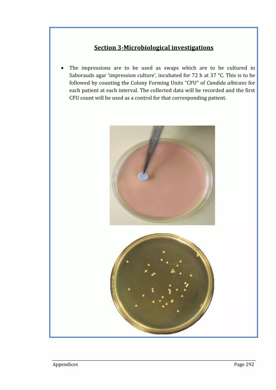
Appendices Page 292
Section 3-Microbiological investigations
The impressions are to be used as swaps which are to be cultured in
Saborauds agar ‘impression culture’, incubated for 72 h at 37 °C. This is to be
followed by counting the Colony Forming Units “CFU” of Candida albicans for
each patient at each interval. The collected data will be recorded and the first
CFU count will be used as a control for that corresponding patient.

Appendices Page 293
APPENDIX - II MIC values of FLUp against MIC values of FLUc for
different Candida species
Fluconazole pure
Lab. no ID EUCAST(1) EUCAST(2)
24 h (50 %) 48 h (50 %)
6258 C. krusei 32 mg/L 32 mg/L
446 C. krusei 32 mg/L 32 mg/L
75 C. tropicalis 0.5 mg/L 0.5 mg/L
461 C. parapsilosis 1.0 mg/L 1.0 mg/L
392 C. guilliermondii 0.5 mg/L 0.5 mg/L
466 C. glabrata 8 mg/L 8 mg/L
4023 C. glabrata 128 mg/L 128 mg/L
2511 C. albicans 128mg/L 128 mg/L
463 C. albicans 0.25 mg/L 0.25 mg/L
90028 C. albicans 0.25 mg/L 0.25 mg/L
SA C. kefyr 0.5 mg/L 0.5 mg/L
Fluconazole capsules
Lab. no ID EUCAST(1) EUCAST(2)
24 h (50 %) 48 h (50 %)
6258 C. krusei 32 mg/L 32 mg/L
446 C. krusei 32 mg/L 32 mg/L
75 C. tropicalis 0.25 mg/L 0.25 mg/L
461 C. parapsilosis 1.0 mg/L 1.0 mg/L
392 C. guilliermondii 0.5 mg/L 0.5 mg/L
466 C. glabrata 8 mg/L 8 mg/L
4023 C. glabrata 128 mg/L 128 mg/L
2511 C. albicans 128 mg/L 128 mg/L
463 C. albicans 0.25 mg/L 0.25 mg/L
90028 C. albicans 0.25 mg/L 0.25 mg/L
SA C. kefyr 0.5 mg/L 0.5 mg/L

Appendices Page 294
APPENDIX - III Publication 1

Appendices Page 295

Appendices Page 296

Appendices Page 297

Appendices Page 298

Appendices Page 299

Appendices Page 300

Appendices Page 301
APPENDIX - IV Publication 2

Appendices Page 302

Appendices Page 303

Appendices Page 304

Appendices Page 305

Appendices Page 306

Appendices Page 307