Under-detection of endospore-forming Firmicutes in metagenomic data
Upload
independentCategory
view
3download
0
Blomström et al. Virology Journal 2012, 9:192http://www.virologyj.com/content/9/1/192
RESEARCH Open Access
Viral metagenomic analysis of bushpigs(Potamochoerus larvatus) in Uganda identifiesnovel variants of Porcine parvovirus 4 and Torqueteno sus virus 1 and 2Anne-Lie Blomström1*, Karl Ståhl1,2, Charles Masembe3, Edward Okoth4, Ademun Rose Okurut5, Patrick Atmnedi6,Stephen Kemp4, Richard Bishop4, Sándor Belák1,2 and Mikael Berg1
Abstract
Background: As a result of rapidly growing human populations, intensification of livestock production andincreasing exploitation of wildlife habitats for animal agriculture, the interface between wildlife, livestock andhumans is expanding, with potential impacts on both domestic animal and human health. Wild animals serve asreservoirs for many viruses, which may occasionally result in novel infections of domestic animals and/or thehuman population. Given this background, we used metagenomics to investigate the presence of viral pathogensin sera collected from bushpigs (Potamochoerus larvatus), a nocturnal species of wild Suid known to move betweennational parks and farmland, in Uganda.
Results: Application of 454 pyrosequencing demonstrated the presence of Torque teno sus virus (TTSuV), porcineparvovirus 4 (PPV4), porcine endogenous retrovirus (PERV), a GB Hepatitis C–like virus, and a Sclerotiniahypovirulence-associated-like virus in sera from the bushpigs. PCR assays for each specific virus combined withSanger sequencing revealed two TTSuV-1 variants, one TTSuV-2 variant as well as PPV4 in the serum samples andthereby confirming the findings from the 454 sequencing.
Conclusions: Using a viral metagenomic approach we have made an initial analysis of viruses present in bushpigsera and demonstrated for the first time the presence of PPV4 in a wild African Suid. In addition we identified novelvariants of TTSuV-1 and 2 in bushpigs.
BackgroundWild animals are carriers of a number of pathogens thathave the potential to infect the human population and/or domestic animals. The intensity of contact betweenwildlife, livestock and human population is increasingdue to a number of factors, primarily human and live-stock population growth leading to encroachment ontowildlife habitat [1,2]. During recent decades a number ofpathogen crossovers from wildlife to humans and live-stock have occurred resulting in emerging diseases, suchas SARS, Hantavirus Pulmonary syndrome 1, Nipahvirus disease 1, and Hendra virus-induced diseases
* Correspondence: [email protected] of Biomedical Sciences and Veterinary Public Health, Section ofVirology, Swedish University of Agricultural Sciences, Uppsala, SwedenFull list of author information is available at the end of the article
© 2012 Blomström et al.; licensee BioMed CenCreative Commons Attribution License (http:/distribution, and reproduction in any medium
among others. However, it is also evident that transmis-sion can occur both ways i.e. pathogens may spill-overto wildlife from humans and/or from livestock. One ex-ample of this is the spill-over of Canine distemper virusfrom domestic dogs (Canis familiaris) to African wilddogs (Lycaon pictus) in Serengeti in 1991 leading to localextinction of wild dogs in the area [2]. Apart from beingcarriers of novel viruses with the potential to cause dis-ease in naïve domestic hosts, wildlife may also act asreservoirs for known viral pathogens – for example thereare a number of wildlife reservoirs for foot and mouthdisease virus such as African buffalo (Syncerus caffer),reindeer (Rangifer tarandus) and wild boar (Sus scrofa)[3]. However, information on the viral flora in wildlife istypically scarce or non-existent.
tral Ltd. This is an Open Access article distributed under the terms of the/creativecommons.org/licenses/by/2.0), which permits unrestricted use,, provided the original work is properly cited.
Blomström et al. Virology Journal 2012, 9:192 Page 2 of 7http://www.virologyj.com/content/9/1/192
Traditional viral detection methods, such as virus iso-lation, are often hindered by the inability to grow virusin cell culture. The divergence of many viruses and ab-sence of a common viral marker gene makes detectiondifficult using standard molecular techniques includingPCR and microarray as they are frequently target-specific through the use of specific primers, probes and/or antibodies. Viral metagenomics is a sequence, andculture-independent approach that has proven to be avaluable tool for the investigation not only of diseases ofunknown etiology but also of the complete viral flora ofdifferent reservoirs and vectors. By providing insightsinto a wide range of unknown potential pathogens andrevealing novel aspects of biodiversity, metagenomics isable to detect and characterise novel pathogens [4-6].In many rural parts of the developing world, domestic
livestock are kept in free-range systems, potentiallyallowing contact with wild animals. In some parts ofUganda, free-range scavenging by pigs is frequent. Atthe same time wild species of suidae such as bushpigs(Potamochoerus larvatus), with a wide distribution inEastern and Southern Africa, live and move at the inter-face between the national parks and the farmland wherethere is an opportunity for interaction and sharing ofpathogens with domestic relatives. Bushpigs are consid-ered to be possible natural reservoirs for African swinefever virus [7], but less is known about what otherviruses bushpigs might carry. Therefore, to investigatewhether bushpigs are carriers of known and or unknownporcine viruses we have in this study investigated theviral flora of bushpig sera.
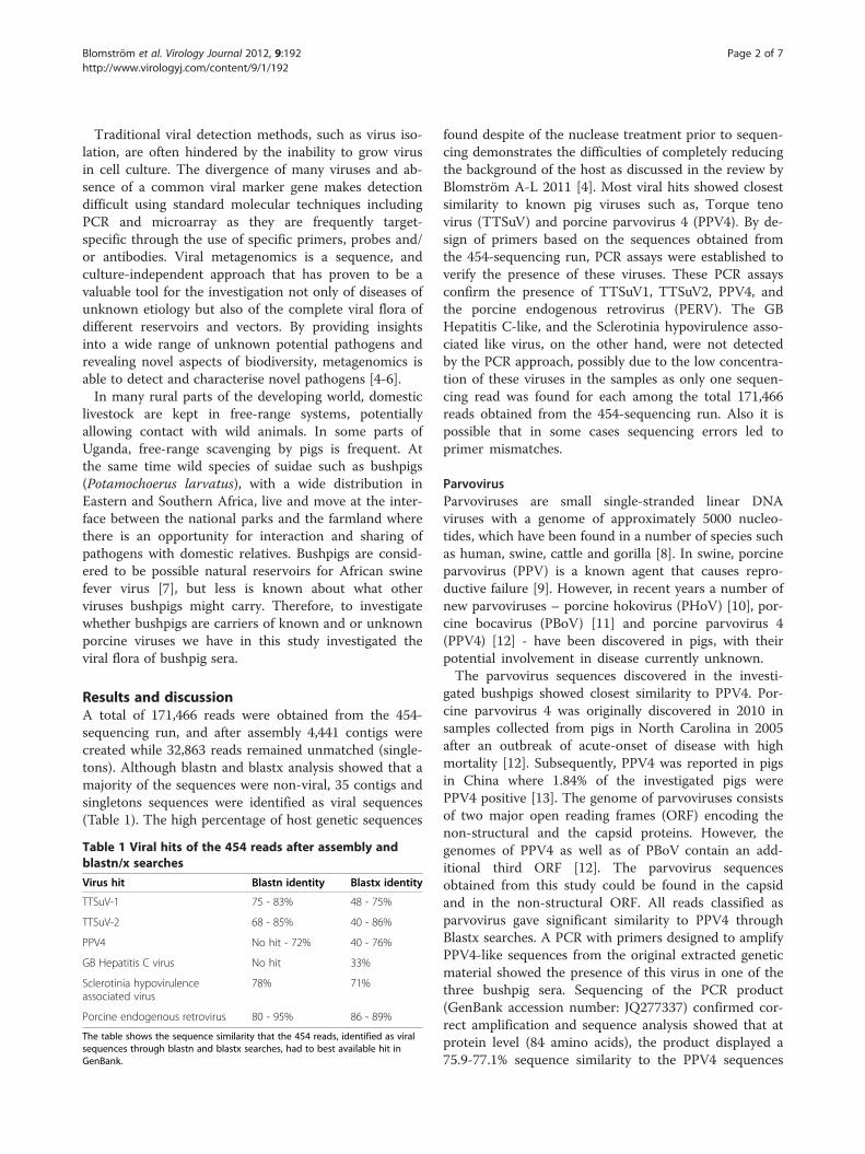
Results and discussionA total of 171,466 reads were obtained from the 454-sequencing run, and after assembly 4,441 contigs werecreated while 32,863 reads remained unmatched (single-tons). Although blastn and blastx analysis showed that amajority of the sequences were non-viral, 35 contigs andsingletons sequences were identified as viral sequences(Table 1). The high percentage of host genetic sequences
Table 1 Viral hits of the 454 reads after assembly andblastn/x searches
Virus hit Blastn identity Blastx identity
TTSuV-1 75 - 83% 48 - 75%
TTSuV-2 68 - 85% 40 - 86%
PPV4 No hit - 72% 40 - 76%
GB Hepatitis C virus No hit 33%
Sclerotinia hypovirulenceassociated virus
78% 71%
Porcine endogenous retrovirus 80 - 95% 86 - 89%
The table shows the sequence similarity that the 454 reads, identified as viralsequences through blastn and blastx searches, had to best available hit inGenBank.
found despite of the nuclease treatment prior to sequen-cing demonstrates the difficulties of completely reducingthe background of the host as discussed in the review byBlomström A-L 2011 [4]. Most viral hits showed closestsimilarity to known pig viruses such as, Torque tenovirus (TTSuV) and porcine parvovirus 4 (PPV4). By de-sign of primers based on the sequences obtained fromthe 454-sequencing run, PCR assays were established toverify the presence of these viruses. These PCR assaysconfirm the presence of TTSuV1, TTSuV2, PPV4, andthe porcine endogenous retrovirus (PERV). The GBHepatitis C-like, and the Sclerotinia hypovirulence asso-ciated like virus, on the other hand, were not detectedby the PCR approach, possibly due to the low concentra-tion of these viruses in the samples as only one sequen-cing read was found for each among the total 171,466reads obtained from the 454-sequencing run. Also it ispossible that in some cases sequencing errors led toprimer mismatches.
ParvovirusParvoviruses are small single-stranded linear DNAviruses with a genome of approximately 5000 nucleo-tides, which have been found in a number of species suchas human, swine, cattle and gorilla [8]. In swine, porcineparvovirus (PPV) is a known agent that causes repro-ductive failure [9]. However, in recent years a number ofnew parvoviruses – porcine hokovirus (PHoV) [10], por-cine bocavirus (PBoV) [11] and porcine parvovirus 4(PPV4) [12] - have been discovered in pigs, with theirpotential involvement in disease currently unknown.The parvovirus sequences discovered in the investi-
gated bushpigs showed closest similarity to PPV4. Por-cine parvovirus 4 was originally discovered in 2010 insamples collected from pigs in North Carolina in 2005after an outbreak of acute-onset of disease with highmortality [12]. Subsequently, PPV4 was reported in pigsin China where 1.84% of the investigated pigs werePPV4 positive [13]. The genome of parvoviruses consistsof two major open reading frames (ORF) encoding thenon-structural and the capsid proteins. However, thegenomes of PPV4 as well as of PBoV contain an add-itional third ORF [12]. The parvovirus sequencesobtained from this study could be found in the capsidand in the non-structural ORF. All reads classified asparvovirus gave significant similarity to PPV4 throughBlastx searches. A PCR with primers designed to amplifyPPV4-like sequences from the original extracted geneticmaterial showed the presence of this virus in one of thethree bushpig sera. Sequencing of the PCR product(GenBank accession number: JQ277337) confirmed cor-rect amplification and sequence analysis showed that atprotein level (84 amino acids), the product displayed a75.9-77.1% sequence similarity to the PPV4 sequences

Blomström et al. Virology Journal 2012, 9:192 Page 3 of 7http://www.virologyj.com/content/9/1/192
available in GenBank. In addition, the phylogenetic treegenerated from these data (Figure 1) grouped the se-quence with PPV4 when analysed together withsequences from all the different parvovirus genera(Parvovirus, Erythrovirus, Dependovirus, Amdovirus andBocavirus) and PPV4. However, the tree also confirms
Figure 1 Neighbour-joining phylogenetic tree of Parvovirus. The Neigbushpig PPV4 sequence (84 amino acid long) and 49 sequences available f
the divergence of the bushpig PPV4 from the otherPPV4.PPV4 has previously been reported in domestic pigs
only in USA and China [12,13]. However with this studywe show the presence of a PPV4 variant in a wild suidin Uganda.
hbour-joining tree shows the phylogenetic relationship between therom GenBank. The bushpig PPV4 is indicated with ♦.

ACZ71253
ADN28525
ADN28553
ADN28533
ADN28541
ADN28569
ADN28557
AEO16615
ADE18857
ADD46846
ADN28573
ADN28561
ADN28529
ACR57080
AAW79281
ADN28517
ADN28549
ADN28509
ADD46842
YP 003587831
ADN28537
JQ277338 (TTSuV-1a)
JQ277339 (TTSuV-1a)
AEO16613
ADN28545
ADM33775
ADM33772
ADN28565
ADN28521
ADM33784
ADN28513
ADM33781
JQ277340 (TTSuV-1b)
JQ277341 (TTSuV-1b)
JQ277342 (TTSuV-1b)
100
100
100
98
95
75
97
97
87
9495
99
84
0.02
Figure 2 Neighbour-joining phylogenetic tree of TTSuV-1. TheNeighbour-joining tree shows the phylogenetic relationshipbetween the five bushpig TTSuV-1 sequences (66 amino acid long)and 30 sequences available in GenBank. The bushpig TTSuV-1a isindicated with ■ and TTSuV-1b with ♦.
Blomström et al. Virology Journal 2012, 9:192 Page 4 of 7http://www.virologyj.com/content/9/1/192
Torque teno sus virusTorque teno virus was discovered 1997 in a serum samplefrom a patient with posttransfusion hepatitis of unknownetiology using representational difference analysis [14].Since then the virus has been detected and characterisedin a number of species such as primates, cats, dogs andpigs [15], but the role of these viruses in disease develop-ment is still controversial. These viruses have small(approximately 2.8 kb) circular DNA genomes. Torqueteno virus in pigs is divided into two different species,Torque teno sus virus 1 and 2, and prevalence studieshave shown that TTSuV is widely spread in pig popu-lations across the world [16,17]. In a previous study[18], we have found that 51.6% of a sample populationof domestic pigs in Uganda were carrying one or boththese TTSuV variants. Although, most studies have tar-geted domestic pigs, TTSuV have also been found inwild boar in Europe [19].As shown in Table 1, our data indicated the presence
of both TTSuV-1 and 2 in the investigated serum sam-ples. Both the TTSuV-1 and 2 sequence reads werelocated in the major open reading frame (ORF1) and allreads showed significant similarity to the respective virusin both blastn and blastx analyses.Two of the TTSuV-1 sequence reads partially over-
lapped and the sequence analysis indicated a significantvariation between the two and therefore two differentTTSuV-1 PCR assays were designed. The results fromthe specific PCR assays showed that one of the TTSuV-1variants (here named TTSuV-1a) could be found in twoof the three bushpig sera while the other one (herenamed TTSuV-1b) was found in all three. The sequenceanalysis of the PCR amplified TTSuV-1 products (Gen-Bank accession number JQ277338 - 42) showed a se-quence similarity between TTSuV-1a and b on proteinlevel at 53–54.5% in the analysed 66 amino acid region.Compared to sequences from other studies, TTSuV-1ashowed a 60.6-84.8% protein sequence similarity whileTTSuV-1b was more divergent (50-56% similarity).These protein sequence identity values were similar tothose seen when comparing the sequences retrievedfrom the GenBank with each other (59-100% similarity).The phylogenetic tree (Figure 2) also indicated thatTTSuV-1b was more divergent than the other sequences.Sequences from different parts of the world such asChina, Germany, Spain, US etc. was used in the phylo-genetic study however no clear geographical clusteringwas seen.TTSuV-2 was confirmed in one of the bushpig sera
and the amplified product GenBank accession numberJQ277343) showed a protein sequence identity of 66.6-79.4% to the other TTSuV-2 sequences used in thephylogenetic study using a 313 amino acid region. Thissequence identity was in the range of the similarity seen
between the different TTSuV-2 sequences from otherstudies used for comparison (64.2-100%). In the phylo-genetic study (Figure 3) the sequenced TTSuV-2grouped with the other TTSuVs but in its own clade.TTSuV-1 and 2 have previously, as mentioned, been
detected both in domestic pigs across the world [16-18]and wild suidae in Europe [19] and now for the first timein bushpigs on the African continent. Studies on the gen-etic variability of TTSuV-1 and 2 have shown a highergenetic diversity in the coding regions compared to theuntranslated region [20]. The sequence analysis of boththe bushpig-derived TTSuV and those from GenBankshows a high genetic variability among the differentTTSuV-1 and TTSuV-2 and also shows the co-infectionof two different TTSuV-1 variants and one TTSuV-2.
RetrovirusEndogenous retroviruses are integrated in the host gen-ome and all vertebrates investigated have been shown tocarry retroviral sequences. It is estimated that up to 10%

ADN28581
ADN28605
ADN28653
ADN28597
ADN28681
ADN28621
ADN28585
ADN28645
ADN28633
ADM33761
YP 004021926
ADN28677
ADM33768
ADM33764
ADN28589
ADM33771
ADM33750
ADN28609
ADN28601
ADM33755
ADM33759
ADM33753
ADN28617
ADD46850
ACZ71256
ADW01383
AEO16617
ADD46854
ADN28641
ADN28593
ADN28629
JQ277343
ADN28665
ADN28657
ADN28685
ADN28637
ADN28649
ADN28577
ADN28625
ADN28613
ADN28661
ADN28669
ADN28673
80
100
100
100
99
100
99
89
78
100
99
99
98
98
90
92
85
87
97
93
98
71
93
97
99
100
88
75
100
88
100
0.02
Figure 3 Neighbour-joining phylogenetic tree of TTSuV-2. TheNeighbour-joining tree shows the phylogenetic relationshipbetween the bushpig TTSuV-2 sequence (313 amino acid long) and42 sequences available in GenBank. The bushpig TTSuV-2 isindicated with ♦.
Blomström et al. Virology Journal 2012, 9:192 Page 5 of 7http://www.virologyj.com/content/9/1/192
of animal genomes are retroviral elements [21]. Also thebushpig genome has been investigated and confirmed tocontain PERV [22-24].By running the specific PERV PCR on both DNA and
on DNase treated RNA we could as expected see that allthe bushpigs had both integrated proviral DNA andexpressed PERV RNA, indicating active viral transcrip-tion and replication. The sequenced PERV products
(GenBank accession number JX566717-719) showed ahigh similarity (85 - 89%) to those available in GenBank.
ConclusionsThrough investigating sera collected from bushpigs inUganda by viral metagenomics, we have for the first timeshowed the presence of PPV4 in a wild Suid on the Afri-can continent. The region of PPV4 investigated indicatesa sequence divergence relative to previously describedPPV4. In addition, novel TTSuV-1 and 2 variants werefound. Further sequence analysis and prevalence studiescan be used to define the genetic relationships of theseviruses and their distribution in both domestic pigs andin wildlife.
MethodsSamplesThe sera from three bushpigs collected from LakeMburo National Park, Uganda in March 2010, as part ofa research project on African swine fever epidemiologywere used in this study. The animals were capturedusing game capture nets (50x3m, 150 mm square mesh,3.5 mm nylon braid khaki, ALNET Ltd, South Africa)with assistance from Uganda Wildlife Authority (UWA)staff, and sedated with zolazepam and tiletamine (Zoletilforte vet 50 mg/ml + 50 mg/ml, Virbac Laboratories,France) before blood sampling from the saphenousvein. The Ministry of Agriculture Animal Industry andFisheries and Uganda Wildlife Authority, together withMakerere University are mandated to carry out animaldisease investigations in livestock and wildlife in thecountry. This is done by veterinarians who handle theanimals under internationally recognized guidelines.
Sample preparation, nucleic acid extraction and randomPCRFifty microliters of serum was aliquoted for the RNAand DNA extraction respectively. One hundred and fiftymicroliter of 1x DNase buffer (Roche, Mannheim, Ger-many) was added to each aliquot of serum after whichthe sample was treated with nucleases - 100 U DNase(Roche, Mannheim, Germany) and 2 μg RNase (Invitro-gen, Carlsbad, CA, USA) for two hours at 37°C in orderto degrade the host nucleic acid. Trizol was added toone of the two aliquots and RNA was extracted using acombination of Trizol and Qiagen RNAeasy kit. DNAwas extracted using the Qiagen DNAeasy mini extrac-tion kit according to the manufacturer’s instructions andeluted in 50 μl elution buffer (EB). The DNA and RNAwere amplified by random PCR as described earlier [25].Before sequencing, the primers were cleaved usingEcoRV (NEB, Ipswich, MA, USA) and the cleaved prod-uct was purified using the Qiagen PCR purification kit(Qiagen, Hilden, Germany) and eluted in 30 μl EB.

Blomström et al. Virology Journal 2012, 9:192 Page 6 of 7http://www.virologyj.com/content/9/1/192
High-throughput sequencing and data analysisThe purified product was sequenced on 1/8th of a picotitre plate using the 454 technology by Roche at InqabaBiotech (South Africa). The sequences were analysedthrough quality check and removal of very shortsequences before being assembled using CLC genomicworkbench v4.6 (http://www.clcbio.com/index.php).Blastn and blastx searches were performed through theCamera 2.0 portal [26,27] and searches through NCBI(http://www.ncbi.nlm.nih.gov/blast/Blast.cgi). The viralblast hits with an e-value of 10-4 or lower were furtheranalysed.
Confirmation PCRs, sequencing and phylogenetic studiesPCR primers were designed based on the reads from the454-sequencing run (Table 2) and PCR assays were setup with the aim to look for each individual virus in eachbushpig sera. For the RNA viruses cDNA synthesis wasperformed prior to PCR using Superscript III (Invitrogen,Carlsbad, CA, USA) and random primers according tothe manufacturer’s instructions. The PERV RNA wastreated with DNase prior to the cDNA synthesis andboth a “+” and a “–“ RT cDNA synthesis reaction wasperformed. The PCR using each specific primer pair(Table 1) was performed according to the following pro-cedure: 1x PCR buffer, 2.5 mM MgCl2, 1.0 mM dNTP,0.4 μM forward primer and reverse primer each, and1.25 U AmpliTaq Gold DNA polymerase (Applied Bio-systems, Foster City, CA, USA). For each reaction, two μlDNA or cDNA from each respective bushpig was used.The amplification was performed with the following re-action conditions: a 12 minute enzyme activation step at95°C followed by 39 cycles of 95°C for 30 seconds, 58°Cfor 30 seconds and 72°C for 90 seconds, finishing with
Table 2 Primers for verification
Primer Sequence 5’- 3’
TTSuV-1a_F TCC CAG CAG AAG ATG TAG TC
TTSuV-1a_R GGA TGG TGG CCT CTA CTA C
TTSuV-1b_F GCA GCA TAA CGC TAG GCT G
TTSuV-1b_R AGA GGA AAT GGG CTA CCT G
PPV4_F CTC TGA TAA TGT ATT ACT GGT C
PPV4_R AAG AAA GAT CCT TCT GTT ACA
GB Hepatitis C virus_F CTG CCT CAA CGT TGA GGC AG
GB Hepatitis C virus_R ACG ACG TAG CAG TGG TAG AT
Sclerotinia hypovirulenceassociated virus_F
CGC ATC ACG GTC AAG TTT GA
Sclerotinia hypovirulenceassociated virus_R
TTA CAT TGC TTC GTC GAC TTC
PERV_F ATG GGA GCT GGG TCC AAT C
PERV_R CCT TAC GTT TGA CTC TCG AC
Primers used for the detection and sequencing of the found viruses.
one cycle for 10 minutes at 72°C. The PCR products werevisualized on a 1.5% agarose gel. The PCR-positive pro-ducts were purified using the QIAquick PCR purificationkit (Qiagen, Hilden, Germany) according to the manufac-turer’s instructions and eluted in 25 μl EB. The purifiedproducts were sequenced with standard Sanger sequen-cing using Big Dye Termination kit (Applied Biosystems,Foster City, CA, USA) according to the manufacturer’sinstructions. The obtained chromatograms were editedusing SeqMan (Lasergene 9, DNASTAR Inc., Madison,USA). Sequence identity plots were performed usingthe Bioedit software [28]. ClustalW as well as thephylogenetic analyses were carried out using Mega 5[29]. The phylogenetic trees were constructed usingthe Neighbour-joining algorithm with p-distances andwith a bootstrap value of 1000.
Competing interestsThe authors declare that they have no competing interests.
Authors’ contributionsALB has performed the laboratory experiments, contributed to the studydesign, data analysis, manuscript draft and final manuscript preparation. KSand CM coordinated the field work and contributed to study design, dataanalysis and final manuscript preparation. EO and PA coordinated thebushpig capture and contributed to the final manuscript preparation. ARO,SK, RB and SB contributed to the final manuscript preparation. MBcontributed to study design, data analysis and final manuscript preparation.All authors have read and approved the final manuscript.
AcknowledgementsWe would like to thank our colleagues Denis Muhangi and Susan Ndyanabofor technical assistance in the laboratory and Uganda Wildlife Authority stafffor assistance in the field. Financial support for this study was provided bythe Swedish International Development Cooperation Agency (Sida; SWE-2009-081), the Swedish research Council Formas (221-2009-1984), theSwedish Ministry of Foreign Affairs as part of its special allocation on globalfood security (through the Swedish University of Agricultural Sciences, SLU)and by the Award of Excellence awarded to SB by SLU.
Author details1Department of Biomedical Sciences and Veterinary Public Health, Section ofVirology, Swedish University of Agricultural Sciences, Uppsala, Sweden.2Department of Virology, Immunobiology and Parasitology (VIP), NationalVeterinary Institute (SVA), Uppsala, Sweden. 3Department of Biology,Makerere University, College of Natural Sciences. School of BiologicalSciences, Box 7026, Kampala, Uganda. 4International Livestock ResearchInstitute (ILRI), Nairobi, Kenya. 5National Animal Disease Diagnostics andEpidemiology Centre (NADDEC), Ministry of Agriculture Animal Industry andFisheries, Entebbe, Uganda. 6Uganda Wildlife Authority, Kampala, Uganda.
Received: 19 March 2012 Accepted: 5 September 2012Published: 11 September 2012
References1. Perry BD, Grace D, Sones K: Livestock and Global Change Special Feature:
Current drivers and future directions of global livestock diseasedynamics. Proc Natl Acad Sci USA 2011.
2. Daszak P, Cunningham AA, Hyatt AD: Emerging infectious diseases ofwildlife–threats to biodiversity and human health. Science 2000,287:443–449.
3. Siembieda JL, Kock RA, McCracken TA, Newman SH: The role of wildlife intransboundary animal diseases. Animal health research reviews / Conferenceof Research Workers in Animal Diseases 2011, 12:95–111.
4. Blomström A-L: Viral metagenomics as an emerging and powerful tool inveterinary medicine. Vet Q 2011, 31:107–114.

Blomström et al. Virology Journal 2012, 9:192 Page 7 of 7http://www.virologyj.com/content/9/1/192
5. Tang P, Chiu C: Metagenomics for the discovery of novel human viruses.Future Microbiol 2010, 5:177–189.
6. Delwart EL: Viral metagenomics. Rev Med Virol 2007, 17:115–131.7. Jori F, Bastos AD: Role of wild suids in the epidemiology of African swine
fever. EcoHealth 2009, 6:296–310.8. Hueffer K, Parrish CR: Parvovirus host range, cell tropism and evolution.
Curr Opin Microbiol 2003, 6:392–398.9. Mengeling WL, Lager KM, Vorwald AC: The effect of porcine parvovirus
and porcine reproductive and respiratory syndrome virus on porcinereproductive performance. Anim Reprod Sci 2000, 60–61:199–210.
10. Lau SK, Woo PC, Tse H, Fu CT, Au WK, Chen XC, Tsoi HW, Tsang TH, ChanJS, Tsang DN, et al: Identification of novel porcine and bovineparvoviruses closely related to human parvovirus 4. J Gen Virol 2008,89:1840–1848.
11. Blomström AL, Belak S, Fossum C, McKillen J, Allan G, Wallgren P, Berg M:Detection of a novel porcine boca-like virus in the background ofporcine circovirus type 2 induced postweaning multisystemic wastingsyndrome. Virus Res 2009, 146:125–129.
12. Cheung AK, Wu G, Wang D, Bayles DO, Lager KM, Vincent AL: Identificationand molecular cloning of a novel porcine parvovirus. Arch Virol 2010,155:801–806.
13. Huang L, Zhai SL, Cheung AK, Zhang HB, Long JX, Yuan SS: Detection of anovel porcine parvovirus, PPV4, in Chinese swine herds. Virol J 2010,7:333.
14. Nishizawa T, Okamoto H, Konishi K, Yoshizawa H, Miyakawa Y, Mayumi M: Anovel DNA virus (TTV) associated with elevated transaminase levels inposttransfusion hepatitis of unknown etiology. Biochem Biophys ResCommun 1997, 241:92–97.
15. Okamoto H: TT viruses in animals. Curr Top Microbiol Immunol 2009,331:35–52.
16. Blomström AL, Belak S, Fossum C, Fuxler L, Wallgren P, Berg M: Studies ofporcine circovirus type 2, porcine boca-like virus and torque teno virusindicate the presence of multiple viral infections in postweaningmultisystemic wasting syndrome pigs. Virus Res 2010, 152:59–64.
17. Segales J, Martinez-Guino L, Cortey M, Navarro N, Huerta E, Sibila M, Pujols J,Kekarainen T: Retrospective study on swine Torque teno virusgenogroups 1 and 2 infection from 1985 to 2005 in Spain. Vet Microbiol2009, 134:199–207.
18. Brink M, Stahl K, Masembe C, Okurut AR, Berg M, Blomstrom AL: First timemolecular detection and phylogenetic relationships of torque teno susvirus 1 and 2 in domestic pigs in Uganda: further evidence for a globaldistribution. Virol J 2012, 9:39.
19. Martinez L, Kekarainen T, Sibila M, Ruiz-Fons F, Vidal D, Gortazar C, Segales J:Torque teno virus (TTV) is highly prevalent in the European wild boar(Sus scrofa). Vet Microbiol 2006, 118:223–229.
20. Cortey M, Macera L, Segales J, Kekarainen T: Genetic variability andphylogeny of Torque teno sus virus 1 (TTSuV1) and 2 (TTSuV2) based oncomplete genomes. Vet Microbiol 2011, 148:125–131.
21. MacLachlan JN, Dubovi EJ: Fenner’s Veterinary Virology. Fourthth edition.London: Academic Press; 2001.
22. Do Nascimento FF, Gongora J, Tristem M, Lowden S, Moran C: Distinctivedifferences in long terminal repeat sequences between gamma1endogenous retroviruses of African and Eurasian suid species. InfectGenet Evol 2011, 11:686–693.
23. Do Nascimento FF, Gongora J, Charleston M, Tristem M, Lowden S, MoranC: Evolution of endogenous retroviruses in the Suidae: evidence fordifferent viral subpopulations in African and Eurasian host species. BMCEvol Biol 2011, 11:139.
24. Patience C, Switzer WM, Takeuchi Y, Griffiths DJ, Goward ME, Heneine W,Stoye JP, Weiss RA: Multiple groups of novel retroviral genomes in pigsand related species. J Virol 2001, 75:2771–2775.
25. Blomström AL, Widen F, Hammer AS, Belak S, Berg M: Detection of a novelastrovirus in brain tissue of mink suffering from shaking mink syndromeby use of viral metagenomics. J Clin Microbiol 2010, 48:4392–4396.
26. Seshadri R, Kravitz SA, Smarr L, Gilna P, Frazier M: CAMERA: a communityresource for metagenomics. PLoS Biol 2007, 5:e75.
27. Sun S, Chen J, Li W, Altintas I, Lin A, Peltier S, Stocks K, Allen EE, Ellisman M,Grethe J, Wooley J: Community cyberinfrastructure for AdvancedMicrobial Ecology Research and Analysis: the CAMERA resource. NucleicAcids Res 2011, 39:D546–551.
28. Hall T: BioEdit: a user-friendly biological sequence alignment editor andanalysis program for Windows 95/98/NT. Nucleic Acids Symp Ser 1999,41:95–98.
29. Tamura K, Dudley J, Nei M, Kumar S: MEGA4: Molecular EvolutionaryGenetics Analysis (MEGA) software version 4.0. Mol Biol Evol 2007,24:1596–1599.
doi:10.1186/1743-422X-9-192Cite this article as: Blomström et al.: Viral metagenomic analysis ofbushpigs (Potamochoerus larvatus) in Uganda identifies novel variantsof Porcine parvovirus 4 and Torque teno sus virus 1 and 2. VirologyJournal 2012 9:192.
Submit your next manuscript to BioMed Centraland take full advantage of:
• Convenient online submission
• Thorough peer review
• No space constraints or color figure charges
• Immediate publication on acceptance
• Inclusion in PubMed, CAS, Scopus and Google Scholar
• Research which is freely available for redistribution
Submit your manuscript at www.biomedcentral.com/submit
Copyright © 2022 FDOKUMEN