Metagenomic analysis of two enhanced biological phosphorus removal (EBPR) sludge communities
Upload
independentCategory
view
0download
0
� ����� ��� � ��
Under-detection of endospore-forming Firmicutes in metagenomic data
Sevasti Filippidou, Thomas Junier, Tina Wunderlin, Chien-Chi Lo, Po-E
Li, Patrick S. Chain, Pilar Junier
PII: S2001-0370(15)00021-5
DOI: doi: 10.1016/j.csbj.2015.04.002
Reference: CSBJ 74
To appear in: Computational and Structural Biotechnology Journal
Received date: 26 December 2014
Revised date: 6 April 2015
Accepted date: 18 April 2015
Please cite this article as: Filippidou Sevasti, Junier Thomas, Wunderlin Tina, Lo Chien-Chi, Li Po-E, Chain Patrick S., Junier Pilar, Under-detection of endospore-forming Fir-micutes in metagenomic data, Computational and Structural Biotechnology Journal (2015),doi: 10.1016/j.csbj.2015.04.002
This is a PDF file of an unedited manuscript that has been accepted for publication.As a service to our customers we are providing this early version of the manuscript.The manuscript will undergo copyediting, typesetting, and review of the resulting proofbefore it is published in its final form. Please note that during the production processerrors may be discovered which could affect the content, and all legal disclaimers thatapply to the journal pertain.

AC
CEPTED
MAN
USC
RIP
T
ACCEPTED MANUSCRIPT
Under-detection of endospore-forming Firmicutes in metagenomic data
Sevasti Filippidou1, Thomas Junier1,2, Tina Wunderlin1§, Chien-Chi Lo3, Po-E Li3, Patrick S. Chain3,
Pilar Junier1*
1Laboratory of Microbiology, Institute of Biology, University of Neuchatel, CH-2000, Neuchâtel,
Switzerland
2Vital-IT group, Swiss Institute of Bioinformatics, CH-1015, Lausanne, Switzerland
3Bioscience Division, Los Alamos National Laboratory, Los Alamos, New Mexico, 87545, USA
* Corresponding author. Mailing address: Laboratory of Microbiology, Institute of
Biology, University of Neuchâtel, CH - 2000 Neuchâtel
Phone: +4132 7182244
Fax: +4132 7182231
E-mail: [email protected]
E-mail co-authors: [email protected]
[email protected] [email protected] [email protected]
§Current address: Department of Biological Sciences, Macquarie University, Australia.
Running title: Underrepresentation of Firmicutes in metagenomes
80000 CHARACTERS (INCLUDING SPACES)
AC
CEPTED
MAN
USC
RIP
T
ACCEPTED MANUSCRIPT
Abstract (250 words)
Microbial diversity studies based on metagenomic sequencing have greatly enhanced our
knowledge of the microbial world. However, one caveat is the fact that not all microorganisms
are equally well detected, questioning the universality of this approach. Firmicutes are known to
be a dominant bacterial group. Several Firmicutes species are endospore formers and this
property makes them hardy in potentially harsh conditions, and thus likely to be present in a
wide variety of environments, even as residents and not functional players. While metagenomic
libraries can be expected to contain endospore formers, endospores are known to be resilient to
many traditional methods of DNA isolation and thus potentially undetectable. In this study we
evaluated the representation of endospore-forming Firmicutes in 73 published metagenomic
datasets using two molecular markers unique to this bacterial group (spo0A and gpr). Both
markers were notably absent in well-known habitats of Firmicutes such as soil, with spo0A found
only in three mammalian gut microbiomes. A tailored DNA extraction method resulted in the
detection of a large diversity of endospore-formers in amplicon sequencing of the 16S rRNA and
spo0A genes. However, shotgun classification was still poor with only a minor fraction of the
community assigned to Firmicutes. Thus, removing a specific bias in a molecular workflow
improves detection in amplicon sequencing, but it was insufficient to overcome the limitations
for detecting endospore-forming Firmicutes in whole-genome metagenomics. In conclusion, this
study highlights the importance of understanding the specific methodological biases that can
contribute to improve the universality of metagenomic approaches.
Highlights (85 characters)
Endospore formers were under-detected by profile analysis of sporulation genes in
metagenomes.
Endospore formers were absent even from those habitats known to harbor them.
A tailored DNA extraction method improved detection in amplicon sequencing.
Ameliorated DNA extraction did not improve shotgun classification.
Endospore-formers represent an undetectable community fraction by metagenomic
approaches.

AC
CEPTED
MAN
USC
RIP
T
ACCEPTED MANUSCRIPT
Key words: endospores, gpr, metagenomics, profile analysis, spo0A

AC
CEPTED
MAN
USC
RIP
T
ACCEPTED MANUSCRIPT
1. Introduction
Metagenomic studies have emerged as promising methods for the collective study of
microbial communities directly extracted from environmental samples [1–3]. These approaches
have been successfully applied to a variety of environments and have helped to unveil new
functional pathways and metabolic processes within the microbial world [4–8].
Biases, however, can occur at all the steps involved in a metagenomic workflow. They can be
associated to the specific type of environment [9,10], the DNA yields obtained [11], the DNA
extraction method [12], the amplification (for example in amplicon sequencing), but also in the
sequencing and the analysis of the sequences, as well. These limitations have been highlighted in
the recent literature and result in problems such as low coverage of the less abundant taxa (the
so-called ╉depth bias╊ for example in the detection of ribosomal genes [13]), low reproducibility
of results [14] and underrepresentation of certain taxa, as discussed herein. In order to overcome
these limitations, single-cell genomics or novel approaches in culture-dependent methodologies,
such as culturomics [15,16] which, in their turn, have their own limitations.
Even though methodological bias of metagenomic diversity surveys associated to particular
types of environments such as soil has been demonstrated experimentally [9,10], the specific
coverage of individual microbial groups within the community is still unknown. One example of a
bacterial group that can be used to test coverage bias in metagenomic datasets is endospore-
forming Firmicutes. Even though, culturing of microorganisms is largely acknowledge to be bias,
according to previous research based on culture collections as well as whole-genome sequencing,
Firmicutes is the second most abundant bacterial phylum [17]. Endospore formers live in a wide range of environments on Earth’s surface and subsurface [18,19]. The hardy outer cortex of
endospores and the small acid-soluble proteins stabilizing their DNA [20–22], allow these
bacteria to be distributed into every habitat on Earth [23]. However, a phylogenetic assessment
of the microbial communities in four metagenomic datasets has revealed surprisingly few
endospore formers [24]. This might appear surprising considering their ubiquity, but endospores
are known to withstand many traditional methods of DNA isolation and are thus potentially
undetectable in a sample. Recently, a DNA extraction method for the extraction of resistant
structures such as endospores has been developed by our group [12]. This DNA extraction
AC
CEPTED
MAN
USC
RIP
T
ACCEPTED MANUSCRIPT
method was combined with amplicon sequencing of the gene coding the master regulator for the
initiation of sporulation (spo0A gene) to demonstrate an improved detection of endospore-
forming Firmicutes in sediment samples [12]. Our group has developed further methods to
separate endospores from vegetative cells, which has open the possibility to carry out genomic
studies only focused on endospores [12,25]. These two studies demonstrate by amplicon
sequencing that the diversity of endospore-forming Firmicutes is far from uncovered. However,
the effectiveness of the improved DNA extraction method for whole-genome metagenomic
studies is unknown.
The aim of this study was to measure the level of detection of endospore formers in
metagenomic studies carried out so far, and to evaluate the effect of an improved DNA extraction
method on the detectability of this group. To do this, we initially searched for functional gene
markers of endospore formation in metagenomic datasets using profiles. We then applied a
modified DNA extraction method that is tailored to release DNA from resistant structures such as
endospores [12] in a selected environmental sample. Amplicon sequencing of the 16S rRNA and
spo0A genes were performed on the sample in order to assess the relative abundance and
phylogenetic diversity of Firmicutes. This was complemented by shotgun sequencing and
classification of the metagenome reads. Our results indicate that endospore-forming Firmicutes
are overlooked in environmental diversity surveys using traditional whole metagenomic
approaches.

AC
CEPTED
MAN
USC
RIP
T
ACCEPTED MANUSCRIPT
2. Material and methods
2.1. Genome sequence retrieval
Complete and draft genome sequences of endospore-forming Firmicutes were downloaded
from the Comprehensive Microbial Resource (CMR, 24.0 data release, cmr.jcvi.org) and
Integrated Microbial Genomes (IMG, 3.0, img.jgi.doe.gov) websites. Protein and nucleotide
sequences of spore-related genes were obtained by search for role category/function sporulation
and germination (CMR) and sporulating (IMG). Additional information on all retrieved genomes
was obtained from the GenBank database (www.ncbi.nlm.nih.gov/genome).
2.2. Detection of orthologous sporulation genes common to all endospore formers
Orthologous groups were delineated based on best reciprocal BLASTp hits [26]. BLASTp was
used to align each sequence in the set against all sequences except those of the same species
(thus avoiding paralogs). The best hit in each species was retained, and sequence pairs, that were
each other's best match, were defined as best reciprocal hits (BRHs). Putative orthologous
groups were defined using the algorithm used by OrthoDB [27]. OrthoDB has data on Fungi,
Metazoa, and Bacteria. An early version of the BRHCLUS program (unpublished at the time) was
obtained from its author, Dr. Tegenfeldt (pers. comm) and run according to the author's
instructions. The program is now available from http://orthodb.org/. To our knowledge, its
utility does not depend on the clade it is used for - OrthoDB uses the same clustering program for
all data in its scope.
2.3. Profile construction and validation
The genomic sequences were filtered in such as way as to keep only one (randomly chosen)
sequence per genus, thus reducing taxonomic sampling bias. Multiple alignments of Spo0A and
Gpr were produced with MAFFT [28]. Gribskov-style sequence profiles were constructed with
EMBOSS's prophecy program [29]. The profiles' score cutoffs were determined by searching with EMBOSS’s prophet program against the original SpoどA ゅresp. Gprょ sequence set as a positive control, and against shuffled versions of the same as negative set.
AC
CEPTED
MAN
USC
RIP
T
ACCEPTED MANUSCRIPT
2.4. Metagenomic datasets retrieval
The metagenome datasets (supplementary Table 1) were downloaded from IMG, GOLD
(genomesonline.org), or the metagenomes subset of the WGS section of EMBL
(ebi.ac.uk/genomes/wgs.html). These datasets included all the metagenomic studies available at
EMBL when the profile analysis was performed. Only sequences or contigs of > 800 bp, which are
slightly shorter than the full-length sporulation genes, were kept for analysis.
2.5. Environmental sampling, DNA extraction and quantitative PCR
Sample was collected at Nea Apollonia (NAP) geothermal spring (N 40° ぬひ,なひな’ E 22°のは,ばどば’),
Greece, in June 2011. Geothermal reservoir was reached through a 120m drilling pipe, used
mostly for pumping 80o C water for bathing purposes. Biofilm from the pipe interior was
collected and frozen within 2h of collection. Upon arrival at the laboratory, a tailored DNA
extraction method previously described [12] was applied to the sample. More precisely, DNA was
extracted using the FastDNA Spin Kit for Soil (MP Biomedicals, California), using a modified
protocol in order to ensure that DNA was not only extracted from vegetative cells but also from
spores and other cells difficult to lyse. These modifications were (a) a separation of the biomass
from the soil, using a Na-hexa-meta-phosphate solution and (b) a sequential bead-beating step
(three times) to ensure mechanical disruption of cells. In total, 10ug of high molecular RNA-free
DNA was obtained.
Moreover, 16S rRNA gene and spo0A gene copy numbers were calculated using a quantitative
PCR assay, as previously described [30].
2.6 Amplicon sequencing of the 16S rRNA and spo0A genes
In order to verify the presence and relative abundance of endospore formers, 454
pyrosequencing of a fragment of the 16S rRNA and spo0A genes was firstly applied to the sample
NAP. Sequencing was done using the services of Eurofins MWG Operon (Ebersberg, Germany).
For 16S rRNA amplicon sequencing, fragments of approximately 500 bp were retrieved using primers Eubぱf ゅの’-AGAGTTTGATCCTGGCTCAG-ぬ’ょ and Eubのなひr ゅの’-GTATTACCGCGGCTGCTGG-ぬ’ょ, as previously described [31]. 16S rRNA gene raw sequence data was analysed with QIIME
[32], using the pipeline for de novo OTU picking. OTUs were identified using a threshold of 97%
AC
CEPTED
MAN
USC
RIP
T
ACCEPTED MANUSCRIPT
sequence similarity. The sequences were then clustered into putative OTUs with the pick_otus.py
program from the QIIME package using the Uclust method [32]. The single sequence picked by
the program as a representative of each OTU was used to build a phylogeny.
For the spo0A amplicon sequening, a 602 bp sequence of the spo0A gene was amplified using the degenerated primer spoどAなははf ゅの’-GATATHATYATGCCDCATYT-ぬ’ょ and spoどAばねぱr ゅの’-GCNACCATHGCRATRAAYTC-ぬ’ょ [12]. 42’151 sequences were received from the sample.
Sequences were then filtered according to Phred [33] quality score (minimum of 30) and
sequences of length shorter than 600 bp were removed. Remaining sequences were translated to
their amino acid sequence; resulting full-length ORFs were then matched against the Spo0A
profile, in order to confirm that the primers actually amplified the spo0A sequences.
Phylogenies were constructed from Phylip-formatted alignments with PhyML [34], using
default parameters. The trees were re-rooted, condensed according to protocol, and displayed
with the Newick Utilities. Each branch represents a cluster of OTUs of > 97% sequence similarity.
Identification of the closest relatives of the environmental sequences was done by protein BLAST
[26] with the translated protein sequences using a reference database of 581 Spo0A protein
sequences from the InterPro site [35].
All metagenomic sequences were submitted to GenBank. The 16S rRNA amplicon sequencing
data can be retrieved under the BioProject ID PRJNA267761 and BioSample ID SAMN03198953
and the spo0A amplicon sequencing data under the BioProject ID PRJNA276803 and Biosample
ID SAMN03392534.
2.7 Metagenomic sequencing
Once high prevalence of endospore formers was confirmed in the 16S rRNA pyrosequencing
data (41% of total bacterial community), whole-metagenome sequencing of NAP was performed
on a full plate of a GS FLX platform, followed by de novo assembly using the services of GATC-
biotech (Konstanz, Germany). The whole metagenome dataset can be retrieved from GenBank
under the BioProject ID PRJNA271123 and BioSample ID SAMN03273062.
2.8. Metagenome data annotation

AC
CEPTED
MAN
USC
RIP
T
ACCEPTED MANUSCRIPT
Several tools were used to produce the read-based metagenomic analysis of NAP metagenome
dataset. GOTTCHA [36]was run using BWA [37] against 4 databases consisting of Phylum, Genus,
Species and Strain-level unique signatures. MetaPhlAn v1.7.7 [38] was run using BowTie2 [39]
with default parameters against its clade-specific maker genes database. Kraken was run with its
reduced taxonomic-specific 31-mer database (mini-database). BWA v0.7.4-r385 that used as a
stand-alone tool was run locally using BWA-backtrack algorithm to map reads against a custom
database of bacterial, archaeal and viral complete genomes retrieved from NCBI RefSeq database
[40]. The mapped reads were subsequently assigned organisms by mapping the GI numbers of
aligned references to NCBI taxonomic ID and rolled up to higher ranks. mOTUs v1.0 [41] was run
with the database composed of 10 universal marker genes and LMAT v1.2.1 [42] was run with
the pre-computed reference search database (kML.18mer.16bit.reduced.db) with default
parameters. Since BWA (standalone), Kraken and LMAT only reported read counts of taxonomies,
the relative abundances were represented by the portion of total classified reads in these tools
(Table 1). While each tool tries to identify similarities among the reads and the databases used,
each tool is centered around a different algorithmic approach to solve this complex challenge,
using either a unique search algorithm, a uniquely designed database, or both. The interpretation
of the results from each tool should thus be taken within its own context. For example, mOTUs
and MetaPhlAn use pre-selected marker genes to perform the analysis, however different marker
genes are used and different methods are used to identify reads that are similar to these marker
genes. Kraken and LMAT both use subsequences within reads (k-mers) and match k-mers
observed within the reads with those observed within known reference genomes. Meanwhile
BWA is a read-mapping tool that we use against the refseq database to report matching reads.

AC
CEPTED
MAN
USC
RIP
T
ACCEPTED MANUSCRIPT
3. Results and discussion
3.1. Selection of functional markers for endospore-formation
We recently identified functional marker genes involved in endospore formation in
endospore-forming Firmicutes [12]. Bidirectional BLAST of the genes annotated as part of the
cellular function of sporulation allowed to select six highly conserved orthologous genes as part
of the endospore-forming Firmicutes proteome. Among those, Spo0A and Gpr, were selected for
the construction of profiles based on their consistent phylogenetic reconstruction with the 16S
rRNA gene phylogeny. These two genes represent significant stages of the endospore-formation
process, namely the commitment to enter sporulation (Spo0A) and the proteolytic activity on
acid-soluble spore proteins (SASPs) during germination (Gpr) [43]. In recent studies analyzing
the minimal set of endospore-formation genes required by endospore-formers had indicated that
spo0A is indeed one of the most conserved genes almost exclusively found among this bacterial
group [44–46]. In the case of gpr, it has been shown that it belongs to a category of genes present
in Bacillus and Clostridium without any known ortholog in Gram-negative Proteobacteria or
Cyanobacteria [21].
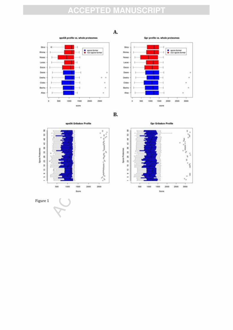
3.2. Profile analysis of sporulation genes in metagenomes
Profiles of Spo0A and Gpr were constructed and compared to metagenomic datasets to find
sequences of high similarity with spo0A and gpr. Profiles are models of conserved sequences built
from an alignment and are more sensitive than BLAST or other pair-wise comparisons especially
for protein searches [47]. The sequence profiles were generated based on 14 aligned sequences.
They were validated on genomes of known endospore-forming and non-sporulating bacteria
(Figure 1A). A single positive hit was found in the genome of each endospore-forming bacterium,
while no hits were found in the negative controls. This result also allowed determining a score
cut-off for Spo0A (2000) and Gpr (2500) profiles to distinguish between positive and negative
hits. Using this cut-off value one orthologous sequence of each of the two genes could be detected
in a further 59 genomes of endospore-forming bacteria (Figure 1B) reported in the genomic
databases of the Comprehensive Microbial Resource (CMR) and Integrated Microbial Genomes
(IMG) (Supplementary Table 1).

AC
CEPTED
MAN
USC
RIP
T
ACCEPTED MANUSCRIPT
The profile analysis was then used to detect Spo0A or Gpr in publicly available environmental
metagenomes. For this, 73 microbial metagenomic datasets (Supplementary Table 2) from a total
of 25 publications or direct submissions were retrieved. The datasets consisted of 6220494
sequences of average length of 957 bp and represented different environments, including marine,
fresh- and ground-waters, acid mine drainage, compost, hypersaline environments, hot springs,
soils, sludge, food and organism-associated environments (ant fungus garden, coral, fish and
human gut).
The profile analysis revealed only three sequences with a score above the cutoff of the Spo0A
profile in all metagenomic datasets (Figure 2A). All three metagenomes (AAQL, BAAY, BAAZ)
originated from human gut [48,49], in which Firmicutes are known to be one of the dominant
bacterial groups [50,51]. For the Gpr gene profile (Figure 2B), no sequences were found with a
similarity score above the cutoff value. These results are surprising considering that some of
these metagenomes were sampled in environments with high abundance of endospore-forming
Firmicutes (e.g. gut or soil; [52,53]). These results showed that these two genes from endospore-
forming Firmicutes are underrepresented in metagenomes. This had been alluded to earlier by
von Mering et al., [24], and is now confirmed here.
A methodological bias during the DNA extraction of resistant structures such as bacterial
endospores has been suggested as the origin of an underrepresentation of microbial groups
producing this structures [24]. Indeed, independently of the methodological approach taken (i.e.
whole genome shotgun analysis, activity- or sequence-driven screening), the first and most
crucial step in any metagenomic project is the extraction of nucleic acids. The isolated DNA
should be representative of all cells in the sample and of sufficient quality and amount for
subsequent sequencing [54]. Clearly, not all microbial species are equally amenable to the DNA
extraction methods used today [9,10], especially considering the diversity of morphological and
physiological states in which microbes can be found in environmental samples. Therefore,
complementary information, in particular concerning the method used for DNA extraction of the
metagenomes was thus considered. The described DNA extraction methods (Supplementary
Table 2) consisted of enzymatic or chemical protocols (18 datasets) or mechanical procedures of
cell lysis (8 datasets). Sequences associated to Firmicutes are reported for some of the analyzed
metagenome projects regardless of the DNA extraction protocol. For example, sequences of
AC
CEPTED
MAN
USC
RIP
T
ACCEPTED MANUSCRIPT
Clostridia (30 %) and Bacilli (1 %) were reported in the wallaby gut extracted enzymatically [55].
Also, in the compost metagenome extracted by bead beating, more than 13 % of sequences were
reported as members of endospore-formers Bacillus spp. or Paenibacillus spp. [56]. Our profile
analyses however, do not show positive hits for Spo0A or Gpr in either of these metagenomes.
Whether this is due to the extraction method applied, to the depth of sequencing or to other
specific bias is hard to establish.
We have developed a tailored DNA extraction method that allows a better assessment of the
abundance and diversity of endospore-formers in environmental samples for amplicon
sequencing [12,57]. Therefore, we next evaluated if using this extraction protocol in an
environmental sample could improve the detection of endospore-formers in a metagenome.
3.3. Amplicon sequencing of an environmental sample with high prevalence of endospore-
forming Firmicutes
We performed amplicon sequencing from a sample in which high prevalence of endospore-
forming Firmicutes was suspected from the ratio of 16S rRNA (bacterial) and spo0A (endospore-
formers) gene numbers measured by quantitative PCR [58]. This ratio was obtained from DNA
extracted using our modified protocol. Sequencing of the 16S rRNA and spo0A gene amplicons
was conducted and revealed not only a high prevalence of endospore-forming Firmicutes, but
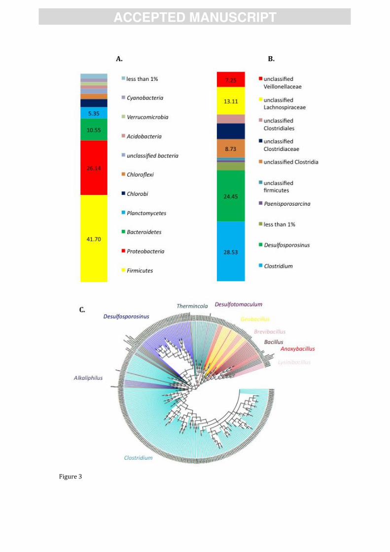
also a high diversity of endospore formers (Figure 3).
In the amplicon sequencing of the 16S rRNA gene, Firmicutes accounted for 41,70% of the
total bacterial community. The abundance of 16S rRNA amplicons corresponding to Firmicutes
was nearly double the amount of Proteobacteria, which was the second most abundant bacterial
Phylum (26.14%). Among the endospore-formers observed in the pyrosequencing results, the
genera Clostridium and Desulfosporosinus dominated the community in the sample, indicating a
clear dominance of anaerobic endospore-formers [59] as could be expected considering the
temperature and other environmental conditions at this geothermal spring. Amplicons affiliated
to Clostridium and Desulfosporosinus were also dominant in the Spo0A amplicon sequencing,
which also showed the dominance of anaerobic endospore-formers. Even though Spo0A
sequences related to aerobic endospore-formers (e.g. Geobacillus and Bacillus) were also
obtained, the classification of the Spo0A from aerobic endospore-formers was ambiguous as
AC
CEPTED
MAN
USC
RIP
T
ACCEPTED MANUSCRIPT
shown by the existence of, for example, clades related to Anoxybacillus but placed at different
positions in the phylogeny (Figure 3C). In fact, only recently environmental spo0A sequences
have started to be obtained [12], and the phylogenetic assignment needs to be refined.
3.3. Metagenomic sequencing
In addition to pyrosequencing, the same sample was also subjected to metagenomic
sequencing. It is worth mentioning that in whole-genome metagenomics a PCR amplification
biases does not apply and thus we did not necessarily expect to find the same groups or the same
frequency detected in the amplicon sequencing. However, the results of the qPCR quatification
and the amplicon sequencing were taken as an indication of the prevalence of Firmicutes in this
specific environmental sample. The NAP dataset consisted of a total of 481810 sequences of
average length of 330 bp. When the Spo0A and Gpr profile analysis were conducted on this
metagenome, none of the two genes were detected. However, looking only at two specific genes
could be an issue, since those could be, for various reasons, underrepresented in the sequences.
Therefore, an extended search for reads that could be assigned to Firmicutes using different
prediction tools on the assembled metagenome was also carried out.
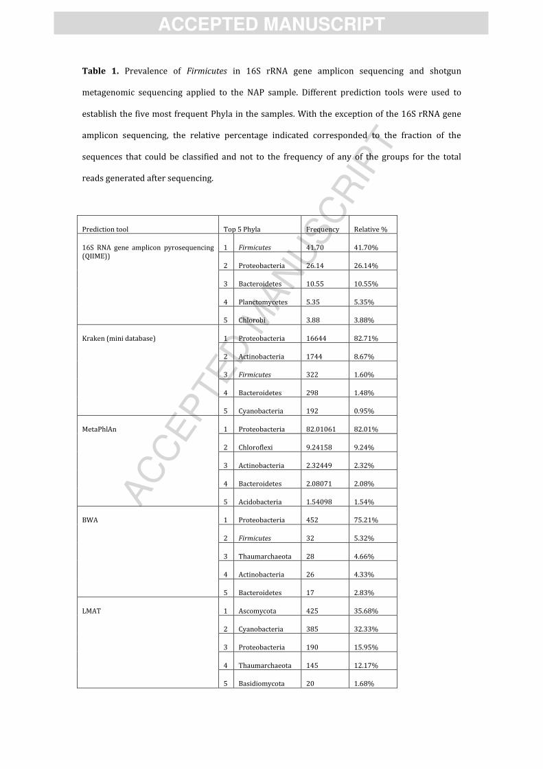
Relative abundances from classified reads were considered to establish the five most
prevalent phyla present in the sample (Table 1). Firmicutes appear in the top five Phyla only for
two of the four prediction tools used. In the case of Kraken, Firmicutes reads corresponded to
1.60% of the classified data, being the third most abundant phylum (the most abundant one was
Proteobacteria with 82.71%). BWA predicted 5.32% of the classified sequences as to belong to
Firmicutes (second most abundant phylum after Proteobacteria with 75.21%). Firmicutes were
not listed after classification with MetaPhlAN and LMAT. Likewise, when reconstruction of full
bacterial genomes was attempted for the NAP metagenome using MetaPhlAn, none of the top 5
microorganisms was assigned to Firmicutes (data not shown).
Thus, even though amplicon sequencing revealed a large fraction of the community as
belonging to Firmicutes, this was not observed in the shotgun metagenome. There are several
possible explanations for these results. One of those is the fact that the ribosomal (rrn) operon is
normally found in several copies and thus the representation of a microbial community based on
16S rRNA gene sequencing is skewed. Furthermore, the average number of rrn operon copies
AC
CEPTED
MAN
USC
RIP
T
ACCEPTED MANUSCRIPT
depends on the group of bacteria. An average value of 7.01 copies of 16S rRNA genes was found
for the phylum Firmicutes in the rrnDB [60], which implies that this group can be
overrepresented in 16S rRNA gene amplicon libraries. In addition, it should be noted that for all
the tools used, classification was poor and only a very small fraction of the sequences could be
actually assigned to a particular taxonomic group. Therefore, the lack of detection of Firmicutes
could be due to the current limitations of the analysis tools. In fact, recent sequencing
technologies generate such large quantities of data as to bring along a new set of challenges in
data analysis, the so-called bioinformatics bottleneck [61]. On the level of interpretation of
metagenomic data there is still an important amount of unexplored information available from
the results, simply because the advances in sequencing technologies are greater than the
complementary progress in annotation, data inventory and standardization of metadata [14].
AC
CEPTED
MAN
USC
RIP
T
ACCEPTED MANUSCRIPT
4. Conclusions
Since Staley and Konopka introduced the ╉great plate count anomaly╊ [62,63], revealing that
only a small fraction of the microbial community can be cultured in the laboratory, one of the
great challenges in environmental microbiology is the understanding of the diversity and
metabolic capabilities of microbes in a culture-independent manner. That bias was partly
overcome by moving into the direction of directly extracting genetic material from
environmental samples. However, our results reveal that for specific microbial groups, we are
still in a phase in which, similar to a percentage of the community being not culturable in culture-
based approaches, a fraction of the genomes of the community might be considered as not
detectable for culture-independent approaches. Nonetheless, profiling of the taxonomic and
phylogenetic composition of microbial communities is at the heart of many metagenomic studies,
and it is an obligatory step to draw conclusions on the role of microorganisms in the
environment based on metagenomics. Our results suggest that in the case of endospore-forming
Firmicutes, classification by various methods still lags behind. However, starting from samples
such as NAP, in which evidence for high frequency of this bacterial group exists, could be the first
step towards developing improved methods of classification and phylogenetic assignment of
metagenomic data.

AC
CEPTED
MAN
USC
RIP
T
ACCEPTED MANUSCRIPT
5. Acknowledgments
This work was supported by the Swiss National Science Foundation grant No. 31003A-
132358/1 and 31003A_152972, from Fondation Pierre Mercier pour la science and from
REGARD for equality of women in science.

AC
CEPTED
MAN
USC
RIP
T
ACCEPTED MANUSCRIPT
References
[1] Suenaga H. Targeted metagenomics: a high-resolution metagenomics approach for
specific gene clusters in complex microbial communities. Environ Microbiol 2012;14:13–22. doi:10.1111/j.1462-2920.2011.02438.x.
[2] Warnecke F, Hugenholtz P. Building on basic metagenomics with complementary
technologies. Genome Biol 2007;8:231. doi:10.1186/gb-2007-8-12-231.
[3] Xu J. Microbial ecology in the age of genomics and metagenomics: concepts, tools, and
recent advances. Mol Ecol 2006;15:1713–31. doi:10.1111/j.1365-294X.2006.02882.x.
[4] Béjà O, Suzuki MT, Heidelberg JF, Nelson WC, Preston CM, Hamada T, et al. Unsuspected
diversity among marine aerobic anoxygenic phototrophs. Nature 2002;415:630–3.
doi:10.1038/415630a.
[5] Béjà O, Aravind L, Koonin EV, Suzuki MT, Hadd A, Nguyen LP, et al. Bacterial rhodopsin:
evidence for a new type of phototrophy in the sea. Science 2000;289:1902–6.
[6] Ram RJ, Verberkmoes NC, Thelen MP, Tyson GW, Baker BJ, Blake RC, et al. Community
proteomics of a natural microbial biofilm. Science 2005;308:1915–20.
doi:10.1126/science. 1109070.
[7] Venter JC, Remington K, Heidelberg JF, Halpern AL, Rusch D, Eisen JA, et al.
Environmental genome shotgun sequencing of the Sargasso Sea. Science 2004;304:66–74. doi:10.1126/science.1093857.
[8] Voget S, Leggewie C, Uesbeck A, Raasch C, Jaeger K-E, Streit WR. Prospecting for novel
biocatalysts in a soil metagenome. Appl Environ Microbiol 2003;69:6235–42.
[9] Delmont TO, Robe P, Cecillon S, Clark IM, Constancias F, Simonet P, et al. Accessing the
soil metagenome for studies of microbial diversity. Appl Environ Microbiol
2011;77:1315–24. doi:10.1128/AEM.01526-10.
[10] Lombard N, Prestat E, van Elsas JD, Simonet P. Soil-specific limitations for access and
analysis of soil microbial communities by metagenomics. FEMS Microbiol Ecol
2011;78:31–49. doi:10.1111/j.1574-6941.2011.01140.x.
[11] Pinard R, de Winter A, Sarkis GJ, Gerstein MB, Tartaro KR, Plant RN, et al. Assessment of
whole genome amplification-induced bias through high-throughput, massively parallel
whole genome sequencing. BMC Genomics 2006;7:216. doi:10.1186/1471-2164-7-216.
[12] Wunderlin T, Junier T, Roussel-Delif L, Jeanneret N, Junier P. Stage 0 sporulation gene A
as a molecular marker to study diversity of endospore-forming Firmicutes. Environ
Microbiol Rep 2013;5:911–24. doi:10.1111/1758-2229.12094.
[13] Batmalle CS, Chiang H-I, Zhang K, Lomas MW, Martiny AC. Development and Bias
Assessment of a Method for Targeted Metagenomic Sequencing of Marine Cyanobacteria.
Appl Environ Microbiol 2014;80:1116–25. doi:10.1128/AEM.02834-13.
[14] Yilmaz P, Gilbert JA, Knight R, Amaral-Zettler L, Karsch-Mizrachi I, Cochrane G, et al. The
genomic standards consortium: bringing standards to life for microbial ecology. ISME J
2011;5:1565–7. doi:10.1038/ismej.2011.39.
[15] Lagier J-C, Armougom F, Million M, Hugon P, Pagnier I, Robert C, et al. Microbial
culturomics: paradigm shift in the human gut microbiome study. Clin Microbiol Infect
2012;18:1185–93. doi:10.1111/1469-0691.12023.
[16] Lagier J-C, Hugon P, Khelaifia S, Fournier P-E, Scola BL, Raoult D. The Rebirth of Culture
in Microbiology through the Example of Culturomics To Study Human Gut Microbiota.
Clin Microbiol Rev 2015;28:237–64. doi:10.1128/CMR.00014-14.
[17] Hugenholtz P. Exploring prokaryotic diversity in the genomic era. Genome Biol
2002;3:reviews0003.1–reviews0003.8.
[18] Nicholson WL, Munakata N, Horneck G, Melosh HJ, Setlow P. Resistance of Bacillus
endospores to extreme terrestrial and extraterrestrial environments. Microbiol Mol Biol
Rev MMBR 2000;64:548–72.
[19] Nicholson WL. Roles of Bacillus endospores in the environment. Cell Mol Life Sci CMLS
2002;59:410–6.
[20] Driks A. Overview: Development in bacteria: spore formation in Bacillus subtilis. Cell Mol
Life Sci CMLS 2002;59:389–91.
[21] Onyenwoke RU, Brill JA, Farahi K, Wiegel J. Sporulation genes in members of the low G+C
Gram-type-positive phylogenetic branch ( Firmicutes). Arch Microbiol 2004;182:182–92.
doi:10.1007/s00203-004-0696-y.
[22] Yudkin MD, Clarkson J. Differential gene expression in genetically identical sister cells:

AC
CEPTED
MAN
USC
RIP
T
ACCEPTED MANUSCRIPT
the initiation of sporulation in Bacillus subtilis. Mol Microbiol 2005;56:578–89.
doi:10.1111/j.1365-2958.2005.04594.x.
[23] Martiny JBH, Bohannan BJM, Brown JH, Colwell RK, Fuhrman JA, Green JL, et al. Microbial
biogeography: putting microorganisms on the map. Nat Rev Microbiol 2006;4:102–12.
doi:10.1038/nrmicro1341.
[24] Von Mering C, Hugenholtz P, Raes J, Tringe SG, Doerks T, Jensen LJ, et al. Quantitative
phylogenetic assessment of microbial communities in diverse environments. Science
2007;315:1126–30. doi:10.1126/science.1133420.
[25] Wunderlin T, Junier T, Roussel-Delif L, Jeanneret N, Junier P. Endospore-enriched
sequencing approach reveals unprecedented diversity of Firmicutes in sediments.
Environ Microbiol Rep 2014;6:631–9. doi:10.1111/1758-2229.12179.
[26] Altschul SF, Madden TL, Schäffer AA, Zhang J, Zhang Z, Miller W, et al. Gapped BLAST and
PSI-BLAST: a new generation of protein database search programs. Nucleic Acids Res
1997;25:3389–402.
[27] Kriventseva EV, Rahman N, Espinosa O, Zdobnov EM. OrthoDB: the hierarchical catalog
of eukaryotic orthologs. Nucleic Acids Res 2008;36:D271–5. doi:10.1093/nar/gkm845.
[28] Katoh K, Misawa K, Kuma K, Miyata T. MAFFT: a novel method for rapid multiple
sequence alignment based on fast Fourier transform. Nucleic Acids Res 2002;30:3059–66.
[29] Rice P, Longden I, Bleasby A. EMBOSS: the European Molecular Biology Open Software
Suite. Trends Genet TIG 2000;16:276–7.
[30] Bueche M, Wunderlin T, Roussel-Delif L, Junier T, Sauvain L, Jeanneret N, et al.
Quantification of endospore-forming firmicutes by quantitative PCR with the functional
gene spo0A. Appl Environ Microbiol 2013;79:5302–12. doi:10.1128/AEM.01376-13.
[31] Li H, Zhang Y, Li D, Xu H, Chen G, Zhang C. Comparisons of different hypervariable
regions of rrs genes for fingerprinting of microbial communities in paddy soils. Soil Biol
Biochem 2009;41:954–68. doi:10.1016/j.soilbio.2008.10.030.
[32] Caporaso JG, Kuczynski J, Stombaugh J, Bittinger K, Bushman FD, Costello EK, et al. QIIME
allows analysis of high-throughput community sequencing data. Nat Methods
2010;7:335–6. doi:10.1038/nmeth.f.303.
[33] Ewing B, Green P. Base-calling of automated sequencer traces using phred. II. Error
probabilities. Genome Res 1998;8:186–94.
[34] Guindon S, Gascuel O. A simple, fast, and accurate algorithm to estimate large
phylogenies by maximum likelihood. Syst Biol 2003;52:696–704.
[35] Mulder NJ, Apweiler R, Attwood TK, Bairoch A, Bateman A, Binns D, et al. InterPro: an
integrated documentation resource for protein families, domains and functional sites.
Brief Bioinform 2002;3:225–35.
[36] Freitas TAK, Li P-E, Scholz MB, Chain PSG. Accurate read-based metagenome
characterization using a hierarchical suite of unique signatures. Nucleic Acids Res
2015:gkv180. doi:10.1093/nar/gkv180.
[37] Li H, Durbin R. Fast and accurate short read alignment with Burrows-Wheeler transform.
Bioinforma Oxf Engl 2009;25:1754–60. doi:10.1093/bioinformatics/btp324.
[38] Segata N, Waldron L, Ballarini A, Narasimhan V, Jousson O, Huttenhower C. Metagenomic
microbial community profiling using unique clade-specific marker genes. Nat Methods
2012;9:811–4. doi:10.1038/nmeth.2066.
[39] Langmead B, Salzberg SL. Fast gapped-read alignment with Bowtie 2. Nat Methods
2012;9:357–9. doi:10.1038/nmeth.1923.
[40] Pruitt KD, Brown GR, Hiatt SM, Thibaud-Nissen F, Astashyn A, Ermolaeva O, et al. RefSeq:
an update on mammalian reference sequences. Nucleic Acids Res 2014;42:D756–63.
doi:10.1093/nar/gkt1114.
[41] Sunagawa S, Mende DR, Zeller G, Izquierdo-Carrasco F, Berger SA, Kultima JR, et al.
Metagenomic species profiling using universal phylogenetic marker genes. Nat Methods
2013;10:1196–9. doi:10.1038/nmeth.2693.
[42] Ames SK, Hysom DA, Gardner SN, Lloyd GS, Gokhale MB, Allen JE. Scalable metagenomic
taxonomy classification using a reference genome database. Bioinforma Oxf Engl
2013;29:2253–60. doi:10.1093/bioinformatics/btt389.
[43] Stragier P, Losick R. Molecular genetics of sporulation in Bacillus subtilis. Annu Rev
Genet 1996;30:297–241. doi:10.1146/annurev.genet.30.1.297.
[44] Abecasis AB, Serrano M, Alves R, Quintais L, Pereira-Leal JB, Henriques AO. A genomic
AC
CEPTED
MAN
USC
RIP
T
ACCEPTED MANUSCRIPT
signature and the identification of new sporulation genes. J Bacteriol 2013;195:2101–15.
doi:10.1128/JB.02110-12.
[45] Traag BA, Pugliese A, Eisen JA, Losick R. Gene conservation among endospore-forming
bacteria reveals additional sporulation genes in Bacillus subtilis. J Bacteriol
2013;195:253–60. doi:10.1128/JB.01778-12.
[46] Galperin MY, Mekhedov SL, Puigbo P, Smirnov S, Wolf YI, Rigden DJ. Genomic
determinants of sporulation in Bacilli and Clostridia: towards the minimal set of
sporulation-specific genes. Environ Microbiol 2012;14:2870–90. doi:10.1111/j.1462-
2920.2012.02841.x.
[47] Gribskov M, McLachlan AD, Eisenberg D. Profile analysis: detection of distantly related
proteins. Proc Natl Acad Sci U S A 1987;84:4355–8.
[48] Gill SR, Pop M, Deboy RT, Eckburg PB, Turnbaugh PJ, Samuel BS, et al. Metagenomic
analysis of the human distal gut microbiome. Science 2006;312:1355–9.
doi:10.1126/science.1124234.
[49] Kurokawa K, Itoh T, Kuwahara T, Oshima K, Toh H, Toyoda A, et al. Comparative
metagenomics revealed commonly enriched gene sets in human gut microbiomes. DNA
Res Int J Rapid Publ Rep Genes Genomes 2007;14:169–81. doi:10.1093/dnares/dsm018.
[50] Zoetendal EG, Vaughan EE, De Vos WM. A microbial world within us. Mol Microbiol
2006;59:1639–50. doi:10.1111/j.1365-2958.2006.05056.x.
[51] Suzuki TA, Worobey M. Geographical variation of human gut microbial composition. Biol
Lett 2014;10:20131037. doi:10.1098/rsbl.2013.1037.
[52] Felske ADM, Tzeneva V, Heyrman J, Langeveld MA, Akkermans ADL, De Vos P. Isolation
and biodiversity of hitherto undescribed soil bacteria related to Bacillus niacini. Microb
Ecol 2004;48:111–9. doi:10.1007/s00248-003-2025-4.
[53] Hoyles L, Honda H, Logan NA, Halket G, La Ragione RM, McCartney AL. Recognition of
greater diversity of Bacillus species and related bacteria in human faeces. Res Microbiol
2012;163:3–13. doi:10.1016/j.resmic.2011.10.004.
[54] Thomas T, Gilbert J, Meyer F. Metagenomics - a guide from sampling to data analysis.
Microb Inform Exp 2012;2:3. doi:10.1186/2042-5783-2-3.
[55] Pope PB, Denman SE, Jones M, Tringe SG, Barry K, Malfatti SA, et al. Adaptation to
herbivory by the Tammar wallaby includes bacterial and glycoside hydrolase profiles
different from other herbivores. Proc Natl Acad Sci U S A 2010;107:14793–8.
doi:10.1073/pnas.1005297107.
[56] Allgaier M, Reddy A, Park J), )vanova N, D’haeseleer P, Lowry S, et al. Targeted discovery
of glycoside hydrolases from a switchgrass-adapted compost community. PloS One
2010;5:e8812. doi:10.1371/journal.pone.0008812.
[57] Wunderlin T, Junier T, Roussel-Delif L, Junier P. Profile analyses of sporulation genes
reveal underrepresentation of endospore- forming bacteria in metagenomes. ISME14,
Copenhagen, Denmark: 2012.
[58] Filippidou S, Bueche M, Wunderlin T, Junier T, Roussel-Delif L, Jeanneret N, et al. Survival
strategy meets classic ecological theory: the case of diversity and abundance of
endospore-forming Firmicutes in extreme environments. Prep n.d.
[59] Schleifer KH. Classification of Bacteria and Archaea: past, present and future. Syst Appl
Microbiol 2009;32:533–42. doi:10.1016/j.syapm.2009.09.002.
[60] Lee ZM-P, Bussema C, Schmidt TM. rrnDB: documenting the number of rRNA and tRNA
genes in bacteria and archaea. Nucleic Acids Res 2009;37:D489–93.
doi:10.1093/nar/gkn689.
[61] Scholz MB, Lo C-C, Chain PSG. Next generation sequencing and bioinformatic bottlenecks:
the current state of metagenomic data analysis. Curr Opin Biotechnol 2012;23:9–15.
doi:10.1016/j.copbio.2011.11.013.
[62] Staley JT, Konopka A. Measurement of in situ activities of nonphotosynthetic
microorganisms in aquatic and terrestrial habitats. Annu Rev Microbiol 1985;39:321–46.
doi:10.1146/annurev.mi.39.100185.001541.
[63] Amann RI, Ludwig W, Schleifer KH. Phylogenetic identification and in situ detection of
individual microbial cells without cultivation. Microbiol Rev 1995;59:143–69.

AC
CEPTED
MAN
USC
RIP
T
ACCEPTED MANUSCRIPT
Figure legends
Figure 1. A. Validation of the profiles created for the genes spo0A and gpr compared to a
selection of genomes of endospore-forming Firmicutes (blue bars) and non spore-forming
genomes (red bars). In endospore-forming Firmicutes a single hit with a score above 2000
(spo0A) and 2500 (gpr) distinguish between positive and negative hits. Strco= Streptomyces
coelicolor; Rhime= Rhizobium melliloti; Nosaz: Nostoc azollae; Lacac= Lactobacillus acidophilus;
Escco= Escherechia coli; Desre= Desulfotomaculum reducens; Desha= Desulfitobacterium
hafniense; Clobo= Clostridium botulinum; Bacha= Bacillus halodurans; Aliac= Alicyclobacillus
acidocaldarius. B. The same analysis was repeated using all 59 endospore-forming genomes
retrieved from IMG and CMR databases (see supplementary Table 1).
Figure 2. Profile similarity hits for Spo0A and Gpr protein profiles in metagenomes from
different origins. The color code identifying different environments is presented under the
results. The genomes included in profile testing (see Figure 1A) were also included in the
analysis and are presented in white (endospore-formers) and grey (non-spore formers).
Figure 3. Analysis of pyrosequencing results obtained from 16S rRNA gene and spo0A
amplicons, from an environmental sample with high prevalence of endospore-forming Firmicutes
(Nea Apollonia, NAP). (A) Total 16S rRNA gene community composition to the phylum level. (B)
Firmicute fraction of the total community (16S rRNA gene) to the genus level. (C). Cladogram
representing the community composition of Firmicutes using the spo0A gene. Sequences color
coded by genus.
AC
CEPTED
MAN
USC
RIP
T
ACCEPTED MANUSCRIPT
Table 1. Prevalence of Firmicutes in 16S rRNA gene amplicon sequencing and shotgun
metagenomic sequencing applied to the NAP sample. Different prediction tools were used to
establish the five most frequent Phyla in the samples. With the exception of the 16S rRNA gene
amplicon sequencing, the relative percentage indicated corresponded to the fraction of the
sequences that could be classified and not to the frequency of any of the groups for the total
reads generated after sequencing.
Prediction tool Top 5 Phyla Frequency Relative %
16S RNA gene amplicon pyrosequencing
(QIIME))
1 Firmicutes 41.70 41.70%
2 Proteobacteria 26.14 26.14%
3 Bacteroidetes 10.55 10.55%
4 Planctomycetes 5.35 5.35%
5 Chlorobi 3.88 3.88%
Kraken (mini database)
1 Proteobacteria 16644 82.71%
2 Actinobacteria 1744 8.67%
3 Firmicutes 322 1.60%
4 Bacteroidetes 298 1.48%
5 Cyanobacteria 192 0.95%
MetaPhlAn
1 Proteobacteria 82.01061 82.01%
2 Chloroflexi 9.24158 9.24%
3 Actinobacteria 2.32449 2.32%
4 Bacteroidetes 2.08071 2.08%
5 Acidobacteria 1.54098 1.54%
BWA
1 Proteobacteria 452 75.21%
2 Firmicutes 32 5.32%
3 Thaumarchaeota 28 4.66%
4 Actinobacteria 26 4.33%
5 Bacteroidetes 17 2.83%
LMAT
1 Ascomycota 425 35.68%
2 Cyanobacteria 385 32.33%
3 Proteobacteria 190 15.95%
4 Thaumarchaeota 145 12.17%
5 Basidiomycota 20 1.68%

AC
CEPTED
MAN
USC
RIP
T
ACCEPTED MANUSCRIPT
AC
CEPTED
MAN
USC
RIP
T
ACCEPTED MANUSCRIPT
Figure 1

AC
CEPTED
MAN
USC
RIP
T
ACCEPTED MANUSCRIPT
Figure 2
AC
CEPTED
MAN
USC
RIP
T
ACCEPTED MANUSCRIPT
Figure 3
Copyright © 2022 FDOKUMEN