A roadmap for metagenomic enzyme discovery - RSC ...
30
A roadmap for metagenomic enzyme discovery Serina L. Robinson, * J ¨ orn Piel and Shinichi Sunagawa Covering: up to 2021 Metagenomics has yielded massive amounts of sequencing data offering a glimpse into the biosynthetic potential of the uncultivated microbial majority. While genome-resolved information about microbial communities from nearly every environment on earth is now available, the ability to accurately predict biocatalytic functions directly from sequencing data remains challenging. Compared to primary metabolic pathways, enzymes involved in secondary metabolism often catalyze specialized reactions with diverse substrates, making these pathways rich resources for the discovery of new enzymology. To date, functional insights gained from studies on environmental DNA (eDNA) have largely relied on PCR- or activity-based screening of eDNA fragments cloned in fosmid or cosmid libraries. As an alternative, shotgun metagenomics holds underexplored potential for the discovery of new enzymes directly from eDNA by avoiding common biases introduced through PCR- or activity-guided functional metagenomics workflows. However, inferring new enzyme functions directly from eDNA is similar to searching for a ‘needle in a haystack’ without direct links between genotype and phenotype. The goal of this review is to provide a roadmap to navigate shotgun metagenomic sequencing data and identify new candidate biosynthetic enzymes. We cover both computational and experimental strategies to mine metagenomes and explore protein sequence space with a spotlight on natural product biosynthesis. Specifically, we compare in silico methods for enzyme discovery including phylogenetics, sequence similarity networks, genomic context, 3D structure-based approaches, and machine learning techniques. We also discuss various experimental strategies to test computational predictions including heterologous expression and screening. Finally, we provide an outlook for future directions in the field with an emphasis on meta- omics, single-cell genomics, cell-free expression systems, and sequence-independent methods. 1. Introduction 1.1. The sequence–structure–function paradigm 1.2. Metagenomics: promises and perils 1.3. Denitions for enzyme discovery 1.4. Caveats and assumptions 2. Setting course: experimental design for metagenomics studies 2.1. Activity-guided functional metagenomics 2.2. PCR-based functional metagenomics 2.3. Shotgun metagenomic sequencing 2.4. Parallels with natural product research 2.5. Hotbeds for enzyme discovery 3. On the road: computational methods for enzyme function prediction 3.1. Querying metagenomic databases 3.2. Phylogenetics 3.3. Sequence similarity networking 3.4. Gene context and interactions 3.5. 3D-structure based methods 3.6. Motifs and active site residues 3.7. Machine learning 4. Reaching the destination: characterizing new enzymes 4.1. Cloning and heterologous expression 4.2. Heterologous expression 4.3. Screening for enzyme activity 5. Scenic drives: a case study on marine metagenomics 5.1. Global ocean microbiomics 5.2. Microbiomes of marine invertebrates 6. Gearing up for the future: new frontiers in enzyme discovery 6.1. Meta-omics 6.2. Single-cell genomics 6.3. Microuidics 6.4. Cell-free platforms 6.5. Sequence-independent methods 7. Conclusions Eidgen¨ ossische Technische Hochschule (ETH), Z¨ urich, Switzerland. E-mail: [email protected] Cite this: Nat. Prod. Rep., 2021, 38, 1994 Received 31st January 2021 DOI: 10.1039/d1np00006c rsc.li/npr 1994 | Nat. Prod. Rep., 2021, 38, 1994–2023 This journal is © The Royal Society of Chemistry 2021 Natural Product Reports REVIEW Open Access Article. Published on 12 April 2021. Downloaded on 1/23/2022 10:49:48 AM. This article is licensed under a Creative Commons Attribution-NonCommercial 3.0 Unported Licence. View Article Online View Journal | View Issue
-
Upload
khangminh22 -
Category
Documents
-
view
0 -
download
0
Transcript of A roadmap for metagenomic enzyme discovery - RSC ...

Natural ProductReports
REVIEW
Ope
n A
cces
s A
rtic
le. P
ublis
hed
on 1
2 A
pril
2021
. Dow
nloa
ded
on 1
/23/
2022
10:
49:4
8 A
M.
Thi
s ar
ticle
is li
cens
ed u
nder
a C
reat
ive
Com
mon
s A
ttrib
utio
n-N
onC
omm
erci
al 3
.0 U
npor
ted
Lic
ence
.
View Article OnlineView Journal | View Issue
A roadmap for m
Eidgenossische Technische Hochschule
Cite this: Nat. Prod. Rep., 2021, 38,1994
Received 31st January 2021
DOI: 10.1039/d1np00006c
rsc.li/npr
1994 | Nat. Prod. Rep., 2021, 38, 199
etagenomic enzyme discovery
Serina L. Robinson, * Jorn Piel and Shinichi Sunagawa
Covering: up to 2021
Metagenomics has yielded massive amounts of sequencing data offering a glimpse into the biosynthetic
potential of the uncultivated microbial majority. While genome-resolved information about microbial
communities from nearly every environment on earth is now available, the ability to accurately predict
biocatalytic functions directly from sequencing data remains challenging. Compared to primary
metabolic pathways, enzymes involved in secondary metabolism often catalyze specialized reactions
with diverse substrates, making these pathways rich resources for the discovery of new enzymology. To
date, functional insights gained from studies on environmental DNA (eDNA) have largely relied on PCR-
or activity-based screening of eDNA fragments cloned in fosmid or cosmid libraries. As an alternative,
shotgun metagenomics holds underexplored potential for the discovery of new enzymes directly from
eDNA by avoiding common biases introduced through PCR- or activity-guided functional metagenomics
workflows. However, inferring new enzyme functions directly from eDNA is similar to searching for
a ‘needle in a haystack’ without direct links between genotype and phenotype. The goal of this review is
to provide a roadmap to navigate shotgun metagenomic sequencing data and identify new candidate
biosynthetic enzymes. We cover both computational and experimental strategies to mine metagenomes
and explore protein sequence space with a spotlight on natural product biosynthesis. Specifically, we
compare in silico methods for enzyme discovery including phylogenetics, sequence similarity networks,
genomic context, 3D structure-based approaches, and machine learning techniques. We also discuss
various experimental strategies to test computational predictions including heterologous expression and
screening. Finally, we provide an outlook for future directions in the field with an emphasis on meta-
omics, single-cell genomics, cell-free expression systems, and sequence-independent methods.
1. Introduction1.1. The sequence–structure–function paradigm1.2. Metagenomics: promises and perils1.3. Denitions for enzyme discovery1.4. Caveats and assumptions2. Setting course: experimental design for metagenomics
studies2.1. Activity-guided functional metagenomics2.2. PCR-based functional metagenomics2.3. Shotgun metagenomic sequencing2.4. Parallels with natural product research2.5. Hotbeds for enzyme discovery3. On the road: computational methods for enzyme function
prediction3.1. Querying metagenomic databases3.2. Phylogenetics3.3. Sequence similarity networking
(ETH), Zurich, Switzerland. E-mail:
4–2023
3.4. Gene context and interactions3.5. 3D-structure based methods3.6. Motifs and active site residues3.7. Machine learning4. Reaching the destination: characterizing new enzymes4.1. Cloning and heterologous expression4.2. Heterologous expression4.3. Screening for enzyme activity5. Scenic drives: a case study on marine metagenomics5.1. Global ocean microbiomics5.2. Microbiomes of marine invertebrates6. Gearing up for the future: new frontiers in enzyme
discovery6.1. Meta-omics6.2. Single-cell genomics6.3. Microuidics6.4. Cell-free platforms6.5. Sequence-independent methods7. Conclusions
This journal is © The Royal Society of Chemistry 2021
Review Natural Product Reports
Ope
n A
cces
s A
rtic
le. P
ublis
hed
on 1
2 A
pril
2021
. Dow
nloa
ded
on 1
/23/
2022
10:
49:4
8 A
M.
Thi
s ar
ticle
is li
cens
ed u
nder
a C
reat
ive
Com
mon
s A
ttrib
utio
n-N
onC
omm
erci
al 3
.0 U
npor
ted
Lic
ence
.View Article Online
7.1. Discoveries oen occur at the boundaries of proteinfamilies
7.2. Think outside the colorimetric assay box to move intounexplored protein space
7.3. Move beyond E. coli into new hosts7.4. (Genome) context is everything8. Conicts of interest9. Acknowledgements10. References
1. Introduction1.1. The sequence–structure–function paradigm
The 1972 Nobel laureate in Chemistry, Christian Annsen,ended his Nobel lecture with the line, “It is certain that majoradvances in the understanding of cellular organization.willoccur when we can predict, in advance, the three-dimensional,
Serina Robinson is an ETHZurich postdoctoral fellow withDr Jorn Piel and will start herindependent career as a tenure-track group leader at the SwissFederal Institute of AquaticScience and Technology (Eawag)in autumn 2021. She obtainedher PhD in Microbiology and MScin Bioinformatics and Computa-tional Biology from the Universityof Minnesota, Minneapolis, USA(advisor: Larry Wackett), where
she applied machine learning and genome mining techniques toinvestigate b-lactone synthetases, a newly-discovered family ofenzymes involved in natural product biosynthesis. Her currentresearch focuses on the discovery of new biosynthetic enzymes frommarine, freshwater, and wastewater metagenomes.
Jorn Piel studied Chemistry at theUniversity of Bonn, Germany,and obtained a PhD in 1998(advisor: Wilhelm Boland). Aera postdoc with Bradley S. Mooreand Heinz G. Floss he becamegroup leader at the Max PlanckInstitute for Chemical Ecology inJena, Germany, in 2000. From2004–2013 he was associateprofessor at the University ofBonn and subsequently fullprofessor at the Institute of
Microbiology, ETH Zurich. His lab works at the interface ofChemistry and Biology, studying bacterial metabolism with anemphasis on microbial and biosynthetic ‘dark matter’, symbiosis,marine natural products, biosynthetic engineering, and chemicalecology.
This journal is © The Royal Society of Chemistry 2021
phenotypic consequences of a genetic message”. Nearly 5decades later, predicting the phenotypic consequences ofprotein sequences remains a complex task. Signicant progresshas been made on the three-dimensional prediction front,however. In 2020, the deep learning algorithm AlphaFold2achieved landmark results for the prediction of 3D proteinstructure from primary sequence. In a rigorous blinded globalcompetition, AlphaFold2 averaged within 1.6 A of the truth,achieving an error less than the width of one atom.1 To thisnews, Frances Arnold, 2018 Nobel laureate in Chemistry, reac-ted with, “Pretty impressive! Perhaps we can now move to theprotein function problem?”.
While accurate predictions for the 3D structures of manyproteins from primary sequence are now within our grasp,understanding function from protein structure or sequence isfar from solved. Even for Escherichia coli, one of the most well-characterized organisms on earth, >35% of genes lack experi-mental evidence of function.2 Moreover, the pan-genome, thatis, the complete set of genes found among all strains of E. coli isestimated to contain >16 000 different families of homologousgenes.3 By these estimates, E. coli is still considered to have anopen pan-genome since the species is undergoing constantgene acquisition and diversication.4 Our limited under-standing of one of the world's most intensively-studied modelorganisms5 emphasizes the challenge in determining thefunctions of coding sequences not from organisms grown inmonoculture in the laboratory but from metagenomic DNAfrom complex environments.
1.2. Metagenomics: promises and perils
Metagenomics, a term rst coined in 1998,6 refers to the studyof environmental DNA (eDNA). This is not only limited tonatural environments in the classical sense, but to essentiallyevery sampling location conceivable, including the hindguts oftermites,7 cheese rinds,8 and the International Space Station.9
Enabled by next-generation sequencing technologies,
Shinichi Sunagawa studiedBiochemistry and Marine Ecologyin Germany, and obtained hisPhD in 2010 at the University ofCalifornia, Merced, USA. Aerreturning to Germany, he joinedthe European Molecular BiologyLaboratory in Heidelberg asa postdoctoral fellow, andcontinued to work on ocean andhuman gut microbial communi-ties as a research- and staffscientist. In 2016, he established
the Microbiome Research Laboratory at the Institute of Microbi-ology at ETH Zurich, which combines bioinformatic and experi-mental approaches to integrate quantitative ‘meta-omics’ readoutswith contextual information to study and the role of environmentalmicroorganisms and mechanisms of host-microbial homeostasis.
Nat. Prod. Rep., 2021, 38, 1994–2023 | 1995
Natural Product Reports Review
Ope
n A
cces
s A
rtic
le. P
ublis
hed
on 1
2 A
pril
2021
. Dow
nloa
ded
on 1
/23/
2022
10:
49:4
8 A
M.
Thi
s ar
ticle
is li
cens
ed u
nder
a C
reat
ive
Com
mon
s A
ttrib
utio
n-N
onC
omm
erci
al 3
.0 U
npor
ted
Lic
ence
.View Article Online
metagenomics quickly became a new scientic eld in its ownright, contributing to exponential growth in the size ofsequencing repositories. In 2007, still relatively early years formetagenomics, a single study – the Global Ocean SamplingExpedition – nearly doubled the total number of proteinsequences in public databases.10 The rate of increase in next-generation sequencing has far surpassed Moore's law andnumber of nucleotide base pairs (bp) in public repositories isestimated to reach exabase-scale (1018 bp) well within the nextve years.11
One of the major advantages of metagenomics is gainingaccess to genetic information about the uncultivated majority ofmicrobes which still largely lack functional characterization.12
Metagenomics studies have reshaped our view of the tree oflife13,14 and led to the identication of deeply rooted andmetabolically-diverse lineages such as the DPANN archaea15 andcandidate phyla radiation.16 Many uncultivatedmicrobial phyla,including ‘Candidatus Tectomicrobia’,17 ‘Eelbacter’18 and‘Angelobacter’18 have had remarkable biosynthetic potentialrevealed by metagenomics. In the case of ‘Ca. Tectomicrobia,’heterologous expression enabled the experimental character-ization of new biosynthetic pathways and products.17,19–21
However, the tantalizing promises of discovering new enzy-mology from metagenomes goes hand-in-hand with the chal-lenges discussed in Section 4.2 of working with DNA fromorganisms that have eluded laboratory cultivation.
In this review, we aim to provide a bird's-eye view of tools andstrategies for metagenomic enzyme discovery. We emphasizeenzymes involved in natural product biosynthesis, but manyproteins outside of biosynthetic contexts will also be discussedas examples for relevant discovery strategies. We will also covera number of examples from microbial isolates and highlighttechniques which may be useful in future metagenome miningefforts.
1.3. Denitions for enzyme discovery
Before diving into methods, we will rst attempt to denemetagenomic enzyme discovery. The simplest denition –
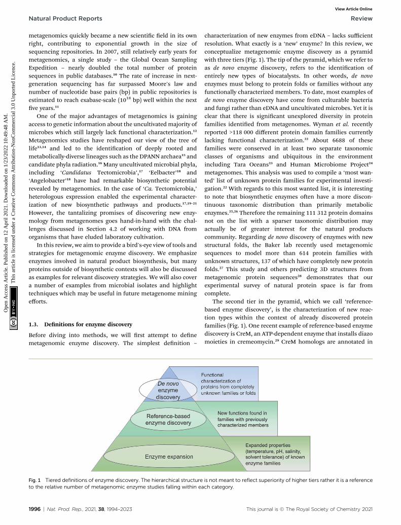
Fig. 1 Tiered definitions of enzyme discovery. The hierarchical structureto the relative number of metagenomic enzyme studies falling within ea
1996 | Nat. Prod. Rep., 2021, 38, 1994–2023
characterization of new enzymes from eDNA – lacks sufficientresolution. What exactly is a ‘new’ enzyme? In this review, weconceptualize metagenomic enzyme discovery as a pyramidwith three tiers (Fig. 1). The tip of the pyramid, which we refer toas de novo enzyme discovery, refers to the identication ofentirely new types of biocatalysts. In other words, de novoenzymes must belong to protein folds or families without anyfunctionally characterized members. To date, most examples ofde novo enzyme discovery have come from culturable bacteriaand fungi rather than eDNA and uncultivated microbes. Yet it isclear that there is signicant unexplored diversity in proteinfamilies identied from metagenomes. Wyman et al. recentlyreported >118 000 different protein domain families currentlylacking functional characterization.22 About 6688 of thesefamilies were conserved in at least two separate taxonomicclasses of organisms and ubiquitous in the environmentincluding Tara Oceans23 and Human Microbiome Project24
metagenomes. This analysis was used to compile a ‘most wan-ted’ list of unknown protein families for experimental investi-gation.22 With regards to this most wanted list, it is interestingto note that biosynthetic enzymes oen have a more discon-tinuous taxonomic distribution than primarily metabolicenzymes.25,26 Therefore the remaining 111 312 protein domainsnot on the list with a sparser taxonomic distribution mayactually be of greater interest for the natural productscommunity. Regarding de novo discovery of enzymes with newstructural folds, the Baker lab recently used metagenomicsequences to model more than 614 protein families withunknown structures, 137 of which have completely new proteinfolds.27 This study and others predicting 3D structures frommetagenomic protein sequences28 demonstrates that ourexperimental survey of natural protein space is far fromcomplete.
The second tier in the pyramid, which we call ‘reference-based enzyme discovery’, is the characterization of new reac-tion types within the context of already discovered proteinfamilies (Fig. 1). One recent example of reference-based enzymediscovery is CreM, an ATP-dependent enzyme that installs diazomoieties in cremeomycin.29 CreM homologs are annotated in
is not meant to reflect superiority of higher tiers rather it is a referencech category.
This journal is © The Royal Society of Chemistry 2021
Review Natural Product Reports
Ope
n A
cces
s A
rtic
le. P
ublis
hed
on 1
2 A
pril
2021
. Dow
nloa
ded
on 1
/23/
2022
10:
49:4
8 A
M.
Thi
s ar
ticle
is li
cens
ed u
nder
a C
reat
ive
Com
mon
s A
ttrib
utio
n-N
onC
omm
erci
al 3
.0 U
npor
ted
Lic
ence
.View Article Online
databases as acyl-CoA ligases but CreM from Streptomyces cre-meus was experimentally found to use nitrite to catalyze N–Nbond formation. Although functional discovery in this speciccase was not aided by metagenomics, this is one of manyreports of mis-annotated enzymes capable of catalyzingunprecedented reactions within well-established enzyme fami-lies.30,31 The distinction between reference-based and de novodiscovery, although seemingly subtle, comes with uniquechallenges in each case. One major difficulty of de novodiscovery is to determine functions for ‘hypothetical proteins’or ‘domains of unknown function’ without any reference pointsfor substrates, cofactors, or enzyme reaction classes. Inreference-based discovery, however, one or more characterizedenzymes within the protein fold or family is already known, butthe newly discovered enzymes are actually functionally diver-gent. The comparison between these tiers is somewhat analo-gous to bugs in computer programming. In the de novo tier, anerror is thrown with the cryptic error message: ‘hypotheticalprotein’. In reference-based enzyme discovery, the analogoussituation is more like a ‘hidden bug’ in that the soware func-tions normally and transfers functional predictions to proteinsbased on homology, but the functional annotation is incorrect.
The base of the pyramid in Fig. 1, representing the largestfraction of metagenomic studies so far, refers to the discovery ofenzymes with different substrate specicities or preferredreaction conditions including temperature, pH, salinity, orsolvent preferences. Although oen described as ‘enzymediscovery’ in the literature, we will refer to cases where theproperties of a known enzyme class are extended as ‘enzymeexpansion’ for clarity. Perhaps the most famous example ofenzyme expansion is the highly thermostable Taq polymerasefrom Thermus aquaticus.32 Substitution of the E. coli DNA poly-merase with T. aquaticus polymerase for improved polymerasechain reaction (PCR) efficiency is viewed by many as one of thekey breakthroughs that advanced the modern eld of molecularbiology. Although Taq polymerase was discovered before theadvent of metagenomics, mining eDNA from extreme environ-ments such as hot springs or hydrothermal vents to identify‘extremozymes’ remains a useful strategy, particularly forindustrial applications. Enzyme expansion studies are exten-sively reviewed elsewhere,33,34 and will largely not be coveredhere so as to focus on biosynthetic novelty.
1.4. Caveats and assumptions
Some important caveats must be mentioned for the scope ofthis review. We will focus on mining metagenomes for naturallyoccurring enzymes and will not cover non-natural enzymesaccessed through engineering or directed evolution strategies.We will also focus mostly on bacterial enzymes encoded inbiosynthetic gene clusters (BGCs) since these have been the mostextensively studied by the natural products community, but wemust emphasize the vast underexplored diversity of enzymesfrom archaea, fungi, plants, and other eukaryotes. Characterizedbiosynthetic enzymes from plants and other non-fungal eukary-otes are especially lacking. For example, the curated MinimumInformation about a BGC (MIBiG) database (version 2.0)35
This journal is © The Royal Society of Chemistry 2021
contains >1500 experimentally characterized BGCs fromprokaryotes but less than 30 from plants and other eukaryotes,excluding fungi. This knowledge gap may be attributed to addi-tional challenges of dealing with sequences from eukaryotesincluding lower genomic coverage, fewer reference genomes,exon–intron architecture, splice variants, unusual enzymology,unclustered genes, RNA editing, and the lack of methods forheterologous expression and gene inactivation. Moreover,eukaryotes also have a signicantly higher percentage of intrin-sically disordered proteins with long (>30 amino acid) disorderedsegments further complicating our understanding of the rela-tionship between protein structure and function.36 Intrinsicallydisordered proteins, small proteins and peptides, and proteinisoforms all lie in the gray area outside the classical eld ofenzymology and thus represent exciting areas for future investi-gation and potential enzyme discovery.
Another important albeit obvious caveat for this review is thatmetagenomic DNA sequences are not fundamentally differentfrom genomic DNA obtained from microbial isolates. Both arestrings of nucleotides which come from biological systems.Architecturally, BGCs from metagenomic samples are largelyindistinguishable from BGCs from the reference genomes ofisolates apart from sometimes being more fragmented due tocontig boundaries and errors introduced during assembly. Somemetagenomic BGCs even have homologous clusters in thegenomes of culturable organisms thereby offering promisingroutes to characterization as we discuss further in Section 4.2.Numerous studies have shown, however, that specializedmetabolism is oen limited to specic taxonomic groups.37,38
Thus, many new classes of biosynthetic enzymes and their cor-responding natural products from deeply-branching, unculti-vated lineages are likely only accessible throughmetagenomics orother cultivation-independent approaches.
2. Setting course: experimentaldesign for metagenomics studies
In this section, we aim to provide a roadmap of in silico andexperimental methods to access new enzymology from meta-genomes with a focus on natural product biosynthesis. Althoughthe main emphasis will be on enzyme discovery from shotgunmetagenomic data, we will rst provide a brief overview of activity-guided and PCR-based methods which are collectively referred toas functional metagenomics methods. Comprehensive reviewsfocusing on functional metagenomics approaches for naturalproducts discovery are available,39,40 therefore only a brief overviewof common methods is provided to allow comparisons withshotgun metagenomic sequencing.
2.1. Activity-guided functional metagenomics
Activity-guided functional metagenomic library screening wasone of the earliest methods developed in the eld of meta-genomics.6 This approach centers on the identication ofclones, e.g., from fosmid, cosmid, or articial chromosomelibraries, that exhibit desired phenotypes. Common methodsfor detection of enzymatic activity includes using antibiotic
Nat. Prod. Rep., 2021, 38, 1994–2023 | 1997

Table 1 Comparison of shotgun metagenomic sequencing with activity-guided and PCR-based functional metagenomics
Methods ofenzyme discovery Shotgun metagenomic sequencing Activity-guided screening PCR-based screening
Pros � Complete functional prole of anenvironment
� Can lead to detection of newenzymes or folds catalyzing knownreactions
� Sensitive for low-abundancesequences
� Genomic context and taxonomyobtained through binning/assembly
�Well-developed methods to screenfor industrially-relevant enzymes,e.g., lipases, cellulases
� Detect variation within a singlegene family at the level of singlenucleotide changes
� Higher accuracy achievable withproximity-guided assembly andlong-read sequencing methods
� Inexpensive � Relatively inexpensive
� Can be combined with other meta-omics analyses
� Activity-forward methodguarantees enzymes are active andexpress well in E. coli
� Generally less biased than activity-and PCR-based methods
Cons � High sequencing depth requiredto detect genes in low abundance
� Limited to genes and small tomedium-sized gene clusters that areexpressed in the screening host
� Requires conserved DNA motifs intarget sequences
� Computationally-intensiveassembly and binning
� Typically limited to types ofreactions that can be screenedrapidly
� Not effective for detecting novelenzyme seqences or folds
� Challenging to infer function fromsequence alone
� Can requires specic high-throughput screening equipment
� Little to no taxonomic information
� No taxonomic information � PCR-bias against GC-richsequences
� Can only screen for one type ofreaction/function at a time
Short reads make gene clustercontext difficult to recover
Natural Product Reports Review
Ope
n A
cces
s A
rtic
le. P
ublis
hed
on 1
2 A
pril
2021
. Dow
nloa
ded
on 1
/23/
2022
10:
49:4
8 A
M.
Thi
s ar
ticle
is li
cens
ed u
nder
a C
reat
ive
Com
mon
s A
ttrib
utio
n-N
onC
omm
erci
al 3
.0 U
npor
ted
Lic
ence
.View Article Online
resistance, zones of inhibition, or colorimetric or uorimetricreadouts, as will be discussed further in Section 4.3. Since thisactivity-forward workow does not rely on sequence homology,it is particularly effective for de novo enzyme discovery. Activity-guided screening has also been widely used in enzyme expan-sion studies, particularly for industrially relevant familiesincluding lipases/esterases, cellulases/hemicellulases, chiti-nases, and amylases.33 There are a number of disadvantagesassociated with activity-based screening for natural productbiosynthetic enzymes however (Table 1). Since many biosyn-thetic enzymes require specialized substrates or cofactors,general assays developed for primary metabolic enzymes areunlikely to detect activity. Moreover, the number of hits can belimited due to incompatibility in codon usage bias, metabolicrequirements, or low expression levels in library hosts. Despitethese limitations, activity-guided screening remains one of themost effective and popular methods for sequence-independentenzyme discovery.41
2.2. PCR-based functional metagenomics
As the name suggests, PCR-based functional screening relies onthe use of degenerate primers for the amplication of genesfrom eDNA coding for protein domains of interest. PCR-basedscreening methods are highly-sensitive and throughput can beenhanced through the use of pooling and deconvolution strat-egies.42,43 Amplicon-based analysis of common biosyntheticmarkers including adenylation and ketosynthase domains havebeen used widely with success to detect new BGCs and natural
1998 | Nat. Prod. Rep., 2021, 38, 1994–2023
products.44,45 In a notable example, a completely new class ofcalcium-dependent antibiotics, the malacidins, were detectedby PCR-based screening of adenylation domains from soilmetagenomes.45 The major drawback of this approach,however, can be summed up with the line, “you get what youscreen for”. PCR-based screening relies on sequence homologyto known biosynthetic domains thereby limiting the detectionof entirely new enzyme classes. Moreover, PCR-based methodshave inherent amplication biases against GC-rich sequences46
and for low-abundance taxa. Short functional amplicons arealso typically not able to provide reliable information about thetaxonomy of the source organism or co-occurrence with otherneighboring genes (Table 1). To combat the latter, Libis et al.reported an innovative method termed CONKAT-Seq whichrelies on co-occurrence network analysis of targeted ampliconsequences.44 The core of the CONKAT-Seq workow is position-barcoded domain amplication followed by statistical analysisof co-occurring biosynthetic domains to identify rare BGCs.Amplicon sequencing is also a relatively low-cost technique(Table 1). As sequencing costs continue to drop however, weanticipate shotgun metagenomics will further advance asa complementary alternative to functional metagenomicsmethods for enzyme discovery.
2.3. Shotgun metagenomic sequencing
In contrast to the aforementioned methods, shotgun meta-genomics refers to the direct, untargeted sequencing of eDNA.Methods for shotgun metagenomic sample preparation,
This journal is © The Royal Society of Chemistry 2021

Review Natural Product Reports
Ope
n A
cces
s A
rtic
le. P
ublis
hed
on 1
2 A
pril
2021
. Dow
nloa
ded
on 1
/23/
2022
10:
49:4
8 A
M.
Thi
s ar
ticle
is li
cens
ed u
nder
a C
reat
ive
Com
mon
s A
ttrib
utio
n-N
onC
omm
erci
al 3
.0 U
npor
ted
Lic
ence
.View Article Online
sequencing, assembly, and analysis are covered in severalcomprehensive reviews.47–50 Compared to functional meta-genomics (Table 1), less bias is typically introduced duringshotgun sequencing since PCR amplication and library hostslike E. coli are not required. Shotgun sequencing is also gener-ally less labor-intensive and yields sequencing data much fasterthan constructing metagenomic fosmid or cosmid libraries.However, shotgun sequencing alone will not provide pheno-typic information, thus downstream cloning and heterologousexpression steps are still required for biochemical character-ization of enzymes from both shotgun and functional meta-genomics methods. Some of the greatest challenges of shotgunmetagenomics includes the requirements for sufficient quantityand quality of eDNA from complex environmental samples andadequate sequencing depth to detect and correct errors inindividual reads. For the detection of BGCs from rare organ-isms, new workows such as Samplix technologies,51 offersolutions for dealing with lower quantities of genetic material.Samplix techniques rely on indirect capture and sequenceenrichment through microdroplet multiple displacementamplication of unknown sequences that ank short, desireddetection sequences. Targeted enrichment methods forsequencing can be especially useful where longer reads fromspecic taxa or BGCs are sought from low amounts of eDNA.
Key disadvantages of shotgun metagenomics using Illuminashort-read sequencing, which is currently the most widely usedtechnology, includes the computational cost, limitations, andinaccuracy of metagenomic assembly and binning. Complemen-tary techniques for short-read assemblies such as Hi–C chromo-some capture for proximity-guided assembly of short reads, havebeen used to obtain improved genome-resolved resolution of cowrumen52 and human gut microbial communities.53 Oxford Nano-pore54 and PacBio HiFi55 methods for long-read sequencing56 canalso be combined with short-read sequencing to dramaticallyimproves the quality of (meta)genomic assemblies,57 particularlywhen dealing with large or repetitive BGCs. Regardless of thesequencing method, one key advantage of direct shotgunsequencing over large-insert libraries is that complete sequencingdatasets are typically deposited in public databases. This processeffectively crowdsources the analysis of metagenomes to differentresearch groups around theworld. As an example, TaraOceans, oneof the largest metagenomic sequencing initiatives to date, hasprioritized making all sequencing datasets with detailed environ-mental metadata available for public analysis. Indeed, since theresearch schooner, Tara, rst set sail in 2009, over 100 papers havebeen published by the project members alone. Different groupsaround the world have further analyzed the released datasets toprobe countless aspects of global ocean ecosystems biology.23 Thisoutput demonstrates how a single meta-omics campaign hascontributed to research ndings spanning the elds of ecology,evolution, enzymology, oceanography, virology, biogeochemistry,and more.
Compared to activity- and PCR-based functional meta-genomics screens, the number of studies in which enzymeswere discovered from direct shotgun metagenome sequencingdata are still relatively rare. In a recent review of metagenomicenzyme discovery in 2017, only seven studies identied new
This journal is © The Royal Society of Chemistry 2021
enzymes through direct metagenomic sequencing compared to>300 that used functional screening methods.33 With theincreasing accessibility of metagenomic sequencing data,however, we predict the tide will continue to shi towards insilico enzyme prospecting of shotgun metagenomes.
2.4. Parallels with natural product research
The balance between functional metagenomics and shotgunmetagenomics-driven enzyme discovery is somewhat analogousto the changing eld of natural products research. Historically,microbial natural products were identied through activity-guided bioassays from cultured organisms. Aer the initialboom of discovery, re-isolation of the same natural producttypes became commonplace, particularly for better-studiedtaxa. In the post-genomic era, genome mining methodscoupled with heterologous expression and MS-based molecularnetworking have emerged as powerful, complementaryapproaches to bioactivity screening. These techniques areuseful for rapid de-replication of candidate compounds to limitrediscovery.58 Nonetheless, new natural products continue to bediscovered regularly through classical bioactivity-guidedscreening methods. Similarly, we anticipate activity-based andPCR-based functional metagenomics techniques will remainimportant pillars for enzyme discovery and expansion.However, advances in bioinformatic algorithms and technolo-gies applicable to shotgun sequencing data offers the promiseof new routes for enzyme discovery.
Specically, we seek to highlight how enzymes involved innatural product biosynthesis can provide useful handles forcombing through large-scale metagenomic datasets to gainfunctional insights into the secondary metabolism of unculti-vated microbes. Our reasoning for the utility of biosyntheticgene products as handles is based on following criteria: (1)biosynthetic genes tend to cluster together. This enables takinga ‘guilt-by-association’ approach (Section 3.4) to predict enzymefunction from genomic information. (2) The ability to predictchemical building blocks and moieties for many BGC typesprovides critical clues into the potential functions andsubstrates of biosynthetic enzymes. (3) Since secondarymetabolism evolved from primary metabolism, secondarymetabolic enzymes are particularly liable to be misannotatedbased on homology transfer from their primary metabolicfunctions. They are more likely therefore to be ‘hidden in plainsight’ by catalyzing different chemical reactions than theirannotation suggests. Lastly, (4) natural products are some of themost complex non-polymeric chemical compounds known onearth. They also oen contain a high number of stereocenters.Therefore, scaffolds require an exceptional diversity of bio-catalysts to install regio- and stereoselective modications.Amidst all this diversity, where do we begin?
2.5. Hotbeds for enzyme discovery
As a starting point, we will rst ask the question, “are therehotbeds for enzyme discovery?” More specically, we will investi-gate strategies to identify protein families with enriched bio-catalytic diversity to increase chances of success for new functional
Nat. Prod. Rep., 2021, 38, 1994–2023 | 1999

Natural Product Reports Review
Ope
n A
cces
s A
rtic
le. P
ublis
hed
on 1
2 A
pril
2021
. Dow
nloa
ded
on 1
/23/
2022
10:
49:4
8 A
M.
Thi
s ar
ticle
is li
cens
ed u
nder
a C
reat
ive
Com
mon
s A
ttrib
utio
n-N
onC
omm
erci
al 3
.0 U
npor
ted
Lic
ence
.View Article Online
discoveries. One strategy is to focus on structural folds that areeasily repurposed, such as the ubiquitous TIM-barrel scaffold usedby at least 15 distinct enzyme families.59 Another route is toinvestigate protein families that tend to bemore promiscuous, thatis, they are able to catalyze one or more side-reactions in additionto their main reaction. Extensive work by Tawk, Copley, Thorn-ton, and others have suggested alternative functions arise froma combination of changes in the protein sequence that alter bothsubstrate binding and the overall chemical reaction.60–62 In the caseof phosphatases and sulfatases, particularly promiscuous enzymefamilies, Pabis et al. found that increased structural and/or elec-trostatic exibility in their binding pockets to allow more unspe-cic accommodation of substrates.63 Ding et al. and others haveproposed that enzymes with radical mechanisms may be morepromiscuous than other enzyme classes.64 Clearly, the reasonsunderlying promiscuity are oen enzyme family-specic,65 makingit difficult to draw broad generalizations about relationshipsbetween enzyme evolution and biocatalysis. Regarding thepromiscuity of enzymes in natural product biosynthesis, we referreaders to excellent recent reviews on secondary metabolic enzymeevolution.25,66
For this review, we sought to systematically explore the diversityof different reactions catalyzed by common natural productbiosynthetic enzymes building on the work of Veprinskiy et al. andothers.67 We rst extracted all protein family (PFAM) domains fromtheMIBiG database35 and quantied PFAM reaction diversity basedon the number of unique Enzyme Commission (EC) codes to thelevel of two digits that were associated with each PFAM domain. ECdigits correspond to varying levels of resolution for enzyme classi-cation. The rst EC digits categorize enzymes into seven largereaction classes: (1) oxidoreductases, (2) transferases, (3) hydro-lases, (4) lyases, (5) isomerases, (6) ligases and (7) translocases. Thesecond digit covers broad reaction type, e.g., EC 2.7, the mostcommon reaction in our dataset, indicates enzymes that transferphosphorus-containing groups. Associations between 1931 PFAMdomains extracted from MIBiG and 8256 high-condence ECDo-mainMiner predictions68 were cross-referenced and visualized asa heatmap (Fig. 2). To constrain heatmap size, we only displayPFAM domains associated with 10 or more different EC classes (tothe level of two EC digits) and occurring in at least 30 differentBGCs in MIBiG. Fig. 2 highlights that oxidoreductases (EC class 1)tend to have the highest number of distinct within-EC-class reac-tions. Indeed, many redox enzymes including cytochrome p450monooxygenases, aldo-keto reductases, short chain dehydroge-nases, and Rieske oxygenases are known to introduce a wide varietyof modications in natural product scaffolds.69–71 In one notableexample, the NAD(P)H-dependent oxidoreductase, IkaB, works intandem with alcohol dehydrogenase-family enzyme, IkaC, for pol-ycyclization of the complex macrolactam structure of ikarugamycin(Fig. 4A).72,73
Cytochrome p450monooxygenases stand in Fig. 2 as one of themost promiscuous and the most prevalent PFAM domains inMIBiG with over >1000 examples found in experimentally charac-terized BGCs. Cytochrome p450s have been shown to modifycompounds from nearly every major natural product class74 andalso play a central role in xenobiotic metabolism and biodegra-dation. Cytochrome p450s catalyze a dizzying array of
2000 | Nat. Prod. Rep., 2021, 38, 1994–2023
transformations including epoxidation, N- and S-oxidation, C–Cbond cleavage, desaturation, and N-, O-, and S-dealkylations.75
Additionally, some naturally occurring cytochrome p450s catalyzeBaeyer–Villiger type oxidations or phenolic couplings.75 A new classof cytochrome p450 enzymes was recently reported to catalyzebiaryl linkages of tripeptides in a BGC containing the smallestsynthesized and post-translationally modied peptide (RiPP)precursor-encoding gene (18 bp) reported to date.76 Engineeredp450s have dramatically expanded beyond the limits of naturallyoccurring biocatalysts to catalyze olen cyclopropanation,77
carbon–silicon,78 and carbon–boron bond formation.79 Structuralanalysis of cytochrome p450 monooxygenases has providedinsights into the reasons underlying their remarkably wide reac-tion range including the highly-reactive activated oxygen speciesgenerated during the catalytic cycle and unusually dynamicelements of the core protein scaffold.69
Transferases (EC class 2) also stand out in Fig. 2 as catalyzingthe highest number of across-EC-class reactions as well asremarkable within-EC-class diversity. Among many possibleexamples, we highlight radical S-adenosyl-L-methionine (SAM)enzymes (PF04055) for their across-EC-class promiscuity.Radical SAM enzymes are notorious for catalyzing C–C bondformation and breakage to install diverse modications acrossa wide range of natural product scaffolds.80 In particular, radicalSAM enzymes post-translationally modify many RiPPs throughepimerization of L- to D-amino acids,19,81 excision of tyramine toform a-keto moieties,82 and formation of intramolecular cross-links including strained cyclophane macrocycles.83 RadicalSAMs also play a role in the biosynthesis of hypermodied tRNAbases84 and nucleoside-based natural products through C–Cbond extension at C50 of ribose rings to connect nucleosides tostructurally diverse functional groups.85
A number of other enzyme classes not covered in detail herealso were predicted to have remarkable across-EC-class reactiondiversity. Thioesterases, phosphopantetheine-bindingdomains, epimerases, and crotonases are predicted to catalyzereactions spanning 5 different EC classes. Overall, our analysissuggests that targeted characterization of hotbed PFAMdomains such as cytochrome p450s and radical SAM enzymesfrom candidate metagenomic BGCs can be a strategy to hedgebets for the identication of new biochemistry. Moreover, it isclear we have only uncovered the tip of the iceberg even forreference-based discovery of new enzymology from BGCs.86 Tofurther facilitate de novo enzyme discovery, applying ECDo-mainMiner or similar tools to predict EC classes for PFAMs ofunknown functions may yield initial insights into relativewithin-EC-class or across-EC-class reaction diversity of under-explored areas of sequence space.
3. On the road: computationalmethods for enzyme functionprediction3.1. Querying metagenomic databases
In the next sections, we will cover computational methods topredict new enzyme functions within protein families, such as
This journal is © The Royal Society of Chemistry 2021

Fig. 2 Heatmap of PFAM domains extracted from the MIBiG database35 cross-referenced with predicted EC reactions for each PFAM domainusing ECDomainMiner.68 Color intensity corresponds to the number of distinct predicted reactions (at the level of two EC class digits) associatedwith each PFAM domain. Y-Axis heatmap labels include standard PFAM domain abbreviations and PFAM family ID and number of occurrences ofeach PFAM domain in MIBiG BGCs in parentheses. X-Axis heatmap labels refer to the standard top-level EC number codes (excluding EC7translocases which were not included in this analysis).
Review Natural Product Reports
Ope
n A
cces
s A
rtic
le. P
ublis
hed
on 1
2 A
pril
2021
. Dow
nloa
ded
on 1
/23/
2022
10:
49:4
8 A
M.
Thi
s ar
ticle
is li
cens
ed u
nder
a C
reat
ive
Com
mon
s A
ttrib
utio
n-N
onC
omm
erci
al 3
.0 U
npor
ted
Lic
ence
.View Article Online
the hotbeds identied in the previous section. Most shotgunmetagenomics studies start with sampling the environment,extracting eDNA, and sequencing. Downstream bioinformaticprocessing steps must then be carried out make metagenomespublicly available in public repositories such as the JointGenome Institute Integrated Microbial Genomes and Micro-biomes resource (JGI IMG/M),87 iMicrobe,88 or MGnify.89 Wespecically highlight MGnify as a consolidated resource whichthe authors highlight as being developed for ‘searching themicrobial dark matter’. One benet of MGnify is the ability to
This journal is © The Royal Society of Chemistry 2021
query metagenomes with Hidden Markov Models (HMMs)rather than using basic sequence alignment-based searchmethods such as BLAST90 or DIAMOND.91 While both of theseare effective and quick methods for a rst pass analysis, HMMsare particularly useful for the identication of more remotehomologs. Prole HMMs can detect distant sequences moresensitively based on their underlying probabilistic models,enabling detection of enzymes at the boundaries of proteinfamilies. Rather than being based on just one single querysequence, HMMs are built from sets of aligned sequences and
Nat. Prod. Rep., 2021, 38, 1994–2023 | 2001
Natural Product Reports Review
Ope
n A
cces
s A
rtic
le. P
ublis
hed
on 1
2 A
pril
2021
. Dow
nloa
ded
on 1
/23/
2022
10:
49:4
8 A
M.
Thi
s ar
ticle
is li
cens
ed u
nder
a C
reat
ive
Com
mon
s A
ttrib
utio
n-N
onC
omm
erci
al 3
.0 U
npor
ted
Lic
ence
.View Article Online
custom HMMs can easily be built for smaller clades of evolu-tionarily related proteins to more accurately mine meta-genomes for specic subfamilies. For example, Neubauer et al.built a custom HMM based on known tryptophan halogenasesequences.92 The authors then queried metagenomes frompublic metagenomic databases and identied 254 HMM hits.One of these avin-dependent halogenases was found toconvert indole to 3-bromoindole. Notably, the enzyme preferredbromination even in the presence of excess chloride. Theauthors note, however, that the relatively low specic activity(2.5 mUmg�1) suggests indole may not be the natural substrate,which further highlights the challenges of determiningsubstrate and function solely based on sequence homology.
A complementary approach to gain genome-resolved infor-mation about shotgun metagenomic datasets is the recon-struction of metagenome-assembled genomes (MAGs). Nayfachet al. recently published >52 000 medium- to high-quality MAGsfrom >10 000 metagenomes from various environments onearth.93 This study was estimated to have expanded the knownphylogenetic diversity of bacteria and archaea by 44% andprovided insights into their predicted biosynthetic potential.Analysis by antiSMASH94 led to identication of >100 000 BGCsincluding the single largest candidate BGC known with 62different modules containing polyketide synthase (PKS) ornonribosomal peptide synthetase (NRPS) domains in the soil-derived MAG for an Acidobacterium. This large BGC still awaitsfunctional characterization. Studies of this scale underpin boththe challenges and opportunity of metagenomics from thesheer quantity of data that are generated. Scientists face a Sisy-phean task of novel functional enzyme discovery from suchlarge metagenomic resources. There is a distinct need forimproved platforms to facilitate and accelerate novel enzyme
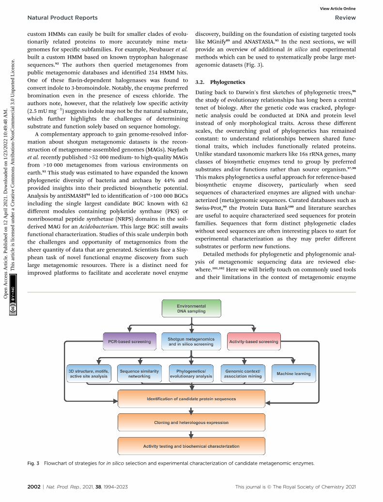
Fig. 3 Flowchart of strategies for in silico selection and experimental ch
2002 | Nat. Prod. Rep., 2021, 38, 1994–2023
discovery, building on the foundation of existing targeted toolslike MGnify89 and ANASTASIA.95 In the next sections, we willprovide an overview of additional in silico and experimentalmethods which can be used to systematically probe large met-agenomic datasets (Fig. 3).
3.2. Phylogenetics
Dating back to Darwin's rst sketches of phylogenetic trees,96
the study of evolutionary relationships has long been a centraltenet of biology. Aer the genetic code was cracked, phyloge-netic analysis could be conducted at DNA and protein levelinstead of only morphological traits. Across these differentscales, the overarching goal of phylogenetics has remainedconstant: to understand relationships between shared func-tional traits, which includes functionally related proteins.Unlike standard taxonomic markers like 16s rRNA genes, manyclasses of biosynthetic enzymes tend to group by preferredsubstrates and/or functions rather than source organism.97,98
This makes phylogenetics a useful approach for reference-basedbiosynthetic enzyme discovery, particularly when seedsequences of characterized enzymes are aligned with unchar-acterized (meta)genomic sequences. Curated databases such asSwiss-Prot,99 the Protein Data Bank100 and literature searchesare useful to acquire characterized seed sequences for proteinfamilies. Sequences that form distinct phylogenetic cladeswithout seed sequences are oen interesting places to start forexperimental characterization as they may prefer differentsubstrates or perform new functions.
Detailed methods for phylogenetic and phylogenomic anal-ysis of metagenomic sequencing data are reviewed else-where.101,102 Here we will briey touch on commonly used toolsand their limitations in the context of metagenomic enzyme
aracterization of candidate metagenomic enzymes.
This journal is © The Royal Society of Chemistry 2021

Table 2 Selected pros and cons of different computational methods for enzyme discovery covered in this review
In silico methods forenzyme discovery Phylogenetics
Sequence similaritynetworking
Genomeneighborhoods andprotein interactionnetworks
3D-structural methods,motifs, and active siteresidues Machine learning
Pros � Longstanding, well-established methods toinvestigate functionalrelationships betweenproteins
� Intuitive graphicalrepresentation ofthousands of proteinsequencessimultaneously
� Guilt-by-associationmethods can reveal newfunctionalrelationships forproteins independentof primary sequence
� Variations in activesite architecture canhave largeconsequences forbiocatalysis / handlesfor discovery
� Deep learning,transfer learning, andautoencoding methodsuseful to learn complexor hidden relationshipsfor functional inference
� Insights intoevolution of proteinfamilies, e.g., throughancestral sequencereconstruction
� Allows users toquickly identify clusterswithout knownrepresentatives insequence space
� Unusual co-occurringdomains or interactingproteins are new targetsfor enzyme discovery
� Structural motifs areuseful for searchesindependent of full-length primarysequence
� Capable ofrecognizing patterns inbig metagenomicdatasets
Cons � Heavily inuenced bythe quality of theunderlying sequencealignment
� Pruning of SSNs byBLAST e-value can besubjective
� Analysis of geneneighborhoods frommetagenomes requiresassembly / introduceserrors and not alwayspossible to recoveranking genes forlowly-abundantorganisms
� Similar structuralfolds catalyze a widerange of differentreactions
� Requires a largequantity of ‘labeled’e.g., experimentally-veried training data
� Not all biosyntheticdomains havea consistent or strongphylogenetic signal
� Unclear how tohandle or gainfunctional insightsfrom ‘singletons’
� Relatively fewstructures solved frommetagenomic sources
� Classication systemslimited in their abilityto predict entirely newenzyme functions
Review Natural Product Reports
Ope
n A
cces
s A
rtic
le. P
ublis
hed
on 1
2 A
pril
2021
. Dow
nloa
ded
on 1
/23/
2022
10:
49:4
8 A
M.
Thi
s ar
ticle
is li
cens
ed u
nder
a C
reat
ive
Com
mon
s A
ttrib
utio
n-N
onC
omm
erci
al 3
.0 U
npor
ted
Lic
ence
.View Article Online
discovery. One key disadvantage is that phylogenetic trees areonly as accurate as the underlying multiple sequence align-ments. Countless tools for generating sequence alignmentsincluding MUSCLE,103 MAFFT,104 and Clustal Omega105 areavailable. Independent of alignment method, an oen over-looked but important intermediate step is manually inspectionof sequence alignments and trimming large gap regions withtools such as trimAl106 or Gblocks before treeing.107 Anotherlimitation of phylogenetic analysis is the computational cost ofestimating trees from large sequence alignments. FastTree108
overcomes this disadvantage by using heuristic methods toconstrain the tree search space and make approximatemaximum-likelihood estimations thereby dramatically cuttingtreeing time. Surprisingly, for many applications, FastTree isoen nearly as accurate as more rigorous maximum-likelihoodmethods109,110 such as PhyML or RaxML that make fewerassumptions but require orders of magnitude more time torun.111 The recently released RaxML-NG also combines theimproved accuracy of RaxML with computational scalability forthe analysis of large (meta)genome-scale datasets.112 Anotherpopular phylogenetic tool is IQ-Tree, which includes the addedfeatures of automated model selection and ultra-fast bootstrapapproximation.113 For visualization and advanced annotationoptions of these phylogenetic trees, we recommend the widely-used Python ETE 3 toolkit114 or ggtree in R.115
Ancestral sequence reconstruction adds another dimensionto phylogenetic analysis by using contemporary proteinsequences to infer their evolutionary history116 such as howbiosynthetic enzymes might have arisen from primary
This journal is © The Royal Society of Chemistry 2021
metabolic enzymes. Ancestral reconstruction of adenylate-forming enzymes suggested that secondary metabolicenzymes such as b-lactone synthetases and nonribosomalpeptide synthases arose from protein scaffolds similar tocontemporary primary metabolic enzymes such as CoAligases.97 Hendrikse et al. reconstructed the evolutionary historyof diterpene cyclases and experimentally characterized thepredicted ancestral sequences. They reported the ancestralenzymes had increased thermostability and broader substratespecicity, both of which are common features of ancestralsequences that may promote the evolution of new functions.117
Probabilistic web-based tools like FastML make ancestralsequence reconstruction accessible to non-experts.118 Bayesianphylogenetic methods are also powerful for understandingevolutionary relationships, as exemplied by a phylogenomicstudy of lanthipeptide synthetases, a family of RiPP maturasesthat introduce sulfur bridges into peptides.119 Through Bayesianphylogenomic analysis of lanthipeptide BGCs, Zhang et al.unexpectedly found that the sequences of lanthipeptideprecursors as well as maturases played a decisive role indetermining the structure of the nal natural products. Overall,phylogenetics remains one of the rst and most fundamentalstops on the roadmap for enzyme bioprospecting from meta-genomes (Fig. 3 and Table 2).
In the context of natural product biosynthesis, many toolshave been developed to predict biosynthetic enzyme substrateor function using phylogenetic methods, as recently reviewed byAdamek et al.120 The Natural Product Domain Seeker (NaPDoS)makes structural inferences about natural products based on
Nat. Prod. Rep., 2021, 38, 1994–2023 | 2003

Natural Product Reports Review
Ope
n A
cces
s A
rtic
le. P
ublis
hed
on 1
2 A
pril
2021
. Dow
nloa
ded
on 1
/23/
2022
10:
49:4
8 A
M.
Thi
s ar
ticle
is li
cens
ed u
nder
a C
reat
ive
Com
mon
s A
ttrib
utio
n-N
onC
omm
erci
al 3
.0 U
npor
ted
Lic
ence
.View Article Online
phylogenetic analysis of ketosynthase and condensationdomains.121 Other phylogeny-based methods such as Predi-CAT122 for NRPS adenylation domains and TransATor for trans-acyltransferase PKS prediction123 both enable natural productstructural predictions for these respective classes. Other classesof biosynthetic domains, however, are less amenable to makingphylogeny-based structural or functional inferences. Forexample, type I thioesterase domains do not have a strongphylogenetic signal for the substrate class or offloading chem-istry.124,125 Plant sesquiterpene synthases are similar and tend togroup based on taxonomy of the source organism rather thanchemical similarity of carbocation product type.126 Even forbiosynthetic domains with a strong signal, there are alwaysphylogenetic outliers which present challenges for substrate ornal natural product structure classication.
For phylogeny-based genome mining to detect new enzymeclasses, we highlight two complementary soware tools, Evo-Mining and CORASON.127,128 EvoMining is based on the premisethat primary metabolic enzymes oen undergo duplication orhorizontal gene transfer events, both of which may lead to theemergence of new enzyme functions in secondary metabolicpathways. EvoMining has been used for example to ndenzymes that catalyze similar chemical reactions but performdifferent cellular functions,129 or to discover new enzymesinvolved in the biosynthesis of arseno-organic metabolites.130 Arelated tool, CORe Analysis of Syntenic Orthologs to prioritizeNatural product BGCs (CORASON),128 generates cluster varia-tion databases for intuitive phylogenetic visualization of coreand ancillary genes in BGC families. Overall, while phylogeneticanalysis is a key rst step, it is oen more informative whenused in combination with other approaches as will be discussedherein (Table 2).
3.3. Sequence similarity networking
Compared to phylogenetics, sequence similarity networks(SSNs) are relatively new methods for the visualization ofprotein families and superfamilies. First published for thepurpose of protein superfamily analysis in 2009,131 SSNs aregraphs that display relationships between protein families.SSNs are usually generated with an all-by-all BLAST search ofa custom sequence set and visualized as a graph where nodesare protein sequences, and each edge represents pairwisesequence similarity. Typically, SSNs are pruned by settingdifferent protein similarity score thresholds to reveal smallerclusters of protein subfamilies. As with phylogenetics, it isuseful to include seed sequences of characterized enzymes inSSNs to serve as anchor points when seeking to identify rela-tionships between enzyme families or subfamilies. In a massiveenzyme screening study from soil and vanilla pod meta-genomes, SSNs were used to identify the location of new func-tional triesterase hits in multiple unexplored protein familysubclusters spread across three different protein superfam-ilies.132 SSNs have also been used in combination with phylo-genetics to propose the nitroreductase protein superfamilyarose from the radial divergence of functional diversity froma minimal cofactor-binding scaffold.133 These examples
2004 | Nat. Prod. Rep., 2021, 38, 1994–2023
demonstrate the utility of SSNs to identify both known andunknown protein subfamily clusters as candidates for experi-mental characterization.
A major advantage of SSNs is the ability to quickly visualizethe relationships between thousands of protein sequencessimultaneously. Compared to a bootstrapped maximum-likelihood phylogenetic tree, SSNs are typically faster tocompute and can be interactively visualized using the open-source soware, Cytoscape, which provides a friendly Graph-ical User Interface.134 A downside of the point-and-click Cyto-scape soware is that workows are oen tedious to reproduce,particularly for large networks with thousands of nodes. Withthe release of the CyREST API, popular high-level languagessuch as Python and R can now be used to program reproducibleSSN workows.135,136 Alternative network analysis packages suchas igraph are also popular and available for Python, R, and C/C++.137 For users without programming experience, the EnzymeFunction Initiative Enzyme Similarity Tool (EFI-EST) was therst web-based application enabling automated construction ofsequence similarity networks.138
A key downside of SSNs is the bias that can be introducedduring the selection of similarity thresholds to prune networks,most commonly based on BLAST e-value. BLAST e-values aredependent on the size of the sequence database and compari-sons of e-value thresholds between SSNs generated usingdatabases of different sizes is misleading. Moreover, varioustypes of graph layouts for SSNs can lead to different interpre-tations. Therefore, we recommend users make the sequences,code, and networks over the full range of possible layouts andBLAST e-values available on a publicly available scientic imagerepository such as Zenodo. This promotes data transparencyand limits the cherry-picking of specic e-values or networktopologies.
3.4. Gene context and interactions
Gene context is an oen underemphasized but highly effectivemethod for enzyme discovery especially for natural productbiosynthesis. Flanking genes can oen provide insights intosubstrates, cofactors, and natural product bioactivity. Forexample, a new family of cobamide-remodeling enzymes wide-spread in the human gut microbiome was identied based ongenome context analysis of a coding sequence of unknownfunction anked by cobamide biosynthesis and salvaginggenes.139 To automate genome neighborhood analysis, a widelyused addition to the EFI-EST is the Genome Neighborhood Tool(GNT).140 EFI-GNT generates genome neighborhood networksand allows for rapid visual assessment of genome context. Italso conducts statistical analysis of gene co-occurrence toidentify possible functional linkages. For natural product BGCswe also recommend specic tools such as BiG-SLICE141 and BiG-SCAPE128 designed to identify and group BGCs into gene clusterfamilies. BiG-SCAPE is integrated with CORASON (Section 3.2),thus combining the power of phylogenetics with neighborhoodclustering methods. BiG-SLICE is specically designed tohandle massive numbers of BGCs by representing them inEuclidean space rather than by pairwise comparison.141 This
This journal is © The Royal Society of Chemistry 2021

Review Natural Product Reports
Ope
n A
cces
s A
rtic
le. P
ublis
hed
on 1
2 A
pril
2021
. Dow
nloa
ded
on 1
/23/
2022
10:
49:4
8 A
M.
Thi
s ar
ticle
is li
cens
ed u
nder
a C
reat
ive
Com
mon
s A
ttrib
utio
n-N
onC
omm
erci
al 3
.0 U
npor
ted
Lic
ence
.View Article Online
dramatically cut runtime to enable clustering of over onemillion BGCs frommetagenome-assembled genomes. Based onits ‘BiG’ savings in computational cost, BiG-SLICE is thereforeparticularly well-suited for analysis of metagenomes forgenome-context guided enzyme discovery. There are alsonumerous genome context tools available for specic naturalproduct classes. For example, RODEO142 and RiPPeR98 are usefulto identify new RiPPs and maturases based on genomic context.Although RODEO is targeted towards RiPPs, it is not restrictedto them and can be used generally to rapidly pull genomeneighborhoods for any set of query sequences from publicdatabases. Flanking genes are provided in tabular format fordownstream PFAM co-occurrence analysis, phylogenetics, andSSN generation.
Genome neighborhood context can also provide insightsinto natural product bioactivity and guide the identication ofnew targets and self-resistance genes. The Antibiotic ResistanceTarget Seeker (ARTS) is one automated approach to identifyknown and potentially new self-resistance targets throughanalysis of gene proximity, duplication, and diversicationevents.143 Culp et al. recently used genome context-guideddetection of known resistance genes combined with phyloge-netic analysis to identify a divergent clade of glycopeptideantibiotic BGCs lacking well-characterized self-resistancegenes.144 This led to the discovery of a completely new modeof action for a divergent clade of glycopeptides represented bycomplestatin and a novel antibiotic, carbomycin. This multi-pronged approach of genome context mining and phyloge-netic analysis oen yields a more holistic picture of BGCdivergence and evolution, thereby guiding selection of candi-date enzymes and cellular targets for experimentalcharacterization.
More generally, the identication of gene functions based ongenomic context has been termed a ‘guilt-by-association’approach.145 One broad use platform that relies on guilt-by-association methods is the STRING web resource.146 STRINGprovides an intuitive interface for functional analysis ofproteins including the prediction of protein–protein interac-tions through text mining of scientic literature and associa-tions inferred from genomic context, co-expression data, orgene orthology to model organisms. Although STRING is notspecically targeted towards metagenomics or natural productbiosynthesis, it can be used to predict–protein interactions suchas for MbtH-like proteins in NRPS systems.147 A more specictool, CO-ED, is useful for network analysis and identication ofunusual co-occurring domains in multi-domain proteinsincluding megasynthases commonly involved in naturalproduct biosynthesis.148 CO-ED relies on PFAM information asinputs which can be extracted from (meta)genomes usingPfamScan.149 CO-ED highlights which co-occurring enzymedomains are already found in public databases (e.g. MIBiG,35
UniPROT,150 or BRENDA151), and which combinations have notyet been characterized. As a proof-of-principle, CO-ED analysisof the Pseudoalteromonas rubra genome identied an unusualnitroreductase-ThiF PFAM domain pair in a protein termedOxzB. Heterologous expression of oxzB and its upstream geneoxzA in 5 different organisms resulted in production of
This journal is © The Royal Society of Chemistry 2021
pigmented yellow natural products with unusual oxazolonemoieties. In vitro characterization of OxzB revealed the nitro-reductase and ThiF-like domains catalyze the oxidation andcyclization of N-acyl amino acid substrates, respectively, to formoxazolone heterocycles (Fig. 4B). Oxazolone-forming enzymeswere previously unknown in nature, thus CO-ED analysis ofprotein domains facilitated biochemical discovery of the rstoxazolone synthase.148
3.5. 3D-structure based methods
Previously, the inclusion of structural information to infermetagenomic enzyme function was hampered by the lack ofsolved protein structures. Rooted in the assumption that novelprotein folds are more likely to perform novel functions, high-throughput protein structural characterization campaignswere initiated around the globe to catalogue protein structuralspace.152 Still, these efforts focused disproportionately onculturable organisms. As of January 2021, less than 0.3% ofentries in the PDB were tagged as belonging to metagenomesor uncultured organisms. Moreover, while these high-throughput structural genomics initiatives have solved thou-sands of structures, they surprisingly yielded far fewercompletely new protein folds than expected.153 Out of thevastness of protein conformations given all possible aminoacid combinations, only a small fraction of this is representedin biological macromolecules, at least in organisms interro-gated to date. It remains to be seen if and how much ofstructural and functional protein space still awaits discoverywithin the uncultivated majority of microbial life. The fact thateven the most conserved protein folds identied to date areable to catalyze a variety of different reactions further under-pins that we are only at the beginning of understanding howthe multi-dimensional space of enzymes affects catalyticdiversity. It is clear that even powerful structure predictiontools like AlphaFold2 will not solve the ‘function’ aspect of thesequence–structure–function problem alone.
Nonetheless, secondary, tertiary, and quaternary structuresof proteins can yield critical insights into function beyondprimary sequence. Many protein families involved in naturalproduct biosynthesis including RiPP recognition elements,154
adenylate-forming enzymes,155 and thioesterase domains124
share the same highly conserved structural fold but relativelylow amino acid sequence similarity with other members of thefamily. Not surprisingly, for many enzyme families, structuralalignment tools such as MAMMOTH,156 MATRAS,157 and Care-tta158 yield signicantly more accurate alignments than purelysequence-based alignment methods.159 Although AlphaFold2 iscurrently not publicly available, existing web-based homologymodeling tools including Phyre2,160 I-TASSER,161 and SWISS-MODEL162 can be used to provide insights into predictedstructural fold of metagenomic sequences. Recently, a deep-learning structure prediction pipeline was used to model TaraOceans metagenomic sequences across different ocean depthsand implicated the involvement of a ubiquitous protein family(PF15461) in photosynthesis.28 Structural modeling is oen therst step towards detecting active site residues and structural
Nat. Prod. Rep., 2021, 38, 1994–2023 | 2005

Fig. 4 Selected enzymes highlighted in this review. (A) IkaB oxidoreductase involved in ikarugamycin polycyclization. (B) ThiF-nitroreductase di-domain enzyme, OxzB, catalyzes cyclization of oxazolone-containing metabolites with homologs detected in metagenomes from variousenvironments (mainly marine). (C) PdxI catalyzes an alder–ene reaction to form a vinyl cyclohexane intermediate in biosynthetic pathways forfungal alkaloids including pyridoxatin and cordypyridones. (D) Arginase-family enzyme, OspR, promiscuously installs ornithines in the backbonesof peptide natural products. OspR homologs were characterized from various microbial isolates and from the uncultivated phylum ‘CandidatusWallbacteria’ from groundwater metagenomes. (E) FrsA thioesterase domain originally detected in an uncultivated leaf symbiont catalyzesintramolecular thioesterification of the Gq protein inhibitor FR900359.
Natural Product Reports Review
Ope
n A
cces
s A
rtic
le. P
ublis
hed
on 1
2 A
pril
2021
. Dow
nloa
ded
on 1
/23/
2022
10:
49:4
8 A
M.
Thi
s ar
ticle
is li
cens
ed u
nder
a C
reat
ive
Com
mon
s A
ttrib
utio
n-N
onC
omm
erci
al 3
.0 U
npor
ted
Lic
ence
.View Article Online
motifs which can play a disproportionately large role in deter-mining protein function as will be discussed in the next section.
3.6. Motifs and active site residues
Enzyme active sites only occupy a small fraction of the volumeof a full-length protein folded in 3D space. Compared to the restof the protein, catalytic residues are typically limited in theiridentity and arranged in conserved architectures.163 Perhaps themost famous example of active site conservation is the Ser-His-Asp catalytic triad used by alpha/beta hydrolases as well asseveral other protein folds including the subtilisin andchymotrypsin folds.164 This same triad hydrolyzes over 17
2006 | Nat. Prod. Rep., 2021, 38, 1994–2023
different reaction mechanisms spanning nearly every type of ECclass. The multifunctionality of the Ser-His-Asp triad in partic-ular is attributed to its ability to accommodate a wide range ofsubstrates which can have different chemical interactions withthe same key catalytic residues.165 Only about half of theenzymes with Ser-His-Asp triads had architectural differences inthe active site such as changes in hydrogen bond partners oracids/bases for new mechanisms; the rest were driven bysubstrate chemistry alone.165 This study is just one example ofhow the same active site architecture can catalyze remarkablechemical diversity, making total prediction of function fromprotein active site alone challenging, if not impossible.
This journal is © The Royal Society of Chemistry 2021

Review Natural Product Reports
Ope
n A
cces
s A
rtic
le. P
ublis
hed
on 1
2 A
pril
2021
. Dow
nloa
ded
on 1
/23/
2022
10:
49:4
8 A
M.
Thi
s ar
ticle
is li
cens
ed u
nder
a C
reat
ive
Com
mon
s A
ttrib
utio
n-N
onC
omm
erci
al 3
.0 U
npor
ted
Lic
ence
.View Article Online
Oen altering even one residue can be sufficient to changethe substrate specicity or enantioselectivity of an enzyme.166,167
Protein engineers are well aware of the fact, however, thatmaking changes in the active site can have dire consequencesfor enzyme activity. The high-risk, high-reward task of active sitemodication oen leads to countless evolutionary dead-ends.As a complement to engineering studies, characterization ofnaturally occurring active site variants that are conserved acrossdifferent (meta)genomes provides an alternative route forenzyme discovery. As a striking example of the importance ofactive site variants, Ohashi et al. discovered several newenzymes originally annotated as O-methyltransferases, e.g.,LepI168 and PdxI,31 which catalyze various types of pericyclicreactions in the biosynthesis of fungal alkaloids (Fig. 4C).Alteration of a single residue (V413M) in PdxI was able to shithe selectivity away from the Alder-ene reaction towards a moreenergetically favorable hetero-Diels–Alder reaction.31 Mutationsof other residues in the PdxI active site could further tuneperiselectivity and regioselectivity and highlighted how evensubtle changes can dramatically affect the nal structures ofnatural products.
Studies targeting active site variants have not yet been widelyapplied to the task of enzyme discovery from shotgun meta-genomes. Aberrant active site architectures are typically onlyremarked on during enzyme characterization following activity-based screening. For example, a divergent catalytic triad in anacid-stable endoglucanase was reported from activity-basedscreening of an soil metagenomic library.169 For detection ofactive site residues without knowledge of the enzyme class orfunction, tools such as CASTp for automated detection of activesite pockets are useful.170 Comprehensive databases such as theMechanism and Catalytic Site Atlas (M-CSA) catalogue knownactive site architectures and mechanisms.171 As of December2020, the M-CSA contains nearly 1000 hand-curated entriesrepresentative of >73k Swiss-Prot entries and >15k PDB struc-tures. However, with >176k structures in the PDB and thenumber growing daily, M-CSA still represents less than 10% ofknown structural space. UniProt also provides predicted activesite information which can be useful for structural alignmentsto identify divergent active site architectures in metagenomesequences.
In addition to the active site, other conserved motifs orcofactor binding sites are also important for protein function
Fig. 5 Common steps in a machine learning workflow for protein funct
This journal is © The Royal Society of Chemistry 2021
and can be detected with tools such as ScanProsite.172 Forexample, structural alignment of promiscuous RiPP maturasesthat install ornithine residues into peptide backbones revealeda conserved ‘DXHXD’ motif which was then used to detect andcharacterize new RiPP-modifying arginases from groundwatermetagenomes and culturable isolates (Fig. 4D).173 In this studyand many others, motif searching is used combination withfull-length sequence homology searches for improved accuracy.For a different approach, however, motif searches can be usedto identify conserved cofactor binding sites or structuralfeatures independent of protein family or fold from meta-genomic sequences.
3.7. Machine learning
Machine learning offers the promise of moving beyond simplehomology transfer methods to learn hidden relationshipsbetween protein sequences, structures, and functions. Advancesin computing power and algorithms, have led to a renaissanceof machine learning in many elds including biology andchemistry. Just in the past decade, >35 different machinelearning-based methods have been published for proteinfunction prediction.174 Rather than compare individual algo-rithms, we will focus on key steps and common pitfalls ina generalized machine learning workow (Fig. 5).
Although machine learning has received a signicantamount of hype in recent years, it is not a panacea. One keydisadvantage is that machine learning techniques are extremelydata hungry. Even the most sophisticated of machine learningmodels are only as good as the underlying quantity and qualitytraining data. In fact, increasing model complexity requireslarger amounts of data. Deep neural networks, in particular,commonly suffer from overtting, that is, they cannot begeneralized to other studies or data sets. Therefore, continuedsupport and curation of public databases which provide high-quality training data, such as MIBiG35 and Swiss-Prot,175 areessential for machine learning to enable future enzymediscoveries. One active area of research that seeks to handle thepaucity of ‘labeled’ or experimentally veried data pointsbiology is known as transfer learning. During transfer learning,models are pre-trained on large quantities of unlabeled data,e.g., unknown metagenomic sequences, to learn features thatare general to these sequences and thereby improve perfor-mance on separate, related tasks such as enzyme function
ion prediction covered in this review.
Nat. Prod. Rep., 2021, 38, 1994–2023 | 2007

Natural Product Reports Review
Ope
n A
cces
s A
rtic
le. P
ublis
hed
on 1
2 A
pril
2021
. Dow
nloa
ded
on 1
/23/
2022
10:
49:4
8 A
M.
Thi
s ar
ticle
is li
cens
ed u
nder
a C
reat
ive
Com
mon
s A
ttrib
utio
n-N
onC
omm
erci
al 3
.0 U
npor
ted
Lic
ence
.View Article Online
prediction through transfer of knowledge. A generalized trans-fer learning model pre-trained on short metagenomic reads wasrecently used for the identication of new candidate oxidore-ductases from Tara Oceans metagenomes.176 Further advancesin transfer learning and other semi-supervised learning tech-niques will help us leverage big metagenomic datasets with fewlabeled examples in the future.
Feature extraction and engineering is another critical step inmachine learning workows (Fig. 5). In addition to usingprotein sequences and structural information as features, somemethods incorporate physicochemical amino acid properties orprotein–protein interaction information to improve functionalprediction accuracy.177,178 Natural language processing methodsfor text mining have also been used to extract features sincebiochemical knowledge continues largely to be stored in textformat in journal articles.179,180 Recently, the use of autoen-coders for unsupervised encoding of protein features hasemerged. Autoencoders are articial neural networks to auto-mate the manual process of feature extraction and engineering,thereby removing human biases during the feature engineeringprocess. One downside, however, is that autoencoders requireeven larger datasets and increased compute time.181,182
Machine learning algorithms used to predict protein func-tion also vary from simple logistic regression and random forestmodels to multi-layer neural networks.174 However, bench-marking performance across studies can be complicated byinconsistent classication systems for the objective, e.g.,protein function prediction. Most machine learning models usehierarchical tree-based structures such as Gene Ontology(GO),183 Functional Catalogue (FunCat),184 or EC classicationsystems185 as objectives but comparisons between modelsremains a challenge. Initiatives such as the Critical Assessmentof Functional Annotation (CAFA) challenge, now in its fourthyear, are making inroads to standardize the eld.186 In recentcomparisons of protein function prediction models, however,even simple homology transfer and logistic regression modelswere still able to outperform deep neural networks for someprotein function prediction tasks.174 Unlike AlphaFold2'sperformance for protein structure prediction, the functionprediction problem is far from solved and the eld is stilldeveloping.
In terms of machine learning applications for naturalproduct biosynthesis, BGCs have a unique advantage sincebiosynthetic logic and linkage to natural product structures canhelp narrow the range of potential substrates and functions.There are a growing number of BGC-specic machine learningtools available to predict natural product structure and bioac-tivity from metagenomes. These include BGC detection andclassication soware reviewed elsewhere187 such as anti-SMASH,94 PRISM,188 DeepBGC189 and most recently, GECCO(https://gecco.embl.de/). Unfortunately, researchers tend towork either on the more general protein function predictionproblem or on natural products biosynthesis, but they do notoen communicate with each other. Increasing integrationbetween these distinct research communities such as throughjoint conferences and workshops would advance progress forthe prediction of new secondary metabolic enzyme functions.
2008 | Nat. Prod. Rep., 2021, 38, 1994–2023
A major limitation of machine learning methods available todate is the task of predicting truly novel enzyme functions. Inmany of the methods described, models are trained on a rangeof possible objectives, e.g., EC, FunCat, or GO terms, or BGCs forknown natural product classes, but they are not capable ofpredicting entirely new classes. One alternative to multi-labelclassication is the use of negative selection algorithms whichcan label a protein as performing a particular function or not.190
The benet of this approach is that negative selection does notforce a protein to t into a previously dened class. Rathera sequence can be predicted to not t into any known functionalclasses or to t into multiple classes, thus suggesting newfunction(s), potential promiscuity or even moonlighting.191
However, the best-case scenario still only provides negative orpositive predictions. Will machine learning algorithms everreach the stage of making completely de novo protein functionpredictions? Only time will tell, but if AlphaFold2 is any indi-cation, then the future is bright.
4. Reaching the destination:characterizing new enzymes4.1. Cloning and heterologous expression
Up until this point, our roadmap has explored various in silicomethods for enzyme function prediction. However, while thesemethods can be helpful for identication and prioritization ofnew areas of protein sequence space they are, at best, onlycomputational predictions. They do not provide functionalvalidation, which requires experimental characterization. Whenselecting proteins to characterize in the lab, an important rststep is quality control to remove chimeras and truncatedsequences that may have sequencing errors or do not encodefully functional proteins. Outliers in sequence length visiblefrom multiple-sequence alignments can also indicate a mis-prediction of start or stop codons. Particularly for Actino-bacteria which are prolic natural product producers, geneproducts with non-canonical start and stop codons192 can bemis-predicted by gene annotation tools leading to frustrationsin the laboratory.
When dealing with hundreds to thousands of metagenomicsequences as candidates to choose from, many tools exist tocluster proteins by similarity and automatically select repre-sentative sequences. Early pioneers in the eld that are stillwidely used include CD-HIT193 and UCLUST.194 Newer clusteringalgorithms such as Linclust available through the MMSeqs2soware suite also can perform clustering for metagenomicprotein sequences in a fraction of the CD-HIT runtime.195 SSNanalysis and selection of cluster representatives using Cyto-scape or igraph can also be used to select protein subfamilyrepresentatives. Independent of clustering strategy, the under-lying assumption is that highly similar proteins will alsoperform the same functions, which is oen, but not always,true.196,197 One striking example where homology did notdetermine function was exemplied by the discovery ofa completely new enzyme within the well-known NifH proteinfamily. Based on previous observations of unexpected ethylene
This journal is © The Royal Society of Chemistry 2021

Review Natural Product Reports
Ope
n A
cces
s A
rtic
le. P
ublis
hed
on 1
2 A
pril
2021
. Dow
nloa
ded
on 1
/23/
2022
10:
49:4
8 A
M.
Thi
s ar
ticle
is li
cens
ed u
nder
a C
reat
ive
Com
mon
s A
ttrib
utio
n-N
onC
omm
erci
al 3
.0 U
npor
ted
Lic
ence
.View Article Online
gas production from freshwater and soil bacteria, North et al.used a combination of differential gene expression andknockouts to determine the new function of a Rhodospirillumrubrum NifH homolog as a methylthio-alkane reductase.198 Thisenzyme is part of a biosynthetic pathway for methionine anda new route for anaerobic ethylene and methane productioncompletely independent from nitrogenase activity. Thus,homology determining function does not always hold, even forfamous and well-characterized families like NifH proteins.Nonetheless, clustering remains a useful method to chooseprotein representatives.
Depending on dataset size, further ltering steps may berequired. For many enzyme activity assays without high-throughput capabilities, screening may be the bottleneckallowing for the selection of only a handful of metagenomicsequences to express and test. In this case, the decision of whichfew sequences to select becomes more tailored to the researchquestion. In general, one of the most obvious strategies is tochoose metagenomic sequences also found in culturableorganisms, since this can permit functional characterization inthe native host. Another popular strategy is the selection ofproteins from thermophilic organisms which tend to encodeenzymes with increased thermostability. We note this is alsoa generalization, however, since analysis of the ‘meltomes’ ofcomplete proteomes of diverse organisms across the tree of lifeshowed high variability in protein melting temperature even fororganisms adapted to temperature extremes.199
Alternative approaches include selecting candidates that aremore likely to be stable and express well including ltering forproteins that do not have high GC-content, transmembraneregions, or long disordered regions. Automated approaches topredict protein stability have been developed mainly for pre-dicting crystallization propensity including tools likeXtalPred,200 XANNPred,201 OB-Score,202 CrystalP2,203 ParCrys,204
and Crysf.205 A recent comparison of these tools was publishedby Wang et al.205 Given the abundance of different approachesto predict protein stability, we recommend using multiplecriteria to rank and prioritize protein sequences for experi-mental characterization. In this way, individual biases inprediction tools may partially be offset by an ensemble-basedapproach to identify the most promising candidates.
Another useful step to increase the likelihood of obtainingsoluble protein is the removal of signal peptides, that is, regions16–30 amino acids in length at the N-terminus of manyprokaryotic and eukaryotic proteins.206 These short regions ofDNA typically direct the export of proteins from the cytosol.Signal peptides can inuence protein solubility and export inheterologous expression experiments, particularly with N-terminal His tags. The gold standard for signal peptide detec-tion and removal has been the soware tool, SignalP,207 butmore advanced machine learning methods for signal peptidedetection and design are emerging.208 Recently, attention-basedneural networks were successfully used to detect and generatediverse, functional signal peptides for a variety of proteinfamilies.208 As our understanding of the relationships betweensignal peptides and protein functions deepens, these short
This journal is © The Royal Society of Chemistry 2021
stretches of amino acids could also as serve features for enzymediscovery efforts in the future.
4.2. Heterologous expression
Once enzymes or BGCs of interest have been identied,constructs for heterologous expression must be designed.Unfortunately, most vectors that work for metagenomic librarypreparation for functional metagenomics methods are nottypically suitable for heterologous expression. Many completeBGCs are also not fully captured in metagenomic libraries sincefosmid/cosmid vectors have a maximum insert size of 45 kb. Inaddition to classical restriction cloning and Gibson assemblymethods, new methods have been developed to improve theefficiency and ease of cloning large BGCs into heterologoushosts.209 One popular method, transformation-associatedrecombination (TAR) cloning, relies on natural homologousrecombination in yeast to piece together overlapping eDNAcosmid/fosmid clones from soil and sponge metagenomes.210
Genetic recombineering,209,211 uses a variety of bacteriophageproteins to mediate homologous recombination in E. coli,including a new rapid method for efficient cloning of largeBGCs using RecET direct cloning coupled to Redab recombi-nation.212 For a comprehensive review of cloning methods forBGCs, we refer readers to Zhang et al.209
To obtain sufficient genetic material for cloning, PCRamplication is still oen the most cost-effective method iforiginal eDNA is still available or if source organisms are cul-turable. In the event genetic material from the source is notavailable, the costs of DNA synthesis have dropped signicantlyin recent years. Moreover, gene synthesis enables completecodon optimization to match codon usage preferences of theheterologous host, which is particularly useful for expression ofmetagenomic sequences from taxonomically distant, unculti-vated organisms.213
Even with constructs that are properly designed, manyheterologous expression experiments still fail. From ourpersonal experience, the activity of some biosynthetic enzymesis only detectable through the expression of complete BGCsrather than expressing genes individually from a cluster. Thisfurther underscores the importance of protein–protein inter-actions for enzyme activity (Section 3.4). In other cases whereexpression fails, enzymes may require cofactors or other meta-bolic machinery not found in model organisms such as E. coli.For example, methylmalonyl-CoA is needed for complex poly-ketide biosynthesis in actinomycetes, but it is not produced byE. coli.214 Modular PKS and NRPS clusters also typically requireco-expression of secondary metabolism-type phosphopante-theine transferases (PPTases) from source organisms since theproper PPTases for post translational modication of thesecomplex natural products oen differ from the PPTases presentin E. coli.215 Many cobalamin-dependent radical SAM enzymessuch as C-methyltransferases involved in maturation ofproteusin-family RiPPs are also inactive in E. coli.216 In all ofthese cases, engineered E. coli strains have been developed,217
including recently published plasmids to improve cobalaminuptake in E. coli.218 Even using engineered strains, many natural
Nat. Prod. Rep., 2021, 38, 1994–2023 | 2009

Fig. 6 Trade-off between generalizability and throughput forcommon enzyme screening approaches.
Natural Product Reports Review
Ope
n A
cces
s A
rtic
le. P
ublis
hed
on 1
2 A
pril
2021
. Dow
nloa
ded
on 1
/23/
2022
10:
49:4
8 A
M.
Thi
s ar
ticle
is li
cens
ed u
nder
a C
reat
ive
Com
mon
s A
ttrib
utio
n-N
onC
omm
erci
al 3
.0 U
npor
ted
Lic
ence
.View Article Online
products still are not detectable in their nal modied formfrom expression in E. coli.
As an alternative to using model organisms as heterologoushosts, non-model hosts can oen be identied through genomemining. When heterologous expression in E. coli yielded lowamounts of FR900359, a potent Gq protein inhibitor rstuncovered from metagenomic eDNA of an uncultivated leafsymbiont,219 Hermes et al. identied a homologous BGC in thegenome of the culturable bacterium Chromobacterium vaccinii.Knockout studies of the native cluster in C. vaccinii andsuccessful heterologous expression of the C. vaccinii enzymes inE. coli enabled characterization of the unusual thioesterasedomain catalyzing intermolecular thioesterication ofFR900359 (Fig. 4E).220 In another example, a homologous BGCto the RiPP-family polytheonamide cluster from the unculti-vated sponge symbiont ‘Candidatus Entotheonella’was found inthe culturable betaproteobacterium, Microvirgula aerodeni-tricans. In particular, the cobalamin-dependent radical SAM C-methyltransferases that were largely inactive in E. coli werefound to be highly active in M. aerodenitricans, enablingproduction of fully modied nal products.221 The poly-geonoides, polytheonamide-like compounds from a meta-genomic bin of a deep-rock subsurface environment, were alsoproduced and characterized from M. aerodenitricans. Thegeneralized strategy of searching for metagenomic BGC hits inthe genomes of culturable organisms can be especially fruitfulin the case that heterologous expression in model organisms isunsuccessful.
Selecting the closest culturable taxonomic relative, particu-larly if genetic tools are available for this strain, can also beanother promising method to select heterologous hosts. Thisstrategy enabled the discovery of Fe–S avoenzymes involved inbile acid dehydroxylation produced by the gut microbiomecommensal, Clostridium scidens.222 Funabashi et al. character-ized these enzymes by introducing them into a closely relatedClostridium strain amenable to genetic manipulation. Thisapproach has long been used to express diverse BGCs fromStreptomyces spp. in the model Streptomyces coelicolor A3(2).223
In other cases, heterologous expression of genes from taxo-nomically distant organisms can still work in E. coli, such as inthe case of expressing a BGC from a diatom for domoic acidproduction.224 As withmany experimental systems, the selectionof a heterologous host is still largely a process of trial-and-error.In the future, we anticipate design-build-test-learn workowsused in synthetic biology and already being applied for theoptimization of hosts will reduce this tedious trial-and-errorprocess.225
4.3. Screening for enzyme activity
Once enzymes of interest have been expressed, the next chal-lenge comes in assaying them for in vivo or in vitro for activity.There is oen a trade-off between throughput and generaliz-ability for enzyme screening methods (Fig. 6). Activity-basedscreening of metagenomic libraries typically involves search-ing for zones of inhibition around bacterial colonies or usingcleavable substrates that produce a color or uorescence.
2010 | Nat. Prod. Rep., 2021, 38, 1994–2023
However, for many biosynthetic enzymes, appropriate substrateanalogs may not be available, or the presence of a large uo-rophore or chromophore may interfere with enzyme activity.Mass spectrometry (MS) is a sensitive and highly generalizabletechnique to monitor activity for many different enzymes sinceit does not require the use of substrate analogs. Due to the needfor chromatographic separation steps, however, MS-basedmethods have historically suffered from low throughput. Anumber of alternative workows to bypass the chromatographicseparation step allow for scaling up MS-based screening ofenzyme activity. For example, nanostructure-initiator massspectrometry (NIMS) substitutes column-based separation withan in situ washing step over a peruoroalkylated surface. Thewash step promotes non-covalent uorous interactionsenabling compound separation.226 Previously, NIMS has onlybeen successfully applied to enzymes with active sites able toaccommodate bulky peruoroalkylated tails such as acetyl-transferases and carbohydrate-degrading enzymes.227 A moregeneralized solution, termed PECAN (Probing Enzymes withClick-Assisted NIMS), relies on click chemistry to expand NIMStechnology to other enzyme classes.228 Although still requiringsubstrate analogs with ‘clickable’ alkynes or azides, these smallhandles are far less bulky than peruoroalkylated tails. Anotherlabel-free alternative to NIMS is known as SAMDI-MS (Self-Assembled Monolayers for matrix-assisted Desorption/Ionization Mass Spectrometry). SAMDI-MS relies on theimmobilization of proteins or metabolites on self-assembledmonolayers of alkanethiolates on gold.229 Importantly, theimmobilization process for SAMDI-MS combines the sensitivityand generalizability of MS-based methods with signicantlyhigher throughput. Since SAMDI-MS does not require labelingof the substrates, it is especially useful for enzyme classes whichare challenging to screen such as glycosyltransferases.230
SAMDI-MS has mainly been applied for directed evolution or
This journal is © The Royal Society of Chemistry 2021
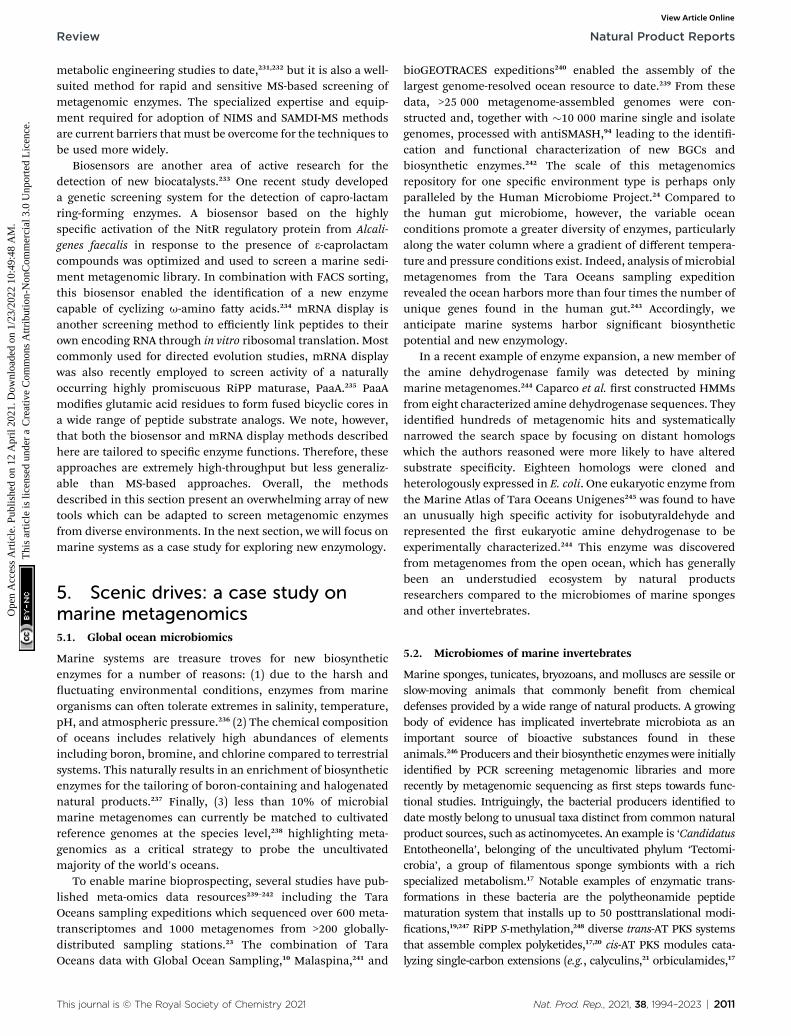
Review Natural Product Reports
Ope
n A
cces
s A
rtic
le. P
ublis
hed
on 1
2 A
pril
2021
. Dow
nloa
ded
on 1
/23/
2022
10:
49:4
8 A
M.
Thi
s ar
ticle
is li
cens
ed u
nder
a C
reat
ive
Com
mon
s A
ttrib
utio
n-N
onC
omm
erci
al 3
.0 U
npor
ted
Lic
ence
.View Article Online
metabolic engineering studies to date,231,232 but it is also a well-suited method for rapid and sensitive MS-based screening ofmetagenomic enzymes. The specialized expertise and equip-ment required for adoption of NIMS and SAMDI-MS methodsare current barriers that must be overcome for the techniques tobe used more widely.
Biosensors are another area of active research for thedetection of new biocatalysts.233 One recent study developeda genetic screening system for the detection of capro-lactamring-forming enzymes. A biosensor based on the highlyspecic activation of the NitR regulatory protein from Alcali-genes faecalis in response to the presence of 3-caprolactamcompounds was optimized and used to screen a marine sedi-ment metagenomic library. In combination with FACS sorting,this biosensor enabled the identication of a new enzymecapable of cyclizing u-amino fatty acids.234 mRNA display isanother screening method to efficiently link peptides to theirown encoding RNA through in vitro ribosomal translation. Mostcommonly used for directed evolution studies, mRNA displaywas also recently employed to screen activity of a naturallyoccurring highly promiscuous RiPP maturase, PaaA.235 PaaAmodies glutamic acid residues to form fused bicyclic cores ina wide range of peptide substrate analogs. We note, however,that both the biosensor and mRNA display methods describedhere are tailored to specic enzyme functions. Therefore, theseapproaches are extremely high-throughput but less generaliz-able than MS-based approaches. Overall, the methodsdescribed in this section present an overwhelming array of newtools which can be adapted to screen metagenomic enzymesfrom diverse environments. In the next section, we will focus onmarine systems as a case study for exploring new enzymology.
5. Scenic drives: a case study onmarine metagenomics5.1. Global ocean microbiomics
Marine systems are treasure troves for new biosyntheticenzymes for a number of reasons: (1) due to the harsh anductuating environmental conditions, enzymes from marineorganisms can oen tolerate extremes in salinity, temperature,pH, and atmospheric pressure.236 (2) The chemical compositionof oceans includes relatively high abundances of elementsincluding boron, bromine, and chlorine compared to terrestrialsystems. This naturally results in an enrichment of biosyntheticenzymes for the tailoring of boron-containing and halogenatednatural products.237 Finally, (3) less than 10% of microbialmarine metagenomes can currently be matched to cultivatedreference genomes at the species level,238 highlighting meta-genomics as a critical strategy to probe the uncultivatedmajority of the world's oceans.
To enable marine bioprospecting, several studies have pub-lished meta-omics data resources239–242 including the TaraOceans sampling expeditions which sequenced over 600 meta-transcriptomes and 1000 metagenomes from >200 globally-distributed sampling stations.23 The combination of TaraOceans data with Global Ocean Sampling,10 Malaspina,241 and
This journal is © The Royal Society of Chemistry 2021
bioGEOTRACES expeditions240 enabled the assembly of thelargest genome-resolved ocean resource to date.239 From thesedata, >25 000 metagenome-assembled genomes were con-structed and, together with �10 000 marine single and isolategenomes, processed with antiSMASH,94 leading to the identi-cation and functional characterization of new BGCs andbiosynthetic enzymes.242 The scale of this metagenomicsrepository for one specic environment type is perhaps onlyparalleled by the Human Microbiome Project.24 Compared tothe human gut microbiome, however, the variable oceanconditions promote a greater diversity of enzymes, particularlyalong the water column where a gradient of different tempera-ture and pressure conditions exist. Indeed, analysis of microbialmetagenomes from the Tara Oceans sampling expeditionrevealed the ocean harbors more than four times the number ofunique genes found in the human gut.243 Accordingly, weanticipate marine systems harbor signicant biosyntheticpotential and new enzymology.
In a recent example of enzyme expansion, a new member ofthe amine dehydrogenase family was detected by miningmarine metagenomes.244 Caparco et al. rst constructed HMMsfrom eight characterized amine dehydrogenase sequences. Theyidentied hundreds of metagenomic hits and systematicallynarrowed the search space by focusing on distant homologswhich the authors reasoned were more likely to have alteredsubstrate specicity. Eighteen homologs were cloned andheterologously expressed in E. coli. One eukaryotic enzyme fromthe Marine Atlas of Tara Oceans Unigenes245 was found to havean unusually high specic activity for isobutyraldehyde andrepresented the rst eukaryotic amine dehydrogenase to beexperimentally characterized.244 This enzyme was discoveredfrom metagenomes from the open ocean, which has generallybeen an understudied ecosystem by natural productsresearchers compared to the microbiomes of marine spongesand other invertebrates.
5.2. Microbiomes of marine invertebrates
Marine sponges, tunicates, bryozoans, and molluscs are sessile orslow-moving animals that commonly benet from chemicaldefenses provided by a wide range of natural products. A growingbody of evidence has implicated invertebrate microbiota as animportant source of bioactive substances found in theseanimals.246 Producers and their biosynthetic enzymes were initiallyidentied by PCR screening metagenomic libraries and morerecently by metagenomic sequencing as rst steps towards func-tional studies. Intriguingly, the bacterial producers identied todate mostly belong to unusual taxa distinct from common naturalproduct sources, such as actinomycetes. An example is ‘CandidatusEntotheonella’, belonging of the uncultivated phylum ‘Tectomi-crobia’, a group of lamentous sponge symbionts with a richspecialized metabolism.17 Notable examples of enzymatic trans-formations in these bacteria are the polytheonamide peptidematuration system that installs up to 50 posttranslational modi-cations,19,247 RiPP S-methylation,248 diverse trans-AT PKS systemsthat assemble complex polyketides,17,20 cis-AT PKS modules cata-lyzing single-carbon extensions (e.g., calyculins,21 orbiculamides,17
Nat. Prod. Rep., 2021, 38, 1994–2023 | 2011

Natural Product Reports Review
Ope
n A
cces
s A
rtic
le. P
ublis
hed
on 1
2 A
pril
2021
. Dow
nloa
ded
on 1
/23/
2022
10:
49:4
8 A
M.
Thi
s ar
ticle
is li
cens
ed u
nder
a C
reat
ive
Com
mon
s A
ttrib
utio
n-N
onC
omm
erci
al 3
.0 U
npor
ted
Lic
ence
.View Article Online
keramamides,249 konbamides17), a promiscuous halogenation,249
and a to-date unique and biosynthetically unassigned peptidecross-link involving a histidine imidazole moiety.250
In addition to ‘Entotheonella’, other producers in spongeshave been identied, all of which remain uncultured. Theseinclude a multiproducer community providing the cytotoxicpelorusides, pateamines, mycalamides, and other compoundsin the sponge Mycale hentscheli, an intracellular producer ofrenieramycin with a highly reduced genome.251,252 Hormoscilla(formerly Oscillatoria) spongeliae, is a cyanobacterial spongesymbiont and a source of halogenated compounds, includingpolybrominated diphenyl ethers (PDBE). The combination of denovo metagenomic sequencing of the sponge metagenome andheterologous expression of the candidate biosynthetic locus forPDBE in a non-standard host, Synechococcus elongatus PCC7942, ultimately led to successful characterization of thisBGC.253 Cyanobacterial symbionts from other marine animals,such as tunicates, are also rich sources of biosynthetic diversity.Biosynthetic pathways for the cyanobactins, a class of cytotoxicRiPPs including the patellamides and trunkamides, were orig-inally discovered from metagenomic eDNA.254–256 The promis-cuity of RiPP maturases encoded in cyanobactin pathways, e.g.,heterocyclases for azoline installation257 or macrocyclases forcyclic peptide formation,258 have been exploited to generatelibraries of synthetic peptides.
Another biosynthetic treasure trove are shipworms, thebivalve molluscs famous for boring holes in wooden boat hullsand piers. Recent shotgun metagenomic analysis coupled withcultivation strategies revealed more than 150–200 distinctBGCs from shipworm gill endosymbionts.259 In addition tobeing biosynthetically talented, shipworm symbionts are ofbiotechnological interest for biomass degradation due to theirwood-based diet. A new enzyme involved in lignocellulosedegradation was recently isolated and characterized from theshipworm symbiont Teredinibacter turnerae.260 This representsa case of enzyme expansion within the family of oxidativeenzymes known as lytic polysaccharide monooxygenases(LPMOs) that degrade chitin and cellulose-like polymers.261
Since the relatively recent discovery of the LPMOs in 2010, thisenzyme family has been of great interest for biotechnologicalapplications including the oxidative degradation of recalcitrantpolymers.261 Perhaps the greatest nding from shipwormmetagenomic studies to date, however, is that most membersof the shipworm gill endosymbiont microbial communities areculturable.259 The ability to cultivate and genetically manipu-late nearly-complete microbial consortia from shipworm gillsprovides an exciting experimentally-tractable system withinwhich to study host-endosymbiont co-evolution of secondarymetabolism.
6. Gearing up for the future: newfrontiers in enzyme discovery
In this section, we will provide an outlook on the future of theeld and highlight emerging techniques which can be pairedwith metagenomics workows to accelerate enzyme discovery.
2012 | Nat. Prod. Rep., 2021, 38, 1994–2023
6.1. Meta-omics
The integration of various meta-omics techniques, includingmetatranscriptomics, metaproteomics, and metabolomics, intoenzyme discovery workows can be a powerful frameworkconnecting genotype to phenotype for hypothesis generation.RNA-Seq, for example, provides a global snapshot of differen-tially expressed genes under conditions of interest to implicatecoding sequences of unknown function in specic cellularprocesses. Maini-Rekdal et al. used RNA-Seq to characterize theinvolvement of an unknown molybdenum-dependent enzyme,DadH, in the catabolism of dopamine in the human gut.262
Differential expression analysis of the gut bacterium Eggerthellalenta revealed dadH was upregulated >2500-fold in the presenceof dopamine. Although DadH from E. lenta only exhibitednarrow substrate specicity for L-dopa and close analogs, met-agenomemining using DadH as a query sequence expanded theprotein family to other molybdenum-dependent enzymescapable of degrading other classes of neurotransmitters anddiet-derived catechols.263 In another example, RNA-Seq analysisof the marine diatom Pseudo-nitzschia under phosphate limi-tation and high CO2 guided the identication of the biosyn-thetic pathway for domoic acid, a harmful neurotoxin.224
Heterologous expression of the domoic acid BGC and structuralanalysis yielded insights into how the unusual biosyntheticenzyme, DabA, that catalyzes N-prenylation of a primary aminearose within the ubiquitous terpene cyclase protein fold.264
Although both of these examples used RNA-Seq to discovernew enzymes from organisms in monoculture, similar strate-gies can be applied to metatranscriptomes. Surprisingly, thenumber of studies using metatranscriptomics for enzymediscovery are still relatively rare but increasing. Recently, met-atranscriptomic analysis of a compost microbial communityresulted in expansion of the glycoside hydrolase family toinclude an unusual enzyme with exo-1,4-b-xylanase activity.265 Anew tool, BiG-MAP, was released to facilitate differentialexpression analysis of BGCs from (meta)transcriptomic data-sets.266 BiG-MAP results can also further be integrated withmetabolomics data. As a proof-of-principle, BiG-MAP was usedto link differentially expressed BGCs from healthy and caries-associated oral microbiome samples with mass fragmentsassociated with reuterin, a natural product inhibiting growth ofthe opportunistic pathogen Streptococcus mutans involved intooth decay. With increasing availability of paired metatran-scriptomic and metabolomic datasets and analysis pipelineslike BiG-MAP, we anticipate meta-omic mining will acceleratethe discovery of new biosynthetic enzymes.
Relative to the other -omics techniques, metaproteomicsremains particularly underexploited. Sukul et al. proposeda workow for functional metaproteomics relying on the directisolation of proteins from soil samples followed by separationusing 2D-polyacrylamide gel electrophoresis. Refolded proteinswere assayed in-gel using a uorogenic lipase substrate to detectnew lipolytic enzymes. Hits were then excised from the gel,digested and subjected to MS analysis. Extracting eDNA fromthe same soil samples for shotgun metagenomic sequencingallowed mass spectra from in-gel lipolytically-active enzymes to
This journal is © The Royal Society of Chemistry 2021
Review Natural Product Reports
Ope
n A
cces
s A
rtic
le. P
ublis
hed
on 1
2 A
pril
2021
. Dow
nloa
ded
on 1
/23/
2022
10:
49:4
8 A
M.
Thi
s ar
ticle
is li
cens
ed u
nder
a C
reat
ive
Com
mon
s A
ttrib
utio
n-N
onC
omm
erci
al 3
.0 U
npor
ted
Lic
ence
.View Article Online
be compared to a custom environmental database to identifyfull-length sequences and permit their taxonomic assign-ment.267 While in-gel metaproteomics workows are viablestrategies for well-characterized enzymes such as lipases, theyare more challenging for enzyme functions lacking establishedcolorimetric or uorimetric substrates. The limited availabilityof functional assays is one downside of metaproteomics inaddition to difficulty with directly isolating proteins fromenvironmental samples and a low likelihood of proper in-gel re-folding of enzymes. Technical challenges notwithstanding, it isclear that the integration of different multi-omics datasetsoffers promising new routes for enzyme discovery.
6.2. Single-cell genomics
Single-cell genomic sequencing is an alternative and comple-mentary approach to shotgun metagenomics. Single-cell geno-mics relies on the sorting of microbial cells, usually withmicrouidics (Section 6.3) or FACS methods, followed by lysisand whole genome multiple displacement amplication withhigh-delity polymerases.268 Despite enabling over a billion-foldamplication of genetic material from a single cell, the quantityof DNA is oen still low, resulting in poor genome quality anda risk of contamination from extracellular DNA. Optimizedprotocols using thermostable polymerases have been developedto improve these issues and also correct for biases againstamplication of GC-rich templates which is especially relevantfor BGCs from Actinobacteria and other organisms with highGC-content.269 The benets of directly linking taxonomic clas-sication to genomic functional content without requiringbinning provides a clear advantage of single-cell genomics overshotgun metagenomic sequencing. Optimally, single-cellgenomics and metagenomics methods are best applied incombination since they have different sampling biases whichminimizes their overlap. Previous analysis in our lab also foundthat reference genomes from cultivated marine isolates rarelyoverlapped with marine SAGs or MAGs, indicating that multi-pronged approaches of cultivation and multiple types ofsequencing contributes to a greater genome-resolved under-standing of ecological community composition.242
There are still relatively few cases of biosynthetic enzymediscovery from SAGs. As the earliest example applied to naturalproduct studies, Grindberg et al. detected the cyanobacterialapratoxin biosynthetic pathway in a mixed bacterial assemblagethrough a combination of single-cell sequencing, in silicomining, and a metagenomic fosmid library screening.270 Basedon biosynthetic logic and a priori knowledge of the apratoxinchemical structure, the authors used known conserved motifsfrom hydroxymethylglutaryl-CoA synthase-like enzymes todetect homologs in their SAG contig library. The motifs werethen used to design degenerate primers and PCR screen theirmetagenomic library to identify overlapping contigs andassemble the complete apratoxin biosynthetic cluster. Thisstudy illustrated how the combination of single-cell genomicsand functional metagenomics ultimately revealed the completeBGC for apratoxin. Skiba et al. later built on these ndings tocharacterize an unusual mononuclear iron-dependent di-
This journal is © The Royal Society of Chemistry 2021
methylating methyltransferase that initiates apratoxin biosyn-thesis through production of branched polyketide starterunits.271 More recently, Mori et al. used single-cell sequencing tocharacterize the remarkable biosynthetic potential of ‘Candi-datus Entotheonella’ symbionts from marine sponges (Section5.2).20
Importantly, single-cell genomics does not rely on theassumption that populations of similar cells are clonal. Asa result, single-cell genomics studies have revealed remarkablewithin-population genome variability and evolution in systemsranging from marine phytoplankton272 to cancer cells.273,274
BGCs are also notoriously strain-specic and single-cellsequencing is useful to detect needle BGCs in the haystack.For example, Sugimoto et al. developed a new HMM-basedcomputational strategy to mine BGCs from human micro-biomes and found that some BGCs are only found in one outstrain out >1000–5500 bacterial isolates from the same taxon.275
Advances in single-cell RNA sequencing in prokaryotes hasfurther demonstrated how even genetically-identical pop-ulations of bacteria exhibit spatial transcriptional heterogeneityin communities.276 Spatial heterogeneity and division of laborhas been documented for secondary metabolite production ina variety of systems both in microbial communities andmonocultures.277 This nascent area of research calls for furtherapplications of single-cell and spatial transcriptomics methodsto better understand how microbial community structure andmicroenvironment affects biosynthetic gene expression.
6.3. Microuidics
Microuidic technologies have revolutionized biomedicine,particularly for devices where ‘lab on a chip’ compactness isdesired. Microuidic-based sorting methods have been widelyapplied for directed evolution and protein engineering studies,278
but have only rarely been used formetagenomic enzyme discovery.Colin et al. screened >1 250 000 water-in-oil droplets usinga microuidic system to identify metagenomic enzymes thathydrolyze sulfate monoesters and phosphotriesters.132 This studyhighlighted microuidics as a useful technique to probe thepromiscuity of metagenomic enzymes in a sequence-independentmanner. Another recent study used optical tweezers and micro-uidics to sort complex microbial communities based on theRaman spectra of individual cells.279 This has numerous applica-tions for downstream single-cell sequencing or cultivation effortsincluding sorting microbes based on stable isotope labels ornatural Raman signals from storage compounds or carotenoids. Byprocessing sorted cells for downstream single-cell sequencing,chemical phenotypes of live individual cells can be directly linkedto their genotypes. Like most other new techniques discussed inthis section, however, the application of microuidics to discovernew biosynthetic enzymes from metagenomes has not yet beenwidely applied.
6.4. Cell-free platforms
An elegant alternative to heterologous expression and proteinpurication is the use of cell-free systems such as ltered lysatefrom E. coli or another host. Since cellular machinery remains
Nat. Prod. Rep., 2021, 38, 1994–2023 | 2013
Natural Product Reports Review
Ope
n A
cces
s A
rtic
le. P
ublis
hed
on 1
2 A
pril
2021
. Dow
nloa
ded
on 1
/23/
2022
10:
49:4
8 A
M.
Thi
s ar
ticle
is li
cens
ed u
nder
a C
reat
ive
Com
mon
s A
ttrib
utio
n-N
onC
omm
erci
al 3
.0 U
npor
ted
Lic
ence
.View Article Online
in the lysate, exogenous addition of components includingcofactors, amino acids, and DNA is all that is required to expressenzymes or pathways of interest.280 Cell-free systems createconditions for rapid transcription and translation of desiredDNA sequences without the constraints of maintaining cellulargrowth. Unlike in vivo expression systems, cell-free platformsalso allow for the production of toxic metabolites that normallykill heterologous hosts. To further increase throughput,screening methods including mRNA display, matrix-assisteddesorption/ionization-MS and in-droplet reaction micro-uidics have already been integrated with cell-free platforms.280
For some biosynthetic pathways, high yields are produced injust a few hours from DNA templates.281,282 In practice, however,low yields are a common challenge especially when workingwith DNA from organisms that are taxonomically-distant fromE. coli.280 Fast degradation of mRNA templates and othernecessary reactions components is another challengecommonly faced when working with cell extract-based systems.Nonetheless, these systems have seen explosive popularity inrecent years, and we anticipate future exploitation of cell-freeplatforms for metagenomic enzyme discovery.
6.5. Sequence-independent methods
On the whole, the vast majority of techniques described in thisreview rely on either sequence-based or structure-based homologyto infer protein function. However, these approaches oen fallshort whenmaking predictions for the ‘unknown unknowns’, thatis, for the de novo discovery of enzymes that do not share sequenceor structural similarity with one or more characterized proteinfamilies. Sequence- or structure-independent approaches are alsorarely used in natural products research, since most computa-tional methods to identify BGCs rely on homology to commonbiosynthetic domains.283 In a departure from sequence-basedmethods, decRiPPter (Data-driven Exploratory Class-independentRiPP TrackER) was developed for the explicit purpose of detect-ing new RiPP classes without relying on homology to known RiPPclasses or enzymatic machinery.284 The core ltering step of thedecRiPPter algorithm uses pan-genomic comparisons to detectoperons that are sparsely distributed within taxonomic groups andthus are likely involved in secondary rather than primarymetabolicfunctions. Kloosterman et al. analyzed 1295 Streptomyces genomeswith decRiPPter to identify a new family of RiPP maturases cata-lyzing dehydration and cyclization reactions for a new lanthipep-tide class of natural products.284 While this singular example isa proof-of-principle that sequence-independent methods can beused successfully for enzyme and natural product discovery, theauthors emphasize a key limitation of this approach is the largenumber of false positives when searching for novelty rather thanhomology. Beyond RiPPs, the eld remains open and poised forthe emergence of new sequence- and structure-independentmethods for enzyme discovery.
7. Conclusions
A major takeaway from this review is the surprising paucity ofstudies of de novo and reference-based enzyme discovery studies
2014 | Nat. Prod. Rep., 2021, 38, 1994–2023
that have used shotgun metagenomics rather than functionalmetagenomics. Even as we amass petabytes of meta-omics datain public databases, there is a disconnect between the relativeease of next-generation sequencing and the difficulty of gaininginsights into new protein families and their functions. Based ona meta-analysis of this review, we will attempt to offer somegeneral recommendations to advance future efforts in the eld:
7.1. Discoveries oen occur at the boundaries of proteinfamilies
Although not a universal rule, proteins with low sequenceidentities to reference proteins of known functions are morelikely than enzymes with high sequence identities to accom-modate different substrates and catalyze new reaction types. Todetect distantly related sequences, we recommend movingbeyond a basic BLAST search and using tools that are moresensitive for remote homology detection such as PSI-BLAST285 orHMMs. MetaHMM286 or HMMSearch tools in the EBI MGnifyportal89 allow non-experts to query metagenomes with proteinqueries or custom HMMs through a web interface. Expert usersmay be interested in applying an iterative HMM search strategy.In this approach, an initial HMM model is used and combinedwith BLAST searches to identify more distant homologs of thesame gene family. The newly identied sequences are then usedto update the initial model and the procedure repeated until noadditional homologs are identied. This strategy has beenapplied to identify new b-defensin members in humans andmice279 and to discover a cysteine-rich gene family in corals.287
7.2. Think outside the colorimetric assay box to move intounexplored protein space
A previous meta-analysis of metagenomic enzymes discoveredbetween Jan 2014 and March 2017 found that >84% belong toeither the lipase/esterase or cellulase/hemicellulase classes.33
Similarly, >82% were discovered by activity-based screening.Clearly, there is a bias in current metagenomic screeningmethods towards industrially relevant enzyme classes that canreadily be detected with standard colorimetric assays. Althoughless well-understood and more challenging to screen, proteinsuperfamilies for which remarkable diversity is already docu-mented, including, but not limited to, radical SAMs enzymes,methyltransferases, thioesterases, and cytochrome p450 mon-ooxygenases, represent hotbeds for new enzymology.
7.3. Move beyond E. coli into new hosts
E. coli has been the workhorse for the vast majority of functionalmetagenomics and heterologous expression efforts to date. Thebenets of using a model organism are undeniable for therelative ease of cloning, expression, and screening. However,a controlled study found that only 30–40% of genes from envi-ronmental bacteria in general and only 7% of high GC-contentDNA could be expressed in E. coli.288 Unfortunately, manysecondary metabolites are produced by organisms classied ashigh GC-content thus classical functional metagenomicsmethods likely fail to capture large swaths of diversity in eDNA.As discussed in Section 4.2, a suite of other problems such as
This journal is © The Royal Society of Chemistry 2021
Review Natural Product Reports
Ope
n A
cces
s A
rtic
le. P
ublis
hed
on 1
2 A
pril
2021
. Dow
nloa
ded
on 1
/23/
2022
10:
49:4
8 A
M.
Thi
s ar
ticle
is li
cens
ed u
nder
a C
reat
ive
Com
mon
s A
ttrib
utio
n-N
onC
omm
erci
al 3
.0 U
npor
ted
Lic
ence
.View Article Online
the lack of proper coenzymes, substrates, cofactors, post-translational modication systems, self-resistance genes,protein folding factors, and additional proteins required formegasynthases (e.g., MbtH-like proteins, PPTases) all can causeheterologous expression in E. coli to fail. One strategy to trou-bleshoot these issues is using alternative hosts. For functionalmetagenomics, Pseudomonas, Streptomyces, Rhodococcus,Bacillus, and even archaea have been used as library hosts aswell as multi-host expression systems with shuttle vectors.289,290
Similarly, non-traditional heterologous expression hosts suchas M. aerodenitricans221 have been developed to access newenzymology from metagenomic BGCs.
7.4. (Genome) context is everything
There has been an explosion of new tools devoted to examinationof genes in the context of their genome neighborhoods rather thanin isolation. This has been a particularly fruitful strategy for thediscovery of new multi-domain enzyme functions148 or RiPPmaturases.173 Taking this one step further, deep learning methodsfor embedding genes as vectors in their genomic context (e.g.,pfam2vec) have led to improvements in BGC prediction.189
Although the current reliance on short-read sequencing methodsrequires binning and assembly to extract genomic context fromshotgun metagenomes, we anticipate that advances in long-readmetagenome sequencing will pave the way for genome neighbor-hoods to be analyzedmore directly and accurately from eDNA. Justas bacteria act differently in communities than in isolation, wepropose that enzymes are best studied and understood in thecomplete milieu of their anking genes. Identication of genecoexpression modules from (meta)transcriptomic datasets canadditionally be used reconstruct transcriptional units and predictthe function of unknown genes through coexpression of geneswith known function. Coexpression networks can also guidehypotheses regarding protein–protein interactions and complexformation. Overall, the protein interactome is an underexploredavenue to uncover new enzyme functions.
Perhaps one day we will see the emergence of Alpha-Function2 as a successor to AlphaFold2. But in order to trainarticial intelligence models to tackle the sequence–structure–function problem, continued exploration of new areas ofprotein space is an important task for experimental enzymolo-gists and computational researchers alike. As a nal remark, weemphasize that AlphaFold2, and many other computationaltools described in this review, were only made possible throughthe biochemical characterization of thousands of proteins byexperimentalists at the bench. Experimental work is more crit-ical than ever to enable new data-driven discoveries.
8. Conflicts of interest
The authors declare no conicts of interest.
9. Acknowledgements
S. L. R. is supported by an ETH Zurich Postdoctoral Fellowship(20-1 FEL-07). Work in J. P.'s lab on uncultivated bacteria was
This journal is © The Royal Society of Chemistry 2021
supported by grants from the Swiss National Science Founda-tion (205321_165695), the Gordon and Betty Moore Foundation(#9204, DOI: 10.37807/GBMF9204), the Helmut Horten Foun-dation, and the Promedica Foundation. Work on the oceanmicrobiome and bioinformatics tool development in S. S.'s labhas been supported by grants of the Swiss National ScienceFoundation (205321_184955), the NCCR Microbiomes(51NF40_180575) and the ETH domain (PHRT #521).
10. References
1 J. Jumper, R. Evans, A. Pritzel, T. Green, M. Figurnov,K. Tunyasuvunakool, O. Ronneberger, R. Bates, A. Zidek,A. Bridgland and C. Meyer, Fourteenth Critical Assessmentof Techniques for Protein Structure Prediction, 2020, pp. 22–24.
2 S. Ghatak, Z. A. King, A. Sastry and B. O. Palsson, NucleicAcids Res., 2019, 47, 2446–2454.
3 R. S. Kaas, C. Friis, D. W. Ussery and F. M. Aarestrup, BMCGenomics, 2012, 13, 577.
4 D. A. Rasko, M. J. Rosovitz, G. S. A. Myers, E. F. Mongodin,W. F. Fricke, P. Gajer, J. Crabtree, M. Sebaihia,N. R. Thomson, R. Chaudhuri, I. R. Henderson,V. Sperandio and J. Ravel, J. Bacteriol., 2008, 190, 6881–6893.
5 Z. D. Blount, eLife, 2015, 4, e05826.6 J. Handelsman, M. R. Rondon, S. F. Brady, J. Clardy andR. M. Goodman, Chem. Biol., 1998, 5, R245–R249.
7 F. Warnecke, P. Luginbuhl, N. Ivanova, M. Ghassemian,T. H. Richardson, J. T. Stege, M. Cayouette,A. C. McHardy, G. Djordjevic, N. Aboushadi, R. Sorek,S. G. Tringe, M. Podar, H. G. Martin, V. Kunin, D. Dalevi,J. Madejska, E. Kirton, D. Platt, E. Szeto, A. Salamov,K. Barry, N. Mikhailova, N. C. Kyrpides, E. G. Matson,E. A. Ottesen, X. Zhang, M. Hernandez, C. Murillo,L. G. Acosta, I. Rigoutsos, G. Tamayo, B. D. Green,C. Chang, E. M. Rubin, E. J. Mathur, D. E. Robertson,P. Hugenholtz and J. R. Leadbetter, Nature, 2007, 450,560–565.
8 B. E. Wolfe, J. E. Button, M. Santarelli and R. J. Dutton, Cell,2014, 158, 422–433.
9 N. A. Be, A. Avila-Herrera, J. E. Allen, N. Singh, A. ChecinskaSielaff, C. Jaing and K. Venkateswaran,Microbiome, 2017, 5,81.
10 S. Yooseph, G. Sutton, D. B. Rusch, A. L. Halpern,S. J. Williamson, K. Remington, J. A. Eisen,K. B. Heidelberg, G. Manning, W. Li, L. Jaroszewski,P. Cieplak, C. S. Miller, H. Li, S. T. Mashiyama,M. P. Joachimiak, C. van Belle, J.-M. Chandonia,D. A. Soergel, Y. Zhai, K. Natarajan, S. Lee, B. J. Raphael,V. Bafna, R. Friedman, S. E. Brenner, A. Godzik,D. Eisenberg, J. E. Dixon, S. S. Taylor, R. L. Strausberg,M. Frazier and J. C. Venter, PLoS Biol., 2007, 5, e16.
11 Z. D. Stephens, S. Y. Lee, F. Faghri, R. H. Campbell, C. Zhai,M. J. Efron, R. Iyer, M. C. Schatz, S. Sinha andG. E. Robinson, PLoS Biol., 2015, 13, e1002195.
Nat. Prod. Rep., 2021, 38, 1994–2023 | 2015
Natural Product Reports Review
Ope
n A
cces
s A
rtic
le. P
ublis
hed
on 1
2 A
pril
2021
. Dow
nloa
ded
on 1
/23/
2022
10:
49:4
8 A
M.
Thi
s ar
ticle
is li
cens
ed u
nder
a C
reat
ive
Com
mon
s A
ttrib
utio
n-N
onC
omm
erci
al 3
.0 U
npor
ted
Lic
ence
.View Article Online
12 L. Solden, K. Lloyd and K. Wrighton, Curr. Opin. Microbiol.,2016, 31, 217–226.
13 L. A. Hug, B. J. Baker, K. Anantharaman, C. T. Brown,A. J. Probst, C. J. Castelle, C. N. Buttereld,A. W. Hernsdorf, Y. Amano, K. Ise, Y. Suzuki, N. Dudek,D. A. Relman, K. M. Finstad, R. Amundson, B. C. Thomasand J. F. Baneld, Nat. Microbiol., 2016, 1, 16048.
14 D. H. Parks, M. Chuvochina, D. W. Waite, C. Rinke,A. Skarshewski, P.-A. Chaumeil and P. Hugenholtz, Nat.Biotechnol., 2018, 36, 996–1004.
15 C. J. Castelle, K. C. Wrighton, B. C. Thomas, L. A. Hug,C. T. Brown, M. J. Wilkins, K. R. Frischkorn, S. G. Tringe,A. Singh, L. M. Markillie, R. C. Taylor, K. H. Williams andJ. F. Baneld, Curr. Biol., 2015, 25, 690–701.
16 A. L. Jaffe, C. J. Castelle, P. B. Matheus Carnevali,S. Gribaldo and J. F. Baneld, BMC Biol., 2020, 18, 69.
17 M. C. Wilson, T. Mori, C. Ruckert, A. R. Uria, M. J. Helf,K. Takada, C. Gernert, U. A. E. Steffens, N. Heycke,S. Schmitt, C. Rinke, E. J. N. Helfrich, A. O. Brachmann,C. Gurgui, T. Wakimoto, M. Kracht, M. Crusemann,U. Hentschel, I. Abe, S. Matsunaga, J. Kalinowski,H. Takeyama and J. Piel, Nature, 2014, 506, 58–62.
18 A. Crits-Christoph, S. Diamond, C. N. Buttereld,B. C. Thomas and J. F. Baneld, Nature, 2018, 558, 440–444.
19 M. F. Freeman, M. J. Helf, A. Bhushan, B. I. Morinaka andJ. Piel, Nat. Chem., 2017, 9, 387–395.
20 T. Mori, J. K. B. Cahn, M. C. Wilson, R. A. Meoded,V. Wiebach, A. F. C. Martinez, E. J. N. Helfrich,A. Albersmeier, D. Wibberg, S. Datwyler, R. Keren, A. Lavy,C. Ruckert, M. Ilan, J. Kalinowski, S. Matsunaga,H. Takeyama and J. Piel, Proc. Natl. Acad. Sci. U. S. A.,2018, 115, 1718–1723.
21 T. Wakimoto, Y. Egami, Y. Nakashima, Y. Wakimoto,T. Mori, T. Awakawa, T. Ito, H. Kenmoku, Y. Asakawa,J. Piel and I. Abe, Nat. Chem. Biol., 2014, 10, 648–655.
22 S. K. Wyman, A. Avila-Herrera, S. Nayfach and K. S. Pollard,PLoS One, 2018, 13, e0205749.
23 S. Sunagawa, S. G. Acinas, P. Bork, C. Bowler, Tara OceansCoordinators, D. Eveillard, G. Gorsky, L. Guidi,D. Iudicone, E. Karsenti, F. Lombard, H. Ogata, S. Pesant,M. B. Sullivan, P. Wincker and C. de Vargas, Nat. Rev.Microbiol., 2020, 18, 428–445.
24 J. Lloyd-Price, A. Mahurkar, G. Rahnavard, J. Crabtree,J. Orvis, A. Brantley Hall, A. Brady, H. H. Creasy,C. McCracken, M. G. Giglio, D. McDonald, E. A. Franzosa,R. Knight, O. White and C. Huttenhower, Nature, 2017,550, 61–66.
25 M. G. Chevrette, K. Gutierrez-Garcıa, N. Selem-Mojica,C. Aguilar-Martınez, A. Yanez-Olvera, H. E. Ramos-Aboites, P. A. Hoskisson and F. Barona-Gomez, Nat. Prod.Rep., 2020, 37, 566–599.
26 A. M. Kloosterman, P. Cimermancic, S. S. Elsayed, C. Du,M. Hadjithomas, M. S. Donia, M. A. Fischbach, G. P. vanWezel and M. H. Medema, PLoS Biol., 2020, 18, e3001026.
27 S. Ovchinnikov, H. Park, N. Varghese, P.-S. Huang,G. A. Pavlopoulos, D. E. Kim, H. Kamisetty, N. C. Kyrpidesand D. Baker, Science, 2017, 355, 294–298.
2016 | Nat. Prod. Rep., 2021, 38, 1994–2023
28 Y. Wang, Q. Shi, P. Yang, C. Zhang, S. M. Mortuza, Z. Xue,K. Ning and Y. Zhang, Genome Biol., 2019, 20, 229.
29 A. J. Waldman and E. P. Balskus, J. Org. Chem., 2018, 83,7539–7546.
30 J. K. Christenson, S. L. Robinson, T. A. Engel, J. E. Richman,A. N. Kim and L. P. Wackett, Biochemistry, 2017, 56, 5278–5287.
31 M. Ohashi, C. S. Jamieson, Y. Cai, D. Tan, D. Kanayama,M.-C. Tang, S. M. Anthony, J. V. Chari, J. S. Barber,E. Picazo, T. B. Kakule, S. Cao, N. K. Garg, J. Zhou,K. N. Houk and Y. Tang, Nature, 2020, 586, 64–69.
32 A. Chien, D. B. Edgar and J. M. Trela, J. Bacteriol., 1976, 127,1550–1557.
33 F. Berini, C. Casciello, G. L. Marcone and F. Marinelli, FEMSMicrobiol. Lett., 2017, 364(21), fnx211.
34 L. Ufarte, G. Potocki-Veronese and E. Laville, Front.Microbiol., 2015, 6, 563.
35 S. A. Kautsar, K. Blin, S. Shaw, J. C. Navarro-Munoz,B. R. Terlouw, J. J. J. van der Hoo, J. A. van Santen,V. Tracanna, H. G. Suarez Duran, V. Pascal Andreu,N. Selem-Mojica, M. Alanjary, S. L. Robinson, G. Lund,S. C. Epstein, A. C. Sisto, L. K. Charkoudian, J. Collemare,R. G. Linington, T. Weber and M. H. Medema, NucleicAcids Res., 2020, 48, D454–D458.
36 C. J. Oldeld, V. N. Uversky, A. Keith Dunker and L. Kurgan,Intrinsically Disord. Proteins, 2019, 1–34.
37 M. G. Chevrette, C. M. Carlson, H. E. Ortega, C. Thomas,G. E. Ananiev, K. J. Barns, A. J. Book, J. Cagnazzo,C. Carlos, W. Flanigan, K. J. Grubbs, H. A. Horn,F. M. Hoffmann, J. L. Klassen, J. J. Knack, G. R. Lewin,B. R. McDonald, L. Muller, W. G. P. Melo, A. A. Pinto-Tomas, A. Schmitz, E. Wendt-Pienkowski, S. Wildman,M. Zhao, F. Zhang, T. S. Bugni, D. R. Andes, M. T. Pupoand C. R. Currie, Nat. Commun., 2019, 10, 516.
38 A. B. Chase, D. Sweeney, M. N. Muskat and D. Guillen-Matus, bioRxiv, 2021, DOI: 10.1101/2020.12.19.423547.
39 J. Piel and M. Rust, Compr. Nat. Prod. Chem., 2020, 50–89.40 M. Katz, B. M. Hover and S. F. Brady, J. Ind. Microbiol.
Biotechnol., 2016, 43, 129–141.41 M. Ferrer, M. Martınez-Martınez, R. Bargiela, W. R. Streit,
O. V. Golyshina and P. N. Golyshin, Microb. Biotechnol.,2016, 9, 22–34.
42 J. G. Owen, B. V. B. Reddy, M. A. Ternei, Z. Charlop-Powers,P. Y. Calle, J. H. Kim and S. F. Brady, Proc. Natl. Acad. Sci. U.S. A., 2013, 110, 11797–11802.
43 S. Hrvatin and J. Piel, J. Microbiol. Methods, 2007, 68, 434–436.
44 V. Libis, N. Antonovsky, M. Zhang, Z. Shang, D. Montiel,J. Maniko, M. A. Ternei, P. Y. Calle, C. Lemetre,J. G. Owen and S. F. Brady, Nat. Commun., 2019, 10, 3848.
45 B. M. Hover, S.-H. Kim, M. Katz, Z. Charlop-Powers,J. G. Owen, M. A. Ternei, J. Maniko, A. B. Estrela,H. Molina, S. Park, D. S. Perlin and S. F. Brady, Nat.Microbiol., 2018, 3, 415–422.
46 M. F. Laursen, M. D. Dalgaard and M. I. Bahl, Front.Microbiol., 2017, 8, 1934.
This journal is © The Royal Society of Chemistry 2021

Review Natural Product Reports
Ope
n A
cces
s A
rtic
le. P
ublis
hed
on 1
2 A
pril
2021
. Dow
nloa
ded
on 1
/23/
2022
10:
49:4
8 A
M.
Thi
s ar
ticle
is li
cens
ed u
nder
a C
reat
ive
Com
mon
s A
ttrib
utio
n-N
onC
omm
erci
al 3
.0 U
npor
ted
Lic
ence
.View Article Online
47 M. Ayling, M. D. Clark and R. M. Leggett, Briengs Bioinf.,2020, 21, 584–594.
48 Z. Wang, Y. Wang, J. A. Fuhrman, F. Sun and S. Zhu,Briengs Bioinf., 2020, 21, 777–790.
49 O. G. G. Almeida and E. C. P. De Martinis, Appl. Microbiol.Biotechnol., 2019, 103, 69–82.
50 R. R. Pal, R. P. More and H. J. Purohit, So Computing forBiological Systems, 2018, pp. 91–110.
51 T. Kvist, L. Sondt-Marcussen andM. J. Mikkelsen, PLoS One,2014, 9, e106817.
52 R. D. Stewart, M. D. Auffret, A. Warr, A. H. Wiser,M. O. Press, K. W. Langford, I. Liachko, T. J. Snelling,R. J. Dewhurst, A. W. Walker, R. Roehe and M. Watson,Nat. Commun., 2018, 9, 870.
53 E. Yaffe and D. A. Relman, Nat. Microbiol., 2020, 5, 343–353.54 M. Jain, H. E. Olsen, B. Paten and M. Akeson, Genome Biol.,
2016, 17, 239.55 T. Hon, K. Mars, G. Young, Y.-C. Tsai, J. W. Karalius,
J. M. Landolin, N. Maurer, D. Kudrna, M. A. Hardigan,C. C. Steiner, S. J. Knapp, D. Ware, B. Shapiro, P. Pelusoand D. R. Rank, Sci. Data, 2020, 7, 399.
56 D. Lang, S. Zhang, P. Ren, F. Liang, Z. Sun, G. Meng, Y. Tan,X. Li, Q. Lai, L. Han, D. Wang, F. Hu, W. Wang and S. Liu,GigaScience, 2020, 9, giaa123.
57 J. Risse, M. Thomson, S. Patrick, G. Blakely,G. Koutsovoulos, M. Blaxter and M. Watson, GigaScience,2015, 4, 60.
58 D. B. B. Trivella and R. de Felicio, mSystems, 2016, 3, 18.59 R. K. Wierenga, FEBS Lett., 2001, 492, 193–198.60 S. Martınez Cuesta, S. A. Rahman, N. Furnham and
J. M. Thornton, Biophys. J., 2015, 109, 1082–1086.61 S. D. Copley, Curr. Opin. Struct. Biol., 2017, 47, 167–175.62 O. Khersonsky and D. S. Tawk, Annu. Rev. Biochem., 2010,
79, 471–505.63 A. Pabis, F. Duarte and S. C. L. Kamerlin, Biochemistry, 2016,
55, 3061–3081.64 W. Ding, X. Ji, Y. Li and Q. Zhang, Front. Chem., 2016, 4, 27.65 S. Martinez Cuesta, N. Furnham, S. A. Rahman, I. Sillitoe
and J. M. Thornton, Curr. Opin. Struct. Biol., 2014, 26,121–130.
66 L. Noda-Garcia and D. S. Tawk, Curr. Opin. Chem. Biol.,2020, 59, 147–154.
67 V. Veprinskiy, L. Heizinger, M. G. Plach and R. Merkl, BMCEvol. Biol., 2017, 17, 36.
68 S. Z. Alborzi, M.-D. Devignes and D. W. Ritchie, BMC Bioinf.,2017, 18, 107.
69 L. M. Podust and D. H. Sherman, Nat. Prod. Rep., 2012, 29,1251.
70 P. Wang, X. Gao and Y. Tang, Curr. Opin. Chem. Biol., 2012,16, 362–369.
71 C. Perry, E. L. C. de Los Santos, L. M. Alkhalaf andG. L. Challis, Nat. Prod. Rep., 2018, 35, 622–632.
72 G. Zhang, W. Zhang, Q. Zhang, T. Shi, L. Ma, Y. Zhu, S. Li,H. Zhang, Y.-L. Zhao, R. Shi and C. Zhang, Angew. Chem.,Int. Ed., 2014, 53, 4840–4844.
73 J. Antosch, F. Schaefers and T. A. M. Gulder, Angew. Chem.,Int. Ed., 2014, 53, 3011–3014.
This journal is © The Royal Society of Chemistry 2021
74 A. Greule, J. E. Stok, J. J. De Voss and M. J. Cryle, Nat. Prod.Rep., 2018, 35, 757–791.
75 V. B. Urlacher and M. Girhard, Trends Biotechnol., 2019, 37,882–897.
76 M. M. Zdouc, M. M. Alanjary, G. S. Zarazua, S. I. Maffioli,M. Crusemann, M. H. Medema, S. Donadio and M. Sosio,Cell Chem. Biol., 2020, DOI: 10.1016/j.chembiol.2020.11.009.
77 P. S. Coelho, E. M. Brustad, A. Kannan and F. H. Arnold,Science, 2013, 339, 307–310.
78 S. B. J. Kan, S. B. Jennifer Kan, R. D. Lewis, K. Chen andF. H. Arnold, Science, 2016, 354, 1048–1051.
79 S. B. J. Kan, X. Huang, Y. Gumulya, K. Chen andF. H. Arnold, Nature, 2017, 552, 132–136.
80 B. Fu and E. P. Balskus, Curr. Opin. Biotechnol., 2020, 65, 94–101.
81 A. L. Vagstad, T. Kuranaga, S. Puntener, V. R. Pattabiraman,J. W. Bode and J. Piel, Angew. Chem., Int. Ed., 2019, 58, 2246–2250.
82 B. I. Morinaka, E. Lakis, M. Verest, M. J. Helf, T. Scalvenzi,A. L. Vagstad, J. Sims, S. Sunagawa, M. Gugger and J. Piel,Science, 2018, 359, 779–782.
83 T. Q. N. Nguyen, Y. W. Tooh, R. Sugiyama, T. P. D. Nguyen,M. Purushothaman, L. C. Leow, K. Hanif, R. H. S. Yong,I. Agatha, F. R. Winnerdy, M. Gugger, A. T. Phan andB. I. Morinaka, Nat. Chem., 2020, 12, 1042–1053.
84 V. Bandarian, Biochim. Biophys. Acta, 2012, 1824, 1245–1253.
85 E. A. Lilla and K. Yokoyama, Nat. Chem. Biol., 2016, 12, 905–907.
86 T. A. Scott and J. Piel, Nat. Rev. Chem., 2019, 3, 404–425.87 I.-M. A. Chen, K. Chu, K. Palaniappan, A. Ratner, J. Huang,
M. Huntemann, P. Hajek, S. Ritter, N. Varghese,R. Seshadri, S. Roux, T. Woyke, E. A. Eloe-Fadrosh,N. N. Ivanova and N. C. Kyrpides, Nucleic Acids Res., 2021,49(D1), D751–D763.
88 K. Youens-Clark, M. Bomhoff, A. J. Ponsero, E. M. Wood-Charlson, J. Lynch, I. Choi, J. H. Hartman andB. L. Hurwitz, GigaScience, 2019, 8, giz083.
89 A. L. Mitchell, A. Almeida, M. Beracochea, M. Boland,J. Burgin, G. Cochrane, M. R. Crusoe, V. Kale, S. C. Potter,L. J. Richardson, E. Sakharova, M. Scheremetjew,A. Korobeynikov, A. Shlemov, O. Kunyavskaya, A. Lapidusand R. D. Finn, Nucleic Acids Res., 2020, 48(D1), D570–D578.
90 S. F. Altschul, W. Gish, W. Miller, E. W. Myers andD. J. Lipman, J. Mol. Biol., 1990, 215, 403–410.
91 B. Buchnk, C. Xie and D. H. Huson,Nat. Methods, 2015, 12,59–60.
92 P. R. Neubauer, C. Widmann, D. Wibberg, L. Schroder,M. Frese, T. Kottke, J. Kalinowski, H. H. Niemann andN. Sewald, PLoS One, 2018, 13, e0196797.
93 S. Nayfach, S. Roux, R. Seshadri, D. Udwary, N. Varghese,F. Schulz, D. Wu, D. Paez-Espino, I.-M. Chen,M. Huntemann, K. Palaniappan, J. Ladau, S. Mukherjee,T. B. K. Reddy, T. Nielsen, E. Kirton, J. P. Faria,J. N. Edirisinghe, C. S. Henry, S. P. Jungbluth, D. Chivian,P. Dehal, E. M. Wood-Charlson, A. P. Arkin, S. G. Tringe,
Nat. Prod. Rep., 2021, 38, 1994–2023 | 2017
Natural Product Reports Review
Ope
n A
cces
s A
rtic
le. P
ublis
hed
on 1
2 A
pril
2021
. Dow
nloa
ded
on 1
/23/
2022
10:
49:4
8 A
M.
Thi
s ar
ticle
is li
cens
ed u
nder
a C
reat
ive
Com
mon
s A
ttrib
utio
n-N
onC
omm
erci
al 3
.0 U
npor
ted
Lic
ence
.View Article Online
A. Visel, IMG/M Data Consortium, T. Woyke, N. J. Mouncey,N. N. Ivanova, N. C. Kyrpides and E. A. Eloe-Fadrosh, Nat.Biotechnol., 2020, 1–11.
94 K. Blin, S. Shaw, K. Steinke, R. Villebro, N. Ziemert,S. Y. Lee, M. H. Medema and T. Weber, Nucleic Acids Res.,2019, 47, W81–W87.
95 T. Koutsandreas, E. Ladoukakis, E. Pilalis, D. Zarafeta,F. N. Kolisis, G. Skretas and A. A. Chatziioannou, Front.Genet., 2019, 10, 469.
96 C. Darwin, On the Origin of Species, 1871.97 S. L. Robinson, B. R. Terlouw, M. D. Smith, S. J. Pidot,
T. P. Stinear, M. H. Medema and L. P. Wackett, J. Biol.Chem., 2020, 295, 14826–14839.
98 J. Santos-Aberturas, G. Chandra, L. Frattaruolo, R. Lacret,T. H. Pham, N. M. Vior, T. H. Eyles and A. W. Truman,Nucleic Acids Res., 2019, 47, 4624–4637.
99 B. Boeckmann, A. Bairoch, R. Apweiler, M.-C. Blatter,A. Estreicher, E. Gasteiger, M. J. Martin, K. Michoud,C. O'Donovan, I. Phan, S. Pilbout and M. Schneider,Nucleic Acids Res., 2003, 31, 365–370.
100 H. M. Berman, J. Westbrook, Z. Feng, G. Gilliland,T. N. Bhat, H. Weissig, I. N. Shindyalov and P. E. Bourne,Nucleic Acids Res., 2000, 28, 235–242.
101 A. Sanchez-Reyes and J. L. Folch-Mallol, in Metagenomics,ed. W. N. Hozzein, IntechOpen, Rijeka, 2020.
102 A. D. Young and J. P. Gillung, Syst. Entomol., 2020, 45, 225–247.
103 R. C. Edgar, Nucleic Acids Res., 2004, 32, 1792–1797.104 K. Katoh, K. Misawa, K.-I. Kuma and T. Miyata,Nucleic Acids
Res., 2002, 30, 3059–3066.105 F. Sievers and D. G. Higgins, Protein Sci., 2018, 27, 135–145.106 S. Capella-Gutierrez, J. M. Silla-Martınez and T. Gabaldon,
Bioinformatics, 2009, 25, 1972–1973.107 D. M. Portik and J. J. Wiens, Syst. Biol., 2020, syaa064, DOI:
10.1093/sysbio/syaa064.108 M. N. Price, P. S. Dehal and A. P. Arkin, PLoS One, 2010, 5,
e9490.109 X. Zhou, X.-X. Shen, C. T. Hittinger and A. Rokas, Mol. Biol.
Evol., 2018, 35, 486–503.110 K. Liu, C. R. Linder and T. Warnow, PLoS One, 2011, 6,
e27731.111 A. Stamatakis, Bioinformatics, 2014, 30, 1312–1313.112 A. M. Kozlov, D. Darriba, T. Flouri, B. Morel and
A. Stamatakis, Bioinformatics, 2019, 35, 4453–4455.113 B. Q. Minh, H. A. Schmidt, O. Chernomor, D. Schrempf,
M. D. Woodhams, A. von Haeseler and R. Lanfear, Mol.Biol. Evol., 2020, 37, 1530–1534.
114 J. Huerta-Cepas, F. Serra and P. Bork,Mol. Biol. Evol., 2016,33, 1635–1638.
115 G. Yu, Curr. Protoc. Bioinf., 2020, 69, e96.116 G. K. A. Hochberg and J. W. Thornton, Annu. Rev. Biophys.,
2017, 46, 247–269.117 N. M. Hendrikse, G. Charpentier, E. Nordling and
P.-O. Syren, FEBS J., 2018, 285, 4660–4673.118 H. Ashkenazy, O. Penn, A. Doron-Faigenboim, O. Cohen,
G. Cannarozzi, O. Zomer and T. Pupko, Nucleic Acids Res.,2012, 40, W580–W584.
2018 | Nat. Prod. Rep., 2021, 38, 1994–2023
119 Q. Zhang, Y. Yu, J. E. Velasquez and W. A. van der Donk,Proc. Natl. Acad. Sci. U. S. A., 2012, 109, 18361–18366.
120 M. Adamek, M. Alanjary and N. Ziemert, Nat. Prod. Rep.,2019, 36, 1295–1312.
121 N. Ziemert, S. Podell, K. Penn, J. H. Badger, E. Allen andP. R. Jensen, PLoS One, 2012, 7, e34064.
122 M. G. Chevrette, F. Aicheler, O. Kohlbacher, C. R. Currieand M. H. Medema, Bioinformatics, 2017, 33, 3202–3210.
123 E. J. N. Helfrich, R. Ueoka, A. Dolev, M. Rust, R. A. Meoded,A. Bhushan, G. Califano, R. Costa, M. Gugger, C. Steinbeck,P. Moreno and J. Piel, Nat. Chem. Biol., 2019, 15, 813–821.
124 M. E. Horsman, T. P. A. Hari and C. N. Boddy, Nat. Prod.Rep., 2016, 33, 183–202.
125 T. P. A. Hari, P. Labana, M. Boileau and C. N. Boddy,ChemBioChem, 2014, 15, 2656–2661.
126 J. Durairaj, A. Di Girolamo, H. J. Bouwmeester, D. deRidder, J. Beekwilder and A. D. van Dijk, Phytochemistry,2019, 158, 157–165.
127 N. Selem-Mojica, C. Aguilar, K. Gutierrez-Garcıa,C. E. Martınez-Guerrero and F. Barona-Gomez, Microb.Genomics, 2019, 5, 445270.
128 J. C. Navarro-Munoz, N. Selem-Mojica, M. W. Mullowney,S. A. Kautsar, J. H. Tryon, E. I. Parkinson, E. L. C. De LosSantos, M. Yeong, P. Cruz-Morales, S. Abubucker,A. Roeters, W. Lokhorst, A. Fernandez-Guerra,L. T. D. Cappelini, A. W. Goering, R. J. Thomson,W. W. Metcalf, N. L. Kelleher, F. Barona-Gomez andM. H. Medema, Nat. Chem. Biol., 2020, 16, 60–68.
129 J. K. Schniete, P. Cruz-Morales, N. Selem-Mojica,L. T. Fernandez-Martınez, I. S. Hunter, F. Barona-Gomezand P. A. Hoskisson, mBio, 2018, 9, e02283-17.
130 P. Cruz-Morales, J. F. Kopp, C. Martınez-Guerrero,L. A. Yanez-Guerra, N. Selem-Mojica, H. Ramos-Aboites,J. Feldmann and F. Barona-Gomez, Genome Biol. Evol.,2016, 8, 1906–1916.
131 H. J. Atkinson, J. H. Morris, T. E. Ferrin and P. C. Babbitt,PLoS One, 2009, 4, e4345.
132 P.-Y. Colin, B. Kintses, F. Gielen, C. M. Miton, G. Fischer,M. F. Mohamed, M. Hyvonen, D. P. Morgavi,D. B. Janssen and F. Hollfelder, Nat. Commun., 2015, 6,10008.
133 E. Akiva, J. N. Copp, N. Tokuriki and P. C. Babbitt, Proc.Natl. Acad. Sci. U. S. A., 2017, 114, E9549–E9558.
134 P. Shannon, A. Markiel, O. Ozier, N. S. Baliga, J. T. Wang,D. Ramage, N. Amin, B. Schwikowski and T. Ideker,Genome Res., 2003, 13, 2498–2504.
135 J. A. Gustavsen, S. Pai, R. Isserlin, B. Demchak andA. R. Pico, F1000Research, 2019, 8, 1774.
136 K. Ono, T. Muetze, G. Kolishovski, P. Shannon andB. Demchak, F1000Research, 2015, 4, 478.
137 G. Csardi and T. Nepusz, InterJournal, 2006, 1695, 1–9.138 J. A. Gerlt, J. T. Bouvier, D. B. Davidson, H. J. Imker,
B. Sadkhin, D. R. Slater and K. L. Whalen, Biochim.Biophys. Acta, 2015, 1854, 1019–1037.
139 K. C. Mok, O. M. Sokolovskaya, A. M. Nicolas, Z. F. Hallberg,A. Deutschbauer, H. K. Carlson and M. E. Taga,mBio, 2020,11, e02507–e02520.
This journal is © The Royal Society of Chemistry 2021

Review Natural Product Reports
Ope
n A
cces
s A
rtic
le. P
ublis
hed
on 1
2 A
pril
2021
. Dow
nloa
ded
on 1
/23/
2022
10:
49:4
8 A
M.
Thi
s ar
ticle
is li
cens
ed u
nder
a C
reat
ive
Com
mon
s A
ttrib
utio
n-N
onC
omm
erci
al 3
.0 U
npor
ted
Lic
ence
.View Article Online
140 R. Zallot, N. Oberg and J. A. Gerlt, Biochemistry, 2019, 58,4169–4182.
141 S. A. Kautsar, J. J. J. van der Hoo, D. de Ridder andM. H. Medema, GigaScience, 2021, 10, giaa154.
142 J. I. Tietz, C. J. Schwalen, P. S. Patel, T. Maxson, P. M. Blair,H.-C. Tai, U. I. Zakai and D. A. Mitchell, Nat. Chem. Biol.,2017, 13, 470–478.
143 M. D. Mungan, M. Alanjary, K. Blin, T. Weber,M. H. Medema and N. Ziemert, Nucleic Acids Res., 2020,48, W546–W552.
144 E. J. Culp, N. Waglechner, W. Wang, A. A. Fiebig-Comyn,Y.-P. Hsu, K. Koteva, D. Sychantha, B. K. Coombes,M. S. Van Nieuwenhze, Y. V. Brun and G. D. Wright,Nature, 2020, 578, 582–587.
145 L. Aravind, Genome Res., 2000, 10, 1074–1077.146 D. Szklarczyk, A. L. Gable, D. Lyon, A. Junge, S. Wyder,
J. Huerta-Cepas, M. Simonovic, N. T. Doncheva,J. H. Morris, P. Bork, L. J. Jensen and C. von Mering,Nucleic Acids Res., 2019, 47, D607–D613.
147 R. D. Zwahlen, C. Pohl, R. A. L. Bovenberg andA. J. M. Driessen, ACS Synth. Biol., 2019, 8, 1776–1787.
148 T. de Rond, J. E. Asay and B. S. Moore, bioRxiv, 2020, DOI:10.1101/2020.06.11.147165.
149 F. Madeira, Y. M. Park, J. Lee, N. Buso, T. Gur,N. Madhusoodanan, P. Basutkar, A. R. N. Tivey,S. C. Potter, R. D. Finn and R. Lopez, Nucleic Acids Res.,2019, 47, W636–W641.
150 UniProt Consortium, Nucleic Acids Res., 2019, 47, D506–D515.
151 A. Chang, L. Jeske, S. Ulbrich, J. Hofmann, J. Koblitz,I. Schomburg, M. Neumann-Schaal, D. Jahn andD. Schomburg, Nucleic Acids Res., 2021, 49, D498–D508.
152 A. Joachimiak, Curr. Opin. Struct. Biol., 2009, 19, 573–584.153 L. Jaroszewski, Z. Li, S. S. Krishna, C. Bakolitsa, J. Wooley,
A. M. Deacon, I. A. Wilson and A. Godzik, PLoS Biol., 2009,7, e1000205.
154 B. J. Burkhart, G. A. Hudson, K. L. Dunbar andD. A. Mitchell, Nat. Chem. Biol., 2015, 11, 564–570.
155 A. M. Gulick, ACS Chem. Biol., 2009, 4, 811–827.156 A. R. Ortiz, C. E. M. Strauss and O. Olmea, Protein Sci., 2002,
11, 2606–2621.157 T. Kawabata, Nucleic Acids Res., 2003, 31, 3367–3369.158 M. Akdel, J. Durairaj, D. de Ridder and A. D. J. van Dijk,
Comput. Struct. Biotechnol. J., 2020, 18, 981–992.159 M. Carpentier and J. Chomilier, Bioinformatics, 2019, 35,
3970–3980.160 L. A. Kelley, S. Mezulis, C. M. Yates, M. N. Wass and
M. J. E. Sternberg, Nat. Protoc., 2015, 10, 845–858.161 J. Yang, R. Yan, A. Roy, D. Xu, J. Poisson and Y. Zhang, Nat.
Methods, 2015, 12, 7–8.162 A. Waterhouse, M. Bertoni, S. Bienert, G. Studer,
G. Tauriello, R. Gumienny, F. T. Heer, T. A. P. de Beer,C. Rempfer, L. Bordoli, R. Lepore and T. Schwede, NucleicAcids Res., 2018, 46, W296–W303.
163 A. J. M. Ribeiro, J. D. Tyzack, N. Borkakoti, G. L. Hollidayand J. M. Thornton, J. Biol. Chem., 2020, 295, 314–324.
This journal is © The Royal Society of Chemistry 2021
164 G. Dodson and A. Wlodawer, Trends Biochem. Sci., 1998, 23,347–352.
165 A. Rauwerdink and R. J. Kazlauskas, ACS Catal., 2015, 5,6153–6176.
166 O. May, P. T. Nguyen and F. H. Arnold, Nat. Biotechnol.,2000, 18, 317–320.
167 E. T. Johnson, S. Ryu, H. Yi, B. Shin, H. Cheong and G. Choi,Plant J., 2001, 25, 325–333.
168 M. Ohashi, F. Liu, Y. Hai, M. Chen, M.-C. Tang, Z. Yang,M. Sato, K. Watanabe, K. N. Houk and Y. Tang, Nature,2017, 549, 502–506.
169 L. Xiang, A. Li, C. Tian, Y. Zhou, G. Zhang and Y. Ma, ProteinExpression Purif., 2014, 102, 20–26.
170 W. Tian, C. Chen, X. Lei, J. Zhao and J. Liang, Nucleic AcidsRes., 2018, 46, W363–W367.
171 A. J. M. Ribeiro, G. L. Holliday, N. Furnham, J. D. Tyzack,K. Ferris and J. M. Thornton, Nucleic Acids Res., 2018, 46,D618–D623.
172 E. de Castro, C. J. A. Sigrist, A. Gattiker, V. Bulliard,P. S. Langendijk-Genevaux, E. Gasteiger, A. Bairoch andN. Hulo, Nucleic Acids Res., 2006, 34, W362–W365.
173 S. Mordhorst, B. I. Morinaka, A. L. Vagstad and J. Piel,Angew. Chem., Int. Ed., 2020, 59, 21442–21447.
174 R. Bonetta and G. Valentino, Proteins, 2020, 88, 397–413.175 S. Poux, C. N. Arighi, M. Magrane, A. Bateman, C.-H. Wei,
Z. Lu, E. Boutet, H. Bye-A-Jee, M. L. Famiglietti,B. Roechert and UniProt Consortium, Bioinformatics,2017, 33, 3454–3460.
176 A. Hoarfrost, A. Aptekmann, G. Farfanuk and Y. Bromberg,bioRxiv, 2021, DOI: 10.1101/2020.12.23.424215.
177 Q. Ni, Z.-Z. Wang, Q. Han, G. Li, X. Wang and G. Wang, 3rdInternational Conference on Bioinformatics and BiomedicalEngineering, 2009.
178 M. Kulmanov, M. A. Khan, R. Hoehndorf and J. Wren,Bioinformatics, 2018, 34, 660–668.
179 R. You, X. Huang and S. Zhu, Methods, 2018, 145, 82–90.180 K. M. Verspoor, Methods Mol. Biol., 2014, 95–108.181 V. Gligorijevic, M. Barot and R. Bonneau, Bioinformatics,
2018, 34, 3873–3881.182 M. Nauman, H. U. Rehman, G. Politano and A. Benso, J.
Grid Comput., 2019, 17, 225–237.183 M. Ashburner, C. A. Ball, J. A. Blake, D. Botstein, H. Butler,
J. M. Cherry, A. P. Davis, K. Dolinski, S. S. Dwight,J. T. Eppig, M. A. Harris, D. P. Hill, L. Issel-Tarver,A. Kasarskis, S. Lewis, J. C. Matese, J. E. Richardson,M. Ringwald, G. M. Rubin and G. Sherlock, Nat. Genet.,2000, 25, 25–29.
184 A. Ruepp, A. Zollner, D. Maier, K. Albermann, J. Hani,M. Mokrejs, I. Tetko, U. Guldener, G. Mannhaupt,M. Munsterkotter and H. W. Mewes, Nucleic Acids Res.,2004, 32, 5539–5545.
185 E. C. Webb, Enzyme Nomenclature 1992, Recommendationsof the Nomenclature Committee of the International Union ofBiochemistry and Molecular Biology and the Nomenclatureand Classication of Enzymes, Academic Press, 1992.
186 N. Zhou, Y. Jiang, T. R. Bergquist, A. J. Lee, B. Z. Kacsoh,A. W. Crocker, K. A. Lewis, G. Georghiou, H. N. Nguyen,
Nat. Prod. Rep., 2021, 38, 1994–2023 | 2019

Natural Product Reports Review
Ope
n A
cces
s A
rtic
le. P
ublis
hed
on 1
2 A
pril
2021
. Dow
nloa
ded
on 1
/23/
2022
10:
49:4
8 A
M.
Thi
s ar
ticle
is li
cens
ed u
nder
a C
reat
ive
Com
mon
s A
ttrib
utio
n-N
onC
omm
erci
al 3
.0 U
npor
ted
Lic
ence
.View Article Online
M. N. Hamid, L. Davis, T. Dogan, V. Atalay, A. S. Rifaioglu,A. Dalkıran, R. Cetin Atalay, C. Zhang, R. L. Hurto,P. L. Freddolino, Y. Zhang, P. Bhat, F. Supek,J. M. Fernandez, B. Gemovic, V. R. Perovic,R. S. Davidovic, N. Sumonja, N. Veljkovic, E. Asgari,M. R. K. Mofrad, G. Proti, C. Savojardo, P. L. Martelli,R. Casadio, F. Boecker, H. Schoof, I. Kahanda,N. Thurlby, A. C. McHardy, A. Renaux, R. Saidi, J. Gough,A. A. Freitas, M. Antczak, F. Fabris, M. N. Wass, J. Hou,J. Cheng, Z. Wang, A. E. Romero, A. Paccanaro, H. Yang,T. Goldberg, C. Zhao, L. Holm, P. Toronen, A. J. Medlar,E. Zosa, I. Borukhov, I. Novikov, A. Wilkins, O. Lichtarge,P.-H. Chi, W.-C. Tseng, M. Linial, P. W. Rose,C. Dessimoz, V. Vidulin, S. Dzeroski, I. Sillitoe, S. Das,J. G. Lees, D. T. Jones, C. Wan, D. Cozzetto, R. Fa,M. Torres, A. Warwick Vesztrocy, J. M. Rodriguez,M. L. Tress, M. Frasca, M. Notaro, G. Grossi, A. Petrini,M. Re, G. Valentini, M. Mesiti, D. B. Roche, J. Reeb,D. W. Ritchie, S. Aridhi, S. Z. Alborzi, M.-D. Devignes,D. C. E. Koo, R. Bonneau, V. Gligorijevic, M. Barot,H. Fang, S. Toppo, E. Lavezzo, M. Falda, M. Berselli,S. C. E. Tosatto, M. Carraro, D. Piovesan, H. Ur Rehman,Q. Mao, S. Zhang, S. Vucetic, G. S. Black, D. Jo, E. Suh,J. B. Dayton, D. J. Larsen, A. R. Omdahl, L. J. McGuffin,D. A. Brackenridge, P. C. Babbitt, J. M. Yunes, P. Fontana,F. Zhang, S. Zhu, R. You, Z. Zhang, S. Dai, S. Yao,W. Tian, R. Cao, C. Chandler, M. Amezola, D. Johnson,J.-M. Chang, W.-H. Liao, Y.-W. Liu, S. Pascarelli, Y. Frank,R. Hoehndorf, M. Kulmanov, I. Boudellioua, G. Politano,S. Di Carlo, A. Benso, K. Hakala, F. Ginter, F. Mehryary,S. Kaewphan, J. Bjorne, H. Moen, M. E. E. Tolvanen,T. Salakoski, D. Kihara, A. Jain, T. Smuc, A. Altenhoff,A. Ben-Hur, B. Rost, S. E. Brenner, C. A. Orengo,C. J. Jeffery, G. Bosco, D. A. Hogan, M. J. Martin,C. O'Donovan, S. D. Mooney, C. S. Greene, P. Radivojacand I. Friedberg, Genome Biol., 2019, 20, 244.
187 D. Prihoda, J. M. Maritz, O. Klempir, D. Dzamba,C. H. Woelk, D. J. Hazuda, D. A. Bitton andG. D. Hannigan, Nat. Prod. Rep., 2021, DOI: 10.1039/d0np00055h.
188 M. A. Skinnider, C. W. Johnston, M. Gunabalasingam,N. J. Merwin, A. M. Kieliszek, R. J. MacLellan, H. Li,M. R. M. Ranieri, A. L. H. Webster, M. P. T. Cao,A. Pfeie, N. Spencer, Q. H. To, D. P. Wallace,C. A. Dejong and N. A. Magarvey, Nat. Commun., 2020, 11,6058.
189 G. D. Hannigan, D. Prihoda, A. Palicka, J. Soukup,O. Klempir, L. Rampula, J. Durcak, M. Wurst, J. Kotowski,D. Chang, R. Wang, G. Piizzi, G. Temesi, D. J. Hazuda,C. H. Woelk and D. A. Bitton, Nucleic Acids Res., 2019, 47,e110.
190 N. Youngs, D. Penfold-Brown, R. Bonneau and D. Shasha,PLoS Comput. Biol., 2014, 10, e1003644.
191 S. D. Copley, Curr. Opin. Chem. Biol., 2003, 7, 265–272.192 F. Belinky, I. B. Rogozin and E. V. Koonin, Sci. Rep., 2017, 7,
12422.
2020 | Nat. Prod. Rep., 2021, 38, 1994–2023
193 L. Fu, B. Niu, Z. Zhu, S. Wu and W. Li, Bioinformatics, 2012,28, 3150–3152.
194 R. C. Edgar, Bioinformatics, 2010, 26, 2460–2461.195 M. Steinegger and J. Soding, Nat. Commun., 2018, 9, 2542.196 K. Verdel-Aranda, S. T. Lopez-Cortina, D. A. Hodgson and
F. Barona-Gomez, Microb. Biotechnol., 2015, 8, 239–252.197 A. Khanal, S. Yu McLoughlin, J. P. Kershner and
S. D. Copley, Mol. Biol. Evol., 2015, 32, 100–108.198 J. A. North, A. B. Narrowe, W. Xiong, K. M. Byerly, G. Zhao,
S. J. Young, S. Murali, J. A. Wildenthal, W. R. Cannon,K. C. Wrighton, R. L. Hettich and F. R. Tabita, Science,2020, 369, 1094–1098.
199 A. Jarzab, N. Kurzawa, T. Hopf, M. Moerch, J. Zecha,N. Leijten, Y. Bian, E. Musiol, M. Maschberger, G. Stoehr,I. Becher, C. Daly, P. Samaras, J. Mergner, B. Spanier,A. Angelov, T. Werner, M. Bantscheff, M. Wilhelm,M. Klingenspor, S. Lemeer, W. Liebl, H. Hahne,M. M. Savitski and B. Kuster, Nat. Methods, 2020, 17, 495–503.
200 L. Slabinski, L. Jaroszewski, L. Rychlewski, I. A. Wilson,S. A. Lesley and A. Godzik, Bioinformatics, 2007, 23, 3403–3405.
201 I. M. Overton, C. A. J. van Niekerk and G. J. Barton, Proteins,2011, 79, 1027–1033.
202 I. M. Overton and G. J. Barton, FEBS Lett., 2006, 580, 4005–4009.
203 L. Kurgan, A. A. Razib, S. Aghakhani, S. Dick, M. Miziantyand S. Jahandideh, BMC Struct. Biol., 2009, 9, 50.
204 I. M. Overton, G. Padovani, M. A. Girolami and G. J. Barton,Bioinformatics, 2008, 24, 901–907.
205 H. Wang, L. Feng, G. I. Webb, L. Kurgan, J. Song and D. Lin,Briengs Bioinf., 2017, 18, 1092.
206 G. von Heijne, J. Mol. Biol., 1985, 184, 99–105.207 J. J. Almagro Armenteros, K. D. Tsirigos, C. K. Sønderby,
T. N. Petersen, O. Winther, S. Brunak, G. von Heijne andH. Nielsen, Nat. Biotechnol., 2019, 37, 420–423.
208 Z. Wu, K. K. Yang, M. J. Liszka, A. Lee, A. Batzilla,D. Wernick, D. P. Weiner and F. H. Arnold, ACS Synth.Biol., 2020, 9, 2154–2161.
209 J. J. Zhang, X. Tang and B. S. Moore, Nat. Prod. Rep., 2019,36, 1313–1332.
210 J. H. Kim, Z. Feng, J. D. Bauer, D. Kallidas, P. Y. Calle andS. F. Brady, Biopolymers, 2010, 93, 833–844.
211 Y. Zhang, F. Buchholz, J. P. Muyrers and A. F. Stewart, Nat.Genet., 1998, 20, 123–128.
212 H. Wang, Z. Li, R. Jia, Y. Hou, J. Yin, X. Bian, A. Li,R. Muller, A. F. Stewart, J. Fu and Y. Zhang, Nat. Protoc.,2016, 11, 1175–1190.
213 A. M. Kunjapur, P. Pngstag and N. C. Thompson, Nat.Commun., 2018, 9, 4425.
214 H. Zhang, B. A. Boghigian and B. A. Pfeifer, Biotechnol.Bioeng., 2010, 105, 567–573.
215 J. Beld, E. C. Sonnenschein, C. R. Vickery, J. P. Noel andM. D. Burkart, Nat. Prod. Rep., 2014, 31, 61–108.
216 M. F. Freeman, Methods Enzymol., 2018, 604, 259–286.217 B. A. Pfeifer and C. Khosla, Microbiol. Mol. Biol. Rev., 2001,
65, 106–118.
This journal is © The Royal Society of Chemistry 2021

Review Natural Product Reports
Ope
n A
cces
s A
rtic
le. P
ublis
hed
on 1
2 A
pril
2021
. Dow
nloa
ded
on 1
/23/
2022
10:
49:4
8 A
M.
Thi
s ar
ticle
is li
cens
ed u
nder
a C
reat
ive
Com
mon
s A
ttrib
utio
n-N
onC
omm
erci
al 3
.0 U
npor
ted
Lic
ence
.View Article Online
218 N. D. Lanz, A. J. Blaszczyk, E. L. McCarthy, B. Wang,R. X. Wang, B. S. Jones and S. J. Booker, Biochemistry,2018, 57, 1475–1490.
219 M. Crusemann, R. Reher, I. Schamari, A. O. Brachmann,T. Ohbayashi, M. Kuschak, D. Malfacini, A. Seidinger,M. Pinto-Carbo, R. Richarz, T. Reuter, S. Kehraus,A. Hallab, M. Attwood, H. B. Schioth, P. Mergaert,Y. Kikuchi, T. F. Schaberle, E. Kostenis, D. Wenzel,C. E. Muller, J. Piel, A. Carlier, L. Eberl and G. M. Konig,Angew. Chem., Int. Ed., 2018, 57, 836–840.
220 C. Hermes, R. Richarz, D. A. Wirtz, J. Patt, W. Hanke,S. Kehraus, J. H. Voß, J. Kuppers, T. Ohbayashi,V. Namasivayam, J. Alenfelder, A. Inoue, P. Mergaert,M. Gutschow, C. E. Muller, E. Kostenis, G. M. Konig andM. Crusemann, Nat. Commun., 2021, 12, 144.
221 A. Bhushan, P. J. Egli, E. E. Peters, M. F. Freeman andJ. Piel, Nat. Chem., 2019, 11, 931–939.
222 M. Funabashi, T. L. Grove, M. Wang, Y. Varma,M. E. McFadden, L. C. Brown, C. Guo, S. Higginbottom,S. C. Almo andM. A. Fischbach, Nature, 2020, 582, 566–570.
223 M. M. Zhang, Y. Wang, E. L. Ang and H. Zhao, Nat. Prod.Rep., 2016, 33, 963–987.
224 J. K. Brunson, S. M. K. McKinnie, J. R. Chekan,J. P. McCrow, Z. D. Miles, E. M. Bertrand, V. A. Bielinski,H. Luhavaya, M. Obornık, G. Jason Smith, D. A. Hutchins,A. E. Allen and B. S. Moore, Science, 2018, 361, 1356–1358.
225 P. Carbonell, A. J. Jervis, C. J. Robinson, C. Yan,M. Dunstan, N. Swainston, M. Vinaixa, K. A. Hollywood,A. Currin, N. J. W. Rattray, S. Taylor, R. Spiess, R. Sung,A. R. Williams, D. Fellows, N. J. Stanford, P. Mulherin,R. Le Feuvre, P. Barran, R. Goodacre, N. J. Turner,C. Goble, G. G. Chen, D. B. Kell, J. Mickleeld,R. Breitling, E. Takano, J.-L. Faulon and N. S. Scrutton,Commun. Biol., 2018, 1, 66.
226 T. R. Northen, J.-C. Lee, L. Hoang, J. Raymond,D.-R. Hwang, S. M. Yannone, C.-H. Wong and G. Siuzdak,Proc. Natl. Acad. Sci. U. S. A., 2008, 105, 3678–3683.
227 T. de Rond, M. Danielewicz and T. Northen, Curr. Opin.Biotechnol., 2015, 31, 1–9.
228 T. de Rond, J. Gao, A. Zargar, M. de Raad, J. Cunha,T. R. Northen and J. D. Keasling, Angew. Chem., Int. Ed.,2019, 58, 10114–10119.
229 M. Mrksich, ACS Nano, 2008, 2, 7–18.230 L. Ban, N. Pettit, L. Li, A. D. Stuparu, L. Cai, W. Chen,
W. Guan, W. Han, P. G. Wang and M. Mrksich, Nat.Chem. Biol., 2012, 8, 769–773.
231 P. T. O'Kane, Q. M. Dudley, A. K. McMillan, M. C. Jewett andM. Mrksich, Sci. Adv., 2019, 5, eaaw9180.
232 A. J. Pluchinsky, D. J. Wackelin, X. Huang, F. H. Arnold andM. Mrksich, J. Am. Chem. Soc., 2020, 142, 19804–19808.
233 E. van der Helm, H. J. Genee and M. O. A. Sommer, Nat.Chem. Biol., 2018, 14, 752–759.
234 S.-J. Yeom, M. Kim, K. K. Kwon, Y. Fu, E. Rha, S.-H. Park,H. Lee, H. Kim, D.-H. Lee, D.-M. Kim and S.-G. Lee, Nat.Commun., 2018, 9, 5053.
235 S. R. Fleming, P. M. Himes, S. V. Ghodge, Y. Goto, H. Sugaand A. A. Bowers, J. Am. Chem. Soc., 2020, 142, 5024–5028.
This journal is © The Royal Society of Chemistry 2021
236 A. Trincone, Mar. Drugs, 2011, 9, 478–499.237 C. J. Carrano, S. Schellenberg, S. A. Amin, D. H. Green and
F. C. Kupper, Mar. Biotechnol., 2009, 11, 431–440.238 S. Nayfach, B. Rodriguez-Mueller, N. Garud and
K. S. Pollard, Genome Res., 2016, 26, 1612–1625.239 G. Salazar, L. Paoli, A. Alberti, J. Huerta-Cepas,
H.-J. Ruscheweyh, M. Cuenca, C. M. Field, L. P. Coelho,C. Cruaud, S. Engelen, A. C. Gregory, K. Labadie,C. Marec, E. Pelletier, M. Royo-Llonch, S. Roux,P. Sanchez, H. Uehara, A. A. Zayed, G. Zeller,M. Carmichael, C. Dimier, J. Ferland, S. Kandels,M. Picheral, S. Pisarev, J. Poulain, Tara OceansCoordinators, S. G. Acinas, M. Babin, P. Bork, C. Bowler,C. de Vargas, L. Guidi, P. Hingamp, D. Iudicone, L. Karp-Boss, E. Karsenti, H. Ogata, S. Pesant, S. Speich,M. B. Sullivan, P. Wincker and S. Sunagawa, Cell, 2019,179, 1068–1083.
240 S. J. Biller, P. M. Berube, K. Dooley, M. Williams,B. M. Satinsky, T. Hackl, S. L. Hogle, A. Coe, K. Bergauer,H. A. Bouman, T. J. Browning, D. De Corte, C. Hassler,D. Hulston, J. E. Jacquot, E. W. Maas, T. Reinthaler,E. Sintes, T. Yokokawa and S. W. Chisholm, Sci. Data,2018, 5, 180176.
241 S. G. Acinas, P. Sanchez, G. Salazar, F. M. Cornejo-Castillo,M. Sebastian, R. Logares, S. Sunagawa, P. Hingamp,H. Ogata, G. Lima-Mendez, S. Roux, J. M. Gonzalez,J. M. Arrieta, I. S. Alam, A. Kamau, C. Bowler, J. Raes,S. Pesant, P. Bork, S. Agustı, T. Gojobori, V. Bajic,D. Vaque, M. B. Sullivan, C. Pedros-Alio, R. Massana,C. M. Duarte and J. M. Gasol, bioRxiv, 2019, DOI: 10.1101/635680.
242 L. Paoli, H. J. Ruscheweyh, C. C. Forneris, S. Kautsar,Q. Clayssen, G. Salazar, A. Milanese, D. Gehrig,M. Larralde, L. Carroll, P. Sanchez, A. Zayed,D. R. Cronin, S. G. Acinas, P. Bork, C. Bowler,T. O. Delmont, M. B. Sullivan, P. Wincker, G. Zeller,S. L. Robinson, J. Piel and S. Sunagawa, bioRxiv, 2021,DOI: 10.1101/2021.03.24.436479.
243 S. Sunagawa, L. P. Coelho, S. Chaffron, J. R. Kultima,K. Labadie, G. Salazar, B. Djahanschiri, G. Zeller,D. R. Mende, A. Alberti, F. M. Cornejo-Castillo,P. I. Costea, C. Cruaud, F. d'Ovidio, S. Engelen, I. Ferrera,J. M. Gasol, L. Guidi, F. Hildebrand, F. Kokoszka,C. Lepoivre, G. Lima-Mendez, J. Poulain, B. T. Poulos,M. Royo-Llonch, H. Sarmento, S. Vieira-Silva, C. Dimier,M. Picheral, S. Searson, S. Kandels-Lewis, Tara OceansCoordinators, C. Bowler, C. de Vargas, G. Gorsky,N. Grimsley, P. Hingamp, D. Iudicone, O. Jaillon, F. Not,H. Ogata, S. Pesant, S. Speich, L. Stemmann,M. B. Sullivan, J. Weissenbach, P. Wincker, E. Karsenti,J. Raes, S. G. Acinas and P. Bork, Science, 2015, 348,1261359.
244 A. A. Caparco, E. Pelletier, J. L. Petit, A. Jouenne,B. R. Bommarius, V. Berardinis, A. Zaparucha,J. A. Champion, A. S. Bommarius andC. Vergne-Vaxelaire, Adv. Synth. Catal., 2020, 362, 2427–2436.
Nat. Prod. Rep., 2021, 38, 1994–2023 | 2021
Natural Product Reports Review
Ope
n A
cces
s A
rtic
le. P
ublis
hed
on 1
2 A
pril
2021
. Dow
nloa
ded
on 1
/23/
2022
10:
49:4
8 A
M.
Thi
s ar
ticle
is li
cens
ed u
nder
a C
reat
ive
Com
mon
s A
ttrib
utio
n-N
onC
omm
erci
al 3
.0 U
npor
ted
Lic
ence
.View Article Online
245 Q. Carradec, E. Pelletier, C. Da Silva, A. Alberti,Y. Seeleuthner, R. Blanc-Mathieu, G. Lima-Mendez,F. Rocha, L. Tirichine, K. Labadie, A. Kirilovsky,A. Bertrand, S. Engelen, M.-A. Madoui, R. Meheust,J. Poulain, S. Romac, D. J. Richter, G. Yoshikawa,C. Dimier, S. Kandels-Lewis, M. Picheral, S. Searson, TaraOceans Coordinators, O. Jaillon, J.-M. Aury, E. Karsenti,M. B. Sullivan, S. Sunagawa, P. Bork, F. Not, P. Hingamp,J. Raes, L. Guidi, H. Ogata, C. de Vargas, D. Iudicone,C. Bowler and P. Wincker, Nat. Commun., 2018, 9, 373.
246 U. Hentschel, J. Piel, S. M. Degnan and M. W. Taylor, Nat.Rev. Microbiol., 2012, 10, 641–654.
247 M. F. Freeman, C. Gurgui, M. J. Helf, B. I. Morinaka,A. R. Uria, N. J. Oldham, H.-G. Sahl, S. Matsunaga andJ. Piel, Science, 2012, 338, 387–390.
248 M. J. Helf, A. Jud and J. Piel, ChemBioChem, 2017, 18, 444–450.
249 D. R. M. Smith, A. R. Uria, E. J. N. Helfrich, D. Milbredt,K.-H. van Pee, J. Piel and R. J. M. Goss, ACS Chem. Biol.,2017, 12, 1281–1287.
250 E. W. Schmidt, A. Y. Obraztsova, S. K. Davidson,D. J. Faulkner and M. G. Haygood, Mar. Biol., 2000, 136,969–977.
251 M. Rust, E. J. N. Helfrich, M. F. Freeman, P. Nanudorn,C. M. Field, C. Ruckert, T. Kundig, M. J. Page, V. L. Webb,J. Kalinowski, S. Sunagawa and J. Piel, Proc. Natl. Acad.Sci. U. S. A., 2020, 117, 9508–9518.
252 M. A. Storey, S. K. Andreassend, J. Bracegirdle, A. Brown,R. A. Keyzers, D. F. Ackerley, P. T. Northcote andJ. G. Owen, mBio, 2020, 11, e02997–e03019.
253 V. Agarwal, J. M. Blanton, S. Podell, A. Taton, M. A. Schorn,J. Busch, Z. Lin, E. W. Schmidt, P. R. Jensen, V. J. Paul,J. S. Biggs, J. W. Golden, E. E. Allen and B. S. Moore, Nat.Chem. Biol., 2017, 13, 537–543.
254 E. W. Schmidt, J. T. Nelson, D. A. Rasko, S. Sudek,J. A. Eisen, M. G. Haygood and J. Ravel, Proc. Natl. Acad.Sci. U. S. A., 2005, 102, 7315–7320.
255 P. F. Long, W. C. Dunlap, C. N. Battershill and M. Jaspars,ChemBioChem, 2005, 6, 1760–1765.
256 J. Koehnke, A. F. Bent, W. E. Houssen, G. Mann, M. Jasparsand J. H. Naismith, Curr. Opin. Struct. Biol., 2014, 29, 112–121.
257 J. A. McIntosh and E. W. Schmidt, ChemBioChem, 2010, 11,1413–1421.
258 S. Sarkar, W. Gu and E. W. Schmidt, ACS Catal., 2020, 10,7146–7153.
259 M. A. Altamia, Z. Lin, A. E. Trindade-Silva, I. D. Uy,J. Reuben Shipway, D. V. Wilke, G. P. Concepcion,D. L. Distel, E. W. Schmidt and M. G. Haygood, mSystems,2020, 5(3), e00261-20.
260 C. A. Fowler, F. Sabbadin, L. Ciano, G. R. Hemsworth,L. Elias, N. Bruce, S. McQueen-Mason, G. J. Davies andP. H. Walton, Biotechnol. Biofuels, 2019, 12, 1–11.
261 G. Vaaje-Kolstad, B. Westereng, S. J. Horn, Z. Liu, H. Zhai,M. Sørlie and V. G. H. Eijsink, Science, 2010, 330, 219–222.
262 V. Maini-Rekdal, E. N. Bess, J. E. Bisanz, P. J. Turnbaughand E. P. Balskus, Science, 2019, 364, 1055.
2022 | Nat. Prod. Rep., 2021, 38, 1994–2023
263 V. M. Rekdal, P. N. Bernadino, M. U. Luescher, S. Kiamehr,C. Le, J. E. Bisanz, P. J. Turnbaugh, E. N. Bess andE. P. Balskus, eLife, 2020, 9, e50845.
264 J. R. Chekan, S. M. K. McKinnie, J. P. Noel and B. S. Moore,Proc. Natl. Acad. Sci. U. S. A., 2020, 117, 12799–12805.
265 B. L. Mello, A. M. Alessi, D. M. Riano-Pachon,E. R. deAzevedo, F. E. G. Guimaraes, M. C. Espirito Santo,S. McQueen-Mason, N. C. Bruce and I. Polikarpov,Biotechnol. Biofuels, 2017, 10, 254.
266 V. P. Andreu, H. E. Augustijn, K. van den Berg, J. J. J. van derHoo, M. A. Fischbach and M. H. Medema, bioRxiv, 2020,DOI: 10.1101/2020.12.14.422671.
267 P. Sukul, S. Schakermann, J. E. Bandow, A. Kusnezowa,M. Nowrousian and L. I. Leichert, Microbiome, 2017, 5(1),1–5.
268 T. Woyke, D. F. R. Doud and F. Schulz, Nat. Methods, 2017,14, 1045–1054.
269 R. Stepanauskas, E. A. Fergusson, J. Brown, N. J. Poulton,B. Tupper, J. M. Labonte, E. D. Becra, J. M. Brown,M. G. Pachiadaki, T. Povilaitis, B. P. Thompson,C. J. Mascena, W. K. Bellows and A. Lubys, Nat. Commun.,2017, 8, 84.
270 R. V. Grindberg, T. Ishoey, D. Brinza, E. Esquenazi,R. C. Coates, W.-T. Liu, L. Gerwick, P. C. Dorrestein,P. Pevzner, R. Lasken and W. H. Gerwick, PLoS One, 2011,6, e18565.
271 M. A. Skiba, A. P. Sikkema, N. A. Moss, C. L. Tran,R. M. Sturgis, L. Gerwick, W. H. Gerwick, D. H. Shermanand J. L. Smith, ACS Chem. Biol., 2017, 12, 3039–3048.
272 M. G. Pachiadaki, J. M. Brown, J. Brown, O. Bezuidt,P. M. Berube, S. J. Biller, N. J. Poulton, M. D. Burkart,J. J. La Clair, S. W. Chisholm and R. Stepanauskas, Cell,2019, 179, 1623–1635.
273 U. Ben-David, B. Siranosian, G. Ha, H. Tang, Y. Oren,K. Hinohara, C. A. Strathdee, J. Dempster, N. J. Lyons,R. Burns, A. Nag, G. Kugener, B. Cimini, P. Tsvetkov,Y. E. Maruvka, R. O'Rourke, A. Garrity, A. A. Tubelli,P. Bandopadhayay, A. Tsherniak, F. Vazquez, B. Wong,C. Birger, M. Ghandi, A. R. Thorner, J. A. Bittker,M. Meyerson, G. Getz, R. Beroukhim and T. R. Golub,Nature, 2018, 560, 325–330.
274 K. Morita, F. Wang, K. Jahn, T. Hu, T. Tanaka, Y. Sasaki,J. Kuipers, S. Loghavi, S. A. Wang, Y. Yan, K. Furudate,J. Matthews, L. Little, C. Gumbs, J. Zhang, X. Song,E. Thompson, K. P. Patel, C. E. Bueso-Ramos,C. D. DiNardo, F. Ravandi, E. Jabbour, M. Andreeff,J. Cortes, K. Bhalla, G. Garcia-Manero, H. Kantarjian,M. Konopleva, D. Nakada, N. Navin, N. Beerenwinkel,P. A. Futreal and K. Takahashi, Nat. Commun., 2020, 11,5327.
275 Y. Sugimoto, F. R. Camacho, S. Wang, P. Chankhamjon,A. Odabas, A. Biswas, P. D. Jeffrey and M. S. Donia,Science, 2019, 366, eaax9176.
276 F. Imdahl, E. Vafadarnejad, C. Homberger, A.-E. Saliba andJ. Vogel, Nat. Microbiol., 2020, 5, 1202–1206.
277 N. J. Tobias and H. B. Bode, J. Mol. Biol., 2019, 431, 4589–4598.
This journal is © The Royal Society of Chemistry 2021
Review Natural Product Reports
Ope
n A
cces
s A
rtic
le. P
ublis
hed
on 1
2 A
pril
2021
. Dow
nloa
ded
on 1
/23/
2022
10:
49:4
8 A
M.
Thi
s ar
ticle
is li
cens
ed u
nder
a C
reat
ive
Com
mon
s A
ttrib
utio
n-N
onC
omm
erci
al 3
.0 U
npor
ted
Lic
ence
.View Article Online
278 J. J. Agresti, E. Antipov, A. R. Abate, K. Ahn, A. C. Rowat,J.-C. Baret, M. Marquez, A. M. Klibanov, A. D. Griffithsand D. A. Weitz, Proc. Natl. Acad. Sci. U. S. A., 2010, 107,4004–4009.
279 K. S. Lee, F. C. Pereira, M. Palatinszky, L. Behrendt,U. Alcolombri, D. Berry, M. Wagner and R. Stocker, Nat.Protoc., 2020, 1–43, DOI: 10.1038/s41596-020-00427-8.
280 J. W. Bogart, M. D. Cabezas, B. Vogeli, D. A. Wong,A. S. Karim and M. C. Jewett, ChemBioChem, 2020, 22, 84–91.
281 L. Zhuang, S. Huang, W.-Q. Liu, A. S. Karim, M. C. Jewettand J. Li, Metab. Eng., 2020, 60, 37–44.
282 Q. M. Dudley, C. J. Nash and M. C. Jewett, Synth. Biol., 2019,4, ysz003.
283 N. Ziemert, M. Alanjary and T. Weber, Nat. Prod. Rep., 2016,33, 988–1005.
This journal is © The Royal Society of Chemistry 2021
284 A. M. Kloosterman, P. Cimermancic, S. S. Elsayed, C. Du,M. Hadjithomas, M. S. Donia, M. A. Fischbach, G. P. vanWezel and M. H. Medema, PLoS Biol., 2020, 18, e3001026.
285 S. F. Altschul, T. L. Madden, A. A. Schaffer, J. Zhang,Z. Zhang, W. Miller and D. J. Lipman, Nucleic Acids Res.,1997, 25, 3389–3402.
286 B. Szalkai and V. Grolmusz, Genomics, 2019, 111, 883–885.287 S. Sunagawa, M. K. DeSalvo, C. R. Voolstra, A. Reyes-
Bermudez and M. Medina, PLoS One, 2009, 4, e4865.288 E. M. Gabor, W. B. L. Alkema and D. B. Janssen, Environ.
Microbiol., 2004, 6, 879–886.289 W. Liebl, A. Angelov, J. Juergensen, J. Chow, A. Loeschcke,
T. Drepper, T. Classen, J. Pietruszka, A. Ehrenreich,W. R. Streit and K.-E. Jaeger, Appl. Microbiol. Biotechnol.,2014, 98, 8099–8109.
290 N. Katzke, A. Knapp, A. Loeschcke, T. Drepper andK.-E. Jaeger, Methods Mol. Biol., 2017, 1539, 159–196.
Nat. Prod. Rep., 2021, 38, 1994–2023 | 2023