Van Driesche et al.: Euonymus Scale Natural Enemies
171
Van Driesche et al.: Euonymus Scale Natural Enemies 1 RELEASE, ESTABLISHMENT AND SPREAD OF ASIAN NATURAL ENEMIES OF EUONYMUS SCALE (HOMOPTERA: DIASPIDIDAE) IN NEW ENGLAND R. G. VAN DRIESCHE, 1 K. IDOINE, 2 M. ROSE 3 AND M. BRYAN 4 1 Department of Entomology, University of Massachusetts, Amherst, MA 01003 2 Massachusetts Extension Service, Univ. of Massachusetts, Amherst, MA 01003 3 Department of Entomology, Texas A&M University, College Station, TX 77843 4 USDA APHIS, National Biological Control Laboratory, Niles, MI 49120 ABSTRACT Between 1990 and 1995, the USDA/APHIS National Biological Control Labora- tory in Niles, MI, Texas A&M University, and the University of Massachusetts con- ducted a biological control introduction program against the Asian diaspidid scale insect Unaspis euonymi (Comstock), a pest of woody landscape plants. Two species of predators (Chilocorus kuwanae Silvestri, Coleop.: Coccinellidae and Cybocephalus sp. nr. nipponicus Enrody-Younga, Coleop.: Cybocephalidae) and three aphelinid parasi- toids (Encarsia sp. nr. diaspidicola [Silvestri], Coccobius sp. nr. fulvus [Compere et Annecke], and Aphytis sp.) were collected near Beijing, China and released in south- ern New England. We report establishment of C. kuwanae, C. sp. nr. nipponicus and Coccobius sp. nr. fulvus in Massachusetts. Chilocorus kuwanae has spread through- out southern New England and the proportion of euonymus shrubs in landscape-level surveys bearing C. kuwanae stages was positively related to scale density, with the coccinellid present on 1.1%, 6.3%, 12.5%, and 26.3% of shrubs whose scale populations were classified as none, light, medium, and heavy, among 4843 plants examined from 1992-1994 in Massachusetts, Connecticut, and Rhode Island. Cybocephalus sp. nr. nipponicus and C. sp. nr. fulvus, while established at some release sites, have been ob- served to spread to new locations in only one and two instances, respectively. Encarsia sp. nr. diaspidicola was recovered at some release locations, but establishment is un- certain. No recoveries were made of the Aphytis sp. parasitoid, but this species was re- leased later than the other species and further recovery efforts are needed. Key Words: Chilocorus kuwanae, Cybocephalus sp. nr. nipponicus, Coccobius sp. nr. fulvus, biological control, establishment RESUMEN Entre 1990 y 1995 el Laboratorio Nacional de Control Biológico del USDA/APHIS en Niles, Michigan, la Universidad de Texas A&M, y la Universidad de Massachusetts dirigieron un programa de control biológico de introducción en contra de la escama asiática Unaspis euonymi (Comstock) (Diaspididae), una plaga que ataca arbustos le- ñosos utilizados en arreglos de jardinería. Dos especies de depredadores ( Chilocorus kuwanae Silvestri, Coleop.: Coccinellidae y Cybocephalus sp. nr. nipponicus Endrody- Younga, Coleop.: Cybocephalidae) y tres parasitoides de Hymenoptera: Aphelinidae (Encarsia sp. nr. diaspidicola (Silvestri), Coccobius sp. nr. fulvus (Compere et An- necke), y Aphytis sp.), fueron colectados cerca de Beijing, China, y liberados en el sur de New England. Reportamos el establecimiento de C. kuwanae, C. sp. nr. nipponicus y Coccobius sp. nr. fulvus en Massachusetts. Chilocorus kuwanae se ha extendido por todo el sur de New England; las proporciones de arbustos de euonymus muestreados
-
Upload
khangminh22 -
Category
Documents
-
view
3 -
download
0
Transcript of Van Driesche et al.: Euonymus Scale Natural Enemies

Van Driesche et al.: Euonymus Scale Natural Enemies
1
RELEASE, ESTABLISHMENT AND SPREAD OF ASIAN NATURAL ENEMIES OF EUONYMUS SCALE (HOMOPTERA:
DIASPIDIDAE) IN NEW ENGLAND
R. G. V
AN
D
RIESCHE
,
1
K. I
DOINE
,
2
M. R
OSE
3
AND
M. B
RYAN
4
1
Department of Entomology, University of Massachusetts, Amherst, MA 01003
2
Massachusetts Extension Service, Univ. of Massachusetts, Amherst, MA 01003
3
Department of Entomology, Texas A&M University, College Station, TX 77843
4
USDA APHIS, National Biological Control Laboratory, Niles, MI 49120
A
BSTRACT
Between 1990 and 1995, the USDA/APHIS National Biological Control Labora-tory in Niles, MI, Texas A&M University, and the University of Massachusetts con-ducted a biological control introduction program against the Asian diaspidid scaleinsect
Unaspis euonymi
(Comstock), a pest of woody landscape plants. Two species ofpredators (
Chilocorus kuwanae
Silvestri, Coleop.: Coccinellidae and
Cybocephalus
sp.nr.
nipponicus
Enrody-Younga, Coleop.: Cybocephalidae) and three aphelinid parasi-toids (
Encarsia
sp. nr.
diaspidicola
[Silvestri],
Coccobius
sp. nr.
fulvus
[Compere etAnnecke], and
Aphytis
sp.) were collected near Beijing, China and released in south-ern New England. We report establishment of
C. kuwanae
,
C.
sp. nr.
nipponicus
and
Coccobius
sp. nr.
fulvus
in Massachusetts.
Chilocorus kuwanae
has spread through-out southern New England and the proportion of euonymus shrubs in landscape-levelsurveys bearing
C. kuwanae
stages was positively related to scale density, with thecoccinellid present on 1.1%, 6.3%, 12.5%, and 26.3% of shrubs whose scale populationswere classified as none, light, medium, and heavy, among 4843 plants examined from1992-1994 in Massachusetts, Connecticut, and Rhode Island.
Cybocephalus
sp. nr.
nipponicus
and
C.
sp. nr.
fulvus
, while established at some release sites, have been ob-served to spread to new locations in only one and two instances, respectively.
Encarsia
sp. nr.
diaspidicola
was recovered at some release locations, but establishment is un-certain. No recoveries were made of the
Aphytis
sp. parasitoid, but this species was re-leased later than the other species and further recovery efforts are needed.
Key Words:
Chilocorus kuwanae
,
Cybocephalus
sp. nr.
nipponicus
,
Coccobius
sp. nr.
fulvus
, biological control, establishment
R
ESUMEN
Entre 1990 y 1995 el Laboratorio Nacional de Control Biológico del USDA/APHISen Niles, Michigan, la Universidad de Texas A&M, y la Universidad de Massachusettsdirigieron un programa de control biológico de introducción en contra de la escamaasiática
Unaspis euonymi
(Comstock) (Diaspididae), una plaga que ataca arbustos le-ñosos utilizados en arreglos de jardinería. Dos especies de depredadores (
Chilocoruskuwanae
Silvestri, Coleop.: Coccinellidae y
Cybocephalus
sp. nr.
nipponicus
Endrody-Younga, Coleop.: Cybocephalidae) y tres parasitoides de Hymenoptera: Aphelinidae(
Encarsia
sp. nr.
diaspidicola
(Silvestri),
Coccobius
sp. nr.
fulvus
(Compere et An-necke), y
Aphytis
sp.), fueron colectados cerca de Beijing, China, y liberados en el surde New England. Reportamos el establecimiento de
C. kuwanae
,
C.
sp. nr.
nipponicus
y
Coccobius
sp. nr.
fulvus
en Massachusetts.
Chilocorus kuwanae
se ha extendido portodo el sur de New England; las proporciones de arbustos de euonymus muestreados
2
Florida Entomologist
81(1) March, 1998
en jardines con estadíos de
C. kuwanae
resultaron estar relacionados estadística-mente en forma positiva con la densidad de la escama, con la presencia de la coccinelaen 1.1%, 6.3%, 12.5%, y 26.3% de los arbustos con poblaciones de escamas clasificadascomo nula, ligera, mediana, y fuerte en 4,843 plantas examinadas en 1992-1994 enMassachusetts, Connecticut, y Rhode Island.
Cybocephalus
sp. nr.
nipponicus
y
C.
sp.nr.
fulvus
, aunque se establecieron en algunos sitios donde se realizaron liberaciones,han sido observados en otros sitios en sólo una y dos ocasiones respectivamente.
En-carsia
sp. nr.
diaspidicola
fué recolectada en varias localidades donde liberaciones fue-ron realizadas, pero su establecimiento no está confirmado. Recolectas del parasitoide
Aphytis
sp. no se han logrado, pero como esta especie fué liberada más tarde que las
otras especies, es necesario que se realicen más esfuerzos de recolección en el futuro.
Euonymus scale,
Unaspis euonymi
(Comstock), is an exotic diaspidid scale ofAsian origin that feeds on foliage and stems of woody landscape plants in the UnitedStates. In New England, the species overwinters as mated adult females, and eggs areproduced in the spring. Three generations occur yearly. Major host plants are speciesof
Euonymus
, many of which were imported from Asia (Flint 1983) and are widelyplanted in urban areas (Gill et al. 1982). Effective natural enemies of euonymus scalewere not present in North America before 1980, when USDA and state cooperatingentomologists began the importation of predators and parasitoids from Korea (Drea& Hendrickson 1988, Hendrickson et al. 1991). From 1991-1994, collections of euony-mus scale were made in the vicinity of Beijing, China and sent to M. Rose at TexasA&M University for quarantine and initiation of natural enemy cultures. Five speciesof scale natural enemies were recovered and released in New England:
Chilocorus ku-wanae
Silvestri (Coleop.: Coccinellidae),
Cybocephalus
sp. nr.
nipponicus
Endrody-Younga (Coleop.: Cybocephalidae), two internal parasitoids,
Coccobius
sp. nr.
fulvus
(Compere et Annecke),
Encarsia
sp. nr.
diaspidicola
(Silvestri), and an external par-asitoid,
Aphytis
sp. (all, Hymenoptera: Aphelinidae).
Chilocorus kuwanae
is a multivoltine coccinellid that feeds on high density popu-lations of various species of diaspidid scales (Nohara & Iwata 1989, Bull et al. 1993).
Cybocephalus
sp. nr.
nipponicus
is a much smaller predator that oviposits under indi-vidual scales, with the larva feeding sequentially on a small number of scales over thecourse of its development (Alvarez et al. in press). Of the three aphelinid parasitoids,only one,
Coccobius
sp. nr.
fulvus
, has received previous study. An internal parasitoid,this species parasitizes adult female scales, both before and after development of scaleeggs (Takagi 1991).
Chilocorus kuwanae
and
C.
sp. nr.
nipponicus
from Korea were established in thenortheastern United States earlier (Drea & Carlson 1987, 1988). We report further re-leases of these predators in New England, releases of three species of parasitoids, es-tablishment of
C. kuwanae
,
C.
sp. nr.
nipponicus
, and
C.
sp. nr.
fulvus
inMassachusetts, and estimates of rates of occurrence of
Chilocorus kuwanae
in south-ern New England on landscape euonymus plants in relation to scale density.
M
ATERIALS
AND
M
ETHODS
Collection and Laboratory Rearing
In 1990, adult
Chilocorus kuwanae
feeding on euonymus scale on
Euonymus
spp.near Beijing, China were collected and shipped to the USDA/ARS quarantine facility

Van Driesche et al.: Euonymus Scale Natural Enemies
3
in Newark, DE, where the coccinellid was identified and bred for one generation priorto release from quarantine. This colony was then used to make releases in southernNew England starting in 1991.
Each year from 1991-1994, 3-5 shipments of
Euonymus
sp. branches infested witheuonymus scale were sent by collectors in China to a quarantine laboratory at TexasA&M University. Collections were made in various locations within 200 km of Beijing,by Mr. Shen Zhicheng (1991), Mr. Du Yongjun (1992, 1993) and Mr. Zhao Youfou(1994). In each year, collections were made in late April-early May of overwinteredadult female scales and then again in summer and early fall. In this manner, differentlife stages of scale predominated in different collections, allowing opportunity to en-counter parasitoids associated with various life stages.
Four of the five species of natural enemies obtained could be reared under labora-tory conditions on San José scale (
Quadraspidiotus
perniciosus
[Comstock]) and fieldcollected stock was used to initiate laboratory cultures on this alternate host. Bothparasitoids emerging from field-collected scales and from laboratory rearing wereused for field releases, except for
C
. sp. nr.
fulvus
which could only be reared on eu-onymus scale. All
C.
sp. nr.
fulvus
released were adults that emerged from immaturescollected in China.
In addition to the euonymus scale natural enemies obtained from China, two spe-cies of natural enemies (
C. kuwanae
and
C.
sp. nr.
nipponicus
) originally collected inKorea were also obtained from USDA entomologists from earlier sites of establish-ment in the Washington, D.C. area. Releases of these predators were made in Massa-chusetts in 1988 and 1989. Subsequent collection of these same natural enemies inChina was intended to find populations of these agents from areas more climaticallysimilar to southern New England, as well as to locate new agents.
Field Releases
Releases were made in Massachusetts, Connecticut, and Rhode Island on
Euony-mus fortunei
(Turz.) Hand.-Mazz. and
Euonymus
europaeus
L. plants infested withmedium to heavy populations of euonymus scale in urban or suburban locations from1991 to 1995. Releases of
C. kuwanae
included adults, older larvae, and pupae. Mobilestages were allowed to crawl from opened 0.5 liter cardboard containers placed in eu-onymus plants. All other agents were released as adults by fixing open vials or cupscontaining parasitoids or
C.
sp. nr.
nipponicus
onto infested shrubs and allowingadults to walk or fly out.
Assessment of Establishment
For
C. kuwanae
, establishment was confirmed by visual inspection of shrubs at re-lease sites to detect larvae, pupae, or adults, which were readily observed.
Chilocorus
adults were identified to species by examination of the pronotal punctation pattern toseparate the released species from the native species
Chilocorus stigma
Say, whichwas occasionally encountered feeding on euonymus scale (Drea & Carlson 1987). Fif-teen release sites (three each in western, central and eastern Massachusetts, Con-necticut and Rhode Island) were visited every three weeks for 1-3 years, depending onsurvival of the shrub at each site, and the number of
C. kuwanae
life stages (larvae,pupae, adults) seen in three 5-min counts was recorded as an index of coccinellid pop-ulation increase.
For
C.
sp. nr.
nipponicus
, establishment was confirmed by recovery of adult speci-mens, relying primarily on detection of males, which have a beige head and pronotum
4
Florida Entomologist
81(1) March, 1998
and black body, and their comparison to voucher specimens. At some locations, estab-lishment of
C.
sp. nr.
nipponicus
was detected by holding scale-infested twigs in card-board cartons for three weeks and later noting the presence of adults or pupae in therearing container.
Establishment of released parasitoid species was assessed by either rearing or dis-section. Rearing of scales was done by holding cut twigs in 0.5 liter cardboard contain-ers with ventilated tops at 21-27
°
C for three weeks. Material in the bottom of therearing containers was then examined for dead adult parasitoids. Parasitoids wereidentified by comparison with voucher specimens.
In 1994, dissection of fully developed third stage female scale insects from 18 loca-tions in Massachusetts was used to detect immature parasitoids. Samples were col-lected every three weeks from April through October. Larval and egg stages of
Encarsia
sp. nr.
diaspidicola
could not be separated from those of
Aspidiotiphagus
sp.,a preexisting euonymus scale parasitoid in the United States that was common insouthern New England, and therefore dissection was not useful in detecting this spe-cies. However, larvae of
Coccobius
sp. nr.
fulvus
were distinctively longer and morethread-like than larvae of either
Aspidiotiphagus
sp. or
Encarsia
sp. nr.
diapsidicola
,and could be reliably recognized. Pupae of
Coccobius
sp. nr.
fulvus
were black in con-trast to the yellow-brown or striped pupae of
Encarsia
sp. nr.
diaspidicola
and
Aspid-iotiphagus
sp. Pupal exuviae of
Coccobius sp. nr. fulvus were completely black andeasily distinguished from those of the other parasitoids. Therefore, C. sp. nr. fulvuscould be reliably detected in samples from release sites by finding their larvae, pupae,or pupal exuviae. Rates of parasitism of the preexisting parasitoid Aspidiotiphagussp. in these samples were also recorded and are reported to provide comparisons to fu-ture samples after introduced parasitoids have had sufficient time to reach their max-imal levels of impact. Voucher specimens of all five natural enemies have beendeposited in the insect collection of the U.S. Natural History Museum.
Rates of C. kuwanae Presence on Landscape Plants
To determine how widespread C. kuwanae had become following its establish-ment, a total of 4843 euonymus plants were examined in surveys conducted in Mas-sachusetts, Connecticut, and Rhode Island from 1992 to 1994. Landscape euonymusplants (E. fortunei and E. europaeus) were located throughout each state. In Massa-chusetts, where three quarters of all surveyed plants were located, surveys were con-ducted yearly in an average of 54 towns in eleven counties. Each shrub was classifiedby scale infestation level category and the presence or absence of C. kuwanae lifestages in 2 minute inspection periods was noted. Scale infestation categories were asfollows: none—close inspection fails to reveal any scales; light—the shrub from onemeter away appears uninfested but close inspection of the undersides of leaves re-veals the presence of scattered second stage male scales (the most abundant, easilyvisible stage); medium—the shrub is visibly infested in casual inspection, but scalesdo not encrust stems, nor is die back of limbs present; and heavy-scales encrust stemsand foliage and are immediately visible from a distance, die back of limbs is common.
Statistical Analysis
Chi square tests were performed on data to determine relationships between pres-ence of C. kuwanae beetles and scale infestation levels on shrubs; and, for other data,to determine the relationship between the presence of the parasitoid Aspidiotiphagussp. and scale infestation levels on shrubs (Daniel 1987). Simple linear regression was
Van Driesche et al.: Euonymus Scale Natural Enemies 5
used to assess the relationship between the post-release counts of the number of C.kuwanae and numbers released at sites to determine if numbers of beetles released ordifferences in site features were more important to C. kuwanae population growth.
RESULTS
Releases
In 1988 and 1989, 400 adults of a Korean population of C. kuwanae were releasedin Massachusetts at 15 sites. In 1991 and 1992, 2535 adults, larvae, or pupae of Chi-nese C. kuwanae were released at 25 sites in southern New England (Massachusetts,Connecticut, or Rhode Island). From 1991 to 1993, 675 adults of C. sp. nr. nipponicusfrom Korea and 945 from China were released at 17 sites in southern New England(Fig. 1b). From 1991 to 1994, 3862 adults of C. sp. nr. fulvus were released at 11 sitesin Hampshire and Franklin Counties in Massachusetts (Fig. 1c, map shows townsused for releases; some towns had several release sites). From 1993 to 1995, a total of12,966 adults of E. sp. nr. diaspidicola were released at 27 sites in eight counties inMassachusetts and one county in Connecticut (Fig. 1d). In 1994 and 1995, 801 adultsof Aphytis sp. were released at five sites in two Massachusetts counties (Fig. 1e).
Establishments
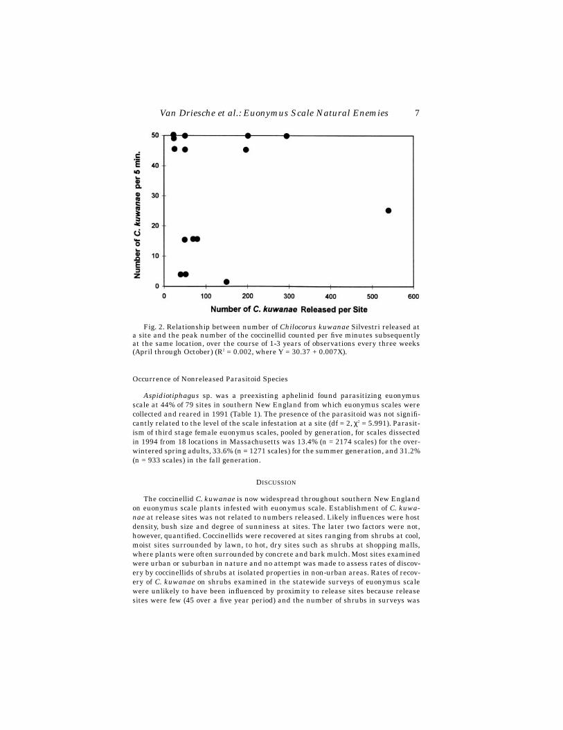
Chilocorus kuwanae (Chinese strain) established at most release sites, but popu-lation increase at sites varied greatly. Of 15 sites followed in detail, the beetle estab-lished at all 15, following release of various numbers. Peak numbers of C. kuwanaelife stages counted per five minute observation varied from 5 or fewer at three siteswhere the beetle scarcely persisted, to over 50 at five sites where beetle increased sub-stantially. No relationship was observed in simple linear regression between numbersreleased per site and subsequent peak counts of beetles (R2 = 0.002, Y = 30.37 +0.007X, with X and Y as in Fig. 2), suggesting that site factors other than initial re-lease rate were primarily responsible for beetle success at individual sites (Fig. 2).
Cybocephalus sp. nr. nipponicus was encountered at five of the seventeen releasesites in the year following release, indicating successful establishment. Same-seasonreproduction of the beetle was observed at ten other sites (Fig. 1b).
Coccobius sp. nr. fulvus was recovered in 1994 from six of eleven release sites (fromreleases in either 1992 or 1993), indicating establishment (six sites, but only fourtowns, Fig. 1c). Same-season reproduction was observed at one additional location. Of155 wasps obtained in rearing samples, 109 (70%) were female.
Encarsia sp. nr. diaspidicola was recovered in 1994 from two of ten 1993 releasesites. Same-season reproduction was observed in 1993 or 1994 at twelve other sites(Fig. 1d—note, some locations marked on the distribution map include several sites).Establishment of this species remains uncertain.
Aphytis sp. releases were made later than those of other species. Recovery effortsin 1995, the last year of the study were unsuccessful and the status of this species isunknown.
Spread of Released Species
Of the five species released, C. kuwanae has achieved the most extensive range insouthern New England (Fig. 1a). In 1992-1994 surveys of randomly selected land-

6 Florida Entomologist 81(1) March, 1998
scape euonymus plants in southern New England, the beetle was found on 14.1% of3141 shrubs that were infested with euonymus scale, out of a total of 4843 plants ex-amined. The proportion of shrubs with C. kuwanae present increased significantlywith increasing scale density (Fig. 3) (df = 3, (χ2 = 516.6).
Spread of other released species of natural enemies was rarely observed. One caseof recovery at a nonrelease site was noted for C. sp. nr. nipponicus and two for C. sp.nr. fulvus.
Fig. 1. Distribution in southern New England of Chilocorus kuwanae Silvestri (a);Cybocephalus sp. nr. nipponicus Enrody-Younga (b); Coccobius sp. nr. fulvus (Compereet Annecke) (c); Encarsia sp. nr. diaspidicola (Silvestri) (d); Aphytis sp. (e); open circles(releases with no recoveries), half filled circles with strike through lines (releases withrecoveries, in the same year only) and filled circles (releases with recoveries one ormore years after release).
Van Driesche et al.: Euonymus Scale Natural Enemies 7
Occurrence of Nonreleased Parasitoid Species
Aspidiotiphagus sp. was a preexisting aphelinid found parasitizing euonymusscale at 44% of 79 sites in southern New England from which euonymus scales werecollected and reared in 1991 (Table 1). The presence of the parasitoid was not signifi-cantly related to the level of the scale infestation at a site (df = 2, χ2 = 5.991). Parasit-ism of third stage female euonymus scales, pooled by generation, for scales dissectedin 1994 from 18 locations in Massachusetts was 13.4% (n = 2174 scales) for the over-wintered spring adults, 33.6% (n = 1271 scales) for the summer generation, and 31.2%(n = 933 scales) in the fall generation.
DISCUSSION
The coccinellid C. kuwanae is now widespread throughout southern New Englandon euonymus scale plants infested with euonymus scale. Establishment of C. kuwa-nae at release sites was not related to numbers released. Likely influences were hostdensity, bush size and degree of sunniness at sites. The later two factors were not,however, quantified. Coccinellids were recovered at sites ranging from shrubs at cool,moist sites surrounded by lawn, to hot, dry sites such as shrubs at shopping malls,where plants were often surrounded by concrete and bark mulch. Most sites examinedwere urban or suburban in nature and no attempt was made to assess rates of discov-ery by coccinellids of shrubs at isolated properties in non-urban areas. Rates of recov-ery of C. kuwanae on shrubs examined in the statewide surveys of euonymus scalewere unlikely to have been influenced by proximity to release sites because releasesites were few (45 over a five year period) and the number of shrubs in surveys was
Fig. 2. Relationship between number of Chilocorus kuwanae Silvestri released ata site and the peak number of the coccinellid counted per five minutes subsequentlyat the same location, over the course of 1-3 years of observations every three weeks(April through October) (R2 = 0.002, where Y = 30.37 + 0.007X).

8 Florida Entomologist 81(1) March, 1998
large (4843) and shrubs were distributed widely over the three state area. Thus, whilea few sites may by chance have been close (under 5 km) to release sites, most were not.
Chilocorus kuwanae life stages were most commonly encountered on plants withmedium or heavy scale infestations, suggesting that the principal effect of this coc-cinellid will be to suppress scales at sites where scale densities are at or approachingdamaging levels, rather than acting when scale densities are light. Because Cyboce-phalus sp. nr. nipponicus and Coccobius. sp. nr. fulvus require fewer hosts for repro-
TABLE 1. PRESENCE OF ASPIDIOTIPHAGUS SP. ON EUONYMUS PLANTS WITH DIFFERENTEUONYMUS SCALE DENSITIES IN SOUTHERN NEW ENGLAND IN 1991.
Scale Inf. LevelSamples
with AspidiotiphagusSamples
without Aspidiotiphagus
light 7 (64%)a 4medium 7 (35%) 13heavy 21 (44%) 27
total 35 (44%) 44
aPercentage samples with parasitoids was not significantly associated with scale density in a χ2 test (df = 2,χ2 = 5.991).
Fig. 3. Percentages (and SE values) of euonymus plants in various categories ofscale density on which Chilocorus kuwanae Silvestri was present in surveys in south-ern New England in 1992-1994, where sample sizes by category were 1702 (N), 1401(L), 745 (M), and 995 (H), and divided by state were 3220 (Massachusetts), 1076 (Con-necticut), and 547 (Rhode Island).

Van Driesche et al.: Euonymus Scale Natural Enemies 9
duction, these species may be more effective in causing mortality to low density scalepopulations. Establishment of these agents in Massachusetts makes possible a futureevaluation of their effects on survivorship of scales in low density populations. The re-maining parasitoids (E. sp. nr. diapidicola and Aphytis sp.) have not yet been shownto have established; future assessment of their status will be needed.
ACKNOWLEDGMENT
We thank the USDA/APHIS National Biological Control Laboratory, Niles, MI, forfinancial support for this project; Narda Wakoluk, Aaron Hechmer, Stephen Healey,Jim Oldham and Don Wilda for technical assistance; R. S. Stauffer for quarantine ac-tivities at Texas A&M University; and the owners of the sites where the work was con-ducted for the use of their property.
REFERENCES CITED
ALVAREZ, J. M., AND R. G. VAN DRIESCHE. Biology of Cybocephalus sp. nr. nipponicus(Coleoptera: Cybocephalidae), a natural enemy of euonymus scale, Unaspis eu-onymi (Homoptera: Diaspididae). Environ. Entomol. in press.
BULL, B. C., M. J. RAUPP, M. R. HARDIN, AND C. S. SADOF. 1993. Suitability of five hor-ticulturally important armored scale insects as hosts for an exotic predaceouslady beetle. J. Environ. Hort. 11: 28-30.
DANIEL, W. W. 1987. Biostatistics: A Foundation for Analysis in The Health Sciences.J. Wiley & Sons, New York. 734 pp.
DREA, J. J., AND R. W. CARLSON. 1987. The establishment of Chilocorus kuwanae (Co-leoptera: Coccinellidae) in eastern United States. Proc. Entomol. Soc. Washing-ton 89: 821-824.
DREA, J. J., AND R. W. CARLSON. 1988. Establishment of Cybocephalus sp. (Co-leoptera: Nitidulidae) from Korea on Unaspis euonymi (Homoptera: Diaspid-idae) in the eastern United States. Proc. Entomol. Soc. Washington 90: 307-309.
DREA, J. J., AND R. M. HENDRICKSON, JR. 1988. Exotic predators. American Nursery-man 168(8): 66-71.
FLINT, H. 1983. Landscape Plants for Eastern North America, Exclusive of Floridaand the Immediate Gulf Coast. John Wiley & Sons, New York. 677 pp.
GILL, S. A., D. R. MILLER, AND J. A. DAVIDSON. 1982. Bionomics and taxonomy of theeuonymus scale, Unaspis euonymi (Comstock), and detailed biological informa-tion on the scale in Maryland. Univ. Maryland Agric. Exp. Sta. Misc. Pub., No.969, 36 pp.
HENDRICKSON, R. M., JR., J. J. DREA, AND M. ROSE. 1991. A distribution and estab-lishment program for Chilocorus kuwanae (Silvestri) (Coleoptera: Coccinel-lidae) in the United States. Proc. Entomol. Soc. Washington 93: 197-200.
NOHARA, K., AND M. IWATA. 1988. Studies on ovipositing behavior of Chilocorus ku-wanae Silvestri (Coleoptera: Coccinellidae). Proceedings of Faculty of Agricul-ture, Kyushu Tokai University 7: 17-24 (in Japanese).
TAKAGI, M. 1991. Host stage selection in Aphytis yanonensis DeBach et Rosen andCoccobius fulvus (Compere et Annecke) (Hymenoptera: Aphelinidiae), intro-duced parasitoids of Unaspis yanonensis (Kuwana) (Homoptera: Diaspididae).Appl. Ent. Zool. 26: 505-513.

10
Florida Entomologist
81(1) March, 1998
A NEW SPECIES OF
MYODOCHA
(HEMIPTERA: LYGAEIDAE: RHYPAROCHROMINAE: MYODOCHINI) FROM THE WEST
INDIES
A
LEX
S
LATER
801 Alabama Street, Lawrence, KS 66044
A
BSTRACT
Myodocha froeschneri
n. sp. is described based on specimens from Dominican Re-public, Haiti, and Jamaica.
Key Words: West Indies,
Myodocha
, new species
R
ESUMEN
Se describe
Myodocha froeschneri
n. sp. basada en especímenes de la República
Dominicana, Haití, y Jamaica.
The genus
Myodocha
is characterized by elongate body and appendages. The headis especially elongate with the postocular region produced into a narrow stalk-likeneck. The seven previously described species are primarily Neotropical with one spe-cies,
M. serripes
Olivier, broadly distributed in the Nearctic.
M. froeschneri
, named forDr. Richard Froeschner of the United States National Museum, is described in ad-vance of a revision of the genus to make the name available for a faunal work on theWest Indies by R. M. Baranowski and J. A. Slater.
Myodocha froeschneri
A. Slater,
New Species
Structure.—Head: vertex evenly convex, shiny, with obscure, barely perceptibletexturing; juga rounded, not carinate, nearly glabrous, no hairs longer than least di-ameter of neck; ocelli just behind line connecting hind margins of compound eyes; lat-eral margin behind eye evenly rounded to neck. Labium: segment I not surpassingposterior margin of compound eye; II not reaching prosternum; IV reaching, not sur-passing, fore coxa. Pronotum: dull; collar and anterior lobe impunctate; posterior lobeshallowly punctate, punctures ranging from contiguous to separated by twice puncturediameter. Scutellum: dull; sparsely, obscurely punctate; indistinct Y-shaped median ca-rina. Thoracic sterna: dull dark brown except mesosternum shiny. Hemelytra: notreaching apex of abdomen. Legs: shiny; fore femur lightly incrassate, hairs short,sparse, spines in two ranks restricted to about apical third, three small spines in ante-rior rank, two small and one large (basal) spine in posterior rank; fore tibia unarmed.
Measurements.—All in mm. Total length 9.0. Head: length 2.6, preocular 0.7, pos-tocular including neck 1.4, width across eyes 1.2, interocular 0.6. Antennal segmentlength: I 1.1, II 2.1, III 1.9, IV 2.3. Labial segment length: I 1.0, II 1.2, III 1.1, IV 0.5.Pronotum: length collar plus anterior lobe 0.8, posterior lobe 0.7, greatest width an-terior lobe 1.1, posterior lobe 1.7. Scutellum: length 0.9, width 0.7. Hemelytra: lengthcorium 3.5, claval commissure 0.8, membrane 2.8 (to corial apex 1.6, beyond corialapex 1.2).
A. Slater: New Species of
Myodocha 11
Color.—Head dark reddish brown becoming darker ventrally, clypeus lightest,neck darkest. Antennal segment I reddish brown, II creamy white, III creamy whiteexcept apical third pale brown, IV creamy white except basal seventh and apical thirdpale brown. Pronotum dark brown fading to dark reddish brown on apical half poste-rior lobe. Scutellum dark brown. Corium dark brown except extreme base, extremeapex, basal half costal margin, claval margin, two small elongate discal spots levelwith middle of clavus, large subapical spot from costal margin almost to membranalmargin, and indistinct discal spot opposite basal angle of membrane off-white. Clavusdark brown; veins lightest, subbasal and larger subapical spot darkest. Membranedark brown, veins subbasally and indistinct elongate apical spot lighter, off-white op-posite corial apex. Coxae dark brown; femora light reddish brown except pale basally;tibiae pale brownish yellow, tarsi yet paler brownish yellow. Abdomen reddish brown,segments V-VIII becoming darker apically, lateral margin segment V and basal halflateral margin segment VI off-white.
Types: Holotype male. HAITI: Enneri, nr. 1000 ft., Sept. 6-11-34, Darlington. De-posited in the American Museum of Natural History. Paratypes. HAITI: 1 female, En-neri, no date, Mann; 1 female, Etang Lachaux, S. W. Peninsula (sic), under 1000 ft.,Oct. 26-27, 1934, Darlington. DOMINICAN REPUBLIC: 1 male, S.R. [San Rafael ?],4 km. S.W. Stgo Rodriguez, May 28, 1978, C. W. & L. B. Obrien & Marshall; 1 male,Barahona, 9.2 km N.W. Paraiso, confluence of Rio Nizso and Rio Coltico, 18-03N, 71-12W, 230 m., 9-10 Aug 1990, J. Rawlins, S. Thompson; 1 male, La Vega, 1.5 km. N Jar-bacoa, 240 m., 21 July 1987, J. Rawlins, R. Davidson. JAMAICA: 1 female, St. An-drews, 9/17 and JA20, at light, A. M. Richie; 1 female, Balaclava, 15 April 1909, A. S.Wright; 1 female, same data but 1 May 1909. In collections of the American Museumof Natural History, Carnegie Museum, Snow Entomological Museum, British Mu-seum (Natural History), J. A. Slater.
Variation.—The holotype male is fairly light in overall color. The darkest speci-mens examined are almost uniformly dark chocolate brown on head and body exceptfor the pale areas on abdominal segments V and VI. On these darker specimens thelight areas on clavus and corium are more distinct except the discal dorial spots andthe light areas on the membrane except at the corial apex are obscured. The color pat-tern of legs and antennae remains constant except that the darker areas are more dis-tinct and antennal segment II becomes pale reddish brown apically. The femoralspines range from 3-5 in the anterior rank and from 2 to 4 in the posterior rank. Threespines in each rank is most common. In only one case is a short spine located basal ofthe long basal spine of the posterior rank. In one case the posterior rank bears 2 aboutequally long basal spines. Total length varies from 8.0-9.0 mm in males and from 9.0-9.6 mm in female.
Etymology.—Named for Dr. Richard C. Froeschner, United States National Mu-seum.
Identification: The combination of smooth head vertex and lack of elongate hairson the “neck” separate
M. froeschneri
from all other members of the genus except theCuban
M. fulvosa
Barber. That species is castaneous or fulvous in color, has unicol-ored fore femora, and typically has at least one small spine basal to the long subapicalspine.
R
EFERENCES
C
ITED
H
ARRINGTON
, B. J. 1980. A generic level revision and cladistic analysis of the Myodo-chini of the world (Hemiptera, Lygaeidae, Rhyparochrominae). Bull. Amer.Mus. Nat. Hist. 167(2): 45-116.

12
Florida Entomologist
81(1) March, 1998
DISTRIBUTION AND DISPERSAL OF
CACTOBLASTIS CACTORUM
(LEPIDOPTERA: PYRALIDAE), AN EXOTIC
OPUNTIA
-FEEDING MOTH, IN FLORIDA
D
EREK
M. J
OHNSON
1
AND
P
ETER
D. S
TILING
Department of Biological Sciences, University of South Florida, Tampa, FL 33620
1
Current Address: Department of Biology, University of MiamiCoral Gables, FL 33124
A
BSTRACT
The recent arrival of
Cactoblastis cactorum
Berg in Florida has raised concern forFlorida's native
Opuntia
cacti. Moreover, the potential for movement of the mothacross the gulf states and into the southwestern United States may endanger cacti inthe
Opuntia
-rich areas of Texas, Arizona, New Mexico, and Mexico. However, thespread of the moth northward through Florida has either slowed since the invasion orthe rate of spread for the first two years was over estimated. The mortality rate ofpads and the distribution of egg sticks at six sites in Florida were recorded on
O.stricta
Haworth, the most common host in Florida. While the percentage of cactuspads with
C. cactorum
damage is as high as 60%, the data indicates that most maturecacti are not being reduced in size. However, small cacti and new growth pads are par-ticularly susceptible to mortality by
C. cactorum
, thus, over time we may expect to seea reduction in the number of plants as a result of an increase in the mortality rate ofrecruits.
Key Words: pest, biological control, herbivory, moth, cactus
R
ESUMEN
La reciente introducción a la Florida de
Cactoblastis cactorum
Berg a causado pre-ocupación en cuanto a los cactos indígenas de la Florida de la especie
Opuntia
. Ade-más, la posibilidad de que la palomilla avance a través de los estados del Golfo haciael suroeste de los Estados Unidos amenaza áreas con abundancia de
Opuntia
enTexas, Arizona, Nuevo México, y México. Sin embargo, la expansión de la palomillahacia el norte a traves de Florida desde que la invasión empezó ha disminuido o el cál-culo de la tasa de expansión durante los dos primeros años fué exagerado. La tasa dela mortalidad de las pencas de cacto y la distribución de grupos de huevos en seis lo-calidades en Florida fueron registrados en
O. stricta
Haworth, que es el hospederomás común dentro de Florida. Aunque el porcentaje observado de pencas dañadas por
C. cactorum
es tan alto como el 60%, los datos indican que los cactos más maduros noestán siendo reducidos en tamaño. Sin embargo, cactos pequeños y pencas nuevas sonparticularmente susceptibles a mortalidad causada por
C. cactorum
; en consecuencia,en un futuro podríamos contar con una reducción del número de plantas debido al au-
mento de la tasa de mortalidad de cactos jóvenes.
In 1957, the moth
Cactoblastis cactorum
Berg was introduced onto the Caribbeanisland of Nevis as a biological control agent for pest
Opuntia
spp. and in 1960 was in-troduced onto Montserrat and Antigua (Simmonds & Bennett, 1966). The moth dis-persed to other islands such as Cuba, Puerto Rico, Hispaniola, the Bahamas, andCuba (Habeck & Bennett, 1990).
Johnson & Stiling:
Cactoblastis cactorum
distribution
13
A Florida Keys record for
C. cactorum
in October, 1989, was a new record for thecontinental United States (Habeck & Bennett, 1990). The moth likely arrived in Flor-ida by one of two methods: (1) natural dispersal via flight/wind from the Caribbean or(2) via shipments of cacti to Miami from the Caribbean (Pemberton, 1995). This spe-cies may disperse beyond Florida, and eventually reach the
Opuntia
-rich desertsouthwest. The moth successfully dispersed several times from one island to anotherin the Caribbean; thus, spread across long distances is possible. The moth may havealready invaded the Yucatan (Pemberton, 1995), thus, the moth also may disperse tothe southwestern United States via Mexico.
Florida has six species of native
Opuntia
(
O. stricta
Haworth,
O. humifusa
(Rafinesque) Rafinesque,
O. spinosissima
(Martyn) Miller,
O. triacantha
(Willdenow)Sweet,
O. cubensis
Britton & Rose, and
O. pusilla
(Haworth) Haworth) (Benson,1982).
Cactoblastis cactorum
has been found on all of the natives except
O. pusilla
(Bob Ehrig, The Nature Conservancy, pers. comm.; pers. obs.). The United Statesranges of three of these cacti,
O. spinosissima
,
O. triacantha
, and
O. cubensis
, are lim-ited to local populations in the Florida Keys. Only 12
O. spinosissima
plants remainin one location in the Florida Keys and
O. triacantha
and
O. cubensis
are rare.This study focuses on the attack of
C. cactorum
on
O. stricta
.
O. stricta
is a commoncactus throughout coastal Florida, growing in sandy soils and shell mounds. We inves-tigated the rate of the moth’s spread throughout Florida, distribution of egg sticks andlarval damage, and extent of damage to
O. stricta
. This information, coupled with theinformation from oviposition and larval choice experiments (Johnson & Stiling, 1996),could be useful in setting future management goals for
C. cactorum
in the continentalUnited States.
M
ATERIALS
AND
M
ETHODS
Damage and Egg Stick Distribution
We repeatedly visited ten sites throughout south and central Florida (Fig. 1). Uponeach visit to every site, we counted the total number of cactus pads on 20 to 100
O.stricta
plants, the number of pads with old
C. cactorum
damage (those that had beenfed upon but the larvae had since abandoned them), the number of pads with new lar-val damage (pads with larvae currently feeding on them), and the number of eggsticks per plant. The same population of plants was surveyed at every census. FromOctober, 1991 to November, 1992, each site was sampled approximately monthly. Thesites were sampled less frequently in 1993.
We are confident that the vast majority, if not all, of old damage, new damage, andegg sticks measured at the eight sites were attributable to
C. cactorum
and not the na-tive cactus-feeding moth,
Melitera prodenialis
Walker, for the following reasons. First,at all sites where
C. cactorum
larvae were not detected, the percentage of pads withold damage was less than one percent, while within a year after
C. cactorum
larvaewere detected, old damage increased to over ten percent. Secondly, cactus pads withnew damage were randomly cut open and the larvae were identified. All Pyralidae lar-vae encountered at the sites were
C. cactorum
. Lastly, the widths of
C. cactorum
eggsticks are narrower than
M. prodenialis
egg sticks and the ranges of width are non-overlapping (DMJ, unpublished data). Three-hundred eighty-two hatched egg stickswere collected from the sites and compared to egg sticks known to be laid by
C. cac-torum
and
M. prodenialis
. All of the egg sticks were determined to be laid by
C. cac-torum
. Thus, while we can not be certain that
M. prodenialis
was always absent, weare confident that its contribution was insignificant.

14
Florida Entomologist
81(1) March, 1998
The distribution of egg sticks on cacti was measured by using the Morisita Indexof Dispersion (Krebs, 1989). The Kruskal-Wallis test (Sokel & Rohlf 1981) was used todetermine whether the number of egg sticks on a plant and the number of egg sticksper pad was related to plant size (as measured by the total number of pads). TheKruskal-Wallis test was also used to determine whether the percent damage was re-lated to the size of the plant.
In addition, all reports of
C. cactorum
detected in new locations around Floridawere examined to determine when the moth extended its known range northwardalong both the east and west coastlines. These reports included personal observations,reports to the Florida Department of Agriculture and Consumer Services (FDACS),and other sources.
Growth and Mortality of
Opuntia stricta
From January to March 1992 all individual pads on 10
O. stricta
plants at the Wal-ton Rocks Beach site were marked. In 1993 10 additional plants were marked. Eachpad was numbered using permanent ink. Upon every subsequent visit, once per
Fig. 1. Opuntia stricta populations surveyed for Cactoblastis cactorum from Octo-ber 1991 to October 1993.

Johnson & Stiling:
Cactoblastis cactorum
distribution
15
month in 1992 and less frequently in 1993, pad mortality and the number of newgrowth pads were recorded. These data were used to determine net growth of the at-tacked plants. The Rank Sum Test (Ambrose & Ambrose 1987) was used to determinewhether smaller cacti had a significantly higher mortality. The Wilcoxon SignedRanks Test (Sokel & Rohlf 1981) was used to determine whether new pads suffered ahigher mortality caused by
C. cactorum
than did old pads.
R
ESULTS
Damage and Egg Stick Distribution
Peaks in new larval damage and percentage of pads with egg sticks varied tempo-rally and spatially (Figs. 2 and 4). No
C. cactorum
was recorded at Lover's Key orRookery Bay. Larval activity was generally highest from May to September, but larvalactivity also heightened in late fall and early winter of 1991 at most sites. Larval dam-age measurements, the percentage of pads with both old and new damage, increasedat every site from the fall of 1991 to the fall of 1992. Overall, measurements of damagedecreased slightly from the fall of 1992 to the fall of 1993 at all of the sites exceptOcean Bluff, Terra Ceia and Brevard #2. Over the 2 year period, the percentage ofdead or damaged pads increased (range 9-37%) at all 6 sites (Fig. 3). Percent damageand plant size were not significantly related (p > 0.05).
Egg sticks were clumped among plants (Table 1). At two of the six sites tested, sig-nificantly more egg sticks were laid on either medium or large-sized plants (Table 1).At the other four sites with a sufficient number of egg sticks the trend was present butnot significant. The number of egg sticks per pad was not significantly different be-tween small, medium, and large plants at any of the sites (p > 0.10) (Table 1).
At the peak of old
C. cactorum
damage, 90% of the plants with over 10 pads hadold damage (Table 2). Excluding plants at the Terra Ceia site, which had a lower per-centage of old damage than the other sites, 172 out of 173 plants (99.4%) with over 10pads showed evidence of previous larval damage as compared to 14 of 28 plants (50%)with 10 pads or fewer having previous larval damage.
The Spread of
Cactoblastis cactorum
The spread of
C. cactorum
up Florida's east coast, assuming that the moth first col-onized the lower Florida Keys and migrated north, has been relatively well docu-mented as compared to Florida's west coast. The moth was first discovered in theUnited States on Big Pine Key in October, 1989 (Habeck & Bennett, 1990). In lessthan a year, the moth was discovered at Key Biscayne State Park in Miami (FDACS,unpublished), approximately 200 km east northeast of Big Pine Key. One year later,in August, 1991, the moth was discovered at Brevard #1 (FDACS, unpublished), ap-proximately 240 km north of Key Biscayne. The most northerly record of
C. cactorum
was at Brevard #2 (Patrick Air Force Base), 50 km north of Brevard #1 (pers. obs.). Themoth arrived at this site in June 1992 and has established there.
The spread of
C. cactorum
up the west coast of Florida has not been so well docu-mented. The first west coast record was in Terra Ceia, Manatee County, in May 1991(FDACS, unpublished), one year and seven months after its discovery in the FloridaKeys and approximately 360 km north. Six months later, the moth was discovered atFort Desoto State Park in Pinellas County (pers. obs.), approximately 16 km north ofTerra Ceia. The most northerly record of
C. cactorum
on the west coast of Florida wasat Upper Tampa Bay Park in Hillsborough County, February, 1992 (pers. obs.). Thissite is approximately 50 km north of Fort Desoto.

16
Florida Entomologist
81(1) March, 1998
Fig. 2. Percentage of Opuntia stricta pads with new damage due to Cactoblastiscactorum larval feeding at eight sites in central and south Florida (see Fig. 1 for loca-tions).
Johnson & Stiling:
Cactoblastis cactorum
distribution
17
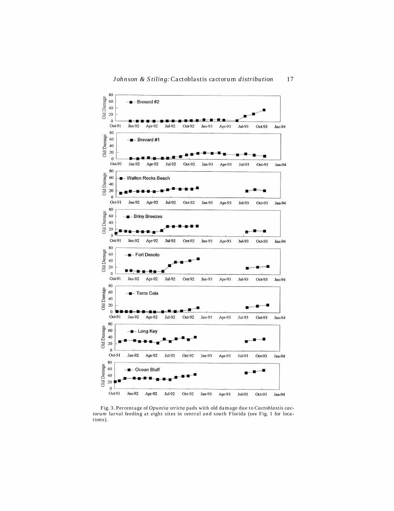
Fig. 3. Percentage of Opuntia stricta pads with old damage due to Cactoblastis cac-torum larval feeding at eight sites in central and south Florida (see Fig. 1 for loca-tions).
18
Florida Entomologist
81(1) March, 1998
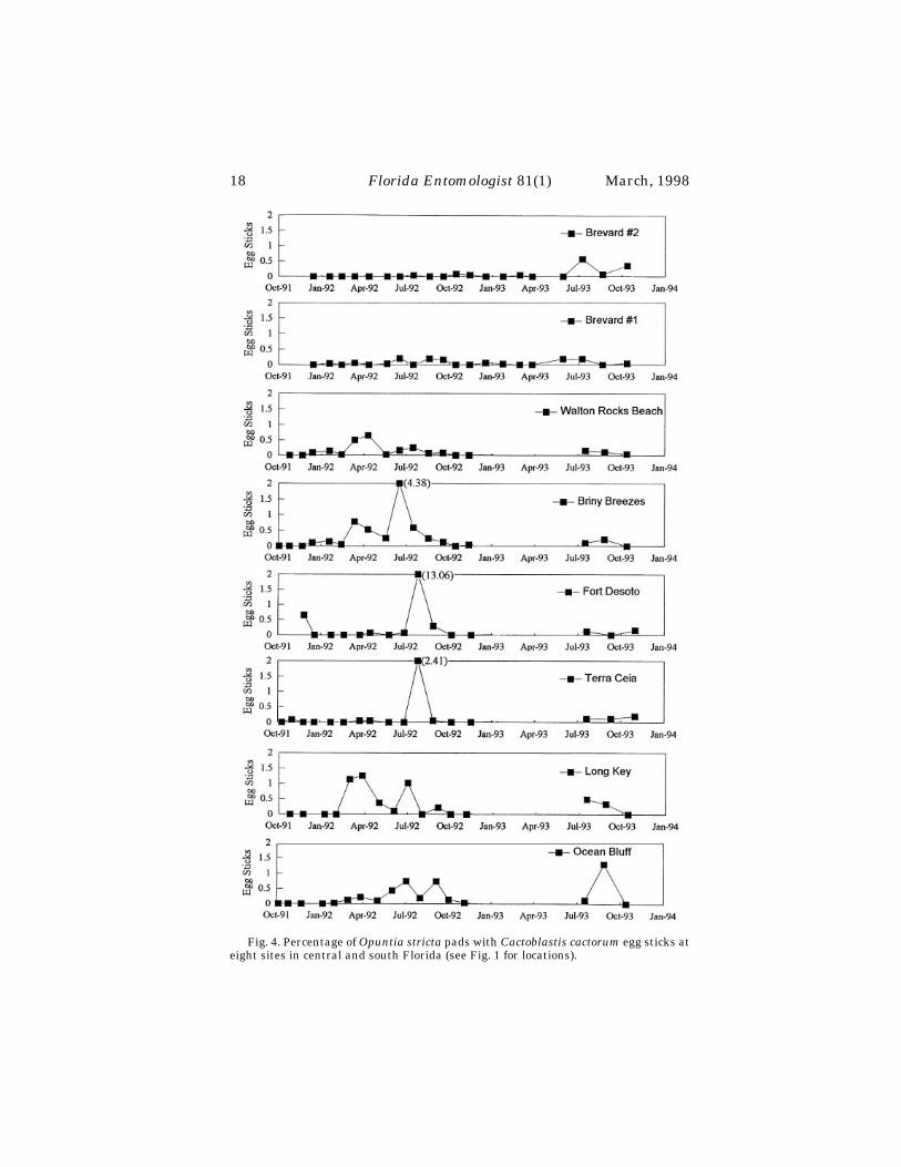
Fig. 4. Percentage of Opuntia stricta pads with Cactoblastis cactorum egg sticks ateight sites in central and south Florida (see Fig. 1 for locations).

Johnson & Stiling:
Cactoblastis cactorum
distribution
19
There has been almost no confirmed records of inland movement of over a few ofkilometers by
C. cactorum
in Florida. The discovery of the moth in Loxahatchee, PalmCounty in June of 1992 (FDACS, unpublished), 24 km inland from the Atlantic Ocean,is the most inland of confirmed records.
Growth and Mortality of
Opuntia stricta
Growth in nine plants marked in 1992 ranged from a loss of 100% of the pads toan increase of 87%. One of the 10 plants marked in 1992 was not located until 1 yearlater. Some of the marks had worn off of the pads, so this plant was omitted from anal-ysis. Two of the nine plants died from larval feeding during 1992. These were two ofthe three smallest marked plants (each having nine pads). The net growth of all of theplants in 1992 was +5%.
During the second year of monitoring, plant growth ranged from -100% to +56%.The net mean growth of all of the plants was +6%. The smallest of the original nineplants, having eight pads, was the only plant in this group to die (killed by
C. cac-torum
) during 1993. Thus, over the two year period the three smallest plants died.Plants with nine or fewer pads had a higher mortality rate than plants with greaterthan nine pads (Rank Sum Test; p < 0.05).
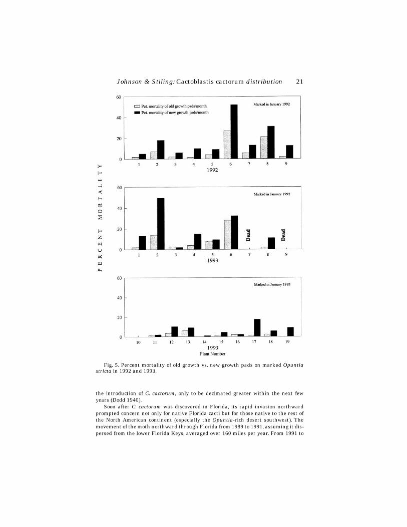
The 10 plants that were marked and monitored in 1993 had a much higher growthrate. Only 1 of the 10 plants decreased in number of pads (-27%) and the highestgrowth rate was +170%. The mean net growth of the 10 plants was +86%. In 1992 and1993 combined, new growth pads sustained a higher mortality rate due to larval dam-age than did old growth pads on 22 out of 25 plants (Wilcoxon Signed Ranks Test; n =25, p < 0.05) (Fig. 5).
D
ISCUSSION
Cactoblastis cactorum
in Florida is more active in the spring and summer. The dis-tribution and spread of the moth largely has been restricted to the coastal regions ofsouth and central Florida. The lack of inland reports of the moth may be because
O.
T
ABLE
1.
C
ACTOBLASTIS
CACTORUM
EGG
STICK
DISTRIBUTIONS
ON
SIX
POPULATIONS
OF
O
PUNTIA
STRICTA
.
Site
Egg Stick Distribution
AmongPlants
a
By Plant Size
b
Plant Size Preferred
Egg Sticks per Pad by Plant Size
b
Ocean Bluff Clumped N. S. — N. S.Long Key Clumped Signif., p < 0.05 Medium N. S.Briny Breezes Clumped N. S. — N. S.Walton Rocks Beach Clumped N. S. — N. S.Terra Ceia Clumped Signif., p < 0.01 Large N. S.Fort DeSoto Clumped N. S. — N. S.
a
Tested using the Morisita index of dispersion (significant at p < 0.05).
b
Tested using the Kruskal-Wallis test (significant at p < 0.05).

20
Florida Entomologist
81(1) March, 1998
stricta
is more common in coastal areas (Benson 1982) or because of other biotic andabiotic factors. Future work should address why
C. cactorum
's distribution is mainlycoastal so we can determine whether it will, in time, invade inland populations of
Opuntia
.Previous studies in Australia and South Africa found that the females lay their egg
sticks in a clumped distribution based on plant size, plant color, and shelter from thewind (Myers et al., 1981; Robertson, 1987). Similarly, egg sticks were clumped at allof our sites and more were laid on medium or large-sized plants at two of the sites.There was no difference, however, in the number of egg sticks per pad among plantsof different sizes. Thus, pads on large plants are no more likely to have egg sticks laidon them than pads on small plants. This is consistent with our finding that there is norelationship between
C. cactorum
damage and plant size.The moth is doing significant damage to
O. stricta
individuals in Florida, but is themoth reducing the populations? Overall, the number of pads on marked individualcacti at Walton Rocks Beach increased in 1992 and 1993. However, while fewer smallplants received moth damage than did medium or large-sized plants, small cacti withmoth damage were most susceptible to mortality. Thus,
C. cactorum
could strongly re-duce the survivorship of maturing
O. stricta
. We may reach a scenario in Floridawhereby large
O. stricta
can withstand the attack, but, in the ensuing years, as theseplants die, there are fewer individuals to replace them. Only then would the total ad-verse effect of
C. cactorum
become noticeable. Forecasting such a process necessitatesa more detailed study in which recruitment as well as the fates of the plants are mea-sured. Also,
O. stricta
populations in Australia partially recovered a few years after
T
ABLE
2. R
ELATIONSHIP
BETWEEN
THE SIZE OF OPUNTIA STRICTA AND DAMAGE BY CAC-TOBLASTIS CACTORUM.
Site Damage
Size of plant
0-10 pads 11-more pads
Ocean Bluff Present 3 39Absent 1 0
Long Key Present 2 18Absent 6 0
Briny Breezes Present 2 24Absent 4 0
Walton Rocks Beach Present 6 61Absent 2 1
Terra Ceia Present 6 42Absent 30 22
Fort Desoto Present 1 33Absent 1 0
Total Present 20 217Absent 44 23
Percentage of plants with damaged pads 31% 90%
Johnson & Stiling: Cactoblastis cactorum distribution 21
the introduction of C. cactorum, only to be decimated greater within the next fewyears (Dodd 1940).
Soon after C. cactorum was discovered in Florida, its rapid invasion northwardprompted concern not only for native Florida cacti but for those native to the rest ofthe North American continent (especially the Opuntia-rich desert southwest). Themovement of the moth northward through Florida from 1989 to 1991, assuming it dis-persed from the lower Florida Keys, averaged over 160 miles per year. From 1991 to
Fig. 5. Percent mortality of old growth vs. new growth pads on marked Opuntiastricta in 1992 and 1993.
22 Florida Entomologist 81(1) March, 1998
1993, however, the spread averaged only 24 miles per year. Recent information (Pem-berton, 1995) indicates that C. cactorum may have invaded Florida via imported cactithrough Miami rather than natural dispersal, in which case the dispersal rate re-ported for 1989 to 1991 is an over estimate. Determining the true rate of spread of C.cactorum and which biotic and/or abiotic factors affect this rate, would be valuable be-cause then we could determine if and when the moth may be expected to attack Opun-tia in other regions of North America (barring accidental introduction on importedcacti).
ACKNOWLEDGEMENTS
The authors thank The Nature Conservancy for providing the opportunity to be apart of this conservation effort. R. Ehrig was of great assistance in locating field sitesand discussing the goals of the project. Thanks to A. Rossi for his invaluable adviseconcerning experimental design and statistical analysis. We are grateful to E. McCoyand H. Mushinsky for reviewing an earlier version of this manuscript. Thanks to D.Gordon, R. Pemberton, D. Habeck, and three anonymous reviewers for reviewing thismanuscript. D. Jones was of great service in solving graphical dilemmas. Funding wasprovided by the Missouri Botanical Garden and The Garden Club of America throughthe Catherine H. Beattie Fellowship. Additional funding was provided by The NatureConservancy and the United States Department of Fish and Wildlife.
REFERENCES CITED
AMBROSE, H. W., III, AND K. P. AMBROSE. 1987. A Handbook of Biological Investiga-tion. Fourth Edition. Hunter Textbooks Inc. Winston-Salem, NC.
BENSON, L. 1982. The Cacti of the United States and Canada, Stanford UniversityPress: Stanford, CA. 1044 pp.
DODD, A. P. 1940. The biological campaign against prickly pear, CommonwealthPrickly Pear Board: Brisbane. 177 pp.
HABECK, D. H., AND F. D. BENNETT. 1990. Cactoblastis cactorum Berg (Lepidoptera:Pyralidae), a phycitine new to Florida. Fla. Dept. Agric. & Consumer Serv. Di-vision of Plant Industries, Entomology Circular 333.
JOHNSON, D. M., AND P. D. STILING. 1996. Host specificity of Cactoblastis cactorumBerg, an exotic Opuntia-feeding moth, in Florida. Environmental Entomology,25(4), 743-748.
KREBS, C. J. 1989. Ecological Methodology. Harper Collins Publishers, Inc., NY.MYERS, J. H., J. MONRO, AND N. MURRAY. 1981. Egg clumping, host plant selection
and population regulation in Cactoblastis cactorum (Lepidoptera). Oecologia(Berlin), 51, 7-13.
PEMBERTON, R. W. 1995. Cactoblastis cactorum: An immigrant or introduction?American Entomologist, 41: 230-232.
ROBERTSON, H. G. 1987. Oviposition site selection in Cactoblastis cactorum (Lepi-doptera): constraints and compromises. Oecologia (Berlin), 73, 601-608.
SIMMONDS, F. J., AND F. D. BENNETT. 1966. Biological control of Opuntia spp. by Cac-toblastis cactorum in the Leeward Islands (West Indies). Entomophaga, II(2),183-189.
SOKAL, R. R., AND F. J. ROHLF. 1981. Biometry, Freeman, San Francisco.
Cruz & Martínez: Effect of Male Secretions on Females
23
EFFECT OF MALE MESADENE SECRETIONS ON FEMALES OF
CANTHON CYANELLUS CYANELLUS
(COLEOPTERA: SCARABAEIDAE)
M
AGDALENA
C
RUZ
R.
AND
I
MELDA
M
ARTINEZ
M.Instituto de Ecología, A.C., Depto. Ecología y Comportamiento Animal
Apartado Postal 63, 91000 Xalapa, Ver. México
A
BSTRACT
This study determined the effect of male mesadene secretions on females of
Can-thon cyanellus cyanellus
LECONTE, both when they were inseminated normally andwhen the secretions were transplanted to virgin females. In the first case, mating tookplace when the ovary was immature, triggering ovarian maturation, egg laying andnest building. In the second case, the transplantation of male mesadene secretions tovirgin females initiated ovarian maturation, but neither egg laying nor nest buildingtook place. Virgin females that did not receive the secretions had no ovarian matura-tion and did not lay eggs or build nests. It is therefore possible that male mesadenesecretions induce ovarian maturation. In the present study, this inducement wasgreater in inseminated females than in those receiving transplanted secretions.
Key Words: Mesadene secretions, Transplant, Ovarian maturation, Scarabaeinae
R
ESUMEN
En este trabajo se determinó el efecto que tienen las secreciones de las mesadeniasdel macho sobre las hembras de
Canthon cyanellus cyanellus
, cuando son insemina-das normalmente, o cuando las secreciones son trasplantadas a hembras vírgenes. Enel primer caso la cópula tuvo lugar cuando el ovario estaba inmaduro, desencade-nando la maduración ovárica, la oviposición y la construcción del nido. En el segundocaso, el trasplante de las secreciones de las mesadenias a hembras vírgenes inició lamaduración ovárica, pero no la oviposición ni la construcción del nido. Las hembrasvírgenes que no recibieron las secreciones no maduraron el ovario ni hubo oviposicióno construcción del nido. Es posible que las secreciones mesadénicas del macho induz-can la maduración ovárica. Esta inducción fue mayor en las hembras inseminadas que
en las que recibieron el trasplante de las secreciones.
In various species of Scarabaeinae (Scarabaeidae), mating takes place shortly af-ter the female emerges (Monteith & Storey 1981; Klemperer 1982) or before egg lay-ing (Halffter & López 1977; Halffter et al. 1980; Huerta et al. 1981; Monteith & Storey1981; Anduaga & Huerta 1983; Sato & Hiramatsu 1993). In the above-mentionedcases, mating is necessary for egg laying and nesting to begin.
In the dung beetles,
Canthon indigaceus chevrolati
HAROLD and
Copris incertus
SAY, the first mating, which occurs during the pre-nesting period, when the ovary isstill immature, is indispensable for ovarian maturation, egg laying and nesting to oc-cur (Martínez & Cruz 1990; Martínez et al. 1996).
In
Canthon cyanellus cyanellus
LECONTE, the first mating is at 10 days after fe-male emergence. This occurs half way through the pre-nesting period, which lastsabout 20 days. During this period ovarian maturation occurs, only food balls are pro-

24
Florida Entomologist
81(1) March, 1998
duced and there is no nesting. Afterwards, during the nesting period, other matingsmay occur (Martínez 1992).
During mating, the male of
C. c. cyanellus
produces a spermatophore containingabundant seminal fluid which consists principally of the secretions of the accessoryglands (mesadenes). Most of this seminal fluid has a high concentration of proteins,although it also contains glycogen and acid mucopolysaccharides (Cruz & Martínez1992). This is also the case in other Coleoptera (Anderson 1950; Landa 1960; Gerber,et al. 1971; Gundevia & Ramamurty 1977; Huignard et al. 1977; Peferoen & de Loof1983; Black & Happ 1985).
The objective of this study was to determine the effect of male mesadene secretionsin
C. c. cyanellus
upon ovarian maturation and female reproductive behavior.
M
ATERIALS
AND
M
ETHODS
This study was carried out on adult
Canthon cyanellus cyanellus
, of known ageand raised in the laboratory. Insects were kept at 27
°
C, 70% RH, a photoperiod of14:10 hours and were fed beef.
Females were tested in one of four manners: 1) a female was kept together with amale from the time of emergence (n = 63), 2) virgin females were isolated from thetime of emergence (n = 57), 3) virgin females received transplants of mesadene secre-tions from mature males at 10 days old (n = 31), and 4) virgin females had sterileRinger-Ephrussi solution injected at 10 days old (n = 25).
The females in categories 1 and 2 were sacrificed at 5, 10, 15, 20, and 25 days ofage, with approximately 10 females per age group. At 10 days of age the females incategories 3 and 4 received the secretion transplant or the injection of Ringer solution.It was allowed to take effect for 5, 10 or 15 days. The females in these last two catego-ries were sacrificed at 15, 20 and 25 days of age. The were about 5 females in these agegroups.
To carry out mesadene transplantation, the reservoir, a structure in which glan-dular secretions are stored, was obtained from 20-30 day-old males. Females were an-aesthetized with ethyl acetate for 3 minutes, which allowed them to recover withoutcomplications. The elytra and wings of anaesthetized females were lifted carefully,and the reservoir was placed in the dorsal region of the abdomen. Using an entomo-logical pin, a dorsal puncture was made through which glandular secretions were in-troduced into the abdominal cavity. It was not necessary to use sealer, as the woundhealed quickly on its own. Females recovered in about 5-10 minutes. After a 24-hoursperiod they were put inside a terrarium (Cruz 1994).
The reproductive systems from all four groups were dissected out in Ringer-Ephrussi solution. Each ovary was measured and drawn to scale with the aid of acamera lucida. The ovary and vaginal frotis together with the spermatheca from eachfemale were obtained and dyed
in toto
using the Feulgen-green light technique. Thepresence of the spermatophore in the vagina or of spermatozoids in the spermathecaindicated that the female had been inseminated.
The length of the basal oocyte was analyzed in each age group and the differentcategories were compared. Sample sizes were 5 to 17 females per age group. Sincethese were small samples, a 95% confidence interval was calculated using the formulax
±
α
n-1
/
√
n (1.96), where x is the sampling median and
α
n-1
is the standard deviation.Analysis of variance (ANOVA) was used to compare means.
Female reproductive behavior was categorized according to the following factors:if they were inseminated, if they laid eggs, and either made food or nest balls.

Cruz & Martínez: Effect of Male Secretions on Females
25
R
ESULTS
The female reproductive system in
C. cyanellus cyanellus
is similar to that of otherScarabaeinae: it consists of a single left ovary with one ovariole, an oviduct, vaginaand spermatheca with its accessory gland.
Females with Males
In these females, the first basal oocyte measured 0.7 mm 5 days after eclosion. Bythe 10th day it had more than doubled in size to 1.5 mm. By the 15th day it had againdoubled in size. After 20 days, it was almost egg-laying size, which in this species is 3mm (Fig. 1; Table 1). Females in this group copulated between 10 and 15 days of age.Spermatozoids were observed in the spermatheca, and the spermatophore was in thevagina. By 15 days after eclosion, all of the females dissected had been inseminatedbut had not yet begun to nest. By 20 days, most of the females had already laid a firstegg, and the second basal oocyte was close to egg-laying size. At the age of 25 days, allfemales were making their first nest with 1 to 7 nest balls, corresponding at 1 to 7 eggslaid.
Fig. 1. First basal oocyte development in Canthon cyanellus cyanellus females. (X)Females with male since emergence, the arrow shows the age at which first copula oc-curs; (✻)Virgin females; (+) Virgin females with transplant from 10 days of age on; (h)Virgin females injected with Ringer’s solution.

26
Florida Entomologist
81(1) March, 1998
Virgin Females
Until the 10th day, the development of the first basal oocyte in these females wassimilar to that of females of the same age that had been kept with males. On the 15thday, the size of the basal oocyte remained almost identical to that observed at 10 days.From day 20 to 25, however, it gradually diminished in size, and was reabsorbed in fe-males more than 50 days old (Fig. 1: Table 1).
The ovaries of virgin females did not fully mature, and the oocytes entered into re-absorption. Virgin females only made food balls, and did not construct nest balls ornest.
Virgin Females with Male Mesadene Secretion Transplant
The transplant of secretions was performed at 10 days of age due to our observa-tion that the first mating tends to occur around this age. At day 15 the first basal oo-cyte measured an average approximately 1.6 mm; this size did not change by 20 daysof age. However, at 25 days of age, the first basal oocyte became substantially larger(Fig. 1; Table 1). All females in this group initiated first basal oocyte maturation, butoocytes did not reach egg-laying size even at 25 days.
Although they initiated ovary maturation, these females did not lay eggs, makenest balls or nest; their only activity was the production of food balls.
Virgin Females Injected with Ringer’s Solution
In these females, first basal oocyte size did not noticeably increase (Fig. 1; Table 1);furthermore, neither egg laying, ovary maturation or nest making took place. Activitywas limited to making food balls which they did not turn into nest balls.
D
ISCUSSION
In
Canthon cyanellus cyanellus
the development of the first basal oocyte in virginswas compared with those of females kept together with the male since the time ofemergence. After 15 days, the size of the first basal oocyte was no longer comparable
T
ABLE
1. B
ASAL
OOCYTE
MATURATION
IN
FEMALES
OF
C
ANTHON
CYANELLUS
CYANEL-LUS
KEPT
WITH
A
MALE
SINCE
EMERGENCE
(F-M),
VIRGIN
FEMALES
(VF),
VIR-GIN
FEMALES
WITH
TRANSPLANT
(VF-T)
AND
VIRGIN
FEMALES
INJECTED
WITH
R
INGER
’
S
SOLUTION
(VF-R). (
X
±
SE
)(
N
)
NUMBER
OF
FEMALES
PER
AGE
.
Age(days)
Basal oocyte length (mm)
F-M VF VF-T VF-R
5 0.72
±
0.10(10) 0.61
±
0.17(11) — —10 1.50
±
0.12(14) 1.52
±
0.15(12) — —mating
15 2.50
±
0.15(17) 1.51
±
0.09(12) 1.68
±
0.15(10) 1.13
±
0.17(10)20 3.58
±
0.10(10) 1.16
±
0.10(10) 1.60
±
0.13(10) 1.18
±
0.08(10)25 2.65
±
0.21(12) 1.10
±
0.10(12) 2.08
±
0.17(11) 1.26
±
0.31(5)

Cruz & Martínez: Effect of Male Secretions on Females
27
between the two groups (F
{1,27}
: 26.8; p < 0.01). In females which were inseminated bya male, the ovary was bigger than in individuals kept alone. This difference was great-est at 20 days of age (F
{1,19}
: 210; p < 0.01) even compared to 25-day-old females (F
{1,22}
:41.8; p < 0.01).
These results demonstrate that the first mating triggers the final maturation ofthe basal oocyte and the ovary, and, in some yet unknown way, induces egg laying andnesting. This has also been demonstrated to be true in
Canthon indigaceus chevrolati
and
Copris incertus
. In these two beetles, virgin females neither finish ovary matura-tion, lay eggs, nor make nests (Martínez & Cruz 1990; Martínez et al. 1996).
A comparison of the size of the first basal oocyte in females which were inseminatedand in those which received male mesadene transplant showed marked differencesfrom the age of 15 days (F
{1,25}
: 14.0; p < 0.01) until 20 days (F
{1,18}
:136; p < 0.01), but dif-ferences were non significant at 25 days (F
{1,21}
: 4.1); the effects of mating and glandularsecretions are, therefore, not comparable. The slow increase in basal oocyte size in fe-males that received the transplant suggests that egg laying size could be reached at amore advanced age, although this was never confirmed through observation.
When virgin females were compared with those that received the transplant ofmale mesadene secretions, it became clear that the secretions do have a positive effecton ovarian maturation. After 20 days of age, the basal oocyte size was larger in the fe-males receiving the transplant than in virgins (F
{1,18}
: 6.4; p < 0.05). The greatest dif-ference was observed during the period between 20 and 25 days of age (F
{1,21}
: 24.6; p <0.01): the ovary continued to mature up to an advanced stage, but oviposition did nottake place during the period of observation. In females over 30 days old, basal oocytesize diminished in unaltered virgins but not in those that had received the secretiontransplant.
An analysis of females with the transplant compared to those with Ringer’s solu-tion injections at various ages yielded the following data: the only significant differ-ence observed was between treatments (F
{1,4}
: 14.4; p < 0.05) regardless of age (F
{4,50}
:1.4; non significant). Virgin females which received male mesadene transplants hadgreater oocyte size than virgin females injected only with Ringer’s solution.
When
C. c. cyanellus
virgin females received the transplant of male mesadene se-cretions, the ovary matured up to an advanced stage, but oviposition never took place.Females can receive the stimulus that induces ovarian maturation either during cop-ulation or through the transplant of secretions. We do not yet know which of the com-ponents in these secretions act directly upon the ovary to induce vitellogenesis andovary maturation, but the abundance of certain proteins may indicate that they areresponsible.
In the weevil
Acanthoscelides obtectus
SAY (Huignard 1984) and in the mosquito
Aedes taeniorhyncus
(WIEDEMANN) (Borovsky 1985), vitellogenesis is induced byproteins called paragonial substances found in secretions of the male glands. In
A. ob-tectus
these substances are distributed through the haemolymph to the female’s head,thorax and abdomen soon after mating and induce ovarian maturation (Huignard1978). In other species of flies, and a grasshopper in which virgin females received se-cretions or male accessory gland extracts, ovarian maturation and egg laying resulted(Merle 1968; Leahy 1973; Burnet et al. 1973; Ramalingan & Craig 1976). Egg layingin various species of Orthoptera is also controlled by the paragonial substances (Pick-ford et al. 1969; Leahy 1973; Friedel & Gillott 1976) and in one butterfly (Santhosh-Babu & Prabhu 1987). In various species of Diptera, these substances control not onlyoviposition but the sexual receptiveness of females after mating (Burnet et al. 1973;Baumann 1974; Ramalingan & Craig 1976; Young & Downe 1987; Ohashi et al. 1991;Spencer et al. 1992).

28
Florida Entomologist
81(1) March, 1998
In various species of insects, after lysis of the spermatophore into the vagina, thespermatozoids and part of the seminal fluid pass into the spermatheca. The remain-der of the seminal fluid enters the haemolymph, where it subsequently reaches targetsites directly or via a hormone that controls reproductive output (Raabe 1986).
Although it has been confirmed that the first mating is necessary for ovarian mat-uration and egg laying in
Canthon indigaceus chevrolati, Copris incertus
and
Canthonc. cyanellus
, only in
C. c. cyanellus
has it been show that the male mesadene secretionsinduce ovarian maturation and egg laying. However, in the dung beetle genus
Sisy-phus
LATREILLE, virgin females sometimes make a few brood balls that contain in-fertile eggs or no eggs at all (Paschalidis 1974), suggesting that in this group the malemesadene secretions is not necessary to induce ovarian maturation and egg laying.
A
CKNOWLEDGMENTS
This study was carried out with the support of Account No. 902-38 of the Institutode Ecología, A. C. Xalapa, Veracruz (México). We thank at Dr. W. D. Edmonds for therevision of the manuscript in English, at two anonymous reviewers and the editor ofthis journal for their sound commentary.
R
EFERENCES
C
ITED
A
NDERSON
, J. M. 1950. A cytological and cytochemical study of the male accessory re-productive glands in the Japanese beetle,
Popillia japonica.
Biol. Bull. 99: 49-64.A
NDUAGA
, S.,
AND
C. H
UERTA
. 1983. Factores que inducen la reabsorción ovárica en
Copris armatus
Harold (Coleoptera, Scarabaeidae, Scarabaeinae). Folia Ento-mol. Mexicana. 56: 53-73.
B
AUMANN
, H. 1974. Biological effects of paragonial substances PS
1
and PS
2
in femalesof
Drosophila funebris
. J. Insect Physiol. 20: 2347-2362.B
LACK
, P. N.,
AND
G. M. H
APP
. 1985. Isolation, partial characterization, and localiza-tion of the A and B proteins from the tubular accessory gland of male
Tenebriomolitor.
Insect Biochem. 15(5): 639-650.B
OROVSKY
, D. 1985. The role of the male accessory gland fluid in stimulating vitello-genesis in
Aedes taeniorhynchus.
Arch. Ins. Biochem. Physiol. 2: 405-413.B
URNET
, B., K. C
ONNOLLY
, M. K
EARNEY
,
AND
R. C
OOK
. 1973. Effects of male parago-nial secretion on sexual receptivity and courtship behaviour of female
Droso-phila melanogaster
. J. Insect Physiol. 19: 2421-2431.C
RUZ
, R. M. 1993. Actividadh reproductora de los machos de
Canthon indigaceus che-vrolati
Harold y
Canton cyanellus cyanellus
LeConte y su influencia en el com-portamiento reproductor de las hembras (Insecta, Coleoptera, Scarabaeinae).Tesis M.C. Inst. Politécnico Nal. E.N.C.B. México. 129 pp.
C
RUZ
, R. M. 1994. Aplicación de una técnica de trasplante a un coleóptero. Acta Zool.Mexicana, (n.s.) 62: 49-50
C
RUZ, R. M., AND I. MARTINEZ. 1992. Estructura y formación del espermatóforo enCanthon Hoffmannsegg (Coleoptera: Scarabaeidae). Elytron, 6: 119-131.
FRIEDEL, T., AND C. GILLOTT. 1976. Male accessory gland substance of Melanoplussanguinipes: an oviposition stimulant under the control of the Corpus Allatum.J. Insect Physiol. 22: 489-495.
GERBER, G. H., N. S. CHURCH, AND J. G. REMPEL. 1971. The structure, formation, his-tochemistry, fate and functions of the spermatophore of Lytta nuttalli Say (Co-leoptera: Meloidae). Canadian J. Zool. 49: 1595-1610.
GUNDEVIA, H. S., AND P. S. RAMAMURTY. 1977. The male accessory reproductiveglands and spermatophore in Hydrophilus olivaceus (Polyphaga, Coleoptera).Z. mickrosk. anat. Forsch., Leipzig. 91(3): 475-492.
HALFFTER, G., V. HALFFTER, AND C. HUERTA. 1980. Copula and nesting behavior ofEurysternus (Coleoptera: Scarabaeinae). Quaet. Ent. 16(3-4): 599-620.
Cruz & Martínez: Effect of Male Secretions on Females 29
HALFFTER, G., AND Y. LOPEZ. 1977. Development of the ovary and copula behavior inPhaneus. Ann. Entomol. Soc. Am. 70(2): 203-213.
HUERTA, C., S. ANDUAGA, AND G. HALFFTER. 1981. Relaciones entre nidificación y ova-rio en Copris (Coleoptera, Scarabaeidae, Scarabaeinae). Folia Entomol. Mexi-cana, 47: 139-170.
HUIGNARD, J. 1978. Transfert de sécrétions mâles du spermatophore vers l’hémo-lynphe chez Acanthoscelides obtectus Say (Coléoptère, Bruchidae). C. R. Acad.Sc., Paris, 287: 1301-1304.
HUIGNARD, J. 1984. Transfert, importance physiologuique et spécificité des sécrétionsmâles chez les femelles d’Acanthoscelides obtectus (Col. Bruchidae). Bull. Soc.ent. France. 89: 953-962.
HUIGNARD, J., A. QUESNEAU-THIERRY, AND M. BARBIER. 1977. Isolement, action bio-logique et evolution des substances paragoniales contenues dans le spermato-phore d’Acanthoscelides obtectus (Coléoptère). J. Insect Physiol. 23: 351-357.
KLEMPERER, H. G. 1982. Normal and atypical nesting behaviour of Copris lunaris (L.):comparison with related species (Coleoptera, Scarabaeidae). Ecol. Entomol. 7:69-83.
LANDA, V. 1960. Origin, development and function of the spermatophore in cockchafer(Melolontha melolontha L.). Acta Soc. Entomol. Cechosloveniae 57(4): 297-315.
LEAHY, M. G. 1973. Oviposition of virgin Schistocerca gregaria (Forskal) (Orthoptera:Acrididae) after implant of the male accessory gland complex. J. Ent. (A). 48(1):69-78.
MARTINEZ, I. 1992. Données comparatives sur l’activité reproductrice de Canthon in-digaceus chevrolati Harold et Canthon cyanellus cyanellus LeConte (Coleop-tera: Scarabaeidae). Annls. Soc. ent. France (N.S.) 28(4): 397-408
MARTINEZ, M. I., AND R. M. CRUZ. 1990. Cópula, función ovárica y nidificación en Can-thon Hoffmannsegg (Coleoptera, Scarabaeinae). Elytron, 4: 161-169.
MARTINEZ, M. I., C. HUERTA, AND R. M. CRUZ. 1996. Comportamiento reproductor enhembras de Copris incertus Say (Coleoptera Scarabaeidae; Scarabaeinae).Bull. Soc. ent. France. 101(2):121-130
MERLE, J. 1968. Fonctionnement ovarien et réceptivité sexuelle de D. melanogasteraprès implantation de fragments de l’appareil génital mâle. J. Insect Physiol.14:1159-1168.
MONTEITH, G. B., AND R. I. STOREY. 1981. The biology of Cephalodesmius, a genus ofdung beetles which synthesizes “dung” from plant material (Coleoptera:Scara-baeidae; Scarabaeinae). Mem. Queensland. Mus. 20(2): 253-277.
OHASHI, Y. Y., K. HAINO-FUKUSHIMA, AND Y. FUYAMA. 1991. Purification and charac-terization of an ovulation stimulating substance from the male accessoryglands of Drosophila suzukii. Insect Biochem. 21(4): 413-419.
PASCHALIDIS, K. M. 1974. The genus Sisyphus Latr. (Coleoptera: Scarabaeidae) inSouthern Africa. Thesis for Master of Sciences. Rhodes University, Graham-stown. 183 pp.
PEFEROEN, M., AND A. DE LOOF. 1983. Proteins secreted by the accessory reproductiveglands of the colorado potato beetle, Leptinotarsa decemlineata. Annls. Soc. r.zool. Belgique. 113(1): 291-299.
PICKFORD, R., A. B. EWEN, AND C. GILLOTT. 1969. Male accessory gland substance: anegg-laying stimulant in Melanoplus sanguinipes (F.) (Orthoptera: Acrididae).Canadian J. Zool. 47: 1199-1203.
RAABE, M. 1986. Insect reproduction: regulation of successive steps. Advances in In-sect Physiology, Academic Press Inc. London. Vol. 19. pp. 154.
RAMALINGAN, S., AND G. B. CRAIG. 1976. Functions of the male accessory gland secre-tions of Aedes mosquitoes (Diptera: Culicidae): transplant studies. CanadianEnt. 108: 955-960.
SANTHOSH-BABU, P. B., AND V. K. K. PRABHU. 1987. Oviposition inducing and copula-inhibiting factor in the male accessory glands of Opisina arenosella Walker(Lepidoptera: Cryptophasidae). Curr. Sci. 56(18): 967-968.

30 Florida Entomologist 81(1) March, 1998
SATO, H., AND K. HIRAMATSU. 1993. Copula behaviour and sexual selection in the af-rican ball-rolling scarab Kheper platynotus (Bates) (Coleoptera: Scarabaeidae).J. Nat. Hist. 27: 657-668.
SPENCER, J. L., G. L. BUSH, J. E. KELLER, AND J. R. MILLER. 1992. Modification of fe-male onion fly, Dalia antigua (Meigen) reproductive behavior by male parago-nial gland extracts (Diptera; Anthomyiidae). J. Insect Behav. 5(6): 689-697.
YOUNG, A. D. M., AND E. R. DOWNE. 1987. Male accessory gland substances and thecontrol of sexual receptivity in female Culex tarsalis. Physiol. Entomol. 12: 233-
♦♦♦♦♦♦♦♦♦♦♦♦♦♦♦♦♦♦♦♦♦♦♦♦♦♦♦♦♦♦♦♦♦♦♦♦♦♦♦♦♦♦♦♦♦♦♦♦♦♦♦♦
239.

30
Florida Entomologist
81(1) March, 1998
NATURAL PARASITISM OF
PHYLLOCNISTIS CITRELLA
(LEPIDOPTERA:GRACILLARIIDAE) AT CUITLAHUAC,
VERACRUZ, MEXICO
N
ESTOR
B
AUTISTA
-M
ARTINEZ
1
, J
OSE
L
UIS
C
ARRILLO
-S
ANCHEZ
1
,H
IRAM
B
RAVO
-M
OJICA
1
AND
S
TEPHEN
D. K
OCH
2
1
Instituto de Fitosanidad, Colegio de Postgraduados, 56230 MontecilloEstado de Mexico, Mexico
2
Instituto de Recursos-Naturales, Colegio de Postgraduados, 56230 MontecilloEstado de Mexico, Mexico
A
BSTRACT
The Citrus Leafminer
Phyllocnistis citrella
(CLM) was reported from Mexico forfirst time in September 1994. This insect spread rapidly over the main citrus-growingareas and it became a serious threat to the citrus industry in Mexico. The options forcontrolling this pest include chemical control and natural biological control. The ob-jective of this investigation is to identify the parasitoids associated with the CLM andthe variation in their populations in Persian lime,
Citrus aurantifolia cv
. ‘Tahiti’, atCuitlahuac, Veracruz, Mexico. The species found are:
Cirrospilus
sp. n.1,
Cirruspilus
sp. n.2,
Horismenus
sp.,
Galeopsomyia
sp. and
Elasmus tischeriae
. From November toMarch of 1995-96, parasitism of the CLM was more than 70% and the most abundantparasitic species were
Galeopsomyia
sp. and
Cirrospilus
sp. n.1 and sp. n. 2.
Key Words:
Phyllocnistis citrella
,
Citrus aurantifolia,
parasitoids, citrus
R
ESUMEN
El minador de la hoja de los cítricos
Phyllocnistis citrella
(MHC) se registró por pri-mera vez en México en septiembre de 1994. El insecto se extendió rápidamente por lasprincipales áreas citrícolas y se ha convertido en una seria amenaza para la citricul-tura mexicana. Las tendencias para el control de la plaga señalan el uso del controlquímico y el control biológico natural. A este respecto, el objetivo del presente trabajoes conocer los parasitoides asociados con el MHC y su fluctuación poblacional en limónPersa,
Citrus aurantifolia cv.
‘Tahiti’
,
en Cuitláhuac, Veracruz. Las especies encontra-das son:
Cirrospilus
sp. n.1,
Cirrospilus
sp. n. 2,
Horismenus
sp.
, Galeopsomyia
sp. y
Elasmus tischeriae.
Durante los meses de noviembre a marzo de 1995-96 se observó

Bautista-Martinez et al.: Parasitism of
Phyllocnistis citrella 31
un parasitismo de MHC mayor del 70%, y las especies más abundantes fueron:
Ga-
leopsomyia
sp. y
Cirrospilus
sp. n.1 y sp. n.2.
The Citrus Leafminer (CLM)
Phyllocnistis citrella
Stainton (Lepidoptera:Gracilla-riidae) was found first in 1993 in Florida USA, (Heppner 1993) and in September 1994in Tamaulipas and Veracruz, Mexico (Ruíz and Coronado 1994) from these points ofintroduction, it spread rapidly through the main citrus-producing areas of Mexico. Atthe present time it is difficult to locate an area free of this pest, which now threatenscitrus production in Mexico. In the southern part of Veracruz State, where Cuitlahuacis located, the problem is especially serious because Persian lime,
Citrus aurantifolia
(Christm.) Sweet
cv
. ‘Tahiti’, is severely damaged by this insect. Other insect speciessuch as the mealybug
Planococus citri
and snow scale
Unaspis citri
also contribute tothe pest problems in the area.
The CLM has its origin in South Asia, where it was observed in Calcuta, India, forthe first time in 1865 (Sponagel and Diaz 1994). In this century the pest has spreadto many countries through shipments of plant materials and by migration. This insectdamages the young tender leaves of all species of the genus
Citrus
. In addition,Clausen (1931) reported that the pest also damages
Aeple marmelos,
Murragakoenigii
and
Jasminium sombae
, and
Quayle (1941) mentioned that CLM feeds onspecies of
Loranthus
. CLM reduces possibly the photosynthetically active leaf area ofits host both by destroying mesophyll cells and by rolling the leaves during pupation.The pest is most destructive in young plantations, where it may cause total defoliationif it is not controlled. Furthermore, as Guerout (1994) pointed out, the mine built byinsect favors the development of citrus canker,
Xanthomonas citri,
and other funguspathogens such as
Alternaria
.At present, CLM control practices consist of the application of chemical products
as well as natural biological control. Classical biological control is another option, us-ing the non-native parasitoid
Ageniaspis citricola
(Hymenoptera:Encyrtidae), whichhas reduced significantly damage by the CLM in Australia (Knapp et al. 1995).
In Mexico, various species of native parasitoids, principally members of the familyEulophidae, have been reported (Table 1). Most of these are ectoparasites and attacknot only lepidopteran leafminers, but also those belonging to other orders.
The main objective of this work is to identify the species of parasitoids associatedwith the CLM at Cuitlahuac and to learn about their effectiveness in controlling thepest at this location.
M
ATERIALS
AND
M
ETHODS
Field work was conducted from August, 1995, to September, 1996, at Cuitlahuac,Veracruz (Fig. 1). This area is located at 18
°
50’ North latitude, 96
°
55’ West longitudeand is 420 meters above sea level. Total precipitation in 1995 was 2, 200 mm, with atemperature that varied from 12
°
C to 36
°
C. The rainy season is from June to Novem-ber (Fig. 2) (Bautista et al. 1996).
The collections were made mainly from Persian lime but some were made fromsweet orange (
Citrus sinensis
(L.) Osbeck) and tangerine (
C. reticulata
Blanco). Everyweek 10 flushes less than eight centimeters long, in five different trees (50 flushes intotal) were examined to determine the level of infestation by CLM. Parasitism of theCLM was detected by collecting a total of 1609 infested leaves located at or below the

32
Florida Entomologist
81(1) March, 1998
middle of flushes 20 cm length. These leaves were placed in paper bags and taken im-mediately to the laboratory where the relative humidity was between 65 to 75% andthe temperature varied between 18 and 25
°
C. The leaves were placed in Petri dishes,three per dish, with their petioles wrapped in moist cotton, and observed daily untilemergence of the adult CLM or its parasitoids. It was necessary to control the humid-
T
ABLE
1. P
ARASITOID
SPECIES
ASSOCIATED
WITH
THE
CLM
IN
M
EXICO
(R
UIZ
& M
A-TEOS
1996, M
ARTÍNEZ
& R
UIZ
1996, P
ERALES
ET
AL
.
1996).
Taxon Locality
Cirrospilus quadristriatus
Tecoman, Colima.
Cirruspilus
sp Tecoman, Colima.northern Veracruzcentral Tamaulipas
Closterocerus
sp Tecoman, Colima.Tecoman, Colima.northern Veracruzcentral Tamaulipas
Horismenus
sp Tecoman, Colima.northern Veracruzcentral Tamaulipas
Pnigalio
sp central Tamaulipas
Tetrastichus
sp Tecoman, Colima.northern Veracruz
Zagrammosoma
sp Tecoman, Colima.central Tamaulipas
Fig. 1. Location of the area of study of the CLM.
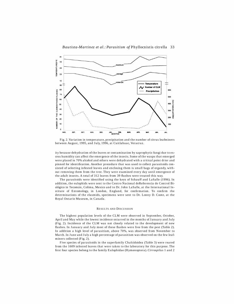
Bautista-Martinez et al.: Parasitism of
Phyllocnistis citrella 33
ity because dehydration of the leaves or contamination by saprophytic fungi due to ex-cess humidity can affect the emergence of the insects. Some of the wasps that emergedwere placed in 70% alcohol and others were dehydrated with a critical point drier andpinned for identification. Another procedure that was used to collect parasitoids con-sisted of selecting infested leaves and enclosing them is small bags of organdy, with-out removing them from the tree. They were examined every day until emergence ofthe adult insects. A total of 312 leaves from 39 flushes were treated this way.
The parasitoids were identified using the keys of Schauff and LaSalle (1996). Inaddition, the eulophids were sent to the Centro Nacional deReferencia de Control Bi-ológico in Tecomán, Colima, Mexico and to Dr. John LaSalle, at the International In-stitute of Entomology, in London, England, for confirmation. To confirm thedeterminations of the elasmids, specimens were sent to Dr. Lonny D. Coote, at theRoyal Ontario Museum, in Canada.
R
ESULTS
AND
D
ISCUSSION
The highest population levels of the CLM were observed in September, October,April and May while the lowest incidence occurred in the months of January and July(Fig. 2). Incidence of the CLM was not closely related to the development of newflushes. In January and July most of these flushes were free from the pest (Table 2).In addition a high level of parasitism, above 70%, was observed from November toMarch. In June and July a high percentage of parasitism was observed on the few leaf-miners collected (Fig. 2).
Five species of parasitoids in the superfamily Chalcidoidea (Table 3) were rearedfrom the 1609 infested leaves that were taken to the laboratory for this purpose. Thefirst four species belong to the family Eulophidae (Hymenoptera).
Cirrospilus
1
and 2
Fig. 2. Variation in temperature, precipitation and the number of citrus leafminersbetween August, 1995, and July, 1996, at Cuitlahuac, Veracruz.

34
Florida Entomologist
81(1) March, 1998
and
Galeopsomyia
sp yet have not been described. The neotropical genus
Horismenus
is very difficult taxonomically, so it is not possible to be sure if the species collected isundescribed or not (LaSalle 1997, personal communication).
Elasmus
tischeriae
is
inthe family Elasmidae, species of which generally attack species in the orders Dipteraand Coleoptera.
In the months of August and September the parasitoids population on the CLMwere reduced considerably, possibly because they were attacking other species of leaf-miners.
Cirrospilus
LaSalle (1996) reported fifteen species of
Cirrospilus
associated with the CLM, in-cluding the undescribed species collected in this study. The same author states that
T
ABLE
2. V
ARIATION
IN
THE
INCIDENCE
OF
THE
CLM
AND
ITS
PARASITOIDS
AT
C
UIT-LAHUAC
, V
ERACRUZ
IN
1995-96.
MonthLeaves
examined
CLM larvae and/or pupae
found
CLM larvae and/or pupae parasitized
Percent parasitized
Aug. 225 207 68 32.8Sep. 291 301 148 49.1Oct. 229 158 92 58.2Nov. 191 148 107 72.2Dec. 95 18 15 83.3Jan. 63 7 6 85.7Feb. 94 11 9 81.8Mar. 111 31 22 70.9Apr. 201 104 71 68.2May. 109 62 29 46.7Jun. 67 12 9 75Jul. 45 4 4 100
T
ABLE
3. P
ARASITOIDS
OF
THE
CLM
AND
THEIR
RELATIVE
IMPORTANCE
AT
C
UIT-LAHUAC
, V
ERACRUZ
.
SpeciesPercent
parasitized Biological phase attacked
1
Cirrospilus
sp 1 22.6 Larvae III (2) and prepupae(7)
Cirrospilus
sp 2 25.1 Larvae III(2), prepupae(1) and pupae(9)
Horismenus
sp 19.3 Prepupae(1) and pupae(13)
Galeopsomyia
sp 27.6 Larvae II(1), III(1), prepupae(4) and pupae(7)
Elasmus tischeriae
5.4 Larvae II(2), III(1) and pupae(2)
1
() number of individuals observed.

Bautista-Martinez et al.: Parasitism of
Phyllocnistis citrella 35
this is a cosmopolitan and polyphagous genus although it prefers to parasitize insectsin the family Gracillaridae. Sometimes the members of
Cirrospilus
act as hyperpara-sitoids (LaSalle 1996), even though this the species collected at Cuitlahuac, Veracruzwere not observed doing this. The two
Cirrospilus
species found at Cuitlahuac are ec-toparasitic, the pupae remaining in the mines of the host.
Galeopsomyia
The collection at Cuitlahuac is the first record of the genus for Mexico (Bautista etal
. 1996). This species, which is being described by LaSalle, is ectoparasitic on theCLM in the larval phases II and III and prepupae. In one case it was observed as a fac-ultative solitary hyperparasitoid. The highest incidence of this species was observedin October and November. According to LaSalle (1994), the species of this genus gen-erally attack Cecidomyiidae (Diptera) and Cynipidae (Hymenoptera).
Horismenus
This ectoparasitoid of the CLM, can be confused with Galeopsomyia. LaSalle(1996) reported two species of Horismenus associated with the CLM which have notyet been described. It is very common to find species of this genus acting as hyperpar-asitoids (Coffelt and Sehultz 1993).
Elasmus
Of the five species of parasitoids associated with the CLM, E. tischeriae Howardwas the least abundant at Cuitlahuac; only about 5% of the parasitoid individualswere of this species. LaSalle (1994) reports three species attacking the CLM, E. tis-cheriae, E. zenhtneri and an undescribed species. To date, E. tischeriae is the only onethat has been found in Mexico. Some authors place this species in the family Eu-lophidae instead of the monotypic Elasmidae. It is the capable of parasitizing lepi-dopteran borers, but it also has been frequently found parasitizing species of Polistes(Hymenoptera: Vespidae) (Borror et al. 1989). In the study area this species was ob-served in the months of August and September.
Other predators
Several other predatory arthropods associated with the CLM were observed atCuitlahuac. These include syrphid flies, common lacewings, spiders (Table 4) and theant species, Crematogaster aff-brevispinosa and Conomyrma bicolor (Hy-menoptera:Formicidae).
TABLE 4. SPIDER SPECIES ASSOCIATED WITH CLM, AT CUITLAHUAC, VERACRUZ.
Family Species
Araneidae Araneus spLeucauge argyra WalckanaerArgiope argentata Fabricius
Salticidae Habronatus spTheridiidae Thymoites unimaculatum
36 Florida Entomologist 81(1) March, 1998
CONCLUSIONS
The highest population levels of the citrus leafminer occurred in September andOctober, then in April and at the beginning of May. The lowest incidence of the pestwas observed in January and July. The parasitoid species with the highest percent-age; of parasitism were Cirrospilus and the Galeopsomyia. In the period from Novem-ber to March, the level of parasitism was very high, about 70%.
ACKNOWLEDGMENTS
This study was supported by Consejo Nacional de Ciencia y Tecnología (CONA-CYT), through Project 0503 PB “Bioecologia del minador de la hoja de los cítricosPhyllocnistis citrella Stainton (Lepidoptera: Gracillariidae). Una nueva plaga para lacitricultura mexicana”. The authors thank Dr. J. LaSalle and Dr. D. Lonny Coote, ofthe International Institute of Entomology, London, England, and Department of En-tomology, Royal Ontario Museum, Toronto, Canada, respectively, for their assistancein the identification of the parasitoids. Identification of the ants was made by M. C.Luis Quiroz Robledo, Institute de Ecologia, A. C., Jalapa, Veracruz, Mexico. Identifi-cation of the spiders was made by Dr. Carlos Solis Rojas, Universidad Autonoma deNuevo Leon, Linares, Mexico.
REFERENCES CITED
BAUTISTA, M., N., H. BRAVO M., AND J. L. CARRILLO S. 1996. Estado actual y perspec-tivas para el manejo del minador de la hoja de los cítricos Phyllocnistis citrellaen México. VI Congreso Nacional de Manejo Integrado de Plagas. Acapulco,Guerrero, Mexico. Memorias: p. 81.
BORROR, D. J., D. M. DELONG, AND C. A. TRIPLEHORN. 1989. An Introduction to theStudy Insects. Ed.6. Holt. Reinhart and Winston, New York. 712-713 pp.
CLAUSEN, C. P. 1931. Two citrus leafminers of the far East. U.S. Dept. Agric. Tecn.Bull. 252. 13 pp.
COFFELT, M. A., AND P. B. SCHULTZ. 1993. Larval parasitism of orangestriped oak-worm (Lepidoptera:Saturniidae) in the urban shade tree environment. Dept.Entomology. University of Virginia. Abs. Biological Control 3(2): 127-134.
GUEROUT, R. 1994. Apparition du Phyllocnistis citrella en Afrique de l’ Ouest. Fruits29: 519-523.
HEPPNER, J. B. 1993. Citrus leafminer Phyllocnistis citrella Stainton (Lepi-doptera:Phyllocnistinae). Florida Dept. Agric. Consumer Services, Divis. PlantIndust., Ent. Circular 359.-2 pp.
KNAPP, J. L., L. G. ALBRIGO., H. W. BROWNING, R. C. BULLOCK, J. B. HEPPNER, D. G.HALL, M. A. HOY, R. NGUYEN, J. E. PENA, AND P. A. STANSLY. 1995. Citrus leaf-miner Phyllocnistis citrella Stainton. Current status in Florida 1995. Coop.Ext. Serv., IFAS, Univ. Florida, Gainesville. 26 pp.
LASALLE, J. 1994. North American genera of the Tetrastichinae (Hymenoptera:Eu-lophidae). J. Natural History 28: 109-236.
LASALLE, J. 1996. The genera of chalcid parasitoids (Hymenoptera: Chalcidoidea) ofthe citrus leafminer (Lepidoptera:Gracillariidae). P. 60 in M. A. Hoy (ed.), Man-aging the citrus leafminer, Proc. Intern. Conf., Orlando, Florida, University ofFlorida, Gainesville. 119 pp.
MARTÍNEZ, B., AND E. RUIZ C. 1996. Citrus leafminer native parasitoids in midlandTamaulipas, Mexico. P. 89 in M. A. Hoy (ed.), Managing the citrus leafminer,Proc. Intern. Conf., Orlando, Florida, University of Florida, Gainesville. 119 pp.
PERALES, G. A., H. C. ARREDONDO B., AND E. GARZA G. 1996. Native parasitoids of cit-rus leafminer Phyllocnistis citrella in Colima, Mexico. Southwestern Entomol-ogist 21:349-350.

Bautista-Martinez et al.: Parasitism of Phyllocnistis citrella 37
QUAYLE, H. J. 1941. Insects of Citrus and Other Subtropical Fruits. Comstock Pub-lishing Company Inc. Ithaca, New York. 582 pp.
RUIZ, C. E., AND J. M. CORONADO B. 1994. El minador de la hoja de los cítricos Phy-llocnistis citrella Stainton (Lepidoptera:Gracillariidae:Phyllocnistinae). Cent.Investig., Fac. Agron. Univ. Auton. Tamaulipas. Folleto Entomol. 1. 2 pp.
RUIZ, C. E., AND R. MATEOS C. 1996. Phyllocnistis citrella Stainton parasites in north-ern Veracruz, Mexico. in M. A. Hoy (ed.), Managing the citrus leafminer, Proc.Intern. Conf., Orlando, Florida, University of Florida, Gainesville. 119 pp.
SCHAUFF, M. E., AND J. LASALLE. 1996. Identification Manual. Citrus Leafminer Par-asitoid Identification Workshop. Orlando Florida. 41 pp.
SPONAGEL, K. W., AND F. J. DIAZ. 1994. El Minador de la Hoja de los Citricos Phylloc-nistis citrella: Un Insecto-plaga de Importancia Económica en la Citrículturade Honduras. Fundación Hondureña de Investigación Agrícola. La Lima Cor-
♦♦♦♦♦♦♦♦♦♦♦♦♦♦♦♦♦♦♦♦♦♦♦♦♦♦♦♦♦♦♦♦♦♦♦♦♦♦♦♦♦♦♦♦♦♦♦♦♦♦♦♦
tés. 25 p.

Michaud: Review of Literature on
Toxoptera citricida 37
A REVIEW OF THE LITERATURE ON
TOXOPTERA CITRICIDA
(KIRKALDY) (HOMOPTERA: APHIDIDAE)
J. P. M
ICHAUD
Citrus Research and Education Center, 700 Exp. St. Rd., Lake Alfred, FL 33850
A
BSTRACT
Literature is reviewed on the brown citrus aphid (BCA),
Toxoptera citricida
Kirkaldy, a serious pest of citrus recently introduced to Florida. Information is sum-marized on the aphid’s distribution, host range, biology, population ecology, naturalenemies, entomopathogens, transmission of plant viruses, and management.
Key Words: citrus tristeza virus, coccinellidae, control, parasitoids, predators, syr-phidae
R
ESUMEN
Es revisada la literatura sobre el pulgón pardo de los cítricos,
Toxoptera citricida
Kirkaldy, una plaga de cítricos recién introducida en la Florida. La información estáresumida en cuanto a la distribución del áfido, sus plantas hospederas, biología, eco-
logía, enemigos naturales, entomopatología, transmisión de virus, y control.
The brown citrus aphid,
Toxoptera citricida
, (BCA) was first discovered in Floridain November, 1995 in Broward and Dade Counties. BCA is a major concern to citrusgrowers throughout the state because of its high efficiency in transmitting citrustristeza virus (CTV). In view of the current interest in this insect, the purpose of thismanuscript is to summarize information available on its biology, ecology, and manage-ment, and its role as a vector of CTV. The volume of literature on CTV warrants a sep-arate review and I have therefore referenced only review articles and thosespecifically concerned with CTV transmission by the BCA.

38
Florida Entomologist
81(1) March, 1998
The BCA, a.k.a. the oriental citrus aphid, was formerly called “
Aphis citricidus
”(1935-1960) and then
Toxoptera citricidus
before the species name was changed to itspresent form to agree in gender with the genus name (Stoetzel 1994a). In earlier lit-erature (prior to 1940) it was commonly referred to as “
Aphis tavaresi
”. Other syn-onyms include “
Myzus citricidus
” and “
Paratoxoptera argentiniensis
”, but usage ofthese names is rare. Essig (1949) lists “
Aphis citricola
” Van der Goot as synonymouswith
T. citricida
, but Hille Ris Lambers (1975), upon re-examination of the originalmaterial collected by Van der Goot in Chile (Van der Goot 1912), concluded that
A. cit-ricola
is synonymous with
Aphis spiraecola
Patch, the green citrus aphid, and thiswas later confirmed by Eastop and Blackman (1988).
A. citricola
has been used as asynonym for
A. spiraecola
in some literature (e.g. Komazaki 1982, 1988), although inpre-1950 publications it often appears as a synonym for BCA. Further confusionarises in studies where the BCA has been mistaken for its close relative
T. aurantii
Kirkaldy, and such cases are difficult to identify.Many of the large number of publications which refer to BCA make only passing
reference to it, or report it as one of many species collected in a general survey of cit-rus insects. I have therefore reviewed in detail only those articles which I felt providedoriginal data, useful observations, or novel insights. The information has been orga-nized under subject headings to provide readers with quick access to particular areasof interest.
D
ESCRIPTION
& D
ISTRIBUTION
Toxoptera citricida
was first described by Kirkaldy (1907) who placed it in the ge-nus
Myzus
. Good descriptions can also be found in Essig (1949), Stroyan (1961), Bän-ziger (1977), Denmark (1978), Stoetzel (1994b), and Halbert and Brown (1996). Thelatter provides a detailed description complete with drawings and a key for distin-guishing other aphids common in Florida citrus. The cytotaxonomy of the Genus
Tox-optera
, including BCA, has been described by Kurl (1980).The BCA is thought to have originated in Southeast Asia (Kirkaldy 1907; Rocha-
Peña et al. 1995) and is common throughout Asia, including China, Cyprus, India, Ja-pan, Laos, Taiwan, Viet Nam (Essig 1949), Sumatra (Takahashi 1926; Mason 1927),Nepal (Knorr & Moin Shah 1971), Sri Lanka (Van Der Goot 1918; Peiris & Bertus1958), Malaysia (Ting 1963; Ting & Arasu 1970), the Philippines (Gavarra & Eastop1976) and Thailand (Bänziger 1977). Pacific islands with records include Hawaii(Kirkaldy 1907), Fiji (Lever 1940), Mauritius (D’Emmerez De Charmoy 1918; Mamet1939), Réunion (Moreira 1967), Samoa (Laing 1927), and Tonga (Carver et al. 1994).BCA has been present in Australia for many years, possibly since the last century(Hely 1968) and can also be found in New Zealand (Cottier 1935).
Specimens of BCA collected by J. S. Tavares in Zambezi were described by DelGuercio in 1908 who named it “
Aphis tavaresi
” (Del Guercio 1917). Anderson (1914)and Theobald (1915) both reported collections of BCA from British East Africa. Otherafrican countries with records include Cameroon, Congo, Ghana, Kenya, Morocco,South Africa, Tanzania, Uganda, Zaire, Zimbabwe (Essig 1949), Ethiopia (Del Guercio1917; Abate 1988; Godfrey-Sam-Aggrey & Balcha 1988), Mozambique, (Saraiva 1929;Annecke 1963) Somalia (Theobald 1928; Chiaromonte 1933), and Tunisia (Halima etal. 1994). It is still absent from Israel (Bar-Joseph & Loebenstein 1973; Raccah &Singer 1987). Essig (1949) reported BCA as present in Italy, Malta and Spain, butthese reports are questionable and remain unconfirmed. Although apparently presentin Turkey (Yumruktepe & Uygun 1994) the BCA is still absent from much of mediter-ranean Europe (Mendel 1956; Jamoussi 1967).
Michaud: Review of Literature on
Toxoptera citricida 39
Accidental introductions of BCA (and CTV) to South America are thought to havebeen made in either Brazil or Argentina during the 1920’s when these regions wereexpanding their citrus production and importing material from Australia and SouthAfrica (Rocha-Peña et al. 1995). Since that time it has spread to Bolivia (Squire 1972;Timmer et al. 1981), Peru (Roistacher 1988), Uruguay, Chile, and Colombia, (Rocha-Peña et al. (1995). It has even been found in jungle regions of the Peruvian Amazon(Ortiz 1981). BCA was first reported in Venezuela in 1976 (Geraud 1976), although ithad been present in neighboring Guyana since at least 1968 (Bisessar 1968) and inSurinam since 1961 (van Hoof 1961). BCA first appeared in the Caribbean islands inTrinidad in 1985 (Yokomi et al. 1994). In 1991 it was found in Guadeloupe, Martin-ique and St. Lucia (Aubert et al. 1992) and in 1992, in Puerto Rico and the DominicanRepublic (Lastra et al. 1992). It is now also present in Jamaica and Cuba (Yokomi etal. 1994). The BCA had spread into Central America as far north as Costa Rica by1989 (Lastra et al. 1991, Voegtlin and Villalobos 1992), to Nicaragua and El Salvadorin 1991 (Lastra et al. 1992, Lee et al. 1992), and to Belize in the fall of 1996 (Halbert1996). In the fall of 1995, the BCA arrived in Florida in the Ft. Lauderdale area and,within one year, spread as far north as Melbourne on the east coast and Ft. Meyers onthe west (Halbert 1997). By the summer of 1997, BCA was widely distributedthroughout southern central and coastal regions of Florida, although it remained ab-sent from much of the ridge citrus north of Highlands county.
H
OST
R
ANGE
The host range of the BCA is largely restricted to the Genus
Citrus
, although thereare many reports of it colonizing other rutaceous plants. A list of the host plants fromwhich BCA has been reported is provided in Table 1. The majority are woody shrubs,although some are perennial vines and annual herbs. Symes’ (1924) report of a singleinfestation on cotton was from Rhodesia. Tao & Tan (1961) collected their specimensin Taiwan. Collections by Mondal et al. (1976) are from India and those of Carver(1978) are all from Australia. Van Harten & Ilharco (1975) and Remaudiere et al.(1985) reported collections from Africa. Ghosh & Raychaudhuri (1981) reported find-ing the BCA feeding on rosaceous fruit trees in India (apple, cherry, peach etc.—spe-cies names not provided), although I suspect this to be a possible mis-identification of
T. aurantii
. The record from white yam,
Dioscorea rotundata
, (Reckhaus 1979) is fromTogo and that for
Passiflora
sp. (Bakker 1974), from Kenya. All collections reported byinspectors of the Florida Department of Agriculture and Consumer Services, Divisionof Plant Industry, are from Florida.
Most non-rutaceous plants are not normally suitable hosts for the BCA and itshould be noted that collections from these plants do not imply they are suitable fordevelopment and reproduction of the aphid. These may be colonized occasionally byvariant individuals when suitable citrus foliage is unavailable, or alates may be col-lected that are resting rather than feeding. Several generations of BCA were rearedsuccessfully in the laboratory on seedlings of West Indian cherry (a.k.a. acerola),
Mal-pighia punicifolia
, in Puerto Rico, although natural colonization of this plant couldnot be elicited by placing flushed seedlings adjacent to infested citrus (Michaud 1996).Halbert et al. 1986 has been erroneously cited as a host record for BCA on soybean,
Glycine max
, (Stibick 1993) but this work examined only transmission of soybean mo-saic virus by BCA. Many of the anomalous occurrences (e.g. cotton) probably repre-sent isolated events or colonizations by variant populations that are atypical of thespecies’ normal host range, although others probably represent mis-identifications(Stoetzel 1994b).
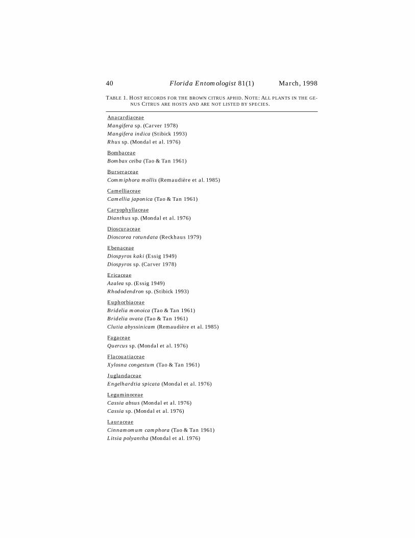
40
Florida Entomologist
81(1) March, 1998
T
ABLE
1. H
OST
RECORDS
FOR
THE
BROWN
CITRUS
APHID
. N
OTE
: A
LL
PLANTS
IN
THE
GE-NUS
C
ITRUS
ARE
HOSTS
AND
ARE
NOT
LISTED
BY
SPECIES
.
Anacardiaceae
Mangifera
sp. (Carver 1978)
Mangifera indica
(Stibick 1993)
Rhus
sp. (Mondal et al. 1976)
Bombaceae
Bombax ceiba
(Tao & Tan 1961)
Burseraceae
Commiphora mollis
(Remaudière et al. 1985)
Camelliaceae
Camellia japonica
(Tao & Tan 1961)
Caryophyllaceae
Dianthus
sp. (Mondal et al. 1976)
Dioscuraceae
Dioscorea rotundata
(Reckhaus 1979)
Ebenaceae
Diospyros kaki
(Essig 1949)
Diospyros
sp. (Carver 1978)
Ericaceae
Azalea
sp. (Essig 1949)
Rhododendron
sp. (Stibick 1993)
Euphorbiaceae
Bridelia monoica
(Tao & Tan 1961)
Bridelia ovata
(Tao & Tan 1961)
Clutia abyssinicam
(Remaudière et al. 1985)
Fagaceae
Quercus
sp. (Mondal et al. 1976)
Flacouatiaceae
Xylosna congestum
(Tao & Tan 1961)
Juglandaceae
Engelhardtia spicata
(Mondal et al. 1976)
Leguminoceae
Cassia absus
(Mondal et al. 1976)
Cassia
sp. (Mondal et al. 1976)
Lauraceae
Cinnamomum camphora
(Tao & Tan 1961)
Litsia polyantha
(Mondal et al. 1976)

Michaud: Review of Literature on
Toxoptera citricida 41
Malpighiaceae
Malpighia punicifolia
(J. P. Michaud unpublished)
Malvaceae
Gossypium hirsutum
(Symes 1924; Carver 1978)
Moraceae
Cudrania triscuspidata
(Essig 1949)
Ficus carica
(Essig 1949)
Ficus ingens
(Remaudière et al. 1985)
Ficus retusa
(Tao & Tan 1961)
Malclura cochinchinensis
(Carver 1978)
Mysinaceae
Maesa chisea
(Mondal et al. 1976)
Maesa
sp. (Mondal et al. 1976)
Nyctaginaceae
Bougainvillea spectabilis
(Remaudière et al. 1985)
Oxalidaceae
Oxalis pes-caprae
Carver (1978)
Passifloraceae
Passiflora foetida
(Mondal et al. 1976)
Passiflora
sp. (Bakker 1974)
Rosaceae
Cottoneaster
sp. (Carver 1978)
Crataegus
sp. (van Harten & Ilharco 1975)
Eriobotrya
sp. (Tao & Tan 1961)
Malus domestica
(van Harten & Ilharco 1975)
Malus sylvestris
(Mondal et al. 1976)
Pyrus communis
(Essig 1949; van Harten & Ilharco 1975; Mondal et al. 1976)
Pyrus
sp. Carver (1978)
Rubiaceae
Lasianthus chinensis
(Tao & Tan 1961)
Rutaceae
Calodendrum capense
(Carver 1978)
Choisya ternata
(Carver 1978)
Citrifortunella floridana
(Fla. Dept. Agr. & Cons. Serv., D.P.I., Gainesville, FL.)
Citrifortunella microcarpa
(Fla. Dept. Agr. & Cons. Serv., D.P.I., Gainesville, FL.)
Citrus
spp.
Clausena lanisum
(Fla. Dept. Agr. & Cons. Serv., D.P.I., Gainesville, FL.)
T
ABLE
1. (C
ONTINUED
) H
OST
RECORDS
FOR
THE
BROWN
CITRUS
APHID
. N
OTE
: A
LLPLANTS
IN
THE
GENUS
C
ITRUS
ARE
HOSTS
AND
ARE
NOT
LISTED
BY
SPECIES
.
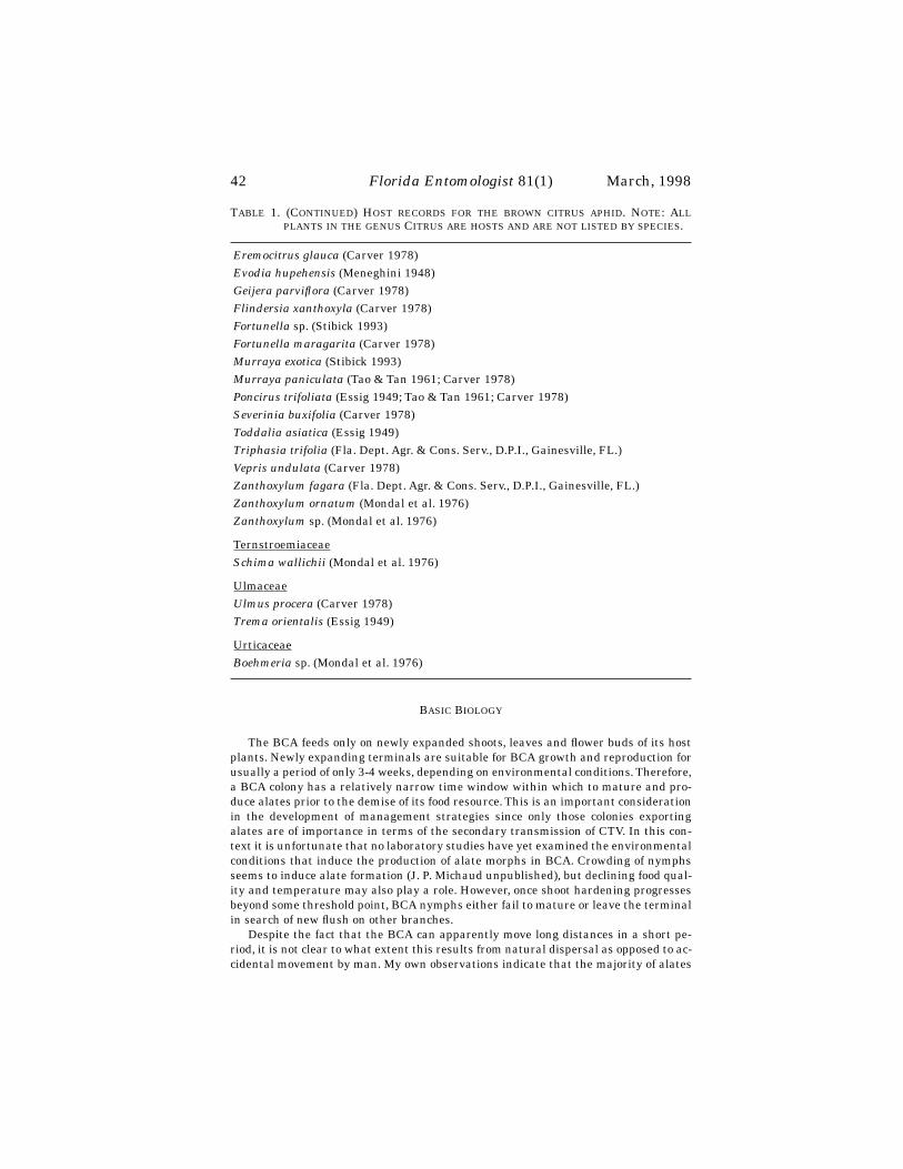
42
Florida Entomologist
81(1) March, 1998
B
ASIC
B
IOLOGY
The BCA feeds only on newly expanded shoots, leaves and flower buds of its hostplants. Newly expanding terminals are suitable for BCA growth and reproduction forusually a period of only 3-4 weeks, depending on environmental conditions. Therefore,a BCA colony has a relatively narrow time window within which to mature and pro-duce alates prior to the demise of its food resource. This is an important considerationin the development of management strategies since only those colonies exportingalates are of importance in terms of the secondary transmission of CTV. In this con-text it is unfortunate that no laboratory studies have yet examined the environmentalconditions that induce the production of alate morphs in BCA. Crowding of nymphsseems to induce alate formation (J. P. Michaud unpublished), but declining food qual-ity and temperature may also play a role. However, once shoot hardening progressesbeyond some threshold point, BCA nymphs either fail to mature or leave the terminalin search of new flush on other branches.
Despite the fact that the BCA can apparently move long distances in a short pe-riod, it is not clear to what extent this results from natural dispersal as opposed to ac-cidental movement by man. My own observations indicate that the majority of alates
Eremocitrus glauca
(Carver 1978)
Evodia hupehensis
(Meneghini 1948)
Geijera parviflora (Carver 1978)Flindersia xanthoxyla (Carver 1978)Fortunella sp. (Stibick 1993)Fortunella maragarita (Carver 1978)Murraya exotica (Stibick 1993)Murraya paniculata (Tao & Tan 1961; Carver 1978)Poncirus trifoliata (Essig 1949; Tao & Tan 1961; Carver 1978)Severinia buxifolia (Carver 1978)Toddalia asiatica (Essig 1949)Triphasia trifolia (Fla. Dept. Agr. & Cons. Serv., D.P.I., Gainesville, FL.)Vepris undulata (Carver 1978)Zanthoxylum fagara (Fla. Dept. Agr. & Cons. Serv., D.P.I., Gainesville, FL.)Zanthoxylum ornatum (Mondal et al. 1976)Zanthoxylum sp. (Mondal et al. 1976)
TernstroemiaceaeSchima wallichii (Mondal et al. 1976)
UlmaceaeUlmus procera (Carver 1978)Trema orientalis (Essig 1949)
UrticaceaeBoehmeria sp. (Mondal et al. 1976)
TABLE 1. (CONTINUED) HOST RECORDS FOR THE BROWN CITRUS APHID. NOTE: ALLPLANTS IN THE GENUS CITRUS ARE HOSTS AND ARE NOT LISTED BY SPECIES.
Michaud: Review of Literature on Toxoptera citricida 43
probably do not fly far from their nascent colony. This is borne out by the fact that fociof BCA infestations are often localized in citrus groves and can be observed to expandmore slowly than those of Aphis gossypii (Gottwald et al. 1995). Furthermore, suctionand yellow-trap catches often underestimate the number of active BCA colonies in aparticular vicinity (e.g. Yokomi et al. 1997). Consequently, BCA infestations tend to beendemic in citrus groves, surviving at low density on bits of asynchronous flush androot sprouts until a new flush cycle provides sufficient food for a population outbreak.Weather conditions such as thermal updrafts or tropical storms may play some role indispersal. For example, the discovery of BCA in Jamaica in 1993 was preceded by thepassage of a strong tropical depression (Lee et al. 1995, p. 209). However, long-rangedispersal by alates is probably rare and movement of infested plant material by hu-mans may be a more important mechanism. Gottwald et al. (1993) estimated very lowprobabilities for colonization of Florida citrus by BCA through arial dispersal from theCarribean, specifically Cuba, but the true mechanism by which it ultimately arrivedin Florida was never established.
The earliest detailed study of the biology and life history of the BCA is by Symes(1924) in Southern Rhodesia. The author reported as many as 30 generations peryear, depending on availability of citrus flush. The time for development to adult wasestimated to range from 8 to 21 d. The BCA is anholocyclic (without sexual genera-tions) throughout most of its range in tropical and subtropical regions. In NewZealand, Cottier (1935) observed the anholocyclic apterae to overwinter, althoughtheir development was greatly retarded. Komazaki et al. (1979) observed a holocyclicgeneration of BCA under temperate conditions in Japan. However, the authors foundvery few overwintering eggs of BCA on trees compared to the numbers of A. gossypiiand A. spiraecola eggs. Furthermore, although hatching rates were similar across spe-cies, the survival rate of hatched BCA nymphs was very low and surviving adult fun-drices produced no second generation. In the laboratory, Komazaki (1990) comparedthe development and reproduction of BCA fundrices hatched from eggs with that ofsubsequent viviparous generations and found that the former took longer to matureat all temperatures tested, and had a lower reproductive rate. Because of low over-wintering populations, the BCA is the last of the citrus aphids to appear on springflush in Japan (Komazaki 1988).
Takanashi (1989) compared the reproductive rates of alate and apterous morphsof the BCA feeding on Citrus natsudaidai in the laboratory. The pre-reproductive pe-riod was longer for alatae than for apterae at both 20° and 25°C, and estimates of bothage-specific fecundity and net reproductive rate were higher for apterae at both tem-peratures. Komazaki (1982) determined that the maximum intrinsic rate of increasefor the BCA occurred at a constant temperature of 27°C, even though the fecundityand net reproductive rate of individual apterous females was maximal at 21.5°C. Ingeneral, the pre-reproductive period, post-reproductive survival, and longevity wereall shortened as temperature increased.
Galatoire (1983) calculated life table statistics for 3 cohorts of BCA grown in out-door enclosures in Argentina. She reported mean life expectancies for apterous fe-males ranging from 28 to 48 d; the shorter life expectancies correlated with highermean daily temperatures and reduced duration of the third and fourth nymphal in-stars. Age-specific mortality rates varied among cohorts, presumably in response tothe different ambient conditions they experienced, although >80% of individuals sur-vived to become adults in all 3 cohorts. Daily fecundity of adult apterae averaged be-tween 5 and 6 nymphs per female per day, and total fecundities ranged between 73and 81 nymphs. The highest mean replacement rate (59 females/female) was observedin a cohort which experienced a moderate regime of daily temperatures, but the high-est instantaneous rate of increase was observed in the cohort experiencing the highest

44 Florida Entomologist 81(1) March, 1998
mean daily temperatures. This result was attributed to reduced generation time un-der the warmer conditions.
There is also evidence that the performance of BCA varies on different species ofcitrus. Komazaki (1982) observed differences between BCA reared on Citrus unshuiand those reared on C. aurantium. He also discovered a temperature-host plant inter-action effect on development. C. unshui yielded aphids with shorter pre-reproductiveperiods and greater longevities and fecundities than did C. aurantium. The thresholdtemperature for BCA development was also lower on C. unshui (8.0°C) than on C. au-rantium (8.4°C). However, the survival rate on C. unshui was 0% at 29.7°C, but 60%on C. aurantium at 29.9°C.
Komazaki (1984) succeeded in rearing 4 generations of BCA on an artificial diet of20% sucrose at pH7, but the growth rate, fecundity, and longevity of these aphids werereduced relative to those fed on citrus.
POPULATION DYNAMICS
Given that the BCA feeds only on tender new citrus terminals, many authors haveobserved population outbreaks to occur about 2 wk following heavy rainfall that in-duces citrus flush (Schwarz 1965a; Klas 1979). Typically, there are two BCA popula-tion peaks per year in subtropical regions, one in the spring and another in the fall.This has been observed in Argentina (Nickel & Klingauf 1985), Australia (Khan 1976;Carver 1978), Brazil (Chagas et al. 1982), Kenya (Seif & Islam 1988), Puerto Rico (J.P. Michaud unpublished), Taiwan (Tao & Tan 1961), and Japan (Nakao 1968; Shindo1972). Komazaki (1981) reported three annual population peaks in Japan during oneyear’s observations.
Geraud (1979) studied the life cycle and population dynamics of BCA in Venezuela,but I was unable to obtain a copy of this thesis. Nickel & Klingauf (1985) studied BCApopulation dynamics for a 2-yr period in Misiones, Argentina. They compared the lon-gevity of exposed colonies with those in exclusion cages and others growing under cli-mate-controlled conditions indoors. The maximum longevity of exposed outdoorcolonies was only 12 d (mean = 7.5) compared with a range of 16-26 d in the exclusioncages and 19-33 d under temperature-controlled conditions. The improved survival ofcolonies in the exclusion cages was attributed, in part, to their protection from naturalenemies, primarily Cycloneda sanguinea (Coleoptera: Coccinellidae) and, in part, tophysical protection afforded by the enclosures. Periods of heavy rain and hot dryweather were both correlated low BCA population densities. Heavy rainfall appearedto impede flight activity as measured by catches in yellow bowl traps. Anderson (1914)also noted a popular belief that BCA populations are checked by heavy showers. How-ever, Nickel & Klingauf (1985) concluded that temperature is an important determi-nant of BCA population trends and that temperature extremes in winter and summerhad a negative impact on development and flight activity. A negative effect of hightemperatures on BCA populations was also inferred by Hall (1930).
NATURAL ENEMIES
The BCA is unusual as an aphid species with few effective parasitoids throughoutits range (Stary 1970). One exception may be Japan where Lysiphlebia japonica Ash-mead (Hymenoptera: Aphidiidae) reportedly exerts some level of control (Kato 1969,1970; Takanashi 1990, 1991). This species was imported to Florida in 1996 and re-leased at 29 sites throughout the state, and to Puerto Rico where it was released at 2sites. To date (Aug, ’97) there no have been no recoveries of this insect from Puerto

Michaud: Review of Literature on Toxoptera citricida 45
Rico. In April 1997, a few weeks after releases were made, a number of specimens ofL. japonica were recovered at several sites in St. Lucie County, Florida. This would in-dicate at least one generation of L. japonica was successful under field conditions inFlorida. However, it is still uncertain whether permanent establishment of this spe-cies will occur and, if so, whether any significant control of BCA will result.
Symes (1924) reports collecting a single unidentified Braconid (probably an Aphi-diid) parasitoid from BCA in South Africa, although Abate (1988) found no parasitoidsof BCA in his survey of citrus disease vectors in Ethiopia. Lysiphlebus testaceipesCresson attacks BCA in Venezuela (Stary & Cermeli 1989) and in Brazil (Gallo et al.1978), although Murakami et al. (1984) did not recover this or any other species ofparasitoid from BCA during their survey in the Cerrados region. L. testaceipes is fre-quently observed parasitizing the BCA in Puerto Rico, but fewer than 5% of mummiesyield viable adults (Yokomi & Tang 1996). In Cuba, L. testaceipes frequently parasit-izes T. aurantii but is only rarely found on BCA (Batista et al. 1995). Carver (1984)found that L. testaceipes (imported to Australia) readily oviposited in BCA but rarelycompleted development. Similarly, Carver & Woolcock (1985) reported incompleteparasitism of BCA by Aphelinus asychis Walker in Australia. L. testaceipes is fre-quently found parasitizing BCA in most citrus-growing regions of Florida, althoughvery few mummies can be found with emergence holes (J. P. Michaud unpublished).
Nickel & Klingauf (1985) reported that, in Argentina, Aphidius colemani Viereckparasitized up to 50% of BCA in some colonies in the fall, although no information ispresented on the survival of the parasitoid in this host. Stary & Cermeli (1989) madecollections of A. colemani from 10 spp. of aphids in Venezuela, but did not find it at-tacking BCA. Valencia & Narciso Cárdenas (1973) reported collection of Aphidius ma-tricariae Haliday from BCA in Peru. De Huiza & Ortiz (1981) collected 4 spp. ofaphidiid wasps from aphids in Peru, including A. colemani and A. matricariae, butonly L. testaceipes emerged from BCA. Carver (1978) noted parasitism of BCA by A.colemani in Australia, although she noted that such mummies were rare in the field.Messing & Rabasse (1995) observed that A. colemani from Réunion Island ovipositedin various aphid species in which the wasp did not complete development. Newman(1924) reports Aphelinus mali (Aphelinidae) emerging from BCA in Western Austra-lia. Flanders & Fisher (1959) reported collections of Lipolexis sp., Trioxys sp., and twoAphelinus spp. from BCA in Kwantung Province, China. Yokomi et al. (1993) reporteda collection of Lipolexis scutellaris and L. gracilus from BCA in Malaysia and Tang etal. (1996) report Aphelinus spiraecolae Evans and Shauff attacking BCA in China.
In most studies of the natural enemies of BCA, emphasis has been placed on pred-atory insects, primarily ladybeetles (Coleoptera: Coccinellidae), and hoverflies(Diptera: Syrphidae) as species causing the greatest mortality to BCA populations.Kato (1968) reported Eristrophe balteatus de Geer, Paragus quadrifasciatus Meigen,Sphaerophoria cylindrica Say, and Syrphus serarius Wiedemann as hoverfly speciesfeeding on BCA in Japan. Symes (1924) reported Xanthogramma aegyptium Wied-mann as the most common syrphid preying on BCA in Rhodesia. Catling (1970) re-ported Allograpta pfeiferi Bigot and Baccha sp. as syrphid predators of BCA in SouthAfrica. Abate (1988) found Sphaerophoria rueppellii Wiedmann attacking BCA inEthiopia, as well as Leucopis spp. (Diptera: Chamaemyiidae). Lever (1946) lists Xan-thogramma (Ischiodon) scutellare F. and Syrphus corollae var. vitiensis Bez. as the im-portant syrphids attacking aphids, including BCA, in Fiji. Goncalves & Goncalves(1976) collected 10 species of syrphids from aphids in São Paulo and Rio de Janeirostates in Brazil. Those attacking BCA included Allograpta exotica (Wiedmann.),Ocyptamus gastrostactus (Wiedmann) (= Baccha gastroctacta) and Pseudodorus clav-atus (F.) (= Baccha clavata). Bartoszeck (1980) also reported the latter two spp. aspredators of BCA in the state of Maranhao, Brazil. Leal et al. (1976) reported O. gas-

46 Florida Entomologist 81(1) March, 1998
troctactus to be the most abundant syrphid on BCA in Pernambuco, Brazil and con-cluded it was the only predator affording any control. P. clavatus and Ocyptamusfuscipennis Say are the most abundant syrphids attacking BCA in Puerto Rico, al-though Allograpta exotica, A. radiata, and Ocyptamus cubanus have also been col-lected (J. P. Michaud unpublished). Valencia and Nárciso Cardenas (1973) collected P.clavatus and Allograpta spp. in their survey of aphid natural enemies in Peru, but didnot find them feeding on BCA. P. clavatus is also abundant on BCA in Cuba (Batistaet al. 1995) and in Trinidad where O. gastroctactus is also important (White 1995).Collections from Florida suggest that P. clavatus is by far the most abundant andubiquitous syrphid on BCA, but Toxomerus geminatus (Say) and Leucopis sp.(Diptera: Chamaemyiidae) have also been recorded (J. P. Michaud unpublished).
Michaud (1996) observed relatively good biological control of BCA in Puerto Ricothroughout the summer months of 1996. Fewer than 5% of suitable citrus terminalswere infested with BCA in most citrus groves between May and August, largely dueto the activities of various species of coccinellids, the adults and larvae of which de-stroyed many colonies in their early stages. The following species of coccinellids havebeen collected from BCA in Puerto Rico: Chilocorus cacti (L.), Cladis nitidula (F.), Co-elophora inaequalis (F.), Coleomegilla innotata (Mulsant), C. sanguinea limbifer (L.),Diomus sp., Egius platycephalus Mulsant, Hippodamia convergens (Guerin), Hy-peraspis sp., Olla v-nigrum (Mulsant), Procula feruuginea (Oliver), and Scymnus(Schymnus) floralis (F.) (J. P. Michaud unpublished). The most abundant species wereC. inaequalis and C. sanguinea. Small species such as Diomus sp., Hyperaspis sp. andS. floralis prey only on early instars of BCA and appear to have only minimal impacton aphid populations. C. sanguinea is the most common coccinellid on BCA in the Do-minican Republic (Borbón et al. 1992) and Cuba (Batista et al. 1995). It is also abun-dant feeding on BCA in Venezuela (Morales & Burandt 1985) and Brazil (Lara et al.1977; Bartoszeck 1980). Additional coccinellid spp. recorded from BCA in Brazil in-clude Cleothera sp., Diomus sp., Exoplectra sp., and Scymnus sp. (Bartoszeck 1980).Chagas et al. (1982) concluded that the coccinellids C. sanguinea, Nephaspis sp.,Stethorus sp. and Scymnus sp. in particular, were the most important predators ofBCA in São Paulo state and provided data on their seasonal abundance in citrus.Other coccinellids collected from BCA in this study included Cleothera sp., Cyclonedaconjugata, Delphastus sp., Hyperaspis sp. Lindorus lophanthes, Neaporia sp., andPentilia sp. Valencia and Narciso Cárdenas (1973) reported C. sanguinea and H. con-vergens as predators of BCA in Peru. Coccinellid species attacking BCA in Florida in-clude Brachiacantha decora (Casey), Brachiacantha dentipes (F.), C. cacti, Chilocorusstigma (L.), Coccinella septmpunctata L., C. ineaqualis, Curinus coeruleus (Mulsant),C. s. sanguinea, Harmonia axyridis (Pallas), H. convergens, Hyperaspis ornatella Gor-don, O. v-nigrum, and Scymnus sp. (J. P. Michaud unpublished).
Kato (1969) listed Scymnus hilaris Motschulsky, Coccinella septepunctata bruckiiMulsant, H. axyridis, Propulaea japonica (Thunberg), Chilocorus kuwanae Silvestriand Hyperaspis japonica (Crotch) as coccinellid species attacking BCA in Japan. Ko-mozaki (1981) reported that S. hilaris was an important predator of BCA in Japanand that its numerical response to increasing BCA populations was relatively good.Nakao (1968) reported Stethorus japonica Kamiya as another coccinellid species prey-ing on BCA in Japan, although Telsimia nigra Weise and C. kuwanae were the mostabundant species in that study.
Maelzer (1978) states that Leis conformis (Boisduval.) is an important predator ofBCA in South Australia. Cheilomenes propinqua is listed by Catling (1970) as a coc-cinellid feeding on BCA in South Africa. Symes (1924) lists Chilomenes lunata F., Ale-sia bohemani Mulsant, A. geisha Gorh., Halyzia exiguenotata F., Lotis sp., andScymnus trepidulus Weise as coccinellids feeding on BCA in Rhodesia, noting that the

Michaud: Review of Literature on Toxoptera citricida 47
first two species were most common. Abate (1988) lists Exochomus sp., Hyperaspissenegalensis (Mulsant), Pharoscymnus madagassus (Weise) and Pharoscymnus sp. ascoccinellid predators of BCA in Ethiopia. Lever (1946) listed C. inaequalis and Coc-cinella repanda var. transversalis F. as important predators of aphids in Fiji, includ-ing BCA.
A study by Nickel & Klingauf (1985) in sub-tropical Argentina indicated that C.sanguinea was the most important predator in that particular region. They measuredpredator-prey ratios varying between 1-40 and 1-10, suggesting relatively good levelsof biological control which they attributed to the semi-natural conditions surroundingtheir study sites. Recruitment of predators from outside citrus groves was judged tobe an important factor. The relatively low numbers of predators present in wintermonths (May-August) was attributed to low rates of recruitment resulting from lowBCA population densities.
Lacewings (Neuroptera) are also predators of aphids that may contribute to sup-pression of their populations. Nakao (1968) reports Micromus novitus Navás and Eu-micromus numerosus Navás (Hemerobiidae) as lacewings feeding on BCA in Japan.Valencia and Narciso Cárdenas (1973) collected Chrysopa sp. (now Chrysoperla sp.chrysopidae) feeding on BCA in Peru. Chagas et al. (1982) reported the seasonal abun-dance of Chrysopa sp., Hemerobius sp. and Megalomus sp. found feeding on BCA inSão Paulo state, Brazil. Nusalala uruguaya (Hemerobiidae) has been recorded fromBCA in Brasil (Souza et al. 1989) and a Ceraeochrysa sp. (Chrysopidae) occurs on BCAin Puerto Rico (J. P. Michaud unpublished), although it is rare. Abate (1988) reportedAnisochrysa boninensis (Okamoto) as a Chrysopid predator of BCA in Ethiopia. White(1995) reported Chrysopa silvana Naval from BCA in Trinidad, although it was notdeemed a significant source of mortality. To date, Ceraeochrysa lineaticornis (Fitch),and Micromus posticus (Walker) have been collected from BCA in Florida (J. P.Michaud unpublished).
Several studies have suggested that BCA may be toxic to certain predators, or nu-tritionally inadequate for their successful development. Tao & Chiu (1971) reportedthat 5 of 13 coccinellid species fed BCA, including C. repanda, suffered injury ordeath, while the others remained unaffected. Souza et al. (1989) reported that larvaeof N. uruguaya fed BCA did not survive to pupation and Tao & Chiu (1971) reportedsimilar findings for two species of Chrysopa. However, Venzon & Carvalho (1993)found Toxoptera spp. (BCA?) to be a suitable diet for Cereaochrysa cubana (Hagen) inBrasil and Ceraeochrysa sp. in Puerto Rico has been successfully reared to adultstages on BCA (J. P. Michaud unpublished). Parker & Singh (1973) found that the coc-cinellids Chilocorus politus Muls., Coccinella arcuata F. and Micraspis (Alesia) dis-color (F.) all expressed a non-preference for BCA in feeding trials, althoughMenochilus (Cheilomenes) sexmaculatus (F.) did not. Morales & Burandt (1985) foundthat C. sanguinea collected in Venezuela and fed BCA in the laboratory failed to com-plete development. On the other hand, C. sanguinea and C. inaequalis collected in Pu-erto Rico developed normally and had good survival on an exclusive diet of BCA (J. P.Michaud unpublished), suggesting that there may exist regional differences betweenbiotypes of predator species with respect to their ability to utilize BCA as food. Inter-estingly, no studies have found any indication of BCA toxicity to syrphids.
Ants are notorious for interfering with the beneficial activities of aphid predatorsand/or parasites and BCA may sometimes benefit from a mutualistic association withcertain ant species. Observing ant and aphid populations in a citrus orchard in Japan,Shindo (1972) concluded that the ant Pristomyrex pungens Mayr interfered with thebehavior of BCA predators (syrphids and coccinellids). Tao & Wu (1968) recommendedthe removal of ant nests in the vicinity of BCA-infested trees in conjunction withchemical treatments for control of the aphid. Bartoszeck (1980) recorded the ants Eu-
48 Florida Entomologist 81(1) March, 1998
cryptocerus placidus (F. Sm.) and Camponotus godmani Forel in association with BCAin Brazil. Fire ants, Solenopsis invicta Buren, are often observed tending BCA in Flor-ida and Puerto Rico (J. P. Michaud unpublished) and are known to remove parasitizedaphids and mummies of L. testaceipes from aphid colonies (Vinson & Scarborough1991). They have also been observed removing predatory larvae and dead aphids fromBCA colonies, and carrying live aphids to fresh, uninfested terminals (J. P. Michaudunpublished). Other ant species observed tending BCA in Puerto Rico include Brach-ymyrmex obscurion Ford, Monomorium ebeninum Ford, Paratrechina longicornis (La-trielle), Pheidole fallax Mayr, Solenopsis globularia (F. Smith), and Wasmanniaauropunctata (Roger) (J. P. Michaud unpublished). In Florida, B. obscurion, Cam-ponotus sexguttatus (F.), and Pseudomyrmex ejectus (F. Smith) have been collected atBCA colonies (J. P. Michaud unpublished).
ENTOMOPATHOGENS
Rondon et al. (1981) found the entomophagous fungus Verticillium lecanii (Zimm.)Viégas to be the most important biological control agent during population outbreaksof the BCA in the Carabobo and Yaracuy states of Venezuela in 1979 and 1980. Thepredators reported as important in this study were C. sanguinea, O. gastrostactus andZelus spp. The authors noted that ideal ambient conditions for sporulation and ger-mination of the fungus (temperatures ranging from 18-24°C at a high relative humid-ity) were prevalent during the 2-yr period of the study. The fungus was reported tosurvive unsuitable conditions in dried aphid mummies, and on aphid cadavers thatoccur on suckering shoots growing in the interior of the tree where they are protectedfrom direct sunlight. The most suitable nutrient media for growing the fungus in thelaboratory were 523, nutrient agar, malt-agar, and PDA. These workers stated that“the elevated pathogenicity on nymphs of different instars, adults, apterous and alate,for which there was >80% mortality in the first 2 wk, prevented culmination of the in-sect’s life cycle”. However, the infection rate of 1.2% of colonies reported by White(1995) in Trinidad is probably a more typical value for the natural occurrence of thisfungus on BCA. V. lecanii has also been recovered from BCA in Puerto Rico, where itappears to be a sporadic and localized source of aphid mortality (J. P. Michaud unpub-lished). De Romero and Romero (1985) evaluated mycelial growth and conidia yield ofV. lecanii collected from BCA in Argentina on 2% potato agar. Batista et al. (1995) re-ported satisfactory results with applications of both V. lecanii (Micotal-1 & Y-57strains) and Paecilomyces fomosoroseus (INISA V strains) against the BCA in Cuba,despite the fact that they observed no natural attacks by entomopathogenic fungi.Samways & Grech (1986) found that the fungus Cladosporium oxysporum (Berk. andCurt.) had considerable impact on BCA populations in field trials in South Africa,which they attributed to the action of an unidentified toxin, rather than direct hyphalgrowth. Other fungi are currently under evaluation for pathogenicity to BCA.
CTV TRANSMISSION
CTV is a closterovirus which causes multiple disease syndromes in citrus. Themost important are quick decline of trees on sour orange rootstock and stem-pittingin susceptible scions irrespective of rootstock. Tristeza is possibly the most serious vi-rus disease of citrus world-wide and yet it is poorly understood. It is not yet possibleto attribute particular symptoms on a particular citrus cultivar to specific viral se-quences, nor is it known which sequences influence transmissibility by its most effi-cient insect vector, the BCA. Meneghini (1946) in Brazil presented the first evidenceof CTV transmission by the BCA. Meneghini (1948) showed semi-persistent transmis-

Michaud: Review of Literature on Toxoptera citricida 49
sion of CTV; viruliferous BCA starved for 48 h lost their ability to transmit the virus,while a 24 h starvation period did not affect the percentage of trees infected. This waslater confirmed by Costa & Grant (1951). Kennedy et al. (1962) suggested thattristeza virus was probably stylet borne. Retuerma & Price (1972) claimed that bothacquisition and transmission of CTV by BCA can occur within a few seconds of feedingand concluded this was proof that CTV was stylet borne, a conclusion subsequentlyquestioned by others. Singh (1978) successfully achieved CTV transmission feedingleaf and bark extracts to BCA through a stretched parafilm membrane and suggestedthat CTV may not be a typical stylet-borne virus, but may also be transmitted in a cir-culative, non-propagative manner. Such bimodal transmission was also suggested byLim & Hagedorn (1977) but this explanation is not generally accepted and further ev-idence of short probing transmission is required (Bar-Joseph et al. 1979). The fact thatCTV is phloem-limited makes transmission by brief probing less likely. The currentconsensus is that CTV transmission best fits the semi-persistent mode (Bar-Joseph etal. 1979; Raccah & Singer 1987) in which the virus is acquired and transmitted byaphids with feeding times ranging from several minutes to several hours, but usuallynot by brief probing.
Historically, introduction of the BCA has invariably resulted in the acceleratedspread of CTV throughout entire citrus growing regions, the best example being thevirtual destruction of the citrus industry in Brazil and Venezuela during the 1970’s,most of which was rooted on sour orange (Lee et al. 1995). A number of studies haveshown that the BCA is a relatively efficient vector of CTV when compared with otheraphids that feed on citrus. Schwartz (1965b) observed that infections of trap plantswith BCA were closely correlated with the numbers of BCA collected from the plants.In the Philippines, Celino et al. (1966) showed that BCA was a more effective vectorthan either T. aurantii or A. gossypii. Sharma (1989) tested 20 isolates of CTV andfound that 12 were most efficiently transmitted by BCA, 5 by A. gossypii, and 3 byMyzus persicae. Yokomi and Damsteegt (1991) quantified the efficiency with whichthe BCA transmitted CTV in comparison to A. gossypii and found the former speciesto be significantly more efficient. Balaraman & Ramakrishnan (1979) showed thatBCA was more efficient than either A. gossypii or T. aurantii at transmitting the 2strains of CTV they tested, and that higher percentage transmission occurred withthe severe strain than with the mild strain. The authors found that a minimum of 15viruliferous aphids/plant were required for 100% transmission when feeding periodswere 24 h each for acquisition and transmission.
A number of studies have shown that variant strains of CTV differ in their trans-missibility by aphids (Bar-Joseph & Loebenstein 1973; Raccah et al. 1978; Raccah etal. 1980). Sharma (1989) showed that severe strains of CTV he tested were transmit-ted by BCA with higher efficiency than the mild strains; acquisition periods wereshorter and retention periods were longer. The fact that citrus varieties vary with re-gard to their suitability for CTV acquisition by BCA further complicates the picture(Bar-Joseph et al. 1979).
A concise review of the literature on CTV transmission by aphids, including a listof infectivity studies on BCA and A. gossypii, is provided by Roistacher & Bar-Joseph(1987) (reproduced in Roistacher & Bar-Joseph 1989). Bar-Joseph et al. (1983) providea review of the epidemiology and control of CTV, as does Lee (1994). Bar-Joseph et al.(1989) and Lee & Rocha-Peña (1992) are both comprehensive reviews of the history ofCTV, its host range, diagnosis, and molecular characterization.
In addition to CTV, the BCA has been implicated in vectoring other plant viruses,although some reports are questionable and lack confirmation. Potyviruses transmit-ted by BCA include yam mosaic virus (Thouvenel & Fauquet 1979), soybean mosaicvirus (Halbert et al. 1986), sugar cane mosaic virus (reported as mosaic of abacá,

50 Florida Entomologist 81(1) March, 1998
Musa textilus, by Gavarra & Eloja (1965)) and chili veinal mottle virus (Ong et al.1979). BCA did not transmit potato virus Y or pepper veinal mottle virus, other vi-ruses infecting chilli peppers, Capsicum annum (Gowda & Reddy 1989). In the sameseries of experiments, BCA failed to transmit cucumber mosaic cucumovirus, which,although not a potyvirus, is non-persistently transmitted by many aphid species.
Using electron microscopy, Maharaj & da Graca (1988) found virions of citrus veinenation virus, a probable luteovirus, in the hindgut lumen and accessory salivaryglands of BCA and subsequently (Maharaj & da Graca 1989), showed transmission bythe aphid. Persistent transmission of citrus vein enation virus has been demonstratedin both A. gossypii and M. persicae (Hermoso de Mendoza et al. 1993). Portillo &Beñatena (1986) claimed that BCA was capable of transmitting citrus psorosis virusto various Citrus spp. in Argentina. However, the assays were based primarily onsymptoms and this conclusion has yet to be confirmed by molecular studies. Protacio(1965) reported that BCA can potentially transmit the agent of cadang-cadang dis-ease to coconut palms, but this disease is now thought to be caused by a viroid andthere is no evidence for insect transmission of viroids.
Broadbent & Fraser (1976) concluded that BCA was not responsible for vectoringthe organism that causes citrus dieback in Australia. Similarly, BCA was shown notto be responsible for vectoring citrus leaf mottle disease in the Philippines (Salibe &Cortez 1967), or the greening disease of citrus that is vectored by Trioza erytreae(McLean & Oberholzer 1965).
MONITORING AND CONTROL
Sticky traps and pan traps have both been used for monitoring flight activity of theBCA, and are more economical than suction traps in terms of capital outlay. However,sticky traps are attractive to many insects and must be replaced frequently. Further-more, aphids caught in such traps inevitably require special solvents to remove andare usually badly damaged, making identification difficult. Pan traps yield specimensin better condition but also need to be emptied on a regular basis and are prone toflooding during periods of heavy rain. Furthermore, it should be kept in mind thattraps only monitor the flight activity of alates, provide little information on the sur-vival or location of aphids in citrus groves, and are not a substitute for effective surveytechniques, i.e. physically searching groves for established colonies.
Alate BCA are not strong fliers and few fly far from their parent colony (Gottwald etal. 1995). Gavarra & Eastop (1976) obtained better catches of BCA in yellow Moericketrays at 152 cm height than they did in trays at ground level. Consequently, optimalplacement of traps is probably above ground level, but lower than the height of sur-rounding trees. Lara et al. (1976) used water traps to compare the attractiveness of var-ious colors to a number of different insects in citrus. In general, they found yellow andwhite to be the most attractive to all species, including the BCA and its predators C.sanguinea and Chrysopa sp. However, Schwarz (1965c) found that the relative attrac-tiveness of yellow and green to BCA changed seasonally, and varied from year to year.
The decision whether or not to apply an insecticide to a BCA infestation will be af-fected by many factors, primarily the type of trees infested. These can be arranged inorder of tolerance (lowest to highest): (1) budwood sources, (2) nursery stock, (3)newly-planted saplings and, (4) mature, producing trees (Knapp et al. 1996). Giventhe Budwood Certification Program presently in effect in the state of Florida whichregulates the maintenance of budwood sources free of severe strains of CTV, the firsttwo categories demand a very low tolerance of BCA infestations. Such trees are bestprotected either by screened enclosures, soil applications of systemic insecticides, ora combination of both. Alternatively, geographic isolation of budwood groves in non-

Michaud: Review of Literature on Toxoptera citricida 51
citrus producing areas might provide a more permanent solution. Heavy infestationson young trees may impede their growth (Hely 1968) and the tolerance level for BCAon saplings is consequentially lower than that for mature trees. There is some indica-tion that BCA infestations can reduce flowering and fruit set (Hall 1930; Smit 1934;Hely 1968), and occasionally lead to dropping of flower buds and young fruit on a largescale (Symes 1924), although good quantitative data is lacking. This suggests that alower injury threshold may exist for trees in flower and early stages of fruit set. Apartfrom these circumstances, tolerance levels for BCA infestations on producing treeswill likely vary with the local incidence of severe CTV strains capable of producing de-cline on sour orange-rooted trees or stem-pitting on scions of orange or grapefruit, andwith their proximity to susceptible trees. Otherwise, there is little indication of directdamage to mature trees, even under heavy feeding pressure.
The earlier reports of successful chemical control of BCA usually recommended fo-liar sprays of lime-sulphate, nicotine sulphate, or soap and nicotine. Hall (1930) rec-ommended spraying winter colonies to reduce populations prior to spring flush. Tao &Tan (1961) recommended chemical treatment of CTV-infected trees, which tend tobloom and flush early, in order to prevent migration of aphids to healthy trees whichflush later. Young resets typically flush earlier than mature trees and could be in-spected for BCA prior to the flush of mature trees and spot-treated as required to pre-clude outbreak populations when flush availability increases. Ideally, growers shouldattempt to prevent BCA infestations developing on CTV-infected trees, as opposed toCTV susceptible, uninfected trees, as there is no evidence to suggest that even thebest systemic compounds can protect against CTV transmission. In practise, this willonly be possible for growers with good information on the location of CTV infectionsin their groves. Foliar formulations of various insecticides can be used to kill aphidson contact, but they can also be expected to have greater impact on non-target andbeneficial insects than systemic materials. Consequently, their use should be limitedto spot applications of heavily infested areas only.
Calza et al. (1968) evaluated 9 organphosphate and carbamate compounds againstBCA in São Paulo, Brazil. Tao & Wu (1968) painted tree trucks with a 30% aqueoussolution of the systemic insecticide monocrotophos (Azodrin) and found it more effec-tive against BCA than dimethoate, vamidothion, or formothion when applied in thismanner. In a later study (Tao & Wu 1969) they found that 2 spring applications ofmonocrotophos at 12.5% a.i. gave good control of BCA and had minimal impact on nat-ural enemies in the grove. Buitendag & Bronkhorst (1986) found monocrotophos to bewithout phytotoxicity to citrus trees and calculated application rates for control ofBCA on trees of different sizes. These authors also calculated rates for trunk applica-tions of Dicrotophos against BCA (Buitendag & Bronkhorst 1984) and described amethod for injecting systemic insecticides directly into trees (Buitendag &Bronkhorst 1980). While systemic compounds may generally have lower impact onnatural enemies, their use as granular formulations or soil drenches on producingtrees is often narrowly restricted to particular seasons to minimize contamination ofground water runoff. Foliar applications should be considered a last resort, unlesscompounds can be selected which minimize impact on beneficial insects within thegrove. For example, Portillo (1977) showed that various concentrations of pirimicarbwere effective against BCA in laboratory tests, and relatively low in toxicity to C. san-guinea. In addition, spot treatments of heavily infested trees or blocks, as opposed toblanket treatment of entire groves, would provide refuges for natural enemy popula-tions and speed their recolonization of treated areas.
Koli et al. (1978) tested 0.05% sprays of 6 insecticides against BCA on citrus seed-lings in India and found that phosphamidon gave the best initial knockdown and themost prolonged activity. Trevizoli & Gravena (1979) compared trunk and foliar sprays
52 Florida Entomologist 81(1) March, 1998
of ethiofencarb, dimethoate, malathion and pirimicarb and found that all methodsand materials were effective against BCA. However, they found that trunk sprays ofpirimicarb and dimethoate had less impact on predator populations (C. sanguineaand Chrysopa sp.) than did foliar sprays.
Milne & de Villiers (1977) determined application rates for delivery of dimethoateby microjet and drip irrigation that gave season-long control of BCA and other ho-mopterans. Milne (1977) protected bagged nursery seedlings from BCA for a period of5-6 wk with a single soil-drench application of dimethoate (40% EC, 1.0 ml/bag). Bui-tendag & Bronkhorst (1983) describe a method for using bands to hold Temik© gran-ules against the trunks of trees. This method provided good control of BCA on youngcitrus trees, but its effectiveness declined when trunk diameter increased above 125mm. Shevale et al. (1987) found that foliar applications of phosphamidon, dimethoate,methyl demeton, monocrotophos and quinalphos (each at 0.025% conc.) all provided>95% reduction of BCA within 24 h of treatment, although phosalone did not. Signif-icant recolonization by BCA was evident in all treatments by day 15. Under the par-ticular conditions of this study, phosphamidon and dimethoate were judged to be themost cost-effective.
Jothi et al. (1990) obtained control of BCA on lime in India with foliar applicationof botanical insecticides. They found that oils of Mahua (Bassia latifolia) and pon-gamia (Pongamia pinnata) at 1% and seed extracts of pongamia and neem (Aza-dirachta indica) at 2% applied early in the flush cycle gave adequate control, andresulted in BCA populations significantly lower than controls after 7 days. Field trialsconducted in Puerto Rico by Yokomi et al. (1995) indicate that foliar treatments ofethion (2.5 lb ai/A) + oil (5 gal), ethion + abamectin (2.5 lb and 1.1 gal ai/A, respec-tively) + oil (5 gal), and pirimicarb (0.25 lb ai/A), and acephate (1.0 lb ai/A) all pro-duced close to 100% mortality of BCA, whereas chlorpyriphos (2.5 lb ai/A) yielded 88%mortality. Knapp et al. (1996) provide current recommendations for management ofBCA in Florida citrus, (budwood sources, nurseries, and producing trees), and includea list of currently approved materials.
ACKNOWLEDGMENTS
The author wishes to thank C. Bergh, C. McCoy, and especially R. Brlansky whosereview and commentary improved the manuscript. S. E. Halbert assisted in compilingthe host list and made other contributions to the MS. Thanks are also due to G. Evans,L. Davis Jr., R. Gordon, L. A. Stange, M. Thomas, and H. Weems for their assistancewith specimen identifications. Special thanks to P. Russ for her help in obtaining themore obscure references. This work was funded by Grant No. 727342212 from USDA-APHIS. University of Florida, Institute of Food and Agricultural Sciences ManuscriptTransmittal No. R-05697.
REFERENCES CITED
ABATE, T. 1988. The identity and bionomics of insect vectors of tristeza and greeningdiseases of Citrus in Ethiopia. Trop. Pest. Man. 34: 19-23.
ANDERSON, T. J. 1914. Report of the Entomologist. Ann. Rep. Dept. Agric. Br. East Af-rica, 1912-13. pp. 124-131.
ANNECKE, D. P. 1963. Observations on some Citrus pests in Moçambique and South-ern Rhodesia. J. Entomol. Soc. S. Afr. 26: 194-225.
AUBERT, B., J. ETIENNE, R. COTTIN, F. LECLANT, P. CAO VAN, C. VUILLAUME, C.JARAMILLO, AND G. BARBEAU. 1992. Citrus tristeza disease a new threat for theCaribbean basin. Report of a survey to Colombia, Dominican Republic, Guade-loupe, Martinique and Trinidad. Fruits 47: 393-404.

Michaud: Review of Literature on Toxoptera citricida 53
BALARAMAN, K., AND K. RAMAKRISHNAN. 1979. Transmission studies with strains oftristeza virus on acid lime. Z. Pflanzenkr. Pflanzen. 86: 653-661.
BAKKER, W. 1974. Notes on East African plant viruses. VI. Virus diseases of Passiflorain Kenya. E. Afr. Agric. For. J. 40: 11-23.
BÄNZIGER, H. 1977. Keys to the identification of aphids (Homoptera). I. Winged aphidsof species economically important in Thailand. F.A.O. Pl. Prot. Bull. 36: 2-41.
BAR-JOSEPH, M., S. M. GARNSEY, AND D. GANSALVES. 1979. The closteroviruses: A dis-tinct group of elongated plant viruses. p. 93-168 in M. A. Laufer, F. B. Bang, K.Maramorosch, and K. M. Smith [eds.], Advances in Virus Research. AcademicPress, New York.
BAR-JOSEPH, M., AND G. LOEBENSTEIN. 1973. Effects of strain, source plant, and tem-perature on the transmissibility of Citrus tristeza virus by the melon aphid.Phytopath. 63: 716-720.
BAR-JOSEPH, M., R. MARCUS, AND R. F. LEE. 1989. The continuing challenge of CitrusTristeza Virus control. Ann. Rev. Phytopath. 27: 291-316.
BAR-JOSEPH, M., C. N. ROISTACHER, AND S. M. GARNSEY. 1983. The epidemiology andcontrol of citrus tristeza disease. p. 61-72 in Plumb, R. T. and J. M. Thresh[eds.], Plant virus epidemiology. The spread and control of insect-borne viruses.Blackwell Scientific, Bet Dagan, Israel.
BARTOSZECK, A. B. 1980. The occurrence of Toxoptera citricidus (Homoptera: Aphid-idae) and its natural enemies in Imperatriz, Maranhao, Brazil. Dusenia: 12: 9-13. (in Port., Eng. sum.).
BATISTA, L., D. N. PORRAS, A GUTÍERREZ, I PEÑA, J. RODRIGUEZ, O. FERNANDEZ DELAMO, R. PÉREZ, AND J. L. MORERA. 1995. Tristeza and Toxoptera citricida inCuba, incidence and control strategy. p. 197-203 in Proceedings of the Third In-ternational Workshop on Citrus Tristeza Virus and Brown Citrus Aphid in theCaribbean Basin: Management Strategies. Lake Alfred, FL, May 15-18, 1995.University of Florida, Institute of Food and Agricultural Sciences.
BISESSAR, S. 1968. Citrus tristeza in Guyana. F.A.O. Pl. Prot. Bull. 16: 45-48. BORBÓN, J. C., A. J. ABUD, P. J. MILLAN, J. ASIÁTICO, AND N. ABREU. 1992. Presencia
de la tristeza de los citricos y Toxoptera citricidus (Kirkaldy) en la RepublicaDominicana. p. 95-101 in Citrus tristeza virus and Toxoptera citricidus in Cen-tral America: Development of management strategies and use of biotechnologyfor control. Proceedings of a Workshop, Maracay, Venezuela, Sept. 14-19, 1992.(in Span., Eng. sum.).
BROADBENT, P., AND L. R. FRASER. 1976. Citrus dieback caused by mycoplasma-likeorganisms, transmitted by Toxoptera citricidus. Agric. Gaz. N.S.W.: 87: 10-11.
BUITENDAG, C. H., AND G. J. BRONKHORST. 1980. Injection of insecticides into treetrunks—a possible new method for control of citrus pests? Cit. Subtrop. Fr. J.556: 5-7.
BUITENDAG, C. H., AND G. J. BRONKHORST. 1983. A technique for applying Temik 15ggranules (aldicarb) to the trunks of citrus trees for the control of pests. Cit. Sub-trop. Fr. J. 597: 6-7, 10. (in Afrik., Eng. sum.).
BUITENDAG, C. H., AND G. J. BRONKHORST. 1984. The application of dicrotophos (Bid-rin 50 WOK) to citrus trunks for the control of psyllid and aphids. Cit. Subtrop.Fr. J. 608: 4, 6-8. (in Afrik., Eng. sum.).
BUITENDAG, C. H., AND G. J. BRONKHORST. 1986. Further aspects of trunk treatmentof citrus with insecticides: phytotoxicity, side effects on incidental pests, anddevelopment of application apparatus. Cit. Subtrop. Fr. J. 623: 7-10.
CALZA, R., N. SUPLICY, AND A. ORLANDO. 1968. Effect of some new insecticides on thecontrol of the black citrus aphid. Biológico 34: 262-264. (in Port., Eng. sum.).
CARVER, M. 1978. The black citrus aphids, Toxoptera citricidus (Kirkaldy) and T. au-rantii (Boyer de Fonscolombe) (Homoptera: Aphididae). J. Aust. Ent. Soc. 17:263-270.
CARVER, M. 1984. The potential host ranges in Australia of some imported aphid par-asites (Hymenoptera: Ichneumonoidea: Aphidiidae). Entomophaga 29: 351-360.

54 Florida Entomologist 81(1) March, 1998
CARVER, M., P. J. HART, AND P. W. WELLINGS. 1994. Aphids (Hemiptera: Aphididae)and associated biota from the Kingdom of Tonga, with respect to biological con-trol. Pan-Pac. Entomol. 69: 250-260.
CARVER, M., AND L. T. WOOLCOCK. 1985. Interactions between Acyrthosiphum kondoi(Homoptera: Aphidoidea) and Aphelinus asychis (Hymenoptera: Chalcidoidea)and other parasites and hosts. Entomophaga 30: 193-198.
CATLING, H. D. 1970. The bionomics of the South African citrus psylla Trioza erytreae(Del Guercio) (Homoptera: Psyllidae) 4. The influence of predators. J. Entomol.Soc. S. Afr. 33: 341-348.
CELINO, C. S., D. R. PANALIGAN, AND U. V. MOLINO. 1966. Studies on insect transmis-sion of the tristeza virus in the Philippines. Philipp. J. Pl. Ind. 31: 89-93.
CHAGAS, E. F. D., S. S. NETO, A. J. B. P. BRAZ, C. P. B. MATEUS, AND I. P. COELHO.1982. Population fluctuations of pest and predator insects in citrus. Pesqui.Agropecu. Bras. 17: 817-824. (in Port., Eng sum.).
CHIAROMONTE, A. 1933. Entomological notes on fruit tree growing in Italian Somali-land. Agric. Colon. 27: 383-385. (in Ital.)
COSTA, A. S., AND T. J. GRANT. 1951. Studies on transmission of the tristeza virus.bythe vector Aphis citricidus. Phytopath. 16: 105-133.
COTTIER, W. 1935. The black citrus aphid and some other tree aphides. N. Z. J. Agric.51: 214-219.
DE HUIZA, I. R., AND P. ORTIZ. 1981. Some aphidiinae (Hymenopt.: Braconidae) thatparasitise aphids (Homopt.: Aphididae) in Peru. Rev. Per. Entomol. 23: 129-132.(in Span., Eng sum.).
DEL GUERCIO, G. 1917. Contribuzione alla conoscenza delgi afidi. Redia, Florence 12:217-221. (in Ital.).
D’EMMEREZ DE CHARMOY, D. 1918. Maur. Dept. Agric. Ann. Rep. pp. 10-12. DENMARK, H. A. 1978. The brown citrus aphid, Toxoptera citricida (Kirkaldy) (Ho-
moptera: Aphididae). Entomol. Circ. No. 194, Division of Plant Industry, Flor-ida Department of Agriculture.
DE ROMERO, M. G. Y., AND E. R. ROMERO. 1985. Evaluation of growth and conidiayield of Verticillium lecanii (Zimm.) Viegas in 2% potato glucose agar. Rev. In-vest. (Centro de Investigaciones para la Regulación de Poblaciones de Organis-mos, Nocivos, Argentina) 111: 47-60. (in Span., Eng. sum.).
EASTOP, V. F., AND R. L. BLACKMAN. 1988. The identity of Aphis citricola van der goot.Systematic Entomol. 13: 157-160.
ESSIG, E. O. 1949. Aphids in relation to quick decline and tristeza of Citrus. Pan. Pac.Entomol. 25: 13-22.
FLANDERS, S. E., AND T. W. FISHER. 1959. The economic effect of aphidiophagous in-sects on Citrus in South China. J. Econ. Entomol. 52: 536-537.
GALATOIRE, I. 1983. Vital statistics of Toxoptera citricidus (Kirkaldy) (Homoptera:Aphididae). Rev. Soc. Entomol. Arg. 42: 353-368. (in Span.).
GALLO, E., O. NAKANO, S. S. NETO, R. P. L. CARVALHO, G. C. DE BATISTA, E. B. FILHO,J. R. P. PARRA, R. A. ZOCCHI, AND S. B. ALVES. 1978. Manual de EntomologiaAgricola. Editôra Agronômica Ceres, São Paulo, 531 pp. (in Port.).
GAVARRA, M. R., AND V. F. EASTOP. 1976. Notes on the estimation of alate aphid pop-ulations using Moericke yellow trays. Phil. Entomol. 3: 246-249.
GAVARRA, M. R., AND A. L. ELOJA. 1965. Experimental transmission of the abaca mo-saic virus by Toxoptera citricida (Kirkaldy). Philipp. J. Pl. Ind. 29: 47-54.
GERAUD, F. 1976. El áfido negro de los cítricos Toxoptera citricida (Kirkaldy) en Vene-zuela (Resumen). Premier Encuentro Venezuelano de Entomología. Univer-sidad central de Venezuela. Faculdad de Agronomía-Instituto de ZoologíaAgrícola. Maracay, Venezuela. (in Span, original not seen)
GERAUD, B. 1979. Dinámica de poblaciones de áfidos en cítricos y observaciones pre-liminares sobre el ciclo de vida de Toxoptera citricida (Kirkaldy). Thesis. Uni-versidad de Zulia, Fac. Agronomia. 47 pp. (in Span., original not seen).

Michaud: Review of Literature on Toxoptera citricida 55
GHOSH, M. R., AND D. N. RAYCHAUDHURI. 1981. Aphids (Homoptera: Aphididae) feed-ing on rosaceous fruit plants in Darjeeling District of West Bengal and Sikkim(India). Entomon. 6: 61-68.
GODFREY-SAM-AGGREY, W., AND A. BALCHA. 1988. Insect fauna in a declining Citrusorchard at Melka Werer in Ethiopia. F.A.O. Pl. Prot. Bull. 36: 75-81.
GONCALVES, C. R., AND A. J. L. GONCALVES. 1976. Observations on syrphid flies as pred-ators of homopterous insects. An Soc. Entomol. Bras. 5: 3-10. (in Port., Eng. sum.).
GOTTWALD, T. R., S. M. GARNSEY, M. CAMBRA, P. MORENO, M. IREY, J. BORBON, A. S.JEAN, AND A. R. SOLIS. 1995. Rate of disease increase and patterns of spread ofCTV when vectored by Toxoptera citricida vs. Aphis gossypii. p. 44-49 in Pro-ceedings of the Third International Workshop on Citrus Tristeza Virus andBrown Citrus Aphid in the Caribbean Basin: Management Strategies. Lake Al-fred, FL, May 15-18, 1995. University of Florida, Institute of Food and Agricul-tural Sciences.
GOTTWALD, T. R., S. M. GARNSEY, AND R. K. YOKOMI. 1993. Potential for spread of cit-rus tristeza virus and its vector, the brown citrus aphid. Proc. Fla. State Hort.Soc. 106: 85-94.
GOWDA, K. T. P., AND H. R. REDDY. 1989. Aphid transmitted viruses infecting chilli.Current Research - University of Agricultural Sciences (Bangalore) 18: 71-72.
HALBERT, S. E. 1996. Entomology Section: Citrus. Tri-ology 34:8. Fla. Dept. Agric. andCons. Serv., Div. Plant Industry, Gainesville, FL.
HALBERT, S. E. 1997. Brown citrus aphid: Current situation and prognosis. Cit. Ind.,April Issue.
HALBERT, S. E., AND L. G. BROWN. 1996. Toxoptera citricida (Kirkaldy), brown citrusaphid - identification, biology and management strategies. Fla. Dept. Agric. &Cons. Serv., Div. Plant Industry, Entomol. Cir. No. 374.
HALBERT, S. E., G. X. ZHANG, AND Z. Q. PU. 1986. Comparison of sampling methodsfor alate aphids and observations on epidemiology of soybean mosaic virus inNanjing, China. Ann. Appl. Biol. 109: 473-483.
HALIMA, K. M. B., J. M. RABASSE, AND M. H. B. HAMOUDA. 1994. The aphids of citrusfruits and their natural enemies in Tunisia. Tropicultura 12: 145-147. (in Port.,Eng. sum).
HALL, W. J. 1930. Notes on the control of some of the more important insect pests ofCitrus in Rhodesia. Rhodesia Agric. J. 27: 737-747.
HELY, P. C. 1968. The entomology of citrus in New South Wales. Misc. Pub. Aust. En-tomol. Soc. 1:1-20.
HERMOSO DE MENDOZA, A., J. A. PINA, J. F. BALLASTER-OLMOS, AND L. NAVARRO.1993. Persistent transmission of citrus vein enation virus by Aphis gossypii andMyzus persicae. p. 361-363 in P. Moreno, J. V. da Graça, and L. W. Timmer[eds.], Proc. Conf. Intl. Org. Citrus Virol., 12th. Riverside California.
HILLE RIS LAMBERS, D. 1975. Aphis citricola v. d. Goot, 1912, replaces Aphis spirae-cola Patch, 1914 (Homoptera: Aphididae). Entomol. Berich. 35: 59.
JAMOUSSI, B. 1967. Virus diseases of Citrus in the Mediterranean Basin. Archs. Inst.Pasteur Tunis. 44: 91-116. (in Fren.).
JOTHI, B. D., A. VERGHESE, AND P. L. TANDON. 1990. Evaluation of different plant oilsand extracts against citrus aphid Toxoptera citricidus (Kirkaldy). Ind. J. Pl.Prot. 18: 251-254.
KATO, T. 1968. Outbreaks of aphids and the role of natural enemies in citrus orchards.Odökon Chûgoku 10: 19-23. (in Jap.).
KATO, T. 1969. Specific relation between aphids and their natural enemies in citrus.Odökon Chûgoku 11: 20-23. (in Jap.).
KATO, T. 1970. Efficiency of Lysiphlebus japonicus Ashmead (Hymenoptera: Aphidi-idae) in control of the citrus aphid Toxoptera citricidus Kirkaldy, infesting cit-rus young shoots in summer. Odökon Chûgoku 12: 1-6. (in Jap.)
KENNEDY, J. S., M. F. DAY, AND V. F. EASTOP. 1962. A conspectus of aphids as vectorsof plant viruses. Commonwealth Institute of Biological Control. 114 p.
56 Florida Entomologist 81(1) March, 1998
KHAN, M. H. 1976. The citrus aphid, Toxoptera citricidus (Kirk.). Bien. Rep. Waite Agr.Res. Inst. 1976-77, p. 100. Univ. of Adelaide, South Australia.
KIRKALDY, G. W. 1907. On some peregrine Aphidae in Oahu, Honolulu. Proc. Hawai-ian Entomol. Soc. 1: 100.
KLAS, F. E. 1979. Population densities and spatial patterns of the aphid tristeza vec-tor, Toxoptera citricida Kirk. Proc. Eighth I.O.C.V. Conf. pp. 83-87.
KNAPP, J. L., H. W. BROWNING, R. F. LEE, AND P. STANSLY. 1996. The brown citrusaphid: Citrus tristeza virus relationship and management guidelines for Flor-ida citrus. Cit. Ind., March issue.
KNORR, L. C., AND S. MOIN SHAH. 1971. World Citrus problems V. Nepal. F.A.O. Pl.Prot. Bull. 19: 73-79.
KOLI, S. Z., K. G. CHOUDHARI, AND P. V. MAKAR. 1978. Comparative study on residualtoxicity of some insecticides against citrus aphid, Toxoptera citricidus Kirk. J.Mahar. Agric. Univ. 3: 273.
KOMAZAKI, S. 1981. Life cycles and population fluctuations of aphids on citrus. Proc.Int. Soc. Citric. 2: 692-695.
KOMAZAKI, S. 1982. Effects of constant temperatures on population growth of threeaphid species, Toxoptera citricidus (Kirkaldy), Aphis citricola van der Goot andAphis gossypii Glover (Homoptera: Aphididae) on Citrus. Appl. Entomol. Zool.17: 75-81.
KOMAZAKI, S. 1984. Rearing of brown citrus aphid, Toxoptera citricida (Kirkaldy),(Homoptera: Aphididae), on a synthetic diet with reference to pH and sucroseconcentration of diet and rearing density. Appl. Entomol. Zool. 19: 114-117.
KOMAZAKI, S. 1988. Growth and reproduction in the first two summer generations oftwo citrus aphids, Aphis citricola Van Der Groot and Toxoptera citricidus(Kirkaldy) (Homoptera: Aphididae), under different thermal conditions. Appl.Entomol. Zool. 23: 220-227.
KOMAZAKI, S. 1990. Seasonal variation of parthenogenetic reproduction in the case ofcitrus aphids. p 351-356 in Hoshi, M. and O. Yamashita [eds.], Advances in In-vertebrate Reproduction 5. Elsevier Science Publishers.
KOMAZAKI, S., Y. SAKAGAMI, AND R. KORENGA. 1979. Overwintering of aphids on cit-rus trees. Jpn. J. Entomol. Zool. 23: 246-250. (in Jap., Eng. sum.).
KURL, S. P. 1980. Cytotaxonomy of the genus Toxoptera (Homoptera: Aphididae). En-tomon. 5: 251-255.
LAING, F. 1927. Coccidae, Aphididae and Aleyrodidae. p. 34-45 in Insects of Samoa II.Brit. Mus. Nat. His., London.
LARA, F. M., S. A. DE BORTOLI, AND E. A. OLIVEIRA. 1976. The influence of colors oncollecting of some insects in citrus sp. An. Soc. Entomol. Bras. 5: 157-163. (inPort., Eng. sum.).
LARA, F. M., S. A. DE BORTOLI, AND E. A. OLIVEIRA. 1977. Population fluctuations insome insects associated with Citrus sp. and their correlation with meteorolog-ical factors. Científica 5: 134-143. (in Port., Eng. sum.).
LASTRA, R., R. LEE, M. A. ROCHA-PEÑA, C. L. NIBLETT, S. M. GARNSEY, AND R. K.YOKOMI. 1992. Survey for presence of tristeza virus and Toxoptera citricidus inMexico and Central America. CATIE-University of Florida-INIFAP/SARH-USDA, Turrialba, Costa Rica.
LASTRA, R., R. MENESES, P. E. STILL, AND C. L. NIBLETT. 1991. The citrus tristeza vi-rus situation in Central America. p. 156-159 in R. H. Brlanski, R. F. Lee, and L.W. Timmer [eds.], Proc. Conf. Intl. Org. Citrus Virol., 11th. Riverside California.
LEAL, C. A., H. C. C. OLIVEIRA, AND J. G. SMITH. 1976. Syrphid predators of aphids onCitrus spp. in Recife, Pernambuco. An. Soc. Entomol. Bras. 5: 138-142. (in Port.,Eng. sum.).
LEE, R. F. [ed.] 1994. Citrus Tristeza Virus: An introduction to current priorities, withspecial reference to the worsening situation in Central America and the Carib-bean. IIBC, Ascot, Berks, UK.
Michaud: Review of Literature on Toxoptera citricida 57
LEE, R. F., AND M. A. ROCHA-PEÑA. 1992. Citrus Tristeza Virus. p. 226-249 in J. Ku-mar, H. S. Chaube, U. S. Singh, and A. N. Mukhopadhyay, [eds.], Plant Diseasesof International Importance. Prentice Hall, Englewood Cliffs, N.J.
LEE, R. F., M. ROCHA-PEÑA, C. L. NIBLETT, F. OCHOA, S. M. GARNSEY, R. K. YOKOMI,AND R. LASTRA [eds.]. 1995. Proceedings of the Third International Workshopon Citrus Tristeza Virus and Brown Citrus Aphid in the Caribbean Basin: Man-agement Strategies. Lake Alfred, FL, May 15-18, 1995. University of Florida,Institute of Food and Agricultural Sciences.
LEE, R. F., C. N. ROISTACHER, C. L. NIBLETT, R. LASTRA, M. A. ROCHE-PEÑA, S. M.GARNSEY, R. K. YOKOMI, D. G. GUMF, AND J. A. DOBBS. 1992. Presence of Tox-optera citricidus in Central America: A threat to citrus in Florida and theUnited States. Cit. Ind. June issue.
LEVER, R. J. A. W. 1940. Insect pests of citrus, pineapple and tobacco. Agric. J. Fiji 11:99-101.
LEVER, R. J. A. W. 1946. The aphids or greenflies of Fiji. Agric. J. Fiji 17: 81. LIM, W. L., AND J. M. HAGEDORN. 1977. p. 237-252 in K. F. Harris and K. Maramo-
rosch [eds.], Aphids as Virus Vectors. Academic Press, New York. MAELZER, D. A. 1978. The growth and voracity of larvae of Leis conformis (Boisd.) (Co-
leoptera: Coccinellidae) fed on the rose aphid Macrosiphum rosae (L.) (Hompot-era: Aphididae) in the laboratory. Aus. J. Zool. 26: 293-304.
MAHARAJ, S. B., AND J. V. DA GRAÇA. 1988. Observation of virus-like particles associ-ated with citrus vein enation -infected citrus and the viruliferous aphid vectorToxoptera citricidus. Phytophylactica 20: 357-360.
MAHARAJ, S. B., AND J. V. DA GRAÇA. 1989. Transmission of citrus vein enation virusby Toxoptera citricidus. Phytophylactica 21: 81-82. (in Afrik., Eng. sum.).
MAMET, R. 1939. The Aphididae of Mauritius. Mauritius Inst. Bull. 1: 43-56. MASON, P. W. 1927. Fauna sumatrensis. (Beitrag No. 38) Aphdididae. Suppl. Entomol.
15: 86-90.MCCLEAN, A. P. D., AND P. C. J. OBERHOLZER. 1965. Greening disease of the sweet or-
ange: evidence that it is caused by a transmissible virus. S. Afr. J. Agric. Sci. 8:253-275.
MENDEL, K. 1956. The threat of tristeza in the Mediterranean Basin. F.A.O. Pl. Prot.Bull. 4: 106-108.
MENEGHINI, M. 1946. Sobre a naturaleza e transmissibilidade de docena “Tristeza”dos citrus. Biológico 12: 285-287. (in Port., Eng. sum.).
MENEGHINI, M. 1948. Experiências de transmissão da doença “tristeza” dos citruspelo pulgão preto da laranjeira. Biológico 14: 115-118. (in Port., Eng. sum.).
MESSING, R. H., AND J. M. RABASSE. 1995. Oviposition behavior of the polyphagousaphid parasitoid Aphidius colemani Viereck (Hymenoptera: Aphidiidae). Agric.Ecosys. Envir. 52: 13-17.
MICHAUD, J. P. 1996. Observations on the natural enemies of brown citrus aphid inPuerto Rico. Cit. Ind., Aug. issue.
MILNE, D. L. 1977. Control of citrus nursery pests by soil applications of dimethoate.Cit. Subtrop. Fr. J. 525: 5-7.
MILNE, D. L., AND E. A. DE VILLIERS. 1977. Dimethoate applied by microjet and dripirrigation for control of citrus psylla, Trioza erytreae, the vector of greening dis-ease. Cit. Subtrop. Fr. J. 523: 21-22.
MONDAL, P. K., R. C. BASU, AND D. N. RAYCHAUDURI. 1976. Studies on the aphidsfrom Eastern India. XXX. The Genus Toxoptera. Oriental Insects 10: 535-540.
MORALES, J., AND C. L. BURANDT JR. 1985. Interactions between Cycloneda sanguineaand the brown citrus aphid: Adult feeding and larval mortality. Eviron. Ento-mol. 14: 520-522.
MOREIRA, S. 1967. Outbreaks and new records. F.A.O. Pl. Prot. Bull. 15: 59-60. MURAKAMI, Y., N. ABE, AND G. W. COSENZA. 1984. Parasitoids of scale insects and
aphids on citrus in the Cerrados region of Brazil (Hymenoptera: Chacidoidea).Appl. Entomol. Zool. 19: 237-244.

58 Florida Entomologist 81(1) March, 1998
NAKAO, S. 1968. Ecological studies on the insect community of citrus groves IV. A listof insects collected in a citrus grove near Fukuoka City. Kontyû: 30: 50-71.
NEWMAN, J. 1924. Report of the Economic Entomologist. West. Aust. Dept. Agric. Ann.Rep. 1923-24, pp. 20-24.
NICKEL, O., AND F. KLINGAUF. 1985. Biologie und massenwechsel der tropischen cit-rus-blattlaus Toxoptera citricidus in beziehung zu Nützlingsaktivität undklima in Misiones Argentinien (Homoptera: Aphididae). Entomol. Gener. 10:231-240. (in Germ., Eng. sum.).
ONG, C. A., W. P. TING, AND G. VARGHESE. 1979. Role of aphid vectors in the spreadof chilli veinal mottle virus. Malay. Agric. J. 8: 52-60.
ORTIZ, M. S. 1981. Aphididae (Homoptera) from the jungle edge: Tingo Maria (Hua-nuco, Peru). Rev. Per. Entomol. 23: 119-120. (in Span., Eng. sum.).
PARKER, B. L., AND G. SINGH. 1973. The distribution, feeding habits and fecundity offour Coccinellidae. Malay. Agric. J. 2: 29-33.
PEIRIS, J. W. L., AND A. L. BERTUS. 1958. The tristeza disease of Citrus in Ceylon.Trop. Agric. 114: 221-229.
PORTILLO, M. M. 1977. Acción del aficida pirimicarb en el pulgon de los citrus Tox-optera citricidus (Kirkaldy) y en el coccinélido predador Cycloneda sanguinea(L.). Idia 331/333: 63-66. (in Span.).
PORTILLO, M. M., AND H. N. BEÑATENA. 1986. Transmission of psorosis from citrus tocitrus by aphids. Revista de la Sociedad Entomológica Argentina 45: 299-305.(in Span, Eng. sum.).
PROTACIO, D. B. 1965. Aphid transmission of a cadang-cadang-like disease to coconutseedlings. Philipp. J. Agric. 28: 79-98.
RACCAH, B., M. BAR-JOSEPH, AND G. LOEBENSTEIN. 1978. p. 221-227 in P. R. Scott andA. Bainbridge [eds.], Plant Disease Epidemiology. Blackwell, Oxford.
RACCAH, B,. G. LOEBENSTEIN, AND S. SINGER. 1980. Aphid-transmissibility variants ofcitrus tristeza virus in infected citrus trees. Phytopath. 70: 89-93.
RACCAH, B., AND S. SINGER. 1987. Incidence and vector potential of the aphids whichtransmit citrus tristeza virus in Israel. Phytophylactica 19: 173-177.
RAMIREZ, G. G., AND C. ZULUAGA. 1995. Identification of hymenopterous parasitoidsof aphids on cotton, citrus and horticultural crops in Valle del Cauca. Rev. Col.Entomol. 21: 71-76. (in Span., Eng. sum.).
RECKHAUS, P. 1979. A virus disease of white yam (Dioscorea rotundata) in Togo. Zeit.Pflanzenk. Pflanzens 86: 763-766.
REMAUDIÈRE, G., A. AUTRIQUE, V. F. EASTOP, P. STARY, AND G. AYMONIN. 1985. Con-tribution to the ecology of African aphids. F.A.O. Pl. Prot. Bull. 64. (in Fren.).
RETUERMA, M. L., AND W. C. PRICE. 1972. Evidence that tristeza virus is stylet borne.FAO Pl. Prot. Bull. 20: 111-114.
ROCHA-PEÑA, M. A., R. F. LEE, R. LASTRA, C. L. NIBLETT, F. M. OCHOA-CORONA, S. M.GARNSEY, AND R. K. YOKOMI. 1995. Citrus tristeza virus and its aphid vectorToxoptera citricida: Threats to citrus production in the Caribbean and Centraland North America. Plant Dis. 79: 437-443.
ROISTACHER, C. N. 1988. Observations on the decline of sweet orange trees in coastalPeru caused by stem-pitting tristeza. Pl. Prot. Bull., India 36: 19-26.
ROISTACHER, C. N., AND M. BAR-JOSEPH. 1987. Aphid transmission of citrus tristezavirus: a review. Phytophylactica 19: 163-167.
ROISTACHER, C. N., AND M. BAR-JOSEPH. 1989. Aphid transmission of citrus tristezavirus. Citrograph X: 117-125.
RONDÓN, A., E. ARNAL, AND F. GODOY. 1981. Comportamiento del Verticillium lecanii(Zimm.) Viegas, patogeno del áfido Toxoptera citricida (Kirk.) en finca citricolasde Venezuela. Agron. Trop. 30: 201-212. (in Span., Eng. sum.).
SALIBE, A. A., AND R. E. CORTEZ. 1967. Studies on the leaf mottling disease of Citrusin the Philippines. F.A.O. Pl. Prot. Bull. 14: 141-144.
SAMWAYS, M. J., AND N. M. GRECH. 1986. Assessment of the fungus Cladosporium ox-ysporum (Berk. and Curt.) as a potential biocontrol agent against certain Ho-moptera. Agric. Ecos. Environ. 15: 231-239.
Michaud: Review of Literature on Toxoptera citricida 59
SARAIVA, A. C. 1929. Report on the inspection of citrus orchards. Bol. Agric. Pecuár. 1-2: 47-48. (in Port.).
SCHWARZ, R. E. 1965a. Aphid-borne virus diseases of citrus and their vectors in SouthAfrica. B. Flight activity of citrus aphids. S. Afr. Agric. Sci. 8: 931-940.
SCHWARZ, R. E. 1965b. Aphid-borne virus diseases of Citrus and their vectors in SouthAfrica. A. Investigations of the epidemiology of aphid transmissible virus dis-eases of Citrus by means of trap plants. S. Afr. J. Agr. Sci. 8: 839-852.
SCHWARZ, R. E. 1965c. Seasonal fluctuations in the behaviour of Toxoptera citricidaKirk. in relation to yellow attractants. Z. PflKrankh. PflPath. PflSchutz. 72: 84-89. (in Germ., Eng. sum.).
SEIF, A. A., AND A. S. ISLAM. 1988. Population densities and spatial distribution pat-terns of Toxoptera citricida (Kirk.) (Aphididae) in Citrus at Kenya coast. Insect.Sci. Appl. 9: 535-538.
SHARMA, S. R. 1989. Factors affecting vector transmission of citrus tristeza virus inSouth Africa. Zentralblatt für Mikrobiologie 144: 283-294.
SHEVALE, B. S., D. S. AJIRI, D. P. BHORE, AND D. B. KADAM. 1987. Studies on relativeefficacy of certain insecticides against citrus aphid, Toxoptera citricida(Kirkaldy). Pestology 11: 9-10.
SHINDO, M. 1972. Relation between ants and aphids in a citrus orchard. Proc. Assoc.Pl. Prot. Kyushu 18: 69-71. (in Jap., Eng sum.).
SINGH, A. B. 1978. Comparative transmission of tristeza virus by aphids acquiredfrom leaves, leaf extracts and bark extracts through stretched parafilm mem-brane. Ind. J. Microbiol. 18: 40-43.
SMIT, B. 1934. A study of the most important insect pests that confront the Citrusgrower in the Eastern Province. S. Afr. J. Sci. 31: 439-441.
SOUZA, B., A. I. CIOCIOLA, AND J. C. MATIOLI. 1989. Comparative biology of Nusalalauruguaya (Navás, 1923) (Neuroptera: Hemerobiidae) fed on different species ofaphids. II. Pre-pupal, pupal and adult stages. An. Soc. Entomol. Bras. 18(suppl.): 43-51. (in Port., Eng. sum.).
SQUIRE, F. 1972. Entomological problems in Bolivia. P.A.N.S. 18: 249-268. British Mis-sion in Tropical Agriculture, Cochabamba, Bolivia.
STARY, P. 1970. Biology of Aphid Parasites. W. Junk, The Hague. STARY, P., AND M. CERMELI. 1989. Parasitoides (Hymenoptera: Aphidiidae) de áfidos
en plantas cultivadas de Venezuela. Bull. Entomol. Venez. 5: 77-80. STIBICK, J. N. L. 1993. new pest response guidelines. Brown citrus aphid, Toxoptera
citricida. USDA APHIS PPQ. STOETZEL, M. B. 1994a. The correct spelling of the name for the brown citrus aphid.
Proc. Entomol. Soc. Wash. 96:179. STOETZEL, M. B. 1994b. Aphids (Homoptera: Aphididae) of potential importance on
citrus in the United States with illustrated keys to species. Proc. Entomol. Soc.Wash. 96: 74-90.
STROYAN, H. L. G. 1961. Identification of aphids living on Citrus. F.A.O. Pl. Prot. Bull.9: 45-65.
SYMES, C. B. 1924. Notes on the black citrus aphis. Rhodesia Agric. J. 11: 612-626. TAKAHASHI, R. 1926. Some Aphididae of Sumatra. 2. Treubia 8: 426-466. TAKANASHI, M. 1989. The reproductive ability of apterous and alate viviparous mor-
phs of the citrus brown aphid, Toxoptera citricidus (Kirkaldy) (Homoptera:Aphididae). Jap. J. Appl. Entomol. Zool. 33: 266-269. (in Jap., Eng. sum.).
TAKANASHI, M. 1990. Development and reproductive ability of Lysiphlebus japonicusAshmead (Hymenoptera: Aphidiidae) parasitizing the brown citrus aphid, Tox-optera citricidus (Kirkaldy) (Homoptera: Aphididae). Jpn. J. Appl. Ent. Zool. 34:237-243. (in Jap., Eng sum.).
TAKANASHI, M. 1991. Percent parasitism by an aphidiid parasitoid, Lysiphlebusjaponicus Ashmead attacking host colonies of different sizes. Proc. Assoc. Pl.Prot. Kyushu 37: 163-166. (in Jap., Eng. sum.).

60 Florida Entomologist 81(1) March, 1998
TANG, Y. Q., R. K. YOKOMI, AND J. G. BROWN. 1996. Parasitoids for biological controlof brown citrus aphid: recent observations. Proc. 13th Conf. IOCV, IOCV, Riv-erside (in press).
TAO, C. C., AND C. CHIU. 1971. Biological control of citrus, vegetable and tobaccoaphids. Spec. Publ. Taiwan Agric. Res. Inst. 10: 1-110.
TAO, C. C., AND M. F. TAN. 1961. Identification, seasonal population and chemical con-trol of citrus aphids in Taiwan. Agric. Res. 10:41-52.
TAO, C. C., AND K. C. WU. 1968. Report on citrus insect control study by chemicals inTaiwan. Pl. Prot. Bull, Taiwan 10: 57-64. (in Chin., Eng. sum.).
TAO, C. C., AND K. C. WU. 1969. Studies on bark treatment against Citrus insects. Pl.Port. Bull. Taiwan 11: 143-149. (in Chin., Eng. sum.).
THEOBALD, F. V. 1915. African Aphididae. II. Bull. Entomol. Res. 2: 103-153. THEOBALD, F. V. 1928. Aphididae from Italian Somaliland and Eritrea. Bull. Entomol.
Res. 11: 177-180. THOUVENEL, J. C., AND C. FAUQUET. 1979. Yam mosaic, a new potyvirus infecting
Dioscorea cayanensis in the Ivory Coast. Ann. Appl. Biol. 93: 279-283. TIMMER, L. W., R. SCORZA, AND R. F. LEE. 1981. Incidence of tristeza and other Citrus
diseases in Bolivia. Pl. Dis. 65: 515-517. TING, W. A. P. 1963. Tristeza disease of Citrus in the Federation of Malaya. F.A.O. Pl.
Prot. Bull. 10: 81-83.TING, W. P., AND N. T. ARASU. 1970. A survey of tristeza virus on Citrus in West Ma-
laysia. Malay. Agric. J. 47: 299-304. TREVIZOLI, D., AND S. GRAVENA. 1979. Efficiency and selectivity of insecticides for in-
tegrated control of the black citrus aphid, Toxoptera citricidus (Kirk, 1907). Ci-entifíca 7: 115-120. (in Port., Eng. sum.).
VALENCIA, V. L., AND D. C. NARCISO CÁRDENAS. 1973. The aphids (Homoptera: Aphi-didae) of the valley of Ica, their host plants and natural enemies. Rev. Peru. En-tomol. 16: 6-14. (in Span.).
VAN DER GOOT, P. 1912. Über einige wahrscheinlich neue blattlausarten aus der sam-mlung des Naturhistorischen Museums Hamburg. Mitt. Natur. Mus. Hamburg29: 273-275. (in Germ.).
VAN DER GOOT, P. 1918. Aphididae of Ceylon. Spolia Zeylanica, Columbo 11: 70-75. VAN HARTEN, A., AND F. A. ILHARCO. A further contribution to the aphid fauna of An-
gola, including the description of a new genus and species. Agron. Lusit. 37: 13-35.
VAN HOOF, H. A. 1961. Observations on aphid flights in Suriname. Entomol. Exp.Appl. 5: 239-243.
VENZON, M., AND C. F. CARVALHO. 1993. Desenvolimento larval, pré-pupal e pupal deCereaochrysa cubana (Hagen) (Neuroptera: Chrysopidae) em diferentes dietase temperaturas. An. Soc. Entomol. Brasil 22: 477-483. (in Port., Eng sum.).
VINSON, S. B., AND T. A. SCARBOROUGH. 1991. Interactions between Solenopsis invicta(Hymenoptera: Formicidae), Rhopalosiphum maidis (Homoptera: Aphididae),and the parasitoid Lysiphlebus testaceipes Cresson (Hymenoptera: Aphidiidae).Ann. Entomol. Soc. Am. 84: 158-164.
VOEGTLIN, D., AND W. VILLALOBOS. 1992. Confirmation of the brown citrus aphid,Toxoptera citricida, in Costa Rica. Fla. Entomol. 75:161-162.
WHITE, G. 1995. Natural enemies of Toxoptera citricida (Kirk.) in Trinidad, W.I. p. 41-43 in Proceedings of the Third International Workshop on Citrus Tristeza Virusand Brown Citrus Aphid in the Caribbean Basin: Management Strategies.Lake Alfred, FL, May 15-18, 1995. University of Florida, Institute of Food andAgricultural Sciences.
YOKOMI, R. K., AND V. C. DAMSTEEGT. 1991. Comparison of citrus tristeza virus trans-mission efficacy by Toxoptera citricidus and Aphis gossypii. in: Peters, D. C., J.A. Webster, and C. S. Choubler [eds.], Proceedings, Aphid-plant Interactions:Populations to Molecules. Stillwater, Oklahoma.
YOKOMI, R. K., R. LASTRA, M. B. STOETZEL, R. F. LEE, S. M. GARNSEY, T. R. GOT-TWALD, M. A. ROCHA-PEÑA, AND C. L. NIBLETT. 1994. Establishment of the

Michaud: Review of Literature on Toxoptera citricida 61
brown citrus aphid Toxoptera citricida (Kirkaldy) (Homoptera: Aphididae) inCentral America and the Caribbean basin and its transmission of citrustristeza virus. J. Econ. Entomol. 87: 1078-1085.
YOKOMI, R. K., D. RIVERA, S. M. GARNSEY, T. R. GOTTWALD, E. ABREU-RODRIGUEZ, V.DAMSTEEGT, P. A. STANSLY, V. F. FEBRES, AND C. L. NIBLETT. 1997. Brown cit-rus aphid and citrus tristeza virus incidence in Puerto Rico. in Proceedings ofthe 13th Conference of the International Organization of Citrus Virologists (inpress).
YOKOMI, R. K., P. A. STANSLY, E. A. RODRIGUEZ, AND T. R. GOTTWALD. 1995. Chemicalmitigation of brown citrus aphid populations in Puerto Rico. p. 75-76 in Pro-ceedings of the Third International Workshop on Citrus Tristeza Virus andBrown Citrus Aphid in the Caribbean Basin: Management Strategies. Lake Al-fred, FL, May 15-18, 1995. University of Florida, Institute of Food and Agricul-tural Sciences.
YOKOMI, R. K., AND Y. Q. TANG. 1996. A survey of parasitoids of brown citrus aphid(Homoptera: Aphididae) in Puerto Rico. Biol. Con. 6: 222-225.
YOKOMI, R. K., Y. Q. TANG, L. NONG, AND M. L. KOK-YOKOMI. 1993. Potential mitiga-tion of the threat of the brown citrus aphid, Toxoptera citricida (Kirkaldy), byintegrated pest management. Proc. Fla. State Hort. Soc. 106: 81-85.
YUMRUKTEPE, R., AND N. UYGUN. 1994. Determination of aphid species (Homoptera:Aphididae) and their natural enemies in Citrus orchards in the Eastern Medi-terranean region. Turkiye III. Biyolojik Mucadele Knogresi Bildirileeri, 25-28Ocak 1994, Ege Universitesi Ziraat Fakultesi, Bitki Koruma Bolumu, Izmir.
♦♦♦♦♦♦♦♦♦♦♦♦♦♦♦♦♦♦♦♦♦♦♦♦♦♦♦♦♦♦♦♦♦♦♦♦♦♦♦♦♦♦♦♦♦♦♦♦♦♦♦♦
1994, 1-12. (in Turk., Eng. sum.).

Wetterer et al.: Ecology of a Costa Rican Leaf-cutting Ant
61
FORAGING AND NESTING ECOLOGY OF
ACROMYRMEX OCTOSPINOSUS
(HYMENOPTERA: FORMICIDAE) IN A COSTA RICAN TROPICAL DRY FOREST
J
AMES
K. W
ETTERER
,
1
D
ANIEL
S. G
RUNER
2
AND
J
ORGÉ
E. L
OPEZ
3
1
Center for Environmental Research & Conservation, Columbia UniversityNew York, NY 10027
2
Department of Zoology, University of Hawaii, Honolulu, HI 96822
3
Department of Ecology and Evolution, University of Chicago, Chicago, IL 60637
A
BSTRACT
Leaf-cutting ants (
Acromyrmex
sp. and
Atta
sp.) in Costa Rica show many intra-and interspecific differences in ecology. Recent taxonomic studies question whetherthe
Acromyrmex octospinosus
populations on the Pacific and Atlantic slopes of CostaRica are a single species. We therefore examined the foraging and nesting ecology of
A. octospinosus
in the tropical dry forest of Palo Verde National Park on the Pacificslope of Costa Rica and compared our findings with published data on the ecology of
A. octospinosus
in the tropical moist forest of La Selva Biological Station on the At-lantic slope. The Pacific
A. octospinosus
foraged primarily on the leaves of herbs andother small plants, fallen leaves, fruit, flowers, and insect frass, but does not cut theleaves of large trees. Worker size distribution within colonies was bimodal with only
62
Florida Entomologist
81(1) March, 1998
the larger workers leaving the nest to forage. Nests were shallow and generally undera few centimeters of organic debris at the base of trees and woody shrubs or in crev-ices. The foraging and nesting ecology of the Pacific
A. octospinosus
appeared to bevery similar to that of the Atlantic
A. octospinosus
, except that the Pacific ants col-lected considerable amounts of insect frass (11% of all loads), whereas the Atlanticants had no recorded loads of frass. This difference in selectivity, however, may havebeen due simply to seasonal differences in availability of frass at the sites.
Acromyrmex octospinosus
was the only species of leaf-cutting ant found at PaloVerde. The vertisol soil of the area, which has very poor drainage when wet and cracksdeeply when dry, may not be suitable for major pest species of leaf-cutters in CostaRica,
Atta cephalotes
and
Atta colombica
, which excavate nests deep underground.
Key Words:
Atta
, foraging selectivity, Palo Verde, pest ants
R
ESUMEN
Las hormigas podadoras (
Acromyrmex
sp. y
Atta
sp.) en Costa Rica muestran mu-chas diferencias intra- e interespecíficas en ecología. Estudios taxonómicos recientesponen en duda la noción que poblaciones de
Acromyrmex octospinosus
en los flancosOrientales y Occidentales de Costa Rica sean una sola especie. Por lo tanto, nosotrosexaminamos la ecología de alimentaciùn y anidamiento de
A. octospinosus
en el bos-que seco tropical del Parque Nacional Palo Verde en el flanco occidental de Costa Rica,y comparamos nuestros resultados con datos previamente publicados sobre la ecologíade
A. octospinosus
en el bosque húmedo tropical de la Estación Biológica La Selva enel flanco oriental. La alimentación de
A. octospinosus
en el lado occidental consistióprincipalmente de hojas de hierbas y otras plantas pequeñas, hojas caídas, frutas, flo-res y excrementos de insectos, pero no cortan hojas de árboles grandes. La distribu-ción del tamaño de las obreras en la colonia es bimodal, y sólo las obreras grandessalen de la colonia para recoger alimentos. Los nidos son poco profundos y por lo ge-neral están bajo unos cuantos centímetros de materia orgánica al pie de árboles y ar-bustos, o en grietas. La ecología de alimentación y anidamiento de
A. octospinosus
enel lado occidental parece ser muy similar a la de
A. octospinosus
en el lado oriental, ex-cepto que las hormigas en el lado occidental recogen cantidades considerables de ex-cremento de insectos (11% de todas las cargas), mientras que las hormigas en el ladooriental no han sido observadas cargando excremento de insectos. Esta diferencia enselectividad, sin embargo, puede ser causada por diferencias en disponibilidad de ex-cremento de insectos en diferentes estaciones entre las localidades.
Acromyrmex oc-tospinosus
fue la única especie de hormiga podadora encontrada en Palo Verde. Elsuelo vertisol de esa área, que tiene muy mal drenaje cuando está mojado y que formarajaduras hondas cuando está seco, puede no ser adequado para otras especies dañi-nas de hormigas podadoras en Costa Rica,
Atta cephalotes
y
Atta
colombica, las cuales
excavan nidos profundos.
Fungus-growing ants (Myrmicinae; Attini) are unique among ants in their habit ofcultivating a symbiotic fungus for food. All attine ants are obligately dependent ontheir fungus. In other aspects of their ecologies, however, different species of attinesvary greatly (Weber 1972, Hölldobler & Wilson 1990). Ants of the nine “lesser” genera(
Cyphomyrmex
,
Mycetosoritis
,
Mycetophylax
,
Mycocepurus
,
Mycetarotes
,
Myrmicoc-rypta
,
Apterostigma
,
Sericomyrmex
, and
Trachymyrmex
) typically collect insect frass,dead insects, or small pieces of fallen plant material to use as substrate for their fun-gal gardens. They tend to have small and inconspicuous colonies (fewer than 5000 in-dividuals), with relatively monomorphic workers. In contrast, ants belonging to two

Wetterer et al.: Ecology of a Costa Rican Leaf-cutting Ant
63
genera of attines,
Acromyrmex
and
Atta
, often depend on harvesting leaves for theirfungal gardens and consequently are commonly called leaf-cutting ants. Leaf-cuttingant colonies can grow to include many thousand to several million highly-polymorphicworkers. Many leaf-cutters, particularly
Atta
species, are major agricultural pests.Earlier studies have found striking differences in the foraging and nesting ecology
of
Acromyrmex octospinosus
(Reich),
Acromyrmex volcanus
(Wheeler),
Acromyrmexcoronatus
(Fabricius), and
Atta cephalotes
(L.), the four species of leaf-cutting antsfound on the Atlantic slope of Costa Rica (Wetterer, 1991, 1993, 1994a, b, 1995).
Ac-romyrmex octospinosus
and
A. volcanus
colonies forage primarily on small herbs andfallen leaves, fruits, and flowers (Wetterer 1991, 1993). These colonies produce twodistinct size castes of workers (Wetterer, unpublished). The minima workers stay in-side the nest, tending the fungus garden and brood, while the relatively monomorphicmaxima workers leave the nest to forage. In
A. coronatus
colonies and small
A. ceph-alotes
colonies, workers primarily cut the soft leaves of herbaceous plants (Wetterer1994a, 1995). These colonies produce a narrow range of small workers, the largest ofwhich are just big enough to cut soft vegetation efficiently (Wilson 1983, Wetterer, un-published). In large
A. cephalotes
colonies, workers primarily attack the leaves andflowers of trees (Cherrett 1968, 1972; Rockwood & Hubbell 1987; Vasconcelos 1990;Wetterer 1994b). These colonies produce an extremely broad continuous size-range ofworkers (Stradling 1978; Wilson 1983). A wide range of medium-size workers forage,with the larger of these cutting thicker and tougher vegetation (Nichols-Orians &Schultz 1989; Wetterer 1994b).
Recent taxonomic research, based on morphological characters and allozyme poly-morphisms, has raised questions as to whether the
A. octospinosus
on the Pacific andAtlantic slopes of Costa Rica are a single species (J. Longino, T. Schultz, & K.Boomsma, personal communication). Because earlier studies indicated marked inter-specific ecological differences among leaf-cutting ants, we compared the foraging andnesting ecology of Pacific and Atlantic
A. octospinosus
. We also examined the questionas to why
A. octospinosus
was the only leaf-cutting ant species present at our studysite in Palo Verde National Park.
M
ETHODS
We studied foraging selectivity and nesting habits of
Acromyrmex octospinosus
col-onies at the start of the wet season (June 1996) in tropical dry forest of Palo Verde Na-tional Park, Guanacaste Province, Costa Rica. We worked in the area around PaloVerde Biological Station (10
°
19’N, 85
°
21’W; elevation 10 m), operated by the Organi-zation for Tropical Studies (see Janzen 1983 for a site description). We conducted ourstudy during daylight hours, when most
A. octospinosus
colonies were actively forag-ing. On an earlier visit to Palo Verde in the dry season (January 1996), there was nodiurnal foraging by
A. octospinosus
(J. Wetterer, personal observation).Through extensive searches along hiking trails and roads around Palo Verde, we
located seventeen
A. octospinosus
colonies. At a nearby open pasture site, Sitio Mojal,we collected data on one additional
A. octospinosus
colony (colony 12), making a totalof eighteen colonies. We found no other leaf-cutting ant colonies at the site, althoughwe did find a single live alate
Atta cephalotes
queen. We explored the area for severalkilometers in different directions, but never located any
A. cephalotes
colonies.At each
A. octospinosus
colony, we collected laden foragers from along their forag-ing trails. To help ensure a representative sample, we collected laden foragers at tensecond intervals as they passed a designated point on their trail, or if there were fewforagers, we collected consecutive foragers. We used data from twelve colonies in our
64
Florida Entomologist
81(1) March, 1998
foraging selectivity analysis. At four large
A. octospinosus
colonies, we collected 25 for-agers from each of the two main trails. From six smaller colonies, we collected 25 for-agers for each colony. At two very small colonies, we collected only fourteen workersfrom one colony and eleven from the other, because no more foragers came in a periodof several hours. At six other small colonies we collected none or only one forager dueto lulls in foraging activity that seemed to be associated with hot sunny weather. Wedid not include these collections in our foraging selectivity analysis.
We classified each load as fresh (soft, pliant, and green) or fallen (dry or yellowed)leaf material (fragment or whole leaf), herb section (stem or stem with leaves, leafbuds, or flowers), flower (part or whole), fruit (part or whole berry), insect frass, or“other.” Whenever possible, we located and identified the original source of each load.
At each colony we followed laden foragers in order to locate their nests. We par-tially excavated nests to confirm the location of their fungus garden. In addition, wecollected one complete colony.
Voucher specimens from this study have been deposited at the National Museumof Natural History, Washington D.C.
R
ESULTS
Foraging selectivity
Acromyrmex octospinosus
foragers carried a wide variety of vegetable matter (Ta-ble 1). There were great differences among the twelve colonies and even betweentrails of the same colony (Table 1). The fresh leaf material that the ants cut came pri-marily from leaves of herbs or from tree seedlings. For example, in the colony with thehighest proportion of fresh leaf fragments (colony 8), all leaf material came from small
Acacia
seedlings growing in the area. Foragers also commonly cut pieces of fallenleaves, sections of herbs (herb stems and stems with leaves, leaf buds, or flowers),fruits, petals, and flower buds, plus variable amounts of other types of plant material(petioles, seeds, bark, sticks, and wood slivers). At several colonies, foragers carriedinsect frass, apparently from caterpillars. Although we once found
A. octospinosus
for-agers with fruit fragments descending a small tree, we never found foragers cuttingthe leaves of large trees.
The general types of material gathered by the Pacific
A. octospinosus
foragers wasvery similar to those gathered by the Atlantic
A. octospinosus
foragers, except that thePacific
A. octospinosus
foragers harvested less fruit and more insect frass (Table 2).
Nesting Habits
We located the nests of all eighteen
A. octospinosus
colonies. All nests were veryshallow and required little excavation. Twelve nests were directly in contact with atree or woody shrub: eight under organic debris at the base of trees or woody shrubs,two under organic debris on top of tree buttress roots, and two in tree crevasses (oneand two meters above the ground). Three nests were associated with artificial sub-strates: one under roofing slates piled in a gully, one between two pieces of sheet metalon the forest floor, and one in the cracked cement foundation of a building. Finally, twonests were under organic debris in open soil areas and one was in a crevice of a rockoutcrop.
Within the
A. octospinosus
nests there was a noticeable bimodal worker size-dis-tribution with two distinct size castes of workers. Only the large workers left the nestto forage.
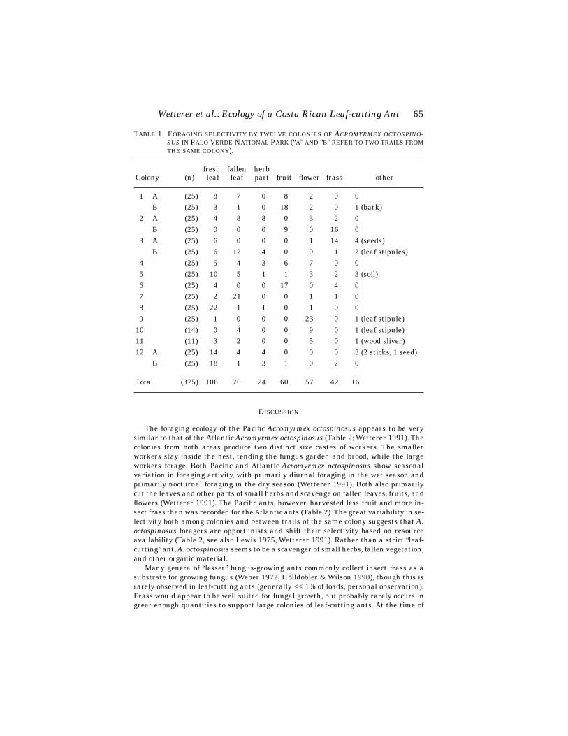
Wetterer et al.: Ecology of a Costa Rican Leaf-cutting Ant
65
D
ISCUSSION
The foraging ecology of the Pacific
Acromyrmex octospinosus
appears to be verysimilar to that of the Atlantic
Acromyrmex octospinosus
(Table 2; Wetterer 1991). Thecolonies from both areas produce two distinct size castes of workers. The smallerworkers stay inside the nest, tending the fungus garden and brood, while the largeworkers forage. Both Pacific and Atlantic
Acromyrmex octospinosus
show seasonalvariation in foraging activity, with primarily diurnal foraging in the wet season andprimarily nocturnal foraging in the dry season (Wetterer 1991). Both also primarilycut the leaves and other parts of small herbs and scavenge on fallen leaves, fruits, andflowers (Wetterer 1991). The Pacific ants, however, harvested less fruit and more in-sect frass than was recorded for the Atlantic ants (Table 2). The great variability in se-lectivity both among colonies and between trails of the same colony suggests that
A.octospinosus
foragers are opportunists and shift their selectivity based on resourceavailability (Table 2, see also Lewis 1975, Wetterer 1991). Rather than a strict “leaf-cutting” ant,
A. octospinosus
seems to be a scavenger of small herbs, fallen vegetation,and other organic material.
Many genera of “lesser” fungus-growing ants commonly collect insect frass as asubstrate for growing fungus (Weber 1972, Hölldobler & Wilson 1990), though this israrely observed in leaf-cutting ants (generally << 1% of loads, personal observation).Frass would appear to be well suited for fungal growth, but probably rarely occurs ingreat enough quantities to support large colonies of leaf-cutting ants. At the time of
T
ABLE
1. F
ORAGING
SELECTIVITY
BY
TWELVE
COLONIES
OF
A
CROMYRMEX
OCTOSPINO-SUS
IN
P
ALO
V
ERDE
N
ATIONAL
P
ARK
(“
A
”
AND
“
B
”
REFER
TO
TWO
TRAILS
FROMTHE
SAME
COLONY
).
Colony (n)fresh leaf
fallen leaf
herb part fruit flower frass other
1 A (25) 8 7 0 8 2 0 0B (25) 3 1 0 18 2 0 1 (bark)
2 A (25) 4 8 8 0 3 2 0B (25) 0 0 0 9 0 16 0
3 A (25) 6 0 0 0 1 14 4 (seeds)B (25) 6 12 4 0 0 1 2 (leaf stipules)
4 (25) 5 4 3 6 7 0 05 (25) 10 5 1 1 3 2 3 (soil)6 (25) 4 0 0 17 0 4 07 (25) 2 21 0 0 1 1 08 (25) 22 1 1 0 1 0 09 (25) 1 0 0 0 23 0 1 (leaf stipule)
10 (14) 0 4 0 0 9 0 1 (leaf stipule)11 (11) 3 2 0 0 5 0 1 (wood sliver)12 A (25) 14 4 4 0 0 0 3 (2 sticks, 1 seed)
B (25) 18 1 3 1 0 2 0
Total (375) 106 70 24 60 57 42 16

66
Florida Entomologist
81(1) March, 1998
our study, the start of the wet season, there was a population explosion of caterpillarsat Palo Verde, resulting in a hyperabundance of caterpillar frass.
The nesting habits of the Pacific
A. octospinosus
also appears to be very similar tothat the Atlantic
A. octospinosus
, as well as those of other
Acromyrmex
species (Weber1945; Wetterer 1991, 1993). At La Selva Biological Station, on the Atlantic slope,
A. octos-pinosus
most commonly nests under organic matter at the base of trees (Wetterer 1993).A similar pattern of nesting for
A. octospinosus
has also been reported by Weber (1945).In contrast,
A. cephalotes
colonies excavate nests deep underground (Weber 1972).The question arises as to why the two major pest species of leaf-cutting ants in
Costa Rica,
Atta cephalotes
and
Atta colombica
Guérin, are absent at Palo Verde, butare common in other dry forest areas of similar climate and vegetation in westernCosta Rica, e.g., Santa Rosa National Park, where they co-occur with
A. octospinosus
(Rockwood 1973; J. Wetterer, personal observation). The presence of an alate Attaqueen indicates they are not excluded due to limitations of dispersal. Instead, a pos-sible explanation may lay in the soil of Palo Verde, a vertisol with a high proportionof montmorillonite clay (E. Olson, personal communication). This soil absorbs waterinto its lattice when wet, increasing its volume dramatically (Fanning and Fanning1989). When the wet soil swells, pore spaces are squeezed out, resulting in very poordrainage and aeration. When the soil dries, it contracts and meter deep cracks form.Surface material fills these cracks causing soil “inversion,” hence the name vertisol.Vertisol soil may not be suitable for deeply excavated nests such as those of most
Atta
species, due to insufficient aeration in the wet season and excessive exposure in thedry season (E. Olson, personal communication). The superficial nests of
A. octospino-sus
may not suffer from these problems. The soil type may also indirectly explain theinsect frass collected by
A. octospinosus
, because many of the lesser fungus-growingants that collect insect frass also have deep nests (Wetterer, personal observation),and thus may be absent at Palo Verde. Surveys for leaf-cutting ants and other fungus-growing ants in areas of different soil types are needed to test these hypotheses.
In conclusion, although taxonomic studies may distinguish differences betweenthe Pacific and Atlantic
A. octospinosus
, we could find no notable ecological differencesin foraging or nesting ecology between sites. This result is particularly surprising con-sidering the distinct differences in vegetation between the Pacific dry forest and theAtlantic rain forest of Costa Rica (see Janzen 1983).
T
ABLE
2. F
ORAGING
SELECTIVITY
BY
COLONIES
OF
THE PACIFIC ACROMYRMEX OCTOS-PINOSUS COMPARED WITH THAT OF COLONIES OF THE ATLANTIC ACROMYRMEXOCTOSPINOSUS (WETTERER 1991), ACROMYRMEX VOLCANUS (WETTERER1993), AND ACROMYRMEX CORONATUS (WETTERER 1995), AND SMALL COLO-NIES OF ATTA CEPHALOTES (WETTERER 1994A).
Species (n)% fresh
leaf% fallen
leaf% herb
part%
flower%
fruit%
other
A. octospinosusPacific (375) 28 19 6 16 15 15Atlantic (275) 23 17 9 19 27 5
A. volcanus (239) 33 8 15 33 6 7A. coronatus (380) 82 11 2 3 1 2A. cephalotes (200) 97 0 0 1 1 1

Wetterer et al.: Ecology of a Costa Rican Leaf-cutting Ant 67
ACKNOWLEDGMENTS
We thank M. Wetterer and E. Olson for comments on this manuscript; the studentsand faculty of the OTS 96-3 course for field assistance and friendship; and especiallyE. Olson for inviting us to join the course and making this study possible. Financialsupport was provided by the Organization for Tropical Studies, Columbia University,College of Natural Sciences and Department of Ecology, Evolution and ConservationBiology at the University of Hawaii, the University of Chicago, and the US Depart-ment of Education (Gann Fellowship P200A40413 to JEL).
REFERENCES CITED
CHERRETT, J. M. 1968. The foraging behavior of Atta cephalotes L. (Hymenoptera, For-micidae) I. Foraging pattern and plant species attacked in tropical rain forest.J. Anim. Ecol. 37: 387-403.
CHERRETT, J. M. 1972. Some factors involved in the selection of vegetable substrateby Atta cephalotes (L.) (Hymenoptera: Formicidae) in tropical rain forest. J.Anim. Ecol. 41: 647-660.
FANNING, D. S., AND N. C. B. FANNING. 1989. Soil: morphology, genesis, and classifi-cation. John Wiley & Sons, NY.
HÖLLDOBLER, B., AND E. O. WILSON. 1990. The Ants. Harvard University Press, Cam-bridge, MA.
JANZEN, D. 1983. Costa Rican Natural History. University of Chicago Press, Chicago,IL.
LEWIS, T. 1975. Colony size, density and distribution of the leaf-cutting ant, Ac-romyrmex octospinosus (Reich) in cultivated fields. Trans. R. Entomol. Soc.London 127: 51-64.
NICHOLS-ORIANS C. M., AND J. C. SCHULTZ. 1989. Leaf toughness affects leaf harvest-ing by the leaf-cutter ant, Atta cephalotes (L.) (Hymenoptera: Formicidae). Bio-tropica 21: 80-83.
ROCKWOOD, L. L. 1973. Distribution, density, and dispersion of two species of Atta(Hymenoptera: Formicidae) in Guanacaste Province, Costa Rica. J. Anim. Ecol.42: 803-817.
ROCKWOOD, L. L., AND S. P. HUBBELL. 1987. Host plant selection, diet diversity, andoptimal foraging in a tropical leafcutting ant. Oecologia 74: 55-61.
STRADLING, D. J. 1978. The influence of size on foraging in the ant, Atta cephalotes,and the effect of some plant defense mechanisms. J. Anim. Ecol. 47: 173-188.
VASCONCELOS, H. L. 1990. Foraging activity of two species of leaf-cutting ants (Atta)in a primary forest of the central Amazon. Insect. Soc. 37: 131-145.
WEBER, N. A. 1945. The biology of the fungus-growing ants. Part VIII. The Trinidad,B.W.I., species. Rev. Entomol. 16: 1-88.
WEBER, N. A. 1972. Gardening ants: the attines. American Philosophical Society, Phil-adelphia, PA.
WETTERER, J. K. 1991. Foraging ecology of the leaf-cutting ant, Acromyrmex octospi-nosus, in a Costa Rican rain forest. Psyche 98: 361-371.
WETTERER, J. K. 1993. Foraging and nesting ecology of a Costa Rican leaf-cutting ant,Acromyrmex volcanus. Psyche 100: 65-76.
WETTERER, J. K. 1994a. Ontogenetic changes in forager polymorphism and foragingecology in the leaf-cutting ant, Atta cephalotes. Oecologia 98: 235-238.
WETTERER, J. K. 1994b. Forager polymorphism, size-matching, and load delivery inthe leaf-cutting ant, Atta cephalotes. Ecol. Entomol. 19: 57-64.
WETTERER, J. K. 1995. Forager size and ecology of Acromyrmex coronatus and otherleaf-cutting ants in Costa Rica. Oecologia 104: 409-415.
WILSON, E. O. 1983. Caste and division of labor in leaf-cutter ants. IV. Colony ontog-eny of A. cephalotes. Behav. Ecol. Sociobiol. 14: 55-60.
68
Florida Entomologist
81(1) March, 1998
TWO NEW SPECIES OF
LEPTOHYPHES
(EPHEMEROPTERA:LEPTOHYPHIDAE) FROM ECUADOR
T. Q. W
ANG
,
1
R. W. S
ITES
,
1
AND
W. P. M
C
C
AFFERTY
2
1
Enns Entomology Museum, Dept. of Entomology, University of Missouri-ColumbiaColumbia, MO 65211
2
Dept. of Entomology, Purdue University, West Lafayette, IN 47907
A
BSTRACT
Leptohyphes liniti
New Species and
Leptohyphes nicholsae
New Species are de-scribed from larvae taken in Ecuador.
Leptohyphes liniti
has a highly developed ridgeon the hind femora and otherwise is most similar to
Leptohyphes tacajalo
Mayo, alsofrom Ecuador.
Leptohyphes nicholsae
is closely related to
L. curiosus
Lugo-Ortiz andMcCafferty, described from Costa Rica, but differs in the presence of abdominal tuber-cles and color pattern.
Key Words: Ephemeroptera, Leptohyphidae,
Leptohyphes liniti
,
Leptohyphes nichol-sae
, New Species
R
ESUMEN
Leptohyphes liniti
Nueva Especie y
Leptohyphes nicholsae
Nueva Especie son des-critas a través de larvas colectadas en Ecuador.
Leptohyphes liniti
tiene una crestamuy desarrollada en los fémures posteriores, pero es muy similar a
Leptohyphes taca-jalo
Mayo, también de Ecuador.
Leptohyphes nicholsae
está cercanamente relacio-nada a
Leptohyphes curiosus
Lugo-Ortiz & McCafferty, descrita de Costa Rica, pero
difiere en la presencia de proyecciones abdominales y en el color del abdomen.
Leptohyphes
Eaton is a New World mayfly genus with 73 currently recognized spe-cies and has been treated by Eaton (1882, 1892), Navás (1920, 1931), Ulmer (1920),Needham and Murphy (1924), Traver (1943, 1958), Allen (1967, 1973, 1978), Mayo(1968), Brusca (1971), Allen and Roback (1969), Allen and Brusca (1973), Kilgore andAllen (1973), Allen and Murvosh (1987), and Lugo-Ortiz and McCafferty (1995). Mostspecies of
Leptohyphes
are known from only larvae or only adults, and thus the tax-onomy of the genus will remain difficult until stage associations are established(Lugo-Ortiz and McCafferty 1995). The various and sometimes unreliable characters(e.g., apical spine on abdominal gill 2, which actually originates from underlying gillfilament of gill 2) used to describe species of
Leptohyphes
in the past suggest that thestatus of several species requires further review.
Recently, one of us (RWS) collected mayflies from northern Ecuador, including twodistinctive new species of
Leptohyphes
. These species are named after M. L. Linit andB. J. Nichols, who helped to collect the material upon which the descriptions hereinare based. Except where noted, the materials examined are deposited in the Wilbur R.Enns Entomology Museum at the University of Missouri-Columbia, USA. All materi-als examined were collected by the three collectors indicated above.

Wang, Sites & McCafferty: New Species of Leptohyphidae
69
Leptohyphes liniti
Wang, Sites and McCafferty, New Species(Figs. 1-8)
Larva: Body length 4.5-6.0 mm; caudal filaments ca 3.0-4.0 mm. General color red-dish brown to light brown. Head patterned and setae as in Fig. 1. Body with scatteredminute spicules and without tubercles. Labrum dorsally with scattered, simple setaeand two to three rows of branched setae along the anterior margin (Fig. 2); mandiblewith partially fused incisors (Figs. 3-4); hypopharynx with poorly developed superlin-guae, superlinguae with simple and branched setae (Fig. 5); maxillary palpi three-segmented (Fig. 6); labium with well-developed postmentum, labial palpi with longsetae, glossae reduced and with several branched setae (Fig. 7). Pronotum with lat-eral margins rounded and produced in anterior half (Fig. 1). Fore femora with well-de-veloped median transverse ridge with spines (Fig. 1); mid- and hind femora withelevated longitudinal ridge (highly developed in hind femora) extending from nearbase to apex of femur, dorsal margins with prominent flat, long, blunt spurs (socketed)and minute setae (Fig. 1); hind femora about 40% longer than fore femora, with ridgewidth more than one-half length; hind tibiae with row of hairlike setae on ventral anddorsal margins; hind tarsi about one-third length of hind tibiae; tarsal claws with sin-gle row of four to six denticles. Abdomen with scattered spicules; lateral flanges de-veloped on segments 2-7 (anterior margins of hind femora fitting on lateral flanges);segments 6-9 with posterolateral projections; sterna reddish brown except hyalinelaterally on sterna 2-7. Abdominal gills 2 with basal spine (or projection) (Fig. 8). Cau-dal filaments with narrow dark brown bands near base in female (Fig. 1), and withbroad dark brown area near middle in male.
Adult: Unknown.Holotype: Female larva, Ecuador, Carchi Prov., Rio San Juan, 1.8 km E Mal-
donado, 2198 m, 16 July 1993. Paratypes: Nine larvae, same data as holotype; ninelarvae, Napo Prov., 6.2 km S Baeza (old town), 1865 m, 21 July 1993 (two housed inthe Purdue Entomological Research Collection, West Lafayette, Indiana; two housedin the Museo Ecuatoriano de Ciencias Naturales, Quito, Ecuador; and two housed inUniversidad Católica, Quito, Ecuador).
Other material examined: 14 larvae, same data as holotype; 20 larvae, Napo Prov.,6.2 km S Baeza (old town), 1865 m, 21 July 1993; three larvae, Pichincha Prov. RioToachi nr footbridge, 0.3 km E Tinalandia, 741 m, 19 July 1993; one larva, PichinchaProv., tributary of Rio Toachi, 2.6 km S La Union del Toachi at dirt rd S from newQuito Rd, 975 m, 19 July 1993; two larvae, Pichincha Prov., Rio Dos Rios at Dos Rios,7.0 km NE on old Quito Rd, 1292 m, 19 July 1993; 42 larvae, Napo Prov., Rio Que-brada Juve, 1996 m, 20 July 1993.
Discussion:
Leptohyphes liniti
appears similar to
Leptohyphes tacajalo
Mayo fromEcuador, but differs in the highly developed hind femora with marginal spurs and thewell-developed marginal rows of hairlike setae on the hind tibiae.
Larvae were taken in small to large streams with substrates consisting of stones,stones in sand, and stones in vegetation. Elevations of the collection sites ranged from741 to 2195 m and water temperatures ranged from 15-22
°
C.
Leptohyphes nicholsae Wang, Sites and McCafferty, New Species(Figs. 9-16)
Larva: Body length 3.0 mm; caudal filaments ca 1.0 mm. Color pattern as in Fig.9. Head pale brown, fringed with numerous long, fine, simple setae; ocelli raisedslightly on rudimentary tubercles; antennae pale yellow, nearly as long as head width.
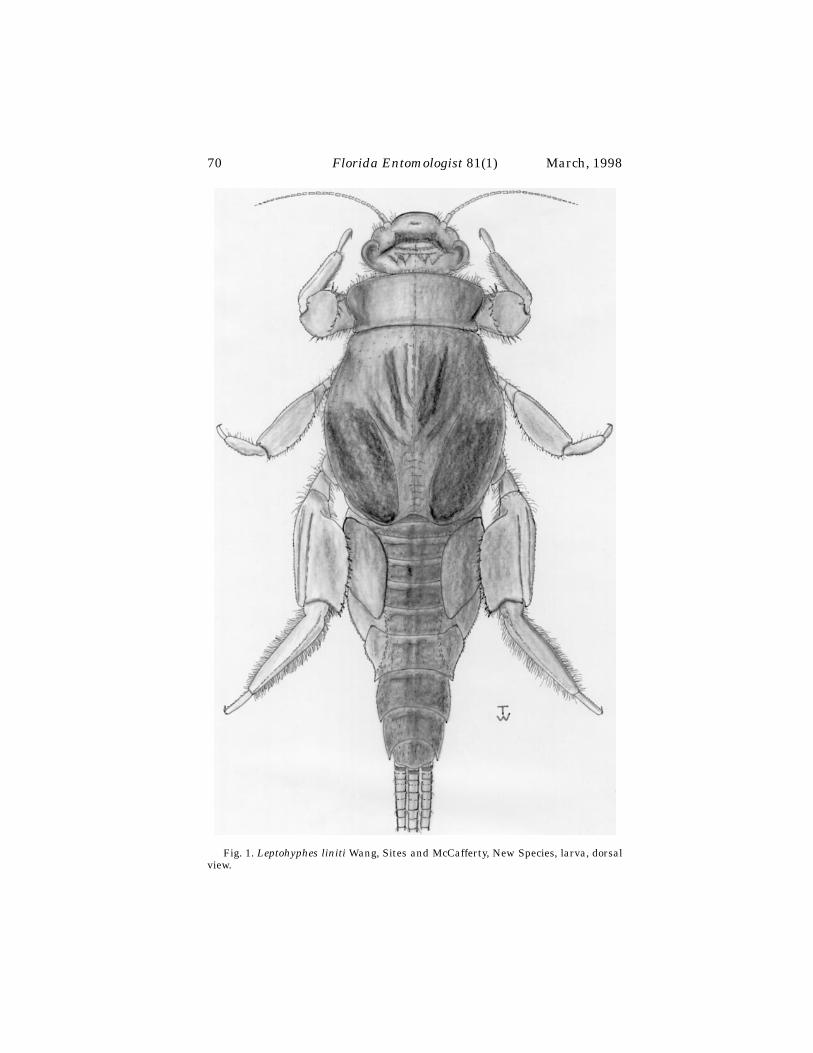
70
Florida Entomologist
81(1) March, 1998
Fig. 1. Leptohyphes liniti Wang, Sites and McCafferty, New Species, larva, dorsalview.

Wang, Sites & McCafferty: New Species of Leptohyphidae
71
Labrum mostly with branched setae (Fig. 10); mandible with partially fused incisors(Figs. 11-12); hypopharynx with well-developed superlinguae and marginal setae(Fig. 13); maxillary palpi absent (Fig. 14); labium with regularly developed postmen-tum, labial palpi with sparse setae, glossae small and with apical branched setae (Fig.15). Lateral margins of pronotum nearly parallel, fringed with fine, simple setae; forefemora with well-developed, curved, setose transverse ridge in basal half of anteriorsurface (Fig. 9); mid- and hind legs with color pattern as shown in Fig. 9; mid- andhind femora and tibiae with long, fine, simple setae on ventral and dorsal margins;hind femora subequal in length to fore femora; hind tibiae and hind tarsi subequal inlength; tarsal claws about half as long as tibiae, and with single row of four to six den-
Figs. 2-8. Leptohyphes liniti Wang, Sites and McCafferty, New Species, larva. 2.Labrum; 3. Left mandible; 4. Right mandible; 5. Hypopharynx; 6. Maxilla; 7. Labium;8. Gill 2, ventral.
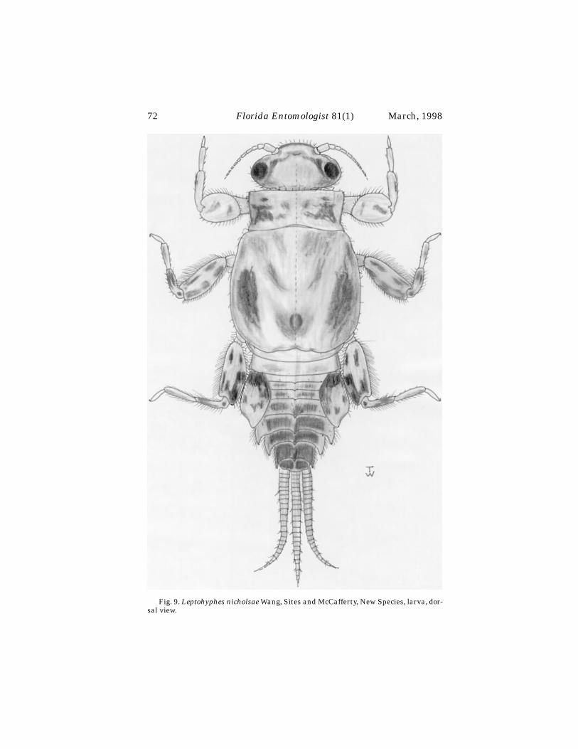
72
Florida Entomologist
81(1) March, 1998
Fig. 9. Leptohyphes nicholsae Wang, Sites and McCafferty, New Species, larva, dor-sal view.

Wang, Sites & McCafferty: New Species of Leptohyphidae
73
ticles. Abdominal terga 3-9 each with midposterior tubercle (strongly developed onterga 6-9); abdominal segments 3-9 with well developed posterolateral projections,and 3-8 with moderately developed lateral flanges (Fig. 9); sterna pale yellow. Oper-culate gills with underlying gill filament longer than operculate gill (Fig. 16), withoutbasal spine, pale yellow, ovate, weakly pointed distally, with distinct basal and me-dian blackish markings (Fig. 9). Caudal filaments without dark brown banded seg-ments, with whorls of short, simple setae at alternating articulations (Fig. 9).
Adult: Unknown.
Figs. 10-16. Leptohyphes nicholsae Wang, Sites and McCafferty, New Species,larva. 10. Labrum; 11. Left mandible; 12. Right mandible; 13. Hypopharynx; 14. Max-illa; 15. Labium; 16. Gill 2, ventral.
74
Florida Entomologist
81(1) March, 1998
Holotype: Male larva, Ecuador, Pichincha Prov., Rio Peripa at Puerto Limon, 314m, 18 July 1993. Paratype: Female larva, same data as holotype.
D
ISCUSSION
Leptohyphes nicholsae
appears to be closely related to
Leptohyphes curiosus
Lugo-Ortiz and McCafferty from Costa Rica because both species possess the following at-tributes: small size, similar operculate gill shape, ridge and setal arrangements onthe legs, and the well-developed posterolateral projections on abdominal segments 7and 8 (see Lugo-Ortiz and McCafferty 1995). It differs from
L. curiosus
by possessingtergal tubercles and a different color pattern on the legs and operculate gills. Lugo-Ortiz and McCafferty (1995) noted the unique characteristics of
L. curiosus
and com-mented that it did not exactly fit the traditional definitions of either
Tricorythodes
Ul-mer or
Leptohyphes
. They further suggested that it was representative of a distinctNeotropical lineage within
Leptohyphes
. The discovery of
L. nicholsae
shows indeedthat the lineage consists of more than one species.
The new species is known from only one collecting site. The Rio Peripa, where itwas taken, was at low elevation (314 m) at Puerto Limon. The collecting site was ap-proximately 30 m wide, had a stony substrate, and a water temperature of 24
°
C.
A
CKNOWLEDGMENTS
We thank G. Onore, Universidad Católica, Quito, for his assistance with local lo-gistics and in obtaining collecting permit No. 038-IC from the Ministerio de Agricul-tura y Ganadería. We also thank A. Provonsha and C. Lugo-Ortiz, Purdue University,West Lafayette, IN, for critically reading the manuscript. The Spanish abstract wastranslated by C. Lugo-Ortiz. Partial funding for this project was provided in part byMU project #PSSL0232. This is Missouri Agricultural Experiment Station journal se-ries paper No. 12632. The paper has also been assigned Purdue ARP Journal No.15405.
R
EFERENCES
C
ITED
A
LLEN
, R. K. 1967. New species of New World Leptohyphinae (Ephemeroptera: Tric-orythidae). Canadian Entomol. 99: 350-375.
A
LLEN
, R. K. 1973. New species of
Leptohyphes
Eaton (Ephemeroptera: Trico-rythidae). Pan-Pacific Entomol. 49: 363-372.
A
LLEN
, R. K. 1978. The nymphs of North and Central American
Leptohyphes
(Ephemeroptera: Tricorythidae). Ann. Entomol. Soc. America 71: 537-558.A
LLEN
, R. K.,
AND
R. C. B
RUSCA
. 1973. New species of Leptohyphinae from Mexicoand Central America (Ephemeroptera: Tricorythidae). Canadian Entomol. 105:83-95.
A
LLEN
, R. K.,
AND
C. M. M
URVOSH
. 1987. Mayflies (Ephemeroptera: Tricorythidae) ofthe southwestern United States and northern Mexico. Ann. Entomol. Soc.America 80: 35-40.
A
LLEN
, R. K.,
AND
S. S. R
OBACK
. 1969. New species and records of New World Lepto-hyphinae (Ephemeroptera: Tricorythidae). J. Kansas Entomol. Soc. 42:372-379.
B
RUSCA
, R. C. 1971. A new species of
Leptohyphes
from Mexico (Ephemeroptera: Tri-corythidae). Pan-Pacific Entomol. 47: 146-148.
E
ATON
, A. E. 1882. An announcement of new genera of the Ephemeridae. Entomol.Mon. Mag. 18: 207-208.
E
ATON
, A. E. 1892. Biologia Centrali-Americana, Insecta, Neuroptera, Ephemeridae.Bernard Quaritch, London 38: 1-16.
Wang, Sites & McCafferty: New Species of Leptohyphidae
75
K
ILGORE
, J. I.,
AND
R. K. A
LLEN
. 1973. Mayflies of the Southwest: new species descrip-tions, and records (Ephemeroptera). Ann. Entomol. Soc. America 66: 321-332.
L
UGO
-O
RTIZ
, C. R.,
AND
W. P. M
C
C
AFFERTY
. 1995. Contribution to the taxonomy ofLeptohyphidae (Insecta: Ephemeroptera) of Central America. Stud. Neotrop.Fauna Environ. 30: 165-176.
M
AYO
, V. K. 1968. Some new mayflies of the subfamily Leptohyphinae(Ephemeroptera: Tricorythidae). Pan-Pacific Entomol. 44: 301-308.
N
AVÁS
, L. 1920. Insectos Sudamericanos. 1a Serie. Anales Soc. Cient. Argentina 90:52-72.
N
AVÁS
, L. 1931. Insectos de la Argentina. Rev. Cos. Entomol. Argentina 3: 317-324.N
EEDHAM
, J. G.,
AND
H. E. M
URPHY
. 1924. Neotropical mayflies. Bull. Lloyd Libr., En-tomol. Ser. 4: 1-79.
T
RAVER
, J. R. 1943. New Venezuelan mayflies. Bol. Entomol. Venezolana 2: 79-98.T
RAVER
, J. R. 1958. Some Mexican and Costa Rican mayflies. Bull. Brooklyn Entomol.Soc. 53: 81-89.
♦♦♦♦♦♦♦♦♦♦♦♦♦♦♦♦♦♦♦♦♦♦♦♦♦♦♦♦♦♦♦♦♦♦♦♦♦♦♦♦♦♦♦♦♦♦♦♦♦♦♦♦
U
LMER
, G. 1920. Neue Ephemeropteren. Arch. Naturg. 85: 1-80.
Baranowski & Slater: Cayman Island Lygaeidae
75
THE LYGAEIDAE OF THE CAYMAN ISLANDS WITH THE DESCRIPTION OF A NEW SPECIES OF
OCHRIMNUS
(HEMIPTERA)
R
ICHARD
M. B
ARANOWSKI
1
AND
J
AMES
A. S
LATER
2
1
University of Florida Institute of Food and Agricultural SciencesTropical Research and Education Center, Homestead, FL 33031
2
Dept. of Ecology & Evolutionary Biology, University of Connecticut, Storrs, CT 06269
A
BSTRACT
A key to the 50 species known from the Cayman Islands is provided, brief descrip-tions are given and the origin and relationships of the lygaeid fauna are discussed. Anew species is described and new synonymy is presented.
Key Words: Cayman Islands, Lygaeidae, new species
R
ESUMEN
Se introduce una clave para las 50 especies que se conocen de las islas Caimán, sepresentan descripciones breves, y se discuten el origen y las relaciones de la fauna
Lygaeidae. Se describe una especie nueva y se introduce nueva sinonimia.
The insect fauna of the Cayman Islands has received considerable attention in re-cent years. Our own interest in these islands has been stimulated by this work sincewe have for a number of years been concentrating a considerable part of our researchand field work on the lygaeid fauna of the West Indies. It thus seems appropriate to

76
Florida Entomologist
81(1) March, 1998
bring together our information on these interesting islands, discuss the faunal rela-tionships, and present a faunal list that is more in keeping with the actual speciescomposition than that currently reflected in the literature.
The natural history and biogeography of the Caymans have been treated in detailby Brunt & Davies (1994) and will not be repeated here except to note that the islandsare all low lying and are closer geographically to such islands of the Greater Antillesas Cuba and Jamaica than they are to any of the small island groups of the Caribbean.
One of the interesting aspects of the Cayman lygaeid fauna is that, in contrast tothe butterfly fauna (Askew 1994) in which endemism occurs only at the subspecificlevel, at least 5 species are endemic. It must be admitted, however, that the Lygaeidaehave not been collected as intensively as the butterflies of the Caribbean. Thus someof the apparent Cayman endemics may be the result of limited collecting on other is-lands. Nevertheless, the Cuban lygaeid fauna have been more carefully studied in thepast than has that of any of the other islands, and we have collected extensively on Ja-maica without taking any of these endemic species.
Our prior knowledge of the Cayman fauna comes from the 1938 Oxford UniversityExpedition material that was studied by Scudder (1958). He reported 17 speciespresent of which four were described as new. Askew (1994) noted that thirteen specieswere collected on Grand Cayman, eight on Cayman Brac, and three on Little Cayman.Slater (1964) listed the same species. Froeschner (1983) listed two species from GrandCayman Island, one of which represented an additional species. Based on our collect-ing over the past 10 years, we can list a total of 50 species, a number surprisingly closeto the known butterfly fauna. With this number of species we feel confident that wehave a reasonably complete inventory of the fauna of the Caymans.
From the outset we have been interested in the possible vicariant nature of thefauna relative to overwater dispersal. We feel that it is not yet possible to analyze sta-tistically the number of species relative to island size, particularly because Little Cay-man seems relatively poorly collected. Nor can we make meaningful contributions tothe degree of faunal “turnover” due to arrival of new immigrants and extinction of el-ements of the known fauna. We do believe that our present knowledge of the lygaeidfauna provides a base line from which future studies can be profitably pursued. Allspecies described as endemic by Scudder (1958), with the exception of
Ozophora mi-nuscula
Scudder, have been taken on the Cayman Islands and none have been takenon any other island of the Caribbean. We also recognize one additional new species asendemic so that at least 5 species are considered endemic, or 10% of the fauna. Askew(1994) commented upon the dominance of Lygaeidae in the Hemiptera fauna, at leaston Grand Cayman, and while he noted that this figure represented only a small frac-tion of the fauna, he believed that the figures probably accurated reflected the balanceof family representatives. We see no reason to doubt the validity of this conclusion.
Of the species that are not endemic on the Caymans, the great majority are wide-spread in the West Indies. Many of these are “weed species” that live in disturbed hab-itats and frequently reproduce on a number of host plants, or that live on or belowhost plants that thrive in ruderal environments.
Dieuches armatipes
(Walker) and
Oxycarenus hyalinipennis
Costa are Old Worldspecies that have only recently been reported from the Western Hemisphere.
Excluding these recently introduced species what can we conclude about the originand relationships of the Caymans lygaeid fauna? To attempt to answer this question,we compared the fauna of the Caymans with that of Cuba and Jamaica, the two largeislands most likely to have served as source areas. Askew (1994) found that the but-terfly fauna of the Caymans was derived predominantly from Cuba. He found that 42of the 48 butterfly taxa were also in Cuba but only 29 were shared with Jamaica andprogressively fewer with Hispaniola, Puerto Rico, and the Bahamas.

Baranowski & Slater: Cayman Island Lygaeidae
77
The lygaeid fauna, while not contradicting this relationship, does not support it toany extent. Of the total of 50 lygaeid species now known from the Caymans, 39 alsooccur on Cuba, but 34 occur on Jamaica. Even this difference appears more likely torepresent lack of collecting than actual absence of species on one or the other of theislands. For example,
Nysius raphanus
Howard and
Cymodema breviceps
Stål havebeen reported from Cuba but not Jamaica, but both are widespread almost through-out the Caribbean. Conversely,
Oedancala cladiumicola
Baranowski and Slater is arelatively recently recognized species that is known from southern Florida as well asthe Bahamas, Caicos, and Jamaica. It is host specific and is almost certain to be foundon Cuba.
We conclude that most, if not all, of the Caymans fauna has reached the islands byoverwater dispersal rather than as a result of vicariance. Slater’s (1988) paper detail-ing his disagreement with Rosen’s (1985) depreciation of dispersalist concepts, shouldbe consulted to understand our position on this matter.
Although we believe the present evidence strongly supports the probability ofoverwater dispersal as the primary reason for the present Caymans lygaeid fauna (inagreement with Askew’s (1994) conclusions on the butterfly fauna), we feel that whileCuba may well be the source area from which much of the fauna is derived, the ma-jority of species are so widespread in the Caribbean that it is impossible to demon-strate this.
In support of our hypothesis that the origin of the Cayman fauna is primarily theresult of overwater dispersal, several points should be considered. Most important is toconsider what is not there, as Rosen (1985) and some other biogeographers seem reluc-tant to do. None of the higher taxa that have restricted distributions in the Greater An-tilles are present. Particularly striking is the absence of the Pamphantinae. Slater(1988) indicated that the distribution on the islands suggests vicariance between Cubaand Hispaniola (and possibly Puerto Rico) since a number of sister taxa occur on thetwo islands and the subfamily is not otherwise represented in the West Indies, includ-ing its absence from Jamaica. This still seems essentially true for the islands, althoughextensive collecting in South America has revealed a considerable pamphantine fauna.Concepts of the taxon as an island-mainland vicariance example, however, will requirecladistic reexamination once the mainland fauna is better understood.
None of the endemic antillocorine West Indian genera, such as
Bathydema
and
An-tillodema
, are present on the Caymans, nor are representatives of the geocorine
Nin-yas
that occurs in less disturbed habitats in both Jamaica and Cuba.Several widespread Caribbean species are absent, indicating a rather random dis-
persal pattern. Examples are
Pachygrontha compacta
Distant and
Oedancala bimacu-lata
(Distant), both of which are sedge feeders and might be expected on the islands. Theabsence of
Ozophora rubrolinea
, an abundant species on the north coast of Jamaica, andcommon on the mainland of Central and northern South America is unexpected. Its ab-sence appears to be real, for members of the genus come to lights abundantly, and anumber of widespread species of
Ozophora
are common on the Caymans.Thus we see a general pattern of a fauna composed primarily of species very wide-
spread on the islands. This distribution suggests the importance of overwater coloni-zation in producing this pattern, even relative to the two species apparently recentlyintroduced from the Eastern Hemisphere. Superimposed on this basic pattern is thepresence of several endemic species and the absence of some widespread and commonspecies, as well as the absence of taxa that tend to be restricted to islands of theGreater Antilles.
All measurements are given in millimeters. The following abbreviations are used;RMB - Richard M. Baranowski collection, JAS - James A. Slater collection, USNM -United States National Museum, BMNH - British Museum of Natural History. Orig-

78
Florida Entomologist
81(1) March, 1998
inal references are not given as they can be found in Slater (1964) or Slater andO’Donnell (1996). We are aware of the higher classification proposed by Henry (1997),but prefer to retain the traditional classification for a faunal paper of this type.
K
EY
TO
THE
C
AYMAN
I
SLANDS
S
PECIES
OF
L
YGAEIDAE
1. Suture between abdominal sterna 4 and 5 curving forward laterally and not at-taining lateral margin of abdomen (Rhyparochrominae) . . . . . . . . . . . . . . . . . 2
1’. Suture between abdominal sterna 4 and 5 straight or nearly so and attaininglateral margin of abdomen . . . . . . . . . . . . . . . . . . . . . . . . . . . . . . . . . . . . . . . . 32
2. With at least spiracles of abdominal segments 3 and 4 located dorsally . . . . 32’. All abdominal spiracles located ventrally . . . . . . . . . . . . . . . . . . . . . . . . . . . . . 43. Lateral margins of anterior pronotal lobe carinate or explanate; only spiracle of
abdominal segments 3 and 4 located dorsally (Rhyparochromini) . . . . . . . . . . . . . . . . . . . . . . . . . . . . . . . . . . . . . . . . . . . . . . . . . . . . . . . . . .
Dieuches armatipes
3’ Lateral margins of anterior pronotal lobe rounded; abdominal spiracles on seg-ments 2, 3, and 4 located dorsally (Myodochini) . . . . . . . . . . . . . . . . . . . . . . . 16
4. Apical corial margins deeply concave on inner half (Antillocorini) . . . . . . . . . 54’. Apical corial margin straight or nearly so . . . . . . . . . . . . . . . . . . . . . . . . . . . . 65. Posterior pronotal lobe uniformly brown . . . . . . . . . . . . . . . .
Botocudo
species5’. Posterior pronotal lobe brown with pale markings . . . . . .
Cligenes distinctus
6. Trichobothria on abdominal sterna 4-5 arranged in a longitudinal linear rowwith posterior trichobothrium located caudad of spiracle 5; head dorsally withan iridescent area at base; usually with a trichobothrium present at each an-tero-lateral pronotal area (Lethaeini) . . . . . . . . . . . . . .
Paragonatas divergens
6’. Two posterior trichobothria on segments 4 and 5 located dorso-ventrad of oneanother; head above lacking iridescent areas; never with antero-lateral prono-tal trichobothria (Ozophorini) . . . . . . . . . . . . . . . . . . . . . . . . . . . . . . . . . . . . . . 7
7. Forewings coleopteroid, meeting along midline; membrane absent or greatlyreduced . . . . . . . . . . . . . . . . . . . . . . . . . . . . . . . . . . . . . . .
Ozophora coleoptrata
7’. Forewings macropterous, fully developed . . . . . . . . . . . . . . . . . . . . . . . . . . . . . 88. Dorsal surface of body with short, but distinct upstanding hairs (viewed later-
ally) . . . . . . . . . . . . . . . . . . . . . . . . . . . . . . . . . . . . . . . . . . . . . . . . . . . . . . . . . . . 98’. Dorsal surface glabrous or with scattered decumbent hairs, or at most an iso-
lated upstanding hair . . . . . . . . . . . . . . . . . . . . . . . . . . . . . . . . . . . . . . . . . . . . 109. Small species, 5 mm or less in length . . . . . . . . . . . . . . .
Ozophora coleoptrata
9’. Larger species, more than 5 mm in length . . . . . . . . . . .
Ozophora burmeisteri
10. Bucculae U-shaped; head anteriorly declivent, almost at a right angle to bodylength . . . . . . . . . . . . . . . . . . . . . . . . . . . . . . . . . . . . . . . .
Ozophora laticephala
10’. Bucculae V-shaped; head somewhat declivent, but never downward curved atalmost a right angle to body length . . . . . . . . . . . . . . . . . . . . . . . . . . . . . . . . . 11
11. Small species, less than 4.75 mm in length . . . . . . . . . .
Ozophora minuscula
11’. Larger species, well over 5 mm in length . . . . . . . . . . . . . . . . . . . . . . . . . . . . 1212. Femora dark brown . . . . . . . . . . . . . . . . . .
Ozophora pallidifemur fuscifemur
12’. Femora stramineous, with or without a brown annulus, never completely darkbrown . . . . . . . . . . . . . . . . . . . . . . . . . . . . . . . . . . . . . . . . . . . . . . . . . . . . . . . . . 13
13. Humeral pronotal angles weakly, but distinctly notched . . . . . . . . . . . . . . . . . . . . . . . . . . . . . . . . . . . . . . . . . . . . . . . . . . . .
Ozophora pallidifemur pallidifemur
13’. Humeral pronotal angles evenly rounded . . . . . . . . . . . . . . . . . . . . . . . . . . . . 1414. Membrane completely dark, lacking a conspicuous white apical macula or
stripe . . . . . . . . . . . . . . . . . . . . . . . . . . . . . . . . . . . . . . . . . . .
Ozophora umbrosa
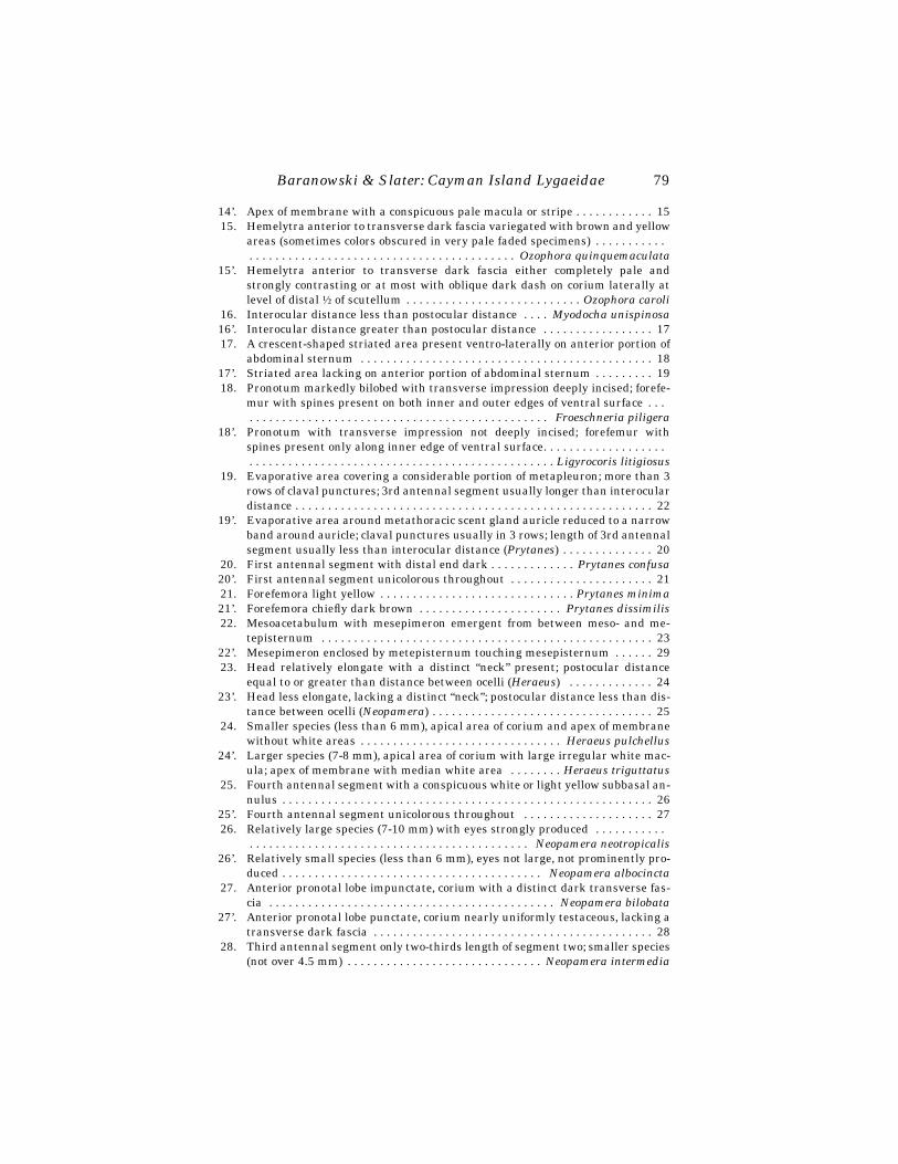
Baranowski & Slater: Cayman Island Lygaeidae
79
14’. Apex of membrane with a conspicuous pale macula or stripe . . . . . . . . . . . . 1515. Hemelytra anterior to transverse dark fascia variegated with brown and yellow
areas (sometimes colors obscured in very pale faded specimens) . . . . . . . . . . .
. . . . . . . . . . . . . . . . . . . . . . . . . . . . . . . . . . . . . . . . . Ozophora quinquemaculata
15’. Hemelytra anterior to transverse dark fascia either completely pale andstrongly contrasting or at most with oblique dark dash on corium laterally atlevel of distal
½
of scutellum . . . . . . . . . . . . . . . . . . . . . . . . . . .
Ozophora caroli
16. Interocular distance less than postocular distance . . . .
Myodocha unispinosa
16’. Interocular distance greater than postocular distance . . . . . . . . . . . . . . . . . 1717. A crescent-shaped striated area present ventro-laterally on anterior portion of
abdominal sternum . . . . . . . . . . . . . . . . . . . . . . . . . . . . . . . . . . . . . . . . . . . . . 1817’. Striated area lacking on anterior portion of abdominal sternum . . . . . . . . . 1918. Pronotum markedly bilobed with transverse impression deeply incised; forefe-
mur with spines present on both inner and outer edges of ventral surface . . . . . . . . . . . . . . . . . . . . . . . . . . . . . . . . . . . . . . . . . . . . . . . . .
Froeschneria piligera
18’. Pronotum with transverse impression not deeply incised; forefemur withspines present only along inner edge of ventral surface. . . . . . . . . . . . . . . . . . .
. . . . . . . . . . . . . . . . . . . . . . . . . . . . . . . . . . . . . . . . . . . . . . . Ligyrocoris litigiosus
19. Evaporative area covering a considerable portion of metapleuron; more than 3rows of claval punctures; 3rd antennal segment usually longer than interoculardistance . . . . . . . . . . . . . . . . . . . . . . . . . . . . . . . . . . . . . . . . . . . . . . . . . . . . . . . 22
19’. Evaporative area around metathoracic scent gland auricle reduced to a narrowband around auricle; claval punctures usually in 3 rows; length of 3rd antennalsegment usually less than interocular distance (
Prytanes
) . . . . . . . . . . . . . . 2020. First antennal segment with distal end dark . . . . . . . . . . . . .
Prytanes confusa
20’. First antennal segment unicolorous throughout . . . . . . . . . . . . . . . . . . . . . . 2121. Forefemora light yellow . . . . . . . . . . . . . . . . . . . . . . . . . . . . . .
Prytanes minima
21’. Forefemora chiefly dark brown . . . . . . . . . . . . . . . . . . . . . .
Prytanes dissimilis
22. Mesoacetabulum with mesepimeron emergent from between meso- and me-tepisternum . . . . . . . . . . . . . . . . . . . . . . . . . . . . . . . . . . . . . . . . . . . . . . . . . . . 23
22’. Mesepimeron enclosed by metepisternum touching mesepisternum . . . . . . 2923. Head relatively elongate with a distinct “neck” present; postocular distance
equal to or greater than distance between ocelli (
Heraeus
) . . . . . . . . . . . . . 2423’. Head less elongate, lacking a distinct “neck”; postocular distance less than dis-
tance between ocelli (
Neopamera
) . . . . . . . . . . . . . . . . . . . . . . . . . . . . . . . . . . 2524. Smaller species (less than 6 mm), apical area of corium and apex of membrane
without white areas . . . . . . . . . . . . . . . . . . . . . . . . . . . . . . .
Heraeus pulchellus
24’. Larger species (7-8 mm), apical area of corium with large irregular white mac-ula; apex of membrane with median white area . . . . . . . .
Heraeus triguttatus
25. Fourth antennal segment with a conspicuous white or light yellow subbasal an-nulus . . . . . . . . . . . . . . . . . . . . . . . . . . . . . . . . . . . . . . . . . . . . . . . . . . . . . . . . . 26
25’. Fourth antennal segment unicolorous throughout . . . . . . . . . . . . . . . . . . . . 2726. Relatively large species (7-10 mm) with eyes strongly produced . . . . . . . . . . .
. . . . . . . . . . . . . . . . . . . . . . . . . . . . . . . . . . . . . . . . . . .
Neopamera neotropicalis
26’. Relatively small species (less than 6 mm), eyes not large, not prominently pro-duced . . . . . . . . . . . . . . . . . . . . . . . . . . . . . . . . . . . . . . . .
Neopamera albocincta
27. Anterior pronotal lobe impunctate, corium with a distinct dark transverse fas-cia . . . . . . . . . . . . . . . . . . . . . . . . . . . . . . . . . . . . . . . . . . . .
Neopamera bilobata
27’. Anterior pronotal lobe punctate, corium nearly uniformly testaceous, lacking atransverse dark fascia . . . . . . . . . . . . . . . . . . . . . . . . . . . . . . . . . . . . . . . . . . . 28
28. Third antennal segment only two-thirds length of segment two; smaller species(not over 4.5 mm) . . . . . . . . . . . . . . . . . . . . . . . . . . . . . .
Neopamera intermedia

80
Florida Entomologist
81(1) March, 1998
28’. Third antennal segment much more than two thirds length of segment two;larger species (5 mm) . . . . . . . . . . . . . . . . . . . . . . . . . . . . .
Neopamera vicarius
29. Elongate (6-9 mm in length); anterior pronotal lobe flattened, slightly convex,in lateral view lower than posterior lobe; pronotal collar with a median “dip” toposterior margin (
Paromius
) . . . . . . . . . . . . . . . . . . . . . . . . . . . . . . . . . . . . . . 3029’. Small (not over 5-6 mm); anterior pronotal lobe strongly convex, not lower than
posterior lobe in lateral view; pronotal collar essentially straight across midline(
Pseudopachybrachius
) . . . . . . . . . . . . . . . . . . . . . . . . . . . . . . . . . . . . . . . . . . . 3130. Length of anterior pronotal lobe at least 1.8 times length of posterior pronotal
lobe; anterior pronotal lobe usually reddish brown . . . . . . .
Paromius dohrnii
30’. Length of anterior pronotal lobe less than 1.6 times length of posterior pronotallobe; anterior pronotal lobe usually black . . . . . . . . . . . . . .
Paromius longulus
31. Posterior (apical) margin of corium black, posterior femora pale . . . . . . . . . . . . . . . . . . . . . . . . . . . . . . . . . . . . . . . . . . . . . . . . . .
Pseudopachybrachius vinctus
31’. Posterior margin of corium pale or light brown; posterior femora with darkmarkings . . . . . . . . . . . . . . . . . . . . . . . . . . . . . . .
Pseudopachybrachius basalis
32. Abdominal spiracles on segments 2 through 7 all placed dorsally . . . . . . . . 3332’. At least one pair of spiracles on segments 2 through 7 located ventrally . . . 4433. Clavus punctate; posterior pronotal margin not depressed laterad of base of
scutellum . . . . . . . . . . . . . . . . . . . . . . . . . . . . . . . . . . . . . .
Kleidocerys virescens
33’. Clavus impunctate; posterior pronotal margin depressed laterad of base ofscutellum . . . . . . . . . . . . . . . . . . . . . . . . . . . . . . . . . . . . . . . . . . . . . . . . . . . . . . 34
34. General coloration dull yellowish brown, never with bright red or orange mark-ings, apical corial margin sinuate on mesal half; hind wing lacking a subcostaand possessing intervanals (Orsillinae) . . . . . . . . . . . . . . . . . . . . . . . . . . . . . 35
34’. Generally with red or orange coloration; apical corial margin straight, hindwing possessing a subcosta and lacking intervanals (Lygaeinae) . . . . . . . . . 40
35. Costal margin of corium straight for greater part of length, at least to level ofdistal end of claval commissure; connexivum often exposed laterad of corium . . . . . . . . . . . . . . . . . . . . . . . . . . . . . . . . . . . . . . . . . . .
Neortholomus jamaicensis
35’. Costal margin of corium expanded from base, never straight for distance be-yond level of posterior end of scutellum; connexivum not exposed laterad of co-rium . . . . . . . . . . . . . . . . . . . . . . . . . . . . . . . . . . . . . . . . . . . . . . . . . . . . . . . . . . 36
36. Stridulatory structure consisting of a row of fine grooves and ridges present onlateral corial margin (
Xyonysius
) . . . . . . . . . . . . . . . . . . . . . . . . . . . . . . . . . . 3736’. Lateral margins of corium lacking a stridulatory structure (
Nysius
) . . . . . . 3837. Corium not or scarcely constricted at base; pronotum distinctly longitudinally
calloused on each side of midline; veins of corium unspotted or at most veryfaintly spotted . . . . . . . . . . . . . . . . . . . . . . . . . . . . . . . . . . . . .
Xyonysius basalis
37’. Corium conspicuously constricted at base; pronotum not distinctly longitudi-nally calloused on either side of midline; veins of corium more or less heavilyspotted with fuscous . . . . . . . . . . . . . . . . . . . . . . . . . . . .
Xyonysius californicus
38. Bucculae low, gradually tapering posteriorly, fading out gradually near base ofhead . . . . . . . . . . . . . . . . . . . . . . . . . . . . . . . . . . . . . . . . . . . . .
Nysius raphanus
38’. Bucculae high anteriorly, slightly narrowing posteriorly, ending abruptly at ornear base of head . . . . . . . . . . . . . . . . . . . . . . . . . . . . . . . . . . . . . . . . . . . . . . . 39
39. Basal portion of lateral corial margins with prominent hairs . . . . . . . . . . . . . . . . . . . . . . . . . . . . . . . . . . . . . . . . . . . . . . . . . . . . . . . . . . . . . . .
Nysius scutellatus
39’. Basal portion of lateral corial margins devoid of hairs, or at most with minute,extremely small hairs present . . . . . . . . . . . . . . . . . . . . . . . . . .
Nysius tenellus

Baranowski & Slater: Cayman Island Lygaeidae
81
40. Scutellum tumid and swollen with a weak median longitudinal carina, butnever with lateral areas excavated; lacking a subbasal transverse carina (
On-copeltus
) . . . . . . . . . . . . . . . . . . . . . . . . . . . . . . . . . . . . . . . . . . . . . . . . . . . . . . 4140’. Scutellum not tumid and swollen with a median carina and adjacent areas flat
or excavated; a subbasal transverse carina usually present . . . . . . . . . . . . . 4241. Hemelytral membrane with a white mark just basad of center . . . . . . . . . . . .
. . . . . . . . . . . . . . . . . . . . . . . . . . . . . . . . . . . . . . . . . . . . . . . . .Oncopeltus aulicus
41’. Hemelytral membrane without white markings . . . . . . .
Oncopeltus fasciatus
42. Pronotum with 4 short deep transverse impressions present behind calli(
Ochrimnus
) . . . . . . . . . . . . . . . . . . . . . . . . . . . . . . . . . . . . . . . . . . . . . . . . . . . 4342’. Pronotum often punctate behind calli, but without 4 transverse impressions
. . . . . . . . . . . . . . . . . . . . . . . . . . . . . . . . . . . . . . . . . . . .
Ochrostomus pulchellus
43. Dorsum black except for indistinct ocher pronotal and corial margins . . . . . . . . . . . . . . . . . . . . . . . . . . . . . . . . . . . . . . . . . . . . . . . . . . . . .
Ochrimnus nigriceps
43’. Dorsum black except posterior pronotal lobe largely orange . . . . . . . . . . . . . . . . . . . . . . . . . . . . . . . . . . . . . . . . . . . . . . . . . . . . . . . .
Ochrimnus bracensis
n. sp.44. Abdominal spiracles on segments 2 to 5 located dorsally, spiracles of segment 7
located ventrally . . . . . . . . . . . . . . . . . . . . . . . . . . . . . . . . . . . . . . . . . . . . . . . 4544’. Abdominal spiracles on at least segments 6 and 7 located dorsally, or all spira-
cles ventral . . . . . . . . . . . . . . . . . . . . . . . . . . . . . . . . . . . . . . . . . . . . . . . . . . . . 4845. Hemelytra coarsely punctate (Cyminae) . . . . . . . . . . . . . . . . . . . . . . . . . . . . 4645’. Hemelytra impunctate, or at most with a few scattered punctures (Blissinae)
. . . . . . . . . . . . . . . . . . . . . . . . . . . . . . . . . . . . . . . . . . . . . . . . . . . . . . . . . . . . . . 4746. Head strongly deflexed in front; eyes somewhat stalked and produced away
from antero-lateral margins of pronotum; corium chiefly hyaline with only afew mesally located punctures . . . . . . . . . . . . . . . . . . . . .
Cymoninus notabilis
46’. Head not strongly declivent anteriorly; eyes usually in contact with or nearly incontact with antero-lateral pronotal angles; corium not hyaline, densely punc-tate over almost entire surface . . . . . . . . . . . . . . . . . . . . .
Cymodema breviceps
47. Body relatively short (less than 4 mm); black with white markings; forecoxalcavities open posteriorly . . . . . . . . . . . . . . . . . . . . . . . . . . . . . .
Blissus antillus
47’. Body elongate and slender; gray brown; forecoxal cavities closed posteriorly . . . . . . . . . . . . . . . . . . . . . . . . . . . . . . . . . . . . . . . . . . . . .
Ischnodemus praecultus
48. Abdominal spiracles on segments 3 and 4 located dorsally; eyes protruding andreniform; forefemora not greatly incrassate and not armed below with numer-ous spines (Geocorinae) . . . . . . . . . . . . . . . . . . . . . . . . . . . . . . . . . . . . . . . . . . 49
48’. All abdominal spiracles located ventrally; eyes not unusually large and reni-form; forefemora incrassate and armed below with numerous spines . . . . . 50
49. Head and pronotum yellow-brown; vertex smooth and shining . . . . . . . . . . . . . . . . . . . . . . . . . . . . . . . . . . . . . . . . . . . . . . . . . . . . . . . . . . . .
Geocoris punctipes
49’. Head and pronotum black, at least pronotum with a strongly contrasting whitestripe running longitudinally through meson . . . . . . . . .
Geocoris lividipennis
50. Forefemora only moderately swollen; corium expanded, extending beyond lat-eral margins of abdomen; bucculae extending to base of head; hind coxaewidely separated (Oxycareninae) . . . . . . . . . . . . . .
Oxycarenus hyalinipennis
50’. Forefemora strongly swollen; corium not extending beyond lateral margins ofabdomen; bucculae short, confined to front of head; hind coxae not widely sep-arated (Pachygronthinae) . . . . . . . . . . . . . . . . . . . . . . . . . . . . . . . . . . . . . . . . 51
51. Apical corial margin with a distinct dark spot (may be faint) along margin . . . . . . . . . . . . . . . . . . . . . . . . . . . . . . . . . . . . . . . . . . . . . . .
Oedancala crassimana
51’. Apical corial margin lacking a distinct dark spot . . .
Oedancala cladiumicola
82
Florida Entomologist
81(1) March, 1998
L
YGAEINAE
Ochrimnus bracensis
Baranowski & Slater,
NEW SPECIES
Black, posterior portion of pronotum orange, fourth antennal segment, labium andtarsi brown. Line between black and orange portions of pronotum forming a “W.”Head, black areas of pronotum, scutellum, clavus, corium, pleural areas of thorax,and abdominal segments 2-5 densely clothed with decumbent black hairs tipped withgold. Hairs on orange part of pronotum light with gold tips. Lateral margin of mem-brane narrowly white. Abdominal segment 6 and sternal areas of segments 2-5 andlegs with long golden hairs.
Length head 1.0, width 1.20, interocular space 0.75. Pronotum trapezoidal, ante-rior margin concave, lateral margins straight, posterior margin slightly convex.Length pronotum 1.33, width 2.08. Scutellum with prominent median carina stemand arms. Length scutellum 0.88, width 1.15. Length claval commissure 0.65. Midlinedistance apex clavus to apex corium 2.10. Midline distance apex corium to apex mem-brane 2.65. Labium short, extending posteriorly between meso- and metacoxae.Length labial segments I 0.62, II 0.65, III 0.68, IV 0.58. Antennal segments I-IIIdensely clothed with short, decumbent black, gold tipped hairs sparsely interspersedwith long upright dark hairs. Segment IV densely clothed with short decumbent goldhairs and long upright gold hairs. Length antennal segments I 0.44, II 1.02, III 0.90,IV 1.06. Total body length 5.50.
Types. Holotype.
?
CAYMAN BRAC: The Creek, 17-X-1995, H. V. & R. M. Bara-nowski (blacklight trap). In United States National Museum (USNM). Paratypes. 4
?
,4
/
, same data as holotype; 1
?
, 2
/
, same except 20-X-95; 1
/
, same except 6-9-VII-97;2
?
, 3
/
same except 7-XI-95, E. A. Dilbert; 2
/
, same except 8-XI-95; 1
/
, same except26-V-96; 1
/
, same except 28-V-96; 1
/
13-VI-96; 2
/
, same except 26-VI-96; 1
/
, sameexcept 22-VII-96. In R. M. Baranowski, J. A. Slater, United States National Museum,Florida State Collection of Arthropods, American Museum of Natural History collec-tions.
Etymology: Named for the island, Cayman Brac, on which it appears to be en-demic.
This striking species very similar to
O. nigriceps
, is readily distinguished by theunique black coloration with only the posterior pronotal lobe a strongly contrastingorange and by the uniquely colored hairs.
Ochrimnus nigriceps
Scudder
Moderate sized (5.8-6.8), almost completely black-brown, covered with a dense, de-cumbent dark pubescence. Apical four-fifths of fourth antennal segment and marginof membrane very narrowly pale.
Grand Cayman (Scudder 1958, RMB,JAS).
Ochrostomus pulchellus
(Fabricius)
Head dark brown with pale basal spot. Pronotal calli and 2 broad rays on posteriorpronotal lobe dark brown. Area anterior to calli white, lateral areas red. Inner one-half of clavus white, outer one-half dark brown. Corium dark brown with lateral andapical margins broadly white, banded with red stripe just within pale margin. Mem-brane black with white margin.
Grand Cayman (Scudder 1958, Froeschner 1983, RMB), Cayman Brac (RMB), Lit-tle Cayman (RMB).

Baranowski & Slater: Cayman Island Lygaeidae
83
Oncopeltus aulicus
(Fabricius)
Red and black, easily recognizable by the large black macula covering most of pos-terior pronotal lobe. Usually a conspicuous white bar on forewing membrane.
Grand Cayman (RMB).
Oncopeltus fasciatus
Dallas
One of the larger members of the genus (10-12). Orange-red and black. Centralarea of pronotum with a transverse fascia, most of membrane and appendages black.
Grand Cayman (Froeschner 1983, RMB, NMNH).
O
RSILLINAE
Neortholomus jamaicensis
(Dallas)
Small (4-5), narrow, elongate. Dull yellowish gray with numerous brown spots onhead, pronotum, and hemelytra. Scutellum dark anteriorly with a broad pale Y-shaped callosity.
Grand Cayman (RMB).
Nysius raphanus
Howard
Very small (3-4), yellowish brown. Scutellum black, except at extreme distal end.Pronotum short, nearly twice as wide as long. Bucculae very low and tapered to pos-terior end.
Grand Cayman (RMB).
Nysius scutellatus
Dallas
Small (3-3.5), bucculae high anteriorly, ending abruptly at or near base of head.Basal portion of corial margins with prominent hairs.
Grand Cayman (Scudder 1958), Cayman Brac (RMB).
Nysius tenellus
Barber
Small (3.6-4), pale yellowish testaceous, bucculae strongly elevated throughout.Pronotum only one-third wider than long. Corial veins usually unspotted. Scutellumbicolored or black.
Grand Cayman (RMB).
Xyonysius basalis
(Dallas)
Subequal in size to
X. californicus
and paler in color, yellowish testaceous ratherthan cinereus. Lateral corial margin scarcely contracted basally, entire lateral corialmargin slightly convex rather than straight. Male genital capsule either completelypale yellow or slightly fuscous basally.
Cayman Brac (Scudder 1958).
Xyonysius californicus
(Stål)
Moderate sized (4.7-7), dull yellowish to brownish gray. Bucculae very short,scarcely extending midway to base of head, head and pronotum subequal in length.Male genital capsule black with broad pale yellow margin.
84
Florida Entomologist
81(1) March, 1998
Grand Cayman (RMB), Cayman Brac (RMB), Little Cayman (RMB).
ISCHNORHYNCHINAE
Kleidocerys virescens (Fabricius)
Relatively small (3-3.5), with head, pronotum, and scutellum ochraceous with fus-cous punctures. Scutellum usually dark anteriorly with distal end pale.
Grand Cayman (RMB).
CYMINAE
Cymoninus notabilis (Distant)
Small (3-3.3), elongate, slender. Head, pronotum, and scutellum somewhat pubes-cent, dark reddish brown. Corium yellowish with extreme apex darkened. Legs dullyellow. Abdomen usually brownish or yellowish, sometimes, particularly in females,green. Apex of labium and distal tarsal segment fuscous. Second antennal segment1.5 times length of segment 3.
Grand Cayman (RMB), Cayman Brac (RMB), Little Cayman (RMB).
Cymodema breviceps (Stål)
Moderate sized (3.4-3.7), light yellow-brown with first 3 antennal segments concol-orous, head and 4th antennal segment darker. Pronotum with yellow line mesally onanterior half. Scutellum also with median yellow line. Apical margin of corium darker.
Grand Cayman (RMB).
BLISSINAE
Blissus antillus Leonard
Blissus planus Leonard 1968: 151-152. NEW SYNONYMY.Blissus slateri Leonard 1968: 150-151. NEW SYNONYMY.
Moderately small (2.5-3.5), posterior pronotal lobe black, strongly contrasting withgray anterior lobe. Labium frequently extending posteriorly beyond middle of mesos-ternum. Hemelytra white with black-brown markings; membrane white. Both mac-ropterous and brachypterous forms known.
Grand Cayman (RMB), Cayman Brac (RMB), Little Cayman (RMB).Leonard (1968) described three species of Blissus from the Caribbean: B. slateri and
B. antillus from Puerto Rico, and B. planus from Grenada. His descriptions are basedlargely on color, pruinosity, size, and the shape of the metathoracic scent gland peri-treme. In some instances he described a feature for one species, but did not discuss orcompare the condition of the same feature in the other species. For example, he statedthat the scutellum of B. slateri is distinctly punctate, but did not indicate the conditionof the punctation in the other two species. In fact, the scutellum is punctate in all threespecies. We have examined considerable material from Jamaica, Dominican Republic,Trinidad, St. Barthelemy, Dominica, Tortola, Virgin Gorda, and St. Lucia and find thescutellum punctate in all specimens examined. Leonard stated B. slateri possesseslight straw-yellow setae on the pronotum but that B. antillus has silvery setae on thepronotum. We cannot see these differences even in the paratypes examined. Becausethe scent gland peritreme varies in the island populations we examined, we believethat it is not a reliable differentiating character. We dissected male genital capsulesand parameres of several paratypes, representing all three species, as well as from

Baranowski & Slater: Cayman Island Lygaeidae 85
specimens from other islands and do not find significant differences. In our opinionBlissus is represented by a single species in the West Indies. We elect to use the nameantillus for this species and reduce B. slateri and B. planus to synonymy.
In addition to our inability to find differentiating features, the West Indian popu-lations occur exclusively in ruderal habitats, suggesting that this is another exampleof a “weed” species found in disturbed habitats throughout the Caribbean.
The relationship of B. antillus to the ubiquitous Florida chinch bug, B. insularisBarber, is important. We have not found differences in the genital capsule or in theparameres between West Indian specimens and specimens from Florida. However,this is the case with a number of apparently distinct species of Blissus.
B. insularis can be separated from B. antillus by a combination of characters. B. in-sularis always has uniformly straw-colored legs whereas B. antillus varies from hav-ing dark brown legs to light brown with a dark brown annulus on one or more femora.The most constant differentiating feature is the anterior portion of the pronotum,which in B. insularis, is covered by a uniform, undivided pubescent band that reachedthe anterior margin. The pubescent band of B. antillus does not reach the anteriormargin and is completely, or partially, divided along the midline.
B. insularis does occur in the West Indies, as we have examined specimens fromthe Bahamas.
Ischnodemus praecultus Distant
Ischnodemus slateri Alayo & Grillo 1982: 58-62 NEW SYNONYMY.Moderate sized (5-5.5). Hemelytra smoky colored with pale white to dull testa-
ceous membrane. Surface of head, pronotum, and scutellum predominantly or en-tirely dull grayish or reddish brown pruinose. Lateral corial margins grayish brownwith shining areas of posterior portion of pronotum separated into three distinct mac-ulae.
Grand Cayman (RMB).We have examined the holotype of I. slateri from Cuba and believe it to be conspe-
cific with the widespread and somewhat variable I. praecultus.
GEOCORINAE
Geocoris lividipennis Stål
Small (2.5-3), strikingly colored, with head, pronotum, and scutellum chiefly blackcontrasting with the pale yellow wings. Pronotum with a complete median longitudi-nal pale stripe and lateral areas of pronotum often yellow-brown. Vertex granulose.
Grand Cayman (RMB), Cayman Brac (RMB), Little Cayman (RMB).
Geocoris punctipes (Say)
A moderate sized (3-3.5) big-eyed bug; pale straw-yellow, usually with a dark me-dian scutellar stripe. Vertex of head smooth and polished with a median sulcus ex-tending to base of head.
Grand Cayman (RMB).
OXYCARENINAE
Oxycarenus hyalinipennis Costa
Small (3-4), elongate, pronotum and head tapering anteriorly. Head and pronotumuniformly brown with dense, upright hairs, entire hemelytra hyaline. Abdomen and
86 Florida Entomologist 81(1) March, 1998
femora shining dark brown, meso- and metatibiae with a striking median, broad,white annulus.
This insect, known as the cotton seed bug, is a common and widespread species inthe Old World tropics. It is an introduced species in the Western Hemisphere and hasbeen established for many years in South America.
Slater & Baranowski (1994) reported it for the first time from the West Indies fromLong Island, Bahamas.
PACHYGRONTHINAE
Oedancala crassimana (Fabricius)
A relatively robust yellowish brown species with relatively thick antennal seg-ments. Usually less than 7.5 mm in length. Often with a dark spot present midwayalong apical corial margin. This is often absent in West Indian specimens, in whichcase it can be distinguished from O. acuminata by the shorter labium; from O. cla-diumicola by the lack of reddish coloration and from O. cubana by the more robustnonlinear body and by not having the scutellum black adjacent to the pale medianvitta.
Grand Cayman (Scudder 1958, RMB).
Oedancala cladiumicola Baranowski & Slater
Similar to crassimana, but lacking black spot along apical corial margins. Reddish.Found only on sawgrass.
Grand Cayman (RMB).
RHYPAROCHROMINAE
ANTILLOCORINI
Cligenes distinctus Distant
Small (2.5-3.0), robust with generally dark appearance. Head blackish, anteriorpronotal lobe, scutellum, and ventral surface dark brown, strongly contrasting withtestaceous hemelytra and legs. Corium with two spots along lateral margins, one atapex of corium, one midway between base and apex. Entire pronotal surface denselypunctate with a very coarse row anteriorly, giving impression of a weak pronotal col-lar.
Grand Cayman (RMB), Cayman Brac (RMB).
Botocudo species
We have collected an apparently undescribed species of the Botocudo complex onCayman Brac. The relationships of species currently placed in Botocudo are very un-satisfactory. Therefore, we feel it best to wait until the generic limits of this tribe arestudied and redefined before attempting to place this species.
LETHAEINI
Paragonatas divergens (Distant)
Moderate sized (4-5); dark brown with humeral angles of pronotum, an elongatevitta on clavus, and irregular spots on corium yellow. Dorsal surface with sparse up-right hairs. Femora dark brown with distal tips pale; tibiae and tarsi light brown.

Baranowski & Slater: Cayman Island Lygaeidae 87
Grand Cayman (Scudder, 1958, RMB), Cayman Brac (Scudder 1958, RMB).
MYODOCHINI
Heraeus pulchellus Barber
One of the smallest West Indian Heraeus species, barely exceeding 4 mm in length;testaceous, lacking an annulus on fourth antennal segment; possesses complete darkbrown fascia across hemelytra.
Grand Cayman (RMB), Cayman Brac (RMB).
Heraeus triguttatus (Guerin)
Larger (7-8) than pulchellus. Typically bright reddish brown with apex of coriumand most of membrane black. Apical area of corium possessing a large irregular whitemacula. Apex of membrane with conspicuous subquadrate median white area.
We have specimens from Grand Cayman and Cayman Brac that are dark brownrather than bright reddish brown; however, we can find no morphological differencesother than the color.
Grand Cayman (Scudder 1958, RMB), Cayman Brac (RMB).
Neopamera albocincta (Barber)
Elongate (5-5.5), pale reddish to reddish brown, postmedian corial fascia generallybroken into series of spots. Fourth antennal segment with proximal white annulus.Elongate head relatively more attenuated than N. bilobata.
Grand Cayman (RMB), Cayman Brac (Scudder 1958, RMB).
Neopamera intermedia (Barber)
Moderate sized (4-4.5), brownish with pale markings; legs yellowish with a narrowband near distal end of posterior femora; antennae light brown with distal third ofsegment III and all of segment IV darker.
Grand Cayman (RMB), Cayman Brac (Scudder 1958, RMB).
Neopamera bilobata (Say)
Rhyparochromus scutellatus Dallas 1852: 575-576 NEW SYNONYMY.Resembles N. albocincta closely in form and color, but tends to run somewhat
darker (although the color is variable), and is slightly larger and more robust andlacks a pale proximal area on the 4th antennal segment.
Dallas’ species N. scutellatus has been treated as a subspecies of N. bilobata sinceits status was reduced by Van Duzee (1909). Barber (1953) discussed the status, not-ing the darker femora, relatively shorter anterior pronotal lobe, and shorter secondantennal segment. However, such specimens apparently have no geographic signifi-cance and occur in many populations. There seems no reason to treat this as a tri-nomen in the modern concept of the subspecies, and it is here synonymized.
Grand Cayman (Scudder 1958), Cayman Brac (RMB).
Paromius longulus (Dallas)
Elongate, moderate sized (5.5-7), head, anterior pronotal lobe, scutellum and un-der surface frequently dull black, often thickly clothed with grayish pubescence. Pos-
88 Florida Entomologist 81(1) March, 1998
terior pronotal lobe reddish brown with darker punctures. Corium and clavus dulltestaceous. Antennae reddish brown with distal end of third and usually all of fourthsegment fuscous.
Grand Cayman (RMB).
Paromius dohrnii Guerin
Light tan, elongate, with anterior pronotal lobe reddish brown and almost twicethe length of posterior lobe, with transverse impression shallow and usually obsoletemesally.
Grand Cayman (RMB).
Pseudopachybrachius basalis (Dallas)
Small (3.5-5), robust; lateral corium lacking a distinct postmedian transverse fas-cia, margins completely pale. Oval pale spot present near inner apical corial angle.Head, pronotum, and scutellum dull black, corium dull yellow with dark brown punc-tures. Antennae reddish brown.
Grand Cayman (RMB), Cayman Brac (RMB).
Pseudopachybrachius vinctus (Say)
Small (2.8-3.0), head dark, pronotum contrasting with pale hemelytra having aconspicuous black spot on apical corial margin. Fourth antennal segment dark brown,contrasting with 3 pale proximal segments. Legs pale.
Grand Cayman (Scudder 1958, RMB), Cayman Brac (Scudder 1958, RMB), LittleCayman (RMB).
Prytanes minima (Guerin)
Small (2.5-3), reddish brown. Third antennal segment (males in particular) some-what thickened. Hemelytra variegated. Legs uniformly pale yellow.
Grand Cayman and Cayman Brac (Scudder 1958, RMB).
Prytanes formosa (Distant)
Body relatively elongate, head acuminate, clothed with decumbent sericeoushairs. Anterior pronotal lobe convex, not higher than posterior lobe and considerablynarrower. Antennnae prominent, first, third, and fourth segments uniformly darkbrown, second segment paler.
Grand Cayman (RMB).
Prytanes dissimilis (Barber)
A relatively robust, moderately large species for the genus (3.5 mm or greater).Scutellum frequently bicolored. Posterior pronotal lobe usually with a median darkstripe. Subdistal ends of middle and hind femora in part dark red-brown.
Cayman Brac (RMB).
Froeschneria piligera (Stål)
Large, robust (7-8) with a deep transverse pronotal impression. Abdominal strid-ulitrum well developed. Body surface with numerous elongate upstanding hairs.
Baranowski & Slater: Cayman Island Lygaeidae 89
Head, anterior pronotal lobe, scutellum, and ventral surface black, hemelytra blackand dull brown with a transverse macula.
Cayman Brac (RMB).
Ligyrocoris litigiosus (Stål)
Medium sized with a distinct lunate abdominal stridulitrum. First antennal seg-ment pale, fourth antennal segment completely dark. Legs and antennae dull yellow.Femora spotted distally.
Grand Cayman (RMB).
Myodocha unispinosa Stål
Relatively large (8) with very elongate “neck.” Head and neck, shining black.Forefemur slender with a single large spine present. Antennae brightly colored, firstsegment red-brown, strongly shining, contrasting with pale yellow second segment.
Grand Cayman (RMB).
OZOPHORINI
Ozophora umbrosa Slater
Moderately large (5.5-6.5), generally dark chocolate brown with pale spots on co-rium; membrane lacking pale apical macula. Dorsal surface glabrous. Fourth anten-nal segment with a conspicuous white annulus.
Scudder (1958) reported Ozophora atropicta Barber from the Caymans. Despiteour rather extensive collection of Ozophora species, we have been unable to collectthis species on the Caymans. We have recently examined part of the Scudder mate-rial, which is deposited in the Natural History Museum (London—not Oxford as theScudder paper states). These specimens are O. umbrosa Slater, a species not describedat the time of Scudder’s paper. We are thus referring the Caymans records of O. atro-picta here.
Little Cayman (Scudder 1958, RMB) Cayman Brac (RMB).
Ozophora burmeisteri (Guerin)
Pronotum nearly uniformly black or dark chocolate brown. Posterior pronotal lobewith yellow streak midway between meson and margin or a pair of yellow spots insame area. Fourth antennal segment with conspicuous white annulus, 3rd segmentslightly swollen distally and dark chocolate brown on distal ⅓. Readily distinguish-able by the combination of its very dark pronotum and erect dorsal hairs.
Grand Cayman (Scudder 1958, RMB), Cayman Brac (Scudder 1958, RMB).
Ozophora caroli Slater & Baranowski
Body elongate, relatively stout; head, anterior pronotal lobe and broad rays ex-tending through posterior pronotal lobe dark red brown. Anterior pronotal colorbroadly pale yellow on either side of median red brown spot. Scutellum chiefly darkred brown, but with raised elliptical calloused area yellow, shading anteriorly to red-dish brown. Antennal segments I, II, and III pale yellow with distal end of III, proxi-mal end and distal ¾ of IV contrastingly dark chocolate brown. Body nearly glabrousabove, lacking conspicuous upstanding hairs.

90 Florida Entomologist 81(1) March, 1998
Grand Cayman (RMB).
Ozophora coleoptrata Slater
Small (3-4.5), chiefly dark chocolate brown with contrasting pale yellow bands onthe posterior pronotal lobe, and on the cubital vein of the clavus. Legs and antennaeuniformly pale yellow. Dorsal surface possesses numerous upright hairs. Color variesconsiderably with some specimens pale yellow laterally on the corium.
Slater (1990) described this species from a series consisting only of coleopteroid in-dividuals with the clavus and corium fused and the membrane reduced to a small flap.We have collected immatures on Grand Cayman that molted to macropterous adults.The macropterous form differs from other small (3-4.5) Ozophora in having sparse up-right hairs on the dorsum.
Grand Cayman (RMB), Cayman Brac (RMB), Little Cayman (RMB).
Ozophora laticephala Slater & O’Donnell
Small (2.8-3.3), short, stout. Generally pale yellow with the posterior pronotal lobeand most of the hemelytra contrasting with the dark anterior pronotal lobe, the latterhaving pale white or yellow lateral margins. Bucculae U-shaped, rather than V-shaped as in other Caribbean species of Ozophora.
Grand Cayman (RMB) and Cayman Brac (RMB).
Ozophora minuscula Scudder
Moderate sized (4.3), markings somewhat similar to O. pallidifemur, but muchsmaller. Known only from the type series consisting of two males collected in a lighttrap near Georgetown. Dr. Peter Fitzgerald operated a blacklight trap for us for sev-eral years but never collected this species.
Originally described and known only from Grand Cayman (Scudder 1958, BMNH).
Ozophora pallidifemur pallidifemur Scudder
Moderate sized (5-5.5), predominantly dark brown with uniformly pale yellowlegs. Antennal segments I, II, and III pale with distal ends of II and III darkened.Fourth antennal segment brown with white subbasal annulus. Membrane uniformlydark.
Originally described and known only from Grand Cayman (Scudder 1958, BMNH,JAS, NMNH, RMB).
Ozophora pallidifemur fuscifemur Scudder
Similar to nominal O. pallidifemur, but can be separated by the dark brown legs.Originally described as a distinct species. Slater (1987) reduced this taxon to a
subspecies of O. pallidifemur on the basis of each being endemic on separate islands. Cayman Brac (Scudder 1958, RMB), Little Cayman (Scudder 1958, RMB).
Ozophora quinquemaculata Barber
Moderate (5-6) sized, relatively stout with strongly variegated light and darkmarkings on pronotum and hemelytra. Posterior pronotal lobe with three dark longi-

Baranowski & Slater: Cayman Island Lygaeidae 91
tudinal stripes, the median one occurring as a broad lobe along the meson, lacking apale streak. Apical corial margins dark distally from inward extension of dark apicalcorial macula completely over raised corial margin apex.
Grand Cayman (RMB).
RHYPAROCHROMINI
Dieuches armatipes (Walker)
Large, robust (9-10), variegated with chocolate brown to black and light yellow toalmost white. Pronotum with broadly explanate pale lateral margins contrastingstrongly with the predominantly dark remainder of pronotum. Scutellum with a pairof small yellow spots. Hemelytra variegated, with a complete transverse dark fasciaand large oval elliptical white subapical corial macula.
This African species apparently has recently become established in the WesternHemisphere. It was first reported from Grand Cayman and St. Kitts and from inter-cepted specimens from the Dominican Republic and Jamaica by Henry and Froe-schner (1993). It is also now known from the Florida mainland.
Grand Cayman (Henry and Froeschner 1993, RMB) and Cayman Brac (RMB).
ACKNOWLEDGMENTS
We thank Mrs. Helen Baranowski for her assistance in collecting many of the spec-imens discussed here; Mrs. Holly Glenn, Ms. Julieta Brambila, Tropical Research andEducation Center, for assistance in mounting, labeling, and curating the collection;Dr. Peter Fitzgerald, Grand Cayman, and Mr. E. A. Dilbert, Cayman Brac, for operat-ing blacklight traps for us. We also thank Dr. A. G. Wheeler, Clemson University andMs. Wendy Meyer for critically reviewing the manuscript. Florida Agricultural Exper-iment Station Journal Series No. R-06114.
REFERENCES CITED
ALAYO, P. D., AND H. GRILLO. 1982. El genero Ischnodemus Fieber (Heteroptera: Lyg-aeidae: Blissinae) en Cuba. Centro Agricola 9: 51-63.
ASKEW, R. R. 1994. Insects of the Cayman Islands. In: M. A. Brunt & J. E. Davies(eds.). The Cayman Islands: Natural History and Biogeography, 333-356. Klu-wer Academic Publishers, The Netherlands.
BARBER, H. G. 1953. The genus Pachybrachius in the United States and Canada withthe description of two new species (Hemiptera: Lygaeidae). J. New York Ento-mol. Soc. 60: 211-220.
BRUNT, M. A., AND J. E. DAVIES (eds.) 1994.The Cayman Islands: Natural History andBiogeography. Kluwer Academic Publishers, The Netherlands. 604 pp.
DALLAS, W. S. 1852. List of the specimens of Hemipterous insects in the collection ofthe British Museum. Pt. II London: Taylor & Francis. pp. 369-592.
FROESCHNER, R. C. 1983. Heteroptera records from the Grand Cayman Island. Proc.Entomol. Soc. Washington 85: 861.
HENRY, T. J. 1997. Phylogenetic analysis of family groups within the infraorder Pen-tatomorpha (Hemiptera: Heteroptera), with emphasis on the Lygaeoidea. Ann.Entomol. Soc. America. 90: 275-301.
HENRY, T. J., AND R. C. FROESCHNER. 1993. Dieuches armatipes (Walker) (Het-eroptera: Lygaeidae) newly discovered in the Western Hemisphere. Proc. Ento-mol. Soc. Washington 95: 449-452.
LEONARD, D. 1968. Three new species of Blissus from the Antilles (Het. Lyg.). Proc.Entomol. Soc. Washington 70: 150-153.

92 Florida Entomologist 81(1) March, 1998
ROSEN, D. E. 1985. Geological hierarchies and biogeographic congruence in the Car-ibbean. Ann. Missouri Bot. Gard. 71: 636-659.
SCUDDER, G. G. E. 1958. Results of the Oxford University Caymans Islands biologicalexpedition of 1938. Entomol. Mon. Mag. 94: 145-150.
SLATER, J. A. 1964. A Catalogue of the Lygaeidae of the World. 2 vol. Univ. of Connect-icut, Storrs, CT. 1668 pp.
SLATER, J. A. 1987. A revision of the Ozophora umbrosa complex in the West Indies(Hemiptera: Lygaeidae. J. New York Entomol. Soc. 95: 414-427.
SLATER, J. A. 1988. Zoogeography of West Indian Lygaeidae (Hemiptera) pp. 38-60.In: Zoogeography of Caribbean Insects J. K. Liebherr (ed.). Cornell Univ. Press,Ithaca, N.Y.
SLATER, J. A. 1990. Seven new species of Ozophora from the West Indies with noteson previously described species (Hemiptera: Lygaeidae). J. New York Entomol.Soc. 98: 139-153.
SLATER, J. A., AND R. M. BARANOWSKI. 1994. The occurrence of Oxycarenus hyalin-ipennis (Costa) (Hemiptera: Lygaeidae) in the West Indies and new Lygaeidaerecords for the Turks and Caicos Islands of Providenciales and North Caicos.Florida Entomol. 77: 495-497.
SLATER, J. A., AND J. E. O’DONNELL. 1996. A catalogue of the Lygaeidae of the World.New York Entomol. Soc., New York, NY. 410 pp.
VAN DUZEE, E. P. 1909. Observations on some Hemiptera taken in Florida in the
♦♦♦♦♦♦♦♦♦♦♦♦♦♦♦♦♦♦♦♦♦♦♦♦♦♦♦♦♦♦♦♦♦♦♦♦♦♦♦♦♦♦♦♦♦♦♦♦♦♦♦♦
spring of 1908. Bull. Buffalo Soc. Nat. Sci. 9: 149-230.

92
Florida Entomologist
81(1) March, 1998
ORGANIC MULCH AS A FACTOR IN THE NYMPHAL HABITAT OF
MYNDUS CRUDUS
(HEMIPTERA: AUCHENORRHYNCHA: CIXIIDAE)
F. W. H
OWARD
1
AND
C. O
ROPEZA
2
1
University of Florida, Fort Lauderdale Research & Education Center3205 College Avenue, Fort Lauderdale, Florida 33314 USA
2
Centro de Investigación Científica de Yucatán, Apartado Postal 87, Cordemex 97310Mérida, Yucatán, México
A
BSTRACT
Certain types of organic mulches spread on the ground in a palm planting with St.Augustine grass,
Stenotaphrum secundatum
, ground cover enhanced the habitat forthe nymphal development of
Myndus crudus
Van Duzee, as measured by the greaternumbers of adults emerging from mulched areas compared with controls.
Plotsmulched with chopped and shredded coconut fronds had greater numbers of
M. cru-dus
than controls. Higher numbers of
M. crudus
emerged from plots with pine barkmulch than from control plots. There were no significant differences in mean numbersof adults captured in plots with eucalyptus and pine bark mulch. The least numbersof adults emerged from plots mulched with pine bark nuggets.
Myndus crudus
nymphs were never in the open but always beneath pieces of mulch. The benefit ofmulches to the insects appears to be that of providing shelter.
Key Words:
Myndus
, Cixiidae, Fulgoroidea, palm, lethal yellowing, cultural control

Howard & Oropeza:
Myndus crudus
and Organic Mulches
93
R
ESUMEN
Ciertas clases de cobertura orgánica dispersada sobre el suelo en un plantío de pal-meras con pasto de San Agustín,
Stenotaphrum secundatum
, mejoraron el habitatpara el desarrollo de las ninfas de
Myndus crudus Van Duzee,
como fue estimado porel mayor número de adultos emergidos de estas áreas en comparación con testigos.Parcelas con frondas de cocotero picadas y ralladas tenían números mayores de estosinsectos que los testigos. Un mayor número de
M. crudus
emergió de parcelas con cás-cara de pino picada en comparación con los testigos. El promedio de adultos captura-dos en parcelas con cobertura de eucalipto y de cáscara de pino no difiriósignificativamente. Las ninfas de
M. crudus
nunca estuvieron al descubierto, sinosiempre debajo de los fragmentos de cobertura. El beneficio de las coberturas orgáni-
cas para
M. crudus
parece ser el de proveer abrigo.
Myndus crudus
Van Duzee (Hemiptera: Auchenorrhyncha: Cixiidae), a widely dis-tributed planthopper in the American tropics (Howard et al. 1984, Kramer 1979, Vil-lanueva Barradas 1991), is a vector of lethal yellowing of palms (Howard 1987,Howard et al. 1983). Lethal yellowing is a fast-spreading, destructive disease associ-ated with a phytoplasma to which at least 35 species of Palmae are susceptible, in-cluding the economically important coconut palm,
Cocos nucifera
L., and date palm,
Phoenix dactylifera
L. The disease has long been known in western islands of the WestIndies, from where it apparently invaded Florida (U.S.A.) and Mexico (Howard 1983,Howard et al. 1984, Villanueva Barradas 1991). It has recently spread to Belize andHonduras (Ashburner et al. 1996). Thus far, there is no evidence implicating any in-sect species other than
M. crudus
as a potential vector of lethal yellowing.The nymphs of
M. crudus
develop at or just under soil level on grasses or sedges,and the adults feed on palms (Howard & Villanueva-Barradas 1994, Eden-Green1978). At least 37 species of grasses (Graminae) and 4 species of sedges (Cyperaceae)have been reported as nymphal hosts of
M. crudus
(Carrillo Ramirez & Piña Razo1990, Howard 1989, Howard 1990a, Howard 1990b, Piña Quijano 1993, Tsai & Kirsch1978, Villanueva B. et al. 1987, Zenner de Polania & Lopez A. 1977). When rearing
M.crudus
, Eden-Green (1978) observed that the nymphs were often hidden beneathpieces of coconut fiber. Therefore, we decided to determine whether coconut fiber andsimilar materials spread on soil among nymphal hosts would improve the habitat for
M. crudus
nymphs, as estimated by the numbers of emerging adults.
M
ATERIALS
AND
M
ETHODS
The study was conducted at the Fort Lauderdale Research & Education Center ina 0.6 ha grove of 100 coconut palms and other susceptible species of palms. St. Augus-tine grass,
Stenotaphrum secundatum
(Walt.) Kuntze, a preferred host of
M. crudus
(Howard 1990b), was the predominant ground cover. Plots (2
×
2 m) selected at ran-dom were not mowed or otherwise disturbed during each experiment.
In the first experiment, which was initiated February 1994, a mulch was obtainedby passing coconut palm fronds through a mechanical brush chipper. This materialconsisted of fine shredded fragments and larger pieces of variable size between 2-5 cmin width and 5-10 cm in length. The material was spread on each of 6 plots (0.05 m
3
/plot) and raked lightly so that it settled on soil and allowed the grass blades toemerge. Six similar plots where fiber was not spread were selected as controls.

94
Florida Entomologist
81(1) March, 1998
In May 1994, traps were used to sample adults emerging from the plots. Traps con-sisted of 60
×
60
×
50 cm plywood boxes. Polypropylene funnels of 5 cm dia and 0.5 cmmouth were fitted over an opening at the center of the top of each box. Transparentplastic test tubes (2.1
×
10 cm) were placed over the funnel mouths. Adults thatemerged from the plants attempted to exit through the funnel opening and thus werecaptured in the tubes and counted.
Emergence traps were placed simultaneously in the center of each of 6 plots withcoconut fiber and 6 control plots. Just prior to placing a trap, the grass was agitatedvigorously for several minutes to repel any adult Auchenorrhynchous insects from thesample area. Additionally, any adults captured in traps during the first 24 hours wereremoved and not counted in samples. The traps then remained in place for 1 week, af-ter which the numbers of male and female
M. crudus
captured in emergence vialswere determined.
In the second experiment, initiated February 1995, the effect of the following ma-terials on the nymphal habitat for
M. crudus
were tested: (1) pine nuggets, (2) pinebark mulch (Hyponex Corporation, Marysville, Ohio), (3) cypress mulch (GreenleafProducts, Inc., Haines City, Florida) and (4) eucalyptus mulch (AACTION NurseryProducts, Inc., Fort Myers, Florida). The length and width of 10 of the larger frag-ments were determined for each type of material. The materials were spread as in thefirst experiment in 6 plots per material, with 6 control plots.
Adult
M. crudus
emerging from plots were sampled during June - July, 1995. Ineach plot, an emergence trap was placed successively for 4 days each in the NE, NW,SW, and SE quadrants, and
M. crudus
female and male adults captured were counted.Plots were closely examined for
M. crudus
nymphs for 2 hours on the afternoon ofOctober 12, 1995. Grass stolons were carefully parted to examine soil surfaces ofabout 400 cm
2
at a time, and mulch fragments were lifted to examine the soil surfacebeneath them.
Data of the experiment with coconut fiber were analyzed by Student’s
t
-test and ofthe experiment with several types of mulch with Analysis of Variance and the Waller-Duncan Bayesian
k
-ratio
t
-test (SAS Institute 1985).
R
ESULTS
A mean of 4.16 (SEM = 1.74) adults were captured in emergence traps from plotsmulched with coconut fiber, compared to 0.17 (SEM = 0.17) adults from control plots(
P
≤
0.05, t = 2.29). The total of 25 adults captured from mulched plots included 18males and 7 females.
In the second experiment, the numbers of
M. crudus
adults captured varied amongmulch types (
F
= 4.08,
df
4, 115,
P
< 0.05)(Table 1). More adults were captured in plotswith pine bark mulch than from plots with cypress mulch, pine bark nuggets or thecontrol plots. The highest numbers of
M. crudus
resulted from plots with pine barkmulch and eucalyptus mulch. Similar numbers of
M. crudus
resulted in plots with cy-press mulch, pine bark nuggets and the control plots.
There were differences between mulch types in lengths of pieces (F = 13.5, df 3, 36,
P
< 0.0001). Based on the length and width of the largest pieces, pine bark nuggetswas the coarsest of the materials in the second experiment, followed by pine barkmulch, eucalyptus mulch, and cypress mulch (Table 2). Pine bark nuggets consisted oflarge pieces of bark with almost no fine material. All the other materials consisted ofabout equal volumes of large pieces and fine fibrous material.
During the 2-hour examination of plots on October 12, a total of only 10
M. crudus
nymphs were observed. All were beneath fragments of mulch. Nymphs occurred sin-gly or in groups of up to three.

Howard & Oropeza:
Myndus crudus
and Organic Mulches
95
D
ISCUSSION
The higher numbers of adult
M. crudus
emerging in plots with certain types of or-ganic mulches probably reflects higher numbers of nymphs in these plots. It is notknown whether this was due to (1) preference of these microhabitats by ovipositing fe-males, (2) movement of nymphs into plots from adjacent areas, or (3) a greater rate ofsurvival in plots. In any case, the fragments benefit the nymphs of this species by pro-viding shelter.
Possibly, organic mulches may also benefit nymphs indirectly by improving thesoil, thus the quality of host plants. The principal benefit to plants of wood mulches isin improving water retention of the soil, thus increasing the availability of water andreducing the rate of leaching of nutrients. The nitrogen content of wood mulch is gen-erally about 0.09% and release of nutrients due to decomposition is extremely slow(Anon. 1994). Release of nutrients from decomposing organic mulch may be importantto plants over long periods, but probably would have been insignificant during the pe-riod of this experiment.
Based on evidence from the first experiment, that coconut frond mulch enhancedthe ground habitat for
M. crudus
development, we expected that any organic mulchwould have similar effects. However, other materials differed in this respect. Fewer
M. crudus
adults developed to adult in the coarsest (pine bark nuggets) and finest (cy-press mulch) materials than in the materials of intermediate sizes. Whether these dif-
T
ABLE
1. M
EAN
NUMBERS
OF
M.
CRUDUS
ADULTS
CAPTURED
IN
EMERGENCE
TRAPS
INPLOTS
WITH
DIFFERENT
TYPES
OF
MULCH
.
Mulch materialMean
1
number of
M. crudus
adults captured
±
Standard Deviation
Pine bark mulch 3.04
±
3.20 aEucalyptus mulch 2.00
±
2.48 abCypress mulch 1.71
±
1.97 bControl 1.08
±
1.41 bPine bark nuggets 0.75
±
1.03 b
1
Means within a column followed by the same letter are not significantly different (P < 0.05, least significantdifference.)
T
ABLE
2. M
EAN
WIDTHS
AND
LENGTHS
OF
FRAGMENTS
(
EXCLUDING
FINE
FIBER
)
OF
DIF-FERENT
MULCH
TYPES
TESTED
FOR
ENHANCEMENT
OF
M.
CRUDUS
NYMPHALHABITAT
.
Mulch type Mean width
±
SD
1
Mean length
±
SD
Pine nuggets 3.190
±
1.15 a 6.1
±
2.22 aPine bark mulch 1.620
±
0.69 b 5.5
±
1.28 aEucalyptus mulch 0.950
±
0.62 c 3.4
±
1.07 bCypress mulch 0.840
±
0.31 c 2.4
±
1.02 b
1
Means within a column followed by the same letter are not significantly different (P < 0.05, least significantdifference.)

96
Florida Entomologist
81(1) March, 1998
ferences in production of the insects were related to the obvious differences in mulchtexture, or to volatile substances in the materials, decomposition rates, or other fac-tors is not known. Of the materials tested in the second experiment, pine bark nug-gets were ranked lowest in productivity of
M. crudus
. Fine fibrous material, whichpine bark nuggets lacked, may be important in enhancing the ground habitat for
M.crudus
nymphs.The results of this study are applicable to enhancing
M. crudus
populations in le-thal yellowing research areas including field trial areas for testing palms for lethalyellowing-resistance. In attempting to subject palms to adequate testing, efforts aremade to enhance disease pressure, which is presumably partly dependent on the lev-els of vector populations (Howard & Harrison 1997). Populations of
M. crudus
may beincreased by spreading organic debris among their nymphal host plants to provideshelter. Coconut palm fiber was found to be suitable for this and can be derived frompalms on and near the testing site. This technique could be combined with that ofmaintaining a ground cover of preferred nymphal host plants of this insect (CarrilloRamirez & Piña Razo 1990, Howard 1989, Howard 1990a, Howard 1990b, PiñaQuijano 1993, Tsai & Kirsch 1978, Villanueva B. et al. 1987, Zenner de Polania & Lo-pez 1977).
On the other hand, elimination of organic debris in palm plantings to reduce thesuitability of the habitat for development of
M. crudus
would probably not be a prac-tical management method for lethal yellowing. Continual removal of organic debrisfrom coconut plantations would be prohibitively costly. Additionally, the role of or-ganic debris in improving the water-holding capacity of soil and in slowly releasingnutrients is regarded as beneficial to palms.
A
CKNOWLEDGMENTS
We thank James V. DeFilippis for technical assistance and Robin Giblin-Davis,Tom Weissling, and two anonymous reviewers for helpful suggestions for improvingthe manuscript. The second author is grateful for support from CONACYT (Ref. 4819-N9406). This is Florida Agricultural Experiment Station Journal Series No. R-05451.
R
EFERENCES
C
ITED
A
NONYMOUS
. 1994. Composting Source Separated Organics. JG Press, Inc., Emmaus,Pennsylvania. 286 p
A
SHBURNER
, G. R., I. I. C
ÓRDOBA
, C. M. O
ROPEZA
, R. I
LLINGWORTH
,
AND
N. A. H
ARRI-SON. 1996. First report of coconut lethal yellowing in Honduras. Plant Dis. 80:960.
CARRILLO RAMIREZ, H., AND J. PIÑA RAZO. 1990. Situación actual del amarillamientoletal en el sureste de México. pp. 69-93 in: Robert, M., & D. V. Zizumbo, editors.La problemática del amarillamiento letal del cocotero en México. Centro de In-vestigación Científica de Yucatán, A. C., Mérida, México.
EDEN-GREEN, S. J. 1978. Rearing and transmission techniques for Haplaxius sp., asuspected vector of lethal yellowing disease of coconut palms in Jamaica. Ann.of Appl. Biol. 89: 173-176.
HOWARD, F. W. 1983. World distribution and possible geographic origin of lethal yel-lowing and its vectors. FAO Plant Prot. Bull. 341: 109-113.
HOWARD, F. W. 1987. Myndus crudus, a vector of lethal yellowing of palms. pp. 117-129 in: Wilson, M. R. & L. R. Nault eds.,Proc. of the 2nd Internat. Workshop onLeafhoppers and Planthoppers of Economic Importance at Brigham YoungUniversity, Provo, Utah, USA. CAB International Institute of Entomology.

Howard & Oropeza: Myndus crudus and Organic Mulches 97
HOWARD, F. W. 1989. Evaluation of six species of grasses as breeding host of Mynduscrudus, a vector of lethal yellowing of palms. Proc. of the Carib. Food Crops Soc.25: 144-146.
HOWARD, F. W. 1990a. Evaluation of Brachiaria and St. Augustine grasses as breed-ing hosts of Myndus crudus Van Duzee, a vector of lethal yellowing disease ofpalms. Beitr. trop. Landwirsch. Vet. med. 28: 425-430.
HOWARD, F. W. 1990b. Evaluation of grasses for cultural control of Myndus crudus, avector of lethal yellowing of palms. Entomol.exper. appl. 56: 131-137.
HOWARD, F. W., AND J. VILLANUEVA-BARRADAS. 1994. The abundance of the Americanpalm cixiid in relation to grass height and location of nymphs in relation to soilsurface. J. Appl. Entomol. 118: 239-244.
HOWARD, F. W., AND N. A. HARRISON. 1997. Susceptibility of palms to lethal yellow-ing: two species of Veitchia. Principes 41: 12-14.
HOWARD, F. W., R. C. NORRIS, AND D. L. THOMAS. 1983. Evidence of transmission ofpalm lethal yellowing agent by a planthopper, Myndus crudus (Homoptera:Cixiidae). Trop. Agric. (Trinidad) 60: 168-171.
HOWARD, F. W., J. P. KRAMER, AND M. TELIZ-ORTIZ. 1984. Myndus crudus (Ho-moptera: Cixiidae) in Cancun, Mexico. Florida Entomol. 67: 577-579.
KRAMER, J. P. 1979. Taxonomic study of the planthopper genus Myndus in the Amer-icas (Homoptera: Fulgoroidea: Cixiidae). Trans. American Entomol. Soc. 105:301-389.
PIÑA-QUIJANO, P. E. 1993. Estudio poblacional de Myndus crudus Van Duzee (Homop-tera: Cixiidae), vector del amarillamiento letal del cocotero en el estado de Yu-catán. Thesis, Licenciado en Biología, Universidad Autónomo de Yucatán,Mérida, Yucatán, México. 76 pp.
SAS INSTITUTE. 1985. SAS user’s guide: statistics. SAS Institute, Cary, N.C.TSAI, J. H., AND O. H. KIRSCH. 1978. Bionomics of Haplaxius crudus (Homoptera:
Cixiidae). Environ. Entomol. 7: 305-308.VILLANUEVA BARRADAS, J. 1991. Informe de la visita del Dr. Juan Villanueva Barra-
das, Consultor de OIRSA, a los países de Centro América en relación con elamarillamiento letal del cocotero. Junio 30 al 26 de julio de 1990. OrganismoInternac. Reg. de San. Agropec., México D. F., México.
VILLANUEVA B., J., R. J. PIÑA, AND R. H. CARRILLO. 1987. Avances sobre el control yla investigación del amarillamiento letal del cocotero en México. Veracruz, Ve-racruz, México: Secretaría de Agricultura y Recursos Hidráulicos, Instituto Na-cional de Investigaciones Forestales y Agropecuarias de Veracruz, CampoAgrícola Experimental Cotaxtla. 19 p.
ZENNER DE POLANIA, I., AND A. A LOPEZ. 1977. Apuntes sobre la biología y hábitos delHaplaxius pallidus, transmisor de la “marchitez sorpresiva” en palma africana.Rev. Colombiana Entomol. 3: 49-62.

98
Florida Entomologist
81(1) March, 1998
STIMULATION OF SEX PHEROMONE PRODUCTION IN CORN EARWORM MOTHS BY INJECTION OF EXTRACTS OF HEADS
OF MALES OF THE CARIBBEAN FRUIT FLY
P
ETER
E. A. T
EAL
Center for Medical, Agricultural and Veterinary Entomology, USDA, ARSPO Box 14565, Gainesville, FL, USA, 32604
A
BSTRACT
Females of the Corn earworm moth,
Helicoverpa zea
Boddie, were induced to pro-duce sex pheromone during the photophase, when no pheromone is normally pro-duced, by injection of aqueous extracts obtained from the heads of sexually maturemales of the Caribbean fruit fly,
Anastrepha suspensa
(Loew). The amounts of sexpheromone present in extracts of pheromone glands from moths, obtained 1 h after in-jection of between 0.25-10 head equivalents of extracts, were greater than thatpresent in extracts from females injected with only saline. Moths injected with 1 headequivalent of fly extract produced as much pheromone as was produced by moths in-jected with 5 pmol of synthetic pheromone biosynthesis activating neuropeptide(PBAN). However, the amount of pheromone was lower in extracts obtained from fe-males injected with 10 head equivalents than in extracts from females injected with1 head equivalent. ELISA studies, conducted using antisera which binds with PBAN,and the biologically active C-terminal decapeptide fragment of PBAN, indicated thatmaterial present in extracts from fly heads bound with the antibody in a dose depen-dent fashion.
Key Words: Sex pheromone biosynthesis, neuropeptide, Caribbean Fruit fly, CornEarworm moth
R
ESUMEN
Palomillas hembra del gusano del maíz,
Helicoverpa zea
Boddie, fueron inducidaspara producir feromona sexual durante la fotofase, cuando normalmente no producenferomona, con una inyección de extractos acuosos obtenidos de la cabeza de machossexualmente maduros de moscas fruteras del Caribe,
Anastrepha suspensa
(Loew).Las cantidades de feromona sexual en los extractos de la glándula feromonal de laspalomillas, extraída una hora después de inyectarlas con el equivalente del extractode 0.25 a 10 cabezas de la mosca del Caribe, fueron mayores que las cantidades pre-sentes en extractos de hembras inyectadas solamente con solución salina. Las palo-millas inyectadas con 1 unidad del extracto produjeron una cantidad de feromonaigual a la que produjeron palomillas inyectadas con 5 pmol del neuropéptido sintéticoque activa la biosíntesis de feromona (PBAN, “pheromone biosynthesis activatingneuropeptide”). Sin embargo, la cantidad de feromona obtenida de palomillas hembrainyectadas con 10 unidades del extracto de la mosca fué menor que la cantidad obte-nida de hembras inyectadas con 1 unidad. Las pruebas de ELISA, las que se realiza-ron usando antisueros que se enlazan al PBAN, al carbón terminal del fragmentodecapéptido del PBAN biológicamente activo, indicaron que el material presente enlos extractos de las cabezas de la mosca del Caribe se unió al anticuerpo de una ma-
nera dependiente del nivel de la dosis.

Teal: Caribbean Fruit Fly Pheromonotropic Neuropeptides
99
Adult females of Heliothine moths including
Helicoverpa
and
Heliothis
species ex-hibit a diel periodicity of sex pheromone production during which pheromone is pro-duced only during discrete periods of the scotophase (Pope et al. 1982, 1984; Teal et al.1993). However, females of these moths can be induced to produce pheromone duringthe photophase, when little or no pheromone is present in the sex pheromone gland,by injection with aqueous extracts of their cephalic ganglia or synthetic pheromonebiosynthesis activating neuropeptide (PBAN) (Abernathy et al. 1995; Teal & Tumlin-son 1989; Teal et al. 1993).
Decapitation of females of the Hessian fly,
Mayetiola destructor
(Say), inhibitspheromone production and injection of extracts of the heads into decapitated femalesrestores the capacity to produce pheromone (Foster et al. 1991). Injection of PBANfrom moths also induces production of pheromone in this fly (Foster et al. 1991). Theseresults indicate that pheromonotropic peptides are used by insects other than Lepi-doptera to regulate the diel periodic production of sex pheromone. Males of tephritidfruit flies, like the Caribbean fruit fly (
Anastrepha suspensa
(Loew)), produce and re-lease sex pheromones in a diel periodic fashion (Nation 1990; Epsky & Heath 1993).In this regard, males of
A. suspensa
are similar to other insects, like moths and theHessian fly, which regulate the diel periodic production of pheromones using phero-monotropic neuropeptides. However, studies on the endogenous regulation of produc-tion of pheromones by males of
A. suspensa
have not been conducted. I report here theresults of studies conducted to determine if the heads of sexually mature males of
A.suspensa
contained factors that would induce pheromone production when injectedinto females of the Corn earworm moth,
Helicoverpa zea
(Boddie), and whether thesefactors had structural similarities with PBAN as indicated by ELISA studies.
M
ATERIALS
AND
M
ETHODS
Insect Cultures:
Females of
H. zea
were obtained as pupae from a colony maintained at our facilityand were allowed to eclose in 4-L paper cartons with screen tops under a 14L:10D pho-toperiod at 26
±
2
°
C and 60
±
5% relative humidity. Newly eclosed females were trans-ferred to different cages daily and were supplied with a 5% aqueous sugar solutionsoaked onto commercial cotton balls. All experiments with moths were conducted us-ing 2-3 day old females during the 6th-8th h of the photophase, when pheromone isnot normally produced (Teal & Tumlinson 1989). Pupae of
A. suspensa
were obtainedfrom cultures maintained at the Division of Plant Industry, State of Florida in Gaines-ville Florida. Flies were housed in 30 cm
3
screen cages in environmental chambersmaintained under the above conditions. Newly eclosed adult males were transferredto new cages daily and provided with a 3:1 mixture of torula yeast hydrolysate and su-crose as food and with water. Males were aged for 10 days prior to use to insure thatthey were reproductively mature (see Nation 1990). Only male flies were used be-cause this sex produces the attractant sex pheromone.
Pheromonotropic Assays:
Heads of male flies were excised during mid scotophase, when no pheromone isproduced (Nation 1990), and homogenized in cold (4
°
C) H
2
O containing 0.1% tri-fluroacetic acid. The supernatant recovered after two separate homogenizations andcentrifugations (18000
×
g) was concentrated to dryness using a Speed Vac concentra-tor and reconstituted in physiological saline (Christensen et al. 1991) at doses of 0.1,

100
Florida Entomologist
81(1) March, 1998
0.25, 0.5, 1.0, 5.0 or 10 head equivalents per 20
µ
l of saline. Pheromonotropic assayswere conducted by injecting female moths with 20
µ
l of either saline or saline contain-ing various doses of extracts of the heads of
A. suspensa
or 5 pmol of synthetic PBANduring the mid photophase (Teal & Tumlinson 1989). After a 1 h incubation the pher-omone glands were excised and extracted in 10
µ
l of hexane containing 1 ng/
µ
l eachof heptadecane and nonadecane as internal standards. The extracts were analyzed bycapillary gas chromatography to determine the amount of pheromone, indicated by(Z)-11-hexadecenal (Z11-16:AL), the component of the sex pheromone present ingreatest amount (Teal & Tumlinson 1989). Data were analyzed using a Fisher’s leastsignificant difference test (p = 0.05) performed after an analysis of variance indicateddifferences among treatments.
ELISA Assays:
For these tests antisera formed against a synthetic PBAN-Keyhole limpet hemocy-anin (KLH) complex were used. Five mg of peptide (L-S-D-D-M-P-A-T-P-A-D-E-M-Y-R-Q-D-P-E-Q-I-N-S-R-T-K-Y-F-S-P-R-L-NH
2
) was mixed with 30 mg of KLH in 2 ml of 0.1M sodium phosphate buffer (pH 7.25) in a 20 ml conical vial containing a teflon® coatedvane magnetic stir bar. Linkage was accomplished by addition of 50
µ
l of 25% glutaral-dehyde in 5
µ
l aliquots while constantly stirring. One h later the sample was diluted byaddition of 12 ml of 10 mM sodium phosphate buffer containing 150 mM NaCl (pH7.25). The sample was concentrated using an Amicon Centriprep® 30 concentrator, di-luted to 5 ml with HPLC grade distilled H
2
O and provided frozen to Kel Farms (Ala-chua, Fl.) for the production of antisera. Inoculation of each of 2 rabbits followed theprotocol of Davis et al. (1989), except that booster inoculations were made at 3-week in-tervals and the final bleeds were obtained 12-weeks after the initial inoculation.
The immunoreactivity of the antisera to PBAN was determined by ELISA as de-scribed by Gazit et al. (1992) with minor modifications. Briefly, wells of CorningELISA plates were coated with 200
µ
l of 0.1M Na
2
CO
3
buffer (pH 9.6) containingamounts of PBAN ranging in concentration from 0.075-1.00 pmol/well and incubatedat 4
°
C overnight. Wells were washed 3 times with PBS-Tween (0.15M NaCl in 50 mMNa
2
HPO
4
, pH 7.25, containing 0.05% Tween-20), filled with 200
µ
l of blocking solution(1% gelatin in PBS) and incubated at 35
°
C for 1.5 h. After blocking, the plates werewashed with PBS-Tween and 200
µ
l of either preimmune sera or the antisera, dilutedto 1:1500 in PBS, were added to each well and the plates incubated overnight at 4
°
C.After the final incubation all plates were washed as above and 200
µ
l of alkaline phos-phatase conjugated goat anti-rabbit IgGs (Bio-Rad Laboratories) diluted 1:2000 inPBS were added to each well. The plates were incubated at 35
°
C for 1.5 h and washed.One hundred fifty
µ
l of 1.0 mg/ml p-nitrophenyl-phosphate in diethanolamine buffer(Bio-Rad Laboratories) were added to each well, and incubated 1 h at room tempera-ture. Enzymatic activity was stopped by adding 100
µ
l of 0.4M NaOH to each well.Plates were read at 405 nm using a Model 450 Bio-Rad Microplate Reader. Specificityof the antibody for other peptides was determined as above using up to 1 nmol of thepeptides. Each plate included blank lanes, lanes containing different dilutions of thetest peptides and lanes containing 0.75 pmol of PBAN as a positive control. After sub-traction of blank values, binding of the antisera with test peptides was expressed asa percentage of binding of the antisera with 0.75 pmol of PBAN. All synthetic peptideswere custom synthesized and purified by reversed phase liquid chromatography priorto use (see Abernathy et al. 1995).
For studies to determine if the extracts of the heads of males of
A. suspensa
reactedwith the antibody five lanes of the plate were initially incubated with amounts ofPBAN from between 0.05-0.75 pmol as above and six lanes were filled with 200
µ
l of

Teal: Caribbean Fruit Fly Pheromonotropic Neuropeptides
101
buffer containing 0.05-2.5 head equivalents of extract overnight at 4
°
C. Lane 12 con-tained only buffer and served as a blank control. After incubation the plates weretreated with antibody and developed as above. ELISA results were calculated as apercentage of maximum binding to PBAN (0.75 pmol) after subtraction of the blank.
R
ESULTS
AND
D
ISCUSSION
To determine if the heads of sexually mature males of
A. suspensa
contained com-pounds that were potentially pheromonotropic, extracts of heads were injected into fe-males of
H. zea
.
H. zea
was chosen as a recipient for treatments because ourlaboratory has conducted a number of studies on the actions of pheromonotropic neu-ropeptides from different sources using this moth (Abernathy et al. 1995). Moths in-jected with 0.25-10.0 head equivalents of the head extract from male flies wereinduced to produce significantly more pheromone than that obtained from pheromoneglands of females injected with only saline. In fact, females injected with 1.0 headequivalent produced as much pheromone as was present in extracts obtained from fe-males injected with 5 pmol of synthetic PBAN, the optimal dose for stimulation ofpheromone production in this moth (Abernathy et al. 1995). Although extracts ob-tained from moths injected with 10 head equivalents contained more pheromone thanwas present in extracts from females injected with only saline, the amount was lowerthan that present in extracts obtained when 5 head equivalents were injected. Thisappears to reflect the fact that moths injected with 10 head equivalents were immo-bile and were apparently suffering from a toxic reaction in response to injection of thecrude extracts from the heads of the flies.
Fig. 1. Comparison of amounts of pheromone present in extracts obtained from fe-males of H. zea 1 h after injection with different amounts of extracts of heads of maleA. suspensa or 5 pmol of synthetic PBAN or only saline. Means (± SE, n = 5/treatment)superscribed with the same letter were not significantly different when compared ina Fisher’s least significant difference test at p = 0.05.
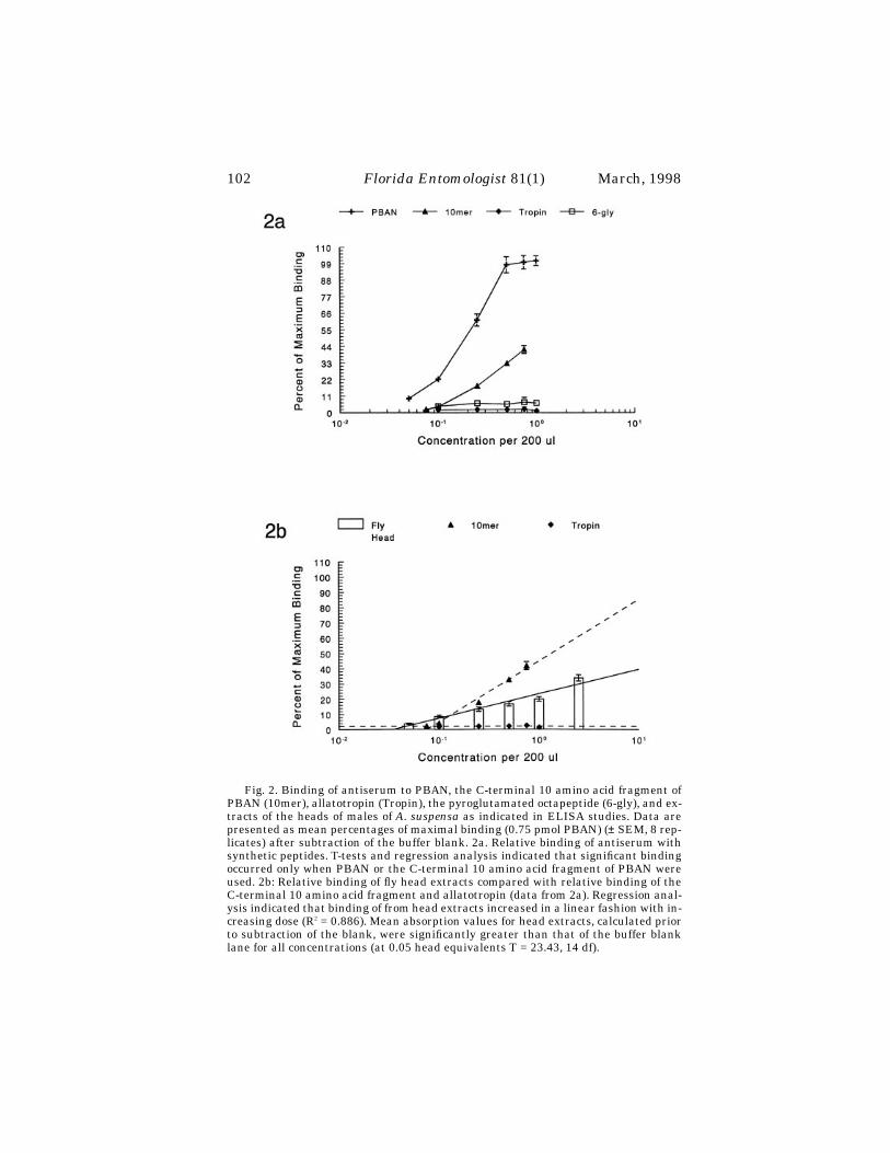
102
Florida Entomologist
81(1) March, 1998
Fig. 2. Binding of antiserum to PBAN, the C-terminal 10 amino acid fragment ofPBAN (10mer), allatotropin (Tropin), the pyroglutamated octapeptide (6-gly), and ex-tracts of the heads of males of A. suspensa as indicated in ELISA studies. Data arepresented as mean percentages of maximal binding (0.75 pmol PBAN) (± SEM, 8 rep-licates) after subtraction of the buffer blank. 2a. Relative binding of antiserum withsynthetic peptides. T-tests and regression analysis indicated that significant bindingoccurred only when PBAN or the C-terminal 10 amino acid fragment of PBAN wereused. 2b: Relative binding of fly head extracts compared with relative binding of theC-terminal 10 amino acid fragment and allatotropin (data from 2a). Regression anal-ysis indicated that binding of from head extracts increased in a linear fashion with in-creasing dose (R2 = 0.886). Mean absorption values for head extracts, calculated priorto subtraction of the blank, were significantly greater than that of the buffer blanklane for all concentrations (at 0.05 head equivalents T = 23.43, 14 df).

Teal: Caribbean Fruit Fly Pheromonotropic Neuropeptides
103
To determine if substances present in the head extracts from male
A. suspensa
hadany structural similarity to PBAN we conducted ELISA studies. Studies on the spec-ificity of the antiserum for PBAN and other neuropeptides including: allatotropin (A-K-S-Y-N-F-G-L-NH
2
), a pyroglutamated octapeptide (pE-T-S-F-T-G-R-L-NH
2
), the C-terminal 10 amino acid fragment of PBAN (S-R-T-K-Y-F-S-P-R-L-NH
2
) and PBAN (L-S-D-D-M-P-A-T-P-A-D-Q-E-M-Y-R-Q-D-P-E-Q-I-D-S-R-T-K-Y-F-S-P-R-L-NH
2
) indi-cated that significant binding occurred only when PBAN or the C-terminal decapep-tide fragment of PBAN were used as substrates in the ELISA (Fig. 2). Additionally,immunochemical studies conducted by Davis et al. (1996) have shown that the anti-serum complexes with PBAN and the pentapeptide F-T-P-R-L-NH
2
, but not with otherinsect neuropeptides like FMRFamide and proctolin. Thus, the antiserum has reac-tivity for PBAN-like molecules and has antibodies directed against the C-terminalportion of the molecule. This C-terminal fragment is the active core of PBAN, whichis necessary and sufficient for pheromonotropic activity (see Abernathy et al. 1996).
ELISA studies comparing the reactivity of head extracts of flies showed that theextracts bind the antiserum (Fig. 2). The binding increased in a linear fashion (per-cent maximum binding = 6.567 + 11.70
×
concentration of head extract, r
2
= 0.886)over the range of doses tested. Therefore, the extracts from sexually mature males of
A. suspensa
contained substances that had some structural similarity with PBAN.I conclude from the results of pheromonotropic and ELISA studies, that phero-
monotropic neuropeptides with structural similarity to moth PBAN are present in theheads of sexually mature males of
A. suspensa
. It is not known if these same peptidesare present in the cephalic ganglia of females of
A. suspensa
, but it is likely that theyare because PBAN is produced by males of the corn earworm moth (Raina & Klun1984). Although males of
A. suspensa
exhibit a diel periodicity of pheromone produc-tion, which in insects like the Hessian fly is regulated by neuropeptides (Foster et al.1991), studies to determine if these factors are responsible for induction of pheromoneproduction by males of
A. suspensa
have not been conducted. It is possible that thepeptides in question are responsible for regulation of other physiological functions, forexample myotropic stimulation of the gut, because myotropic neuropeptides fromroaches and the migratory locust will stimulate pheromone production when injectedinto females of H. zea (Abernathy et al. 1995). However, no myotropic neuropeptideshaving homology with the active core of PBAN have been identified from Diptera.Further research will be required to determine if males of
A. suspensa
regulate the in-duction of pheromone production with these or other neuropeptides.
R
EFERENCES
C
ITED
A
BERNATHY
, R. L., R. J. N
ACHMAN
, P. E. A. T
EAL
, O. Y
AMASHITA
,
AND
J. H. T
UMLIN-SON
. 1995. Pheromonotropic activity of naturally occurring pyrokinin insectneuropeptides (FXPRLamide) in
Helicoverpa zea
. Peptides. 16: 1-5.C
HRISTENSEN
, T. C., H. I
TAGAKI
., P. E. A. T
EAL
, R. D. J
ASENSKY
, J. H. T
UMLINSON
,
AND
J. G. H
ILDEBRAND
. 1991. Innervation and neural regulation of the sexpheromone gland in female
Heliothis
moths. Proc. Nat. Acad.Sci. 88: 4971-4975.
D
AVIS
, N., U. H
OMBERG
, P. E. A. T
EAL
, M. A
LTSTEIN
,
AND
J. G. H
ILDEBRAND
. 1996.Neuroanatomy and immunocytochemistry of the neurosecretory system of thesubesophageal ganglion of the tobacco hawkmoth, Manduca sexta: Immunore-activity to PBAN and other neuropeptides. Microscopy Research and Tech. 35:201-226.
D
AVIS
, N. T., S. V
ELLEMAN
, T. K
INGAN
,
AND
H. K
ESHISHIAN
. 1989. Identification anddistribution of a proctolin-like neuropeptide in the nervous system of the gypsymoth,
Lymantria dispar
, and other Lepidoptera. J.Comp. Neurol. 283, 71-85.

104
Florida Entomologist
81(1) March, 1998
E
PSKY
, N. D.,
AND
R. R. H
EATH
. 1993. Pheromone production by male
Anastrepha sus-pensa
(Diptera: Tephritidae) under natural light cycles in greenhouse studies.Environ. Entomol. 22: 464-469.
F
OSTER
, S. P., J. C. B
ERGH
, S. R
OSE
,
AND
M. O. H
ARRIS
. 1991. Aspects of pheromonebiosynthesis in the Hessian Fly,
Mayetiola destructor
(Say). J. Insect Physiol.37: 899-906.
G
AZIT
, Y., E. D
UNKELBLUM
, O. B
EN
-A
ZIZ
,
AND
M. A
LTSTEIN
. 1992. Immunological andbiological analysis of pheromone biosynthesis activating neuropeptide in
Helio-this peltigera
. Arch. Insect Biochem. Physiol. 19: 247-260.N
ATION
, J. L. 1990. Biology of pheromone release by male Caribbean fruit flies,
Anas-trepha suspensa
(Diptera: Tephritidae). J. Chem. Ecol. 16: 553-572.P
OPE
, M. M., L. K. G
ASTON
,
AND
T. C. B
AKER
. 1982. Composition, quantification, andperiodicity of sex pheromone gland volatiles from individual
Heliothis virescens
females. J. Chem. Ecol. 8: 1043-1055.P
OPE
, M. M., L. K. G
ASTON
, AND T. C. BAKER. 1984. Composition, quantification, andperiodicity of sex pheromone volatiles from individual Heliothis zea females. J.Insect Physiol. 30: 943-945.
RAINA, A. K., AND J. A. KLUN. 1984. Brain factor control of sex pheromone productionin female corn earworm moth. Science 225: 531-533.
TEAL, P. E. A., AND J. H. TUMLINSON. 1989. Neurohormonal regulation of pheromonebiosynthesis by Heliothis zea during the light period. Canadian Entomol. 121:43-46.
TEAL, P. E. A., A. OOSTENDORP, AND J. H. TUMLINSON. 1993 Induction of pheromoneproduction in females of Heliothis virescens (F.) and H. subflexa (Gn.) (Lepi-
♦♦♦♦♦♦♦♦♦♦♦♦♦♦♦♦♦♦♦♦♦♦♦♦♦♦♦♦♦♦♦♦♦♦♦♦♦♦♦♦♦♦♦♦♦♦♦♦♦♦♦♦
doptera: Noctuidae) during the photophase. Canadian Entomol. 125: 355-366.
104
Florida Entomologist
81(1) March, 1998
ST. LOUIS ENCEPHALITIS VIRUS TRANSMISSION TO EMUS (
DROMAIUS NOVAEHOLLANDIAE
) IN PALM BEACH COUNTY, FLORIDA WITH EVIDENCE OF WESTERN EQUINE
ENCEPHALITIS VIRUS ANTIBODY TRANSPORT TO FLORIDA BY EMUS INFECTED IN OTHER STATES
J
ONATHAN
F. D
AY
1
AND
L
ILLIAN
M. S
TARK
2
1
Florida Medical Entomology Laboratory, Institute of Food and Agricultural SciencesUniversity of Florida, 200 9th St. SE, Vero Beach, FL 32962
2
Tampa Branch Laboratory-Virology SectionFlorida Department of Health and Rehabilitative Services
3952 West M. L. King, Jr. Boulevard, Tampa, FL 33614
A
BSTRACT
From November 1993 through January 1995, sera were collected from 59 domesticemus (
Dromaius novaehollandiae
) at a ranch in Palm Beach County, FL and tested forantibody evidence of arboviral infection. Hemagglutination inhibition (HI) and neu-tralizing (NT) antibodies to St. Louis encephalitis (SLE) virus were identified in seracollected each year. In addition, HI and NT antibodies to eastern equine encephalitis(EEE) virus and NT antibodies to western equine encephalitis (WEE) virus were de-tected in emus imported to and then maintained at the ranch. Neither of the equine
Day and Stark: SLE Transmission to Florida Emus
105
encephalitis viruses are common in Palm Beach County and many of the EEE andWEE antibody-positive emus were imported from other states prior to vaccination.Emus imported from California, Louisiana and Texas had evidence of naturally ac-quired antibodies to EEE and WEE viruses. This observation underscores the poten-tial threat of arboviral introduction by infected vertebrates and emphasizes theimportance of instituting quarantine procedures to regulate the transport of hoststhat may be infected with arboviral agents.
Key Words: Encephalitis virus, SLE, EEE, WEE, emu
R
ESUMEN
Desde noviembre 1993 hasta enero 1995 se colectaron sueros de 59 emúes domés-ticos (
Dromaius novaehollandiae
) ubicados en una finca en el condado de Palm Beach,Florida, y se examinaron para determinar la presencia de anticuerpos contra infecciónarboviral. La inhibición de la hemoaglutinación (HI) y los anticuerpos neutralizadores(NT) del virus de la encefalitis de St. Louis (SLE) fueron identificados en los sueros co-lectados anualmente. Además, la HI y el NT del virus de la encefalitis equina oriental(EEE) y el NT del virus de la encefalitis equina occidental (WEE) fueron detectados enemúes importados a la finca y luego mantenidos allí. Ninguno de los dos viruses de en-cefalitis equina son comunes en el condado de Palm Beach y muchos de los emús queresultaron tener anticuerpos contra EEE y WEE habían sido importados de otros es-tados antes de recibir vacunaciones. Los emús importados de California, Louisiana, yTexas tenían evidencia de anticuerpos para EEE y WEE adquiridos naturalmente.Esta observación enfatiza la amenaza potencial de la introducción de arboviruses envertebrados infectados y subraya la importancia de establecer procedimientos de cua-
rentena para reglamentar el transporte de hospederos infectados con arboviruses.
Naturally occurring mosquito-borne arboviruses infect and cause mortality in do-mestic emus (
Dromaius novaehollandiae
Linn.). In Texas, emus from 8 flocks were in-fected with western equine encephalitis (WEE) virus in 1992 and suffered 15-50%morbidity and 8.8% mortality (Ayers et al. 1994). In 1991, emus were infected witheastern equine encephalitis (EEE) virus in Louisiana, where an 87% mortality ratewas reported (Tully et al. 1992). Two emus died of EEE virus infection in Georgia dur-ing the summer of 1992 (Brown et al. 1993). In Volusia County, FL during the springof 1992, EEE-related morbidity and mortality rates in an emu flock were 40.5% and14.1%, respectively. This outbreak resulted in a one year financial loss of an estimated$192,000 (Day and Stark 1996a).
In southern Florida and many parts of the central USA, St. Louis encephalitis(SLE) virus is the most commonly transmitted mosquito-borne arbovirus (Chamber-lain 1980, Day & Stark 1996b). However, SLE infection in emus maintained in Flor-ida, and in other areas of North America where SLE is endemic, has been reportedonly once (Day & Stark 1996b).
Eastern equine encephalitis virus, as well as antibody evidence of its transmis-sion, is reported frequently in the northern half of Florida, but is rarely found in the10 counties comprising the southern tip of the state (Day & Stark 1996b). The WEEvirus is found west of the Mississippi drainage basin (Reisen & Monath 1989). How-ever, WEE virus comprises a complex of at least six serologically related but distinctviruses. One of these, Highlands J (HJ) virus, is present in central and north Florida,but is absent from the southern tip of the state (Trent & Grant 1980).

106
Florida Entomologist
81(1) March, 1998
The purpose of our study was to monitor natural arboviral transmission to emusin Palm Beach County, FL. Additionally, we present evidence that emus imported intoFlorida were initially infected with EEE and WEE viruses in the central and westernUSA.
M
ATERIALS
AND
M
ETHODS
Study Site
Emus were maintained at a 4 ha ranch in Palm Beach County (26
°
40’N, 80
°
15’W),FL. The ranch was located within a slash pine (
Pinus elliottii
Engelm.)/saw palmetto[
Serenoa repens
(Bartr.) Small] habitat in the western part of the county.During the spring of 1993, approximately 150 juvenile emus were imported to the
ranch from locations in Florida, California, Louisiana, Pennsylvania and Texas. Ourstudy began in November 1993 in response to emu morbidity that was suspected of be-ing caused by an arboviral agent. The study continued through January 1995.
When the first blood sample was taken, emus were sexed and placed into one of 3age groups: hatching year (HY) = 1-120 days old, juvenile (J) = 121-365 days old, andadult (A) =
≥
366 days old.
Serum Collection and Analysis
Blood was drawn from the jugular veins of restrained emus. A 3.0 ml sample wascollected from HY emus, and a 5.0 ml sample from J and A birds. Blood samples wereallowed to clot overnight at room temperature, were centrifuged at 3,400
×
g for 30min, and the resulting sera used for SLE and EEE virus hemagglutination inhibition(HI) and neutralizing (NT) antibody assays. All sera were analyzed for HI antibody toSLE and EEE viruses and for NT antibody to SLE virus. Selected sera were also an-alyzed for NT antibody to EEE and WEE viruses.
A micro-adaption of the HI antibody test of Beaty et al. (1989) was used with a he-magglutinin (HA) prepared from a Florida human SLE isolate (TBH-28, Florida De-partment of Health and Rehabilitative Services, Tampa Branch Laboratory).Additionally, all sera were examined in the same manner for HI antibody againstEEE virus using an HA prepared from a Florida human isolate (NJ-60, Florida De-partment of Health and Rehabilitative Services, Tampa Branch Laboratory). Themethodology used for HI antibody testing is described in detail by Day et al. (1996).
Aliquots of all sera were examined for NT antibody to SLE virus by serial virus di-lution with undiluted serum (Beaty et al. 1989). The challenge virus was a Florida iso-late (SLE-P15, Florida Department of Health and Rehabilitative Services, TampaBranch Laboratory) obtained from a pool of
Culex nigripalpus
Theobald mosquitoes.Two NT tests for EEE antibody were performed on selected serum aliquots. The chal-lenge viruses for these tests were a 1964 Florida human isolate (D64-837, Florida De-partment of Health and Rehabilitative Services, Tampa Branch Laboratory) and anisolate (VO-73, Florida Department of Health and Rehabilitative Services, TampaBranch Laboratory) from a pool of 7
Cx. erraticus
(Dyar and Knab) collected in August1992 in Volusia County, FL (Day & Stark 1996a). A single NT test for WEE antibodywas performed on selected serum aliquots. The challenge virus for these tests wasWEE Fleming strain (VR-1251, American Type Culture Collection, Rockville, MD).We tested for cross reactivity of the VR-1251 strain with Florida EEE and HJ viralstrains and found none.
The LD
50
virus dilutions for each series of serum-virus mixtures, along with thatof the control, were determined to a single decimal point. A logarithmic LD
50
was ex-
Day and Stark: SLE Transmission to Florida Emus
107
pressed as the exponent of the reciprocal of the endpoint dilution. The log neutraliza-tion index of each serum was obtained by subtracting its LD
50
from that of the control.Indices of <1.0 were considered negative, 1.0 to 1.6 were equivocal and those
≥
1.7 werepositive. The methodology used for NT antibody testing is described in detail by Dayet al. (1996).
Viral Isolation Attempts
Blood from 10 emus and tissue (brain, heart, liver and spleen) from 2 emus thatdied of encephalitis-like symptoms were assayed for arboviral agents as described be-low. One drop of blood was mixed in 0.7 ml of laboratory-prepared biological field di-luent (BFD) (90% Minimum Essential Medium with Hank’s salts (Sigma ChemicalCo., St. Louis, MO), 10% fetal bovine serum (Intergen Co., Purchase, NY), 200 U/mlpenicillin (Sigma Chemical), 200 ug/ml streptomycin (Sigma Chemical), 2.5 ug/ml am-photericin B (Sigma Chemical), and 50 ug/ml kanamycin (Sigma Chemical). Bloodsamples were placed immediately on wet ice in the field and transported to the labo-ratory where they were stored at -70
°
C until analysis.Approximately 0.5 g of tissue was pulverized in 0.7 ml BFD with a chilled 1.0 ml
Potter-Elvehjem tissue grinder (Fisher Scientific, Orlando, FL) in a laminar flow bio-safety cabinet. The suspension was clarified by centrifugation at 800
×
g and renderedfree of bacteria either by centrifugation at 4,300
×
g or filtration with a 0.2 um syringefilter. The supernatant was aliquoted into sterile 1 ml polypropylene cryopreservationvials (Fisher Scientific, Orlando, FL) and stored at -70
°
C until analysis as describedby Day et al. (1996).
Vaccination and Maternal Antibody Transfer
During April and May 1994, selected emus were vaccinated with 1.0 ml of Enceph-aloid IM
R
, an inactivated EEE/WEE vaccine (Ft. Dodge Laboratories, Ft. Dodge, IA).Emus were bled before vaccination to establish baseline titers and periodically follow-ing vaccination to track resulting antibody titers. Chicks from hens with natural SLEinfections were bled within 24 days of hatching to determine the presence or absenceof maternally acquired SLE antibody.
Statistical Tests
Statistical differences in EEE antibody titers (HI and NT) before and after vacci-nation were tested by using unplanned tests of the homogeneity of replicates testedfor goodness of fit (G-statistic) (Sokal and Rohlf 1981).
R
ESULTS
Sera from 59 emus maintained at the Palm Beach County ranch were tested be-tween November 1993 and January 1995 for evidence of arboviral transmission.Twenty-six of the emus originated in California, 13 in Florida, 11 in Louisiana, 8 inTexas and one in Pennsylvania. Twenty-four of the emus were bled more than once.Five were HY, 34 were J and 20 were A age group when first bled.
Most of the emus had HI (43 of 59, 73%) and NT (44 of 59, 75%) antibody to SLEvirus. Antibody titers for individual emus were as follows. SLE HI antibody titers:1:10 = 2 emus, 1:20 = 5, 1:40 = 2, 1:80 = 6, 1:160 = 15, 1:320 = 11 and
≥
640 = 2. SLENT antibody titers:
≥
1.8 = 6 emus,
≥
2.0 = 3,
≥
2.1 = 4,
≥
2.3 = 5,
≥
2.4 = 1,
≥
2.6 = 1,
≥
2.7
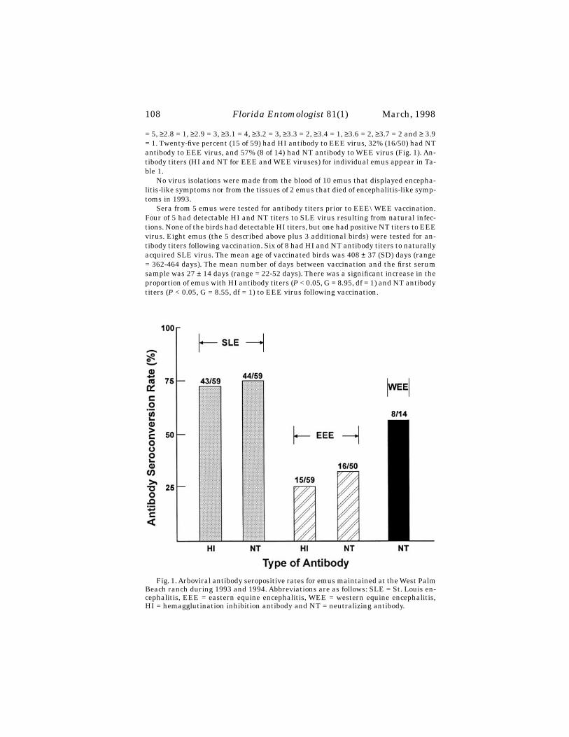
108
Florida Entomologist
81(1) March, 1998
= 5,
≥
2.8 = 1,
≥
2.9 = 3,
≥
3.1 = 4,
≥
3.2 = 3,
≥
3.3 = 2,
≥
3.4 = 1,
≥
3.6 = 2,
≥
3.7 = 2 and
≥
3.9= 1. Twenty-five percent (15 of 59) had HI antibody to EEE virus, 32% (16/50) had NTantibody to EEE virus, and 57% (8 of 14) had NT antibody to WEE virus (Fig. 1). An-tibody titers (HI and NT for EEE and WEE viruses) for individual emus appear in Ta-ble 1.
No virus isolations were made from the blood of 10 emus that displayed encepha-litis-like symptoms nor from the tissues of 2 emus that died of encephalitis-like symp-toms in 1993.
Sera from 5 emus were tested for antibody titers prior to EEE\WEE vaccination.Four of 5 had detectable HI and NT titers to SLE virus resulting from natural infec-tions. None of the birds had detectable HI titers, but one had positive NT titers to EEEvirus. Eight emus (the 5 described above plus 3 additional birds) were tested for an-tibody titers following vaccination. Six of 8 had HI and NT antibody titers to naturallyacquired SLE virus. The mean age of vaccinated birds was 408
±
37 (SD) days (range= 362-464 days). The mean number of days between vaccination and the first serumsample was 27
±
14 days (range = 22-52 days). There was a significant increase in theproportion of emus with HI antibody titers (
P
< 0.05, G = 8.95, df = 1) and NT antibodytiters (
P
< 0.05, G = 8.55, df = 1) to EEE virus following vaccination.
Fig. 1. Arboviral antibody seropositive rates for emus maintained at the West PalmBeach ranch during 1993 and 1994. Abbreviations are as follows: SLE = St. Louis en-cephalitis, EEE = eastern equine encephalitis, WEE = western equine encephalitis,HI = hemagglutination inhibition antibody and NT = neutralizing antibody.
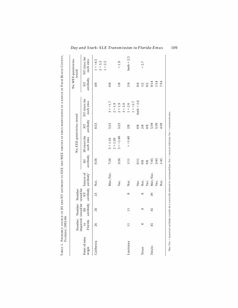
Day and Stark: SLE Transmission to Florida Emus
109
T
AB
LE
1.
P
RO
BA
BL
E
SO
UR
CE
OF
HI
AN
D
NT
AN
TIB
OD
Y
TO
EE
E
AN
D
WE
E
VIR
US
ES
IN
EM
US
MA
INT
AIN
ED
AT
A
RA
NC
H
IN
P
AL
M
B
EA
CH
C
OU
NT
Y
,F
LO
RID
A
: 199
3-94
.
Poi
nt
of e
mu
or
igin
Nu
mbe
r im
port
ed t
o F
lori
da
Nu
mbe
r te
sted
for
HI
anti
body
Nu
mbe
r te
sted
for
NT
an
tibo
dyS
ourc
e of
an
tibo
dy
1
No.
EE
E-p
osit
ive/
no.
tes
ted
No.
WE
E-p
osit
ive/
no.
te
sted
HI
anti
body
HI t
iter
s fo
r ea
ch e
mu
NT
an
tibo
dyN
T t
iter
s fo
r ea
ch e
mu
NT
an
tibo
dyN
T t
iter
s fo
r ea
ch e
mu
Cal
ifor
nia
2626
23N
at.
0/26
0/23
4/8
1 =
>4.5
2 =
3.3
1 =
2.5
Mat
./Vac
.7/
265
= 1:
105/
233
= >1
.70/
82
= 1:
202
= 1.
9V
ac.
3/26
3 =
>1:4
05/
232
= 1.
91/
8 =
1.9
3 =
2.0
Lou
isia
na
1111
8N
at.
1/11
= >
1:40
2/8
1 =
2.6
2/4
both
= 2
.51
= >2
.7V
ac.
0/11
0/8
both
= 2
.60/
4T
exas
88
8N
at.
0/8
2/8
1/2
= 2
.7V
ac.
0/8
0/8
0/2
Tot
als:
4545
39M
at./V
ac.
7/45
5/39
0/14
Vac
.3/
455/
391/
14N
at.
1/45
4/39
7/14
1
Mat
./Vac
. = m
ater
nal
an
tibo
dy t
ran
sfer
by
a n
atu
rall
y in
fect
ed o
r va
ccin
ated
hen
, Nat
. = n
atu
ral i
nfe
ctio
n, V
ac. =
vac
cin
ated
em
u.
110
Florida Entomologist
81(1) March, 1998
Natural EEE transmission is rare in Palm Beach County (Day & Stark 1996b). Be-cause many of the emus in our study were HI and NT antibody-positive for EEE virusand because many of the birds originated west of the Mississippi River, selected serawere tested for NT antibody to WEE virus. Most of the sera were collected before theemus were treated with an EEE/WEE vaccine. Therefore, positive findings in thesebirds indicated the possibility of natural infection or maternal antibody transfer (Day& Stark 1996a). In Volusia County, Florida, we observed that emus naturally infectedwith EEE virus usually had NT antibody titers >2.0. Newly hatched emus with ma-ternally derived antibody titers (through natural infection or vaccination of the hens)usually displayed NT antibody titers <2.0. Eight of 14 emus tested for NT antibody toWEE virus were positive, all but one with NT antibody titers >2.0, indicating possiblenatural infection at their hatching site. One of 45 (2%) HI tests and 4 of 39 (10%) NTtests indicated EEE antibody titers that most likely resulted from natural infection(Table 1).
Four of 5 newly hatched chicks from hens with a natural SLE infection had HI andNT antibody titers to SLE virus. The mean age of the chicks was 11.2
±
8.1 days (range= 6-24 days) when they were first tested for antibody. All had negative EEE antibodytiters.
D
ISCUSSION
The introduction of exotic hosts into habitats that have active arbovirus transmis-sion provides a potential vehicle by which transfer of virus from one location to an-other may be facilitated. Emus are susceptible to infection by EEE virus (Tully et al.1992, Brown et al. 1993, Day & Stark 1996a), WEE virus (Ayers et al. 1994) and SLEvirus. An important, and yet unanswered question is whether and for how long in-fected emus circulate virus titers sufficient to infect mosquitoes or other potential vec-tors. Emus are sold and transported throughout North America and can potentiallytransport an arbovirus from an active focus to an area where the virus is not extantbut vectors suitable to establish an active viral focus are present.
The consistently high SLE antibody levels in emus from Palm Beach County indi-cate that these birds were most likely infected naturally with SLE virus during theautumn of 1993 when an unusually high level of SLE transmission was reported insentinel chickens along the east-central coast of Florida (Stark, unpublished data).The emu ranch was located in a habitat favored by the major vector of SLE virus inFlorida,
Cx. nigripalpus
, and it is likely that an SLE transmission focus involvingmosquitoes, emus and wild birds was established around the emu ranch during 1993.It is also possible that some of the emus were infected with SLE virus in California,Louisiana or Texas prior to their import into Florida. However, the facts that the ma-jority of emus in the flock were HI and NT antibody positive (73% and 75% respec-tively) for SLE virus and that the titers for both types of antibody were high, indicatesthat SLE transmission was not sporadic and, regardless of the state of origin of theemus, virtually all of them ended up SLE positive at the West Palm ranch.
No viral isolates were made from sick or dead emus in 1993. Judging from the highSLE antibody rates and the low morbidity and mortality rates among infected birds,it does not appear that SLE virus causes as severe an infection nor as high a mortalityrate as do EEE and WEE viruses.
Natural transmission of EEE and HJ viruses is uncommon in Palm Beach County(Day & Stark 1996b). However, 15 of 59 (25%) of the emus in our study had HI anti-body titers and 16 of 50 (32%) had NT antibody titers to EEE virus. Additionally, 8 of14 (57%) had NT antibody titers to WEE virus. Forty-six (78%) of the emus in our

Day and Stark: SLE Transmission to Florida Emus
111
study originated outside of Florida (one emu, not shown in Table 1, originated inPennsylvania). Five of the 13 emus that originated in Florida hatched at the PalmBeach facility, whereas 8 were imported from a ranch in Volusia County where EEEvirus is endemic (Day & Stark 1996a). Some of the antibody-positive emus reacted toEEE or WEE tests because of vaccination or maternally-derived antibody. However,judging from the high antibody titers, at least some of the emus had antibody ac-quired as the result of natural infection at their hatching location (Table 1). Emus andother exotic avian hosts may potentially transport live virus across state lines. It isimportant to determine the extent and duration of viremias in infected emus to eval-uate the risk of viral introduction by emus infected at one site and then transportedto a secondary or even a tertiary location supporting suitable vector populations.
A
CKNOWLEDGMENTS
William Kohl, Nazar Hussain, Arnie Croteau and Tersa Lowry assisted with thisstudy. This research was funded by a Research Contract from the Florida Emu Asso-ciation. Florida Agricultural Sciences Experiment Station Journal Series No. R-05160.
R
EFERENCES
C
ITED
A
YERS
, J. R., T. L. L
ESTER
,
AND
A. B. A
NGULO
. 1994. An epizootic attributable to west-ern equine encephalitis virus infection in emus in Texas. J. Am. Vet. Med. As-soc. 205: 600-601.
B
EATY
, B. J., C. H. C
ALISHER
,
AND
R. E. S
HOPE
. 1989. Arboviruses, pp. 797-855
in:
Schmidt, N. J. and R. W. Emmons [eds.], Diagnostic procedures for viral, rick-ettsial and chlamydial infections. Am. Public Health Assoc., Washington, DC.
B
ROWN
, T. P., W. R
OBERTS
,
AND
R. K. P
AGE
. 1993. Acute hemorrhagic enterocolitis inratites: isolation of eastern equine encephalomyelitis virus and reproduction ofthe disease in ostriches and turkey poults. Avian Diseases 37: 602-605.
C
HAMBERLAIN
, R. W. 1980. History of St. Louis encephalitis. Chapter 1
in
Monath, T.P. [ed.], St. Louis encephalitis. Am. Public Health Assoc., Washington, DC.
D
AY
, J. F.,
AND
L. M. S
TARK
. 1996a. Eastern equine encephalitis transmission to emus(
Dromaius novaehollandiae
) in Volusia County, Florida: 1992 through 1994. J.Am. Mosq. Control Assoc. 12: 429-436.
D
AY
, J. F.,
AND
L. M. S
TARK
. 1996b. Transmission patterns of St. Louis encephalitisand eastern equine encephalitis viruses in Florida: 1978-1993. J. Med. Ento-mol. 33: 132-139.
D
AY
, J. F., L. M. S
TARK
, J-T. Z
HANG
, A. M. R
AMSEY
,
AND
T. W. S
COTT
. 1996. Antibod-ies to arthropod-borne encephalitis viruses in small mammals from southernFlorida. J. Wildlife Dis. 32: 431-436.
R
EED
, L. J.,
AND
H. A. M
UENCH
. 1938. A simple method of estimating fifty percentendpoints. Am. J. Hyg. 27: 493-497.
R
EISEN
, W. K.,
AND
T. P. M
ONATH
. 1989. Western equine encephalomyelitis. Chapter50
in
Monath, T. P. (ed.)The arboviruses: epidemiology and ecology, Vol. V. CRCPress, Inc., Boca Raton, FL.
S
OKAL
, R. R.,
AND
F. J. R
OHLF
. 1981. Biometry, 2nd ed. W. H. Freeman and Co., SanFrancisco, CA.
T
RENT
, D. W.,
AND
J. A. G
RANT
. 1980. A comparison of New World alphaviruses in thewestern equine encephalitis virus complex by immunochemical and oligonucle-otide fingerprint techniques. J. Gen. Virol. 47: 261-282.
T
ULLY
, T. N., S. M. S
HANE
, R. P. P
OSTON
, J. J. E
NGLAND
, C. C. V
ICE
, D-Y. C
HO
,
AND
B. P
ANIGRAHY
. 1992. Eastern equine encephalitis in a flock of emus (
Dromaiusnovaehollandiae
). Avian Diseases 36: 808-812.

112
Florida Entomologist
81(1) March, 1998
A NEW GENUS AND SPECIES OF SPIDER BEETLE FROM THE VIRGIN ISLANDS:
LACHNONIPTUS LINDAE
(COLEOPTERA: ANOBIIDAE: PTININAE)
T. K
EITH
P
HILIPS
Department of Entomology, Museum of Biological DiversityThe Ohio State University, 1315 Kinnear Road, Columbus, Ohio 43212
A
BSTRACT
Lachnoniptus lindae
, a
new genus
and
new species
from the Virgin Islands, isdescribed. It appears most similar to
Trigonogenius
, and characters differentiatingthe two genera are given. The habitat and probable biology are discussed.
Key Words: Ptininae, Ptinidae, spider beetles, Virgin Islands, West Indies
R
ESUMEN
Lachnoniptus lindae
, un nuevo género y especie de las Islas Vírgenes, es descrito.Es parecido a
Trigonogenius
, y los caracteres que diferencian a ambos géneros son
provistos. El hábitat y probable biología de esta nueva especie son discutidos.
The ptinine fauna of the Virgin Islands includes one recorded species
, Pitnus anti-llanus
Bellés (Bellés 1992). In the more inclusive West Indies, another species of
Pit-nus
Gorham, four species of
Ptinus
Linnaeus, three
Gibbium
Scopoli, and one
Fabrasia
Martinez and Viana (=
Cubaptinus
Zayas, Philips 1997) have been recorded(Boieldieu 1856, Gorham 1898, Pic 1906, Lepesme 1947, Wolcott 1948, Bellés and Hal-stead 1985, Zayas 1988, Bellés 1992). Recent investigations on the beetle fauna of theVirgin Islands have resulted in the discovery of two undescribed ptinine species. Oneof these belongs in the genus
Ptinus
, but the unique morphology and phylogenetic po-sition of the other requires the creation of a new genus. I take this opportunity tomake a name available for the latter.
Lachnoniptus
Philips
New Genus
Type Species.
Lachnoniptus lindae
New Species
.
Diagnosis
. This genus is easily recognized by the transverse pronotum and thevery convex globose shape of the elytra. The dense pubescence dorsally results in awoolly, fluffy appearance. Also, the apical antennomere is slightly but distinctly widerthan the penultimate one.
Trigonogenius
is the only genus that approaches this ely-tral and pronotal shape, but
Trigonogenius
has dense, appressed setae on the elytra,unlike the fluffy elytral setae of
Lachnoniptus
. Further,
Trigonogenius
does not havethe laterally enlarged and distally rounded apical antennomere of
Lachnoniptus
. Incontrast,
Trigonogenius
is characterized by the apical and penultimate antennomeressubequal in diameter and the apex acuminate. Other differences include the follow-ing:
Lachnoniptus
with a mesosternal process
2
/
3
the width of the mesocoxa (
1
/
3
thewidth for
Trigonogenius
); the absence of the mesosternal-mesepisternal suture(present in
Trigonogenius
); metepisternum not visible due to fusion with metaster-

Philips: New Genus and Species of Spider Beetle
113
num (distinctly visible in
Trigonogenius
); metacoxae approximately round
(rectangu-lar
in Trigonogenius); the metacoxae laterally contacting the first ventrite (laterallyadjacent to the metepisternum in
Trigonogenius
); the fourth ventrite compared tothird about equal in length (a ratio of about 1.15:1) (greatly reduced in
Trigonogenius
(a ratio of about 3:1)); and the anterior margin of the first ventrite narrowly pointedlaterally (sharply pointed in
Trigonogenius
). Two more subtle characters also differ-entiate these two genera. The scutellum is narrowly rounded in
Lachnoniptus
but isbroadly rounded in
Trigonogenius.
In some specimens of
Lachnoniptus
there is a faintrow of transverse setal tufts on the pronotum. Pronotal tufts are always absent in
Trigonogenius
. While
Lachnoniptus
is known from the West Indies,
Trigonogenius
isfound in the western part of North and South America.
Description
. Body robust and globular, densely covered with erect fluffy setaethat obscure cuticular surface.
Head. Very robust, not visible from above, partially hidden within the pronotumup to the posterior dorsal margin of the eye, eye nearly semicircular, slightly roundedon dorsal side; longitudinal groove on the frons between antennal insertions, clypeusequilaterally triangular, labrum narrow, no wider than proximal edges of antennal in-sertions, anterior margin approximately truncate, anterio-lateral edges broadlyrounded; antennae 11 segmented, second segment attached on side of scape nearapex, apical antennomere distinctly widest at anterior
1
/
3
, tapering to rounded tip;mentum slightly longer than wide, triangular, not truncate but narrowly rounded atanterior margin, with a small round depression medially at basal
1
/
3
, a patch of 4-8moderately long setae anterio-medially; hypopharygeal setal rows closely spaced andnearly overlapping; maxillary and labial palpi with apical segment tapered to a point;galea and lacinia with stout spines, spines obscured with fine setae.
Thorax. Pronotum globose and convex, transverse, 1.35 times wider than long,widest at middle; scutellum small, hidden, slightly tranversely ovoid, distinctly belowlevel of elytra; procoxae cylindrical, projecting, prosternal process with apex ex-panded and rounded, extending ventrally about as far as coxae, at narrowest widthabout
1
/
3
the width of coxa; mesosternum smoothly concave, slightly narrower poste-riorly, process about
2
/
5
width of mesocoxa; mesepimeron visible but narrow, mesoster-nal-mesepisternal suture absent; metasternum about half the length of themesosternum, sharply, obtusely emarginate at posterior margin; metacoxae trans-versely triangular, laterally in contact with first ventrite.
Elytra. Globose and convex, fused, nearly as wide as long, length 1.10 times width;large strial punctures easily discernible and in longitudinal rows, puncture edgesbroadly rounded, intervals convex, surface usually hidden beneath dense pubescence.
Ventrites. All sternal sutures clearly defined, second ventrite widest at middle andat lateral edge, third ventrite distinctly narrowest at middle, third ventrite onlyslightly longer than fourth, about 1.1-1.2 times length of fourth.
Legs. Femora gradually increasing in size towards apex, pro- and mesotibiae about
2
/
3
as long as metatibiae, all tarsomeres about equal in length, except first metatar-somere about
1
/
3
longer than second.Etymology. Derived from the Boieldieu genus
Niptus
combined with the greek
lachno
. The name translates as “woolly haired”
Niptus
. Discussion. Putative synapomorphies for
Lachnoniptus
are as follows: 1) the firstventrite narrowly pointed laterally; 2) the ultimate antennomere enlarged; 3) thescutellum vertical and narrowly rounded posteriorly; and 4) the fourth ventrite ap-proximately equal in length to the third. The first character state appears to beuniquely derived while the second and third have similar states or convergences inother ptinine taxa. The fourth character state is hypothesized to be a reversal to thepleisiomorphic state. While most ptinines have the fourth ventrite reduced to various
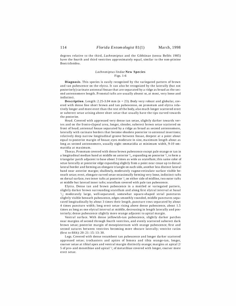
114
Florida Entomologist
81(1) March, 1998
degrees relative to the third,
Lachnoniptus
and the Gibbiinae (sensu Bellés 1985)have the fourth and third ventrites approximately equal, similar to the non-ptinineBostrichoidea.
Lachnoniptus lindae
New Species
Figs. 1-6
Diagnosis
. This species is easily recognized by the variegated pattern of brownand tan pubescence on the elytra. It can also be recognized by the laterally (but notposteriorly) carinate antennal fossae that are separated by a ridge as broad as the sec-ond antennomere length. Pronotal tufts are usually absent or, at most, very loose andindistinct.
Description
. Length: 2.25-3.04 mm (n = 23). Body very robust and globular, cov-ered with dense fine short brown and tan pubescense, on pronotum and elytra rela-tively longer and more erect than the rest of the body, also much longer scattered erector suberect setae arising above short setae that usually have the tips curved towardsthe posterior.
Head. Covered with appressed very dense tan setae, slightly darker towards ver-tex and on the fronto-clypeal area, longer, slender, suberect brown setae scattered onfront of head; antennal fossae separated by a ridge as broad as second antennomere,laterally with carinate borders that become obsolete posterior to antennal insertions;relatively deep narrow longitudinal groove between fossae, deepest at a point aboutequal to posterior margin of fossae; eyes moderate in size, maximum length about aslong as second antennomere, usually eight ommatidia at minimum width, 9-10 om-matidia at maximum.
Thorax. Pronotum covered with dense brown pubescence except pale orange or tan ina longitudinal median band at middle on anterior
3
/
5
, expanding on posterior
2
/
5
to form atriangular patch adjacent to base about 3 times as wide as scutellum; this same color ofsetae laterally at posterior edge expanding slightly from a point near coxae up to dorsal-lateral border and forming an elongate triangle on each side, another less distinct lateralband near anterior margin; shallowly, moderately rugose-reticulate surface visible be-neath setae; erect, elongate curved setae occasionally forming very loose, indistinct tuftson dorsal surface, two inner tufts at posterior
1
/
3
on either side of midline, two outer tuftsat middle but laterad inner tufts; scutellum covered with pale tan pubescence.
Elytra. Dense tan and brown pubescence in a mottled or variegated pattern,slightly darker brown surrounding scutellum and along first elytral interval at basal
1
/
5
; moderately large, well-separated, somewhat square-shaped strial puncturesslightly visible beneath pubescence, edges smoothly rounded, middle punctures sepa-rated longitudinally by about 3 times their length, puncture rows separated by about4 times puncture width; long erect setae rising above dense pubescence, about 1.5times as long as one elytral interval at middle, decreasing in length laterally and pos-teriorly; dense pubescence slightly more orange adjacent to apical margin.
Ventral surface. With dense yellowish-tan pubescence, slightly darker patchesnear margins of second through fourth ventrites, and evenly scattered suberect darkbrown setae; posterior margin of mesepisternum with orange pubescence; first andsecond sutures between ventrites becoming more obscure laterally; ventrite ratios(first to fifth): 20: 21: 15: 13: 30.
Legs. Covered with dense recumbent tan pubescence and longer darker scatteredappressed setae; trochanters and apices of femora and tibia orange-tan, longer,coarser setae at tibial apex and ventral margin distinctly orange; margins at apical 2/5 of pro- and mesotibiae and apical
1
/
3
of metatibiae covered with longer, coarser moreerect setae.

Philips: New Genus and Species of Spider Beetle
115
Sexual dimorphism. None.TYPES. Holotype:
Virgin Islands
: Guana Island, Quail Dove Ghut, 600 ft.,12.VII-09.X.1994, flight intercept #13, M. A. & L. L. Ivie colr [600, 18
°
28.49’N,64
°
34.21’W] (NMNH); Paratypes, same data as holotype (10), same data except 12-24.VII.1994, S. A. Bucklin colr (1), 25.I-25.II.1993, Lio Wei Peng colr (1), 25.II.-
Figs. 1-4. External morphology of Lachnoniptus lindae: 1) dorsal habitus; 2)lateralview; 3) ventrites (most of the setae abraded); 4) elytral setae. Scale line = 1.0 mm forfigs. 1-2, 100 um for 3-4.

116
Florida Entomologist
81(1) March, 1998
25.III.1993, Lio Wei Peng colr, flight intercept trap #5 [400’, 18
°
28.64’N, 64
°
34.20’W](5), Monkey Point Trail, 5-10.X.1997, T. K. Philips, dung pitfalls (26), St John, EstateCarolina, NW Coral Bay, 250 ft., 09.V.1994, litter among rocks, Muchmore (1), StJohn, Lameshur Bay, V.I.E.R.S., 10.III.1984, leaf litter, W. B. Muchmore (1), St John,Maho Bay, 12.III.1984, in hollow tree, W. B. Muchmore colr (1), Guana Island, VII-X.1993, “beetle-trap,” collected by C. Bartlett & J. Cryan (1), Tortola, Windy Hill, 25-28.XII.1993, 350’, thorn-scrub for., T. K. Philips, colr., dung pitfall (1) (Paratypes in thecollections of the author, the Virgin Islands Beetle Project Collection [Montana StateUniversity, Bozeman, Montana], Xavier Bellés, Fred Andrews, The Natural HistoryMuseum [BMNH], Canadian Museum of Nature [CMNC], Muséum National d’His-toire Naturelle de Paris [MHNP], United States National Museum [USNM]).
Distribution. This species is known from three of the northern Virgin Islands(Guana Island, St John, and Tortola). It seems likely that it will be found on other Vir-gin Islands and Puerto Rico, of the Puerto Rican Bank, which were connected as a sin-gle land mass during periods of low eustatic water levels during the Pleistocene(Heatwole and MacKenzie 1966).
Etymology. Named after my wife Linda, in recognition of her support and encour-agement of my career.
D
ISCUSSION
Lachnoniptus lindae
is one of the more highly derived ptinines as characterized byfused elytra, winglessness, and a very globular body form. The majority of species inthis group use dung or other accumulated organic material of animal or plant originas a food source. Most specimens have been captured with dung baited pitfall traps.Dung from numerous reptiles and introduced mammals, such as sheep and goats, arelikely sources of food for both adults and larvae. Larvae have been easily reared on catdung in the laboratory.
All sites where this species was collected are tropical dry forest, characterized byrocky, thin red soils. Evapotranspiration is considerably higher than rainfall for muchof the year, especially during droughts, such as those occurring in 1993-1994. The ab-sence of scarabaeine competition in these areas might be critical for successful repro-duction of this ptinine.
Figs. 5-6. Head of Lachnoniptus lindae: 5) frontal view; 6) lateral view of eye. Scaleline = 100 um.

Philips: New Genus and Species of Spider Beetle
117
A
CKNOWLEDGMENTS
My gratitude to James Lazell and The Conservation Agency for logistic and finan-cial support on Guana Island and to Molly and Wilfred Gerofsky for their support offield work on Tortola, British Virgin Islands. My appreciation to Michael Ivie for lend-ing specimens of this species and for his review of the manuscript. Also thanks to Jo-seph McHugh and one anonymous individual for their careful reviews. MiguelArchangelsky helped with the Spanish version of the abstract and John Mitchell withthe scanning electron microscope pictures.
R
EFERENCES
C
ITED
B
ELLÉS
, X. 1985. Sistemática, filogenia y biogeografía de la subfamilia Gibbiinae (Co-leoptera, Ptinidae). Treballs del Museu de Zoologia 3: 1-95.
B
ELLÉS
, X. 1992. Sistemática, historia natural y biogeografía del género
Pitnus
Gorham, 1880 (Coleoptera, Ptinidae). Eos 68: 167-192.B
ELLÉS
, X.,
AND
D. G. H. H
ALSTEAD
. 1985. Identification and geographical distribu-tion of
Gibbium aequinoctiale
Boieldieu and
Gibbium psylloides
(Czenpinski)(Coleoptera: Ptinidae). J. Stored Products Research 21: 151-155.
B
OIELDIEU
, A. 1856. Monographie des ptiniores. Annales de la Société Entomologiquede France, ser. 3, vol. 4: 285-315, 486-504, 628-686, illus.
G
ORHAM
, H. S. 1898. On the serricorn Coleoptera of St. Vincent, Grenada, and theGrenadines (Malacodermata, Ptinidae, Bostrychidae), with descriptions of newspecies. Proc. Zool. Soc. London 1898: 315-333, illus.
H
EATWOLE
, H.,
AND
F. M
AC
K
ENZIE
. 1966. Herpetogeography of Puerto Rico. IV. Paleo-geography, faunal similarity and endemism. Evolution 21: 429-438.
L
EPESME
, P. 1947. Bostrychoidea. pp. 194-233. In: Fleutiaux, E., Legros, C., Lepesme,P., and Paulian, R. Coléoptères des Antilles Vol. 1. In: Fauna de EmpireFrançais Vol. 7.
P
HILIPS
, T. K. 1997.
Cubaptinus
Zayas, a new synonym of
Fabrasia
Martinez and Vi-ana. Coleopterists Bulletin 51: 52.
P
IC
, M. 1906. Deux nouveaux “Ptinidae” de la Guadeloupe. L’Échange 22: 21-22.W
OLCOTT
, G. N. 1948. [1950]. The insects of Puerto Rico. J. Agric. Univ. Puerto Rico 32(2): 225-416.
Z
AYAS
, F. D
E
. 1988. Entomofauna Cubana. Orden Coleoptera. Separata. Descripciónde nuevas espécies. Editorial Cientifico-Técnica. 212 pp.
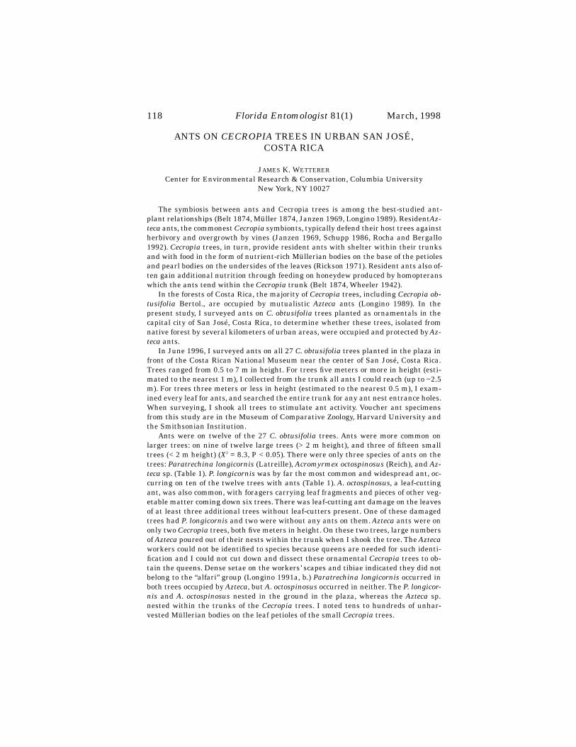
118
Florida Entomologist
81(1) March, 1998
ANTS ON
CECROPIA
TREES IN URBAN SAN JOSÉ,COSTA RICA
J
AMES
K. W
ETTERER
Center for Environmental Research & Conservation, Columbia UniversityNew York, NY 10027
The symbiosis between ants and Cecropia trees is among the best-studied ant-plant relationships (Belt 1874, Müller 1874, Janzen 1969, Longino 1989). Resident
Az-teca
ants, the commonest
Cecropia
symbionts, typically defend their host trees againstherbivory and overgrowth by vines (Janzen 1969, Schupp 1986, Rocha and Bergallo1992).
Cecropia
trees, in turn, provide resident ants with shelter within their trunksand with food in the form of nutrient-rich Müllerian bodies on the base of the petiolesand pearl bodies on the undersides of the leaves (Rickson 1971). Resident ants also of-ten gain additional nutrition through feeding on honeydew produced by homopteranswhich the ants tend within the
Cecropia
trunk (Belt 1874, Wheeler 1942).In the forests of Costa Rica, the majority of
Cecropia
trees, including
Cecropia ob-tusifolia
Bertol., are occupied by mutualistic
Azteca
ants (Longino 1989). In thepresent study, I surveyed ants on
C. obtusifolia
trees planted as ornamentals in thecapital city of San José, Costa Rica, to determine whether these trees, isolated fromnative forest by several kilometers of urban areas, were occupied and protected by
Az-teca
ants.In June 1996, I surveyed ants on all 27
C. obtusifolia
trees planted in the plaza infront of the Costa Rican National Museum near the center of San José, Costa Rica.Trees ranged from 0.5 to 7 m in height. For trees five meters or more in height (esti-mated to the nearest 1 m), I collected from the trunk all ants I could reach (up to ~2.5m). For trees three meters or less in height (estimated to the nearest 0.5 m), I exam-ined every leaf for ants, and searched the entire trunk for any ant nest entrance holes.When surveying, I shook all trees to stimulate ant activity. Voucher ant specimensfrom this study are in the Museum of Comparative Zoology, Harvard University andthe Smithsonian Institution.
Ants were on twelve of the 27
C. obtusifolia
trees. Ants were more common onlarger trees: on nine of twelve large trees (> 2 m height), and three of fifteen smalltrees (< 2 m height) (
X
2
= 8.3, P < 0.05). There were only three species of ants on thetrees:
Paratrechina longicornis
(Latreille),
Acromyrmex octospinosus
(Reich), and
Az-teca
sp. (Table 1).
P. longicornis
was by far the most common and widespread ant, oc-curring on ten of the twelve trees with ants (Table 1).
A. octospinosus
, a leaf-cuttingant, was also common, with foragers carrying leaf fragments and pieces of other veg-etable matter coming down six trees. There was leaf-cutting ant damage on the leavesof at least three additional trees without leaf-cutters present. One of these damagedtrees had
P. longicornis
and two were without any ants on them.
Azteca
ants were ononly two
Cecropia
trees, both five meters in height. On these two trees, large numbersof
Azteca
poured out of their nests within the trunk when I shook the tree. The
Azteca
workers could not be identified to species because queens are needed for such identi-fication and I could not cut down and dissect these ornamental
Cecropia
trees to ob-tain the queens. Dense setae on the workers’ scapes and tibiae indicated they did notbelong to the “alfari” group (Longino 1991a, b.)
Paratrechina longicornis
occurred inboth trees occupied by
Azteca
, but
A. octospinosus
occurred in neither. The
P. longicor-nis
and
A. octospinosus
nested in the ground in the plaza, whereas the
Azteca
sp.nested within the trunks of the
Cecropia
trees. I noted tens to hundreds of unhar-vested Müllerian bodies on the leaf petioles of the small
Cecropia
trees.

Scientific Notes
119
The most numerous ants on the
C. obtusifolia
trees in urban San José were
P. lon-gicornis
.
P. longicornis
is called the “crazy” ant because of the fast, jerky movementsof the workers. This species is of Old World origin, but is now one of the most commontramp ants throughout the tropics and subtropics, usually associated with distur-bance, e.g., in disturbed natural environments, in urban environments, and even onships (Weber 1940, Smith 1965, Miller 1994, Ferster and Prusak 1994, Klotz et al.1995; Delabie et al. 1995).
P. longicornis
dominates a variety of disturbed habitats, in-cluding the Dry Tortugas, the highly-exposed, outermost islands of the Florida Keys(Hölldobler and Wilson 1990) and the disturbed artificial ecosystems of Biosphere 2,a 1.28-hectare greenhouse structure outside Tucson, Arizona (Wetterer et al. 1997). InBiosphere 2, the main source of carbohydrates for
P. longicornis
was honeydew pro-duced by homopterans (Wetterer et al. 1997). This also appeared to be the major foodsource of tramp ants on
Cecropia
trees in Hawaii (Wetterer 1997), and probably in thepresent study as well.
A. octospinosus
foragers were also common on the San José
Cecropia
trees. Theseleaf-cutting ants are native to Central America, South America, and the Caribbean(Weber 1972). They seem to do well in disturbed urban habitats and are also commonin Panama City, Panama (personal observation). In the both the wet and dry forestsof Costa Rica,
A. octospinosus
foragers typically cut the leaves of small herbaceousplants, fallen flowers, and fallen fruit parts (Wetterer 1991, Wetterer et al. 1998).They appear to be opportunists, however, and will cut the leaves and flowers of plan-tation trees (Lewis 1975) and even collect insect frass (Wetterer et al. 1998).
Azteca
ants occupied only two 5-m
Cecropia
trees in the museum plaza, 7% of thetrees studied. This is a much lower occupancy rate than in natural habitats in CostaRica. For example, Longino (1989) found that on transects through the Arenal andMonteverde areas (400-1500 m elevation), obligate
Azteca
mutualists occupied 86% of
T
ABLE
1. A
NTS
ON
TWELVE
C
ECROPIA
OBTUSIFOLIA
TREES
IN
THE
PLAZA
IN
FRONT
OFTHE
C
OSTA
R
ICAN
N
ATIONAL
M
USEUM
, S
AN
J
OSÉ
, C
OSTA
R
ICA
. I
N
ADDITION
,
FIFTEEN
C.
OBTUSIFOLIA
TREES
HAD
NO
ANTS
(
ONE
6
M
,
TWO
5
M
,
THREE
1.5
M
,
SEVEN
1
M
,
AND
TWO
0.5
M
TALL
). C
RAZY
ANT
=
P
ARATRECHINA
LONGICOR-NIS
; L
EAF
-
CUTTER
=
A
CROMYRMEX
OCTOSPINOSUS
,
AND
A
ZTECA
=
A
ZTECA
SP
.; X =
ANTS
PRESENT
;
D
=
LEAF
-
CUTTING
ANT
DAMAGE
OBSERVED
.
Tree height (m) Crazy ant Leaf-cutter
Azteca
1 7 X X2 6 X X3 6 X4 6 X5 5 X X6 5 X X7 5 X X8 3 X 9 2 X D
10 1.5 X X11 1.5 X12 1.5 X

120
Florida Entomologist
81(1) March, 1998
all
Cecropia
trees. The majority of the trees without
Azteca
were saplings. Questionsarise as to how the
Azteca
ants came to occupy two
Cecropia
trees in San José, so faraway from native forest. It seems likely that the
Azteca
ants had already colonizedthese trees before they were transplanted to San José. Longino (1989) found that mostunoccupied
Cecropia
trees showed evidence within their trunks of
Azteca
colonies thathad died. It is unknown whether any of the unoccupied trees in San José once housed
Azteca
colonies.The
Azteca
ants showed no evidence of excluding crazy ants from their host
Cecro-pia
trees. It may be that the
Azteca
ants do not recognize this non-indigenous ant asa threat. Alternatively, the
Azteca
colonies may not be capable of fending off the largenumbers of crazy ants. On the other hand, the
Azteca
ants in San José appeared to beexcluding the native leaf-cutting ants.
A. octospinosus
foragers were not attacking ei-ther of trees occupied by
Azteca
, but were attacking or had attacked at least nine ofthe 25
Cecropia
trees without
Azteca
.It would be interesting to census these
Cecropia
trees at a later date to determinewhether the
Azteca
ants persist in the trees and spread to others, or whether crazyants exterminate them as they appear to have done with ant species in other dis-turbed environments (Hölldobler and Wilson 1990, Wetterer et al. 1997, see also Zen-ner Polania 1994).
Earlier studies have indicated that
C. obtusifolia
trees are not obligately depen-dent on ants (see Wetterer 1997).
C. obtusifolia
has been introduced to Hawaii, appar-ently as an ornamental (Wagner et al. 1990).
Azteca
ants have not been recorded inHawaii. Either they did not accompany the
Cecropia
trees to Hawaii or did not survivethere. Still, the
Cecropia
trees are thriving in disturbed lowland forests of Hawaii inthe complete absence of mutualist
Azteca
ants (Wetterer 1997). In part, this successmay be because most of
C. obtusifolia
’s Neotropical herbivores and competitors are ab-sent in Hawaii. A similar absence may explain their survival in urban San José.
I thank M. Wetterer and J. C. Morales for comments on this manuscript; S. Coverfor identifying the ants; E. Olson for inviting me to teach on his OTS 96-3 course. Fi-nancial support was provided by the Organization for Tropical Studies and ColumbiaUniversity.
S
UMMARY
Three species of ants inhabited 12 of 27
Cecropia
trees in urban San José, CostaRica: crazy ants (
Paratrechina longicornis
) in ten, leaf-cutting ants (Acromyrmexoctospinosus) in six, and “Cecropia” ants (Azteca spp.) in only two. Occupancy rate byAzteca was much lower than in local forest Cecropia. P. longicornis inhabited both Az-teca-occupied trees, but A. octospinosus occurred in neither.
REFERENCES CITED
BELT, T. 1874. The naturalist in Nicaragua. University of Chicago Press. Chicago, IL.DELABIE, J. H. C., I. C. DO NASCIMENTO, P. PACHECO, AND A. B. CASIMIRO. 1995. Com-
munity structure of house-infesting ants (Hymenoptera: Formicidae) in south-ern Bahia, Brazil. Florida Entomol. 78: 264-270.
FERSTER, B., AND Z. PRUSAK. 1994. A preliminary checklist of the ants (Hymenoptera:Formicidae) of Everglades National Park. Florida Entomol. 77: 508-512.
HÖLLDOBLER, B., AND E. O. WILSON. 1990. The Ants. Harvard University Press, Cam-bridge, MA.
JANZEN, D. H. 1969. Allelopathy by myrmecophytes: the ant Azteca as an allelopathicagent of Cecropia. Ecology 50: 147-153.

Scientific Notes 121
KLOTZ, J. H., J. R. MANGOLD, K. M. VAIL, L. R. DAVIS, JR., AND R. S. PATTERNSON.1995. A survey of the urban pest ants (Hymenoptera: Formicidae) of PeninsularFlorida. Florida Entomol. 78: 109-118.
LEWIS, T. 1975. Colony size, density and distribution of the leaf-cutting ants, Ac-romyrmex octospinosus (Reich) in cultivated fields. Trans. Royal Entomol. Soc.127: 51-64
LONGINO, J. T. 1989. Geographic variation and community structure in an ant-plantmutualism: Azteca and Cecropia in Costa Rica. Biotropica 21: 126-132.
LONGINO, J. T. 1991a. Taxonomy of Cecropia-inhabiting Azteca ants. J. Natur. Hist.25: 1571-1602.
LONGINO, J. T. 1991b. Azteca ants in Cecropia trees: taxonomy, colony structure, andbehavior. In: C. R. Huxley and D. F. Cutler (Eds.). Ant-plant interactions. pp.271-288. Oxford Press, New York, NY.
MILLER, S. E. 1994. Dispersal of plant pests into the Virgin Islands. Florida Entomol.77: 520-521.
MÜLLER, F. 1874. The habits of various insects. Nature 10: 102-103.RICKSON, F. R. 1971. Glycogen plastids in Müllerian body cells of Cecropia peltata - a
higher green plant. Science 173: 344-347.ROCHA, C. F. D., AND H. G. BERGALLO. 1992. Bigger ant colonies reduce herbivore res-
idence time on leaves of an ant-plant: Azteca muelleri vs. Coelomera ruficornison Cecropia pachystachya. Oecologia 91: 249-252.
SCHUPP, E. W. 1986. Azteca protection of Cecropia: ant occupation benefits juveniletrees. Oecologia 70: 379-385.
SMITH, M. R. 1965. House-infesting Ants of the Eastern United States. Their Recog-nition, Biology, and Economic Importance. Tech.l Bull. 1326, Agric. Res. Serv.,United States Depart. Agric.
WAGNER, W. L., D. R. HERBST, AND S. H. SOHMER. 1990. Manual of the floweringplants of Hawaii. vol. 1. University of Hawaii Press, Honolulu, HI.
WEBER, N. A. 1940. Ants on a Nile River steamer. Ecology 21: 292-293.WEBER, N. A. 1972. Gardening ants: the attines. American Philosophical Society. Phil-
adelphia, PA.WETTERER, J. K. 1991. Foraging ecology of the leaf-cutting ant, Acromyrmex octospi-
nosus, in a Costa Rican rain forest. Psyche 98: 361-371.WETTERER, J. K. 1997. Ants on Cecropia in Hawaii. Biotropica 29: 128-132.WETTERER, J. K., C. DUNNING, M. YOSPIN, AND A. HIMLER. 1997. Invertebrate Diver-
sity and Ecology in Biosphere 2. Sonoran Arthropod Studies Institute, Tucson,AZ. pp. 119-122. Invertebrates in Captivity Conference Proceedings.
WETTERER, J. K., D. S. GRUNER, AND J. E. LOPEZ. 1998. Foraging and nesting ecologyof Acromyrmex octospinosus in a Pacific dry forest of Costa Rica. In press, Flor-ida Entomol.
WHEELER, W. M. 1942. Studies of neotropical ant-plants and their ants. Bull.Mus.Compar. Zool. 90: 1-263.
ZENNER POLANIA, I. 1994. Impact of Paratrechina fulva on other ant species. pp 121-132. In: Exotic ants: Biology, impact, and control of introduced species, West-view Press, Boulder, CO.
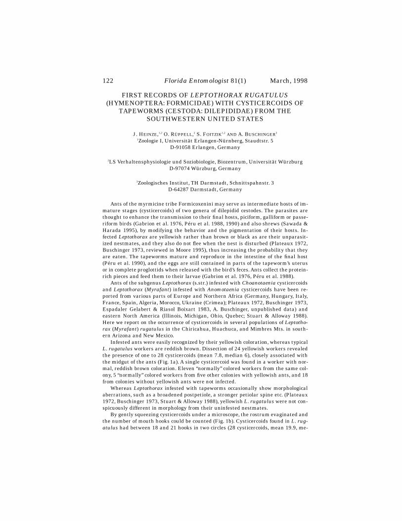
122
Florida Entomologist
81(1) March, 1998
FIRST RECORDS OF
LEPTOTHORAX RUGATULUS
(HYMENOPTERA: FORMICIDAE) WITH CYSTICERCOIDS OF
TAPEWORMS (CESTODA: DILEPIDIDAE) FROM THE SOUTHWESTERN UNITED STATES
J. H
EINZE
,
1,2
O. R
ÜPPELL
,
2
S. F
OITZIK
1,2
AND
A. B
USCHINGER
3
1
Zoologie I, Universität Erlangen-Nürnberg, Staudtstr. 5D-91058 Erlangen, Germany
2
LS Verhaltensphysiologie und Soziobiologie, Biozentrum, Universität WürzburgD-97074 Würzburg, Germany
3
Zoologisches Institut, TH Darmstadt, Schnittspahnstr. 3D-64287 Darmstadt, Germany
Ants of the myrmicine tribe Formicoxenini may serve as intermediate hosts of im-mature stages (cysticercoids) of two genera of dilepidid cestodes. The parasites arethought to enhance the transmission to their final hosts, piciform, galliform or passe-riform birds (Gabrion et al. 1976, Péru et al. 1988, 1990) and also shrews (Sawada &Harada 1995), by modifying the behavior and the pigmentation of their hosts. In-fected
Leptothorax
are yellowish rather than brown or black as are their unparasit-ized nestmates, and they also do not flee when the nest is disturbed (Plateaux 1972,Buschinger 1973, reviewed in Moore 1995), thus increasing the probability that theyare eaten. The tapeworms mature and reproduce in the intestine of the final host(Péru et al. 1990), and the eggs are still contained in parts of the tapeworm’s uterusor in complete proglottids when released with the bird’s feces. Ants collect the protein-rich pieces and feed them to their larvae (Gabrion et al. 1976, Péru et al. 1988).
Ants of the subgenus
Leptothorax
(s.str.) infested with
Choanotaenia
cysticercoidsand
Leptothorax
(
Myrafant
) infested with
Anomotaenia
cysticercoids have been re-ported from various parts of Europe and Northern Africa (Germany, Hungary, Italy,France, Spain, Algeria, Morocco, Ukraine (Crimea); Plateaux 1972, Buschinger 1973,Espadaler Gelabert & Riasol Boixart 1983, A. Buschinger, unpublished data) andeastern North America (Illinois, Michigan, Ohio, Quebec; Stuart & Alloway 1988).Here we report on the occurrence of cysticercoids in several populations of
Leptotho-rax
(
Myrafant
)
rugatulus
in the Chiricahua, Huachuca, and Mimbres Mts. in south-ern Arizona and New Mexico.
Infested ants were easily recognized by their yellowish coloration, whereas typical
L. rugatulus
workers are reddish brown. Dissection of 24 yellowish workers revealedthe presence of one to 28 cysticercoids (mean 7.8, median 6), closely associated withthe midgut of the ants (Fig. 1a). A single cysticercoid was found in a worker with nor-mal, reddish brown coloration. Eleven “normally” colored workers from the same col-ony, 5 “normally” colored workers from five other colonies with yellowish ants, and 18from colonies without yellowish ants were not infected.
Whereas
Leptothorax
infested with tapeworms occasionally show morphologicalaberrations, such as a broadened postpetiole, a stronger petiolar spine etc. (Plateaux1972, Buschinger 1973, Stuart & Alloway 1988), yellowish
L. rugatulus
were not con-spicuously different in morphology from their uninfested nestmates.
By gently squeezing cysticercoids under a microscope, the rostrum evaginated andthe number of mouth hooks could be counted (Fig. 1b). Cysticercoids found in
L. rug-atulus
had between 18 and 21 hooks in two circles (28 cysticercoids, mean 19.9, me-

Scientific Notes
123
dian 20; compared to
Anomotaenia brevis
with 17 to 22, rarely 26 hooks, and
Choanotaenia
with 18 to 27 hooks, Buschinger 1973, Gabrion et al. 1976). One excep-tional cysticercoid from
L. rugatulus
had one circle consisting of 18 large hooks and asecond circle of 13 small hooks. Cysticercoids from
L. rugatulus
were similar in sizeto those from Europe (48 cysticercoids in
L. rugatulus
: mean 224
µ
m, median 216
µ
m,range 180-290
µ
m; compared to A. brevis 220-250
µ
m and
Choanotaenia
170-210
µ
m,Buschinger 1973, Gabrion et al. 1976), but the average length of the hooks by far ex-ceeded that of European specimens (in
L. rugatulus
: 60
µ
m; compared to A. brevis23.5-28.5
µ
m and
Choanotania
40-45
µ
m). Overall, cysticercoids from
L. rugatulus
appear to belong to a taxon different from both European species, but closer relatedto
Anomotaenia
than to
Choanotaenia
. The final host of the tapeworm is unknown.In addition to yellowish workers, several colonies also contained yellowish dealate
queens (Table 1). None of 12 dissected yellowish queens was found to be inseminated,and none of approximately 90 dissected, inseminated queens was infected, suggestingthat infestation somehow interferes with the queens’ sexual behavior. The ovaries offive yellowish virgin queens contained 1 to 3 developing eggs, but according to the ab-sence of corpora lutea, they had only recently become fertile.
In the Chiricahua, Huachuca, and Mimbres Mts., between 5% and 20% of the in-spected colonies contained yellowish workers (Table 1), and in single colonies, up to50% of the workers were infected (mean appr. 10%). Infestation by tapeworm cysticer-coids thus appears to be a rather common phenomenon in the mountain ranges ofSouthern Arizona and New Mexico. No cysticercoids, however, were found in
L. rug-atulus
from several other populations in Arizona, New Mexico, and Colorado. Simi-larly, a species of
Leptothorax
(s.str.), regularly co-occurring with
L. rugatulus
inSouthern Arizona and New Mexico (an as yet undescribed member of the
L.
(s.str.)
muscorum
complex referred to as
Leptothorax
(s.str.) F, Heinze 1989), was never foundinfested with cysticercoids. The apparent restriction of the
Anomotaenia
-like cysticer-coids to
L. rugatulus
is consistent with the hypothesis that cysticercoids of dilepididtapeworms are limited to host species from a single
Leptothorax
subgenus (Busch-inger 1973, Péru et al. 1990).
Fig. 1. a) Four cysticercoids of a dilepidid cestode associated with the midgut of aworker of the ant Leptothorax rugatulus. b) Cysticercoid after devagination of the ros-trum. The hooks are clearly visible.

124
Florida Entomologist
81(1) March, 1998
Field work in the USA was supported by the Deutsche Forschungsgemeinschaft(Heisenberg-grant He 1623/6-1 and Graduiertenkolleg “Arthropodenverhalten”). Thestudies were performed in part in the Southwestern Research Station of the AmericanMuseum of Natural History, Portal, AZ.
S
UMMARY
Cysticercoids of a dilepidid cestode were found by dissection of aberrant yellowishworkers and virgin queens of the ant
Leptothorax rugatulus
from several mountainranges in the southwestern United States. The cestode was not positively identified,but cysticercoids are similar to those of the genus
Anomotaenia
previously found in re-lated ants.
T
ABLE
1. O
CCURRENCE
OF
L
EPTOTHORAX
RUGATULUS
ANTS
INFECTED
WITH
CYSTICER-COIDS
OF
A
DILEPIDID
TAPEWORM
. T
HE
TABLE
GIVES
THE
RATIOS
OF
YELLOW-ISH
DEALATE
QUEENS
AND
WORKERS
PER
COLONY
TO
THE
TOTAL
NUMBER
OFDEALATE
QUEENS
AND
WORKERS
,
AND
OF
COLONIES
CONTAINING
INFECTEDINDIVIDUALS
TO
THE
TOTAL
NUMBER
OF
COLONIES
COLLECTED
PER
POPULA-TION
.
Mountain Range, County, State
Collecting Site, Elevation
yellowish individuals per colony
colonies with
yellowish individualsqueens workers
Chiricahua Mts., Cochise Co., AZ
Barefoot Lookout, 2630m
0/2 4/180/16 14/174 4/303/25 16/1370/3 6/62
FSR 40, 0.8km W of Onion Saddle, 2200m
17/19 0/51 1/13
FSR 40, 1.5km W of Onion Saddle, 2100m
0/1 3/139 2/250/18 3/115
Pinery Campground, 2100m
42/43 49/156 1/5
Onion Saddle, 2280m 0/5 8/1040/1 4/330/1 3/21 4/840/0 1/31
Rustler Park, 2580m 0/7 1/46 1/5Morse CanyonTrailhead, 1980m
1/4 10/342 1/11
Huachuca Mts.,Cochise Co., AZ
Carr Canyon Road, 2400m
0/2 1/79 1/20
Mimbres Mts., Grant Co., NM
4.8km west of Emory Passm 2200
0/1 2/300/1 6/17 3/125/6 1/17
total number, including additional populations of
L. rugatulus
in New Mexico, Arizona, and Colorado18/523
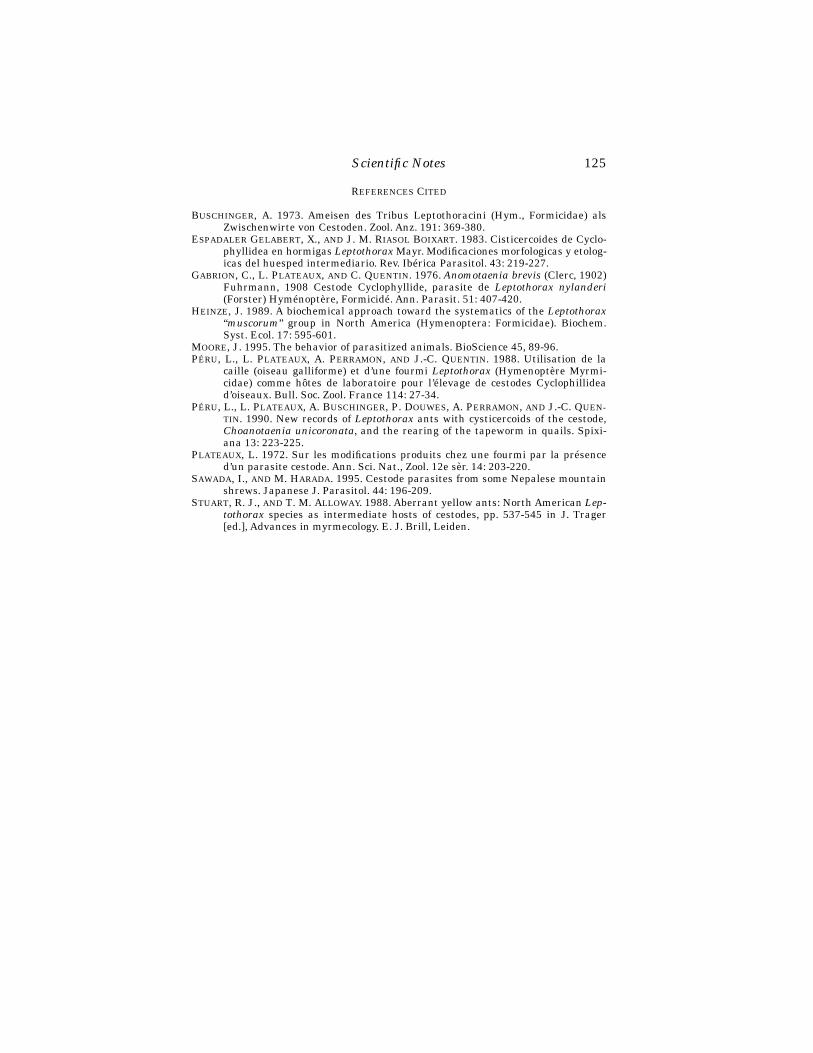
Scientific Notes
125
R
EFERENCES
C
ITED
B
USCHINGER
, A. 1973. Ameisen des Tribus Leptothoracini (Hym., Formicidae) alsZwischenwirte von Cestoden. Zool. Anz. 191: 369-380.
E
SPADALER
G
ELABERT
, X.,
AND
J. M. R
IASOL
B
OIXART
. 1983. Cisticercoides de Cyclo-phyllidea en hormigas
Leptothorax
Mayr. Modificaciones morfologicas y etolog-icas del huesped intermediario. Rev. Ibérica Parasitol. 43: 219-227.
G
ABRION
, C., L. P
LATEAUX
,
AND
C. Q
UENTIN
. 1976.
Anomotaenia brevis
(Clerc, 1902)Fuhrmann, 1908 Cestode Cyclophyllide, parasite de
Leptothorax nylanderi
(Forster) Hyménoptère, Formicidé. Ann. Parasit. 51: 407-420.H
EINZE
, J. 1989. A biochemical approach toward the systematics of the
Leptothorax
“
muscorum
” group in North America (Hymenoptera: Formicidae). Biochem.Syst. Ecol. 17: 595-601.
M
OORE
, J. 1995. The behavior of parasitized animals. BioScience 45, 89-96.P
ÉRU
, L., L. P
LATEAUX
, A. P
ERRAMON
,
AND
J.-C. Q
UENTIN
. 1988. Utilisation de lacaille (oiseau galliforme) et d’une fourmi
Leptothorax
(Hymenoptère Myrmi-cidae) comme hôtes de laboratoire pour l’élevage de cestodes Cyclophillidead’oiseaux. Bull. Soc. Zool. France 114: 27-34.
P
ÉRU
, L., L. P
LATEAUX
, A. B
USCHINGER
, P. D
OUWES
, A. P
ERRAMON
,
AND
J.-C. Q
UEN-TIN
. 1990. New records of
Leptothorax
ants with cysticercoids of the cestode,
Choanotaenia unicoronata
, and the rearing of the tapeworm in quails. Spixi-ana 13: 223-225.
P
LATEAUX
, L. 1972. Sur les modifications produits chez une fourmi par la présenced’un parasite cestode. Ann. Sci. Nat., Zool. 12e sèr. 14: 203-220.
S
AWADA, I., AND M. HARADA. 1995. Cestode parasites from some Nepalese mountainshrews. Japanese J. Parasitol. 44: 196-209.
STUART, R. J., AND T. M. ALLOWAY. 1988. Aberrant yellow ants: North American Lep-tothorax species as intermediate hosts of cestodes, pp. 537-545 in J. Trager[ed.], Advances in myrmecology. E. J. Brill, Leiden.

126
Florida Entomologist
81(1) March, 1998
BOOK REVIEWS
K
UZNETSOV
, V. N. 1997. Ladybeetles of the Russian Far East. Center for System-atic Entomology; Gainesville, Florida. Memoir 1, 248 p. ISBN 1-877743-25-5. Hard-back. Price per copy $58 ($29 to members of CSE) plus $1.65 postage and handling toUSA addresses, from The Treasurer, Center for Systematic Entomology, P.O. Box147100, Gainesville, FL 32614-7100.
This is the first volume of the Memoir Series of the Center for Systematic Ento-mology (CSE), an organization which is a direct support group for the Florida StateCollection of Arthropods, the 6th largest public insect collection in the USA, with 7.5million specimens housed. The reduced price to members is intended to attract newmembers to CSE for an annual subscription of US$35.00 (for which members receiveissues of
Insecta Mundi
, a quarterly journal of insect systematics, and a quarterlynewsletter). Prospective new members should write to Dr. Avas Hamon, Treasurer,Center for Systematic Entomology [address above]. CSE supports insect systematicsnot only in Florida, but throughout the world. There is no better bargain in insect sys-tematics in the USA: where else can systematists publish their works in a reputablepeer-reviewed journal without page charges?
This book was published in English thanks to a facilitation grant from the Na-tional Biological Control Institute, USDA-APHIS. Translation from Russian wasmade by S. S. Ishevsky and M. S. Ishevskaya. It provides keys, species descriptions,illustrations (drawings), and some information on food and behavior of the coccinellidspecies of the Russian Far East. It draws its information from numerous publicationsin Russian (and some in other languages) as well as the decades of experience of itsauthor Victor Kuznetsov, including a book that he published in Russian in 1993. Thus,it is indispensable to all serious students of Coccinellidae. It does not fail to point out,that members of the genera
Halyzia
,
Vidibia
and
Psyllobora
(tribe Psylloborini) feedon mildew, and that
Henosepilachna
,
Epilachna
,
Subcoccinella
and
Cynegetis
(sub-family Epilachninae) are phytophagous, and some of them are important pests. Itdoes not discuss the controversy caused by introduction of
Harmonia axyridis
into theUSA as a generalist predator of aphids. It offers essential information to biologicalcontrol practitioners who would import predatory coccinellid species from the RussianFar East to control pest Homoptera.
Three other publishers in Gainesville specialize in entomological books. One ofthem is The Sandhill Crane Press, which in 1996 published “The Beetles of northeast-ern North America” by N. M. Downie and R. H. Arnett, Jr., 2 vols. (for a review seeFlorida Entomologist 79: 471-473). The second is Associated Publishers, which in1995 published Memoirs on Entomology, International, vol. 3 “Rove beetles of the sub-tribe Philonthina of America north of Mexico (Coleoptera: Staphylinidae), classifica-tion, phylogeny and taxonomic revision” by A. Smetana (for a review, see FloridaEntomologist 79: 81-82). The third is Scientific Publishers, which in 1996 published“Damselflies of North America” by M. J. Westfall, Jr. and M. L. May. The Florida En-tomological Society publishes Florida Entomologist but no books. The Center for Sys-tematic Entomology is a new entrant in the Gainesville book-publishing scene.
The introductory materials of the book occupy pages that are numbered in Romannumerals i-xii, but which are also (curiously) considered to be pages 1-12. Pages 13-72 then consist of an historical review of Coccinellidae of the area of interest, methods,many pages on general morphology, including a morphology of larvae, and sections onbiology and ecology, on natural enemies, and on employment of Coccinellidae in bio-

Book Reviews
127
logical control of homopterous pests in the former USSR. Then comes the major partof the text, on taxonomy, down to the species level, and including notes on food, habi-tat, and seasonality. This is extremely useful to anyone concerned with taxonomy ofCoccinellidae, including persons concerned with importation of Russian species for bi-ological control purposes.
Literature cited occupies p. 229-244. It translates Russian titles of papers andbook chapters and books into English, and transliterates names of Russian journalsfrom Cyrillic into Roman characters. For readers who need to go no further (the vastmajority), this is ideal: the work of translating, summarizing, and integrating theRussian literature has been done.
For readers who are taxonomists and bibliophiles and want to go further by obtain-ing copies of items cited, this bibliography will cause problems because it defies con-ventions. If you can read the languages of the original references, you will note errorsand peculiarities in the bibliography. Your inter-library-loan librarian also will havetrouble with it if you try to request copies of the articles cited. For example, it omitsall diacritical marks (accents) in French, German, Spanish, Swedish, and Italian. Foranother example, where the latest C.B.E. style manual recommends that book cita-tions should list the name of the city of publication and name of publisher (in that or-der, separated by a semi-colon, and using the English name of the place of publication)the reader will not find such a scheme in citations of this bibliography such as “Mos-cow, Leningrad” or “New York, London” or “Hafniae” or “Lipsiae” or “Wien” (the words“Hafniae” and “Lipsiae” are the genitive case of the Latin names of the cities Copen-hagen and Leipzig). It seldom gives the name of the publisher. Where it transliteratesRussian into English, it frequently slips into non-standard conventions, for exampleby writing as “entomologicheskoje” rather than the stan-dard “entomologicheskoe” for no apparent reason. Where it transliterates the Russianword “ ” (meaning “and”) into the Roman character “i” it uses a capital “I”, whichlooks very strange among a string of words all in lower case. On the other hand, allthese foreign-language publications will be useful only to people who can read the lan-guages, and they—potential users—will simply have to explain the correct spellingsto their inter-library-loan librarians—to the rest of the world it does not matter.
The final chapter, p. 245-248, is called a taxonomic index but is, in fact, a usefulnon-alphabetical classification that lists page numbers of the main entries in thechapter on taxonomy. An additional, alphabetical index would have given additionalvalue especially because it could have included information from all chapters.
J. H. FrankEntomology & Nematology Dept.University of FloridaGainesville, FL 32611-0630
Book Reviews
127
W
HITE
, I. M.,
AND
D. L. H
ANCOCK
. 1997. Indo-Australasian Dacini Fruit Flies.(Computer Aided Biological Identification Key). International Institute of Entomol-ogy, London. ISBN 0-85199-171-0. Compact diskette. $320.00 (for single user).
Among the tephritid fruit flies, the Dacini comprise one of the largest and mosttaxonomically challenging groups. Here Dacini refer largely to the genera
Bactrocera
and
Dacus
, which include many important tropical and subtropical fruit and vegeta-ble pests, such as the Oriental, melon, and Queensland fruit flies. Accurate identifica-tion is a problem, especially when keys are written for females, but interceptions andsurveys are usually of males taken in pheromone traps.
128
Florida Entomologist
81(1) March, 1998
This CD-ROM conveniently brings together in one place an enormous amount ofinformation on the dacines. It includes four programs: Key to the Dacini of the Indo-Australasian region, which forms the heart of the offering; CABIKEY Help with de-tails on system requirements, general workings and capabilities of the software, anda simulated identification session; Dacini Help with a glossary of about 200 terms cov-ering fruit fly morphology plus a few nomenclatural and ecological terms; and an Un-install utility. Recommended computer system specifications include Windows 3.1x orWindows 95 operating system, 8 MB of RAM (16 is ideal, 4 is adequate), and displayof 256 colors or more. The installation proceeded quickly and seamlessly on my Dell®486 running Windows 95 and its files consumed 1.32 MB of hard disk space (it may in-stall up to 4.73 MB of files).
The usual advantages of computer aided keys over dichotomous keys apply here;e.g., it is possible to circumvent certain characters in working through the key, and acomplete and consistently scored set of character states (a “description”) is viewablefor each taxon. The key covers 507 taxa and is based on a 66-character matrix includ-ing geographic range and host plants. Data are included for all valid species found inthe Oriental, Australasian and Pacific areas, and the Mascarene Islands. Special at-tention was paid to separation of the 68 species in the Oriental fruit fly (
Bactroceradorsalis
) complex. For this complex several characters are used that are not appliedto other species, such as measurements of the male aedeagus and female aculeus. To“complete” the system for worldwide coverage of dacine species of quarantine impor-tance, pest species from the Afrotropical region are also included. This is not a key toDacini of the world, as about 150 African non-pest species and over 100 other knownbut undescribed species are excluded. A complete world checklist is included as an ap-pendix.
The user-friendliness of the system is very high. Even upon first use, choice of the“standard” option allows one easily to work through an identification without resort-ing to help screens. This option automatically chooses the best characters and queriesthe user with a choice of available character states. The initial screens allow the userto limit the taxa or characters considered based on sex, geographic region, or pest sta-tus. For example, choice of only pest species reduces the list to 52 taxa. Each querypresents a very clear choice of character states with excellent supporting illustrations,in which character state differences are highlighted by use of color overlays on blackand white drawings, and text explanations. At any step in the process, it is possible toreview and alter previous character state choices, examine character illustrations, listremaining taxa, preview taxa to be included/excluded by a given character state se-lection, check the glossary, etc.
For pest taxa, there is significant accompanying text on taxonomy, host plants,identification characteristics, and distribution information that is largely verbatim ofthat in White & Elson-Harris (1994, Fruit Flies of Economic Significance, CABI, UK,601 p.) but updated with recent nomenclature, especially from Drew & Hancock(1994, The
Bactrocera dorsalis
complex of fruit flies in Asia, Bull. Ent. Res. Suppl. 2,68 p.). Good color photographs of actual specimens are provided for almost all pestspecies. Non-pest species receive only a short catalog entry stating synonymies, dis-tribution, and the specimens examined upon which the character matrix was built. Ahabitus drawing, distribution map, diagnosis and description (entire character stateset) is provided for every species.
Much of the information in the system can be printed out for more convenient us-age away from the computer. Especially useful to the working taxonomist is the pro-gram’s ability to generate a table of character states that vary among as many as fivetaxa and copy the table to a spreadsheet for printing. This is something that no tradi-tional key or monograph can do and is invaluable for doing a thorough inspection and
Book Reviews
129
comparison of specimens at the microscope. Descriptive text, distribution maps, glos-sary entries including labeled line drawings of morphological features, and even thecolor and SEM photos can all be printed out. Complete descriptions and diagnoses ofindividual species, however, cannot be exported for printing, nor can those screens beprinted that illustrate character state choices as one progresses through the key.
Within the Key program is a set of useful appendices. The aforementioned check-list of world Dacini (all described species as of late 1996) is not available elsewhere.Association Data (economically important host plants, arranged alphabetically by ge-nus and by common name with corresponding fruit fly denizens), Methods of Study,Morphology, and other appendices are modified versions of text in White & Elson-Har-ris (1994).
If the potential user is concerned only with pest species, e.g., a port identifier need-ing only to recognize pests of quarantine significance, one’s library budget is betterspent on White & Elson-Harris’s 1994 book, which contains nearly all the same infor-mation on pest dacines plus a whole lot more on other fruit fly pests and identificationof larvae. The book may not be high tech, but it’s $250 cheaper and a truly excellentreference. Museum curators, collectors, surveyors, and other taxonomists who arelikely to encounter a diverse assemblage of non-pest species will be most likely to fullyutilize and appreciate the wealth of character data presented here. This is a wonder-ful tool that is well-designed for easy use. Yet, using even this good a tool, one may stillarrive at an “answer” and nonetheless feel a nagging doubt about its veracity. Thenone must resort to even better tools—comprehensive collections and human experts!
Contribution No. 857, Bureau of Entomology, Nematology and Plant Pathology,Florida Department of Agriculture and Consumer Services.
Gary J. SteckDivision of Plant IndustryFlorida Department of Agriculture and Consumer ServicesGainesville, FL 32614-7100
Book Reviews
129
B
RODSKY
, A
NDREI
K. 1996. The Evolution of Insect Flight. Oxford UniversityPress, New York. xiv + 229 p. ISBN 0-19-850089-0. Paperback (first paperback edi-tion). $55.00.
In the preface to this translation of the original Russian hardback, the authorfrankly states that “This book is not for easy reading at idle moments.” I like that.Gaining an understanding of the physiology of flight, and indeed of any aspect of in-sect physiology, is serious business. Brodsky, Professor in the Department of Entomol-ogy of St. Petersburg University, St. Petersburg, Russia, has presented an in-depthanalysis of the basic principles of flight in part I, and the evolution of flight in Part II.Part I consists of 4 chapters, detailing structure of wings, the way wings work, theaerodynamics of flight, and the role of flight in insect behavior.
In chapters 5, 6, and 7 in Part II, the author describes models and ideas about theorigin of flight and wings in insects, with details and examples from mayflies, dragon-flies, stoneflies, and cockroaches. In chapter 8 he discusses the differences in flight offour-winged insects and two-winged insects, with examples from Psocoptera, Ho-moptera, and Heteroptera. Chapter 9 is a discussion of changes in wing structure,thorax, hinging of wings, and musculature as insects evolved into more diverse forms.What, for example, are beetles to do with the hard, inflexible elytra (forewings) duringflight? The answer is that all Coleoptera except the rose chafers (
Cetonia
spp.) spreadthe elytra at some angle to the long axis of the body, and often swing them in small
130
Florida Entomologist
81(1) March, 1998
arcs at the frequency of the hindwings. Tiger beetles, which are fast fliers with rapidtake-off from a surface, hold the forewings nearly at right angles to the body axis andsupport them with the first pair of legs “. . . like the struts of a high-wing monoplane.”Especially, but not exclusively, during evolution of Diptera and Hymenoptera changesoccurred in wing structure, musculature, and thoracic structure that enabled muchfaster flight and greater maneuverability, such as hovering and frequent changes indirection. Maneuverability during flight made possible swarming behavior in manysmall species of Diptera, some of which fly equally well forward, backward, or side-ways.
Just as high speed flight is essential to the behavior and biology of flies and bees,so is slow flapping and gliding flight important to some species of Neuroptera, andmany species of Lepidoptera. I was surprised to learn that up to 40-50% of flight timein some Lepidoptera is gliding flight. Changes in wing shape in Lepidoptera aretraced from the narrowing of the wing plane and development of long fringes on thewings of some small moths, to the large wings of some butterflies and moths. Glidingflight required changes in wing structure and in the articulation of the wings to thethorax. Airflow, vortices, and drag forces are discussed in relationship to wing struc-ture and shape. I found very interesting a physiological explanation for the evolutionof the tails of
Papilio
spp. as modifying the drag of air vortices coming off the mainsurface of the wings, and thus enabling the butterflies “to use greater angles of attackduring gliding without significant increase in drag.” Other gliding butterflies that donot have tails solved the problem of drag in other ways.
The last chapter is a general summary of the evolution of wing structure, the ax-illary apparatus that hinges the wing to the thorax, and muscles that power flight.There are 3+ pages of Postscript as a sort of brief summary of principles without tech-nical terms and jargon.
Having to grapple with many technical terms is one of the features of the book thatmake it, as the author warns, heavy reading. These terms include thoracic anatomy,wing venation, the names of numerous sclerites at the base of the wings, and taxo-nomic groupings. Many of the anatomical terms are abbreviated, and a list of abbre-viations is given at the front of the book. The book is well illustrated with diagrams,and line drawings of thoracic structure, wing structure and venation, and muscula-ture. There are nine pages of references, many of which are in the older literature, andin foreign publications probably less well known to many entomologists. There is ataxonomic index for those who might like to know what the book contains that is rel-evant to a particular insect or group, and a subject index.
I recommend this book to those teaching introductory entomology, insect anatomy,and insect physiology. It is packed with useful information, and is well worth seriousstudy.
James L. NationProfessorDepartment of Entomology & NematologyUniversity of FloridaGainesville, FL 32611-0620
Book Reviews
131
S
MITH
, D., G. A. C. B
EATTIE
,
AND
R. B
ROADLEY
(eds.). 1997. Citrus Pests and TheirNatural Enemies. Integrated Pest Management in Australia. Department of PrimaryIndustries, Brisbane, Queensland, Australia. xvi + 282 p. ISBN 0 7242 6695 X. Paper-back. Australian $75.
The high quality of the cover photograph—eggs, nymphs, and adult of a spined cit-rus bug in beautiful color—immediately caught my eye. As I paged through the book,I realized it was just one of over 400 wonderful color photographs in this book on cit-rus pests and their natural enemies in Australia! The photographs are of unusuallyhigh quality and justify the price of the book. There is much more to admire about thisbook because it has several features that make it particularly useful, even to pestmanagers in other countries.
The book includes descriptions of the biology and damage caused by over 100 cit-rus pests, a taxonomic key to the parasitic wasps of scales and mealybugs in Austra-lian citrus groves, a good index, and a list of relevant references. It begins with aglossary of terms, a brief introduction of the Australian citrus industry, a descriptionof the citrus varieties grown, and the weather conditions in the major citrus growingregions. The book deals with tree diseases, insects, mites, nematodes, snails, and(even) a pest spider! Over 100 pest species are described, and their frequency is cate-gorized as “Major,” “Occasionally important,” or “Minor” in each of the growing regionsusing color-coded maps. I decided to focus on one section, describing the mite pests ofcitrus, to determine whether I could use the manual as intended.
Citrus in Australia has several pest mites, including the brown citrus rust mite,
Tegolophus australis
. Photographs show all the life stages of the mite, with discrimi-nating characters to differentiate it from the citrus rust mite,
Phyllocoptruta oleivora
,also a pest in Australia. The photographs were sufficiently good that I could use themto identify the mites in the field using a hand lens. A map showed the importance anddistribution of the brown citrus rust mite in Australia. A diagram of the life cycle isgiven, and photographs illustrate the type of damage these mites do to lemons and Va-lencia oranges. A separate box labeled “Damage” summarizes the damage to the fruit,leaves, and twigs.
The natural enemies of the brown citrus rust mite are described and a beautifulphotograph of the predatory mite
Euseius victoriensis
is given. This phytoseiid mite isdescribed as an effective predator, particularly in inland citrus areas and in sub-coastal Queensland. Other phytoseiid mites are listed as important in other geo-graphic regions, and other natural enemies are mentioned, including predatory stig-maeid mites, cecidomyiid fly larvae, and fungal pathogens.
Cultural practices that are important in managing the brown citrus rust mite areincluded: grass should be planted between tree rows in order that pollen for the pred-atory mites is consistently available as a supplementary food source. Pollen also canbe provided by eucalyptus trees serving as windbreaks. Monitoring is an integral com-ponent of the management program. Guidelines are given on how to monitor pestmite densities, an “Action level” is given, and “Appropriate action” is described if thepest population exceeds the action level. A graph takes the number of predatory mitesper 100 leaves into account as well as the number of rust mites, and miticides are rec-ommended that are least toxic to the predatory mites. Under the “Additional manage-ment” notes, recommendations are made to encourage growth of pollen-producingplants that will provide food to natural enemies (including predatory mites and par-asitoids of red scale), discourage growth of plants (broadleaf weeds and legumes) thatare hosts of the lightbrown apple moth,
Epiphyas postvittana
(another citrus pest),avoid overzealous mowing by mowing alternate rows every 2 to 4 weeks, and establish

132
Florida Entomologist
81(1) March, 1998
peripheral windbreaks of predator-harboring plans such as eucalyptus, especially innew citrus plantings.
Another interesting section is on aphids, in part because the “brown citrus aphid”recently invaded Florida’s citrus. Brown citrus aphid (but called black citrus aphid inAustralia),
Toxoptera citricida
, and
Toxoptera aurantii
are described as “minor” pestsin Australia. This is of particular interest to Florida’s citrus growers because
T. citri-cida
is expected to transmit tristeza virus to trees grafted on susceptible rootstocks.In Australia, the “Damage” listing does not mention tree loss due to tristeza. The de-scription of natural enemies includes the statement: “Natural enemies are importantin controlling citrus aphids.” Diverse predator species are pictured, including thetransverse ladybird,
Coccinella transversalis
; the common spotted ladybird,
Harmo-nia conformis
;
Harmonia testudinaria
; the variable ladybird,
Coelophora inaequalis
;the yellow-shouldered ladybird,
Scymnodes lividigaster
; syrphid flies, including
Si-mosyrphus grandicornis
; and lacewing larvae. A photograph of aphid mummiesshows high levels of parasitism resulting from “a small aphelinid wasp”; and both“
Aphidius
spp. and
Aphelinus
spp.” are listed as natural enemies of the aphids. In ad-dition, an
Entomophthora
fungus attacks the aphids. The authors state “It is rarelynecessary to spray aphids in mature orchards, as the large numbers of natural ene-mies usually give satisfactory control.” It is likely that these aphids are relatively un-important in Australia due to the combined effects of natural enemies and becausethe citrus rootstocks recommended are resistant or tolerant to tristeza.
Unusual facts contained within this book include the description of a
pest
spider.Most of us consider spiders as beneficial, but the brown house spider,
Badumna long-inqua
, is a pest in Australian citrus groves because the webbing snares numerous
natural enemies
of pest insects. The webs are also a nuisance to fruit pickers.A section on IPM provides a template for IPM in citrus. The authors note that sev-
eral components make up a practical IPM program, including identification of pestsand their natural enemies, monitoring of pests and their natural enemies, data re-cording and reporting, decision making, taking appropriate action to manage pests,and reappraisal and research. Each of these components is addressed in this section,with sample data sheets and monitoring guides provided. The monitoring guides arevery well constructed with small color photographs of each pest and a color-coded keyto their location (i.e., on shoots, flowers, leaves, young fruit, maturing fruit, or twigsand branches). The recommended frequency of sampling for each pest is provided andaction levels are given for each pest (i.e., 25%, 50%, 15% infestation levels). The Mon-itoring Guides are specific to each geographic region (Queensland and coastal NewSouth Wales; inland New South Wales, Sunraysia and Riverland regions; or WesternAustralia) and are divided into Early Season, Mid-Season, and Late Season.
The acute toxicity of pesticides (insecticides, miticides, and fungicides) to naturalenemies is rated as low, moderate, or high. In addition, the residual toxicity is esti-mated for each product in weeks so that natural enemies can be released safely intogroves after a pesticide application. Petroleum spray oils are recommended as solu-tions to widespread resistance to synthetic pesticides by the pests and to environmen-tal and health problems associated with pesticide use. Spray oils are recommendedbecause their toxicity to vertebrates is low, they have fewer detrimental effects onbeneficial insects and mites, they do not stimulate pest outbreaks, no pests have be-come resistant to them, and they can be broken down within weeks by microbes, oxi-dation, and UV light. Guidelines are provided for timing applications of spray oils inthe different growing regions to control an array of pests, including armored scales,soft scales, mealybugs, whiteflies, thrips, and mites.
This book is very well presented and organized. It is packed with useful informa-tion on citrus pests and their natural enemies that will be helpful to biological control
Book Reviews
133
workers and pest managers everywhere.
Citrus Pests and their Natural Enemies
is anoutstanding contribution to the IPM literature because it emphasizes ecologically-and biologically-based pest management tactics and is not simply a manual describ-ing how to apply toxic pesticides more efficiently.
Marjorie A. HoyDepartment of Entomology and NematologyUniversity of FloridaGainesville, Florida 32611-0620The e-mail address of the publisher is [email protected].
134
Florida Entomologist
81(1) March, 1998
STERILE INSECT AND PARASITE AUGMENTATION TECHNIQUES: UNEXPLOITED SOLUTIONS FOR MANY
INSECT PEST PROBLEMS
1
E. F. K
NIPLING
2
The Florida Entomological Society is to be commended for selecting The Screw-worm Research Team as the recipient of your Pioneer Award. The selection of thisteam for the award will not only be an honor for this pioneering group of scientists, itwill also enhance the prestige of the new awards program. I feel doubly honored tohave been invited to deliver the Pioneer Lecture for this special occasion.
It is regretted that several members of the team are no longer with us. I am surethat if they were here today they would be as proud and would feel as honored as thoseof us who have had the good fortune to survive the years that have gone by.
I wish to pay special tribute to the deceased members of the team. I would like toacknowledge one of my most respected colleagues and a special friend of the family,Dr. Raymond C. Bushland. Dr. Bushland and his assistant D. A. Hopkins, who is alsodeceased, demonstrated in the laboratory that the sterile insect technique was a via-ble concept. They participated in the field studies that proved that the sterile flieswould perform under natural conditions. Dr. Bushland was a technical advisor to thepest mangers who conducted the suppression programs.
I also wish to pay tribute to another highly respected colleague, an able and dedi-cated scientist and a special friend of the family, Dr. Arthur W. Lindquist. He had con-fidence in the sterile insect technique from the beginning. As chief of the section onInsects Affecting Man and Animals, he did all that he could to support the efforts ofthose who were conducting the laboratory and field investigations.
Another key member of the team who made the technique work in practice wasChet Husman. Mr. Husman was the engineer who designed the machinery for themass rearing of the flies. He designed and supervised the construction of the firstscrewworm rearing facility that was located at Sebring, Florida, and the larger facil-ity that was later constructed in Texas. Every aspect of the engineering work requiredinnovation on the part of Chet Husman because never before had insects been rearedon the scale of 50 to 100 million per week.
I want also to acknowledge the members of the team who have had the good for-tune to survive the many years that have passed. A key member of the team who wasdirectly in charge of the field release experiments was Al Baumhover. He made thefirst sterile fly releases on the Island of Sanibal which is not far from here. That ex-periment demonstrated that the sterile males would perform their mission in a natu-ral environment. He was also in charge of the experiment conducted on the island ofCuracao, Netherlands Antilles. This classic experiment was of great importance. Itdemonstrated that the technique could eradicate an isolated screwworm population.In the conduct of this experiment he had the cooperation and support of Dr. B. A. Bit-ter, the veterinary officer for the island.
I hope that Al will have the opportunity to describe the details of the pioneering ex-periment. I recall the great satisfaction that Drs. Bushland, Lindquist, and I had inreceiving Mr. Baumhover’s favorable report each week for the first 9 weeks after the
1
This is a more detailed version of the lecture that was presented to the society members August5, 1997.
2
Former Director Entomology Research Division, Agricultural Research Service, United StatesDepartment of Agriculture.
Knipling: Second Pioneer Lecture, FES
135
sterile fly releases began. The proportion of the sterile egg masses deposited by thewild female flies increased each screwworm cycle, while the number of egg masses de-posited decreased. The results confirmed the type of suppressive action predicted bythe model I had developed. Within a period equal to four generations of the fly no moreegg masses, either fertile or sterile, were collected.
There were other key members of the team. They included A. J. Graham, F. H.Dudley, W. D. New and G. W. Eddy. All these individuals played important roles in theresearch that led to the eradication of the screwworm from Florida and the southeast-ern states and later in the Southwestern United States and Mexico.
During my long career I observed important research in progress by many ableand dedicated scientists. No research team has ever performed more diligently or ac-complished as much as did the team you are honoring today.
It would be a serious omission, however, if I did not acknowledge the contributionsof the many able and dedicated pest managers and workmen with the United StatesDepartment of Agriculture and the cooperating state agencies who put the researchfindings into practice - first in Florida and the Southeast and later in the Southwestand Mexico. Virtually every aspect of those unique programs required ingenuity, per-severance and innovations by those who conducted them. They involved the produc-tion and handling of millions of insects per week and the distribution of the flies byaircraft on several hundred thousand square miles of all types of terrain. They dealtwith thousands of livestock producers in the United States and Mexico who were in-volved in one way or another with the program.
Many people have asked me how I got the idea of controlling the screwworm fly bythe sterility technique? The answer to this question involved a number of other devel-opments. The discovery by Cushing and Patton (1933) that the screwworm was a trueparasite and differed from the common and very abundant scavenger blow fly,
Cochlo-myia macellariia
, was an important factor. This meant that the pest existed in rela-tively few numbers in the natural population. Also, Melvin and Bushland (1936) haddeveloped an artificial medium for rearing the fly. These two factors suggested that itmight be feasible to rear more flies than existed in the natural population.
For years, research effort had been focused on controlling the worms in the animalwounds and on protecting the animals from reattack by the flies. But I kept thinkingof possible ways to control the pests before most of the damage had been done to live-stock and wild animals. This suggested the possibility of control by rearing and releas-ing flies that were sterile or that had genetic deficiencies that could be transmitted tothe natural population.
It was not possible for any of us to undertake research on the idea of sterility re-leases for a number of years. During the 1940’s we were assigned to other projects, in-cluding the research conducted at Orlando, Florida to develop means to control insectpests and disease vectors of military importance. But I kept thinking of ways thatscrewworm might be controlled before it caused damage to the livestock and wild an-imals. I discussed the feasibility of the genetic approach with a number of scientists.Most expressed skepticism that this would be successful. It was not until after the warthat active research was started on the sterility procedure.
I wish I could say that all aspects of the operational programs that followed wentsmoothly. But this would not be correct. Those responsible for the control programsencountered and had to resolve many operational problems. There was skepticism bymany; there were critics. After the programs in the southwest were initiated some sci-entists tried to discredit the work. Self-proclaimed authorities on the screwworm andthe sterility technique grew in numbers. Some even went so far as to advocate that theprogram be discontinued.
136
Florida Entomologist
81(1) March, 1998
I am happy to say, however, that all the obstacles were eventually overcome. Therewards by the dedicated people who executed the program have been very great. Thesavings for the livestock economy in the United States and Mexico have amounted tomany billions of dollars during the past 40 years. Those benefits will continue to ac-crue by a half billion dollars each year so long as measures are taken to prevent thereestablishment of the pest in its former range. One of the greatest satisfactions fromthe success of the programs is the knowledge that one of nature’s most cruel and ob-noxious pests can be brought under control. Only those who have witnessed thetrauma and suffering that helpless domestic and wild animals experience when theyare literally eaten alive by thousands of maggots can fully appreciate the full benefitsof this achievement.
It was a privilege for me to be associated in one way or another with various as-pects of the research and control programs from the time of their inception until theybecame a reality. When I see the difficulty today of gaining the support needed to un-dertake other new approaches to insect pest control and think of the many obstaclesthat people can throw in the paths, I consider it miraculous that the screwworm pro-grams ever materialized and that they proved to be so successful.
I wish time would permit a more detailed discussion of some of the aspects of theresearch and operations that became involved in the development and execution ofthe sterile insect technique for screwworm control. This is not feasible here. However,I would like to call attention to some of the publications that deal with the early re-search and the control program that followed. They include those by Bushland andHopkins (1951 and 1953), Baumhover et al. (1955), Lindquist (1955), Knipling (1955and 1959), O. H. Graham (1985), Scruggs (1975) and Meyer and Simpson (1996).
M
ERITS
AND
L
IMITATIONS
OF
THE
S
TERILE
I
NSECT
T
ECHNIQUE
The sterile insect technique (SIT) represented a new principle of insect populationsuppression. The type of suppressive action that follows the release of sterile insects isgenerally well understood. However, I have developed a model to demonstrate how apest population will respond to the release of sterile insects throughout its ecosystem.
A basic model that depicts the demographics and dynamics of an uncontrolled in-sect pest is first needed to show how a pest population grows in the absence of control.Such model is shown in Table 1. Authorities on various pests could make appropriatemodifications to such a model so that it would be reasonably representative of the de-mographics and dynamics of a wide range of insect pests. I have developed hypothet-ical models of this nature for many of our important insect pests (Knipling 1979,1992). The degree of accuracy of some of the models may be questioned. Nevertheless,they have proved to be very valuable for analyzing the principles and for estimatingthe influence of various methods of control.
The hypothetical pest is assumed to exist at its normal low density level at the be-ginning of a season. It will grow to its normal high level as the season advances. Eachgeneration the rate of population growth diminishes as the population increases. Itwill eventually stabilize due to the action of various density dependent suppressionfactors. By season’s end this hypothetical population has increased by about 20 fold.Then, due to the high mortality that generally occurs during the winter months thepopulation at the beginning of the next year will be similar to the population at thestart of the first year. Populations of most insects are extremely variable from habitatto habitat, generation to generation, and from year to year. But they establish asteady density level that when averaged over a period of years tends to remain ratherconstant. This is a fundamental characteristic of all animal populations.
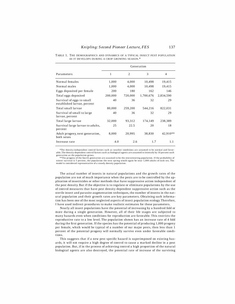
Knipling: Second Pioneer Lecture, FES
137
The actual number of insects in natural populations and the growth rates of thepopulation are not of much importance when the pests are to be controlled by the ap-plication of insecticides or other methods that have suppressive action independent ofthe pest density. But if the objective is to regulate or eliminate populations by the useof control measures that have pest density dependent suppressive action such as thesterile insect and parasite augmentation techniques, the number of insects in the nat-ural population and their growth rates are key parameters. Obtaining such informa-tion has been one of the most neglected aspects of insect population ecology. Therefore,I have used indirect procedures to make realistic estimates for these parameters.
Nearly all insect populations have the potential of increasing by a hundred fold ormore during a single generation. However, all of their life stages are subjected tomany hazards even when conditions for reproduction are favorable. This restricts thereproductive rate to a low level. The population shown has an increase rate of 4 foldduring the first generation. If the species has the potential of producing 1,000 progenyper female, which would be typical of a number of our major pests, then less than 1percent of the potential progeny will normally survive even under favorable condi-tions.
This suggests that if a new pest specific hazard is superimposed on existing haz-ards, it will not require a high degree of control to cause a marked decline in a pestpopulation. But, if in the process of achieving control a high proportion of the naturalbiological agents are also destroyed, the potential rate of increase of the surviving
T
ABLE
1. T
HE
DEMOGRAPHICS
AND
DYNAMICS
OF
A
TYPICAL
INSECT
PEST
POPULATIONAS
IT
DEVELOPS
DURING
A
CROP
GROWING
SEASON
.*
Parameters
Generation
1 2 3 4
Normal females 1,000 4,000 10,498 19,415Normal males 1,000 4,000 10,498 19,415Eggs deposited per female 200 180 162 146Total eggs deposited 200,000 720,000 1,700,676 2,834,590Survival of eggs to smallestablished larvae, percent
40 36 32 29
Total small larvae 80,000 259,200 544,216 822,031Survival of small to largelarvae, percent
40 36 32 29
Total large larvae 32,000 93,312 174,149 238,389Survival large larvae to adults, percent
25 22.5 20 18
Adult progeny, next generation, both sexes
8,000 20,995 38,830 42,910**
Increase rate 4.0 2.6 1.7 1.1
*The density-independent control factors such as weather conditions are assumed to be normal and favor-able. The density-dependent control factors such as biological agents are assumed to intensify by 10 percent eachgeneration as the population grows.
**The progeny of the fourth generation are assumed to be the overwintering population. If the probability ofwinter survival is 5 percent, the population the next spring would again be near 1,000 adults of each sex. Themodel is considered representative of a steady density population.

138
Florida Entomologist
81(1) March, 1998
pests and coexisting non-targeted pests that are normally held in check by natural bi-ological control agents may increase by 10 fold or more per generation. This in essenceis what we have been doing to many insects during the past 50 years because of theuse of broad-spectrum insecticides.
The model in table 2 depicts the influence of the release of sterile insects into a pestpopulation of the nature shown in table 1. If enough fully competitive sterile insectsare released
throughout
the pest population to achieve a sterile to fertile ratio of 9:1,only 10 percent of the normal females will mate with normal males and the reproduc-tive success of the population will be reduced by 90 percent. If the reproducing fe-males produce an average of 10 adult progeny the population will decrease by 50percent. The release of the same number of sterile insects will then result in a sterileto fertile ratio of 18:1, and reproduction will be inhibited by about 95 percent. This inturn will cause an even sharper decline in the pest population and the release of thesame number of sterile insects during generation 3 will then inhibit reproduction bymore than 98 percent. By generation 4 the natural population will be so low that therelease of relatively few insects will reduce the probability of a fertile female matingto less than one. The model shown is representative of a classic sterile insect suppres-sion model I first described in 1955 (Knipling 1955). In theory all reproduction willcease by the fourth generation. This is what happened to the screwworm on Curacao.
The success of the sterile insect technique against the screw worm prompted sci-entists to investigate the feasibility of using the technique for dealing with other in-sect pests.
The first intensive research effort on other insects involved tropical fruit fly spe-cies. As a complex these are the world’s most important fruit insect pests. Early re-search on the technique was led by L. F. Steiner, Director of the Agricultural ResearchService Laboratory in Hawaii. He and his associates demonstrated that the techniquecould be used to suppress and eliminate isolated populations of the melon fly,
Bactro-cera cucurbitae
, and the oriental fruit fly,
Bactrocera dorsalis
, (Steiner et al. 1965 andSteiner 1970). Japanese scientists further developed the technique in order to eradi-cate the melon fly from the Okinawa islands (Kakinohama
et.al.
1997). This virtually
T
ABLE
2. T
HE
INFLUENCE
OF
STERILE
INSECT
RELEASES
ON
THE
DYNAMICS
OF
A
PESTPOPULATION
OF
THE
NATURE
SHOWN
IN
T
ABLE
1.
Parameters
Generations
1 2 3 4
Normal females 1,000 500 132 10Normal males 1,000 500 132 10Sterile insects released, both sexes
18,000 18,000 18,000 400
Sterile-to-fertile ratio 9:1 18:1 68:1 20:1Normal females mated with normal males
100 26.3 2 0
Expected adult progeny per normal mated female
10 10 10 —
Adult progeny, next generation, both sexes
1,000 263 20 —

Knipling: Second Pioneer Lecture, FES
139
doubled the vegetable economy of the islands. The technique also played a role in theelimination of the oriental fruit fly from Japan, which was accomplished largely by theuse of the male attractant, methyl eugenol (Koyama
et al.
1984).The SIT is playing a major role in dealing with the Mediterranean fruit fly,
Cerati-tis capitata
, (medfly) in various parts of the world. This is the most important of thetropical fruit fly species. Scientists in several countries have contributed to the devel-opment of the technique for controlling or eradicating this pest. Much of the researcheffort has been sponsored by the International Atomic Energy Agency (IAEA). Scien-tists with the U.S. Department of Agriculture (USDA) Laboratory in Hawaii in coop-eration with the University of Hawaii and the University of California conductedmuch of the early research that led to use of the technique as a key component in erad-ication and containment programs for the medfly in the United States, Mexico andother countries.
There have been numerous introductions and establishments of medfly popula-tions in the United States during the past half century. Until the SIT came into usecontrol agencies relied largely on insecticide applications and drastic host removal toeliminate them. However, there is strong public opposition to the use of non-selectiveinsecticides for eradicating insect pests, especially in urban areas where medfly intro-ductions are most likely to become established. Relying on SIT for dealing with suchintroductions was slow to be accepted. The technique is not very effective when thepest populations are high and its suppressive action is slow compared with the use ofinsecticides. But limited use of insecticides followed by the release of sterile flies tocomplete eradication or to prevent the reestablishment of populations is now an ac-cepted practices.
SIT has also had a rather slow history of development for the eradication of tsetsefly populations,
Glossina spp
. Tsetse flies are probably the most important insectpests in the world. The IAEA assumed leadership in developing and promoting theuse of SIT for eradicating these disease vectors. Because of the difficulty and high costof rearing tsetse flies these insects would seem to be poor candidates for eradicationby SIT. However, by using insecticides when the pest populations are high and thenreleasing sterile flies against the reduced populations, this pest management strategyhas been used successfully.
Sterile pink boll worm moths have also been used for some years to prevent the es-tablishment of this cotton pest in the San Jauquin Valley of California. This importantcotton growing area has been kept free of the pink boll worm for about two decades bythe use of the technique (Bartlett and Staten 1996). Canadian scientists are using thetechnique in efforts to eradicate the codling moth from Western Canada.
Thus the SIT is playing an increasing role in dealing with several important insectpest problems. It is my opinion, however, that the technique has not been developedand put into use to the extent that it should have been. There are a dozen or more ma-jor insect pests in the United States and in other parts of the world that would be goodcandidates for management on a year by year basis by the sterility technique, or foreradication when this is a feasible option. The pests I have in mind would include allof the tropical fruit fly species; European corn borer,
Ostrinia nubilalis
; sugarcaneborer,
Diatraea succharalis
; and even the widespread and costly Helicoverpa/Helio-this complex. However, development and use of the technique has lagged for a numberof reasons. Technology must be developed and made available to mass produce largenumbers of insects. This has not yet been done for a number of candidate species.Some insects are very susceptible to irradiation damage and thus are not sexuallycompetitive with wild rivals. This is the case for the boll weevil,
Anthonomus grandis
.However, one of the main reasons for the limited use of the technique has been the
lack of support from and even opposition by much of the pest management community

140
Florida Entomologist
81(1) March, 1998
to the holistic approach to insect pest management. This makes it difficult to obtainthe public and political support for areawide insect pest management programs. Astime goes on there will be increasing concern over the use of potentially hazardous in-secticides. It will also become increasingly apparent that a defensive reactive pestmanagement procedure based largely on the use of insecticides will not provide satis-factory solutions for many pest problems. SIT has some important limitations, but italso has merits not possessed by other methods of control. With greater recognition ofthe advantages of the holistic approach to insect pest problems there will be more in-terest in feasible ways to achieve this objective.
M
ERITS
AND
L
IMITATIONS
OF
THE
U
SE
OF
I
NSECTICIDES
Control of insects by the use of insecticides has always appealed to agriculturists.The availability of many effective synthetic insecticides has made it possible for farm-ers to control most insects of agricultural importance. Their intensive use worldwideis one of the reasons that agriculture has been able to meet the food requirements forthe expanding world population. Also their availability for the control of arthropodvectors of human diseases has prevented illness or the death by hundreds of millionsof people each year during the past 50 years.
Insecticides have a number of advantages over other methods of insect control.They are rapid and positive in action. Insect outbreaks can be controlled in a matterof days and disease transmission can be halted within hours by the application of in-secticides. They are readily available to consumers and equipment for their applica-tion is also readily available. Society should be mindful of the important contributionsthat insecticides have made to human welfare. People should also be mindful of thecontinuing need for these chemical insecticides for the indefinite future.
At the same time, however, the extensive use of the insecticides, most of whichhave broad-spectrum activity, has created many complex environmental problems.Residues of the chemicals in or on agricultural products pose potential hazards to peo-ple and animals. The welfare of fish and wildlife in environments treated for insectpest control can be jeopardized. The balance between destructive and beneficial in-sects can be so seriously disrupted that natural biological control agents cannot per-form their normal function. In my opinion, the ecological disruptions resulting fromthe use of broad-spectrum insecticides is the most serious of the environmental haz-ard problems associated with the use of insecticides. I can say without qualificationthat ecologically sound insect pest management will not be possible so long as the useof broad-spectrum insecticides is a major component in pest management systems.
Pest management scientists are aware of this, and accordingly have devoted mucheffort to the development of safer ways to control insects. Major efforts have beenmade to find new and better insect attractants; insect pathologists have discovered awide range of insect pathogens and microbial agents that offer promise as safe alter-natives to chemical insecticides. More emphasis has been placed on classical biologi-cal control and the use of entomophagous insects for augmentation purposes. Plantbreeders and entomologists have intensified investigations on host plant resistance,including resistance by biotechnology.
Good progress has been made on all of these approaches for insect control. Butwhen viewed from a broad perspective very little of the new technology has been putinto practice. Agriculture still depends largely on insecticides for the control of insects(U.S. Congress 1995).
In the efforts to reduce the amount of insecticides used, a supervised system of in-sect control evolved that is broadly defined as Insect Pest Management (IPM). To min-imize the use of environmentally hazardous insecticides and to reduce costs farmers

Knipling: Second Pioneer Lecture, FES
141
are urged to rely as much as possible on natural control agents before applying insec-ticides. They are also encouraged to use supplemental control measures such as cul-tural procedures and to grow pest resistant crop varieties. As a guide for growers,economic treatment levels have been established by pest managers for various insectson different crops.
IPM has achieved some important objectives. It has virtually eliminated the indis-criminate use of insecticides. It has been profitable for growers because it minimizesexpenditures for insecticides yet prevents the heavy losses that can occur if controlmeasures are not applied. However, it has not reduced primary reliance on pesticidesto control pests.
After intensive efforts to develop alternative ways to control insects, we must askourselves why is agriculture still so dependent on insecticides? The answer involves anumber of complex factors. But the basic reasons are clear. Most of the possible alter-native methods of control are not as effective or as practical as insecticides after insectpest populations have already reached economic treatment levels. They also lack thefast and positive action that growers desire for protecting their crops when insect pestpopulations reach such levels. Moreover, the alternative methods of control are usu-ally not available to individual growers. Therefore, agriculture has continued to de-pend largely on fast-acting insecticides for controlling insect pests for a half century.And when broad-spectrum insecticides are applied it defeats one of the primary objec-tives of IPM programs: to obtain optimum benefits from natural biological agents.
Despite all that has been said about the benefits of IPM, when we analyze the dy-namics of insect pest populations and the kinds of suppressive action that is achievedby the use of insecticides, the defensive reactive method of insect control that hasevolved is not a sound insect pest management system. It gives insect pest popula-tions the opportunity to increase virtually unhampered from their normal low to theirnormal high levels each year. And when they reach the economic treatment levelsabout the only option that growers have is to apply fast-acting, broad-spectrum insec-ticides. Then, the reproductive success of the proportion of the target pest populationnot controlled is higher than normal leading to possible resistance and the serious de-pletion of the total complex of natural control agents will permit normally minor orsecondary pests to become important pests. The method also concedes to insects thelosses they cause below the economic threshold levels. For some of our important in-sect pests such as the corn earworm on corn and the European corn borer, these lossesamount to about $500 million per year for either pest. What the total damage is thatfalls below the economic pest control level is difficult to determine, but it probablyamounts to several billion dollars annually. The losses that fall below current eco-nomic threshold levels give us a good perspective of the amount that agriculture couldafford to spend on alternate ways to deal with specific pest problems that would beless costly and more acceptable from environmental standpoints.
This is the current status of insect pest management. While the methods of controlthat have evolved are profitable for the growers, they have not reduced the overallthreat that insects pose for agriculture. In support of this conclusion, if a list weremade of the insect pests that were of major concern to agriculture before the new in-secticides came into being and such list were also drawn up today most of the samepests would be on both lists—except that the current list would be longer. Only two im-portant insect pests would not be on the current list: the screwworm and the boll wee-vil in the southeastern cotton growing region. It is significant that these are the onlyinsect pest problems that have been subjected to organized suppression programs thatare designed to eliminate or maintain total populations below damage levels.
The limitations of the largely defensive systems of insect control have promptedme to continue theoretical appraisals of other techniques and strategies for control-

142
Florida Entomologist
81(1) March, 1998
ling insects. My efforts have been focused on the possibility of making use of ento-mophagous insects to regulate total pest populations as a preventative measure. Asthe investigations have progressed and with the promising results obtained on sev-eral of the major insect pests by research entomologists who are investigating areawide management by the release of biological agents, I have gained more and moreconfidence in the holistic approach to insect pest control.
T
HE
H
OLISTIC
A
PPROACH
TO
I
NSECT
P
EST
M
ANAGEMENT
BY
THE
P
ARASITE
A
UGMENTATION
T
ECHNIQUE
The possibility of controlling insect pests by the release of reared insect parasi-toids or insect predators into pest habitats is not a new concept. Indeed, biologistshave devoted as much or more research efforts to this method of control than to anyother method besides the use of insecticides. Much of this effort has been on Tri-chogramma parasitoids. While the use of these parasitoids is reportedly successful insome countries for controlling certain lepidopterous pests (Ridgway and Vinson 1977),it has not been very successful in other countries. The results obtained with other par-asitoids or predators have been largely unsatisfactory. This is reflected in the limiteduse of reared biological agents for controlling insect pests. While a number of such bi-ological agents are being produced and sold by small private industries (Hunter1994), probably less than one percent of the agricultural insect pests are currentlycontrolled by the release of insect parasitoids or predators into insect pest habitats.
There are several reasons for such limited use of the technique. It is a technique thatis not very effective, practical or reliable when used on a farm-by-farm basis and onlyas the need arises. But when used as a preventative measure to regulate total insectpest populations it has very important advantages over all other methods of control.
Until recent years the possibility of rigidly regulating total insect pest populationsas a preventative measure by the release of selected parasitoids or predators through-out pest ecosystems has been given little consideration by biologists. Very few insectpests have been subjected to such releases. The use of natural enemies to control in-sect pests in greenhouses would be an exception to this generalization. Releases of in-sect predators or parasitoids are often quite successful because the total pestpopulations are subjected to the releases.
Notwithstanding the poor record of certain insect parasitoids both in nature orwhen spot released, I undertook a detailed study of parasitism processes and the in-fluence that parasitoids have on the dynamics of insect pests (Knipling 1992). Thestudy was conducted largely by theoretical means, but I reached the conclusion thatthe parasite release technique offers almost unlimited possibilities for regulating pop-ulations of many of the nation’s and world’s major insect pests if selected species canbe mass produced at reasonable costs and appropriate numbers of the parasitoids aredistributed
throughout
pest ecosystems at strategic times during the seasonal or pe-riodic cycles of the pests. I emphasize the term
throughout
. Like sterile insects, para-sitoid releases are not effective when releases are made in small, unisolated habitats.
The results of the investigation I conducted have been published in a handbook en-titled
Principles of Insect Parasitism Analyzed from New Perspectives: Practical Impli-cations for Regulating Insect Populations by Biological Mean
. U.S. Department ofAgriculture, Agricultural Research Service. Handbook No. 693, (1992).
The publication explains how parasitism works in natural environments and howbiological actions evolved under nature’s natural balancing mechanisms to maintainthe relative number of parasites and hosts within safe limits. Simple rationalizationstell us that the numerical relationships between parasites and hosts must be main-tained within certain limits for the welfare of the hosts as well as for the parasitoids
Knipling: Second Pioneer Lecture, FES
143
that are dependent on specific hosts or host complexes for reproduction and survival.It is nature’s objective to maintain a safe balance between associated organisms, andit is very successful in achieving that objective. But this objective can be in conflict withagriculture’s needs. It permits too many insect pests to cause excessive damage tocrops and livestock. Also, intensive agriculture favors many insect pests. So, the impli-cations are clear. To achieve satisfactory management of insect pest populations by theuse of insect parasitoids it will be necessary to increase the ratio of parasitoids to hostsin coexisting populations in all parts of the ecosystem they cohabitate. About the onlyway this can be done will be to augment the natural populations by artificial means.
To determine if it is feasible to regulate total populations of various insect pests byuse of the parasite release technique, it is necessary to have reasonably good informa-tion on the actual number of parasitoids that must be produced and maintained in thepest ecosystems to achieve various rates of parasitism. Such information is not nowavailable for any of our parasitoids. However, largely by deductive procedures and bythe use of hypothetical models of the nature already described, I feel that I have beenvery successful in making realistic estimates of the
relative
number of parasitoids andhosts that normally coexist in natural populations for a wide range of our pests. Thisin turn makes it possible to estimate the
actual
number of parasitoids that will haveto be produced and released in pest ecosystems to achieve satisfactory control.
For solitary parasite species, the proportion of the host population that is normallyparasitized provides a good clue to the normal parasitoid to host ratio that occurs innatural populations. It is a good clue because the probability of survival of the imma-ture parasitoids in the host to the adult parasitoid stage and the probability of sur-vival of the hosts that are not parasitized to the adult host stage are very closelylinked at all host and parasite density levels. Biologists have obtained considerableinformation on the usual rates of parasitism caused by virtually all of the betterknown parasitoid species of our major insect pests. While such data are usually vari-able because of agricultural practices and other factors, including inadequate sam-pling, they nevertheless give us a good indication of the normal ratio of parasitoids tohosts in natural populations and a good indication of the rate of parasitism to expectfrom different ratios. Recognition and adoption of this logical theory has made it pos-sible to gain a good understanding of virtually all aspects of insect parasitism. It hasshed new light on what we can and cannot expect from insect parasitoids.
If a parasitoid species causes an average of about 10 percent parasitism, whichwould be typical of some parasite species, the normal ratio of adult parasites to adulthosts will be near 10:90 or 1:9. If the average rate of parasitism is near 20 percent,which would be typical of many other species, the ratio will be near 20:80 or 1:4 andso forth. Those approximate numerical relationships will hold regardless of the biol-ogy and behavior of the parasitoid and regardless of the host stage parasitized. Mostimportantly such numerical relationships and rates of parasitism hold regardless ofthe density of the host and parasitoid populations. The practical implications of thistheory are clear when pest populations exist at low levels.
Another very important conclusion reached from the investigation is that, con-trary to popular opinions, differences in the rates of parasitism caused by differentparasite species are due to differences in the ratio of parasites to hosts that can coexistbecause of the constraints imposed by nature’s natural balancing mechanisms.
Many biologists assume that differences in the rates of parasitism caused by var-ious parasite species are due to differences in their host-searching and host-findingability. Parasitoids are often categorized as good or poor host-finders based on therates of parasitism they achieve. But we can accept with complete assurance that
allparasite species are diligent host-searchers and highly efficient host-finders and at allhost density levels
. Otherwise they would not exist.
144
Florida Entomologist
81(1) March, 1998
The way parasitism works has very great practical implications. It means that theusually low rates of parasitism that occur naturally can be overcome by the simpleprocedure of mass producing and liberating appropriate numbers of key parasitoidspecies
throughout
pest ecosystems. This may not be easy to accomplish, but therewill be few if any barriers to the attainment of the objective if insect rearing expertsare given even modest support for conducting research on parasitoid and host rearingprocedures. I am confident that scientists and engineers can, by in vivo or in vitro pro-cedures, mass produce any parasitoid species in unlimited numbers and at reasonablecosts if given such support.
If the approximate rate of parasitism caused by a given parasitoid to host ratio isknown it is possible by extrapolation to calculate the approximate rate of parasitismthat will be achieved by increasing the parasitoid to host ratio. If a ratio of 1:4 causes20 percent parasitism, a ratio of 2:4 will cause 20 + .20 (100-20) or 36 percent parasit-ism. A ratio of 4:4 or 1:1 will in theory cause 36 + .36 (1000-36) = 59 percent parasit-ism. This procedure makes allowances for the intraspecific competition factor. It doesnot, however, make allowances for the probable shorter life span and the lower aver-age host-finding ability of the female parasitoids when the populations exist at abnor-mally high levels. But adequate allowances can also be made for this factor.
When allowances are made for both factors, I estimate that an adult parasitoid toadult host ratio of 1:1 will cause about 50 percent parasitism; a ratio of 2:1 will causeabout 75 percent parasitism; and a ratio of 5:1 will cause about 95 percent parasitism.If these estimates are accepted as realistic, we have a good idea of the influence thatvarious rates of parasitism will have on the dynamics of different insect pests. Wehave already noted that natural control factors greatly limit the growth rate of insectpopulations. I believe that most of our major insect pests would be of minor impor-tance if an additional 50 percent control were superimposed on all natural hazardsthat each pest faces and if this occurred
throughout
the pest ecosystem. Again, I em-phasize that such additional mortality must be achieved against the total population.
One might ask how realistic are these assumptions? I believe that most biologistswould agree that if a new biological agent were introduced that achieved 50 percentparasitism every cycle, most of our major insect pests would become minor pests. Ifthis is true, there is every reason to assume that 75 percent mortality above normalhazards would assure virtually complete control of nearly all of the major insect pests.It would seem almost certain that 95 percent control superimposed on all natural con-trol factors would within a few cycles result in complete elimination of a pest popula-tion—and keep in mind that the lower the pest density the fewer the number ofparasitoids that will be required to achieve high parasitoid to host ratios.
If we accept these hypotheses as reasonable expectations—and I firmly believethat they are—the practical question is whether there are any feasible ways to super-impose such levels of control with reasonable consistency and uniformity throughoutpest ecosystems each pest cycle. In my opinion, the most feasible way to accomplishthis will be by the release of mobile parasitoids that have the instincts and the
capa-bility
of finding the precise location of the hosts.We have already estimated the rate of parasitism to expect from different parasi-
toid to host ratios. But, we must consider how pest populations will respond to differ-ent rates of parasitism. We will use a hypothetical pest population to estimate theinfluence as we did for the sterile insect technique.
I
NFLUENCE
OF
P
ARASITOID
R
ELEASES
ON
P
EST
H
OST
P
OPULATIONS
To estimate the influence of the release of parasitoids in pest host ecosystems, weneed information on the rates of parasitism to expect from different parasitoids to

Knipling: Second Pioneer Lecture, FES
145
host ratios as well as information on the average host-finding efficiency of the para-sites to be released. I have indicated the rates of parasitism to expect from differentparasitoid to host ratios, but we also need to quantify the actual numerical relation-ships and rates of parasitism.
The basic host population model shown in table 1 can be used to postulate the rel-ative and actual number of parasitoids and hosts that will normally coexist in naturalpopulations and also estimate the average number of host larvae that the female par-asitoids will parasitize during their lifetimes if the normal rate of parasitism isknown. The model shown in table 3 depicts the coexistence pattern of a pest an asso-ciated parasitoid species. The hypothetical parasitoid is a solitary species that aver-ages between 15 to 20 percent parasitism. For this model an average of 17 percentparasitism is assumed. Therefore, based on the comparable survival theory, the nor-mal ratio of adult parasitoids to adult hosts will be near 17:83 or about 1:5. If the hostpopulation numbers 1,000 females, as we have assumed, this would mean that 200 fe-male parasitoids will normally coexist with 1,000 female hosts. It will also mean thatif the rate of parasitism averages 17 percent, the female parasitoids will parasitize13,600 of the 80,000 host larvae presumed to be present. It follows that each femaleparasitoid will on average parasitize 68 hosts during their lifetime during the firstgeneration. Thus, by indirect procedures we have established approximate values forall the parameters needed to calculate the influence a known number of parasitoidswill have on the dynamics of a host population having a known number of hosts.
The model depicted in table 4 shows the theoretical results to expect if enough par-asitoids of the nature depicted in table 3 are released during generation 1 to achievean adult parasitoid to adult host ratio of 3:1. This would require the release of 3,000female parasitoids (6,000 of both sexes.) It should be kept in mind that a 3:1 ratiowould be
15 times
the 1:5 ratio that is estimated to cause 17 percent parasitism of thehost larvae. Thus, the pest population would be subjected to a very high parasitoidpopulation. We have estimated that at normal densities each female parasitoid willparasitize 68 host larvae. But because of the high parasitoid density I assume, ratherarbitrarily, that the average host finding ability of the female parasitoids will be 25percent lower or 51 per female. However, this will permit 3,000 females to find andparasitize 153,000 host larvae.
Since only 80,000 host larvae are available to be parasitized, the ratio of hosts en-countered to hosts available will be about 1.9:1. According to the formula described fortable 1 in the publication by Knipling (1992), approximately 15 percent of the host lar-vae will escape parasitism by chance and 85 percent will be parasitized one or moretimes when the ratio of total hosts parasitized to hosts present is 1.9:1. Therefore12,000 host larvae will escape parasitism by chance and 68,000 will be parasitized oneor more times. Being a solitary species only one immature parasitoid can develop suc-cessfully in one parasitized host.
According to the basic model, table 3, the survival rate of the unparasitized larvaeto the adult host stage and the survival rate of the parasitized host larvae to the adultparasitoid stage will be 12 percent during the first generation. Therefore, in generation2 the adult host population will number 720 females, and the adult parasitoid popula-tion will number 4,088. This would be a 28 percent decrease in the host population buta 32 percent increase in the parasitoid population. Therefore, the ratio of adult para-sitoids to adult hosts during generation 2 would increase substantially. There are onlyslight changes in the average number of host larvae produced per female host and theaverage number found per female parasitoid so the ratio of hosts encountered to hostspresent will increase to 3.3:1. This in theory will cause 96 percent parasitism.
Such high rate of parasitism would cause a marked decline in the pest populationfor generation 3, but, significantly, the parasitoid population would not decline as
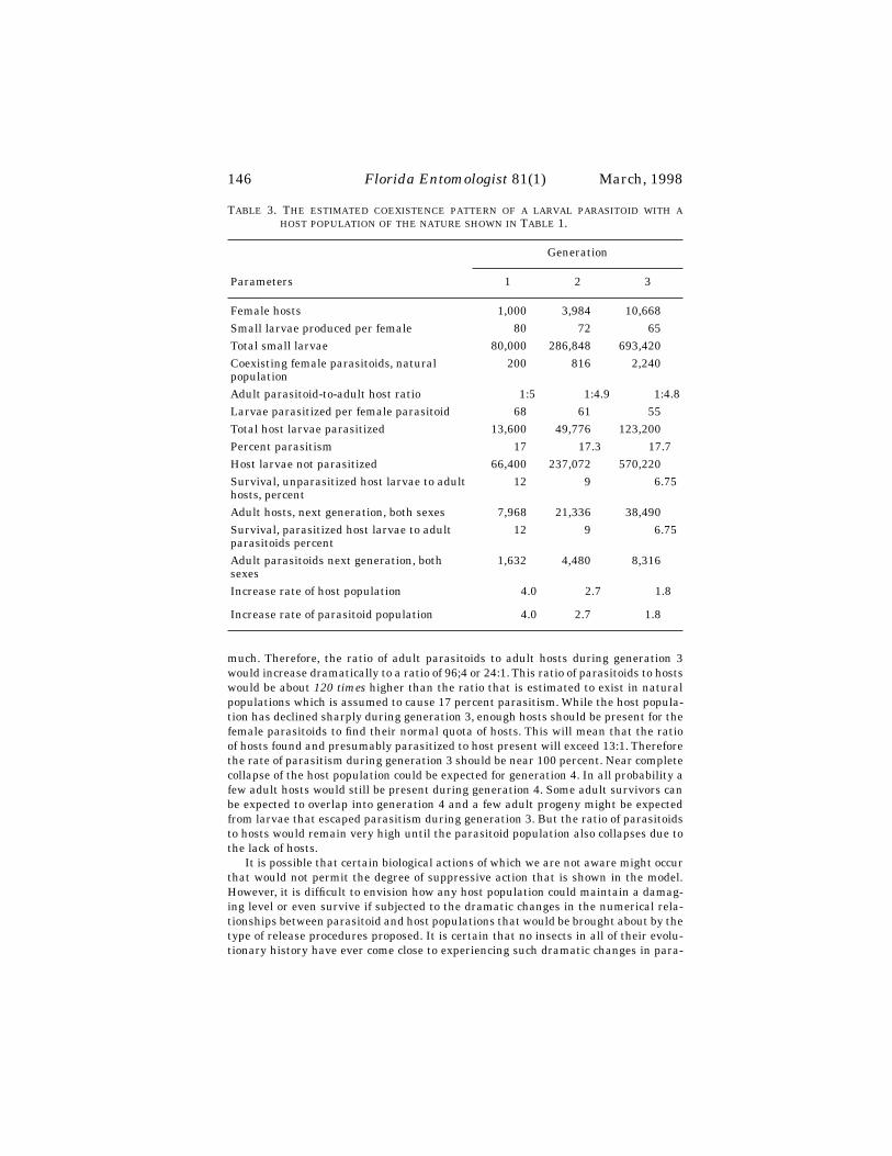
146
Florida Entomologist
81(1) March, 1998
much. Therefore, the ratio of adult parasitoids to adult hosts during generation 3would increase dramatically to a ratio of 96;4 or 24:1. This ratio of parasitoids to hostswould be about
120 times
higher than the ratio that is estimated to exist in naturalpopulations which is assumed to cause 17 percent parasitism. While the host popula-tion has declined sharply during generation 3, enough hosts should be present for thefemale parasitoids to find their normal quota of hosts. This will mean that the ratioof hosts found and presumably parasitized to host present will exceed 13:1. Thereforethe rate of parasitism during generation 3 should be near 100 percent. Near completecollapse of the host population could be expected for generation 4. In all probability afew adult hosts would still be present during generation 4. Some adult survivors canbe expected to overlap into generation 4 and a few adult progeny might be expectedfrom larvae that escaped parasitism during generation 3. But the ratio of parasitoidsto hosts would remain very high until the parasitoid population also collapses due tothe lack of hosts.
It is possible that certain biological actions of which we are not aware might occurthat would not permit the degree of suppressive action that is shown in the model.However, it is difficult to envision how any host population could maintain a damag-ing level or even survive if subjected to the dramatic changes in the numerical rela-tionships between parasitoid and host populations that would be brought about by thetype of release procedures proposed. It is certain that no insects in all of their evolu-tionary history have ever come close to experiencing such dramatic changes in para-
T
ABLE
3. T
HE
ESTIMATED
COEXISTENCE
PATTERN
OF
A
LARVAL
PARASITOID
WITH
AHOST
POPULATION
OF
THE
NATURE
SHOWN
IN
T
ABLE
1.
Parameters
Generation
1 2 3
Female hosts 1,000 3,984 10,668Small larvae produced per female 80 72 65Total small larvae 80,000 286,848 693,420Coexisting female parasitoids, naturalpopulation
200 816 2,240
Adult parasitoid-to-adult host ratio 1:5 1:4.9 1:4.8Larvae parasitized per female parasitoid 68 61 55Total host larvae parasitized 13,600 49,776 123,200Percent parasitism 17 17.3 17.7Host larvae not parasitized 66,400 237,072 570,220Survival, unparasitized host larvae to adult hosts, percent
12 9 6.75
Adult hosts, next generation, both sexes 7,968 21,336 38,490Survival, parasitized host larvae to adult parasitoids percent
12 9 6.75
Adult parasitoids next generation, both sexes
1,632 4,480 8,316
Increase rate of host population 4.0 2.7 1.8
Increase rate of parasitoid population 4.0 2.7 1.8
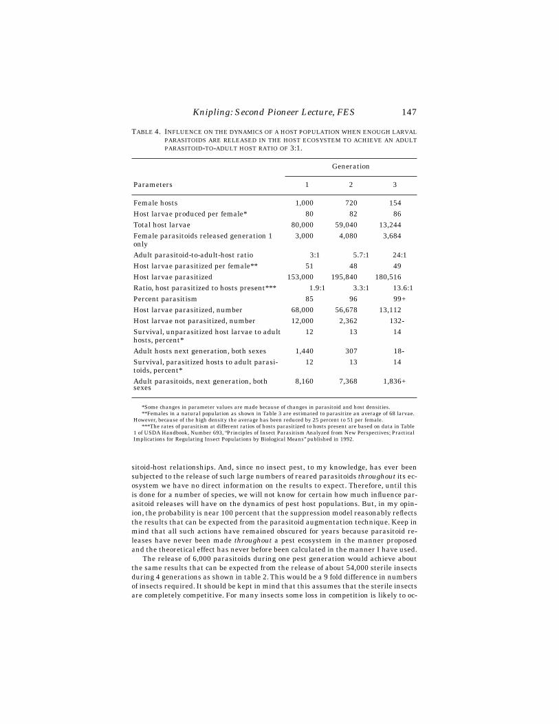
Knipling: Second Pioneer Lecture, FES
147
sitoid-host relationships. And, since no insect pest, to my knowledge, has ever beensubjected to the release of such large numbers of reared parasitoids
throughout
its ec-osystem we have no direct information on the results to expect. Therefore, until thisis done for a number of species, we will not know for certain how much influence par-asitoid releases will have on the dynamics of pest host populations. But, in my opin-ion, the probability is near 100 percent that the suppression model reasonably reflectsthe results that can be expected from the parasitoid augmentation technique. Keep inmind that all such actions have remained obscured for years because parasitoid re-leases have never been made
throughout
a pest ecosystem in the manner proposedand the theoretical effect has never before been calculated in the manner I have used.
The release of 6,000 parasitoids during one pest generation would achieve aboutthe same results that can be expected from the release of about 54,000 sterile insectsduring 4 generations as shown in table 2. This would be a 9 fold difference in numbersof insects required. It should be kept in mind that this assumes that the sterile insectsare completely competitive. For many insects some loss in competition is likely to oc-
T
ABLE
4. I
NFLUENCE
ON
THE DYNAMICS OF A HOST POPULATION WHEN ENOUGH LARVALPARASITOIDS ARE RELEASED IN THE HOST ECOSYSTEM TO ACHIEVE AN ADULTPARASITOID-TO-ADULT HOST RATIO OF 3:1.
Parameters
Generation
1 2 3
Female hosts 1,000 720 154Host larvae produced per female* 80 82 86Total host larvae 80,000 59,040 13,244Female parasitoids released generation 1 only
3,000 4,080 3,684
Adult parasitoid-to-adult-host ratio 3:1 5.7:1 24:1Host larvae parasitized per female** 51 48 49Host larvae parasitized 153,000 195,840 180,516Ratio, host parasitized to hosts present*** 1.9:1 3.3:1 13.6:1Percent parasitism 85 96 99+Host larvae parasitized, number 68,000 56,678 13,112Host larvae not parasitized, number 12,000 2,362 132-Survival, unparasitized host larvae to adult hosts, percent*
12 13 14
Adult hosts next generation, both sexes 1,440 307 18-Survival, parasitized hosts to adult parasi-toids, percent*
12 13 14
Adult parasitoids, next generation, both sexes
8,160 7,368 1,836+
*Some changes in parameter values are made because of changes in parasitoid and host densities. **Females in a natural population as shown in Table 3 are estimated to parasitize an average of 68 larvae.
However, because of the high density the average has been reduced by 25 percent to 51 per female.***The rates of parasitism at different ratios of hosts parasitized to hosts present are based on data in Table
1 of USDA Handbook, Number 693, “Principles of Insect Parasitism Analyzed from New Perspectives; PracticalImplications for Regulating Insect Populations by Biological Means” published in 1992.

148 Florida Entomologist 81(1) March, 1998
cur because of radiation damage. Therefore, there could be an even greater differencein the relative efficiency of parasitoids and sterile insects than is indicated by thisstudy. Also, the higher the parasitoid to host ratio the greater will be the advantageof parasitoids over sterile insects.
I have developed and critically analyzed similar models involving known parasi-toids of a number of our major pests including the boll weevil, Anthonomus grandis;corn earworm, Heliothis zea; tobacco budworm, Heliothis virescens; European cornborer, Ostrinia nubilas; sugarcane borer, Diatraea saccharalis; Colorado potato bee-tle, Leptinotarsa decimlineata; Mediterranean fruit fly, Ceratitis capitata, tsetse flies,Glossina spp. and a dozen other parasitoid-host associations. In every case, the para-sitoid population will, in theory, so dominate the host population within a few cyclesthat the elimination of completely isolated host populations would seem to be inevi-table. But until we subject completely isolated populations to parasitoid releases inthe manner proposed we will not know for certain to what level augmented parasitoidpopulations can suppress their host populations. However, the same question con-fronted researchers on the sterile insect technique. But the question no longer existsfor the sterile insect technique. Sterile insects have been shown to be capable of elim-inating populations of several different pests on numerous occasions. I feel certainthat host specific or primary parasitoids will be much more effective than sterile in-sects for the management or elimination of pest populations. This assumption is ofsuch great importance from both economic and environmental standpoints that it willbe critically analyzed in greater detail.
A COMPARISON OF THE SUPPRESSION CHARACTERISTICS OF THE PARASITOID
AUGMENTATION AND STERILE INSECT TECHNIQUES WHEN USED FOR REGULATING
TOTAL POPULATIONS
We have shown by hypothetical population models how insect pest populationswill respond to sterile insect and parasitoid releases. The suppression characteristicsof the two techniques have also been discussed to some extent. However, a full under-standing of the suppression characteristics of both techniques is vital to the total pestmanagement concept and to the role that the two techniques can be made to play infuture insect pest management systems. Therefore a critical comparison will be madeof the suppression characteristics of the two techniques with some comments on howthey differ from other insect control methods. There is increasing interest in the area-wide approach to insect pest management. However, it is very important to have agood understanding of the suppression characteristics of different methods of controlbecause they differ in their effectiveness for areawide management.
Target Pest Specificity
The chief goal of insect pest management scientists for several decades has beento develop ways of controlling insects that will permit natural biological agents to per-form their normal functions. We noted previously how important this is and why eco-logically sound insect pest management will not be possible so long as the use ofbroad-spectrum insecticides is a major component in pest management systems. Yet,most agricultural insect pests are now being controlled by the use of broad-spectruminsecticides. Fortunately, most alternative methods of control that scientists are in-vestigating have a high degree of pest specificity. The parasitoid release technique us-ing host specific or primary parasitoids would be about as pest specific as is possibleto achieve. The sterile insect technique is also highly pest specific although when the

Knipling: Second Pioneer Lecture, FES 149
adult stage causes harm to plants or animals the technique may not be acceptable. Forsome insects this is not a matter of great importance because when the pest popula-tions are very low the number of sterile insects that will be required will usually causelittle damage. But no harm to people, animals or plants will occur when parasitoidsare released in pest ecosystems.
Pest Density Dependent Suppressive Action
The suppressive action of all methods of insect control can be placed in two cate-gories: those that achieve about the same degree of control whether the pests areabundant or scarce; and those that have low effectiveness and efficiency when thepest populations are high but which become increasingly effective and efficientagainst declining pest populations. The sterile insect technique, the parasitoid aug-mentation technique and the use of sex pheromones have pest density dependent sup-pressive action. This is a negative characteristic when pest populations are high, butit is a very positive characteristic when the pest populations are low.
The suppressive action of all other methods of control is independent of the pestdensity. A given chemical or biological insecticide formula will kill about the same pro-portion of a pest population whether the insect population are high or low. It is a fa-vorable characteristic when the pest populations have reached high levels. However,there may be little or no advantage to the use of insecticides against low pest popula-tions in areawide insect management programs. But, it should be kept in mind thatthere can be a great advantage to the integration of density independent and densitydependent methods of control when the objective is to manage total populations on anareawide basis. This is a principle of insect pest management that has not been ade-quately exploited.
Mobility Factors
The excessive movement of released parasitoids and their progeny out of smallunisolated pest habitats and the excessive movement of the pests into habitats whereparasitoids have been released has probably done more to obscure the true efficiencyof the parasitoid release technique than any other factor. This can also be said for thesterile insect technique and probably also for the sex pheromone control technique.However, when the objective is to manage insect pests on an areawide basis, the abil-ity of the organisms to disperse and achieve control in all of the pest habitats becomesone of the most favorable characteristics of the parasitoid release and the sterile in-sect techniques.
One of the major disadvantages of the use of control methods that lack mobile ac-tion is that it will require much greater precision in their application than will the re-lease of mobile parasitoids or sterile insects. Treatment intervals of 100 feet or lesswill probably be required for highly effective control by non-mobile biological organ-isms, such as insect pathogens and immature insect predators, whereas release inter-vals ranging between one half to one mile (approximately 2,500 to 5,000 feet) shouldbe adequate for most parasitoid species and sterile insects. Therefore there can be asmuch as a 25 fold difference in the cost of applying mobile versus non-mobile controlprocedures when the objective is areawide pest management.
Pest Guidance Factor
When host-specific parasitoids are released in pest ecosystems, the released or-ganisms will tend to disperse into all of the pest habitats in their search for the hosts.
150 Florida Entomologist 81(1) March, 1998
These highly specialized organisms by nature have the ability to find their hosts witha high degree of efficiency (Nordlund et al. 1981) even when the host populations arevery low. Their highly developed host-detection mechanisms, plus their mobility andinstincts for host-finding give real meaning to the often used phrase: “You can run, butyou can’t hide.” One could characterize insect parasitoids as “biological guided mis-siles.” I am sure that they can find pest targets with much greater precision than canthe most sophisticated guided missiles for detecting military targets. Sterile insectsalso have the mobility factor, but when both sexes are released the sterile males or fe-males are no more likely to search specifically for wild mates than for their sterile sib-lings. If it is feasible to release males only the pest guidance factor should also applyto the sterile insect technique.
The pest guidance factor is a characteristic of immense practical significance forinsect pest management or eradication programs. The parasitoid release procedureand the release of males only in sterile insect programs are the only techniques thatpossess these characteristics.
Self-Perpetuating Parasite Populations
The frequency that the control procedure must be applied can be a very importantfactor in selecting the way to deal with insect pest problems. Long residual action ofinsecticides can be a desirable characteristic in controlling certain pests. However,when an insecticide has broad-spectrum activity long lasting effects can also be veryobjectionable because of ecological reasons. New plant growth can nullify insecticideresidual action under certain circumstances.
The ability of released parasitoids to maintain self-perpetuating populations andachieve control from cycle to cycle throughout a pest ecosystem is without a doubt oneof the most important characteristics of the parasitoid release technique. This is aninherent characteristic of both classical and augmentative biological control. How-ever, in natural populations, as we have already noted, there are constraints imposedby nature’s natural balancing mechanisms which tends to maintain the relative num-ber and the rate of parasitism within certain limits. This natural barrier can be over-come by augmented populations. If enough parasitoids are released throughout a pestecosystem to achieve an adult parasitoid to adult host ratio higher than 1:1 this can,in theory, induce a progressive increase in the rate of parasitism from cycle to cyclewithout the release of additional parasitoids. This theoretically will continue until thehost population is eliminated. For some parasitoid-host associations, there seem to bemechanisms that will prevent this progressive increase, as described by Knipling(1992). For others, however, such mechanisms seem not to exist. The self-increasingeffect cannot be triggered so long as the rate of parasitism remains below 50 percent.It can occur only when the parasitoid progeny will exceed the host progeny that areproduced throughout coexisting populations. If such increasing action can be con-firmed for any parasitoid species until the host population is eliminated it would be abiological action new to science.
The self-perpetuating characteristic is one of the most important advantages ofthe parasitoid release technique over the sterile insect technique and most other con-trol methods. There is some persistent suppressive action when lepidopterous insectsthat receive substerilizing dosages of irradiation are released in pest ecosystems (Car-penter, et al. 1987), but such action does not persist for more than about one genera-tion. Growing pest resistant host plants, whether developed by genetic selection or bybiotechnology, is perhaps the most persistent suppressive action that can be achieved.But host resistance is not applicable for some pests and has limited action for others.

Knipling: Second Pioneer Lecture, FES 151
General Discussion
Thus, when the objective is total population management, no method of controlhas as many desirable characteristics as does the parasitoid augmentation technique.However, the full potential of this technique for dealing with insect pest problems haslong remained obscured because the science of insect pest management has not con-sidered or tested its use to manage total pest populations. The effort that have beenmade in the past to use the technique has generally been patterned after the way in-secticides are employed. However, it is my opinion that neither the parasite augmen-tation or sterile insect techniques will ever play prominent roles in future insect pestmanagement systems if they are intended for use against high pest populations on afarm by farm basis as a substitute for insecticides. Their density dependent suppres-sive action, the propensity for the organisms to move out of small size habitats, plustheir slow action will make these two method of control unacceptable for growers nomatter how much they might be encouraged to use such biological control procedures.
However, one or the other of the two techniques has characteristics which are ofspecial importance for regulating total pest populations. They offer almost unlimitedpotential for dealing with many insect pest populations in an effective, low-cost andenvironmentally safe manner. The only requirement will be the technology for massproducing the organisms in large numbers and at reasonable costs. Considering theprogress that insect rearing experts have achieved in rearing a variety of insects on alarge scale (King and Leppla 1984) (Anderson and Leppla 1992) there are reasons tobelieve that rearing insects for augmentation purposes or for autocidal control willpose no serious problems.
A few examples can be cited. Excellent progress has been made on rearing severalparasitoids of tropical fruit fly species, Harris and Okamoto Cyo 1991, (Wong and Ra-madan 1992, Burns 1993). A tachinid parasitoid of the corn earworm Archytas mar-moratus can be readily reared under laboratory conditions, Gross and Johnson 1985as can be the tachinid Lixophaga diatraeae, a parasitoid of the sugarcane borer.
It might be necessary to rear most insect parasitoids on their hosts or a surrogatehost, but excellent progress is being made in rearing parasitoid species by in vitro pro-cedures. This has been accomplished for the boll weevil parasitoid Catolaccus grandisby Royas et al. (1996). Carpenter and Greany, (1997), report progress in rearing a pu-pal parasitoid of the corn earworm on artificial media. Also, Cohan (1997) has devel-oped in vitro procedures for rearing two well known insect predators, Geocorispunctipes and Chrysoperla carnea.
An important obstacle to overcome before making wider use of the parasitoid aug-mentation technique to manage insect pests on an areawide basis is the need to dem-onstrate that the techniques will in fact be highly effective and practical. This is thesame problem that has handicapped researchers developing the sterile insect tech-nique.
It is not possible to obtain a high degree of suppression or to achieve eradicationof insect pest populations by the release of parasitoids unless the releases are madeagainst isolated populations or in large areas. Research scientists have not had theopportunity to conduct such experiments for any parasitoid species. Until this is donewe will not know the full potential of the parasitoid release technique for rigidly man-aging total pest populations or if the technique can be used to eradicate isolated pesthost populations.
To demonstrate that mobile biological organisms are capable of suppressing pestpopulations to very low levels or to achieve eradication of isolated populations will re-quire carefully planned and well executed experiments. Each trial is likely to cost sev-eral million dollars. However, this would be a very small investment in research on

152 Florida Entomologist 81(1) March, 1998
methods of insect management that could eventually benefit agriculture by severalbillion dollars per year and also make a major contribution to reducing the need forecologically disruptive insecticides.
SYNERGISTIC SUPPRESSIVE ACTION
WHEN DIFFERENT INSECT CONTROL METHODS ARE INTEGRATED
In appraising the influence of the integration of different methods of insect control,I recognized a suppression mechanism that I call mutual synergistic suppressive ac-tion. The concurrent release of sterile insects and parasitoids produces this type of ac-tion.
This previously unrecognized type of suppressive action can add a new dimensionto insect pest management methodology. It offers the science of insect pest manage-ment the opportunity to achieve extraordinary effectiveness and efficiency when it isfeasible to integrate two or more control techniques that have this characteristic.
The model shown in table 5 depicts the theoretical influence of the concurrent re-lease of enough sterile insects and parasitoids during the first generation to achieveratios of 2:1 for each organism.
The sterile insects are assumed to be fully competitive. The release of 4,000 sterileinsects of both sexes would achieve a sterile-to-fertile ratio of 2:1 during generation 1.Therefore only 33.3 percent or 333 of the 1,000 normal females will mate with fertilemales. The concurrent presence of 2,000 female parasitoids would mean a parasitoidto host ratio of 6:1. This will have an effect equal to the release of 12,000 female par-asitoids alone. A ratio of 2,000 female parasitoids to 333 reproducing female hosts isestimated to cause about 98 percent parasitism. Thus the combined influence wouldbe 67 +.98 (100-67) = 99.3 control.
To inhibit reproduction by 99.3 percent by the release of sterile insects alone thesterile to fertile ratio would have to be about 130:1. This would require the releaseabout 260,000 sterile insects of both sexes. To achieve 99.3 percent parasitism by therelease of parasitoids alone, I estimate that it would be necessary to release about16,000 parasitoids of both sexes.
The advantage of the release of both organisms is not limited to the action duringgeneration 1. Even if no sterile insects were released during generation 2, enough par-asitoid progeny would in theory be present to achieve an adult parasitoid-to-host ratioof 49:1 during generation 2. Thus, releases would be necessary for only one pest gen-eration. Earlier we noted that it has not yet been demonstrated that parasitoids aloneare capable of eliminating pest populations. But, if even a few sterile insects were re-leased along with parasitoids this would ensure the use of a method of suppressionthat can lead to eradication of isolated populations.
The degree of enhanced suppressive action depends on the size of the initial ratiosof released parasitoids to the natural populations. The higher the ratios, the greaterwill be the enhanced suppressive action. At ratios as high as 3:1 for each organism -or an overall ratio of 6:1 - the combined suppressive action during the first and secondgenerations would be so high it would be difficult to even calculate. It would requirethe release of about 2,000,000 sterile insects alone to achieve a ratio of 1,000:1 toequal the effect of a ratio of 6:1 of both organisms. When pest populations are very lowdue to natural causes or are first suppressed to a very low level by other means, itwould not require the production and release of many organisms of both kinds toachieve ratios as high as 10:1 or even 100:1.
In areas subject to the invasion of only a few insects by long distance flight or byway of imported infested commodities the routine release of only a few insects per unit

Knipling: Second Pioneer Lecture, FES 153
area might ensure overwhelming ratios and protect the area from the establishmentof invading pests. This would be a new procedure for preventing the establishment ofinvading insect pests.
The examples described are merely an indication of the potential benefits that canbe realized by combining different insect control methods that have density depen-dent and mobile suppressive action. In theory, similar enhanced suppressive actioncan be realized by releasing two parasitoid species that parasitize and complete de-velopment in different life stages of the host. The concurrent release of a parasitoidspecies that parasitizes and completes development in the host eggs and another spe-cies that parasitizes and completes development in the host larvae or pupae would beexamples of such parasitoids. The concurrent release of a larval and puparia parasi-toid would be another example. The use of insect sex pheromones in traps or as attrac-ticides that have long range attraction power plus the release of parasitoids would beanother example of the integration of two suppression techniques that could lead tomutually enhanced suppressive action.
It is difficult to anticipate just what this potentially powerful mechanism of sup-pression can mean to insect pest management if it is exploited and applied for the reg-ulation and eradication of insect pests. When the pest populations are very low for anyreason and if relatively few insects would have to be released to achieve high ratiosthe cost can be very low. The release of biological organisms that have density depen-
TABLE 5. INFLUENCE OF THE CONCURRENT RELEASE OF ENOUGH STERILE INSECTS ANDPARASITOIDS TO ACHIEVE RATIOS OF 2:1 DURING GENERATION 1 OF A PESTPOPULATION OF THE NATURE SHOWN IN TABLE 1.
Parameters
Generation
1 2
Normal males 1,000 32Normal females 1,000 32Sterile insects, both sexes 4,000 noneSterile-to-fertile ratio 2:1 —Females reproducing 333 32Small larvae per female 80 86Total small larvae 26,640 2,752Female parasitoids released, generation 1 only 2,000 1,567Ratio, female parasitoids to reproducing female hosts 6:1 49:1Host larvae parasitized per female 52 52Total larvae parasitized 104,000 29,484Ratio, larvae parasitized to larvae present 3.9:1 10.7:1Percent parasitism 98 99+Larvae parasitized, number 26,107 2,724+Larvae not parasitized, number 533 27Survival larvae to adults, percent 12 —Adult hosts next generation, both sexes 64 —Adult parasitoids, next generation both sexes 3,133 —

154 Florida Entomologist 81(1) March, 1998
dent suppressive action and which also have mobility and pest detection mechanismswould combine several characteristics that can result in a very powerful method of in-sect control and eradication. And we should keep in mind that we would be usingmethods of control that would be highly pest specific and would permit all other nat-ural control factors to operate in a near normal manner. If these principles are con-firmed and exploited it could dramatically alter future insect pest management anderadication programs with great economic and environmental benefits.
PRACTICAL IMPLICATIONS FOR SPECIFIC INSECT PESTS
I have described various mechanisms and principles of insect control and the re-sults to expect from different control procedures. However, what agricultural execu-tives and farmers want to know is what does all this mean in terms of dealing withspecific insect pest problems and what will be the costs and benefits compared withcurrent control procedures.
Any one of a dozen insect pests could be selected to make such analysis. However,I will describe in a general way the procedure that could be followed in making use ofinsect parasitoids and sterile insects for regulating total populations of two of ourmost important insect pest species; the corn earworm, Helicoverpa zea, and the to-bacco bud worm, Heliothis virescens.
The two pests differ somewhat in their biology and host plant preferences, but theywill be regarded as one pest entity. To relate the theoretical results we have presentedto practical utilization several estimates and assumptions must be made. The re-leased organisms will be directed against the insects that emerge from the overwin-tering pupae. Most of the reproduction by this first generation normally occurs onwild host plants. But most of the wild hosts are in cultivated areas.
A vitally important parameter that will determine the feasibility of regulatingpopulations of the two pests is the number of insects that emerge from the overwin-tering population. I have, with the help of some of my colleagues, made estimates ofthe number of adults that are likely to emerge from the overwintering puparia. I es-timate that the overwintering population will normally consist of about 1 billionmoths of both sexes. Emergence and reproduction by the overwintered population areassumed to occur primarily in cultivated areas comprising about 75,000 square milesor about 50 million acres of cultivated lands. The suppressive measures will be ap-plied in the United States and northern Mexico where most overwintering occurs. Bi-ologists have obtained information which suggests that many of the insects thatreproduce early in Mexico will emigrate to the United States. Therefore, internationalcooperation would be necessary.
Thorough surveys and monitoring of the pest populations will be an essential as-pect of such a program. The biological organisms should be released at the proper timeand in appropriate numbers as emergence of the natural population occurs. It nodoubt would be advantageous to release more than one parasitoid species. Promisingparasitoids would include the hymenopterous larval parasitoid, Microplitis croceipesand the tachinid larval-pupal parasitoids, Archytas marmoratus and Eucelatoria bry-ani. As depicted in table 5, good control will result if sufficient numbers of a parasitoidare released for one generation to achieve an adult parasitoid to adult host ratio of 3:1.Thus the release of 3 billion parasitoids is proposed. Releases in the different regionswould begin, perhaps in February in Mexico. Then as the season advances the para-sitoids would be released to coincide with the emergence of the pests from the over-wintering pupae.
I assume that the parasitoids will be released in each area at 1 mile intervals everyfive days for 40 days or about 8 releases. If reproduction of the first generation is

Knipling: Second Pioneer Lecture, FES 155
largely inhibited this would minimize the number of immigrant moths that normallydisperse for several hundred miles to supplement the overwintering populations.While such program may seem to be of unprecedented scope, it would not be muchlarger than the screwworm program that has been conducted in the United Statesand Mexico for the past 25 years.
As depicted in table 4, an adult parasitoid to adult host ratio of 3:1 is estimated tocause virtually complete control within several cycles. A high degree of control of theoverwintering population could be expected in the United States and northern Mexicothe first year. If so, very few moths should be available to migrate into the agriculturalareas north of the normal overwintering areas and thereby avoid the heavy losses thepests cause each year in the northern states.
Obviously we are dealing with rough estimates and assumptions that would haveto be investigated intensely. However, for the purpose of this gross analysis I estimatethat the eventual cost of rearing suitable parasitoids will average $10 per 1,000 or $10million per billion. The cost for distributing the organisms is estimated to be $2 permile of flight for the aircraft. It is assumed that the biological agents will be distrib-uted at 1 mile intervals in about 75,000 square miles of territory. The cost for distrib-uting the organisms each release period would amount to only about $150,000 or $1.2million for 8 releases. Based on a rearing cost estimate of $10 million per billion par-asitoids, the cost for 3 billion parasitoids would be $30 million. Other costs includingthorough surveys, general supervision and other costs are assumed to increase thecost during the first year to about $35 million. This is about what the screwworm pro-grams have cost during the past 25 years.
Recognizing that there are unavoidable inefficiencies in such operations, it wouldseem more rational to assume the need for producing and distributing 6 billion para-sitoids rather than 3 billion during year 1. Other costs would be about the same re-gardless of the number of parasites released. Therefore, we might assume that thecosts during year 1 would be about $65 million. This would be less than 10 percent ofthe losses the pests now cause under current management procedures.
In dealing with insect pest populations from a total population perspective usingcontrol methods that have pest density dependent suppressive action the major ben-efits should be realized after the first year. The elimination of the population is out ofthe question. Some adults of both species no doubt can immigrate several hundredmiles from other regions. Some of the insects will be present in the management areaseach year even though most of the reproduction is inhibited. However, if we assumethat the normal overwintering and immigrating population will be as low as 100 mil-lion as few as one billion parasitoids should achieve a parasitoid to host ratio as highas 10:1. In that event the annual cost for management of the pests might be below $25million.
In considering the feasibility of dealing with the corn earworm and the tobaccobudworm by the procedure described it should be kept in mind that under currentcontrol procedures the two pests are estimated to cost agriculture in the United Statesmore than $1 billion per year (King et al. 1989). They are also responsible for much ofthe insecticides required for agricultural purposes.
Some or all of the estimates I have made may be too liberal. The normal overwin-tering population might be higher than 1 billion and the parasitoid-to-host ratio mayhave to be higher than 6:1 to achieve adequate control. On the other hand, it is alsopossible that one or more of my estimates are too high. If the overwintering popula-tion is normally as low as 667 million and the parasitoids could eventually be rearedat a cost as low as $5 million per billion, the estimates I have made for the first yearwould be too high even if the parasitoid to host ratio would have to be as high as 10:1.The continuing annual cost might be less than half the cost during the first year.

156 Florida Entomologist 81(1) March, 1998
From a national and worldwide standpoint, it is probable that no insect pest com-plex has been investigated as intensively as has the Helicoverpa/Heliothis complex.In my opinion about all of the basic technology needed has already been developed. Nonew discoveries are necessary. Nature has created the organisms. However, scientistsand agriculturalists must accept the holistic approach and focus on making the tran-sition from basic information to practical application. I see no obstacle to making thistransition other than failure of our agriculture executives in both the public and pri-vate sectors to accept the concept and unwillingness of our scientists to undertake theresearch challenges that would be involved in perfecting the technology and provingits performance.
The same principles and the same techniques of control should apply for manyother insect pests. Other important species that I consider good candidates for totalpopulation management in a similar cost effective manner include such major pestsas the bollweevil, Anthonomus grandis; European corn borer, Ostrinia nubilalis; sug-arcane borer, Diatraea saccharalis; the medfly, Ceratitis capitata, and other tropicalfruit fly species; Colorado potato beetle, Leptinotarsa decimlineata; codling moth,Laspeyresia pomonella; and numerous other species. For some of the species the costsduring year 1 might be quite high and other methods of control may be needed to re-duce the normally high natural populations to a level that can be managed by para-sitoids and/or sterile insects.
Much research will have to be conducted to obtain the needed ecological informa-tion and to perfect the technology. Research on managing or eradicating total insectpest populations will be costly and demanding. It will be necessary to demonstratethat the proposed procedure will perform as expected. It might cost several milliondollars to conduct one trial experiment involving a single pest. But we are dealingwith techniques and strategies that have the potential of benefitting agriculture byseveral billion dollars per year and also make a major contribution to the goals ofgreatly reducing the need for costly and ecologically disruptive insecticides. Consid-ering these large potential benefits, an expenditure of $25 million per year for re-search and development focused on the holistic approach to insect control bybiological procedures would be very small. It could be one of the best research invest-ments that agriculture could make. I cannot envision any alternative techniques andstrategies that would even approach the economic and environmental benefits thatcould be realized by the holistic approach using mobile biological organisms.
INSECT PEST PROBLEMS ESPECIALLY RELEVANT
FOR THE STATE OF FLORIDA
The principles and mechanisms of suppression that I have described in this lectureare basic and fundamental. They will apply for a wide range of insect pests. Entomol-ogists in Florida could do a great deal to advance the principles and concepts thathave been discussed. Your state is a well isolated ecosystem. It could take unilateralaction and benefit more than could other states that are not as well isolated from longrange insect immigration.
The probability is high that at least two of your important pest problems could bereadily resolved by the parasitoid release technique especially if supplemented by therelease of sterile insects. I refer to the Caribbean fruit fly, Anastrepha suspensa, andthe sugarcane borer. Excellent parasitoids of both of the pests are known and efficientmass rearing procedures have been developed for them. Also considerable researchhas been conducted on parasitoid releases that have given promising results againstthe sugarcane borer, (Summers et al. 1976). The sterile insect technique has beenshown to be an effective means of suppressing several tropical fruit fly and lepidopter-
Knipling: Second Pioneer Lecture, FES 157
ous pests. Parasitoid releases have also shown promise for suppressing tropical fruitflies, (Wong, et al. 1992 and Sivinski 1996). I can see no reasons for permitting theCaribbean fruit fly to continue to exist in your state and cause the problems it does foryour fruit industry. Also, I can see no reason why the sugarcane borer should be cost-ing sugarcane growers $25 to $50 per acre or more in losses year by year. The firstyear costs might be considerably higher, but I am confident that after sugarcane borerpopulations have been reduced to very low levels, they could be maintained below thelevel of damage at a cost of $2 to $3 per acre per year by the release of relatively fewparasitoids per acre perhaps supplemented by the release of some sterile moths.
However, there are a number of other insect pests in your state that are of nationalsignificance and which find refuge in your state during the winter months. Eachspring and summer enough of the pests spread northward into other agricultural re-gions and cause damage to a variety of crops before season’s end. Those pests includethe fall armyworm, Spodoptera frugiperda; the beet armyworm, Spodoptera exigua;cabbage looper, Trichoplusia ni; and diamondback moth, Plutella tylostella.
The number of insects of these species that reproduce only in Florida during thewinter months has not been determined. But there is little question that the popula-tions that emigrate from your state can increase by 25 to 50 fold during the growingseason and threaten losses on perhaps 25 times the crop acreage in other states beforeseason’s end.
What the total losses due to these pests amount to per year in the states north ofFlorida is difficult to say, but they probably aggregate several hundred million dollarsper year. I am greatly indebted to Dr. F. A. Johnston who made a special effort to ob-tain information on the annual cost of control and losses caused by the fall armyworm,beet armyworm, corn earworm, diamondback moth and cabbage looper in Florida. In-secticides now provide the only way to control these pests. I was amazed at theamount that growers spend per year controlling these pests. On most crops itamounts to as much as $100 per acre or more.
From time to time I have discussed with various scientists the feasibility of rigidlycontrolling such pests during the winter months in Florida to avoid the losses thepests cause other states after they spread northward. But research programs to in-vestigate the feasibility of dealing with the pests during the winter in Florida havenot yet materialized.
Since I have been interested primarily in the possible use of the sterile insect andparasitoid release techniques I have made gross estimates of the probable size of theoverwintering populations in Florida. It is difficult to say how accurate they are, butI feel confident that it would be feasible to rigidly manage populations of the pestsmentioned during the winter primarily by the use of parasitoids at costs of only a frac-tion of the losses they cause under present methods of control. Mandatory culturalmeasures such as the destruction of host plants within a certain period after the hostcrops are harvested should be integrated into a total population management system.This would cost farmers very little, but it can be of major importance in an integratedprogram. This alone might reduce the number of parasitoids or sterile insects re-quired by half and still result in a more effective program.
The feasibility of managing these pests in the winter by the release of parasitoidsand/or sterile insects will depend on the number of the pests that reproduce in yourstate during the winter and early spring. In my opinion, research to determine as ac-curately as possible the actual number of the different species that exist in the natu-ral population would be of the highest priority. Certainly through joint efforts by thestate and federal scientists, it should be possible to obtain good information on the ac-tual number of the different pests that reproduce in Florida during that period andthe source of the reproduction. Until such information is obtained, there is likely to be

158 Florida Entomologist 81(1) March, 1998
little that anyone can or will do to develop and make use of the proposed biologicaltechniques for dealing with these pest problems.
I raise the question, however, whether agriculture can afford to be so dogmatic andintransigent in meeting its insect pest problems. Is there any justification for or doesit make any sense to continue to rely year by year on costly and ecologically disruptiveinsect control methods when the prospects are so favorable for dealing with the pestproblems at very low cost and in an environmentally safe manner? Will the time comewhen society will demand the use of environmentally safe ways to deal with pest prob-lems especially if it will also greatly benefit producers and consumers? What can wedo as entomologists to gain the interest and support needed to achieve these goals.
LOOKING TO THE FUTURE
The world’s leading scientific institutions are projecting that agriculture within 30years must produce two times as much food as it now produces to meet the demand ofan expanding world population and economy. This will require that agriculture world-wide become more efficient in a number of ways. Protecting crops and livestock frominsect damage will be one of the ways to meet the monumental tasks that agriculturefaces within the short span of a few decades. People will and should demand that thefood that is produced be safe and that the quality of the environment in which we livewill not be jeopardized.
The methods of insect control that have evolved during the past 50 years have con-tributed to the highly productive agriculture that we now enjoy. However, this in parthas been accomplished by relying primarily on costly and ecologically disruptive in-secticides for insect control. The prospects seem dim to me that our present defensivesystem of management can be greatly improved, if at all. Insects continue to developresistance to insecticides. People continue to be concerned over the environmentalhazards insecticides can produce and more restrictions are being placed on their use.Agriculturists are beginning to wonder if we can maintain the standards of controlthat have been achieved in the past.
A study of the dynamics of many of our major insect pests and an analysis of thesuppression characteristics of various methods of control clearly indicate to me thatthe preventative measures applied against total insect pest populations would be amuch more rational way to deal with many of our major insect pests than to rely onthe largely defensive procedures that have been followed for half a century. Two bio-logical control procedures involving mobile organisms offer the best hopes of accom-plishing this objective in an effective, economical and environmentally safe manner:the mass-rearing of key parasitoid species and their release throughout the pest eco-system in adequate numbers and at strategic times in the pest’s seasonal cycle; andthe release of sterile or genetically altered insects in the same way.
The holistic approach to the management of insect pests by these two biologicalprocedures offers almost unlimited potential for dealing with insect pest problems inan environmentally safe manner and at very low cost. Mass producing the biologicalorganisms should pose no serious technical problems in view of the outstandingprogress that insect-rearing scientists and engineers have made in rearing manykinds of insects in large numbers.
The time will come, and sooner than we realize, when it will be necessary for agri-culture worldwide to produce the maximum amount of food on diminishing agricul-tural lands and water for irrigation. This will require greater efficiency andproductivity from every aspect of agriculture. Better protection of crops and livestockfrom insect attack will be one of the ways to achieve these objectives. This will be theresponsibility of entomologists and our associated scientists.
Knipling: Second Pioneer Lecture, FES 159
I am confident that we already have or could readily develop the technology thatwill be needed to deal with most of our major insect pest problems in a much lesscostly and a more ecologically acceptable manner during the next half century thanwe have done during the last few decades. But it will require that our agriculturalleaders in both the public and private sectors make provisions for developing and im-plementing the type of technology that will permit maximum efficiency and yields andwhich will also be environmentally acceptable.
We need more positive decisions and bold actions by our scientists and agriculturalleaderships in both the public and private sectors that will lead to many other pro-grams as successful and practical as the screwworm program that your society hashonored today.
REFERENCES CITED
ANDERSON, THOMAS E., AND NORMAN C. LEPPLA. 1992. Advances in Insect Rearing forResearch and Pest Management. Westview Press. Boulder, CO, 517 pp.
BAUMHOVER, A. H., A. J. GRAHAM, B. A. BITTER, D. F. HOPKINS, W. D. NEW, F. H.DUDLEY, AND R. C. BUSHLAND. 1955. Screwworm control through release ofsterilized flies. J. Econ. Entomol. 48(4): 462-66.
BURNS, E. 1993. Caribbean fruit fly rearing and release. Procedural manual FDAES-DPL. V-1,12/93. Gainesville, FL.
BUSHLAND, R. C., AND D. E. HOPKINS. 1951. Experiments with screwworm flies ster-ilized by X-rays. J. Econ. Entomol. 44(5): 725-731.
BUSHLAND, R. C., AND D. E. HOPKINS. 1953. Sterilization of screwworm flies with X-rays and gamma rays. J. Econ. Entomol. 46(4): 648-56.
CARPENTER, J. E., AND P. D. GREANY. 1997. Comparative development and perfor-mance of artificially-reared versus host-reared Diapetimorpha introita (Cres-son)(Hymenoptera: Ichneumonidae) wasps for biological control. Unpublishedmanuscript.
CARPENTER, J. E., J. R. YOUNG, A. H. SPARKS, H. L. CROMROY, AND M. A. CHOWDBURY.1986. Corn earworm (Lepidoptera;Noctuidae): Effects of substerilizing doses ofradiation and inherited sterility on reproduction. J. Econ. Entomol. 80(1987):483-489.
COHEN, A. C. 1997. Mass-reared insects get fast food. Agric. Research. 46:6. June1997.
CUSHING, E. C., AND W. S. PATTON. 1993. Studies on the higher diptera of medical andveterinary importance. Cochliomyia americana. Sp.Nov. “The screwworm fly ofthe new world.” Annals of Tropical Medical Parasitology. 27(4): 539-51.
GROSS, H. R., AND R. JOHNSON. 1985. Archytas marmoratus (Diptera: Tachinidae).Advances in large scale rearing and associated biological studies. J. Econ. En-tomol. 78: 1350-1353.
GRAHAM, O. H. 1985. Symposium on eradication of the screwworm from the UnitedStates and Mexico. Entomol. Soc. Am. Misc. Publ. 62. 68 pp.
HARRIS, E. J., AND R. Y. OKAMOTO CYO. 1991. A method of rearing Biosteres arisanus(Hymnoptera:Braconidae) in the laboratory. J. Econ. Entomol. 84: 417-422.
HUNTER, CHARLES D. 1994. Suppliers of beneficial organisms in North America. Cal-ifornia Environmental Protection Agency. Dept. of Pesticide Regulation. Sacra-mento, CA. 30 pp.
JACKSON, R. D., E. G. KING, AND E. F. KNIPLING. 1989. Heliothis: need for interna-tional collaborative research on biological control of Heliothis. New Delhi, In-dia. Nov. 1985.
KAKINOHANA, H., H. KUBA, T. KOHAMA, K. KINJO, AND OTHERS. 1997. Eradication ofthe melon fly, Bactrocera cucurbitae, Cog. by mass release of sterile flies in Ok-inawa Prefecture, Japan. Jaro. 31 (2): 91-100.
KING, E. G., AND N. C. LEPPLA (eds). 1984. Advances and challenges in insect rearing.U.S. Dept. Agric. Agric. Res. Serv. (Southern Region). New Orleans, LA. 306 pp.
160 Florida Entomologist 81(1) March, 1998
KNIPLING, E. F. 1955. Possibilities of insect control or eradication through the use ofsexually sterile males. J. Econ. Entomol. 48(4): 459-62.
KNIPLING, E. F. 1959. Screwworm eradication: concepts and research leading to thesterile male method. Smithsonian Institution Publication 4365. SmithsonianInstitution. 409-18.
KNIPLING, E. F. 1979. The basic principles of insect population suppression and man-agement. U.S. Dep. Agric. Agric. Handbook No. 512. 659 pp.
KNIPLING, E. F. 1992. Principles of insect parasitism analyzed from new perspectives:Practical implications for regulating insect populations by biological means.U.S. Dep. Agric. Agric. Res. Serv. Handbook No. 693. 337 pp.
KOYAMA, J., T. TANAKA, AND K. TANOLIA. 1984. Eradication of the oriental fruit fly(Diptera: Tephritidae) from the Okinawan Islands by a male annihilationmethod. J. Econ. Entomol. 77: 468-472.
LINDQUIST, A. W. 1955. Possibilities on use of gamma radiation for control or eradica-tion of the screwworm. J.Econ. Entomol. 48 (4): 467-469.
MELVIN, ROY, AND R. C. BUSHLAND. 1936. A method of rearing Cochliomyia ameri-cana C and P on artificial media. U.S. Bur. Entomol. and Plant Quar. ET. 88.
MEYER, NORREN L., AND OCELORIS SIMPSON. 1996. History of the Mexico and UnitedStates Screwworm Eradication Program. Vintage Press. New York. 367 pp.
NORDLUND, DONALD A., RICHARD L. JONES, AND W. JOE LEWIS (eds). 1981. Semichem-icals: Their Role in Pest Control. John Wiley and Sons. New York, Chichester,Brisbane, Toronto. 306 pp.
RIDGWAY, R. L., AND S. B. VINSON (eds). 1977. Biological Control by Augmentation ofNatural Enemies. Plenum Press. New York London. 480 pp.
ROJAS, MARIA G., JUAN A. MORALES-RAMOS, AND EDGAR G. KING. 1996. In vitro rear-ing of the boll weevil (Coleoptera Curculionidae) Ectoparasitoid Catolaccusgrandis (Hymenoptera: Pteromelidae) on mediric diets. J. Econ. Entomol.89(5): 1095-1104.
SCRUGGS, D. G. 1975. The Peaceful Atom and the Deadly Fly. The Pembarton Press.Austin.
SIVINSKI, J., C. CALKINS, R. BARANOWSKI, D. HARRIS, J. BRAMBILA, J. DIAZ, R. BURNS,T. HOLLER, AND G. DODSON. 1996. Suppression of Caribbean fruit fly popula-tions through augmented releases of the parasitoid Diachasmimorpha longi-caudata (Ashmead) Hymenoptera: Braconidae. Bio. Control. 6:177-185.
STEINER, L. F., W. G. HART, E. J. HARRIS, AND OTHERS. 1970. Eradication of the ori-ental fruit fly from the Mariana Islands by the method of male annihilation andsterile insect releases. J.Econ. Entomol. 63: 131-135.
STEINER, L. F., W. C. MITCHEL, AND OTHERS. 1965. Melon fly eradication by overflood-ing with sterile flies. J.Econ. Entomol. 58(3): 519-522.
SUMMERS, T. E., E. G. KING, D. F. MARTIN, AND R. D. JACKSON. 1976. Biological con-trol of Diatraeae saccharilis (Lepidoptera-Pyrilidae) in Florida by periodic re-leases of Lixophaga diatraeae (Dipera Tachinidae). Entomophaga. 21: 359-66.
U.S. CONGRESS, OFFICE OF TECHNOLOGY ASSESSMENT. 1995. Biologically Based Tech-nologies for Pest Control. OTA-ENV. 636. U.S. Government Printing Office,Washington D.C.
WONG, T., M. RAMADAN, J. HERR, AND D. MCINNIS. 1992. Suppression of Mediterra-nean fruit fly (Diptera: Tephritidae) populations with concurrent parasitoidand sterile fly releases in Kula Maui, Hawaii. J. Econ. Entomol. 85: 1671-1681.
WONG, T., AND M. RAMADAN. 1992. Mass-rearing biology of larval parasitoids Hym-noptera: Braconidae of tephritid fruit flies in Hawaii. In Anderson and Leppla(eds) Advances in Insect Rearing for Research and Pest Management. West-view Press. Boulder, CO. 405-476.
Florida Entomological Society Initiates AuthorLinks
161
FLORIDA ENTOMOLOGICAL SOCIETY INITIATES AUTHORLINKS
At its September meeting, the FES Executive Committee approved an importantnew service.
For the price of publishing one more page (currently $45), an author of an articlein
Florida Entomologist
can have an “AuthorLink” in the online
Florida Entomologist
.The link can be to a file that is submitted by the author and is posted on the sameWWW server as the article, or it can be to any URL that the author designates.
An AuthorLink is indicated by the phrase “
More information
” beneath the author’sname in the online table of contents. The author can make the link more descriptiveby specifying up to 20 additional words. For example, a link could read “
More infor-mation:
color pictures of all species and the complete data matrix.”Authors will be reminded that they can purchase an AuthorLink when proofs are
sent. In addition, an author may at any time purchase an AuthorLink for an articlepreviously included in the online
Florida Entomologist
. To do so, the author must fillout a form and send it, the submitted file (if any), and $45 to Business Manager,
Flor-ida Entomologist
, P.O. Box 1007, Lutz, FL 33548-1007. The form and an explanationof AuthorLinks is available at http://www.fcla.edu/FlaEnt/ (the home page of the on-line
Florida Entomologist
).An AuthorLink will be removed without charge when the Associate Editor for
Flor-ida Entomologist
on WWW (presently [email protected]) receives a written requestfrom the author. For the cost of a new AuthorLink, an author may substitute a newLink for an old one.
To see a representative AuthorLink, go online to the article by James E. Lloyd inthe June 1997 issue of
Florida Entomologist
(http://www.fcla.ufl.edu/FlaEnt/fe802.htm).
H
OW
A
UTHOR
L
INKS
O
PERATE
An “AuthorLink” functions in one of these two ways.
(1) Accesses a file submitted by the author and posted on the same WWW server as the article.
In most cases the file will be in HTML and contain a collection of links to files onother servers with brief descriptions of each—in other words, it will be a gateway toinformation that the author considers relevant to the published article.
In some cases, the submitted file may not link to other files. In these cases the filemay still be in HTML but more likely it would be a graphic file (e.g., jpeg or gif) or adata set (e.g., comma-delimited ASCII file).
An author may submit only one file for posting with the article, but there is nolimit on the size of that file. If files are referenced in the submitted file, they must beposted elsewhere. Most will likely be on the author’s server.
Once the submitted file is posted, the author will not be able to change its contents.Therefore, the author should make sure that the information in the file will notquickly become out dated. Note, however, that the author may use the submitted fileto link to files under the author’s control that can be updated whenever needed.
Preparing a file to be submitted for an AuthorLink is entirely the responsibility ofthe author. Before submission, the author should make sure that all links work andthat spelling and grammar meet the author’s standards.
162
Florida Entomologist
81(1) March, 1998
(2) Accesses a URL of the author’s choosing.
In this case, the author submits a URL rather than a file. In most cases the linkwill be to an HTML file under the author’s control, and the file will direct the viewerto other files relevant to the published article.
When an author submits a file, that file is archived along with the electronic versionof the author’s article. When an author submits a URL, the file that it links to is un-likely to remain posted for more than 50 years.
T. J. WalkerAssoc. Editor for
Florida Entomologist
on WWW

Florida Entomological Society Initiates AuthorLinks
163
ONLINE FLORIDA ENTOMOLOGIST: AUTHORLINK
This form may be used to request an AuthorLink for any article that is already in
Florida Entomologist
on WWW (http://www.fcla.edu/FlaEnt/fehmpg.htm) or for anyarticle that has been accepted for publication in
Florida Entomologist
but is not yetonline.
Send the completed form and $45 to Business Manager,
Florida Entomologist
, P.O.Box 1007, Lutz, FL 33548-1007.Name: ______________________________________________________________________
Title of article: _______________________________________________________________
_____________________________________________________________________________
Volume and issue of
Florida Entomologist
: _____________________________________
What type of Link do you want? (check one)
□
submitted file
□
URL.
If a URL, what is the URL? ___________________________________________________
If a file, please submit it on a diskette with this form or e-mail it as an attachment [email protected]
How do you want your Link labeled? (check one)
□
The default, which is “
More information
.”
□
“
More information
: _________________________________________________________
____________________________________________________________________________ .”
[Add as many as 20 words.]