Potential Antibiotics for the Treatment of Neonatal Sepsis ...
Upload
independentCategory
view
0download
0
Value Addition in the Efficacy of ConventionalAntibiotics by Nisin against SalmonellaAman Preet Singh, Vijay Prabha, Praveen Rishi*
Department of Microbiology, Basic Medical Sciences Block, Panjab University, Chandigarh, India
Abstract
Frequent and indiscriminate use of existing battery of antibiotics has led to the development of multi drug resistant (MDR)strains of pathogens. As decreasing the concentration of the antibiotic required to treat Salmonellosis might help incombating the development of resistant strains, the present study was designed to assess the synergistic effects, if any, ofnisin, in combination with conventional anti-Salmonella antibiotics against Salmonella enterica serovar Typhimurium.Minimum inhibitory concentrations (MICs) of the selected antimicrobial agents were determined by micro and macro brothdilution assays. In-vitro synergy between the agents was evaluated by radial diffusion assay, fractional inhibitoryconcentration (FIC) index (checkerboard test) and time-kill assay. Scanning electron microscopy (SEM) was also performedto substantiate the effect of the combinations. In-vivo synergistic efficacy of the combinations selected on the basis of in-vitro results was also evaluated in the murine model, in terms of reduction in the number of Salmonellae in liver, spleen andintestine. Nisin-ampicillin and nisin-EDTA combinations were observed to have additive effects, whereas the combinationsof nisin-ceftriaxone and nisin-cefotaxime were found to be highly synergistic against serovar Typhimurium as evident bycheckerboard test and time-kill assay. SEM results revealed marked changes on the outer membrane of the bacterial cellstreated with various combinations. In-vivo synergy was evident from the larger log unit decreases in all the target organs ofmice treated with the combinations than in those treated with drugs alone. This study thus highlights that nisin has thepotential to act in conjunction with conventional antibiotics at much lower MICs. These observations seem to be significant,as reducing the therapeutic concentrations of antibiotics may be a valuable strategy for avoiding/reducing thedevelopment of emerging antibiotic resistance. Value added potential of nisin in the efficacy of conventional antibioticsmay thus be exploited not only against Salmonella but against other Gram-negative infections as well.
Citation: Singh AP, Prabha V, Rishi P (2013) Value Addition in the Efficacy of Conventional Antibiotics by Nisin against Salmonella. PLoS ONE 8(10): e76844.doi:10.1371/journal.pone.0076844
Editor: Nicholas J. Mantis, Wadsworth Center, New York State Dept. Health, United States of America
Received March 11, 2013; Accepted August 31, 2013; Published October 8, 2013
Copyright: � 2013 Singh et al. This is an open-access article distributed under the terms of the Creative Commons Attribution License, which permitsunrestricted use, distribution, and reproduction in any medium, provided the original author and source are credited.
Funding: Financial support to carry out this study was provided by CSIR funded Junior Research Fellowship, under the fellowship grant no. 09/135(0584)/2009-EMR-I to Mr. Aman Preet Singh. The funders had no role in study design, data collection and analysis, decision to publish, or preparation of the manuscript.
Competing Interests: The authors have declared that no competing interests exist.
* E-mail: [email protected]
Introduction
Most of the gastrointestinal tract infections, except those
resulting in systemic disease such as typhoid fever, may not be
treated with antibiotics unless affecting patients with underlying
illnesses and having complicated febrile or immuno-compromised
state [1]. However, the general trend has been the use of
antibiotics to treat even mild gastroenteritis caused by non-
typhoidal Salmonella [2,3]. Thus frequent and indiscriminate use
has led to the development of pathogenic bacteria that are
resistant to the existing battery of antibiotics [4,5]. The efficacy of
these drugs has now been questioned with the emergence of
resistance to traditional first line antibiotics as well as increased
MICs of second generation quinolones [5]. This problem has
prompted efforts towards the development of effective alternatives
with limited avenues for bacterial drug resistance. Therefore, to
combat such infections and to reduce the chances of emergence of
resistance, co-therapy using two or more antimicrobial agents is
being exploited [6–8].
In this context, antimicrobial peptides from prokaryotic and
eukaryotic origin have attracted much attention due to their
favorable properties in contrast to conventional antibiotics [9–11].
One promising alternative is the use of nisin, a ribosomally
synthesized and post-translationally modified bacteriocin (Class I
bacteriocin) produced by Lactococcus lactis subsp. lactis. This peptide
from prokaryotic origin has been in use for decades in the food
industry and has not been reported to induce widespread
resistance. It is the only bacteriocin approved as ‘‘Generally
Regarded As Safe’’ (GRAS) compound for use as food preservative
in over 50 countries [12]. Nisin, though generally successful
against Gram-positive organisms, has been found to be effective
against Gram-negative organisms only in the presence of EDTA
[13,14] and some other agents like citrate [15], lysozyme [16] and
plant essential oils [17,18]. Its use in clinical trials is restricted due
to the limitations on in-vivo use of EDTA. It has been suggested
that the sensitization of Gram-negative organisms by chelating
agents may not work since the concomitant acid production can
antagonize the chelation reaction [19].
Recently, it has been revealed that antimicrobial peptides
combined with clinically used antibiotics could be the alternatives
to solve the problem of antibiotic-resistance [7,20]. The present
study was therefore, aimed at evaluating the potential of nisin to
increase the efficacy of conventional antibiotics coupled with the
possibility of preventing or at least reducing the chances of
emergence of resistance in Salmonella enterica serovar Typhimurium.
To the best of our knowledge, no information has been available
PLOS ONE | www.plosone.org 1 October 2013 | Volume 8 | Issue 10 | e76844

on the combined activity of nisin and conventional anti-Salmonella
drugs.
Materials and Methods
Ethics statementThe experimental protocols were approved by the Institutional
Animal Ethics Committee (Approval ID: IAEC/156 dated
25.08.2011) of the Panjab University, Chandigarh, India (Regis-
tration number: 45/1999/CPCSEA) and performed in accor-
dance with the guidelines of Committee for the Purpose of Control
and Supervision of Experiments on Animals (CPCSEA), Govern-
ment of India, on animal experimentation. All the efforts were
made to minimize the suffering of animals.
AnimalsFemale BALB/c mice (18 to 22 g, 4 to 5 weeks old) were
procured from Central Animal House, Panjab University,
Chandigarh (India). The animals were housed under standard
conditions with free access to food and water ad libitum.
Bacterial strain and growth mediumStandard strain of Salmonella enterica serovar Typhimurium
NCTC74, originally provided by Central Research Institute,
Kasauli, India, was used in the present study. This strain has been
maintained in our laboratory for the last several years and has also
been used in recent studies. For the sake of standardization in the
pilot studies, the strain was maintained on MacConkey agar plates
at 4uC and subcultured every two weeks on the same medium as
well as on nutrient agar slants (37uC). Thereafter, every time
glycerol stocks were used. Stock cultures were stored at 280uC in
glycerol (20%). Purity of the strain was confirmed biochemically as
well as serologically.
For preparation of bacterial cell suspension, bacterial cells
grown overnight (at 37uC, 150 rpm) in nutrient broth (5.0 g/liter
peptone, 5.0 g/liter NaCl, 1.5 g/liter beef extract, 1.5 g/liter
yeast extract, pH 7.460.2) were harvested by centrifugation
(8000 rpm, 15 min), washed once with 10 mM sodium phos-
phate-buffered saline (PBS, pH 7.2), and resuspended in PBS to a
final concentration of approximately 107 CFU/ml.
AgentsNisin, EDTA, ampicillin (ampi), chloramphenicol (chlor),
ciprofloxacin (cipro), ceftriaxone (cef) and cefotaxime (cefo)
powder were obtained from Sigma Aldrich (St Louis, MO,
USA). Ampicillin, chloramphenicol, ceftriaxone, cefotaxime and
EDTA were dissolved in distilled water whereas, ciprofloxacin and
nisin were dissolved in 0.1 N HCl and 0.2N HCl respectively.
Stock solutions of 500 mM for EDTA and 1 mg/ml for other
agents were prepared and used within one week.
Radial diffusion assayThe feasibility of using nisin in combination with ampicillin,
chloramphenicol, ciprofloxacin, ceftriaxone, cefotaxime and
EDTA was confirmed by the radial diffusion assay as described
by us earlier [21]. Following the addition of control sample
(normal saline) and test agents alone in each well, combinations of
nisin-EDTA, nisin-ampicillin, nisin-chloramphenicol, nisin-cipro-
floxacin, nisin-ceftriaxone and nisin-cefotaxime were added
separately in wells. The plates were then incubated at 37uC for
24 hours and observed for the zones of inhibition around the wells.
Anti-bacterial activity was evaluated by measuring the size of the
clear zone of inhibition of bacterial growth around the wells.
Quantitative determination of the antibacterial activity(MIC determination)
On the basis of radial diffusion assay, three antibiotics
(ampicillin, ceftriaxone and cefotaxime) in addition to nisin and
EDTA were chosen for further studies. The minimum inhibitory
concentrations (MICs) of these agents against serovar Typhimur-
ium were determined by macro and micro broth dilution assay
methods. The MIC was defined as the lowest concentration of
each antibiotic not producing any visible microbial growth.
(A) Macro broth dilution assay. Macro broth dilution assay
was performed as described by us earlier [7]. In brief, cells were
grown individually in the presence of different concentrations of
nisin (1–400 mg/ml), ampicillin (0.1–16 mg/ml), EDTA (1 mM–
100 mM), ceftriaxone (0.1–16 mg/ml) and cefotaxime (0.1–16 mg/
ml). Overnight growth was monitored by measuring the optical
density at 620 nm.
(B) Micro broth dilution assay. Various antimicrobial
agents (nisin, antibiotics and EDTA) were added to the bacterial
suspension containing approximately 107 CFU/ml of log phase
cells of the bacterial strain in a 96-well flat bottom plate. Plates
were incubated at 37uC for 18–24 hours. Inhibition of bacterial
growth was determined by comparing the change in turbidity at
OD600 in the presence of agent to that in the absence of agent.
Fractional inhibitory concentrations (FIC)To evaluate synergy, the fractional inhibitory concentration
(FIC) index was determined by checkerboard microtitre test, using
an 8-by-8 well configuration at 64 different combinations as
described earlier [7,22]. Briefly, twofold serial dilutions of each
antibiotic and nisin were prepared, with concentrations ranging
from 0.016 to 2 times the MIC. Ten microliters of each nisin
dilution was added to the wells of a 96-well plate in a vertical
orientation, and same amount of each antibiotic dilution was
added in a horizontal orientation so that the plates contained
various combinations of the two agents (Illustrated in Data S1).
Each well was then supplemented with 80 m (107 CFU/ml) of
serovar Typhimurium, and the plate was incubated at 37uC. Wells
not containing any antibacterial agent were used as positive
growth controls.
The FIC was calculated after dividing the MIC of the tested
agent in combination by the MIC of tested agent alone separately.
The FIC index, obtained by adding both the FICs, was interpreted
as indicating a synergistic effect when it was #0.5, as additive or
indifferent when it was .0.5 and #2.0, and as antagonistic when
it was .2.0.
Time-kill AssayThe in-vitro synergy as estimated by checkerboard technique was
further confirmed by time-kill assay. The antimicrobial action of
all the antibacterial agents (nisin, EDTA and antibiotics) was
determined at their respective MICs, when the agents were used
alone or in combination. Briefly, all the agents (alone and in
combination) were added to 3 ml of nutrient broth containing
107 CFU of serovar Typhimurium and then incubated at 37uC.
For each agent, six flasks of nutrient broth were prepared
corresponding to various time intervals i.e. 0 hour, 3 hour,
6 hour, 12 hour, 24 hour, and 48 hour. For all the agents, at
the given time intervals, entire sample (3 ml) divided into 30
aliquots of 100 ml each was plated on to 30 MacConkey agar
plates. The plates were incubated at 37uC for 24 hours for
enumeration of colony forming units.
Nisin-Antibiotic Combinations
PLOS ONE | www.plosone.org 2 October 2013 | Volume 8 | Issue 10 | e76844

Scanning electron microscopyThe ultrastructural changes if any, induced by the combinations
were studied by scanning electron microscopy (SEM). For the
SEM study, washed and concentrated cells of serovar Typhimur-
ium from the log phase were incubated with various antimicrobial
agents alone at their sub-inhibitory concentrations [at 0.25 times
the MIC i.e. nisin (100 mg/ml), ampicillin (0.25 mg/ml), ceftria-
xone (3 mg/ml), cefotaxime (2 mg/ml), EDTA (7.5 mM)] and in
combinations at their MICs [i.e. nisin (100 mg/ml) + ampicillin
(0.5 mg/ml), nisin (4 mg/ml) + ceftriaxone (0.75 mg/ml), and nisin
(5 mg/ml) + cefotaxime (1 mg/ml) and nisin (20 mg/ml) + EDTA
(20 mM)] for 2 hours at 37uC. Using agents alone at their MICs,
the damage observed was too extensive to demonstrate the
changes on the bacterial surface. Therefore, the above mentioned
concentrations were selected after standardization where the
action of the agent(s) was best appreciated. Treated samples were
centrifuged (800 rpm for 20 mins); pellets were resuspended and
fixed in 2% glutaraldehyde (1 hour at room temperature),
dehydrated in a series of graded alcohol baths, transferred on to
glass cover slip and then subjected to air drying. Finally, the cover
slips containing samples were mounted on aluminium stubs,
coated with gold-palladium at a thickness of 200Au, and examined
for the change in morphology by scanning electron microscope
(Hitachi S-3400N model).
In-vivo study using various combinations against murineSalmonella infection
The magnitude of in-vitro synergism observed between nisin-
ceftriaxone and nisin-cefotaxime prompted us to further assess
their clinical relevancy through in-vivo study using a murine model.
Ninety mice were infected with 0.25 ml of 107 CFU of serovar
Typhimurium orally. Seven days after the challenge, establishment
of Salmonella infection was confirmed by bacterial translocation
into the intestines, livers, and spleens of the infected mice. At 7
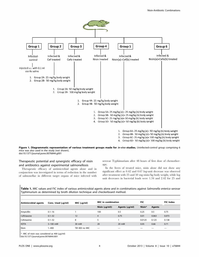
days post-infection, mice were divided into 6 groups each
comprising of at-least 6 mice. A group of six mice was set aside
which served as uninfected-control. Various treatment groups
have been represented in figure 1. All the test agents were injected
in four doses subcutaneously (s.c.) after 12 hour interval individ-
ually and in combination. The doses used were selected on the
basis of pilot studies. At 48 hour post-therapy, mice were sacrificed
and their livers, small intestines, and spleens were removed
aseptically, rinsed in isotonic saline solution and weighed. Ten
percent (w/v) of tissue homogenates were prepared in sterile PBS
using a Potter Elvehjem homogenizer. Serial 10-fold dilutions of
each homogenate were plated on MacConkey agar medium for
enumeration of CFU per organ after incubation at 37uC for
24 hours.
Another set of experiment was performed to evaluate the survival
assay wherein, thirty mice were infected with 0.25 ml of 107 CFU
of serovar Typhimurium orally. At 7 days post-infection, mice were
divided into 3 groups each comprising of 10 mice. Group 1 was
injected subcutaneously with sterile saline. Group 2 was injected
with four doses of nisin (50 mg/kg body weight) + ceftriaxone
(50 mg/kg body weight) subcutaneously (s.c.) after 12 hour interval.
Group 3 was injected with four doses of nisin (50 mg/kg body
weight) + cefotaxime (100 mg/kg body weight) subcutaneously (s.c.)
each after 12 hour interval. The numbers of surviving mice were
recorded at 12 hour intervals over 21 days post-infection.
Statistical analysisData were expressed as means 6 standard deviations for three
to five independent experiments. Statistical analysis was done by
Student’s unpaired t test and one-way analysis of variance
(ANOVA), followed by pairwise comparison procedures (Tukey
test), using Jandel Sigma Stat statistical software, version 2.0. In all
cases, statistical significance was defined as having a P value of
,0.05.
Results
Radial diffusion assayNo zone of inhibition was observed when nisin was used alone.
Agar well diffusion assay indicated that nisin-EDTA, nisin-
ampicillin, nisin-ceftriaxone and nisin-cefotaxime acted in con-
junction for serovar Typhimurium which was evidenced by the
increase in zone of growth inhibition when used together as
compared to the size of zone when these agents were used alone.
However, no difference in zone of inhibition was observed when
nisin-chloramphenicol and nisin-ciprofloxacin were used together
(Data S2).
MIC determinationMIC for various agents was determined and confirmed by both
macro and micro broth dilution assay. For ampicillin, ceftriaxone,
cefotaxime and EDTA, MIC was observed to be 1 mg/ml, 12 mg/
ml, 8 m/ml, and 30 mM, respectively. Using nisin, no inhibition of
growth was observed till 400 mg/ml (Table 1). According to
Clinical and Laboratory Standards Institute guidelines (CLSI
guidelines) [23], serovar Typhimurium was classified as resistant to
ceftriaxone and cefotaxime, and susceptible to ampicillin [as per
the MIC breakpoints for ceftriaxone and cefotaxime susceptible
(#1), intermediate (1–4) and resistant ($4), for ampicillin
susceptible (#8), intermediate (8–32) and resistant ($32)].
Fractional inhibitory concentrations (FIC)The combination of nisin-ceftriaxone and nisin-cefotaxime were
found to be highly synergistic as indicated by FIC indices, which
were #0.5 for serovar Typhimurium. However, nisin-ampicillin
and nisin-EDTA combinations were found to have additive effects
against serovar Typhimurium as indicated by FIC indices, which
were 0.75 and 0.71 respectively (Table 1).
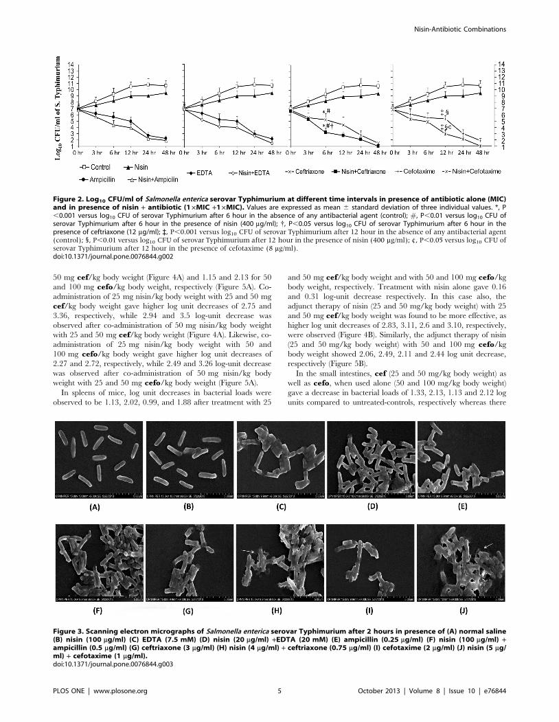
Time-kill AssayAll synergistic interactions inferred from checkerboard analysis
were reassessed by time-kill assay performed with nisin in
combination with ampicillin, ceftriaxone, cefotaxime and EDTA.
Only nisin-ceftriaxone and nisin-cefotaxime combination showed
synergy after 6 hours and 12 hours respectively which was evident
by a $2 log10 unit reduction as compared to the killing of same
magnitude by each agent alone, which seems to occur at 24 hours
(approximately) indicating that the combinations could kill
bacteria more rapidly than each single agent against serovar
Typhimurium (Figure 2). Using nisin-ceftriaxone and nisin-
cefotaxime combination, few colonies were observed when plated
from the flasks after 24 hour incubation. However, when the
incubation was prolonged upto 48 hours, interestingly no colony
was observed.
Scanning electron microscopyIn contrast to control samples, marked changes were evident on
the outer membranes of serovar Typhimurium treated with
various combinations. However, nisin-ceftriaxone and nisin-
cefotaxime combinations seem to cause extensive damage to the
bacterial cells. It is indicated that both these combinations could
lead to clubbing and disruption of bacterial membrane after
2 hours of treatment period (Figure 3).
Nisin-Antibiotic Combinations
PLOS ONE | www.plosone.org 3 October 2013 | Volume 8 | Issue 10 | e76844
Therapeutic potential and synergistic efficacy of nisinand antibiotics against experimental salmonellosis
Therapeutic efficacy of antimicrobial agents alone and in
conjunction was investigated in terms of reduction in the number
of salmonellae in different target organs of mice infected with
serovar Typhimurium after 48 hours of first dose of chemother-
apy.
In the livers of treated mice, nisin alone did not show any
significant effect as 0.42 and 0.67 log-unit decrease was observed
after treatment with 25 and 50 mg nisin/kg body weight, while log
unit decreases in bacterial loads were 1.34 and 2.42 for 25 and
Figure 1. Diagrammatic representation of various treatment groups made for in-vivo studies. Uninfected-control group comprising 6mice was also used in the study (not shown).doi:10.1371/journal.pone.0076844.g001
Table 1. MIC values and FIC index of various antimicrobial agents alone and in combinations against Salmonella enterica serovarTyphimurium as determined by broth dilution technique and checkerboard method.
Antimicrobial agents Conc. Used (mg/ml) MIC (mg/ml) MIC in combination FIC FIC Index
Nisin (mg/ml)) Agents (mg/ml) Nisin* Agents
Ampicillin 0.1–16 1 100 0.5 0.25 0.5 0.75
Ceftriaxone 0.1–32 12 4 0.75 0.01 0.063 0.073
Cefotaxime 0.1–32 8 5 1 0.0125 0.125 0.138
EDTA 5–100 mM 30 mM 20 20 mM 0.05 0.66 0.71
Nisin 1–400 Till 400 no MIC — — — — —
*- MIC of nisin was considered as 400 (mg/ml)doi:10.1371/journal.pone.0076844.t001
Nisin-Antibiotic Combinations
PLOS ONE | www.plosone.org 4 October 2013 | Volume 8 | Issue 10 | e76844
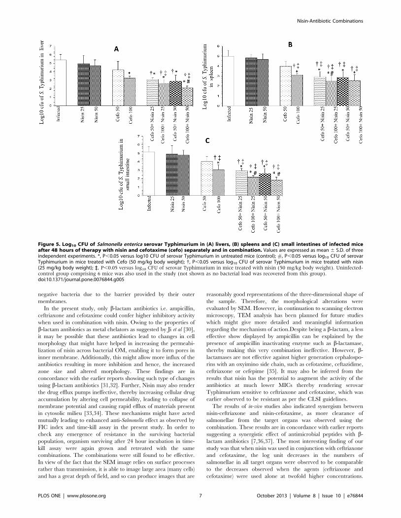
50 mg cef/kg body weight (Figure 4A) and 1.15 and 2.13 for 50
and 100 mg cefo/kg body weight, respectively (Figure 5A). Co-
administration of 25 mg nisin/kg body weight with 25 and 50 mg
cef/kg body weight gave higher log unit decreases of 2.75 and
3.36, respectively, while 2.94 and 3.5 log-unit decrease was
observed after co-administration of 50 mg nisin/kg body weight
with 25 and 50 mg cef/kg body weight (Figure 4A). Likewise, co-
administration of 25 mg nisin/kg body weight with 50 and
100 mg cefo/kg body weight gave higher log unit decreases of
2.27 and 2.72, respectively, while 2.49 and 3.26 log-unit decrease
was observed after co-administration of 50 mg nisin/kg body
weight with 25 and 50 mg cefo/kg body weight (Figure 5A).
In spleens of mice, log unit decreases in bacterial loads were
observed to be 1.13, 2.02, 0.99, and 1.88 after treatment with 25
and 50 mg cef/kg body weight and with 50 and 100 mg cefo/kg
body weight, respectively. Treatment with nisin alone gave 0.16
and 0.31 log-unit decrease respectively. In this case also, the
adjunct therapy of nisin (25 and 50 mg/kg body weight) with 25
and 50 mg cef/kg body weight was found to be more effective, as
higher log unit decreases of 2.83, 3.11, 2.6 and 3.10, respectively,
were observed (Figure 4B). Similarly, the adjunct therapy of nisin
(25 and 50 mg/kg body weight) with 50 and 100 mg cefo/kg
body weight showed 2.06, 2.49, 2.11 and 2.44 log unit decrease,
respectively (Figure 5B).
In the small intestines, cef (25 and 50 mg/kg body weight) as
well as cefo, when used alone (50 and 100 mg/kg body weight)
gave a decrease in bacterial loads of 1.33, 2.13, 1.13 and 2.12 log
units compared to untreated-controls, respectively whereas there
Figure 2. Log10 CFU/ml of Salmonella enterica serovar Typhimurium at different time intervals in presence of antibiotic alone (MIC)and in presence of nisin + antibiotic (16MIC +16MIC). Values are expressed as mean 6 standard deviation of three individual values. *, P,0.001 versus log10 CFU of serovar Typhimurium after 6 hour in the absence of any antibacterial agent (control); #, P,0.01 versus log10 CFU ofserovar Typhimurium after 6 hour in the presence of nisin (400 mg/ml); {, P,0.05 versus log10 CFU of serovar Typhimurium after 6 hour in thepresence of ceftriaxone (12 mg/ml); `, P,0.001 versus log10 CFU of serovar Typhimurium after 12 hour in the absence of any antibacterial agent(control); 1, P,0.01 versus log10 CFU of serovar Typhimurium after 12 hour in the presence of nisin (400 mg/ml); ¢, P,0.05 versus log10 CFU ofserovar Typhimurium after 12 hour in the presence of cefotaxime (8 mg/ml).doi:10.1371/journal.pone.0076844.g002
Figure 3. Scanning electron micrographs of Salmonella enterica serovar Typhimurium after 2 hours in presence of (A) normal saline(B) nisin (100 mg/ml) (C) EDTA (7.5 mM) (D) nisin (20 mg/ml) +EDTA (20 mM) (E) ampicillin (0.25 mg/ml) (F) nisin (100 mg/ml) +ampicillin (0.5 mg/ml) (G) ceftriaxone (3 mg/ml) (H) nisin (4 mg/ml) + ceftriaxone (0.75 mg/ml) (I) cefotaxime (2 mg/ml) (J) nisin (5 mg/ml) + cefotaxime (1 mg/ml).doi:10.1371/journal.pone.0076844.g003
Nisin-Antibiotic Combinations
PLOS ONE | www.plosone.org 5 October 2013 | Volume 8 | Issue 10 | e76844

were log unit decreases of 0.26 and 0.38 in the number of
salmonellae in small intestines of mice treated with 25 and 50 mg
nisin/kg body weight, respectively. However, when 25 and 50 mg
of nisin/kg body weight were used in combination with cef (25
and 50 mg/kg body weight), the log unit decreases in intestinal
bacterial loads were found to be 2.24, 2.71, 2.35 and 2.83 units,
respectively (Figure 4C). Similar trend was observed using nisin-
cefotaxime combination. Combination of nisin (25 and 50 mg/kg
body weight) with cefo (50 and 100 mg/kg body weight) showed
2.23, 3, 2.57 and 3.34 log-unit decrease respectively (Figure 5C).
In other words, bacterial loads recovered from all target organs,
when treated with lower doses of each agent in conjunction were
observed to be at par with the recovered bacterial loads observed
when the agents were used alone at twofold higher concentrations.
In the survival assay, mice from both the groups receiving
treatments showed an increase survival as compared to that in the
infected-control group. In the infected-control group 50%, 70%
and 100% mortality was observed at 9, 11 and 14 days post-
infection respectively. However, 90% and 80% survival was
observed up to 21 days in nisin-ceftriaxone and nisin-cefotaxime
treated groups respectively (Data S3). 10 and 20% mortality
observed in the treated groups may be due to some other reason
including their immuno-compromised state. However, further
investigation such as histology of the liver and intestinal tissues
along with blood evaluations would substantiate the clarity of the
survival curve.
Discussion
Synergy between two antimicrobial drugs is obtained when the
combination of drugs elicits a more than additive effect as opposed
to each drug alone. Its clinical importance results in taking
advantage of different mechanisms of action of the agents involved
[24] and providing an additional therapeutic choice in difficult-to-
treat infections [25]. A major problem in combating Salmonella is its
outer membrane (OM). The OM is a lipid bilayer in which the
predominant lipid is the lipopolysaccharide (LPS). The LPS layer
forms a tight shield [26] and acts as a barrier to many compounds,
including antibiotics, hydrophobic compounds, detergents and
dyes [27]. The anionic LPS layer is stabilized by divalent cations.
If these cations are removed, lipopolysaccharide molecules are
released from the OM, exposing the underlying phospholipid
bilayer and jeopardizing the integrity of the OM [27]. Bacteriocins
have been found to have many distinct mechanisms of action that
differ from those of antibiotics. These mechanisms can be broadly
divided into those that function primarily at the cell envelope and
those that are active primarily within the cell, affecting gene
expression and protein production [28]. Nisin has been reported
to exert its antimicrobial activity through both pore formation in
the membrane and by binding to lipid II, which is an essential
intermediate in peptidoglycan biogenesis. Precisely how this occurs
and whether a receptor is involved is yet to be elucidated [28,29].
However, nisin alone is generally not effective against Gram-
Figure 4. Log10 CFU of Salmonella enterica serovar Typhimurium in (A) livers, (B) spleens and (C) small intestines of infected miceafter 48 hours of therapy with nisin and ceftriaxone (cef) separately and in combination. Values are expressed as mean 6 S.D. of threeindependent experiments. *, P,0.05 versus log10 CFU of serovar Typhimurium in untreated mice (control); #, P,0.05 versus log10 CFU of serovarTyphimurium in mice treated with Cef (25 mg/kg body weight); {, P,0.05 versus log10 CFU of serovar Typhimurium in mice treated with nisin(25 mg/kg body weight); `, P,0.05 versus log10 CFU of serovar Typhimurium in mice treated with nisin (50 mg/kg body weight). Uninfected-control group comprising 6 mice was also used in the study (not shown as no bacterial load was recovered from this group).doi:10.1371/journal.pone.0076844.g004
Nisin-Antibiotic Combinations
PLOS ONE | www.plosone.org 6 October 2013 | Volume 8 | Issue 10 | e76844
negative bacteria due to the barrier provided by their outer
membranes.
In the present study, only b-lactam antibiotics i.e. ampicillin,
ceftriaxone and cefotaxime could confer higher inhibitory activity
when used in combination with nisin. Owing to the properties of
b-lactam antibiotics as metal chelators as suggested by Ji et al [30],
it may be possible that these antibiotics lead to changes in cell
morphology that might have helped in increasing the permeabi-
lization of nisin across bacterial OM, enabling it to form pores in
inner membrane. Additionally, this might allow more influx of the
antibiotics resulting in more inhibition and hence, the increased
zone size and altered morphology. These findings are in
concordance with the earlier reports showing such type of changes
using b-lactam antibiotics [31,32]. Further, Nisin may also render
the drug efflux pumps ineffective, thereby increasing cellular drug
accumulation by altering cell permeability, leading to collapse of
membrane potential and causing rapid efflux of materials present
in cytosolic milieu [33,34]. These mechanisms might have acted
mutually leading to enhanced anti-Salmonella effect as observed by
FIC index and time-kill assay in the present study. In order to
check any emergence of resistance in the surviving bacterial
population, organism surviving after 24 hour incubation in time-
kill assay were again grown and retreated with the same
combinations. The combinations were still found to be effective.
In view of the fact that the SEM image relies on surface processes
rather than transmission, it is able to image large area (many cells)
and has a great depth of field, and so can produce images that are
reasonably good representations of the three-dimensional shape of
the sample. Therefore, the morphological alterations were
evaluated by SEM. However, in continuation to scanning electron
microscopy, TEM analysis has been planned for future studies
which might give more detailed and meaningful information
regarding the mechanism of action.Despite being a b-lactam, a less
effective show displayed by ampicillin can be explained by the
presence of ampicillin inactivating enzyme such as b-lactamase,
thereby making this very combination ineffective. However, b-
lactamases are not effective against higher generation cephalospo-
rins with an oxyimino side chain, such as cefotaxime, ceftazidime,
ceftriaxone or cefepime [35]. It may also be inferred from the
results that nisin has the potential to augment the activity of the
antibiotics at much lower MICs thereby rendering serovar
Typhimurium sensitive to ceftriaxone and cefotaxime, which was
earlier observed to be resistant as per the CLSI guidelines.
The results of in-vivo studies also indicated synergism between
nisin-ceftriaxone and nisin-cefotaxime, as more clearance of
salmonellae from the target organs was observed using the
combination. These results are in concordance with earlier reports
suggesting a synergistic effect of antimicrobial peptides with b-
lactam antibiotics [7,36,37]. The most interesting finding of our
study was that when nisin was used in conjunction with ceftriaxone
and cefotaxime, the log unit decreases in the numbers of
salmonellae in all target organs were observed to be comparable
to the decreases observed when the agents (ceftriaxone and
cefotaxime) were used alone at twofold higher concentrations.
Figure 5. Log10 CFU of Salmonella enterica serovar Typhimurium in (A) livers, (B) spleens and (C) small intestines of infected miceafter 48 hours of therapy with nisin and cefotaxime (cefo) separately and in combination. Values are expressed as mean 6 S.D. of threeindependent experiments. *, P,0.05 versus log10 CFU of serovar Typhimurium in untreated mice (control); #, P,0.05 versus log10 CFU of serovarTyphimurium in mice treated with Cefo (50 mg/kg body weight); {, P,0.05 versus log10 CFU of serovar Typhimurium in mice treated with nisin(25 mg/kg body weight); `, P,0.05 versus log10 CFU of serovar Typhimurium in mice treated with nisin (50 mg/kg body weight). Uninfected-control group comprising 6 mice was also used in the study (not shown as no bacterial load was recovered from this group).doi:10.1371/journal.pone.0076844.g005
Nisin-Antibiotic Combinations
PLOS ONE | www.plosone.org 7 October 2013 | Volume 8 | Issue 10 | e76844
Therefore, it reduced the therapeutic dosage of both agents to half
(from 50 to 25 mg/kg of ceftriaxone and from 100 to 50 mg/kg of
cefotaxime) while maintaining the increased therapeutic efficacy.
Moreover, treatment with the combination also enhanced the
survival rate of the infected animals. However, it is worth
mentioning here that though the treatment resulted in significant
reduction in the bacterial load but the organisms could still be
isolated from the organs indicating that animals have not fully
rejuvenated. Had the treatment been either started earlier or
prolonged, absolute recovery would have been possible.
Thus judicious and restricted antibiotic use might help in
withdrawal of selective pressure, thereby reducing the chances of
developing resistance induced by the action of extended spectrum
b-lactamases (ESBL) [38]. Therefore, the present investigation can
now be used as a blueprint for the development of new class of
highly efficient antibiotics for the treatment of Gram-negative
infections.
In conclusion, nisin-ceftriaxone and nisin-cefotaxime combina-
tions demonstrated excellent in-vitro and in-vivo synergism against
the serovar Typhimurium. This study highlights that nisin has the
potential to act in conjunction with conventional antibiotics at
much lower concentrations (as evidenced from in-vitro and in-vivo
results). Thus, in addition to having the faster effect using the
combination, reducing the concentration of the antibiotics may
reduce the chances of developing resistance in the pathogens
besides reducing the associated side effects of the former. Nisin-
antibiotic combination appears to fulfill the dual purpose over
nisin-EDTA combination as i) the former may reduce the effective
therapeutic dose of the antibiotics and ii) would avoid the
otherwise chelating property of the latter in the in-vivo situation.
Given the value addition potential of nisin, its combination with
antibiotics can thus be exploited instead of nisin-EDTA combi-
nation both against Salmonella and other Gram-negative infections
as well. However, the effect of these combinations against the
intestinal microbiota needs to be explored in future studies in
order to consider the combinations clinically more relevant.
Supporting Information
Data S1 An illustration of checkerboard assay showing64 combinations used to determine FIC.
(DOC)
Data S2 Zone of inhibition in presence of variousantimicrobial agents.
(DOC)
Data S3 Survival analysis of BALB/c mice infected withSalmonella enterica serovar Typhimurium followed bytreatment with nisin (50 mg/kg body weight) + ceftria-xone (50 mg/kg body weight) and nisin (50 mg/kg bodyweight) + cefotaxime (100 mg/kg body weight) subcuta-neously (s.c.).
(TIF)
Acknowledgments
The authors express their gratitude to Mr. Mohinder Singh at the
Sophisticated Analytical Instrumentation Facility (SAIF)/Central Instru-
mentation Laboratory, Panjab University, Chandigarh, India and Mr.
Rahul Mahajan at National Institute of Pharmaceutical and Educational
Research (NIPER), S.A.S. Nagar, Punjab, India, for making stubs and
providing assistance in scanning electron microscopic analysis of the
samples.
Author Contributions
Conceived and designed the experiments: PR APS. Performed the
experiments: APS. Analyzed the data: APS. Contributed reagents/
materials/analysis tools: PR VP. Wrote the paper: APS PR.
References
1. Chalon MC, Acuna L, Morero RD, Minahk CJ, Bellomio A (2012) Membrane-active bacteriocins to control Salmonella in foods: Are they the definite hurdle?
Food Res Int 45: 735–744.
2. Cohen ML, Tauxe RV (1986) Drug-resistant Salmonella in the United States: An
epidemiologic perspective. Science 234: 964–969.
3. Mølbak K (2005) Human health consequences of antimicrobial drug-resistantSalmonella and other foodborne pathogens. Clin Infect Dis 41: 1613–1620.
4. Joerger RD (2003) Alternatives to antibiotics: bacteriocins, antimicrobialpeptides and bacteriophages. Poult Sci 82: 640–647.
5. Birosova L, Mikulasova M (2009) Development of triclosan and antibiotic
resistance in Salmonella enterica serovar Typhimurium. J Med Microbiol 58: 436–441.
6. Li B, Yu JPJ, Brunzelle JS, Moll GN, Van der Donk WA, et al. (2006) Structureand mechanism of the lantibiotic cyclase involved in nisin biosynthesis. Science
311: 1464–1467.
7. Rishi P, Preet S, Bharrhan S, Verma I (2011) In-vitro and in-vivo synergistic effects
of cryptdin 2 and ampicillin against Salmonella. Antimicrob Agents Chemother55: 4176–4182.
8. Singh AP, Preet S, Rishi P (2011) Augmentation of antimicrobial activity of
conventional antibiotics by cell free extract of L. plantarum. J Antibiot 64: 795–798.
9. Hancock REW, Chapple DS (1999) Peptide antibiotics. Antimicrob AgentsChemother 43: 1317–1323.
10. Hancock REW, Scott MG (2000) The role of antimicrobial peptides in animal
defenses. Proc Natl Acad Sci U S A 97: 8856–8861.
11. Preet S, Verma I, Rishi P (2010) Cryptdin-2: a novel therapeutic agent for
experimental Salmonella Typhimurium infection. J Antimicrob Chemother 65:991–994.
12. Jay JM (2000) Modern Food Microbiology, 6th ed. Aspen Publishers Inc.
Gaithersburg, Maryland. 679 p.
13. Murdock CA, Cleveland J, Matthews KR, Chikindas ML (2007) The synergistic
effect of nisin and lactoferin on the inhibition of Listeria monocytogenes andEscherichia coli O157:H7. Lett Appl Microbiol 44: 255–261.
14. Cutter CN, Siragusa GR (1995) Population reduction of Gram negative
pathogens Following Treatments with Nisin and Chelators. J Food Prot 58: 977–983.
15. Stevens KA, Sheldon BW, Klapes NA, Klaenhammer TR (1991) Nisin
treatment for inactivation of Salmonella species and other Gram-negativebacteria. Appl Environ Microbiol 57: 3613–3615.
16. Carneiro de Melo AM, Cassar CA, Miles RJ (1998) Trisodium phosphate
increase sensitivity of Gram-negative bacteria to lysozyme and nisin. J Food Prot
61: 839–844.
17. Solomakos N, Govaris A, Koidis P, Botsoglou N (2008) The antimicrobial effect
of thyme essential oil, nisin and their combination against Escherichia coli
O157:H7 in minced beef during refrigerated storage. Meat Sci 80: 159–166.
18. Govaris A, Solomakos N, Pexara A, Chatzopoulou PS (2010) The antimicrobialeffect of oregano essential oil, nisin and their combination against Salmonella
enteritidis in minced sheep meat during refrigerated storage. Int J Food Microbiol137: 175–180.
19. Boziaris IS, Adams MR (1999) Effect of chelators and nisin produced in situ on
inhibition and inactivation of Gram negatives. Int J Food Microbiol 53: 105–113.
20. Lin CH, Hou RF, Shyu CL, Shia WY, Lin CF, et al. (2012) In-vitro activity of
mastoparan-AF alone and in combination with clinically used antibiotics againstmultiple-antibiotic-resistant Escherichia coli isolates from animals. Peptides 36:
114–120.
21. Rishi P, Preet S, Kaur P (2011) Effect of L. plantarum cell-free extract and co-
trimoxazole against Salmonella typhimurium: a possible adjunct therapy. Ann ClinMicrobiol Antimicrob 10: 9.
22. Yenugu S, Narmadha G (2010) The human male reproductive tract
antimicrobial peptides of the HE2 family exhibit potent synergy with standardantibiotics. J Pept Sci 16: 337–341.
23. Clinical and Laboratory Standards Institute (2010) Performance Standards for
Antimicrobial Susceptibility Testing: 20th Informational Supplement M100-S20. CLSI, Wayne, PA, USA.
24. Rybak JM, McGrath J (1996) Combination antimicrobial therapy for bacterial
infections, Guidelines for the Clinician. Drugs 52: 390–402.
25. Serra P, Brandimarte C, Martino P, Carlone S, Giunchi G (1977) Synergistictreatment of enterococcal endocarditis: in vitro and in vivo studies. Arch Intern
Med 137: 1562–1567.
26. Raetz CRH, Whitfield C (2002) Lipopolysaccharide endotoxins. Annu RevBiochem 71: 635–700.
Nisin-Antibiotic Combinations
PLOS ONE | www.plosone.org 8 October 2013 | Volume 8 | Issue 10 | e76844

27. Vaara M (1992) Agents that increase the permeability of the outer membrane.
Microbiol Rev 56: 395–411.28. Paul D, Cotter R, Ross P, Hill C (2013) Bacteriocins- a viable alternative to
antibiotics? Nat Rev Microbiol 11: 95–105.
29. Freeman MF, Gurgui C, Helf MJ, Morinaka BI, Uria AR, et al. (2012)Metagenome mining reveals polytheonamides as posttranslationally modified
ribosomal peptides. Science 338: 387–390.30. Ji HF, Shen L, Zhang HY (2005) b-Lactam antibiotics are multipotent agents to
combat neurological diseases. Biochem and Biophy Res Commun 333: 661–663.
31. Spratt BG (1975) Distinct penicillin binding proteins involved in the division,elongation, and shape of Escherichia coli K12. Proc Natl Acad Sci U S A 72:
2999–3003.32. Kitano K, Tomasz A (1979) Triggering of autolytic cell wall degradation in
Escherichia coli by beta-lactam antibiotics. Antimicrob Agents Chemother 16:838–848.
33. Ruhr E, Sahl HG (1985) Mode of action of the peptide antibiotic nisin and
influence on the membrane potential of whole cells and on cytoplasmic andartificial membrane vesicles. Antimicrob Agents Chemother 27: 841–845.
34. Gut IM, Blanke SR, van der Donk WA (2011) Mechanism of inhibition of
Bacillus anthracis spore outgrowth by the lantibiotic nisin. ACS Chem Biol 6: 744–752.
35. Medeiros AA (1997) Evolution and dissemination of b-lactamases accelerated bygenerations of b-lactam antibiotics. Clin Infect Dis 24 (Suppl 1): S19–45.
36. Ulvatne H, Karoliussen S, Stiberg T, Rekdal O, Svendsen JS (2001) Short
antibacterial peptides and erythromycin act synergically against Escherichia coli.J Antimicrob Chemother 48: 203–208.
37. Kalita A, Verma I, Khuller GK (2004) Role of human neutrophil peptide-1 as apossible adjunct to antituberculosis chemotherapy. J Infect Dis 190: 1476–1480.
38. Chaudhary U, Aggarwal R (2004) Extended spectrum b-lactamases (ESBL) - Anemerging threat to clinical therapeutics. Indian J Med Microbiol 22: 75–80.
Nisin-Antibiotic Combinations
PLOS ONE | www.plosone.org 9 October 2013 | Volume 8 | Issue 10 | e76844
Copyright © 2022 FDOKUMEN