Validation of Myocardial Acceleration During Isovolumic Contraction as a Novel Noninvasive Index of...
8
Keld Sorensen and Andrew N. Redington Michael Vogel, Michael R. Schmidt, Steen B. Kristiansen, Michael Cheung, Paul A. White, Pressure-Volume Relations in an Animal Model Noninvasive Index of Right Ventricular Contractility: Comparison With Ventricular Validation of Myocardial Acceleration During Isovolumic Contraction as a Novel Print ISSN: 0009-7322. Online ISSN: 1524-4539 Copyright © 2002 American Heart Association, Inc. All rights reserved. is published by the American Heart Association, 7272 Greenville Avenue, Dallas, TX 75231 Circulation doi: 10.1161/01.CIR.0000013773.67850.BA 2002;105:1693-1699; originally published online March 25, 2002; Circulation. http://circ.ahajournals.org/content/105/14/1693 World Wide Web at: The online version of this article, along with updated information and services, is located on the http://circ.ahajournals.org//subscriptions/ is online at: Circulation Information about subscribing to Subscriptions: http://www.lww.com/reprints Information about reprints can be found online at: Reprints: document. Permissions and Rights Question and Answer this process is available in the click Request Permissions in the middle column of the Web page under Services. Further information about Office. Once the online version of the published article for which permission is being requested is located, can be obtained via RightsLink, a service of the Copyright Clearance Center, not the Editorial Circulation in Requests for permissions to reproduce figures, tables, or portions of articles originally published Permissions: by guest on May 30, 2014 http://circ.ahajournals.org/ Downloaded from by guest on May 30, 2014 http://circ.ahajournals.org/ Downloaded from
Transcript of Validation of Myocardial Acceleration During Isovolumic Contraction as a Novel Noninvasive Index of...
Keld Sorensen and Andrew N. RedingtonMichael Vogel, Michael R. Schmidt, Steen B. Kristiansen, Michael Cheung, Paul A. White,
Pressure-Volume Relations in an Animal ModelNoninvasive Index of Right Ventricular Contractility: Comparison With Ventricular
Validation of Myocardial Acceleration During Isovolumic Contraction as a Novel
Print ISSN: 0009-7322. Online ISSN: 1524-4539 Copyright © 2002 American Heart Association, Inc. All rights reserved.
is published by the American Heart Association, 7272 Greenville Avenue, Dallas, TX 75231Circulation doi: 10.1161/01.CIR.0000013773.67850.BA
2002;105:1693-1699; originally published online March 25, 2002;Circulation.
http://circ.ahajournals.org/content/105/14/1693World Wide Web at:
The online version of this article, along with updated information and services, is located on the
http://circ.ahajournals.org//subscriptions/
is online at: Circulation Information about subscribing to Subscriptions:
http://www.lww.com/reprints Information about reprints can be found online at: Reprints:
document. Permissions and Rights Question and Answer this process is available in the
click Request Permissions in the middle column of the Web page under Services. Further information aboutOffice. Once the online version of the published article for which permission is being requested is located,
can be obtained via RightsLink, a service of the Copyright Clearance Center, not the EditorialCirculationin Requests for permissions to reproduce figures, tables, or portions of articles originally publishedPermissions:
by guest on May 30, 2014http://circ.ahajournals.org/Downloaded from by guest on May 30, 2014http://circ.ahajournals.org/Downloaded from

Validation of Myocardial Acceleration During IsovolumicContraction as a Novel Noninvasive Index of Right
Ventricular ContractilityComparison With Ventricular Pressure-Volume Relations
in an Animal Model
Michael Vogel, MD, PhD; Michael R. Schmidt, MD; Steen B. Kristiansen, MD;Michael Cheung, MB, ChB; Paul A. White, PhD; Keld Sorensen, MD; Andrew N. Redington, MD, FRCP
Background—We have demonstrated that myocardial acceleration during isovolumic contraction (IVA) is a sensitiveindex of left ventricular contractile function. In this study, we assessed the utility of IVA to measure right ventricular(RV) contractile function.
Methods and Results—We examined 8 pigs by using tissue Doppler imaging of the RV free wall and simultaneousmeasurements of intraventricular pressure, volume, maximal elastance (emax), preload recruitable stroke work, anddP/dtmax by conductance catheterization. Animals were paced in the right atrium at a rate of 130 beats per minute (bpm).IVA was compared with elastance during contractility modulation by esmolol and dobutamine and during preloadreduction and afterload increase by transient balloon occlusion of the inferior vena cava and pulmonary artery,respectively. Data were also obtained during incremental atrial pacing from 110 to 210 bpm. Esmolol led to a decreasein IVA and dP/dtmax. During dobutamine infusion, IVA, dP/dtmax, preload recruitable stroke work, and emax all increasedsignificantly. During preload reduction and afterload increase, IVA remained constant up to a reduction of RV volumeby 54% and an RV systolic pressure increase of 58%. Pacing up to a rate of 190 bpm led to a stepwise increase in IVAand dP/dtmax, with a subsequent fall at a pacing rate of 210 bpm.
Conclusions—IVA is a measurement of RV contractile function that is unaffected by preload and afterload changes in aphysiological range and is able to measure the force-frequency relation. This novel index may be ideally suited to theassessment of acute changes of RV function in clinical studies. (Circulation. 2002;105:1693-1699.)
Key Words: ventricles � pacing � contractility � echocardiography
In recent years there has been an increased interest in theassessment of right ventricular (RV) function in both
acquired and congenital heart disease.1–3 Because of itscomplex geometric shape and other spatial considerations, ithas been notoriously difficult to noninvasively assess RVfunction.4 Although RV ejection fraction can now be derivedfrom volumetric data obtained by MRI or 3-dimensionalechocardiography, the load dependency of ejection fractionlimits its utility.4,5 Indeed, validity of any single beat–derivedejection phase index of RV contractile function is question-able because changes in loading conditions, especially after-load, markedly influence such measurements. Furthermore,the RV in congenital heart disease is frequently characterizedby pressure or volume overload, obviating useful comparisonof these ejection phase indexes with data derived in thenormal RV.1 Tissue Doppler imaging (TDI) has the potential
to assess ventricular contractile function independent of theshape of the ventricle. However, the magnitude of ejectionphase myocardial velocities has been shown to be preload andafterload dependent.6 Isovolumic indexes are likely to bemore robust. Indeed, isometric myocardial twitch velocitiesare frequently used in in vitro studies of myocardial contrac-tile function.7,8 Myocardial velocities, recorded in vivo,during isovolumic contraction have also been studied throughthe use of TDI.9 In an animal study there was an acceptable(r�0.72) correlation between peak velocity during isovolu-mic contraction and ejection fraction, but neither load norheart rate dependency was formally assessed.9 Myocardialacceleration has not been measured previously through theuse of TDI; however, with the use of a microaccelerometersensor located in the tip of a pacing lead developed with theintention to optimize pacing systems,10 experimental and
Received October 19, 2001; revision received January 29, 2002; accepted January 29, 2002.From the Cardiothoracic Unit (M.V., M.C., P.A.W., A.N.R.), Great Ormond Street Hospital for Children, NHS Trust, London, UK, and the
Departments of Experimental Research (M.R.S., S.B.K.) and Cardiology (K.S.), Skejby Hospital, Aarhus, Denmark.Correspondence to Prof Andrew Redington, Division of Cardiology, The Hospital for Sick Children, 555 University Ave, Toronto M5G 1X 8, Canada.
E-mail [email protected]© 2002 American Heart Association, Inc.
Circulation is available at http://www.circulationaha.org DOI: 10.1161/01.CIR.0000013773.67850.BA
1693 by guest on May 30, 2014http://circ.ahajournals.org/Downloaded from

clinical studies11,12 have demonstrated that peak endocardialacceleration occurs during isovolumic contraction and in-creases linearly during dobutamine infusion.11 Recently, wedemonstrated using TDI in an animal model that myocardialacceleration during isovolumic contraction (IVA) is a rela-tively load-independent index of left ventricular (LV) con-tractile function. The hypothesis of this study was that IVAwould be a similarly robust index of contractile function inthe RV. We compared IVA with other TDI-derived indexesof systolic function and the “gold” standard of invasiveassessment of contractile function, the analysis of pressure-volume relations in a closed-chest animal model duringmodulation of preload, afterload, contractility, and heart rate.
MethodsAnimal ProtocolWe studied 8 Danish land-race pigs weighing �15 kg. The studyconformed to the guidelines of the American Heart Association onresearch animal experiments. After premedication with azaperone (4mg/kg), midazolam (0.1 mg/kg), and etomidate (0,5 mg/kg IV), theanimals were endotracheally intubated and ventilated with a Servo900 (Siemens) ventilator. Arterial blood gases were taken to confirmadequate ventilation during the study. Anesthesia was maintainedwith 2% isofluorane in a mixture of NO2 and oxygen. A cutdown wasperformed on both sides of the neck. Through the right internaljugular vein, a custom-made 8 polar (total interelectrode distance, 3cm), 5F combination conductance-pressure catheter (Millar Instru-ments) was placed into the apex of the RV under fluoroscopicguidance. The micromanometer pressure transducer output was fedto a custom-built amplifier. The conductance electrodes were con-nected to a signal-processing unit (Sigma 5DF, CardiodynamicsCorp). A 20-mm latex balloon catheter (Boston Scientific) wasplaced into the right external jugular vein at the junction of theinferior vena cava and the right atrium and subsequently inflated tomodify preload. Through the left internal jugular vein, a Rashkindballoon septostomy catheter (Meditec) was placed in the mainpulmonary artery and prepared for inflation to modify RV afterload.After modification of loading conditions, the balloon catheter in thepulmonary artery was removed and replaced by a standard 7Fthermodilution catheter (Baxter Healthcare) connected to a dedicatedcardiac output processing computer (Com2, Baxter Edwards). A 5Fpacing wire was inserted into the left external jugular vein, advancedinto the right atrium, and connected to an external pacemakergenerator (Medtronic). The right atrium was paced at a constant rateof 130 beats per minute (bpm) during preload and afterload changesand during contractility modulation by esmolol and dobutamine.Pressure and volume data were digitized at 1000 Hz and stored on apersonal computer with dedicated software for later analysis. Parallelconductance was calculated in the usual way13 after injection of 2mL of 10% NaCl into the inferior vena cava. The gain constant �was calculated as the ratio of conductance-derived stroke volume andthe stroke volume measured by thermodilution.13
Transthoracic echocardiography was performed with the use of aGE Vingmed Vivid V ultrasound scanner with a frame rate between131 and 248 Hertz. In a 4-chamber equivalent view, the RV free wallwas imaged and color-coded myocardial velocities were recorded atthe base immediately below the insertion of the tricuspid valveleaflets. Recordings were made simultaneously with ECG. Acineloop of at least 6 consecutive heartbeats was stored digitally foroff-line analysis.
ProtocolAll physiological data were obtained during apnea to minimizecardiopulmonary interactions. Echocardiography and pressure-volume data were simultaneously recorded, and at each stage cardiacoutput was measured.
The measurements were obtained during the following protocol,chronologically:
1. BaselineBaseline measurements were performed first.
2. Preload ReductionTo assess the effect of preload reduction on IVA, TDI was performedcontinuously during partial occlusion of the inferior vena cava, anda cineloop of 6 consecutive cardiac cycles at different stages ofventricular volume was recorded. An electrical timing signal wasrecorded to synchronize TDI and pressure and volume data duringballoon inflation.
3. Afterload IncreaseAfter a 10-minute rest period, a balloon was inflated in thepulmonary artery until RV systolic pressure had increased by at least25%. TDI and RV pressure data were recorded simultaneously.
4. Heart Rate ModulationThe pacemaker rate, which otherwise was kept constant at 130 bpm,was reduced to 110 for 1 minute and rapidly increased by incrementsof 10 bpm to a maximum rate of 210 bpm. At each increment, TDIdata and ventricular pressure recordings were obtained.
5. Reduced ContractilityAfter a further 10-minute rest period, an infusion of 500 �g/kg perminute of esmolol was started, which was increased to 1 mg/kg perminute after 5 minutes and maintained at the latter level for a further5 minutes. Conductance catheter–derived pressure-volume data wereobtained during transient preload reduction by balloon occlusion13 atsteady state before commencing esmolol and at each dosage incre-ment. TDI data were also recorded immediately before each conduc-tance catheter measurement.
6. Increased ContractilityAfter a further 10-minute rest, dobutamine was infused at a rate of 10�g/kg per minute for 10 minutes. Conductance catheter–derivedpressure-volume data were obtained during transient preload reduc-tion by balloon occlusion13 at steady state before commencingdobutamine and after dobutamine had been administered for at least10 minutes. TDI data were also recorded immediately before eachconductance catheter measurement.
Data AnalysisPressure-volume data were analyzed off-line by one examiner whowas blinded to the data obtained by TDI, which were analyzed by adifferent observer. All conductance volumes were corrected forparallel conductance and the gain constant �.13 The maximal slope(maximal P/V) of the end-systolic pressure-volume relation: end-sys-tolic elastance (emax) during caval occlusion, preload recruitablestroke work (PRSW), and dP/dtmax were calculated.13 Echopacsoftware (GE Vingmed) was used to analyze the stored TDI data.The sample volume was placed in the middle of the myocardium atthe basal free wall. Peak myocardial velocities during isovolumiccontraction (Q wave to onset of systolic ejection), systolic ejection (Swave), as well as acceleration during isovolumic contraction wererecorded (Figure 1). Acceleration was calculated as the differencebetween baseline and peak velocity divided by their time interval.Measurements of myocardial acceleration and velocities were calcu-lated from 3 consecutive cardiac cycles with the average of the 3measurements recorded. An second independent observer, who wasblinded to the results of the TDI derived data analysis, measured IVAin 40 data sets to assess interobserver variability, and the firstobserver measured these 40 data sets on a different day to assessintraobserver variability.
Statistical AnalysisData are expressed as mean�SD. Measurements of TDI-derivedparameters and pressure-volume data at the various stages of preloadand afterload modification were analyzed by means of ANOVA forrepeated measurements. Linear regression analysis was used to
1694 Circulation April 9, 2002
by guest on May 30, 2014http://circ.ahajournals.org/Downloaded from

compare changes in emax with changes in IVA and isovolumiccontraction myocardial velocity. A Bland-Altman analysis wasperformed on the interobserver data.14
ResultsInfluence of Preload and Afterload ChangesDuring preload reduction, RV end-diastolic volume fell by34�12% (P�0.0001). During afterload increase, RV systolic
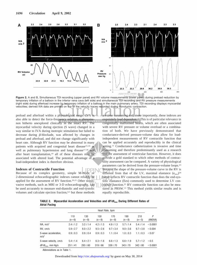
pressure increased by 54�12% (P�0.0001). IVA was unaf-fected by these changes in preload and afterload, whereas theejection systolic myocardial velocities (S wave) changedsignificantly (Table 1 and Figure 2, A and B).
Heart Rate ModulationThere was a positive correlation between IVA and heart rate(r�0.74, P�0.01). Overall, IVA increased by 135�62%(P�0.0003) as heart rate increased from 110 to 190 bpm(Table 2). Simultaneously measured dP/dtmax increased by76�23% (P�0.001). At a heart rate of 210 bpm, however,IVA fell by 5.6�3% and dP/dtmax by 0.3�0.2% from thevalues measured at a heart rate of 190 bpm (Figure 3), butneither the fall in IVA or dP/dtmax reached statisticalsignificance.
IVA as an Index of RV Contractile FunctionDuring contractility modulation by esmolol infusion, therewas no significant fall in emax or PRSW, but IVA and dP/dtmax
fell significantly by 16�19% (P�0.03) and 15�13%(P�0.002), respectively (Table 3). The other TDI-derivedparameters did not change during esmolol infusion.
During dobutamine infusion, we found a significant in-crease of IVA by 68�73%, emax by 53�63%, PRSW by124�84%, and dP/dtmax by 52�19%. Among the otherTDI-derived parameters, S-wave velocity increased signifi-cantly by 57�29% (Table 3 and Figure 4).
Observer VariabilityThe mean difference between observer 1 and observer 2 was8.9�7.6%. At higher IVA, some differences between observ-ers were outside the confidence limits (Figure 5). The meanintraobserver difference was 5.6�4.3%.
DiscussionThis study demonstrates that IVA measured by TDI reflectsRV myocardial contractile function and is unaffected by
Figure 1. Typical tissue Doppler tracing obtained at the base ofthe RV. Wave during isovolumic contraction precedes the Swave and begins before the peak of the R wave on ECG. IVAwas measured by dividing peak velocity by the time intervalfrom onset of the wave (zero-crossing) during isovolumic con-traction to the time at peak velocity of this wave.
TABLE 1. Data on Preload Reduction With Simultaneous Volume Measurements and Afterload IncreaseWith Simultaneous Pressure Measurements by Conductance Catheterization and TDI-Derived Data During 6Consecutive Cardiac Cycles
CycleBefore
Cycle 1After
Cycle 2After
Cycle 3After
Cycle 4After
Cycle 5After
P(ANOVA)
A. Preload reduction
IVA, m/s2 2.6�1.0 2.5�1.0 2.6�1.0 2.8�1.3 2.8�1.3 2.5�1.3 �0.5
IVV, cm/s 7.3�2.9 7.4�2.6 7.6�2.8 7.8�2.9 8.1�3.3 7.6�4.1 �0.5
S-wave acc, m/s2 0.8�0.3 0.7�0.2 0.7�0.2 0.7�0.2 0.6�0.2 0.6�0.1 �0.04
S-wave velocity, cm/s 6.1�1.6 5.8�1.6 5.2�1.3 5.0�1.5 4.5�1.8 4.3�1.6 �0.0001
RV end-diastolic volume, m/s 36.7�7.4 33.6�10 31�11 27.2�9 22.9�9.4 19.9�8.2 �0.0001
dP/dtmax, mm Hg/s 306�48 295�54 285�51 275�53 264�50 254�50 �0.0001
B. Afterload increase
IVA, m/s2 2.3�0.6 2.4�0.7 2.3�0.7 2.2�0.7 2.3�0.6 2.2�0.7 �0.6
IVV, cm/s 5.5�1.5 5.8�1.3 5.5�1.6 5.2�1.7 4.6�1.4 5.0�1.1 �0.0001
S-wave acc, m/s2 0.8�0.3 0.7�0.3 0.7�0.3 0.6�0.3 0.6�0.2 0.6�0.2 �0.007
S-wave velocity, cm/s 7.1�2.6 6.4�2.9 6.4�2.6 5.7�2.4 5.4�2.0 5.2�1.8 �0.001
RV sys pressure, mm Hg 21.6�4.0 24.8�5.4 29.0�4.6 32.1�5.2 32.9�5.3 32.9�5.2 �0.001
dP/dtmax, mm Hg/s 303�44 340�57 348�68 357�73 353�68 353�67 �0.0001
IVV indicates isovolumic velocity; acc, acceleration; RV, right ventricle; and sys, systolic.
Vogel et al Isovolumic Acceleration Assessment in RV 1695
by guest on May 30, 2014http://circ.ahajournals.org/Downloaded from
preload and afterload within a physiological range. IVA isalso able to detect the force-frequency relation, a phenome-non hitherto unexplored clinically in the intact RV. Themyocardial velocity during ejection (S wave) changed in away similar to IVA during inotropic stimulation but failed todecrease during �-blockade, was affected by changes inpreload and afterload, and did not change significantly withheart rate. Although RV function may be abnormal in manypatients with acquired and congenital heart disease15,16 aswell as pulmonary hypertension and lung disease17,18 andafter heart transplantation,19 all of these diseases may beassociated with altered load. The potential advantage of aload-independent index is therefore obvious.
Indexes of Contractile FunctionBecause of its complex geometry, simple M-mode or2-dimensional echocardiographic indexes cannot reliably beapplied for the assessment of RV function.20,21 Other nonin-vasive methods, such as MRI or 3-D echocardiography, canbe used accurately to measure end-diastolic and end-systolicvolumes and calculate ejection fraction,4,5 but these methods
are time consuming and, more importantly, these indexes areexquisitely load dependent.22 This is of particular relevance incongenitally malformed hearts, which are often associatedwith severe RV pressure or volume overload or a combina-tion of both. We have previously demonstrated thatconductance-derived pressure-volume data allow for load-independent measurements of RV contractile function thatcan be applied accurately and reproducibly in the clinicalsetting.13 Conductance catheterization is invasive and timeconsuming and therefore predominantly used as a researchtool for assessment of ventricular function. However, it doesprovide a gold standard to which other methods of contrac-tility assessment can be compared. A variety of physiologicalparameters can be derived from the pressure-volume loops.23
Because the shape of the pressure-volume curve in the RV isdifferent from that of the LV, maximal elastance (emax)24
better reflects RV contractile function than does the end-sys-tolic elastance (Ees) commonly used to determine LV con-tractile function.25 RV contractile function can also be mea-sured as PRSW.13 This method yields similar results and isequally reproducible.
Figure 2. A and B, Simultaneous TDI recording (upper panel) and RV volume measurements (lower panel) during preload reduction bytemporary inflation of a balloon in the inferior vena cava (left side) and simultaneous TDI recording and RV pressure measurements(right side) during afterload increase by temporary inflation of a balloon in the main pulmonary artery. TDI recording displays myocardialvelocities; derived IVA data are printed on top of the velocity traces recorded during isovolumic contraction.
TABLE 2. Myocardial Acceleration and Velocities and dP/dtmax During Different Rates ofAtrial Pacing
Heart Rate, bpm
P(ANOVA)
110(n�6)
130(n�8)
150(n�8)
170(n�8)
190(n�8)
210(n�5)
IVA, m/s2 2.4�1.2 3.2�1.4 4.2�1.5 4.9�1.3 5.7�1.4 5.4�1.4 �0.0001
IVV, cm/s 5.9�2.7 8.0�2.2 9.5�2.6 9.7�3.4 9.0�3.6 9.7�3.8 �0.004
S-wave acceleration,m/s2
0.9�0.3 0.9�0.4 0.9�0.3 1.1�0.4 1.0�0.3 1.1�0.3 �0.07
S-wave velocity, cm/s 5.4�1.4 6.4�2.1 6.3�1.6 6.6�1.3 6.6�1.9 5.7�1.2 �0.5
dP/dtmax, mm Hg/s 251�41 293�60 319�66 339�70 343�70 342�80 �0.0001
Abbreviations as in Table 1.
1696 Circulation April 9, 2002
by guest on May 30, 2014http://circ.ahajournals.org/Downloaded from

In a previous animal experiment with closed-chest pigs, theslope of the end-systolic pressure-volume relation was unaf-fected by a bolus injection of 1 mg/kg esmolol, whereasdP/dtmax fell significantly.23 We found a similar phenomenonduring a 10-minute infusion of esmolol: emax was unchangedbut dP/dtmax fell, a change mirrored by IVA. However, IVAwas relatively independent of changes in load, unlike dP/dtmax.26 These data reinforce the physiological separationbetween myocardial and cavitary events.
IVA might be considered a surrogate of dP/dtmax becauseboth indexes describe the rate of change of contractile forceduring isovolumic contraction. We have previously shownthat peak IVA precedes the timing of dP/dtmax and has reacheda nadir when dP/dtmax is reaching its peak. Indeed, RV dP/dtmax
may not even be an isovolumic event in the normal RV.23,27
Thus, IVA reflects an earlier isovolumic event and is morerobust in terms of load dependency compared with dP/dtmax
and more sensitive to changes in contractile state than emax.Furthermore, it is easily measured noninvasively and there-fore ideally suited for the assessment of RV contractilefunction.
Limitations of the StudyIn this experiment, we measured myocardial acceleration at afixed position, and thus we cannot evaluate possible regionalvariations in contractility. The 4-chamber section was chosenbecause the Doppler interrogation in this view is optimallyaligned to the movement of the myocardium from base toapex.28 This optimal alignment may be more difficult toachieve in the RV than in the LV. Furthermore, the measure-ment of IVA is not immune to problems of imaging the RVfrom a transthoracic window, which in some patients maylimit the clinical utility of this novel method of contractilityassessment. We are unable to comment on the utility of IVAobtained from other views. In the normal LV and RV,myocardial velocities decrease rapidly from base to apex,29
and IVA may be affected in a similar way. The exactplacement of the sample volume becomes a crucial part ofstudies of contractile function with IVA. The repeated mea-surements to assess observer variability were made from theprerecorded digital data. Although they included reselectionof the site of sample volume placement in the same data set,we did not remove and replace the transducer in a manner in
Figure 3. Measurement of dP/dtmax and IVA during pacing in 8 animals. IVA (right) and dP/dtmax (left) at different rates of pacing areshown.
TABLE 3. Data at Rest and During Modulation of Contractility by Esmolol and Dobutamine WithSimultaneous Assessment of Contractile Function by Conductance Catheterization and TDI
Reduced Contractility Enhanced Contractility
RestEsmolol, 1000 �g/kg
per minute P RestDobutamine, 10
�g/kg per minute P
Cardiac output, L/min 3.0�0.6 2.5�0.4 �0.004 2.7�0.5 3.6�0.7 �0.0004
emax, mm Hg/mL 0.33�0.09 0.31�0.1 �0.9 0.3�0.07 0.46�0.2 �0.05
dP/dtmax, mm Hg/s 310�58 255�23 �0.002 275�30 416�69 �0.0002
PRSW 7.2�3.1 4.9�2.3 �0.15 6.1�2.4 13.4�6.5 �0.01
IVA, m/s2 3.2�1.7 2.8�1.2 �0.03 3.0�1.4 4.7�1.5 �0.006
IVV, cm/s 7.9�3.0 7.6�2.7 �0.5 7.4�3.3 8.8�3.5 �0.1
S-wave acceleration, m/s2 0.9�0.2 0.7�0.3 �0.3 0.7�0.2 0.8�0.3 �0.6
S-wave velocity, cm/s 6.2�1.9 5.5�1.1 �0.3 5.6�2.7 8.4�5.0 �0.02
Vogel et al Isovolumic Acceleration Assessment in RV 1697
by guest on May 30, 2014http://circ.ahajournals.org/Downloaded from

which repeated measurements would be made clinicallybecause of time constraints during the experiments. Finally,these data cannot be used to imply utility of this index todetect changes, longitudinally, with progression of disease.Further studies will be needed, but IVA appears to be uniquein its ability to monitor acute changes in contractile state andhow it may be affected by therapeutic interventions.
ConclusionsIVA is a noninvasive measurement of RV contractile functionthat is unaffected by of RV loading conditions over a widerange, making it eminently suitable for assessment of patientswith acquired and congenital heart disease.
AcknowledgmentsThis work was supported by an unrestricted educational grant fromGE Vingmed Ultrasound, Horten, Norway. Research at the Great
Ormond Street Hospital for Children NHS Trust benefits from R &D funding by the NHS chief executive.
References1. Fogel M, Rychik J. Right ventricular function in congenital heart disease:
pressure and volume overload lesions. Prog Cardiovasc Dis. 1998;40:343–345.
2. Mendes A, Dec GW, Picard MH, et al. Right ventricular dysfunction: anindependent predictor of adverse outcome in patients with myocarditis.Am Heart J. 1994;128:301–307.
3. Ramahi TM, Longo MD, Cadariu AR, et al. Left ventricular inotropicreserve and right ventricular function predict increase of left ventricularejection fraction after beta-blocker therapy in nonischemic cardiomyop-athy. J Am Coll Cardiol. 2001;37:818–824.
4. Helbing WA, Bosch HG, Maliepaard C, et al. Comparison of echocar-diographic methods with magnetic resonance imaging for assessment ofright ventricular function in children. Am J Cardiol. 1995;76:589–594.
5. Vogel M, Gutberlet M, Dittrich S, et al. Comparison of transthoracicthree-dimensional echocardiography with magnetic resonance imaging inthe assessment of right ventricular volume and mass. Heart. 1997;78:127–130.
6. Oki T, Fukuda K, Tahara T, et al. Effect of acute increase in afterload onleft ventricular regional wall motion velocity in healthy subjects. J AmSoc Echocardiogr. 1999;12:476–483.
7. Hasenfuss G, Reineke H, Studer R, et al. Relation between myocardialfunction and expression of sarcoplasmic reticulum Ca (2�)-ATPase infailing and nonfailing human myocardium. Circ Res. 1994;75:434–442.
8. Pieske B, Kretschmann B, Meyer M, et al. Alterations in intracellularcalcium handling associated with the inverse force-frequency relation inhuman dilated cardiomyopathy. Circulation. 1995;92:1169–1178.
9. Pellerin D, Berdeaux A, Cohen L, et al. Pre-ejectional left ventricular wallmotions studied on conscious dogs using Doppler myocardial imaging:relationships with indices of left ventricular function. Ultrasound MedBiol. 1998;24:1271–1283.
10. Bongiorni MG, Soldati E, Arena G, et al. Local myocardial contractilityrelated to endocardial acceleration signals detected by a transvenouspacing lead? PACE. 1996;19:1682–1688.
Figure 4. Simultaneous TDI recording and pressure-volume loops before and during dobutamine infusion. Increase of emax from 0.36before dobutamine to 0.46 (mm Hg/mL) during dobutamine was mirrored by an increase in IVA from 2.5 to 3.5 (m/s2).
Figure 5. Bland-Altman graph depicting limits of agreementbetween 2 observers (Obs 1 and Obs 2) for measuring IVA in 40data sets.
1698 Circulation April 9, 2002
by guest on May 30, 2014http://circ.ahajournals.org/Downloaded from

11. Rickards AF, Bombardini T, Corbucci G, et al. An implantable intra-cardiac accelerometer for monitoring myocardial contractility. the multi-center PEA study group. PACE. 1996;19:2066–2071.
12. Langenfeld H, Krein A, Kirsten M, et al. Peak endocardialacceleration-based clinical test of the “BEST” DDDR pacemaker:European PEA clinical investigation group. PACE. 1998;21:2187–2191.
13. Brookes CIO, White PA, Bishop A, et al. Validation of a new intraop-erative technique to evaluate load-independent indices of right ventricularperformance in patients undergoing cardiac operations. J Thorac Car-diovasc Surg. 1998;116:468–476.
14. Bland JM, Altman DG. Statistical methods for assessing agreementbetween two methods of clinical measurement. Lancet. 1986;1:307–310.
15. Redington AN, Oldershaw PJ, Shinebourne EA, et al. A new techniquefor the assessment of pulmonary regurgitation and its application to theassessment of right ventricular function before and after repair oftetralogy of Fallot. Br Heart J. 1988;60:57–65.
16. Meijboom F, Szatmari A, Deckers JW, et al. Long-term follow-up (10–17years) after Mustard repair for transposition of the great arteries. J ThoracCardiovasc Surg. 1996;111:1158–1168.
17. Lualdi JC, Goldhaber SZ. Right ventricular dysfunction after acute pul-monary embolism: pathophysiologic factors, detection, and therapeuticimplications. Am Heart J. 1995;130:1276–1282.
18. Ionescu AA, Ionescu A-A, Payne N, et al. Subclinical right ventriculardysfunction in cystic fibrosis: a study using tissue Doppler echocardiog-raphy. Am J Respir Crit Care Med. 2001;163:1–7.
19. Addonizio LJ, Gersony WM, Robbins RC, et al. Elevated pulmonaryvascular resistance and cardiac transplantation. Circulation. 1987;76(suppl V):V-52–V-55.
20. Silverman NH, Hudson S. Evaluation of right ventricular volume andejection fraction in children by two-dimensional echocardiography.Pediatr Cardiol. 1983;4:197–204.
21. Levine RA, Gibson TC, Aretz T, et al. Echocardiographic measurementsof right ventricular volume. Circulation. 1984;69:497–505.
22. van den Bos GC, Elzinga G, Westerhof N, et al. Problems in the use ofindices of myocardial contractility. Cardiovasc Res. 1973;7:736–745.
23. Dickstein ML, Yano O, Spotnitz HM, et al. Assessment of right ventric-ular contractile state with the conductance catheter technique in the pig.Cardiovasc Res. 1995;29:820–826.
24. Brown KA, Ditchey RV. Human right ventricular end-systolic pressure-volume relation defined by maximal elastance. Circulation. 1988;78:81–91.
25. Sagawa K, Suga H, Shoukas AA, et al. End-systolic pressure/volumeratio: a new index of contractility in man. Circulation. 1977;40:748–753.
26. Kass DA, Maughan WL, Guo ZM, et al. Comparative influence of loadversus inotropic states on indices of ventricular contractility: experi-mental and theoretical analysis based on pressure-volume relationships.Circulation. 1987;76:1422–1426.
27. Rouleau JL, Paradis P, Shenasa H, et al. Faster time to peak tension andvelocity of shortening in right versus left ventricular trabeculae andpapillary muscles of dogs. Circ Res. 1986;59:556–561.
28. McDicken WN, Sutherland GR, Moran CM, et al. Colour Dopplervelocity imaging of the myocardium. Ultrasound Med Biol. 1992;18:651–654.
29. Vogel M, Sponring J, Cullen S, et al. Regional wall motion and abnor-malities of electrical depolarization and repolarization in patients aftersurgical repair of tetralogy of Fallot. Circulation. 2001;103:1669–1673.
Vogel et al Isovolumic Acceleration Assessment in RV 1699
by guest on May 30, 2014http://circ.ahajournals.org/Downloaded from