Vaccination of Mice with Salmonella Expressing VapA: Mucosal and Systemic Th1 Responses Provide...
12
Vaccination of Mice with Salmonella Expressing VapA: Mucosal and Systemic Th1 Responses Provide Protection against Rhodococcus equi Infection Aline F. Oliveira, Luciana P. Ruas, Silvia A. Cardoso, Sandro G. Soares, Maria-Cristina Roque-Barreira* Departamento de Biologia Celular e Molecular e Bioagentes Patoge ˆ nicos, Faculdade de Medicina de Ribeira ˜o Preto, Universidade de Sa ˜o Paulo, Ribeira ˜o Preto, Sa ˜o Paulo, Brazil Abstract Conventional vaccines to prevent the pneumonia caused by Rhodococcus equi have not been successful. We have recently demonstrated that immunization with Salmonella enterica Typhimurium expressing the VapA antigen protects mice against R. equi infection. We now report that oral vaccination of mice with this recombinant strain results in high and persistent fecal levels of antigen-specific IgA, and specific proliferation of the spleen cells of immunized mice in response to the in vitro stimulation with R. equi antigen. After in vitro stimulation, spleen cells of immunized mice produce high levels of Th1 cytokines and show a prominent mRNA expression of the Th1 transcription factor T-bet, in detriment of the Th2 transcription factor GATA-3. Following R. equi challenge, a high H 2 O 2 , NO, IL-12, and IFN-c content is detected in the organs of immunized mice. On the other hand, TNF-a and IL-4 levels are markedly lower in the organs of vaccinated mice, compared with the non-vaccinated ones. The IL-10 content and the mRNA transcription level of TGF-b are also higher in the organs of immunized mice. A greater incidence of CD4 + and CD8 + T cells and B lymphocytes is verified in vaccinated mice. However, there is no difference between vaccinated and non-vaccinated mice in terms of the frequency of CD4 + CD25 + Foxp3 + T cells. Finally, we show that the vaccination confers a long-term protection against R. equi infection. Altogether, these data indicate that the oral vaccination of mice with S. enterica Typhimurium expressing VapA induces specific and long-lasting humoral and cellular responses against the pathogen, which are appropriately regulated and allow tissue integrity after challenge. Citation: Oliveira AF, Ruas LP, Cardoso SA, Soares SG, Roque-Barreira M-C (2010) Vaccination of Mice with Salmonella Expressing VapA: Mucosal and Systemic Th1 Responses Provide Protection against Rhodococcus equi Infection. PLoS ONE 5(1): e8644. doi:10.1371/journal.pone.0008644 Editor: Laurent Re ´ nia, BMSI-A*STAR, Singapore Received October 26, 2009; Accepted December 21, 2009; Published January 13, 2010 Copyright: ß 2010 Oliveira et al. This is an open-access article distributed under the terms of the Creative Commons Attribution License, which permits unrestricted use, distribution, and reproduction in any medium, provided the original author and source are credited. Funding: This work was supported by Fundacao de Amparo a Pesquisa do Estado de Sao Paulo (FAPESP) and Conselho Nacional de Desenvolvimento Cientı ´fico e Tecnolo ´ gico (CNPq). A.F.O. received a scholarship from Fundacao de Amparo a Pesquisa do Estado de Sao Paulo (FAPESP). The funders had no role in study design, data collection and analysis, decision to publish, or preparation of the manuscript. Competing Interests: The authors have declared that no competing interests exist. * E-mail: [email protected] Introduction Rhococcus equi, a gram-positive bacterium, is a facultative intracellular coccobacillus that causes bronchopneumonia in foals aged 1 to 6 months. It has also been increasingly identified as an opportunistic pathogen of immunocompromised humans [1], such as those with acquired immunodeficiency syndrome (AIDS) or undergoing immunosuppressive therapy [2,3]. First isolated from pulmonary lesions of foals by Magnusson in 1923 [4], R. equi is able to infect, survive, and multiply inside the host cells, mainly in alveolar macrophages [5]. The infection begins through inhalation of bacteria from the soil or dust and can result in a severe disease, characterized by chronic pyogranulomatous pneumonia and lung abscesses in both foals and humans. Extrapulmonary lesions may also occur [1]. Although the pathogenic mechanisms of R. equi remain largely unknown, there is evidence that virulent strains contain a large 85- to 90-kb plasmid bearing a 27.5-kb pathogenicity island that encodes, among others, nine genes of the virulence-associated protein (vap) family [6,7]. One member of this family is VapA, a highly immunogenic 15–17 kDa protein that is abundantly expressed on the bacterial surface [6,8] and plays a crucial role in pathogen growth inside macrophages as well as disease development [9,10]. Furthermore, VapA is thought to be important in generating immunity against R. equi [11,12]. Several vaccination strategies have been assayed in an attempt to prevent rhodococcosis. However, there are currently no safe and effective vaccines against the disease, and the only method to avoid that foals of an endemic farm develop R. equi pneumonia is the administration of specific hyperimmune plasma [13], which can provide positive effects [14] but is expensive, labor-intensive, and not universally effective [15,16]. Therefore, an effective vaccine suitable for large-scale administration is greatly needed for the prevention of rhodococcal infection. To protect host against rhodoccocosis, a vaccine may need to stimulate both cell-mediated and humoral immunity [14]. Data obtained from immune adult horses and deepened by studies in the murine model of rhodococcosis indicate that resistance to R. equi is mainly mediated by T-lymphocyte and depends on IFN-c production [14,17–19]. In recent years, several studies have demonstrated the feasibility of using attenuated Gram-positive and Gram-negative intracellu- lar bacteria as live vectors for the oral delivery of recombinant vaccine antigens [20,21]. Several Salmonella enterica Typhimurium PLoS ONE | www.plosone.org 1 January 2010 | Volume 5 | Issue 1 | e8644
-
Upload
independent -
Category
Documents
-
view
3 -
download
0
Transcript of Vaccination of Mice with Salmonella Expressing VapA: Mucosal and Systemic Th1 Responses Provide...

Vaccination of Mice with Salmonella Expressing VapA:Mucosal and Systemic Th1 Responses Provide Protectionagainst Rhodococcus equi InfectionAline F. Oliveira, Luciana P. Ruas, Silvia A. Cardoso, Sandro G. Soares, Maria-Cristina Roque-Barreira*
Departamento de Biologia Celular e Molecular e Bioagentes Patogenicos, Faculdade de Medicina de Ribeirao Preto, Universidade de Sao Paulo, Ribeirao Preto, Sao Paulo,
Brazil
Abstract
Conventional vaccines to prevent the pneumonia caused by Rhodococcus equi have not been successful. We have recentlydemonstrated that immunization with Salmonella enterica Typhimurium expressing the VapA antigen protects mice againstR. equi infection. We now report that oral vaccination of mice with this recombinant strain results in high and persistentfecal levels of antigen-specific IgA, and specific proliferation of the spleen cells of immunized mice in response to the in vitrostimulation with R. equi antigen. After in vitro stimulation, spleen cells of immunized mice produce high levels of Th1cytokines and show a prominent mRNA expression of the Th1 transcription factor T-bet, in detriment of the Th2transcription factor GATA-3. Following R. equi challenge, a high H2O2, NO, IL-12, and IFN-c content is detected in the organsof immunized mice. On the other hand, TNF-a and IL-4 levels are markedly lower in the organs of vaccinated mice,compared with the non-vaccinated ones. The IL-10 content and the mRNA transcription level of TGF-b are also higher in theorgans of immunized mice. A greater incidence of CD4+ and CD8+ T cells and B lymphocytes is verified in vaccinated mice.However, there is no difference between vaccinated and non-vaccinated mice in terms of the frequency ofCD4+CD25+Foxp3+ T cells. Finally, we show that the vaccination confers a long-term protection against R. equi infection.Altogether, these data indicate that the oral vaccination of mice with S. enterica Typhimurium expressing VapA inducesspecific and long-lasting humoral and cellular responses against the pathogen, which are appropriately regulated and allowtissue integrity after challenge.
Citation: Oliveira AF, Ruas LP, Cardoso SA, Soares SG, Roque-Barreira M-C (2010) Vaccination of Mice with Salmonella Expressing VapA: Mucosal and Systemic Th1Responses Provide Protection against Rhodococcus equi Infection. PLoS ONE 5(1): e8644. doi:10.1371/journal.pone.0008644
Editor: Laurent Renia, BMSI-A*STAR, Singapore
Received October 26, 2009; Accepted December 21, 2009; Published January 13, 2010
Copyright: � 2010 Oliveira et al. This is an open-access article distributed under the terms of the Creative Commons Attribution License, which permitsunrestricted use, distribution, and reproduction in any medium, provided the original author and source are credited.
Funding: This work was supported by Fundacao de Amparo a Pesquisa do Estado de Sao Paulo (FAPESP) and Conselho Nacional de Desenvolvimento Cientıficoe Tecnologico (CNPq). A.F.O. received a scholarship from Fundacao de Amparo a Pesquisa do Estado de Sao Paulo (FAPESP). The funders had no role in studydesign, data collection and analysis, decision to publish, or preparation of the manuscript.
Competing Interests: The authors have declared that no competing interests exist.
* E-mail: [email protected]
Introduction
Rhococcus equi, a gram-positive bacterium, is a facultative
intracellular coccobacillus that causes bronchopneumonia in foals
aged 1 to 6 months. It has also been increasingly identified as an
opportunistic pathogen of immunocompromised humans [1], such
as those with acquired immunodeficiency syndrome (AIDS) or
undergoing immunosuppressive therapy [2,3]. First isolated from
pulmonary lesions of foals by Magnusson in 1923 [4], R. equi is
able to infect, survive, and multiply inside the host cells, mainly in
alveolar macrophages [5]. The infection begins through inhalation
of bacteria from the soil or dust and can result in a severe disease,
characterized by chronic pyogranulomatous pneumonia and lung
abscesses in both foals and humans. Extrapulmonary lesions may
also occur [1].
Although the pathogenic mechanisms of R. equi remain largely
unknown, there is evidence that virulent strains contain a large
85- to 90-kb plasmid bearing a 27.5-kb pathogenicity island that
encodes, among others, nine genes of the virulence-associated
protein (vap) family [6,7]. One member of this family is VapA, a
highly immunogenic 15–17 kDa protein that is abundantly
expressed on the bacterial surface [6,8] and plays a crucial role
in pathogen growth inside macrophages as well as disease
development [9,10]. Furthermore, VapA is thought to be
important in generating immunity against R. equi [11,12].
Several vaccination strategies have been assayed in an attempt
to prevent rhodococcosis. However, there are currently no safe
and effective vaccines against the disease, and the only method to
avoid that foals of an endemic farm develop R. equi pneumonia is
the administration of specific hyperimmune plasma [13], which
can provide positive effects [14] but is expensive, labor-intensive,
and not universally effective [15,16]. Therefore, an effective
vaccine suitable for large-scale administration is greatly needed for
the prevention of rhodococcal infection.
To protect host against rhodoccocosis, a vaccine may need to
stimulate both cell-mediated and humoral immunity [14]. Data
obtained from immune adult horses and deepened by studies in
the murine model of rhodococcosis indicate that resistance to R.
equi is mainly mediated by T-lymphocyte and depends on IFN-cproduction [14,17–19].
In recent years, several studies have demonstrated the feasibility
of using attenuated Gram-positive and Gram-negative intracellu-
lar bacteria as live vectors for the oral delivery of recombinant
vaccine antigens [20,21]. Several Salmonella enterica Typhimurium
PLoS ONE | www.plosone.org 1 January 2010 | Volume 5 | Issue 1 | e8644
strains submitted to attenuation procedures lost their pathogenicity
but remained invasive and are used as live vectors for delivery
of foreign antigens. These strains are able to induce protective
mucosal, humoral, and systemic immune responses against
bacteria, viruses, and parasites in a variety of animal models
[22,23]. When used as oral vehicle, they invade enterocytes of the
small intestine, including the M cells of the Peyer’s patches, before
disseminating to the mesenteric lymph nodes and through the
reticuloendothelial system to deep tissues, such as the liver and
spleen. Both antibody and cellular specific responses to recombi-
nant antigens expressed by Salmonella strains have been detected
after immunization of mice via mucosal surfaces [24,25]. The
response includes the production of specific secretory immuno-
globulins [25,26].
We have previously reported that oral vaccination of mice with
an attenuated S. enterica Typhimurium vaccine strain expressing
the VapA protein confers protection against virulent Rhodococcus
equi [27]. In the present work we examined the profile of the
immune response that was developed in vaccinated mice and
whether the immunization procedure was able to induce a long-
term protection against R. equi infection.
Materials and Methods
Experimental AnimalsEach experimental or control group consisted of five BALB/c
mice, which were housed under specific-pathogen-free conditions
in the Animal Research Facilities of the Medical School of
Ribeirao Preto-USP. All animals used for the experiments were
female, at 6 to 8 wk of age. The Ethics Committee on Animal
Research of the University of Sao Paulo approved all the
procedures performed in the studies described here.
Bacterial Strain and Growth ConditionsThe S. enterica Typhimurium x3987 Dcya Dcrp Dasd attenuated
strain [28] was employed previously by us for expression of the
VapA antigen [27]. Bacterial strains were grown in Luria Broth
(LB) medium, in a rotary shaker at 250 rpm, at 37uC. For the
preparation of bacterial suspensions for administration in mice,
overnight cultures of the S. enterica Typhimurium x3987 strains
were precipitated by centrifugation (30006g; 15 min), and the
pellet was re-suspended in phosphate-buffered saline (PBS) to a
final cell density of 1-561010 CFU/mL. CFU values were
determined by plating dilutions of the bacterial suspension onto
MacConkey agar plates.
The virulent strain of R. equi ATCC33701 was kindly provided
by Dr. Shinji Takai (University of Kitasato, Towada, Japan). R.
equi was inoculated into 250 mL brain heart infusion broth (BHI,
Oxoid, Hampshire, England). Cultures were incubated in a rotary
shaker at 100 rpm, 37uC, for 60 h (optical density, OD600 = 1.3).
For inoculation into mice, bacterial cultures were washed and
resuspended in PBS; actual numbers of inoculated bacteria were
confirmed by plating serial dilutions on BHI agar plates at the
injection time.
Protocol of Mice ImmunizationOn days 0 and 14, groups of BALB/c mice were intragastrically
immunized by gavage needle with 16109 CFU of either Salmonella
enterica Typhimurium x3987-pYA3137vapA or Salmonella enterica
Typhimurium x3987-pYA3137 re-suspended in 200 mL PBS. In a
3rd group, mice were inoculated with 200 mL PBS only.
Throughout this work, mice receiving S. enterica Typhimurium
x3987-pYA3137vapA were denominated immunized mice and
those inoculated with either S. enterica Typhimurium x3987-
pYA3137 or PBS were denominated control mice.
Estimation of Bacteria in the OrgansThe number of virulent R. equi recovered from the organs of
mice following intravenous inoculation was estimated on day 8
post-infection. Briefly, groups of mice were infected with a
sublethal dose (46106 CFU) [27] of virulent R. equi, 14 days after
the last immunization, and euthanized by cervical dislocation.
Lung, spleen and liver were collected, aseptically weighed,
homogenized, and serially diluted in PBS, to determine the
number of CFU. Aliquots of 100 mL homogenates were plated
onto BHI agar in duplicates. The plates were incubated at 37uCfor 36 h, for bacterial counting.
To evaluate long-term protection, the challenge of mice was
done 5 months after the last immunization, using the same dose of
R. equi.
Detection of Specific IgA AntibodySamples of feces were collected from five mice per group on
days 7, 14, 21, 28, 35, and 42 after the 2nd immunization and
assessed by enzyme-linked immunosorbent assay (ELISA) for IgA
detection. After R. equi challenge, lung samples of similar weight
were harvested from immunized and control mice, and homog-
enized in 1 mL PBS. The supernatant of each sample was also
assessed for IgA concentration. Briefly, 96-well microtitre plates
were coated overnight at 4uC with 1 mg/well APTX (acetone
precipitate containing surface proteins of R. equi - a VapA-
enriched antigen preparation) diluted in 0.2 M carbonate buffer
(pH 9.6). Individual sample were added at a dilution of 1:20 in
PBS-1% gelatin, and the plates were incubated for 2 h at 37uC.
Rabbit anti-mouse IgA antibody was added at a 1:500 dilution,
and the plates were incubated for 1 h at 37uC. Goat anti-rabbit
antibody conjugated with horseradish-peroxidase (Sigma) was then
added at 1:1,000, and the plates were incubated for another hour
at 37uC. Color was developed for 15 min at 37uC with 3,39,5,59-
tetramethylbenzidine substrate (TMB) prepared according to the
manufacturer’s instruction (Pierce Chemical Co., Rockford, IL,
USA), and the reaction was stopped with 2 M H2SO4 before
readings were taken at 450 nm in a Microplate Scanning
Spectrophotometer (PowerWave X, Bio-Tek Instruments, Inc.,
Winooski, VT, USA).
Proliferation AssayImmunized and control mice were euthanized 14 days post-
infection and their spleens were removed. The spleen cell
suspensions were washed in RPMI 1640 medium (Invitrogen)
and treated for 5 min with erythrocyte lysing buffer (9 volumes of
0.16 M NH4Cl and 1 volume of 0.17 M Tris-HCl, pH 7.5). The
erythrocyte-free cells were then washed three times and adjusted
to 26106 cells/mL in RPMI 1640 containing 5% fetal bovine
serum (FBS), and supplemented with penicillin G, streptomycin,
and amphotericin B (16104 U/mL, 16104 mg/mL, and 25 mg/
mL, respectively). The cell suspension (1 mL) was distributed in a
24-well tissue culture plate (Corning) and cultured in the presence
of APTX (5 mg/mL), Concanavalin A (Sigma) (2 mg/mL), or
medium alone, at 37uC, in a humidified atmosphere with 5% CO2
for 72 h. After 60 h of incubation, 3H-thymidine (0.5 mCi) was
added to each well under sterile conditions. The cultures were kept
under similar conditions for a further 12 h; thereafter the cells
were harvested and the radioactivity was measured by suspending
the harvested cells in Bray’s scintillation fluid and counting on a b-
counter (Rackbeta, 1214). The results are expressed as counts per
minute (c.p.m.).
R. equi Vaccine
PLoS ONE | www.plosone.org 2 January 2010 | Volume 5 | Issue 1 | e8644
Cytokine Detection in Culture Supernatants of SpleenCells
Spleen cells from immunized and control mice, aseptically
removed 14 days post-infection, were prepared as described in the
previous item. A concentration of 26106 cells/mL was distributed
in a 24-well tissue culture plate (Corning), and cultured in the
presence of APTX (5 mg/mL), LPS (Sigma) (1 mg/mL) plus IFN-c(BD Pharmingen) (1 ng/mL) or medium alone, at 37uC, in a
humidified 5% CO2 incubator. The culture supernatants were
collected after 48 h for determination of IL-12, IFN-c, TNF-a, IL-
10, and IL-4 cytokines, using a sandwich ELISA, according to the
manufacturer’s instructions (BD Pharmingen). Cytokine concen-
trations were determined by comparison with standard curves
constructed with known amounts of the respective mouse
recombinant cytokines.
Detection of Cytokines, Nitric Oxide, and HydrogenPeroxide in the Organs Homogenates
Lung, liver, and spleen were harvested from immunized and
control mice on days 2, 4, 8, and 10 post-infection, and
homogenized in 1 mL PBS using a tissue homogenizer. The
samples were centrifuged at 50006g for 10 min, and the
supernatants were stored at - 20uC until assay.
The levels of IL-12p40, IFN-c, TNF-a, IL-10, and IL-4 in the
organs homogenates were quantified using a commercially
available kit, according to the manufacturer’s instructions (OptEIA
set; Pharmingen, San Diego, CA, USA). The cytokines concen-
trations were determined by comparison with standard curves
generated using known amounts of the respective mouse
recombinant cytokines. The lower limits of detection were
15 pg/mL for IL-12p40 and TNF-a, 30 pg/mL for IFN-c and
IL-10 , and 7 pg/mL for IL-4.
NO production was quantified by accumulation of nitrite in the
homogenates of lung, liver, and spleen of mice, using the standard
Griess reaction. Briefly, 50 mL organ homogenates were incubated
with an equal volume of the Griess reagent (1% sulfanilamide,
0.1% naphthyl ethylenediamine dihydrochloride, 2.5% H3PO4)
for 10 min, at room temperature. The absorbance was measured
at 550 nm in the Microplate scanning spectrophotometer. The
conversion of absorbance into micromolar concentrations of NO
was deduced from a standard curve using a known concentration
of NaNO2.
The H2O2 levels in the organs homogenates were measured by
horseradish peroxidase-dependent phenol red oxidation methods
as previously described [29].
Real-Time Quantitative PCR AnalysisTotal RNA was isolated from the spleen cells of immunized and
control mice using the TRIzol Reagent (Invitrogen Life Technol-
ogies, Carlsbad, CA, USA), following the manufacturer’s instruc-
tions. cDNA synthesis was performed in a final volume of 20 mL,
using ImProm-II Reverse Transcriptase (Promega Corporation,
Madison, WI, USA). The reaction mixture contained 4 mg of total
RNA, 20 pmol of oligo dT primer (Invitrogen Life Technologies),
40 U RNAsin, 1 M dNTP mix, and 1 U reverse transcriptase
buffer. The cDNA was treated with 10 mg of RNase (Gibco,
Carlsbad, CA, USA) and then immediately used or stored at -
20uC. PCR amplification and analysis were achieved using an ABI
Prism 7500 sequence detector (Applied Biosystems, Foster City,
CA, USA). All the reactions were performed with SYBR Green
Master Mix (Applied Biosystems) using a 20 mL volume in each
reaction, which contained 2 mL of template cDNA, 5 pmol of each
primer, and 12.5 mL of SYBR Green. Data were normalized by b-
actin gene, and the relative quantification was carried out by the
delta Ct method (Applied Biosystems, Foster City, CA, USA). The
primers used for PCR amplification were as follows: for GATA-3,
59- AAGAAAGGCATGAAGGACGC -39 (forward) and 59- GTG
TGCCCATTTGGACATCA -39 (reverse); for T-bet, 59- CACT
AAGCAAGGACGGCGAA -39 (forward) and 59-CCACCAAGA
CCACATCCACA -39 (reverse); TGF-b 59- GACTCTCCACCT
GCAAGACCA -39 (forward) and 59- GGGACTGGCGAGCCT
TAGTT -39 (reverse); for b-actin, 59- AGCTGCGTTTTACACC
CTTT -39 (forward) and 59- AAGCCATGCCAATGTTGTCT -
39 (reverse).
Flow Cytometry AnalysisSpleen cells were harvested from immunized and control mice 8
days post-infection. The cells were washed with ice-cold PBS and
incubated for 30 min at 4uC with 0.5 mg anti-CD16/CD32 mAb
(Fc block, clone 2.4G2, BD Pharmingen, San Diego, CA),
followed by addition of 0.5 mg per 16106 cells of the antibodies.
Phycoerythrin-conjugated anti-CD19, anti-CD4, and anti-CD8,
and fluorescein isothiocyanate-conjugated anti-CD3 and anti-
CD25 mAbs were used for surface staining. Intracellular staining
for Foxp3 was done with PeCy5-conjugated anti-mouse Foxp3,
according to the manufacturer’s instruction (all from BD
Pharmingen). After 45 minutes of incubation on ice and washing
with PBS, cells were fixed and analyzed on FACScan flow
cytometer (BD Bioscience).
Statistical AnalysisStatistical analysis was performed using analysis of variance
followed by the parametric Tukey-Kramer test (INSTAT
software, GraphPad, San Diego, CA, USA). Results are presented
as the mean and SEM. A P value,0.05 was considered
statistically significant. A two-way ANOVA was used for repeated
measurements.
Results
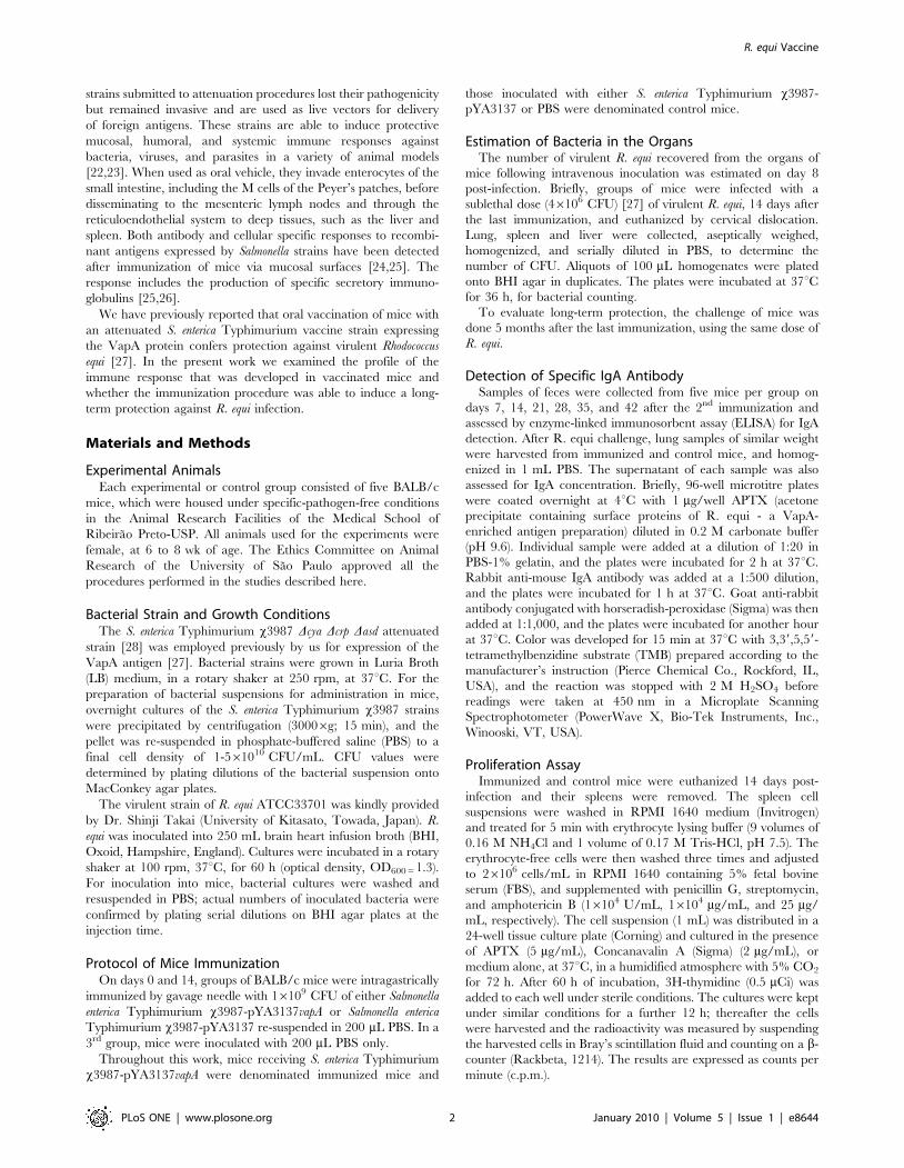
Vaccination with Salmonella x3987-pYA3137vapAProtects Mice against R. equi Infection and Elicits StrongMucosal Humoral Response and Systemic Cell Immunity
Eight days after infection with R. equi, BALB/c mice showed
bacterial burden in the lung, liver, and spleen, which was
significantly diminished in the animals that were immunized with
live attenuated Salmonella expressing the VapA protein; i.e., with
x3987-pYA3137vapA (data not shown). In agreement with our
previous report [27], CFU recovery from the lung, liver, and
spleen was 3- to 4-fold lower in the VapA-immunized mice than in
the two control groups of mice, which received Salmonella x3987-
pYA3137 or PBS, respectively. These results confirm that
immunization with attenuated S. enterica Typhimurium expressing
VapA confers resistance to R. equi infection.
In order to evaluate if, in addition to eliciting high levels of
specific IgG circulating antibodies [27], oral immunization with
live attenuated Salmonella expressing VapA would also induce
humoral mucosal immunity, specific IgA was measured in the fecal
extracts of mice. The IgA levels found in the immunized mice were
at least 5-fold higher than those verified in the feces of control
mice during the whole 42-day period that followed the second
immunization (Fig. 1A), thus indicating that a strong and
persistent mucosal response production was triggered by immu-
nization with S. enterica Typhimurium expressing VapA. A higher
production of IgA was also detected in the lung homogenates of
immunized mice after infection with R. equi. Three- and 2-times
higher production of IgA was detected in the lung of immunized
R. equi Vaccine
PLoS ONE | www.plosone.org 3 January 2010 | Volume 5 | Issue 1 | e8644
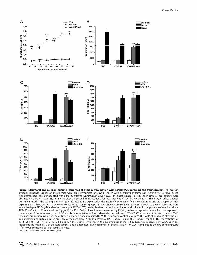
Figure 1. Humoral and cellular immune responses elicited by vaccination with Salmonella expressing the VapA protein. (A) Fecal IgAantibody response. Groups of BALB/c mice were orally immunized on days 0 and 14 with S. enterica Typhimurium x3987-pYA3137vapA (closedtriangles/dashed line), or inoculated with either S. enterica Typhimurium x3987-pYA3137 (closed squares) or PBS (open circles). Fecal extracts wereobtained on days 7, 14, 21, 28, 35, and 42 after the second immunization, for measurement of specific IgA by ELISA. The R. equi surface antigen(APTX) was used as the coating antigen (1 mg/mL). Results are expressed as the mean of OD values of five mice per group and are a representativeexperiment of three assays. ***p,0.001 compared to control groups. (B) Lymphocyte proliferative response. Spleen cells were harvested fromimmunized (pYA3137vapA) and control mice (pYA3137 or PBS) on day 14 after the last immunization and cultured in the presence of medium alone,APTX (5 mg/mL), or Concanavalin A (2 mg/mL) for 72 h. Cell proliferation was measured by [3H]-thymidine incorporation assay. Each bar representsthe average of five mice per group 6 SD and is representative of four independent experiments. ***p,0.001 compared to control groups. (C–F)Cytokines production. Whole spleen cells were collected from immunized (pYA3137vapA) and control mice (pYA3137 or PBS) on day 14 after the lastimmunization and cultured in the presence of medium alone, APTX (5 mg/mL), or LPS (1 mg/mL) plus IFN-c (1 ng/mL) for 48 h. The concentration ofIL-12 (C), IFN-c (D), TNF-a (E), IL-10 (F), and IL-4 (not shown) cytokines in the supernatants of the cell cultures was measured by ELISA. Each barrepresents the mean 6 SD of triplicate samples and is a representative experiment of three assays. ***p,0.001 compared to the two control groups;+++p,0.001 compared to PBS-inoculated mice.doi:10.1371/journal.pone.0008644.g001
R. equi Vaccine
PLoS ONE | www.plosone.org 4 January 2010 | Volume 5 | Issue 1 | e8644
mice compared with control mice, on days 2 and 8 post-challenge
with R. equi, respectively (data not shown).
To investigate whether immunization with S. enterica Typhimur-
ium expressing VapA could induce cell-mediated immunity
against R. equi, spleen cells from immunized and control mice
were assayed in terms of their antigen-specific proliferative
response. A preparation of R. equi surface proteins (APTX)
induced proliferation of spleen cells from immunized mice only
(Fig. 1B), while a polyclonal stimulus (ConA) induced proliferation
of cells from control and immunized mice. Intriguingly, prolifer-
ative response to ConA was significantly inhibited in animals that
received x3987-pYA3137, expressing VapA or not.
Together, these results indicate that VapA-immunization
triggers mucosal-humoral response and systemic cell-mediated
immunity.
R. equi Antigen Stimulates the Production of Th1Cytokines by Spleen Cells from Mice Vaccinated withSalmonella x3987-pYA3137vapA
To characterize the cytokine production associated with
inoculation of Salmonella x3987-pYA3137vapA, cultures of spleen
cells from mice of the immunized group and mice of the two
control groups were stimulated with APTX. The positive control
of stimulation was provided by LPS/IFN-c, whereas medium
alone was used as the negative counterpart. Spleen cells from mice
of both control groups did not respond to APTX through cytokine
production, whereas LPS/IFN-c, as expected, markedly stimulat-
ed cytokines production by cells from mice that had been
inoculated with Salmonella x3987-pYA3137, expressing VapA or
not (Fig. 1C-F). APTX only stimulated cells from mice immunized
with Salmonella x3987-pYA3137vapA, which produced IL-12, IFN-
c, and TNF-a in levels as high as those produced by cells
stimulated with LPS/IFN-c (Fig. 1C–E). Concerning IL-10, cells
from mice immunized with Salmonella x3987-pYA3137vapA
furnished levels that were significantly higher than those produced
by cells of the PBS control group. However, IL-10 levels produced
by the former cells were similar to those provided by cells of mice
of the second control group, which received Salmonella x3987-
pYA3137 (Fig. 1F). On the other hand, no IL-4 was detected in
the spleen cells supernatant of the immunized mice (data not
shown).
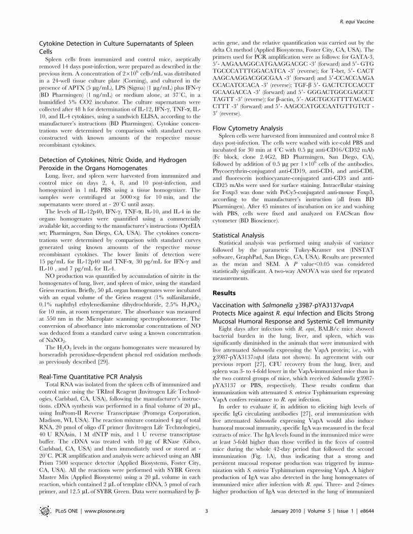
Since T-bet and GATA-3 are well characterized transcription
factors for differentiation of Th1 and Th2 cells, respectively, the
mRNA levels for both transcription factors was analyzed. In
agreement with the high production of cytokines related to Th1
skewed immunity, spleen cells of immunized mice showed higher
mRNA levels for T-bet and lower for GATA-3, compared with the
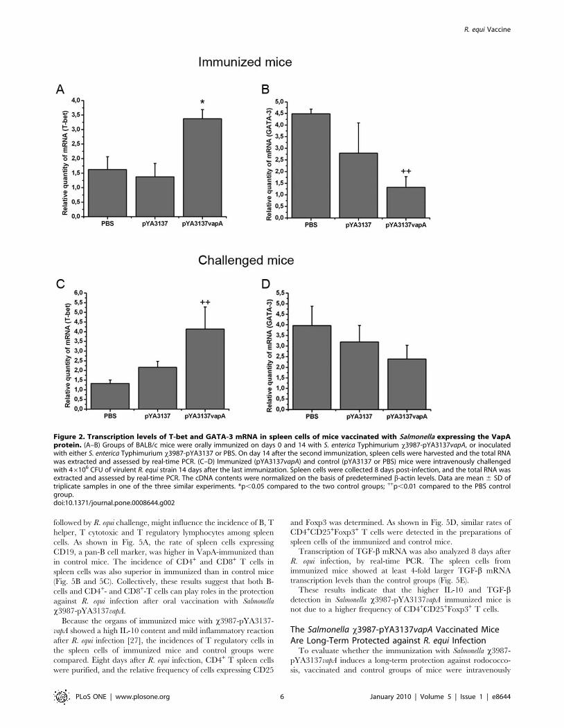
levels of the two control groups (Fig. 2A and 2B).
The Th1 Immunity Induced by Vaccination withSalmonella x3987-pYA3137vapA Is Sustained FollowingR. equi Challenge
To investigate the effect of R. equi infection on the pattern of
immune response induced by VapA-immunization, the cytokines
levels in organs of mice that had been intravenously challenged
with virulent R. equi were examined two weeks after the last
immunization. Groups of mice were sacrificed in different periods
(2 to 10 days) post-challenge, and the time-course curves of
individual cytokine content in the lungs, liver, and spleen are
represented in Fig. 3. As a general rule, there was no significant
difference in the cytokines levels detected in the organs of the two
control groups of mice during the period of observation (10 days
post-infection). As an exception, IL-4 levels in the organs of the
x3987-pYA3137 control group were nearer to the levels detected
in the Salmonella x3987-pYA3137vapA immunized mice, compared
with the levels detected in the control group that received PBS, a
fact that was remarkable in the spleen. A high IL-12 content was
early detected in the organs of immunized mice and was sustained
at least for 4 days in the spleen and liver, and 8 days in the lung. In
the organs of non-immunized mice, a tendency to increasing IL-12
content was observed only later (8–10 days after challenge).
High IFN-c levels were detected after challenge in all examined
organs of immunized mice. Although in all groups of mice the
IFN-c content increased progressively after R. equi challenge, the
levels attained by the immunized group were significantly higher
than those of control groups in the whole observation period.
The TNF-a content was markedly lower in the lung and liver of
immunized mice during the whole observation period. Less
significant differences were detected in the spleen on days 2 and
8 post-challenge.
The IL-10 content was low and stable in the organs of control
mice, while in immunized animals the IL-10 content had
augmented progressively, reaching the highest levels by day 8
and 10 post-challenge in the lung and liver, and in the spleen,
respectively.
IL-4 concentrations were persistently lower in the organs of
immunized mice compared with the PBS control group, but
sometimes similar to the IL-4 concentrations in the organs of the
x3987-pYA3137 control group, especially in the spleen (Fig. 3E, J,
and O).
Eight days after challenge, we found that the mRNA
transcription level of T-bet was higher in the spleen cells of
immunized mice compared with the PBS control group and not
different from the x3987-pYA3137 control group (Fig. 2C). The
expression of GATA-3 mRNA in the spleen tissue was not
significantly different among the three groups of mice (Fig. 2D).
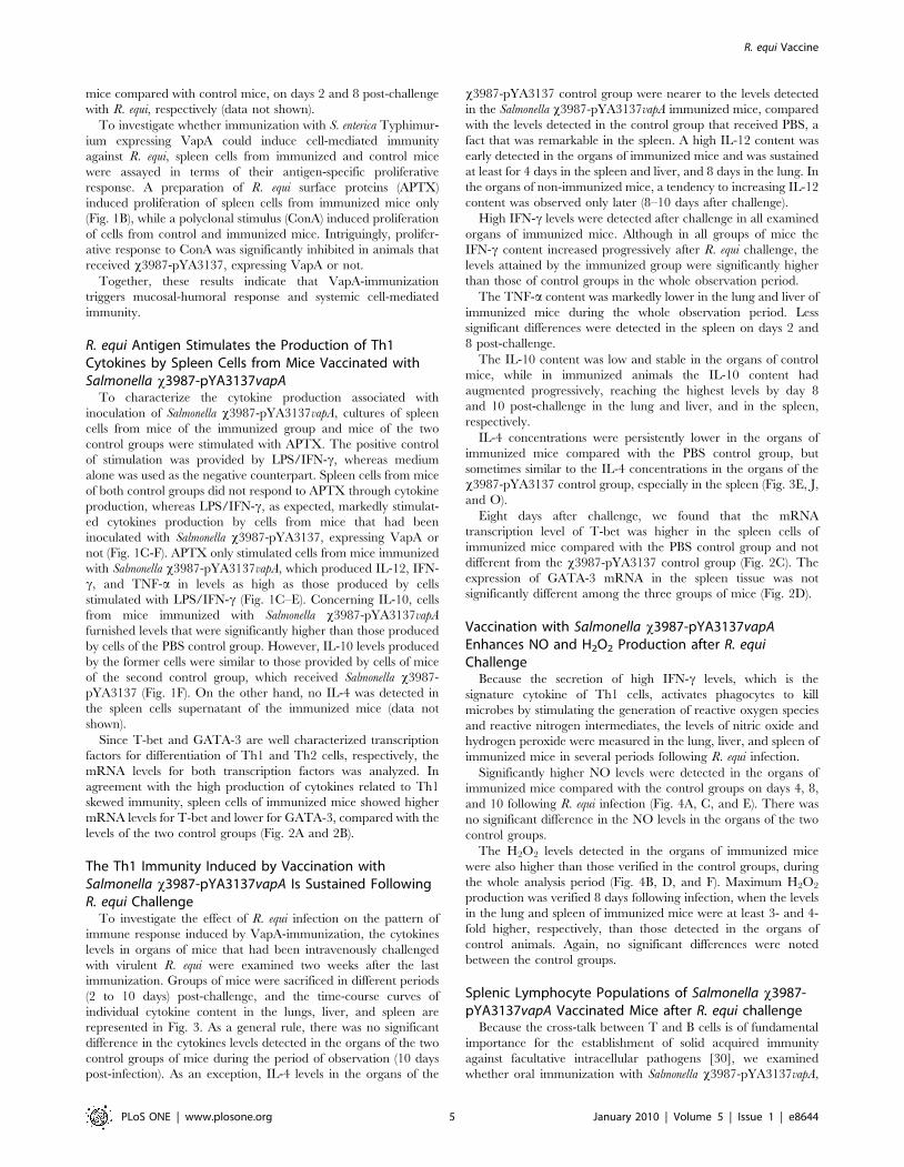
Vaccination with Salmonella x3987-pYA3137vapAEnhances NO and H2O2 Production after R. equiChallenge
Because the secretion of high IFN-c levels, which is the
signature cytokine of Th1 cells, activates phagocytes to kill
microbes by stimulating the generation of reactive oxygen species
and reactive nitrogen intermediates, the levels of nitric oxide and
hydrogen peroxide were measured in the lung, liver, and spleen of
immunized mice in several periods following R. equi infection.
Significantly higher NO levels were detected in the organs of
immunized mice compared with the control groups on days 4, 8,
and 10 following R. equi infection (Fig. 4A, C, and E). There was
no significant difference in the NO levels in the organs of the two
control groups.
The H2O2 levels detected in the organs of immunized mice
were also higher than those verified in the control groups, during
the whole analysis period (Fig. 4B, D, and F). Maximum H2O2
production was verified 8 days following infection, when the levels
in the lung and spleen of immunized mice were at least 3- and 4-
fold higher, respectively, than those detected in the organs of
control animals. Again, no significant differences were noted
between the control groups.
Splenic Lymphocyte Populations of Salmonella x3987-pYA3137vapA Vaccinated Mice after R. equi challenge
Because the cross-talk between T and B cells is of fundamental
importance for the establishment of solid acquired immunity
against facultative intracellular pathogens [30], we examined
whether oral immunization with Salmonella x3987-pYA3137vapA,
R. equi Vaccine
PLoS ONE | www.plosone.org 5 January 2010 | Volume 5 | Issue 1 | e8644
followed by R. equi challenge, might influence the incidence of B, T
helper, T cytotoxic and T regulatory lymphocytes among spleen
cells. As shown in Fig. 5A, the rate of spleen cells expressing
CD19, a pan-B cell marker, was higher in VapA-immunized than
in control mice. The incidence of CD4+ and CD8+ T cells in
spleen cells was also superior in immunized than in control mice
(Fig. 5B and 5C). Collectively, these results suggest that both B-
cells and CD4+- and CD8+-T cells can play roles in the protection
against R. equi infection after oral vaccination with Salmonella
x3987-pYA3137vapA.
Because the organs of immunized mice with x3987-pYA3137-
vapA showed a high IL-10 content and mild inflammatory reaction
after R. equi infection [27], the incidences of T regulatory cells in
the spleen cells of immunized mice and control groups were
compared. Eight days after R. equi infection, CD4+ T spleen cells
were purified, and the relative frequency of cells expressing CD25
and Foxp3 was determined. As shown in Fig. 5D, similar rates of
CD4+CD25+Foxp3+ T cells were detected in the preparations of
spleen cells of the immunized and control mice.
Transcription of TGF-b mRNA was also analyzed 8 days after
R. equi infection, by real-time PCR. The spleen cells from
immunized mice showed at least 4-fold larger TGF-b mRNA
transcription levels than the control groups (Fig. 5E).
These results indicate that the higher IL-10 and TGF-bdetection in Salmonella x3987-pYA3137vapA immunized mice is
not due to a higher frequency of CD4+CD25+Foxp3+ T cells.
The Salmonella x3987-pYA3137vapA Vaccinated MiceAre Long-Term Protected against R. equi Infection
To evaluate whether the immunization with Salmonella x3987-
pYA3137vapA induces a long-term protection against rodococco-
sis, vaccinated and control groups of mice were intravenously
Figure 2. Transcription levels of T-bet and GATA-3 mRNA in spleen cells of mice vaccinated with Salmonella expressing the VapAprotein. (A–B) Groups of BALB/c mice were orally immunized on days 0 and 14 with S. enterica Typhimurium x3987-pYA3137vapA, or inoculatedwith either S. enterica Typhimurium x3987-pYA3137 or PBS. On day 14 after the second immunization, spleen cells were harvested and the total RNAwas extracted and assessed by real-time PCR. (C–D) Immunized (pYA3137vapA) and control (pYA3137 or PBS) mice were intravenously challengedwith 46106 CFU of virulent R. equi strain 14 days after the last immunization. Spleen cells were collected 8 days post-infection, and the total RNA wasextracted and assessed by real-time PCR. The cDNA contents were normalized on the basis of predetermined b-actin levels. Data are mean 6 SD oftriplicate samples in one of the three similar experiments. *p,0.05 compared to the two control groups; ++p,0.01 compared to the PBS controlgroup.doi:10.1371/journal.pone.0008644.g002
R. equi Vaccine
PLoS ONE | www.plosone.org 6 January 2010 | Volume 5 | Issue 1 | e8644

challenged with virulent R. equi strain 5 months after the last
immunization. On day 8 post-challenge, the spleen and liver
homogenates were evaluated in terms of bacterial counting and
NO levels.
Significantly lower R. equi CFU were recovered from the tissues
of immunized mice compared with the two control animal groups
(Fig. 6A and data not shown). The two control groups had similar
CFU. The number of CFU recovered from the spleen of
immunized mice reached levels 4-fold inferior to those recovered
from the spleen of control mice (Fig. 6A). Immunized mice
produced a significantly higher amount of NO compared with the
control groups (Fig. 6B). Taken together, these results indicate that
immunization with Salmonella x3987-pYA3137vapA induces a long-
term protection against R. equi infection.
Discussion
Vaccination with attenuated S. enterica Typhimurium x3987
strain expressing the VapA antigen confers protection against R.
equi infection. This had been demonstrated in oral immunized
mice that, when challenged with R. equi, showed higher survival
rates, higher bacterial clearance, and milder inflammatory
response compared with the non-vaccinated mice [27]. In the
present work we report that the immune response developed by
the vaccinated mice with S. enterica Typhimurium expressing the
VapA protein is featured by strong mucosal-humoral and systemic
cell-mediated immunity. Besides that, we show that the vaccina-
tion confers a long-term protection against R. equi infection.
The occurrence of mucosal immunity in response to vaccination
is indicated by the detection of high and persistent fecal levels of
antigen-specific IgA. This fact may be a relevant consequence of
the immunization procedure because secretory immunoglobulins
constitute a frontline defense against several microorganisms
[25,26].
The specific cellular immune response developed after vacci-
nation with Salmonella expressing VapA is revealed by the
proliferation of the spleen cells of immunized mice in response
to the in vitro stimulation with R. equi antigen. Because R. equi is a
facultative intracellular pathogen, resistance to infection crucially
depends on effective cell-mediated immunity [5,14,31,32]. This
requirement has been previously demonstrated by the severe
pulmonary granulomas and failure to eliminate the bacteria
presented by athymic nude mice infected with R. equi [33]. Other
authors have already reported that vaccination with distinct
antigens carried by attenuated S. enterica Typhimurium is
associated with antigen-stimulated lymphoproliferative response
[21,34].
The present study shows that Th1 cytokines are intensely
produced by vaccinated mice. Firstly, the levels of IL-12, IFN-c,
and TNF-a cytokines released in vitro by antigen-stimulated spleen
cells from vaccinated mice are significantly higher than those
produced by cells from mice of the non-vaccinated control groups.
Figure 3. Cytokine levels in the organs of mice after R. equi infection. BALB/c mice were orally immunized on days 0 and 14 with S. entericaTyphimurium x3987-pYA3137vapA (closed triangles/dashed line), or inoculated with either S. enterica Typhimurium x3987-pYA3137 (closed squares)or PBS (open circles). All mice were intravenously challenged with 46106 CFU of R. equi ATCC33701 14 days after the last immunization. The lung(A–E), liver (F–J), and spleen (K–O) were harvested 2, 4, 8, and 10 days post-infection and homogenized for IL-12 (A, F, and K), IFN-c (B, G, and L),TNF-a (C, H, and M), IL-10 (D, I, and N), and IL-4 (E, J, and O) detection. Results are expressed as means of five mice per group 6 SD and arerepresentative of three experiments. *p,0.05, **p,0.01, and ***p,0.001 compared to the two control groups; and +p,0.05, ++p,0.01, and+++p,0.001 compared to the PBS control group.doi:10.1371/journal.pone.0008644.g003
R. equi Vaccine
PLoS ONE | www.plosone.org 7 January 2010 | Volume 5 | Issue 1 | e8644

Figure 4. Nitrite and hydrogen peroxide production in the organs of mice following R. equi infection. BALB/c mice were orallyimmunized on days 0 and 14 with S. enterica Typhimurium x3987-pYA3137vapA (closed triangles/dashed line), or inoculated with either S. entericaTyphimurium x3987-pYA3137 (closed squares) or PBS (open circles). All mice were intravenously challenged with 46106 CFU of R. equi ATCC33701 14days after the last immunization. On days 2, 4, 8, and 10 after infection, the lung, liver, and spleen were harvested and homogenized for NO (A, C, andE) and H2O2 (B, D, and F) detection. The endogenous H2O2 release was measured by the horseradish peroxidase-dependent phenol red oxidationmethods, whereas the nitrite levels were measured by Griess reaction. Data are expressed as means of five mice per group 6 SD and arerepresentative of three separate experiments. *p,0.05, **p,0.01, and ***p,0.001 compared to the two control groups.doi:10.1371/journal.pone.0008644.g004
R. equi Vaccine
PLoS ONE | www.plosone.org 8 January 2010 | Volume 5 | Issue 1 | e8644

Secondly, tissues of the vaccinated mice challenged with R. equi
contain higher IFN-c levels than the tissues of mice belonging
to the control groups, during the whole observation period.
Interestingly, the tissue detection of IL-12 has been precocious,
peaked at 2 and 4 days post-infection, but it declined thereafter, in
a period when the vaccinated mice had already cleared the
infection and no more lesions were observed in their lung, liver or
spleen [27]. The induction of Th1 immunity by vaccination has
been previously suggested to occur on the basis of the detection of
high serum levels of IgG2a specific antibodies in oral immunized
mice [27]. It is well established that during R. equi infection Th2-
biased immunity is associated with disease development, while
Figure 5. Alteration of lymphocyte subpopulation in the spleen of mice. BALB/c mice orally administered with S. enterica Typhimuriumx3987-pYA3137vapA, S. enterica Typhimurium x3987-pYA3137, or PBS, on days 0 and 14, were intravenously challenged with 46106 CFU of virulentR. equi strain 14 days after the last immunization. Spleen cells were harvested 8 days post-infection and analyzed by flow cytometry. The cells werestained with specific anti-CD19 (A), anti-CD3 plus anti-CD4 (B), and anti-CD3 plus anti-CD8 (C) monoclonal antibodies. (D) CD4+ spleen T cells stainedwith specific anti-CD25 plus anti-Foxp-3 monoclonal antibodies. The average of the percentage of spleen cells positive for the respective CD markersin five samples is shown. Similar results were obtained in three independent experiments. (E) Total RNA was extracted from spleen cells harvested 8days post-infection and assessed by real-time PCR for detection of TGF-b mRNA. The cDNA contents were normalized on the basis of predeterminedb-actin levels. Data are mean 6 SD of triplicate samples in one of the three similar experiments. *p,0.05, **p,0.01, and ***p,0.001 compared to thetwo control groups.doi:10.1371/journal.pone.0008644.g005
R. equi Vaccine
PLoS ONE | www.plosone.org 9 January 2010 | Volume 5 | Issue 1 | e8644

Th1-prone response affords infection control [7,14,17–19,33,35].
Beyond doubt, the Th1-cell immunity developed by the
vaccinated mice fits with the protection conferred against
rodococcosis. Previous reports have associated the utilization of
attenuated Salmonella strains as vaccine vectors with the occurrence
of strong and sustained Th1-immunity [34].
The activities exerted by Th1 cytokines are crucial for
the elimination of intracellular pathogens, such as R. equi
[7,14,17–19,33,35,36]. IL-12, released by activated dendritic cells
and macrophages, induces differentiation of Th0 into Th1 cells.
The release of IFN-c by Th1 cells promotes macrophage effector
activities, such as those mediated by nitric oxide and hydrogen
peroxide, which have been detected in high levels in the lung, liver
and spleen of vaccinated mice challenged with R. equi. As
previously demonstrated, the synergistic action of nitric oxide
and superoxide is crucial for the intracellular killing of R. equi [37].
Still regarding the Th1 cytokines, TNF-a is highly produced in vitro
by the antigen-stimulated spleen cells of vaccinated mice, but is
barely detected in the organs of vaccinated mice after R. equi
challenge. This can be advantageous considering that TNF-a is a
prototypical proinflammatory cytokine [38] whose overproduction
is associated with the occurrence of host tissue injury [39].
IL-4, an essential cytokine for generation of Th2 responses [40],
is not produced by the antigen-stimulated spleen cells of
vaccinated mice. On the other hand, IL-4 is persistently detected
in the organs of vaccinated mice after challenge with R. equi, albeit
in concentrations significantly lower than those detected in the
organs of non-vaccinated mice. With respect to IL-10, the antigen-
stimulated spleen cells from vaccinated mice produce significantly
higher levels of this cytokine than those afforded by the cells of
mice belonging to the control group that received PBS instead of
vaccine. However, the IL-10 levels in the spleen cells of the
vaccinated mice are similar to those produced by the control mice
that received Salmonella carrying the plasmid alone (without VapA
expression). This strongly suggests that the vaccinal vector
accounts for induction of IL-10 production. The IL-10 tissue
levels in the organs of vaccinated mice augment progressively after
R. equi challenge, reaching the maximum after day 8 post-
challenge. IL-10 favors the balance between pathology and
protection during diseases that course with inflammation. Such
immunoregulatory role is due to the IL-10 ability of inhibiting the
expression of the most inducible cytokines and secondary
mediators that contribute to inflammation. Such a phenomenon
may be responsible, in our study, for the decrease in TNF-asecretion, which is temporally coincident with the elevation of
IL-10.
The induction of Th1-immunity by vaccination is evidenced by
quantification of the expression of the transcription factors T-bet
and GATA-3. Following vaccination, spleen cells of mice display a
prominent mRNA expression of the Th1 transcription factor T-
bet, in detriment of the Th2 transcription factor GATA-3, a
finding that is consistent with the cytokines profile verified in the
immunized animals. After challenge with R. equi, the vaccinated
mice still present higher mRNA transcription level of T-bet than
the non-vaccinated mice, while the levels of GATA-3 mRNA
expression are not significantly different between cells from
vaccinated and non-vaccinated mice.
Examination of the relative frequency of lymphocyte popula-
tions in the groups of mice challenged with R. equi reveals greater
incidence of B lymphocytes (CD19+), CD4+ and CD8+ T cells in
the vaccinated mice compared with the control. Thus, humoral
and cellular mechanisms may be acting together, a hypothesis that
is consistent with the previous demonstration that cooperation
between humoral and cellular branches of immunity is necessary
to protect the host against R. equi infection [13,14,16,41,42].
Concerning the T cell subpopulations, whilst CD4+ T cells
generate the appropriate cytokine milieu to support resistance
against R. equi, CD8+ T cells account for the destruction of the
infected host cells [17,43] as well as for the release of the bacteria,
which become accessible to the humoral effector mechanisms [44].
Besides that, through cytokine-mediated interactions with B
lymphocytes, the CD4+ T cells contribute to the generation of
high-affinity IgG antibodies, which are able to perform bacterial
opsonization and complement activation. So the lymphocyte
populations provide multiple effector mechanisms capable of
eliminating the R. equi infection.
Because secretion of IL-10 and TGF-b is one mechanism
through which T regulatory cells exert suppression of effector
immune response, whether this cell population could be expanded
in the spleen of vaccinated mice has been investigated. There is no
Figure 6. Recall immune response induced by immunization with Salmonella expressing the VapA protein. BALB/c mice were orallyimmunized with S. enterica Typhimurium x3987-pYA3137vapA, or inoculated with either S. enterica Typhimurium x3987-pYA3137 or PBS on days 0and 14. Five months after the last immunization, all mice were intravenously challenged with 46106 CFU of virulent R. equi. On day 8 after infection,the spleen (A and B) and liver (not shown) were harvested and homogenized for bacterial burdens and NO quantification. Results are expressed asthe mean of five mice per group 6 SD. The experiment was performed twice with similar results. *p,0.05, **p,0.01, and ***p,0.001 compared tothe control groups.doi:10.1371/journal.pone.0008644.g006
R. equi Vaccine
PLoS ONE | www.plosone.org 10 January 2010 | Volume 5 | Issue 1 | e8644

difference between vaccinated and non-vaccinated mice in terms
of the relative frequency of CD4+CD25+Foxp3+ T cells, although
the transcription levels of TGF-b mRNA are significantly higher in
the spleen cells from vaccinated mice. Nevertheless, TGF-b can
also be produced by naive CD4+ T cells upon TCR stimulation
[45], and the high IL-10 production induced by vaccination may
also be attributed to a non-Treg source. Recent studies have
shown that Th1 cell themselves can produce IL-10, which is
essential for the inhibition of exaggerated Th1 cell responses
during infection [46,47]. It has also been reported that CD4+ T
cells producing high amounts of IFN-c are the source of IL-10 that
inhibits the development of immunopathology upon T. gondii
infection [47]. These T cells express the Th1 cell lineage
transcription factor T-bet, but not the Treg cell marker Foxp3.
In any case, we postulate that the high IL-10 levels associated with
vaccination exert an important role in maintaining the control of
the inflammatory reaction that follows R. equi infection. This
control is also favored by the low TNF-a present in the organs of
the vaccinated mice, compared with the control mice. As already
mentioned, TNF-a often mediates the host tissue injury that
occurs in exacerbated inflammation, while IL-10 downmodulates
Th1 immunity and minimizes inflammatory tissue damage
[39,48,49]. Therefore, our results provide enlightenment regard-
ing our previous observation that, after being challenged with R.
equi, vaccinated mice develop a milder inflammatory reaction than
that observed in the control animals.
Conceptually, an effective vaccine must be able to induce a
long-term memory against the heterologous antigen. We have
verified that vaccination with Salmonella x3987-pYA3137vapA
induces a protection against R. equi infection with a minimal
duration of 5 months after the last immunization. Compared with
the non-vaccinated animals, a significantly lower number of
bacteria are recovered from the spleen of the vaccinated mice, and
their spleen cells release higher concentration of nitric oxide.
These observations indicate that vaccinated mice are still immune
late after vaccination.
In conclusion, the Salmonella enterica serovar Typhimurium
x3987-pYA3137vapA is endowed of properties that feature a
good vaccine to prevent rhodococcosis: (1) it confers protection
against R. equi infection; (2) it induces strong and specific
mucosal, humoral, and cell-mediated Th1-responses against
the heterologous antigen; (3) it generates an appropriate
regulatory cytokine response, which allows for tissue integrity;
and (4) it induces a long-term protection. The immunization of
foals is being performed by our group, to investigate the
efficiency of the vaccination in the species most frequently
affected by R. equi.
Acknowledgments
We thank Sandra M.O. Thomaz, and Patricia E. Vendrusculo for
excellent technical assistance. We also acknowledge Julio A. Siqueira,
Cristiane C.P. Ribas, Savio H.F. Miranda, and Ednelson A. Mazzotto for
expert animal care. We are thankful to Dr. Leandro L. Oliveira for helpful
discussions.
Author Contributions
Conceived and designed the experiments: AFO SAC SGS. Performed the
experiments: AFO LPR SAC. Analyzed the data: AFO SAC SGS.
Contributed reagents/materials/analysis tools: MCRB. Wrote the paper:
AFO LPR MCRB.
References
1. Giguere S, Prescott JF (1997) Clinical manifestations, diagnosis, treatment, and
prevention of Rhodococcus equi infections in foals. Vet Microbiol 56: 313–334.
2. Donisi A, Suardi MG, Casari S, Longo M, Cadeo GP, et al. (1996) Rhodococcus
equi infection in HIV-infected patients. AIDS 10: 359–362.
3. Meijer WG, Prescott JF (2004) Rhodococcus equi. Vet Res 35: 383–396.
4. Magnusson H (1923) Spezifische infektiose pneumonie beim Fohlen. Ein neuer
eiterreger beim Pferd. Arch Wiss Prakt Tierhelkd 50: 22–38.
5. Hondalus MK (1997) Pathogenesis and virulence of Rhodococcus equi. Vet
Microbiol 56: 257–268.
6. Takai S, Hines SA, Sekizaki T, Nicholson VM, Alperin DA, et al. (2000) DNA
sequence and comparison of virulence plasmids from Rhodococcus equi
ATCC33701 and 103. Infect Immun 68: 6840–6847.
7. Muscatello G, Leadon DP, Klayt M, Ocampo-Sosa A, Lewis DA, et al. (2007)
Rhodococcus equi infection in foals: the science of ‘rattles’. Equine Vet J 39:
470–478.
8. Takai S, Iie M, Watanabe Y, Tsubaki S, Sekizaki T (1992) Virulence-associated
15- to 17-kilodalton antigens in Rhodococcus equi: temperature dependent
expression and location of the antigens. Infect Immun 60: 2995–2997.
9. Giguere S, Hondalus MK, Yager JA, Darrah P, Mosser DM, et al. (1999) Role
of the 85-kilobase plasmid and plasmid-encoded virulence-associated protein A
in intracellular survival and virulence of Rhodococcus equi. Infect Immun 67:
3548–3557.
10. Jain S, Bloom BR, Hondalus MK (2003) Deletion of vapA encoding Virulence
Associated Protein A attenuates the intracellular actinomycete R. equi. Mol
Microbiol 50: 115–128.
11. Prescott JF, Patterson MC, Nicholson VM, Morein B, Yager JA (1997)
Assessment of the immunogenic potential of Rhodococcus equi virulence associated
protein (VapA) in mice. Vet Microbiol 56: 213–225.
12. Hooper-McGrevy KE, Giguere S, Wilkie BN, Prescott JF (2001) Evaluation of
equine immunoglobulin specific for Rhodococcus equi virulence-associated proteins
A and C for use in protecting foals against Rhodococcus equi-induced pneumonia.
Am J Vet Res 62: 1307–1313.
13. Martens RJ, Martens JG, Fiske RA, Hietala SK (1989) Rhodococcus equi foal
pneumonia: protective effects of immune plasma in experimentally infected
foals. Equine Vet J 21: 249–255.
14. Hines SA, Kanaly ST, Byrne BA, Palmer GH (1997) Immunity to Rhodococcus
equi. Vet Microbiol 56: 177–185.
15. Hurley JR, Begg AP (1995) Failure of hyperimmune plasma to prevent
pneumonia caused by Rhodococcus equi in foals. Aust Vet J 72: 418–420.
16. Giguere S, Gaskin JM, Bowman JL (2002) Evaluation of a commercially
available hyperimmune plasma product for prevention of naturally acquired
pneumonia caused by Rhodococcus equi in foals. J Am Vet Med Assoc 220: 59–63.
17. Hines SA, Stone DM, Hines MT, Alperin DC, Knowles DP, et al. (2003)Clearance of virulent but not avirulent Rhodococcus equi from the lungs of adult
horses is associated with intracytoplasmic gamma interferon production by
CD4+ and CD8+ T lymphocytes. Clin Diagn Lab Immunol 10: 208–215.
18. Kanaly ST, Hines SA, Palmer GH (1995) Cytokine modulation alters
pulmonary clearance of Rhodococcus equi and development of granulomatouspneumonia. Infect Immun 63: 3037–3041.
19. Kanaly ST, Hines SA, Palmer GH (1996) Transfer of a CD4+ Th1 cell line to
nude mice effects clearance of Rhodococcus equi from the lung. Infect Immun 64:
1126–1132.
20. Dietrich G, Kolb-Maurer A, Spreng S, Schartl M, Goebel W, et al. (2001)
Gram-positive and Gram-negative bacteria as carrier systems for DNA vaccines.Vaccine 19: 2506–2512.
21. Du A, Wang S (2005) Efficacy of a DNA vaccine delivered in attenuatedSalmonella Typhimurium against Eimeria tenella infection in chickens. Int J Parasitol
35: 777–785.
22. Pasetti MF, Levine MM, Sztein MB (2003) Animal models paving the way for
clinical trials of attenuated Salmonella enterica serovar Typhi live oral vaccines andlive vectors. Vaccine 21: 401–418.
23. Kotton CN, Hohmann EL (2004) Enteric pathogens as vaccine vectors forforeign antigen delivery. Infect Immun 72: 5535–5547.
24. Wyszynska A, Raczko A, Lis M, Jagusztyn-krynicka EK (2004) Oral
immunization of chickens with avirulent Salmonella vaccine strain carrying C.
jejuni 72Dz/92 cjaA gene elicits specific humoral immune response associatedwith protection against challenge with wild-type Campylobacter. Vaccine 22:
1379–1389.
25. Shiau AL, Chen CC, Yo YT, Chu CY, Wang SY, et al. (2005) Enhancement of
humoral and cellular immune responses by an oral Salmonella choleraesuis vaccineexpressing porcine prothymosin alpha. Vaccine 23: 5563–5571.
26. Yang X, Hinnebusch BJ, Trunkle T, Bosio CM, Suo Z, et al. (2007) Oralvaccination with Salmonella simultaneously expressing Yersinia pestis F1 and V
antigens protects against bubonic and pneumonic plague. J Immunol 178:1059–1067.
27. Oliveira AF, Ferraz LC, Brocchi M, Roque-Barreira MC (2007) Oraladministration of a live attenuated Salmonella vaccine strain expressing the
VapA protein induces protection against infection by Rhodococcus equi. Microbes
Infect 9: 382–390.
R. equi Vaccine
PLoS ONE | www.plosone.org 11 January 2010 | Volume 5 | Issue 1 | e8644

28. Curtiss 3rd R, Kelly SM (1987) Salmonella typhimurium deletion mutants lacking
adenylate cyclase and cyclic AMP receptor protein are avirulent andimmunogenic. Infect Immun 55: 3035–3043.
29. Pick E, Mizel D (1981) Rapid microassays for the measurement of superoxide
and hydrogen peroxide production by macrophages in culture using anautomatic enzyme immunoassay reader. J Immunol Methods 46: 211–226.
30. Hochadel JF, Keller KF (1977) Protective effects of passively transferred immuneT- or B-lymphocytes in mice infected with Salmonella Typhimurium. J Infect Dis
135: 813–823.
31. Lopez AM, Hines MT, Palmer GH, Knowles DP, Alperin DC, et al. (2003)Analysis of anamnestic immune responses in adult horses and priming in
neonates induced by a DNA vaccine expressing the vapA of Rhodococcus equi.
Vaccine 21: 3815–3825.
32. Yager JA, Prescott CA, Kramar DP, Hannah H, Balson GA, et al. (1991) Theeffect of experimental infection with Rhodococcus equi on immunodeficient mice.
Vet Microbiol 28: 363–376.
33. Kanaly ST, Hines SA, Palmer GH (1993) Failure of pulmonary clearance ofRhodococcus equi infection in CD4+ T-lymphocyte-Deficient transgenic mice.
Infect Immun 61: 4929–4932.34. Cong H, Gu QM, Jiang Y, He SY, Zhou HY, et al. (2005) Oral immunization
with a live recombinant attenuated Salmonella typhimurium protects mice against
Toxoplasma gondii. Parasite Immunol 27: 29–35.35. Breathnach CC, Sturgill-Wright T, Stiltner JL, Adams AA, Lunn DP, et al.
(2006) Foals are interferon gamma-deficient at birth. Vet Immunol Immuno-pathol 112: 199–209.
36. Kasuga-Aoki H, Takai S, Sasaki Y, Tsubaki S, Madarame H, et al. (1999)Tumor necrosis factor and interferon-c are required in host resistance against
virulent Rhodococcus equi infection in mice: cytokine production depends on the
virulence levels of R.equi. Immunol 96: 122–127.37. Darrah PA, Hondalus MK, Chen Q, Ischiropoulos H, Mosser DM (2000)
Cooperation between reactive oxygen and nitrogen intermediates in killing ofRhodococcus equi by activated macrophages. Infect Immun 68: 3587–3593.
38. Criscione LG, St Clair EW (2002) Tumor necrosis factor-a antagonists for the
treatment of rheumatic diseases. Curr Opin Rheumatol 14: 204–211.
39. Abuzakouk M, Feighery C, Jackson J (2002) Tumor necrosis factor blocking
agents: a new therapeutic modality for inflammatory disorders. Br J Biomed Sci59: 173–179.
40. Dong C, Flavell RA (2000) Control of T helper cell differentiation – in search of
master genes. Science’s stke 49: 1–5.41. Madigan EJ, Hietala S, Muller SN (1991) Protection against naturally acquired
Rhodococcus equi pneumonia in foals by administration of hyperimmune plasma.J Reprod Fertil Suppl 44: 571–578.
42. Nordmann P, Ronco E, Nauciel C (1992) Role of T-lymphocyte subsets in
Rhodococcus equi infection. Infect Immun 60: 2748–2752.43. Patton KM, McGuire TC, Fraser DG, Hines SA (2004) Rhodococcus equi-infected
macrophages are recognized and killed by CD8+ T lymphocytes in a majorhistocompatibility complex class I-unrestricted fashion. Infect Immun 72:
7073–7083.44. Mastroeni P, Villarreal-Ramos B, Hormaeche CE (1992) Role of T cells TNF-a
and IFN-c in recall of immunity to oral challenge with virulent salmonellae in
mice vaccinated with live attenuated aro-Salmonella vaccines. Microb Pathog 13:477–491.
45. Li MO, Wan YY, Flavell RA (2007) T cell-produced Transforming GrowthFactor-b1 controls T cell tolerance and regulates Th1- and Th17-cell
differentiation. Immunity 26: 579–591.
4 6 . A n d e r s o n C F , O u k k a M , K u c h r o o V J , S a c k s D ( 2 0 0 7 )CD4(+)CD25(2)Foxp3(2) Th1 cells are the source of IL-10-mediated immune
suppression in chronic cutaneous leishmaniasis. Anderson J Exp Med 204:285–297.
47. Jankovic D, Kullberg MC, Feng CG, Goldszmid RS, Collazo CM, et al.Conventional Tbet(+) Foxp3(2) Th1 cells are the major source of host-protective
regulatory IL-10 during intracellular protozoan infection. J Exp Med 204:
273–283.48. Moore KW, O’Garra A, De Waal MR, Vieira P, Mosmann TR (1993)
Interleukin-10. Annu Ver Immunol 11: 165–190.49. Roncarolo MG, Gregori S, Battaglia M, Bacchetta R, Fleischhauer K, et al.
(2006) Interleukin-10-secreting type 1 regulatory T cells in rodents and humans.
Immunol Rev 212: 28–50.
R. equi Vaccine
PLoS ONE | www.plosone.org 12 January 2010 | Volume 5 | Issue 1 | e8644