Characterization of Pseudomonas syringae pv. syringae from ...
Upload
independentCategory
view
1download
0
Beta hydroxylation of the aspartyl residue in the phytotoxinsyringomycin E: characterization of two candidate hydroxylasesAspH and SyrP in Pseudomonas syringae
Gitanjali M. Singh‡, Pascal Fortin‡, Alexander Koglin‡, and Christopher T. Walsh‡,*
‡Department of Biological Chemistry & Molecular Pharmacology, Harvard Medical School, Boston, MA02115
AbstractThe pseudomonal phytotoxin syringomycin E, and related nonribosomal peptides, contain an L-threo-β-hydroxy-aspartyl residue at the eighth position of the lipodepsipeptide backbone as part ofa conserved nonproteinogenic tripeptide motif. Informatic analysis of the P. syringae genomesuggests only one putative non-heme iron hydroxylase, AspH. On heterologous expression inE.coli AspH shows robust catalytic activity with free L-Asp and L-Asp thioesters to make β-OH-Asp, but yields the erythro diastereomer rather than the threo configuration that is found insyringomycin. Further analysis of the Syr gene cluster indicated that SyrP, previously annotated asthe gene regulatory protein for the five- gene Syr cluster, is actually homologous to the known non-heme mononuclear iron hydroxylase TauD. Indeed, purified SyrP acts on Asp tethered as the protein-bound S-pantetheinyl thioester on the eighth module of the SyrE megasynthetase. The hydroxylationgives the anticipated L-threo-3-OH-Asp diastereomer found in syringomycin. The knockout ofsyrP abolishes the production of the mature syringomycin E, while knockout of aspH has no effecton syringomycin production.
The pseudomonad genus of gram-negative bacteria includes prolific producers ofnonribosomal peptide (NRP) natural products. Phytopathogenic Pseudomonas syringae strainsproduce a number of such plant toxins, including coronatine, syringomycins, phaseolotoxin,tabtoxin, and tagetitoxin (1-5). Syringomycins are members of a family of related cycliclipodepsipeptides that act as plant toxins by aggregating to form pores in plant cell membranesto facilitate exit of nutrients. One hallmark of nonribosomal peptides is the content of non-proteinogenic amino acid residues. In the five known pseudomonal lipopeptides, syringomycinE, cormycin A, pseudomycin B, syringostatin A and syringotoxin B there is a conserved C-terminal tripeptide moiety in the scaffold, composed of dehydrobutyrine7, β-OH-Asp8, and 4-Cl-Thr9 (Figure 1a) (3,6-9). This nonproteinogenic tripeptide motif, comprising residuesseven-nine of syringomycin, may be consequential for the biological function of these toxins.
The syringomycin gene cluster contains six ORFs, syrD, P, B1, B2, C, and E. The sequenceof this biosynthetic gene cluster (10) for lipo-nonapeptidolactone syringomycin (Figure 1b)suggested that residues one through eight are assembled on a multimodular megasynthetaseprotein SyrE. Residue nine is the 4-Cl-threonine, inserted at the end of the assembly line bythe tandem action of three enzymes SyrB1, SyrB2, and SyrC (11-13). Threonine is activatedby the adenylation (A) domain of SyrB1, installed as the S-pantetheinyl thioester on theadjacent T (thiolation) domain (11), and then chlorinated by the non-heme mononuclear iron-containing halogenase SyrB2 (11,12). At that juncture SyrC acts as an acyltransferase for the
*To whom correspondence should be addressed: [email protected], phone: 617.432.1715, fax: 617.432.0438.
NIH Public AccessAuthor ManuscriptBiochemistry. Author manuscript; available in PMC 2008 December 10.
Published in final edited form as:Biochemistry. 2008 October 28; 47(43): 11310–11320. doi:10.1021/bi801322z.
NIH
-PA Author Manuscript
NIH
-PA Author Manuscript
NIH
-PA Author Manuscript
4-Cl-Thr moiety, moving it to the T9 domain of the SyrE assembly line (13). SyrD is annotatedas an ABC-type export protein and SyrP as a regulatory phosphoryl transferase that is an analogto a sensor kinase (14,15).
Having worked out the tailoring and insertion of threonine as 4-Cl-Thr9 (11,13), we have nowturned our attention to genesis of the L-threo-β-OH-Asp8 residue in syringomycin and therelated pseudomonal lipodepsipeptides. Many of the proteinogenic amino acid monomers,including Asp, Asn, Glu, Pro, and Lys, undergo enzymatic β-hydroxylation, both in theposttranslational modification of ribosomal proteins (16,17) and in the biosynthesis ofnonribosomal peptides (18-22). The known hydroxylases use iron as the redox catalyst in theactive site, either Fe3+ in a cytochrome P450 heme environment (19) or Fe2+ in nonhememononuclear state at the active site (22).
Informatic analysis of the P. syringae pv. syringae B728A genome (23) turns up one ORF atthe primary locus Psyr_1584, aspH, predictively annotated as a non-heme, iron-dependentaspartyl hydroxylase. To evaluate the catalytic function of AspH, we have expressed AspH inE.coli and report on its activity here. Our particular interest was 1) in the capacity of AspH tohydroxylate free Asp vs. Asp in a thioester linkage and 2) determination of the chirality of β-hydroxylation. Our unanticipated finding, that AspH generates L-erythro- rather than L-threo-3-OH-Asp, has led us in this work to re-examine the remaining ORF in the syringomycincluster, syrP (Figure 1b). Although SyrP has been annotated as a regulatory protein for thepathway (15), we report that it is actually an aspartyl hydroxylase that generates only thethreo-3-OH-Asp product, and that it is involved in syringomycin maturation.
Experimental ProceduresMaterials and general methods
All radiolabeled chemicals were obtained from American Radiolabeled Chemicals Inc. (ARC).L-threo-3-OH-Asp was obtained from Tocris Pharmaceuticals, and L-erythro-3-OH-Asp wasobtained from Wako Pure Chemicals, Ltd. All other chemicals used were from Sigma, unlessotherwise specified. The TOP10 and BL21(DE3) competent E. coli strains were purchasedfrom Stratagene. The PfuTurbo DNA Polymerase used in PCR was purchased from Stratagene.During protein purification, cells were lysed using an Avestin EmμLsiflex-C5 cell disruptor.SDS polyacrylamide gels were obtained from BioRad and autoradiography was performed ona Typhoon 9400 scanner (GE Healthcare). HPLC was carried out on a Beckman Coulter SystemGold using a Vydac C18 column or a Phenomenex Chirex column. Radio-HPLC wasperformed on an identical system equipped with a β-Ram radioisotope detector (IN/US). LC-MS analysis was performed on an LCMS-QP8000a spectrometer (Shimadzu) with a VydacC18 LC-MS column.
Cloning, overproduction, and purification of SyrE-A8T8, AspH, and SyrPThe SyrE-A8T8 construct was obtained by PCR amplification from a pET-28a plasmidcontaining the SyrE-A8T8C9T9TE fragment (13). The following oligonucleotide pairs wereused in PCR amplification: 5′ CGG GGA TCC CAT ATG CTT GAG CAG GAT CCG 3′ and5′ CGG ATA GAG CTC CTA ACG GCC AGA GCG ATT 3′. The aspH and syrP genes wereobtained from GeneArt in synthetic form with codons optimized for expression in E. coli. Allconstructs were digested with NdeI and XhoI and ligated into similarly digested pET28a vectorsto generate N-terminally His6-tagged constructs.
The pET-28a expression vectors containing the constructs described above were transformedinto BL21 (DE3) competent cells. Cultures were grown in Luria-Bertani medium supplementedwith 30 μg/mL kanamycin at 37°C until the OD600 reached ∼0.3, at which time the cultures
Singh et al. Page 2
Biochemistry. Author manuscript; available in PMC 2008 December 10.
NIH
-PA Author Manuscript
NIH
-PA Author Manuscript
NIH
-PA Author Manuscript

were cooled to 25°C, and grown until the OD600 reached ∼0.6. The cultures were induced with0.1 mM IPTG and grown at 15°C overnight. Cells were harvested by centrifugation at 6,000rpm for 30 min, flash frozen in liquid N2, and stored at -80°C until further purification.
Cells were thawed, resuspended in Buffer A (300 mM NaCl, 5 mM imidazole, 20 mM Hepps,pH 8.0), and lysed by cell disruption. Cell debris was removed from the lysate by centrifugationat 15,000 rpm for 30 min, and the supernatant was removed and bound to Ni-NTA resin byrocking at 4°C for 2 hr. The resin was added to a Bio-Rad Econo-Sphere column and washedwith Buffer A. Protein was eluted with Buffer B (300 mM NaCl, 30 mM imidazole, 20 mMHepps, pH 8.0) and Buffer C (100 mM NaCl, 200 mM imidazole, 20 mM Hepps, pH 8.0).Protein-containing fractions were identified by SDS-PAGE, combined and dialyzed overnightin 100 mM NaCl, 1 mM EDTA, 50 mM Hepps, pH 8.0 with 10% glycerol. The dialyzed proteinwas concentrated, flash frozen in liquid N2 and stored at -80°C.
ATP-PPi exchange assay for SyrE-A8A typical reaction contained 10 mM amino acid, 10 mM MgCl2, 1 mM ATP, 1 mM DTT, 2μM SyrE8,9, and 5 mM sodium [32P]-pyrophosphate in a total volume of 500 μL with 50 mMHepes, pH 7.5. Reactions were carried out with L-Asp, L-erythro-3-OH-Asp, and L-threo-3-OH-Asp. In tandem, reactions containing no enzyme were carried out as negative controls.Reactions were initiated by the addition of enzyme and aliquots were quenched at 0, 1, 2, 5,10, 20, 30, and 60 min by the addition of 750 μL of a solution containing 1.6% (w/v) activatedcharcoal, 200 mM sodium pyrophosphate, and 3.5% (v/v) perchloric acid. For each time point,the charcoal was pelleted by centrifugation and washed twice with 750 μL of a solutioncontaining 200 mM sodium pyrophosphate with 3.5% perchloric acid (wash buffer). Thecharcoal pellet was then resuspended in 750 μL of wash buffer and mixed with liquidscintillation fluid. Radioactivity bound to the charcoal was measured by liquid scintillationcounting.
Phosophopantetheinylation of SyrE-A8T8In order to generate the active holo form of the T8 thiolation domain in the SyrE-A8T8 construct,a phosphopantetheinylation reaction must be carried out. In a typical reaction, 1 nmol ofenzyme is incubated with MgCl2 (0.5 μmol), CoA (50 nmol), Sfp (10 nmol) in 50 mM Hepesbuffer, pH 7.5, in a total reaction volume of 25 μL for 30 min at room temperature to producethe holo-enzyme.
Autoradiographic assay for autoaminoacylation of SyrE-A8T8Holo-SyrE-A8T8 (0.6 nmol) was incubated with L-[14C]Asp (20 nmol) and ATP (100 nmol)in 50 mM Hepes buffer, pH 7.5 in a total reaction volume of 59 μL at room temperature. Thereaction was initiated by the addition of ATP. At 1, 10, and 30 min, 20 μL aliquots of thereaction were quenched in 2× SDS-PAGE loading buffer. The quenched aliquots were heatedto 90°C for 10 minutes and then run on a 12% SDS polyacrylamide gel. The gel was stained,destained, dried, and exposed to a phosphorimager screen for 3 days. The screen was thenscanned using a Typhoon imager.
Assay for aminoacylation of SyrE-A8T8 by TCA precipitationHolo-SyrE-A8T8 (17 nmol) was incubated with L-[14C]Asp (170 nmol) and ATP (200 nmol)in 50 mM Hepes buffer, pH 7.5 in a total reaction volume of 650 μL at room temperature. Thereaction was initiated by the addition of ATP. At various time points, 50 μL aliquots of thereaction were quenched in 500 μL of 10% trichloroacetic acid (TCA). To each quenchedaliquot, 100 μL of bovine serμM albumin (BSA) was added, and the aliquots were incubatedon ice for 5 min. To pellet the precipitated proteins, the reactions were centrifuged at 13,000
Singh et al. Page 3
Biochemistry. Author manuscript; available in PMC 2008 December 10.
NIH
-PA Author Manuscript
NIH
-PA Author Manuscript
NIH
-PA Author Manuscript
rpm for 5 min, and an additional 500 μL of 10% TCA was added to each. Centrifugation wasrepeated, and the supernatant was aspirated. The remaining protein pellets were redissolved in100 μL formic acid and all samples were subjected to liquid scintillation counting.
General assay for hydroxlase activity with various substratesIn a typical 100 μL reaction, the recombinant hydroxylase (either AspH or SyrP) was incubatedwith substrate (250 μM), (NH4)2Fe(SO4) (0.5 mM), alpha-ketoglutarate (0.5 mM), ascorbate(0.5 mM), dithiothreitol (0.5 mM), and catalase (0.5 ng/μL) in 50 mM Hepes, pH 7.5. Reactionswere incubated at room temperature for 30 min.
Hydroxylase assay with L-[14C]AspRecombinant hydroxylase (either AspH or SyrP) was incubated with L-[14C]Asp under theconditions described above. The reactions were quenched in 10% TCA, centrifuged at 13,000rpm for 20 min, and the supernatant was analyzed by chiral radio-HPLC on a Phenomenex 250× 4.6 mm 3126 Chirex column with an isocratic gradient of 95% 2 mM copper sulfate in 5%isopropanol, monitoring 14C radioactive counts. Reactions with cold L-Asp were performedin tandem for LC-MS analysis.
Hydroxylase assay with SyrE-A8T8-S-L-[14C]AspThe SyrE-A8T8-S-L-[14C]Asp substrate was generated by phosphopantetheinylation followedby autoaminoacylation of SyrE-A8T8 with L-[14C]-Asp, as described above. The reactionmixture containing SyrE-A8T8-S-L-[14C]Asp was washed twice with 300 μL of 50 mM Hepesin a Biomax 10 kDa MWCO centrifugation filter to remove unbound L-[14C]Asp. The SyrE-A8T8-S-L-[14C]Asp substrate was then incubated with either of the recombinant hydroxylasesas described, and reactions were quenched with 10% TCA, and centrifuged at 13,000 rpm for20 min. The supernatant was aspirated, and the protein pellet was redissolved and thioesterlinkage hydrolyzed in 200 μL of 100 mM LiOH with incubation at 80°C for 20 min. The proteinwas then reprecipitated by addition of 40 μL of 50% TCA followed by centrifugation at 13,000rpm for 20 min. The supernatant was analyzed by chiral radio-HPLC as described above.
Hydroxylase assay with L-aspartyl-S-N-acetylcysteamineThe aspartyl-S-N-acetylcysteamine (L-Asp-SNAC) substrate was synthesized as describedpreviously (24). Recombinant hydroxylase was incubated with the L-Asp-SNAC, reactionswere quenched in 10% TCA, and the thioester bond was hydrolyzed in 100 mM LiOH, as withthe SyrE-A8T8-S-L-[14C]Asp substrate. Reactions were analyzed by chiral radio-HPLC asdescribed.
Hydroxylase assay with N-acyl-nonapeptide (COOH-L-Thr-L-Asp-L-Abu-L-Phe-L-Arg-L-Dab-D-Dab-D-Ser-L-Ser-N-Ac), and N-acyl-nonapeptidyl-CoA (CoA-S-L-Thr-L-Asp-L-Abu-L-Phe-L-Arg-L-Dab-D-Dab-D-Ser-L-Ser-N-Ac)
The nonapeptide and nonapeptidyl-CoA were synthesized as described previously (13).Recombinant hydroxylase was incubated with either substrate, reactions were quenched in10% TCA, centrifuged to remove protein, and analyzed by LC-MS and HPLC. The HPLCanalysis was performed on a Vydac 250 × 4.6 mM C18 small pore column with a 0-100%acetonitrile gradient over 40 minutes beginning in 0.1 % TFA in H2O and monitoringabsorbance at 220 nm.
Construction of the aspH gene disruption cassetteThe aspH gene was PCR-amplified from genomic DNA from Pseudomonas syringae pv.syringae B301D (obtained as a gift from Dennis C. Gross, Texas A&M University, College
Singh et al. Page 4
Biochemistry. Author manuscript; available in PMC 2008 December 10.
NIH
-PA Author Manuscript
NIH
-PA Author Manuscript
NIH
-PA Author Manuscript
Station, Texas) using the following primers: 5′-CCGGATCCATGACCCTTTCATTTGTTGCCAAGGC-3′ and 3′-CCAAGCTTTTAACCAAACAACCAGTAGCCCAG-5′ containing the BamHI and HindIIIsites, respectively. Amplification was carried out using Finnzyme Phusion DNA polymerase(New England Biolabs), following the manufacturer's instructions. The resulting fragment wasdigested with BamHI/HindIII and ligated into the similarly digested pEX18Gm genereplacement vector (25) to produce the pEX18GmA1 plasmid. A 559 bp central fragment wasexcised from the aspH gene within pEX18GmA1 using NruI and NsiI, and the vector fragmentwas purified and its 5′ and 3′ termini were blunted using the NEB Quick Blunting Kit.
The cat gene was PCR-amplified from pKD3 (26) using the following primers: 5′CCGAATTCATGGAGAAAAAAATCACTGGATATACCAC3′ and 3′CCGAATTCTCATCGCAGTACTGTTGTATTCATTAAGC5′ both encoding for EcoRIsites. The resulting fragment was EcoRI digested and ligated into the similarly digested pPS854vector between the FRT-encoding sites in the multiple-cloning site of the vector to generatethe pPS854-FCF vector. The cat gene flanked by the two FRT sites was excised from pPS854-FCF with SacI, and the resulting FRT-cat-FRT fragment was gel purified and its 5′ and 3′termini were blunted as described above. The FRT-cat-FRT fragment was then ligated viablunt ends into the pEX18GmA1 linear fragment described above to generate thepEX18GmAKO gene replacement vector.
Construction of the syrP gene disruption cassetteThe syrP gene was PCR-amplified from genomic DNA from Pseudomonas syringae pv.syringae B301D using the following primers: 5′-CCGGATCCATGAGTCTTCAAGCTAACACTGCGCC-3′ and 3′-CCAAGCTTTCAGGCCGGTTGCCAAACGTCGCC-5′ containing the BamHI and HindIIIsites, respectively. Amplification was carried out as described above. The resulting fragmentwas digested with BamHI/HindIII and ligated into the similarly digested pET28a vector toproduce the pET28aP vector. A central 300 bp segment of the syrP gene in pET28aP wasexcised with BstBI and the linear vector fragment was gel-purified and its 5′ and 3′ terminiwere blunted as described. Blunt-end ligation was carried out between this fragment and theFRT-cat-FRT, generated as described in the previous section to generate the pET28aPFCFPvector. Using the BamHI and HindIII sites in this plasmid, the 1.6 kb fragment containingsyrP interrupted by the FRT-cat-FRT cassette (ie. syrP-FRT-cat-FRT-syrP) was excised andligated into the pEX18Gm gene transfer vector to produce the pEX18GmPKO genereplacement vector.
Introduction of disrupted aspH and syrP genes into P. syringae pv. syringaeThe pEX18GmAKO and pEX18GmPKO gene replacement vectors were transformed into theE. coli S17.7λ (27) mating strain by electroporation (28). Conjugation was performed usingthe filter membrane mating technique (29), using the donor strain described above and arifampicin-resistant variant of P. syringae pv. syringae B301D as the recipient strain. Briefly,the donor strain carrying the desired delivery plasmid was grown overnight at 37 °C in 2 mLof LB mediμM containing 10 μg/mL gentamycin and 25 μg/mL chloramphenicol. The recipientstrains were grown overnight in 2 mL of LB medium without selection. The donor cells wereharvested and washed twice with 2 mL of 1% NaCl to remove the excess antibiotics. Ten μLof the donor cells and 100 μL of the recipient cells were added to 5 mL of 1% NaCl, vortexed,then filtered through a 25 mm type HA 0.45 μm Millipore membrane (Bedford, MA). The filterwas placed on LB solid medium and incubated at 30 °C for 12 hours. The filter was thenimmersed in 5 mL of 1% NaCl and vortexed to resuspend the mating culture. Primary integrantswere selected by plating 10-500 μL aliquots of the mating resuspension on LB solid mediumcontaining 25 μg/mL rifampicin and 25 μg/mL chloramphenicol. Successful intergration
Singh et al. Page 5
Biochemistry. Author manuscript; available in PMC 2008 December 10.
NIH
-PA Author Manuscript
NIH
-PA Author Manuscript
NIH
-PA Author Manuscript
events were confirmed by testing for ampicillin sensitivity. The second homologous crossoverwas selected for by dual resistance to sucrose and chloramphenicol resulting from the loss ofthe sacB counterselectable marker. Total genomic DNA was isolated from the sucrose andchloramphenicol resistant colonies and PCR was used to confirm the presence of the FRT-cat-FRT cassette in the aspH or syrP gene within the P. syringae chromosome (i.e. aspH∷catand syrP∷cat).
Removal of the cat selection marker from the resulting P. syringae knockout clones wasperformed by transformation of the latter with the Flp recombinase-encoding plasmid pFLP2(25). Transformants were selected for by ampicillin resistance and were grown in liquid culture.The Flp-catalyzed excision of the cat selection marker was confirmed by chloramphenicolsensitivity. Total genomic DNA was isolated from sensitive colonies and PCR was used toconfirm loss of the cat element and the presence of the truncated syrP and aspH genes.
Syringomycin production, extraction, and analysisThe syrP∷cat, ΔsyrP, and wild type strains of P. syringae pv. syringae B301D were grown in200 mL of PDB media in still culture at 25°C for 6 days, and the ΔaspH strain was grown for8 days due to delayed growth. Cultures were quenched with 1:1 volumes of ice-cold acetone,acidified to pH 2.5, and stored at 4°C overnight. Cell debris was removed by centrifugationand acetone was removed by rotary evaporation under vacuum. The water residue was thenextracted thrice in water-saturated n-butanol and the butanol phase was evaporated by rotaryevaporation under vacuum. The remaining water residue was lyophilized and redissolved in 5mL ddH2O pH 2.0. The extracts were analyzed both by LC-MS and by C18 HPLC. The HPLCanalysis was performed on a Vydac 250 × 4.6 mM C18 small pore column with a 0-100%acetonitrile gradient over 50 minutes beginning in 0.1 % TFA in H2O and monitoringabsorbance at 220 nm.
ResultsOverproduction and purification of AspH and SyrE-A8T8
A search of the annotated genome of P. syringae pv. syringae B728a led to the identificationof a single aspartyl/asparaginyl beta-hydroxylase at the primary locus Psyr_1584 (GenBankID AAY36632.1), which we hereafter term aspH. Comparison of the protein sequence of AspHto other known bacterial iron and alpha-ketoglutarate-dependent aspartyl/asparaginylhydroxylases indicates that it contains the conserved His2-Asp facial triad responsible for iron-binding as well as the two arginines required for binding alpha-ketoglutarate. Indeed, AspHshows significant homology to known aspartyl beta-hydroxylases from Alcanivorax andRalstonia, as well as to LpxO, an iron and alpha-KG dependent dioxygenase from S.typimurium, that hydroxylates lipid A (30). To circumvent difficulties in recombinantexpression, aspH was obtained as a synthetic gene with codons optimized for expression in E.coli. The N-terminally His6-tagged 35.7 kDa protein AspH was expressed in E. coli at 15°Cfollowing induction with 0.1 mM IPTG and purified using nickel-affinity chromatography togive a yield of 0.5 mg/ L of culture (Figure 2a).
In addition, a construct comprising part of the eighth module of the SyrE megasynthetase,namely, SyrE- A8T8, was cloned into the pET-28a vector and expressed in E. coli as describedabove. With this 70.1 kDa construct, yields of 8-10 mg protein/ L of culture were obtained(Figure 2b).
ATP-PPi exchange assays to evaluate SyrE-A8 substrate specificityIn order to obtain a first approximation of the timing of aspartyl hydroxylation in syringomycinbiosynthesis, the ATP-PPi exchange assay was performed to examine the substrate preference
Singh et al. Page 6
Biochemistry. Author manuscript; available in PMC 2008 December 10.
NIH
-PA Author Manuscript
NIH
-PA Author Manuscript
NIH
-PA Author Manuscript
of the eighth adenylation domain of SyrE (SyrE-A8). The ability of the adenylation domain ofSyrE-A8T8 to activate L-Asp, L-erythro-3-OH-Asp, and L-threo-3-OH-Asp was assayed. Itwas found that A8 preferentially activates unmodified L-Asp (kobs = 6.3 min-1) in comparisonto the erythro (kobs = 0.1 min-1) or threo (kobs = 0.2 min-1) 3-OH-Asp substrates (Figure 3).These results indicate that hydroxylation of Asp8 in syringomycin biosynthesis occurs eitheron the assembly line, or after cyclization and release of the cyclic lipopeptidolactone, but isunlikely to occur on free aspartic acid.
Phosphopantetheinylation and auto-aminoacylation of SyrE-A8T8While the ATP-PPi exchange assay demonstrated the activity and substrate specificity of theadenylation domain of the SyrE-A8T8 construct, further work was necessary to prove thecompetence of the thiolation domain for covalent thioesterification with aspartic acid. Reactionof SyrE- A8T8 with Coenzyme A and the phosphopantetheinyl transferase Sfp yielded the holo-form of the thiolation domain. Further incubation with L-[14C]-Asp and ATP led to activationof the substrate and subsequent aminoacylation of the thiolation domain. Auto-aminoacylationwas monitored over time by TCA precipitation followed by liquid scintillation counting aswell as by autoradiography (Figure 4).
Characterization of AspH dioxygenase activity with free amino acidsIn order to determine the timing of hydroxylation carried out by AspH, a number of substrateswere used to test AspH activity. A chiral radio-HPLC assay was developed in order to monitorproduct formation and determine product chirality. In a typical assay, 1 μM AspH wasincubated with 250 μM substrate, 0.5 mM (NH4)2Fe(SO4), 0.5 mM alpha-KG, 0.5 mMascorbate, 0.5 mM DTT, and 0.2 ng/μL catalase in 50 mM Hepes buffer for 30 min. In all cases,control reactions were carried out without enzyme, without (NH4)2Fe(SO4), and without alpha-KG. Upon incubating AspH with L-[14C]Asp under the conditions described above, an enzyme,Fe(II), and alpha-KG-dependent peak was observed in the chiral radio-HPLC chromatogram(Figure 5a). The peak co-eluted with the L-erythro-3-OH-Asp authentic standard and LC-MSindicated its mass coincided with that of 3-OH-Asp (3-OH-Asp 149.10 [(M+H)+] calculated,149.02 observed). The generation of the L-erythro product was quite unexpected, sincesyringomycin contains the L-threo diastereomer. In a similar set of experiments, D-Asp, D-Glu, L-Glu, D-Asn, and L-Asn were tested as substrates for AspH, but no hydroxylation activitywas noted for any of these substrates.
Characterization of AspH dioxygenase activity with thioester-linked substratesThe kinetic parameters of AspH with L-[14C]Asp (kcat = 0.15 min-1, Km = 51.9 μM) suggestthat the free substrate is sub-optimal, so the question arose as to whether the enzyme requiresa thioester-linked substrate as opposed to one with a free carboxylate. Therefore, the SyrE-A8T8-S- L-[14C]Asp substrate was generated by phosphopantetheinylation of SyrE-A8T8 viaSfp followed by auto-aminoacylation of the holo construct with L-[14C]Asp as describedabove. The SyrE-A8T8-S- L-[14C]Asp substrate was incubated with AspH under standardconditions, and the product was released hydrolytically with LiOH. Chiral radio-HPLC andLC-MS analysis of the reaction products again showed formation of the L-erythro-3-OH-Aspproduct in an enzyme, Fe(II) and alpha-KG-dependent fashion (3-OH-Asp 149.10 [(M+H)+]calculated, 149.02 observed) (Figure 5b)
Since it is difficult to accurately determine the concentration of the SyrE-A8T8-S- L-[14C]Aspsubstrate, the kinetic parameters of AspH with this substrate could not be determined. However,an aspartyl-S-N-acetylcysteamine (Asp-SNAC) substrate was generated as a mimic of the PCP-tethered substrate such that kinetic studies of AspH with a thioester-linked substrate could becarried out. The L-Asp-SNAC substrate was incubated with AspH under standard conditions,the thioester linkage was cleaved with LiOH, and the reaction products were analyzed as
Singh et al. Page 7
Biochemistry. Author manuscript; available in PMC 2008 December 10.
NIH
-PA Author Manuscript
NIH
-PA Author Manuscript
NIH
-PA Author Manuscript

described. In this case we again noted the formation of the L-erythro-3-OH-Asp product (Figure5c) and the mass of the 3-OH-Asp product was noted by LC-MS (3-OH-Asp 149.10 [(M+H)+] calculated, 149.02 observed). The kinetic parameters of the hydroxylation of the L-Asp-SNAC (kcat = 42.5 min-1, Km = 43.6 μM) were significantly improved over those for the freeL-Asp substrate, with kcat increasing by about 283-fold.
Characterization of AspH dioxygenase activity with peptide substratesTo determine AspH activity on a third class of substrates, a linear N-butyryl-nonapeptide mimicof the syringomycin lipononapeptide was generated by chemical synthesis. The linearnonapeptide was incubated with AspH under standard conditions, and the reaction productswere characterized by C18 HPLC and LC-MS. In the HPLC chromatogram, an AspH-dependent peak was observed (Figure 5d), and the mass of the hydroxylated peptide wasapparent by LC-MS (1098.53 [(M+H)+] calculated, 1099.04 observed). However, the exactposition of hydroxylation in the nonapeptide could not be determined by these methods, andwould have to be determined by hydrolysis of the reaction product followed by chiral HPLC.It has thus far not been possible to generate sufficient material for the hydrolytic analysis ofthe hydroxylated product.
Although the exact position and stereochemistry of hydroxylation in the nonapeptide remainsto be determined, the kinetic parameters of AspH with the linear lipononapeptide substratewere obtained (kcat = 120 min-1, Km = 26.9 μM) and suggest that this substrate is the mostkinetically competent substrate of those tested, with an apparent catalytic efficiency (kcat/Km) improved 1500- and 4.6-fold over that of L-Asp and L-Asp-SNAC, respectively.
In combination, these data suggest that AspH preferentially modifies substrates in which theC-terminus of L-Asp is bound in a thioester or amide linkage, which may indicate that AspHis involved in posttranslational modification of peptides and proteins (Table 1). However, inall cases where stereochemistry could be determined, AspH generates the L-erythro-3-OH-Asp diastereomer, while syringomycin contains the L-threo diastereomer. This observation,in combination with the fact that AspH is located far upstream of the syringomycin gene cluster,strongly suggests that AspH is probably not involved in tailoring Asp8 in syringomycinbiosynthesis.
Overproduction and purification of SyrPThe fact that AspH does not appear to be involved in syringomycin tailoring led us to re-examine the members of the syringomycin gene cluster in search of a putative dioxygenase,since it is most likely that the tailoring enzyme would be in or near the gene cluster itself. ABLAST analysis of SyrP, which was previously assigned as a regulator of syringomycinproduction, revealed a high degree of homology to TauD, the well-studied taurine dioxygenase(31-33) (Figure 6). Indeed SyrP contained the conserved His2-Asp facial triad responsible foriron-binding, as well as the conserved Arg residues that act as alpha-KG ligands. This stronglysuggested that SyrP might be the aspartyl dioxygenase responsible for hydroxylation ofAsp8.
A synthetic gene for syrP with codons optimized for expression in E. coli was cloned into N-terminal His6-tagged vector. Then SyrP was expressed in E. coli at 15°C following inductionwith 0.1 mM IPTG. The soluble 40.4 kDa protein was purified using nickel-affinitychromatography to produce a yield of approximately 10 mg/ L of culture (Figure 2c).
Examination of SyrP dioxygenase activity with Asp-containing substratesThe ability of SyrP to act as an aspartyl hydroxylase was examined with a number of Asp-containing substrates, including free L-[14C]Asp, SyrE-A8T8-S- L-[14C]Asp, L-Asp-SNAC,
Singh et al. Page 8
Biochemistry. Author manuscript; available in PMC 2008 December 10.
NIH
-PA Author Manuscript
NIH
-PA Author Manuscript
NIH
-PA Author Manuscript

the linear N-acyl-nonapeptide, the N-acyl-nonapeptidyl-CoA, and the cyclic N-dodecanoylnonapeptidolactone. Assays were performed as described above, and products werecharacterized by HPLC and LC-MS. However, SyrP did not generate a hydroxylated productfrom any of the listed substrates except SyrE-A8T8-S- L-[14C]Asp. Furthermore, with thissubstrate, the stereochemistry of the 3-OH-Asp product was found to be exclusively in the L-threo configuration (Figure 7). Interestingly, the SyrP-mediated hydroxylation was found tobe dependent on alpha-KG, but was independent of exogenous (NH4)2Fe(SO4), leading to thehypothesis that SyrP is purified with Fe(II) bound. The Ferrene S assay (34) revealed thatindeed, pure SyrP contains 28% Fe(II).
These data suggest that SyrP is likely to be the Fe(II)- and alpha-KG-dependent aspartylhydroxylase responsible for hydroxylating Asp8 in syringomycin biosynthesis. In addition, thefact that SyrP acts exclusively on the PCP-tethered substrate indicates that hydroxylationoccurs while the substrate is on the assembly line, corresponding with the results of the ATP-PPi exchange assay.
Properties of the SyrP-mediated hydroxylation reactionDue to the difficulty of determining the exact concentration of the SyrE-A8T8-S- L-[14C]Aspsubstrate because of small variations in the loading of A8T8 with L-[14C]Asp, the kineticparameters of SyrP dioxygenase activity with this substrate could not be determined. Despitethis obstacle, the catalytic properties of the SyrP mediated hydroxylation could be characterizedby approximately calculating the amount of SyrE-A8T8-S- L-[14C]Asp present using the TCAprecipitation assay.
In order to determine whether product formation catalyzed by SyrP is proportional to theamount of substrate present, the hydroxylation reaction was analyzed at increasingconcentrations of SyrE-A8T8-S- L-[14C]Asp. This experiment revealed the linear correlationof the amount of SyrE-A8T8-S- L-[14C]Asp to the amount of L-threo-3-OH-Asp product(Figure 8a). To further examine the catalytic role of SyrP, an experiment was carried out inwhich SyrE-A8T8-S- L-[14C]Asp was held constant at 4 nmol, and SyrP was titrated from 25to 500 pmol. In this case, SyrP was found to catalyze multiple turnovers at low SyrP:A8T8ratios, indicating that it does indeed function catalytically (Figure 8b).
Generation and characterization of syrP and aspH knockout strains of P. syringae pv.syringae
The ΔaspH and ΔsyrP strains of P. syringae pv. syringae B301D were generated by theintroduction of gene-replacement vectors containing the FRT-flanked, cat-disrupted aspH orsyrP genes into the parent strain as described in the methods. The deletions were returned tothe P. syringae pv. syringae B301D chromosome through successive selection steps for eachof two homologous recombination events. The resulting aspH∷cat and syrP∷cat strains werethen electroporated with the plasmid pFLP2. Subsequent expression of the Flp recombinase,which acts at the FRT sites flanking the cat gene in the P. syringae chromosome, resulted inFlp-catalyzed excision of the cat elements from the host chromosome and produced theunmarked ΔaspH and ΔsyrP strains. PCR was performed on genomic DNA isolated from theaspH∷cat, syrP∷cat, ΔaspH, and ΔsyrP strains to confirm the presence of the desired insertionsand deletions.
The syrP∷cat, ΔsyrP, and wild type strains were grown in PDB media in still culture at 25°Cfor 6 days, and the ΔaspH strain was grown under identical conditions for an additional twodays due to slow growth. Cultures were extracted in water-saturated n-butanol and wereassayed for syringomycin production by HPLC and LC-MS (Figure 9). This analysis revealedthat the ΔaspH strain produced roughly equivalent amounts of syringomycin (1225.6 [(M
Singh et al. Page 9
Biochemistry. Author manuscript; available in PMC 2008 December 10.
NIH
-PA Author Manuscript
NIH
-PA Author Manuscript
NIH
-PA Author Manuscript

+H)+] calc., 1225.6 obs.) as the wild type strain, despite its reduced growth rate, supportingthe hypothesis that AspH is not involved in syringomycin biosynthesis. However, analysis ofthe syrP∷cat strain indicated that syringomycin production was entirely abolished. This led usto suspect that the insertion of the cat gene in the P. syringae chromosome was causing polareffects on downstream genes in the syringomycin gene cluster. Subsequent HPLC analysis ofthe unmarked ΔsyrP strain suggested that it, too, did not produce any syringomycin, but insteadproduced a new compound with a retention time that is 0.3 minutes later than that ofsyringomycin. The LC-MS analysis of material eluting in this peak showed it to have m/z of1216.6 rather than the 1209.6 m/z expected for the des-hydroxy species.
The syrP knockout strain was grown in the presence of 13C-Asp and 15N-NH4Cl in order toobtain 13C and 15N-labeled syringomycin derivatives. NMR studies of the compound producedby the ΔsyrP species indicated that it is, in fact, an immature form of syringomycin lacking theβ-OH form of Asp8. However, due to difficulties in extracting sufficient amounts of the labeledcompound, these NMR studies were unable to fully elucidate the structure of this species.
DiscussionSyringomycin and related nonribosomal peptides from pseudomonads are pore-forming N-acylpeptidolactones. The long-chain acyl group is added at the N-terminal residue at the startwhile the lactone/depsipeptide linkage is formed in the terminal chain release/cyclization step.The full-length nonapeptidyl chain, tethered as a pantetheinyl-S-peptide on the ninth and lastmodule of the megasynthetase SyrE enzyme, is released by intramolecular capture of thethioester carbonyl of Cl-Thr9 by the OH-side chain of Ser1. In the pseudomonallipodepsipeptide family the residues at positions 7-9 are conserved as dehydrobutyrine7-3-OH-Asp8-4-Cl-Thr9, arising respectively from the proteinogenic monomers, Thr, Asp, and Thr ina set of enzyme-mediated tailoring steps.
In principle, the tailoring of those three residues could happen (a) prior to NRPS assembly lineaction, (b) during assembly line action, or (c) after release of the nonapeptidolactones from theassembly lines (35). Our prior studies on 4-Cl-Thr9 have established that it is chlorinated onthe assembly line, as a Thr-S-pantetheinyl-SyrB1 covalent intermediate, and then transferredby the agency of the shuttle enzyme SyrC to the T9 thiolation domain on SyrE. Nothing isknown on the timing of dehydration of Thr7 to dehydrobutyrine although it most likely occursafter Thr7 has been incorporated into the growing chain to avoid enolization and adventitioushydrolysis to 2-ketobutyrate. In this study we have investigated the timing of Asp8 enzymatictailoring.
The adenylation-thiolation didomain pair, A8T8, expected to activate and load Asp8 onto thesyringomycin synthetase assembly line is embedded as part of the eighth module in themegadalton protein SyrE. We have been able to express A8T8 as a free-standing fragment andposttranslationally prime T8 with phosphopantetheine via the enzyme Sfp (36) to yield the holoform of A8T8 as a soluble, well-folded 70.1 kDa protein. The A domain shows a preferencefor activating L-Asp by 60-fold over the 3-OH erythro diastereomer and 30 fold over the 3-OH-threo diastereomer, consistent with the view that hydroxylation occurs after selection,activation, and installation of Asp as the S-pantetheinyl thioester on T8. By use of [14C]-L-Asp, we could generate A8T8-S-L-[14C]Asp as a potential substrate for the two candidatehydroxylases, AspH and SyrP, examined in this work.
Based on precedent, we expected the conversion of the L-Asp residue into the L-3-OH-Aspresidue in syringomycin would be carried out by an iron-based hydroxylase. Such an oxygenasecould possibly be a heme protein but more likely would be a nonheme mononuclear iron witha two-His, one-carboxylate facial triad as the active site ligands (22,33). Initial inspection of
Singh et al. Page 10
Biochemistry. Author manuscript; available in PMC 2008 December 10.
NIH
-PA Author Manuscript
NIH
-PA Author Manuscript
NIH
-PA Author Manuscript

the syringomycin biosynthetic gene cluster annotations (15) did not reveal either type of ironhydroxylase within the cluster. Reasoning that such a hydroxylase might be encoded elsewherein the genome or even be borrowed from another pathway, we examined all the orfs in theproducing Pseudomonas syringae genome. The most likely candidate was an ORF at theprimary locus Psyr_1584. Residues 149-229 of AspH align well with Asp nonheme ironhydroxylases from Alcanivorax and from Ralstonia as well as LpxO from S. typhimurium. Inparticular, the putative iron ligands, two histidines and a conserved aspartate in an HXD array,as well as the two arginine side chains observed to coordinate α-ketoglutarate in thissuperfamily (22,33) are all conserved, strongly suggesting AspH would be an Asp-selectiveα-ketoglutarate-requiring nonheme iron hydroxylase.
Indeed, AspH is such a catalyst. Our evidence shows that free aspartate is a detectable but poorsubstrate (kcat = 0.15 min-1) for hydroxylation in an O2- and FeII-dependent manner. Asp-SNAC, a soluble mimic of the Asp-S-pantetheinyl tether for covalent intermediates on NRPSassembly lines, has a 280 fold higher kcat at 42 min-1. While this might point to oxygenativetailoring on module eight of SyrE, we also examined the ability of AspH to hydroxylate a linearN-acylnonapeptide surrogate of syringomycin and found an even higher kcat of 120 min-1. Thislatter finding, that an Asp side chain in the peptide is hydroxylated 800 fold faster than the freeAsp amino acid would argue that the function of AspH may be to hydroxylate Asp side chainsin a peptide scaffold, but that has yet to be resolved.
However, in each of the above three contexts, free Asp, Asp-SNAC, and the linear nonapeptide,the AspH enzyme generated the L-erythro 3-OH diastereomer, with no detectable formationof the L-threo diasteromer. Since it is the L-threo form that is found in syringomycin, we wereforced to the following conclusion. AspH, while indeed a mononuclear iron aspartyl β-hydroxylase with robust activity toward the aspartyl side chain when the carboxyl wasderivatized, is not likely to be involved in a syringomycin tailoring step.
At this juncture we returned to a bioinformatics approach by re-examining the Syr gene clusterand made an unanticipated observation about SyrP. The publication in 1997 (15) assigning aregulatory function to the 40 kDa SyrP protein noted similarity to phospho-transfer andphospho-receiver domains found in the E. coli chemotaxis protein CheA although SyrP lackedthe domains associated with autophosphorylation activity. Despite this predicted lack ofautophosphorylation capability, Zhang et al. suggested, but did not directly prove, that SyrPmight have a regulatory phosphotransfer role in control of Syr gene cluster expression (5).
However, a contemporary BLAST analysis shows a high degree of homology between SyrPand the P. mendocina TauD protein rather than to chemotaxis homologs. TauD is the nonhemeiron enzyme taurine hydroxylase, a paradigm for mechanistic and structural knowledge aboutthe mononuclear FeII hydroxylase family (31-33). Most notably, SyrP contains the conservedHXE motif that comprises two of the three facial triad ligands to the FeII in the TauD activesite. In addition, SyrP also has the third conserved His and the two Arg residues that functionin this enzyme family to position the cosubstrate α-ketoglutarate.
To evaluate whether SyrP could indeed be the sought after syringomycin Asp hydroxylase, weprocured a synthetic gene, expressed it in E. coli, and obtained a soluble, active hydroxylase.It was fortunate that we had prepared the variety of substrates used to profile AspH activitybecause SyrP showed no hydroxylase activity toward free Asp or the linear nonapeptide analogof syringomycin. However, SyrP was active when presented with Asp-S-pantetheinyl T8 in thecontext of the didomain A8T8 fragment excised from the nonamodular SyrE. Mostreassuringly, chirality analysis after hydrolytic release of product from thioester linkageshowed the product to be the desired L-threo-3-OH-aspartate. Kinetic characterization of SyrPwith the incompletely loaded Asp-S-A8T8 70 kDa protein substrate was not feasible, but the
Singh et al. Page 11
Biochemistry. Author manuscript; available in PMC 2008 December 10.
NIH
-PA Author Manuscript
NIH
-PA Author Manuscript
NIH
-PA Author Manuscript
hydroxylation reaction is linearly dependent on SyrP concentrations and multiple turnoversare detected.
Several conclusions can be drawn. SyrP is indeed a mononuclear iron-containing Asphydroxylase, but not one that acts on the free amino acid. It works on L-Asp tethered inpantetheinyl thioester tethered to the T8 carrier protein domain of SyrE. This accords with ourobservation that A8 activates Asp about 30 times faster than L-threo-3-OH-Asp, consistentwith oxygenative tailoring after the A8 adenylation domain has activated loaded Asp on theT8 domain. The generation of a ΔsyrP strain of P. syringae pv. syringae B301D indicated thatmature syringomycin is no longer produced but instead an immature form of the species, whichlacks the β-OH moiety on Asp8, is generated. NMR and MS data indicate that this immatureform of syringomycin also lacks other structural features of the mature species, but insufficientamounts of this compound were obtained for complete structural characterization.
At this juncture SyrP joins the SyrB1/B2 couple as a third enzyme in the Syr gene cluster actingin tandem to tailor Asp8 and Thr9 on the NRPS assembly line. SyrC has been assigned by us(13) as the 4-Cl-Thr aminoacyl shuttle enzyme. SyrD is annotated as an efflux pump. As thereare no other genes in the cluster, the enzymatic dehydration of Thr7 to dehydrobutyrine7 duringmaturation of syringomycin has yet to be determined.
Finally, this work reveals that the P. syringae genome encodes two aspartyl hydroxylases, bothnonheme mononuclear iron catalysts decarboxylating cosubstrate α-ketoglutarate duringoxygen transfer. AspH and SyrP, respectively, are stereospecific for generating the erythro- orthe threo-3-OH aspartyl hydroxylated products. While SyrP acts on residue eight in theassembly of the nonapeptidolactone syringomycin, the substrate and product from AspH actionis not known. Unlike SyrP, AspH is not in an operon context that is revealing or predictive offunction. Given that 3-OH-Asp residues are formed posttranslationally in proteins (16,17), itmay be that a comparative proteomics search of 3-OH-Asp-containing proteins in wild typeand AspH knockout strains would produce mass spectrometric evidence of proteins generatedby AspH action.
AcknowledgementsWe thank Prof. Jon Takemoto (Utah State University, Logan, Utah) for the generous gift of a syringomycin authenticstandard. This work was supported in part by National Institutes of Health Grant GM20011 (C.T.W.) and NationalScience Foundation Predoctoral Fellowship (G.M.S).
Funding Information: This work was supported by grants from the National Science Foundation and the NationalInstitutes of Health
References1. Bender CL, Palmer DA, Penaloza-Vazquez A, Rangaswamy V, Llrich M. Biosynthesis and regulation
of coronatine, a non-host-specific phytotoxin produced by Pseudomonas syringae. Sub-cellularbiochemistry 1998;29:321–341. [PubMed: 9594652]
2. Mitchell RE, Bieleski RL. Involvement of Phaseolotoxin in Halo Blight of Beans: Transport andConversion to Functional Toxin. Plant Physiol 1977;60:723–729. [PubMed: 16660172]
3. Segre A, Bachmann RC, Ballio A, Bossa F, Grgurina I, Iacobellis NS, Marino G, Pucci P, SimmacoM, Takemoto JY. The structure of syringomycins A1, E and G. FEBS letters 1989;255:27–31.[PubMed: 2676599]
4. Mathews DE, Durbin RD. Mechanistic aspects of tagetitoxin inhibition of RNA polymerase fromEscherichia coli. Biochemistry 1994;33:11987–11992. [PubMed: 7918417]
5. Thomas MD, Langston-Unkefer PJ, Uchytil TF, Durbin RD. Inhibition of Glutamine Synthetase fromPea by Tabtoxinine-beta-lactam. Plant Physiol 1983;71:912–915. [PubMed: 16662928]
Singh et al. Page 12
Biochemistry. Author manuscript; available in PMC 2008 December 10.
NIH
-PA Author Manuscript
NIH
-PA Author Manuscript
NIH
-PA Author Manuscript
6. Scaloni A, Dalla Serra M, Amodeo P, Mannina L, Vitale RM, Segre AL, Cruciani O, Lodovichetti F,Greco ML, Fiore A, Gallo M, D'Ambrosio C, Coraiola M, Menestrina G, Graniti A, Fogliano V.Structure, conformation and biological activity of a novel lipodepsipeptide from Pseudomonascorrugata: cormycin A. The Biochemical journal 2004;384:25–36. [PubMed: 15196052]
7. Ballio A, Bossa F, Di Giorgio D, Ferranti P, Paci M, Pucci P, Scaloni A, Segre A, Strobel GA. Novelbioactive lipodepsipeptides from Pseudomonas syringae: the pseudomycins. FEBS letters1994;355:96–100. [PubMed: 7957970]
8. Fukuchi N, Isogai A, Nakayama J, Suzuki A. Structure of syringotoxin B, a phytotoxin produced bycitrus isolates of Pseudomonas syringae pv. syringae. Agricultural and biological chemistry1990;54:3377–3379. [PubMed: 1368646]
9. Sorensen KN, Kim KH, Takemoto JY. In vitro antifungal and fungicidal activities and erythrocytetoxicities of cyclic lipodepsinonapeptides produced by Pseudomonas syringae pv. syringae.Antimicrobial agents and chemotherapy 1996;40:2710–2713. [PubMed: 9124827]
10. Scholz-Schroeder BK, SoμLe JD, Lu SE, Grgurina I, Gross DC. A physical map of the syringomycinand syringopeptin gene clusters localized to an approximately 145-kb DNA region of Pseudomonassyringae pv. syringae strain B301D. Mol Plant Microbe Interact 2001;14:1426–1435. [PubMed:11768538]
11. Vaillancourt FH, Yin J, Walsh CT. SyrB2 in syringomycin E biosynthesis is a nonheme FeII alpha-ketoglutarate- and O2-dependent halogenase. Proceedings of the National Academy of Sciences ofthe United States of America 2005;102:10111–10116. [PubMed: 16002467]
12. Blasiak LC, Vaillancourt FH, Walsh CT, Drennan CL. Crystal structure of the non-haem ironhalogenase SyrB2 in syringomycin biosynthesis. Nature 2006;440:368–371. [PubMed: 16541079]
13. Singh GM, Vaillancourt FH, Yin J, Walsh CT. Characterization of SyrC, an aminoacyltransferaseshuttling threonyl and chlorothreonyl residues in the syringomycin biosynthetic assembly line.Chemistry & biology 2007;14:31–40. [PubMed: 17254950]
14. Quigley NB, Mo YY, Gross DC. SyrD is required for syringomycin production by Pseudomonassyringae pathovar syringae and is related to a family of ATP-binding secretion proteins. Molecularmicrobiology 1993;9:787–801. [PubMed: 8231810]
15. Zhang JH, Quigley NB, Gross DC. Analysis of the syrP gene, which regulates syringomycin synthesisby Pseudomonas syringae pv. syringae. Applied and environmental microbiology 1997;63:2771–2778. [PubMed: 9212424]
16. Lancaster DE, McDonough MA, Schofield CJ. Factor inhibiting hypoxia-inducible factor (FIH) andother asparaginyl hydroxylases. Biochemical Society transactions 2004;32:943–945. [PubMed:15506931]
17. Korioth F, Gieffers C, Frey J. Cloning and characterization of the human gene encoding aspartyl beta-hydroxylase. Gene 1994;150:395–399. [PubMed: 7821814]
18. Chen H, Thomas MG, O'Connor SE, Hubbard BK, Burkart MD, Walsh CT. Aminoacyl-S-enzymeintermediates in beta-hydroxylations and alpha,beta-desaturations of amino acids in peptideantibiotics. Biochemistry 2001;40:11651–11659. [PubMed: 11570865]
19. Chen H, Walsh CT. Coumarin formation in novobiocin biosynthesis: beta-hydroxylation of theaminoacyl enzyme tyrosyl-S-NovH by a cytochrome P450 NovI. Chemistry & biology 2001;8:301–312. [PubMed: 11325587]
20. von Dohren H, Keller U, Vater J, Zocher R. Multifunctional Peptide Synthetases. Chem Rev1997;97:2675–2706. [PubMed: 11851477]
21. Chen H, Hubbard BK, O'Connor SE, Walsh CT. Formation of beta-hydroxy histidine in thebiosynthesis of nikkomycin antibiotics. Chemistry & biology 2002;9:103–112. [PubMed: 11841943]
22. Strieker M, Kopp F, Mahlert C, Essen LO, Marahiel MA. Mechanistic and structural basis ofstereospecific Cbeta-hydroxylation in calcium-dependent antibiotic, a daptomycin-type lipopeptide.ACS chemical biology 2007;2:187–196. [PubMed: 17373765]
23. Feil H, Feil WS, Chain P, Larimer F, DiBartolo G, Copeland A, Lykidis A, Trong S, Nolan M,Goltsman E, Thiel J, Malfatti S, Loper JE, Lapidus A, Detter JC, Land M, Richardson PM, KyrpidesNC, Ivanova N, Lindow SE. Comparison of the complete genome sequences of Pseudomonassyringae pv. syringae B728a and pv. tomato DC3000. Proceedings of the National Academy ofSciences of the United States of America 2005;102:11064–11069. [PubMed: 16043691]
Singh et al. Page 13
Biochemistry. Author manuscript; available in PMC 2008 December 10.
NIH
-PA Author Manuscript
NIH
-PA Author Manuscript
NIH
-PA Author Manuscript
24. Ehmann DE, Trauger JW, Stachelhaus T, Walsh CT. Aminoacyl-SNACs as small-molecule substratesfor the condensation domains of nonribosomal peptide synthetases. Chemistry & biology2000;7:765–772. [PubMed: 11033080]
25. Hoang T, Karkhoff-Schwizer R, Kutchma A, Schwizer H. A broad-host-range Flp-FRTrecombination system for site-specific excision of chromosomally-located DNA sequences:application for isolation of unmarked Pseudomonas aeruginosa mutants. Gene 1998:77–86.[PubMed: 9661666]
26. Datsenko KA, Wanner BL. One-step inactivation of chromosomal genes in Escherichia coli K-12using PCR products. Proceedings of the National Academy of Sciences 2000;97:6640–6645.
27. de Lorenzo V, Fernandez S, Herrero M, Jakubzik U, Timmis KN. Engineering of alkyl- andhaloaromatic-responsive gene expression with mini-transposons containing regulated promoters ofbiodegradative pathways of Pseudomonas. Gene 1993;130:41–46. [PubMed: 8393826]
28. Nickoloff, JA. Electroporation Protocols for Microorganisms. 47. Humans Press; Totowa, N. J.: 1995.29. de Lorenzo V, Timmis KN. Analysis and construction of stable phenotypes in gram-negative bacteria
with Tn5- and Tn10-derived minitransposons. Methods in Enzymology 1994;235:386–405.[PubMed: 8057911]
30. Raetz CR. Regulated covalent modifications of lipid A. Journal of endotoxin research 2001;7:73–78.[PubMed: 11521087]
31. Eichhorn E, van der Ploeg JR, Kertesz MA, Leisinger T. Characterization of alpha-ketoglutarate-dependent taurine dioxygenase from Escherichia coli. The Journal of biological chemistry1997;272:23031–23036. [PubMed: 9287300]
32. Ryle MJ, Padmakumar R, Hausinger RP. Stopped-flow kinetic analysis of Escherichia coli taurine/alpha-ketoglutarate dioxygenase: interactions with alpha-ketoglutarate, taurine, and oxygen.Biochemistry 1999;38:15278–15286. [PubMed: 10563813]
33. Elkins JM, Ryle MJ, Clifton IJ, Dunning Hotopp JC, Lloyd JS, Burzlaff NI, Baldwin JE, HausingerRP, Roach PL. X-ray crystal structure of Escherichia coli taurine/alpha-ketoglutarate dioxygenasecomplexed to ferrous iron and substrates. Biochemistry 2002;41:5185–5192. [PubMed: 11955067]
34. Zabinski R, Munck E, Champion PM, Wood JM. Kinetic and Mossbauer studies on the mechanismof protocatechuic acid 4,5-oxygenase. Biochemistry 1972;11:3212–3219. [PubMed: 5048285]
35. Walsh CT, Chen H, Keating TA, Hubbard BK, Losey HC, Luo L, Marshall CG, Miller DA, PatelHM. Tailoring enzymes that modify nonribosomal peptides during and after chain elongation onNRPS assembly lines. Current opinion in chemical biology 2001;5:525–534. [PubMed: 11578925]
36. Quadri LE, Weinreb PH, Lei M, Nakano MM, Zuber P, Walsh CT. Characterization of Sfp, a Bacillussubtilis phosphopantetheinyl transferase for peptidyl carrier protein domains in peptide synthetases.Biochemistry 1998;37:1585–1595. [PubMed: 9484229]
AbbreviationsA
adenylation domain
α-KG alpha-ketoglutarate
Ala alanine
Arg arginine
Asn asparagine
Asp aspartate
Singh et al. Page 14
Biochemistry. Author manuscript; available in PMC 2008 December 10.
NIH
-PA Author Manuscript
NIH
-PA Author Manuscript
NIH
-PA Author Manuscript
C condensation domain
CoA Coenzyme A thioester
Cys cysteine
Da Dalton
Dap 2,3-diaminopropionic acid
DMSO dimethyl sulfoxide
DMF dimethylformamide
DTT dithiothreitol
EDTA ethylenediaminetetraacetic acid
Glu glutamate
HPLC high pressure liquid chromatography
IPTG isopropyl thiogalactopyranoside
LC-MS liquid chromatography-mass spectrometry
MS-MS tandem mass spectrometry
NMR nuclear magnetic resonance
Ser serine
SNAC N-acetylcysteamine
T thiolation domain
TCA trichloroacetic acid
TFA trifluoroacetic acid
Singh et al. Page 15
Biochemistry. Author manuscript; available in PMC 2008 December 10.
NIH
-PA Author Manuscript
NIH
-PA Author Manuscript
NIH
-PA Author Manuscript
TFE trifluoroethanol
TE thioesterase domain
Singh et al. Page 16
Biochemistry. Author manuscript; available in PMC 2008 December 10.
NIH
-PA Author Manuscript
NIH
-PA Author Manuscript
NIH
-PA Author Manuscript
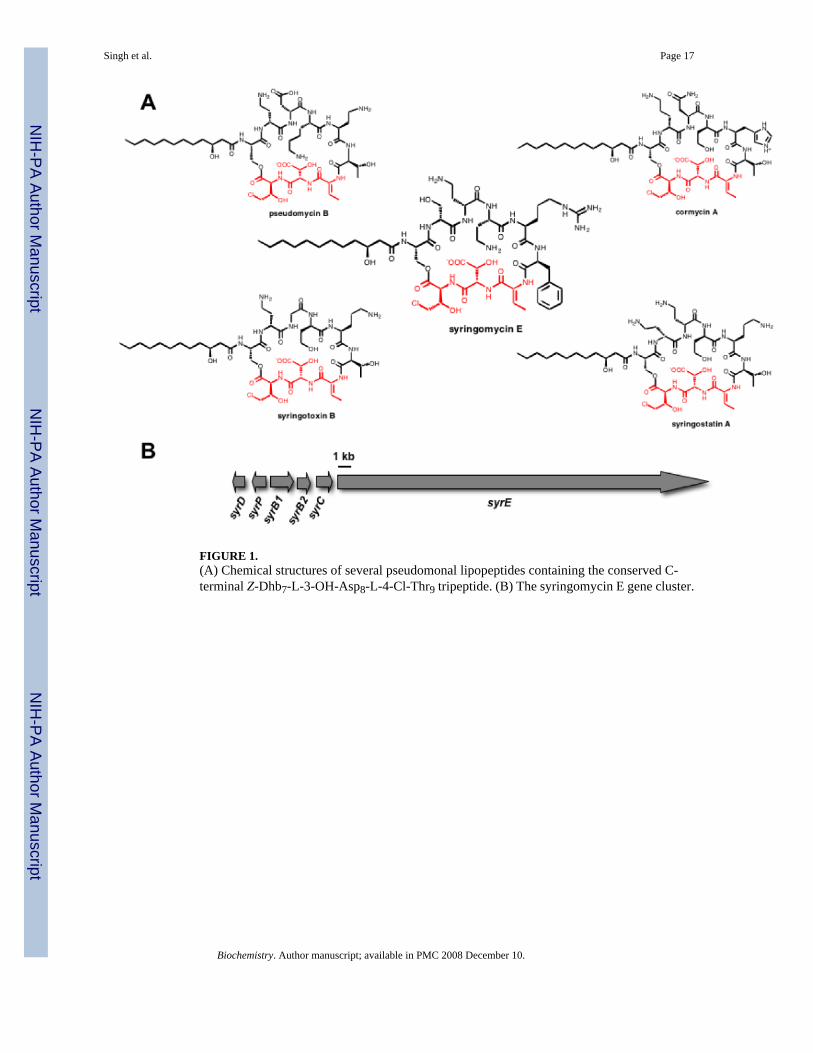
FIGURE 1.(A) Chemical structures of several pseudomonal lipopeptides containing the conserved C-terminal Z-Dhb7-L-3-OH-Asp8-L-4-Cl-Thr9 tripeptide. (B) The syringomycin E gene cluster.
Singh et al. Page 17
Biochemistry. Author manuscript; available in PMC 2008 December 10.
NIH
-PA Author Manuscript
NIH
-PA Author Manuscript
NIH
-PA Author Manuscript
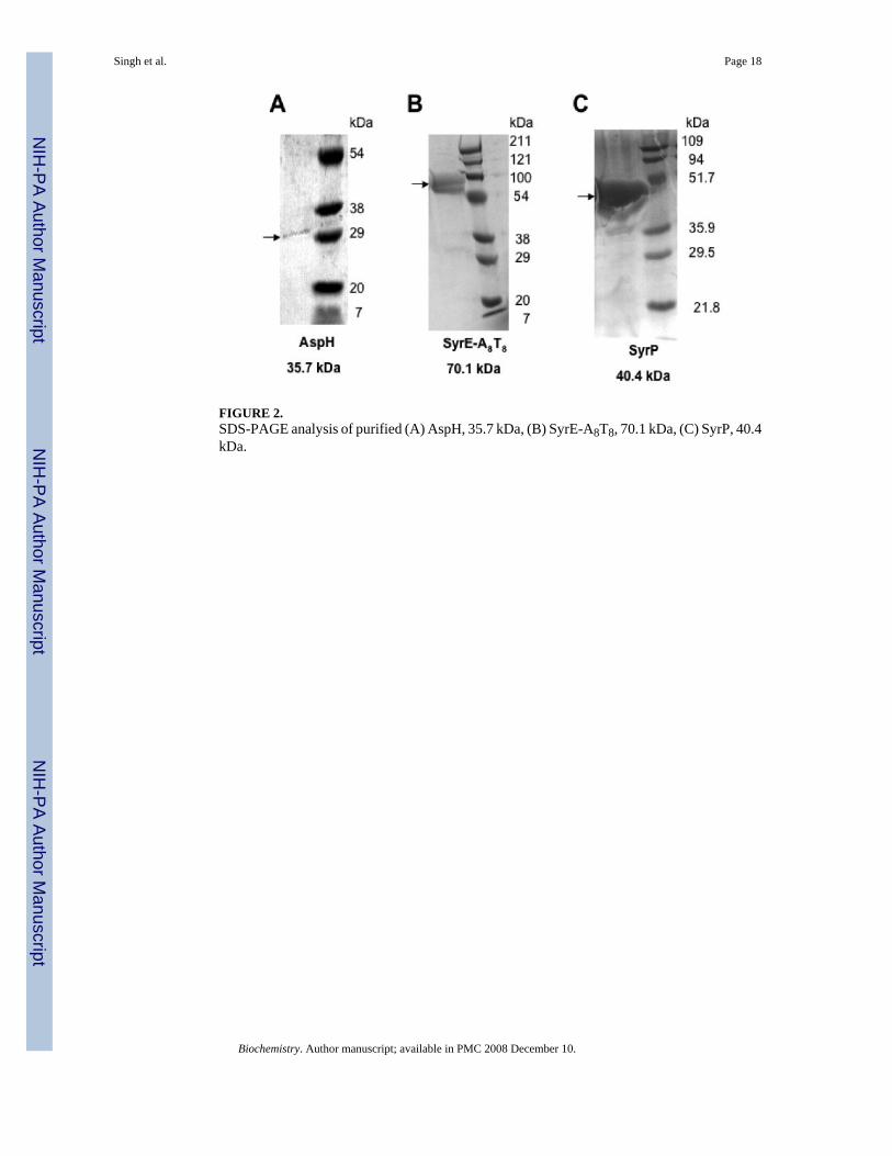
FIGURE 2.SDS-PAGE analysis of purified (A) AspH, 35.7 kDa, (B) SyrE-A8T8, 70.1 kDa, (C) SyrP, 40.4kDa.
Singh et al. Page 18
Biochemistry. Author manuscript; available in PMC 2008 December 10.
NIH
-PA Author Manuscript
NIH
-PA Author Manuscript
NIH
-PA Author Manuscript

FIGURE 3.Results of the ATP-PPi exchange assay to determine the substrate specificity of SyrE-A8 (A)Determination of kobs for three substrates: black diamonds, L-Asp, kobs = 6.3 min-1; graysquares, L-threo-3-OH-Asp, kobs = 0.2 min-1; white circles, L-erythro-3-OH-Asp, kobs = 0.1min-1. (B) Comparison of % activation by SyrE-A8 of L-Asp, L-threo-3-OH-Asp, L-erythro-3-OH-Asp.
Singh et al. Page 19
Biochemistry. Author manuscript; available in PMC 2008 December 10.
NIH
-PA Author Manuscript
NIH
-PA Author Manuscript
NIH
-PA Author Manuscript
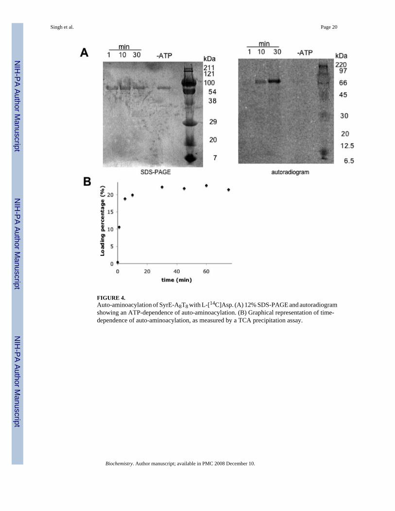
FIGURE 4.Auto-aminoacylation of SyrE-A8T8 with L-[14C]Asp. (A) 12% SDS-PAGE and autoradiogramshowing an ATP-dependence of auto-aminoacylation. (B) Graphical representation of time-dependence of auto-aminoacylation, as measured by a TCA precipitation assay.
Singh et al. Page 20
Biochemistry. Author manuscript; available in PMC 2008 December 10.
NIH
-PA Author Manuscript
NIH
-PA Author Manuscript
NIH
-PA Author Manuscript
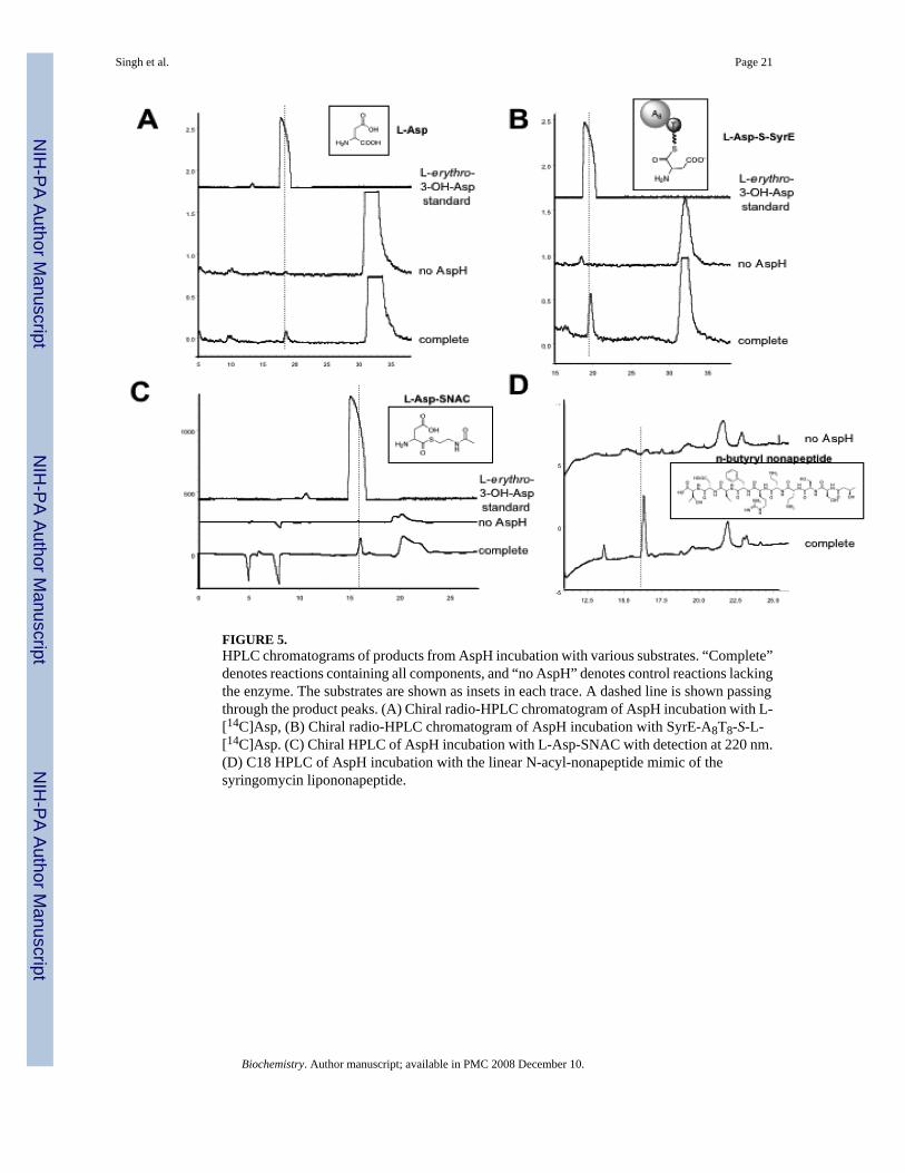
FIGURE 5.HPLC chromatograms of products from AspH incubation with various substrates. “Complete”denotes reactions containing all components, and “no AspH” denotes control reactions lackingthe enzyme. The substrates are shown as insets in each trace. A dashed line is shown passingthrough the product peaks. (A) Chiral radio-HPLC chromatogram of AspH incubation with L-[14C]Asp, (B) Chiral radio-HPLC chromatogram of AspH incubation with SyrE-A8T8-S-L-[14C]Asp. (C) Chiral HPLC of AspH incubation with L-Asp-SNAC with detection at 220 nm.(D) C18 HPLC of AspH incubation with the linear N-acyl-nonapeptide mimic of thesyringomycin lipononapeptide.
Singh et al. Page 21
Biochemistry. Author manuscript; available in PMC 2008 December 10.
NIH
-PA Author Manuscript
NIH
-PA Author Manuscript
NIH
-PA Author Manuscript

FIGURE 6.Alignment of the protein sequences of SyrP from P. syringae pv. syringae B728a with that ofTauD from P. mendocina. Red arrows indicate the His2/Asp iron-binding motif and blue arrowspoint to the two conserved Arg residues that bind alpha-ketoglutarate.
Singh et al. Page 22
Biochemistry. Author manuscript; available in PMC 2008 December 10.
NIH
-PA Author Manuscript
NIH
-PA Author Manuscript
NIH
-PA Author Manuscript
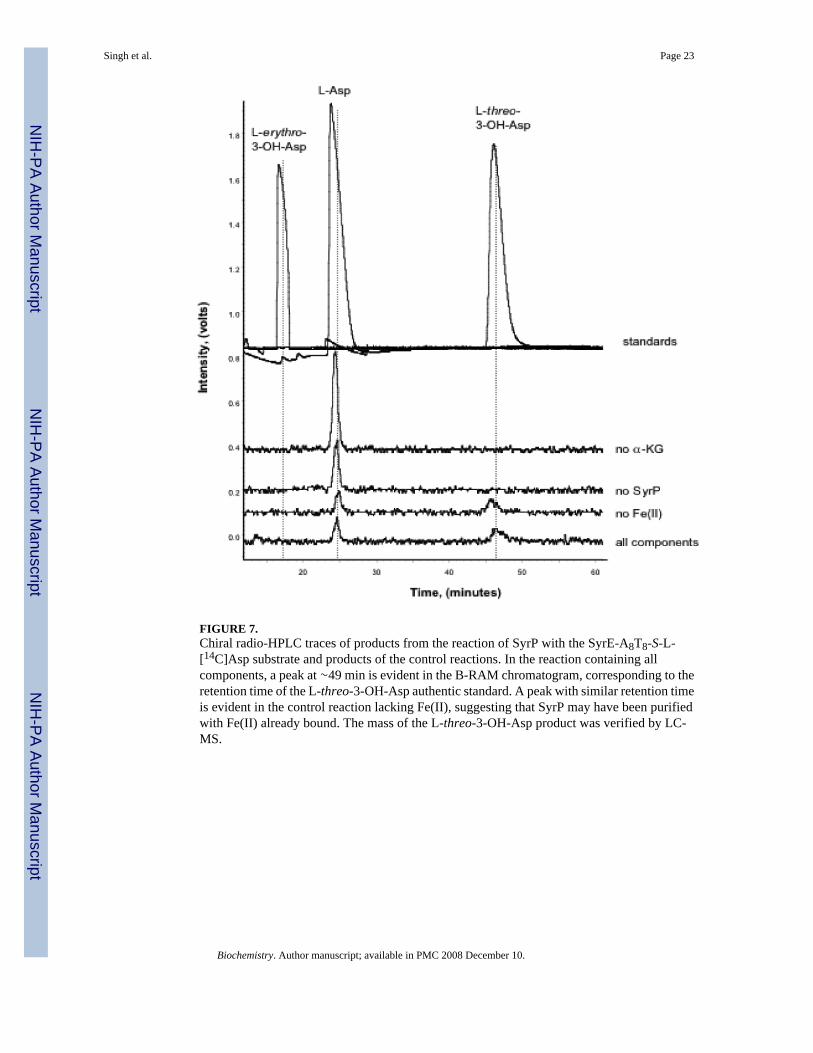
FIGURE 7.Chiral radio-HPLC traces of products from the reaction of SyrP with the SyrE-A8T8-S-L-[14C]Asp substrate and products of the control reactions. In the reaction containing allcomponents, a peak at ∼49 min is evident in the B-RAM chromatogram, corresponding to theretention time of the L-threo-3-OH-Asp authentic standard. A peak with similar retention timeis evident in the control reaction lacking Fe(II), suggesting that SyrP may have been purifiedwith Fe(II) already bound. The mass of the L-threo-3-OH-Asp product was verified by LC-MS.
Singh et al. Page 23
Biochemistry. Author manuscript; available in PMC 2008 December 10.
NIH
-PA Author Manuscript
NIH
-PA Author Manuscript
NIH
-PA Author Manuscript
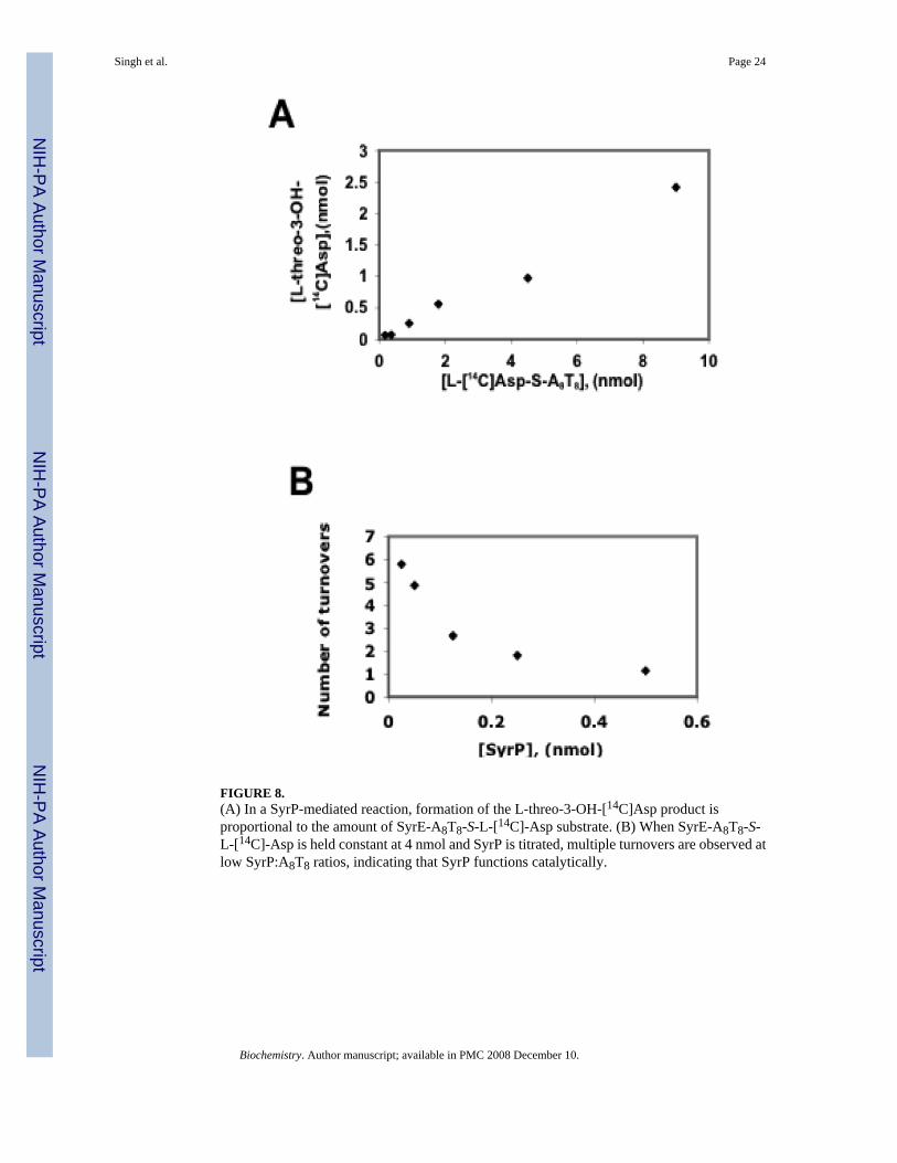
FIGURE 8.(A) In a SyrP-mediated reaction, formation of the L-threo-3-OH-[14C]Asp product isproportional to the amount of SyrE-A8T8-S-L-[14C]-Asp substrate. (B) When SyrE-A8T8-S-L-[14C]-Asp is held constant at 4 nmol and SyrP is titrated, multiple turnovers are observed atlow SyrP:A8T8 ratios, indicating that SyrP functions catalytically.
Singh et al. Page 24
Biochemistry. Author manuscript; available in PMC 2008 December 10.
NIH
-PA Author Manuscript
NIH
-PA Author Manuscript
NIH
-PA Author Manuscript
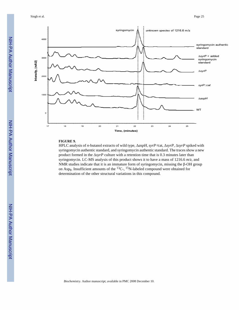
FIGURE 9.HPLC analysis of n-butanol extracts of wild type, ΔaspH, syrP∷cat, ΔsyrP, ΔsyrP spiked withsyringomycin authentic standard, and syringomycin authentic standard. The traces show a newproduct formed in the ΔsyrP culture with a retention time that is 0.3 minutes later thansyringomycin. LC-MS analysis of this product shows it to have a mass of 1216.6 m/z, andNMR studies indicate that it is an immature form of syringomycin, missing the β-OH groupon Asp8. Insufficient amounts of the 13C-, 15N-labeled compound were obtained fordetermination of the other structural variations in this compound.
Singh et al. Page 25
Biochemistry. Author manuscript; available in PMC 2008 December 10.
NIH
-PA Author Manuscript
NIH
-PA Author Manuscript
NIH
-PA Author Manuscript

NIH
-PA Author Manuscript
NIH
-PA Author Manuscript
NIH
-PA Author Manuscript
Singh et al. Page 26
Table 1Kinetics data collected for AspH with aspartate-containing substrates.
Substrate kcat (min-1) Km (μM) kcat/Km
L-Asp 0.15 ± 0.03 52.9 ± 4.7 0.002L-Asp-SNAC 42.5 ± 5.2 43.6 ± 2.3 0.97
Linear nonapeptide 120.0 ± 15.4 26.9 ± 9.9 4.6
Biochemistry. Author manuscript; available in PMC 2008 December 10.
Copyright © 2022 FDOKUMEN