The Mangotoxin Biosynthetic Operon (mbo) Is Specifically Distributed within Pseudomonas syringae...
13
Published Ahead of Print 9 November 2012. 10.1128/AEM.03007-12. 2013, 79(3):756. DOI: Appl. Environ. Microbiol. Francisco M. Cazorla and Jesús Murillo Vicente, Arrebola, Leire Bardaji, Juan C. Codina, Antonio de Víctor J. Carrión, José A. Gutiérrez-Barranquero, Eva Evolution and Was Acquired Only Once during Pseudomonas syringae Genomospecies 1 Is Specifically Distributed within ) mbo The Mangotoxin Biosynthetic Operon ( http://aem.asm.org/content/79/3/756 Updated information and services can be found at: These include: SUPPLEMENTAL MATERIAL Supplemental material REFERENCES http://aem.asm.org/content/79/3/756#ref-list-1 at: This article cites 61 articles, 21 of which can be accessed free CONTENT ALERTS more» articles cite this article), Receive: RSS Feeds, eTOCs, free email alerts (when new http://journals.asm.org/site/misc/reprints.xhtml Information about commercial reprint orders: http://journals.asm.org/site/subscriptions/ To subscribe to to another ASM Journal go to: on January 15, 2013 by Alejandro Perez-Garcia http://aem.asm.org/ Downloaded from
Transcript of The Mangotoxin Biosynthetic Operon (mbo) Is Specifically Distributed within Pseudomonas syringae...
Published Ahead of Print 9 November 2012. 10.1128/AEM.03007-12.
2013, 79(3):756. DOI:Appl. Environ. Microbiol. Francisco M. Cazorla and Jesús Murillo
Vicente,Arrebola, Leire Bardaji, Juan C. Codina, Antonio de Víctor J. Carrión, José A. Gutiérrez-Barranquero, Eva Evolutionand Was Acquired Only Once during Pseudomonas syringae Genomospecies 1Is Specifically Distributed within
)mboThe Mangotoxin Biosynthetic Operon (
http://aem.asm.org/content/79/3/756Updated information and services can be found at:
These include:
SUPPLEMENTAL MATERIAL Supplemental material
REFERENCEShttp://aem.asm.org/content/79/3/756#ref-list-1at:
This article cites 61 articles, 21 of which can be accessed free
CONTENT ALERTS more»articles cite this article),
Receive: RSS Feeds, eTOCs, free email alerts (when new
http://journals.asm.org/site/misc/reprints.xhtmlInformation about commercial reprint orders: http://journals.asm.org/site/subscriptions/To subscribe to to another ASM Journal go to:
on January 15, 2013 by Alejandro P
erez-Garcia
http://aem.asm
.org/D
ownloaded from

The Mangotoxin Biosynthetic Operon (mbo) Is Specifically Distributedwithin Pseudomonas syringae Genomospecies 1 and Was AcquiredOnly Once during Evolution
Víctor J. Carrión,a José A. Gutiérrez-Barranquero,a Eva Arrebola,b Leire Bardaji,c Juan C. Codina,a Antonio de Vicente,a
Francisco M. Cazorla,a Jesús Murilloc
Instituto de Hortofruticultura Subtropical y Mediterránea La Mayora (IHSM-UMA-CSIC), Departamento de Microbiología, Facultad de Ciencias, Universidad de Málaga,Málaga, Spaina; Instituto de Hortofruticultura Subtropical y Mediterránea La Mayora (IHSMUMA-CSIC), Estación Experimental La Mayora, Algarrobo-Costa, Málaga, Spainb;Laboratorio de Patología Vegetal, ETS Ingenieros Agrónomos, Universidad Pública de Navarra, Pamplona, Spainc
Mangotoxin production was first described in Pseudomonas syringae pv. syringae strains. A phenotypic characterization of 94 P.syringae strains was carried out to determine the genetic evolution of the mangotoxin biosynthetic operon (mbo). We designed aPCR primer pair specific for the mbo operon to examine its distribution within the P. syringae complex. These primers amplifieda 692-bp DNA fragment from 52 mangotoxin-producing strains and from 7 non-mangotoxin-producing strains that harbor thembo operon, whereas 35 non-mangotoxin-producing strains did not yield any amplification. This, together with the analysis ofdraft genomes, allowed the identification of the mbo operon in five pathovars (pathovars aptata, avellanae, japonica, pisi, andsyringae), all of which belong to genomospecies 1, suggesting a limited distribution of the mbo genes in the P. syringae complex.Phylogenetic analyses using partial sequences from housekeeping genes differentiated three groups within genomospecies 1. Allof the strains containing the mbo operon clustered in groups I and II, whereas those lacking the operon clustered in group III;however, the relative branching order of these three groups is dependent on the genes used to construct the phylogeny. The mbooperon maintains synteny and is inserted in the same genomic location, with high sequence conservation around the insertionpoint, for all the strains in groups I and II. These data support the idea that the mbo operon was acquired horizontally and onlyonce by the ancestor of groups I and II from genomospecies 1 within the P. syringae complex.
The bacterial plant pathogen Pseudomonas syringae is ubiqui-tous in nature and often causes economically important plant
diseases. This bacterial species establishes an epiphytic populationin association with a plant host’s surfaces prior to infection. Thereare at least 50 pathovars, many of which cause a wide range ofplant diseases that exhibit diverse symptoms, such as leaf or fruitlesions, cankers, blasts, and galls (1–7).
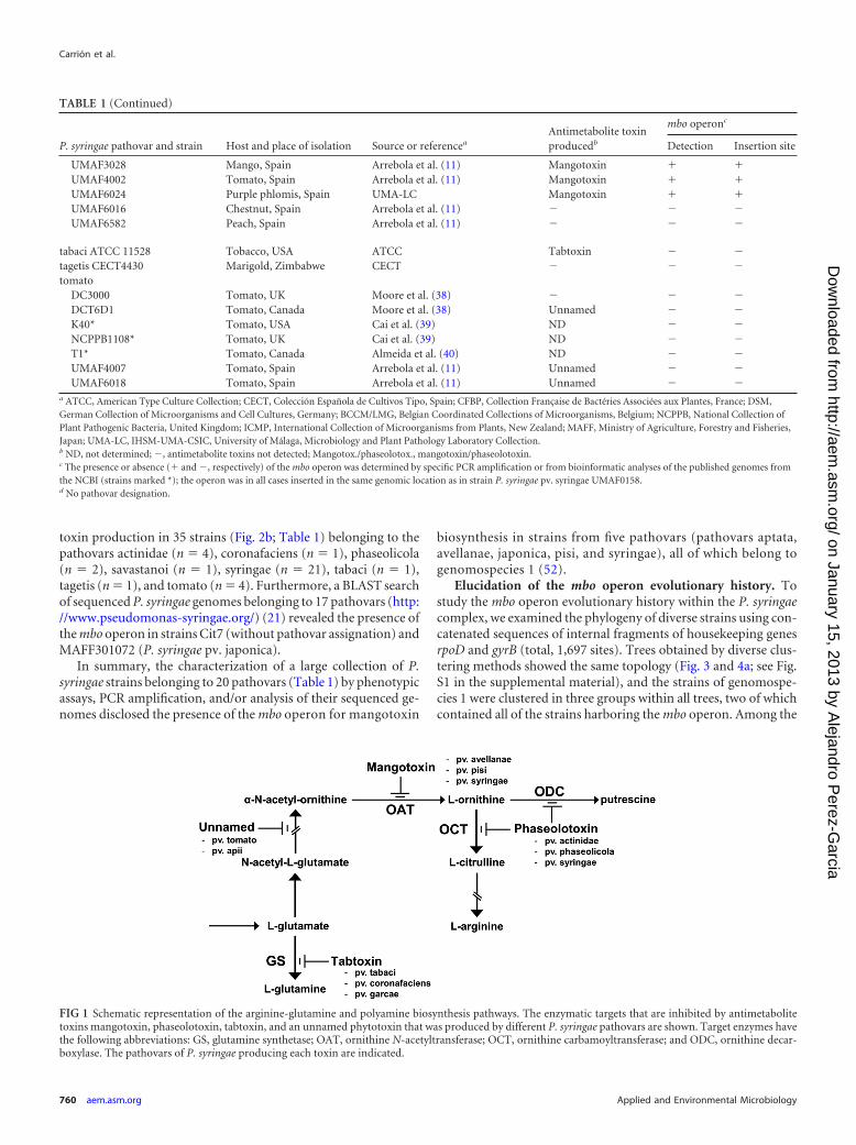
P. syringae produces several type III effectors and virulencefactors, but the role of its toxins is particularly significant duringsymptom development (1, 8–10). The current study is focused onmangotoxin, which inhibits the enzyme ornithine N-acetyltrans-ferase (11, 12). Mangotoxin was initially detected in strains fromP. syringae pv. syringae (11); however, its production was recentlyobserved in strains of pathovar avellanae (13). Recent studies havereported the involvement of the mgo (8, 12, 14) and mbo (15)operons in mangotoxin production and P. syringae pv. syringaevirulence. The mbo operon is composed of six genes, and all ofthem are directly involved in mangotoxin production.
The development of specific methods for pathogen detectionin planta is very important. In this sense, different PCR methodshave been developed and improved to detect different P. syringaepathovars. For example, one PCR method used primers that werebased on the 16S-23S ribosomal genes to identify strains of P.syringae pv. actinidae in which the PCR resulted in a specific am-plicon of this pathovar (16). A multiplex PCR assay has been de-veloped for the identification and differentiation of P. fragi, P.lundensis, and P. putida on the basis of the coamplification ofdifferent carbamoyl phosphate synthase small-subunit gene frag-ments (17). Another example is the amplification of the gene iaaL,yielding a 454-bp fragment that allows the identification and de-
tection of P. syringae pv. savastanoi strains (18). In the currentstudy, we have developed a sensitive and specific method for thedetection of mangotoxin-producing strains by a PCR that is basedon the amplification of an mbo operon region in P. syringae. Thespecificity of the mbo operon within mangotoxin biosynthesis wasessential for the planning and development of this method (15).
Additionally, the study of specific genes for the biosynthesisand production of virulence factors, as in the case of the mbooperon, could also be used in phylogenetic studies. Sawada et al.(19) revealed a remarkable degree of congruence between twohousekeeping genes (gyrB and rpoD) and two components of thepathogenesis-associated type III secretion system (hrpS and hrpL),leading to the conclusion that the type III secretion system wasacquired prior to the diversification of the P. syringae pathovars.The diversity of P. syringae strains has been further explored byphysical mapping of the ribosomal gene cluster, revealing that thesize and structure of P. syringae genomes vary greatly by pathovarand that large-scale genomic rearrangements are common (20). Inthis study, a phylogenetic analysis using housekeeping genes has
Received 1 October 2012 Accepted 7 November 2012
Published ahead of print 9 November 2012
Address correspondence to Francisco M. Cazorla, [email protected], or JesúsMurillo, [email protected].
Supplemental material for this article may be found at http://dx.doi.org/10.1128/AEM.03007-12.
Copyright © 2013, American Society for Microbiology. All Rights Reserved.
doi:10.1128/AEM.03007-12
756 aem.asm.org Applied and Environmental Microbiology p. 756–767 February 2013 Volume 79 Number 3
on January 15, 2013 by Alejandro P
erez-Garcia
http://aem.asm
.org/D
ownloaded from
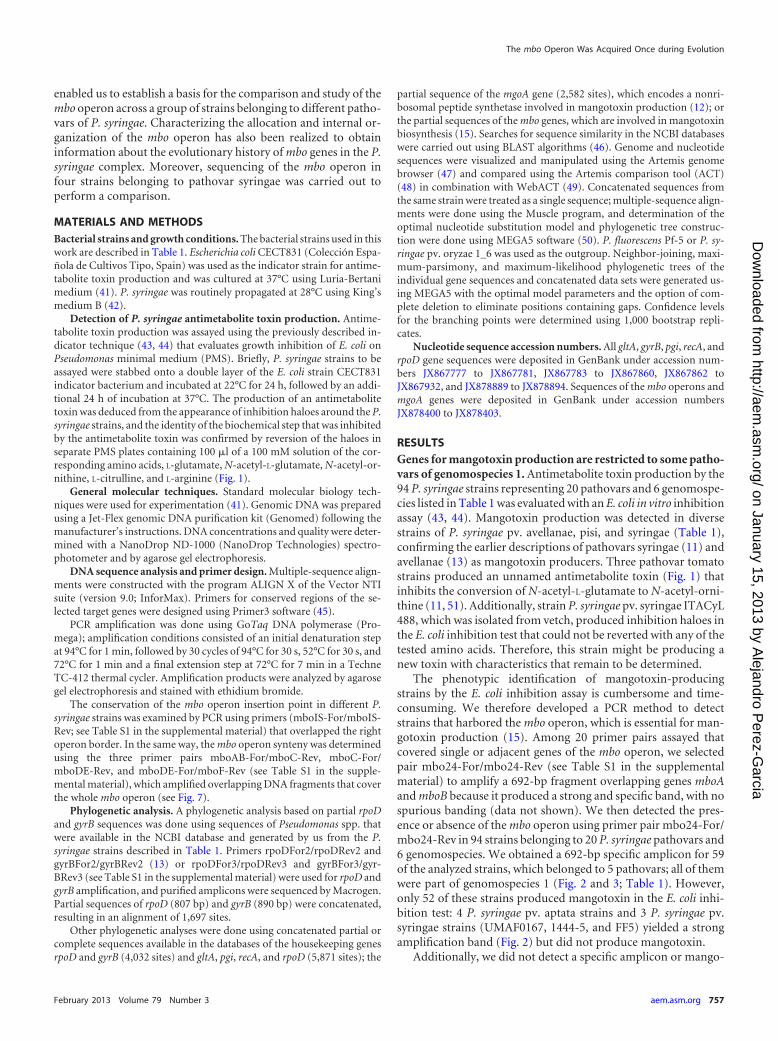
enabled us to establish a basis for the comparison and study of thembo operon across a group of strains belonging to different patho-vars of P. syringae. Characterizing the allocation and internal or-ganization of the mbo operon has also been realized to obtaininformation about the evolutionary history of mbo genes in the P.syringae complex. Moreover, sequencing of the mbo operon infour strains belonging to pathovar syringae was carried out toperform a comparison.
MATERIALS AND METHODSBacterial strains and growth conditions. The bacterial strains used in thiswork are described in Table 1. Escherichia coli CECT831 (Colección Espa-ñola de Cultivos Tipo, Spain) was used as the indicator strain for antime-tabolite toxin production and was cultured at 37°C using Luria-Bertanimedium (41). P. syringae was routinely propagated at 28°C using King’smedium B (42).
Detection of P. syringae antimetabolite toxin production. Antime-tabolite toxin production was assayed using the previously described in-dicator technique (43, 44) that evaluates growth inhibition of E. coli onPseudomonas minimal medium (PMS). Briefly, P. syringae strains to beassayed were stabbed onto a double layer of the E. coli strain CECT831indicator bacterium and incubated at 22°C for 24 h, followed by an addi-tional 24 h of incubation at 37°C. The production of an antimetabolitetoxin was deduced from the appearance of inhibition haloes around the P.syringae strains, and the identity of the biochemical step that was inhibitedby the antimetabolite toxin was confirmed by reversion of the haloes inseparate PMS plates containing 100 �l of a 100 mM solution of the cor-responding amino acids, L-glutamate, N-acetyl-L-glutamate, N-acetyl-or-nithine, L-citrulline, and L-arginine (Fig. 1).
General molecular techniques. Standard molecular biology tech-niques were used for experimentation (41). Genomic DNA was preparedusing a Jet-Flex genomic DNA purification kit (Genomed) following themanufacturer’s instructions. DNA concentrations and quality were deter-mined with a NanoDrop ND-1000 (NanoDrop Technologies) spectro-photometer and by agarose gel electrophoresis.
DNA sequence analysis and primer design. Multiple-sequence align-ments were constructed with the program ALIGN X of the Vector NTIsuite (version 9.0; InforMax). Primers for conserved regions of the se-lected target genes were designed using Primer3 software (45).
PCR amplification was done using GoTaq DNA polymerase (Pro-mega); amplification conditions consisted of an initial denaturation stepat 94°C for 1 min, followed by 30 cycles of 94°C for 30 s, 52°C for 30 s, and72°C for 1 min and a final extension step at 72°C for 7 min in a TechneTC-412 thermal cycler. Amplification products were analyzed by agarosegel electrophoresis and stained with ethidium bromide.
The conservation of the mbo operon insertion point in different P.syringae strains was examined by PCR using primers (mboIS-For/mboIS-Rev; see Table S1 in the supplemental material) that overlapped the rightoperon border. In the same way, the mbo operon synteny was determinedusing the three primer pairs mboAB-For/mboC-Rev, mboC-For/mboDE-Rev, and mboDE-For/mboF-Rev (see Table S1 in the supple-mental material), which amplified overlapping DNA fragments that coverthe whole mbo operon (see Fig. 7).
Phylogenetic analysis. A phylogenetic analysis based on partial rpoDand gyrB sequences was done using sequences of Pseudomonas spp. thatwere available in the NCBI database and generated by us from the P.syringae strains described in Table 1. Primers rpoDFor2/rpoDRev2 andgyrBFor2/gyrBRev2 (13) or rpoDFor3/rpoDRev3 and gyrBFor3/gyr-BRev3 (see Table S1 in the supplemental material) were used for rpoD andgyrB amplification, and purified amplicons were sequenced by Macrogen.Partial sequences of rpoD (807 bp) and gyrB (890 bp) were concatenated,resulting in an alignment of 1,697 sites.
Other phylogenetic analyses were done using concatenated partial orcomplete sequences available in the databases of the housekeeping genesrpoD and gyrB (4,032 sites) and gltA, pgi, recA, and rpoD (5,871 sites); the
partial sequence of the mgoA gene (2,582 sites), which encodes a nonri-bosomal peptide synthetase involved in mangotoxin production (12); orthe partial sequences of the mbo genes, which are involved in mangotoxinbiosynthesis (15). Searches for sequence similarity in the NCBI databaseswere carried out using BLAST algorithms (46). Genome and nucleotidesequences were visualized and manipulated using the Artemis genomebrowser (47) and compared using the Artemis comparison tool (ACT)(48) in combination with WebACT (49). Concatenated sequences fromthe same strain were treated as a single sequence; multiple-sequence align-ments were done using the Muscle program, and determination of theoptimal nucleotide substitution model and phylogenetic tree construc-tion were done using MEGA5 software (50). P. fluorescens Pf-5 or P. sy-ringae pv. oryzae 1_6 was used as the outgroup. Neighbor-joining, maxi-mum-parsimony, and maximum-likelihood phylogenetic trees of theindividual gene sequences and concatenated data sets were generated us-ing MEGA5 with the optimal model parameters and the option of com-plete deletion to eliminate positions containing gaps. Confidence levelsfor the branching points were determined using 1,000 bootstrap repli-cates.
Nucleotide sequence accession numbers. All gltA, gyrB, pgi, recA, andrpoD gene sequences were deposited in GenBank under accession num-bers JX867777 to JX867781, JX867783 to JX867860, JX867862 toJX867932, and JX878889 to JX878894. Sequences of the mbo operons andmgoA genes were deposited in GenBank under accession numbersJX878400 to JX878403.
RESULTSGenes for mangotoxin production are restricted to some patho-vars of genomospecies 1. Antimetabolite toxin production by the94 P. syringae strains representing 20 pathovars and 6 genomospe-cies listed in Table 1 was evaluated with an E. coli in vitro inhibitionassay (43, 44). Mangotoxin production was detected in diversestrains of P. syringae pv. avellanae, pisi, and syringae (Table 1),confirming the earlier descriptions of pathovars syringae (11) andavellanae (13) as mangotoxin producers. Three pathovar tomatostrains produced an unnamed antimetabolite toxin (Fig. 1) thatinhibits the conversion of N-acetyl-L-glutamate to N-acetyl-orni-thine (11, 51). Additionally, strain P. syringae pv. syringae ITACyL488, which was isolated from vetch, produced inhibition haloes inthe E. coli inhibition test that could not be reverted with any of thetested amino acids. Therefore, this strain might be producing anew toxin with characteristics that remain to be determined.
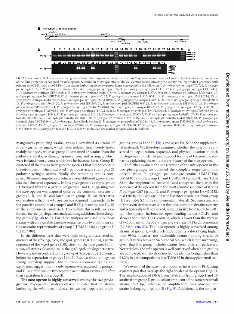
The phenotypic identification of mangotoxin-producingstrains by the E. coli inhibition assay is cumbersome and time-consuming. We therefore developed a PCR method to detectstrains that harbored the mbo operon, which is essential for man-gotoxin production (15). Among 20 primer pairs assayed thatcovered single or adjacent genes of the mbo operon, we selectedpair mbo24-For/mbo24-Rev (see Table S1 in the supplementalmaterial) to amplify a 692-bp fragment overlapping genes mboAand mboB because it produced a strong and specific band, with nospurious banding (data not shown). We then detected the pres-ence or absence of the mbo operon using primer pair mbo24-For/mbo24-Rev in 94 strains belonging to 20 P. syringae pathovars and6 genomospecies. We obtained a 692-bp specific amplicon for 59of the analyzed strains, which belonged to 5 pathovars; all of themwere part of genomospecies 1 (Fig. 2 and 3; Table 1). However,only 52 of these strains produced mangotoxin in the E. coli inhi-bition test: 4 P. syringae pv. aptata strains and 3 P. syringae pv.syringae strains (UMAF0167, 1444-5, and FF5) yielded a strongamplification band (Fig. 2) but did not produce mangotoxin.
Additionally, we did not detect a specific amplicon or mango-
The mbo Operon Was Acquired Once during Evolution
February 2013 Volume 79 Number 3 aem.asm.org 757
on January 15, 2013 by Alejandro P
erez-Garcia
http://aem.asm
.org/D
ownloaded from
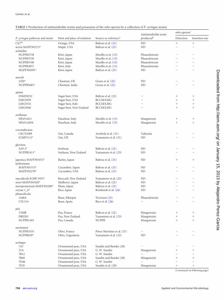
TABLE 1 Production of antimetabolite toxins and possession of the mbo operon by a collection of P. syringae strains
P. syringae pathovar and strain Host and place of isolation Source or referencea
Antimetabolite toxinproducedb
mbo operonc
Detection Insertion site
Cit7d* Orange, USA Baltrus et al. (21) ND � �aceris MAFF302273* Maple, USA Baltrus et al. (21) ND � �actinidae
NCPPB3738 Kiwi, Japan Murillo et al. (13) Phaseolotoxin � �NCPPB3739 Kiwi, Japan Murillo et al. (13) Phaseolotoxin � �NCPPB3740 Kiwi, Japan Murillo et al. (13) Phaseolotoxin � �NCPPB3871 Kiwi, Italy Murillo et al. (13) Phaseolotoxin � �MAFF302091* Kiwi, Japan Baltrus et al. (21) ND � �
aesculi2250* Chestnut, UK Green et al. (22) ND � �NCPPB3681* Chestnut, India Green et al. (22) ND � �
aptataDSM50252 Sugar beet, USA Baltrus et al. (21) � � �LMG5059 Sugar beet, USA BCCM/LMG � � �LMG5532 Sugar beet, Italy BCCM/LMG � � �LMG5646 Sugar beet, New Zealand BCCM/LMG � � �
avellanaeISPaVe011 Hazelnut, Italy Murillo et al. (13) Mangotoxin � �ISPaVe2056 Hazelnut, Italy Murillo et al. (13) Mangotoxin � �
coronafaciensCECT4389 Oat, Canada Arrebola et al. (11) Tabtoxin � �ICMP3113* Oat, UK Yamamoto et al. (21) ND � �
glycineaA29-2* Soybean, Baltrus et al. (21) ND � �NCPPB2411* Soybean, New Zealand Yamamoto et al. (23) ND � �
japonica MAFF301072* Barley, Japan Baltrus et al. (21) ND � �lachrymans
MAFF301315* Cucumber, Japan Baltrus et al. (21) ND � �MAFF302278* Cucumber, USA Baltrus et al. (21) ND � �
maculicola ICMP 3935* Broccoli, New Zealand Yamamoto et al. (23) ND � �mori MAFF301020* Mulberry, Japan Baltrus et al. (21) ND � �morsprunorum MAFF302280* Plum, Japan Baltrus et al. (21) ND � �oryzae 1_6* Rice, Japan Reinhardt et al. (24) ND � �phaseolicola
1448A Bean, Ethiopia Teverson (25) Phaseolotoxin � �CYL314 Bean, Spain Rico et al. (26) � � �
pisi1704B Pea, France Baltrus et al. (21) Mangotoxin � �HRI203 Pea, New Zealand Yamamoto et al. (23) Mangotoxin � �NCPPB1365 Pea, Canada NCPPB Mangotoxin � �
savastanoiNCPPB3335 Olive, France Pérez-Martínez et al. (27) � � �NCPPB639* Olive, Yugoslavia Yamamoto et al. (23) ND � �
syringae7A7 Ornamental pear, USA Sundin and Bender (28) � � �7C6 Ornamental pear, USA G. W. Sundin Mangotoxin � �7B12 Ornamental pear, USA G. W. Sundin � � �7B40 Ornamental pear, USA Sundin and Bender (28) Mangotoxin � �7D46 Ornamental pear, USA G. W. Sundin � � �7F29 Ornamental pear, USA Sundin et al. (29) Mangotoxin � �
(Continued on following page)
Carrión et al.
758 aem.asm.org Applied and Environmental Microbiology
on January 15, 2013 by Alejandro P
erez-Garcia
http://aem.asm
.org/D
ownloaded from
TABLE 1 (Continued)
P. syringae pathovar and strain Host and place of isolation Source or referencea
Antimetabolite toxinproducedb
mbo operonc
Detection Insertion site
8B48 Ornamental pear, USA Sundin et al. (29) � � �8C32 Ornamental pear, USA Sundin et al. (29) Mangotoxin � �8C43 Ornamental pear, USA Sundin et al. (29) � � �8F21 Ornamental pear, USA Sundin et al. (29) Mangotoxin � �1444-5 Laurel, Spain Arrebola et al. (11) � � �1507-7 Hawthorn, Spain Arrebola et al. (11) � � �1559-9 Mango, Spain Arrebola et al. (11) Mangotoxin � �B728a Bean, USA Feil et al. (30) � � �CECT127 Lilac, UK CECT Mangotoxin � �CECT4429 Lilac, UK CECT Mangotoxin � �CFBP3388 Vetch, France Tourte and Manceau (31) Mangotox./phaseolotox. � �CRD 09-87 Chickling pea, Spain Martín-Sanz et al. (32) � � �DAR77787 Mango, Australia Young (33) Mangotoxin � �DAR77789 Mango, Australia Young (33) Mangotoxin � �EPSMV3 Pear, Spain Arrebola et al. (11) � � �EPS17A Pear, Spain Arrebola et al. (11) Mangotoxin � �FF5 Ornamental pear, USA Sohn et al. (34) � � �ITACyL 488 Vetch, Spain Martín-Sanz et al. (35) Unknown � �ITACyL 522 Chickling pea, Spain Martín-Sanz et al. (32) � � �ITACyL 523 Chickling pea, Spain Martín-Sanz et al. (32) � � �ITACyL 524 Chickling pea, Spain Martín-Sanz et al. (32) � � �ITACyL 525 Chickling pea, Spain Martín-Sanz et al. (32) � � �ITACyL 526 Grass pea, Spain Martín-Sanz et al. (32) � � �ITACyL 527 Grass pea, Spain Martín-Sanz et al. (32) � � �ITACyL 528 Grass pea, Spain Martín-Sanz et al. (32) � � �ITACyL 529 Vetch, Spain Martín-Sanz et al. (32) � � �NCPPB1239 Bean, Kenya NCPPB Mangotoxin � �Ps-5 Mango, Israel UMA-LC Mangotoxin � �Ps-6 Mango, Israel Arrebola et al. (11) Mangotoxin � �Ps-10 Mango, Israel Arrebola et al. (11) Mangotoxin � �Ps-35 Mango, Israel Arrebola et al. (11) Mangotoxin � �UMAF0049 Mango, Spain Cazorla et al. (36) Mangotoxin � �UMAF0081 Mango, Spain Cazorla et al. (36) Mangotoxin � �UMAF0158 Mango, Spain Cazorla et al. (36) Mangotoxin � �UMAF0167 Mango, Spain Cazorla et al. (36) � � �UMAF0170 Mango, Spain Cazorla et al. (36) Mangotoxin � �UMAF0176 Mango, Spain Arrebola et al. (11) Mangotoxin � �UMAF0209 Mango, Spain UMA-LC Mangotoxin � �UMAF0214 Mango, Spain UMA-LC Mangotoxin � �UMAF0217 Mango, Spain UMA-LC Mangotoxin � �UMAF0220 Mango, Spain UMA-LC Mangotoxin � �UMAF0221 Mango, Spain UMA-LC Mangotoxin � �UMAF0222 Mango, Spain UMA-LC Mangotoxin � �UMAF0223 Mango, Spain UMA-LC Mangotoxin � �UMAF0225 Mango, Spain UMA-LC Mangotoxin � �UMAF0226 Mango, Spain UMA-LC Mangotoxin � �UMAF1003 Mango, Spain Arrebola et al. (11) Mangotoxin � �UMAF1060 Mango, Spain Gutiérrez-Barranquero et al. (37) Mangotoxin � �UMAF2007 Mango, Portugal Arrebola et al. (11) Mangotoxin � �UMAF2008 Mango, Portugal UMA-LC Mangotoxin � �UMAF2025 Mango, Portugal Cazorla et al. (36) Mangotoxin � �UMAF2026 Mango, Portugal Cazorla et al. (36) Mangotoxin � �UMAF2676 Bean, South Africa Arrebola et al. (11) � � �UMAF2700 Mango, Italy UMA-LC Mangotoxin � �UMAF2702 Mango, Italy Gutiérrez-Barranquero et al. (37) Mangotoxin � �UMAF2801 Mango, Spain Gutiérrez-Barranquero et al. (37) Mangotoxin � �UMAF2802 Mango, Spain Gutiérrez-Barranquero et al. (37) Mangotoxin � �UMAF2805 Mango, Spain UMA-LC Mangotoxin � �UMAF2808 Mango, Spain UMA-LC Mangotoxin � �UMAF2811 Mango, Spain UMA-LC Mangotoxin � �
(Continued on following page)
The mbo Operon Was Acquired Once during Evolution
February 2013 Volume 79 Number 3 aem.asm.org 759
on January 15, 2013 by Alejandro P
erez-Garcia
http://aem.asm
.org/D
ownloaded from
toxin production in 35 strains (Fig. 2b; Table 1) belonging to thepathovars actinidae (n � 4), coronafaciens (n � 1), phaseolicola(n � 2), savastanoi (n � 1), syringae (n � 21), tabaci (n � 1),tagetis (n � 1), and tomato (n � 4). Furthermore, a BLAST searchof sequenced P. syringae genomes belonging to 17 pathovars (http://www.pseudomonas-syringae.org/) (21) revealed the presence ofthe mbo operon in strains Cit7 (without pathovar assignation) andMAFF301072 (P. syringae pv. japonica).
In summary, the characterization of a large collection of P.syringae strains belonging to 20 pathovars (Table 1) by phenotypicassays, PCR amplification, and/or analysis of their sequenced ge-nomes disclosed the presence of the mbo operon for mangotoxin
biosynthesis in strains from five pathovars (pathovars aptata,avellanae, japonica, pisi, and syringae), all of which belong togenomospecies 1 (52).
Elucidation of the mbo operon evolutionary history. Tostudy the mbo operon evolutionary history within the P. syringaecomplex, we examined the phylogeny of diverse strains using con-catenated sequences of internal fragments of housekeeping genesrpoD and gyrB (total, 1,697 sites). Trees obtained by diverse clus-tering methods showed the same topology (Fig. 3 and 4a; see Fig.S1 in the supplemental material), and the strains of genomospe-cies 1 were clustered in three groups within all trees, two of whichcontained all of the strains harboring the mbo operon. Among the
TABLE 1 (Continued)
P. syringae pathovar and strain Host and place of isolation Source or referencea
Antimetabolite toxinproducedb
mbo operonc
Detection Insertion site
UMAF3028 Mango, Spain Arrebola et al. (11) Mangotoxin � �UMAF4002 Tomato, Spain Arrebola et al. (11) Mangotoxin � �UMAF6024 Purple phlomis, Spain UMA-LC Mangotoxin � �UMAF6016 Chestnut, Spain Arrebola et al. (11) � � �UMAF6582 Peach, Spain Arrebola et al. (11) � � �
tabaci ATCC 11528 Tobacco, USA ATCC Tabtoxin � �tagetis CECT4430 Marigold, Zimbabwe CECT � � �tomato
DC3000 Tomato, UK Moore et al. (38) � � �DCT6D1 Tomato, Canada Moore et al. (38) Unnamed � �K40* Tomato, USA Cai et al. (39) ND � �NCPPB1108* Tomato, UK Cai et al. (39) ND � �T1* Tomato, Canada Almeida et al. (40) ND � �UMAF4007 Tomato, Spain Arrebola et al. (11) Unnamed � �UMAF6018 Tomato, Spain Arrebola et al. (11) Unnamed � �
a ATCC, American Type Culture Collection; CECT, Colección Española de Cultivos Tipo, Spain; CFBP, Collection Française de Bactéries Associées aux Plantes, France; DSM,German Collection of Microorganisms and Cell Cultures, Germany; BCCM/LMG, Belgian Coordinated Collections of Microorganisms, Belgium; NCPPB, National Collection ofPlant Pathogenic Bacteria, United Kingdom; ICMP, International Collection of Microorganisms from Plants, New Zealand; MAFF, Ministry of Agriculture, Forestry and Fisheries,Japan; UMA-LC, IHSM-UMA-CSIC, University of Málaga, Microbiology and Plant Pathology Laboratory Collection.b ND, not determined; �, antimetabolite toxins not detected; Mangotox./phaseolotox., mangotoxin/phaseolotoxin.c The presence or absence (� and �, respectively) of the mbo operon was determined by specific PCR amplification or from bioinformatic analyses of the published genomes fromthe NCBI (strains marked *); the operon was in all cases inserted in the same genomic location as in strain P. syringae pv. syringae UMAF0158.d No pathovar designation.
FIG 1 Schematic representation of the arginine-glutamine and polyamine biosynthesis pathways. The enzymatic targets that are inhibited by antimetabolitetoxins mangotoxin, phaseolotoxin, tabtoxin, and an unnamed phytotoxin that was produced by different P. syringae pathovars are shown. Target enzymes havethe following abbreviations: GS, glutamine synthetase; OAT, ornithine N-acetyltransferase; OCT, ornithine carbamoyltransferase; and ODC, ornithine decar-boxylase. The pathovars of P. syringae producing each toxin are indicated.
Carrión et al.
760 aem.asm.org Applied and Environmental Microbiology
on January 15, 2013 by Alejandro P
erez-Garcia
http://aem.asm
.org/D
ownloaded from
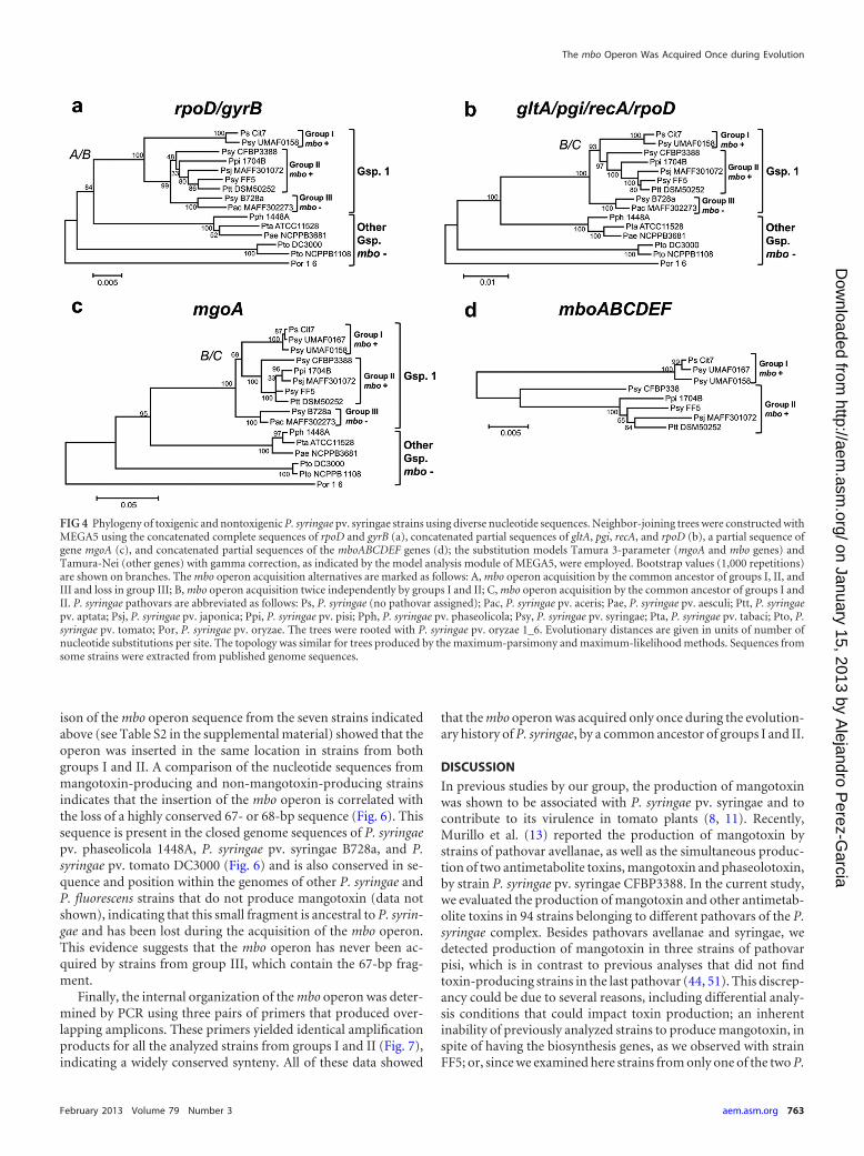
mangotoxin-producing strains, group I contained 45 strains ofP. syringae pv. syringae, which were isolated from woody hosts,mainly mangoes, whereas group II contained 16 strains from thepathovars aptata, avellanae, japonica, pisi, and syringae, whichwere isolated from diverse woody and herbaceous hosts. Group IIIclustered all the strains from genomospecies 1 that did not containthe mbo operon, which included 1 pathovar aceris strain plus 21pathovar syringae strains. Finally, the remaining strains com-prised 30 non-mangotoxin producers from different genomospe-cies that clustered separately. In this phylogenetic analysis, groupIII diverged after the separation of groups I and II, suggesting thatthe mbo operon was acquired once by the common ancestor ofgroups I, II, and III and later lost in group III. An alternativeexplanation is that the mbo operon was acquired independently bythe putative ancestors of groups I and II (Fig. 3 and 4a; see Fig. S1in the supplemental material). To confirm this result, we per-formed further phylogenetic analyses using additional housekeep-ing genes (Fig. 4b to d). For these analyses, we used only thosestrains with an available genome sequence plus two pathovar sy-ringae strains representative of group I (UMAF0158) and group II(CFBP3388).
In the different trees that were built using concatenated se-quences of the gltA, pgi, recA, and rpoD genes (5,871 sites), a partialsequence of the mgoA gene (2,582 sites), or the mbo genes (5,114sites), all strains clustered as in the gyrB-rpoD phylogenetic tree.However, and in contrast to the gyrB-rpoD tree, group III divergedbefore the separation of groups I and II. Because this topology hasstrong bootstrap support, the multilocus sequence typing andmgoA trees suggest that the mbo operon was acquired by groups Iand II in either one or two separate acquisition events and aftertheir separation from group III.
The mbo operon is highly conserved among the two phylo-groups. Phylogenetic analyses clearly indicated that the strainsharboring the mbo operon cluster in two well-separated phylo-
groups, groups I and II (Fig. 3 and 4; see Fig. S1 in the supplemen-tal material). We therefore examined whether the operon is con-served in organization, sequence, and physical location in bothphylogroups in order to gain support for one of the possible sce-narios explaining the evolutionary history of the mbo operon.
To further evaluate the conservation of the mbo operon in thetwo phylogroups, we obtained the complete sequence of thisoperon from P. syringae pv. syringae strains UMAF0158,UMAF0167 (both group I), and CFBP3388 (group II) (see TableS2 in the supplemental material) and compared them with thesequence of the operon from the draft genome sequence of strainsP. syringae Cit7 (group I), and P. syringae pv. aptata DSM50252,pisi 1704B, and syringae FF5 (the last three strains are from groupII) (see Table S2 in the supplemental material). Sequence analysisof the seven strains reveals that the mbo operon maintains syntenyand is generally well conserved, ranging in size from 6,769 to 6,801bp. The operon harbors six open reading frames (ORFs) andshows a 55 to 56% G�C content, which is lower than the averageG�C content of the P. syringae pv. syringae B728a chromosome(59.23%) (30, 53). The mbo operon is highly conserved amongstrains of group I, with nucleotide identity values being higherthan 99%; however, the nucleotide identity among strains ofgroup II varies between 96.5 and 98.5%, which is not surprising,given that this group includes strains from different pathovars.Nevertheless, the mbo operon is well conserved when both groupsare compared, with levels of nucleotide identity being higher than94.2% in pair comparisons (see Table S2 in the supplemental ma-terial).
We examined the mbo operon point of insertion by PCR usinga primer pair that overlaps the right border of the operon (Fig. 5).The amplification of DNA from 19 strains from group I and 15strains from group II produced an amplicon of the same size for allstrains (692 bp), whereas no amplification was observed forstrains belonging to group III (Fig. 5). Additionally, the compar-
FIG 2 Detection by PCR of a specific mangotoxin biosynthesis operon sequence in different P. syringae genomospecies 1 strains. (a) Schematic representationof the four primer pairs designed for mbo operon detection in P. syringae strains. (b) Gel electrophoresis showing the specific 692-bp product generated withprimers mbo24-For and mbo24-Rev from strains harboring the mbo operon. Lanes correspond to the following: 1, P. syringae pv. syringae 7C67; 2, P. syringaepv. syringae 7F29; 3, P. syringae pv. syringae 8F21; 4, P. syringae pv. syringae 1559-9; 5, P. syringae pv. syringae CECT127; 6, P. syringae pv. syringae CECT4429;7, P. syringae pv. syringae CFBP3388; 8, P. syringae pv. syringae DAR77787; 9, P. syringae pv. syringae DAR77789; 10, P. syringae pv. syringae EPS17A; 11, P.syringae pv. syringae NCPPB1239; 12, P. syringae pv. syringae Ps-5; 13, P. syringae pv. syringae UMAF0081; 14, P. syringae pv. syringae UMAF0214; 15, P.syringae pv. syringae UMAF0225; 16, P. syringae pv. syringae UMAF1060; 17, P. syringae pv. syringae UMAF6024; 18, P. syringae pv. syringae UMAF6016;19, P. syringae pv. pisi 1704B; 20, P. syringae pv. pisi HRI203; 21, P. syringae pv. pisi NCPPB1365; 22, P. syringae pv. avellanae ISPaVe011; 23, P. syringaepv. avellanae ISPaVe2056; 24, P. syringae pv. syringae 7D46; 25, 8B48; 26, P. syringae pv. syringae 8C43; 27, P. syringae pv. syringae ITACyL 488; 28, P.syringae pv. syringae ITACyL 521; 29, P. syringae pv. syringae ITACyL 523; 30, P. syringae pv. syringae ITACyL 526; 31, P. syringae pv. syringae ITACyL 529; 32,P. syringae pv. syringae 1444-5; 33, P. syringae pv. syringae FF5; 34, P. syringae pv. syringae EPSMV3; 35, P. syringae pv. syringae UMAF0167; 36, P. syringae pv.tomato DC3000; 37, P. syringae pv. tomato DCT6D1; 38, P. syringae pv. tomato UMAF4007; 39, P. syringae pv. tomato UMAF6018; 40, P. syringae pv.coronafaciens CECT4389; 41, P. syringae pv. phaseolicola 1448A; 42, P. syringae pv. phaseolicola CYL314; 43, P. syringae pv. aptata DSM50252; 44, P. syringae pv.syringae 1507-7; 45, P. syringae pv. syringae B728a; 46, P. syringae pv. syringae CECT4430; 47, P. syringae pv. syringae 0049; 48, P. syringae pv. syringaeUMAF0158; 49, P. syringae pv. tabaci ATCC 11528; M, molecular size marker (HyperLadder I; Bioline).
The mbo Operon Was Acquired Once during Evolution
February 2013 Volume 79 Number 3 aem.asm.org 761
on January 15, 2013 by Alejandro P
erez-Garcia
http://aem.asm
.org/D
ownloaded from

FIG 3 Schematic neighbor-joining tree of P. syringae strains belonging to different pathovars and harboring or not the mbo genes (mbo � and mbo �,respectively) for mangotoxin production. The neighbor-joining tree was constructed with MEGA5 using the concatenated partial sequences of rpoD and gyrB.The Tamura-Nei substitution model with gamma correction was used for tree construction. Bootstrap values (1,000 repetitions) are shown on each branch. Onehundred thirteen strains of different P. syringae pathovars were analyzed. The pathovar, host, and country of isolation are presented on the right, together withthe number of strains (indicated by n) and the genomospecies (Gsp.) to which they belong, according to Gardan et al. (52). Evolutionary distances are given inunits of nucleotide substitutions per site. The topology was similar among trees produced by the maximum-parsimony and maximum-likelihood methods.Sequences of rpoD and gyrB were extracted from published genome sequences or were sequenced for this work. For more detailed information, see Fig. S1 in thesupplemental material.
Carrión et al.
762 aem.asm.org Applied and Environmental Microbiology
on January 15, 2013 by Alejandro P
erez-Garcia
http://aem.asm
.org/D
ownloaded from
ison of the mbo operon sequence from the seven strains indicatedabove (see Table S2 in the supplemental material) showed that theoperon was inserted in the same location in strains from bothgroups I and II. A comparison of the nucleotide sequences frommangotoxin-producing and non-mangotoxin-producing strainsindicates that the insertion of the mbo operon is correlated withthe loss of a highly conserved 67- or 68-bp sequence (Fig. 6). Thissequence is present in the closed genome sequences of P. syringaepv. phaseolicola 1448A, P. syringae pv. syringae B728a, and P.syringae pv. tomato DC3000 (Fig. 6) and is also conserved in se-quence and position within the genomes of other P. syringae andP. fluorescens strains that do not produce mangotoxin (data notshown), indicating that this small fragment is ancestral to P. syrin-gae and has been lost during the acquisition of the mbo operon.This evidence suggests that the mbo operon has never been ac-quired by strains from group III, which contain the 67-bp frag-ment.
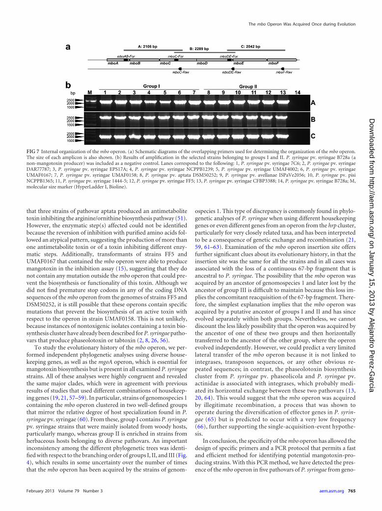
Finally, the internal organization of the mbo operon was deter-mined by PCR using three pairs of primers that produced over-lapping amplicons. These primers yielded identical amplificationproducts for all the analyzed strains from groups I and II (Fig. 7),indicating a widely conserved synteny. All of these data showed
that the mbo operon was acquired only once during the evolution-ary history of P. syringae, by a common ancestor of groups I and II.
DISCUSSION
In previous studies by our group, the production of mangotoxinwas shown to be associated with P. syringae pv. syringae and tocontribute to its virulence in tomato plants (8, 11). Recently,Murillo et al. (13) reported the production of mangotoxin bystrains of pathovar avellanae, as well as the simultaneous produc-tion of two antimetabolite toxins, mangotoxin and phaseolotoxin,by strain P. syringae pv. syringae CFBP3388. In the current study,we evaluated the production of mangotoxin and other antimetab-olite toxins in 94 strains belonging to different pathovars of the P.syringae complex. Besides pathovars avellanae and syringae, wedetected production of mangotoxin in three strains of pathovarpisi, which is in contrast to previous analyses that did not findtoxin-producing strains in the last pathovar (44, 51). This discrep-ancy could be due to several reasons, including differential analy-sis conditions that could impact toxin production; an inherentinability of previously analyzed strains to produce mangotoxin, inspite of having the biosynthesis genes, as we observed with strainFF5; or, since we examined here strains from only one of the two P.
FIG 4 Phylogeny of toxigenic and nontoxigenic P. syringae pv. syringae strains using diverse nucleotide sequences. Neighbor-joining trees were constructed withMEGA5 using the concatenated complete sequences of rpoD and gyrB (a), concatenated partial sequences of gltA, pgi, recA, and rpoD (b), a partial sequence ofgene mgoA (c), and concatenated partial sequences of the mboABCDEF genes (d); the substitution models Tamura 3-parameter (mgoA and mbo genes) andTamura-Nei (other genes) with gamma correction, as indicated by the model analysis module of MEGA5, were employed. Bootstrap values (1,000 repetitions)are shown on branches. The mbo operon acquisition alternatives are marked as follows: A, mbo operon acquisition by the common ancestor of groups I, II, andIII and loss in group III; B, mbo operon acquisition twice independently by groups I and II; C, mbo operon acquisition by the common ancestor of groups I andII. P. syringae pathovars are abbreviated as follows: Ps, P. syringae (no pathovar assigned); Pac, P. syringae pv. aceris; Pae, P. syringae pv. aesculi; Ptt, P. syringaepv. aptata; Psj, P. syringae pv. japonica; Ppi, P. syringae pv. pisi; Pph, P. syringae pv. phaseolicola; Psy, P. syringae pv. syringae; Pta, P. syringae pv. tabaci; Pto, P.syringae pv. tomato; Por, P. syringae pv. oryzae. The trees were rooted with P. syringae pv. oryzae 1_6. Evolutionary distances are given in units of number ofnucleotide substitutions per site. The topology was similar for trees produced by the maximum-parsimony and maximum-likelihood methods. Sequences fromsome strains were extracted from published genome sequences.
The mbo Operon Was Acquired Once during Evolution
February 2013 Volume 79 Number 3 aem.asm.org 763
on January 15, 2013 by Alejandro P
erez-Garcia
http://aem.asm
.org/D
ownloaded from

syringae pv. pisi lineages (54), the possibility that the previouslyanalyzed strains belong to a phylogenetic lineage that does notproduce toxins.
We designed a primer pair specific for the mbo operon that wasused to screen a collection of 94 strains from 20 P. syringae patho-vars. The primers were highly specific, yielding amplification onlyin strains that produced mangotoxin in an in vivo inhibition testor in strains that contained the mbo operon, confirming the use-fulness of this primer pair. All of the strains isolated from lesionsof apical necrosis of mango so far have been shown to producemangotoxin during the inhibition bioassay (11, 55), supportingthe idea that this primer pair could potentially be used for thedetection of P. syringae pv. syringae in mango plants and forbroader disease diagnosis. The PCR and the inhibition analyses,
combined with the search for mangotoxin biosynthesis genes inthe P. syringae genomes from the databases, allowed us to deter-mine that the mangotoxin biosynthesis operon is present only instrains from P. syringae pv. aptata, avellanae, japonica, pisi, andsyringae, all of which belong to genomospecies 1. The amplifica-tion of overlapping DNA fragments as well as the sequencecomparisons showed that the composition, structure, and se-quence of the mbo operon are highly conserved in all thesepathovars. Although P. syringae pv. syringae FF5, UMAF0167,and UMAF1444-5 and the four P. syringae pv. aptata strainsyielded the expected amplification products for the mbo operon orcontained the mbo operon in their genome sequence, we did notdetect production of mangotoxin in the in vivo inhibition test forany of the strains. This is in contrast to a previous report showing
FIG 5 Mapping of the mbo operon insertion point in P. syringae strains. (a) Schematic representation of the PCR amplification strategy for the 3= end of the mbooperon. Primers mboIS-For/mboIS-Rev (amplicon of 580-bp) were designed between the 3= end of the mboF gene and the next ORF downstream of the mbooperon. (b) Representative results of amplification using 34 strains that harbor the mbo operon and two non-mangotoxin-producing strains that were used asnegative controls. Lanes correspond to the following: 1, P. syringae pv. aptata DSM50252; 2, P. syringae pv. aptata LMG5059; 3, P. syringae pv. aptata LMG5532;4, P. syringae pv. aptata LMG5646; 5, P. syringae pv. avellanae ISPaVe011; 6, P. syringae pv. avellanae ISPaVe2056; 7, P. syringae pv. pisi HRI203; 8, P. syringae pv.pisi NCPPB1365; 9, P. syringae pv. pisi 1704B; 10, P. syringae pv. syringae 7C6; 11, P. syringae pv. syringae 7B40; 12, P. syringae pv. syringae 7F29; 13, P. syringaepv. syringae 1444-5; 14, P. syringae pv. syringae 1559-9; 15, P. syringae pv. syringae CECT127; 16, P. syringae pv. syringae CECT4429; 17, P. syringae pv. syringaeCFBP3388; 18, P. syringae pv. syringae DAR77787; 19, P. syringae pv. syringae EPS17A; 20, P. syringae pv. syringae FF5; 21, P. syringae pv. syringae NCPPB1239; 22,P. syringae pv. syringae Ps-10; 23, P. syringae pv. syringae UMAF0049; 24, P. syringae pv. syringae UMAF0081; 25, P. syringae pv. syringae UMAF0167; 26, P. syringae pv.syringae UMAF0209; 27, P. syringae pv. syringae UMAF0221; 28, P. syringae pv. syringae UMAF1060; 29, P. syringae pv. syringae UMAF2008; 30, P. syringae pv. syringaeUMAF2700; 31, P. syringae pv. syringae UMAF2802; 32, P. syringae pv. syringae UMAF4002; 33, P. syringae pv. syringae UMAF6024; 34, P. syringae pv. phaseolicola1448A; 35, P. syringae pv. syringae B728a; 36, P. syringae pv. syringae UMAF0158; M, molecular size marker (HyperLadder I, Bioline).
FIG 6 mbo operon insertion site analysis. Alignment of the nucleotide sequences bordering the mbo operon with the corresponding sequences in non-mangotoxin-producing strains (P. syringae pv. phaseolicola 1448A, P. syringae pv. syringae B728a, and P. syringae pv. tomato DC3000). The nucleotidedifferences with respect to strain DC3000 are indicated, while dots indicate identical nucleotides and a hyphen shows an indel. The CDSs flanking the point ofinsertion of the mbo operon are shown in black, with indication of their locus tag numbers in the genomes of P. syringae pv. tomato DC3000, P. syringae pv.phaseolicola 1448A, and P. syringae pv. syringae B728a. nt, nucleotides.
Carrión et al.
764 aem.asm.org Applied and Environmental Microbiology
on January 15, 2013 by Alejandro P
erez-Garcia
http://aem.asm
.org/D
ownloaded from
that three strains of pathovar aptata produced an antimetabolitetoxin inhibiting the arginine/ornithine biosynthesis pathway (51).However, the enzymatic step(s) affected could not be identifiedbecause the reversion of inhibition with purified amino acids fol-lowed an atypical pattern, suggesting the production of more thanone antimetabolite toxin or of a toxin inhibiting different enzy-matic steps. Additionally, transformants of strains FF5 andUMAF0167 that contained the mbo operon were able to producemangotoxin in the inhibition assay (15), suggesting that they donot contain any mutation outside the mbo operon that could pre-vent the biosynthesis or functionality of this toxin. Although wedid not find premature stop codons in any of the coding DNAsequences of the mbo operon from the genomes of strains FF5 andDSM50252, it is still possible that these operons contain specificmutations that prevent the biosynthesis of an active toxin withrespect to the operon in strain UMAF0158. This is not unlikely,because instances of nontoxigenic isolates containing a toxin bio-synthesis cluster have already been described for P. syringae patho-vars that produce phaseolotoxin or tabtoxin (2, 8, 26, 56).
To study the evolutionary history of the mbo operon, we per-formed independent phylogenetic analyses using diverse house-keeping genes, as well as the mgoA operon, which is essential formangotoxin biosynthesis but is present in all examined P. syringaestrains. All of these analyses were highly congruent and revealedthe same major clades, which were in agreement with previousresults of studies that used different combinations of housekeep-ing genes (19, 21, 57–59). In particular, strains of genomospecies 1containing the mbo operon clustered in two well-defined groupsthat mirror the relative degree of host specialization found in P.syringae pv. syringae (60). From these, group I contains P. syringaepv. syringae strains that were mainly isolated from woody hosts,particularly mango, whereas group II is enriched in strains fromherbaceous hosts belonging to diverse pathovars. An importantinconsistency among the different phylogenetic trees was identi-fied with respect to the branching order of groups I, II, and III (Fig.4), which results in some uncertainty over the number of timesthat the mbo operon has been acquired by the strains of genom-
ospecies 1. This type of discrepancy is commonly found in phylo-genetic analyses of P. syringae when using different housekeepinggenes or even different genes from an operon from the hrp cluster,particularly for very closely related taxa, and has been interpretedto be a consequence of genetic exchange and recombination (21,59, 61–63). Examination of the mbo operon insertion site offersfurther significant clues about its evolutionary history, in that theinsertion site was the same for all the strains and in all cases wasassociated with the loss of a continuous 67-bp fragment that isancestral to P. syringae. The possibility that the mbo operon wasacquired by an ancestor of genomospecies 1 and later lost by theancestor of group III is difficult to maintain because this loss im-plies the concomitant reacquisition of the 67-bp fragment. There-fore, the simplest explanation implies that the mbo operon wasacquired by a putative ancestor of groups I and II and has sinceevolved separately within both groups. Nevertheless, we cannotdiscount the less likely possibility that the operon was acquired bythe ancestor of one of these two groups and then horizontallytransferred to the ancestor of the other group, where the operonevolved independently. However, we could predict a very limitedlateral transfer of the mbo operon because it is not linked tointegrases, transposon sequences, or any other obvious re-peated sequences; in contrast, the phaseolotoxin biosynthesiscluster from P. syringae pv. phaseolicola and P. syringae pv.actinidae is associated with integrases, which probably medi-ated its horizontal exchange between these two pathovars (13,20, 64). This would suggest that the mbo operon was acquiredby illegitimate recombination, a process that was shown tooperate during the diversification of effector genes in P. syrin-gae (65) but is predicted to occur with a very low frequency(66), further supporting the single-acquisition-event hypothe-sis.
In conclusion, the specificity of the mbo operon has allowed thedesign of specific primers and a PCR protocol that permits a fastand efficient method for identifying potential mangotoxin-pro-ducing strains. With this PCR method, we have detected the pres-ence of the mbo operon in five pathovars of P. syringae from geno-
FIG 7 Internal organization of the mbo operon. (a) Schematic diagrams of the overlapping primers used for determining the organization of the mbo operon.The size of each amplicon is also shown. (b) Results of amplification in the selected strains belonging to groups I and II. P. syringae pv. syringae B728a (anon-mangotoxin producer) was included as a negative control. Lanes correspond to the following: 1, P. syringae pv. syringae 7C6; 2, P. syringae pv. syringaeDAR77787; 3, P. syringae pv. syringae EPS17A; 4, P. syringae pv. syringae NCPPB1239; 5, P. syringae pv. syringae UMAF4002; 6, P. syringae pv. syringaeUMAF0167; 7, P. syringae pv. syringae UMAF0158; 8, P. syringae pv. aptata DSM50252; 9, P. syringae pv. avellanae ISPaVe2056; 10, P. syringae pv. pisiNCPPB1365; 11, P. syringae pv. syringae 1444-5; 12, P. syringae pv. syringae FF5; 13, P. syringae pv. syringae CFBP3388; 14, P. syringae pv. syringae B728a; M,molecular size marker (HyperLadder I, Bioline).
The mbo Operon Was Acquired Once during Evolution
February 2013 Volume 79 Number 3 aem.asm.org 765
on January 15, 2013 by Alejandro P
erez-Garcia
http://aem.asm
.org/D
ownloaded from

mospecies 1, whereas mangotoxin production has been reportedfor the first time in P. syringae pv. pisi. The high conservation ofthe structure, sequence, and genomic location of the mbo operon,together with the phylogenetic analysis showing that the mbooperon is present in only two groups from genomospecies 1 of theP. syringae complex, strongly suggests that the mbo operon hasbeen horizontally acquired only once during the evolution of thisbacterial species.
ACKNOWLEDGMENTS
This work was supported by grants from the regional government of An-dalucía (Spain), grants from CICE-Junta de Andalucía, Ayudas GrupoPAIDI AGR-169, and Proyecto de Excelencia (P07-AGR-02471) and PlanNacional I�D�I grant AGL2008-55311-CO2-01 (Ministerio de Ciencia eInnovación), cofinanced by FEDER (European Union). Plan Propio ofthe University of Málaga funded a stay by V.J.C. at the UniversidadPública de Navarra, Spain. V.J.C. was supported with a fellowship fromthe Junta de Andalucía, Spain, and E.A. was supported with a JAEDocgrant from the CSIC, which was cofinanced by ESF.
We thank George W. Sundin, Alberto Martín-Sanz, and ConstantinoCaminero for providing isolates of P. syringae. We also thank Menno vander Voort for critically reading the manuscript and the research group ofCayo Ramos for their help with some aspects of this research.
REFERENCES1. Bender CL, Alarcón-Chaidez F, Gross DC. 1999. Pseudomonas syringae
phytotoxins: mode of action, regulation, and biosynthesis by peptide andpolyketide synthetases. Microbiol. Mol. Biol. Rev. 63:266 –292.
2. Gutiérrez-Barranquero JA, Arrebola E, Bonilla N, Sarmiento D, Ca-zorla FM, de Vicente A. 2012. Environmentally friendly treatment alter-natives to Bordeaux mixture for controlling bacterial apical necrosis(BAN) of mango. Plant Pathol. 61:665– 676.
3. Kennelly MM, Cazorla FM, de Vicente A, Ramos C, Sundin GW. 2007.Pseudomonas syringae diseases of fruit trees: progress toward understand-ing and control. Plant Dis. 91:4 –17.
4. Krieg NR, Holt JG. 1984. Bergey’s manual of systematic bacteriology, vol1. Williams & Wilkins, Baltimore, MD.
5. Mansfield J, Genin S, Magori S, Citovsky V, Sriariyanum M, Ronald P,Dow M, Verdier V, Beer SV, Machado MA, Toth I, Salmond G, FosterGD. 2012. Top 10 plant pathogenic bacteria in molecular plant pathology.Mol. Plant Pathol. 13:614 – 629.
6. Mansfield JW. 2009. From bacterial avirulence genes to effector functionsvia the hrp delivery system: an overview of 25 years of progress in ourunderstanding of plant innate immunity. Mol. Plant Pathol. 10:721–734.
7. Ramos C, Matas IM, Bardaji L, Aragón IM, Murillo J. 2012. Pseudomo-nas savastanoi pv. savastanoi: some like it knot. Mol. Plant Pathol. 13:998 –1009.
8. Arrebola E, Cazorla FM, Codina JC, Gutiérrez-Barranquero JA, Pérez-García A, de Vicente A. 2009. Contribution of mangotoxin to the viru-lence and epiphytic fitness of Pseudomonas syringae pv. syringae. Int. Mi-crobiol. 12:87–95.
9. Arrebola E, Cazorla FM, Pérez-García A, de Vicente A. 2011. Chemicaland metabolic aspects of antimetabolite toxins produced by Pseudomonassyringae pathovars. Toxins 3:1089 –1110.
10. Arrebola E, Cazorla FM, Pérez-García A, de Vicente A. 2011. Genesinvolved in the production of antimetabolite toxins by Pseudomonas sy-ringae pathovars. Genes 2:640 – 660.
11. Arrebola E, Cazorla FM, Durán VE, Rivera E, Olea F, Codina JC,Pérez-García A, de Vicente A. 2003. Mangotoxin: a novel antimetabolitetoxin produced by Pseudomonas syringae inhibiting ornithine/argininebiosynthesis. Physiol. Mol. Plant Pathol. 63:117–127.
12. Arrebola E, Cazorla FM, Romero D, Pérez-García A, de Vicente A.2007. A nonribosomal peptide synthetase gene (mgoA) of Pseudomonassyringae pv. syringae is involved in mangotoxin biosynthesis and is re-quired for full virulence. Mol. Plant-Microbe Interact. 20:500 –509.
13. Murillo J, Bardaji L, Navarro de la Fuente L, Führer ME, Aguilera S,Álvarez-Morales A. 2011. Variation in conservation of the cluster forbiosynthesis of the phytotoxin phaseolotoxin in Pseudomonas syringae
suggests at least two events of horizontal acquisition. Res. Microbiol. 162:253–261.
14. Arrebola E, Carrión VJ, Cazorla FM, Pérez-García A, Murillo J, deVicente A. 2012. Characterisation of the mgo operon in Pseudomonassyringae pv. syringae UMAF0158 that is required for mangotoxin produc-tion. BMC Microbiol. 12:10. doi:10.1186/1471-2180-12-10.
15. Carrión VJ, Arrebola E, Cazorla FM, Murillo J, de Vicente A. 2012. Thembo operon is specific and essential for biosynthesis of mangotoxin inPseudomonas syringae. PLoS One 7:e36709. doi:10.1371/journal.pone.0036709.
16. Rees-George J, Vanneste JL, Cornish DA, Pushparajah IPS, Yu J,Templeton MD, Everett KR. 2010. Detection of Pseudomonas syringae pv.actinidiae using polymerase chain reaction (PCR) primers based on the16S-23S rDNA intertranscribed spacer region and comparison with PCRprimers based on other gene regions. Plant Pathol. 59:453– 464.
17. Ercolani GL. 1985. The relation between dosage, bacterial growth andtime for disease response during infection of bean leaves by Pseudomonassyringae pv. phaseolicola. J. Appl. Microbiol. 58:63–75.
18. Penyalver R, García A, Ferrer A, Bertolini E, López MM. 2000. Detec-tion of Pseudomonas savastanoi pv. savastanoi in olive plants by enrich-ment and PCR. Appl. Environ. Microbiol. 66:2673–2677.
19. Sawada H, Suzuki F, Matsuda I, Saitou N. 1999. Phylogenetic analysis ofPseudomonas syringae pathovars suggests the horizontal gene transfer ofargK and the evolutionary stability of hrp gene cluster. J. Mol. Evol. 49:627– 644.
20. Sawada H, Kanaya S, Tsuda M, Suzuki F, Azegami K, Saitou N. 2002.A phylogenomic study of the OCTase genes in Pseudomonas syringaepathovars: the horizontal rransfer of the argK-tox cluster and the evolu-tionary history of OCTase genes on their genomes. J. Mol. Evol. 54:437–457.
21. Baltrus DA, Nishimura MT, Romanchuk A, Chang JH, Mukhtar MS,Cherkis K, Roach J, Grant SR, Jones CD, Dangl JL. 2011. Dynamicevolution of pathogenicity revealed by sequencing and comparativegenomics of 19 Pseudomonas syringae isolates. PLoS Pathog. 7:e1002132.doi:10.1371/journal.ppat.1002132.
22. Green S, Studholme DJ, Laue BE, Dorati F, Lovell H, Arnold D, CottrellJE, Bridgett S, Blaxter M, Huitema E, Thwaites R, Sharp PM, JacksonRW, Kamoun S. 2010. Comparative genome analysis provides insightsinto the evolution and adaptation of Pseudomonas syringae pv. aesculi onAesculus hippocastanum. PLoS One 5:e10224. doi:10.1371/journal.pone.0010224.
23. Yamamoto S, Kasai H, Arnold DL, Jackson RW, Vivian A, Harayama S.2000. Phylogeny of the genus Pseudomonas: intrageneric structure recon-structed from the nucleotide sequences of gyrB and rpoD genes. Microbi-ology 146:2385–2394.
24. Reinhardt JA, Baltrus DA, Nishimura MT, Jeck WR, Jones CD, DanglJL. 2009. De novo assembly using low-coverage short read sequence datafrom the rice pathogen Pseudomonas syringae pv. oryzae. Genome Res.19:294 –305.
25. Teverson DM. 1991. Genetics of pathogenicity and resistance in thehaloblight disease of beans in Africa. Ph.D. thesis. University of Birming-ham, Birmingham, United Kingdom.
26. Rico A, López R, Asensio C, Aizpún MT, Asensio-S-Manzanera MC,Murillo J. 2003. Nontoxigenic strains of Pseudomonas syringae pv. phase-olicola are a main cause of halo blight of beans in Spain and escape currentdetection methods. Phytopathology 93:1553–1559.
27. Pérez-Martínez I, Rodriguez-Moreno L, Matas IM, Ramos C. 2007.Strain selection and improvement of gene transfer for genetic manipula-tion of Pseudomonas savastanoi isolated from olive knots. Res. Microbiol.158:60 – 69.
28. Sundin GW, Bender CL. 1996. Dissemination of the strA-strB strepto-mycin-resistance genes among commensal and pathogenic bacteria fromhumans, animals, and plants. Mol. Ecol. 5:133–143.
29. Sundin GW, Demezas DH, Bender CL. 1994. Genetic and plasmiddiversity within natural populations of Pseudomonas syringae with variousexposures to copper and streptomycin bactericides. Appl. Environ. Mi-crobiol. 60:4421– 4431.
30. Feil H, Feil WS, Chain P, Larimer F, DiBartolo G, Copeland A, LykidisA, Trong S, Nolan M, Goltsman E, Thiel J, Malfatti S, Loper JE, LapidusA, Detter JC, Land M, Richardson PM, Kyrpides NC, Ivanova N,Lindow SE. 2005. Comparison of the complete genome sequences ofPseudomonas syringae pv. syringae B728a and pv. tomato DC3000. Proc.Natl. Acad. Sci. U. S. A. 102:11064 –11069.
Carrión et al.
766 aem.asm.org Applied and Environmental Microbiology
on January 15, 2013 by Alejandro P
erez-Garcia
http://aem.asm
.org/D
ownloaded from
31. Tourte C, Manceau C. 1995. A strain of Pseudomonas syringae which doesnot belong to pathovar phaseolicola produces phaseolotoxin. Eur. J. PlantPathol. 101:483– 490.
32. Martín-Sanz A, Palomo J, Pérez de la Vega M, Caminero C. 2012.Characterization of Pseudomonas syringae pv. syringae isolates associatedwith bacterial blight in Lathyrus spp. and sources of resistance. Eur. J. PlantPathol. 134:205–216.
33. Young A. 2008. Notes on Pseudomonas syringae pv. syringae bacterialnecrosis of mango (Mangifera indica) in Australia. Australas. Plant Dis.Notes 3:138 –140.
34. Sohn KH, Jones JDG, Studholme DJ. 2012. Draft genome sequence ofPseudomonas syringae pathovar syringae strain FF5, causal agent of stemtip dieback disease on ornamental pear. J. Bacteriol. 194:3733–3734.
35. Martín-Sanz A, Palomo JL, Caminero C. 2009. First report of bacterialblight caused by Pseudomonas syringae pv. syringae on common vetch inSpain. Plant Dis. 93:1348.
36. Cazorla FM, Arrebola E, Sesma A, Pérez-García A, Codina JC, MurilloJ, de Vicente A. 2002. Copper resistance in Pseudomonas syringae strainsisolated from mango is encoded mainly by plasmids. Phytopathology 92:909 –916.
37. Gutiérrez-Barranquero JA, Arrebola E, Pérez-García A, Codina JC,Murillo J, de Vicente A, Cazorla FM. 2008. Evaluation of phenotypic andgenetic techniques to analyze diversity of Pseudomonas syringae pv. syrin-gae strains isolates from mango trees, p 271–281. In Fatmi MB, Collmer A,Iacobellis NS, Mansfield JW, Murillo J, Schaad NW, Ullrich M (ed), Pseu-domonas syringae pathovars and related pathogens—identification, epide-miology and genomics. Springer Netherlands, Dordrecht, The Nether-lands.
38. Moore RA, Starratt AN, Ma SW, Morris VL, Cuppels DA. 1989.Identification of a chromosomal region required for biosynthesis of thephytotoxin coronatine by Pseudomonas syringae pv. tomato. Can. J. Mi-crobiol. 35:910 –917.
39. Cai R, Lewis J, Yan S, Liu H, Clarke CR, Campanile F, Almeida NF,Studholme DJ, Lindeberg M, Schneider D, Zaccardelli M, Setubal JC,Morales-Lizcano NP, Bernal A, Coaker G, Baker C, Bender CL, LemanS, Vinatzer BA. 2011. The plant pathogen Pseudomonas syringae pv. to-mato is genetically monomorphic and under strong selection to evadetomato immunity. PLoS Pathog. 7:e1002130. doi:10.1371/journal.ppat.1002130.
40. Almeida NF, Yan S, Lindeberg M, Studholme DJ, Schneider DJ, Con-don B, Liu H, Viana CJ, Warren A, Evans C, Kemen E, MacLean D,Angot A, Martin GB, Jones JD, Collmer A, Setubal JC, Vinatzer BA.2008. A draft genome sequence of Pseudomonas syringae pv. tomato T1reveals a type III effector repertoire significantly divergent from that ofPseudomonas syringae pv. tomato DC3000. Mol. Plant-Microbe Interact.22:52– 62.
41. Sambrook J, Russell DW. 2001. Molecular cloning: a laboratory man-ual, 3rd ed, vol 1. Cold Spring Harbor Laboratory Press, Cold SpringHarbor, NY.
42. King EO, Ward MK, Raney DE. 1954. Two simple media for the dem-onstration of pyocyanin and fluorescein. J. Lab. Clin. Med. 44:301–307.
43. Cazorla FM, Olalla L, Torés JA, Codina JC, Pérez-GarcíA A, de VicenteA. 1997. Pseudomonas syringae pv. syringae as microorganism involved inapical necrosis of mango: characterization of some virulence factors, p82– 87. In Rudolph K, Burr TJ, Mansfield JW, Stead D, Vivian A, vonKietzell J (ed), Pseudomonas syringae pathovars and related pathogens.Kluwer Academic Publishers, Dordrecht, The Netherlands.
44. Gasson MJ. 1980. Indicator technique for antimetabolic toxin productionby phytopathogenic species of Pseudomonas. Appl. Environ. Microbiol.39:25–29.
45. Rozen S, Skaletsky H. 2000. Primer3 on the WWW for general users andfor biologist programmers. Methods Mol. Biol. 132:365–386.
46. Altschul SF, Gish W, Miller W, Myers EW, Lipman DJ. 1990. Basic localalignment search tool. J. Mol. Biol. 215:403– 410.
47. Rutherford K, Parkhill J, Crook J, Horsnell T, Rice P, Rajandream M-A,Barrell B. 2000. Artemis: sequence visualization and annotation. Bioin-formatics 16:944 –945.
48. Carver TJ, Rutherford KM, Berriman M, Rajandream M-A, Barrell BG,Parkhill J. 2005. ACT: the Artemis comparison tool. Bioinformatics 21:3422–3423.
49. Abbott JC, Aanensen DM, Rutherford K, Butcher S, Spratt BG. 2005.WebACT—an online companion for the Artemis comparison tool. Bioin-formatics 21:3665–3666.
50. Tamura K, Peterson D, Peterson N, Stecher G, Nei M, Kumar S. 2011.MEGA5: molecular evolutionary genetics analysis using maximum likeli-hood, evolutionary distance, and maximum parsimony methods. Mol.Biol. Evol. 28:2731–2739.
51. Völksch B, Weingart H. 1998. Toxin production by pathovars of Pseu-domonas syringae and their antagonistic activities against epiphytic micro-organisms. J. Basic Microbiol. 38:135–145.
52. Gardan L, Shafik H, Belouin S, Broch R, Grimont F, Grimont PAD.1999. DNA relatedness among the pathovars of Pseudomonas syringae anddescription of Pseudomonas tremae sp. nov. and Pseudomonas cannabinasp. nov. (ex Sutic and Dowson 1959). Int. J. Syst. Bacteriol. 49:469 – 478.
53. Buell CR, Joardar V, Lindeberg M, Selengut J, Paulsen IT, Gwinn ML,Dodson RJ, Deboy RT, Durkin AS, Kolonay JF, Madupu R, DaughertyS, Brinkac L, Beanan MJ, Haft DH, Nelson WC, Davidsen T, Zafar N,Zhou L, Liu J, Yuan Q, Khouri H, Fedorova N, Tran B, Russell D, BerryK, Utterback T, Van Aken SE, Feldblyum TV, D’Ascenzo M, Deng W-L,Ramos AR, Alfano JR, Cartinhour S, Chatterjee AK, Delaney TP,Lazarowitz SG, Martin GB, Schneider DJ, Tang X, Bender CL, White O,Fraser CM, Collmer A. 2003. The complete genome sequence of theArabidopsis and tomato pathogen Pseudomonas syringae pv. tomatoDC3000. Proc. Natl. Acad. Sci. U. S. A. 100:10181–10186.
54. Martín-Sanz A, Pérez de la Vega M, Murillo J, Caminero C. 2012.Genetic, biochemical and pathogenic diversity of Pseudomonas syringaepv. pisi strains. Plant Pathol. 61:1063–1072.
55. Cazorla FM, Torés JA, Olalla L, Pérez-García A, Farré JM, de VicenteA. 1998. Bacterial apical necrosis of mango in southern Spain: a diseasecaused by Pseudomonas syringae pv. syringae. Phytopathology 88:614 –620.
56. Schaad NW, Cheong SS, Tamaki S, Hatziloukas E, Panopoulos NJ.1995. A combined biological and enzymatic amplification (BIO-PCR)technique to detect Pseudomonas syringae pv. phaseolicola in bean seedextracts. Phytopathology 85:243–248.
57. Gironde S, Manceau C. 2012. Housekeeping gene sequencing and mul-tilocus variable-number tandem-repeat analysis to identify subpopula-tions within Pseudomonas syringae pv. maculicola and Pseudomonas syrin-gae pv. tomato that correlate with host specificity. Appl. Environ.Microbiol. 78:3266 –3279.
58. Hwang MS, Morgan RL, Sarkar SF, Wang PW, Guttman DS. 2005.Phylogenetic characterization of virulence and resistance phenotypes ofPseudomonas syringae. Appl. Environ. Microbiol. 71:5182–5191.
59. Sarkar SF, Guttman DS. 2004. Evolution of the core genome of Pseu-domonas syringae, a highly clonal, endemic plant pathogen. Appl. Environ.Microbiol. 70:1999 –2012.
60. Little EL, Bostock RM, Kirkpatrick BC. 1998. Genetic characterization ofPseudomonas syringae pv. syringae strains from stone fruits in California.Appl. Environ. Microbiol. 64:3818 –3823.
61. Guttman DS, Gropp SJ, Morgan RL, Wang PW. 2006. Diversifyingselection drives the evolution of the type III secretion system pilus ofPseudomonas syringae. Mol. Biol. Evol. 23:2342–2354.
62. Wang PW, Morgan RL, Scortichini M, Guttman DS. 2007. Convergentevolution of phytopathogenic pseudomonads onto hazelnut. Microbiol-ogy 153:2067–2073.
63. Yan S, Liu H, Mohr TJ, Jenrette J, Chiodini R, Zaccardelli M, SetubalJC, Vinatzer BA. 2008. Role of recombination in the evolution of themodel plant pathogen Pseudomonas syringae pv. tomato DC3000, a veryatypical tomato strain. Appl. Environ. Microbiol. 74:3171–3181.
64. Genka H, Baba T, Tsuda M, Kanaya S, Mori H, Yoshida T, Noguchi M,Tsuchiya K, Sawada H. 2006. Comparative analysis of argK-tox clustersand their flanking regions in phaseolotoxin-producing Pseudomonas sy-ringae pathovars. J. Mol. Evol. 63:401– 414.
65. Stavrinides J, Ma W, Guttman DS. 2006. Terminal reassortment drivesthe quantum evolution of type III effectors in bacterial pathogens. PLoSPathog. 2:e104. doi:10.1371/journal.ppat.0020104.
66. Thomas CM, Nielsen KM. 2005. Mechanisms of, and barriers to, hori-zontal gene transfer between bacteria. Nat. Rev. Microbiol. 3:711–721.
The mbo Operon Was Acquired Once during Evolution
February 2013 Volume 79 Number 3 aem.asm.org 767
on January 15, 2013 by Alejandro P
erez-Garcia
http://aem.asm
.org/D
ownloaded from