Curli fibers are highly conserved between Salmonella typhimurium and Escherichia coli with respect...
10
JOURNAL OF BACTERIOLOGY, 0021-9193/98/$04.0010 Feb. 1998, p. 722–731 Vol. 180, No. 3 Copyright © 1998, American Society for Microbiology Curli Fibers Are Highly Conserved between Salmonella typhimurium and Escherichia coli with Respect to Operon Structure and Regulation UTE RO ¨ MLING, 1 * ZHAO BIAN, 1 MÅRTEN HAMMAR, 1 WALTER D. SIERRALTA, 2 AND STAFFAN NORMARK 1 Department of Bacteriology, Microbiology and Tumorbiology Center, Karolinska Institutet, S-17177 Stockholm, Sweden, 1 and Max-Planck-Institut fu ¨r Experimentelle Endokrinologie, D-30603 Hannover, Germany 2 Received 27 August 1997/Accepted 12 November 1997 Mouse-virulent Salmonella typhimurium strains SR-11 and ATCC 14028-1s express curli fibers, thin aggre- gative fibers, at ambient temperature on plates as judged by Western blot analysis and electron microscopy. Concomitantly with curli expression, cells develop a rough and dry colony morphology and bind the dye Congo red (called the rdar morphotype). Cloning and characterization of the two divergently transcribed operons required for curli biogenesis, csgBA(C) and csgDEFG, from S. typhimurium SR-11 revealed the same gene order and flanking genes as in Escherichia coli. The divergence of the curli region between S. typhimurium and E. coli at the nucleotide level is above average (22.4%). However, a high level of conservation at the protein level, which ranged from 86% amino acid homology for the fiber subunit CsgA to 99% homology for the lipoprotein CsgG, implies functional constraints on the gene products. Consequently, S. typhimurium genes on low-copy-number plasmids were able to complement respective E. coli mutants, although not always to wild-type levels. rpoS and ompR are required for transcriptional activation of (at least) the csgD promoter. The high degree of conser- vation at the protein level and the identical regulation patterns in E. coli and S. typhimurium suggest similar roles of curli fibers in the same ecological niche in the two species. Proteinaceous, filamentous appendices on bacterial sur- faces, called fimbriae or pili, enable the bacterial cell to make contact with inanimate surfaces and eukaryotic or prokaryotic cells. Tight contact, called adherence, precedes, e.g., coloniza- tion of surfaces and invasion of eukaryotic cells by the bacteria. Fimbriae are best studied in the family Enterobacteriaceae, particularly in Escherichia coli and Salmonella enterica (49), in the context of pathogen-host interactions (44). Related spe- cies, subspecies, and even particular strains can have a specific set of fimbrial genes which are often located on pathogenicity islands on the chromosome or on plasmids (28, 31, 43, 45). The need for flexibility in the strategy of adhesion in order to overcome the host immune system, for example, has also led to a variability in fimbrial genes derived from a common ancestor. The immunogenic and adhesive properties of these fimbriae, which can be encoded either by the fimbrial subunit gene, as in the case of K88 fimbriae, or by separate genes, as in the case of the Pap pili, can be exchanged as gene cassettes in the context of a common frame (45). Therefore, fimbrial genes often do not appear to fit the phylogenetic classification of the bacte- rium but are shared by more distantly related organisms occu- pying the same ecological niche (45, 63). Most of the fimbriae identified in Salmonella enterica subsp. enterica serotype Typhimurium (in this paper, referred to as S. typhimurium) have been described only phenotypically; the few whose genes have been cloned and sequenced (6, 15, 16, 28, 56) are unique to S. enterica or a subset of its subspecies (5, 56). However, curli fibers, thin aggregative fibers, seem to be present and expressed in almost all Salmonella spp. and E. coli (5, 17, 23) and maybe also in other Enterobacteriaceae, such as Shigella, Citrobacter, and Enterobacter spp. (23). So far, two nomenclature systems exist (20, 34). The genes for curli bio- genesis (csg) in E. coli are called agf (thin aggregative fibers) in Salmonella enteritidis. For convenience, we use one nomencla- ture system in this communication. Curli fibers detected, for example, on S. typhimurium strains causing acute salmonellosis in pigeons (32) and on E. coli isolates causing bovine mastitis (54) mediate binding to fibronec- tin (54), a variety of other human serum and tissue matrix proteins (7, 54, 62), and the dye Congo red (CR) (18). In E. coli MC4100, two divergently transcribed operons, csgDEFG and csgBA(C), which are separated by a 513-bp intergenic region are required for the biogenesis of curli fibers (34). Transposon insertions in the csgD gene, which encodes a transcriptional regulator belonging to the LuxR family as identified by the sequence similarity of the DNA binding helix-turn-helix motif, completely abolished transcription of the csgBA operon (34). Assembled by the extracellular nucleation-precipitation path- way, the secreted fiber subunit CsgA is polymerized on the surface-exposed nucleator CsgB (35), which, in addition, is present along the filament in minor amounts (8). CsgA and CsgB show 49% similarity and contain repeat regions whose interaction triggers polymerization of CsgA (8, 35). The outer- membrane-located lipoprotein CsgG is required to protect CsgA and CsgB from proteolysis (48). The roles of csgE and csgF are just beginning to be elucidated. csgE is required for the fibronectin and CR binding properties of curli fibers but does not significantly affect polymerization of the fiber subunit (36). The nucleation function is impaired in a csgF mutant, in which CsgA is released into the growth medium (37). Curli expression in E. coli MC4100 and YMel is highly regulated by environmental conditions; it is restricted to low temperature on plates containing medium with a low salt concentration. * Corresponding author. Mailing address: Karolinska Institutet, Mi- crobiology and Tumorbiology Center (MTC), Box 280, S-17177 Stock- holm, Sweden. Phone: (46) 8-728-7409. Fax: (46) 8-342651. E-mail: ute [email protected]. 722 on February 26, 2016 by guest http://jb.asm.org/ Downloaded from
-
Upload
independent -
Category
Documents
-
view
2 -
download
0
Transcript of Curli fibers are highly conserved between Salmonella typhimurium and Escherichia coli with respect...

JOURNAL OF BACTERIOLOGY,0021-9193/98/$04.0010
Feb. 1998, p. 722–731 Vol. 180, No. 3
Copyright © 1998, American Society for Microbiology
Curli Fibers Are Highly Conserved between Salmonella typhimuriumand Escherichia coli with Respect to Operon
Structure and RegulationUTE ROMLING,1* ZHAO BIAN,1 MÅRTEN HAMMAR,1 WALTER D. SIERRALTA,2
AND STAFFAN NORMARK1
Department of Bacteriology, Microbiology and Tumorbiology Center, Karolinska Institutet,S-17177 Stockholm, Sweden,1 and Max-Planck-Institut fur Experimentelle
Endokrinologie, D-30603 Hannover, Germany2
Received 27 August 1997/Accepted 12 November 1997
Mouse-virulent Salmonella typhimurium strains SR-11 and ATCC 14028-1s express curli fibers, thin aggre-gative fibers, at ambient temperature on plates as judged by Western blot analysis and electron microscopy.Concomitantly with curli expression, cells develop a rough and dry colony morphology and bind the dye Congored (called the rdar morphotype). Cloning and characterization of the two divergently transcribed operonsrequired for curli biogenesis, csgBA(C) and csgDEFG, from S. typhimurium SR-11 revealed the same gene orderand flanking genes as in Escherichia coli. The divergence of the curli region between S. typhimurium and E. coliat the nucleotide level is above average (22.4%). However, a high level of conservation at the protein level, whichranged from 86% amino acid homology for the fiber subunit CsgA to 99% homology for the lipoprotein CsgG,implies functional constraints on the gene products. Consequently, S. typhimurium genes on low-copy-numberplasmids were able to complement respective E. coli mutants, although not always to wild-type levels. rpoS andompR are required for transcriptional activation of (at least) the csgD promoter. The high degree of conser-vation at the protein level and the identical regulation patterns in E. coli and S. typhimurium suggest similarroles of curli fibers in the same ecological niche in the two species.
Proteinaceous, filamentous appendices on bacterial sur-faces, called fimbriae or pili, enable the bacterial cell to makecontact with inanimate surfaces and eukaryotic or prokaryoticcells. Tight contact, called adherence, precedes, e.g., coloniza-tion of surfaces and invasion of eukaryotic cells by the bacteria.Fimbriae are best studied in the family Enterobacteriaceae,particularly in Escherichia coli and Salmonella enterica (49), inthe context of pathogen-host interactions (44). Related spe-cies, subspecies, and even particular strains can have a specificset of fimbrial genes which are often located on pathogenicityislands on the chromosome or on plasmids (28, 31, 43, 45). Theneed for flexibility in the strategy of adhesion in order toovercome the host immune system, for example, has also led toa variability in fimbrial genes derived from a common ancestor.The immunogenic and adhesive properties of these fimbriae,which can be encoded either by the fimbrial subunit gene, as inthe case of K88 fimbriae, or by separate genes, as in the case ofthe Pap pili, can be exchanged as gene cassettes in the contextof a common frame (45). Therefore, fimbrial genes often donot appear to fit the phylogenetic classification of the bacte-rium but are shared by more distantly related organisms occu-pying the same ecological niche (45, 63).
Most of the fimbriae identified in Salmonella enterica subsp.enterica serotype Typhimurium (in this paper, referred to asS. typhimurium) have been described only phenotypically; thefew whose genes have been cloned and sequenced (6, 15, 16,28, 56) are unique to S. enterica or a subset of its subspecies (5,56). However, curli fibers, thin aggregative fibers, seem to bepresent and expressed in almost all Salmonella spp. and E. coli
(5, 17, 23) and maybe also in other Enterobacteriaceae, such asShigella, Citrobacter, and Enterobacter spp. (23). So far, twonomenclature systems exist (20, 34). The genes for curli bio-genesis (csg) in E. coli are called agf (thin aggregative fibers) inSalmonella enteritidis. For convenience, we use one nomencla-ture system in this communication.
Curli fibers detected, for example, on S. typhimurium strainscausing acute salmonellosis in pigeons (32) and on E. coliisolates causing bovine mastitis (54) mediate binding to fibronec-tin (54), a variety of other human serum and tissue matrixproteins (7, 54, 62), and the dye Congo red (CR) (18). In E. coliMC4100, two divergently transcribed operons, csgDEFG andcsgBA(C), which are separated by a 513-bp intergenic regionare required for the biogenesis of curli fibers (34). Transposoninsertions in the csgD gene, which encodes a transcriptionalregulator belonging to the LuxR family as identified by thesequence similarity of the DNA binding helix-turn-helix motif,completely abolished transcription of the csgBA operon (34).Assembled by the extracellular nucleation-precipitation path-way, the secreted fiber subunit CsgA is polymerized on thesurface-exposed nucleator CsgB (35), which, in addition, ispresent along the filament in minor amounts (8). CsgA andCsgB show 49% similarity and contain repeat regions whoseinteraction triggers polymerization of CsgA (8, 35). The outer-membrane-located lipoprotein CsgG is required to protectCsgA and CsgB from proteolysis (48). The roles of csgE andcsgF are just beginning to be elucidated. csgE is required forthe fibronectin and CR binding properties of curli fibers butdoes not significantly affect polymerization of the fiber subunit(36). The nucleation function is impaired in a csgF mutant, inwhich CsgA is released into the growth medium (37). Curliexpression in E. coli MC4100 and YMel is highly regulated byenvironmental conditions; it is restricted to low temperatureon plates containing medium with a low salt concentration.
* Corresponding author. Mailing address: Karolinska Institutet, Mi-crobiology and Tumorbiology Center (MTC), Box 280, S-17177 Stock-holm, Sweden. Phone: (46) 8-728-7409. Fax: (46) 8-342651. E-mail: [email protected].
722
on February 26, 2016 by guest
http://jb.asm.org/
Dow
nloaded from

The alternative sigma factor RpoS (sS) is a global regulatorcontrolling the expression of a large number of genes duringstarvation and other stress conditions in E. coli (52) and S. ty-phimurium (26). rpoS-deficient S. typhimurium strains are im-paired in their virulence in the mouse model for typhoid fever(22, 26); the rpoS deficiency also seems to be the cause ofattenuation of common laboratory derivatives of strain LT2(65, 68). Transcription by the RNA polymerase containing sS
at different promoters can include complex interactions withadditional regulators (25). The stationary-phase-induced tran-scription of the genes for curli biogenesis is dependent on sS inE. coli. It has not been resolved whether rpoS is needed onlyfor transcription from the E. coli csgD promoter or also affectsthe CsgD-dependent csgBA promoter (34). Absence of H-NShas been shown to make at least the csgD promoter indepen-dent of rpoS, suggesting an efficient repression of rpoD-depen-dent transcription by hns (2, 34).
Increasing osmolarity has been shown to shut off expressionof curli genes at the transcriptional level (53) but to increasethe levels of RpoS (42). Therefore, other regulators must alsoinfluence the transcription from the csgD and csgBA promot-ers. OmpR is a transcriptional regulator which was studiedmainly for its role in regulating transcription of the outermembrane proteins OmpF and OmpC in response to sur-rounding osmolarity sensed by EnvZ in E. coli (55). BesidesOmpF and OmpC, a tripeptide permease (TppB) is known tobe regulated by OmpR in S. typhimurium (30). ompR mutantsof S. typhimurium are attenuated in vivo (24) and unable to killmacrophages in vitro (47). ompR has been reported to berequired for transcriptional activation of both the csgBA andthe csgDEFG promoters in E. coli (40); however, no experi-mental data have been reported so far.
In this paper, we report the cloning and characterization
of the two operons for curli biogenesis from S. typhimuriumSR-11. The highly conserved genes displayed the same arrange-ment in the same chromosomal context as in E. coli. Conse-quently, S. typhimurium-derived genes on plasmids could func-tionally replace their E. coli counterparts, although not alwaysto the wild-type levels. Regulation of curli biogenesis in S. ty-phimurium SR-11 and ATCC 14028-1s was reminiscent ofE. coli MC4100 and YMel. Curli biogenesis was restricted toambient temperature on plates, and transcription from thecsgDEFG and csgBA promoters required rpoS and ompR. Theconservation of genes and of the regulation pattern implies animportant role of curli fibers in the lifestyle of E. coli andSalmonella spp. in the same ecological niche.
MATERIALS AND METHODS
Bacterial strains, plasmids, and growth conditions. Bacterial strains and plas-mids used and constructed in this study are given in Tables 1 and 2, respectively.In the beginning, S. typhimurium SR-11 was used for the analysis of curli bio-genesis. However, the initial lack of tools for genetic analysis led to a subsequentshift to strain ATCC 14028-1s, a virulent derivative of the well-characterized LT2strain. Curli expression in E. coli and S. typhimurium was monitored by growth onsolid Yesca (35) and Luria-Bertani (LB) medium without salt, respectively.Media were supplemented with CR (40 mg/ml) and Coomassie brilliant blue (20mg/ml) to judge colony morphology and color (34, 35). However, CR slightlyinhibits the growth of S. typhimurium; therefore, the colony morphology developslater than on medium without CR. Recombinant clones were grown on LBmedium supplemented with recommended concentrations of antibiotics (4), ifrequired.
DNA techniques. Isolation of plasmid, cosmid, and chromosomal DNAs andall enzymatic manipulations (restriction digestion, ligation, phosphorylation, andPCR) were carried out by standard protocols (4, 58) with enzymes from Boehr-inger Mannheim or Biolabs. Southern blotting was done as described previously(57). Individual PCR fragments were purified with a Quiaquick PCR purificationkit (Quiagen); otherwise, a Geneclean II kit (Bio 101, Inc.) was used afterelectrophoresis. After polyethylene glycol precipitation (1), plasmids were se-quenced with a Cycle Sequencing Ready Reaction kit (Perkin-Elmer). All se-
TABLE 1. Bacterial strains used in this study
Strain Relevant genotype Source or reference
S. typhimuriumATCC 14028-1s wt1a American Type Culture
CollectionSR-11 wt1a R. Curtiss IIILB5010 metA22 metE551 ilv-452 leu-3121 trpD2 xyl-404 galE856 hsdLT6 hsdSA29 hsdSB121 rpsL120 12146 polA-2 zig214::Tn10 (Tetr) M. RhenSF1005 rpoS::pRR10(DtrfA)(Ampr) D. GuineyUMR1 ATCC 14028-1s Nalr This studyUMR3 SR-11 Nalr This studyMAE1 UMR1 DcsgA101::Kmr This studyMAE2 UMR3 DcsgA101::Kmr This studyMAE5 UMR1 DcsgA101 This studyINK1 SR-11 zcg-101::Kmr This studyMAE40 SF1005 3 UMR1; UMR1 rpoS::pRR10(DtrfA)(Ampr) This studyMAE29 SF1005 3 SR-11; SR-11 rpoS::pRR10 (DtrfA)(Ampr) This studyMAE46 UMR1 DompR101::Ampr This studyMAE34 UMR3 DompR101::Ampr This study
E. coli K-12DH5a endA1 hsdR17 supE44 thi-1 recA1 gyrA relA1 D(lacZYA-argF)U169 (f80lacZDM15) Laboratory collectionMC4100 F2 araD139 D(argF-lac)U169 rpsL150 relA1 flbB5301 deoC1 ptsF25 rbsR Laboratory collectionXL1 Blue MR D(mcrA)183 D(mcrCB-hsdSMR-mrr)173 endA1 supE44 thi-1 recA1 gyrA96 relA1 lac StratageneHLO7 MC4100 D(csgG-csgC)::Kmr 47aMHR204 MC4100 csgA2::Tn105 Cmr 34MHR210 MC4100 csgG1::Tn105 Cmr 34MHR261 MC4100 csgB2 35MHR426 MC4100 csgF4 36MHR480 MC4100 DcsgE3 37MHR503 MC4100 csgD6 This study
a wt1, wild type 1.
VOL. 180, 1998 CURLI GENES FROM S. TYPHIMURIUM 723
on February 26, 2016 by guest
http://jb.asm.org/
Dow
nloaded from

quence analyses were performed with the Genetics Computer Group package,version 8 or 9 (GCG, University of Wisconsin).
Cloning of the genes for curli biogenesis from S. typhimurium SR-11. The curligenes from S. typhimurium SR-11 were cloned in the following way. First, a DNAfragment downstream of the csgC gene was sought. Therefore, plasmid pCurli,containing a 3.1-kb HindIII fragment (analogous to the fragment described inreference 17) from the non-curli-producing strain S. typhimurium SL2965 inpUC18, was integrated into the chromosome of a polA2 derivative of strainLB5010. After determination of appropriate restriction sites by hybridizationwith the vector, genomic DNA isolated from an integrant was cut with PstIand ligated under conditions favoring the intramolecular reaction (pMU1). AHindIII/PstI digest with subsequent subcloning depleted pMU1 from any DNAfragments containing curli sequences (pMU2).
As the second step, a resistance marker was placed downstream of csgC. The6.6-kb HindIII/PstI fragment of pMU2 was cloned into pMAK705 (pMU3a), anda Kmr cassette was introduced into a single NsiI site (pMU3b-1). The Kmr
marker was placed on the chromosome of SR-11 by a procedure described below(see “Strain construction”), thereby creating the zcg-101::Kmr allele on the chro-mosome.
For cloning of the curli operons, after the position of the fragment containingthe curli genes and the Kmr marker was checked by Southern hybridization of aPstI-digested chromosomal DNA, the DNA fragments of respective size wereisolated from a gel by the Freeze-Squeeze method (66) and ligated with PstI-cutpLAFR3. The partial cosmid library was packed (Gigapack III Gold; Stratagene)and amplified in E. coli XL1 Blue MR in liquid culture with kanamycin resistanceas an additional selection marker. The cells were plated for individual colonies tobe examined further, and subsequently one cosmid clone (pMU4) which com-plemented HLO7 [D(csgG-csgC)] was chosen for sequencing. Sequence identitybetween SR-11 and ATCC 14028-1s was confirmed for csgD and the intergenicregion.
Strain construction. Phage P22 HT105/1 int-201 (60) was used for transduc-tion of LT2 strains and SR-11 according to the recommended protocol (13, 50).In order to detect lytically infected cells, LT2 strains were streaked on greenplates. Lysogens were detected by streaking LT2 derivatives against phage H5.Since SR-11 does not support the propagation of P22, the purification proce-dures were skipped. DNA translocation into bacteria was also done by usingcompetent cells (41), electrocompetent cells (9), and conjugation by triparentalmating. A deletion in csgA was constructed as follows. Primers CSGBD (dACGAAAGCTTGCACTGCTGTGGGTTG [HindIII restriction site underlined]),CSGA1 (dCGTCTGCAGGATTGCTGCGAATGCTGC [PstI site underlined]),CSGA2 (dCGTCTGCAGTGGAACGCTAAAAACTC [PstI site underlined]),and CSGC (dCGAGGATCCGGCCATTGTTGTGATAAA [BamHI site under-lined]) were used to create fragments PCR1 (CSGBD7CSGA1) and PCR2(CSGA27CSGC). After restriction digestion, PCR1 and PCR2 were directlycloned into pMAK700 cut with HindIII and BamHI, which resulted in plasmidpUMR2b. Cloning of the Kmr cassette of pUC4K into the single PstI site ofpUMR2b resulted in pUMR2c-1. The plasmids were passaged through LB5010
and finally electroporated into S. typhimurium wild-type strains. After propaga-tion of the strains at 28°C, the temperature was shifted to 44°C in order tointegrate the pMAK derivative into the chromosome. For pUMR2c-1, for whicha selectable marker was available, individual colonies were streaked on chlor-amphenicol and kanamycin plates in order to screen for a double crossoverevent. With pUMR2b, 10 individual integrants were selected and incubated at28°C in order to resolve the plasmid again. Strains harboring a deletion wereselected by their white color on CR plates. All constructs were checked bySouthern hybridization and/or PCR. A deletion in ompR was created in thesame way. The ompR primers OMPR1 (dTGGAAGCTTTTGTTTGAGTGTTTCGT [HindIII site underlined]), OMPR2 (dCTTAGATCTCTCTTGCATTGTCTGT [BglII site underlined]), OMPR3 (dCCTGAGATCTGTCTTTGTACCGGAC [BglII site underlined]), and OMPR4 (dGGCTCTAGAACTTCTACCTGAAACCAG [XbaI site underlined]) were selected on the basis of sequenceX12374 (EMBL database [46]). After restriction digestion, PCR fragmentsPCR3 (OMPR17OMPR2) and PCR4 (OMPR37OMPR4) were cloned intoXbaI/HindIII-digested pMAK705, yielding pUMR7a. Cloning of the ampicillinresistance gene from pWKS30 (BglII/BamHI digested) into the single BglII siteresulted in pUMR7b-2.
Plasmid construction. Individual genes from the curli operon were cloned intolow-copy-number vector pWSK29 or pWSK30. csgA was amplified by usingprimers CSGB2 (dCGTCTGCAGTGCAGAAACAGTCGCA [PstI site under-lined]) and CSGC (dCGAGGATCCGGCCATTGTTGTGATAAA [BamHI siteunderlined]), and for csgB primers CSGBD (HindIII) and CSGA1 (PstI) wereused. The restriction sites at the primer ends were used to clone the fragmentsinto pWKS30, yielding plasmids pCSGA and pCSGB, respectively. csgD wasamplified with primers SP2 (dTTTCTCTTTCTGGATAATGGG) and SP5 (dTGTTTAACACGCATGACAGC), csgE was amplified with primers SP5 and SP9(dCCTGACGATTATCCCTACC), and csgF was amplified with primers SP6(dGATTGTTAACCGACCATACC) and SP17 (dGCAGGTAAGTGCGTCAAATC). The ends of the PCR fragments were treated with Klenow polymerase.Primer pair SP2-SP5 was digested with EcoRI, SP5-SP9 was digested with BstYI,and SP6-SP17 was digested with EcoRI. SP2-SP5 and SP5-SP9 were cloned intopWSK29 digested with the respective restriction enzymes, yielding pCSGD andpCSGE, respectively, and SP6-SP17 was cloned into pWKS30 in order to createpCSGF. The gene sequences for csgEFG were also amplified by using primersCSGD2 (dTGGATGCATACCCAGGCAGTTTCATGG [NsiI site underlined])and SP21 (dGCTTTGTCGTATTCATCAGG) and cloned into pWSK29, yield-ing pCSGEFG.
RNA techniques. Total RNA was prepared from 10 mg of S. typhimurium cellsby the hot-phenol method. Cells were resuspended in 300 ml of 0.3 M sucrose–0.01 M sodium acetate (pH 4.5) and the same amount of 0.01 M sodium acetate(pH 4.5)–2% sodium dodecyl sulfate (SDS). After being mixed with an equalamount of hot acidic phenol, the cells were incubated at 65°C for 5 min. Theextraction was repeated once with hot acidic phenol and twice with cold acidicphenol. After precipitation, the remaining DNA was digested with 10 U of RQ1DNase (Promega) for 30 min in 0.05 M Tris (pH 7.5)–0.05 M NaCl–0.01 MMgCl2, and phenol-CHCl3 extraction was carried out twice. The concentration ofthe RNA dissolved in water was determined spectroscopically. A 10-mg sampleof RNA was loaded on a 1.2% morpholine propanesulfonic acid (MOPS)-formaldehyde gel (4) which was run for 4 h at 4 V/cm with a 0.24- to 9.5-kb RNAladder (Life Technologies) as a standard. After the gel was soaked in H2O twice(20 min each), the RNA was transferred to an Amersham Hybond-N membraneovernight by capillary blotting with 203 SSC (13 SSC is 0.15 M NaCl plus 0.015M sodium citrate) (4). Single-stranded probes complementary to the RNA tem-plate on the blot were constructed by an asymmetric PCR with primers CSGB2and SP2 on symmetric PCR templates spanning the region of the csgA (primersCSGB2 and SP8 [dCTAAATTAATACTGGTTGA]) and csgD (primers SP2 andSP24 [dTAACTCTGCTGCTACAATCC]) genes, respectively, and labeled withthe RadPrime Labelling System (Life Technologies) using 30 mCi of [a-32P]dCTP (3,000 Ci/nmol; Amersham). Hybridization (using less than 6 ng of probeper ml) and washing of blots were carried out according to standard procedures(4). The quality of transfer to the membrane was checked by probing with partof the 16S RNA sequence from plasmid pKK3535 (11) cut by HindIII. Signalswere analyzed with a radioisotope imaging system (PhosphorImager 445SI; Mo-lecular Dynamics) and quantified by integration over all bands detected by asingle probe.
For primer extension, 10 mg of RNA and 2 pmol of primer PEXD1 (dTGACAGATGTTGCACTGCTG) were diluted in 9 ml of 13 avian myeloblastosisvirus (AMV) reverse transcriptase buffer (Boehringer Mannheim). A 10-pmolamount of PEXD1 had been labeled with 30 mCi of [g-32P]ATP (3,000 Ci/nmol;Amersham) by use of 10 U of polynucleotide kinase (Boehringer Mannheim).After incubations at 95°C for 5 min, 67°C for 3 min, and 50°C for 5 min, 5 U ofAMV reverse transcriptase (Boehringer Mannheim) and 5 mM deoxynucleosidetriphosphates (Pharmacia) were added, and primer extension was performed at50°C for 1 h. The reaction mixture, containing 5 ml of formamide loading buffer,was heated at 95°C for 5 min and cooled on ice, and 6 ml was analyzed on a 7%denaturing polyacrylamide gel (4). A sequencing ladder generated with a T7Sequencing kit (Pharmacia) using the PEXD1 primer and pUMR10-7 as tem-plate DNA was run as a standard.
Western blotting. Bacteria were grown for 48 to 60 h on plates at 28°C and for17 to 24 h at 37°C. In order to depolymerize the curli fiber into subunits, the cells
TABLE 2. Plasmids used and constructed in this study
Plasmid Characterization Reference
pLAFR3 Tetr RK2 oriT 64pMAK700 Cmr temperature-sensitive replicon derived
from pSU10133
pMAK705 pMAK700 containing pUC19 polylinker 33pRK2013 Kmr ColE1 mob tra 27pUC4K Ampr Kmr cassette PharmaciapWSK29 Ampr pSC101 ori 67pWKS30 Ampr pSC101 ori 67pUMR2b pMAK700 DcsgA101; Cmr This studypUMR2c-1 pMAK700 DcsgA101::Kmr; Cmr Kmr This studypUMR10-7 pBS, NsiI-BamHI-cut CSGD1-CSGB3 from
SR-11; Ampr56a
pMU1 pUC18, PstI fragment of zcg-101; Ampr This studypMU2 pUC18, HindIII-PstI fragment of zcg-101; Ampr This studypMU3a pMAK705, HindIII-PstI fragment of zcg-101; Cmr This studypMU3b-1 pMAK705, zcg-101::Km; Cmr Kmr This studypMU4 pLAFR3 containing a 39-kb PstI fragment
harboring the csg genes; Tetr KmrThis study
pCSGA pWKS30, PstI-BamHI-cut CSGB2-CSGC; Ampr This studypCSGB pWKS30, HindIII-PstI-cut CSGBD-CSGA1; Ampr This studypCSGD pWSK29 (SmaI/EcoRI), EcoRI-cut SP2-SP5; Ampr This studypCSGE pWSK29 (SmaI/BamHI), BstYI-cut SP5-SP9; Ampr This studypCSGF pWKS30 (EcoRI/SmaI), EcoRI-cut SP6-SP17; Ampr This studypCSGEFG pWSK29 (PstI/EcoRI), NsiI-cut CSGD2-SP21; Ampr This studypUMR7b-2 pMAK705 DompR101::Ampr; Cmr Ampr This study
724 ROMLING ET AL. J. BACTERIOL.
on February 26, 2016 by guest
http://jb.asm.org/
Dow
nloaded from

have to be treated with strong acids (19). After resuspension of the cells in 100ml of 99% formic acid and incubation for 10 min on ice, the liquid was removedby evaporation in a Speed Vac. The pellet was resuspended in 200 ml of SDS-polyacrylamide gel electrophoresis sample buffer (4), while 3 ml was loaded on agel (15% separating gel with a 4% stacking gel, with a double concentration ofbuffer used in the separation gel and the running buffer). Proteins were trans-ferred to polyvinylidene difluoride membranes (Immobilon P; Millipore). Themembranes were blocked and incubated with a 1:4,000 dilution of an anti-E. coliCsgA antiserum (34); a secondary goat antibody against rabbit immunoglobulinG conjugated with horseradish peroxidase was used for detection according tothe protocol of the manufacturer (Boehringer Mannheim).
Electron microscopy. Bacteria were grown on plates under the same condi-tions used for Western blotting. A concentrated bacterial suspension in waterwas allowed to adhere to a carbon-coated copper grid for 2 min. The liquid wasremoved by blotting, and staining was carried out for 30 s with 0.7% ammoniummolybdate–150 mg of bacitracin per ml. The samples were examined with a Zeissmicroscope at 80 kV.
Nucleotide sequence accession number. The nucleotide sequence of the genesfor curli biogenesis has been submitted to the EMBL data library under acces-sion no. AJ002301.
RESULTS
Detection of curli expression in S. typhimurium SR-11 andATCC 14028-1s on plates. The mouse-virulent strains SR-11and ATCC 14028-1s exhibited distinct colony morphologieswhen grown on CR plates at different temperatures; a whiteand smooth colony morphology was seen at 37°C, while a red,dry, and rough colony morphology was displayed at 28°C (Fig.1). We called the two morphotypes saw37 and rdar28, respec-tively. Morphotypes similar to the rdar morphotype were pre-viously described for E. coli MC4100 and YMel (34) and Sal-monella enteritidis (18) and shown to be tightly linked to theexpression of a thin aggregative fiber called curli fiber or SEF17, respectively.
Both S. typhimurium strains were analyzed for expression ofcurli fibers by immunoblot analysis using a polyclonal anti-
serum against CsgA (34), detection of fibers on the surfaceof the cells by electron microscopy, and gene replacement ofcsgA, the fiber subunit gene, the sequence of which was takenfrom the one published for S. enteritidis (17). In accordancewith the colony morphology, a signal for CsgA was detectedonly at 28°C in both strains (Fig. 2). By electron microscopy, anabundance of fibers was detected at 28°C (Fig. 3), while veryfew were occasionally seen at 37°C (data not shown). However,electron micrographs might not reflect the actual amount of
FIG. 1. Colony morphology and color of S. typhimurium ATCC 14028-1s and SR-11 and their respective derivatives. (A) Cells grown at 28°C on LB medium plateswithout salt containing the dye CR. SR-11 and ATCC 14028-1s (here shown as the Nalr derivatives UMR3 and UMR1, respectively) developed a rough colonymorphology with a dry surface and showed a deep red color by binding the dye CR, the rdar morphotype. The strains with deletions in csgA (MAE1, MAE2, and MAE5)were also rough but had a shinier surface. Binding of the dye CR led to a pinkish color of the colonies (the pdar morphotype). The strains with deletions in rpoS (MAE40and MAE29) and ompR (MAE46 and MAE34) were white. (B) The same strains as in panel A but grown at 37°C. All cells were white and smooth, the saw morphotype.
FIG. 2. Western blot analysis of fiber-derived CsgA from whole cells grownat 28 and 37°C. The cell pellets were immediately resuspended in SDS samplebuffer (2) or treated with formic acid (1) as described in Materials and Meth-ods. Minor signals for the fiber subunit which could vary in their intensities wereregularly found in the slot (s) and in the gel corresponding to a dimer (d), whilethe major signal was consistent with the running behavior of a monomer (m).SR-11 and ATCC 14024-1s showed a signal for CsgA only at 28°C and not at37°C. csgA knockouts did not show any signal at all. Lanes: 1, SR-11; 2, MAE2;3, ATCC 14028-1s; 4, MAE1.
VOL. 180, 1998 CURLI GENES FROM S. TYPHIMURIUM 725
on February 26, 2016 by guest
http://jb.asm.org/
Dow
nloaded from
curli fibers present on the cells at 28°C, since the cells clumptogether and only a fraction of them can be released from thetight extracellular matrix. Replacement of the curli subunitgene csgA with a Kmr cassette (MAE1 and MAE2) or an in-frame deletion of csgA (MAE5) abolished the rdar28 pheno-type. Instead of rdar, mutant strains of SR-11 and ATCC14028-1s, MAE2 and MAE1, respectively, and MAE5 dis-played a pink colony which developed a delayed roughness, thepdar28 morphotype (Fig. 1). This phenotype seems to be morecommon in Salmonella spp., since it was also described forS. enteritidis 27655-3b after gene replacement of csgA (17),whereas E. coli MC4100 and YMel gave white colonies afterknockout of the fiber subunit gene (34, 35). All these experi-ments showed that the curli fibers are expressed in SR-11 and
ATCC 14028-1s in a temperature- and surface-dependent man-ner; therefore, the curli operon from SR-11 was cloned and char-acterized (see Materials and Methods). The organization ofthe two divergently transcribed csg operons, csgDEFG andcsgBAC, is shown in Fig. 4.
Transcriptional analysis of the two curli operons. Primerextension analysis of the csgD operon revealed the nucleotideG 174 bp upstream of the putative translation start of the csgDtranscript as the transcription start site in S. typhimuriumATCC 14028-1s (Fig. 5). Transcription initiation of the E. colicsgD operon takes place 2 bp further upstream (34). A signalwas obtained only when RNA isolated from cells grown onplates at 28°C, and not at 37°C, was used. Primer extension
FIG. 3. Electron micrograph of negatively stained ATCC 14028-1s cells grown at 28°C on LB agar without salt. The cells which are surrounded by a thick layer ofcurli fibers show a different cell morphology. Bar, 1 mm.
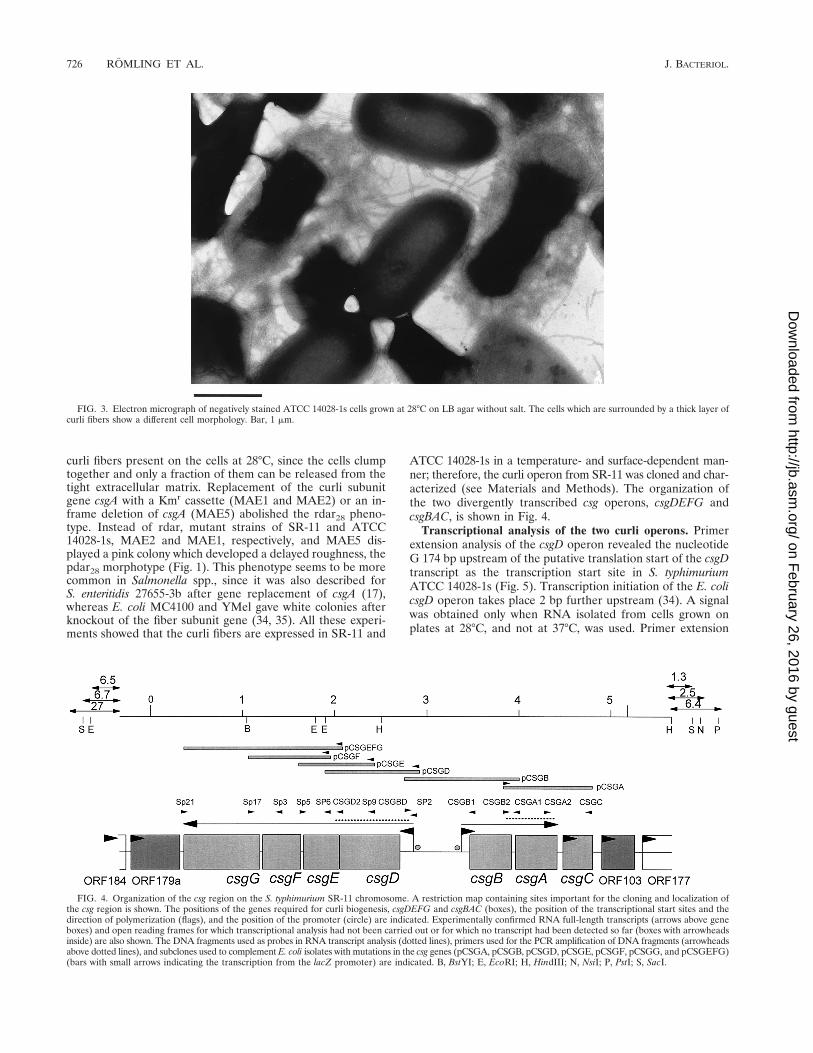
FIG. 4. Organization of the csg region on the S. typhimurium SR-11 chromosome. A restriction map containing sites important for the cloning and localization ofthe csg region is shown. The positions of the genes required for curli biogenesis, csgDEFG and csgBAC (boxes), the position of the transcriptional start sites and thedirection of polymerization (flags), and the position of the promoter (circle) are indicated. Experimentally confirmed RNA full-length transcripts (arrows above geneboxes) and open reading frames for which transcriptional analysis had not been carried out or for which no transcript had been detected so far (boxes with arrowheadsinside) are also shown. The DNA fragments used as probes in RNA transcript analysis (dotted lines), primers used for the PCR amplification of DNA fragments (arrowheadsabove dotted lines), and subclones used to complement E. coli isolates with mutations in the csg genes (pCSGA, pCSGB, pCSGD, pCSGE, pCSGF, pCSGG, and pCSGEFG)(bars with small arrows indicating the transcription from the lacZ promoter) are indicated. B, BstYI; E, EcoRI; H, HindIII; N, NsiI; P, PstI; S, SacI.
726 ROMLING ET AL. J. BACTERIOL.
on February 26, 2016 by guest
http://jb.asm.org/
Dow
nloaded from

analysis of the csgBAC operon confirmed previous results (2,17; also data not shown).
In order to determine the expression state of both csg oper-ons, analysis of the steady-state levels of the RNA transcriptsof csgD, the transcriptional regulator, and csgA, the fiber sub-unit gene, was carried out by Northern blot analysis (Fig. 6). Byusing a probe encompassing the whole csgD gene, four majorbands of 2.3, 1.3, 0.9, and 0.7 kb which could correspond to thetranscripts csgDEFG, csgDEF, csgDE, and csgD (with theoret-ical sizes of 2.5, 1.6, 1.2, and 0.8 kb, respectively) were de-tected; the csgA probe hybridized to three bands of 0.9, 0.6, and0.5 kb, as in S. enteritidis (17). Signals of the same intensitywere detected for ATCC 14028-1s and SR-11 after the cellswere grown at 28°C for 24 or 48 h on plates but not for cellsgrown at 37°C for 17 or 28 h. Therefore, detection of csgD andcsgA transcripts is concomitant with the expression of CsgA at28°C, and transcripts are present long after entrance into thestationary phase.
Comparative sequence analysis of the curli region. The csggenes are located at the same positions on the chromosomes ofE. coli K-12 and S. typhimurium LT2 (26 centisomes [20]). InS. typhimurium, two divergently transcribed operons, csgBACand csgDEFG, flank a 521-bp intergenic region (Fig. 4), asituation as in E. coli MC4100 (34). The nucleotide sequencesof the two species showed an identity of 77.6%, a value whichis below the average sequence conservation of 84.4% (61). Theoverall G1C contents in S. typhimurium and E. coli are similar;however, single genes show some variability in G1C contentconservation (Table 3). The intergenic region between thetranscriptional start sites has a low G1C content and is theleast conserved (71%) (Table 3). However, the intergenic re-gion has not homogeneously diverged but can be subdividedinto four regions. Only four nucleotide substitutions occurred
in the 60 bp upstream of the transcription start site of the csgDoperon. The csgD promoter shares the characteristics of pro-moters transcribed by sD both in S. typhimurium and E. coli,with the E. coli 235 box being closer to the consensus se-quence. The region upstream of the csgD promoter has a verylow G1C content (21.2%) which is identical in S. typhimuriumand E. coli, and a conspicuous peak of curvature at position2147 (data not shown) was found by calculation with the
FIG. 5. Primer extension analyses for the determination of the transcrip-tional start site of the csgDEFG operon. RNA was prepared from strains UMR1and MAE40 (the rpoS derivative of ATCC 14028-1s) grown at 28°C on plates,and primer extension was carried out as described in Materials and Methods. Anextension product is seen for UMR1 but not for MAE40. Primer PEXD1,located 58 bp downstream of the csgDEFG start codon, was used for the exten-sion reaction as well as for the sequencing reaction on pUMR10-7 as a template.The sequence derived from the PEXD1 primer on pUMR10-7 is complementaryto the RNA template, so the coding strand is automatically shown. The tran-scriptional start site (asterisk) and bases belonging to the putative promotersequences (boxed) are indicated.
FIG. 6. Northern blot analysis of RNA transcripts of the csg region usingS. typhimurium ATCC 14028-1s and SR-11 grown on LB medium plates withoutsalt at 28 and 37°C for different periods. Probes covering the whole csgD and csgAgenes were used; their locations are indicated in Fig. 4. Lanes: 1, UMR3; 2,UMR1. The sizes of the bands detected by the respective probes are shown onthe left, calculated by using a 0.24- to 9.5-kb RNA ladder (Life Technologies) asa standard. (A) Hybridization with the csgD probe; (B) hybridization with thecsgA probe; (C) control hybridization with 16S RNA as described in Materialsand Methods.
TABLE 3. Properties of the csg genes and surroundingopen reading frames
Gene orregion
% G1C content(S. typhimurium/
E. coli)
Sequenceidentity
(%)a
Aminoidentity/similarity
(%)a
KS
ORF179ab 52.2/54.1 75.7 86.6/92.5 NDe
csg operon 43.5/43.2 77.6csgG 49.0/50.1 83.4 96.0/99.3 1.065csgF 44.6/42.4 81.0 89.9/94.2 1.018csgE 44.4/43.1 80.8 91.4/96.9 1.496csgD 40.7/41.8 81.1 92.1/95.8 1.155Intergenic region 33.7c/34.4 73.0c
30.6d/32.2 70.6d
csgB 43.4/41.8 82.9 82.1/90.7 0.564csgA 51.5/50.1 73.2 74.8/86.1 1.021csgC 45.0/42.6 71.9 73.1/87.0 1.263ORF103b 47.7/43.6 45.2 39.4/57.6 ND
a Between S. typhimurium SR-11 and E. coli MG1655.b Nomenclature and sequence from E. coli MG1655 and W3110; EMBL files
with accession no. ecae205 and ecae206 and ecd740 to ecd742 were used.c Sequence between the translational start sites of csgD and csgB.d Sequence between the transcriptional start sites of csgD and csgB.e ND, not determined.
VOL. 180, 1998 CURLI GENES FROM S. TYPHIMURIUM 727
on February 26, 2016 by guest
http://jb.asm.org/
Dow
nloaded from

DNase I-based parameters using the bend.it server (29). Se-quence identity drops to 42.7% between 165 and 443 bp up-stream of the transcriptional start site of csgD. The csgB pro-moter has a 210 box, which resembles more the proposedconsensus sequence of sS than that of sD (38). Two alternative235 boxes have been found which both suggest an unusuallywide spacing of 19 or 21 bp between the 235 and 210 boxes.Some promoters whose transcription can be initiated in theabsence of specific 235 hexamer contacts contain an upstreamextension of the 210 element (10) by 59-Tgn-39, a sequencewhich was not found at the csgB promoter.
The putative proteins encoded by the genes of the two oper-ons showed variability in sequence conservation. With the ex-ception of CsgC, whose role has not been unambiguously de-termined, the proteins for the fiber subunit, CsgA, and thenucleator, CsgB, displayed the lowest amino acid identities(74.8 and 82.1%, respectively). These values lie at the lowerend of gene conservation between S. typhimurium and E. coli,which ranges from 100 to 74.3% amino acid identity (61), butare surprisingly high when homologies among fimbriae, even ofcommon origin within a species, are considered (45). In addi-tion, the amino acid identities of CsgA, CsgB, and CsgC were100% when the proteins of S. typhimurium SR-11 and S. en-teritidis 27655-36 were compared (see reference 17 for furthercharacterization of the genes).
The degree of conservation of the CsgD, CsgE, CsgF, andCsgG proteins between S. typhimurium and E. coli is high andlies between 89.5 and 96% amino acid identity (Table 3). Thelipoprotein CsgG, which has a putative molecular mass of 30kDa, was most conserved and showed only conservative aminoacid exchanges. The helix-turn-helix DNA binding motif ofCsgD, the putative transcriptional regulator of 25 kDa, is com-pletely conserved between the two species. Classified accord-ing to the sequence similarity of the DNA binding motif, CsgDbelongs to the LuxR family. The closest sequence homology isto regulators belonging to a two-component sensory transduc-tion system, such as DegU of Bacillus subtilis and NarP ofE. coli.
Complementation of E. coli csg mutants with S. typhimuriumgenes. Considering the high homology of the Csg proteinsbetween S. typhimurium and E. coli, functional complementa-tion of gene products seemed likely. In order to test this hy-pothesis, PCR-generated gene fragments from S. typhimurium
were cloned into the low-copy-number vectors pWSK29 andpWKS30 so that transcription occurred from the lacZ pro-moter (see Materials and Methods). All E. coli csg mutants werewhite and, with the exception of MHR480 (the csgE mutant),produced no curli fibers. MHR261 (csgB2) and MHR426 (csgF4)secrete CsgA in a soluble form (35, 36), but the soluble form ofthe protein was not detected in the whole-cell preparationsused here (Fig. 7). Complementation was judged by the devel-opment of a red and rough colony morphology type and byWestern blots detecting CsgA as an acid-sensitive polymer(Fig. 7 and Table 4). As seen in Table 4, all S. typhimuriumgenes complemented the respective E. coli csg mutants, al-though to different degrees. Single csgB, csgD, and csgE genescomplemented the respective E. coli mutants to the wild-typephenotype. The csgA and csgF mutations could be only verypoorly replaced by the copy on the plasmid. In the case of csgA,two signals of almost equal intensity were seen on an overex-
FIG. 7. Western blot analysis of the complementation of the E. coli csgmutants. Results for whole-cell preparations treated with formic acid are shown.The mutant (2) and the mutant harboring the respective complementing plas-mid (1) were run next to each other. Except for MHR480 (DcsgE3), none of theE. coli mutants showed a CsgA signal derived from polymerized fibers. MHR261(csgB2) and MHR503 (csgD6) were complemented to wild-type levels by usingplasmids with the single genes. MHR426 (csgF4) was complemented to wild typeonly when a plasmid carrying csgEFG was used. Faint signals of CsgA weredetected when pCSGA was introduced into MHR204 (csgA::Tn105).
FIG. 8. Northern blot analysis of RNA transcripts from S. typhimuriumATCC 14028-1s and rpoS and ompR derivatives grown on LB medium plateswithout salt at 28 and 37°C. Probes covering the whole csgD and csgA genes wereused; their locations are indicated in Fig. 4. Lanes: 1, UMR1; 2, MAE40 (rpoS);3, MAE46 (ompR). The sizes of the bands detected by the respective probes areshown on the left, calculated by using a 0.24- to 9.5-kb RNA ladder (LifeTechnologies) as a standard. (A) Hybridization with the csgD probe; (B) hybrid-ization with the csgA probe; (C) control hybridization with 16S RNA as describedin Materials and Methods.
TABLE 4. Complementation of E. coli csg mutantswith S. typhimurium genes
Mutationin E. coli
E. colistrain
Complement-ing plasmid
Colonymorphology
Detection ofCsgA derived
from fibers
csgA2::Tn105 MHR204 pCSGA Brown but notrough
Very weaksignal
csgB2 MHR261 pCSGB wta wt levelscsgD6 MHR503 pCSGD wt wt levelsDcsgE3 MHR480 pCSGE wt wt levels
pCSGEFG wt wt levelscsgF4 MHR426 pCSGF Reddish but not
roughWeak signal
pCSGEFG wt wt levelscsgG1::Tn105 MHR210 pCSGEFG wt wt levels
a wt, wild type.
728 ROMLING ET AL. J. BACTERIOL.
on February 26, 2016 by guest
http://jb.asm.org/
Dow
nloaded from

posed Western blot, one of which has a slightly higher molec-ular weight than the wild-type signal and is most likely a pre-mature form of CsgA (data not shown). In addition, the verylow signal intensity could be explained by a lower specificity ofthe anti-E. coli CsgA antiserum against CsgA from S. typhi-murium. MHR426, the csgF mutant used in this study, has apolar effect on csgG expression, leading to decreased amountsof CsgG (36). The reduced amount of CsgG might limit the fullcomplementation of MHR426 by pCSGF, as occurs with therespective E. coli gene (36). Neither the vector control nor thecsgA, csgD, csgE, and csgF genes cloned in the direction whichwould allow transcription from the T7 promoter gave a changein the color of the colonies or their morphology with respect tothe wild type or in signal intensity of CsgA on Western blots(data not shown). We conclude from the available data thatinterspecies complementation of the csg genes is possible inprinciple.
Analysis of the effect of rpoS and ompR mutations on colonymorphology and transcription from the csg promoters. ForE. coli, it was demonstrated that transcription of the csgD andcsgBA operon requires sS. Transduction of the mutant rpoSallele from SF1005 into ATCC 14028-1s and SR-11 gave whiteand smooth colonies, a saw28 morphotype (Fig. 1). Transcrip-tional analysis with the csgD and csgA probes on RNA ex-tracted from the rpoS mutant of ATCC 14028-1s (MAE40)grown at 28 and 37°C detected no signal for either probe (Fig.8). In addition, no extension product was seen in the latterstrain by primer extension analysis (Fig. 5). Therefore, rpoS isalso required for transcription of the csg operons in S. typhi-murium strains.
It is also known that ompR is necessary for transcriptionfrom both the csgBA and the csgD promoters in E. coli (40). AnompR mutant was constructed by introducing a deleted ompRgene carrying an Ampr cassette instead of the major part of itsopen reading frame into the chromosome of ATCC 14028-1sand SR-11 by double crossover, yielding strains MAE46 andMAE34, respectively (Table 1; see Materials and Methods).The ompR mutants were white at 28 and 37°C (Fig. 1). NoCsgA signal was detected for MAE46 in Western blots (datanot shown). Northern blot analysis of RNA extracted fromstrain MAE46 grown at both temperatures gave no signal forcsgD or csgA (Fig. 8). Since ompR was shown to be necessaryfor the transcription of the csgD and csgBA promoters, theintergenic region was examined for putative ompR bindingsites. One nucleotide sequence which has one mismatch basepair to the recently proposed consensus sequence for indepen-dent binding was found (39). This sequence is centered atposition 250.5 relative to the transcriptional start site of thecsgD promoter. If this sequence is used for OmpR binding, itcan be imagined that transcriptional regulation takes placemainly at the csgD promoter. CsgD may then act upon thecsgBA promoter to initiate transcription there.
DISCUSSION
Although many fimbrial gene clusters have been isolatedfrom Salmonella spp. and E. coli, variation in operon structureand genes encoding regulatory control features of pili from thesame structural class suggests frequent remodeling of DNAsequences due to the necessity to respond flexibly to changingenvironmental conditions and various host environments (49).In contrast to this behavior of fimbrial operons, the operons forcurli biogenesis embedded in the same context on the chro-mosome (reference 20 and this study) are remarkably con-served between E. coli and S. typhimurium. In addition, thesequence similarities at the amino acid level of all proteins in
the operon, ranging from the fiber subunit CsgA (86%) to thetranscriptional regulator CsgD (96%) and the lipoproteinCsgG (99%), are higher than the homologies for most func-tionally and sequentially related fimbrial gene products, evenwithin a species (49, 51, 56). It is therefore suggested that thethin aggregative fibers in Salmonella be named curli fibers, asfor the products of the homologous E. coli genes (csg) (59).
The sequence similarity at the amino acid level is reflectedby the successful complementation of individual csg gene mu-tants of E. coli by S. typhimurium genes. The S. typhimuriumcsgB gene could complement the homologous E. coli gene towild-type levels, showing that the four-repeat structure consist-ing of 22-amino-acid-long putative b-strand–turn–b-strand–turn units (35) which are all required for organelle assembly(8) functions between the species. It can be imagined thatinterspecies exchange of organelle subunits can occur in anatural environment where bacteria are tightly packed, such asthe gut flora. The consensus sequence for the repeat structureof CsgB was determined to be NLA-I-Q-GS-N-A-I-Q-G--,where the underlined amino acids show 100% conservationbetween S. typhimurium and E. coli. The consensus motif forCsgA, which has five 23-amino-acid-long units, is NS--T-TQYG-GN-AT-DQTAA-. There may be several reasons forthe lack of complementation to a full wild type for some genes,such as functional restriction of the proteins, imbalance ofprotein ratios, or instability of the RNA message created fromthe plasmid, which remain to be elucidated. Alternatively, po-lar effects of the csg mutations on downstream genes mayprevent the full complementation to the wild-type phenotype.
The high degree of conservation at the protein level contra-dicts the low conservation at the nucleotide level. The nucle-otide sequences encoding csgDEFG diverged more than aver-age, and so does KS (0.94 on the average), a value for theestimation of the number of synonymous substitutions per site(Table 3). csgE has a KS value of 1.5 and is therefore almostsaturated with substitutions. The high rate of nucleotide sub-stitutions could reflect the chromosomal location of the csgoperons proximal to the terminus of replication, where morenucleotide changes take place, and/or the low expression stateof the genes from the csgDEFG operon (61). The high conser-vation of proteins could indicate a lack of selective pressures,such as the immune response in a host, on the fibers or func-tional constraints on the macromolecules and their specifici-ties, which do not tolerate an evolution which develops too farfrom a common ancestor (45).
The two csg operons are flanked by the same open readingframes as in the fully sequenced E. coli MG1655 strain (EMBLaccession no. ecae205, ecae206, and ecd740 to ecd742). Thehomologous ORF179a showed the same ambivalent conserva-tion scheme as the curli genes, low conservation on the nucle-otide level and high conservation of the amino acid sequence(Table 3); the intergenic region between csgG and ORF179ashowed no homology. Downstream of csgC, the nucleotidehomology already starts to decline at the end of csgC (the stopcodon has changed with respect to E. coli) to a level of 60%,which continues within ORF103.
Another fiber, type 1 fimbria, which seems to be present inall Salmonella species, shares morphological and adhesiveproperties with E. coli type 1 fimbria. However, the differentlocation on the chromosome in the two species and the highdegree of sequence diversity and gene rearrangements (14),together with different regulation schemes having no overlap-ping regulating factors, suggest that these genes were acquiredindependently by S. typhimurium and E. coli and subject toevolutionary pressures (45). Therefore, curli genes are the only
VOL. 180, 1998 CURLI GENES FROM S. TYPHIMURIUM 729
on February 26, 2016 by guest
http://jb.asm.org/
Dow
nloaded from

fimbrial genes detected which were already present in a com-mon ancestor of E. coli and Salmonella spp.
The regulation of the curli genes in S. typhimurium SR-11and ATCC 14028-1s by temperature, rpoS, and ompR is iden-tical to that in E. coli MC4100 and YMel (2, 3, 34, 40, 53),implying that fiber expression responds to the same environ-mental cues in general. The complementation to wild-typelevels of the E. coli csgD gene by the respective S. typhimuriumgene shows that the recognition sites for the transcriptionalactivator are also conserved between the species. The corre-spondence in regulation pattern in addition to gene conserva-tion points to a similar or identical function of the curli fibersin the two species in the same ecological niche. The expressionpattern on plates at low temperature and low osmolarity sug-gests a role for curli fibers primarily outside a host, on surfaces.A participation of curli fibers in a bacterial network such as abiofilm (21) is possible, considering the adhesive nature of thefibers and the colony morphology of cells. However, moreenvironmental studies need to be performed before a firm roleof curli fibers in biofilm formation can be concluded.
ACKNOWLEDGMENTS
We thank the following people for providing plasmids, strains andantibodies: D. G. Guiney for SF1005, S. R. Kushner for pWSK29 andpWKS30, A. Lazdunski for pLAFR3, H. Loferer for HLO7, E. Mor-feldt and S. Arvidson for anti-OmpA antiserum, and M. Rhen for 146,LB5010, and pCurli. We are grateful to H. Loferer for helpful tech-nical advice.
U.R. was the recipient of a fellowship from the program “Infek-tionsbiologie” from the German Bundesministerium fur Forschungund Technologie (BMFT). This work was supported by grants from theSwedish Medical Research Council (B96-16X-10843-03A) and theSwedish Natural Science Research Council and by an unrestrictedgrant for infectious disease research from the Bristol-Myers SquibbCompany to S. Normark.
REFERENCES
1. Applied Biosystems, Inc. 1991. User bulletin 18. Applied Biosystems, Inc.,Foster City, Calif.
2. Arnqvist, A., A. Olsen, and S. Normark. 1994. sS-dependent growth-phaseinduction of the csgBA promoter can be achieved in vivo by s70 in theabsence of the nucleoid-associated protein H-NS. Mol. Microbiol. 13:1021–1032.
3. Arnqvist, A., A. Olsen, J. Pfeifer, D. G. Russell, and S. Normark. 1992. TheCrl protein activates cryptic genes for curli formation and fibronectin bindingin Escherichia coli HB101. Mol. Microbiol. 6:2443–2452.
4. Ausubel, F. M., R. Brent, R. E. Kingston, D. D. Moore, J. G. Seidman, J. A.Smith, and K. Struhl (ed.). 1994. Current protocols in molecular biology.John Wiley & Sons, New York, N.Y.
5. Baumler, A. J., A. J. Gilde, R. M. Tsolis, A. W. M. van der Velden, B. M. M.Ahmer, and F. Heffron. 1997. Contribution of horizontal gene transfer anddeletion events to development of distinctive patterns of fimbrial operonsduring evolution of Salmonella serotypes. J. Bacteriol. 179:317–322.
6. Baumler, A. J., and F. Heffron. 1995. Identification and sequence analysis oflpfABCDE, a putative fimbrial operon of Salmonella typhimurium. J. Bacte-riol. 177:2087–2097.
7. Ben Nasr, A., A. Olsen, U. Sjobring, W. Muller-Esterl, and L. Bjorck. 1996.Assembly of human contact phase proteins and release of bradykinin at thesurface of curli-expressing Escherichia coli. Mol. Microbiol. 20:927–935.
8. Bian, Z., and S. Normark. 1997. Nucleator function of CsgB for the assemblyof adhesive surface organelles in Escherichia coli. EMBO J. 16:5827–5836.
9. Bio-Rad Laboratories. 1993. Gene pulser electroprotocol. Bio-Rad Labora-tories, Hercules, Calif.
10. Bown, J., T. Belyaeva, S. Busby, and S. Minchin. 1997. Extended 210promoters. Nucleic Acids Mol. Biol. 11:41–52.
11. Brosius, J., A. Ullrich, M. A. Raker, A. Gray, T. J. Dull, R. R. Gutell, andH. F. Noller. 1981. Construction and fine mapping of recombinant plasmidscontaining the rrnB ribosomal RNA operon of E. coli. Plasmid 6:112–118.
12. Bullas, L. R., and J.-I. Ryu. 1983. Salmonella typhimurium LT2 strains whichare r2m1 for all three chromosomally located systems of DNA restrictionand modification. J. Bacteriol. 156:471–474.
13. Casadaban, M. J. 1976. Transposition and fusion of the lac genes to selected
promoters in Escherichia coli using bacteriophage lambda and Mu. J. Mol.Biol. 104:541–555.
14. Clegg, S., and D. L. Swenson. 1994. Salmonella fimbriae, p. 105–113. In P.Klemm (ed.), Fimbriae, adhesion, genetics, biogenesis and vaccine. CRCPress, Boca Raton, Fla.
15. Clouthier, S. C., S. K. Collinson, and W. W. Kay. 1994. Unique fimbriae-likestructures encoded by sefD on the SEF14 fimbrial gene cluster of Salmonellaenteritidis. Mol. Microbiol. 12:893–903.
16. Clouthier, S. C., K.-H. Muller, J. L. Doran, S. K. Collinson, and W. W. Kay.1993. Characterization of three fimbrial genes, sefABC, of Salmonella enter-itidis. J. Bacteriol. 175:2523–2533.
17. Collinson, S. K., S. C. Clouthier, J. L. Doran, P. A. Banser, and W. W. Kay.1996. Salmonella enteritidis agfBAC operon encoding thin, aggregative fim-briae. J. Bacteriol. 178:662–667.
18. Collinson, S. K., P. C. Doig, J. L. Doran, S. Clouthier, T. J. Trust, and W. W.Kay. 1993. Thin, aggregative fimbriae mediate binding of Salmonella enter-itidis to fibronectin. J. Bacteriol. 175:12–18.
19. Collinson, S. K., L. Emody, K.-H. Muller, T. J. Trust, and W. W. Kay. 1991.Purification and characterization of thin, aggregative fimbriae from Salmo-nella enteritidis. J. Bacteriol. 173:4773–4781.
20. Collinson, S. K., S.-L. Liu, S. C. Clouthier, P. A. Banser, J. L. Doran, K. E.Sanderson, and W. W. Kay. 1996. The location of four fimbrin-encodinggenes, agfA, fimA, sefA and sefD, on the Salmonella enteritidis and/or S. ty-phimurium XbaI-BlnI genomic restriction maps. Gene 169:75–80.
21. Costerton, J. W., K.-J. Cheng, G. G. Geesey, T. I. Ladd, J. C. Nickel, M.Dasgupta, and T. J. Marrie. 1987. Bacterial biofilms in nature and disease.Annu. Rev. Microbiol. 41:435–464.
22. Coynault, C., V. Robbe-Saule, and F. Norel. 1996. Virulence and vaccinepotential of Salmonella typhimurium mutants deficient in the expression ofthe RpoS (sS) regulon. Mol. Microbiol. 22:149–160.
23. Doran, J. L., S. K. Collinson, J. Burian, G. Sarlos, E. C. D. Todd, C. K.Munro, C. M. Kay, P. A. Banser, P. I. Peterkin, and W. W. Kay. 1993.DNA-based diagnostic tests for Salmonella species targeting agfA, the struc-tural gene for thin, aggregative fimbriae. J. Clin. Microbiol. 31:2263–2273.
24. Dorman, C. J., S. Chatfield, C. F. Higgins, C. Hayward, and G. Dougan.1989. Characterization of porin and ompR mutants of a virulent strain ofSalmonella typhimurium: ompR mutants are attenuated in vivo. Infect. Im-mun. 57:2136–2140.
25. Eisenstark, A., J. M. Calcutt, M. Backer-Hapak, and A. Ivanova. 1996. Roleof Escherichia coli rpoS and associated genes in defense against oxidativedamage. Free Radical Biol. Med. 21:975–993.
26. Fang, F. C., S. J. Libby, N. A. Buchmeier, P. C. Loewen, J. Switala, J.Harwood, and D. G. Guiney. 1992. The alternative s factor KatF (RpoS)regulates Salmonella virulence. Proc. Natl. Acad. Sci. USA 89:11978–11982.
27. Figurski, D. H., and D. R. Helinski. 1979. Replication of an origin-containingderivative of plasmid RK2 dependent on a plasmid function provided intrans. Proc. Natl. Acad. Sci. USA 76:1648–1652.
28. Friedrich, M. J., N. E. Kinsey, J. Vila, and R. J. Kadner. 1993. Nucleotidesequence of a 13.9 kb segment of the 90 kb virulence plasmid of Salmonellatyphimurium: the presence of fimbrial biosynthetic genes. Mol. Microbiol.8:543–558.
29. Gabrielian, A., A. Simoncsits, and S. Pongor. 1996. Distribution of bendingpropensity in DNA sequences. FEBS Lett. 393:124–130.
30. Gibson, M. M., E. M. Ellis, K. A. Graeme-Cook, and C. F. Higgins. 1987.OmpR and EnvZ are pleiotropic regulatory proteins: positive regulation ofthe tripeptide permease (tppB) of Salmonella typhimurium. Mol. Gen. Genet.207:120–129.
31. Giron, J. A., M. M. Livine, and J. B. Kaper. 1994. Longus: a long pilusultrastructure produced by human enterotoxigenic Escherichia coli. Mol.Microbiol. 12:71–82.
32. Grund, S., and A. Weber. 1988. A new type of fimbriae on Salmonellatyphimurium. J. Vet. Med. B 35:779–782.
33. Hamilton, C. M., M. Aldea, B. K. Washburn, P. Babitzke, and S. R. Kushner.1989. New method for generating deletions and gene replacements in Esch-erichia coli. J. Bacteriol. 171:4617–4622.
34. Hammar, M., A. Arnqvist, Z. Bian, A. Olsen, and S. Normark. 1995. Ex-pression of two csg operons is required for production of fibronectin- andCongo red-binding curli polymers in Escherichia coli K-12. Mol. Microbiol.18:661–670.
35. Hammar, M., Z. Bian, and S. Normark. 1996. Nucleator-dependent inter-cellular assembly of adhesive curli organelles in Escherichia coli. Proc. Natl.Acad. Sci. USA 93:6562–6566.
36. Hammar, M., H. Loferer, A. Olsen, and S. Normark. Nucleation dependentextracellular formation of curli; requirement of the CsgF protein. Submittedfor publication.
37. Hammar, M., A. Olsen, H. Loferer, and S. Normark. The binding propertyof adhesive curli organelles in Escherichia coli requires nagA, an N-acetyl-glucosamine-6-phosphate deacetylase, and CsgE. Submitted for publication.
38. Hengge-Aronis, R. 1996. Regulation of gene expression during entry intostationary phase, p. 1497–1512. In F. C. Neidhardt, R. Curtiss III, J. L.Ingraham, E. C. C. Lin, K. B. Low, B. Magasanik, W. S. Reznikoff, M. Riley,M. Schaechter, and H. E. Umbarger (ed.), Escherichia coli and Salmonella:
730 ROMLING ET AL. J. BACTERIOL.
on February 26, 2016 by guest
http://jb.asm.org/
Dow
nloaded from

cellular and molecular biology. ASM Press, Washington, D.C.39. Huang, K.-J., and M. M. Igo. 1996. Identification of the bases in the ompF
regulatory region which interact with the transcription factor OmpR. J. Mol.Biol. 262:615–628.
40. Hultgren, S. J., C. Hal Jones, and S. Normark. 1996. Bacterial adhesions andtheir assembly, p. 2730–2756. In F. C. Neidhardt, R. Curtiss III, J. L. Ingra-ham, E. C. C. Lin, K. B. Low, B. Magasanik, W. S. Reznikoff, M. Riley, M.Schaechter, and H. E. Umbarger (ed.), Escherichia coli and Salmonella:cellular and molecular biology. ASM Press, Washington, D.C.
41. Inoue, H., H. Nojima, and H. Okayama. 1990. High efficiency transformationof Escherichia coli with plasmids. Gene 96:23–28.
42. Jishage, M., and A. Ishihama. 1995. Regulation of RNA polymerase sigmasubunit synthesis in Escherichia coli: intracellular levels of s70 and s38. J.Bacteriol. 177:6832–6835.
43. Knapp, S., J. Hacker, T. Jarchau, and W. Goebel. 1994. Large, unstableinserts in the chromosome affect virulence properties of uropathogenic Esch-erichia coli O6 strain 536. J. Bacteriol. 168:22–30.
44. Krogfelt, K. A. 1991. Bacterial adhesion: genetics, biogenesis, and role inpathogenesis of fimbrial adhesions of Escherichia coli. Rev. Infect. Dis.13:721–735.
45. Kusters, J. G., and W. Gaastra. 1994. Fimbrial operons and evolution, p.179–196. In P. Klemm (ed.), Fimbriae, adhesion, genetics, biogenesis andvaccine. CRC Press, Boca Raton, Fla.
46. Liljestrom, P., I. Laamanen, and E. T. Palva. 1988. Structure and expressionof the ompB operon, the regulatory locus for the outer membrane porinregulon in Salmonella typhimurium LT2. J. Mol. Biol. 201:663–673.
47. Lindgren, S. W., I. Stojikovic, and F. Heffron. 1996. Macrophage killing is anessential virulence mechanism of Salmonella typhimurium. Proc. Natl. Acad.Sci. USA 93:4197–4201.
47a.Loferer, H. Unpublished data.48. Loferer, H., M. Hammar, and S. Normark. 1997. Availability of the fiber
subunit CsgA and the nucleator protein CsgB during assembly of fibronec-tin-binding curli is limited by the intracellular concentration of the novellipoprotein CsgG. Mol. Microbiol. 26:11–23.
49. Low, D., B. Braaten, and M. van der Woude. 1996. Fimbriae, p. 146–157. InF. C. Neidhardt, R. Curtiss III, J. L. Ingraham, E. C. C. Lin, K. B. Low, B.Magasanik, W. S. Reznikoff, M. Riley, M. Schaechter, and H. E. Umbarger(ed.), Escherichia coli and Salmonella: cellular and molecular biology. ASMPress, Washington, D.C.
50. Maloy, S. R., V. J. Stewart, and R. K. Taylor. 1996. Genetic analysis ofpathogenic bacteria. A laboratory manual. Cold Spring Harbor LaboratoryPress, Cold Spring Harbor, N.Y.
51. Marc, D., and M. Dho-Moulin. 1996. Analysis of the fim cluster of an avianO2 strain of Escherichia coli: serogroup-specific sites within fimA and nucle-otide sequence of fimI. J. Med. Microbiol. 44:444–452.
52. McCann, M. P., J. P. Kidwell, and A. Matin. 1991. The putative s factorKatF has a central role in development of starvation-mediated general re-sistance in Escherichia coli. J. Bacteriol. 173:4188–4194.
53. Olsen, A., A. Arnqvist, M. Hammar, S. Sukupolvi, and S. Normark. 1993.The RpoS sigma factor relieves H-NS-mediated transcriptional repression of
csgA, the subunit gene of fibronectin-binding curli in Escherichia coli. Mol.Microbiol. 7:523–536.
54. Olsen, A., A. Jansson, and S. Normark. 1989. Fibronectin binding mediatedby a novel class of surface organelles on Escherichia coli. Nature 338:652–655.
55. Pratt, L. A., and T. J. Silhavy. 1995. Porin regulon of Escherichia coli,p. 105–127. In J. A. Hoch and T. J. Silhavy (ed.), Two-component signaltransduction. ASM Press, Washington, D.C.
56. Purcell, B. K., J. Pruckler, and S. Clegg. 1987. Nucleotide sequences of thegenes encoding type 1 fimbrial subunits of Klebsiella pneumoniae and Sal-monella typhimurium. J. Bacteriol. 169:5831–5834.
56a.Romling, U. Unpublished data.57. Romling, U., T. Heuer, and B. Tummler. 1994. Bacterial genome analysis by
pulsed field gel electrophoresis techniques, p. 355–406. In A. Chrambach,M. J. Dunn, and B. J. Radola (ed.), Advances in electrophoresis. VCH,Weinheim, Germany.
58. Sambrook, J., E. F. Fritsch, and T. Maniatis. 1989. Molecular cloning: alaboratory manual, 2nd ed. Cold Spring Harbor Laboratory Press, ColdSpring Harbor, N.Y.
59. Sanderson, K. E., A. Hessel, S. L. Liu, and K. E. Rudd. 1996. The geneticmap of Salmonella typhimurium, edition VIII, p. 1903–1999. In F. C. Neid-hardt, R. Curtiss III, J. L. Ingraham, E. C. C. Lin, K. B. Low, B. Magasanik,W. S. Reznikoff, M. Riley, M. Schaechter, and H. E. Umbarger (ed.), Esch-erichia coli and Salmonella: cellular and molecular biology. ASM Press,Washington, D.C.
60. Schmieger, H. 1972. Phage P22 mutants with increased or decreased trans-duction abilities. Mol. Gen. Genet. 119:75–88.
61. Sharp, P. M. 1991. Determinants of DNA sequence divergence betweenEscherichia coli and Salmonella typhimurium: codon usage, map position, andconcerted evolution. J. Mol. Evol. 33:23–33.
62. Sjobring, U., G. Pohl, and A. Olsen. 1994. Plasminogen, absorbed by Esch-erichia coli expressing curli or by Salmonella enteritidis expressing thin ag-gregative fimbriae, can be activated by simultaneously captured tissue-typeplasminogen activator (t-PA). Mol. Microbiol. 14:443–452.
63. Spangenberg, C., R. Fislage, U. Romling, and B. Tummler. 1997. Disrespect-ful type IV pilins. Mol. Microbiol. 25:203–204.
64. Staskawicz, B., D. Dahlbeck, N. Keen, and C. Napoli. 1987. Molecularcharacterization of cloned avirulence genes from race 0 and race 1 of Pseudo-monas syringae pv. glycinea. J. Bacteriol. 169:5789–5794.
65. Swords, W. E., B. M. Cannon, and W. H. Benjamin, Jr. 1997. Avirulence ofLT2 strains of Salmonella typhimurium results from a defective rpoS gene.Infect. Immun. 65:2451–2453.
66. Tautz, D., and M. Renz. 1983. An optimized freeze-squeeze method for therecovery of DNA fragments from agarose gels. Anal. Biochem. 132:14–19.
67. Wang, R. F., and S. R. Kushner. 1991. Construction of versatile low-copy-number vectors for cloning, sequencing and gene expression in Escherichiacoli. Gene 100:195–199.
68. Wilmes-Riesenberg, M. R., J. W. Foster, and R. Curtiss III. 1997. An alteredrpoS allele contributes to the avirulence of Salmonella typhimurium LT2.Infect. Immun. 65:203–210.
VOL. 180, 1998 CURLI GENES FROM S. TYPHIMURIUM 731
on February 26, 2016 by guest
http://jb.asm.org/
Dow
nloaded from