Characterization of the Fur Regulon in Pseudomonas syringae pv.
15
Published Ahead of Print 22 July 2011. 2011, 193(18):4598. DOI: 10.1128/JB.00340-11. J. Bacteriol. D. Helmann, David J. Schneider and Samuel W. Cartinhour Melanie J. Filiatrault, Bryan Swingle, Ahmed Gaballa, John Myers, Paul V. Stodghill, James J. Bolton, Eric J. Markel, Bronwyn G. Butcher, Philip A. Bronstein, Christopher R. Pseudomonas syringae pv. tomato DC3000 Characterization of the Fur Regulon in http://jb.asm.org/content/193/18/4598 Updated information and services can be found at: These include: SUPPLEMENTAL MATERIAL Supplemental material REFERENCES http://jb.asm.org/content/193/18/4598#ref-list-1 at: This article cites 42 articles, 16 of which can be accessed free CONTENT ALERTS more» articles cite this article), Receive: RSS Feeds, eTOCs, free email alerts (when new http://journals.asm.org/site/misc/reprints.xhtml Information about commercial reprint orders: http://journals.asm.org/site/subscriptions/ To subscribe to to another ASM Journal go to: on November 17, 2014 by guest http://jb.asm.org/ Downloaded from on November 17, 2014 by guest http://jb.asm.org/ Downloaded from
-
Upload
odessacollege -
Category
Documents
-
view
0 -
download
0
Transcript of Characterization of the Fur Regulon in Pseudomonas syringae pv.

Published Ahead of Print 22 July 2011. 2011, 193(18):4598. DOI: 10.1128/JB.00340-11. J. Bacteriol.
D. Helmann, David J. Schneider and Samuel W. CartinhourMelanie J. Filiatrault, Bryan Swingle, Ahmed Gaballa, JohnMyers, Paul V. Stodghill, James J. Bolton, Eric J. Markel, Bronwyn G. Butcher, Philip A. Bronstein, Christopher R. Pseudomonas syringae pv. tomato DC3000Characterization of the Fur Regulon in
http://jb.asm.org/content/193/18/4598Updated information and services can be found at:
These include:
SUPPLEMENTAL MATERIAL Supplemental material
REFERENCEShttp://jb.asm.org/content/193/18/4598#ref-list-1at:
This article cites 42 articles, 16 of which can be accessed free
CONTENT ALERTS more»articles cite this article),
Receive: RSS Feeds, eTOCs, free email alerts (when new
http://journals.asm.org/site/misc/reprints.xhtmlInformation about commercial reprint orders: http://journals.asm.org/site/subscriptions/To subscribe to to another ASM Journal go to:
on Novem
ber 17, 2014 by guesthttp://jb.asm
.org/D
ownloaded from
on N
ovember 17, 2014 by guest
http://jb.asm.org/
Dow
nloaded from
JOURNAL OF BACTERIOLOGY, Sept. 2011, p. 4598–4611 Vol. 193, No. 180021-9193/11/$12.00 doi:10.1128/JB.00340-11Copyright © 2011, American Society for Microbiology. All Rights Reserved.
Characterization of the Fur Regulon in Pseudomonas syringae pv.tomato DC3000�†
Bronwyn G. Butcher,1 Philip A. Bronstein,1,2# Christopher R. Myers,3 Paul V. Stodghill,2
James J. Bolton,2 Eric J. Markel,2 Melanie J. Filiatrault,1,2 Bryan Swingle,1,2 Ahmed Gaballa,4John D. Helmann,4 David J. Schneider,1,2 and Samuel W. Cartinhour1,2*
Department of Plant Pathology and Plant-Microbe Biology, Cornell University, Ithaca, New York 148531; United States Department ofAgriculture-Agricultural Research Service, Ithaca, New York 148532; Department of Physics, Laboratory of Atomic and
Solid State Physics, and Computational Biology Service Unit, Cornell University, Ithaca, New York 148533; andDepartment of Microbiology, Cornell University, Ithaca, New York 148534
Received 10 March 2011/Accepted 16 June 2011
The plant pathogen Pseudomonas syringae pv. tomato DC3000 (DC3000) is found in a wide variety ofenvironments and must monitor and respond to various environmental signals such as the availability of iron,an essential element for bacterial growth. An important regulator of iron homeostasis is Fur (ferric uptakeregulator), and here we present the first study of the Fur regulon in DC3000. Using chromatin immunopre-cipitation followed by massively parallel sequencing (ChIP-seq), 312 chromosomal regions were highly en-riched by coimmunoprecipitation with a C-terminally tagged Fur protein. Integration of these data withprevious microarray and global transcriptome analyses allowed us to expand the putative DC3000 Fur regulonto include genes both repressed and activated in the presence of bioavailable iron. Using nonradioactive DNaseI footprinting, we confirmed Fur binding in 41 regions, including upstream of 11 iron-repressed genes andthe iron-activated genes encoding two bacterioferritins (PSPTO_0653 and PSPTO_4160), a ParA protein(PSPTO_0855), and a two-component system (TCS) (PSPTO_3382 to PSPTO_3380).
The Gram-negative bacterium Pseudomonas syringae pv. to-mato DC3000 (DC3000), a widely used model organism for thestudy of bacterial plant-pathogen interactions, is the causalagent of bacterial speck on tomato and is also a pathogen ofthe model plant Arabidopsis thaliana (4). All pathogens mustclosely monitor and respond to various environmental signalsto secure nutrients from and defend against their hosts. Oneimportant environmental signal is iron, which is an essentialelement for bacterial growth due to the fact that it is a cofactorin many proteins involved in major biological processes such asmetabolism and electron transport. While iron is the fourthmost abundant element in the earth’s crust, it is found mostlyin insoluble iron (III) oxide forms and is therefore not readilyavailable to bacteria. For this reason many organisms, includ-ing P. syringae, rely on siderophores to chelate and solubilizethe available iron. However, because excess iron can generatetoxic hydroxyl radicals through Fenton chemistry in the pres-ence of oxygen, bacteria must also regulate intracellular ironconcentrations carefully to avoid damage or death.
The primary regulator of iron homeostasis in bacteria is Fur(ferric uptake regulator). This DNA binding protein has beenidentified in many bacterial species where it regulates expres-sion of iron uptake and storage genes and is also involved in
virulence and protection against oxidative stress (reviewed inreference 5). Fur typically functions as a repressor by bindingto DNA in the presence of iron and preventing gene transcrip-tion. Under low-iron conditions the affinity of Fur for thesebinding sites is greatly reduced, resulting in expression of thegenes that it negatively regulates. The role of Fur has been wellstudied in the opportunistic human pathogen Pseudomonasaeruginosa, which must respond to the low-iron conditions en-countered in the host to ensure its survival. The first crystalstructure of Fur was determined using protein isolated from P.aeruginosa PAO1 (36), and many studies have led to the elu-cidation of the Fur regulon in this species (31, 32, 35). Fur is anessential protein in PAO1 and, like in many other organisms,represses genes involved in iron acquisition (some of whichcontribute to virulence) such as siderophores and heme uptakesystems. It also indirectly regulates (via PvdS) the expression ofextracellular proteinases, which degrade the host iron bindingproteins, and the virulence factor exotoxin A, which is a potentextracellular toxin (reviewed in reference 11). In addition,PAO1 Fur represses the expression of other regulators such as10 alternative extracytoplasmic function (ECF) sigma factorsand two regulatory noncoding RNAs (ncRNAs) (prrF1 andprrF2). The prrF1 and prrF2 ncRNAs are functionally similar toryhB in Escherichia coli and act by causing the degradation ofmRNAs encoding iron-containing proteins (33, 41).
While the classical repression model represents the mostfamiliar mechanism by which Fur controls gene expression, it isnow understood that Fur can exert regulation in other ways.For example, Fur-dependent activation of genes can occurindirectly when Fur blocks the binding of a second repressor(e.g., Fur inhibits H-NS binding upstream of fntA in E. coli[29]). In at least one organism (Neisseria meningitidis), Fur
* Corresponding author. Mailing address: USDA Agricultural Re-search Service, Plant-Microbe Interactions Research Unit, 334 PlantScience Building, Cornell University, Ithaca, NY 14853. Phone: (607)255-8091. Fax: (607) 255-4471. E-mail: [email protected].
# Present address: United States Department of Agriculture-FoodSafety and Inspection Service, Washington, DC 20250.
† Supplemental material for this article may be found at http://jb.asm.org/.
� Published ahead of print on 22 July 2011.
4598
on Novem
ber 17, 2014 by guesthttp://jb.asm
.org/D
ownloaded from

appears to directly activate the expression of genes that haveiron centers (13). In Helicobacter pylori, the iron-depleted(apo) form of Fur represses a subset of genes under low-ironconditions (12, 14, 17, 28). Although it is not yet knownwhether these mechanisms are broadly shared among bacteria,they provide evidence that the regulatory role of Fur may beunexpectedly subtle.
Little is known about the regulation by Fur and its role inpathogenicity in P. syringae. In the pathovar tabaci (causalagent of wildfire disease in tobacco), Fur regulates the swarm-ing motility and production of tabtoxin, siderophores, and thequorum-sensing molecule N-acyl homoserine lactone (6). Thetabaci fur mutant is also less virulent on tobacco leaves. InDC3000, we determined the regulon of the iron-responsiveextracytoplasmic function (ECF) sigma factor, PvdS, whichregulates expression of genes responsible for production of thesiderophore pyoverdine (39), and used microarrays to identifygenes that were differentially expressed in response to ironbioavailability (3). The microarray analysis revealed a widevariety of genes that are repressed in an iron-dependentmanner, many of which are downstream of putative Furmotifs, suggesting that they are members of the Fur regulonin DC3000. However, these experiments did not provide directevidence for DNA binding by Fur. In this study, we use high-throughput methods to identify genomic sites bound to Fur inthe presence of iron. Chromatin immunoprecipitation fol-lowed by massively parallel sequencing (ChIP-seq) has beenextensively used to study nucleic acid-protein interactions ineukaryotes. However, this technology has only recently beenapplied to the study of DNA binding proteins in bacteria (24,27). Integration of our results with our previously publishedmicroarray and transcriptome data sets provides stronger evi-dence for regulation by Fur and suggests that Fur function inP. syringae involves activation as well as repression.
MATERIALS AND METHODS
Creation of a strain containing a FLAG-tagged Fur. Integration of the FLAGepitope tag at the 3� end of fur was achieved using a pK18mobsacB (37)-basedconstruct as follows: A 1.053-kb region containing the fur gene and upstreamsequence was amplified using oligomers oSWC2099 and oSWC02101 (all primersequences are listed in Table S1 in the supplemental material). PrimeroSWC02101 inserts the FLAG sequence in front of the fur stop codon. A0.939-kb region downstream of the fur gene was amplified using primersoSWC02102 and oSWC02100. oSWC02102 contains a sequence complementaryto oSWC02101. These fragments were amplified using the Expand high-fidelityPCR system (Roche). The PCR products were gel purified using the Zymocleangel DNA recovery kit (Zymo Research) and were joined using the Expandhigh-fidelity PCR system (Roche) with primers oSWC02099 and oSWC02100and the up- and downstream fragments as the PCR template. The product(including the fur gene with a FLAG epitope at the 3� end, up- and downstreamsequences, and flanking XbaI sites) was cloned into pK18MobsacB. This suicideplasmid was introduced into DC3000 by electroporation, and integration eventswere selected on modified Luria medium (LM) (10 g Bacto tryptone, 6 g yeastextract, 0.6 g NaCl, 0.4 g MgSO4 � 7H2O, and 1.5 g K2HPO4 per liter) with 50�g/ml kanamycin. Two independent colonies (BBPS29#1 and #2) were chosenand subjected to counterselection on 10% sucrose to select for the loss of thesacB gene. Kanamycin-sensitive colonies were then screened for the presence ofthe FLAG tag by PCR with primers oSWC02099 and oSWC02103 (a primerspecific to the FLAG sequence) using Premix Taq (Ex Taq version 2.0) (Takara),and three colonies were selected (BBPS30#1 and #2 and BBPS31) for furtherexperiments. The fur-FLAG and flanking areas in these strains were sequencedto confirm that no other mutations had been introduced in these regions duringstrain construction.
Growth of the Fur-FLAG and WT strains in bioreactors. BBPS30#1,BBPS30#2, and BBPS31 along with three independent wild-type (WT) DC3000cultures were prepared using a Sixfors bioreactor system as described previously(3). Briefly, strains were grown in 400 ml mannitol-glutamate (MG) medium (10g/liter mannitol, 2 g/liter L-glutamic acid, 0.5 g/liter KH2PO4, 0.2 g/liter NaCl, 0.2g/liter MgSO4, final pH of 7). Cultures were adjusted to 50 �M iron citrate (�Iron) or 50 �M Na citrate (� Iron) at an optical density at 600 nm (OD600) of0.35 to 0.4 and grown for an additional 30 min prior to harvest. These conditionscorrespond to those used to study the global transcriptional response of DC3000to the addition of iron (3). One-milliliter samples were used to confirm thepresence of the FLAG-tagged Fur protein by Western blotting (data not shown).One hundred milliliters from each culture was subjected to cross-linking forchromatin immunoprecipitation (ChIP) as described below.
ChIP-seq to enrich for Fur binding sites. The 100-ml samples removed fromthe bioreactor after exposure for 30 min to iron citrate or sodium citrate weretreated with 1% formaldehyde at room temperature (RT) with gentle shaking for20 min. Cross-linking was quenched by the addition of 0.36 M glycine, followedby incubation at RT with gentle agitation for 5 min. The cells were harvested bycentrifugation at 5,000 � g, and the pellet washed twice with an equal volume ofice-cold Tris-buffered saline buffer (TBS; 10 mM Tris HCl [pH 7.4], 150 mMNaCl, and 2.7 mM KCl). The cell pellet was stored at �80°C until required. Cellswere lysed by incubation at 37°C for 10 min in 1 ml CelLytic B lysis reagent(Sigma), 10 �l Longlife lysozyme (G-Biosciences), and 1 mM phenylmethyl-sulfonyl fluoride (PMSF) followed by sonication six times for 30 s at 15% power(using a Fisher Scientific 550 sonic dismembrator with a microtip). This hadpreviously been determined to shear the DNA to an average size of approxi-mately 400 bp. Insoluble material was removed by centrifugation at 16,000 � gfor 10 min at 4°C. One hundred microliters of the supernatant was retained asthe lysate control. The remaining supernatant was applied to 40 �l of anti-FLAGM2 affinity gel (Sigma), which had been washed twice with TBS as described bythe manufacturer. The supernatant–anti-FLAG suspension was incubated at4°C for 2 h with gentle agitation. The resin was collected by centrifugation at5,000 � g for 30 s at 4°C and the supernatant carefully removed and dis-carded. The resin was resuspended in 500 �l of ice-cold TBS, transferred to aSpin-X centrifuge tube filter (Sigma), and collected by centrifugation at 3,000 �g for 30 s. The resin was then washed two times with ice-cold TBS. The boundmaterial was eluted from the resin by incubation with 100 �l TBS containing 300ng/�l FLAG peptide (Sigma) for 30 min at 4°C with gentle agitation. The Spin-Xcolumn was transferred to a new tube and the elutant (IP sample) collected bycentrifugation at 5,000 � g for 30 s. The cross-links in 20 �l of the lysate or 100�l of the IP sample were reversed by the addition of 2 mg/ml pronase (proteasefrom Streptomyces griseus [Sigma]) followed by incubation at 42°C for 2 h andthen heating to 65°C for 6 h. DNA was purified using the QIAquick PCRpurification kit (Qiagen) and eluted with 30 �l of Milli-Q H2O.
Purified DNA from the � Iron samples was subjected to library preparationand high-throughput sequencing using the Illumina Solexa platform at the Cor-nell University Life Sciences Core Laboratories Center (http://cores.lifesciences.cornell.edu/). The number of raw Illumina reads for each sample is shown in thesecond column of Table S2 in the supplemental material. Reads were not obtainedfor one of the three WT samples. Reads were aligned to the P. syringae pv. tomatoDC3000 genome (NCBI accession numbers: NC_004578.1, NC_004632.1, andNC_004633.1) and to controls consisting of primer and adapter sequences byusing SOAPaligner/soap2 version 2.20 (26). Reads that aligned to more than onelocation or to the artificial sequence were discarded. The number of uniquelyaligned reads is shown in the third column of Table S2.
qPCR. Quantitative PCR (qPCR) was performed to confirm enrichment ofpredicted Fur targets upstream of known or predicted Fur targets pvdS(PSPTO_2133; primers oSWC01393/oSWC01394) and PSPTO_1209 (primersoSWC1395/oSWC1396) before submitting DNA from the � Iron sample forsequencing. qPCR was also performed on the � Iron and � Iron samples to testfor iron-dependent enrichment of additional targets identified through ChIP-seq(see Table S1 in the supplemental material for primers). qPCR was performedwith 10 ng of purified DNA from the lysate or IP samples as the template usingIQ SYBR green Supermix (Bio-Rad) on a iQ5 multicolor real-time PCR detec-tion system (Bio-Rad). Enrichment was determined relative to regions within thegap1 (primers oSWC00381/oSWC00382) and gyrA genes (primers oSWC00379/oSWC00380).
Profile generation. Two sets of Artemis-loadable user plots (profiles) weregenerated from the reads that aligned with the DC3000 chromosome as de-scribed in reference 21. Sinister profiles are histograms of the number of alignedreads whose 5� end aligns at each position of the genome, with coincidentlocations on the positive and negative strands being counted separately. Naiveprofiles are histograms of the number of aligned reads that overlap each genomic
VOL. 193, 2011 Fur REGULON IN PSEUDOMONAS SYRINGAE PV. TOMATO DC3000 4599
on Novem
ber 17, 2014 by guesthttp://jb.asm
.org/D
ownloaded from
position. Sinister profiles were used to construct an input data set for peakcalling. Naive profiles are used in some figures as noted. All profiles are includedin File S1 in the supplemental material.
Peak calling. ChIP-seq data corresponding to the main DC3000 chromosome(NC_004578.1) were analyzed using CSDeconv (27) to detect regions that hadbeen enriched by immunoprecipitation (“peaks”). The log-likelihood thresholdfor the initial phase of enriched region estimation was set to 200, and the penaltyterm for estimating multiple binding sites within an enriched region was set to20,000, resulting in a total of 322 called binding sites within the set of 300enriched regions. To identify conserved genomic motifs, 80 nucleotides of flank-ing sequence on either side of a called binding site (161 nucleotides total) wereextracted. Enriched regions containing multiple binding sites were merged if theflanking sequence windows around those sites overlapped. This procedure re-sulted in 312 regions that were subjected to further analysis (listed in File S2 inthe supplemental material). A GFF-formatted file describing the genomic loca-tions of these regions is provided in File S3 in the supplemental material.
We can estimate a false-discovery rate (FDR) as part of the peak-callingprocedure using CSDeconv (described in reference 27) by swapping treatmentand control samples and rerunning CSDeconv to obtain enriched regions andbinding sites for the swapped samples. Using this method with the parametersdescribed previously, no enriched regions were found in the swapped sample ascompared to 300 for the actual sample, placing an upper bound on the FDR forenriched region identification at an FDR of less than 1/300 (0.0033). Because noenriched regions were identified in the swapped sample, the second phase ofbinding site estimation could not be carried out; therefore, we can only place anupper bound for the second phase at an FDR of less than 1/312 (0.0032).
Motif identification. Putative Fur-DNA binding motifs were generated usingMEME (version 4.4, http://meme.sdsc.edu/meme4_4_0/intro.html), with inputsequences derived from ChIP-seq peak calling (described above) or via DNase Ifootprinting (described below). A preliminary search for motifs between 10 and30 nucleotides in width (based on the expected size of the dimeric Fur box of 15to 21 nucleotides), at a frequency of zero or one times per enriched regionrevealed a motif (P � 1.2e-166) of 19 nucleotides which was found in each of the312 input regions (see Fig. 3A). A second MEME analysis in which we requireda palindromic (inverted repeat) structure but where other parameters wereunchanged produced a motif (P � 1.6e-84) of 15 nucleotides wide also found ineach of the 312 input regions (see Fig. 3B). Sites were sorted into one of twosubsets depending on whether they overlapped genomic intervals spanning 250nucleotides upstream of an annotated coding sequence (CDS) to 50 nucleotidesdownstream. The 167 sites satisfying this test are referred to as “upstream ofannotated CDS” in our flowchart (see Fig. 2). Operon predictions for DC3000from VIMSS (Virtual Institute for Microbial Stress and Survival; http://microbesonline.org), along with a small number of manual curations (20, 39),were used to identify operons under putative regulatory control by Fur.
Overproduction and purification of P. syringae pv. tomato DC3000 Fur. Thefur gene from P. syringae pv. tomato DC3000 was amplified by PCR with primersoSWC02036 and oSWC01249 from genomic DNA by using Expand high-fidelityPCR system (Roche) (see Table S1 in the supplemental material for primersequences). The PCR product was gel purified using the Zymoclean gel DNArecovery kit (Zymo Research), digested with NcoI and BamHI (New EnglandBioLabs), and cloned into pET-16b (Novagen) digested with the same re-striction enzymes to create pBB54. This plasmid places the fur gene under thecontrol of the T7 promoter. Plasmid pBB54 was transformed into One ShotBL21(DE3)pLysS chemically competent E. coli (Invitrogen). One liter of Luria-Bertani (LB) medium was inoculated from an overnight culture of the transfor-mant and grown at 37°C until an OD600 of 0.6 was reached. IPTG (isopropyl-�-D-thiogalactopyranoside; 1 mM) was added to induce protein expression, theculture was incubated for an additional 2 hours, and 800 ml of cells was harvestedby centrifugation and frozen at �80°C. Cells were thawed, resuspended in 10 mlbuffer A (50 mM Tris-HCl [pH 8], 150 nM NaCl, 2 mM dithiothreitol [DTT], and10% vol/vol glycerol), lysed by sonication, and centrifuged at 10,000 � g for 10min. Fur protein was purified from the soluble extract using fast-protein liquidchromatography (FPLC) by sequential application to mono-Q ion-exchange andSuperdex-200 size exclusion columns. Fractions were analyzed for the presenceof Fur by using SDS-PAGE, and those containing Fur were stored at �80°C forlater use.
EMSAs. PCR fragments containing predicted Fur binding sites upstream ofpvdS (PSPTO_2133), prrF2 (PSPTO_5657), and PSPTO_0070 or a control regionwith no Fur binding site (upstream of PSPTO_1900) were purified and labeledwith [�-32P]ATP by using T4 polynucleotide kinase (NEB) for 1 h at 37°C (seeTable S1 in the supplemental material for primers). In addition, eight intragenicFur binding sites were also amplified and labeled as described above (see TableS1 in the supplemental material for primers). Labeled probes were purified using
a QIAquick PCR purification column (Qiagen) and eluted in 50 �l Milli-Q H2O.Electrophoretic mobility shift assays (EMSAs) were performed as described byde Lorenzo et al. (15). Briefly, increasing concentrations of Fur protein (0 to 50nM) were added to the labeled probes in binding buffer (10 mM bis-Tris [pH 7.5],5 �g/ml salmon sperm DNA, 5% [vol/vol] glycerol, 1 mM MgCl2, 100 �M MnCl2,40 mM KCl, 100 �g/ml bovine serum albumin) and incubated for 10 min at 37°C.Specific competition assays were performed by adding excess unlabeled probe tothe binding reaction and as a negative control the unlabeled PSPTO_1900 probewas added instead (see Fig. S1 in the supplemental material). The reactionmixtures were loaded onto 5% polyacrylamide gels polymerized in 20 mMbis-Tris (pH 7.5) buffer containing 100 �M MnCl2 and were subjected to elec-trophoresis for 1.5 h at 100 V. The gels were dried, exposed to a PhosphorImagerscreen, and imaged on a Storm 840 PhosphorImager scanner (Molecular Dy-namics).
Nonradioactive DNase I footprinting. To examine additional candidate Furbinding sites identified by ChIP-seq, we used fluorescently labeled primers tovisualize standard DNase I protection assays by using an Applied BioSystems3730xl DNA analyzer (42). DNA fragments (300 bp in size) containing pre-dicted Fur binding sites were amplified using a primer labeled with the 6-car-boxyfluorescein (6-FAM) fluorescent dye (Integrated DNA Technologies). Ifnecessary, PCR products were purified using the QIAquick PCR purification kit(Qiagen). Binding reaction mixtures (50-�l total volume) consisted of 100 ng oflabeled DNA probe and either 0 nM, 17.5 nM, 25 nM, or 50 nM Fur in bindingbuffer (10 mM bis-Tris [pH 7.0], 2.5 �g/ml salmon sperm DNA, 100 �M MnCl2,100 �g/ml bovine serum albumin, 2 mM MgCl2, 100 mM KCl, and 1 mM CaCl2).These were incubated at 37°C for 10 min, transferred to room temperature, andallowed to cool for 2 min. DNase I (0.1 U or 0.2 U; New England BioLabs) wasadded to the reaction mixtures and digestion was allowed to proceed for 2 minat room temperature. The reaction was stopped by the addition of 50 �l ofDNase I stop solution (20 mM EDTA [pH 8], 1% SDS, and 200 nM NaCl). TheDNA was purified using the QIAquick 96 PCR purification kit (Qiagen) andeluted in 60 �l H2O. Ten microliters of each purified fragment was mixed with0.2 �l of the LIZ 500 size standard and 9.8 �l of HiDi formamide (AppliedBiosystems) and submitted to the Cornell University Life Sciences Core Labo-ratories Center (CLC) for fragment analysis on an Applied BioSystems 3730xlDNA analyzer. The results were visualized using Peak Scanner Software v1.0from Applied Biosystems. Protein binding sites were indicated by the disappear-ance of peaks. The position of the binding site was estimated by comparison ofthe size of the peaks with the sequence of the amplified target.
5� RACE. Transcriptional start sites (TSS) were determined using Invitrogen’s5� RACE system for rapid amplification of cDNA ends (version 2.0) as recom-mended by the manufacturer. RNA was isolated from cells grown as describedabove and used as the template. Oligonucleotides used for reverse transcriptionand PCR are listed in Table S1 in the supplemental material. Products wereseparated by agarose gel electrophoresis to assess purity and product size. Ifnecessary, products were excised and gel eluted (Qiagen). The 5� RACE prod-ucts were sequenced by the Cornell University Life Sciences Core LaboratoriesCenter (CLC). The sequencing results were interpreted by pairwise alignmentsof the 5� RACE product sequence with the DC3000 genomic sequence.
RESULTS
ChIP-seq of DC3000 Fur-FLAG binding sites. We per-formed ChIP using three independently isolated DC3000strains containing the fur-FLAG integrated at the native locus.Our goal was to facilitate comparisons of the ChIP-seq datawith other data sets already available (3, 21); therefore, strainswere grown under the same conditions in a minimal medium(mannitol-glutamate [MG]) by using a bioreactor system. SinceFur binds DNA in the presence of iron, the medium wassupplemented with 50 �M ferric citrate 30 min before harvest-ing the cells.
Quantitative PCR of the DNA collected by immunoprecipi-tation confirmed enrichment of predicted Fur targets pvdS andPSPTO_1209 (Fig. 1A). These genes encode FecI-type ECFsigma factors, which are known or hypothesized to be involvedin the regulation of iron transport systems (34, 39). Both genesare a short distance downstream from predicted Fur bindingsites (3) and homologs of both pvdS and PSPTO_1209 in P.
4600 BUTCHER ET AL. J. BACTERIOL.
on Novem
ber 17, 2014 by guesthttp://jb.asm
.org/D
ownloaded from

aeruginosa PAO1 (pvdS and PA3899, respectively) are Furregulated (31). The pvdS and PSPTO_1209 upstream regionswere enriched 100- to 300-fold in the DNA isolated from ChIPwith the fur-FLAG strains, while little enrichment was observedwhen ChIP was performed on the wild-type DC3000 (Fig. 1A).For comparison, a region within the gyrA coding sequence was notenriched after ChIP in either strain (Fig. 1A).
After massively parallel sequencing was performed on thesesamples, sequences were aligned to the DC3000 genome andDNA enrichment by ChIP was visualized as peaks containinglarger numbers of sequence reads relative to other areas ofthe genome (see File S1 in the supplemental material).Representative regions displaying predicted Fur binding up-stream of pvdS and PSPTO_1209 are shown in Fig. 1B. Atotal of 312 enriched regions were computationally identi-fied and extracted for further characterization (see Materi-als and Methods; enriched regions are listed in File S2 in thesupplemental material).
Motif identification from ChIP-seq data. Figure 2 shows anoverview of the analysis of the ChIP-seq data. Using MEMEwe identified a 19-bp motif that was found in each of the 312regions enriched after ChIP-seq (Fig. 3A). Inspection of thealignment reveals similarity to a Fur-DNA consensus bindingsequence (GATAATGATAATCATTATC) that we reportedpreviously (3), and to Fur-DNA motifs in E. coli, P. aeruginosa,
FIG. 1. (A) qPCR performed on ChIP samples showing enrichment of the pvdS (gray) and PSPTO_1209 (white) promoter regions relative toDNA sequences within the gap1 gene after immunoprecipitation with Fur-FLAG-tagged strains, but not with WT strains. No enrichment of DNAwithin the gyrA gene was observed (black bars). (B) Naive profiles of ChIP-seq data displayed above the annotated genome by using Artemis.Enrichment of DNA sequences (black and gray peaks) upstream of the pvdS and PSPTO_1209 genes was observed when ChIP-seq was performedwith FLAG-tagged Fur (top panel) but not with the wild-type DC3000 strain (lower panel). The black line represents sequences corresponding tothe positive strand, while the gray line shows sequences corresponding to the negative strand. Annotated CDSs (open arrows) are shown on thegenome below the profiles.
FIG. 2. Workflow of the analysis performed on the ChIP-seq data.A computational method, CSDeconv, was applied to the data, and 312peaks were identified for further analysis by integration with our pre-viously published iron microarray and DC3000 transcriptome data sets.
VOL. 193, 2011 Fur REGULON IN PSEUDOMONAS SYRINGAE PV. TOMATO DC3000 4601
on Novem
ber 17, 2014 by guesthttp://jb.asm
.org/D
ownloaded from
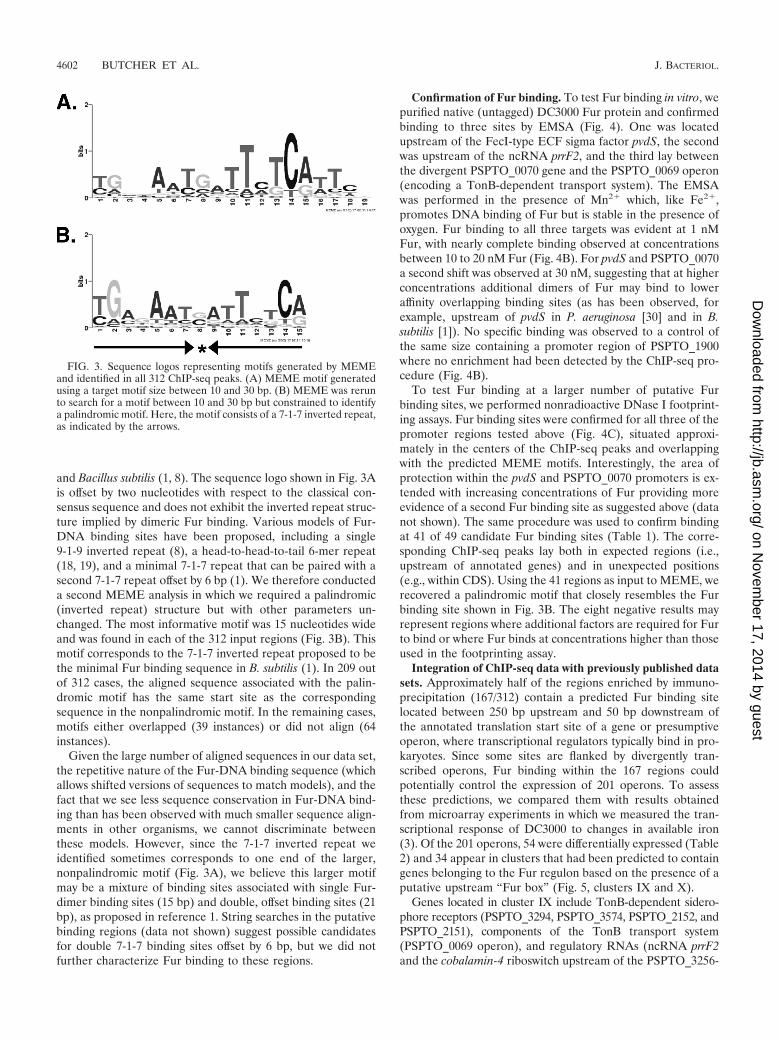
and Bacillus subtilis (1, 8). The sequence logo shown in Fig. 3Ais offset by two nucleotides with respect to the classical con-sensus sequence and does not exhibit the inverted repeat struc-ture implied by dimeric Fur binding. Various models of Fur-DNA binding sites have been proposed, including a single9-1-9 inverted repeat (8), a head-to-head-to-tail 6-mer repeat(18, 19), and a minimal 7-1-7 repeat that can be paired with asecond 7-1-7 repeat offset by 6 bp (1). We therefore conducteda second MEME analysis in which we required a palindromic(inverted repeat) structure but with other parameters un-changed. The most informative motif was 15 nucleotides wideand was found in each of the 312 input regions (Fig. 3B). Thismotif corresponds to the 7-1-7 inverted repeat proposed to bethe minimal Fur binding sequence in B. subtilis (1). In 209 outof 312 cases, the aligned sequence associated with the palin-dromic motif has the same start site as the correspondingsequence in the nonpalindromic motif. In the remaining cases,motifs either overlapped (39 instances) or did not align (64instances).
Given the large number of aligned sequences in our data set,the repetitive nature of the Fur-DNA binding sequence (whichallows shifted versions of sequences to match models), and thefact that we see less sequence conservation in Fur-DNA bind-ing than has been observed with much smaller sequence align-ments in other organisms, we cannot discriminate betweenthese models. However, since the 7-1-7 inverted repeat weidentified sometimes corresponds to one end of the larger,nonpalindromic motif (Fig. 3A), we believe this larger motifmay be a mixture of binding sites associated with single Fur-dimer binding sites (15 bp) and double, offset binding sites (21bp), as proposed in reference 1. String searches in the putativebinding regions (data not shown) suggest possible candidatesfor double 7-1-7 binding sites offset by 6 bp, but we did notfurther characterize Fur binding to these regions.
Confirmation of Fur binding. To test Fur binding in vitro, wepurified native (untagged) DC3000 Fur protein and confirmedbinding to three sites by EMSA (Fig. 4). One was locatedupstream of the FecI-type ECF sigma factor pvdS, the secondwas upstream of the ncRNA prrF2, and the third lay betweenthe divergent PSPTO_0070 gene and the PSPTO_0069 operon(encoding a TonB-dependent transport system). The EMSAwas performed in the presence of Mn2� which, like Fe2�,promotes DNA binding of Fur but is stable in the presence ofoxygen. Fur binding to all three targets was evident at 1 nMFur, with nearly complete binding observed at concentrationsbetween 10 to 20 nM Fur (Fig. 4B). For pvdS and PSPTO_0070a second shift was observed at 30 nM, suggesting that at higherconcentrations additional dimers of Fur may bind to loweraffinity overlapping binding sites (as has been observed, forexample, upstream of pvdS in P. aeruginosa [30] and in B.subtilis [1]). No specific binding was observed to a control ofthe same size containing a promoter region of PSPTO_1900where no enrichment had been detected by the ChIP-seq pro-cedure (Fig. 4B).
To test Fur binding at a larger number of putative Furbinding sites, we performed nonradioactive DNase I footprint-ing assays. Fur binding sites were confirmed for all three of thepromoter regions tested above (Fig. 4C), situated approxi-mately in the centers of the ChIP-seq peaks and overlappingwith the predicted MEME motifs. Interestingly, the area ofprotection within the pvdS and PSPTO_0070 promoters is ex-tended with increasing concentrations of Fur providing moreevidence of a second Fur binding site as suggested above (datanot shown). The same procedure was used to confirm bindingat 41 of 49 candidate Fur binding sites (Table 1). The corre-sponding ChIP-seq peaks lay both in expected regions (i.e.,upstream of annotated genes) and in unexpected positions(e.g., within CDS). Using the 41 regions as input to MEME, werecovered a palindromic motif that closely resembles the Furbinding site shown in Fig. 3B. The eight negative results mayrepresent regions where additional factors are required for Furto bind or where Fur binds at concentrations higher than thoseused in the footprinting assay.
Integration of ChIP-seq data with previously published datasets. Approximately half of the regions enriched by immuno-precipitation (167/312) contain a predicted Fur binding sitelocated between 250 bp upstream and 50 bp downstream ofthe annotated translation start site of a gene or presumptiveoperon, where transcriptional regulators typically bind in pro-karyotes. Since some sites are flanked by divergently tran-scribed operons, Fur binding within the 167 regions couldpotentially control the expression of 201 operons. To assessthese predictions, we compared them with results obtainedfrom microarray experiments in which we measured the tran-scriptional response of DC3000 to changes in available iron(3). Of the 201 operons, 54 were differentially expressed (Table2) and 34 appear in clusters that had been predicted to containgenes belonging to the Fur regulon based on the presence of aputative upstream “Fur box” (Fig. 5, clusters IX and X).
Genes located in cluster IX include TonB-dependent sidero-phore receptors (PSPTO_3294, PSPTO_3574, PSPTO_2152, andPSPTO_2151), components of the TonB transport system(PSPTO_0069 operon), and regulatory RNAs (ncRNA prrF2and the cobalamin-4 riboswitch upstream of the PSPTO_3256-
FIG. 3. Sequence logos representing motifs generated by MEMEand identified in all 312 ChIP-seq peaks. (A) MEME motif generatedusing a target motif size between 10 and 30 bp. (B) MEME was rerunto search for a motif between 10 and 30 bp but constrained to identifya palindromic motif. Here, the motif consists of a 7-1-7 inverted repeat,as indicated by the arrows.
4602 BUTCHER ET AL. J. BACTERIOL.
on Novem
ber 17, 2014 by guesthttp://jb.asm
.org/D
ownloaded from

PSPTO_3258 putative iron ABC transport system). Except fortwo genes (PSPTO_4136 and PSPTO_4145, encoding anamino acid ABC transporter periplasmic amino acid-bindingprotein and CapB cold shock protein, respectively), all genes incluster IX are associated with ChIP-seq peaks (Fig. 5).
Cluster X contains genes involved in the production of thesiderophores yersiniabactin and pyoverdine (3). Several genesin this group are regulated by the FecI-type ECF sigma PvdSbut appear to be regulated by Fur as well (as indicated by thepresence of a Fur ChIP-seq peak) (Fig. 5). Also in this groupare two other FecI-type sigma factors (PSPTO_1209 andPSPTO_1286) as well as three TonB-dependent siderophorereceptors (PSPTO_3462, PSPTO_2605, and PSPTO_3692)and a TonB-dependent outer membrane heme receptor(PSPTO_1284). Genes not associated with Fur ChIP-seq peaks
in this cluster are either involved in yersiniabactin productionor regulated by PvdS without direct Fur involvement.
Many (144/201) operons that were associated with Fur bind-ing were not identified as differentially regulated in our mi-croarray experiments (Fig. 2), and 68 were not detectably ex-pressed in our global transcriptome analysis of DC3000 (21) byusing cells grown in iron-limited medium. It is possible thatsome of these are differentially expressed but did not meet thethreshold for statistical significance. Alternatively, other fac-tors not present under these conditions may be required forexpression of these genes even though they are regulated byFur. In fact it has been noted that in P. aeruginosa the growthmedium (rich medium versus minimal medium) has an effecton which groups of genes are most active under high-ironconditions (32). We also know that several of the genes not
FIG. 4. (A) Coomassie-stained SDS-PAGE gel showing DC3000 Fur after purification. (B) EMSA of Fur targets, regions upstream of pvdS andprrF2, and region between PSPTO_0069 and PSPTO_0070. As a negative control, Fur binding to the PSPTO_1900 promoter (not predicted to bebound by Fur) was also assessed. Unbound probes are shown in the first lane and reduced mobility is observed with increasing Fur concentrationsfor all three Fur targets. Competition assays with specific unlabeled probe were performed and resulted in a loss of band shift (see Fig. S1 in thesupplemental material). (C) Fragment analysis from nonradioactive footprinting assays. In each case, the lower panel shows the fragments obtainedafter DNase I digestion of the probe alone, while in the top panel the probes were incubated with 25 nM Fur prior to DNase I digestion. Protectionby Fur can be seen in the boxed regions.
VOL. 193, 2011 Fur REGULON IN PSEUDOMONAS SYRINGAE PV. TOMATO DC3000 4603
on Novem
ber 17, 2014 by guesthttp://jb.asm
.org/D
ownloaded from
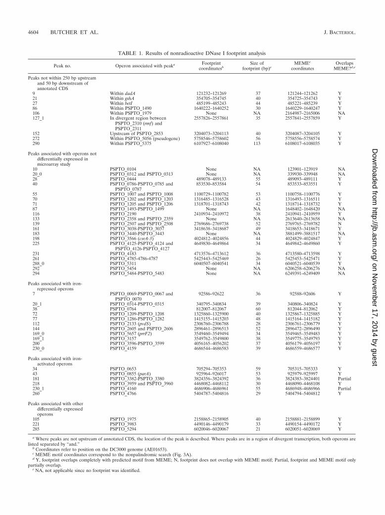
TABLE 1. Results of nonradioactive DNase I footprint analysis
Peak no. Operon associated with peaka Footprintcoordinatesb
Size offootprint (bp)e
MEMEc
coordinatesOverlaps
MEME?d,e
Peaks not within 250 bp upstreamand 50 bp downstream ofannotated CDS
9 Within dadA 121232–121269 37 121244–121262 Y21 Within gshA 354705–354745 40 354725–354743 Y27 Within betI 485199–485243 44 485221–485239 Y86 Within PSPTO_1490 1640222–1640252 30 1640229–1640247 Y106 Within PSPTO_1979 None NA 2164987–2165006 NA127_1 In divergent region between
PSPTO_2310 (rmf) andPSPTO_2311
2557826–2557861 35 2557841–2557859 Y
152 Upstream of PSPTO_2853 3204073–3204113 40 3204087–3204105 Y272 Within PSPTO_5056 (pseudogene) 5758546–5758602 56 5758556–5758574 Y290 Within PSPTO_5375 6107927–6108040 113 6108017–6108035 Y
Peaks associated with operons notdifferentially expressed inmicroarray study
10 PSPTO_0104 None NA 123901–123919 NA20_0 PSPTO_0312 and PSPTO_0313 None NA 339930–339948 NA28 PSPTO_0444 489078–489133 55 489093–489111 Y40 PSPTO_0786-PSPTO_0785 and
PSPTO_0787853530–853584 54 853533–853551 Y
55 PSPTO_1007 and PSPTO_1008 1100729–1100782 53 1100758–1100776 Y70 PSPTO_1202 and PSPTO_1203 1316485–1316528 43 1316493–1316511 Y71 PSPTO_1205 and PSPTO_1206 1318701–1318743 42 1318714–1318732 Y87 PSPTO_1493-PSPTO_1499 None NA 1648402–1648420 NA116 PSPTO_2190 2410934–2410972 38 2410941–2410959 Y133 PSPTO_2358 and PSPTO_2359 None NA 2613640–2613658 NA139 PSPTO_2507 and PSPTO_2508 2769686–2769738 52 2769765–2769782 N161 PSPTO_3038-PSPTO_3037 3418638–3418687 49 3418653–3418671 Y183 PSPTO_3440-PSPTO_3443 None NA 3881499–3881517 NA198 PSPTO_3566 (csrA-3) 4024812–4024856 44 4024829–4024847 Y225 PSPTO_4125-PSPTO_4124 and
PSPTO_4126-PSPTO_41274649830–4649864 34 4649842–4649860 Y
231 PSPTO_4183 4713576–4713612 36 4713580–4713598 Y261 PSPTO_4785-4786-4787 5425443–5425469 26 5425453–5425471 Y288_0 PSPTO_5311 6040507–6040541 34 6040521–6040539 Y292 PSPTO_5454 None NA 6206258–6206276 NA294 PSPTO_5484-PSPTO_5483 None NA 6249391–6249409 NA
Peaks associated with iron-repressed operons
7 PSPTO_0069-PSPTO_0067 andPSPTO_0070
92586–92622 36 92588–92606 Y
20_1 PSPTO_0314-PSPTO_0315 340795–340834 39 340806–340824 Y38 PSPTO_0764 812007–812067 60 812044–812062 Y72 PSPTO_1209-PSPTO_1208 1325860–1325900 40 1325867–1325885 Y77 PSPTO_1286-PSPTO_1282 1415155–1415203 48 1415164–1415182 Y112 PSPTO_2133 (pvdS) 2306760–2306788 28 2306761–2306779 Y144 PSPTO_2605 and PSPTO_2606 2896461–2896513 52 2896472–2896490 Y169_0 PSPTO_5657 (prrF2) 3549460–3549494 34 3549465–3549483 Y169_1 PSPTO_3157 3549762–3549800 38 3549775–3549793 Y200 PSPTO_3596-PSPTO_3599 4056165–4056202 37 4056179–4056197 Y230_0 PSPTO_4159 4686544–4686583 39 4686559–4686577 Y
Peaks associated with iron-activated operons
34 PSPTO_0653 705294–705353 59 705315–705333 Y43 PSPTO_0855 (parA) 925964–926017 53 925979–925997 Y181 PSPTO_3382-PSPTO_3380 3824356–3824392 36 3824383–3824401 Partial218 PSPTO_3959 and PSPTO_3960 4468082–4468112 30 4468090–4468108 Y230_1 PSPTO_4160 4686906–4686961 55 4686948–4686966 Partial260 PSPTO_4766 5404787–5404816 29 5404794–5404812 Y
Peaks associated with otherdifferentially expressedoperons
105 PSPTO_1975 2158865–2158905 40 2158881–2158899 Y221 PSPTO_3983 4490146–4490179 33 4490154–4490172 Y285 PSPTO_5294 6020046–6020067 21 6020051–6020069 Y
a Where peaks are not upstream of annotated CDS, the location of the peak is described. Where peaks are in a region of divergent transcription, both operons arelisted separated by “and.”
b Coordinates refer to position on the DC3000 genome (AE01653).c MEME motif coordinates correspond to the nonpalindromic search (Fig. 3A).d Y, footprint overlaps completely with predicted motif from MEME; N, footprint does not overlap with MEME motif; Partial, footprint and MEME motif only
partially overlap.e NA, not applicable since no footprint was identified.
4604 BUTCHER ET AL. J. BACTERIOL.
on Novem
ber 17, 2014 by guesthttp://jb.asm
.org/D
ownloaded from
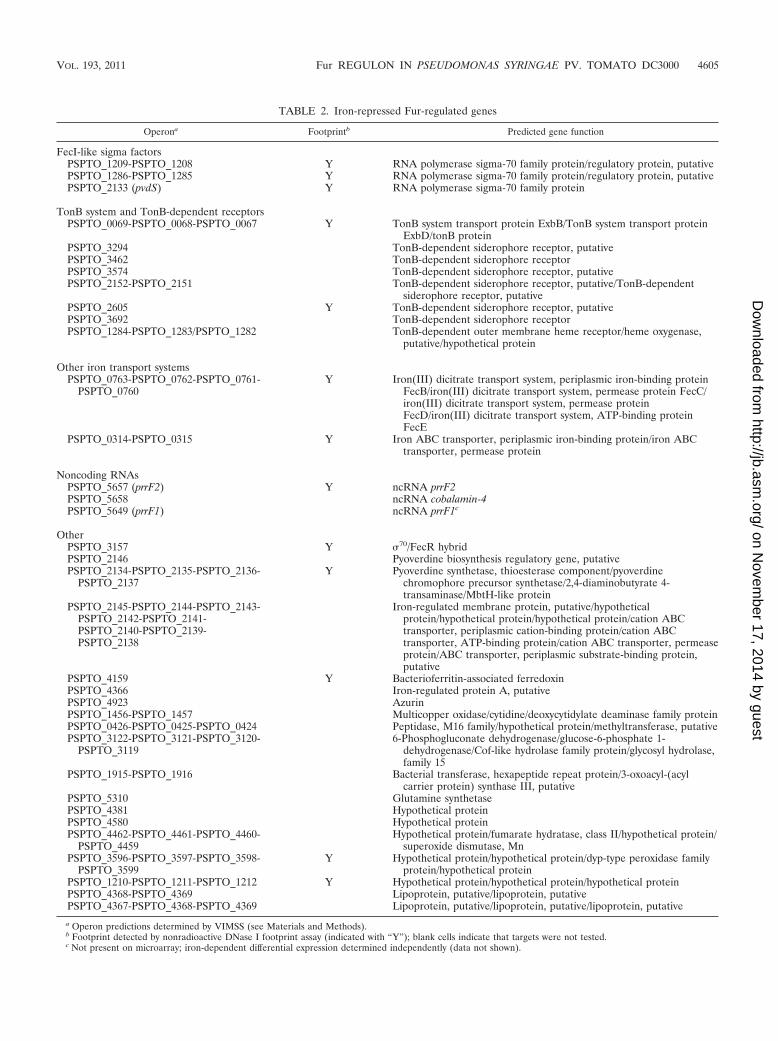
TABLE 2. Iron-repressed Fur-regulated genes
Operona Footprintb Predicted gene function
FecI-like sigma factorsPSPTO_1209-PSPTO_1208 Y RNA polymerase sigma-70 family protein/regulatory protein, putativePSPTO_1286-PSPTO_1285 Y RNA polymerase sigma-70 family protein/regulatory protein, putativePSPTO_2133 (pvdS) Y RNA polymerase sigma-70 family protein
TonB system and TonB-dependent receptorsPSPTO_0069-PSPTO_0068-PSPTO_0067 Y TonB system transport protein ExbB/TonB system transport protein
ExbD/tonB proteinPSPTO_3294 TonB-dependent siderophore receptor, putativePSPTO_3462 TonB-dependent siderophore receptorPSPTO_3574 TonB-dependent siderophore receptor, putativePSPTO_2152-PSPTO_2151 TonB-dependent siderophore receptor, putative/TonB-dependent
siderophore receptor, putativePSPTO_2605 Y TonB-dependent siderophore receptor, putativePSPTO_3692 TonB-dependent siderophore receptorPSPTO_1284-PSPTO_1283/PSPTO_1282 TonB-dependent outer membrane heme receptor/heme oxygenase,
putative/hypothetical protein
Other iron transport systemsPSPTO_0763-PSPTO_0762-PSPTO_0761-
PSPTO_0760Y Iron(III) dicitrate transport system, periplasmic iron-binding protein
FecB/iron(III) dicitrate transport system, permease protein FecC/iron(III) dicitrate transport system, permease proteinFecD/iron(III) dicitrate transport system, ATP-binding proteinFecE
PSPTO_0314-PSPTO_0315 Y Iron ABC transporter, periplasmic iron-binding protein/iron ABCtransporter, permease protein
Noncoding RNAsPSPTO_5657 (prrF2) Y ncRNA prrF2PSPTO_5658 ncRNA cobalamin-4PSPTO_5649 (prrF1) ncRNA prrF1c
OtherPSPTO_3157 Y 70/FecR hybridPSPTO_2146 Pyoverdine biosynthesis regulatory gene, putativePSPTO_2134-PSPTO_2135-PSPTO_2136-
PSPTO_2137Y Pyoverdine synthetase, thioesterase component/pyoverdine
chromophore precursor synthetase/2,4-diaminobutyrate 4-transaminase/MbtH-like protein
PSPTO_2145-PSPTO_2144-PSPTO_2143-PSPTO_2142-PSPTO_2141-PSPTO_2140-PSPTO_2139-PSPTO_2138
Iron-regulated membrane protein, putative/hypotheticalprotein/hypothetical protein/hypothetical protein/cation ABCtransporter, periplasmic cation-binding protein/cation ABCtransporter, ATP-binding protein/cation ABC transporter, permeaseprotein/ABC transporter, periplasmic substrate-binding protein,putative
PSPTO_4159 Y Bacterioferritin-associated ferredoxinPSPTO_4366 Iron-regulated protein A, putativePSPTO_4923 AzurinPSPTO_1456-PSPTO_1457 Multicopper oxidase/cytidine/deoxycytidylate deaminase family proteinPSPTO_0426-PSPTO_0425-PSPTO_0424 Peptidase, M16 family/hypothetical protein/methyltransferase, putativePSPTO_3122-PSPTO_3121-PSPTO_3120-
PSPTO_31196-Phosphogluconate dehydrogenase/glucose-6-phosphate 1-
dehydrogenase/Cof-like hydrolase family protein/glycosyl hydrolase,family 15
PSPTO_1915-PSPTO_1916 Bacterial transferase, hexapeptide repeat protein/3-oxoacyl-(acylcarrier protein) synthase III, putative
PSPTO_5310 Glutamine synthetasePSPTO_4381 Hypothetical proteinPSPTO_4580 Hypothetical proteinPSPTO_4462-PSPTO_4461-PSPTO_4460-
PSPTO_4459Hypothetical protein/fumarate hydratase, class II/hypothetical protein/
superoxide dismutase, MnPSPTO_3596-PSPTO_3597-PSPTO_3598-
PSPTO_3599Y Hypothetical protein/hypothetical protein/dyp-type peroxidase family
protein/hypothetical proteinPSPTO_1210-PSPTO_1211-PSPTO_1212 Y Hypothetical protein/hypothetical protein/hypothetical proteinPSPTO_4368-PSPTO_4369 Lipoprotein, putative/lipoprotein, putativePSPTO_4367-PSPTO_4368-PSPTO_4369 Lipoprotein, putative/lipoprotein, putative/lipoprotein, putative
a Operon predictions determined by VIMSS (see Materials and Methods).b Footprint detected by nonradioactive DNase I footprint assay (indicated with “Y”); blank cells indicate that targets were not tested.c Not present on microarray; iron-dependent differential expression determined independently (data not shown).
VOL. 193, 2011 Fur REGULON IN PSEUDOMONAS SYRINGAE PV. TOMATO DC3000 4605
on Novem
ber 17, 2014 by guesthttp://jb.asm
.org/D
ownloaded from
expressed during growth in our medium are controlled byalternative sigma factors not active under these conditions(unpublished results). For example, expression of the FecI-likesigma factor PSPTO_1203 is induced by the presence of
hyroxamate siderophores not produced by DC3000 (E. Markelet al., submitted for publication). At least one confirmed targetof PSPTO_1203, the TonB-dependent siderophore receptorPSPTO_1206, is also preceded by a Fur binding site but wasnot differentially expressed in our microarray studies. OtherFur-regulated but not differentially expressed genes includethe two remaining FecI-like sigma factors (PSPTO_0444 andPSPTO_1203), five TonB-dependent receptors, two type IIIeffectors, genes involved in alginate biosynthesis, and severalABC transporters, as well as a number of putative regulators(see Table S3 and File S2 in the supplemental material).
Fur-regulated regulators. DC3000 Fur binds upstream of avariety of putative regulators (Table 2; also see Table S3 inthe supplemental material), including a RNA-binding proteinand several ncRNAs and FecI-type sigma factors. CsrA-3(PSPTO_3566) belongs to the RsmA/CrsA family of proteinsthat bind to target RNAs and block interaction of the ribosomewith the ribosome binding site, thereby preventing initiation oftranslation eventually leading to destabilization and degrada-tion of the transcript (2). We confirm that Fur binds upstreamof the prrF1 and prrF2 ncRNAs, which are homologous toFur-regulated ncRNAs in P. aeruginosa (21), and that Fur alsoregulates a third ncRNA, the cobalamin-4 riboswitch locatedupstream of the PSPTO_3256-PSPTO_3258 operon. All fiveFecI-type ECF sigma factors in DC3000 are immediately down-stream of Fur binding sites, but only three (pvdS, PSPTO_1209,and PSPTO_1286) were differentially expressed in our mi-croarray experiments. The two remaining sigma factors may berepressed by additional proteins or require activators that areunavailable under the conditions of our experiments.
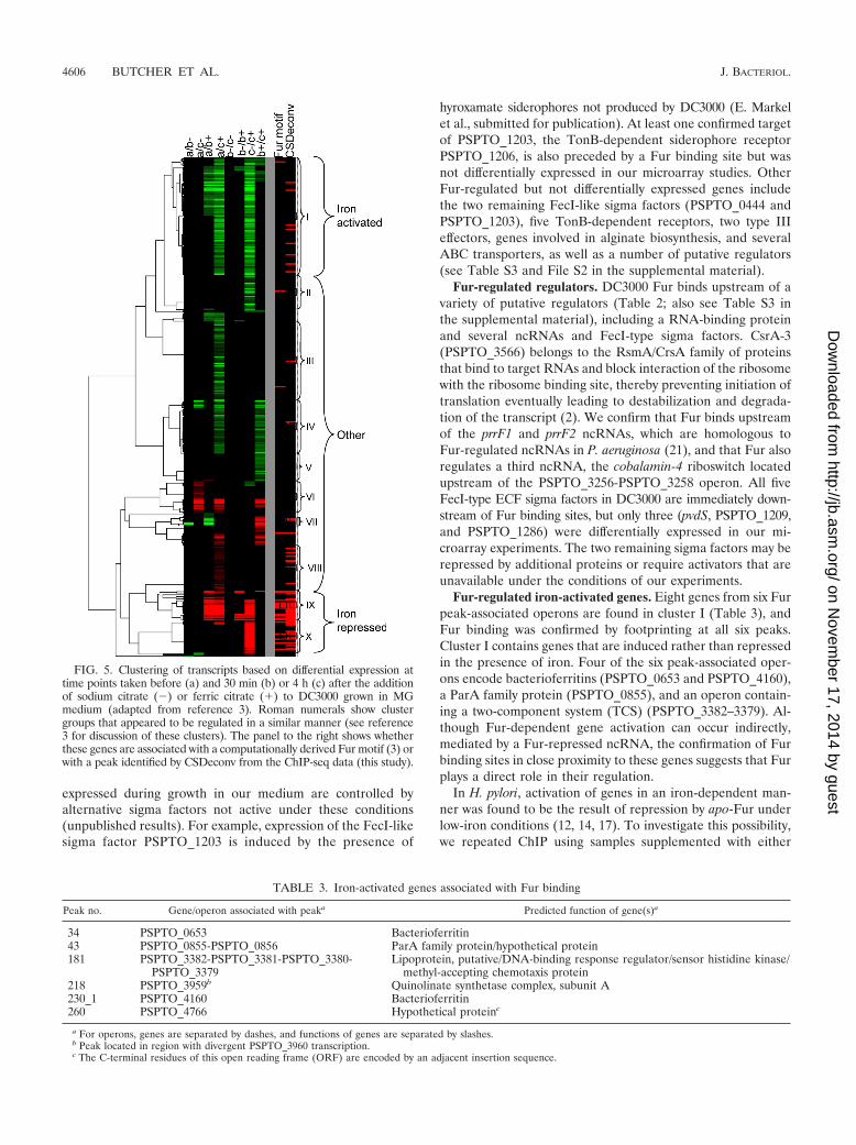
Fur-regulated iron-activated genes. Eight genes from six Furpeak-associated operons are found in cluster I (Table 3), andFur binding was confirmed by footprinting at all six peaks.Cluster I contains genes that are induced rather than repressedin the presence of iron. Four of the six peak-associated oper-ons encode bacterioferritins (PSPTO_0653 and PSPTO_4160),a ParA family protein (PSPTO_0855), and an operon contain-ing a two-component system (TCS) (PSPTO_3382–3379). Al-though Fur-dependent gene activation can occur indirectly,mediated by a Fur-repressed ncRNA, the confirmation of Furbinding sites in close proximity to these genes suggests that Furplays a direct role in their regulation.
In H. pylori, activation of genes in an iron-dependent man-ner was found to be the result of repression by apo-Fur underlow-iron conditions (12, 14, 17). To investigate this possibility,we repeated ChIP using samples supplemented with either
FIG. 5. Clustering of transcripts based on differential expression attime points taken before (a) and 30 min (b) or 4 h (c) after the additionof sodium citrate (�) or ferric citrate (�) to DC3000 grown in MGmedium (adapted from reference 3). Roman numerals show clustergroups that appeared to be regulated in a similar manner (see reference3 for discussion of these clusters). The panel to the right shows whetherthese genes are associated with a computationally derived Fur motif (3) orwith a peak identified by CSDeconv from the ChIP-seq data (this study).
TABLE 3. Iron-activated genes associated with Fur binding
Peak no. Gene/operon associated with peaka Predicted function of gene(s)a
34 PSPTO_0653 Bacterioferritin43 PSPTO_0855-PSPTO_0856 ParA family protein/hypothetical protein181 PSPTO_3382-PSPTO_3381-PSPTO_3380-
PSPTO_3379Lipoprotein, putative/DNA-binding response regulator/sensor histidine kinase/
methyl-accepting chemotaxis protein218 PSPTO_3959b Quinolinate synthetase complex, subunit A230_1 PSPTO_4160 Bacterioferritin260 PSPTO_4766 Hypothetical proteinc
a For operons, genes are separated by dashes, and functions of genes are separated by slashes.b Peak located in region with divergent PSPTO_3960 transcription.c The C-terminal residues of this open reading frame (ORF) are encoded by an adjacent insertion sequence.
4606 BUTCHER ET AL. J. BACTERIOL.
on Novem
ber 17, 2014 by guesthttp://jb.asm
.org/D
ownloaded from
ferric citrate (� Iron) or sodium citrate (� Iron). If apo-Furwere binding to the promoter elements, we would expect to seeenrichment of these regions under low-iron conditions. How-ever, we found that while regions upstream of PSPTO_4160and PSPTO_0653 were enriched in the � Iron sample, therewas little enrichment after ChIP of the � Iron sample (see Fig.7). Since we are unable to make a fur mutant in DC3000, wecannot confirm that apo-Fur is not involved; however, thesedata suggest that iron-bound Fur is binding upstream of thesegenes.
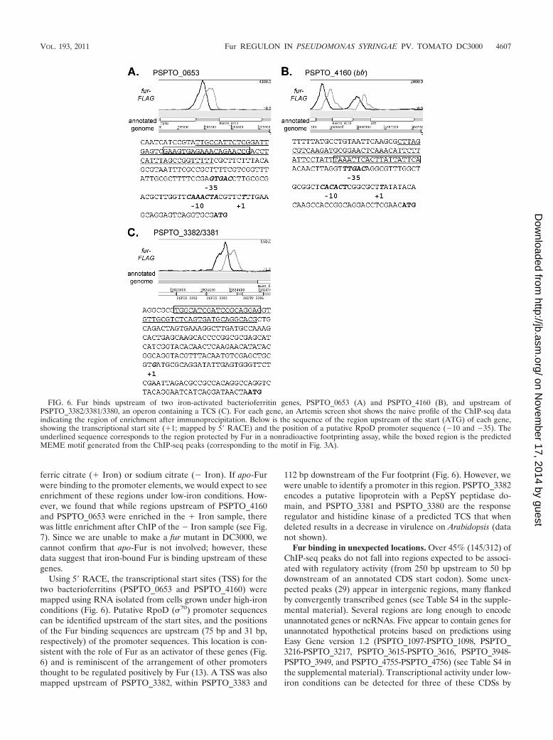
Using 5� RACE, the transcriptional start sites (TSS) for thetwo bacterioferritins (PSPTO_0653 and PSPTO_4160) weremapped using RNA isolated from cells grown under high-ironconditions (Fig. 6). Putative RpoD (70) promoter sequencescan be identified upstream of the start sites, and the positionsof the Fur binding sequences are upstream (75 bp and 31 bp,respectively) of the promoter sequences. This location is con-sistent with the role of Fur as an activator of these genes (Fig.6) and is reminiscent of the arrangement of other promotersthought to be regulated positively by Fur (13). A TSS was alsomapped upstream of PSPTO_3382, within PSPTO_3383 and
112 bp downstream of the Fur footprint (Fig. 6). However, wewere unable to identify a promoter in this region. PSPTO_3382encodes a putative lipoprotein with a PepSY peptidase do-main, and PSPTO_3381 and PSPTO_3380 are the responseregulator and histidine kinase of a predicted TCS that whendeleted results in a decrease in virulence on Arabidopsis (datanot shown).
Fur binding in unexpected locations. Over 45% (145/312) ofChIP-seq peaks do not fall into regions expected to be associ-ated with regulatory activity (from 250 bp upstream to 50 bpdownstream of an annotated CDS start codon). Some unex-pected peaks (29) appear in intergenic regions, many flankedby convergently transcribed genes (see Table S4 in the supple-mental material). Several regions are long enough to encodeunannotated genes or ncRNAs. Five appear to contain genes forunannotated hypothetical proteins based on predictions usingEasy Gene version 1.2 (PSPTO_1097-PSPTO_1098, PSPTO_3216-PSPTO_3217, PSPTO_3615-PSPTO_3616, PSPTO_3948-PSPTO_3949, and PSPTO_4755-PSPTO_4756) (see Table S4 inthe supplemental material). Transcriptional activity under low-iron conditions can be detected for three of these CDSs by
FIG. 6. Fur binds upstream of two iron-activated bacterioferritin genes, PSPTO_0653 (A) and PSPTO_4160 (B), and upstream ofPSPTO_3382/3381/3380, an operon containing a TCS (C). For each gene, an Artemis screen shot shows the naive profile of the ChIP-seq dataindicating the region of enrichment after immunoprecipitation. Below is the sequence of the region upstream of the start (ATG) of each gene,showing the transcriptional start site (�1; mapped by 5� RACE) and the position of a putative RpoD promoter sequence (�10 and �35). Theunderlined sequence corresponds to the region protected by Fur in a nonradioactive footprinting assay, while the boxed region is the predictedMEME motif generated from the ChIP-seq peaks (corresponding to the motif in Fig. 3A).
VOL. 193, 2011 Fur REGULON IN PSEUDOMONAS SYRINGAE PV. TOMATO DC3000 4607
on Novem
ber 17, 2014 by guesthttp://jb.asm
.org/D
ownloaded from

using global transcription profiles (21). Two of the unanno-tated CDSs (located between PSPTO_1097 and PSPTO_1098and PSPTO_3216 and PSPTO_3217) share 87 to 92% identitywith hypothetical proteins from Pseudomonas syringae pv.aesculi but do not appear to be present in the genomes of theother pseudomonads. The large intergenic regions were alsoevaluated to determine if they were associated with known orpredicted ncRNAs, and Fur peaks were found upstream of thencRNAs prrF1 and prrF2. In addition, a peak was found be-tween two divergently transcribed genes (PSPTO_2310 andPSPTO_2311) in a region where a ncRNA (rmf) is predictedupstream of PSPTO_2310 (40). Transcriptional activity hasbeen detected in this region (21) and is consistent with thepresence of the ncRNA.
In four cases, peaks were located adjacent to regions con-taining insertion sequences. It is possible that the insertionshave separated the Fur binding sites from their original neigh-bors. For example, one predicted Fur binding site is locatedupstream of PSPTO_2167, a transposase. The surrounding re-gion (without the transposase) is conserved in the P. syringaestrains 1448a and B728a. It is possible that Fur may haveregulated the PSPTO_2168-2170 operon and that the Tn in-sertion separated the Fur binding site from this set of genes inDC3000.
Finally, in two cases the peaks are located well upstreamof the closest potential regulatory targets, PSPTO_2853 andPSPTO_3365. These regions do not appear to contain possibleopen reading frames (ORFs) or predicted ncRNAs. Compar-ison with the transcriptional profile shows that transcription
begins close to the predicted peak/Fur binding sites, sug-gesting that these genes are preceded by long 5� untrans-lated regions (UTRs). Fur binding was confirmed by DNaseI footprinting upstream of PSPTO_2853 (TonB-dependentreceptor). PSPTO_3365 is the first gene in the nuoA-nuoNgene cluster, encoding subunits of the NADH dehydrogenaseof the aerobic respiratory chain, and in Neisseria, these genesare induced in response to iron (13, 23). In Neisseria meningi-tidis, Fur was shown to bind to and activate expression from apromoter upstream of nuoA (13).
The majority of the Fur binding sites in this group (116;80%) lie within coding sequences. Fur binding was confirmedby footprint analysis in six of these cases (Table 1). To furtherinvestigate intragenic binding sites, we selected eight regions(four of which had been footprinted) and performed qPCR onDNA isolated after ChIP on samples with and without ironadded. We found that in all except one case (PSPTO_1346)enrichment was iron dependent (Fig. 7). In most cases, levelsof enrichment under high-iron conditions were also compara-ble to those of intergenic Fur binding sites. We used EMSA toassess whether Fur bound at these intragenic sites with a rel-ative affinity lower than that at intergenic sites, as has beenshown for one site in N. gonorrhoeae (23). We found that whilehalf of the sites displayed lower binding affinities, binding atthe remaining sites was comparable to that at pvdS, prrF2, andPSPTO_0070 (Fig. 7).
Fur binding at intragenic sites is difficult to interpret in termsof current models for regulation by Fur. One possibility is thatFur binding results in altered transcription patterns in these
FIG. 7. qPCR performed on ChIP samples from cultures grown with iron citrate (� Iron; gray) or sodium citrate (� Iron; white) for 30 min.Enrichment of selected Fur binding regions is calculated relative to DNA sequences within the gap1 gene after immunoprecipitation withFur-FLAG-tagged strains. No enrichment is observed of DNA within the gyrA gene. Shown below are the estimated Fur concentrations (nM) atwhich half shift and complete shift of a radiolabeled probe was observed after EMSA (see Fig. S2 in the supplemental material). For comparison,EMSA of other intergenic probes can be seen in Fig. 4B. Nonspecific shift of a PSPTO_1900 probe was observed only at concentrations of 50 nMFur and higher (Fig. 4B). ND, not done.
4608 BUTCHER ET AL. J. BACTERIOL.
on Novem
ber 17, 2014 by guesthttp://jb.asm
.org/D
ownloaded from
regions in response to iron. To test this hypothesis, quantitativereverse transcription-PCR (qRT-PCR) was performed on tar-geted regions upstream and downstream of the Fur bindingsites within these genes by using RNA isolated from cellsgrown in high- or low-iron conditions. Gene transcription wasdetected under both growth conditions, but we were unable todemonstrate any difference in RNA levels, either by comparingtranscript levels between conditions or by comparing levelsupstream and downstream of the Fur binding sites (data notshown).
DISCUSSION
This study is the first to characterize the Fur regulon inDC3000, an important model organism for bacterium-plantinteractions, and is one of only a handful of studies to applyChIP-seq to bacterial regulators. We have observed Fur bind-ing to genomic targets in vivo in the presence of iron citrateand have integrated this information with data from priorexperiments in which we investigated regulation by iron. Thesynthesis significantly expands our understanding of the Furregulon and highlights the complexity of Fur function.
While the Fur regulon has been well characterized in theclosely related human pathogen P. aeruginosa, where iron issequestered from the pathogen by the mammalian host, thesituation is very different in the P. syringae host environment. P.syringae is likely to experience iron limitation on the surface ofthe leaf, where it is in competition with other epiphytes pro-ducing siderophores with binding affinities higher than thoseproduced by DC3000 (16); however, once DC3000 has enteredthe apoplast it will likely be presented with an environmentricher in iron (22). We expect that the role that Fur plays invirulence will be shown to be different between P. aeruginosaand DC3000. Unfortunately, we have been unable to constructa fur mutant in DC3000 and so are unable to assess the role ofthis protein in virulence. Therefore, this elucidation of theDC3000 Fur regulon provides targets for further study. Forexample, we have identified several additional regulators con-trolled by Fur extending the role of Fur in response to ironlevels in DC3000. In addition, the Fur-regulated iron-activatedgenes such as the PSPTO_3381 and PSPTO_3380 TCSs mayplay an important role in virulence once the pathogen entersthe iron-rich environment of the leaf apoplast.
Fur has been studied in many bacteria, but in most casesglobal regulation is investigated by microarray studies, andoften, additional Fur binding sites are determined computa-tionally. Our previous microarray study identified severalgenes that appeared to be Fur regulated, but in the absence ofa fur mutant we were unable to confirm these predictions. OurChIP-seq results highlight that at least in DC3000 Fur bindingis more extensive than we had suspected and that our previouscomputational predictions missed many bona fide bindingsites. In addition, since the ChIP-seq method detects direct Furbinding, we identified many binding sites upstream of genesthat were not expressed and therefore would not be identifiedin our earlier transcription-based studies, even though cellshad been grown in low-iron conditions (when Fur repression isrelieved).
As expected, the DC3000 Fur regulon (consisting of genesinvolved in iron uptake, storage, and homeostasis) resembles
that identified in other bacteria. Nonetheless, DC3000 offersseveral interesting differences. For example, DC3000 (like P.aeruginosa) has an increased number of TonB-dependent re-ceptors, allowing the bacteria competitive fitness under iron-limited conditions by scavenging an array of siderophores pro-duced by other microorganisms (10, 25). However, unlike P.aeruginosa where most (28/32) of the receptors are indirectlyregulated by Fur via FecI-type ECF sigma factors or otherregulators (9), in DC3000 we find that more than half (14/25)are associated with Fur binding (Table 2; also see Table S3 inthe supplemental material). In addition, we find that many ofthe TonB-dependent transducers are both controlled by Furand expressed via a Fur-regulated FecI-like sigma factor (B.Swingle, unpublished results).
We were also surprised to find 37% of the DC3000 Furbinding sites within annotated CDSs. While Fur binding withinCDSs has been reported before (23), this is the first study toextensively identify intragenic binding sites in vivo. We con-firmed Fur binding (by footprinting and/or EMSA) at 10 ofthese sites and in seven cases binding appears to occur pre-dominately under high-iron conditions (Fig. 7). A recent studyin N. gonorrhea showed that Fur bound within one gene withlower affinity than at a binding site upstream of a second gene,leading to the hypothesis that the binding affinity at intragenicFur binding sites is lower than that at intergenic sites (23).However, in DC3000 the binding affinity of Fur at some intra-genic sites is comparable to that observed for intergenic sites(e.g., betI versus pvdS) (Fig. 7), and thus we see no obviousgeneral differences between the two classes. The role Fur playsat these sites remains puzzling. One obvious model is that Furacts as a repressor and prevents transcription downstream ofthe binding site, resulting in different levels of expression up-stream and downstream of the Fur complex. However, our testof this hypothesis at Fur binding sites in eight genes yieldednegative results. We also explored the possibility that Fur con-trols expression on the noncoding strands of these genes. If so,we would expect to detect antisense transcription under con-ditions when Fur does not bind. Our previous RNA sequencingexperiments, conducted using cells grown in low-iron condi-tions, failed to reveal any convincing examples where expres-sion does not correspond to the annotated genome in theregion of these Fur binding sites. We cannot exclude the pos-sibility that Fur binding at these sites may play a nonregulatoryrole in the cell (e.g., on organization of the bacterial chroma-tin). Further studies are clearly required to determine the roleof Fur in these cases.
Interestingly we also found Fur associated with genes thatare activated in the presence of iron. The Fur-regulated iron-activated genes PSPTO_0653 and PSPTO_4160 encode bac-terioferritins homologous to BfrA and BfrB of P. aeruginosaPAO1. These proteins oxidize Fe2� and store Fe3� to protectthe cell from toxic reactive oxygen species and reserve iron foruse under iron-limited conditions (38). In P. aeruginosa PAO1,expression of bfrB is positively regulated by Fur and Fe2� andis independent of the prrF ncRNAs (41). The authors sug-gested that this positive regulation may be due to directactivation by Fur, an undetected ncRNA, or a Fur-repressedrepressor. Bacterioferritin homologs bfr� and bfr� fromPseudomonas putida KT2440 are also activated by iron in aFur-dependent manner (7) but since no Fur motif was identi-
VOL. 193, 2011 Fur REGULON IN PSEUDOMONAS SYRINGAE PV. TOMATO DC3000 4609
on Novem
ber 17, 2014 by guesthttp://jb.asm
.org/D
ownloaded from

fied, the authors noted that positive regulation by Fur is likelyto be indirect. Here we confirm that at least in DC3000 Furdoes bind upstream of the bacterioferritin genes, despite theabsence of an easily recognizable Fur box consensus sequence(Fig. 6), suggesting that Fur directly regulates expression ofthese genes.
There are three ways in which Fur binding has been shownto result in increased expression under high-iron conditions.First, binding of iron-bound Fur may block binding of a secondrepressor. For example, in E. coli Fur was found to activateftnA expression by blocking histone-like nucleoid-associatedprotein (H-NS)-mediated repression (29). As such, Fur stillacts in the classical manner by binding DNA in the presence ofiron and blocking the action of another protein. Second, bind-ing of iron-bound Fur directly activates expression of thedownstream gene. In N. meningitidis Delany and coworkers(13) showed that the binding of Fur upstream of three genesencoding iron-containing respiratory proteins (pan1, norB, andnuoA) was essential for activation of these genes both in vitroand in vivo. Third, binding of apo-Fur represses expression ofgenes under low-iron conditions. To date, direct evidence forthis type of binding has been demonstrated only in H. pylori,where apo-Fur represses expression of sodB and pfr (14,17). We found that enrichment of the PSPTO_4160 andPSPTO_0653 upstream regions was significantly reduced un-der low-iron conditions, suggesting that apo-Fur binding is notoccurring at these sites. However, further investigation will berequired to determine whether Fur binding activates expres-sion of the DC3000 genes or merely blocks binding of a secondrepressor.
ACKNOWLEDGMENTS
J.D.H. and A.G. were supported by NIH grant GM059323.We thank Desmond Lun for assistance with the use of the
CSDeconv peak calling program.
REFERENCES
1. Baichoo, N., and J. D. Helmann. 2002. Recognition of DNA by Fur: areinterpretation of the Fur box consensus sequence. J. Bacteriol. 184:5826–5832.
2. Brencic, A., et al. 2009. The GacS/GacA signal transduction system ofPseudomonas aeruginosa acts exclusively through its control over thetranscription of the RsmY and RsmZ regulatory small RNAs. Mol. Mi-crobiol. 73:434–445.
3. Bronstein, P. A., et al. 2008. Global transcriptional responses of Pseudomo-nas syringae DC3000 to changes in iron bioavailability in vitro. BMC Micro-biol. 8:209.
4. Buell, C. R., et al. 2003. The complete genome sequence of the Arabidopsisand tomato pathogen Pseudomonas syringae pv. tomato DC3000. Proc. Natl.Acad. Sci. U. S. A. 100:10181–10186.
5. Carpenter, B. M., J. M. Whitmire, and D. S. Merrell. 2009. This is not yourmother’s repressor: the complex role of fur in pathogenesis. Infect. Immun.77:2590–2601.
6. Cha, J. Y., J. S. Lee, J. I. Oh, J. W. Choi, and H. S. Baik. 2008. Functionalanalysis of the role of Fur in the virulence of Pseudomonas syringae pv. tabaci11528: Fur controls expression of genes involved in quorum-sensing.Biochem. Biophys. Res. Commun. 366:281–287.
7. Chen, S., W. F. Bleam, and W. J. Hickey. 2010. Molecular analysis of twobacterioferritin genes, bfr� and bfr�, in the model rhizobacterium Pseudomo-nas putida KT2440. Appl. Environ. Microbiol. 76:5335–5343.
8. Chen, Z., et al. 2007. Discovery of Fur binding site clusters in Escherichia coliby information theory models. Nucleic Acids Res. 35:6762–6777.
9. Cornelis, P. 2010. Iron uptake and metabolism in pseudomonads. Appl.Microbiol. Biotechnol. 86:1637–1645.
10. Cornelis, P., and J. Bodilis. 2009. A survey of TonB-dependent receptors influorescent pseudomonads. Environ. Microbiol. Reports 1:256–262.
11. Cornelis, P., S. Matthijs, and L. Van Oeffelen. 2009. Iron uptake regulationin Pseudomonas aeruginosa. Biometals 22:15–22.
12. Delany, I., A. B. Pacheco, G. Spohn, R. Rappuoli, and V. Scarlato. 2001.
Iron-dependent transcription of the frpB gene of Helicobacter pylori is con-trolled by the Fur repressor protein. J. Bacteriol. 183:4932–4937.
13. Delany, I., R. Rappuoli, and V. Scarlato. 2004. Fur functions as an activatorand as a repressor of putative virulence genes in Neisseria meningitidis. Mol.Microbiol. 52:1081–1090.
14. Delany, I., G. Spohn, R. Rappuoli, and V. Scarlato. 2001. The Fur repressorcontrols transcription of iron-activated and -repressed genes in Helicobacterpylori. Mol. Microbiol. 42:1297–1309.
15. De Lorenzo, V., F. Giovannini, M. Herrero, and J. B. Neilands. 1988. Metalion regulation of gene expression: Fur repressor-operator interaction at thepromoter region of the aerobactin system of pCoIV-K30. J. Mol. Biology.203:875–884.
16. Dulla, G. F., K. V. Krasileva, and S. E. Lindow. 2010. Interference of quorumsensing in Pseudomonas syringae by bacterial epiphytes that limit iron avail-ability. Environ. Microbiol. 12:1762–1774.
17. Ernst, F. D., et al. 2005. Iron-responsive regulation of the Helicobacter pyloriiron-cofactored superoxide dismutase SodB is mediated by Fur. J. Bacteriol.187:3687–3692.
18. Escolar, L., J. Perez-Martin, and V. de Lorenzo. 1998. Binding of the fur(ferric uptake regulator) repressor of Escherichia coli to arrays of theGATAAT sequence. J. Mol. Biol. 283:537–547.
19. Escolar, L., J. Perez-Martin, and V. de Lorenzo. 1999. Opening the iron box:transcriptional metalloregulation by the Fur protein. J. Bacteriol. 181:6223–6229.
20. Ferreira, A. O., et al. 2006. Whole-genome expression profiling defines theHrpL regulon of Pseudomonas syringae pv. tomato DC3000, allows de novoreconstruction of the Hrp cis clement, and identifies novel coregulatedgenes. Mol. Plant Microbe Interact. 19:1167–1179.
21. Filiatrault, M. J., et al. 2010. Transcriptome analysis of Pseudomonas syrin-gae identifies new genes, noncoding RNAs, and antisense activity. J. Bacte-riol. 192:2359–2372.
22. Hernandez-Morales, A., et al. 2009. Transcriptional profile of Pseudomonassyringae pv. phaseolicola NPS3121 in response to tissue extracts from asusceptible Phaseolus vulgaris L. cultivar. BMC Microbiol. 9:257.
23. Jackson, L. A., et al. 2010. Transcriptional and functional analysis of theNeisseria gonorrhoeae Fur regulon. J. Bacteriol. 192:77–85.
24. Kahramanoglou, C., et al. 2011. Direct and indirect effects of H-NS and Fison global gene expression control in Escherichia coli. Nucleic Acids Res.39:2073–2091.
25. Koebnik, R. 2005. TonB-dependent trans-envelope signalling: the exceptionor the rule? Trends Microbiol. 13:343–347.
26. Li, R., et al. 2009. SOAP2: an improved ultrafast tool for short read align-ment. Bioinformatics 25:1966–1967.
27. Lun, D. S., A. Sherrid, B. Weiner, D. R. Sherman, and J. E. Galagan. 2009.A blind deconvolution approach to high-resolution mapping of transcriptionfactor binding sites from ChIP-seq data. Genome Biol. 10:R142.
28. Miles, S., B. M. Carpenter, H. Gancz, and D. S. Merrell. 2010. Helicobacterpylori apo-Fur regulation appears unconserved across species. J. Microbiol.48:378–386.
29. Nandal, A., et al. 2010. Induction of the ferritin gene (ftnA) of Escherichiacoli by Fe(2�)-Fur is mediated by reversal of H-NS silencing and is RyhBindependent. Mol. Microbiol. 75:637–657.
30. Ochsner, U. A., A. I. Vasil, and M. L. Vasil. 1995. Role of the ferric uptakeregulator of Pseudomonas aeruginosa in the regulation of siderophores andexotoxin A expression: purification and activity on iron-regulated promoters.J. Bacteriol. 177:7194–7201.
31. Ochsner, U. A., and M. L. Vasil. 1996. Gene repression by the ferric uptakeregulator in Pseudomonas aeruginosa: cycle selection of iron-regulated genes.Proc. Natl. Acad. Sci. U. S. A. 93:4409–4414.
32. Ochsner, U. A., P. J. Wilderman, A. I. Vasil, and M. L. Vasil. 2002.GeneChip expression analysis of the iron starvation response in Pseu-domonas aeruginosa: identification of novel pyoverdine biosynthesisgenes. Mol. Microbiol. 45:1277–1287.
33. Oglesby, A. G., et al. 2008. The influence of iron on Pseudomonas aeruginosaphysiology: a regulatory link between iron and quorum sensing. J. Biol.Chem. 283:15558–15567.
34. Oguiza, J. A., K. Kiil, and D. W. Ussery. 2005. Extracytoplasmic functionsigma factors in Pseudomonas syringae. Trends Microbiol. 13:565–568.
35. Palma, M., S. Worgall, and L. E. Quadri. 2003. Transcriptome analysis ofthe Pseudomonas aeruginosa response to iron. Arch. Microbiol. 180:374–379.
36. Pohl, E., et al. 2003. Architecture of a protein central to iron homeostasis:crystal structure and spectroscopic analysis of the ferric uptake regulator.Mol. Microbiol. 47:903–915.
37. Schafer, A., et al. 1994. Small mobilizable multi-purpose cloning vectorsderived from the Escherichia coli plasmids pK18 and pK19: selection ofdefined deletions in the chromosome of Corynebacterium glutamicum. Gene145:69–73.
38. Smith, J. L. 2004. The physiological role of ferritin-like compounds in bac-teria. Crit. Rev. Microbiol. 30:173–185.
39. Swingle, B., et al. 2008. Characterization of the PvdS-regulated promotermotif in Pseudomonas syringae pv. tomato DC3000 reveals regulon members
4610 BUTCHER ET AL. J. BACTERIOL.
on Novem
ber 17, 2014 by guesthttp://jb.asm
.org/D
ownloaded from

and insights regarding PvdS function in other pseudomonads. Mol. Micro-biol. 68:871–889.
40. Weinberg, Z., et al. 2010. Comparative genomics reveals 104 candidatestructured RNAs from bacteria, archaea, and their metagenomes. GenomeBiol. 11:R31.
41. Wilderman, P. J., et al. 2004. Identification of tandem duplicate regulatory
small RNAs in Pseudomonas aeruginosa involved in iron homeostasis. Proc.Natl. Acad. Sci. U. S. A. 101:9792–9797.
42. Zianni, M., K. Tessanne, M. Merighi, R. Laguna, and F. R. Tabita. 2006.Identification of the DNA bases of a DNase I footprint by the use of dyeprimer sequencing on an automated capillary DNA analysis instrument.J. Biomol. Tech. 17:103–113.
VOL. 193, 2011 Fur REGULON IN PSEUDOMONAS SYRINGAE PV. TOMATO DC3000 4611
on Novem
ber 17, 2014 by guesthttp://jb.asm
.org/D
ownloaded from