US EPA Nanotechnology Grantees Meeting
263
-
Upload
khangminh22 -
Category
Documents
-
view
1 -
download
0
Transcript of US EPA Nanotechnology Grantees Meeting


The Office of Research and Development’s National Center for Environmental Research ii

U.S. EPA Nanotechnology Grantees Meeting
The Office of Research and Development’s National Center for Environmental Research iii
Table of Contents Agenda (presenter’s name is linked to his or her presentation) .................................................. vii Meeting Summary .......................................................................................................... 1 Abstracts and Presentations (listed in the same order as in the Agenda) Day 1, Monday, November 8, 2010 AM Session 1: Systems Approaches An Integrated Approach Toward Understanding the Impact of Aggregation and Dissolution of Metal and Metal Oxide Nanoparticles ............................................................................................................................ 47 Vicki Grassian, University of Iowa
Life Cycle Analysis and Nanostructured Materials .............................................................................................. 52 Thomas Theis, University of Illinois at Chicago Platinum-Containing Nanomaterials: Sources, Speciation, and Transformation in the Environment ................. 57 Martin Shafer, University of Wisconsin-Madison Role of NLRP3 Inflammasome and Nickel in Multi-Walled Carbon Nanotube-Induced Lung Injury ................ 64 Andrij Holian, The University of Montana AM Session 2: Effects of Nanoparticle Surface Properties Microbial Bioavailability of Polyethylene Oxide Grafted to Engineered Nanomaterials ..................................... 67 Gregory Lowry, Carnegie Mellon University Surface Oxides: Their Influence on Multi-Walled Nanotubes Colloidal, Sorption, and Transport Properties .............................................................................................................................................................. 72 Howard Fairbrother, Johns Hopkins University Development of Hyphenated and “Particle Counting” ICP-MS Methods Exposure Assessment of Inorganic Nanoparticles .................................................................................................................................... 79 James Ranville, Colorado School of Mines Controlled Release of Biologically Active Silver From Nanosilver Surfaces ...................................................... 85 Jingyu Liu, Brown University Effects of Polyethyleneimine Surface Modifications of Multi-Walled Carbon Nanotubes: Their Toxicity, Sorption Behaviors, and Ecological Uptake by Earthworms and Daphnia Magna .............................. 89 Roger Pinto, University of Michigan, Ann Arbor PM Session 1: Characterization Methods A Biological Surface Adsorption Index for Characterizing Nanomaterials in Aquatic Environments and Their Correlation With Skin Absorption of Nanomaterials ........................................................................... 92 Xin-Rui Xia, North Carolina State University

U.S. EPA Nanotechnology Grantees Meeting
The Office of Research and Development’s National Center for Environmental Research iv
Flexible Nanostructured Conducting Poly(amic) Acid Membrane Captures, Isolates, and Simultaneously Detects Engineered Nanoparticles ........................................................................................................................ 97 Wunmi Sadik, State University of New York at Binghamton Fate and Effects of Nanosized Metal Particles Examined Along a Simulated Terrestrial Food Chain Using Genomic and Microspectroscopic Techniques ........................................................................................... 99 Jason Unrine, University of Kentucky Determination of Manufactured Nanoparticle Toxicity Using Novel Rapid Screening Methods ........................ 105 John J. Rowe, Maqusood Ahamed, Ryan Posgai, Yiling Hong, Jayne Robinson, and Mark Nielsen PM Session 2: Environmental Effects on Nanoparticles Influence of Natural Organic Matter on the Behavior and Bioavailability of Carbon Nanoparticles in Aquatic Ecosystems .......................................................................................................................................... 112 Stephen Klaine, Clemson University Environmental Photochemical Reactions of nC60 and Functionalized Single-Walled Carbon Nanotubes in Aqueous Suspensions ....................................................................................................................................... 117 Chad Jafvert, Purdue University Impact of Photochemical Oxidation on the Stability of nC60 and Multi-Walled Carbon Nanotubes in Aqueous Solutions ............................................................................................................................................ 126 Qilin Li, Rice University The Environmental Behaviors of Multi-Walled Carbon Nanotubes in Aquatic Systems ..................................... 132 Quingguo Huang, University of Georgia Day 2, Tuesday, November 9, 2010 AM Session 1: Effects on Cells Functional Effects of Nanoparticle Exposure on Airway Epithelial Cells ............................................................ 138 Amiraj Banga, Indiana University-Purdue University at Indianapolis Toxicity Assessment of Nanomaterials in Alveolar Epithelial Cells at the Air-Liquid Interface ......................... 144 Galya Orr, Pacific Northwest National Laboratory Interactions of Nanomaterials With Model Cell Membranes ............................................................................... 146 Jonathan Posner, Arizona State University Development of an In Vitro Test and a Prototype Model To Predict Cellular Penetration of Nanoparticles ....... 154 Yongsheng Chen, Georgia Institute of Technology AM Session 2: Effects at Sub-Cellular Level Impacts of Quantum Dots on Gene Expression in Pseudomonas aeruginosa ...................................................... 161 Shaily Mahendra, University of California, Los Angeles

U.S. EPA Nanotechnology Grantees Meeting
The Office of Research and Development’s National Center for Environmental Research v
Thiol Redox-Dependent Toxicity and Inflammation Caused by TOPO-PMAT Modified Quantum Dots .......... 164 Terrence Kavanagh, University of Washington Bioavailability and Fates of CdSe and TiO2 Nanoparticles in Eukaryotes and Bacteria ...................................... 168 Patricia Holden, University of California, Santa Barbara Using Zebrafish Embryos To Test Phototoxicity of TiO2 Nanoparticles ............................................................. 174 Warren Heideman, University of Wisconsin-Madison PM Session 1: Effects on Fish and Oysters Effects of Subchronic Exposure to Nanoparticulate Silver in Zebrafish .............................................................. 180 David Barber, University of Florida Refinements to the Use of Zebrafish for Nanomaterial-Biological Interaction Assessments ............................... 185 Robert Tanguay, Oregon State University Impacts of Functionalization of Fullerenes and Carbon Nanotubes on the Immune Response of Rainbow Trout .................................................................................................................................................. 192 Devrah Arndt, University of Wisconsin-Milwaukee Characterization of the Potential Toxicity of Metal Nanoparticles in Marine Ecosystems Using Oysters – Silver Nanoparticle Studies With Adults and Embryos ........................................................................ 198 Amy Ringwood, University of North Carolina-Charlotte PM Session 2: Nanoparticles and Waste Treatment Bioavailability of Metallic Nanoparticles and Heavy Metals in Landfills ............................................................ 206 Zhiqiang Hu, University of Missouri Biological Fate and Electron Microscopy Detection of Nanoparticles During Wastewater Treatment ............... 212 Paul Westerhoff, Arizona State University Analysis and Fate of Single-Walled Carbon Nanotubes and Their Manufacturing Byproducts in Estuarine Sediments and Benthic Organisms ................................................................................................... 217 P. Lee Ferguson, Duke University Safety/Toxicity Assessment of Ceria (A Model Engineered NP) to the Brain ..................................................... 225 Robert Yokel, University of Kentucky Handouts on Centers for Environmental Implications of Nanotechnology (CEIN) University of California ........................................................................................................................................233 Duke University ....................................................................................................................................................235 Speaker List ................................................................................................................... 237 Participants List ............................................................................................................ 241 Links to Information on Federal Agency Nanotechnology Programs ...................... 251 E-announcement Flyer .................................................................................................. 252

mosvald
Text Box
vi

vii
(Session and presentation times in this agenda are the same as in the SETAC agenda)
Meeting Contacts: [email protected] and [email protected] Registration Contact: [email protected]
DAY 1, Monday, November 8, 2010 7:30 – 7:45 a.m. Registration (Rooms D135 and D136) 7:45 – 8:00 a.m. Welcome and Ground Rules 8:00 – 9:35 a.m. AM Session 1: Systems Approaches
8:00 – 8:20 a.m. An Integrated Approach Toward Understanding the Impact of Aggregation and Dissolution of Metal and Metal Oxide Nanoparticles Vicki Grassian, University of Iowa
8:25 – 8:45 a.m. Life Cycle Analysis and Nanostructured Materials
Thomas Theis, University of Illinois at Chicago
8:50 – 9:10 a.m. Platinum-Containing Nanomaterials: Sources, Speciation, and Transformation in the Environment
Martin Shafer, University of Wisconsin-Madison
9:15 – 9:35 a.m. Role of NLRP3 Inflammasome and Nickel in Multi-Walled Carbon Nanotube-Induced Lung Injury Andrij Holian, The University of Montana
9:35 – 10:15 a.m. BREAK 10:15 – 11:50 a.m. AM Session 2: Effects of Nanoparticle Surface Properties
10:15 – 10:35 a.m. Microbial Bioavailability of Polyethylene Oxide Grafted to Engineered Nanomaterials
Gregory Lowry, Carnegie Mellon University 10:40 – 11:00 a.m. Surface Oxides: Their Influence on Multi-Walled Nanotubes Colloidal,
Sorption, and Transport Properties Howard Fairbrother, Johns Hopkins University
mosvald
Text Box
vii
viii
DAY 1, Monday, November 8, 2010 (Continued)
11:05 – 11:25 a.m. Development of Hyphenated and “Particle Counting” ICP-MS
Methods Exposure Assessment of Inorganic Nanoparticles James Ranville, Colorado School of Mines
11:30 – 11:50 a.m. Controlled Release of Biologically Active Silver From Nanosilver
Surfaces Jingyu Liu, Brown University
Note: Additional Presentation that could not be presented at the meeting:
Effects of Polyethyleneimine Surface Modifications of Multi-Walled Carbon Nanotubes: Their Toxicity, Sorption Behaviors, and Ecological Uptake by Earthworms and Daphnia Magna Roger Pinto, University of Michigan, Ann Arbor
11:50 a.m. – 1:45 p.m. LUNCH 1:45 – 3:30 p.m. PM Session 1: Characterization Methods
1:45 – 2:15 p.m. A Biological Surface Adsorption Index for Characterizing Nanomaterials in Aquatic Environments and Their Correlation With Skin Absorption of Nanomaterials Xin-Rui Xia, North Carolina State University
2:20 – 2:40 p.m. Flexible Nanostructured Conducting Poly(amic) Acid Membrane
Captures, Isolates, and Simultaneously Detects Engineered Nanoparticles Wunmi Sadik, State University of New York at Binghamton
2:45 – 3:05 p.m. Fate and Effects of Nanosized Metal Particles Examined Along a
Simulated Terrestrial Food Chain Using Genomic and Microspectroscopic Techniques
Jason Unrine, University of Kentucky
3:10 – 3:30 p.m. Determination of Manufactured Nanoparticle Toxicity Using Novel Rapid Screening Methods
John Rowe, University of Dayton 3:30 – 4:10 p.m. BREAK 4:10 – 5:45 p.m. PM Session 2: Environmental Effects on Nanoparticles
4:10 – 4:30 p.m. Influence of Natural Organic Matter on the Behavior and Bioavailability of Carbon Nanoparticles in Aquatic Ecosystems
Stephen Klaine, Clemson University
4:35 – 4:55 p.m. Environmental Photochemical Reactions of nC60 and Functionalized Single-Walled Carbon Nanotubes in Aqueous Suspensions
Chad Jafvert, Purdue University
mosvald
Text Box
viii

ix
DAY 1, Monday, November 8, 2010 (Continued) 5:00 – 5:20 p.m. Impact of Photochemical Oxidation on the Stability of nC60 and Multi-
Walled Carbon Nanotubes in Aqueous Solutions Qilin Li, Rice University
5:25 – 5:45 p.m. The Environmental Behaviors of Multi-Walled Carbon Nanotubes in
Aquatic Systems Quingguo Huang, University of Georgia
5:45 – 6:30 p.m. Open Discussion
6:30 p.m. Adjournment DAY 2, Tuesday, November 9, 2010 7:30 – 7:45 a.m. Registration (Rooms D135 and D136) 7:45 – 8:00 a.m. Review of Monday and Plans/Ground Rules for Today 8:00 – 9:35 a.m. AM Session 1: Effects on Cells
8:00 – 8:20 a.m. Functional Effects of Nanoparticle Exposure on Airway Epithelial Cells Amiraj Banga, Indiana University-Purdue University at Indianapolis
8:25 – 8:45 a.m. Toxicity Assessment of Nanomaterials in Alveolar Epithelial Cells at the Air-Liquid Interface
Galya Orr, Pacific Northwest National Laboratory
8:50 – 9:10 a.m. Interactions of Nanomaterials With Model Cell Membranes Jonathan Posner, Arizona State University
9:15 – 9:35 a.m. Development of an In Vitro Test and a Prototype Model To Predict Cellular Penetration of Nanoparticles
Yongsheng Chen, Georgia Institute of Technology 9:35 – 10:15 a.m. BREAK 10:15 – 11:50 a.m. AM Session 2: Effects at Sub-Cellular Level
10:15 – 10:35 a.m. Impacts of Quantum Dots on Gene Expression in Pseudomonas aeruginosa Shaily Mahendra, University of California, Los Angeles
10:40 – 11:00 a.m. Thiol Redox-Dependent Toxicity and Inflammation Caused by TOPO-
PMAT Modified Quantum Dots Terrence Kavanagh, University of Washington
11:05 – 11:25 a.m. Bioavailability and Fates of CdSe and TiO2 Nanoparticles in
Eukaryotes and Bacteria Patricia Holden, University of California, Santa Barbara
mosvald
Text Box
ix

x
DAY 2, Tuesday, November 9, 2010 (Continued)
11:30 – 11:50 a.m. Using Zebrafish Embryos To Test Phototoxicity of TiO2 Nanoparticles Warren Heideman, University of Wisconsin-Madison
11:50 a.m. – 1:45 p.m. LUNCH 1:45 – 3:30 p.m. PM Session 1: Effects on Fish and Oysters
1:45 – 2:15 p.m. Effects of Subchronic Exposure to Nanoparticulate Silver in Zebrafish David Barber, University of Florida
2:20 – 2:40 p.m. Refinements to the Use of Zebrafish for Nanomaterial-Biological
Interaction Assessments Robert Tanguay, Oregon State University
2:45 – 3:05 p.m. Impacts of Functionalization of Fullerenes and Carbon Nanotubes on
the Immune Response of Rainbow Trout Devrah Arndt, University of Wisconsin-Milwaukee
3:10 – 3:30 p.m. Characterization of the Potential Toxicity of Metal Nanoparticles in Marine Ecosystems Using Oysters – Silver Nanoparticle Studies With Adults and Embryos
Amy Ringwood, University of North Carolina-Charlotte 3:30 – 4:10 p.m. BREAK 4:10 – 5:45 p.m. PM Session 2: Nanoparticles and Waste Treatment
4:10 – 4:30 p.m. Bioavailability of Metallic Nanoparticles and Heavy Metals in Landfills Zhiqiang Hu, University of Missouri
4:35 – 4:55 p.m. Biological Fate and Electron Microscopy Detection of Nanoparticles During Wastewater Treatment
Paul Westerhoff, Arizona State University
5:00 – 5:20 p.m. Analysis and Fate of Single-Walled Carbon Nanotubes and Their Manufacturing Byproducts in Estuarine Sediments and Benthic Organisms
P. Lee Ferguson, Duke University
5:25 – 5:45 p.m. Safety/Toxicity Assessment of Ceria (A Model Engineered NP) to the Brain Robert Yokel, University of Kentucky
5:45 – 6:30 p.m. Open Discussion 6:30 p.m. Adjournment
mosvald
Text Box
x

U.S. EPA Nanotechnology Grantees Meeting
The Office of Research and Development’s National Center for Environmental Research 1
U.S. EPA Nanotechnology Grantees Meeting
Oregon Convention Center Rooms D135 and D136
777 NE Martin Luther King Jr. Boulevard Portland, OR
November 8 – 9, 2010
MEETING SUMMARY
The U.S. Environmental Protection Agency held this meeting in conjunction with the Society of Environmental Toxicology and Chemistry’s (SETAC) North America 31st Annual Meeting: Bridging Science with Communities.

U.S. EPA Nanotechnology Grantees Meeting
The Office of Research and Development’s National Center for Environmental Research 2

U.S. EPA Nanotechnology Grantees Meeting
The Office of Research and Development’s National Center for Environmental Research 3
NOVEMBER 8, 2010
OVERVIEW
The U.S. Environmental Protection Agency (EPA) currently funds research that focuses on what happens to nanoparticles, and what impacts on aquatic organisms the particles have, when they enter water environments. EPA holds an annual meeting at which its nanotechnology grantees present their research. There may also be presentations by researchers who have been funded by other Federal agencies with which EPA co-sponsored a Request for Applications. The purpose of the 2010 meeting was to provide a forum for the researchers to share their findings, problems, solutions, and project plans, and to address issues of common concern. The meeting was held in conjunction with the Society for Environmental Toxicology and Chemistry (SETAC) North America 31st Annual Meeting: Bridging Science with Communities so that EPA researchers could attend the SETAC meeting and people attending the SETAC meeting could attend the EPA meeting. The meetings were coordinated so that the EPA meeting was held at the beginning of the week and the SETAC nanotechnology sessions were held later in the week. As a result, there were 117 attendees from academia, industry, and government at the EPA meeting. For information concerning the SETAC meeting go to: http://portland.setac.org/ The meeting was organized by Paul Shapiro of the EPA Office of Research and Development (ORD) National Center for Environmental Research (NCER). The leader of the NCER nanotechnology research program is Nora Savage. Mitch Lasat and Michael McKittrick are also members of the NCER nanotechnology team. Welcome Paul Shapiro, EPA
Mr. Shapiro called the meeting to order at 7:45 am and welcomed the participants. He introduced Nora Savage, Mitch Lasat, and the contractor support staff. He explained the logistics of the meeting. He emphasized the need to stick to the schedule because it matched the SETAC schedule, which set the length of each presentation at 20 minutes and the time between each presentation at 5 minutes. Mr. Shapiro said that in the past attendees have requested an opportunity to have an open discussion of issues that come up during the presentations. He said that the schedule for this meeting includes an open discussion session at the end of each day. There was an easel at the front of the room to serve as a “parking lot” for attendees to write down topics they would like to discuss during these open sessions. Meeting participants were asked to complete evaluation forms of the sessions each day and to submit them to the meeting staff at the registration table. Mr. Shapiro noted that those presentations for which the presenters give permission will be published on the Web site following the meeting. Dr. Savage explained that the National Nanotechnology Initiative (NNI) is in the process of finalizing its 2010 Strategic Plan; public comment currently is being accepted. She announced that a Gordon Research Conference focused on environmental nanotechnology will be held at the Waterville Valley Resort in New Hampshire from May 29 to June 3, 2011. The conference steering committee is accepting abstract sub-missions. Every accepted oral presentation also will be required to have an accompanying poster presented during the conference. EPA also is working with the Organisation for Economic Co-operation and Development (OECD) on a research strategy to understand fate and transport of nanomaterials to ultimately understand toxicity. The next Nanotechnology Grantees Meeting will be held at Duke University in May 2011 in conjunction with a meeting sponsored by the Duke University Center for the Environmental

U.S. EPA Nanotechnology Grantees Meeting
The Office of Research and Development’s National Center for Environmental Research 4
Implications of NanoTechnology (CEINT) and the University of California, Los Angeles Center for the Environmental Implications of Nanotechnology (commonly known as CEIN). Dr. Savage asked participants who have ideas for future meetings to submit them to her or Mr. Shapiro.
MORNING SESSION 1: SYSTEMS APPROACHES
An Integrated Approach Toward Understanding the Impact of Aggregation and Dissolution of Metal and Metal Oxide Nanoparticles Vicki Grassian, University of Iowa
This project aims at understanding the environmental and health implications of nanotechnology from the perspectives of air, water, and soil. The researchers are interested in the toxicity of nanomaterials and have partnered with other researchers to examine inhalation exposure to nanomaterials. Also of interest are particles with size-dependent properties and quantifying their effects as they relate to toxicity in water, air, or in vivo conditions. Particle dissolution impacts particle size and can impact aggregation by causing deaggregation as the particles within the aggregate dissolve. Particle aggregation impacts size, shape, density, available surface area, and surface chemistry. The researchers have chosen an experimental approach that integrates macroscopic and microscopic measurements and methods to better understand the implications of nanomaterials and are performing toxicity and biological interaction studies. The researchers synthesize or purchase commercial nanomaterial powders and perform bulk and surface characterization of these nanomaterials to determine their fate and transformation in water and aerosol and inhalation toxicity.
Titanium dioxide (TiO2) nanoparticles from nanostructured and amorphous materials are some of the smallest commercially manufactured oxide nanoparticles, and although they are sold at a primary size of 5 nm, characterization shows them to be 4 nm in size. The researchers determined that these nanoparticles aggregate but do not dissolve in water at a temperature of 293 K. Aggregation and sedimentation in aqueous suspensions will depend on nanoparticle-to-nanoparticle interactions. Research also indicates that there is a switch in stability of TiO2 nanoparticle suspensions in the presence of citric acid. Derjaguin, Landau, Verwey, and Overbeek (DLVO) calculations along with zeta potential measurements of the surface charge show that TiO2 nanoparticle suspensions are stable at low pH in the absence of citric acid and at near neutral pH in the presence of citric acid. Surface speciation suggests that pKa values are lower for surface adsorbed citric acid; less adsorption at higher pH is a result of the surface charge becoming more negative with increasing pH. Thus, mobility in the environment of nanoscale TiO2 will depend on surface coatings, coverage, and charge and pH in a complex manner.
The researchers compared the dissolution of nanorods to microrods and found that nanorods showed increased surface density of hydroxyl groups compared to microrods. Nanorods can extensively aggregate under certain conditions and form tight bundles. Nanorods have enhanced dissolution but aggregate more readily than microrods in some conditions, but different chemical behavior is seen in different conditions. Enhanced dissolution on the nanoscale is quenched in the aggregated state; therefore, dissolution depends on aggregation and the aggregation state, and nanoparticle aggregation and dissolution are connected in ways that are not fully understood. When researchers compared the inflammatory response of mice to various metal and metal oxide nanomaterial aggregates, the greatest inflammatory response was found for copper-based nanoparticles, and copper nanoparticles showed a higher propensity for dissolution in simulated biological media. Differences between iron and copper nanoparticles are a result of different chemical reactivity in biological media. Lung tissues show no evidence of copper nanoparticles, suggesting that the nanoparticles dissolve, which may increase the inflammatory response.
The results of environmental fate and transport studies indicate that metals and metal oxides show unique reactivity and physicochemical behavior on the nanoscale, and this behavior will be impacted by aggregation. Surface area and chemistry impact aggregation, and aggregation impacts surface reactivity

U.S. EPA Nanotechnology Grantees Meeting
The Office of Research and Development’s National Center for Environmental Research 5
(e.g., dissolution). Some ongoing environmental fate and transport studies in the laboratory include those of size-dependent dissolution of zinc oxide (ZnO) nanoparticles and nanorods as well as aggregation and dissolution of copper nanoparticles in aqueous media as a function of pH and in the presence of citrate aggregation and dissolution. Inhalation toxicity studies indicate that chemical composition, size, and the ability to undergo dissolution and translocation are important to toxicity in ways that have not been discerned previously. Additional studies on silver, ZnO, and copper nanoparticles currently are underway.
Discussion
Warren Heideman (University of Wisconsin–Madison) asked, in terms of metal toxicity, particularly in the cases of silver and copper, whether the effects of the nanoparticles can be distinguished from those of the carrier ion. Dr. Grassian responded that her laboratory currently is exploring this with follow-up studies.
Bonnie Blazer-Yost (Indiana University–Purdue University Indianapolis) asked how reagents react with mucin in the airway. Dr. Grassian replied that these experiments had not been performed.
Boris Jovanovic (Iowa State University) asked from which company the laboratory ordered the 4 nm TiO2 nanoparticles. Dr. Grassian responded that they had been supplied from Nanostructured and Amorphous Materials, Inc., but laboratories using them should be sure to characterize them because some were up to 10 nm in size.
Qilin Li (Rice University) asked whether the pH adjustment and nanoparticle contact with citric acid were simultaneous and about the reversibility of absorption. Dr. Grassian responded that the researchers set the pH with citric acid and then added the nanoparticles. Then, pH was measured, and pH changes were not seen. In terms of reversibility, this is a good question, and these studies were not performed.
Mr. Shapiro asked what types of products use copper nanoparticles. Dr. Grassian replied that they are used as catalysts in electronics, and they also are beginning to be used for agricultural applications.
Life Cycle Analysis and Nanostructured Materials Thomas Theis, University of Illinois at Chicago
Many of the topics discussed during this presentation were addressed at the National Science Foundation (NSF)/EPA Life Cycle Aspects of Nanoproducts, Nanostructured Materials, and Nanomanufacturing: Problem Definitions, Data Gaps, and Research Needs Workshop, which 60 individuals attended. Life cycle assessment (LCA) is a systems methodology for compiling information on the flow of materials and energy throughout a product chain. LCA evolved from industry needs to understand manufacturing and market behavior and make choices among competing designs, processes, and products. It defines four general sections of the product chain: (1) materials acquisition, (2) manufacturing/fabrication, (3) product use, and (4) downstream disposition of the product. LCA is standardized by ISO 14040 and 14044 in a framework whose four steps (goal and scope definition, inventory analysis, impact assessment, and interpretation) can be described as “improvement analysis” and whose outcomes are expressed in common units to allow a comparative systems tool.
EPA’s LCA includes potentials for exposure to workers and consumers and disposal exposures. The Agency imposes a risk assessment paradigm on LCA, which is difficult to accomplish. This adaptation of LCA is a method by which to gather information on waste production, energy demand, and the potential for risk to exposed populations. It works best when risks are nonlocal and the population is nonspecific. It is not a substitute for regulatory risk assessment. The nanomaterial health/materials paradox was discussed at the above-mentioned workshop. Those attributes of nanomaterials that are prized for commercial development and application are the same ones that cause toxic reactions.

U.S. EPA Nanotechnology Grantees Meeting
The Office of Research and Development’s National Center for Environmental Research 6
EPA’s nanotechnology research is a two-pronged approach that focuses on environmental applications and implications. This is a worthy approach for environmental regulation but does not apply to LCA. Elements of an LCA-inspired interdisciplinary research program for nanotechnology include use of less toxic and more available components, focus on structures that are less bioavailable, lowering the life cycle energy of manufacturing, design for recovery of nanocomponents at end- of-life, understanding the social contexts in which nano-based products are used and disposed, and application of LCA methodology to the entire product chain. The topic of nanotechnology LCA is not well published and data are lagging. Additionally, manufacturing of nanomaterials causes a variety of impacts including low process yields, significant energy requirements, use of toxic and organic solvents, and high water consumption.
There also is an energy paradox to nanomaterials: Although nanomaterials are some of the most energy-intensive materials known, they currently represent less than 1 percent of manufacturing costs. The costs are low at this point because these materials are not yet commodities, but energy costs may not remain low as they become commodities. Current estimates of world production of various nanomaterials appear to be close to actual amounts, but carbon nanotubes and quantum dots are difficult to mass produce with their current energy requirements. The potential U.S. energy savings from eight nanotechnology applications is approximately 15 percent, but the stability of nanomaterials in the environment is a challenge that must be overcome. Another challenge is that composite materials are not recycled.
In summary, engineered nanomaterials and products are already in use, not widely understood by consumers, often energy intensive and materially inefficient to make, and often difficult to recover once placed in commerce. They have increasingly complex functionalities and provide high added value, although they often are composed of toxic and/or scarce chemicals or use such chemicals in processing. The comparative benefits and impacts of nanoproducts are unknown, and LCA research and applications for nanomaterials are lagging.
Discussion
Gregory Lowry (Carnegie Mellon University) noted that an issue in regard to risk assessment is developing reasonable and reliable numbers for inputs and sources of nanomaterials into the environment. He asked how the annual production figures for nanomaterials are determined and whether they are reliable. Also, can information on potential product types and their release be distributed? Dr. Theis responded that the figures are an estimate based on patents and the open literature. It is too speculative to release information by potential product types, and potential demand is too difficult to predict.
Dr. Grassian noted that many consumer products state that they are “nano” when they actually are “micro.” Dr. Theis agreed and stated that the accepted definition of nanomaterials is those smaller than 100 nm in size. When reviewing the literature, only those products identified as smaller than 100 nm were included.
Platinum-Containing Nanomaterials: Sources, Speciation, and Transformation in the Environment Martin Shafer, University of Wisconsin–Madison
The work on platinum was motivated by several factors, including the increase in platinum levels in many environmental receptors during the past 40 years as a result of platinum use in automobile exhaust catalysts and industrial catalysts, the toxicity of certain platinum species, and the ability of platinum to transform in environmental matrices. Platinum is likely to continue to be used because of a lack of other suitable substances. The toxicological responses of many metals, including platinum, are determined by the specific chemical and physical speciation in the primary source or environmental receptor. Extant modern methodologies, however, provide little relevant speciation information, and traditional techniques that are speciation capable lack the required sensitivity. The specific objectives of the study are to refine analytical tools for measurement and chemical speciation of platinum in environmentally relevant sources and

U.S. EPA Nanotechnology Grantees Meeting
The Office of Research and Development’s National Center for Environmental Research 7
receptors and integrate source and environmental sampling with advances in platinum analytical speciation tools.
The study examines automobiles, diesel engines, roadside dust and soils, and ambient aerosol from urban centers. Roadway and tunnel dust, which is an excellent integrated receptor from emissions and mobile sources, from Milwaukee, Los Angeles, Atlanta, and Denver was studied. Roadside soils and catalyst materials also were studied, and air sampling was performed adjacent to heavily trafficked roads. The diesel engine dynamometer studies focused on platinum-cerium amended fuel, and the researchers completed a good deal of roadside and ambient aerosol sampling and characterization. Extraction-based and solid-phase speciation and electronic microscopy were used to characterize particulate matter (PM). Physiologically relevant fluids were used for the extraction-based characterization.
The researchers measured the levels of platinum in road dusts and determined that it was from an anthropo-genic source. A small fraction of platinum in road dust from the Los Angeles site was found to be soluble, and it is much more soluble in macrophages. Sampling at the Milwaukee site indicated that there is a significant difference in platinum and palladium aerosol mass-size distributions from week to week. The Milwaukee road dust also showed increased solubility in the macrophage of platinum in roadside aerosol; the levels approached the critical range established by EPA. Researchers also noted a potential dilution effect with cerium. The extractable fraction of speciated water-soluble platinum in diesel PM was approximately 3 percent. Studies showed that, in terms of gasoline vehicle catalyst, the modeled fraction of oxidized platinum is significant. Significant contributions from oxidized platinum species are evident in the spectrum in primary vehicle emissions. Early data suggest that oxide and metal are the two dominant platinum species. Additionally, the laboratory is targeting two documented toxic/allergenic chloroplatinate compounds and their hydrolysis products because only very limited information on the concentrations of chloroplatinates in potential environmental sources and receptors is available and environmental fate and transport data are lacking. The laboratory is developing an isocratic and gradient method to examine the toxic form of platinum and will continue this work; once complete, it will apply the methods to engine PM, road dusts, and airborne PM samples. Researchers also will study various environments to examine the transformation state in different environments.
Discussion
Dr. Lowry asked where the chloroplatinate was found in the samples. Dr. Shafer responded that it has the potential to form during the combustion process, so it sits in road dusts and attaches to surfaces. It is more soluble than oxide species. Dr. Lowry asked whether it was possible to distinguish between adsorbed species and others. Dr. Shafer explained that this was not possible with the tools that the laboratory uses.
Dr. Grassian asked whether different regions had specific chloroplatination profiles. Dr. Shafer replied that the method had not been developed to the point that it could be quantitatively applied to field samples.
Quingguo (Jack) Huang (University of Georgia) asked what “SF” stood for in one of the mentioned methods. Dr. Shafer explained that it meant “sector field.” Dr. Huang asked whether using solids would return original speciation to the particles. Dr. Shafer answered that the laboratory is collecting a large volume of presize-fractionated aerosols so that species can be associated.
Role of NLRP3 Inflammasome and Nickel in Multiwalled Carbon Nanotube-Induced Lung Injury Andrij Holian, The University of Montana
The researchers have focused on determining the central mechanism to explain how engineered nano-materials cause pathology and developing a high throughput in vitro screening tool to separate bioactive from nonbioactive nanomaterials. The alveolar macrophage was chosen as a vehicle for study because it is the front-line defense against inhaled particles and plays a major role in both the innate and adaptive

U.S. EPA Nanotechnology Grantees Meeting
The Office of Research and Development’s National Center for Environmental Research 8
immune responses. The alveolar macrophage is responsible for particle clearance from the lung and contributes to the regulation of the inflammatory response. The research focuses on the NLRP3 inflamma-some, which is present in alveolar macrophages and plays an important role in mediating the inflammatory response to various danger signals, including crystalline particles. The inflammasome is activated by cathepsin B, which signals assembly of the NLRP3 inflammasome and results in active caspase 1, which in turn activates gene transcription of pro-inflammatory cytokines (e.g., interleukin [IL]-1β, IL-18). The laboratory tested 24 different multiwalled carbon nanotubes and evaluated cytotoxicity and the inflammasome in THP-1 cells and alveolar macrophages in mice.
Results indicated that the type of metal, diameter, purity, and length were not important following histopathological analysis 7 days postexposure by two blind scorers. Pathology only correlated with nickel content. At 56 days postexposure, multiwalled carbon nanotubes still were present as were granuloma formations. The 7-day and 56-day pathology data are well-correlated; therefore, the 7-day data can be used to predict the 56-day outcomes. There is significant correlation between nickel and various inflammatory response markers (e.g., IL-1β, IL-18, percent viable cells). The increased correlation with in vivo cell viability compared to in vitro was probably a result of the heterogeneity of the alveolar macrophages versus the cell line. The work has not answered the question of whether there is a relationship between the effect on cell viability and inflammasome activation, which are occurring by separate mechanisms. Additionally, in vitro assays are predictive of pathology. There was an excellent correlation between IL-1β production and percent viable cells with prediction of pathology, indicating that measurements of the inflammasome can be used to predict pathological outcomes. Inflammasome production of IL-1β is critical to the inflam-matory response.
In summary, the NLRP3 inflammasome is important in the bioactivity of engineered nanomaterials, and IL-1β is central to initiating inflammation. Nickel on multiwalled carbon nanotubes appears to be a good predictor of NLRP3 inflammasome activation, and activation of the NLRP3 inflammasome provides a good explanation of in vitro and in vivo observations for both multiwalled carbon nanotubes and TiO2 nanowires. Also, activation of the NLRP3 inflammasome, which can utilize alveolar macrophages or THP-1 cells, is a good predictor of nanoparticle bioactivity. Disruption of lysosomes, which can be caused by bioactive but not nonbioactive engineered nanomaterials, is required for NLRP3 inflammasome activation.
Discussion
In response to a question by Dr. Blazer-Yost, Dr. Holian explained that the test materials were selected because there was a clear difference among them in nickel content but not in size, which minimized the variables; therefore, the main variable tested was nickel content. Dr. Blazer-Yost asked whether the nickel was being taken up with the nanotubes, to which Dr. Holian replied that this was definitely the case. Dr. Blazer-Yost asked about the concentration of nanomaterials in the lungs. Dr. Holian responded that each mouse received 100 μg. Agglomeration, suspension, and singlets are critical determinants in the process, and this is what the next phase of the research will study.
Dr. Jovanovic noted that this is an important field of immunotoxicology that has not been explored enough in the past. He asked whether the researchers had considered additional work with neutrophils, especially considering the recent Nature article indicating that nanoparticles are important inducers of neutrophil interactions at environmentally relevant concentrations. Dr. Holian agreed that neutrophils are first responders and contribute to cleanup, but he did not think that they contribute to chronic inflammation and injury.
Wen Zhang (Georgia Institute of Technology) asked why the researchers chose a 7-day timeframe to observe pathology. Dr. Holian responded that the time was chosen for practical considerations (e.g., expense), and many publications have indicated that multiwalled carbon nanotubes are able to cause

U.S. EPA Nanotechnology Grantees Meeting
The Office of Research and Development’s National Center for Environmental Research 9
distinct pathology within 7 days. Because it would be advantageous to perform shorter experiments, the researchers then determined whether this timeframe was a valid predictor.
Howard Fairbrother (Johns Hopkins University) asked whether the correlation with nickel could have been predicted a priori. Dr. Holian replied that nickel is more reactive and a better catalyst in redox reactions than iron. A 2009 paper indicated that nickel was capable of activating the inflammasome. Dr. Holian’s theory is that nickel is being released by the multiwalled carbon nanotubes, or it has unique bioactive properties and/or catalytic activities. Therefore, the results possibly could have been predicted, but the study provides a deeper understanding. Dr. Fairbrother asked whether toxicological effects of nanotubes are a result of nickel rather than the nanotubes themselves. Dr. Holian thought that contaminants would be an important predictor, and pure nanotubes have less bioactivity. The idea is that nanotubes can interact with lysosomal proteins and cause lysosomal permeability. Dr. Fairbrother noted that the data that Dr. Holian showed indicated that there were nickel subsets that did not correlate with pathology. Dr. Holian responded that the correlation occurs with those multiwalled carbon nanotubes that are composed of at least 2 percent nickel.
MORNING SESSION 2: EFFECTS OF NANOPARTICLE SURFACE PROPERTIES
Microbial Bioavailability of Polyethylene Oxide Grafted to Engineered Nanomaterials Gregory Lowry, Carnegie Mellon University
The goal of the research was to determine the effect of surface coatings on the environmental and microbial fate of nano-iron and iron oxide (FeO) nanoparticles. The specific objectives were to determine the: (1) fate of nanoscale zero valent iron (nZVI) in the environment, (2) effects of nZVI and its coatings on biogeochemistry, and (3) fate of the coatings. To understand nanoparticle fate and transport, it is necessary to understand coating fate; coatings affect aggregation, deposition, and biological interactions. Therefore, the researchers asked whether nanomaterial coatings are bioavailable. Because nanomaterials must be 5 nm or smaller to enter bacteria, the researchers focused on this size.
The researchers placed polystyrene covalently bound with polyethylene glycol (PEG) in water to determine whether microbes could remove the coating in an aqueous environment and demonstrated that PEGs are nontoxic, provide a permanent coating, and do not hydrolyze in water. Next, water from an urban river with PEG degraders was run through enrichment culture to select for these PEG degraders, and species of Novosphingobium, Pseudomonas, and Hydrogenophaga were found. These bacterial species were provided with PEG, and their growth correlates with the addition of PEG. The same analysis was performed with copolymers, and the same growth was seen, which is evidence that bacteria are able to remove PEG from copolymers. Additionally, the researchers determined that microbes induced PEG copolymer aggregation via a change in surface properties.
The researchers concluded that covalently bound PEG on nanoparticles is bioavailable, and microorganisms can change nanoparticle stability, which in turn changes environmental fate and transport. Bioavailability depends on coating attachment and degradability. The next step is to determine what happens to coatings in the environment. The researchers faced several challenges, including the difficulty of tracking coating fate in real environmental samples, recovering engineered nanomaterials from environmental samples, and measuring the process and effects at realistic nanomaterial concentrations.
Discussion
Robert Yokel (University of Kentucky) asked whether similar results were received with citrate coatings. Dr. Lowry replied that the researchers have not performed extensive studies regarding the bioavailability or biodegradation of citrate. Free citrate would expected to be readily biodegradable, but if it is bound, then Dr. Lowry was unsure of its ability to biodegrade.

U.S. EPA Nanotechnology Grantees Meeting
The Office of Research and Development’s National Center for Environmental Research 10
Dr. Heideman noted that the opportunity is present to measure molar amounts of carbon with the microsystem and asked how much of the coating is removed. Dr. Lowry responded that the mass balance on the particles indicated that percent levels are converted to carbon dioxide (CO2), and it clearly is changing the character of the particles. This information has been included in a paper that will be submitted to Nano Letters shortly.
Wunmi Sadik (State University of New York at Binghamton) asked whether the researchers performed structural characterization. Dr. Lowry answered that this was difficult for these particular particles. They are uncharged, so measuring zeta potential does not make sense. Dr. Lowry was unaware of any analytical tools available to answer this question, so to indirectly address this, the laboratory examined the nature of the particles postexposure. The mechanism by which the bacteria are removing the coating is interesting but not fully known at this point. Dr. Sadik suggested that one method might be to look at the nuclear magnetic resonance or mass spectrometry of the solution. Dr. Lowry responded that the laboratory would have to restructure its approach to use these methods because of the concentrations involved.
Dr. Grassian asked about the quantitative aspects of surface chemistry and absorption. Dr. Lowry said that the researchers had performed static light scattering on the particles, and this analysis showed that some of the material was removed and converted to CO2.
Qilin Li (Rice University) asked, because the particles were not taken up by the bacteria, whether enzymes in the extracellular matrix are responsible. Dr. Lowry replied that the next step is to determine the process by which the bacteria are removing the coating.
Elijah Petersen (National Institute of Standards and Technology [NIST]) suggested the use of thermal gravimetric analysis to examine carbon amounts that are released as the nanoparticles are released. Dr. Lowry replied that this method could not be used because of the polystyrene core.
Surface Oxides: Their Influence on Multiwalled Nanotubes’ Colloidal, Sorption, and Transport Properties Howard Fairbrother, Johns Hopkins University
This study focuses on the role that oxygen functional groups play in regulating the properties of multiwalled carbon nanotubes. The laboratory performs physicochemical characterization to develop the functional relationships related to material properties to create models to predict environmentally relevant behavior. Surface analysis is a key component of the research; x-ray photoelectron spectroscopy (XPS) is used to determine surface oxygen concentration because it is the most reliable and convenient method to control the amount of oxygen grafted to the sidewalls. Aggregation properties are examined in a laboratory setting. Surface oxygen may be a predictive metric as stabilization correlates with the amount of surface oxygen. Other properties that the researchers measured were poor metrics for colloidal stability for carbon nanotubes.
The researchers also are interested in studying turbidity, organisms, and natural organic matter to determine the environmental aggregation behavior. To understand complex environmental behaviors, the researchers study colloidal stability and correlate it with adsorption properties to ultimately determine whether surface chemistry of the underlying particle plays a role after natural organic matter adsorption. Surface concentration reduces the adsorption of natural organic matter onto the multiwalled carbon nanotubes’ surface. Results clearly indicate inversion of properties in environmental conditions and that surface chemistry plays a significant role in how the multiwalled carbon nanotubes interact in the environment. The researchers designed a column transport experiment to determine how surface oxygen affects the ability to transport in the environment. Results indicated that as the amount of salt increases, multiwalled carbon nanotubes show decreased transport ability. The researchers used a standard calculation method to determine behavior and also found that pH plays a fairly important role in transport; an increase in pH

U.S. EPA Nanotechnology Grantees Meeting
The Office of Research and Development’s National Center for Environmental Research 11
causes an increased ability of the multiwalled carbon nanotubes to transport. The researchers also determined the optimal conditions under which to obtain reliable and reproducible results.
Future work in the laboratory will focus on the effect of different oxidation on the deposition of surface-oxidized multiwalled carbon nanotubes, the effect of particle sizes on deposition of surface-oxidized MWCNTs, and facilitated transport.
Discussion
Dr. Grassian asked about the morphology of the multiwalled carbon nanotubes in water. Dr. Fairbrother stated that they could be described as floppy rods.
Dr. Li asked Dr. Fairbrother to explain the fact that pulse results were larger than the researchers observed. Dr. Fairbrother said that the confusion might be a result of the order in which he presented his slides, as some of the results were obtained prior to the researchers determining how to consistently reproduce the results. The plan is to return to these experiments now that this is known. Dr. Li asked about the shape of the ethyl concentration profile, which was not typical, and whether it could have been caused because the average was measured. Dr. Fairbrother agreed that this was possible.
A participant asked whether the researchers examined other nanotube-to-natural organic matter ratios besides 10:1. Dr. Fairbrother responded that they studied ratios from zero to 30, and there is a systematic evolution of the particle stability as a function of the amount of natural organic matter.
Hyphenated and “Particle Counting” ICP-MS Methods for the Detection and Characterization of Metal and Metal Oxide Nanoparticles James Ranville, Colorado School of Mines
The research focuses on risk assessment of nanotechnology. There are many factors that can be identified, and the researchers initially focused on effects (e.g., uptake, toxicity). To understand exposure, it is necessary to understand stability, for which aggregation and dissolution are important. Additionally, to study exposure better metrology (e.g., quantitation, detection, characterization) must be developed. The researchers observed the optical properties over time, which may indicate that reactivity may be changing. Questions to be addressed regarding detection and characterization are: How much sensitivity and selectivity are needed? How can methods be applied to complex matrices? What is exposure? Are researchers studying what they think that they are studying?
With respect to nanosilver, material flow indicates that surface waters and sewage treatment plants should be studied, and environmentally relevant concentrations must be assessed at the parts per trillion (ppt) level although toxic effects are seen at the parts per billion and parts per million levels in the laboratory. The standard hypothesis is that inductively coupled plasma (ICP) mass spectrometry (MS) can be used to detect, count, and size individual silver nanoparticles. The approach is to use element-specific “pulse” counting (e.g., real-time single-particle [RTSP]-ICP-MS; time-resolved ICP-MS; single-particle ICP-MS). The researchers chose to examine health food supplements, but these are polydispersed in size, so the laboratory used nanoComposix, which is monodispersed.
Results indicated that silver nanoparticles up to 100 nm in size could be quantitatively detected by ICP-MS. If the particle counting approach is valid, then the number of pulses will increase with increasing silver nanoparticle concentration, the number of pulses will be reduced by filtration or acidification, and the intensity of the pulse will be related to nanoparticle size. The results correlated with this. The time data can be used to determine the difference between dissolved and particulate materials. Disk centrifuge is another method to analyze particle size, and these data are in agreement with the ICP-MS particle counting method.

U.S. EPA Nanotechnology Grantees Meeting
The Office of Research and Development’s National Center for Environmental Research 12
The researchers than performed a proof-of-concept study to quantify silver in wastewater, and the results were comparable to estimates from a previously completed materials flow analysis.
Another focus of the project was to determine whether nanotechnology researchers in general are studying what they expect. The researchers examined the biovailability of the cadmium selenide (CdSe) quantum dot core and whether it is toxic to Daphnia magna to help answer this question. Field-flow fractionation (FFF)-ICP-MS can be used to sort the nanoparticles by size to allow further analysis of the nanoparticles. Tests indicated that the cadmium-to-selenium ratio was not 1:1. The cadmium was associated with the quantum dot but not with the core, possibly because cadmium associated with the polymer coating as a result of poor washing during synthesis. The tests appeared to study the cadmium on the surface rather than in the core, highlighting the fact that good characterization techniques are needed to ensure that researchers indeed are studying what they expect.
In summary, RTSP-ICP-MS: (1) can be used to detect silver nanoparticles at environmentally relevant concentrations (i.e., ppt levels) with high specificity; (2) can distinguish between dissolved and particle silver, which provides the potential for the method’s application in stability and exposure/toxicity laboratory studies; and (3) has limitations in that there is a 40 nm size limit, and it cannot identify nanoparticle type. FFF-ICP-MS can be used to more fully characterize complex nanoparticles and provide information to interpret the results of experiments in which mixtures are used, manufacturing impurities are present, and/or transformation/degradation products are present.
Discussion
Patricia Holden (University of California, Santa Barbara) asked how the method will enable researchers to track mobile particle association. Dr. Ranville answered that the researchers plan to perform experiments to simulate the processes occurring in wastewater at each step. Coupling FFF with particle counting may lead the researchers forward.
Kim Rogers (EPA) stated that crystallography experiments were being performed to determine the association of silver chloride with silver nanoparticles. Dr. Ranville acknowledged the limitations of the current methods and noted that complementary techniques will be performed to obtain more information.
Dr. Huang asked how applicable the method is to other materials. Dr. Ranville replied that it could be used element-by-element to build correlations between silver and other elements.
Controlled Release of Biologically Active Silver From Nanosilver Surfaces Jingyu Liu, Brown University
Silver is a broad-spectrum antibiotic that has relatively low toxicity in humans and is being manufactured in large quantities and incorporated into consumer and medical products. Is it a risk to the environment and human health? It is known to be more toxic to aquatic organisms than any other metal except mercury. It bioaccumulates quickly, and some organisms have a low toxicity threshold to nanosilver. Silver has potential toxic effects on beneficial soil bacteria. An important research question is whether nanosilver interacting in biological and environmental systems is the particle or the ion. Metal ions may coexist in metal-containing nanoparticle suspensions. Silver ion is a known toxicant that binds to thiol groups in enzymes, such as NADH dehydrogenase, which disrupts the bacterial respiratory chain and generates reactive oxygen species (ROS) that can lead to oxidative stress and cell damage. Nanosilver particles themselves may also contribute by binding to or passing through cell membranes and generating ROS through surface reactions. There is some controversy about the role of particle-based mechanisms, but there is broad agreement that silver ion is an important toxicant. Previous work regarding ion release kinetics and particle persistence in aqueous nanosilver clouds indicate that the reaction produces active peroxide

U.S. EPA Nanotechnology Grantees Meeting
The Office of Research and Development’s National Center for Environmental Research 13
intermediates, is inhibited by natural organic matter, and leads to complete particle dissolution in aerobic environments.
The researchers are interested in controlled-release nanosilver and application of the drug delivery paradigm. Two questions that are being considered are: Can ion release rate be systematically increased or decreased? Can nanosilver materials be engineered for optimal ion release? Specific benefits of controlled release nanosilver formulations might include: (1) dose control to achieve desired bactericidal or bacteriostatic effects; (2) dose limitation to avoid eukaryotic toxicity; (3) control of product lifetime, before dissolution and diffusion end antibacterial activity; (4) minimization of environmental release through excess ion production beyond that necessary for product performance; or (5) optimization of release profile for targeted delivery to specific tissue or intracellular targets. The researchers use ultrafiltration and atomic absorption to study particle-ion partitioning in aqueous nanosilver colloids. The results indicate that bulk silver oxidatively resolves but much more slowly than nanosilver. Visual MINTEQ software was used to determine the effects of chloride and thiol. The results showed that biological thiol can drive silver equilibrium in a biological system. Nanosilver causes the gradual release of ionic silver because of its affinity to thiol.
Functionalized nanosilver in the presence of citrate, sodium sulfide, or mercaptoundecanoic acid was studied, and all three methods were found to inhibit ion release from silver nanoparticles. Pre-oxidation shows a distinct two-stage release (i.e., fast then slow). The first stage is a result of the rapid dissolution, and the second is because the remaining metal reacts with dissolved oxygen. Other results indicated that antioxidants can inhibit silver ion release. Different surface treatment methods induce different release rates. The primary release mechanism appears to be oxidative dissolution, which can be inhibited through ROS. Other mechanisms are reversible surface binding, inhibition by insoluble silver sulfide, surface passivation, and pre-oxidation. Future work will focus on the biological and environmental implications of ion release kinetics and control.
Discussion
John Rowe (University of Dayton) asked whether this was tested in vitro or in tissue culture; he asked because ion effects should be differential, with different toxic effects on prokaryotic and eukaryotic cells. Ms. Liu responded that the researchers plan to perform this type of work in the future, but the current focus is on the basic chemistry of ion release. Dr. Rowe commented that this type of work would be important to perform because there may be two different toxic methods depending on whether the organism is prokaryotic or eukaryotic.
AFTERNOON SESSION 1: CHARACTERIZATION METHODS
A Biological Surface Adsorption Index for Characterizing Nanomaterials in Aquatic Environments and Their Correlation With Skin Adsorption of Nanomaterials Xin-Rui Xia, North Carolina State University
Currently, most methods to characterize nanomaterials in aqueous environments measure physical parameters. Surface chemistry and core material compositions are the only measurable chemical information on nanomaterials, but these cannot be used directly for quantitative analyses. The octanol-water partition coefficient has been used widely for predictive model development for small molecules, but it is difficult to use for nanomaterials because most nanomaterials form stable suspensions in water or oil but not both. Efforts have been made to understand the chemical interactions between nanoparticles and biological or environmental components. Researchers have demonstrated that lipophilicity is a significant factor in the nanoparticle adsorption of small chemicals. To date, there is no generally applicable approach to quantitatively measure the molecular interactions of nanoparticles with biological or environmental components, which is crucial information needed to develop a quantitative structure-activity relationship
U.S. EPA Nanotechnology Grantees Meeting
The Office of Research and Development’s National Center for Environmental Research 14
for nanomedicine research and risk assessment and safety evaluation of nanomaterials in occupational and environmental exposures. Many researchers have focused on nanocharacterization of pure nanomaterials in industrial applications, nanoprotein coronas in biological systems, and the nanohumic acid complex in the environment.
The researchers have identified that the adsorption property at the solid-liquid interface is key to understanding the behavior of nanoparticles in aqueous environments. The researchers also have developed a biological surface adsorption index (BSAI) approach to characterize the molecular interaction strengths of nanoparticles with small molecules and macromolecules in biological and environmental systems. The BSAI approach is based on the molecular interaction similarity between nano–small molecule interactions and nano–macromolecule interactions. Forces that govern the chemical and biological behavior of nanoparticles are the Coulomb force, London dispersion, hydrogen bonding, dipolarity/polarizability, and lone-pair elections. Results indicate that nanodescriptors derived from the BSAI approach provide better prediction. The predictive model was cross-validated and determined to be robust.
The BSAI database is the final product of the approach, and it is composed of the five nano-descriptors for each of the nanomaterials. The nanodescriptors are free energy-related quantities quantitatively describing the molecular interaction potentials of the nanomaterials at the nano–water interface. Biological activities are free energy-related quantities; their logarithmic values can be predicted directly via the similar predictive model shown for multiwalled carbon nanotubes. The development of the BSAI approach could open a quantitative avenue toward predictive nanomedicine development, particularly for developing integrated physiologically based pharmacokinetic models and for quantitative risk assessment and safety evaluation of nanomaterials.
The researchers also studied the impact of physicochemical properties on skin absorption of manufactured nanomaterials. Pristine fullerene (C60) in different solvents is used in many industrial and pharmaceutical manufacturing processes; therefore, human exposure to C60 could occur in various solvents. Currently, the impact of solvents on its skin penetration is unknown. The laboratory studied four types of representative industrial solvents. The laboratory developed a novel method to prepare nC60 nanoparticles with a narrow size distribution. nC60 and most of the unprotected nanomaterials have a very narrow window in their colloidal stability, and biological electrolytes will cause their aggregation. The researchers determined that once the nanoparticles aggregate, they cannot get through the skin. Aqueous colloidal nanomaterials with coatings did not penetrate intact skin regardless of particle size. Ion-pairing agents did not promote skin penetration. Skin penetration of C60 was observed in different industrial solvents. Significant solvent effects were observed; toluene and chloroform promote skin penetration of C60, whereas mineral oil does not promote skin penetration. The same results were found when the researchers examined deeper skin layers as well.
The laboratory performed short-term studies, but long-term studies also are needed. Skin absorption into aquatic animals should be studied because of their different skin structure (e.g., amphibian skin is very permeable to small molecules). Additionally, more work is needed to make the BSAI approach a generally useful tool for quantitative correlation and risk assessment of various nanomaterials.
Discussion
Mr. Shapiro asked whether the results could be used to design nanoparticles to have specific impacts on the skin. Dr. Xia answered that tailor-made nanoparticles may be possible in the future.
Dr. Lowry expressed concern about applying an equilibrium system to a system so far from equilibrium. Dr. Xia replied that this is a general question for the field. For example, quantitative structure-activity relationship can be used as a driver, but then the kinetics of the actual model are used. Dr. Lowry still had concerns about applying kinetics in this situation. Dr. Xia said that the approach was to correlate

U.S. EPA Nanotechnology Grantees Meeting
The Office of Research and Development’s National Center for Environmental Research 15
equilibrium parameters. Dr. Lowry asked whether the approach had been applied to macromolecules. Dr. Xia replied that much more work was needed at the current level before moving into macromolecules.
Flexible Nanostructured Conducting Poly(amic) Acid Membrane Captures, Isolates, and Simultaneously Detects Engineered Nanoparticles Wunmi Sadik, State University of New York at Binghamton
Two types of sensors have been defined by an EPA white paper. Category 1 includes sensors that are nanoscale or have nanoscale materials or components, and category 2 includes sensors that are used to measure nanoscale properties. The overall project objective is to develop novel category 2 nanosensors for application in complex environmental matrices. Nanoparticles must be isolated from complex matrices, and there are several current characterization techniques. Environmental matrices require ultrafiltration of free-engineered nanoparticles. The researchers used functional groups on poly(amic) acid (PAA) to isolate nanomaterials. The researchers have studied nanoparticle crosslinking with PAA for years, as well as the chemistry of the materials used for crosslinking. Additionally, ultrafiltration often is used for the separation of suspended solids, colloids, bacteria, and viruses. If the porosity of the membrane is controlled, then the ions and particles that pass through the membrane can be controlled. The researchers used the phase-inverted membrane method to create several types of flexible PAA membranes. Phase-inverted membranes allow control of pore size and are stable to most organic solvents, conductive, and flexible.
The researchers successfully filtered quantum dots directly from aqueous solution with 99 percent efficiency and were able to control porosity. Next, the researchers analyzed commercially available products, including food supplements and beverages. Nanosilver in food supplements can cause permanent bluish-gray discoloration of the skin and eyes; nanosilver can be toxic at a dose of as low as 15 ppm and is 50 percent more toxic than asbestos. PAA coordinates different nanomaterial functionalities and separates nanosilver, TiO2 nanoparticles, and quantum dots. The researchers compared the developed membranes to commercially available membranes and found that the membranes developed by the laboratory show superior performance.
In summary, the laboratory has developed a new class of polymeric materials that exhibit spatio-selection via three-dimensional binding interaction with engineered nanomaterials, control porosity, provide accessibility to the underlying transducer, and enable the removal of major interferences. PAA membranes can be regenerated by exposure to fresh solvents or acid washing, and the laboratory successfully filtered nanosilver and quantum dots directly from commercial products with greater than 99 percent efficiency. Future work will focus on improving the fabrication process and testing other nanoparticle combinations to correct defects of the PAA membrane and functionalize the surface of the PAA membrane to improve selectivity.
Discussion
Dr. Huang asked whether the researchers had differentiated between silver ions and other nanoparticles. Dr. Sadik responded that this had not been examined yet.
Dr. Li asked whether the main method of interaction between nanoparticles and the membrane was size or chemical interactions. Dr. Sadik replied that both size exclusion and selective chemistry were occurring. Dr. Li asked what the advantages of the membrane developed by Dr. Sadik’s laboratory were compared to commercial membranes. Dr. Sadik answered that the ability to control functional groups on the surface of the membrane allowed for selectivity. Commercial membranes only offer physical selectivity. Dr. Li noted that it would be beneficial to create a membrane that allowed for separation of particles of different sizes. Dr. Sadik agreed and stated that the laboratory currently was working on this.

U.S. EPA Nanotechnology Grantees Meeting
The Office of Research and Development’s National Center for Environmental Research 16
In response to a question from Mr. Shapiro, Dr. Sadik explained that the researchers had not considered commercializing the membrane that had been developed.
Fate and Effects of Nanosized Metal Particles Examined Along a Simulated Terrestrial Food Chain Using Genomic and Microspectroscopic Techniques Jason Unrine, University of Kentucky
The researchers are examining the fate, transport, and effects of manufactured nanoparticles in the environment by focusing on uptake of nanoparticles by soil invertebrates, microbes, and plants and their subsequent transfer to higher trophic levels. The worm Eisenia fetida is a semimodel organism that is important to the toxicity testing model; the test medium is natural sandy loam, and gold nanoparticles are used as a probe for particle uptake. Nanoparticles up to 50 nm in size can be absorbed by earthworms. The researchers examined the effect of source on bioavailability and determined that primary particle size alone does not determine uptake in complex media, such as soil. The researchers next hypothesized that nanoparticles are more bioavailable through trophic rather than direct exposure and added frogs to their experimental procedures. Transformation appears to occur during the first few weeks of exposure that affect uptake; therefore, future studies should examine this. Results indicated that there was slow elimination of the gold by the earthworms with no significant decrease of gold particles. Frogs that were exposed to gold via ingestion of earthworms showed much higher levels of gold accumulation than those that were directly exposed through gavage. Therefore, the hypothesis is correct, and persistence has significant implications for the food chain. Although there was no difference in frog growth between the two experimental groups, frogs exposed via earthworms showed greater gold concentrations in kidney, liver, and muscle tissues compared to those directly exposed. One alternative hypothesis is that once particles enter earthworm tissues, they acquire a protein corona and become more bioavailable, and another alternative is that earthworms absorb only the most bioavailable particles from the total population of particles, thus enriching the transferable fraction.
Next, the researchers tested various silver nanoparticles with different coatings in two different media; the sandy loam increased oxidation compared to artificial soil media, and the percentages correlate well with the toxicity seen. Results also indicated transient changes in gene expression, so studies should be performed in a time-result manner to observe changes while the organism is adapting. Studies involving protein carbonyl showed an increased amount of protein carbonyl, which correlates with downregulation of catalase gene expression. Catalase transcription is complex and context dependent. There is a cascade of effects leading to the downregulation of catalase, and what most likely is being observed is accumulation of peroxide, which can accelerate the dissolution of particles; therefore, this could be a self-feeding cycle. Following molecular exploration, the researchers examined integrated orgamismal response to nano-particles. Initial avoidance was seen in soil, but there are intact particles. It may be that dissolution is occurring close to the biological surfaces, but the researchers did rule out that it was the result of changes in microbial community composition.
The researchers concluded that nanoparticles are bioavailable from soil and can be transferred to higher trophic levels, and particle size and redox properties are important for uptake and toxicity. Silver particles cause a variety of adverse effects in earthworms translating from the molecular level to the population level, some at concentrations similar to those expected in sewage sludge. Environmental variables are probably more important than particle variables for silver toxicity.
Discussion
Christian Andersen (EPA) asked whether the differences seen between the two experimental frog groups exposed directly or trophically were an experimental artifact from gavage. Dr. Unrine responded that this was not the case; the doses and their confidence levels are known. Dr. Andersen asked whether the waste

U.S. EPA Nanotechnology Grantees Meeting
The Office of Research and Development’s National Center for Environmental Research 17
products were collected. Dr. Unrine explained that this was not possible because the frogs live in water, and the waste products disperse.
Dr. Heideman asked Dr. Unrine to explain why the removal phase occurred more rapidly than the outflow phase. Dr. Unrine replied that the mass of the worm at different time points needs to be examined, and this study has not been completed yet. Worms can detach part of their body, which could be one possibility, but it is puzzling.
Maria Victoria Peeler (Washington State Department of Ecology) asked whether the researchers had examined sediments. Dr. Unrine answered that this type of work had not been completed, but there are plans to collaborate with laboratories that work with sediments.
In response to a comment from Dr. Grassian, Dr. Unrine explained that the redox potentials listed in one of his slides were for illustration purposes only.
Determination of Manufactured Nanoparticle Toxicity Using Novel Rapid Screening Methods John Rowe, University of Dayton
The focus of this project is to devise biological systems to rapidly assess the potential toxic effects of nanoparticles and correlate in vitro results with in vivo outcomes. The approach is multidomain, using viruses, plants, bacterial assays, mammalian in vitro cells, and Drosophila melanogaster as an in vivo model, and examines the biogeochemical cycle and its effects on plants. D. melanogaster, which has a fast life cycle, is used to study acute toxicity, and studies have moved to examine chronic toxicity. The overall objective of the project is to establish D. melanogaster as an in vivo model system for rapid assessment of nanoparticle toxicity. The current project objective is to study the effects of nanoparticle ingestion on D. melanogaster growth and development.
Nanoparticle behavior is function of size, shape, and surface reactivity, and the researchers compare the effects of different sizes and coating of nanoparticles on D. melanogaster development and reproduction. Polysaccharide-coated silver nanoparticles were used in the experiments that were characterized by transmission electron microscopy (TEM) and dynamic light scattering. Food was treated with uncoated silver 10 nm in size, resulting in a linear effect on survivorship up to 30 μg/ml of silver. Survivors showed a significant increase in pupation time and had a phenotype significantly different than untreated D. melanogaster. The same toxic effects, although less, were seen with coated silver nanoparticles and silver nanoparticles that were 60 nm in size. Nanoparticles have been shown to increase ROS, which may result in oxidative stress, inflammation, and consequent damage to proteins, membranes, and DNA. The researchers tested whether oxidative stress occurs in vivo using the model system and determined the effect of treatment with ascorbic acid, which is a protector against oxidative stress through the antioxidant defense mechanism, a pathway that provides protection against the harmful effects of ROS. Silver nanoparticles induced superoxide dismutase activity, which is part of the antioxidant defense mechanism. Results also indicated that ascorbic acid has protective effects. Additionally, results showed that silver nanoparticles induced oxidative stress, which may be a mechanism of silver nanoparticle toxicity.
In summary, the researchers established an in vivo D. melanogaster model for studying nanoparticle toxicity and demonstrated induction of oxidative stress by silver nanoparticles and the protective effect of ascorbic acid treatment. Future directions will include elucidation of the pathway of oxidative stress involved in the process and evaluation of the efficacy of an array of antioxidants.
Discussion
Paul Westerhoff (Arizona State University) asked whether the researchers had examined the first generation offspring for the presence of nanoparticles. Dr. Rowe responded that other laboratories have

U.S. EPA Nanotechnology Grantees Meeting
The Office of Research and Development’s National Center for Environmental Research 18
demonstrated this, and researchers are studying an inhalation model to determine whether nanoparticles enter the system completely via respiration.
Dr. Rogers asked whether the researchers knew how ROS production occurs in response to silver nanoparticles. Dr. Rowe replied that the activation appears to be direct because it can be reversed with an antioxidant.
Dr. Blazer-Yost asked whether the researchers tested lower concentrations of nanoparticles. Dr. Rowe explained that this would be one of the next steps of the laboratory. The researchers used high concentrations to ensure that an effect was seen before moving to lower concentrations. This is an important question because the ultimate fate of nanoparticles is unknown.
Dr. Lowry asked whether the researchers experimented with silver nitrate in food. Dr. Rower answered that they had not, but it may be worthwhile to do so.
AFTERNOON SESSION 2: ENVIRONMENTAL EFFECTS ON NANOPARTICLES
Influence of Natural Organic Matter on the Behavior and Bioavailability of Carbon Nanoparticles in Aquatic Systems Stephen Klaine, Clemson University
The researchers are examining how water quality parameters (e.g., natural organic material) influence the bioavailability of carbon nanoparticles, and a major goal of the research is to examine food chain uptake. A standard D. magna bioassay was used to measure the toxicity of a variety of carbon nanomaterials. Multiwalled carbon nanotube toxicity did not change as a function of natural organic material. Natural organic material-stabilized C60, C70, and single-walled nanotubes were nontoxic.
The researchers explored whether carbon nanomaterials (multiwalled carbon nanotubes, carbon dots, single-walled carbon nanotubes) are absorbed from the intestinal tract. Results indicated that there was no movement of nanotubes in between the microvilli, and they do not appear to be biochemically toxic but are physically toxic because they clog the intestinal tract. Acidified single-walled carbon nanotubes, however, are found in between and in the microvilli, indicating that they have moved into the organism. Aggregation inside the tissue also was observed. Raman spectroscopy was used to determine where the single-walled carbon nanotubes were located within biological tissues, and results showed that they were within the intestinal tract. Movement outside of the intestinal tract also was seen, and acidified single-walled carbon nanotubes stabilized by natural organic material move farther outside of the tract than other single-walled carbon nanotubes.
Carbon dots are useful for examining where nanomaterials travel after digestion by D. magna. The carbon dots used by the researchers possess a carbon core with a PEG coating and have the same fluorescence as quantum dots. Confocal microscopy was used to observe movement outside of the intestinal tract and showed that there was a buildup around various organs and organ systems outside of the tract.
The researchers observed that multiwalled carbon nanotubes are acutely toxic to D. magna, and this is not a function of natural organic material but appears to be a result of interference with orgamismal food processing. It took 29 hours for D. magna to clear multiwalled carbon nanotubes from the intestinal tract, compared to 30 minutes for clearance of clay. Multiwalled carbon nanotubes are not taken up from the intestinal tract. Carbon dots migrate from the intestinal tract and appear to be associated with organelles. Hydroxyl-functionalized single-walled carbon nanotubes may migrate from the intestinal tract, whereas PEG-coated single-walled carbon nanotubes do not. The next steps are to continue to examine uptake from the intestinal tract using fluorescent-labeled single-walled carbon nanotubes and employing other surface

U.S. EPA Nanotechnology Grantees Meeting
The Office of Research and Development’s National Center for Environmental Research 19
modifications. Food chain studies will use labeled carbon nanoparticles that are known to be bioavailable to determine their movement through the aquatic food chain.
Discussion
Dr. Petersen asked about any impurities present in the original single-walled carbon nanotubes. Dr. Klaine replied that they were pure by the time that they were treated with acid. Dr. Petersen suggested that the researchers use electron energy loss spectroscopy to gain more definitive results.
Dr. Fairbrother asked why the acidified single-walled carbon nanotubes were more likely to move through the organisms. Dr. Klaine answered that he was unsure, but possibly it was because items that are more hydrophilic better associate with the microvilli. Dr. Fairbrother asked if similar results were seen with multiwalled carbon nanotubes. Dr. Klaine answered that multiwalled carbon nanotubes are very stable.
Galya Orr (Pacific Northwest National Laboratory) asked about the zeta potential. Dr. Klaine was unsure whether the laboratory had obtained these data. Dr. Petersen added that disbursement of nanotubes could be the result of bundling and stronger interactions among the single-walled carbon nanotubes, which probably are easier to disperse following acid treatment. Dr. Klaine agreed that this correlated with the data.
Environmental Photochemical Reactions of nC60 and Functionalized Single-Walled Carbon Nanotubes in Aqueous Suspensions Chad Jafvert, Purdue University
Dr. Jafvert described published results of the grant, which is coming to an end. A paper focusing on the photochemical transformation of aqueous C60 clusters in sunlight was the first paper to report on C60 photochemical decay, measured by high-performance liquid chromatography (HPLC), in aqueous media under sunlight. Results indicated that smaller clusters result in faster loss of C60, and the photo-transformation rate is not pH dependent. There is a negligible rate change with humic acids present, and molecular oxygen is required for the process. A paper reporting on the photochemistry of aqueous C60 clusters highlighted that singlet oxygen forms during solar irradiation of nC60, consistent with known reaction mechanisms involving singlet oxygen. The photo-transformation of nC60 is mediated by singlet oxygen, and the rate of singlet oxygen production is auto-catalyzed by nC60 water-soluble products formed during irradiation. The singlet oxygen production rate increases with decreases in the size of nC60. The concentration of singlet oxygen induced by nC60 in sunlight is four- to 65-fold higher than the average concentration typically found in sunlit natural surface waters.
A paper focusing on wavelength dependency and product characterization in terms of the photochemistry of aqueous C60 clusters showed that several laboratory methods indicate that oxidation of C60 occurs in aqueous suspensions of nC60 under sunlight, and C60 photo-transformation and singlet oxygen production occur in visible light. Another paper focused on the photoreactivity of carboxylated single-walled carbon nanotubes in sunlight and ROS production in water. In oxic aqueous solutions under sunlight, carboxylated single-walled carbon nanotube dispersions generate singlet oxygen, superoxide anion, and hydroxyl radicals. Reactions with probe molecules were corroborated, and photo-induced aggregation occurred at a low pH. Another paper highlighted projects focusing on solar light-induced ROS production by single-walled carbon nanotubes in water and the role of surface functionalization. Results indicated that oxic aqueous colloidal dispersions of both types of functionalized nanotubes generated ROS in sunlight, and Type I and Type II photochemical pathways occur by the functionalized single-walled carbon nanotubes in sunlight. It appears that the functionalized single-walled carbon nanotubes can act as the electron donor directly, resulting in a change in their properties, or can shuttle electrons from other electron donors to form ROS.

U.S. EPA Nanotechnology Grantees Meeting
The Office of Research and Development’s National Center for Environmental Research 20
Discussion
Richard Zepp (EPA) asked whether the researchers attempted to generate ROS by nonphotochemical methods to observe how they react with substrate. Dr. Jafvert answered that others have performed this in organic solvents, and it would be worthwhile to attempt.
Dr. Jovanovic asked whether the researchers exposed the C60 species from the Materials and Electro-chemical Research Corporation to sunlight or ultraviolet (UV) light to determine if it generated ROS. Dr. Jafvert said that the compounds being generated could not be quantified. C60 was not used as the starting material in any of the experiments.
Dr. Petersen asked whether multiwalled carbon nanotubes had been tested. Dr. Jafvert replied that this is planned for the future.
Impact of Photochemical Oxidation on the Stability of nC60 and Multiwalled Carbon Nanotubes in Aqueous Solution Qilin Li, Rice University
The main objectives of the study are to understand the changes in physicochemical properties of carbon-based nanomaterials, specifically C60 and carbon nanotubes, in natural aquatic systems as a result of interactions with NOM and sunlight and determine the subsequent changes in their aggregation and deposition behaviors. To simulate the particle structures that may form when C60 and multiwalled carbon nanotubes are released into the natural aqueous environment, the researchers prepared the nanoparticle suspensions using a direct sonication method without using any organic solvent. Carboxylated multiwalled carbon nanotubes were used for easier dispersal; C60 and the carboxylated multiwalled carbon nanotubes were well dispersed in water. Sunlight irradiation was simulated with a photoreactor equipped with UV lamps, and samples taken at various times of irradiation were characterized for their physicochemical properties.
Results indicated that the outer surface layer of nC60 particles was oxidized following 7 days of irradiation. When the aggregation of these surface-oxidized nC60 particles was examined, it was found that they were significantly more stable than the pristine nC60 particles, as demonstrated by the reduced aggregation rate. Comparison of stability curves shows that the surface oxidation caused by irradiation increased the critical coagulation concentration by more than fivefold. Additionally, irradiated nC60 responds to humic acid differently from the pristine nC60, showing no change in particle stability, and shows differences in calcium chloride (CaCl2) as well. The steric hindrance effect of humic acid in CaCl2, however, did not seem to be affected by UVA irradiation. An adsorption experiment confirmed that this was a result of significant humic acid adsorption on the irradiated nC60 surface, aided by calcium.
A similar study used carboxylated multiwalled carbon nanotubes. Contrary to the nC60 results, irradiation reduced multiwalled carbon nanotube stability, and the surface negative charge decreased after irradiation, suggesting changes in surface chemistry. Carboxylated multiwalled carbon nanotubes also appear to lose surface hydroxyl and/or carboxyl groups following irradiation. In CaCl2 solutions, however, the stability before and after irradiation was very similar. Multiwalled carbon nanotubes are unstable in the presence of calcium, so it is important to remember that these nanotubes most likely will aggregate and settle quickly in most natural aquatic systems.
In conclusion, sunlight irradiation and humic acid sorption mediate nC60 and carboxylated multiwalled carbon nanotubes aggregation, and specific and nonspecific interactions are involved. Nanocarbon surface chemistry plays a key role in its environmental fate and transport. Ongoing and future work in the laboratory focuses on the impact of irradiation and natural organic material on sorption/deposition and
U.S. EPA Nanotechnology Grantees Meeting
The Office of Research and Development’s National Center for Environmental Research 21
transport in a subsurface porous medium as well as on the impact of irradiation and natural organic material on bioavailability and bioaccumulation of nanoparticles.
Discussion
P. Lee Ferguson (Duke University) asked whether the researchers had plans to examine truly pristine nanotubes. Dr. Li responded that the laboratory is interested in this, but original attempts with pristine nanotubes did not allow for high enough concentrations.
Dr. Fairbrother noted that the techniques used were meant for solids rather than powders, and the method may need to be modified. Dr. Li acknowledged the limitations of the technique and explained that to counteract this the laboratory analyzed the XPS spectrum. Dr. Fairbrother cautioned that there still might be issues, and the two researchers agreed to discuss the specifics later.
The Environmental Behaviors of Multi-Walled Carbon Nanotubes in Aquatic Systems Quingguo Huang, University of Georgia
The objective of the research project is to examine solubilized carbon nanotubes currently under development for a variety of applications. Their mobility and exposure also are being examined. The project focuses on sorption, transformation, bioaccumulation, and trophic transfer. Because sediments affect dispersed carbon nanotubes and dissolved organic matter, it is necessary to design experiments to better understand each of these situations. The three treatments applied were control, peat with dissolved organic matter, and solid peat. Results indicated that in peat, sodium is necessary for sorption in a dose-dependent manner; in shale, there is strong sorption.
Dr. Huang noted that an inner nanotube core may slide, almost without friction, within its outer nanotube shell, thus creating an atomically perfect linear or rotational bearing. Additionally, studies show that C60 can be degraded by microbes via an enzyme. The researchers examined whether white rot fungus, used in bioremediation, could degrade multiwalled carbon nanotubes and found that it could not. Bacterial degradation was evidenced by multiwalled carbon nanotube mineralization, so the researchers attempted to determine the method of degradation using DNA extraction, propagation, isolation, sequencing, and comparison. Three bacteria (Burkholderia, Delftia, and Stenotrophomonas) were identified, all of which are Gram-negative aerobes involved in the degradation of organic contaminants. These field bacteria probably work in concert to degrade. Bacterial degradation has implications for nanotube behavior and sequestration.
The researchers also examined chronic exposures using Ceriodaphnia dubia with the goal of evaluating reproductive toxicity and accumulation of multiwalled carbon nanotubes by adult and neonate C. dubia under two different solubilization protocols. Results indicated that sonication increased toxicity, whereas natural organic material stabilized the nanotubes. Sonicated multiwalled carbon nanotubes adhered to adult organisms and prevented molting and release of neonates. Natural organic material was protective against reproductive toxicity, with no observed adherence to adults. There was significant accumulation of natural organic material-solubilized multiwalled carbon nanotubes in neonates. The next step is a feeding study that will determine whether there is trophic transfer from C. dubia exposed to multiwalled carbon nanotubes following ingestion by Artemia and fathead minnows. Also, full life cycle exposures of multiwalled carbon nanotubes will be evaluated in fathead minnows.
Discussion
Dr. Zepp asked about the strategy used to locate the bacteria. Dr. Huang replied that the bacteria were found attached to samples that were being examined for the white rot fungus. Dr. Zepp asked how the researchers synthesized the labeled materials. Dr. Huang stated that he used a common method that has been described in many papers.

U.S. EPA Nanotechnology Grantees Meeting
The Office of Research and Development’s National Center for Environmental Research 22
A participant asked about the purity of the carbon-labeled material. Dr. Huang responded that amorphous carbon is not seen because it has been removed. The participant asked about the size distribution. Dr. Huang answered that a variety of sizes had been examined. Dr. Petersen added that these sizes were 100 to 420 nm. The participant asked whether the researchers examined distribution following aggregation. Dr. Huang explained that this would be difficult to accomplish using scanning electron microscopy with the mixed system; the researchers will be examining the chemistry, however.
OPEN DISCUSSION
Mr. Shapiro explained that in the past, participants have asked for an open discussion session to be included in the meeting to discuss issues that are introduced throughout the day’s presentations. Four issues were introduced during the Day 1 discussions: (1) How can more be done toward LCA? (2) Is there a substitute for platinum as a catalyst? (3) Should research focus on plumes or far lower concentrations? (4) EPA Administrator Lisa Jackson is interested in examining methods by which to treat groups of drinking water contaminants, including nanoparticles.
Mr. Shapiro asked Dr. Grassian to discuss her concern, which is item 3 above. Dr. Grassian wondered what is the “right” concentration to study. Sometimes, research is conducted at high concentrations that may be relevant to plumes. Dr. Westerhoff agreed that this is an important consideration and noted that it does not make sense to study concentrations that are orders of magnitude lower than those at which an effect is seen. Concentrations studied should be environmentally relevant. Dr. Lowry commented that lower doses may be relevant to chronic toxicity. Dr. Orr agreed that bioaccumulation is important. Dr. Holden added that what constitutes a dose also is an important question. Mr. Shapiro asked Dr. Holden’s opinion on what consti-tutes a dose, and she responded that it depends on the mechanism. Dr. Xia said that it was hard to generalize, and the approach to determining this should include dose response, exposure, and screening. Dr. Petersen noted that there could be a wide range of doses (e.g., plume vs. environmental concentrations). It is necessary to be cautious when interpreting toxicity results, which must be placed in context and compared to other compounds in the environment.
Dr. Savage asked the best method for writing a solicitation that would address chronic toxicity of nanomaterials, which requires a significant amount of time and money. Dr. Lowry stated that this issue cannot be confined to nanotechnology. There is an EPA model that addresses similar issues; therefore, the uncertainty already has been dealt with. Dr. Grassian noted that the National Institutes of Health fund long-term studies (greater than 20 years). Dr. Savage asked whether a reasonable approach would be to focus on fate and transport and then examine chronic toxicity after the fate and transport studies have yielded results. A participant noted that extrapolating from acute toxicity is difficult. Terrence Kavanagh (University of Washington) agreed that this extrapolation was difficult because often the targets of acute and chronic toxicity are completely unrelated (e.g., organophosphate [OP] acute toxicity results in cholinesterase inhibition, whereas OP chronic toxicity results in neurotoxicity).
A participant asked whether carbon nanomaterials, reactive metal nanomaterials, and so forth could each be grouped together for study. Dr. Savage explained that the Request for Applications (RFA) should focus on mixtures because the compound-by-compound approach is not working. Dr. Orr suggested that libraries of data be created that focus on an array of compound modifications to develop the whole picture. Dr. Petersen commented that some trends may be emerging (e.g., carbon nanotubes and physical effects). Classification could be based on how nanoparticles cause toxicity generally to organisms. A participant stated that a good deal of research has focused on the toxicology of chemicals and pharmacology of toxicity. From a pharmacological point of view, receptors are important. Some nanotubes may have properties and interact in ways for which researchers have no foundation; this is an infant science. Dr. Lowry noted that even though this is an infant science, there are 20 years of toxicological PM research, and particle science is not new.

U.S. EPA Nanotechnology Grantees Meeting
The Office of Research and Development’s National Center for Environmental Research 23
Dr. Rogers suggested considering nanotechnology from a purpose or functional standpoint. Nanomaterial designers do not think about toxicity; they focus on making the best material for their purpose. If manufacturers are encouraged or required to consider toxicity, then they will be more receptive to hearing recommendations from the research community. The perception that nanotechnology toxicological research is performed for the benefit of industry so that manufacturers avoid potential disasters should be perpetuated. Dr. Kavanagh agreed that industry would appreciate feedback from nanotechnology researchers.
The participants discussed persistence. A participant noted that even if a compound degrades quickly, chronic exposures are possible if there is continual loading. Dr. Yokel pointed out that ceria nanoparticles can persist for at least 3 months.
In response to a question from Mr. Shapiro, Dr. Lowry explained that there were two different thoughts: to either make nanomaterials safe or minimize exposure. His opinion is that both approaches should be implemented. Dr. Savage noted that it is difficult to design green nanotechnology because often a nanomaterial appears safe, but in the aquatic environment it is not. Dr. Lowry stated that it was necessary to obtain as much information as possible to make informed decisions. A participant noted that some sources can be controlled, but it is difficult for other source streams; there is no one solution.
Dr. Orr commented that positively charged nanoparticles increase toxicity in mammalian cells, so positively charged nanomaterials should be avoided. Research can start building similar “rules.”
Mr. Shapiro asked what the participants thought about performing joint research with industry. Dr. Yokel noted the example of the Health Effects Institute, which was cofounded by EPA and the automobile industry. Any nanoparticle research that relates to combustion could have a funding source in place. Dr. Savage added that the National Nanotechnology Initiative aims to increase private-public partnerships (e.g., CEINT). Dr. Lowry stated that BASF Corporation and IBM Corporation would like to engage the nanotechnology research community, although they are not interested in providing funding. He suggested that making research relevant to the needs of industry may increase private funding. Industry has some answers, but they are not publicized because of the nature of their confidential business materials. Industry will release research and development materials. A participant cautioned that patents must be considered when dealing with industry.
Mr. Shapiro asked whether it would be helpful if EPA emphasized, assisted, or encouraged the commer-cialization of research products; this is another manner in which to partner with industry. Dr. Lowry noted that NSF has programs that require grantees to have industry partners. Also, there are Small Business Innovation Research grants available from various federal agencies.
Mr. Shapiro thanked the participants for attending and recessed the meeting at 6:22 p.m.

U.S. EPA Nanotechnology Grantees Meeting
The Office of Research and Development’s National Center for Environmental Research 24
NOVEMBER 9, 2010
Review of Day 1 and Plans/Ground Rules for Day 2 Paul Shapiro, U.S. EPA
Mr. Shapiro called the meeting to order at 7:54 a.m. and welcomed the participants to the second day of the meeting. He reiterated the ground rules and expectations for the meeting and discussed logistical issues. He reminded the participants that the schedule is based on the SETAC schedule, and it was important to adhere to this schedule. He pointed out the “parking lot” for the open discussion session at the end of the day.
During breaks between presentations, Mitch Lasat explained that EPA has partnered with other agencies and international organizations to facilitate the flow of ideas and increase innovation. The resulting program will support innovative nanotechnology research co-funded by EPA and a partner in the United Kingdom. Each project is funded for 4 years at $1 million per year. Dr. Savage provided information about the nanotechnology RFA that closed in February 2010. There were more than 100 submissions, and five were funded by EPA, five by NSF, and four by the U.S. Department of Agriculture. The two research categories are: (1) environmental matrices and (2) biological matrices with a food focus. A new $4 million center will attempt to understand environmental matrices.
MORNING SESSION 1: EFFECTS ON CELLS
Functional Effects of Nanoparticle Exposure on Airway Epithelial Cells Amiraj Banga, Indiana University–Purdue University at Indianapolis
Nanoparticles are being scrutinized as a health hazard, and humans are exposed to nanoparticles in various ways. Workers handle nanoparticle materials in many industrial jobs, and nanoparticles can enter the body via inhalation, ingestion, and penetration through the skin. Complete information about health effects of nanoparticles is lacking. The research used three different unpurified and as-manufactured carbon nanoparticles: multiwalled carbon nanotubes, single-walled carbon nanotubes, and C60. The hypothesis is that manufactured, nonfunctionalized carbon nanoparticles, when exposed to barrier epithelia, exert a biological effect on the cell membrane and may alter the cell function. Dr. Banga explained the laboratory approach and noted that all concentrations are expressed in micrograms per square centimeter.
Results indicated that both nanotubes significantly decreased the resistances of cells over a wide range of concentrations, but C60 did not. It is interesting to note that the effects of these low concentrations have not been reported in the literature; the laboratory hypothesizes that these concentrations are physiologically more relevant. Additional experiments highlighted the fact that chloride moves in a secretory direction, causing water to follow and leading to hydration of the passageway. Exposure to different types and concentrations of carbon nanoparticles showed a variable response, but the effect of nanoparticles still is observed at the lowest concentration (0.004 μg/cm2). Because the initial increase in chloride secretion is mediated predominantly by an increase in intracellular cyclic adenosine monophosphate (cAMP), resulting in activation of protein kinase A and consequently phosphorylation and activation of CFTR, the researchers examined cAMP in the treated and control cells. After epinephrine stimulation, the rise in cAMP was found to be the same in nanoparticle-exposed and control monolayers. These results suggest that the ion transport element affected by the nanoparticles lies beyond the basolateral membrane epinephrine receptor and intracellular cAMP production.
In summary, low-dose nanotube exposures decrease the barrier function of airway epithelial cells. Low-dose nanotube exposures affect the ability of the airway epithelial cells to secrete chloride. These data suggest that the levels of nanotubes found in the workplace, particularly during chronic exposures, are likely to have physiological effects that can cause or exacerbate respiratory problems.

U.S. EPA Nanotechnology Grantees Meeting
The Office of Research and Development’s National Center for Environmental Research 25
Discussion
Dr. Holian asked if the researchers had examined nanotubes of varying metal content or considered that the nanotubes might be conducting. He also asked about the purity of the materials. Dr. Banga responded that the purity was 99 percent, and the nanotubes contain certain metals (nickel, cobalt, iron), but as 1-hour experiments showed no significant effects, the nanotubes were not conducting. Dr. Holian noted that nanotubes must enter the cells to conduct. He asked whether the researchers had tried carboxylation. Dr. Banga responded that they had not yet done so.
Toxicity Assessment of Nanomaterials in Alveolar Epithelial Cells at the Air-Liquid Interface Galya Orr, Pacific Northwest National Laboratory
The rationale of the project is that airborne nanomaterials that enter the respiratory tract are likely to be deposited in the alveolar region, where alveolar epithelial cells are found at the interface with ambient air. To date, the majority of in vitro studies characterizing the interactions and impact of engineered nanomaterials in these cells have been carried out in cells submersed under growth media. To more closely mimic in vivo exposures, the researchers have established the growth of alveolar type II epithelial cells at the air-liquid interface, enabling realistic exposures to aerosolized nanoparticles. Type II cells play critical roles in the function of the alveoli by secreting pulmonary surfactants, and by differentiating into type I epithelial cells when these are damaged. Importantly, type II cells participate in the immune response to certain particles and pathogens by releasing chemokines. By collecting the particles on millimeter-size grids placed randomly over the cells and visualizing them using electron microscopy, it is possible to accurately quantify the number of particles delivered per square centimeter or per cell. This approach also enables physical and chemical characterizations of the collected nanoparticles, providing properties that are relevant to airborne nanoparticles and the actual exposure at the air-liquid interface.
The project studies manufactured amorphous silica nanoparticles, which are used extensively in a wide range of industrial applications. The results did not show decreased membrane integrity or proliferation of alveolar type II epithelial cells following exposure to 50 nm bare amorphous silica nanoparticles. The researchers estimated equivalent doses in submersed and air-liquid interface conditions using the computational In Vitro Sedimentation, Diffusion, and Dosimetry (ISDD) Model. The ISDD model integrates the influence of particle properties and cell culture conditions to calculate the actual deposited cellular dose (particles per cell). Using estimates from the particokinetics model, cells were exposed to submersed conditions, and no membrane compromise, toxicity, or decrease in proliferation was observed.
Next, the researchers focused on ZnO nanoparticles, which can be highly toxic, an effect that might originate from the dissolved molecules. Large aggregates were created in two different solutions. Following exposure to aggregates, toxicity in cells emerged at a concentration of 9–10 μg/ml (300 aggregates per cell). Under submersed conditions, ZnO toxicity was observed at a concentration of 25 μg/ml. Therefore, ZnO toxicity can be induced by intact particles or dissolution of the molecule in a local area, which provides insight into ZnO toxicity. Testing the same outcome but in a different manner still showed ZnO toxicity at a concentration of 25 μg/ml.
In conclusion, exposures of alveolar type II epithelial cells to 50 nm bare amorphous silica nanoparticles at the air-liquid interface elicit no significant cytotoxic response at concentrations ranging from 10 to 1,000 particles per cell. These observations agree with the response of submersed cells exposed to equivalent doses as estimated by a computational particokinetics model. Dose-response evaluations of 300 nm ZnO aggregates (25 nm primary particle size) in alveolar type II epithelial cells exposed at the air-liquid interface show a toxic response starting at approximately 300 aggregates per cell (10 µg/ml) 24 hours following exposure. Toxicity evaluation of these aggregates in submersed cells elicits a toxic response at approximately 25 µg/ml, indicating that they might be slightly more toxic at the air-liquid interface. These

U.S. EPA Nanotechnology Grantees Meeting
The Office of Research and Development’s National Center for Environmental Research 26
findings support the idea that ZnO aggregate toxicity can originate from the intact nanoparticles or from molecules dissolved locally at the cell membrane or inside the cell.
Discussion
Dr. Yokel asked whether surfactants had any effect on the nanomaterials. Dr. Orr replied that the laboratory is attempting to obtain artificial surfactants prescribed to premature infants to study next.
Dr. Sadik asked how background was accounted for by the researchers and whether the researchers were aware that flow cytometry often introduces artifacts into results. Dr. Orr responded that controls always are included to manage these types of issues. In terms of the flow cytometry, filters are applied or oxidative trace studies are performed.
Interactions of Nanomaterials With Model Cell Membranes Jonathan Posner, Arizona State University
This project attempts to measure particle properties and perform toxicity assays to develop global descriptors that predict bioaccumulation for use in models. The main global descriptor is the octanol-water partition coefficient, which is a ratio of concentration of solute in between two immiscible phases, generally octanol and water. It is used in water quality models to predict fate, accumulation, and aquatic toxicity of organic pollutants in the environment. It is not defined for particles, however. To examine octanol-water partitioning of engineered nanomaterials, the researchers studied a variety of materials and determined that surface charge is important, but it is difficult to identify trends. The researchers attempted to quantify partitioning at various interfaces and conditions. Partitioning occurs because of the minimi-zation in Helmholtz free energy. Although zeta potential correlates with pH, it cannot be used to predict partitioning.
Challenges with determining octanol-water partitioning of engineered nanomaterials include importation into EPA models and treatment of mass at the interface. Additionally, partitioning provides no information on the state of engineered nanomaterials (e.g., aggregation, dissolution), is path dependent, does not correlate with bioaccumulation, and is dependent on the poorly defined interfacial area. Therefore, researchers have taken an analogous approach using lipids. The lipid bilayer is an important interface between life and its environment and a potential exposure route for engineered nanomaterials. The lipid bilayer-water distribution has been shown to be a more appropriate indicator than octanol-water partitioning for bioaccumulation of ionizable organic molecular and surface active compounds, with which engineered nanomaterials share some properties. Lipid bilayer-water distribution is being used increasingly in environmental research regarding molecular pollutants. Lipid bilayers are the primary constituent of many biological cellular membranes and often are used to model passive transport into cells.
The researchers used commercially available lipid bilayers noncovalently bound to silica in their experiments. The engineered nanomaterials used were aqueous C60 aggregates, fullerol, and gold nano-particles. The concentration of nC60 was determined by HPLC, fullerol concentration by UV-visible absorption spectroscopy, gold nanoparticle concentration by ICP-optical emission spectroscopy, lipid concentration by malachite green dye assay, and the sizes and zeta potential of the liposomes and engineered nanoparticles by dynamic light scattering. Fullerols and nC60 were found to have similar size distributions and charge. The researchers quantified all of the mass in the system and determined that there was no loss to the glass walls at pH 7.4 and that distribution of nC60 and fullerol in lipid-water is pH dependent. The next goal was to compare isotherms in environmentally relevant situations, and the laboratory found qualitative agreement with other studies that suggest higher bioaccumulation and toxicity of nC60 compared to fullerol. Lipid-water distribution isotherms of gold nanoparticles suggest that number of particles appears to be a reasonable metric.

U.S. EPA Nanotechnology Grantees Meeting
The Office of Research and Development’s National Center for Environmental Research 27
In summary, the lipid bilayer-water distribution of the selected engineered nanomaterials is a pseudo-equilibrium process that can be described by isotherm behaviors. The distribution behavior and accumulation to lipid bilayers are pH dependent. Size dependency studies show that 20 nm gold nanoparticles exhibit the highest propensity to accumulate in lipid bilayers. Comparisons with bioaccumulation and toxicity studies using organisms suggest that the lipid bilayer-water distribution is promising for assessing the bioaccumulation and toxicity potentials of engineered nanomaterials. Bioaccumulation data (i.e., bioconcentration factor) data are needed for a variety of engineered nanomaterials to verify whether lipid-water distribution can be used to predict the fate of engineered nanomaterials.
Discussion
Mr. Shapiro asked about the next steps. Dr. Posner responded that the laboratory will examine a variety of particles and collect additional bioconcentration factor data to determine trends.
In response to a question from a participant, Dr. Posner explained that when nanoparticles and electrolytes are mixed, zeta potential is modified slightly in the final solution. When asked if this would occur with all nanoparticles, Dr. Posner replied that it depended on the nanoparticle. For example, the researchers did not observe gold absorption to glass; therefore, he did not want to generalize.
In response to a question from Dr. Huang, Dr. Posner explained that particles have ionizable surfaces; therefore, the rationale is that the surface chemistry is similar.
Development of an In Vitro Test and a Prototype Model To Predict Cellular Penetration of Nanoparticles Yongsheng Chen, Georgia Institute of Technology
Surface interactions are the first step for nanomaterials to act in a beneficial or detrimental manner. Governing parameters that contribute to interactions include nanoparticle properties, cell properties, and the environment; these parameters lead to biological consequences (e.g., interfacial forces, sorption processes, cellular damages). The researchers addressed the question of how particle size impacts biological interactions and focused on hematite as a reference material because it is relatively stable and displays uniform size distribution in culture media. Escherichia coli is used because it is a common model for toxicity tests and ubiquitous in the environment. A model epithelium cell line for human intestinal cells was used as well. The researchers evaluated surface property changes of E. coli, adsorption kinetics, size effects on the adsorption kinetics, and DNA binding with ultrafine nanoparticles.
Results indicated that hematite accumulates on the surface of E. coli, causing deformity, death, and flagella damage. Surface disruption can disrupt cellular respiration without nanoparticle entry into the cell. Adsorption kinetics of hematite nanoparticles on E. coli cells also show the dependency on particle size, with adsorption rates being faster for small nanoparticles compared to large ones. The contradiction in the trend of size effects on adsorption kinetics caused by concentration expressions can be interpreted via the Interaction Force Boundary Layer (IFBL) Theory. IFBL and DLVO are combined to interpret the size effect on the adsorption kinetic, and the model agrees with the experimental observations.
Results of DNA binding experiments following E. coli exposure to quantum dots indicate that ultrafine quantum dots can permeate into E. coli cells and unintentionally bind with DNA. Results of human cell line experiments indicate that bio-nano interactions cause microvillus disruption, including structural damage and decreased cellular integrity and nutrient absorption, and adhesion junction disruption. Cells lose their integrity and eventually die. Adsorption kinetics on the human cell line show similar features to hematite nanoparticle adsorption in E. coli. Large particles adsorbed faster by mass-based concentrations, and small particles adsorbed faster in number-based concentrations. In terms of the size effects on the disruption of

U.S. EPA Nanotechnology Grantees Meeting
The Office of Research and Development’s National Center for Environmental Research 28
the adhesion junction and cell penetration, small nanoparticles penetrated cell lines faster and led to more severe junctional disruption.
The researchers concluded that hematite nanoparticles are ideal for use as a reference nanomaterial because of their high stability, uniform size distribution, and low aggregation. Adsorption kinetics are size dependent, which can be interpreted by IFBL Theory. The exposure to hematite nanoparticles induced reorganization and distortion of surface structure damages and cell penetration. Challenges include determination of the role of interfacial forces and diffusion in the transport of nanoparticles toward biological systems and DLVO theory versus mass transport mechanisms. During the next year, the laboratory will continue to extend its developed methodologies (e.g., models) to evaluate other types of nanoparticles regarding their environmental and biological behaviors. The researchers plan to develop sophisticated imaging and quantifying techniques for the surface characterization of nanoparticles and their interactions with the biological system at the nanoscale. The laboratory has published 15 papers in peer-reviewed journals, submitted six manuscripts, and presented 20 invited talks at national and international conferences.
Discussion
Mr. Shapiro thought that the presentation related to the previous day’s discussion regarding providing industry with recommendations and information.
Dr. Holian asked how the researchers took agglomeration into account. Dr. Chen replied that the researchers verified that the small particles still were stable in the cell culture media.
Dr. Petersen asked how the researchers differentiated between adsorption and absorption because both could be occurring; this is important to consider in a model based primarily on surface interactions. Dr. Chen agreed that this was a good point.
Dr. Heideman asked how the researchers distinguished between live and dead E. coli with atomic force microscopy. Dr. Chen replied that they mobilized E. coli cells on a silicone chip and verified whether they were alive or dead via colony numbers. Dr. Heideman asked how the researchers could tell whether the particles bound DNA in vivo. Dr. Chen dispersed quantum dots into the suspension for a 1-hour exposure, extracted the DNA, and observed changes in the DNA, some of which may not be conformational. He agreed, in response to a comment by Dr. Heideman, that this could have occurred following the opening of the cells.
Dr. Unrine noted that receptor-mediated endocytosis occurs in eukaryotes. He asked whether the researchers considered the strength of the interactions with cell surface receptors in the model. Dr. Chen answered that this was difficult, and the laboratory is providing compelling evidence that penetration cannot be controlled.
MORNING SESSION 2: EFFECTS AT THE SUBCELLULAR LEVEL
Impacts of Quantum Dots on Gene Expression in Pseudomonas aeruginosa Shaily Mahendra, University of California, Los Angeles
Quantum dots are semiconducting nanocrystals that have biomedical and electronics applications. Biocompatible quantum dots have a hydrophobic core, often containing toxic metals, surrounded by an inorganic shell. Because of the hydrophobic core, these quantum dots can be stabilized in water by derivatizing the surface with amphiphilic organic coatings. In terms of quantum dot weathering, the laboratory’s hypothesis is that the toxicity of quantum dots primarily is a result of free metal, and environmental weathering of the coating will increase their toxicity to cells. They are safe for intended

U.S. EPA Nanotechnology Grantees Meeting
The Office of Research and Development’s National Center for Environmental Research 29
uses; therefore, decreasing exposure and/or degradation will eliminate, to the extent possible, quantum dot toxicity. Degraded quantum dot cores increase bioavailability to cells and microbes.
Laboratory results indicate that coated quantum dots retard cell growth, and weathered quantum dots kill bacteria. Additionally, cadmium and selenite are toxic to cells. Pseudomonas aeruginosa tolerated high concentrations of cadmium from quantum dots; the bacterial species apparently has mechanisms by which to expel quantum dots. Therefore, this may be a good candidate at the molecular (genetic) level to under-stand the impact of sublethal doses, which would allow proactive predictions of risk. Dr. Mahendra outlined several mechanisms of bacterial toxicity, including protein oxidation by nC60, disruption of cell membranes by single-walled carbon nanotubes, generation of ROS by TiO2, DNA damage by multiwalled carbon nanotubes, and release of toxic ions by ZnO and nanosilver.
The researchers use microarrays to provide a snapshot of genome expression following exposure and analyze global gene responses to quantum dot exposure. The researchers analyzed differences in gene expression, functional genes and pathways affected by coated quantum dots, and functional genes and pathways affected by weathered quantum dots. Results indicated that metal resistance genes were induced by weathered quantum dots but not coated quantum dots.
In summary, coated and weathered quantum dots affected gene expression in P. aeruginosa, and the functional categories of amino acid metabolism, energy production, and carbohydrate metabolism were primarily regulated. Metal-resistance genes were upregulated following weathered quantum dot exposure. Results also indicated that there is an apparent change from ammonium-assimilating aerobic metabolism toward anaerobic, denitrifying metabolism in response to stress.
Discussion
Dr. Rowe stated that P. aeruginosa tends to switch to anaerobic respiration after sitting, so this may be the cause of that observation. He also recommended that the researchers examine the proteosome to determine whether there is translation, which is more relevant, in addition to transcription.
Dr. Petersen asked whether the researchers examined amounts of cadmium and selenium in the cells following exposure to weathered and coated quantum dots. Dr. Mahendra replied that the laboratory used TEM to image cells exposed to quantum dots and identified cadmium ions and zero-valent CdSe, which were associated mostly with the surface membrane. Dr. Petersen asked whether the researchers performed ICP-MS. Dr. Mahendra answered that they had, and the data are in a manuscript under review.
Dr. Lowry asked whether the researchers had examined different kinds of particles (e.g., FeO, silica) that do not injure the bacteria to ensure that the bacteria are responding to the nanoparticle. Dr. Mahendra responded that these types of experiments had been performed; bacterial and fungal responses were compared, and bacteria responded only to the cadmium in the nanoparticles. The ion appears to be more important than the nanoparticle.
Thiol Redox-Dependent Toxicity and Inflammation Caused by TOPO-PMAT Modified Quantum Dots Terrence Kavanagh, University of Washington
Dr. Kavanagh explained that there was a recent review in Science regarding activities of nanoparticles in the environment and how important surface chemistry is to induce the various forms of oxidative stress. The hierarchical model of oxidative stress induced by exposure to nanoparticles consists of tiers: (1) antiox-idant defense mechanisms, (2) inflammation, and (3) cytotoxicity. These increase as oxidative stress increases. The researchers used quantum dots, which have multiple uses, including gene and drug delivery. Because uncoated quantum dots often have poor solubility and are unstable in biological systems, the researchers chose manufactured quantum dots that are exceptionally stable in aqueous solution and display

U.S. EPA Nanotechnology Grantees Meeting
The Office of Research and Development’s National Center for Environmental Research 30
red-orange fluorescence for use as in vivo tracers. The project examined the interactions of cell types with macrophages, and results indicated that the effects (changes in NADPH, thiols, and viability) were relatively minor after 24 hours, but colony forming efficiency decreased after 7 days. Researchers also observed an increase in hemoxygenase and glutamate-cysteine ligase (GCLM), which is involved in glutathione production. Hemoxygenase induction and necrosis are highly correlated with quantum dot uptake, and a number of proinflammatory cytokines are upregulated as well.
Quantum dots can release heavy metals, causing oxidative stress and toxicity in biological systems. Glutathione is important in preventing oxidative damage to cellular macromolecules and has been shown to be an important modulator of the immune response. Therefore, glutathione could be an important determinant of quantum dot-induced toxicity and inflammation. Glutathione, a heterodimer, is important in scavenging free radicals, and its levels are controlled by cysteine availability, synthesis, and utilization, and organ import and export. The researchers found that glutathione depletion does not increase the toxicity of the quantum dots to the mouse macrophage cell line, and, unexpectedly, glutathione depletion suppresses cytokine responses in this cell line. To more thoroughly investigate this phenomenon, researchers used a Gclm-null mouse as an in vivo model of glutathione depletion. Humans are known to have polymorphisms in GCLM, which predispose them to heart disease, lung diseases, schizophrenia, and heavy metal body burden.
The researchers tested the susceptibility of mice with varying amounts of GCLM production to nanoparticle-induced lung injury by exposing them to quantum dots. Gclm-null mice have low GCLM activity and low levels of glutathione in most tissues. The researchers exposed the mice to quantum dots via nasal instillation. There is correlation between neutrophil influx and protein in bronchoalveolar lavage fluid 8 hours postexposure. Surprisingly, nasal instillation of quantum dots increases neutrophils in the airways of wild-type and Gclm-heterozygous mice but not Gclm-null mice, and quantum dots increase inflammatory cytokine levels in the bronchoalveolar lavage fluid of wild-type and Gclm-heterozygous mice but not Gclm-null mice. Possible reasons for the lack of inflammation in Gclm-null mice could be the failure of their macrophages to take up the quantum dots, produce and/or secrete chemotactic peptides and cytokines, or produce ROS. Alternatively, perhaps the lack of glutathione has resulted in an adaptive response (e.g., upregulation of protective genes), which acts to squelch oxidative stress or the immune response. Researchers also found that Gclm-null mice have attenuated myeloperoxidase activity but not matrix metalloproteinase activity in their lungs after quantum dot exposure, and quantum dot-induced cytokine responses are attenuated in cultured peritoneal macrophages from Gclm-null mice. Glutathione depletion enhances nuclear factor-kappa B translocation induced by quantum dots in the mouse macrophage cell line.
Ongoing studies focus on the mechanisms of quantum dot uptake by macrophages, markers of oxidative stress in lung tissue and bronchoalveolar lavage cells and fluid, chronic effects of exposure to quantum dots, DNA microarray analysis of gene expression for additional biomarkers of lung injury, systemic inflammation/markers of lung injury, translocation of quantum dots and cadmium to other organs, and effects on the olfactory epithelium and brain.
Discussion
Dr. Rowe asked if the phenotype of the knockout mouse was generally healthy. Dr. Kavanagh responded that they are relatively healthy but have compromised fertility, and one research group saw behavior similar to schizophrenia. Dr. Rowe asked whether the researchers examined weathered dots. Dr. Kavanagh replied that various coatings and stability were examined.
David Barber (University of Florida) stated that a theme in the literature is that dramatic toxicity is not seen until mitochondrial glutathione is depleted. He asked whether the researchers had examined the difference between cytosolic and mitochondrial glutathione in the knockout mice. Dr. Kavanagh answered that they

U.S. EPA Nanotechnology Grantees Meeting
The Office of Research and Development’s National Center for Environmental Research 31
had; the knockout mice have depleted glutathione in the mitochondria (30% of normal) but not to the extent of cytosolic glutathione depletion (10–15% of normal).
Bioavailability and Fates of CdSe and TiO2 Nanoparticles in Eukaryotes and Bacteria Patricia Holden, University of California, Santa Barbara
Through manufacturing and use, nanoparticles will enter the environment, particularly via waste streams, where they will be taken up by individual cells, especially bacteria. The bioavailability continuum (agglomeration, adhesion, entry, accumulation) is simplistic. Four questions to consider are: (1) When do nanoparticles enter cells? (2) Do the particles stay intact? (3) What are the cellular effects? (4) What are the variables? The hypothetical framework of the interactions of nanomaterials and cells has become more complex as more research is completed.
The researchers used laboratory-synthesized CdSe/zinc sulfide (ZnS) quantum dots and laboratory- and industrial-synthesized TiO2 nanoparticles and varied light and dark conditions. Certain laboratory methods were selected to determine whether electron transfer is occurring between nanoparticles and cells that could contribute to cell oxidation and generation of free radicals that ultimately could allow nanoparticles to enter cells. Other methods were used to characterize and quantify the nanomaterials and measure exposure to and effects on cells. Previous studies have shown that CdSe quantum dots enter planktonic cells in light conditions. Quantum dot fluorescence lifetime is examined to study energy transfer to quantum dots to understand how energy transfer may ultimately be linked to the generation of free radicals that could affect cells with which quantum dots are associated. These quantum dot lifetimes vary with different cores, caps, and conjugates.
Results indicated that CdSe/ZnS quantum dots photosynthesized with dopamine increased in superoxide dismutase, intracellular ROS, and reactivity and decreased in metabolism in the cells. Bare CdSe quantum dots enter and are toxic to Pseudomonas in dark conditions, and cadmium telluride quantum dots differentially bind and transfer electrons to bacterial strains. As a result of electron transfer, Gram-positive bacterial membranes are depolarized, but bacterial growth is not slowed. The researchers concluded that quantum dots can enter cells with ROS-mediated membrane damage, but the ROS form varies. Quantum dots can enter cells intact, but caps slow dissolution. Cells show consequences of uptake of quantum dots (e.g., slow growth rate and lower yield), but membrane depolarization does not appear to be fully toxic. When the laboratory examined the consequences for the next trophic level, it showed that CdSe quantum dots can be trophically transferred from Pseudomonas to Tetrahymena, a protozoan. Furthermore, Pseudo-monas binds and disagglomerates TiO2.
In summary, quantum dots can damage and enter cells and activate electron transfer. TiO2 binds to cells but does not enter. Variables include light versus dark conditions, strain, specific nanoparticle, cap, conjugate, and oxygen. The next steps of the laboratory are to perform high throughput studies on membrane effects and quantify cell loading and bioprocessing.
Discussion
Dr. Rowe asked about the size distribution used when uptake was observed. Dr. Holden responded that it was 5 nm. Dr. Rowe asked if the researchers used a size curve, to which Dr. Holden replied that they did not. Dr. Rowe thought that bacterial surfactants might be involved, but Dr. Holden explained that the researchers had proven that they were not by measuring surface tension; dispersion in citrate also did not occur. Dr. Rowe asked whether the size and shape of the bacteria were taken into account in terms of surface binding. Dr. Holden answered that for the quantum dot experiments, the researchers quantified the amount of cadmium that was associated with the cell using cadmium as a tracer and used microscopy to observe orientation. Therefore, the size and shape were not taken directly into account.

U.S. EPA Nanotechnology Grantees Meeting
The Office of Research and Development’s National Center for Environmental Research 32
Using Zebrafish Embryos To Test Phototoxicity of TiO2 Nanoparticles Warren Heideman, University of Wisconsin–Madison
The laboratory is examining the theory that light causes ROS production in vivo, and the question is whether this matters in vivo. Zebrafish show cardiotoxicity following exposure to nanoparticles. Testing TiO2 nanoparticles in vivo requires TiO2 nanoparticles, zebrafish embryos, and light. Because coral is photodependent, the aquarium hobby industry has developed aquarium lights that mimic sunlight; the researchers used these lights for their experiments. Results indicated that zebrafish embryos exposed to TiO2 nanoparticles and illumination do not survive. When zebrafish embryo survival at various times and TiO2 nanoparticle concentration were examined, the researchers found that decreased nanoparticle concentrations increased survival time. There are several phenotypic defects associated with TiO2 nanoparticle exposure and illumination, including malformed head and tail, stinting, edema, and extended yolk.
Because the researchers realized that it was possible that toxicity unrelated to the nanoparticles might have been caused by a new reactive species of chemical created as a result of the plastic well in which the experiments were conducted, they illuminated the TiO2 nanoparticles prior to exposing embryos, which did not cause toxicity. Embryos pre-exposed to TiO2 nanoparticles, washed, and then illuminated showed toxicity. TiO2 nanoparticles have a pronounced tendency to aggregate, and TiO2 nanoparticle exposure adds measurable titanium to the fish. TiO2 nanoparticles are found throughout the zebrafish embryo. TEM determined that the egg chorion shields the embryos from toxicity. Dehydroergosterol fluorescence was used to detect superoxide production, and the yolk showed autofluorescence under all experimental conditions. Additionally, DNA adducts are formed when TiO2 exposure is combined with illumination. Fish have a clear defense mechanism to protect cells from oxidative stress; it involves transcription factors that bind to a canonical sequence called “ARE” that drives production of enzymes that protect the organism from oxidative stress. Using green fluorescent protein as an ARE reporter shows activation by TiO2 nanoparticles combined with illumination; therefore, this is the normal response to oxidative stress. Preloading embryos with N-acetyl cysteine (NAC) can prevent some of the effects of TiO2 nanoparticle exposure.
The photochemistry of TiO2 nanoparticles predicted that the nanoparticles might cause phototoxicity as a result of ROS production. The uncertainty was whether this occurs in vivo. Using zebrafish embryos, the researchers showed that TiO2 nanoparticles cause light-dependent toxicity associated with uptake and ROS production. The findings in zebrafish may be relevant to humans because many biological systems are strongly conserved, and mechanisms that work in zebrafish often are found in humans.
Discussion
Dr. Savage asked whether oxidative stress was seen in all of the same cells. Dr. Heideman responded that he was unsure which cells are being affected, but the pattern is the same.
Dr. Jovanovic was concerned about the environmental relevance of the study because of the artificial nature of the lights designed for coral, despite the in vivo construct. Many studies show that the amount of energy needed to cause photoactivation is much higher than the particles can receive from sunlight. Dr. Heideman replied that the response tends to be seen with high concentrations of nanoparticles. It is difficult to determine whether this is environmentally relevant because zebrafish are relatively hearty. The artificial light was developed by scientists in a very scientific manner. The illumination used likely is lower than sunlight received on a sunny day, but it is necessary to remember that sunlight changes throughout the day, so it is difficult to equate the two types of illumination.

U.S. EPA Nanotechnology Grantees Meeting
The Office of Research and Development’s National Center for Environmental Research 33
Xinyu Yang (Duke University) noted that NAC is a chelator and wondered whether it was possible that NAC is chelating the metals in TiO2. Dr. Heideman said that he had not considered this, and another set of controls would need to be added to test this.
AFTERNOON SESSION 1: EFFECTS ON FISH AND OYSTERS
Effects of Subchronic Exposure to Nanoparticulate Silver in Zebrafish David Barber, University of Florida
Nanosilver is not as toxic as other nanomaterials in terms of gill proliferation in zebrafish, but accumulation of nanosilver could cause chronic toxicity. The experimental design incorporates zebrafish exposed to various concentrations of 25 nm nanosilver. Results indicated that accumulation was approximately 50 percent of nominal, and levels dropped to zero 4 days following the removal of nanosilver exposure. Silver levels on day 3 following exposure were similar despite the exposure concentrations being different by orders of magnitude. This may be because the researchers are not measuring soluble silver or because the system is being saturated with particulate silver. Carcass tissue burden was found to be dose and time dependent, indicating absorption and accumulation of particulate and dissolved silver in tissues outside of the intestinal tract. After nanosilver exposure was discontinued, tissue silver concentrations remained stable. Gill silver concentrations were greater than tissue concentrations, which is expected because gills have increased accumulation compared to other organs. This concentration, however, decreased after 2 weeks, possibly indicating an adaptive response.
The bioconcentration factor decreased as concentration increased, indicating that the bioconcentration factor is concentration dependent. There also is significant correlation between carcass/gill burden and nanosilver concentration but a lack of correlation with soluble silver. Gill morphology (i.e., cell proliferation in the interlamellar space) appears unchanged following the 28-day exposure. Although accumulation of silver is seen in the skin and nasal epithelium, there is no evidence of morphological injury; the same is true for several other tissues and organs (e.g., liver, heart). The researchers examined transcriptional effects on the gill following 28 days of exposure, and a cluster analysis found three distinct clusters: control, solubility, and high concentration. Although hundreds of genes were up- or downregulated in response to the various concentrations, only 55 genes were common to all concentrations. Increases in nanosilver concentrations increase the number of genes, but the genes differ by treatment. A pathway analysis indicated that ribosomal and organ development effects were significant pathways.
The researchers concluded that zebrafish accumulate significant silver tissue burdens, gill levels are 10 times greater than carcass levels, and nanosilver remains for up to 4 days in the absence of additional nanosilver exposure. There is a significant correlation between nanosilver concentration and tissue burden, and soluble silver is not significant. There is no observable effect on epithelial morphology. Microarray data indicate significant alterations in gene expression patterns and that there is a dose response pattern for the number of genes affected. Pathway analysis indicates two pathways: organ development and ribosome biogenesis.
Discussion
Dr. Westerhoff asked about the experiments in which bioconcentration decreased as a function of concentration and whether on a nanogram per gram basis of tissue the results were similar. Dr. Barber responded that tissue concentrations at later times were similar between the nominal and high doses.
Dr. Heideman suggested that the researchers examine a subset of genes affected by nanosilver versus soluble silver. Dr. Barber answered that these types of studies have been performed in the past, and there definitely is such a subset, but it is not annotated very well, so further work is needed to characterize it.

U.S. EPA Nanotechnology Grantees Meeting
The Office of Research and Development’s National Center for Environmental Research 34
Dr. Kavanagh asked whether the researchers looked specifically for widely recognized genes that might be responsive (e.g., metallothionein). Dr. Barber answered that the researchers examined metallothionein, but there was very little success in obtaining reproducibility with silver. Past studies indicate that there is induction of a number of metal transport factors, heat shock proteins, and ROS.
Dr. Petersen asked about the laboratory’s definition of nanosilver. Dr. Barber replied that finding soluble silver was a challenge, and centrifugation yields more reproducible results compared to filtration at this size. The researchers are aware, however, that complete dissolution probably is not being measured.
Refinements to the Use of Zebrafish for Nanomaterial–Biological Interaction Assessments Lisa Truong, Oregon State University
Physicochemical properties influence nanoparticle behavior. Nanoparticle exposure to air, water, and ground result in a variety of responses, including agglomeration, accumulation, aggregation, dissolution, and so forth. Interaction of nanoparticles with environmental and biological systems remains largely unknown. The research community is missing toxicological data to understand biocompatibility and needs to identify the risk associated with nanoparticle exposure. The goal of the research is to determine what influence each nanoparticle parameter has on biological activity. The hypothesis is that more than one parameter (size, surface charge, functional group) activates different biological responses. Researchers used a zebrafish model to test the hypothesis because zebrafish embryos develop within 120 hours. Zebrafish are continuously exposed to various nanoparticles from 6 to 120 hours postfertilization. The researchers assessed more than 200 nanoparticles via high throughput screening and found that a large portion did not induce a biological response. Whether there are false negatives has not been established.
Nanoparticle properties change depending on the aqueous environment and conditions, aggregation can occur in high-ionic-strength media, biological response can be altered, and it is necessary to characterize aggregation in test media and throughout the exposure period. Therefore, the laboratory assessed nanoparti-cle aggregation in aqueous media using gold nanoparticles. The three research questions were: Does ionic strength play a role in aggregation? Can zebrafish develop and behave normally in low- or no-ion media? Will suspension of gold nanoparticles in low-ionic-strength media induce biological activity? Results regarding the first question indicate that high-ionic-strength media cause gold nanoparticle aggregation. In terms of the second question, zebrafish morbidity, mortality, and phenotype were similar in all media. Additionally, there was no statistical difference in the biological media following a period of darkness. The researchers concluded that zebrafish develop normally in low-ionic-strength media. In answering the third question, results indicated that decreasing the ionic concentration increased mortality and behavioral effects. The researchers concluded that low-ionic-strength media favor dispersion of gold nanoparticles, which are more toxic when dispersed.
The implications of these results are that every parameter must be taken into consideration when per-forming nanomaterial–biological interaction studies and that refinement of the current high throughput screening to include avoid false negatives and assess nanoparticles was deemed problematic. Finally, zebrafish are a versatile model.
Discussion
Dr. Jovanovic asked whether the original medium was egg water or embryo water. Ms. Truong replied that it was E2 embryo medium. Dr. Jovanovic asked how the low-ionic-strength medium was derived. Ms. Truong responded that it was E2 embryo medium diluted with reverse osmosis water.
Dr. Zhang commented that this is an open carbon system that should allow CO2 to transfer to liquid, which results in carbon speciation that could contribute depending on pH. He asked whether the researchers considered this. Ms. Truong answered that they did.

U.S. EPA Nanotechnology Grantees Meeting
The Office of Research and Development’s National Center for Environmental Research 35
Dr. Holden stated that the point of using high throughput screening is to survey many different types of nanoparticles and asked how the researchers plan to approach this issue with other nanoparticles. Ms. Truong replied that most collaborations are characterizing media to determine whether nanoparticles are available prior to assessment so that false positives and negatives are eliminated or at least minimized.
Dr. Heideman asked how the researchers buffered the system to prevent the pH from turning acidic. Ms. Truong explained that embryo medium is not made with buffering capacity, and removal of ions further decreases buffering capacity. The pH was measured throughout the experiment to ensure that it remained neutral.
Dr. Xia asked about the ability of various coatings to aggregate depending on ionic strength. Ms. Truong replied that all of the coatings were screened, and none of them aggregated.
Impacts of Functionalization of Fullerenes and Carbon Nanotubes on the Immune Response of Rainbow Trout Devrah Arndt, University of Wisconsin–Milwaukee
The immune system of all vertebrates is designed to recognize something as foreign, pathogens in parti-cular. The immune system recognizes molecular patterns on the outside of pathogens, which then triggers inflammatory and other biochemical responses. Different pathways are stimulated in the primary immune system depending on the type of pathogen.
The hypothesis of the laboratory is that nanomaterials may instigate the same pathways and some unique responses from the immune system. The laboratory’s specific hypotheses are that: (1) nanoparticles should be considered foreign and will stimulate the immune system, (2) core structure will impact the ability to stimulate the immune system, (3) functionalization will impact the ability to stimulate the immune system, and (4) nanomaterials will cause unique gene expression patterns that differ from each other and from traditional stimulants. The researchers chose macrophages to assess the primary immune response to nanomaterials because they are key to the innate primary immune response. The laboratory produces carbon based nanoparticles of different types with various functionalizations, testing the impacts of these particles first on cell viability. Next they chose nontoxic concentrations to evaluate key gene expression; currently, the researchers are evaluating global gene expression in macrophage cells. Particles and their suspensions were characterized using TEM, ICP-MS, and dynamic light scattering. Additionally, the re-searchers have examined single-walled carbon nanotubes with carboxyl, amide, PEG, and other functional groups.
Results indicated that cell viability does not decline with nanomaterial exposures when not suspended with surfactants. Phagocytosis was initiated following 24-hour exposure to nanomaterials. Macrophage responses to nanomaterials were more similar to that following bacterial exposure rather than viral expo-sure. The researchers determined that the surfactants that were used stimulated an immune response by themselves, so suspensions were created through sonication or stirring to eliminate surfactant use. The results also indicated that multiwalled carbon nanotubes appear to be slightly more stimulatory than single-walled carbon nanotubes. The researchers found that C60 appears to be equally as stimulatory to the immune system as multiwalled carbon nanotubes with anionic functional groups. IL-1β also increased in response to carbon nanotube exposure.
Current work compares nanomaterials in terms of global gene expression profiles. The researchers plan to use the data to determine whether these profiles are similar to those of known pathogens and identify any unique signatures these materials have on the immune system. The goal is to begin to group nanomaterials by their toxicity based on these gene expression patterns. RNA from control and exposed fish were replicated in the arrays and then compared to a database of more than 200 different exposures that have been carried out on this platform. Preliminary results indicate that C60 causes a change in the total number

U.S. EPA Nanotechnology Grantees Meeting
The Office of Research and Development’s National Center for Environmental Research 36
of genes that is similar to a major component of the outer membrane of Gram-negative bacteria. Single-walled nanotubes cause a change in approximately 20 percent of the genes responsive to the C60 treatment. There is great similarity between the bacterial membrane component and C60 exposures but some difference in the genes expressed and the extent of the fold-change, indicating a potentially different mechanism for dealing with these nanomaterials.
In conclusion, trout macrophages are a sensitive tool to investigate the effects of nanoparticles on the immune system. Nanomaterials stimulate the immune system without complete cell toxicity, and the level of stimulation depends on the core structure and surface chemistry of nanomaterials. Functionalization may increase toxicity, and C60 may bind RNA and influence total gene expression in cells. Finally, nano-materials have unique gene expression signatures.
Discussion
Dr. Petersen asked whether the researchers looked for fullerols inside the cells. Dr. Arndt responded that they had not, but it would be interesting.
Dr. Jovanovic asked whether there is proof that fullerols bind to everything. Dr. Arndt replied that this is a hypothesis that the laboratory will investigate. Dr. Jovanovic added that a design for Parkinson’s disease is to bind the second messenger to stop second messenger pathways.
Characterization of the Potential Toxicity of Metal Nanoparticles in Marine Ecosystems Using Oysters—Silver Nanoparticle Studies With Adults and Embryos Amy Ringwood, UNC Charlotte
Oysters are coastal estuary organisms. Filter-feeding bivalves are good models because they are highly effective at removing particles, have high filtration rates, and sample the water column and surface and resuspended sediments. Additionally, there is extensive information regarding their toxic responses to metals and organic contaminants. Oyster nanoparticle studies in adults indicate that lysosomal destabili-zation, lipid peroxidation, antioxidant responses, and tissue and cellular accumulation occur. Embryo exposure to nanoparticles results in antioxidant responses and normal development. The researchers used lysosomal destabilization assays extensively and determined that lysosomal endpoints have biological and ecological relevance. The researchers worked with a variety of nanoparticle types and shapes.
Results showed a dose-dependent lysosomal destabilization response to fairly low concentrations of nanosilver “seeds” in a citrate-based preparation. The researchers attempted to work with environmentally relevant concentrations. Lipid peroxidation is significant in the hepatopancreas at higher doses of nanosilver seeds, but no significant effect is seen in gills, suggesting that oyster gill responses differ from those of fish gills. Glutathione was not significantly upregulated, as was expected. There was a threshold response in terms of embryo development at the highest concentration. Data indicated an increase in metallothionein gene expression, particularly in embryos.
Similar experiments were carried out with polyvinylpyrrolidone (PVP)-coated “spheres” and “prisms.” Spheres significantly increased lysosomal destabilization compared to control, and prisms significantly increased lysosomal destabilization compared to control and other treatments. There was no significant increase in lipid peroxidation as a result of sphere or prism exposure. Prisms appear to increase toxicity, showing a shape-based effect. Exposed oyster embryos showed a threshold-based response, and prisms were toxic at lower concentrations compared to other shapes. Seeds, prisms, and “plates” significantly increased embryo ROS production compared to control, and prisms significantly increased embryo ROS production compared to other treatments.

U.S. EPA Nanotechnology Grantees Meeting
The Office of Research and Development’s National Center for Environmental Research 37
In summary, nanosilver prisms were found to be more toxic than spheres and plates in adult and embryo oyster studies. Mechanisms of toxicity were associated with lysosomal dysfunction and oxidative stress. PVP-coated particles may be slightly less toxic than citrate-based preparations. Oysters and other filter feeding bivalves are valuable model organisms for characterizing potential nanoparticle toxicity.
Discussion
In response to a question from Dr. Lowry, Dr. Ringwood explained that oyster hepatopancreas cells have a pH of 7.3. Dr. Lowry asked if this is related to differences in dissolution and amounts of available ion. Dr. Ringwood responded that this is a good question, and she was unsure of the answer, but there is evidence of a shape-based effect. Dr. Lowry asked whether the researchers had used prism forms of other nanoparticles. Dr. Ringwood answered that they have completed some studies with titanium, and they will continue to explore this.
Dr. Barber asked about the strength of the seawater, to which Dr. Ringwood responded that it is about 25 parts per thousand, which is not full strength. Dr. Barber asked about the solubility and effects of silver toxicity at various concentrations of chloride. Dr. Ringwood answered that the researchers have not examined the range of salinities, but they have worked with dynamic light scattering and TEM analysis, which suggest increased aggregation in the distilled water and lowest salinity preparations.
Dr. Xia thought that the shape-based effect was interesting and asked whether the different geometries were prepared by the same chemical process. Dr. Ringwood explained that seeds are a precursor for prisms, which in turn are precursors for plates. The effect was geometric and not chemical.
Dr. Chen was surprised that PVP was found to be less toxic, and Dr. Ringwood agreed.
AFTERNOON SESSION 2: NANOPARTICLES AND WASTE TREATMENT
Bioavailability of Metallic Nanoparticles and Heavy Metals in Landfills Zhiqiang Hu, University of Missouri
Silver ions and nanoparticles are commonly used in consumer products, and predicted silver concentrations in sludge in wastewater treatment plants range from 7 to 39 mg/kg. In North America, approximately 2,200 mg of silver per year are wasted through landfill, accounting for approximately one-half the total wasted silver. Nanosilver flows from products to the environment with potentially high exposure, and a significant amount ultimately goes to landfills. Silver ion has been found to affect bacterial growth, interact with thiol groups, deactivate vital enzymes, and inhibit DNA replication. Silver nanoparticles inhibit autotrophic bacterial growth and are highly toxic to zebrafish, daphnids, and algal species. Silver nanoparticles less than 10 nm in size may enter cells directly to release silver ions. There are two types of sanitary landfills. Conventional landfills are based on the storage/containment concept and offer slow and natural degradation with no recirculation. Bioreactor landfills offer leachate recirculation, increased degradation rates, improvement of the setting ability of solids, and recovery of landfill space; they also enhance methane generation in the leachate. Major biological processes in bioreactor landfills are hydrolysis, acidogenesis/acetogenesis, and methanogenesis. Methanogens are important microorganisms for final biogas production and good indicators of functional anaerobic bioreactor landfills.
The experimental design utilized municipal solid waste from a bioreactor landfill site in Columbia, Missouri. Results indicated that there was a significant difference in gas volume between the control and each of the reactors treated with the low and high concentrations of nanosilver. Solids treated with a low concentration of nanosilver showed no inhibition of anaerobic process, whereas those treated at the higher concentration affected biogas generation rate and volume. The pH drop resulting from volatile fatty acid accumulation and the changes of leachate chemical oxygen demand in the bioreactor treated with the higher

U.S. EPA Nanotechnology Grantees Meeting
The Office of Research and Development’s National Center for Environmental Research 38
nanosilver concentration confirmed the inhibitory effect of nanosilver on anaerobic biodegradation of solid waste. The dynamic changes of volatile fatty acids and acetic acid from the reactor containing the higher nanosilver concentration confirmed the accumulation of volatile fatty acids and acetic acid, resulting in a consistently low pH in the leachate; these results are consistent with the biogas production profile.
During the early stage of anaerobic decomposition, Methanobacteriales was the dominant organism (greater than 90%) in the control and low-nanosilver-concentration bioreactors. By comparison, Methanosaeta accounted for 40 percent of the bacterial species present in the high-nanosilver-concentration bioreactor. Additionally, the methanogenic bacterial population continues to evolve in bench-scale bioreactor landfills. Results of experiments focusing on total silver in leachate indicate that silver could be precipitated or absorbed in landfill solid waste.
In summary, there was no significant difference in the cumulative gas production between the low-nanosilver-concentration bioreactor and the control, whereas the high-nanosilver-concentration bioreactor resulted in reduced biogas production, volatile fatty acid accumulation, and lower pH in the leachate. Other results demonstrated a dominant population shift from acetoclastic methanogens to hydrogenotrophic methanogens at the early stage of anaerobic solid degradation. These results could be useful to regulatory agencies and landfill operators for decision-making and remedial actions.
Discussion
In response to a question by Dr. Holden, Dr. Hu replied that methane was not measured initially because of CO2. At the early stages, there is no methane present.
Dr. Zhang asked how the researchers inoculated the methanogens and whether they used organic sludge. Dr. Hu answered that the source of the methanogens was the municipal solid waste landfill, so they already were inoculated. The food source was the organic waste from the landfill.
Dr. Mahendra asked Dr. Hu to clarify whether he thought that the acetoclastic methanogens were more sensitive to silver than the hydrogenotrophic methanogens and whether this was the reason for the metabolism shift. Dr. Hu replied that examining the substrate at the earliest stages was beneficial to attempt to answer this question. The predominant reactions at early stages of the process favor the hydrogenotrophic methanogens. Dr. Mahendra commented that metals comprised 1 percent of the waste at municipal solid waste landfills and asked whether the researchers had characterized what metals are present, as methanogens are susceptible to copper. Dr. Hu responded that silver was the only metal measured, but the controls helped to determine that copper susceptibility is not the cause of the results.
Biological Fate and Electron Microscopy Detection of Nanoparticles During Wastewater Treatment Paul Westerhoff, Arizona State University
The goal of the project is to quantify interactions between manufactured nanoparticles and wastewater biosolids. The laboratory hypothesizes that dense bacterial populations at wastewater treatment plants should effectively remove nanoparticles from sewage, concentrate nanoparticles into biosolids, and/or possibly biotransform nanoparticles. The relatively low nanoparticle concentrations in sewage should have negligible impact on the wastewater treatment plant biological activity or performance. The researchers aim to develop mechanistic models for nanoparticle removal in wastewater treatment plants. Dr. Westerhoff highlighted three papers that examine the release of nanosilver in consumer products and noted that his laboratory submitted a paper that examines detection of fullerenes in cosmetic products. The dominant removal mechanisms at wastewater treatment plants are settling and biosorption; therefore, the research evaluated batch sorption to biomass, continuous loading bioreactors, and occurrence at full-scale treatment plants.

U.S. EPA Nanotechnology Grantees Meeting
The Office of Research and Development’s National Center for Environmental Research 39
Previous research indicated that surface properties were important to biosorption of nanoparticles on heterotrophic wastewater biomass. The researchers confirmed that the EPA sorption method is not valid for nanosilver, and it is likely that it is not valid for other nanomaterials as well. Data also indicate that the primary mechanism of the removal of nanomaterials is the interaction of the nanomaterials with wastewater biomass. Results also indicated that freeze-dried biomass has different morphology. Next, the researchers performed a continuous nanomaterial loading study. The removal of functionalized silver is a function of the amount of biosolids present in the system. The same pattern is seen with titanium (i.e., biosolids concentration decreases).
In terms of occurrence at full-scale wastewater treatment plants, nanoscale, microscale, and mixed element titanium already are found in biosolids at these plants. The researchers evaluated the presence of TiO2 at several wastewater treatment plants, as well as membrane technologies to characterize or remove nanomaterials. Data indicate that titanium is well removed at wastewater treatment plants in Arizona. Other experiments showed that nanomaterial surface properties were more important than membrane material properties. Tighter ultrafiltration rejection was high, but recovery indicates significant absorption.
In summary, nanomaterials will accumulate in biosolids. Approximately 60 percent of wastewater treatment plant biosolids are land applied, 22 percent are incinerated, and 17 percent are sent to landfills. Better tools are needed to differentiate engineered from “other” nanoparticles in wastewaters, and pollutant removal models for wastewater treatment plants currently are not suitable for predicting the fate of nanoparticles. Better relationships between surface charge and core composition versus biosorption are needed. Finally, the fate of nanomaterials in biosolids is poorly understood.
Discussion
Mr. Shapiro asked what the most cost-effective treatment would be for wastewater treatment plants. Dr. Westerhoff responded that the best goal would be to design wastewater treatment plants to stop all pollutants via a membrane bioreactor and tighter membranes.
A participant asked whether the biosolids were returned to the anaerobic bioreactor directly from sludge. Dr. Westerhoff answered that all activated sludge is returned from the aeration basin; there are plans to perform anaerobic digester sampling.
Dr. Holden asked whether settling characteristics are affected by the affinity to biomass. Dr. Westerhoff replied that it was much more difficult with nanosilver compared to C60 to control the sequencing batch reactors.
Dr. Huang asked how the researchers determined TiO2 plus and minus. Dr. Westerhoff explained that the researchers did not determine these factors as the TiO2 was acquired from a commercial source.
Analysis and Fate of Single-Walled Carbon Nanotubes and Their Manufacturing Byproducts in Estuarine Sediments and Benthic Organisms P. Lee Ferguson, Duke University
Single-walled carbon nanotube composites have made their way into the marketplace, and numerous companies now supply single-walled carbon nanotubes on a kilogram scale. Annual worldwide production of single-walled carbon nanotubes is estimated to be greater than 1,000 tons by 2011. Currently, there are no reliable methods to detect single-walled carbon nanotubes in complex mixtures at low concentrations. The laboratory takes advantage of unique structural properties of single-walled carbon nanotubes that create unique electronic properties. The overall research objective is to implement and apply near-infrared fluorescence (NIRF) spectroscopy for qualitative and quantitative analysis of single-walled carbon nanotubes in complex environmental media. The specific objectives are to: (1) develop sample preparation

U.S. EPA Nanotechnology Grantees Meeting
The Office of Research and Development’s National Center for Environmental Research 40
methods for isolating single-walled carbon nanotubes from sediment and tissue prior to NIRF spectroscopy, (2) explore asymmetric flow field flow fractionation coupled with NIRF spectroscopy for separating single-walled carbon nanotubes and reducing interferences, and (3) apply asymmetric field flow fractionation (AFFF)-NIRF spectroscopy to analysis of single-walled carbon nanotube uptake and accumulation in sediment-dwelling organisms. Researchers use a multilaser near-infrared spectrofluorometer to excite the samples and measure emissions at defined excitation. The laboratory method is quantitative with little matrix effect and is reproducible.
Results indicated that single-walled carbon nanotubes are detectible in complex sediment extracts using AFFF-NIRF spectroscopy, and single-walled carbon nanotubes do not degrade in sediments during a 1-month timescale. Single-walled carbon nanotubes were undetectable in sediment-exposed amphipods and mysid shrimp. When the researchers measured single-walled carbon nanotube body burden in sediment- and/or food-exposed organisms, they found that the nanotubes are present in nondepurated amphipods. Accumulation of single-walled carbon nanotubes in benthic macroinvertebrates and single-walled carbon nanotube bioaccumulation and trophic transfer using worm and clam species also were examined. No internal filter artifacts were present in the NIRF analysis of clam extracts. Additional microcosm-based experiments will track the uptake of single-walled carbon nanotube manufacturing byproducts in sediment-dwelling organisms as well as degradation in sediments, investigate chirality and diameter-dependence of single-walled carbon nanotube interaction with sediment and organisms, and survey environmental media for contamination with single-walled carbon nanotubes.
The researchers concluded that a novel and highly sensitive method based on NIRF spectroscopy for analysis of single-walled carbon nanotubes in sediments has been developed. NIRF spectral features of single-walled carbon nanotubes were retained after extraction from sediment, allowing diameter and chi-rality characterization for dilute solutions. AFFF can be used as a clean-up tool prior to NIRF analysis. Single-walled carbon nanotubes do not appear to be highly bioaccumulative in estuarine invertebrates exposed via sediment or dietary routes.
Discussion
Dr. Zhang asked whether the extraction procedure was sensitive to the sample matrices and whether there was a positive control to show nanotubes in biomass. Dr. Ferguson responded that calibration curves are used to compare sediment extract spiked with nanotubes at different concentrations and clean surfactant solution at the same concentration to ensure that there is no matrix effect. In terms of the second question, positive control experiments always are performed to ensure that they can matrix spike and recover.
Dr. Zepp asked whether there was a faster, less expensive method to clean up the samples other than AFFF. Dr. Ferguson replied that the nanotubes are “sticky.” The researchers tried ultrafiltration, which did not work. XAD is a possibility that the laboratory could try.
Dr. Holden asked whether the developed method could be used to identify rare earth nanomaterials. Dr. Ferguson answered that it was possible if the excitation and emission pairs could be matched, but time-resolved fluorescence might be more appropriate for rare earths.
A participant from EPA asked about the recovery with the AFFF and noted that there appeared to be bimodal distribution in the sediment extract based on the results that were presented. Dr. Ferguson agreed that there was bimodal distribution. The sediment matrix type makes a difference in peak shapes of the AFFF. Natural organic material sorption to nanotubes is important. There is significant recovery of nanotubes on membranes.

U.S. EPA Nanotechnology Grantees Meeting
The Office of Research and Development’s National Center for Environmental Research 41
Safety/Toxicity Assessment of Ceria (A Model Engineered Nanoparticle) to the Brain Robert Yokel, University of Kentucky
The objective of this research is to characterize the physicochemical properties of a model engineered nanomaterial that influence its biodistribution and effects, including distribution across the blood-brain barrier, effects on oxidative stress endpoints in the brain, uptake into selected peripheral organs, and persistence over time. The researchers studied ceria (also known as cerium dioxide or cerium oxide) because it is an insoluble metal oxide that can be readily observed and quantified in tissue. Also, ceria has current commercial applications and has been reported to be cytotoxic as well as neuroprotective, representing the controversy about nanoscale materials. The laboratory prepared and characterized citrate-coated ceria of five different sizes.
When the researchers assessed the influence of size on engineered nanomaterial distribution, persistence, translocation, and toxicity, highest concentrations were found in the spleen and liver. Cerium found in the brain did not necessarily cross the blood-brain barrier. Liver and spleen showed little decrease in cerium concentration during a 30-day time period. The researchers concluded that brain cortex cerium always was less than 1 percent of the dose, and ceria was seen only in brain vasculature. Spleen cerium concentration was greater than liver cerium concentration, although liver had the greatest mass amount of the ceria dose. There was little decrease in liver and spleen cerium up to 30 days.
Oxidative stress markers and antioxidant enzyme levels and activities were determined following exposure to 5, 30, and 65 nm ceria, and significant changes were seen. In vivo exposure to ceria indicated that cerium concentrations in the blood decreased over time. The 15 and 30 nm ceria predominantly associated with blood cells, whereas the 5 and 65 nm ceria were generally evenly distributed between the two compartments. The greatest association of the 30 nm citrate-coated ceria with blood cells in the clot fraction is consistent with reports showing that this size is optimal for protein wrapping of engineered nanomaterials. A 90-day survival study to assess longer term distribution, persistence, and effects revealed that exposure resulted in modestly decreased body weight gain, and ceria was retained primarily in reticulo-endothelial tissues. No significant decrease of the mass amount of ceria in liver and spleen was seen during the 90-day period. Liver pathology was examined 30 and 90 days postexposure for 5 and 30 nm ceria, and results 30 days after exposure to 5 nm ceria showed nonuniform granuloma formations that contained ceria-loaded Kupffer cells and mononucleated cell infiltration among the hepatic parenchyma and at perivascular sites. Mononucleated cells appeared to encircle Kupffer cells, and there was no evidence of fibrosis or abscess formation. Live pathology 90 days after exposure to 30 nm ceria showed granulomatous formations.
Ultimately, the researchers concluded that citrate-coated 5 to 65 nm ceria do not enter the brain to any significant extent, and ceria primarily is cleared by reticuloendothelial organs and sequestered in intracellular agglomerates. The cerium valence does not change in situ during the first 30 days. There is little clearance of 5 to 65 nm ceria from reticuloendothelial organs. The smaller the ceria, the longer it remains in blood before being cleared. Maximal distribution into blood cells was seen with 30 nm ceria, and granulomatous formations were seen. Ceria and the cerium ion are very slowly eliminated, and ceria does not always behave in a manner similar to the cerium ion in its distribution in blood or tissues. These results further support the concern about the long-term fate and adverse effects of inert nanoscale metal oxides that reach systemic circulation, from which they can distribute throughout the body, resulting in persistent retention and potential adverse effects in multiple organs.
Future plans are to complete the histopathology, agglomeration extent and localization, cerium valence, and oxidative stress marker analyses as a function of time following 30 nm ceria infusion; assess the biodistribution and effects of a noncubic/nonpolyhedral ceria in the rat; and perform more direct assessment of the physicochemical properties of ceria that influence brain uptake and blood-brain barrier effects.

U.S. EPA Nanotechnology Grantees Meeting
The Office of Research and Development’s National Center for Environmental Research 42
Discussion
Vishal Shah (Dowling College) noted that most of the results appeared to be primarily dependent on the stimulation and asked whether this might have more to do with stabilization than the nanoparticles. Dr. Yokel replied that surface properties probably are the most important variables.
OPEN DISCUSSION
Mr. Shapiro opened the discussion regarding all issues that were introduced during the day. Dr. Xia thought that the lipid bilayer-water partitioning approach was a better technique than octanol-water partitioning, but he was concerned about the size and thickness of the lipid bilayer. Dr. Posner stated that the thickness of the lipid bilayer is 4 nm, so it is small relative to the particle. One of the models that the researchers use is a biologically relevant surface, another model that examines passage is extruded, and the third measures conductance. Dr. Xia asked whether the effective surface was used when the partition coefficient was measured. Dr. Posner responded that examination of particle numbers showed a nice trend. The surface area does not deform in any way, so the surface area properties of the biological surface are well known. Dr. Xia asked whether this would be published, and Dr. Posner replied that it would be.
Dr. Huang thanked EPA for a very informative meeting and asked the EPA staff members to share their view of future directions. Dr. Savage replied that the next RFA will be released by February 2011. The specific details are unknown, but the general theme will pertain to the lifecycle of nanomaterials. There was a suggestion for EPA to request preproposals, and the EPA team still is working on the details.
Mr. Shapiro asked the participants what they thought should be the focus of the solicitation. A participant noted that the literature does not allow all results to be compared. Another participant noted that no study had demonstrated a nanoparticle that exerts acute toxicity at environmentally relevant levels. More research is needed on understanding mechanisms of factors other than acute toxicity. Dr. Yokel stated that it is acute versus chronic in terms of compensatory changes to repeated or prolonged exposure; therefore, regulation should be function of exposure. There could be a significant difference between acute and chronic toxicity. Mr. Shapiro asked if the typical 3- to 4-year grant cycle would be enough time to study chronic toxicity. Dr. Yokel replied that it depends on the model.
A participant thought that, in terms of susceptibility factors, acute toxicity beyond cell death and gene expression changes should be examined. How can researchers interpret an adaptive response? These types of data should be incorporated into risk assessment.
Dr. Heideman commented that there is a significant diversity regarding what people see as a potential risk and the best methods by which to examine these risks. He cautioned not to “put all of the eggs in one basket” so that this diversity is not missed. Dr. Savage noted that the strategy of EPA’s Office of Research and Development identified classes of materials, which were included in the last solicitation; examining susceptible populations may go beyond populations currently known to be susceptible (e.g., children, elderly) when the genomic databases are established.
Dr. Heideman thought that preselecting a theme might prevent the submission of proposals that are too broad. Dr. Savage replied that the RFA must have a research topic. The current thought is that the RFA will focus on understanding nanomaterials throughout their lifecycles. Possibly, materials could be tiered. The compound-by-compound approach is not working, so a better method is needed to examine outcomes.
Dr. Heideman thought that each proposal should demonstrate a clear and present danger via preliminary results so that it is plausible that the research is addressing a truly hazardous situation. Dr. Hu commented that the purpose of EPA-funded research is to help the Agency with its regulatory needs, and decision-makers need to know the critical numbers to allow them to make informed decisions. Dr. Ringwood had

U.S. EPA Nanotechnology Grantees Meeting
The Office of Research and Development’s National Center for Environmental Research 43
concerns about focusing on acute toxicity. Sublethal effects are different than chronic effects and can be important. She did not think that the analytical tools currently are available to definitively declare something a clear and present danger. It is more important to examine different kinds of potential receptors to identify those that increase susceptibility. Researchers must continue to examine diverse systems because different species may have different responses. She thought that there still was too much uncertainty to eliminate any systems because important risk issues may be missed.
Dr. Barber said that a missing element was how to apply the 5 years’ worth of data that have been collected and begin to synthesize them into a product to help with risk assessment, which would help identify knowledge gaps. A participant noted that the National Institute of Environmental Health Sciences released a solicitation with a risk assessment core and agreed that the ability to apply data would be useful.
Dr. Savage explained that EPA is under more scrutiny than any other federal regulatory agency. Solicitations must be released for the sake of science, although program offices do supply feedback. All of the NNI agencies are realizing that data must be assimilated, but the question is which agency will maintain the resulting database. There has been some discussion that OECD will maintain it, but this is not con-firmed. The Woodrow Wilson International Center for Scholars no longer is maintaining its Nanotech-nology Consumer Products Inventory. These are issues with which federal agencies are struggling as budgets decrease.
Dr. Huang commented that a good use of the data would be to be able to generalize key issues in an intercorrelated manner. The various nanotechnology research groups could determine relationships.
Dr. Yokel asked the EPA staff members how useful data that have not been extensively characterized are in terms of risk assessment. Dr. Savage responded that characterization of data is very important to risk assessment. The new environmental health and safety strategy emphasizes characterization as a key issue. Unfortunately, many manufacturers incorrectly characterize their nanomaterials or cannot divulge characteristics because of their confidential business practices.
Dr. Petersen asked whether the EPA staff members had any recommended reading regarding what EPA has done in the past regarding uncertainty of pH and metals; this possibly could be applied to this field. He noted that other fields have had 30 years to work through these issues, and nanotechnology research is expected to have answers after only 5 years. Dr. Savage responded that past approaches are not working, so they should not be repeated. Nanomaterials are novel, but it may be possible to glean generalities that allow use of traditional chemical knowledge. Dr. Petersen thought that the new approach involving tiering was helpful in providing decision-makers with the best possible data to make informed decisions. Dr. Savage said that it also would be helpful for NIST to provide characterization. Dr. Petersen said that there are some options, but they require funding.
Dr. Heideman suggested the idea of identifying rules for nanomaterial groups so that it is not necessary to investigate each new one as it is developed. This is the only reasonable manner by which to approach this problem. His initial remark about determining a clear and present danger was intended to communicate the fact that research is not ready to develop these rules. Information on characterization and compound concentration are needed to compare results because there are so many potential hazards. Dr. Savage asked, if the research is not ready, how to get it ready. Dr. Heideman said that chemical companies constantly develop new products and could be a model for how to proceed in terms of nanotechnology.
Dr. Xia stated that Dr. Savage and the EPA team did an excellent job in developing the last RFA; the diversity of researchers and the amount of results and data presented during the meeting were impressive. He thought that it was beneficial to have many researchers to increase the diversity of the research.

U.S. EPA Nanotechnology Grantees Meeting
The Office of Research and Development’s National Center for Environmental Research 44
Dr. Orr thought that the research community is “drowning” in too much data. The science is good, but researchers need to focus the data to develop practical guidelines and predictions. There needs to be a compromise so that the focus is not too narrow or broad. She suggested that EPA choose a common nanomaterial and have all of the researchers focus on it.
Dr. Yokel asked whether OECD was assembling the data to synthesize U.S. collaborations. Dr. Savage explained that currently OECD is gathering its own data. Dr. Yokel asked whether there were enough data to start a data-gathering effort. Dr. Savage stated that there are enough data but no one to maintain a database. Dr. Yokel thought that regulatory agencies would have a stake in this and, therefore, would be interested in funding an effort. Dr. Savage agreed and said that the Consumer Product Safety Commission, Occupational Safety and Health Administration, Food and Drug Administration (FDA), and EPA are the agencies with the most interest, but it is falling to EPA, the agency with the smallest budget.
Dr. Ringwood asked whether industry could be pressured to provide funding. Dr. Savage explained that public-private partnerships were being explored; this is in the strategic plan, but the data would not be available to the general scientific public. Dr. Kavanagh noted that in addition to the example of the Health Effects Institute, the Superfund Basic Research Program and a training effort funded via oil taxes also serve as examples. The latter recognize that many problems were the result of synthetic manufacturing based on petroleum. Although taxation should not be used to the extent that it stifles innovation, it is one possibility. Another example is that pharmacological companies pay FDA for each new investigative drug. Dr. Savage agreed that taxation was not favorable in the current political and economic climate, and Dr. Xia suggested that industry be charged a “registration fee.” Eric Grulke (University of Kentucky) added that many of the U.S. companies that manufacture nanomaterials are small businesses and would not be viable if taxes were leveraged against them. Much of the value added for nanomaterials is how they are used in various media; therefore, functionalization is critical. This is an important clash that needs to be addressed. Dr. Savage explained that many small businesses came to EPA and were very proactive regarding potential problems.
Dr. Holden thought that larger federal agencies that benefit from the research (e.g., Department of Defense) should be lobbied to increase funding for nanomaterial research. Environmental toxicologists could partner with these agencies’ researchers. Dr. Savage agreed that some agencies have dedicated more funding since 2003, and they could increase funding significantly, but it is not their mission. Even the Department of Energy should be more interested because it is in their best interest and the interest of the United States.
A participant said that the training of young scientists should be a priority. Dr. Savage explained that EPA’s People, Prosperity, and the Planet (commonly known as P3) Program accomplishes this.
Mr. Shapiro thanked the participants for attending on behalf of himself and Drs. Savage and Lasat and adjourned the meeting at 6:44 p.m.

Abstracts and Presentations

Day 1, Monday, November 8, 2010
AM Session 1: Systems Approaches

2010 U.S. EPA Nanotechnology Grantees Meeting
The Office of Research and Development’s National Center for Environmental Research 47
Vicki H. Grassian
An Integrated Approach Toward Understanding the Impact of Aggregation and
Dissolution of Metal and Metal Oxide Nanoparticles
Vicki H. Grassian Department of Chemistry and Nanoscience and Nanotechnology Institute,
University of Iowa, Iowa City, IA
Nanoparticles, the primary building blocks of many nanomaterials, may become suspended in air or get into water systems (e.g., drinking water systems, ground water systems, estuaries and lakes, etc.). Therefore, manufactured nanoparticles can become a component of the air we breathe or the water we drink. One important issue in understanding the environmental fate, transport, toxicity, and occupational health hazards of nanoparticles is in characterizing the nature and state of nanoparticles in air, water, or in vivo. For the nanoparticles of interest in these studies, metals and metal oxides, it can be asked: (1) will metal oxide and metal nanoparticles be present in air or water as isolated particles or in the form of aggregates?; (2) will metal oxide and metal nanoparticles dissolve in aqueous solution or in vivo?; and (3) under what conditions will metal oxide and metal nanoparticles aggregate or dissolve? As the size regime will be very different depending on the state of the nanoparticles, as dissolved ions, isolated nanoparticles, or nanoparticle aggregates, these questions are important to address as it impacts the size regime that needs to be considered or modeled in, for example, environmental transport or lung deposition models. Furthermore, the effect on biological systems including nanoparticle-biological interactions and toxicity will depend on the state of nanoparticles. In the studies discussed here, macroscopic and molecular-based probes that include quantitative solution phase adsorption measurements, ATR-FTIR spectroscopy, dynamic light scattering techniques and zeta-potential measurements are used to investigate the physicochemical properties including nanoparticle interactions as a function of important environmental variables such as pH, presence of organic ligands, surface chemistry, nanoparticle concentration, and solar irradiation. We have focused on several different metal and metal oxide nanoparticles in aqueous environments, including those that contain Fe, Ag, Zn, Cu, Ce, and Ti. Results for these different metal-containing nanomaterials will be presented with a focus on aggregation and dissolution in the presence of citrate, a common organic ligand found in the environment. This research is beneficial as it significantly contributes to the growing database as to the potential environmental and health implications of nanoscience and nanotechnology and how nanomaterials will behave in the environment and impact human health. EPA Grant Number: R833891
mosvald
Text Box
47

48
An Integrated Approach Toward Understanding the Impact of Aggregation and Dissolution on the Fate,
Behavior and Toxicity of Metal and Metal Oxide Nanoparticles
Vi ki H G iVicki H. GrassianDepartments of Chemistry, Chemical and Biochemical Engineering and
Occupational and Environmental Healthand Nanoscience and Nanotechnology Institute at the
University of Iowa
EPA Grantees Meeting - 2010Portland, Oregon
Motivation
• Environmental Fate and Transport of Nanoparticles in Air and in Water Systems-Nanoparticle Fe oxides as a reactive constituent in air, water and soil.-Manufactured nanoparticles may become suspended in air during production, distribution, use and disposal(i.e. any time during its life cycle) or get into water systems (lakes, estuaries ground water) Therefore manufactured nanoparticles can become a component
Interest in understanding the environmental and health implications of natural and engineered nanomaterials
estuaries, ground water). Therefore, manufactured nanoparticles can become a component of the the air we breath or the water that we drink.
• Occupational Hazards and Toxicity of Airborne Nanoparticles-Majority of reports indicate that exposure by inhalation is the greatest hazard faced by w-orkers in the nanotechnology industry. Furthermore, it is well known that ultrafine particles are associated with health problems. Therefore, in occupational settings, there may be associated risks with the production of nanomaterials.
Nanomaterials composed of metals and metal oxides are a large percentage of the commercially developed nanomaterials on the
market and a focus of these studies.
Most inorganic nanoparticles are not spherical in shape but in fact more cubicor octahedral in nature.Defect sites which include edges and corners are more reactive – many cornerand edge sites for nanoparticles.
edge
corner
Nanoparticles Less Than ca. 20 nm Are of Particular InterestQuantum Size Effects and Other Size-Dependent Properties Become Increasingly Important
Reactive Edge and Corner Site Density Increase
1-5 cm x 5 cm cube has length of edges = 60 cm125-1cm x 1cm cubes have length of edges = 1500 cm (25 times greater).Edge length increases by 1/(factor of size change)2
Number of corners increases by 1/(factor of size change)3
5nm TiO2 particlecourtesy of MJ Shultz
Literature Studies Suggest Unique Surface Reactivity for Smaller Metal and Metal Oxide Nanoparticles
Differences in reactivity due increase number of edge and corner sites?Does this make smaller nanoparticles more or less toxic, or behave differently in the
environment compared to larger particles?
One Issue is Related to the State of Metal and Metal Oxide Nanoparticles in Different Environments and Under
Different Conditions?
Water, Air and In Vivo
For a science that is “all about size” modeling these size regimes in e.g. transport and lung deposition models will be very different
Size Issues Beyond Primary Particle Size
0.0
0.2
0.4
0.6
0.8
1.0
Nor
mal
ized
dN
/dlo
gDp (#
/cm3 )
T
12nm
7nm
dN/d
Eff E h O h
Particle AggregationParticle Dissolution
Particle Dissolution and Aggregation From the Particle Perspective
5 10 15 20
Time
Size (nm)
-Impacts Size, Shape and Density
-Impacts Available Surface Area
-Impacts Surface Chemistry Including Nanoparticle Dissolution
Effect Each Other
-Impacts Particle Size
-Specifically, With the Formation of Metal Ions and There is Also the Formation of
Smaller Nanoparticles
-Dissolution Can Impact Aggregation By Causing Deaggregation as the Particles
Within the Aggregate Dissolve
An Experimental Approach That Integrates Macroscopic and Microscopic Measurements and Methods Taken From Surface Science, Surface
Chemistry, Solid State and Materials Chemistry, Colloid Science and Aerosol Science to Better Understand the Implications of Nanomaterials
XX--Ray Diffraction and MicroscopyRay Diffraction and MicroscopySEM, TEM and AFM
Surface AreaSurface AreaBET
Particle Sizing Particle Sizing SMPSDLS
Metal and Metal Oxide Nanomaterials in Gas and Liquid
Quantitative Reactor StudiesQuantitative Reactor StudiesAdsorption Measurements
Reactivity StudiesDissolution Measurements
Surface SpectroscopySurface SpectroscopyATR FTIR
Transmission FTIRX-Ray Photoelectron Spectroscopy
DLSNanomaterials in Gas and Liquid Phase Environments
along with studies of toxicity and biological interactions
mosvald
Text Box
48

49
Synthesize or Purchase Commercial Nanomaterial Powders
Combined Integrated Approach to Better Understand Implications of Nanomaterials
Bulk and Surface Characterization of Nanomaterials
Molecular, Particle and Batch Reactor Studies of Surface
Adsorption and Dissolution in the Aqueous PhasepH, ionic strength,
organic ligands
Inhalation Toxicity with Aerosol Characterization,
Inflammatory Response and Studies of Dissolution in
Simulated Biological Fluids
Air/Aerosol and Inhalation ToxicityFate and Transformation in Water
Environmental Fate and TransportExamples of the Physicochemical Properties and Conditions that Influence the Aggregation and Dissolution of Nanomaterials in
Aqueous EnvironmentsAqueous Environments
TiO2 Nanoparticlesand -FeOOH Nanorods
TiO2nanoparticles from Nanostructured and Amorphous Materials aresome of the smallest commercially manufactured oxide nanoparticles andis sold as having a primary particle size of 5 nm.
Titanium Dioxide Nanoparticles - ca. 4 nm
TEM of isolated particles3.5 + 1.0 nm
XRD anatase phase Surface Spectroscopy -Surface Functionalization
Characterization of Bulk and Surface Properties
3.5 + 1.0 nm(sonicated in methanol before deposition
on to the TEM grid)
Surface Area - BET measured 219 + 3 m2 g-1
Will they dissolve in water, aggregate or remain as isolated particles?
-No dissolution observed at 293 K -Aggregation is observed at 293 K
Aggregation and Sedimentation in Aqueous Suspensions Will Depend on Nanoparticle-Nanoparticle Interactions
and whether that interaction is overall net repulsive or attractive (Vtot = Vrep + Vatt) DLVO (Derjaguin, Landau, Verwey and Overbeek) theory can be
used to calculate Vtot
Attractive Interactions (Vtot < 0)DLS M t
Repulsive Interactions (Vtot> 0)
x
d
DLS M tDLS MeasurementsLarge Particle Size (Aggregate)
Light Scattering Measurements of Sedimentation - Changes in light scattering as a function of time as the aggregates settle out
NormalizedExtinction
Time
NormalizedExtinction
Time
DLS MeasurementsSmall particle size
Light Scattering Measurements of Sedimentation - Stable signal as particles
remain suspended
4 nm TiO2 Particles in the Presence and Absence of Citric Acid, A Common Organic Acid Found in the Environment,
at pH 2 and pH 6 Sedimentation Plots
DLS measurements with increasing addition of citric
acid at pH 6
Switch in stability of TiO2nanoparticle suspensions in the presence of citric acid.
DLVO Calculations Along with Zeta Potential Measurements of the Surface Charge Show that TiO2 Nanoparticle Suspensions Are
Stable at Low pH in the Absence of Citric Acid and at Near Neutral pH in the Presence of Citric Acid
DLVO Calculations
-4
-2
0
2
4
6
pH2 pH5
Rel
ativ
e En
ergy
pH 2 greatest repulsive interaction in the absence of CA
Vto
t /k B
T
Vtot = Velec + VvdW
Repulsive
-6
-4
-2
0
2
4
6
0 2 4 6 8 10
pH2pH3pH4
pH5pH6pH7
Distance (nm)
Rel
ativ
e En
ergy
pH 6 greatest repulsive interaction in the presence of CA
Vto
t /k B
T
-6
4
0 2 4 6 8 10
pH2pH3pH4
pH5pH6pH7
Distance (nm)
Attractive
d (nm)
d (nm)
mosvald
Text Box
49

50
Surface Speciation and Surface Coverage of Adsorbed Citric Acid as f(pH)
Citric Acid
pKa1 = 3.13
pKa2 = 4.76
pKa3 = 6.40
Surface Coverage as f(pH)
Aqueous Phase - Speciation and pKa values
pH Maximum Surface Coverage (molecules cm-2)
2.0 9.7 ± 0.4 x 10¹³
4 0 7 5 ± 0 3 x 10¹³
QuickTime™ and a decompressor
are needed to see this picture.
Surface speciation suggests pKa values are lower for surface adsorbed citric acid. Less adsorption at higher pHa result of surface charge becoming more negative with increasing pH.
Surface Speciation
4.0 7.5 ± 0.3 x 10
6.0 6.3 ± 0.5 x 10¹³
7.5 3.1 ± 0.4 x 10¹³
Summary of the Behavior of Some of the Smallest Commercially Available TiO2 Nanoparticles
Thus mobility in the environment of
nanoscale TiO2 will depend on surface coatings, surface coverage, surface charge and pH in relatively complex
ways.
Comparison of the Dissolution of of -FeOOH Nanorods (7 nm x 80 nm) to Microrods (25 nm x 700 nm)
Abso
Nanorods Microrods
orba
nce
1.5 mL of 1 g/L suspension dried on ZnSe ATR element
ZnSeIR beam from source IR beam to
detector 0.01
Abs
orba
nce
3661
3489Surface Hydroxyl Groups
Bulk-OH stretch
Bulk-OH bend
Fe-O stretch
4000 3000 2000 1000
Wavenumbers (cm-1)
0.2
orbance
Abs
o
Consistent with a greater density of surface hydroxyl groups found for nanorods.But even greater than surface area
considerations
3800 3700 3600 3500 3400
Wavenumbers (cm -1)
Surface Hydroxyl Groups
(021) 8.2 groups/nm2
(110) 3.0 groups/nm2
Barron & Torrent, JCIS, 1996
Surface OH stretch
Isolated Nanorods Aggregated Nanorods
Nanorods Can Extensively Aggregate Under Certain Conditions
200 nm 500 nm
50 nm
Enhanced Dissolution on the Nanoscale Is Quenched in the Aggregated State
-FeOOH Acid Assisted Dissolution at pH 2 Nanorods vs MicrorodsIsolated Rods Aggregated Rods
Increased Ionic Strength
Nanorod dissolution quenched in the aggregated state
Enhanced dissolution on the nanoscale
Rubansinghege, Lentz, Scherer and Grassian, Langmuir 2010
Therefore, dissolution will depend on aggregation and the aggregation state. Thus nanoparticle aggregation and dissolution (nanoparticle reactivity in general?) are connected in ways that are not yet fully
understood.
Inhalation ToxicitySize and Composition
O iMetal and Metal Oxides
mosvald
Text Box
50

51
Goal of this approach is to determine which physicochemical properties are important in
Integrated Approach To Nanoparticle Inhalation Toxicity: Research Design
Apply state-of-the-art techniques tofully characterize bulk and surfaceproperties of manufacturednanoparticles.
Characterization ofmanufactured nanoparticleaerosol in situ using SMPSand ex situ using TEM.
Characterization ofnanoparticles in exposedanimals using ICP analysisand TEM (see below).
Physicochemical Characterization of Nanoparticles Prior to Exposure, DuringExposure and Post Exposure
important in nanoparticle toxicity
Indictors of Inflammation and Response to Inhalation Exposure toCommercially Manufactured Nanoparticles
Bronchiolavelor lavage fluid analysis of totaland differential cells, total protein,hemoglobin, ALP and LDH activity, oxidativestress biomarkers (GSH/GSSG) andcytokines.
Lung excision includinghistopathology andÊTEMcharacterization.
QuickTime™ and a decompressor
are needed to see this picture.
Comparison of Inflammatory Response of Mice to Different Metal and Metal Oxide Nanomaterial
Aggregates on the order of 100 - 200 nm
decompressorare needed to see this picture.
Greatest Inflammatory Response Found for Cu-Based Nanoparticles as Determined by Elevated Cell Count in BAL Fluid and Greater Percentage of Neutrophils and
Lymphocytes. Copper Nanoparticles Showed a Higher Propensity for Dissolution in Simulated Biological Media (Which Contain Citric Acid).
Adamcakova-Dodd, A.; Thorne, P.S.; Grassian, V.H. “In Vivo Toxicity Studies of Metal and Metal Oxide
Nanoparticles” Handbook of Systems Toxicology, John Wiley and Sons 2010 (in press).
Fe and Cu Nanoparticle and Aerosol Characterization
Iron nanoparticles Copper nanoparticles
Primary Particle Size 25 + 2 nm 12 + 1 nm
Crystalline or Amorphous Material Crystalline Crystalline
Crystalline Phases Fe,Fe3O4, -Fe2O3 Cu, Cu2O, CuO
Surface Phase -Fe2O3 CuO
Surface Functionality O, O-H and H2O O, O-H and H2O
Surface Area
BET 17 ± 1 m2 g-1 12 ± 0.2 m2 g-1
Aerosol Size Distribution*
Acute Exposure 187.0 (1.3) 187.9 (1.3)
Sub-acute Exposure 199.9 (1.3) 190.1 (1.3)
*GM, nm, (GSD) in Exposure Chamber
Lung Tissues Show No Evidence for Cu particles
Controls - Staining Alone
Fe (blue stain) present in macrophages Cu (red stain) not present in macrophages
Suggesting Dissolution and/or Translocation of Cu particles
Environmental Fate and Transport: Metal and metal oxides show unique reactivity and physicochemical behavior on the nanoscale and this behavior will be impacted by aggregation. Surface chemistry and surface impacts aggregation and aggregation impacts surface reactivity (e.g. dissolution).
Some ongoing studies include: size-dependent dissolution of ZnO nanoparticles and nanorods; aggregation and dissolution of copper nanoparticles in aqueous
media as a f(pH) and presence of citrate aggregation and dissolution EPA
Imali Mudunkotuwa, Thillini Rupasighege,G R b i h D Sh i Bi
Conclusions and Acknowledgements
Gayan Rubasinghege, Dr. Shaowei Bian
Inhalation Toxicity: Chemical composition, size and ability to undergo dissolution and translocation are important in the toxicity in ways that have not been
discerned previously through inhalation toxicity studies. Additional studies on Ag, ZnO and Cu nanoparticles are currently underway
NIOSHProfessors Peter Thorne and Patrick O’Shaughnessy,
Drs. John Pettibone, Andrea Adamacova-Dodd and Larissa Stebonouva
mosvald
Text Box
51

2010 U.S. EPA Nanotechnology Grantees Meeting
The Office of Research and Development’s National Center for Environmental Research 52
Thomas Theis
Life Cycle Analysis and Nanostructured Materials
Thomas Theis1, Bhavik Bakshi2, Delcie Durham3, Vasilis Fthenakis4, Timothy Gutowski5,
Jackie Isaacs6, and Thomas Seager7 1University of Illinois at Chicago, Chicago, IL; 2Ohio State University, Columbus, OH;
3University of South Florida, Tampa, FL; 4Columbia University, New York, NY; 5Massachusetts Institute of Technology, Cambridge, MA; 6Northeastern University,
Boston, MA; 7Arizona State University, Tempe, AZ
The term nanotechnology is now widely employed to describe the unique properties and applications of materials in the nm size range, typically taken to be 1-100 nm. Advances in our understanding of molecular events at the atomic or near-atomic level, coupled with new methods of measurement and observation, have led to the development of new products and manufacturing processes that comprise the domain of nanotechnology. Nanoproducts are defined as small structures of controlled shape, size, composition, and function (e.g., nanoparticles, carbon nanotubes, nanowires, nanofilms, quantum dots). Examples of industries or sectors where nanoproducts or nanomanufacturing methods are being used today include ceramics, membranes, coatings, composites, skin care products, biotechnology, semiconductors, and thin films. However, this area is growing rapidly, thus new applications and products will undoubtedly be developed in the near term.
Present environmental research on nanotechnology appears to be proceeding along two separate pathways; one as a receptive view recognizing nanotechnology as an enabling force providing benefits such as innovative remediation alternatives, improved catalysts and membranes, and better sensors for detection of contaminants, and the other as a precautionary view seeking to identify fate and transport, potential toxicity, risk, and health effects of nanostructured materials and resultant products. Significant research efforts on human health impacts are underway; however, there are comparatively few studies that have focused on the application of life cycle concepts.
This presentation will review the findings from a U.S. Environmental Protection Agency/National Science Foundation-sponsored workshop on life cycle analysis and nanostructured materials and products. It will examine the function and composition of nanostructured materials, their manufacture, and explore ways in which a life cycle approach can be used to guide research on their environmental and health properties, manufacturing methods, and end-of-life disposition.
EPA Grant Number: R831521
mosvald
Text Box
52

53
Life Cycle Analysis and Nanostructured Materials
Thomas L. TheisInstitute for Environmental Science and PolicyInstitute for Environmental Science and Policy
University of Illinois at Chicago
Nano Grantees Meeting8 November 2010
NSF/EPA Workshop
Life Cycle Aspects of Nanoproducts, Nanostructured Materials, and Nanomanufacturing: Problem Definitions Data Gaps andProblem Definitions, Data Gaps, and Research Needs
Life Cycle Assessment•A systems methodology for compiling information on the flow of materials and energy throughout a product chain
•LCA evolved from industry needs to understand manufacturing and market behavior and makemanufacturing, and market behavior, and make choices among competing designs, processes, and products
•Defines four general sections of the product chain:•materials acquisition •manufacturing/fabrication •product use •downstream disposition of the product
ISO 14040 & 14044
Goal andScope
Definition
Life cycle assessment framework
InventoryAnalysis
ImpactAssessment
Interpretation
Major Impact Categories
HH (cancer) kg benzene eq/unitHH (non cancer) kg toluene eq/unitGlobal Warming kg CO2 eq/unitEutrophication kg N eq/unit p g qEcotoxicity kg 2,4 D eq/unitAcidification eq H+/unitSmog Formation kg NOx eq/unitOzone Depletion kg CFC eq/unitLand Use (in progress)
Life Cycle Assessment Stages(USEPA)
mosvald
Text Box
53

54
LCA and Environmental Regulation
•Adaption of LCA as a way to gather information on waste production, energy demand, and the potential for risk to exposed populationss o e posed popu a o s
•Works best when risks are non-local, and the population is non-specific
•Not a substitute for regulatory risk assessment
The Health/Materials ParadoxWhy might nanostructured materials be toxic?
sizeshapecompositionphotoactivityredox activitysolubilityenvironmental instabilitypotential for exposure
Those attributes of NSM’s that are prized for commercial development and application, are the same ones that cause toxic reactions
EPA Nanotech Research Focus
• Environmental Applications– Membranes, remediation, etc.
• Environmental Implications– End-of-pipep p– Toxicity– Fate, transport, transformation– Focus on NPs already in commercial production
• CNTs• Ag0
• Fe0
• TiO2
• CeO2
Elements of a LCA-Inspired Interdisciplinary Research Program for Nanotechnology
• Use of less toxic, more available components (eg. Cd, Pb-free, AlP)
• Focus on structures that are less bioavailable (e.g. coatings, solubility, stability, kinetics)
• Lowering of life cycle energy of manufacturingg y gy g• Design for recovery of nano-components at end-
of-life•Understanding the social contexts in which nano-
based products are used and disposed of•Application of LCA methodology to the entire
product chain
Nanotechnology Publication Trends
10000
100000
1000000
Total nanoS
1
10
100
1000
1990 1991 1992 1993 1994 1995 1996 1997 1998 1999 2000 2001 2002 2003 2004 2005 2006 2007 2008 2009
Total nano
EHS nano
Nano LCA
YEAR
PUBLICATIONS
Embodied Energy (Cradle-to-Gate)
3
4
5
6
7
(MJ/
kg)
34
5
6
0
11
2
EAF Steel Aluminum Poly Si Wafer Si Nanotubes Quantum dots
Material
Lo
g(
Adapted from Gutowski et al. 2007, and Sengul and Theis 2008.
1
2
3
mosvald
Text Box
54

55
Sources of Impacts During
Manufacturing of NSMs
• Strict purity requirements and less tolerance for contamination during processing (up to “nine nines”)
• Low process yields
• Significant energy requirements• Significant energy requirements
• Batch processing (post-processing, reprocessing), or very low-yield continuous processing
• Use of toxic/basic/acidic chemicals and organic solvents
• High (or low) temperatures, pressures
• High water consumption Sengul and Theis JIE, 2008.
Cumulative energy demand (embodied energy) CdSe q-dots
10,000
100,000
1
10
100
1,000
Dimethylcadmium
TOP Selenium Methanol 1-Butanol Transport, TOPO Argon Electricity Disposal
CE
D (
MJ/
kg
) The Energy Paradox
• Some of the most energy intensive materials known to humankind
• Less than 1% (currently) of the mfg cost (Healy, Isaacs, 2008)
5
6
7
8
9
10c Tonnes)
Current and Forecast World Production of Various Materials
Steel
Al
nano‐TiO2
MWCNT
0
1
2
3
4
5
1985 1990 1995 2000 2005 2010 2015 2020 2025 2030
Log (M
etri SWCNT
CNF
QD
nano‐Ag
nZVI
nano‐CeO2
References:1. Steel & Aluminum: USGS (2008)2. Nano‐TiO2: Robichaud, et al. (2009)3. CNT: Pilato (2010)4. QD: Sanderson (2009)5. Nano‐Ag: Muller & Nowack (2008)6. nZVI: Muller & Nowack (2010)7. Nano‐CeO2: Wakefield (2008)
3
4
5
6
J/Kg)
Forecast Embodied Energy of Various Materials
Steel
Al
nano‐TiO2
0
1
2
1975 1980 1985 1990 1995 2000 2005 2010 2015 2020 2025 2030
Log (M
nano TiO2
CNT
QD
nZVI
Nano-based Energy Savings
mosvald
Text Box
55

56
CdSe in aquatic environments
10
15
20
25
H2SeO3(aq)
HSeO3-
HSeO4-
SeO4-2, Cd+2
O2
H O
Cd(OH)2(s)
RainNaturalwaters
-15
-10
-5
0
5
0 1 2 3 4 5 6 7 8 9 10 11 12 13 14
pH
pЄ
SeO3-2
CdSe(s)
Se(s)
H2O
H2
H2O
waters
SedimentsAndSoils
Intracellenvironment
Composite materials are not recycled
Ashby, M.F. Materials and the Environment,Elsevier (2009)
To summarize…Engineered nano-materials and products• are already in use (how are they actually used?)• are not widely understood by consumers• are often energy intensive and materially inefficient to
make•have increasingly complex functionalitieshave increasingly complex functionalities• have very high “value added”• often use or are composed of toxic and/or “scarce”
chemicals in processing (availability?) • are often difficult to recover once placed in commerce
(recyclability of bulk matrices)?• comparative benefits and impacts of nanoproducts?• LCA research and applications for NSMs are lagging
mosvald
Text Box
56

2010 U.S. EPA Nanotechnology Grantees Meeting
The Office of Research and Development’s National Center for Environmental Research 57
Martin Shafer
Platinum-Containing Nanomaterials: Sources, Speciation, and Transformation
in the Environment
Martin Shafer1, James Schauer1, and Brandy Toner2
1University of Wisconsin-Madison, Madison WI; 2University of Minnesota-Twin Cities, Minneapolis MN Platinum is the archetypal element where chemical and physical speciation is essential for valid toxicology assessments, yet critical basic information on environmental pools, speciation, and reactivity is lacking. Anthropogenic platinum emissions to the environment have dramatically risen over the past 2-3 decades and consumptive use, particularly in nano-catalytic applications, is projected to increase. Nano-particulate species of platinum represent a major fraction of total platinum in most primary emissions, though it was thought to be present in relatively benign elemental species. Recent evidence, however, indicates that primary emissions may contain a significant oxidized platinum component and some studies suggest that the speciation of nano-platinum can change rapidly after release into the environment—a factor that must be considered in fate/transport and toxicology modeling. Information on environmental levels of the recognized toxic species of platinum (chloroplatinates) is essentially absent. Our research addresses three major questions: (1) what are the primary sources and environmental receptors of platinum and nano-platinum? (2) what are the chemical forms of platinum introduced into the environment from current and potential major sources? and (3) how does the speciation of platinum change within specific environmental reservoirs after release? Our focus is on aerosol-mediated emissions, transport, and exposure in non-occupational settings. Emissions from vehicles (exhaust catalysts [e.g., Three-Way-Catalysts, TWC] are a major source of environmental platinum) are being addressed using roadside aerosol sampling and a synoptic program of roadway dust sampling. Engine dynamometer experiments are being conducted to evaluate platinum emissions from platinum-cerium based fuel-borne catalysts (FBC). High-volume air samplers are used to collect ambient aerosols in several urban environments. Concentrations and chemical speciation of platinum in particulate and “soluble” phases of these samples is being determined with a suite of analytical tools. Synchrotron XAS (sXAS) is applied to solid phases. “Soluble” species, as defined with physiologically relevant fluid extractions, including Gamble’s Saline and Alveolar Macrophage Vacuole Fluid, are characterized for particle size (Ultrafiltration and STEM), and charge (Ion Chromatography). The presence of the particularly toxic chloroplatinate species is being probed using an HPLC-IC-ICPMS method. Platinum species transformation will be evaluated in controlled laboratory experiments with both environmental and model samples. Road dust collections from multiple sites in cities across the country (including Atlanta, Denver, Los Angeles, Milwaukee) exhibit elevated levels of total platinum (200–800 ng/g). Significant (8-23% of total) soluble pools of platinum, with measureable anionic character, were measured in these vehicle emission receptor samples. Our sXAS studies (ANL-APS, 20-BM) of aerosol emissions (PM) from diesel engines burning a Pt/Ce-based FBC reveal a large fraction of oxidized platinum. Spectral fitting suggests that a platinum(IV)oxide-hydrate is the dominant oxidized platinum species in the engine PM. Similarly, a substantial component of the platinum pool in used TWCs was found to be oxidized. The majority of the primary emissions of platinum from diesel engines burning a Pt-FBC was present in fine and ultra-fine particle-size fractions. We have advanced the HPLC-IC-SFICPMS analytical methodology for separation and detection of hexa- and tetra-chloroplatinate to achieve quantification limits of lower than 10 ng/L—an order-of-magnitude better than reported in the literature—and we are working to further improve these limits. Through our multidisciplinary approach, we expect to substantially advance our understanding of the sources, speciation, transformation, and potential human exposures to nano-platinum materials in the

2010 U.S. EPA Nanotechnology Grantees Meeting
The Office of Research and Development’s National Center for Environmental Research 58
environment. We expect to provide some of the first measurements of the recognized toxic species of platinum in environmental media. Vital information on the concentrations and chemical species of platinum in mobile source emissions and important environmental receptors will be provided. Fundamental data on rates of species transformation will be acquired. The chemical speciation and exposure data will enable enhanced assessments of the toxicological relevance of environmental nano-platinum species. EPA Grant Number: R833892

59
Platinum-Containing Nanomaterials:Sources, Speciation and Transformation
in the Environment
1University of Wisconsin-Madison Environmental Chemistry & Technology Program and State Laboratory of Hygiene
Martin Shafer1, James Schauer1, Brandy Toner2
Program and State Laboratory of Hygiene2University of Minnesota-Twin Cities
U.S. EPA Nanotechnology Grantees Meeting
November 08-09, 2010Portland, OR
R833892
General Motivation
Platinum is an archetypal element where speciation is essentialfor valid toxicological assessments, yet relevant information onenvironmental pools, speciation and reactivity is lacking.
Certain platinum species (most notably the chloroplatinates) aretoxic (allergenic).
Platinum levels in many environmental receptors has increasedover the past 40 years due to platinum use in automobile exhaustcatalysts and industrial catalystscatalysts and industrial catalysts.
Platinum‐based catalysts are likely to remain a key strategy forreduction of regulated pollutants from mobile sources.
Though platinum in most primary emission sources was thoughtto be present in relatively benign elemental [Pt0] species,evidence is mounting that the speciation of platinum (particularlynano‐sized platinum) can change rapidly after release.
Motivation (Mobile Sources)
Controlling emissions from mobile sources are critical forcontinued reduction in health impacts of air pollution, and foraddressing regional and global climate impacts.
Most current and proposed emission control strategies fordiesel and gasoline engines employ metal catalysts to reducetailpipe emissions of regulated species. G li Th W C t l t (Pt Pd Rh) Gasoline Three‐Way‐Catalysts (Pt, Pd, Rh) Diesel Fuel‐Borne Catalysts (Pt‐FBC) Diesel Particulate Filters (Pt‐Catalyzed) Diesel Selective Catalytic Reactors (V‐SCR)
The use of these metals raises concerns about environmentaldissemination.
MOTIVATION – Chemical Speciation
The toxicological responses of many metals (e.g. Cr, Cu, Mn, Pt, V)are determined by the specific chemical & physical speciation inthe primary source or environmental receptor.
Extant modern methodologies provide little relevant speciation information.
Traditional techniques that are speciation capable lack therequired sensitivity, particularly in the context of (a) ambientaerosols, and (b) lower emissions from vehicles equipped withmodern control devices.
OSHA‐PEL / ACGIH‐TLV
Soluble salts – 0.002 mg/m3 (Pt)
Metal – 1 mg/m3
EPA – Toxicological Review (2009)
NOAELADJ 1x10‐6 mg/m3
RfC (halogenated Pt salts) 1x10‐9 mg/m3
Oxidized, halogenated (e.g.chloroplatinic acids) species(H, NH4, K, Na) are verysoluble and are 500-fold moretoxic than metallic species.
Specific Objectives of Study Refine analytical tools for measurement and chemical speciation
of platinum in environmentally relevant sources and receptors.
Integrate source and environmental sampling with advances in platinum analytical speciation tools. Determine the physical and chemical forms of platinum introduced into
the environment from selected current and potential major sources.
Evaluate changes in the speciation of platinum within specific reservoirs after release to the environment.
Soluble Species “Insoluble” Species
Halogenated Pt Salts(chloroplatinates)
Pt metal
Cisplatin, Carboplatin Pt oxides (PtO, PtO2)
PtCl4 PtCl2Pt(SO4)2 Pt sulfides (PtS2)
Pt(NH3)4Cl2 Pt(cyclo-octadiene)
PtBr4 PtO2·H2O
Aerosol Sources
Soluble Species
Toxic Species
Nano‐sized Species
Primary Platinum Sources and Receptors Under Study
Automobiles: Three-Way-Catalysts (TWC).
Diesel Engines: Platinum-Amended Diesel Fuel (FBC) and Platinum-Catalyzed Particulate Filters (DPF)
Roadside Dust / Soils Roadside Dust / Soils
Ambient Aerosol from Urban Centers
Gasoline Engine Catalytic Converter
mosvald
Text Box
59

60
Mobile Source Emissions: Pt-Catalyst Equipped Vehicles
• Roadway and Tunnel Dust
Several Urban Centers (Milwaukee, Los Angeles, Atlanta, Denver)
Excellent integrated receptor for emissions from mobile sources
Sieved and resuspended PM10 and PM2.5
PGE concentrations 50‐500x background g
• Roadside Soils
• Air Sampling adjacent to heavily trafficked roads.
Size‐fractionated recently emitted PM.
• Catalyst Materials New and Used.
Lough, G.C., J.J. Schauer, J-S. Park, M.M. Shafer, J.T. Deminter, and J.P. Weinstein. 2005. Emissions of metals associated with motor vehicle roadways. Environ. Sci. Technol., 39:826-836.
Diesel Engine Dynamometer Studies: Pt/Ce Amended Fuel
Our sampling train installed at MATC Engine Research Laboratory. Platinum speciation was examined as a function of engine load, hot/cold start, particle size, and [Pt/Ce].
Carbon
Liquid HydrocarbonsAsh
FBC • Catalyst dosed directly into diesel fuel – Pt / Ce fuel‐soluble bimetallic catalyst– delivered in situ
• Active in high temperature combustion zone – higher efficiency of fuel HC combustion
• FBC intimate contact with PM – more complete combustion of solid C, HC
• Delivers Catalyst to DOC / DPF
Atmospheric Aerosol Sampling
• Roadside Aerosol Sampling & Characterization
• Ambient Aerosol Sampling & Characterization
Size-resolving impactor (PCIS) sampler
PM2.5 Inlets
PM10 Inlet
25 mm PCIS Substrate
Size-resolving impactor (PCIS) sampler
Majestic B.J., J.J. Schauer, M.M. Shafer, P.M. Fine, M.Singh, and C. Sioutas. 2008. Trace metal analysis ofatmospheric particulate matter: A comparison of personaland ambient samplers. J. Environ. Eng. & Sci. 7(4):289‐298.
Ntziachristos L., Z. Ning, M. D. Geller, R. J. Sheesley, J. J.Schauer, and C. Sioutas. 2007. Fine, Ultrafine andNanoparticle Trace Element Composition Near A MajorFreeway With Heavy Duty Diesel Traffic. AtmosphericEnvironment 41(27): 5684‐5696.
Particulate Matter Characterization
Total Elemental and Isotopic: SF‐ICPMS Extraction‐Based Speciation Solid‐Phase Speciation: XAS (XANES & EXAFS) Electron Microscopy: STEM
Applied to each of our target source and receptor samples:a) PM from diesel engines burning Pt‐amended fuelsb) R d d t d d id ilb) Roadway dusts and roadside soilsc) Urban atmosphere aerosolsd) Catalyst materials
Finnigan Element 2 SF-ICPMS
Microwave digestion system
Total Pt (+48 additional elements) by SF‐ICP‐MSafter microwave‐assisted mixed acid digestion inmicro‐Teflon bombs.
Extraction-Based Characterization StrategySolubility – Biochemically relevant fluids
• I. Gambles Saline (pH=7.4)• II. Macrophage Vacuole Cytoplasm Fluid (pH=4.6)• III. MQ• IV. 1 M HCl • V. Methanol (access binding sites sequestered in hydrophobic
soot matrix)• Each extract filtered at 0.22 µm• Time points of 2, 6, 24, and 48 hours (kinetics of release)• Three solid solution ratios (200 500 and 2000 mg L‐1)• Three solid‐solution ratios (200, 500, and 2000 mg L 1)• Room temperature and protected from light• Soluble ions (nitrate, chloride, sulfate & ammonium) and TOC determined.
Complementary Total and Extractable Methods
Each filtered extract subjected to the following separations:
• Colloid/Nano‐Particle Charge:Ion Chromatography (DEAE chromatography). Anionic versus Cationic+Neutral.Fractions SF-ICP-MS.
• Colloid/Nano‐Particle Size: Ultrafiltration (10 kDa). Nano-particulate versus “dissolved”. Fractions SF-ICP-MS
Platinum & PGM Concentrations in Road Dusts
Levels of Platinum in Road Dusts
ng
/g P
t
200
300
400
500
600
700
800
Pt/
Al
0 4
0.6
0.8
1.0
1.2
1.4
1.6
Pt (ng/g)Pt/Al
LA
CC
L
LA
VE
R
LA
FR
E
LA
CE
N
LA
PIU
LA
I-1
0
LA
I-7
10
AT
L I
-10
off
AT
L I
-10
on
AT
L I
-10
DE
N C
OL
DE
N 5
th
DE
N M
OR
MIL
CA
V
MIL
TU
N
MIL
RS
L
0
100
200
0.0
0.2
0.4
PGM Element Ratios in Road Dusts
LA
CC
L
LA
VE
R
LA
FR
E
LA
CE
N
LA
PIU
LA
I-1
0
LA
I-7
10
AT
L I-
10 o
ff
AT
L I-
10 o
n
AT
L I-
10
DE
N C
OL
DE
N 5
th
DE
N M
OR
MIL
CA
V
MIL
TU
N
MIL
RS
L
Ma
ss
Ra
tio
0
1
2
3
4
5
Ma
ss
Ra
tio
0
1
2
3
4
5
Pt/Pd (x10) Pt/Rh Pd/Rh (/10)
mosvald
Text Box
60

61
Speciation of Platinum in
Extracts of Road Dust
Road Dust: Los Angeles CCL Site
f T
ota
l Pla
tin
um
0.6
0.8
1.0
1.2
< 220 nm< 10 kDaAnionic
Total Pt = 720 ng/g
Los Angeles CCL Site
Century Avenue Exit of I‐110
Gambles Saline Milli-Q Macrophage VF 1 M HCl
% o
f
0.0
0.2
0.4
Platinum & Palladium Aerosol Mass‐Size Distributions: Milwaukee, July – August 2010
5
6
100
120
Aug 09-Aug 12 2010Pt/Al
Platinum
Particle Size (µm)0.01 0.1 1 10
dM
/dlo
gD
p (
pg
/m3)
0.0
0.5
1.0
1.5
2.0
2.5
Pt/
Al
0
2
4
6
8
10
12
14
Jul 15-Jul 22 2010Pt/Al
Palladium
Particle Size (µm)0.01 0.1 1 10
dM
/dlo
gD
p (
pg
/m3)
0
1
2
3
4
5
6
7
Pd
/Al
0
5
10
15
20
25
30
35
Jul 15-Jul 22 2010Pd/Al
8.0
60
70Aug 09-Aug 12 2010Pd/Al
A
B
C
D
Platinum
% o
f P
M1
0
40
60
80
100
120
Platinum
Particle Size (µm)0.01 0.1 1 10
dM
/dlo
gD
p (
pg
/m3)
0
1
2
3
4
5
Pt/
Al
0
20
40
60
80
100Palladium
Particle Size (µm)0.01 0.1 1 10
dM
/dlo
gD
p (
pg
/m3 )
0.0
2.0
4.0
6.0
Pd
/Al
0
10
20
30
40
50
Palladium
Week 2 Week 4 Week 6
% o
f P
M1
0
0
20
40
60
80
100
120
Week 2 Week 4 Week 6
0
20
< 0.25 µm 0.25-0.5 µm0.5-1 µm1-2.5 µm2.5-10 µm
Road-Side Aerosol: Milwaukee Site
To
tal P
lati
nu
m
10
15
20
25
< 220 nm< 10 kDaAnionic
Total Pt = 146 ng/g (5.4 pg m-3)
Speciation of Platinum in Extracts of Road‐Side Aerosol
Gambles Saline Milli-Q Macrophage VF 0.07 M HCl
% o
f T
0
5
10
Influence of FBC on Chemical Composition of PM
10
15
20
25 EC emissionOC emission
ion
[mg
/ bkW
-hr]
0.2
0.3
0.4
0.5
/ Tot
al C
arbo
n
Okuda, T., J.J. Schauer, M.R. Olson, M.M. Shafer, A.P. Rutter, K.A. Walz, and P.A. Morschauser. 2009. Effects of a platinum-cerium bimetallic fuel additive on the chemical composition of diesel engine exhaust particles. Energy&Fuels 23:4974-4980.
0
5
0.0 0.2 0.4 0.6 0.8 1.0
Em
issi
Additive conc (ppm-Pt)
0.0
0.1
0.0 0.2 0.4 0.6 0.8 1.0
EC
Additive conc (ppm-Pt)
54% reduction in PM2.5 EC; 23% reduction in PM2.5 OC; 34% reduction in PM2.5 MASS at 0.1 ppm Pt and 7.5 ppm Ce (Hot Start)Engine out platinum fraction = 7% (1.7 µg bkW-1h-1 at 0.1 ppm Pt)Engine out platinum fraction = 14% (3.4 µg bkW-1h-1 at 0.7 ppm Pt)
“Typical” E/O% = 2-22% of added Pt
Particle Size Distribution of Platinum and Cerium in PM
Emissions from Engines Burning Pt‐Amended Fuel
< 0.25 µm0 25 0 5 µm
Platinum/Cerium Mass Ratio in Engine PM
Particle Size (µm)0.01 0.1 1 10
Pt/
Ce
( x10
0)
0.0
0.5
1.0
1.5
2.0
2.5
3.0
Pt/
Ce
(x10
0)
0.0
0.5
1.0
1.5
2.0
2.5
3.0Rated SpeedHigh SpeedRated Speed, High Pt Dosing
Platinum
dM
/dlo
gD
p (
µg
Pt
/ K
g C
O2)
0.5
1.0
1.5
2.0
2.5
µg
/g P
t
200
400
600
800
Pt Emission Rateµg/g Pt in PM
Rated Engine Speed - Recommended Platinum Dosing
Platinum
Pt-RD-RSPt-RD-HS
Pt-HD-RS
Ce-RD-RS
Ce-RD-HS
Ce-HD-RS
% o
f P
M1
0
0
20
40
60
80
100
1200.25-0.5 µm0.5-1 µm1-2.5 µm2.5-10 µm CeriumParticle Size (µm)
0.01 0.1 1 10
0.0 0
Cerium
Particle Size (µm)0.01 0.1 1 10
dM
/dlo
gD
p (
µg
Ce/
Kg
CO
2)
0
200
400
600
800
1000
µg
/g C
e
0
10000
20000
30000
40000
Ce Emission Rateµg/g Ce in PM
% o
f T
ota
l
3
4
5
6
7
8
9µ
g/h
p-h
r (x
10)
3
4
5
6
7
8
9
< 450 nm< 10 kDaAnionic
Total = 364ng/filter
Total = 85.2ng/filter
Total = 125ng/filter
Total = 24.1µg/hp-hr
Speciated Water Soluble Platinum in Diesel PM
1. Extractable fraction = < 3%.2. Large colloidal fraction (44% of
extractable species).3. Dissolved (<10 kDa) species
exhibit significant anionic character on DEAE (42%).
6-hour water extractions
Engine Burning Pt‐Amended Fuel ‐FTP Cycle Means
Platinum90
Total Platinum Emission Rates
Cold Hot Composite Composite
0
1
2
0
1
2
(% of Total) (% of Total) (% of Total) (µg/hp-hr)
Platinum
Cold Hot Composite Composite
µg
/hp
-hr
0
10
20
30
40
50
60
70
80
WithDPF
0.285,000-50,000 ng/km Pt (w/o DPF)50 – 500 ng/km Pt (w DPF)
TWC Vehicles: 10-100 ng/km Pt
mosvald
Text Box
61

62
1
1.5
2
Pt-foilPt-aluminaPt-CPt(II)Cl2
Synchrotron XAS: EXAFS Spectra of Platinum Reference Materials
LBL-Advanced Light Source
EXAFS Spectra of 18 Platinum Reference
Compounds Measured to Date
0
0.5
11460 11560 11660 11760 11860 11960 12060
Pt(II)Cl2TCPPtO2Pt(IV)Cl4HCP
Pt‐foil = platinum foil; Pt‐alumina = 5% platinum on alumina; Pt‐C = 5% platinum on graphite; Pt(II)Cl2 =platinum(II) chloride; TCP = potassium platinum(II) tetrachloroplatinate; PtO2 = platinum(IV) oxide;Pt(IV)Cl4 = platinum(IV) chloride; HCP = potassium platinum(IV) hexachloroplatinate.
Majestic, B.J., J.J. Schauer, M.M. Shafer. 2007. Applicationof Synchrotron Radiation for Measurement of Iron Red‐OxSpeciation in Atmospherically Processed Aerosols.Atmospheric Chemistry and Physics 7:2475‐2487.
A C DPt speciation was studied in a 4 year old 3-way automobile catalyst. A 30
m thick, quartz slide mounted, longitudinal section of the center of
PtCuCe1
0.0
0.5
1.0
1.5
2.0
2.5
3.0
3.5
11530 11580 11630 11680 11730
Energy (eV)
Nor
mal
ized
Abs
orpt
ion PtO2
K2Pt(II)Cl4
5% Pt-Alumina
0.0
0.2
0.4
0.6
0.8
1.0
1.2
11450 11550 11650 11750 11850 11950Energy (eV)
Nor
mal
ized
Abs
orba
nce
spot0 dataspot0 resspot0 fit
88% Pt-alumina10% K2Pt(II)Cl42% PtO2NSS 5.2E-5
m thick, quartz slide mounted, longitudinal section of the center of the catalyst was prepared.
(A) Light microscope image; outlined area was examined with XRF mapping.
(B) Red (Pt) – green (Cu) – blue (Ce) XRF-derived tricolor map. Pt LIII-edge extended-XANES spectra were collected at spots 0-2.
(C) The e-XANES spectra (11,466-12,077 eV) were fit with reference spectra - Pt foil, 5% Pt in alumina matrix, 5 % Pt in carbon matrix, Pt(II)Cl2, Pt(IV)Cl4, PtO2, K2Pt(IV)Cl6·H20, and K2Pt(II)Cl4 – by linear least squares method.
(D) Select reference spectra and an example fit shown in C and D.
0
2
430 mB
Modeled fraction of oxidized Pt is significant.
Gasoline Vehicle Catalyst (TWC).
1
0 2
3
6
X-ray Fluorescence Map
Spot#
Elemental Composition
Linear Least-Squares Fit Results
0 Ce, Zn, S 88% Pt-alumina, 7% Pt-foil, 5% TCP
1 Ce 56% Pt-alumina, 21% Pt-C, 24% PtO2
2 Fe-rich 87% Pt-alumina, 10% PtO2, 3% HCP
XRF Map, and Extended-XANES Fitting Results, of Diesel Exhaust Particulates Trapped on a Diesel Particulate Filter (engine running with Pt-FBC)
Particle – Pt Speciation
PtCe
4
5
3 Pt alone 81% Pt-alumina, 18% PtO2, 2% TCP
4 Ca, S 59% Pt-foil, 41% PtO2
5 S-rich 86% Pt-C, 11% TCP, 7% Pt-alumina
6 Ce 86% Pt-alumina, 14% PtO2, 3% HCP
7 Pt-rich 100% Pt-foil
8 Ce, Pt-rich 64% Pt-alumina, 11% PtO2, 26% Pt-foil
9 Ce-rich 36% Pt-foil, 7% HCP, 59% Pt-C
Large heterogeneities in particle compositionare observed with many particles exhibiting asignificant oxidized platinum component.
Strong evidence for PtO2 (14-25% in many spots, up to 40% whenassociated with Ca and S).
Platinum XASXAS Spectra of Diesel Engine Exhaust PM (Pt-FBC Fuel at 0.7 ppm Pt; PM8)
Significant contributions from oxidized platinum species are evident in spectrum.
•Platinum (IV) oxide-monohydrate(in primary vehicle emissions)
K‐space Spectrum of PM from Pt‐amended fuel
Quality of EXAFS spectrum will support shell-by-shell fitting. Early data suggests that oxide and metal are the two dominant platinum species.
Chloroplatinate Method Development and Application
We are targeting two documented toxic/allergenic chloroplatinate compounds and their hydrolysis products. hexa-chloroplatinate (PtCl6-2) Pt(IV) (H+, K+, Na+, NH4
+) tetra-chloroplatinate (PtCl4-2) Pt(II) (H+, K+, Na+, NH4
+) Only very limited information on the concentrations of chloroplatinates in potential
environmental sources and receptors is available. Environmental fate and transport data is lacking. It is unknown whether chloroplatinates may form from environmental processing of
th l ti d i th i tother platinum compounds in the environment. Very sensitive analytical techniques are required as the levels in environmental
samples are expected to be very low (ng g-1, pg ml-1, pg m-3). Few published methods. [Nachtigall, Nischwitz : HPLC-ICPMS (0.3 µg/L (12 pg)).
tetra-chloroplatinatehexa-chloroplatinate
mosvald
Text Box
62

63
Tetrachloroplatinate (PtCl42‐, “Tetra”)
and hexachloroplatinate (PtCl62‐,
“Hexa”) are separated isocraticallyusing a Dionex AG11 guard columncontaining an alkanol quaternaryammonium stationary phase and amobile phase consisting of 0.1 M Na‐perchlorate/HCl at pH= 1.9 anddetected with magnetic sector ICP‐MS.
HPLC-SF-ICP-MS Isocratic Method
HPLC SF ICP MS G di M h dHPLC-SF-ICP-MS Gradient Method
A gradient elution method(total run time = 25 min.) wasdeveloped to help elucidatethe identity of chloroplatinatetransformation products.
PtClx2- + H2O → PtClx-1(H2O)- + Cl-
With an 80 μl injection (sample loop) thecurrent limit of detection of the method isapproximately 15 parts‐per‐trillion (ppt) [1pg] in both standard mixtures and inspiked tunnel dust (CRM 723) extracts (1MHCl). (10‐fold improvement over publishedmethods). We are working toward another10‐fold improvement. Our goal is <0.05%of total Pt (<50 pg/g, <0.5 pg/10 mg).
Chromatogram displayingresponse at LOD.
Chromatogram of ”tetra” spike, post turbovap blowdown of MeOH/EDTA extract.
Tetra‐chloroplatinate peak
Void peak (hydrolysis products, etc.)
Signal suppression due to methanol in injection matrix Iridium trace
showing switching valve timing
Peak‐Capture WindowWith extract pre‐concentration
and peak‐capture we have achieved LODs of 0.2 pg.
Continued method development on chloroplatinate speciation Further improvement in already achieved sub‐pg detection limits
volume reduction (turbo‐vap)off‐line peak capture and concentrationBr‐PADAP or trioctylamine ligands (in MIKB) to selectively complex (and preserve) chloroplatinates
Further validation of extraction methods (MeOH/HCl and MeOH/EDTA) for target environmental matrices Synthesize stable‐isotopically enriched (194Pt and 196Pt) target compounds for isotope dilution and tracer experiments
Chloroplatinate Method Development Plans
for isotope dilution and tracer experiments
Apply methods to engine PM, road dusts and airborne PM samples Determination of ambient chloroplatinate concentrations Investigation of transformation and degradation of chloroplatinates in the environmental matrices
Follow‐up with our previous work with tandem mass spectrometry (hydrolysis products).
Environmental Transformation Studies University of Wisconsin‐Madison Biotron
• Environments– Aerosol in contact with air– Soil‐sediment system– Aquatic suspension
• Samples– PM from Pt‐FBC‐treated diesel exhaust– Tunnel /road dust and roadside aerosolSi l d PM f b i– Size‐resolved PM from urban air
• Variables– Time– Humidity– Light– Oxidant
Acknowledgments
R833892
mosvald
Text Box
63

2010 U.S. EPA Nanotechnology Grantees Meeting
The Office of Research and Development’s National Center for Environmental Research 64
Andrij Holian
Role of NLRP3 Inflammasome and Nickel in Multi-Walled Carbon
Nanotube-Induced Lung Injury
Andrij Holian, Teri Girtsman, Mary Buford, and Raymond Hamilton, Jr. Center for Environmental Health Sciences, The University of Montana, Missoula, MN
There is insufficient information on what characteristics of engineered nanomaterials (ENM) result in the greatest health risk. Significant questions regarding chronic inflammation and the subsequent development of fibrosis as observed in animal models need to be addressed. In addition, discrepancies in study outcomes for the same class of materials makes risk assessment difficult. Specifically, carbon nanotubes have been reported by some to have minimal effects while others have reported significant pathological outcomes following exposure. It is likely that variations in the manufacturing methods of these materials are responsible for the inconsistent results in the literature. For example, multiwall carbon nanotubes (MWCNT) are prepared by a variety of methods using different metals as catalysts. This variability in manufacturing method results in tubes that not only vary in size, but also metal content. The molecular mechanism of action where ENM such as MWCNT causes lung inflammation leading to lung fibrosis has not been elucidated. Studies with other particles such as silica and asbestos indicate that activation of the NLRP3 inflammasome resulting in the release of potent inflammatory cytokines such as IL-1β is important in the resulting pathogenesis. Furthermore, we have reported that long TiO2 nanobelts activate the NLRP3 inflammasome and generate an inflammatory response in vivo. Therefore, the current study utilized the availability of a family of MWCNT that were provided by the National Toxicology Program and characterized by the Research Triangle Institute to test the hypothesis that the inflammatory potential of MWCNT correlated with activation of the NLRP3 inflammasome. These studies were conducted in vitro using primary alveolar macrophages (AM) isolated from C57Bl/6 mice and human macrophage like THP-1 cells. In vivo studies were conducted to examine the pathology at 7 and 56 days. All MWCNT were suspended in dispersion medium and administered by pharyngeal aspiration. Pathology varied from little to no evidence of lung injury to significant inflammation and pathology. Correlations were made depending on contaminants. When a subset of MWCNT was evaluated with similar diameters, there was an excellent correlation between pathology and Ni content, but not Fe, Co or Mo. The correlation held for pathology at 7 and 56 days, although there was a tendency towards resolution at 56 days compared to 7 days. Also, there was significant correlation between Ni content and inflammasome stimulation (IL-1β and IL-18 release) in both primary AM and THP-1 cells. Furthermore, inflammasome activation correlated with in vivo pathology using both primary AM and THP-1 cells. Activation of the NLRP3 inflammasome required lysosomal rupture and release of cathpesin B. In summary, the bioactivity of a broad range of MWCNT could be predicted from NLRP3 inflammasome activation using either primary AM or THP-1 cells. For MWCNT, Ni content was an excellent predictor of lysosomal rupture, NLRP3 activation and pathology. EPA Grant Number: R828602

65
Principal investigator did not authorize publication of the presentation.

AM Session 2: Effects of Nanoparticle Surface Properties

2010 U.S. EPA Nanotechnology Grantees Meeting
The Office of Research and Development’s National Center for Environmental Research 67
Gregory V. Lowry
Microbial Bioavailability of Polyethylene Oxide Grafted
To Engineered Nanomaterials
Teresa Kirschling1,2, Kelvin Gregory1, Robert Tilton2, and Gregory V. Lowry1
1Department of Civil and Environmental Engineering, 2Department of Chemical Engineering, Carnegie Mellon University, Pittsburgh, PA
Coatings are an integral part of nanoparticle design, imparting changes in particle reactivity, stability, and toxicity. These coatings frequently consist of polymers adsorbed or grafted to particle surfaces. The fate of these polymeric coatings will affect the long-term fate, partitioning, and ecological impact of engineered nanoparticles in the environment. Depending on the polymer composition and method of surface attachment, some coatings may be removed from particle surfaces by desorption or non-biological hydrolysis processes. Direct microbiological removal or degradation of nanoparticle coatings has not been demonstrated. In this study, we synthesized 70 nm diameter star copolymers consisting of 2000 molecular weight polyethylene oxide (PEO) arms emanating from dense, cross-linked polystyrene-like cores via atom transfer radical polymerization (ATRP). These serve as model engineered nanomaterials with a covalently grafted polymer brush coating for a study of direct coating degradation by microbes. Because the arms are covalently linked to the cores, the possibility of PEO desorption and subsequent microbial degradation of free polymers in solution is eliminated. A consortium of PEO degrading microorganisms was enriched from Monongahela River (Pittsburgh, PA) water. Cultures were grown on either a 2000 molecular weight PEO homopolymer solution or a PEO star polymer solution as the sole carbon source. Cultures grew on both carbon sources indicating that the covalently attached PEO coatings on these nanoparticles are bioavailable. PEO star copolymers aggregated after microbial degradation, demonstrating the loss of colloidal stability caused by PEO arm degradation. Such microbiological processing of nanoparticle coatings would have significant implications for the long-term mobility of engineered nanoparticles in the environment. Furthermore, because a growing body of evidence shows that toxicity of engineered nanoparticles to cells and organisms is decreased by aggregation, microbial processing also may impact the long-term ecotoxicity of engineered nanoparticles. EPA Grant Number: R833326
mosvald
Text Box
67

68
Microbial Bioavailability of Polyethylene Oxide Microbial Bioavailability of Polyethylene Oxide Grafted NanomaterialsGrafted Nanomaterials
Teresa L. KirschlingTeresa L. Kirschling11, Patricia L. Golas, Patricia L. Golas22, Kris Matyjaszewski, Kris Matyjaszewski22, , HyeHye--Jin Kim, Brian C. Reinsch, Edwin G. Minkley Jr., Christopher Kim (Chapman Jin Kim, Brian C. Reinsch, Edwin G. Minkley Jr., Christopher Kim (Chapman
University), Pedro J.J. Alvarez (Rice University), Kelvin B. GregoryUniversity), Pedro J.J. Alvarez (Rice University), Kelvin B. Gregory33, Gregory V. Lowry, Gregory V. Lowry33, , Robert D. TiltonRobert D. Tilton1,41,4
Carnegie Mellon University Departments of Carnegie Mellon University Departments of 11Chemical Engineering, Chemical Engineering, 22Chemistry, Chemistry, 33Civil and Environmental Engineering and Civil and Environmental Engineering and 44Biomedical EngineeringBiomedical Engineering
R833326R833326
EPA STAR Grantees MeetingNovember 8-9, 2010
The Effect of Surface Coatings on theThe Effect of Surface Coatings on theEnvironmental and Microbial Fate of NanoironEnvironmental and Microbial Fate of Nanoiron
and Feand Fe‐‐Oxide NanoparticlesOxide Nanoparticles Objectives
Determine the fate of NZVI in the environment EXAFS characterization after aging (Reinsch et al., EST 2009)
Effects of NZVI and coatings on biogeochemistry Effect of coatings on NZVI toxicity (Li et al. EST 2010, Phenrat et al., EST
2010
Examine shifts in native microbial populations and dehalococcoides spp. upon exposure to NZVI (3 g/L)
Kirschling et al., EST 2010, Xiu et al., Biotech Bioproc., 2010, EST 2011
Determine the fate of the coatings Desorption of coatings from NZVI (Kim et al., EST 2009)
Biodegradation of covalently bound polymers on ENMs
Impacts of Nanoparticle CoatingsImpacts of Nanoparticle Coatings
Manufactured Coating
Release to Environment
Modified Coated Particle
Aggregation
Bare Nanomaterial
Uncoated
Environment
Deposition
Biological Interactions
In order to understand nanoparticle fate and transport, we must understand coating fate
3
5nm cutoff
ARE ARE NANOMATERIALNANOMATERIALCOATINGS BIOAVAILABLE?COATINGS BIOAVAILABLE?
4
Bioavailability experimentsBioavailability experiments
•Changes in nanoparticles
H3CO
OCH3
5
•Growth of culture
Model nanoparticles: PEO star copolymersModel nanoparticles: PEO star copolymers
No question about permanency of attachment
10
12
0
2
4
6
8
1 10 100 1000 10000
Inte
nsity
(%)
Hydrodynamic Diameter (nm)
6
•Nontoxic•Permanent coating•Does not hydrolyze in water
mosvald
Text Box
68

69
Enrichment cultureEnrichment culture
7
Enrichment cultureEnrichment culture
selective pressure
8
Enrichment CultureEnrichment Culture
50%
60%
70%
80%
90%
chm
ent
Cu
ltu
re
9
0%
10%
20%
30%
40%
Novosphingobium sp. Pseudomonas sp. Hydrogenophaga sp. Other
Per
cen
t of
En
ric
Enrichment cultureEnrichment culture
50
60
70
80
90
duct
ion
(mg/
L)
300
400
500
600
2000
(mg/
L)
P1
P2
0
10
20
30
40
0 50 100
Prot
ein
prod
Time (hours)
0
100
200
0 50 100
PEG
2
Time (hours)
10
50
60
70
80
mg/
L)
PEG2000PEO starsNo carbon control
Growth on PEO star copolymersGrowth on PEO star copolymers
H3CO
OCH3
0
10
20
30
40
0 20 40 60 80 100
Pro
tein
(m
Time (hours)
11
Growth on PEO star copolymersGrowth on PEO star copolymers
60
80
100
120
tion
(m
g/L
)
H3CO
OCH3
-20
0
20
40
60
PEG2000 PEO star copolymers
No Carbon Control
7 d
ay C
O2
pro
du
ct
12
mosvald
Text Box
69

70
Microbial induced PEO star copolymer Microbial induced PEO star copolymer aggregationaggregation
110120130140
Dia
met
er
60708090
100110
0 100 200 300 400
Hyd
rod
ynam
ic D
(nm
)
Time (hours)
Sterile Control
Stars Culture 1
Star Culture 2
13
Microbial induced PEO star Microbial induced PEO star aggregationaggregation
14
ConclusionsConclusions
Covalently boundCovalently bound PEO on nanoparticles is PEO on nanoparticles is bioavailablebioavailable
•• Microorganisms can change nanoparticle Microorganisms can change nanoparticle stability which will change the fate and stability which will change the fate and transport in the environmenttransport in the environmenttransport in the environment.transport in the environment.
•• Availability will depend on Availability will depend on
•• Coating attachmentCoating attachment
•• Degradability of coatingDegradability of coating
15
Next StepsNext StepsWhat happens to coatings in the environment?What happens to coatings in the environment?
OH
Desorption Displacement Hydrolysis Photodegradation Multilayers
16
Problems Encountered
Difficult to track coating fate in real environmental samples 14C labeled coatings
R i ENM f l i t l l Recovering ENMs form real environmental samples
Measuring processes and effects occurring at realistic concentrations of ENMs
Questions?
mosvald
Text Box
70

71
8
10
12
14
nsity
(%)
Initial
30 days
Microbial induced PEO star Microbial induced PEO star aggregationaggregation
0
2
4
6
1 10 100 1000 10000
Inte
n
Hydrodynamic Diameter (nm)
19
Availability of polymer end groupsAvailability of polymer end groups
20
How coatings influence interactionsHow coatings influence interactions
Electrostatic Steric Electrosteric
21
mosvald
Text Box
71

2010 U.S. EPA Nanotechnology Grantees Meeting
The Office of Research and Development’s National Center for Environmental Research 72
Howard Fairbrother
Surface Oxides: Their Influence on Multi-Walled Nanotubes Colloidal, Sorption
and Transport Properties
Howard Fairbrother, William Ball, Billy Smith, Jin Jang, Kevin Wepasnick, and Julie Bitter Johns Hopkins University, Baltimore, MD
Nanomaterials are being produced and integrated into consumer products and specialized applications at an accelerating rate, and concern has increased about their environmental fate and effect. Fueling this apprehension, in part, is the fact that many nanomaterials are being deliberately surface functionalized to enhance their aqueous colloidal stability and biocompatibility. As a consequence, these surface modified nanomaterials are likely to exhibit different behaviors in aquatic environments as compared to the pristine nanomaterials. In our research group, we have focused on understanding how oxygen-containing functional groups (surface oxides) influence the environmental properties (e.g., colloidal stability, transport through porous media, and sorption) of multi-walled carbon nanotubes (MWNT), a prominent class of engineered nanomaterials. In doing so, we hope to provide the information that can be used to predict and rationalize the effect of surface chemistry on the environmental fate of MWNTs. Our scientific approach has been to develop structure-property relationships between the MWNT’s surface oxygen concentration and their colloidal, transport and sorption properties. To accomplish this task, we have used a suite of wet chemical-treatments that allow us to controllably vary the extent of MWNT surface oxidation. Typical oxidants include HNO3, KMnO4, and mixtures of H2SO4-HNO3. To determine the concentration of surface oxides imparted by these treatments, we have used X-ray photoelectron spectroscopy. Additional characterization of our as-received and oxidized CNTs has been carried out using the techniques listed in Table 1.
Table 1. Analytical techniques used to characterize oxidized MWCNTs and the information acquired Colloidal Stability: To examine the aqueous colloidal stability and aggregation properties of oxidized MWNTs, sedimentation and time-resolved dynamic light scattering (TR-DLS) experiments were conducted on single component suspensions prepared by prolonged sonication of MWNTs in Milli-Q water. Over a range of environmentally relevant pH values (4-9) and electrolyte (NaCl, CaCl2) concentrations (0.001-1.000 M), the aggregation and colloidal properties of MWCNTs were found to agree with the basic tenants of DLVO theory, in that (1) more highly oxidized, negatively charged MWNTs remained stable over a wider range of solution conditions than lowly oxidized tubes, (2) oxidized MWNTs adhered to the empirical Schulze-Hardy rule, and
Analytical Technique Information Obtained on MWNTs Transmission Electron Microscopy (TEM) Structural Integrity
Atomic Force Microscopy (AFM) Length Distribution - Before/After Oxidation BET Isotherm Surface Area
Chemical Derivatization Surface Concentration Hydroxyls, Carbonyls and Carboxyls
Dynamic Light Scattering (DLS) Spherically Equivalent Particle Size Potentiometric Titration Surface Charge Electrophoretic Mobility Sense of Surface Potential

2010 U.S. EPA Nanotechnology Grantees Meeting
The Office of Research and Development’s National Center for Environmental Research 73
(3) MWNTs exhibited reaction- and diffusion-limited aggregation regimes. To complement investigations conducted under ideal solution conditions, the effect that natural organic matter (NOM) had on the MWNT’s colloidal properties also was examined. Due to steric stabilization, the colloidal stability of MWNTs was greatly enhanced in the presence of NOM, as expected. However, bench-top sedimentation and TR-DLS studies indicated that the colloidal stability of less oxidized MWNTs was greater than that of more highly oxidized MWNTs at environmentally relevant NOM concentrations (~3 mg/L). This effect was due to the fact that although the presence of negatively charged surface oxides increases the colloidal stability of MWNTs, they also decrease their sorption capacity towards NOM. Consequently, surface oxidation has the effect of increasing the colloidal stability of MWNTs in the laboratory but decreases the relative colloidal stability of MWNTs in the natural environment. Transport: Studies examining the transport properties of MWCNT through model columns are underway. Suspensions of MWCNTs are prepared by prolonged sonication in Milli-Q water and model columns have been prepared using spherical glass beads (0.355-0.425 mm diameter). Current results show that the transport of MWCNTs through idealized porous media obeys traditional DLVO and clean bed filtration theory. Specifically, the deposition rate of colloidal MWCNTs increases with increasing ionic strength until reaching a diffusion-limited deposition regime. For a set of highly oxidized MWCNTs, critical deposition was found to increase significantly with pH. Having completed these initial studies, the next step in this investigation is to examine the role that MWCNT surface oxidation plays in transport. Next Steps: Two new avenues of research are being undertaken to further examine the role that surface oxides play in regulating the environmental fate of CNTs. In one study, solid phase MWCNT powders are being continuously stirred in water to mimic natural currents. The goal of these experiments is to examine the rate of CNT transfer from one phase to another (solid to colloidal) and to determine how releases rates are influenced by particle and aqueous phase conditions. The other avenue of research is to determine the extent to which results from our MWCNT studies apply to single-walled CNTs (SWCNT). While seemingly straightforward, issues associated with purity arise when using pristine and oxidized SWCNTs. According to TEM analysis, purity issues are predominantly associated with amorphous carbon. Methods to purify as-received and oxidized SWCNTs are currently being investigated. One method that has shown some promise is rinsing SWCNTs with strong NaOH.
EPA Grant Number: R828771

74
Howard FairbrotherDepartment of ChemistryJohns Hopkins University
Background Research Questions Method Results Problems &
Solutions Future plans
Background Research Questions Method Results Problems &
Solutions Future plans
Develop functional relationships related to
PHYSICOCHEMICALCHARACTERIZATIONSurface Chemistry, Size, Shape
Create models used to predict
ENVIRONMENTALLYRELEVANT BEHAVIORAggregation, Deposition, Facilitated transport
MATERIALS PROPERTIESSurface charge, Colloidal Stability, adsorption ability
Background Research Questions Method Results Problems &
Solutions Future plans
OH O
OHO
OxidantMWCNT
Reflux at140ºC, 2 hours
Clean by DI water
Dry at 70ºC Ball mill
Oxidants: H2SO4/HNO3, HNO3,
KMnO4, O3, H2O2
Oxidants: H2SO4/HNO3, HNO3,
KMnO4, O3, H2O2
Prevalent Oxidative Method
Background Research Questions Method Results Problems &
Solutions Future plans
Surface AnalysisX h t l t S t 1
2
3
4
5
6
7
8
9
Surf
ace
func
tiona
l gro
up %
%O
%O(C-OH)
%O(COOH)
%O(C=O)
%O(Residual)
Controllable Oxidation
OH O
OHO
X-ray photoelectron Spectroscopy
Determine Surface Oxygen Concentration (at. %)
0
1
70% HNO3
53% HNO3
43% HNO3
25% HNO3
18% HNO3
HNO3 % w/w0 20 40 60 80
Surf
ace
Oxy
gen
(Ato
mic
Per
cent
)
0
2
4
6
8
10
Background Research Questions Method Results Problems &
Solutions Future plans
mosvald
Text Box
74

75
Background Research Questions Method Results Problems &
Solutions Future plans
150
200
250
mM
NaC
l)
(mM
NaC
l)
100
150
200
pH 4
pH 8pH 6
0
50
100
34
56
78
9
4 6 8 10 12
CC
C (m
pH
Surface Oxygen (at. %)
Surface Oxygen (at. %)
2 4 6 8 10 12
CC
C
0
50
Surface Oxygen (at. %)A possible predictive metric
PLANE OF STABILITY:CCC = -173 +12(%O)+ 30(pH)
Background Research Questions Method Results Problems &
Solutions Future plans
OH O
OHO
+
Aggregation Behavior
Turbidity Organisms NOM
Background Research Questions Method Results Problems &
Solutions Future plans
S P C S
PHYSICOCHEMICAL CHARACTERIZATIONChemical composition, size, shape
SORPTION PROPERTIESAs a function of background NOM Conc., pH and ionic strength
COLLOIDAL STABILITYAs a function of background NOM Conc., pH and ionic strength
GOALTo see if the surface chemistry of the underlying particle plays a role after NOM adsorption.
Background Research Questions Method Results Problems &
Solutions Future plans
C/g
MW
NT)
100
pH 6, IS 0.001
Surface oxide concentration reduces the adsorption of NOM onto the CNT surface
Ce (mg DOC/L)
0.1 1 10 100
q e(m
g D
OC
1
10 2.7 % 3.7 %4.9 %7.9 %8.3 %
Background Research Questions Method Results Problems &
Solutions Future plans
3.7% 7.9%3.7% 7.9%3.7% 7.9%3.7% 7.9%
• 290 mM NaCl• 10:1 NOM:CNT
High-Ox Tubes Aggregated First
4.2 mg/L MWCNT @ pH 6
• 60 mM NaCl• 10:1 NOM:CNT
No Aggregation Observed
• 60 mM NaCl• No NOM
Low-Ox Tubes aggregate first -
as expected
Background Research Questions Method Results Problems &
Solutions Future plans
3.7% 7.9%3.7% 7.9%3.7% 7.9%
60 mM NaCl; No NOM
Time (min)
0 5 10 15 20
Part
icle
Dia
met
er
0
200
400
600
800
1000
3.7%7.9%
60 mM NaCl; 10xNOM
Time(min)
0 5 10 15 20
3.7% O7.9% O
290 mM NaCl; 10xNOM
Time (min)
0 5 10 15 20
3.7% O7.9% O
Pictures: 4.2 mg/L MWCNT @ pH 6 after 13 hours
TR-DLS: 0.8 mg/L MWCNT @ pH 6
mosvald
Text Box
75

76
Background Research Questions Method Results Problems &
Solutions Future plans Background Research Questions Method Results Problems &
Solutions Future plans
Step-Input Method
Glass beads 0.355-0.425mm
Typical step-input breakthrough curves
Pore Volume
0 2 4 6 8 10 12
C/C
0
-0.2
0.0
0.2
0.4
0.6
0.8
1.0
36 mM NaCl50 mM NaCl64 mM NaCl
AA
NaCl soln
Dispersed MWCNTs
270 nm WL5 cm cuvette
pH
breakthrough curves5 mL/min
2 mL/min
Background Research Questions Method Results Problems &
Solutions Future plans
Glass beads 0.355-0.425mm
Pore volume0 1 2 3 4
Con
c., m
g/L
0.00
0.05
0.10
0.15
0.20
0.250 mM NaCl5 mM NaCl7 mM NaCl20 mM NaCl50 mM NaCl200 mM NaCl500 mM NaCl
Pulse-Input Method
Typical pulse-input 3.5 mL/min
2.5 µgCNTs
pH
NaCl soln
DI water
270 nm WL5 cm cuvette
breakthrough curves
3.5 mL/min
Background Research Questions Method Results Problems &
Solutions Future plans
Flow parameters Column parameters
Longcolumn
Short column
Volumetric 7 ml/min length 10.2 cm 5.2 cmVolumetric flow rate
7 ml/min length 10.2 cm 5.2 cm
Linear velocity
0.0238 cm/s Intersection area
4.91 cm2 4.91 cm2
Superficial velocity
0.0624 cm/s Porosity 0.38 0.38
Pore volume 18.67 cm3 9.73 cm3
Background Research Questions Method Results Problems &
Solutions Future plans
c., m
g/L
0 10
0.15
0.20
0.250 mM NaCl3 mM NaCl7 mM NaCl10 mM NaCl20 mM NaCl40 mM NaCl nc
., m
g/L
0 10
0.15
0.20
0.25
0 mM NaCl5 mM NaCl7 mM NaCl20 mM NaCl50 mM NaCl200 mM NaCl
Long columnsShort columns
Pore volume0 1 2 3 4 5 6
Con
0.00
0.05
0.10 50 mM NaCl100 mM NaCl150 mM NaCl500 mM NaCl
Breakthrough curves of 53% HNO3 treated MWCNTs at different ionic Breakthrough curves of 53% HNO3 treated MWCNTs at different ionic strengthesstrengthes at pH 5.6~5.8at pH 5.6~5.8
Pore volume0 1 2 3 4
Con
0.00
0.05
0.10 500 mM NaCl
Background Research Questions Method Results Problems &
Solutions Future plans
• One-dimensional advection-dispersion equation with a sink
• Colloid deposition rate coefficient k
• For step inputs • For short-pulse input
• Collision efficiency
mosvald
Text Box
76

77
Background Research Questions Method Results Problems &
Solutions Future plans
Effic
ienc
y,
1b
NaClCDC
][1
1
pH 4 0
[NaCl], mM1 10 100 1000
Att
achm
ent E
0.01
0.1 pH 10.0pH 5.8
pH 4.0
pH 4.0CDC = 5.2 mM
pH 5.8CDC = 28.6 mM
pH 10.0CDC = 114.8 mM
Background Research Questions Method Results Problems &
Solutions Future plans
Background Research Questions Method Results Problems &
Solutions Future plans
0.3
0.4
0.5
15 mM NaCl
0.4
0.5
0.6
Pore volume
0 5 10 15 20 25
C/C
0
0.0
0.1
0.2
Pore volume0 2 4 6 8 10 12
C/C
0
0.0
0.1
0.2
0.3
Breakthrough curve of 30p HNO3 oxidized MWCNTs at 15 mM NaCl
Breakthrough curve of 30p HNO3 oxidized MWCNTs at 64 mM NaCl in different runs
Background Research Questions Method Results Problems &
Solutions Future plans
ficie
ncy,
1
[NaCl], mM0.1 1 10 100 1000
Atta
chm
ent E
ff
0.01
0.1
Beads sonicated in 250 ml beakerBeads sonicated in 600 ml beaker
Background Research Questions Method Results Problems &
Solutions Future plans
effic
ienc
y,
0.20
0.25
0.30
0.35
0.30
Att
achm
ent e
0.00
0.05
0.10
0.15
0.100.12
30 min 60 min 90 minAttachment efficiency of O-MWCNTs through
porous media treated for different sonication times. (pH 5.8, 10 mM NaCl)
Background Research Questions Method Results Problems &
Solutions Future plans
Acid & base wash glass beads.
Pack a quartz column with wet-pack q pmethod.
Take the column apart, sonicatebeads for 1 h and repack the column.
mosvald
Text Box
77

78
Background Research Questions Method Results Problems &
Solutions Future plans Background Research Questions Method Results Problems &
Solutions Future plans
Ni2+ or Zn2+
solutions
Dark, constant temp at
23±0.5 , 40 RPM0.2 µmfilter
MWCNTs powder Analysis
by AA
Background Research Questions Method Results Problems &
Solutions Future plans
mm
ol/g
0.2
0.3
0.4qce
= (9.13 * 0.041 Ce) / (1+0.041 Ce)
R2 = 0.99
qce= (7.42 * 0.042 Ce) / (1+0.042 Ce)
R2 = 0.98
qc = (2.75 * 0.087 Ce) / (1+0.087 Ce)
Ce, mmol/l0.0 0.5 1.0 1.5 2.0 2.5 3.0
q e, m
0.0
0.1
qce( ) ( e)
R2 = 0.99
qce= (1.31 * 0.13 Ce) / (1+0.13 Ce)
R2 = 0.84
53% HNO3 O-MWCNTs43% HNO3 O-MWCNTs25% HNO3 O-MWCNTs15% HNO3 O-MWCNTs
Effect of oxidation degree (oxygen concentration) on the Effect of oxidation degree (oxygen concentration) on the adsorption of Zn (II) onto MWCNTs, total ionic strength is 30 adsorption of Zn (II) onto MWCNTs, total ionic strength is 30 mMmM, ,
pH = 6.0 was buffered by MES.pH = 6.0 was buffered by MES.
Background Research Questions Method Results Problems &
Solutions Future plans
mm
ol/g
0 2
0.3
0.4
0.5
0.6s/l = 8.78 * 10-4 g/ls/l = 4.09 * 10-3 g/l
Ce, mmol/l0.0 0.5 1.0 1.5 2.0 2.5 3.0
q e,
0.0
0.1
0.2
NiNi2+ 2+ adsorption affinity of colloidal Oadsorption affinity of colloidal O--MWCNTs at different solidMWCNTs at different solid--toto--liquid liquid (s/l) ratios; pH = 7.0 buffered by NaHCO3.(s/l) ratios; pH = 7.0 buffered by NaHCO3.
Background Research Questions Method Results Problems &
Solutions Future plans
e, m
mol
/g
0.2
0.3
0.4
0.5
0.6
s/l = 8.78*10-4 g/l (colloidal)
s/l = 4.09*10-3 g/l (colloidal)
s/l = 2.5 g/l (powdered)
Ce, mmol/l0.0 0.5 1.0 1.5 2.0 2.5 3.0
q e
0.0
0.1
NiNi2+2+ adsorption affinities of colloidal (open symbols) versus powdered adsorption affinities of colloidal (open symbols) versus powdered (solid phase, closed symbols) O(solid phase, closed symbols) O--MWCNTs. MWCNTs. pH = 7.0 buffered by pH = 7.0 buffered by
NaHCONaHCO33..
Background Research Questions Method Results Problems &
Solutions Future plans
Effect of different oxidation on deposition of O-MWCNTs
Effect of particle sizes on deposition of O-MWCNTs
Facilitated transport
mosvald
Text Box
78

2010 U.S. EPA Nanotechnology Grantees Meeting
The Office of Research and Development’s National Center for Environmental Research 79
James Ranville
Development of Hyphenated and “Particle Counting” ICP-MS Methods Exposure Assessment of Inorganic Nanoparticles
James Ranville1 and Christopher Higgins2
1Department of Chemistry and Geochemistry, 2Environmental Science and Engineering Division, Colorado School of Mines, Golden, CO
Quantifying environmental loadings and organism exposures is critical for the development of nanoparticle (NP) risk assessment models. Development of detection, characterization, and quantitation methods could lead to direct measures of organism exposure, both in laboratory and field settings. Inductively coupled plasma (ICP) techniques, which are generally capable of achieving ppt detection limits, are well suited to the analysis of metal-containing NPs. The power of ICP for detecting trace amounts of the elemental constituents of the NP must be combined with a means of discriminating between dissolved and NP-associated elements. Furthermore, techniques that provide size distribution information for NPs greatly increase our ability to understand their environmental transformations and implications. Although generally an accepted technique for NP size characterization, serial filtration and ultrafiltration are prone to numerous artifacts. New methods for using ICP-mass spectrometry (ICP-MS) include hyphenated techniques such as field flow fractionation (FFF-ICP-MS) and hydrodynamic chromatography (HDC-ICP-MS). Use of ICP-MS as an element specific single particle counter (SP-ICP-MS) can be achieved by using the ICP-MS in a non-traditional mode of operation. Recent developments in these methods and their potential for use in environmental fate and effects studies will be the central topics of the presentation. EPA Grant Number: RD-83332401-1

80
Hyphenated and “Particle Counting” ICP-MS methods for the detection and characterization of metal and metal oxide nanoparticles
Dr James F Ranville
Presented at EPA PI meeting, Portland, Nov 8th 2010
Department of Chemistry & Geochemistry
Dr. Chris HigginsEnvironmental Science & Engineering
Colorado School of MinesGolden CO
Risk Assessment of Nanotechnology
Are metals in NPs bioavailable
(t i )?
Our initial research question was on effects;Uptake and toxicity What is the
actualexposure?(amount,
For exposure we need to understand stability:aggregation and dissolutionWhat is the
EPA 620/K-09/011 | June 2009 EPA Nanomaterial Research Strategy
(toxic)?
B. Jackson et al, 2009, Anal. Bioanal. Chem., 394, 911-917.H. Pace et al, 2010, Environ. Toxicol. Chem., 29, 1338-1344.
( ,form)
gg g
B. Lee & J. Ranville, Poster RP059, Thursday
What is theactual
exposure?(amount,
form)
For exposure we need to develop better metrology:Quantitation, detection, characterization
Detection & Characterization• Questions to be addressed
– Detection & Quantification (counting methods)• How much sensitivity & selectivity do we need?• How do we apply methods to complex matrices (waters,
ti di t )?tissues, sediments)?• Example: Nano Ag in wastewater
– Characterization (hyphenated methods)• What is the exposure (form and amount)?• Are we studying what we think we are?• Example: FFF applied to QD toxicity on D. magna
Material Flow Analysis: Ag
Detection: How much sensitivity & selectivity do we need?
Production
Gottschalk et al., ET&C, 29, 1036-1048, 2010
Production & Use
STP
SurfaceWater
Predicted Environmental Concentrations (PECs)
Parts per trillion
Detection: How much sensitivity & selectivity do we need?
Gottschalk et al., ET&C, 29, 1036-1048, 2010
• Environmental levels unknown, likely ppt• From laboratory toxicity testing, effects seen at ppb, ppm
Hypothesis: We can use ICP-MS to:•detect •count •size
individual Ag nanoparticles
Approach is to use element specific “pulse” countingReal time single particle ICP-MSor Time resolved ICP-MSorSingle particle ICP-MS
mosvald
Text Box
80

81
Silver NanomaterialsASAP (polydisperse) Nanocomposix
(monodisperse)
How does it work?• ICP-MS of dilute solutions
– Dissolved metals produce relatively constant signal
– NP appear as a pulse which deviates from the baseline or “dissolved” background
• ICP-MS Parameters– Collect individual data points
10 to 20 ms dwell times– Optimize parameters (flow
rate, neb. Gas flow, etc.) for time
Dwell time
Sample containing dissolved metals
constant stream of charged ions
Dwell time
Sample Plasma Result
Constant signal
M+
best Ag detection
• Analyze Signal Intensity– Ideally in dilute solns only one
NP will be ionized and will appear as a packet of ions
– Compare pulse height to dissolved standards
pulses of charged ions
Dwell time
time
Dwell time
Sample containing metal NPs
Individual pulses
M+
What the data look like. Are Ag Nanoparticles quantitatively detected by ICP-MS ?
Defining NPsRun blanks (a) and
dissolved standards (b)
Determine background concentration (c )
Find appropriate waye
Find appropriate way to differentiate between background and NP (d)
Determine number concentration and mass of NP (e)
cd
a
b
Time
Inte
nsity
• Number of pulses will increase with increasing nano Ag concentration• Number of pulses will be reduced by filtration or
idifi ti
If the particle counting approach is valid:
acidification
• The intensity of the pulse will be related to NP size
mosvald
Text Box
81
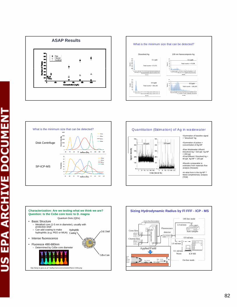
82
ASAP Results
0.1 ppb 0.1 ppb
Dissolved Ag 100 nm Nanocomposix Ag
Total counts = 47,838Total counts = 57,472
What is the minimum size that can be detected?
0.5 ppb 0.5 ppb
Total counts = 180,203Total counts = 306,165
What is the minimum size that can be detected?
Disk Centrifuge
SP-ICP-MS
Quantitation (Estimation) of Ag in wastewater•Summation of baseline signal = “dissolved” Ag
•Summation of pulses = concentration of Ag-NP
•Raw Wastewater influent: Dissolved Ag = 520 ppt Ag-NP
200 t= 200 ppt •Final Effluent: Dissolved Ag = 60 ppt Ag-NP = 100 ppt
•Results comparable to estimates from materials flow analysis (Nowack)
•In what form is the Ag-NP ? Need complimentary analysis (TEM)
• Basic Structure– Metalloid core (1-5 nm in diameter), usually with
protective shell– Can add coating to make
hydrophilic (e.g. PEO or MUA)
• Intense fluorescence
ZnS ShellHydrophilicCoating
Quantum Dots (QDs)
Characterization: Are we testing what we think we are?Question: Is the CsSe core toxic to D. magna
• Intense fluorescence
• Fluoresce 490-680nm– Determined by CdSe core diameter
CdSe Core
http://lamp.tu-graz.ac.at/~hadley/nanoscience/week2/Nano-CdSe.png
Sizing Hydrodynamic Radius by Fl FFF - ICP - MS
2.31
0.9
Fluorescence
detector Auto samplerCross flow
Channel flo 0.9 ml/min
1.0 ml/min
Off-line modeCross-flow Recirculation
1.0
ICP-MS
Fl-FFF
channel
Channel flow
1.0 ml/min
0.1 ml/minWaste
On-line mode
mosvald
Text Box
82

83
Field: 0.9 ml/minCarrier flow:1.0 ml/minPSS standards (Duke)20 microliter injectionFl detector
Size analysis by FFF theory or calibration Elemental Size Distribution by Fl FFF-ICP-MS
26 nm
Non 1:1 Cd: Se ratioPossible explanation: Cd associated with polymer coating due to poor washing during synthesis
Low Zn indicates thin ZnS shell
Elemental Size Distribution by Fl FFF-ICP-MS
24 nm
Thick ZnS shell
Large Zn peak at early timePossible explanation:Zn associated with free polymerNo dissolved Zn detected in a 3K Dalton filtrate
Dissolved metals (mg/L) and % dissolved metals for a 7.5 nmol/L QD solution
1
1.5
2
2.5en
trat
ion
(mg/
L) 0 hrs
48 hrs
33%
0hrs (mg/L) BDL 0.014 BDL 0.006 BDL BDL BDL 0.01348hrs (mg/L) 0.222 0.044 2.3 0.083 BDL BDL BDL 0.045
0
0.5
Cd Zn Cd Zn Cd Zn Cd Zn
Green MUA Red MUA Green PEO Red PEO
Con
ce
61 %86 % 14% 7%
0
0.2
0.4
0.6
0.8
1
0 1 2 3 4 5
Frac
. Mor
talit
y
QD Concentration (mg dots/L)*
MUA coated
MUA NPs release Cd to solution.
“Excess” Cd source of toxicity?
Are we testing CeSe core bioavailablity or some other form?
0
0.2
0.4
0.6
0.8
1
0 1 2 3 4 5
Frac
. Mor
talit
y
QD Concentration (mg dots/L)*
PEO coatedPEO NP stable during test
Appears to be a size affect when comparing mass dose
0
0.2
0.4
0.6
0.8
1
0 5 10 15 20 25 30
Frac
. Mor
talit
y
QD Concentration (nmol/L)
No Size effect when comparing number dose
SummaryRTSP-ICP-MS can be used to:
• Detect NP Ag at environmentally relevant concentrations (ppt levels)
•High specificity (contrast to DLS)• Distinguish between “dissolved” and NP Ag
•Potential for application in stability and exposure/toxicity laboratory studies
• Current limitations• About 40 nm size limit•Cannot identify NP type
FFF-ICP-MS can be used to:• More fully characterize complex NPs• Provide information to interpret results of experiments where:
• Mixtures are used• Manufacturing impurities are present• Transformation/ degredation products are present
mosvald
Text Box
83

84
Acknowledgments
CollaboratorsDr. Anthony Bednar: USACEDr. Nicola Rodgers: CSIRODr. Antonio Nogueira: U. of AveiroDr. Brian Jackson: Dartmouth College
RD-83332401-0
FundingUS Environmental Protection Agency
US Army Corps of Engineers
StudentsE. Lesher, D. Mitrano,J. Monserud, H. Pace
Unexpected metal ratios
• Cd:Se ratio not 1:1• Excess Cd in MUA QDs, especially high for red MUA QDs
Sizing of Core by UV-Vis
0
0.01
0.02
0.03
0.04
0.05
450 500 550 600 650Nor
mal
ized
Abs
orba
nce Green PEO
Red PEO
Green MUA
λ = 591D = 4.2 nm
λ = 500D = 2.3 nm
Polar, non-ionic
Wavelength (nm)
0
0.01
0.02
0.03
0.04
0.05
450 500 550 600 650
Nor
mal
ized
Abs
orba
nce
Wavelength (nm)
Green MUARed MUA
λ = 525D= 2.6 nm
λ = 605D = 4.8 nm
Anionic (carboxyl functionality)
mosvald
Text Box
84

2010 U.S. EPA Nanotechnology Grantees Meeting
The Office of Research and Development’s National Center for Environmental Research 85
Jingyu Liu
Controlled Release of Biologically Active Silver from Nanosilver Surfaces
Jingyu Liu1, David Sonshine2, Saira Shervani2, and Robert Hurt2,3 1Department of Chemistry, 2Division of Engineering, 3Institute for Molecular and Nanoscale Innovation,
Brown University, Providence, RI Major pathways in the antibacterial activity and eukaryotic toxicity of nano-silver involve the silver cation and its soluble complexes, which are well-established thiol toxicants. Through these pathways, nano-silver behaves in analogy to a drug delivery system, in which the particle contains a concentrated inventory of an active species, the ion, which is transported to and released near biological target sites. Although the importance of silver ion in the biological response to nano-silver is widely recognized, the drug delivery paradigm has not been well developed for this system, and there is significant potential to improve nano-silver technologies through controlled release formulations. This work applies the drug delivery paradigm to nano-silver dissolution and presents a systematic study of chemical concepts for controlled release. After presenting thermodynamic calculations of silver species partitioning in biological media, the rates of oxidative silver dissolution are measured for nanoparticles and macroscopic foils and used to derive unified area-based release kinetics. A variety of competing chemical approaches are demonstrated for controlling the ion release rate over four orders of magnitude. Release can be systematically slowed by thiol and citrate ligand binding, formation of sulfidic coatings, or the scavenging of peroxy-intermediates. Release can be accelerated by pre-oxidation or particle size reduction, while polymer coatings with complexation sites alter the release profile by storing and releasing inventories of surface-bound silver. Finally, the ability to tune biological activity is demonstrated through bacterial inhibition zone assay carried out on selected formulations of controlled release nano-silver. EPA Grant Number: R833862

86
Controlled Release of Biologically Active Silverfrom Nanosilver Surfaces
Jingyu Liu, David A. Sonshine, Saira Shervani, Robert H. Hurtgy , , ,
Department of Chemistry, Division of Engineering, Institute for Molecular and Nanoscale Innovation
Brown University, Providence, RI
R833862
Silver is a broad spectrum antibiotic
Nanosilver – Two Faces of Janus
A new generation of antimicrobials
Silver is a known toxicant to aquatic organismsA risk to the environment and human health?
has relatively low toxicity in humans;
is being manufactured in large quantities and incorporated into consumer and medical products.
is more toxic than any other metal except mercury;
bioaccumulates quickly;
nanosilver has toxicity threshold as low as 10 ng/L (zebrafish embryos).
Silver has potential toxic effects on beneficial bacteria in soil Argyria
nAg
Metal ions may coexist in metal-containing nanoparticle suspensions.
Silver ion is a known toxicant that binds to thiol groups in enzymes, such as NADH dehydrogenase, which disrupts the bacterial respiratory chain generating ROS that can lead to oxidative stress and cell damage.
Nanosilver particles themselves may also contribute by binding to or
Nanosilver in Biological and Environmental Systems– Is It the Particle or the Ion?
Nanosilver particles themselves may also contribute by binding to or passing through cell membranes, and generating ROS through surface reactions.
There is some controversy about the role of particle-based mechanisms, but there is broad agreement that silver ion is an important toxicant.
What determines particle/ion partitioning?
Ion Release Kinetics and Particle Persistence in Aqueous Nano-Silver Colloids
Liu J.; Hurt R.H. Environ. Sci. Technol. 2010, 44, 2169–2175
O2, H+This reaction produces active peroxide intermediates
Is inhibited by natural organic matter
Ag+ (slow)nAg
H2O2, ·O2-
][22.0
7/
10][1 NOMae
MHAe
dtdm
mRTE
Dissolution kinetic law
Leads to complete particle dissolution in aerobic environmentsAg+ (fast)
H2O
Specific benefits of controlled release nanosilver formulations might include:
(i) dose control to achieve desired bactericidal or bacteriostatic effects;
“Controlled Release” Nanosilver - application of the drug delivery paradigm
Can we systematically increase or decrease ion release rate?
Can we engineer nanosilver materials for optimal ion release profile?
nAg O2
ROS
(i) dose control to achieve desired bactericidal or bacteriostatic effects;
(ii) dose limitation to avoid eukaryotic toxicity that can, for example, slow woundhealing in bandage applications;
(iii) control of product lifetime, before dissolution and diffusion end antibacterial activity;
(iv) minimization of environmental release through excess ion production beyond that necessary for product performance;
(v) optimization of release profile for targeted delivery to specific tissue or intracellular targets.
Particle-Ion Partitioning in Aqueous nAg Colloids
Basic Experiment
Ultrafiltration + Atomic absorption
total ionic silver by AAs(graphite furnace atomic absorption spectroscopy)nAg
Ag+
Incubation
Amicon cellulose membrane3K Da, (1-2 nm pore size)
ultrafiltration
Separation
Ag+
nAg
Quantification
mosvald
Text Box
86

87
If nano-silver oxidatively dissolves, why doesn’t bulk silver?
Answer: it does! (but slowly – about 2 nm/day)
Speciation in Biological Media – Effect of Chloride and Thiol
AgCl nanoparticles
Particle aggregate
nAg
(By Visual MINTEQ)
Liu, Sonshine, Shervani, Hurt, “Controlled Release of Biologically Active Silver from
Nanosilver Surfaces” ACS Nano, in press
][][][
RSAgRSAgKa
0.2 mM
~1012
O2, H+
Ag+
known toxicant that binds to biological thiol groups
nAg
H2O2, ·O2-
Reactive oxygen species that damage lipids, proteins, DNA
Nanosilver Behavior in Biological Environments– Nanosilver vs. Silver Salts
Controlled Release Approaches: Coatings and Ligands
Mercaptoundecanoic acid(MUA)
Ligand exchange
Original nAgCitrate anion
Two-Stage (fast/slow) Release
Controlled Release Approaches: Pre-Oxidation
O i
nAg powder
Fiber substratedoped with nAg
Ozone in flowing air
Air
Ozone generator
Ozone analyzer
Controlled Release Approaches: Media Composition
Total silver (2 mg/L)
Antioxidants (GSH, ascorbic acid)Oxidized GSH,
Amino acidSodium citrate
Typical 1-day ion release in acetate buffer (0.28 mg/L)
Acetate buffer
Analysis of Agdis
nAg
mosvald
Text Box
87

88
Comparison of Surface Treatment Methods for Release Control
Biological Response to Controlled Release nAg
E. coli, 18 hr incubationWith 10 mm nAg-doped filter papers
Experimental assistance from Dr. Charles Vaslet, Kane laboratory
Approaches for Controlling Biologically Active SilverRelease from Nanosilver Surfaces
Liu, Sonshine, Shervani, Hurt“Controlled Release of Biologically Active Silver from Nanosilver Surfaces”
ACS Nano, in press
Hurt lab – dissolution kinetics and controlled release formulations
How can the transformations of nAg be engineered and controlled?Role of sulfides and the role of photochemistry
Pennell – coupled reactive dissolution kinetics laws with environmental material flow modeling
Future work – Biological and Environmental Implicationsof Ion Release Kinetics and Control
What are the ultimate amount, fate, and form of nAg in the environment?
Kane lab – uptake and biodistribution in Xenopus
What is the fate and form of nAgin whole biological organisms?
Financial support from the US EPA Science to Achieve ResultsProgram and the NIEHS Superfund Research Program grant at
Brown is gratefully acknowledged
S i
R833862
The Laboratory for Environmental and Health NanoscienceAt Brown University
Saira
mosvald
Text Box
88

2010 U.S. EPA Nanotechnology Grantees Meeting
The Office of Research and Development’s National Center for Environmental Research 89
Roger A. Pinto
Effects of Polyethyleneimine Surface Modifications of Multi-Walled Carbon Nanotubes: Their Toxicity, Sorption Behaviors, and Ecological Uptake by
Earthworms and Daphnia magna
Roger Pinto1, Elijah Petersen2, and Walter Weber, Jr.1 1Department of Chemical Engineering, University of Michigan, Ann Arbor, MI; 2 Chemical Science and
Technology Laboratory, National Institute of Standards and Technology, Gaithersburg, MD
In support of the mission of the U.S. Environmental Protection Agency to provide substantive information for ecological risk assessments, this research focuses on investigation of the environmental fate, bioaccumulation potential, and toxicity of surface-modified carbon nanotubes (CNTs) in terrestrial and aquatic ecosystems. Carbon-14 multi-walled carbon nanotubes (MWNTs) were synthesized by a CVD process, grafted with polyethyleneimine (PEI) polymers, and further modified to render them with a range of different surface charges and resultant higher stability in aqueous suspensions. Assessments of the extent to which these modifications influence CNT ecotoxicity, accumulation, and elimination behaviors were performed using the earthworm Eisenia foetida and the water flea, Daphnia magna. Liquid scintillation counting of residual 14C in vivo provided insights on the uptake and elimination behaviors for the organisms tested. D. magna exposed to PEI-coated and acid-modified MWNTs at concentrations of approximately 25 or 250 μg/L indicated that the PEI surface coatings did not appear to substantially impact nanotube accumulation or elimination rates. Microscopy observations revealed substantial aggregation in the guts of D. magna similarly to previous studies with acid-treated MWNTs and fullerenes. Algae feeding to Daphnia was necessary to achieve almost complete elimination in 48 h, whereas the absence of algal amendments caused minimal CNT elimination. Immobilization studies allowed for the determination of EC50 values and indicated that PEI modifications increased MWNT acute toxicities, though this trend corresponded to the overall size of the grafted polymers. Phase distribution experiments with soils measuring a combined effect of sorption and attachment to particles indicated linear sorption isotherms for the regular MWNTs and non-linear trends for the PEI-modified MWNTs. Differences in uptake behaviors by earthworms were not apparent among the different types of PEI-modified and MWNTs, results that indicated limited interaction of these carbon nanotubes with the organism tissues. In contrast to previous results for unmodified MWNTs, elimination patterns for the grafted PEI-MWNTs were well fit by an exponential decay. To determine whether earthworm exposure to these MWNTs elicits a stress response, two biomarkers of oxidative stress (glutathione-S-transferase (GST), catalase) and two biomarkers of neurological stress (monoamine oxidase, cholinesterase) were measured in whole-body samples. A dose-response relationship was not observed within the concentration range of the exposure treatments (3-1,000 mg MWNT kg-1 soil). However, positively (PEI-amino) and neutrally (PEI-acetate) charged nanotubes consistently revealed a toxic response with catalase, monoamine oxidase, and cholinesterase, while negatively charged CNTs (PEI-succinic and acid-treated MWNTs) had no effect. EPA Grant Number: RD-833321

90
Principal investigator did not authorize publication of the presentation.

PM Session 1: Characterization Methods

2010 U.S. EPA Nanotechnology Grantees Meeting
The Office of Research and Development’s National Center for Environmental Research 92
Xin-Rui Xia
A Biological Surface Adsorption Index for Characterizing Nanomaterials
in Aquatic Environments and Their Correlation With Skin Absorption of Nanomaterials
Xin-Rui Xia, Nancy Monteiro-Riviere, and Jim Riviere
North Carolina State University, Raleigh, NC
As nanoparticles are increasingly being used in commercial products, it becomes more and more important to understand how they interact with living organisms and the environment. The behavior of nanomaterials in a biological or environmental system is governed by the molecular interactions of their surface species with the biological or environmental components. Quantitative assessment of the adsorption properties of nanomaterials is a crucial step for developing predictive structure-activity relationship in nanomedicine and risk assessment of nanomaterials. We have developed a biological surface adsorption index (BSAI) approach to characterize the surface activity of nanomaterials in biological systems. A set of small molecules having diverse physicochemical properties was used as probe compounds. The adsorption coefficients (k) of the probe compounds were obtained by measuring the quantities of the probe compounds adsorbed on the surfaces of the nanomaterials and the equilibrium concentrations of the probe compounds in the media. The log (k) values were scaled to a set of solvation molecular descriptors of the probe compounds via multiple linear regressions to provide a set of five nano-descriptors representing the contributions of the five types of molecular interactions (hydrophobicity, hydrogen-bond acidity and basicity, dipolarity/polarizability, and lone pair electrons). The nano-descriptors for multi-walled carbon nanomaterials (MWCNT) with different surface chemistries (unmodified, -OH and –COOH modified) and fullerenes were measured; for example, the regression model obtained for MWCNT (-OH modified) was log(k) = 0.77R + 2.55π - 0.14α - 2.36β + 4.90V; n = 30, R2 = 0.89. The measured nano-descriptors can be used to develop predictive structure-activity relationships in nanomedicine and nanomaterial risk assessments. We have prepared a series of hydroxylated fullerenes, C60(OH)x. Their hydrophobicity was adjusted by controlling the number of hydroxyl groups (e.g., x = 16, 20, 30, 40). After characterization using conventional techniques such as particle size, zeta-potential and solubility, the nano-descriptors were measured using the BSAI approach for each of the C60(OH)x nanomaterials. Then, the adsorption coefficients of different C60(OH)x into the stratum corneum, the primary barrier of skin, were measured by in vitro adsorption experiments. The equilibrium adsorption coefficients were correlated with the physicochemical parameters and the nano-descriptors to establish quantitative correlations. The following findings will benefit EPA: (1) The BSAI approach measures five molecular descriptors for each of the nanomaterials. The BSA indexes are free energy-related physicochemical parameters that can be used for predictive models developments in EPA guidelines for environmental health of nanomaterials; (2) The hydroxylated fullerenes with different hydrophobicity can be used to study the environmental transport and fate of fullerene nanomaterials; (3) The quantitative approach to correlate the adsorption coefficients of stratum corneum with the BSAI nano-descriptors of the C60(OH)x nanomaterials could be a useful approach for developing predictive models for safety evaluation and risk assessment of nanomaterials. EPA Grant Number: R833328

93
A Biological Surface Adsorption IndexA Biological Surface Adsorption Indexfor Characterizing Nanomaterials in Aquatic for Characterizing Nanomaterials in Aquatic
Environments and Their Correlation with Environments and Their Correlation with Skin Absorption of NanomaterialsSkin Absorption of Nanomaterials
XinXin--Rui Xia (PI), Nancy A. MonteiroRui Xia (PI), Nancy A. Monteiro--Riviere, Jim E. Riviere Riviere, Jim E. Riviere
Center for Chemical Toxicology Research & Center for Chemical Toxicology Research & Pharmacokinetics (CCTRP)Pharmacokinetics (CCTRP)
North Carolina State University, Raleigh, NCNorth Carolina State University, Raleigh, NC
How to characterize nanomaterials in How to characterize nanomaterials in aqueous environmentsaqueous environments
Currently, most of the methods measure the physical parameters, such as, particle size, Currently, most of the methods measure the physical parameters, such as, particle size, size distribution, shape, charges, specific surface area, …size distribution, shape, charges, specific surface area, …
surface chemistry and core material compositions are the only measurable chemical surface chemistry and core material compositions are the only measurable chemical information of nanomaterials, but cannot be directly used for quantitative analyses. information of nanomaterials, but cannot be directly used for quantitative analyses.
Octanol water partition coefficient log(Kow) has been widely used for predictive model Octanol water partition coefficient log(Kow) has been widely used for predictive model development for small molecules, but it is difficult to be measured for nanomaterials since development for small molecules, but it is difficult to be measured for nanomaterials since most of the nanomaterials form stable suspension in water or oil, but in not both.most of the nanomaterials form stable suspension in water or oil, but in not both.most of the nanomaterials form stable suspension in water or oil, but in not both. most of the nanomaterials form stable suspension in water or oil, but in not both.
Great efforts have been made to understand the chemical interactions between Great efforts have been made to understand the chemical interactions between nanoparticles and biological or environmental components in the scientific communities. nanoparticles and biological or environmental components in the scientific communities.
We have demonstrated that lipophilicity was a significant factor in the nanoparticle We have demonstrated that lipophilicity was a significant factor in the nanoparticle adsorption of small chemicals. adsorption of small chemicals.
To date, there is no general applicable approach to quantitatively measure the molecular To date, there is no general applicable approach to quantitatively measure the molecular interactions of nanoparticles with biological or environmental components, which is the interactions of nanoparticles with biological or environmental components, which is the crucial information needed to crucial information needed to develop quantitative structuredevelop quantitative structure--activity relationship for activity relationship for nanomedicine researches and for risk assessment and safety evaluation of the nanomedicine researches and for risk assessment and safety evaluation of the nanomaterials in occupational and environmental exposures. nanomaterials in occupational and environmental exposures.
Competitive Adsorption onto NP Surfaces.Clean surface of pure nanomaterials only exist in high Clean surface of pure nanomaterials only exist in high vacuum; having high surface energy, ready to adsorb vacuum; having high surface energy, ready to adsorb any compound to reduce their surface energy.any compound to reduce their surface energy.
Once contact an aqueous environment, the Once contact an aqueous environment, the nanoparticle surface is dramatically altered by solvation nanoparticle surface is dramatically altered by solvation to form a solidto form a solid--liquid interface. This solidliquid interface. This solid--liquid liquid interface is unique feature in aqueous environments. interface is unique feature in aqueous environments.
Small molecules and macromomolecules are adsorbed Small molecules and macromomolecules are adsorbed onto the interface to form nanoonto the interface to form nano--macromolecule coronas macromolecule coronas (or complex).(or complex).
Current NanoCurrent Nano--characterization focus:characterization focus:•• Pure nanomaterials in industrial applicationsPure nanomaterials in industrial applications•• NanoNano--protein coronas in biological systemsprotein coronas in biological systems
•• NanoNano--humuic acid complex in environmentalhumuic acid complex in environmental
We have identified that the adsorption property at the We have identified that the adsorption property at the solidsolid--liquid interface is key to understanding the liquid interface is key to understanding the behavior of nanoparticles in aqueous environments. behavior of nanoparticles in aqueous environments.
•• it is not only determined colloidal stabilityit is not only determined colloidal stability
•• but also the affinity and selectivity of environmental but also the affinity and selectivity of environmental components in forming ‘nanoparticle coronas’components in forming ‘nanoparticle coronas’
Illustration of the competitive adsorption of small molecules and macromolecules onto NP surfaces
Molecular interactions in the surface Molecular interactions in the surface adsorption processesadsorption processes
We have developed a biological surface adsorption index (BSAI) approach to We have developed a biological surface adsorption index (BSAI) approach to characterize the molecular interaction strengths of nanoparticles with small characterize the molecular interaction strengths of nanoparticles with small molecules and macromolecules in biological and environmental systems. molecules and macromolecules in biological and environmental systems.
The BSAI approach is based on the molecular interaction similarity between nanoThe BSAI approach is based on the molecular interaction similarity between nano--small molecule interactions and nanosmall molecule interactions and nano--macromolecule interactions. macromolecule interactions.
Basic molecular interactions forces: Basic molecular interactions forces:
Coulombs force (charged nanos) Coulombs force (charged nanos) ------measured by zetameasured by zeta--potentialpotential
London dispersion (hydrophobicity) London dispersion (hydrophobicity)
Hydrogen bonding (acidity/basicity) Hydrogen bonding (acidity/basicity)
Dipolarity / polarizability Dipolarity / polarizability
lonelone--pair electrons pair electrons
Obtained by BSAI approachObtained by BSAI approach
NanoNano--descriptors derived by BSAI approachdescriptors derived by BSAI approach
It is hypothesized that the adsorption properties of nanomaterials are It is hypothesized that the adsorption properties of nanomaterials are governed by the contributions of each type of molecular interactions, which governed by the contributions of each type of molecular interactions, which can be detected by a set of small molecular probes having diverse can be detected by a set of small molecular probes having diverse physicochemical properties covering the vector spaces of molecular physicochemical properties covering the vector spaces of molecular interactions between the nanomaterial and macromolecules.interactions between the nanomaterial and macromolecules.
The adsorption coefficients (The adsorption coefficients (kk) of the probe compounds on a given ) of the probe compounds on a given nanomaterial were measured using our SPMEnanomaterial were measured using our SPME--GC/MS method. GC/MS method.
Then, the log(Then, the log(kk) values were scaled to a set of known molecular descriptors ) values were scaled to a set of known molecular descriptors of the probe compounds via multiple linear regressions.of the probe compounds via multiple linear regressions.
i = 1, 2, 3, … , n
Here, the solvation descriptors and a linear free energy relationship (LFER) Here, the solvation descriptors and a linear free energy relationship (LFER) and multiand multi--walled carbon nanotubes (MWCNT) will be used as examples to walled carbon nanotubes (MWCNT) will be used as examples to demonstrate the BSAI approach. demonstrate the BSAI approach.
iiiiii vVbaprRck log
Adsorption coefficients (logk) and solute descriptors of the probe compounds
Probe Compounds* logKo/w logk R π α β V
1 chlorobenzene 2.84 3.25 0.718 0.65 0 0.07 0.839
2 ethylbenzene 3.15 3.19 0.613 0.51 0 0.15 0.998
3 p-xylene 3.15 3.26 0.613 0.52 0 0.16 0.998
4 bromobenzene 2.99 3.50 0.882 0.73 0 0.09 0.891
5 propylbenzene 3.69 3.76 0.604 0.5 0 0.15 1.139
6 4-chlorotoluene 3.33 3.82 0.705 0.67 0 0.07 0.98
7 phenol 1.46 2.87 0.805 0.89 0.6 0.3 0.775
8 benzonitrile 1.56 3.04 0.742 1.11 0 0.33 0.871
9 4-fluorophenol 1.77 2.68 0.67 0.97 0.63 0.23 0.793
10 benzyl alcohol 1.1 2.10 0.803 0.87 0.33 0.56 0.916
11 iodobenzene 3.25 3.88 1.188 0.82 0 0.12 0.975
12 acetophenone 1.58 3.26 0.818 1.01 0 0.48 1.014
13 3-methylphenol 1.96 3.08 0.822 0.88 0.57 0.34 0.916
14 methyl benzoate 2.12 3.70 0.733 0.85 0 0.46 1.073
15 4-chloroanisole 2.78 4.07 0.838 0.86 0 0.24 1.038
16 phenethyl alcohol 1.36 2.54 0.784 0.83 0.3 0.66 1.057
17 3-methylbenzyl alcohol 1.6 2.85 0.815 0.9 0.33 0.59 1.057
18 4-ethylphenol 2.58 3.62 0.8 0.9 0.55 0.36 1.057
19 3,5-dimethylphenol 2.35 3.49 0.82 0.84 0.57 0.36 1.057
20 ethyl benzoate 2.64 4.14 0.689 0.85 0 0.46 1.214
21 methyl 2-methylbenzoate 2.75 4.12 0.772 0.87 0 0.43 1.214
22 naphthalene 3.3 4.44 1.36 0.92 0 0.2 1.085
23 3-chlorophenol 2.5 3.62 0.909 1.06 0.69 0.15 0.898
24 4-nitrotoluene 2.37 4.44 0.87 1.11 0 0.28 1.032
25 4-chloroacetophenone 2.32 4.28 0.955 1.09 0 0.44 1.136
26 3-bromophenol 2.63 3.79 1.06 1.15 0.7 0.16 0.95
27 1-methylnaphthalene 3.87 4.89 1.344 0.9 0 0.2 1.226
28 biphenyl 3.98 5.18 1.36 0.99 0 0.22 1.324
mosvald
Text Box
93

94
Predictive Model established for MWCNTPredictive Model established for MWCNT
1
2
3
4
5
Str
eng
th
Nanoparticle
Nano-Water Interface
log (log (kk) = ) = --1.33 + 0.0431.33 + 0.043RR + 1.75 + 1.75 ππ -- 0.37α 0.37α -- 2.782.78ββ + 4.18+ 4.18VV
nn =28, =28, RR22 =0.93 and =0.93 and FF =63, =63, QQ22LOOLOO=0.888 and =0.888 and QQ22
LMO25%LMO25%=0.883 =0.883
3
4
5
6
log
k
-4
-3
-2
-1
0
R π α β V
Rel
ati
ve S
Aqueous Phase
Relative molecular interaction strengths at the nanoRelative molecular interaction strengths at the nano--water interface.water interface.
Left:Left: Correlation of logCorrelation of logkk of the probe compounds on MWCNT with their logKo/w values, suggesting of the probe compounds on MWCNT with their logKo/w values, suggesting lipophilicity is significant in the adsorption process (Rlipophilicity is significant in the adsorption process (R22=0.57) but is not the only factor. =0.57) but is not the only factor.
Right:Right: The five nanoThe five nano--descriptors [descriptors [r, p, a, b, vr, p, a, b, v] measured by the BSAI approach, representing the five major ] measured by the BSAI approach, representing the five major molecular interactions in nanoparticle adsorption processes: lonemolecular interactions in nanoparticle adsorption processes: lone--pair electrons, polarity/polarizability, pair electrons, polarity/polarizability, hydrogenhydrogen--bond donor, hydrogenbond donor, hydrogen--bond acceptor, and London dispersion, respectively. bond acceptor, and London dispersion, respectively.
y = 0.681x + 1.875R2 = 0.573
0
1
2
0 1 2 3 4 5
log Kow
-1.5
-0.5
0.5
1.5
2.5
2 3 4 5
Sta
ndar
d R
esid
uals
y = 0.935x + 0.235R2 = 0.935
4
5
6
ted
log
k
NanoNano--Descriptors Provide Better PredictionDescriptors Provide Better Prediction
-2.5
Predicted log k
Residual plot of the linear regression. Residual plot of the linear regression. The random scattering of the data The random scattering of the data points above and below the zero points above and below the zero residual line reveals that the residual line reveals that the regression model well described the regression model well described the experimental data.experimental data.
1
2
3
1 2 3 4 5 6
Measured log k
Pre
dic
t
The predicted versus measured log(kThe predicted versus measured log(k)) values values of the probe compounds on MWNCT.of the probe compounds on MWNCT.
Validation of the predictive model for MWCNTValidation of the predictive model for MWCNT
The robustness of the model for MWCNT was studied by internal crossThe robustness of the model for MWCNT was studied by internal cross--
validation using the leavevalidation using the leave--oneone--out (LOO) and leaveout (LOO) and leave--manymany--out (LMO25%) out (LMO25%)
techniques with validation coefficients techniques with validation coefficients QQ22LOOLOO of 0.888of 0.888 andand QQ22
LMO25%LMO25% of 0.883, of 0.883,
respectively. respectively.
Both of the crossBoth of the cross--validation coefficients were greater than 0.7, revealing the validation coefficients were greater than 0.7, revealing the
robustness of the predictive model.robustness of the predictive model.obust ess o t e p ed ct e odeobust ess o t e p ed ct e ode
External validation was conducted by using 12 different compounds; their logExternal validation was conducted by using 12 different compounds; their logkk
values were measured using same protocols as the probe compounds. values were measured using same protocols as the probe compounds.
The external validation coefficient (The external validation coefficient (QQ22extext) of the model was 0.78, suggesting ) of the model was 0.78, suggesting
satisfactory predictivity for the external validation compounds. satisfactory predictivity for the external validation compounds.
The applicability domain of the predictive The applicability domain of the predictive model for MWCNT model for MWCNT
y = 0.94x + 0.22R2 = 0.93
1
2
3
4
5
6
7
Pre
dic
ted
log
k
Training
Validation3
-2
-1
0
1
2
3
4
0.0 0.2 0.4 0.6 0.8 1.0
Sta
nd
ard
ized
Res
idu
als
Training
Validation
b
h*=0.64
0
0 1 2 3 4 5 6 7
Measured log k
-4
-3
Hat
Predictive model performance and validation for MWCNT.Predictive model performance and validation for MWCNT.
Left:Left: The predicted versus measured logThe predicted versus measured logkk values of the training probe and validation values of the training probe and validation compounds. The data points of the validation compounds lying closely along the regression compounds. The data points of the validation compounds lying closely along the regression line reveal the robustness of the predictive model. line reveal the robustness of the predictive model.
Right:Right: Williams plot reveals the applicable domain of the predictive model. The dotted lines Williams plot reveals the applicable domain of the predictive model. The dotted lines defined the chemical domain (defined the chemical domain (3σ and 3σ and hh*=0.64) by the training probe compounds. The log*=0.64) by the training probe compounds. The logkkvalues for the training probe and validation compounds are all within the chemical domain, values for the training probe and validation compounds are all within the chemical domain, suggesting no outliers and the model predictivity is reliable. suggesting no outliers and the model predictivity is reliable.
BSAI Database and ApplicationsBSAI Database and Applications BSAI Database is the final product of the BSAI approach, composed of BSAI Database is the final product of the BSAI approach, composed of
the five nanothe five nano--descriptors for each of the nanomaterials.descriptors for each of the nanomaterials.
NanoNano--descriptors descriptors [[r, r, ππ, , αα, , ββ, v, v]]
r r –– lone lone ––pair electrons interaction strengthpair electrons interaction strengthππ –– dipolarity/dipolarizability interaction strengthdipolarity/dipolarizability interaction strengthαα –– hydrogenhydrogen--bond acidity interaction strengthbond acidity interaction strengthββ –– hydrogenhydrogen--bond basicity interaction strength bond basicity interaction strength v v –– hydrophobicity interaction strengthhydrophobicity interaction strength
Physical parametersPhysical parameters
Particle sizeParticle sizeSize distributionSize distributionParticle shapeParticle shapeSpecific surface areaSpecific surface area… …… …y p y gy p y g
The nanoThe nano--descriptors are free energy related quantities quantitatively describing descriptors are free energy related quantities quantitatively describing the molecular interaction potentials of the nanomaterials at the nanothe molecular interaction potentials of the nanomaterials at the nano--water water interface.interface.
Biological activities, such as, solubility, partition coefficient, rate constant, binding Biological activities, such as, solubility, partition coefficient, rate constant, binding and inhibition constants are free energy related quantities; their logarithmic values and inhibition constants are free energy related quantities; their logarithmic values can be directly predicted via similar predictive model shown for MWCNT. can be directly predicted via similar predictive model shown for MWCNT.
The development of the BSAI approach could open a quantitative avenue toward The development of the BSAI approach could open a quantitative avenue toward predictive nanomedicine development, particularly, for developing integrated predictive nanomedicine development, particularly, for developing integrated physiological based pharmacokinetic models, for quantitative risk assessment and physiological based pharmacokinetic models, for quantitative risk assessment and safety evaluation of nanomaterials.safety evaluation of nanomaterials.
Project Summary Project Summary
This project is to study the impact of physicochemical properties on skin This project is to study the impact of physicochemical properties on skin absorption of manufactured nanomaterials.absorption of manufactured nanomaterials.
Four physicochemical properties were studied;Four physicochemical properties were studied;
Particle size (nm): 1 (CParticle size (nm): 1 (C6060), 20, 50, 80 (silver nanoparticles)), 20, 50, 80 (silver nanoparticles)
Surface charge: nC60 and coated nC60; the zetaSurface charge: nC60 and coated nC60; the zeta--potential was manipulated by potential was manipulated by different iondifferent ion--pairing agentspairing agents
Hydrophobicity: the difficulty in obtaining logHydrophobicity: the difficulty in obtaining logKKo/wo/w for nanomaterials forced us to for nanomaterials forced us to develop the BSAI approachdevelop the BSAI approach
Solvent effects: 6 industrial solvents (toluene, cyclohexane, chloroform, ethanol, Solvent effects: 6 industrial solvents (toluene, cyclohexane, chloroform, ethanol, acetone and propylene glycol) using the diffusion, tapeacetone and propylene glycol) using the diffusion, tape--stripping and in vivo stripping and in vivo methods. methods.
mosvald
Text Box
94

95
Impact of Physicochemical Properties on Skin Impact of Physicochemical Properties on Skin Absorption of NanomaterialsAbsorption of Nanomaterials
Pristine C60 in different solvents will Pristine C60 in different solvents will be used in many industrial and be used in many industrial and pharmaceutical manufacturing pharmaceutical manufacturing processes. processes.
Human exposure to C60 could Human exposure to C60 could occur in various solvents.occur in various solvents.
C tl th i t f l tC tl th i t f l t Currently, the impact of solvents on Currently, the impact of solvents on its skin penetration is unknown.its skin penetration is unknown.
We have studied four types of We have studied four types of industrial solvents:industrial solvents:
–– Toluene (aromatic)Toluene (aromatic)
–– Cyclohexane (aliphatic)Cyclohexane (aliphatic)
–– Chloroform (chlorinated)Chloroform (chlorinated)
–– Mineral oil (oil) Mineral oil (oil)
–– Water (aqueous environmental)Water (aqueous environmental)
Layered structure of the skinLayered structure of the skin
•• Uppermost is the stratum corneum Uppermost is the stratum corneum (SC): the primary barrier of skin ((SC): the primary barrier of skin (15 15 μμm in thickness), m in thickness),
•• C60 distribution in the SC layer was C60 distribution in the SC layer was studied by a tapestudied by a tape--stripping methodstripping method. .
nCnC6060 CharacterizationCharacterization
Dynamic size distribution of nCDynamic size distribution of nC6060
nanoparticlesnanoparticlesZeta-potential of nC60 after 14-day dialysis
We have developed a novel method to prepare nC60 nanoparticle with a narrow size distribution. This method does not use THF while provides nC60 concentration in water 100 times higher than the THF method. The nC60 nanoparticles are formed in a SDS aqueous solution, then SDS is removed via dialysis. After exhaustive dialysis, the nC60 nanoparticles were stable in water for years.
Ion-Pairing Effects on Particle Size of nC60
80
120
160
200
e S
ize
(Ave
, nm
) TFA
HFBA
PA
TBAC
TEA
nCnC6060 and most of the unprotected and most of the unprotected nanomaterials have a very narrow nanomaterials have a very narrow window in their colloidal stability window in their colloidal stability (even though they are stable in (even though they are stable in pure water). pure water).
•• IonIon--pairing agents (e.g. > pairing agents (e.g. > 0 05%TFA) will cause their0 05%TFA) will cause their
Impact of nCImpact of nC6060 Colloidal Stability on Colloidal Stability on Skin AbsorptionSkin Absorption
0
40
80
0 0.05 0.1 0.15 0.2
Ion-Pairing Concentration (%)
Par
ticl
e TEA
IonIon--pairing effects on particle size of pairing effects on particle size of nCnC6060 in aqueous solutionsin aqueous solutions
0.05%TFA) will cause their 0.05%TFA) will cause their aggregation.aggregation.
•• Biological electrolytes will cause Biological electrolytes will cause their aggregation.their aggregation.
•• Once the nanoparticles aggregate, Once the nanoparticles aggregate, they can not get through the skin.they can not get through the skin.
3 anions: TFA TFA (trifluoroacetic acid),(trifluoroacetic acid), HFBA HFBA (heptafluorobutyric acid),(heptafluorobutyric acid), and PA and PA (phosphoric acid);(phosphoric acid);2 cations: TBAC TBAC (tetrabutylammonium chloride)(tetrabutylammonium chloride), TEA , TEA (triethylamine)(triethylamine)
Correlation of SC absorption of ANnC60 with Zeta-potential of the nanoparticles
y = 3.02x + 60.49R2 = 0.91
40
60
80
100
mo
un
t (u
g/g
)
Aqueous colloidal Aqueous colloidal nanomaterials (nC60, nanomaterials (nC60, water soluble nAg) did water soluble nAg) did not penetrate intact skin not penetrate intact skin regardless particle regardless particle sizes. sizes.
IonIon--pairing agent could pairing agent could alter their zetaalter their zeta potentialpotential
Impact of ZetaImpact of Zeta--potential on Skin Adsorptionpotential on Skin Adsorption
-40
-20
0
20
-30 -25 -20 -15 -10 -5 0 5 10
Zeta-potential (mV)
Fu
llere
ne
Am alter their zetaalter their zeta--potential, potential,
even diminish their even diminish their surface charges, but did surface charges, but did not promote their skin not promote their skin penetration (penetration (because nanobecause nano--aggregation occurred as zetaaggregation occurred as zeta--
potential was reducedpotential was reduced).).
Correlation of SC adsorption of ANnC60 with ZetaCorrelation of SC adsorption of ANnC60 with Zeta--potential of the nanoparticlespotential of the nanoparticles
Impact of Solvents on Skin Penetration of CImpact of Solvents on Skin Penetration of C6060
2
3
entr
atio
n, C
n (
ug
/cm
-3)
C60/Toluene
C60/Cyclohexane
C60/Chloroform
C60/Mineral OilSkin penetration of CSkin penetration of C6060 was observed in was observed in different industrial solvents. different industrial solvents.
Significant solvent effects were observed; Significant solvent effects were observed; toluene, chloroform promote skin toluene, chloroform promote skin
Depth distribution profiles of C60 in Depth distribution profiles of C60 in stratum corneum after exposure in stratum corneum after exposure in solvents. solvents.
0
1
0 5 10 15
Depth, Xn (um)
C60
Co
nce
pppenetration of C60, while mineral oil did penetration of C60, while mineral oil did not promote skin penetration. not promote skin penetration.
All three solvents have good CAll three solvents have good C6060 solubility.solubility.
Fullerenes Detected in Skin Tissues Fullerenes Detected in Skin Tissues After 26 TapeAfter 26 Tape--StripsStrips
The amounts of fullerenes in the skin The amounts of fullerenes in the skin tissues (epidermis and dermis) were tissues (epidermis and dermis) were analyzed using the improved sLLEanalyzed using the improved sLLE--HPLC method. HPLC method.
Nanomaterial residues were detected Nanomaterial residues were detected in the skin tissues even after 26in the skin tissues even after 26 2
3
4
5
du
es in
Ski
n (
ug
/g)
in the skin tissues even after 26 in the skin tissues even after 26 tapetape--strips. strips.
The amount of fullerene residues The amount of fullerene residues was greater when dosed in was greater when dosed in chloroform than dosed in toluene or chloroform than dosed in toluene or cyclohexane. cyclohexane.
No C60 was detected in the skin No C60 was detected in the skin tissues when dosed in mineral oil; tissues when dosed in mineral oil; this is consistent with the results of this is consistent with the results of the tapethe tape--strip analysis.strip analysis.
0
1
Toluene Cyclohexane Chloroform Mineral oil
C60
Res
id
Fullerene residues in skin tissues Fullerene residues in skin tissues after tapeafter tape--stripping.stripping.
mosvald
Text Box
95

96
Further Research Needed Further Research Needed
All conclusions obtained in this study is human heath oriented using All conclusions obtained in this study is human heath oriented using weanling pig skin as a model and short term (< 2 weeks in vivo).weanling pig skin as a model and short term (< 2 weeks in vivo).
LongLong--term studies are needed because the longterm studies are needed because the long--term effects on term effects on skin could not show in short term studies such as nano contact skin could not show in short term studies such as nano contact dermatitis. dermatitis.
Skin absorption into aquatic animals (fishes, amphibian …) should Skin absorption into aquatic animals (fishes, amphibian …) should be studied because they have totally different skin structure be studied because they have totally different skin structure (e.g., (e.g., amphibian skin is very permeable to small molecules)amphibian skin is very permeable to small molecules). .
Lot works are needed to make the BSAI approach to be a general Lot works are needed to make the BSAI approach to be a general useful tool for quantitative correlation and risk assessment of useful tool for quantitative correlation and risk assessment of different nanomaterials. different nanomaterials.
AcknowledgementsAcknowledgements
Supported by the US EPASupported by the US EPA--Science to Achieve Results Science to Achieve Results (STAR) Program (STAR) Program
mosvald
Text Box
96

2010 U.S. EPA Nanotechnology Grantees Meeting
The Office of Research and Development’s National Center for Environmental Research 97
Wunmi Sadik
Flexible Nanostructured Conducting Poly(amic) Acid Membrane Captures, Isolates, and Simultaneously Detects Engineered Nanoparticles
*Wunmi Sadik1, Nian Du1, Michael Feurstein1, Veronica Okello1, Cheuk Wong2, and Howard Wang2
1Center for Advanced Sensors and Environmental Systems (CASE), Department of Chemistry; 2Department of Mechanical Engineering and Materials Science Program,
State University of New York-Binghamton, Binghamton, NY
The goal of this project is to develop nanocavity sensor (category II) arrays for the isolation, detection and quantitation of engineered nanoparticles (ENPs) in complex environmental matrices. There is urgent demand for rapid screening methods to isolate, detect, and monitor engineered nanomaterials in the environment. Conventional methods for characterizing nanomaterials such as transmission electron microscopy, scanning electron microscopy, and atomic force microscopy tend to be bulky and inadequate for field and rapid screening of free nanomaterials.1 At SUNY-Binghamton, we have developed a new class of nanostructured poly(amic acid) –PAA-membranes that are conductive and electroactive by preventing its imidization to polyimide, while retaining its carboxylic acid and amine functionalities.2-4 We have studied the effect of composition and microstructure on the optical and electrochemical properties of PAA hybrid composites. The uniqueness of PAA lies in its excellent physical and chemical properties: transparency, flexibility, electrical conductivity, and accessibility to forming a large-area device. During the reporting period, our group discovered that this new class of flexible, stand-alone membranes could be successfully used as both sensors and nanofilters. A new nanofilters device based on PAA membranes is hereby introduced. The nanofilters were derived from phase-inverted, copolymers of PAA and other polymers, with the surface and pore sizes systematically controlled by varying the conditions of the synthesis. This presentation will focus on the use of PAA membranes for simultaneous removal and electrochemical detection of silver nanoparticles, quantum dots, and titanium dioxide nanocrystal from food supplements and environmental samples. References: 1. Sadik OA, Zhou AL, Kikandi S, Du N, Wang Q. Sensors as tools for quantitation, nanotoxicity and
nanomonitoring assessment of engineered nanomaterials, Journal of Environmental Monitoring (Critical Review) 2009;11:1782-1800.
2. Du N, Wong C, Feurstein M, Sadik O, Umbach C, Sammakia B. Flexible conducting polymers: effects of chemical composition on structural, electrochemical and mechanical properties. Langmuir 2010. DOI: 10.1021/la101314j
3. Andreescu D, Wanekaya A, Sadik OA, Wang J. Nanostructured polyamic membranes as electrode material. Langmuir 2005;21(15):6891-6899.
4. Breimer M, Yevgheny E, Sadik OA. Incorporation of metal particles in polymerized organic conducting polymers - a mechanistic insight. Nano Letters 2001;1(6):305.
EPA Grant Number: R834091

98
Principal investigator did not authorize publication of the presentation.

2010 U.S. EPA Nanotechnology Grantees Meeting
The Office of Research and Development’s National Center for Environmental Research 99
Jason Unrine
Fate and Effects of Nanosized Metal Particles Examined Along a Simulated Terrestrial Food Chain Using Genomic
and Microspectroscopic Techniques
Jason Unrine, Aaron-Shoults Wilson, Olga Tsyusko, Sarita Hardas, D. Allan Butterfield, and Paul Bertsch Department of Plant and Soil Science, University of Kentucky, Lexington, KY
Terrestrial environments are likely to serve as the ultimate sink for a significant fraction of manufactured nanomaterials (MNM) from accidental releases, use in agriculture, and through land application of sewage sludge as biosolids. Risk from exposure to MNM in terrestrial food webs partly depends on their propensity for uptake and retention by detritivorous soil organisms and subsequent trophic transfer to higher trophic levels as well as inherent toxicity. Our research is investigating interactions between chemical composition and particle size in determining bioavailability and adverse effects of Cu, Ag, and Au nanoparticles (NPs). Our results indicate that uptake of nanoparticles from soil by earthworms does not vary systematically with primary particle size on a mass concentration basis; however, on a number concentration basis, smaller particles are much more bioavailable than larger ones. Also, we have found that nature of surface coating (PVP versus oleic acid) has little effect on Ag uptake. It is clear that the redox behavior of metal NPs in soil varies considerably. Although Cu NPs oxidize immediately upon exposure to the air, Au NPs are completely stable and resistant to oxidation. Ag NPs are resistant to oxidation in the air, but they can be readily transformed in natural soil. We have confirmed that either reduced or oxidized metal NPs can be absorbed from soil and taken up into internal tissues using a combination of X-ray microspectroscopy, laser ablation-inductively coupled plasma mass spectrometry, asymmetrical field flow fractionation-multidetection (AF4), and expression of the metal specific gene, metallothionein. We have observed decreased reproductive success associated with exposure to Au and Ag NPs as well as evidence of soil avoidance for Ag NPs. We also have obtained evidence for oxidative damage of proteins as a result of exposure to Ag ions and Ag NPs. In our toxicity tests, Au and Ag NPs were significantly less toxic than their corresponding metal salts; however, NPs, including oxidized NPs, may be bioavailable and cause adverse effects at relatively high concentrations. Behavioral avoidance of Ag NPs is the most sensitive endpoint investigated to date and occurs at concentrations of Ag NPs that are similar to those expected in sewage sludge. The next phase of our research investigated trophic transfer of NPs along a simulated food chain consisting of soil, earthworms, and bullfrogs. We obtained evidence that Au NPs can be transmitted from soil to earthworms to bullfrogs, although the rate of transfer is somewhat limited. It is important for future studies to investigate how aging processes influence the stability and surface chemistry of metal NPs over longer time periods and how this impacts toxicity. This research is likely to be of great benefit to the U.S. Environmental Protection Agency, which is charged with regulating the land application of biosolids and pesticides. The results will be beneficial for making predictions about how chemical composition and particle size relate to biogeochemical transformations of NPs in soil and NP bioavailability as well as providing information on potential adverse effects on soil invertebrates and by extension ecosystem functions. These predictions will be necessary components of ecological risk assessments for MNMs and for deriving models that predict MNM behavior in the environment based on physicochemical properties. EPA Grant Number: R833335

100
Bioavailability and Toxicity of Nanosized Metal Particles Along a Simulated Terrestrial Food Chain
PIs: Jason Unrine1, Olga Tsyusko1, Paul Bertsch1 , Andrew Neal2Postdocs: W. Aaron Shoults-Wilson1, Simona Hunyadi1,3
Undergraduate Students: Alison Willis4, Oksana Zurbich5
Collaborators: Brian Reinsch6, Greg Lowry6
1.University of Kentucky, Department of Plant and Soil Sciences, Lexington, KY2.Rothamsted Research Harpenden, UK.3.Savannah River National Laboratory, Aiken, SC4.Toxicology Excellence for Risk Assessment, Cincinnati, OH5.University of Kentucky, Department of Chemistry, Lexington, KY6.Carengie Mellon University, Dept. of Civil and Environmental Engineering, Pittsburgh, PA
R833335
HIGHER TROPHIC LEVELSinsectivores carnivoresomnivores herbivores
DETRITIVORES
Fate, transport, and, effects of manufactured nanoparticles in the environment
PLANTSPLANTS
solid phase speciation
aqueous phase speciationSEDIMENT GEOCHEMISTRY
sorption and aging effects
DETRITIVORES
detritus
detritivores
MICROBIOLOGYMICROBIOLOGY
CuAg
Group 11“Noble Metals”
Particle Size
tabi
lity
Half Reaction
Eº (V)
Cu2+ + 2e- Cu0 +0.34
Ag+ + e- Ag0 +0.74
ydis
pers
ity
Au St
Au3+ + 3e- Au0 +1.52
Pol
y
Eisenia fetidasemi-model organismImportant soil toxicity testing model
OECD/EPA test media (70% quartz, 20 % kaolin, 10 % sphagnum peat)
Natural sandy loam
2000
2500
20 nm 55 nm
Au
HAuCl4
500 1000 1500 2000
500
1000
1500
2000
2500
100100010000100000
500 1000 1500 2000
Y di
stan
ce (u
m)
500
1000
1500
2000
10 100 1000 10000
2000
5.0e+4 1.0e+5 1.5e+5 2.0e+5
500 1000 1500 2000 2500
500
1000
1500
2000
2500
10 100 1000 10000
2000
2500
Using Au NPs as a probe for particle uptake –LA‐ICPMS
Animals exposed to 25 mg kg-1 HAuCl4or 50 mg kg-1
Au NPs in OECD soil media for 28 d.
X Distance (um)500 1000 1500 2000
500
1000
1500
2000 4000 6000 8000 10000
Ti
X distance (um)500 1000 1500 2000
Y d
ista
nce
(um
)
500
1000
15002.5e+5 3.0e+5 3.5e+5
X distance (um)500 1000 1500 2000 2500
500
1000
1500
20000 40000 60000 80000
Bulk tissue concentrations are < 1 mg kg-1
Unrine et al., 2010, ES&T
Unrine et al., in press ES&T
mosvald
Text Box
100

101
Day Gavage Worm
1 X
2 X
3 X
4 X
5 X~10 ppm
<1 ppm
Trophic transfer of Au NPs
… X
14 X
Treatment 1 = Control Gavage, Control worm
Treatment 2 = Au Gavage, Control Worm
Treatment 3 = Control Gavage, Control Worm~100 ppm
• Gavage dose adjusted each time to maintain similar cumulative dose as earthworm dose
8
10
12
14
16
u/g FW
Uptake Phase Elimination PhaseSoil concentration 100 mg Au kg‐1 Controil soil
Earthworms
0
2
4
6
8
0 20 40 60 80 100 120 140
ug Au
Days
Accumulation of Au NPs in Tissues –Effect of source on bioavailability
~2.5 mg kg-1
0
20
40
60
80
100
120
140
kidney liver muscle
ng Au/g FW
Gavage Au
Worm Au
0
1000
2000
3000
4000
5000
6000
Gavage Au Worm Au
ng Au/g FW
Spleen
Accumulation in liver
0.008
0.01
0.012
0.014
n liver
Mean burden
0.03%
0.04%
0.05%
d dose in
liver
% of dose
0
0.002
0.004
0.006
0.008
Gavage Worm
ug of Au i
0.00%
0.01%
0.02%
Gavage Worm
% of administered
Alternative hypotheses
• Once particles enter the earthworm tissues, they acquire a protein corona and thus become more bioavailable
• Earthworms absorb only the most bioavailable• Earthworms absorb only the most bioavailable particles from the total population of particles, thus enriching the transferable fraction (analogous to trophic enrichment of methylmercury).
Ag nanoparticles –size and coating
Particle Diameter (nm) Coating Properties
35.23 ± 0.81 PVP Hydrophilic
56.35 ± 1.16 PVP Hydrophilic
50.60 ± 1.02 Oleic acid Amphiphilic
27.37 ± 0.36 Citrate Hydrophilic
S ilSoils
Name pHH2O CEC Sand Silt Clay OM
YaegerSandy Loam
5.17 9.18 76.34% 16.53% 7.13% 1.77%
OECD 7.00 14.45 79.12% 6.71% 14.17% 7.65%
mosvald
Text Box
101

102
Stored in glovebox
Geometric Diameter (nm)
0 10 20 30 40 50 60 70 80 90 100
Freq
uenc
y
0
10
20
30
40
Cum
ulat
ive
%0
20
40
60
80
100
FrequencyCumulative %
A.
Shoults-Wilson et al. in press, SSSAJ
28 d in artificial soil medium
28 d in sandy loam
3.0
4.0
5.0
6.0
7.0
ber of Cocoons/Worm
*
*
0.0
1.0
2.0
1 10 100 1000
Mean
Num
[Ag] in soil* (mg kg‐1)
AgNO310nm AgNP (PVP)30‐50nm AgNP (PVP)30‐50nm AgNP (Oleic Acid)Control
*
*
**
Shoults-Wilson et al. in press, SSSAJ* OECD artificial soil media
10 nm PVP Ag Nps
30-50 nm PVP Ag Nps
Ag ions
30-50 nm PVP Ag Nps
Compared with Day‐3 control:
*** p<0.001** p<0.01*p<0.05
*****
*
Protein –C = O
(Student’s t‐Test)
Hardas, Butterfield et al., in progress
*
Possible mechanisms of toxicity
Ag np Ag+
Protein Hsp60Hsp70
No evidence of lipid peroxidation (HNE)
Cat transcription factors
CatH2O2
Protein carbonyls
Protein dysfunction
Ubq
No evidence of nitrosative stress(4-NT)
mosvald
Text Box
102

103
Avoidance of Ag NPs
• Sensitive
– Initial avoidance
– Final avoidance
• 48 h
• High ecological relevance
Shoults-Wilson et al. in review
% in
Control Soil
40
50
60
70
80
90
100
110AgNO3
SPN
LPN
Individual
Combined
% in
Control Soil
40
50
60
70
80
90
100
110AgNO3
SPN
LPN
Avoidance
* * *
Initial Response Final Response
[Ag]soil (mg kg‐1)
0.1 1 10 100
30
[Ag]soil (mg kg‐1)
0.1 1 10 100
30
Shoults-Wilson et al. in review
arth
wor
ms
in c
ontr
ol s
oil (
48 h
)
708090
100110120130140
AgNO3
SPNP LPNP
bb
bb
b
*a *a *a
Avoidance at 10 mg kg-1
SoilYSL ART LOWPe
rcen
tage
of e
a
30405060 b
Shoults-Wilson et al. in review
-6 -4 -2 0 2 4 6
Prin
cipl
e C
ompo
nent
2
-6
-4
-2
0
2
4
6A.
-6 -4 -2 0 2 4 6
Prin
cipl
e C
ompo
nent
2
-6
-4
-2
0
2
4
6B.
Fatty acid methyl ester analysis (FAME) – Microbial community composition
PC1 –variables associated with bacterial community
PC2
Sandy loam OECD soil medium
initial
Principle Component 1 Principle Component 1
Principle Component 1-6 -4 -2 0 2 4 6
Prin
cipl
e C
ompo
nent
2
-6
-4
-2
0
2
4
6C.
Principle Component 1-6 -4 -2 0 2 4 6
Prin
cipl
e C
ompo
nent
2
-6
-4
-2
0
2
4
6D.
PC2-Variables associated with fungal community48 Hr
Shoults-Wilson et al. in review
ControlAg NPsAgNO3
Conclusions• Nanoparticle are bioavailable from soil and can be transferred to higher trophic levels.
• Particle size and redox properties are important for uptake and toxicity.
• Ag particles cause a variety of adverse effects in g p yearthworms translating from the molecular level up to the population level, some at concentrations similar to those expected in sewage sludge.
• Environmental variables are probably more important than particle variables for Ag toxicity.
Publications1. W. Aaron. Shoults-Wilson, Brian C. Reinsch, Olga V. Tsyusko, Paul M. Bertsch, Greg V. Lowry, Jason M. Unrine. 2010. Toxicity
of silver nanoparticles to the earthworm (Eisenia fetida): The role of particle size and soil type. Soil Science Society of America Journal. DOI. doi:10.2136/sssaj2010.0127nps
2. Unrine, J.M., Tsyusko, O.V., Hunyadi, S.E., Judy, J.D. and Bertsch, P.M. 2010. Effects of particle size on chemical speciation and bioavailability of Cu to earthworms (Eisenia fetida) exposed to Cu nanoparticles. Journal of Environmental Quality. DOI 10.2134/jeq2009.0387.
3. Jason M. Unrine, Simona E. Hunyadi, Olga V. Tsyusko, W. Aaron Shoults-Wilson, William Rao, Paul M. Bertsch. 2010, Evidence for bioavailability of Au nanoparticles from soil and biodistribution within earthworms (Eisenia fetida). Environmental Science and Technology 44:8308-8313. DOI 10.1021/es101885w
4. Jason Unrine, Paul Bertsch and Simona Hunyadi. 2008. Bioavailability, trophic transfer and toxicity of manufactured metal and metal oxide nanoparticles in terrestrial environments. In: Nanoscience and Nanotechnology: Environmental and Health Impacts. Grassian, V.H. Ed., John Wiley and Sons, pp345-360.
Published
5. W. Aaron. Shoults-Wilson, Brian C. Reinsch, Olga V. Tsyusko, Paul M. Bertsch, Greg V. Lowry, Jason M. Unrine*. In Press, Nanotoxicology Role of particle surface coating for bioaccumulation and reproductive toxicity of silver in earthworms (Eiseniafetida) exposed to silver nanoparticles.
6. W. Aaron Shoults-Wilson, Oksana I. Zhurbich, David McNear, Olga V. Tsyusko, Paul M. Bertsch, Jason M. Unrine*. In review, Ecotoxicology. Earthworms avoid silver nanoparticles.
7. J. Unrine, W. Shoults-Wilson, O.Tsyusko. Trophic transfer of Au nanoparticles in a terrestrial food chain. In prep.
8. S. Hardas, A. Butterfield, W. Shoults-Wilson, O. Tsyusko. J Unrine. Oxidative stress and proteomic responses of earthworms to Ag nanoparticles. In prep.
9. O. Tsyusko, W. Shoults-Wilson, J. Unrine. Temporal dynamics of gene expression in earthworms exposed to Ag nanoparticles. In prep.
In Review
In Prep
mosvald
Text Box
103

104
•Antonio Lanzirotti –U. Chicago/NSLS•William Rao –UKY/NSLS•Melissa Lacey- UKY•Jonathan Judy –UKY•Greg Joice - UKY
833335
• Portions of this work were performed at Beamline X26A, National Synchrotron Light Source (NSLS), Brookhaven National Laboratory. X26A is supported by the Department of Energy (DOE) - Geosciences (DE-FG02-92ER14244 to The University of Chicago - CARS) and DOE - Office of Biological and Environmental Research, Environmental Remediation Sciences Div. (DE-FC09-96-SR18546 to the University of Kentucky). Use of the NSLS was supported by DOE under Contract No. DE-AC02-98CH10886.
Greg Joice UKY•Diane Addis –Medical College of Georgia•Ellen Harding – Transylvania University•The Kim Lab- Chapman University•Sam Webb, John Bargar, Joe Rogers -SSRL
mosvald
Text Box
104

2010 U.S. EPA Nanotechnology Grantees Meeting
The Office of Research and Development’s National Center for Environmental Research 105
John J. Rowe
Determination of Manufactured Nanoparticle Toxicity Using Novel Rapid
Screening Methods
John Rowe1, Saber Hussain2, Rajender Varma3, Ryan Posgai1, Caitlin-Cipolla McCulloch1, Tim Gorey, and Mark Nielsen1
1Department of Biology, University of Dayton, Dayton, OH; 2Biosciences and Performance Division Human Effectiveness Directorate, Air Force Research Laboratory, Wright-Patterson AFB, Dayton, OH; 3Clean Processes Branch, Sustainable Technology Division, National Risk Management Research Laboratory,
U.S. Environmental Protection Agency, Cincinnati, OH
We have developed a first tier rapid screening system to determine the toxicity of nanoparticles (NPs) that includes a broad spectrum of organisms, including plants, bacteria, tissue culture, and the fruit fly. The individual organismal (Posgai et al., 2009; Ahamed et al., 2010) and in vitro (Ahamed et al., 2008) models have been used to address not only toxicity but also the molecular mechanisms behind toxicity. We will focus in this presentation on Ag NPs and the correlation of surface properties with toxicity. Special emphasis will be placed on the effects of sublethal concentrations on development and reproduction in the fruit fly. The results will be prefaced with a brief review of our earlier molecular findings implicating excess ROS production after exposure to Ag NPs (Ahamed et al., 2008, 2009; Posgai et al., 2009). Fruit flies provide a powerful model for investigating human health and nanotoxicity. Counterparts of genes responsible for more than 700 different human genetic diseases, including neurological, immunological, cardiovascular, auditory, visual, developmental and metabolic disorders, are found in Drosophila (Koh et al. 2006; Wolf et al., 2006; Khurana et al., 2006; Rieter et al., 2001; Sykiotis and Bohmann, 2008). Flies are particularly amenable to investigations of chronic exposure health effects and ecotoxicology, two particularly understudied aspects of NP toxicology. Invertebrates lie at the bottom of food webs and thus are likely to interact with and potentially bioaccumulate environmental NP pollution. Their cost-effectiveness, experimental flexibility, and short generation time permit rapid assessment of the vast number of NPs being produced, including chronic, reproductive, and genotoxic effects less accessible in mammalian systems. In generating a fly NP toxicity model, we first developed models for different uptake modes (ingestion, Ahamed et al., 2010; inhalation, Posgai et al., 2009). Herein, we report long-term chronic exposure effects. Survival (LD50), developmental rate, reproductive effort, gene expression, and cell physiology (Ahamed et al., 2010) will be assessed, with fully characterized particles generated using different coatings, dispersants and agglomeration states, and particle sizes. Co-exposure with anti-oxidants, phenocopying NP toxicity with known oxidants, and tests in mutant fly backgrounds will be used to experimentally dissect mechanisms of NP toxicity. The results of our ingestion studies with Ag NPs demonstrate very clear toxic effects on viability, development, and reproduction at levels as low as 10μg/mL. At sublethal concentrations, development was retarded and pupation rate significantly lower than the control. There were also clear differences in phenotype, especially size and coloring at all stages of development. The effects of Ag NPs on development was not reversed by vitamin E but was almost completely reversed by vitamin C. References: Ahamed M, Karns M, Goodson M, Rowe J, Hussain SM, Schlager JJ, Hong Y. DNA damage response to different surface chemistry of silver nanoparticles in mammalian cells. Toxicology and Applied Pharmacology 2008;233:404-410.

2010 U.S. EPA Nanotechnology Grantees Meeting
The Office of Research and Development’s National Center for Environmental Research 106
Ahamed M, Posgai,R, Gorey T, Nielsen MG, Hussain SM, Rowe JJ. Silver nanoparticles induced heat shock protein 70, oxidative stress and apoptosis in Drosophila melanogaster. Toxicology and Applied Pharmacology 2010;242:263-269. Koh K, Evans JM, Hendricks JC, Sehgal A. A Drosophila model for age-associated changes in sleep:wake cycles. Proceedings of the National Academies of Science USA 2006;103:1383-1384. Khurana V, Lu Y, Steinhilb ML, Oldham S, Shulman JM, Feany MB. TOR-mediated cell-cycle activation causes neurodegeneration in a Drosophila tauopathy model. Current Biology 2006;16:230-241. Posgai R, Ahamed M, Hussain SM, Rowe JJ, Nielsen MG. Inhalation method for delivery of nanoparticles to the Drosophila respiratory system for toxicity testing. Science of the Total Environment 2009;408:439-443. Reiter LT, Potocki L, Chien S, Gribskov M, Bier E. A systematic analysis of human disease-associated gene sequences in Drosophila melanogaster. Genome Research 2001;11:114-1125. Sykiotis GP, Bohmann D. Keap1/Nrf2 signaling re3gulates oxidative stress tolerance and lifespan in Drosophila. Developmental Cell 2008;14:76-85. Wolf M, Amrein H, Izatt JA, Choma MA, Reedy MC, Rockman HA. (2006) Drosophila as a model for the identification of genes causing adult human heart disease. Proceedings of the National Academies of Science USA 2006;103:1394-1399. EPA Grant Number (vis NSF): CBET-0833953

107
Model Systems for RapidAssessment of Long and Short Term
Effects of Nanomaterials on BiologicalSystems
John Rowe, Ph.D.Department of BiologyUniversity of Dayton
UD/AFRL Nanotoxicity Research GroupJohn Rowe Ph.D., Jayne Robinson Ph.D., Mark Nielsen Ph.D., Saber Hussain Ph.D., Maqusood Ahamed, Ph.D., Tracy Collins
Ph.D., Ryan Posgai, Brittany Demmitt, Caitlin Cipolla-McCulloch, Timothy Gorey, Kyle Murphy
UD/AFRL Nanotoxicity Group
• Multi-Domain approach• Standardized approach• Coupled in vitro/in vivo
modelsNanotoxicology
– Long term studies– Reproductive effects– Development– Multi-dimensional
assaysBacterial Assay
In Vitro Mammalian
cellsIn Vivo
Drosophila
Plantsviruses
Life history toxicity effects and vitamin C reversal: a novel in vivo Drosophila model
for chronic nanoparticle exposure
Department of BiologyUniversity of Dayton
WPAFB/AFRL
Drosophila melanogaster: Life Cycle
Overall Objective:• Establish D. melanogaster as a model system for rapid assessment
of nanoparticle toxicity, in vivo
Current Project Objective:• Study the effects of nanoparticle ingestion on D. melanogaster
growth and development
Fly food media
NPs Supplement fly food with NPs
Allow fly larvae to feedon the NP laced foodFly food media
+Assay for: 1) Survivorship
2) Development3) Fecundity4) Mechanism(s) of
toxicity
Method:
NP Parameters InvestigatedNP Parameters Investigated
NP behavior is function of: size shape surface reactivity
• Compare the effects of different sizes andcoating of NPs on Drosophila developmentand reproduction– Uncoated or polysaccharide coated
mosvald
Text Box
107

108
Silver Nanoparticles (Ag NPs) Gift: Dr. Dan Goia, Center for Advanced materials,
Clarkson University
Size: 10 nm, 60 nm Shape: Almost Spherical
Surface Coating/ Stabilizing Agent: Polysaccharide(Starch) The 10 nm coated Ag NPs were synthesized bythe reduction of silver ions in a solution of apolysaccharide (acacia gum), which leads to surfacecoating.
0
10
2030
40
50
<5.00 5.00-10.0 >10.0-15.0 >15.0-20.0 >20.0
Sil i l di ( )
Freq
uenc
y (R
elat
ive
%)
Transmission Electron Microscopy Characterization
TEM Mean±SD = 10.91±4.58 (n=188)
20 nm Silver nanoparticles diameter (nm)
Size diameter (nm)
Inte
nsity
(%)
Inte
nsity
(kcp
s)
Zeta Potential (mV)
Dynamic Light Scattering Characterization
DLS Mean= 48 nm Zeta Potential = -38.6
15045%
50%
pa
tio
n (
ho
urs
)
vors
hip
Quantification of NP effect on Drosophila larvae
110
130
30%
35%
40%
0 5 50 100 200 Avg
. T
ime
Un
til
Pu
p
% S
urv
iv
Uncoated 20nm Titanium Oxide (μg/mL)% Survivorship Avg Time Until Pupation (hours)
Average Time Until Pupation
mosvald
Text Box
108

109
Mechanism of NP ToxicityMechanism of NP Toxicity
• NPs have been shown to increase ROS – may result in oxidative stress, inflammation,
and consequent damage to proteins, membranes and DNA
• We tested whether oxidative stress occurs in vivo using our model system
• Determined effect of treatment with ascorbic acid (Vitamin C)– Protector against oxidative stress
Oxidative Stress
•DEFINITION:Oxidative stress occurs when generation of reactive oxygen species (ROS)
exceeds the capacity of antioxidant defense mechanisms of cells.
•LIPID PEROXIDATION (LPO):The process whereby ROS “steal” electrons from the lipids in our cell
membranes, resulting in cell damage and increased production of ROS
•REACTIVE OXYGEN SPECIES (ROS):Superoxide ion: O2
Hydroxyl radical: OH
Hydrogen peroxide H202
•ANTIOXIDANT DEFENSE MECHANISMPathway that provide protection against harmful effects of ROS.
Antioxidant Molecule: e.g. Glutathione (GSH)Antioxidant Enzymes: e.g. Superoxide dismutase (SOD) and Catalase (CAT)
Ag NPs Enhanced Membrane Lipid Peroxidation
14ein
24 hour 48 hour
Malondialdehyde (MDA), an end product of lipid peroxidationwas quantified to see the extend of membrane lipidperoxidation
Data represented are mean±SD (n = 3). Significance is ascribed as *p < 0.05 vs. control
02468
1012
Control 50 µg/ml 100 µg/ml
Ag NPs 10nm
Nan
omol
es M
DA
/h/m
g pr
ot
**
* *
Ag NPs Induced Superoxide Dismutase (SOD) Activity
6
24 hour 48 hour
O2. O2
.H2O2+
SOD ()
NADPH NADP
Data represented are mean±SD (n = 3). Significance is ascribed as *p < 0.05 vs. control
0
1
2
3
4
5
6
Control 50 µg/ml 100 µg/ml
Ag NPs 10 nm
Uni
ts/m
in/m
g pr
otei
n
*
**
*
β-actin
SOD
Ag NPs Induced Catalase (CAT) Activity and Depletes Glutathione (GSH) Content
400
in/m
g 24 hour 48 hour
*
H2O +CAT ()
H2OH2O2GSH GSSG H2O +H2O2 +
160
ein
24 hour 48 hour
Data represented are mean±SD (n = 3). Significance is ascribed as *p < 0.05 vs. control
050
100150200250300350
Control 50 µg/ml 100 µg/ml
Ag NPs 10 nm
Mic
rom
oles
H2O
2 de
com
pose
d/m
ipr
otei
n *
* **
020406080
100120140
Control 50 µg/ml 100 µg/ml
Ag NPs 10 nm
Nan
omol
es G
SH/m
g pr
ote
** *
*
2010, Silver nanoparticles induced heat shock protein 70, oxidative stress and apoptosis in Drosophila melanogaster.Toxicology and Applied Pharmacology. 242(3):263-269.
Vitamin C Protection Against the Deliterious Effects of Silver NPs on D. melanogaster Development
150
155
160
165
40%
60%
pat
ion
(h
ou
rs)
rsh
ip
P < .05 P < .05
125
130
135
140
145
150
0%
20%
Negative Control 30 µg/mL 30 µg/mL + 50 mM Vit C
30 µg/mL + 50 mM Vit C Palmitate
Avg
. T
ime
Un
til P
up
% S
urv
ivo
r
60nm Silver (Ag) Uncoated
% Survivorship Avg. Time Until Pupation (hours)
mosvald
Text Box
109

110
Protection by Vitamin C from Reproductive Effects of Uncoated 60 nm Silver Ingestion
ful M
atin
g P
airs
prin
g P
er S
ucce
ssfu
l tin
g P
air
% o
f Suc
cess
f
Avg.
# o
f Offs
pM
at
12
14
16
18
20
22
24
Glu
tath
ion
e (µ
M)
Glutathione
Oxidative Stress as Mechanism of Silver NP Oxidative Stress as Mechanism of Silver NP Toxicity?Toxicity?
10Neg 60 µg/mL
60nm Ag60 µg/mL
60nm Ag + 50 mM VitC
60 µg/mL 60nm Ag +
50 mM PalmC
0.2
0.3
0.4
0.5
0.6
0.7
0.8
Neg 60 µg/mL 60nm Ag
60 µg/mL 60nm Ag + 50 mM VitC
60 µg/mL 60nm Ag +
50 mM PalmC
SO
D L
evel
(U
/mL
)
Superoxide Dismutase
0
5
10
15
20
25
Neg 60 µg/mL 60nm Ag
60 µg/mL 60nm Ag + 50 mM VitC
60 µg/mL 60nm Ag +
50 mM PalmC
Cat
alas
e A
ctiv
ity
(nm
ol/m
in/m
L) Catalase
Ag NPs Induced Oxidative Stress
O2.
SOD ()
LPO ()
Ag NPs
H2O2
H2O + O2
H2O + O2
CAT ()GSH ()
GSSG()
DNA Damage & Apoptosis
Impaired Oxidant/Antioxidant Status
SummaryConclusions• Established in vivo D. melanogaster model for
studying NP toxicity• Demonstrated
Ind ction of o idati e stress b sil er NPs– Induction of oxidative stress by silver NPs– Protective effect of Vitamin C treatment
Future Directions• Elucidate pathway of oxidative stress involved• Evaluate efficacy of an array of antioxidants
UD/AFRLNanotoxicity Research Group
John Rowe Ph.D.Jayne Robinson Ph.D.Mark Nielsen Ph.D.Saber Hussain Ph.D.Tracy Collins PostdoctoralMaqusood Ahamed PostdoctoralMaqusood Ahamed PostdoctoralRyan Posgai Ph.D. Candidate
Undergraduates:Timothy GoreyCaitlin Cipolla-McCullochBrittany DemmittKyle Murphy
Funding: EPA STAR program via NSF CBET-0833953, Air Force Research Laboratories, Naval Research Laboratories, and Consortium of UniversitiesResearch Fellows Program WPAFB/AFRL
Effect of Larval Uncoated and Coated 60 nm Silver NP Ingestion on Reproductive Success
mosvald
Text Box
110

PM Session 2: Environmental Effects on Nanoparticles

2010 U.S. EPA Nanotechnology Grantees Meeting
The Office of Research and Development’s National Center for Environmental Research 112
Stephen J. Klaine
Influence of Natural Organic Matter on the Behavior and Bioavailability of Carbon Nanoparticles in Aquatic Ecosystems
Stephen Klaine1, Aaron Roberts2, Sharmila Mukhopadhyay3, G. Allen Burton4, Pu-Chun Ke1,
and E. Michael Perdue5
1Clemson University, Clemson, SC; 2University of North Texas, Denton, TX; 3Wright State University, Dayton, OH; 4University of Michigan, Ann Arbor, MI; 5Georgia Institute of Technology, Atlanta, GA
The overall goal of this research was to characterize the interaction between carbon nanoparticles and natural organic matter (NOM) and the influence this interaction might have on nanoparticle bioavailability. Further, our goal also was to characterize movement of these particles through an aquatic food chain. This research is approximately 18 months into the 36-month project. We have examined the behavior of carbon nanoparticles in solutions of natural organic matter. Suwannee River NOM was obtained from the International Humic Substances Society. This research has utilized transmission electron microscopy, dynamic light scattering, and infrared analysis of tubes before and after NOM adsorption. Stability of multi-walled nanotubes is not influenced by NOM concentrations over 2 mg/L as carbon suggesting that these nanoparticles could be stable in most surface waters. As expected, increased ionic strength decreased the stability of these nanoparticle suspensions. Similar results were obtained with C60 and C70 fullerenes. However, single-walled carbon nanotubes were not stable in the NOM solution. Also, we have examined the bioavailability of surface modified carbon nanomaterials. For this research, we conducted static renewal bioassays with the aquatic filter-feeding invertebrate, Daphnia magna. Methodology for both rearing organisms and bioassays was as described in the EPA methods. We used transmission electron microscopy to examine the fate of the nanotubes within the organism. Multi-walled carbon nanotube toxicity to D. magna was not influenced by the concentration of NOM. The 96 hr LC50 value was 2.2 ± 0.2 for concentrations of NOM ranging from 2 – 20 mg/L carbon. These nanotubes did not appear to aggregate in the gut tract of the organism. Further, these nanotubes appeared to stay within the gut tract and were ultimately eliminated when transferred to clean medium. Toxicity appeared to be due to gut tract clogging and interference with food uptake and processing. This is an energetics effect and similar to that which we described previously for suspended clay particles. The maximum concentration of fullerenes that we were able to achieve was 15 mg/L in NOM solutions. These suspensions, while stable, did not exhibit sufficient toxicity to generate an LC50 value. However, C70, surface modified with gallic acid (a phenolic acid) was not only stable, but also acutely toxic to D. magna with a 96 hr LC50 value 0.4 ± 0.1 mg/L. In a 21-day chronic study, the NOEC was 0.02 mg/L. Because there was no indication that either multi-walled carbon nanotubes or fullerenes entered the D. magna body from the gut tract, we examined small, highly fluorescent carbon dots (4 nm diameter). These particles are very hydrophilic with a polyethylene glycol surface coating. These particles were non-toxic to D. magna, and we were able to detect migration out of the gut tract and into the organisms. Because one of our ultimate goals is to examine the fate of these materials in an aquatic food chain we have begun focusing on which parameters facilitate the movement of nanoparticles accross the D. magna gut tract. EPA Grant Number: R834092

113
Influence of Natural Organic Matter on the Behavior and Bioavailability of Carbon Nanoparticles in Aquatic
EcosystemsEcosystems.
Stephen J. Klaine
Clemson University
Collaborators
• Aaron Roberts, University of North Texas
• Sharmila Mukhopadhyay, Wright State University
G ll i i f i hi• G. Allen Burton, University of Michigan
• Pu‐Chun Ke, Clemson University
• E. Michael Perdue, Georgia Institute of Technology
How do Water Quality Parameters such as NOM Influence the Bioavailability of Carbon Nanoparticles ?
A B C D E F G H*400 mg/Lnanoparticles
NOM stabilizes most carbon nanoparticle suspensions
• A: Water
• B: 100 mg/L NOM
• C: 100 mg/L NOM + C60• D: 100 mg/L NOM + C70• E: 100 mg/L NOM + SWNT
• F: 100 mg/L NOM + MWNT
• G: 100 mg/L NOM + Nanocoil
• H: 100 mg/L NOM + Nanowire
**Sonicated in small quantitiesfor 30 min
Toxicity of Carbon Nanomaterials(96hrLC50 values)
• MWNT (NOM stabilized) 2.2 mg/L
• C60 (NOM stabilized) >15 mg/L
• C70 (NOM stabilized) >15 mg/L
• C70‐Gallic acid 0.4 mg/L
• OH‐SWNT (NOM stabilized) no toxicity at 2mg/L
• PEG‐SWNT (NOM stabilized)no toxicity at 2mg/L
• Carbon dots >20 mg/L
Are Carbon Nanomaterials Absorbed from the Intestinal Tract?
• MWNT
• Carbon Dots
• SWNT
– OH
– PEG
Edgington AJ, Roberts AP, Taylor LM, Reppert J, Rao AM, Mao J, Klaine SJ. 2010. The Influence of Natural Organic Matter on the Toxicity of Multiwalled Carbon Nanotubes. Environ. Toxicol. Chem. 29(11):2511‐2518.
mosvald
Text Box
113

114
PEG Functionalized PEG Functionalized
OH‐SWNT OH‐SWNT
Raman Spectroscopy
SWNTs posses resonant Raman spectral features. A typical spectra is on the left.
We can track G‐band, D‐Band for mapping SWNTs in any biological tissue.
OH‐SWNTs Raman Spectroscopy
D. magna 96 hr exposure to 2 mg/L OH‐SWNTs8 um section, strong G and D Band signal withingut tract
mosvald
Text Box
114

115
OH‐SWNTs Raman Spectroscopy Carbon Dots
Sun et al.
Optical effects due to size
Available in range of sizes
8, 40, 113nm
Confocal Microscopy
ControlControl TreatmentTreatment
10X10X
FluorescenceFluorescence Intensity PlotIntensity Plot
Carbon Dot Uptake
CoControlntrol TreatmentTreatment
Observations
• MWNTs are acutely toxic to D. magna• Not a function of NOM
• Appears to be a result of interfering with food processing.
– Gut tract clearance: 29 hrs for MWNT; 30 min for clay
• MWNTs are not taken up from the gut tract
• Carbon dots migrate from the gut tract and appear to be associated with organelles
• OH‐SWNT may migrate from the gut tract
• PEG‐SWNT do not migrate from the gut tract
Next Steps
• Continue to examine uptake from the gut tract
– Fluorescent labeled SWNT
– Other surface modifications
• Food chain studiesFood chain studies
– Carbon nanoparticles that are bioavailable will be 13C‐enriched and run through our aquatic food chain
mosvald
Text Box
115

116
Acknowledgments
• Aaron Edgington, Brandon Seda
• U.S. Environmental Protection Agency's STAR program
R833886 R834092
• Clemson University Public Service Actvities
• Clemson University Office of Research
mosvald
Text Box
116

2010 U.S. EPA Nanotechnology Grantees Meeting
The Office of Research and Development’s National Center for Environmental Research 117
Chad T. Jafvert
Environmental Photochemical Reactions of nC60 and Functionalized
Single-Walled Carbon Nanotubes in Aqueous Suspensions
Chad T. Jafvert, Wen-Che Hou, and Chia-Ying Chen School of Civil Engineering and Division of Environmental and Ecological Engineering, Purdue University, West Lafayette, IN
Risk assessment of engineered nanomaterials necessitates the need for information on the reactivity (or conversely, persistence) and transformation pathways of these materials in the natural environment. To this end, we have characterized the reaction rates and products formed when aqueous C60 clusters (nC60) are exposed to natural sunlight, and have initiated studies on the photochemical reactivity of functionalized and unfunctionalized single-walled carbon nanotubes (SWCTs). Using furfuryl alcohol (FFA) as a singlet oxygen (1O2) scavenger, we have shown that aqueous suspensions of nC60 clusters produce singlet oxygen (1O2) upon exposure to sunlight. Mass loss of molecular C60 occurs within these suspensions over a period of days in summer sunlight (40° 26’ N lat.), whereas mass loss does not occur in dark control samples or in samples containing no O2. A combination of 13C-NMR analysis of 13C-enriched nC60, X-ray photoelectron spectroscopy, and FTIR analysis indicates that photoproducts have olefinic carbon atoms as well as a variety of oxygen-containing functional groups, including vinyl ether and carbonyl or carboxyl groups, whose presence destroys the native π-electron system of C60. Thus, the photoreactivity of nC60 in sunlight leads to the formation of water soluble C60 derivatives. Laser desorption ionization time-of-flight (LDI-TOF) mass spectroscopy indicated that most of the photoproducts formed after 947 hours of irradiation in natural sunlight retain a 60-atom carbon structure. Long-wavelength visible light (λ ≥ 400 nm) isolated from sunlight, was shown to be important in both the phototransformation of nC60 and in the production of 1O2. Unlike molecular C60 that can be analytically separated and quantified by HPLC methods, the reactivity of carbon nanotubes in sunlight must be studied by examining: (1) formation of indirect photochemical products, (2) changes in spectroscopic properties from which functional group distributions can be deduced, and (3) changes in other bulk physicochemical properties, such as length, colloidal stability, electrophoretic mobility, etc. As a start, we have investigated the production of reactive oxygen species (ROS) (i.e., indirect photochemical product formation) in aqueous suspensions of commercial preparations of carboxylic acid functionalized SWNTs (SWNT-COOH), polyethylene glycol functionalized SWNTs (SWNT-PEG), and unmodified (i.e., pristine or unfunctionalized) SWNTs. Using FFA, a tetrazolium salt, and p-chlorobenzoic acid as molecular probes for 1O2, superoxide anion (O2
.-), and hydroxyl radial (·OH), respectively, photo-production of all three reactive oxygen species occurred in aqueous suspensions of both types of functionalized tubes, but not to any significant degree over the time period of our experiments in aqueous suspensions of unfunctionalized SWNTs containing sodium dodecylsulfate, used to facilitate disaggregation and dispersion. Defects in the fullerene surface caused by functionalization may facilitate ROS production, as well as differences in amorphous carbon and metal impurity content within the different SWNT preparations. Experiments suggest that the metal impurities may especially contribute to ·OH generation. These results suggest that functionalization, even with moieties that do not contain sunlight-active chromophores, and/or surface defects strongly influence the environmental photoreactivity of SWCTs, and potentially the environmental persistence of carbon nanotubes in general. References: 1. Hou Wen-Che, Jafvert Chad T. Photochemical transformation of aqueous C60 clusters in sunlight.
Environmental Science and Technology 2009;43:362-767.

2010 U.S. EPA Nanotechnology Grantees Meeting
The Office of Research and Development’s National Center for Environmental Research 118
2. Hou Wen-Che, Jafvert Chad T. Photochemistry of aqueous C60 clusters: evidence of 1O2 formation and its role in mediating C60 phototransformation. Environmental Science and Technology 2009;43:5257-5262.
3. Chen Chia-Ying, Jafvert Chad T. Photoreactivity of carboxylated single-walled carbon nanotube in sunlight: reactive oxygen species production in water. Environmental Science and Technology 2010;44:6674-6679.
4. Hou Wen-Che, Kong Lingju, Wepasnick Kevin A, Zepp Richard G, Fairbrother D. Howard, Jafvert Chad T. Photochemical transformation of aqueous C60 clusters: wavelength dependency and product characterization. Environmental Science and Technology (in press, 2010).
5. Chen Chia-Ying and Chad T. Jafvert. Photoinduced reactive oxygen species production by single-walled carbon nanotubes in water: role of surface functionalization. (to be submitted, 2010).
EPA Grant Number: R8333401

119
Environmental Photochemical Reactions of nC60 and Functionalized Single‐Walled
Carbon Nanotubes in Aqueous Suspensions
Chad T. Jafvert, Wen‐Che Hou, Chia‐Ying Chen
Division of Environmental & Ecological Engineering
and School of Civil Engineeringand School of Civil Engineering
Purdue University, West Lafayette, IN 47907
83334001
Previous Studies with C60‡
“Solubility of C60 in solvent mixtures”
(Env. Sci. Technol. 42: 845‐851, 2008)
“C60’s Kow and Aqueous Solubility”
(Env. Sci. Technol. 42: 5945‐5950, 2008)
“Sorption of C60 to Saturated Soils”
(Env. Sci. Technol. 43: 7370‐7375, 2009)‡Funded by NSF
Previous Results Solvated crystals occur
Kow 106.7 ; Koc 106.2 – 107.1
Aqueous Solubility limit 8 ng/L
0 4
0.5
0.6jnPTi
ETEii n
gngRT,,
ln
1.6
10.1loglog loc SK
-0.1
0
0.1
0.2
0.3
0.4
0 0.2 0.4 0.6 0.8 1XT OL or XTHF
gE /RT
Excess free energy of mixtures (TOL-ACN, THF-ACN, TOL-THF TOL-EOH). For the TOL-THF data set, the abscissa is XTOL (not XTHF).
0
0.4
0.8
1.2
0 0.2 0.4 0.6
Ce (mg/L)
Cs
(mg/
kg)
Xeoh=1Xeoh=0.854Xeoh=0.735Xeoh=0.636Xeoh=0.553Xeoh=0.419
Current EPA‐funded StudyProject period: May 2007 – April 2009 (currently in extension)
“Photochemical transformation of aqueous C60 clusters (nC60) in sunlight” (Env. Sci. Technol. 2009, 43:362‐367)
“Photochemistry of aqueous C60 clusters: Evidence of 1O2 formation and
its role in mediating C60 phototransformation” (Env. Sci. Technol. 2009, 43:5257‐5262)
“Photochemistry of aqueous C clusters: Wavelength dependency and Photochemistry of aqueous C60 clusters: Wavelength dependency and product characterization” (Env. Sci. Technol. 2010, 8121‐8127)
“Photoreactivity of carboxylated single‐walled carbon nanotubes in sunlight: Reactive oxygen species production in water” (Env. Sci. Technol. 2010, 6674‐6679)
“Solar light induced reactive oxygen species production by single‐walled carbon nanotubes in water: Role of Surface Functionalization” (under review, Env. Sci. Technol.)
Irradiation timeunder lamps (day) 0 10 30 65
[nC60] (mg/L) 65 19.5 2.6 0.47
Color
Photochemical transformation of aqueous C60 clusters (nC60) in sunlight” (Env. Sci. Technol. 2009, 43:362‐367)
5
*Scale bars indicate 1000 nm.**Mean hydrodynamic diameters by DLS.***Samples after centrifugation (13000×g, 1 h) and filtration (nylon membrane, 0.2‐μm pore size).
TEM image*
Mean diameter**(nm) 500 350 250 160
After Centrifugation***
Summary
First paper to report on C60 photochemical decay in aqueous media under sunlight.
C60 measured quantitatively by HPLC
Smaller clusters result in faster loss of C60 son/nC60 and THF/nC60 react at similar rates
Photo‐transformation rate is not pH dependent (3‐11)
Negligible rate change with humic acids present
Molecular Oxygen (O2) is required.
mosvald
Text Box
119

120
1O2 measurement
• The production of 1O2 during irradiation of nanomaterials in sunlight and solar‐simulated light is monitored by the loss of furfuryl alcohol (FFA) as a trapping indicator.
• [FFA] is analyzed by HPLC with UV detection at 219 nm.
][][][ 1 FFAOkFFAd ][][][2
1 FFAOkdt ssr
][][exp FFAk
dtFFAd
rss k
kO exp
21 ][
[Haag et al., 1984, 1986]
Irradiated nC60, deoxygenated ()
Dark control of nC60, deoxygenated ()
Irradiated nC60, air‐equilibrated ()
Dark control of nC60, air‐equilibrated (∆)
Photochemistry of aqueous C60 clusters: Evidence of 1O2 formation and its role in mediating C60 phototransformation” (Env. Sci. Technol. 2009, 43:5257‐5262)
8
Irradiation of 0.8 mg/L nC60 with 0.2 mM FFA at pH = 7 in lamp light, showing (a) C60 and (b) FFA measured in deoxygenated, air‐
equilibrated, and dark control samples.
(b) FFA(a) C60
0
0.5
1
1.5
2
2.5
3
0 5 10 15 20 25
[1O
2] ×
1013
(M)
Time (hrs)
(a) FFA
(b) Estimated [1O2]130 nm ()500 nm ()
[1O ] l l ti
9
Photochemical production of 1O2 by 130-nm () and 500-nm () diameter nC60 (1 mg/L) under sunlight from July 23 to August 11, 2008
at pH = 7, showing (a) FFA loss, and (b) the calculated [1O2] in the irradiated samples, and the recovery of FFA in dark control samples.
Steady-state [1O2]ss (Haag et al., 1986): Non-steady-state [1O2]t :
[1O2] calculation
Comparison of [1O2] measured in this study to values reported for surface waters.
Water source
Sunlight intensity
DOC [1O2]0ssg [1O2]
0ss/DOC
(W/m2) (mg/L) (× 1014 M)(× 1014 M per
mg/L)
nC60a 525 5 71.1h,i 14.2h
FAa 525 2.6 5.6i 2.1
Swiss surface watersb
1000f 3.2‐13 5.9‐28i 0.8‐3.2
Municipal1000f 8 6 31 11 15i 0 3 1 1
10
Municipal wastewatersb,c
1000f 8.6‐31 11‐15i 0.3‐1.1
US surface and coastal watersd
800 4‐77 6‐71j 0.7‐2.9
Dutch surface waterse
800 8‐21 0.4‐7.6j 0.22
aThis study. bData from Haag et al. (18). cInfluents and secondary effluents, and the inflow and outflow of a waste stabilization pond in Switzerland. dDatafrom Zepp et al. (17). eData from Wolff et al. (29). fλ = 280‐2800 nm in summer‐noon sunlight. gCorrected to a flat surface water body (18). Sλ was calculated at 400 nm for nC60.
hValue after 10 h of sunlight irradiation. iMeasured by the FFA method. jMeasured by 2, 5‐dimethylfuran (DMF) method using kr = 6.3 × 10
8 M‐1s‐1 (18).
1O2 forms during solar irradiation of nC60.
(Loss of FFA (as the probe molecule) in D2O and in the presence of NaN3 is consistent with known reaction mechanisms involving 1O2)
The photo‐transformation of nC60 is mediated by 1O2.
Summary
The rate of 1O2 production is auto‐catalyzed by nC60 water‐soluble products (formed during irradiation).
1O2 production rate is higher when nC60 size is smaller.
[1O2] induced by nC60 in sunlight is 4‐65 fold higher than the average concentration typically found in sunlit natural surface waters.
e (A
.U.)
(a)
(b)
(a) Dark control sample
(b) Irradiated sample (780 h)
(c) Fullerenol, C60(O)x(OH)y, x + y=22
Photochemistry of aqueous C60 clusters: Wavelength dependency and product characterization” (Env. Sci. Technol. 2010, 8121‐8127)
Wen‐Che Hou, Lingjun Kong, Kevin Wepasnick, Richard Zepp, Howard Fairbrother, Chad Jafvert
Purdue University, U.S. EPA, Athens GA, Johns Hopkins U.
FTIR spectra of AQU/nC60 showing (a) the dark control sample, (b) the irradiated sample (780 h), and (c) a commercial fullerenol [C60(O)x(OH)y, where x + y = 22,
(MER Corp.)]. Spectrum (c) is reproduced from Fortner, 2007.
5001000150020002500300035004000
Tran
smitt
ance
Wavenumbers (cm-1)
(b)
(c)
C‐O stretch (1060 cm‐1)
C‐O‐H in‐plane bending or carboxylate asymmetric stretching (1390 cm‐1)
C=C stretching or carboxylate symmetric stretching (1600 cm‐1)
mosvald
Text Box
120

121
(w/ Carl Wood, Campus-Wide Mass Spectrometry Center)
Laser desorption ionization (LDI)-time-of-flight (TOF) mass spectroscopy of AQU/nC60, showing (a) the dark control
sample, (b) the irradiated sample (780 h) in sunlight
27 days of exposure
49 days of exposure
113 days of exposure
Remaining C60
0.0 mg/L
5 mg/L
25 mg/L
176 ppmC=O(Carboxyl orCarbonyl)
161 ppmC=C‐O(vinyl ether Carbon)
(w/ Zepp and Kong, EPA)
13C NMR spectra of 13C-enriched (25%) THF/nC60 (58 mg/L) exposed to sunlight from February 12 to June 15, 2009.
(parent peak occurs at 143 ppm)
Dark control
58 mg/L
135 ppmC=Colefinic
C(1s)O(1s)
289290291292293
x 0.5
(A)
XP spectral envelopes of the O(1s) and C(1s) regions for (a) as-received C60, (b) dark control AQU/nC60, and (c) AQU/nC60
irradiated in sunlight for 947 h. For each sample the integrated area under the C(1s) spectral envelope has been normalized and the O(1s) signal intensity adjusted accordingly. In the C(1s) spectra of (b) the binding
(w/ Wepasnick and Fairbrother, JHU)
Binding Energy (eV)
528530532534536538
Binding Energy (eV)
282284286288290292294
289290291292293
289290291292293
(C)
(B)
C(1s) spectra of (b), the binding energy regions for C−C/C=C and oxidized carbon atoms (i.e., COx
species) has been shown. The inset in the C(1s) region shows the change in the π-π* shake-up feature centered at 291 eV for each C60 sample; in each insert, the vertical arrow indicates the binding energy below which spectral intensity is observed.
NMR
FTIR
LDI‐TOF‐MS
XPS
Summary
‐ all indicate oxidation of C60 occurs
in aqueous suspensions of nC60under sunlight (i.e., destruction of π‐bonds)
(vinyl ethers, carbonyl and/or carboxyl groups)
Experiments with 400 nm cut‐off filters and with monochromatic light at λ = 436 nm indicate that C60photo‐transformation and 1O2 production occur in visible light (λ > 400 nm).
0.1
0.15
0.2
FA (m
M) H2O
50% (v/v) D2OD2O
“Photoreactivity of carboxylated single‐walled carbon nanotubes in sunlight: Reactive oxygen species production in water” (Env. Sci. Technol. 2010, 6674‐6679)
FFA loss at pH 7 under lamp light in 0.01 mg/mL COOH‐ARC dispersed in 100 % (v/v) D2O, 50 % (v/v) D2O, and H2O, and FFA recovery in the corresponding dark control samples
0
0.05
0 20 40 60 80
FF
Time (hrs)
0.08
0.12
0.16
ance
at 5
30 n
m
NT+NBTNBT aloneNT alone
0.04
0.06
0.08
banc
e at
530
nm
NT + NBTNBT aloneNT alone
(a) (b)
Are other Reactive Oxygen Species involve in COOH‐ARC phototransformation in sunlight?
Evidence of O2.‐ production, via NBT2+ (0.2 mM) product formation
induced by 10 mg/L COOH‐ARC at pH 7 under (a) lamp light and (b) sunlight.
0
0.04
0 20 40 60 80
Abs
orba
Time (hrs)
0
0.02
0 20 40 60
Abs
orb
Time (hrs)
mosvald
Text Box
121

122
Superoxide anion (O2.‐) measurement
• Nitro blue tetrazolium salt (NBT2+) has been one of the most widely used reagent for the detection of O2
.‐
[Bartosz, 2006]: NBT2+ reacts with O2.‐ producing
products that absorbs light at 530 nm.
• XTT forms a water‐soluble reduction product in the ppresence of O2
.‐ [Ukeda, 1997; Bartosz, 2006]. The concentration of superoxide was measured by comparing XTT (0.1 mM) reduction with and without superoxide dismutase (40 U/mL)
XTT = 3’‐1‐[(phenylamino)‐carbonyl]‐3,4‐tetrazolium‐bis(4‐methoxy‐6‐nitro)‐benzenesulfonic acid
•OH Measurement
•p‐Chlorobenzoic acid (pCBA) was used as a reactive ∙OH radical scavenger.
•pCBA concentrations were measured by HPLC with a UV/Vis detector set at 230 nm.
][ CBAd ][][][, CBAOHk
dtCBAd
ssCBAOH
][][exp CBAk
dtCBAd
CBAOHss k
kOH
,
exp][ [Elovitz et al., 2000]
pH effect?
0.05
0.1
0.15
0.2
FFA
(mM
)
357911w/o
FFA loss under lamp light in 0.01 mg/mL COOH‐ARC at pH 3, 5, 7, 9, 11, and without buffering , and FFA recovery in the corresponding dark control sample at respective pH
00 20 40 60 80 100 120
Time (hrs)
(a) (b)
0.01 mg/mL COOH‐ARC in water after 6 hours (λ = 350 ± 50 nm): (a) without buffer and at pH = 11, 9, 7, 5, 3 (left to right), and (b) and the same irradiated pH 3 sample (left) and dark control
sample (right).
In oxic aqueous solutions under sunlight, carboxylated‐SWNTs dispersions generate singlet oxygen (1O2), superoxide anion (O2
‐), and hydroxyl radicals (OH).
Reactions with probe molecules were
Summary
corroborated with experiments using D2O and azide (for 1O2), superoxide dismutase (for O2
.‐), and tert‐butanol (for OH).
Photo‐induced aggregation occurred at pH 3.
Sample Synthesis method Functionalization Metal residue content*
Carbonaceous purity
SWNT‐ARC Electric arc discharge No functionalization 30% ~53%
SWNT ARC P El i di h N f i li i 5% 90%
“Solar light induced reactive oxygen species production by single‐walled carbon nanotubes in water: Role of Surface Functionalization” (under review, Env. Sci. Technol.)
SWNT‐ARC‐P Electric arc discharge No functionalization 5% >90%
COOH‐ARC Electric arc discharge Carboxylation 5.9% >90%
PEG‐ARC Electric arc discharge PEGylation 5.2% >90%
SWNT‐CVD‐P Chemical vapor deposition
No functionalization ‐‐ >95%
*Vender specification determined by thermal gravimetric analysis (TGA) at 900 ºC in air.The supplier of “ARC” tubes was Carbon Solutions, Inc.The supplier of “CVD” tubes was NanoLab, Inc.
mosvald
Text Box
122
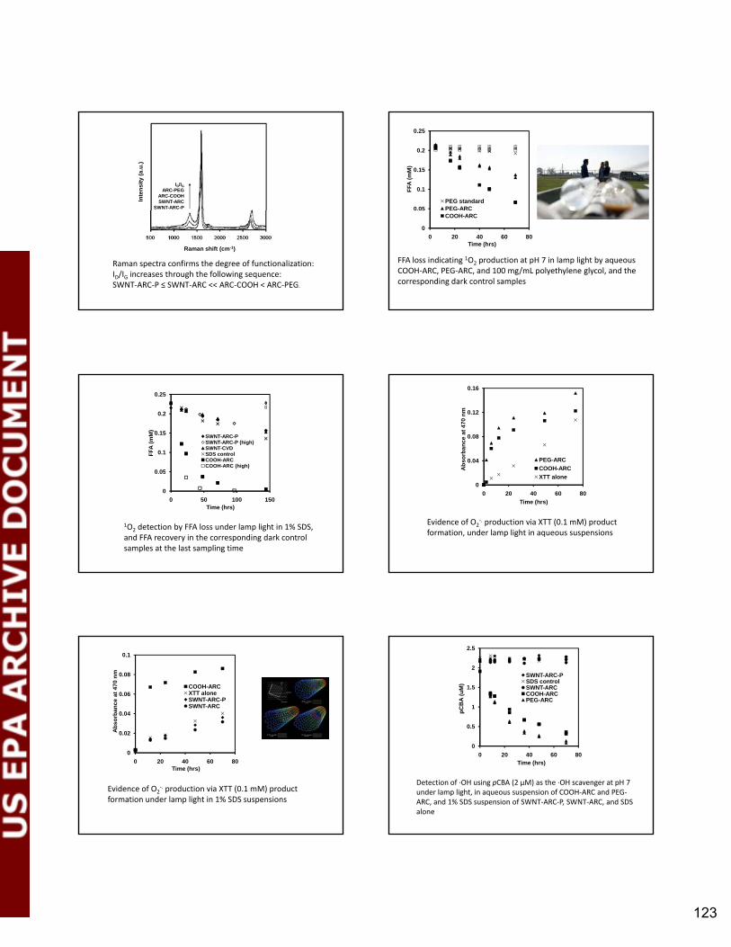
123
ID/IGARC-PEG
ARC-COOHSWNT-ARC
SWNT-ARC-P
Inte
nsity
(a.u
.)
Raman spectra confirms the degree of functionalization: ID/IG increases through the following sequence: SWNT‐ARC‐P ≤ SWNT‐ARC << ARC‐COOH < ARC‐PEG.
Raman shift (cm-1)
0.05
0.1
0.15
0.2
0.25
FFA
(mM
)
PEG standardPEG-ARC
FFA loss indicating 1O2 production at pH 7 in lamp light by aqueous COOH‐ARC, PEG‐ARC, and 100 mg/mL polyethylene glycol, and the corresponding dark control samples
00 20 40 60 80
Time (hrs)
COOH-ARC
0.05
0.1
0.15
0.2
0.25
FFA
(mM
)
SWNT-ARC-PSWNT-ARC-P (high)SWNT-CVDSDS controlCOOH-ARCCOOH-ARC (high)
1O2 detection by FFA loss under lamp light in 1% SDS, and FFA recovery in the corresponding dark control samples at the last sampling time
0
0.05
0 50 100 150Time (hrs)
0.04
0.08
0.12
0.16
Abs
orba
nce
at 4
70 n
mPEG-ARCCOOH-ARC
Evidence of O2‐. production via XTT (0.1 mM) product
formation, under lamp light in aqueous suspensions
00 20 40 60 80
Time (hrs)
XTT alone
0.02
0.04
0.06
0.08
0.1
Abs
orba
nce
at 4
70 n
m
COOH-ARCXTT aloneSWNT-ARC-PSWNT-ARC
Evidence of O2‐. production via XTT (0.1 mM) product
formation under lamp light in 1% SDS suspensions
00 20 40 60 80
Time (hrs)
0.5
1
1.5
2
2.5
pCB
A(u
M)
SWNT-ARC-PSDS controlSWNT-ARCCOOH-ARCPEG-ARC
00 20 40 60 80
Time (hrs)
Detection of ∙OH using pCBA (2 μM) as the ∙OH scavenger at pH 7 under lamp light, in aqueous suspension of COOH‐ARC and PEG‐ARC, and 1% SDS suspension of SWNT‐ARC‐P, SWNT‐ARC, and SDS alone
mosvald
Text Box
123

124
0.05
0.1
0.15
Abs
orba
nce
at 4
70 n
mSWNT-COOHfilt t t H 7
0.05
0.1
0.15
0.2
0.25
FFA
(mM
)
pure water at pH 7COOH-ARC filtrateCOOH-ARC
(a) (b)
00 10 20 30 40
Time (hrs)
filtrate at pH 7XTT alone
(a) 1O2 and (b) O2‐. production ‐ at pH 7 in lamp light by
aqueous COOH‐ARC, COOH‐ARC filtrate, and pure water.
00 15 30 45 60
Time (hrs)
0.5
1
1.5
2
2.5
pCB
A (u
M) COOH-ARC w/ catalase
pure water at pH 7COOH-ARC filtrateCOOH-ARC (Catalase reacts with H202)
00 20 40 60 80
Time (hrs)
Detection of ∙OH using pCBA (2 μM) as the ∙OH scavenger at pH 7 under lamp light, in aqueous suspension of COOH‐ARC, COOH‐ARC filtrate, COOH‐ARC and catalase (500 U/mL), and pure water control.
0.2
0.4
0.6
0.8
1
1.2
pBB
A (C
t/C0)
Ni 2+filtrate at pH 7 + deferoxaminefiltrate at pH 7
0.2
0.4
0.6
0.8
1
1.2
pCB
A (C
t/Co)
H2O2 dark controlH2O2 @ SunlightH2O2 @ lamp light
00 20 40 60 80
Time (hrs)
filtrate at pH 70
0 10 20 30 40 50Time (hrs)
H2O2 @ lamp light
Detection of ∙OH using pCBA (2 μM) as the ∙OH scavenger at pH 7 under lamp light and/or sunlight in (a) aqueous suspension of COOH‐ARC, COOH‐ARC filtrate, COOH‐ARC w/deferoxamine, and NiCl2 (2 mg/L), and (b) H2O2 100 μM solution, and the corresponding dark control samples
0.5
1
1.5
Abs
orba
nce
at 4
70 n
m
COOH-ARC+NADHNADHSWNT-ARC-P+NADH
Effects of NADH (0.2 mM) on O2‐. production
detected via XTT (0.1 mM) product formation at pH 7 under lamp light and dark controls
00 1 2 3
Time (hrs)
Reactions that may occur upon sunlight absorption by functionalized SWNTs
Oxic aqueous colloidal dispersions of both types of functionalized nanotubes generated ROS (1O2, O2
.‐, and ∙OH) in sunlight.
Both Type I and Type II photochemical pathways occur by the functionalized SWNTs in sunlight.
It appears that the functionalized SWNTs can act
Summary
as the electron donor directly (resulting in a change in their properties) or can shuttle electrons from other electron donors to form these reactive oxygen species.
mosvald
Text Box
124

125
Questions?Questions?
mosvald
Text Box
125

2010 U.S. EPA Nanotechnology Grantees Meeting
The Office of Research and Development’s National Center for Environmental Research 126
Qilin Li
Impact of Photochemical Oxidation on the Stability of nC60 and Multi-Walled
Carbon Nanotubes in Aqueous Solutions
Xiaolei Qu, Yu Sik Hwang, Pedro J.J. Alvarez, and Qilin Li Department of Civil and Environmental Engineering, Rice University, Houston TX
In natural aquatic systems, various environmental factors including natural organic matter and sunlight can interact with engineered carbon nanomaterials and influence their transport. The main objective of this study was to investigate the impact of natural organic matter and sunlight on the aggregation and deposition behaviors of aqueous C60 fullerene nanoparticles (nC60) and multiwalled carbon nanotubes. Suwannee River humic acid (SRHA) standard (II) and Elliot soil humic acid (ESHA) were used as the model aquatic and soil organic matter, respectively, and the UVA fraction of sunlight was simulated with UV lamps with output wavelength and intensity of 350 ± 50 nm and 1.66 mW/cm2, respectively. Initial aggregation rates of nC60 and carboxylated multiwalled carbon nanotubes (COOH-MWCNTs) before and after irradiation in solutions of different ionic strength, ionic composition, and humic acid concentration were determined from time resolved particle size measurement using dynamic light scattering. Deposition onto SiO2 surfaces was characterized using a quartz crystal microbalance with dissipation (QCMD) and compared to results from traditional column experiments; the impact of soil organic matter was investigated using ESHA coated SiO2 crystals or quartz sand. Our study revealed that UVA irradiation in the presence of dissolved oxygen introduced oxygen-containing function groups on nC60 surface, but reduced the oxygen content of the COOH-MWCNTs. Such changes in surface chemistry greatly altered the humic acid adsorption capacity and aggregation and deposition behavior of these carbon nanomaterials. In NaCl solutions, UVA irradiation induced surface oxidation remarkably increased nC60 stability by increasing the negative surface charge and reducing surface hydrophobicity. On the contrary, UVA irradiation reduced nC60 stability in CaCl2 solutions due to specific interactions of Ca2+ with the oxygen-containing functional groups on the UVA-irradiated nC60 surface and the consequent charge neutralization. In the absence of Ca2+, the surface photochemical oxidation greatly reduced the adsorption of SRHA on nC60 surface, resulting in weak dependence of nC60 stability on SRHA; Ca2+, on the other hand, facilitated SRHA adsorption on the UVA-irradiated nC60 surface by neutralizing surface charges of both UVA-irradiated nC60 and SRHA as well as forming intermolecular bridges, leading to enhanced stability in the presence of SRHA. Deposition of nC60 onto silica surface was found to be controlled by electrostatic interactions. The attachment efficiency increased with increasing ionic strength due to surface charge screening. ESHA adsorbed on the quartz crystal and sand surfaces hindered nC60 deposition at NaCl concentrations between 5 and 40 mM. However, at lower NaCl concentrations, enhanced deposition was observed on ESHA coated quartz crystal and sand. UVA irradiation affected stability of the COOH-MWCNTs differently. Unlike nC60, the stability of COOH-MWCNTs in NaCl solutions decreased with increasing UVA irradiation time. Meanwhile, the deposition rate of the COOH-MWCNTs onto silica surface increased by 2.6 times after 1 week of UVA irradiation. Particle electrophoretic mobility measurements suggested that UVA-irradiated COOH-MWCNTs were less negatively charged than the pristine COOH-MWCNTs, consistent with their higher aggregation and deposition rates. Based on the reduced oxygen content observed in XPS analyses, we speculate that the COOH-MWCNT surface underwent decarboxylation during the UVA irradiation, but the underlying mechanism remains unclear and requires further study.

2010 U.S. EPA Nanotechnology Grantees Meeting
The Office of Research and Development’s National Center for Environmental Research 127
Immediate future study will focus on two tasks to: (1) determine the impact of UVA irradiation on aquatic NOM adsorption to nC60 particle surface. NOM sorption will be investigated via batch adsorption and QCMD experiments under various solution conditions and (2) investigate the effect of organic matter content in sediment and soil on nC60 deposition/sorption. Batch sorption and column experiments will be conducted. References: 1. Hwang YS and Li QL. Characterizing photochemical transformation of aqueous nC(60) under
environmentally relevant conditions. Environmental Science and Technology 2010;44:3008-3013.
2. Qu X, Hwang YS, Alvarez PJJ, Bouchard D, and Li Q. UV irradiation and humic acid mediate aggregation of aqueous fullerene (nC60) nanoparticles. Environmental Science and Technology (DOI: 10.1021/es101947f).
EPA Grant Number: R834093

Qilin Li
Department of Civil & Environmental EngineeringRice UniversityRice University
Research ObjectivesC60
Research Objectives
Aqueous suspensionRelease
Living organismsExposure
MWCNTsAqueous suspension Living organisms
Interactions with Environmental Components( NOM d li ht)(e.g. NOM and sunlight)
Changes of physicochemical properties in nature aquatic systems (e.g. interacts with NOM and sunlight )
Resulting changes of transport pattern (e.g. aggregation and deposition)
Suspension Preparation by Sonicationp p y
Sonication/
C60
MWCNT-COOH
(2μm Filtration)
MWCNT-COOH
20
25
1
1.2
2.5 mg/L5 mg/L
nC60MWCNT COOH
5
10
15
0 2
0.4
0.6
0.8
AbS
.
5 mg/L7.5 mg/L10 mg/L
200 nm
0
0 100 200 300 400Particle Size (nm)
0
0.2
200 400 600 800
Wavenumber (nm)
Photochemical TransformationPhotochemical TransformationUVA-irradiation CharacterizationSonication
Particle size (DLS)Zeta potential
C concentrationC60 concentrationUV/VisXPSFTIR
- Luzchem reactor - Four Hitachi lamps
(300-400 nm, 8W) Measurement of( , )- 2 mW/cm2
- 3.8 – 4.0 mg/L- pH : 5.5-6.0
Measurement ofaggregation anddeposition rate
Photochemical Oxidation of nC6060
UVA-1week
Coun
t/sec
1
ount
/sec
unt/s
ec After etching
C
23Co
Cou
P k P i i % C(1 ) C b
275280285290295300C(1s) binding energyC(1s) binding energy
275280285290295300C(1s) binding energy
Peak Position % C(1s) Carbon1 284.3 66% Underivatized C(C=C)
2 285.6 19% Monooxygenated C
( C O)(e.g., C-O)
3 288.0 15% Di-oxygenated C
(e.g., O-C-O and C=O) Hwang, Y. S.; Li, Q. L. ES&T, 2010,44, 3008 3013.
UVA Irradiation Increases nC60Stability in NaCl
220
240
0 day1 day2 days
3 days7days
nm)
180
2002 days
Rad
ius
(n
140
160
dyna
mic
100
120
Hyd
ro
300 mM NaCl800 200 400 600 800 1000 1200 1400 1600
Time (s)
300 mM NaCl
Qu et al., ES&T, 2010
mosvald
Text Box
128

UVA Irradiation Increases nC6060Stability in NaCl
1 )
1
Pristine7DUV
cy,
-1
0Pristine 7DUV
8 m2 V
-1 S
-1
1
Effi
cien
c
-2
obili
ty (1
0-80.1
achm
ent
-3ph
oret
ic M
o
0.010.01 0.1 1
Atta
N Cl C t ti (M)
84mM 449 mM-4
0.001 0.01 0.1Elec
trop
N Cl C t ti (M)NaCl Concentration (M) NaCl Concentration (M)
Qu et al., ES&T, 2010
Humic Acid Adsorption Mediated by Surface Oxidationby Surface Oxidation
7DUV7DUV + 1 mg/L SRHAy,
1
PristinePristine + 1 mg/L SRHA
cien
cy,
17DUV + 10 mg/L SRHA
Effic
ienc
y
0.1
hmen
t Effi
c
0.1
tach
men
t
0.010.01 0.1 1
Atta
ch
84 mM 331 mM
160
180
200
dius
(nm
)
0.010.1 1
Att
NaCl Concentration (M)
449 mM 563 mMPristine nC60 10 mg/L SRHA
NaCl Concentration (M)
nC60
SRHA added
SRHA after sorption
q (mg/kg)(mg/L) (mg/L) (mg/L) 120
140
160
ynam
ic R
ad 10 mg/L SRHA1.5 M NaCl
Pristine 192 0.934 0.702 12097DUV 192 0.947 0.971 - 80
100
0 200 400 600 800 1000 1200Hyd
rody
Time (s)Qu et al., ES&T, 2010
UVA Irradiation Decreases nC60Stability in CaCl2
1 )
10
ncy,
-0.5
0
0-8 m
2 V-1
s-
1
nt E
ffici
e
-1.5
-1
Mob
ility
(10
0.1
PristineAtta
chm
en
-2.5
-2
Pristineopho
retic
M
0.010.001 0.01 0.1
7DUVA
CaCl2 Concentration (M)
3.2 mM 4.2 mM-3
0 0.001 0.002 0.003 0.004 0.005 0.006
7DUV
Elec
tro
CaCl2 Concentration (M)
Qu et al., ES&T, 2010
Humic Acid Increases nC60Stability in CaCl2
1
PristinePristine + 1 mg/L SRHA
cy,
7DUV7DUV + 1 mg/L SRHA
cy,
1
t Effi
cien 1
t Effi
cien
0.1
tach
men
t
0.1
tach
men
t0.01
0.001 0.01 0.1 1
Att
CaCl Concentration (M)
4.2 mM 12.8 mM0.01
0.001 0.01 0.1 1A
ttC Cl C t ti (M)
3.2 mM 5.9 mM
CaCl2
Concentration (M) CaCl2
Concentration (M)
Qu et al., ES&T, 2010
Ca2+ Enhances SRHA Adsorption on UVA irradiated nC60
2
0
2
z)
-4
-2
hang
e (H
z
3 mM CaCl2
UVA irradiation
-6
quen
cy C
h 2
-10
-8
Pristine nC60 DI waterUV nC60 DI water
Freq
-120 1000 2000 3000 4000 5000
UV nC60 3 mM CaCl2
Time (s)
UVA Irradiation Reduces MWCNT Stability in NaCl
0-1)
1
10
ncy,
52 mM 175 mM -1
0
-8 m
2 V-1
S-
0.1
1
nt E
ffici
en
-2
Mob
ility
(10
0.01
Pristine MWCNTttach
men
-3
Pristine MWCNTphor
etic
M
0.0010.01 0.1 1
Pristine MWCNTUV MWCNTA
t
NaCl Concentration (M)
-4
10 100
Pristine MWCNTUV MWCNT
Elec
trop
NaCl Concentration (mM)NaCl Concentration (M) NaCl Concentration (mM)
mosvald
Text Box
129

L f O F ti l GLoss of Oxygen Functional Groups
sc/
s
250300350400450500550
Binding Energy (eV)
Original MWCNT COOH (14% O)
278280282284286288290292294296298Binding Energy (eV)
Irradiated MWCNT COOH (11% O)
Unchanged Stability in CaClUnchanged Stability in CaCl2
1 )
1ncy, 0.8 mM
-0.5
0
8 m2 V
-1 S
-1
nt E
ffici
en
1.1 mM
-1
obili
ty (1
0-8
0.1
Pristine MWCNTttach
men
-1.5
Pristine MWCNTphor
etic
Mo
0.010.0001 0.001 0.01 0.1
Pristine MWCNTUV MWCNTA
t
CaCl2 Concentration (M)
-20.0001 0.001 0.01
Pristine MWCNTUV MWCNT
Elec
trop
CaCl2 Concentration (M)CaCl2 Concentration (M) CaCl2 Concentration (M)
Concluding RemarksConcluding Remarks
Sunlight irradiation and humic acid sorption mediate nC60 and MWCNT-COOH aggregation
Both specific and nonspecific (i.e., DLVO) interactions are involved
nanocarbon surface chemistry plays a key role in its environmental fate and transport.
Ongoing Research and FutureOngoing Research and FutureDirections
Impact of UVA irradiation and NOM on sorption/deposition and transport in subsurfacesorption/deposition and transport in subsurfaceporous medium
N t f NOM ti ilNature of NOM: aquatic vs. soil
Properties of suspended solids/sediment/ aquifer dimedia
Impact of UVA irradiation and NOM on bioavailability and bioaccumulation
C60 D iti SiOnC60 Deposition on SiO2
5
010Bare SilicaEHA-coated silica,
-10
-5
al (m
V)
1
EHA coated silica
ffici
ency
,
-20
-15
Pot
entia
0.1
hmen
t Ef
-30
-25
1 10 100
SiO2SiO2-EHA
0.011 10 100
Atta
ch
1 10 100NaCl (mM)
1 10 100
NaCl (mM)
AcknowledgementAcknowledgement
NSF Center for Biological and Environmental Nanotechnology (Award EEC-0647452)
USEPA STAR program (Grant No. 834093)
mosvald
Text Box
130

Questions and commentsQuestions and comments
Fullerene cosmetics
mosvald
Text Box
131

2010 U.S. EPA Nanotechnology Grantees Meeting
The Office of Research and Development’s National Center for Environmental Research 132
Qingguo Huang
The Environmental Behaviors of Multi-Walled Carbon Nanotubes
In Aquatic Systems
Qingguo Huang1, Marsha C. Black2, Liwen Zhang1, Emily R. Roberts2, and Elijah Petersen3
1Department of Crop and Soil Sciences, University of Georgia, Griffin, GA; 2Department of Environmental Health and Science, University of Georgia, Athens, GA; 3Chemical Science and Technology Laboratory,
National Institute of Standards and Technology, Gaithersburg, MD
Our study was designed to investigate the environmental behavior of water-dispersed carbon nanotubes in natural aquatic systems (i.e., water-sediment phase distribution, possible degradation, ecological exposure and toxicity), thereby providing useful information for environmental risk assessment and potential waste treatment. We have used C14-labeled multi-walled carbon nanotubes (14C-MWNTs) in our experiments to unambiguously identify and quantify carbon nanotubes from various natural materials, including water, sediments and organisms. Our experiments have yielded important information regarding three important behaviors of MWNTs in aquatic systems: water/solid phase distribution, biotic degradation, and possible toxicity and exposure to aquatic organisms. The results on each topic are briefly summarized below, respectively. We conducted experiments to examine the phase distribution of 14C-MWNTs in aqueous systems containing peat, shale, or clay as model solid phases under a series of varying pH and ionic strength conditions. Our results suggest that solid matter interacts with water-dispersed MWNTs via three interactive processes: (1) dissolved cations tend to promote MWNT aggregation via double layer compression; (2) dissolved organic matters released from the solid phase tend to stabilize MWNT dispersion; and (3) MWNTs sorb to the solid phase, primarily driven by hydrophobic interaction. All processes are variously influenced by aqueous conditions (e.g., pH, electrolytes) and their interplay governs the phase distribution of MWNTs. Recent studies have discovered biotic degradations of fullerols and single-walled carbon nanotubes (SWNTs), but there has not been a report on microbial degradation of MWNTs. We in our study found an enrichment culture that is capable of mineralizing 14C-MWNTs into 14CO2. Our initial study indicates that the mechanism involved in MWNT degradation seems to differ from that of fullerols and SWNTs. The microorganisms responsible for MWNTs degradation may not be fungi, but a consortium of bacteria, and the peroxidases that were found responsible for SWNT and fullerol degradation were absent in the MWNT-degrading culture. Currently, we are conducting a systematic study to identify and characterize the microorganisms that are responsible for degrading MWNTs and the biochemical pathways that are involved in MWNT metabolism. We studied chronic effects of 14C-MWNTs on Ceriodaphnia dubia, an aquatic invertebrate, in 8-day exposures. For chronic exposures, 14C-MWNTs were solubilized in moderately hard water (MHW) by four different methods: bath sonication (Branson) for 2 h; probe sonication for 2 h with 50 sec. pulses (Cole-Parmer 500-Watt Ultrasonic Homogenizer); bath sonication followed by addition of Sewanee River natural organic matter (NOM; final concentration = 4.5 mg/L); and by stirring nanotubes overnight in 4.5 mg/L Sewanee River NOM dissolved in MHW. Ceriodaphnia exposed to bath-sonicated MWCNTs had significantly smaller brood numbers and size at the 2.5 mg/L concentration (LOEC), compared with controls. Chronic exposures with probe-sonicated nanotubes showed less reproductive toxicity, with a LOEC of 5 mg/L. No reproductive toxicity was observed for nanotube exposures with added NOM. Reproductive toxicity of the bath-sonicated nanotubes may be related to association of the MWNTs onto the body surfaces of the adults, which likely interfered with molting and prevented neonate release. EPA Grant Number: R834094

133
The Environmental Behaviors of SolubilizedMulti‐walled Carbon Nanotubes
in Aquatic Systems
834094
Qingguo Huang1, Marsha C. Black2, Liwen Zhang1, Emily R. Roberts2, Elijah Petersen3
1Department of Crop and Soil Sciences, University of Georgia, Griffin, GA 302232Department of Environmental Health Science, University of Georgia, Athens, GA
3Chemical Science and Technology Laboratory, NIST, Gaithersburg, MD
Objective
• Solubilized CNTs Mobility Exposure
• Tasks– 1) Sorption
– 2) Transformation
– 3)Toxicity, accumulation and transfers
Contents
1. Objective
2. Sorption
3. Transformation
4. Toxicity/Bioaccumulation
Dispersed CNTs
Sorption
Phase Distribution
4
Aggregation Sorption
SedimentsDissolved Species
Three Treatments
BlankPeat
Dissolved Solid peat
Treatment 1: H2O + CNTs
Treatment 2: peat DOM + CNTs
Treatment 3: Peat + CNTs
Speciespeat
Effect of Peat
100
150
[Na+] = 40 mM
0 5 10 15 20 25 30
Apparent Solid Phase MWNT (µg)
Aqueous MWNT (µg ml-1)
50
0
[Na+] = 4 mM
[Na+] = 0 mM
mosvald
Text Box
133

134
Effect of Shale
80
120
Solid Phase
BLK 0 mM 0 mM
BLK 4 mM 4 mM
BLK 40 mM 40 mM
-40
0
40
0 4 8 12
MWNT (µg)
Aqueous MWNT (µg ml-1)
0
Contents
1. Objective
2. Sorption
3. Transformation
4. Toxicity/Bioaccumulation
Reactivity Biotransformation: Fullerene
COCO22
White Rot Fungus: Phanerochaete chrysosporium
• Lignin Peroxidase
Safety BottleSafety Bottle 0.5 M 0.5 M NaOHNaOH
Aeration (OAeration (O22))
Mineralization: Mineralization: Complete Complete
degradationdegradation
1414CC‐‐MWNTs, MWNTs, Fungus, Fungus, NutrientsNutrients
solutionsolution
Oops...
Control with nonControl with non‐‐sterili ed MWNTsterili ed MWNT
Control with sterilized Control with sterilized sterilized MWNTsterilized MWNT MWNTMWNT
mosvald
Text Box
134

135
NT
Mas
s (u
g)
1
10
ge (%
)5
6
7no SR NOMwith 8 ppm SR NOMPercentage of MWNTs Mineralized
Mineralization
Initial MWNT Mass (ug)
50 28.5 10 5.7 4 3.2
Min
eral
ized
MW
N
.01
.1
1
Perc
enta
g
1
2
3
4
Molecular DNA Identification Sequence Results
Outline of Research PlanContents
1. Objective
2. Sorption
3. Transformation
4. Toxicity/Bioaccumulation
mosvald
Text Box
135

136
Chronic Exposures with Ceriodaphnia dubia
Goal: Evaluate reproductive toxicity and accumulation of MWCNTs by adult and neonate Ceriodaphnia dubia in solutions that are prepared by two methods
• Bath sonication
• Sewanee River NOM (4.5 mg/L)
• 7-day chronic test – C. dubia <24 h old; 3 brood test (US EPA)
– 14C -MWNT concentrations: 1.25-5 mg/L (in MHW)
– Solubilization procedures
– Daily renewal (exposure water + food)
– Endpoints: # of broods, # of offspring• NOEC, LOEC calculated by ToxCalc®
– Accumulation measured by LSC (14C)
Results
Treatment NOEC LOEC
MWNT (sonicated) 1.25 mg/L 2.5 mg/L
MWNT (NOM) >5 mg/L >5mg/L
Reproductive Effects of MWNTs in C. dubia
Treatment NOEC LOEC
MWNT (sonicated) >5 mg/L >5 mg/L
MWNTs (NOM) 2.5 mg/L 3.75 mg/L
Accumulation of MWNTs in C. dubia neonates
Discussion and Conclusions
2.5 mg/L MWNT (40x) 2.5 mg/L MWNT+NOM (40x)Control (40x)
• Sonicated MWNTs adhered to adult organisms– Prevented molting/release of neonates = fewer broods
• NOM protective against reproductive toxicity– No observed adherence to adults
• Significant accumulation of NOM‐solubilized MWCNTs in neonates– NOM‐MWCNTs were consumed (vs. diffusion)?– Clumping of sonicated MWCNTs prevented consumption?
What’s Next?
• Feeding studies
– C. dubia fed Artemia exposed to MWNTs
– Fathead minnows fed C. dubia exposed to MWCNTs
Trophic transfer?– Trophic transfer?
• Full lifecycle exposures of MWNTs
– Fathead minnow
– Maternal transfer?
Acknowledgements
• Major ParticipantsLiwen Zhang; Emily Roberts; Dr. Marsha Black; Dr. Zhengwei Pan; Dr. Elijah Petersen; Dr. Mussie Habteselassie
• Major CollaboratorjRoger Pinto; Yenjun Zhuang; Vijaya Mantri; Wen Zhang; Dr. Yongsheng Chen; Dr. Aaron Thompson
• EPA STAR support
mosvald
Text Box
136

Day 2, Tuesday, November 9, 2010
AM Session 1: Effects on Cells

2010 U.S. EPA Nanotechnology Grantees Meeting
The Office of Research and Development’s National Center for Environmental Research 138
Amiraj Banga
Functional Effects of Nanoparticle Exposure on Airway Epithelial Cells
Amiraj Banga1, Frank A. Witzmann2, and Bonnie L Blazer-Yost1,2
1Department of Biology, Indiana University Purdue University at Indianapolis, Indianapolis, IN; 2Department of Cellular and Integrative Physiology, Indiana University School of Medicine,
Indianapolis, IN
Nanotechnology, the creation and manipulation of structures and systems at a nanoscale level (< 100 nm), significantly alters fundamental properties from large-scale materials. With nanotechnology being a focused area of exponential scientific and industrial growth in the last few decades, concerns have arisen regarding the potential biological effects of nanoscale materials. These effects remain poorly understood, especially with regard to occupational and environmental hazards. Populations exposed to increasing levels of nanomaterials include not only workers exposed during the production, recycling, and disposal, but also to the general population that uses commercially available nanomaterial-containing products and is exposed to them via environmental contamination.
The unique physico-chemical properties of these nanoscale products cause them to interact with cellular
systems in an unknown and undefined manner. Demonstrated effects include oxidative stress, inflammatory cytokine production, DNA mutation membrane damage, and even cell death.1 Although the nanotechnology industry holds great promise in the future, its darker side has to be explored to obtain the maximum benefits from this industry in a safe manner.
Carbon-based nanoparticles are one group of widely produced nanomaterials both industrially and
environmentally. These include fullerenes and nanotubes (Single-wall carbon nanotubes [SWCNT] and Multi-wall carbon nanotubes [MWCNT]). Fullerenes or Buckyballs are the most stable and are composed of 60 carbon atoms with an average diameter of 0.72 nm. Carbon nanotubes are graphite sheets rolled to form seamless tubes or cylinders. Whereas SWCNT consist of a single layer with diameters ranging near 1 nm, MWCNT are larger and consist of many single-walled tubes stacked one inside the other with diameters reaching 100 nm. Because of their nano sizes, fibrous shapes, and carbon base, CNTs are expected to behave differently than the large-sized particles. They are potentially toxic like other small fibers (asbestos and silica) and biopersistent because of their stability.
One primary route of nanoparticle uptake in the body is through inspiration of airborne nanoparticles.
Combustion-derived nanoparticles have been shown to cause lung cell injury and inflammation due to oxidative stress2 that may manifest itself as airway disease, cardiovascular disease, fibrosis, or cancer.3 Using quartz and carbon nanoparticles at equal mass dose, it was concluded that SWCNT in the lungs were far more toxic than carbon black and even quartz.4 Following inhalation, ultrafine carbon particles can travel through the circulatory system and invade the brain.5 Nanoparticles can have prothrombotic effects in vivo and demonstrate platelet activation in vitro, as has been shown in response to SWCNT exposures.6 They also demonstrated that MWCNTs can reach the subpleural tissue in mice with a single inhalation dose of 30 mg/m3 for 6 h. A stable C60 suspension has been shown to produce genotoxicity as a result of DNA damage in human lymphocytes.7
One of the respiratory cell lines commonly used for tracheobronchial epithelial cell studies is Calu-3.
Although it is adenocarcinoma in origin, it is one of the few cell lines that form tight junctions in vitro and produces features of a differentiated, functional human airway epithelium. Calu-3 cell line is a human airway epithelial cell line that responds to epinephrine with an increase in Cl- secretion via Cystic Fibrosis Transmembrane Regulator channel (CFTR). Water follows Cl- and together with mucous helps to clear the airways of any foreign substances. The cells show a high resistance phenotype after 13 days of growth. The

2010 U.S. EPA Nanotechnology Grantees Meeting
The Office of Research and Development’s National Center for Environmental Research 138
current studies utilize this well-characterized model to study the effects of unpurified, as manufactured, nanoparticles that are most likely to be found as environmental and occupational pollutants.
We hypothesized that exposure of epithelial cells to unpurified, as manufactured CNPs such as C60, SWNT
and MWNT, may alter the function of barrier epithelia. The effect of exposure of each of three different CNPs was studied in air-interface cultured Calu-3 cell model over seven orders of magnitude (4 µg/cm2-0.004 ng/cm2). Electrophysiological techniques were used to study transepithelial ion transport and the barrier function expressed as Trans Epithelial Electrical Resistance (TEER). After 48 h of exposure to CNPs, fullerenes did not show any effect on TEER, whereas the nanotubes significantly decreased TEER over a wide range of concentrations (4 µg/cm2-0.004 ng/cm2). The ion transport response to epinephrine also was significantly decreased by the nanotubes but not by fullerenes. To look at the effect of exposure times, cells were exposed to same concentrations of CNPs for 24 and 1h time periods. Although the 48 h and 24 h time period exposures exhibited same effects, there was no effect seen after 1 h in terms of TEER or hormonal responses. In cells exposed to either of the nanotubes, the TEER was not statistically different from control after treatment with 4 ng/cm2 concentration, whereas in the case of hormonal responses, the nanotubes, especially multi-walled, still showed significant inhibitory effect. To examine which step of the epinephrine stimulated intracellular pathway is affected by CNPs, cAMP assays were performed. The cAMP levels for the exposed cells vs. the control cells were not different, suggesting an effect manifested after the epinephrine-induced increase in cAMP.
Our results indicate that there are changes in response to physiologically significant nanoparticle
concentrations that could, in vivo, be manifested as changes in transcellular permeability and hormone responsiveness. Such effects could alter airway function, emphasizing the need of further study on the effect of these nanoparticles.
References:
1. Oberdörster G, Maynard A, Donaldson K, Castranova V, Fitzpatrick J, Ausman K, Carter J, Karn B,
Kreyling W, Lai D, Olin S, Monteiro-Riviere N, Warheit D, Yangvv H. Principles for characterizing the potential human health effects from exposure to nanomaterials: elements of a screening strategy. Particle and Fibre Toxicology 2005;2:8.
2. Donaldson K, Tran CL. Mutation Research 2004;53:5–9. 3. Mauderly JL, Snipes MB, Barr EB, Belinsky SA, Bond JA, Brooks AL, Chang IY, Cheng YS, Gillett NA,
Griffith WC, et al. Part I: neoplastic and nonneoplastic lung lesions. Research Report/Health Effects Institute 1994;68(1): 1–75.
4. Lam CW, James JT McCluskey R, Hunter RL. 2004. Pulmonary toxicity of single-wall carbon nanotubes in mice 7 and 90 days after intratracheal instillation. Toxicological Sciences 2004;77:126–34.
5. Oberdorster G, Sharp Z, Elder AP, Gelein R, Kreyling W, Cox C. Inhalation Toxicology 2004;16:437–45. 6. Bihari P, Holzer M, Praetner M, Fent J, Lerchenberger M, Reichel CA, Rehberg M, Lakatos S Krombach
F. Toxicology 2010;269(2-3):148-154. 7. Dhawan A, Taurozzi AS, Pandey AK, Shan W, Miller SM, Hashsham SA, Tarabara VV. Environmental
Science and Technology 2006;40:7394–7401. NIH/NIGMS Grant Number: R01GM085218
mosvald
Text Box
139

140
Functional Effects of Functional Effects of NNanoparticle anoparticle Toxicity on Airway Barrier Epithelial Toxicity on Airway Barrier Epithelial
Cell FunctionCell FunctionCell FunctionCell FunctionAmy Banga
Blazer-Yost/Witzmann Group IUPUI, Indianapolis
Health Hazard of NanoparticlesHealth Hazard of Nanoparticles
Workers handle nanoparticle materials inmany industrial jobs to produce consumers’items
Nanoparticles can enter the body by:Nanoparticles can enter the body by:-Inhalation-Swallowing-Penetration through the skin
Complete information about health effects islacking
MWCNTMWCNT(diameter ~2(diameter ~2--25 nm;25 nm;
SWCNTSWCNT(diameter as small as 1 nm;(diameter as small as 1 nm;length: few nm to microns)length: few nm to microns)
Images from: http://www.photon.t.uImages from: http://www.photon.t.u--tokyo.ac.jp/~maruyama/agallery/agallery.htmltokyo.ac.jp/~maruyama/agallery/agallery.html
(diameter 2(diameter 2 25 nm;25 nm;length: few nm to microns)length: few nm to microns)
CC6060 (fullerene)(fullerene)((AvgAvg diameter 0.72 nm)diameter 0.72 nm)
CNP purchased from SES Research, Inc., Houston, TX CNP purchased from SES Research, Inc., Houston, TX ((http://www.sesres.com)http://www.sesres.com)
HypothesisHypothesis
Manufactured, non-functionalized carbon nanoparticles (CNPs), when exposed to barrier
epithelia (lung/airway, kidney and intestinal cells), exert a biological effect on the cell
membrane and may alter the cell function.
ApproachApproachCell model used – Calu-3 (airway epithelial cells).
Features of cell line Forms a monolayer on permeable supports under
air-liquid interface culture conditions withTransepithelial Electrical Resistance (TEER) of800±80 Ω.cm2.
Simulates a barrier epithelium. Displays hormonal responsiveness of airway cells
in vivo.
Technique used - Electrophysiological studies wereused to determine the effect of CNPs onTransepithelial Electrical Resistance (TEER) thatmeasures barrier function and Short Circuit Current(SCC) that measures net ion transport.
Measuring net ion transport across the monolayerMeasuring net ion transport across the monolayer
ShortShort--Circuit Current (SCC) ElectrophysiologyCircuit Current (SCC) Electrophysiology
APICAL (LUMINAL)
SEROSAL
Porous Filter
Measuring net ion transport across the monolayerMeasuring net ion transport across the monolayer
Ussing Chamber
Serosal (plasma)
Apical (luminal)
mosvald
Text Box
140

141
ApproachApproach
CNP preparation – CNP suspended in FetalBovine Serum (FBS), sonicated, autoclaved andadded to serum free media. The amount of CNPwas regulated so as to obtain a desired finalconcentration of CNP in media when FBS wasdd dadded.
CNP exposure – The cells were incubatedwith CNP-FBS containing media at a normalconcentration of 15% for last 1, 24 or 48 h ofgrowth to simulate in vivo CNP exposure.
Conversion equationConversion equation
X µg/cm2 = 25X µg/ml.
ctric
al R
esis
tanc
e (
.cm
2 )
400
600
800
* * **
*
* * * **
*
48 h
Effect on the TEER of CaluEffect on the TEER of Calu--3 cells by exposure to different 3 cells by exposure to different types and concentrations of CNPs for 48 htypes and concentrations of CNPs for 48 h
Tran
sepi
thel
ial E
lec
0
200
18 3 3 2 2 3 3 3 2 3 2 2 3 3 3 2 3 2 2 3 3 3
Cont
rol
4 g
/cm
20.4
g/
cm2
40 n
g/cm
24 n
g/cm
20.4
ng/
cm2
40 p
g/cm
24 p
g/cm
24 g
/cm2
40 n
g/cm
2
0.4
g/cm
24 n
g/cm
20.4
ng/
cm2
40 p
g/cm
24
pg/c
m2
4 g
/cm2
0.4
g/cm
240
ng/
cm2
4 ng
/cm
20.
4 ng/
cm2
40 p
g/cm
24 p
g/cm
2
Fullerenes Single wall Multi wall
* indicates that the value was statistically different from the control value * indicates that the value was statistically different from the control value (P<0.05) using a Students’ t(P<0.05) using a Students’ t--test.test.
Response of CaluResponse of Calu--3 cells to epinephrine 3 cells to epinephrine (
A/c
m2 )
60
70
80
90
A/c
m2 )
70
80
90
EpinephrineNPPB
Time (min)
-30 -20 -10 0 10 20 30
SCC
(
20
30
40
50
Time (min)
0 2 4 6 8 10 12 14 16 18SC
C (
A40
50
60
Hormonal response of Calu-3 cells exposed to different types of nanoparticles for 48 h at a concentration of 4g/cm2 to epinephrine
SCC
( A
/cm
2 )
100
150
200
CC
( A
/cm
2 )
100
150
200
Epinephrine NPPB
Time (min)
-20 0 20 40
S
0
50
Control, n=9Fullerene, n=2Single walled, n=2Multi walled, n=2
Time (min)
0 2 4 6 8 10
SC
0
50
Epinephrine
Hormonal response of Calu-3 cells exposed to different types of nanoparticles for 48 h at a concentration of 4 pg/cm2 to epinephrine
C (
A/c
m2 )
100
150
200
C (
A/c
m2 )
100
150
200
Time (min)
-20 0 20 40
SCC
0
50
Control, n=9Fullerenes, n=2Single walled, n=2Multi walled, n=2
Time (min)
0 2 4 6 8 10
SCC
0
50
Epinephrine NPPB
mosvald
Text Box
141

142
ectr
ical
Res
ista
nce
( .c
m2 )
400
600
80024 h
**
* * **
*
Effect on the TEER of CaluEffect on the TEER of Calu--3 cells by exposure to different 3 cells by exposure to different types and concentrations of CNPs for 24 htypes and concentrations of CNPs for 24 h
Tran
sepi
thel
ial E
le
0
200
18 3 3 3 3 6 6 6 3 3 3 3 3 4 4 3 3 3 3 3 4 4
Cont
rol
4 g
/cm
20.
4 g
/cm
240
ng/
cm2
4 ng
/cm
20.
4 ng
/cm
240
pg/
cm2
4 pg
/cm
24 g
/cm
240
ng/
cm2
0.4 g
/cm
24
ng/c
m2
0.4
ng/c
m2
40 p
g/cm
24
pg/c
m2
4 g
/cm
20.
4 g
/cm
24
ng/c
m2
4 ng
/cm
20.
4 ng
/cm
240
pg/
cm2
4 pg
/cm
2 Fullerenes Single wall Multi wall
* indicates that the value was statistically different from the control value * indicates that the value was statistically different from the control value (P<0.05) using a Students’ t(P<0.05) using a Students’ t--test.test.
Hormonal response of Calu-3 cells exposed to different types of nanoparticles for 24 h at concentration of 4 g/cm2 to epinephrine
CC
( A
/cm
2 )
100
150
200
CC
( A
/cm
2 )
100
150
200
Time (min)
-20 0 20 40
SC
0
50
Control (n=11)Fullerenes (n=3)Single walled (n=3)Multi walled (n=3)
Time (min)
0 2 4 6 8 10
SC
0
50Epinephrine
NPPB
Hormonal response of Calu-3 cells exposed to different types of nanoparticles for 24 h at concentration of 4 pg/cm2 to epinephrine
CC
( A
/cm
2 )
100
150
200
C (
A/c
m2 )
100
150
200
Time (min)
-20 0 20 40
SC
0
50
Control (n=11)Fullerenes (n=3)Single walled (n=3)Multi walled (n=3)
Time (min)
0 5 10 15 20
SC
0
50Epinephrine
NPPB
ial E
lect
rical
Res
ista
nce
( .c
m2 )
400
600
800 1 h
Effect on the TEER of CaluEffect on the TEER of Calu--3 cells by exposure to different 3 cells by exposure to different types and concentrations of CNPs for an hourtypes and concentrations of CNPs for an hour
Tran
sepi
thel
i
0
200
Cont
rol
4 g
/cm
2
4 g
/cm
2
4 g
/cm
2
0,4 g
/cm
240
ng/
cm2
4 ng
/cm
2
0.4 g
/cm
240
ng/
cm2
4 ng
/cm
2
0.4 g
/cm
240
ng/
cm2
4 ng
/cm
2
Fullerenes Single wall Multi wall
5 5 3 410 3 3 3 3 3 35 5
* indicates that the value was statistically different from the control value * indicates that the value was statistically different from the control value (P<0.05) using a Students’ t(P<0.05) using a Students’ t--test.test.
Hormonal response of Calu-3 cells exposed to different types of nanoparticles for 1h at concetration of 4 g/cm2 to epinephrine
CC
( A
/cm
2 )
100
150
200
SCC
( A
/cm
2 )
100
150
200
Time (min)
-20 0 20 40
SC
0
50
control (n=10)Fullerene (n=3)Single walled (n=3)Multi walled (n=3)
Time (min)
0 5 10 15 20
S
0
50Epinephrine
NPPB
Hormonal response of Calu-3 cells exposed to different types of nanoparticles for 1 h at concetration of 4 pg/cm2 to epinephrine
CC
( A
/cm
2 )
100
150
200
CC
( A
/cm
2 )
100
150
200
Time (min)
-20 0 20 40
SC
0
50
control (n=10)Fullerenes (n=3)Single walled (n=3)Multi walled (n=3)
Time (min)
0 5 10 15 20
SC
0
50EpinephrineNPPB
mosvald
Text Box
142

143
CFTRCFTR
Cl- Cl-
PKAPKA
Apical (filtrate)
PKAPKA
Aquaporin
H2O
Epinephrine
cAMP
AdenylateCyclase
ATP
Basal (blood side)Blazer-Yost
cAMP cAMP
CNPConcentration
ofCNP(µg/cm2)
Control *CNP treated
SWCNT 4 6.5 7.6
MWCNT 4 8 3 6 6
Increase in Increase in cAMPcAMP concentration after epinephrine stimulationconcentration after epinephrine stimulation
Fold increase in cAMP after epinephrine stimulation
MWCNT 4 8.3 6.6
SWCNT 0.004 11.3 11.8
MWCNT 0.004 5.3 7.8
*CNP Treated = 48 hrs Carbon nanoparticle treated cells at concentrations indicated
SummarySummary
Low dose nanotube exposures decreases thebarrier function of airway epithelial cells.
Low dose nanotube exposures affects theability of the airway epithelial cells to secreteability of the airway epithelial cells to secretechloride.
These data suggest that levels of nanotubesfound in the workplace, particularly duringchronic exposures, are likely to havephysiological effects that can cause orexacerbate respiratory problems.
ThanksThanksThanksThanks
0
0.4 µg/cm2
1h 24 hPreliminary Imaging study
4 µg/cm2
40 µg/cm2
mosvald
Text Box
143

2010 U.S. EPA Nanotechnology Grantees Meeting
The Office of Research and Development’s National Center for Environmental Research 144
Galya Orr
Toxicity Assessment of Nanomaterials in Alveolar Epithelial Cells
at the Air-Liquid Interface
Galya Orr, Yumei Xie, Nolann Williams, Ana Tolic, Justin Teeguarden, and Alexander Laskin Pacific Northwest National Laboratory, Richland, WA
Airborne nanomaterials that enter the respiratory tract are likely to be deposited in the alveolar region, where alveolar epithelial cells are found at the interface with ambient air. These cells provide a vulnerable target for particles that escape the first line of defense by the alveolar macrophages. To date, the majority of in vitro studies characterizing the interactions and impact of engineered nanomaterials in these cells have been carried out in cells submersed under growth media. To more closely mimic in vivo exposures, we have established the growth of alveolar type II epithelial cells (C10 cell line) at the air-liquid interface (ALI), enabling realistic exposures to aerosolized nanoparticles. This approach supports accurate quantification of the delivered particles per cm2 (or particles per cell) by collecting the particles on millimeter-size grids or glass cover-slips, placed randomly over the cells and visualized using electron or fluorescence microscopy, respectively. This approach also enables physical and chemical characterizations of the collected nanoparticles, providing properties that are relevant to airborne nanoparticles and the actual exposure at the air-liquid interface. The cells have been cultured on membrane inserts and initially grown under submersed conditions until reached confluence. The apical surface of the cell monolayer was then exposed to ambient conditions for 24 hours, and the integrity of a representative subset of the cells has been monitored using propidium iodide, quantified by fluorescence microscopy. Exposures to aerosolized nanoparticles, generated using a vibrating membrane nebulizer, have been done over 10-minute sessions using an enclosed exposure chamber that ensures uniform particle delivery. The cells have been maintained at the ALI until assayed for lactose dehydrogenase (LDH) release to evaluate cell damage at 6 and 24 hours post exposure, and for proliferation rate (MTS) to evaluate viability at 24 hours post exposure. Using the above conditions, we found that exposures to 50 nm bare amorphous silica nanoparticles, containing embedded fluorescent dye molecules, elicit no significant cytotoxic response at concentrations ranging from 10 to 1000 particles per cell. These observations agree with observations we obtained in submersed cells exposed to equivalent doses, as estimated by a computational particokinetics (sedimentation, diffusion) model. Studies with surface modified aminated 50 nm amorphous silica nanoparticles and other particles that have shown to elicit toxic responses in submersed conditions are currently being pursued at the ALI. EPA Grant Number: R833338

145
Principal investigator did not authorize publication of the presentation.

2010 U.S. EPA Nanotechnology Grantees Meeting
The Office of Research and Development’s National Center for Environmental Research 146
Jonathan D. Posner
Interactions of Nanomaterials With Model Cell Membranes
Jonathan Posner1,2, Wen-Che Hou1,3, Steve Klein1,2, Babak Moghadam1,2, Charles Corredor1,2,
Kiril Hristovski4, and Paul Westerhoff4 1Chemical Engineering, 2Mechanical Engineering, 3Environmental Engineering, 4School of Sustainable
Engineering and the Built Environment, Arizona State University, Tempe, AZ Toxicological studies of engineered nanomaterials (ENMs) have primarily focused on the toxicity and uptake of ENMs by a variety of organisms, including human cell lines, microbes, plants, or aquatic organisms such as fish and Daphnia magna. Because the reported results vary depending on the organisms and test conditions, it is difficult to draw a comprehensive conclusion of ENMs’ environmental impact based on these empirical studies, especially considering the ecological diversity and wide range of ENMs’ properties. The partitioning between the organic solvent phases (typically n-octanol) and water (KOW) has traditionally been used as an empirical approach to evaluate the bioavailability of organic pollutants and is used extensively in current EPA models. For colloids, mechanistic and dynamic fate models in aqueous matrices are more complex than for organic pollutants and require multi-parameter input to describe the colloid transport and interactions with soils and biota. Characterization of ENMs often involves numerous physical measurements of size distribution, surface area, porosity, aqueous zeta potential, surface chemistry, and stability. However, it is challenging to transition from these precise measurements to models suitable to assess fate and bioavailability of ENMs in the environment, especially in complex matrices. Analogous partition type global descriptor methods have not been used extensively for nanomaterials; therefore, there is a need to develop empirical model approaches for predicting bioaccumulation of ENM that account for the collective influence of ENM properties, in a similar way as Kow depends on multiple parameters of organic pollutants (molecular weight, conformation, hydration states, ionic charge, etc.). In this talk, we quantify the lipid-water distribution coefficients for ENMs and use them as a global descriptor that captures the critical interactions between ENM and biological interfaces, which may be used to predict the bioaccumulation potential of ENM. The lipid-water distribution ratio has been shown to be a more appropriate descriptor than Kow partitioning for biological uptake and bioaccumulation of hydrophobic ionizable compounds and surfactants, which ENMs share similar properties (e.g. charged, resident at interfaces). Lipid bilayers’ mass is nearly all at the interface that eliminates the difficulty encountered in the octanol-water partitioning of surface-active compounds and some types of ENMs that also partition to the interfaces. We evaluate the lipid-water distribution of aqueous C60 clusters (nC60) and polyhydroxylated C60 aggregates (i.e., fullerol) using lipid bilayers that mimic biological membranes. The kinetic studies indicate that the distributions of nC60 and fullerol between lipid bilayers and aqueous phases reach pseudo-equilibrium after 2 days and 2 h of equilibration, respectively. The pseudo-equilibrium distribution of nC60 and fullerol can be described by isotherm-like behaviors that fit reasonably with Langmuir isotherms. Although both nC60 and fullerol exhibit pH-dependent distribution behaviors with accumulation in lipid bilayers increasing systematically with the decrease in pH from 8.6 to 3, the distribution coefficients of nC60 is up to 1 order of magnitude larger those of fullerol. The pH-dependent distribution trend is consistent with the decrease in the electrostatic repulsion between lipid bilayers and fullerene aggregates, as the zeta potentials of both increased (i.e., became less negative) as pH decreased. The lipid bilayers that make up cellular membranes are believed to be impenetrable to ions and unfunctionalized macromolecules; however, epidemiological studies have shown that unfunctionalized ENMs can, under some conditions, cross or disrupt the cell membrane through passive, unmediated routes causing

2010 U.S. EPA Nanotechnology Grantees Meeting
The Office of Research and Development’s National Center for Environmental Research 146
acute cellular toxicity and cell death. The unmediated ENM disruption of cellular membranes is poorly understood. Also, we are developing methods for understanding the mechanisms and conditions under which engineered nanomaterials can cause disruption of, and passive transport through, simplified model cell membranes, namely lipid bilayers. We will show that some ENM disrupt membranes allowing the flux of ions across the membranes. Distribution of ENM between the aqueous phase and biologically relevant interfaces and disruption of bilayers by ENM may ultimately be used as high-throughput global descriptors for predicting bioaccumulation and toxicity of ENM. Future work includes (1) extending current work to include ENMs of other core compositions, sizes, shapes, and surface functionalities as well as water chemistry parameters such as ionic strength and ion species; (2) correlate with existing or future bioaccumulation studies using aquatic organisms; and (3) translating methods to high-throughput formats. DOE Grant Number: DE-FG02-08ER64613
mosvald
Text Box
147
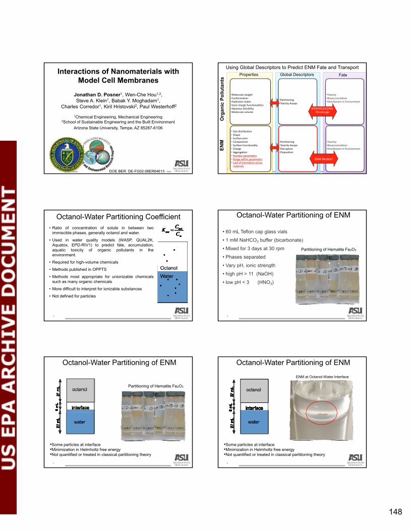
148
Interactions of Nanomaterials with Model Cell Membranes
Jonathan D. Posner1, Wen-Che Hou1,2, Steve A. Klein1, Babak Y. Moghadam1,
Charles Corredor1, Kiril Hristovski2, Paul Westerhoff2
1Chemical Engineering, Mechanical EngineeringChemical Engineering, Mechanical Engineering2School of Sustainable Engineering and the Built Environment
Arizona State University, Tempe, AZ 85287-6106
DOE BER: DE-FG02-08ER64613
Using Global Descriptors to Predict ENM Fate and Transport
Properties Global Descriptors Fate
•Molecular weight•Conformation•Hydration states•Ionic charge functionalities•Aqueous Solubility•Molecular volume
•Partitioning•Toxicity Assays
•Toxicity•Bioaccumulation•Distribution in Environment
Org
anic
Po
lluta
nts
EPIWIN,ECOSA, OncoLogic
2
• Size distribution• Shape• Surface area• Composition• Surface Functionality• Charge• Aggregation• Number parameters• Range within parameters• Lack of translation across materials
•Partitioning•Toxicity Assays•Disruption•Deposition
•Toxicity•Bioaccumulation•Distribution in EnvironmentE
NM
O
ENM Models?
Octanol-Water Partitioning Coefficient• Ratio of concentration of solute in between two
immiscible phases, generally octanol and water.
• Used in water quality models (WASP, QUAL2K,Aquatox, EPD-RIV1) to predict fate, accumulation,aquatic toxicity of organic pollutants in theenvironment.
• Required for high-volume chemicals
3
• Methods published in OPPTS
• Methods most appropriate for unionizable chemicalssuch as many organic chemicals
• More difficult to interpret for ionizable substances
• Not defined for particles
• 60 mL Teflon cap glass vials
• 1 mM NaHCO3 buffer (bicarbonate)
• Mixed for 3 days at 30 rpm
• Phases separated
• Vary pH ionic strength
Octanol-Water Partitioning of ENM
Partitioning of Hematite Fe2O3
4
• Vary pH, ionic strength
• high pH > 11 (NaOH)
• low pH < 3 (HNO3)
Octanol-Water Partitioning of ENM
Partitioning of Hematite Fe2O3
5
•Some particles at interface•Minimization in Helmholtz free energy•Not quantified or treated in classical partitioning theory
Octanol-Water Partitioning of ENM
ENM at Octanol-Water Interface
6
•Some particles at interface•Minimization in Helmholtz free energy•Not quantified or treated in classical partitioning theory
mosvald
Text Box
148

149
Octanol-Water Partitioning of ENM
Minimum in Helmholtz Energy
7
•Some particles at interface•Minimization in Helmholtz free energy•Not quantified or treated in classical partitioning theory•Basis for Pickering emulsions
Minimum in Helmholtz Energy
Octanol-Water Partitioning
A – in the aqueous phaseB – In the octanolC – at interface
Observe that ENM partitioning experiments result in combination of three primary, path and solution chemistry dependent scenarios:
8
• Importing into EPA models
• How do we treat mass at interface?
• Partitioning gives no information on state on ENM (aggregation and settled in water, dissolved, suspended, emulsion)
• Partitioning is path dependent
Challenges with ENM Octanol-Water Partitioning
9
• Does not correlate with bioacumulation*
• Partitioning dependent on poorly defined interfacial area
*Petersen et al. Relevance of Octanol-Water Distribution Measurements to the potential Ecological Uptake of Multi-Walled Carbon Nanotubes, Environmental Toxicology and Chemsitry, 2010
ENM Distributions in Lipid‐Water Systems
• Lipid bilayer is an important interfaces between life and its environment and a potential exposure route to ENMs.
• The lipid bilayer-water distribution (Klipw) has been shown to be a more appropriate indicator than (Kow) for bioaccumulation of ionizable organic molecular and surface active compounds, which ENMs share some properties (e.g., charged surface and residence in interface)
10
in interface).
• Klipw is increasingly used by the pharmaceutical industry and environmental research for drugs and molecular pollutants.
• All mass is at the surface
• Surface area can be controlled and quantified
Lipid Bilayers
11
• Primary constituent of many biological cellular membranes.
• Often used to model passive transport into cells.
Lipid bilayer-water distribution on Solid Supported Lipid Bilayers
12
10 microns
• Lipid Bilayer noncovalent bond to silica
• Bilayer is fluid• Bilayer robust over wide range
electrolyte conditions
mosvald
Text Box
149

150
Lipid Bilayer-Water Distribution of ENMs Method
Validated using a reference compound (2,4,6-trichlorophenol).
log Klipw = 3.89 ± 0.03 (L/kg), close to 3.90 reported by Escher et al. (ES&T, 1996)
ENMs and Lipid Sources and PreparationsENMs:
• Aqueous C60 aggregates (nC60): dry C60 powders (MER, Tucson, AZ) were pulverized and then mixed with DI water for 2 weeks prior to passing through 0.7 and 0.45 μm filters sequentially (Hou et al., 2009).
• Fullerol (C60(ONa)x(OH)y, x + y = 24): dry fullerol powder (MER, Tucson, AZ) was directly mixed with water and then passed through 0.7, 0.45, and 0.2 μm filters sequentially.
• Gold nanoparticles (nAu) were tannic acid coated and well-characterized gold colloids at 5, 10, 20, 50, and 70 nm, purchasing from nanoComposix (San g , , , , , p g p (Diego, CA).
Lipid bilayers:
• SSLMs were purchased from Sovicell (Leipzig, Germany). The lipid composition was 100% phosphatidylcholine from chicken egg.
• Unilamellar lipid bilayer vesicles (i.e., liposomes) were prepared using the same lipid composition as SSLMs by the extrusion method (Hope et al., 1985). Liposomes were used to determined the effective zeta potential of lipid bilayers.
Analytical methods• nC60 concentration was determined by high performance liquid
chromatography (HPLC) with UV detection at 336 nm. Because HPLC is only applicable to molecules, molecular C60 was extracted from the aqueous phase to toluene in the aid of 0.1 M Mg(ClO4)2. The toluene extract was injected to HPLC (Hou et al., 2009).
• Fullerol concentration was determined by UV-visible absorption spectroscopy using UV at 254 nm.
• nAu concentration was determined by inductively coupled plasma-optical emission spectroscopy (ICP-OES). Prior to ICP, nAu was dissolved in aqua y ( )regia (i.e., 1 part of HNO3 and 3 parts of HCl).
• Lipid concentration was determined by the malachite green dye assay (Petitou et al., 1978). Before assay, lipid was digested, adding concentrated H2SO4 and H2O2 under heating.
• The sizes and zeta potential of liposomes and ENMs were determined by dynamic light scattering (DLS) (NICOMP 380 ZLS, Particle Sizing Systems, Santa Barbara, CA).
Lipid bilayer vesicles ()Fullerol 650 mg/L (♦)nC60 6.5 mg/L (∆)
Size and Effective Zeta
nC60 and fullerolshave similar size distributions and charge
Fullerol and nC60 Lipid Bilayer Interaction Kinetics
[lipid] = 0.47 mM --- Controls are vial without SSLM ---- [nC60]0 = 6.5 mg/L at pH = 7.4 and 5. [fullerol]0=8.0 mg/L (pH 7.4); 11.0 mg/L (pH 7.4), and 11.0 mg/L (pH 3)
pH=3 (♦) and control (◊) pH=5 () and control () pH=7.4 () and control (∆)
nC60 Mass Balances
Black: free nC60 ---- Grey: nC60 in SSLMs ---- White: nC60 lost to walls
Serial ENM extraction from SSLM by original electrolyte then toluene
mosvald
Text Box
150

151
pH=3 (♦)pH=4 ()pH=5 ()pH=7.4 (∆)pH=8.6 ()
Distribution of nC60 and Fullerol in Lipid-Water
• Lines are Langmuir isotherms• Bars indicate one standard deviation.
pH=5 ()pH=7.4 (∆)
10000
100000
1000000
eq(m
g/k
g)
(a) nC60nC60_pH=5 ()nC60_pH=7.4 (∆)
Daphnia magna* ( )_pH=72-chlorophenol_pH=74-chlorophenol_pH=72,4-dichlorophenol_pH=72,4,5-trichlorophenol_pH=7
Comparisons with Daphnia Bioaccumulation & Ionizable Organic Compounds
I i bl d
100
1000
0.1 1 10 100
Cli
p,
Cw,eq (mg/L)
*Bioaccumulation of nC60 in aquatic organisms (Daphnia magna) (Tervonen et al., 2010)
• Ionizable compound• Show pH dependent
partitioning• Compare with chlorophenol
compounds (Escher 1996)
pH=3 (♦)pH=4 ()pH=5 ()pH=7.4 (∆)pH=8.6 ()
1000
10000
100000
1000000
Cli
p,e
q(m
g/k
g)
(a) nC60
Comparisons with Daphnia Bioaccumulation & Ionizable Organic Compounds
pH=5 ()pH=7.4 (∆)
Studies suggest higher bioaccumulation and toxicity of nC60 than fullerol:•Kiser et al., Water Res., 2010. (biosorption using wastewater biomass) •Sayes et al., Nano Lett., 2004. (toxicity to human cell membranes)•Zhu et al., Environ. Toxicol. Chem., 2007. (toxicity to fish)•Fang et al., Environ. Sci. Technol., 2007. (toxicity to bacteria membranes)
100
1000
0.1 1 10 100Cw,eq (mg/L)
100
1000
10000
100000
Cli
p,e
q(m
g/k
g)
5 nm
10 nm
20 nm
50 nm
70 nm
1.E+15
1.E+16
1.E+17
1.E+18
1.E+19
Cli
p,e
q (
# /
kg)
5 nm
10 nm
20 nm
50 nm
70 nm
0.001
0.01
0.1
1
Cli
p,e
q(m
2 /m
2)
5 nm
10 nm
20 nm
50 nm
70 nm
by surface areaby massby particle #
Lipid-Water Distribution Isotherms of nAu
[nAu]0=3-30 mg/L, [lipid]=0.47 mM, and pH = 7.4)
10
0.1 1 10 100Cw,eq (mg/L)
1.E+14
1.E+11 1.E+13 1.E+15 1.E+17
Cw,eq (# / L)
0.0001
0.001 0.01 0.1 1 10
Cw,eq (m2/L)
yby mass
Measure [nAu]0/[lipid]
mol/mol 0.03 to 0.32
area/area 0.01 to 0.476
particle #/kg 1016 to 1019
Also observed for cells. Chithrani et al. Nanoletters2006
10 μm
SEM Images of 50 nm nAu Adsorbed on SSLMs
100 nm
μ
• The lipid bilayer-water distribution of the selected ENMs is pseudo-equilibrium process that can be described by isotherm behaviors.
• The distribution behavior is pH dependent
• Accumulation to lipid bilayers increasing as pH dependent (electrostatics)
• Analogous distribution behaviors of ionizable organic pollutants such as chlorinated phenols.
• Size dependency studies show that 20 nm gold nanoparticles exhibit the
Summary OF Lipid-Water Distributions
24
• Size dependency studies show that 20 nm gold nanoparticles exhibit the highest propensity to accumulate in lipid bilayers.
• Comparisons with bioaccumulation and toxicity studies using organisms suggest that the lipid bilayer-water distribution is promising for assessing the bioaccumulation and toxicity potentials of ENMs.
• Need bioaccumulation data (BCF) data on variety of ENM to verify if the lipid-water distribution can be used for predicting ENM fate.
mosvald
Text Box
151

152
backup
25
• Lipid bilayers are the primary constituents of many biological cellular membranes. Arguably the most important interface between cellular life and its
di i t
Lipid bilayers as model cell membranes
surrounding environment.
• Lipid - amphiphilic molecule that can spontaneously arrange in aqueous solution to have a hydrophobic interior and hydrophilic exterior.
26
HydrophobicHydrophilic
Focus• Assess the affinity of ENPs to model cell membranes by
quantifying ENP distribution between lipid bilayer and water.
• ENPs: C60 and polyhydroxylated C60 (i.e., fullerol) aggregates Applications in cosmetics, energy production, catalysts, etc.
Focus of recent ecotoxicological studies
Fullerol‐like materials is potential transformation products of C60 in the environment.
• pH dependency: pH = 3 4 5 7 4 and 8 6 using phosphate
27
• pH dependency: pH = 3, 4, 5, 7.4, and 8.6 using phosphate electrolytes (5‐20 mM), covering the pH range of physiological and environmental conditions,
• Interaction kinetics
• Pseudo‐equilibrium isotherms: Langmuir or Freundlich model
• Comparisons with existing aquatic organism bioaccumulation and toxicity studies and with lipophilicity of ionizable organic pollutants.
Mass Balance
After initial sorption,
supernatants drawn for
analysis (Cw)
Mass Balance
Extract nC60
Supernatants drawn for
analysis (Cw)
Extract w/ original electrolytes at pH = 7.4 or 5 and then w/
electrolyte at pH = 8.6
60
associated
w/ SSLMs
Mass Balance
Extract nC60
Supernatants drawn for
analysis (Cw)
Extract w/ original electrolytes at pH = 7.4 or 5 and then w/
electrolyte at pH = 8.6
Supernatants drawn for analysis
(Celectrolytes)
60
associated equilibration
& separationw/ SSLMs
mosvald
Text Box
152

153
Mass Balance
Extract nC60
Supernatants drawn for
analysis (Cw)
Extract w/ original electrolytes at pH = 7.4 or 5 and then w/
electrolyte at pH = 8.6
Supernatants drawn for analysis
(Celectrolytes)
Resuspended in H2O and transferred to
new clean vials
60
associated equilibration
& separationw/ SSLMs
Mass Balance
Extract nC60
Supernatants drawn for
analysis (Cw)
Extract w/ original electrolytes at pH = 7.4 or 5 and then w/
electrolyte at pH = 8.6
Supernatants drawn for analysis
(Celectrolytes)
Extract w/ toluene(Clip)
60
associated equilibration
& separation
Extract w/ toluene(Cwall)
Ctotal = Cw + Celectrolytes (combined) + Clip + Cwall
w/ SSLMs
ENP pH
Freundlich model: Clip,eq = KF·Cw,eq
n
Langmuir model: Clip,eq = Klip,max·Kads·Cw,eq / (1 +
Kads·Cw,eq)
log KF
(Ln/mgn-1·kg lipid)n r2 log Klip, max
(mg/kg lipid)Kads
(L/mg)r2
5 3.85 0.55 0.95 4.70 0.2 0.91
Table 1. Parameters of the Freundlich and Langmuir models fitted to nC60 and fullerol isotherm data.
nC607.4 3.22 0.74 0.98 5.05 0.009 1.00
Fullerol
3 4.21 0.94 0.95 5.70 0.05 0.90
4 4.02 0.54 0.95 4.70 0.2 0.98
5 3.52 0.33 0.73 4.10 0.2 0.94
7.4 3.08 0.37 0.98 3.52 0.38 0.98
8.6 2.39 0.54 0.76 3.22 0.12 0.85
Gold nanoparticles size measurements in DI water
34
mosvald
Text Box
153

2010 U.S. EPA Nanotechnology Grantees Meeting
The Office of Research and Development’s National Center for Environmental Research 154
Yongsheng Chen
Development of an In Vitro Test and a Prototype Model To Predict
Cellular Penetration of Nanoparticles
Yongsheng Chen1, David Capco2, and Zhongfang Chen3 1School of Civil and Environmental Engineering, Georgia Institute of Technology, Atlanta, GA;
2School of Life Sciences, Arizona State University, Tempe, AZ; 3Department of Chemistry, University of Puerto Rico, San Juan, PR
The aim of this study is to gain fundamental insight into the nanoscale properties of engineered nanomaterials and the relationships with environmental and biological impacts. The overarching questions we are trying to answer are how nanoparticles interact with the environment and cells and how these interactions will impact the environment and biological systems. To answer these questions, we have been focusing on characterizations of NPs in the environment and investigating the nano-bio interactions over the past 1 year. Characterizations. We are trying to obtain the physicochemical parameters of various nanomaterials of interest by experiments and theoretical computations. One reason is that the environmental behaviors (e.g., fate, transport, and biological impacts) are significantly influenced by their inherent nanoscale properties and characterization of these properties is critical for better understanding their environmental impacts. For experimental research, we focused on Fe2O3, CeO2, and Ag to conduct substantial experiments. Our results indicated that except hematite (α-Fe2O3), CeO2, and Ag NPs had larger hydrodynamic diameters than the diameter measured by transmission electron microscopy (TEM) and atomic force microscopy (AFM). Our AFM images revealed that particle aggregation occurred instantly when they were dispersed, especially at the presence of electrolyte (e.g., KCl) in the solution. The particle size and particle size distribution influenced by the solution chemistry show significant effects on their fate and biological impacts. Thus, we conducted aggregation kinetics experiments, which were studied by time resolved-dynamic light scattering (TR-DLS) technique. Derjaguin-Landau-Verwey-Overbeek (DLVO) theory and the attachment efficiency (or inverse stability ratio) was used to distinguish the aggregation into two regimes, diffusion-limited and reaction limited regimes, which were both observed for the aggregation kinetics of CeO2 and Ag NPs under ionic strengths between 0.005 and 0.1 M. Based on DLVO theory, we developed a combination of Arrhenius equation, extended DLVO theory, and von Smoluchowski’s population balance equation for modeling the aggregation kinetics, which was not only suitable for interpreting the aggregation kinetics in the initial aggregation stages, but also applicable in the transition and post-aggregation stages. In particular, this work lays the groundwork for developing appropriate mathematical descriptions of nanoparticle behaviors and provided insight into aggregation kinetics mechanisms. For example, particle aggregation is dominated by the interplay of van der Waals, electrostatic, and acid-base forces and particle size as well as particle surface charge (indicated by zeta potential) that contribute to the magnitude of energy barrier, which governs the aggregation kinetics rate. In quantum calculation, we obtained theoretical predictions of various nanomaterials (e.g., ZnO) on their structural, electronic, and magnetic properties, and these predictions can not only help our experimental work, but also provide potential physico-chemical properties to develop our prototype model. The biological interactions. In this aspect, we carried out our studies through two approaches; one is quantifying the cellular exposure to NPs to establish the quantitative description and correlation in the nanotoxicity database, and the other is developing imaging techniques to provide a visualization of the exposure impacts on cells at the nanoscale. For cellular exposure, we conducted experiments with Caco-2 cells, which are a model epithelium for human intestinal cells and Escherichia coli (E. coli) cells, which are one of the most common microorganisms widely present in the environment and widely used in toxicological tests. Cellular impairment of Caco-2 cells was evaluated by measuring transepithelial electrical resistance (TEER). Cell surface disruption, localization, and translocation of NPs through the cells were further analyzed

2010 U.S. EPA Nanotechnology Grantees Meeting
The Office of Research and Development’s National Center for Environmental Research 155
with immunocytochemical staining and confocal microscopy. Our results showed that hematite NPs reached adsorption equilibriums after approximately 5 min but adsorption kinetics were size dependent. After the adsorption equilibrium and a longer exposure time (> 3 hr), severe cellular effects were observed from the drop of TEER compared to the control cells. Hematite NPs triggered a dynamic reorganization and detachment of microvilli structures from Caco-2 cell surfaces. Particularly, the confocal microscopy revealed that the exposure to 26 nm disrupted the cellular junctions more severely than larger sizes. Similarly, the adsorption kinetics of hematite NPs upon E. coli cells was also found size dependent. The adsorption rates expressed as mg Fe·L-1·s-1 decreased in the order of 98 nm > 76 nm > 53 nm > 26 nm. However, adsorption rates expressed as the number of adsorbed hematite NPs per unit cell surface area (#·m-2·s-1) were faster for small NPs than those for large NPs. To interpret the size effects on adsorption kinetics, the extended DLVO theory was combined with interfacial force boundary layer (IFBL) theory. The theories divided the adsorption into two regimes, one is dominated by interfacial forces and the other is dominated by diffusion. Faster kinetics for smaller NPs could be attributed to faster particle mobility and lower energy barriers in the total interaction energy according to the derived adsorption rate from EDLVO-IFBL theories. To visualize the exposure impacts on E. coli cells, we developed an AFM-based imaging technique as a novel tool to investigate the NP-cell interactions. Our results demonstrated for the first time AFM’s superior performance in resolving the individual hematite NPs interacting with live E. coli cells, which provided a striking visualization of the adsorption of hematite NPs onto E. coli cells and the subsequent disruption in their extracellular appendages (flagella). The major challenge we encounter for exposure experiments, also recognized by other groups, is the difficulty in maintaining a stable dispersion of NPs in the biological relevant solutions. In most cases, NPs aggregate rapidly and transit to colloidal particles (amorphous and large size clusters). The biological impacts observed from such toxicity experiments may not be representative of what real NPs exhibit. Another issue we identified through adsorption experiments is the concentration of NPs, which is used to establish the relationship of dose-response in risk evaluation. However, NPs, due to the size distribution, may require a number-based concentration rather than the mass-based concentration alone for establishing dose-response risk assessment. Next year, we will continue to extend our developed methodologies to other types of NPs (besides, hematite, CeO2, Ag, and QDs) to study the environmental behaviors such as aggregation and ion release. Furthermore, we will establish AFM-based methods for quantifying the surface characteristics of NPs and their interactions with the biological system. References: 1. Wen Zhang, Madhavi Kalive, David G Capco, Yongsheng Chen. Effect of nanoparticle size on adsorption
onto E. coli and Caco-2 cells and the role of adhesion force. Nanotechnology 21 355103. 2. Wen Zhang and Yongsheng Chen. Effect of nanoparticle size on bacterial cell adhesion force. Colloids
and Surfaces B: Biointerfaces, 10.1016/j.colsurfb.2010.09.003 3. Wen Zhang, Kungang Li, Yongsheng Chen. A novel approach for analysis of aggregation kinetics of CeO2
nanoparticles. Environmental Science and Technology (under review). 4. Wen Zhang, Joe Hughes, Yongsheng Chen. Imaging and quantifying nanoelectric properties of hematite
nanoparticles interacting with E. coli cells. Environmental Science and Technology (under review). 5. Wen Zhang, Ying Yao, Yongsheng Chen. Quantifying and imaging the morphology and nanoelectric
properties of soluble quantum dot nanoparticles interacting with DNA. Journal of Physical Chemistry (under review).
6. Lu Wang, Yiyang Sun, Kyuho Lee, Damian West, Zhongfang Chen, Jijun Zhao, Shengbai Zhang. Stability of graphene oxide phases from first-principles calculations. Physical Review B (Rapid Communication) 2010, accepted.
7. Xingfa Gao, De-en Jiang, Yuliang Zhao, Shigeru Nagase, Shengbai B. Zhang, Zhongfang Chen. Theoretical insights into the structures of graphene oxide and its chemical conversions between graphene. Journal of Computational and Theoretical Nanoscience 2010, accepted

2010 U.S. EPA Nanotechnology Grantees Meeting
The Office of Research and Development’s National Center for Environmental Research 156
8. Qing Tang, Yafei Li, Zhen Zhou, Yongsheng Chen, Zhongfang Chen, Tuning electronic and magnetic properties of wurtzite ZnO nanosheets by surface hydrogenation. ACS Applied Materials & Interfaces 2010;2:2442–47.
9. Yiyang Sun, Kyuho Lee, Lu Wang, Yong-Hyun Kim, Wei Chen, Zhongfang Chen, SB Zhang. Accuracy of density functional theory methods for weakly bonded systems: the case of dihydrogen binding on metal centers. Physical Review B. 2010;82:073401.
10. Jijun Zhao, Lu Wang, Fengyu Li, Zhongfang Chen. B80 and other medium-sized boron clusters: core-shell structures, not hollow cage. Journal of Physical Chemistry A. 2010;114:9969–72.
11. Jijun Zhao, Zhongfang Chen. Highlighted by Chem. Eng. News as Science & Technology Concentrates (August 16 issue, Page 40) Highlighted as a cover picture, Forward, A Special Issue on Structures, Properties, and Applications of Nanomaterials: a Computational Exploration. Journal of Computational and Theoretical Nanoscience (editorial) 2010, accepted
12. Yafei Li, Zhen Zhou, Peng Jin, Yongsheng Chen, Shengbai Zhang, Zhongfang Chen. Achieving ferromagnetism in single-crystalline ZnS wurtzite nanowires via Cr-doping. Journal of Physical Chemistry C. 2010;114:12099–103.
13. Kyuho Lee, Yong-Hyun Kim YY, Sun D. West, Yufeng Zhao, Zhongfang Chen, SB Zhang. Hole-mediated hydrogen spillover mechanism in metal-organic frameworks. Physical Review Letters 2010;104:236101.
14. Chang Liu, Zhongfang Chen, Chen-Zhong Li. Surface engineering of graphene-enzyme nano composites for miniaturized biofuel cell. IEEE Transactions on Nanotechnology 2010, accepted
15. Peng Jin, Fengyu Li, Kevin Riley, Dieter Lenoir, Paul v. R. Schleyer, Zhongfang Chen. Meisenheimer-Wheland complex between sym-Triaminobenzene and 4, 6-Dinitrobenzofuroxan. Journal of Organic Chemistry 2010;75:3761–65.
16. Peng Jin, Zhen Zhou, Ce Hao, Zhanxian Gao, Kai Tan, Xin Lu, Zhongfang Chen. NC unit trapped by fullerenes: a density functional theory study on Sc3NC@C2n (2n=68, 78 and 80) PCCP 2010, DOI: 10.1039/B923106D. Highlighted as a cover picture
17. Yafei Li, Zhen Zhou, Zhongfang Chen. Polyphenylene: experimentally available porous graphene as hydrogen purification membrane. Chemical Communications 2010;46:3672-3674. Highlighted by Nature China.
18. Tristram Chivers, Robert W. Hilts, Peng Jin, Zhongfang Chen, Xin Lu, Anne Pichon. Separation materials: An ace in the hole. Published online: 2 June 2010, doi:10.1038/nchina.2010.71
19. Yafei Li, Zhen Zhou, Guangtao Yu, Wei Chen, Zhongfang Chen. CO catalytic oxidation on iron-embedded graphene: a computational quest for low-cost nanocatalysts. Journal of Physical Chemistry C. 2010;114:6250–6254. Highlighted as cover picture
20. Wei Chen, Yafei Li, Guangtao Yu, Chenzhong Li, Shengbai Zhang, Zhen Zhou, Zhongfang Chen. Hydrogenation: a simple approach to realize semiconductor―half-metal―metal transition in boron nitride nanoribbons. Journal of the American Chemical Society 2010;132:1699–1705. Highlighted by www.nanowerk.com (http://www.nanowerk.com/spotlight/spotid=15647.php).
EPA Grant Number: R83385601-1

157
Development of an In Vitro Test and a Prototype Model To Predict Cellular
Penetration of Nanoparticles
PI: Yongsheng Chen*,1, Co-PI: David Capco2, and Zhongfang Chen3
* Current Affiliation: School of Civil and Environmental Engineering, GeorgiaInstitute of Technology, GA 30332; Email: [email protected]; Phone:(+1) 404-894-3089;1 Previous Affiliation: Department of Civil and Environmental Engineering,Arizona State University, Tempe, AZ 85287;2 School of Life Sciences, Arizona State University, Tempe, AZ 85287;3 Department of Chemistry, University of Puerto Rico, San Juan, Puerto Rico00931-3341.
1
Bio-nano Interface
NPs
G i t
Biological consequences: Interfacial forces;Sorption processes;Surface accumulation, permeation, penetration, and cellular accumulation;Cellular damages (membrane disruption, machinery malfunction, genetic mutation)
Andre E. Nel, et al., Nature nanotechnology. 2009
Governing parameters: Nanoparticles (NPs): Size, aggregation state, surface chemistry, hydrophobicity, surface geometry, surface roughness, surface charge, etcCell: Protein, lipo-polysaccharide, hydrophobicity, metabolic/signaling response, etcEnvironment: pH, temperature, ionic strength, organic content, etc 2
Characterizing, Imaging, and Quantifying theBiological Interactions with EngineeredNanomaterials.
Today’s talk
Questions to answerHow particle size impacts the biologicalinteractions (e.g., interfacial forces, adsorptionkinetics, cell surface disruption)?
3
Nanomaterials and their Characterizations
Morphology, size, nanoelectric, and adhesive properties (TEM, SEM, and AFM);
Surface energy, hydrophobicity, and crystallographic analysis (contact angle and HRTEM);
α-Fe2O3
CeO2
TiO2
ZnOAl O angle and HRTEM);
Hydrodynamic sizes and zeta potential (DLS);
Environmental behaviors: aggregation, metal ion release, and bioaccumulation (DLS and ICP-MS);
Al2O3
CuOSiO2
QDsAuAg
4
Bio-nano interactions
(A) (B)
50 nm 50 nm
(C) (D)
50 nm 50 nm
5
SideSide viewview TopTop viewview
microvillusmicrovillus
Bio-nano interactions: hematite NPs versus Caco-2 cell line
Different sampling time
t1 t2 t3 t4 t5 t6
48o Thermo ScientificVari‐Mix Platform Mixer
Adsorption kinetics
Evaluation
Membrane disruption byi l t
Caco-2 cell line
Microvilli
Suspended NPs
Surface of Petri dish
6
scanning electronmicroscopy
Transepithelial ElectricalResistance (TEER)measurement and confocalmicroscopy to indicate cellpenetration
mosvald
Text Box
157

158
Before exposure After exposure
Microvillus disruption
Structural damage
Cellular integrity
nutrient absorption
Results: Bio-nano interactions using Caco-2 Cell line
Zhang, Wen, et al 2010 Nanotechnology 21 355103 doi: 10.1088/0957-4484/21/35/355103
Adhesion junctionaldisruptionGreen network:γ catenin antibody stained junctional structure
Blue dots: nucleus
7
Structural disruption of microvilli on the cell surface
Microvilli
NPs
Bulk solution
The left SEM image shows the morphological changes of microvilli onCaco-2 cell surfaces after the exposure to hematite NPs;The right Carton shows the possible mechanism of depletion attraction bywhich the structures of microvilli were affected.
Intact microvilli After the exposure
Zhang, Wen, et al 2010 Nanotechnology 21 355103 doi: 10.1088/0957-4484/21/35/355103 8
Results: Adsorption kinetics on Caco-2 cells
Adsorption kineticsshow similar features(e.g., size dependency)as hematite NPs didwith E. coli cells;Large particlesadsorbed faster by10
100
1000
26nm 53nm 76nm 98nm
in t
he
bu
ffer
, mg/
L
dso bed s e bymass-basedconcentrations;Small particlesadsorbed faster innumber-basedconcentrations.
0.1
1
0 5 10 15 20 25 30 35 40 45Adsorption time, min
200 mg/L NPs
Con
cen
trat
ion
s of
NP
s
Zhang, Wen, et al 2010 Nanotechnology 21 355103 doi: 10.1088/0957-4484/21/35/355103 9
Results: Size affects the disruption of adhesion junction and cell penetration
60
80
100
120
ge o
f TE
ER
, %
Control
*
(A)
Hematite NPs
Small NPs penetrated cell lines faster and led to severer junctional disruption.
0
20
40
Per
cent
ag
Exposure time, h
26 nm
53 nm
76 nm
98 nm
0 3 24 5 days
**
Zhang, Wen, et al 2010 Nanotechnology 21 355103 doi: 10.1088/0957-4484/21/35/355103 10
Nucleus
Conclusions
Hematite NPs is ideal for use as a referencenanomaterial due to the high stability withuniform size distributions and low aggregation;
Adsorption kinetics is size dependent, which canbe interpreted by IFBL theory;be interpreted by IFBL theory;
The exposure to hematite NPs inducedreorganization and distortion of surface structuredamages (e.g., microvilli and flagella), and cellpenetration.
11
What are the roles of interfacial forces and diffusion in the transport of nanoparticles toward biological system?
Challenges and problems
DLVO theory VS mass transport mechanisms
Appendages (pili or lipopolysaccharides)
NPs
Region dominated by interfacial forces
Region dominated by diffusion
0
[(1 ) exp( ( ) / ) 1]diffusiona D
EDLVOHTOT B
D
DiffusionkR U D k T dDD
12
Interaction distance (D)δIFBL
D=0
C=0
Cw,L
Cw,S
Cb,S
Cb,L
(D)δIFBL
Wall thickness
Bulk concentrations
Combined effects are incorporated in IFBL theory
mosvald
Text Box
158

159
Next year we will continue to extend our developed methodologies (e.g., models) to evaluate other types of NPs (e.g., CeO2, Ag, and QDs) in their environmental and biological behaviors.
Furthermore, we will develop sophisticated imaging
Next steps
, p p g gand quantifying techniques for the surface characterizations of NPs and their interactions with biological system at nanoscale .
13
Achievements
• 15 papers published in journals, such as JACS, Nano letters, and ACS Nano in 2009 to 2010;
• 6 manuscripts have been submitted.
14
• 20 invited talks or presentations in international wide conferences.
Dr. Yongsheng Chen’s Group members
PhD students:Wen Zhang, Kungang Li,Jia Yang, Wei Zhang,Nicole Sullivan.
Research engineers:Research engineers:Ying Yao, Hao Jiang.
Visiting scholars:Ying Huang,Rongjun Su,Yang Li.
15
U.S. Environmental Protection Agency Science to Achieve Results Program Grant RD-83385601; Semiconductor Research Corporation (SRC)/ESH grant (425.025).
Acknowledgements
RD83385601RD83385601
16
mosvald
Text Box
159

AM Session 2: Effects at Sub‐Cellular Level

2010 U.S. EPA Nanotechnology Grantees Meeting
The Office of Research and Development’s National Center for Environmental Research 161
Shaily Mahendra
Impacts of Quantum Dots on Gene Expression in Pseudomonas aeruginosa
Shaily Mahendra1,2, Yu Yang2, Huiguang Zhu3, Vicki Colvin2,3, and Pedro J.J. Alvarez2
1Department of Civil and Environmental Engineering, University of California, Los Angeles, CA; 2Department of Civil and Environmental Engineering, Rice University, Houston, TX; 3Department
of Chemistry, Rice University, Houston, TX
Gene expression studies are valuable techniques for characterizing cellular responses to toxic substances as well as identifying mechanisms of toxicity. Global gene expression of Pseudomonas aeruginosa exposed to quantum dots (QDs) was investigated using whole transcriptome microarrays. Following exposure to 20 mg/L Qdot 655 ITK carboxyl-coated QDs, 54 genes were downregulated while 25 genes were upregulated. QDs artificially weathered by exposure to low pH, released Cd, Se, and Zn into the medium and caused repression of 100 genes and induction of 40 genes. Forty-four downregulated genes and 11 upregulated genes were found in both treatments. Quantitative PCR of impacted genes validated the microarray results (R2 = 0.92). Gene ontology analyses revealed that classes of inorganic ion transport and metabolism, energy production and conversion, nucleotide transport and metabolism, and DNA replication and repair were primarily upregulated. On the other hand, in the categories of carbohydrate, coenzyme, fatty acid and lipid transport and metabolism, cell motility, transcription, translation, and post-translational modification, the downregulated gene numbers were higher than those upregulated. P. aeruginosa is a Gram-negative bacterium, which contains cation-antiporter Cd and Zn efflux pumps. Exposure to weathered QDs, which released heavy metals, caused relatively more negative impacts on gene expression. The categories of fatty acid, phospholipid, inorganic ion and coenzyme transport and metabolism, as well as transcription, cell motility, and energy production included more downregulated than upregulated genes. Weathered QDs also restrained aerobic respiration and energy production genes, amino acid synthesis, citric acid cycle, and acetyl-CoA assimilation. Although ammonium assimilation was inhibited, pathways of anaerobic respiration, fermentation, and denitrification were induced. This suggests that significant changes in cellular metabolism occurred in response to toxic stress. This research will develop and disseminate critical data central to EPA’s mission of environmental risk assessment and management. Characterizing ecotoxicological impacts of engineered nanomaterials such as QDs also will enhance future modeling efforts to support regulatory decisions, evaluate mitigation and cleanup strategies, and the development of durable QDs. Thus, this project will contribute to strengthening our scientific, engineering, research, education, and human resource base. EPA Grant Number: R833858

162
Impacts of Quantum Dots on Gene Expression in Pseudomonas aeruginosa
Shaily Mahendra
Interagency Nanotechnology Implications Grantees Workshop Portland, OR, Nov. 8, 2010
Shaily Mahendra
Civil and Environmental Engineering
University of California, Los Angeles
AcknowledgementsYu Yang, Huiguang Zhu,
Vicki Colvin, & Pedro Alvarez
Quantum Dots
Biomedical Applications:
in‐vivo imaging, immunoassays,
targeted gene and drug delivery
Electronics: solar cells, flat panel
LED displays, solid state lighting
Wu et al., 2003
Biocompatible Quantum Dots
• Hydrophobic core/shell contains toxic metals (e.g., Cd and Se, Pb) surrounded by inorganic shell (e.g., ZnS)
• Can be stabilized in water by derivatizing the surface with amphiphilic organic coatings (may be elliptical)
Quantum Dot Weathering
Hypothesis: Toxicity of quantum dots is primarily due to free metal, and environmental weathering of the coating will increase their toxicity to cells
Capped QD DegradedCoating
Exposed Core Degraded Core
Coated QDs Retard Cell Growth; Weathered QDs Kill Bacteria
0.4
0.6
0.8
1
1.2
0.4
0.6
0.8
1
1.2
A 600
E. coliB. subtilis
A 600
0
0.2
0 10 20 30 40 50 60
T ime (hours)
control coated weathered
0
0.2
0 10 20 30 40 50 60
Time (hours)
control coated weathered
(Mahendra et al., ES&T, 2008)
Cadmium and Selenite are Toxic
80
100
120
orta
lity
Coated QDs
Cd2+ + SeO32-
Weathered QDs
(Mahendra et al., ES&T, 2008)
0
20
40
60
0 50 100 150 200
Cadmium (mg/L)
% C
ell m
o
Cd2+
mosvald
Text Box
162

163
04
0.6
0.8
1
1.2
P. aeruginosa tolerated high Cd2+ from QDsOD600
0
0.2
0.4
0 10 20 30 40 50 60Time (hours)
0 mg/L Cd 15 mg/L Cd 75 mg/L Cd 150 mg/L Cd
O
P. aeruginosa expresses Cd resistance via cation‐antiporter efflux system.
(Mahendra et al., ES&T, 2008)
Bacterial Toxicity Mechanisms
Disruption of cell membrane by SWNTs and carboxyfullerene
Protein oxidationby nC60
Release of toxic ionsby ZnO, nAg Ag+
DNA damage by MWNTs
ROS
e-
e-Generation of Reactive Oxygen Species by TiO2
Interruption of electron transport and protein oxidation upon contact by CeO2
(Mahendra et al., Chap 12 in Nanotech. Apps. For Clean Water, 2008)
reverse transcription
RNA isolation
RNA
cells
RNA labeling labeled cDNA
cDNA
hybridization
Gene Expression ‐Microarrays
scanning
hybridization
102
101
100
10-1
10-2
103 104 105 106 107102
analysis
Global Gene Responses to QD Exposure
Down‐regulated genes Up‐regulated genes
Coated QDs Weathered QDs Coated QDs Weathered QDs
1st Cluster 2nd Cluster 3rd Cluster
4th Cluster
5th Cluster
6th Cluster
• V
Summary
• Quantum dots exhibited antibacterial activity.
• Microarrays were used to understand QD toxicity mechanisms.
• Coated as well as weathered QDs affected gene expression in P. aeruginosa.
• Functional categories of amino acid metabolism, energy production, and carbohydrate metabolism were primarily regulated.
mosvald
Text Box
163

2010 U.S. EPA Nanotechnology Grantees Meeting
The Office of Research and Development’s National Center for Environmental Research 164
Terrance J. Kavanagh
Thiol Redox-Dependent Toxicity and Inflammation Caused by TOPO-PMAT
Modified Quantum Dots
Terrance Kavanagh1, Dianne Botta1, Collin White1, Chad Weldy1, Lisa McConnachie1, Jasmine Wilkerson1, Sean Gill2, Xiaoge Hu3, William Parks,2 and Xiaohu Gao3
1Department of Environmental and Occupational Health Sciences, 2Department of Medicine, 3Department of Bioengineering, University of Washington, Seattle, WA
Quantum dots (QDs) are semi-conductor fluorescent nanoparticles with potential uses in a variety of applications. Concerns have been expressed regarding their potential toxicity, specifically their capacity to induce oxidative stress. In this study, we assessed the effects of CdSe/ZnS core/shell QDs with a tri-n-octylphosphine oxide, poly(maleic anhydride-alt-1-tetradecene) (TOPO-PMAT) coating on the respiratory tract of mice. In vitro data in macrophages had shown that these QDs cause mild oxidative stress and secretion of pro-inflammatory cytokines, but this was dependent on the levels of the antioxidant tripeptide glutathione (GSH). We therefore investigated the pro-inflammatory effects of TOPO-PMAT QDs in vivo in mice genetically engineered to have deficiencies in GSH synthesis (GCLM null mice). Mice were exposed to QDs via nasal aspiration. Neutrophil counts in broncho-alveolar lavage fluid (BALF) increased in both wild-type (WT) as well as GCLM heterozygous (HT) mice, whereas GCLM null (KO) mice exhibited no increase in neutrophils. HT mice had a significantly higher level of neutrophils than WT mice. TOPO-PMAT gold nanoparticles had no effect on neutrophil influx in either WT or HT mice. Lung cadmium (Cd) levels peaked at 1 hr in HT mice, but were similar in WT mice at 0.5 hr, 1 hr and 3 hr. Cd levels in KO mice peaked at 0.5 hr. Levels of the pro-inflammatory cytokines KC and TNFa in BALF increased in the WT and HT mice, but not in KO mice. There was no change in matrix mettaloproteinase (MMP) activity in the lungs for any genotype. Neither WT nor HT mice had increased levels of myeloperoxidase (MPO – neutrophil marker). Interestingly, there was a decrease in MPO in the KO mice relative to untreated WT mice. We conclude that TOPO-PMAT QDs are pro-inflammatory in the respiratory tract of mice and are modulated by GSH-status. Because people are known to carry functional polymorphisms in GCLM which compromise GSH synthesis, GCLM HT and KO mice may be good models for investigating genetic susceptibility to QD-induced lung-inflammation in humans. NIH/NIEHS Grant Numbers: P50ES015915, P30ES07033, T32ES07032, and R01ES016189

165
Thiol Redox-Dependent Toxicity and Inflammation Caused by TOPO-PMAT
Modified Quantum Dots
Terrance J Kavanagh1 Dianne Botta1 Collin C White1 Chad S Weldy1 LisaTerrance J Kavanagh , Dianne Botta , Collin C White , Chad S Weldy , Lisa A McConnachie1, Jasmine Wilkerson1, Sean E Gill2, Xiaoge Hu3, William
C Parks2 and Xiaohu Gao3
Departments of 1Environmental and Occupational Health Sciences, 2Medicine, and 3Bioengineering, University of Washington, Seattle, WA 98195
Overview• Interactions of nanoparticles with biological systems,
especially oxidative stress• Role of glutathione (GSH) in preventing oxidative
stress• Quantum dot (QD) nanoparticles as a model system• Effects of amphiphilic polymer coated QDs on
multiple cultured mouse and human cell types• Inflammatory response in normal and GSH depleted
macrophages and in mice treated with QDs • Ongoing studies
Quantum Dots (Qdots)
• Metalloid crystalline core Cadmium (Cd) Selenium (Se) Tellurium (Te) Indium (In)
• Range in size from approximately 2 - 150 nm
QuickTime™ and aTIFF (LZW) decompressor
are needed to see this picture.
“Tunable” Fluorescence
• Semiconductor nanocrystals Highly resistant to photobleaching Narrow fluorescence emission pattern
( ) Mercury (Hg) Lead (Pb Arsenic (As)
• Cap or shell covering core Zinc Sulfide (ZnS)
• Coatings Biocompatible coatings Amphiphilic polymer w/ functional groups
QuickTime™ and aTIFF (LZW) decompressor
are needed to see this picture.
• Multiple uses Biological imaging, photo-optronics, smart dyes Gene and drug delivery
Synthesis of Stable Aqueously Soluble Functionalized QDs
• CdSe core
• Uncoated QDs often have poor solubility and are unstable in biological systems.
• We thus decided to mfg stable QDs for use as in vivo tracers.
• ZnS shell
• TOPO: Tri-n-octylphosphine oxide capping ligand
• PMAT: Poly (maleic anhydride-alt-1-tetradecene); polymer with functional groups
• Exceptionally stable in aqueous solution (pH 7) and display red-orange fluorescence
picture.
Quantum dot nanoparticles, oxidative stress and GSH
• QDs can release heavy metals (e.g. Cd, Zn) causing oxidative stress and toxicity in biological systems
• GSH is important in preventing oxidative damage to cellular macromolecules
• GSH has also been shown to be an important modulator of the immune response (lymphocyte proliferation; antigen presentation; T-helper cell polarization)
• GSH could thus be an important determinant of QD induced toxicity and inflammation
Glutathione (GSH)
• Tripeptide thiol (glutamylcysteinylglycine)• Antioxidant properties• Important in scavenging free radicals• Xenobiotic conjugation reactions (GSTs)• Other important functions:
Amino acid transportAmino acid transportProtein thiol redox statusCysteine storage
• Cellular GSH levels are controlled by:Cysteine availabilitySynthesis and utilizationOrgan export/import
QuickTime™ and aTIFF (LZW) decompressor
are needed to see this picture.
mosvald
Text Box
165

166
GLUTATHIONE-S-TRANSFERASES
(GST)
-GLUTAMYLTRANSPEPTIDASE
AMINO ACIDTRANSPORT
GLUTATHIONE-PROTEIN MIXEDDISULFIDE OXIDO-REDUCTASE
PROTEINTHIOL REDOX
XENOBIOTICCONJUGATION
GLUTAMATE-CYSTEINE
LIGASEGLUTATHIONESYNTHETASE
GLUTATHIONE METABOLISM
GLUTATHIONE SYNTHESIS
CYSTEINEGLUTAMATE
GLYCINE
GLUCOSE
HEXOSEMONOPHOSPHATESHUNT
NADPH
NADP GSH
GSSG
GLUTATHIONEPEROXIDASE
GLUTATHIONEREDUCTASE
H O2
2H O2
RIBULOSE5-PHOSPHATE
SOD
O 2-.
Glutathione Biosynthesis
Glutamate Cysteine
GCL
GCLC 73 kDa
GCLM 31 kDaBSO
GC
GSH
GlycineGS
• GCLC possesses all catalytic activity• GCLC is feedback inhibited by GSH• GCLM: lowers the Km for glutamate
increases the Ki for GSHpossesses no catalytic activity
• GCL is inhibited by buthionine sulfoximine (BSO)
The Gclm null mouse: an in vivo model of GSH depletion
• To more thoroughly characterize the role of GCLM in GSH biosynthesis and oxidative stress, we made a Gclm knock-out mouse model.
• Humans are known to have polymorphisms in GCLM p y pwhich predispose to heart disease, lung diseases, schizophrenia, and heavy metal body burden
• We tested the susceptibility of mice with varying amounts of GCLM production to nanoparticle induced lung injury by exposing them to Qdots
A B
Gclm null mice have low GCL activity and low levels of GSH in most tissues
GSH levels GCL activity
****
**
****
****
**
*
****
** **
**
**
** **
*
*
*
*
**
**
McConnachie et al., Tox Sci, 2007
Nasal Instillation of PMAT QDs
Lightly anesthetize mouse (ketamine/xylazine)
Instill 0.4 ul/gm of a 20 nM solution intranasally (~6 ug/kg Cd)
Sacrifice 8 or 24 hrs post instillation.
Collect BALF cells and fluid, serum, lungs, heart, spleen, kidney
Stain BALF cells for M (F4/80+) and neutrophils (Gr1+)
Analyze cells by FACS for % Gr1+ cells
Measure total protein in BALF fluid
Possible reasons for lack of inflammation in Gclm null mice
• Failure of their M to take up Qdots?• Failure of their M or airway epithelium to produce
and/or secrete chemotactic peptides and cytokines?• Failure of their M to produce ROS (NADPH oxidase
activity compromised?)y p )• Perhaps the lack of GSH has resulted in an adaptive
response (up regulation of protective genes) which acts to squelch oxidative stress or the immune response?
mosvald
Text Box
166

167
Ongoing studies• Mechanisms of PMAT Qdot uptake by M
– Scavenger Receptors; SRA, MARCO, LOX1– Calveoli, clathrin, endocytosis/macropinocytosis
• Examine markers of oxidative stress in the lung tissue, and BAL cells and fluid
• Chronic effects of exposure to PMAT-Qdots (e.g. pulmonary fibrosis?)pulmonary fibrosis?)
• DNA microarray analysis of gene expression for additional biomarkers of lung injury
• Systemic inflammation/markers of lung injury (plasma cytokines/chemokines; CC16; SPD; KL-6)
• Translocation of QDs and Cd to other organs• Effects on olfactory epithelium and brain
Many Thanks To:
Kavanagh LabDianne BottaCollin WhiteKellie FayTao LinChad WeldyDave CoxLisa McConnachieShelly Hsiao
UW ColleaguesXiaohu GaoEmily HuDave EatonFrançois BaneyxMike YostElaine FaustmanBill ParksSean Gill
Mike DabrowskiMegan ZadwornyErin PeckWes Smith
FundingNIH/NIEHS Grants P50ES015915, P30ES07033, T32ES07032, R01ES016189
Carol WareWarren LadigesSengkeo SrinouanprachanhPat StapletonJasmine WilkersonFred FarinTheo BammlerDick Beyer
mosvald
Text Box
167

2010 U.S. EPA Nanotechnology Grantees Meeting
The Office of Research and Development’s National Center for Environmental Research 168
Patricia A. Holden
Bioavailability and Fates of CdSe and TiO2 Nanoparticles in Eukaryotes
and Bacteria
Patricia Holden1, Galen Stucky2, and Jay Nadeau3 1Bren School of Environmental Science and Management, 2Department of Chemistry and Biochemistry,
University of California, Santa Barbara, CA; 3Biomedical Engineering, Faculty of Medicine, McGill University, Montréal, Canada
This research addressed questions regarding interactions between specific engineered nanoparticles and cellular organisms, including: (1) what are the characteristics of nanoparticle uptake into cells?, (2) what is the stability of nanoparticles in association with cells?, (3) how do nanoparticles, including intact materials and products of instability, affect cells?, and (4) how do uptake, stability, and cellular effects vary with nanoparticles, cells, and environmental conditions? To address these questions, laboratory experiments were designed and performed using a variety of cells and nanoparticles. Research activities included nanoparticle synthesis and characterization, cell culturing, quantifying nanoparticle effects on growth and other cellular outcomes, analysis of nanoparticle and chemical constituent state during experiments, and using microscopy and spectroscopic methods as needed to assess cellular spatial interactions with nanoparticles at various levels of resolution. This research was aimed primarily at assessing the bioavailability of nanoparticles to cells and cellular outcomes of bioavailable nanoparticles, given that cellular effects including toxicity and cell-mediated nanoparticle alteration are predicated upon cells and nanoparticles interacting physicochemically. Major findings included that quantum dots (QDs) were toxic to bacteria, with oxidative stress and membrane damage explaining much of the observed nanoparticle-specific toxicity. Effects levels varied with gram positive versus gram negative bacteria, and between CdSe and CdTe-composed QDs.1 Cadmium ions did not fully account for cellular effects of Cd-containing QDs.2 Similarly to non-toxic gold nanoparticles that had been developed in this project as phylogenetic probes using oligonucleotide conjugation3, QDs entered planktonic bacteria; however, differently, the QDs decomposed intracellularly plus generated cell-damaging reactive oxygen species (ROS).2 Although cell exopolymers afforded some differential protective effects in planktonic culture1, the exopolymer matrix of biofilm bacteria both in natural system mixed communities4 and in laboratory pure cultures (manuscript in preparation) did not impede delivery of QDs to cells as assessed by staining for fluorescence and electron microscopy (EM) imaging4 and by toxic effects to biofilm bacteria (manuscript in preparation). Photosensitization5 of illuminated QDs and photoenhancement6 of QDs using specific conjugates each led to ROS formation which damages cells in the light. Cellular toxicity also was shown to occur with direct electron transfer between cells and QDs; irradiated particles generated cell-damaging hydroxyl radicals.7
Differences between gram positive and gram negative bacteria were observed7, yet questions remain regarding the role of nano-bio interfacial charge transfer to cellular toxicity under dark conditions. Intracellularization and bioaccumulation of QDs by bacteria2 resulted in QD-containing bacterial prey that were subsequently studied for trophic transfer and QD biomagnification by protozoa. These studies showed that QDs were trophically transferred intact and were differently toxic to protozoan predators as compared to cadmium ions also packaged within bacterial prey. In contrast to QDs, nano-TiO2 particles did not enter cells but associated externally on bacterial membranes, which led to the disagglomeration of large nanoparticle agglomerates outside of cells.8 Association occurred in the dark, as did growth inhibition that appeared to scale inversely with nanoparticle size. Studies with these and other metal oxide nanoparticles are continuing to address outstanding questions regarding origins of apparent toxicity in dark conditions, including membrane damage and potentially cellular oxidation, plus indirect effects owing to the frequently observed affinity of metal oxide nanoparticles for cell envelopes. Overall, this research provides new and important insights into mechanisms of select nanoparticle toxicity to cells. This research suggests that nanoparticles are frequently growth-inhibitory, particularly under conditions that promote direct cell contact. Although not directly studied in this research, there may be broader implications to bacterially-driven processes in the environment as most

2010 U.S. EPA Nanotechnology Grantees Meeting
The Office of Research and Development’s National Center for Environmental Research 169
biogeochemical processes in nature are catalyzed through bacterial population growth that was studied in this research. The importance of this research to the U.S. Environmental Protection Agency is that it provides insights into the effects of nanoparticles on microbes that are responsible for biogeochemical processes, including pollutant transformations; this research also addresses fates of nanoparticles in the environment as affected by microbes that are omnipresent in soil, sediment, and water. The research is similarly important to the National Science Foundation with its focus on mechanisms of effects on microbes that could be important targets for rapid screening or biomarker development. References: 1. Dumas EM, et al. Toxicity of CdTe quantum dots in bacterial strains. IEEE Transactions on
Nanobioscience 2009;8(1):58-64. 2. Priester JH, et al. Effects of soluble cadmium salts versus CdSe quantum dots on the growth of planktonic
Pseudomonas aeruginosa. Environmental Science & Technology 2009;43(7):2589-94. 3. Ehrhardt CJ, et al. An improved method for nanogold in situ hybridization visualized with environmental
scanning electron microscopy. Journal of Microscopy 2009;236(1):5-10. 4. Clarke S, et al. Bacterial and mineral elements in an Arctic biofilm: a correlative study using fluorescence
and electron microscopy. Microscopy and Microanalysis 2010;16(2):153-65. 5. Cooper DR, NM Dimitrijevic, JL Nadeau. Photosensitization of CdSe/ZnS QDs and reliability of assays
for reactive oxygen species production. Nanoscale 2010;2(1):114-21. 6. Cooper DR, et al. Photoenhancement of lifetimes in CdSe/ZnS and CdTe quantum dot-dopamine
conjugates. Physical Chemistry Chemical Physics 2009;11(21):4298-4310. 7. Dumas E, et al. Interfacial charge transfer between CdTe quantum dots and gram negative vs gram
positive bacteria. Environmental Science & Technology 2010;44(4):1464-70. 8. Horst AM, et al. Dispersion of TiO2 nanoparticle agglomerates by Pseudomonas aeruginosa. Applied and
Environmental Microbiology 2010 (in press). EPA Grant Number: R833323

170
Bioavailability and Fates of CdSe and TiO2 Nanoparticles in Eukaryotes and Bacteria
P. A. HoldenBren School of Environ. Sci. & Mgmt., University of CA, Santa Barbara
J. L. NadeauDept. Biomedical Engineering, McGill University
G. D. StuckyDept. Chem. & Biochem., Materials Research Laboratory, University of CA, Santa Barbara
manufacturing
use
Allison Horst © 2007
exposure
Bioavailability Continuum
agglomeration
adhesionentry accumulation
Questions
• When do nanoparticles enter cells?
D th ti l t i t t?• Do the particles stay intact?
• What are the cellular effects?
• What are the variables?
Hypothetical Interactions: Nanomaterials (NMs) and cellsNanparticles in this Research
• CdSe quantum dots (QDs)– laboratory-synthesized
– bare and core shell (ZnS)
– various conjugates
• TiO2
– Industrial (Evonik P25) and laboratory synthesized
– 80% anatase / 20%– also CdTe
80% anatase / 20% rutile
100 nm
Evonik P25
(Horst et al. 2010. AEM)
mosvald
Text Box
170

171
MethodsNanomaterials
• Characterization– Microscopy (optical,
TEM/EDS, STEM/EDS)
– DLS, EPM (TiO2)
– ROS (DCFDA;SOSG for 1O O
Cells
• Exposure– Planktonic (growth, short
term) & biofilms
– Light / dark; oxygen/anoxic
• Effects1O2; XTT for O2
-; Na terephthalate for ˙OH; EPR spectroscopy)
– TCSPC (lifetime fluorescence)
• Quantification– ICP-AES; AA
– Dialysis / ultrafiltration
– XANES (Se oxidation state)
– Growth (rate, extents)
– Membrane integrity (as above); LIVE/DEAD
– Membrane potential (BacLight: DiOC2)
– Eukaryotic specific (e.g. MMP)
– Metabolism (dehydrogenase; MTT).
Background: CdSe/ZnS QDs Enter Planktonic Cells in Light Conditions
Mammalian A9 cells w/ green QD-dopamine.
B. subtilis w/ yellow QD-adenine.
Adenine auxotrophic E. coli w/ green QD-adenine.
(Kloepfer et al., 2003; Kloepfer et al., 2005; Clarke et al., 2006)
→ Photoactivated uptake and fluorescence→ Conjugate and receptor mediated→ External binding prerequisite→ Transient membrane damage→Cellular processing→Toxicity not from Cd(II)
QD fluorescence lifetimes vary with core, cap, conjugate
(MAA)
(DA = dopamine conjugate)
(Cooper et al. 2009. Phys. Chem. Chem. Phys).
CdSe/ZnS QDs Photosensitized w/ Dopamine; PC12 Cells inhibited
O2˙- intracellular ROS metabolism
↑ ↑ ↓(Cooper et al. Nanoscale. 2010)
Bare CdSe QDs Enter & Toxic to Pseudomonas in Dark Conditions,
(Priester et al. ES&T 2009)
1 m
CdTe QDs differentially bind, transfer e- to Bacteria Strains
(Dumas et al. ES&T. 2010)
mosvald
Text Box
171

172
G+ bacterial membranes depolarized, but growth not slowed
(Dumas et al. ES&T. 2010)
Quantum Dot summary
• QDs can enter cells– With ROS-mediated membrane damage
– ROS form varies (light/dark; QD; conjugate)
– Binding prerequisiteg p q
• QDs can enter intact– Cap slows dissolution
• Cells show consequences– Slow growth rate and lower yield
– Membrane depolarization (not “toxic”?)
CdSe QDs trophically transferredfrom Pseudomonas to Tetrahymena
control cadmium acetate CdSe QDs
(Werlin et al. 2010 in revision)
Pseudomonas binds & disagglomerates TiO2
10 μm
- cells
10 μm
+ cells
(Horst et al. 2010. AEM)
1 m
Summary & Next Steps• SUMMARY:
– QDs can damage & enter cells; e- transfer
– TiO2 binds, but didn’t enter
– consequences to NP transport
– variables: light/dark, strain, NP, cap/conjugate, oxygen
• NEXT STEPS:– High throughput studies: membrane effects
– Quantify cell loading (dose)
– Quantify bioprocessing
GrowthMetabolismVirulence
DNA repairMembrane repair
Efflux pump synthesisAntioxidant synthesis
iii
ii
Poster WP190 Wednesday
DNA damage (mutation)Membrane damage (permeabilization)
Membrane depolarization (proton motive force)e- transfer impairment (dehydrogenase activity)Starvation (nutrient sorption, cellular adhesion)
Antioxidant synthesis
i
mosvald
Text Box
172

173
Acknowledgments People: John Priester, Allison Horst, Andrea Neal, Won Suh, Peter Stoimenov, Sam Clarke, Sam Webb, Chris Ehrhardt, Randy Mielke Rebecca Werlin Ed Orias Gary Cherr
R833323
Mielke, Rebecca Werlin , Ed Orias, Gary Cherr, Suzy Jackson, Rachel Haymon, Stephan Krӓmer
Funding: U.S. EPA STAR ProgramUC TSR&TP NSF /EPA UC CEINDOE (DE-FG02-06ER64250),UC CEIN (NSF/EPA DBI-0830117)
This material is based upon work supported by the National Science Foundation and the Environmental Protection Agency under Cooperative Agreement Number DBI-0830117. Any opinions, findings, and conclusions or recommendations expressed in this material are those of the author(s) and do not necessarily reflect the views of the National Science Foundation or the Environmental Protection Agency. This work has not been subjected to EPA review and no official endorsement should be inferred.
mosvald
Text Box
173

2010 U.S. EPA Nanotechnology Grantees Meeting
The Office of Research and Development’s National Center for Environmental Research 174
Warren Heideman
Using Zebrafish Embryos to Test Phototoxicity of TiO2 Nanoparticles
Warren Heideman1, Ofek Bar-Ilan2, Sarah Yang2, Joel Pedersen2, Robert Hamers2,
and Richard Peterson2 1School of Pharmacy, 2University of Wisconsin-Madison, Madison, WI
The properties of nano-scale TiO2 allow the production of an electron-hole pair in response to absorption of a photon of sufficient energy. In aqueous solutions, this can lead to the generation of Reactive Oxygen Species (ROS). Because ROS can react with a variety of essential cellular macromolecules, the production of ROS can be cytotoxic. With these facts in mind, we hypothesized that TiO2 nanoparticles might produce toxicity in vivo if an exposed organism is illuminated. To test this hypothesis, we conducted dose-response experiments in which zebrafish embryos were exposed to a solution containing graded doses of commercially available TiO2 nanoparticles. The fish were divided into two groups. In one group, the fish were illuminated with a bright light source using a 14h/10h light/dark cycle. This illumination was designed to simulate the slightly blue-shifted spectrum of sunlight, such as would be found at approximately 1 m below the surface of a clear body of water. The other group was kept in dim tungsten filament lighting using the same light/dark cycle. After 5 days of exposure, we observed toxicity that was clearly photo-dependent. In the illuminated group, we observed lethality with an LC50 in the upper ppm range. The non-illuminated group showed almost no lethality at any concentration tested. We found that embryos pre-exposed to TiO2 nanoparticle solution and then washed into fresh water retained photosensitivity, consistent with a model in which the embryos absorb the TiO2 nanoparticles internally. The nominal individual particle size was 21 nm; however, the particles rapidly aggregated in solution to produce aggregates of approximately 1 micron in size. Nonetheless, these particles were internalized by the developing zebrafish. Studies of uptake using inductively coupled plasma optical emission spectrometry (ICP-OES) and transmission electron microscopy (TEM) showed uptake throughout the tissues of the developing zebrafish. Uptake and the potency of the nanoparticles were affected by hatching from the chorion, the protective egg shell. Artificial dechorionation produced increased TiO2 uptake, and increased sensitivity to toxicity. To test the hypothesis that light exposure would induce the production of ROS in vivo, we used a combination of chemical probes for ROS presence and measures of cellular damage induced by ROS. These showed light-dependent ROS production. We also developed a transgenic zebrafish line in which a GFP reporter is expressed from Antioxidant Response Elements (AREs). The Tg(are:eGFP) line showed elevated reporter expression when the fish were exposed to both the TiO2 nanoparticles and illumination, but not in response to either stimulus alone. Together, these results demonstrate an in vivo test of our hypothesis that TiO2 nanoparticles activated by light produce ROS and phototoxicity in a developing vertebrate. EPA Grant Number: RD-83386001

Using zebrafish embryos to test photo-dependent toxicity of titanium
dioxide nanoparticles
W H id
2010 EPA Nanotechnology Grantees Meeting2010 EPA Nanotechnology Grantees Meeting
Warren HeidemanUniversity of Wisconsin-Madison
Titanium Dioxide
2010 EPA Nanotechnology Grantees Meeting2010 EPA Nanotechnology Grantees Meeting
Nanoparticles
2010 EPA Nanotechnology Grantees Meeting2010 EPA Nanotechnology Grantees Meeting
Light causes ROS production in vitro
Does that matter in vivo?
2010 EPA Nanotechnology Grantees Meeting2010 EPA Nanotechnology Grantees Meeting
72 hpf
2010 EPA Nanotechnology Grantees Meeting2010 EPA Nanotechnology Grantees Meeting
Vatsal MehtaZebrafish cardiotoxicity –if it were a mouse it would be dead.
2010 EPA Nanotechnology Grantees Meeting2010 EPA Nanotechnology Grantees MeetingVatsal Mehta
A sense of zebrafish scale
•Toxicity•Uptake•ROS in vivo
mosvald
Text Box
175
mosvald
Text Box
175

Testing TiO2 nanoparticles in vivo requires:• TiO2 nanoparticles• embryos
2010 EPA Nanotechnology Grantees Meeting2010 EPA Nanotechnology Grantees Meeting
Testing TiO2 nanoparticles in vivo requires:• TiO2 nanoparticles• embryos• light
2010 EPA Nanotechnology Grantees Meeting2010 EPA Nanotechnology Grantees Meeting
Illumination comes from the aquarium hobby
2010 EPA Nanotechnology Grantees Meeting2010 EPA Nanotechnology Grantees Meeting
Zebrafish embryos exposed to TiO2nanoparticles and
Toxicity
2010 EPA Nanotechnology Grantees Meeting2010 EPA Nanotechnology Grantees Meeting
nanoparticles and illumination do not survive
Ofek Bar-Ilan
Zebrafish embryo survival vs. time and TiO2 nanoparticle concentration
Toxicity
2010 EPA Nanotechnology Grantees Meeting2010 EPA Nanotechnology Grantees Meeting
Ofek Bar-Ilan
Defects associated with TiO2 nanoparticle exposure and illumination
Toxicity
2010 EPA Nanotechnology Grantees Meeting2010 EPA Nanotechnology Grantees MeetingOfek Bar-Ilan
mosvald
Text Box
176

Illuminate TiO2nanoparticles prior to exposing to embryos
Pre expose
Toxicity
2010 EPA Nanotechnology Grantees Meeting2010 EPA Nanotechnology Grantees Meeting
Pre-expose embryos to TiO2nanoparticles, wash, then illuminate
Ofek Bar-Ilan
The TiO2nanoparticles have a pronounced tendency to
TiO2 Uptake
2010 EPA Nanotechnology Grantees Meeting2010 EPA Nanotechnology Grantees Meeting
aggregate
Ofek Bar-Ilan
TiO2 nanoparticle exposure adds measurable Ti to the fish.
TiO2 Uptake
2010 EPA Nanotechnology Grantees Meeting2010 EPA Nanotechnology Grantees Meeting
Ofek Bar-Ilan
We find TiO2nanoparticles throughout the zebrafish embryo
TiO2 Uptake
2010 EPA Nanotechnology Grantees Meeting2010 EPA Nanotechnology Grantees Meeting
Ofek Bar-Ilan
The egg chorion shields the embryos from toxicity
TiO2 Uptake
2010 EPA Nanotechnology Grantees Meeting2010 EPA Nanotechnology Grantees Meeting
TiO2 and ROS in vivo
Using DHE to detect superoxide production
TiO2NPs +illumination
TiO NPs
2010 EPA Nanotechnology Grantees Meeting2010 EPA Nanotechnology Grantees Meeting
TiO2NPs
illumination
yolk always shows autofluorescence
mosvald
Text Box
177

10
15
20
25
OH
dG(p
g/µg
DN
A)
ELISA for 8-OHdG in 25 hpf DechorionatedEmbryos (1 h exposure)
TiO2 and ROS in vivo
DNA adducts are formed when TiO2 exposure is combined with illumination
2010 EPA Nanotechnology Grantees Meeting2010 EPA Nanotechnology Grantees Meeting
0
5
H2O; control TiO2NPs; control H2O; light TiO2NPs; light
8-O illumination
TiO2 and ROS in vivo
Using GFP as an ARE reporter shows activation by TiO2nanoparticles
bi d ith
Exposed to TiO2 nanoparticles
2010 EPA Nanotechnology Grantees Meeting2010 EPA Nanotechnology Grantees Meeting
illuminated not illuminated
combined with illumination
TiO2 and ROS in vivo
Preloading embryos with N-acetyl cysteine can prevent some of the effects of TiO
2010 EPA Nanotechnology Grantees Meeting2010 EPA Nanotechnology Grantees Meeting
effects of TiO2nanoparticle exposure
• Using zebrafish embryos we show that TiO2 nanoparticles cause toxicity th t i d d t li ht d
• The photochemistry of TiO2 nanoparticles predicted that the nanoparticles might cause phototoxicity due to ROS production. The uncertainty was whether this occurs in vivo.
2010 EPA Nanotechnology Grantees Meeting2010 EPA Nanotechnology Grantees Meeting
that is dependent on light and associated with uptake and ROS production.
• Zebrafish are not humans. However biological systems are strongly conserved. Mechanisms that work in zebrafish are often found in humans.
Kevin LanhamDorothy NesbitOfek Bar-IlanMinsik Kim
Christian AbnettEric AndreasenRobert TanguayCassandra BelairSean SeversonA P h
Richard E. Peterson
Joel PedersonRobert HamersK i L i
2010 EPA Nanotechnology Grantees Meeting2010 EPA Nanotechnology Grantees Meeting
Jessica PlavickiTracie BakerPeter HofsteenShaina JohnsonFelipe Burns
Amy PraschSara CarneySue BelloAdrian HillTisha King-HeidenVatsal MehtaKong Xiong
US EPA RD-83386001
Kacie LouisSarah Yang
mosvald
Text Box
178

PM Session 1: Effects on Fish and Oysters

2010 U.S. EPA Nanotechnology Grantees Meeting
The Office of Research and Development’s National Center for Environmental Research 180
David S. Barber
Effects of Subchronic Exposure to Nanoparticulate Silver in Zebrafish
Robert Griffitt1, Rachel Ryan1, and David Barber1
1Center for Environmental and Human Toxicology, 2Particle Engineering Research Center, University of Florida, Gainesville, FL
To examine the effects of subchronic exposure to nanoparticulate silver on zebrafish, we exposed adult female Danio rerio to nominal concentrations of 5, 15, 25, or 50 μg/L nanoparticulate silver for 28 days using a flowthrough system. A soluble silver treatment (5 μg/L nominal, ~2.5 μg/L measured) also was included. Samples were taken at days 7, 14, 21, and 28 for gene expression and tissue burden analysis, and at days 14 and 28 for histopathology analysis. Our results indicate that the use of flowthrough systems for chronic nanometal studies is a viable concept, as we were able to maintain measured concentrations of approximately 60 percent of nominal values over the course of the 28 day exposure. Dissolution of nanoparticulate silver were measured twice weekly throughout the exposure, with measured concentrations ranging from 0.5 to 1.0 μg/L, and there were no significant differences between treatments. Silver burdens of gills at conclusion of the study was concentration dependent with the 50 ppb nominal exposure producing burdens of 45 ± 9 ng Ag/mg wet wt, which was similar to that produced by silver nitrate (28 ± 6 ng Ag/mg wet wt). Microarray analysis of gills demonstrated that expression of 3,019 genes was significantly altered by silver exposure, mostly driven by 50 ppb nominal exposure. Clustering using LSMEANS places the 50 ppb and 25 ppb exposures together, 5 ppb and 15 ppb together, and all treatments separate from controls. These data demonstrate that subchronic exposure to nanosilver produces substantial effects on gill transcription, which does not appear to be driven solely by bulk release of dissolved silver as dissolved silver concentrations were comparable in all treatments. NSF Grant Number: 0834075

181
Effects of subchronic exposure to nanoparticulate silver in zebrafish
Joe Griffitt, Rachel RyanUniversity of Southern Mississippi, Gulf Coast Research Laboratory
Andrew Kane, David BarberUniversity of Florida, Center for Environmental and Human Toxicology
Gill proliferation – acute exposure
24 hours 48 hours
Tox. Sci. 107(2) 404‐415.
Silver accumulation – acute exposure
Gills +48 hours Carcass +48 hours
Tox. Sci. 107(2) 404‐415.
Experimental Design
• QSI nanosilver (25nm primary particles)
• Stable suspension prepared by centrifugation
• 28d flow through exposures with 6 groups
– 4 concentrations of nanosilver
• 5 ug/L, 15 ug/L, 25 ug/L 50 ug/L
– 1 Ag+ exposure (5 ug/L)
– Control
• Sampled at Day 7, 14, 21, 28, 32
r con
cent
ratio
n (u
g/L)
20
30
40 Fish added
Silverremoved
Exposure Day
-10 -7 -4 0 3 10 14 17 21 24 28 32
Silv
er
0
10
CTRL SOLB 5 PPB 15 PPB 25 PPB 50 PPB
Particle Dissolution
mosvald
Text Box
181

182
tissu
e co
nten
t (ng
/g)
400
600
800 Carcass tissue burden
Exposure Day
7 14 21 28 32
Silv
er
0
200
CTRL SOLB 5 PPB 15 PPB 25 PPB 50 PPB
er ti
ssue
con
tent
(ng/
g)
4000
6000
8000
Gill Tissue burden
Exposure Day
7 14 21 28 32
Silv
e
0
2000
CTRL SOLB 5 PPB 15 PPB 25 PPB 50 PPB
Silver tissue accumulation
den
(ng/
g)
1000
10000
P < 0 001
P = 0.047
Particulate silver and tissue burden
Water NPS concentration (ug/L)
0 5 10 15 20 25 30 35
Tiss
ue b
urd
1
10
100
NPS concentration (ug/L) vs Carcass burden (ng/g)NPS concentration (ug/L) vs Gill burden (ng/g)
P < 0.001
Soluble silver and tissue burden
(ng/
g)
1000
10000
P = 0.170
Water soluble silver concentration (ug/L)
0 1 2 3 4 5 6 7
Tiss
ue b
urde
n
1
10
100
Soluble silver (ug/L) vs Carcass burden (ng/g) Soluble silver (ug/L) vs Gill burden (ng/g)
P = 0.551
Gill morphology
Control Gill – 28 days 50 ppb gill – 28 days
mosvald
Text Box
182

183
Skin morphology
• Control Gill – 28 days • 50 ppb gill – 28 days
Nasal epithelium
• Control Gill – 28 days • 50 ppb gill – 28 days
Transcriptional effects in gill at 28 days
GO analysisGO TERM Adjusted p‐
value
pseudouridine synthesis 0.0402
RNA modification 0.04442
skeletal system development
0.0402
ribosome biogenesis 0.00003509
rRNA processing 0.001303
RNA processing 0.006019
embryonic organ morphogenesis
0.04442
embryonic organ development
0.001303
DNA‐dependent DNA replication
0.0402
Conclusions
• Zebrafish accumulate significant silver tissue burdens– Gill levels 10X higher than carcass levels– Remains for up to 4 days in the absence of AgNP
• Significant correlation between AgNP concentration and tissue burden
N t i ifi t f l bl il– Not significant for soluble silver
• No observable effect on epithelial morphology• Microarray data indicates significant alterations in gene
expression patterns– Dose response pattern on number of genes affected.– GO analysis indicates two pathways
• Organ development• Ribosome biogenesis
mosvald
Text Box
183

184
Acknowledgements
• Nancy Brown‐Peterson, Idrissa Boube, Steve Manning (USM)
il i k d S i h ( )• April Feswick, Cody Smith (UF)
• National Science Foundation (BES 0540920)
mosvald
Text Box
184

2010 U.S. EPA Nanotechnology Grantees Meeting
The Office of Research and Development’s National Center for Environmental Research 185
Robert Tanguay
Refinements to the Use of Zebrafish for Nanomaterial-Biological
Interaction Assessments
Lisa Truong1,2, Tatiana Zaikova2,3, Jim Hutchison2,3, and Robert Tanguay1,2
¹Department of Environmental and Molecular Toxicology, Environmental Health Sciences Center, Oregon State University, Corvallis, OR; 2The Oregon Nanoscience and Microtechnologies Institute and the Safer
Nanomaterials and Nanomanufacturing Initiative, Corvallis, OR; 3Department of Chemistry, University of Oregon, Eugene, OR
With the increased usage and introduction of nanoparticles into industrial and consumer products, the concern about environment and health impacts remains unclear. It is largely unknown what environmental conditions and/or physico-chemical property influence how a nanoparticle will behave in complex environments. Often, under standard aqueous exposure conditions, nanoparticles precipitate and/or agglo-merate complicating hazard identification. Ionic concentrations in aqueous media have a major influence on NP agglomeration rates and as a result, synthesis and storage of nanoparticles are often in ion-free water. This is problematic, however, for biological systems requiring buffering ions. Necessary suspension of nanoparticles in media appropriate for zebrafish embryo toxicity testing presents an assay development challenge. Our group has developed rapid methods to assess nanomaterial responses in embryonic zebrafish. To date, we have assessed several classes of nanomaterials, including carbon nanotubes, fullerenes, silver and gold nanoparticles. Zebrafish offer inherent advantages, including rapid external development, optical clarity, genetic similarity to humans, and minimal material requirement (~1 mg). Additionally, zebrafish are native to brackish water. Our focus was to define the minimum ion concentration necessary to support normal embryonic development. We reasoned that if zebrafish could develop normally in low ionic concentration, more classes of NPs could be assessed with this rapid in vivo model because of reduced NP agglomeration during the exposure period. To determine the lowest tolerable salinity level ,embryos were developed in to 0, 0.16, 0.8, 4, 20, and 100 % zebrafish medium. Embryos in all groups developed normally when assessed at 120 hpf. The embryos were enzymatically removed from their chorions at 4 hours post fertilization (hpf). We anticipated that de-chorionated embryos would be more sensitive to developmental malformations. This was not the case. To determine if more subtle changes occurred, we also assessed larval behavioral. Exposure to both zero and 100% zebrafish medium did not induce statistically different behavior, reinforcing the morphological finding that plain RO water supports normal zebrafish development. A standard protocol was used to characterize nanoparticle agglomeration under the different ionic concentrations. We selected a gold nanoparticle (AuNP) highly predisposed to agglomeration in 100% embryo medium and used UV-Vis spectroscopy to characterize the percentage of either a 10 or 50 µg/mL AuNP concentration remaining in 0, 0.16, 0.8, 4, 20, and 100 % medium over time. We found that at concentrations ≥ 4% zebrafish medium, both the 10 and 50 µg/mL AuNP precipitated almost immediately. At < 4% embryo medium, more than 80 percent of the nanoparticles remained in solution and monodispersed, confirming the ionic effect on agglomeration. Increasing ion concentration, and the resultant agglomeration, predictably affected AuNP toxicity. In the higher (4 – 100%) ionic strength medium, embryos exposed to 0.08, 0.4, 2, 10, or 50 µg/mL did not exhibit behavioral or morphological aberrations. In the lower ionic strength medium, embryos exhibited both developmental and behavioral responses to the range of AuNP concentrations. Our findings of normal zebrafish development in RO water and reduced toxicity of AuNPs in higher ionic strength medium have substantially refined the exposure conditions for more accurate nano-toxicology in the zebrafish. EPA Grant Number: R833320

186
Refinements to the Use of Zebrafish for Nanomaterial‐Biological Interaction AssessmentsInteraction Assessments
Lisa Truong1,2, Tatiana Zaikova2,3, Jim Hutchison2,3, Robert Tanguay1,2¹Department of Environmental and Molecular Toxicology, Environmental Health Sciences Center, Oregon State University, Corvallis,OR, 2The Oregon Nanoscience and Microtechnologies Institute and the Safer Nanomaterials and Nanomanufacturing Initiative,
3Department of Chemistry, University of Oregon, Eugene, Oregon
EPA Nanotechnology Grantee Meeting – Nov 7‐8, 2010
Physico‐Chemical Properties Influence “Behavior” of Nanoparticles
AirPrecipitation
Agglomeration
Biological Response
Biological Interactions
2
Nanoparticle
WaterGround
Aggregation
Dissolution
Environmental Fate Environmental Interactions
Biological Fate
Knowledge Gap
Interaction of nanoparticles with environment and biological systems remains largely unknown
Missing toxicological data to understand
3
Identify risk associated with nanoparticle exposure
GAP
Current Knowledge Target Knowledge
Missing toxicological data to understand biocompatibility
Research Goal
To determine what influence each NP parameter has on biological activity
4
Hypothesis
SizeSurface Charge
Functional Group
5
Differential Biological Responses
The Zebrafish Model
Vertebrates share many cellular, anatomical and physiological characteristics with humans
Early development is the period most well‐conserved bbetween species
Embryos are clear, which allows for non‐invasive assessments over the course of development
Islet 1‐GFP
6
11 hr 24 hr 120 hr10 min
mosvald
Text Box
186
187
6 hpf*
Exposure
120 hpf
High Throughput ScreeningExperimental Design
7
6 hour post fertilization (hpf) Left: embryo with chorion Right: dechorionated embryo
• 5 Concentration ranges: 0.08 to 50 µg/mL
• 100 µL NP solution per well
• 1 embryo per well
* hours post fertilization
High Throughput ScreeningResults
Assessed and evaluated over 200 nanoparticles
A large portion did not induce a biological response
8
Are there false negatives?
Assessment of NP Aggregation in Aqueous Media
NP properties change depending on aqueous environment/condition
Aggregation can occur in high ionic strength media
9
Aggregation can occur in high ionic strength media
Biological response can be altered
Necessary to characterize aggregation in test media and over‐exposure period
Gold Nanoparticles (AuNPs)
A diverse family of functionalized AuNPs has been prepared for 0.8‐nm, 1.5‐nm and 10‐nm core sizes
‐(CH2)2SO3‐Na+
‐(CH2)3SO3‐Na+
‐CH2COO‐Na+
‐(CH2)2COOH
(CH ) COOH
10
‐(CH2)2N+HMe2Cl
‐
‐(CH2)2N+Me3Cl
‐
‐(CH2)17CH3
‐(CH2)15CH3
‐(CH2)11CH3
‐(CH2)9CH3
‐(CH2)8CH3
‐(CH2)7CH3
‐(CH2)5CH3
‐(CH2)2CH3
‐(CH2)2Si(OMe)3
‐(CH2)11COOH
‐(CH2)2OH
‐(CH2)2PO(OH)2
‐[(CH2)2O]2(CH2)2OH
‐(CH2)2O(CH2)2OH
‐[(CH2)2O]2CH2COOH
‐(CH2)2COGlyGlyOH
‐(CH2)2CONH(CH2)14CH3
‐(CH2)2O(CH2)2N+Me3
‐OTs
‐(CH2)2O(CH2)2O(CH2)2 N+Me3
‐OTs
‐(CH2)2O(CH2)2O(CH2)2 N+Et3
‐OTs
Characterization of 1.5nm 3‐MPA in Test Media
0 5
1.0
e(A
U)
1.5t=0 ht=18 ht=114 h
11
300 400 500 600 700 800
-1.0
0
-0.5
0.5
Wavelength (nm)
Abs
orba
nce
Research Questions
Question 1
Does ionic strength play a role in aggregation?
Question 2
Question 3
12
Question 2 Can zebrafish develop and behave normally in
low/no ion media?
Will suspension of 1.5nm 3‐MPA‐AuNP in low ionic strength media induce biological activity?
mosvald
Text Box
187

188
Research Question 1
Specific Aim 1 Does ionic strength play a role in aggregation?
Specific Aim 3 Will suspension of 1.5nm 3‐MPA‐AuNP in low ionic strength media induce biological activity?
13
Specific Aim 2 Can zebrafish develop and behave normally in
low/no ion media?
6 hpf 120 hpf24 hpf
Size analysis using UV‐Vis Spectroscopy
Q1: Does ionic strength play a role in aggregation?Experimental Design
14
Exposure
t=0h t=18h t=114h
Media
NP
6 Varying Ionic Strength: 0, 0.16, 0.8, 4, 20 100% EM
Q1: Does ionic strength play a role in aggregation?Results
100
114 hr
1.8%
10 (µg/mL) 50 (µg/mL)Percentage ofEmbryo Media (%) 18 hr
5.4%
114 hr
4.8%
18 hr
3.4%
15
20
4
0.8
0.16
0
3.0%
88.1%
94.4%
93.3%
91.8%
85.4%
98.5%
98.6%
94.9%
78.5%
14.5%
63.2%
94.2%
88.83%
81.2%
24.9%
96.7%
90.4%
95.8%
93.5%
High ionic strength media causes 1.5nm 3‐MPA‐AuNP aggregation
Conclusions
16
Research Question 2
Specific Aim 1 Does ionic strength play a role in aggregation?
Specific Aim 3 Will suspension of 1.5nm 3‐MPA‐AuNP in low ionic strength media induce biological activity?
17
Specific Aim 2 Can zebrafish develop and behave normally in
low/no ion media?
Q2: What Ion Concentration is Necessary?Experimental Design
6 hpf
Exposure
120 hpf
18
• High Throughput Evaluation• Behavioral Analysis
• 6 Ionic Concentration Media0, 0.16, 0.8, 4, 20, 100% EM
• 100 µL solution per well
• 1 embryo per well
mosvald
Text Box
188

189
0
0.8oM
edia 0.16
Q2: What Ion Concentration is Necessary?Results
19
0.8
4
20
100
%of
Embr
yo
Percent of Total (%)0 20 40 60 80 100
Mortality Malformation Unaffected
Q2: What Ion Concentration is Necessary?Results
20
100% EM
0% EM
Q2: What Ion Concentration is Necessary?Experimental Design
6 hpf
Exposure
120 hpf
21
• High Throughput Evaluation• Behavioral Analysis
Q2: What Ion Concentration is Necessary?Experimental Design ‐ Behavior
22
Q2: What Ion Concentration is Necessary?Experimental Design ‐ Behavior
ec
10
12
14100% EM
23
Time (Alternating Dark/Light)
0 5 10 15 20 25 30 35
Pix
els
per s
e
0
2
4
6
8
Q2: What Ion Concentration is Necessary?Results
seco
nd
10
8
6
24
Pixe
lspe
rs
Dark Cycle
6
4
2
0
100% EM 20% EM 4% EM 0.8% EM0.16% EM 0% EM
mosvald
Text Box
189
190
High ionic strength media causes 1.5nm 3‐MPA‐AuNP aggregation
Zebrafish develop normally in low ionic media
Conclusions
25
Research Question 3
Specific Aim 1 Does ionic strength play a role in aggregation?
Specific Aim 3 Will suspension of 1.5nm 3‐MPA‐AuNP in low ionic strength media induce biological activity?
26
Specific Aim 2 Can zebrafish develop and behave normally in
low/no ion media?
Q3: Biological Responses when NP is bioavailable?Results
entr
atio
n(µ
g/m
L)
50
2
0.4
0.08
0
10
100% EM 20% EM 4% EM
*
*
*
27Data presented with * designate statistically significant values (One Way ANOVA – Dunnetts Post Hoc Test, p<0.05).
Percent of Total (%)Mortality Malformation Unaffected
Wat
erbo
rne
Expo
sure
Con
ce
*
0
0 20 40 60 80 100 0 20 40 60 80 100 0 20 40 60 80 100
50
2
0.4
0.08
0
0 20 40 60 80 100
10
0 20 40 60 80 100 0 20 40 60 80 100
0.8% EM 0.16% EM 0% EM
* *
*
*
*
*
*
*
*
*
*
*
*
*
*
*
*
*
*
Q3: Biological Responses when NP is bioavailable?Results
nd
100% EM10
8
6
4
20% EM 4% EM
28Data presented with * designate statistically significant values (One Way ANOVA – Dunnetts Post Hoc Test, p<0.05).
0 µg/mL 0.08 µg/mL 0.4 µg/mL 2 µg/mL 10 µg/mL 50 µg/mLDark Cycle
Pixe
lspe
rsec
on 2
0
0.16% EM 0% EM0.8% EM10
8
6
4
2
0
* *
High ionic strength media causes 1.5nm 3‐MPA‐AuNP aggregation
Zebrafish develop normally in low ionic media
Conclusions
29
Zebrafish develop normally in low ionic media
Low ionic strength media favors dispersion of 1.5nm 3‐MPA‐AuNP
1.5nm 3‐MPA‐AuNP are more toxic when dispersed
Every parameter must be taken into consideration when doing nanomaterial‐biological interaction studies
Implications
30
Refinement of the current high throughput screening to include avoid false negatives and assess NPs deemed “problematic”
The zebrafish is a versatile model
mosvald
Text Box
190

191
Acknowledgements
Tanguay LabDr. Robert TanguayDr. Mike SimonichDr. Tamara TalJane LaDuKate SailiJill F
Funding Agency• EPA RD‐833320• NIEHS P3000210• ES016896• Air Force Research Laboratory
#FA8650‐05‐1‐5041Jill FranzosaBritton GoodaleGalen MillerJoe FisherJiangFei ChenAndrea Knecht
31
CollaboratorsDr. Jim HutchisonDr. Tatiana Zaikova
RD‐833320
QUESTIONS?QUESTIONS?
mosvald
Text Box
191

2010 U.S. EPA Nanotechnology Grantees Meeting
The Office of Research and Development’s National Center for Environmental Research 192
Devrah Arndt
Impacts of Functionalization of Fullerenes and Carbon Nanotubes on the Immune
Response of Rainbow Trout
Rebecca Klaper1, Devrah Arndt1, Jian Chen2, and Frederick Goetz1
1School of Freshwater Sciences, 2Department of Chemistry, University of Wisconsin-Milwaukee, Milwaukee, WI
The overall objective of this project is to assess the innate immune reaction of an aquatic model, the rainbow trout, to manufactured nanomaterials of varying chemistries at levels not inducing cellular toxicity. This research will create a mechanism with which to test other nanomaterials, provide data to support ecological risk assessments, and ultimately inform decisions as to which materials will be the safest to industrialize and use with respect to aquatic environments. We investigated how structure and type of functionalization of manufactured nanomaterials could impact the immune response of the aquatic model species Onchorycus mykiss (rainbow trout). We examined cell viability as well as gene expression of genes associated with a pro-inflammatory or antiviral response in a well-studied trout macrophage primary cell culture system. There was a significant difference among different carbon nanotube-based nanomaterials in their level of pro-inflammatory gene expression behavior in macrophage cells and the dose at which they became stimulatory. All concentrations tested were sublethal to cells, yet almost all nanomaterials were stimulatory at some concentration. Functionalization to create water soluble particles caused a variable effect. Each nanotube type caused a dose-dependent response with the lowest exposures (0.05 to 1.0 μg/mL) having no stimulatory response and at the highest concentrations (5 μg/mL and 10 μg/mL) stimulating a response similar to the positive LPS positive control. Anionic functionalized multi-walled nanotubes and zwitterionic single-walled nanotubes were stimulatory at the lowest dose (0.5 μg/mL. Surfactants often used to suspend nanomaterials also were as stimulatory to the immune cells as the nanomaterials. For fullerene-based particles, almost all nanomaterials we have tested caused an increase in candidate proinflammatory genes that is equivalent to stimulation of positive controls at the highest concentration of 10 μg/mL but as we decreased the concentration to 1.0 or 0.1 ug/mL, we began to see differences in inflammatory responses in a dose-dependent fashion. This would indicate that the dose that ultimately enters the organism will be extremely important to determine potential immune responses. Our data indicate that chemicals used for functionalization also may stimulate the immune response and that this response is equivalent to the nanoparticle alone. We are now focusing on a broader suite of genes to monitor for each compound at these lower doses. These include individual genes known to be important for immune function as well as others that have been identified through microarray experiments. This study is the first report of the effects of nanomaterials on the function of the immune system in a nonmammalian vertebrate. Because the innate immune system is the first to respond to the intrusion of foreign material, analysis of the effects of nanomaterials on cells of the innate immune system should provide valuable information on how these materials are perceived and affect an animal. Ultimately, such research will provide the means to determine which nanomaterials are most harmful to aquatic species and how particles may be altered or functionalized to decrease their toxicity.

2010 U.S. EPA Nanotechnology Grantees Meeting
The Office of Research and Development’s National Center for Environmental Research 193
References: Klaper R, Arndt D, Setyowati K, Chen J, Goetz F. Structure and functionalization impacts the effects of carbon nanotubes on the immune system of an aquatic vertebrate model. Aquatic Toxicology 2010;100(2):211-7. Epub 2010 Jul 27. Klaper R, Crago J, Arndt D, Goetz R, Chen J. Impact of nanomaterial structure and composition on the ecotoxicology of nanomaterials on aquatic species. Proceedings of the International Environmental Nanotechnology Conference-Applications and Implications, U.S. EPA, Chicago, IL, October 2008. Klaper R. Ecological effects of nanomaterials: impacts from genomic to immune system in Daphnia and trout. NanoECO Meeting, March 3-8, 2008, Ascona, Switzerland. Klaper R, Chen J, Goetz F. The cellular and gene expression effects of manufactured nanoparticles on primary cell cultures of rainbow trout macrophages. SETAC, November 11-15, 2007, Milwaukee, WI. EPA Grant Number: R833319

194
The cellular and gene expression effects of The cellular and gene expression effects of manufactured nanoparticles on primary cell manufactured nanoparticles on primary cell
cultures of rainbow trout macrophagescultures of rainbow trout macrophagesRebecca KlaperRebecca Klaper
Devrah Arndt, Frederick W. GoetzDevrah Arndt, Frederick W. GoetzJian ChenJian Chen
Great Lakes WATER Institute &Great Lakes WATER Institute &Department of ChemistryDepartment of Chemistry
University of WisconsinUniversity of Wisconsin--MilwaukeeMilwaukeeMilwaukee, WisconsinMilwaukee, Wisconsin
RD833319
1
Gram negativeGram negativebacteriabacteria
One Way Immune System Recognizes something as Foreign :One Way Immune System Recognizes something as Foreign :
Pathogen Associated Molecular Patterns (PAMPs)Pathogen Associated Molecular Patterns (PAMPs)
Gram positiveGram positivebacteriabacteria
VirusesViruses
2
Hypotheses:Hypotheses:
1)1)Nanoparticles should be considered foreign and will stimulateNanoparticles should be considered foreign and will stimulatethe immune systemthe immune system
2) Core structure will impact ability to stimulate immune system2) Core structure will impact ability to stimulate immune system
-- May act as PAMPsMay act as PAMPs
3) Functionlization will impact ability to stimulate immune system3) Functionlization will impact ability to stimulate immune system
4) Nanomaterials will cause unique gene expression patterns that 4) Nanomaterials will cause unique gene expression patterns that differ both from each other and from traditional stimulants differ both from each other and from traditional stimulants (bacteria and viruses) (bacteria and viruses)
3
Phagocytosis Phagocytosis (engulf)(engulf)
InflammationInflammation(increase access)(increase access)
The MacrophageThe Macrophage–– key in innate immune responseskey in innate immune responses
AntigenAntigenPresentationPresentation(stimulate adaptive (stimulate adaptive immune system)immune system)
Production ofProduction ofReactive OxygenReactive Oxygen
(kill pathogen)(kill pathogen)
CytokinesCytokines(signal molecule)(signal molecule)
4
Overall Experimental StrategyOverall Experimental Strategy
1.1. Produce nanoparticles of different types (fullerenes and Produce nanoparticles of different types (fullerenes and tubes) and with different side groups (functionalizations, tubes) and with different side groups (functionalizations, anionic, cationic)anionic, cationic)
2. Test the nanoparticles directly on macrophages in culture2. Test the nanoparticles directly on macrophages in culture
p y p gp y p g
•• Cell viabilityCell viability
•• Gene expressionGene expression
-- candidate genescandidate genes
-- microarraysmicroarrays
5
C60-XZP=-42.2 mV
Z ave=103.7 nm
Nanoparticle Types
C60-OH(24)ZP=-54.2 mV
Z ave=171.1 nm
http://en.wikipedia.org/wiki/File:Cyclodextrin.svghttp://www.mtr-ltd.com/Bio_Medicine.htm
Klaper, R. and Arndt, D.
Klaper, R. and Arndt, D.
6
mosvald
Text Box
194

195
SWNT-COOHZP= -61.1 mV
Z ave=227.5 nmKlaper, R. and Arndt, D.
Nanoparticle Types
SWNT-CONH2ZP= -52.4 mV
Z ave=177.1 nm
SWNT-PEGZP= -58.1 mV
Z ave=452.4 nm
http://www.sigmaaldrich.com/catalog/ProductDetail.do?lang=en&N4=685380|ALDRICH&N5=SEARCH_CONCAT_PNO|BRAND_KEY&F=SPEC
Klaper, R. and Arndt, D.
Klaper, R. and Arndt, D.7
24 h24 h
72 h72 h
Time in cultureTime in culturemonocytesmonocytes
0.1
1
10
100
1000
10000
24 hours 72 hours 120 hours
controlLPS
Fold
dif
fere
nce
Fold
dif
fere
nce
vers
us c
ontr
olve
rsus
con
trol
TNF TNF -- ExpressionExpressionQPCRQPCR
Northern BlotNorthern Blot
Differentiation of monocytes to macrophages in vitroDifferentiation of monocytes to macrophages in vitro
72 h72 h
120 h120 hmacrophagesmacrophages
2h 48h 96h2h 48h 96h
C S C S C SC S C S C S
TNFTNF
28s RNA28s RNA
S = LPS (lipopolysaccharide)S = LPS (lipopolysaccharide)C = control (no LPS)C = control (no LPS)
Northern BlotNorthern Blot
8
1. Head kidneys isolated1. Head kidneys isolated
2. Minced through 200 um mesh in DMEM 2. Minced through 200 um mesh in DMEM (high glucose) + 10% FCS, glutamine,(high glucose) + 10% FCS, glutamine,penicillin/streptomycinpenicillin/streptomycin
5. At 5 days total, remove5. At 5 days total, removeold medium & stimulatedold medium & stimulated
4. Washed at 24 hrs, new medium 4. Washed at 24 hrs, new medium added & incubated for 4 daysadded & incubated for 4 days
ExperimentalExperimentalPlatform Platform
Trout MacrophageTrout Macrophage
3. Homogenate plated at 10 million3. Homogenate plated at 10 millioncells/ml on poly D cells/ml on poly D -- lysine plateslysine plates
McKenzie, S., Planas, J. and F.W. Goetz. (2003). LPSMcKenzie, S., Planas, J. and F.W. Goetz. (2003). LPS--stimulated expression of a tumor necrosis factorstimulated expression of a tumor necrosis factor--like mRNA in primarylike mRNA in primarymonocytes and monocytes and in vitroin vitro differentiated macrophages. Developmental and Comparative Immunology 27:393differentiated macrophages. Developmental and Comparative Immunology 27:393--400.400.
9
1. Plate trout macrophages 1. Plate trout macrophages -- incubate 5 daysincubate 5 days
2.2. Remove medium and add Remove medium and add nanoparticlesnanoparticles
3.3. Incubate for 24 hours (Incubate for 24 hours (proinflammatoryproinflammatory) or 6 hours ) or 6 hours ((proviralproviral))
Gene Gene effectseffects
Cell Cell viabilityviability
Specific Experimental SchemeSpecific Experimental Scheme
pp
4.4. Cell ViabilityCell Viability: Add : Add QBlueQBlue Reagent and measure Reagent and measure fluorescence fluorescence
5.5. Gene EffectsGene Effects: Remove medium, add 1.0 ml : Remove medium, add 1.0 ml TrizolTrizol, , extract for RNA, prepare extract for RNA, prepare cDNAcDNA and QPCR for ILand QPCR for IL--11,,TNFTNF ((proinflammatoryproinflammatory) or IFN) or IFN, IP, IP--10 (10 (proviralproviral))
10
Cell viability does not decline with nanomaterial exposures Cell viability does not decline with nanomaterial exposures when not when not suspended suspended with surfactantswith surfactants
Cell Viability
0.00
200.00
400.00
600.00
800.00
10 uglml C60-X
5 ug/ml C60-X 1 ug/ml C60-X 10 ug/ml X 5 ug/ml X 1 ug/ml X Water Medium Saponin No Addition
Fluo
resc
ence
11
C
ControlControl LPS 20 ug/ml LPS 20 ug/ml
Cells following 24 hr nanomaterial stimulation: Cells following 24 hr nanomaterial stimulation: phagocytosis with nanomaterial exposures phagocytosis with nanomaterial exposures
10 ug/ml C6010 ug/ml C601 ug/ml C60 1 ug/ml C60
12
mosvald
Text Box
195

196
Gram negativeGram negativeBacteriaBacteria
(LPS)(LPS)
Pathogen Associated Molecular Patterns (PAMPs)Pathogen Associated Molecular Patterns (PAMPs)
Gram positiveGram positivebacteriabacteria
VirusesViruses(IFN)(IFN)
13
Expression of IL1B with nanoparticle exposure:Expression of IL1B with nanoparticle exposure:Sodium deoxycholate and other surfactants cause inflammatory responseSodium deoxycholate and other surfactants cause inflammatory response
Klaper et al. 2010. Aquatic Toxicology14
Expression of IL1B with nanoparticle exposure:Expression of IL1B with nanoparticle exposure:Single walled nanotubes inflammatory at lower concentrationsSingle walled nanotubes inflammatory at lower concentrationsFunctionalization increased reactivityFunctionalization increased reactivity
Klaper et al. 2010. Aquatic Toxicology15
Expression of IL1B with nanoparticle exposureExpression of IL1B with nanoparticle exposure
Expression versus Control
8101214
-6-4-20246
X_1
X_5
X_10
X_C
601
X_C
605
X_C
6010
C60
a1C
60a5
C60
a10
C60
OH
1C
60O
H5
C60
OH
10M
WN
TAFG
1M
WN
TAFG
2.5
MW
NTA
FG5
SWN
TNFG
1SW
NTN
FG5
SWN
TNFG
10LP
S
16
Expression of Actin with nanoparticle exposure:Expression of Actin with nanoparticle exposure:Adding C60Adding C60--OH to RNA prior to cDNA synthesis = decrease in Actin OH to RNA prior to cDNA synthesis = decrease in Actin expression measuredexpression measured
2530354045
Average C(t) of Decreasing C60-OH Concentrations
Klaper et al. in prep
05
10152025
1 no nanos added-CONTROL
3=0.01 mg/mL C60-OH added instead of H2O during DNAse treatment
7>0.033 mg/mL C60 OH added instead of H2O during DNAse treatment
6>0.067 mg/mL C60 OH added instead of H2O during DNAse treatment
2<0.1 mg/mL C60-OH added instead of H2O during DNAse treatment
Ave
rage
C(t)
Nanoparticle Concentration
17
Expression of Actin with nanoparticle exposure:Expression of Actin with nanoparticle exposure:Adding C60 to RNA prior to cDNA synthesis = no impact on qPCRAdding C60 to RNA prior to cDNA synthesis = no impact on qPCR
25303540
Average C(t) of Decreasing C60 Concentrations
Klaper et al. in prep
05
101520
1 no nanos added-CONTROL
11:0.01 mg/mL C60 added instead of H2O during DNAse treatment
10;0.033 mg/mL C60 added instead of H2O during DNAse treatment
9;0.067 mg/mL C60 added instead of H2O during DNAse treatment
890.1 mg/mL C60 added instead of H2O during DNAse treatment
Aver
age
C(t)
Nanoparticle Concentration
18
mosvald
Text Box
196

197
NanoparticleNanoparticle ControlsControls
Trout immune/metabolic targeted array 1800 genes Trout immune/metabolic targeted array 1800 genes -- spotted X 6 for each genespotted X 6 for each gene
MacrophagesMacrophages
Comparative transcriptomics with microarraysComparative transcriptomics with microarrays
Mean for 3 replicatesMean for 3 replicates Compare to databaseCompare to database19 20
21
ConclusionsConclusions1. Trout macrophages are a sensitive tool to investigate 1. Trout macrophages are a sensitive tool to investigate
the effects of nanoparticles (NP) on immune systemthe effects of nanoparticles (NP) on immune system
2. Nanomaterials are stimulatory of the immune system 2. Nanomaterials are stimulatory of the immune system without complete cell toxicity without complete cell toxicity
3. Level of stimulation depends on core structure AND surface 3. Level of stimulation depends on core structure AND surface chemistry of nanomaterialschemistry of nanomaterialschemistry of nanomaterialschemistry of nanomaterials
44. . Functionalization may increase toxicity Functionalization may increase toxicity
55. . C60C60--OH may bind RNA and influence total gene expression in cellsOH may bind RNA and influence total gene expression in cells
66. . Nanomaterials have unique gene expression signatures.Nanomaterials have unique gene expression signatures.
22
AcknowledgementsAcknowledgements
RD833319
Devrah Arndt Devrah Arndt -- technical supporttechnical supportKristen SetyowatiKristen Setyowati
23
mosvald
Text Box
197

2010 U.S. EPA Nanotechnology Grantees Meeting
The Office of Research and Development’s National Center for Environmental Research 198
Amy Ringwood
Characterization of the Potential Toxicity of Metal Nanoparticles in
Marine Ecosystems Using Oysters - Silver Nanoparticle Studies with Adults and Embryos
Amy Ringwood1, Melissa McCarthy1, Nicole Levi-Polychenko2, and David Carroll3
1University of North Carolina, Charlotte, NC; 2Department of Plastic and Reconstructive Surgery, Wake Forest University Health Sciences, Winston-Salem, NC; 3Wake Forest University, Center for
Nanotechnology and Molecular Materials, Winston-Salem, NC
The use of silver and other metal nanoparticles continue to be incorporated into numerous consumer products. Metal nanoparticles may be introduced into aquatic environments during production processes and also as a result of release following their use in electronic and biological applications. The purpose of these ongoing studies is to characterize the toxicity of various metal nanoparticle preparations on oysters, Crassostrea virginica, a common estuarine species. Filter-feeding bivalve mollusks such as oysters spend their lives removing particles so they are very valuable as model species for characterizing nanoparticle bioavailability and interactions with basic cellular processes. Moreover, the adults release their gametes into the environment, so their embryos are also likely targets of nanoparticles. Therefore, the effects on lysosomal integrity, antioxidants, and oxidative damage, as well as the responses of different life history stages, are being investigated. Adult oysters and newly fertilized oyster embryos were exposed to different preparations of silver (Ag) nanoparticles and dissolved Ag (AgNO3) for 48 hours. For one set of studies, silver nanoparticle spheres and prisms were prepared with PVP; for another set of studies, silver nanoparticle spheres, prisms, and hexagonal plates were prepared with citrate. Gill and hepatopancreas tissues of adult oysters (both whole animal and isolated tissue exposures) were used to evaluate lysosomal destabilization, lipid peroxidation, and cellular antioxidant and detoxification responses (e.g., glutathione, catalase, superoxide dismutase, and metallothionein gene expression). Some studies with isolated hepatopancreas tissues also were conducted using an intracellular fluorescent probe to visually evaluate the production of reactive oxygen species (ROS) by microscopy. For the embryo studies, the percent normal development was determined. The intracellular fluorescent probe also was used to visually evaluate the production of ROS in the oyster larvae. The results of the lysosomal destabilization and lipid peroxidation assays with the adult oysters indicated differential toxicity with the different Ag nanoparticles. The prism preparations were consistently more toxic than either the spheres or plates. Based on the lipid peroxidation results, there was less toxicity with the PVP-coated particles. For the embryo studies, the prisms also were more toxic than the spheres or plates. Furthermore, the results of the fluorescent ROS studies with both oyster hepatopancreas cells and oyster larvae indicated higher levels of ROS in the prism exposed organisms. This research program is designed to address a number of important issues regarding metal nanoparticle toxicity in marine organisms (e.g., nanoparticle characteristics associated with toxicity and adverse effects on fundamental cellular responses). These kinds of basic studies are essential for characterizing the potential risks and impacts of nanoengineered particles on estuarine and marine organisms. EPA Grant Number: R833337

199
Characterization of the Potential Toxicity of Metal Nanoparticlesin Marine Ecosystems using Oysters –
Silver Nanoparticle Studies with Adults and Embryos
Amy H. Ringwood, Melissa McCarthyUniversity of NC-Charlotte,
Charlotte, NC USA
David Carroll, Nicole Levi-PolyachenkoWake Forest University,
Center for Nanotechnology and Molecular Materials, andWake Forest University Health Sciences, Winston-Salem, NC USA
Lauren Marston, NC State University, Raleigh, NC
RD833337
REU REU NanoSureNanoSure
UNCCUNCC
Nanoparticle Products
Oysters - Crassostrea virginica
Highly effective at removing particles
High filtration rates
Filter Feeding Bivalves as Models
Sample water column AND surface / resuspended sediments
Extensive information regarding toxic responses to metals and organic contaminants
Adult Exposures• Lysosomal Destabilization• Lipid Peroxidation• Antioxidant Responses
Oyster Nanoparticle Studies
• Antioxidant Responses• Tissue / Cellular Accumulation
Embryo Exposures• Normal Development• Antioxidant Responses
mosvald
Text Box
199

200
HepatopancreasHepatopancreas
Oysters - Crassostrea virginica
Dissect hepatopancreas
Lysosomal Destabilization Assay
Incubate in Ca++Mg++-free saline, + trypsin on a shaker
Centrifuge gently thru 41 um nylon screen
Incubate in neutral red for 1 hr and score > 50 cells
Oyster Hepatopancreas CellsOyster Hepatopancreas Cells
Stable Lysosomes
Destabilized Lysosomes
Embryo Exposures• Normal Development• Metallothioneins
Oyster Nanoparticle Studies
Oyster Veligers (48 hour)Oyster Veligers (48 hour)
r² = 0.5p < 0.01
40
60
80
100
al D
evel
opm
ent
0
20
10 20 30 40 50 60 70 80
% N
orm
a
% Lysosomal Destabilization
mosvald
Text Box
200

201
Ag Ag NanoparticleNanoparticle StudiesStudies
“Seeds” “Seeds” –– CitrateCitrate
“Spheres” and “Prisms” “Spheres” and “Prisms” –– PVP (PVP (PolyvinylpyrrolidonePolyvinylpyrrolidone) )
“Seeds”, “Prisms”, “Plates” “Seeds”, “Prisms”, “Plates” -- CitrateCitrate
“Seeds” “Prisms” “Plates”
**
*
30
40
50
60
70
Hepatopancreas Cells - Lysosomal Destabilization"Seeds" - Citrate
lized
Lys
osom
es
0
10
20
30
Control 0.025 0.25 2.50 25.0
[Ag] Nanoparticles - ug/L
% D
esta
bil
* **
250300350400450
x (n
mol
/g)
Lipid Peroxidation"Seeds" Citrate
Gill
Hepatopancreas
050
100150200
Control 0.025 0.25 2.5 25
LPx
Ag Nanoparticles (ug/L)
**
*
20
40
60
80Lysosomal Destabilization
Des
tabi
lized
Lys
osom
es
* **
100150200250300350400450
LP
x (n
mol
/g)
Lipid Peroxidation
Gill
Hepatopancreas
0
20
Control 0.025 0.25 2.50 25.0
% D
050
100
Control 0.025 0.25 2.5 25
LAg Nanoparticles (ug/L)
600800
1000120014001600
H (n
mol
/g)
Glutathione
0200400
Control 0.025 0.25 2.50 25
GSH
Ag Nanoparticles (ug/L)
mosvald
Text Box
201

202
METALLOTHIONEINS (MT)
Low molecular weight metal-binding proteins
(6000 - 7000 D)
High cysteine content (30%), (Cys - X - Cys) g y ( ), ( y y )
Metallothionein Gene Expression
15
202530
MT
Expr
essio
n
05
10
Con 0.025 0.25 2.5 25 Con 0.2
[Ag] NP - ug/L
Rel
ativ
e M
Adults Embryos
Ag Ag NanoparticleNanoparticle StudiesStudies
“Seeds” – Citrate
“Spheres” and “Prisms” “Spheres” and “Prisms” –– PVP (PVP (PolyvinylpyrrolidonePolyvinylpyrrolidone) )
“Seeds”, “Prisms”, “Plates” - Citrate
*
* *#*#
*
40
60
80iz
ed L
ysos
omes
Lysosomal Destabilization – Ag NPs (PVP)
0
20
Con
trol
SW
Car
rier
Con
Sphe
res
0.2
Sphe
res
20
Pris
ms
0.2
Pris
ms
20
AgN
O3
0.2
AgN
O3
20%
Des
tabi
li
Ag NPs (PVP)(20 ug/L Ag)
*
120
140
160
180
200
ol/g
)
Gill
Hepatopancreas
0
20
40
60
80
100
Con
trol
Car
rier
Con
Sphe
res
Prism
s
AgN
O3
LPx
(nm
o
Ag Ag NanoparticleNanoparticle StudiesStudies
“Seeds” – Citrate
“Spheres” and “Prisms” – PVP (Polyvinylpyrrolidone)
“Seeds”, “Prisms”, “Plates” “Seeds”, “Prisms”, “Plates” -- CitrateCitrate
“Seeds” “Prisms” “Plates”
mosvald
Text Box
202

203
Lysosomal Destabilization – Ag NPs (Citrate)
**
30
40
50
60
lized
Lys
osom
es
Lysosomal Destabilization(Isolated Hepatopancreas Tissues)
0
10
20
Control
% D
esta
bil
Seeds Prisms Plates
Ag (20 ug/L) Nanoparticles
*
200
250
300
350
g M
DA
Lipid Peroxidation(Isolated Tissues)
Gills
Hepatopancreas
0
50
100
150
Control Seeds Prisms Plates
nmol
/g
Ag (20 ug/L) Nanoparticles
Embryo ROS Studies
1: Very low 3: Moderate 5: High
Embryos (24 hr) were exposed to a 20 ppb concentration of AgNPs for 2 hr.
Reactive oxygen species (ROS) production was assessed using afluorescent probe (Carboxy H2DFDA, Molecular Probes)
Embryo fluorescence was categorized as a 1, 3, or 5
Embryo ROS Studies
mosvald
Text Box
203

204
Ag Nano “Prisms” were more toxic than “Spheres”, and “Plates” in both adult and embryo oyster studies
Mechanisms of toxicity associated with lysosomaldysfunction and oxidative stress.
SummarySummary
PVP coated particles may be slightly less toxic than citrate-based preparations.
Oysters and other filter feeding bivalves are valuable model organisms for characterizing potential nanoparticle toxicity.
Edourd Manet
mosvald
Text Box
204

PM Session 2: Nanoparticles and Waste Treatment

2010 U.S. EPA Nanotechnology Grantees Meeting
The Office of Research and Development’s National Center for Environmental Research 206
Zhiqiang Hu
Bioavailability of Metallic Nanoparticles and Heavy Metals in Landfills
Yu Yang, Meng Xu, and Zhiqiang Hu
University of Missouri, Columbia, MO
Silver nanoparticles (AgNPs, nanosilver) released from the industry and consumer products will be likely disposed in landfills. The objectives of this research are to determine the bioavailability of nanoparticles and heavy metal species in bioreactor landfills as compared to traditional municipal solid waste landfills and to elucidate the mechanisms governing bioavailability as well as the mode of antimicrobial action by nanoparticles. In this study, bioreactor landfill experiments were carried out to determine the impact of newly synthesized AgNPs (average particle sized = 21 nm) on the anaerobic/fermentation process in bench-scale bioreactor landfills. The solid waste taken from Columbia Sanitary Landfill (Columbia, MO) was exposed to AgNPs at the concentrations ranging from 1 to 10 ppm (mg/kg). The time course of cumulative biogas volume was recorded automatically, and the gas composition was determined by the gas tube method. At AgNPs concentrations of 1 ppm, there was no statistically significant difference of the cumulative gas volume or gas production rate between the nanosilver treated solid waste and the control. However, exposure of solid waste to nanosilver at a concentration of 10 ppm resulted in the reduced cumulative biogas volume (p < 0.05). Volatile fatty acid (VFA) accumulation and thereby consistently acidic condition (pH = 5.2) was observed in the leachate from the 10 ppm nanosilver treated bioreactor. The results suggest that AgNPs at low concentrations (1 ppm or below) have negligible impact on anaerobic waste decomposition and biogas production, but the concentration of nanosilver at 10 ppm result in reduced gas production and changes of methanogenic assemblages. Quantitative PCR results demonstrated dominant methanogenic population shift from acetoclastic methanogens to hydrogenotrophic ones with nanosilver concentration. The bioreactor exposed to 10 ppm AgNPs had 40% acetoclastic methanogens in total, while the control bioreactor and the one treated with 1 ppm nanosilver had above 90% hydrogenotrophic methanogens, mainly Methanobacteriales. Total silver in the leachate decreased rapidly in 10 ppm nanosilver-treated bioreactor from 14.8 mg/L to below 2 mg/L. The concentrations of silver in leachates from the control and 1 ppm nanosilver treated bioreactor were approximately 2 mg/L. Results of this project provide some of the first data on the bioavailability and risk assessment of metallic nanomaterials in solid waste disposal systems, especially under anaerobic conditions. Considering the potential release of nanomaterials in municipal landfills, the results of this project could help to better understand the transport, partitioning, and toxicity of nanoparticles to syntrophic anaerobic communities in municipal landfills. EPA Grant Number: R833893

207
The Impact of Silver Nanoparticles on Anaerobic Processes in Bench-scale
Bi t L dfillBioreactor Landfills
Yu Yang, Meng Xu, Zhiqiang HuDepartment of Civil and Environmental Engineering
University of MissouriColumbia, MO 65211
Outline
Introduction
Materials and Methods
Results and Discussion
Summary
Silver ions and silver nanoparticles AgNPs (nano silver): now both commonly used in consumer products.
AgNPs as An Antimicrobial Agent
With a concentration factor of more than 100 in WWTP,the predicted silver concentrations in sludge is from 7 to 39 mg/kg (Blaser et al. 2008).
In North America about 2200 Mg Ag/year were wasted through landfill, which was about 50% of the total wasted silver (Eckelman and Graedel 2007).
Fate of Nanosilver as Solid Wastes
Nanosilver flows from product to environments at high exposure scenario. WIP; waste incineration plants, STP; sewage treatment plants. The number is a value in tons/year. (Environ. Sci. Technol. 2008, 42, (12), 4447–4453)
Silver Ion and Nanosilver Toxicity
Ag+
-Affected bacterial growth at 200 ppb (1.9 μM) under some pH.
-Reduced bacterial growth entirely at 2000 ppb (19 μM) (Fabrega et al. 2009).
-Interact with thiol groups of proteins, deactivate vital enzymes and inhibit DNA replication (Klaine et al. 2008).
AgNPs
-Inhibited autotrophic bacterial growth by 86% at 1 mg/L (Choi, 2008).
-Highly toxic to zebrafish, daphnids and algal species with 48-h median lethal concentrations as 40 to 60 μg/L (Griffitt et al. 2008).
-Small size AgNPs (<10 nm) may enter the cells directly to release silver ions (Morones et al. 2005).
Sanitary Landfills
(i) Conventional Landfill: Storage/containment concept
No recirculation
Slowly and naturally degradation
(ii) Bioreactor Landfill:( )
Leachate recirculation
Increased degradation rate
Improved the setting ability of solids, the recovery of landfill space
Enhance the methane generation in the leachate
mosvald
Text Box
207

208
Major Biological Processes in Bioreactor Landfills
I. HydrolysisDegrade long chain polymers such as cellulose and hemicelluloses to simple organic molecules.
II. Acidogenesis/AcetogenesisAmino Acids, long chain fatty acids and simple sugars are degraded d i f i i d i VFA i l di i idduring fermentation reactions, producing VFAs including acetic acids.
III. Methanogenesis
Convert primarily acetate and hydrogen plus carbon dioxide to methane.
Hydrolysis and fermentation provide the substrate for methane generation.
Methogenesis is very sensitive to reactor conditions
Inhibition on methanogenesis may result from the interruption of hydrolysis and fermentation
Anaerobic Microorganisms:-Methanogens
Methanogens: Important microorganisms for final biogas production; Good indicator of functional anaerobic bioreactor landfill.
Acetoclastic Methanogens: Methanosaeta and Methanosarcina which convert acetate to methane and carbon dioxideCH3COOHCO2+ CH4
Hydrogenotrophic Methanogens: Methanobacteriales, Methanococci and Methanomicrobiales CO2+4H2CH4+2H2O
Ks μ ( h-1 ) ReferenceMethanosaeta 30-40 mg/L 0.003 (Grady et al., 1999)
Methanosarcina 300 mg/L 0.014Methanococci 1.5 mM 0.009-0.03 (Demirel and Scherer
2008)Methanomicrobiales - 0.144Methanobacteriales - 0.053-0.082
Materials and Methods
AgNPs Fabrication and Characterization
AgNPs: AgNPs suspension (0.7 mM) were newly prepared using a chemical reduction method.
2AgNO3+ 2NaBH42Ag + H2+ B2H6 + 2NaNO3
16 Ag NPs
0
4
8
12
0 30 60 90 120
Freq
uenc
y, (
%)
Diameter, (nm)
Dynamic Light Scattering: average, 29 nmTEM
Bioreactor Landfill Experimental DesignMunicipal Solid Waste (MSW): Fresh MSW from the sanitary landfill site in Columbia, MO.
The solid composition (in weight): metal 0.9%, paper 14.1%, brick 17.7%, wood and shredding 4.7%, soil 36.7%, organic waste 15.6%, plastic bags 10.2%.
Bioreactor Setup
•Total Volume: 9 L
•2.9 kg MSW +1 L anaerobic sludge + Control/ 1ppm AgNPs/ 10 ppm AgNPs.
•Temperature, 37 oC. Recirculation rate, 5% of reactor volume.
mosvald
Text Box
208

209
Bench-scale Bioreactors
•The bioreactor landfills are operated with automatic biogas recording using Challenge resiprometer.
•The leachate collection bottles on the ground are not shown.
Sampling & Chemical/Microbial Analysis
Gas Production and Chemical Analysis:
•Total volume of gas :AER-200 Respirometer •Carbon dioxide : Gastec Tube 2HT (Gastec, Japan). •20 mL leachate withdrawn every two weeks
(add 20 mL DI water back after sampling)-pH, COD and NH4
+, NO2- and NO3
-
-Total silver : ICP-AES -Volatile fatty acids (VFAs) (HPLC)
DNA extraction and real time PCR
•The extracted DNA samples from leachates were stored at -20oC before use.•Real time PCR assays were performed using ABI 7500 Real Time PCR System for methanogens.
Primers and Probes for qPCR
Primer and Probe Method Reference
Methanosaeta MS1b 585F (5'-CCGGCCGGATAAGTCTCTT GA-3') SYBR Green (Conklin, Stensel et al. 2006)
Sae 835R (5'-GACAACGGTCGCACCGTGGCC-3')
Methanosarcina Mb1b 586F (5'-CGGTTTGGTCAGTC CTCCGG-3')
Sar 835R (5'-AGACACGGTCGCGCCATGCCT-3')
M th i bi l MMB 282F(5' ATCGRTACGG GTTGTGGG 3') T P b (Zh DiB i t l 2009)Methanomicrobiales MMB 282F(5'-ATCGRTACGG GTTGTGGG-3') Taqman Probe (Zhang, DiBaise et al. 2009)
MMB 832R (5' -CACCTAACGCRCATHGTTTAC-3' )
MMB 749-probe(5' -TYCGACAGTGAGGRACGAAAGCTG-
3' )
Methanobacteriales MBT 857f (5'-CGWAGGGAAGCTGTT AAGT-3')
MBT 1196R (5'-TACCGTCGTCCACTCCTT-3')
MBT 929-probe (5' -AGCAC CACAACGCGTGGA-3' )
Results and Discussion
Cumulative Biogas Production
40000
60000
80000
100000
CO
2 Vo
lum
e (m
l)
10ppm AgNPs 1ppm AgNPs Control
*A big difference of gas volume between the control, the reactors treated with 1 ppm AgNPs and the 10 ppm AgNPs. * Solids treated with 1 ppm AgNPs show no inhibition on anaerobic process, while those treated at 10 ppm did affect biogas generation rate and volume.
0
20000
0 10 20 30 40 50 60 70 80 90 100 110 120 130 140
Acc
umul
ated
C
Day
10 mg/kg AgNPs
Leachate pH and COD Changes
6
7
8
pH
40000
60000
80000
100000
CO
D (m
g/L
)
* The pH drop due to VFA accumulation in the bioreactor treated with 10 ppmAgNPs and the changes of leachate COD in 10 ppm AgNPs reactor confirmed the inhibitory effect of nanosilver on anaerobic biodegradation of solid waste.
4
5
0 30 60 90 120 150Day
0
20000
0 30 60 90 120 150Day
mosvald
Text Box
209

210
Changes of VFAs and Acetic Acid in the Leachate
3000
4500
6000
VFA
(mg/
L)
1000
1500
2000
2500
3000
tic A
cid
(mg/
L)
*The dynamic changes of VFAs and acetic acid from the reactor containing 10 ppm AgNPs confirmed the accumulation of VFAs and acetic acid, resulting in consistently low pH (5) in the leachate. * Results are consistent with biogas production profile.
0
1500
0 20 40 60 80 100
V
Day
0
500
1000
0 20 40 60 80 100
Ace
t
Day
Leachate Ammonic Concentration
40
60
80
4+(m
g/L
)
*Ammonia-N appeared to be constant at relatively low concentrations (~50 mg/L).
0
20
0 30 60 90 120 150
NH
Day
Changes of Methanogens Population in Leachate
1 E+08
2.E+08
2.E+08
3.E+08
3.E+08
Met
hano
gens
*Beginning: similar methanogen numbers , mainly from the anaerobic sludge added. *At day 30, the control bioreactor had the highest methanogen numbers, about 8 and 2 times higher than 10 ppm AgNPs, 1 ppm AgNPs respectively. *From day 40 to 70: almost the same total methanogenic population
0.E+00
5.E+07
1.E+08
0 20 40 60 80
Tota
l M
20%
40%
60%
80%
100%
10 p
pm A
gNPs
20%
40%
60%
80%
100%
1 pp
m A
gNPs
0%
0 14 28 42 70Day
0%
0 14 28 42 70Day
0%
20%
40%
60%
80%
100%
0 14 28 42 70
Con
trol
Day
• During the early stage of anaerobic decomposition (from day 14 to day 42), in control and bioreactor treated with 1 ppmAgNPs, Methanobacteriales dominated (at 90%). •For comparison, in bioreactor treated with 10 ppm AgNPs, Methanosaeta was still above 40%.•The methanogenic population continues to evolve in bench-scale bioreactor landfills.
Total Silver in Leachate
oTotal Ag in leachate from 10 ppm AgNPs treated bioreactor was about 14.8 mg/L, decreasing to 2 mg/L after about 100 days of operation
8
10
12
14
16
ilver
(mg/
L)
oTotal silver from the bioreactor treated with 1 ppm AgNPs and the control was around 1 mg/L, below TCLP maximum value 5 mg/L.
o Results indicate that silver could be precipitated or absorbed in landfill solid waste
0
2
4
6
0 20 40 60 80 100 120
Tota
l Si
Day
Summary There was no significant difference of the cumulative gas production
between the bioreactor treated with 1 ppm (mg Ag/kg solid) AgNPs and the control. But 10 ppm AgNPs resulted in the reduced biogas production, VFA accumulation and lower pH ( around 5) in the leachate.
qPCR results demonstrated dominant population shift from acetoclastic methanogens to hydrogenotrophic methanogens at the early stage of anaerobic solid degradation. In early stage of solid degradation, leachate from bioreactor treated with10 ppm
AgNPs had 40% acetoclastic methanogens in total, compared to other reactors including the control which had more than 90% hydrogenotrophic methanogens-mainly Methanobacteriales.
After about 100 days, total silver concentrations in the leachate were all around 1-2 mg/L in bench-scale bioreactors.
The results could be useful to the regulatory agencies and landfill operators for decision making and remedial actions.
mosvald
Text Box
210

211
Acknowledgements
83389301
Funded by EPA STAR Program (#83389301)
mosvald
Text Box
211

2010 U.S. EPA Nanotechnology Grantees Meeting
The Office of Research and Development’s National Center for Environmental Research 212
Paul Westerhoff
Biological Fate and Electron Microscopy Detection of Nanoparticles
During Wastewater Treatment
Paul Westerhoff, Bruce Rittmann, and Terry Alford Arizona State University, Tempe, AZ
The market for nanomaterials is increasing rapidly, and nanoparticles (NPs) present in consumer products, industrial wastes, biomedical applications, and so on will become significant in the near future for wastewater treatment, just as nutrients, pathogens, metals, and synthetic organic chemicals have been important for the last few decades. Wastewater (WW) treatment plant (WWTP) discharges (treated effluent, biosolids, and possibly aerosols) may become significant routes for NPs to enter the environment. Today, almost no information is available on the fate of manufactured NPs during biological wastewater treatment. The goal of this project is to quantify interactions between manufactured NPs and WW biosolids. The objectives of this project are to: (1) quantify removal mechanisms and biotransformation of NPs by wastewater biomass/biosolids under different operational regimes (aerobic, anoxic, anaerobic); (2) verify that low NP dosages have minimal effect on WWTP operations; (3) develop microscopy techniques to rapidly scan for the presence of NPs in biological matrices and develop extraction techniques to separate NPs from biosolids; and (4) assess the relative significance of WWTP effluents and biosolids as significant environmental loadings of NPs. Over the past year, a series of sequencing batch reactors (SBRs) containing either heterotrophic or nitrifying bacteria were operated with and without NP addition. The systems were operated for up to 120 days, and both hydraulic and sludge residence times were managed. NPs were applied twice per day. Silver (functionalized and non-functionalized), titanium dioxide (TiO2), fullerene, and fullerols had no effect on heterotrophic bacteria performance (i.e., COD removal). At milligram per liter application dosages, silver NPs and silver ions impaired nitrifying bacteria. Functionalized nano-silver was removed from the SBR supernatant, compared with non-functionalized nano-sliver. Nano-TiO2 was well removed (> 90%). Fullerols were removed less efficiently than aqueous fullerenes. In addition to SBRs, a number of batch isotherm-like experiments also have been completed with the same NPs and show removal results comparable with SBR findings, which suggest that simpler batch methods may be valuable for screening NP removal capability by wastewater biosolids. This research has been published and additional manuscripts are in preparation. Papers on sources of NPs from textiles, a wide range of household products and cosmetics have been published or submitted. This is critical to provide NP content of commercial products that will enter sewage. Imaging of NPs in commercial products and full scale WWTPs and their biosolids have been published. We are currently analyzing titania solids in biosolids from the U.S. Environmental Protection Agency and U.S. Department of Agriculture archives.
EPA Grant Number: R833322
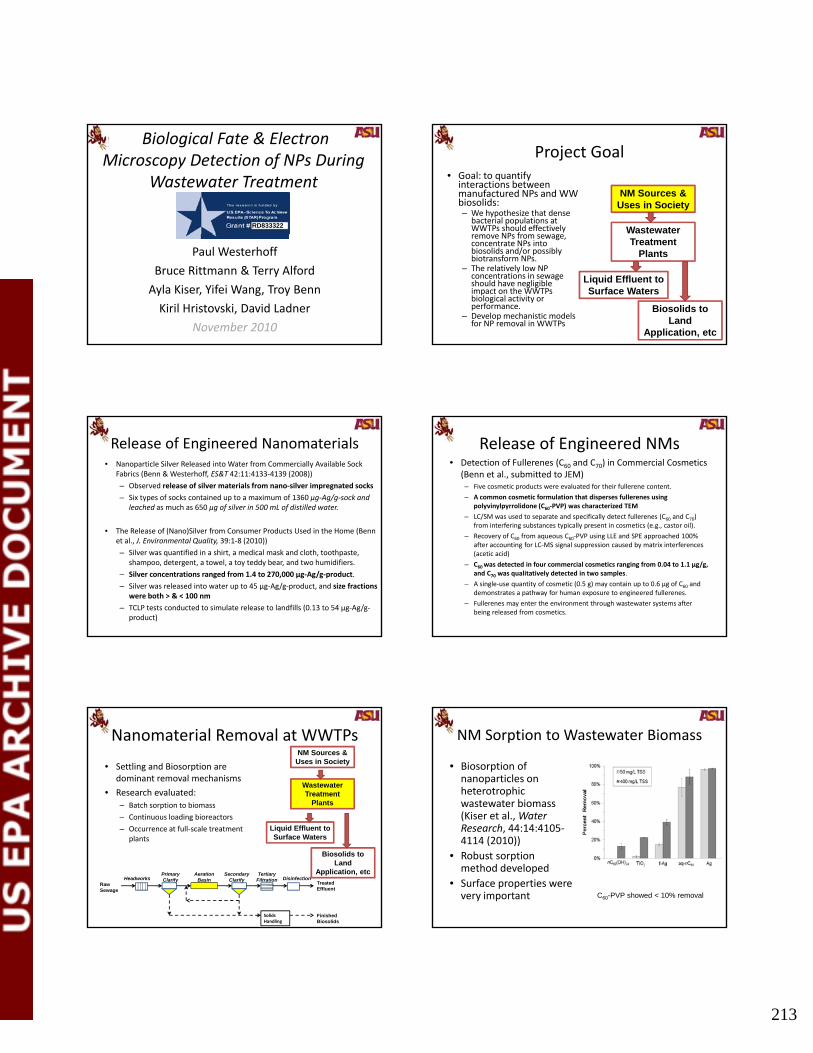
213
Biological Fate & Electron Microscopy Detection of NPs During
Wastewater Treatment
RD833322
Paul Westerhoff
Bruce Rittmann & Terry Alford
Ayla Kiser, Yifei Wang, Troy Benn
Kiril Hristovski, David Ladner
November 2010
Project Goal• Goal: to quantify
interactions between manufactured NPs and WW biosolids:– We hypothesize that dense
bacterial populations at WWTPs should effectively remove NPs from sewage
NM Sources & Uses in Society
Wastewater remove NPs from sewage, concentrate NPs into biosolids and/or possibly biotransform NPs.
– The relatively low NP concentrations in sewage should have negligible impact on the WWTPs biological activity or performance.
– Develop mechanistic models for NP removal in WWTPs
Treatment Plants
Biosolids to Land
Application, etc
Liquid Effluent to Surface Waters
Release of Engineered Nanomaterials• Nanoparticle Silver Released into Water from Commercially Available Sock
Fabrics (Benn & Westerhoff, ES&T 42:11:4133‐4139 (2008))
– Observed release of silver materials from nano‐silver impregnated socks
– Six types of socks contained up to a maximum of 1360 μg‐Ag/g‐sock and leached as much as 650 μg of silver in 500 mL of distilled water.
• The Release of (Nano)Silver from Consumer Products Used in the Home (Benn et al., J. Environmental Quality, 39:1‐8 (2010))
– Silver was quantified in a shirt, a medical mask and cloth, toothpaste, shampoo, detergent, a towel, a toy teddy bear, and two humidifiers.
– Silver concentrations ranged from 1.4 to 270,000 µg‐Ag/g‐product.
– Silver was released into water up to 45 µg‐Ag/g‐product, and size fractions were both > & < 100 nm
– TCLP tests conducted to simulate release to landfills (0.13 to 54 µg‐Ag/g‐product)
Release of Engineered NMs• Detection of Fullerenes (C60 and C70) in Commercial Cosmetics
(Benn et al., submitted to JEM)– Five cosmetic products were evaluated for their fullerene content.
– A common cosmetic formulation that disperses fullerenes using polyvinylpyrrolidone (C60‐PVP) was characterized TEM
– LC/SM was used to separate and specifically detect fullerenes (C60 and C70) from interfering substances typically present in cosmetics (e g castor oil)from interfering substances typically present in cosmetics (e.g., castor oil).
– Recovery of C60 from aqueous C60‐PVP using LLE and SPE approached 100% after accounting for LC‐MS signal suppression caused by matrix interferences (acetic acid)
– C60 was detected in four commercial cosmetics ranging from 0.04 to 1.1 µg/g, and C70 was qualitatively detected in two samples.
– A single‐use quantity of cosmetic (0.5 g) may contain up to 0.6 µg of C60 and demonstrates a pathway for human exposure to engineered fullerenes.
– Fullerenes may enter the environment through wastewater systems after being released from cosmetics.
Nanomaterial Removal at WWTPs
• Settling and Biosorption are dominant removal mechanisms
• Research evaluated:– Batch sorption to biomass
– Continuous loading bioreactors
NM Sources & Uses in Society
Wastewater Treatment
Plants
Continuous loading bioreactors
– Occurrence at full‐scale treatment plants
Biosolids to Land
Application, etc
Liquid Effluent to Surface Waters
Solids Handling
Raw Sewage
Treated Effluent
Finished Biosolids
HeadworksPrimary Clarify
Aeration Basin
Secondary Clarify
Tertiary Filtration Disinfection
NM Sorption to Wastewater Biomass
• Biosorption of nanoparticles on heterotrophic wastewater biomass (Kiser et al Water(Kiser et al., Water Research, 44:14:4105‐4114 (2010))
• Robust sorption method developed
• Surface properties were very important C60-PVP showed < 10% removal
mosvald
Text Box
213

214
Validate EPA Sorption Method
• OPPTS 835.1110 Activated Sludge Sorption Isotherm
• Validated method for organic pollutant (MB) using fresh and freeze‐dried biomass
• Method not valid for nanosilver, and likely other NMs
Materials courtesy of CEINt (Wiesner)
Freeze‐Dried Biomass has different morphology
Freeze-Dried Biomass:
Fresh Biomass:
* Biosorption of fluorescent latex spheres (20 nm sulfate coated) are far less on freeze-dried biomass
Continuous NM Loading Study• Sequencing batch reactors (SBRs)
operated for weeks to months with daily renewal of simulated sewage + NMs
• Operated are realistic HRT and SRT
• Trends in removal for different types of NMs followed batch isotherms
1
2
3
Concentration
(mgA
g/L or gTSS/L)
Influent Ag
Settled effluent Ag
Biomass
• Functionalized nanosilver resulted in sludge bulking issues; removal decreased as TSS fell
• Varied nC60 loading and reduced biomass – still achieved very high nC60 removals
• No effect of heterotrophic removal of COD
• Minimal effect of nano‐Ag on nitrification, compared to major effect from ionic silver
00 20 40 60
Time (days)
0
1
2
3
0 20 40 60 80 100 120
Concentration
(mnC60/L or gTSS/L)
Time (days)
Influent nC60Effluent nC60Biomass
Another Example (f‐Ag)
0.4
0.6
0.8
1
1.2
ntration of Ag
mgA
g/L)
1st Round
0.4
0.6
0.8
12nd Round
• Round 2 has 50% more biomass (TSS) than round 1
0
0.2
0 5 10
Concen (m
Days of Operation
0
0.2
0 10 20 30Days of Operation
Influent (red)Effluent (blue)
Nanomaterials Removed from Liquid Go to Biosolids (fn‐Ag example)
TiOTiO2
Nanomaterials Removed from Liquid Go to Biosolids (n‐TiO2 example)
6
8
10
2
3
4
Ti
SS)
Ag
SS)
Silver Titanium
No NP in FeedNP in Feed
0
2
4
0
1
2
0 5 10 15 20 25 30
Conc.of T
(mgTi/gS
Conc.ofA
(mgA
g/gS
Days of Operation
mosvald
Text Box
214

215
Occurrence at Full‐Scale WWTPsTitanium is found in biosolids at full‐scale WWTPs already
• Titanium Nanomaterial Removal and Release from Wastewater Treatment Plants (Kiser et al., ES&T, 43:17:6757‐6763 ,2009) 1000
10000
100000
centration
µg/g for solid
s )
7:00 AM
11:00 AM
3:00 PM
6:00 PM
,2009)
• >90% removal in coming titanium
• Three forms of titanium present:– Nanoscale– Microscale– Mixed element (clays)
1
10
100
Headworks
Primary
Effluent
Secondary
Effluent
Tertiary
Effluent
Primary Solids
Aeration Basin
Secondary
Solids
Titan
ium Conc
(µg/L for liq
uid and
Nanomaterials in WWTP Effluents
• Evaluate TiO2 presence at several WWTPs
• Evaluate membrane
NM Sources & Uses in Society
Wastewater • Evaluate membrane technologies to characterize or remove NMs
Treatment Plants
Biosolids to Land
Application, etc
Liquid Effluent to Surface Waters
Titanium well removed at WWTPs in Arizona
Different Facilities
Titanium Content of water (ugTi/L)
Headworks Effluent
Activated sludge 615 5
Act. Sludge + filter 180 7
Activated sludge 363 3
• Effluent Ti concentrations are similar to LCA model predictions
M b Activated sludge 141 2
Activated sludge 581 18
Activated sludge 8
Activated sludge 233 2
Trickling filter 549 13
MBR 310 1
MBR 422 4
Average 377 6
• Membrane bioreactors (MBR) have very low effluent Ti
• We isolated NPs in effluents also using roto‐evap + dialysis (under analysis)
NM surface properties trumped membrane material properties in 0.22‐µm
syringe filtration experiments
mea
te /
[NP]
feed
16
[NP]
perm
Tighter Ultrafiltration Rejects NMs
Amicon-Ultra centrifugal filtration device (image from Sigma-Aldrich web site)(image from Sigma Aldrich web site).
Rejection was high, but recovery indicates significant sorption
Nanomaterial in Biosolids
• Nanomaterials will accumulate in biosolids
• What do we do with WWTP biosolids:– 60% land applied
22% incinerated
NM Sources & Uses in Society
Wastewater – 22% incinerated– 17% landfilled
• Approximate content (not all “nano”):– 0.4 to 1 mgTi/g dry SS– 0.004 to 0.03 mgAg/ g dry SS
• Working with biosolids from EPA Inventory and local facilities
Treatment Plants
Biosolids to Land
Application, etc
Liquid Effluent to Surface Waters
mosvald
Text Box
215

216
Summary & Needs
• Need better tools to differentiate engineered from “other” NPs in wastewaters
• Pollutant removal models f WWTP
NM Sources & Uses in Society
Wastewater Treatment
for WWTPs are current not suitable for predicting NPs
• Better relationships between surface charge and core composition versus biosorption needed
• Fate of NMs in biosolids is poorly understood
Treatment Plants
Biosolids to Land
Application, etc
Liquid Effluent to Surface Waters
Biological Fate & Electron Microscopy Detection of NPs During
Wastewater Treatment
RD833322
Gordon Conference 2011 Gordon Research Conference
on Environmental Nanotechnology
Waterville Valley Resort, Waterville Valley, NH,May 29 ‐ June 3, 2011
NPs used here were functionalized and small (even among nanoparticles.)
Ag(-) Au(-) TiO2(-)
21
Au(+) TiO2(+)
Fluorescence Images
Freeze-Dried Biomass:
Fresh Biomass:
* Freeze-dried biomass shows less fluorescence than fresh biomass.
mosvald
Text Box
216

2010 U.S. EPA Nanotechnology Grantees Meeting
The Office of Research and Development’s National Center for Environmental Research 217
P. Lee Ferguson
Analysis and Fate of Single-Walled Carbon Nanotubes and Their Manufacturing Byproducts in Estuarine Sediments and Benthic Organisms
P. Lee Ferguson1 and G. Thomas Chandler2
1Department of Civil and Environmental Engineering, Duke University, Durham, NC; 2Department of Environmental Health Sciences, University of South Carolina, Columbia, SC
Single-walled carbon nanotubes (SWNT) have emerged as a promising material for commercial and industrial applications due to their outstanding electrical, optical, mechanical, and thermal properties. It is clear that as these nanomaterials become more commonplace, they will eventually reach the ambient environment through waste discharge or disposal. Our recent work and that of others has shown that SWNT have high affinity for natural particulates in aquatic systems and are thus expected to concentrate in sediments after discharge to receiving waters. Any assessment of the occurrence and fate of SWNT in the aquatic environment will thus necessitate development of sensitive and selective detection of these materials in sediments. Near Infrared fluorescence (NIRF) spectroscopy has advanced as a highly selective and information-rich technique for sensitive detection and structural characterization of SWNT materials. We have combined asymmetric flow field flow fractionation (A4F) with NIRF spectroscopy as a promising tool for determination of SWNT in the environment. Different purification, concentration, and separation methods are discussed to reduce matrix complexity and improve the detection limit of SWNT. In addition to concentration, structural information such as shape, length distribution, or agglomeration state of SWNT also must be identified and quantified to describe behavior and transport processes as well as biological interactions. NIRF spectral features of SWNT were retained after extraction from sediment, allowing diameter/chiral wrapping angle characterization for dilute solutions. Furthermore, A4F was applied as a separation method prior to NIRF spectroscopic analysis to determine SWNT length distribution and to reduce matrix complexity by separation of NOM and SWNT. We have utilized this comprehensive analytical approach to assess the fate and biological uptake of CoMoCAT SWNT in marine sediment microcosms and benthic deposit feeding organisms. SWNT were extracted from sediments and meiobenthic copepods and polychaete worms by ultrasonication in 2% surfactant solutions and individual surfactant-wrapped nanotubes were isolated from aggregates by ultracentrifugation. SWNT extracted from sediment and tissue in 2% sodium deoxycholate could be quantified down to 9 ng/mL, and detection was linear over > 3 orders of magnitude. Our results show that NIRF-spectroscopy is a valuable method for detection and characterization of surfactant-stabilized SWNT at trace concentrations in the aquatic environment. EPA Grant Number: R833859

218
Analysis and fate of single-walled carbon nanotubes in estuarine
sediments and benthic organisms
P. Lee Ferguson1,2, Ashley N. Parks1, P. Ariette Schierz2, Kate Washburn, G. Thomas Chandler3,
Kay Ho4, and Rob Burgess4
1Nicholas School of the Environment and 2Department of Civil & Environmental Engineering, Duke University, Durham, NC
3Department of Environmental Health Sciences, University of South Carolina, Columbia, SC
4Atlantic Ecology Division, NHEERL, Narragansett, RI
SWNT as potential environmental contaminants
• SWNT composites have already made their way into the marketplace (composite sports equipment, nanoelectronic devices).
• Numerous companies now supply SWNTs on kilogram scaleSWNTs on kilogram scale.
• Annual worldwide production of SWNT is estimated > 1,000 t by 2011.
• There are currently no reliable methods to detect SWNT in complex mixtures (e.g. sediment, tissue, ambient waters) at low concentrations.
SWNT have unique structural characteristics
0° zigzag
Each possible nanotube structure is labeled by two integers, (n,m), that uniquely define its diameter and chiral angle. Red- metallic,
black semimetallic
d 0.0794 n2 nm m2
Adapted from Fluorometric Characterization of SWCNT, Weisman, RB, 2009.
30°
armchair
RESEARCH OBJECTIVE:
Implement and apply near infrared fluorescence spectroscopy for qualitative and quantitative analysis of
SWNT in complex environmental media
1. Develop sample preparation methods for isolating SWNT from
sediment and tissue prior to near infrared fluorescence
spectroscopy.
2. Explore asymmetric flow field flow fractionation coupled with NIRF
spectroscopy for separating SWNT and reducing intereferences.
3. Apply AFFF‐NIRF spectroscopy to analysis of SWNT uptake and
accumulation in sediment‐dwelling organisms.
Multi-laser NIR spectrofluorometer
Laser 1: 638 nm32 mW
Near infrared spectrograph and cooled InGaAs 512 pixel array detector
Laser 2: 691 nm31 mW
Laser 3: 782 nm74 mW
http://www.appliednanofluorescence.com/ns1.html
Qualitative characterization of CoMoCat SWNT type SG65 by NIRF spectroscopy
d 0.0794 n2 nm m2
6,4
9,1
8,3
6,5
7,3
7,5
10,2
9,48,4
7,6
9,2
12,1
8,6
11,3
9,5
10,3
10,5
8,7
13,2
9,7
12,411,4
12,2
10,6 11,6
9,8
15,1
14,3
10,8
13,512,5
13,3
14,1
11,7 12,7
10,9 11,9
15,2
14,4
7,0
6,1
6,2
5,3
5,4
8,0
7,2
8,1
10,0 11,0
11,1
13,0 14,0 16,0
13,6
12,8
11,10
armchair
zigzag
mosvald
Text Box
218

219
Detection of CoMoCat SWNT type SG65 in estuarine sediment by NIRF
• CoMoCat SWNT were spiked into estuarine sediment at 10 g/g concentration.
• Sequential extractions were performed with 2% sodium deoxycholate (ultrasonication at 40 W for 10 minutes) 638 nm excitation wave lengthfor 10 minutes).
SWNT Diameter (nm)
Chiralspecies
Recovery % after 4 extractions
0.782 8,3 790.757 6,5 960.706 7,3 740.829 7,5 770.884 10,2 1030.916 9,4 790.840 8,4 620.895 7,6 810.806 9,2 75
6000 7000 8000 9000 10000 11000 12000
0.00000
0.00001
0.00002
0.00003
0.00004
0.00005
1. extraction 2. extraction 3. extraction 4. extraction 5. extraction
em
issi
on p
owe
r [n
W c
m-1]
wave number [cm-1]
638 nm excitation wave length
SWNT extracted from sediment: 81 ± 5 %
1E8
1E9 SWNT in sed extract SWNT in 2% SDC
r (nW
cm
-1)
Quantitative performance of NIRF spectroscopy for SWNT in sediments
280 ng ml-1
0.01 0.1 1 101000000
1E7
cSWNT, 65
(ug ml-1)
emis
sion
pow
er
1E8
1E9 SWNT in sed extract SWNT in 2% SDC
er (n
W c
m-1
)
Quantitative performance of NIRF spectroscopy for SWNT in sediments
sediment extract
29 l 1
0.01 0.1 1 101000000
1E7
cSWNT, 65
(ug ml-1)
emis
sion
pow
e
2% SDC29 ng ml-1
Challenges: Sample purification methods
7000 8000 9000 10000 110000.000000
0.000005
0.000010
0.000015
0.000020
0.000025
0.000030 SWNT cSWNT =31 ng/ml
extracts of natural sediments
- Oxidation e.g. H2O2, KMnO4, .. - Ultrafiltration (Centriprep 100 kDa)- Ultracentrifugation - Asymmetric Flow Field Flow Fractionation
7000 8000 9000 10000 11000extracts of natural sediments
Separation mechanism in asymmetric flow field flow fractionation AF4
Field flow fractionation coupled with NIRF spectroscopy
FFF channel
mosvald
Text Box
219

220
FFF channel
DAD detector
Field flow fractionation coupled with NIRF spectroscopy
FFF channel
DAD detector
Field flow fractionation coupled with NIRF spectroscopy
MALS detector
FFF channel
DAD detector
Field flow fractionation coupled with NIRF spectroscopy
MALS detector
NIR Fluorescense detector
AF4 of CoMoCat SWNT Injection Focus
Elution
0.056
0.064
2
Qualitative detection of CoMoCat SWNT in 2% SDC by AF4 –NIRF spectroscopy
UV at 320 nm
SLS at 90°
SWNT SG65 in 2% SDC c0 = 46 µg ml-1
ty
1
0 10 20 30 40 50
0
3x10-13
6x10-13
8810 cm-1
9756 cm-1
10140 cm-1
time (min)
638 nm excitation
SLS at 90
inte
nsi
NIR Flourescence at638 nm excitation
0.00E+000
3.00E-013
6.00E-013
0.00E+000
3.00E-013
6.00E-013
8810 cm-1
9765 cm-1
10140 cm-1
SWNT c0 = 10 µg ml-1
SWNT c0 = 10 µg ml-1
A
e
nsity
[nW
cm
-1]
638 nm excitation
Separation of CoMoCat SWNT by AF4 coupled with NIRF spectroscopy
0.00E+000
6.00E-013
1.20E-012
0 20 40 60 80 100 120
0.00E+000
6.00E-013
1.20E-012
time [min]
SWNT c0 = 20 µg ml-1
SWNT c0 = 20 µg ml-1
em
issi
on
inte
mosvald
Text Box
220

221
0.00E+000
3.00E-013
6.00E-013
0.00E+000
3.00E-013
6.00E-013
8810 cm-1
9765 cm-1
10140 cm-1
SWNT c0 = 10 µg ml-1
SWNT c0 = 10 µg ml-1
A
e
nsity
[nW
cm
-1]
Separation of CoMoCat SWNT by AF4 coupled with NIRF spectroscopy
0.00E+000
6.00E-013
1.20E-012
0 20 40 60 80 100 120
0.00E+000
6.00E-013
1.20E-012
time [min]
SWNT c0 = 20 µg ml-1
SWNT c0 = 20 µg ml-1
em
issi
on
inte
Cross flow variation
0.055
0.060
0.055
0.060
Detection of CoMoCat SWNT extracted from sediment by UV
SWNT in 2%SDC
2%SDC
SWNT SG65 c0 = 46 µg ml-1
0.05
0.10
0 20 40 60 80
0.05
0.10
SWNT extracted fromsediment
Blank sediment extract
SWNT SG65 c0 = 10 µg ml-1
Detection of CoMoCat SWNT in sediment by static light scattering
0 20 40 60 80
1
2
3
1
2
3
SWNT in 2%SDC
2%SDC
SWNT SG65 c0 = 46 µg ml-1
0
4
8
0 20 40 60 800
4
8
time [min]
SWNT extracted fromsediment
Blank sediment extract
SWNT SG65 c0 = 10 µg ml-1
0
4x10-11
8x10-11
0
4x10-11
nsity
[nW
cm
-1]
Detection of CoMoCat SWNT in sedimentextracts by NIRF
SWNT in 2%SDC
2%SDC
SWNT SG65 c0 = 46 µg ml-1
0
4x10-12
8x10-12
0 20 40 60 80
0
4x10-12
8x10-12
638 nm excitation 691 nm excitation 782 nm excitation
emis
sion
inte
n
time [min]
SWNT extracted fromsediment
Blank sediment extract
SWNT SG65 c0 = 10 µg ml-1
CoMaCat SWNT are detectible in complexsediment extracts using AFFF-NIRF
NIRF static Reference: sediment extract
0.000025
0.000030
sediment spiked 10 g SWNT/g
7000 8000 9000 10000 110000.000000
0.000005
0.000010
0.000015
0.000020
AFFF-NIRFtime resolved
NIRF static Reference: sediment extract
0.000025
0.000030
sediment spiked 10 g SWNT/g
CoMaCat SWNT are detectible in complexsediment extracts using AFFF-NIRF
4x10-12
y [n
W c
m-1]
7000 8000 9000 10000 110000.000000
0.000005
0.000010
0.000015
0.000020
0 10 20 30 40 50 60
0
2x10-12
time [min]
emis
sion
inte
nsit
mosvald
Text Box
221

222
SWNT do not degrade in sediments over one month timescale
Bread & Butter creek
Bread & Butter creek 7000 8000 9000 10000 11000
0.000000
0.000004
0.000008
0.000012
0.000016 638 nm excitation 691 nm excitation 782 nm excitation
em
issi
on
pow
er
[nW
cm
-1]
wave number [cm-1]
Detection of SWNT SG65 in sediments and tissues by NIRF spectroscopy
Sediment extracts (2% SDC)
DL = 60 ng g-1 sed
Enhydrosoma propinquum7000 8000 9000 10000 11000
-0.0000006
-0.0000004
-0.0000002
0.0000000
0.0000002
0.0000004
0.0000006
0.0000008
0.0000010
emis
sion
po
wer
[n
W c
m-1]
wave number [cm-1]
638 nm excitation 691 nm excitation 782 nm excitation Extracts of Enhydrosoma
propinquum (2% SDC)
LOD= 7 ng g-1 tissue
CoMoCat SWNT were undetectable in sediment-exposed amphipods and mysid shrimp
SG76
0.000002
0.000004
0.000006
0.000008
0.000010
Emis
sion
Inte
nsity
nW
/cm
-1
638nm 691nm 782nm
CG100
-0.000002
0.000000
0.000002
0.000004
0.000006
0.000008
Emis
sion
Inte
nsity
nW
/cm
-1 638nm 691nm 782nm
0.000000
0.000002
0.000004
0.000006
0.000008
Emis
sion
Inte
nsity
nW
/cm
-1 638nm 691nm 782nm
-0.0000008
-0.0000004
0.0000000
0.0000004
Emis
sion
Inte
nsity
nW
/cm
-1
638nm 691nm 782nm
SG65
OECD
6000 7000 8000 9000 10000 11000 120000.0000
0.0005
0.0010
0.0015
0.0020
0.0025
Em
issi
on In
tens
ity n
W/c
m-1
Wavenumber cm -1
638nm 691nm 782nm
6000 7000 8000 9000 10000 11000 120000.0000
0.0005
0.0010
0.0015
0.0020
0.0025
Emis
sion
Inte
nsity
nW
/cm
-1
Wavenumber cm -1
638nm 691nm 782nm
6000 7000 8000 9000 10000 11000 120000.00000
0.00005
0.00010
0.00015
0.00020
Emis
sion
Inte
nsity
nW
/cm
-1
Wavenumber cm -1
638nm 691nm 782nm
6000 7000 8000 9000 10000 11000 120000.0000
0.0005
0.0010
0.0015
0.0020
Emis
sion
Inte
nsity
nW
/cm
-1
Wavenumber cm -1
638nm 691nm 782nm
6000 7000 8000 9000 10000 11000 12000-0.0000005
0.0000000
0.0000005
0.0000010
0.0000015
0.0000020
Em
issi
on In
tens
ity n
W/c
m-1
Wavenumber cm^-1
638nm 691nm 782nm
6000 7000 8000 9000 10000 11000 120000.000000
Wavenumber cm^-1
6000 7000 8000 9000 10000 11000 12000-0.0000005
0.0000000
0.0000005
0.0000010
0.0000015
0.0000020
Em
issi
on In
tens
ity n
W/c
m-1
Wavenumber cm^-1
638nm 691nm 782nm
6000 7000 8000 9000 10000 11000 12000Wavenumber cm^-1
6000 7000 8000 9000 10000 11000 12000Wavenumber cm^-1
6000 7000 8000 9000 10000 11000 12000Wavenumber cm^-1
6000 7000 8000 9000 100001100012000
-0.0000005
0.0000000
0.0000005
0.0000010
0.0000015
0.0000020
0.0000025
Em
issi
on In
tens
ity n
W/c
m-1
Wavenumber cm^-1
638nm 691nm 782nm
75
100400
600
800
gy
g
SWNT body burden measurements in sediment and/or food exposed organisms
473.93+ 254.04
6000 7000 8000 9000 10000 11000 12000
0.00000
0.00002
0.00004
0.00006
0.00008E
mis
sion
Inte
nsity
nW
/cm
-1
Wavenumber cm^-1
638nm 691nm 782nm
6000 7000 8000 9000 10000 11000 12000
-0.0000008
-0.0000004
0.0000000
0.0000004
Emis
sion
Inte
nsity
nW
/cm
-1
Wavenumber cm^-1
638nm 691nm 782nm
45 72+ 5 446000 7000 8000 9000 10000 11000 12000-0.000001
0.000000
0.000001
0.000002
0.000003
0.000004
0.000005
Emis
sion
Inte
nsity
nW
/cm
-1
W b ^ 1
638nm 691nm 782nm
10g SWNT/g sedimentAlgaeDepurated AmphipodsNon-depurated Amphipods
6000 7000 8000 9000 10000 11000 12000-0.000002
0.000000
0.000002
0.000004
0.000006
0.000008
E
mis
sion
Inte
nsity
nW
/cm
-1
Wavenumber cm -1
638nm 691nm 782nm
G65
/g d
ry
wei
ght
0
25
50
µgg
Sediment Extract
Algae Extract Amphipod:sediment exposure
Amphipod:sediment + algae
exposure
Amphipod:algae
exposure
6.02+ 0.59
6000 7000 8000 9000 10000 11000 12000
0.00000
0.00004
0.00008
0.00012
0.00016
0.00020
Emis
sion
Inte
nsity
nW
/cm
-1
Wavenumber cm^-1
638nm 691nm 782nm
45.72+ 5.44
38.10+ 3.94
Wavenumber cm^-1
0
µg
SG w
0 00
Accumulation of SWNT in benthic macroinvertebrates
SWNT suspension in 0.5% gum arabic
Nominal concentrations: 1 µg/g 10 µg/g
Long Island Sound sediment
Nereis virens
14-day exposure following US EPA (1993)
µg/g, 10 µg/g
SWNT bioaccumulation and trophic transfer
SWNT suspension in 0.5% gum arabic
SWNT-spiked Isochrysis galbana
Mercenaria mercenaria fed SWNT-spiked Isochrysis
Isochrysisgalbana
Mercenaria mercenaria Nereis
virens
14-day exposure 28-day exposure
Nominal concentrations: 100 µg/g, 1000 µg/g
mosvald
Text Box
222

223
Worm feeding
SWNT-spiked sediment exposure to Nereis virens
Nominal SWNT –spiked sediment concentration
Measured SWNT concentration:Batch A
Measured SWNT concentration:Batch B
10 µg/g 7.1+1.4 µg/g 10.4+1.6 µg/g
NIRF l i f SWNT i NIRF l i f SWNT i NIRF l i f SWNT i
6000 7000 8000 9000 1000011000120000.00000
0.00002
0.00004
0.00006
0.00008
Emis
sion
Inte
nsity
nW
/cm
-1
Wavenumber cm^-1
638nm 691nm 782nm
6000 7000 8000 9000 10000 11000120000.00000
0.00002
0.00004
0.00006
0.00008
Emis
sion
Inte
nsity
nW
/cm
-1
Wavenumber cm^-1
638nm 691nm 782nm
6000 7000 8000 9000 10000 11000120000.00000
0.00002
0.00004
0.00006
0.00008
Emis
sion
Inte
nsity
nW
/cm
-1
Wavenumber cm^-1
638nm 691nm 782nm
NIRF analysis of SWNT in spiked sediment
NIRF analysis of SWNT in depurated worms
NIRF analysis of SWNT in non-depurated worms
Direct NIRF analysis of worm extracts may be limited by internal filter effects
0.40.60.81.01.21.41.61.8
Abs
orba
nce
0.40.60.81.01.21.41.61.8
Abs
orba
nce
NIRF analysis of SWNT in depurated worms
NIRF analysis of SWNT in non-depurated worms
900 1000 1100 1200 1300 1400 1500 16000.00.2
Wavelength nm900 1000 1100 1200 1300 1400 1500 1600
0.00.2
Wavelength nm
0 20 40 60 80 100 1200.00E+000
5.00E-013
1.00E-012
1.50E-012
2.00E-012
2.50E-012
3.00E-012
Emis
sion
Inte
nsity
(nW
/cm
-1)
Time (min)
AFFF-NIRF analysis at 638nm excitation
SWNT-spiked Isochrysis galbana exposure to Mercenaria mercenaria
Nominal SWNT –spiked algae concentration
Measured SWNT concentration:7/30/10
Measured SWNT concentration:8/2/10
1000 µg/g 94.5 µg/g 63.2 µg/ghttp://www.topsybaits.nl/
6000 7000 8000 9000 10000 11000 120000.00000
0.00002
0.00004
0.00006
0.00008
0.00010
0.00012
Emis
sion
Inte
nsity
nW
/cm
-1
Wavenumber cm^-1
638nm 691nm 782nm
6000 7000 8000 9000 10000 11000 120000.00000
0.00002
0.00004
0.00006
0.00008
0.00010
0.00012
Emis
sion
Inte
nsity
nW
/cm
-1
Wavenumber cm^-1
638nm 691nm 782nm
6000 7000 8000 9000 10000 11000 120000.00000
0.00002
0.00004
0.00006
0.00008
0.00010
0.00012
Emis
sion
Inte
nsity
nW
/cm
-1
Wavenumber cm^-1
638nm 691nm 782nm
NIRF analysis of SWNT in spiked
algae
NIRF analysis of SWNT in
depurated clams
NIRF analysis of SWNT in non-
depurated clams
No internal filter artifacts present in NIRF analysis of clam extracts
1.41.61.8
1.41.61.8
NIRF analysis of SWNT in
depurated clams
NIRF analysis of SWNT in non-
depurated clams
900 1000 1100 1200 1300 1400 1500 16000.00.20.40.60.81.01.2
Abs
orba
nce
Wavelength nm900 1000 1100 1200 1300 1400 1500 1600
0.00.20.40.60.81.01.2
Abs
orba
nce
Wavelength nm
• Additional microcosm-based experiments− Track uptake of SWNT manufacturing
byproducts in sediment-dwelling organismsas well as degradation in sediments
− Investigate chirality/diameter-dependence of SWNT interaction with sediment and organisms
OUTLOOK
• Survey environmental media (e.g. field sediment) for contamination with SWNT
mosvald
Text Box
223

224
Conclusions
• A novel and highly sensitive method for analysis of SWNT in sediments
based on near infrared fluorescence spectroscopy has been developed.
• NIRF spectral features of SWNT were retained after extraction from
sediment, allowing diameter/chirality characterization for dilute
solutions.
• AFFF can be used as a clean up tool prior to NIRF analysis.
• SWNT do not appear to be highly bioaccumulative in estuarine
invertebrates exposed via sediment or dietary routes.
AcknowledgementRon Meyer and Dr. Sigrid Kuebler, Wyatt Technology
Dr. Phil Wallis, SouthWest Nanotechnolygy
Dr. Sergei M. Bachilo, Applied Nanofluorescense
RD833859
mosvald
Text Box
224

2010 U.S. EPA Nanotechnology Grantees Meeting
The Office of Research and Development’s National Center for Environmental Research 225
Robert A. Yokel
Safety/Toxicity Assessment of Ceria (A Model Engineered NP) to the Brain
Robert Yokel1, Mo Dan1, Rebecca Florence1, Jason Unrine1, Robert MacPhail2, Michael Tseng3,
Uschi Graham1, Rukhsana Sultana1, Sarita Hardas1, D. Allan Butterfield1 Peng Wu1, and Eric Grulke1
1University of Kentucky, Lexington, KY; 2U.S. Environmental Protection Agency, Research Triangle Park, NC; 3University of Louisville, Louisville, KY
Nanoscale ceria has extensive commercial uses that can contribute to its environmental release, including its use as a diesel fuel additive. We are studying it as a model metal oxide engineered nanomaterial (ENM). Nanoscale ceria was nominated by the National Institute of Environmental Health Sciences for toxicological consideration and is on the priority list of the OECD for measurement, toxicology, and risk assessment studies. The purpose of this study is to characterize the physico-chemical properties of a representative ENM that influences its distribution in blood, and into the brain compared to peripheral organs, biopersistence in those organs, and resultant effects. Studies were conducted with in-house produced and characterized ~ 5, 15, 30, and 65 nm citrate-coated ceria ENM, compared to the cerium ion. Ceria ENM or the cerium ion was iv infused into rats to enable study of its distribution in blood and translocation from systemic circulation, as would occur following absorption into blood from any route of exposure. Blood was repeatedly sampled, an aliquot allowed to clot, and cerium determined by ICP-MS in serum and clot up to 4 h and in whole blood for much longer. To extend our prior work showing no appreciable reduction of cerium in mammalian reticuloendothelial tissues up to 30 days after a single administration of nanoscale ceria; determine the routes and rate of its excretion; and further characterize its distribution, persistence, and associated effects in the rat; a longer term study was conducted with 30 nm ceria. Rats were terminated 1, 7, 30, or 90 days after a single iv ceria ENM infusion, compared to cerium ion or vehicle controls. Rats were housed in metabolism cages for up to 2 weeks to quantify urinary and fecal cerium output, cage-side observations were recorded daily, and they were weighed weekly. Nine organs were weighed and samples of 14 tissues, blood, and CSF were collected for cerium determination by ICP-MS. Oxidative stress markers (protein carbonyls [PC], 3-nitrotyrosine [3-NT], and protein bound 4-hydroxy-2-trans-nonenal [HNE]), the glutathione antioxidant defense system (glutathione reductase and peroxidase), and antioxidant enzymes (Mn-SOD and catalase) were measured. Ten minutes after infusion, < 1 percent of 15 to 65 nm ceria ENM, < 2 percent of a mixture of 30 nm cubic and rod ceria ENM, approximately 14 percent of the cerium ion, and approximately 33 percent of the 5 nm ceria ENM remained in blood. For all 4 ceria ENMs the elimination from blood was biphasic, with an initial half life of approximately 1 h and the second for the 5 to 30 nm ceria of approximately 100 to 200 h, and approximately 12 h for the 65 nm ceria. The 15 and 30 nm ceria predominantly associated with blood cells, whereas the 5 and 65 nm ceria and the cerium ion were approximately evenly distributed between serum and the clot fraction of blood. The 5 nm ceria ENM was not seen in BBB or brain cells. The amount of 15 to 65 nm ceria ENMs in brain samples was very small. Energy electron loss spectroscopy showed the ceria in situ to have similar valence (considerable Ce(III)) to the dosing material up to 30 days. The 30 nm ceria ENM was less acutely toxic than the cerium ion. Less than 1 percent of the ceria or cerium ion was excreted in the first week, of which 98 percent was in feces. Ceria was primarily retained in the spleen, liver, and bone marrow. Spleen weight was significantly increased in ceria-treated rats at several times after its infusion, and associated with visual evidence of abnormalities. Thirty nm ceria was associated with blood cells to a greater extent than larger or smaller ENMs, consistent with reports showing this size is optimal for protein wrapping of ENMs. Ceria in blood is primarily cleared by

2010 U.S. EPA Nanotechnology Grantees Meeting
The Office of Research and Development’s National Center for Environmental Research 226
the reticuloendothelial tissues, in which it persists without significant decrease in mass amount for at least 3 months. Little enters the brain. Referring to nanoscale fiber-like structures, it has been stated: “The slower [they] are cleared (high bio-persistence), the higher is the probability of an adverse response” (European Parliament, Policy Department Economic and Scientific Policy “Nanomaterials in consumer products”). Our results support the concern about the long-term fate and adverse effects of inert nanoscale metal oxides that reach systemic circulation, from which they can distribute throughout the body, resulting in persistent retention and potential adverse effects in multiple organs. These results of ENM translocation, biopersistence, and hazard identification in the mammal provide data for ENM risk characterization. Reference: Hardas SS, Butterfield DA, Sultana RL, Tseng MT, Dan M, Florence R, Unrine JM, Graham UM, Wu P, Grulke EA, Yokel RA. Brain distribution and toxicological evaluation of a systemically delivered engineered nanoscale ceria. Toxicological Sciences 2010;116(2):562-576, doi: 10.1093/toxsci/ kfq137. EPA Grant Number: R833772

Safety/toxicity assessment of Safety/toxicity assessment of ceria (a model engineered NP) ceria (a model engineered NP)
to the brainto the brain
The research teamThe research team
• Robert A. Yokel and Mo Dan– Department of Pharmaceutical Sciences, College
of Pharmacy & Graduate Center for Toxicology, University of Kentucky, Lexington, KY
• Jason Unrine• Jason Unrine– Department of Plant and Soil Sciences, U KY
• Michael T. Tseng– Departments of Anatomical Sciences &
Neurobiology, University of Louisville, Louisville, KY
The research teamThe research team
• Uschi M. Graham– Center for Applied Energy Research, U KY
• D. Allan Butterfield, Rukhsana Sultana, & Sarita Hardas
– Department of Chemistry, U KY & (DAB) Center of Membrane Sciences, U KY
• Eric A. Grulke & Peng Wu– Chemical & Materials Engineering
Department, U KY
Objective of this researchObjective of this research
• Characterize the physico-chemical properties of a model engineered nanomaterial (ENM) that influence its biodistribution and effects, including:– distribution across the blood-brain barrier (BBB)– effects on oxidative stress endpoints in the brain– uptake into selected peripheral organs– persistence over time.
ENM studiedENM studied
• Ceria (CeO2, cerium dioxide, cerium oxide) was selected because:– it is an insoluble metal oxide that can be readily
observed and quantified in tissue (electron microscopy, ICP-MS).py )
– it has current commercial applications (a catalyst in diesel fuel and an abrasive in integrated circuit fabrication).
– it has been reported to be cytotoxic as well as neuroprotective, representing the controversy about nanoscale materials.
ENM studiedENM studied• We prepared citrate-coated ceria and
characterized them using:– dynamic light scattering (size)– transmission electron microscopy (TEM) with an energy
dispersive X-ray spectrometer (size and morphology)– X-ray diffraction (composition & crystallinity)X ray diffraction (composition & crystallinity)– BET surface area analyzer (size)– zeta potential (surface charge & stabilizing agent)– FTIR (surface charge and stabilizing agent)– scanning TEM with electron energy loss spectroscopy
(valence)– thermogravimetric analysis with mass spectrometry
(surface citrate coating)
mosvald
Text Box
227

ENMs studiedENMs studied
Ceria ENM size (nm)
shape Zeta potential Miller indices Extent of surface citrate
coating
5 polyhedral -53 ± 7 mV at pH ~ 7.35 (111), (210), (200) ~ 40%
15 l h d l 57 5 V t H 7 3 (111) (210) (200) 27%15 polyhedral -57 ± 5 mV at pH ~ 7.3 (111), (210), (200) ~ 27%
30 cubic -56 ± 8 mV at pH ~ 7.3 (111), (210), (200) ~ 18%
30 cubic + rods
65 polyhedral -22 ± 5 mV at pH ~7 (111), (210), (200) ~ 15%
HRTEM and STEM images of ceria ENMsHRTEM and STEM images of ceria ENMs5 nm polyhedral 15 nm polyhedral 30 nm cube
30 nm cube & rod 65 nm polyhedral
Objective: To assess the influence of size Objective: To assess the influence of size on ENM distribution, persistence, on ENM distribution, persistence,
translocation and toxicitytranslocation and toxicity
• Citrate-coated ceria i.v. infused into un-anesthetized rats (0 or ~ 100 mg/kg); terminated 1 or 20 h or 30 days later.y
Blood and tissue [cerium]Blood and tissue [cerium]
• Brain cortex cerium was always < 1% of the dose. – We did not see 5 nm ceria in brain, only in brain
vasculature • Spleen cerium concentration was greater thanSpleen cerium concentration was greater than
liver cerium concentration.• Liver had the greatest mass amount of the ceria
dose.• There was little decrease in liver and spleen
cerium up to 30 days.
Electron energy loss spectrometry Electron energy loss spectrometry characterization of ceria as synthesized and in characterization of ceria as synthesized and in
situsitu
There was no observable change in the M5/M4 peak ratio (Ce(III)/Ce(IV) ratio) of 5 or 30 nm ceria in spleen agglomerates 30 days after
i d i i t ti d t th f hlceria administration compared to the freshly prepared ceria ENM.
Ceria distribution in and Ceria distribution in and elimination from bloodelimination from blood
• Rats were iv infused with 5, 15, 30 or 65 nm citrate-coated ceria, an mixture of 30 nm cubic and rod citrate-coated ceria, or the cerium ion.
• After the infusion blood was drawn at 10 30• After the infusion blood was drawn at 10, 30, 45, 60, 120 and 240 min.
• Cerium was determined in whole blood, plasma and clot.
• Whole blood cerium was determined in these and other rats up to 90 days after ceria dosing.
mosvald
Text Box
228

Ceria in bloodCeria in blood• Ten min after completion of the 1 h ceria infusion
30% of the 5 nm ceria was in blood; < 1% of the 15, 30 and 65 nm ceria.
• Compartmental pharmacokinetic analysis of whole blood cerium generally showed an initial t½ of 1 h and a beta phase half life of 100 ha beta phase half-life of ~ 100 h.
• The 15 and 30 nm ceria predominantly associated with blood cells, whereas the 5 and 65 nm ceria were ~ evenly distributed between the two compartments.
• The greatest association of the 30 nm citrate-coated ceria with blood cells in the clot fraction is consistent with reports showing this size is optimal for protein wrapping of ENMs.
A 90 day survival study to assess longer A 90 day survival study to assess longer term distribution, persistence and effectsterm distribution, persistence and effects
• Single iv dosing of 87 mg 30 nm ceria/kg, 50 mg cerium ion/kg, or vehicle.
• Termination 1, 7, 30 or 90 days later.• Fecal and urinary Ce excretion (metabolicFecal and urinary Ce excretion (metabolic
cage).• Weekly body weight.• Weights and cerium concentration in multiple
organs and fluids.• Oxidative stress markers, histology (LM &
EM).
• A single ceria ENM infusion resulted in modest decreased body weight gain.
• Less than 1% of the ceria ENM or cerium ion dose was eliminated in a week.
• Ceria was retained primarily in reticuloendothelial tissues The liverreticuloendothelial tissues. The liver contained ~ 20% and the spleen ~ 15% of the dose 90 days after ceria administration.
• No great decrease of the mass amount of ceria in liver and spleen occurred over 90 days.
Spleen pathology 30 and 90 days after ceriaSpleen pathology 30 and 90 days after ceria
– Splenomegaly: ~ 2-fold with 5 and 15 nm and 1.5-fold with 30 nm at 30 days.
– The red pulp 30 days after 5 nm ceria showed numerous densely stained lymphatic cells .
– The white pulp 90 days after 30 nm ceria showed ceria containing cell clusters.
– Granulomatous formations were seen 90 days after ceria.
Liver pathology 30 and 90 days after ceriaLiver pathology 30 and 90 days after ceria
• 30 days after 5 nm ceria:– Non-uniform granuloma formations containing
ceria-loaded Kupffer cells.– Mononucleated cell infiltration among the
hepatic parenchyma and at perivascular sites. – Mononucleated cells appeared to encircle
Kupffer cells.– No evidence of fibrosis or abscess formation.
• 90 days after 30 nm ceria:– Granulomatous formations seen.
ConclusionsConclusions• Citrate-coated 5 to 65 nm ceria does not enter the brain
to any significant extent.• It is primarily cleared by reticuloendothelial organs and
sequestered in intracellular agglomerates.• Some of these results are in Hardas et al Toxicological
Sciences 2010 116(2):562-576.C ( f• The Ce valence does not change in situ (in the first 30
days).• There is little clearance of 5 to 65 nm ceria from
reticuloendothelial organs, for 30 nm up to 90 days.• The smaller the ceria ENM, the longer it remains in
blood before being cleared.• Maximal distribution into blood cells was seen with 30
nm ceria.
mosvald
Text Box
229

ConclusionsConclusions• Ceria ENM and the cerium ion are very slowly eliminated.• Ceria ENM does not always behave like the cerium ion, in
its distribution in blood or tissues. • Referring to nanoscale fiber-like structures, it has been
stated: “The slower [they] are cleared (high bio-persistence) the higher is the probability of an adverse response” (European Parliament Policy Departmentresponse . (European Parliament, Policy Department Economic and Scientific Policy “Nanomaterials in consumer products”).
• These results further support the concern about the long term fate and adverse effects of inert nanoscale metal oxides that reach systemic circulation, from which they can distribute throughout the body, resulting in persistent retention and potential adverse effects in multiple organs.
Future PlansFuture Plans
• Complete the histopathology, agglomeration extent and localization, cerium valence, and oxidative stress marker analyses as a function of time (1, 7, 30 and 90 days) after the 30 nm ceria infusionthe 30 nm ceria infusion.
mosvald
Text Box
230

Handouts on Centers for Environmental Implications of Nanotechnology (CEIN)

232

Mission Statement The University of California Center for Environmental Implications of Nanotechnology (UC CEIN) was established to ensure that nanotechnology is introduced in a responsible and environmentally compatible manner, thereby allowing the US and International Communities to leverage the benefits of nanotechnology for global economic and social benefit. This mission is being accomplished by developing a series of decision tools based on models of predictive toxicology and risk ranking premised on selected nanomaterial property-activity relationships that determine fate, transport, exposure, and biological injury mechanisms at cellular, tissue, organism, and population levels. Since its founding in September 2008, the UC CEIN has successfully integrated the expertise of engineers, chemists, colloid and material scientists, ecologists, marine biologists, cell biologists, bacteriologists, toxicologists, computer scientists, and social scientists to create the predictive scientific platform that will inform us about the possible hazards and safe design of engineered nanomaterials (ENMs) that may come into contact with the environment.
The research of the UC CEIN is carried out by 46 distinct but highly interactive research projects across 7 interdisciplinary research groups (IRGs):
ENM Standard Reference and Combinatorial Libraries and Physical-chemical Characterization (IRG 1)
Studying ENM Interactions at the Molecular, Cellular, Organ, and System Levels (IRG 2)
Organismal and Community Ecotoxicology (IRG 3)
Nanoparticle Fate and Transport (IRG 4)
High-Throughput Screening (HTS), Data Mining, and Quantitative-Structure Relationships for NM Properties and Nanotoxicity (IRG 5)
Modeling of the Environmental Multimedia NM Distribution and Toxicity (IRG 6)
Risk Perception of Potential Environmental Impacts of Nanotechnology (IRG 7)
Key Center Accomplishments
UC CEIN plays a national leadership role in Nano EHS initiatives
Compiled a nanomaterial library with 60+ different types of NP (characterized and introduced into active research) - Includes ZnO, CeO2, TiO2, CNT, Ag, Au, CdSe, Silica, Clays
Developed a series of standardized dispersion protocols for relevant environmental conditions
Successfully demonstrated the oxidative stress heirarchical paradigm for NPs with varying properties
As an exercise of safe design, ZnO doped with Fe has been found to significantly reduce ZnO NP toxicity in mammalian cells
Use of zebrafish embryos as an in vivo model for high-content screening has shown that in vitro toxicity testing results agree with HCS zebrafish in vivo studies, except when using silver nanoparticles
Bacterial toxicity research shows that ZnO and CeO2 inhibit growth more than TiO2, with gram positive bacteria found to be relatively more sensitive
Exposure to Quantum Dots across trophic levels show significant bioaccumulation in bacteria and biomagnification in protozoa
Testing of ZnO across ecosystems has shown that ZnO is consistently toxic, with toxicity resulting from exposure to Zn ions following ENM aggregation and Zn ion shedding
Studies of mobility, persistence, bioavailability and reactivity of ENMs in environmental conditions are providing key characteristics of metal oxide NPs in seawater, freshwater, and terrestrial environments
Efficient removal of ENM from aqueous systems can be archived by pH destabilization, coagulant dosing, sedimentation, and ultrafiltration
mosvald
Text Box
233

Through validation of commercially available HTS technology, we have implemented gene reporter assays that provide readouts of known cellular signaling pathways. Preliminary results identify genotoxicity in a subset of ENMs
A new efficient computer algorithm for feature selection ranking was developed for screening and ranking nanoparticle properties for the development of quantitative property-structure relationships
An international survey of Industry NanoEHS is providing key insights into industry practices, perceived risks, and gaps in understanding, with "lack of information" being a key impediment to the implementation of Nano EHS programs in industry
A Summer 2010 survey of nanotoxicology and regulatory experts will provide a vital comparative framework for future public and industry risk perception studies
Future Directions Include:
Develop and characterize new libraries of Pt, Pd, SWCNT, Mesoporous Silica, and new derivations of metal oxides
Continue ongoing cytotoxicity studies with ENM libraries and analyze data from in vitro and in vivo studies to rank NP toxicity, assess predictive power of in vitro studies, and begin building expert system required to generate structure activity relationships
Expand marine organisms, cellular, and bacterial studies beyond the initial metal oxide NPs (ZnO, TiO2, CeO2)
Adapt HTS methods of toxicity screening to marine and bacterial cells, demonstrating and documenting performance and challenges
Results from ongoing HTS ecotoxicology experiments are being used to design mesocosm experiments in marine, terrestrial, and freshwater ecosystem studies
Incorporation of CNT, Ag, QDs, Pt as well as NP of different sizes and morphologies into ongoing Fate and Transport studies
Advance HTS gene knockout studies with yeast and bacterial strains
Development of an automated high content screening method to enhance zebrafish in vitro toxicity studies
As experimental data from across Center projects enters the Central Data Management system, models for multimedia transport and fate and nanoparticle structure activity relationship models will be refined and expanded. Development of a series of NP decision tools will commence with an initial focus on establishing questions needed to design model pathways
Data from industry survey, public environmental risk perception survey, and survey of nanotoxicology and regulatory experts will help provide valuable knowledge about the societal implications and contexts for risk characterization
Education and Outreach UC CEIN will serve to enhance our understanding of the environmental hazards of nanomaterials. Education and outreach programs to train scientists, develop safe handling guidance for nanomaterials, and develop methods to communicate the implications of our research to the public are key to the success of the Center. The knowledge generated by the Center will directly benefit scientists, public agencies, industrial stakeholders, and the general population.
UC CEIN is housed within the California NanoSystems Institute at the University of California, Los Angeles, with a second major hub at the University of California, Santa Barbara. Research partners include: UC Davis, UC Riverside, Columbia University, University of Texas, Nanyang Technological University, Northwestern University, the Molecular Foundry at Lawrence Berkeley National Laboratory, Lawrence Livermore National Laboratory, Sandia National Laboratory, University of Bremen, University of British Colombia, University College Dublin, Cardiff University, and the Universitat Rovira I Virgili.
For more information: Please visit the UC CEIN website: http://www.cein.ucla.edu UC CEIN, 6522 CNSI, Box 957227, Los Angeles, CA 90095-7227
Andre E. Nel, UCLA, Director/PI; Arturo Keller, UCSB, Associate Director/Co-PI; Hilary Godwin, UCLA, Education Director/Co-PI; Yoram Cohen, UCLA, Co-PI; Roger Nisbet, UCSB, Co-PI; David Avery, UCLA, CAO
University of California Center for Environmental Implications of Nanotechnology (UC CEIN) is supported by the National Science Foundation and the U.S. Environmental Protection Agency (NSF: DBE-0830117)
Similar behavior(Cluster)
mosvald
Text Box
234

!"#$%&'!( )*+*',(
-*-$#*'./0!(#'$#!'.!%(
'1%2(
!/$%3%4!5( $'6*-1%5(!-61-!!'!,( 1-/1,!-4*0( -*4&'*0(
!"#$#%&%
!"#$#%'%!"#$#%(%
)*+#%,% )*+#%-%
)*+#%)%
!"#$%"&'()"*+,($-'(.'/+0#1)#''('2+34$('+56+
!"&7("*+"#2+8#9)2'#&"*+34$('+:6+
;<'%'+=>+0?-$/7('@+A"&'B+;("#/-$(&B+"#2+;("#/C$(%".$#+
;<'%'+=D+4'**7*"(+E+F(1"#)/%"*+G'/-$#/'/+
;<'%'+=H+09$/I/&'%+G'/-$#/'/+
J$2'*)#1B+G)/K+5//'//%'#&B+"#2+L$9)'&"*+8%-*)9".$#/+34$('+46+
G'/'"(9<+!''2/+"#2+,()$().
'/+
A$(%7*"&'+M
I-$&<'/'/+8#&'(-('&+F
N/'(O".$#/+
8#&'(-('&+FN/'(O".$#
/+A$(%
7*"&'+MI-$&<'
/'/+
Key Findings to Date 1. Laboratory experiments in simplified
systems were not sufficient to fully evaluate NM risks • Observed mesocosm effects from Ag NP exposure were not predicted based on findings from laboratory experiments
2. ParEcle coaEngs have a substanEal role in all observed NP behaviors • CoaEngs (parEcularly organic macromolecular) directly impact fate, transport, toxicity, and effects observed for nanomaterials
3. NPs are ubiquitous in the environment • Natural NPs are found in impacted and unimpacted natural environments and in engineered environments
4. Different nanoparEcle properEes map to different toxicological endpoints
Research Focus Areas CEINT organizes a comprehensive effort looking at the environmental implicaEons of nanotechnology with a focus on: • Exposure: transport, fate, and transformaEon
• Effects in complex, real environments • Risk assessment to inform decision-‐making
CEINT Goals • Elucidate the fundamental principles that determine environmental behavior and effects of nanomaterials
• Provide guidance in assessing exisEng and future concerns surrounding the use of engineered nanomaterials
• Educate students and the general public regarding nanotechnology, nanoscale science, and the environment
CEINT Research Themes and Cores
CEINT Research Approach
!"#$%&'()*(+&!"#$%&,-+./(+&
mosvald
Text Box
235

Significant Challenges and Approaches to Overcoming Them 1. QuanEfying and speciaEng NMs in complex matrices at relevant environmental concentraEons
o DisEnguishing between effects from dissolved and parEculate Ag species • Working on methods to speciate Ag in complex media
o Difficult to quanEfy and locate C-‐ and Fe-‐based nanomaterials in natural samples because of background concentraEons of these elements • Developing new detecEon methods and using advanced techniques, including synchrotron
techniques (XANES and µ-‐XRF), and darkfield microscopy/hyperspectral imaging o Difficult to characterize nanoparEcle macromolecular coaEngs
• Developing methods to characterize the adsorbed macromolecules on NP surfaces in situ 2. Bioavailability and Toxicity
o SeparaEng dissolved and parEculate toxicity is difficult – using mulEple techniques to improve confidence in results
o Difficult to determine uptake mechanisms (i.e. dissolved vs. parEculate)-‐using advanced techniques, including synchrotron techniques (XANES and µ-‐XRF), and darkfield microscopy/hyperspectral imaging paired with various ICP-‐MS methods (e.g. laser ablaEon and flow field-‐flow fracEonaEon)
3. Providing NPs of sufficient quality and consistency for Center researchers and for mesocosms o Avoiding commercially produced Ag NPs due to insufficient characterizaEon and inconsistencies
between batches o Dedicated personnel producing large quanEEes of materials with stringent QC protocols o DramaEc differences in the behavior and toxicity of the “same” materials (similar size, composiEon,
coaEng) from commercial and internal sources 4. Insufficient and fragmentary informaEon available for risk assessment
o Reducing variance of esEmates of nanoparEcle producEon and environmental emissions o Using machine learning to idenEfy nanoparEcle properEes responsible for toxicity and to idenEfy the
most meaningful measurements (units, distribuEons) of these properEes o PrioriEzing research needs based on value of informaEon from research projects
Contact InformaCon hdp://ceint.duke.edu Mark Wiesner, Director, Duke University, [email protected] Greg Lowry, Deputy Director, Carnegie Mellon, [email protected] Cole Matson, ExecuEve Director, Duke University, [email protected] Glenda Kelly, Assoc. Dir. for Assessment and Outreach, Duke University, [email protected]
Funding This material is based upon work supported by the NaEonal Science FoundaEon (NSF) and the Environmental ProtecEon Agency (EPA) under NSF CooperaEve Agreement EF-‐0830093, Center for the Environmental ImplicaEons of NanoTechnology (CEINT). Any opinions, findings, conclusions or recommendaEons expressed in this material are those of the author(s) and do not necessarily reflect the views of the NSF or the EPA. This work has not been subjected to EPA review and no official endorsement should be inferred.
mosvald
Text Box
236

237
Speaker List
Devrah Arndt University of Wisconsin, Milwaukee School of Freshwater Sciences 600 E Greenfield Avenue Milwaukee, WI 53204 Telephone: (414) 382-1700 E-mail: [email protected] Amiraj Banga Indiana University-Purdue University
Indianapolis Department of Biology SL 354 723 W Michigan Street Indianapolis, IN 46202 Telephone: (317) 517-2880 E-mail: [email protected] David Barber University of Florida Department of Physiological Sciences Building 471, Mowry Road PO Box 110885 Gainesville, FL 32611 Telephone: (352) 294-4636 E-mail: [email protected] Yongsheng Chen Georgia Institute of Technology School of Civil and Environmental
Engineering 200 Bobby Doddy Way Atlanta, GA 30332 Telephone: (404) 894-3089 E-mail: [email protected]
Howard Fairbrother Johns Hopkins University Department of Chemistry 3400 N Charles Street Baltimore, MD 21218 Telephone: (410) 516-4328 E-mail: [email protected] P. Lee Ferguson Duke University Department of Civil and Environmental
Engineering 121 Hudson Hall, Box 90287 Durham, NC 27708 Telephone: (919) 660-5460 E-mail: [email protected] Vicki Grassian University of Iowa Department of Chemistry Chemistry Building Madison Street Iowa City, IA 52242 Telephone: (319) 335-1392 E-mail: [email protected] Warren Heideman University of Wisconsin Department of Pharmaceutical Sciences 777 Highland Avenue Madison, WI 53705 Telephone: (608) 276-7997 E-mail: [email protected]
238
Patricia Holden University of California Bren School 3508 Bren Hall Santa Barbara, CA 93106-5131 Telephone: (805) 893-3195 E-mail: [email protected] Andrij Holian The University of Montana Center for Environmental Health Sciences 280 Skaggs Building 32 Campus Drive Missoula, MT 59812 Telephone: (406) 243-4018 E-mail: [email protected] Zhiqiang Hu University of Missouri Department of Civil and Environmental
Engineering E2509 Lafferre Hall Columbia, MO 65211 Telephone: (573) 884-0497 E-mail: [email protected] Jack Huang University of Georgia Department of Crop and Soil Sciences 1109 Experiment Street Griffin, GA 30223 Telephone: (770) 229-3302 E-mail: [email protected] Chad Jafvert Purdue University Department of Civil Engineering 550 Stadium Mall Drive West Lafayette, IN 47907 Telephone: (765) 494-2196 E-mail: [email protected]
Terrance Kavanagh University of Washington Department of Environmental and
Occupational Health Sciences Box 354695 Seattle, WA 98195 Telephone: (206) 685-8479 E-mail: [email protected] Stephen Klaine Clemson University Institute of Environmental Toxicology 509 Westinghouse Road PO Box 709 Pendleton, SC 29670 Telephone: (864) 710-6763 E-mail: [email protected] Qilin Li Rice University Department of Civil and Environmental
Engineering 6100 Main Street Houston, TX 77005 Telephone: (713) 348-2046 E-mail: [email protected] Jingyu Liu Brown University Department of Chemistry 324 Brook Street Providence, RI 02912 Telephone: (401) 489-8407 E-mail: [email protected] Gregory Lowry Carnegie Mellon University Department of Civil and Environmental
Engineering 119 Porter Hall Pittsburgh, PA 15213 Telephone: (412) 268-2948 E-mail: [email protected]

239
Shaily Mahendra University of California, Los Angeles Department of Civil and Environmental
Engineering 5732 Boelter Hall 420 Westwood Plaza Los Angeles, CA 90095 Telephone: (310) 794-9850 E-mail: [email protected] Galya Orr Pacific Northwest National Laboratory Chemical and Materials Sciences Division 3335 Q Avenue Richland, WA 99352-0000 Telephone: (509) 371-6127 E-mail: [email protected] Jonathan Posner Arizona State University Department of Chemical Engineering PO Box 876106 Tempe, AZ 85287 Telephone: (480) 965-1799 E-mail: [email protected] James Ranville Colorado School of Mines Department of Chemistry and Geochemistry 1400 Illinois Street Golden, CO 80401 Telephone: (303) 273-3004 E-mail: [email protected] Amy Ringwood University of North Carolina, Charlotte Department of Biology 9201 University City Boulevard Charlotte, NC 28223 Telephone: (704) 687-8501 E-mail: [email protected]
John Rowe University of Dayton Department of Biology 300 College Park Dayton, OH 45460-2320 Telephone: (937) 725-7308 E-mail: [email protected] Wunmi Sadik State University of New York, Binghamton Department of Chemistry Center for Advanced Sensors and
Environmental Systems PO 6000 Binghamton, NY 13902-6000 Telephone: (607) 777-4132 E-mail: [email protected] Martin Shafer University of Wisconsin, Madison Department of Environmental Chemistry
and Technology 660 N Park Street Madison, WI 53706 Telephone: (608) 217-7500 E-mail: [email protected] Robert Tanguay Oregon State University Environmental and Molecular Toxicology
Department Sinnhuber Aquatic Research Laboratory 28645 E Highway 34 Corvallis, OR 97333 Telephone: (541) 737-6514 E-mail: [email protected] Thomas Theis University of Illinois, Chicago The Institute for Environmental Science
and Policy 2121 W Taylor Street Chicago, IL 60612 Telephone: (312) 996-1081 E-mail: [email protected]

Jason Unrine University of Kentucky Department of Plant and Soil Sciences N212N Agricultural Science Center North Lexington, KY 40546 Telephone: (859) 257-1657 E-mail: [email protected] Paul Westerhoff Arizona State University Department of Civil, Environmental, and
Sustainable Engineering Box 5306 Tempe, AZ 85287-5306 Telephone: (480) 965-2885 E-mail: [email protected]
Xin-Rui Xia North Carolina State University Center for Chemical Toxicology Research
and Pharmacokinetics College of Veterinary Medicine 4700 Hillsborough Street Raleigh, NC 27606 Telephone: (919) 513-6188 E-mail: [email protected] Robert Yokel University of Kentucky College of Pharmacy Department of Pharmaceutical Sciences 511C Multidisciplinary Science Building 725 Rose Street Lexington, KY 40536-0082 Telephone: (859) 257-4855 E-mail: [email protected]
mosvald
Text Box
240

241
Participants List
Christian Andersen U.S. Environmental Protection Agency Office of Research and Development National Health and Environmental Effects
Research Laboratory 200 SW 35th Street Corvallis, OR 93333 Telephone: (541) 754-4791 E-mail: [email protected] Joel Baker University of Washington Center for Urban Waters 326 E D Street Tacoma, WA 98402 Telephone: (253) 254-7025 E-mail: [email protected] Ofek Bar-Ilan University of Wisconsin, Madison Division of Pharmaceutical Sciences 777 Highland Avenue Madison, WI 53705 Telephone: (608) 262-4525 E-mail: [email protected] Marsha Black University of Georgia The College of Public Health,
Environmental Health Science 150 E Green Street Athens, GA 30602-2102 Telephone: (706) 542-0998 E-mail: [email protected]
Bonnie Blalock Western Washington University Department of Environmental Toxicology 2514 Peabody Street Bellingham, WA 98225 Telephone: (360) 570-8652 E-mail: [email protected] Bonnie Blazer-Yost Indiana University-Purdue University
Indianapolis Department of Biology 723 W Michigan, SL358 Indianapolis, IN 46202 Telephone: (317) 278-1145 E-mail: [email protected] Raanan Bloom U.S. Food and Drug Administration Center for Drug Evaluation and Research 10903 New Hampshire Avenue Silver Spring, MD 20993 Telephone: (301) 796-2185 E-mail: [email protected] Audrey Bone Duke University Nichols School of the Environment Levine Science Research Center 308 Research Drive Durham, NC 27707 Telephone: (843) 870-3752 E-mail: [email protected]

242
Ashley Bradley Tennessee State University Department of Chemistry 709 S 18th Street Nashville, TN 37206 Telephone: (615) 983-2409 E-mail: [email protected] Amanda Carew University of Victoria Department of Biochemistry and
Microbiology 3800 Finnerty Road Victoria, BC V8N6K8 Canada Telephone: (250) 721-7086 E-mail: [email protected] Sung-Su Choi Korea Environmental Industry and
Technology Institute Green Technology Development Office 290, Jinheung-ro, Eunpyeong-gu Seoul 122-706 Korea Telephone: 82-2-3800-331 E-mail: [email protected] Rhett Clark University of Alberta Department of Chemistry 11227 Saskatchewan Drive Edmonton, AB T2G 3E6 Canada Telephone: (780) 492-3046 E-mail: [email protected] Fiona Crocker U.S. Army Engineer Research and
Development Center Environmental Laboratory 3909 Halls Ferry Road Vicksburg, MS 39180 Telephone: (601) 634-4673 E-mail: [email protected]
David Cwiertny University of California, Riverside Chemical Division Department of Environmental Engineering A242 Bourns Hall Riverside, CA 92521 Telephone: (951) 827-7959 E-mail: [email protected] John Davis Dow Chemical 1803 Building Midland, MI 48674 Telephone: (989) 636-8887 E-mail: [email protected] James Ede University of Alberta Department of Biological Sciences 11455 Saskatchewan Drive Edmonton, AB TSJ0B5 Canada Telephone: (780) 492-6162 E-mail: [email protected] Charles Eirkson U.S. Food and Drug Administration Center for Veterinary Medicine Office of New Animal Drug Evaluation 7500 Standish Place Rockville, MD 20855 Telephone: (240) 276-8173 E-mail: [email protected] Amro El Badawy University of Cincinnati Department of Environmental Engineering 3430 Telford Street, Apt. 9 Cincinnati, OH 45220 Telephone: (513) 702-4117 E-mail: [email protected]

243
Jennifer Field Oregon State University Department of Environmental and
Molecular Toxicology 1007 Agriculture and Life Sciences Building Corvallis, OR 97330 Telephone: (541) 737-2265 E-mail: [email protected] Joe Fisher Oregon State University Division of Public Health Department of Environmental and
Molecular Toxicology PO Box 2875 Corvallis, OR 97339 Telephone: (831) 915-9404 E-mail: [email protected] April Ga Northeastern University Department of Civil and Environmental
Engineering 435 Snell Engineering Center 360 Huntington Avenue Boston, MA 02115 Telephone: (617) 373-3631 E-mail: [email protected] Gordon Getzinger Duke University Nicholas School of the Environment Division of Environmental Sciences and
Policy Box 2SRC Durham, NC 27707 Telephone: (330) 328-1734 E-mail: [email protected] Greg Goss University of Alberta Department of Biological Science 11455 Saskatchewan Drive Edmonton, AB T6H5N7 Canada Telephone: (780) 492-2381 E-mail: [email protected]
Eric Grulke University of Kentucky College of
Engineering Department of Chemical and Materials
Engineering 359 Ralph G. Anderson Building Lexington, KY 40506-6097 Telephone: (859) 257-6097 E-mail: [email protected] Raymond Hamilton University of Montana Center for Environmental Health Sciences Department of Biomedical and
Pharmaceutical Sciences 32 Campus Drive Missoula, MT 59812 Telephone: (406) 243-4542 E-mail: [email protected] Heather Henry National Institutes of Health National Institute of Environmental Health
Sciences Division of Extramural Research and
Training Superfund Research Program 530 Davis Drive Research Triangle Park, NC 27709 Telephone: (919) 541-5330 E-mail: [email protected] Ashley Hinther University of Victoria Department of Microbiology and
Biochemistry 3800 Finnerty Road Victoria, BC V8N 6K8 Canada Telephone: (250) 721-7086 E-mail: [email protected]

244
Kay Ho U.S. Environmental Protection Agency Office of Research and Development National Health and Environmental Effects
Research Laboratory 27 Tarzwell Drive Narragansett, RI 02874 Telephone: (401) 782-3196 E-mail: [email protected] Ehsanul Hoque Trent University Environmental Resource Studies Program CSB E108 1600 W Bank Drive Peterborough, ON K9J7B8 Canada E-mail: [email protected] Wen-Che Hou Arizona State University School of Sustainable Engineering and the
Built Environment Department of Chemical Engineering 501 E Tyler Mall, ECG 303 Tempe, AZ 85287 Telephone: (480) 727-9463 E-mail: [email protected] Helen Hsu-Kim Duke University Department of Civil and Environmental
Engineering 121 Hudson Hall Durham, NC 27708 E-mail: [email protected]
Wesley Hunter U.S. Food and Drug Administration Center for Veterinary Medicine Office of New Animal Drug Evaluation MPN II (HFV-162) 7500 Standish Place Rockville, MD 20855 Telephone: (240) 276-9548 E-mail: [email protected] David Johnson U.S. Army Engineer Research and
Development Center Environmental Laboratory Department of Risk Assessment Building 6011 3909 Halls Ferry Road Vicksburg, MS 39180 Telephone: (601) 634-2910 E-mail: [email protected] Mark Johnson U.S. Environmental Protection Agency Office of Research and Development National Health and Environmental Effects
Research Laboratory Western Ecology Division 200 SW 35th Street Corvallis, OR 97333 Telephone: (541) 754-4696 E-mail: [email protected] Boris Jovanovic Iowa State University Division of Veterinary Medicine Department of Biomedical Sciences 1090 College of Veterinary Medicine Ames, IA 50011-1250 Telephone: (515) 509-3274 E-mail: [email protected]

245
Kitae Kim Oregon State University Department of Environmental Molecular
and Toxicology 1007 Agriculture and Life Sciences Building Corvallis, OR 97331 Telephone: (801) 503-5349 E-mail: [email protected] Jussi Kukkonen University of Eastern Finland Joensuu Campus Department of Biology PO Box 111 Joensuu F1-80101 Finland E-mail: [email protected] David Lai U.S. Environmental Protection Agency Office of Pollution Prevention and Toxics Ariel Rios Building (7403M) 1200 Pennsylvania Avenue, NW Washington, DC 20460 Telephone: (202) 564-7667 E-mail: [email protected] Mitch Lasat U.S. Environmental Protection Agency Office of Research and Development Ariel Rios Building (8722F) 1200 Pennsylvania Avenue, NW Washington, DC 20460 Telephone: (703) 347-8099 E-mail: [email protected] Eunkyung Lee Gwangju Institute of Science and
Technology Department of Environmental Science and
Engineering 261 Cheomdan-Gwagiro (Oryoug-dong) Buk-gu, Gwangju 500-712 Republic of Korea Telephone: 82-62-715-2449 E-mail: [email protected]
Shibin Li Texas Tech University Institute of Environmental Toxicology and
Human Health Department of Environmental Toxicology Reese Technology Center Building 555 1207 Gilbert Drive Lubbock, TX 79416 Telephone: (806) 790-1927 E-mail: [email protected] Igor Linkov U.S. Army Engineer Research and
Development Center Environmental Laboratory 696 Virginia Road Concord, MA 01742 Telephone: (617) 233-9869 E-mail: [email protected] Stephen Lofts Natural Environment Research Council Lancaster Environment Centre Library Avenue, Bailrigg Lancaster LAI4AP United Kingdom Telephone: 44-1524-595878 E-mail: [email protected] Cole Matson Duke University Center for the Environmental Implications
of NanoTechnology 124 Hudson Hall Durham, NC 27708 Telephone: (919) 660-5193 E-mail: [email protected] Heather McShane McGill University Division of Natural Resource Sciences 72111 Lakeshore Boulevard Sainte-Anne-de-Bellevue, QC H9X 3V9 Canada Telephone: (514) 398-4306 E-mail: [email protected]

246
He Miao Tsinghua University of China Department of Environmental Science and
Engineering Beijing 100086 China Telephone: 86-10-62796952 E-mail: [email protected] Denise Mitrano Colorado School of Mines Department of Chemistry and Geochemistry 1100 Illinois Avenue Golden, CO 80401 Telephone: (603) 568-8324 E-mail: [email protected] Wataru Naito National Institute for Advanced Industrial
Science and Technology 16-1 Onogawa Tsukuba Ibaraki 305-8569 Japan Telephone: 81-29-861-8299 E-mail: [email protected] Kimberly Ong University of Alberta Department of Biological Sciences CW 315 Edmonton, AB T6G 2E9 Canada Telephone: (780) 277-0355 E-mail: [email protected] Emily Oostreen Texas Tech University Institute of Environmental Toxicology and
Human Health Department of Environmental Toxicology PO Box 41163 Lubbock, TX 79409 E-mail: [email protected]
Maria Victoria Peeler Washington State Department of Ecology Department of Hazardous Waste
and Pollution Prevention PO Box 47600 Olympia, WA 98504-7600 Telephone: (360) 407-6704 E-mail: [email protected] Elijah Petersen National Institute of Standards and
Technology Biochemical Science Division 100 Bureau Drive, MS 6311 Gaithersburg, MD 20899 Telephone: (301) 975-8142 E-mail: [email protected] Helen Poynton University of Massachusetts, Boston Department of Environment, Earth, and
Ocean Sciences 100 Morrissey Boulevard Boston, MA 02125-3393 Telephone: (617) 287-7323 E-mail: [email protected] Robert Reed Colorado School of Mines Department of Chemistry 1500 Illinois Street Golden, CO 80401 Telephone: (360) 434-1017 E-mail: [email protected] Kim Rogers U.S. Environmental Protection Agency Office of Research and Development National Exposure Research Laboratory 944 E Harmon Avenue Las Vegas, NV 89119 Telephone: (702) 798-2299 E-mail: [email protected]

247
Charlita Rosal U.S. Environmental Protection Agency Office of Research and Development National Exposure Research Laboratory 944 E Harmon Avenue Las Vegas, NV 89119 Telephone: (702) 798-2179 E-mail: [email protected] Nora Savage U.S. Environmental Protection Agency Office of Research and Development National Center for Environmental Research Potomac Yards South (Building 1) (8722P) 2777 S. Crystal Drive Arlington, VA 22202 Telephone: (703) 347-8104 E-mail: [email protected] Anne Sergeant U.S. Environmental Protection Agency Office of Research and Development National Center for Environmental Research Potomac Yards South (Building 1) (8722P) 2777 S. Crystal Drive Arlington, VA 22202 Telephone: (202) 343-9661 E-mail: [email protected] Vishal Shah Dowling College Department of Biology 150 Idle Hour Boulevard Oakdale, NY 11769 Telephone: (631) 244-3339 E-mail: [email protected] Paul Shapiro U.S. Environmental Protection Agency Office of Research and Development National Center for Environmental Research Potomac Yards South (Building 1) (8722P) 2777 S. Crystal Drive Arlington, VA 22202 Telephone: (703) 347-8106 E-mail: [email protected]
Heather Shipley University of Texas at San Antonio Department of Civil and Environmental
Engineering One UTSA Circle San Antonio, TX 78249 Telephone: (210) 458-7926 E-mail: [email protected] Babina Shrestha Texas Tech University Institute of Environmental Toxicology and
Human Health Department of Environmental Toxicology Reese Technology Center Building 555 1207 Gilbert Drive Lubbock, TX 79415 E-mail: [email protected] Ruth Sofield Western Washington University Huxley College of the Environment Department of Environmental Sciences 516 High Street, MS 9181 Bellingham, WA 98225 Telephone: (360) 650-2181 E-mail: [email protected] Jeff Stevens U.S. Army Engineer Research and
Development Center Environmental Laboratory 3909 Halls Ferry Road Vicksburg, MS 39180 Telephone: (601) 634-4199 E-mail: [email protected] David Stewart University at Buffalo Department of Chemistry 261 Robert Drive, Apt. #4 North Tonawanda, NY 14120 Telephone: (716) 696-0382 E-mail: [email protected]

248
Bikram Subedi Baylor University Department of Chemistry 2019 S Second Street Waco, TX 76706 Telephone: (256) 292-3513 E-mail: [email protected] Toshinari Suzuki Tokyo Metro Institute of Public Health Division of Water Quality 3-24-1 Hyakunin-Cho, Shinjaka Tokyo 169-0073 Japan Telephone: 81-3-3363-3231 E-mail:
[email protected] Claus Svendsen Centre for Ecology and Hydrology Department of Ecotoxicology Maclean Building Benson Lane Crowmarsh Gifford Wallingford, Oxon OX101BS United Kingdom Telephone: 44-7789920919 E-mail: [email protected] Soheyl Tadjiki Postnova Analytics 230 S 500 E, #120 Salt Lake City, UT 84102 Telephone: (801) 521-2004 E-mail: [email protected] Lisa Truong Oregon State University Environmental and Molecular Toxicology
Department 28645 E Highway 34 Corvallis, OR 97333 Telephone: (425) 445-8182 E-mail: [email protected]
Laxminath Tumburu U.S. Environmental Protection Agency Office of Research and Development National Health and Environmental Effects
Research Laboratory 200 SW 35th Street Corvallis, OR 97333-4902 Telephone: (541) 754-4632 E-mail: [email protected] George Tuttle Oregon State University Environmental and Molecular Toxicology
Department 1007 Agriculture and Life Sciences Building Corvallis, OR 97330 Telephone: (707) 236-0918 E-mail: [email protected] Sascha Usenko Baylor University Department of Environmental Sciences One Bear Place, 97226 Waco, TX 76655 Telephone: (541) 760-3855 E-mail: [email protected] Katrina Varner U.S. Environmental Protection Agency Office of Research and Development National Exposure Research Laboratory PO Box 933478 Las Vegas, NV 89193-3478 Telephone: (702) 798-2645 E-mail: [email protected] Jiafan Wang Texas Tech University Institute of Environmental Toxicology and
Human Health Department of Environmental Toxicology PO Box 41160 Lubbock, TX 79410 Telephone: (806) 224-7080 E-mail: [email protected]

249
Annie Whitley University of Kentucky Department of Plant and Soil Sciences Agriculture Science Center North Building (Office N-122G) Lexington, KY 40546 Telephone: (904) 864-8155 E-mail: [email protected] Paige Wiecinski University of Wisconsin, Madison Department of Molecular and
Environmental Toxicology 1525 Observatory Drive Madison, WI 53706 Telephone: (608) 265-4849 E-mail: [email protected] Frank Witzmann Indiana University School of Medicine Department of Physiology 1345 W 16th Street, Room 308 Indianapolis, IN 46202 Telephone: (317) 278-5741 E-mail: [email protected] Sarah Yang University of Wisconsin, Madison Department of Toxicology 1555 Observatory Drive Madison, WI 53706 Telephone: (608) 265-4849 E-mail: [email protected] Xinyu Yang Duke University Nicholas School of the Environment and
Earth Sciences Box 90328 Durham, NC 27705 Telephone: (919) 475-8104 E-mail: [email protected]
Young-Hun Yoon Korea Environmental Industry and
Technology Institute Green Technology Development Office 290, Jinheung-ro, Eunpyeong-gu Seoul 122-706 Korea Telephone: 82-2-3800-345 E-mail: [email protected] Holly Zahner U.S. Food and Drug Administration 7500 Standish Place, HFV-162 Rockville, MD 20855 Telephone: (240) 276-8181 E-mail: [email protected] Richard Zepp U.S. Environmental Protection Agency Office of Research and Development National Exposure Research Laboratory 960 College Station Road Athens, GA 30605-2700 Telephone: (706) 355-8117 E-mail: [email protected] Wen Zhang Georgia Institute of Technology School of Civil and Environmental
Engineering 200 Bobby Dodd Way Atlanta, GA 30332 Telephone: (480) 294-9782 E-mail: [email protected] Huajun Zhen Rutgers University Department of Environmental Sciences 14 College Farm Road New Brunswick, NJ 08901 E-mail: [email protected]

250
Contractor Support: Mary Compton The Scientific Consulting Group, Inc. 656 Quince Orchard Road, Suite 210 Gaithersburg, MD 20878 Telephone: (301) 670-4990 E-mail: [email protected] Denise Hoffman The Scientific Consulting Group, Inc. 656 Quince Orchard Road, Suite 210 Gaithersburg, MD 20878 Telephone: (301) 670-4990 E-mail: [email protected]
Kristen LeBaron The Scientific Consulting Group, Inc. 656 Quince Orchard Road, Suite 210 Gaithersburg, MD 20878 Telephone: (301) 670-4990 E-mail: [email protected]

mosvald
Text Box
251
DHOFFMAN
Typewritten Text
DHOFFMAN
Typewritten Text

mosvald
Text Box
252
DHOFFMAN
Typewritten Text