The Promise of Nanotechnology in Personalized Medicine
36
J. Pers. Med. 2022, 12, 673. https://doi.org/10.3390/jpm12050673 www.mdpi.com/journal/jpm Review The Promise of Nanotechnology in Personalized Medicine Maha Ali Alghamdi 1,2 , Antonino N. Fallica 3 , Nicola Virzì 3 , Prashant Kesharwani 4 , Valeria Pittalà 3 and Khaled Greish 2, * 1 Department of Biotechnology, College of science, Taif University, Taif 21974, Saudi Arabia; [email protected] 2 Department of Molecular Medicine, Princess Al-Jawhara Centre for Molecular Medicine, College of Medicine and Medical Sciences, Arabian Gulf University, Manama 329, Bahrain 3 Department of Drug and Health Sciences, University of Catania, 95125 Catania, Italy; [email protected] (A.N.F.); [email protected] (N.V.); [email protected] (V.P.) 4 Department of Pharmaceutics, School of Pharmaceutical Education and Research, Jamia Hamdard, New Delhi 110062, India; [email protected] * Correspondence: [email protected]; Tel.: +973-1723-7393 Abstract: Both personalized medicine and nanomedicine are new to medical practice. Nanomedi- cine is an application of the advances of nanotechnology in medicine and is being integrated into diagnostic and therapeutic tools to manage an array of medical conditions. On the other hand, per- sonalized medicine, which is also referred to as precision medicine, is a novel concept that aims to individualize/customize therapeutic management based on the personal attributes of the patient to overcome blanket treatment that is only efficient in a subset of patients, leaving others with either ineffective treatment or treatment that results in significant toxicity. Novel nanomedicines have been employed in the treatment of several diseases, which can be adapted to each patient-specific case according to their genetic profiles. In this review, we discuss both areas and the intersection between the two emerging scientific domains. The review focuses on the current situation in per- sonalized medicine, the advantages that can be offered by nanomedicine to personalized medicine, and the application of nanoconstructs in the diagnosis of genetic variability that can identify the right drug for the right patient. Finally, we touch upon the challenges in both fields towards the translation of nano-personalized medicine. Keywords: nanomedicine; personalized medicine; pharmacogenetics; pharmacokinetics 1. Introduction Personalized medicine may be defined as the tailored individualized management approach to achieve the right drug at the right dose to the right patient [1]. The approach was driven by multiple factors including unjustifiable drug adverse effects in many pa- tients as well as lack of unity in drug efficacy that can vary from 25 to 80% according to drug classes. Personalized medicine involves proteomic, genomics, and epigenetic studies, as well as specific patient health conditions and environmental influence [2]. In turn, nanotech- nology is a broad term that encompasses systems in the range of 10–100 nm [3]. The term also implies the ability to control structures at this nano-range towards a desirable out- come. Molecules at the nano size range could interact with cells at the subcellular and molecular levels as the size allows for this otherwise unattainable interaction at a larger scale (e.g., larger than 1 μm scale). Nanomedicine had been implicated in the prevention, monitoring, diagnosis, and treatment of disease, and many of these inventions are used every day in the current clinical practice [4]. The intersection between nano and personalized medicine lies at multiple points. Firstly, the diagnostic area, and here nanotechnology has a lot to offer in areas of exploring Citation: Alghamdi, M.A.; Fallica, A.N.; Virzì , N.; Kesharwani, P.; Pittalà, V. ; Greish, K. The Promise of Nanotechnology in Personalized Medicine. J. Pers. Med. 2022, 12, 673. https://doi.org/10.3390/jpm12050673 Academic Editor: Stefano Leporatti Received: 22 March 2022 Accepted: 19 April 2022 Published: 22 April 2022 Publisher’s Note: MDPI stays neu- tral with regard to jurisdictional claims in published maps and institu- tional affiliations. Copyright: © 2022 by the authors. Li- censee MDPI, Basel, Switzerland. This article is an open access article distributed under the terms and con- ditions of the Creative Commons At- tribution (CC BY) license (https://cre- ativecommons.org/licenses/by/4.0/).
-
Upload
khangminh22 -
Category
Documents
-
view
1 -
download
0
Transcript of The Promise of Nanotechnology in Personalized Medicine
J. Pers. Med. 2022, 12, 673. https://doi.org/10.3390/jpm12050673 www.mdpi.com/journal/jpm
Review
The Promise of Nanotechnology in Personalized Medicine
Maha Ali Alghamdi 1,2, Antonino N. Fallica 3, Nicola Virzì 3, Prashant Kesharwani 4, Valeria Pittalà 3
and Khaled Greish 2,*
1 Department of Biotechnology, College of science, Taif University, Taif 21974, Saudi Arabia;
[email protected] 2 Department of Molecular Medicine, Princess Al-Jawhara Centre for Molecular Medicine, College of
Medicine and Medical Sciences, Arabian Gulf University, Manama 329, Bahrain 3 Department of Drug and Health Sciences, University of Catania, 95125 Catania, Italy;
[email protected] (A.N.F.); [email protected] (N.V.); [email protected] (V.P.) 4 Department of Pharmaceutics, School of Pharmaceutical Education and Research, Jamia Hamdard,
New Delhi 110062, India; [email protected]
* Correspondence: [email protected]; Tel.: +973-1723-7393
Abstract: Both personalized medicine and nanomedicine are new to medical practice. Nanomedi-
cine is an application of the advances of nanotechnology in medicine and is being integrated into
diagnostic and therapeutic tools to manage an array of medical conditions. On the other hand, per-
sonalized medicine, which is also referred to as precision medicine, is a novel concept that aims to
individualize/customize therapeutic management based on the personal attributes of the patient to
overcome blanket treatment that is only efficient in a subset of patients, leaving others with either
ineffective treatment or treatment that results in significant toxicity. Novel nanomedicines have
been employed in the treatment of several diseases, which can be adapted to each patient-specific
case according to their genetic profiles. In this review, we discuss both areas and the intersection
between the two emerging scientific domains. The review focuses on the current situation in per-
sonalized medicine, the advantages that can be offered by nanomedicine to personalized medicine,
and the application of nanoconstructs in the diagnosis of genetic variability that can identify the
right drug for the right patient. Finally, we touch upon the challenges in both fields towards the
translation of nano-personalized medicine.
Keywords: nanomedicine; personalized medicine; pharmacogenetics; pharmacokinetics
1. Introduction
Personalized medicine may be defined as the tailored individualized management
approach to achieve the right drug at the right dose to the right patient [1]. The approach
was driven by multiple factors including unjustifiable drug adverse effects in many pa-
tients as well as lack of unity in drug efficacy that can vary from 25 to 80% according to
drug classes.
Personalized medicine involves proteomic, genomics, and epigenetic studies, as well
as specific patient health conditions and environmental influence [2]. In turn, nanotech-
nology is a broad term that encompasses systems in the range of 10–100 nm [3]. The term
also implies the ability to control structures at this nano-range towards a desirable out-
come. Molecules at the nano size range could interact with cells at the subcellular and
molecular levels as the size allows for this otherwise unattainable interaction at a larger
scale (e.g., larger than 1 μm scale). Nanomedicine had been implicated in the prevention,
monitoring, diagnosis, and treatment of disease, and many of these inventions are used
every day in the current clinical practice [4].
The intersection between nano and personalized medicine lies at multiple points.
Firstly, the diagnostic area, and here nanotechnology has a lot to offer in areas of exploring
Citation: Alghamdi, M.A.;
Fallica, A.N.; Virzì, N.;
Kesharwani, P.; Pittalà, V. ;
Greish, K. The Promise of
Nanotechnology in Personalized
Medicine. J. Pers. Med. 2022, 12, 673.
https://doi.org/10.3390/jpm12050673
Academic Editor: Stefano Leporatti
Received: 22 March 2022
Accepted: 19 April 2022
Published: 22 April 2022
Publisher’s Note: MDPI stays neu-
tral with regard to jurisdictional
claims in published maps and institu-
tional affiliations.
Copyright: © 2022 by the authors. Li-
censee MDPI, Basel, Switzerland.
This article is an open access article
distributed under the terms and con-
ditions of the Creative Commons At-
tribution (CC BY) license (https://cre-
ativecommons.org/licenses/by/4.0/).
J. Pers. Med. 2022, 12, 673 2 of 36
the status of specific drug targets, the pharmacogenetic testing, and the ability to perform
both in vitro and in vivo testing. Secondly, the therapeutic area, as the nanomedicine can
tailor the drug to a specific target identified for a specific disease in a specific patient [5].
In addition, with nanomedicine, due to its targeting capability, it is possible to
achieve much higher doses than the maximum tolerated dose for the non-formulated
drug. Hence, the dose can be tailored based on individualized patient conditions [6]. Fi-
nally, nanomedicine can circumvent two major determinants in individualized drug re-
sponse related to the variability in cytochrome-P enzymes (CYP) and drug transporters in
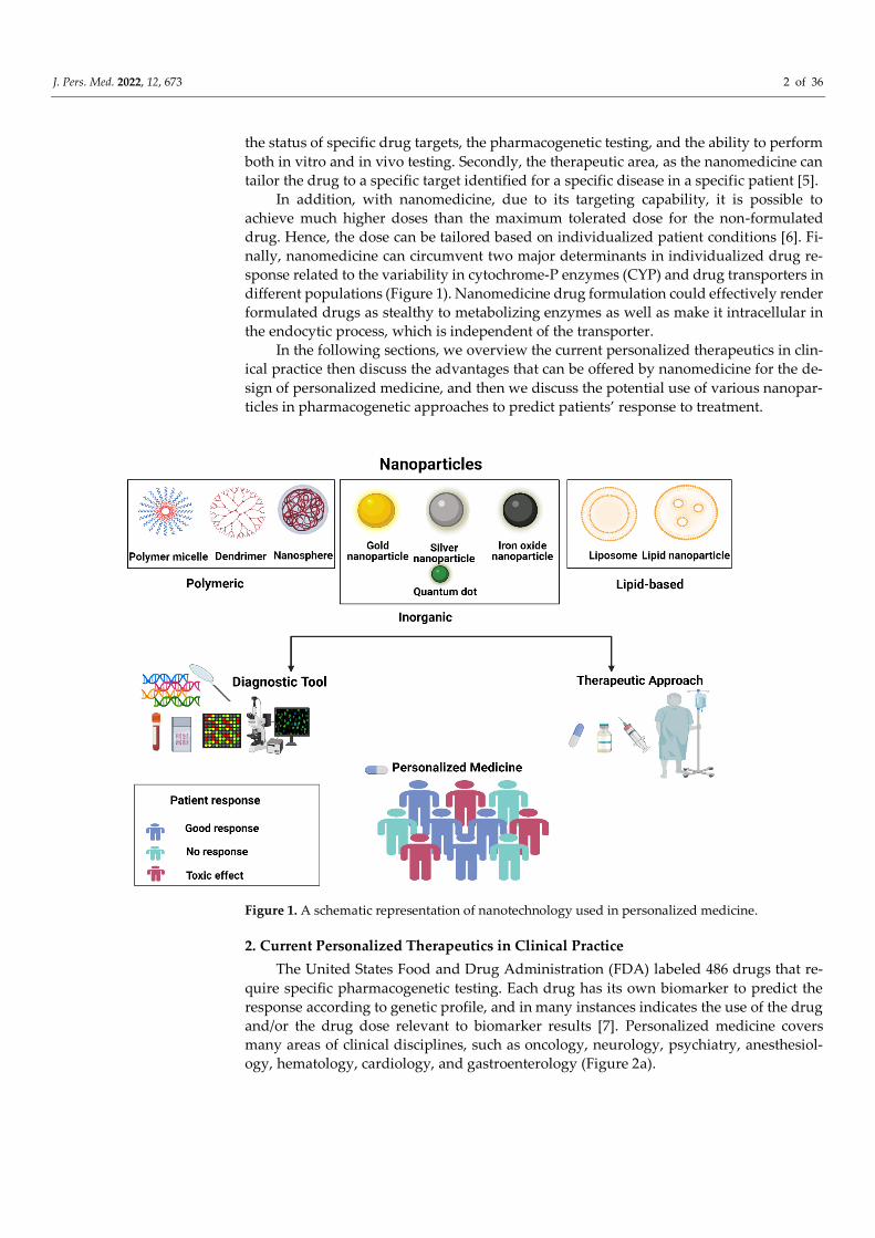
different populations (Figure 1). Nanomedicine drug formulation could effectively render
formulated drugs as stealthy to metabolizing enzymes as well as make it intracellular in
the endocytic process, which is independent of the transporter.
In the following sections, we overview the current personalized therapeutics in clin-
ical practice then discuss the advantages that can be offered by nanomedicine for the de-
sign of personalized medicine, and then we discuss the potential use of various nanopar-
ticles in pharmacogenetic approaches to predict patients’ response to treatment.
Figure 1. A schematic representation of nanotechnology used in personalized medicine.
2. Current Personalized Therapeutics in Clinical Practice
The United States Food and Drug Administration (FDA) labeled 486 drugs that re-
quire specific pharmacogenetic testing. Each drug has its own biomarker to predict the
response according to genetic profile, and in many instances indicates the use of the drug
and/or the drug dose relevant to biomarker results [7]. Personalized medicine covers
many areas of clinical disciplines, such as oncology, neurology, psychiatry, anesthesiol-
ogy, hematology, cardiology, and gastroenterology (Figure 2a).

J. Pers. Med. 2022, 12, 673 3 of 36
(a) (b)
(c) (d)
(e) (f)
Figure 2. (a): Number of drugs in therapeutic areas; (b): biomarkers related to clinical therapeutics
area anesthesiology; (c): biomarkers related to clinical therapeutics area cardiology; (d): biomarkers related to clinical therapeutics area hematology; (e): biomarkers related to clinical therapeutics area
neurology; and (f): biomarkers related to clinical therapeutics area oncology.
The biomarkers’ majority is related to CYP enzyme polymorphisms, resulting in pa-
tients having different metabolic activities as described later. Most of the CYP variations
are found in CYP2D6; CYP2C9 related to the therapeutic area of anesthesiology; CYP2D6,

J. Pers. Med. 2022, 12, 673 4 of 36
CYP2C19, CYP2C9, and CYP3A5 related to the therapeutic area of cardiology; CYP2C9 in
hematology; and CYP2D6 in oncology (Figure 2b–d,f).
The second phase metabolic enzymes are also used as biomarkers such as transfer-
ases that comprise uridine disphosphate glucoronosyltransferase (UGTs). UGTs are a ma-
jor part of phase II metabolism and are endoplasmic reticulum-bound enzymes responsi-
ble for the process of glucuronidation that includes 22 different functional enzymes.
In addition, glutathione S-transferases (GSTs) and sulfotransferases (SULTs) are im-
portant conjugative enzymes mediating phase II reactions. In the list of biomarkers, N-
acetyltransferases 1 and 2 (NAT1 and NAT2) cytosolic enzymes catalyze the acetylation
reactions; thiopurine S-methyltransferase (TPMT) is a cytoplasmic enzyme that catalyzes
the S-methylation of drugs. Efflux transporters include ATP-binding cassette (ABC) and
solute-linked carrier (SLC) proteins. These biomarkers were found to be related to some
therapeutics in cardiology, neurology, and oncology (Figure 2c,e,f) [8].
The majority of drugs associated with biomarkers are anticancer drugs (206 drugs)
(Figure 2a) and mount to 55% of total personalized drugs.
For instance, there are 20 anticancer drugs that target ERBB2(HER2). The situation of
HER2 biomarker would influence the prescription, dosage, or safety of administered
drugs such as Abemaciclib, Lapatinib, Alpelisib, Neratinib, Trastuzumab, and Olaparib.
3. The Pharmacokinetics (PK) and Pharmacodynamics (PD) Properties of Nanomedi-
cine and Formulation Advantages
Nanodrugs were originally designed to improve the properties of an already availa-
ble drug or diagnostic agent. Today nanoparticles are designed to minimize local and sys-
temic side effects, to enhance the bioavailability of drugs taken orally, and to improve the
half-life and overall pharmacokinetic and pharmacodynamic properties. Moreover, nano-
particles can reduce the frequency of administration of drugs, leading to better compli-
ance and ameliorating clinical outcomes [9,10].
Compared with traditional drug delivery systems, there are several pharmacokinetic
advantages that a nanodrug can offer: major solubility and absorption, the possibility of
controlled release, improvement of drug stability and metabolism, reduction of side ef-
fects, extended blood circulation, and better performance in targeted delivery [11].
Although the focus of pharmaceutical industries and of nanoparticles development
remains the optimization of the one-size-fits-all solution, the ability of nanomedicine to
personalize the pharmacokinetics and pharmacodynamics of the drugs and to overcome
the biological barriers limitations represents for an individual or a cohort with specific
genome requirements a promising personalized therapeutical opportunity [12,13]. Thus,
the aim of nanoparticle usage in personalized medicine is to exploit specific genomic pa-
tient information, comorbidities, and subjection to the environment to create an individ-
ualized treatment with improved drug specificity and optimized doses delivered in a spe-
cific site [13]. Regarding the pharmacokinetic processes, a drug could undergo four stages,
indicated by the acronym ADME: absorption, distribution, metabolism, and excretion
(Figure 3). The nanoparticle advantages in pharmacokinetic and pharmacodynamic pro-
cesses will be discussed in the following sections.

J. Pers. Med. 2022, 12, 673 5 of 36
Figure 3. Pharmacokinetics absorption, distribution, metabolism, and excretion (ADME) of person-
alized drugs or drugs combined with nanoparticles.
3.1. Absorption
The absorption process starts with the entering of nanodrugs into blood circulation
via different physiological routes. The frequently used routes of administration are intra-
venous, oral, transdermal, and nasal administration. The most important and extensively
used routes are the intravenous and the oral route. By means of an intravenous injection,
there is no need for absorption processes, indeed the nanodrug directly enters the blood
circulation. The surface charge, hydrophobicity, and the size of nanoparticles affect their
mucosal absorption. Indeed, smaller nanoparticles could have a better transcellular up-
take when compared to larger ones [14]. Moreover, nanoparticles with a size up to 500 nm
generally have better circulating and targeting ability and display a safer profile, reducing
the risk of capillary occlusions and embolism [15]. In addition, larger nanodrugs are rap-
idly cleared from the bloodstream due to opsonization processes and to the action of mac-
rophages of the reticuloendothelial system (RES) [9]. Via non-intravenous administration
routes, nanodrugs must pass through biological barriers before reaching the bloodstream.
This process is not always possible for traditional drugs, due to their unsuitable chemical
properties such as worst log P, solubility, and stability. Nanoparticles can overcome these
issues, improving the passage through biological barriers and absorption, allowing the
use of different administration routes that could not be used for traditional drugs [16].
For instance, after oral administration, a traditional drug could be degraded by the
low gastric pH or enzyme activity or could not be soluble in human fluids, affecting the
absorption process and the therapeutic action. When a drug is encapsulated in nanopar-
ticles, all these issues are overcome, and the absorption process strictly depends on the
nanoparticle’s physicochemical properties. When nanoparticles are orally administered,
they could be absorbed through the gastrointestinal tract by different processes, such as
paracellular pathway transport, transcytosis mediated by the carrier, passive cross-cell
diffusion, or microfold cells (M cells) absorption [16,17]. Thus, nanoparticles are able to
enter the systemic blood circulation by intestinal lymph node or through the portal vein;
moreover, nanoparticles absorption by M cells improves the drug bioavailability because
of the bypass of cytochrome P450 metabolism, hepatic first-pass, and P-glycoprotein (P-
gp)-mediated efflux [18].
One example of the advantages of nanoparticles in overcoming poor absorption is
the anticancer drug Olaparib (Ola). The drug’s main action is through poly ADP ribose

J. Pers. Med. 2022, 12, 673 6 of 36
polymerase (PARP) inhibition. The drug showed selective activity in tumors with a mu-
tated BRCA gene. However, Ola is poorly absorbed through the gastrointestinal tract. Ola
entrapment into a liposphere efficiently improved its oral bioavailability and further re-
duced its hematological toxicity [19].
The skin is the most difficult obstacle to overtake for nanodrugs designed for a trans-
dermal administration. The skin is composed of different lipophilic and hydrophilic lay-
ers, and this variability influences the difficult traditional drug absorption [11]. However,
Wang et al. proved that imidazole-based ionic liquid microemulsions are able to reduce
the skin barrier properties, disrupting the arrangements of corneocytes and moderating
the surface characteristics of the stratum corneum [20].
During the nasal administration, nanodrugs deposited in the lungs can be removed
via mucociliary clearance to the gastrointestinal tract, exhaled or sequestered, and de-
graded by macrophages. The remainder can cross the mucus and lung epithelium, being
absorbed [21]. In addition, it is known that positively charged nanodrugs have an en-
hanced absorption process through the lung mucosa [17], because of the interaction with
the negatively charged sulphate sialic acid and sugar moieties of mucin [14].
3.2. Distribution
The distribution process starts when the nanodrug is translocated from the blood
circulation to the tissues and cells. Usually, after absorption, nanoparticles are rapidly dis-
tributed and accumulated to the spleen, bone marrow, and liver, because of the presence
of the sinusoidal endothelial capillaries. Instead, the nanoparticles amount is low in the
kidney after injection and highest after 1 month of the administration [22]. Moreover, the
distribution of nanodrugs to the brain tissue is difficult to achieve because of the blood–
brain barrier (BBB). Nanoparticles usually have a size greater than 5 nm, and this influences
the distribution because the majority of the endothelia present in the human body have fen-
estration of about 5 nm [17]. It is possible to distinguish between two different types of dis-
tribution processes for nanodrugs, which are known as passive and active targeting.
3.2.1. Passive Targeting and EPR Effect
The passive targeting essentially depends on the size of nanoparticles and on the in-
tact fenestration of the endothelia. The enhanced permeability and retention effect (EPR)
is a phenomenon discovered by Maeda et al. [23] that allows a more specific drug accu-
mulation in solid tumors and in infection sites [24].
On the one hand, tumor blood vessels and inflamed tissues have an impaired lym-
phatic drainage system and leaky vasculature systems with pore sizes that vary from 200
nm to about 800 nm, depending on the cell type or the condition of the tissues. This is
mainly due to a defective vascular architecture and widespread and rapid angiogenesis.
On the other hand, normal tissues have a vasculature system with pore sizes that do not
allow the nanoparticles to pass. For this reason, nanodrugs that fulfill the dimensions re-
ported above can passively arrive at the impaired site and release the drug, having a more
specific and effective therapeutic action, with a massive reduction of the needed dosage
and consequently reduced onset side effects [25].
Greish et al. utilized styrene-co-maleic acid (SMA) as a micellar nano-carrier, for
many anticancer agents. In a study utilizing SMA-doxorubicin, the group demonstrated
the preferential accumulation of SMA-doxorubicin 13-fold higher in tumor tissues com-
pared to equivalent doses of free doxorubicin. Similarly, the group utilized the same mi-
cellar system to deliver nano micelles containing dasatinib and targeting PDGF, KIT, and
ABl [26,27].
At the same time, it is possible to say that the EPR effect could have some limitations.
Indeed, non-solid tumors such as leukemia cannot benefit from the EPR effect. Moreover,
different tumor types could have different pore sizes, and the nanodrug may be able to
target only some areas of the tumor, giving an unpredictable and maybe inefficient ther-
apeutic outcome [28].

J. Pers. Med. 2022, 12, 673 7 of 36
3.2.2. Active Targeting
Although EPR effect helps nanoparticles selectively reach the tumor interstitium, it
has no efficiency in promoting cellular uptake. The functionalization of nanoparticles with
ligands able to address the nanodrugs to specific cancer cells or sub-cellular sites not only
reduces the side effects due to passive targeting but also promotes the uptake of the na-
noparticles into the cells (Figure 4) [29]. This is due to the presence/overexpression of spe-
cific receptors over the cell surface that can be targeted by means of a specific binding
moiety. Moreover, active targeting could be a suitable method to exploit in personalized
medicine, allowing to treat the patient using the best possible ligand to selectively direct
the right dose of nanodrug to the right site.
Figure 4. Overview of nanoparticles modifications to improve PK and PD properties.
A wide range of ligands was used to decorate the surface of nanoparticles, including an-
tibodies or their fragment, aptamers, peptides or proteins, and many other receptor ligands.
Ligand-Based Targeting
A large variety of ligands were used to decorate the nanodrug’s surfaces, such as
adenosine, folate ligands, and glucose.
Adenosine is a nucleoside that plays a fundamental role in cellular function regula-
tion and activation of the adenosine receptors. Various types of tumors, such as colorectal,
prostatic, lymphoma, and breast cancer overexpress the adenosine A1 receptor [30].
Swami et al. investigated the ability of adenosine to direct solid lipid nanoparticles (SLN)
charged with docetaxel into human breast cancer and prostate cancer [31]. The adenosine-
conjugated SLN charged with docetaxel (ADN-SLN-DTX) demonstrated higher cytotoxi-
city and better pharmacokinetic parameters compared to the unconjugated SLN-DTX.
Folate receptors belong to a high-affinity folate-binding protein class that plays a role
in the cellular uptake of folate. While normal tissues do not present any folate receptors
on surface cells, they are overexpressed in several types of solid tumors such as testicular,
brain, endometrium, breast, and colon, and this overexpression is usually related to poor
clinical outcomes [30,32,33]. As reported by Patil et al., the grafting of pegylated liposomes
charged with a mitomycin C prodrug with folate ligands leads to a higher level of
nanodrug uptake by tumor cells, leading to increased cytotoxicity [34].
Glucose could be exploited for the active targeting approach as well. Cancer cells are
metabolically more active compared to normal cells, and this leads to an increased need
J. Pers. Med. 2022, 12, 673 8 of 36
for glucose. For this reason, glucose-coated nanodrugs, thanks to the presence of the glu-
cose transporter channel 1, have a higher permeation into cancer cells and this could be
helpful for the theragnostic approach [35]. In this context, Gromnicova et al. reported that
glucose-coated gold nanoparticles are able to transport loperamide selectively and effi-
ciently across the BBB [36].
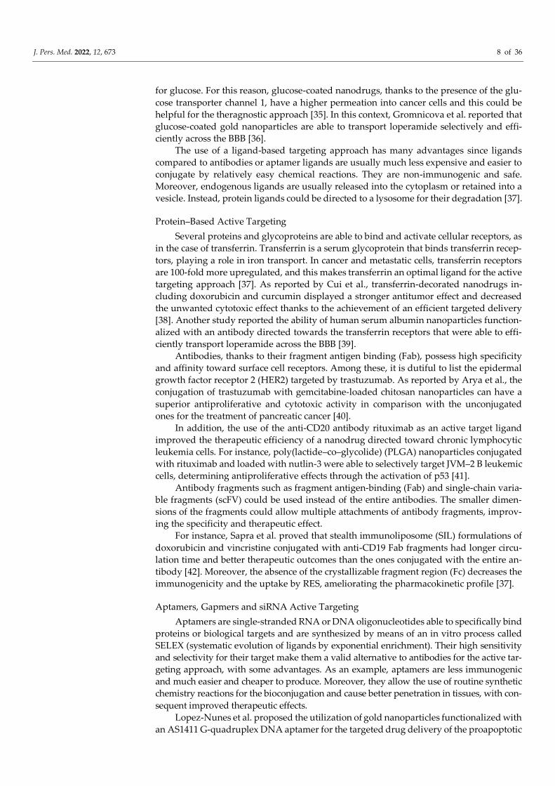
The use of a ligand-based targeting approach has many advantages since ligands
compared to antibodies or aptamer ligands are usually much less expensive and easier to
conjugate by relatively easy chemical reactions. They are non-immunogenic and safe.
Moreover, endogenous ligands are usually released into the cytoplasm or retained into a
vesicle. Instead, protein ligands could be directed to a lysosome for their degradation [37].
Protein–Based Active Targeting
Several proteins and glycoproteins are able to bind and activate cellular receptors, as
in the case of transferrin. Transferrin is a serum glycoprotein that binds transferrin recep-
tors, playing a role in iron transport. In cancer and metastatic cells, transferrin receptors
are 100-fold more upregulated, and this makes transferrin an optimal ligand for the active
targeting approach [37]. As reported by Cui et al., transferrin-decorated nanodrugs in-
cluding doxorubicin and curcumin displayed a stronger antitumor effect and decreased
the unwanted cytotoxic effect thanks to the achievement of an efficient targeted delivery
[38]. Another study reported the ability of human serum albumin nanoparticles function-
alized with an antibody directed towards the transferrin receptors that were able to effi-
ciently transport loperamide across the BBB [39].
Antibodies, thanks to their fragment antigen binding (Fab), possess high specificity
and affinity toward surface cell receptors. Among these, it is dutiful to list the epidermal
growth factor receptor 2 (HER2) targeted by trastuzumab. As reported by Arya et al., the
conjugation of trastuzumab with gemcitabine-loaded chitosan nanoparticles can have a
superior antiproliferative and cytotoxic activity in comparison with the unconjugated
ones for the treatment of pancreatic cancer [40].
In addition, the use of the anti-CD20 antibody rituximab as an active target ligand
improved the therapeutic efficiency of a nanodrug directed toward chronic lymphocytic
leukemia cells. For instance, poly(lactide–co–glycolide) (PLGA) nanoparticles conjugated
with rituximab and loaded with nutlin-3 were able to selectively target JVM–2 B leukemic
cells, determining antiproliferative effects through the activation of p53 [41].
Antibody fragments such as fragment antigen-binding (Fab) and single-chain varia-
ble fragments (scFV) could be used instead of the entire antibodies. The smaller dimen-
sions of the fragments could allow multiple attachments of antibody fragments, improv-
ing the specificity and therapeutic effect.
For instance, Sapra et al. proved that stealth immunoliposome (SIL) formulations of
doxorubicin and vincristine conjugated with anti-CD19 Fab fragments had longer circu-
lation time and better therapeutic outcomes than the ones conjugated with the entire an-
tibody [42]. Moreover, the absence of the crystallizable fragment region (Fc) decreases the
immunogenicity and the uptake by RES, ameliorating the pharmacokinetic profile [37].
Aptamers, Gapmers and siRNA Active Targeting
Aptamers are single-stranded RNA or DNA oligonucleotides able to specifically bind
proteins or biological targets and are synthesized by means of an in vitro process called
SELEX (systematic evolution of ligands by exponential enrichment). Their high sensitivity
and selectivity for their target make them a valid alternative to antibodies for the active tar-
geting approach, with some advantages. As an example, aptamers are less immunogenic
and much easier and cheaper to produce. Moreover, they allow the use of routine synthetic
chemistry reactions for the bioconjugation and cause better penetration in tissues, with con-
sequent improved therapeutic effects.
Lopez-Nunes et al. proposed the utilization of gold nanoparticles functionalized with
an AS1411 G-quadruplex DNA aptamer for the targeted drug delivery of the proapoptotic

J. Pers. Med. 2022, 12, 673 9 of 36
molecule imiquimod for the treatment of cervical cancer [43]. The chosen aptamer pos-
sesses a strong affinity for nucleolin, thus it is internalized by tumor cells and releases the
payload selectively in the diseased tissue. Results showed that this aptamer-based strat-
egy was successful in reducing HeLa cell viability and had less side effects on healthy cells
thanks to their lower expression of nucleolin and consequent reduced interaction between
this protein and the AS1411 DNA aptamer.
A similar strategy was exploited also for the management of lung cancer. Indeed,
Zhang and co-workers reported the development of a stimuli-responsive EGFR aptamer-
modified PLGA-SS-PEG nanoconstruct loaded with homoharringtonine, a natural alka-
loid with antitumoral properties [44]. The interaction of the aptamer with the EGFR pro-
tein up-regulated in non-small lung cancer cells (NSCLC) allowed the internalization of
the nanoparticle by endocytosis. The high reductive intracellular environment promoted
by the higher expression of glutathione (GSH) in NSCLC cells triggered the breakage of
the disulfide bonds of the nanoparticle, releasing the cargo inside the cell. Results obtained
in vitro and in vivo showed that this approach was more effective in decreasing cell via-
bility when compared to the single administration of the cytotoxic drug. Similarly, conju-
gation of the A10 RNA aptamer, directed towards the prostate-specific membrane antigen
(PSMA) with poly (D, L-lactic-co-glycolic acid)-block-poly (ethylene glycol) (PLGA-b-
PEG) nanoparticles encapsulating docetaxel showed an improved cytotoxic effect when
compared to the unconjugated ones [45].
Gapmers are short chimeric antisense oligonucleotides made of a central DNA-core
flanked by 2′-O-methylated-RNA-like sequences. This chimera can bind a complementary
oligonucleotide and silence a specific target RNA through its degradation by the action of
RNase-H [46]. Gapmers possess an intriguing therapeutic potential in the context of can-
cer and precision medicine. For example, Garcia-Garrido et al. designed gold nanoparti-
cles coated with citric acid, functionalized with polyethylene glycol (PEG), and branched
with short polyethylenimine (bPEI) chains. This scaffold was further cross-linked with
succinimidyl 3-(2-pyridyldithio) propionate (SPDP), affording the insertion of a GSH-sen-
sitive disulfide bond in the nanoconstruct structure. Overall, the chemically modified gold
nanoparticle showed a high stability and a global positive surface charge, which was ex-
ploited for cell transfection and for the formation of electrostatic interactions with gapmers
targeting the p53 mutant protein in pancreatic and breast cancer cells [47]. In the presence
of GSH, the nanoparticle structure undergoes a degradative process that allows the release
of the p53 targeting gapmer. This approach was successful in reducing cell proliferation in
PANC-1 and MDA-MB-231 cancer cell lines, which present the p53 mutation, whereas no
effects were observed in the MCF-7 cancer cells. Moreover, silencing of the mutated p53
protein reverted the cells susceptibility to gemcitabine, a chemotherapeutic agent whose bi-
ological effects are impaired when the p53 mutation is observed. In general, this approach
could be particularly useful for patients whose resistance to antitumoral drugs is dependent
on the alteration of proteins involved in pro-apoptotic pathways.
The utilization of siRNAs represents another efficient gene-silencing strategy that can
be used in antiviral and antiumoral therapeutic contexts. As an example, Idris and collab-
orators proposed the intravenous use of stealth liposomes as drug delivery systems for
siRNAs targeting highly conserved regions of SARS-CoV-2 with the aim of blocking virus
expression and replication [48]. These liposomes were formulated by fine tuning the
amount of the cationic molecules 1,2-dioleoyl-3-trimethylammonium-propane (DOTAP)
and MC3 in order to reduce the toxicity due to an excessive cationic surface charge and in
turn ameliorate the endosomal release of the siRNA in lung cells.
A phase 0 clinical study (NCT03020017) highlighted the potential of siRNA utiliza-
tion for the treatment of recurrent glioblastoma (GBM) [49]. The therapeutic treatment
usually involves the utilization of lomustine, carmustine, temozolomide, or bevacizumab,
coupled with surgery and radiotherapy. However, this type of cancer is particularly ag-
gressive, and poor chances of survival have often been reported. Therefore, new anti-
cancer strategies based on the discovery of novel small molecules or nanomedicine

J. Pers. Med. 2022, 12, 673 10 of 36
approaches are urgently needed [50]. Kumthekar et al. developed a RNA interference-
based spherical nucleic acids (SNAs), made of a gold nanoparticle conjugated with siRNA
oligonucleotides targeting the highly expressed GBM oncogene Bcl2Like12 (Bcl2L12). Af-
ter an intravenous administration, the SNA construct was able to reach the tumor site as
highlighted by the detection of gold through X-ray fluorescence microscopy. Inductively
coupled plasma mass spectrometry (ICP-MS) allowed to quantify gold plasma concentra-
tion. In general, gold clearance was slower than siRNA clearance, meaning that the nano-
particle slowly releases the cargo at the tumor site. The siRNA was able to efficiently si-
lence the Bcl2L12 oncogene, induce caspase-3 activation, and increase wild-type p53 pro-
tein expression. Finally, toxicological studies showed that the nanoconstruct did not de-
termine severe side effects in the treated patients. Despite the fact that additional studies
need to be performed, the proposed SNA nanoconjugate denotes the goodness of the
siRNA therapeutic efficiency as a promising precision medicine strategy for the treatment
of recurrent GBM.
3.3. Protection against Degradation Enzymes and Metabolism
The oral administration route suffers from several pharmacokinetic variabilities due
to the pre-uptake metabolism and first-pass metabolism [51,52]. Orally administrated
drugs have to overcome several host gastrointestinal obstacles such as pH mutability, gas-
trointestinal motility, physical barriers such as mucus and mucosa, bacterial diversity, and
especially several degradation enzymes (e.g., trypsin, lipase, and CYP450) and efflux
pump (e.g., P-gp) [53]. Among them, the CYP450 family, and especially CYP3A4, contrib-
ute to the large variability in drug response in a special population (e.g., children, preg-
nant women, elderly, and ethnicities) [54], leading to unpredictable therapeutic results
and/or several side effects. The CYP3A4 is responsible for the metabolism of more than
50% of marketed drugs [55] and the amount of this isoenzyme in the gastrointestinal tract
is about 40% of those present in the liver [52]. Therefore, the pre-uptake metabolism of
orally taken drugs should not be overlooked. CYP3A4 content in the gastrointestinal tract
decreases from the proximal duodenum to the distal ileum, and the activity of the enzyme
could largely vary from person to person [56]. Indeed, CYP3A4 content could be influ-
enced by diseases such as obesity, cancer, infection, and inflammation as the CYP3A4 ex-
pression strongly depends on the pregnane X receptor (PXR). Indeed, PXR is downregu-
lated in the diseases mentioned above [57].
Many types of nanoparticle formulation strategies have been tested to overcome the
pre-uptake metabolism and the variability derived from it, in order to enhance the phar-
macokinetic profile of the oral administrated drugs. The use of mucoadhesive polymers
could allow a better residence time and resistance to peristaltic movement, favoring the
absorption of the nanodrug through the gastrointestinal tract. For instance, Han et al. en-
hanced the adsorption at the mucus layer by coating the alendronate containing liposomes
with the cationic polysaccharide chitosan, which is able to interact with the negatively
charged mucin present in the proximal tract [58].
Another strategy is the use of highly lipophilic lipid nanoparticles (HLLN), especially
those containing triglycerides, which are able to transport the drug across the enterocytes
and to lymphatic vessels. Indeed, the HLLN is degraded in the intestinal lumen, absorbed
by enterocytes, and successively recomposed into chylomicron. Then, the drug incorpo-
rated into the chylomicron will be sorted directly into the lymphatic vessels avoiding pre-
uptake metabolism and first-pass metabolism [59].
The use of nanoparticles containing CYP3A4 inhibitors to inhibit the isoenzyme dur-
ing transcytosis could be another option [60], but the risk of potential toxic side effects due
to shut-down of the physiological action of the enzyme could be a relevant downside [61].
On the contrary, the use of common surfactant, co-solvents, and oil such as Tween, PEG,
Poloxamer, Cremophor, Polysorbate 80, and oleic acid proved to be able to notably inhibit
the CYP3A4 activity [62,63].

J. Pers. Med. 2022, 12, 673 11 of 36
Moreover, another delivery approach is the use of M cell-targeting nanoparticles to
directly deliver the nanodrug into the lymphatic vessels. The M cells are located in the
Peyer’s patches and have a high inclination to transport and induce endocytosis of anti-
gens into these ones. Strategies that target the M cells include the mimicking of the entry
of pathogens such as Salmonella and Yersinia or the targeting of specific receptors such
as the integrins that are located on the surface of these cells [64].
Another strategy that may be used is the development of nanoparticles capable of
releasing the drug in low CYP3A4 expression areas in pH-dependent manner (e.g., in the
ileum). The distal jejunum and ileum are regions in which the CYP3A4 expression and
activity are lower than that present in the proximal gastrointestinal tract. As the pH of the
jejunum and ileum is near 7 and 8, respectively, the use of pH-sensitive nanoparticles
could be useful to deliver a higher concentration of drugs in these regions, in order to
over-saturate the CYP3A4 enzyme, and to have a greater number of drugs that are able to
bypass the metabolism [64].
Lastly, the use of nanoparticles linked with vitamin B12 receptor–ligand could be
exploited in order to avoid the CYP3A4 metabolism. Liu et al. developed polymeric na-
noparticles (H/VC-LPNs) integrated with vitamin B12-modified chitosan, which is able to
enhance the oral bioavailability of curcumin [65].
As previously stated, the metabolic action of the degradation enzymes of the CYP450
family is one of the most influential factors in the pharmacokinetic destiny of a drug and
of its side effects. The gene variability of CYP450 contributes even more to the unpredict-
ability of the administrated drug, usually leading to severe side effects. For instance, this
is the case of epilepsy treatment, with it being one of the most studied cases in which the
CYP450 genetic factors variability influences the efficiency and safety of the antiepileptic
drugs a lot. Thus, the seizure control and the adverse reaction responses are different
across the patients, and the large variety of genotypic and phenotypic heterogeneity com-
plicates the physician choice, which, in these cases, relies on empirical data. Most of the
metabolism undergone by antiepileptic drugs is mediated by the CYP2C9 [66]. In the iso-
enzyme, several allelic variants could exist that encode for several isoforms with different
metabolic activity, distinguishing between:
1. Poor metabolizers: in which the drug is metabolized very slowly, experiencing sev-
eral side effects at standard doses;
2. Intermediate metabolizers: in which the drug is metabolized at a slow rate, having
potential side effects at standard doses;
3. Extensive metabolizers: in which the drug is metabolized at a normal rate and with
minimum risk of side effects and maximum therapeutic efficacy;
4. Ultrarapid metabolizers: in which the drug is rapidly metabolized and removed too
quickly to provide a therapeutic effect.
Therefore, the gene testing for the CYP450 enzyme polymorphism could be helpful
to identify the level of metabolic activity of the specific phenotype, to classify the patient
on the basis of the type of the polymorphism, allowing the use of the right dosage for the
seizure control. Established evidence showed how polymorphic CYP2C9 variant allele
can notably lead to the various antiepileptic drug concentration levels in the blood [67].
For instance, phenytoin metabolism depends on CYP2C9 activity. Patients with
CYP2C19*17 variant allele were found to demonstrate the fast metabolization of pheny-
toin, resulting in the absence of a drug response [68,69].
In such cases, nanomedicine could be helpful in overcoming these issues, protecting
the drug from the degradation enzymes, improving its half-life, and carrying the right
dose to the target site.
Nanoformulations of anti-retroviral drugs (ARVs) are a good example of how nano-
medicine could improve the bioavailability of a drug, bypassing the CYP metabolism and
overcoming the limitations and the side effects of the canonical therapeutic scheme, which
includes the need for a pharmaco-enhancer that could lead to drug–drug interactions [70].

J. Pers. Med. 2022, 12, 673 12 of 36
For instance, the potent HIV protease inhibitor Atazanavir suffers from a rapid CYP3A4
first-pass hepatic metabolism, leading to low availability. Chattopadhyay et al. demon-
strated in vitro how solid lipid nanoparticles encapsulating Atazanavir were able to en-
hance the uptake of the drug into hCMEC/D3 cell line, also bypassing efflux transporters
[71]. Chaowanachan et al. demonstrated how PLGA nanoparticles loaded with Efavirenz
(NP-EFV) led to a 50-fold reduction in the 50% IC50 in comparison with the free drug and
potent protection against HIV–1 BaL infection in vitro [72].
Considering the information discussed above, it could be possible to state that nano-
particles may have the potential to conduct canonical therapeutic schemes to a more effi-
cient choice for the patient and clinician, moving a step forward towards the future of
personalized medicine.
3.4. Nanoparticles Interaction with the Microenvironment
Precision medicine’s purpose is to use specific genetic, environmental, and comor-
bidities in patient information’s in order to perform accurate patient stratification and to
treat their disease condition specifically. As previously discussed, nanoparticles could be
helpful in precision medicine because of their ability to deliver in a more safely and tar-
geted way the encapsulated drug. Despite these advantages, nanoparticle efficacy is more
often diminished because of the heterogeneity of biological barriers and microenviron-
ment of the human body tissues, especially in the case of variability given by the morbid-
ities and comorbidities [13]. Nevertheless, nanoparticle clinical trials are still performed
in unstratified patient populations [73] and the tendency will possibly change in the future
because of the need for personalized treatment. In oncology, patient stratification proved
to be essential to produce positive results, even when patients were treated with nanon-
medicine [74]. Hence, patient stratification and nanoparticles modifications based on the
patient information have to go hand in hand with successfully performing personalized
medicine [75]. Moreover, despite the benefits given by the active targeting, diseased cells
markers can vary among patients making target selection processes limiting. Moreover,
the microenvironment seems to heavily influence the successfulness of drug-delivery pro-
cesses [76]. For instance, Qin Dai et al. reported that nanoparticles grafted with antibodies
are able to target only 2% of the tumour cells, causing a failure in treatment [77]. Indeed,
nanoparticles have to cross the local microenvironment to successfully deliver the drug in
the target cells, and here the obstacles may include physical barriers and changes in chem-
ical conditions. Thus, an in-depth understanding of the microenvironments seems to be
critical to reach the desired tissues with nanomedicines [13]. Moreover, microenvironment
features are generally different from the circulation ones, resulting in the possible altera-
tion in stability and properties of the nanoparticles.
For instance, several components of the tumor microenvironment, such as the extra-
cellular matrix (ECM) density, vasculature, and interstitial fluid, seem to contribute to the
non-penetration of nanoparticles in the tumor cells [13,78–80]. Moreover, pH or tempera-
ture variation in the microenvironment, such as in tumor conditions or in the wound heal-
ing process, could negatively influence the destiny of the nanoparticles. On the other
hand, these particular conditions may be exploited to perform personalized release of the
drug only in the diseased tissues, by means, as example, of pH or temperature-sensitive
nanoparticles [78].
Generally, only 0.1% of the free drug is able to accumulate to the target site and about
15% of the administered nanoparticles are able to do the same. This increase in tumor
accumulation is usually attributed to the EPR effect, as previously discussed. Many recent
findings have reconsidered and greatly de-emphasized the role of EPR effect in the accu-
mulation, proving that only a small fraction of penetrated nanodrugs could be attributed
to the EPR effect. Indeed, a crucial role seems to be played by immune cells interaction
and protein coronas mechanism [81].
Moreover, the heterogeneous formation of vasculature around the tumor can be
strictly influenced by individual factors such as lifestyle, genetics, age, chemotherapy, and

J. Pers. Med. 2022, 12, 673 13 of 36
comorbidities. To perform personalized treatments, nanoparticles must be selected on the
basis of the individual vasculature of the patient [73,82]. In addition to this, Sykes et al.
reported that variation in the tumor volume can influence the penetration and accumula-
tion of the nanoparticles in the tumor site. Suggesting that, nanoparticles can be poten-
tially personalized according to the tumor conditions to achieve hopeful therapeutic out-
comes [79]. Moreover, in the microenvironment, cells can overproduce altered ECM com-
ponents, resulting in a denser barrier that obstructs the penetration of nanoparticles
[77,80,83]. An additional obstacle for positively charged nanoparticles is the possible
charge interaction with the negatively charged ECM components, blocking the permea-
tion into the target site [84,85]. Obviously, the limited nanoparticle perfusion in the brain
can be likewise correlated to the limited extracellular space present in the brain microen-
vironment and to a non-specific adherence to ECM [13].
Additionally, biofilms and mucus layers can influence the distribution of nanoparti-
cles, entrapping them in various mesh pore size or by means of non-specific interaction,
leading to clearance from the epithelial surfaces [86]. Mucus composition, viscoelasticity,
and hydration depend on physiological conditions and location [86–88]. For instance, in
cystic fibrosis, the overexpression of MUC5B polymers results in decreasing the mucus
clearance and pore sizes [88,89]. Henceforth, since the microenvironment seems to be crit-
ical for the destiny of a nanoparticle, it is important to design novel types of nanoparticles
or modify them in order to take advantage of this variability. Exploiting endogenous trig-
gers such as the presence of a high level of matrix metalloproteinases (MMps), proteases
or of hypoxic, or an acidic microenvironment could enhance nanoparticles degradation
and drug release [90,91]. The use of exogenous triggers such as near-infrared light, radiof-
requencies, or magnetic fields could also be used to control the nanoparticles delivery
from the outside [91,92].
Even the incorporation of macrophage or leukocyte cell membranes derived from the
patient into nanoparticles seems to improve the efficiency in the targeted cancer cells,
while a weak targeting is given when the donor is different from the patient [93,94]. Usu-
ally, nanoparticles wrapped with cell membranes show a massive increase in drug activity
compared to a free drug [93].
Another used strategy to overcome the microenvironment-related issues is the use of
nanoparticles aimed t the remodeling of the microenvironment. Microenvironment re-
modeling could be helpful to increase nanoparticle penetration and to sensitize the tumor
to a specific treatment. For instance, Wilson et al. proved that the regulation of the gene
TREX1 in endothelial cells by means of microRNA can alter the tumor vasculature, sensi-
tizing the tumor to chemotherapy [95]. Moreover, the microenvironment modification
could allow to reduce the patient variability and to recruit more eligible patients in the
stratification.
In conclusion, it is possible to speculate that precision medicine strictly relies on strat-
ified patient populations, and the improvement of the delivery through the microenviron-
ment could increase the efficacy of the treatments. Targeting the microenvironment could
be possible to diminish the differences between patients’ variability, allowing them to be
included into stratified populations.
Moreover, the large modification availability for nanoparticles in terms of shape, size,
charge, surface properties, and active-targeting ligand modifications may be helpful in
precision medicine to better adapt the delivery systems to the microenvironment.
3.5. Intracellular Internalization and Subcellular Organelles Targeting
The effectiveness of nanomedicine therapeutic strategies is strictly dependent on the
design of the nano formulation since by modulating nanoparticle physicochemical prop-
erties it is possible to discriminate between healthy and diseased cells and predict the
mechanisms of cellular uptake and intracellular targeting. However, the translation from
in vitro to in vivo studies and from theory to practice is often affected by the complexity
of the biological systems with which nanoparticles interact [96]. In personalized medicine,
J. Pers. Med. 2022, 12, 673 14 of 36
these factors need to be carefully taken into consideration, indeed, drug targeting could
be highly affected on the basis of age, sex, target tissues and organs, metabolic differences,
and concomitant diseases that can alter different biological parameters. For instance, after
systemic administration, nanoparticles are often altered in their outer surface properties
by the absorption of serum proteins that can mask the presence of ligands grafted in the
nanoparticle surface necessary for a specific cellular targeting. The absorbed proteins,
known also as protein corona, could drastically change the characteristics of the nano for-
mulation with consequent hampered cellular uptake, inactivation, and premature elimi-
nation [97]. The impact of protein corona formation can be partially overcome by the en-
gineering of nanoparticles possessing a zwitterionic charge in their surface and a global
hydrophobic nature [98] or by the insertion of poly(ethylene glycol) (PEG) chains with
low molecular weight [99].
Bertrand and co-workers tried to understand how the physicochemical properties of
PEG-PLGA nanoparticles and their interaction with plasma resident proteins could mod-
ulate the biodistribution and clearance [100]. The authors found that a low PEG density
reduces the nanoparticle clearance whereas a higher PEGylation determines an opposite
effect. Interestingly, a total number of 20 PEG chains per 100 nm2 represents a threshold
value beyond which the circulation time and clearance of the nanoparticle remain almost
the same independently from the nanoparticle size. In addition, this threshold value was
found to be consistent in organisms possessing different protein phenotypes. On the other
hand, the nature of the proteins composing the protein corona varies on the basis of the
steric properties of the nanoparticle surface, with a consequent different impact on the
circulation time. Indeed, despite proteins of the complement cascade not seeming to be
involved in modifying the clearance of nanoparticles as shown by wild type and comple-
ment protein 3 (C3) knockout mice, different results were obtained with apolipoprotein E
(ApoE). The latter belongs to a class of proteins hardly adherent to the nanoparticle sur-
face. A low PEG density was associated with a high deposition of ApoE in vivo, with a
consequent reduced clearance. An opposite effect was noticed in high PEG-covered nano-
particles or in ApoE−/− animals with a resultant higher nanoparticle opsonization and elim-
ination. The LDL receptor (LDLR) also showed to interfere with nanoparticles distribution
and elimination rate independently from the PEG density. In fact, LDLR−/− knockout mice
or pretreatment of the experimental animals with a strong LDLR binder such as propro-
tein convertase subtilisin/kexin type 9 (PCSK9) increased the nanoparticle circulation time
when compared to the control.
Recently, the formation of a personalized protein corona started to be considered not
only as an obstacle for proper nanoparticles targeting but also as an emerging tool for the
development of diagnostic and therapeutic personalized nanomedicine. In this context,
Ren and co-workers reported a proteomic study in which polyanionic gadolinium
metallofullerenol (Gd@C82(OH)22) nanoparticles were used for the analysis of the compo-
sition of the protein corona in 10 patients with lung squamous cell carcinoma [101]. Inter-
estingly, results led to the identification of C1q, a protein of the complement system, as
the most bound biomarker to the surface of Gd@C82(OH)22 nanoparticles. Binding of C1q
Gd@C82(OH)22 nanoconstructs led to the disruption of the secondary structure of the pro-
tein, nanoparticle endosomal internalization, and enhanced activation of the immune sys-
tem. This approach could be further explored for the design of nanoformulations in which
the nature of the personalized protein corona could be exploited for the production of
novel diagnostic and anticancer nanomedicines.
Another aspect that can prevent nanoparticles cellular uptake is represented by the
surface charge. The phospholipidic nature of cell membranes determines a general surface
negative charge that can inhibit the uptake of anionic nanoparticles. On the other hand,
positively charged nanoparticles could benefit from a better intracellular uptake, even if
some cytotoxic effects have been reported [102]. The most common mechanisms of nano-
particles cellular uptake are represented by direct diffusion or endocytosis. The former
entry mechanism is typical of nanostructures with a diameter of <5 nm [103], for lipid
J. Pers. Med. 2022, 12, 673 15 of 36
nanoparticles [104], and for nanoparticles grafted with cell-penetrating peptides, which
are short aminoacidic sequences that help a covalently or non-covalently-bound cargo to
penetrate inside a cell or a specific organelle [105]. This entrance route is usually desired
when the nanoparticle payload must be released directly into the cytoplasm, such as for
siRNA delivery [106].
During the last years, increased knowledge of endocytic processes allowed the de-
sign of “smarter” nanoparticles with improved targeting properties [13,107]. Nanoparti-
cles with a size ranging from 100 to 500 nm are usually internalized through a clathrin-
dependent endocytosis pathway [108,109]. This mechanism is triggered by the binding of
nanoparticle ligands to specific cellular receptors. Ligand–receptor binding determines
the formation of vesicles coated at the cytoplasmic level by clarithrin molecules whose
polymerization brings vesicles to maturation and subsequent scissure from the cell mem-
brane. The newly formed vesicles are later transported in the cytoplasm and uncoated from
clarithrin, bringing about the formation of endosomes [110]. Similarly, a second common
nanoparticle internalization pathway is represented by the caveolin-dependent endocytosis.
This process does not require a ligand–receptor binding mechanism for its activation and
the resulting endosomal vesicles are transferred to cytoplasmatic organelles, such as the en-
doplasmic reticulum and the Golgi apparatus. Caveolin-dependent endocytosis is usually
observed for nanoparticles with sizes ranging from 50 to 100 nm [108,109].
Nanoparticles can also enter cells by phagocytosis and macropinocytosis. The first
mechanism is carried out by phagocytes, immune cells that recognize elements foreign to
the organism. The phagocytic process involves the recognition of such elements after
binding to scavenger receptors. Phagocyte entrapment of the foreign materials brings
about the formation of phagosome vesicles that are later fused to lysosomes, bringing
about the formation of phagolysosomes [109]. Macropinocytosis is an actin-dependent
non-specific mechanism of cellular uptake regulated by the activity of Ras protein [111].
Through this process, macrophages and dendritic cells can internalize viruses, growth
factors, and particles whose size falls in the micromolar range. Macropinocytosis repre-
sents an important mechanism exploited by cancer cells for the translocation and traffick-
ing of components of the plasma membrane and growth factors, contributing to the en-
hancement of cancer aggressiveness and metastasization [112]. Moreover, mutations in
proteins of the Ras family, such as KRAS, are associated with a heightened macropinocy-
totic activity that comes up with cell proliferation and sustained ATP accumulation and
consumption in the tumor microenvironment [113].
After cellular uptake, nanoparticles entrapped in endosomes through the abovemen-
tioned processes need to be released from these vesicles in order to exert their activity at
the desired site of action. Rational modification of nanoparticles surface charge and mate-
rial composition could be performed in order to disrupt the endosomal membrane and
facilitate the release of the cargo [114]. In addition to this, endosomes are also character-
ized by an acidic environment. In this context, several examples of pH-sensitive nanopar-
ticles have been reported [115–118], and the nature of the chemical bond that links the pay-
load to the nanoparticle can be exploited for the pH-triggered release of the drug [119]. Fi-
nally, enzyme-cleavable bonds can also be utilized for the release of the therapeutic drug.
Indeed, endosomes and lysosomes possess enzymes that can cleave specific chemical bonds
or linkers properly inserted in the engineered nanoparticle in order to facilitate the release
of the drug to the cytoplasm. For instance, the insertion of a Gly–Phe–Leu–Gly peptidyl
linker has been exploited for the development of poly (glycolic acid) (PGA)–paclitaxel na-
noparticles [120,121]. After the cleavage of the peptidyl linker by the lysosomal cathepsin-B
enzyme, the drug can be easily released from the nanoparticle, exerting a better cytotoxic
effect in NSCLC patients. Moreover, the estrogen-mediated higher production of cathepsin-
B [122] led to better therapeutic results in women treated with such PGA-paclitaxel nano-
particles, paving the way for a gender-based personalized anticancer treatment.
In the context of precision medicine, nanoparticles also need to exert their effects at the
subcellular level [123]. Nuclear drug delivery is often hampered by the size of the nuclear

J. Pers. Med. 2022, 12, 673 16 of 36
pore complex, which allows the passive diffusion of nanoparticles with a diameter of < 10
nm [124]. For nanoparticles with a higher size, an active nuclear transport is required. This
problem can be overcome by the insertion in the nanoconstructs of basic rich aminoacidic
sequences known as nuclear localization signal (NLS) motifs [125], such as the trans activa-
tor of transcription (TAT) peptide (YGRKKRRQRRR), which bind to importins and translo-
cate inside the cell nucleus [126]. This approach is particularly desired when the nanoparti-
cle cargo is represented by a small molecule targeting DNA, such as doxorubicin.
Mitochondria represent a difficult subcellular organelle targeted by nanoparticles,
mostly because of its highly negative membrane electric potential. In order to bypass this
drawback, mitochondrial localization signal (MLS) can be used for the design of more effi-
cient nanomedicines [127]. MLS are short peptide sequences containing basic and positively
charged amino acids that can facilitate the entry of the cargo linked to the nanoparticle in-
side the cell. In addition, triphenyl phosphonium is often used for engineering mitochon-
dria-targeting nanoparticles because of its lipophilic properties and its positive charge, with
both properties favoring the internalization of the payload into the mitochondria [128].
Finally, the Golgi apparatus represents another potential subcellular nanoparticle
target exploitable for the treatment of several pathologies, including cancer. This subcel-
lular complex is responsible for the post-translational modifications and trafficking of pro-
teins to different intracellular compartments. The alteration of Golgi’s activity could lead
to the inactivation of proteins responsible for the onset of cancer. Intrigued by the obser-
vation that chondroitin sulphate accumulates in the Golgi apparatus of tumor cells and
retinoic acid alters the Golgi apparatus morphology, Li and co-workers designed a nano
formulation based on paclitaxel, chondroitin sulphate, and retinoic acid (PTX-CS–RA),
which showed anti-metastatic effects by inhibiting metastasis-associated proteins through
Golgi apparatus disruption and reduced tumor growth in 4T1 cells [129]. Similarly, Luo
et al. reported the preparation of chondroitin-modified lipid nanoparticles loaded with
doxorubicin (DOX) and retinoic acid (RA), which displayed better antitumor effects in
SMMC–7721 hepatoma cells when compared to the separate administration of the two
free drugs [130].
3.6. Overcome MDR Mechanisms
Multidrug resistance (MDR) phenomena represent a major drawback for the achieve-
ment of optimal therapeutic effectiveness. These mechanisms, usually related to the fail-
ure of cancer treatments, take into consideration a wide plethora of expedients utilized by
cancer cells to inhibit cell death and sustain tumor progression, such as reduced drug cel-
lular uptake, mutation of cellular targets, and increased drug inactivation through en-
hanced metabolism or alteration of drug targets [131,132]. The increasing knowledge in
pharmacoproteomics, pharmacogenomics, pharmacogenetics, and pharmacometabolom-
ics could be helpful in defining novel useful approaches for personalized therapeutic reg-
imens [133]. In this context, nanomedicine and personalized medicine are strictly inter-
twined. Indeed, nanoparticles represent powerful tools in which it is possible to combine
at the same time more than one drug, diagnostic agents, and/or biotechnological drugs
with the aim to interfere with those mechanisms involved in the onset of MDR, even in a
personalized manner.
One of the most important mechanisms related to MDR is represented by the over-
expression of molecules transporters, better known as ATP-binding cassettes. These trans-
porters extrude drugs outside the cell, lowering the optimal concentration of the drug
required for the cytotoxic effect. Furthermore, cells who acquire resistance to chemother-
apeutic agents usually become resistant also to drugs belonging to different chemical clas-
ses or to drugs acting with a different mechanism of action, globally worsening the MDR
phenomena [28]. Pharmacogenomics studies focused on MDR-associated proteins are im-
portant in forecasting the goodness of a therapeutic strategy in a subset of patients in
which interindividual differences could play a significant role in the success of a pharma-
cological approach [134,135].

J. Pers. Med. 2022, 12, 673 17 of 36
The most known transporter involved in MDR is P-gp. This protein is responsible for
the removal of doxorubicin, paclitaxel, etoposide, and vinblastine from cancer cells [136].
Nanomedicine approaches targeting the P-gp are usually based on the contemporary ad-
ministration with a singular nano formulation of a cytotoxic drug and a P-gp inhibitor,
such as verapamil, cyclosporine, or curcumin [136]. An additional strategy is also repre-
sented by the administration of siRNA-targeting genes involved in the production of ABC
transporters [132,137,138]. The global result would consist of a reduced efflux of the drug
outside the cell and its consequent higher intracellular concentration. For example, Jiang
and co-workers reported the fabrication of RGD peptide-modified cationic liposomes de-
livering doxorubicin and ABCB1 siRNA [139]. Binding of the RGD peptide to integrin
receptors of tumor cells enhanced the intracellular uptake of doxorubicin and siRNA. In
vivo studies performed in a mouse model of doxorubicin-resistant MCF/A cells showed
that these liposomes possessed a higher cytotoxic effect when compared to liposomes
loading doxorubicin alone. This result should be attributable to the accumulation of the
siRNA inside the cells with consequent higher cytotoxicity due to the administration of
doxorubicin.
Genetic variants can also determine poor chemotherapy responses. Mutations in pro-
teins involved in apoptotic processes, over-expression of pro-apoptotic, and down-regu-
lation of anti-apoptotic proteins represent additional mechanisms involved in the onset of
MDR. For instance, the over-expression of anti-apoptotic proteins belonging to the Bcl-2
family is one of the most common hallmarks of resistant cancer cells. Yu et al. reported on
the co-delivery of epirubicin and Bcl-2 siRNA (siBCL-2) through pH-sensitive lipid nano-
particles [140]. The acidic endosomal environment allowed the escape of the siBCL-2 and
proper tumor cell transfection; moreover, the lipid nano construct was also able to down-
regulate P-gp overexpression and inhibited cell proliferation. Similarly, Ghaffari and co-
workers designed a polyamidoamine (PAMAM) dendrimer loaded with curcumin and
grafted with a Bcl-2 siRNA with improved anticancer effects in HeLa cells when compared
with the effects exerted by curcumin alone or PAMAM-curcumin [141]. On the basis of
these examples, it is obvious that an in-depth analysis of genetic variants and gene muta-
tions through pharmacogenomics and pharmacoproteomic studies is highly desirable. In-
deed, precise tumor typing could allow a better comprehension of the mechanisms in-
volved in MDR, and consequently, it could be helpful in the design of personalized ther-
apeutic strategies.
The tumor microenvironment (TME) and cancer stem cells also play a pivotal role in
the development of MDR. Cancer stem cells are characterized by quiescence, they express
drug efflux proteins, and possess an intrinsic resistance towards cytotoxic drugs because
they frequently repair any possible damage in the DNA structure [142]. In addition, the
tumor microenvironment contributes to cancer cell proliferation through the production
of growth factors [143]. Due to their high plasticity after radiotherapy and chemotherapy,
cancer stem cells can adopt a different phenotype that allows them to survive and give
rise to a new subpopulation of tumor cells [144,145]. The identification of specific TME
and cancer stem cells biomarkers is of particular importance for new personalized anti-
cancer therapies [146,147]. Within this framework, Gaio and co-workers designed hyalu-
ronic acid-coated polymeric nanoparticles for the delivery of docetaxel and the photosen-
sitizer meso-tetraphenyl chlorine di-sulfonate targeting breast cancer stem cells over-ex-
pressing the CD44 glycoprotein, combining within a single nano construct chemotherapy
and photodynamic therapy [148]. Binding of hyaluronic acid to CD44 allowed the nano-
particles to enter inside the cells through an endocytotic pathway, followed by cytotoxi-
city, which was enhanced by the presence of the photosensitizer. In another work, the
hypoxic environment in which cancer stem cells reside was exploited for the design of
hypoxia-sensitive nanoparticles [149]. A nitro-imidazole-modified hyaluronic acid–oxa-
late–camptothecin polymer-drug conjugate loaded with a differentiation-inducing agent,
all-trans-retinoic acid (ATRA), was engineered to suppress MCF-7/CD44+ tumor growth.
In tumor cells, reactive oxygen species (ROS) production triggered the disassembly of the
J. Pers. Med. 2022, 12, 673 18 of 36
nanoparticle with a consequent release of both ATRA and camptothecin. In cancer stem
cells, the binding between CD44 and hyaluronic acid caused nanoparticle cellular uptake,
whereas the hypoxic environment determined ATRA release but not camptothecin disas-
sembly from the hyaluronic acid polymer. Binding of ATRA to retinoic acid receptors and
cell differentiation also occurred. This last event determined a higher mitochondrial ac-
tivity and ROS generation, which in turn brought about the release of camptothecin. In
conclusion, the hypoxia-dependent release strategy allowed a controlled release in both
cancer and non-cancer stem cells, with a reduction of drug resistance phenomena in the
former and an overall tumor growth suppression and potential metastasization for both
groups of cancer cells.
3.7. Solubility
The discovery of a new drug and its potential usage for the treatment of a certain
pathology is often hindered by the physicochemical properties of the drug itself. In fact,
highly lipophilic drugs with high molecular weight are often characterized by poor solu-
bility that could prevent their utilization with consequent drug formulation issues [150].
Drugs endowed with high lipophilicity are not perfectly absorbed because they could be
trapped in the phospholipidic bilayer of cells. On the other hand, hydrophilic drugs such
as proteins and nucleic acids cannot be uptaken by simple passive diffusion by cells be-
cause of their incapacity to cross the cell membrane and could suffer poor stability in the
aqueous environment [151]. Highly charged drugs such as DNA, miRNA, and siRNA
could be administered after encapsulation in polymeric nanoparticles made of cationic
building blocks [151]. These problems could be overcome by the encapsulation of such
drugs into nanoparticles with a consequent improvement of their bioavailability.
5–Fluorouracil (5-FU) is a hydrophilic compound belonging to the chemical class of
cytotoxic antimetabolites. Drug resistance phenomena have been encountered with this
compound in colon cancer, and several strategies, such as the design of mutual prodrugs
[152,153] or encapsulation in nanoparticles, are used to avoid this complication. For in-
stance, 5-FU hydrophobicity and cytotoxicity can be alleviated by the production of pro-
drugs that can be efficiently loaded in xylan-stearic acid conjugates [154], liposomes [155]
or exosomes [156]. These strategies alter the hydrophilic/hydrophobic nature of the mol-
ecule, allowing also a co-administration with compounds possessing different chemical
profiles, such as doxorubicin or miRNAs.
The solubility of lipophilic compounds can be increased by loading the drugs in am-
phiphilic or hydrophobic nanoparticles. Thanks to this approach, Karve and co-workers
revived the use of wortmannin, a phosphoinositide 3-kinase inhibitor whose clinical
translation was hampered due to its high toxicity, poor stability, and high lipophilicity
[157]. Loading of wortmannin in a biodegradable lipid–polymer nanoparticle platform
reduced the intrinsic toxicity of the drug and enhanced its radio-sensitizing properties in
vitro and in vivo.
Romana et al. suggested the utilization of liposome–micelle hybrids for the delivery
of poorly soluble compounds. Using lovastatin as a drug model, they obtained better drug
loading when compared with loading in traditional liposomes or micelles; in addition,
they demonstrated a higher intestinal drug absorption and better transportation in a Caco-
2 cell monolayer model through P-gp transporter inhibition [158].
Another drug with great potential in anticancer therapies is represented by curcu-
min. This natural compound is highly lipophilic and photo sensible, unstable in acidic and
basic conditions, and rapidly metabolized and eliminated from the organism. The devel-
opment of curcumin nanoparticles has been extensively reported [159–163] and the best
results in terms of curcumin drug loading, stability, and solubility were reported by Gupta
and co-workers through encapsulation in solid–lipid nanoconstructs [164].
Salinomycin, an anticancer antibiotic, is another drug possessing a low aqueous sol-
ubility. Ni et al. engineered PEGylated poly (lactic-co-glycolic acid) nanoparticles loaded
with salinomycin and conjugated with a CD133 aptamer [165]. The new nanoformulations

J. Pers. Med. 2022, 12, 673 19 of 36
displayed a selective targeting and toxicity to osteosarcoma cancer stem cells expressing
the CD133 protein, with the potential to overcome MDR phenomena linked to the pro-
tumorigenic activity of such cancer stem cells.
Overall, these examples demonstrate that poorly soluble drugs can be reproposed
after encapsulation in specific engineered nanoconstructs for the establishment of novel
potential nanomedicine approaches. Moreover, grafting these nanoparticles with ligands
targeting a specific receptor over-expressed in a subpopulation of cells could represent a
suitable strategy to be further explored for personalized medicine therapeutic regimens.
4. Nanoparticles in Pharmacogenetic Testing
Pharmacogenetic testing is the first step for personalized medicine. In this respect,
nanomaterials such as metal nanoparticles, dendrimers, liposomes, quantum dots, and
carbon nanotubes have been explored for the assembly of a patient-specific molecular pro-
file for providing an accurate diagnosis of specific targets/genes. These nanoparticles al-
low for testing a plethora of patient-specific genes, clinically leading to a high precision
diagnosis and personalized management. Single DNA molecules can be sequenced using
nanodevices and nano-systems due to the small dimension of nanoparticles. Further to a
specific diagnosis, nanoparticles can provide a valuable tool to target the specific genetic
abnormalities. Currently, it is possible to clinically test multigene panels for the patient
and determine the decision on treatment for a particular drug [166].
Metallic nanoparticles have been studied in the diagnostic area of personalized med-
icine. Various nanoparticles such as gold nanoparticles (AuNPs), silver nanoparticles
(AgNPs), iron nanoparticles (FeNPs), and other polymeric nanoparticles were investi-
gated in pharmacogenetics.
The following is an overview of the usage of nanoparticles in personalized medicine
on both diagnostic and therapeutic fronts (Tables 1 and 2).
Table 1. Summary of nanotechnology application in genetic testing.
Nanoparticles Diagnostic Targets References
AuNPs
AuNPs are used as fluorescence quenchers detection of SNP [167]
AuNP detect TP53 point mutations [168]
AuNP detection of SNPs in BRCA1 [169]
AuNP detection of SNPs in CF genes [170]
AuNPs probes detect the expression of heparin in cancer cells. [171]
AuNPs electrochemical chip-based method Detection of cancer cells with KRAS and BRAF mu-
tations in lung cancer [172,173]
AuNPs fabricated as nanobeads with fluorophore
in micro array system
for the detection of C677T polymorphism of
MTHFR gene [174]
AgNPs
AgNP/Pt hybrid fabricated as nanocluster probe detect variant gene alleles in B-Thalassemia [175]
AgNP combined with carbon nanotubes detect the SNP related to mitochondrial DNA mu-
tation [176]
AgNPs probes detection of single variation presence in the breast
cancer BRCA1 gene [177]
DNA-AgNPs probes coating polystyrene mi-
crowells
detection of the presence of the specific sequence
DNA targets [178]
QDs
QDs Qbead system multiplexed SNP genotyping systems of 200 SNP
genotypes of CYPP450 family [179]
QDs labelling in a microarray detection system 10,000 SNPs from the unamplified DNA in a single
reaction [180]
QDs-mediated fluorescent method detection of hepatitis B M204I mutation, which is
associated with drug resistance. [181]

J. Pers. Med. 2022, 12, 673 20 of 36
FeNPs
FeNPs scanometric assay of DNA-based NPs identify SNP variants [182,183]
Biotin label captured and amplified on the sur-
face of streptavidin-coated FeNPs to detect C677T polymorphisms of MTHFR gene [184]
FeNPs coated with poly-amidoamine dendrimers
conjugated to fluorescein isothiocyanate and folic
acid
targeting of overexpressed FAR cancer cells [185]
Polymer NPs
cationic polyelectrolytes form a complex with
DNA by electrostatic interactions
identification of three types of SNP genotypes in
one extension reaction [186]
optically amplifying Poly[(9,9-bis(6′-N,N,N-tri
methyl ammonium hexyl bromide) fluorene)-co-
phenylene with the targeted DNA
SNP detection and genotyping assays were applied
in detection of SNPs in chromosome 17 polymor-
phism associated with frontotemporal neurodegen-
erative disorders.
[187]
fluorescent polymer (polyhedral oligomeric
silsesquioxanes) with PLGA with the surface an-
tibody of HER2.
distinguish the high Her2-expressing cancer cells [188]
Single nucleotide polymorphism (SNP), cystic fibrosis (CF), Kirsten rat sarcoma (KRAS), B-Raf proto-oncogene (BRAF), methylenetetrahydrofolate reductase (MTHFR), CYP P450 cytochrome
P450 family, folic acid receptor (FAR), human epidermal growth factor receptor 2 (HER2), and poly
(lactic-co-glycolic acid) (PLGA).
Table 2. Summary of drugs/nano construct applications in therapeutic areas and their targets.
Nanoparticles Therapeutic Targets Reference
AuNPs
Afatinib conjugated to AuNPs EGFR in NSCLC [189]
self-assembly gefitinib conjugated to colloi-
dal AuNPs EGFR to treat lung cancers [190]
Dasatinib loaded on AuNPs CML [191]
(PEG-PPG-PEG) with functionalized
AuNPs tyrosine kinase inhibitor-
Vandetanib, (ZD6474).
EGFR and VEGFR—for treatment of met-
astatic breast cancer [192]
AgNPs
AgNPs embedded in graphene oxide con-
jugated with the folate analog, MTX folate receptor-positive breast cells [193]
Capecitabine bonded to AgNPs Antiproliferative and proapoptotic effects
for different cancers [194]
AgNPs/FeNPs modified with (PEG)-car-
boxyl and folate and loaded with DOX cancer cells [195]
QDs
erlotinib conjugated to QDs EGFR in NSCLC [196]
carbon quantum dot CQD-based DOX
nanocarrier system against breast cancer cells [197]
CQD system conjugated with Quinic Acid
loaded with gemcitabine targeting agent toward breast cancer [198]
graphene quantum dots with imatinib
decrease BCR-ABL activity by targeting
ABL, c-kit, and PDGF-R-treatment of leu-
kemia
[199]
FeNPs
erlotinib-conjugated FeNPs EGFR in NSCLC [200]
Erlotinib-conjugated FeNPs lung adenocarcinoma [201]
FeNPs–carbon nanotubes with (PAMAM–
PEG–PAMAM) linear-dendritic copoly-
mers loaded with DOX
hybrid nanostructure can be used for tar-
geting, imaging, and cancer treatment [202]

J. Pers. Med. 2022, 12, 673 21 of 36
dasatinib-loaded FeNPs core with self-as-
sembly micelles
multitargeted inhibitor of many essential
kinases impacting oncogenesis in breast
cancer
[203]
Polymer NPs
poly (α, l-glutamic acid) polymer/selu-
metinib and dabrafenib BRAF, MEK—melanoma [204]
SMA/Crizotinib and dasatinib Met, ROS1, KIT, and ABL—glioblastoma
multiforme [27]
SMA/Sorafenib and nilotinib
VEGFR, PDGFR, FLT3, ALK, FGFR, c-
KIT, JAK, CSF1R, RET, and Bcr-Abl—
prostate cancer
[205]
chitosan-based polymeric nanoparti-
cles/Imatinib Bcr-Abl—colorectal cancer [206]
PLGA polymer/Tamoxifen estrogen receptor-positive breast cancer
cells [207]
PLGA polymer/Erlotinib EGFR in NSCLC [208]
Chronic myeloid leukemia (CML), epidermal growth factor receptor EGFR, non–small-cell lung
cancer (NSCLC), amphiphilic polymer of polyethylene glycol-block-polypropylene glycol-block-polyethylene glycol-block (PEG-PPG-PEG), vascular endothelial growth factor receptor (VEGFR),
methotrexate (MTX), amino-poly (ethylene glycol) (PEG), doxorubicin (DOX), stem cell factor re-
ceptor (c-kit), platelet-derived growth factor receptor (PDGF-R), polyamidoamine–polyethylene glycol–polyamidoamine (PAMAM–PEG–PAMAM), B-Raf proto-oncogene (BRAF), Mitogen-acti-
vated protein kinase (MEK), poly(styrene-co-maleic acid) (SMA), anaplastic lymphoma kinase
(Met), Proto-Oncogene 1, Receptor Tyrosine Kinase (ROS1), Tyrosine-protein kinase (ABL), Fms-like tyrosine kinase-3 receptor (FLT3), anaplastic lymphoma kinase (ALK), fibroblast growth factor
receptor (FGFR), Janus kinase (JAK), colony-stimulating factor 1 receptor (CSF1R), and glial cell
line-derived neurotrophic factor receptor(RET).
4.1. Gold Nanoparticles
Gold nanoparticles (AuNPs) are currently available as an integral part of biomarkers
assays in the detection of various genetic abnormalities. AuNPs with dense DNA shells
provide a versatile and programable means for the diagnostic purpose that can detect a
wide range of genes. The gold nanoparticle part in the nano-bio-complex provides the
optical advantages in the bioassay system with the ability of detecting the limit at the
picomolar concentration [209].
In addition, AuNPs can be used as a fluorescence quencher to suppress the effect of
luminescence or to enhance the electrochemiluminescence (ECL) of cadmium sulphide
(CdS) nanocrystal. Chen et al. devised a hybrid system that utilizes AuNP with CdS na-
noparticles for high-sensitivity detection of SNP. In this system, AuNPs are used as fluo-
rescence quenchers to suppress the effect of luminescence or to enhance ECL of the CdS
nanocrystal fabricated as a film through the distance modulation between the nano-me-
tallic and semiconductor components by a hairpin DNA [167].
Surface plasmon resonance (SPR) has been utilized for the detection of SNP. For ex-
ample, Jiang et al. [168] devised a system to detect TP53 point mutations utilizing oligo-
nucleotides immobilized on AuNP. The system was inexpensive and could be readily
used for clinical diagnosis. The same approach has been adopted by several companies
for the detection of SNPs in multiple genes such as BRCA1 and cystic fibrosis (CF) genes
[169,170].
Another advantage of AuNPs is their ability to measure intracellular gene expres-
sion. AuNPs can be constructed into “off-on” probes to quench light efficiently, resulting
in lower background signals than conventional molecular approaches. An important
property of the AuNPs DNA complex is their ability to be taken up by various types of
cells.
Lee and co-workers [171] used this approach to detect the expression of heparin in
cancer cells. Fluorophores were combined on the AuNP surface, resulting in a quenched

J. Pers. Med. 2022, 12, 673 22 of 36
fluorescence in an “off-state”. Upon a selective interaction with the expressed gene in met-
astatic cancer cells, the fluorophore is released, leading to a measurable fluorescent re-
sponse. These same properties can be utilized to target and modify a specific gene intra-
cellularly as a personalized therapeutic approach, as was explored by Seferos et al. [210].
Das et al. used the electrochemical properties of AuNP for the detection of specific
tumor circulating DNA in the blood stream obtained from the patient’s serum, using the
electrochemical chip-based method. Cancer cells with Kirsten rat sarcoma (KRAS) and
BRAF mutations could be readily quantified with high sensitivity and specificity. This sys-
tem is used as well to identify BRAF mutations in melanoma patients and to compare the
outcome results of this chip, employing the electrochemical assay with a PCR-based
method. The AuNP-based method achieved high levels of sensitivity and specificity with a
30 min short detection period advantage over the 2–3 h needed for a PCR [173]. Such a sys-
tem can be of great value in identifying patients who would respond to Sotorasib, which
specifically targets KRAS mutation and is used for the management of lung cancer [172].
In addition, the genotyping of large numbers of SNPs in an automated and a highly
productive manner was achieved by Song Li et al., who developed a micro array system
based on AuNP. The DNA primer-coated AuNPs were fabricated as nanobeads with a
fluorophore and set on a clean glass slide; then each sample genotype was discriminated
by bead array scanning. The system was applied on 320 samples for the detection of the
C677T polymorphism of methylenetetrahydrofolate reductase (MTHFR) gene in a simple,
fast, and cost-effective diagnostic tool [211]. This assay would be of great importance to
predict the response to methotrexate in patients with rheumatoid arthritis and hemato-
logical malignancies and to predict the response to 5-fluorouracil in patients with colorec-
tal cancer [174].
4.2. Silver Nanoparticles (AgNPs)
Silver nanoparticles are another attractive tool for pharmacogenetics testing and SNP
detection. Various techniques employed for SNP studies can be augmented by AgNPs for
ultrasensitive detection. AgNPs can infer high conductivity and enhanced electron transfer
properties. Wu et al. devised a AgNP/Pt hybrid to fabricate a nanocluster probe with locked
nucleic acid. The system was able to detect variant gene alleles in β-Thalassemia [175].
Jio et al. reported the electrochemical-sensing properties of AgNP combined with
carbon nanotubes in the detection of SNP. In their study, they detected the SNP related to
mitochondrial DNA mutation in type 2 diabetes mellitus by the DNA-mediated growth
of AgNPs within the single-walled carbon nanotubes SWCNTs-modified electrode. This
system accomplished high sensitivity to the targeted DNA with a low detection limit of 3
pM [176].
In another work Shi et al.used AgNPs to enhance the sensitivity and markedly im-
prove the detection limit of surface-enhanced Raman spectroscopy (SERS), utilizing the
dissolved silver ions from AgNPs. They forecast their system to be utilized in the future
for ultrasensitive genetic studies [212]. In another study, Wabuyele et al. applied the plas-
monic effect and SERS in the design of AgNPs label. This system exhibited high specificity
and selectivity of AgNPs probes in the detection of single variation presence in the breast
cancer BRCA1 gene [177]. The mutation in BRCA gene in ovarian cancer patients highly
affects the choice of treatment and could determine their response to anticancer drugs
such as platinum-based chemotherapeutics, and anthracyclines [213–215].
Another approach of the nanoparticle-based chemiluminescent (CL) method applied
the AgNPs for ultrasensitive detection of SNP. The assay in this system depends on the
hybridization of AgNPs with DNA. The hybridized technique was based on DNA-AgNPs
probes coating polystyrene microwells. This was followed by the detection of the presence
of the specific sequence DNA targets through the signal appearance after HNO3 solution
addition. The system offers the advantages of quantification of target DNA, simplicity,
and high sensitivity [178].

J. Pers. Med. 2022, 12, 673 23 of 36
4.3. Quantum Dots (QDs)
QDs are NPs that have a range size between 1 and 20 nm and form metal salts (such
as cadmium sulfide (CdS), cadmium telluride (CdTe), cadmium selenide (CdSe), and zinc
oxide (ZnQDs). QDs pose multiple innate characteristics that are suitable for pharmaco-
genetic testing. QDs have a broad absorption spectrum, tunable emission, high quantum
yields, and long lifetime fluorescence. These properties make them an ideal candidate for
the design of nanoparticle probes adopting Förster resonance energy transfer (FRET).
FRET is a phenomenon that happens when there is an energy transfer between two excited
donors through non-radiative dipole–dipole coupling when they are in the proximity of
a few nanometers [216,217].
QDs have been integrated into multiplexed SNP genotyping systems and used as
fluorescent probes, such as the Qbead™ system developed by Xu et al. Qbead system was
designed for the identification of around 200 SNP genotypes of cytochrome P450 family
from clinical samples. The Qbead system was a reliable and highly sensitive system with
100% accurate results concordant with those obtained from direct DNA sequencing [179].
This system is particularly important to detect variants of CYP2D6 and CYP2C19 that
can determine drug response variability in variants associated with ultrarapid metabo-
lism compared to other variants associated with poor drug metabolism [218].
Another interesting genotyping QD-based system reported by Karlin-Neumann et
al. employs four different QDs. The QDs were used for labeling in a microarray detection
system, more than 10,000 SNPs from the unamplified DNA in a single reaction. The sys-
tem proved advantageous in terms of broadband spectrum absorption with shorter wave-
lengths that resulted in higher levels of fluorescence emission [180].
QDs have been explored as fluorescent probes for the detection of gene mutations in
chronic hepatitis B. Cheng Zhang et al. used QDs-mediated fluorescent method for the
detection of hepatitis B M204I mutation that is associated with drug resistance. This mu-
tation is important in HBV drug management with tenofovir dipivoxil, telbivudine, ade-
fovir, lamivudine, and entecavir analogues [181].
For the detection of HBV mutants, the team utilized QD-labeled DNA. Then a fluo-
rescence microscope was used to visualize the fluorescence signal emitted from the QDs
with an excitation wavelength of 460–550 nm. The limit of detection for the detection of
HBV genetic variation in this system was 103 IU/mL [219].
4.4. Iron Oxide Nanoparticles (FeNPs)
Iron oxide nanoparticles are members of the class of ferrimagnetic materials that can
be used in different biomedical applications [220]. SNP detection using FeNPs has been
explored in several studies due to the biocompatibility, high efficiency, simple and fast
detection, reusability, and lack of interaction with separated substances activity [221].
Nam et al. reported the design of a sensitive scanometric assay of DNA-based NPs
tagged with biological barcoded DNA that serves as signal probe, and oligo-DNA FeNPs
serving as a capture probe for the targeted DNA to identify SNP variants. This system
represents an ultrasensitive SNP sensing technology with a detection limit of attomole
concentration (10−18 M) [182,183].
Liu et al. developed a high-throughput system for SNP genotyping utilizing solid-
phase polymerase chain reaction using a biotin-labeled primer captured and amplified on
the surface of streptavidin-coated FeNPs. Then SNPs were investigated by hybridization
with probes. The system was tested to detect C677T polymorphisms of the MTHFR gene,
yielding highly specific and sensitive results with the possible application of SNP on var-
ious targeted genes [184].
In addition, FeNPs probes can be employed for the simultaneous fluorescence and
magnetic resonance imaging of targeted overexpressed genes. The FeNPs could be cova-
lently or non-covalently bound to fluorescent dyes as reported by Wang et al. They used
FeNPs coated with poly-amidoamine dendrimers that were conjugated to fluorescein
J. Pers. Med. 2022, 12, 673 24 of 36
isothiocyanate (FITC) and folic acid, applying a layer-by-layer assembly method for spe-
cific targeting of overexpressed folic acid receptors (FAR) in cancer cells. The FeNPs core
was employed in conjunction with the FITC label to demonstrate their selectivity in tar-
geting KB cells through magnetic resonance and fluorescence imaging [185].
4.5. Polymeric Nanoparticles (Polymeric NPs)
Polymeric NPs can be sorted into two types: nano capsules as reservoir systems, and
nanospheres as matrix systems. They can be synthesized by natural materials, such as
protein-based (albumin, gelatin, and collagen) polymers or polysaccharide-based (hyalu-
ronic acid and chitosan) polymers. The other group of polymers is synthetic polymers, for
example, dendrimers, PLGA, polyglycolic acid (PGA), polyacrylic acid (PAA), and pol-
ylactic acid (PLA).
Conjugated polymers (CPs) are large, repeated units, delocalized molecular struc-
tures that manifest some unique properties such as optical (colorimetric or fluorometric)
and electrochemical properties, making them suitable as a sensing element. Several stud-
ies utilized CPs as transducers for SNP genotyping by FRET between CPs and a chromo-
phore-labeled DNA probe. Other studies have utilized CPs for SNP detection through the
conformational changes of CPs induced by a combination of specific DNA targets [222].
Duan et al. reported SNP genotyping assays using multi-step FRET and optical am-
plification of CPs. Water-soluble cationic polyelectrolytes can form complexes with DNA
by electrostatic interactions [186]. The system design depends on the energy transfer cas-
cade between complexes of CPs with two separately labeled DNAs with fluorescein and
Cy3. The amplification of fluorescence signals is based on the CPs–DNA strongly electro-
static interaction. This system was successful in concomitant identification of three types
of SNP genotypes in one extension reaction with high sensitivity.
In another study employing the optically amplifying CPs with the targeted DNA re-
ported by Gaylord et al. [187], SNP detection and genotyping assays were applied in the
detection of SNPs in chromosome 17 polymorphism associated with frontotemporal neu-
rodegenerative disorders. Poly[(9,9-bis(6′-N,N,N-trimethylammoniumhexylbromide)flu-
orene)-co-phenylene in addition to targeted DNA were recognized by sequence-specific
hybridization between the fluorescein-labeled peptide nucleic acid probe and the target
DNA sequence.
In a study by Li et al., the semiconducting fluorescent polymer (polyhedral oligo-
meric silsesquioxanes (POSS) was coated with poly(lactic-co-glycolic acid) (PLGA). The
nanoparticles’ sizes ranged in between 230 and 260 nm, and their surface was functional-
ized with the antibody of human epidermal growth factor receptor 2 (HER2). This system
was successfully used to discriminate between Her2-expressing SK-BR-3 cancer cells from
cancer cells not expressing the abovementioned receptor through fluorescence with high
quantum yield [188].
5. Challenges in Nanomedicine and Personalized Medicine
There has been a rapid increase in the number of nanomedicines that are being de-
veloped as therapeutics, however, clinical translation efficiency is less than satisfactory,
which indirectly suggests and highlights different challenges related to nanomedicine that
need to be addressed before introducing them for translational use. The major issues with
the nanomedicines are related to their safety at a biological level, their cost, and upscale
wide range production. Similarly, precision medicine, which relates to identifying and
targeting an individual’s genome for a particular disease, also faces challenges related to
ethical, social, and legal issues [223]. Nanomedicines, when administered to the body, en-
counter the biological environment, but there are no standard safety evaluation parame-
ters that determine their toxicity. Moreover, morphological and physiochemical charac-
teristics can affect the biodistribution and interaction with the biological membranes when
in systemic circulation, and hence, pose a threat to safety. There may be issues associated
with specific nanomedicine products and for which particular evaluations might be

J. Pers. Med. 2022, 12, 673 25 of 36
required. Toxicity in the medical industry is defined in terms of acute and chronic toxicity.
While the acute toxicity refers to inflammation, hemolysis, oxidative stress, and other or-
gan-specific effects as such, the chronic toxicity occurs after a long period of time and is
not usually addressed [224].
There is a need to understand the physiochemical properties of both the drug and
the nanocarrier, along with the reagents and pathways utilized for synthesis, from a toxi-
cological perspective for improving their chances of success. As opposed to traditional
pharmaceuticals, nanomedicine is a complex three-dimensional system with multiple
components, each of which is intended to serve a specific purpose. Due to their complex-
ities, advanced analytical tests are needed to fully detect, characterize, and quantify each
component, as well as evaluate the interactions between them and the interaction between
the drug-loaded nanoparticle and the biological systems. Furthermore, an additional issue
of concern is the stability and the long-term storage of these nanoparticles-based products;
hence, the type of lipids and polymers used for synthesis must be kept in check [225]. On
a larger scale, even after a nanomedicine is introduced into market, it can be withdrawn
if the quality and safety levels do not comply with the standard regulations.
To translate nanomedicine into a personalized drug entity, large-scale synthesis with
high reproducibility is necessary. Typically, these drug-loaded nanocarriers are synthe-
sized in small batches in laboratories, or for preclinical and clinical investigations. The
large-scale production on the other hand is much more difficult considering the complex-
ities of different nanoparticles, and even the slightest change in the manufacturing process
can cause critical alterations in the characteristic’s properties of these nanoparticles. It is
crucial that parameters such as the ratio of the nanocarrier polymer, entrapped drug, or-
ganic solvent, crosslinker, emulsifier, and even temperature, and pH be determined and
kept at appropriate levels. In addition to this, the process of scaling up of these nanomed-
icines is often a costly multistep process not just in terms of manufacturing but also in
terms of the cost needed for preclinical and clinical studies, which makes a new nanomed-
icine unlikely to succeed. It can be challenging to gain regulatory approval for new nano-
medicines, especially if existing products with similar efficacy already exist with the same
indication, hence the researchers’ work to improve the biodistribution, bioavailability and
efficacy of conventional drugs [226]
Moreover, developing nanomedicines in general is hampered by a lack of standards
and regulations in manufacturing practices, quality control, safety, and efficacy evalua-
tion. Now, the standards of nanomedicines are determined by regulatory authorities such
as USFDA and EMA to keep a check and to provide guidance, although no specific regu-
latory standards governing their production or clinical translation have been devised till
date. Therefore, when it comes to the application of nanotechnology, different geograph-
ical regions have distinctive approaches such as the nanomedicine approved in one coun-
try might not get approval in the other [227,228].
Similarly, when participating in drug testing in personalized medicine, patients will
need to understand the potential risk that might be associated with it, and hence receiving
informed consent can be a rigorous process in various cases. Moreover, just like the pro-
duction of nanomedicine, the cost related to the development of a precision medicine is
also very high and will require funding for years with a daunting uncertainty of success.
The technologies required for sequencing of an individual’s DNA and designing nano-
medicine also add to the cost. Therefore, prior to introducing personalized medicine in
the market, it must become part of a routine healthcare. There is a need to understand the
disease at the molecular and genetic level, to interpret the genetic testing required for the
patient, and then design the medication thereafter, which will not only reduce the chances
of failure but will also improve the knowledge and understanding of a patient’s specific
conditions [229–231].
Regarding clinical trials, it is important that these trails be performed as individual-
ized treatment plans to cope up with the ever-changing landscape of diseases.

J. Pers. Med. 2022, 12, 673 26 of 36
In order to enroll patients in clinical trials, multicenter collaboration is critical when
drug development is in its early stages. Additionally, there are established concepts in the
field of drug development, such as phase 1 expansion cohorts replacing phase 2 tests, reg-
ulatory approvals based on nonrandomized trials, and tumor-agnostic approvals. Yet, in
clinical trials, there are problems related to patient accrual and unresolved regulatory is-
sues, as well as technical limitations of molecular tests that need to be resolved [232,233].
Although these advanced treatments are still relatively new, they have already
shown promise in treating conditions such as epilepsy, cystic fibrosis, and some forms of
cancer and diseases that have a deeper genetic basis. There are two different ways by
which precision medicine can change clinical trials. Firstly, more trials need to be con-
ducted to test the efficacy of medicine in patients, which is almost like what happens in
MATCH trails. Secondly, gene sequencing must be performed simultaneously to help in
creating and dividing patients into different subtypes of disease.
It has been suggested that smaller clinical trials can serve to be a better and more
efficient option when testing a precision medicine. Scientists have proposed that when a
drug is meant for a small group of targeted individuals, it would show variation in terms
of the efficacy and the chances of it failing in a regular clinical trial would be high. For
instance, the drug ivacaftor (Kalydeco) meant for cystic fibrosis treatment was approved
in 2012 for certain specific gene mutations and was effective in only about 4% of the total
patients of cystic fibrosis. Clinical trials done in smaller specific populations might as well
produce efficacious results, and hence, faster drug approval. Researchers are still working
on defining and designing better randomized studies to improve the translation of preci-
sion medicine in the market [234]. In order to integrate genomic data efficiently and dy-
namically and to evaluate the validity of matching unique genomic alterations with spe-
cific interventions or treatments, new strategies have been developed. These include clin-
ical studies with adaptive designs, umbrella trials, basket trils, and aplatform trials among
others. Adaptive design refers to a clinical study designed to adapt to data analysis (usu-
ally intermediate findings) of the study subjects with the intention of modifying one or
more specified elements of the research design and hypothesis. Similarly, an umbrella
trial, also known as a master trial, is a program in which patients are identified as eligible
based on the presence of a specific tumor type that is sub stratified based on specific mo-
lecular alterations matched with different anticancer therapies. For example, this method
was utilized in the MoTriColor EU H2020-funded project, which involved a set of molec-
ularly guided trials with specific treatment strategies in patients with advanced newly
molecular defined subtypes of colorectal cancer. Unlike the above-stated methods, in a
basket trial, different tumor types with a common molecular alteration are treated with
the same matched therapy. Through these approaches, targeted agents can be evaluated
in molecularly selected populations and are accessible for patients across a wide range of
tumor types, potentially including rare cancers that could not be studied in conventional
trials. Lastly, the platform trials serve as an effective method, since this trial aims to eval-
uate different therapeutics and/or target cohorts for one or more diseases through ongoing
changes in sub studies [235–237].

J. Pers. Med. 2022, 12, 673 27 of 36
6. Conclusions
The new field of research in personalized/precision medicine is emerging, utilizing a
precision, personalized diagnosis of a genetic profile of an individual and targeted treat-
ment of that disease condition. It considers the genetic, phenotypic, and environmental
factor of a patient or a group of similar patients that could have an influence on the safety
and efficacy of a particular treatment. Over recent years, researchers were active in com-
bining different aspects of nanotechnology for their use in personalized medicine.
Nanomedicine has opened new avenues not just for the delivery of drugs, but equally
in molecular and genetic diagnosis.
Personalized medicine can exploit nanomedicine for increasing binding affinity;
achieving better bioavailability and compatibility; and having maximum therapeutic effi-
cacy with a controlled drug release profile for the drug to reach the right target, in the
right patient, at the right time. It can also contribute to understanding an individual ge-
nome and thereby designing endpoint strategies for diagnosis and therapeutics.
Laboratory-synthesized nanoparticles have shown promising results and it could just
be a matter of time before nanomedicine is used at a far greater scale in personalized med-
icine. Both nano and personalized medicine have a wide scope and can collectively be the
future of medicine.
Author Contributions: Conceptualization, K.G.; methodology, validation, resources, data curation, and writing—original draft preparation, M.A.A., A.N.F., P.K. and N.V.; writing—review and edit-
ing, visualization, and supervision, K.G. and V.P.; project administration, K.G. All authors have
read and agreed to the published version of the manuscript.
Funding: This research was funded by Arabian Gulf University grant to KG. Grant No. G-E002-PI-04/18.
Institutional Review Board Statement: Not applicable.
Informed Consent Statement: Not applicable.
Data Availability Statement: Not applicable.
Conflicts of Interest: The authors declare no conflict of interest.
References
1. Personalized Medicine. Available online: https://www.cancer.gov/publications/dictionaries/cancer-terms/def/personalized-medicine?redirect=true (accessed on 17 March 2022).
2. Spear, B.B.; Heath-Chiozzi, M.; Huff, J. Clinical application of pharmacogenetics. Trends Mol. Med. 2001, 7, 201–204.
3. Auffan, M.; Rose, J.; Bottero, J.-Y.; Lowry, G.V.; Jolivet, J.-P.; Wiesner, M.R. Towards a definition of inorganic nanoparticles from an environmental, health and safety perspective. J. Nat. Nanotechnol. 2009, 4, 634–641.
4. Greish, K. Enhanced permeability and retention effect for selective targeting of anticancer nanomedicine: Are we there yet?
Drug Discov. Today Technol. 2012, 9, e161–e166.
5. Ventola, C.L. The nanomedicine revolution: Part 1: Emerging concepts. Pharm. Ther. 2012, 37, 512. 6. Maeda, H. SMANCS and polymer-conjugated macromolecular drugs: Advantages in cancer chemotherapy. Adv. Drug Deliv.
Rev. 2001, 46, 169–185.
7. Table of Pharmacogenomic Biomarkers in Drug Labeling. Available online: https://www.fda.gov/drugs/science-and-research-drugs/table-pharmacogenomic-biomarkers-drug-labeling (accessed on 17 March 2022).
8. Li, Y.; Meng, Q.; Yang, M.; Liu, D.; Hou, X.; Tang, L.; Wang, X.; Lyu, Y.; Chen, X.; Liu, K. Current trends in drug metabolism
and pharmacokinetics. Acta Pharm. Sin. B 2019, 9, 1113–1144. 9. Onoue, S.; Yamada, S.; Chan, H.K. Nanodrugs: Pharmacokinetics and safety. Int. J. Nanomed. 2014, 9, 1025–1037.
https://doi.org/10.2147/ijn.S38378.
10. De Jong, W.H.; Borm, P.J. Drug delivery and nanoparticles: Applications and hazards. Int. J. Nanomed. 2008, 3, 133–149. https://doi.org/10.2147/ijn.s596.
11. Zhang, A.; Meng, K.; Liu, Y.; Pan, Y.; Qu, W.; Chen, D.; Xie, S. Absorption, distribution, metabolism, and excretion of nanocar-
riers in vivo and their influences. Adv. Colloid Interface Sci. 2020, 284, 102261. 12. Pereira, M.C.; Reshetnyak, Y.K.; Andreev, O.A. Advanced targeted nanomedicine. J. Biotechnol. 2015, 202, 88–97.
https://doi.org/10.1016/j.jbiotec.2015.01.009.
13. Mitchell, M.J.; Billingsley, M.M.; Haley, R.M.; Wechsler, M.E.; Peppas, N.A.; Langer, R. Engineering precision nanoparticles for drug delivery. Nat. Rev. Drug Discov. 2021, 20, 101–124. https://doi.org/10.1038/s41573-020-0090-8.

J. Pers. Med. 2022, 12, 673 28 of 36
14. Roger, E.; Lagarce, F.; Garcion, E.; Benoit, J.P. Biopharmaceutical parameters to consider in order to alter the fate of nanocarriers
after oral delivery. Nanomedicine 2010, 5, 287–306. https://doi.org/10.2217/nnm.09.110. 15. Su, C.; Liu, Y.; Li, R.; Wu, W.; Fawcett, J.P.; Gu, J. Absorption, distribution, metabolism and excretion of the biomaterials used
in Nanocarrier drug delivery systems. Adv. Drug Deliv. Rev. 2019, 143, 97–114. https://doi.org/10.1016/j.addr.2019.06.008.
16. Zhang, Z.; Gao, F.; Bu, H.; Xiao, J.; Li, Y. Solid lipid nanoparticles loading candesartan cilexetil enhance oral bioavailability: In
vitro characteristics and absorption mechanism in rats. Nanomedicine 2012, 8, 740–747. https://doi.org/10.1016/j.nano.2011.08.016.
17. Tashima, T. Delivery of orally administered digestible antibodies using nanoparticles. International Journal of Molecular Sciences
2021, 22, 3349. https://doi.org/10.3390/ijms22073349.
18. Tariq, M.; Alam, M.A.; Singh, A.T.; Iqbal, Z.; Panda, A.K.; Talegaonkar, S. Biodegradable polymeric nanoparticles for oral de-livery of epirubicin: In vitro, ex vivo, and in vivo investigations. Colloids Surf. B Biointerfaces 2015, 128, 448–456.
https://doi.org/10.1016/j.colsurfb.2015.02.043.
19. Pathade, A.D.; Kommineni, N.; Bulbake, U.; Thummar, M.M.; Samanthula, G.; Khan, W. Preparation and comparison of oral bioavailability for different nano-formulations of olaparib. AAPS PharmSciTech 2019, 20, 276.
20. Wang, C.; Zhu, J.; Zhang, D.; Yang, Y.; Zheng, L.; Qu, Y.; Yang, X.; Cui, X. Ionic liquid–microemulsions assisting in the trans-
dermal delivery of Dencichine: Preparation, in-vitro and in-vivo evaluations, and investigation of the permeation mechanism. Int. J. Pharm. 2018, 535, 120–131. https://doi.org/10.1016/j.ijpharm.2017.10.024.
21. Yuan, D.; He, H.; Wu, Y.; Fan, J.; Cao, Y. Physiologically Based Pharmacokinetic Modeling of Nanoparticles. J. Pharm. Sci. 2019,
108, 58–72. https://doi.org/10.1016/j.xphs.2018.10.037. 22. Liang, X.; Wang, H.; Grice, J.E.; Li, L.; Liu, X.; Xu, Z.P.; Roberts, M.S. Physiologically Based Pharmacokinetic Model for Long-
Circulating Inorganic Nanoparticles. Nano Lett. 2016, 16, 939–945. https://doi.org/10.1021/acs.nanolett.5b03854.
23. Maeda, H.; Wu, J.; Sawa, T.; Matsumura, Y.; Hori, K. Tumor vascular permeability and the EPR effect in macromolecular ther-apeutics: A review. J. Control Release 2000, 65, 271–284. https://doi.org/10.1016/s0168-3659(99)00248-5.
24. Haider, M.; Elsherbeny, A.; Pittalà, V.; Fallica, A.N.; Alghamdi, M.A.; Greish, K. The Potential Role of Sildenafil in Cancer
Management through EPR Augmentation. J. Pers. Med. 2021, 11, 585. https://doi.org/10.3390/jpm11060585. 25. Figueiró Longo, J.P.; Muehlmann, L.A. Nanomedicine beyond tumor passive targeting: What next? Nanomedicine 2020, 15, 1819–
1822. https://doi.org/10.2217/nnm-2020-0208.
26. Greish, K.; Sawa, T.; Fang, J.; Akaike, T.; Maeda, H. SMA–doxorubicin, a new polymeric micellar drug for effective targeting to solid tumours. J. Control. Release 2004, 97, 219–230.
27. Greish, K.; Jasim, A.; Parayath, N.; Abdelghany, S.; Alkhateeb, A.; Taurin, S.; Nehoff, H. Micellar formulations of Crizotinib and
Dasatinib in the management of glioblastoma multiforme. J. Drug Target. 2018, 26, 692–708. 28. Markman, J.L.; Rekechenetskiy, A.; Holler, E.; Ljubimova, J.Y. Nanomedicine therapeutic approaches to overcome cancer drug
resistance. Adv. Drug Deliv. Rev. 2013, 65, 1866–1879. https://doi.org/10.1016/j.addr.2013.09.019.
29. Ambade, A.V.; Savariar, E.N.; Thayumanavan, S. Dendrimeric micelles for controlled drug release and targeted delivery. Mol. Pharm. 2005, 2, 264–272. https://doi.org/10.1021/mp050020d.
30. Ahmad, A.; Khan, F.; Mishra, R.K.; Khan, R. Precision Cancer Nanotherapy: Evolving Role of Multifunctional Nanoparticles for
Cancer Active Targeting. J. Med. Chem. 2019, 62, 10475–10496. https://doi.org/10.1021/acs.jmedchem.9b00511.
31. Swami, R.; Singh, I.; Jeengar, M.K.; Naidu, V.G.; Khan, W.; Sistla, R. Adenosine conjugated lipidic nanoparticles for enhanced
tumor targeting. Int. J. Pharm. 2015, 486, 287–296. https://doi.org/10.1016/j.ijpharm.2015.03.065.
32. Zhang, Z.; Wang, J.; Tacha, D.E.; Li, P.; Bremer, R.E.; Chen, H.; Wei, B.; Xiao, X.; Da, J.; Skinner, K.; et al. Folate receptor α associated with triple-negative breast cancer and poor prognosis. Arch. Pathol. Lab. Med. 2014, 138, 890–895.
https://doi.org/10.5858/arpa.2013-0309-OA.
33. Xia, W.; Low, P.S. Folate-targeted therapies for cancer. J. Med. Chem. 2010, 53, 6811–6824. https://doi.org/10.1021/jm100509v. 34. Patil, Y.; Amitay, Y.; Ohana, P.; Shmeeda, H.; Gabizon, A. Targeting of pegylated liposomal mitomycin-C prodrug to the folate
receptor of cancer cells: Intracellular activation and enhanced cytotoxicity. J. Control. Release 2016, 225, 87–95.
https://doi.org/10.1016/j.jconrel.2016.01.039. 35. Venturelli, L.; Nappini, S.; Bulfoni, M.; Gianfranceschi, G.; Dal Zilio, S.; Coceano, G.; Del Ben, F.; Turetta, M.; Scoles, G.; Vaccari,
L.; et al. Glucose is a key driver for GLUT1-mediated nanoparticles internalization in breast cancer cells. Sci. Rep. 2016, 6, 21629.
https://doi.org/10.1038/srep21629.
36. Gromnicova, R.; Davies, H.A.; Sreekanthreddy, P.; Romero, I.A.; Lund, T.; Roitt, I.M.; Phillips, J.B.; Male, D.K. Glucose-coated gold nanoparticles transfer across human brain endothelium and enter astrocytes in vitro. PLoS ONE 2013, 8, e81043.
https://doi.org/10.1371/journal.pone.0081043.
37. Bazak, R.; Houri, M.; El Achy, S.; Kamel, S.; Refaat, T. Cancer active targeting by nanoparticles: A comprehensive review of literature. J. Cancer Res. Clin. Oncol. 2015, 141, 769–784. https://doi.org/10.1007/s00432-014-1767-3.
38. Cui, T.; Zhang, S.; Sun, H. Co-delivery of doxorubicin and pH-sensitive curcumin prodrug by transferrin-targeted nanoparticles
for breast cancer treatment. Oncol. Rep. 2017, 37, 1253–1260. https://doi.org/10.3892/or.2017.5345. 39. Ulbrich, K.; Hekmatara, T.; Herbert, E.; Kreuter, J. Transferrin- and transferrin-receptor-antibody-modified nanoparticles enable
drug delivery across the blood-brain barrier (BBB). Eur. J. Pharm. Biopharm. 2009, 71, 251–256.
https://doi.org/10.1016/j.ejpb.2008.08.021.

J. Pers. Med. 2022, 12, 673 29 of 36
40. Arya, G.; Vandana, M.; Acharya, S.; Sahoo, S.K. Enhanced antiproliferative activity of Herceptin (HER2)-conjugated gemcita-
bine-loaded chitosan nanoparticle in pancreatic cancer therapy. Nanomedicine 2011, 7, 859–870. https://doi.org/10.1016/j.nano.2011.03.009.
41. Voltan, R.; Secchiero, P.; Ruozi, B.; Forni, F.; Agostinis, C.; Caruso, L.; Vandelli, M.A.; Zauli, G. Nanoparticles engineered with
rituximab and loaded with Nutlin-3 show promising therapeutic activity in B-leukemic xenografts. Clin. Cancer Res. 2013, 19,
3871–3880. https://doi.org/10.1158/1078-0432.Ccr-13-0015.
42. Sapra, P.; Moase, E.H.; Ma, J.; Allen, T.M. Improved therapeutic responses in a xenograft model of human B lymphoma
(Namalwa) for liposomal vincristine versus liposomal doxorubicin targeted via anti-CD19 IgG2a or Fab’ fragments. Clin. Cancer
Res. 2004, 10, 1100–1111. https://doi.org/10.1158/1078-0432.ccr-03-0041. 43. Lopes-Nunes, J.; Agonia, A.S.; Rosado, T.; Gallardo, E.; Palmeira-de-Oliveira, R.; Palmeira-de-Oliveira, A.; Martinez-de-
Oliveira, J.; Fonseca-Moutinho, J.; Campello, M.P.C.; Paiva, A. Aptamer-Functionalized Gold Nanoparticles for Drug Delivery
to Gynecological Carcinoma Cells. Cancers 2021, 13, 4038. 44. Zhang, Z.; Cheng, W.; Pan, Y.; Jia, L. An anticancer agent-loaded PLGA nanomedicine with glutathione-response and targeted
delivery for the treatment of lung cancer. J. Mater. Chem. B 2020, 8, 655–665.
45. Farokhzad, O.C.; Cheng, J.; Teply, B.A.; Sherifi, I.; Jon, S.; Kantoff, P.W.; Richie, J.P.; Langer, R. Targeted nanoparticle-aptamer bioconjugates for cancer chemotherapy in vivo. Proc. Natl. Acad. Sci. USA 2006, 103, 6315–6320.
https://doi.org/10.1073/pnas.0601755103.
46. Adachi, H.; Hengesbach, M.; Yu, Y.-T.; Morais, P. From antisense RNA to RNA modification: Therapeutic potential of RNA-based technologies. Biomedicines 2021, 9, 550.
47. García-Garrido, E.; Cordani, M.; Somoza, Á . Modified Gold Nanoparticles to Overcome the Chemoresistance to Gemcitabine
in Mutant p53 Cancer Cells. Pharmaceutics 2021, 13, 2067. 48. Idris, A.; Davis, A.; Supramaniam, A.; Acharya, D.; Kelly, G.; Tayyar, Y.; West, N.; Zhang, P.; McMillan, C.L.; Soemardy, C. A
SARS-CoV-2 targeted siRNA-nanoparticle therapy for COVID-19. Mol. Ther. 2021, 29, 2219–2226.
49. Kumthekar, P.; Ko, C.H.; Paunesku, T.; Dixit, K.; Sonabend, A.M.; Bloch, O.; Tate, M.; Schwartz, M.; Zuckerman, L.; Lezon, R. A first-in-human phase 0 clinical study of RNA interference–based spherical nucleic acids in patients with recurrent glioblas-
toma. Sci. Transl. Med. 2021, 13, eabb3945.
50. Fallica, A.N.; Sorrenti, V.; D’Amico, A.G.; Salerno, L.; Romeo, G.; Intagliata, S.; Consoli, V.; Floresta, G.; Rescifina, A.; D’Agata, V. Discovery of Novel Acetamide-Based Heme Oxygenase-1 Inhibitors with Potent In Vitro Antiproliferative Activity. J. Med.
Chem. 2021, 64, 13373–13393.
51. Vinarov, Z.; Abdallah, M.; Agundez, J.A.G.; Allegaert, K.; Basit, A.W.; Braeckmans, M.; Ceulemans, J.; Corsetti, M.; Griffin, B.T.; Grimm, M.; et al. Impact of gastrointestinal tract variability on oral drug absorption and pharmacokinetics: An UNGAP review.
Eur. J. Pharm. Sci. 2021, 162, 105812. https://doi.org/10.1016/j.ejps.2021.105812.
52. Uetrecht, J.P.; Trager, W. Drug Metabolism: Chemical and Enzymatic Aspects, 1st ed.; CRC Press: Boca Raton, FL, USA, 2007.
53. Suchaoin, W.; Bernkop-Schnürch, A. Nanocarriers protecting toward an intestinal pre-uptake metabolism. Nanomedicine 2017,
12, 255–269. https://doi.org/10.2217/nnm-2016-0331.
54. Grimsrud, K.N.; Sherwin, C.M.; Constance, J.E.; Tak, C.; Zuppa, A.F.; Spigarelli, M.G.; Mihalopoulos, N.L. Special population
considerations and regulatory affairs for clinical research. Clin. Res. Regul. Aff. 2015, 32, 47–56.
https://doi.org/10.3109/10601333.2015.1001900. 55. Zhou, S.F.; Xue, C.C.; Yu, X.Q.; Li, C.; Wang, G. Clinically important drug interactions potentially involving mechanism-based
inhibition of cytochrome P450 3A4 and the role of therapeutic drug monitoring. Ther. Drug Monit. 2007, 29, 687–710.
https://doi.org/10.1097/FTD.0b013e31815c16f5. 56. Paine, M.F.; Khalighi, M.; Fisher, J.M.; Shen, D.D.; Kunze, K.L.; Marsh, C.L.; Perkins, J.D.; Thummel, K.E. Characterization of
interintestinal and intraintestinal variations in human CYP3A-dependent metabolism. J. Pharmacol. Exp. Ther. 1997, 283, 1552–
1562. 57. Xing, Y.; Yan, J.; Niu, Y. PXR: A center of transcriptional regulation in cancer. Acta Pharm. Sin. B 2020, 10, 197–206.
https://doi.org/10.1016/j.apsb.2019.06.012.
58. Han, H.K.; Shin, H.J.; Ha, D.H. Improved oral bioavailability of alendronate via the mucoadhesive liposomal delivery system. Eur. J. Pharm. Sci. 2012, 46, 500–507. https://doi.org/10.1016/j.ejps.2012.04.002.
59. Trevaskis, N.L.; Charman, W.N.; Porter, C.J. Lipid-based delivery systems and intestinal lymphatic drug transport: A mecha-
nistic update. Adv. Drug Deliv. Rev. 2008, 60, 702–716. https://doi.org/10.1016/j.addr.2007.09.007. 60. Martin, P.; Giardiello, M.; McDonald, T.O.; Smith, D.; Siccardi, M.; Rannard, S.P.; Owen, A. Augmented Inhibition of CYP3A4
in Human Primary Hepatocytes by Ritonavir Solid Drug Nanoparticles. Mol. Pharm. 2015, 12, 3556–3568.
https://doi.org/10.1021/acs.molpharmaceut.5b00204. 61. Bernkop-Schnürch, A.; Schmitz, T. Presystemic metabolism of orally administered peptide drugs and strategies to overcome it.
Curr. Drug Metab. 2007, 8, 509–517. https://doi.org/10.2174/138920007780866834.
62. Zhao, G.; Huang, J.; Xue, K.; Si, L.; Li, G. Enhanced intestinal absorption of etoposide by self-microemulsifying drug delivery systems: Roles of P-glycoprotein and cytochrome P450 3A inhibition. Eur. J. Pharm. Sci. 2013, 50, 429–439.
https://doi.org/10.1016/j.ejps.2013.08.016.
J. Pers. Med. 2022, 12, 673 30 of 36
63. Ren, X.; Mao, X.; Cao, L.; Xue, K.; Si, L.; Qiu, J.; Schimmer, A.D.; Li, G. Nonionic surfactants are strong inhibitors of cytochrome
P450 3A biotransformation activity in vitro and in vivo. Eur. J. Pharm. Sci. 2009, 36, 401–411. https://doi.org/10.1016/j.ejps.2008.11.002.
64. Zhang, R.X.; Dong, K.; Wang, Z.; Miao, R.; Lu, W.; Wu, X.Y. Nanoparticulate Drug Delivery Strategies to Address Intestinal
Cytochrome P450 CYP3A4 Metabolism towards Personalized Medicine. Pharmaceutics 2021, 13, 1261.
https://doi.org/10.3390/pharmaceutics13081261.
65. Liu, Y.; Jiang, Z.; Hou, X.; Xie, X.; Shi, J.; Shen, J.; He, Y.; Wang, Z.; Feng, N. Functional lipid polymeric nanoparticles for oral
drug delivery: Rapid mucus penetration and improved cell entry and cellular transport. Nanomedicine 2019, 21, 102075.
https://doi.org/10.1016/j.nano.2019.102075. 66. Balestrini, S.; Sisodiya, S.M. Pharmacogenomics in epilepsy. Neurosci. Lett. 2018, 667, 27–39.
https://doi.org/10.1016/j.neulet.2017.01.014.
67. Lopez-Garcia, M.A.; Feria-Romero, I.A.; Fernando-Serrano, H.; Escalante-Santiago, D.; Grijalva, I.; Orozco-Suarez, S. Genetic polymorphisms associated with antiepileptic metabolism. Front. Biosci. (Elite Ed.) 2014, 6, 377–386. https://doi.org/10.2741/e713.
68. Lee, C.R.; Goldstein, J.A.; Pieper, J.A. Cytochrome P450 2C9 polymorphisms: A comprehensive review of the in-vitro and hu-
man data. Pharmacogenetics 2002, 12, 251–263. https://doi.org/10.1097/00008571-200204000-00010. 69. Depondt, C.; Godard, P.; Espel, R.S.; Da Cruz, A.L.; Lienard, P.; Pandolfo, M. A candidate gene study of antiepileptic drug
tolerability and efficacy identifies an association of CYP2C9 variants with phenytoin toxicity. Eur. J. Neurol. 2011, 18, 1159–1164.
https://doi.org/10.1111/j.1468-1331.2011.03361.x. 70. Gong, Y.; Chowdhury, P.; Nagesh, P.K.B.; Cory, T.J.; Dezfuli, C.; Kodidela, S.; Singh, A.; Yallapu, M.M.; Kumar, S. Nanotech-
nology approaches for delivery of cytochrome P450 substrates in HIV treatment. Expert Opin. Drug Deliv. 2019, 16, 869–882.
https://doi.org/10.1080/17425247.2019.1646725. 71. Chattopadhyay, N.; Zastre, J.; Wong, H.L.; Wu, X.Y.; Bendayan, R. Solid lipid nanoparticles enhance the delivery of the HIV
protease inhibitor, atazanavir, by a human brain endothelial cell line. Pharm. Res. 2008, 25, 2262–2271.
https://doi.org/10.1007/s11095-008-9615-2. 72. Chaowanachan, T.; Krogstad, E.; Ball, C.; Woodrow, K.A. Drug synergy of tenofovir and nanoparticle-based antiretrovirals for
HIV prophylaxis. PLoS ONE 2013, 8, e61416. https://doi.org/10.1371/journal.pone.0061416.
73. Scheetz, L.; Park, K.S.; Li, Q.; Lowenstein, P.R.; Castro, M.G.; Schwendeman, A.; Moon, J.J. Engineering patient-specific cancer immunotherapies. Nat. Biomed. Eng. 2019, 3, 768–782.
74. van der Meel, R.; Sulheim, E.; Shi, Y.; Kiessling, F.; Mulder, W.J.; Lammers, T. Smart cancer nanomedicine. Nat. Nanotechnol.
2019, 14, 1007–1017. 75. Nag, O.K.; Delehanty, J.B. Active cellular and subcellular targeting of nanoparticles for drug delivery. Pharmaceutics 2019, 11,
543.
76. Sounni, N.E.; Noel, A. Targeting the tumor microenvironment for cancer therapy. Clin. Chem. 2013, 59, 85–93. 77. Dai, Q.; Wilhelm, S.; Ding, D.; Syed, A.M.; Sindhwani, S.; Zhang, Y.; Chen, Y.Y.; MacMillan, P.; Chan, W.C. Quantifying the
ligand-coated nanoparticle delivery to cancer cells in solid tumors. ACS Nano 2018, 12, 8423–8435.
78. Kruse, C.; Singh, M.; Targosinski, S.; Sinha, I.; Sørensen, J.; Eriksson, E. The effect of pH on cell viability, cell migration, cell
proliferation, wound closure, and wound reepithelialization. Wound Repair Regen. 2017, 25, 260–269.
79. Sykes, E.A.; Dai, Q.; Sarsons, C.D.; Chen, J.; Rocheleau, J.V.; Hwang, D.M.; Zheng, G.; Cramb, D.T.; Rinker, K.D.; Chan, W.C.
Tailoring nanoparticle designs to target cancer based on tumor pathophysiology. Proc. Natl. Acad. Sci. USA 2016, 113, E1142–E1151.
80. Zan, Y.; Dai, Z.; Liang, L.; Deng, Y.; Dong, L. Co-delivery of plantamajoside and sorafenib by a multi-functional nanoparticle to
combat the drug resistance of hepatocellular carcinoma through reprograming the tumor hypoxic microenvironment. Drug Deliv. 2019, 26, 1080–1091.
81. Sindhwani, S.; Syed, A.M.; Ngai, J.; Kingston, B.R.; Maiorino, L.; Rothschild, J.; MacMillan, P.; Zhang, Y.; Rajesh, N.U.; Hoang,
T. The entry of nanoparticles into solid tumours. Nat. Mater. 2020, 19, 566–575. 82. Hui, Y.; Yi, X.; Hou, F.; Wibowo, D.; Zhang, F.; Zhao, D.; Gao, H.; Zhao, C.-X. Role of nanoparticle mechanical properties in
cancer drug delivery. ACS Nano 2019, 13, 7410–7424.
83. Witten, J.; Ribbeck, K. The particle in the spider’s web: Transport through biological hydrogels. Nanoscale 2017, 9, 8080–8095.
84. Bah, A.; Vergne, I. Macrophage autophagy and bacterial infections. Front. Immunol. 2017, 8, 1483. 85. Ding, J.; Chen, J.; Gao, L.; Jiang, Z.; Zhang, Y.; Li, M.; Xiao, Q.; Lee, S.S.; Chen, X. Engineered nanomedicines with enhanced
tumor penetration. Nano Today 2019, 29, 100800.
86. Ensign, L.M.; Cone, R.; Hanes, J. Oral drug delivery with polymeric nanoparticles: The gastrointestinal mucus barriers. Adv. Drug Deliv. Rev. 2012, 64, 557–570.
87. Cone, R.A. Barrier properties of mucus. Adv. Drug Deliv. Rev. 2009, 61, 75–85.
88. Fahy, J.V.; Dickey, B.F. Airway mucus function and dysfunction. N. Engl. J. Med. 2010, 363, 2233–2247. 89. Livraghi-Butrico, A.; Grubb, B.R.; Wilkinson, K.J.; Volmer, A.S.; Burns, K.A.; Evans, C.M.; O’Neal, W.K.; Boucher, R.C. Contri-
bution of mucus concentration and secreted mucins Muc5ac and Muc5b to the pathogenesis of muco-obstructive lung disease.
Mucosal Immunol. 2017, 10, 395–407.

J. Pers. Med. 2022, 12, 673 31 of 36
90. Li, H.-J.; Du, J.-Z.; Du, X.-J.; Xu, C.-F.; Sun, C.-Y.; Wang, H.-X.; Cao, Z.-T.; Yang, X.-Z.; Zhu, Y.-H.; Nie, S.; et al. Stimuli-respon-
sive clustered nanoparticles for improved tumor penetration and therapeutic efficacy. Proc. Natl. Acad. Sci. USA 2016, 113, 4164–4169.
91. Kim, C.S.; Duncan, B.; Creran, B.; Rotello, V.M. Triggered nanoparticles as therapeutics. Nano Today 2013, 8, 439–447.
92. Riley, R.S.; Dang, M.N.; Billingsley, M.M.; Abraham, B.; Gundlach, L.; Day, E.S. Evaluating the mechanisms of light-triggered
siRNA release from nanoshells for temporal control over gene regulation. Nano Lett. 2018, 18, 3565–3570.
93. Rao, L.; Yu, G.T.; Meng, Q.F.; Bu, L.L.; Tian, R.; Lin, L.S.; Deng, H.; Yang, W.; Zan, M.; Ding, J.; et al. Cancer cell membrane-
coated nanoparticles for personalized therapy in patient-derived xenograft models. Adv. Funct. Mater. 2019, 29, 1905671.
94. He, Z.; Zhang, Y.; Feng, N. Cell membrane-coated nanosized active targeted drug delivery systems homing to tumor cells: A review. Mater. Sci. Eng. C 2020, 106, 110298.
95. Wilson, R.; Espinosa-Diez, C.; Kanner, N.; Chatterjee, N.; Ruhl, R.; Hipfinger, C.; Advani, S.J.; Li, J.; Khan, O.F.; Franovic, A.; et
al. MicroRNA regulation of endothelial TREX1 reprograms the tumour microenvironment. Nat. Commun. 2016, 7, 13597. 96. Caracciolo, G.; Farokhzad, O.C.; Mahmoudi, M. Biological Identity of Nanoparticles In Vivo: Clinical Implications of the Protein
Corona. Trends Biotechnol. 2017, 35, 257–264. https://doi.org/10.1016/j.tibtech.2016.08.011.
97. Corbo, C.; Molinaro, R.; Tabatabaei, M.; Farokhzad, O.C.; Mahmoudi, M. Personalized protein corona on nanoparticles and its clinical implications. Biomater. Sci. 2017, 5, 378–387. https://doi.org/10.1039/c6bm00921b.
98. Moyano, D.F.; Saha, K.; Prakash, G.; Yan, B.; Kong, H.; Yazdani, M.; Rotello, V.M. Fabrication of corona-free nanoparticles with
tunable hydrophobicity. ACS Nano 2014, 8, 6748–6755. https://doi.org/10.1021/nn5006478. 99. Dai, Q.; Walkey, C.; Chan, W.C. Polyethylene glycol backfilling mitigates the negative impact of the protein corona on nano-
particle cell targeting. Angew. Chem. Int. Ed. Engl. 2014, 53, 5093–5096. https://doi.org/10.1002/anie.201309464.
100. Bertrand, N.; Grenier, P.; Mahmoudi, M.; Lima, E.M.; Appel, E.A.; Dormont, F.; Lim, J.-M.; Karnik, R.; Langer, R.; Farokhzad, O.C. Mechanistic understanding of in vivo protein corona formation on polymeric nanoparticles and impact on pharmacoki-
netics. Nat. Commun. 2017, 8, 777.
101. Ren, J.; Cai, R.; Wang, J.; Daniyal, M.; Baimanov, D.; Liu, Y.; Yin, D.; Liu, Y.; Miao, Q.; Zhao, Y.; et al. Precision Nanomedicine Development Based on Specific Opsonization of Human Cancer Patient-Personalized Protein Coronas. Nano Lett. 2019, 19, 4692–
4701. https://doi.org/10.1021/acs.nanolett.9b01774.
102. Lin, J.; Zhang, H.; Chen, Z.; Zheng, Y. Penetration of lipid membranes by gold nanoparticles: Insights into cellular uptake, cytotoxicity, and their relationship. ACS Nano 2010, 4, 5421–5429. https://doi.org/10.1021/nn1010792.
103. Jiang, Y.; Huo, S.; Mizuhara, T.; Das, R.; Lee, Y.W.; Hou, S.; Moyano, D.F.; Duncan, B.; Liang, X.J.; Rotello, V.M. The Interplay
of Size and Surface Functionality on the Cellular Uptake of Sub-10 nm Gold Nanoparticles. ACS Nano 2015, 9, 9986–9993. https://doi.org/10.1021/acsnano.5b03521.
104. Kube, S.; Hersch, N.; Naumovska, E.; Gensch, T.; Hendriks, J.; Franzen, A.; Landvogt, L.; Siebrasse, J.P.; Kubitscheck, U.; Hoff-
mann, B.; et al. Fusogenic Liposomes as Nanocarriers for the Delivery of Intracellular Proteins. Langmuir 2017, 33, 1051–1059. https://doi.org/10.1021/acs.langmuir.6b04304.
105. Copolovici, D.M.; Langel, K.; Eriste, E.; Langel, Ü . Cell-penetrating peptides: Design, synthesis, and applications. ACS Nano
2014, 8, 1972–1994. https://doi.org/10.1021/nn4057269.
106. Kim, B.; Pang, H.B.; Kang, J.; Park, J.H.; Ruoslahti, E.; Sailor, M.J. Immunogene therapy with fusogenic nanoparticles modulates
macrophage response to Staphylococcus aureus. Nat. Commun. 2018, 9, 1969. https://doi.org/10.1038/s41467-018-04390-7.
107. Debnath, K.; Pal, S.; Jana, N.R. Chemically Designed Nanoscale Materials for Controlling Cellular Processes. Acc. Chem. Res. 2021, 54, 2916–2927. https://doi.org/10.1021/acs.accounts.1c00215.
108. Rennick, J.J.; Johnston, A.P.R.; Parton, R.G. Key principles and methods for studying the endocytosis of biological and nano-
particle therapeutics. Nat. Nanotechnol. 2021, 16, 266–276. https://doi.org/10.1038/s41565-021-00858-8. 109. Donahue, N.D.; Acar, H.; Wilhelm, S. Concepts of nanoparticle cellular uptake, intracellular trafficking, and kinetics in nano-
medicine. Adv. Drug Deliv. Rev. 2019, 143, 68–96. https://doi.org/10.1016/j.addr.2019.04.008.
110. McMahon, H.T.; Boucrot, E. Molecular mechanism and physiological functions of clathrin-mediated endocytosis. Nat. Rev. Mol. Cell Biol. 2011, 12, 517–533. https://doi.org/10.1038/nrm3151.
111. Means, N.; Elechalawar, C.K.; Chen, W.R.; Bhattacharya, R.; Mukherjee, P. Revealing macropinocytosis using nanoparticles.
Mol. Asp. Med. 2021, 83, 100993. https://doi.org/10.1016/j.mam.2021.100993.
112. Ha, K.D.; Bidlingmaier, S.M.; Liu, B. Macropinocytosis Exploitation by Cancers and Cancer Therapeutics. Front. Physiol. 2016, 7, 381. https://doi.org/10.3389/fphys.2016.00381.
113. Qian, Y.; Wang, X.; Liu, Y.; Li, Y.; Colvin, R.A.; Tong, L.; Wu, S.; Chen, X. Extracellular ATP is internalized by macropinocytosis
and induces intracellular ATP increase and drug resistance in cancer cells. Cancer Lett. 2014, 351, 242–251. https://doi.org/10.1016/j.canlet.2014.06.008.
114. Melamed, J.R.; Ioele, S.A.; Hannum, A.J.; Ullman, V.M.; Day, E.S. Polyethylenimine-Spherical Nucleic Acid Nanoparticles
against Gli1 Reduce the Chemoresistance and Stemness of Glioblastoma Cells. Mol. Pharm. 2018, 15, 5135–5145. https://doi.org/10.1021/acs.molpharmaceut.8b00707.
115. Lim, E.K.; Chung, B.H.; Chung, S.J. Recent Advances in pH-Sensitive Polymeric Nanoparticles for Smart Drug Delivery in
Cancer Therapy. Curr. Drug Targets 2018, 19, 300–317. https://doi.org/10.2174/1389450117666160602202339.

J. Pers. Med. 2022, 12, 673 32 of 36
116. Sharma, S.; Verma, A.; Singh, J.; Teja, B.V.; Mittapelly, N.; Pandey, G.; Urandur, S.; Shukla, R.P.; Konwar, R.; Mishra, P.R. Vita-
min B6 Tethered Endosomal pH Responsive Lipid Nanoparticles for Triggered Intracellular Release of Doxorubicin. ACS Appl. Mater Interfaces 2016, 8, 30407–30421. https://doi.org/10.1021/acsami.6b08958.
117. Wang, X.; Wilhelm, J.; Li, W.; Li, S.; Wang, Z.; Huang, G.; Wang, J.; Tang, H.; Khorsandi, S.; Sun, Z.; et al. Polycarbonate-based
ultra-pH sensitive nanoparticles improve therapeutic window. Nat. Commun. 2020, 11, 5828. https://doi.org/10.1038/s41467-020-
19651-7.
118. Saroj, S.; Rajput, S.J. Tailor-made pH-sensitive polyacrylic acid functionalized mesoporous silica nanoparticles for efficient and
controlled delivery of anti-cancer drug Etoposide. Drug Dev. Ind. Pharm. 2018, 44, 1198–1211.
https://doi.org/10.1080/03639045.2018.1438467. 119. Fleige, E.; Quadir, M.A.; Haag, R. Stimuli-responsive polymeric nanocarriers for the controlled transport of active compounds:
Concepts and applications. Adv. Drug Deliv. Rev. 2012, 64, 866–884. https://doi.org/10.1016/j.addr.2012.01.020.
120. Singer, J.W.; Shaffer, S.; Baker, B.; Bernareggi, A.; Stromatt, S.; Nienstedt, D.; Besman, M. Paclitaxel poliglumex (XYOTAX; CT-2103): An intracellularly targeted taxane. Anticancer Drugs 2005, 16, 243–254. https://doi.org/10.1097/00001813-200503000-00003.
121. Singer, J.W.; Baker, B.; De Vries, P.; Kumar, A.; Shaffer, S.; Vawter, E.; Bolton, M.; Garzone, P. Poly-(L)-glutamic acid-paclitaxel
(CT-2103) [XYOTAX], a biodegradable polymeric drug conjugate: Characterization, preclinical pharmacology, and preliminary clinical data. Adv. Exp. Med. Biol. 2003, 519, 81–99. https://doi.org/10.1007/0-306-47932-x_6.
122. Kremer, M.; Judd, J.; Rifkin, B.; Auszmann, J.; Oursler, M.J. Estrogen modulation of osteoclast lysosomal enzyme secretion. J.
Cell Biochem. 1995, 57, 271–279. https://doi.org/10.1002/jcb.240570211. 123. Liu, C.G.; Han, Y.H.; Kankala, R.K.; Wang, S.B.; Chen, A.Z. Subcellular Performance of Nanoparticles in Cancer Therapy. Int. J.
Nanomed. 2020, 15, 675–704. https://doi.org/10.2147/ijn.S226186.
124. Sakhrani, N.M.; Padh, H. Organelle targeting: Third level of drug targeting. Drug Des. Dev. 2013, 7, 585–599. https://doi.org/10.2147/dddt.S45614.
125. Torchilin, V.P. Tat peptide-mediated intracellular delivery of pharmaceutical nanocarriers. Adv. Drug Deliv. Rev. 2008, 60, 548–
558. https://doi.org/10.1016/j.addr.2007.10.008. 126. Han, S.S.; Li, Z.Y.; Zhu, J.Y.; Han, K.; Zeng, Z.Y.; Hong, W.; Li, W.X.; Jia, H.Z.; Liu, Y.; Zhuo, R.X.; et al. Dual-pH Sensitive
Charge-Reversal Polypeptide Micelles for Tumor-Triggered Targeting Uptake and Nuclear Drug Delivery. Small 2015, 11, 2543–
2554. https://doi.org/10.1002/smll.201402865. 127. Lu, P.; Bruno, B.J.; Rabenau, M.; Lim, C.S. Delivery of drugs and macromolecules to the mitochondria for cancer therapy. J
Control. Release 2016, 240, 38–51. https://doi.org/10.1016/j.jconrel.2015.10.023.
128. Qu, Q.; Ma, X.; Zhao, Y. Targeted delivery of doxorubicin to mitochondria using mesoporous silica nanoparticle nanocarriers. Nanoscale 2015, 7, 16677–16686. https://doi.org/10.1039/c5nr05139h.
129. Li, H.; Zhang, P.; Luo, J.; Hu, D.; Huang, Y.; Zhang, Z.R.; Fu, Y.; Gong, T. Chondroitin Sulfate-Linked Prodrug Nanoparticles
Target the Golgi Apparatus for Cancer Metastasis Treatment. ACS Nano 2019, 13, 9386–9396. https://doi.org/10.1021/acsnano.9b04166.
130. Luo, J.; Gong, T.; Ma, L. Chondroitin-modified lipid nanoparticles target the Golgi to degrade extracellular matrix for liver
cancer management. Carbohydr. Polym. 2020, 249, 116887. https://doi.org/10.1016/j.carbpol.2020.116887.
131. Bukowski, K.; Kciuk, M.; Kontek, R. Mechanisms of Multidrug Resistance in Cancer Chemotherapy. Int. J. Mol. Sci. 2020, 21,
3233. https://doi.org/10.3390/ijms21093233.
132. Bar-Zeev, M.; Livney, Y.D.; Assaraf, Y.G. Targeted nanomedicine for cancer therapeutics: Towards precision medicine over-coming drug resistance. Drug Resist. Updat. 2017, 31, 15–30. https://doi.org/10.1016/j.drup.2017.05.002.
133. Jain, K.K. Nanobiotechnology and personalized medicine. Prog. Mol. Biol. Transl. Sci. 2011, 104, 325–354.
https://doi.org/10.1016/b978-0-12-416020-0.00008-5. 134. Fisel, P.; Nies, A.T.; Schaeffeler, E.; Schwab, M. The importance of drug transporter characterization to precision medicine.
Expert Opin. Drug Metab. Toxicol. 2017, 13, 361–365. https://doi.org/10.1080/17425255.2017.1290083.
135. Juan-Carlos, P.M.; Perla-Lidia, P.P.; Stephanie-Talia, M.M.; Mónica-Griselda, A.M.; Luz-María, T.E. ABC transporter superfam-ily. An updated overview, relevance in cancer multidrug resistance and perspectives with personalized medicine. Mol. Biol.
Rep. 2021, 48, 1883–1901. https://doi.org/10.1007/s11033-021-06155-w.
136. Cascorbi, I. P-glycoprotein: Tissue distribution, substrates, and functional consequences of genetic variations. In The Handbook
of Experimental Pharmacology; Springer: Berlin, Heidelberg, 2011; pp. 261–283. https://doi.org/10.1007/978-3-642-14541-4_6. 137. Yang, X.; Wang, Y.; Chen, S.; Zhang, S.; Cui, C. Cetuximab-Modified Human Serum Albumin Nanoparticles Co-Loaded with
Doxorubicin and MDR1 siRNA for the Treatment of Drug-Resistant Breast Tumors. Int. J. Nanomed. 2021, 16, 7051–7069.
https://doi.org/10.2147/ijn.S332830. 138. Conte, C.; Dal Poggetto, G.; Schiano Di Cola, V.; Russo, A.; Ungaro, F.; Russo, G.; Laurienzo, P.; Quaglia, F. PEGylated cationic
nanoassemblies based on triblock copolymers to combine siRNA therapeutics with anticancer drugs. Biomater. Sci. 2021, 9, 6251–
6265. https://doi.org/10.1039/d1bm00909e. 139. Jiang, J.; Yang, S.J.; Wang, J.C.; Yang, L.J.; Xu, Z.Z.; Yang, T.; Liu, X.Y.; Zhang, Q. Sequential treatment of drug-resistant tumors
with RGD-modified liposomes containing siRNA or doxorubicin. Eur. J. Pharm. Biopharm. 2010, 76, 170–178.
https://doi.org/10.1016/j.ejpb.2010.06.011.

J. Pers. Med. 2022, 12, 673 33 of 36
140. Yu, M.; Han, S.; Kou, Z.; Dai, J.; Liu, J.; Wei, C.; Li, Y.; Jiang, L.; Sun, Y. Lipid nanoparticle-based co-delivery of epirubicin and
BCL-2 siRNA for enhanced intracellular drug release and reversing multidrug resistance. Artif. Cells Nanomed. Biotechnol. 2018, 46, 323–332. https://doi.org/10.1080/21691401.2017.1307215.
141. Ghaffari, M.; Dehghan, G.; Baradaran, B.; Zarebkohan, A.; Mansoori, B.; Soleymani, J.; Ezzati Nazhad Dolatabadi, J.; Hamblin,
M.R. Co-delivery of curcumin and Bcl-2 siRNA by PAMAM dendrimers for enhancement of the therapeutic efficacy in HeLa
cancer cells. Colloids Surf. B Biointerfaces 2020, 188, 110762. https://doi.org/10.1016/j.colsurfb.2019.110762.
142. Dean, M.; Fojo, T.; Bates, S. Tumour stem cells and drug resistance. Nat. Rev. Cancer 2005, 5, 275–284.
https://doi.org/10.1038/nrc1590.
143. Wu, T.; Dai, Y. Tumor microenvironment and therapeutic response. Cancer Lett. 2017, 387, 61–68. https://doi.org/10.1016/j.can-let.2016.01.043.
144. Walcher, L.; Kistenmacher, A.K.; Suo, H.; Kitte, R.; Dluczek, S.; Strauß, A.; Blaudszun, A.R.; Yevsa, T.; Fricke, S.; Kossatz-Boeh-
lert, U. Cancer Stem Cells-Origins and Biomarkers: Perspectives for Targeted Personalized Therapies. Front. Immunol. 2020, 11, 1280. https://doi.org/10.3389/fimmu.2020.01280.
145. Siravegna, G.; Marsoni, S.; Siena, S.; Bardelli, A. Integrating liquid biopsies into the management of cancer. Nat. Rev. Clin. Oncol.
2017, 14, 531–548. https://doi.org/10.1038/nrclinonc.2017.14. 146. Hong, I.S.; Jang, G.B.; Lee, H.Y.; Nam, J.S. Targeting cancer stem cells by using the nanoparticles. Int. J. Nanomed. 2015, 10, 251–
260. https://doi.org/10.2147/ijn.S88310.
147. Ertas, Y.N.; Abedi Dorcheh, K.; Akbari, A.; Jabbari, E. Nanoparticles for Targeted Drug Delivery to Cancer Stem Cells: A Review of Recent Advances. Nanomaterials 2021, 11, 1755. https://doi.org/10.3390/nano11071755.
148. Gaio, E.; Conte, C.; Esposito, D.; Reddi, E.; Quaglia, F.; Moret, F. CD44 Targeting Mediated by Polymeric Nanoparticles and
Combination of Chlorine TPCS(2a)-PDT and Docetaxel-Chemotherapy for Efficient Killing of Breast Differentiated and Stem Cancer Cells In Vitro. Cancers 2020, 12, 278. https://doi.org/10.3390/cancers12020278.
149. Shen, S.; Xu, X.; Lin, S.; Zhang, Y.; Liu, H.; Zhang, C.; Mo, R. A nanotherapeutic strategy to overcome chemotherapeutic re-
sistance of cancer stem-like cells. Nat. Nanotechnol. 2021, 16, 104–113. https://doi.org/10.1038/s41565-020-00793-0. 150. Kalepu, S.; Nekkanti, V. Insoluble drug delivery strategies: Review of recent advances and business prospects. Acta Pharm. Sin.
B 2015, 5, 442–453. https://doi.org/10.1016/j.apsb.2015.07.003.
151. Sun, T.; Zhang, Y.S.; Pang, B.; Hyun, D.C.; Yang, M.; Xia, Y. Engineered nanoparticles for drug delivery in cancer therapy. Angew. Chem. Int. Ed. Engl. 2014, 53, 12320–12364. https://doi.org/10.1002/anie.201403036.
152. Salerno, L.; Vanella, L.; Sorrenti, V.; Consoli, V.; Ciaffaglione, V.; Fallica, A.N.; Canale, V.; Zajdel, P.; Pignatello, R.; Intagliata,
S. Novel mutual prodrug of 5-fluorouracil and heme oxygenase-1 inhibitor (5-FU/HO-1 hybrid): Design and preliminary in vitro evaluation. J. Enzym. Inhib. Med. Chem. 2021, 36, 1378–1386. https://doi.org/10.1080/14756366.2021.1928111.
153. Ciaffaglione, V.; Modica, M.N.; Pittalà, V.; Romeo, G.; Salerno, L.; Intagliata, S. Mutual Prodrugs of 5-Fluorouracil: From a
Classic Chemotherapeutic Agent to Novel Potential Anticancer Drugs. ChemMedChem 2021, 16, 3496–3512. https://doi.org/10.1002/cmdc.202100473.
154. Sauraj; Kumar, V.; Kumar, B.; Deeba, F.; Bano, S.; Kulshreshtha, A.; Gopinath, P.; Negi, Y.S. Lipophilic 5-fluorouracil prodrug
encapsulated xylan-stearic acid conjugates nanoparticles for colon cancer therapy. Int. J. Biol. Macromol. 2019, 128, 204–213.
https://doi.org/10.1016/j.ijbiomac.2019.01.101.
155. Wu, D.; Vogus, D.; Krishnan, V.; Broto, M.; Pusuluri, A.; Zhao, Z.; Kapate, N.; Mitragotri, S. Optimized 5-Fluorouridine Prodrug
for Co-Loading with Doxorubicin in Clinically Relevant Liposomes. Pharmaceutics 2021, 13, 107. https://doi.org/10.3390/phar-maceutics13010107.
156. Liang, G.; Zhu, Y.; Ali, D.J.; Tian, T.; Xu, H.; Si, K.; Sun, B.; Chen, B.; Xiao, Z. Engineered exosomes for targeted co-delivery of
miR-21 inhibitor and chemotherapeutics to reverse drug resistance in colon cancer. J. Nanobiotechnol. 2020, 18, 10. https://doi.org/10.1186/s12951-019-0563-2.
157. Karve, S.; Werner, M.E.; Sukumar, R.; Cummings, N.D.; Copp, J.A.; Wang, E.C.; Li, C.; Sethi, M.; Chen, R.C.; Pacold, M.E.; et al.
Revival of the abandoned therapeutic wortmannin by nanoparticle drug delivery. Proc. Natl. Acad. Sci. USA 2012, 109, 8230–8235. https://doi.org/10.1073/pnas.1120508109.
158. Romana, B.; Hassan, M.M.; Sonvico, F.; Garrastazu Pereira, G.; Mason, A.F.; Thordarson, P.; Bremmell, K.E.; Barnes, T.J.; Pres-
tidge, C.A. A liposome-micelle-hybrid (LMH) oral delivery system for poorly water-soluble drugs: Enhancing solubilisation
and intestinal transport. Eur. J. Pharm. Biopharm. 2020, 154, 338–347. https://doi.org/10.1016/j.ejpb.2020.07.022. 159. Greish, K.; Pittalà, V.; Taurin, S.; Taha, S.; Bahman, F.; Mathur, A.; Jasim, A.; Mohammed, F.; El-Deeb, I.M.; Fredericks, S.; et al.
Curcumin-Copper Complex Nanoparticles for the Management of Triple-Negative Breast Cancer. Nanomaterials 2018, 8, 884.
https://doi.org/10.3390/nano8110884. 160. Punfa, W.; Suzuki, S.; Pitchakarn, P.; Yodkeeree, S.; Naiki, T.; Takahashi, S.; Limtrakul, P. Curcumin-loaded PLGA nanoparticles
conjugated with anti- P-glycoprotein antibody to overcome multidrug resistance. Asian Pac. J. Cancer Prev. 2014, 15, 9249–9258.
https://doi.org/10.7314/apjcp.2014.15.21.9249. 161. Duan, J.; Mansour, H.M.; Zhang, Y.; Deng, X.; Chen, Y.; Wang, J.; Pan, Y.; Zhao, J. Reversion of multidrug resistance by co-
encapsulation of doxorubicin and curcumin in chitosan/poly(butyl cyanoacrylate) nanoparticles. Int. J. Pharm. 2012, 426, 193–
201. https://doi.org/10.1016/j.ijpharm.2012.01.020.
J. Pers. Med. 2022, 12, 673 34 of 36
162. Ni, W.; Li, Z.; Liu, Z.; Ji, Y.; Wu, L.; Sun, S.; Jian, X.; Gao, X. Dual-Targeting Nanoparticles: Codelivery of Curcumin and 5-
Fluorouracil for Synergistic Treatment of Hepatocarcinoma. J. Pharm. Sci. 2019, 108, 1284–1295. https://doi.org/10.1016/j.xphs.2018.10.042.
163. Lababidi, N.; Montefusco-Pereira, C.V.; de Souza Carvalho-Wodarz, C.; Lehr, C.M.; Schneider, M. Spray-dried multidrug par-
ticles for pulmonary co-delivery of antibiotics with N-acetylcysteine and curcumin-loaded PLGA-nanoparticles. Eur. J. Pharm.
Biopharm. 2020, 157, 200–210. https://doi.org/10.1016/j.ejpb.2020.10.010.
164. Gupta, T.; Singh, J.; Kaur, S.; Sandhu, S.; Singh, G.; Kaur, I.P. Enhancing Bioavailability and Stability of Curcumin Using Solid
Lipid Nanoparticles (CLEN): A Covenant for Its Effectiveness. Front. Bioeng. Biotechnol. 2020, 8, 879.
https://doi.org/10.3389/fbioe.2020.00879. 165. Ni, M.; Xiong, M.; Zhang, X.; Cai, G.; Chen, H.; Zeng, Q.; Yu, Z. Poly(lactic-co-glycolic acid) nanoparticles conjugated with
CD133 aptamers for targeted salinomycin delivery to CD133+ osteosarcoma cancer stem cells. Int. J. Nanomed. 2015, 10, 2537–
2554. https://doi.org/10.2147/ijn.S78498. 166. Mukhtar, M.; Sargazi, S.; Barani, M.; Madry, H.; Rahdar, A.; Cucchiarini, M. Application of Nanotechnology for Sensitive De-
tection of Low-Abundance Single-Nucleotide Variations in Genomic DNA: A Review. Nanomaterials 2021, 11, 1384.
167. Zhou, H.; Liu, J.; Xu, J.-J.; Chen, H.-Y. Ultrasensitive DNA detection based on Au nanoparticles and isothermal circular double-assisted electrochemiluminescence signal amplification. Chem. Commun. 2011, 47, 8358–8360.
168. Jiang, T.; Minunni, M.; Wilson, P.; Zhang, J.; Turner, A.P.; Mascini, M. Detection of TP53 mutation using a portable surface
plasmon resonance DNA-based biosensor. Biosens. Bioelectron. 2005, 20, 1939–1945. 169. Li, Y.; Wark, A.W.; Lee, H.J.; Corn, R.M. Single-nucleotide polymorphism genotyping by nanoparticle-enhanced surface plas-
mon resonance imaging measurements of surface ligation reactions. Anal. Chem. 2006, 78, 3158–3164.
170. Corradini, R.; Feriotto, G.; Sforza, S.; Marchelli, R.; Gambari, R. Enhanced recognition of cystic fibrosis W1282X DNA point mutation by chiral peptide nucleic acid probes by a surface plasmon resonance biosensor. J. Mol. Recognit. 2004, 17, 76–84.
171. Canney, P.A.; Moore, M.; Wilkinson, P.M.; James, R.D. Ovarian cancer antigen CA125: A prospective clinical assessment of its
role as a tumour marker. Br. J. Cancer 1984, 50, 765–769. 172. Skoulidis, F.; Li, B.T.; Dy, G.K.; Price, T.J.; Falchook, G.S.; Wolf, J.; Italiano, A.; Schuler, M.; Borghaei, H.; Barlesi, F.; et al. Sotora-
sib for Lung Cancers with KRAS p. G12C Mutation. N. Engl. J. Med. 2021, 384, 2371–2381.
173. Das, J.; Ivanov, I.; Sargent, E.H.; Kelley, S.O. DNA clutch probes for circulating tumor DNA analysis. J. Am. Chem. Soc. 2016, 138, 11009–11016.
174. Guéant-Rodriguez, R.-M.; Guéant, J.-L.; Debard, R.; Thirion, S.; Hong, L.X.; Bronowicki, J.-P.; Namour, F.; Chabi, N.W.; Sanni,
A.; Anello, G.; et al. Prevalence of methylenetetrahydrofolate reductase 677T and 1298C alleles and folate status: A comparative study in Mexican, West African, and European populations. Am. J. Clin. Nutr. 2006, 83, 701–707.
175. Wu, F.; Lin, Q.; Wang, L.; Zou, Y.; Chen, M.; Xia, Y.; Lan, J.; Chen, J. A DNA electrochemical biosensor based on triplex DNA-
templated Ag/Pt nanoclusters for the detection of single-nucleotide variant. Talanta 2020, 207, 120257. 176. Tao, J.; Zhao, P.; Zheng, J.; Wu, C.; Shi, M.; Li, J.; Li, Y.; Yang, R. Electrochemical detection of type 2 diabetes mellitus-related
SNP via DNA-mediated growth of silver nanoparticles on single walled carbon nanotubes. Chem. Commun. 2015, 51, 15704–
15707.
177. Wabuyele, M.B.; Yan, F.; Vo-Dinh, T. Plasmonics nanoprobes: Detection of single-nucleotide polymorphisms in the breast can-
cer BRCA1 gene. Anal. Bioanal. Chem. 2010, 398, 729–736.
178. Liu, C.-H.; Li, Z.-P.; Du, B.-A.; Duan, X.-R.; Wang, Y.-C. Silver nanoparticle-based ultrasensitive chemiluminescent detection of DNA hybridization and single-nucleotide polymorphisms. Anal. Chem. 2006, 78, 3738–3744.
179. Xu, H.; Sha, M.Y.; Wong, E.Y.; Uphoff, J.; Xu, Y.; Treadway, J.A.; Truong, A.; O’Brien, E.; Asquith, S.; Stubbins, M.; et al. Multi-
plexed SNP genotyping using the Qbead™ system: A quantum dot-encoded microsphere-based assay. Nucleic Acids Res. 2003, 31, e43.
180. Genotyping, F.-C. Application of Quantum Dots to Multicolor Microarray Experiments. Quantum Dots 2007, 374, 239–251.
181. Zhou, Z.X.; Chen, M.L.; Li, H.P. Multilocus drug resistance mutation analysis in 148 HBV patients in northern Henan Province of China. medRxiv 2021. https://doi.org/10.1101/2021.05.14.21257253.
182. Lapitan, L.D., Jr.; Guo, Y.; Zhou, D. Nano-enabled bioanalytical approaches to ultrasensitive detection of low abundance single
nucleotide polymorphisms. Analyst 2015, 140, 3872–3887.
183. Nam, J.-M.; Stoeva, S.I.; Mirkin, C.A. Bio-bar-code-based DNA detection with PCR-like sensitivity. J. Am. Chem. Soc. 2004, 126, 5932–5933.
184. Liu, H.; Li, S.; Wang, Z.; Ji, M.; Nie, L.; He, N. High-throughput SNP genotyping based on solid-phase PCR on magnetic nano-
particles with dual-color hybridization. J. Biotechnol. 2007, 131, 217–222. 185. Wang, S.H.; Shi, X.; Van Antwerp, M.; Cao, Z.; Swanson, S.D.; Bi, X.; Baker, J.R., Jr. Dendrimer-functionalized iron oxide nano-
particles for specific targeting and imaging of cancer cells. Adv. Funct. Mater. 2007, 17, 3043–3050.
186. Duan, X.; Wang, S.; Li, Z. Conjugated polyelectrolyte-DNA complexes for multi-color and one-tube SNP genotyping assays. Chem. Commun. (Camb. Engl.) 2008, 1302–1304. https://doi.org/10.1039/b717300h.
187. Gaylord, B.S.; Massie, M.R.; Feinstein, S.C.; Bazan, G.C. SNP detection using peptide nucleic acid probes and conjugated poly-
mers: Applications in neurodegenerative disease identification. Proc. Natl. Acad. Sci. USA 2005, 102, 34–39. https://doi.org/10.1073/pnas.0407578101.

J. Pers. Med. 2022, 12, 673 35 of 36
188. Wu, C.; Schneider, T.; Zeigler, M.; Yu, J.; Schiro, P.G.; Burnham, D.R.; McNeill, J.D.; Chiu, D.T. Bioconjugation of ultrabright
semiconducting polymer dots for specific cellular targeting. J. Am. Chem. Soc. 2010, 132, 15410–15417. https://doi.org/10.1021/ja107196s.
189. Cryer, A.M.; Chan, C.; Eftychidou, A.; Maksoudian, C.; Mahesh, M.; Tetley, T.D.; Spivey, A.C.; Thorley, A.J. Tyrosine kinase
inhibitor gold nanoconjugates for the treatment of non-small cell lung cancer. ACS Appl. Mater. Interfaces 2019, 11, 16336–16346.
190. Lam, A.T.N.; Yoon, J.; Ganbold, E.-O.; Singh, D.K.; Kim, D.; Cho, K.-H.; Lee, S.Y.; Choo, J.; Lee, K.; Joo, S.-W. Colloidal gold
nanoparticle conjugates of gefitinib. Colloids Surf. B Biointerfaces 2014, 123, 61–67.
191. Adena, S.K.R.; Upadhyay, M.; Vardhan, H.; Mishra, B. Gold nanoparticles for sustained antileukemia drug release: Develop-
ment, optimization and evaluation by quality-by-design approach. Nanomedicine 2019, 14, 851. 192. Sarkar, S.; Konar, S.; Prasad, P.N.; Rajput, S.; Kumar, B.P.; Rao, R.R.; Pathak, A.; Fisher, P.B.; Mandal, M. Micellear gold nano-
particles as delivery vehicles for dual tyrosine kinase inhibitor ZD6474 for metastatic breast cancer treatment. Langmuir 2017,
33, 7649–7659. 193. Thapa, R.K.; Kim, J.H.; Jeong, J.-H.; Shin, B.S.; Choi, H.-G.; Yong, C.S.; Kim, J.O. Silver nanoparticle-embedded graphene oxide-
methotrexate for targeted cancer treatment. Colloids Surf. B: Biointerfaces 2017, 153, 95–103.
194. Hepokur, C.; Kariper, İ.A.; Mısır, S.; Ay, E.; Tunoğlu, S.; Ersez, M.S.; Zeybek, Ü .; Kuruca, S.E.; Yaylım, İ. Silver nanoparti-cle/capecitabine for breast cancer cell treatment. Toxicol. In Vitro 2019, 61, 104600.
195. Wang, M.; Liang, Y.; Zhang, Z.; Ren, G.; Liu, Y.; Wu, S.; Shen, J. Ag@ Fe3O4@ C nanoparticles for multi-modal imaging-guided
chemo-photothermal synergistic targeting for cancer therapy. Anal. Chim. Acta 2019, 1086, 122–132. 196. Kulkarni, N.S.; Parvathaneni, V.; Shukla, S.K.; Barasa, L.; Perron, J.C.; Yoganathan, S.; Muth, A.; Gupta, V. Tyrosine kinase
inhibitor conjugated quantum dots for non-small cell lung cancer (NSCLC) treatment. Eur. J. Pharm. Sci. 2019, 133, 145–159.
197. Li, X.; Vinothini, K.; Ramesh, T.; Rajan, M.; Ramu, A. Combined photodynamic-chemotherapy investigation of cancer cells using carbon quantum dot-based drug carrier system. Drug Deliv. 2020, 27, 791–804.
198. Samimi, S.; Ardestani, M.S.; Dorkoosh, F.A. Preparation of carbon quantum dots-quinic acid for drug delivery of gemcitabine
to breast cancer cells. J. Drug Deliv. Sci. Technol. 2021, 61, 102287. 199. Felix, D.M.; Alencar, L.M.R.; de Menezes, F.D.; Midlej, V.d.V.P.; Aguiar, L.; Piperni, S.G.; Zhang, J.; Liu, Y.; Ricci-Junior, E.;
Alexis, F.; et al. Graphene quantum dots decorated with imatinib for leukemia treatment. J. Drug Deliv. Sci. Technol. 2021, 61,
102117. 200. Chen, S.; Liang, Q.; Liu, E.; Yu, Z.; Sun, L.; Ye, J.; Shin, M.-C.; Wang, J.; He, H. Curcumin/sunitinib co-loaded BSA-stabilized
SPIOs for synergistic combination therapy for breast cancer. J. Mater. Chem. B 2017, 5, 4060–4072.
201. Hsu, F.-T.; Liu, H.-S.; Ali, A.A.A.; Tsai, P.-H.; Kao, Y.-C.; Lu, C.-F.; Huang, H.-S.; Chen, C.-Y. Assessing the selective therapeutic efficacy of superparamagnetic erlotinib nanoparticles in lung cancer by using quantitative magnetic resonance imaging and a
nuclear factor kappa-B reporter gene system. Nanomed. Nanotechnol. Biol. Med. 2018, 14, 1019–1031.
202. Adeli, M.; Ashiri, M.; Khodadadi Chegeni, B.; Sasanpour, P. Tumor-targeted drug delivery systems based on supramolecular interactions between iron oxide–carbon nanotubes PAMAM–PEG–PAMAM linear-dendritic copolymers. J. Iran. Chem. Soc.
2013, 10, 701–708.
203. Sabra, S.A.; Sheweita, S.A.; Haroun, M.; Ragab, D.; Eldemellawy, M.A.; Xia, Y.; Goodale, D.; Allan, A.L.; Elzoghby, A.O.; Rohani,
S. Magnetically guided self-assembled protein micelles for enhanced delivery of dasatinib to human triple-negative breast can-
cer cells. J. Pharm. Sci. 2019, 108, 1713–1725.
204. Pisarevsky, E.; Blau, R.; Epshtein, Y.; Ben-Shushan, D.; Eldar-Boock, A.; Tiram, G.; Koshrovski-Michael, S.; Scomparin, A.; Pozzi, S.; Krivitsky, A.; et al. Rational design of polyglutamic acid delivering an optimized combination of drugs targeting mutated
BRAF and MEK in melanoma. Adv. Ther. 2020, 3, 2000028.
205. Archibald, M.; Pritchard, T.; Nehoff, H.; Rosengren, R.J.; Greish, K.; Taurin, S. A combination of sorafenib and nilotinib reduces the growth of castrate-resistant prostate cancer. Int. J. Nanomed. 2016, 11, 179.
206. Bhattacharya, S. Fabrication and characterization of chitosan-based polymeric nanoparticles of Imatinib for colorectal cancer
targeting application. Int. J. Biol. Macromol. 2020, 151, 104–115. 207. Pandey, S.K.; Patel, D.K.; Maurya, A.K.; Thakur, R.; Mishra, D.P.; Vinayak, M.; Haldar, C.; Maiti, P. Controlled release of drug
and better bioavailability using poly (lactic acid-co-glycolic acid) nanoparticles. Int. J. Biol. Macromol. 2016, 89, 99–110.
208. Marslin, G.; Sheeba, C.J.; Kalaichelvan, V.; Manavalan, R.; Neelakanta Reddy, P.; Franklin, G. Poly (D, L-lactic-co-glycolic acid)
nanoencapsulation reduces Erlotinib-induced subacute toxicity in rat. J. Biomed. Nanotechnol. 2009, 5, 464–471. 209. Liu, B.; Liu, J. Interface-driven hybrid materials based on DNA-functionalized gold nanoparticles. Matter 2019, 1, 825–847.
210. Seferos, D.S.; Giljohann, D.A.; Rosi, N.L.; Mirkin, C.A. Locked nucleic acid–nanoparticle conjugates. ChemBioChem 2007, 8, 1230–
1232. 211. Li, S.; Liu, H.; Jia, Y.; Deng, Y.; Zhang, L.; Lu, Z.; He, N. A novel SNPs detection method based on gold magnetic nanoparticles
array and single base extension. Theranostics 2012, 2, 967.
212. Shi, M.; Zheng, J.; Tan, Y.; Tan, G.; Li, J.; Li, Y.; Li, X.; Zhou, Z.; Yang, R. Ultrasensitive detection of single nucleotide polymor-phism in human mitochondrial DNA utilizing ion-mediated cascade surface-enhanced Raman spectroscopy amplification.
Anal. Chem. 2015, 87, 2734–2740.
213. Lorusso, D.; Perotto, S. Ovarian cancer treatment in mutation carriers/BRCAness. Minerva Ginecol. 2016, 68, 566–578. 214. Tan, D.S.; Kaye, S.B. Chemotherapy for patients with BRCA1 and BRCA2–mutated ovarian cancer: Same or different? Am. Soc.
Clin. Oncol. Educ. Book 2015, 35, 114–121.

J. Pers. Med. 2022, 12, 673 36 of 36
215. Pan, Z.; Xie, X. BRCA mutations in the manifestation and treatment of ovarian cancer. Oncotarget 2017, 8, 97657.
216. Förster, T. Zwischenmolekulare energiewanderung und fluoreszenz. Ann. Der. Phys. 1948, 437, 55–75. 217. Medintz, I.L.; Uyeda, H.T.; Goldman, E.R.; Mattoussi, H. Quantum dot bioconjugates for imaging, labelling and sensing. Nat.
Mater. 2005, 4, 435–446.
218. Austin-Zimmerman, I.; Wronska, M.; Wang, B.; Irizar, H.; Thygesen, J.H.; Bhat, A.; Denaxas, S.; Fatemifar, G.; Finan, C.; Harju-
Seppänen, J.; et al. The influence of CYP2D6 and CYP2C19 genetic variation on diabetes mellitus risk in people taking antide-
pressants and antipsychotics. Genes 2021, 12, 1758.
219. Zhang, C.; Chen, Y.; Liang, X.; Zhang, G.; Ma, H.; Nie, L.; Wang, Y. Detection of hepatitis B virus M204I mutation by quantum
dot-labeled DNA probe. Sensors 2017, 17, 961. 220. Figuerola, A.; Di Corato, R.; Manna, L.; Pellegrino, T. From iron oxide nanoparticles towards advanced iron-based inorganic
materials designed for biomedical applications. Pharmacol. Res. 2010, 62, 126–143.
221. Tang, C.; He, Z.; Liu, H.; Xu, Y.; Huang, H.; Yang, G.; Xiao, Z.; Li, S.; Liu, H.; Deng, Y.; et al. Application of magnetic nanopar-ticles in nucleic acid detection. J. Nanobiotechnol. 2020, 18, 62.
222. Liu, X.; Fan, Q.; Huang, W. DNA biosensors based on water-soluble conjugated polymers. Biosens. Bioelectron. 2011, 26, 2154–
2164. https://doi.org/10.1016/j.bios.2010.09.025. 223. Liu, X.; Luo, X.; Jiang, C.; Zhao, H. Difficulties and challenges in the development of precision medicine. Clin. Genet. 2019, 95,
569–574.
224. Metselaar, J.M.; Lammers, T. Challenges in nanomedicine clinical translation. Drug Deliv. Transl. Res. 2020, 10, 721–725. 225. Hua, S.; De Matos, M.B.; Metselaar, J.M.; Storm, G. Current trends and challenges in the clinical translation of nanoparticulate
nanomedicines: Pathways for translational development and commercialization. Front. Pharmacol. 2018, 9, 790.
226. Wu, L.-P.; Wang, D.; Li, Z. Grand challenges in nanomedicine. Mater. Sci. Eng. C 2020, 106, 110302. 227. Zhang, C.; Yan, L.; Wang, X.; Zhu, S.; Chen, C.; Gu, Z.; Zhao, Y. Progress, challenges, and future of nanomedicine. Nano Today
2020, 35, 101008.
228. Wicki, A.; Witzigmann, D.; Balasubramanian, V.; Huwyler, J. Nanomedicine in cancer therapy: Challenges, opportunities, and clinical applications. J. Control. Release 2015, 200, 138–157.
229. Goetz, L.H.; Schork, N.J. Personalized medicine: Motivation, challenges, and progress. Fertil. Steril. 2018, 109, 952–963.
230. McGrath, S.P.; Peabody, A.E., Jr.; Walton, D.; Walton, N. Legal Challenges in Precision Medicine: What Duties Arising From Genetic and Genomic Testing Does a Physician Owe to Patients? Front. Med. 2021, 8, 663014.
231. Medicine, N.L.o. What Are Some of the Challenges Facing Precision Medicine and the Precision Medicine Initiative? Available
online: https://medlineplus.gov/genetics/understanding/precisionmedicine/challenges/(accessed on 17 March 2022). 232. Heckman-Stoddard, B.M.; Smith, J.J. Precision medicine clinical trials: Defining new treatment strategies. Semin. Oncol. Nurs.
2014, 30, 109–116.
233. Polanco, C.; Buhse, T.; Samaniego, J.; Castañón-González, J. Precision medicine and portable embedded system devices.(Letter to the Editor) Schork, NJ Personalized medicine: Time for one-person trials. Nature 2015, 50, 609–611.
234. How Will Precision Medicine Change Clinical Trials? Available online: https://www.webmd.com/cancer/precision-medicine-
clinical-trials (accessed on 17 March 2022).
235. Garralda, E.; Dienstmann, R.; Piris-Gim Enez, A. BraNa I, Rodon J, Tabernero J. New clinical trial designs in the era of precision
medicine. Mol. Oncol. 2019, 13, 549–557.
236. Li, A.; Bergan, R.C. Clinical trial design: Past, present, and future in the context of big data and precision medicine. Cancer 2020, 126, 4838–4846.
237. Clinical Trials in the Era of Precision Medicine What Needs to Change, Available online: https://www.clini-
calleader.com/doc/clinical-trials-in-the-era-of-precision-medicine-what-needs-to-change-0001 (accessed on 17 March 2022).