Functional melatonin receptors and metabolic coupling in cultured chick astrocytes
Upload
independentCategory
view
1download
0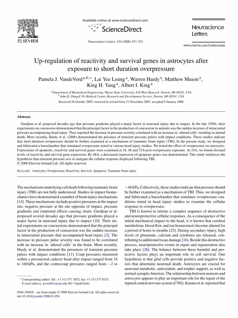
A
epdtaElh©
K
Tic[sgpmtfiiwHpwt
0d
Available online at www.sciencedirect.com
Neuroscience Letters 434 (2008) 247–252
Up-regulation of reactivity and survival genes in astrocytes afterexposure to short duration overpressure
Pamela J. VandeVord a,b,∗, Lai Yee Leung a, Warren Hardy a, Matthew Mason a,King H. Yang a, Albert I. King a
a Department of Biomedical Engineering, Wayne State University, 818 West Hancock, Detroit, MI 48202, USAb John D. Dingell VA Medical Center, Research and Development Service, Detroit, MI 48201, USA
Received 26 October 2007; received in revised form 21 December 2007; accepted 9 January 2008
bstract
Gurdjian et al. proposed decades ago that pressure gradients played a major factor in neuronal injury due to impact. In the late 1950s, theirxperiments on concussion demonstrated that the principal factor in the production of concussion in animals was the sudden increase of intracranialressure accompanying head injury. They reported the increase in pressure severity correlated with an increase in ‘altered cells’ resulting in animaleath. More recently, Hardy et al. (2006) demonstrated the presence of transient pressure pulses with impact conditions. These studies indicatehat short duration overpressure should be further examined as a mechanism of traumatic brain injury (TBI). In the present study, we designednd fabricated a barochamber that simulated overpressure noted in various head injury studies. We tested the effect of overpressure on astrocytes.
xpressions of apoptotic, reactivity and survival genes were examined at 24, 48 and 72 h post-overpressure exposure. At 24 h, we found elevatedevels of reactivity and survival gene expression. By 48 h, a decreased expression of apoptotic genes was demonstrated. This study reinforces theypothesis that transient pressure acts to instigate the cellular response displayed following TBI. 2008 Elsevier Ireland Ltd. All rights reserved.
ic bra
−badr
aimaltpt
eywords: Astrocytes; Overpressure; Reactivity; Survival; Apoptosis; Traumat
he mechanisms underlying cell death following traumatic brainnjury (TBI) are not fully understood. Studies in impact biome-hanics have demonstrated a number of brain injury mechanisms14]. These mechanisms include positive pressures at the impactite, negative pressure at the site opposite of impact, pressureradients and rotational effects causing strain. Gurdjian et al.roposed several decades ago that pressure gradients played aajor factor in neuronal injury due to impact [10]. Their ini-
ial experiments on concussions demonstrated that the principalactor in the production of concussion was the sudden increasen intracranial pressure that accompanied head injury [2]. Thencrease in pressure pulse severity was found to be correlatedith an increase in ‘altered cells’ in the brain. More recently,ardy et al. demonstrated the presences of transient pressure
ulses with impact conditions [11]. Coup pressures measuredithin a pressurized cadaver head after impact ranged from 34o 160 kPa, and the contrecoup pressures ranged from −2 to
∗ Corresponding author. Tel.: +1 313 577 3852; fax: +1 313 577 8333.E-mail address: [email protected] (P.J. VandeVord).
thtnnai
304-3940/$ – see front matter © 2008 Elsevier Ireland Ltd. All rights reserved.oi:10.1016/j.neulet.2008.01.056
in injury
48 kPa. Collectively, these studies indicate that pressure shoulde further examined as a mechanism of TBI. Thus, we designednd fabricated a barochamber that simulates overpressure con-itions noted in head injury studies to examine the cellularesponse to overpressure.
TBI is known to initiate a complex sequence of destructivend neuroprotective cellular responses. As a consequence of thenitial mechanical impact to the head, it is known that cerebral
etabolism, blood flow and ion homeostasis become altered forperiod of hours to months [25]. During secondary injury, high
evels of glutamate, calcium and cytokines are released, con-ributing to additional tissue damage [16]. Beside this destructiverocess, neuroprotective events in repair and regeneration alsoake place [26]. The balance between these harmful and pro-ective factors plays an important role in cell survival. Oneypothesis is that glial cells provide positive and negative fac-ors that determine neuronal death. Astrocytes are crucial for
euronal metabolic, antioxidant, and trophic support, as well asormal synaptic function. The relationship between neurons andstrocytes appears to play an important role for the repair of thenjured central nervous system (CNS). Katano et al. reported that
248 P.J. VandeVord et al. / Neuroscience Letters 434 (2008) 247–252
Fig. 1. (a) Barochamber consists of a driving cylinder, a hose, a top hemisphere, an ultra-low mass piston, the main chamber and a bottom hemisphere. (b) Lowerp t of thp ber, wa
ii3potas
i(ttchawwctap
w
hafs1adswtCiplpa
rwc
art of the chamber, showing a Petri dish residing on the pedestal. (c) Upper pariston inside the driving cylinder. (d) Pressure wave generated by the barochamreas (green in color) is the time integral of the pressure or ‘positive impulse’.
mmediate early genes were up-regulated in cultured glial cellsn 30 min after mechanical injury in vitro, then subsiding withinh [12]. The delayed response of glial cells to traumatic injury,articularly due to transient pressure is still unknown. Thus, thebjective of the present study was to examine astrocyte responseo overpressure at 24, 48 and 72 h. Cell response was gauged bylterations in expression of known apoptotic, proliferative andurvival factors.
We designed, constructed and tested a barochamber shownn Fig. 1. The device consists of a five-piece aluminum chamberFig. 1a). The bottom three-quarters of the chamber are designedo house the cell cultures, which are supported on a pedestal posi-ioned at the center of the chamber (Fig. 1b). The section of thehamber below the top hemisphere contains a piston. The upperemisphere section is ported to a driving cylinder mechanism viahydraulic hose. When operational, the entire system is filledith distilled warm (37 ◦C) water. A pressure wave is producedhen a metal ball strikes the rod of the piston in the driving
ylinder (Fig. 1c). The resulting pressure pulse is recorded byhe pressure transducers within the chamber. The first pulse is of
high amplitude and short duration, which resembles the impactressures previously described [2,11].C6 rat astrocytoma cell line (ATCC, Manassas, VA)as maintained in F-12K medium supplemented with 10%
u2we
e chamber, showing the metal ball dropping on the rod, which is connected to aith high amplitude and short duration in the first pulse. The sum of the shaded
orse serum, 2.5% fetal bovine serum, and 1% antibiotics–ntimycotics Invitrogen, Carlsbad, CA. C6 cell was chosenor its phenotype resemblance of native astrocytes. Cells wereeeded on a Petri dish (35-mm diameter) at a density of× 105 cells/dish 1 day prior to testing. They were randomlyssigned into control and impact groups. Just before testing, allishes were filled with culture media and covered with Parafilmo that no air bubbles were present. Dishes of the impact groupere placed on the pedestal within the chamber (Fig. 1b) and
he wave front of pressure pulse was made to pass over the cells.ontrol group remained in the culture hood throughout the test-
ng period. Post-testing samples resided in the incubator for aeriod of 24, 48 or 72 h. Pressure–time history data were col-ected using the TDAS Pro module (DTS, USA). The averageositive impulse (i.e. time integral of the positive pressure pulsess shown in Fig. 1d) was 3.259 ± 0.848 kPa s.
At 24, 48 and 72 h after testing, total RNA was extracted foreal-time PCR, viability/cytotoxicity assay and apoptotic assaysere performed. Briefly, total RNA was isolated from C6 glioma
ells using Trizol (Invitrogen, Carlsbad, CA) following the man-
facturer’s protocol. RNA was quantified by UV absorbance at60 nm. Reverse transcription employing the oligo(dT)20 primeras then performed to convert mRNA to cDNA template. cDNAquivalent to 50 ng of total RNA was subjected to subsequent
P.J. VandeVord et al. / Neuroscience Letters 434 (2008) 247–252 249
Table 1Description of primers utilized for real-time PCR
Gene Primer sequences Product (bp)
Apoptotic Genes Bax 5′-ATGGAGCTGCAGAGGATGATTGCT-3′ (forward) 3185′-TCCAGCCACAAAGATGGTCACTGT-3′ (reverse)
Casp 8 5′-ACAGGCTTAGAACAGGAGCACGTT-3′ (forward) 2915′-TAGTGTGAAGATGGGCTGTGGCAT-3′ (reverse)
Fas-lig 5′-TGAGTTCACCAACCACAGCCTT-3′ (forward) 1065′-TGTTAAGTGGGCCACACTCCTT-3′ (reverse)
Casp 3 5′-TGGTACCGATGTCGATGCAGCTAA-3′ (forward) 4715′-ATGGCGCAAAGTGACTGGATGAAC-3′ (reverse)
Survival Genes Bcl-2 5′-AGCCGGGAGAACAGGGTATGATAA-3′ (forward) 5155′-TCATCCACAGAGCGATGTTGTCCA-3′ (reverse)
IL-3 5′-TTGCGGAGAGTAAACCTGGACGAA-3′ (forward) 1185′-TCGCAGCTGCAGGAATACAACA-3′ (reverse)
GDNF 5′-ATAAAGCTTGGACGGGACTCTAAGATGAAG-3′ (forward) 2755′-ATATCTAGATCAGATACATCCACACCGTT-3′ (reverse)
Reactivity Genes Map2K1 5′-TTCAAGGTCTCCCACAAGCCATCT-3′ (forward) 2135′-TTGATCCAAGGACCCACCATCCAT-3′ (reverse)
NES 5′-TACACCAGACCAGACCTTGTGC-3′ (forward) 2195′-TTGACCCATGGTATTTGTCCTG-3′ (reverse)
GFAP 5′-GTTGTGTTCAAGCAGCCTGG-3′ (forward) 2185′-CCAGTGAGTAAAGGTGACAG-3′ (reverse)
′ CTT ′TTG
P(gIplptaoppTmfTgcf[g
�
�
R
fit
piEwI1a#F(NTtageofp
plbet8tb
VIM 5 -ACAATG5′-AGGTTC
CR analysis using specific primer pairs for apoptotic genesbax, caspase 3 and caspase 8 and Fas-ligand (Fas-lig)), survivalenes (bcl-2, glial cell derived neurotrophic factor (GDNF),nterleukin-3 (IL-3)) and reactivity genes (mitogen activatedrotein kinase kinase 1 (Map2K1), Nestin (Nes), glial fibril-ary acidic protein (GFAP) and vimentin (VIM)) (Table 1). Therimers were designed using Primer 3 software. Master mix con-aining 1× SYBR® Green (Applied Biosystems), and forwardnd reverse primers (0.4 �M each) were dispensed into wellsf a 96-well optical plate. After adding cDNA templates, thelate was capped and spun to settle the contents. It was thenlaced in the retractable plate holder of the 7500 Fast Real-ime PCR System (Applied Biosystems, Foster City, CA). Theachine was operated using the profile: 50 ◦C for 2 min, 95 ◦C
or 10 min, and 40 cycles of 95 ◦C for 15 s and 60 ◦C for 1 min.he house-keeping gene glyceraldehyde-3-phosphate dehydro-enase (GAPDH) was used as an internal control [3]. Thresholdycle (Ct) value of each sample with each primer was obtainedrom the real-time PCR output. Delta–delta (��) Ct method17] was used to determine the fold change (R) in each targetene, according to the following equations:
Ct = Ctsample − CtGAPDH
�Ct = �Ctimpact − �Ctcontrol
= 2−��Ct
An average fold change for each primer was calculatedor the three time points, respectively. Live/Dead® viabil-ty/cytotoxicity assay (Invitrogen, Carlsbad, CA) was employedo quantify the percent of C6 cells surviving and dying after over-
3sat
CTCTGGCACGTCT-3 (forward) 220GCAGCCACACTTT-3′ (reverse)
ressure. Samples from impact group and control group werencubated with Live/Dead stain (2 �M calcein AM and 4 �MthD-1 in PBS) for 30 min at room temperature. The samplesere viewed under a Zeiss fluorescence microscope (Carl Zeiss
nc) with filters separating calcein (live, stained green) and EthD-(dead, stained red) light emissions. In addition, necrotic and
poptotic cells were counted using Vybrant® apoptosis assay kit2. Both control and impact groups were hybridized with Alexaluor® 488 annexin V (1:20 dilution) and propidium iodide3 �M) in annexin-binding buffer (10 mM HEPES, 140 mMaCl, 2.5 mM CaCl2, pH 7.4) for 15 min at room temperature.hey were then visualized using fluorescent microscopy with fil-
ers for green and red fluorescence. Necrotic cells were identifieds those generating red signals, while apoptotic cells were distin-uished by green signals. Data were reported as mean ± standardrror of the mean (S.E.M.). For multiple group comparisons,ne-way ANOVA, followed by LSD post hoc test were per-ormed using SPSS 12.0 (SPSS, Chicago, IL). Differences with-values <0.05 were regarded as significant.
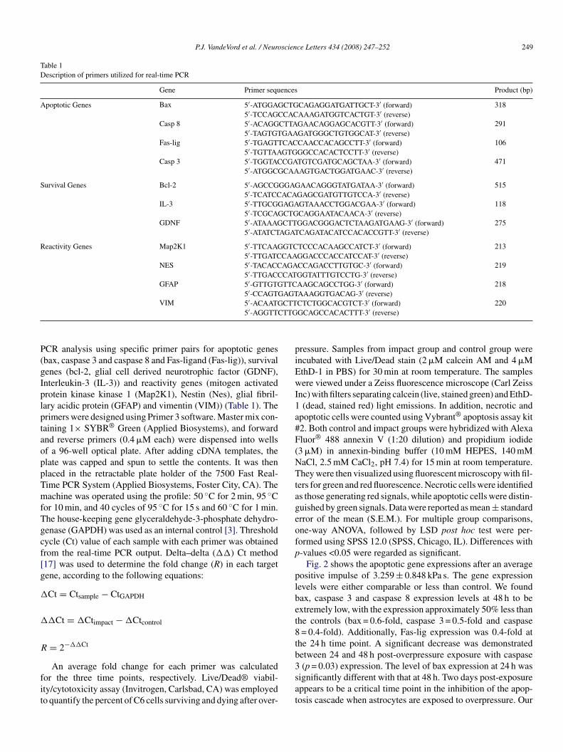
Fig. 2 shows the apoptotic gene expressions after an averageositive impulse of 3.259 ± 0.848 kPa s. The gene expressionevels were either comparable or less than control. We foundax, caspase 3 and caspase 8 expression levels at 48 h to bextremely low, with the expression approximately 50% less thanhe controls (bax = 0.6-fold, caspase 3 = 0.5-fold and caspase= 0.4-fold). Additionally, Fas-lig expression was 0.4-fold at
he 24 h time point. A significant decrease was demonstratedetween 24 and 48 h post-overpressure exposure with caspase
(p = 0.03) expression. The level of bax expression at 24 h wasignificantly different with that at 48 h. Two days post-exposureppears to be a critical time point in the inhibition of the apop-osis cascade when astrocytes are exposed to overpressure. Our

250 P.J. VandeVord et al. / Neuroscience Letters 434 (2008) 247–252
F val gec leve
rcwhscthwobnorborartoeaesg
odppTnbni
otboaaviiIcW
Fi
ig. 2. Results demonstrating alteration in expression of apoptotic genes, surviompared among time points. †p = 0.06 as IL-3 level at 48 h is compared to IL-3
esults indicate that overpressure leads to a decrease in astro-yte death factors within 3 days. For survival gene expressions,e found that two of the three genes examined demonstratedigh levels of expression at 24 and 48 h after overpressure expo-ure. IL-3 expression was shown to have a 1.7-fold increase overontrols at 24 h, peaking with a 2.6-fold increase at 48 h. Theseime points are much higher than at 72 h which is still 1.3-foldigher than control (p = 0.062). The level of GDNF expressionas determined to be 2.6-fold higher than controls at 24 h post-verpressure testing. This level is significantly higher than atoth 48 and 72 h (p = 0.01). Lastly, bcl-2 levels were not sig-ificantly different from control. This is interesting as the ratiof intracellular bax:bcl-2 is used to measure cell survival. Ouresults indicate a thrust toward cell survival since the levels ofax expression are low and bcl-2 remained constant after theverpressure injury. On the same figure, expression levels ofeactivity genes Map2K1, GFAP and VIM are significantly hight 24 h. Map2K1 expression was elevated by 2.0-fold at 24 h andemained high up to 48 h (1.6-fold) as compared to controls. Onhe contrary, GFAP expression dropped to a low level (0.5-foldf the controls) after reaching a peak (1.3-fold) at 24 h. VIMxpression was elevated with 1.5-fold of the controls at 24 h
nd then declined to a lower level (0.8-fold) at 48 h. Nes levelsxhibited a decreasing trend with time, although they were notignificantly different than controls. The expression levels of allenes returned to normal (comparable to the expression levelhwao
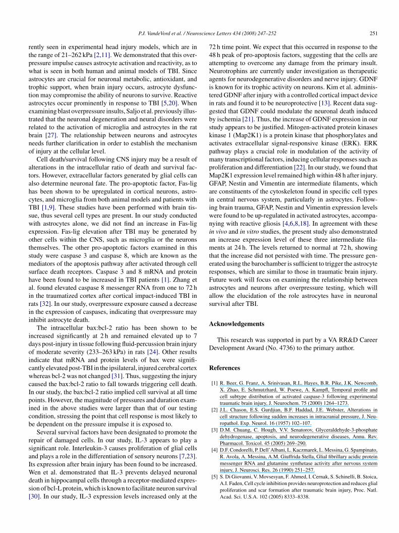
ig. 3. Live/Dead assay demonstrating the viability of C6 cells after overpressure; (bmpact group and the control group; data were presented as mean percent (%) ± stand
nes and reactivity genes after overpressure exposure. *p < 0.05 and **p < 0.01;l at 72 h.
f control group) at 72 h. Live/Dead assay, as shown in Fig. 3a,emonstrated the viability of C6 cells after overpressure. Com-ared with the control group, impact group maintained a highercentage of live cells (>99%) consistently at all time points.he apoptotic assay (Fig. 3b) showed that the percentages ofecrotic and apoptotic cells in the impact group were compara-le to those in the control group, except at 72 h in which moreecrotic cells were observed in the control group than in thempact group.
Intracranial pressure effects during impact have been a focusf speculation for over 50 years. Several groups have reported onhe effects of strain and strain rates at a cellular level [15,19,29],ut there is a lack of information with regards to the effect ofverpressure on CNS cells in vitro. One report by Shepard etl. examined human glial cell response to overpressure withinfluid percussion barotrauma model [28]. They stated that celliability was maintained at levels below 255 kPa, with a dramaticncrease in cell injury at levels above this threshold. Cell viabil-ty was assessed by percent of detached cells 5 min after testing.n our study, the pressure magnitude was below 207 kPa and theells appeared to remain in their pre-exposed monolayer state.
hen examining the results our study with Shepard et al., we
ypothesized that there is a pressure threshold that glial cells canithstand. At pressures lower than 207 kPa, astrocytes becomectivated and proliferate. Thus, our study describes a methodf subjecting cells in culture to pressures similar to those cur-
) apoptosis assay showing the percentages of necrotic and apoptotic cells in theard deviation.
scien
rtpwattaetrbno
atahcTsweotsmshairii
idoicwcIpicb
rsaIWds[
74aNaitigbskapmpMGaiiwniamterFaas
A
D
R
P.J. VandeVord et al. / Neuro
ently seen in experimental head injury models, which are inhe range of 21–262 kPa [2,11]. We demonstrated that this over-ressure impulse causes astrocyte activation and reactivity, as tohat is seen in both human and animal models of TBI. Since
strocytes are crucial for neuronal metabolic, antioxidant, androphic support, when brain injury occurs, astrocyte dysfunc-ion may compromise the ability of neurons to survive. Reactivestrocytes occur prominently in response to TBI [5,20]. Whenxamining blast overpressure insults, Saljo et al. previously illus-rated that the neuronal degeneration and neural disorders wereelated to the activation of microglia and astrocytes in the ratrain [27]. The relationship between neurons and astrocyteseeds further clarification in order to establish the mechanismf injury at the cellular level.
Cell death/survival following CNS injury may be a result oflterations in the intracellular ratio of death and survival fac-ors. However, extracellular factors generated by glial cells canlso determine neuronal fate. The pro-apoptotic factor, Fas-ligas been shown to be upregulated in cortical neurons, astro-ytes, and microglia from both animal models and patients withBI [1,9]. These studies have been performed with brain tis-ue, thus several cell types are present. In our study conductedith astrocytes alone, we did not find an increase in Fas-lig
xpression. Fas-lig elevation after TBI may be generated byther cells within the CNS, such as microglia or the neuronshemselves. The other pro-apoptotic factors examined in thistudy were caspase 3 and caspase 8, which are known as theediators of the apoptosis pathway after activated through cell
urface death receptors. Caspase 3 and 8 mRNA and proteinave been found to be increased in TBI patients [1]. Zhang etl. found elevated caspase 8 messenger RNA from one to 72 hn the traumatized cortex after cortical impact-induced TBI inats [32]. In our study, overpressure exposure caused a decreasen the expression of caspases, indicating that overpressure maynhibit astrocyte death.
The intracellular bax:bcl-2 ratio has been shown to bencreased significantly at 2 h and remained elevated up to 7ays post-injury in tissue following fluid-percussion brain injuryf moderate severity (233–263 kPa) in rats [24]. Other resultsndicate that mRNA and protein levels of bax were signifi-antly elevated post-TBI in the ipsilateral, injured cerebral cortexhereas bcl-2 was not changed [31]. Thus, suggesting the injury
aused the bax:bcl-2 ratio to fall towards triggering cell death.n our study, the bax:bcl-2 ratio implied cell survival at all timeoints. However, the magnitude of pressures and duration exam-ned in the above studies were larger than that of our testingondition, stressing the point that cell response is most likely toe dependent on the pressure impulse it is exposed to.
Several survival factors have been designated to promote theepair of damaged cells. In our study, IL-3 appears to play aignificant role. Interleukin-3 causes proliferation of glial cellsnd plays a role in the differentiation of sensory neurons [7,23].ts expression after brain injury has been found to be increased.
en et al. demonstrated that IL-3 prevents delayed neuronaleath in hippocampal cells through a receptor-mediated expres-ion of bcl-L protein, which is known to facilitate neuron survival30]. In our study, IL-3 expression levels increased only at the
ce Letters 434 (2008) 247–252 251
2 h time point. We expect that this occurred in response to the8 h peak of pro-apoptosis factors, suggesting that the cells arettempting to overcome any damage from the primary insult.eurotrophins are currently under investigation as therapeutic
gents for neurodegenerative disorders and nerve injury. GDNFs known for its trophic activity on neurons. Kim et al. adminis-ered GDNF after injury with a controlled cortical impact devicen rats and found it to be neuroprotective [13]. Recent data sug-ested that GDNF could modulate the neuronal death inducedy ischemia [21]. Thus, the increase of GDNF expression in ourtudy appears to be justified. Mitogen-activated protein kinasesinase 1 (Map2K1) is a protein kinase that phosphorylates andctivates extracellular signal-responsive kinase (ERK). ERKathway plays a crucial role in modulation of the activity ofany transcriptional factors, inducing cellular responses such as
roliferation and differentiation [22]. In our study, we found thatap2K1 expression level remained high within 48 h after injury.FAP, Nestin and Vimentin are intermediate filaments, which
re constituents of the cytoskeleton found in specific cell typesn central nervous system, particularly in astrocytes. Follow-ng brain trauma, GFAP, Nestin and Vimentin expression levelsere found to be up-regulated in activated astrocytes, accompa-ying with reactive gliosis [4,6,8,18]. In agreement with thesen vivo and in vitro studies, the present study also demonstratedn increase expression level of these three intermediate fila-ents at 24 h. The levels returned to normal at 72 h, showing
hat the increase did not persisted with time. The pressure gen-rated using the barochamber is sufficient to trigger the astrocyteesponses, which are similar to those in traumatic brain injury.uture work will focus on examining the relationship betweenstrocytes and neurons after overpressure testing, which willllow the elucidation of the role astrocytes have in neuronalurvival after TBI.
cknowledgements
This research was supported in part by a VA RR&D Careerevelopment Award (No. 4736) to the primary author.
eferences
[1] R. Beer, G. Franz, A. Srinivasan, R.L. Hayes, B.R. Pike, J.K. Newcomb,X. Zhao, E. Schmutzhard, W. Poewe, A. Kampfl, Temporal profile andcell subtype distribution of activated caspase-3 following experimentaltraumatic brain injury, J. Neurochem. 75 (2000) 1264–1273.
[2] J.L. Chason, E.S. Gurdjian, B.F. Haddad, J.E. Webster, Alterations incell structure following sudden increases in intracranial pressure, J. Neu-ropathol. Exp. Neurol. 16 (1957) 102–107.
[3] D.M. Chuang, C. Hough, V.V. Senatorov, Glyceraldehyde-3-phosphatedehydrogenase, apoptosis, and neurodegenerative diseases, Annu. Rev.Pharmacol. Toxicol. 45 (2005) 269–290.
[4] D.F. Condorelli, P. Dell’Albani, L. Kaczmarek, L. Messina, G. Spampinato,R. Avola, A. Messina, A.M. Giuffrida Stella, Glial fibrillary acidic proteinmessenger RNA and glutamine synthetase activity after nervous system
injury, J. Neurosci. Res. 26 (1990) 251–257.[5] S. Di Giovanni, V. Movsesyan, F. Ahmed, I. Cernak, S. Schinelli, B. Stoica,A.I. Faden, Cell cycle inhibition provides neuroprotection and reduces glialproliferation and scar formation after traumatic brain injury, Proc. Natl.Acad. Sci. U.S.A. 102 (2005) 8333–8338.

2 ience
[
[
[
[
[
[
[
[
[
[
[
[
[
[
[
[
[
[
[
[
[
[
52 P.J. VandeVord et al. / Neurosc
[6] W.D. Dietrich, J. Truettner, W. Zhao, O.F. Alonso, R. Busto, M.D. Ginsberg,Sequential changes in glial fibrillary acidic protein and gene expressionfollowing parasagittal fluid-percussion brain injury in rats, J. Neurotrauma.16 (1999) 567–581.
[7] M. Ebadi, R.M. Bashir, M.L. Heidrick, F.M. Hamada, H.E. Refaey, A.Hamed, G. Helal, M.D. Baxi, D.R. Cerutis, N.K. Lassi, Neurotrophinsand their receptors in nerve injury and repair, Neurochem. Int. 30 (1997)347–374.
[8] C. Eliasson, C. Sahlgren, C.H. Berthold, J. Stakeberg, J.E. Celis, C. Bet-sholtz, J.E. Eriksson, M. Pekny, Intermediate filament protein partnershipin astrocytes, J. Biol. Chem. 274 (1999) 23996–24006.
[9] M.B. Grosjean, P.M. Lenzlinger, P.F. Stahel, I. Yatsiv, E. Shohami, O.Trentz, T. Kossmann, M.C. Morganti-Kossmann, Immunohistochemicalcharacterization of Fas (CD95) and Fas Ligand (FasL/CD95L) expressionin the injured brain: relationship with neuronal cell death and inflammatorymediators, Histol. Histopathol. 22 (2007) 235–250.
10] E.S. Gurdjian, H.R. Lissner, V.R. Hodgson, L.M. Patrick, Mechanism ofhead injury, Clin. Neurosurg. 12 (1964) 112–128.
11] W.N. Hardy, M. Mason, C.D. Foster, K.H. Yang, A.I. King, Comparisonof intracranial pressure response to cadaver head kinematics, J. Biomech.39 (2006) S155.
12] H. Katano, K. Fujita, T. Kato, K. Asai, Y. Kawamura, A. Masago, K.Yamada, A metabotropic glutamate receptor antagonist, alpha-methyl-4-carboxyphenylglycine, attenuates immediate early gene mRNA expressionfollowing traumatic injury in cultured rat cortical glial cells, Neurosci. Lett.306 (2001) 101–105.
13] B.T. Kim, V.L. Rao, K.A. Sailor, K.K. Bowen, R.J. Dempsey, Protectiveeffects of glial cell line-derived neurotrophic factor on hippocampal neu-rons after traumatic brain injury in rats, J. Neurosurg. 95 (2001) 674–679.
14] A.I. King, Fundamentals of impact biomechanics: part I—biomechanicsof the head, neck, and thorax, Annu. Rev. Biomed. Eng. 2 (2000) 55–81.
15] M.C. LaPlaca, D.K. Cullen, J.J. McLoughlin, R.S. Cargill 2nd, High rateshear strain of three-dimensional neural cell cultures: a new in vitro trau-matic brain injury model, J. Biomech. 38 (2005) 1093–1105.
16] P.M. Lenzlinger, M.C. Morganti-Kossmann, H.L. Laurer, T.K. McIntosh,The duality of the inflammatory response to traumatic brain injury, Mol.Neurobiol. 24 (2001) 169–181.
17] K.J. Livak, T.D. Schmittgen, Analysis of relative gene expression data usingreal-time quantitative PCR and the 2(-Delta Delta C(T)) method, Methods
25 (2001) 402–408.18] C. Moon, M. Ahn, S. Kim, J.K. Jin, K.B. Sim, H.M. Kim, M.Y. Lee, T.Shin, Temporal patterns of the embryonic intermediate filaments nestin andvimentin expression in the cerebral cortex of adult rats after cryoinjury,Brain Res. 1028 (2004) 238–242.
[
Letters 434 (2008) 247–252
19] A.G. Mukhin, S.A. Ivanova, J.W. Allen, A.I. Faden, Mechanical injury toneuronal/glial cultures in microplates: role of NMDA receptors and pH insecondary neuronal cell death, J. Neurosci. Res. 51 (1998) 748–758.
20] D.J. Myer, G.G. Gurkoff, S.M. Lee, D.A. Hovda, M.V. Sofroniew, Essentialprotective roles of reactive astrocytes in traumatic brain injury, Brain 129(2006) 2761–2772.
21] O. Nicole, C. Ali, F. Docagne, L. Plawinski, E.T. MacKenzie, D. Vivien, A.Buisson, Neuroprotection mediated by glial cell line-derived neurotrophicfactor: involvement of a reduction of NMDA-induced calcium influx bythe mitogen-activated protein kinase pathway, J. Neurosci. 21 (2001)3024–3033.
22] N. Otani, H. Nawashiro, S. Fukui, H. Ooigawa, A. Ohsumi, T. Toyooka, K.Shima, Role of the activated extracellular signal-regulated kinase pathwayon histological and behavioral outcome after traumatic brain injury in rats,J. Clin. Neurosci. 14 (2007) 42–48.
23] M. Penkowa, T. Moos, J. Carrasco, H. Hadberg, A. Molinero, H. Blueth-mann, J. Hidalgo, Strongly compromised inflammatory response to braininjury in interleukin-6-deficient mice, Glia 25 (1999) 343–357.
24] R. Raghupathi, K.I. Strauss, C. Zhang, S. Krajewski, J.C. Reed, T.K. McIn-tosh, Temporal alterations in cellular Bax:Bcl-2 ratio following traumaticbrain injury in the rat, J. Neurotrauma. 20 (2003) 421–435.
25] S.K. Ray, C.E. Dixon, N.L. Banik, Molecular mechanisms in the pathogen-esis of traumatic brain injury, Histol. Histopathol. 17 (2002) 1137–1152.
26] A.C. Rice, A. Khaldi, H.B. Harvey, N.J. Salman, F. White, H. Fillmore,M.R. Bullock, Proliferation and neuronal differentiation of mitoticallyactive cells following traumatic brain injury, Exp. Neurol. 183 (2003)406–417.
27] A. Saljo, F. Bao, A. Hamberger, K.G. Haglid, H.A. Hansson, Exposure toshort-lasting impulse noise causes microglial and astroglial cell activationin the adult rat brain, Pathophysiology 8 (2001) 105–111.
28] S.R. Shepard, J.B. Ghajar, R. Giannuzzi, S. Kupferman, R.J. Hariri, Fluidpercussion barotrauma chamber: a new in vitro model for traumatic braininjury, J. Surg. Res. 51 (1991) 417–424.
29] D.H. Smith, J.A. Wolf, T.A. Lusardi, V.M. Lee, D.F. Meaney, High tol-erance and delayed elastic response of cultured axons to dynamic stretchinjury, J. Neurosci. 19 (1999) 4263–4269.
30] T.C. Wen, J. Tanaka, H. Peng, J. Desaki, S. Matsuda, N. Maeda, H. Fujita,K. Sato, M. Sakanaka, Interleukin 3 prevents delayed neuronal death in thehippocampal CA1 field, J. Exp. Med. 188 (1998) 635–649.
31] X.L. Yao, J. Liu, E. Lee, G.S. Ling, J.T. McCabe, Progesterone differentially
regulates pro- and anti-apoptotic gene expression in cerebral cortex follow-ing traumatic brain injury in rats, J. Neurotrauma. 22 (2005) 656–668.32] X. Zhang, S.H. Graham, P.M. Kochanek, D.W. Marion, P.D. Nathaniel,S.C. Watkins, R.S. Clark, Caspase-8 expression and proteolysis in humanbrain after severe head injury, FASEB J. 17 (2003) 1367–1369.
Copyright © 2022 FDOKUMEN