Empowerment and State-dependent Noise - An Intrinsic Motivation for Avoiding Unpredictable Agents
Upload
independentCategory
view
5download
0
Unpredictable feeding schedules unmask a system for dailyresetting of behavioural and metabolic food entrainment
Carolina Escobar,1 Ma Teresa Martınez-Merlos,1,2 Manuel Angeles-Castellanos,1 Marıa del Carmen Minana1
and Ruud M. Buijs3
1Departamento de Anatomıa, Edificio B 4� piso, Facultad de Medicina, Circuito Escolar S ⁄ N, Universidad Nacional Autonoma deMexico, Mexico DF 04510, Mexico2Moore House Medical School, Atlanta Georgia, USA3Departamento de Biologıa Celular y Fisiologıa, Instituto de Investigaciones Biomedicas Universidad Nacional Autonoma deMexico, Mexico City 04510, Mexico and Netherlands Institute for Neuroscience
Keywords: circadian rhythms, metabolism, food-entrainment, hypothalamus
Abstract
Restricted feeding schedules (RFS) are a potent Zeitgeber that uncouples daily metabolic and clock gene oscillations in peripheraltissues from the suprachiasmatic nucleus (SCN), which remains entrained to the light–dark cycle. Under RFS, animals develop foodanticipatory activity (FAA), characterized by arousal and increased locomotion. Food availability in nature is not precise, whichsuggests that animals need to adjust their food-associated activity on a daily basis. This study explored the capacity of rats to adjustto variable and unpredictable feeding schedules. Rats were exposed either to RFS with fixed daily meal (RF) or to a variable mealtime (VAR) during the light phase. RF and VAR rats exhibited daily metabolic oscillations driven by the last meal event; however, VARrats were not able to show a robust adjustment in the anticipating corticosterone peak. VAR rats were unable to exhibit FAA butexhibited a daily activation pattern in phase with the previous meal. In both groups the dorsomedial nucleus of the hypothalamus andarcuate nucleus, involved in energy balance, exhibited increased c-Fos expression 24 h after the last meal, while only RF ratsexhibited low c-Fos expression in the SCN. Data show that metabolic and behavioural food-entrained rhythms can be reset on a dailybasis; the two conditions elicit a similar hypothalamic response, while only the SCN is inhibited in rats exhibiting anticipatory activity.The variable feeding strategy uncovered a rapid (24-h basis) resetting mechanism for metabolism and general behaviour.
Introduction
In addition to the suprachiasmatic nucleus (SCN), the masterbiological clock, diverse tissues and cerebral structures express clockgenes that oscillate with a period similar to 24 h; they are thereforeclassified as peripheral oscillators. Peripheral clock genes as well asdigestive and metabolic functions under physiological conditionsoscillate in synchrony with the SCN but preferentially adjust theirdaily oscillations to feeding schedules (Damiola et al., 2000; Stokkanet al., 2001; Hastings et al., 2003).Nocturnal rodents ingest their meals and produce the main digestive
and food-related signals during the night, which maintains theoscillations of peripheral tissues coupled to the SCN and adjusted tothe light–dark (LD) cycle. When nocturnal rodents are forced to eatduring the light phase by scheduling food access during the day,peripheral oscillators uncouple from the SCN, which maintains itsphase coupled to the LD cycle (Stokkan et al., 2001; Damiola et al.,2001). Thus under restricted feeding schedules (RFS), food becomes apotent Zeitgeber that overrides the influence of the SCN on peripheraloscillators. The mechanism by which the feeding schedule providesthe entraining signal to set the phase of the peripheral oscillators is notclear but may involve catabolic–anabolic cycles and the induction ofcycles in the redox state in the cells (Dıaz-Munoz et al., 2000; Rutteret al., 2001; Cardona, 2004). It has been suggested that, as
metabolites, hormones and temperature modify the metabolic stateof cells, they may play a relevant role as entraining signals (Balsalobreet al., 2000; Damiola et al., 2001; Rutter et al., 2001; Brown et al.,2002; Hirota et al., 2002).Single pulses of hormones and metabolites are sufficient to initiate
oscillations in cultured fibroblasts (Balsalobre et al., 1998) or toproduce a phase change of clock gene products in peripheraloscillators (Balsalobre et al., 2000; Le Minh et al., 2001; Shibata,2004). Such findings support the possibility that peripheral oscillatorsare capable of a fast phase shift.Neuronal activity in diverse brain structures is also entrained to
feeding schedules and becomes uncoupled from the SCN (Wakamatsuet al., 2001; Lamont et al., 2005). Food-entrained patterns of c-Fosand clock gene expression have been reported in hypothalamic andbrain stem areas that are involved in energy balance and arousal(Angeles-Castellanos et al., 2004, 2005; Gooley et al., 2006; Miedaet al., 2006; Waddington-Lamont et al., 2006) and are also found inlimbic structures involved in motivational and affective responses(Mendoza et al., 2005; Angeles-Castellanos et al., 2007). After4–5 days of RFS, brain structures exhibit food-entrained patterns ofc-Fos expression (Inzunza et al., 2000; Meynard et al., 2005).In consequence, RFS also entrains behavioural patterns; rats
restricted to food access during the light phase develop a pattern ofactivity known as food anticipatory activity (FAA), which ischaracterized by increased movement and foraging starting 1–2 hprior to the scheduled meal time (Mistlberger, 1994; Stephan, 2002).Similar to c-Fos expression, FAA can be observed after 3–5 days of
Correspondence: Dr Carolina Escobar, as above.E-mail: [email protected]
Received 12 April 2007, revised 4 September 2007, accepted 18 September 2007
European Journal of Neuroscience, Vol. 26, pp. 2804–2814, 2007 doi:10.1111/j.1460-9568.2007.05893.x
ª The Authors (2007). Journal Compilation ª Federation of European Neuroscience Societies and Blackwell Publishing Ltd

RFS. The capacity to adjust FAA to phase shifts in the feedingschedule has been explored in rats bearing a bilateral lesion of theSCN: a phase delay of 4–10 h resulted in transitory cycles (Stephan,1984, 1992) while in some cases the rapid re-entrainment did not showtransitory cycles (Stephan, 1992). The shifting speed was reported tobe faster in rats bearing a bilateral lesion of the SCN than in intact rats.There are no further reports on the shifting properties of FAA.
The present study was designed to explore the speed with whichoscillations in metabolic variables, brain structures and FAA adapt toshifts in meal time. Because we assumed that they depend on differentmechanisms we hypothesized that metabolism would adapt to dailychallenges and thus oscillations in metabolites and hormones wouldshift on a daily basis, while FAA and the activity pattern of brainstructures would need several cycles to adjust to shifts in meal time.Therefore, this study characterized metabolic oscillations, c-Fosexpression (as marker of neuronal activity) in the SCN andhypothalamic nuclei involved in energy balance, and behaviouralpatterns of rats exposed to RFS with food provided at fixed or atvariable and unpredictable hours daily.
Materials and methods
Animals and general housing conditions
Adult male Wistar rats weighing between 250 and 300 g at thebeginning of the experiment were maintained in a 12 : 12 h LD cycle[lights on at 07.00 h, Zeitgeber time (ZTO)], constant temperature(22 ± 1 �C) and free access to food (Rodent Laboratory Chow 5001)and water, unless otherwise stated. Experimental procedures wereapproved and conducted according to the ethical committee at theMedical Faculty in accordance with the institutional guide for care anduse of animal experimentation (Universidad Nacional Autonoma deMexico), which conforms to international guidelines for animalhandling.
Experiment I
A first set of rats was used to characterize metabolic rhythms underfixed or unpredictable feeding schedules, as well as to examine thepossible persistence of food-entrained rhythms for two cycles infasting conditions. Rats were randomly assigned to one of the threefeeding conditions and maintained in this situation for 2 weeks.After the last scheduled meal rats remained in total food deprivationfor 24 h. (i) Fixed restricted feeding (RF) schedules: for this groupfood was available for 2 h daily from ZT5 to ZT7. (ii) Variablefeeding schedules (VAR12): food was available once, mainly in andaround the light period, for 2 h at variable and unpredictable times.The interval between meals was never shorter than 18 h and neverlonger than 36 h (one example of such a schedule is as follows:09.00, 13.00, 08.00, 16.00, 11.00, 20.00, 19.00, 10.00, 13.00,21.00, 18.00, 09.00, 15.00 and 12.00 h). The last meal for thisgroup before starting blood sampling was at ZT5 (12 h). (iii)Variable restricted feeding schedules (VAR18): this group wastreated in the same way as the VAR12, but the last meal wasdelivered at 18.00 h (one example of such a schedule is as follows:11.00, 17.00, 13.00, 12.00, 16 19.00, 10.00, 20.00, 09.00, 14.00,18.00, 08.00, 15.00, 21.00 and 18.00 h).
For the three groups the last meal time was defined as MT0 to allowcomparson of food-related oscillations. Before the last meal rats wereweighed. Blood sampling was started at MT3, which represents 3 hafter the last scheduled meal and was continued every 3 h to completea 24-h cycle (n ¼ 4–6 rats per group and time point).
In order to determine the persistence of food-entrained metabolicrhythms a second set of rats was entrained as described for groups RFand VAR12 and then maintained in fasting for two 24-h cycles. Bloodsampling was performed for the interval between 42 and 58 h after thelast meal (MT42–58) and for corticosterone determinations for theinterval MT42–55.
Serum and tissue sampling
For each temporal point and each group 4–6 rats were decapitated andtrunk blood (3–4 mL) was collected in 10-mL test tubes containing aclot-forming gel (Vacutainer; Becton Dickinson). Tubes were centri-fuged at 2500 r.p.m. for 15 min to obtain blood serum. Aliquots of250 and 700 lL were coded and frozen at )70 �C for subsequentdetermination of serum corticosterone, leptin and free fatty acids(FFA).After blood collection the stomach was dissected out at the level of
the lower gastroesophageal sphincter and the pylorus, and wetstomach weight was registered.
Determination of serum hormones and FFAs
Serum corticosterone and leptin were determined in duplicate with astandard 125I radioimmunoassay kit (Medidores Industriales TKRC1for corticosterone, and Linco Res, Inc., St Charles, MO, USA forleptin). Assays were performed with a sensitivity of 0.5 ng ⁄ mL, andintra- and interassay coefficients of variation of 4%.Serum FFAs were processed by using a colourimetric method, as
described elsewhere (Escobar et al., 1998).
Statistical analysis
Data are reported as mean ± SEM and are represented as temporalwaveforms. Body weight after each feeding protocol was comparedwith a one-way anova. Metabolic variables were analysed with a two-way anova for the factors group and time. Temporal effects for eachgroup were tested with a one-way anova followed by a Tukeymultiple comparison post hoc test with a set at P < 0.05. Statisticalanalysis was performed with the package Statistica (StatSoft, Inc.1993).
Experiment II
A different set of rats was placed in an automated activity monitoringsystem in order to determine temporal behavioural organization undera fixed or a variable unpredictable schedule (n ¼ 16 per group) and todetermine the possible persistence of food-entrained patterns in fastingconditions.
Behavioural monitoring
Rats were placed in individual cages (45 · 30 · 35 cm) positioned onplates with movement sensors in soundproof lockers with controlledlighting conditions and with a regulated temperature of 24 ± 1 �C anda 12 : 12-h LD cycle (lights on at 07.00 h). This system wasdeveloped in our group with contributions from Nico Bos inAmsterdam the Netherlands and the Mexican biomedical companyOmnialva. Behavioural events were collected with a digitized systemand stored at 10-min intervals for further analysis with an analysissystem developed for our laboratory in Matlab (SPAD9).For baseline, rats were monitored for 14 days with free access to
food and water; on day 15 food was removed and starting on day 17food was delivered for 2 h under an RF schedule. A first group of ratswas assigned to an RF schedule with daily food access from 12.00 to
Daily resetting of food entrainment 2805
ª The Authors (2007). Journal Compilation ª Federation of European Neuroscience Societies and Blackwell Publishing LtdEuropean Journal of Neuroscience, 26, 2804–2814

14.00 h local time. A second group was assigned to a variable andunpredictable feeding schedule (VAR12) with random daily foodaccess as described for experiment 1. At the end of each feedingprotocol rats were left for 2 days in total food deprivation to evaluatepersistence of the food-entrained behavioural rhythm. This protocolwas performed in two series of eight animals each, giving a total of 16rats per group.Actograms were obtained in order to visualize temporal organiza-
tion of behaviour. In each 10-min period, the duration of detectedmovement was measured, normalized and converted to a percentage oftotal daily activity. Mean activity profiles were obtained for baselineand entrainment interval for each group. In order to determine FAAand persistence of entrained patterns for the two conditions, fixed andunpredictable meal schedules, a daily analysis was performedcomparing the activity 2 h prior to, 2 h during and 2 h after mealtime. In addition, the activity in the same intervals the following daywas analysed. For the VAR group, analysis days were chosen such thatin the first day food access had been scheduled early and in thefollowing day food was delivered later, in order to observe persistenceof behavioural patterns. Days used for this later analysis were days 1(meal at 08.30 h) and 2 (meal at 13.00 h), days 5 (meal at 08.30 h)and 6 (meal at 18.00 h) and days 11 (meal at 12.00 h) and 12 (nofood). Total activity counts in the 2-h intervals for both groups werecompared with the mean activity for the same interval during thebaseline. A two-way anova was used for the factors group and 2-hintervals followed by a Tukey multiple comparison post hoc test witha set at P < 0.05.
Experiment III
A third set of rats was used to characterize neuronal activity duringFAA in rats entrained by fixed feeding schedules or exposed tovariable and unpredictable daily food restriction. Rats were randomlyassigned to and maintained for 2 weeks in one of three groups (n ¼ 6per group). (i) Ad libitum (control), in which animals had free access tofood and water during the 24-h cycle. (ii) Fixed restricted feeding(RF); food was available for 2 h daily from 12.00 to 14.00 h (ZT5–7).(iii) Variable feeding schedules (VAR); food was available daily for2 h at variable and unpredictable times of the day as described forexperiment 1. The last meal for this group was at 12.00 h.In order to determine neuronal activity associated with anticipatory
activity, rats were perfused the day after termination of the entrainingprotocols 1 h after the expected meal (i.e. perfused at 13.00 h) andtheir brains were removed and processed.
Immunohistochemistry
Rats were anaesthetized with an overdose of sodium pentobarbital(Sedal-Vet 65 mg ⁄ mL), and were perfused transcardially with 150 mLof 0.9% saline followed by 250 mL of fixative: 4% paraformaldehydein phosphate-buffered saline (PBS; 0.1 m, pH 7.2). Brains wereremoved, post-fixed for 18 h and cryoprotected in 30% sucrose for3–4 days. Brains were frozen and cut into sections of 40 lm at)18 �C. Sections were serially collected in four sets; one set wasstained with cresyl violet acetate (Sigma Chemical Company) and asecond set was processed for cFos immunohistochemistry. Free-floating sections were incubated in c-Fos antibody raised in rabbit(1 : 2500; Santa Cruz Biotechnology, Santa Cruz, CA, USA) for 72 h.This was followed by incubation in secondary antibody, processedusing the avidin–biotin method for immunohistochemistry as previ-ously described (Angeles-Castellanos et al., 2004). Tissues were
reacted in diaminobenzidine with hydrogen peroxide, mounted,dehydrated and coverslipped for light-microscopical analysis.
Cell count and analysis
Fos-immunoreactive cells were evaluated in the suprachiasmaticnucleus (SCN), the arcuate nucleus (ARC) and the dorsomedialnucleus of the hypothalamus (DMH).Two representative sections for each nucleus were selected in
accordance with the stereotaxic atlas of Paxinos and Watson (1998).Bilateral images of selected sections were digitized at a 200·magnification using a computerized image analysis system (MetaVue series 4.5; Universal Imaging Corporation) attached to a Nikonlight microscope (Nikon Eclipse E600). To minimize the number offalse positives, background optic density was established in a nearbyregion lacking Fos immunoreactivity; stained nuclei that reached orsurpassed 2· the background optic density were considered positiveand were included, whereas cells below this staining threshold werediscarded. A single examiner, who was blinded to treatment condi-tions, performed all counts.The mean cell number for the two representative sections were
classified by group and given as mean ± SEM. Data between groups(three levels) were compared with a one-way anova for independentmeasures followed by a Tukey post hoc test with significant values setat P < 0.01. Statistical analysis was performed with the programStatistica version 4.5 (StatSoft, Inc. 1993).
Results
Experiment I
On the last day of restricted feeding, rats in the two variable feedinggroups (VAR12 and VAR18) showed a significantly lower bodyweight than rats fed at a fixed feeding schedule (RF, 330.3 ± 5.1;VAR12, 311.9 ± 6.9, VAR18, 312 ± 3.1 g; F2,119 ¼ 4.49, P < 0.03).
Stomach weight and serum determinations
Stomach weight of the three food-restricted groups (RF, VAR12 andVAR18) showed a daily oscillation associated with the scheduled mealtime (two-way anova: F7,99 ¼ 107.6, P < 0.0001). Single one-wayanovas performed per group confirmed a significant effect of time forthe three groups (RF, F7,24 ¼ 47.78, P < 0.0001; VAR12,F7,32 ¼ 71.18, P < 0.00001; VAR18, F7,32 ¼ 20.79, P < 0.0001). Inall groups, the highest values were observed at the first sampled timepoint after meal time (MT3) and stomach weight decreased slowly toattain statistically significantly lower values 15 h after the last meal.Empty stomachs were observed 21 h after feeding, thus 3 h before theexpected meal (Fig. 1, top). Although the temporal pattern was similarfor the three groups, the two groups exposed to variable feedingschedules attained lower values after the meal; two-way anova
showed a significant effect among groups (F2,88 ¼ 40.96; P < 0.001)and a significant group · time interaction (F14,88 ¼ 8.19; P < 0.001).After two cycles of fasting, RF and VAR12 exhibited low valuesindicating an empty stomach and no statistical difference wasobserved between groups [F1,60 ¼ 3.49; not significant (NS)] or dueto time (F5,60 ¼ 2.19; NS).Serum leptin of the three food-restricted groups showed a daily
oscillation associated with meal time (two-way anova:F7,117 ¼ 57.77, P < 0.0001), highest values were observed 3–6 hafter meal time and decreased slowly to attain the lowest values 12 hafter the last meal (Fig. 1, right top).The single one-way anova per
2806 C. Escobar et al.
ª The Authors (2007). Journal Compilation ª Federation of European Neuroscience Societies and Blackwell Publishing LtdEuropean Journal of Neuroscience, 26, 2804–2814

group confirmed a significant effect of time for the three groups (RF,F7,40 ¼ 16.64, P < 0.0001; VAR12, F740 ¼ 37.04, P < 0.00001;VAR18, F7,37 ¼ 11.71, P < 0.0001). Highest values were observed3–6 h after meal time and decreased slowly to the lowest values 12 hafter the last meal (Fig. 1, right top). At 3, 6 and 9 h after meal timethe RF and VAR12 groups exhibited significantly higher values thanthe VAR18 group; however, at the subsequent time points leptin levelswere similar for the two groups. The two-way anova indicated asignificant difference among groups (F2,117 ¼ 33.91; P < 0.0002) andinteraction group · time (F14,117 ¼ 4.40; P < 0.0002. During fastingboth RF and VAR12 exhibited low values and no oscillations of serumleptin; no statistical difference was observed between groups(F1,66 ¼ 0.07; NS) or due to time (F5,66 ¼ 1.68; NS).
In serum FFAs the three groups showed a similar pattern associatedwith meal time (F7,114 ¼ 24.69; P < 0.001), characterized by lowvalues for the 9 h following the last meal and an increasing tendencyto reach peak values 3 h before and at the moment of the nextexpected meal (Fig. 1, left bottom). The single one-way anova
performed per group confirmed a significant effect of time for the threegroups (RF, F7,40 ¼ 17.53, P < 0.0001; VAR12, F7,37 ¼ 9.71,P < 0.0001; VAR18, F7,37 ¼ 4.02, P < 0.002). The anticipatoryincrease was more evident in the RF group and was significantlydifferent from that of the variable groups. The two-way anova
indicated main differences among groups (F1,114 ¼ 81.58;P < 0.0001) and for the interaction group · time (F14,114 ¼ 3.16;P < 0.001). During fasting there was a gradual increase in FFA due to2 days fasting. Remarkably, under these conditions the RF and theVAR12 groups showed an increase in serum FFA before the expectedmeal time followed by a sharp decrease after the expected (but notgiven) feeding. Main effects were observed between groups(F1,48 ¼ 17.10; P < 0.0001) and for the factor time (F5,48 ¼ 4.40;P < 0.002), but not for their interaction (F5,48 ¼ 1.21; NS). This latereffect was confirmed by one-way anova per group, which indicated a
significant effect of time for the RF group (F5,24 ¼ 2.96; P < 0.03)but not for the VAR12 group (F5,24 ¼ 2.47; P ¼ 0.06).Serum corticosterone for the three food-restricted groups showed a
daily oscillation associated with the last meal time (F7,78 ¼ 3.98;P < 0.001), highest values were observed 24 h after the last meal(Fig. 1, right bottom). In both groups exposed to variable meals meanvalues were higher than in the RF group and the two-way anova
indicated a significant difference among groups (F2,78 ¼ 30.27;P < 0.0001). It also indicated a significant effect of time(F7,78 ¼ 3.98; P < 0.001), but not due to the interaction of the twofactors (F14,78 ¼ 1.31; NS). This was further confirmed with the one-way anova per group for which a significant effect of time wasstatistically significant for the RF group (F7,24 ¼ 19.79; P < 0.0001)but not for the VAR12 (F7,24 ¼ 2.03; P ¼ 0.09) or for the VAR18(F7,30 ¼ 1.47; NS) groups. After 2 days fasting, the RF groupexhibited high values before the expected meal; these decreased afterthe expected meal interval. This food-entrained pattern was notobserved in the VAR12 group. The two-way anova indicated nostatistically significant difference between groups (F1,35 ¼ 2.13; NS),due to time (F4,35 ¼ 2.13; NS) or due to their interaction(F4,35 ¼ 1.81; NS). The one-way anova per group confirmed asignificant effect of time for the RF group (F4,19 ¼ 3.66; P < 0.02)but not for the VAR12 group (F4,19 ¼ 0.67; NS).
Experiment II
During baseline all rats exhibited a daily rhythm with low activityduring the light phase (22%) and high activity during the night (78%;Fig. 2, right top). During food restriction RF rats developedanticipatory activity characterized by increased movement starting� 2 h prior to food access, with peak values at the moment of mealtime (Fig. 2, left top and right middle). This activity was clearlyobserved on days 4–5 of food restriction and promoted an increase in
Fig. 1. Stomach weight and serum determinations of rats maintained under fixed (RF; white circles) or variable (VAR12, black squares; VAR18, black diamonds)feeding schedules. The three groups exhibited daily rhythms adjusted to the last meal. Asterisks indicate time points where high values for the 3 groups werestatistically significantly different from lowest values of the same group (*P < 0.01, Tukey). Letters indicate significant difference between (a) RF and the VAR12groups and (b) RF and the VAR18 groups. The solid bar indicates last meal and hatched bars indicate expected meal time during the fasting interval.
Daily resetting of food entrainment 2807
ª The Authors (2007). Journal Compilation ª Federation of European Neuroscience Societies and Blackwell Publishing LtdEuropean Journal of Neuroscience, 26, 2804–2814

activity during the light phase to 44%. VAR rats did not exhibitanticipatory activity; however, they showed increased general activa-tion at the moment that food was delivered (Fig. 2, left bottom).Similar to the RF group, their mean activity in the light phase wasincreased, to 37% (Fig. 2, right bottom).The detailed quantitative analysis for specific days allowed
visualization of mean activity 2 h before, 2 h during and 2 h aftermeal time for both groups. After 2 days of total food deprivation thefirst meal event (day 1) produced in rats of both groups, RF and VAR,increased movement counts during the 2 h-feeding episode (Fig. 3,top). Activity counts for both groups were statistically significantlydifferent from that observed at this phase during baseline(F2,122 ¼ 13.44; P < 0.0005). The two-way anova also indicated a
significant difference among time points (F2,122 ¼ 11.48; P < 0.001)and for the interaction group · time (F4,122 ¼ 4.45; P < 0.002). Onday 2, both groups exhibited a slight increase in activity during the 2 hprior to the expected meal (22 h after the previous feeding event);however, this was not statistically significantly different from thebaseline activity. Both groups exhibited significantly increased activityduring the 2-h interval of the expected meal time compared with thebaseline activity. The proportion of activity exhibited by the VARgroup was similar to that observed the previous day during foodpresentation and was significantly different from baseline. Because theRF group received food at this time, the activity displayed wassignificantly higher than and different from the VAR group (Fig. 3,bottom). The two-way anova indicated a statistically significant
Fig. 2. (Left) Actograms for general activity of individual rats synchronized to (top) a restricted feeding schedule or to (bottom) a variable feeding schedule. Meanactivity graphs of 16 rats per group are shown on the right; vertical axis, percentage of total daily activity occurring in 10-min time bins. The total percentages ofactivity in the light and the dark phases are indicated above each plot. The mean expected activity for the light phase as observed during the baseline is indicated withthe dotted line. In the actogram the vertical bar and the stars for the VAR rat indicate meal time. The three squares indicate the consecutive days that were furtheranalysed.
2808 C. Escobar et al.
ª The Authors (2007). Journal Compilation ª Federation of European Neuroscience Societies and Blackwell Publishing LtdEuropean Journal of Neuroscience, 26, 2804–2814

difference among groups (F2,101 ¼ 33.79; P < 0.0001), among timepoints (F2,101 ¼ 15.30; P < 0.0001) and due to the interactiongroup · time (F4,101 ¼ 8.90; P < 0.0001).
The analysis for days 5 and 6 under fixed or variable restrictedfeeding schedules exemplifies the development of anticipatory activityin the RF group with highest values of activity during the 2 h prior tomeal time and also high activity counts during food access comparedto the baseline activity (Fig. 4). In contrast, VAR rats did not showincreased activity before food access; however, they showed signif-icant increased values during meal time on day 5. The following day(day 6) VAR rats did not receive food for 24 h; however, during andafter the expected meal time (estimated 24 h after the last meal) theyshowed increased activity at the time of the previous meal, althoughno food was delivered (Fig. 4, bottom). The two-way anova indicatedsignificant differences between groups (F2,105 ¼ 41.22, P < 0.0001for day 5 and F2,105 ¼ 51.62, P < 0.0001 for day 6), a main effect oftime (F2,105 ¼ 6.42, P < 0.002 for day 5 and F2,105 ¼ 3.30, P < 0.04for day 6) and due to the group · time interaction (F4,105 ¼ 6.62,P < 0.0001 for day 5 and F2,105 ¼ 7.41, P < 0.0001 for day 6).
On day 11 of restricted feeding the RF group again exhibited highactivity values in anticipation of and during meal time, while the VARgroup exhibited increased values only during food access (Fig. 5, top).On day 12 neither group received food at the expected time. The RFgroup again exhibited increased activity in anticipation and both theRF and VAR groups exhibited activation during and after the expectedmeal (Fig. 5, bottom). The two-way anova indicated significantdifference among groups (F2,81 ¼ 28.28, P < 0.0001 for day 11 andF2,81 ¼ 35.51, P < 0.0001 for day 12), due to time (F2,81 ¼ 11.20,P < 0.0001 for day 11 and F2,81 ¼ 11.16, P < 0.001 for day 12) anddue to the group · time interaction (F4,105 ¼ 9.70, P < 0.0001 forday 11 and F2,105 ¼ 3.88, P < 0.006 for day (12).
Experiment III
The brains of rats of the two food-restricted groups (RF and VAR)were obtained 25 h after the last meal scheduled at 12 h (assumingthat c-Fos protein expression takes at least 1 h). Rats were expectingfood but had not been fed at the time of perfusion. Evaluation ofc-Fos-positive cells in the SCN was performed separately for theventral and dorsal regions. In both regions the VAR group showedsimilar values as the ad libitum control, while the RF group showed
Fig. 3. Percentage of the total daily activity for 2 h intervals before, duringand after the first meal episode for the RF (hatched bars) and the VAR (blackbars) group (n ¼ 16 rats per group). On day 2 only the RF group received food,in accordance with the 24-h schedule. The dotted horizontal line representsmean values obtained for the same phase during baseline, while continuoushorizontal lines represent SEM; +P < 0.05 vs. the baseline values, **P < 0.05between the highest point and the two lower points in the same group, and*P < 0.05 between the highest and the lowest point in the same group andbetween RF and VAR rats (Tukey’s post hoc test). &P < 0.01, RF vs. VARgroup.
Fig. 4. Percentage of total daily activity for 2-h intervals before, during andafter the fifth meal episode for the RF (hatched bars) and the VAR (solid bars)group (n ¼ 16 per group). On the following day (day 6) only the RF groupreceived food, in accordance with the 24-h schedule. Other indications as inFig. 3.
Daily resetting of food entrainment 2809
ª The Authors (2007). Journal Compilation ª Federation of European Neuroscience Societies and Blackwell Publishing LtdEuropean Journal of Neuroscience, 26, 2804–2814
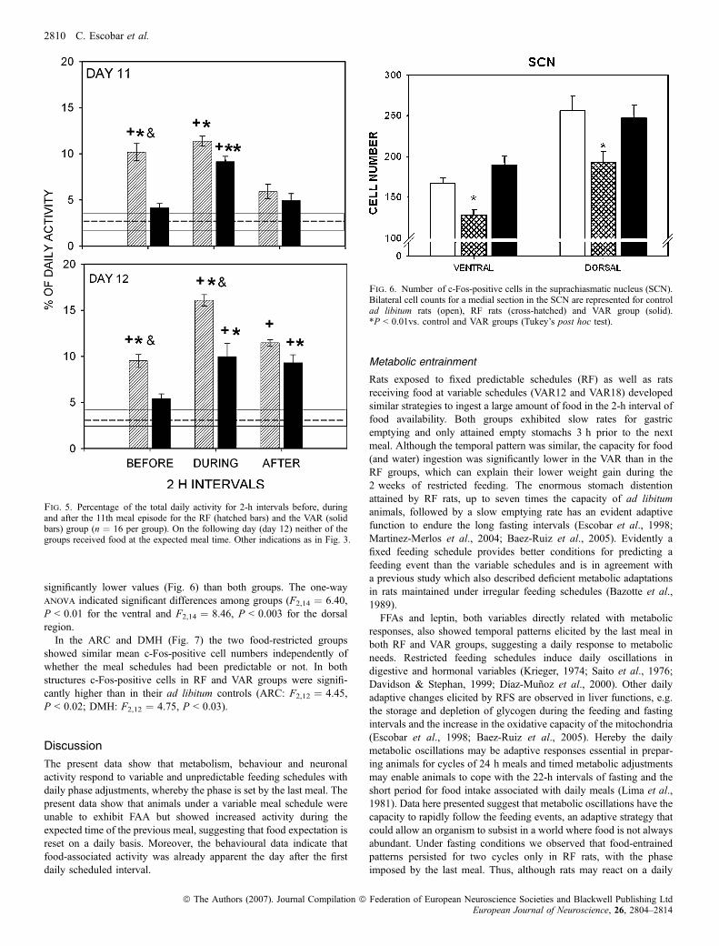
significantly lower values (Fig. 6) than both groups. The one-wayanova indicated significant differences among groups (F2,14 ¼ 6.40,P < 0.01 for the ventral and F2,14 ¼ 8.46, P < 0.003 for the dorsalregion.In the ARC and DMH (Fig. 7) the two food-restricted groups
showed similar mean c-Fos-positive cell numbers independently ofwhether the meal schedules had been predictable or not. In bothstructures c-Fos-positive cells in RF and VAR groups were signifi-cantly higher than in their ad libitum controls (ARC: F2,12 ¼ 4.45,P < 0.02; DMH: F2,12 ¼ 4.75, P < 0.03).
Discussion
The present data show that metabolism, behaviour and neuronalactivity respond to variable and unpredictable feeding schedules withdaily phase adjustments, whereby the phase is set by the last meal. Thepresent data show that animals under a variable meal schedule wereunable to exhibit FAA but showed increased activity during theexpected time of the previous meal, suggesting that food expectation isreset on a daily basis. Moreover, the behavioural data indicate thatfood-associated activity was already apparent the day after the firstdaily scheduled interval.
Metabolic entrainment
Rats exposed to fixed predictable schedules (RF) as well as ratsreceiving food at variable schedules (VAR12 and VAR18) developedsimilar strategies to ingest a large amount of food in the 2-h interval offood availability. Both groups exhibited slow rates for gastricemptying and only attained empty stomachs 3 h prior to the nextmeal. Although the temporal pattern was similar, the capacity for food(and water) ingestion was significantly lower in the VAR than in theRF groups, which can explain their lower weight gain during the2 weeks of restricted feeding. The enormous stomach distentionattained by RF rats, up to seven times the capacity of ad libitumanimals, followed by a slow emptying rate has an evident adaptivefunction to endure the long fasting intervals (Escobar et al., 1998;Martinez-Merlos et al., 2004; Baez-Ruiz et al., 2005). Evidently afixed feeding schedule provides better conditions for predicting afeeding event than the variable schedules and is in agreement witha previous study which also described deficient metabolic adaptationsin rats maintained under irregular feeding schedules (Bazotte et al.,1989).FFAs and leptin, both variables directly related with metabolic
responses, also showed temporal patterns elicited by the last meal inboth RF and VAR groups, suggesting a daily response to metabolicneeds. Restricted feeding schedules induce daily oscillations indigestive and hormonal variables (Krieger, 1974; Saito et al., 1976;Davidson & Stephan, 1999; Dıaz-Munoz et al., 2000). Other dailyadaptive changes elicited by RFS are observed in liver functions, e.g.the storage and depletion of glycogen during the feeding and fastingintervals and the increase in the oxidative capacity of the mitochondria(Escobar et al., 1998; Baez-Ruiz et al., 2005). Hereby the dailymetabolic oscillations may be adaptive responses essential in prepar-ing animals for cycles of 24 h meals and timed metabolic adjustmentsmay enable animals to cope with the 22-h intervals of fasting and theshort period for food intake associated with daily meals (Lima et al.,1981). Data here presented suggest that metabolic oscillations have thecapacity to rapidly follow the feeding events, an adaptive strategy thatcould allow an organism to subsist in a world where food is not alwaysabundant. Under fasting conditions we observed that food-entrainedpatterns persisted for two cycles only in RF rats, with the phaseimposed by the last meal. Thus, although rats may react on a daily
Fig. 6. Number of c-Fos-positive cells in the suprachiasmatic nucleus (SCN).Bilateral cell counts for a medial section in the SCN are represented for controlad libitum rats (open), RF rats (cross-hatched) and VAR group (solid).*P < 0.01vs. control and VAR groups (Tukey’s post hoc test).
Fig. 5. Percentage of the total daily activity for 2-h intervals before, duringand after the 11th meal episode for the RF (hatched bars) and the VAR (solidbars) group (n ¼ 16 per group). On the following day (day 12) neither of thegroups received food at the expected meal time. Other indications as in Fig. 3.
2810 C. Escobar et al.
ª The Authors (2007). Journal Compilation ª Federation of European Neuroscience Societies and Blackwell Publishing LtdEuropean Journal of Neuroscience, 26, 2804–2814

basis to feeding schedules, a strong entraining effect that can persistfor more than two cycles can only be attained after a regular feedingschedule.
In contrast to metabolic responses, the premeal corticosterone peakdid not attain complete adjustment under variable schedules asobserved for both VAR12 and VAR18 groups. It is well establishedthat the circadian corticosterone rhythm depends strongly on SCNregulation and that under RFS an additional peak anticipating mealtime can be elicited (Honma et al., 1984); moreover, this premealcorticosterone peak under RF conditions is influenced by the SCN(Kalsbeek et al., 1998) Data here presented suggest that variablefeeding schedules do not exert the same power to produce thesignificant food anticipatory peak observed under RFS (Krieger, 1974)and thus possibly cannot override signals elicited by the SCN.However, Honma et al. (1983) showed that after a similar fastinginterval as used in the present study the onset of a corticosterone peakwas observed after the first meal cycle ‘as if anticipating the nextcycle’, indicating a critical role of the fasting state in development andmaintenance of this oscillation.
A rapid shifting capacity seems to be tissue-dependent (Davidsonet al., 2003) and mainly expressed in digestive and feeding-relatedfunctions. A recent report indicates that oxygen consumptionrate shifts after the first cycle of diurnal feeding, while such ashift takes up to 6 days for clock genes in the liver (Satoh et al.,2006).
Metabolic oscillations can constitute time-keeping systems similarto an hour glass and can explain that 24 h daily oscillations can beeasily reset. On the other hand, persistence of entrained metabolicpatterns for at least two cycles in intact animals (Martinez-Merloset al., 2004) and of clock gene expression in isolated tissue explantsfor longer intervals (Stokkan et al., 2001; Yoo et al., 2004) suggeststhe influence of an oscillatory mechanism that can keep time memoryof the phase imposed by the last meal. This mechanism may dependon local oscillations maintained in the tissue by clock genes(Yamazaki et al., 2000; Yoo et al., 2004) and ⁄ or can be driven inintact animals by metabolic signals or by central nervous signals(Buijs & Kalsbeek, 2001; Terazono et al., 2003; Cailotto et al., 2005;Guo et al. 2005). Present data support the existence of oscillatorymechanisms, which, however, are rapidly reset; the fact that clockgene-entrained patterns rapidly adapt to a new cycle (Stokkan et al.,2001) supports this proposal.
Food-entrained behavioural patternsRats under RF and VAR rats showed already at day 2 an increasein activity at the expected meal time. Rats under RF exhibitedconsistent anticipatory activity on day 5 and the following dayswhile visual inspection of actograms indicated that most RF animalsexhibited FAA as early as day 3. Because in contrast to VAR, RFanimals received food at the expected time, behavioural activityduring food presentation was always associated with the given food.In the VAR animals, however, already at day 2 a significantincrease in activity could be seen at the time when the animals hadreceived food on the previous day. Similarly, the following days’food-related activity in the VAR animals increased even more andmaintained significantly higher values than the daily activityobserved during baseline.The use of movement detectors to monitor general activity in this
study provided relevant advantages in order to observe food expec-tancy. Other movement indicators such as wheel running andapproaches to a food bin have been shown to be strong markers forFAA (Mistlberger, 1994); however, they do not provide informationabout other behavioural activity during the feeding interval.There is no previous study in the literature that has tried to
determine the capacity to react to changing feeding schedules. Studiesperformed by Stephan (1984, 1992) showed that, in rats bearingbilateral SCN lesions, the FAA can shift in 2–3 cycles to a new mealtime; this is faster than in intact rats. For these studies FAA was usedas a measure of food entrainment; however, present data indicate that,without exhibiting FAA, rats are able to change their food expectancyactivity on a daily basis to the expected meal time.Studies using palatable treats that do not seem to challenge
metabolic functions have shown that the regularity of a stimulus thatcan produce high motivational state and arousal is sufficient to elicitFAA (Mistlberger & Rusak, 1988; Mendoza et al., 2005). Thisevidence combined with data here presented confirms that FAA isexpressed when feeding events are regular and predictable. Also,several studies have provided evidence that FAA is elicited when mealintervals are maintained in a circadian range of 19–29 h (Bolles &Moot, 1973; Stephan, 1981). When the interval between mealsexceeds or is shorter than this circadian range, animals are unable toexhibit FAA. The feeding program for the VAR group exceeded thisrange during certain days, because feeding intervals fluctuatedbetween 18 and 36 h. Although this was not the case on a daily
Fig. 7. Number of c-Fos-positive cells in the arcuate nucleus (ARC) and the dorsomedial nucleus of the hypothalamus (DMH). Bilateral cell counts for arepresentative section of both regions is represented for control ad libitum (open), RF (cross-hatched) and VAR (solid) rat groups. *P < 0.01, **P < 0.001 vs.control.
Daily resetting of food entrainment 2811
ª The Authors (2007). Journal Compilation ª Federation of European Neuroscience Societies and Blackwell Publishing LtdEuropean Journal of Neuroscience, 26, 2804–2814

basis, this factor could have hindered the VAR rats in developingFAA.On the other hand, this study allowed the observation that FAA and
activation at the moment of food expectancy can depend on twodifferent systems, as this behavioural expectation was expressed inVAR but not FAA rats: Food expectancy as observed in the VAR rats,defined as the behavioural activation at the expected meal time, mayrepresent the initial phase of FAA behaviour. This food expectationmay rely on the interaction of hormonal, metabolic or digestive signalswith brain structures involved in energy balance, including the ARCand DMH, in order to promote foraging and food-seeking behavioursafter a long fasting interval. Clearly, the present data show that afterjust one single meal (under the right fasting conditions) rats exhibit anincrease in behavioural activity the next day at the previous meal time.This agrees with the early observation of Honma et al. (1983) that,after fasting, animals who had received a meal show a peak incorticosterone 24 h later.
The biological clock
It has been thought that the SCN is not involved in food entrainmentbecause FAA can be elicited after SCN lesion. (Stephan et al., 1979;Clarke & Coleman, 1986). However, this does not rule out thepossibility that under regular conditions the SCN is participating orreacting during FAA.The present study demonstrates that, when rats were exhibiting
FAA, neuronal activity, as observed by c-Fos expression, wasdecreased in the SCN. This effect was observed exclusively in ratsthat exhibited FAA in the fixed feeding schedules and not in rats thatdid not anticipate because of the unpredictable meal time. In thisexperimental design, rats were perfused 1 h after the expected mealtime, which was 24 h after the previous meal, when the c-Fos proteincorresponding to meal time expectation was reaching high levels ofexpression (Morgan & Curran, 1989). It is, however, possible that inthe VAR rats the behavioural activation at the moment of feeding hada lower intensity than that observed in RF rats exhibiting FAA. Inaddition, the differential activation during FAA and food expectationdue to variable feeding may underlie different brain processes; whilstFAA may require time-keeping signals and probably a time-learningprocess, food expectation, as observed in VAR rats, may rely onbehavioural activation by metabolic or hunger signals.Previous data from our group did not show inhibition of the SCN
during FAA; this could be due to the fact that horizontal sections wereused and only small parts of the SCN could be evaluated (Angeles-Castellanos et al., 2004) In the present study coronal sections allowedbetter analysis of the SCN and enabled differentiation of the ventraland dorsal areas. This observation agrees with an earlier study(Kalsbeek et al., 1998) which showed that vasopressin secretion fromSCN terminals is modified in an RFS, consistent with a modificationof corticosterone secretion. In the present study the inhibition of c-Fosexpression observed in the SCN suggests that arousal associated withFAA can influence the activity in the SCN.The cellular response in the SCN due to nonphotic stimuli that
produce arousal is not well understood; some studies report theinduction of c-Fos expression (Janik et al., 1995; Amir et al., 1999)and others the lack of c-Fos expression (Mead et al., 1992). It has alsobeen suggested that photic and nonphotic stimuli could have opposingeffects on the SCN activity (Mistlberger & Antle, 1998; Maywoodet al., 1999; Yi et al., 2006); however, other studies do not support thisopposing effect (Edelstein & Mrosovsky, 2001). Taken together itseems that arousal during FAA can be considered a nonphotic stimulusfor the SCN, and it is reported that under certain conditions it can
influence the SCN activity (for a review, see Mendoza, 2007). Otherstudies have tried to determine the influence of RFS on the SCN usingthe phase of clock gene products as activity markers. All studies havereported consistently that clock genes in the SCN are not shifted withmeal time (Damiola et al., 2000; Hara et al., 2001; Wakamatsu et al.,2001; Mieda et al., 2006). However, c-Fos (the activity marker in ourstudy) and clock gene oscillations reflect two different cellularprocesses; c-Fos reflects acute responses to stimuli (Morgan & Curran,1989) while clock gene products result from 24-h oscillatorymechanisms (Reppert & Weaver, 2001).Our results are consistent with the idea that, during FAA, neuronal
activity in the SCN can be modulated, probably allowing theexpression of behavioural and hormonal parameters associated withFAA.
Other hypothalamic structures
The DMH and ARC nuclei are structures involved in functions ofmetabolic integration (Horvath & Diano, 2004). The two structuresshowed similar neural activity when rats were expecting food, 24 hafter the last meal, and this response was different from the rats fedad libitum. Because the two groups, RF and VAR, exhibited similarmetabolic responses 24 h after the last meal, it is possible that theneuronal responses of both hypothalamic structures are related tosignals originating in the periphery, induced by the prolonged fastinginterval. In the ARC, increased levels of c-Fos expression have beenreported in rats during fasting (Johnston et al., 2006; Scott et al.,2007), and increased c-Fos has been reported in the DMH in ratsanticipating a meal after 22 h of fasting (Angeles-Castellanos et al.,2004; Gooley et al., 2006).The ARC nucleus receives hormonal signals related to energy
balance and plays a relevant role transmitting these metabolic signalsto other hypothalamic areas, including the DMH and SCN (Yi et al.,2006). The DMH also receives signals from the periphery and can beconsidered an integrative area linking feeding-related processes witharousal (Saper, 2006; Sakurai, 2007). It is well documented that theDMH exhibits increased c-Fos when rats are displaying FAA andwhile they are feeding (Angeles-Castellanos et al., 2004; Gooleyet al., 2006). Also, RFS entrain the clock genes per1 and per2 in theDMH, confirming the influence of feeding schedules on the cellularcycles on this structure (Mieda et al., 2006). It remains to beestablished whether in addition to inducing c-Fos expression, variablefeeding schedules can also influence clock gene oscillations inhypothalamic structures or whether this requires a regular andpredictable schedule.
Conclusions
Altogether, the present data illustrate a strong adaptive capacity of ratsto adjust to meal time on a daily basis. This fast daily response wasobserved on metabolism and on activity of hypothalamic structuresand behaviour, and may be a relevant adaptive strategy for coping witha changing environment in which food is not presented daily at aprecise time.Metabolic signals associated with an empty stomach and depletion
of energy stores may trigger the hypothalamic activity reported here.The activity of this and other brain areas may underlie the generalactivation observed in rats 24 h after the last meal. This processrepresents a rapidly resetting clock on a 24 h basis to alert and activateorganisms for the next meal. This system, however, proved to bedifferent from a system promoting FAA, as rats under variable feeding
2812 C. Escobar et al.
ª The Authors (2007). Journal Compilation ª Federation of European Neuroscience Societies and Blackwell Publishing LtdEuropean Journal of Neuroscience, 26, 2804–2814

schedules were not anticipating. Thus, the functional systems involvedin producing anticipatory activity require regular and predictablefeeding schedules which are different from but may be expressed inparallel with metabolic signals.
In contrast to the LD cycle, which is strongly predictable, foodaccess in nature is not necessarily precise and time of food availabilitycan differ from one day to another. The present experiments indicatethat this condition has induced functional systems that respond tochanges in food availability and suggest that they can adjust theirfunction on a daily basis.
Acknowledgements
This research was supported by grants from CONACyT 43950-N, DGAPAPAPIIT IN-203907 UNAM, Mexico.
Abbreviations
ARC, the arcuate nucleus; DMH, dorsomedial nucleus of the hypothalamus;FAA, food anticipatory activity; FFA, free fatty acids; LD, light–dark; MT, time(h) since last meal; NS, not significant; RF, fixed restricted feeding; RFS,restricted feeding schedule(s); SCN, suprachiasmatic nucleus; ZT, Zeitgebertime.
References
Amir, S., Cain, S., Sullivan, J., Robinson, B. & Stewart, J. (1999) Olfactorystimulation enhances light-induced phase shifts in free-running activityrhythms and Fos expression in the suprachiasmatic nucleus. Neuroscience,92, 1165–1170.
Angeles-Castellanos, M., Aguilar-Roblero, R. & Escobar, C. (2004) c-Fosexpression in hypothalamic nuclei of food-entrained rats. Am. J. Physiol.Regul. Integr. Comp. Physiol., 286, R158–R165.
Angeles-Castellanos, M., Mendoza, J., Dıaz-Munoz, M. & Escobar, C. (2005)Food entrainment modifies the c-Fos expression pattern in brain stem nucleiof rats. Am. J. Physiol. Regul. Integr. Comp. Physiol., 288, R678–R684.
Angeles-Castellanos, M., Mendoza, J. & Escobar, C. (2007) Restricted feedingschedules phase shift daily rhythms of c-Fos and protein Per1 immunore-activity in corticolimbic regions in rats. Neuroscience., 144, 344–355.
Baez-Ruiz, A., Escobar, C., Aguilar-Roblero, R., Vazquez-Martınez, O. &Dıaz-Munoz, M. (2005) Metabolic adaptations of liver mitochondria duringrestricted feeding schedules. Am. J. Physiol. Gastrointest. Liver Physiol.,289, G1015–G1023.
Balsalobre, A., Brown, S.A., Marcacci, L., Tronche, F., Kellendonk, C.,Reichardt, H.M., Schutz, G. & Schibler, U. (2000) Resetting of circadiantime in peripheral tissues by glucocorticoid signaling. Science, 289, 2344–2347.
Balsalobre, A., Damiola, F. & Schibler, U. (1998) A serum shock inducescircadian gene expression in mammalian tissue culture cells. Cell, 93, 929–937.
Bazotte, R.B., Curi, R. & Hell, N.S. (1989) Metabolic changes caused byirregular-feeding schedule as compared with meal-feeding. Physiol. Behav.,46, 109–113.
Bolles, R.C. & Moot, S.A. (1973) The rat’s anticipation of two meals a day.J. Comp. Physiol. Psychol., 83, 510–514.
Brown, S.A., Zumbrunn, G., Fleury-Olela, F., Preitner, N. & Schibler, U.(2002) Rhythms of mammalian body temperature can sustain peripheralcircadian clocks. Curr. Biol., 12, 1574–1583.
Buijs, R.M. & Kalsbeek, A. (2001) Hypothalamic integration of central andperipheral clocks. Nat. Rev. Neurosci., 2, 521–526.
Cailotto, C., La Fleur, S.E., Van Heijningen, C., Wortel, J., Kalsbeek, A.,Feenstra, M., Pevet, P. & Buijs, R.M. (2005) The suprachiasmatic nucleuscontrols the daily variation of plasma glucose via the autonomic output to theliver: are the clock genes involved? Eur. J. Neurosci., 22, 2531–2540.
Cardona, F. (2004) Periodic dip of lipidperoxidation in humans: a redox signalto synchronize peripheral circadian clocks? Med. Hypotheses, 63, 841–846.
Clarke, J.D. & Coleman, G.J. (1986) Persistent meal-associated rhythms inSCN-lesioned rats. Physiol. Behav., 36, 105–113.
Damiola, F., Le Minh, N., Preitner, N., Kornmann, B., Fleury-Olela, F. &Schibler, U. (2000) Restricted feeding uncouples circadian oscillators in
peripheral tissues from the central pacemaker in the suprachiasmatic nucleus.Genes Dev., 14, 2950–2961.
Davidson, A.J., Poole, A.S., Yamazaki, S. & Menaker, M. (2003) Is the food-entrainable circadian oscillator in the digestive system? Genes Brain Behav.,2, 32–39.
Davidson, A.J. & Stephan, F.K. (1999) Plasma glucagon, glucose, insulin, andmotilin in rats anticipating daily meals. Physiol. Behav., 66, 309–315.
Dıaz-Munoz, M., Vazquez-Martınez, O., Aguilar-Roblero, R. & Escobar, C.(2000) Anticipatory changes in liver metabolism and entrainment of insulin,glucagon, and corticosterone in food-restricted rats. Am. J. Physiol. Regul.Integr. Comp. Physiol., 279, R2048–R2056.
Edelstein, K. & Mrosovsky, N. (2001) Behavioral responses to light in micewith dorsal lateral geniculate lesions. Brain Res., 918, 107–112.
Escobar, C., Diaz-Munoz, M., Encinas, F. & Aguilar-Roblero, R. (1998)Persistence of metabolic rhythmicity during fasting and its entrainment byrestricted feeding schedules in rats. Am. J. Physiol., 274, R1309–R1316.
Gooley, J.J., Schomer, A. & Saper, C.B. (2006) The dorsomedial hypothalamicnucleus is critical for the expression of food-entrainable circadian rhythms.Nat. Neurosci., 9, 398–407.
Guo, H., Brewer, J.M., Champhekar, A., Harris, R.B. & Bittman, E.L. (2005)Differential control of peripheral circadian rhythms by suprachiasmatic-dependent neural signals. Proc. Natl. Acad. Sci. USA, 102, 3111–3116.
Hara, R., Wan, K., Wakamatsu, H., Aida, R., Moriya, T., Akiyama, M. &Shibata, S. (2001) Restricted feeding entrains liver clock without participa-tion of the suprachiasmatic nucleus. Genes Cells, 6, 269–278.
Hastings, M.H., Reddy, A.B. & Maywood, E.S. (2003) A clockwork web:Circadian timing in brain and periphery, in health and disease. Nat. Rev.Neurosci., 4, 649–661.
Hirota, T., Okano, T., Kokame, K., Shirotani-Ikejima, H., Miyata, T. & Fukada,Y. (2002) Glucose down-regulates Per1 and Per2 mRNA levels and inducescircadian gene expression in cultured Rat-1 fibroblasts. J. Biol. Chem., 277,44244–44251.
Honma, K.I., Honma, S. & Hiroshige, T. (1983) Critical role of foodamount for prefeeding corticosterone peak in rats. Am. J. Physiol., 245,R339–R344.
Honma, K.I., Honma, S. & Hiroshige, T. (1984) Feeding-associated cortico-sterone peak in rats under various feeding cycles. Am. J. Physiol., 246,R721–R726.
Horvath, T.L. & Diano, S. (2004) Opinion: The floating blueprint ofhypothalamic feeding circuits. Nat. Rev. Neurosci., 5, 662–667.
Inzunza, O., Seron-Ferre, M.J., Bravo, H. & Torrealba, F. (2000) Tubero-mammillary nucleus activation anticipates feeding under a restrictedschedule in rats. Neurosci. Lett., 293, 139–142.
Janik, D., Mikkelsen, J.D. & Mrosovsky, N. (1995) Cellular colocalization ofFos and neuropeptide Y in the intergeniculate leaflet after nonphotic phase-shifting events. Brain Res., 698, 137–145.
Johnstone, L.E., Fong, T.M. & Leng, G. (2006) Neuronal activation in thehypothalamus and brainstem during feeding in rats. Cell Metab., 14, 313–321.
Kalsbeek, A., Van Heerikhuize, J.J., Wortel, J. & Buijs, R.M. (1998) Restricteddaytime feeding modifies suprachiasmatic nucleus vasopressin release inrats. J. Biol. Rhythms, 13, 18–29.
Krieger, D.T. (1974) Food and water restriction shifts corticosterone, temper-ature, activity and brain amine periodicity. Endocrinology, 95, 1195–1201.
Lamont, E.W., Diaz, L.R., Barry-Shaw, J., Stewart, J. & Amir, S. (2005) Dailyrestricted feeding rescues a rhythm of period2 expression in the arrhythmicsuprachiasmatic nucleus. Neuroscience, 132, 245–248.
Le Minh, N., Damiola, F., Tronche, F., Schutz, G. & Schibler, U. (2001)Glucocorticoid hormones inhibit food-induced phase-shifting of peripheralcircadian oscillators. EMBO J., 20, 7128–7136.
Lima, F.B., Hell, N.S., Timo-Iaria, C., Scivoletto, R., Dolnikoff, M.S. & Pupo,A.A. (1981) Metabolic consequences of food restriction in rats. Physiol.Behav., 27, 115–123.
Martinez-Merlos, M.T., Angeles-Castellanos, M., Dıaz-Munoz, M., Aguilar-Roblero, R., Mendoza, J. & Escobar, C. (2004) Dissociation between adiposetissue signals, behavior and the food-entrained oscillator. J. Endocrinol., 181,53–63.
Maywood, E.S., Mrosovsky, N., Field, M.D. & Hastings, M.H. (1999) Rapiddown-regulation of mammalian period genes during behavioral resetting ofthe circadian clock. Proc. Natl. Acad. Sci. USA, 96, 15211–15216.
Mead, S., Ebling, F.J.P., Maywood, E.S., Humby, T., Herbert, J. & Hastings,M.H. (1992) A nonphotic stimulus causes instantaneous phase advances ofthe light-entrainable circadian oscillator of the syrian hamster but does notinduce the expression of c-fos in the suprachiasmatic nuclei. J. Neurosci., 12,2516–2522.
Daily resetting of food entrainment 2813
ª The Authors (2007). Journal Compilation ª Federation of European Neuroscience Societies and Blackwell Publishing LtdEuropean Journal of Neuroscience, 26, 2804–2814

Mendoza, J. (2007) Circadian clocks: setting time by food. J. Neuroendocri-nol., 19, 127–137.
Mendoza, J., Angeles-Castellanos, M. & Escobar, C. (2005) A daily palatablemeal without food deprivation entrains the suprachiasmatic nucleus of rats.Eur. J. Neurosci., 22, 2855–2862.
Meynard, M.M., Valdes, J.L., Recabarren, M., Seron-Ferre, M. & Torrealba, F.(2005) Specific activation of histaminergic neurons during daily feedinganticipatory behavior in rats. Behav. Brain Res., 158, 311–319.
Mieda, M., Williams, S.C., Richardson, J.A., Tanaka, K. & Yanagisawa, M.(2006) The dorsomedial hypothalamic nucleus as a putative food-entrainable circadian pacemaker. Proc. Natl. Acad. Sci. USA, 103,12150–12155.
Mistlberger, R.E. (1994) Circadian food-anticipatory activity: Formalmodels and physiological mechanisms. Neurosci. Biobehav. Rev., 18,171–195.
Mistlberger, R.E. & Antle, M.C. (1998) Behavioral inhibition of light-inducedcircadian phase resetting is phase and serotonin dependent. Brain Res., 786,31–38.
Mistlberger, R.E. & Rusak, B. (1988) Food-anticipatory circadian rhythms inrats with paraventricular and lateral hypothalamic ablations. J. Biol.Rhythms, 3, 277–291.
Morgan, J.I. & Curran, T. (1989) Stimulus-transcription coupling inneurons: role of cellular immediate-early genes. Trends Neurosci., 12,459–462.
Paxinos, G. & Watson, C. (1998) The Rat Brain in Stereotaxic Coordinates, 4th
edition, San Diego, USA, Academic Press.Reppert, S.M. & Weaver, D.R. (2001) Molecular analysis of mammaliancircadian rhythms. Annu. Rev. Physiol., 63, 647–676., 647–676.
Rutter, J., Reick, M., Wu, L.C. & McKnight, S.L. (2001b) Regulation of clockand NPAS2 DNA binding by the redox state of NAD cofactors. Science,2001, 510–514.
Saito, M., Murakami, E., Nishida, T., Fujisawa, Y. & Suda, M. (1976)Circadian rhythms of digestive enzymes in the small intestine of the rat. II.Effects of fasting and refeeding. J. Biochem. (Tokyo), 80, 563–568.
Sakurai, T. (2007) The neural circuit of orexin (hypocretin): maintaining sleepand wakefulness. Nat. Rev. Neurosci., 8, 171–181.
Saper, C.B. (2006) Staying awake for dinner: hypothalamic integration ofsleep, feeding, and circadian rhythms. Prog. Brain Res., 153, 243–252.,243–252.
Satoh, Y., Kawai, H., Kudo, N., Kawashima, Y. & Mitsumoto, A. (2006) Time-restricted feeding entrains daily rhythms of energy metabolism in mice.Am. J. Physiol. Regul. Integr. Comp. Physiol., 290, R1276–R1283.
Scott, V., McDade, D.M. & Luckeman, S.M. (2007) Rapid changes in thesensitivity of arcuate nucleus neurons to central ghrelin in relation to feedingstatus. Physiol. Behav., 1, 180–185.
Shibata, S. (2004) Neural regulation of the hepatic circadium rhythm. Anat.Rec. A, 280, 901–909.
Stephan, F.K. (1981) Limits of entrainment to periodic feeding in rats withsuprachiasmatic lesions. J. Comp. Physiol., 143, 401–410.
Stephan, F.K. (1984) Phase shifts of circadian rhythms in activity entrained tofood access. Physiol. Behav., 32, 663–671.
Stephan, F.K. (1992) Resetting of a feeding-entrainable circadian clock in therat. Physiol. Behav., 52, 985–995.
Stephan, F.K. (2002) The ‘other’ circadian system: food as a Zeitgeber. J. Biol.Rhythms, 17, 284–292.
Stephan, F.K., Swann, J.M. & Sisk, C.L. (1979) Entrainment of circadianrhythms by feeding schedules in rats with suprachiasmatic lesions. Behav.Neural Biol., 25, 545–554.
Stokkan, K.A., Yamazaki, S., Tei, H., Sakaki, Y. & Menaker, M. (2001)Entrainment of the circadian clock in the liver by feeding. Science, 291, 490–493.
Terazono, H., Mutoh, T., Yamaguchi, S., Kobayashi, M., Akiyama, M., Udo, R.,Ohdo, S., Okamura, H. & Shibata, S. (2003b) Adrenergic regulation of clockgene expression in mouse liver. Proc. Natl. Acad. Sci. USA 100, 6795–6800.
Waddington-Lamont, E., Harbour, V.L., Barry-Shaw, J., Renteria, D.L.,Robinson, B., Stewart, J. & Amir, S. (2006) Restricted access to food, butnot sucrose, saccharine, or salt, synchronizes the expression of Period2protein in the limbic forebrain. Neuroscience., 144, 402–411.
Wakamatsu, H., Yoshinobu, Y., Aida, R., Moriya, T., Akiyama, M. & Shibata,S. (2001) Restricted-feeding-induced anticipatory activity rhythm is associ-ated with a phase-shift of the expression of mPer1 and mPer2 mRNA in thecerebral cortex and hippocampus but not in the suprachiasmatic nucleus ofmice. Eur. J. Neurosci., 13, 1190–1196.
Yamazaki, S., Numano, R., Abe, M., Hida, A., Takahashi, R., Ueda, M., Block,G.D., Sakaki, Y., Menaker, M. & Tei, H. (2000) Resetting central andperipheral circadian oscillators in transgenic rats. Science, 288, 682–685.
Yi, C.X., Van, D.V., Dai, J., Yin, G., Ru, L. & Buijs, R.M. (2006) Ventromedialarcuate nucleus communicates peripheral metabolic information to thesuprachiasmatic nucleus. Endocrinology., 147, 283–294.
Yoo, S.H., Yamazaki, S., Lowrey, P.L., Shimomura, K., Ko, C.H., Buhr, E.D.,Siepka, S.M., Hong, H.K., Oh, W.J., Yoo, O.J., Menaker, M. & Takahashi,J.S. (2004) PERIOD2: LUCIFERASE real-time reporting of circadiandynamics reveals persistent circadian oscillations in mouse peripheraltissues. Proc. Natl. Acad. Sci. USA, 102., 608–613.
2814 C. Escobar et al.
ª The Authors (2007). Journal Compilation ª Federation of European Neuroscience Societies and Blackwell Publishing LtdEuropean Journal of Neuroscience, 26, 2804–2814
Copyright © 2022 FDOKUMEN