UNIVERSITY OF CALIFORNIA, SAN DIEGO Design and Synthesis ...
292
UNIVERSITY OF CALIFORNIA, SAN DIEGO Design and Synthesis of Archaea-Inspired Tetraether Lipids A dissertation submitted in partial satisfaction of the requirements for the degree Doctor of Philosophy in Chemistry by Takaoki Koyanagi Committee in charge: Professor Jerry Yang, Chair Professor Michael Burkart Professor Michael Gilson Professor Francesco Paesani Professor Charles Perrin 2017
-
Upload
khangminh22 -
Category
Documents
-
view
1 -
download
0
Transcript of UNIVERSITY OF CALIFORNIA, SAN DIEGO Design and Synthesis ...

UNIVERSITY OF CALIFORNIA, SAN DIEGO
Design and Synthesis of Archaea-Inspired Tetraether Lipids
A dissertation submitted in partial satisfaction of the requirements for the
degree Doctor of Philosophy
in
Chemistry
by
Takaoki Koyanagi
Committee in charge:
Professor Jerry Yang, Chair Professor Michael Burkart Professor Michael Gilson Professor Francesco Paesani Professor Charles Perrin
2017

ii
Copyright
Takoaki Koyanagi, 2017
All rights reserved.

iii
The dissertation of Takaoki Koyanagi is approved, and it is acceptable in
quality and form for publication on microfilm and electronically:
______________________________________________________________
______________________________________________________________
______________________________________________________________
______________________________________________________________
______________________________________________________________
Chair
University of California, San Diego
2017

iv
TABLE OF CONTENTS
Signature Page ............................................................................................................ iii
Table of Contents ........................................................................................................ iv
List of Abbreviations .................................................................................................... ix
List of Figures .......................................................................................................... xiii
List of Schemes ......................................................................................................... xvi
List of Tables ............................................................................................................ xviii
Acknowledgements .................................................................................................... xix
Vita ............................................................................................................................ xxii
Abstract of the Dissertation ....................................................................................... xxv
Chapter 1 Introduction .................................................................................................. 1
1.1 Introduction to Nano-Drug Delivery Systems .......................................................... 1
1.1.1 Polymeric Nanoparticles for Drug Delivery .................................................... 2
1.1.2 Liposomes as Drug Delivery Systems ........................................................... 6
1.2 Different Synthetic Methods to Improve Liposome Stability .................................. 11
1.2.1 Synthetic Diacyl Lipids to Improve Liposomal Stability ................................ 11
1.2.2 Archaea Inspired Synthetic Tetraether Lipids for Robust Vesicles .............. 14
1.2.2.1 Synthetic Strategy used to Design Archaeal Macro-cyclic Tetraether Lipid Analogs .................................................................................................... 14
1.2.2.2 Synthetic Strategy used to Design Archaeal Hemicyclic Tetraether Lipid Analogs ............................................................................................................. 24
1.3 References ........................................................................................................... 28

v
Chapter 2 Synthesis of Archaea Inspired Tetraether Lipids and Investigation of Structural Functional Relationship to Membrane Leakage ......................................... 40
2.1 Introduction to Archaeal Lipid Structures and Structural Functional Relationship of Membrane Permeability .............................................................................................. 40
2.2 Synthesis of Archaea Inspired Tetraether Lipids .................................................. 44
2.2.1 Synthesis of Archaea Inspired Tetraether Lipids with Varying Hydrophobic Core ...................................................................................................................... 44
2.2.1.1 Synthesis of Phytanyl Functionalized Glycerol Backbone .................... 47
2.2.1.2 Synthesis of GMGTPC T28, T32, T36 .................................................. 48
2.2.1.3 Synthesis of Cycloalkane Integrated GMGTPC ................................... 50
2.2.2 Synthesis of Archaea Inspired Tetraether Lipids with Varying Ploar Lipid Headgroups .......................................................................................................... 57
2.3 In vitro Assay of Synthetic Archaea Inspired Tetraether Lipids ............................ 65
2.3.1 Liposome Formation and Characterization .................................................. 65
2.3.1.1 Liposome Formation and Characterization of Archaea Inspired Tetraether Lipids with Varying Hydrophobic Core ............................................ 65
2.3.1.2 Liposome Formation and Characterization of Archaea Inspired Tetraether Lipids with Varying Polar Lipid Headgroups ................................... 66
2.3.2 Small Ion Membrane Leakage Assay .......................................................... 69
2.3.2.1 Small Ion Membrane Leakage Assay Procedure ................................. 69
2.3.2.2 The Effect of Changes in Alkyl Chain Length of Tethered Lipid Region on Small Ion Membrane Permeability ............................................................... 71
2.3.2.3 The Effect of Integration of Alkyl Rings to the Tethered Lipid Region on Small Ion Membrane Permeability .................................................................... 73
2.3.2.4 The Effect of Changes in Polar Lipid Headgroups on Small Ion Membrane Permeability .................................................................................... 76
2.4 Conclusion ............................................................................................................ 82
2.5 Materials and Methods ......................................................................................... 83
2.5.1 Reagents and Instruments ........................................................................... 83
2.5.2 General Synthetic Procedures ..................................................................... 86
2.5.2.1 Alcohol Oxidation Using Albright-Onodera Conditions ......................... 86
2.5.2.2 Wittig Olefination .................................................................................. 86
2.5.2.3 Bromination of Diol Using Hydrobromic Acid Solution .......................... 87

vi
2.5.2.4 Formation of Tetraether Lipid Scaffold by SN2 Reaction ...................... 87
2.5.2.5 Debenzylation of Lipid Scaffold by Hydrogenation ............................... 88
2.5.2.6 Formation of Phosphocholine Lipid ...................................................... 88
2.5.3 Synthetic Procedure for Glycerol Scaffold ................................................... 89
2.5.4 Synthetic Procedure for GMGTPC T28, T32, T36 ....................................... 91
2.5.5 Synthetic Procedure for GMGTPC-CP1 and GMGTPC-CH1 .................... 108
2.5.6 Synthetic Procedure for GMGTPC-CP2 and GMGTPC-CH2 .................... 121
2.5.7 Synthetic Procedure for GMGTPC-CP3 .................................................... 139
2.5.8 Synthetic Procedure for GMGT Analogs with Varying Polar Lipid Headgroups ........................................................................................................ 150
2.5.9 General Procedure for Liposome Formation .............................................. 167
2.5.10 General Procedure to Measure pH Equilibrium of CF ............................. 168
2.6 Acknowledgements ............................................................................................. 169
2.7 References ......................................................................................................... 171
Chapter 3 Combining Structural Strategies Used by Eukaryotes and Archaea to Enhance Membrane Properties of Archaea Inspired Tetraether Lipids .................... 177
3.1 Introduction ......................................................................................................... 177
3.2 Design/Synthesis of GcGTPC-CH ...................................................................... 181
3.3 Physical Characterization of Liposomes Made from Pure GcGTPC-CH, GMGTPC-CH and Diacyl Lipids................................................................................ 185
3.4 Investigation of Membrane Leakage Across Membranes Composed of Synthetic and Diacyl Lipids ....................................................................................................... 187
3.4.1 Comparison of Small Ion Leakage Rates Across Membranes Composed of Synthetic and Diacyl Lipids ................................................................................. 187
3.4.2 Comparison of Small Molecule Leakage Rates Across Membranes Composed of Synthetic and Diacyl Lipids ........................................................... 190
3.4.3 Molecular Dynamics Simulations of Membranes Composed of Synthetic and Diacyl Lipids ........................................................................................................ 195
3.5 Investigation of Functional Membrane Properties of GcGTPC-CH Membranes 197
3.5.1 Investigation of GcGTPC-CH Lipid Membrane to Support Gramicidin A Formation ............................................................................................................ 198

vii
3.5.2 Investigation of GcGTPC-CH Lipid Membrane to Act as a Substrate for PLD ............................................................................................................................ 199
3.6 Exploration of Serum Stability of GcGTPC-CH Liposomes ................................ 201
3.7 Small Molecule Delivery to Mammalian Cells ..................................................... 202
3.8 Conclusion .......................................................................................................... 204
3.9 Materials and Methods ....................................................................................... 205
3.9.1 Reagents and Instruments ......................................................................... 205
3.9.2 General Synthetic Procedures ................................................................... 208
3.9.3 General Buffer Preparation Procedures ..................................................... 217
3.9.4 Gemcitabine Leakage Experiment Procedures ......................................... 218
3.9.5 Molecular Dynamics Simulations and Analysis Procedures ...................... 220
3.9.6 General Procedure for Monitoring Gramicidin A Activity in Liposomes ...... 221
3.9.7 General Procedure for Phospholipase-D Induced Cleavage of Choline .... 221
3.9.8 General Procedure for Self-Quenched CF Liposomal Release Assay in Serum ................................................................................................................. 222
3.9.9 General Procedure for Cellular Uptake of Small Molecules Entrapped in GcGTPC-CH Liposomes .................................................................................... 222
3.9.9.1 Cell Toxicity Studies of GcGTPC-CH Liposomes ............................... 222
3.9.9.2 Fluorescence Microscopy of GcGTPC-CH Liposomal Uptake ........... 224
3.10 Acknowledgements ........................................................................................... 224
3.11 References ....................................................................................................... 224
Chapter 4 Thiol-Responsive Hybrid Tetraether Lipid for Drug Delivery .................... 229
4.1 Introduction ......................................................................................................... 229
4.2 Design/Synthesis of GcGT(S-S)PC-CH .............................................................. 233
4.3 Physical Characterization of Liposomes Made from Pure GcGTPC-CH, GcGT(S-S)PC-CH, and Diacyl Lipids ...................................................................................... 236
4.4 Investigation of Membrane Leakage Properties of GcGT(S-S)PC-CH, GcGTPC-CH and Diacyl Lipid .................................................................................................. 237
4.4.1 Examination of Serum Stability of GcGT(S-S)PC-CH, GcGTPC-CH and Diacyl Liposomes ................................................................................................ 237

viii
4.4.2 Examination of Thiol Responsive Release of GcGT(S-S)PC-CH Liposomes ............................................................................................................................ 239
4.4.3 Exploration of DOX Encapsulated GcGT(S-S)PC-CH and GcGTPC-CH Liposomes ........................................................................................................... 242
4.5 Small Molecule Liposomal Delivery to Mammalian Cells .................................... 243
4.6 Conclusion .......................................................................................................... 245
4.7 Materials and Methods ....................................................................................... 245
4.7.1 Reagents and Instruments ......................................................................... 245
4.7.2 General Synthetic Procedures ................................................................... 248
4.7.3 General Procedure for Thiol Triggered Self-Quenched CF Liposomal Release Assay .................................................................................................... 257
4.7.4 General Procedure for DOX Loaded Liposomal Release Assay ............... 258
4.7.5 Genereal Procedure for Cellular Uptake of DOX Entrapped in Liposomes ............................................................................................................................ 260
4.7.5.1 Confocal Microscopy of the Cellular Uptake of Doxorubicin ............... 260
4.7.5.2 MTT Cell Proliferation Assay in HeLa Cells ........................................ 261
4.8 Acknowledgements ............................................................................................. 262
4.9 References ......................................................................................................... 262

ix
LIST OF ABBREVIATIONS
a.u. = arbitrary units
°C = degrees Celsius
CaCl2 = calcium chloride
CBz = carboxybenzyl
CDCl3 = deuterated chloroform
δ = NMR chemical shift
d = doublet
DCM = dichloromethane
DHP = 3,4-dihydro-2H-pyran
DIBALH = diisobutylaluminum hydride
DLS = dynamic light scattering
DMF = dimethylformamide
DMP = Dess-Martin periodinane
DMSO = dimethylsulfoxide
DMT = dimethoxytrityl
DSC = differential scanning calorimetry
EDTA = ethylenediaminetetraacetic acid
EPC = L-α-phosphatidylcholine (egg,chicken)

x
EPR = enhanced permeation and retention
ESI = electron spray ionization
Et = ethyl
EtOac = ethylacetate
EtOH = ethanol
FDA = food and drug administration
g= grams
h= hours
HCl = hydrochloric acid
HBr = hydrobromic acid
HBS = hepes buffered saline
Hepes = 2-[4-(2-hydroxyethyl)piperazin-1-yl]ethanesulfonic acid
HPMA = poly[N-(2-hydroxypropyl) methacylamide]
Hz = Hertz (s-1)
J = NMR coupling constant
K = Kelvin, thousand
KOH = potassium hydroxide
λabs = absorbance wavelength
λem = emissions wavelength

xi
λex = excitation wavelength
L = liters
LAH = lithium aluminum hydride
m = meters, multiplet
mCPBA = meta chloroperoxybenzoic acid
Me = methyl
MeOH = methanol
MPS = mononuclear phagocyte system
mol = moles
MS = mass spectrometry
Mw = molecular weight
Nab = nanoparticle albumin bound
NaCl = sodium chloride
NIS = N-iodosuccinimide
NMR = nuclear magnetic resonance
NP = nanoparticle
PAcM = poly(N-acryloyl morpholine)
PBS = phosphate buffered saline
PDMA = poly(N,N-dimethylacrylamide)

xii
PEG = polyethylene glycol
PMOX = poly(2-methyl-2-oxazoline)
POPC = 1-palmitoyl-2-oleoylphosphatidylcholine
PVP = poly(vinylpyrrolidone)
q = quartet
R = organic group
rt = room temperature
s = seconds, singlet
siRNA = small interfering ribonucleic acid
t = triplet
T = temperature
TBAB = tetra-n-butylammonium bromide
TBDMS = tert-butyldimethylsilyl
TEA = triethylamine
TFA = trifluoroacetic acid
THF = tetrahydrofuran
THP = tetrahydropyran
UV = ultraviolet

xiii
LIST OF FIGURES
Figure 1.1. Historical timeline of major developments in the field of cancer nanomedicine. ............................................................................................................. 2
Figure 1.2. Illustration of liposomes used for nanomedicine. ............................ 7
Figure 1.3. Membranes of the Archaea and Bacteria. .................................... 16
Figure 1.4. Sample representation set of synthetic archaeal lipid analogs in current research. ............................................................................................ 17
Figure 2.1. Tetraether lipids with varying hydrophobic core and polar lipid headgroups found in archaeal membranes .................................................... 42
Figure 2.2. Structures of synthesized tetraether lipids .................................... 44
Figure 2.3. Physical characterization of lipids ................................................. 66
Figure 2.4. Hydrodynamic radius measured using dynamic Light Scattering . 67
Figure 2.5. DSC measurements taken of synthetic GMGT lipid analogs and diacyl lipids ..................................................................................................... 68
Figure 2.6. Observed rate of pH equilibration from liposomes formed from EggPC or synthetic lipids ................................................................................ 72
Figure 2.7. Comparison of observed initial rates of pH equilibration of liposomes comprising lipids with 0 -3 cyclopentane ring(s) ............................ 74
Figure 2.8. Comparison of observed initial rates of pH equilibration of liposomes comprised of lipids with 0, 1, or 2 cyclohexane ring(s) .................. 75
Figure 2.9. pH equilibration of CF vs. time ..................................................... 77
Figure 2.10. Observed initial rate of pH equilibration from liposomes formed from GMGT or PO series of lipids .................................................................. 80
Figure 2.11. Relative effects of headgroups on small ion membrane leakage 81

xiv
Figure 3.1. Comparison of strategies to improve membrane integrity used by eukaryotes (A) or Archaea (B) with the novel strategy that combines both strategies in the synthetic hybrid lipid introduced here (C) ........................... 178
Figure 3.2. Structures of Archaea-inspired tetraether lipids with (GcGTPC-CH) or without (GMGTPC-CH) covalent cholesterol integration .......................... 179
Figure 3.3. Physical characterization of lipids ............................................... 186
Figure 3.4. Normalized average plot of % CF fluorescence vs. time (h) ....... 188
Figure 3.5. Membrane leakage results of small ions from liposomes formed from synthetic or POPC lipids with/without added free cholesterol ............... 189
Figure 3.6. FDA approved drugs screened for membrane leakage .............. 191
Figure 3.7. Membrane leakage results of gemcitabine (GEM, a neutrally charged drug) from liposomes formed from synthetic or POPC lipids with/without added free cholesterol .............................................................. 194
Figure 3.8. Results from molecular dynamics (MD) simulations of a membrane composed of pure POPC, GMGTPC-CH, or GcGTPC-CH lipids ................. 195
Figure 3.9. Total number of waters within the membrane for each snapshot over the final 50 ns of the trajectory ............................................................. 196
Figure 3.10. Effect of gramicidin A on GcGTPC-CH liposomes ................... 199
Figure 3.11. Effect of PLD on liposomes made with GcGTPC-CH ............... 200
Figure 3.12. Percent liposomal leakage of CF after incubation with 30% serum in PBS at 37 ºC over 5 days ........................................................................ 202
Figure 3.13. Cell uptake experiment with GcGTPC-CH liposomes .............. 203
Figure 4.1. Schematic illustration of intracellular liposomal drug release triggered by thiol ........................................................................................... 230
Figure 4.2. Molecular design of thiol responsive hybrid lipid GcGT(S-S)PC-CH ..................................................................................................................... 231

xv
Figure 4.3. Physical characterization of lipids ............................................... 236
Figure 4.4. Serum induced leakage of CF from liposomes vs. time (h) ........ 237
Figure 4.5. In vitro assay of thiol triggered response of liposomes made with GcGT(S-S)PC-CH {red} or GcGTPC-CH {blue} ........................................... 239
Figure 4.6. DLS traces of liposomes made with GcGT(S-S)PC-CH or GcGTPC-CH before and after incubation in HEPES buffer pH 7.4 with/without 20 mM DTT after 60 minute incubation at 37 ºC .......................................... 240
Figure 4.7. Percent leakage of DOX from liposomes after incubation at 37 ºC over 4 hours ................................................................................................. 241
Figure 4.8. Cell viability of HeLa cells incubated for 48 h after short-term exposure (4h) to cleavable (GcGT(S-S)PC-CH, red) or non-cleavable (GcGTPC-CH, blue) liposomes .................................................................... 242
Figure 4.9. Cellular assay of DOX-loaded liposomes delivered to HeLa cells ..................................................................................................................... 243
Figure 4.10. Raw kinetic traces of increased CF fluorescence from liposomal leakage ......................................................................................................... 257
Figure 4.11. Cellular uptake of DOX into HeLa cells after 4h exposure with free DOX, Doxil and untreated (UT) control (HBS) .............................................. 260

xvi
LIST OF SCHEMES
Scheme 1.1. Synthetic scheme by Szoka group of covalently attached cholesterol to
daicyl lipids ................................................................................................................ 13
Scheme 1.2. Retrosynthetic strategy designed by the Kakinuma group to synthesize molecule I .................................................................................... 20
Scheme 1.3. Retrosynthetic strategy designed by the Kakinuma group to synthesize molecule II ................................................................................... 22
Scheme 1.4. Retrosynthetic strategy designed by the Thompson group to synthesize molecule III .................................................................................. 23
Scheme 1.5. Retrosynthetic strategy designed by the Thompson group to synthesize molecule V ................................................................................... 25
Scheme 1.6. Retrosynthetic strategy designed by the Benvegnu group to synthesize molecule VI .................................................................................. 27
Scheme 2.1. Retrosynthetic strategy to synthesize tetraether lipid derivatives with different hydrophobic cores .................................................................... 46
Scheme 2.2. Synthesis of glycerol Scaffold 2.4 ............................................. 47
Scheme 2.3. Synthesis of GMGTPC T28, T32, T36 ...................................... 49
Scheme 2.4. Synthesis of GMGTPC-CP1 and GMGTPC-CH1 ..................... 51
Scheme 2.5. Synthesis of GMGTPC-CP2 and GMGTPC-CH2 ..................... 52
Scheme 2.6. Synthesis of GMGTPC-CP3 designed and synthesized by Dr. Leriche ........................................................................................................... 56
Scheme 2.7. Synthesis of GMGT lipids with different polar lipid headgroups 58
Scheme 2.8. Two strategies to synthesize GMGTPI ..................................... 59
Scheme 3.1. Unsuccessful synthesis employed for GcGTPC-CH ............... 182

xvii
Scheme 3.2. Successful synthesis of GcGTPC-CH ..................................... 184
Scheme 4.1. Retrosynthetic design of GcGT(S-S)PC-CH lipid .................... 234
Scheme 4.2. Synthesis of GcGT(S-S)PC-CH lipid ....................................... 235

xviii
LIST OF TABLES
Table 2.1. Reaction conditions screened to deprotect GMGTPI precursor 2.91 ....................................................................................................................... 62
Table 2.2. Observed rate calculated for small ion permeability of liposomes using method of initial rates ........................................................................... 79
xix
ACKNOWLEDGEMENTS
I would like to thank Professor Jerry Yang for his support and his
guidance to foster my development to write and publish scientific literature.
I would like to thank Dr. Geoffray Leriche for his countless scientific
discussions guiding me to design and finish projects with ease. Without his help,
my research would not have been completed in less than 4 years.
I would like to thank John Kim for watching over my dog, Dio, which
allowed me to take time away from lab to decompress.
Chapter 2 is adapted, in part, from Takaoki Koyanagi, Geoffray Leriche,
David Onofrei, Gregory P. Holland, Michael Mayer, and Jerry Yang.
“Cyclohexane Rings Reduce Membrane Permeability to Small Ions in Archaea-
Inspired Tetraether Lipids.” Angew. Chem., Int. Ed. 2016, 55, 1890–1893.
Copyright 2016 Wiley. Permission to use copyrighted images and data in the
manuscript was also obtained from Geoffray Leriche, David Onofrei, Gregory P.
Holland, Michael Mayer, and Jerry Yang. The dissertation author is the first
author of this manuscript. Chapter 2 is also adapted, in part, from Takaoki
Koyanagi, Geoffray Leriche, Alvin Yep, David Onofrei, Gregory P. Holland,
Michael Mayer, and Jerry Yang. “Effect of Headgroups on Small-Ion
Permeability across Archaea-Inspired Tetraether Lipid Membranes. Chem.” -
Eur. J. 2016, 22, 8074–8077. Copyright 2016 Wiley. Permission to use copyright
images and data in the manuscript was also obtained from Geoffray Leriche,

xx
Alvin Yep, David Onofrei, Gregory P. Holland, Michael Mayer, and Jerry Yang.
The dissertation author is the first author of this manuscript. Chapter 2 is also
adapted, in part, from Thomas B. H. Schroeder, Geoffray Leriche, Takaoki
Koyanagi, Mitchell A. Johnson, Kathryn N. Haengel, Olivia M. Eggenberger,
Claire L. Wang, Young Hun Kim, Karthik Diraviyam, David Sept, Jerry Yang,
and Michael Mayer. “Effects of Lipid Tethering in Extremophile-Inspired
Membranes on H+/OH− Flux at Room Temperature.” Biophys. J. 2016, 110,
2430–2440. Copyright 2016 Elsevier. Permission to use copyright images and
data in the manuscript was also obtained from Thomas B. H. Schroeder,
Geoffray Leriche, Mitchell A. Johnson, Kathryn N. Haengel, Olivia M.
Eggenberger, Claire L. Wang, Young Hun Kim, Karthik Diraviyam, David Sept,
Jerry Yang, and Michael Mayer. The dissertation author is a co-author.
Chapter 3 is adapted, in part, from Takaoki Koyanagi, Kevin J. Cao,
Geoffray Leriche, David Onofrei, Gregory P. Holland, Michael Mayer, David
Sept, and Jerry Yang, “Hybrid Lipids Inspired by Extremophiles and Eukaryotes
Afford Serum-Stable Membranes with Low Leakage.” Chem. - Eur. J. 2017, 23,
6757–6762. Copyright 2017 Wiley. Permission to use copyrighted images and
data in the manuscript was obtained from Kevin J. Cao, Geoffray Leriche, David
Onofrei, Gregory P. Holland, Michael Mayer, David Sept, and Jerry Yang. The
dissertation author is the first author of this manuscript.
Chapter 4 is adapted, in part, from Takaoki Koyanagi, Jessica L. Cifelli,
Geoffray Leriche, David Onofrei, Gregory P. Holland, and Jerry Yang, “Thiol-

xxi
triggered release of intraliposomal content from liposomes made of
extremophile-inspired tetraether lipids.” Bioconjugate Chem. 2017, ASAP.
Copyright 2017 American Chemical Society. Permission to use copyrighted
images and data in the manuscript was obtained from Jessica L. Cifelli, Geoffray
Leriche, David Onofrei, Gregory P. Holland, and Jerry Yang. The dissertation
author is the first author of this manuscript.

xxii
VITA
2007 Bachelor of Science, Illinois State University
2009 Masters of Science, Illinois State University
2010 Manufacturing Associate II, Spherotech inc.
2016 Medicinal Chemistry intern., Vertex Pharmaceuticals
2017 Doctor of Philosophy, University of California, San Diego
PUBLICATIONS
"Thiol-triggered release of intraliposomal content from liposomes made of
extremophile-inspired tetraether lipids " T. Koyanagi, J. L. Cifelli, G. Leriche, D. Onofrei,
G. Holland, J. Yang. Bioconjugate Chemistry, 2017, 28, 2041-2045.
"Hybrid lipids inspired by extremophiles and eukaryotes afford serum stable
membranes with low leakage" T. Koyanagi, K. Cao, G. Leriche, D. Onofrei, G. Holland,
M. Mayer, D. Sept, J. Yang. Chemistry – A European Journal, 2017, 23, 6757–6762.
"Characterization of drug encapsulation and retention in archaea-inspired tetraether
liposomes" G. Leriche, J. L. Cifelli, A, K. C. Sibucao, J. P. Patterson, T. Koyanagi , N.
C. Gianneschi, J. Yang. Organic & Biomolecular Chemistry, 2017, 15, 2157-2162.

xxiii
"Effect of of headgroups on small ion permeability across Archaea-inspired lipid
membranes" T. Koyanagi, G. Leriche, A. Yep, D. Onofrei, G. Holland, M. Mayer, J.
Yang. Chemistry – A European Journal, 2016, 22, 8074-8077.
"Effects of Transmembrane Tethers in Extremophile-Inspired Lipid Membranes on
H+/OH- Flux at Room Temperature" T. B. H. Schroeder, G. Leriche, T.Koyanagi, M. A.
Johnson, K. N. Haengel, O. M. Eggenberger, C. L. Wang, Y. H. Kim, K. Diraviyam, D.
Sept, J. Yang, M. Mayer. Biophysical Journal, 2016, 110, 2430-2440.
"Cyclohexane rings reduce membrane permeability to small ions in Archaea-inspired
tetraether lipids" T. Koyanagi, G. Leriche, D. Onofrei, G. Holland, M. Mayer, J. Yang.
Angewandte Chemie, International Edition, 2016, 128, 1890-1893.
"Solvent-free synthesis of monoacylaminals from the reaction of amides and aminals
as precursors in carbinolamide synthesis" M. Sansone, T. Koyanagi, D. Przybyla, R.
Nagorski. Tetrahedron Letters, 2010, 51, 6031-6033.
"(6R)-2-tert-Butyl-6-[(4R,5S)-3-isopropyl-4-methyl-5-phenyloxazolidin-2-yl]phenol"
T.Koyanagi, K. Edler, R. Parrott II, S. Hitchcock, G. Ferrence. Acta Crystallographica
Section E. 2010, E66, 0898-0899.

xxiv
PATENTS
“Bipolar Tetraether Lipids” J. Yang, T. Koyanagi, G. Leriche, M. Mayer:
(WO2017040702)

xxv
ABSTRACT OF THE DISSERTATION
Design and Synthesis of Archaea-Inspired Tetraether Lipids
by
Takaoki Koyanagi
Doctor of Philosophy in Chemistry
University of California, San Diego, 2017
Professor Jerry C. Yang, Chair
Maintaining the correct ion homeostasis across membranes is a major
challenge in both nature and artificial systems. Archaea, have evolved to solve
membrane permeability problems to survive in extreme environments by
incorporating unique structural features found in their lipid. Specifically,
inclusion of phytanyl side chains, ether glycerol linkages, tethering of lipids,

xxvi
cycloalkanes, and different polar lipid headgroups into their lipid membrane are
believed to contribute to membrane stability.
We sought to gain a better understanding of the functional benefits
attributed to these structural features to membrane stability to design a new
class of synthetic Archaea inspired lipid membranes that can be used to
overcome limitations (i.e. unstable in serum environment, high background
leakage, and prone to hydrolysis) found in current lipid based technologies.
Leakage experiments revealed liposomes made from GMGTPC (glycerol
monoalkyl glycerol tetraether lipid with phosphatidylcholine headgroup)
demonstrated a two order magnitude reduction in membrane leakage to small
ions when compared with liposomes made from EggPC. Additionally, liposomes
composed of GMGTPC-CH (cyclohexane integrated) lipid displayed an
additional 40% decrease in membrane leakage to small ions when compared
with liposomes made from GMGTPC lipids. Furthermore, leakage experiments
revealed a higher degree of tolerance to headgroup modifications to membrane
leakage for liposomes made from GMGT lipid analogs when compared with
liposomes made from POPC.
After designing an optimal tetraether lipid scaffold that incorporates key
Archaeal structural features for membrane leakage, we explored to integrate
strategies employed by eukaryotes to improve membrane properties (i.e.
addition of cholesterol). Liposomes made from the hybrid lipid, GcGTPC-CH,
displayed a five-fold decrease in membrane leakage when compared with
xxvii
liposomes made from GMGTPC-CH, while maintaining functional membrane
properties similar to membranes made from diacyl lipids.
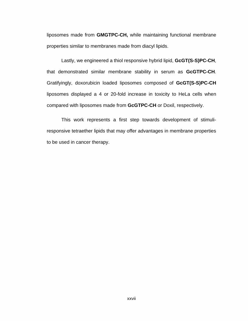
Lastly, we engineered a thiol responsive hybrid lipid, GcGT(S-S)PC-CH,
that demonstrated similar membrane stability in serum as GcGTPC-CH.
Gratifyingly, doxorubicin loaded liposomes composed of GcGT(S-S)PC-CH
liposomes displayed a 4 or 20-fold increase in toxicity to HeLa cells when
compared with liposomes made from GcGTPC-CH or Doxil, respectively.
This work represents a first step towards development of stimuli-
responsive tetraether lipids that may offer advantages in membrane properties
to be used in cancer therapy.

1
Chapter 1
Introduction
1.1 Introduction to Nano-Drug Delivery Systems
Drug delivery systems have been investigated for the process and
administration of pharmaceutical compounds to improve therapeutic effect in a
clinical setting.1–8 To date, most nanomedicines in clinical developments are
formulations or synthetic derivatives of previously FDA approved drugs which
are more effective and more tolerable than with systemic delivery.1,3,5,9–11 Some
examples of improvements in therapeutic effects include controlling
pharmacokinetics, reducing side effects, targeted delivery of drug, and reducing
toxicity of the drug. 1,3,8,12–14 Several different delivery systems have been
synthesized and investigated for tunable anti-cancer treatment to improve
therapeutic effects.3 The term “nanomedicine” was first used to represent nano-
drug delivery systems that uses molecular tools and knowledge of human body
for medical diagnosis and treatment in the 1990’s, with the first research
published in 2000.6,7 An illustration of historical timeline, designed by Shi et. al,
depicting some selected major developments of nanomedicine beginning with
the structural discovery of liposomes in the mid-1960s and continuing on to
more recent development of polymers can be observed in Figure 1.1. Since
1995, at least 50 nanoparticle-based drugs (typically hydrophobic molecules)

2
have entered clinical practice.5 In the brief time since discovery, research in
nanomedicine has led to developments in imaging agents, in vitro diagnostics,
therapeutics, and medical devices with over 200 nanomedicines that have been
approved or under clinical investigations by the FDA.4,15,16 Examples of popular
nanomedicines under investigation includes protein nanoparticles, polymer
drug conjugates, polymeric micelles and nanoparticles, dendrimers, and
liposomes.5
1.1.1 Polymeric Nanoparticles for Drug Delivery
Protein nanoparticles are typically protein (albumin) bound drug-
nanoparticles that improve solubility and in vivo delivery of poorly soluble
drugs.1,17,18 One example, Abraxane®,18 is an albumin-bound formulation of
Figure 1.1. Historical timeline of major developments in the field of cancer
nanomedicine. Reprinted by permission from Macmillan Publishers Ltd: [Nature
Reviews Cancer] (Shi, J.; Kantoff, P. W.; Wooster, R.; Farokhzad, O. C. Nat. Rev.
Cancer 2016, 17 (1), 20–37.), copyright (2016)

3
paclitaxel (FDA approved chemotherapeutic agent to treat solid tumors that is
was originally extracted from the bark of the western yew tree, Taxus
brevifolia).19 Studies have shown, conjugation of paclitaxel to albumin prior to
administration increases solubility and obtain desired PK of paclitaxel in vivo.18
For example, Abraxane® has been shown to provide superior efficacy towards
metastatic cancer with reduced toxicity and elimination of acute drug
hypersensitivity when compared with Cremophor/ethanol-based formulation of
Taxol, (drug marketed as Kolliphor® EL).20 One concern, however, when using
albumin is the potential infection and transmission of disease from donated
plasma. To reduce the potential for disease transmission from HSA extracted
from donated plasma, a recombinant human albumin (Recombumin®) has been
developed by Novozymes.21 Recombumin®, recombinant human albumin, has
also been shown to be effective as: 1) an inhibitor for adsorption of the active
molecules to the storage bottle, 2) a surfactant to aid in solubilization of the drug,
and 3) a stabilizer of conformational structures of the molecules to remain active
during storage.21 Although, conjugation of paclitaxel to albumin prior to
administration is relatively straightforward, protein nanoparticles are limited in
versatility (restricted in use for formulation of hydrophobic drug molecules) and
improved pharmacokinetic properties for other drugs are not guaranteed.
While polymer drug conjugates share similar advantages for formulation
drugs as protein nanoparticles, they differ in the physical/chemical linkage used
to attach drugs to polymers.22 Systematic administration of synthetic drugs or
biomolecules have been shown to be limited by the short half-life of the

4
molecules in circulation caused by renal clearance or enzymatic degradation.5
For instance, chemical conjugation of drug, protein, or peptide to
polyethyleneglycol (PEG) polymers has been shown to improve solubility,
tunability of drug size, controllability of PK, and stability by reducing immune-
mediated clearance and enzyme-mediated degradation.11,22,23 Another example,
XyotaxTM, a drug conjugate formulation of paclitaxel that was under investigation
as a treatment for cancer, evolved to Opaxio®, a nanomedicine that
functionalizes paclitaxel to polyglutamic acid through ester bonds.24 The
polyglutamate functionalized paclitaxel showed large improvement in PK of
systemic injections of paclitaxel alone with an added control of slow release of
paclitaxel. The slow controlled release of paclitaxel was dictated by the rate of
ester hydrolysis from the conjugated formulation in Opaxio.25 This method of
covalently attaching paclitaxel to the polyglutamic backbone allows for similar
advantages discussed for protein nanoparticles, however, suffers from
problems associated with challenges in drug efficacy from chemical modification
of the drug.26
Polymeric micelles differ from other polymer conjugated drugs, which
removes the additional challenges of covalently attaching drug molecules to the
polymeric backbone.26 Polymeric micelles passively encapsulate solubilized
drugs inside a preformed polymeric micelle or nanoparticle.27 Micelles are
formed by the addition of amphiphillic surfactant or polymeric molecules to
aqueous media that spontaneously associate to form core-shell structures.27,28
Typical polymeric monomers used to form polymeric micelles include

5
poly(ethyleneoxide)-poly(benzyl-L-aspartate) and poly(N-isopropylacrylamide)-
polystyrene.29 An alternative formulation to aforementioned Abraxane® (protein
nanoparticle formulation of palcitaxol), Genexol-PM® is a polymeric micelle
formulation of paclitaxel devoid of Cremophor solvent that uses biodegradable
amphiphillic diblock co-polymer poly(ethylene-glycol)-block-poly(D,L-
lactide)(mPEG-DDLLA).17 Genexol-PM® has been shown to be less toxic and
increased tumor uptake when compared with solvent-formulated paclitaxel.5
The observed increased drug uptake into tissues is believed to be promoted by
an increased vascular permeability when compared with free drugs.28 Despite
the advantages discussed, however, the predominant hydrophobic character of
micelle structures limits passive encapsulation of drugs to hydrophobic
molecules and creates further disadvantages of low encapsulation efficiency.27
Dendrimers, the most recent polymeric technology under investigation
as the next nanomedicine, are discrete nanostructures that are branched layers
that grow outwards from the core.2 Dendrimers are synthesized by starting with
a core and grow stepwise (convergent or divergent polymerization) in concentric
layers to increase in size, akin to globular proteins or onions.30 Unlike traditional
polymeric nanoparticles, dendrimers are the only known synthetic nanoparticle
that allows for mathematically defined synthetic control and systematic
engineering of nanostructures with total control that allows for a more versatile
nanomedicine when conjugated with drugs.2 For example, Vivagel® is an FDA
approved topical antiviral dendrimeric formulations made with an anionic G4-
poly(L-lysine)-type dendrimer displaying 32 naphthalene disulfonate groups on

6
the surface.31 Additional advantages dendrimers have over other polymeric
nanoparticles includes the ability to deliver 20-40% w/w of encapsulated or
conjugated drugs, tunable size to make <10 nm dendrimer-drug conjugates, and
mechanically robust enough to be freeze dried for storage.2 However, due to
the intricate design of the tunable branched polymers, difficult synthesis and
high cost of production has been a barrier to be used successfully in cancer
therapy.29
1.1.2. Liposomes as Drug Delivery Systems
Liposomes are self-assembled colloidal particles (shown in Figure 1.2)
that are biocompatible, biodegradable, non-toxic, long circulating, targetable,
and high drug loading drug delivery system that are typically made with
phospholipid that occur naturally or prepared in a laboratory setting. The first
structural characterization of liposomes was featured by Bangham and Horne
in the mid-1960 using electron microscopy.32–39 Liposomes were first studied by
Bangham et al. as a platform to study biological membranes, however,
liposomes began to garner interest as potential drug delivery system in the late
1960’s.32,33 The advantage of liposomes compared with other nano-drug
delivery systems is their ability to tailor properties for delivering a range of cargo
(hydrophilic and hydrophobic molecules), due to the ready access to a toolbox
of commercial lipids with known biophysical properties.40 Other lipid options
include lipid extracted from organisms or synthesized through rational design

7
equipped with a variety of headgroups, linkers and hydrophobic tails.41 The
ability to mix and match different lipids to obtain the exact membrane property
necessary for a liposomal delivery system with control over liposome
macrostructures, biophysical characteristics and in vivo properties allows for a
powerfully adaptable drug delivery system for a variety of applications.33 Despite
the promising drug delivery properties of liposomes, early results of liposomes
as a drug delivery vehicle, however, was stifled by their colloidal and biological
instability, inefficient and unstable encapsulation of drug molecules.8,42
The initial limitation liposomes faced was rapid clearance of large
multilamellar liposomes by mononuclear phagocyte system (MPS). Traditionally,
liposomes were made by creating a thin film of lipid on a glass container by first
dissolving lipids in an organic solvent, continued by evaporation of the organic
Figure 1.2. Illustration of liposomes used for nanomedicine. © {2014} {Melis Çağdaş, Ali
Demir Sezer and Seyda Bucak}. Originally published in {Liposome as potential drug carrier
systems for drug delivery, Chapter 1, Figure 1; open access} under {CC BY 3.0} license.
Available from: {doi. 10.5772/58459}135

8
solvent.35,43 The thin film was then hydrated in a buffer containing drugs and the
lipids self-assemble into large multilamellar vesicles.35,43 To improve evasion
from MPS, liposomes formed with methods of extrusion, that further processes
large multilamellar vesicles through a porous membrane or homogenization,
allowed for production of smaller uniform liposomes with an average diameter
of ~100 nm, which resulted in slower uptake by the MPS.37 In addition to the
reduced liposomal uptake by the MPS, liposomes smaller than 100 nm were
observed to increase drug accumulation in cancer tissues owing to a
phenomena coined the enhanced permeability and retention (EPR) effect.38,44,45
Thus, liposomes with low background leakage that are <100 nm in diameter can
be used to target tumor tissues while minimizing drug interaction with other
tissues to reduce side effects compared with formulation of free drugs.46–48
The next improvement for liposomal technology that followed control of
size distribution was the discovery of addition of free cholesterol to liposomal
formulations to increase membrane stability.49 In mammalian cells, sterols have
been observed to be vital in regulating membrane fluidity.50 While free
cholesterol does not form bilayer structures on their own, due to their
amphiphillic structure, cholesterol can integrate inside liposome bilayers.51 The
addition of cholesterol to liposomal formulation is believed to strengthen the
mechanical bilayer properties and inhibit protein interactions with the liposomes.
For example, addition of free cholesterol to liposomal formulations has been
shown to reduce interaction with opsonins, a protein that has been observed to

9
tag liposomes, that signals macrophages to rapidly clear liposomal from the
body.52 Furthermore, liposome formed with the addition of 40 mol% of free
cholesterol to longer saturated diacyl lipids, that are solid at room temperature,
have been observed to eliminate phase transition by forcing bilayers into a
stable gel-like state allowing for easier formation of liposomes without the need
of external heating.47
The recent discovery that paved the road for liposomes to be used as
drug carriers was the development of different liposomal drug encapsulation
methods using passive or active loading.53,54 When the drug is loaded passively,
the hydrophilic drugs are introduced in the hydration solution during liposomal
formation.55 Using the method of freeze thaw technique, which uses a sequence
of freezing and thawing of liposome suspensions, allows for an increase in
unilamellar vesicles and a decrease in multilamellar vesicles promoted by
transient hole formation by the development of ice crystals that increase drug
penetration and liposome volume.54 However, even with the application of
freeze thaw technique that has been shown to increase encapsulation of drug
inside the liposomes, only 5 – 20% of the drug molecule has been observed to
retain inside the liposome after removal of free drug.54 Another passive
encapsulation technique, called ethanol injections, begins with a solution of lipid
dissolved in ethanol that is then rapidly injected into an excess of drug solution
using a thin needle to form unilamellar liposomes with a defined diameter
(controlled by lipid concentration, injection rate, temperature and lipid

10
composition).56 This strategy of forming liposomes was shown to be less
favorable, due to the unresolved low encapsulation efficiency and reduced
concentration of the drug caused by the presence of ethanol in liposomal
solution.57
To address the low encapsulation efficiency of drugs inside liposomes,
an alternative method termed active loading was established by Nicols, Deamer,
and Bally.58,59 The method of active loading differs from passive loading, in that,
the drug is loaded after the liposomes have formed. The active loading process
is performed by first achieving either an acidic (intraliposomal) or ammonium
sulfate concentration gradient across the internal and external liposomal
environment. Next, the liposomes encapsulated with either acid or ammonium
sulfate is introduced to a solution of a weakly basic drug dissolved in a buffer
containing media, typically PBS or HBS. As the neutral weak base, the drug
permeates inside the liposomal membrane, the weak base becomes protonated
inside the liposomes, thus, trapping the protonated molecule inside the
liposome.58–60 Anticancer drugs such as doxorubicin, idarubicin, or daunorubicin
can be loaded in a drug to lipid ratio of 0.3 % w/w with an encapsulation
efficiency of nearly 100% with excellent in vitro stability.61
The collective Improvements to liposomal formulations mentioned above
allowed for the development and FDA approval of DOXIL® (doxorubicin, ALZA
Corporation, USA), MyocetTM (doxorubicin, Enzon Pharmaceuticals, Inc., USA),
DaunoXome (daunorubicin, NeXtar Pharmaceuticals, USA), and Marqibo®
11
(vincristine, Talon Therapeutics, USA), and DepoCyt® (cytarabine, Pacira, USA)
for cancer therapy.62,63 However, very few liposomal nanomedicines have
recently been approved. We hypothesize that further exploration to generate
more stable synthetic lipids with improved membrane properties can aid in the
development of more FDA approved liposomal formulations.
1.2. Different Synthetic Methods to Improve Liposome Stability
1.2.1 Synthetic Diacyl Lipids to Improve Liposomal Stability
From the advent of liposomes, several research groups have advanced
liposomal technology by synthesizing diacyl lipids with reduced passive leakage
and improved serum stability. One of the largest milestone of synthetic diacyl
lipid developed by Papahadjopoulos et al. began with the invention of steric
stabilized liposomes by chemically conjugating hydrophilic polymers to diacyl
lipids.64 The most common chemical modification reported for diacyl lipid
conjugation is by covalently attaching polyethylene glycol (PEG) to a
phosphatidylethanolamine lipid through the formation of an amide bond. When
sterically stabilized lipid derivative was added to liposomal formulation (5% w/w)
to another non-functionalized commercial diacyl phospholipid, it was observed
to protect the liposome surface from penetration and destruction by plasma
proteins and also reduced macrophage uptake.36,44,65

12
Since the discovery of PEG functionalized diacyl lipids, the addition of
PEG lipids to liposomal formulations has been a staple in current FDA approved
drug delivery products due to the increased aqueous solubility, low cost of
manufacturing, and biocompatibility.25 Despite the favorable attributes gained
by the addition of PEG functionalized diacyl lipids to liposomal formulations, the
addition of PEG was not immunologically inert as first believed.66,67 Studies
have shown that even a low dose of PEGylated material can initiate an immune
response to generate anti-PEG IgM antibodies in various species upon the first
injection.37,68 Subsequent injections resulted in the rapid clearance of the
PEGylated therapeutic that require multiple doses within the window of the
accelerated blood clearance effect of PEGylated therapeutics (3-28 days after
first dose).69 In some severe cases, the liposomes that were not taken up by the
MPS or did not get excreted from the body, would accumulated in the epithelial
cells causing toxic effects known as hand-foot syndrome and stomatitis.70 To
circumvent the negative effects of PEG while maintaining the beneficial effects
of adding PEG to liposomes, several research groups have synthesized PEG
alternatives such as PMOX,71,72 PAcM,73 PDMA,74 polyglycerol,69 PVP,75 and
HPMA76 that have demonstrated increase circulation half-life while reducing the
formation of anti-PEG IgM71.
Another problem encountered in liposomal technology was the leaching
of free cholesterol from liposomes during circulation by rapid transfer into
biomembranes and lipoproteins, resulting in membrane destabilization of the
liposomes. 77,78 To prevent the leaching and transfer of free cholesterol, Szoka’s

13
group had synthesized a class of diacyl lipids that covalently attaches
cholesterol to the glycerol backbone of the phophatidylcholine lipid (shown in
Scheme 1.1).79 The synthesis of cholesterol integrated diacyl lipids begins with
an initial alkylation of 2-pheynyl-1,3-dioxane-5-ol, followed by a deprotection of
the acetal resulting in the formation of the mono-functionalized glycerol diol. The
synthesis of sterol modified diacyl lipid was completed after the addition of
cholesterol to the lipid core via carbamate linkage and the addition of the
phoshpotidylcholine headgroup to the remaining free alcohol of the glycerol.
This new class of sterol modified phospholipids was shown to form stable
liposomes, while having similar effects to free cholesterol when mixed to
stabilize commercial diacyl liposomes showing reduced passive intraliposomal
leakage.80 By anchoring the cholesterol to the lipid membrane, it was shown to
successfully reduce cholesterol transfer and increased in vivo liposome
stability.81 In addition to the decreased passive permeability of inner cargo, the
Scheme 1.1 Synthetic scheme for preparation of covalently attached cholesterol to daicyl
lipids. Reagents and conditions: (i) (1) NaH (1.2 equiv.), toluene, r.t., 30 min; (2)
iodoalkane (1.25 equiv.), reflux, overnight; (ii) HCl (conc.) in MeOH (10%), reflux, 5 h; (ii)
cholesteryl chloroformate (1.05 equiv.), DIPEA (1.4 equiv.), DMAP (0.5 equiv.), CHCl3,
0 °C, 0.5 h, and then r.t., overnight; (iv) POCl3 (1.1 equiv.), pyridine (2 equiv.), THF, 0 °C,
2-3 h; (v) choline tetraphenyl borate (2 equiv.), TPS (2.5 equiv.), pyridine, 70 °C, 1 h,
then r.t., 3 h. Reprinted with permission from (Huang, Z.; Szoka, F. C. J. Am. Chem. Soc.
2008, 130 (46), 15702–15712.). Copyright (2008) American Chemical Society

14
sterol modified liposomes had improved cellular uptake and slower clearance
from the liver and spleen compared with traditional liposomes.81
Despite the promising synthetic progress for improved membrane
properties for diacyl lipid on membrane leakage, diacyl lipids remained unstable
in environments involving acid, enzymes, high lipid concentrated, and metal
ions.82–86
1.2.2. Archaea Inspired Synthetic Tetraether Lipids for Robust Vesicles
1.2.2.1 Synthetic Strategy used to Design Archaeal Macrocyclic Tetraether
Lipid Analogs
Archaea are extremophiles that thrive in harsh conditions such as high
temperature, high acidity, high salinity, and high pressure.87–89 The membranes
found in these organisms have been observed to have a high degree of
chemical and enzymatic stability.90 One strategy that researchers have turned
to in order to improve liposomal stability is by studying natural archaeal
lipids.82,91–98 Specifically, to investigate the essence of the organisms’ inherent
stability under extreme conditions, archaeal lipids have been extracted from
archaeal biomass and characterized by many research groups.99–102 In
particular, De Rosa and Kates group have pioneered investigations into the
biosynthesis and characterization of membrane lipids that have led to
elucidation of a large number of unique structures of archaeal polar lipids
(shown in Figure 1.3).103,104 The unique structural features found in archaeal

15
lipids that differ from bacterial lipids includes: 1) ether linkages between fatty
acids and the glycerol backbone that differ from bacterial lipids with ester
linkages, 2) isoprenoid hydrocarbon chains that are highly methyl-branched in
archaeal lipids, whereas, their bacterial counterpart have straight-chain fatty
acids with and without double bonds, 3) lipid tails that are covalently tethered
resulting in creating a membrane spanning lipids that form monolayer
membranes that differs from bacterial lipids that form bilayers, and 4)
cyclopentane and/or cyclohexane rings in the tethered region of the
hydrophobic tails.88,100 The ether linkages, isoprenoid chains, tethered
tetraether lipid and integration of small rings have been suggested to play a
significant role for the membrane stability or Archaea in extreme habitat.90,105–
107
To investigate whether the inherent stability of Archaea is due to their
lipid composition in the membrane, archaeal lipids have been extracted from
Sulfolobus acidocaldarius, called polar lipid fraction E, to form liposomes to
study membrane leakage.82 These liposomes, coined archaeosomes, exhibited
low permeability, tight membrane packing and improved stability compared with
liposomes formed from diacyl lipids.82,108,109 However, harvesting reproducible
and large quantities of specific lipid compositions from cultured archaeal
species can be challenging.82,108,109 Instead, several groups have reported

16
model archaeal lipid analogs that incorporate key structural features that are
believed to increase membrane stability of archaeal membranes.97,110–126
Figure 1.3. Membranes of the Archaea and Bacteria. Reprinted by permission from Macmillan
Publishers Ltd: [Nature Reviews Microbiology] (Valentine, D. L. Nat. Rev. Microbiol. 2007, 5,
316–323.), copyright (2007)

17
In the past, considerable effort has been invested in the development of
Archaea inspired lipids with tetraether or tetraester127,128 glycerol linkages. In
this introduction, the synthesis of tetraether lipids will be the main focus
considering the increased stability imparted by ether linkages when compared
Figure 1.4. Sample representation set of synthetic archaeal lipid analogs in current research.
Ref: I114, II113, III117, IV133, V133, VI119.

18
with ester linkages.129,130 In addition to the chemically promiscuous ester
functional group, Yamauchi et al. has published research that demonstrates
liposomes made with ordinary ester linked lipids were prone to aggregation and
precipitation under high lipid concentration and in the presence of metal ions,
resulting in loss of interior content within a few days.83 Chemical structures of
the most representative synthetic archaeal lipid analogues (shown in Figure 1.4)
differ in their hydrophobic moiety. Previously synthesized examples of tetraether
lipids that incorporate structural features found in Archaea possess either
macrocyclic or hemicyclic membrane spanning hydrophobic chains as shown in
Figure 1.4.
In nature, Archaeal lipids have been found to form 36-membered
macrocyclic diether and 72-membered macrocyclic tetraether lipids.88,131 To
investigate the membrane leakage properties of these macrocyclic tetraether
lipids, Kakinuma’s group has successfully synthesized 36 (molecule I)114 and
72-membered (molecule II)113,115 macrocyclic lipids shown in Figure 1.4. The
synthetic strategy used by the Kakinuma’s group to design the 36-membered
macrocyclic diether (I) involved the stereo-selective preparation of
functionalized isoprenoid chains to the glycerol backbone through an ether
linkage starting from a chiral (R)-3-hydroxy-2-methyl-propionate (shown in
Scheme 1.2). The chirally pure C20 isoprenoid chains were synthesized by a
series of elongation reactions to the C5 unit consisting of protection,
deprotection, displacement of the iodide by an enol-equivalent sulfur dioxide

19
analog and followed by the removal of the sulfone using Na(Hg). Following the
formation of the C20 isoprenoid unit, alkylation by the glycerol backbone derived
from 1-O-benzyl-sn-glycerol afforded the desired ether linkage affording the two
half lipid scaffold. The key C-C bond connection necessary in the total synthesis
of the 36-membered diether lipid was completed via intramolecular McMurry
coupling of the oxidized alcohols catalyzed by low-valent titanium. The
synthesis of the 36-membered diether was completed by an initial olefin
reduction followed by the addition of the phosphatidylcholine headgroup to the
remaining glycerol alcohol.
The total synthesis of 72-membered macrocyclic tetraether (II) was
performed using the bifunctionalized C20 isoprenoid building block used in the
36-membered macrocyclic tetraether lipid synthesis (shown in Scheme 1.3). It
is notable, to obtain the desired stereo-configuration of the glycerol backbone,
the free alcohol of C20 isoprenoid building block was oxidized to an aldehyde
and reacted with sn-1-O-benzylglycerol to form the acetal intermediate.
Alkylation of the orthogonally protected C20 isoprenoid building block by the free
hydroxyl group after the deprotected acetal afforded the acyclic diether lipid
precursor which was later tethered together by the formation of β-
hydroxysulfone. β-hydroxysulfone was successfully removed using a SmI2
catalyzed reductive elimination, followed by the reduction of the olefin. Using
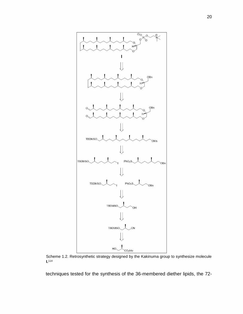
20
techniques tested for the synthesis of the 36-membered diether lipids, the 72-
Scheme 1.2. Retrosynthetic strategy designed by the Kakinuma group to synthesize molecule
I.114

21
membered macro-cyclization was completed using McMurry coupling. The total
synthesis of the 72-membered macrocyclic tetraether was completed following
the reduction of the olefin and the addition of the phosphatidylcholine headgroup
Scheme 1.3. Retrosynthetic strategy designed by the Kakinuma group to synthesize molecule
II. 113

22
to the remaining glycerol alcohols in 27 steps (longest linear reaction steps) with
an overall yield of 1.7%.115
Total synthesis of the 72 membered macrocyclic archaeal lipid was
elegantly performed by the Kakinuma group. However, to study the structure
functional relationship of archaeal structures to membrane permeability in
aqueous media requires a large amount of pure lipid. To address the synthetic
challenges associated with synthesis of optically pure isoprenoid units involving
low yield, Thompson’s group designed a macrocyclic tetraether lipids equipped
with saturated alkyl chains (III).117 The synthetic strategy employed by the
Thompson group begins with 2-phenyl-1,3-dioxan-5-ol as the masked glycerol
backbone that participates in alkylating the terminal alkyne (shown in Scheme
1.4). The terminal alkyne of the alkylated glycerol is coupled together through
an initial Glaser oxidation to form the hemicyclic tetraether lipid. This alkylation
and coupling process was repeated to complete the macro-cyclization, followed
by reduction of the alkynes and the addition of phosphatidylcholine headgroup
to the free glycerol alcohol to complete the synthesis of a 48-membered
macrocyclic tetraether lipid.117 The saturated macrocyclic tetraether lipid was
elegantly designed with fewer steps, however, without the branched methyl
groups, the saturated macrocyclic tetraether lipid displayed a high phase
transition temperature, which would cause complications when preparing
liposomes.132

23
Scheme 1.4. Retrosynthetic strategy designed by the Thompson group to synthesize molecule
III.117

24
1.2.2.2 Synthetic Strategy used to Design Archaeal Hemicyclic Tetraether
Lipid Analogs
Scientists interested in studying structure functional relationships
associated with structural features found in archaeal lipids on membrane
permeability have focused their synthetic efforts on simpler model molecules
that are hemicyclic, which still incorporate structures found in natural archaeal
lipids and can be produce in a larger amounts. Thompson’s group, one of the
research groups that had shifted their research towards the simpler model
molecules, designed a new set of hemicyclic tetraether lipids substituted with
either a saturated alkyl or phytanyl group on the untethered portion on the lipid
(shown in Figure 1.4). Both hemicyclic tetraether lipids (IV and V) were
synthesized using a similar strategy as their synthesis of the macrocyclic
tetraether lipid that differed in the initial alkylation of 2-phenyl-1,3-dioxan-5-ol
with either an iodinated saturated alkyl chain or a iodine derivative of
commercially available phytol (retrosynthetic strategy to synthesize phytanyl
containing hemicyclic tetraether (V) shown in Scheme 1.5). The dimerization of
the tethered region was completed using metathesis catalyzed by Grubb’s
reagent to form the hemicyclic tetraether lipids. Fascinatingly, without the
branched alkane substituents, the saturated hemicyclic tetraether lipid, similar
to the saturated macrocyclic tetraether lipid, still exhibited a high phase
transition temperature (>85 °C)133 which is not observed for natural archaeal
lipids89.

25
To explore the structural benefits associated with incorporation of
cyclopentane rings in the hydrophobic core of natural archaeal lipids,
Benvegnu’s group have successfully designed model hemicyclic lipids that
incorporates a cis-1,3-cyclopentane in the lipid core. The synthetic strategy
used to design molecule (VI) shown in Figure 1.4, began with the synthesis of
Scheme 1.5. Retrosynthetic strategy designed by the Thompson group to synthesize molecule
V. 133

26
2 precursors, cyclopentane integrated hydrophobic chain and phytanyl affixed
glycerol backbone (shown in Scheme 1.6). The cyclopentane integrated
hydrophobic chain was elegantly synthesized by first a ring opening of
norbornene via ozonolysis, followed by the reduction of the dialdehyde to form
the diol. The newly formed alcohols were subsequently transformed to a
phosphonium salt which were then couple through a Wittig reaction with two
alkyl aldehydes to form the hydrophobic chain precursor. The diolefinated
homologated hydrophobic chain was reduced/O-deprotected and the free
primary alcohols were triflated. The glycerol backbone of the lipid, second piece
of the tetraether lipid, was synthesized starting from solketal through a series of
deprotection, protection and alkylation with iodophytyl. The hemi-cyclization of
the lipid was performed through a round of alkylation of the phytanylated
glycerol with the ditriflate of the cyclopentane integrated alkane. The synthesis
of cyclopentane integrated hemicyclic tetraether (VI) was completed after the
addition of phosphatidylcholine headgroup to the free alcohols of the glycerol
backbone.
Despite the progress of the elegant synthesis of archaeal lipid analogues
designed by many research groups, the relationship between structure and
function of the tetraether lipids on membrane stability remains unclear due to
the limited data available on their membrane permeability
properties83,84,86,97,122,134. A systematic study that probes the effect of specific
structural features found in the archaeal-inspired lipids on membrane leakage

27
could prove fortuitous in designing lipids with improved membrane properties
for drug delivery.
Scheme 1.6. Retrosynthetic strategy designed by the Benvegnu group to synthesize molecule
VI. 119

28
1.3 References
(1) Shi, J.; Kantoff, P. W.; Wooster, R.; Farokhzad, O. C. Nat. Rev. Cancer 2016, 17 (1), 20–37.
(2) Kannan, R. M.; Nance, E.; Kannan, S.; Tomalia, D. A. J. Intern. Med. 2014, 276 (6), 579–617.
(3) Caster, J. M.; Patel, A. N.; Zhang, T.; Wang, A. Wiley Interdiscip. Rev. Nanomed. Nanobiotechnol. 2016, 9.
(4) Etheridge, M. L.; Campbell, S. A.; Erdman, A. G.; Haynes, C. L.; Wolf, S. M.; McCullough, J. Nanomedicine Nanotechnology, Biol. Med. 2013, 9 (1), 1–14.
(5) Min, Y.; Caster, J. M.; Eblan, M. J.; Wang, A. Z. Chem. Rev. 2015, 115 (19), 11147–11190.
(6) Wagner, V.; Dullaart, A.; Bock, A.-K.; Zweck, A. Nat. Biotechnol. 2006, 24 (10), 1211–1217.
(7) Wang, R.; Billone, P. S.; Mullett, W. M. J. Nanomater. 2013, 2013 (April), 1–12.
(8) Allen, T. M.; Cullis, P. R. Adv. Drug Deliv. Rev. 2013, 65 (1), 36–48.
(9) Barenholz, Y. J. Control. Release 2012, 160 (2), 117–134.
(10) Mayer, M. I.; Weickum, R. J.; Jr, D. A. S.; Fileta, B. B.; Abdel-rahim, M. M.; Fant, G. V. 2001, 35, 1548–1551.
(11) Duncan, R. Nat. Rev. Cancer 2006, 6 (9), 688–701.

29
(12) Malam, Y.; Loizidou, M.; Seifalian, A. M. Trends Pharmacol. Sci. 2009, 30 (11), 592–599.
(13) Tiwari, G.; Sriwastawa, B.; Pandey, S.; Bannerjee, S.; Tiwari, R.; Bhati, L.; Pandey, P. Int. J. Pharm. Investig. 2012, 2 (1), 2.
(14) Li, J.; Wang, X.; Zhang, T.; Wang, C.; Huang, Z.; Luo, X.; Deng, Y. Asian J. Pharm. Sci. 2014, 10 (2), 81–98.
(15) Weissig, V.; Pettinger, T. K.; Murdock, N. Int. J. Nanomedicine 2014, 9, 4357–4373.
(16) Hare, J. I.; Lammers, T.; Ashford, M. B.; Puri, S.; Storm, G.; Barry, S. T. Adv. Drug Deliv. Rev. 2016, 108, 25–38.
(17) Werner, M. E.; Cummings, N. D.; Sethi, M.; Wang, E. C.; Sukumar, R.; Moore, D. T.; Wang, A. Z. Int. J. Radiat. Oncol. Biol. Phys. 2013, 86 (3), 463–468.
(18) Miele, E.; Spinelli, G. P.; Miele, E.; Tomao, F.; Tomao, S. Int. J. Nanomedicine 2009, 4, 99–105.
(19) Wani, M. C.; Taylor, H. L.; Wall, M. E.; Coggon, P.; McPhail, a T. J. Am. Chem. Soc. 1971, 93 (9), 2325–2327.
(20) Gelderblom, H.; Verweij, J.; Nooter, K.; Sparreboom, A. Eur. J. Cancer 2001, 37 (13), 1590–1598.
(21) Mead, D.; Pearson, D.; Devine, M. Innov. Pharm. Technol. 2007, No. 22, 42–44.
(22) Kopeček, J. Adv. Drug Deliv. Rev. 2013, 65 (1), 49–59.

30
(23) Pasut, G.; Veronese, F. M. Adv. Drug Deliv. Rev. 2009, 61 (13), 1177–1188.
(24) Singer, J. W. J. Control. Release 2005, 109 (1–3), 120–126.
(25) Knop, K.; Hoogenboom, R.; Fischer, D.; Schubert, U. S. Angew. Chemie - Int. Ed. 2010, 49 (36), 6288–6308.
(26) Kim, E. G.; Kim, K. M. Biomol. Ther. 2015, 23 (6), 493–509.
(27) Nishiyama, N.; Kataoka, K. Pharmacol. Ther. 2006, 112 (3), 630–648.
(28) Mourya, V. K.; Inamdar, N.; Nawale, R. B.; Kulthe, S. S. Indian J. Pharm. Educ. Res. 2011, 45 (2), 128–138.
(29) Dikmen, G.; Genç, L.; Güney, G. 2011, 5 (November 2016), 468–472.
(30) Namazi, H.; Adeli, M. Biomaterials 2005, 26 (10), 1175–1183.
(31) Mccarthy, T. D.; Karellas, P.; Henderson, S. A.; Giannis, M.; David, F.; Keefe, O.; Heery, G.; Paull, J. R. A.; Matthews, B. R.; Holan, G.; Mccarthy, T. D.; Karellas, P.; Henderson, S. A.; Giannis, M.; Keefe, D. F. O.; Heery, G.; Paull, J. R. A.; Matthews, B. R. Mol. Pharm. 2005, 2, 312–318.
(32) Bangham, A. D.; Horne, R. W. J. Mol. Biol. 1964, 8 (5), 660–668.
(33) Akbarzadeh, A.; Rezaei-Sadabady, R.; Davaran, S.; Joo, S. W.; Zarghami, N.; Hanifehpour, Y.; Samiei, M.; Kouhi, M.; Nejati-Koshki, K. Nanoscale Res. Lett. 2013, 8 (1), 102.
(34) Immordino, M. L.; Dosio, F.; Cattel, L. Int. J. Nanomedicine 2006, 1 (3), 297–315.
31
(35) Van der Meel, R.; Fens, M. H. A. M.; Vader, P.; Van Solinge, W. W.; Eniola-Adefeso, O.; Schiffelers, R. M. J. Control. Release 2014, 195, 72–85.
(36) Woodle, M. C. Adv. Drug Deliv. Rev. 1995, 16 (2–3), 249–265.
(37) Ishida, T.; Harashima, H.; Kiwada, H. Biosci. Rep. 2002, 22 (2), 197–224.
(38) Drummond, D. C.; Meyer, O.; Hong, K.; Kirpotin, D. B.; Papahadjopoulos, D. Pharmacol. Rev. 1999, 51 (4), 691–743.
(39) Cullis, P. R.; Mayer, L. D.; Bally, M. B.; Madden, T. D.; Hope, M. J. Adv. Drug Deliv. Rev. 1989, 3 (3), 267–282.
(40) Sipai Altaf Bhai, M.; Vandana, Y.; Mamatha, Y.; Prasanth, V. V. J. Pharm. Sci. Innov. 2012, 1 (1), 13–21.
(41) Kohli, A. G.; Kierstead, P. H.; Venditto, V. J.; Walsh, C. L.; Szoka, F. C. J. Control. Release 2014, 190, 274–287.
(42) Chrai, S. S.; Murari, R.; Ahmad, I. Biotech Trends 2002, 30–34.
(43) Berger, N.; Sachse, a; Bender, J.; Schubert, R.; Brandl, M. Int. J. Pharm. 2001, 223 (1–2), 55–68.
(44) Allen, T. M.; Hansen, C.; Martin, F.; Redemann, C.; Yau-Young, A. BBA - Biomembr. 1991, 1066 (1), 29–36.
(45) Proffitt, R. T.; Williams, L. E.; Presant, C. A.; Tin, G. W.; Uliana, J. A.; Gamble, R. C.; Baldeschwieler, J. D. J Nucl Med 1983, 24 (1), 45–51.
(46) Loi, M.; Marchio, S.; Becherini, P.; Di Paolo, D.; Soster, M.; Curnis, F.;

32
Brignole, C.; Pagnan, G.; Perri, P.; Caffa, I.; Longhi, R.; Nico, B.; Bussolino, F.; Gambini, C.; Ribatti, D.; Cilli, M.; Arap, W.; Pasqualini, R.; Allen, T. M.; Corti, A.; Ponzoni, M.; Pastorino, F. J. Control. Release 2010, 145 (1), 66–73.
(47) Mabrey, S.; Mateo, P. L.; Sturtevant, J. M. Biochemistry 1978, 17 (12), 2464–2468.
(48) Pastorino, F.; Paolo, D. Di; Loi, M.; Becherini, P.; Caffa, I.; Zorzoli, A.; Marimpietri, D. 2009, 1021–1027.
(49) Senior, J.; Gregoriadis, G. Life Sci. 1982, 30 (I), 2123–2136.
(50) De Kruyff, B.; Demel, R. A.; Slotboom, A. J.; Van Deenen, L. L. M.; Rosenthal, A. F. Biochim. Biophys. Acta - Biomembr. 1973, 307 (1), 1–19.
(51) Haines, T. H. Prog. Lipid Res. 2001, 40 (4), 299–324.
(52) Oja, C. D.; Semple, S. C.; Chonn, A.; Cullis, P. R. Biochim. Biophys. Acta - Biomembr. 1996, 1281 (1), 31–37.
(53) Woodle, M. C.; Papahadjopoulos, D. Methods Enzymol. 1989, 171 (1984), 193–217.
(54) Mayer, L. D.; Hope, M. J.; Cullis, P. R.; Janoff, A. S. BBA - Biomembr. 1985, 817 (1), 193–196.
(55) Szoka, F.; Papahadjopoulos, D. Ann. Rev. Biophys. Bioeng. 1980, 9, 467–508.
(56) Batzri, S.; Korn, E. D. BBA - Biomembr. 1973, 298 (4), 1015–1019.

33
(57) Pons, M.; Foradada, M.; Estelrich, J. Int. J. Pharm. 1993, 95 (1–3), 51–56.
(58) Bally, M. B.; Mayer, L. D.; Loughrey, H.; Redelmeier, T.; Madden, T. D.; Wong, K.; Harrigan, P. R.; Hope, M. J.; Cullis, P. R. Chem. Phys. Lipids 1988, 47 (2), 97–107.
(59) Mayer, L. D.; Bally, M. B.; Cullis, P. R. BBA - Biomembr. 1986, 857 (1), 123–126.
(60) Acta, B.; Bba, E.; Columbia, B.; November, R. 1985, 816, 294–302.
(61) Dos Santos, N.; Cox, K. A.; McKenzie, C. A.; Van Baarda, F.; Gallagher, R. C.; Karlsson, G.; Edwards, K.; Mayer, L. D.; Allen, C.; Bally, M. B. Biochim. Biophys. Acta - Biomembr. 2004, 1661 (1), 47–60.
(62) Forssen, E. A. Adv. Drug Deliv. Rev. 1997, 24 (2–3), 133–150.
(63) Swenson, C. E.; Perkins, W. R.; Roberts, P.; Janoff, A. S. The Breast 2001, 10, 1–7.
(64) Papahadjopoulos, D.; Allen, T. M.; Gabizon, A.; Mayhew, E.; Matthay, K.; Huang, S. K.; Lee, K. D.; Woodle, M. C.; Lasic, D. D.; Redemann, C. Proc. Natl. Acad. Sci. U. S. A. 1991, 88 (24), 11460–11464.
(65) Woodle, M. C.; Newman, M. S.; Cohen, J. a. J. Drug Target. 1994, 2 (5), 397–403.
(66) Owens, D. E.; Peppas, N. A. Int. J. Pharm. 2006, 307 (1), 93–102.
(67) Richter, A. W.; Åkerblom, E. Int. Arch. Allergy Immunol. 1984, 74 (1), 36–39.

34
(68) Ishida, T.; Ichihara, M.; Wang, X.; Kiwada, H. J. Control. Release 2006, 115 (3), 243–250.
(69) Abu Lila, A. S.; Kiwada, H.; Ishida, T. J. Control. Release 2013, 172 (1), 38–47.
(70) Uziely, B.; Jeffers, S.; Isacson, R.; Kutsch, K.; Wei-Tsao, D.; Yehoshua, Z.; Libson, E.; Muggia, F. M.; Gabizon, A. J. Clin. Oncol. 1995, 13 (7), 1777–1785.
(71) Kierstead, P. H.; Okochi, H.; Venditto, V. J.; Chuong, T. C.; Kivimae, S.; Fréchet, J. M. J.; Szoka, F. C. J. Control. Release 2015, 213, 1–9.
(72) Gaertner, F. C.; Luxenhofer, R.; Blechert, B.; Jordan, R.; Essler, M. J. Control. Release 2007, 119 (3), 291–300.
(73) Torchilin, V. P.; Trubetskoy, V. S.; Whiteman, K. R.; Caliceti, P.; Ferruti, P.; Veronese, F. M. J. Pharm. Sci. 1995, 84 (9), 1049–1053.
(74) Kohori, F.; Sakai, K.; Aoyagi, T.; Yokoyama, M.; Yamato, M.; Sakurai, Y.; Okano, T. Colloids Surfaces B Biointerfaces 1999, 16 (1–4), 195–205.
(75) Romberg, B.; Oussoren, C.; Snel, C. J.; Carstens, M. G.; Hennink, W. E.; Storm, G. Biochim. Biophys. Acta - Biomembr. 2007, 1768 (3), 737–743.
(76) Duncan, R.; Vicent, M. J. Adv. Drug Deliv. Rev. 2010, 62 (2), 272–282.
(77) Phillips, M. C.; Johnson, W. J.; Rothblat, G. H. Biochim. Biophys. Acta 1987, 906 (2), 223–276.
(78) Hamilton, J. A. Curr. Opin. Lipidol. 2003, 14 (3), 263–271.

35
(79) Huang, Z.; Szoka Jr., F. C. J. Am. Chem. Soc. 2008, 130 (46), 15702–15712.
(80) Huang, Z.; Jaafari, M. R.; Szoka, F. C. Angew. Chem., Int. Ed. Engl. 2009, 48 (23), 4146–4149.
(81) Kohli, A. G.; Kieler-Ferguson, H. M.; Chan, D.; Szoka, F. C. J. Control. Release 2014, 176 (1), 86–93.
(82) Elferink, M. G. L.; de Wit, J. G.; Driessen, A. J. M.; Konings, W. N. Biochim. Biophys. Acta, Biomembr. 1994, 1193 (2), 247–254.
(83) Yamauchi, K.; Doi, K.; Kinoshita, M.; Kii, F.; Fukuda, H. Biochim. Biophys. Acta 1992, 1110 (2), 171–177.
(84) Yamauchi, K.; Doi, K.; Yoshida, Y.; Kinoshita, M. Biochim. Biophys. Acta, Biomembr. 1993, 1146 (2), 178–182.
(85) Yamauchi, K.; Doi, K.; Kinoshita, M. Biochim. Biophys. Acta - Biomembr. 1996, 1283 (2), 163–169.
(86) Arakawa, K.; Eguchi, T.; Kakinuma, K. Chem. Lett. 2001, 5, 440–441.
(87) Koga, Y.; Nakano, M. Syst. Appl. Microbiol. 2008, 31 (3), 169–182.
(88) Koga, Y. Archaea 2012, 2012, 789652.
(89) Blöcher, D.; Gutermann, R.; Henkel, B.; Ring, K. BBA - Biomembr. 1984, 778 (1), 74–80.
(90) Chong, P. L.-G.; Ayesa, U.; Daswani, V. P.; Hur, E. C. Archaea 2012, 2012, 1–11.

36
(91) Gambacorta, A.; Gliozzi, A.; De Rosa, M. World J. Microbiol. Biotechnol. 1995, 11 (1), 115–131.
(92) Whitfield, D. M.; Eichler, E. E.; Sprott, G. D. Carbohydr. Res. 2008, 343, 2349–2360.
(93) Benvegnu, T.; Lemiègre, L.; Cammas-Marion, S. Recent Pat. Drug Deliv. Formul. 2009, 3 (3), 206–220.
(94) Le Gall, T.; Barbeau, J.; Barrier, S.; Berchel, M.; Lemiègre, L.; Jeftić, J.; Meriadec, C.; Artzner, F.; Gill, D. R.; Hyde, S. C.; Férec, C.; Lehn, P.; Jaffrès, P.-A.; Benvegnu, T.; Montier, T. Mol. Pharm. 2014, 11 (9), 2973–2988.
(95) Gliozzi, A.; Relini, A.; Chong, P. L.-G. J. Membr. Sci. 2002, 206, 131–147.
(96) Bücher, C.; Grosse, X.; Rothe, H.; Fiethen, A.; Kuhn, H.; Liefeith, K. Biointerphases 2014, 9 (1), 11002.
(97) Barbeau, J.; Cammas-Marion, S.; Auvray, P.; Benvegnu, T. J. Drug Deliv. 2011, 2011, 396068.
(98) Patel, G. B.; Agnew, B. J.; Deschatelets, L.; Fleming, L. P.; Sprott, G. D. Int. J. Pharm. 2000, 194, 39–49.
(99) Carballeira, N. M.; Reyes, M.; Sostre, A.; Huang, H.; Verhagen, M. F.; Adams, M. W. J. bacteriol. 1997, 179 (8), 2766–2768.
(100) Valentine, D. L. Nat. Rev. Microbiol. 2007, 5, 316–323.
(101) Villanueva, L.; Damsté, J. S. S.; Schouten, S. Nat. Rev. Microbiol. 2014, 12 (6), 438–448.
37
(102) De Rosa, M.; Gambacorta, A. Prog. Lipid Res. 1988, 27 (3), 153–175.
(103) De Rosa, M.; Gambacorta, a; Gliozzi, a. Microbiol. Rev. 1986, 50 (1), 70–80.
(104) Kates, M. In Prog. Chem. Fats other Lipids; Pergamon Press Ltd, 1978; pp 301–342.
(105) Koga, Y.; Morii, H. Microbiol. Mol. Biol. Rev. 2007, 71 (1), 97–120.
(106) Gabriel, J. L.; Lee Gau Chong, P. Chem. Phys. Lipids 2000, 105 (2), 193–200.
(107) Komatsu, H.; Chong, P. L. Biochemistry 1998, 37 (1), 107–115.
(108) Chang, E. L. Biochem. Biophys. Res. Commun. 1994, pp 673–679.
(109) Uda, I.; Sugai, a; Itoh, Y. H.; Itoh, T. Lipids 2001, 36 (1), 103–105.
(110) Montenegro, E.; Gabler, B.; Paradies, G.; Seemann, M.; Helmchen, G. Angew. Chem. 2003, 42, 2419–2421.
(111) Arakawa, K.; Eguchi, T.; Kakinuma, K. Bull. Chem. Soc. Jpn. 2001, 74 (2), 347–356.
(112) Arakawa, K.; Eguchi, T.; Kakinuma, K. J. Org. Chem. 1998, 63 (14), 4741–4745.
(113) Eguchi, T.; Ibaragi, K.; Kakinuma, K. J. Org. Chem. 1998, 63 (8), 2689–2698.
(114) Eguchi, T.; Arakawa, K.; Terachi, T.; Kakinuma, K. J. Org. Chem. 1997, 62 (7), 1924–1933.

38
(115) Eguchi, T.; Arakawa, K.; Kakinuma, K.; Rapp, G.; Ghosh, S.; Nakatani, Y.; Ourisson, G. Chem. - A Eur. J. 2000, 6 (18), 3351–3358.
(116) Markowski, T.; Drescher, S.; Meister, A.; Hause, G.; Blume, A.; Dobner, B. Eur. J. Org. Chem. 2011, 5894–5904.
(117) Patwardhan, A. P.; Thompson, D. H. Org. Lett. 1999, 1 (2), 241–243.
(118) Svenson, S.; Thompson, D. J. Org. Chem. 1998, 3263 (98), 7180–7182.
(119) Brard, M.; Lainé, C.; Réthoré, G.; Laurent, I.; Neveu, C.; Lemiègre, L.; Benvegnu, T. J. Org. Chem. 2007, 72 (22), 8267–8279.
(120) Raguse, B.; Culshaw, P. N.; Prashar, J. K.; Raval, K. Tetrahedron Lett. 2000, 41 (16), 2971–2974.
(121) Jacquemet, A.; Lemiègre, L.; Lambert, O.; Benvegnu, T. J. Org. Chem. 2011, 76 (23), 9738–9747.
(122) Benvegnu, T.; Réthoré, G.; Brard, M.; Richter, W.; Plusquellec, D. Chem. Commun. (Cambridge, U. K.) 2005, No. 44, 5536–5538.
(123) Lecollinet, G.; Auzély-Velty, R.; Benvegnu, T.; Plusquellec, D.; Mackenzie, G.; Goodby, J. W. Chemical Communications. 1998.
(124) Barbeau, J.; Belmadi, N.; Montier, T.; Le Gall, T.; Dalençon, S.; Lemiègre, L.; Benvegnu, T. Tetrahedron Lett. 2016, 2, 1–5.
(125) Lecollinet, G.; Auzély-Velty, R.; Danel, M.; Benvegnu, T.; Mackenzie, G.; Goodby, J. W.; Plusquellec, D. J. Org. Chem. 1999, 64 (9), 3139–3150.

39
(126) Long, L.; Zhou, L.; Wang, L.; Meng, S.; Gong, A.; Du, F.; Zhang, C. 2013, 8214–8220.
(127) Menger, F. M.; Chen, X. Y. Tetrahedron Lett. 1996, 37 (3), 323–326.
(128) Cuccia, L. a; Morin, F.; Beck, A.; Hébert, N.; Just, G.; Lennox, R. B. Chem. Eur. J. 2000, 6 (23), 4379–4384.
(129) Nacka, F.; Cansell, M.; Gouygou, J. P.; Gerbeaud, C.; Méléard, P.; Entressangles, B. Colloids Surf. B. Biointerfaces 2001, 20 (3), 257–266.
(130) Snyder, W. R. Biochim. Biophys. Acta (BBA)/Lipids Lipid Metab. 1987, 920 (2), 155–160.
(131) Damsté, J. S. S.; Rijpstra, W. I. C.; Hopmans, E. C.; Schouten, S.; Balk, M.; Stams, A. J. M. Arch. Microbiol. 2007, 188 (6), 629–641.
(132) Thompson, D. H.; Wong, K. F.; Humphry-baker, R.; Wheeler, J. J.; Kim, J.; Rananware, S. B. J. Am. Chem. Soc. 1999, 114, 9035–9042.
(133) Febo-Ayala, W.; Morera-Félix, S. L.; Hrycyna, C. A.; Thompson, D. H. Biochemistry 2006, 45 (49), 14683–14694.
(134) Yamauchi, K.; Sakamoto, Y.; Moriya, A.; Yamada, K.; Hosokawa, T.; Higuchi, T.; Kinoshita, M. J. Am. Chem. Soc. 1990, 112 (8), 3188–3191.
(135) Melis Çağdaş, A. D. S. and S. B. (Ed.), P. A. D. S., Ed.; InTech, 2014; 2.

40
Chapter 2
Synthesis of Archaea Inspired Tetraether Lipids and
Investigation of Structural Functional Relationship to
Membrane Leakage
2.1 Introduction to Archaeal Lipid Structures and Structural Functional
Relationship On Membrane Permeability
In nature, organisms respond to environmental stress by modifying their
membrane lipid composition to remain viable.1–4 For example, Archaea are
extremophiles classified as either halophiles, thermophiles, or acidophiles
depending on the environment they inhabit and have evolved mechanically and
chemically robust membrane compositions that allows for their survival in high
salinity, high temperature, or high acidity, respectively.5 Archaeal membranes
have been extensively studied in the hopes to unveil their unique structural
benefits on membrane permeability that differs from their bacterial counterparts.
A representative collection of lipid structures of archaeal lipids are shown in
Figure 2.1 that highlight the unique structures found in macrocyclic tetraether
lipids (i.e. isoprenoid alkyl chains, ether glycerol linkage, alkyl rings, and polar
lipid headgroups). Notably, one structural feature shown in Figure 2.1 that has
fascinated scientists, is how the integration of up to eight cyclopentane rings to

41
the lipid core affect membrane properties. For example, a hyperthermophillic
Archaea, Crenarchaeota, have been observed to thrives in temperature above
80 °C and have different numbers of cyclopentane rings found in the lipids of
the membrane depending on their growth temperature.2,6,7 The increased
number of cyclopentane rings is believed to tighten membrane packing as the
environmental temperature rises.8 In recent studies, Thaumarchaeota, a newly
added category in the kingdom of Archaea, have been observed to incorporate
cyclohexane rings into their lipids.7 The reason for the incorporation of
cyclohexane to the lipid tail has been enigmatic. One hypothesis proposed from
molecular dynamic simulation calculation data suggests the addition of
cyclohexane rings to the lipid core allows for a higher conformational freedom
than cyclopentane that allow Thaumarchaeota to survive in colder marine
environment.9 In addition to the structural changes observed in the archaeal
lipid tails for adaptation to environmental stress, changes in polar lipid
headgroups have been observed as well.10,11 For example, lipid compositions
of archaeal membranes found in hot acid springs with pH < 3, have been found
to have higher concentrations of polar lipid headgroups capable of increased
hydrogen bonding.12–14 This additional hydrogen bonding network has been
hypothesized to function as an additional barrier to decrease membrane
permeability towards protons. However, the difficulty of isolation of pure lipids
with different polar lipid headgroups precludes testing this hypothesis.15

42
One approach, mention in chapter 1 of this dissertation, used to study
structural/functional relationship of archaeal lipids on membrane leakage is by
extracting natural lipids and forming vesicles. Specifically, liposomes made from
polar lipid fraction E (PLFE) that are extracted from Sulfolobus acidocaldarius
or Thermoplasma acidophilum exhibit low permeability towards small
ions/molecules, tight membrane packing, and high stability in human bile
containing solutions.13,16 Another study showed a general trend of increased
membrane stability of diacyl lipid membranes as the mole fraction of tetraether
lipid increased.17 Despite the favorable membrane properties attributed to
Figure 2.1. Tetraether lipids with varying hydrophobic core and polar lipid
headgroups found in archaeal membranes. Reprinted by permission from Annual
Reviews: [The Annual Review Earth and Planetary Sciences] (Pearson, A.; Ingalls,
A. E. Annu. Rev. Earth Planet. Sci. 2013, 41 (1), 359–384.), copyright (2013)
43
liposomes made by extracted lipids from Archaea, the structure-function
relationship of archaeal lipid structures remains unclear because archaeosomes
are made from mixtures of lipids.18 A different strategy employed by scientists
studying the structural-functional relationship of archaeal lipids, have turned
their focus on synthesizing archaeal lipid analogs (as mentioned in section 1.2.2
of this thesis). While many groups have successfully synthesized archaeal lipid
analogs, the structural functional relationship of these lipids on membrane
permeability remains unclear. A systematic study that probes the structural
effect of archaeal lipids on membrane permeability will be necessary to develop
new classes of robust liposomes.
While archaeal tetraether lipids found in nature are typically macrocyclic
(i.e., both transmembrane lipid tails are tethered), we designed Archaea-
inspired tetraether lipids to be hemicyclic, analogous to the method employed
by Thompson19 and Benvegnu20 groups. By synthesizing model hemicyclic
tetraether lipids that incorporate structural features found in natural archaeal
lipids, we can avoid problems of low yields associated with the increased
number of steps that are required to synthesize useful quantities of the lipid
molecules.21 Using this strategy, we were able to synthesize sufficient amounts
of lipids with varying hydrophobic core and polar lipid headgroups (Figure 2.2)
necessary to form liposomes to study membrane leakage properties. To ensure
stable liposomal formation, we initially designed our tetraether lipids with
phosphocholine head groups because zwitterionic head groups are known to
produce stable liposomes.22 Next, after deciding on the lipid scaffold to be used

44
for synthesizing lipid analogs with different polar lipid head groups, we
continued our investigation to study how structural changes affect membrane
leakage.
2.2 Synthesis of Archaea Inspired Tetraether Lipids
2.2.1 Synthesis of Archaea Inspired Tetraether Lipids with Varying
Hydrophobic Core
The retrosynthesis of tetraether lipid analogs with varying hydrophobic
core (shown in Scheme 2.1) began with an initial dissection between the polar
lipid headgroup and the lipid scaffold to minimize the number of purification
steps with charged species, thus, separating the molecule into two pieces (a
phosphocholine moiety and a lipid scaffold diol) that can be covalently joined
through alkylation. The construction of hemicyclic lipophilic diols can be formed
through tethering of the hydrophobic core by alkylation of dibromoalkane by a
Figure 2.2. Structures of synthesized tetraether lipids.41,67 *synthesized by Dr. Leriche

45
functionalized glycerol, which simplifies the molecule into two parts (phytanyl
functionalized glycerol and dibromoalkane analogs carrying different
hydrophobic cores). Beginning with the design of the functionalized glycerol, the
architecture of the monobenzyl protected glycerol can be formed through a ring
opening of the acetal using DIBAL-H and reduction of the olefin can be
performed using standard hydrogenation conditions. The phytanyl appendage
of the benzylidene acetal can be added to the commercially available 1,3-
benzylideneglycerol by alkylation to iodophytol, which can be made from
commercially available phytol. The other alkylation partner, the dibromoalkyl
intermediate, can be formed through bromination of the alkyldiol analog. The
alkyldiol analog, with varying alkyl lengths, can be formed through homologation
using Wittig conditions followed by the removal of the benzyl protecting groups
and reduction of the double bonds, which simplifies the molecule into the two
Wittig reaction partners. One of the two Wittig partners, the aldehyde, can be
synthesized by a selective monobenzyl protection of the diol followed by an
oxidation of the free alcohol to the aldehyde. The other Wittig partner, the
phosphonium salt, can be synthesized starting from a diol through a series of
iodination of the diol, followed by the displacement of the newly formed diiodides
by triphenylphosphine. With the synthetic strategy of tetraether lipid analogs
(with varying hydrophobic cores) completed, I will next describe the specific
chemical transformations used to accomplish the synthesis of a set of tetraether
lipids with various hydrophobic cores.

46
Scheme 2.1. Retrosynthetic strategy to synthesize tetraether lipid derivatives with different
hydrophobic cores.

47
2.2.1.1 Synthesis of Phytanyl Functionalized Glycerol Backbone
The synthesis of phytanyl functionalized glycerol backbone 2.4, used for
all the Archaea inspired tetraether lipid analogs synthesized in this chapter,
began by iodination of commercially available phytol (shown in Scheme 2.2)
using standard conditions, resulting in the formation of iodinated phytol 2.1 in
70% yield. Next, iodinated phytol 2.1 was alkylated by commercially available
2-phenyl-1,3-dioxan-5-ol to generate the masked glycerol 2.2. Following the
addition of the phytanyl group, hydrogenation of the alkene 2.2 catalyzed by
Wilkinson’s catalyst afforded the reduced phytanyl substituted glycerol 2.3, in
85% yield. Lastly, the synthesis of benzyl protected glycerol backbone 2.4 was
completed by a selective ring opening of the acetal using DIBAL-H in 93% yield
as a mixture of diastereomers.
Scheme 2.2. Synthesis of glycerol scaffold 2.4.

48
2.2.1.2 Synthesis of GMGTPC T28, T32, T36
The Synthesis of GMGTPC lipids with varying alkyl chain lengths of the
tethered region began by the formation of an alkyldiol with the desired number
of carbon chains. For example, 1,20-eicosanediol 2.5 was synthesized by the
reduction of 1,20-eicosandioic acid by LAH for the T36 precursor, whereas, the
1,12-dodecanediol and 1,16-hexadecanediol were used for the synthesis of T28
and T32 precursors, respectively (shown in Scheme 2.3). Next, the alkyldiols of
varying lengths were oxidized to form the respective alkyldialdehyde (2.6-2.8)
using Albright-Onodera conditions. In parallel, Wittig coupling partner 2.10, was
generated by a series of reactions beginning with a selective THP
monoprotection of 1,8-octanediol forming the mono-THP protected alcohol 2.9.
The free alcohol of 2.9 was transformed to the corresponding phosphonium salt
2.10 by an initial iodination of the free alcohol followed by a nucleophilic
displacement of the iodide by triphenylphosphine (with an overall yield of 62%
after 2 steps) which successively reacted with alkyldialdehyde (2.6-2.8) under
Wittig conditions to afford the di-THP protected elongated olefin (2.11-2.13) as
mostly the cis isomer. Notably, the mixture of conformational isomers of diolefin
becomes inconsequential because the alkenes are subsequently reduced in the
following step. The formation of Alkyldibromide (2.17-2.19) was completed by a
series of Pd catalyzed hydrogenation steps to form the saturated THP protected
diol (2.14-2.16), followed by a simultaneous deprotection of THP and
bromination of diols using triphenylphosphine and carbontetrabromide. Next,
the dibromoalkane was alkylated by glycerol moiety 2.4 by heating in THF to

49
reflux under basic conditions to generate the tethered benzyl-protected lipid
precursor as a mixture of diastereomers (2.20-2.22). The removal of the benzyl
protecting groups were accomplished by a Pd catalyzed hydrogenation,
affording lipid scaffold diol (2.23-2.25). Upon the deprotection of the diol, the
formation of GMGTPC T28, T32, and T36 (as a mixture of diastereomers) was
completed by reacting diol (2.23-2.25) with 2-bromoethyl dichlorophosphate,
ending with the displacement of the two bromides on the phosphatidylcholine
precursor by trimethylamine.
2.2.1.3 Synthesis of Cycloalkane Integrated GMGTPC
Scheme 2.3. Synthesis of GMGTPC T28, T32, T36.

50
We envisioned using three separate strategies for the synthesis of
symmetrical cis-1,3-substituted 1, 2, or 3 cycloalkane-containing alkyldibromide.
The first strategy to synthesize the tetraether lipid with one cycloalkane (shown
in Scheme 2.4), began with the synthesis of the respective cis-1,3-cycloalkyl
diphosphonium salts (2.26 or 2.28). The cyclohexane containing
diphosphonium moiety 2.28 was formed by reduction of cis-1,3-
cyclohexanedicarboxylic acid using LAH, followed by the transformation of the
newly formed diol to the diphosphonium iodide derivative 2.28 using conditions
from section 2.2.1.2. The cyclopentane containing diphosphonium moiety 2.26
was synthesized as previously reported by Benvegnu23 group beginning with
the formation of cis-1,3-cyclopentyldialdehyde by ozonolysis of norbornene,
which was followed by the reduction of the dialdehyde using NaBH4 to form the
cis-1,3-cyclopentanediol. The formation of phosphonium salt 2.26 was
completed by an initial iodination of diol followed by a nucleophilic substitution
of the diiodides by triphenylphosphine. In parallel, the Wittig coupling partner
2.30, was synthesized by a selective monobenzyl protection of dodecanediol
2.29, followed by a DMP oxidation of the free alcohol to generate the
monobenzyl protected aldehyde 2.30 (35% yield over 2 steps). The two Wittig
partners, diphosphonium salt (2.26 or 2.28) and monoprotected aldehyde 2.30,
were coupled using Wittig conditions to form the elongated dibenzyl-protected
olefin (2.31 or 2.32). The newly formed dibenzyl-protected olefin was
simultaneously debenzylated and reduced using Pd catalyzed hydrogenation to
form diol 2.33 or 2.34. Subsequently, diol 2.33 or 2.34 was heated to reflux in

51
hydrobromic acid, which concluded the formation of dibromalkane 2.35 or 2.36.
The synthesis of GMGTPC-CP1 or GMGTPC-CH1 (as mixtures of
diastereomers) was completed in a series of reactions beginning with alkylation
of cycloalkane integrated dibromoalkane 2.35 or 2.36 by the phytanyl
functionalized glycerol 2.4, followed by the attachment of phosphocholine using
synthetic conditions for the formation of GMGTPC (section 2.2.1.2).
Scheme 2.4. Synthesis of GMGTPC-CP1 and GMGTPC-CH1.

52
Our second strategy (shown in Scheme 2.5) to synthesize alkyldibromide
analogues with 2 cycloalkanes began with a selective monoprotection of diol
2.27 (for GMGTPC-CH2) using THP, which was subsequently transformed to
Scheme 2.5. Synthesis of GMGTPC-CP2 and GMGTPC-CH2.
53
the phosphonium iodide derivative 2.43 (19% yield over 2 steps). The
cyclopentylphosphoniumiodide analog (for GMGTPC-CP2) 2.41 was
synthesized as previously reported by Benvegnu group24 using similar
conditions as the cyclohexyl analog 2.43. Aldehyde intermediate 2.45, which
was used in the subsequent Wittig reaction, was formed through a selective
monobenzylation of 1,6-hexanediol to form the monoprotected alcohol 2.44,
which was followed by a DMP oxidation of the free alcohol to generate aldehyde
2.45 (51% yield over 2 steps). Homologation reaction involving the
phosphonium iodide derivative 2.41 or 2.43 and monobenzylated aldehyde 2.45
was completed using Wittig conditions to afford the orthogonally protected olefin
2.46 or 2.47. Next, setting the stage for the subsequent Wittig reaction, the THP
protecting group on the orthogonally protected olefin 2.46 or 2.47 was
selectively deprotected under acidic conditions affording monobenzylated olefin
2.48 or 2.49. For the ensuing Wittig reaction involving the cyclohexyl analog, we
explored two different synthetic strategies. For our first strategy, we envisioned
converting the free alcohol of olefin 2.48 to the phosphonium derivative 2.50.
The phosphonium derivative 2.50 was next coupled with 1,6-hexandial 2.52 to
generate the extended olefin 2.54 in 21% yield over two steps. For our second
strategy, we switched the functional roles between the two Wittig coupling
partners. Aldehyde 2.51 was formed by oxidation of the free alcohol of olefin
2.49 using DMP, which was coupled with 1,6-hexandiphosphonium iodide 2.53
to generate the extended olefin 2.55 in 52% yield over two steps. The increased
yield observed for the second strategy is believed to originate from the

54
increased solubility of the crystalline 1,6-hexandiphosphonium iodide 2.53
compared with the less soluble oily phosphonium derivative 2.50. Upon the
completion of the homologation reactions to obtain the desired alkyl length 2.54
or 2.55, the synthesis of diol 2.56 or 2.57 was completed by a Pd catalyzed
hydrogenation resulting in debenzylation and reduction of the double bonds.
The free diols were converted to the respective alkyldibromides 2.58 or 2.59 by
heating in hydrobromic acid at reflux overnight, which was then alkylated by
glycerol 2.4 to form the lipid precursor 2.60 or 2.61 as a mixture of
diastereomers. The synthesis of GMGTPC-CP2 or GMGTPC-CH2 (as a mixture
of diastereomers) was completed through a series of reactions involving
deprotection of the diol and concluding with the addition of the
phosphatidylcholine headgroups as described in section 2.2.1.2.
For our last strategy to synthesize the symmetrical alkyldibromide
containing three cis-1,3-substituted cyclopentane (synthesized by Dr. Leriche)
began with the formation of the central cyclopentane intermediate by coupling
aldehyde 2.65, formed by ozonlysis of monobenzyl-protected terminal alkene
2.64, with diphosphonium substituted cyclopentane moiety 2.27 under Wittig
conditions to generate the elongated olefin 2.66 (40% yield over 2 steps). Next,
the elongated olefin was debenzylated and the alkene was reduced by a Pd
catalyzed hydrogenation to form diol 2.67, which was sequentially converted to
diphosphonium salt 2.68 (using standard conditions; 49% yield over 2 steps). In
parallel, unsymmetrical cyclopentane moiety 2.71 was formed in a similar series
of reactions, beginning with ozonolysis of benzylprotected terminal olefin 2.69

55
that was subsequently reacted with a THP protected monophosphonium
cyclopentane 2.41 under Wittig conditions to form the extended olefin 2.71 (32%
yield over 2 steps). Setting the stage for the next Wittig reaction, the THP
protecting group of the orthogonally protected molecule 2.71 was selectively
deprotected under acidic conditions to form alcohol 2.72 in quantitative yield.
The free alcohol of the monobenzyl protected olefin 2.72 was oxidized using
DMP, forming aldehyde 2.73 (70% yield), which was coupled with
diphosphonium 2.68 in another Wittig reaction to form the elongated olefin 2.74
containing three cis-1,3 substituted cyclopentanes (as a mixture of syndiotactic
and isotactic isomers; 77% yield). The key tethering fragment 2.76 was formed
through a set of reactions beginning with debenzylation and reduction of
alkenes through a Pd-catalyzed hydrogenation followed by bromination of the
free diol 2.75 by heating to reflux in hydrobromic acid (52% yield over 2 steps).
The synthesis of diastereomeric mixture GMGTPC-CP-3 was completed
through a series of reactions involving alkylation, deprotection of the diol, and
addition of phosphatidylcholine headgroups using the same method described
in section 2.2.1.2.

56
Scheme 2.6. Synthesis of GMGTPC-CP3 designed and synthesized by Dr Leriche.

57
2.2.2 Synthesis of Archaea Inspired Tetraether Lipids with Varying Polar Lipid
Headgroups
Our strategy to synthesize tetraether lipids with varying polar headgroup
began with the preparation of benzyldiisopropylphosphoramidite analogs that
would be coupled to diol 2.23 (shown in Figure 2.7). The newly formed
phosphite-triester adducts would then be oxidized to the corresponding
phosphate-triesters followed by removal of protecting groups by hydrogenation
with Pd to afford tetraether lipid analogs with varying polar lipid headgroups. In
addition to tetraether lipids equipped with polar lipid headgroups with phosphate
derivatives, we also explored the synthesis of lactose containing GMGT lipid,
GMGTLa, using traditional glycosidation conditions to glycosylate diol 2.23.
Starting with the synthesis of GMGTPG as the target compound, we
began the synthesis by forming dibenzylprotected glycerol 2.80 from
benzylation of 2-phenyl-1,3-dioxan-5-ol to form the masked glycerol 2.79 in
quantitative yield. The formation of dibenzyl protected glycerol 2.80 was
completed by a ring opening of acetal 2.79 using DIBAL-H in 96% yield. Next,
phosphatidylglycerol tetraether lipid precursor 2.86 was generated in a series of
reactions starting by coupling alcohol 2.80 with benzylalcohol functionalized
tetraisopropylphosphanediamine 2.81, forming prophosphoglycerol
benzyldiisopropylphosphoramidite 2.82 that was sequentially reacted with diol
2.23. Lastly, the formation of GMGTPG was completed after an oxidation of the
phosphite intermediate with tert-butyl peroxide, forming phosphoramidite

58
intermediate 2.86, followed by debenzylation using Pd catalyzed hydrogenation
in 26% overall yield.
Similarly, synthesis of GMGTPE (by Dr. Leriche) began by a selective
CBz protection of the amine of ethanolamine, followed by the addition of the
free alcohol of 2.83 to phosphoramidite 2.81 that afforded
phosphoethanolamine precursor, benzyldiisopropylphosphoramidite
intermediate 2.84. The synthesis of GMGTPE was completed in a series of
Scheme 2.7. Synthesis of GMGT lipids with different polar lipid headgroups.

59
reactions beginning with the union between diol 2.23 and 2.81, followed by a
tert-butyl peroxide propagated oxidation of the phosphite intermediate forming
phosphate lipid precursor 2.87, that concluded with deprotection of the amine
and alcohol using Pd catalyzed hydrogenation in 38% overall yield.
Scheme 2.8. Two strategies to synthesize GMGTPI.

60
Preparation of GMGTPA was performed in fewer steps by reacting diol
2.23 with the commercially available, dibenzyl diisopropylphosphoramidite,
which was followed by oxidation of the phosphite intermediate using tert-butyl
peroxide to form phosphate lipid intermediate 2.85 that was debenzylated by
hydrogenation with Pd in 58% overall yield.
Notably, we were unsuccessful in synthesizing phosphatidylinositol
GMGTPI lipid using the same synthetic strategy as mentioned above. Our initial
attempt to synthesize GMGTPI began by selectively protecting all but one
hydroxyl group on myo-inositol using orthoester and benzyl protection of
alcohols. First, a selective orthoester formation25 of myo-inositol using
triethylorthoformate was accomplished to afford the selectively protected
inositol 2.88 (shown in Scheme 2.8). The two adjacent axial alcohol groups were
selectively benzyl-protected using n-BuLi to form the orthogonally protected
inositol 2.89. After all but one alcohol of the inositol was orthogonally protected,
the free alcohol of 2.89 was reacted with phosphoramide 2.81 to afford the
protected inositol phosphoramidite intermediate 2.90. Similar to other GMGT
lipid analogs synthesis, phosphoramidite intermediate 2.90 and diol 2.23 were
coupled, which was followed by oxidation of the phosphite to phosphate
affording inositol lipid precursor 2.91. Up to this point, we were successful at
synthesizing GMGTPI lipid precursor 2.91. However, although several reactions
conditions were attempted (shown in Table 2.1), we were unable to isolate
GMGTPI after several deprotection conditions that were explored on Inositol
lipid precursor 2.91. Our initial deprotection condition for orthogonally protected

61
inositol lipid 2.91 was through a simultaneous acetal deprotection and benzyl
deprotection using hydrogenation with Pd in the presence of TFA (shown in
entry 1 of Table 2.1). However, under these conditions, we did not recover
product or starting material. Perhaps a longer reaction time was required for the
deprotection to occur, therefore, we next explored conditions involving longer
reaction time while holding other conditions constant (entry 2 of Table 2.1).
Interestingly, no product or starting material was observed for both reaction
conditions which led us to believe that degradation of starting material and/or
product may be taking place. Next, we tested the reaction conditions that
focused solely on deprotection of the acetal under various solvent, time, and
acid conditions (shown in entry 3-7 of Table 2.1). However, similar to results
from entry 1-2 (Table 2.1), no product or starting material was present upon
workup that further suggested the possibility of degradation. For the latter two
conditions (entry 8.1-9.2 of Table 2.1), a sequential two-step deprotection of the
acetal followed by debenzylation was attempted with different solvent ratios of
DCM and MeOH. However, no starting material nor product was detected again.
For the last deprotection condition (entry 10.1 and 10.2 of Table 2.1) we
switched the two-step sequence order, hence, debenzylation followed by
deprotection of the acetal. Nevertheless, no product nor starting material was
detected upon workup.

62
From the results observed from our first synthetic strategy that used two
orthogonal protection of alcohol, which suggests a potential degradation
problem caused during deprotection of the orthoester, we designed a second
synthetic strategy to synthesize phosphatidylinositol GMGTPI that solely used
benzyl-protected alcohols for GMGTPI lipid precursor molecule (shown in
Table 2.1. Reaction conditions screened to deprotect GMGTPI precursor 2.91.
63
Scheme 2.8). We began our second synthetic attempt by benzylation of all three
free alcohols of the orthoester protected inositol intermediate 2.88 to form
orthogonally protected inositol 2.92. The orthoester of 2.92 was selectively
opened26 using DIBAL-H to afford acetal protected inositol 2.93. The free
alcohol of 2.93 was benzylated forming intermediate 2.94, which was followed
by deprotection of the acetal under acidic conditions to afford diol 2.95. Lastly,
using one equivalence of NaH and BnBr, the targeted benzyl-protected inositol
with one free alcohol 2.96 was formed. The free alcohol on 2.96 was reacted
with phosphoramidite 2.81 to form the phosphoramide inositol derivative 2.97.
Following the addition of 2.97 to lipid diol 2.23 and oxidation of the phosphite to
phosphate, we successfully synthesized phosphatidylinositol lipid precursor
2.98. Finally, the GMGTPI lipid precursor 2.98 was subjected to a Pd catalyzed
hydrogenation to liberate the benzyl protected alcohol to generate the desired
GMGTPI. However, during and after hydrogenation, spontaneous formation of
white precipitate was observed. To purify and identify the precipitate, various
solvent mixtures were employed (i.e. DCM, MeOH, EtOH, EtO2, THF, ACN,
BuOH, acetone, benzene, DMSO) to dissolve the solid. However, the white solid
remained insoluble. Notably, similar to the results I observed during
hydrogenation of GMGTPI lipid precursor 2.98, formation of insoluble solid was
also observed by Dr. Leriche during his deprotection reaction of CBz-protected
phosphatidylserine tetraether lipid derivative, GMGTPS. We hypothesize the
poor solubility of GMGTPI and GMGTPS may be due to the poor balance
between the hydrophobic and hydrophilic nature of lipids preventing the

64
molecule to become solvated. Another possibility may be caused by the ability
of the lipid to pack too well, as suggested by the high phase transition to liquid
state observed for diacyl derivatives of phosphatidylinositol or
phosphatidylserine.27,28 The extraordinary rigid packing hypothesis may support
the inability for solvents to solvate GMGTPI and GMGTPS lipids. Unfortunately,
to study membrane properties, we must be able to form liposomes by creating
thin lipid films by drying lipids dissolved in volatile solvent. However, because
the crude lipid mixture was insoluble in organic solvents that were surveyed, we
deemed the lipids unsuitable for further study in membrane leakage
experiments.
In addition to lipids with phosphatidyl headgroups, the lactose conjugated
lipid, GMGTLa, was synthesized using traditional glycosylation reactions.23 The
synthesis of GMGTLa began by acetylating all the alcohols of lactose using
acetic anhydride to form protected lactose 2.99. The anomeric carbon on the
glucose portion of lactose was converted to thioether 2.100 by mercaptoethanol
in ethereal trifluoroborate. Next, the thoiether of the protected lactose 2.100 was
activated with N-iodosuccinmide (NIS) allowing for attack by the diol of lipid
scaffold 2.23 to form the lactose lipid precursor. Lastly, de-acetylation of lactose
using basic methanol completed the formation of lipid analog GMGTLa in 29%
overall yield.

65
2.3 In vitro Assay of Synthetic Archaea Inspired Tetraether Lipids
2.3.1 Liposome Formation and Characterization
2.3.1.1 Liposome Formation and Characterization of Archaea Inspired
Tetraether Lipids with Varying Hydrophobic Core
Liposomes were prepared from pure GMGT lipid analogs, with varying
hydrophobic core, by dissolving the lipid in volatile organic solvents that was
subsequently dried under vacuum. Next, the dried lipid films were hydrated (10
mg/mL) in 10 mM Bis-Tris buffer (including 4 mM 5,6-carboxyfluorescein (CF),
100 mM NaCl, pH 7.2 or pH 5.8) and extruded 25 times through a 200 nm
polycarbonate membrane, followed by a second extrusion through a 100 nm
membrane 51 times. This liposome preparation afforded liposomes of ~130 nm
average hydrodynamic diameter as determined by dynamic light scattering
(DLS, a more detailed description on liposome formation can be found in section
2.5.9 General Procedure for Liposome Formation). The average size of
liposomes made with synthetic tetraether lipid analogs remained stable for at
least 6 hours (see Figure 2.3A.).
After we confirmed the ability of synthetic GMGT lipid analogs to form
stable liposomes, we examined whether GMGT lipid analogs displayed a phase
transition using differential scanning calorimetry (DSC, detailed information can
be found in section 2.5.1. Reagents and Instruments). Using 1,2-dimyristoyl-sn-
glycero-3-phosphocholine (DMPC) as the positive control that displays a phase
transition ~ 24 °C to validate our method (shown in Figure 2.3B, measurements
taken by David Onofrei), we observed an absence of a lipid phase transition

66
from 5-65 ºC, which suggests that the GMGTPC lipid analogs were suitable for
evaluation as liposomes to measure SFR to membrane leakage.
2.3.1.2 Liposome Formation and Characterization of Archaea Inspired
Tetraether Lipids with Varying Polar Lipid Headgroups
When we began our examination of the effect of changes in polar lipid
headgroup on membrane leakage properties, pure GMGTPC, GMGTPA and
GMGTPG readily formed stable liposomes (using liposome preparation
described in section 2.5.9 General Procedure for Liposome Formation).
However, we were unable to form stable liposomes from pure GMGTPE or
GMGTLa lipids (similar results have been shown previously with standard
diacylphospholipids with PE headgroups due to the relatively small headgroup
size29). Since we were able to form stable liposomes comprised of 1:1 mixtures
Figure 2.3. Physical characterization of lipids. A) DLS measurements of Liposomes made
with GMGTPC lipid analogs or EggPC. (N = 10 for each trace); B) DSC measurements of
synthetic GMGTPC lipid analogs and diacyl lipids. Measurement taken by David Onfrei.

67
between PC lipid analogs with lipid analogs equipped with PA, PG, or PE
headgroups, we used these mixtures of lipids to evaluate the effects of changes
in headgroup on relative membrane permeability (Figure 2.4). Regrettably,
GMGTLa lipids did not form stable liposomes even when mixed with 1:1 with
GMGTPC. Therefore, liposomal leakage assays were performed solely on
GMGTPC, GMGTPA, GMGTPG and GMGTPE liposomes.
Figure 2.4. Hydrodynamic radius measured using Dynamic Light Scattering. PG, PE, PA
liposomes were comprised of 1:1 mixture with PC lipids.

68
DSC measurements (by David Onofrei) of tetraether lipid headgroup
analogs were taken to ensure suspensions of liposomal formulations made from
GMGTPC, GMGTPA, GMGTPG or GMGTPE lipids in water maintained a liquid
phase without an observed phase transition between 5 and 65 °C (see Figure
2.5). Similar to results obtained for GMGT lipid analogs with varying
hydrophobic structures, no phase transition was observed and the lipids
remained liquid within the window of temperature tested. In addition,
morphology of liposomes made with GMGT lipids with varying headgroup were
measured by DLS within the timeframe of the leakage assays and liposomes
morphology remained static (shown in Figure 2.4), which suggests that
liposome remained stable during leakage measurements and fusion or
aggregation does not significantly contribute to the observed rate of small ion
permeability measured.
Figure 2.5. DSC measurements taken of synthetic GMGT lipid analogs and diacyl lipids.
Measured by David Onofrei.

69
2.3.2 Small Ion Membrane Leakage Assay
2.3.2.1 Small Ion Membrane Leakage Assay Procedure
To study the effects of membrane properties attributed to changes in
structure of synthetic lipids, we developed a modified pH equilibration assay that
was previously reported by Kakinuma and coworkers.30 In this assay, we
encapsulated CF within the liposomes with an initial internal liposomal pH of 7.2,
the liposomes were then incubated in a Bis-Tris buffered solution with an
external pH of 5.8 and the change in fluorescence intensity of CF was monitored
over time as the internal liposome pH equilibrated to pH 5.8 (a more detailed
procedure can be found in section 2.5.10. General Procedure to Measure pH
Equilibrium of CF). We chose to use pH 5.8 and 7.2 as external and internal
liposomal pH, respectively, since CF exhibits a linear correlation between its
fluorescence and environmental pH within this pH range.32 To ensure the CF
quenching monitored is attributed to leakage of small ions and not fusion
induced leakage between liposomes, the addition of divalent cations in the
buffer was avoided to prevent negatively charged lipid polar headgroups from
inducing aggregation/fusion of liposomes.31 While we expect all of the
membranes to be most permeable to protons over any other ionic species under
these experimental conditions, it is possible the intraliposomal buffer and other
ions such as OH-, Na+, or Cl- could also contribute to the observed rate of pH
equilibration. We, therefore, consider the observed rates of pH equilibration
from membranes composed of different lipids to represent an estimate of their

70
overall permeability to small ions rather than an estimate of the permeability of
a single, specific ionic entity. The measured initial rates of pH equilibration,
therefore, can be considered more generally as an estimate for membrane
permeability to small ions rather than as an estimate of simply permeability to
protons. Additionally, to minimize error in our leakage rate measurement from
photobleaching and evaporation, we used initial rates (i.e., the first 15% change
in CF fluorescence) to evaluate the rate of pH equilibration at room temperature.
For each assay, the relative fluorescence (Frel) was normalized using
equation (1) below. F0 represents fluorescence at initial time T0, FA represents
fluorescence measurements taken at different times, and FNig is the
fluorescence measurement of the liposome solution including Nigericin after
600 second in Bis-Tris buffer at pH 5.8. Nigericin, a polyether ionophore known
to form pores in membranes, was used as our positive control of complete pH
exchange between the internal/external buffer systems. After the data was
normalized, equation (2) was used to determine the initial rate (i.e., up to 15%
decrease in CF fluorescence) of the decrease in CF fluorescence by combining
individual measurements using GraphPad Prism 5 software.
𝐹𝑟𝑒𝑙 = (1 −(𝐹0 − 𝐹𝐴)
(𝐹0 − 𝐹𝑁𝑖𝑔)) 𝑋 100 (1)
𝑙𝑛 (𝐹𝑟𝑒𝑙) = −𝑘𝑡 (2)
71
2.3.2.2 The Effect of Changes in Alkyl Chain Length of Tethered Lipid Region
on Small Ion Membrane Permeability
To understand whether liposomes made with tetraether lipids showed
improvement to small ion membrane leakage to liposomes made with diacyl
lipids, we evaluated the observed initial rates of pH equilibration of liposomes
composed of EggPC and GMGTPC-T28 (shown in Figure 2.6A). Similar to
previously reported trends comparing liposome permeability of lipids derived
from Archaea vs. Bacteria,16 GMGTPC-T28 liposomes exhibited ~2 orders of
magnitude reduction in rate of leakage of small ions when compared with
liposomes formed from EggPC. This result could arise, in part, by a combination
of the absence of the ester linkage found in EggPC, the presence of the
branched alkane network provided by the phytanyl group in GMGTPC-T28, and
the elimination of the small aqueous layer found in between the two lipid leaflets
of a bilayer forming lipid (which, presumably, would not be present in a
membrane composed of pure tethered GMGTPC-T28 lipids).16,34,35
Next, to study the effects of changes in alkyl chain length of the tethered
region of the lipid on small ion membrane permeability, small ion membrane
permeability assay was administered to liposomes made from tetraether lipid
analogs GMGTPC-T28, GMGTPC-T32, and GMGTPC-T36 (shown in Figure
2.2). In contrast to the dramatic membrane leakage effect observed between
liposomes made with EggPC and GMGTPC-T28, no significant statistical effect
on membrane leakage was observed when alkyl chain lengths of the tethered

72
region changed from 28 to 36 (shown in Figure 2.6B and 2.6C). From this
observation, because no statistical differences were observed for small ion
membrane permeability between GMGTPC-T28, GMGTPC-T32, and
GMGTPC-T36, we will refer to GMGTPC-T28 simply as GMGTPC, which was
used as our control lipid for the membrane leakage assays described later in
this thesis.
Figure 2.6. Observed rate of pH equilibration from liposomes formed from EggPC or
synthetic lipids. (A) Graph of changes in CF fluorescence from CF encapsulated GMGTPC
or EggPC liposomes vs. time (h); (B) Comparison of the observed initial rates of decreased
CF fluorescence from CF encapsulated liposomes comprised of different lipids; (C) Average
observed initial rates of pH equilibration of liposomes comprised with different lipids.
Standard errors of the mean are provided based on 9 measurements each. Statistical
analyses were performed using a t-test with 95% confidence interval. ** indicate a p-value <
0.01.
Two strategies to synthesize GMGTPI.

73
2.3.2.3 The Effect of Integration of Alkyl Rings to the Tethered Lipid Region
on Small Ion Membrane Permeability
The addition of cyclopentane rings in the hydrophobic region of natural
archaeal lipids have been observed to increase packing and reduce membrane
permeability.7 To examine whether the presence of cyclopentane rings within
the tethered transmembrane region of tetraether lipids found in many natural
Crenarchaeota lipids is accompanied by a decrease in membrane permeability,
we examined leakage properties of liposomes made with synthetic GMGTPC
analogs functionalized with alkyl rings.36-38 Specifically, to examine the effect of
cyclopentane ring(s) integration to the tethered lipid tail on small ion membrane
leakage, we evaluated the rate of pH equilibration of liposomes formed from
GMGTPC-CP1-3 (Figure 2.7A and 2.7B), which contained 1, 2, or 3 cis-1,3-
cyclopentane rings in the hydrophobic core (shown in Figure 2.2). Interestingly,
we found that the presence or number of cyclopentane rings had no significant
observable effect on the rate of pH equilibration at room temperature compared
to liposomes made with GMGTPC (which has zero rings within its tethered
hydrophobic core) lipids. These results are in contrast to reported computational
studies suggesting that cyclopentane rings39 in tethered lipids increased lipid
packing.7,40 While the stereochemistry of the cyclopentane rings (cis versus
trans) may account for the discrepancy between our experimental results,
computational studies by our collaborators, conformational analysis41 and X-ray
studies42 of polymers containing multiple 1,3-cyclopentane rings support that

74
the energy of inter-strand packing of these polymers is independent of the cis
or trans configuration of the rings. Nevertheless, the results shown in Figure 2.7
demonstrate that any structural effects to the lipid through introduction of cis
cyclopentane rings is not sufficient to significantly affect membrane leakage of
small ions under the conditions used in these studies.
In order to examine the effects of incorporating cyclohexane rings into
the tethered region on membrane leakage, we evaluated membrane leakage
properties of GMGTPC-CH1 and GMGTPC-CH2 (Figure 2.2) comprising one or
two cis-1,3-cyclohexane group(s). Interestingly, as shown in Figure 2.8,
liposomes composed of GMGTPC-CH1 lipids exhibited an additional ~40%
reduction in the rate of small ion membrane leakage (kobsd = 0.8± 0.1)
compared with liposomes composed of lipids with no rings (GMGTPC) or with
one or more cyclopentane rings (GMGTPC-CP1-3). When we compared the
Figure 2.7. Comparison of observed initial rates of pH equilibration of liposomes comprising
lipids with 0 - 3 cyclopentane ring(s). (A) Average observed initial rates of pH equilibration
of CF (fluorescence intensity (monitored at λ(Ex/Em) = 485/517 nm) in liposomes comprised
of GMGTPC, GMGTPC-CP1, GMGTPC-CP2, or GMGTPC-CP3. Standard errors of the
mean are provided based on 9 measurements each. (B) Graph of the observed rates of CF
fluorescence decrease for GMGTPC, GMGTPC-CP1, GMGTPC-CP2, or GMGTPC-CP3.

75
structural effects of the addition of two cis-1,3- substituted cyclohexane rings,
GMGTPC-CH2 with GMGTPC-CH1, we observed smaller benefits of reduction
in small ion membrane leakage when compared with GMGTPC (~28%), which
suggests the addition of more cyclohexane rings to the lipid core does not
translate into a cumulative advantage for GMGTPC liposomes. Presumably, the
added benefit of integrating cyclohexane rings over cyclopentane rings may be
due to the differences in flexibility of the cyclohexane ring compared with a
cyclopentane ring, which may affect lipid packing and lead to reduced
membrane permeability of small ions for GMGTPC-CH1 liposomes.43 These
results could help support the structural benefits of cyclohexane ring
incorporation found in lipids derived from Thaumarchaeota, which grow in a vast
range of environmental conditions between pH 5-8 and temperature of 20-
72°C.2,32
Figure 2.8. Comparison of observed initial rates of pH equilibration of liposomes comprised
of lipids with 0, 1, or 2 cyclohexane ring(s). (A) Average observed initial rates of pH
equilibration of CF (fluorescence intensity (monitored at λ(Ex/Em) = 485/517 nm) in
liposomes comprised of GMGTPC, GMGTPC-CH1, GMGTPC-CH2. Standard errors of the
mean are provided based on 9 measurements each. (B) Graph of the observed rates of CF
fluorescence decrease for GMGTPC, GMGTPC-CH1, GMGTPC-CH2. Statistical analyses
were performed using a t-test with 95% confidence interval. *, ** indicate a p-value < 0.1,
0.01, respectively.

76
2.3.2.4 The Effect of Changes in Polar Lipid Headgroups on Small Ion
Membrane Permeability
Eukaryotes and prokaryotes often respond to environmental stress by
modifying the membrane lipid composition.1,33,34 Specifically, the ratio of polar
lipid headgroups in mixtures in Archaea membranes have been observed to be
dependent on specific growth conditions.7 For instance, under acidic conditions,
the lipid composition of Archaea will include a higher fraction of headgroups that
can facilitate hydrogen bonding between the headgroups on adjacent lipids.9,14
Several research groups have investigated the effect of changes in polar lipid
headgroup on membrane properties using computational35 and experimental
studies36 that suggest lipid headgroups affect membrane permeation of water,
but have little effect on permeability of small organic molecules across
membranes.37 However, no systematic study on effects of headgroups on small
ion permeability had previously been reported. To study whether changes in
polar lipid headgroups translates to decrease in membrane leakage, small ion
membrane leakage from liposomes comprised of 1:1 mixtures of GMGT lipid
analogs equipped with either PA, PE, or PG polar lipid headgroups (shown in
Figure 2.2) and GMGTPC were studied. Surprisingly, no statistically significant
difference between the observed rate of leakage of small ions from liposomes
composed of 1:1 mixtures of GMGTPC lipids with lipids containing GMGTPE or
GMGTPA were observed compared to liposomes comprised of pure GMGTPC
lipids (shown in Figure 2.9). Liposomes formed from 1:1 mixtures of
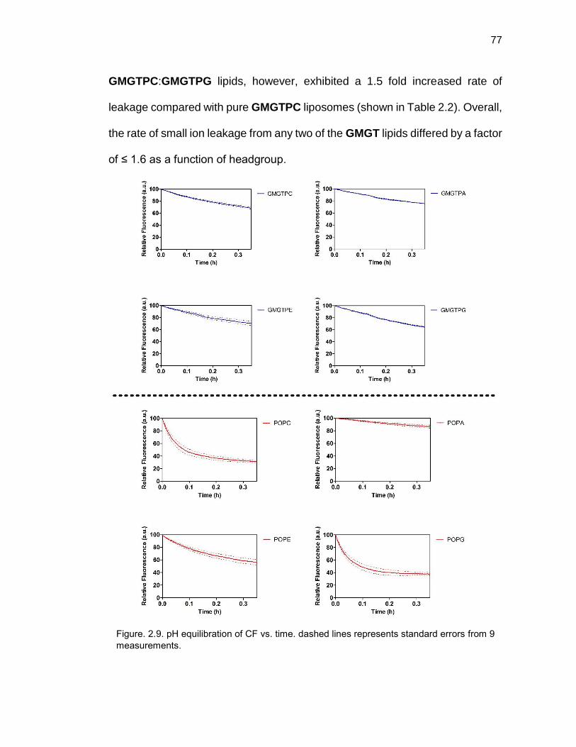
77
GMGTPC:GMGTPG lipids, however, exhibited a 1.5 fold increased rate of
leakage compared with pure GMGTPC liposomes (shown in Table 2.2). Overall,
the rate of small ion leakage from any two of the GMGT lipids differed by a factor
of ≤ 1.6 as a function of headgroup.
Figure. 2.9. pH equilibration of CF vs. time. dashed lines represents standard errors from 9
measurements.

78
We next examined how these results compared with the effect of
headgroups on standard bilayer-forming diacylphospholipids, specifically, 1-
palmitoyl-2-oleoyl-sn-glycerol (PO) lipids. To be able to compare membrane
leakage results obtained from synthetic GMGT lipid analogs, we generated
liposomes by mixing POPC lipids 1:1 with POPG, POPE, or POPA (i.e., the
same four polar lipid headgroups examined for synthetic GMGT lipids) and
compared their observed rate of leakage to liposomes comprised of pure POPC
lipids. DSC measurements showed that liposomes comprised of 1:1 mixtures of
POPC with POPA, POPE, or POPG maintained a liquid phase at room
temperature and did not exhibit a phase transition between 5 and 65 °C (Figure
2.5). DLS measurement also confirmed that lipid fusion or aggregation is not
expected to significantly contribute to the observed rate of membrane leakage
(Figure 2.4).
When membrane leakage for liposomes made with PO series of lipids
were monitored, a more dramatic effect of head groups on membrane leakage
was observed compared with membrane leakage of liposomes made with
synthetic tetraether lipid analogs. We observed significant differences in
observed rates of leakage in respect to changes in polar lipid headgroups on
liposomes made with PO lipid analogs (Figure 2.10). Similar leakage trend to
GMGTPG lipids was observed with the addition of PG headgroup to diacyl lipids,
showing an increase in membrane leakage of small ions by a factor of 2.5
compared with liposomal membrane leakage from PO lipid analogs with PC

79
headgroups only. In contrast to the case with GMGTPE or GMGTPA (which did
not affect membrane leakage compared to GMGTPC lipids), the rate of leakage
from POPE and POPA-containing membranes was a factor of 2.0 and 14.3
slower, respectively, compared with membranes formed from pure POPC lipids
(shown in Figure 2.10). To illustrate the difference clearer, we normalize the
effect of changes in polar lipid headgroups with the PC lipid on membrane
leakage (shown in Figure 2.11), where we can see a greater dependence for
liposomes made with PO lipid series than liposomes made with GMGT lipids on
membrane leakage.
Lipids Observed Rate (h-1)
Std. Error of the Mean
GMGTPC 1.34 0.07 GMGTPC/PE 1.49 0.01 GMGTPC/PA 1.24 0.06 GMGTPC/PG 1.97 0.13 POPC 6.53 0.46 POPC/PE 3.37 0.31 POPC/PA 0.50 0.05 POPC/PG 16.21 1.14
Table 2.2. Observed rate calculated for small ion permeability of liposomes using method of
initial rates (15% completion; the observed rate was calculated by taking the mean of 9
kinetic rates per experiment. However, the observed rate of GMGTPE was calculated using
8 measured kinetic rates.1 experiment was determined to be an outlier using Q-test 95%
confidence, and was omitted from the analysis).

80
The reduced permeation of small ions observed for liposomes containing
POPC mixed with POPA or POPE (compared with pure POPC liposomes) could
arise from increased intermolecular hydrogen bonding between headgroups of
neighboring lipids. Such intermolecular hydrogen bonding may lead to exclusion
of water molecules near the membrane surface and increased membrane
packing, as suggested through X-ray diffraction38, FT-IR39 and computation40
studies. For GMGT lipid analogs, we have shown leakage of small ions from
tethered lipids was significantly reduced compared with bilayer-forming lipids,41
presumably as a result of favorable lipid packing of hydrocarbon chains in
neighboring lipids within the membrane. Such inherently tight membrane
packing in GMGT lipid analogs, therefore, may not be as influenced by
Figure 2.10. Observed initial rate of pH equilibration from liposomes formed from GMGT or PO series of lipids. Comparison of the observed initial rates of decreased CF fluorescence intensity (monitored at λ(Ex/Em) = 485/517 nm) from CF-encapsulated liposomes comprised of pure GMGTPC lipid or 1:1 mixtures of GMGTPC with GMGTPG, GMGTPE, or GMGTPA (the inset represents a zoomed in graph of the observed initial rates of leakage from the GMGT lipids); and pure POPC lipid or 1:1 mixtures of POPC with POPG, POPE, or POPA. Statistical significance was determined using a paired Student t-test. *, **, *** indicate a p-value of < 0.1, 0.01, 0.001, respectively, relative to the kobsd of the analogous PC lipid.

81
membrane surface effects induced by the presence of PE or PA headgroups.
On the other hand, for lipids with PG headgroups, the presence of multiple
hydroxyl groups may lead to an increase in the number of water molecules in
between lipid headgroups near the membrane surface, which could cause a
decrease in lipid packing and an increase in membrane leakage.36 We expect
such an effect on leakage by PG headgroups would be more pronounced in PO
lipids (which presumably are inherently more loosely packed) compared with
GMGT lipid analogs as suggested by membrane permeability results observed
in Figure 2.10 or 2.11.
Figure 2.11. Relative effects of headgroups on small ion membrane leakage. A) Graph of
the relative variation of leakage rate from membranes comprised of 1:1 mixtures of PC with
PA, PE or PG lipids compared to membranes formed from pure PC lipids. Data represents
the percent deviation of observed initial rates of membrane leakage compared to the
observed initial rate of leakage from pure POPC or GMGTPC lipid membranes (zero
percent). B) Graph showing the relative leakage rate of membranes comprised of 1:1
mixtures of PC with PA, PE, or PG lipids relative to the observed initial rate of leakage from
membranes comprised of pure POPC or pure GMGTPC lipids (normalized to 1).

82
2.4 Conclusion
We have, thus, presented a systematic study of the effects on membrane
leakage of some key structural features inspired from natural Archaea lipids. As
anticipated, the incorporation of phytanyl groups, the presence of tethering, and
the incorporation of ether glycerol backbones in tetraether lipids substantially
reduced membrane permeability of small ions , ~2 orders of magnitude, when
compared with commercially available EggPC lipids. Surprisingly, however, we
found that incorporation of cis-1,3 substituted cyclopentane rings had no
significant effect on membrane leakage of small ions in GMGTPC lipids. In
contrast, incorporation of a 1 or 2 cis-1,3 cyclohexane ring significantly reduced
small ion membrane leakage when compared with all other GMGTPC lipids
studied, albeit, a cumulative leakage benefit of increasing the number of
cyclohexane rings was not observed. Such differences in small ion membrane
permeability between lipids containing cyclopentane versus cyclohexane rings
could reflect differences in ring flexibility for cyclohexanes, as it relates to lipid
packing.
When we focus our attention to the effects of changes in polar lipid
headgroup on tetraether lipid analogs or diacyl lipids, we observed changes in
polar lipid headgroups for liposomes containing tetraether lipids did not display
a strong dependence on membrane leakage. Whereas, changes in polar lipid
headgroups for liposomes made with diacyl PO lipids exhibited a strong effect
on membrane leakage. Such large effects on small ion permeability exhibited

83
for diacyl lipids from headgroups modification may limit the utility of PO (and
possibly other bilayer-forming) lipids in applications and studies where
maintaining a consistent membrane permeability is important across various
headgroups. On the other hand, membrane leakage from liposomes comprised
of synthetic GMGT lipids was not heavily influenced by changes in polar lipid
headgroups. These findings suggest that GMGT lipids may offer greater
flexibility for tailoring the functionality presented in the lipid headgroups, without
significantly compromising membrane permeability. Such versatility in the
design of lipid headgroups may open up opportunities to use the cyclohexane
integrated GMGT lipids in place of current diacyl lipid based technologies in a
range of applications including the incorporation of receptor targeting molecules
on lipids for development of liposomal drug delivery systems or the
incorporation of ligands as well as charged headgroups to attract specific
analytes or binding partners to the membrane surface for biophysical and drug
delivery studies. This work represents an important step towards establishing
some design principles inspired from nature for generating lipids with low
membrane permeability for drug delivery applications.
2.5 Materials and Methods
2.5.1 Reagents and Instruments
All reagents were purchased from commercial sources and used without
further purification. Diacyl lipids were purchased from Avanti Polar Lipids. Diacyl

84
lipids were stored under Argon at -20°C and used within 3 months of purchase.
Glassware was dried at 115°C overnight. Air and moisture-sensitive reagents
were transferred using a syringe or stainless steel cannula. Intermediates were
purified over silica (60Å, particle size 40-63 m) purchased from Dynamic
Adsorbents, Inc. Reactions were monitored by thin-layer chromatography (TLC)
using 0.25 mm silica gel plates (60F-254) from Dynamic Adsorbents, Inc.
Deuterated solvents were purchased from Cambridge Isotope Laboratories, Inc.
1H, 13C, 31P NMR spectra were obtained on either JEOL ECA 500 spectrometer
or Varian 400 MHz/500MHz spectrometer. Chemical shifts are reported in ppm
relative to residual solvent. The FID files were analyzed using NMRnotebook
version 2.70 build 0.10 by NMRTEC.
Dynamic Light Scattering (DLS) measurements were performed on a
Wyatt DynaPro NanoStar (Wyatt Technology, Santa Barbara, CA) instrument
using a disposable cuvette (Eppendorf UVette 220 nm – 1,600 nm) and data
processed using Wyatt DYNAMICS V7 software. Each analysis involved an
average of 10 measurements. The data was exported for final plotting using
GraphPad Prism 5 (GraphPad Software, Inc., La Jolla, CA).
Low resolution MS analysis was performed on a Micromass Quattro
Ultima triple quadrupole mass spectrometer with an electrospray ionization
(ESI) source. High resolution MS analysis was performed using Agilent 6230
Accurate-Mass TOFMS with an electrospray ionization (ESI) source by
Molecular Mass Spectrometry Facility (MMSF) in the department of chemistry
and biochemistry at University of California, San Diego.

85
Stopped-Flow fluorescence measurements were taken using Applied
Photophysics SX-17MV stopped-flow. The slit was set at 4 nm and the PMU set
to 600. The experiments were run using asymmetrical mixing; one solution
comprised of the lipid solution in Bis-Tris buffer at pH 7.2 (0.1 mg/mL) and the
other solution with either Bis-Tris buffer pH 7.2 / pH 5.8. The kinetic was
measured when 25 µL of the liposome solution was mixed with 225 µl of Bis-
Tris buffer pH7.2 / pH5.8 (0.01 mg/mL final liposome concentration) for 1000
seconds. The excitation was set at 485 nm and the fluorescence data was
collected using a high pass cut-off filter at 505 nm. The data was exported for
final plotting using GraphPad Prism 5 (GraphPad Software, Inc., La Jolla, CA).
Longer fluorescence decay measurements were taken on a Perkin Elmer
Enspire© multimode plate reader (excitation 485 nm, emission 516 nm and 75
flashes; initially, each measurement was taken every second for 500 seconds,
followed by every 60 seconds for 6 hours). Costar EIA/RIA plates were used
(96 well half area, no lid, flat bottom, non-treated black polystyrene). The data
were exported for final plotting using GraphPad Prism 5 (GraphPad Software,
Inc., La Jolla, CA).
DSC experiments were performed in duplicate using a Thermal Analysis
Q2000 DSC. Each experiment involved a 5 °C/min ramp from 0 °C to 70 °C
under high purity N2 at 50 mL/min. Samples were ~0.3 – 1.0 mg of lipid dissolved
in water at ~5% by weight. TA Universal Analysis was used to extract Tm for
these samples. All of the synthetic lipids did not exhibit a phase transition from

86
0 - 70 °C shown in Figure S7. DMPC was used as a positive control which
showed a phase transition at 24 °C.
2.5.2 General Synthetic Procedures
2.5.2.1 Alcohol Oxidation Using Albright–Onodera Conditions
To a cold solution of the starting alcohol (1 eq) in a mixture of dry
dichloromethane (DCM)/dimethyl sulfoxide (DMSO) (1:1) (0.15 M), phosphorus
pentoxide (3 eq) was slowly added and reaction was stirred at room temperature
for 30 minutes. Then, the reaction mixture was cooled using an ice-water bath
and triethylamine (Et3N) (10 eq) was slowly added. After 30 min of stirring at
room temperature, the reaction mixture was cooled down again and 10%
aqueous HCl was added. The resulting mixture was extracted with DCM,
washed with water, dried over Na2SO4 and purified by column chromatography
on silica gel.
Note: When a diol was used, the number of equivalents for P2O5 and Et3N were
6 and 20 respectively.
2.5.2.2 Wittig Olefination
To a suspension of phosphonium salt (1 eq) in dry THF (0.07 M),
butyllithium (1.2 eq) was added dropwise at 0°C. The reaction mixture was
stirred for 15 min at 0°C, and the reactive aldehyde (1.1 eq) dissolved in dry
tetrahydrofuran (THF) (0.09 M) was added dropwise. After 30 minutes of stirring

87
at room temperature, the reaction was quenched with water and solvent was
removed under vacuum. The aqueous residue was extracted with DCM,
washed successively with water and brine, dried over Na2SO4 and purified by
column chromatography on silica gel.
Note: For double Wittig reactions, the amount of butyllithium was increased to
2.2 equivalents.
2.5.2.3 Bromination of Diol Using Hydrobromic Acid Solution
To a suspension of diol (0.02 M, 1 eq) in a solution of hydrobromic acid
(48 wt. % in H2O), tetrabutylammonium bromide (0.5 eq) was added and the
reaction mixture was stirred at reflux for 24 hours. The solution was cooled down
and extracted with DCM. The combined organic layers were washed with water
until the aqueous layer remained at neutral pH, and dried over Na2SO4. Unless
noted otherwise, the desired product was obtained after purification by column
chromatography on silica gel using hexane/Ethyl Acetate (EtOAc) (99:1) as the
eluent.
2.5.2.4 Formation of Tetraether Lipid Scaffold by SN2 Reaction
To a cold solution of 2.4 (0.21 M, 2.5 eq) in dry THF, sodium hydride (2.7
eq) was added portionwise. The solution was stirred for 1 hour at room
temperature and cooled down again. A solution of dibromoalkane (0.09 M, 1 eq)
in dry THF was added and the reaction was stirred at reflux for 16 hours. The
reaction was quenched with water and the solvent was removed under vacuum.

88
The aqueous residue was extracted with DCM, washed successively with water
and brine, then dried over Na2SO4 and purified by column chromatography on
silica gel.
2.5.2.5 Debenzylation of Lipid Scaffold by Hydrogenation
Benzylated lipid (1 eq) was dissolved in a degassed mixture of ethanol
(EtOH)/THF (1:1) (0.01 M) and 20% Pd(OH)2 (10% w/w) was added. The
reaction was stirred under 1 atm H2 at room temperature for 16 hours. The
catalyst was removed by filtration through a pad of celite, and the resulting
residue was purified by column chromatography on silica gel.
2.5.2.6 Formation of Phosphocholine Lipid
First, bromoethyldichlorophosphate was prepared following a reported
protocol.42 To a solution of bromoethyldichlorophosphate (8 eq) in dry THF (0.33
M), a solution of the diol (1 eq) and Et3N (11 eq) in dry THF (0.04 M) was added
dropwise. After stirring the mixture for 3 days in the dark at room temperature,
toluene was added to precipitate triethylammonium chloride. Then, the solution
was filtered through a small pad of celite and the filtrate concentrated. The
resulting residue was dissolved in a mixture of THF/NaHCO3 (sat) (2.8 mM) and
the reaction was stirred for 16 hours at room temperature. THF was evaporated
under vacuum and the resulting aqueous solution was acidified to pH 1 using a
dilution solution of hydrochloric acid (1M) and extracted using several portions

89
of DCM/Methanol (MeOH) (8:2). The organic layers were combined, dried over
Na2SO4 and concentrated under reduced pressure.
To a solution of the previous crude in a mixture of THF/chloroform
(CHCl3) (2:1) (0.03 M), Me3N (33% in EtOH) (180 eq) was added and the
reaction was stirred in a sealed tube at room temperature for 5 days. The
reaction mixture was concentrated to dryness, purified on sephadex LH-20
using DCM/MeOH (1:1) as eluent and purified by column chromatography on
silica gel.
2.5.3 Synthetic Procedure for Glycerol Scaffold
(E)-1-iodo-3,7,11,15-tetramethylhexadec-2-ene (2.1)
Compound 2.1 was synthesized following a reported protocol.43
(E)-2-phenyl-5-((3,7,11,15-tetramethylhexadec-2-en-1-yl)oxy)-1,3-dioxane
(2.2)
Compound 2.2 was synthesized following a reported protocol.43
2-phenyl-5-((3,7,11,15-tetramethylhexadecyl)oxy)-1,3-dioxane (2.3)

90
2.2 (0.50 g, 1.10 mmol) was dissolved in a degasiised solution of tert-butanol
(tBuOH)/THF (1:1) (10 mL) and Wilkinson’s catalyst (Rh(PPh3)3Cl) (0.02 g, 4%
w/w) was added. The mixture was stirred under 1 atm H2, at room temperature
for 24 hours, and filtered through a small pad of alumina. The solvent was
removed under vacuum and the crude product was purified by column
chromatography on silica gel using hexane/EtOAc (95:5) as the eluent.
Compound 2.3 was obtained as a yellow oil (0.43 g, 85%) and 1H NMR
spectrum matched previously reported data.43
3-(benzyloxy)-2-((3,7,11,15-tetramethylhexadecyl)oxy)propan-1-ol (2.4)
Compound 2.4 was synthesized following a reported protocol.43

91
2.5.4 Synthetic Procedure for GMGTPC T28, T32, T34
1,20-eicosanediol (2.5)
1,20-eicosanediol was synthesized from 1,20-eicosanedioic acid following a
reported protocol.44
1,12-dodecanedial (2.6)
Dialdehyde 2.6 was synthesized from 1,12-dodecandiol (3.50 g, 17.3 mmol)
according to the general procedure for Alcohol oxidation (Albright–Onodera
conditions) (see section 2.5.2.1). 2.6 (2.06 g, 60%) was obtained as a white
solid after purification by column chromatography on silica gel using
hexane/EtOAc (95:5 to 90:10) as the eluent. 1H NMR data matched previously
reported data.45
1,16-hexadecanedial (2.7)
Dialdehyde 2.7 was synthesized from 1,16-hexadecanediol (342 mg, 1.32
mmol) according to the general procedure for Alcohol oxidation (Albright–
Onodera conditions) (see section 2.5.2.1). 1,16-hexadecanedial (249 mg, 74%)
was obtained as a white solid after purification by column chromatography on
O O10
O O14

92
silica gel using hexane/EtOAc (9:1 to 8:2) as the eluent. 1H NMR data matched
previously reported data.46
1,20-icosanedial (2.8)
Dialdehyde 2.8 was synthesized from 1,20-eicosanediol (2.65 g, 8.44 mmol)
according to the general procedure for Alcohol oxidation (Albright–Onodera
conditions) (see section 2.5.2.1). 1,20-icosanedial (1.07 g, 41%) was obtained
as a white solid after purification by column chromatography on silica gel using
hexane/EtOAc (95:5 to 90:10) as the eluent.
Rf: 0.40 (hexane/EtOAc 9:1); 1H NMR (500 MHz, CDCl3-d1) δ 9.72 (t, J = 1.9
Hz, 2H), 2.38 (ddd, J = 1.9, 7.2, 7.5 Hz, 4H), 1.61-1.56 (m, 4H), 1.29-1.21 (m,
28H); 13C NMR (126 MHz, CDCl3-d1) δ 203.1, 44.1, 29.8, 29.8, 29.6, 29.5, 29.3;
ESI-MS: 311.3 [M+H]+; HRMS 311.2945 calcd for [C20H39O2]+, found 311.2945.
8-((tetrahydro-2H-pyran-2-yl)oxy)octan-1-ol (2.9)
Compound 2.9 was synthesized following a reported protocol.47
HO OTHP6
O O18

93
triphenyl(8-((tetrahydro-2H-pyran-2-yl)oxy)octyl)phosphonium iodide (2.10)
To a cold solution of triphenylphosphine (6.1 g, 23.5 mmol) and imidazole (1.6
g, 23.5 mmol) in DCM (45 mL), iodine (6.0 g, 23.5 mmol) was added. After
stirring of the mixture at room temperature in the dark for 10 min, a solution of
compound 2.9 (4.5 g, 19.6 mmol) in DCM (12 mL) was added. The reaction
mixture was stirred for 2 hours at room temperature in the dark. The reaction
mixture was washed with 10% Na2S2O3 aqueous solution and extracted with
DCM. The combined organic layers were washed with water, dried over Na2SO4,
and purified by silica gel chromatography using hexane/EtOAc (8:2) as eluent.
The resulting oil was carried out to the next step without further purification and
characterization. To a solution of the purified crude in acetonitrile (ACN) (100
mL), potassium carbonate (3.2 g, 23.5 mmol) and triphenylphosphine (6.0 g,
23.5 mmol) were added. After stirring at reflux for two days, the reaction mixture
was cooled down, evaporated and purified by column chromatography on silica
gel using DCM/MeOH (100:0 to 95:5) as the eluent. Phosphonium salt 2.10 (9.1
g, 77%) was obtained as a white fluffy solid.
Rf: 0.78 (DCM/MeOH 95:5); 1H NMR (500 MHz, CDCl3-d1) δ 7.76-7.63 (m, 15H),
4.45 (dd, J = 3.2, 4.4 Hz, 1H), 3.76 (ddd, J = 3.2, 7.4, 11.3 Hz, 1H), 3.60 (td, J
= 6.9, 9.6 Hz, 1H), 3.54-3.48 (m, 2H), 3.42-3.38 (m, 1H), 3.26 (td, J = 6.9, 9.6
Hz, 1H), 1.74-1.40 (m, 12H), 1.23-1.15 (m, 6H); 13C NMR (126 MHz, CDCl3-d1)
δ 135.2 (d, JC-P= 3.0 Hz), 133.7 (d, JC-P = 10.0 Hz), 130.7 (d, JC-P = 12.5 Hz),
118.1 (d, JC-P= 86.0 Hz), 99.0, 67.6, 62.6, 30.8, 30.5, 30.3, 29.6, 29.1, 29.0,

94
26.1, 25.5, 23.3, 22.9, 22.6, 22.5, 19.8; ESI-MS: 475.3 [M+Na]+; HRMS
475.2760 calcd for [C31H40O2P]+, found 475.2757.
(8E,20E)-1,28-bis((tetrahydro-2H-pyran-2-yl)oxy)octacosa-8,20-diene (2.11)
Compound 2.11 was synthesized by reaction of 2.6 (4.44 g, 7.37 mmol) and
2.10 (0.66 g, 3.4 mmol) according to the general procedure for Wittig olefination
(see section 2.5.2.2). Olefin 2.11 (1.27 g, 61%) was obtained as a colorless oil
after purification by column chromatography on silica gel using hexane/EtOAc
(97:3 to 95:5) as the eluent.
Rf: 0.29 (hexane/EtOAc 97:3); 1H NMR (500 MHz, CDCl3-d1) δ 5.34-5.26 (m,
4H), 4.53 (dd, J = 2.8, 4.4 Hz, 2H), 3.84-3.80 (m, 2H), 3.71-3.66 (m, 2H), 3.47-
3.43 (m, 2H), 3.35-3.31 (m, 2H), 1.98-1.45 (m, 25H), 1.27-1.22 (m, 33H); 13C
NMR (126 MHz, CDCl3-d1) δ 130.5, 130.4, 130.1, 129.9, 99.0, 67.8, 62.4, 32.8,
32.7, 29.9, 29.9, 29.9, 29.8, 29.7, 29.7, 29.6, 29.5, 29.4, 29.3, 29.2, 27.2, 27.3,
26.4, 25.7, 19.8; ESI-MS: 613.6 [M+Na]+; HRMS 613.5166 calcd for
[C38H70O4Na]+, found 613.5167.
THPO OTHP10 77

95
(8E,24E)-1,32-bis((tetrahydro-2H-pyran-2-yl)oxy)dotriaconta-8,24-diene (2.12)
Compound 2.12 was synthesized by reaction of 2.7 (3.33 g, 5.53 mmol) and
2.10 (0.64 g, 3.40 mmol) according to the general procedure for Wittig
olefination (see section 2.5.2.2). Olefin 2.12 (1.30 g, 80%) was obtained as a
colorless oil after purification by column chromatography on silica gel using
hexane/EtOAc (97:3 to 95:5) as eluent.
Rf: 0.41 (hexane/EtOAc 95:5); 1H NMR (500 MHz, CDCl3-d1) δ 5.35-5.26 (m,
4H), 4.54 (dd, J = 2.8, 4.4 Hz, 2H), 3.86-3.80 (m, 2H), 3.72-3.66 (m, 2H), 3.49-
3.43 (m, 2H), 3.37-3.31 (m, 2H), 1.99-1.92 (m, 8H), 1.83-1.65 (m, 4H), 1.57-
1.46 (m, 12H), 1.33-1.22 (m, 40H); 13C NMR (126 MHz, CDCl3-d1) δ 130.6,
130.4, 130.1, 129.9, 99.0, 67.8, 62.5, 32.8, 32.7, 31.0, 29.9, 29.9, 29.7, 29.6,
29.5, 29.4, 29.4, 29.3, 27.4, 26.4, 25.7, 19.9.
(8E,28E)-1,36-bis((tetrahydro-2H-pyran-2-yl)oxy)hexatriaconta-8,28-diene
(2.13)
Compound 2.13 was synthesized by reaction of 2.8 (3.71 g, 6.16 mmol) and
2.10 (0.87 g, 2.80 mmol) according to the general procedure for Wittig
olefination (see section 2.5.2.2). Olefin 2.13 (1.14 g, 58%) was obtained as a
colorless oil after purification by column chromatography on silica gel using
hexane/EtOAc (98:2 to 95:5) as eluent.

96
Rf: 0.47 (hexane/EtOAc 95:5); 1H NMR (500 MHz, CDCl3-d1) δ 5.35-5.27 (m,
4H), 4.54 (dd, J = 2.9, 4.4 Hz, 2H), 3.86-3.81 (m, 2H), 3.72-3.67 (m, 2H), 3.48
(m, 2H), 3.36-3.32 (m, 2H), 1.99-1.92 (m, 8H), 1.83-1.65 (m, 4H), 1.58-1.46 (m,
12H), 1.32-1.22 (m, 48H); 13C NMR (126 MHz, CDCl3-d1) δ 130.6, 130.4, 130.1,
130.0, 99.0, 67.8, 62.5, 32.8, 32,7, 31.0, 30.0, 29.9, 29.8, 29.8, 29.7, 29.6, 29.5,
29.4, 29.4, 29.3, 27.4, 27.4, 26.4, 25.7, 19.9.
1,28-bis((tetrahydro-2H-pyran-2-yl)oxy)octacosane (2.14)
2.11 (1.27 g, 2.15 mmol) was dissolved in a degassed mixture of
EtOH/EtOAc/pyridine (9:1:0.02) (55 mL) and 10% Pd/C (0.65 g, 50% w/w) was
added. The reaction was stirred under 1 atm H2 at room temperature for 16
hours. The catalyst was removed by filtration on celite after the solvent was
heated to 40°C, and evaporation of the filtrate gave 2.14 (1.24 g, 97%) as a
white solid without further purification.
1H NMR (400 MHz, CDCl3-d1) δ 4.55 (dd, J = 2.8, 4.4 Hz, 2H), 3.87-3.83 (m,
2H), 3.70 (td, J = 6.9, 9.6 Hz, 2H), 3.49-3.44 (m, 2H), 3.35 (td, J = 6.9, 9.6 Hz,
2H), 1.84-1.76 (m, 2H), 1.72-1.66 (m, 2H), 1.58-1.46 (12H), 1.33-1.16 (m, 48H);
13C NMR (100 MHz, CDCl3-d1) δ 99.0, 67.9, 62.5, 31.0, 29.9, 29.8, 29.7, 26.4,
25.7, 19.9; ESI-MS: 617.5 [M+Na]+; HRMS 617.5479 calcd for [C38H74O4Na]+,
found 617.5480.

97
1,32-bis((tetrahydro-2H-pyran-2-yl)oxy)dotriacontane (2.15)
Compound 2.12 (1.30 g, 2.0 mmol) was dissolved in a degassed mixture of
EtOH/EtOAc/pyridine (9:1:0.02) (55 mL) and 10% Pd/C (0.65 g, 50% w/w) was
added. The reaction was stirred under 1 atm H2 at room temperature for 16
hours. The catalyst was removed by filtration on celite after the solvent was
heated to 40°C, and evaporation of the filtrate gave 2.15 (1.27 g, 98%) as a
white solid which was used in the next step without further purification and
characterization.
1,36-bis((tetrahydro-2H-pyran-2-yl)oxy)hexatriacontane (2.16)
Compound 2.13 (1.10 g, 1.56 mmol) was dissolved in a degassed mixture of
EtOH/EtOAc/pyridine (9:1:0.02) (55 mL) and 10% Pd/C (0.55 g, 50% w/w) was
added. The reaction was stirred under 1 atm H2 at room temperature for 16
hours. The catalyst was removed by filtration on celite after the solvent was
heated to 40°C, and evaporation of the filtrate gave 2.16 (0.96 g, 87%) as a
white solid which was used in the next step without further purification and
characterization.

98
1,28-dibromooctacosane (2.17)
1,28-dibromooctacosane was synthesized following a reported protocol for
conversion of tetrahydropyranylated alcohols to their corresponding bromides.48
Carbon tetrabromide (1.80 g, 5.44 mmol) was added to a solution of 2.14 (1.08
g, 1.81 mmol) in dry DCM (30 mL). After stirring for 10 min, the solution was
cooled down and triphenylphosphine (2.85 g, 10.88 mmol) was added. The
mixture was stirred 24 hours at room temperature and purified by column
chromatography on silica gel using DCM/hexane (1:1) as the eluent. Compound
2.17 (0.76 g, 76%) was obtained as a white solid.
Rf: 0.90 (DCM/hexane 1:1); 1H NMR (500 MHz, CDCl3-d1) δ 3.39 (t, J = 6.9 Hz,
4H), 1.86-1.80 (m, 4H), 1.42-1.37 (m, 4H), 1.29-1.23 (m, 44H); 13C NMR (126
MHz, CDCl3-d1) δ 34.3, 33.0, 29.9, 29.8, 29.8, 29.7, 29.0, 28.4.
1,32-dibromodotriacontane (2.18)
1,32-dibromodotriacontane was synthesized from protected diol 2.15 (1.27 g,
1.95 mmol) according to the general procedure for conversion of
tetrahydropyranylated alcohols to their corresponding bromides (see section
2.5.2.3). Dibromoalkane 2.18 (0.95 g, 80%) was obtained as a white solid after
purification by column chromatography on silica gel using hexane/DCM (1:1) as
the eluent.

99
Rf: 0.90 (DCM/hexane 1:1); 1H NMR (500 MHz, CDCl3-d1) δ 3.38 (t, J = 6.9 Hz,
4H), 1.86-1.80 (m, 4H), 1.40-1.24 (m, 56H); 13C NMR (126 MHz, CDCl3-d1) δ
34.1, 33.1, 29.9, 29.8, 29.8, 29.7, 29.0, 28.4.
1,36-dibromohexatriacontane (2.19)
1,36-dibromohexatriacontane was synthesized from protected diol 2.16 (0.96 g,
1.36 mmol) according to the general procedure for conversion of
tetrahydropyranylated alcohols to their corresponding bromides (see section
2.5.2.3). Dibromoalkane 2.19 (0.56 g, 61%) was obtained as a white solid after
purification by column chromatography on silica gel using hexane/DCM (1:1) as
the eluent.
Rf: 0.95 (DCM/hexane 1:1); 1H NMR (500 MHz, CDCl3-d1) δ 3.38 (t, J = 6.9 Hz,
4H), 1.86-1.81 (m, 4H), 1.50-1.09 (m, 64H); 13C NMR (126 MHz, CDCl3-d1) δ
34.2, 33.1, 29.9, 29.8, 29.8, 29.7, 29.0, 28.4.
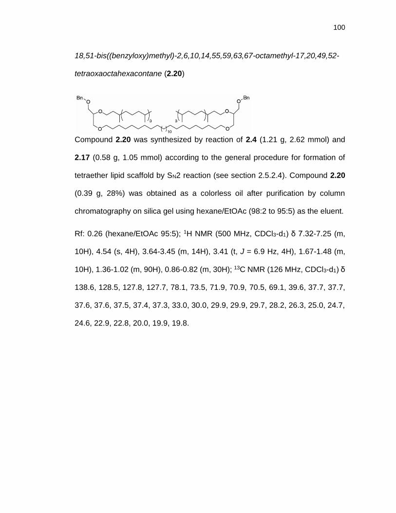
100
18,51-bis((benzyloxy)methyl)-2,6,10,14,55,59,63,67-octamethyl-17,20,49,52-
tetraoxaoctahexacontane (2.20)
Compound 2.20 was synthesized by reaction of 2.4 (1.21 g, 2.62 mmol) and
2.17 (0.58 g, 1.05 mmol) according to the general procedure for formation of
tetraether lipid scaffold by SN2 reaction (see section 2.5.2.4). Compound 2.20
(0.39 g, 28%) was obtained as a colorless oil after purification by column
chromatography on silica gel using hexane/EtOAc (98:2 to 95:5) as the eluent.
Rf: 0.26 (hexane/EtOAc 95:5); 1H NMR (500 MHz, CDCl3-d1) δ 7.32-7.25 (m,
10H), 4.54 (s, 4H), 3.64-3.45 (m, 14H), 3.41 (t, J = 6.9 Hz, 4H), 1.67-1.48 (m,
10H), 1.36-1.02 (m, 90H), 0.86-0.82 (m, 30H); 13C NMR (126 MHz, CDCl3-d1) δ
138.6, 128.5, 127.8, 127.7, 78.1, 73.5, 71.9, 70.9, 70.5, 69.1, 39.6, 37.7, 37.7,
37.6, 37.6, 37.5, 37.4, 37.3, 33.0, 30.0, 29.9, 29.9, 29.7, 28.2, 26.3, 25.0, 24.7,
24.6, 22.9, 22.8, 20.0, 19.9, 19.8.

101
18,55-bis((benzyloxy)methyl)-2,6,10,14,59,63,67,71-octamethyl-17,20,53,56-
tetraoxadoheptacontane (2.21)
Compound 2.21 was synthesized by reaction of 2.4 (1.25 g, 2.69 mmol) and
2.18 (0.66 g, 1.08 mmol) according to the general procedure for formation of
tetraether lipid scaffold by SN2 reaction (see section 2.5.2.4). Compound 2.21
(0.42 g, 30%) was obtained as a colorless oil after purification by column
chromatography on silica gel using hexane/EtOAc (98:2 to 95:5) as the eluent.
Rf: 0.37 (hexane/EtOAc 95:5); 1H NMR (500 MHz, CDCl3-d1) δ 7.32-7.24 (m,
10H), 4.54 (s, 4H), 3.62-3.40 (m, 18H), 1.62-1.48 (m, 10H), 1.36-1.04 (m, 98H),
0.86-0.82 (m, 30H); 13C NMR (126 MHz, CDCl3-d1) δ 138.6, 128.5, 127.8, 127.7,
78.2, 73.5, 71.9, 70.9, 70.5, 69.1, 39.6, 37.7, 37.7, 37.6, 37.5, 37.4, 37.3, 33.0,
29.9, 29.9, 29.7, 28.2, 26.3, 25.0, 24.7, 24.6, 22.9, 22.8, 20.0, 19.9, 19.8; ESI-
MS: 1394.3 [M+Na]+; HRMS 1394.2890 calcd for [C92H170O6Na]+, found
1394.2888.

102
18,59-bis((benzyloxy)methyl)-2,6,10,14,63,67,71,75-octamethyl-17,20,57,60-
tetraoxahexaheptacontane (2.22)
Compound 2.22 was synthesized by reaction of 2.4 (1.07 g, 2.32 mmol) and
2.19 (0.53 g, 0.79 mmol) according to the general procedure for formation of
tetraether lipid scaffold by SN2 reaction (see section 2.5.2.4). Compound 2.22
(0.33 g, 29%) was obtained as a colorless oil after purification by column
chromatography on silica gel using hexane/EtOAc (98:2 to 95:5) as the eluent.
Rf: 0.35 (hexane/EtOAc 95:5); 1H NMR (500 MHz, CDCl3-d1) δ 7.32-7.25 (m,
10H), 4.54 (s, 4H), 3.64-3.40 (m, 18H), 1.64-1.48 (m, 10H), 1.38-1.00 (m, 106H),
0.86-0.82 (m, 30H); 13C NMR (126 MHz, CDCl3-d1) δ 138.6, 128.5, 127.8, 127.7,
78.1, 73.6, 71.9, 70.9, 70.5, 69.1, 39.6, 37.7, 37.7, 37.6, 37.6, 37.5, 37.4, 37.3,
33.0, 29.9, 29.9, 29.7, 28.2, 26.3, 25.0, 24.7, 24.6, 22.9, 22.9, 20.0, 19.9, 19.8;
ESI-MS: 1450.4 [M+Na]+; HRMS 1450.3516 calcd for [C96H178O6Na]+, found
1450.3518.

103
3-((28-(3-hydroxy-2-((3,7,11,15-tetramethylhexadecyl)oxy)propoxy)octacosyl)
oxy)-2-((3,7,11,15-tetramethylhexadecyl) oxy)propan-1-ol (2.23)
Compound 2.23 was synthesized by hydrogenation of 2.20 (0.39 g, 0.30 mmol)
according to the general procedure for debenzylation of the lipid scaffold by
hydrogenation (see section 2.5.2.5). Diol 2.23 (0.23 g, 68%) was obtained as a
white solid after purification by column chromatography on silica gel using
hexane/EtOAc (9:1 to 8:2) as the eluent.
Rf: 0.52 (hexane/EtOAc 8:2); 1H NMR (500 MHz, CDCl3-d1) δ 3.71-3.39 (m,
18H), 2.16 (brs, 2H), 1.62-1.45 (m, 10H), 1.39-1.02 (m, 90H), 0.85-0.81 (m,
30H); 13C NMR (126 MHz, CDCl3-d1) δ 78.5, 72.1, 72.1, 68.9, 63.3, 39.6, 37.7,
37.7, 37.6, 37.5, 37.5, 37.3, 37.2, 33.0, 30.0, 29.9, 29.8, 29.7, 28.2, 26.3, 25.0,
24.7, 24.6, 22.9, 22.8, 20.0, 19.9,19.8; ESI-MS: 1135.8 [M+H]+.
3-((32-(3-hydroxy-2-((3,7,11,15-tetramethylhexadecyl)oxy)propoxy)
dotriacontyl)oxy)-2-((3,7,11,15-tetramethylhexadecyl)oxy)propan-1-ol (2.24)
Compound 2.24 was synthesized by hydrogenation of 2.21 (0.42 g, 0.31 mmol)
according to the general procedure for debenzylation of the lipid scaffold by

104
hydrogenation (see section 2.5.2.5). Diol 2.24 (0.28 g, 77%) was obtained as a
white solid after purification by column chromatography on silica gel using
hexane/EtOAc (9:1 to 8:2) as the eluent.
Rf: 0.70 (hexane/EtOAc 8:2); 1H NMR (500 MHz, CDCl3-d1) δ 3.71-3.39 (m,
18H), 2.19 (brs, 2H), 1.61-1.47 (m, 10H), 1.38-1.03 (m, 98H), 0.86-0.81 (m,
30H); 13C NMR (126 MHz, CDCl3-d1) δ 78.5, 72.1, 71.1, 68.8, 63.3, 39.6, 37.7,
37.7, 37.6, 37.5, 37.3, 37.2, 33.0, 30.0, 29.9, 29.8, 29.7, 28.2, 26.3, 25.0, 24.7,
24.6, 22.9, 22.8, 20.0, 19.9, 19.8.
3-((36-(3-hydroxy-2-((3,7,11,15-tetramethylhexadecyl)oxy)propoxy)hexa
triacontyl)oxy)-2-((3,7,11,15-tetramethylhexadecyl)oxy)propan-1-ol (2.25)
Compound 2.25 was synthesized by hydrogenation of 2.22 (0.33 g, 0.23 mmol)
according to the general procedure for debenzylation of the lipid scaffold by
hydrogenation (see section 2.5.2.5). Diol 2.25 (0.23 g, 81%) was obtained as a
white solid after purification by column chromatography on silica gel using
hexane/EtOAc (9:1 to 8:2) as the eluent.
Rf: 0.39 (hexane/EtOAc 8:2); 1H NMR (500 MHz, CDCl3-d1) δ 3.71-3.39 (m,
18H), 2.22 (brs, 2H), 1.63-1.46 (m, 10H), 1.38-1.00 (m, 106H), 0.86-0.81 (m,
30H); 13C NMR (126 MHz, CDCl3-d1) δ 78.5, 72.1, 71.1, 68.8, 63.3, 39.6, 37.7,

105
37.6, 37.5, 37.5, 37.3, 37.2, 33.0, 30.0, 29.9, 29.8, 29.7, 28.2, 26.3, 25.0, 24.7,
24.6, 22.9, 22.8, 20.0, 19.9, 19.8.
(octacosane-1,28-diylbis(oxy))bis(2-((3,7,11,15-tetramethylhexadecyl)oxy)
propane-3, 1-diyl) bis(2-(trimethylammonio)ethyl) bis(phosphate) (GMGTPC-
T28)
Lipid GMGTPC-T28 was synthesized from diol 2.23 (0.23 g, 0.21 mmol)
following the general procedure for formation of phosphocholine lipid (see
section 2.5.2.6). Lipid GMGTPC-T28 (0.16 g, 54%) was obtained as a white
gum after purification by column chromatography on silica gel using
DCM/MeOH/H2O (70:30:5) as the eluent.
Rf: 0.33 (DCM/MeOH/H2O 70:30:5); 1H NMR (500 MHz, MeOD-d4/CDCl3-d1
1:1) δ 4.00-3.96 (m, 4H), 3.63 (t, J = 5.5 Hz, 4H), 3.40-3.31 (m, 12H), 3.23-3.17
(m, 6H), 2.95 (s, 18H), 1.39-1.22 (m, 10H), 1.14-0.79 (m, 90H), 0.62-0.58 (30H);
13C NMR (126 MHz, MeOD-d4/CDCl3-d1 1:1) δ 77.8, 77.7, 77.7, 77.6, 71.3, 70.2,
68.6, 68.3, 66.0, 64.7, 64.6, 58.6, 58.5, 53.5, 39.0, 37.3, 37.1, 37.0, 36.8, 36.8,
36.7, 32.4, 29.5, 29.3, 29.1, 27.5, 25.6, 24.3, 24.0, 22.0, 21.9, 19.1, 19.0, 19.0,
18.9, 18.9; 31P NMR (202 MHz, MeOD-d4/CDCl3-d1 1:1) δ 0.16; ESI-MS: 1466.0
[M+H]+; HRMS calcd 1466.2615 for [C84H175N2O12P2]+, found 1466.2597.

106
(dotriacontane-1,32-diylbis(oxy))bis(2-((3,7,11,15-tetramethylhexadecyl)oxy)
propane-3,1-diyl) bis(2-(trimethylammonio)ethyl) bis(phosphate) (GMGTPC-
T32)
Lipid GMGTPC-T32 was synthesized from diol 2.24 (0.28 g, 0.23 mmol)
following the general procedure for formation of phosphocholine lipid (see
section 2.5.2.6). GMGTPC-T32 (0.19 g, 54%) was obtained as a white gum after
purification by column chromatography on silica gel using DCM/MeOH/H2O
(70:30:5) as the eluent.
Rf: 0.40 (DCM/MeOH/H2O 70:30:5); 1H NMR (500 MHz, MeOD-d4/CDCl3-d1 1:1)
δ 3.93-3.88 (m, 4H), 3.56 (t, J = 5.5 Hz, 4H), 3.32-3.09 (m, 18H), 2.88 (s, 18H),
1.27-1.16 (m, 10H), 1.04-0.71 (m, 98H), 0.55-0.50 (m, 30H); 13C NMR (126 MHz,
MeOD-d4/CDCl3-d1 1:1) δ 77.8, 77.7, 77.7, 77.6, 77.5, 71.4, 70.2, 68.7, 68.4,
66.0, 64.7, 58.6, 58.6, 53.6, 39.0, 37.3, 37.1, 37.1, 37.0, 36.9, 36.8, 36.7, 32.4,
29.5, 29.4, 29.3, 29.2, 27.6, 25.7, 24.4, 24.1, 24.0, 22.1, 22.0, 19.2, 19.1, 19.1,
19.0, 18.9; 31P NMR (202 MHz, MeOD-d4/CDCl3-d1 1:1) δ 0.16; ESI-MS: 1522.2
[M+H]+; HRMS calcd 1522.3241 for [C88H183N2O12P2]+, found 1522.3261.

107
(hexatriacontane-1,36-diylbis(oxy))bis(2-((3,7,11,15-tetramethylhexadecyl)oxy)
propane-3,1-diyl) bis(2-(trimethylammonio)ethyl) bis(phosphate) (GMGTPC-
T36)
Lipid GMGTPC-T36 was synthesized from diol 2.25 (0.23 g, 0.19 mmol)
following the general procedure for formation of phosphocholine lipid (see
section 2.5.2.6). GMGTPC-T36 (0.10 g, 36%) was obtained as a white gum after
purification by column chromatography on silica gel using DCM/MeOH/H2O
(70:30:5) as the eluent.
Rf: 0.36 (DCM/MeOH/H2O 70:30:5); 1H NMR (500 MHz, MeOD-d4/CDCl3-d1 1:1)
δ 3.95-3.90 (m, 4H), 3.56 (t, J = 5.5 Hz, 4H), 3.34-3.11 (m, 18H), 2.89 (s, 18H),
1.28-1.17 (m, 10H), 1.08-0.73 (m, 108H), 0.56-0.51 (m, 30H); 13C NMR (126
MHz, MeOD-d4/CDCl3-d1 1:1) δ 77.8, 77.7, 77.7, 77.6, 77.5, 71.4, 70.2, 68.7,
68.4, 66.0, 64.7, 58.6, 58.6, 53.6, 39.0, 37.3, 37.2, 37.1, 37.0, 36.9, 36.8, 36.7,
32.4, 29.6, 29.3, 29.2, 27.6, 25.7, 24.4, 24.1, 24.0, 22.1, 22.1, 19.2, 19.1, 19.1,
19.0, 19.0; 31P NMR (202 MHz, MeOD-d4/CDCl3-d1 1:1) δ 0.21; ESI-MS: 1578.2
[M+H]+; HRMS calcd 1578.3867 for [C92H191N2O12P2]+, found 1578.3853.

108
2.5.5 Synthetic Procedure for GMGTPC-CP1 and GMGTPC-CH1
((1R,3S)-cyclohexane-1,3-diyl)dimethanol (2.27)
To a cold suspension of LiAlH4 (10.4 g, 272.0 mmol) in dry Et2O (300 mL), a
solution of cis-1,3-cyclohexanedicarboxylic acid (10.9 g, 54.4 mmol) in dry Et2O
(230 mL) was added dropwise. After stirring at 0C for 1.5 hours, 1M HCl
solution was added slowly to quench the reaction. The reaction mixture was
extracted with EtOAc and the combined organic layer was washed with water,
brine and dried over Na2SO4. Diol 2.27 (4.4 g, 55%) was obtained as a white
solid after solvent evaporation.
1H NMR (500 MHz, CDCl3-d1) δ 3.38-3.40 (m, 4H), 2.04-2.00 (m, 3H), 1.88-1.73
(m, 4H), 1.55-1.47 (m, 2H), 1.32-1.22 (m, 1H), 0.89-0.81 (m, 2H), 0.65-0.58 (m,
1H).
(((1R,3S)-cyclopentane-1,3-diyl)bis(methylene))bis(triphenylphosphonium)
iodide (2.26)
Compound 2.26 was synthesized following a reported protocol.23
IPh3P PPh3I

109
(((1R,3S)-cyclohexane-1,3-diyl)bis(methylene))bis(triphenylphosphonium)
iodide (2.28)
To a cold solution of triphenylphosphine (17.4 g, 66.5 mmol) and imidazole (4.5
g, 66.5 mmol) in mixture of dry ACN/Et2O (1:3) (150 mL), iodine (16.8 g, 66.5
mmol) was added. After stirring of the mixture at room temperature in the dark
for 10 min, a solution of compound 2.27 (4.4 g, 30.2 mmol) in dry Et2O (30 mL)
was added. The reaction mixture was stirred for 2 hours at room temperature in
the dark. Reaction mixture was washed with 10% Na2S2O3 aqueous solution
and extracted with Et2O. The combined organic layers were washed with water,
dried over Na2SO4, and purified by silica gel chromatography using a mixture of
hexane/EtOAc (8:2) as the eluent. The resulting oil was carried out to the next
step without further purification and characterization. To a solution of the purified
crude in ACN (80 mL), triphenylphosphine (23.7 g, 90.6 mmol) was added and
the reaction mixture was stirred for 16 hours at reflux. After the reaction mixture
was allowed to cool down to room temperature, the product was precipitated by
adding toluene. Pure compound 2.28 (13.7 g, 51%) was obtained as a white
solid after recrystallization from MeOH and Et2O.
1H NMR (500 MHz, MeOD-d4) δ 7.89-7.72 (m, 30H), 3.52-3.36 (m, 4H), 1.88-
1.70 (m, 3H), 1.64-1.57 (m, 1H), 1.45-1.42 (m, 1H), 1.33-1.30 (m, 2H), 1.20-
1.12 (m, 2H), 1.03-0.94 (m, 1H); 13C NMR (126 MHz, MeOD-d4) δ 136.4, 135.0
(d, JC-P = 10.1 Hz), 131.7 (d, J C-P= 12.6 Hz), 120.5 (d, J C-P= 86.0 Hz), 34.7,
IPh3P PPh3I

110
43.6, 43.6, 34.6, 34.5, 34.1, 29.4, 29.0, 29.2; 31P NMR (202 MHz, MeOD-d4) δ
22.78; ESI-MS: 317.5 [M-2I]2+; HRMS calcd 317.1454 for [C44H44P2]2+, found
317.1452.
12-(benzyloxy)dodecan-1-ol (2.29)
Compound 2.29 was synthesized following a reported protocol.49
12-(benzyloxy)dodecanal (2.30)
To a cold solution of 2.29 (1.44 g, 4.90 mmol) in dry DCM (45 mL), Dess-Martin
Periodinane (DMP) (2.50 g, 5.89 mmol) was added portion-wise. The resulting
mixture was allowed to reach room temperature over 1 hour. After 3 more hours
of stirring at room temperature, the mixture was diluted with DCM, washed
successively with 1M NaOH solution, water and brine. The organic layer was
then dried over Na2SO4, concentrated under reduce pressure and purified by
column chromatography on silica gel using hexane/EtOAc (95:5) as the eluent.
Aldehyde 2.30 (1.25 g, 88%) was obtained as a colorless oil.
Rf: 0.47 (hexane/EtOAc 95:5); 1H NMR (400 MHz, CDCl3-d1) δ 9.74 (t, J = 1.7
Hz, 1H), 7.33-7.25 (m, 5H), 4.48 (s, 2H), 3.44 (t, J = 6.7 Hz, 2H), 2.40 (tt, J =
OBnHO
10
OBnO
10

111
1.7, 6.7 Hz, 2H), 1.62-1.55 (m, 4H), 1.35-1.25 (m, 14H); ESI-MS: 291.2 [M+H]+;
HRMS 291.2319 calcd for [C19H31O2]+, found 291.2322.
(1R,3S)-1,3-bis((E)-13-(benzyloxy)tridec-1-en-1-yl)cyclopentane (2.31)
Compound 2.31 was synthesized by reaction of 2.30 (3.84 g, 13.24 mmol) and
2.26 (4.87 g, 5.57 mmol) according to the general procedure for Wittig
olefination (see section 2.5.2.2). Olefin 2.31 (2.44 g, 68%) was obtained as a
colorless oil after purification by column chromatography on silica gel using
hexane/EtOAc (99:1) as the eluent.
Rf: 0.45 (hexane/EtOAc 99:1); 1H NMR (500 MHz, CDCl3-d1) δ 7.35-7.26 (m,
10H), 5.37-5.27 (m, 4H), 4.50 (s, 4H), 3.46 (dd, J = 6.5, 6.8 Hz, 4H), 2.82-2.76
(m, 1.5H), 2.49-2.43 (m, 0.5H), 2.04-1.77 (m, 7H), 1.64-1.58 (m, 4H), 1.36-1.26
(m, 34H), 1.06-0.96 (m, 1H); 13C NMR (126 MHz, CDCl3-d1) δ 138.9, 135.3,
135.2, 135.0, 134.9, 128.8, 128.8, 128.7, 128.5, 127.8, 127.6, 73.0, 70.7, 43.7,
42.3, 41.9, 38.4, 38.2, 33.0, 32.8, 32.7, 32.5, 32.4, 30.1, 30.0, 29.8, 29.7, 29.7,
29.5, 29.4, 27.7, 26.4; ESI-MS: 660.6 [M+NH4]+; HRMS calcd 660.5714 for
[C45H70O2NH4]+, found 660.5715.
BnO OBn
1010

112
(1R,3S)-1,3-bis((E)-13-(benzyloxy)tridec-1-en-1-yl)cyclohexane (2.32)
Compound 2.32 was synthesized by reaction of 2.30 (2.57 g, 2.89 mmol) and
2.28 (2.01 g, 6.93 mmol) according to the general procedure for Wittig
olefination (see section 2.5.2.2). Olefin 2.32 (1.00 g, 53%) was obtained as a
colorless oil after purification by column chromatography on silica gel using
hexane/EtOAc (99:1) as the eluent.
Rf: 0.40 (Hexane/EtOAc 99:1); 1H NMR (500 MHz, CDCl3-d1) δ 7.34-7.25 (m,
10H), 5.40-5.14 (m, 4H), 4.50 (s, 4H), 3.46 (t, J = 6.6 Hz, 4H), 2.35 (m, 1H),
2.06-1.92 (m, 5H), 1.78-1.52 (m, 8H), 1.38-1.26 (m, 33H), 1.02-0.81 (m, 3H);
13C NMR (126 MHz, CDCl3-d1) δ 138.9, 136.3, 136.2, 135.9, 135.8, 134.0, 133.8,
128.5, 128.5, 128.1, 127.8, 127.6, 73.0, 70.7, 40.8, 40.5, 40.1, 40.0, 36.3, 36.2,
33.1, 32.9, 32.9, 32.8, 30.2, 30.0, 29.9, 29.8, 29.7, 29.5, 29.3, 27.7, 27.6, 26.4,
26.0, 25.9; ESI-MS: 657.5 [M+H]+; HRMS calcd 679.5425 for [C46H72O2Na]+,
found 679.5424.
13,13'-((1R,3S)-cyclopentane-1,3-diyl)bis(tridecan-1-ol) (2.33)
To a degassed solution of 2.31 (2.38 g, 3.71 mmol) in EtOH (220 mL), 10%
Pd/C (0.60 g, 25% w/w) was added. The reaction was stirred under 1 atm H2 at
HO OH
1010

113
room temperature for 24 hours. The solution was then filtered through celite,
and the solvent was evaporated. Diol 2.33 (1.64 g, 95%) was obtained as a
white solid and used without further purification.
Rf: 0.26 (hexane/EtOAc 95:5); 1H NMR (500 MHz, MeOD-d4/CDCl3-d1 1:1) δ
3.58 (t, J = 7.0 Hz, 4H), 1.92-1.68 (m, 5H), 1.58-1.51 (m, 4H), 1.39-1.21 (m,
44H), 1.18-1.11 (m, 2H), 0.67-0.59 (m, 1H); 13C NMR (126 MHz, MeOD-
d4/CDCl3-d1 1:1) δ 62.4, 40.7, 40.1, 36.7, 32.5, 31.6, 29.9, 29.7, 29.6, 29.5, 28.7,
13,13'-((1R,3S)-cyclohexane-1,3-diyl)bis(tridecan-1-ol) (2.34)
To a degassed solution of olefin 2.32 (0.95 g, 1.45 mmol) in EtOH (88 mL), 10%
Pd/C (0.24 g, 25% w/w) was added. The reaction was stirred under 1 atm H2 at
room temperature for 24 hours. The solution was then filtered through celite,
and the solvent was evaporated to yield a white solid which was used in the
next step without further purification and characterization.
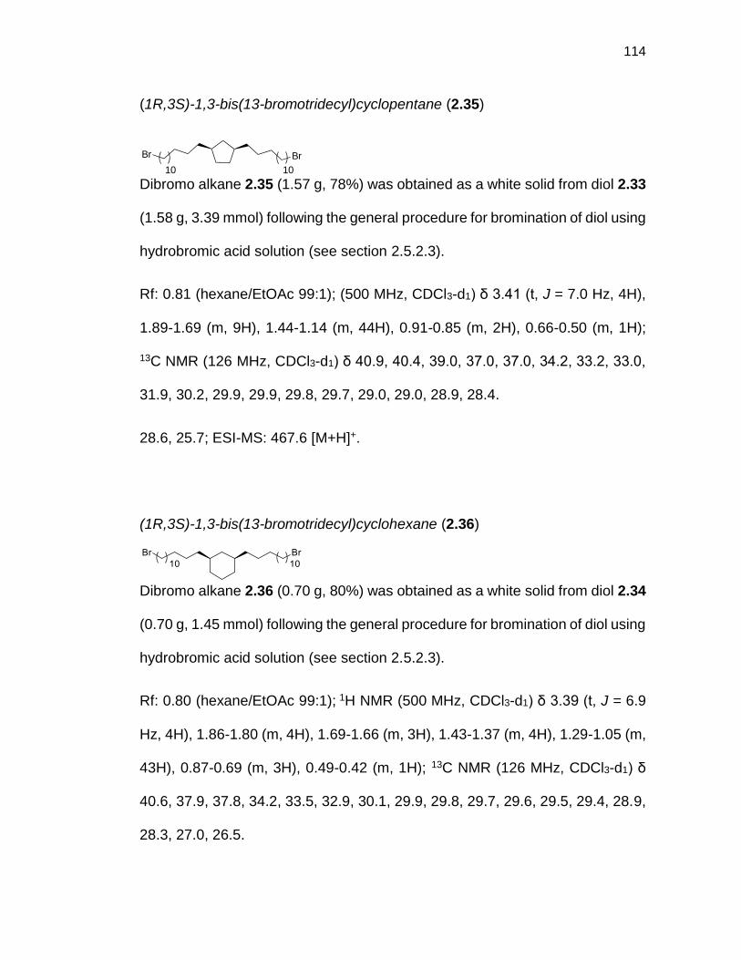
114
(1R,3S)-1,3-bis(13-bromotridecyl)cyclopentane (2.35)
Dibromo alkane 2.35 (1.57 g, 78%) was obtained as a white solid from diol 2.33
(1.58 g, 3.39 mmol) following the general procedure for bromination of diol using
hydrobromic acid solution (see section 2.5.2.3).
Rf: 0.81 (hexane/EtOAc 99:1); (500 MHz, CDCl3-d1) δ 3.41 (t, J = 7.0 Hz, 4H),
1.89-1.69 (m, 9H), 1.44-1.14 (m, 44H), 0.91-0.85 (m, 2H), 0.66-0.50 (m, 1H);
13C NMR (126 MHz, CDCl3-d1) δ 40.9, 40.4, 39.0, 37.0, 37.0, 34.2, 33.2, 33.0,
31.9, 30.2, 29.9, 29.9, 29.8, 29.7, 29.0, 29.0, 28.9, 28.4.
28.6, 25.7; ESI-MS: 467.6 [M+H]+.
(1R,3S)-1,3-bis(13-bromotridecyl)cyclohexane (2.36)
Dibromo alkane 2.36 (0.70 g, 80%) was obtained as a white solid from diol 2.34
(0.70 g, 1.45 mmol) following the general procedure for bromination of diol using
hydrobromic acid solution (see section 2.5.2.3).
Rf: 0.80 (hexane/EtOAc 99:1); 1H NMR (500 MHz, CDCl3-d1) δ 3.39 (t, J = 6.9
Hz, 4H), 1.86-1.80 (m, 4H), 1.69-1.66 (m, 3H), 1.43-1.37 (m, 4H), 1.29-1.05 (m,
43H), 0.87-0.69 (m, 3H), 0.49-0.42 (m, 1H); 13C NMR (126 MHz, CDCl3-d1) δ
40.6, 37.9, 37.8, 34.2, 33.5, 32.9, 30.1, 29.9, 29.8, 29.7, 29.6, 29.5, 29.4, 28.9,
28.3, 27.0, 26.5.
Br Br
1010

115
(1R,3S)-1,3-bis(13-(3-(benzyloxy)-2-((3,7,11,15-tetramethylhexadecyl)oxy)
propoxy)tridecyl)cyclopentane (2.37)
Compound 2.37 was synthesized by reaction of 2.4 (0.39 g, 0.84 mmol) and
2.35 (0.20 g, 0.34 mmol) according to the general procedure for formation of
tetraether lipid scaffold by SN2 reaction (see section 2.5.2.4). Compound 2.37
(0.13 g, 29%) was obtained as a colorless oil after purification by column
chromatography on silica gel using hexane/EtOAc (99:1 to 95:5) as the eluent.
Rf: 0.27 (hexane/EtOAc 95:5); 1H NMR (500 MHz, CDCl3-d1) δ 7.25-7.26 (m,
10H), 4.57 (s, 4H), 3.68-3.43 (m, 18H), 1.94-1.89 (m, 1H), 1.81-1.71 (m, 4H),
1.69-1.50 (m, 10H), 1.42-1.07 (m, 88H), 0.89-0.60 (m, 31H); 13C NMR (126 MHz,
CDCl3-d1) δ 138.6, 128.6, 128.5, 127.8, 127.8, 128.7, 100.1, 78.1, 73.5, 71.8,
70.9, 70.5, 69.0, 40.9, 40.3, 39.6, 39.0, 37.7, 37.7, 37.6, 37.6, 37.5, 37.5, 36.9,
33.0, 31.8, 30.2, 30.0, 29.9, 29.8, 29.7, 29.0, 28.1, 26.3, 25.0, 24.7, 24.6, 22.9,
22.8, 20.0, 19.9, 19.8.

116
(1R,3S)-1,3-bis(13-(3-(benzyloxy)-2-((3,7,11,15-tetramethylhexadecyl)oxy)
propoxy)tridecyl)cyclohexane (2.38)
Compound 2.38 was synthesized by reaction of 2.4 (1.53 g, 3.32 mmol) and
2.36 (0.67 g, 1.11 mmol) according to the general procedure for formation of
tetraether lipid scaffold by SN2 reaction (see section 2.5.2.4). Compound 2.38
(0.32 g, 21%) was obtained as a colorless oil after purification by column
chromatography on silica gel using hexane/EtOAc (98:2 to 95:5) as the eluent.
Rf: 0.35 (hexane/EtOAc 95:5); 1H NMR (500 MHz, CDCl3-d1) δ 7.33-7.24 (m,
10H), 4.55 (s, 4H), 3.65-3.41 (m, 18H), 1.72-1.50 (m, 14H), 1.39-1.04 (m, 89H),
0.88-0.72 (m, 32H), 0.52-0.45 (m, 1H); 13C NMR (126 MHz, CDCl3-d1) δ 138.6,
128.4, 127.7, 127.6, 78.1, 73.5, 71.8, 70.9, 70.4, 69.0, 40.7, 39.5, 38.0, 37.9,
37.8, 37.7, 37.6, 37.5, 37.4, 37.3, 37.2, 33.6, 33.0, 30.3, 29.9, 29.8, 29.7, 28.1,
27.1, 26.6, 26.3, 25.0, 24.7, 24.5, 22.9, 22.8, 20.0, 19.9, 19.8.

117
3,3'-((((1R,3S)-cyclopentane-1,3-diyl)bis(tridecane-13,1-diyl))bis(oxy))bis(2-
((3,7,11,15-tetramethylhexadecyl)oxy)propan-1-ol) (2.39)
Compound 2.39 was synthesized by hydrogen-ation of 2.37 (0.28 g, 0.28 mmol)
according to the general procedure for debenzylation of lipid scaffold by
hydrogenation (see section 2.5.2.5). Diol 2.39 (0.28 g, 85%) was obtained as a
colorless oil after purification by column chromatography on silica gel using
hexane/EtOAc (9:1 to 8:2) as the eluent.
Rf: 0.43 (hexane/EtOAc 8:2); 1H NMR (500 MHz, CDCl3-d1) δ 3.75-3.37 (m,
18H), 2.22 (t, J = 7.0 Hz, 2H), 1.91-1.48 (m, 17H), 1.43-1.01 (m, 86H), 0.88-
0.84 (m, 30H), 0.75-0.59 (m, 1H); 13C NMR (126 MHz, CDCl3-d1) δ 78.4, 72.1,
71.1, 68.9, 63.3, 40.9, 40.3, 39.6, 39.0, 37.7, 37.7, 37.6, 37.6, 37.5, 37.3, 37.3,
37.0, 33.2, 33.0, 31.8, 30.2, 30.0, 29.9, 29.8, 29.7, 29.0, 28.9, 28.2, 26.3, 25.0,
24.7, 24.6, 22.9, 22.8, 20.0, 19.9, 19.8; ESI-MS: 1175.9 [M+H]+; HRMS calcd
1176.1818 for [C77H155O6]+, found 1176.1817.

118
3,3'-((((1R,3S)-cyclohexane-1,3-diyl)bis(tridecane-13,1-diyl))bis(oxy))bis(2-
((3,7,11,15-tetramethylhexadecyl)oxy)propan-1-ol) (2.40)
Compound 2.40 was synthesized by hydrogenation of 2.38 (0.32 g, 0.23 mmol)
according to the general procedure for debenzylation of lipid scaffold by
hydrogenation (see section 2.5.2.5). Diol 2.40 (0.28 g, Qt.) was obtained as a
colorless oil after purification by column chromatography on silica gel using
hexane/EtOAc (8:2) as the eluent.
Rf: 0.48 (hexane/EtOAc 8:2); 1H NMR (500 MHz, CDCl3-d1) δ 3.71-3.39 (m,
18H), 2.11 (brs, 2H), 1.69-1.46 (m, 14H), 1.39-1.00 (m, 89H), 0.86-0.68 (m,
32H), 0.49-0.42 (m, 1H); 13C NMR (126 MHz, CDCl3-d1) δ 78.4, 72.0, 71.1, 71.0,
68.8, 63.3, 63.2, 40.7, 39.6, 38.0, 37.9, 37.8, 37.7, 37.6, 37.5, 37.4, 37.3, 37.2,
33.6, 33.0, 30.3, 30.1, 30.0, 29.9, 29.8, 29.7, 28.2, 27.1, 26.6, 26.3, 25.0, 24.7,
24.5, 22.9, 22.8, 20.0, 19.9, 19.8; ESI-MS: 1190.2 [M+H]+; HRMS calcd
1190.1975 for [C78H157O6]+, found 1190.1979.

119
((((1R,3S)-cyclopentane-1,3-diyl)bis(tridecane-13,1-diyl))bis(oxy))bis(2-
((3,7,11,15-tetramethylhexadecyl)oxy)propane-3,1-diyl) bis(2-
(trimethylammonio) ethyl) bis(phosphate) (GMGTPC-CP1)
Lipid GMGTPC-CP1 was synthesized from diol 2.39 (0.38 g, 0.32 mmol)
following the general procedure for formation of phosphocholine lipid (see
section 2.5.2.6). Lipid GMGTPC-CP1 (0.42 g, 85%) was obtained as a white
gum after purification by column chromatography on silica gel using
DCM/MeOH/H2O (70:30:5) as the eluent.
Rf: 0.32 (DCM/MeOH/H2O 70:30:5); 1H NMR (500 MHz, MeOD-d4/CDCl3-d1
1:1) δ 3.91 (m, 4H), 3.56 (t, J = 5.5 Hz, 4H), 3.33-3.10 (m, 18H), 2.89 (s, 18H),
1.75-1.15 (m, 14H), 1.05-0.72 (m, 89H), 0.55-0.51 (m, 31H), 0.33-0.26 (m, 1H);
13C NMR (126 MHz, MeOD-d4/CDCl3-d1 1:1) δ 77.8, 77.8, 77.7, 71.4, 70.2, 68.7,
68.5, 66.1, 64.7, 64.7, 58.6, 58.6, 53.6, 40.4, 39.8, 39.0, 37.3, 37.1, 37.0, 36.9,
36.8, 36.7, 36.4, 32.4, 31.2, 29.6, 29.4, 29.3, 29.3, 29.2, 28.4, 27.6, 25.8, 24.4,
24.1, 24.0, 22.1, 22.0, 19.2, 19.1, 19.1, 19.0; 31P NMR (202 MHz, MeOD-
d4/CDCl3-d1 1:1) δ 0.22; ESI-MS: 1505.9 [M+H]+; HRMS calcd 1506.2928 for
[C87H179N2O12P2]+, found 1506.2930.

120
((((1R,3S)-cyclohexane-1,3-diyl)bis(tridecane-13,1-diyl))bis(oxy))bis(2-((3,7,11,
15-tetramethylhexadecyl)oxy)propane-3,1-diyl)bis(2-(trimethylammonio)ethyl)
bis(phosphate) (GMGTPC-CH1)
Lipid GMGTPC-CH1 was synthesized from diol 2.40 (0.26 g, 0.22 mmol)
following the general pro-cedure for formation of phosphocholine lipid (section
2.5.2.6). Lipid GMGTPC-CH1 (0.25 g, 75%) was obtained as a white gum after
purification by column chromatography on silica gel using DCM/MeOH/H2O
(70:30:5) as the eluent.
Rf: 0.55 (DCM/MeOH/H2O 70:30:5); 1H NMR (500 MHz, MeOD-d4/CDCl3-d1 1:1)
δ 3.90-3.86 (m, 4H), 3.53 (t, J = 5.6 Hz, 4H), 3.32-3.20 (m, 12H), 3.13-3.06 (m,
6H), 2.04 (s, 18H), 1.37-1.32 (m, 4H), 1.25-1.12 (m, 10H), 1.02-0.69 (m, 89H),
0.52-0.48 (m, 30H), 0.43-0.35 (m, 2H), 0.15-0.08 (m, 1H); 13C NMR (126 MHz,
MeOD-d4/CDCl3-d1 1:1) δ 77.8, 77.7, 77.6, 71.3, 70.2, 68.6, 68.4, 66.0, 64.7,
64.6, 58.6, 58.5, 53.5, 40.2, 39.0, 37.5, 37.4, 37.3, 37.2, 37.1, 37.0, 36.9, 36.8,
36.7, 33.1, 32.4, 29.6, 29.5, 29.4, 29.3, 29.2, 29.1, 27.5, 26.5, 26.0, 25.7, 24.4,
24.0, 23.9, 22.1, 22.0, 19.2, 19.1, 19.0, 18.9, 18.8; 31P NMR (202 MHz, MeOD-
d4/CDCl3-d1 1:1) δ 0.15; ESI-MS: 1520.4 [M+H]+; HRMS calcd 1520.3084 for
[C88H181N2O12P2]+, found 1520.3093.
2.5.6 Synthetic Procedure for GMGTPC-CP2 and GMGTPC-CH2

121
((1R,3S)-3-(((tetrahydro-2H-pyran-2-yl)oxy)methyl)cyclohexyl)methanol (2.42)
To a solution of 2.26 (5.8 g, 0.05 mol) in 255 mL of toluene, 14.6 mL of DHP
and 8.9 g of Dowex 50WX2 resin was added and stirred at 30 °C for 5 hours.
The resin was filtered, concentrated under reduce pressure and filtered through
a plug of silica gel using hexane/EtOAc (4:1) as the eluent. THP protected 2.42
(5.3 g, 52%) was obtained as a colorless oil and used in the next step without
purification.
triphenyl(((1S,3R)-3-(((tetrahydro-2H-pyran-2-
yl)oxy)methyl)cyclopentyl)methyl) phosphonium iodide (2.41)
Compound 2.41 was synthesized following a reported protocol.24
iodotriphenyl(((1R,3S)-3-(((tetrahydro-2H-pyran-2-
yl)oxy)methyl)cyclohexyl)methyl)-l5-phosphane (2.43)
To a cold solution of triphenylphosphine (7.3 g, 27.9 mmol) and imidazole (1.9
g, 27.9 mmol) in DCM (54 mL), iodine (7.1 mg, 27.9 mmol) was added. After
stirring of the mixture at room temperature in the dark for 10 min, a solution of
compound 2.42 (5.3 g, 23.3 mmol) in DCM (14 mL) was added. The reaction
THPO PPh3I

122
mixture was stirred for 2 hours at room temperature in the dark. The reaction
mixture was washed with 10% Na2S2O3 aqueous solution and extracted with
DCM. The combined organic layers were washed with water, dried over Na2SO4,
and purified by silica gel chromatography using DCM as the eluent. The
resulting oil was carried out to the next step without further purification and
characterization. To a solution of the purified crude in ACN (124 mL),
triphenylphosphine (7.3 g, 27.9 mmol) was added. After stirring at reflux for two
days, the reaction mixture was cooled down, evaporated and purified by column
chromatography on silica gel using DCM/MeOH (100:0 to 98:2) as the eluent.
Phosphonium salt 2.43 (5.1 g, 36%) was obtained as a yellow pale solid.
Rf: 0.46 (DCM/MeOH 95:5); 1H NMR (500 MHz, CDCl3-d1) δ 7.86-7.64 (m, 15H),
4.40-4.34 (m, 1H), 3.91-3.63 (m, 2H), 3.50-3.32 (m, 3H), 3.04-3.2.92 (m, 1H),
1.77-1.37 (m, 16H; 2 extra H’s from water at 1.6 ppm), 1.15-1.00 (m, 2H), 0.90-
0.82 (m, 1H)
6-(benzyloxy)hexan-1-ol (2.44)
Compound 2.44 was synthesized following a reported protocol49 and 1H NMR
data match previously reported data.50
6-(benzyloxy)hexanal (2.45)
OBnHO
4

123
To a cold solution of 2.44 (2.15 g, 10.34 mmol) in dry DCM (100 mL), DMP (5.26
g, 12.40 mmol) was added portionwise. The resulting mixture was allowed to
reach room temperature over 1 hour. After 3 more hours of stirring at room
temperature, the mixture was diluted with DCM, washed successively with 1M
NaOH solution, water and brine. The organic solution was dried over Na2SO4,
concentrated under reduce pressure and purified by column chromatography
on silica gel using hexane/EtOAc (95:5 to 90:10) as the eluent. Aldehyde 2.45
(1.77 g, 83%) was obtained as a colorless oil and 1H NMR data match previously
reported data.51
2-(((1S,3R)-3-((E)-7-(benzyloxy)hept-1-en-1-
yl)cyclopentyl)methoxy)tetrahydro-2H-pyran (2.46)
Compound 2.46 was synthesized by reaction of 2.45 (0.85 g, 4.14 mmol) and
2.41 (2.02 g, 3.45 mmol) according to the general procedure for Wittig
olefination (see section 2.5.2.2). Olefin 2.46 (0.76 g, 57%) was obtained as a
colorless oil after purification by column chromatography on silica gel using
hexane/EtOAc (99:1 to 95:5) as the eluent.
Rf: 0.39 (hexane/EtOAc 95:5); 1H NMR (500 MHz, CDCl3-d1) δ 7.34-7.25 (m,
5H), 5.39-5.24 (m, 2H), 4.58 (ddd, J = 1.9, 2.9, 4.2 Hz, 1H), 4.50 (s, 2H), 3.89-

124
3.84 (m, 1H), 3.65-3.60 (m, 1H), 3.51-3.45 (m, 3H), 3.31-3.26 (m, 1H), 2.79-
2.71 (m, 0.8H), 2.45-2.42 (m, 0.2H), 2.28-2.20 (m, 1H), 2.08-2.89 (m, 3H), 1.86-
1.67 (m, 4H), 1.65-1.23 (m, 12H), 1.03-0.90 (m, 1H); 13C NMR (126 MHz,
CDCl3-d1) δ 138.8, 135.2, 134.9, 128.8, 128.7, 128.5, 127.8, 127.6, 99.0, 73.0,
72.5, 72.4, 70.6, 62.4, 62.3, 43.7, 43.6, 39.7, 39.5, 38.5, 38.4, 38.4, 38.1, 37.9,
37.7, 32.9, 32.9, 32.6, 32.5, 30.9, 30.0, 29.9, 29.8, 29.6, 29.1, 29.0, 28.8, 27.6,
36.0, 25.9, 25.7, 19.8, 19.7; ESI-MS: 409.3 [M+Na]+; HRMS calcd 409.2713 for
[C25H38O3Na]+, found 409.2714.
2-(((1S,3R)-3-((E)-7-(benzyloxy)hept-1-en-1-yl)cyclohexyl)methoxy)tetrahydro-
2H-pyran (2.47)
Compound 2.47 was synthesized by reaction of 2.45 (2.1 g, 10.1 mmol) and
2.43 (5.1 g, 8.5 mmol) according to the general procedure for Wittig olefination
(see section 2.5.2.2). Olefin 2.47 (2.3 g, 68%) was obtained as a colorless oil
after purification by column chromatography on silica gel using hexane/EtOAc
(99:1 to 95:5) as the eluent.
Rf: 0.55 (hexane/EtOAc 95:5); 1H NMR (500 MHz, CDCl3-d1) δ 7.31-7.24 (m,
5H), 5.31-5.11 (m, 2H), 4.53-4.52 (m, 1H), 4.48 (s, 2H), 3.85-3.79 (m, 1H), 3.53-
3.42 (m, 4H), 3.18-3.12 (m, 1H), 2.31-2.22 (m, 1H), 2.05-1.97 (m, 2H), 1.94-

125
1.45 (m, 16.4H extra protons from water at 1.6 ppm), 1.38-1.22 (m, 4.3H), 1.01-
0.69 (m, 2.5H)
((1S,3R)-3-((E)-7-(benzyloxy)hept-1-en-1-yl)cyclopentyl)methanol (2.48)
To a solution of 2.46 (740 mg, 1.92 mmol) in MeOH (32 mL), p-toluenesulfonic
acid (18 mg, 0.3 mmol) was added. The solution was stirred 2 hours at room
temperature, concentrated and purified by column chromatography on silica gel
using hexane/EtOAc (9:1 to 8:2) as the eluent. Alcohol 2.48 (477 mg, 83%) was
obtained as a colorless oil.
Rf: 0.41 (hexane/EtOAc 8:2); 1H NMR (500 MHz, CDCl3-d1) δ 7.34-7.25 (m, 5H),
5.31-5.24 (m, 2H), 4.49 (s, 2H), 3,51-3.44 (m, 4H), 2.79-2.71 (m, 0.8H), 2.47-
2.39 (m, 0.2H), 2.19-2.13 (m, 1H), 2.06-1.95 (m, 2H), 1.94-1.87 (m, 1H), 1.79-
1.59 (m, 5H), 1.44-1.22 (m, 6H), 0.98-0.88 (m, 1H); 13C NMR (126 MHz, CDCl3-
d1) δ 138.8, 134.9, 134.7, 129.0, 128.8, 128.5, 127.8, 127.6, 73.0, 70.6, 67.7,
67.6, 43.6, 42.2, 42.0, 38.5, 37.6, 37.1, 32.9, 32.6, 32.4, 29.9, 29.8, 29.8, 29.5,
28.4, 28.2, 27.6, 26.0, 25.8; ESI-MS: 303.2 [M+H]+; HRMS calcd 325.2138 for
[C20H30O2Na]+, found 325.2137.

126
((1S,3R)-3-((E)-7-(benzyloxy)hept-1-en-1-yl)cyclohexyl)methanol (2.49)
To a solution of 2.47 (2.3 g, 5.7 mmol) in MeOH (98 mL), p-toluenesulfonic acid
(54 mg, 0.3 mmol) was added. The solution was stirred for 2 hours at room
temperature, concentrated, washed with 40 mL of 1M NaOH three times, 40 mL
of water, 40 mL of brine solution, dried over MgSO4, then concentrated under
vacuum. Alcohol 2.49 (2.0 g, Qt.) was obtained as a colorless oil. The crude
product was used without further purification and used in the next step.
(((1S,3R)-3-((E)-7-(benzyloxy)hept-1-en-1-yl)cyclopentyl)methyl)triphenyl-
phosphonium iodide (2.50)
To a cold solution of triphenylphosphine (476 mg, 1.82 mmol) and imidazole
(218 mg, 3.20 mmol) in DCM (11 mL), iodine (539 mg, 2.13 mmol) was added.
After stirring of the mixture at room temperature in the dark for 10 min, a solution
of compound 2.48 (460 mg, 1.52 mmol) in DCM (11 mL) was added. The
reaction mixture was stirred for 2 hours at room temperature in the dark. The
reaction mixture was washed with 10% Na2S2O3 aqueous solution and
extracted with DCM. The combined organic layers were washed with water,
dried over Na2SO4, and purified by silica gel chromatography using DCM as the
eluent. The resulting oil was carried out to the next step without further
purification and characterization. To a solution of the purified crude in ACN (70

127
mL), triphenylphosphine (476 mg, 1.82 mmol) was added. After stirring at reflux
for two days, the reaction mixture was cooled down, evaporated and purified by
column chromatography on silica gel using DCM/MeOH (100:0 to 98:2) as the
eluent. Phosphonium salt 2.50 (427 mg, 42%) was obtained as a yellow pale
solid.
Rf: 0.46 (DCM/MeOH 95:5); 1H NMR (500 MHz, CDCl3-d1) δ 7.79-7.62 (m, 15H),
7.25-7.17 (m, 5H), 5.20-5.13 (m, 2H), 4.40 (s, 2H), 3.77-3.64 (m, 2H), 3.38-3.35
(m, 2H), 2.57-2.49 (m, 0.8H), 2.24-2.13 (m, 1.2H), 1.91-1.80 (m, 2H), 1.74-1.66
(m, 1H), 1.62-1.13 (m, 11H); 13C NMR (126 MHz, CDCl3-d1) δ 138.6, 138.5,
135.1, 133.8, 133.7, 133.6, 132.0, 132.0, 130.6, 130.5, 129.2, 129.1, 128.5 (JC-
P = 12.5 Hz), 128.3, 127.6, 127.4, 118.4 (JC-P= 86.0 Hz), 72.8, 70.3, 42.8, 42.5,
42.4, 42.2, 42.1, 37.5, 34.5, 24.5, 34.3, 33.2, 33.1, 32.3, 32.0, 31.5, 29.6, 29.2,
29.1, 28.7, 27.3, 25.6, 25.6; ESI-MS: 547.4 [M+H]+.
(1S,3R)-3-((E)-7-(benzyloxy)hept-1-en-1-yl)cyclohexane-1-carbaldehyde
(2.51)
Alcohol 2.49 (2.0 g, 6.2 mmol) was dissolved in DCM (57 mL) then DMP reagent
(3.1 g, 7.4 mmol) and mixed for 2 hours at 0 °C, followed by another 2 hours of
mixing at room temperature. The solution was washed with 50 mL of 10 %
sodium thiosulfate, 50 mL of 1 M NaOH, 50 mL of brine and purified by column
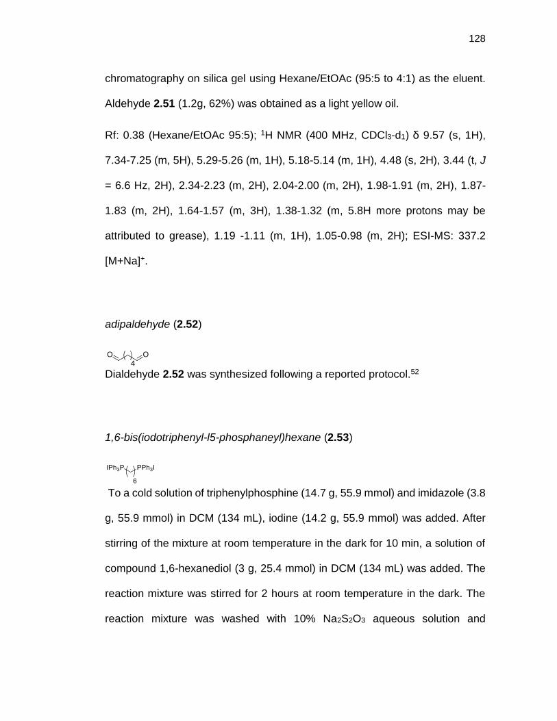
128
chromatography on silica gel using Hexane/EtOAc (95:5 to 4:1) as the eluent.
Aldehyde 2.51 (1.2g, 62%) was obtained as a light yellow oil.
Rf: 0.38 (Hexane/EtOAc 95:5); 1H NMR (400 MHz, CDCl3-d1) δ 9.57 (s, 1H),
7.34-7.25 (m, 5H), 5.29-5.26 (m, 1H), 5.18-5.14 (m, 1H), 4.48 (s, 2H), 3.44 (t, J
= 6.6 Hz, 2H), 2.34-2.23 (m, 2H), 2.04-2.00 (m, 2H), 1.98-1.91 (m, 2H), 1.87-
1.83 (m, 2H), 1.64-1.57 (m, 3H), 1.38-1.32 (m, 5.8H more protons may be
attributed to grease), 1.19 -1.11 (m, 1H), 1.05-0.98 (m, 2H); ESI-MS: 337.2
[M+Na]+.
adipaldehyde (2.52)
Dialdehyde 2.52 was synthesized following a reported protocol.52
1,6-bis(iodotriphenyl-l5-phosphaneyl)hexane (2.53)
To a cold solution of triphenylphosphine (14.7 g, 55.9 mmol) and imidazole (3.8
g, 55.9 mmol) in DCM (134 mL), iodine (14.2 g, 55.9 mmol) was added. After
stirring of the mixture at room temperature in the dark for 10 min, a solution of
compound 1,6-hexanediol (3 g, 25.4 mmol) in DCM (134 mL) was added. The
reaction mixture was stirred for 2 hours at room temperature in the dark. The
reaction mixture was washed with 10% Na2S2O3 aqueous solution and
OO4
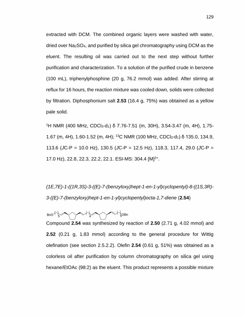
129
extracted with DCM. The combined organic layers were washed with water,
dried over Na2SO4, and purified by silica gel chromatography using DCM as the
eluent. The resulting oil was carried out to the next step without further
purification and characterization. To a solution of the purified crude in benzene
(100 mL), triphenylphosphine (20 g, 76.2 mmol) was added. After stirring at
reflux for 16 hours, the reaction mixture was cooled down, solids were collected
by filtration. Diphosphonium salt 2.53 (16.4 g, 75%) was obtained as a yellow
pale solid.
1H NMR (400 MHz, CDCl3-d1) δ 7.76-7.51 (m, 30H), 3.54-3.47 (m, 4H), 1.75-
1.67 (m, 4H), 1.60-1.52 (m, 4H); 13C NMR (100 MHz, CDCl3-d1) δ 135.0, 134.9,
113.6 (JC-P = 10.0 Hz), 130.5 (JC-P = 12.5 Hz), 118.3, 117.4, 29.0 (JC-P =
17.0 Hz), 22.8, 22.3, 22.2, 22.1. ESI-MS: 304.4 [M]2+.
(1E,7E)-1-((1R,3S)-3-((E)-7-(benzyloxy)hept-1-en-1-yl)cyclopentyl)-8-((1S,3R)-
3-((E)-7-(benzyloxy)hept-1-en-1-yl)cyclopentyl)octa-1,7-diene (2.54)
Compound 2.54 was synthesized by reaction of 2.50 (2.71 g, 4.02 mmol) and
2.52 (0.21 g, 1.83 mmol) according to the general procedure for Wittig
olefination (see section 2.5.2.2). Olefin 2.54 (0.61 g, 51%) was obtained as a
colorless oil after purification by column chromatography on silica gel using
hexane/EtOAc (98:2) as the eluent. This product represents a possible mixture
45 5OBnBnO

130
of isotactic and syndiotactic isomers. For simplicity, only the isotactic isomer is
shown in Figure 1 and throughout the supporting information.
Rf: 0.28 (hexane/EtOAc 98:2); 1H NMR (500 MHz, CDCl3-d1) δ 7.34-7.25 (m,
10H), 5.36-5.26 (m, 8H), 4.48 (s, 4H), 3.44 (t, J = 6.9 Hz, 4H), 2.82-2.73 (m,
3H), 2.47-2.41 (m, 0.8H), 2.30-2.27 (m, 0.2H), 2.01-1.92 (m, 7H), 1.88-1.74 (m,
6H), 1.65-1.55 (m, 5H), 1.41-1.30 (m, 16H), 1.05-0.91 (m, 2H); 13C NMR (126
MHz, CDCl3-d1) δ 138.8, 135.5, 135.4, 135.4, 135.3, 135.1, 135.0, 134.0, 133.8,
128.9, 128.7, 128.6, 128.6, 128.5, 127.8, 127.6, 73.0, 70.6, 43.7, 43.4, 42.3,
41.9, 38.4, 38.1, 33.0, 32.8, 32.6, 32.6, 32.5, 32.3, 29.9, 29.8, 29.8, 29.7, 29.6,
29.3, 27.6, 27.6, 26.0, 25.9.
(1E,7E)-1-((1R,3S)-3-((E)-7-(benzyloxy)hept-1-en-1-yl)cyclohexyl)-8-((1S,3R)-
3-((E)-7-(benzyloxy)hept-1-en-1-yl)cyclohexyl)octa-1,7-diene (2.55)
Compound 2.55 was synthesized by a reaction of 2.53 (1.3 g, 1.6 mmol) and
2.51 (1.1 g, 3.4 mmol) according to the general procedure for Wittig olefination
(see section 2.5.2.2). Olefin 2.55 (0.89 g, 84%) was obtained as a colorless oil
after purification by column chromatography on silica gel using hexane/EtOAc
(98:2) as the eluent. This product represents a possible mixture of isotactic and
syndiotactic isomers. For simplicity, only the isotactic isomer is shown.

131
Rf: 0.56 (hexane/EtOAc 98:2); 1H NMR (500 MHz, CDCl3-d1) δ 7.36-7.28 (m,
10H), 5.38-5.17 (m, 8H), 4.52 (s, 4H), 3.49 (t, J = 6.6 Hz, 4H), 2.39-2.30 (m,
3H), 2.09-1.96 (m, 8.6H), 1.79-1.55 (m, 12.4H), 1.43-1.34 (m, 14H), 1.04-0.86
(m, 6H); 13C NMR (126 MHz, CDCl3-d1) δ138.8, 136.3, 136.3, 136.0 135.9,
135.9, 135.8, 128.3, 128.2, 128.2, 128.1, 127.8, 127.8, 127.6, 73.0, 73.0, 70.5,
40.5, 40.0, 39.9, 36.3, 36.1, 33.0, 32.9, 32.8, 32.7, 32.7, 30.0, 29.9, 29.8, 29.7,
29.7, 29.6, 29.6, 29.4, 27.6, 27.5, 26.0, 25.9, 25.8. ESI-MS: 696.5 [M + NH4]+;
HRMS calcd 701.5268 for [C48H70O2 Na]+, found 701.5270.
1-((1R,3S)-3-(heptyl-7-ol)cyclopentyl)-8-((1S,3R)-3-(heptyl-7-ol)cyclopentyl)
octane (2.56)
To a degassed solution of olefin 2.54 (0.85 g, 1.31 mmol) in EtOH (76 mL), 10%
Pd/C (0.21 g, 25% w/w) was added. The reaction was stirred under 1 atm H2 at
room temperature for 24 hours. The solution was then filtered through celite,
and the solvent was evaporated. Diol 2.56 (0.45 g, 72%) was obtained as a
white solid.
1H NMR (500 MHz, MeOD-d4/CDCl3-d1 9:1) δ 3.34 (t, J = 6.9 Hz, 4H), 1.71-1.66
(m, 2H), 1.56-1.47 (m, 8H), 1.35-1.29 (m, 4H), 1.14-0,98 (m, 40H), 0.96-0.80
(m, 6H), 0.43-0.37 (m, 2H); 13C NMR (126 MHz, MeOD-d4/CDCl3-d1 9:1) δ 62.2,
40.6, 40.0, 38.6, 38.6, 32.9, 32.4, 31.5, 29.8, 29.6, 29.4, 28.6, 28.5, 25.6.
HO
6 66
OH

132
7,7'-((1R,1'R,3S,3'S)-octane-1,8-diylbis(cyclohexane-3,1-diyl))bis(heptan-1-ol)
(2.57)
To a degassed solution of olefin 2.55 (0.89 g, 1.3 mmol) in EtOH (25.4 mL) and
EtOAc (3.2 mL), 10% Pd/C (0.22 g, 25% w/w) was added. The reaction was
stirred under 1 atm H2 at room temperature for 24 hours. The solution was then
filtered through celite, and the solvent was evaporated. Diol 2.57 (0.40 g, 60%)
was obtained as a white solid. The crude solid was not further purified and used
in the next step.
1-((1R,3S)-3-(7-bromoheptyl)cyclopentyl)-8-((1S,3R)-3-(7-bromoheptyl)
cyclopentyl)octane (2.58)
Dibromo alkane 2.58 (0.47 g, 83%) was obtained as a white solid from diol 2.56
(0.45 g, 0.94 mmol) following the general procedure for bromination of diol using
hydrobromic acid solution (see section 2.5.2.3).
Rf: 0.75 (hexane); 1H NMR (500 MHz, CDCl3-d1) δ 3.38 (t, J = 6.9 Hz, 4H), 1.90-
1.66 (m, 13H), 1.42-1.00 (m, 40H), 0.87-0.83 (m, 1H), 0.63-0.56 (m, 2H); 13C
NMR (126 MHz, CDCl3-d1) δ 40.9, 40.3, 40.3, 39.0, 38.9, 36.9, 36.8, 34.3, 33.2,
33.0, 31.8, 30.2, 29.9, 29.0, 29.0, 28.9, 28.8, 28.7, 28.4.
Br
6 66
Br

133
1,8-bis((1S,3R)-3-(7-bromoheptyl)cyclohexyl)octane (2.59)
Dibromo alkane 2.59 (0.45 g, 90%) was obtained as a white solid from diol 2.57
(0.40 g, 0.79 mmol) following the general procedure for bromination of diol using
hydrobromic acid solution (see section 2.5.2.3).
Rf: 0.89 (DCM/hexane 1:1); 1H NMR (500 MHz, CDCl3-d1) δ 3.39 (t, J = 6.9 Hz,
4H), 1.86-1.80 (m, 4H), 1.70-1.63 (m, 7.6H), 1.50-1.46 (m, 0.4H), 1.41-1.37 (m,
4H), 1.30-1.10 (m, 38H), 0.78-0.69 (m, 4H), 0.50-0.43 (m, 2H); 13C NMR (126
MHz, CDCl3-d1) δ 40.7, 38.0, 34.3, 33.7, 33.1, 30.3, 30.0, 30.0, 29.0, 28.4, 27.1,
27.0, 26.6.
1-((1R,3S)-3-(7-(3-(benzyloxy)-2-((3,7,11,15-
tetramethylhexadecyl)oxy)propoxy) heptyl)cyclopentyl)-8-((1S,3R)-3-(7-(3-
(benzyloxy)-2-((3,7,11,15-tetramethyl-
hexadecyl)oxy)propoxy)heptyl)cyclopentyl)octane (2.60)
Compound 2.60 was synthesized by reaction of 2.4 (1.10 g, 2.35 mmol) and
2.58 (0.47 g, 0.78 mmol) according to the general procedure for formation of
tetraether lipid scaffold by SN2 reaction (see section 2.5.2.4). Product 2.60 (0.32

134
g, 30%) was obtained as a colorless oil after purification by column
chromatography on silica gel using hexane/EtOAc (99:1 to 95:5) as the eluent.
Rf: 0.50 (hexane/EtOAc 95:5); 1H NMR (500 MHz, CDCl3-d1) δ 7.32-7.25 (m,
10H), 4.53 (s, 4H), 3.63-3.92 (m, 18H), 1.88-1.85 (m, 2H), 1.76-1.66 (m, 6H),
1.62-1.47 (m, 10H), 1.37-1.01 (m, 84H), 0.85-0.81 (m, 30H), 0.73-0.56 (m, 2H);
13C NMR (126 MHz, CDCl3-d1) δ 138.6, 128.5, 127.8, 127.7, 78.1, 73.6, 71.9,
70.9, 70.5, 69.1, 40.9, 40.3, 39.6, 39.0, 37.7, 37.7, 37.6, 37.6, 37.5, 37.4, 37.3,
37.0, 36.9, 33.3, 33.0, 31.8, 30.2, 30.1, 30.0, 30.0, 29.9, 29.8, 29.0, 28.9, 28.2,
26.4, 25.0, 24.7, 24.6, 23.0, 22.9, 20.0, 19.9, 19.8.
1-((1R,3S)-3-(7-(3-(benzyloxy)-2-((3,7,11,15-
tetramethylhexadecyl)oxy)propoxy)heptyl)cyclohexyl)-8-((1S,3R)-3-(7-(3-
(benzyloxy)-2-((3,7,11,15-
tetramethylhexadecyl)oxy)propoxy)heptyl)cyclohexyl)octane (2.61)
Compound 2.61 was synthesized by reaction of 2.4 (0.98 g, 2.12 mmol) and
2.59 (0.45 g, 0.71 mmol) according to the general procedure for formation of
tetraether lipid scaffold by SN2 reaction (see section 2.5.2.4). Product 2.61
(0.130 g, 13%) was obtained as a colorless oil after purification by column
chromatography on silica gel using hexane/EtOAc (99:1 to 95:5) as the eluent.

135
Rf: 0.33 (hexane/EtOAc 95:5); 1H NMR (500 MHz, CDCl3-d1) δ 7.32-7.24 (m,
10H), 4.54 (s, 4H), 3.62-3.40 (m, 18H), 1.71-1.59 (m, 10H), 1.55-1.48 (m, 8H),
1.38-1.02 (m, 84H), 0.86-0.82 (m, 30H), 0.78-0.70 (m, 4.2H), 0.51-0.44 (m,
1.8H); 13C NMR (126 MHz, CDCl3-d1) δ 138.6, 128.5, 127.8, 127.7, 78.1, 73.6,
71.9, 71.0, 70.5, 69.1, 40.8, 39.6, 38.0, 38.0, 37.9, 37.7, 37.7, 37.6, 37.6, 37.5,
37.4, 37.3, 33.6, 33.0, 30.3, 30.2, 30.0, 30.0, 29.9, 29.8, 28.2, 27.1, 27.1, 26.6,
26.4, 25.0, 24.7, 24.7, 24.6, 22.9, 22.8, 20.0, 19.9, 19.8 ESI-MS: 1413.8 [M +
NH4]+; HRMS calcd 1418.2890 for [C94H170O6 Na]+, found 1418.2907.
3-((7-((1R,3S)-3-(8-((1R,3S)-3-(7-(3-hydroxy-2-((3,7,11,15-
tetramethylhexadecyl)
oxy)propoxy)heptyl)cyclopentyl)octyl)cyclopentyl)heptyl)oxy)-2-((3,7,11,15-
tetramethylhexadecyl)oxy)propan-1-ol (2.62)
Compound 2.62 was synthesized by hydrogenation of 2.60 (0.22 g, 0.16 mmol)
according to the general procedure for debenzylation of lipid scaffold by
hydrogenation (see section 2.5.2.5). Diol 2.62 (0.18 g, 95%) was obtained as a
colorless oil after purification by column chromatography on silica gel using
hexane/EtOAc (95:5 to 85:15) as the eluent.

136
Rf: 0.41 (hexane/EtOAc 80:20); 1H NMR (500 MHz, CDCl3-d1) δ 3.71-3.40 (m,
18H), 1.90-1.85 (m, 2H), 1.74-1.47 (m, 20H), 1.38-1.01 (m, 82H), 0.86-0.81 (m,
30H), 0.63-0.56 (m, 1H); 13C NMR (126 MHz, CDCl3-d1) δ 78.5, 72.1, 71.1, 68.9,
40.9, 40.3, 39.6, 39.0, 37.7, 37.7, 37.6, 37.5, 37.4, 37.3, 37.0, 36.9, 33.2, 33.0,
31.9, 30.2, 30.1, 30.0, 30.0, 29.9, 29.7, 29.0, 28.9, 28.8, 28.2, 26.3, 25.0, 24.7,
24.6, 23.0, 22.9, 20.0, 19.9, 19.8; ESI-MS: 1187.8 [M+H]+.
3-((7-((1R,3S)-3-(8-((1R,3S)-3-(7-(3-hydroxy-2-((3,7,11,15-
tetramethylhexadecyl)oxy)propoxy)heptyl)cyclohexyl)octyl)cyclohexyl)heptyl)o
xy)-2-((3,7,11,15-tetramethylhexadecyl)oxy)propan-1-ol (2.63)
Compound 2.63 was synthesized by hydrogenation of 2.61 (0.130 g, 0.09 mmol)
according to the general procedure for debenzylation of lipid scaffold by
hydrogenation (see section 2.5.2.5). Diol 2.63 (0.10 g, 88%) was obtained as a
colorless oil after purification by column chromatography on silica gel using
hexane/EtOAc (95:5 to 85:15) as the eluent.
Rf: 0.40 (hexane/EtOAc 80:20); 1H NMR (500 MHz, CDCl3-d1) δ 3.64-3.35 (m,
18H), 2.20-2.15 (m, 1.8H), 1.74-0.89 (m, 102.2H), 0.81-0.64 (m, 34H), 0.42-
0.40 (m, 2H); 13C NMR (126 MHz, CDCl3-d1) δ 78.5, 72.0, 71.1, 71.1, 68.8, 63.3,
63.3, 40.7, 39.6, 38.0, 37.9, 37.7, 37.6, 37.6, 37.5, 37.5, 37.3, 37.3, 33.6, 33.0,
33.0, 30.3, 30.2, 30.0, 29.9, 29.8, 29.7, 28.2, 27.1, 27.0, 26.6, 26.3, 25.0, 24.7,

137
24.5, 22.9, 22.8, 19.9, 19.9, 19.8; ESI-MS: 1217.1 [M+H]+; HRMS calcd
1238.1951 for [C80H158O6 Na]+, found 1238.1950.
3-((7-((1S,3R)-3-(8-((1S,3R)-3-(7-(3-((oxido(2-(trimethylammonio)ethoxy)
phosphoryl)oxy)-2-((3,7,11,15-tetramethylhexadecyl)oxy)propoxy)heptyl)
cyclopentyl)octyl)cyclopentyl)heptyl)oxy)-2-((3,7,11,15-
tetramethylhexadecyl)oxy) propyl-(2-(trimethylammonio)ethyl) phosphate
(GMGTPC-CP2)
Lipid GMGTPC-CP2 was synthesized from diol 2.62 (0.17 g, 0.14 mmol)
following the general procedure for formation of phosphocholine lipid (see
section 2.5.2.6). Lipid GMGTPC-CP2 (0.15 g, 82%) was obtained as a white
gum after purification by column chromatography on silica gel using
DCM/MeOH/H2O (70:30:5) as the eluent.
Rf: 0.50 (DCM/MeOH/H2O 70:30:5); 1H NMR (500 MHz, MeOD-d4/CDCl3-d1
1:1) δ 3.90-3.87 (m, 4H), 3.53 (t, J = 5.5 Hz, 4H), 3.32-3.06 (m, 18H), 2.86 (s,
18H), 1.58-1.34 (m, 10H), 1.28-1.13 (m, 10H), 1.03-0.70 (m, 82H), 0.53-0.48 (m
30H), 0.30-0.24 (m, 2H); 13C NMR (126 MHz, MeOD-d4/CDCl3-d1 1:1) δ 77.8,
77.7, 77.6, 77.6, 71.3, 70.2, 68.6, 68.4, 66.0, 64.7, 64.6, 58.6, 58.5, 53.5, 40.3,

138
39.8, 39.0, 38.4, 38.3, 37.3, 37.1, 37.0, 37.0, 36.9, 36.8, 36.7, 36.4, 36.3, 32.6,
32.4, 31.2, 29.5, 29.4, 29.3, 29.2, 29.1, 28.3, 27.5, 25.7, 24.4, 24.0, 24.0, 22.1,
22.0, 19.1, 19.1, 19.0, 19.0, 18.9; 31P NMR (202 MHz, MeOD-d4/CDCl3-d1 1:1)
δ 0.20; ESI-MS: 1518.2 [M+H]+; HRMS calcd 1518.2928 for [C88H179N2O12P2]+,
found 1518.2923.
3-((7-((1S,3R)-3-(8-((1S,3R)-3-(7-(3-((oxido(2-
(trimethylammonio)ethoxy)phosphoryl)oxy)-2-((3,7,11,15-
tetramethylhexadecyl)oxy)propoxy)heptyl)cyclohexyl)octyl)cyclohexyl)heptyl)o
xy)-2-((3,7,11,15-tetramethylhexadecyl)oxy)propyl (2-(trimethylammonio)ethyl)
phosphate (GMGTPC-CH2)
Lipid GMGTPC-CH2 was synthesized from diol 2.63 (0.10 g, 0.08 mmol)
following the general procedure for formation of phosphocholine lipid (see
section 2.5.2.6). Lipid GMGTPC-CH2 (0.10 g, 77%) was obtained as a white
gum after purification by column chromatography on silica gel using
DCM/MeOH/H2O (70:30:5) as the eluent.
Rf: 0.12 (DCM/MeOH/H2O 70:30:5); 1H NMR (400 MHz, MeOD-d4/CDCl3-d1
1:1) δ 3.93-3.84 (m, 4H), 3.52 (t, J = 5.6 Hz, 4H), 3.30-3.06 (m, 18H), 2.84 (s,
18H), 1.39-1.11 (m, 19H), 1.06-0.64 (m, 86H), 0.56-0.07 (m, 34H); 13C NMR

139
(100 MHz, MeOD-d4/CDCl3-d1 1:1) δ 71.3, 70.2, 68.6, 68.3, 66.0, 64.6, 64.6,
59.4, 58.5, 58.5, 53.5, 42.8, 40.2, 38.9, 37.3, 37.2, 37.0, 36.9, 36.8, 36.7, 36.7,
33.0, 32.3, 29.6, 29.5, 29.3, 29.2, 29.2, 29.1, 27.5, 26.5, 25.9, 25.7, 24.3, 24.0,
24.0, 22.0, 21.9, 19.1, 19.1, 19.0, 19.0, 18.9; 31P NMR (161 MHz, MeOD-
d4/CDCl3-d1 1:1) δ 0.20; ESI-MS: 1546.2 [M+H]+; HRMS calcd 1546.3241 for
[C90H183N2O12P2]+, found 1546.3235.
2.5.7 Synthetic Procedure for GMGTPC-CP3
((pent-4-en-1-yloxy)methyl)benzene (2.64)
Compound 2.64 was synthesized following a reported protocol.53
4-(benzyloxy)butanal (2.65)
To a solution of 2.64 (6.63 g, 37.6 mmol) in DCM (300 mL) at -78 °C ozone was
bubbled until the solution turned blue. Then, oxygen was bubbled for 10 min
and triphenylphosphine (10.8 g, 41.4 mmol) was added portionwise at -78 °C.
The reaction mixture was allowed to warm up slowly to room temperature and
stirred overnight. The solvent was removed under reduced pressure and the
resulting solid was purified by column chromatography on silica gel using

140
hexane/EtOAc (90:10 to 80:20) as the eluent. Aldehyde 2.65 (5.85 g, 88%) was
obtained as a colorless oil and 1H NMR data match previously reported data.54
(1R,3S)-1,3-bis((E)-5-(benzyloxy)pent-1-en-1-yl)cyclopentane (2.66)
Compound 2.66 was synthesized by reaction of 2.27 (13.1 g, 14.9 mmol) and
2.65 (5.9 g, 32.9 mmol) according to the general procedure for Wittig olefination
(see section 2.5.2.2). Olefin 2.66 (2.8 g, 45%) was obtained as a colorless oil
after purification by column chromatography on silica gel using hexane/EtOAc
(98:2) as the eluent.
Rf: 0.45 (hexane/EtOAc 95:5); 1H NMR (500 MHz, CDCl3-d1) δ 7.36-7.28 (m,
10H); 5.41-5.29 (m, 4H); 4.52 (s, 4H); 3.51-3.48 (m, 4H), 2.86-2.77 (m, 1.7H),
2.50-2.45 (m, 0.3H); 2.19-2.08 (m, 4H), 1.93-1.78 (m, 3H), 1.73-1.67 (m, 4H),
1.41-1.34 (m, 2H), 1.08-0.99 (m, 1H); 13C NMR (126 MHz, CDCl3-d1) δ 138.8,
136.0, 135.9, 135.7, 135.5, 128.5, 127.9, 127.8, 127.6, 73.1, 73.0, 70.0, 69.9,
43.6, 43.4, 42.2, 41.8, 38.4, 38.1, 32.9, 32.8, 32.4, 32.3, 30.1, 29.8, 29.2, 24.3.
5,5'-((1R,3S)-cyclopentane-1,3-diyl)bis(pentan-1-ol) (2.67)
To a degassed solution of 2.66 (2.75 g, 6.58 mmol) in EtOH/THF (9:1) (75 mL),
10% Pd/C (0.69 g, 25% w/w) was added. The reaction was stirred under 1 atm

141
H2 at room temperature for 16 hours. The solution was then filtered through
celite, and the solvent was evaporated. Diol 2.67 (0.83 g, 52%) was obtained
as a white solid after purification by column chromatography on silica gel using
hexane/EtOAc (75:25 to 50:50) as the eluent.
Rf: 0.42 (hexane/EtOAc 1:1); 1H NMR (500 MHz, CDCl3-d1) δ 3.49 (t, J = 6.9
Hz, 4H), 3.23 (brs, 2H), 1.84-1.79 (m, 1H), 1.73-1.61 (m, 4H), 1.48-1.42 (m, 4H),
1.25-1.04 (m, 14.2H), 0.57-0.51 (m, 0.8H); 13C NMR (126 MHz, CDCl3-d1) δ
62.6, 40.1, 38.8, 36.8, 36.7, 33.0, 32.7, 31.6, 28.6, 28.5, 26.1; ESI-MS: 265.1
[M+Na]+; HRMS calcd 265.2138 for [C15H30O2Na]+, found 265.2141.
(((1R,3S)-cyclopentane-1,3-diyl)bis(pentane-5,1-
diyl))bis(triphenylphosphonium) iodide (2.68)
To a cold solution of triphenylphosphine (1.98 g, 7.55 mmol) and imidazole (0.52
g, 7.55 mmol) in DCM (18 mL), iodine (1.92 g, 7.55 mmol) was added. After
stirring of the mixture at room temperature in the dark for 10 min, a solution of
compound 2.67 (0.83 g, 3.43 mmol) in DCM (18 mL) was added. The reaction
mixture was stirred for 2 hours at room temperature in the dark. The reaction
mixture was washed with 10% Na2S2O3 aqueous solution and extracted with
DCM. The combined organic layers were washed with water, dried over Na2SO4,
and purified by silica gel chromatography using a mixture of hexane/EtOAc (8:2)
as eluent. The resulting oil was carried out to the next step without further

142
purification and characterization. To a solution of the purified crude in benzene
(15 mL), triphenylphosphine (2.70 g, 10.29 mmol) was added. After stirring at
reflux for 16 hours, the reaction mixture was cooled down, evaporated and
purified by column chromatography on silica gel using DCM/MeOH (1:0 to 9:1)
as the eluent. diphosphonium salt 2.68 (3.19 g, 94%) was obtained as a white
solid.
1H NMR (500 MHz, CDCl3-d1) δ 7.74-.61 (m, 30H), 3.48-3.42 (m, 4H), 1.76-1.71
(m, 1H), 1.57-1.50 (m, 12H), 1.16-0.85 (10.2H), 0.43-0.37 (m, 0.8H); 13C NMR
(126 MHz, CDCl3-d1) δ 135.2, 133.6 (d, JC-P = 10.0 Hz), 130.6 (d, JC-P = 12.5
Hz), 117.9 (d, JC-P = 85.7 Hz), 53.6, 40.2, 39.7, 38.3, 38.2, 36.0, 32.8, 31.5,
30.6, 30.5, 28.0, 23.2, 22.8, 22.5, 22.5; ESI-MS: 366.3 [M-2I]2+; HRMS calcd
366.2001 for [C51H58P2]2+, found 366.2003.
((but-3-en-1-yloxy)methyl)benzene (2.69)
Compound 2.69 was synthesized following a reported protocol.55
3-(benzyloxy)propanal (2.70)
Compound 2.70 was synthesized following a reported protocol.56

143
2-(((1S,3R)-3-((E)-4-(benzyloxy)but-1-en-1-yl)cyclopentyl)methoxy)tetrahydro-
2H-pyran (2.71)
Compound 2.71 was synthesized by reaction of 2.41 (4.41 g, 7.52 mmol) and
2.70 (1.45 g, 9.02 mmol) according to the general procedure for Wittig
olefination (see section 2.1). Olefin 2.71 (1.10 g, 43%) was obtained as a
colorless oil after purification by column chromatography on silica gel using
hexane/EtOAc (98:2 to 95:5) as the eluent.
Rf: 0.33 (hexane/EtOAc 95:5); 1H NMR (500 MHz, CDCl3-d1) δ 7.32-7.24 (m,
5H), 5.49-5.30 (m, 2H), 4.57 (dd, J = 2.9, 4.2 Hz, 1H), 4.50 (s, 2H), 3.87-3.83
(m, 1H), 3.63-3.59 (m, 1H), 3.49-3.44 (m, 3H), 3.29-3.24 (m, 1H), 2.79-2.71 (m,
0.8H), 2.44-2.22 (m, 3.3H); 1.98-1.90 (m, 1H), 1.84-1.66 (m, 4H), 1.59-1.40 (m,
5H), 1.34-1.24 (m, 1H), 1.00-0.89 (m, 1H); 13C NMR (126 MHz, CDCl3-d1) δ
138.7, 138.6, 137.1, 136.9, 128.4, 127.7, 127.6, 124.7, 124.4, 98.9, 72.9, 72.9,
72.4, 72.3, 72.2, 70.4, 70.3, 62.3, 62.2, 43.7, 43.6, 41.7, 39.7, 39.4, 38.6, 38.5,
38.2, 38.0, 37.7, 37.5, 33.1, 32.8, 32.8, 32.3, 30.8, 29.0, 28.9, 28.7, 28.4, 25.6,
19.7, 19.6.

144
((1S,3R)-3-((E)-4-(benzyloxy)but-1-en-1-yl)cyclopentyl)methanol (2.72)
To a solution of 2.71 (1.10 g, 3.20 mmol) in a mixture of MeOH (150 mL) and
THF (30 mL), p-toluenesulfonic acid (30 mg, 0.16 mmol) was added and the
solution was stirred for 16 hours at room temperature. Then, the solvent was
evaporated and the resulting residue was dissolved in EtOAc. The organic
solution was washed successively with 1M NaOH solution, water, brine and
dried over Na2SO4 to yield the alcohol 2.72 (0.84 g, Qt.) as a colorless oil.
1H NMR (500 MHz, CDCl3-d1) δ 7.33-7.24 (m, 5H), 5.49-5.30 (m, 2H), 4.50 (s,
2H), 3.48-3.44 (m, 4H), 2.80-2.71 (m, 0.8H), 2.40-2.28 (M, 3.3H), 2.17-2.10 (m,
1H), 1.93-1.88 (m, 1H), 1.78-1.69 (m, 2H), 1.44-1.23 (m, 2H), 0.96-0.88 (m, 1H);
13C NMR (126 MHz, CDCl3-d1) δ 138.5, 138.4, 136.9, 136.7, 128.4, 127.7, 127.6,
124.7, 124.5, 72.9, 72.8, 70.3, 70.2, 67.3, 67.2, 43.5, 42.1, 41.9, 38.5, 37.5,
36.9, 33.0, 32.7, 32.2, 28.4, 28.3, 28.2; ESI-MS: 283.1 [M+Na]+; HRMS calcd
283.1669 for [C17H24O2Na]+, found 283.1668.
(1S,3R)-3-((E)-4-(benzyloxy)but-1-en-1-yl)cyclopentane-1-carbaldehyde (2.73)
To a cold solution of 2.72 (0.84 g, 3.23 mmol) in dry DCM (30 mL), DMP (1.64
g, 3.88 mmol) was added portionwise. The resulting mixture was allowed to
reach room temperature over 1 hour. After 3 more hours of stirring at room

145
temperature, the mixture was diluted with DCM, washed successively with 1M
NaOH solution, water and brine. The organic solution was dried over Na2SO4,
concentrated under reduce pressure and purified by column chromatography
on silica gel using hexane/EtOAc (90:10 to 75:25) as the eluent. Aldehyde 2.73
(0.59 g, 70%) was obtained as a colorless oil.
Rf: 0.63 (hexane/EtOAc 75:25); 1H NMR (500 MHz, CDCl3-d1) δ 9.64 (d, J = 2.5
Hz, 1H), 7.34-7.30 (m, 5H), 5.52-5.38 (m, 2H), 4.55 (s, 2H), 3.54-3.50 (m, 2H),
2.92-2.80 (m, 1.8H), 2.60-2.33 (m, 2.2H), 2.07-1.98 (m, 2H), 1.89-1.90 (m, 2H),
1.63-1.51 (m, 1H), 1.38-1.27 (m, 1H); 13C NMR (126 MHz, CDCl3-d1) δ 203.6,
203.4, 138.6, 138.5, 135.3, 135.1, 128.4, 127.7, 127.6, 126.0, 125.7, 73.0, 72.9,
70.1, 70.0, 51.5, 51.3, 43.7, 38.7, 33.9, 33.8, 33.4, 33.1, 33.0, 32.7, 28.4, 25.9,
25.7; ESI-MS: 281.1 [M+Na]+; HRMS calcd 281.1512 for [C18H22O2Na]+, found
281.1514.
(1S,3R)-1-((E)-6-((1R,3S)-3-((E)-4-(benzyloxy)but-1-en-1-yl)cyclopentyl)hex-5-
en-1-yl)-3-((E)-6-((1S,3R)-3-((E)-4-(benzyloxy)but-1-en-1-yl)cyclopentyl)hex-5-
en-1-yl)cyclopentane (2.74)
Compound 2.74 was synthesized by reaction of 2.68 (2.41 g, 2.44 mmol) and
2.73 (1.40 g, 5.43 mmol) according to the general procedure for Wittig
olefination (see section 2.5.2.2). Olefin 2.74 (0.80 g, 43%) was obtained as a
colorless oil after purification by column chromatography on silica gel using

146
hexane/EtOAc (98:2) as the eluent. This product represents a possible mixture
of isotactic and syndiotactic isomers. For simplicity, only the isotactic isomer is
shown.
Rf: 0.32 (hexane/EtOAc 95:5); 1H NMR (500 MHz, CDCl3-d1) δ 7.33-7.25 (m,
10H), 5.49-5.23 (m, 8H), 4.51 (s, 4H), 3.48-3.44 (m, 4H), 2.82-2.75 (m, 3H),
2.50-2.28 (m, 5H), 2.02-1.70 (m, 15H), 1.38-1.24 (m, 16H), 1.14-0.96 (m, 4.2H),
0.66-0.58 (m, 0.8H); 13C NMR (126 MHz, CDCl3-d1) δ 138.7, 137.6, 137.4, 137.4,
137.2, 135.3, 135.1, 134.8, 129.0, 128.9, 128.9, 128.6, 127.8, 127.7, 124.6,
124.3, 124.2, 73.1, 73.0, 70.5, 70.4, 43.7, 43.7, 42.3, 41.9, 41.8, 40.9, 40.3,
40.3, 39.0, 38.6, 38.5, 38.4, 38.3, 38.2, 36.9, 36.8, 36.7, 33.2, 33.0, 32.9, 32.8,
32.8, 32.5, 32.4, 31.8, 30.4, 30.1, 28.6, 28.5, 28.4, 27.7.
4-((1R,3S)-3-(6-((1R,3S)-3-(6-((1R,3S)-3-(4-hydroxybutyl)cyclopentyl)hexyl)
cyclopentyl)hexyl)cyclopentyl)butan-1-ol (2.75)
To a degassed solution of olefin 2.74 (0.77 g, 1.16 mmol) in EtOH/THF (8:2) (45
mL), 10% Pd/C (0.28 g, 25% w/w) was added. The reaction was stirred under 1
atm H2 at room temperature for 24 hours. The solution was then filtered through
celite, and the solvent was evaporated to yield a white solid which was used in
the next step without further purification and characterization.

147
((1S,3R)-1-(6-((1R,3S)-3-(4-bromobutyl)cyclopentyl)hexyl)-3-(6-((1S,3R)-3-(4-
bromobutyl)cyclopentyl)hexyl)cyclopentane (2.76)
Dibromo alkane 2.76 (0.38 g, 67%) was obtained as a white solid from diol 2.75
(0.46 g, 0.89 mmol) following the general procedure for bromination of diol using
hydrobromic acid solution (see section 2.5.2.3). The purification was
accomplished by column chromatography on silica gel using hexane/DCM (1:1)
as the eluent.
Rf: 0.86 (hexane/DCM); 1H NMR (500 MHz, CDCl3-d1) δ 3.38 (dd, J = 6.6, 6.9
Hz, 4H), 1.90-1.67 (m, 19H), 1.43-1.02 (m, 40H), 0.87-0.59 (m, 3H); 13C NMR
(126 MHz, CDCl3-d1) δ 40.9, 40.8, 40.3, 40.1, 39.0, 38.9, 38.7, 37.0, 37.0, 36.9,
36.0, 35.9, 34.2, 33.3, 33.2, 31.8, 31.8, 31.7, 30.2, 28.9, 28.9, 27.5, 27.4.
(1S,3R)-1-(6-((1R,3S)-3-(4-(3-(benzyloxy)-2-((3,7,11,15-tetramethylhexadecyl)
oxy)propoxy)butyl)cyclopentyl)hexyl)-3-(6-((1S,3R)-3-(4-(3-(benzyloxy)-2-
((3,7,11,15-tetramethylhexadecyl)oxy)propoxy)butyl)cyclopentyl)hexyl)
cyclopentane (2.77)
Compound 2.77 was synthesized by reaction of 2.4 (0.85 g, 1.84 mmol) and
2.76 (0.38 g, 0.60 mmol) according to the general procedure for formation of

148
tetraether lipid scaffold by SN2 reaction (see section 2.5.2.4). Compound 2.77
(0.24 g, 29%) was obtained as a colorless oil after purification by column
chromatography on silica gel using hexane/EtOAc (98:2 to 95:5) as eluent.
Rf: 0.29 (hexane/EtOAc 95:5); 1H NMR (500 MHz, CDCl3-d1) δ 7.32-7.25 (m,
10H), 4.55 (s, 4H), 3.63-3.40 (m, 18H), 1.91-1.68 (m, 15H), 1.65-1.49 (m, 10H),
1.39-1.03 (m, 80H), 0.87-0.83 (m, 30H), 0.75-0.59 (m, 3H); 13C NMR (126 MHz,
CDCl3-d1) δ 138.6, 128.5, 127.8, 127.7, 78.1, 73.5, 71.9, 71.0, 70.5, 69.1, 40.9,
40.9, 40.3, 40.3, 39.6, 39.0, 38.9, 37.7, 37.7, 37.6, 37.6, 37.5, 37.4, 37.3, 37.0,
36.9, 36.8, 36.7, 33.2, 32.0, 31.8, 31.8, 30.2, 30.1, 30.0, 29.0, 28.9, 28.2, 25.4,
25.4, 25.0, 24.7, 24.6, 22.9, 22.8, 20.0, 19.9, 19.8; ESI-MS: 1408.9 [M-H]-.
3-(4-((1R,3S)-3-(6-((1R,3S)-3-(6-((1R,3S)-3-(4-(3-hydroxy-2-((3,7,11,15-tetra
methylhexadecyl)oxy)propoxy)butyl)cyclopentyl)hexyl)cyclopentyl)hexyl)cyclop
entyl )butoxy)-2-((3,7,11,15-tetramethylhexadecyl)oxy)propan-1-ol (2.78)
Compound 2.78 was synthesized by hydrogen-ation of 2.77 (0.24 g, 0.17 mmol)
according to the general procedure for debenzylation of lipid scaffold by
hydrogenation (see section 2.5.2.5). Diol 2.78 (0.18 g, 87%) was obtained as a
colorless oil after purification by column chromatography on silica gel using
hexane/EtOAc (9:1 to 8:2) as the eluent.

149
Rf: 0.38 (hexane/EtOAc); 1H NMR (500 MHz, CDCl3-d1) δ 3.70-3.39 (m, 18H),
2.24 (brs, 2H), 1.90-1.47 (m, 25H), 1.37-1.00 (m, 80H), 0.85-0.81 (m, 30H),
0.72-0.58 (m, 3H); 13C NMR (126 MHz, CDCl3-d1) δ 78.5, 72.0, 71.1, 68.8, 40.9,
40.8, 40.3, 40.2, 39.6, 39.0, 38.9, 37.7, 37.6, 37.6, 37.5, 37.5, 37.3, 37.3, 37.0,
36.9, 36.9, 36.7, 36.6, 33.2, 33.2, 33.0, 31.8, 31.8, 30.2, 30.1, 30.0, 28.9, 28.9,
28.2, 25.4, 25.3, 25.0, 24.7, 24.6, 22.9, 22.8, 20.0, 19.9, 19.8; ESI-MS: 1226.6
[M-H]-.
3-(4-((1S,3R)-3-(6-((1S,3R)-3-(6-((1S,3R)-3-(4-(3-((oxido(2-
(trimethylammonio) ethoxy)phosphoryl)oxy)-2-((3,7,11,15-
tetramethylhexadecyl)oxy)propoxy)butyl)
cyclopentyl)hexyl)cyclopentyl)hexyl)cyclopentyl)butoxy)-2-((3,7,11,15-
tetramethylhexadecyl)oxy)propyl (2-(trimethylammonio)ethyl) phosphate
(GMGTPC-CP3)
Lipid GMGTPC-CP3 was synthesized from diol 2.78 (0.18 g, 0.14 mmol)
following the general procedure for formation of phosphocholine lipid (see
section 2.5.2.6). Lipid GMGTPC-CP3 (0.20 g, 90%) was obtained as a white
gum after purification by column chromatography on silica gel using
DCM/MeOH/H2O (70:30:5) as the eluent.

150
Rf: 0.59 (DCM/MeOH/H2O 70:30:5); 1H NMR (500 MHz, MeOD-d4/CDCl3-d1
1:1) δ 3.91-3.86 (m, 4H), 3.53 (t, J = 5.4 Hz, 4H), 3.29-3.07 (m, 18H), 2.85 (m,
18H), 1.56-1.31 (m, 15H), 1.28-1.12 (m, 10H), 1.02-0.69 (m, 80H), 0.52-0.45 (m,
30H), 0.38-0.24 (m, 3H); 13C NMR (126 MHz, MeOD-d4/CDCl3-d1 1:1) δ 77.8,
77.7, 77.6, 77.5, 71.3, 70.2, 68.6, 68.4, 66.0, 66.0, 64.7, 58.6, 58.6, 53.5, 48.8,
39.8, 39.7, 39.0, 38.4, 38.3, 37.3, 37.1, 37.0, 37.0, 36.9, 36.8, 36.7, 36.4, 36.3,
36.2, 36.1, 32.6, 32.4, 31.2, 31.1, 29.5, 29.5, 29.4, 28.3, 28.2, 27.5, 24.8, 24.7,
24.4, 24.0, 24.0, 22.0, 21.9, 19.1, 19.0, 19.0, 18.9, 18.9; 31P NMR (202 MHz,
MeOD-d4/CDCl3-d1 1:1) δ 0.16; ESI-MS: 1558.2 [M-H]-; HRMS calcd 1558.3241
for [C91H183N2O12P2]+, found 1558.3241.
2.5.8 Synthetic Procedure for GMGT Analogs with Varying Polar Lipid
Headgroups
5-(benzyloxy)-2-phenyl-1,3-dioxane (2.79)
Compound 2.79 was prepared following a previously reported protocol.57
2,3-bis(benzyloxy)propan-1-ol (2.80)
A solution of 2.79 (2.4 g, 1.3 mmol) in dry DCM was cooled to -78°C. DIBAL-H
in DCM (1 M, 3.4 mL) was added dropwise to the cooled solution and reacted
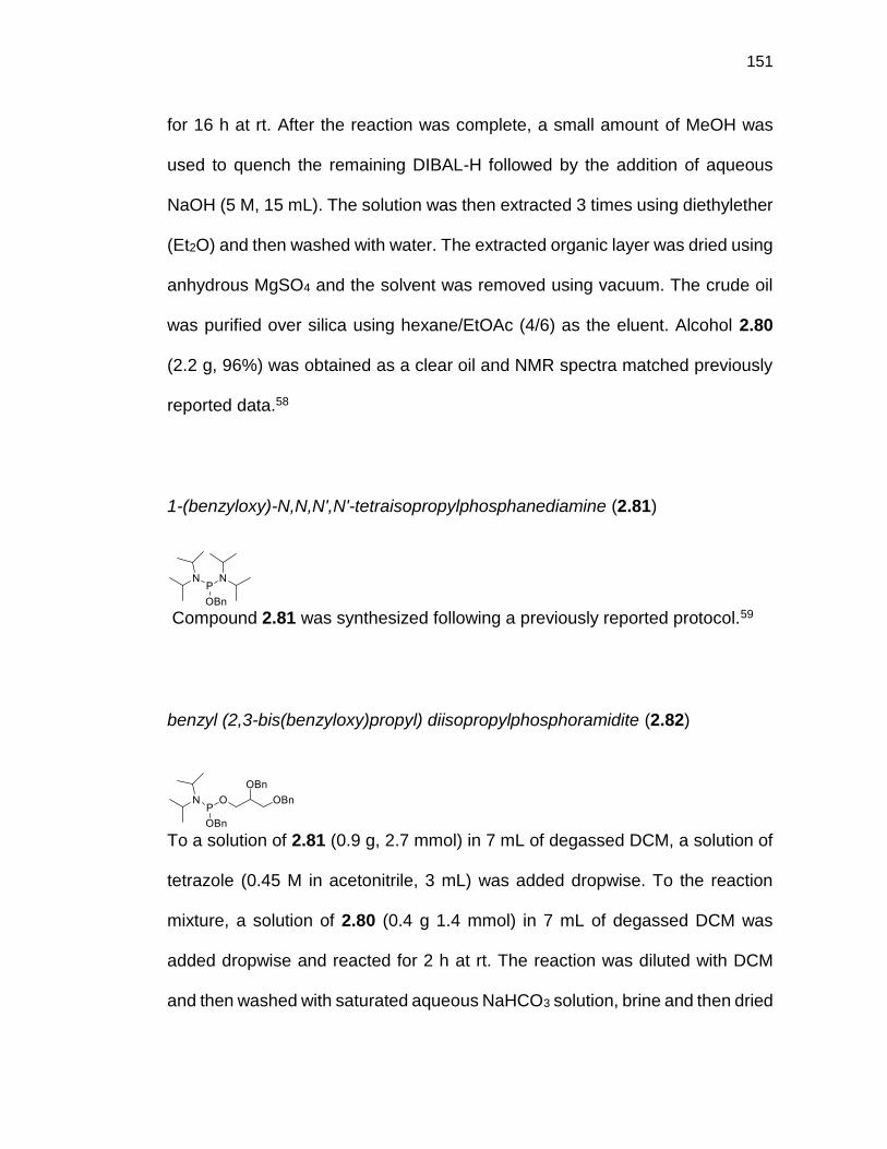
151
for 16 h at rt. After the reaction was complete, a small amount of MeOH was
used to quench the remaining DIBAL-H followed by the addition of aqueous
NaOH (5 M, 15 mL). The solution was then extracted 3 times using diethylether
(Et2O) and then washed with water. The extracted organic layer was dried using
anhydrous MgSO4 and the solvent was removed using vacuum. The crude oil
was purified over silica using hexane/EtOAc (4/6) as the eluent. Alcohol 2.80
(2.2 g, 96%) was obtained as a clear oil and NMR spectra matched previously
reported data.58
1-(benzyloxy)-N,N,N',N'-tetraisopropylphosphanediamine (2.81)
Compound 2.81 was synthesized following a previously reported protocol.59
benzyl (2,3-bis(benzyloxy)propyl) diisopropylphosphoramidite (2.82)
To a solution of 2.81 (0.9 g, 2.7 mmol) in 7 mL of degassed DCM, a solution of
tetrazole (0.45 M in acetonitrile, 3 mL) was added dropwise. To the reaction
mixture, a solution of 2.80 (0.4 g 1.4 mmol) in 7 mL of degassed DCM was
added dropwise and reacted for 2 h at rt. The reaction was diluted with DCM
and then washed with saturated aqueous NaHCO3 solution, brine and then dried

152
over MgSO4. The solvent was evaporated under vacuum and purified over silica
using hexane/EtOAc/triethylamine (90:10:3) as the mobile phase.
Phospharamidite 2.82 was obtained (252 mg, 39%) as a clear oil.
1H NMR (500 MHz, CDCl3-d1) δ 7.43-7.29 (m, 15H), 4.82-4.70 (m, 4H), 4.61-
4.59 (m, 2H), 3.90-3.65 (m, 7H), 1.28-1.24 (m, 12H); 13C NMR (126 MHz, CDCl3-
d1) δ; 139.7 (dd, J = 7.2, 3.2 Hz), 138.9, 138.5, 128.5, 128.4, 128.4, 127.9, 127.8,
127.7, 127.6, 127.6, 127.4, 127.1, 78.1 (dd, J = 7.7, 4.0 Hz), 73.5, 72.4, 70.6,
65.4 (dd, J = 17.9, 2.9 Hz), 63.2 (dd, J = 15.7, 9.6 Hz), 43.1 (dd, J = 12.2, 1.6
Hz), 24.8 (J = 7.2, 5.1 Hz); 31P NMR (126 MHz, CDCl3-d1) δ 148.38.
benzyl (2-hydroxyethyl)carbamate (2.83)
Compound 2.83 was prepared using a previously reported protocol.60
benzyl (2-(((benzyloxy)(diisopropylamino)phosphanyl)oxy)ethyl)carbamate
(2.84)
Compound 2.84 was prepared using previously reported protocol.61

153
tetrabenzyl((octacosane-1,28-diylbis(oxy))bis(2-((3,7,11,15-
tetramethylhexadecyl)oxy)propane-3,1-diyl)) bis(phosphate) (2.85)
A solution of 2.23 (45 mg, 0.04 mmol) in 1 mL of degassed dichloromethane
(DCM) and tetrazole (0.45 M in acetonitrile, 711 µL) was prepared. Then, a
solution of dibenzyl-N,N-diisopropylphosphor-amidite (55 mg, 0.2 mmol) in 1 mL
of degassed DCM was added dropwise and the reaction mixture was stirred at
room temperature (rt) for 16 h. After the reaction was then cooled to -40°C, a
tert-Butyl hydroperoxide solution (5-6 M in decane, 160 µL) was added and the
reaction mixture was allowed to warm up to rt over 2 h. The solvent was
evaporated over vacuum and the resulting residue was purified over silica using
ethyl acetate/hexane (EtOAc/Hexane) (1/1) as the mobile phase. Benzylated
lipid 2.85 was obtained as a clear oil (62 mg, 95%).
Rf = 0.81 (hexane/EtOAc 5:5); 1H NMR (500 MHz, CDCl3-d1) δ 7.33-7.24 (m,
20H), 5.04-5.02 (m, 8H), 4.12-3.98 (m, 4H), 3.54-3.53 (m, 6H), 3.42-3.37 (m,
8H), 1.61-1.48 (m, 10H), 1.25-1.20 (m, 90H), 0.85-0.80 (m, 30H); 13C NMR (126
MHz, CDCl3-d1) δ 135.9 (dd, J = 7.2, 2.5 Hz), 128.6, 128.5, 127.9, 71.8, 69.8,
69.3, 69.2, 69.1, 69.0, 67.1, 39.4, 37.6, 37.6, 37.5, 37.4, 37.3, 37.1, 37.0, 32.8,
29.9, 29.8, 29.7, 29.7, 29.6, 28.0, 26.1, 24.9, 24.5, 24.4, 22.8, 22.7, 19.8, 19.7,

154
19.7, 19.7, 19.6, 19.6; 31P NMR (202 MHz, CDCl3) δ 0.29; HRMS: 1678.2529
calcd for [C102H176O12P2Na]+ found 1678.2554.
(2.86)
A solution of 2.23 (47 mg, 0.04 mmol) and tetrazole (0.45 M in acetonitrile, 800
µL) in 1.5 mL of degassed DCM was prepared. Then, a solution of 2.82 (84 mg,
0.17 mmol) in 1.5 mL of degassed DCM was added dropwise and the reaction
mixture was stirred at rt for 16 h. After the reaction was then cooled to
-40°C, a tert-Butyl hydroperoxide solution (5-6 M in decane, 169 µL) was added
and the reaction mixture was allowed to warm up to rt over 2 h. The solvent was
evaporated and purified over silica using EtOAc/hexane (1/1) as the mobile
phase. Protected lipid 2.86 was obtained as a clear oil (75 mg, 91%).
Rf = 0.80 (hexane/EtOAc 5:5); 1H NMR (500 MHz, CDCl3-d1) δ 7.34-7.25 (m,
30H), 5.04-5.03 (m, 4H), 4.66-4.59 (m,4H), 4.49 (d, J = 3.5 Hz, 4H), 4.22-4.17
(m, 2H), 4.14-4.07(m, 4H), 4.03-3.98 (m, 2H), 3.78-3.75 (m, 2H), 3.56-3.49 (m,
10H), 3.41-3.33 (m, 8H), 1.53-1.48 (m, 10H), 1.33-1.08 (m, 90H), 0.85-0.80 (m,
30H); 13C NMR (126 MHz, CDCl3-d1) δ 138.3, 138.2, 136.1 (d, J = 7.2 Hz), 128.9,
128.7, 128.7, 128.6, 128.5, 128.1, 128.0, 127.9, 127.8, 73.6, 72.5, 72.0, 70.0,

155
69.4, 69.3, 69.2, 67.3, 67.1 (d, J = 7.2 Hz), 39.6, 37.8, 37.7, 37.6, 37.6, 37.5,
37.3, 37.2, 33.0, 30.0, 30.0, 29.9, 29.9, 29.8, 28.2, 26.3, 25.0, 24.7, 24.6, 23.0,
22.9, 20.0, 19.9, 19.9, 19.8, 19.8, 19.7; 31P NMR (202 MHz, CDCl3) δ -0.22;
HRMS: 1984.4384 calcd for [C122H201O16P2]+ found 1984.4389.
(2.87)
A solution of 2.23 (43 mg, 0.04 mmol) in 1 mL of degassed DCM and tetrazole
(0.45 M in acetonitrile, 680 µL) was prepared. Then, a solution of 2.84 (65 mg,
0.15 mmol) in 1 mL of degassed DCM was added dropwise and the reaction
mixture was stirred at rt for 16 h. After the reaction was then cooled to -40 °C, a
tert-Butyl hydroperoxide solution (5-6 M in decane, 140 µL) was added and the
reaction mixture was allowed to warm up to room temperature over 2 h. The
solvent was evaporated and purified over silica using ethyl EtOAc/hexane (1/1)
as the mobile phase. Compound 2.87 was obtained as a clear oil (54 mg, 78%).
Rf = 0.70 (hexane/EtOAc 25:75) 1H NMR (500 MHz, CDCl3-d1) δ 7.35-7.31 (m,
20H), 5.07-5.04 (m, 8H), 4.15-3.98 (m, 8H), 3.56-3.33 (m, 18H), 1.56-1.45 (m,
10H), 1.34-1.03 (m, 90H), 0.85-0.81 (m, 30H); 13C NMR (126 MHz, CDCl3-d1) δ
156.5, 136.6, 135.9 (dd, J = 6.5, 2.1 Hz), 128.8, 128.8, 128.7, 128.3, 128.2,

156
128.2, 128.2, 72.0, 69.6, 69.2, 69.1, 67.5, 67.4, 67.1, 67.1, 67.0, 66.9, 41.6,
41.5, 39.5, 37.8, 37.7, 37.6, 37.6, 37.5, 37.2, 37.1, 33.0, 30.0, 29.9, 29.8, 29.8,
29.7, 28.1, 26.3, 25.0, 24.7, 24.5, 22.9, 22.8, 19.9, 19.8, 19.7, 19.7; ESI-MS:
1852.99 [M+Na]+; HRMS: 1852.3170 calcd for [C108H186N2O16P2Na]+ found
1852.3171.
(octacosane-1,28-diylbis(oxy))bis(2-((3,7,11,15-
tetramethylhexadecyl)oxy)propane-3,1-diyl) bis(hydrogen phosphate)
(GMGTPA)
Benzylated lipid 2.85 (62 mg, 0.04 mmol) was dissolved in a degassed mixture
of 3 mL of ethanol/tetrahydrofuran (EtOH/THF) (1:1) and 20% Pd(OH)2 (10%
w/w) was added. The reaction was stirred under hydrogen atmosphere (1 atm)
at rt for 6 hours. The catalyst was removed by filtration through a pad of celite,
and the resulting residue was purified by column chromatography on Sephadex
LH 20 (DCM/MeOH 70:30). GMGTPA was obtained as a clear oil (29 mg, 60%).
1H NMR (500 MHz, MeOD-d4/CDCl3-d1 1:1) δ 3.94-3.92 (m, 4H), 3.67-3.56 (m,
8H), 3.49-3.30 (m, 6H), 1.65-1.47 (m, 10H), 1.35-1.04 (m, 90H), 0.87-0.82 (m,
30H); 13C NMR (126 MHz, MeOD-d4/CDCl3-d1 1:1) δ 72.5, 71.3, 69.8, 69.6,
65.7, 65.7, 40.1, 38.4, 38.2, 38.2, 38.1, 38.0, 37.8, 37.8, 33.5, 30.6, 30.4, 30.3,

157
28.7, 26.9, 25.6, 25.2, 25.1, 23.2, 23.1, 203, 20.2, 20.1, 20.1, 20.0; 31P NMR
(202 MHz, MeOD-d4/CDCl3-d1 1:1) δ 1.18; ESI-MS: 1317.95 [M+Na]+; HRMS:
1294.0686 calcd for [C74H151O12P2]- found 1294.0682.
(GMGTPG)
Benzylated lipid 2.86 (79 mg, 0.04 mmol) was dissolved in 3 mL of a degassed
mixture of EtOH/THF (1:1) and 20% Pd(OH)2 (10% w/w) was added. The
reaction was stirred under hydrogen atmosphere (1 atm) at rt for 6 hours. The
catalyst was removed by filtration through a pad of celite, and the resulting
residue was purified by column chromatography on Sephadex LH 20
(DCM/MeOH 70:30). GMGTPG was obtained as a clear oil (44 mg, 77%).
1H NMR (500 MHz, CDCl3-d1) δ 3.98-3.02 (m, 28H), 1.23-1.13 (m, 10H), 1.05-
0.73 (m, 90H), 0.54-0.49 (m, 30H); 13C NMR (126 MHz, CDCl3-d1) δ 71.4, 70.4,
70.3, 69.7, 68.8, 68.5, 67.3, 67.3, 65.7, 65.7, 62.0, 62.0, 61.5, 39.0, 37.2, 37.0,
37.0, 36.9, 36.7, 36.7, 32.4, 29.5, 29.3, 29.1, 27.6, 25.7, 24.4, 24.1, 24.0, 22.1,
22.0, 19.1, 19.1, 19.0, 18.9; 31P NMR (202 MHz, MeOD-d4/CDCl3-d1 1:1) δ 0.54;
ESI-MS: 720.56 [M-2H]2-; HRMS: 1442.1422 calcd for [C80H163O16P2]- found
1442.1428.

158
(GMGTPE)
Benzylated lipid 2.87 (54 mg, 0.03 mmol) was dissolved in 6 mL of a degassed
mixture of (EtOH)/THF (2:1) and 20% Pd/C (10% w/w) was added. The reaction
was stirred under hydrogen atmosphere (1 atm) at rt for 6 hours. The catalyst
was removed by filtration through a pad of celite, and the resulting residue was
purified by column chromatography on Sephadex LH 20 (DCM/MeOH 70:30).
GMGTPE was obtained as a clear oil (37 mg, 91%).
1H NMR (500 MHz, MeOD-d4/CDCl3-d1 1:1) δ 4.09-3.99 (m, 4H), 3.93-3.84 (m,
4H), 3.66-3.54 (m, 8H), 3.48-3.40 (m, 6H), 3.18-3.09 (m, 4H), 1.59-1.48 (m,
10H), 1.35-1.04 (m, 90H), 0.87-0.82 (m, 30H); 13C NMR (126 MHz, MeOD-
d4/CDCl3-d1 1:1) δ 71.3, 70.1, 68.6, 68.4, 64.7, 64.7, 61.1, 61.1, 60.1, 60.1, 42.5,
40.1, 40.1, 39.0, 37.3, 37.1, 37.1, 37.0, 36.9, 36.8, 36.7, 36.7, 32.4, 29.6, 29.5,
29.4, 29.3, 29.1, 27.6, 25.7, 24.4, 24.1, 24.0, 22.1, 22.0, 19.1, 19.1, 19.0, 19.0,
18.9, 10.4; 31P NMR (202 MHz, MeOD-d4/CDCl3-d1 1:1) δ 1.10; ESI-MS:
1382.15 [M+Na]+; HRMS: 1382.1676 calcd for [C78H163N2O12P2]+ found
1382.1673.

159
(1R,3s,5r,6R,7S,8s,9S)-2,4,10-trioxaadamantane-6,8,9-triol (2.88)
Compound 2.88 was synthesized as previously reported.25
(1r,3r,5R,6r,7S,8R,9S)-8,9-bis(benzyloxy)-2,4,10-trioxaadamantan-6-ol (2.89)
Compound 2.89 was synthesized as previously reported.62
benzyl ((1s,3s,5R,6s,7S,8R,9S)-8,9-bis(benzyloxy)-2,4,10-trioxaadamantan-6-
yl) diisopropylphosphoramidite (2.90)
To a solution of 2.81 (1.26 g, 3.7 mmol) in 9.5 mL of degassed DCM, a solution
of tetrazole (0.45 M in acetonitrile, 4.1 mL) was added dropwise. To the reaction
mixture, a solution of 2.89 (0.69 g 1.9 mmol) in 9.5 mL of degassed DCM was
added dropwise and reacted for 2 h at rt. The reaction was diluted with DCM
and then washed with saturated aqueous NaHCO3 solution, brine and then dried
over MgSO4. The solvent was evaporated under vacuum and purified over silica
using hexane/EtOAc/triethylamine (90:10:3) as the mobile phase.
Phospharamidite 2.90 was obtained (937 mg, 83%) as a clear oil.

160
Rf = 0.53 (hexane/EtOAc/TEA 80:20:3) 1H NMR (500 MHz, CDCl3-d1) δ 7.32-
7.20 (m, 20H should be 15H, but CDCl3 peak adds integration), 5.51 (s, 1H),
4.78-4.67 (m, 2H), 4.63-4.60 (m, 1H), 4.57-4.50 (m, 4H), 4.44-4.42 (m, 1H),
4.36-4.35 (m, 1H), 4.33-4.31 (m, 2H), 4.28-4.26 (m, 1H), 3.72-3.64 (m, 2H), 1.15
(dd, J = 10.7, 6.8 Hz); 13C NMR (126 MHz, CDCl3-d1) δ 139.1 (d, J = 7.3 Hz),
137.3, 127.9, 127.9, 127.3, 127.2, 127.1, 126.9, 126.7, 103.0, 73.9, 72.0 (d, J =
4.6 Hz), 71.7 (d, J = 4.6 Hz), 71.0, 67.7, 65.2 (d, J = 17.5 Hz), 62.0 (d, J = 16
Hz), 42.8 (d, J = 12.3 Hz), 24.3 (d, J = 7.3 Hz), 24.1 (d, J = 7.3 Hz); 31P NMR
(202 MHz, CDCl3-d1) δ 146.
2.91
A solution of 2.23 (100 mg, 0.09 mmol) in 3 mL of degassed DCM and tetrazole
(0.45 M in acetonitrile, 1600 µL) was prepared. Then, a solution of 2.90 (214
mg, 0.35 mmol) in 1 mL of degassed DCM was added dropwise and the reaction
mixture was stirred at rt for 16 h. After the reaction was then cooled to -40 °C, a
tert-Butyl hydroperoxide solution (5-6 M in decane, 356 µL) was added and the
reaction mixture was allowed to warm up to room temperature over 2 h. The
solvent was evaporated and purified over silica using EtOAc/hexane (1/1) as
the mobile phase. Compound 2.91 was obtained as a clear oil (190 mg, 99%).

161
Rf = 0.72 (hexane/EtOAc 65:35); 1H NMR (500 MHz, CDCl3-d1) δ 7.35-7.14 (m,
30H), 5.38 (s, 1.9H), 5.03-5.00 (m, 4H), 4.84-4.83 (m, 2H), 4.52-4.43 (m, 8H),
4.36-4.32 (m, 4H), 4.27-4.25 (m, 6H), 4.09-3.98 (m, 4H), 3.49-3.29 (m, 12H),
3.26-3.24 (m, 2H), 1.45-1.38 (m, 10H), 1.29-0.95 (m, 98H extra protons may be
from grease), 0.76-0.72 (m, 30H); 13C NMR (126 MHz, CDCl3-d1) δ 137.1, 135.3,
135.2, 128.5, 128.5, 128.3, 127.8, 127.7, 127.6, 102.9, 73.5, 71.6, 71.5, 70.7,
70.7, 70.6, 69.7, 69.7, 69.6, 69.4, 68.9, 68.8, 68.0, 68.0, 67.9, 67.6, 67.3, 67.2,
49.3, 39.2, 37.4, 37.3, 37.3, 37.2, 37.1, 36.8, 36.8, 32.6, 29.6, 29.5, 29.5, 29.4,
29.3, 27.8, 25.9, 24.6, 24.3, 24.2, 22.4, 22.3, 19.5, 19.4, 19.3, 19.3, 19.2; 31P
NMR (202 MHz, CDCl3-d1) δ -0.2; ESI-MS: 2203.42 [M+Na]+; HRMS:
2202.4212 calcd for [C130H204O22P2Na]+ found 2202.4196.
(1R,3s,5r,6R,7S,8s,9S)-6,8,9-tris(benzyloxy)-2,4,10-trioxaadamantane (2.92)
Compound 2.92 was prepared as previously reported.26
(1R,5S,6R,7s,8S,9s)-6,8,9-tris(benzyloxy)-2,4-dioxabicyclo[3.3.1]nonan-7-ol
(2.93)
Compound 2.93 was prepared as previously reported.26

162
(1R,5S,6R,7s,8S,9s)-6,7,8,9-tetrakis(benzyloxy)-2,4-
dioxabicyclo[3.3.1]nonane (2.94)
Compound 2.94 was prepared as previously reported.26
(1R,2r,3S,4R,5r,6S)-2,4,5,6-tetrakis(benzyloxy)cyclohexane-1,3-diol (2.95)
To a solution of 2.94 (2.5 g, 4.5 mmol) in MeOH (92 mL) concentrated HCl (8.3
mL) was added and heated at reflux for 3 hours. The solution was cooled to
room temperature, MeOH was evaporated and purified over silica using
EtOAc/hexane (6/4) as the mobile phase. Diol 2.95 was afforded (2.0 g, 83%)
as a clear oil.
Rf = 0.67 (hexane/EtOAc 2:3) NMR spectrum agreed with previously published
spectrum.63
(1S,2S,3R,4S,5S,6S)-2,3,4,5,6-pentakis(benzyloxy)cyclohexan-1-ol (2.96)
To a solution of 2.95 (2.0 g, 3.7 mmol) in 25 mL of THF, NaH (0.12 g, 5.2 mmol)
was added at 0 °C and stirred for 1 hour. BnBr (0.430 mL, 3.7 mmol) was added

163
to the reaction mixture and stirred at room temperature overnight. THF was
evaporated and then extracted with 40 mL of EtOAc three times, washed with
brine, dried over MgSO4 and solvent was removed under vacuum. The crude
liquid was purified over silica using EtOAc/hexane (1/4) as the mobile phase.
Benzyl protected 2.96 (1.44 g, 62%) was obtained as a white powder.
Rf = 0.17 (hexane/EtOAc 1:4) NMR spectrum agreed with previously published
spectrum.64
benzyl ((1S,2R,3R,4S,5S,6R)-2,3,4,5,6-pentakis(benzyloxy)cyclohexyl)
diisopropylphosphoramidite (2.97)
To a solution of 2.81 (1.35 g, 4.0 mmol) in 10.2 mL of degassed DCM, a solution
of tetrazole (0.45 M in acetonitrile, 4.4 mL) was added dropwise. To the reaction
mixture, a solution of 2.96 (0.91 g 1.4 mmol) in 10.2 mL of degassed DCM was
added dropwise and reacted for 2 h at rt. The reaction was diluted with DCM
and then washed with saturated aqueous NaHCO3 solution, brine and then dried
over MgSO4. The solvent was evaporated under vacuum and purified over silica
using hexane/EtOAc/triethylamine (90:10:3) as the mobile phase.
Phospharamidite 2.97 was obtained (1.0 g, 58%) as a clear oil.
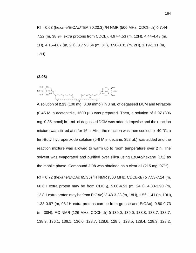
164
Rf = 0.63 (hexane/EtOAc/TEA 80:20:3) 1H NMR (500 MHz, CDCl3-d1) δ 7.44-
7.22 (m, 38.9H extra protons from CDCl3), 4.97-4.53 (m, 12H), 4.44-4.43 (m,
1H), 4.15-4.07 (m, 2H), 3.77-3.64 (m, 3H), 3.50-3.31 (m, 2H), 1.19-1.11 (m,
12H)
(2.98)
A solution of 2.23 (100 mg, 0.09 mmol) in 3 mL of degassed DCM and tetrazole
(0.45 M in acetonitrile, 1600 µL) was prepared. Then, a solution of 2.97 (306
mg, 0.35 mmol) in 1 mL of degassed DCM was added dropwise and the reaction
mixture was stirred at rt for 16 h. After the reaction was then cooled to -40 °C, a
tert-Butyl hydroperoxide solution (5-6 M in decane, 352 µL) was added and the
reaction mixture was allowed to warm up to room temperature over 2 h. The
solvent was evaporated and purified over silica using EtOAc/hexane (1/1) as
the mobile phase. Compound 2.98 was obtained as a clear oil (215 mg, 97%).
Rf = 0.72 (hexane/EtOAc 65:35) 1H NMR (500 MHz, CDCl3-d1) δ 7.33-7.14 (m,
60.6H extra proton may be from CDCl3), 5.00-4.53 (m, 24H), 4.33-3.90 (m,
12.8H extra proton may be from EtOAc), 3.48-3.23 (m, 18H), 1.56-1.41 (m, 10H),
1.33-0.97 (m, 98.1H extra protons can be from grease and EtOAc), 0.80-0.73
(m, 30H); 13C NMR (126 MHz, CDCl3-d1) δ 139.0, 139.0, 138.8, 138.7, 138.7,
138.3, 136.1, 136.1, 136.0, 128.7, 128.6, 128.5, 128.5, 128.4, 128.3, 128.2,

165
128.1, 128.0, 127.9, 127.9, 127.8, 127.7, 127.6, 127.6, 127.5, 83.3, 81.5, 80.8,
80.7, 80.6, 80.3, 80.3, 80.2, 78.9, 78.8, 78.8, 76.7, 76.6, 76.2, 76.0, 75.7, 75.6,
75.3, 75.2, 72.9, 72.9, 72.9, 72.0, 71.9, 70.0, 69.9, 69.5, 69.5, 69.4, 69.3, 69.2,
69.1, 67.3, 67.3, 60.5, 39.5, 37.8, 37.7, 37.6, 37.6, 37.5, 37.3, 37.2, 33.0, 30.0,
29.9, 29.8, 29.7, 28.1, 26.3, 25.0, 24.7, 24.5, 22.9, 22.8, 21.2, 19.9, 19.9, 19.8,
19.7, 19.6, 14.4; HRMS: 2722.7342 calcd for [C170H244O22P2Na]+ found
2722.7398.
(3R,4S,5R,6R)-6-(acetoxymethyl)-4-(((2S,3R,4S,5S,6R)-3,4,5-triacetoxy-6-
(acetoxymethyl)tetrahydro-2H-pyran-2-yl)oxy)tetrahydro-2H-pyran-2,3,5-triyl
triacetate (2.99)
Molecule 2.99 was synthesized as previously reported.65
(2R,3S,4S,5R,6S)-2-(acetoxymethyl)-6-(((2R,3R,4S,5R,6S)-3,5-diacetoxy-2-
(acetoxymethyl)-6-(ethylthio)tetrahydro-2H-pyran-4-yl)oxy)tetrahydro-2H-
pyran-3,4,5-triyl triacetate (2.100)
Molecule 2.100 was prepared as previously reported.66
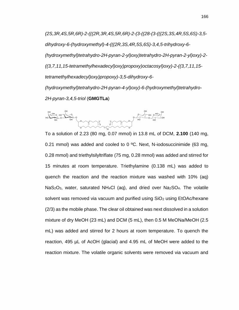
166
(2S,3R,4S,5R,6R)-2-(((2R,3R,4S,5R,6R)-2-(3-((28-(3-(((2S,3S,4R,5S,6S)-3,5-
dihydroxy-6-(hydroxymethyl)-4-(((2R,3S,4R,5S,6S)-3,4,5-trihydroxy-6-
(hydroxymethyl)tetrahydro-2H-pyran-2-yl)oxy)tetrahydro-2H-pyran-2-yl)oxy)-2-
((3,7,11,15-tetramethylhexadecyl)oxy)propoxy)octacosyl)oxy)-2-((3,7,11,15-
tetramethylhexadecyl)oxy)propoxy)-3,5-dihydroxy-6-
(hydroxymethyl)tetrahydro-2H-pyran-4-yl)oxy)-6-(hydroxymethyl)tetrahydro-
2H-pyran-3,4,5-triol (GMGTLa)
To a solution of 2.23 (80 mg, 0.07 mmol) in 13.8 mL of DCM, 2.100 (140 mg,
0.21 mmol) was added and cooled to 0 ºC. Next, N-iodosuccinimide (63 mg,
0.28 mmol) and triethylsilyltriflate (75 mg, 0.28 mmol) was added and stirred for
15 minutes at room temperature. Triethylamine (0.138 mL) was added to
quench the reaction and the reaction mixture was washed with 10% (aq)
NaS2O3, water, saturated NH4Cl (aq), and dried over Na2SO4. The volatile
solvent was removed via vacuum and purified using SiO2 using EtOAc/hexane
(2/3) as the mobile phase. The clear oil obtained was next dissolved in a solution
mixture of dry MeOH (23 mL) and DCM (5 mL), then 0.5 M MeONa/MeOH (2.5
mL) was added and stirred for 2 hours at room temperature. To quench the
reaction, 495 µL of AcOH (glacial) and 4.95 mL of MeOH were added to the
reaction mixture. The volatile organic solvents were removed via vacuum and

167
the crude product was purified using Sephadex LH-20 (DCM/MeOH: 70/30) to
obtain 24 mg of clear oil in 25% yield.
1H NMR (500 MHz, MeOD-d4/CDCl3-d1 1:1) δ 4.03-3.92 (m, 3H), 3.62-3.11 (m,
38H), 3.08-2.95 (m, 5H), 1.46-0.49 (m, 144H); 13C NMR (126 MHz, MeOD-
d4/CDCl3-d1 1:1) δ 103.5, 103.0, 103.0, 95.4, 80.8, 79.6, 75.3, 74.7, 74.5, 73.2,
72.9, 71.4, 70.9, 70.0, 68.7, 68.6, 68.3, 61.0, 60.7, 39.0, 37.0, 37.0, 36.9, 36.6,
32.4, 29.3, 29.1, 27.6, 25.7, 24.4, 24.0, 24.0, 22.0, 21.9, 19.1, 19.1, 19.0, 18.9
2.5.9 General Procedure for Liposome Formation
10 mg/mL liposome solution was prepared by first dissolving 5 mg of lipid
of interest into a 5 mL round bottom flask in a DCM/MeOH (7/3) solution. A thin
lipid film was achieved by evaporating the solvent using a rotary evaporator
(BUCHI RE111) then dried further over a hi-vacuum pump (Welch 1402) for 4
hrs. The thin lipid film was then hydrated, in a 4 mM Carboxyfluorescein (CF)
solution prepared in buffer A, by vortexing the solution for 30 seconds followed
by sonication in a water bath sonicator (Branson 2510) for 30 mins. After
sonication, the lipid mixture underwent 5 freeze thaw cycles that consisted of 2
mins at -78°C followed by 2 mins at 50°C. The lipid solution was then extruded
(Avanti mini-extruder) through 200 nm polycarbonate membrane 25 times
followed by another extrusion with a 100 nm polycarbonate membrane 51 times.

168
The lipid solution was then stored at 4°C in Protein Lo-Bind Eppendorf tube.
Liposome radius is shown in Figure S8 below.
2.5.10 General Procedure to Measure pH Equilibrium of CF
In order to obtain the fast rate of pH induced decrease of CF for EggPC,
a stopped-flow assay was necessary. Before each assay, 10 µL of the stock
extruded lipid solution was diluted in 500 µL of Bis-Tris buffer pH 7.2. Free CF
was removed using a PD miniTrapTM G-25 SephadexTM column from GE
Healthcare ending 100 times dilution from the stock extruded solution (0.1
mg/mL). The reaction cell was cleaned using Bis-Tris buffer pH 7.2 (15 drives)
until a steady signal was obtained before running any experiments. After a
steady fluorescence signal was achieved and free CF was removed from the
liposome solution, 25 µl of the lipid solution was mixed with 225 µL of Bis-Tris
buffer pH 7.2 to obtain the relative maximum fluorescence at pH 7.2 (5
acquisitions). The reaction cell was again cleaned until a steady signal was
observed and repeated with Bis-Tris buffer pH 5.8 (5 acquisitions). To confirm
100% change of fluorescence at pH 5.8 from the assay, the relative
fluorescence at 1000 seconds was confirmed by comparison to the relative
fluorescence of liposomes in Bis-Tris buffer pH 5.8 doped with 1 µL of a solution
of Nigericin in Ethanol (100 µM) using the plate reader.

169
To obtain the pH equilibration observed rate of each lipid, the decrease
in fluorescence of CF was followed using Perkin Elmer Enspire© multimode
plate reader. Before each assay, 10 µL of the stock extruded lipid solution was
diluted in 500 µL of buffer A. Free CF was removed using a PD miniTrapTM G-
25 SephadexTM column from GE Healthcare ending in 100 times dilution from
the stock extruded solution (0.1 mg/mL). 45 µL of purified liposome solution was
next added into three 0.5 mL Protein Lo-Bind tubes for each lipid solution. In
one tube, 405 µL of Bis-Tris buffer pH 7.2 was added. In the second tube, 405
µL of Bis-Tris buffer pH 5.8 was added with 1 µL of 100 µM solution of Nigericin
in Ethanol. In the third tube, right before starting the measurement, 405 µL of
Bis-Tris buffer pH 5.8 was added. 125 µL was added to each well of the plate
three times for each tubes resulting in three measurements with three replicates
with a total of 9 measurements per lipid solution.
2.6 Acknowledgements
Chapter 2 is adapted, in part, from Takaoki Koyanagi, Geoffray Leriche,
David Onofrei, Gregory P. Holland, Michael Mayer, and Jerry Yang.
“Cyclohexane Rings Reduce Membrane Permeability to Small Ions in Archaea-
Inspired Tetraether Lipids.” Angew. Chem., Int. Ed. 2016, 55, 1890–1893.
Copyright 2016 Wiley. Permission to use copyrighted images and data in the
manuscript was also obtained from Geoffray Leriche, David Onofrei, Gregory P.
Holland, Michael Mayer, and Jerry Yang. The dissertation author is the first

170
author of this manuscript. Chapter 2 is also adapted, in part, from Takaoki
Koyanagi, Geoffray Leriche, Alvin Yep, David Onofrei, Gregory P. Holland,
Michael Mayer, and Jerry Yang. “Effect of Headgroups on Small-Ion
Permeability across Archaea-Inspired Tetraether Lipid Membranes. Chem.” -
Eur. J. 2016, 22, 8074–8077. Copyright 2016 Wiley. Permission to use copyright
images and data in the manuscript was also obtained from Geoffray Leriche,
Alvin Yep, David Onofrei, Gregory P. Holland, Michael Mayer, and Jerry Yang.
The dissertation author is the first author of this manuscript. Chapter 2 is also
adapted, in part, from Thomas B. H. Schroeder, Geoffray Leriche, Takaoki
Koyanagi, Mitchell A. Johnson, Kathryn N. Haengel, Olivia M. Eggenberger,
Claire L. Wang, Young Hun Kim, Karthik Diraviyam, David Sept, Jerry Yang,
and Michael Mayer. “Effects of Lipid Tethering in Extremophile-Inspired
Membranes on H+/OH− Flux at Room Temperature.” Biophys. J. 2016, 110,
2430–2440. Copyright 2016 Elsevier. Permission to use copyright images and
data in the manuscript was also obtained from Thomas B. H. Schroeder,
Geoffray Leriche, Mitchell A. Johnson, Kathryn N. Haengel, Olivia M.
Eggenberger, Claire L. Wang, Young Hun Kim, Karthik Diraviyam, David Sept,
Jerry Yang, and Michael Mayer.

171
2.7 References
(1) Jain, S.; Caforio, A.; Driessen, A. J. M. Front. Microbiol. 2014, 5, 641.
(2) Villanueva, L.; Damsté, J. S. S.; Schouten, S. Nat. Rev. Microbiol. 2014, 12 (6), 438–448.
(3) Van Meer, G.; Voelker, D. R.; Feigenson, G. W. Nat. Rev. Mol. Cell Biol. 2008, 9 (2), 112–124.
(4) Koga, Y.; Morii, H. Microbiol. Mol. Biol. Rev. 2007, 71 (1), 97–120.
(5) Woese, C. R.; Magrum, L. J.; Fox, G. E. J. Mol. Evol. 1978, 11 (3), 245–252.
(6) Valentine, D. L. Nat. Rev. Microbiol. 2007, 5, 316–323.
(7) Chong, P. L.-G.; Ayesa, U.; Daswani, V. P.; Hur, E. C. Archaea 2012, 2012, 1–11.
(8) Gabriel, J. L.; Lee Gau Chong, P. Chem. Phys. Lipids 2000, 105 (2), 193–200.
(9) Chong, P. L. G. Chem. Phys. Lipids 2010, 163 (3), 253–265.
(10) Konings, W. N.; Albers, S. V; Koning, S.; Driessen, A. J. M. Antonie van Leeuwen 2002, 81, 61–72.
(11) Boyd, E. S.; Hamilton, T. L.; Wang, J.; He, L.; Zhang, C. L. Front. Microbiol. 2013, 4 (APR), 1–15.
(12) Baker-Austin, C.; Dopson, M. Trends Microbiol. 2007, 15 (4), 165–171.
172
(13) Elferink, M. G. L.; de Wit, J. G.; Driessen, A. J. M.; Konings, W. N. Biochim. Biophys. Acta, Biomembr. 1994, 1193 (2), 247–254.
(14) Shimada, H.; Nemoto, N.; Shida, Y.; Oshima, T.; Yamagishi, A. J. Bacteriol. 2008, 190 (15), 5404–5411.
(15) Wang, X.; Lv, B.; Cai, G.; Fu, L.; Wu, Y.; Wang, X.; Ren, B.; Ma, H. Sci. Rep. 2012, 2 (2), 892.
(16) Chang, E. L. Biochem. Biophys. Res. Commun. 1994, 202 (2), 673–679.
(17) Patel, G. B.; Agnew, B. J.; Deschatelets, L.; Fleming, L. P.; Sprott, G. D. Int. J. Pharm. 2000, 194, 39–49.
(18) Uda, I.; Sugai, a; Itoh, Y. H.; Itoh, T. Lipids 2001, 36 (1), 103–105.
(19) Svenson, S.; Thompson, D. J. Org. Chem. 1998, 3263 (98), 7180–7182.
(20) Brard, M.; Lainé, C.; Réthoré, G.; Laurent, I.; Neveu, C.; Lemiègre, L.; Benvegnu, T. J. Org. Chem. 2007, 72 (22), 8267–8279.
(21) Eguchi, T.; Ibaragi, K.; Kakinuma, K. J. Org. Chem. 1998, 63 (8), 2689–2698.
(22) WJ, Z.; CE, C. Cell and Model Membrane Interactions; Ohki, S., Ed.; Plenum Press: New York, 1991.
(23) Lecollinet, G.; Auzély-Velty, R.; Danel, M.; Benvegnu, T.; Mackenzie, G.; Goodby, J. W.; Plusquellec, D. J. Org. Chem. 1999, 64 (9), 3139–3150.
(24) Brard, M.; Richter, W.; Benvegnu, T.; Plusquellec, D. J. Am. Chem. Soc. 2004, 126 (32), 10003–10012.
173
(25) Godage, H. Y.; Riley, A. M.; Woodman, T. J.; Thomas, M. P.; Mahon, M. F.; Potter, B. V. L. J. Org. Chem. 2013, 78, 2275–2288.
(26) Gilbert, I. H.; Holmes, A. B.; Young, R. C. Tetrahedron Lett. 1990, 31 (18), 2633–2634.
(27) Lewis, R. N.; McElhaney, R. N. Biophys. J. 2000, 79, 1455–1464.
(28) Nagle, J. F. J. Membr. Biol. 1976, 27 (3), 233–250.
(29) Sipai Altaf Bhai, M.; Vandana, Y.; Mamatha, Y.; Prasanth, V. V. J. Pharm. Sci. Innov. 2012, 1 (1), 13–21.
(30) Arakawa, K.; Eguchi, T.; Kakinuma, K. Bull. Chem. Soc. Jpn. 2001, 74 (2), 347–356.
(31) Wilschut, J.; Papahadjopoulos, D. Nature. 1979, pp 690–692.
(32) Archaea, B.; Organismal, H. Group 1983, 103–112.
(33) Gliozzi, A.; Relini, A.; Chong, P. L.-G. J. Membr. Sci. 2002, 206, 131–147.
(34) Andersson, A.-S. J. Biol. Chem. 1996, 271 (12), 6801–6809.
(35) Mathai, J. C.; Tristram-Nagle, S.; Nagle, J. F.; Zeidel, M. L. J. Gen. Physiol. 2007, 131 (1), 69–76.
(36) Jansen, M.; Blume, A. Biophys. J. 1995, 68 (3), 997–1008.
(37) Spooner, M. J.; Gale, P. A. Chem. Commun. 2015, 51 (51), 4883–4886.
(38) Boggs, J. M. Biochim. Biophys. Acta 1987, 906 (3), 353–404.

174
(39) Hubner, W.; Blume, A. Chem. Phys. Lipids 1998, 96 (1–2), 99–123.
(40) Murzyn, K.; Róg, T.; Pasenkiewicz-Gierula, M. Biophys. J. 2005, 88 (2), 1091–1103.
(41) Koyanagi, T.; Leriche, G.; Onofrei, D.; Holland, G. P.; Mayer, M.; Yang, J. Angew. Chem., Int. Ed. 2016, 55, 1890–1893.
(42) Chang Chung, Y.; Hong Chiu, Y.; Wei Wu, Y.; Tai Tao, Y. Biomaterials 2005, 26 (15), 2313–2324.
(43) Febo-Ayala, W.; Morera-Félix, S. L.; Hrycyna, C. A.; Thompson, D. H. Biochemistry 2006, 45 (49), 14683–14694.
(44) Gao, Y.; Vlahakis, J. Z.; Szarek, W. A.; Brockhausen, I. Bioorg. Med. Chem. 2013, 21 (5), 1305–1311.
(45) Shirouzu, T.; Watari, K.; Ono, M.; Koizumi, K.; Saiki, I.; Tanaka, C.; van Soest, R. W. M.; Miyamoto, T. J. Nat. Prod. 2013, 76 (7), 1337–1342.
(46) Mangaleswaran, S.; Argade, P. N. J. Or 2001, No. 17, 5259–5261.
(47) Babu, K. V.; Sharma, G. V. M. Tetrahedron Asym. 2008, 19 (5), 577–583.
(48) Mioskowski, C.; Heitz, M.-P.; Wagner, A. Tetrahedron Lett. 1989, No. 5, 557–558.
(49) WO 2009/135977.
(50) Shimojo, M.; Matsumoto, K.; Hatanaka, M. Tetrahedron 2000, 56 (47), 9281–9288.

175
(51) Pan, X.; Lacôte, E.; Lalevée, J.; Curran, D. P. J. Am. Chem. Soc. 2012, 134 (12), 5669–5674.
(52) Trigo, G. G.; Muñoz, E. M.; Llama-Hurtado, E. J. Heterocycl. Chem. 1984, 21 (5), 1479–1483.
(53) Lowik, D. W. P. M.; Liskamp, R. M. J. Eur. J. Org. Chem. 2000, 2000 (7), 1219–1228.
(54) Prabhakar, P.; Rajaram, S.; Reddy, D. K.; Shekar, V.; Venkateswarlu, Y. Tetrahedron Asym. 2010, 21 (2), 216–221.
(55) Qin, L.; Ren, X.; Lu, Y.; Li, Y.; Zhou, J. Angew. Chem., Int. Ed. Engl. 2012, 51 (24), 5915–5919.
(56) Flowers, C. L.; Vogel, P. Chem. - A Eur. J. 2010, 16 (47), 14074–14082.
(57) Marinier, A.; Deslongchamps, P. Tetrahedron Lett. 1988, 29 (48), 6215–6218.
(58) Bartoli, S.; De Nicola, G.; Roelens, S. J. Org. Chem. 2003, 68 (21), 8149–8156.
(59) Iwashita, M.; Makide, K.; Nonomura, T.; Misumi, Y.; Otani, Y.; Ishida, M.; Taguchi, R.; Tsujimoto, M.; Aoki, J.; Arai, H.; Ohwada, T. J. Med. Chem. 2009, 52 (19), 5837–5863.
(60) Yar, M.; Fritz, S. P.; Gates, P. J.; McGarrigle, E. M.; Aggarwal, V. K. Eur. J. Org. Chem. 2012, 1, 160–166.
(61) Rzepecki, P. W.; Prestwich, G. D. J. Org. Chem. 2002, 67 (16), 5454–5460.

176
(62) Devaraj, S.; Shashidhar, M. S.; Dixit, S. S. Tetrahedron 2005, 61 (3), 529–536.
(63) Conway, S. J.; Gardiner, J.; Grove, S. J. a; Johns, M. K.; Lim, Z.-Y.; Painter, G. F.; Robinson, D. E. J. E.; Schieber, C.; Thuring, J. W.; Wong, L. S.-M.; Yin, M.-X.; Burgess, A. W.; Catimel, B.; Hawkins, P. T.; Ktistakis, N. T.; Stephens, L. R.; Holmes, A. B. Org. Biomol. Chem. 2010, 8 (1), 66–76.
(64) Elie, C. J. J.; Verduyn, R.; Dreef, C. E.; Brounts, D. M.; van der Marel, G. A.; van Boom, J. H. Tetrahedron 1990, 46 (24), 8243–8254.
(65) Šardzík, R.; Noble, G. T.; Weissenborn, M. J.; Martin, A.; Webb, S. J.; Flitsch, S. L. Beilstein J. Org. Chem. 2010, 6, 699–703.
(66) Contour, M. O.; Defaye, J.; Little, M.; Wong, E. Carbohydr. Res. 1989, 193, 283–287.
(67) Koyanagi, T.; Leriche, G.; Yep, A.; Onofrei, D.; Holland, G. P.; Mayer, M.; Yang, J. Chem. - Eur. J. 2016, 22 (24), 8074–8077.

177
Chapter 3
Combining Structural Strategies Used by Eukaryotes
and Archaea to Enhance Membrane Properties of
Archaea Inspired Tetraether Lipids
3.1. Introduction
Leakage across membranes is a problem for applications in drug
delivery.1–3 For example, liposomes made from commercially available diacyl
lipids were shown to be unstable towards hydrolysis and leaky, especially in the
presence of serum.3 The problem of content leakage and membrane instability
has prompted many researchers to steer away from liposomal delivery systems
and towards technologies based on covalent attachment of drugs to polymeric
backbones of nanoparticles, as discussed in chapter 1 of this dissertation.4–9
However, unlike drug-polymer conjugates that are typically limited to chemically
adhering only one type of drug or cell-targeting functional group to a polymeric
backbone, the potential versatility of liposomes include encapsulation of
different types of drugs, facile addition of steric barrier functionalized lipid, and
simple addition of peptide lipid conjugates for cell-mediated drug delivery
without the need for multiple-chemical modification continues to make liposomal

178
delivery systems an attractive incentive for further development of membranes
with low permeability.1,10–13
In nature, several strategies are used by organisms to address problems
of membrane integrity. Eukaryotic organisms, for instance, manufacture free
cholesterol to fill defects in bilayer lipid membranes, which lead to increased
membrane stability (Figure 3.1A).14 Archaea, on the other hand, generate lipids
with ether linkages and tethered lipid tails (Figure 3.1B) that result in chemically
stable membrane-spanning lipids that make it possible for the organisms to
thrive in extreme environments.15
Figure 3.1. Comparison of strategies to improve membrane integrity used by eukaryotes
(A) or Archaea (B) with the novel strategy that combines both strategies in the synthetic
hybrid lipid introduced here (C).

179
To develop liposomes with reduced leakage properties, we have
designed Archaea-inspired tetraether lipids, GMGTPC-CH (synthesized in
Chapter 2 of this dissertation), that incorporate structural features that are
believed to increase membrane properties (Figure 3.2). This newly developed
synthetic tetraether lipids readily formed stable liposomes and exhibited
reduction of small ion membrane leakage by ~2 orders of magnitude when
compared with leakage properties of membranes formed from commercially
available diacyl lipids, EggPC (see chapter 2).16 In contrast, the addition of free
cholesterol to diacyl lipid formulations (using the strategy invoked by
Eukaryotes) has been a common practice used by research groups to decrease
membrane leakage by improving membrane integrity. However, free cholesterol
has been observed to leach out from liposomes after injection into serum
Figure 3.2. Structures of Archaea-inspired tetraether lipids with (GcGTPC-CH) or without
(GMGTPC-CH) covalent cholesterol integration.

180
containing environment, resulting in liposomal destabilization leading to
premature loss of cargo.17 One solution developed by Szoka’s group, has been
to covalently attach cholesterol to the glycerol backbone, which has been shown
to eliminate cholesterol leaching from liposomes.18–20
In our design to synthesize lipids with further improved membrane
properties, we explored the possibility to combine these two strategies used by
nature for generating robust membranes by creating a hybrid lipid that
incorporates covalent cholesterol integration into a tethered tetraether lipid
(Figure 3.1C). Extracting the best structural features that reduces membrane
permeability from our previously synthesized Archaea-inspired tetraether lipids
from chapter 2, we preserved the cyclohexane integrated tethered tetraether
lipid with phosphocholine headgroups, while replacing the phytanyl groups with
an ethylene glycol extended cholesterol (shown in Figure 3.2.). Following the
synthesis, we compared the leakage properties of membranes composed of this
hybrid lipid to membranes formed from commercial diacyl lipids and an
Archaea-inspired lipid that did not comprise covalently attached cholesterol
(both with and without incorporation of free cholesterol in the membranes). In
addition, molecular dynamics (MD) simulations provided a molecular
interpretation for the observed experimental differences in leakage properties
between different lipid membrane compositions. Lastly, we examined the
capability of liposomes made from this hybrid lipid to incorporate functional ion
channels into membranes, explored the capability of these lipids to be

181
recognized as substrates by a membrane-active phospholipase-D (PLD)
enzyme, assessed the stability of liposomes composed of these lipids in serum,
and evaluated the uptake of such liposomes in cells.
3.2. Design/Synthesis of GcGTPC-CH
Several synthetic strategies of GcGTPC-CH were tested to maintain the
olefin of the added cholesterol appendage. First, shown in Scheme 3.1A, we
initially examined the possibility of synthesizing the glycerol backbone with
epichlorohydrin following a previously published synthesis by Benvegnu’s group
that reported ~50% product conversion to a hemicyclic lipid scaffold.21 However,
when the crude material was analyzed via TLC and NMR after the reaction was
mixed for 12 hours at 70 °C, only starting material diol 2.34 was recovered. To
examine whether the initial coupling of diol 2.34 with epichlorohydrin takes place,
the same reaction was repeated with an excess of epichlorohydrin (12 eq. from
2.1 eq.), however, no product formation was observed via 1H-NMR. The lack of
transformation observed for this reaction could be explained by the poor
solubility of diol 2.34 in hexane, which may differ in solubility properties to the
diol used by Benvegnu’s group.
Next, we examined the possibility of starting the reaction of the glycerol
scaffold from solketal (shown in Scheme 3.1B). Initial alkylation of alkyldibromo
2.36 by the free alcohol on solketal afforded tethered lipid precursor 3.1.
Following an acid catalyzed ring opening of the acetals forming tetraol 3.2, the

182
terminal alcohols were protected by trityl groups affording protected lipid
precursor 3.3. Following a previously published protocol by Benvegnu’s
groups21, alkylation of tosylated cholesterol was attempted with the free
secondary diols of 3.3 under basic conditions in THF by heating the reaction to
reflux while allowing the solvent to evaporate at 130 °C. However, after the
reaction was stirred at 130 °C overnight (effectively neat), no product or starting
material was isolated (black degradation products were observed). Believing
the degradation occurred because the reaction was performed neat, the same
reaction was performed in DMF. After the initial addition of NaH, however, the
Scheme 3.1. Unsuccessful synthesis employed for GcGTPC-CH. A) Synthetic strategy of
GcGTPC-CH using epichlorohydrin. B) Synthetic strategy of GcGTPC-CH using solketal.

183
reaction became black and turbid. The suspension in DMF was stirred for 24
hours at 130 °C, but the suspension never became homogenous and
degradation products were observed. The synthetic scheme was not further
optimized because of the low yields associated with the reactions steps up to
that point (9% over 3 steps).
For our third synthetic approach to GcGTPC-CH, we attempted a similar
strategy used for our previously synthesized tetraether lipids (shown in chapter
2) as our starting point. As shown in Scheme 3.1B, we initially designed the
GcGTPC-CH lipid to comprise a cholesterol group directly attached to the
glycerol lipid backbone without an ethylene glycol spacer. However, alkylation
with the glycerol alcohol to the tosylated cholesterol was not achieved, but rather,
the alcohol of the glycerol became tosylated instead. From this result, we
decided to increase access of the nucleophile to the tosylated electrophile by
synthetically modifying the cholesterol to include an ethylene glycol spacer on
the 3β alcohol. The new plan to synthesize GcGTPC-CH began with a tosylation
of the 3β alcohol on cholesterol followed by substitution of the tosyl group with
ethylene glycol that afforded modified cholesterol 3.5 with retention of
stereochemistry on cholesterol (shown in Scheme 3.2).22 The free terminal
alcohol in 3.5 was then converted to the corresponding tosylate to form 3.6,

184
which successively reacted with 2-phenyl-1,3-dioxan-5-ol. The acetal of the
Scheme 3.2. Successful synthesis of GcGTPC-CH

185
newly formed cholesterol integrated glycerol scaffold 3.7 was deprotected using
DIBAL-H to form the benzyl protected glycerol backbone 3.8. Next, the
cyclohexane integrated diibromoalkane 2.36, from chapter 2, was alkylated by
glycerol 3.8 under basic conditions to generate benzyl-protected lipid 3.9.
Notably, to obtain the diol 3.10 without reducing the olefins associated with the
cholesterol, several conditions were screened (i.e. reaction time, amount of Pd,
reaction concentration). Ultimately, we were able to optimize the reaction
conditions for debenzylation without reducing the olefin by reducing the reaction
time (to 20 minutes compared to our previous conditions that required 5 hours
to 16 hours of mixing at room temperature) and using less Pd (5% w/w vs. 10%
w/w used in previous conditions) and diluting the reaction (4.6 times) in
THF:EtOH (1:1) to generate the desired diol 3.10 with the olefins intact. The
formation of GcGTPC-CH was completed in a series of reactions that involved
reacting diol 3.10 with 2- bromoethyl dichlorophosphate, which was followed by
the displacement of bromides by trimethylamine (i.e., an analogous synthetic
strategy as described in chapter 2).
3.3. Physical Characterization of Liposomes Made from Pure GcGTPC-CH,
GMGTPC-CH, and Diacyl Lipids
After successfully synthesizing GcGTPC-CH, we examined whether we
can form stable liposomes with pure GcGTPC-CH lipids. Dynamic light
scattering (DLS) measurements demonstrated that we could generate
liposomes with an average hydrodynamic diameter of ~150 nm by extrusion

186
through a 100 nm polycarbonate membrane (see Figure 3.3A). We then probed
the physical characteristics of GcGTPC-CH and examined whether replacing
the phytanyl group (which prevents natural archaeal tetraether lipids from
undergoing a phase transition between 0 to 80 ⁰C)23 in GMGTPC-CH with a
cholesterol moiety would still maintain a fluid phase for liposomes within a useful
temperature range. Differential scanning calorimetry (DSC) measurements
(taken by David Onofrei) revealed that GcGTPC-CH lipid remained in a liquid
phase from 5 to 65 °C (see Figure 3.3B). This result was in agreement with
Figure 3.3. Physical characterization of lipids. A) DLS measurements of varying liposomal
formulations. B) DSC measurements taken of synthetic and diacyl lipids. DSC
measurements taken by David Onofrei.

187
previous reports, which showed that adding 40-50 mol% of free cholesterol to
diacyl lipids24 maintains a fluid phase and diminishes its phase transition.
Furthermore, liposomes remained structurally stable throughout the time frame
of leakage experiments and we did not observe any measurable changes in size
by DLS (see Figure 3.3A).
3.4. Investigation of Membrane Leakage Across Membranes Composed of
GcGTPC-CH and Diacyl Lipid
3.4.1. Comparison of Small Ion Leakage Rates Across Membranes Composed
of Synthetic and Diacyl Lipids
Liposomes were formed using the same protocol as described in Chapter
2 (section 2.5.9). Using a previously described pH equilibration method (section
2.5.10) to estimate initial rates of leakage of small ions from liposomes,16,25 we
evaluated the observed rates of small ion membrane leakage to study whether
the addition of free cholesterol (40 mol%) to POPC, GMGTPC-CH, or GcGTPC-
CH liposomal formulations affected membrane permeation of small ions (see
Figure 3.4). As expected, the addition of free cholesterol to POPC resulted in a
beneficial decrease (10-fold) in observed rate of small ion membrane leakage
compared with cholesterol-free POPC membranes26 (Figure 3.5.). However, the
addition of free cholesterol to GMGTPC-CH or GcGTPC-CH did not have a
significant effect on the observed rate of small ion membrane leakage compared

188
to the same lipid without added free cholesterol. To further probe this result, we
examined whether free cholesterol was capable of integrating into the
membranes composed of GMGTPC-CH or GcGTPC-CH. The presence of free
cholesterol in GMGTPC-CH liposomes was confirmed by titration of both total
phosphorus content and free total cholesterol using a Bartlett assay27 and an
Figure 3.4. Normalized average plot of % CF fluorescence vs. time (h). (N = 9) fluorescence
intensity (monitored at λ(Ex/Em) = 485/517 nm)

189
Amplex® Red cholesterol assay, respectively. The result showed a presence of
31 ± 10 mol% of free cholesterol in GMGTPC-CH liposomes, which is within the
expected range of cholesterol concentration for liposomal formulations made by
extrusion.28 However, no free cholesterol was detected in GcGTPC-CH
liposomes by the same analysis used for GMGTPC-CH liposomes. The
absence of free cholesterol incorporated in GcGTPC-CH lipid membranes is in
agreement with previous reports that suggest that the maximum incorporation
Figure 3.5. Membrane leakage results of small ions from liposomes formed from synthetic
or POPC lipids with/without added free cholesterol. Comparison of the observed initial rates
of decreased carboxyfluorescein (CF) fluorescence intensity (monitored at λ(Ex/Em) =
485/517 nm) from CF-encapsulated liposomes comprised of GMGTPC-CH, GcGTPC-CH,
or POPC lipid with/without added 40 mol% cholesterol during liposome formation with
averaged observed initial rates of membrane leakage noted over each box (unit = h-1;N=9).

190
of total cholesterol in fluid lipid membranes is ~50 mol% for liposomes prepared
by extrusion.28 Remarkably, despite the absence of membrane benefits
associated with the addition of free cholesterol to liposomes, liposomes
composed of pure GcGTPC-CH displayed a five-fold decrease in the observed
rate of small ion membrane leakage compared with liposomes made of pure
GMGTPC-CH (Figure 3.5). This observed reduction in small ion membrane
leakage from GcGTPC-CH is consistent with the hypothesis that covalently
attaching cholesterol to the glycerol backbone in GcGTPC-CH allows
cholesterol to appropriately orient within the membrane and results in tighter
lipid packing than GMGTPC-CH (with or without added free cholesterol).
3.4.2. Comparison of Small Molecule Leakage Rates Across Membranes
Composed of Synthetic and Diacyl Lipids
When deciding on a FDA approved drug that will be used to probe
membrane leakage properties of small molecules, we limited our options of
drugs to neutrally charged molecules instead of charged molecules based on
our previously published data that showed very little leakage from liposomes
made from GMGTPC lipids for charged molecules.29 From the list of drug
candidates to screen for membrane leakage (shown in Figure 3.6), we began
with 5-FU (5-fluorouracil). Initially, we passively encapsulated 5-FU in liposomes
made with POPC/chol, GMGTPC-CH/chol or GcGTPC-CH and used a dialysis

191
assay to compare the leakage of 5-FU over a 48 hour incubation at 37 °C (see
section 3.9.4. for more details). However, after the liposomes were purified
using Sephadex®, no free 5-FU was detected via HPLC for liposomes made
with either POPC/chol or GMGTPC-CH/chol. Liposomes made with GcGTPC-
CH, on the other hand, had detectable free drug after purification, but the rate
of leakage was too fast and did not appear to significantly differ from leakage of
free drug through the dialysis membrane. Thinking the fast leakage from
liposomes was due to the small size of 5-FU (130 g/mol), we examined the
leakage of 5-FdU (246 g/mol), which has a molecular weight of ~ 2 times larger.
Unfortunately, although the free drug was detectable using HPLC for
POPC/chol liposomes after purification, the observed rate of leakage was too
fast and the leakage rate was similar to the rate of leakage of the molecule
through the dialysis membrane in the absence of liposome encapsulation. This
Figure 3.6. FDA approved drugs screened for membrane leakage.

192
conclusion arose from having two separate batches of POPC/chol liposomes
made sequentially with a 15 minute waiting period between the time it required
for the Sephadex® to be washed, while the second batch of liposomes loaded
with 5-FdU was equilibrating with the buffer (leaking). When we compared the
two initial HPLC signals, the area measured for 5FdU was effectively baseline
for the initial time point for the second batch (suggesting complete leakage while
the Sephadex® was being cleaned). After concluding that the fast rate of
leakage of 5-FdU from POPC/chol liposomes would be a challenge to interpret
kinetically, we decided to screen a different drug instead of trying to tease out
small differences that will most likely be masked by signal noise/error. Trying to
understand why the molecules tested, thus far, had a fast rate of leakage, we
hypothesized the fast leakage was caused by the fluorine substituent. Fluorine
groups have been added to pharmaceutical molecules to enhance permeation
through stomach linings, as such, may contribute to the rapid membrane
leakage.30 The next drug that was screened was Stavudine, (shown in Figure
3.6), an FDA approved drug that resembles a reduced 5-FdU without the
fluorine substituent. However, similar fast leakage profile of 5FU was observed
for Stavudine. From the membrane leakage results obtained for Stavudine, our
hypothesis of the fluorine substituent driving the fast leakage was dismissed.
Therefore, we decided to probe another drug, gemcitabine (shown in
Figure 3.6), an FDA drug most similar to cytarabine (an FDA approved drug that
has been shown to be retained (80%) inside liposomes made with GMGTPC

193
lipids after 48 hours at 37 °C29). We passively encapsulated gemcitabine
(GEM)31 in liposomes to compare permeability of a neutrally charged molecule
across membranes composed of POPC/chol, GMGTPC-CH/chol or pure
GcGTPC-CH. When we compare the leakage of GEM over a 48 hour incubation
at 37 °C (see section 3.9.4. for more details), we were able to observe retention
of free drug inside liposomes long enough to measure leakage rates.
Remarkably, in contrast to the results obtained from the small ion membrane
leakage assay (Figure 3.5) that did not show a significant difference between
POPC/chol and GMGTPC-CH/chol, all 3 liposomal compositions exhibited
significantly different leakage profiles for encapsulated GEM (Figure 3.7).
Notably, liposomes made from pure GcGTPC-CH retained 69.1 ± 4.1% of GEM
after 48 hours, while liposomes made from POPC/chol (2.3 ± 0.1% retained
GEM) or GMGTPC-CH/chol (7.1 ± 1.2% retained GEM) exhibited almost
complete loss of encapsulated GEM over the same period of time. These
contrasting results between small ion versus small molecule leakage profiles for
the different lipids could reflect differences in mechanisms of leakage, where
small ions are believed to leak through transient membrane pores and neutral
molecules could leak through both membrane pores and membrane
partitioning.32

194
3.4.3. Molecular Dynamics Simulations of Membranes Composed of Synthetic
and Diacyl Lipids
In order to provide a molecular interpretation for the observed
experimental trend in membrane permeability for POPC, GMGTPC-CH, and
GcGTPC-CH, we performed molecular dynamics (MD) simulations of pure lipids
Figure 3.7. Membrane leakage results of gemcitabine (GEM, a neutrally charged drug) from
liposomes formed from synthetic or POPC lipids with/without added free cholesterol. Graph
of percent retention versus time of GEM encapsulated inside liposomes made from
GcGTPC-CH, GMGTPC-CH with 40 mol% of free cholesterol, POPC with 40 mol% of free
cholesterol, or free GEM (i.e., no liposomes) across a dialysis membrane upon incubation at
37 ºC for 48 hours.

195
in membranes (see section 3.9.5. for details). Figure 3.8A shows overlaid
Figure 3.8. Results from molecular dynamics (MD) simulations of a membrane composed
of pure POPC, GMGTPC-CH, or GcGTPC-CH lipids. A) Overlay of 10 snapshots of
individual lipids (taken in 5 ns intervals from MD simulations of pure lipid membranes)
highlighting the increased ordering of the lipid tails from POPC to GMGTPC-CH to
GcGTPC-CH. B) Graph of the area occupied by individual lipids (within membranes
comprised of pure lipids) over a 50 ns membrane simulation highlighting the variance in
area per lipid (represented quantitatively as standard deviation (sd)). Experiments
performed by Prof. David Sept.

196
snapshots of the same individual lipids at 5 ns intervals revealing the
conformational space explored by each lipid over 50 ns (experiments performed
by Prof. David Sept). The results highlight striking differences in the order of the
lipid tails between these lipids. The reduced flexibility of lipid tails in the
tetraether lipids translated to a decreased variance in the area occupied per lipid
(Figure 3.8B), which could be represented quantitatively by the standard
deviation (sd) around the mean area/lipid (Å2). These calculations are in good
agreement with the observed experimental trends for leakage of small ions and
molecules across lipid membranes (Figure 3.5 and 3.7), supporting the concept
that decreased variance in area per lipid leads to tighter lipid packing and lower
Figure 3.9. Total number of waters within the membrane for each snapshot over the final
50 ns of the trajectory. Experiments performed by Prof. David Sept.

197
permeability.33 Furthermore, MD simulations (experiments performed by Prof.
David Sept) shows an estimate of membrane water penetration for the different
lipids34 where we found 24 penetration events (i.e., transient events where 3 or
more water molecules resided anywhere within the hydrophobic region of a
membrane over a 50 ns MD simulation) for pure POPC membranes, 20 events
for GMGTPC-CH and only 1 event for GcGTPC-CH under the same conditions
(see Figure 3.9). These differences in the number of observed water penetration
events presumably reflect variations in both composition and conformational
freedom between the different lipids tails, since all lipids contained the same
phosphocholine headgroups.
3.5. Investigation of Functional Membrane Properties of GcGTPC-CH
Membranes
To explore whether membranes composed of GcGTPC-CH lipids
retained useful membrane properties for potential biological applications
currently using diacyl lipids, we examined whether 1) GcGTPC-CH lipid
membranes can act as a membrane support for natural ion channel forming
peptides such as gramicidin A (gA),35,36 2) GcGTPC-CH lipids can serve as a
substrate for membrane active enzymes such as phospholipase-D (PLD),37–39
3) liposomes derived from GcGTPC-CH can remain sufficiently stable in serum-
containing buffers,3,40,41 and 4) small molecules encapsulated in GcGTPC-CH
liposomes can be delivered to living cells.42–44

198
3.5.1. Investigation of GcGTPC-CH Lipid Membrane to Support Gramicidin A
Formation
Gramicidin A (gA) is an antibiotic peptide that forms well defined ion
channels in membranes.45 Proper nanopore formation of gA in liposomal
membranes allows monovalent cations to selectively cross between the
surrounding buffer and the intra-liposomal buffer. Using the pH equilibration
assay that we used to measure passive membrane leakage, we added gA to a
buffered solution at pH 5.8 containing GcGTPC-CH liposomes with
encapsulated carboxyfluorescein (CF) and an intra-liposomal pH of 7.2. To
determine whether gA was functional upon incorporation into the membrane,
pH-dependent fluorescence emission of CF was monitored for a flux of
monovalent cations (predominantly H+ ion flux) into the liposome, causing an
accelerated reduction of CF fluorescence when compared with liposomes
without the addition of gA. Similar to previously reported results that used diacyl
lipids,35,36 the addition of gA to GcGTPC-CH liposomes reduced 95% of the
fluorescence of CF after 30 minutes (Figure 3.10A), suggesting that the ion
channels were functional within the membrane. In addition, liposomes with just
DMSO (without gA) was added to measure fluorescence changes to CF to
confirm liposomal stability in the presence of DMSO (Figure 3.10A).
Furthermore, to ensure the addition of gA does not cause the liposomes to
rupture, the presence of stable liposomal structures throughout the experiment
was confirmed using DLS measurements (see Figure 3.10B).

199
3.5.2. Investigation of GcGTPC-CH Lipid Membrane to Act as a Substrate for
PLD
We assessed whether phospholipase-D (PLD), a biological membrane-
active enzyme that cleaves phosphodiester bonds in lipids, could recognize
synthetic GcGTPC-CH lipids as a substrate. While phospholipase-C (PLC)
requires an ester functionality on the C-2 carbon of the glycerol backbone for
lipid recognition46, the structural requirements necessary for PLD to recognize
a lipid substrate are not known. Following reported protocols for probing the
activity of PLD in liposomes composing commercial diacyl lipids,37–39 PLD was
added to a liposomal suspension composed of pure GcGTPC-CH lipids in the
presence of Ca2+ ions at 37 °C. We observed a large morphological change in
Figure 3.10. Effect of gramicidin A on GcGTPC-CH liposomes. A) Percent fluorescence
of CF after incubation with or without gA at 37 ºC for 30 minutes. B) DLS measurements
of GcGTPC-CH liposomes under different conditions.

200
the liposomes by DLS, beginning with liposomes with average diameter of ~150
nm with Ca2+ containing buffer without PLD and ending with ~1500 nm diameter
objects after a 30 minute incubation of GcGTPC-CH liposomes with PLD
(Figure 3.11 blue). This result is consistent with previous reports on the effect
of PLD on liposomes composed of commercial phosphocholine (PC) diacyl
lipids,37–39 which is attributed to PLD-catalyzed cleavage of the choline group
from the PC headgroup that is followed by a subsequent aggregation of
liposomes upon interactions of phosphatidic acid (PA) headgroups with Ca2+
that is present in solution. Using a commercial choline detection assay, we also
confirmed that PLD can cleave the choline appendage from the lipid
headgroups in pure GcGTPC-CH liposomes, resulting in 76.5 ± 7.8% release
Figure 3.11. Effect of PLD on liposomes made with GcGTPC-CH. Liposome diameter
measured using dynamic light scattering (DLS) after incubation with/without
phospholipase-D (PLD) at 37 ºC for 30 min (blue, left axis; N = 10); Percent choline
cleaved from GcGTPC-CH liposomes with/without the presence of PLD after 30 min
incubation at rt (green, right axis; N = 2).

201
of choline from the lipid headgroups after 30 minutes at 37 °C, whereas no
detectable free choline was cleaved from the lipids in the absence of PLD
(Figure 3.11 green).
3.6. Exploration of Serum Stability of GcGTPC-CH Liposomes
In order to examine the potential advantage of GcGTPC-CH liposomes
have over tetraether lipids that only contain structures from Archaea (GMGTPC-
CH) for drug delivery system in protein-rich environments, a standard self-
quenching leakage assay of CF from GMGTPC-CH (with or without added
cholesterol) and GcGTPC-CH liposomes in serum-containing solutions were
examined (see 3.9.8. for more details).47 Briefly, liposomes were introduced into
a buffered solution with fetal bovine serum (30% in PBS) at 37 °C and the
increased fluorescence of CF upon leakage from liposomes was monitored over
time. We observed an initial burst of CF fluorescence when CF-encapsulated
GMGTPC-CH liposomes were incubated in serum-containing solutions (Figure
3.12). Whereas, CF-encapsulated GMGTPC-CH liposomes incubated with 40
mol% free cholesterol, exhibited a more gradual CF leakage profile than pure
GMGTPC-CH liposomes that did not contain free cholesterol. After 5 days, the
amount of CF leaked from GMGTPC-CH liposomes was similar, to the extent
leaked with or without added free cholesterol. In contrast to these results from
GMGTPC-CH liposomes, pure GcGTPC-CH liposomes exhibited a significantly
reduced leakage profile of CF compared with GMGTPC-CH liposomes (with or

202
without added cholesterol), with an estimated 80% retention of encapsulated
CF after 5 days of incubation in serum (Figure 3.12).
3.7. Small Molecule Delivery to Mammalian Cells
After observing that GcGTPC-CH liposomes were relatively stable in
serum-containing solutions, we examined whether GcGTPC-CH liposomes
could be used to deliver small molecule cargo into living mammalian cells. After
confirming that GcGTPC-CH liposomes (without encapsulated cargo) were not
toxic at total lipid concentrations up to 100 µM (see Figure 3.13; cell experiments
were performed by Dr. Kevin Cao), we examined whether pure GcGTPC-CH
liposomes could deliver liposome-encapsulated calcein to KB cells (a HeLa-
Figure 3.12. Percent liposomal leakage of CF after incubation with 30% serum in PBS at
37 ºC over 5 days (monitored at λ(Ex/Em) = 485/517 nm).

203
derived, epithelial-like cell line). We used calcein as a model for a small
molecule drug in these studies because it is well known that calcein is not taken
up by cells as a free molecule in cell medium, and the fluorescent properties of
this molecule can be used to study its uptake in cells. In these studies, we also
incorporated 0.5 mol% of a DSPE-PEG-folate (1,2-distearoyl-sn-slycero-3-
phosphoethanolamine-polyethyleneglycol-folate) lipid into the liposomal
membranes to facilitate folate-mediated endocytosis into KB cells (see section
3.9.9. for details).48 The fluorescence micrograph in Figure 3.13B (taken by Dr.
Figure 3.13. Cell uptake experiment with GcGTPC-CH liposomes. A) Cell viability assay of
blank GcGTPC-CH liposomes against KB cells. B) Fluorescence microscopy image of KB
cells after incubation with calcein (green)-encapsulated GcGTPC-CH liposomes with added
0.5 mol% of DSPE-PEG-folate lipid for 6 hours. Scale bar = 20 µm. Hoescht nuclear stain
(blue) was added to stain the nuclei. Taken by Dr. Kevin Cao.

204
Kevin Cao) shows that calcein-encapsulated GcGTPC-CH liposomes are
internalized into cells, demonstrating proof-of-concept that this synthetic lipid
may have potential utility in liposomal drug formulations that exhibit good
stability in serum-containing solutions.
3.8. Conclusion
We were successfully able to design and synthesize a hybrid GcGTPC-
CH tetraether lipid that incorporates design elements inspired from two different
strategies used by nature to improve membrane robustness through
modifications of membrane composition and lipid structure. The new generation
of tetraether lipid, GcGTPC-CH, exhibited a 50-fold reduction in membrane
permeability to small ions when compared with membranes composed of a
typical diacyl lipid, POPC, and a 5-fold reduction in membrane permeability
compared with our previously reported best archaea-inspired tetraether lipid,
GMGTPC-CH, that lacks a covalently attached cholesterol group.16
Furthermore, pure GcGTPC-CH liposomes exhibited a ~30-fold reduction in
leakage of a neutrally charged drug, gemcitabine, compared with POPC
liposomes with 40 mol% added cholesterol, and a ~10-fold reduction in leakage
of the drug compared with GMGTPC-CH/chol liposomes. These remarkable
membrane stability data was in agreement with MD simulations that provided
some mechanistic insight that both tethering of lipid tails and incorporation of
covalently-attached cholesterol significantly increases the order of the lipid tails,

205
which decreases the variance in area per lipid and tightens lipid packing. These
MD simulations also predict that the GcGTPC-CH lipids would exhibit the lowest
water permeability of all 3 lipids examined. While GcGTPC-CH lipid membranes
exhibit remarkably low permeability and exceptional stability in solution (with or
without serum), the synthetic tetraether lipids can also retain properties that are
typically found in natural membranes, such as the capability to incorporate
functional biomolecules (ion channels formed from gramicidin A) and act as
substrates for membrane-active enzymes (phospholipase-D). In addition,
liposomes made of GcGTPC-CH can be used as a vehicle for cellular uptake of
small molecule cargo (calcein was used as a model for a small molecule drug).
While it remains to be seen whether the apparently advantageous properties of
these lipids for drug delivery applications will translate to an in vivo setting, the
results presented here support the concept that integrating covalently-attached
cholesterol groups to a tethered tetraether lipid, which combines lipid design
strategies exercised by eukaryotes and Archaea, leads to membranes with
significantly improved stability and reduced permeability compared with
membranes formed from common bilayer-forming lipid formulations.
3.9. Materials and Methods
3.9.1. Reagents and Instruments
All reagents were purchased from commercial sources and used without
further purification. POPC lipid was purchased from Avanti Polar Lipids. The

206
POPC lipid was stored under Argon at -20°C and used within 3 months of
purchase. Glassware was dried at 115°C overnight. Air and moisture-sensitive
reagents were transferred using a syringe or stainless steel cannula.
Intermediates were purified over silica (60Å, particle size 40-63 m) purchased
from Dynamic Adsorbents, Inc. Reactions were monitored by thin-layer
chromatography (TLC) using 0.25 mm silica gel plates (60F-254) from Dynamic
Adsorbents, Inc. Deuterated solvents were purchased from Cambridge Isotope
Laboratories, Inc. 1H, 13C, 31P NMR spectra were obtained on either JEOL ECA
500 spectrometer or Varian 400 MHz/500MHz spectrometer. Chemical shifts
are reported in ppm relative to residual solvent. The FID file was analyzed using
NMRnotebook version 2.70 build 0.10 by NMRTEC.
Dynamic Light Scattering (DLS) measurements were performed on a
Wyatt DynaPro NanoStar (Wyatt Technology, Santa Barbara, CA) instrument
using a disposable cuvette (Eppendorf UVette 220 nm – 1,600 nm) and data
processed using Wyatt DYNAMICS V7 software. Each analysis involved an
average of 10 measurements. The data was exported for final plotting using
GraphPad Prism 5 (GraphPad Software, Inc., La Jolla, CA).
DSC experiments were performed in duplicate using a Thermal Analysis
Q2000 DSC. Each experiment involved a 5 °C/min ramp from 0 °C to 67 °C
under high purity N2 at 50 mL/min. Samples were ~0.3 – 1.0 mg of liposomes
dissolved in water at ~5% by weight. TA Universal Analysis was used to extract
Tm for these samples.

207
Low resolution MS analysis was performed on a Micromass Quattro
Ultima triple quadrupole mass spectrometer with an electrospray ionization
(ESI) source. High resolution MS analysis was performed using Agilent 6230
Accurate-Mass TOFMS with an electrospray ionization (ESI) source by
Molecular Mass Spectrometry Facility (MMSF) in the department of chemistry
and biochemistry at University of California, San Diego.
Fluorescence decay measurements were taken on a Perkin Elmer
Enspire© multimode plate reader (excitation 485 nm, emission 517 nm and 75
flashes); each measurement was taken with a 10 sec delay with 125 repeat
(total acquisition time = 21 min). Costar EIA/RIA plates were used (96 well half
area, no lid, flat bottom, non-treated black polystyrene). The data were exported
for final plotting using GraphPad Prism 5 (GraphPad Software, Inc., La Jolla,
CA).
HPLC analyses were performed using an Agilent 1100 Series HPLC and
an analytical reverse-phase column (Eclipse XDB-C18 Agilent, 5 µm, 150 x 4.6
mm). Flow: 1 mL/min. Injection volume = 50 µL. Detection: 254 and 280 nm.
Mobile phase: water/acetonitrile containing 0.1% (v/v) trifluoroacetic acid (TFA).
Method A: Linear gradient from 5% to 95% of ACN in 10 minutes followed by 2
minutes of re-equilibration at 5% of ACN.

208
3.9.2. General Synthetic Procedures
(1R,3S)-1,3-bis(13-((2,2-dimethyl-1,3-dioxolan-4-
yl)methoxy)tridecyl)cyclohexane (3.1)
To a solution of solketal (0.26 g, 1.96 mmol) in 25 mL of dry THF, NaH (0.07g,
2.8 mmol)) was added at 0 °C and stirred for 1 hour at room temperature. Next,
0.56 g (0.94 mmol) molecule 2.36 was added and then heated at reflux for 16
hours. The reaction was quenched with water and the solvent was removed
under vacuum. The residue was extracted with DCM, washed successively with
water and brine, then dried over MgSO4 and purified by column chromatography
on silica gel using Hexane/EtOAc (9:1) as the eluent. Compound 3.1 was
obtained as a clear oil (0.206 g, 31%).
Rf: 0.35 (Hexane/EtOAc 9:1); 1H NMR (500 MHz, CDCl3-d1) δ 4.21 (m, J = 6.0
Hz, 2H), 4.00 (dd, J = 8.3, 6.0 Hz, 2H), 3.67 (dd, J = 8.3, 6.0 Hz, 2H), 3.48-3.35
(m, 8H), 1.68-1.61 (m, 4H), 1.55-1.50 (m, 4H), 1.37 (s, 6H), 1.31 (s, 6H), 1.25-
1.08 (m, 47H), 0.74-0.66 (m, 2H), 0.46-0.39 (m, 1H); 13C NMR (126 MHz,
CDCl3-d1) δ 109.5, 77.5, 77.2, 77.0, 74.9, 72.0, 72.0, 67.1, 40.7, 38.0, 37.9, 33.6,
30.2, 29.9, 29.9, 29.8, 29.7, 29.6, 27.1, 26.9, 26.6, 26.2, 25.6; ESI-MS: 731.61
[M+Na]+; HRMS 731.6160 calcd for [C44H84O6Na]+, found 731.6158.

209
3,3'-((((1R,3S)-cyclohexane-1,3-diyl)bis(tridecane-13,1-
diyl))bis(oxy))bis(propane-1,2-diol) (3.2)
To a solution of 3.1 (0.206g, 0.29 mmol) in 0.4 mL of THF and 0.12 mL of TFA,
0.06 mL of conc. HCl (aq) was added and stirred at rt for 4 hours. Solvent was
removed under vacuum and dried further using a high vac. overnight. 3.2 (141
mg, 77%) was obtained as a white solid.
1H NMR (500 MHz, MeOD-d4/CDCl3-d1 1:1) δ 3.47-3.43 (m, 2H), 3.28-3.21 (m,
2H), 3.20-3.09 (m, 10H), 1.36-1.33 (m, 4H), 1.25-1.21 (m, 4H), 0.92-0.78 (m,
47H), 0.45-0.37 (m, 2H), 0.17-0.10 (m, 1H).
3,3'-((((1R,3S)-cyclohexane-1,3-diyl)bis(tridecane-13,1-diyl))bis(oxy))bis(1-
(trityloxy)propan-2-ol) (3.3)
To a solution of 3.2 (0.139 mg, 0.22 mmol) in 1.2 mL of dry THF and 0.142 mL
of dry pyridine, trityl chloride (0.15 g, 0.55 mmol) was added and stirred at 50 °C
overnight. The reaction was cooled to rt and then the solvent was removed
under vacuum, extracted with EtOAc 10 mL three times, washed with water then
brine, and dried over MgSO4. The crude oil was purified by column
chromatography on silica gel using Hexane/EtOAc (9:1) as the eluent.
Compound 3.3 was obtained as a clear oil (0.096 g, 39%).

210
Rf: 0.48 (Hexane/EtOAc 4:1); 1H NMR (500 MHz, CDCl3-d1) δ 7.48-7.46 (m,
12H), 7.33-7.24 (m, 18H), 4.00-3.96 (m, 2H), 3.56-3.42 (m, 8H), 3.23-3.19 (m,
4H), 2.53-2.50 (m, 2H), 1.75-1.71 (m, 4H), 1.58-1.54 (m, 4H), 1.32-1.18 (m,
51.5H extra protons most likely from EtOAc), 0.82-0.74 (m, 2H), 0.55-0.48(m,
1H); 13C NMR (126 MHz, CDCl3-d1) δ 144.0, 128.8, 128.0, 127.2, 86.7, 72.2,
71.8, 70.0, 64.7, 40.7, 37.9, 37.9, 33.6, 30.2, 29.8, 29.8, 29.7, 27.1, 26.6, 26.2.
(3S,8S,9S,10R,13R,14S,17R)-10,13-dimethyl-17-((R)-6-methylheptan-2-yl)-
2,3,4,7,8,9,10,11,12,13,14,15,16,17-tetradecahydro-1H-
cyclopenta[a]phenanthren-3-yl 4-methylbenzenesulfonate (3.4)
Molecule 3.4 was synthesized as previously reported.49
2-(((3S,8S,9S,10R,13R,14S)-10,13-dimethyl-17-((R)-6-methylheptan-2-yl)-
2,3,4,7,8,9,10,11,12,13,14,15,16,17-tetradecahydro-1H-
cyclopenta[a]phenanthren-3-yl)oxy)ethan-1-ol (3.5)
Compound 3.5 was prepared following a previously reported protocol.22

211
2-(((3S,8S,9S,10R,13R,14S)-10,13-dimethyl-17-((R)-6-methylheptan-2-yl)-
2,3,4,7,8,9,10,11,12,13,14,15,16,17-tetradecahydro-1H-
cyclopenta[a]phenanthren-3-yl)oxy)ethyl 4-methylbenzenesulfonate (3.6)
Compound 3.6 was prepared following a previously reported protocol.22
5-(2-(((3S,8S,9S,10R,13R,14S)-10,13-dimethyl-17-((R)-6-methylheptan-2-yl)-
2,3,4,7,8,9,10,11,12,13,14,15,16,17-tetradecahydro-1H-
cyclopenta[a]phenanthren-3-yl)oxy)ethoxy)-2-phenyl-1,3-dioxane (3.7)
To a cooled suspension of 0.27 g (11 mmol) of NaH in 5 mL of dry
tetrahydrofuran (THF), 0.67 g (3.7 mmol) of cis-1,3-O-Benzylideneglycerol was
added drop wise and reacted for 1 hour at room temperature. Next, 1.97 g (4.1
mmol) molecule 3.6 was added and then heated at reflux for 16 hours. The
reaction was quenched with water and the solvent was removed under vacuum.
The residue was extracted with DCM, washed successively with water and brine,
then dried over MgSO4 and compound 3.7 was obtained as a white solid and
then engaged to the next step.

212
3-(benzyloxy)-2-(2-(((3S,8S,9S,10R,13R,14S)-10,13-dimethyl-17-((R)-6-
methylheptan-2-yl)-2,3,4,7,8,9,10,11,12,13,14,15,16,17-tetradecahydro-1H-
cyclopenta[a]phenanthren-3-yl)oxy)ethoxy)propan-1-ol (3.8)
1.57 g (2.7 mmol) of 3.7 was dissolved in 7.5 mL of dry dichloromethane (DCM)
then cooled to -78 °C. 7.7 mL of DIBAL-H in DCM (1 M) was added dropwise
and the reaction was slowly brought back to room temperature and reacted for
16 hours. The reaction was quenched with methanol and 27 mL of (5 M) NaOH
was added. The solution was then extracted with diethyl ether, washed
successively with water and brine, then dried over MgSO4 and purified by
column chromatography on silica gel using Hexane/EtOAc (1:1) as the eluent.
Compound 3.8 was obtained as a white oily solid (1.39 g, 88%).
Rf: 0.59 (Hexane/EtOAc 1:1); 1H NMR (500 MHz, CDCl3-d1) δ 7.28-7.17 (m, 5H),
5.27-5.26 (m, 1H), 4.46 (s, 2H), 3.82-3.80 (m, 1H), 3.63-3.38 (m, 9H), 3.18-3.08
(m, 1H), 2.34-2.27 (m, 1H), 2.17-2.11 (m, 1H), 1.96-1.72 (m, 5H), 1.51-1.18 (m,
11H), 1.09-0.78 (m, 22H), 0.61 (s, 3H); 13C NMR (126 MHz, CDCl3-d1) δ 140.5,
138.1, 128.3, 127.6, 127.6, 121.7, 80.2, 80.2, 79.5, 73.4, 70.3, 70.2, 70.1, 67.6,
67.5, 62.7, 56.7, 56.2, 50.1, 42.3, 39.8, 39.5, 38.9, 37.1, 36.8, 36.2, 35.8, 31.9,
31.8, 28.3, 28.0, 24.3, 23.9, 22.9, 22.6, 21.1, 19.4, 18.8, 11.9; ESI-MS: 617.53
[M+Na]+; HRMS 617.4540 calcd for [C39H62O4Na]+, found 617.4534.

213
(3R,8R,9R,10S,13S,14R)-3-(2-((1-(benzyloxy)-3-((12-((1S,3R)-3-(13-(3-
(benzyloxy)-2-(2-(((3S,8S,9S,10R,13R,14S)-10,13-dimethyl-17-((R)-6-
methylheptan-2-yl)-2,3,4,7,8,9,10,11,12,13,14,15,16,17-tetradecahydro-1H-
cyclopenta[a]phenanthren-3-
yl)oxy)ethoxy)propoxy)tridecyl)cyclohexyl)dodecyl)oxy)propan-2-
yl)oxy)ethoxy)-10,13-dimethyl-17-((S)-6-methylheptan-2-yl)-
2,3,4,7,8,9,10,11,12,13,14,15,16,17-tetradecahydro-1H-
cyclopenta[a]phenanthrene (3.9)
To a cooled suspension of 0.08 g (3.2 mmol) of NaH in 34 mL of dry THF, 1.39
g (2.3 mmol) of 3.8 was added drop wise and reacted for 1 hour at room
temperature. Next, 0.64 g (1.1 mmol) molecule 2.36 was added and then heated
at reflux for 16 hours. The reaction was quenched with water and the solvent
was removed under vacuum. The residue was extracted with DCM, washed
successively with water and brine, then dried over MgSO4 and purified by
column chromatography on silica gel using Hexane/EtOAc (9:1) as the eluent.
Compound 3.9 was obtained as a clear oil (0.441 g, 29%).
Rf: 0.15 (Hexane/EtOAc 9:1); 1H NMR (500 MHz, CDCl3-d1) δ 7.33-7.25 (m,
10H), 5.32-5.31 (m, 2H), 4.55 (s, 4H), 3.76-3.74 (m, 4H), 3.72-3.58 (m, 2H),
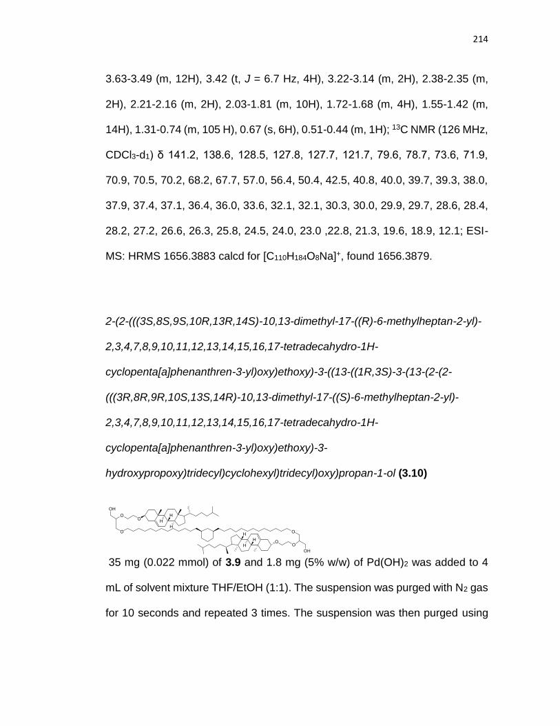
214
3.63-3.49 (m, 12H), 3.42 (t, J = 6.7 Hz, 4H), 3.22-3.14 (m, 2H), 2.38-2.35 (m,
2H), 2.21-2.16 (m, 2H), 2.03-1.81 (m, 10H), 1.72-1.68 (m, 4H), 1.55-1.42 (m,
14H), 1.31-0.74 (m, 105 H), 0.67 (s, 6H), 0.51-0.44 (m, 1H); 13C NMR (126 MHz,
CDCl3-d1) δ 141.2, 138.6, 128.5, 127.8, 127.7, 121.7, 79.6, 78.7, 73.6, 71.9,
70.9, 70.5, 70.2, 68.2, 67.7, 57.0, 56.4, 50.4, 42.5, 40.8, 40.0, 39.7, 39.3, 38.0,
37.9, 37.4, 37.1, 36.4, 36.0, 33.6, 32.1, 32.1, 30.3, 30.0, 29.9, 29.7, 28.6, 28.4,
28.2, 27.2, 26.6, 26.3, 25.8, 24.5, 24.0, 23.0 ,22.8, 21.3, 19.6, 18.9, 12.1; ESI-
MS: HRMS 1656.3883 calcd for [C110H184O8Na]+, found 1656.3879.
2-(2-(((3S,8S,9S,10R,13R,14S)-10,13-dimethyl-17-((R)-6-methylheptan-2-yl)-
2,3,4,7,8,9,10,11,12,13,14,15,16,17-tetradecahydro-1H-
cyclopenta[a]phenanthren-3-yl)oxy)ethoxy)-3-((13-((1R,3S)-3-(13-(2-(2-
(((3R,8R,9R,10S,13S,14R)-10,13-dimethyl-17-((S)-6-methylheptan-2-yl)-
2,3,4,7,8,9,10,11,12,13,14,15,16,17-tetradecahydro-1H-
cyclopenta[a]phenanthren-3-yl)oxy)ethoxy)-3-
hydroxypropoxy)tridecyl)cyclohexyl)tridecyl)oxy)propan-1-ol (3.10)
35 mg (0.022 mmol) of 3.9 and 1.8 mg (5% w/w) of Pd(OH)2 was added to 4
mL of solvent mixture THF/EtOH (1:1). The suspension was purged with N2 gas
for 10 seconds and repeated 3 times. The suspension was then purged using

215
H2 for 10 seconds and repeated 3 times. The reaction was stirred at room
temperature for 20 mins. The suspension was then immediately filtered over
celite and purified using column chromatography on silica gel using
Hexane/EtOAc (7:3) as the eluent. Compound 3.10 was obtained as a white
viscous oil (20.3 mg g, 65%).
Rf: 0.13 (Hexane/EtOAc 7:3); 1H NMR (500 MHz, CDCl3-d1) δ 5.27-5.26 (m, 2H),
3.86-3.81 (m, 2H), 3.64-3.34 (m, 20H), 3.18-3.11 (m, 2H), 2.32-2.27 (m, 2H),
2.18-2.11 (m, 2H), 1.96-1.73 (m, 10H), 1.65-1.59 (m, 4H), 1.50-1.38 (m, 14H),
1.27-0.65 (m, 105H), 0.59 (s, 6H), 0.44-0.38 (m, 1H); 13C NMR (126 MHz,
CDCl3-d1) δ 140.9, 122.0, 80.3, 80.3, 79.8, 72.0, 71.2, 70.4, 70.4, 67.8, 67.7,
63.3, 57.0, 56.4, 50.4, 42.5, 40.8, 40.0, 39.7, 39.1, 39.1, 38.0, 38.0, 37,4, 37.1,
36.4, 36.0, 33.7, 32.1, 32.1, 30.3, 30.0, 29.9, 29.8, 29.7, 28.5, 28.5, 28.4, 28.2,
27.2, 26.6, 26.3, 24.5, 24.0, 23.0 22.8, 21.3, 19.6, 18.9, 12.1; ESI-MS: 1476.28
[M+Na]+; HRMS 1476.2944 calcd for [C96H172O8Na]+, found 1476.2952.
2-(2-(((3S,8S,9S,10R,13R,14S)-10,13-dimethyl-17-((R)-6-methylheptan-2-yl)-
2,3,4,7,8,9,10,11,12,13,14,15,16,17-tetradecahydro-1H-
cyclopenta[a]phenanthren-3-yl)oxy)ethoxy)-3-((13-((1R,3S)-3-(13-(2-(2-
(((3R,8R,9R,10S,13S,14R)-10,13-dimethyl-17-((S)-6-methylheptan-2-yl)-
2,3,4,7,8,9,10,11,12,13,14,15,16,17-tetradecahydro-1H-
cyclopenta[a]phenanthren-3-yl)oxy)ethoxy)-3-((oxido(2-

216
(trimethylammonio)ethoxy)phosphoryl)oxy)propoxy)tridecyl)cyclohexyl)tridecyl)
oxy)propyl (2-(trimethylammonio)ethyl) phosphate (GcGTPC-CH)
To a solution of 81 mg (0.34 mmol) of bromoethyldichlorophosphate in 1.5 mL
of dry THF, a solution of 61 mg (0.04 mmol) of 3.10 and 64 µL of Et3N (0.46
mmol) in 1.5 mL of dry THF was added dropwise. After stirring the mixture for 3
days in the dark at room temperature, toluene was added to precipitate
triethylammonium chloride. Then, the solution was filtered through a small pad
of celite and the filtrate was concentrated. The resulting residue was dissolved
in a mixture of THF/NaHCO3 (sat) (2.8 mM) and the reaction was stirred for 16
hours at room temperature. THF was evaporated under vacuum and the
resulting aqueous solution was acidified to pH 1 using a dilution solution of
hydrochloric acid (1M) and extracted using several portions of DCM/Methanol
(MeOH) (8:2). The organic layers were combined, dried over Na2SO4 and
concentrated under reduced pressure.
To a solution of the previous crude in a mixture of 3.5 mL of chloroform (CHCl3)
and 4 mL of Me3N (33% in EtOH) was added and the reaction was stirred in a
sealed tube at room temperature for 5 days. The reaction mixture was
concentrated to dryness, purified on sephadex LH-20 using DCM/MeOH (1:1)

217
as eluent and purified by column chromatography on silica gel using
DCM/MeOH/Water (70:30:5) as the eluent. GcGTPC-CH was obtained as a
white (viscous oil) (40 mg, 53%).
Rf: 0.15 (Hexane/EtOAc 9:1); 1H NMR (500 MHz, MeOD-d4/CDCl3-d1 1:1) δ
4.99-4.95 (m, 2H), 3.92-3.85 (m, 4H), 3.57-3.52 (m, 4H), 3.43-3.33 (m, 6H),
3.29-3.05 (m, 16H), 2.97-2.94 (m, 2H), 2.85-2.84 (m, 18H), 2.00-1.77 (m, 4H),
1.67-1.44 (m, 10H), 1.34-1.32 (m, 4H), 1.19-1.07 (m, 15H), 0.98-0.32 (m, 110H),
0.16-0.08 (m, 1H); 13C NMR (126 MHz, CDCl3-d1) δ 140.2, 121.4, 79.1, 78.3,
78.2, 71.3, 70.3, 69.6, 67.1, 66.0, 64.8, 64.8, 58.6, 58.6, 56.5, 55.9, 53.5, 49.9,
41.9, 40.2, 39.5, 39.1, 38.7, 37.4, 36.8, 36.4, 35.8, 35.5, 33.1, 31.6, 31.5, 29.7,
29.4, 29.3, 29.2, 28.0, 27.8, 27.6, 26.6, 26.0, 25.8, 23.9, 23.4, 22.1, 21.9, 20.7,
18.8, 18.1, 11.3; 31P NMR (202 MHz, MeOD-d4/CDCl3-d1 1:1) δ 0.12; HRMS
1784.4235 calcd for [C106H197N2O14P2]+, found 1784.4264.
3.9.3. General Buffer Preparation Procedures
Preparation of Buffer A- 4.18 g of Bis Tris (10 mM) and 11.68 g of NaCl (100
mM) was dissolved in 2 L of Milli-Q filtered deionized water. The pH was then
adjusted to 7.2 by minimal addition of 2 M HCl.

218
Preparation of Buffer B - 4.18 g of Bis Tris (10 mM) and 11.68 g of NaCl (100
mM) was dissolved in 2 L of Milli-Q filtered deionized water. The pH was then
adjusted to 5.8 by minimal addition of 2 M HCl.
Preparation of Buffer C – 5.2 g of 2-[4-(2-hydroxyethyl)piperazin-1-
yl]ethanesulfonic acid (HEPES) (10 mM) and 17.5 g of NaCl (150 mM), were
dissolved in 2 L of Milli-Q filtered deionized water. The pH was then adjusted to
7.4.
Preparation of Buffer D – 0.46 g of 2-[(2-Hydroxy-1,1-
bis(hydroxymethyl)ethyl)amino]ethanesulfonic acid, N-
[Tris(hydroxymethyl)methyl]-2-aminoethanesulfonic acid (TES) (2 mM), 5.84 g
of NaCl (100 mM), 0.029 g of ethylenediaminetetraacetic acid (EDTA) (0.1 mM),
and 0.31 g of histidine (2 mM) was dissolved in 1 L of Milli-Q filtered deionized
water. The pH was then adjusted to 7.4.
Preparation of Buffer E – 0.23 g of TES (2 mM), 2.11 g of NaCl (72.23 mM),
0.62 g of CaCl2 (11.11 mM), 0.015 g of EDTA (0.1 mM), and 0.16 g of histidine
(2 mM) was dissolved in 0.5 L of Milli-Q filtered deionized water. The pH was
then adjusted to 7.4.
3.9.4. Gemcitabine Leakage Experiment Procedures

219
The in vitro leakage of the gemcitabine (GEM) from liposomes was
measured using a dialysis assay. GEM encapsulated liposomes were made by
weighing 10 mg of lipid and 40 mol% of cholesterol (only for POPC and
GMGTPC-CH) into a 5 mL round bottom flask and dissolved in 1:1
chloroform/methanol. The solvent was evaporated via vacuo to form a lipid film
and hydrated with 500 µL of 60 mM GEM in Buffer C. 50 µL of the stock
liposomal formulation (20 mg/mL) was diluted with buffer C to 500 µL and
purified using Sephadex® G-25 to prepare a 1 mL working solution. The purified
liposomal suspensions were placed in a dialysis device (Slide-A-Lyzer® Mini
dialysis, ref 88405, Thermo Scientific) with a molecular weight cutoff of 20 kDa
and dialyzed against 42.5 mL of buffer C at 37 °C and shaken on an orbital
shaker (50 rpm). At various time points, aliquots (40 μL) were withdrawn from
the dialysis compartment and retention of encapsulated drug was measured by
HPLC.
For HPLC measurements analysis, to a vial, 40 μL of a Triton solution
(0.5 % w/v in HBS) was added to 40 μL of liposomal suspension. The samples
were vortexed and analyzed by HPLC using method A for the detection of GEM.
Data was analyzed using Agilent analysis software (Chemstation®). For each
time point, the percentage of drug remaining in the liposomes was calculated
using the following equation (1):
𝑅𝑒𝑡𝑎𝑖𝑛𝑒𝑑 𝑑𝑟𝑢𝑔 (%) = 100 ×𝐴𝑟𝑒𝑎(𝑡)
𝐴𝑟𝑒𝑎 (𝑡0) (1)

220
Molecular Dynamics Simulations and Analysis Procedures
Lipids were first constructed using Maestro (Schrödinger LLC, New York,
NY). Membranes containing 729 lipids were then built and solvated using TIP3P
water using VMD5. Each system was minimized and heated in 75 K steps,
reaching a final temperature of 300 K. Following about 20 ns of equilibration,
MD simulations for all membrane systems were performed for 100 ns under
isothermal-isobaric (NpT) conditions. The temperature was maintained by using
the Nose-Hoover chain method and the pressure was maintained at 1 atm. The
CHARMM36 force field was used with a 10 Å cut-off for van der Waals with an
8.5 Å switching distance, and Particle Mesh Ewald for long-range electrostatics.
All the simulations were carried out using NAMD6, and post-simulation
trajectory analysis was carried out using both VMD, R (http://www.r-project.org),
and the Bio3D package.50,51
To identify the number of water molecules in the core of the membrane,
we used methods described previously34. In short, we identified water molecules
that penetrated past the head groups and into the carbon-rich region of the
membrane, i.e. between the ether or ester oxygens of the head groups on either
side of the membrane. The calculation was performed in a grid fashion across
the full membrane and the results reported were the total number of waters
within the membrane for each snapshot over the final 50 ns of the trajectory.

221
3.9.5. General Procedure for Monitoring Gramicidin A Activity in Liposomes
GcGTPC-CH liposomes were prepared as described in general
procedures for liposome extrusion. The general procedure to measure pH
equilibrium of CF was used with a small modification. After the liposomes were
prepared, a solution of gA was prepared 225 µM in DMSO. To each tube (i.e.
pH 7.2, pH 5.8, pH 5.8 + nigericin) 2 µL of buffer A was added to control the
volume. Additional solution containing 2 µL of DMSO (vehicle) or 2 µL gA (final
concentration of 1 µM) was incubated at 37 ºC with liposomes for 30 minutes
and CF fluorescence was measured and analyzed.
3.9.6. General Procedure for Phospholipase-D Induced Cleavage of Choline
GcGTPC-CH liposomes were prepared as described in chapter 2,
section 2.5.9. The liposomes were first hydrated in buffer D, then the liposome
formulation was incubated in buffer E with or without 5 units of PLD from
cabbage (Sigma Aldrich) at 37 ºC for 30 minutes. After incubation, the liposome
size was measured using dynamic light scattering (DLS) to study whether a
morphology change occurred.
Additionally, PLD activity was confirmed using a commercially available
phosphorus assay kit (Sigma, MAK122) according to the manufacturer’s
protocol. We estimated the percent choline released, which is directly correlated

222
with the phosphorus concentration, by normalization to the phosphorus
concentration obtained by the Bartlett assay27.
3.9.7. General Procedure for Self-Quenched CF Liposomal Release Assay in
Serum
Liposomes prepared as described in chapter 2, section 2.5.9. Using the
method of self-quenched CF loaded (100 mM in PBS) leakage assay47,
liposomes comprised of GcGTPC-CH (using general procedure for liposome
extrusion) were incubated in PBS with 30 % fetal bovine serum (FBS) at 37 ºC
(final liposome dilution of 1000 times from stock extruded solution) and CF
fluorescence was monitored (Ex 485 nm/Em 517) nm for up to 5 days.
3.9.8. General Procedure for Cellular Uptake of Small Molecules Entrapped in
GcGTPC-CH Liposomes
3.9.8.1. Cell Toxicity Studies of GcGTPC-CH Liposomes
KB cells were plated onto a fibronectin-treated 96 well plate at 5000
cells/well in folate deficient Roswell Park Memorial Institute (RPMI) 1640
medium (Invitrogen). The cells were incubated for 24 hours under a humidified
atmosphere of 95% air and 5% CO2 at 37 °C. After 24 hours of incubation, the
cells were dosed with various concentrations of the liposomes, in triplicate. The
cells were incubated with the liposomes for 24 hours, and then washed to

223
remove unbound compound. After incubation with the liposomes, the cells were
carefully washed three times with 200 µL phosphate buffered saline (PBS)
buffer and fixed with a solution of 200 µL PBS and 50 µL of 50% trichloroacetic
acid. The cells were allowed to fix at 4°C for 1 hr. After fixation, the cells were
washed five times with water and allowed to dry. After the plates are dried, 100
µL of a 0.4 % sulforhodamine B (SRB, Sigma Aldrich, S1402) solution in 1%
acetic acid was added to each of the wells and incubated for 30 minutes at room
temperature on a shaker. The SRB-treated cells were then washed five times
with 1 % acetic acid and allowed to dry. Tris base solution (100 mM, 200 µL)
was then added to each well and the plates were placed on an orbital shaker
for 30 minutes. The plates were then read on a microplate reader at 515 nm.
The absorbance values were used to create cell-toxicity curves.
3.9.8.2. Fluorescence Microscopy of GcGTPC-CH Liposomal Uptake
To a dry lipid film of GcGTPC-CH lipid with added 0.5 mol % of DSPE-
PEG2000-folate lipid, 10 mM calcein in buffer A was then used to hydrate the
lipid film. Liposomes were then prepared as described in chapter 2, section 2.5.9.
KB cells were plated with folate deficient RPMI-1640 media
supplemented with 10% fetal bovine serum (FBS) on 10mm glass bottom plates
and incubated for 12 hours. Media was then removed and a solution containing
10 µM of liposomes, free calcein, or PBS control was added to the cells and

224
incubated for 6 hours. Cells were then rinsed with media once, and a fresh
media containing 2 µg/mL Hoescht nuclear stain was added. The living cells
where then immediately imaged with an Olympus FluoView FV1000
deconvolution IX81 inverted confocal microscope equipped with a 405, 488, and
543 laser line. Fluorescence images were processed with ImageJ.
3.10. Acknowledgements
Chapter 3 is adapted, in part, from Takaoki Koyanagi, Kevin J. Cao,
Geoffray Leriche, David Onofrei, Gregory P. Holland, Michael Mayer, David
Sept, and Jerry Yang, “Hybrid Lipids Inspired by Extremophiles and Eukaryotes
Afford Serum-Stable Membranes with Low Leakage.” Chem. - Eur. J. 2017,
6757–6762. Copyright 2017 Wiley. Permission to use copyrighted images and
data in the manuscript was obtained from Kevin J. Cao, Geoffray Leriche, David
Onofrei, Gregory P. Holland, Michael Mayer, David Sept, and Jerry Yang. The
dissertation author is the first author of this manuscript.
3.11. References
(1) Torchilin, V. P. Nat. Rev. Drug Discov. 2005, 4 (2), 145–160.
(2) Lasic, D. D. Angew. Chem., Int. Ed. Engl. 1994, 33 (17), 1685–1698.
(3) Senior, J.; Gregoriadis, G. Life Sci. 1982, 30 (I), 2123–2136.

225
(4) Spuch, C.; Navarro, C. J. Drug Deliv. 2011, 2011, 1–12.
(5) Cheng, J.; Teply, B. A.; Sherifi, I.; Sung, J.; Luther, G.; Gu, F. X.; Levy-Nissenbaum, E.; Radovic-Moreno, A. F.; Langer, R.; Farokhzad, O. C. Biomaterials 2007, 28 (5), 869–876.
(6) Malam, Y.; Loizidou, M.; Seifalian, A. M. Trends Pharmacol. Sci. 2009, 30 (11), 592–599.
(7) Colby, A. H.; Liu, R.; Schulz, M. D.; Padera, R. F.; Colson, Y. L.; Grinstaff, M. W. Sci. Rep. 2016, 6 (November 2015), 18720.
(8) Tiwari, G.; Sriwastawa, B.; Pandey, S.; Bannerjee, S.; Tiwari, R.; Bhati, L.; Pandey, P. Int. J. Pharm. Investig. 2012, 2 (1), 2.
(9) Danhier, F.; Feron, O.; Préat, V. J. Control. Release 2010, 148 (2), 135–146.
(10) Ercole, F.; Whittaker, M. R.; Quinn, J. F.; Davis, T. P. Biomacromolecules 2015, 16, 1886–1914.
(11) Allen, T. M.; Cullis, P. R. Adv. Drug Deliv. Rev. 2013, 65 (1), 36–48.
(12) Mura, S.; Nicolas, J.; Couvreur, P. Nat. Mater. 2013, 12 (11), 991–1003.
(13) Benvegnu, T.; Réthoré, G.; Brard, M.; Richter, W.; Plusquellec, D. Chem. Commun. 2005, 5536–5538.
(14) van Meer, G.; Voelker, D. R.; Feigenson, G. W. Nat. Rev. Mol. Cell Biol. 2008, 9 (2), 112–124.
(15) Chong, P. L.-G.; Ayesa, U.; Daswani, V. P.; Hur, E. C. Archaea 2012, 2012, 1–11.

226
(16) Koyanagi, T.; Leriche, G.; Onofrei, D.; Holland, G. P.; Mayer, M.; Yang, J. Angew. Chemie Int. Ed. 2016, 55 (5), 1890–1893.
(17) Haines, T. H. Prog. Lipid Res. 2001, 40 (4), 299–324.
(18) Huang, Z.; Jaafari, M. R.; Szoka, F. C. Angew. Chem., Int. Ed. Engl. 2009, 48 (23), 4146–4149.
(19) Foglia, F.; Barlow, D. J.; Szoka, F. C.; Huang, Z.; Rogers, S. E.; Lawrence, M. J. Langmuir 2011, 27 (13), 8275–8281.
(20) Kohli, A. G.; Kierstead, P. H.; Venditto, V. J.; Walsh, C. L.; Szoka, F. C. J. Control. Release 2014, 190, 274–287.
(21) Jacquemet, A.; Lemiègre, L.; Lambert, O.; Benvegnu, T. J. Org. Chem. 2011, 76 (23), 9738–9747.
(22) Bajaj, A.; Kondiah, P.; Bhattacharya, S. J. Med. Chem. 2007, 50 (10), 2432–2442.
(23) Blöcher, D.; Gutermann, R.; Henkel, B.; Ring, K. BBA - Biomembr. 1984, 778 (1), 74–80.
(24) Mabrey, S.; Mateo, P. L.; Sturtevant, J. M. Biochemistry 1978, 17 (12), 2464–2468.
(25) Koyanagi, T.; Leriche, G.; Yep, A.; Onofrei, D.; Holland, G. P.; Mayer, M.; Yang, J. Chem. - A Eur. J. 2016, 22 (24), 8074–8077.
(26) Papahadjopoulos, D.; Nir, S.; Oki, S. Biochim. Biophys. Acta - Biomembr. 1972, 266, 561–583.

227
(27) Bartlett, G. R. J. Biol. Chem. 1959, 234 (3), 466–468.
(28) Ibarguren, M.; Alonso, A.; Tenchov, B. G.; Goñi, F. M. Biochim. Biophys. Acta - Biomembr. 2010, 1798 (9), 1735–1738.
(29) Leriche, G.; Cifelli, J. L.; Sibucao, K. C.; Patterson, J. P.; Koyanagi, T.; Gianneschi, N. C.; Yang, J. Org. Biomol. Chem. 2017, 15 (10), 2157–2162.
(30) Swallow, S. Prog. Med. Chem. 2015, 54 (2), 65–133.
(31) Kroep, J. R.; Pinedo, H. M.; van Groeningen, C. J.; Peters, G. J. Ann Oncol 1999, 10 Suppl 4, 234–238.
(32) Paula, S.; Volkov, G.; Van Hoek, N.; Haines, T. H.; Deamer, D. W. Biophys. J. 1996, 70 (1), 339–348.
(33) Nagle, J. F.; Scott, H. L. BBA - Biomembr. 1978, 513 (2), 236–243.
(34) Schroeder, T. B. H.; Leriche, G.; Koyanagi, T.; Johnson, M. A.; Haengel, K. N.; Eggenberger, O. M.; Wang, C. L.; Kim, Y. H.; Diraviyam, K.; Sept, D.; Yang, J.; Mayer, M. Biophys. J. 2016, 110 (11), 2430–2440.
(35) Ingólfsson, H. I.; Andersen, O. S. Assay Drug Dev. Technol. 2010, 8 (4), 427–436.
(36) Capone, R.; Blake, S.; Restrepo, M. R.; Yang, J.; Mayer, M. J. Am. Chem. Soc. 2007, 129 (31), 9737–9745.
(37) Ichikawa, S.; Walde, P. Langmuir 2004, 20 (3), 941–949.
(38) Blackwood, R. A.; Smolen, J. E.; Transue, A.; Hessler, R. J.; Harsh, D. M.; Brower, R. C.; French, S. Am. J. Physiol. 1997, 272 (4 Pt 1), C1279–C1285.
228
(39) Leventis, R.; Gagné, J.; Fuller, N.; Rand, R. P.; Silvius, J. R. Biochemistry 1986, 25 (22), 6978–6987.
(40) Jones, M. N.; Nicholas, A. R. Biochim. Biophys. Acta - Biomembr. 1991, 1065 (2), 145–152.
(41) Huang, Z.; Szoka, F. C. J. Am. Chem. Soc. 2008, 130 (46), 15702–15712.
(42) Leamon, C. P.; Low, P. S. Drug Discov. Today 2001, 6 (1), 44–51.
(43) Yang, G.; Yang, T.; Zhang, W.; Lu, M.; Ma, X.; Xiang, G. J. Agric. Food Chem. 2014, 62 (10), 2207–2215.
(44) Wang, S.; Low, P. S. J. Control. Release 1998, 53, 39–48.
(45) Mayer, M.; Yang, J. Acc. Chem. Res. 2013, 46 (12), 2998–3008.
(46) Snyder, W. R. Biochim. Biophys. Acta (BBA)/Lipids Lipid Metab. 1987, 920 (2), 155–160.
(47) Weinstein, J. N.; Yoshikami, S.; Henkart, P.; Blumenthal, R.; Hagins, W. a. Science 1977, 195 (4277), 489–492.
(48) Cao, Y.; Yang, J. Bioconjug. Chem. 2014, 25 (5), 873–878.
(49) Ju, J.; Huan, M.-L.; Wan, N.; Qiu, H.; Zhou, S.-Y.; Zhang, B.-L. Int. J. Mol. Sci. 2015, 16 (3), 5666–5681.
(50) Grant, B. J.; Rodrigues, A. P. C.; ElSawy, K. M.; McCammon, J. A.; Caves, L. S. D. Bioinformatics 2006, 22 (21), 2695–2696.
(51) Skjærven, L.; Yao, X.-Q.; Scarabelli, G.; Grant, B. J. BMC Bioinformatics 2014, 15 (1), 399.

229
Chapter 4
Thiol-Responsive Hybrid Tetraether Lipid for Drug
Delivery
4.1. Introduction
Liposomes have been extensively studied as a drug delivery system for
their low toxicity, good biocompatibility, high entrapment efficiency and the ease
of chemical structural modifications.1–6 Specifically, stimuli-responsive
liposomes have gained interest for their utility to deliver drugs that can
selectively release drugs at a specific target site, and therefore, reducing side
effects and overcoming drug resistance.1,7–9 Examples of stimuli-responsive
liposomes include lipids that integrate cleavable linkers sensitive to acid, UV,
enzyme, biological thiols and redox.10,11 In particular, thiol-responsive liposomes
have garnered interest as a delivery system to cancer cells, in which the
intracellular thiol level (i.e. thioredoxin, thioredoxin reductase, cysteine,
dihydrolipoic acid and glutathione) is found to be significantly higher than the
extracellular fluid that allows retention of cargo in the extracellular fluid and
liposomal release of cargo upon cellular uptake (shown in Figure 4.1).12,13
Liposomal technologies responsive to thiol are traditionally mixed with other
lipids that acts as the liposomal foundation to form stable liposomes.1 However,

230
one disadvantage of doping triggerable lipid analogs into diacyl lipids is the
instability of liposomes in the presence of serum.14 We are the first to
demonstrate a thiol-responsive cleavable tetraether lipids that are capable of
forming stable liposomes pure.
To address liposomal stability in serum, we have previously designed a
synthetic hybrid lipid, namely, glycerol cholesterol-integrated glycerol tetraether
lipid with phosphocholine headgroups (GcGTPC-CH), that incorporate
structural features derived from both Eukaryotes (covalently attaching
cholesterol to the glycerol lipid backbone) and Archaea (integration of
cyclohexane ring to the hydrophobic core and tethering of lipid tails)15 that
showed ~50 times reduction of small ion membrane permeability and ~35 times
increased retention of a neutral small molecule (gemcitabine) after 48 hours
when compared with a commercial diacyl lipid while remaining stable in serum
Figure 4.1. Schematic illustration of intracellular liposomal drug release triggered by thiol.

231
(Chapter 3 of this dissertation). However, despite the enhanced stability and
robustness, GcGTPC-CH liposomes lack the ability to be responsive towards
stimuli, which would be advantageous for drug delivery.
Over the course of my PhD studies, we have designed and structurally
optimized tetraether lipids, GcGTPC-CH, to have significantly reduced
permeability to small ions and small molecules compared with commercial
diacyl lipids. Consequently, we want to minimize addition of new structural
features to the optimized lipid molecule to maintain the low permeability
properties of GcGTPC-CH lipid, while making it responsive. After consideration
of different possible stimuli-responsive cleavable groups, we chose to add a
disulfide linker under the hypothesis that the relatively small addition to a larger
lipid core will not perturb membrane packing of the lipid, rather than, for instance,
the addition of H2O2/UV/acid cleavable linkers that typically involve the addition
Figure 4.2. Molecular design of thiol responsive hybrid lipid GcGT(S-S)PC-CH.

232
of bulky aromatic groups (boronic acid conjugated to phenyl rings,
nitrobenzene) or heteroatoms (orthoesters, acetals), respectively, that can
disrupt lipid-membrane organization.16–18 We began the design of the thiol
responsive lipid analog by focusing the chemical modifications to a solvent
accessible location, near the polar lipid headgroup, instead of the hydrophobic
portion of the lipid to allow access of free thiols to the disulfide bonds. Further
support to target near the headgroup of the lipid is shown in chapter 3, in which
MD simulations suggests low variability in area per lipid tail that limits water
molecules from entering the hydrophobic region of GcGTPC-CH lipid
membrane. We envision that a disulfide linker between the polar lipid headgroup
and the glycerol backbone will minimally affect membrane packing of the
hydrophobic region of the lipid and, hence, nominally affect membrane
permeability (shown in Figure 4.2). This hypothesis is further supported by our
previous results that displayed a low dependence of changes in polar lipid
headgroup on membrane leakage to small ions for tetraether lipid analogs
(Chapter 2 of this dissertation).19 By using this strategy, we envisioned
synthesizing a cleavable lipid that would be stable and robust enough to form
liposomes in pure form without the need of mixing with other lipids.
Herein, we present the synthesis of GcGT(S-S)PC-CH (Figure 4.2) and
examined whether pure GcGT(S-S)PC-CH lipids can form stable liposomes.
We next examined the effect of free thiols on triggered liposomal release of
encapsulated cargo. Lastly, we examine the efficacy of Doxorubicin (DOX)

233
loaded liposomes made with GcGT(S-S)PC-CH lipids to kill mammalian cancer
cells.
4.2. Design/Synthesis of GcGT(S-S)PC-CH
We envision the most difficult step in the synthesis of GcGT(S-S)PC-CH
lipid (Scheme 4.1) would be the formation of the disulfide bonds. We, thus,
dissected the molecule into two fragments: 1) a dithiol lipid core and 2) a thiol
conjugated phosphocholine. The retrosynthetic strategy for generating the thiol
lipid core follows a similar synthetic strategy used for our previous tetraether
lipid analogs that uses alkylation of a cyclohexane integrated dibromoalkane by
a S-protected cholesterol functionalized mercaptoglycerol backbone. The
addition of the cholesterol appendage to the mercaptoglycerol could then be
accomplished by alkylation to the tosylated cholesterol derivative (from chapter
3) with a terminally diprotected mercaptoglycerol that can be formed by a
selective terminal protection of mercaptoglycerol. Next, we focus our
retrosynthetic attention to the thiol conjugated phosphocholine analog, which
can be formed by alkylation of chlorophospholane by a S-Boc protected
ethanolamine, followed by ring opening of the phospholane by trimethylamine.

234
The synthesis of GcGT(S-S)PC-CH lipid (shown in Scheme 4.2) began
by first synthesizing the phosphocholine derivative 4.2 by a selective thiol Boc-
protection of 2-mercaptoethanol to form 4.1. A nucleophilic substitution of 4.1 to
2-choloro-2-oxo-1,3,2-dioxaphospholane followed by a ring opening of
phospholane with the addition of trimethylamine afforded the Boc-protected
phophocholine derivative 4.2. In parallel, the lipid core was synthesized by
selectively protecting the terminal alcohol and terminal thiol of 3-
mercaptopropane-1,2-diol to afford dimethoxytrityl protected glycerol derivative
Scheme 4.1. Retrosynthetic design of GcGT(S-S)PC-CH lipid.

235
4.4. The free secondary alcohol of 4.4 was reacted with tosylated cholesterol
derivative 3.6 under basic conditions to afford the protected glycerol scaffold 4.5.
The glycerol scaffold 4.5 was selectively O-deprotected20 with 80 % aqueous
acetic acid to form 4.6, in which the free alcohol successively reacted with
dibromoalkane 2.36 under basic alkylating conditions to form the dimethoxytrityl
protected thiol lipid 4.7. Both protected thiol derivatives 4.2 and 4.7 were
deprotected in the presence of acid, purified, dried briefly under vacuum and
immediately reacted together to afford GcGT(S-S)PC-CH in a series of
reactions involving activation of thiols on the lipid scaffold 4.8 by 4-phenyl-1,2,4-
triazoline-3,5-dione (PTAD) and concluding the synthesis with the formation of
the disulfide bond with thiol integrated phosphocholine 4.3 (see section 4.7.2.
for details on the synthesis and characterization).
Scheme 4.2. Synthesis of GcGT(S-S)PC-CH lipid.

236
4.3. Physical Characterization of Liposomes Made from Pure GcGTPC-CH,
GcGT(S-S)PC-CH, and Diacyl Lipids
Next, we examined whether the addition of the disulfide bonds would
allow for stable liposome to form within a useful temperature range. Liposomes
were successfully made using GcGT(S-S)PC-CH lipid by extrusion at room
temperature following the protocol described in section 2.5.9. Furthermore,
Differential scanning calorimetry (DSC) measurements revealed the lipid
remains in a liquid phase from 5 to 65 °C and did not exhibition a phase
transition (DMPC was used as a positive control, see Figure 4.3A). In addition,
dynamic light scattering (DLS) measurement of lipids extruded through a 100
nm polycarbonate membrane successfully exhibited liposomes with an average
diameter of ~160 nm (see Figure 4.3B).
Figure 4.3. Physical characterization of lipids. A) DSC measurements of lipids. B) DLS
measurements of synthetic lipids.

237
4.4. Investigation of Membrane Leakage Properties of GcGT(S-S)PC-CH,
GcGTPC-CH and Diacyl Lipid
4.4.1. Examination of Serum Stability of GcGT(S-S)PC-CH, GcGTPC-CH and
Diacyl Liposomes
Serum stability of liposomes made with either GcGTPC-CH, GcGT(S-
S)PC-CH or POPC was examined. Following the same protocol used in chapter
3 (section 3.9.8.) that measures membrane stability by monitoring the leakage
of CF in serum containing HEPES buffer (pH 7.4), liposomes made from
palmitoyloleoylphosphatidylcholine (POPC) lost ~80 % of the CF cargo in 2
hours, whereas, both GcGTPC-CH and GcGT(S-S)PC-CH liposomes remained
stable in serum for up to 48 hours at 37 ºC(shown in Figure 4.4). The serum
stability of GcGT(S-S)PC-CH liposomes in serum supports the hypothesis that
Figure 4.4. Serum induced leakage of CF from liposomes vs. time (h).
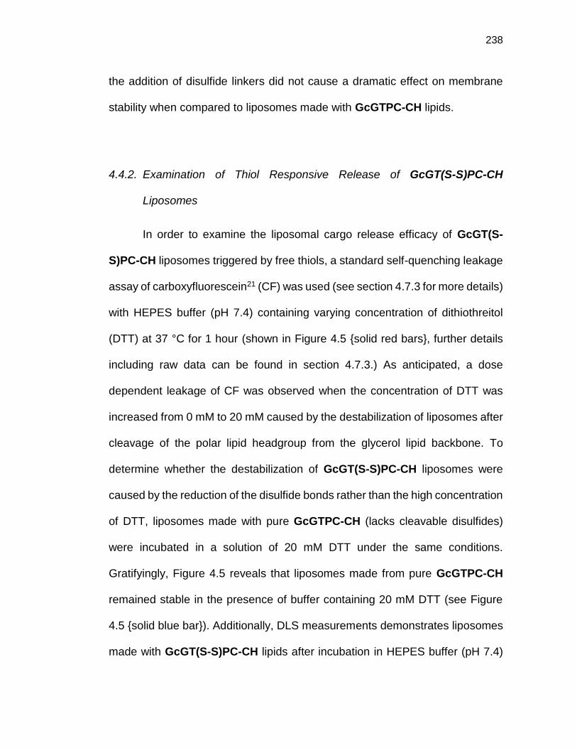
238
the addition of disulfide linkers did not cause a dramatic effect on membrane
stability when compared to liposomes made with GcGTPC-CH lipids.
4.4.2. Examination of Thiol Responsive Release of GcGT(S-S)PC-CH
Liposomes
In order to examine the liposomal cargo release efficacy of GcGT(S-
S)PC-CH liposomes triggered by free thiols, a standard self-quenching leakage
assay of carboxyfluorescein21 (CF) was used (see section 4.7.3 for more details)
with HEPES buffer (pH 7.4) containing varying concentration of dithiothreitol
(DTT) at 37 °C for 1 hour (shown in Figure 4.5 {solid red bars}, further details
including raw data can be found in section 4.7.3.) As anticipated, a dose
dependent leakage of CF was observed when the concentration of DTT was
increased from 0 mM to 20 mM caused by the destabilization of liposomes after
cleavage of the polar lipid headgroup from the glycerol lipid backbone. To
determine whether the destabilization of GcGT(S-S)PC-CH liposomes were
caused by the reduction of the disulfide bonds rather than the high concentration
of DTT, liposomes made with pure GcGTPC-CH (lacks cleavable disulfides)
were incubated in a solution of 20 mM DTT under the same conditions.
Gratifyingly, Figure 4.5 reveals that liposomes made from pure GcGTPC-CH
remained stable in the presence of buffer containing 20 mM DTT (see Figure
4.5 {solid blue bar}). Additionally, DLS measurements demonstrates liposomes
made with GcGT(S-S)PC-CH lipids after incubation in HEPES buffer (pH 7.4)

239
containing 20 mM DTT at 37 ºC for 60 minutes underwent a change in
morphology, while the morphology of liposomes made from GcGTPC-CH lipids
remained static under the same conditions (Figure 4.6). Next, to probe whether
biologically relevant thiols could trigger the release of cargo from GcGT(S-
S)PC-CH liposomes, GcGT(S-S)PC-CH liposomes were introduced to buffers
containing cysteine and glutathione. After GcGT(S-S)PC-CH liposomes were
incubated in buffer containing 20 mM of cysteine for 2 hours at 37 °C, ~ 45% of
CF was released (see Figure 4.5). On the other hand, when GcGT(S-S)PC-CH
liposomes were incubated in buffer containing 20 mM of glutathione, no
Figure 4.5. In vitro assay of thiol triggered response of liposomes made with GcGT(S-S)PC-
CH {red} or GcGTPC-CH {blue}. CF triggered by DTT in HEPES buffer (pH 7.4) at 37 °C
over 1 hour (monitored at λ(Ex/Em) = 485/517 nm); effect of 20 mM GSH and 20 mM
cysteine in HEPES buffer at 37 °C over 2 hours (monitored at λ(Ex/Em) = 485/517 nm)

240
significant amount of CF was released after 2 hours at 37 °C (see Figure 4.5).
This phenomena may be explained by either the lower reducing potential of
glutathione22,23 when compared with DTT or the increased negative charges on
the cell impermeable glutathione compared with cysteine that results in a limited
access to the disulfide bonds.24 While these experiments using different thiols
in cell free assays produced mixed results regarding the capability of thiols to
trigger the release of encapsulated cargo from GcGT(S-S)PC-CH liposomes,
we hypothesized that the complex mixture of different thiol-containing molecules
within a cell may offer enough reducing potential to cause accelerated release
of intraliposomal content.
Figure 4.6. DLS traces of liposomes made with GcGT(S-S)PC-CH or GcGTPC-CH before
and after incubation in HEPES buffer pH 7.4 with/without 20 mM DTT after 60 minute
incubation at 37 ºC.

241
4.4.3. Exploration of DOX Encapsulated GcGT(S-S)PC-CH and GcGTPC-CH
Liposomes
To probe whether thiol-responsive GcGT(S-S)PC-CH liposomes could
form stable liposomes using active loading of drugs, we examined whether DOX
could be actively loaded inside liposomes using a previously reported method
employing an acid gradient (see section 4.7.4. for more details).25 Briefly, 300
mM citrate buffer was passively encapsulated inside ~160 nm diameter
extruded liposomes and incubated in a solution of DOX at 60 °C for 15 minutes.
Unencapsulated DOX was removed by filtration using Sephadex® G-100 and
the DOX loaded liposomes were used immediately. The encapsulation
efficiency of DOX inside GcGT(S-S)PC-CH lipid and GcGTPC-CH liposomes
was 86% (3.5% w/w DOX/lipid) and 84% (3.9% w/w DOX/lipid), respectively
(DOX concentration was determined using HPLC and lipid concentration was
Figure 4.7. Percent leakage of DOX from liposomes after incubation at 37 °C over 4 hours.
Free drug released through dialysis shown in grey (monitored at λabs = 280 nm).

242
determined using the Bartlett assay26). In addition, no significant difference in
DOX leakage was observed from GcGTPC-CH and GcGT(S-S)PC-CH
liposomes for 4 hours incubated at 37 °C in HEPES buffer (pH 7.4) by HPLC
using a dialysis method27 (shown in Figure 4.7).
4.5. Small Molecule Liposomal Delivery to Mammalian Cells
Before investigating the efficacy of liposomes made with GcGT(S-S)PC-CH
lipids for accelerated release of cargo after cell-uptake, we first confirmed that
both GcGTPC-CH and GcGT(S-S)PC-CH liposomes (without encapsulated
cargo) were non-toxic for up to 300 µM of total lipid concentration against HeLa
cancer cells (see Figure 4.8, all cellular experiments were performed by Dr.
Jessica L. Cifelli). Next, when DOX-loaded liposomes were incubated with HeLa
cells for four hours, confocal imaging of native DOX fluorescence revealed that
liposomes readily internalized into cells (shown in Figure 4.9A; additional
Figure 4.8. Cell viability of HeLa cells incubated for 48 h after short-term exposure (4h) to
cleavable (GcGT(S-S)PC-CH, red) and non-cleavable (GcGTPC-CH, blue) liposomes. n ≥
3, All concentrations are n.s. from control cells as determined by unpaired-t test. Data taken
by Dr. Jessica L. Cifelli.
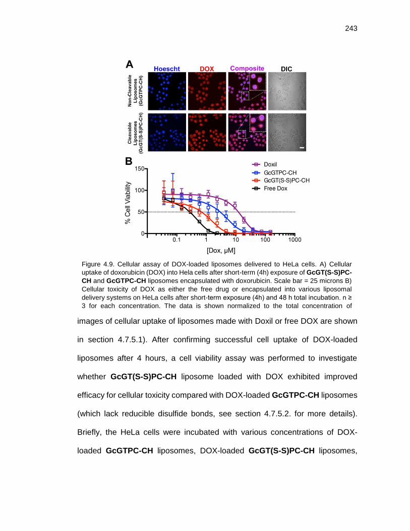
243
images of cellular uptake of liposomes made with Doxil or free DOX are shown
in section 4.7.5.1). After confirming successful cell uptake of DOX-loaded
liposomes after 4 hours, a cell viability assay was performed to investigate
whether GcGT(S-S)PC-CH liposome loaded with DOX exhibited improved
efficacy for cellular toxicity compared with DOX-loaded GcGTPC-CH liposomes
(which lack reducible disulfide bonds, see section 4.7.5.2. for more details).
Briefly, the HeLa cells were incubated with various concentrations of DOX-
loaded GcGTPC-CH liposomes, DOX-loaded GcGT(S-S)PC-CH liposomes,
Figure 4.9. Cellular assay of DOX-loaded liposomes delivered to HeLa cells. A) Cellular
uptake of doxorubicin (DOX) into Hela cells after short-term (4h) exposure of GcGT(S-S)PC-
CH and GcGTPC-CH liposomes encapsulated with doxorubicin. Scale bar = 25 microns B)
Cellular toxicity of DOX as either the free drug or encapsulated into various liposomal
delivery systems on HeLa cells after short-term exposure (4h) and 48 h total incubation. n ≥
3 for each concentration. The data is shown normalized to the total concentration of
encapsulated or free DOX. Data taken by Dr. Jessica L. Cifelli.

244
DOXIL (a Food and Drug Administration [FDA] approved liposomal formulation
of DOX), or free DOX for four hours. The cells were then washed and incubated
at 37 °C for 48 hours, followed by analysis using an MTT cell viability assay. As
shown in Figure 4.9B, DOX-loaded GcGT(S-S)PC-CH liposomes exhibited
good toxicity towards HeLa cells with an IC50 of 0.5 µM, which was comparable
to the toxicity observed for free DOX (IC50 of 0.2 µM). The DOX-loaded
GcGTPC-CH liposomes (lacking disulfide bonds) displayed an IC50 of 1.9 µM,
which is an order of magnitude less toxic than the free drug under these
experimental conditions. DOXIL, on the other hand, displayed an IC50 value of
9.8 µM, which is ~20 times less toxic than the DOX-loaded GcGT(S-S)PC-CH
liposomes. These results support the hypothesis of the natural reducing
environment within the HeLa cancers is sufficient to accelerate the release of
DOX from GcGT(S-S)PC-CH liposomes containing a thiol-cleavable linker
when compared with DOX-loaded GcGTPC-CH liposomes or DOXIL, which
contain lipids that do not contain disulfide groups.
4.6. Conclusion
We have, thus, successfully designed and synthesized the first tetraether
lipid that contains disulfide cleavable linker that are stable enough to form
liposomes in pure form, while maintaining optimized structural features explored
in chapter 2 & 3 to allow stability in serum-containing buffer. Moreover, DOX-
loaded GcGT(S-S)PC-CH liposomes were capable of delivering and promoting
245
accelerated intracellular release of DOX in HeLa cells with an IC50 value similar
to free DOX and ~4 and ~20 times more toxic than our previous generation of
GcGTPC-CH lipid and Doxil, respectively. Through synthetic exploration of the
structure-function relationship of chemical groups found in natural (archaeal and
eukaryotic) lipids, we were able to design a new GcGT(S-S)PC-CH lipid that
has shown to form stable and robust liposomes with low inherent leakage. The
liposomes were capable of delivering DOX to cancer cells with similar toxicity
as for free DOX, which holds promise as an new class of drug delivery system
for cancer therapy. This work represents a first step towards development of
stimuli-responsive tetraether lipids, which may offer advantages of improved
stability and reduced passive leakage of weakly basic and charged drugs
compared with current liposomal drug delivery formulations used for cancer
therapy and other systemically treated diseases.
4.7. Materials and Methods
4.7.1. Reagents and Instruments
All reagents were purchased from commercial sources and used without
further purification. Glassware was dried at 115°C overnight. Air and moisture-
sensitive reagents were transferred using a syringe or stainless steel cannula.
Intermediates were purified over silica (60Å, particle size 40-63 m) purchased
from Dynamic Adsorbents, Inc. Reactions were monitored by thin-layer
chromatography (TLC) using 0.25 mm silica gel plates (60F-254) from Dynamic

246
Adsorbents, Inc. Deuterated solvents were purchased from Cambridge Isotope
Laboratories, Inc.
HeLa cells (human epithelial cervix carcinoma, Product No: CCL-2) and
3-(4,5-dimethylthiazolyl-2)-2, 5-diphenyltetrazolium bromide (MTT) cell
proliferation assay (Product No: 30-1010K) were purchased from American
Type Culture Collection (ATCC) (Manassas, VA). Doxil (DOX HCl liposome
injection, 20mg/mL) was a generous gift from Dr. Stephen B. Howell of Moores
Cancer Center (Lot: 600120P1).
1H, 13C, 31P NMR spectra were obtained on either JEOL ECA 500
spectrometer or Varian 400 MHz/500MHz spectrometer. Chemical shifts are
reported in ppm relative to residual solvent. The FID file was analyzed using
NMRnotebook version 2.70 build 0.10 by NMRTEC.
Dynamic Light Scattering (DLS) measurements were performed on a
Wyatt DynaPro NanoStar (Wyatt Technology, Santa Barbara, CA) or Malvern
Zetasizer (Zen 3690, model Zs90) instrument using a disposable cuvette
(Eppendorf UVette 220 nm – 1,600 nm) and data processed using Wyatt
DYNAMICS V7 software. Each analysis involved an average of 10
measurements. The data was exported for final plotting using GraphPad Prism
5 (GraphPad Software, Inc., La Jolla, CA).
Low resolution MS analysis was performed on a Micromass Quattro
Ultima triple quadrupole mass spectrometer with an electrospray ionization
247
(ESI) source. High resolution MS analysis was performed using Agilent 6230
Accurate-Mass TOFMS with an electrospray ionization (ESI) source by
Molecular Mass Spectrometry Facility (MMSF) in the department of chemistry
and biochemistry at University of California, San Diego.
Fluorescence decay measurements were taken on a Perkin Elmer
Enspire© multimode plate reader (excitation 485 nm, emission 517 nm and 75
flashes); each measurement was taken with a 10 sec delay with 125 repeat
(total acquisition time = 21 min). Costar EIA/RIA plates were used (96 well half
area, no lid, flat bottom, non-treated black polystyrene). The data were exported
for final plotting using GraphPad Prism 5 (GraphPad Software, Inc., La Jolla,
CA).
HPLC analyses were performed using an Agilent 1100 Series HPLC and
an analytical reverse-phase column (Eclipse XDB-C18 Agilent, 5 µm, 150 x 4.6
mm). Flow: 1 mL/min. Injection volume = 50 µL. Detection: 254 and 280 nm.
Mobile phase: water/acetonitrile containing 0.1% (v/v) trifluoroacetic acid (TFA).
Method A: Linear gradient from 5% to 95% of ACN in 10 minutes followed
by 2 minutes of re-equilibration at 5% of ACN.

248
4.7.2. General Synthetic Procedures
O-(tert-butyl) S-(2-hydroxyethyl) carbonothioate (4.1)
Compound 4.1 was prepared following a previously reported protocol.28
2-((tert-butoxycarbonyl)thio)ethyl (2-(trimethylammonio)ethyl) phosphate (4.2)
To a solution of 0.38 g (0.002 mol) 4.1 and 0.30 g (0.002 mol) of 2-chloro-1,3,2-
dioxaphospolane 2-oxde in 6.5 mL of acetonitrile, 0.02 g (0.00018 mol) of
Na2CO3 was suspended and stirred at rt for 2 hours. The salt was removed by
filtration over a small pad of celite. The filtrate was then transferred to a pressure
tube and 2 mL of trimethylamine was added at 0 ºC. After the addition, the
reaction was warmed to rt, then heated to 70 ºC for 48 hours. Volatile solvents
were removed via vacuum and the crude residue was purified over SiO2 using
a gradient of MeOH and water in DCM starting with 80/20 (DCM/MeOH),
80/20/2 (DCM/MeOH/H2O), 70/30/5 (DCM/MeOH/H2O), to 60/40/10
(DCM/MeOH/H2O). Compound 4.2 was obtained as a clear oil (205 mg, 30%).
Rf: 0.40 (DCM/MeOH/H20 60:40:10); 1H NMR (400 MHz, MeOD-d4) δ 4.33-4.25
(2H, m), 4.02-3.95 (2H, m), 3.68-3.66 (2H, m), 3.25 (9H,s), 3.05 (2H, t, J = 6.79
Hz), 1.48 (9H, s); 13C NMR (100 MHz, MeOD-d4) δ 169.0, 84.9, 66.3, 64.4 (d, J
= 5.55 Hz ), 59.3 (d, J = 4.95 Hz ), 53.6, 31.2 (d, J = 8.07 Hz ), 27.3; 31P NMR

249
(161 MHz, MeOD-d4) δ 0.44; ESI-MS: 344.08 [M+H]+; HRMS 344.1291 calcd
for [C12H27NO6PS]+ found 344.1290.
2-mercaptoethyl (2-(trimethylammonio)ethyl) phosphate (4.3)
0.060 g (0.17 mmol) of 4.2 was dissolved in 3 mL of HCl in MeOH (3 M) then
stirred at rt for 20 hours. Volatile solvents were removed to recover Compound
4.3 as a white oily solid (0.043 g, Qt).
ESI-MS: 244.16 [M+H]+; HRMS 244.0767 calcd for [C7H19O4PS]+ found
244.0764.
1-(bis(4-methoxyphenyl)(phenyl)methoxy)-3-((bis(4-
methoxyphenyl)(phenyl)methyl)thio) propan-2-ol (4.4)
To a solution of 1 g (9.2 mmol) of thioglycerol and 6.6 g (19.3 mmol) 4,4’-
dimethoxytrityl chloride (DMTr-Cl) in 20 mL of dry THF, 12.8 mL (92 mmol) of
triethylamine was added drop wise and stirred at rt overnight. Volatile solvents
were removed via vacuum and purified by column chromatography on silica gel
using Hexane/EtOAc (7:3) as the eluent. Compound 4.4 was obtained as a
violet solid (2.6 g, 40 %).

250
Rf: 0.65 (Hexane/EtOAc 1:1); 1H NMR (500 MHz, MeOD-d4) δ 7.38-6.76 (18H,
m), 6.79-6.76 (8H, m), 3.74 (6H, s), 3.71 (6H, s), 3.60-3.56 (1H, m), 3.02-2.99
(1H, m), 2.89-2.86 (1H, m), 2.54-2.51 (1H, m), 2.31-2.27 (1H, m); 13C NMR (126
MHz, MeOD-d4) δ 160.2, 159.8, 147.3, 146.6, 138.8, 138.7, 137.5, 132.2, 132.1,
131.5, 130.7, 129.5, 129.0, 128.8, 127.8, 127.7, 114.2, 114.1, 101.5, 87.4, 71.4,
68.0, 66.8, 61.7, 55.8, 38.1, 21.0, 14.6; ESI-MS: 735.19 [M+Na]+; HRMS
735.2751 calcd for [C45H44O6S Na]+, found 735.2746.
2-(((3S,8S,9S,10R,13R,14S)-10,13-dimethyl-17-((R)-6-methylheptan-2-yl)-
2,3,4,7,8,9,10,11,12,13,14,15,16,17-tetradecahydro-1H-
cyclopenta[a]phenanthren-3-yl)oxy)ethyl 4-methylbenzenesulfonate (3.6)
Compound 3.6 was prepared following a previously reported protocol.29

251
(3-(bis(4-methoxyphenyl)(phenyl)methoxy)-2-(2-
(((3S,8S,9S,10R,13R,14S,17R)-10,13-dimethyl-17-((R)-6-methylheptan-2-yl)-
2,3,4,7,8,9,10,11,12,13,14,15,16,17-tetradecahydro-1H-
cyclopenta[a]phenanthren-3-yl)oxy)ethoxy)propyl)(bis(4-
methoxyphenyl)(phenyl)methyl)sulfane (4.5)
In a pressure tube, 500 mg (0.70 mmol) of 4.4 was dissolved in 3 mL of dry
THF and 50 mg (2.1 mmol) was added at 0 ºC and stirred for 1 hour. 374 mg
(0.77 mmol) of 3.6 was added and heated to 65 ºC then stirred overnight. The
reaction was cooled to 0 ºC and quenched with H2O and THF was removed via
vacuum. The residue was resuspended in EtOAc and washed with water, brine
and dried over Na2SO4 then purified over SiO2 using Hexane/EtOAc (9:1) as the
eluent. Compound 4.5 was obtained as a white viscous oil (494 mg g, 65%).
Rf: 0.43 (Hexane/EtOAc 8:2); 1H NMR (400 MHz, CDCl3-d1) δ 7.42-7.20 (18H,
m), 6.82-6.78 (8H, m), 5.32-5.29 (1H, m), 3.79 (6H, s), 3.77 (6H, s), 3.56-3.51
(4H, m), 3.37-3.32 (1H, m), 3.20-3.11 (2H, m), 3.01-2.97 (1H, m), 2.47-2.34 (3H,
m), 2.21-2.16 (1H, m), 2.06-1.96 (2H, m), 1.88-1.81 (2H, m), 1.61-1.25 (12H,
m), 1.19-0.88 (22H, m), 0.70 (3H, s); 13C NMR (100 MHz, CDCl3-d1) δ 158.6,
158.2, 145.8, 145.2, 141.2, 137.6, 136.4, 136.4, 131.2,, 131.0, 130.3, 129.7,
128.5, 128.0,, 127.9,, 126.8,, 126.6,, 121.7,, 113.3,, 113.2,, 86.2, 79.5,, 79.5,,
79.1, 79.1, 70.3, 70.3, 67.6, 67.5, 65.7, 65.3, 57.0, 56.4, 55.4, 50.4, 42.6, 40.0,

252
39.8, 39.3, 37.5, 37.1, 36.4, 36.0, 34.9, 34.9, 32.2, 32.1, 31.8, 28.6, 28.5, 28.3,
24.5, 24.1, 23.1, 22.9, 22.8, 21.3, 19.6, 19.0, 14.4, 12.1; ESI-MS: 1147.67
[M+Na]+; HRMS 1147.6456 calcd for [C74H92O7S Na]+, found 1147.6446.
3-((bis(4-methoxyphenyl)(phenyl)methyl)thio)-2-(2-
(((3S,8S,9S,10R,13R,14S,17R)-10,13-dimethyl-17-((R)-6-methylheptan-2-yl)-
2,3,4,7,8,9,10,11,12,13,14,15,16,17-tetradecahydro-1H-
cyclopenta[a]phenanthren-3-yl)oxy)ethoxy)propan-1-ol (4.6)
494 mg (0.44 mmol) of 4.5 was dissolved in minimal THF and 10 mL of 80%
AcOH (aq) was added dropwise then stirred at rt for 4 hours. The acid was
neutralized with a solution of sat. NaHCO3 (aq), then extracted with DCM and
dried over MgSO4. The solvent was removed via vacuo and purified over SiO2
using Hexane/EtOAc (9:1) as the eluent. Compound 4.6 was obtained as a clear
oil (271 mg, 76%).
Rf: 0.15 (Hexane/EtOAc 8:2); 1H NMR (500 MHz, CDCl3-d1) δ 7.49-7.25 (9H,
m), 6.87 (4H, d, J = 8.64 Hz), 5.41-5.40 (1H, m), 3.83 (6H, s), 3.79-3.76 (1H, m),
3.63-3.41 (5H, m), 3.27-3.22 (2H, m), 1.68-1.31 (12H, m), 1.23-0.94 (22H, m),
0.76 (3H, s); 13C NMR (126 MHz, CDCl3-d1) δ 158.1, 145.4, 140.6, 137.1, 130.8,
129.5, 127.9, 126.6, 121.8, 113.2, 80.7, 80.6, 79.5, 70.1, 70.1, 67.4, 67.4, 66.0,
64.6, 56.8, 56.2, 55.2, 50.2, 42.4, 39.8, 39.6, 39.0, 38.9, 37.2, 36.9, 36.3, 35.9,

253
33.4, 32.0, 31.9, 28.3, 28.1, 24.4, 23.9, 22.9, 22.7, 21.1, 19.4, 18.8, 11.9; ESI-
MS: 845.33 [M+Na]+;HRMS 845.5149 calcd for [C53H74O5S Na]+, found
845.5152.
(1R,3S)-1,3-bis(14-bromotetradecyl)cyclohexane (2.36)
Compound 2.36 was prepared following a previously reported protocol.15
(1R,3S)-1,3-bis(13-(3-((bis(4-methoxyphenyl)(phenyl)methyl)thio)-2-(2-
(((3S,8S,9S,10R,13R,14S,17R)-10,13-dimethyl-17-((R)-6-methylheptan-2-yl)-
2,3,4,7,8,9,10,11,12,13,14,15,16,17-tetradecahydro-1H-
cyclopenta[a]phenanthren-3-yl)oxy)ethoxy)propoxy)tridecyl)cyclohexane (4.7)
176 mg (0.21 mmol) of 4.6 and 34 mg (0.60 mmol) of KOH were suspended in
6 mL of dry DMSO and stirred at rt for 30 minutes. To the reaction, 46 mg (0.08
mmol) of 2.36 was added and stirred at rt overnight. After the reaction was
stirred at rt overnight, the reaction was heated to 40 ºC and stirred for 4 days.
500 mL of water was added and extracted with EtOAc. The combined organic
layers were back extracted with water, brine and dried over MgSO4 and purified

254
over SiO2 using Hexane/EtOAc (9:1) as the eluent. Compound 4.7 was obtained
as a clear oil (76 mg, 48%).
Rf: 0.54 (Hexane/EtOAc 8:2); 1H NMR (400 MHz, CDCl3-d1) δ 7.41-7.19 (18H,
m), 6.82-6.78 (8H, m), 5.31 (2H, m), 3.81-3.77 (12H, s), 3.53-3.13 (20H, m),
2.42-0.69 (147H, m), 0.54-0.45 (1H, m), ; 13C NMR (100 MHz, CDCl3-d1) δ 158.2,
145.7, 141.2, 137.5, 131.3, 131.0, 130.3, 129.9, 129.8, 129.4, 128.0, 126.7,
121.8, 121.7, 113.4, 113.3, 79.5, 79.5, 78.8, 78.7, 72.4, 71.8, 69.9, 69.8, 67.5,
65.9, 57.0, 56.4, 55.4, 55.3, 50.4, 42.6, 40.8, 40.0, 39.8, 39.3, 38.1, 38.0, 37.5,
37.1, 36.4, 36.0, 33.7, 33.6, 32.2, 32.1, 30.4, 30.0, 30.0, 29.9, 29.8, 28.6, 28.5,
28.3, 27.2, 26.7, 26.4, 24.5, 24.1, 23.1, 22.8, 21.3, 19.6, 19.0, 12.1; ESI-MS:
Not detectable.
3,3'-((((1R,3S)-cyclohexane-1,3-diyl)bis(tridecane-13,1-diyl))bis(oxy))bis(2-(2-
(((3S,8S,9S,10R,13R,14S,17R)-10,13-dimethyl-17-((R)-6-methylheptan-2-yl)-
2,3,4,7,8,9,10,11,12,13,14,15,16,17-tetradecahydro-1H-
cyclopenta[a]phenanthren-3-yl)oxy)ethoxy)propane-1-thiol) (4.8)
76 mg (0.036 mmol) of 4.7 and 30 mg (0.19 mmol) of triisopropylsilane were
dissolved in 1.9 mL of DCM and 0.1 mL of TFA was added dropwise and stirred
at rt for 1 hour. The acid was neutralized by the addition of sat. NaHCO3 (aq)

255
and extracted with DCM. The combined organic layers were dried over MgSO4
and purified over SiO2 using Hexane/EtOAc (9:1) as the eluent. Compound 4.8
was obtained as a clear oil (54 mg, Qt)
Rf: 0.30 (Hexane/EtOAc 9:1); 1H NMR (400 MHz, CDCl3-d1) δ 5.34-5.33 (2H,
m), 3.74-3.41 (18H, m), 3.23-3.16 (2H, m), 2.73-2.66 (4H, m), 2.39-0.44 (146H,
m); 13C NMR (100 MHz, CDCl3-d1) δ 141.1, 121.8, 80.2, 79.7, 71.9, 71.4, 70.2,
67.7, 57.0, 56.4, 50.4, 42.5, 40.0, 39.7, 39.3, 38.0, 38.0, 37.5, 37.1, 36.4, 36.0,
33.7, 32.2, 32.1, 30.3, 30.0, 29.9, 29.7, 28.7, 28.6, 28.5, 28.2, 27.2, 26.7, 26.6,
26.4, 24.5, 24.1, 23.1, 22.8, 21.3, 19.6, 19.0, 12.1 ; ESI-MS: 1502.92 [M+NH3]+;
HRMS 1508.2488 calcd for [C96H172O6S2 Na]+, found 1508.2463.
((((((1R,3S)-cyclohexane-1,3-diyl)bis(tridecane-13,1-diyl))bis(oxy))bis(2-(2-
(((3S,8S,9S,10R,13R,14S,17R)-10,13-dimethyl-17-((R)-6-methylheptan-2-yl)-
2,3,4,7,8,9,10,11,12,13,14,15,16,17-tetradecahydro-1H-
cyclopenta[a]phenanthren-3-yl)oxy)ethoxy)propane-3,1-
diyl))bis(disulfanediyl))bis(ethane-2,1-diyl) bis(2-(trimethylammonio)ethyl)
bis(phosphate) (GcGT(S-S)PC-CH)
37 mg (0.025 mmol) of 4.8 and 9 mg (0.052 mmol) 4-phenyl-1,2,4-triazole-3,5-
dione (PTAD) were dissolved in 1 mL degassed CHCl3 and stirred at rt for 30

256
minutes. The mixture was next heated to 55 ºC and stirred for 2 hours until the
red color disappeared. The reaction was cooled to rt and a solution of 32 mg
(0.17 mmol) of 4.3 in 0.4 mL of degassed MeOH was added and stirred at rt for
3 days. The volatile solvent was removed and purified over SiO2 using
DCM/MeOH/H2O (60:40:10) as the eluent and Sephadex LH20 as the eluent
DCM/MeOH (1:1). GcGT(S-S)PC-CH was obtained as a viscous oil (16 mg,
33%)
Rf: 0.44 (DCM/MeOH/H2O 60:40:10); 1H NMR (500 MHz, MeOD-d4/CDCl3-d1
1:1) δ 5.00-4.99 (2H, m), 3.93-3.89 (4H, m), 3.80-3.76 (4H, m), 3.42-3.10 (24H,
m), 2.86 (18H, s), 2.61-2.53 (8H, m), 2.04-2.01 (2H, m), 1.87-1.81 (2H, m), 1.66-
0.34 (139H, m), 0.14-0.12 (1H, m); 13C NMR (126 MHz, MeOD-d4/CDCl3-d1 1:1)
δ 140.2, 121.4, 79.2, 71.4, 71.2, 69.5, 67.0, 66.0, 66.0, 63.2, 63.1, 58.7, 58.7,
56.4, 55.8, 53.5, 49.9, 41.9, 40.2, 40.1, 39.5, 39.1, 38.7, 38.3, 38.2, 37.4, 37.4,
36.8, 36.4, 35.8, 35.5, 33.1, 31.6, 29.7, 29.4, 29.3, 29.3, 29.2, 28.0, 27.9, 27.6,
26.6, 25.8, 23.9, 23.4, 22.2, 21.9, 20.7, 18.8, 18.1, 11.3; 31P NMR (202 MHz,
MeOD-d4/CDCl3-d1 1:1) δ 0.53; ESI-MS: 1991.20 [M+Na]+;HRMS 1968.3743
calcd for [C110H205N2O14P2S4]+, found 1968.3715.

257
4.7.3. General Procedure for Thiol Triggered Self-Quenched CF Liposomal
Release Assay
Liposomes were prepared as described in general procedures for
liposome extrusion. Using the method of self-quenched CF loaded (100 mM in
HEPES buffer, pH 7.4) leakage assay30, self-quench CF loaded liposomes
comprised of GcGTPC-CH or GcGT(S-S)PC-CH lipids were subjected to
varying concentration of dithiothreitol (DTT; 20 mM, 10 mM, 5.0 mM, 1.0 mM,
0.1 mM, and 0 mM of DTT), 20mM cysteine and 9.0 mM/ 20 mM glutathione
(GSH) containing HEPES buffer shown in Figure 4.10.
Membrane permeability of CF from liposomes was measured by
monitoring the increase in fluorescence of CF using Perkin Elmer Enspire©
Figure 4.10. Raw kinetic traces of increased CF fluorescence from liposomal leakage. A)
Dose dependent effect of DTT on self-quench loaded GcGT(S-S)PC-CH lipid liposomes. B)
Effect of 20 mM DTT on liposomes made with GcGTPC-CH. C) Effect of 20 mM Cysteine on
liposomes made with GcGT(S-S)PC-CH lipid. D) Effect of 9/20 mM of GSH on liposomes
made with GcGT(S-S)PC-CH lipid. (Fluorescence of CF monitored at λ(Ex/Em) = 485/517
nm)
258
multimode plate reader (λ(Ex/Em) = 485/517 nm). Before each assay, 10 µL of
the stock extruded lipid solution was diluted in 500 µL of HEPES buffer. Free
CF was removed using a PD miniTrapTM G-25 SephadexTM column from GE
Healthcare ending in 100 times dilution from the stock extruded solution (0.1
mg/mL). 50 µL of purified liposome solution was next added into three 0.5 mL
Protein Lo-Bind tubes for each lipid solution. In one tube, 450 µL of HEPES
Buffer containing either DTT (shown in Figure 4.10A and 4.10B), cysteine
(shown in Figure 4.10C) or GSH (shown in Figure 4.10D) was added. In the
second tube, 450 µL of HEPES buffer containing Triton X-100 (0.5% v/v) was
added to obtain 100% liposomal releases of CF. In the third tube, 450 µL of
HEPES buffer without added thiols. 150 µL was added to each well of the plate
three times for each tubes resulting in three measurements with three replicates
with a total of 9 measurements per lipid solution.
4.7.4. General Procedure for DOX Loaded Liposomal Release Assay
The in vitro leakage of Doxorubicin (DOX) from liposomes was measured
using a dialysis assay27. Briefly, DOX encapsulated liposomes were made by
weighing 10 mg of lipid into a 5 mL round bottom flask and dissolved in 1:1
chloroform/methanol. The solvent was evaporated via vacuo to form a lipid film
and hydrated with 500 µL of 300 mM citrate dissolved in Milli Q filtered water.
150 µL of the stock liposomal formulation (20 mg/mL) was diluted with HEPES
buffer (pH 7.4) to 500 µL and purified using SephadexTM G-25 to prepare a 1

259
mL working solution. Immediately after free citrate was removed, 35.1 µL of 6
mM DOX solution dissolved in Milli Q filtered water was added to the liposomal
solution and heated at 60 °C for 15 minute. Unencapsulated free DOX was
removed by filtering the liposomal solution over SephadexTM G-100. The purified
liposomal suspensions was placed in a dialysis device (Slide-A-Lyzer® Mini
dialysis, ref 88405, Thermo Scientific) with a molecular weight cutoff of 20 kDa
and dialyzed against 42.5 mL of buffer C at 37 °C and shaken on an orbital
shaker (50 rpm). At various time points, aliquots (40 μL) were withdrawn from
the dialysis compartment and retention of encapsulated drug was measured by
HPLC (λabs of DOX was monitored at 280 nM).
To a vial, 40 μL of a Triton solution (0.5 % w/v in HEPES buffer) was
added to 40 μL of liposomal suspension. The samples were vortexed and
analyzed by HPLC using method A for the detection of DOX. Data was analyzed
using Agilent analysis software (Chemstation®). For each time point, the
percentage of drug remaining in the liposomes was calculated using the
following equation (1):
𝑅𝑒𝑡𝑎𝑖𝑛𝑒𝑑 𝑑𝑟𝑢𝑔 (%) = 100 ×𝐴𝑟𝑒𝑎(𝑡)
𝐴𝑟𝑒𝑎 (𝑡0) (1)

260
4.7.5. General Procedure for Cellular Uptake of DOX Entrapped in Liposomes
4.7.5.1. Confocal Microscopy of the Cellular Uptake of Doxorubicin
HeLa cells were cultured on 35 mm dishes (MatTek) and incubated
overnight in DMEM supplemented with 10% FBS. The growth medium was
removed and solutions containing either free doxorubicin, or various liposomal
encapsulated doxorubicin solutions (10 μM, final concentration of DOX for all),
or HBS (UT control) were added. The cells were then incubated for 4 hours
(37 °C, 5% CO2). MatTeks were then rinsed (3x, ice cold-PBS) and nuclei were
stained with Hoescht 33258 (10 minutes, 37 °C). Cells were rinsed again (2x,
PBS) immediately prior to imaging. Cellular uptake of doxorubicin was imaged
Figure 4.11. Cellular uptake of DOX into HeLa cells after 4h exposure with free DOX, Doxil,
and untreated (UT) control (HBS). Scale bar = 50 µm, Data taken by Dr. Jessica L. Cifelli.
261
with an Olympus FV1000 spectral deconvolution confocal system equipped with
an Olympus IX81 inverted microscope. Cell images were analyzed using
ImageJ. The images shown in Figure 4.11 are fluorescence micrographs of
representative z-slices from within the cells.
4.7.5.2. MTT Cell Proliferation Assay in HeLa Cells
An MTT cell proliferation assay was performed as previously reported
with noted changes27. Briefly, cells were plated in 96-well dishes at a density of
10,000 cells/well in 100 µL of DMEM supplemented with 10% FBS. After
adhering overnight, the medium was replaced with 100 μL of solutions
containing various concentrations of either free doxorubicin, or various
liposomal formulations of encapsulated doxorubicin (Doxil, cleavable (GcGT(S-
S)PC-CH Lipid), and non-cleavable (GcGTPC-CH) liposomes: see general
procedures for liposomes extrusion for details) with final concentrations of
doxorubicin (0-150 µM). As controls, cells were also exposed to the cleavable
and non-cleavable liposomes without DOX. Cells were exposed to these various
solutions for 4 hours at 37 °C. Following, in which cells were rinsed (cold PBS),
fresh complete medium was added (200 μL) and cells were let incubate. After
48 h of incubation, an MTT cell viability assay was then used to determine cell
viability. Briefly, the MTT reagent (10 μL of the solution from the commercial kit)
was added to each well and the cells were incubated for 3 additional hours. All
solutions were then removed and the formazan crystals were subsequently

262
solubilized with DMSO (100 μL). The cell viability was determined by measuring
the absorbance at 570 nm using a Perkin Elmer EnSpire© multimode plate
reader. All results, shown in Figure 4.7, were expressed as percent reduction of
MTT relative to untreated controls (defined as 100% viability) and the average
absorbance value for each treatment was blanked with the absorbance reading
of media control blanks. Each concentration was done in at least triplicate.
4.8. Acknowledgements
Chapter 4 is adapted, in part, from Takaoki Koyanagi, Jessica L. Cifelli,
Geoffray Leriche, David Onofrei, Gregory P. Holland, and Jerry Yang, “Thiol-
triggered release of intraliposomal content from liposomes made of
extremophile-inspired tetraether lipids.” Bioconjugate Chem. 2017, ASAP.
Copyright 2017 American Chemical Society. Permission to use copyrighted
images and data in the manuscript was obtained from Jessica L. Cifelli, Geoffray
Leriche, David Onofrei, Gregory P. Holland, and Jerry Yang. The dissertation
author is the first author of this manuscript.
4.9. References
(1) Torchilin, V. P. Nat. Rev. Drug Discov. 2005, 4 (2), 145–160.
(2) Clark, a P. Cancer Pract. 6, 251–253.
(3) Lasic, D. Trends Biotechnol. 1998, 16 (7), 307–321.

263
(4) Chrai, S. S.; Murari, R.; Ahmad, I. Biotech Trends 2002, No. April, 30–34.
(5) Allen, T. M.; Cullis, P. R. Adv. Drug Deliv. Rev. 2013, 65 (1), 36–48.
(6) Tiwari, G.; Sriwastawa, B.; Pandey, S.; Bannerjee, S.; Tiwari, R.; Bhati, L.; Pandey, P. Int. J. Pharm. Investig. 2012, 2 (1), 2.
(7) Wen, H.; Li, Y. Med. Chem. (Los. Angeles). 2014, 4 (11), 748–755.
(8) Connor, J.; Yatvin, M. B.; Huang, L. Proc. Natl. Acad. Sci. U. S. A. 1984, 81 (6), 1715–1718.
(9) Mura, S.; Nicolas, J.; Couvreur, P. Nat. Mater. 2013, 12 (11), 991–1003.
(10) Lee, Y.; Thompson, D. H. Wiley Interdiscip. Rev. Nanomed. Nanobiotechnol. 2017, e1450, 1–40.
(11) Danhier, F.; Feron, O.; Préat, V. J. Control. Release 2010, 148 (2), 135–146.
(12) Lockwood, T. D. Antioxid. Redox Signal. 2002, 4 (4), 681–691.
(13) Saito, G.; Swanson, J. A.; Lee, K. D. Adv. Drug Deliv. Rev. 2003, 55 (2), 199–215.
(14) Jones, M. N.; Nicholas, A. R. Biochim. Biophys. Acta - Biomembr. 1991, 1065 (2), 145–152.
(15) Koyanagi, T.; Leriche, G.; Onofrei, D.; Holland, G. P.; Mayer, M.; Yang, J. Angew. Chem., Int. Ed. 2016, 55, 1890–1893.
(16) De Gracia Lux, C.; Joshi-Barr, S.; Nguyen, T.; Mahmoud, E.; Schopf, E.; Fomina, N.; Almutairi, A. J. Am. Chem. Soc. 2012, 134 (38), 15758–15764.

264
(17) Barbeau, J.; Belmadi, N.; Montier, T.; Le Gall, T.; Dalençon, S.; Lemiègre, L.; Benvegnu, T. Tetrahedron Lett. 2016, 2, 1–5.
(18) Mueller, A.; Bondurant, B.; O’Brien, D. F. Macromolecules 2000, 33 (13), 4799–4804.
(19) Koyanagi, T.; Leriche, G.; Yep, A.; Onofrei, D.; Holland, G. P.; Mayer, M.; Yang, J. Chem. - Eur. J. 2016, 22 (24), 8074–8077.
(20) Z. Huang, S. A. B. Synlett 1993, 83–84.
(21) Weinstein, J.; Yoshikami, S.; Henkart, P.; Blumenthal, R.; Hagins, W. Science. 1977, 195 (4277), 489–492.
(22) Banerjee, R. J. Biol. Chem. 2012, 287 (7), 4397–4402.
(23) Brülisauer, L.; Gauthier, M. A.; Leroux, J. J. Control. Release 2014, 195, 147–154.
(24) Chompoosor, A.; Han, G.; Rotello, V. M. Bioconjug. Chem. 2008, 19 (7), 1342–1345.
(25) Gubernator, J. Expert Opin. Drug Deliv. 2011, 8 (5), 565–580.
(26) Bartlett, G. R. J. Biol. Chem. 1959, 234 (3), 466–468.
(27) Leriche, G.; Cifelli, J. L.; Sibucao, K. C.; Patterson, J. P.; Koyanagi, T.; Gianneschi, N. C.; Yang, J. Org. Biomol. Chem. 2017, 15 (10), 2157–2162.
(28) Temperini, A.; Annesi, D.; Testaferri, L.; Tiecco, M. Tetrahedron Lett. 2010, 51 (41), 5368–5371.

265
(29) Bajaj, A.; Kondiah, P.; Bhattacharya, S. J. Med. Chem. 2007, 50 (10), 2432–2442.
(30) Weinstein, J. N.; Yoshikami, S.; Henkart, P.; Blumenthal, R.; Hagins, W. Science. 1977, 195 (4277), 489–492.