Estadísticas de exportaciones de material de defensa y de ...
Upload
khangminh22Category
view
2download
0
UNIVERSITE DE NANTES
FACULTE DE MEDECINE
⎯⎯⎯⎯
Année 2016 N°
T H E S E pour le
DIPLOME D’ETAT DE DOCTEUR EN MEDECINE
D.E.S de Chirurgie générale
par
Fanny-Laure MERLET
née le 17 août 1987, à Saint Nazaire
⎯⎯⎯⎯
Présentée et soutenue publiquement le 21 octobre 2016
⎯⎯⎯⎯
Intérêt du traitement fonctionnel isolé dans le traitement des fractures articulaires du processus condylien chez l’adulte. Etude rétrospective de 83
cas. ⎯⎯⎯⎯
Présidentdujury:
MonsieurleProfesseurCORRE
Directeurdethèse:
MadameleDocteurGRIMAUD
Membresdujury:
MonsieurleProfesseurMERCIER
MonsieurleProfesseurLAURE
MonsieurleProfesseurPERROT

2
Tabledesmatières
Table des matières .............................................................................. 2
Liste des abréviations .......................................................................... 4
A. Abréviationsfrançaises.............................................................................................................4
B. Englishabbreviations.................................................................................................................4
Table des figures ................................................................................. 5
Table des tableaux .............................................................................. 5
I. Introduction ................................................................................... 7
II. Article .......................................................................................... 11
A. Abstract..........................................................................................................................................11
B. Introduction.................................................................................................................................13
C. Materialsandmethods...........................................................................................................16
1. Clinicaldata.............................................................................................................................16
2. Radiologicaldata...................................................................................................................16
3. Treatment.................................................................................................................................19
4. Statisticalanalyses...............................................................................................................20
D. Results............................................................................................................................................21
1. Epidemiologicdata...............................................................................................................21
2. Clinicaldata.............................................................................................................................22
3
3. Radiologicaldata...................................................................................................................25
4. Therapeuticdata...................................................................................................................27
E. Discussion.....................................................................................................................................29
F. Conclusion....................................................................................................................................34
III. Discussion ................................................................................... 35
IV. Conclusion .................................................................................. 42
V. Références bibliographiques ...................................................... 43

4
Listedesabréviations
A. Abréviationsfrançaises
ATM:articulationtemporo-mandibulaire
CBCT:tomographievolumiqueàfaisceauconique
IRM:Imagerieparrésonancemagnétique
OB:ouverturebuccale
RA:retentissementarticulaire
DAM:Dysfonctionnementdel’appareilmanducateur
B. Englishabbreviations
LT:laterotrusion
MMF:maxillo-mandibularfixation
MMO:maximalmouthopening
NO:non-operated
O:operated
ORIF:openreductionandinternalfixation
TMJ:temporo-mandibularjoint
3D:threedimensional
5
Tabledesfigures
Figure1:ClassificationdeSPIESSLandSCHROLL..............................................................................8
Figure2:Classificationdesfracturesintra-articulairesduprocessuscondylien..................9
Figure 3: Height ascending ramus measurement on CBCT three-dimensional
reconstructions(ontheleft)andonpanoramicX-ray(ontheright).............................17
Figure4:3DfrontalCBCTreconstructionshowingocclusionplan...........................................18
Figure 5: Articular movements analysis: MMO (at the top), laterotrusion (in the
middle) and propulsion (in the lower) O: operated NO: non-operated NS: non-
significant*:significant......................................................................................................................23
Figure6:Vuecoronaled’unCBCT.Imagepost-opératoireimmédiate(àgauche), Image
post-opératoire tardive(à1an)d’unevisd’ostéosynthèseencontactavec labase
ducrâne(àdroite).................................................................................................................................38
Figure 7: Reconstructions scannograpiques tridimensionnelles illustrant une bifidité
condyliennedroite................................................................................................................................40
Tabledestableaux
Tableau1:Classificationanatomo-fonctionnelled’aprèsDelaire,Mercier,Perrin..............8
Table2:Generalcharacteristicsofthepatients................................................................................21
Table3:Articularmovementsanalysis,mm........................................................................................22

6
Table4:ComparisonofMMO(mm)accordingtouni-andbilateralfracturesinoperated
andnon-operatedpatients................................................................................................................24
Table5:proportionofTMJdysfunction...............................................................................................24
Table6:Fracturesdistributionaccordingtothedifferentclassifications.............................25
Table7:Lossofheightbetweenthe2ramus,mm,(%)inthecaseofunilateralcondylar
processfractures....................................................................................................................................26
Table8:Averageof the2 ramusheight,mm, in the caseofbilateral condylarprocess
fractures.....................................................................................................................................................26
Table9:Non-operatedcondylarremodeling......................................................................................27
Table11:Comparisonofclassifications...............................................................................................30
Table12:Comparaisonentrelesdifférentesclassifications.......................................................36

7
I. Introduction
Lesfracturesdemandibulessontlesfracturesfacialeslesplusfréquentes(1,2).Elles
impliquent le processus condylien dans 30% des cas (3–5). L’articulation temporo-
mandibulaire (ATM) est lésée le plus souvent de façon indirecte à la suite d’un
traumatismeisolésurlasymphysementonnièreoudanslecadred’unpolytraumatisme.
Dans ce cas, la fracture du condyle peut être associée à d’autres fractures dumassif
facial.Lescirconstancesdedécouvertesontavanttoutcliniques-unedouleurenregard
d’une articulation temporo-mandibulaire, une plaie du menton, un trouble occlusal -
puis radiologiques (panoramiquedentaire, tomographie volumique à faisceau conique
(CBCT),scannerdumassiffacial).Chezl’adulte,lesconséquencesdecetypedefractures
sontmultiples. Au delà de l’impotence fonctionnelle et de la douleur quimarquent la
phase aigue, le risque principal est avant tout fonctionnel, avec un risque d’évolution
vers l’ankylose temporo-mandibulaire (6–8). Il peut y avoir également des risques de
troublede l’occlusion,d’asymétrie facialeoudedysfonctionde l’appareilmanducateur
(DAM)(9,10).Bienqu’iln’existepasderéelconsensusquantàlapriseenchargeidéale
decesfractures,lathérapeutiqueestfortementorientéeparl’aspectradiologiquedela
fracture et ses potentielles conséquences fonctionnelles. Plusieurs classifications des
fractures ont ainsi été décrites (11). La classification de LOUKOTA (12,13) propose
l’échancruresigmoïdecommeséparationentrelesfracturesducolcondylien(fractures
sous condyliennes hautes) et les fractures de la base du condyle (fractures sous
condyliennes basses) et y ajoute les fractures de la tête condylienne (fractures
capitales). Cette classification décrit la localisation de la fracture, sans cependant
préciser le degré de déplacement et de bascule condylienne, ce qui peut avoir des
conséquencessur la fonctionarticulaire.LaclassificationdeSPIESSLetSCHROLL(14)
apportedesprécisionsanatomiquessurlesfracturesetorienteainsilapriseencharge
chirurgicale. Elle prend en compte le degré de déplacement et de bascule du condyle
(figure1).

8
TypeI:Fracturescondyliennesnon-déplacées,TypeII:Fracturessous-condyliennesbassesdéplacées,TypeIII:Fracturesous-condylienneshautesdéplacées,TypeIV:Fracturessous-condyliennesbassesluxées,TypeV:Fracturessous-condylienneshautes
luxées,TypeVI:Fracturesintra-capsulaires/diacapitales
Figure1:ClassificationdeSPIESSLandSCHROLL
Laclassificationanatomo-fonctionnelleproposéeparDELAIRE(7)etreprisepar
MERCIERetPERRIN(6,15)présentel’avantaged’indiquerd’embléelanécessitéou
nond’instaureruntraitementfonctionnelspécifique.Elleopposeainsilesfractures
avec retentissement articulaire, c’est-à-dire susceptibles d’entraîner des séquelles
fonctionnellesparlésiondesstructuresarticulairestellesqueledisqueoulacapsule
decellessansretentissementarticulaire(tableau1).
Tableau1:Classificationanatomo-fonctionnelled’aprèsDelaire,Mercier,Perrin
Fracturesàretentissementarticulaire Fracturesansretentissementarticulaire
Capitalesou
sous-capitales
Sous-
condylienne
hauteluxée
Sous-
condylienne
basseluxée
Sous-
condylienne
hauteoubasse
sans
déplacement
Sous-
condylienne
hauteoubasse
basculée,sans
luxation
Sous-
condylienne
hauteoubasse
chevauchée
Lecondyleestsortidelaglène Lecondyleestrestédanslaglène

9
Les fractures sous-condyliennes basses sont généralement traitées par réduction
chirurgicale et blocage maxillo-mandibulaire de durée variable voire par traitement
conservateurseul(10)etsontdebonpronostic(1,16–18).
Lapriseenchargedesfracturessous-condylienneshautesdéplacéeset/ouluxéesou
les fractures capitales, chez l’adulte est, quant à elle, très controversée (19). Les
fractures les plus problématiques en terme de traitement et de pronostic fonctionnel
sont les fractures intra-articulaires (ou diacapitulaires). Elles ont été subdivisées, à
partir de la classification de Spiessel and Schroll, par Rasse, Neff, Hlawitschka et
Loukota,enfonctiondelalocalisationdutraitdefracture(13,20).LetypeAreprésente
unefracturedutuberculemédial,n’entrainantpasdepertedehauteurduramus.Dans
letypeB, l’ensemblededucondyleestdéplacé,entrainantunediminutiondehauteur.
LetypeCcorrespondautypeVdelaclassificationdeSpiesslandSchroll(figure2).
Figure2:Classificationdesfracturesintra-articulairesduprocessuscondylien.
Ces fractures sont toutes considérées dans la classification de Delaire comme des
fracturesàretentissementarticulaire, indiquantsystématiquement laréalisationd’une
rééducation fonctionnelle spécifiquede l’ATM.L’abord chirurgical de ces fractures est
souventcompliquédeparledéplacementdufragmentmédialetlalocalisationdutrait
defracture,etleursynthèseenestsouventdifficile(2).Ilestainsinécessaired’évaluer
la balance bénéfice/risque avant de proposer une prise en charge chirurgicale. Les
partisans du traitement chirurgical estiment que restaurer l’anatomie et utiliser une

10
fixation rigide des fragments permettent d’obtenir une meilleure récupération de la
fonctionarticulaire(21,22).Lavoied’abordestchoisieenfonctiondelalocalisationdu
trait de fracture, de l’association avec d’autres fractures, des risques de séquelles
esthétiques(23)maiségalementenfonctiondeshabitudesetpréférencesdel’opérateur
(24).Letraitementchirurgicalprésentecependantdeslimites(5,25):laréductiondela
fractureet,surtout,sonostéosynthèsepeuventêtredifficilesetinstables.Lescicatrices
disgracieuses, les lésions du nerf facial ou la survenue de fistules salivaires (23,26)
représententlescomplicationslesplusfréquentes.Enfin,lefaitmêmed’aborderl’ATM
pour réduire la fracture est susceptible de léser des structures intra-articulaires et
d’entrainerdesséquellesfonctionnelles.
Enalternativeau traitement chirurgical, le traitement fonctionnelparmobilisation
immédiatede lamandibuleenpropulsionaétéproposé(7).Le traitement fonctionnel
constituelapriseenchargederéférencedesfracturescondyliennedel’enfant(27).Ila
eneffetétémontréque lamobilisation induisaitetaccélérait le remodelagecondylien
(28–30). Le traitement fonctionnel décrit par Delaire (7) repose sur la réalisation de
mouvements répétés de propulsion mandibulaire centrée et de diduction latérale
(mécanothérapie active) parfois associée à des tractions élastiques sur arcs
(mécanothérapiepassive).
L’objectif de ce travail était d’évaluer les résultats du traitement fonctionnel seul
dans le traitement des fractures condyliennes hautes de l’adulte, à retentissement
articulaire(typesA,B,C),encomparaisonavecletraitementchirurgical.

11
II. Article
Outcomes of functional treatment versus open reduction and internal fixation of condylar mandibular fracture with articular impact. Retrospective
study in 83 adults.
A. Abstract
Introduction : The treatment of fractures of the mandibular condylar process
remains controversial. The aim of this study was to assess the outcomes of isolated
functionaltreatmentversusopenreductionandinternal fixation(ORIF)ofmandibular
condylarfracturewitharticularimpactbasedonclinicalandradiologicalcriteria.
Materials andmethods :Heigthy-three patients with mandibular condylar fracture
witharticularimpactwereincludedinthisretrospectivestudy,anddividedaccordingto
Loukota, Spiessl and Schroll, Delaire andRasse, Neff, Hlawitschka classifications. Two
groups were created: operated patients (operated) and non-operated patients (non-
operated).Occlusalandfunctionalfeatureswereevaluatedusingclinicalmeasurements
at1,3,6,and12monthsafterthetreatment,andradiologicalmeasurementsperformed
preoperatively,6weekslater,andattheendofthefollow-up.
Results:Amalepredominancewasobserved(69.9%,p<0.0001).Isolatedfunctional
treatmentwas applied in 55 patients (66.26%). Twenty-height patients (33.7%)were
operated using pre-auricular ormodified Risdon’s approach.Maximalmouth-opening

12
(MMO) was lesser in “operated” group compared to “non-operated” group until 6
months (25.75 mm vs 31.96 mm, 34.76 mm vs 37.95 mm, 38.06 mm vs 41.87mm
respectively 1, 3 and, 6 months, p<0.05). Results were satisfactory 1 year after
traumatism (41.29 mm vs 45.22mm, p>0.05). There was no difference concerning
temporo-mandibular joint dysfunctions between operated and non-operated patients.
For unilateral fractures, the loss of height of the ramus was significatively higher in
operated patients initially compared to “non operated” group (p = 0.0137). After
surgicalcorrection, therewasnodifferencebetweenthetwosidesofmandible.At the
endof the follow-up, the therewasnodifferencebetweenoperatedandnonoperated
ramus(p=0.1304and0.6420).
Conclusion:Thepresentstudyshowedthatanisolatedfunctionaltreatmentwhichis
properly followed provided as good clinical results as ORIF for mandibular condylar
fractureswitharticularimpact.Surgicaltreatmentshouldbepreferredwhenthelossof
height of the ramus is important, to restore the ramusheight, since in adult condylar
remodelingexistsbutislessefficientthaninchildren.

13
B. Introduction
Mandibularfracturesarethemostfrequentfacialfractures(1,2).Itinvolvescondylar
processin30%ofcases(3–5).
Temporo-mandibular joint (TMJ) is often damaged indirectly after an isolated
traumatismonthechinsymphysisorinamultipletraumatismcontext,andinthiscase
beassociatedwithotherfacialfractures.Diagnosisisfirstofallclinical-TMJpain,chin
wound, occlusal disorder- then radiological. For adults patients, consequences are
multiple.BeyondTMJfunctionaldisturbanceandpainwhichareat thefirstphase, the
risk to develop a temporo-mandibular ankylosis is high (6–8). Other complications
includeocclusaldisorders,facialasymmetryorTMJdysfunctionalpain(9,10).Although
thereisnorealconsensusabouttheidealmanagementofthesefractures,thetreatment
is strongly oriented by the fracture radiological aspect and its potential functional
consequences. Several classifications have been described (11). Loukota et al have
proposed to consider the sigmoid notch as an anatomical border to differentiate
between high and low condylar process fractures. Condylar base fracture, condylar
collum fracture, and diacapitular fractures have been described (12,13). This
classificationdescribesthe locationofthefracture,but isnotrelevantforappreciating
the degree of displacement and dislocation of the condyle, that can have functional
consequences.Inclinicalpractice,theclassificationaccordingtoSpiesslandSchrollhas
proved to bemost useful. Indeed, this classification gives anatomic information about
the fracture but also guides across surgical treatment. The anatomo-functional
classification proposed by Delaire (7), and resumed by Mercier and Perrin (6,15)
presents one advantage: it indicates if a specific functional treatment is necessary.It
opposesarticular impact fractures(TypeA,B,C),whichcancause functionaldamages
onjointstructureslikediscorcapsule,andnoarticularimpactones.
Condylar base fractures are usually surgically treated, and blocked with maxillo-
mandibularfixationifthereisadisplacement,orwithconservativetreatmentifthereis
nodisplacement(10).Theyareofgoodprognosis(1,16–18).

14
In adults, the treatment of high collum fractures with displacement and/or with
dislocation anddiacapitular fractures is still a continuingdebateoverhow tomanage
thistypeoffractures(19).FollowingtheclassificationofSpiesslandSchroll,Rasse,Neff
(20), Hlawitschka (31) and Loukota (13) additionally classified the intraarticular or
diacapitular condylar fractures according to the fracture line. Type A represents a
displacementofmedialcondylarpolewithpreservationoftheverticaldimension.Type
B: the lateral condylar pole is involved with loss of the vertical dimension. Type
Ccorresponds to class V according to Spiessl and Schroll: high collum fractures with
dislocation.These fracturespresent the same features as articular impact fractures of
Delaire’sclassification.Treatmentofcondylarneckfractureswitharticular impactstill
remains controversial. Indeed, surgical difficulty to obtain anatomic reduction limits
indicationsofsurgicaltreatment.(2).
It is then necessary to assess the benefit-risk equation before proposing ORIF.
Proponents of surgical treatment estimate that restoring the anatomy and using rigid
fixationallowtoobtainabetterrecoveryofthearticularfunction(21,22).Theapproach
ischosenaccordingtothelocationofthefracture,theassociationwithotherfractures,
the riskofesthetic consequences (23)butalsoaccording to thesurgeon’shabits (24).
Thesurgicaltreatmenthashoweveritslimits(5,25):thereductionofthefracture,and
especially, the osteosynthesis could be difficult and precarious. Dysesthetic scars,
damagesonthe facialnerveortheoccurrenceofsalivary fistula(23,26)representthe
most common complications. Finally, the only fact to open the TMJ to reduce the
fracture, can cause damages to the intra-articular structures, and be responsible for
functionalsequelae.
As analternative to the surgical treatment, functional treatment,which consistsof
immediate mobilization of the mandible on propulsion, can be proposed. Functional
treatmentisthereferenceinthecaseofcondylarfracturesforthechildren(27).Indeed,
the mobilization of the joint has proved induces and accelerates the condylar
remodeling (28–30). This treatment described by Delaire et al. (7) is based on the
realizationofrepeatedcentredprotrusivemandibularmovements,andlateralexcursion
(active reeducation) sometimes associated with elastic traction on arches (passive
reeducation).

15
Thepurposeof thisstudywastoevaluatetheclinicalandradiologicaloutcomesof
theisolatedfunctionaltreatmentinthecaseofcondylarprocessfracturewitharticular
impactinadults,incomparisonwithsurgicaltreatment.

16
C. Materialsandmethods
From january2009 todecember2015,patientsolder than15years and3months
presentedwith a uni- or bilateralmandibular condylar fracturewith articular impact
accordingtoDelaireclassification(correspondingtohighcondylarprocessdisplacedor
dislocated fracture according to Loukota, class III, V, and VI according to Spiessl and
Schroll, and type ABC) were included. Patients with other kinds of condylar process
fractures or other facial fractures involving occlusal disorder were excluded such as
patients younger or whose follow-up period was shorter than 3 months. In this
retrospective study, no change to the current clinical practice or randomization was
performed.Anethicscommitteeapprovalwasnotrequiredinordertousethesedatain
the epidemiologic study, as per French legislation article L.1121-1 paragraph 1 and
R1121-2ofthePublicHealthCode.
1. Clinicaldata
Data collectionwas done at 1, 3, 6months, and 1 year after the traumatism. The
amplitudeofmaximalmouthopening(MMO),lateralexcursionoftheaffectedcondyle,
andpropulsionwere recorded.Temporo-mandibular jointdysfunction, andpainwere
noted.Wealsonoted, foroperatedpatients,scarappearanceand facialpalsyoneyear
aftersurgery.
2. Radiologicaldata
Radiological exams were analyzed by two different reviewers to characterize the
kindofthecondylarprocessfractureandtomeasuretheheightoftheramus.

17
a) Classificationofthefractures
At first, fractureswere classifiedaccording toLoukota, Spiessl andSchroll,Delaire
andsubdividedtoA,BandC.Ifadifferenceoccurred,examswereanalyzedbyathird
reviewer.
b) Heightoftheramus
Secondly, theheightof theascendingramuswasmeasuredbetween the topof the
condylarprocessandthedistalpartoftheangularnotchoninitialradiologicalexam,6
weeks later (after complete bone healing), and at the end of the follow-up.Measures
wereperformedonpanoramicX-rayoronthree-dimensional(3D)reconstructionsfrom
CBCT(fig.3).
Figure3:HeightascendingramusmeasurementonCBCTthree-dimensionalreconstructions(ontheleft)and
onpanoramicX-ray(ontheright).
Inunilateralfractures,measurementofthetwoascendingramusallowstocalculate
apercentageoftheinitialandfinallossofheight.Ifthereweredifferences,examswere

18
analyzed by the 2 reviewers together. For this result, bilateral fractures were not
considered.
c) Chinpositionandocclusalplan
Analysisofthree-dimensionalreconstructionsallowedtoassesstheconsequencesof
the fracture on facial and mandibular symmetries. We analyzed chin position and
horizontal correctionof theocclusalplanat theendof follow-up.Two reference lines
wereused for the frontal analysis.Thesupra-orbital line joining the topof theorbital
roofsservedasonereferenceline,andtheocclusalplanpassingthroughocclusalpoints
offirstmolars(fig.4).
Figure4:3DfrontalCBCTreconstructionshowingocclusionplan

19
d) Condylarremodelling
Condylarremodelingwasanalyzed fromthe lastX-raycontrol, if itwasrealizedat
least 3 months after traumatism, only for non-operated patients. Remodeling quality
was classified in 3 levelsas previously defined by Gilhuus-Moe (32). 2 reviewers
analyzedremodeling,andthelevelwasestablishedfromtheaverageofthetworesults.
Completeremodeling (+++): no condylar deformity radiologically, symmetrical
condylarprocessesandsymmetricalmandible.
Moderate remodeling (++): irregular condylarprocessnot grosslymalformedwith
thecondyleclearlyoutlinedinbothlateralandfrontalprojections.
Poorremodeling(+):condyleobviouslydeformedandirregularinthelateralview.
3. Treatment
In our department, all patients who present a condylar process fracture with
articular impact profit from a functional treatment. If the fracture is displaced or
dislocated provoking a loss of height with major occlusal discomfort, functional
treatmentisassociatedwithsurgicaltreatment.
a) Functionaltreatment
It refers to Delaire’s technique (7). Briefly, it associates active reeducation (with
propulsion and lateral excursion movements) and passive reeducation with elastics
placedonvestibularstainlessarchesmadefrompatient’sdentalimpression.
b) Surgicaltreatment
Twosurgicalapproacheswererealizedaccordingtothelocationofthefracture:
- submandibulartransmassetericapproachormodifiedRisdon’sapproach
- preauricularapproach.

20
-
4. Statisticalanalyses
Statistical analyses were realized with GraphPad Prism 5.0 for Mac (GraphPad
Softwere, La Jolla, United States). Quantitative datawere compared using a Student’s
test for independent and paired sample if there weremore than 30 values. AMann-
WhitneyandaWilcoxontestwereperformedforsmallsamples.Qualitativedatawere
compared using Chi2 test. Statistical significancewas determinedwhen a p-valuewas
lessthan0.05(p<0.05).
21
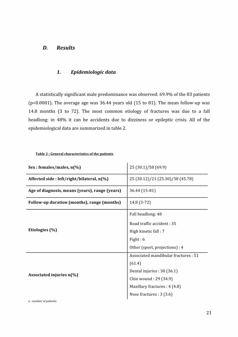
D. Results
1. Epidemiologicdata
Astatisticallysignificantmalepredominancewasobserved:69.9%ofthe83patients
(p<0.0001).Theaverageagewas36.44yearsold (15 to81).Themean follow-upwas
14.8 months (3 to 72). The most common etiology of fractures was due to a fall
headlong: in 48% it can be accidents due to dizziness or epileptic crisis. All of the
epidemiologicaldataaresummarizedintable2.
Table2:Generalcharacteristicsofthepatients
Sex:females/males,n(%) 25(30.1)/58(69.9)
Affectedside:left/right/bilateral,n(%)25(30.12)/21(25.30)/38(45.78)
Ageofdiagnosis,means(years),range(years) 36.44(15-81)
Follow-upduration(months),range(months)14.8(3-72)
Etiologies(%)
Fallheadlong:48
Roadtrafficaccident:35Highkineticfall:7Fight:6Other(sport,projections):4
Associatedinjuriesn(%)
Associatedmandibularfractures:51
(61.4)Dentalinjuries:30(36.1)Chinwound:29(34.9)Maxillaryfractures:4(4.8)Nosefractures:3(3.6)
n:numberofpatients
22
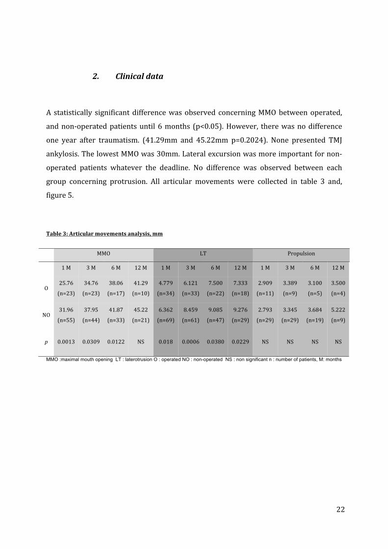
2. Clinicaldata
A statistically significantdifferencewasobserved concerningMMObetweenoperated,
andnon-operatedpatientsuntil6months (p<0.05).However, therewasnodifference
one year after traumatism. (41.29mm and 45.22mm p=0.2024). None presented TMJ
ankylosis.ThelowestMMOwas30mm.Lateralexcursionwasmoreimportantfornon-
operated patients whatever the deadline. No difference was observed between each
group concerning protrusion. All articular movements were collected in table 3 and,
figure5.
Table3:Articularmovementsanalysis,mm
MMO LT Propulsion
1M 3M 6M 12M 1M 3M 6M 12M 1M 3M 6M 12M
O25.76
(n=23)
34.76
(n=23)
38.06
(n=17)
41.29
(n=10)
4.779
(n=34)
6.121
(n=33)
7.500
(n=22)
7.333
(n=18)
2.909
(n=11)
3.389
(n=9)
3.100
(n=5)
3.500
(n=4)
NO31.96
(n=55)
37.95
(n=44)
41.87
(n=33)
45.22
(n=21)
6.362
(n=69)
8.459
(n=61)
9.085
(n=47)
9.276
(n=29)
2.793
(n=29)
3.345
(n=29)
3.684
(n=19)
5.222
(n=9)
p 0.0013 0.0309 0.0122 NS 0.018 0.0006 0.0380 0.0229 NS NS NS NS
MMO :maximal mouth opening LT : laterotrusion O : operated NO : non-operated NS : non significant n : number of patients, M: months

23
Figure5:Articularmovementsanalysis:MMO(atthetop),laterotrusion(inthemiddle)andpropulsion
(inthelower)O:operatedNO:non-operatedNS:non-significant*:significant

24
Wedistinguishedunilateralversusbilateralfractureswhetheritshouldinfluencethe
MMO.Resultsaresummarizedintable4.Therewasnostatisticallysignificantdifference
ofMMObetweenuni-orbilateralfractures.
Table4:ComparisonofMMO(mm)according touni-andbilateral fractures inoperatedandnon-operated
patients
O NO
Unilateral Bilateral p Unilateral Bilateral p
1month 26 24.91 NS 32.58 30.20 NS
3months 34.82 34.42 NS 38.82 36.77 NS
6months 40.75 34.67 0.0489 43.13 40.09 NS
12months 44.33 38.38 NS 45.69 46.50 NS
O:operatedNO:non-operatedNS:non-significant
No statistically significant difference was observed concerning TMJ dysfunctions
betweenoperatedandnon-operatedpatients.Theproportionofthissymptomincreased
withalongerfollow-up.Allresultsaresummarizedintable5.
Table5:proportionofTMJdysfunction
1month 3months 6months 1year
O 1(3.57%) 1(3.57%) 2(7.14%) 3(10.71%)NO 2(3.64%) 5(9.09%) 5(9.09%) 4(7.27%)p NS NS NS NS
O : operated NO : non-operated NS : non-significant

25
3. Radiologicaldata
Eachreadernotedtheresultsoftheanalysis.Then,averagesofdatawerecalculated
and compared between the two readers. Therewas no significant difference between
thetworeaders(p=0,6687).
a) Classificationofthefractures
Table 6 collected fractures distribution according to Loukota, Delaire and Spiessl
andSchroll’sclassifications.WeclassifiedtheintraarticularfracturesaccordingtoA,B,C
classification.Allcondylarprocessfractureswerenoted,butinthestudy,weconsidered
onlycondylarprocessfractureswitharticularimpact.
Table6:Fracturesdistributionaccordingtothedifferentclassifications
Right Left Total
LOUKOTACondylarbase 6 4 10Condylarcollum 22 22 44Diacapitular 31 37 68
DELAIRERA+ 54 59 113RA- 5 4 9
SPIESSLand
SCHROLL
I 2 1 3II 1 3 4III 4 0 4IV 3 1 4V 17 18 35VI 27 34 61UC 5 6 11
Intraarticular
fractures
A 5 6 11B 27 34 61C 17 18 35
UC : unclassified RA+ : with articular impact RA- : without articular impact

26
b) Lossofheight
The loss of height between the two ramuswas initially statistically different between
operated and non-operated patients (p = 0.0137). After surgical correction, the
differencewas not significant anymore between the two groups early after treatment
andattheendofthefollow-up(p=0.1304and0.6420).Thedifferenceoframusheight
wasalsostatisticallydifferentbeforeandaftersurgeryforoperated-patients(p=0.001)
(table7).
Table7:Lossofheightbetweenthe2ramus,mm,(%)inthecaseofunilateralcondylarprocessfractures
Beforetreatment Earlyaftertreatment Finalfollowup
Operated11.21,n=13(16.34)
3.242,n=15(4.57)
5.208,n=9(6.77)
Non-operated5.98,n=37(7.76)
5.288,n=33(7.27)
4.786,n=24(7.64)
p 0.0137 0.1304 0.6420n:numberofpatients
Inbilateralcondylarprocessfractures,therewasnostatisticalsignificantdifference
oftheaverageof2ramusheightbetweenoperatedandnon-operatedpatients,andthis
averagewas not different between before treatment, early after treatment and at the
endofthefollow-up.(table8)
Table8:Averageofthe2ramusheight,mm,inthecaseofbilateralcondylarprocessfractures
BeforetreatmentEarlyafter
treatmentFinalfollow-up
Operated61.59
n=12
65.47
n=15
64.19
n=13
Non-operated64.50
n=13
63.97
n=13
64.07
n=10
p NS NS NSNS:non-significant,n:numberofpatients

27
c) Condylarremodeling
We analyzed condylar process remodeling in non-operated patients who presented
intraarticularfractures.ResultsarecollectedinTable9.
Table9:Non-operatedcondylarremodeling
Remodeling
Typeof
fracture+++ ++ +
A 5 3 0B 3 15 7
C 3 4 6+++ : complete remodeling, ++ : moderate remodeling, + : poor remodeling
d) Chinpositionandocclusalplan
Eightpatientshadtheirocclusionplan inclinedat theendof the follow-up(3non-
operated patients and 5 operated patients). The average of tilted occlusal plan for
operatedpatientswas7.244°and4.243°fornon-operatedpatients(p=0.25).
4. Therapeuticdata
All patients profited functional reeducation. 77 patients (92.77%) profited passive
reeducationwitharches,6(7.23%)patientsdidonlyactivereeducation.
Twenty-eight patients (33.7%) and 36 intraarticular fractures profited surgical
treatment(table10):
- 8patients:bilateralpre-auricularapproaches
- 8patients:unilateralpre-auricularapproach
- 9patients:unilateralmodifiedRISDON’sapproach
- 3patients:modifiedRISDON’sapproach+pre-auricularapproach

28
Amongthe28patientswhowereoperated,26presentedasatisfactoryscar.One
scar(3.57%)wasinduratedandone(3.57%)wascolored.Nonesufferedfromfacial
paralysisoneyearaftertheoperation.
Table10:Distributionofkindsofintraarticularfractures
Two patients (7.14%) presented secondarymovements after surgerywhich didn’t
require new surgical intervention. There was no functional consequence. These two
patientspresentedtypeC(IV)fractures.
Onepatient(3.57%)requiredanewsurgicalinterventiononeyearaftertraumatism
becauseofcontactbetweentheosteosynthesisscrewandtheskullbase. ItwastypeB
fracture.
A B C
Operated 0 20 16Non-operated 11 41 19

29
E. Discussion
The purpose of this studywas to evaluate the outcomes of the isolated functional
treatment in the case of condylar process fracture with articular impact in adults, in
comparisonwithsurgicaltreatment.
In our study, 83 patientswere includedwith amale predominance, which agrees
with almostother studies (1,8,23,24,33).Etiologiesweredifferentbetweenmales and
females. Indeedmales have riskier behaviour(24,34,35): fights, road traffic accidents,
sports. The most frequent condylar process fractures’ etiologies for females were
accidentalfallsorafterdizziness.
Patients’averageageofourstudywas36.44yearsold.Thisresultisolderthanother
almost studies (24,34) which is between 20 and 30 years old. We can explain this
difference because we included only patients older than 15 years old. Children were
excluded because their treatment for this kind of fracture is always functional
(15,28,29).
Wenoted35%ofchinwoundsand36%ofdental injuries.Theseresultsshowthe
importancetolookforcondylarprocessfractureswhenthesesymptomsarepresent.
Many classifications exist to describe condylar process fractures (6,12,13,24). We
choose tobaseour studyonLoukota andSpiessl andSchroll’s ones, because theyare
very often used in the literature and is currently considered as the basis of many
comparativestudies(11,23,36–39).HoweverSpiesslandSchroll’sclassificationdoesn’t
explicitlydemarcatesthedegreeofdisplacementordislocation,whichcouldberelevant
totheprognosis.Moreover,itdoesn’tallowtoclassifyallkindsofhighcondylarprocess
fractures as shown in table 6. That’s why we also referred to Delaire’s and “ABC”
classificationsbecause theybring interesting therapeuticprospects.Theexistenceofa
lotofclassificationsmadecomparativestudieshard(40).Wethustriedto linkSpiessl
andSchrollandDelaireand“ABC”classificationstoclarifyourpurpose(table11).

30
Table11:Comparisonofclassifications
Articularimpactfractures Noimpactarticularfractures
Anatomo-fonctionalclassification
Diacapitular
Highcollumfractureswith
dislocation
Deepcollumfractureswith
dislocation
Collumfractureswithout
considerabledisplacement
Collumfractureswith
angulationwithout
dislocation
Collumfractureswith
overriding
Condylarprocessoutoftheglenoidfossa
Condylarprocessintheglenoidfossa
SpiesslandSchroll
classification
ClassVIandI ClassV ClassIV
ClassI
ClassIIandIII
ABCclassification AandB C
In our study, 33.7% of patients profited surgical treatment. Neither type A
fracturewasoperated,32.8%fortypeBand45%fortypeC.Wedidn’toperatetypeA
fracturesbecauseinthesecasestherewasapreservationoftheverticaldimensionand
occlusion that represented for us an operating criteria. Trost et al (16,41) in 2012
showed tendency was towards surgery with 82% of operated high collum fractures
(versus29%in2005)and35%ofdiacapitularfractures(versus10%in2005).
Condylar process surgical approaches are varied (26,42–44).We can differentiate
extraoral and endoscopically assisted intraoral approaches. Intraoral approaches
advantages are lack of noticeable scar and risk reduction of facial nerve damage.
Howeverosteosynthesisisgenerallymoredifficultbecauseofasmallerexposition(42).
Inourstudy,weusedonlyextraoralapproacheswithmodifiedRisdon’sapproach,and
preauricular approach. No damage of facial nerve was noted and scars werediscrete
without aesthetic discomfort, which seemed to validate the approaches we chose
regarding other reports (25,36,45). Only one patient complaining from TMJ one year

31
after surgery, needed to be reoperated to remove a screwon contactwith skull base.
Regardingtherateofosteosynthesisfailure,Seemanandal(46)observedthatitoccurs
in11.8to17.4%.Intheirstudy,thebestpredictorofosteosynthesis failurewasbased
on the ramusheight. Incasesof reducedornormalheight, theoddsofosteosynthesis
failurewas significantly reduced to a 10th.Onepossible explanation for lower failure
rates in reduced ramus heights might be reduced chewing force. There was a
significantly higher risk of osteosynthesis failure rate when no other mandibular
fracture existed (47,48). In our study the 2 patients who presented secondary
movementsaftersurgeryhadalsoisolatedcondylarprocessfractures.
TMJearlymobilization ishighlyrecommended formostofauthors(1,16,21).Early
maxilla-mandibularelasticsphysiotherapywasusedfor92.77%ofpatients.Onepatient
who didn’t profit initially from maxillo-mandibular physiotherapy arches, needed it
secondarily(4monthslater)becauseofpersistentocclusaltrouble.Inliterature,afewof
surgical teamsuse functional treatmentwithdiurnalactiveandpassivephysiotherapy
and nocturnal maxilla-mandibular fixation. After surgery, diurnal maxilla-mandibular
fixationisgenerallyproscribedbecauseitcausesmandibularlimitationmotions(11,49),
butcanbeusednightlytopromotebonehealing.
In the present study, with regard to range of motion, mouth opening was
significantly higher for non-operated patients until 6 months after traumatism. It
becamenosignificant1yearaftertraumatism.MMOwashigherthanonaverage40mm
at the end for both groups, demonstrating that conservative management including
earlyfunctionaltreatmentcouldresultinrestoringTMJfunction.InitiallowerMMOfor
operated patients could be explained by the more displaced fractures in this group.
Better mouth-opening recovery for non-operated patients could be due to earlier
reeducationofnon-operatedpatients thatstarted justafter thediagnosis. In theother
side, postoperative pain probably limited articular motions and prevented an early
recovery inoperatedpatients.Otherpredictive factorsofpoorrecoveryaredisplaced,
multiple, bilateral fractures in elderpatients (50). Inour studywedidn’t observeany
differenceinmouthopeningbetweenuni-orbilateralfracturesattheendofthefollow-
up.Itcouldbemoredifficultforoldpatientstounderstandreeducationprinciplesand,
thus cause poorer results. Three patients were more than 75 years old, and their
average MMO was 38mm 6 months after traumatism. No ankylosis case was noted.

32
Throckmorton(49)brought to lightcompleteMMOrecovery3yearsafter traumatism
concerning operated or non-operated patients. Furthermore, he noted that the longer
the MMF period the longer the time required for recovery. With regard to lateral
excursion,itwasalwaysmoreimportantfornon-operatedpatientsthanoperatedones.
ThisresultagreeswithDandaetal(51)and,HaugandAssael(52).Insomecasesofthe
present study, we observed a good fracture reduction and functional treatment
completion, but with inadequate functional results. In these particular cases it could
have been interesting to look for TMJ’s elements damages (disk, capsule) with MRI
(42,53,54).Unfortunately,thesedatawerenotavailableinthepresentstudysinceMRI
isnotpartofourcondylarfracturemanagementprotocoltodate.
TherewasnostatisticallysignificantdifferenceconcerningTMJdysfunctionbetween
thetwogroups.AccordingtoEllis(9)TMJdysfunctionisincreasedbycondylarprocess
displacement,MMFperiod andpatient’s age.Wedidn’t notice suchdifference. Indeed
patientswithdysfunction’saverageagewas36anddidn’tprofitMMF.
In the present study, the condylar process remodeling observed in adults after
functional treatment onlywas lower than in children for the same fracture (55). One
possiblelongtermsequelofhighcondylarprocessfractureisbifidcondyle.Onecaseof
bifid condyle was discovered by chance in the study in the group “non-operated”.
Another patient of our department who complained about TMJ pain 20 years after
condyle fracturepresentedwiththesamefeature.Thisunusualcondylarprocess from
congenitalorsecondarycauseusuallyrequiresonlymedicaltreatment.(56).
Differenceinramusheightwasinitiallysignificantlydifferentbetweenoperatedand
non-operated patients (16.34% for operated versus 7.76% for others). After surgery,
bothgroupsdidn’tshowdifferenceanymore.Asexpected,ORIFallowedtorestorethe
ramus height (22). This result induced biase between the two groups because initial
fractures in operated group were more displaced (40) which probably caused more
occlusaltroubleandTMJdamages.Inthepresentstudy,patientswerenotrandomized
regardingthelossofheightoftheramus.Weassumedthatthemoredisplacedfractures
were less able to remodel than fractures with little displacement. According to
literature, there isno consensus in the lossofheight thatpermits to choose forORIF.
Schneideretalconsiderthatadifferenceinramusheightmorethan2mmisasurgical

33
indication(4,57,58)whereasSugiuraetal(59)consideronlyalossofheighthigherthan
7mm.Thatwasourchoiceinthisstudysincethemeandifferencebetweenramuswas
about11mm.
Weacknowledgesomeflawsexistedinourstudy.First,asaretrospectivestudy,data
collectionwasbasedonmedical files anddatawere sometimesmissing.Moreover, as
frequently in traumatology studies,manypatientswere lost to follow-up.Anyway, on
thecontrarytoNeffetal(20)whoaffirmedthatconservativetreatmentofTMJcondylar
fractures often showed poor clinical results, our study tended to demonstrate that
functionaltreatmentonlycouldprovideasgoodresultsasORIFfollowedbyfunctional
treatment. Although this retrospective study suffers many biases, it could provide
relevantdatatostartarandomizedprospectivestudy.

34
F. Conclusion
Properly followed functional treatment of condylar process mandibular fractures
with articular impact provides satisfactory clinical results. Early mobilization is
essential. However, when fractures are too much displaced or dislocated, surgical
treatmentisnecessarytorestoreramusheight.

35
III. Discussion
L’objectif de ce travail était d’évaluer les résultats du traitement fonctionnel seul
dans le traitement des fractures condyliennes à retentissement articulaire de l’adulte
(typesA,B,C),encomparaisonavecletraitementchirurgical.
Lesfracturesducondylereprésentent9à45%desfracturesmandibulaires(26).
La prise en charge des fractures sous-condyliennes hautes à retentissement
articulaire est souvent source de débat. En effet devant la difficulté chirurgicale
d’obtenir une réduction anatomique et les risques potentiels, l’indication d’une
interventionchirurgicaleestsouventremiseencause.
Dansnotreétude83patientsontétéinclusavecuneprédominanced’hommescequi
estenaccordavec laplupartdesétudes (1,8,23,24,33).Lesétiologies responsablesde
ces fracturesnesontpas lesmêmeschez leshommesetchez les femmes.Eneffet, les
hommesontplus souventdes comportements à risque responsablesde traumatismes
faciaux(24,34,35):agression,accidentde lavoiepublique,sport.Chez les femmes, les
causes de fracture du condyle les plus fréquentes sont des chutes sur malaise ou
accidentelles.
L’âgemoyendespatientsdenotreétudeétaitde36.44ans, cequi est supérieurà
celuide laplupartdesétudes(24,34),quiengénéralestentre20et30ans.Cecipeut
s’expliquer par l’exclusion des patients âgés demoins de 15 ans et 3mois que nous
avons décidé de ne pas prendre en compte dans notre étude pour s’intéresser
uniquementauxadultes.Eneffetchezl’enfant,lapriseenchargedecetypedefracture
est moins controversée, le traitement conservateur reste le traitement de référence
(15,28,29).
Onretrouveunnombreimportantdeplaiesmentonnières(35%)etdetraumatismes
dentaires (36%). Ces résultats montrent l’importance de rechercher une fracture du
processuscondylienlorsdestraumatismesmentonniers.

36
Les fractures du condyle font l’objet de nombreuses classifications (6,12,13,24).
NousavonschoisidebasernotreétudesurcelledeLoukotaainsiquecelledeSpiessl
and Schroll puisque celles-ci sont largement utilisées dans la littérature
(13,23,36,38,38,39). Cependant cette dernière ne permet pas de préciser le degré de
déplacementoudebasculedecondyle,cequiserévèleêtreimportantpourlepronostic
fonctionnel,etnepermetpasdeclasserl’ensembledesfractures(tableau6).C’estdans
cecasquetrouvel’intérêtdelaclassificationdesfracturesintra-articulaires.Nousavons
égalementeurecoursàlaclassificationanatomo-fonctionnelledécriteparDELAIRE,qui
offredesperspectivesthérapeutiquesintéressantes.Ilnoussemblaitdoncpertinentde
l’utiliser et ainsi la mettre en parallèle des classifications habituelles. L’existence de
nombreuses classifications rend difficile la comparaison entre les différentes études
(40).Nous avons essayéde réaliserunparallélismediagnostique entre lesdifférentes
classifications(Tableau12).
Table12:Comparaisonentrelesdifférentesclassifications
Fracturesàretentissementarticulaire Fracturessansretentissementarticulaire
ClassificationAnatomo-
fonctionnelle
Capitalesou sous-capitales
Souscondylienneshautesluxées
Sous-condyliennesbassesluxées
Sous-condylienneshautesoubassessansdéplacement
SCHouSCB
basculées,sans
luxation
SCHouSCBchevauchées
Lecondyleestsortidelaglène Lecondyleestrestédanslaglène
ClassificationdeSpiesslandSchroll
ClasseVIetI ClasseV ClasseIV
ClasseI
ClasseIIetIII
ClassificationABC AetB C

37
Dans notre étude, 33.7% des patients ont bénéficié d’un traitement chirurgical.
Aucune fracturedetypeAn’aétéopérée.Enrevanche,32.8%destypesBet45%des
types C ont été opérés. Les fractures de type A ne sont pas, selon notre service,
chirurgicalescarladimensionverticaleestpréservé.SelonTrostetal(16,41)en2012,
la tendancegénéraleestversunepriseenchargechirurgicaleavec82%des fractures
sous-condylienneshautesopérées(contre29%en2005)et35%desfracturescapitales
contre10%en2005.
Les abords chirurgicaux du condylemandibulaire sont variés (26,42–44). On peut
différencier les voies d’abord extra-orales de celles endo-buccales, plus ou moins
assistéesparendoscopie.L’avantagede lavoieendo-buccaleest l’absencede cicatrice
visibleetunediminutiondurisqued’atteintedunerf facial.Cependant lasynthèseest
généralement plus difficile du fait d’une exposition moins bonne (42). De plus, elle
nécessite du matériel et une formation spécifiques. Dans notre étude nous avons
essentiellementeurecoursàdesvoiesextra-oralesparvoiedeRISDONmodifiéeoupar
voiepré-tragienne.Nousn’avonsnotéaucuneatteintedunerffacialenpost-opératoire
ce qui est inférieur aux résultats décrits dans la littérature (25,36,45). Les cicatrices
cervicalesoupré-tragiennesrestaientdiscrètesn’entrainantpasdegêneesthétiquechez
lespatients.Danslescomplicationspost-opératoires,nousavonsretrouvéunepatiente
qui a présenté à distance une douleur de l’ATM à l’ouverture buccale à 1 an post-
opératoire.L’imagerieréaliséeretrouvaitunevisd’ostéosynthèseaucontactdelabase
ducraneliéàlafonteducondyle,nécessitantunereprisechirurgicale.
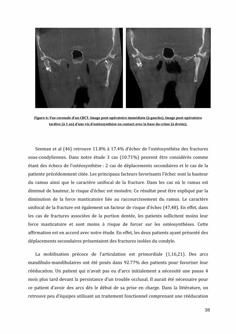
38
Figure6:Vuecoronaled’unCBCT.Imagepost-opératoireimmédiate(àgauche),Imagepost-opératoire
tardive(à1an)d’unevisd’ostéosynthèseencontactaveclabaseducrâne(àdroite).
Seemanetal(46)retrouve11.8%à17.4%d’échecdel’ostéosynthèsedesfractures
sous-condyliennes. Dans notre étude 3 cas (10.71%) peuvent être considérés comme
étantdeséchecsde l’ostéosynthèse:2casdedéplacementssecondaireset lecasde la
patienteprécédemmentcitée.Lesprincipauxfacteursfavorisantsl’échecsontlahauteur
du ramus ainsi que le caractère unifocal de la fracture. Dans les cas où le ramus est
diminuédehauteur,lerisqued’échecestmoindre.Cerésultatpeutêtreexpliquéparla
diminution de la force masticatoire liée au raccourcissement du ramus. Le caractère
unifocaldelafractureestégalementunfacteurderisqued’échec(47,48).Eneffet,dans
les cas de fractures associées de la portion dentée, les patients sollicitentmoins leur
force masticatoire et sont moins à risque de forcer sur les ostéosynthèses. Cette
affirmationestenaccordavecnotreétude.Eneffet,lesdeuxpatientsayantprésentédes
déplacementssecondairesprésentaientdesfracturesisoléesducondyle.
La mobilisation précoce de l’articulation est primordiale (1,16,21). Des arcs
mandibulo-mandibulaires ont été posés dans 92.77%des patients pour favoriser leur
rééducation.Unpatient qui n’avait pas eud’arcs initialement a nécessité unepause4
moisplustarddevantlapersistanced’untroubleocclusal.Ilauraitéténécessairepour
ce patient d’avoir des arcs dès le début de sa prise en charge. Dans la littérature, on
retrouvepeud’équipesutilisantuntraitementfonctionnelcomprenantunerééducation

39
diurne et un blocagemaxillo-mandibulaire nocturne. Le blocagemaxillo-mandibulaire
strict est à proscrire puisqu’il diminue la mobilité mandibulaire (49), et favorise la
survenue de limitation des amplitudes articulaires (11), mais utilisé en nocturne et
associéàunerééducationdiurneilparticipeàretrouveruneocclusionsatisfaisante.
Dansl’étude,encequiconcernelesamplitudesarticulaires,l’ouverturebuccaleétait
significativement plus importante chez les patients non-opérés jusqu’à 6 mois post-
traumatique. Elle devenait non significative à 1 an. Les ouvertures buccales étaient à
plusde40mmenmoyenneà la finde lapriseenchargedanslesdeuxgroupes,cequi
montre que le traitement fonctionnel permet d’obtenir une bonne restauration de la
fonctiondel’ATM.L’ouverturebuccaleplusfaibleinitialementchezlespatientsopérés
(25.76mm (min 10mm) chez les opérés contre 31.96mm (min 15mm) chez les non-
opérés,p:0.0013)estpossiblementliéeaufaitquelesfracturesétaientplusdéplacées
dans ce groupe. La récupération d’une ouverture buccale plus importante est
probablementdueàunerééducationréaliséeplusprécocementdanslecasdespatients
n’ayant pas été opérés.Dans le cas des patients opérés, les douleurs post-opératoires
peuventlimiterlesmouvementsarticulaires.Lesautresfacteursderisquedemauvaise
récupérationretrouvésdanslalittératuresont: lessujetsâgés,lesfracturesdéplacées,
les fractures bilatérales et dans les cas où il y a d’autres fractures mandibulaires
associées(50).Dansnotreétudenousn’avonspas trouvédedifférencesignificativede
l’ouverturebuccaleàunanentrelespatientsayanteuuneatteinteunioubilatérale(p:
0.19). L’âge peut altérer la compréhension des principes de la rééducation, et ainsi
entraînerdemoinsbonsrésultats.Dansnotreétude3patientsavaientplusde75anset
l’ouverture buccale moyenne à 6 mois était de 38mm. Aucun cas d’ankylose n’a été
retrouvé dans notre étude. Throckmorton (49) met en évidence une récupération
complète de l’ouverture buccale à 3 ans que ce soit chez des sujets opérés ou non-
opérés.Ilnote,deplus,unerécupérationpluslonguedanslecasoùunblocagemaxillo-
mandibulaireestinstauréetladuréederécupérationestproportionnelleàladuréedu
blocage.Encequiconcernelesdiductions,ellesétaienttoujourssignificativementplus
importantesducôtépathologiquechezlespatientsnon-opérésparrapportauxpatients
opérés.CerésultatestégalementretrouvédanslesétudesdeDandaetal(51)etHaug
andAssael(52).Danslescasoùlaréductionanatomiqueestsatisfaisanteassociéeàun
traitement fonctionnel bienmené et que les résultats fonctionnels sont insuffisants, il

40
serait intéressant de rechercher une atteinte de l’appareil disco-ligamentaire par une
IRM(42,53,54).Cependantnousn’avonspasderésultatsencequiconcernecetexamen.
Iln’yavaitpasdedifférencesignificativedanslasurvenuedeSADAMentrelesdeux
groupes.SelonELLIS(9), lerisquedeSADAMestaugmentéavecledegréd’inclinaison
ducondyle, laduréedublocagemaxillo-mandibulaireetl’âgedupatient.Nousn’avons
pasconstatéunetellecorrélation.Lamoyenned’âgedespatientsprésentantunSADAM
étaitde36ansetaucundespatientsn’abénéficiéd’unblocagemaxillo-mandibulaire.
Dansl’étudenousavonspuconstaterqueleremodelageosseuxchezl’adulteexiste
maisilestmoinsperformantquechezl’enfant(55).Uneséquelleàlongtermepouvant
être retrouvéeest la créationd’uncondylebifide.Uncasdebifidité condylienneaété
retrouvédefaçonfortuitedansnotreétudedanslegroupedespatientsnon-opérés.Un
autrecasdecetteanomalieavaitétéretrouvédans leservice,misenévidence20ans
aprèsletraumatismedevantdesdouleursdel’ATM.Cetteanomalieatypiquepeutêtre
d’origine congénitale ou post-traumatique (56). Le traitement est principalement
fonctionnel.
Figure7:Reconstructionsscannograpiquestridimensionnellesillustrantunebifiditécondyliennedroite
La différence de hauteur des ramus était significativement différente initialement
entrelespatientsopérésetnon-opérés(16.34%depertechezlesopéréscontre7.76%
chez les patients non-opérés). Après prise en charge chirurgicale de ces fractures, les
deux groupes ne présentaient plus de différence significative (p = 0.1594 au temps

41
précoceetp=0.6894autempstardif).Le traitementchirurgicalpermetderétablir la
hauteurduramus(22).Cerésultatinduitunbiaisdansnotrecarlesfracturesdugroupe
«opérés»étaientdesfracturesplusdéplacées(40)etayantdoncplusdeconséquences
auniveaudel’occlusion.Dansl’étudelespatientsn’étaientpasrandomisésenfonction
delapertedehauteurduramus.Danslalittérature,iln’yapasdeconsensusquantàla
perte de hauteur minimale pour une prise en charge chirurgicale. Schneider et al
considèrequ’unedifférencedehauteurdeplusde2mmestuneindicationchirurgicale
(4,57,58).Sugiuraetal.(59)quantà luiconsidèrequelapertedehauteurdoitêtrede
7mmpourêtreuneindicationchirurgicale.
Notreétudecomporteplusieursdéfautsdeconception.Toutd’abord, ils’agitd’une
étude rétrospective dont le recueil de données a été réalisé à partir des dossiers
médicaux qui ne sont pas toujours correctement remplis et dans lesquels certaines
informations sont manquantes. De plus, comme souvent dans des études de
traumatologie, de nombreux patients ont été perdus de vue limitant le nombre de
patientsinclus.Cependant,onpeutnoterquelesrésultatscliniquessontcorrectsdans
les deux groupes, avec une récupération de l’ouverture buccale à un an. Les patients
présentaientdebonsrésultatsavecuntraitementfonctionnelseul.Cetteétudepourrait
servirdepointdedépartpouruneétudeprospective,randomisée.

42
IV. Conclusion
Un traitement fonctionnel bien mené des fractures du processus condylien à
retentissement articulaire chez l’adulte permet l’obtention de résultats satisfaisants à
long terme sur le plan fonctionnel et architectural. Il est nécessaire de réaliser une
mobilisation précoce de l’articulation lésée. Cependant dans les cas où les fractures
présententundéplacementouuneluxationcondyliennetropimportants,untraitement
chirurgicalestindispensablepourpermettrederétablirlahauteurduramus.

43
V. Référencesbibliographiques
1. Zachariades N, Mezitis M, Mourouzis C, Papadakis D, Spanou A. Fractures of the
mandibular condyle: a review of 466 cases. Literature review, reflections on treatment and
proposals. J Cranio-Maxillo-fac Surg Off Publ Eur Assoc Cranio-Maxillo-fac Surg. 2006
Oct;34(7):421–32.
2. Schneider M, Lauer G, Eckelt U. Surgical treatment of fractures of the mandibular
condyle: a comparison of long-term results following different approaches - functional,
axiographical, and radiological findings. J Cranio-Maxillo-fac Surg Off Publ Eur Assoc
Cranio-Maxillo-fac Surg. 2007 Apr;35(3):151–60.
3. Ellis E, Throckmorton GS. Treatment of mandibular condylar process fractures:
biological considerations. J Oral Maxillofac Surg Off J Am Assoc Oral Maxillofac Surg. 2005
Jan;63(1):115–34.
4. Eckelt U, Schneider M, Erasmus F, Gerlach KL, Kuhlisch E, Loukota R, et al. Open
versus closed treatment of fractures of the mandibular condylar process-a prospective
randomized multi-centre study. J Cranio-Maxillo-fac Surg Off Publ Eur Assoc Cranio-
Maxillo-fac Surg. 2006 Jul;34(5):306–14.
5. Rutges JPHJ, Kruizinga EHW, Rosenberg A, Koole R. Functional results after
conservative treatment of fractures of the mandibular condyle. Br J Oral Maxillofac Surg.
2007 Jan;45(1):30–4.
6. Mercier J, Huet P, Perrin JP. [Functional management of fractures of the mandibular
condyle]. Rev Stomatol Chir Maxillofac. 2000 Oct;101(4):203–6.
7. Delaire J, Le Roux J, Tulasne JF. [Functional treatment of fractures of the mandibular
condyle and its neck]. Rev Stomatol Chir Maxillofac. 1975 Jun;76(4):331–50.
8. Amaratunga NA. A study of condylar fractures in Sri Lankan patients with special
reference to the recent views on treatment, healing and sequelae. Br J Oral Maxillofac Surg.
1987 Oct;25(5):391–7.

44
9. Ellis E. Complications of mandibular condyle fractures. Int J Oral Maxillofac Surg.
1998 Aug;27(4):255–7.
10. Kadlub N, Trost O, Duvernay A, Parmentier J, Wirth C, Malka G.
[Orthopaedic treatment of extraarticular condylar fractures of the mandible: retrospective
study of 39 unilateral cases]. Rev Stomatol Chir Maxillofac. 2008 Nov;109(5):301–5;
discussion 305–6.
11. Handschel J, Rüggeberg T, Depprich R, Schwarz F, Meyer U, Kübler NR, et
al. Comparison of various approaches for the treatment of fractures of the mandibular
condylar process. J Cranio-Maxillo-fac Surg Off Publ Eur Assoc Cranio-Maxillo-fac Surg.
2012 Dec;40(8):e397–401.
12. Loukota RA, Eckelt U, De Bont L, Rasse M. Subclassification of fractures of
the condylar process of the mandible. Br J Oral Maxillofac Surg. 2005 Feb;43(1):72–3.
13. Loukota RA, Neff A, Rasse M. Nomenclature/classification of fractures of the
mandibular condylar head. Br J Oral Maxillofac Surg. 2010 Sep;48(6):477–8.
14. Schneider,Lauer,Eckelt. Surgical treatment of fractures of the mandibular
condyle: a comparison of long-term results following different approaches - functional,
axiograph... - PubMed - NCBI [Internet]. [cited 2016 Feb 21]. Available from:
http://www.ncbi.nlm.nih.gov/pubmed/17583525
15. Mercier J, Lemoine V, Gaillard A, Delaire J. [Results of treatment of
mandibular fractures in 27 children (author’s transl)]. Rev Stomatol Chir Maxillofac.
1980;81(5):296–300.
16. Trost O, Péron J-M. [Latest trends in the surgical management of mandibular
condyle fractures in France, 2005-2012]. Rev Stomatol Chir Maxillo-Faciale Chir Orale. 2013
Dec;114(6):341–8.
17. Kommers SC, Boffano P, Forouzanfar T. Consensus or controversy? The
classification and treatment decision-making by 491 maxillofacial surgeons from around the
world in three cases of a unilateral mandibular condyle fracture. J Cranio-Maxillo-fac Surg
Off Publ Eur Assoc Cranio-Maxillo-fac Surg. 2015 Dec;43(10):1952–60.

45
18. Gupta M, Iyer N, Das D, Nagaraj J. Analysis of different treatment protocols
for fractures of condylar process of mandible. J Oral Maxillofac Surg Off J Am Assoc Oral
Maxillofac Surg. 2012 Jan;70(1):83–91.
19. Ellis E. Method to determine when open treatment of condylar process
fractures is not necessary. J Oral Maxillofac Surg Off J Am Assoc Oral Maxillofac Surg. 2009
Aug;67(8):1685–90.
20. Neff A, Kolk A, Deppe H, Horch HH. [New aspects for indications of surgical
management of intra-articular and high temporomandibular dislocation fractures]. Mund-
Kiefer- Gesichtschirurgie MKG. 1999 Jan;3(1):24–9.
21. Meyer C. [Fractures of the condylar region: functional treatment or surgery?].
Rev Stomatol Chir Maxillofac. 2006 Jun;107(3):133–5.
22. Kolk A, Neff A. Long-term results of ORIF of condylar head fractures of the
mandible: A prospective 5-year follow-up study of small-fragment positional-screw
osteosynthesis (SFPSO). J Cranio-Maxillo-fac Surg Off Publ Eur Assoc Cranio-Maxillo-fac
Surg. 2015 May;43(4):452–61.
23. Klatt J, Pohlenz P, Blessmann M, Blake F, Eichhorn W, Schmelzle R, et al.
Clinical follow-up examination of surgically treated fractures of the condylar process using
the transparotid approach. J Oral Maxillofac Surg Off J Am Assoc Oral Maxillofac Surg.
2010 Mar;68(3):611–7.
24. Zhou H-H, Liu Q, Cheng G, Li Z-B. Aetiology, pattern and treatment of
mandibular condylar fractures in 549 patients: a 22-year retrospective study. J Cranio-
Maxillo-fac Surg Off Publ Eur Assoc Cranio-Maxillo-fac Surg. 2013 Jan;41(1):34–41.
25. Ellis E, McFadden D, Simon P, Throckmorton G. Surgical complications with
open treatment of mandibular condylar process fractures. J Oral Maxillofac Surg Off J Am
Assoc Oral Maxillofac Surg. 2000 Sep;58(9):950–8.
26. Schmelzeisen R, Cienfuegos-Monroy R, Schön R, Chen C-T, Cunningham L,
Goldhahn S. Patient benefit from endoscopically assisted fixation of condylar neck fractures--
a randomized controlled trial. J Oral Maxillofac Surg Off J Am Assoc Oral Maxillofac Surg.
2009 Jan;67(1):147–58.

46
27. Sawhney R, Brown R, Ducic Y. Condylar fractures. Otolaryngol Clin North
Am. 2013 Oct;46(5):779–90.
28. Zhao Y, Yang J, Bai R, Ge L, Zhang Y. A retrospective study of using
removable occlusal splint in the treatment of condylar fracture in children. J Cranio-Maxillo-
fac Surg Off Publ Eur Assoc Cranio-Maxillo-fac Surg. 2014 Oct;42(7):1078–82.
29. Chrcanovic BR. Open versus closed reduction: mandibular condylar fractures
in children. Oral Maxillofac Surg. 2012 Sep;16(3):245–55.
30. Tuna EB, Dündar A, Çankaya AB, Gençay K. Conservative Approach to
Unilateral Condylar Fracture in a Growing Patient: A 2.5-Year Follow Up. Open Dent J. 2012
Jan 12;6:1–4.
31. Hlawitschka M, Eckelt U. Assessment of patients treated for intracapsular
fractures of the mandibular condyle by closed techniques. J Oral Maxillofac Surg Off J Am
Assoc Oral Maxillofac Surg. 2002 Jul;60(7):784–91; discussion 792.
32. Gilhuus-Moe O. Fractures of the mandibular condyle in the growth period.
Histologic and autoradiographic observations in the contralateral, nontraumatized condyle.
Acta Odontol Scand. 1971 Apr;29(1):53–63.
33. Villarreal PM, Monje F, Junquera LM, Mateo J, Morillo AJ, González C.
Mandibular condyle fractures: determinants of treatment and outcome. J Oral Maxillofac Surg
Off J Am Assoc Oral Maxillofac Surg. 2004 Feb;62(2):155–63.
34. Ellis E, Moos KF, el-Attar A. Ten years of mandibular fractures: an analysis of
2,137 cases. Oral Surg Oral Med Oral Pathol. 1985 Feb;59(2):120–9.
35. Silvennoinen U, Iizuka T, Lindqvist C, Oikarinen K. Different patterns of
condylar fractures: an analysis of 382 patients in a 3-year period. J Oral Maxillofac Surg Off J
Am Assoc Oral Maxillofac Surg. 1992 Oct;50(10):1032–7.
36. Landes CA, Day K, Lipphardt R, Sader R. Closed versus open operative
treatment of nondisplaced diacapitular (Class VI) fractures. J Oral Maxillofac Surg Off J Am
Assoc Oral Maxillofac Surg. 2008 Aug;66(8):1586–94.
37. Landes CA, Day K, Glasl B, Ludwig B, Sader R, Kovács AF. Prospective

47
evaluation of closed treatment of nondisplaced and nondislocated mandibular condyle
fractures versus open reposition and rigid fixation of displaced and dislocated fractures in
children. J Oral Maxillofac Surg Off J Am Assoc Oral Maxillofac Surg. 2008
Jun;66(6):1184–93.
38. Landes CA, Lipphardt R. Prospective evaluation of a pragmatic treatment
rationale: open reduction and internal fixation of displaced and dislocated condyle and
condylar head fractures and closed reduction of non-displaced, non-dislocated fractures Part
II: high condylar and condylar head fractures. Int J Oral Maxillofac Surg. 2006
Feb;35(2):115–26.
39. Boehle AP, Herrmann E, Ghanaati S, Ballon A, Landes CA. Transoral vs.
extraoral approach in the treatment of condylar neck fractures. J Cranio-Maxillo-fac Surg Off
Publ Eur Assoc Cranio-Maxillo-fac Surg. 2015 Mar;43(2):224–31.
40. Berner T, Essig H, Schumann P, Blumer M, Lanzer M, Rücker M, et al. Closed
versus open treatment of mandibular condylar process fractures: A meta-analysis of
retrospective and prospective studies. J Cranio-Maxillo-fac Surg Off Publ Eur Assoc Cranio-
Maxillo-fac Surg. 2015 Oct;43(8):1404–8.
41. Trost O, Kadlub N, Abu El-Naaj I, Danino A, Trouilloud P, Malka G. [Surgical
management of mandibular condylar fractures in adults in France, 2005]. Rev Stomatol Chir
Maxillofac. 2007 Jun;108(3):183–8.
42. Jensen T, Jensen J, Nørholt SE, Dahl M, Lenk-Hansen L, Svensson P. Open
reduction and rigid internal fixation of mandibular condylar fractures by an intraoral
approach: a long-term follow-up study of 15 patients. J Oral Maxillofac Surg Off J Am Assoc
Oral Maxillofac Surg. 2006 Dec;64(12):1771–9.
43. Lutz J-C, Clavert P, Wolfram-Gabel R, Wilk A, Kahn J-L. Is the high
submandibular transmasseteric approach to the mandibular condyle safe for the inferior
buccal branch? Surg Radiol Anat SRA. 2010 Dec;32(10):963–9.
44. Manisali M, Amin M, Aghabeigi B, Newman L. Retromandibular approach to
the mandibular condyle: a clinical and cadaveric study. Int J Oral Maxillofac Surg. 2003
Jun;32(3):253–6.

48
45. Vesnaver A, Ahčan U, Rozman J. Evaluation of surgical treatment in
mandibular condyle fractures. J Cranio-Maxillo-fac Surg Off Publ Eur Assoc Cranio-Maxillo-
fac Surg. 2012 Dec;40(8):647–53.
46. Seemann R, Undt G, Lauer G, Holawe S, Schicho K, Czerny C, et al. Is failure
of condylar neck osteosynthesis predictable based on orthopantomography? Oral Surg Oral
Med Oral Pathol Oral Radiol Endod. 2011 Mar;111(3):362–71.
47. Seemann R, Frerich B, Müller S, Koenke R, Ploder O, Schicho K, et al.
Comparison of locking and nonlocking plates in the treatment of mandibular condyle
fractures. Oral Surg Oral Med Oral Pathol Oral Radiol Endod. 2009 Sep;108(3):328–34.
48. Seemann R, Perisanidis C, Schicho K, Wutzl A, Poeschl WP, Köhnke R, et al.
Complication rates of operatively treated mandibular fractures--the mandibular neck. Oral
Surg Oral Med Oral Pathol Oral Radiol Endod. 2010 Jun;109(6):815–9.
49. Throckmorton GS, Ellis E. Recovery of mandibular motion after closed and
open treatment of unilateral mandibular condylar process fractures. Int J Oral Maxillofac
Surg. 2000 Dec;29(6):421–7.
50. Niezen ET, Stuive I, Post WJ, Bos RRM, Dijkstra PU. Recovery of mouth-
opening after closed treatment of a fracture of the mandibular condyle: a longitudinal study.
Br J Oral Maxillofac Surg. 2015 Feb;53(2):170–5.
51. Danda AK, Muthusekhar MR, Narayanan V, Baig MF, Siddareddi A. Open
versus closed treatment of unilateral subcondylar and condylar neck fractures: a prospective,
randomized clinical study. J Oral Maxillofac Surg Off J Am Assoc Oral Maxillofac Surg.
2010 Jun;68(6):1238–41.
52. Haug RH, Assael LA. Outcomes of open versus closed treatment of
mandibular subcondylar fractures. J Oral Maxillofac Surg Off J Am Assoc Oral Maxillofac
Surg. 2001 Apr;59(4):370–5; discussion 375–6.
53. Hlawitschka M, Loukota R, Eckelt U. Functional and radiological results of
open and closed treatment of intracapsular (diacapitular) condylar fractures of the mandible.
Int J Oral Maxillofac Surg. 2005 Sep;34(6):597–604.

49
54. Kim BC, Lee YC, Cha HS, Lee S-H. Characteristics of temporomandibular
joint structures after mandibular condyle fractures revealed by magnetic resonance imaging.
Maxillofac Plast Reconstr Surg. 2016 Dec;38(1):24.
55. Lindahl L, Hollender L. Condylar fractures of the mandible. II. a radiographic
study of remodeling processes in the temporomandibular joint. Int J Oral Surg. 1977
Jun;6(3):153–65.
56. Khonsari R-H, Corre P, Bouguila J, Lumineau J-P, Heuzé Y. [Bifid mandibular
condyle: position of the supernumerary condyle]. Rev Stomatol Chir Maxillofac. 2010
Sep;111(4):221–4.
57. Schneider M, Erasmus F, Gerlach KL, Kuhlisch E, Loukota RA, Rasse M, et
al. Open reduction and internal fixation versus closed treatment and mandibulomaxillary
fixation of fractures of the mandibular condylar process: a randomized, prospective,
multicenter study with special evaluation of fracture level. J Oral Maxillofac Surg Off J Am
Assoc Oral Maxillofac Surg. 2008 Dec;66(12):2537–44.
58. Kommers S, Moghimi M, van de Ven L, Forouzanfar T. Is radiological
shortening of the ramus a reliable guide to operative management of unilateral fractures of the
mandibular condyle? Br J Oral Maxillofac Surg. 2014 Jul;52(6):491–5.
59. Sugiura T, Yamamoto K, Murakami K, Sugimura M. A comparative evaluation
of osteosynthesis with lag screws, miniplates, or Kirschner wires for mandibular condylar
process fractures. J Oral Maxillofac Surg Off J Am Assoc Oral Maxillofac Surg. 2001
Oct;59(10):1161–8; discussion 1169–70.

50
Vu, le Président du Jury,
(tampon et signature)
Vu, le Directeur de Thèse,
(tampon et signature)
Vu, le Doyen de la Faculté,
(tampon et signature)

51
NOM : MERLET PRENOM : Fanny-Laure
Intérêt du traitement fonctionnel isolé dans le traitement des fractures articulaires du processus condylien chez l’adulte. Etude rétrospective de 83 cas.
⎯⎯⎯⎯⎯⎯⎯⎯⎯⎯⎯⎯⎯⎯⎯⎯⎯⎯⎯⎯⎯⎯⎯⎯⎯⎯⎯⎯⎯⎯⎯⎯⎯⎯⎯⎯
RESUME
Introduction: La prise en charge thérapeutique des fractures du condyle mandibulaire restecontroversée. Le but de cette étude était d'évaluer les résulats cliniques et radiologiques du
traitement fonctionnel isolé contre le traitement chirurgical dans les fractures du condyle à
retentissementarticulaire.
Matériel et méthode:Quatre-vingt trois patients présentant des fractures du processus
condylienàretentissementarticulaireontétéinclusdanscetteétuderétrospective.Ilsontétéclassés
en fonction des classifications de Loukota, de Spiessl and Schroll, de Delaire et de Rasse, Nef,
Hlawitschka. Deux groupes ont été formés: patients opérés (opérés) et patients non-opérés (non-
opérés). Les résultats occlusaux et fonctionnels ont été évalués à 1, 3, 6 et 12 mois après le
traitement.Lesmesuresradiologiquesontétéréaliséesenpré-thérapeutique,à6semainesaprèsle
débutdutraitementetàlafindusuivi.
Résultats : Une prédominance d'hommes a été observées (69.9%, p<0.0001). Un traitement
fonctionnelisoléaétéréaliséchez55patients(66.26%).Vingthuitpatients(33.7%)ontétéopérés,
soitavecunevoiedeRisdonmodifiéesoitavecunevoiepré-tragienne.L'ouverturebuccalemaximale
était inférieurechez lespatientsopérésparrapportauxpatientsnon-opérés jusqu'à6mois(25.75
mmvs31.96mm,34.76mmvs37.95mm,38.06mmvs41.87mmrespectivement1, 3 et, 6mois,
p<0.05).Lesrésultatsétaientsatisfaisantsdanslesdeuxgroupesunanaprèsletraumatisme(41.29
mm vs 45.22mm, p>0.05). Il n'y avait pas de différence concernant le dysfonctionnement de
l’appareilmanducateurentrelesdeuxgroupes.Pourlesfracturesunilatérales,lapertedehauteurdu
ramusétaitsignificativementplusimportantedanslegroupedespatientsopérés(p=0.0137).Après
correctionchirurgicale,iln'yavaitplusdedifférenceentrelesdeuxgroupes.
Conclusion: Notre étude montrait qu'un traitement fonctionnel qui est bien réalisé permet
d'obtenird'aussibonsrésultatscliniquesqu'untraitementchirurgicaldanslecadredesfracturesdu
condylemandibulaireàretentissmentarticulaire.Letraitementchirurgicaldoitêtreréservéauxcas
où la perte de hauteur du ramus est importante pour permettre de restaurer une hauteur
satisfaisante puisqu'en effet, le remodelage condylien est moins efficace chez l'adulte que chez
l'enfant.
Copyright © 2022 FDOKUMEN