università degli studi di milano - AIR Unimi
75
1 UNIVERSITÀ DEGLI STUDI DI MILANO DOTTORATO Di Ricerca In Scienze Odontostomatologiche XXXIII CICLO Dipartimento Di Scienze Biomediche, Chirurgiche e Odontiatriche EVALUATION OF ARTHROCENTESIS WITH HYALURONIC ACID INJECTIONS FOR MANAGEMENT OF TEMPOROMANDIBULAR DISORDERS Dottoranda: FUNDA GOKER Matricola: R11884 Tutor: Professor MASSIMO DEL FABBRO Coordinatore del Dottorato: Professor MASSIMO DEL FABBRO A.A. 2019/2020
-
Upload
khangminh22 -
Category
Documents
-
view
0 -
download
0
Transcript of università degli studi di milano - AIR Unimi

1
UNIVERSITÀ DEGLI STUDI DI MILANO
DOTTORATO Di Ricerca In Scienze Odontostomatologiche
XXXIII CICLO
Dipartimento Di Scienze Biomediche, Chirurgiche e Odontiatriche
EVALUATION OF ARTHROCENTESIS WITH HYALURONIC ACID
INJECTIONS FOR MANAGEMENT OF TEMPOROMANDIBULAR
DISORDERS
Dottoranda: FUNDA GOKER
Matricola: R11884
Tutor: Professor MASSIMO DEL FABBRO
Coordinatore del Dottorato: Professor MASSIMO DEL FABBRO
A.A. 2019/2020

2
INDEX
1- INTRODUCTION
A. ANATOMY
A.1. Temporomandibular Joint
A.2. Innervation and Vascularization
A.3. Ligaments of the TMJ
B. TEMPOROMANDIBULAR JOINT DISORDERS (TMD)
B.1. Classification of Temporomandibular Joint Disorders (TMD)
B.2. Etiology and Identification of Temporomandibular Disorders (TMD)
B.3. Muscular Etiopathogenesis C. FUNCTIONAL TMJ DISORDERS C.1. Derangements Of The Condyle-Disc Complex
C.1.1. Disc dislocation (displacement) with reduction
C.1.2. Disc dislocation without reduction
C.1.3. Luxation, Subluxation, Hypermobility
C.1.4. Compression
C.2. Structural Incompatibility of the Articular Surfaces C.3. Inflammatory Joint Disorders
D. DIAGNOSTIC IMAGING MODALITIES FOR THE ASSESSMENT OF PAIN
SYMPTOMS IN TMD
E. TMD THERAPY
F. SURGICAL INTERVENTIONS FOR TMD

3
G. ARTHROCENTESIS
G.1. History and Description
G.2.Rationale of Arthrocentesis
G.3. Indications for Arthrocentesis
G.4. Contraindications for Arthrocentesis
G.5. Postoperative complications
G.6. Irrigating solutions and volume/pressure considerations
G.7.Temporomandibular joint arthrocentesis procedure
G.8. Post-arthrocentesis injections and considerations
H. Quality of life and the SF-36 Health Survey in patients with TMD
2- MATERIALS AND METHODS
A. Systematic Review
B. Clinical case series protocol
3- RESULTS
4- DISCUSSION
5- CONCLUSION 6- REFERENCES

4
ABSTRACT Objectives: Although, arthrocentesis is an accepted safe treatment modality used for the management of TMD patients with pain, the benefit of hyaluronic acid (HA) injections remains uncertain. The aim of this study was to investigate whether intra-articular injections of hyaluronic acid as an adjunct therapy with arthrocentesis can be more effective than other medications for the improvement of symptoms associated with temporomandibular disorders. Materials and methods: For this purpose, an electronic search of Medline, Scopus and Cochrane databases was performed up to September 2020. No language, and publication date limitation was set. The following search terms were used: “arthrocentesis”, “hyaluronic acid”, “intra-articular injections”, “viscosupplementation”, with “temporomandibular disorders”. Inclusion criteria was prospective or retrospective studies, case reports, and randomized clinical trials that reported the application of HA injections compared to other intra-articular drugs for the treatment of temporomandibular disorders. Exclusion criteria included systemic reviews, animal studies. Additionally, a retrospective clinical study was performed on 12 TMD cases for evaluation of changes before and after arthrocentesis with hyaluronic acid (HA) injections in quality of life (QoL) of these patients.
Results: In the systemic review, the initial screening included 1327 articles. After a more detailed evaluation of the titles, abstracts, and full texts; a total of 29 studies were selected (26 randomized studies, 2 controlled clinical trials, 1 retrospective report). In the clinical study 12 patients were included. According to the results, intra-articular injections of HA and other medications together with arthrocentesis seemed to be beneficial for improvement of functional symptoms of TMD and pain. The case series also supported support the efficacy of HA injections with a significant improvement of QoL of these patients. However, after the evaluation of the reports in literature, it was impossible to identify an optimum drug or a protocol in improving the pain and/or functional symptoms of temporomandibular problems due to diversity of treatment modalities and conflicting results.
Conclusion: As a conclusion, there was no consensus in the studies that HA injections showed better results in comparison with other treatment modalities. According to the results of this systematic review and clinical study HA injections with/without arthrocentesis seems to be beneficial in terms of clinical symptoms and QoL of the patients.
Keywords: arthrocentesis, hyaluronic acid, intra-articular injections, temporomandibular disorders, quality of life

5
1- INTRODUCTION
A. ANATOMY
A.1. Anatomy of the TMJ
TMJ, located in the area where the mandible articulates with cranium just anterior to the external auditory meatus, is considered as one of the most complex joints in humans (Okeson, 2008, Shaffer, et al., 2014a). TMJ consists of the “mandibular condyle” of the mandibular bone (Shaffer, et al., 2014a) sitting in the “glenoid fossa” or “mandibular fossa” of the temporalis bone (Figure 1) (Okeson, 2008, Shaffer, et al., 2014a, Gray, et al., 2011). Upper articular surface consists of temporal bone’s “articular eminence” or “articular tubercule”, “mandibular fossa” or “glenoid fossa” and posteriorly “tympanic plate”. Immediately anterior to the glenoid fossa is a convex prominence, called the “articular eminence” or “articular tubercule” of the temporal bone. The degree of the convexity of the articular eminence dictates the pathway of the condyle in anterior positions (Okeson, 2008). Inferior articular part consists of condylar head of the mandible which is positioned in the mandibular fossa. The mandibular condyle usually manifests variable shapes with the posterior articulating surface greater than anterior surface (Okeson, 2008). The articulating part of the condyle is usually described as convex, angular, rounded, or flat (Alomar, et al., 2007) with an anteroposterior width of 8-10 mm, and a mediolateral length of 18-23 mm (Okeson, 2008). Articular surfaces are covered with fibrocartilage and the intraarticular disc situated between the condyle and fossa divides the joint cavity into upper and lower parts. TMJ is a synovial joint with condylar variety. TMJ is considered as a ginglymo-arthrodial joint, because it is capable of providing both hinging (as ginglymoid joint) and gliding movements (as arthrodial joint) (Okeson, 2008).
The biconcave intraarticular disk within the joint capsule (Okeson, 2008, Shaffer, et al., 2014-a) is attached to the capsular ligament anterior-posteriorly and medial-laterally). The disc has concave superior surface and convex inferior surface. The disc separates the joint into disco-mandibular lower and disco-temporal upper spaces (Shaffer, et al., 2014-a). The upper compartment permits gliding movements, while the lower compartment permits rotatory as well as gliding movements. The disc prevents the friction between the articular surfaces. It also acts as a cushion and helps in shock absorption. The disc of TMJ consists of fibrocartilage (not hyaline cartilage like other synovial joints), devoid of any blood and nerve vessels (Shaffer, et al. -a, 2014, Kuroda, et al., 2009). However, the extreme periphery of the disc is slightly innervated. From anterior view the medial part of the disc is usually thicker than lateral part and in sagittal plane articular disc can be divided into 3 regions. The posterior border of the disc is thicker than anterior border, while the intermediate zone is the thinnest. The articular surface of the mandibular condyle is located on the intermediate zone of the

6
disc, bordered by the anterior and posterior zones (Okesan, 2008). During articulative movements the shape of the disc is somewhat flexible and can adapt to the functional demands of the articular surfaces (Figure 2) (Okesan, 2008).
Figure 1: Temporomandibular joint The periphery of the disc is attached to fibrous joint capsule. This capsule is attached to articular tubercule, circumference of mandibular fossa, squamo-tympanic tissue and the neck of the mandible. The fibrous capsule is loose above the intraarticular disc and tight below the disc. The joint capsule contains synovial fluid, is lined by a synovial membrane, and possesses a lateral ligamentous thickening (temporomandibular ligament) that reinforces the joint (Shaffer, et al., 2014-a). The synovial membrane lines the fibrous capsule and the neck of the mandible. The synovial fluid, which is present in both joint cavities acts as a lubricant during function between articular surfaces. And because the articular surfaces of TMJ joint are free of vascular tissues, it also acts as a medium for providing metabolic requirements. There is a rapid exchange between the synovial fluid, the articular surfaces, and the vessels of the capsule (Okesan, 2008).

7
Figure 2: Normal closed and open disc location: Diagrammatic representation showing normal condyle disk position with junction of posterior attachment and posterior band of disk aligned approximately at the 12 o’clock position with regard to the condylar surface (Quinn & Granquist, 2015) The TMJ is tentative to local and extensive degeneration. The degenerative changes can be seen in the upper joint part or in the lower joint part. Although the temporal bone and upper joint space generally undergo less degeneration relative to the mandibular condyle and lower joint space. In cases of local degenerations, the lateral aspect of the articular tubercle of the temporal bone is most likely to be affected and mandibular condyle is not much effected. Extensive changes can culminate in a total loss of articular cartilage. The inferior surface of the disk undergoes degenerative changes more frequently than the superior aspect. The intraarticular disk can displace in the anterior, medial, lateral, or posterior directions (Shaffer, et al., 2014 -a).
A.2. Innervation-vascularization Motor and sensory innervation of TMJ is by the trigeminal nerve, while branches of the mandibular nerve provide the afferent innervation. Auriculotemporal nerve provides most of the innervation, since it leaves the mandibular nerve behind the joint and ascends superiorly and laterally and wraps posterior part of the joint (Okesan, 2008, Schmidt, et al., 1998). Because of its course, is at greatest risk for irritation or entrapment since the auriculotemporal nerve bundles can become displaced and impinged between the articular fossa and the mandibular condyle (Okesan, 2008, Shmidt, et al., 1998). Other additional nerves, that provide innervation to TMJ, are the deep temporal and masseteric nerves (Okesan, 2008). Vascularization of the TMJ is mostly by the superficial temporal artery posteriorly and by the middle meningeal artery anteriorly, the internal maxillary artery inferiorly, and additionally by ascending pharyngeal, deep auricular, anterior tympanic arteries (Okesan, 2008).

8
A.3. Ligaments of the TMJ The capsular ligament surrounds and encompasses the entire TMJ. Superiorly, the fibers of the capsular ligament are attached to temporal bone along mandibular fossa and extend anteriorly to include the articular eminence. Inferiorly they are attached to the neck of the mandibular condyle. The function of this ligament is to resist medial, lateral, and inferior forces that tend to dislocate articular surfaces. Collateral ligaments attach the lateral and medial borders of the articular disc to the lateral and medial poles of the condyle. They are composed of collagenous connective tissue and they divide the joint mediolaterally into superior, and inferior cavities. These ligaments provide the hinging movement of the TMJ. They allow the disc to be rotated on the articular surface of the mandibular condyle as it glides anteriorly and posteriorly. These ligaments do not stretch and a strain on them can be painful. Temporomandibular ligament (lateral ligament) Lateral temporomandibular ligament is attached to articular tubercule and posterior lateral aspect of the mandible. It strengthens the lateral part of the capsular ligament. Fibers of it are directed downwards and backwards. Lateral ligament is composed of horizontal and oblique parts. The outer oblique portion influences the normal rotational opening movement of the jaws and limits the extend of mouth opening. When the mouth opens, the teeth are separated approximately 20-25mm, the outer oblique portion is stretched and tense, the neck of the condyle cannot rotate any further. In case the mouth is to be opened wider, the condyle would need to move forward and downward out of the fossae and across the articular eminence, which shall create a second arc of opening. This can be demonstrated in the clinics by closing the mouth and applying mild posterior force to the chin of the patient. At this point patient will be able to open until the teeth are 20-25 mm apart, and if the jaws are opened wider, a distinct change in opening movement will occur because of the tightening of TM ligament. This serves as to prevent the mandible to impinge on the vital retro- and sub-mandibular structures of the neck. The inner horizontal portion prevents the posterior movement of the condyle and the disc into the posterior region of the mandibular fossa, thus protects the retrodiscal tissues. It also protects the lateral pterygoid muscle from extension. In cases of extreme trauma to the mandible, the neck of the condyle fractures before retrodiscal tissues are damaged or before the condyle enters the middle cranial fossa. Stylomandibular ligament The stylomandibular ligament represents a thickened part of the deep cervical fascia, which separates the parotid and submandibular salivary glands. This ligament limits excessive protrusive movements of the mandible. It extends between the styloid process and below the angle and adjacent part of the posterior border of the ramus mandible. It is most relaxed when the mouth is opened, but it becomes taut, when the mandible is protruded (Okesan, 2008, Alomar, et al., 2007). Sphenomandibular ligament

9
Sphenomandibular ligament is an accessory ligament that lies on a deep plane away from the fibrous capsule and it does not present any limiting action on mandibular movements. It is attached superiorly to the spine of the sphenoid and inferiorly to the lingula of the mandibular foramen. Its relations are as follows; i) laterally to lateropterygoid muscle, auriculotemporal nerve and maxillary nerve, ii) medially to chorda tympani, and the wall of the pharynx, iii) near to its end, it is pierced by the mylohyoid nerve and vessels. Sphenomandibular ligament is a remnant of Meckel’s cartilage (Okesan, 2008, Alomar, et al., 2007).
B. TEMPOROMANDIBULAR JOINT DISORDERS (TMD)
B.1. Classification of Temporomandibular Joint Disorders (TMD)
The ultimate goal of a classification is to provide clinically useful schemes for an easier management of patients in the clinical setting. However, despite all efforts by the several international experts and organizations, none of the proposed classifications of OFP (oral facial pain)/TMD is free of any shortcomings (Klasser, et al., 2018, Ohrbach, et al., 2019).
Table 1: The classification of TMD (Schiffman, et al., 2014)
TMJ DISORDERS:
1. Joint pain: i-Arthralgia ii-Arthritis 2. Joint disorders.
A-Disc disorders: i- Disc displacement with reduction ii- Disc displacement with reduction with intermittent locking, iii- Disc displacement without reduction with limited opening iv- Disc displacement without reduction without limited opening
B-Hypomobility disorders other than disc disorders: i- Adhesions/adherence ii- Ankylosis a. Fibrous, b. Osseous
C-Hypermobility disorders: Dislocations a. Subluxation b. Luxation
3. Joint diseases 4. Degenerative joint disease: i- Osteoarthrosis, ii- Osteoarthritis 5. Systemic arthritides 6. Condylysis/idiopathic condylar resorption 7. Osteochondritis dissecans 8. Osteonecrosis 9. Neoplasm 10. Synovial chondromatosis

10
11. Fractures 12. Congenital/developmental disorders: A- Aplasia B- Hypoplasia C-Hyperplasia
MASTICATORY MUSCLE DISORDERS:
Muscle pain: A. Spasm, B. Tendonitis, C. Myositis, D. Myalgia: 1. Local myalgia 2. Myofascial pain 3. Myofascial pain with referral: contracture, hypertrophy, neoplasm, masticatory muscle pain attributed to systemic/central pain disorders, fibromyalgia/widespread pain, headache attributed to TMD
Movement disorders: a. Orofacial dyskinesia, b. Oromandibular dystonia
B.2. Etiology and Identification of Temporomandibular Disorders (TMD)
1- Local muscle soreness (Protective co-contraction) 2- Myofascial Pain and trigger points 3- Myospasm (CNS mediated acute muscle disorder) 4- Chronic Centrally mediated myalgia 5- Derangement of the condyle-disc complex a- disc displacement and disc dislocation with
reduction, b- disc dislocation without reduction, c- Compression 6- Structural incompatibility a- Deviation in form and adhesions b- subluxation-
hypermobility, c- spontaneous dislocation (open lock) 7- Inflammatory disorders of the TMJ a- Capsulitis, b- synovitis, c-retro-discitis, d- traumatic
arthritis, e- osteoarthritis, f- infectious arthritis, 8- Ankylosis (Okesan, 2008).

11
Figure 3: Wilkes classification for TMJ internal Derangements (Wilkes, 1989)
B.3. Muscular Etiopathogenesis Table 2: Etiopathogenesis of Temporomandibular Disorders (Okesan, 2008, Prati, 2017) Muscular disorders Causes: parafunction, stress, trauma, TMJ pathology Progressive overload of the muscle and metabolic alteration Manifestation: rigidity/muscle fatigue, muscular pain, spasm, trigger points, cephalalgy (=headache)
Protective co-contraction In case of injury or in a threat of injury, normal sequencing of muscle activity seems to be altered so as to protect the threatened part from further injury. Ex: in case of high crown protective co-contraction as a protective mechanism. Limited mouth opening. The patient can achieve full opening when asked to open slowly. The key identifying factor is co-contraction is that it immediately follows an event (Okesan, 2008). Local muscle soreness (noninflammatory myalgia)

12
In cases of prolonged muscle tissue contraction, local trauma, excessive use of the muscle. It can be also referred as delayed onset muscle soreness or postexercise muscle soreness. Changes in local environment of muscle tissues are seen such as, bradykinin, histamine (algogenic substances) release. Deep pain is present, which can further produce protective co-contraction as a cycle. Clinically muscles are tender to palpation. There is muscle weakness and limited mouth opening. The patient has great difficulty opening any wider. Central nervous system (CNS) effects on muscle pain Muscle pain with central origin as a secondary respond to one of these three factors: 1-The presence of ongoing deep pain input, 2- increased level of emotional stress, 3-changes in the descending inhibitory system that lead to a decrease in the ability to counter afferent input (okesan). Treatment: Relaxation, splints/night guards, medication, botox, pain killers, muscle relaxants, release techniques sach as stretch. C. FUNCTIONAL TMJ DISORDERS Functional Disorders can be divided as follows: • Derangements of the condyle-disc complex • Structural incompatibility of the articular surfaces • Inflammatory joint disorders (Prati, 2017, Schiffman, et al., 2014).
C.1. Derangements Of The Condyle-Disc Complex C.1.1. Disc dislocation (displacement) with reduction
Figure 4: Disc dislocation (displacement) with reduction

13
Diagnosis: Disc is located anterior-medial with respect to the condyle. While opening the condyle moves into the posterior border of the disc, which can be accompanied by clicking sound.
Treatment: Treatment modalities include repositioning bite and exercises of remodeling of the disc 10x6 with progressive retrusion
non symptomatic - just click à exercises
Symptomatic- (Pain and block without dislocation of the disc) à if Pain and block is intenseà bite.
if Pain and block is not intense à exercises.
Symptomatic- (Pain and block with dislocation of the disc) à If prognostic grid is unfavorable à exercises
If prognostic grid is favorable àrepositioning bite
Table 3: Prognostic Grid (Prati, 2017)
Unfavorable clinical factors Unfavorable instrumental factors Late reduction ++ Partial reduction +++ Advanced therapeutic position +++ Disc deformity ++ Hyperlaxity of the ligaments ++ With construction bite: Partial reduction +++ With construction bite: extremely advanced position of
the condyle +++ C.1.2. Disc dislocation without reduction Disc is dislocated when mouth is closed, while opening the condyle does not move into the disc, but rather it pushes the disc forward.
Diagnosis: 1. Jaw locked so that the mouth would not open all the way; AND 2. Limitation in jaw opening severe enough to limit jaw opening and interfere with ability to eat. (note: without limited opening- Positive for those in the past)

14
Figure 5: Disc dislocation without reduction
When this diagnosis needs to be confirmed, TMJ MRI Imaging is the reference standard for this diagnosis. 1. In the maximum intercuspidal position, the posterior band of the disc is located anterior to the 11:30 position and the intermediate zone of the disc is anterior to the condylar head, AND 2. On full opening, the intermediate zone of the disc is located anterior to the condylar head.
Note: Maximum assisted opening of < 40 mm is determined clinically.
Presence of TMJ noise (ex: clicking sound during opening) does not exclude this diagnosis.
Disc displacement without reduction with limited opening: Positive for the following: 1. Maximum assisted opening (passive stretch) movement including vertical incisal overlap < 40 mm.
Disc displacement without reduction without limited opening: Positive for the following: 1. Maximum assisted opening (passive stretch) movement including vertical incisal overlap ≥ 40 mm. (Schiffmann, et al. 2014)
Therapy: “Dislocation of the disc without reduction acute”: repositioning bite and manual therapy. “Dislocation of the disc without reduction chronic”: distraction bite, and if patient does not get better in 40-50 days arthrocentesis (Prati, 2017).

15
C.1.3. Luxation, Subluxation, Hypermobility
A sudden forward movement of the condyle during the latter phase of mouth opening. Condyle moves beyond the crest of the eminence and appears to jump forward to the wide-open position. hypermobility disorder involving the disc-condyle complex and the articular eminence: In the open mouth position, the disc-condyle complex is positioned anterior to the articular eminence and is unable to return to a normal closed mouth position without a manipulative maneuver. The duration of dislocation may be momentary or prolonged. When the patient can reduce the dislocation himself/herself, this is referred to as subluxation. When the patient needs the assistance of the clinician to reduce the dislocation and normalize jaw movement, this is referred to as luxation. This disorder is also referred to as “open lock.” The sensitivity and specificity have been established for only subluxation (Schiffmann, et al. 2014).
Positive for both of the following: In last 30 days, jaw locking or catching in a wide-open mouth position, even for a moment and inability to close the mouth from a wide-open position without a self-maneuver. When this diagnosis needs to be confirmed, imaging criteria are positive for the condyle positioned beyond the height of the articular eminence with the patient unable to close his/her mouth (Schiffmann, et al. 2014).
Spontaneous Dislocation (open lock): Hyper-extention of the TMJ, resulting in a condition that fixes the joint in an open position and prevents any translation. Normal at resting position, when mouth is open, TMJ can be either posteriorly or anteriorly dislocated, and the patient is unable to close the mouth.
Dislocations of the jaw should not be confused with subluxation. Dislocation is a complete disruption of the joint, while luxation is a partial dislocation followed by a relocation. In luxation, the joint is still partially attached to the bone, and it is the point where the joint begins to dislocate. In dislocation, the two parts of joint lose contact. Subluxation is loosening without displacement, however in luxation there is partial displacement. In Subluxation, there is a moment of sense of not being able to close the mouth is present, while in luxation there is a block in opening movement. Luxation can be detected radiographically.
Hypermobility of the TMJ posteriorly favors the dislocation of the disc, hypermobility of the TMJ anteriorly favors the “luxation” and mandible and disc can exit the glenoid, generally with a click noise “subluxation” or in some cases, remains luxated and prevents closing of the jaws “open lock”. In pathologies of a block of opening/closing in luxation, the contraction of the reflex muscles plays an important role mainly due to panic affecting the block patient. While in cases of subluxation, the fear of being blocked triggers an autonomically controlled mechanism of opening which results in the disfunction of the muscles (Prati, 2017).

16
Treatment modality in subluxation is counselling and home exercises, while in luxation manual therapy of reduction is considered appropriate (Prati, 2017).
C.1.4. Compression
Condyle in retro-position with reduction of the anterior space. The disc is dislocated anteriorly when mouth is closed. At maximum opening, there is “end -feel, which can provoke pain.
C.2. Structural incompatibility of the articular surfaces (Degenerative joint disease)
A degenerative disorder involving the joint characterized by deterioration of articular tissue with concomitant osseous changes in the condyle and/or articular eminence. Positive for at least one of the following: 1. In the last 30 days, any TMJ noise(s) present with jaw movement or function; OR 2. Patient report of any noise present during the exam. Positive for the following:1. Crepitus detected with palpation during at least one of the following: opening, closing, right or left lateral, or protrusive movement(s). When this diagnosis needs to be confirmed, then TMJ CT criteria106 are positive for at least one of the following: Subchondral cyst(s), erosion(s), generalized sclerosis, or osteophyte(s). Note: Flattening and/or cortical sclerosis are considered indeterminant findings for degenerative joint disease (DJD) and may represent normal variation, aging, remodeling, or a precursor to frank DJD (Schiffmann, et al. 2014).
It is important to distinguish acute compression from chronic one. Acute form is the one which is present less than 3-4 months, while chronic compression can be considered in case of duration more than 4 months. Treatment modalities: In “post-traumatic acute compression”- Anti-inflammatory drugs for a week, and counselling. These patients should be controlled at a follow-up visit after 15 days. In case of no resolution, they should be reevaluated by use of MR. In “non-traumatic acute compression”- Manual therapy, counselling, and home exercises are indicated. In “chronic compression”- Bite of distraction is indicated (Prati, 2017)
C.3. Inflammatory joint disorders Inflammation of the various tissues that make up the joint structure (such as, synovitis, capsulitis, retrodiscitis, arthritides) as a result of unusual function, trauma, insult or breakdown. In disc derangement disorders, pain is often momentary and associated with joint movement. In inflammatory joint disorders, pain is constant and dull, which is accentuated by joint movement (Prati, 2017, Okeson, 2008).
Progressive changes in internal derangement: Traditionally, internal derangement of the TMJ is described as a progressive disorder that may be classified into four consecutive clinical stages (Barkin &Weinberg, 2000, Dolwick, et al.,1983). Although, in most of the cases, internal derangement of the disc undergoes a progressive change, it is still not clear whether this

17
progression happens in all cases. Longitudinal epidemiological studies do not support the idea of progression and clinical evidence does support progressive worsening of the condition in some patients. The probability that TMJ clicking would disappear in a symptomatic individual seems to be equal to the probability of it appearing in an asymptomatic individual (Barkin &Weinberg, 2000).
C. DIAGNOSTIC IMAGING MODALITIES FOR THE ASSESSMENT OF PAIN SYMPTOMS IN TMD
Panoramic radiography= For initial diagnosis and assessment of TMJ alteration. Diagnosis of highly advanced bone alterations in the condyle such as asymmetries, erosions, osteophytes, fractures, changes in size and shape, degenerative and inflammatory processes, growth alterations, maxillary tumors, metastases, ankylosis, and fractures. However, it does not provide functional information. Also useful for detecting odontogenic alterations whose symptoms can overlap with TMD (Ferreira, et al., 2016).
Planigraphy (or panoramic radiography with programs for TMJ)= In addition to the conventional panoramics, this technique can be useful for functional assessment of mouth opening, evaluation of morphological alteration and the joint spaces, analysis of dimension, fractures, and ankylosis.
Transcranial radiography= Anatomical assessment of the condyle, fossa, and articular tubercle. However, this type of projection is limited by the fact that it produces an image with a large overlap of the skull bones.
Arthrography= A technique to assess the TMJ soft tissues by contrast injection into the superior and/or inferior joint spaces. After the injection, dynamic images were obtained, recording mandibular movements. However, it is not currently recommended as it is an invasive procedure and carries a risk of iatrogenic disc perforation, facial nerve damage, pain and limitation of movement after the injections, infections, allergies to the injected dye, and presence of the risks of radiation to radiosensitive structures such as, crystalline and thyroid.
Medical CT and cone beam CT = Structural assessment of bone components of the TMJ. Pathological bony changes such as fractures, neoplasms, ankylosis, erosion, osteophyte, deformity, osteochondritis dissecans, hyperplasia of condylar, coronoid, and styloid processes. However, few details are provided on soft tissue and it is not possible to evaluate the joint disc Additionally, levels of radiation and costs are high.
Magnetic resonance (MR)= MRI has been the method of choice to study disease processes involving the TMJ soft tissues. The articular disc, ligaments, retro-disc tissues, intra- capsular synovial content, adjacent masticatory muscles, as well as cortical and medullary integrity of bone components can be examined by MR. It is considered the gold standard for assessing disc position

18
and is highly sensitive for intraarticular degenerative alterations. The technique allows three-dimensional analysis in the axial, coronal, and sagittal plane. Its disadvantages are related to the high cost and the need for sophisticated facilities. It is contraindicated in claustrophobic patients, those with pacemakers and metallic heart valves, ferromagnetic foreign bodies, and pregnant women. It has some limitations that could influence the final diagnostic outcome, such as the lack of standardization concerning the range of mouth opening, and even the parameters for image acquisition, such as surface coil, time of repetition, field of view, and slice thickness (Pupo, et al 2016).
Static MR imaging= Disk positional abnormalities such as, sideways or anterior disc displacements with/ without reduction. Bone marrow abnormalities such as osteonecrosis and edema. Joint effusion. Posterior disc attachment evaluation by T2 weight image in cases of higher T2 signal (MR images with the timing of frequency pulse sequences that are capable to highlight excess fat and water). Marked joint effusion. Tumor involvement, inflammatory diseases and autoimmune processes such as rheumatoid arthritis. Proximity between the TMJ disk and the mandibular nerve.
Dynamic MR imaging with contrast material= Contrast enhancement of joint effusion, the posterior disk attachment
Functional MR imaging= The regions and the network of brain activation associated with TMD
MR spectroscopy= Ascending of insular glutamine levels
Magnetization transfer contrast imaging= Edematous and ischemic changes in the muscles
Ultrasonography= A high-resolution imaging equipment for the assessment of disc position in internal TMJ disorders. Muscular edema by low-level contraction. However, it does not have accuracy for the cortical and articular disc morphological diagnosis. However, it can be a useful option particularly in patients with contraindications to MRI. Moreover, it is less expensive, allows real-time visualization without the use of ionizing radiation, and is quick and comfortable
Nuclear medicine evaluation
Bone scintigraphy= neoplastic activity, metabolic disorders, bone growth, synovitis and osteoarthritis. It has some advantages over radiographies, conventional CT, and MRI because it provides an estimate of metabolic and inflammatory activity. It can facilitate an early diagnosis and is less costly than CT and MRI. However, it does not differentiate among bone scar disorders, infections, osteoarthritic manifestations, or tumors.

19
Positron-emission tomography (PET)= Positron-emission tomography (PET) is usually indicated for the assessment and staging of metastatic tumors. It is able to provide accurate functional, morphological, and metabolic information.
Single photon emission computed tomography (SPECT/CT) = Currently, single photon emission computed tomography with technetium-99m methylene diphosphate (SPECT/CT with 99m Tc-MDP) is largely employed. The radiotracer 99m Tc is able to reflect the local osteometabolic rate, while the anatomic mapping is obtained by tomographic technique. As in the PET, anatomical and functional data are fused into a single image. Its main advantage is its sensitivity and specificity (Ferreira, et al., 2016, Suenaga, et al., 2016).
E. TMD THERAPY
1- Counselling or Patient Education
Counselling is a central component and indicated in all types of TMD. Each patient should receive individualized education (Prati, 2017, Shaffer, et al. 2014 b). “Counselling” includes: i- communication with the patient about the cause, diagnosis, prognosis, and options of treatment, ii- auto-controlling of the parafunction, iii- home exercises (Prati, 2017).
2- Splinting (Bite)
Splint therapy has very frequently been indicated for treating TMD. The most common conditions are masticatory myalgia, TMJ arthralgia, and TMJ dysfunction (9 Hasegawa). The therapeutic advantage of splint therapy, especially stabilization appliances, lies in producing stability of occlusion and equal distribution of abnormal forces causing overload of the masticatory muscles and the TMJ. There is a high probability that splint therapy would not be successful in patients with abnormalities such as bone marrow abnormalities and biconvex disc configuration of the TMJ (Hasegawa, et al., 2017). Oral splints can be in a variety of different materials and styles. One fabrication method involves obtaining impressions of the upper and lower teeth to make a custom splint. Others do not cover the teeth at all. Generally, splints are worn only while sleeping to protect the teeth, decrease nighttime bruxism, and/or reduce TMD symptoms (Prati, 2017, Shaffer, et al. 2014 b).
a- Neuromuscular bite
Indications: Chronical muscular disorders, protection against bruxism, for correcting physiologic position of the mandible. Contraindications: muscular pathologies with dislocation of the disc with reduction or subluxation. If occlusion has a predisposing role, the following should be corrected by the bite: Modification of the mandibular position with correction of pre-contacts. Modification of the vertical occlusal dimension. Modification of the masticatory interferences. Modification of the oral-buccal proprioception (Prati, 2017).

20
b- Bite for repositioning the mandible
Indications: dislocation of the disc with reduction, acute dislocation of the disc without reduction. Contraindications: All other disorders (Prati, 2017). c- Bite for distraction
Indications: Chronic compression of the disc, chronic dislocation of the disc without reduction (chronic locking), arthrosis. Contraindications: All other disorders. It should be discussed with the patient that in case of this treatment modality shall be unsuccessful after 2 months, arthrocentesis and arthroscopy shall be considered (Prati, 2017).
3- Therapeutic Exercise and Manual therapy
Perhaps the most widely known exercise routine for TMD is the Rocabado 6x6 program, which utilizes six exercises six times per day (Table below) (Prati, 2017, Shaffer, et al. 2014 b, Rocabado & Iglarsh 1991).
Table 4: Rocabado’s 6x6 exercise program
Rocabado’s 6x6 exercise program
Rest position of the tongue= The anterior 1/3 of the tongue is placed at the palate with mild pressure, which rests the tongue and jaw musculature and promotes diaphragmatic breathing
Control of TMJ rotation = The jaw is repeatedly opened and closed with the anterior 1/3 of the tongue on the palate, which decreases initiating jaw movements (e.g. protrusive movement in opening, talking, or chewing)
Rhythmic stabilization technique= Gentle isometrics in the resting position are performed for jaw opening, closing, and lateral deviation to promote muscular relaxation via reciprocal inhibition, which promotes an improved resting position of the jaw through proprioceptive input.
Axial extension of the neck= Combined upper cervical flexion with lower cervical extension, allowing reduction of tension in the cervical musculature.
Shoulder posture= Shoulder girdle retraction and depression to facilitate postural corrections.
Stabilized head flexion= Upper cervical spine distraction via chin tuck (without additional cervical flexion), during which it is recommended that the fingers be laced behind the neck to stabilize C2-7 while the head nods

21
4- Pharmacological Therapy
Articular pathologies, especially in dislocation of the disc without reduction and arthrosis: Anti-inflammatory and analgesic medications such as, Ibuprofene 40mg, Naproxene 250-500 mg, Ketorolac 15 mg (every 12 h) etc. Muscular pathologies: Anti-inflammatory drugs have modest effect. Paracetamol (with/ without opoids) and muscle relaxants. Anxiolytics such as diazepam can be also useful (Prati, 2017). 5- Antidepressants:
Bruxism and TMDs are commonly associated with depression. Bruxism results from an imbalance between the direct and indirect pathways of the basal ganglia through the disruption of action potential transmission, which is medi- ated by dopamine. Dopamine agonists are therefore helpful in treating bruxism because they increase dopamine levels and restore the balance between the basal ganglia pathways (Abouelhuda, et al., 2018, Prati, 2017). 6- Orthodontic therapy
To date there has never been a satisfactory evaluation of orthodontics with respect to TMD. However, it might be useful for the patients with severe malocclusion (Luther, et al., 2010).
7- Low level laser therapy, Ultrasound and electrical stimulation
LLLT can result in muscle relaxation and an increase in blood microcirculation, thereby accelerating the clearance of catabolites from tissues. Masseter and temporal muscles with painful symptoms were reported to be treated with the use of LLLT GA-Al-As (gallium-aluminum-arsenide) diode laser (Abouelhuda, et al., 2018).
Therapeutic ultrasound has an output of 20 to 60 kHz. It produces deep heat at joints and treats joint contracture by increasing the stretch of the extra capsular soft tissue. It also decreases non-acute pain, muscle spasms, and tendonitis, facilitating the stretch of soft tissue by decreasing the viscosity of collagen (Abouelhuda, et al., 2018).
Electrical stimulation devices for the treatment of TMDs have two main purposes: relief of pain and relief of muscle hyperactivity or spasm (Abouelhuda, et al., 2018).
In particular, various types of electrotherapy, such as pulsed radiofrequency energy, TENS, and LLLT, were reported not to show no better results than their respective placebos in the treatment of TMD (Gil-Martínez, et al., 2018)
8- Muscle Injections with Botox

22
BTX is a strong biological exotoxin produced by Clos- tridium botulinum, a Gram-positive anaerobic bacterium. According to the literature, BTX treatment seems to be useful to treat bruxism, myofascial pain, disorders associated with TMJ disc displacement, and habitual dislocation of the mandible. Treatment with BTX is contraindicated in women during pregnancy and lactation, in patients with known hypersensitivity to any component of the drug (Abouelhuda, et al., 2018).
9- Surgical Therapy
F. Surgical interventions for TMD
MINIMALLY INVASIVE: Arthrocentesis, Arthroscopy (lysis and lavage) (Politi, et al.,2007). Arthroscopy 1. Disc release 2. Disc repositioning
DISK REPOSITIONING (open): Disk plication (with or without recontouring), Eminoplasty in conjunction with disk repositioning
DISK REMOVAL (open): Discectomy (without replacement), Discectomy (with replacement) dermis, fascia, muscle flap, platelet rich plasma (Quinn & Granquist, 2015).
Surgical interventions for Class Wilkes III–V: JOINT REPLACEMENT (Autogenous: Costochondral, Fibula, Sternoclavicular or Alloplastic: stock or custom)
JOINT REGENERATION (Distraction osteogenesis)
ARTHROPLASTY (Conservative removal of osteophytes and fibrosis: Inter-positional: • Fat • Temporalis fascia • Temporary silicone)
Surgical options for Ankylosis:
Fibrous (false): Arthroplasty with meniscal salvage, Arthroplasty with meniscectomy
Bony (true): Arthroplasty + 1. Inter-positional graft (fascia, fat, dermis) 2. Temporalis muscle flap.
Gap arthroplasty (2.5–3.0 cm minimal osteotomy) + 1. Autogenous graft, 2. Alloplastic prosthesis (stock or custom)
Surgical options for condylar fractures: Closed reduction: (1. Intracapsular, 2. Minimal displacement, 3. “Greenstick” in children)

23
Open reduction: (1. Marked displacement of condyle out of glenoid fossa, 2. Bilateral condylar fractures with apertognathia, 3. Displaced condylar fractures with concomitant midface fractures, 4. Continued pain, malocclusion, or obstructed opening following closed reduction (10-14 days), 5- Medical conditions precluding intermaxillary fixation)
Surgical options for septic arthritis: Joint aspiration, Arthrocentesis, Arthroscopy, Incision and drainage
Surgical options for TMJ hypermobility:
Arthroplasty: Anatomic obstruction removal (Eminoplasty), Restrictive technique (Eminence Lengthening:1. Inter-positional bone graft 2. Autogenous “cap” implant 3. Eminence“down fracture” (Le Clerc).
Arthroscopic procedures: “Scarring procedures”, eminoplasty, lateral pterygoid myotomy.
Muscle procedures: (Lateral pterygoid myotomy, Botox).
Indications for surgery
Ankylosis of TMJ (e.g., fibrous or osseous joint fusion) (Dolwick & Dimitroulis, 1994)
• Neoplasia (e.g., osteochondroma of the condyle) • Dislocation of TMJ either recurrent or chronic • Developmental disorders affecting the TMJ • Relative indications • Internal derangement of TMJ • Osteoarthrosis • Trauma to the TMJ (Soni, 2019).
Indications for surgical (operative) arthroscopy
• Conditions that represent a disability for the patient which are refractory to medical treatment and require internal structural modifications. Any additional myofascial pain symptoms should be under successful management.
• Patient with a significant pain or dysfunction, producing a disability and poor quality of life.
• Failure of appropriate non-surgical treatment over a reasonable length of time. • TMJ is the origin of the pain or dysfunction. • Possible therapeutic benefit in arthroscopic surgery as a diagnostic modality prior to open
joint surgery.

24
• subclassifications of TMJ arthropathy (1) hypomobility secondary to anteriorly displaced discs with or without reduction
(adhesions), (2) hypermobility, (3) degenerative joint disease (osteoarthritis) and (4) synovitis (Bouloux, et al. 2017, De Rui, et al., 2019).
G. ARTHROCENTESIS
G.1. History and Description
The root word for arthrocentesis, comes from ancient Greek as “arthron” (joint) + “kentesis” (puncture). Arthrocentesis is commonly defined as a simple, minimally invasive form of surgical therapy, a “lavage of the TMJ without viewing the joint space” using sterile needles and sterile irrigants. The aims are the washing out inflammatory mediators to reduce the pain and or to increase the mandibular mobility by removing disrupting intra-articular adhesions between the surface of the disc and the joint fossa by means of hydraulic pressure of the lavage solution from irrigation of the upper chamber of the TMJ (Soni, 2019, Tvrdy, et al., 2015).
Arterocentesis dates back to 1552, as it was first mentioned in a translation of an Aztec manual of herbal medicine “Libellus de medicinalibus Indorum herbis”, known as the Codexus Badianus, for the Spanish king Charles V. In 1592, in the second edition of “Tractado breve de medicina in Mexico”, a detailed description of indications, technique and therapeutic benefit of knee arthrocentesis was described by Fray Augustin Farfan (Tvrdy, et al., 2015, Aceves‐Avila, et al., 2003). Arthrocentesis of the temporomandibular joint was first described by D. W. Nitzan in 1991 as the simplest form of surgical therapy. (Nitzan, et al., 1991).
G.2.Rationale of Arthrocentesis
Arthrocentesis is generally suggested in TMD patients with pain that fail to respond to conventional conservative therapies (Diraçoğlu, et al. 2009, Barkin et al., 2000). Whether inflammatory or not, TMDs are usually associated with structural changes in the joint tissues, such as cartilage degradation and subchondral bone alterations due to the changes in the articular loading (Soni, 2019). In inflammatory TMDs, various mediators of inflammation, particularly cytokines, might be responsible for rearrangement of the extracellular matrix in joint tissues, altering normal cell reactions and allowing enzymatic degradation of the matrix. Collagenases and matrix metalloproteinases (MMPs), zinc-containing proteins with enzymatic activity, likely play roles in this process. Macromolecular degradation of the matrix determines physical and biological deterioration of the tissues and promotes the disease, because the degradation fragments, proteoglycans, and collagen released into the synovial fluid generate inflammatory pain, with further release of MMPs. (De Riu, et al. 2013). In addition to this, the current clinical evidence

25
also suggested that the TMJ pain or dysfunction may be attributed to alterations in joint pressure (negative intra-articular pressure) and biochemical constituents of the synovial fluid (failure of lubrication) which may lead to clicking and derangement of the TMJ (Nishimura, et al., 2004, Dolwick 1995, Alpaslan, et al. 2000). Arthrocentesis reduces the pain by allowing the elimination of inflammatory cells from the joint space and increases the mandibular mobility by removing intra-articular adhesions, eliminating the negative pressure within the joint, thus recovering disc and fossa space which reduces the mechanical obstruction caused by anterior disc displacement (Nishimura, et al., 2004, Nitzan, et al., 1991, Dimitroulis, et al., 1995, Kaneyama, et al., 2004, Moses, et al., 1989, Sembronio, et al., 2008).
G.3. Indications for Arthrocentesis
o Patients with internal TMDs not responding to conservative clinical treatment o Anterior disc dislocation with or without reduction o Hemarthrosis due to recent trauma o First line procedure for the treatment of acute and chronic closed lock of the TMJ in internal
derangement. o Disc adhesions, either next to the fossa and/or the upper aspect of the articular tubercle,
with mouth opening restrictions o Synovitis/capsulitis not responding to nonsurgical treatment o as palliation treatment for degenerative osteoarthritis o patients with painful mouth opening and/or closing with joint noises (Soni, 2019).
G.4. Contraindications for Arthrocentesis
• An inflammatory abscess or cellulitis at the site of the needle insertion • Bacteremia • ankylosis • Adjacent osteomyelitis • Coagulopathy • Malignant tumor (Soni, 2019).
G.5. Postoperative complications
• Injury to the nerves (Fifth nerve deficit, Injury to the facial nerve • Otic injury

26
Edema due to leakage of the lavage fluid (Ringer's solution) into the extra-articular space
• Needle breakage within the joint • Acute joint inflammation • Allergic reaction to the anesthetics or drugs that are administered • Intracranial perforation • Local jaw trauma as a function of the number of repeated punctures • Violent vertigo, without hearing disorders • Preauricular hematoma • Extradural hematoma • Injury to the superficial temporal artery resulting in aneurysm • Development of arteriovenous fistula • Bleeding into the joint.
One of the most frequent complications is the otic injury, which mainly occurs due to close anatomical proximity of TMJ to the middle ear cavity and the cartilaginous wall of the ear canal. These include perforation of the external auditory canal, the occurrence of blood clots in the external auditory canal, perforation of the tympanic membrane, partial hearing loss, a feeling of fullness of the ear, and vertigo. These can be accompanied by preauricular edema, redness, pain, and restricted mouth opening (Soni, 2019, McCain, et al., 1992, Tsuyama, et al., 2000, González-García, et al., 2006, Tozoglu, et al., 2011, Grossmann, 2012, Vaira, et al., 2017 & 2018).
Intracranial perforation following the procedure is one of the most serious potential complication. The surgeon must be very careful during the needle introduction and must correct the pressure on the needle during its insertion to avoid intracranial perforation (Tozoglu, et al., 2011). The frequency range of arthrocentesis postoperative complications mentioned in the literature is between 2% and 10% (Tvrdy, et al., 2015). However, they are usually transient in nature [ Vaira 2018]. Most of the complications due to repeated attempt in introducing a needle into the joint space after an unsuccessful primary needle insertion and incorrect pressure on the needle during its insertion. The single-needle approach might be suitable in such cases (Rahal et al., 2009, Talaat et al., 2016).
G.6. Irrigating solutions and volume/pressure considerations
Ringer's lactate or physiological saline are the mostly used irrigating solutions for injecting into the superior joint space for arthrocentesis. The fibrous tissue of the articular disc has a better tolerance for Ringer's solution than for an isotonic saline solution (Shinjo, et al., 2002). Since Ringer's lactate in comparison to other irrigants is close to Human serum, it is considered to be better tolerated by the tissues (Soni, 2019, Shinjo, et al., 2002). The volume of solution used for

27
TMJ arthrocentesis mentioned in various published studies varied widely and ranged from 50 to 500 mL (Soni, 2019).
The injection of fluid under pressure was reported as a useful way of dealing with the adhesions that are considered to be the main cause of anchorage of the disc to the fossa or eminence or both resulting in reduced translation of condyle, and their release may allow an immediate improvement in mouth opening (Dolwick, 2007, Yura et al. 2003) Yura et al. reported that low-pressure arthrocentesis (6.7 kPa) was unsuccessful in patients with severe adhesions whereas arthrocentesis under sufficient pressure (40 kPa) released them. High-pressure irrigation, was reported with the ability to wash away inflammatory mediators, providing immediate pain relief. Guarda-Nardini et al. (Guarda-Nardini et al. 2012) in their study suggested that in case of adhesions or little adhesiveness, it might be recommended to obstruct one of the needles, to increase the pressure on syringe plunger while the patient performs opening and laterality movements. However, it should be kept in mind that the pressure applied on the syringe during the procedure should be under control to avoid any complication (Soni, 2019).
G.7.Temporomandibular joint arthrocentesis procedure
First described technique of TMJ arthrocentesis was made by Murakami et al. in 1987 (Murakami, et al., 1987). This technique included pumping irrigation and hydraulic pressure to the upper joint cavity followed by manipulation of the jaw.
Various techniques for arthrocentesis were proposed in literature (Tvrdy, et al., 2015). However, before performing any of those, there are some common considerations that are listed below: The field of surgery, including preauricular region and ear, should be cleaned and draped by the use of povidone iodine. External auditory canal should be protected from accumulation of blood and fluid using a cotton pledget. The auriculotemporal nerve block is given, and the areas of joint penetration should be infiltrated (Grossmann 2012). Arthrocentesis can be performed under local or general anesthesia, or intravenous conscious sedation, depending on the patient and surgeon preference (Monje-Gil, et al., 2012).
The two-needle technique
The classical technique of arthrocentesis with insertion of two needles into the upper joint compartment, permitting more effective lavage of the joint was first described by Nitzan et al. in 1991 (Nitzan, et al., 1991). The landmarks for the insertion of needles are marked on the skin according to the method suggested by McCain et al. (McCain, et al., 1991 & 1996) for arthroscopy (posterolateral approach to the upper joint space). This technique uses two needles, one for injecting and the other for aspirating the solution (Tozoglu, et al, 2011).

28
A line is drawn on the skin joining the medial portion of the tragus of the ear to the outer canthus of the ipsilateral eye, at this point “the Holmlund–Hellsing line (canthotragal line)” is taken as indicator (Guarda-Nardini, et al. 2008). Two points are marked on this line, one at a distance of 10 mm from the tragus and 2 mm inferior to canthotragal line, which corresponds to the posterior extent of the glenoid fossa. The second at 20 mm anterior to tragus and 10 mm inferior to canthotragal line, which corresponds to the height of articular eminence (Neeli, et al., 2010, Guarda-Nardini, et al. 2012, Reddy, et al. 2013).
First needle insertion: The patient is asked to open the mouth and deviate it to the opposite side for distracting the condyle from the glenoid fossa, in order to increase the joint space. Nagori et al suggested that a custom-made mouth prop as an effective tool to hold the mandible in eccentric position during arthrocentesis (Nagori, et al., 2017). The first needle is inserted into the superior joint space in the most posterior point directing upward, forward, and inward to a depth of about 20–25 mm, after the tip of the needle has come into contact with the posterior wall of the articular eminence, with the patient's mouth open (Tvrdy, et al., 2015). At this point, the surgeon must be cautious not to insert the needle more than 25 mm depth into the joint space (Nitzan, et al., 2006). The glenoid fossa is thin, between 0.5–1.5 mm. and the dura and temporal lobe are located beneath the glenoid fossa. It is possible that during the procedure, this structure can get perforated, and joints can be eroded by degenerative arthritis or previous infections (McCain 1996). Irrigating solution of approximately 5 mL of fluid “Hartmann” or “Ringer's lactate” solution or physiological saline is administered through the first needle with the aim of distending the superior joint space (Grossmann, et al., 2012). This is followed by administration of irrigating solution (Hartmann solution = Ringer's lactate solution or physiological saline) through the first needle with the aim of distending the superior joint space (Grossmann, et al., 2012). This compartment will take up to 5 mL of fluid (Guarda-Nardini, et al., 2008).
Second needle insertion: The second needle is introduced in front of the first needle at the marked point, allowing the visualization of the solution and orienting the flow of the joint lavage solution (Grossmann 2012). Some modifications were proposed by researchers, such as, Laskin (Laskin, 1998) proposed insertion in the posterior recess of the upper joint compartment by placing it 3–4 mm anterior to the first one. Alkan and Etöz (Alkan & Etöz, 2010) proposed second needle was adjusted parallel and almost 3 mm posterior to the first until bony contact was made 7 mm anterior from the middle of the tragus and 2 mm inferior along the canthotragal line. They assumed that when the second needle is inserted posterior to the first one in the wider part of the upper joint compartment, the outflow of the solution from the joint cavity is easier to achieve.
The positioning of two needles within the joint cavity may cause some discomfort to patients, especially at the first lavage. It was reported that the patient's perception of tolerability increases with time with the sequential arthrocentesis interventions. The catabolites are removed and adhesions are broken down at the first injections, which can be the possible explanation that makes

29
the insertion of the needles easier and consequently, the quality of the posttreatment course can be improved (Soni, 2019).
The single-needle technique (SPA)
The single-needle approach for the lavage of TMJ is based on the rationale that pumping saline injection into the superior joint compartment with the patient in an open mouth position provides enough pressure to release joint adherences and to allow fluid outflow when the patient closes his/her mouth (Guarda-Nardini, et al., 2012). The injection-ejection process must be performed for up to 10 repetitions for a total amount of about 40 ml. The single-needle technique indicated in the case of hypomobile joints with strong adherences or joints with degenerative changes that make the insertion of the second needle difficult (Guarda-Nardini, et al., 2008). The single-needle technique provides the underpressure fluid injection to expand the joint cavity (Guarda-Nardini, et al., 2008) and to break joint adherences that are responsible for the reduced translatory movement of the condyle (Nitzan & Etsion 2002, Guarda Nardini, et al., 2012, Soni, 2019).
Advantages of this technique can be listed as follows: i) It is a simple, easier, and less invasive technique, ii) the positioning of a second needle could interfere with the stability of the first one, so the single-needle technique provides more reliable and stable access to the joint cavity, iii) postoperative pain and discomfort to the patient is less, iv) lesser amount of local anesthetic is needed, which may further reduce the risks of postoperative facial nerve paresthesia, v) an anteriorly positioned second needle may cause trauma to the facial nerve, that lies anteriorly and medially to the glenoid fossa, which is the site where the second needle is usually inserted; single needle approach reduces the chances of such injuries, vi) total time for the procedure is reduced, vii) the risk of hyaluronic acid flowing out through the second point of injection is absent, which might allow full retention of the injected hyaluronic acid within the joint compartment (Guarda-Nardini, , et al., 2008).
Limitations can be listed as follows: i) the total circulating volume of the irrigating solution is lower, so it is hardly able to eliminate algogenic substances present in the synovial fluid of the upper TMJ compartment, responsible for pain and bone and fibrocartilaginous changes, ii) only a part will return through the needle, regardless of patients closing their mouth. Part of the fluid may leak from the upper compartment toward the face, producing local edema which may generate intra- and post- operative pain (Guarda-Nardini, et al., 2008).
The efficacy of the double- and single- needle arthrocentesis techniques were compared in several studies reported that the single-needle technique may be a good alternative with the advantages of easier application in cases where it is not possible to perform the double-needle technique (Soni, 2019, Sindel et al., 2017). Şentürk et al. found that arthrocentesis of the TMJ was successful with both techniques (Şentürk et al, 2016).

30
Double needles in a single cannula
Double needles in a single cannula with two adjacent irrigation and aspiration tubes that allow sufficient irrigation and lavage of the joint with the same device under the desired pressure was tested by Alkan and Bas (Alkan & Bas, 2007) and was reported as a safe procedure. However, the major limitation of this technique is when there are major degenerative changes with decreased joint space and presence of osteophytes; it can be more difficult to enter this instrument into the joint space.
Shepard's single cannula
A similar device was proposed by Rehman and Hall (Rehman & Hall 2009) “Shepard cannula” that holds two needles together. However, the device is relatively thick, and has the potential to damage the nerve. By repetitive use the tip of the needles become blunt and increase the risk of infection.
Arthrocentesis technique with automatic irrigation under high pressure
Alkan and Kilic (Alkan & Kilic 2009) described a modification of the two needle technique, in which an irrigation pump from a surgical and dental implant motor was connected to the second needle, and automatic irrigation was performed to irrigate the upper joint space in 2 min with saline solution of 300 ml under high pressure. However, there are some possible complications may develop in the surrounding tissues as a result of the high pressure, in cases when the irrigation pump is connected to the first needle without manual confirmation with the second needle. In addition, if the outlet needle suddenly blocks during the procedure, the irrigation must be stopped immediately(Soni, 2019).
Concentric needles unit
Öreroğlu et al.( Öreroğlu et al., 2011) use a concentric-needle cannula system, i.e., using 2 different gauge needles placed in a concentric manner for SPA in TMJ and reported it to be the least traumatic and perhaps the most feasible and cost-effective method for TMJ lavage.
G.8. Post-arthrocentesis injections and considerations
After arthrocentesis lavage, steroids or sodium hyaluronate injection might be administered into the joint space to alleviate intracapsular inflammation (Ohnishi, 1990). The anti-inflammatory effects of intra-articular corticosteroids might be useful for decreasing pain, swelling, and dysfunction in patients with inflammatory and noninflammatory joint diseases (Wenneberg, et al., 1991). Methylprednisolone and triamcinolone (40 mg/1 ml) preparations are the most commonly used corticosteroids which have long-acting period (Laskin, et al. 2006).

31
Hyaluronic acid (HA) is a natural glycosaminoglycan produced by synovial cells that is naturally present in the synovial fluid (Nitzan, 2001a). HA is considered an essential component of synovial fluid, participating in joint lubrication, and the degradation of HA is usually observed in cases of TMJ degeneration (Nitzan, 2001a-2001b). HA is a viscous, high- molecular-weight material, which plays a substantial role in joint lubrication and cartilage protection in TMJ internal derangements (Alpaslan & Alpaslan, 2001). HA lowers levels of inflammatory mediators, thus helping to relieve joint pain (Tuncel, 2012). Researchers have advocated the use HA injection with or without arthrocentesis and reported pain relief and great improvement in subjective symptoms and jaw function (Korkmaz, et al., 2016). Intra-articular HA injections into the TMJ have been used for the treatment of TMJ-OA and internal derangement for many years, showing positive results regarding mouth opening and a decrease in pain intensity (Escoda-Francoli, et al., 2010, Gencer, et al., 2014, Korkmaz, et al., 2016). Repeated intra-articular HA injections are commonly used to treat orthopedic disease, a procedure called viscosupplementation, which is believed to supplement the viscosity of the synovial fluid, thereby lubricating and cushioning the joint (Basterzi, et al., 2009, Wen, 2000). Despite the extensive literature on the subject, the exact mechanism of action of HA remains unclear, although it is thought that the positive effects are the result of the increase in viscosity of the synovial fluid, restoration of nutrition, and reduction of inflammatory mediators (Manfredini, et al., 2010, Escoda-Francoli, et al., 2010).
PRP is obtained through the withdrawal of patient’s blood and centrifugation to acquire a high concentration of platelets, which in some cases can exceed 2,000,000/ml (Eppley et al., 2004, Bousnaki, et al., 2018). PRP concentrate has shown great potential as a therapeutic modality due to the abundance of growth factors that it contains (Marx, 2004, Bousnaki, et al., 2018). Despite the fact that the exact mechanism of action of PRP remains unknown, it has become popular in sports medicine and orthopaedics, and has emerged as a prom- ising treatment for degenerative cartilage defects and osteoarthritis (Kon et al., 2010, Bousnaki, et al., 2018). This is mainly attribut- ed to its anti-inflammatory and analgesic properties, as well as the positive out- comes obtained in clinical studies when it has been administered intra-articularly into joints with a cartilage pathology, such as knee osteoarthritis (Fortier, et al., 2011, Vaquerizo et al., 2013). Furthermore, PRP is easily obtained and prepared, and is asso- ciated with few postoperative complica- tions
(Zhu et al., 2013). Recently, PRP injections have been ap- plied intra-articularly into the TMJ in patients with TMJ osteoarthritis and disc displacement. Despite the extensive literature on the application of PRP in other joints, especially the knee, its application as a treatment in the TMJ is relatively new (Bousnaki, et al., 2018).
Tenoxicam is a thienothrazine NSAID derivative of the oxicam group which displays good efficacy and tolerability in therapy for rheumatic disorders (Kirchheimer, et al., 1982). It is soluble in water and is suitable for intra- articular injections. The results of previous studies indicate that tenoxicam injected intra-articularly can provide beneficial effects without side effects of local intolerance even in knees with osteoarthritis (Papathanassiou, 1994; Cook, et al., 1997). It was stated that intra-articular application of tenoxicam provides superior post- operative analgesia and reduces

32
postoperative analgesic requirements compared with i.v. tenoxicam in patients who had undergone knee arthroscopy (Colbert, et al., 1999; Elhakim, et al., 1996). Results of a preliminary study indicate that a single administration of tenoxicam injected intra-articularly can provide long-lasting beneficial effects without problems of local intolerance in osteoarthritis (Papathanassiou, 1994). Despite their lower efficacy levels against hyaluronic acid; tenoxicam and corticosteroids may be considered as more economic intra-articular treatment alternatives (Gencer, et al., 2014).
Postoperatively, patients should be informed to keep on soft diet for a few days. Exercises of range of movement are started immediately and continued for several days. Analgesics should be prescribed as necessary for pain (Dolwick, 2007). Intra-articular injection of morphine (10 mg in 1 ml), as a long- acting analgesic or bupivacaine or their combinations might be utilized in patients with continuing pain in the TMJ (Brennan & Ilankovan 2006, Furst, et al., 2001).
H. Quality of life and the SF-36 Health Survey in patients with TMD
The standardised 36-Item Short Form Health Survey questionnaire (SF-36) is one of the most common instruments used in health research for evaluating Health- Related Quality of Life (Liliane Lins & Carvallo, 2016). SF-36 is a very popular instrument which can be used both in population-based surveys and in studies to evaluate health policies (Liliane Lins & Carvallo, 2016, Laguardia, et al., 2011).
The SF-36 Health Survey is a multi-purpose, short-form health survey which contains 36 questions. It measures eight multi-unit dimensions (Liliane Lins & Carvallo, 2016, Jenkinson, et al., 1996, Ware & Gandek, 1998, Brazier, et al., 1992). The eight-scales are: physical functioning (PF) (10items), role limitations due to physical problems-role physical (RP) (4 items), bodily pain (BP) (2 items), general health (GH) (5 items), energy/vitality (VT) (4 items), social functioning (SF) (2 items), role limitations due to emotional problems- role emotional (RE) (3 items), and mental health (MH) (5 items) (Liliane Lins & Carvallo, 2016, Jenkinson, et al., 1996, Ware & Gandek, 1998). There is a further unscaled single item asking respondents about health change over the past year (Jenkinson, et al., 1996).
Aim of SF-36® is to detect medically- and socially- relevant differences in health status and changes in health status over time using a small number of statistically- efficient dimensions. The multi-item scale devel- oped employs multidimensional health concepts used in comprehensive health surveys, including measures of well-being and self-evaluation of health status (Laguardia, et al., 2011).

33
The eight scales can be aggregated into two independent summary measures that can be measured by the SF-36®: a physical dimension, represented by the Physical Component Summary (PCS), and a mental dimension, represented by the Mental Component Summary (MCS) (Liliane Lins & Carvallo, 2016, Laguardia, et al., 2011). Thus, instead of one overall score, the two summary scores can be used (Liliane Lins & Carvallo, 2016, SF-36.org)
The items in the SF-36® questionnaire were selected from the set of 149 items of the Functioning and Well-Being Profile, which covered 40 health concepts used in the Medical Outcomes Study (MOS), and organised in a standard version, which is available since 1990 (Ware & Gandek, 1998, Laguardia, et al., 2011). The Short Form 36 (SF-36) consists of 36 questions (Laguardia, et al., 2011). One of the questions measures health transitions over a one-year period and is not used in scale calculation. The remaining questions are grouped into eight scales and higher scores indicate better health (Laguardia, et al., 2011). However, at least nine different ways of calculating SF-36 Total Score can be identified. It is difficult to conceive that all these diverse ways of calculation would provide the same and valid measure (Liliane Lins & Carvallo, 2016).
SF-36® an used both in population-based surveys and in studies to evaluate health policies (Laguardia, et al., 2011). It is a useful tool to compare general and specific populations, to screen individual patients, to evaluate outcomes of different therapies, and to estimate relative burden of different diseases (Ware & Gandek, 1998). In 1991, the International Quality of Life Assessment (IQOLA) Project was established to validate, to translate, and to standardize the SF-36 Health Survey internationally (Ware & Gandek, 1998). The SF-36 was translated into various languages and used in several countries to assess the health perceptions (Laguardia, et al., 2011).
Its accuracy is 10% to 20% lower than that of longer questionnaires used in the MOS, however its completion time is short which is about 5-10 minutes. It is also adaptable to different uses such as; self-completion, personal or telephone interview with persons aged over 14 years. Additionally, the levels of reliability and validity of SF-36®above the recommended minimum standards make it an attractive tool for use in combination with other questionnaires in population surveys (Laguardia, et al., 2011, Dahlstrom, et al., 2010, Bitiene, et al., 2018).
Chronic pain is the most common TMD symptom, and it often leads to various forms of psychological distress like anxiety, stress or depression, social impairment, reduced working capacity, social costs, physical disability, reduced economical income which is caused by extensive need of medical services (Bitiene, et al., 2018). Consequently, quality of life is negatively affected in TMD chronic pain situations. Physical abnormalities of jaw muscles or teeth or joints and emotional stress can cause patients the need for psychological assistance (Bitiene, et al., 2018). Sleep disruption of falling or staying asleep due to pain seen in TMD disorders can lead to sleep apnea and insomnia. Additionally, in large percentage of TMD patients, pain and stress represent a negative influence on systemic health and quality of life, which compromise daily

34
social activities at school or work, social functions, affective and cognitive equilibrium, and physical activities (Bitiene, et al., 2018). TMDs are directly correlated with a decrease in quality of life and most commonly used methods of assessment of TMDs are quality of life questionnaires SF-36 and OHIP-14 to monitor the disease (Bitiene, et al., 2018).
There are some reports suggesting that TMDs have a greater impact on QoL than other orofacial diseases/conditions. It was also assumed that age and gender can be a factor on impact. TMDs have 83.33% negative impact on quality of life (Bitiene, et al., 2018). Subjective TMD symptoms have greater impact than clinical findings, and “the more painful and severe the TMDs are, the greater the impact on quality of life” was found by researchers (Dahlstrom, et al., 2010).

35
2- MATERIALS AND METHODS Aim: The purpose of this study was to assess the efficacy and benefits of intra-articular injections of hyaluronic acid as an adjunct therapy with arthrocentesis for the improvement of symptoms associated with temporomandibular disorders. In the systematic review section of this work, the effect of HA on TMDs was compared to other medications and different protocols (including: injections of other therapeutic substances/ frequency of HA injections/total number of HA injections/different dosages of HA injections/placebo) were assessed.
Although, arthrocentesis is an accepted safe treatment modality used for the management of TMD patients with pain, the benefit of hyaluronic acid (HA) injections remains uncertain. The purpose of this study was to assess the efficacy of HA, after arthrocentesis for impact and changes in quality of life (QoL).
Question
The question addressed is: “Are intra-articular injections of hyaluronic acid effective as an adjunct therapy with arthrocentesis for the management of temporomandibular disorders?”
A. Systematic Review
Search Strategy An electronic search was performed on the following databases: MEDLINE using Pubmed search engine (http://www.ncbi.nlm.nih.gov/sites/pubmed), Scopus (http://www.scopus.com), Cochrane Central Register of Controlled Trials (CENTRAL). Grey literature databases were searched: HealthInfonet (http://www.healthinfonet.ecu.edu.au), Closing the Gap Clearinghouse (http://www.aihw.gov.au/closingthegap) and OpenGrey (http://www.opengrey.eu). The last search was performed on 12/03/2020. The search terms (medical subject heading (MESH) terms) included “TMJ arthrocentesis”, “hyaluronic acid”, “intra- articular injections”, ‘visco-supplementation’, with ‘temporomandibular disorder’. These terms were used alone or in combination using Boolean operators AND, OR. The reference list of the retrieved literature reviews and of the included studies as well as related articles suggested by PubMed was also manually checked for possible additional eligible studies not identified by the electronic search. Inclusion Criteria For being included, studies had to report clinical results of HA injections and comparison of HA with other treatment modalities and comparison of HA injection protocols for the management of temporomandibular disorders or arthritis (osteoarthritis or rheumatoid arthritis) or inflammatory joint disorders. The search was limited to clinical studies involving human subjects. Restrictions were not placed regarding the language and the year of publication. Both prospective and retrospective studies were included. No limitation on sample size was placed. The studies had to provide details on the type and dosage of intra-articular injections, the indication for TMD therapy, and the duration of the treatment. They also had to provide clear definitions of the clinical outcomes for considering success or failure of the procedure.

36
Publications that didn’t meet the above inclusion criteria and those that were not dealing with original clinical cases (e.g. reviews, technical reports, expert opinions) were excluded. Multiple publications of the same pool of patients were also excluded. When papers from the same group of authors, with very similar databases of patients, materials, methods and outcomes were identified, the authors were contacted for clarifying if the pool of patients was indeed the same. In case of multiple publications relative to consecutive phases of the same study or to enlargements of the original sample size, only the most recent data (those with the longer follow-up and the larger sample size) were considered. Selection of the Studies The articles retrieved through the electronic and manually search was initially evaluated for relevance based on their titles and abstracts by two reviewers independently (MDF and FG). The concordance between reviewers was assessed by means of the Cohen’s Kappa coefficient. In case of disagreement, a joint decision was taken by discussion. The same two reviewers assessed independently all the full text of eligible studies, in order to check if they met all inclusion criteria. Cases of disagreement were discussed together until agreement was reached. For articles excluded at this stage, the reason for exclusion was recorded. The identified suitable articles were subject to data extraction and analysis, and were also assessed for their methodological quality, and for their suitability to inclusion in a meta-analysis. Data analysis Data were extracted by two reviewers independently (MDF and FG), using an ad hoc data collection form. Cases of disagreement were subject to joint evaluation until an agreement was reached. Studies were divided into two groups for evaluation (TMID group and Arthritis group). TMID group mainly consisted of temporomandibular internal derangement publications, other than studies, that reported outcomes in arthritis patients. Arthritis group consisted of publications that evaluated TMJ osteoarthritis and osteoarthrosis. The main variables extracted from each study included: study design, sample size, patients’ gender and age, type and dosage of drug injected, reason for treatment, duration of treatment, and follow-up period.

37
Figure 6: Flowchart of the articles retrieved with the electronic search and subsequently screened.

38
Quality assessment and Risk-of-bias assessment
The following methodological parameters were recorded: for randomized studies only, the random sequence generation method and allocation concealment; for all studies: clear definition of inclusion and exclusion criteria, clear definition of outcomes assessment and success criteria; completeness of the outcome data reported, recall rate (it was assumed adequate if dropout <20%), explanation for dropouts/withdrawal (when applicable), sample size (it was assumed adequate if >20 patients treated), and length of follow-up period (it was assumed adequate if the mean duration was ≥6 months). The methodological quality of the selected studies was evaluated independently and in duplicate by two reviewers (MDF and FG) according to the above methodological parameters. All the criteria were assessed as adequate, unclear, or inadequate. The authors of the included studies were contacted for providing clarifications or missing information as needed. Studies were considered at low risk of bias if more than 2/3 of the parameters were judged as adequate.
B. Clinical case series protocol
This retrospective clinical study included TMD patients who received hyaluronic acid (HA) injections after arthrocentesis. The diagnosis of TMJ disc disorder was based on anamnesis, clinical and radiologic examination. The clinical examination was done including the evaluation of the maximal mouth opening which was measured by a caliper as the distance between the incisal edges of the upper and lower incisors. The range of the lateral and protrusive mandibular movements were additionally checked.
The present study was conducted between 15/05/2019 and 15/10/2020 in the Department of Oral Science and Maxillofacial surgery, University of Milan, Granda Ospedale Maggiore Policlinico, Regione Lombardia, Milan. A total of 12 patients were included and a signed informed consent form was obtained from all subjects for the arthrocentesis procedure. The study was in compliance with the principles laid down in the Declaration of Helsinki on medical protocol and ethics.
Inclusion criteria
Inclusion criteria was set as; patients with recent history of arthralgia associated with one of the following:
§ acute and chronic closed lock of the TMJ in internal derangement. § disc adhesions, either next to the fossa and/or the upper aspect of the articular tubercle,
with mouth opening restrictions § synovitis/capsulitis not responding to non-surgical treatment as palliation treatment for
degenerative osteoarthritis § patients with painful mouth opening and/or closing with joint noises. § pain with dislocation of the disc without reduction if patient did not get better following
the use of distraction bite for 40-50 days § patients who are not responding to non-surgical treatment § limited mouth opening of less than 30 mm

39
§ impeded lateral movement towards unaffected side § deviation towards affected side § patients with internal TMDs not responding to conservative clinical treatment § at least 18 years of age § ability to give informed consent § arthralgia, disc displacement, or degenerative joint § disease of the TMJs as the primary source of pain
Exclusion criteria:
§ the patients who had invasive procedures recently § an inflammatory abscess or cellulitis at the site of the needle insertion § bacteremia § adjacent osteomyelitis coagulopathy § malignant tumor § history of bony or fibrous adhesion § condylar fractures § patients with psychological problems § cervical or myofascial pain dysfunction as the sole or primary source of pain § systemic arthropathy (systemic lupus erythematosus, rheumatoid arthritis, ankylosing
spondylitis) § fibromyalgia § nonsteroidal anti-inflammatory drug use within the previous 48 hours § declaration of allergy to any of the medications § limited mouth opening secondary to extra- articular pathology § pregnancy or breast feeding.
All patients underwent arthrocentesis followed by an intra-articular injection of HA. A SF-36© questionnaire was obtained from all subjects before interventions. After the procedure the patients were instructed to apply jaw exercises over the next 2 weeks and to resume a soft-mild diet. All the patients were prescribed with post-operative analgesics with no antibiotics administered pre- or post-operatively. A standardized follow-up protocol, including clinical examinations and a second SF-36© questionnaire was planned as 15 days. Table 5: SF-36ã Questionnaire
SF-36ã Questionnaire
General Health (GH)
GH01 In general, would you say your health is:
1.Excellent, 2. Very good, 3. Good, 4. Fair, 5. Poor
Health transition (HT) 2- Compared to one year ago, how would you rate your health in general

40
now?
1.Much better now than one year ago, 2.Somewhat better now than one year ago, 3.About the same as one year ago, 4.Somewhat worse now than one year ago, 5.Much worse now than one year ago
Physical functioning (PF) 3- The following items are about activities you might do during a typical day. Does your health now limit you in these activities? Is so, how much?
1.Yes, limited a lot; 2. Yes, limited a little; 3. No, not limited at all (Answers to PF01-PF10)
PF01 3A. Vigorous activities, such as running, lifting heavy objects, participating in strenuous sports
PF02 3B. Moderate activities, such as moving a table, pushing a vacuum cleaner, bowling, or playing golf
PF03 3C. Lifting or carrying groceries
PF04 3D. Climbing several flights of stairs
PF05 3E. Climbing one flight of stairs
PF06 3F. Bending, kneeling, or stooping
PF07 3G. Walking more than a mile
PF08 3H. Walking several blocks
PF09 3I. Walking one block
PF10 3J. Bathing or dressing yourself
Role physical (RP) 4 During the past 4weeks, have you had any of the following problems with your work or other regular daily activities as a result of your physical health?
1.Yes, 2. No
RP01 4A. Cut down the amount of time one spent on work or other activities
RP02 4B. Accomplished less than you would like
RP03 4C. Limited in kind of work or other activites
RP04 4D. Had difficulty performing work or other activities (i.g., took extra effort

41
7. Intensity of bodily pain
Role emotional (RE) 5. During the past 4 weeks, have you had any of the following problems with your work or other regular daily activities as a result of any emotional problems (such as feeling depressed or anxious).
1.Yes, 2. No
RE01 5A. Cut down on the amount of time you spent on work or other activities
RE02 5B. Accomplished less than you would like
RE03 5C. Didn’t do work or other activities as carefully as usual
Social functioning (SF)
SF01 6. During the past 4 weeks, to what extent has your physical health or emotional problems interfered with your normal social activities with family, friends, neighbors, or groups?
1.None, 2. Very mild, 3. Mild, 5. Severe, 6. Very severe
Bodily pain (BP) BP01 7. How much bodily pain have you had during the past 4 weeks?
1.None, 2. Very mild, 3. Moderate, 5. Severe, 6. Very severe
BP02 8. During the past 4 weeks, how much did pain interfere with your normal work (including both work outside the home and housework)?
1.None of the time, 2.A little of the time, 3. Some of the time, 4.Most of the time, 5.All of the time
9. These questions are about how you feel and how things have been with you during the past 4 weeks. For each question, please give the one answer that comes closest to the way you have been feeling. How much of the time during the past 4 weeks
1. All of the time, 2. Most of the time, 3. A good bit of the time, 4. Some of the time, 5. A little of the time, 6. None of the time
Vitality (VT) VT01 9A. Did you feel full of pep?

42
Mental health (MH) MH01 9B. Have you been a very nervous person?
MH02 9C. Have you felt so down in the dumps that nothing could cheer you up?
MH03 9D. Have you felt calm and peaceful?
VT02 9E. Did you have a lot of energy?
MH04 9F. Have you felt downhearted and blue?
VT03 9G. Did you feel worn out?
MH05 9H. Have you been a happy person?
VT04 9I. Did you feel tired?
SF02 10. During the past 4 weeks, how much of the time has your physical health or emotional problems interfered with your social activities (like visiting with friends, relatives, etc.)?
1. All of the time, 2. Most of the time, 3. Some of the time, 4. A little of the time, 5. None of the time
General health (GH) How TRUE or FALSE is each of the following statements for you?
1.Definitely true, 2. Mostly true, 3. Don’t know, 4. Mostly false, 5. Definitely false
GH02 11 A. I seem to get sick a little easier than other people
GH03 11 B. I am as healthy as anybody I know
GH04 11C. I expect my health to get worse
GH05 11D. My health is excellent
Surgical Procedures
Arthrocentesis procedures were performed under local or general anesthesia, or intravenous conscious sedation, depending on the patient variables. After proper preparation of the target site and disinfecting the preauricular area with 10% povidone iodine solution. External auditory was protected from accumulation of blood and fluid using a cotton pledget. The auriculotemporal nerve block was given with local anesthetic (4% articaine with 1:100,000 adrenalin), and the areas of joint penetration was infiltrated. Anesthetic solution was also injected and aspirated into the TMJ area to anesthetize and wash out the space with pumping felt. At this point, 5 mL syringe containing 4 mL of local anesthetic and a 22-gauge needle were used for the joint injection.

43
The classical technique of arthrocentesis was used in this study, which utilizes the insertion of two needles into the upper joint compartment, permitting more effective lavage of the joint as described by Nitzan et al. in 1991 (Nitzan, et al, 1991). This technique uses two needles, one for injecting and the other for aspirating the solution.
The landmarks for the insertion of needles were located by using one of the following methods; 1- a line was drawn from the middle of the tragus to the outer canthus. The first posterior needle’s entrance point was located along the canthotragal line, 10mm from the middle of the tragus and 2mm below the line. The second needle entrance point was 10mm farther along the line and 10mm below the first needle, or 2- The patient is asked to open the mouth and deviate it to the opposite side for distracting the condyle from the glenoid fossa, in order to increase the joint space. The articular fossa and eminence for entering TMJ joint capsule was indicated for first needle entrance. This was the approximate area of the maximum concavity of the glenoid fossa. This first entrance point was for pumping the saline into the upper compartment to increase the hydraulic pressure within the joint. 18-gauge needles were utilized for lavage.
In brief, the first needle was inserted (with the patient's mouth open) into the superior joint space in the most posterior point directing upward, forward, and inward to a depth of about 20–25 mm, after the tip of the needle has come into contact with the posterior wall of the articular eminence, behind the condyle and beneath the zygoma. This was followed by administration of irrigating physiological saline solution through the first needle with the aim of distending the superior joint space.

44
Figure 7: Arthrocentesis procedure. A-B-C: Pre-operative local anesthetic injections, D: Lavage with two-needles
The second needle was introduced in front of the first needle and about 20 mm in front of tragus and 10 mm below. This point is site of the eminence of TMJ and it is for allowing the outflow of the solution from the joint cavity. Following the insertion of two needles, physiological saline solution was connected to one of the needles with sufficient pressure to assure the free flow of 500ml solution (Figure 7A-D).

45
On termination of the procedure an ampule containing 1 mL of HA (Hyalgan; Fidia Pharma USA, Parsippany, NJ, USA; 10 mg/mL) was connected to the needle and injected into the joint space, which was followed by the removal of needle. The patient was then instructed to move his/ her mandible with or without manipulation by the operator. Postoperative analgesics with muscle relaxants for 1 week were prescribed. Follow up of the patient was done after 2 weeks.
Data collection and Outcome evaluation
Data collection included demographics, medical history, magnetic resonance imaging findings, and SF-36ã Questionnaire forms. A brief questionnaire SF-36ã was given to all patients before the procedure and after 15 days. The SF-36ã is a validated oral health QoL tool that is used to record specific domains. The primary outcome variables of the clinical part of this study based on SF-36ã Quality of life Questionnaire form taken before operation and at the follow-up visits.
The success rate of arthrocentesis was evaluated with SF-36ã Questionnaire comparing before and after treatment. Statistical analyses were done between these two variables using multiple statistical tests, with a P value less than 0 .05 considered significant. Statistical analysis It was planned to undertake a meta-analysis of the studies included in the review only in case of sufficient homogeneity in the clinical protocols, populations, outcome measures, type of comparison, follow-up on at least three different studies. Otherwise, qualitative evaluation of the included studies was to be performed by summarizing the cases treated with each approach and the main outcomes. Risk of bias of the included studies was assessed by using the criteria recommended by the Cochrane Collaboration.31 Descriptive statistics of the case series was done using mean values and standard deviation (SD) for quantitative variables normally distributed. Normality of distributions was assessed using the d’Agostino and Pearson omnibus test. Each subscale of the preoperative and postoperative SF-36ã questionnaires was compared by using the paired Student’s t-test. For each subscale, all the items were averaged, so as to have a single value. p=0.05 was considered as the significance threshold. Statistical analysis was performed using GraphPad Prism 5.03 (GraphPad Software, Inc., La Jolla, CA, USA).

46
3- RESULTS
Study Selection for the review
The initial search resulted with 1465 articles in total (Figure 1). After duplicate removal, 1327 titles and abstracts of the studies were evaluated, and the articles that fulfilled the inclusion criteria were included. Twenty-nine articles were found eligible and were assessed according to the Risk of Bias criteria listed on Table 6. Twenty-six RCTs were finally included in this review. However, after a careful assessment of protocols, study populations, patient characteristics, and outcome measures, it was decided not to undertake any meta-analysis because of the wide clinical diversity between the studies. Only a qualitative evaluation of the selected studies is presented.
Characteristics of the included studies
The reports included in this review had a total of 1356 participants for both groups (697 TMD, 659 osteoarthritis). Female patients were the majority, though some of the studies gave no information regarding gender of the participants. Mean age of the population was 40.6 (SD 11.7) years (TMD group mean age- 28 (SD 1.4) years old and osteoarthritis group mean age- 49 (SD 2.6) years old). Follow up periods ranged between 1.5 to 24 months with a mean value of 8.7 (SD 9.7) months.
The main characteristics of the studies such as author and year of publication, study design, sample size, mean age of the population, intervention, diagnosis for TMD therapy, follow up duration of the treatment and risk of bias are listed on Table 7 for TMID groups and on Table 8 for arthritis groups. The study groups of the same studies and treatment outcomes are listed on Table 9 for TMID groups and on Table 10 for arthritis groups. Regarding the clinical diagnosis for HA injections, 13 studies were found eligible for TMD group (after discussion among authors FG and MDF, Bouloux et al., 2017a-b and Møystad et al., 2008a-b were reconsidered as 1 article instead of 2, because both articles by the same group included the same study population), and 16 articles were found eligible for osteoarthritis group. One of the studies in TMJ osteoarthritis group included osteoarthrosis or an inflammatory joint disorder (Kopp, et al. 1987). Detailed and additional information for all of these studies are listed in Table 6-10.

47
Table 6: Risk of Bias of the selected studies
AUTHOR
Random sequence generation
Allocation concealment
Blinding of participants and personnel
Blinding of outcome assessment
Completeness of the outcome data reported
Selective Reporting
Comparability of control and treatment groups at entry
Clear definition of inclusion and exclusion criteria
Clear definition of outcomes assessment and success criteria
Recall follow-up (<3monthH, 3-6 month M,> 12 month L)
Sample size(<20H,20-50M, >50L)
Gencer et al. 2014 L M L L M L M M M M L
Emes et al. 2014 H M H H M M M M M M H
Manfredini et al. 2012 H H H L H L M M M H L
Bjørnland et al. 2007 M L L L M L M M M H M
Kopp et al. 1991 L L L L M M M M M M M
Kopp et al. 1987 L L L L M L M M M M M
Kopp et al.1985 L L L L M L M M M M M
Møystad et al. 2008 L L L L M L M M M M M
Ozdamar et al. 2017 L L L L H L M M M L M
Bouloux et al. 2017 a-b L L L L H L M M M M L
Alpaslan et al. 2001 M H H H M L M M M M M
Bertolami et al.1993 L L L L L L M M M M L
Giraddi et al. 2015 M H H H M L M M M M H
Hepguler et al. 2002 L L L L L L M M M M M
Korkmaz et al. 2016 L L H L L L M M M M L
Sharma et al. 2013 M H H H M L M M M M M
Guarda-Nardini et al. 2004 M H H H M L M M M M M
Guarda-Nardini et al. 2005 M H H M M M M M M M L
Guarda-Nardini et al. 2012 M M L L L L M M M M M
Guarda-Nardini et al. 2015 M H H L L L M M M M M
Guarda-Nardini et al. 2014 H H H H H M M M M M L
Hegab et al. 2015 M H H H M L M M M L L
Tang et al. 2010 M M L L M L M M M H M
Yilmaz et al. 2019 M M M M M M M M M M L
Kutuk et al. 2019 M M M M M M M M M M L
Comert-Kilic et al. 2016 M M M M M M M M M L M
Fernandez-Ferro et al.53 2017 M M M M M M M M M L L
Berstrand et al. 2019 M M M M L M M M M L M
Oliveras-Moreno et al. 2008 M H H H L L M M M M M
H=high risk of bias; M=medium risk of bias; L=low risk of bias.

48
Table 7: The main characteristics of the studies for TMID groups
AUTHOR /year
Study design
Sample size Age Intervention Diagnosis
Follow up months ROB
Gencer et al. 2014
DB RCT 100 42.5
efficacy of HA, corticosteroid and NSAID injections on pain relief
Wilkes stage IV and V disease 1.5 M
Bouloux et al. 2017a-b
DB RCT 51 45.23
efficacy of HA and corticosteroid injections on various clinical symptoms
internal derangements of the TMJ 3 L
Giraddi et al. 2015 RCT 14 30.42
comparison betamethasone and sodium hyaluronate (combination) with betamethasone (CO) alone after arthrocentesis using single puncture technique into the upper joint space
TMJ internal derangement 6 M
Ozdamar et al. 2017
DB RCT 24 26.87
effects of arthrocentesis procedure, either alone or in combination with hyaluronic acid (HA) injection on the prognosis
Wilkes stage III and over 6 L
Alpaslan et al. 2001 RCT 31 27
efficacy of arthrocentesis with and without injection of sodium hyaluronate
internal derangements of the TMJ 24 M
Bertolami et al.1993 RCT 121 38.35
sodium hyaluronate in treating temporomandibular joint disorders
internal derangements of the TMJ 6 L
Hepguler et al. 2002
DB RCT 38 31.52
the efficacy of intra-articular hyaluronic acid (HA) treatment
TMJ disc displacement with reduction. 6 L
Korkmaz et al. 2016
Pros CC 51 31.33
the efficacy of intra-articular hyaluronic acid (HA) treatment
TMJ disc displacement with reduction. 6 L
Sharma et al. 2013 RCT 20
15-25
arthrocentesis alone and arthrocentesis with sodium hyaluronate
TMJ disc displacement with reduction. 6 M
Yilmaz et al. 2019 RCT 90 33.9 effectiveness of hyaluronic acid (HA) injection TMDs 6 M Emes et al. 2014 Retro 16 30.8
efficacy of arthrocentesis associated with HA and NSAID injections on various clinical symptoms
Wilkes stage I to V disease 3 M
Fernandez-Ferro et al. 2017 RCT 100 35.5 HA vs PRP TMDs 18 M Oliveras- Moreno et al. 2008 RCT 41 29 HA vs Methocarbamol + Paracetamol tablets
Wilkes stage II 3 M
RCT= Randomized controlled trial; DB= Double blind; Pros= Prospective; Retro= Retrospective; CC=Case-control; TMJ=Temporomandibular joint; TMD= Temporomandibular disorder; HA= Hyaluronic acid; PRP= Platelet rich plasma; NSAID= Nonsteroidal anti-inflammatory drugs; CS= Corticosteroids; ROB= Risk of bias; H=High risk of bias; M=Medium risk of bias; L=Low risk of bias.

49
Table 8: The main characteristics of the studies for TMJ arthritis groups
AUTHOR /journal/year/doi
Study design
Sample size Age Intervention
Follow up months ROB
Manfredini et al. 2012 DB RCT 60 50.1
Efficacy of arthrocentesis with or without other drugs (Cortison) 3 M
Møystad et al. 2008 DB RCT 36 49.9
Bone changes after HA and corticosteroid injections 6 M
Bjørnland et al. 2007 DB RCT 40 51.7 Efficacy of HA and corticosteroid injections 6 M Kopp et al. 1991 DB RCT 41 60.5 Efficacy of HA and saline injections 1 M
Kopp et al. 1987 DB RCT 24 50 Efficacy of HA and corticosteroid injections on various clinical symptoms 24 M
Kopp et al. 1985 DB RCT 33 46 Efficacy of HA and corticosteroid injections 1.5 M Guarda-Nardini et al. 2004 RCT 27 53.9 HA on clinical features 3 M
Guarda-Nardini et al. 2005
RCT (except control group) 60 49.2 sodium hyaluronate effect 6 M
Guarda-Nardini et al. 2012 RCT 40 50.3
comparison of low– or medium–molecular weight HA after arthrocentesis 3 M
Guarda-Nardini et al. 2015 RCT 30
45-65 HA different protocols comparison 6 M
Guarda-Nardini et al. 2014 CC 50
40-60
effectiveness of viscosupplementation with HA 6 H
Hegab et al. 2015 RCT 50 38.6 HA vs PRP 12 M Tang et al. 2010 RCT 40 42.6 HA effect 1.25 M Kutuk et al.2019 RCT 60 35.6 comparison of CS, HA, and PRP 3 M Comert-Kilic et al. 2016 RCT 31 30.48 HA vs PRP 12 M Berstrand et al. 2019 RCT 37 51 Arterocentesis with versus without HA 47 M
RCT= Randomized controlled trial; DB= Double blind; CC=Case-control; TMJ=Temporomandibular joint; HA= Hyaluronic acid; PRP= Platelet rich plasma; NSAID= Nonsteroidal anti-inflammatory drugs; CS= Corticosteroids; ROB= Risk of bias; H=high risk of bias; M=medium risk of bias; L=low risk of bias.

50
Table 9: The study groups and treatment outcomes of the studies for TMID groups
AUTHOR /year Study groups (G) Result
Gencer et al. 2014 Group 1: 0.5-ml saline, Group 2: 0.5-ml HA, Group 3: 0.5-ml CS, Group 4: 0.5-ml NSAID
0.5-ml HA injection significantly more effective in reducing pain
Bouloux et al. 2017a-b
Arthrocentesis+ HA Group 1 mL (10 mg/l, 500-700 kDa) Cortison group: 1 mL of betamethasone (6 mg/mL), Placebo group: 1 mL of Ringer's lactate
All groups within-group improvements in JFLS score and MIO. No significant difference among the groups in pain, quality of life, functional limitation.
Giraddi et al. 2015 HA+CS group:0.5 mL HA and 0.5 mL of betamethasone). CS group: 1 mL of betamethasone.
The HA + CO group showed a significant improvement in pain reduction, increase of the mouth opening and reduction of joint noises.
Ozdamar et al. 2017
2 mL of sodium hyaluronate (Orthovisc"‡ 30 mg/2 mL Intra- articular Syringe) following arthrocentesis as HA group, & 2 mL of saline solution 0.9% as SS group
Arthrocentesis procedure improves both pain VAS and MIO but no significant difference between the groups.
Alpaslan et al. 2001 HA group 1 mL HA (15 mg/ mL, 1000 kDa) Control (with no HA)
There was a significant difference in improvement of the mouth opening, lateral movements, pain and mandibular function in the HA group.
Bertolami et al. 1993 HA group (41. 1% sodium hyaluronate in physiologic saline Control group: physiologic saline.
Significant improvements in TMD patients in HA groups for the clinical and anamnestic index of dysfunction. Osteoarthritis: no statistical difference in any variable.
Hepguler et al. 2002 HA group (n=19) (Orthovisc), placebo group (n=19) HA group all measurements improved significantly, in the placebo group no change
Korkmaz et al. 2016
Group 1: participants who refused treatment, Group 2: 1 injection of 1 mL HA (15 mg/mL -> 1000 kDa) (Orthovisc). Group 3: double injections of 1 mL HA at 1-month interval (Orthovisc). Group 4: occlusal appliance for 6 months.
HA groups had significant improvement in pain, mouth opening, and QoL compared to the occlusal appliance group. No difference between the two HA groups.
Sharma et al.2013 Group HA: 2 arthrocenteses plus 1 mL HA (20 mg/mL). Control group: 2 arthrocentesis treatments, 1 week apart. No significant difference was found between groups.
Yilmaz et al. 2019
group I (DDwR) and group II (DDwoR). Sub-groups were made depending on allocated treatment: group Ia (arthrocentesis plus HA), group Ib (single HA), group Ic (control), group IIa (arthrocentesis plus HA), group IIb (single HA), and group IIc (control).
At the 6-month follow- up, improvement in all parameters, except for TMJ sounds in all treatment groups, with no improvements in control (no HA) groups. Notably, arthrocentesis +HA superior improvement chewing efficiency and QoL compared to HA without arthocentesis. Both procedures improved the symptoms but arthrocentesis plus HA injection seemed superior.
Emes et al. 2014 G1: arthrocentesis + 1.0 ml HA injection G2: NSAID injection (10 TMJs) no significant benefits for either technique
Fernandez-Ferro et al. 2017 Group A received an injection of PRGF, and Group B received an injection of HA.
Better results were observed in the group treated with PRGF, with a significant reduction in pain at 18 months, compared with HA treatment. For mouth opening, an increase in both groups, but no significant difference.
Oliveras-Moreno et al. 2008
1 injection of 1 mL HA 1% (Ostenil mini) vs Control Group: 380 mg methocarbamol plus 300 mg paracetamol: 2 tablets every 6 hour for 4 weeks.
There was no significant difference between groups in resting pain. HA group showed significant improvement in pain during mouth opening, joint function and chewing pain, compared to the control group
TMD= Temporomandibular disorder; HA= Hyaluronic acid; PRP= Platelet rich plasma; PRGF= Platelet rich in growth factors; NSAID= Nonsteroidal anti-inflammatory drugs; CS= Corticosteroids; VAS= Visual analog score for pain; MIO= Maximum mouth opening; DDwR=Disc displacement with reduction; DDwoR=Disc displacement without reduction.

51
Table 10: The study groups and treatment outcomes of the studies for TMJ arthritis groups
AUTHOR /journal/year/doi Study groups (G) Result
Manfredini et al. 2012
G1: 2-needle arthrocentesis, G2: 2-needle arthrocentesis + 1.0 ml corticosteroid injection, G3: 2-needle arthrocentesis + 1.0 ml low MW HA injection, G4: 2-needle arthrocentesis + 1.0 ml high MW HA injection, G5: 5 weekly 2-needle arthrocentesis + 1.0 ml low MW HA injection, G6: 5 weekly 1-needle arthrocentesis + 1.0 ml low MW HA injection. Improvement in clinical symptoms in all groups
Møystad et al. 2008
G1: two HA injections (Hylan G-F 20 (Synvisc®) (8 mg/mLe 6000 kDa) (14 days apart), G2: two corticosteroid injections (betamethasone (5.7 mg/mL) (Celestone Chronodose®) (14 days apart)
Significant difference in pain reduction in the HA group, when compared to the CS group. No statistical difference between groups
Bjørnland et al. 2007
G1: two 0.7–1.0-ml HA injections (Hylan G-F 20 (Synvisc®) (8 mg/mLe 6000 kDa) (14 days apart), G2: two 0.7–1.0-ml corticosteroid injections (betamethasone (5.7 mg/mL) (Celestone Chronodose®) (14 days apart)
Significant difference in pain reduction in HA group, compared to CS group. No significant difference between groups regarding mandibular movements and bone changes.
Kopp et al. 1991
G1: two 0.7-ml saline injections (14 days apart), G2: two 0.7-ml HA injections (14 days apart), G3: two 0.7-ml corticosteroid injections (14 days apart)
In Groups 3 and 2 improvement in clinical symptoms
Kopp et al. 1987 G1: two 0.5-ml HA injections (14 days apart), G2: two 0.5-ml corticosteroid injections (14 days apart) Groups 1 and 2- improvement in clinical symptoms
Kopp et al. 1985 G1: two 0.5-ml HA injections (14 days apart), G2: two 0.5-ml corticosteroid injections (14 days apart) in Groups 1 and 2 improvement in clinical symptoms
Guarda-Nardini et al. 2004
HA group: 5 Arthrocentesis +2 mL HA, Control: 3 Arthrocentesis with Ringer's solution, 1 week apart
The HA group showed significant improvement in pain, functional limitation and masticatory efficiency. In control group, no significant difference between any variable.
Guarda-Nardini et al. 2005
Group A: 5 arthrocentesis plus HA (20 mg/2 mL, 500 e700,000- Hyalgan®). Group B: occlusal appliance for 6 months. Control: patients who refused therapy
No significant difference was observed between groups A and B. Group A showed better treatment toleration compared to group B with a significant difference
Guarda-Nardini et al. 2012
Five sessions of arthrocentesis with a single needle: Group A: 1 mL HA (16mg/2mL,1200kDa- Synovial®). Group B: 1 mL HA (20 mg/mL, 600 KDa - Hyalgan®).
The groups showed similar positive results with no significant difference in any variable.
Guarda-Nardini et al. 2015
Group A: 1 arthrocentesis + injection of HA (7000 kDa Durolane SJ®). Group B (10): 1 arthrocentesis + HA (1200 kDae16 mg/ 2 mL, Synovial®). Group C: 5 arthrocentesis +HA (Synovial®).
Group C (5 arthrocentesis and HA) showed significant improvement in pain when evaluating the overall effect of treatment compared to the other groups.
Guarda-Nardini et al. 2014 G1: HA in patients with effusion, G2: HA in patients without effusion
both groups showed significant improvements in all parameters
Hegab et al. 2015 HA Group: 5 injections of 1 mL HA (1,500 e2,500 kDa; Sofast) at 1-week intervals. Control Group: 5 saline injections at 1-week intervals.
The HA group showed significant improvement in pain compared to the control group.
Tang et al. 2010 A-group: arthrocentesis with lavage alone, AS-group: arthrocentesis combined with hyaluronic acid treatment
Both methods resulted in significant long-term improvements in pain and jaw function (no difference among groups). Joint sounds did not significantly improve within groups.
Kutuk et al. 2019 Group 1 (PRP), Group 2 (HA), and Group 3 (CS)
intra-articular PRP injections decreased TMJ palpation pain more effectively when compared to HA and CS groups.
Comert-Kilic et al. 2016 PRP group and HA group
No statistically significant difference was observed between the groups in VAS parameters or MIO measurements. Both treatment techniques resulted in significant improvements in clinical parameters
Berstrand et al. 2019
3 arthrocentesis, followed by:PRP Group: 1 mL of autologous PRP. Group HA: 1 mL of HA (20 mg/2 mL, 500e730 kDa, Suplasyn®), 1 week apart.
The HA group showed significant improvement in mouth opening and pain over the PRP group after 1, 3 and 6 months but after 12 months the PRP group showed better results.
TMJ=Temporomandibular joint; HA= Hyaluronic acid; PRP= Platelet rich plasma; PRGF= Platelet rich in growth factors; NSAID= Nonsteroidal anti-inflammatory drugs; CS= Corticosteroids; MW= Molecular weight; VAS= Visual analog score for pain; MIO= Maximum mouth opening.

52
Study Characteristics for SF-36© clinical evaluation A total of 12 patients (10 Female, 2 Male) were included in this study. The mean age of the study population at the time of arthrocentesis was 45.9 (SD 18.17) years, ranging from 20 to 79 years. All of the patients were diagnosed for temporomandibular disorders. Five patients had a history of occlusal appliance therapy. The SF-36© results from this clinical study are listed in Table 11. Items related to patients’ Bodily pain and General health showed a significant improvement after treatment, while other parameters did not significantly change. Table 11: SF 36ã results and significance of the comparison between pre- and post-intervention evaluation done using paired Student’s t-test.
SF-36ã Question groups Pre %, mean (SD)
Post %, mean (SD)
p-value
HT (Health transition) (1 question) 45.8(23.4) 47.9(24.9) 0.34 PF (Physical functioning) (10 questions) 70.4(31.4) 73.8(32.3) 0.33 RP (Role limitations due to physical health) (4 questions) 75(36.9) 56.2(50.1) 0.08 RE (Role limitations due to emotional problems) (3 questions)
69.4(36.1) 58.3(42.9) 0.49
VT (Energy/ fatigue) (4 questions) 50.4(21.0) 52.5(19.5) 0.94 MH (Emotional well-being) (5 questions) 71.1(18.3) 72(15.1) 0.84 SF (Social functioning) (2 questions) 69.8(25.8) 66.7(23.4) 0.57 BP (Bodily Pain) (2 questions) 55.2(27.2) 71.9(23.7) 0.004* GH (General health) (5 questions) 59.0(23.7) 51.7(23.8) 0.04*
* Statistically significant difference; SD=Standard Deviation.

53
4- DISCUSSION
Arthrocentesis of the TMJ is a minimally invasive technique which refers to the lavage of the upper joint space. It utilizes hydraulic pressure and manipulation for releasing adhesions of the the disc to the fossa (Nitzan et al. 1997 a-b). Arthrocentesis was first tested and termed by Nitzan et al in 1991, to treat acute closed lock of the mandible (Nitzan, et al., 1991). In arthrocentesis, pain mediators and the microscopic tissue resulting from the breakdown of the articular surfaces can be washed out. As a consequence; a decrease in pain, an increase in maximal incisal opening, an improvement in jaw motions, a relief in TMD symptoms can be obtained and lubricating properties of synovial membrane can turn to normal.
Arthrocentesis of the TMJ is considered as the first line treatment for internal TMD patients who do not respond to conservative therapies such as inter-occlusal bites, physical therapy, soft diet, and medications. In 2019, Soni (Soni, et al. 2019) defined arthrocentesis as “an emerging alternative to surgical intervention”. According to their study, the overall success rates of the arthrocentesis procedures mentioned in the literature varies considerably. Several authors reported different success rates for arthrocentesis as in a range 70%- 91% (Murakami, et al. 1995- 2000, Hosaka, et al. in 1996, Nitzan, et al. 1997, Carvajal & Laskin, 2000, Al-Belasy and Dolwick in 2007, Kim, et al. 2014, Tutamayi, et al. 2014). Currently, arthrocentesis is considered as an indication in various TMJ disorders. Anterior disc displacement with or without reduction associated with pain is one of the most common indications. Arthrocentesis was reported to be up to 91% effective in treating those type of patients (Soni, et al., 2019).
In cases of acute and chronic closed lock of the TMJ in internal derangement, arthrocentesis was reported as a useful technique for treatment (Frost & Kendell 1999, Thomas, et al., 2012). In closed lock, the central portion of disc separates from the fossa, leaving rims fastened to surface of eminence, which causes an increased negative pressure, and the disc is compressed against the fossa. Arthrocentesis, eliminates this intra-articular pressure, loosens the adhered disc, and re-establishes the free sliding movement (Soni, et al., 2019, Nitzan, et al., 2006). Thus, the patients with disc adhesions, either next to the fossa and/or the upper aspect of the articular tubercle, with mouth opening restrictions can have a significant improvement in mouth opening (Grossman, et al., 2012).
Arthrocentesis can also be used in synovitis/capsulitis for the patients who do not respond to nonsurgical treatments (Grossman, et al., 2012). Rationale for this is the washing off inflammatory mediators of pain from the upper joint space for reducing pain by removing mediators from the joint (Tvrdy, et al., 2015).
Degenerative TMJ osteoarthritis patients suffer from pain, swelling, morning stiffness in TMJ and limitation in jaw movements (Soni, et al., 2019, Nitzan, et al., 2006). These symptoms are seen as result of increased intra-articular pressure and TMJ joint overloading and causes sclerosis of

54
subchondral bone, decreased blood supply. The inflammatory substrates which are removed during normal joint mobilization cannot be removed in such cases. Arthrocentesis, is considered as palliation treatment for degenerative osteoarthritis and presents a lavage of the inflamed synovial fluid, thus reducing pain with an improvement in mouth opening (Emshoff, et al., 2000, 2003). Painful joint noises during mouth opening and/or closing are usually caused by displacement of the disc and can be intermittent or constant (Grossman, et al., 2012). Arthrocentesis spreads out the upper joint compartment, relieves the lag, as a result the clicking sound reduces (Emshoff & Rudisch, 2003).
It was reported that age can influence the outcome and prognosis of arthrocentesis (Nitzan, et al., 1997). Following arthrocentesis, patients 40 years or older recoved usually slower and when compared to younger patients However, Guarda-Nardini in 2012 reported that arthrocentesis using hyaluronic acid can be less effective for patients under 45 years (Guarda-Nardini, et al., 2012).
Bruxism and clenching are thought to be major contributing factors to the etiology of TMJ. In patients with oral habits, arthrocentesis was found less effective, and the success rate of the procedure was found low in patients with these oral habits (Alpaslan et al.2003, Kim, et al., 2014).
Arthrocentesis consists of the lavage of the upper TMJ compartment with an irrigation fluid and can be performed under local anesthesia. Following arthrocentesis lavage, administration of various agents, such as HA, PRP, steroids or sodium hyaluronate were proposed by several authors to decrease intracapsular inflammation and to increase the effects (Goiato, et al., 2016 Ohnishi, 1990, Monje-Gil, et al., 2012).
Hyaluronic acid (HA) helps relieving joint pain by decreasing levels of inflammatory mediators (Korkmaz, et al., 2016). HA has been suggested by researchers as an alternative therapeutic agent for the management of symptoms in the clinical setting of internal TMJ derangements (Basterzi, et al., 2009, Korkmaz, et al., 2016, Manfredini, et al., 2013, Kopp, et al., 1985-1987-1991, Goiato, et al., 2016, Su, et al., 2014). However, the mechanism by which HA improves joint disease is not entirely clear (Naichuan et al in 2014). Various authors reported that HA injections with or without arthrocentesis are effective in decreasing pain produced by symptomatic joints (Tuncel, et al., 2012, Guarda Nardini, et al., 2012a-b, Kopp, et al., 1985-1987-1991, Goiato, et al., 2016).
Repeated intra-articular HA injections without arthrocentesis are common procedures to treat orthopedic disease, a procedure called viscosupplementation, which is believed to supplement the viscosity of the synovial fluid, thereby lubricating and cushioning the joint (Korkmaz, et al., 2016). There are several reports in literature about single or double injections of HA. In a prospective clinical trial Korkmaz et al investigated the effectiveness of single or double HA injections over splint therapy in TMD patients. They evaluated the results on basis of pain, TMJ noise, quality of life, and level of jaw movements after six months. As a result, all treatment modalities were found

55
successful. However, both HA injection groups showed statistically better results in decreasing pain and increasing QoL when compared with the splint group (P < .05) (Korkmaz, et al., 2016).
Kopp et al investigated the short-term (Kopp, et al., 1985) and long term (Kopp, et al., 1987) effects of intra-articular injections of sodium hyaluronate and corticosteroid on temporomandibular joint pain and dysfunction in osteoarthrit or inflammatory joint disorder patients. They reported improvement in clinical symptoms in all groups at 6 weeks and 24 months of follow-up periods. In another trial, the same group tested short-term effects of intra-articular sodium hyaluronate, glucocorticoid, and saline injections on rheumatoid arthritis of the temporomandibular joint (Kopp, et al., 1991). At 4 weeks groups with intra-articular sodium hyaluronate, glucocorticoid injections showed improvement in clinical symptoms, while in saline injection group the improvement was not found.
Bjørnland et al. evaluated of the effects and complications of corticosteroid injection compared with injection with sodium hyaluronate. As an outcome, pain diminished at a percentage of 35% in HA group (Bjørnland, et al. 2007), and 30% in cortisone group at 6 months of follow up in patients of osteoarthritis of the temporomandibular joint. In similar study, Møystad et al. compared bone changes after HA and corticosteroid injections in osteoarthritis patients and found no statistical difference between two injections (Møystad, et al. 2008). In another study by Gencer et al the impact of intra-articular injections of saline, hyaluronic acid, tenoxicam and betametazon on the relief of temporomandibular joint disorder complaints were compared for Wilkes stage IV and V disease patients. As a result, HA group was found to be significantly more effective in reducing pain at 6 weeks follow-up (Gencer, et al., 2014).
Several studies have evaluated the effectiveness of arthrocentesis associated with HA (Basterzi, et al., 2009, Korkmaz, et al., 2016, Manfredini, et al., 2013, Kopp, et al., 1985-1987-1991, Goiato, et al., 2016, Su, et al., 2014, Bouloux, et al., 2017) or corticosteroid (Arabshahi, et al., 2005, Samiee, et al., 2011, Giraddi, et al., 2012, Olsen-Bergem, 2014, Davoudi, et al., 2018, Bouloux, et al., 2017) or NSAID (Guarda Nardini, et al., 2002, Emes, et al., 2014, Manfredini, et al., 2012, Alparslan & Alparslan, 2001) injections on pain intensity and jaw function in patients with TMDs. Manfredini et al in 2012 compared arthrocentesis with or without additional drugs in temporomandibular joint inflammatory degenerative disease. Six treatment protocols can be listed as follows; i) 2-needle arthrocentesis, ii) 2-needle arthrocentesis + 1.0 ml corticosteroid injection, iii) 2-needle arthrocentesis + 1.0 ml low MW(molecular weight) HA injection, iv) 2-needle arthrocentesis + 1.0 ml high MW HA injection, v) 5 weekly 2-needle arthrocentesis + 1.0 ml low MW HA injection, vi) 5 weekly 1-needle arthrocentesis + 1.0 ml low MW HA injection. Follow-up was 3 months and all groups except group iv, showed improvement in clinical symptoms. On the basis of these findings, they assumed that a protocol providing 5 weekly two-needle arthrocentesis plus HA injections can be a potential reference for comparison with other methods ⁄ protocol for administering arthrocentesis (Manfredini, et al., 2012).

56
In another study by Emes et al. in 2014, tested efficacy of arthrocentesis associated with HA and NSAID injections on various clinical symptoms in a retrospective study in Wilkes stage I to V disease. They reported no significant benefits for either technique (Emes, et al. 2014). Alpaslan and Alpaslan (Alparslan & Alparslan, 2001) reported the long-term lubricating effect of HA for reducing the actions of inflammatory mediators and increasing joint mobility. Aktas et al. (Aktas, et al. 2010) found arthrocentesis sufficient for the treatment of patients with no degenerative changes on imaging examinations, however they reported that additional HA injections should be considered in cases of is joint degeneration. As a conclusion, various authors found that the combination treatments, that utilize arthrocentesis with injections are beneficial for the improvement of the pain and/or functional symptoms of TMDs (Emes, et al., 2014, Manfredini, et al., 2012, Alparslan & Alparslan, 2001).
Corticosteroids (CS) are anti-inflammatory drugs that interrupt and suppress the inflammatory and immune pathways, which are being used for both therapeutic and diagnostic purposes (Davoudi, et al., 2018). Samiee et al. evaluated the use of intra-articular injection of CS and local anesthesia in patients with disc displacement without reduction and reported positive results (Samiee, et al., 2011). Giraddi et al. evaluated arthrocentesis plus CS in patents with internal derangements of TMJ and reported improvement in both betamethasone and sodium hyaluronate injections following arthrocentesis (Giraddi, et al., 2012). On the contrary, Olsen-Bergem and Bjørnland reported CS (triamcinolone hexacetonide) injections did not provided any additional effect in patients with juvenile idiopathic arthritis (Olsen-Bergem, et al., 2014).
There are conflicting results on benefits of CS and HA injections after arthrocentesis. Giraddi et al (Giraddi,et al., 2012) reported no difference between the 2 agents. However, the same study group reported that a combination of CS and HA was superior to CS alone (Giraddi, et al., 2015). Currently there is an increasing growth of interest in oral-health related quality of life questionnaires (Bitiniene, et al. 2018). Various aspects of mental and physical wellbeing patients and factors that impair their quality of life can be evaluated by the use of these forms. The standardised questionnaires with general health measures can provide the clinicians and researchers the opportunity to assess several parameters such as; i) comparing the health profiles of patients with different diagnosis and treatments, ii) monitoring transitions of health status over time, iii) comparing outcomes of different treatment modalities, iv) evaluating the prevalence of disease in populations with chronic and psychiatric diseases and in healthy population (Laguardia, et al. 2008).
The SF-36 is a promising new instrument for measuring health perception as opposed to one that targets a specific age, disease, or treatment group (Ware, et al. 1998). SF-36® is easy to use, acceptable to patients, and fulfils stringent criteria of reliability and validity. However, the use of SF 36 survey with different disease groups including TMDs requires further research (Brazier, et al. 1992).

57
TMDs can have consequences that affect various aspects of mental and physical wellbeing and impair the quality of life of these patients (Bitiniene, et al. 2018). An association between impaired general health and TMD symptoms is well established and several studies have lately documented comorbidity between TMD symptoms and pain and disorders in other parts of the body (Dahlstrom & Carlsson, 2010). The presence of temporomandibular joint (TMJ) pain often results in limited jaw function and a decrease in quality of life (QoL).
Bouloux et al evaluated the efficacy of HA, CS, and lactated Ringer solution (LR; placebo) after arthro- centesis for changes in quality of life (QoL), jaw function (Jaw Function Limitation Scale [JFLS] score), and maximum incisal opening (MIO). As a conclusion they reported arthrocentesis alone as adequate to improve JFLS score and MIO, with no additional benefit resulting from HA or CS injections (Bouloux, et al. 2017).
Currently, QoL has received attention for evaluating outcomes after oral and TMD interventions such as arthrocentesis. The primary goal of each treatment modality for TMDs is typically decreasing pain and improving function, which as a consequence effect QoL of the patients. One of the limitations in this study, is the SF-36® survey that was utilized to evaluate the outcomes of arthrocentesis. The measurements of SF 36 are not specific to TMJ pain or dysfunction, and a different measurement might have been more sensitive in identifying potential changes in QoL.
In literature there is not sufficient evidence to support or refute the use of corticosteroid (CS) or hyaluronic acid (HA) injections literatureand to evaluate QoL in patients undergoing arthrocentesis for TMJ pain with or without the use of adjunct medications (Bouloux, et al. 2017).
Su et al. evaluated evaluate the changes in oral-health-related quality of life (OHRQoL) in patients with temporomandibular joint osteoarthritis (TMJ-OA) who underwent five weekly hyaluronic acid injections together with oral glucosamine hydrochloride for three months. For this purpose, they used Chinese version of the 14-item Oral Health Impact Profile (OHIP-C14) before treatment (T0), and at first month (T1), third month (T2), and sixth month (T3) after the first injection. As a result, they concluded that temporomandibular joint osteoarthritis has a negative impact on QoL of the patients. OHRQoL scores were improved to normal levels in both the short and long terms after hyaluronic acid injections together with oral glucosamine hydrochloride therapy. OHRQoL improvement was the greatest in younger patients but did not differ between any gender (Su, et al, 2018) .
Gopalakrishnan et al. evaluated the efficacy of intra-articular analgesics in improving outcomes after temporo-mandibular joint (TMJ) arthrocentesis in a review article (Gopalakrishnan, et al., 2018). Morphine (Kunjur, et al., 2003), tramadol (Sipahi, et al., 2015), fentanyl (Fayed, et al, 2016), buprenorphine (Prager, et al., 2007), tenoxicam (Aktas, et al., 2010), and COX-2 inhibitors (Hamed, 2012) were the intra-articular analgesics reported in literature. Placebo-controlled studies reported significant decrease in pain scores with morphine after TMJ arthrocentesis at short- and

58
long-term follow-up (Kunjur, et al., 2003) and fentanyl and sodium hyaluronate (Fayed, et al., 2016), and but not with buprenorphine (Prager, et al., 2007) and tenoxicam (Aktas, et al., 2010). Sipahi et al. reported significant decrease in pain scores with morphine, tramadol, and placebo at short- and long-term follow-up (Sipahi, et al., 2015). Hamed et al reported significant decrease in pain scores through all periods in COX-2 and tramadol group. Pain score was significantly less with tramadol at short- and long-term follow-up as compared with COX-2 for effects in mouth opening. Tramadol was found to be better than COX-2 inhibitor. (Hamed, et al., 2012). As a conclusion, they found limited evidence in literature supporting the use of intra-articular analgesics after TMJ arthrocentesis.
Platelet-rich plasma (PRP) is a product of autologous blood which contain many growth factors with potential healing properties on new bone and cartilage. Comert Kilic et al. compared platelet-rich plasma and hyaluronic acid injections combined with arthrocentesis for the treatment of temporomandibular joint osteoarthritis. In PRP group group patients underwent initial arthrocentesis plus PRP injection and then four consecutive PRP injections. While, in HA group patients underwent one session of arthrocentesis plus HA injection. The outcome variables included visual analogue scale (VAS) evaluations and maximum inter-incisal opening (MIO) measurements that were recorded preoperatively and at 12 months postoperative. Both treatment modalities showed significant clinical improvements in all VAS parameters and painless MIO. PRP was not found to be superior to HA injections (Comert Kilic & Gungormus, 2016).
Al-Delayme et al (Al-Delayme et al., 2017), evaluated ultrasound-guided PRP injections in the upper compartment of the TMJ and reported significant reduction in pain intensity and significant improvement in MMO. Several authors (Pihut et al., 2014, Giacomello et al., Hancı et al. 2015, Kilic, et al. 2015/2016, Hegab et al., 2015) evaluated arthrocentesis in combinations with PRP injections and reported beneficial results with reduction in pain intensity. Fernandez-Sanroman et al. and Fernandez-Ferro et al. tested arthroscopy with PRP injection and found PRP successful in terms of decreasing pain (Fernandez-Sanroman et al., 2016, Fernandez-Ferro et al., 2017). Bousnaki et al evaluated Platelet-rich plasma for the therapeutic management of temporomandibular joint disorders (Bousnaki et al., 2018). As a conclusion, they stated that there is limited evidence for the potential benefits of intra-articular injections of PRP in patients with TMJ-OA (Bousnaki et al., 2018). On the contrast, Hancı et al. examined the benefit of an intra-articular platelet-rich plasma (PRP) injection and to compare it with arthrocentesis. They found a decrease in the pain scores, increase in mouth opening, and decrease in joint sound in PRP group. Their results showed that intra-articular PRP injection for the treatment of reducible disc displacement of the TMJ is a more effective method than arthrocentesis (Hanci, et al., 2015).
Arthrocentesis consists of the lavage of the upper TMJ space with a lavage fluid and can be performed under local anesthesia. Following arthrocentesis irrigation, administration of various agents, such as HA, PRP, steroids or sodium hyaluronate were proposed by several authors to decrease intracapsular inflammation and to increase the effects (Goiato, et al. 2016, Ohnishi, et al.

59
1990, Monje-Gil, et al. 2012). In literature, several authors compared HA injections with injections of different other substances and with different protocols. According to the results of the present systematic review, concerning the types of treatment performed, 3 study groups compared the use of HA with CO (corticosteroids) in TMID group (Table 1, 3) (Bouloux, et al. 2017 a-b, Møystad, et al. 2008, Gencer, et al. 2014, Giraddi, et al. 2015). Gencer et al. (2014) reported HA injections and Giraddi et al., (2015) (HA+ CO versus corticosteroid) reported HA injection in combination with CO as more efficient. However, another study group found no significant difference among groups (Bouloux, et al. 2017 a-b). In arthritis group, 6 studies compared the use of HA with CO (Table 2, 4) (Kopp, et al. 1985, Kopp, et al. 1987, Kopp, et al. 1991, Møystad, et al. 2008 a-b, Bjørnland, et al. 2007). All of these studies found beneficial results for HA and CO injections, while two of the study groups reported a significant difference in pain reduction for HA groups when compared to control groups (Møystad, et al. 2008 a-b, Bjørnland, et al. 2007).
A study by Emes et al. (2017) evaluated NSAID and HA injections and reported no significant benefits for either technique. There were 7 studies in TMID group comparing HA injections with saline /placebo/ occlusal appliance (Bertolami, et al. 1993, Ozdamar, et al. 2017, Hepguler, et al. 2002, Yilmaz, et al. 2019, Sharma, et al. 2013, Korkmaz, et al. 2016, Alpaslan, et al. 2001). Five of these articles reported better results for HA injection groups (Bertolami, et al. 1993, Hepguler, et al. 2002, Yilmaz, et al. 2019, Korkmaz, et al. 2016, Alpaslan, et al. 2001) while two reported no difference among the groups evaluated (Ozdamar, et al. 2017, Sharma, et al. 2013). In osteoarthritis group, seven studies evaluated HA injections versus saline /placebo/ occlusal appliance/ with or without arthrocentesis/ different number of injections and periods (Guarda-Nardini, et al. 2004, Guarda-Nardini, et al. 2005, Guarda-Nardini, et al. 2012, Guarda-Nardini, et al. 2014, Guarda-Nardini, et al. 2015, Tang, et al. 2010, Bergstrand, et al. 2019). All of these studies concluded on positive results in clinical parameters. Additionally, Tang et al. (2010) and Guarda-Nardini et al. (2015) reported significant positive result (significant result for five applications of arthrocentesis plus HA) in pain for HA groups (Guarda-Nardini, et al. 2015, Tang, et al. 2010). One study compared HA injections with 380 mg methocarbamol plus 300 mg paracetamol tablets. As a result, they reported significant improvement in pain during mouth opening, joint function and chewing pain, in HA group compared to the control group (Oliveras-Moreno, et al. 2008). PRP (Platelet-rich plasma) and PRGF (Platelet rich in growth factor) are products of autologous blood containing many growth factors with potential healing properties on new bone and cartilage. Various authors compared PRP/PRGF and hyaluronic acid injections. There were conflicting results: two studies found better results for PRP/PRGF (Fernandez-Ferro, et al. 2017, Kutuk, et al. 2019) while another reported better results for HA (Hegab, et al. 2015) , and one study reported no significant difference among groups (2016). According to the results of this review, there was no consensus in the studies showing either HA injections or intra-articular use of other medications are better for the treatment of TMD.
The clinical part of this study is not free of limitations. First, the study had an observational design and it is not a randomized controlled trial. However due to questionable ethnics of randomizing patients to a no-treatment group, case series study was found appropriate. The sample size is limited with respect to the generalizability of the findings. The number of patients lost to follow-up. The SF-36© questionnaire results from this study showed an improvement in the quality of life

60
of the patients following arthrocentesis with HA injections (Table 6). In particular, pain and general health (which are among the most relevant for patients’ comfort) improved significantly, while other parameters did not show a significant change respect to pre-operative assessment.
5- CONCLUSION
Due to the great variety of applications and conflicting results among them, the results of this review must be cautiously interpreted. However, according to clinical application outcomes and results from the literature, HA injections with/without arthrocentesis can be suggestive of possible benefits for management of TMD and osteoarthritis, especially in terms of pain and mouth opening. HA injections seem to be advantageous in terms of an increase in the quality of life of such patients. To confirm these results, further well-designed randomized controlled clinical studies with large sample size are necessary to identify an optimum drug or a protocol for intra-articular injections in the management of TMD symptoms.

61
6- REFERENCES
• Abouelhuda AM, Khalifa A, Kim Y-K, Hegazy SA. Non-invasive different modalities of treatment for temporomandibular disorders: review of literature J Korean Assoc Oral Maxillofac Surg 2018;44:43-51
• Aceves‐Avila FJ, Delgadillo‐Ruano MA, Ramos‐Remus C, Gómez‐ Vargas A, Gutiérrez‐Ureña S. The first descriptions of therapeutic arthrocentesis: a historical note. Rheumatology 2003;42(1):180-3.
• Aktas I, Yalcin S, Sencer S. Intra-articular injection of tenoxicam following temporomandibular joint arthrocentesis: a pi- lot study. Int J Oral Maxillofac Surg 2010; 39:440–445. https://doi.org/10. 1016/j.ijom.2010.02.010—
• Aktas I, Yalcin S, Sencer S. Prognostic indicators of the outcome of arthrocentesis with and without sodium hyaluronate injection for the treatment of disc displacement without reduction: a magnetic resonance imaging study. Int J Oral Maxillofac Surg 2010;39:1080–5. http://dx.doi.org/10.1016/ j.ijom.2010.07.001.
• Al-Belasy FA, Dolwick MF. Arthrocentesis for the treatment of temporomandibular joint closed lock: A review article. Int J Oral Maxillofac Surg. 2007;36:773–82
• Al-Delayme RM, Alnuamy SH, Hamid FT, Azzamily TJ, Ismaeel SA, Sammir R, Hadeel M, Nabeel J, Shwan R, Alfalahi SJ, Yasin A. The efficacy of platelets rich plasma injection in the superior joint space of the temporomandibular joint guided by ultra sound in patients with non-reducing disk displacement. J Maxillofac Oral Surg 2017;16:43–7.
• Alkan A, Baş B. The use of double-needle canula method for temporomandibular joint arthrocentesis: Clinical report. Eur J Dent. 2007;1:179–82.
• Alkan A, Kilic E. A new approach to arthrocentesis of the temporomandibular joint. Int J Oral Maxillofac Surg. 2009;38:85–6.
• Alkan A, Etöz OA. A new anatomical landmark to simplify temporomandibular joint arthrocentesis. Br J Oral Maxillofac Surg. 2010;48:310–1.
• Alomar X, Medrano J, Cabratosa J, Clavero JA, Lorente M, Serra I, et al. Anatomy of the temporomandibular joint. Semin Ultrasound CT MR. 2007;28: 170–83.
• Alpaslan C, Bilgihan A, Alpaslan GH, Güner B, Ozgür Yis M, Erbaş D. Effect of arthrocentesis and sodium hyaluronate injection on nitrite, nitrate, and thiobarbituric acid-reactive substance levels in the synovial fluid. Oral Surg Oral Med Oral Pathol Oral Radiol Endod. 2000;89:686–90.
• Alpaslan GH, Alpaslan C. Efficacy of temporomandibular joint arthrocentesis with and without injection of sodium hyaluronate in treatment of internal derangements. J Oral Maxillofac Surg, 2001; 59:613.
• Alpaslan GH, Alpaslan C. Efficacy of temporomandibular joint arthrocentesis with and without injection of sodium hyaluronate in treatment of internal derangements. J Oral Maxillofac Surg 2001; 59:613-8.
• Alpaslan C, Dolwick MF, Heft MW. Five-year retrospective evaluation of temporomandibular joint arthrocentesis. Int J Oral Maxillofac Surg. 2003;32:263–7.
• Arabshahi B, Dewitt EM, Cahill AM, Kaye RD, Baskin KM, Towbin RB, et al. Utility of corticosteroid injection for temporomandibular arthritis in children with juvenile idiopathic arthritis. Arthritis Rheum. 2005;52:3563-9.

62
• Barkin S, Weinberg S. Internal derangements of the temporomandibular joint: The role of arthroscopic surgery and arthrocentesis. J Can Dent Assoc. 2000;66:199–203.
• Basterzi Y, Sari A, Demirkan F. Intraarticular hyaluronic acid injection for the treatment of reducing and nonreducing disc displacement of the temporomandibular joint. Ann Plast Surg 2009; 62:265.
• Bergstrand S, Ingstad HK, Møystad A, Bjørnland T. Long-term effectiveness of arthrocentesis with and without hyaluronic acid injection for treatment of temporomandibular joint osteoarthritis Journal of Oral Science, 2019; 61: No. 1, 82-88.
• Bertolami CN, Gay T, Clark GT, Rendell J, Shetty V, Liu C, et al: Use of sodium hya- luronate in treating temporomandibular joint disorders: a randomized, double- blind, placebo-controlled clinical trial. J Oral Maxillofac Surg 1993; 51: 232-242.
• Bitiniene D, Zamaliauskiene R, Kubilius R, Leketas M, Gailius T, Smirnovaite K. Quality of life in patients with temporomandibular disorders. A systematic review Stomatologija, Baltic Dental and Maxillofacial Journal 2018; 20: 3-9.
• Bjørnland T, Gjaerum AA, Møystad A. Os- teoarthritis of the temporomandibular joint: an evaluation of the effects and complications of corticosteroid injection compared with injection with sodium hyaluronate. J Oral Rehabil 2007;34:583–9. http://dx.doi.org/ 10.1111/j.1365-2842.2007.01759.x.
• Bouloux GF, Chou J, Krishnan D, Aghaloo T, Nora Kahenasa N, Smith JA, Giannakopoulos H. Is Hyaluronic Acid or Corticosteroid Superior to Lactated Ringer Solution in the Short Term for Improving Function and Quality of Life After Arthrocentesis? Part 2. J Oral Maxillofac Surg 2017; 75:63-72.
• Bouloux G, Koslin MG, Ness G, Shafer D. Temporomandibular Joint Surgery. J Oral Maxfac Surg 2017; 75(8): 195-223.
• Bousnaki M, Bakopoulou A, Koidis P. Platelet-rich plasma for the therapeutic management of temporomandibular joint disorders: a systematic review. Int. J. Oral Maxillofac. Surg. 2018; 47: 188–198.
• Brazier JE, Harper R, Jones NMB, Cathain AO, Thomas KJ, Sherwood TU, WestlakeL. Validating the SF-36 health survey questionnaire: new outcome measure for primary care BMJ 1992; Volume 305.
• Brennan PA, Ilankovan V. Arthrocentesis for temporomandibular joint pain dysfunction syndrome. J Oral Maxillofac Surg. 2006;64:949–51.
• Carvajal WA, Laskin DM. Long-term evaluation of arthrocentesis for the treatment of internal derangements of the temporomandibular joint. J Oral Maxillofac Surg. 2000;58:852–5.
• Colbert ST, Curran E, O’Hanlon DM, Moran R, McCarroll M: Intra-articular tenoxicam improves postoperative analgesia in knee arthroscopy. Can J Anaesth 46: 653e 657, 1999
• Comert Kilic SC, Gungormus ̧ M. Is arthrocentesis plus platelet-rich plasma superior to arthrocentesis plus hyaluronic acid for the treatment of temporomandibular joint osteoarthritis: a randomized clinical trial. Int. J. Oral Maxillofac. Surg. 2016; 45: 1538–1544.
• Cook TM, Tuckey JP, Nolan JP: Analgesia after daycase knee arthroscopy: double- blind study of intra-articular tenoxicam, intra-articular bupivacaine and pla- cebo. Br J Anaesth 1997; 78: 163e168

63
• Davoudi A, Khaki H, Mohammadi I, Daneshmand M, Tamizifar A, Bigdelou M, Ansaripoor F. Is arthrocentesis of temporomandibular joint with corticosteroids beneficial? A systematic review. Med Oral Patol Oral Cir Bucal. 2018 May 1;23 (3):e367-75.
• Dahlstrom L & Carlsson GE. Temporomandibular disorders and oral health-related quality of life. A systematic review Acta Odontologica Scandinavica, 2010; 68: 80–85
• De Riu G, Stimolo M, Meloni SM, Soma D, Pisano M, Sembronio S. Arthrocentesis and temporomandibular joint disorders: Clinical and radiological results of a prospective study. Int J Dent. 2013;2013:790648. [PMC free article] [PubMed] [Google Scholar]
• De Riu G, Vaira LA, Carta E, Meloni SM, Sembronio S, Robiony M. Bone marrow nucleated cell concentrate autograft in temporomandibular joint degenerative disorders: 1-year results of a randomized clinical trial. J Craniomaxillofac Surg. 2019; 47(11):1728-1738. doi: 10.1016/j.jcms.2018.11.025. Epub 2018 Dec 24. PMID: 31601466.
• Dimitroulis G, Dolwick MF, Martinez A. Temporomandibular joint arthrocentesis and lavage for the treatment of closed lock: A follow-up study. Br J Oral Maxillofac Surg. 1995;33:23–6.
• Diraçoğlu D, Saral IB, Keklik B, Kurt H, Emekli U, Ozçakar L, et al. Arthrocentesis versus nonsurgical methods in the treatment of temporomandibular disc displacement without reduction. Oral Surg Oral Med Oral Pathol Oral Radiol Endod. 2009;108:3–8.
• Dolwick MF, Katzberg RW, Helms CA. Internal derangements of the temporomandibular joint: fact or fiction? J Prosthet Dent 1983; 49:415-8.
• Dolwick MF, Dimitroulis G. Is there a role for temporomandibular joint surgery? Br J Oral Maxillofac Surg. 1994;32:307–13.
• Dolwick MF. Intra-articular disc displacement. Part I: Its questionable role in temporomandibular joint pathology. J Oral Maxillofac Surg. 1995;53:1069–72.
• Dolwick MF. Temporomandibular joint surgery for internal derangement. Dent Clin North Am. 2007;51:195–208.
• Elhakim M, Fathy A, Elkott M, Said MM: Intra-articular tenoxicam relieves post arthroscopy pain. Acta Anaesthesiol Scand 40: 1223e1226, 1996
• Emes Y, Arpınar IS, Oncu B, Aybar B, Aktas I, Al Badri N. The next step in the treatment of persistent temporoman- dibular joint pain following arthrocentesis: a retrospective study of 18 cases. J Craniomaxillofac Surg 2014;42:e65–9. http:// dx.doi.org/10.1016/j.jcms.2013.06.010
• Emshoff R, Puffer P, Rudisch A, Gassner R. Temporomandibular joint pain: Relationship to internal derangement type, osteoarthrosis, and synovial fluid mediator level of tumor necrosis factor- alpha. Oral Surg Oral Med Oral Pathol Oral Radiol Endod. 2000;90:442–9.
• Emshoff R, Rudisch A. Are internal derangement and osteoarthrosis linked to changes in clinical outcome measures of arthrocentesis of the temporomandibular joint? J Oral Maxillofac Surg. 2003;61:1162–7.
• Eppley BL, Woodell JE, Higgins J. Platelet quantification and growth factor analysis from platelet-rich plasma: implications for wound healing. Plast Reconstr Surg 2004; 114:1502–8.
• Escoda-Francoli J, Vazquez-Delgado E, Gay-Escoda C. Scientific evidence on the usefulness of intraarticular hyaluronic acid injection in the management of temporo- mandibular dysfunction. Med Oral Patol Oral Cir Bucal 2010;15:e644–8.

64
• Fayed H-T-AM, Elsharrawy EA, Hamed TA, Abd-Allah A-E. Clinical assessment of intra-articular fentanyl injection following arthrocentesis for management of temporomandibular joint internal derangement. Futur Dent J 2016; 2:86–90. https://doi.org/10. 1016/j.fdj.2016.07.002
• Fernandez-Ferro M, Fernandez-Sanroman J, Blanco-Carrion A, Costas-Lopez A, Lopez- Betancourt A, Arenaz-Bua J, Stavaru Marinescu B. Comparison of intra-articular in- jection of plasma rich in growth factors versus hyaluronic acid following arthroscopy in the treatment of temporomandibular dysfunction: A randomised prospective study. J Craniomaxillofac Surg 2017;45: 449–54.
• Fernandez-Sanroman J, Fernandez-Ferro M, Costas Lopez A, Arenaz Bua J, Lopez A. Does injection of plasma rich in growth factors after temporomandibular joint arthroscopy improve outcomes in patients with Wilkes stage IV internal derangement? A randomized prospective clinical study. Int J Oral Maxillofac Surg 2016;45:828–35.
• Ferreira LA, Grossmann E, Januzzi E, de Paula MVQ, Carvalho ACP. Diagnosis of temporomandibular joint disorders: indication of imaging exams. Braz J Otorhinolaryngol. 2016;82:341-52.
• Fortier LA, Hackett CH, Cole BJ. The effects of platelet-rich plasma on cartilage: basic science and clinical application. Oper Tech Sports Med 2011;19:154–9.
• Frost DE, Kendell BD. Part II: The use of arthrocentesis for treatment of temporomandibular joint disorders. J Oral Maxillofac Surg. 1999;57:583–7.
• Furst IM, Kryshtalskyj B, Weinberg S. The use of intra-articular opioids and bupivacaine for analgesia following temporomandibular joint arthroscopy: A prospective, randomizedtrial. J Oral Maxillofac Surg. 2001;59:979–83.
• Gencer ZK, Ozkiriş M, Okur A, Korkmaz M, Saydam L. A comparative study on the impact of intra-articular injections of hyaluronic acid, tenoxicam and betametazon on the relief of temporomandibular joint disorder complaints. J Craniomaxillofac Surg 2014;42:1117–21.
• Giacomello M, Giacomello A, Mortellaro C, Gallesio G, Mozzati M. Temporomandibular joint disorders treated with articular injection: the effectiveness of plasma rich in growth factors–Endoret. J Craniofac Surg 2015;26:709–13.
• Gil-Martínez A, Paris-Alemany A, López-de-Uralde I, Roy La Touche V. Management of pain in patients with temporomandibular disorder (TMD): challenges and solutions. Journal of Pain Research 2018:11 571–587. http://dx.doi.org/10.2147/JPR.S127950
• Giraddi GB, Siddaraju A, Kumar B, Singh C. Internal derangement of temporomandibular joint: an evaluation of effect of corticosteroid injection compared with injection of sodium hyaluronate after arthrocentesis. J Maxillofac Oral Surg. 2012;11:258-63.
• Giraddi GB, Siddaraju A, Kumar B. Comparison between betamethasone and sodium hyaluronate combination with beta-methasone alone after arthrocentesis in the treatment of internal derangement of TMJ-using single puncture technique: A preliminary study. J Maxillofac Oral Surg 2015; 14:403.
• Goiato MC, da Silva EVF, de Medeiros RA, et al. Are intra-articular injections of hyaluronic acid effective for the treatment of temporomandibular disorders? A systematic review. Int J Oral Maxillofac Surg 2016; 45:1531–7.

65
• Gopalakrishnan V, Nagori SA, Chowdhury SKR, Saxena V. The use of intra-articular analgesics to improve outcomes after temporomandibular joint arthrocentesis: a review Oral and Maxillofacial Surgery 2018; 22:357–364 https://doi.org/10.1007/s10006-018-0720-z
• Gray RJ, Al-Ani MZ. Temporomandibular disorders: a problem-based approach. Ames, IA, USA: Wiley-Blackwell; 2011.
• González-García R, Rodríguez-Campo FJ, Escorial-Hernández V, Muñoz-Guerra MF, Sastre-Pérez J, Naval-Gías L, et al. Complications of temporomandibular joint arthroscopy: A retrospective analytic study of 670 arthroscopic procedures. J Oral Maxillofac Surg. 2006;64:1587–91.
• Grossmann E. Arthrocentesis techniques applied to arthrogenic temporomandibular joint disorders. Rev Dor Sao Paulo. 2012;13:374–81.
• Guarda Nardini L, Tito R, Beltrame A. Treatment of temporo-mandibular joint closed-lock using intra-articular injection of mepivacaine with immediate resolution durable in time (six months follow- up) Minerva Stomatol. 2002;51:21–8.
• Guarda-Nardini L, Oliviero F, Ramonda R, Ferronato G: Influence of intra-articular injections of sodium hyaluronate on clinical features and synovial fluid nitric oxide levels of temporomandibular osteoarthritis. Reumatismo 2004; 56: 272-277.
• Guarda-Nardini L, Masiero S, Marioni G: Conservative treatment of temporoman- dibular joint osteoarthrosis: intra-articular injection of sodium hyaluronate. J Oral Rehabil 2005; 32:729-734, 2005.
• Guarda-Nardini L, Manfredini D, Ferronato G. Arthrocentesis of the temporomandibular joint: A proposal for a single-needle technique. Oral Surg Oral Med Oral Pathol OralRadiol Endod. 2008;106:483–6.
• Guarda-Nardini L, Olivo M, Ferronato G, Salmaso L, Bonnini S, Manfredini D, et al. Treatment effectiveness of arthrocentesis plus hyaluronic acid injections in different age groups of patients with temporomandibular joint osteoarthritis. J Oral Maxillofac Surg. 2012a;70:2048–56.
• Guarda-Nardini L, Ferronato G, Manfredini D. Two-needle vs. Single-needle technique for TMJ arthrocentesis plus hyaluronic acid injections: A comparative trial over a sixmonth follow up. Int J Oral Maxillofac Surg. 2012b;41:506–13.
• Guarda-Nardini L, Cadorin C, Frizziero A, Ferronato G, Manfredini D: Comparison of 2 hyaluronic acid drugs for the treatment of temporomandibular joint osteo- arthritis. J Oral Maxillofac Surg 2012; 70: 2522-2530.
• Guarda-Nardini L, Rossi A, Arboretti R, Bonnini S, Stellini E, Manfredini D: Single- or multiple-session viscosupplementation protocols for temporomandibular joint degenerative disorders: a randomized clinical trial. J Oral Rehabil 2015; 42: 521-528.
• Guarda-Nardini L, Rossi A, Ramonda A, Punzi L, Ferronato G, Manfredini D. Effectiveness of treatment with viscosupplementation in temporomandibular joints with or without effusion. Int. J. Oral Maxillofac. Surg. 2014; 43: 1218-1223. http://dx.doi.org/10.1016/j.ijom.2014.05.001
• Hamed T. Arthrocentesis with injection of tramadol and cox- 2 inhibitor for the management of internal derangements of the tempromandibular joint (a comparative study) tamer. J Am Sci 2012; 8: 277–283
• Hancı M, Karamese M, Tosun Z, Aktan TM, Duman S, Savaci N. Intra-articular platelet-rich plasma injection for the treatment of temporomandibular disorders and a comparison with arthrocentesisJournal of Cranio-Maxillo-Facial Surgery 2015; 43:162-166.

66
• Hasegawa Y, Kakimoto N, Tomita S, Fujiwara M, Ishikura R, Kishimoto H, Honda K. Evaluation of the role of splint therapy in the treatment of temporomandibular joint pain on the basis of MRI evidence of altered disc position Journal of Cranio-Maxillo-Facial Surgery 45 (2017) 455e460
• Hegab AF, Ali HE, Elmasry M, Khallaf MG. Platelet-rich plasma injection as an effective treatment for temporomandibular joint osteoarthritis. J Oral Maxillofac Surg 2015;73: 1706–13.
• Hepguler S, Akkoc Y, Pehlivan M, Ozturk C, Celebi G, Saracoglu A, et al: The efficacy of intra-articular sodium hyaluronate in patients with reducing displaced disc of the temporomandibular joint. J Oral Rehabil 2002, 29: 80-86.
• Hosaka H, Murakami K, Goto K, Iizuka T. Outcome of arthrocentesis for temporomandibular joint with closed lock at 3 years follow-up. Oral Surg Oral Med Oral Pathol Oral Radiol Endod. 1996;82:501–4.
• Jenkinson C, Layte R, Coulter A, Wright L. Evidence for the sensitivity of the SF-36 health statusmeasure toinequalitiesinhealth:results from the Oxford healthy lifestyles survey.journalofEpidemiology and Comimunity Health 1996;50:377-380
• Kaneyama K, Segami N, Nishimura M, Sato J, Fujimura K, Yoshimura H, et al. The ideal lavage volume for removing bradykinin, interleukin-6, and protein from the temporomandibular joint by arthrocentesis. J Oral Maxillofac Surg. 2004;62:657–61.
• Kilic SC, Gungormus S, Sumbullu MA. Is arthrocentesis plus platelet-rich plas- ma superior to arthrocentesis alone in the treatment of temporomandibular joint oste- oarthritis? A randomized clinical trial. J Oral Maxillofac Surg 2015;73:1473–83.
• Kim YH, Jeong TM, Pang KM, Song SI. Influencing factor on the prognosis of arthrocentesis. J Korean Assoc Oral Maxillofac Surg. 2014;40:155–9
• Kirchheimer B, Holm P, Jensen EM, Kryger J, Romberg O, Salveson A: A new long- acting antiinflammatory agent tenoxicam (tilcotil) in osteoarthritis of the knee and the hip: a randomized comparison with indomethacin. Curr Ther Res 32: 627e632, 1982
• Klasser GD, Manfredini D, Goulet J-P, De Laat A. Oro-facial pain and temporomandibular disorders classification systems: A critical appraisal and future directions. J Oral Rehabil. 2018;45:258–268. DOI: 10.1111/joor.12590
• Kon E, Buda R, Filardo G, Di Martino A, Timoncini A, Cenacchi A, Fornasari PM, Giannini S, Marcacci M. Platelet-rich plas- ma: intraarticular knee injections produced favorable results on degenerative cartilage lesions. Knee Surg Sports Traumatol Arthrosc 2010;4:472–9.
• Kopp S, Wenneberg B, Haraldson T, Carls- son GE. The short-term effect of intra-articular injections of sodium hyaluronate and corticosteroid on temporomandibular joint pain and dysfunction. J Oral Maxillofac Surg 1985;43:429–35.
• Kopp S, Carlsson GE, Haraldson T, Wenne- berg B. Long-term effect of intra-articular injections of sodium hyaluronate and corti- costeroid on temporomandibular joint arthri- tis. J Oral Maxillofac Surg 1987;45:929–35.
• Kopp S, Akerman S, Nilner M. Short-term effects of intra-articular sodium hyaluronate, glucocorticoid, and saline injections on rheu- matoid arthritis of the temporomandibular joint. J Craniomandib Disord 1991;5:231–8.

67
• Korkmaz YT, Altıntas NY, Korkmaz FM, Candırlı C, Coskun U, Durmuslar MC. Is hyaluronic acid injection effective for the treatment of temporomandibular joint disc displacement with reduction? J Oral Maxillofac Surg 2016; 74:1728-1740.
• Kunjur J, Anand R, Brennan PA, Ilankovan V. An audit of 405 temporomandibular joint arthrocentesis with intra-articular morphine infusion. Br J Oral Maxillofac Surg 2003; 41:29–31. https:// doi.org/10.1016/S0266-4356(02)00286-3
• Kuroda S, Tanimoto K, Izawa T, Fujihara S, Koolstra JH, Tanaka E. Biomechanical and biochemical characteristics of the mandibular condylar cartilage. Osteoarthritis Cartilage. 2009;17:1408–15.
• Kutuk G, Gokce G, Arslan M, Ozkan Y, Kutuk M, Kursat Arikan O. Clinical and radiological comparison of effects of platelet-rich plasma, hyaluronic acid, and corticosteroid injections on temporomandibular joint osteoarthritis. The Journal of Craniofacial Surgery 2019; 30(4):1144-1148. DOI: 10.1097/SCS.0000000000005211.
• Laguardia J, Campos MR, Travassos CM, Najar AL, Anjos LA, Vasconcellos M. Psychometric evaluation of the SF-36 (v.2) questionnaire in a probability sample of Brazilian households: results of the survey Pesquisa Dimensões Sociais das Desigualdades (PDSD), Brazil, 2008 Health and Quality of Life Outcomes 2011, 9:61
• Laskin DM. Needle placement for arthrocentesis. J Oral Maxillofac Surg. 1998;56:907. • Laskin DM, Greene CS, Hylander WL. 1st ed. Chicago: Quintessence; 2006.
Temporamandibular Disorders: An Evidence-Based Approach to Diagnosis and Treatment; pp. 441–81.
• Liliane Lins and Carvalho FM. SF-36 total score as a single measure of health-related quality of life. Scoping review SAGE Open Medicine 2016; Volume 4: 1–12
• Luther F, Layton S, McDonald F. Orthodontics for treating temporomandibular joint (TMJ) disorders. Cochrane Database of Systematic Reviews 2010, Issue 7. Art. No.: CD006541. DOI: 10.1002/14651858.CD006541.pub2.
• Manfredini D, Favero L, Del Giudice A, Masiero S, Stellini E, Guarda- Nardini L. Axis II psychosocial findings predict effectiveness of TMJ hyaluronic acid injections. Int. J. Oral Maxillofac Surg. 2013; 42: 364–368.
• Manfredini D, Rancitelli D, Ferronato G, Guarda-Nardini L. Arthrocentesis with or without additional drugs in temporomandibular joint inflammatory-degenerative disease: comparison of six treatment protocols. J Oral Rehabil 2012;39:245–51. http://dx.doi.org/ 10.1111/j.1365-2842.2011.02265.x
• McCain JP, Sanders B, Koslin MG, Quinn JH, Peters PB, Indresano AT, et al. Temporomandibular joint arthroscopy: A 6-year multicenter retrospective study of 4,831 joints. J Oral Maxillofac Surg. 1992;50:926–30. Erratum in: J Oral Maxillofac Surg 1992;50:1349. McCain JP, de la Rua H, LeBlanc WG. Puncture technique and portals of entry for diagnostic and operative arthroscopy of the temporomandibular joint. Arthroscopy.1991;7:221–32.
• McCain JP. St. Louis: Mosby; 1996. Principles and Practice of Temporomandibular Joint Arthroscopy.
• Monje-Gil F, Nitzan D, González-Garcia R. Temporomandibular joint arthrocentesis. Review of the literature. Med Oral Patol Oral Cir Bucal. 2012;17:e575–81.
• Moses JJ, Sartoris D, Glass R, Tanaka T, Poker I. The effect of arthroscopic surgical lysis and lavage of the superior joint space on TMJ disc position and mobility. J Oral Maxillofac Surg. 1989;47:674–8.

68
• Møystad A, Mork-Knutsen BB, Bjørnland T. Injection of sodium hyaluronate compared to a corticosteroid in the treatment of patients with temporomandibular joint osteoarthritis: a CT evaluation. Oral Surg Oral Med Oral Pathol Oral Radiol Endod 2008;105:e53–60. http://dx.doi.org/ 10.1016/j.tripleo.2007.08.024
• Murakami KI, Iizuka T, Matsuki M, Ono T. Recapturing the persistent anteriorly displaced disk by mandibular manipulation after pumping and hydraulic pressure to the upper joint cavity of the temporomandibular joint. Cranio. 1987;5:17–24.
• Murakami K, Hosaka H, Moriya Y, Segami N, Iizuka T. Short-term treatment outcome study for the management of temporomandibular joint closed lock. A comparison of arthrocentesis to nonsurgical therapy and arthroscopic lysis and lavage. Oral Surg Oral Med Oral Pathol Oral Radiol Endod 1995;80:253–7.
• Murakami K, Segami N, Okamoto M, Yamamura I, Takahashi K, Tsuboi Y. Outcome of Arthroscopic Surgery for Internal Derangement of the Temperomandibular Joint: Long-term results covering 10 years. J Craniomaxillofac Surg. 2000;28(5):264–71.
• Nagori SA, Chattopadhyay PK, Kumar S, Kamalpathey K. Custom-made mouth prop for TMJ arthrocentesis: A technical note. Oral Maxillofac Surg. 2017;21:75–7.
• Neeli AS, Umarani M, Kotrashetti SM, Baliga S. Arthrocentesis for the treatment of internal derangement of the temporomandibular joint. J Maxillofac Oral Surg. 2010;9:350–4.
• Nishimura M, Segami N, Kaneyama K, Sato J, Fujimura K. Comparison of cytokine level in synovial fluid between successful and unsuccessful cases in arthrocentesis of the temporomandibular joint. J Oral Maxillofac Surg. 2004;62:284–7.
• Nitzan DW, Marmary Y. The "anchored disc phenomenon": a proposed etiology for sudden-onset, severe, and persistent closed lock of the temporomandibular joint. J Oral Maxillofac Surg. 1997a; Aug;55(8):797-802; discussion 802-3.
• Nitzan DW, Samson B, Better H. Long-term outcome of arthrocentesis for sudden-onset, persistent, severe closed lock of the temporomandibular joint. J Oral Maxillofac Surg. 1997b;55:151–7.
• Nitzan DW, Dolwick MF, Martinez GA. Temporomandibular joint arthrocentesis: a simplified treatment for severe, limited mouth opening. J Oral Maxillofac Surg. 1991 Nov;49(11):1163-7; discussion 1168-70.
• Nitzan DW. The process of lubrication impairment and its involvement in temporo- mandibular joint disc displacement: a theoretical concept. J Oral Maxillofac Surg 2001;59:36–45-a.
• Nitzan DW, Nitzan U, Dan P, Yedgar S. The role of hyaluronic acid in protecting surface- active phospholipids from lysis by exogenous phospholipase. Rheumatology (Oxford) 2001;40:336–40-b.
• Nitzan DW, Etsion I. Adhesive force: The underlying cause of the disc anchorage to the fossa and/or eminence in the temporomandibular joint – A new concept. Int J Oral Maxillofac Surg. 2002;31:94–9.
• Nitzan DW. Arthrocentesis – Incentives for using this minimally invasive approach fortemporomandibular disorders. Oral Maxillofac Surg Clin North Am. 2006;18:311–28, vi.
• Ohnishi M. Arthroscopy and arthroscopic surgery of the temporomandibular joint (TMJ) Rev Stomatol Chir Maxillofac. 1990;91(2):143–50.

69
• Ohrbach R, and Dworkin SF. AAPT Diagnostic Criteria for Chronic Painful Temporomandibular Disorders. The Journal of Pain, Vol 00, No 00 (), 2019: pp 1−17
• Okeson JP: Management of Temporomandibular Disorders and Occlusion (ed 6). St Louis, MO, Mosby Elsevier, 2008, pp 410–480.
• Oliveras-Moreno J-M, Hernandez-Pacheco E, et al. Efficacy and Safety of Sodium Hyaluronate in the Treatment of Wilkes Stage II Disease. J Oral Maxillofac Surg 2008; 66:2243-2246.
• Olsen-Bergem H, Bjornland T. A cohort study of patients with juvenile idiopathic arthritis and arthritis of the temporomandibular joint: outcome of arthrocentesis with and without the use of steroids. Int J Oral Maxillofac Surg. 2014;43:990-5.
• Oreroğlu AR, Özkaya Ö, Öztürk MB, Bingöl D, Akan M. Concentric-needle cannula method for single-puncture arthrocentesis in temporomandibular joint disease: An inexpensive and feasible technique. J Oral Maxillofac Surg. 2011;69:2334–8.
• Ozdamar SM, Alev B, Yarat A. The impact of arthrocentesis with and without hy- aluronic acid injection in the prognosis and synovial fluid myeloperoxidase levels of patients with painful symptomatic internal derangement of tempo- romandibular joint: a randomised controlled clinical trial. J Oral Rehabil 2017; 44: 73-80.
• Papathanassiou NP: Intra-articular use of tenoxicam in degenerative osteoarthritis of the knee joint. J Int Med Res 22: 332e337, 1994
• Pihut M, Szuta M, Ferendiuk E, Zenczak- Wieckiewicz D. Evaluation of pain regres- sion in patients with temporomandibular dysfunction treated by intra-articular plate- let-rich plasma injections: a preliminary re- port. Biomed Res Int 2014;2014:132369.
• Politi M, Sembronio S, Robiony M, Costa F, Toro C, Undt G. High condylectomy and disc repositioning compared to arthroscopic lysis, lavage, and capsular stretch for the treatment of chronic closed lock of the temporomandibular joint. Oral Surg Oral Med Oral Pathol Oral Radiol Endod. 2007;103(1):27-33. doi: 10.1016/j.tripleo.2006.01.008. Epub 2006 Jul 27. PMID: 17178490.
• Prager T, Mischkowski R, Zoller J. Effects of intra-articular administration of buprenorphine after arthrocentesis of the tempo- romandibular joint: a pilot study. Quintessence Int 2007; 38:e484–e489
• Prati S. Disordini craniomandibolari. 2017, Edizioni Martina, Bologna S.r.l. • Pupo YM, Pantoja LLQ, Veiga FF, Stechman-Neto J, Zwir LF, Farago PV, Canto GDL,
Porporatti AL. Diagnostic validity of clinical protocols to assess temporomandibular disk displacement disorders: a meta-analysis Oral Surg Oral Med Oral Pathol Oral Radiol 2016;122:572-586.
• Quinn PD, Granquist EJ. Atlas of Temporomandibular Joint surgery. 2nd Ed. 2015. Wiley, Blackwell. Oxford, London.
• Rahal A, Poirier J, Ahmarani C. Single-puncture arthrocentesis – Introducing a new technique and a novel device. J Oral Maxillofac Surg. 2009;67:1771–3.
• Reddy R, Reddy VS, Reddy S, Reddy S. Arthrocentesis – A minimally invasive treatment of temporomandibular joint dysfunction: Our experience. J Dr NTR UnivHealth Sci. 2013;2:196–200.
• Rehman KU, Hall T. Single needle arthrocentesis. Br J Oral Maxillofac Surg. 2009;47:403–4.

70
• Rocabado M, Iglarsh ZA. Musculoskeletal approach to maxillofacial pain. Philadelphia, PA: JB Lippincott; 1991. p. 187–92.
• Samiee A, Sabzerou D, Edalatpajouh F, Clark GT, Ram S. Tem- poromandibular joint injection with corticosteroid and local anesthetic for limited mouth opening. J Oral Sci. 2011;53:321-5.
• Schmidt, B.L., Pogrel A., Necoechea M., Kearns G. The distribution of the auriculotemporal nerve around the temporomandibular joint. Oral Surg Oral Med Oral Pathol Oral Radiol Endod 1998;86:165-8.
• Schiffman E, Ohrbach R, Truelove E, Look J, Anderson G, Goulet J-P, et al. Diagnostic Criteria for Temporomandibular Disorders (DC/TMD) for Clinical and Research Applications: Recommendations of the International RDC/TMD Consortium Network* and Orofacial Pain Special Interest Group. Journal of Oral & Facial Pain and Headache. 2014; Volume 28, Number 1, 6-27.
• Sembronio S, Albiero AM, Toro C, Robiony M, Politi M. Is there a role for arthrocentesis in recapturing the displaced disc in patients with closed lock of the temporomandibular joint? Oral Surg Oral Med Oral Pathol Oral Radiol Endod. 2008;105:274–80.
• Sentürk MF, Tüzüner-Öncül AM, Cambazoğlu M. Prospective short term comparison of outcomes after single or double puncture arthrocentesis of the temporomandibular joint. Br J Oral Maxillofac Surg. 2016;54:26–9.
• SF-36.org. FAQ: is there one summary score that is a combined score for the various subscales in the SF-36® so that a single score could be used for each patient? http://www. webcitation.org/6cfeefPkf.
• Shaffer SM, Brismeè JM, Sizer PS, Courtney CA. Temporomandibular disorders. Part 1: anatomy and examination/diagnosis. Journal of Manual and Manipulative Therapy 2014.VOL. 22 NO. 1 : 2-12. DOI 10.1179/2042618613Y.0000000060 (a)
• Shaffer SM, Brismeè JM, Sizer PS, Courtney CA. Temporomandibular disorders. Part 2: conservative management. Journal of Manual and Manipulative Therapy 2014 VOL. 22 NO. 1: 13-23. DOI 10.1179/2042618613Y.0000000061 (b)
• Sharma A, Rana AS, Jain G, Kalra P, Gupta D, Sharma S: Evaluation of efficacy of arthrocentesis (with normal saline) with or without sodium hyaluronate in treatment of internal derangement of TMJ e a prospective randomized study in 20 patients. J Oral Biol Craniofac Res 2013; 3: 112-119.
• Shinjo H, Nakata K, Shino K, Hamada M, Nakamura N, Mae T, et al. Effect of irrigation solutions for arthroscopic surgery on intraarticular tissue: Comparison in human meniscus-derived primary cell culture between lactate ringer's solution and saline solution. J Orthop Res. 2002;20:1305–10.
• Sindel A, Uzuner F, Sindel M, Tozoğlu S. Comparison of the efficiency of irrigation of single and double-needle techniques of temporomandibular joint arthrocentesis: A cadaver study. Cranio. 2017;35:405–9.
• Sipahi A, Satilmis T, Basa S. Comparative study in patients with symptomatic internal derangements of the temporomandibular joint: analgesic outcomes of arthrocentesis with or without intra- articular morphine and tramadol. Br J Oral Maxillofac Surg 2015; 53: 316–320. https://doi.org/10.1016/j.bjoms.2014.12.018

71
• Soni A. Arthrocentesis of Temporomandibular Joint- Bridging the Gap Between Non-Surgical and Surgical Treatment. Ann Maxillofac Surg. 2019; 9(1): 158–167. PMCID: PMC6585213 doi: 10.4103/ams.ams_160_17 PMID: 31293946
• Su N, Yang X, Liu Y, Huang Y, Shi Z. Evaluation of arthrocentesis with hyaluronic acid injection plus oral glucosamine hydrochloride for temporomandibular joint osteoarthritis in oral-health-related quality of life Journal of Cranio-Maxillo-Facial Surgery 2014;42: 846-851.
• Su N, Wang H, van Wijk AJ, Visscher C, Lobbezoo F, Shi Z, van der Heijden GJMG. Prediction Models for Oral Health–Related Quality of Life in Patients with Temporomandibular Joint Osteoarthritis 1 and 6 Months After Arthrocentesis with Hyaluronic Acid Injections J Oral Facial Pain Headache 2018 (13 pages). doi:10.11607/ofph.2044
• Suenaga S, Nagayama K, Nagasawa T, Indo H, Majima HJ. The usefulness of diagnostic imaging for the assessment of pain symptoms in temporomandibular disorders. Japanese Dental Science Review (2016) 52, 93—106
• Tang YL, Zhu GQ, Hu L, Zheng M, Zhang JY, Shi ZD. Effects of intra-articular administration of sodium hyaluronate on plasminogen activator system in temporomandibular joints with osteoarthritis. Oral Surg Oral Med Oral Pathol Oral Radiol Endod 2010; 109: 541-547.
• Talaat W, Ghoneim MM, Elsholkamy M. Single-needle arthrocentesis (Shepard cannula) vs. Double-needle arthrocentesis for treating disc displacement without reduction. Cranio.2016;34:296– 302.
• Thomas H, Neelakantan RS, Thomas TK. Role of arthrocentesis in the management of acute closed lock of TM joint: A pilot study. J Maxillofac Oral Surg. 2012;11:390–3.
• Tozoglu S, Al-Belasy FA, Dolwick MF. A review of techniques of lysis and lavage of the TMJ. Br J Oral Maxillofac Surg. 2011;49:302–9.
• Tsuyama M, Kondoh T, Seto K, Fukuda J. Complications of temporomandibular joint arthroscopy: A retrospective analysis of 301 lysis and lavage procedures performed using the triangulation technique. J Oral Maxillofac Surg. 2000;58:500–5.
• Tuncel U. Repeated sodium hyaluronate injections following multiple arthrocenteses in the treatment of early stage reducing disc displacement of the temporomandibular joint: A preliminary report. J Craniomaxillofac Surg 2012;40:685.
• Tutamayi SH, Al-Kamali RK. Arthrocentesis versus conservative treatments for TMJ dysfunctions: A preliminary prospective study. Zanco J Med Sci. 2014;18:739–45.
• Tvrdy P, Heinz P, Pink R. Arthrocentesis of the temporomandibular joint: A review. Biomed Pap Med Fac Univ Palacky Olomouc Czech Repub. 2015;159:31–4.
• Vaira LA, Soma D, Meloni SM, Dellàversana Orabona G, Piombino P, De Riu G. Vertiginous crisis following temporomandibular joint athrocentesis: A case report. OralMaxillofac Surg. 2017;21:79–81.
• Vaira LA, Raho MT, Soma D, Salzano G, Dell’aversana Orabona G, Piombino P. Complications and post-operative sequelae of temporomandibular joint arthrocentesis. Cranio. 2018;36:264–7.
• Vaquerizo V, Plasencia MA, Arribas I, Seijas R, Padilla S, Orive G, Anitua E. Comparison of intra-articular injections of plasma rich in growth factors (PRGF-Endoret) versus Dur- olane hyaluronic acid in the treatment of patients with symptomatic osteoarthritis: a randomized controlled trial. Arthroscopy 2013;29:1635–43.

72
• Ware JE, Jr., and Gandek B. Overview of the SF-36 Health Survey and the International Quality of Life Assessment (IQOLA) Project J Clin Epidemiol 1998;Vol. 51, No. 11, pp. 903–912.
• Wilkes CH. Internal derangements of the temporomandibular joint. Pathological variations. Arch Otolaryngol Head Neck Surg 1989;115:469–77. http://dx.doi.org/ 10.1001/archotol.1989.01860280067019.
• Wen DY. Intra-articular hyaluronic acid injections for knee osteoarthritis. Am Fam Physician 2000; 62:565.
• Wenneberg B, Koop S, Grondahl HG. Long term effect of intra-articular injections of glucocorticosteroids into the TMJ: a clinical and radiographic 8 year follow up. J Craniomandib Disord. 1991;5:11–8.
• Yilmaz O, Korkmaz YT, Tuzuner T. Comparison of treatment efficacy between hyaluronic acid and arthrocentesis plus hyaluronic acid in internal derangements of temporomandibular joint. Journal of Cranio-Maxillo-Facial Surgery 2019; 47:1720-7.
• Yura S, Totsuka Y, Yoshikawa T, Inoue N. Can arthrocentesis release intracapsular adhesions? Arthroscopic findings before and after irrigation under sufficient hydraulicpressure. J Oral Maxillofac Surg. 2003;61:1253–6.
• Zhu Y, Yuan M, Meng HY, Wang AY, Guo QY, Wang Y, Peng J. Basic science and clinical application of platelet-rich plasma for cartilage defects and osteoarthritis: a review. Osteoarthritis Cartilage 2013;21: 1627–37.

73
FIGURES
Figure 1: Temporomandibular joint
Figure 2: Normal closed and open disc location: Diagrammatic representation showing normal
condyle disk position with junction of posterior attachment and posterior band of disk aligned
approximately at the 12 o’clock position with regard to the condylar surface
Figure 3: Wilkes classification for TMJ internal Derangements
Figure 4: Disc dislocation (displacement) with reduction
Figure 5: Disc dislocation without reduction
Figure 6: Flowchart of the articles retrieved with the electronic search and subsequently
screened.
Figure 7: Arthrocentesis procedure. A-B-C: Pre-operative local anesthetic injections, D: Lavage
with two-needles.
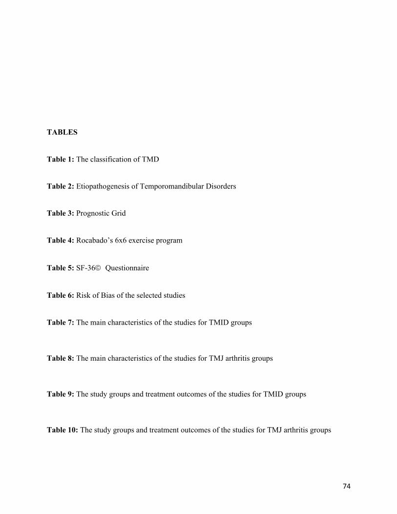
74
TABLES
Table 1: The classification of TMD
Table 2: Etiopathogenesis of Temporomandibular Disorders
Table 3: Prognostic Grid
Table 4: Rocabado’s 6x6 exercise program
Table 5: SF-36ã Questionnaire
Table 6: Risk of Bias of the selected studies
Table 7: The main characteristics of the studies for TMID groups
Table 8: The main characteristics of the studies for TMJ arthritis groups
Table 9: The study groups and treatment outcomes of the studies for TMID groups
Table 10: The study groups and treatment outcomes of the studies for TMJ arthritis groups

75
Table 11: SF 36ã results and significance of the comparison between pre- and post-intervention
evaluation done using paired Student’s t-test.