Livestock manure treatment for nutrients removal - AIR Unimi
Upload
khangminh22Category
view
0download
0
Università degli Studi di Milano
GRADUATE SCHOOL OF VETERINARY SCIENCES FOR ANIMAL HEALTH AND FOOD SAFETY
Director: Prof. Vittorio Dell’Orto
Doctoral Program in Animal Nutrition and Food Safety
Academic Year: 2009-2010
Stimulation of the health and productivity of nursing
and weaned piglets.
Graziano Mantovani
Tutor & coordinator:
Prof. Valentino Bontempo
DEPARTMENT OF VETERINARY SCIENCE AND TECHNOLOGY FOR FOOD SAFETY

2
Ai mie nonni e ai miei genitori Modello di vita, umiltà, principio

3
Preface ......................................................................................................................................... 6
Chapter 1 ..................................................................................................................................... 8
1.1 General Introduction...................................................................................................................... 9 1.1.1 Development of gut immune system .......................................................................................................... 9 1.1.2 Gut barrier and colonization resistance establishment ............................................................................ 11 1.1.3 Development of immune system .............................................................................................................. 12 1.1.4 Modulation of intestinal equilibrium through the feed ............................................................................ 15 1.1.5 Functional properties of milk and its Physiological Characteristics .......................................................... 16
1.1.5.1 Immunomodulatory Milk peptides .................................................................................................. 17 1.1.5.2 Milk peptides – Suppression of immune response .......................................................................... 18 1.1.5.3 Milk peptides – Induction of immune response ............................................................................... 20
1.1.6 Vegetable milks as functional foods ......................................................................................................... 24 1.1.6.1 Some components of vegetable milks ................................................................................................... 25 1.1.6.2 Inositol ................................................................................................................................................... 25 1.1.6.3 Ɣ Oryzanol ............................................................................................................................................. 26
1.1.7 Prebiotics .................................................................................................................................................. 26 1.1.7.1 Mannanoligosaccharides (MOS) ............................................................................................................ 29
1.1.8 Plant Products ................................................................................................................................................ 31
1.9 References ..................................................................................................................................... 41
Chapter 2 ................................................................................................................................... 50
2.1 Objectives ..................................................................................................................................... 51 2.1.1 Abstract 1 ....................................................................................................................................................... 51 2.1.2 Abstract 2 ....................................................................................................................................................... 52 2.1.3 Abstract 3 ....................................................................................................................................................... 53
Chapter 3 ................................................................................................................................... 54
3.1 Abstract ........................................................................................................................................ 55
3.2 Introduction .................................................................................................................................. 55
3.3 Materials and methods ................................................................................................................. 57 3.3.1 First animal trial ............................................................................................................................................. 57
3.3.1.2. Reagents and vaccine preparation ........................................................................................................ 57 3.3.1.3. Animals, vaccine administration and blood samples............................................................................. 57 3.3.1.4. Enzyme‐linked immunosorbent assay against ovalbumin ..................................................................... 57 3.3.1.5. Ovalbumin specific cell proliferation ..................................................................................................... 58 3.3.1.6. Flow cytometry analysis of cell proliferation ......................................................................................... 58
3.3.2. Second animal trial ........................................................................................................................................ 59 3.3.2.1. Cloning and expression of Salmonella enterica serovar Typhimurium’s B subunit Flagellin ................. 59 3.3.2.2. Tablets preparation ............................................................................................................................... 59 3.3.2.3. Animals, vaccine administration, blood samples and feces samples. ................................................... 59 3.3.2.4 Gene expression study ............................................................................................................................ 59 3.3.2.5 ELISA and specific cell proliferation against OVA ................................................................................... 61
3.4. Statistical analysis ........................................................................................................................ 61
3.5. Results ......................................................................................................................................... 61 3.5.1 First animal trial ............................................................................................................................................. 61
3.5.1.1 Measure of circulating antibody against ovalbumin .............................................................................. 61 3.5.1.2 Measure of specific in vitro cell proliferation of Peripheral blood mononuclear cells after antigen (ovalbumin) recall ............................................................................................................................................... 63
3.5.2 Second animal trial ......................................................................................................................................... 63

4
3.5.2.1 Measure of circulating antibody against ovalbumin ............................................................................. 63 3.5.2.2 Measure of IgG and IgA in feces by ELISA .............................................................................................. 64 3.5.2.3 Measure of specific in vitro cell proliferation of PBMCs after antigen (OVA) recall .............................. 65 3.5.2.4 Results of real‐time PCR: cytokine expression of MLN (mesenteric lynph node) cells stimulated in vitro with OVA. ............................................................................................................................................................ 66
3.6 Discussion and conclusion .............................................................................................................. 68
3.7 References ..................................................................................................................................... 71
Chapter 4 ................................................................................................................................... 74
4.1 Abstract ........................................................................................................................................ 75
4.2 Introduction .................................................................................................................................. 75
4.3 Materials and methods ................................................................................................................. 77 4.3.1Animals and diet ............................................................................................................................................. 77
4.3.1.1 Nursing period ........................................................................................................................................ 77 4.3.1.2 Post‐weaning .......................................................................................................................................... 77
4.3.2 Recorded parameters ..................................................................................................................................... 81 4.3.2.1 Performance: .......................................................................................................................................... 81 4.3.2.2. Health status ......................................................................................................................................... 81 4.3.2.3. Sampling and analysis ........................................................................................................................... 81
4.4 Statistical analysis ......................................................................................................................... 81
4.5 Results .......................................................................................................................................... 82 4.5.1 Growth performance ...................................................................................................................................... 82 4.5.2. Blood Parameters .......................................................................................................................................... 83 4.5.3. Health status ................................................................................................................................................. 85
4.6 Discussion and conclusion .............................................................................................................. 85
4.7. References .................................................................................................................................... 87
Chapter 5 ................................................................................................................................... 90
5.1 Abstract ........................................................................................................................................ 91
5.2 Introduction .................................................................................................................................. 91
5.3 Materials and methods ................................................................................................................. 92 5.3.2. Growth performance and samples collection ................................................................................................ 95 5.3.3 Chemical.analysis ........................................................................................................................................... 95
5.3.3.1 Blood parameters ................................................................................................................................... 95 5.3.3.2. Plasma parameters................................................................................................................................ 95 5.3.3.3 Intestinal parameters ............................................................................................................................. 96
5.4 Statistical analysis ........................................................................................................................ 97
5.5 Results .......................................................................................................................................... 97 5.5.1 Growth Performance ...................................................................................................................................... 97 5.5.2. Blood parameters ........................................................................................................................................ 101 5.5.3 Antioxidants ................................................................................................................................................. 103 5.5.4 Plasma metabolites ...................................................................................................................................... 107 5.5.5 Enzyme activities .......................................................................................................................................... 109
5.6 Discussion and conclusion ........................................................................................................... 110
5.7 References .................................................................................................................................. 117
Chapter 6 ................................................................................................................................. 123
6.1 General discussion ....................................................................................................................... 124

5
Chapter 7 ................................................................................................................................. 127
7.1 Summary ..................................................................................................................................... 128
Chapter 8 ................................................................................................................................. 135
8.1 Acknowledgements ..................................................................................................................... 136

6
Preface

7
Worldwide the swine industries are under pressure to reduce the use of antibiotics while
maintaining animal health and performance. The control and prevention of infectious diseases in of
basic economic importance although the occurrence of production disease has relevance not only to
economic production, but also to animal welfare. The introduction of antibiotics to treat bacterial
infections almost 50 years ago led to an improvement in animal production but the use of antibiotics
feed additives in food production animals was linked to emergence in the food chain of multiple
drug-resistance bacteria. The emergence of antibiotics resistant strains presumably demonstrated
that treatment of bacterial infections can not rely on the use of antibiotics without critical
considerations. In 2006 all the AGP (antibiotic growth promoters) and some feed additives with
growth promoting activities (Cu, Zn) have been limited. Since the first restrictive measures were
taken, and due to the beginning of the negative consequences of the ban, numerous efforts have
been done to find alternatives or replacement strategies to maintain pig growth performance and
controlling enteric bacterial diseases. The need of more economic and safety substances as replacers
of AGP for livestock animals as probiotics, prebiotics and feed additives of natural origin, focused
the attention of the research of the last years. Several studies reported the effects of probiotics,
prebiotics, organic acids, bioactive milk molecules and plant extracts on pig gut and general health
status. From literature they showed promising results, although still not comparable of AGP. In
some cases they were supplemented as single additive, while more recently additives of different
nature were pooled with the aim to evaluate possible additive or combined effects. Most of these
compounds are thought to act by modulating the microbiota equilibrium, the mucosa intestinal
morphometry and gut associate lymphoid tissue (GALT), while others might be active in the
improvement of pig health and performance by other different mechanisms. In order to asses, the
primary mode of action, metabolic pathways and optimal dosage of the additives more research will
be needed. Therefore, the aims of the following trials were to study the effects of prebiotics, natural
substances and bioactive milk peptides on health and performance of post-weaned piglets.

Chapter 1
General Introduction
8

9
1.1 General Introduction 1.1.1 Development of gut immune system Weaning is a crucial phase in pig production. Piglet’s immune system that is inexperienced and
immature moreover it is continuously challenged by lot of pathogens. The immune system gathers
experience as it is continuously faced with a myriad of antigens. Quantitatively, as well as
qualitatively, the most important site of this interaction is the intestine. Encounter alone does not
guarantee maturation, however, the intestinal immune system develops in intimate connection with
the indigenous microbiota of the gut and has distinct features, the most prominent of which being
the ability to launch suppressive, tolerogenic responses essential to maintaining a disease free state
in the gut as well as systemically. Aberrant immune responsiveness with an imbalance in the
composition of microbiota in the gut is characteristic to the pathogenesis of many inflammatory
conditions. The development of atopic diseases in particular appears to be associated with defective
tolerogenic responsiveness in the gut and alterations in the gut microbiota. The rise in incidence of
atopic diseases in the industrialised pig during the last decades, attributed to changes of farm
strategies (as the anticipated weaning at 21 days) in feeding and hygiene, may thus be partly
explained by altered gut microecology. Piglets are nowadays weaned around three/four weeks of
age because this procedure is commercially advantageous, but thus they often suffer altered
gastrointestinal conditions, in part because of the sudden alteration in the intestinal microflora
consequent to the rough passage from mother’s milk to a solid food. This in addition may cause a
strong reduction in the length of intestinal villi height and the depth of the intestinal crypts, which
consequently reduces the gut digestive as well as absorptive capacities (Pluske et al., 1997; Van
Beers-Schreurs et al., 1998). This in turn may cause negative effects on average daily gain (ADG),
as well as on defensive responses against infectious diseases. Actually, recent scientific evidence
suggests that weaning piglets at three to four weeks of age may overwhelmingly challenge a
substantially immature immune system, conducting it to a possible paralysis condition and creating
an ideal environment for diseases to develop (Ladekjær-Mikkelsen et al., 2002). In this fragile
equilibrium the development of diseases has a great economical impact in pig production.
One critical change associated with weaning is the shift from sow’s milk to a dry feed, which
induces a period of fasting during the first few days following weaning. This weaning anorexia has
a negative impact on growth and leads to mobilization of fat stores, as shown by the sharp increase
of circulating free fatty acids levels (Barke et al., 1996). It may also be involved in the digestive
problems that are frequently encountered after weaning (McCracken et al., 1999). Experimental
data indicates that this period of under nutrition markedly alters the functioning of several

10
neuroendocrine system during the post-weaning period. For instance, the changed measured in the
somatotrophic axis (increased GH and reduction IGF levels) and in the autonomic nervous system
(reduced cathecolamine excretion in urine) are similar to those measured in fasting animals (Carroll
et al., 1998, Hay et al., 2001). These changes may be long lasting, as in the case of weaning at 6
days of age for instance (Hay et al., 2001). Some of the metabolic response to weaning can be
corrected by milk feeding, which improves the growth rate of the animals as compared to dry feed
(McCracken et al.,1995, Kim et al., 2001). Indeed, piglets weaned at 2-3 days of age and
subsequently fed with a milk replacer display greater growth rates by day 7-8 of lactation than
piglets raised by their dam, showing that sow milk yield is a limiting factor to piglet growth (Harrel
et al., 1993). The increase factor of voluntary food intake after weaning by using whole cow’s milk
(due to it’s palatability) also improves the mucosa architecture of the small intestine (Pluske et al.,
1996). However in commercial settings, weaning piglets are usually offered dry food, for
economical reasons. Such a food is not accepted as well as a liquid milk replacer, and weaning
alters average daily feed intake and average daily weight gain, the magnitude of which is larger
with earlier weaning age (2, 3 and 4 weeks) resulted in reduced weight gain depression. All the
animals nevertheless reached similar body weight at 6 weeks of age (Leibbrandt et al., 1975). There
is no doubt that weaning is a period of intense stress for piglets, with profound consequences on
growth, physiology and disease outbursts, which reveal severe welfare problems. Experimental data
clearly show that the anorexia or, at least, the nutritional deficit due to the abrupt transition between
milk and solid food, induces severe taxation of the adaptive mechanisms of piglets and may be of
special relevance from a welfare point of view. Most of the problem comes from the fact that during
lactation spontaneous intake of dry food remains very low up to 3 weeks of age and does not
become significant until the 4th week, indicating that the appetite for dry food is very low in
younger animals. The large individual variation suggests that this trait may be influenced by genetic
factors and could therefore respond to genetic selection. It would be valuable to gain more
information on the physiological changes induced by weaning at different ages. Maintanance of gut
health in the pig is probably the last emerging problem when the other pathologies are under
control. Infact gut health degradation is most often observed during the post-weaning period and
may in turn affect further general health through enhancement of bacterial translocation and
disturbances in the maturation of the immune system including immuno-tolerance.
One approach to improve the gut health of the piglets is the dietary manipulation. Dietary
manipulation of the gut environment by the use of feed additives, or by choice of dietary raw
materials that may influence the luminal environment throughout the entire length of the intestinal
tact and thereby improving “gut health”( Knud E. et al.). As discussed by Montagne et al. (2003),

the concept of gut health is complex and, at present, it is an ill-defined notion. Conway (1994)
proposed that there are three mayor components of gut health, namely the diet, the mucosa and the
commensal flora. The mucosa is composed of the digestive epithelium, the gut-associated lympoid
tissue (GALT) and the mucus overlying the epithelium. The GALT, commensal bacteria, mucus
and host epithelial cells interact with each other forming a fragile and dynamic equilibrium within
the alimentary tract that ensures efficient functioning and absorption capacity of the digestive
system. The feed should be selected to favour conditions in the gut that create and stabilise this
balance between the host, the microflora and environment, and to prevent disturbance of the
structure and function of the gut. In this respect, the relative gut health value of a dietary component
or diets should rest with their capacity either to stabilise or to perturb this equilibrium.
1.1.2 Gut barrier and colonization resistance establishment
The indigenous microbiota suppresses colonization of incoming bacteria by a process named
colonization resistance that is a first line of defense against invasions by exogenous bacteria,
potential pathogenic organisms or indigenous opportunists (Van der Waaij et al., 1989; Hooper et
al.,1998). This process involves several different complex interacting
Fig. 1 Schematic representation of the different elements in the gut ecosystem making yp the concept of gut
health. Modified from Conway (1994) as discussed by Montagne et al. 2003.
11

12
mechanisms of both the bacteria and the hosts. The host factors involved in colonization resistance
are different: the peristaltic movement; the secretion of diverse digestive enzymes and electrolytes;
the mucus secretion; the epithelial cell desquamation; the gut associated lymphoid tissue; and
secretory IgA (Stewart et al., 1993). Besides, indigenous microbiota prevents bacterial colonization
by competing for epithelium receptors (Blomberg et al., 1993; Bernet et al., 1994) and enteric
nutrients (Stewart et al., 1999) producing antimicrobial compounds such as bacteriocines (Brook,
1999) and metabolizing nutrients to create a restrictive environment which is generally unfavorable
for the growth of many enteric pathogens (Lievin et al., 2000). Moreover, bacterial recognition and
adhesion to receptors is not only a prerequisite for colonization, which determines microbiota
composition and permanent colonization, especially in the upper gastrointestinal tract (Alandet et
al., 1999). It also determines antagonistic activity against enteropathogens (Coconnier et al., 1993),
modulation of the immune system and also the improvement of healing in the damaged gastric
mucosa (Elliot et al., 1998). Several factors are involved in the control of bacterial attachment and
thus in the modulation of the indigenous microbiota profile (Freitas et al., 2002). Special interest is
nowadays focused on genetic modulation of receptors by the host and the bacteria. Bacterial-
mucose attachment appears consequently as a key point defining indigenous microbiota
composition and different bacterial-mediated functions. Two main components are essential to the
recognition between the host and the bacteria: the glucoconjugates on the gut enterocytes and
bacterial adhesions metabolize cholesterol to coprostanol (Baron and Hylemon, 1997).
1.1.3 Development of immune system Pigs have evolved many immunological characteristics in response to the environmental and
infectious selection pressures they continually face. Pigs are omnivores and their enteric system
must logically be adapted to respond to the range of intestinal pathogens, and therefore defence
mechanisms have developed to protect the host and maintain intestinal health. Nevertheless the
piglet is immunodeficient during the early phase of his life and is highly dependent upon a supply
of both specific and non specific immune factors present in maternal colostrums and milk for
immune protection, development and survival (Varley et al., 1987). Therefore, development of
immunocompetence is an absolute requirement for optimum growth and performance (Stokes et al.,
2004). Bailey et al. (2001) surmised that immunological development in the neonatal pig occurs as
a balance between regulatory function and effector functions, and that GIT integrity depends on the
maintenance of this balance. The immune system in the young pig comprises several organs (bone
marrow, thymus, spleen, mesenteric lymphonodes) and several cell types (lymphocites-specific
immune recognition of foreign antigen, phagocytes – production of innate immunity) that recognize

13
foreign antigens. The immune defence system has two “arms” the innate or the acquired. The innate
immune system is thought to have evolved before the adaptive immune system, and hence has
evolved also as the first line of defence against a pathogenic/antigenic challenge.
Development of the immune system in early weaned piglet and the atopic type of immune
responsiveness T helper cells have paradigmatically been labelled either T helper (Th)1 or Th2 cells
depending on the cytokines they produce (Mosmann et al., 1986; Del Prete et al., 1991). Th1 cells
produce predominantly interferon γ (IFN- γ) and play a central role in immune defence against
intracellular pathogens. In contrast, Th2 cells produce cytokines such as interleukin (IL-4-5-13)
implicated in responses to helminthic infections. Both Th1- and Th2-type responses have been
recognised in the pathogenesis of human disease as well (Romagnani, 1996). It is well established
that there is a counter-regulatory balance between Th1 and Th2 responses, but recently distinct
mechanisms effecting both Th1 and Th2 cells have been discovered. Suppressive cytokines, such as
transforming growth factor ß (TGF-ß) and IL-10, secreted by gut-derived regulatory T cells named
Th3 and Tr1 cells, respectively, provide important suppressive balance and protection from disease
(Nagler and Anderson, 2000). Notwithstanding the ample evidence indicating the involvement of
Th2-skewed immune responsiveness in fully established atopic disease, the events leading to the
establishment of such pathological skewness early in life remain less well understood. It is of note
that due to the immunological balance prevailing in utero the immune responses early in life are
physiologically predominantly of the Th2-type (Piccinni et al., 1998). Recently, it has been
observed that neonatal CD8 T cells produce large amounts of IL-13 which may account for the Th2
bias in the newborn (Ribeiro-do-Couto et al., 2001). Interestingly, however, a previous study
indicated that infants who later develop atopic disease exhibit impaired IL-13 production at birth
(Williams et al., 2000). The same phenomenon has been demonstrated with regard to IL-4 as well,
as neonatal IL-4 responses were lower in infants who developed atopic disease as compared to
those who remained healthy (Prescott et al., 1999). Consequently, a significant overlap in the
concentrations of Th2 cytokines, such as IL-4, and IgE antibodies prevails between atopics and non-
atopics at an early age (Kulig et al., 1999; Prescott et al., 1999). Thus the concentration of IgE
antibodies in cord blood, thought to reflect sensitisation in utero, predicts poorly the development of
atopic disease (Bergmann et al., 1997). An age-dependent decline in Th2 responses during the early
postnatal period in non-atopic children and a converse pattern in atopic children has been observed,
suggesting that infants who develop atopic disease may exhibit defective suppression of Th2-type
responsiveness (Prescott et al., 1999). Recent observations have lead to the conclusion that such
suppressive mechanisms at least in part originate in the gut as a result of stimulation by dietary and
microbial antigens. Whilst there is a strong hereditary component in the development of atopic

14
disease, the rapid increase in morbidity can only be explained by environmental factors (Bergmann
et al., 1997). The hygiene hypothesis of allergy, first introduced by Strachan, suggests a causal
relationship between reduced exposure to microbes at an early age and the increase in the
prevalence of atopic disease (Strachan et al., 1989). There is epidemiological evidence indicating
that infectious diseases early in life may protect the individual from atopy. However, as it has
recently been pointed out, the most important source of tolerogenic stimuli may be the indigenous
microbiota of the intestine (Monteleone et al., 2001). The immunomodulatory events produced by
the interaction between the intestinal immune system and microbiota as well as the induction and
maintenance of oral tolerance is complex and surpass the Th1/Th2 paradigm. The intestinal immune
system must be able to discriminate between potentially pathogenic microbial antigens and the non-
pathogenic dietary and indigenous microbial antigens in order to avoid both invasive infections and
chronic inflammatory conditions. The unresponsiveness to dietary proteins and indigenous
microbiota, known as oral tolerance, is the result of several distinct processes. Furthermore, there is
growing evidence suggesting that resident microbiota provide the intestinal immune system with
endogenous stimuli which are essential for its normal maturation and function (Toms et al., 2001).
There are somewhat contradictory data on the type of immune responsiveness preferentially elicited
by the intestinal immune system although it is apparent that T lymphocytes are the main effector
cells in responses to luminal antigens. Results obtained from animal models suggest that mucosal T
cell responses are favourably of the Th2 type (Xu-Amano et al., 1992). This Th2-bias is thought to
originate in utero and as a result, transient sensitisation to dietary antigens is common at an early
age. On the other hand, however, Th2-type responsiveness, such as IgE antibody production seems
to be most readily susceptible to suppression (Nagler-Anderson, 2000). Furthermore, dietary
antigens appear to induce Th1-type responsiveness in the human intestine, which may down-
regulate Th2-type responsiveness (MacDonald et al., 2001). Oral tolerance has traditionally been
defined as the induction of peripheral unresponsiveness as the result of oral administration of
soluble protein antigens (Nagler- Anderson, 2000). It is well documented that there are distinct
mechanisms, including clonal deletion, clonal anergy and active suppression, by which tolerance,
defined recently not only as unresponsiveness but more broadly as any mechanism by which a
potentially injurious immune response is prevented, suppressed, or shifted to a noninjurious class of
immune response, is induced and maintained in the intestinal mucosa (Weiner, 2001). The relative
contributions of these mechanisms appears to be dependent on the dose of the antigen: High doses
of oral antigen result in clonal anergy/deletion, a phenomenon often explained by small amounts of
antigen bypassing the intestinal immune system, gaining access to circulation and thus inducing
anergy due to the lack of sufficient costimulatory signalling (Weiner, 2001). Oral administration of

15
antigen leads to the generation of a unique subset of T cells named Th3 cells in both animal models
and humans (Chen et al., 1996; Fukaura et al., 1996). Th3 cells produce predominantly TGF-ß and
are thought to be pivotal in the active suppression leading to oral tolerance after low doses of oral
antigen. After the priming of Th3 cells has taken place in the Peyer’s patches, the gut-originating
cells migrate to the periphery and are capable of suppressing inflammatory responses upon
reactivation thus mediating tolerance in sites other than the gut as well (Weiner, 2001). In addition
to down-regulating both Th1- and Th2 type responses, TGF-ß elicits immunomodulatory effects on
antigen-presenting cells (Takeuchi et al., 1998). Furthermore, TGF-ß may favour the development
of IL-10-secreting regulatory Tr1 cells (Toms et al., 2001), another subset of suppressive T cells
characteristic of the intestinal immune system. IL-10 has a central role in maintaining intestinal
homeostasis, especially by suppressing inflammatory responses towards intestinal microbial agents.
There is an intimate interplay between different subsets of T cells and antigen-presenting cells, such
as dendritic cells, in the intestine. In murine models, high basal levels of IL-4, IL-10 and TGF-ß
expression have been detected in the intestinal mucosa and this cytokine milieu may be crucial for
the induction of Th2 and Th3 type responsiveness (Weiner, 2001). However, as it has recently been
pointed out there may be major differences between species in mucosal immune responses. In fact,
there are data on record indicating a Th1-skewed cytokine profile as a constant finding in the
intestine of humans (MacDonald, 2001; Nagata et al., 2000). A transient induction of IFN-γ
producing Th1 cells has been detected in the early phases of oral tolerance formation (Mowat et al.,
1999). Even though both Th1 and Th2 cytokines regulate the function of Th3 cells, neither are
essential for the induction of peripheral tolerance in a murine model (Garside et al., 1995; Mowat et
al., 1999; Shi et al., 1999). The individual role of each functional subset of T cells in the inductive
phase of oral tolerance thus remains to be elucidated, but it is evident that Th3 cells provide
tolerogenic suppression both in the intestine and in other target organs.
1.1.4 Modulation of intestinal equilibrium through the feed
Weaning is characterized by significant nutritional, social and environmental changes that can
impose a significant penalty on subsequent growth and production. A marked feature of weaning is
the transient decrease in macronutrient and micronutrient intake that occurs as piglets are switched
abruptly from a mostly all milk diet to a solid diet. Despite to the studies conducted in the past years
concerning nutrition and weaning under commercial farm conditions, there appears to have been
very little research directed specifically towards immune system (Pluske J.R., 2007). There are
surprisingly few studies examining the roles specifically of feed additives/feed ingredients on the
immune system GIT in young pigs, although an increasing number of studies deriving
16
predominantly from Europe have occurred more recently in view of the ban on antibiotic feed
additives in diets. The use of nutraceuticals or functional foods plays an interesting role in the
intestinal equilibrium of the early weaned piglet. Functional foods have been defined as food with
ingredients (either naturally occurring or added) that provide a health benefit beyond the traditional
nutrient value of the food. The following sections discuss several feed additives and functional
foods with an important role on the GIT immune function.
1.1.5 Functional properties of milk and its Physiological Characteristics
The discovered of functional molecules in food with extra-nutritional activities opened the way to a
nutrition not just only with the aim of to meet the nutritional needs, but also to improve the health
of humans and animals. Bioactive substances in functional foods able to improve the health of the
animals are defined as nutraceutical. For its physiological significance in primis milk can be
considered "functional food" for its essential components for the growth during the neonatal period
and for its richness in bioactive molecules with immunological, anti-infective actions due to protect
the health of the newborn. Among the native components of milk, of particular importance and
interest are attributable to some lipid and protein fraction. The importance of these compounds is
due not only for their content but also for their bioavailability and bioaccessibility often positively
affected by their inclusion in a milk matrix (Baldi and Pinotti, 2008). In the evaluation of the
functional activity of milk it is important remember the protective activity towards the
oligosaccharide component against intestinal infectious diseases. The protective role of the
bioactive components of milk has an action on intestinal ecosystem, developing newborn immune
system, reducing the risk of developing of health problems improving performance. These
components may have a potential use as nutraceuticals to improve health, both in relation to their
presence in food or as food additives. In this field, knowledge is more developed with regard to
human or bovine milk, while less known or are missing data on the sow's milk and other species.
The importance of milk in the nutrition of all mammals has been recognized from time immemorial.
The biosynthetic capacity of the mammary gland, both in terms of the number of substances
synthesized and the magnitude of the total synthetic ability is remarkable. Mammary gland
synthesizes a number of proteins that have unique aminoacids composition and highly desirable
physicochemical properties. Caseins and whey proteins are the two main protein groups in milk.
Caseins designate the predominant group of proteins and they are characterized by ester-bound
phosphate, high proline content, few or no cysteine residues and low solubility at pH 4.0–5.0.
Cow’s milk contains 4 individual caseins: as1-casein, comprising of 199 amino acids (aa), b-casein
(209 aa), k-casein (169 aa) and as2-casein (207 aa). The major whey proteins in milk include b-

17
lactoglobulin, a-lactalbumin, immunoglobulins and bovine serum albumin (Ng Kwai Hang, 2003).
In addition to the major milk proteins, milk contains a wide group of biologically active proteins,
involved in development and healthfulness of the neonate. Among these bioactive components the
80 KDa iron-binding glycoprotein lactoferrin is recognized as one of the most versatile. Its role was
originally considered to be essentially antimicrobial, but several findings have demonstrated that the
protein has nutritional and physiological significance (Lo¨nnerdal & Iyer, 1995). Milk proteins can
be subjected to inadvertent proteolysis during storage before or after milking. Proteolysis in the
mammary gland can be attributed to native proteases, such as the plasmin/plasminogen system
(Baldi et al. 1996; Fantuz et al. 2001; Politis, 2007). Proteins can be subjected to deliberate
proteolysis such as the proteolysis occurring during manufacture of fermented dairy products
(yoghurt) and cheese manufacture and/or ripening. Lastly, proteins are subjected to hydrolytic
breakdown during gastric processing and later upon exposure of the proteins with indigenous or
intestinal bacteria-derived enzymes in the gut. Proteolytic breakdown of milk proteins generates a
number of peptides endowed with various biological properties (antimicrobial, antihypertensive,
antithrombotic) including those that have the ability to modulate immune function (Hill et al. 2000).
Immunomodulatory peptides are inactive within the sequence of the parent protein and they can be
released by enzymatic proteolysis. One good example is the proteolysis occurring by the action of
proteinases and peptidases from lactic acid bacteria (LAB). LAB utilize milk proteins, mainly
caseins, as their prime source of essential aminoacids. The proteolytic machinery of LAB includes a
cell wall bound proteinase and several intracellular peptidases. The transport system of LAB
enables them to internalize oligopeptides up to 18 aa in length. Longer oligopeptides can become a
major source of bioactive peptides in fermented milk when further degraded by gastrointestinal
enzymes or by intracellular peptidases of lysed LAB after consumption of these products (Law &
Haandrikman,1997).
1.1.5.1 Immunomodulatory Milk peptides Suppression or Induction Milk proteins represent the exclusive protein supply for the newborn; all
human and animals neonates depend upon milk proteins until they enter the fifth month of their life
(Table 1). Evidence will be presented regarding the ability of milk peptides to regulate
gastrointestinal immune function leading us to hypothesize that these peptides may guide the local
immune system until it develops its full functionality. The first few weeks is a very critical period in
life because the immune system should develop oral tolerance to nutrient molecules and avoid
tolerance to pathogen-derived antigens. The term immunomodulation was purposely adopted to
indicate that suppression of the immune system may be required in certain instances (oral tolerance)

18
or induction towards pathogen-derived antigens. The latter is very important in order to avoid the
well recognized susceptibility of newborns to infection (Hill, 1987).
Table 1 Biological function of the principle bioactive milk component (adapted from Séverin e Wenshui, 2005). Protein Function
Caseine (α-, β-, κ-caseine) Ionic Carrier (Ca, PO4, Fe, Zn, Cu), precursors of bioactive peptides
Serum proteins
β-Lattoglobuline Carrier del retinolo, legante acidi grassi, possibile antiossidante α-Lattalbumine Lactose synthesis in mamary gland, Ca carrier, immunomodulator,
anticarcinogenic Immunoglobuline (A, M & G) Immunoprotection Lactoferrine Antimicrobial, antioxidant, immunomodulator, iron absorption,
anticarcinogenic Lactoperossidase Antimicrobial Lysozyme Antimicrobial, synergic action with immunogglobuline and
lactoferrine Glicomacropeptide Antivirus, biphydogenic
1.1.5.2 Milk peptides – Suppression of immune response
The mucosal immune system of infants is stimulated daily by the continuous passage of milk
proteins. Breakdown of proteins in the gut and activation of suppressor T-cells lead to a systemic
hypo-responsiveness to ingested protein antigens, a phenomenon known as ‘oral tolerance’. Pecquet
et al. (2000) reported that peptides obtained from tryptic hydrolysis of bovine b-lg down regulate
various immune functions and induce specific oral tolerance in mice. Mice fed with either b-lg
hydrolyzates or fractions of the hydrolyzate were tolerized against b-lg. Specific serum and
intestinal IgE levels were reduced. Delayedtype hypersensitivity and proliferative responses were
inhibited. In a more recent study, Prioult et al. (2004) examined the possibility that the
microorganism Lactobacillus paracasei induces oral tolerance by generating a number of
immunomodulatory peptides from the hydrolysis of b-lg. They showed that Lb. paracasei peptidases
were capable of further hydrolyzing, tryptic-chymotryptic peptides of b-lg. Furthermore, they
reported that these peptides repressed lymphocyte proliferation and up regulated interleukin-10 (IL-
10) production. Based on these results, they concluded that Lb. paracasei induces oral tolerance to
b-lg in vivo by degrading its acidic peptides and releasing immunomodulatory peptides capable of
stimulating regulatory T-cells, which function as major immunosuppresants by secreting IL-10.

19
Hydrolysis of casein with pepsin and trypsin or additionally with enzymes derived from Lb. casei
strain GG generates molecules capable of inhibiting lymphocyte proliferation (Sutas et al. 1996b).
Thus, indigenous enzymes in the gut together with enzymes of bacteria origin have been proven
beneficial in the down regulation of hypersensitivity reactions to ingested proteins in human
neonates. Pessi et al. (2001) investigated the effect of digests of bovine casein by enzymes derived
from Lb. rhamnosus GG on T-cell activation. They found that the digests reduced expression of IL-
2 and inhibited protein kinase C translocation. Both effects indicate suppression of T-cell activation
by casein digests. Peptides obtained from the hydrolysis of casein by trypsin induce oral tolerance
in mice (Hachimura et al. 1993). A synthetic peptide corresponding to residues 142–149 of as1-
casein was proven to be an effective inducer of CD8(+) T-cells which recognize the parent peptide
and secrete interferon-gamma (IFNc) , a potent inhibitor of Th-2 dependent events, including IgE
production (Totsuka et al. 1998). Otani & Hata (1995) reported that intact bovine k-casein inhibited
proliferative responses of mouse spleen lymphocytes and rabbit Peyer’s patch cells to various
mitogens. Pancreatin and trypsin digests of k-casein, as1-casein and b-casein inhibited cell
proliferation in response to mitogens. Based on these results, they suggested that peptides obtained
from various caseins by the action of trypsin may suppress immune response of neonates. In further
support of this notion, Kayser & Meisel (1996) reported that pepsin and trypsin digests of bovine
as1-casein and b-casein inhibited mitogen-induced proliferation of peripheral blood mononuclear
cells. The k-caseinoglycopeptide corresponding to residues 106–169 of bovine k-casein is a potent
inhibitor of phytohaemagglutinin-induced and lipopolysaccharide-induced proliferation of murine
splenic lymphocytes and rabbit Peyer’s patch cells and well as antibody production in various
spleen cell cultures (Otani et al. 1995). They suggested that the k-caseinoglycopeptide down
regulates immune response by acting on both T- and B-lymphocytes. Furthermore, they showed that
the inhibitory effect was abolished after neuraminidase treatment which indicates that both the
peptide and the carbohydrate parts of the molecule are necessary for the inhibitory effect. Elitsur &
Luk (1991) investigated the effect of b-casomorphins (fragments of b-casein, residues 60–70) on
immune function. They reported that casomorphins inhibited proliferation of human lamina propria-
derived lymphocytes in vitro. However, these results should be interpreted with caution because
casomorphins appear to stimulate resistance of mice to infection (Klebsiella pneumoniae), thus,
having the opposite effect in vivo. Despite the fact that casomorphins are rapidly degraded once
they enter the blood stream, casomorphins were found in the plasma of new born calves after their
first milk intake (Umbach et al. 1985). Thus, the neonatal intestine appears to be permeable to pre-
casomorphins (Clare & Swaisgood, 2000). Recent data obtained at the Agricultural University of
Athens indicate that the effect of casein digests on immune parameters may depend upon the

20
maturity of the immune system. We have examined the effect of two synthetic peptides on
membrane-bound urokinase plasminogen activator (uPA) and expression of major
histocompatability complex (MHC) class II antigens by porcine blood neutrophils. The two
peptides utilized were the tripeptide LLY which corresponds to residues 191–193 of bovine b-
casein and the hexapeptide PGPIPN which corresponds to residues 63–68 of bovine b-casein. The
uPA is a critical enzyme for neutrophil diapedesis and the overall ability of neutrophils to resist
against various pathogens (Politis et al.2003). In this model system, neutrophils are obtained at
weaning which is considered a period of immaturity of the immune system and 4 weeks later when
the immune system is thought to have gained its full functionality (Fragou et al. 2004). Results
indicated that both peptides reduced the amount of u-PA present on the cell membrane as well as
the expression of MHC class II antigens only at the time of weaning and not 4 weeks later. Thus, b-
casein synthetic peptides appear to be effective only at a time period which coincides with
immaturity of the immune system and they are not effective when the immune system has
presumably gained its full functionality.
1.1.5.3 Milk peptides – Induction of immune response
The main mechanism of protection against pathogen derived antigens provided by mucosal
immunity is mediated through IgA-producing cells and secretory IgA which neutralize and, thus,
prevent the entry of potentially harmful antigens in the host. Thus, stimulation of local immune
response can be effective in preventing certain diseases caused by microorganisms entering the host
through the oral route. Feeding mice with milk fermented with Lb. helveticus increased the number
of IgA(+) B cells in the small intestine and bronchial tissues. A protease deficient derivative of Lb.
helveticus was ineffective (Matar et al. 2001). These results taken together indicate that peptides
generated by the action of proteases are responsible for this effect.
Furthermore, it appears that the peptides can stimulate local immune response in the gut and
activate the IgA cycle as shown by the increase of the IgA(+) B cells in the bronchial tissues. In
another study, Perdigon et al. (1999) demonstrated that feeding mice with milk fermented by LAB
resulted in increases in IgA(+) cells in the gut, macrophage activity and specific antibody responses
during infection. However, it was not certain whether the effect was mediated by milk peptides
and/or the presence of the LAB. To further elucidate the mechanism, Leblanc et al.(2002)
investigated the effect of specific peptides generated following fermentation of milk with Lb.
helveticus on humoral immunity. Three specific peptide fractions were isolated from fermented
milk by chromatography and then were fed to mice. All fractions increased the number of the
IgA(+) B cells in the gut which indicates that bioactive peptides are generated by the action of

21
LAB. There was no increase in the number of the IgA(+) B cells in the bronchial tissues which
indicates that the effect of the peptides was restricted in the gut and there was no activation of the
IgA cycle. A great number of studies demonstrating the ability of milk peptides to enhance the
function of immune system were performed in the 80s and early to middle 90s and some excellent
reviews of these studies are available (Clare & Swaisgood, 2000; Hill et al. 2000). Therefore,
emphasis in the present review will be given towards recent studies or those not adequately covered
in the previous reviews. A recent study investigated the effect of bovine glycomacropeptide on
proliferation and phagocytic activity of the U937 human macrophages like cell line (Li & Mine,
2004). They showed that the glycomacropeptide to be a potent enhancer of cell proliferation and
phagocytic ability. Furthermore, they showed that both the peptide and the carbohydrate portions
were essential for the stimulatory effect. Politis (1995-2007) investigated the effect of a synthetic
hexapeptide corresponding to residues 63–68 of bovine b-casein on several immune parameters of
bovine macrophages and neutrophils. Results showed that the hexapeptide up regulated interleukin-
1 production and expression of MHC class II molecules by macrophages and superoxide production
by neutrophils. In contrast, the hexapeptide had no effect on chemotactic responsiveness of bovine
neutrophils. It is clear that the effectiveness of milk peptides is cell-specific. It is difficult to
speculate as to why the hexapeptide differentially affects two functions of bovine neutrophils
(superoxide production, chemotactic responsiveness). A study performed in the 90s demonstrates
the dichotomy between suppression and/or induction of immune response by milk peptides. Sutas et
al. (1996a) examined the effect of milk peptides generated by an intestinal Lactobacillus strain on
various immune functions. They showed that purified casein peptides up regulated IL-4 and IFN-
gamma production by blood peripheral mononuclear cells. In contrast, Lactobacillus-degraded
caseins up regulated IL-4 production but they had no effect on IFNgamma. It is clear that the
overall effect of milk peptides on the immune system might be peptide-specific. Non-nutrient milk
components modulating gastrointestinal development: Colostrum and milk contain a high number
of biologically active compounds that modulate gastrointestinal activity and function and promote
the development of the newborn (Blum & Baumrucker, 2002). A significative number of studies
demonstrated the efficacy of these peptides to modulate the intestinal immune response of the
newborn (Baldi et al., 2005). Chronopoulou et al. (2006) demonstrated an increase of superoxide
anion production from macrophages and neutrophils isolated from weaning piglets treated with
casein peptides. This research group tested hanno testato in vivo the effect of casein peptides on
phagocytic cells of 27 piglets in the first weaning phase in three experimental groups: control (basal
diet), low integration (300 mg/die), high integration (600 mg/die) (Politis e Chronopoulou 2008).

The production of sueroxide anions from serum monocytes, macrophages and neutrophils at days 7,
14, 21 increased compared to the level of the integration (Figure 2).
Figure 2 Milk peptides and immune response in the neonate adapted from (Politis I., Chronopoulou R. 2008).
Among these compounds there are: lactoferrin, a multifunctional protein involved in non-specific
immune defence against bacteria and viruses, immunoregulation, anti-inflammation and iron
metabolism (Brock, 2002). From a nutritional point of view lactoferrin may be of interest not so
much as a dietary protein source of amino acids but for iron bioavailability (Lo¨nnerdal & Iyer,
1995). Although amino acid composition of lactoferrin suggests a high biological value of the
protein, some evidence indicated that the major function is not as a nutritional support of amino
acids for the newborn. In fact, lactoferrin is extremely resistant to proteolytic enzymes and the
ironsaturated form is more resistant to enzymatic digestion than the apoprotein (Brines & Brock,
1983; Kuwata et al. 2001). As a consequence, ingested lactoferrin is not highly degraded in the
gastrointestinal tract and nearly intact protein is delivered in the gut (Hutchens et al. 1991). This
argument is supported by the fact that intact lactoferrin can be found in the faeces of breastfed
neonate; thus, the protein undergoes incomplete proteolysis and it does not represent a source of
utilizable amino acids (Davidson & Lo¨nnerdal, 1987). These data suggested that intact lactoferrin
should have a physiological significance for the gut of the neonate. Following oral ingestion,
survival of lactoferrin and other milk-borne bioactive compounds in the gastrointestinal tract is a
prerequisite for direct action in the gut or for activity on peripheral targets after absorption into the
22

23
blood circulation (Xu et al. 2000). Lactoferrin is involved in iron uptake by the newborn; this
potential role for the bioavailability of this micronutrient in milk arises from the observation that its
bioavailability in breast milk is high, although its content is low (Lo¨nnerdal & Iyer, 1995). Studies
by Kawakami & Lo¨nnerdal (1991) and Suzuki et al. (2001) showed that lactoferrin facilitates iron
uptake by human intestinal cell cultures and this biological effect is mediated by the interaction
with a receptor localized on enterocytes. Lactoferrin receptors were identified in the small intestine
brush-border membrane of several species, such as mice (Hu et al. 1990), piglets (Gislason et al.
1993) and cows (Talukder et al. 2003). The binding of lactoferrin to the receptor is highly species-
specific. Studies by Gislason et al. (1993) showed that human lactoferrin, bovine lactoferrin and pig
transferrin do not interact with the receptor for lactoferrin localized on the small intestine of piglets.
Another interesting aspect of the interaction of lactoferrin with its specific receptor is that the
protein can be internalized after binding. Recent findings have showed that lactoferrin has an import
signal for translocation into the nucleus (Penco et al. 2001) and surface nucleolin is involved in the
binding and endocytosis of lactoferrin in target cells (Legrand et al. 2004). Ashida et al. (2004)
studied the cellular internalization of lactoferrin in the intestinal epithelial cell line Caco-2; they
showed that lactoferrin is localized to the nuclei after uptake. Baumrucker & Erondu (2000)
demonstrated that bovine lactoferrin is involved in both the interaction and entry of IGFBP-3 in the
nucleus of mammary cells. The nuclear localization of lactoferrin suggested its involvement as a
transcription factor after binding to specific D In cow’s milk, more than 100 nutrients are included.
Preisely, milk is categorized as perfect food containing protein, lactose, calcium, phosphorus,
magnesium and trace elements, and varius vitamins. And it is also mentioned as a natural food with
biological function and significant meaning to the body. The history of milk as a food started from
B.C 6000. According to the many studies since then, we can say that milk is a great food involving
various physiological functions, such as bone-growth and immune control.
Immunoglobulin (Ig). The structure of the Ig present in milk is exactly same as that in the serum or
other excreta. But the content of each Ig is totally different in between serum and milk. IgG is the
most common Ig in serum of human, bovine and porcine (80-90 % of total Ig). In colostrum the
most common Ig is Ig A in human, and for bovine and pig’s colostrum the most represente is the
IgG. Lactoferrin: Is a glycoprotein in milk containing 2 molecules of iron. It presents relatively
higher in human breast milk, and especially 10-20 times higher than cow’s milk (6-8 mg/l in
colostrum and 2-4 mg/l in normal milk). This glycoprotein plays an important role in resistance
toward E.coli infection and its function in colon is known to be strong. In addition, the content of
lactoferrin is generally high in mammal colostrums, which is helpful in restricting the growth of
pathogenic bacteria and E.coli in order to make Bifidobacteria settle down in large intestine in the

24
young. Due to its strong adherence to metal ions, this glycoprotein may provide the function of
suppressing the pathogenic bacteria growth by transforming the active minerals into inactive ones
which is essential for bacteria growth, and the function of preventing anemia with controlling iron
absorption. In addition, its known functions are immune restoration, anti-inflammation, cell
proliferation and anti-oxidation.
Lysozyme: has anti-bacterial anti-viral and immune enhancing effects, and involves in blood
clotting and stryptic process, anti-inflammation and milk digestion. The content of lysozyme is
around 170 µg % in milk. When infants were fed with formula containing lysozyme shown an
increasy of the growth of Bifidobacteria and a decrease of E.coli in the feces.
The changes in bacterial composition can be explained by N-acetylglucosamin, produced when
lysozyme degrades the mucus polysaccharides on the cell walls of bacteria, that increases the
growth of Bifidobacteria.
Mucin: is chemical compound of olighosaccarides and glycoprotein and has lubricative and
protectie functions, existing in pituauitary gland, gastric mucosa, cartilage, saliva, blood, nasal
mucosa in the body. Especially, it is high in breast milk and cow’s mil and can protect stomach
from gastric juice such as pepsin and and hydrochloride.
1.1.6 Vegetable milks as functional foods
Compared to cow's milk, vegetable milks contains more carbohydrates, but does not contain
significant amounts of calcium or protein, and no cholesterol or lactose. Commercial brands of
vegetable milks such as rice milk and soy milk, however, are often fortified with vitamins and
minerals, including calcium, vitamin B12, vitamin B3, and iron. Soy milk for example has about the
same amount of protein as cow's milk. Natural soy milk contains also little digestible calcium as it
is bound to the bean's pulp. To counter this, many manufacturers enrich their products with calcium
carbonate available to human digestion. Unlike cow's milk, these kind of vegetable milks have little
saturated fat and no cholesterol. Soy products contain sucrose as the basic disaccharide, which
breaks down into glucose and fructose. Since soy doesn't contain galactose, a product of lactose
breakdown, soy-based infant formulas can safely replace breast milk in children with galactosemia
(Franck M et al., 2006). Soy milk is promoted as an healthy alternative to cow's milk for reasons
including: source of lecithin and vitamin E, lacks casein, safe for people with lactose intolerance or
milk allergy, contains far less saturated fat than cow’s milk, contains isoflavones, organic chemicals
that may possibly be beneficial to health. Rice milk is a kind of grain milk processed from rice. It is
mostly made from brown rice and commonly unsweetened. The sweetness in most rice milk
varieties is generated by a natural enzymatic process, cleaving the carbohydrates into sugars,

25
especially glucose, similar to the Japanese amazake. Some rice milk kinds may nevertheless be
sweetened with sugarcane syrup or other sugars.
1.1.6.1 Some components of vegetable milks 1.1.6.2 Inositol
Inositol is not-in the group of vitamin B, but is closely associated with choline and biotin. Animal
studies have shown that vitamin B6, folic acid, pantothenic acid and PABA also have very close
activity association with inositol. The myoinositol is the only active constituent from the nutritional
point of view of phosphatidylinositol. Inositol is active in cell membranes and in sending messages
to the nervous system to control the cellular functions. Like choline, inositol is also present in
lecithin. The animal tissues and some plants contain inositol. In animal tissues is presented as a
component of phospholipids, substances containing phosphorus, fatty acids and nitrogen bases. In
plant cells is in the form of phytic acid, an organic acid that binds calcium and iron in an insoluble
complex and interfere with their assimilation. Is effective to stimulate the production of lecithin in
the body. Fats are mobilized from the liver cells with the help of lecithin and inositol, therefore,
contributes to the metabolism of fats and helps reduce cholesterol in the blood. Combined with
choline, prevents hardening of the fat in the arteries and protects the liver, kidneys and heart. It was
also reported its usefulness in nutrition of brain cells. Large amounts of inositol are found in the
nerves of the spinal cord, brain and cerebro-spinal fluid. And 'necessary for the growth and survival
of bone marrow cells, for the eye membranes and intestines. Is important for hair growth and may
prevent their weakening and baldness. Of all the isomers present in nature only mio-inositol is the
active form of Inositol.
Inositol and a number of its mono and polyphosphates function as the basis for a number of
signaling and secondary messenger molecules. They are involved in a number of biological
processes, including: insulin signal transduction, cytoskeleton assembly, nerve guidance (Epsin),
intracellular calcium (Ca2+) concentration control, cell membrane potential maintenance, serotonin
activity modulation, breakdown of fats and reducing blood cholesterol, gene expression.

myo- scyllo- muco- chiro-
Fig.3 Isomers of Inositol
1.1.6.3 Ɣ Oryzanol
Gamma-oryzanol, a mixture of ferulic acid esters of sterol and triterpene alcohols, it occurs in rice
bran oil at a level of 1 to 2%, where it serves as natural antioxidant. Recent research has shown that
gamma-Oryzanol can lower the cholesterol levels in the blood, lowering the risk of coronary heart
disease, besides that, also has been used in Japan like natural antioxidant in foods, beverages and
cosmetics. Gamma-Oryzanol plays an important role for its physiochemical properties, its presence
in the rice bran oil, its antioxidant and hypocholesterolemic activity, as well as, identification,
quantitation and extraction methods. (Scavariello EM et al. 1998). Gamma-oryzanol may be a more
important antioxidant of rice bran in the reduction of cholesterol oxidation than vitamin E, which
has been considered to be the major antioxidant in rice bran. The antioxidant function of these
components against cholesterol oxidation may contribute to the potential hypocholesterolemic
property of rice bran (Xu Z et al. 2001).
1.1.7 Prebiotics
In 1995 Gibson and Robertfroid defined a prebiotic as a non digestible food ingredient that
beneficially affects the host by selectively stimulating the growth and/or activity of a limited
number of bacteria in the colon. Prebiotics are non-digestible dietary components that have a
positive effect on intestinal well-being, selectively stimulating the growth and activity of a limited
26

27
number of useful bacterial species already present in the intestine. These kinds of products are not
only presents in the plant derived products but also in the maternal milk (Table 2). Among the food
additives the activity of these compounds is very interesting for two classes of oligosaccharides:
fruit-oligosaccharides (FOS) and mannan-oligosaccharides (MOS). Both of the two classes of
oligosaccharides can be a good alternative to the use of AGP, in fact they facilitate and support the
symbiotic relationship between the host and the intestinal microflora.
Table 2. Human milk: glicoconiugates and oligosaccharides pathogens inhibitorsi (adapted from Newburg et al., 2005; Séverin e Wenshui, 2005). Glicoconiugates Pathogens Action Concentration(*) GM1
Vibrio cholera Campylobacter jejuni
Legame con tossina 180 µg/L
GM3 E. coli enteropathogenic Bind the toxin 13 mg/L Gb3 Shiga toxin Bind the toxin 100-150 µg/L Condroitin sulphate HIV Bind the virus 6 mg/L Lactaderine Rotavirus Bind the virus 100 µg/L Mucine E. coli Bind the pathogen 1 g/L Mannosilate Glycopeptides
E. coli enteroemorragic Bind the toxin 60 mg/L
Oligosaccharides Streptococcus pneumoniae E. coli enteropathogenic Listeria monocytogenes Clostrium spp. Campylobacter jejuni Rotavirus
Bind the pathogen 0.2-10 g/L 3 g/L 3 g/L
Fucosilate Oligosaccharides
Campylobacter jejuni Vibrio cholera E. coli (enterotoxin)
Bind the pathogen Bind the toxin
1-25 mg/L 1-25 mg/L 40 µg/L
Glicoproteines associated to macromolecules
Norovirus Pseudomonas aeruginosa
370 mg/L
Sialillactose Cholera toxin E. coli P. aeruginosa Influenza virus Poliomavirus Helicobacter pylori
200 mg/L
(*) Concentration of the active fraction/component in the milk
Inulin and Mannano oligo saccharides are the most studied and well-established prebiotics in pig
diets. They escape digestion in the upper gastrointestinal tract and reach the large intestine virtually
intact where they are fermented. Indeed, in many studies it was shown the selective stimulation of
growth of the beneficial flora, namely bifidobacteria, and to a lesser extent, lactobacilli and possibly
other species like Clostridium coccoides, Eubacterium rectale, cluster known to be a butyrate
producer. (Klesser et al., 2001). The mechanism of the bifidogenic effect of inulin and MOS is
thought to be their selective fermentation by the bifidobacteria that produce an intracellular
inulinase necessary to hydrolyze the β-(2 → 1) fructosyl-fructose osidic linkages. But these data

28
also call attention to the facts that not all inulin derivates have, qualitatively, the same effects on
intestinal microflora and that for each inulin-type fructan, the different segments of the large bowel
(including the feces), might be differently influenced (Apajalahti et al., 2002). In this regard, two
different questions have attracted attention concerning the qualitative aspects of the prebiotic effect.
These questions can be formulated as follows: are the different inulin-type fructans and MOS
equally effective? Can a dose-effect relationship be established? At the population level it is the
fecal flora composition characteristic to each individual that determines the efficacy of a prebiotics
but not necessarily the dose itself. The ingested prebiotic stimulates the whole indigenous
population of bifidobacteria to growth. The dose argument, as supported by scientific data can not
be generalized because of the factors controlling the prebiotic effect are multiple (Roberfroid,
2005). One important question remaining unanswered is the effect of prebiotic, especially inulin-
type fructans, not on the numbers of bacteria, especially bifidobacteria, but rather on activity
associated with these bacteria. Inulin-type fructans classify as functional feed ingredients that target
gastrointestinal functions but also, most likely, via their effects on the gut microflora, systematic
functions that are known to be closely related to health and well-being. Nowadays gastrointestinal
functions and especially colonic functions (control of the colonic environment, regulation of
hormone-dependent metabolic processes, modulation of the brain-gut axis, systemic impact of gut
fermentation products and activity of the immune system) deserve special attention. Indeed
disturbances of colon functions may lead to dysfunctions not only in the gut but also in the whole
body (Table 3).
29
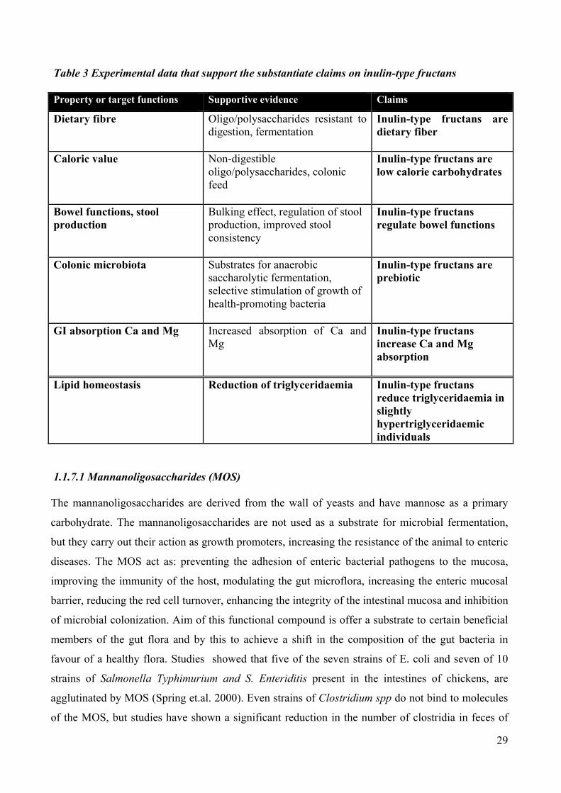
Table 3 Experimental data that support the substantiate claims on inulin-type fructans
Property or target functions Supportive evidence Claims
Dietary fibre Oligo/polysaccharides resistant to digestion, fermentation
Inulin-type fructans are dietary fiber
Caloric value Non-digestible oligo/polysaccharides, colonic feed
Inulin-type fructans are low calorie carbohydrates
Bowel functions, stool production
Bulking effect, regulation of stool production, improved stool consistency
Inulin-type fructans regulate bowel functions
Colonic microbiota Substrates for anaerobic saccharolytic fermentation, selective stimulation of growth of health-promoting bacteria
Inulin-type fructans are prebiotic
GI absorption Ca and Mg Increased absorption of Ca and Mg
Inulin-type fructans increase Ca and Mg absorption
Lipid homeostasis Reduction of triglyceridaemia Inulin-type fructans reduce triglyceridaemia in slightly hypertriglyceridaemic individuals
1.1.7.1 Mannanoligosaccharides (MOS)
The mannanoligosaccharides are derived from the wall of yeasts and have mannose as a primary
carbohydrate. The mannanoligosaccharides are not used as a substrate for microbial fermentation,
but they carry out their action as growth promoters, increasing the resistance of the animal to enteric
diseases. The MOS act as: preventing the adhesion of enteric bacterial pathogens to the mucosa,
improving the immunity of the host, modulating the gut microflora, increasing the enteric mucosal
barrier, reducing the red cell turnover, enhancing the integrity of the intestinal mucosa and inhibition
of microbial colonization. Aim of this functional compound is offer a substrate to certain beneficial
members of the gut flora and by this to achieve a shift in the composition of the gut bacteria in
favour of a healthy flora. Studies showed that five of the seven strains of E. coli and seven of 10
strains of Salmonella Typhimurium and S. Enteriditis present in the intestines of chickens, are
agglutinated by MOS (Spring et.al. 2000). Even strains of Clostridium spp do not bind to molecules
of the MOS, but studies have shown a significant reduction in the number of clostridia in feces of

30
animals fed MOS (Lalles et al., 2009). This is probably due to the effects of MOS increasing the
resistance of the barrier role of mucine or enhancing the immune function. It is well known that all
animals reared under commercial conditions, are subjected to immunological stress caused by the
presence of a high load of pathogens in their environment. The release of cytokines, associated with
inflammatory processes and the immature immune system, lead to hyperthermia. The consequences
are a decrease appetite, mobilization of the body reserves of glucose, amino acids, minerals,
decrease of the intestinal nutrients absorption, loss of body fluid and the development of diarrhea.
The use of MOS seems to reduce the production of pro-inflammatory cytokines by reducing
hyperthermia, and the events mentioned, allowing an increase in growth performance. It has also
been shown that MOS have a positive influence on immunity and humoral immune status. There is
evidence that the administration of MOS, causes an increase in the number of immunoglobulins
(IgA, IgG and IgM) in the intestine in both serum and blood. This increase in the antibody response
is due to the immune system's ability to respond to exogenous antigens present on the molecule of
MOS because of their microbial origin (Sauerwein et al., 2007). Although the cecum is the main
intestinal site where occur fermentation process, the fermentation processes that occur in the
jejunum have a greater influence on the digestion and absorption of nutrients (Lalles et al., 2009).
The use of MOS can decrease the concentration of strains such as Salmonella and E. coli in the
gastrointestinal tract increasing the growth of bacteria such as Lactobacillus spp Enteric-coated and
Bifidobacterium spp. Thanks to the increase of volatile fatty acids production decreasing intestinal
pH (Moore WEC et al., 1987). The benefits of MOS may be also associated with intestinal
morphology. During the weaning period, the massive colonization by microorganisms of the
intestinal lumen and the contribution of new types of food proteins is often associated with atrophy
of the villi, which results in a decreasing ratio of villous height / crypt depth. This phenomenon is
often due to malabsorption syndromes with subsequent onset of diarrhea (Pierce et al. 2006-2007).
Studies in piglets, chickens and mice, have shown that mannano oligosaccharides have a major
influence on the morphology of intestinal villi and their use in the diet after weaning increases the
ratio villus height / crypt depth (Konstantinov et al. 2004a-2006). Maintaining a good relationship
villus height / crypt depth is necessary for good absorption of nutrients and prevent the onset of
diarrhea. The state of the lining of the intestinal lumen, is the first barrier that opposes the gut
against enteric pathogens. Mucus production is proportional to the number of goblet cells in the gut
and nutrition including MOS has been shown to be able to increase the number of these cells at the
level of enteric villi (Bontempo et al. 2006). Thus, increased of prebiotics creates favourable lumen
conditions for gut health but direct evidence for enhanced resistance to unfavourable conditions is
still lacking (Lalles JP et al., 2007).

31
1.1.8 Plant Products
For thousands of years, herbs and spices have provided distinctive flavouring properties to foods
and many have been proven as potent antimicrobial agents. The use of these substances was very
common in both western and chinese culture. Some of the most common plant products (PP) known
for their antimicrobial properties belong to the genus Allium as garlic, onion and leek; others are
thyme, oregano, marjoram, basil and cumin. The natural plant antimicrobials are found in barks,
roots, stems, leaves, flowers and fruits of many plants. In recent years, in vitro studies demonstrated
the antibacterial activity of some of these products (Sen et al., 1998; Friedman et al., 2002), and
more recently the supplementation of plant products to pigs diets has been proposed in order to
counteract intestinal disorders, especially in the weaning period. However, the effects on growth
performance and gut health are not so consistent which may depend on the plant product tested.
One of the main issue concerning plant products is their characterization (Cowan, 1999). Plant
products contain several different active compounds in different concentration and their
composition is mainly affected by the method of extraction (solvent and extraction conditions) and
the niche of the plant used related to plant variety and age, climatic conditions and geografic origin.
Characterization is important for scientific as well as for legal purposes. All characteristics affecting
plant product composition affect at the same time their biological effects. Hence, for scientific
purpose it is better to work with pure active substances or with accurately controlled blends. In
regards to legislation, traceability, and thus characterization, is one of the main requisites to register
additives. It is, therefore important to accurately assess all the different compounds that the product
contains.
1.1.8.1 Effect of plant products on microbiota
Plant products have intensively been used in the past for medical and food preservation purposes.
The use is mostly motivated by the main characteristic of PP, their antimicrobial activity (Didry et
al., 1994). This activity has been studied in several in vitro studies with promising results. This
application is still too recent and there is too limited information available about the actual
possibilities of these products. However, the legislative requirements and the great interest to this
sector are motivating the appearance of the first studies using PP in vivo (Evans and Martin, 2000;
Herman et al., 2003; Allan et al., 2005). It is difficult to define the antimicrobial or different active
compounds present in a plant product. Usually, antimicrobial active substances in PP have a very
different chemical structure, with high occurrence of phenolic rings, mostly hydrofobic and some of
them with similar structure to important molecules from bacterial metabolism such as receptors or

32
enzyme substrates (Cowan, 1999). It also known that many of these substances are secondary
metabolites that plants use against predators, or with different functions such as pigmentation,
aromatizing or flavoring. In Table 4 the principal chemical structures producing antimicrobial
activity in plant products and the mechanism of action referenced are reported (Guen et al., 1991).
Some of these effects need to be better investigated while some of them are more studied as the
hydroxyl group (-OH) present in phenol compounds. The importance of this group on antimicrobial
activity is well known (Cowan, 1999) and any variation in its position inside the mulecule, as it
happens between carvacrol and thymol, produces marked differences in antimicrobial power
(Dorman and Deans, 2000). Components with phenolic structures, such as carvacrol and thymol are
highly active against test bacteria dispite their low capacity to dissolve in water. This has been
known for centuries, but the importance of the hydroxyl group in the phenolic ring was confirmed
in terms of activity when carvacrol is compared to its methyl ester. The high activity of the phenolic
components may be further explaned in terms of the alkyl substitution into the phenolics nucleus,
which is known to enhance the antimicrobial activity of phenols (Han et al. 2006). It was suggested
that plant products act via two main mechanisms of action. The first is related to the general
hydrophobicity of PP, which facilitates their adhesion to the bacterial surface inducing
unstabilization (Tsuchiya et al., 1996). The second mechanism is the inactivation of different
molecules of the bacteria (such as enzymes or receptors) through their adhesion to specific sites
(Sharon and Ofek, 1986).
Table 4 Chemical structures implicated in antimicrobial effect of PP and relative mechanism of action (Cowan, 1999)
Class Subclass Mechanism of action
Phenol compounds Simple phenols and phenolic acids Enzyme inactivation (1)
Membrane un-stabilizers (2)
Quinones Irreversible adhesion to adhesins,
membrane polypeptides and enzymes
that become inactive (3)
Flavonoids, flavones and flavonols
Tannins 1,2,3 and metal chelators
Coumarins Interact with eukariote DNA
Terpenoids Membrane un-stabilizers
Alkaloids Insertion in cellular wall or in DNA
structures
Lectines and polypeptides Block viral fusion and adsorption
Di-sulphur bridges formation

33
Some authors suggested a higher efficacy of plant products against gram negative organism but
others did not find any difference between gram positive and negative bacteria and, sometimes
even the opposite effect was proposed (Manzanilla et al., 2004; Han et al. 2006). Actually, it is
possible that some PP have specific actions and other PP have diverse effects due to the different
nature and composition. In fact, as it happens for antibiotics, the chemical structure will determine
the mode of action and hence a possible selective effect of plant product. For instance, alkilic chains
plus a phenol group seem to perform better activity against gram negative bacteria, due to the
characteristic of their cellular wall. In any case, the specific effect of some PP could be interesting
in therapeutic or preventive applications like it happens with antibiotics. Concerning growth
promotion, it must be studied if a selective effect of PP could be useful, because of the risk of
producing detrimental effects (Dorman and Deans, 2000). The effect of PP on different bacterial
species has been determined in a high number of in vitro studies using spectrophotometry
measurements or agar plate inhibition rings. Many of these studies have explored the real
antimicrobial power of classic herbal products or spices (Dorman and Deans, 2000; Friedman et al.,
2002). From these previous studies we can draw some conclusions. First of all, different bacteria
have different sensitivity to different plant products. It can be also observed how some compounds
as α-terpinen are highly effective against a very interesting target as Salmonella but not against the
other microorganisms.
Table 5 Antibacterial activity of different essential oils against 25 different bacteria tested (from Lis-Balchin M, 2003) Essential oil
Antibacterial activity Number of bacteria
Peppermint 15-22
Petitgrain 21
Pine niddle 19
Rosewood 24
Rosemary 21
Sage, Dalmatian 16
Tea tree 24
Thyme 14-25
Verbena 18

34
In the application of PP in vivo it is important to consider the dosage. Compared to antibiotics, the
in vitro dose of PP to obtain similar effect is normally 10- to 100-fold higher (Karaman et al.,
2001). So far, we know that complex media as the chime in the digestive tract could affect the in
vitro effective dosage.
1.1.8.2 Receptors analogs as anti-adhesive agents There is evidence that receptor analogs as agents for anti-adhesion therapy would be practical
primary against pathogens that bind to animal cells via carbohydrate-specific adhesins (i.e. lectins).
In this case the receptor analogs are saccharides that are structurally similar to those of the
glycoprotein and glycolipid receptors for the adhesin and, therefore, act by competitive inhibition. It
was less than three decades ago that mannose was first shown to be a receptor for enterobacteria
(Ofek et al., 1977). Since then, the sugar specifities of many bacteria have been determined, leading
to the development of receptors-like carbohydrates, which inhibit the adhesion of pathogens to host
cells and tissue (Ofek et al., 2003). The concentration of the carbohydrates required for effective
inhibition of adhesion in vitro are usually high, in the millimolar range, because the affinity of the
saccharides for the bacterial lectins is low. It can be increased several orders of magnitude by
covalently linking a hydrophobic residue such as phenyl or methyl umbelliferyl to the saccharide
(Firon et al., 1987). Affinity can be similarly increased by attaching many copies of the saccharide
to a suitable carrier, yelding multivalent adhesin inhibitors, as demonstrated for type 1 fimbriated
Escherichia coli (Lindhorst et al., 1998). The feasibility of using saccharides to protect against
experimental infections by bacteria expressing adhesive lectins was first demonstrated more than
two decades ago. Administration of methyl α-mannoside together with E. coli expressing the
mannose-specific type 1 fimbrial lectin into the bladders of mice reduced the extent of bladder
colonization by uropathogenic E coli by about two thirds compared to animals that had received the
bacteria alone or with methyl α-glucoside, a sugar that does not inhibit the mannose-specific
bacterial lectin. Subsequently many studies have confirmed the ability of saccharides to prevent
experimental infections caused by different pathogenic bacteria in a variety of animals ( Namkung
et al., 2004) (Table 6).

35
Table 6 Carbohydrates preventing bacterial colonization and/or infection in vivo (modified from Ofek et al., 2003b) Organism Animal, site of action Inhibitor
C. jejuni Mouse intestine Milk oligosaccharides
E. coli, type 1 fimbriated Mice GT Mannose
E. coli, P fimbriated Mice UT Globotetraose
(Galα1,4Gal)-containing GP*
E. coli K99 Calf GT Glycopeptides
H. pylori Piglet GT Sialyl-3’-LacNAc
Shigella flexneri, type 1
fimbriated
Guinea pig eye Mannose
S. pneumonia Rabbit and rat lungs Sialyl-3’-Galβ(1→4)LacNAc
S. sobrinus Rat oral cavity Oxidized α1,6 glucan
S. pyogenes Mouse pharynx Hyaluronan
*Gp: glycoprotein found in dove and pigeon egg white
1.1.8.3 Adhesins analogs as anti-adhesive agents The strategy for using adhesins to prevent infections is based on the assumption that the isolated
adhesin molecules, or an active synthetic or recombinant fragment, bind to the receptor and
competitively block adhesion of the bacteria (Ofek et al., 2003). It has thus far been impratical to
use analogs of adhesins for anti-adhesion therapy, because they are typically macromolecules that
are not readly available and bacause they must be employed at relatively high concentrations. In
addition, careful consideration must be given to their toxicity and immunogenecity. Novertheless,
modern proteomics and recombinant biotechnology have permitted the development of unique
types of relativelly small peptides for anti-adhesion therapy, as reported by Kelly et al. (1999). Non-
proteinaceous adhesins may also be useful for anti-adhesion therapy, as shown in studies of the
lipotheicoic acid (LTA)-mediated adhesion of group A and group B streptococci.

36
Table 7 Anti-adhesin activity of plant constituents (modified from Ofek et al.,2003)
Plant Constituent Bacterium affected
Azadirachta indica (neem
stick)
ND S sanguis
Camillia sinensis (green tea)
(-) epicatechin gallate, (-)
gallocathechin gallate
P gingivalis
Oolong tea Polyphenol S mutans; S sobranus
Gilanthus nivalis (snowdrop) Mannose-sensitive lectin E coli
Gloipeltis furcata and
Gigartina teldi (seaweeds)
Sulfated polisaccharides S sobrinus
Hop bracht Polyphenols (36-40 kDa) S mutans
Melaphis chinensis Gallotannin S sanguis
Persea americana (avocado) Tannins S mutans
Legume storage protein Glycoprotein E coli
ND: not determined
1.1.8.4 Dietary inhibitors of adhesion
Some of the most efficient anti-adhesion agents identified so far are present in feedstuffs. Feedstuffs
containing either a mixture of inhibitors or an inhibitor with a broad spectrum of activity could be
especially effective. While it may be possible to find suitable inhibitors for particular pathogens, it
is unlikely that it will be possible to match every individual or group of pathogens with specific
diets that contain complementary adhesins inhibitors (Ofek et al., 2003b). Empirical observations
over the years have suggested that may be effective to counteract bacterial infections. However,
caution should be used, because some dietary components may also be bactericidal and selective
pressures imposed by such compounds are undesirable and should be avoided. Human milk and
plant-derived constituents are rich in oligosaccharides and related compounds to which many
bacteria bind.
1.1.8.5 Plant products as bacteria adhesion inhibitors Because of their ready availability, plant materials possessing anti-adhesion activities are attractive
candidates for antibacterial agents. There is, however a relative paucity of information regarding the
anti-adhesive properties of most plant materials. Although plant lectins are well rappresented in the

37
diets, and many of these lectins are very characteristic, their application to anti-adhesion therapy is
very limited. Theorically, these lecitins could interact with animal cell surface saccharides to block
adhesion mediated by lectin-carrying bacteria and they may enhance clearance of bacteria from the
host (Slifkin and Doyle, 1990). Feed lectins may have deleterious effects as well. They may bind
to mucosal cells and thus function as receptors for bacterial glycans and enhance bacterial adhesion
to the tissue. Moreover some dietary lectins may reach the GI tract in a faunctional form and may
similarly enhance the binding of the bacteria to the different parts of the intestine. A pratical
advantage in the search of dietary plant extracts for agents to use in the therapy of bacterial
infections is that clinical trials are probably easier to perform, mainly because toxicity is usually not
as much of an issue. Among the plant extract listed in Table 8, those obtained from Vaccinium
macrocarpon (cranberry) are the most throughly studied with respect to their anti-adhesion activity
in vitro and, so far, are the only one sow to be effective in in vivo trials. The initial in vitro
experiments on the effects of cranberry extracts on bacterial adhesion were stimulated by the long
known anecdotal evidence on the beneficial effects of cranberry juice consumption in therapy of
urinary tract infections. Several lines of evidence implicate two different cranberry constituents as
active anti-adhesive agents. One of these is a high molecular mass (> 15 kDa) material and the other
is a protoanthocyanin. The high molecular mass material is devoid of proteins and carbohydrates
and behaves in some respects like tannin (Ofek et al., 1996). It inhibited the adhesion to animals
cells of uropathogenic E. coli including P fimbriae-, S fimbriae- and non-fimbrial.
Table 8 Anti-adhesion effects of juices or extracts of Vaccinium spp. (cranberry). From Ofek et al., 2003 (modified) Bacterium Disease Adhesion assay Effect
E. coli UTI and
pyelonephritis
HA UroEp. I
E. coli Diarrhea HA NI
E. coli Meningitis HA I
Oral bacteria Dental decay
periodontitis
Coaggregation and
buccal epithelial
adhesion
I
H. pylori Gastric ulcer Human gastric
mucus, TC cells
I

38
In addition, it inhibited the coaggregation of Gram-negative pairs of oral bacterial more often than it
inhibited coaggregations between Gram positive bacteria. Extracts containing protoanthocyanins in
their condensed form inhibited adhesion of P fimbriated E. coli to erythrocytes.
1.1.8.6 Plant products: antioxidant capacity Antioxidative properties are well described for herbs and spices (Cuppett and Hall, 1998; Craig,
1999; Nakatani, 2000; Wei and Shibamoto, 2007). Among a variety of plants bearing antioxidative
constituents, the volatile oils from the Labiatae family (mint plants) have been attracting the
greatest interest, especially products from rosemary. Their antioxidant activity arises from phenolic
terpenes, such as rosmarinic acid and rosmarol (Cuppett and Hall, 1998). Other Labiatae species
with significant antioxidative properties are thyme and oregano, which contain large amounts of the
monoterpenes thymol and carvacrol (Cuppett and Hall, 1998). Plant species from the families of
Zingiberaceae (ginger and curcuma) and Umbelliferae (anise and coriander), as well as plants rich
in flavonoids (green tea) and anthocyans (many fruits), are also described as exerting antioxidative
properties (Nakatani,2000; Wei and Shibamoto, 2007). Furthermore,pepper (Piper nigrum), red
pepper (Capsicum annuum L.), and chili (Capsicum frutescene) contain antioxidative components
(Nakatani, 1994). In many of these plants, parts of the active substances are highly odorous or may
taste hot or pungent, which may restrict their use for animal feeding purposes. The antioxidant
property of many phytogenic compounds may be assumed to contribute to protection of feed lipids
from oxidative damage, such as the antioxidants usually added to diets (α-tocopheryl acetate or
butylated hydroxytoluene). Although this aspect has not been explicitly investigated for piglet and
poultry feeds, there is a wide practice of successfully using essential oils, especially those from the
Labiatae plant family, as natural antioxidants in human food (Cuppett and Hall, 1998), as well as in
the feed of companion animals. The principal potential of feed additives from the Labiatae plant
family containing herbal phenolic compounds to improve the oxidative stability of animalderived
products has been demonstrated for poultry meat (Botsoglou et al., 2002, 2003a,b; Papageorgiou et
al., 2003; Young et al., 2003; Basmacioglu et al., 2004; Govaris et al., 2004; Giannenas et al., 2005;
Florou-Paneri et al., 2006), pork (Janz et al., 2007), rabbit meat (Botsoglou et al., 2004b), and eggs
(Botsoglou et al.,2005). Oxidative stability was also shown to be improbe with other herbal
products (Botsoglou et al.,2004a; Schiavone et al., 2007). Nevertheless, it remains unclear whether
these phytogenic antioxidants are able to replace the antioxidants usually added to the feeds (α-
tocopherols) to a quantitatively relevant extent under conditions of common feeding practice.
The tea plant, Camellina sinensis, is used in livestock as a feed additive. The major molecules that
make up the tea and give the biological properties are: Methylxanthines: like caffeine or protein

(4%), smaller amounts of theobromine and theophylline, and traces of adenine and xanthine. They
are partly related to tannins. Polyphenols: Catechins prevail, whose percentage ranged between 10
and 20% depending on the variety and age of the leaf. Were isolated several compounds such as,
epicatechin, gallocatechin and other 4-catechin gallates; flavanil-3-ols and diols flavanil. Other: as
oligomeric procyanidins, fenolcarbossilici acids, saponins, aldehydes, alcohols and monoterpenes
(Fig. 4)
Fig.4 Structure of molecole contained in Tea leaves (Friedman 2007)
39
The catechins contained in tea leaves, have antibacterial and antitoxic effects on different
microorganisms. Juneja et al (2007) have shown that tea extracts are able to inhibit the growth and
sporulation of Cl. perfrigenes in land-based broth. Studies in vivo in mice, show how green tea
extract, protect the animals from systemic and neurological symptoms caused by toxins of E. coli
O157: H7 (Isogai et al., 2001). Alcoholic extracts of black tea, showed inhibitory to strains of
Salmonella typhimurium (Ciraj, Sulaim., 2001). It is well documented the role of different
antioxidant catechins as acceptors of H, are able to protect cells from free radicals. Human studies
have demonstrated the ability of the polyphenols in green tea inhibit in vivo oxidation of LDL, thus

reducing the risk of atherogenesis. It also demonstrated a capacity of catechins antimicetica and
antiviral (Weber et al, 2003). Grapes contain especially in the peel and seeds large percentage of the
anthocyanins and proanthocyanidins. Anthocyanins are among the most important groups of
pigments found in plants and belong to the family of flavonoids. These molecules are composed of
a benzene molecule with a cast of Piran (heterocyclic ring containing oxygen), connected in turn
with a phenyl group which may in turn be linked to various substituents. This complex molecule
called a cation flavilio and is the basic structure of all anthocyanins. (Fig.5)
Fig.5 Flavilio cation is the basic structure of antiocians
As part of the family of polyphenols, anthocyanins also have the same biological properties
including a strong antioxidant properties. The Olive (Olea europaea.) Is a plant belonging to the
family of Oleaceae. The essential oil of olive leaves is used in animal husbandry due to the presence
of high concentrations of biologically active substances, including flavonoids, triterpenes, fatty
acids, saturated and polyunsaturated fats (alpha-linolenic acid), monounsaturated (oleic),
oleuropein, tannins and chlorophyll . This is known as oleuropein molecule that has a strong
antioxidative action antibattericae, common to the family of polyphenols to which it belongs.
Oleuropein metabolites, such as football elenolato derived from its hydrolysis, seems to be also
responsible for antiviral action of the molecule.
40

41
1.9 References
Adam, K., A. Sivropoulou, S. Kokkini, T. Lanaras, and M. Arsenakis. 1998. Antifungal activities of Origanum vulgare subsp. hirtum,Mentha spicata, Lavandula angustifolia, and Salvia fruticosa essential oils against human pathogenic fungi. J. Agric. Food Chem. 46:1739–1745. Aksit, M., E. Goksoy, F. Kok, D. Ozdemir, and M. Ozdogan. 2006. The impacts of organic acid and essential oil supplementations to diets on the microbiological quality of chicken carcasses. Arch.Geflugelkd. 70:168–173. Alcicek, A., M. Bozkurt, and M. Cabuk. 2003. The effect of an essential oil combination derived from selected herbs growing wild in Turkey on broiler performance. S. Afr. J. Anim. Sci. 33:89–94. Alcicek, A., M. Bozkurt, and M. Cabuk. 2004. The effect of a mixture of herbal essential oils, an organic acid or a probiotic on broiler performance. S. Afr. J. Anim. Sci. 34:217–222. Anadon, A., M. Abroix Arzo, G. Bories, P. Brantom, J. Brufau de Barbera, A. Chesson, P. S. Concconcelli, J. de Knecht, N. Dierick, G. Flachowsky, A. Franklin, J. Gropp, A. K. Haldorsen, I. Halle, A. Mantovani, K. Peltonen, G. Rychen, P. Sanders, A. Soares P. Wester, and W. Windisch. 2005. Opinion of the FEEDAP Panel on the safety and efficacy of the product Farmatan for rabbits and piglets. The EFSA Journal 222:1–20. Baba, S., N. Osakabe, M. Natsume, A. Yasuda, Y. Muto, K. Hiyoshi,H. Takano, T. Yoshikawa, and J. Terao. 2005. Absorption, metabolism, degradation and urinary excretion of rosmarinic acid after intake of Perilla frutescens extract in humans. Eur. J. Nutr. 44:1–9. Baldi A., Pinotti L. 2008. Lipophilic microconstituents of milk. 2008. In: Bösze Ed. Bioactive components of milk: pp. 109-125. Baldi A., Politis I., Pecorini C., Fusi E., Chronopoulou R., Dell’Orto V., 2005. Biological effects of milk proteins and their peptides with emphasis on those related to the gastrointestinal ecosystem. Journal of Daily Research, 72: 66-72. Bampidis, V. A., V. Christodoulou, P. Florou-Paneri, E. Christaki, P. S. Chatzopoulou, T. Tsiligianni, and A. B. Spais. 2005. Effect of dietary dried oregano leaves on growth performance, carcase characteristics and serum cholesterol of female early maturing turkeys. Br. Poult. Sci. 46:595–601. Basmacioglu, H., O. Tokusoglu, and M. Ergul. 2004. The effect of oregano and rosemary essential oils or alpha-tocopheryl acetate on performance and lipid oxidation of meat enriched with n-3 PUFAs in broilers. S. Afr. J. Anim. Sci. 34:197–210. Bee, G. 2000. Dietary conjugated linoleic acids alter adipose tissue and milk lipids of pregnant and lactating sows. Journal of Nutrition, 130: 2292–2298. Belurt M.A. 2002. Dietary conjugated linoleic acid in health: physiological effects and mechanisms of action. Annual Review of Nutrition, 22: 505-531.

42
Bontempo V., Sciannimanico D., Pastorelli G., Rossi R., Rosi F., Corino C. 2004. Dietary conjugated linoleic acid positively affects immunologic variables in lactating sows and piglets. Journal of Nutrition, 134: 817-824. Botsoglou, N. A., E. Christaki, P. Florou-Paneri, I. Giannenas, G. Papageorgiou, and A. B. Spais. 2004a. The effect of a mixture of herbal essential oils or alpha-tocopheryl acetate on performance parameters and oxidation of body lipid in broilers. S. Afr. J. Anim. Sci. 34:52–61. Botsoglou, N. A., P. Florou-Paneri, E. Botsoglou, V. Dotas, I. Giannenas,A. Koidis, and P. Mitrakos. 2005. The effect of feeding rosemary, oregano, saffron and alpha-tocopheryl acetate on hen performance and oxidative stability of eggs. S. Afr. J. Anim. Sci. 35:143–151. Botsoglou, N. A., P. Florou-Paneri, E. Christaki, D. J. Fletouris, and A. B. Spais. 2002. Effect of dietary oregano essential oil on performance of chickens and on iron-induced lipid oxidation of breast,thigh and abdominal fat tissues. Br. Poult. Sci. 43:223–230. Botsoglou, N. A., P. Florou-Paneri, E. Christaki, I. Giannenas, and A. B. Spais. 2004b. Performance of rabbits and oxidative stability of muscle tissues as affected by dietary supplementation with oregano essential oil. Arch. Anim. Nutr. 58:209–218. Botsoglou, N. A., A. Govaris, E. Botsoglou, S. H. Grigoropoulou, and G. Papageorgiou. 2003a. Antioxidant activity of dietary oregano essential oil and alpha-tocopheryl acetate supplementation in long-term frozen stored turkey meat. J. Agric. Food Chem. 51:2930–2936. Botsoglou, N. A., S. H. Grigoropoulou, E. Botsoglou, A. Govaris, and G. Papageorgiou. 2003b. The effects of dietary oregano essential oil and alpha-tocopheryl acetate on lipid oxidation in raw and cooked turkey during refrigerated storage. Meat Sci. 65:1193– 1200. Brock J.H. 2002. The physiology of lactoferrin. Biochemistry and Cell Biology, 80: 1-6. Burt, S. 2004. Essential oils: Their antibacterial properties and potential applications in food—A review. Int. J. Food Microbiol. 94:223–253. Cabuk, M., M. Bozkurt, A. Alcicek, Y. Akbas, and K. Ku¨ cu¨ kyilmaz. 2006. Effect of a herbal essential oil mixture on growth and internal organ weight of broilers from young and old breeder flocks. S. Afr. J. Anim. Sci. 36:135–141. Castillo, M., S. M. Martin-Orue, M. Roca, E. G. Manzanilla, I. Badiola, J. F. Perez, and J. Gasa. 2006. The response of gastrointestinal microbiota to avilamycin, butyrate, and plant extracts in earlyweaned pigs. J. Anim. Sci. 84:2725–2734. Cevolani D., Arioli E. 2008. L'impiego di silimarina garantisce un latte di qualità. Suinicoltura, 7: 26-29.

43
Cho, J. H., Y. J. Chen, B. J. Min, H. J. Kim, O. S. Kwon, K. S. Shon,I. H. Kim, S. Kim, and A. Asamer. 2006. Effects of essential oils supplementation on growth performance, IgG concentration and fecal noxious gas concentration of weaned pigs. Asian-australas. J. Anim. Sci. 19:80–85. Chronopoulou R., Xylouri E., Fegeros K., Politis I. 2006. The effect of two bovine β-casein peptides on various functional properties of porcine macrophages and neutrophils: differential roles of protein kinase A and exchange protein directly activated by cyclic AMP-1. British Journal of Nutrition, 96: 553-561. Chrubasik, S., M. H. Pittler, and B. D. Roufogalis. 2005. Zingiberis rhizome: A comprehensive review on the ginger effect and efficacy profiles. Phytomedicine 12:684–701. Cicero AFG, Gaddi A. Rice bran oil and -oryzanol in the treatment of hyperlipoproteinemias and other conditions. Phytother Res 2001; 15:277-89. Craig, W. J. 1999. Health promoting properties of common herbs. Am. J. Clin. Nutr. 70(Suppl.):491S–499S. Cuppett, S. L., and C. A. Hall. 1998. Antioxidant activity of Labiatae. Adv. Food Nutr. Res. 42:245–271. Demir, E., S. Sarica, M. A. O¨ zcan, and M. Suicmez. 2005. The use of natural feed additives as alternative to an antibiotic growth promoter in boiler diets. Arch. Geflugelkd. 69:110–116. intestinal equilibrium of early-weaned pigs. J. Anim. Sci. 82:3210–3218. Dionysius D.A., Grieve P.A., Milne J.M. 1993. Forms of lactoferrin: their antibacterial effect on enterotoxigenic Escherichia coli. Journal of Dairy Science, 76: 2597-2606. Feng S., LiLi Z., YongJiu H., GuiJuan L., Tian W. 2006. Effects of casein hydrolysate on the intestinal growth and development in intrauterine growth retardation neonatal piglets. Journal of Nanjing Agricultural University, 29 (3): 59-63. Frank M. Sacks MD, et a. (2006) Soy Protein, Isoflavones, and Cardiovascular Health. An American Heart Association Science Advisory for Professionals From the Nutrition Committee in Circulation German J.B. e Dillard C.J. 2006. Composition, structure and absorption of milk lipids: a source of energy, fat-soluble nutrients and bioactive molecules. Critical Reviews in Food Science and Nutrition, 46: 57-92. Gibson G.R and Robertfroid M.B., Dietary modulation of the human colonic microbiota: introducing the concept of prebiotics, J. Nutr., 125:1401-1412, 1995 Gnoth, M. J., Kunz, C., Kinne-Saffran, E., Rudloff S. 2000. Human milk oligosaccharides are minimally digested in vitro. Journal of Nutrition, 130 (12): 3014-3020. Griffiths E.A., Duffy L.C., Schanbacher F.L., Dryja D., Leavens A., Neiswander R.L., Qiao H., DiRienzo D., and Ogra P. 2003. In vitro growth responses of bifidobacteria and enteropathogens to bovine and human lactoferrin. Digestive diseases and sciences, 48(7):1324-1332.

44
Hill H.R. 1987. Biochemical, structural and functional abnormalities of polymorphonuclear leycocytes in the neonate. Pediatric Research, 22: 375-382. Konstantinov SR, Favier CF, Zhu WY, Williams BA, Kluss J, Souffrant WB, De Vos WM, Akkermans ADL, Smidt H & de Vos WM (2006ab) Post natal development of the porcine microbiota composition and activities. Environmental Microbiology 8, 1191-1199 Konstantinov SR, Awati A, Smidt H, Williams BA, Akkermanns ADL & de Vos WM (2004a) Specific response of a novel and abundant Lactobacillusamylovorus- like phylotype to dietary prebiotics in the ileum and colon of weaning piglets. Applied Environmental Microbiology 70, 3821-3830. Kunz, C., Rudloff S., Baier W., Klein N., Strobel S.. 2000. Oligosaccharides in human milk: Structural, functional, and metabolic aspects. Annual Review of Nutrition, 20: 699–722. Kwon O.S., Yoo J.S., Min B.J., Son K.S., Cho J.H., Kim H.J., Chen Y.J., Kim I.H. 2005. Effect of supplemental medicinal plant (Artemisia, Acanthopanax and Garlic) on growth performance and serum characteristic in lactating sows, suckling and weanling pigs. Journal of Animal Science and Technology, 47 (4): 501-512. Jariwalla RJ. Rice-bran products: phytonutrients with potential applications in preventive and clinical medicine. Drugs Exp Clin Res 2001; 27:17-26. Jean-Paul Lalles, Paolo Bosi, Hauke Smidt and Chris R. Stokes, Nutritional management of gut health in pigs around weaning, Proceedings of the Nutrition society (2007), 66, 260-268 Ishihara M. Effect of -oryzanol on serum lipid peroxide level and clinical symptoms of patients with climateric disturbancies. Asia Oceania J Obstet Gynaecol 1984; 10:317-23. Lichtenstein AH, Ausman LM, Carrasco W, et al. Rice bran oil consumption and plasma lipid levels in moderately hypercholesterolemic humans. Arterioscler Thromb 1994; 14: 549-56. Maass N., Bauer J., Paulicks B.R., Böhmer B.M., Roth-Maier D.A. 2005. Efficiency of Echinacea purpurea on performance and immune status in pigs. Journal of Animal Physiology and Animal Nutrition, 89: 244-252. Matar C., Valdez J.C., Medina M., Rachid M., Perdigon G., 2001. Immunomodulating effects of milks fermented by Lactobacillus helveticus and its non-proteolitic variant. Journal of Dairy Research, 68: 601-609. Meisel H., 2005. Biochemical properties of peptides encrypted in bovine milk proteins. Current Medicinal Chemistry, 12: 1905-1919. Molkentin J. 1999. Bioactive lipids naturally occurring in bovine milk. Nahrung, 43: 185-189. Mitsch, P., K. Zitterl-Eglseer, B. Kohler, C. Gabler, R. Losa, and I.Zimpernik. 2004. The effect of two different blends of essential oil components on the proliferation of Clostridium perfringens in the intestines of broiler chickens. Poult. Sci. 83:669–675.

45
Moore WEC, Moore LWH, Cato EP, Wilkins TD, Kornegay ET (1987) Effect of high fiber and high oil diets on the fecal flora of swine. Appl Environ Microbiol, 53: 1638-1644. Nakatani, N. 1994. Antioxidants from spices and herbs. In Food Phytochemicals for Cancer Prevention II: Teas, Spices and Herbs. ACS Symposium Series 547. C.-T. Ho, T. Newburg, D. S., Pickering, L. K., McCluer, R. H., Cleary T.G. 1990. Fucosylated oligosaccharides of human milk protect suckling mice from heat-stable enterotoxin of Escherichia coli. Journal of Infectious Diseases, 162: 1075-1080 Newburg, D. S., Ruiz-Palacios, G. M., Morrow, A. L. 2005. Human milk glycans protect infants against enteric pathogens. Annual Review of Nutrition, 25: 37-58. Nicolosi RJ, Ausman LM, Hegsted DM. Rice bran oil lowers serum total and low density lipoprotein Nuijens JH., van Berkel PHC., Schanbacher FL. 1996. Structure and biological actions of lactoferrin. J ournal of Mammary Gland Biololy Neoplasia 1, p. 285–295. Oram J.D., Reiter B. 1968. Inhibition of bacteria by lactoferrin and other iron-chelating agents. Biochimica et Biophysica Acta, 170: 351-365. Osawa, M.-T. Huang, and R. T. Rosen, ed. Am. Chem. Soc., Washington, DC. Nakatani, N. 2000. Phenolic antioxidants from herbs and spices. Biofactors 13:141–146. Namkung, H., M. Li, J. Gong, H. Yu, M. Cottrill, and C. F. M. de Lange. 2004. Impact of feeding blends of organic acids and herbal extracts on growth performance, gut microbiota and digestive function in newly weaned pigs. Can. J. Anim. Sci. 84:697–704. Nazeer, M. S., T. N. Pasha, S. Abbas, and Z. Ali. 2002. Effect of yucca saponin on urease activity and development of ascites in broiler chicken. Int. J. Poult. Sci. 1:174–178. Newton, S. M., C. Lau, S. S. Gurcha, G. S. Besra, and C. W. Wright. 2002. The evaluation of forty-three plant species for in vitro antimycobacterial activities: Isolation of active constituents from Psoralea corylifolia and Sanguinaria canadensis. J. Ethnopharmacol. 79:57–67. Nofrarias, M., E. G. Manzanilla, J. Pujols, X. Gilbert, N. Majo, J.Segales, and J. Gasa. 2006. Effects of spray-dried porcine plasma and plant extracts on intestinal morphology and on leukocyte cell subsets of weaning pigs. J. Anim. Sci. 84:2735–2742. Oetting, L. L., C. E. Utiyama, P. A. Giani, U. D. Ruiz, and V. S. Miyada. 2006. Effects of herbal extracts and antimicrobials on apparent digestibility, performance, organs morphometry and intestinal histology of weanling pigs. Braz. J. Anim. Sci. 35:1389–1397. Oviedo-Rondon, E. O., M. E. Hume, C. Hernandez, and S. Clemente-Hernandez. 2006. Intestinal microbial ecology of broilers vaccinated and challenged with mixed Eimeria species, and supplemented with essential oil blends. Poult. Sci. 85:854–860. O¨zer, H., M. So¨kmen, M. Gu¨ llu¨ ce, A. Adigu¨ zel, F. Sahin, A. So¨kmen,H. Kilic, andO¨ . Baris. 2007. Chemical composition and antimicrobial and antioxidant activities of the essential oil

46
and methanol extract of Hippomarathum microcarpum (Bieb.) from Turkey. J. Agric. Food Chem. 55:937–942. Papageorgiou, G., N. A. Botsoglou, A. Govaris, I. Giannenas, S. Iliadis, and E. Botsoglou. 2003. Effect of dietary oregano oil and alphatocopheryl acetate supplementation on iron-induced lipid oxidation of turkey breast, thigh, liver and heart tissues. J. Anim.Physiol. Anim. Nutr. (Berl.) 87:324–335. Pecorini C., Savazzini F., Martino P.A., Fusi E., Fogher C., Baldi A. 2005. Heterologous expression of biologically ctive porcine lactoferrin in Pichia pastoris yeast. Veterinary Research Communications, 29 (Suppl. 2): 379-382. Pettigrew, J. E. e B. G. Harmon. 1977a. Milk proteins for artificially reared piglets. I Comparison to egg white protein and effect of added immunoglobulins. Journal of Animal Science, 44: 374-382. Pettigrew, J. E., B. G. Harmon, J. Simon and D. H. Baker. 1977b. Milk proteins for artificially reared piglets. 1I Comparison to a skim milk hydrolysate. Journal of Animal Science, 44: 383-388. Pettigrew, J. E., B. G. Harmon, S. E. Curtis, S. G. Cornelius, H. W. Norton and A. H. Jensen. 1977c. Milk proteins for artificially reared piglets. III. Efficacy of sodium caseinate and sweet dried whey. Journal of Animal Science, 45:261-268. Platel, K., and K. Srinivasan. 2000a. Influence of dietary spices and their active principles on pancreatic digestive enzymes in albino rats. Nahrung 44:41–46. Platel, K., and K. Srinivasan. 2000b. Stimulatory influence of select spices on bile secretion in rats. Nutr. Res. 20:1493–1503. Platel, K., and K. Srinivasan. 2004. Digestive stimulant action of spices: A myth or reality? Indian J. Med. Res. 119:167–179. Pluske J.R., I.H. Williams and F.X Aherne, 1995. Nutrition of the neonatal pig. In: M.A. Varley (editor), The Neonatal Pig Development and survival. CAB international, Wallingford, Oxon. Pluske J.R., I.H. Williams and F.X Aherne 1996a. Maintanance of villous height and crypths depth in piglets by providing continous utrition after weaning. Animal Science 62, 131-144 Politis I., Chronopoulou R. 2008. Milk peptides and immune response in the neonate. In: Bösze Ed. Bioactive components of milk: pp. 253-269. Raghuram TC, Rukimini C. Nutritional significance of rice bran oil. Indian J Med Res 1995; 102:241-4. Rao, R. R., K. Platel, and K. Srinivasan. 2003. In vitro influence of spices and spice-active principles on digestive enzymes of rat pancreas and small intestine. Nahrung 47:408–412. Raghuram T, Brahmaji G, Rukmini C. Studies in hypolipidemic effects of dietary rice bran oil in human subjects. Nutr Rep Int 1989; 35:889-95. Rodehutscord, M., and H. Kluth. 2002. Tierfu¨ tterung ohne antibiotisch wirkende Leistungsfo¨rderer. Zuchtungskunde 74:445–452.

47
Rong M, Ausman LM, Nicolosi RJ. Oryzanol decreases cholesterol absorption and aortic fatty streaks in hamsters. Lipids 1997; 32:303-9. Roth, F. X., G. G. Gotterbarm, W. Windisch, and M. Kirchgessner. 1999. Whole-body protein turnover and nitrogen balance in growing pigs supplied with antibiotic feed additive (Avilamycin). J. Anim. Physiol. Anim. Nutr. (Berl.) 82:88–93. Roth, F. X., and M. Kirchgessner. 1998. Organic acids as feed additives for young pigs: Nutritional and gastrointestinal effects. J. Anim. Feed Sci. 8:25–33. Roth-Maier, D. A., B. M. Bohmer, N. Maass, K. Damme, and B. R.Paulicks. 2005. Efficiency of Echinacea purpurea on performance of broilers and layers. Arch. Geflugelkd. 69:123–127. Rukimini C, Raghuram TC. Nutritional and biochemical aspects of the hypolipidemic action of rice bran oil: a review. J Am Coll Nutr 1991; 10:593-601. Sarica, S., A. Ciftci, E. Demir, K. Kilinc, and Y. Yildirim. 2005. Use of an antibiotic growth promoter and two herbal natural feed additives with and without exogenous enzymes in wheat based broiler diets. S. Afr. J. Anim. Sci. 35:61–72. Sasaki J, Takada Y, Handa K, et al. Effects of gamma-oryzanol on serum lipid and apolipoproteins in dyslipidemic schizophrenic receiving major tranquilizers. Clin Ther 1990; 12:263-8. Savoini G., Bontempo V., Cheli F., Baldi A., Sala V., Mancin G., Agazzi A., Dell’Orto V. 2002. The Veterinary Record, 151: 577-580. Séverin S., Wenshui X., 2005. Milk Biologically Active Components as Nutraceuticals: Review. Critical Reviews in Food Science and Nutrition, 45: 645–656. Schiavone, A., F. Righi, A. Quarantelli, R. Bruni, P. Serventi, and A. Fusari. 2007. Use of Sibyllum marianum fruit extract in broiler chicken nutrition: Influence on performance and meat quality. J. Anim. Physiol. Anim. Nutr. (Berl.) 91:256–267. Scho¨ne, F., A. Vetter, H. Hartung, H. Bergmann, A. Biertumpfel, G. Richter, S. Mu¨ ller, and G. Breitschuh. 2006. Effects of essential oils from fennel (Foeniculi aetheroleum) and caraway (Carvi aetheroleum) in pigs. J. Anim. Physiol. Anim. Nutr. (Berl.) 90:500–510. Seskeviciene, J., J. Jankowski, and K. Kozlowski. 2005. Effect of probiotic preparation and phytogeneous feed additive on digestibility of nutrients, metabolizability of gross energy and content of metabolizable energy of a practical feed ration for fattening turkeys. Arch. Geflugelkd. 69:107–109. Si, W., J. Gong, R. Tsao, T. Zhou, H. Yu, C. Poppe, R. Johnson, and Z. Du. 2006. Antimicrobial activity of essential oils and structurally related synthetic food additives towards selected pathogenic and beneficial gut bacteria. J. Appl. Microbiol. 100:296–305. Smith-Palmer, A., J. Stewart, and L. Fyfe. 1998. Antimicrobial properties of plant essential oils and essences against five important food-borne pathogens. Lett. Appl. Microbiol. 26:118–122.

48
Stoni, A., K. Zitterl-Egelseer, A. Kroismayr, W. Wetscherek, and W.Windisch. 2006. Tissue recovery of essential oils used as feed additive in piglet feeding and impact on nutrient digestibility. Proc. Soc. Nutr. Physiol. 15:60. Straub, R., S. Gebert, C. Wenk, and M. Wanner. 2005. Growth performance,energy, and nitrogen balance of weanling pigs fed a cereal-based diet supplemented with Chinese rhubarb. Livest. Prod. Sci. 92:261–269. Sugano M, Koba K, Tsuji E. Health benefits of rice bran oil.Anticancer Res 1999; 19:3651-7. Sugano M, Tsuji E. Rice bran oil and cholesterol metabolism. J Nutr 1997; 127:521S-524S. Tao N., DePeters E. J., Freeman S., German J. B., Grimm R., and Lebrilla C. B. 2008. Bovine Milk Glycome. Journal of Dairy Science, 91: 3768–3778. Teraguchi S., Shin K., Ozawa K., Nakamura S., Fukuwatari Y., Tsuyuri S., Namihira H., Shimanura S. 1995. Bacteriostatic effect of orally administered bovine lactoferrin on proliferation of Clostridium species in the gut of mice fed bovine milk. Applied and Environmental Microbiology, 61: 501-506. Tsuji E, Itoh H, Itakura H. Comparison of effects of dietary saturated and polyunsaturated fats on the serum lipids levels. Clin Ther Cardiovasc 1989; 8:149- 51. Van Leeuwen P., Oosting S.J., Mouwen J., Verstegen M.W.A. 2000. Effects of a lactoperoxidase system and lactoferrin, added to a milk replacer diet, on severity of diarrhoea, intestinal morphology and microbiology of digesta and faeces in young calves. Journal of Animal Physiology and Animal Nutrition, 83: 15-23. Veh, R. W., Michalski J. C., Corfield A. P., Sander-Wewer M., Gies D., Schauer R.. 1981. New chromatographic system for the rapid analysis and preparation of colostrum sialyloligosaccharides. Journal of Chromatography A, 212: 313–322. Vissers MN, Zock PL, Gert WM, Katan MB. Effect of plant sterols from rice bran oil and triterpene alcohols from sheanut oil on serum lipoprotein concentrations in humans. Am J Clin Nutr 2000; 72:1510-5. Wei, A., and T. Shibamoto. 2007. Antioxidant activities and volatile constituents of various essential oils. J. Agric. Food Chem. 55:1737–1742. Wilson TA, Ausman LM, Lawton CW, Hegsted DM, Nicolosi RJ. Comparative cholesterol lowering properties of vegetable oils: beyond fatty acids. J Am Coll Nutr 2000; 19:601-7. Yeo, J., and K. I. Kim. 1997. Effect of feeding diets containing an antibiotic, a probiotic, or yucca extract on growth and intestinal urease activity in broiler chicks. Poult. Sci. 76:381–385. Yoshino G, Kazumi T, Amano M, et al. Effects of -oryzanol and probucol on hyperlipidemia. Curr Ther Res 1989; 45:975-82.

49
YongJiu H., Tian W., LiLi Z., Feng S., GuoBin C. 2006. Effect of pre-hydrolyzed casein on the development of gastrointestinal tract in neonatal pigs. Journal of Yangzhou University, Agricultural and Life Sciences 27, 4: 47-50. Young, J. F., J. Stagsted, S. K. Jensen, A. H. Karlsson, and P. Henckel. 2003. Ascorbic acid, alpha-tocopherol, and oregano supplements reduce stress-induced deterioration of chicken meat quality. Poult. Sci. 82:1343–1351.

Chapter 2
Objectives
50

51
2.1 Objectives Worldwide the swine industries are under pressure to reduce the use of antibiotics while maintaining animal
health and performance. In 2006 all the AGP (antibiotic growth promoters) and some feed additives with
growth promoting activities were banned (Cu, Zn). Since the first restrictive measures were taken, and due to
the beginning of the negative consequences of the ban, numerous efforts have been done to find alternatives
or replacement strategies to maintain pig growth performance and controlling enteric bacterial diseases.
Therefore, the following studies aimed to find ways to control additive efficacy with due consideration to
animal welfare and consumers requirements. The main objective of this thesis was to improve our
knowledge on the properties of new additives as feeding strategy and oral vaccination to improve general
and gut health in young pigs with the aim to substitute antibiotics growth promoters. To achieve these
objectives, three different trials were designed to study different strategies to improve the gut health of the
post-weaning piglets.
2.1.1 Abstract 1 I) Evaluation of the immune response against ovalbumin (OVA) in pigs orally vaccinated with OVA in tablets containing carboxymethyl high amylose starch (CM-HAS), flagellin and sow’s milk. This trial was designed with the aim to determine the effects of a new delivery system designed for
oral vaccination or delivery of bioactive molecule in the gut. The trial was also designed to
evaluate the potential of the sow’s milk to modulate the systemic as well as local immune response
against the antigen ovalbumin incorporated in the vaccine. Sixteen piglets of 28 days (d) of age
were divided into 4 groups of 4 animals each. The first group was vaccinated with compressed
tablets containing only ovalbumin (OVA); the second group was immunized with tablets containing
ovalbumin and the B subunit of the flagellin from Salmonella enterica Typhimurium (OVA-FLA);
the 3rd and 4th groups were immunized with tablets containing ovalbumin pre-incubated in sow milk
with or without flagellin, respectively (OVA-LT or OVA-LT-FLA). The oral vaccination was
performed in two periods of three days; the 1st period being from day 28 to day 30 of age (D1-D3
of the trial) and the 2nd from 42 d to 45 d of age ( D14-D16). Blood samples were collected at days
1, 14, 21, 28 and 42 of the trial to determine, by ELISA, serum antibody titre against OVA. Feces
samples were also collected before the first immunization and intestinal content from the ileum was
collected at slaughtering to evaluate level of IgA anti-OVA by ELISA. All the animals were
euthanized 42 days post-immunization to collect blood, mesenteric lymph nodes (MLN) and ileum
content. The immune cells were isolated from the MLN and from blood and cultured with OVA to
evaluate the lymphocyte proliferative response by flow cytometry (CFSE cell proliferation kit) and

52
production of the main cytokine produced in adaptative immune response such as Interleukine-2
(IL-2) , IL-4, IL-10 and interferon-gamma (IFN-γ). Experimental animal studies have indicated that
oral administration of antigens targets the systemic T cell compartment, diminishes cell-mediated
immune responses, and induces tolerance. This phenomenon might lead to the induction of
cytokines such as Transforming Growth Factor beta (TGF–β) and IL-10, and consequently enhance
antigen-specific antibodies such as IgA and IgG. While the humoral immune response is critical in
the control of some mucosal pathogens, those effects might be inappropriate on other mucosal
pathogens where cell mediated immune responses may play a larger role.
2.1.2 Abstract 2 II) Milk rice in piglet nutrition: benefits or not? Rice milk has been shown to contain a high sugar level and antioxidants such as Ɣ-Oryzanol. For
this reason the effects on growth performance and health status of piglets have been studied on 36
litters from (Landrace x LargeWhite) x Penerland sows. At 10 days after birth 18 litters were
supplemented with rice milk (T) until weaning while the others 18 received creep feed (C). At 21
days a total of 288 piglets were weaned. Half from each group (72 piglets) were randomly assigned
to either control (C) or treated (T), so there were four experimental piglet groups: C-C, C-T, T-C
and T-T. All the animals received a pre-starter (0-14 d post weaning) and a starter (14-42 d pw)
diets. Piglets from C-T and T-T groups were also supplemented with rice milk from weaning to 14
days pw. Piglets of T-C group had significantly higher weight at 42 d pw (P<0.01), an higher
average daily gain (ADG) and dry matter feed intake (DMFI) (P< 0.01). Rice milk supplementation
had no influence in glucose, urea, total protein and lysozyme. Reactive oxygen metabolites (ROMs)
levels resulted lower in T-T piglets than C-C and C-T groups at 14 d-post w. (P<0.05). Total
antioxidant capacity was higher in C-C and C-T than TC piglets at 14 d after weaning (P<0.05). The
data from this trial suggest that rice milk was associated with greater post weaning growth and
health status when supplementation was given only during the nursing period.

53
2.1.3 Abstract 3
III) Effects of plant polyphenols and mannan oligosaccharide on growth performance, antioxidant defense system and gut health in Escherichia coli Challenged piglets Objective of the current study was to evaluate the effects of plant polyphenols (PP) and/or, mannan
oligosaccharide (MOS) on growth performance, plasma antioxidant capacity and health in E. Coli-
challenged weaned piglets. Ninety-six piglets (7.43 + 0.89 kg L.W., 21 d age) were randomly
allotted into 4 dietary treatments: control (basal diet), 0.1% PP, 0.1 % MOS or PP+MOS in a 6
weeks study. At 21 and 25 d on trial half piglets of each group were orally inoculated with 4 ml of
E.coli (1×10⁹ cfu/ml) and half with the same amount of saline water.
No difference was observed in growth. Plasma urea was markedly increased in PP+MOS group
compared to PP group (P< 0.05), and lysozyme content was significantly decreased in PP+MOS
group compared to groups on d 7 (P< 0.05). PP+MOS dietary supplementation decreased intestinal
lipase and trypsin compared to PP on d 21 (P< 0.05). Plasma malondialdehyde content (MAD)
increased, while plasma total antioxidant capacity (TAOC) and catalase (CAT) activities decreased
in piglets 6 days after infection. Challenged piglets fed PP and those fed MOS showed higher
TAOC than challenged piglets fed PP in combination with MOS. CAT activity resulted higher in
challenged piglets fed diet supplemented with PP or PP+MOS than control or MOS at d 13 after
infection (P< 0.05). The results showed that dietary PP or MOS had the potential to improve
enhance systemic antioxidant capacity. However no synergic effect was observed when PP and
MOS were combined.

Chapter 3
First animal trial
54

55
Evaluation of the immune response against ovalbumin (OVA) in pigs orally vaccinated with OVA in tablets containing carboxymethyl high amylose starch (CM-HAS), flagellin and sow’s milk. 3.1 Abstract This trial was designed with the aim to determine the effects of a new delivery system designed for oral vaccination or delivery of bioactive molecule in the gut. The trial was also designed to evaluate the potential of the sow’s milk to modulate the systemic as well as local immune response against the antigen ovalbumin incorporated in the vaccine. Sixteen piglets of 28 days (d) of age were divided into 4 groups of 4 animals each. The first group was vaccinated with compressed tablets containing only ovalbumin (OVA); the second group was immunized with tablets containing ovalbumin and the B subunit of the flagellin from Salmonella enterica Typhimurium (OVA-FLA); the 3rd and 4th groups were immunized with tablets containing ovalbumin pre-incubated in sow milk with or without flagellin, respectively (OVA-LT or OVA-LT-FLA). The oral vaccination was performed in two periods of three days; the 1st period being from day 28 to day 30 of age (D1-D3 of the trial) and the 2nd from 42 d to 45 d of age ( D14-D16). Blood samples were collected at days 1, 14, 21, 28 and 42 of the trial to determine, by ELISA, serum antibody titre against OVA. Feces samples were also collected before the first immunization and intestinal content from the ileum was collected at slaughtering to evaluate level of IgA anti-OVA by ELISA. All the animals were euthanized 42 days post-immunization to collect blood, mesenteric lymph nodes (MLN) and ileum content. The immune cells were isolated from the MLN and from blood and cultured with OVA to evaluate the lymphocyte proliferative response by flow cytometry (CFSE cell proliferation kit) and production of the main cytokine produced in adaptative immune response such as Interleukine-2 (IL-2) , IL-4, IL-10 and interferon-gamma (IFN-γ). Experimental animal studies have indicated that oral administration of antigens targets the systemic T cell compartment, diminishes cell-mediated immune responses, and induces tolerance. This phenomenon might lead to the induction of cytokines such as Transforming Growth Factor beta (TGF–β) and IL-10, and consequently enhance antigen-specific antibodies such as IgA and IgG. While the humoral immune response is critical in the control of some mucosal pathogens, those effects might be inappropriate on other mucosal pathogens where cell mediated immune responses may play a larger role.
Key words: Piglets, gut health, ovalbumin, sow’s milk , oral vaccination, cytokines
3.2 Introduction Over the past few decades, research regarding the development of new dietary formulation for
weanling pigs to improve performance and health has gained in interest. Since then, new pathogens
emerge and it appears that new formulations with functional foods are needed to enhance immunity
without the use of antibiotics with growth promoting action. Rather, nutritional approaches to
enhance immune function should focus on supplying the nutrients at the appropriate times and in
the appropriate amounts that complement the ‘pool’ associated with increased nutrient partitioning
for immune function. As nutrients continue to be examined for their immunomodulatory properties,

56
it is important to carefully select criteria to be used to determine their effectiveness or response
(Fulton, 2004). Specifically, it will be important for future studies in this area to select assays that
are accurate in their estimation and are relevant and/or related to disease resistance (Cunningham-
Rundles, 1998). It is not clear how changes in relative immune organ weights relate to disease
resistance. Furthermore, it is assumed that in vitro lymphocyte proliferative response to mitogenic
stimulation involve similar signalization pathways and cytokine production that triggers in vivo
proliferation of lymphocytes after antigenic stimulations (Jun 2009). The gut is the largest lymphoid
tissue in the body and therefore represents an attractive target for modulating immunity. Gut
associated lymphoid tissue can be modulated by feeding supplements of live microorganisms,
probiotics or prebiotics. Feeding probiotics and prebiotics results in improved gut health and
increased enteric and systemic immune responses to specific pathogens (Erickson et al., 2000;
Rastall et al., 2002). Drastic changes in the gut microbial ecology are detrimental and can result in
opportunistic infections. The immune system in the gastrointestinal tract plays a crucial role in the
control of infection, as it constitutes the first line of defense against mucosal pathogens. The
attractive features of oral immunization have led to the exploration of a variety of oral delivery
systems. However, none of these oral delivery systems have been applied to existing commercial
vaccines. To overcome this, a new generation of oral vaccine delivery systems that target antigens
to gut-associated lymphoid tissue is required. One promising approach is to exploit the potential of
microfold (M) cells by mimicking the entry of pathogens into these cells. Targeting specific
receptors on the apical surface of M cells might enhance the entry of antigens, initiating the immune
response and consequently leading to protection against mucosal pathogens. The aim of this
preliminary study was to determine the potential encapsulated ovalbumin (OVA) into monolithic
tablets made of carboxymethyl high amylase starch (CM-HAS) to induce a gut mucosal immune
response against OVA. In previous study, CM-HAS has been already used succesfully to protect
probiotics bacteria against gastrointestinal conditions (Calineescu et Matescuu, 2008). The filament
of the flagella, that gives the mobility to the bacteria, is a polymer constituted mainly by flagellin.
In this study, the B subunit of the Salmonella enterica serovar Thyphimurium flagellin was chosen
for is property to bind to toll-like receptor-5 and to pass through the M cells (Azizi et al., 2010) of
the intestinal mucosa and so, deliver antigen to the Peyer patches which underlies the Microfold
(M) cells. Through this oral vaccination approach, the immunological properties of sow’s milk was
evaluated by measuring the potential of milk to modulate the immune response against ovalbumin
encapsulated in CM-HAS.
The experiment was subdivided in two animal trials.

57
3.3 Materials and methods 3.3.1 First animal trial This first animal trial was designed to evaluate the potency of sow’s milk to modulate humoral
immune response following an intramuscular administration of ovalbumin with or without sow’s
milk.
3.3.1.2. Reagents and vaccine preparation Ovalbumin and incomplete Freund adjuvant were purchased from Sigma-Aldrich. Sow’s milk
obtained at day 3 of lactation was pasteurized by heating the sample at 60 ºC for 30 minutes with
gentle shaking. One hundred microliter of milk were plated on nutrient agar and MacConkey No.2
agar plates to verify the pasteurization process.
3.3.1.3. Animals, vaccine administration and blood samples Twenty four Yorkshire X Landrace piglets weaned at 21 days were split in 3 groups (n=8). The
piglets came from 3 different litters and were divided to ensure homogeneity between groups
according to litter, weight (9.1± 0.95 kg) and sex in each group. The animals were housed in
separate pens according to their groups. All the piglets were vaccinated at day 1 (28 days of age)
and day 14 of the project. Group A was vaccinated with 1 mg ovalbumin that was emulsified with
incomplete freund adjuvant. Group B was vaccinated with 1 mg ovalbumin that was mixed with 20
µL of pasteurized sow’s milk. Group C was vaccinated with 1 mg ovalbumin that was mixed with
20 µL pasteurized sow’s milk and was then emulsified in incomplete freund adjuvant. Blood
samples were taken from the jugular vein at days 1, 14, 21, 28, 35 and 42 after the first vaccination.
3.3.1.4. Enzyme-linked immunosorbent assay against ovalbumin After blood coagulation at room temperature and centrifugation, serum samples were stored at -
20ºC until analyzed. Anti-OVA antibodies were measured by specific ELISA. Microtiter plates
(Maxisorp, Nunc) were coated with a solution of 8 µg ovalbumin/mL 50 mM carbonate buffer pH
9.6. The plates were incubated overnight at 4ºC. The plate was blocked with 0.05% Tween 20 in
phosphate buffered saline (PBST) pH 7.2 for 1 hour at 37ºC. Pre-titered serum used as standard
and serum samples were diluted in PBST and then distributed in the plates. The plates were placed
at room temperature for 2 hours. Horseradish-peroxydase (HRP) coupled anti-pig IgG (bethyl)

58
diluted in PBST was then added to the wells and incubated for 1 hour at 37ºC. The plates were
revealed with 100 µL TMB 2-component system (Kirkegaard, Mandel) for 3 minutes and then
stopped with 100 µL of 2M sulfuric acid. Plates were read on a SpectraMax 250 Microplate Reader
at a wavelength of 450 nm. Between each step, the plates were washed 5 times with PBST. The titer
was extrapolated from the standard curve.
3.3.1.5. Ovalbumin specific cell proliferation On day 35 following the first immunization, heparinized blood was collected from the pigs of the 3
groups. Peripheral blood mononuclear cells (PBMCs) were obtained by layering 30 ml of blood on
Ficoll-Paque PLUS density gradient and were processed as described by Lessard et al. (2005). The
cells were counted with Countess® automated cell counter (Invitrogen). 1,5x 10⁷ cells were then
transferred to a 15ml culture tube and centrifuged at 750 x g for 5 minutes at room temperature.
The cells were then resuspended in 750 µL of 0,5 µM carboxyfluorescein diacetate succinimidyl
ester (CFSE, Invitrogen) and then incubated for 15 minutes at 37ºC in a 5% CO2 humidified
atmosphere. The cells were then centrifuged 5 minutes at 750 x g at room temperature and placed
in a quenching media (RPMI + 7,5% Donor calf serum) to remove excess of dye. After a 15
minutes incubation at room temperature, cells were centrifuge 5 minutes at 750 x g at room
temperature and resuspended in complete media (RPMI + 10 % fetal bovine serum + 1X penicillin-
streptomycin solution) and then put in 24-wells cell culture plate at a concentration of 2.5x 10⁶cells
per well. The cells were then left unstimulated (control) or stimulated with 5 µg OVA/mL of media.
The cells were then incubated for 6 days at 37ºC in a 5% CO2 humidified atmosphere. After de
proliferation period, the cells were centrifuged 5 minutes at 750 x g at room temperature and
resuspended in fixation buffer (1% formaldehyde diluted in ISOTON II (Beckman coulter))
3.3.1.6. Flow cytometry analysis of cell proliferation The samples were analysed with a Becmand Coulter XL_MCL flow cytometer equipped with a
488nm argon ion laser. Cells were first gated according to forward scatter and side scatter to
analyse the data from living lymphocytes. Then the fluorescence level was evaluated with FL1
channel. The voltage was set to obtain fluorescence of non-stimulated cells (control cells with no
fluorescence) in the third log of FL1. A gate was place on the non stimulated cells sample of each
animal with a percentage of gated cells of no more than 2%. The percentage of relative proliferating
cells was then obtained from the OVA stimulated cells. For each animal, all samples were taken in
duplicate.

59
3.3.2. Second animal trial
3.3.2.1. Cloning and expression of Salmonella enterica serovar Typhimurium’s B subunit Flagellin The purified FLA used for this study was kindly prepared by Dr. Archibault lab.
3.3.2.2. Tablets preparation The monolithic tablets of carboxymethyl high amylose starch (CM-HAS) were provided by Dr.
Mateescu. They were prepared as described by Calinescu & Mateescu (2008). The probiotic
bacteria used in their experiment was replaced by 100 mg of ovalbumin (OVA) that was premixed,
or not, for 1h with sow’s milk (SM). Ten microgram of recombinant flagellin (FLA) prepared in Dr
Denis Archambault’s laboratory by Aurélie Girard (PhD student) and graciously given was added,
or not, to the tablets. Hence, four types of tablets were produced: 1) CM-HAS + OVA; 2) CM-HAS
+ OVA + FLA; 3) CM-HAS + OVA + SM; 4) CM-HAS + OVA + FLA + SM.
3.3.2.3. Animals, vaccine administration, blood samples and feces samples. Sixteen Yorkshire X Landrace piglets weaned at 21 days were split in 4 groups (n=4). The piglets
came from 4 different litters and were divided to ensure homogeneity between groups according to
litter, weight (10.74± 0.33 kg) and sex of the animals in each group. The animals were housed in
separate pens according to their groups. All the piglets were vaccinated by tablets administration on
3 consecutive days on days 1 to 3 (28±1 to 30±1 days of age) and again, on 3 consecutive days from
day 14 to 17 of the project. Blood samples were taken at days 1, 14, 21, 28, 35 and 42 after the first
day of vaccination. Serum was stored at -20ºC until analysed. Feces were collected on day 1 and
42 of the project. Feces were diluted 10 times in PBS buffer, homogenized in stomacher 2 X 60
second and then centrifuged at 3000 x g for 20 minutes at 4ºC. The supernatant was stored at -20ºC
until analysed. The animals were euthanized at day 42 and mesenteric lymph nodes were used for
cell proliferation assay (as described in sections 2.1.4 and 2.1.4.1) and gene expression study.
3.3.2.4 Gene expression study Mesenteric lymph node were kept on ice in Hanks balanced salt solution without calcium and
magnesium (HBSS) containing penicillin (200 I.U./mL) and streptomycin (200 µg/mL). The lymph
nodes were grinded in small pieces and then passed several times into a 5 ml syringes to obtain a
single cell suspension. Cell suspension was transferred into 15 ml conical tube. The debris were

60
allowed to decant for 5 minutes and the supernatant containing cell suspension was carefully
layered over 4 mL of Ficoll Paque Plus. After 40 minutes of centrifugation at 750 x g, the
mononuclear cells were removed and diluted in 10 ml of HBSS containing antibiotics. Cells were
centrifuged and washed twice. Cells were then counted and 1x10⁷ cells were placed in a 6 well
culture plate with 3 ml per well of RPMI containing antibiotics and 10% FBS. The cells were then
stimulated with media (negative control) Concanevalin A at 0,5 µg/ml (positive control) or
ovalbumin at 10 µg/ml. The supernatant was carefully removed after 24 hours of incubation in a
humidified atmosphere with 5% CO2 and cells were lysed adding 1 mL of Trizol reagent
(Invitrogen). Cells were then mixed and transferred into tubes before freezing them at -80ºC. The
RNA was extracted following the Trizol manufacturer’s instructions. Six micrograms of RNA were
digested with DNase from Ambion’s DNA-free kit following the manufacturer’s recommendations.
For each sample 1 µg of total RNA was used for cDNA synthesis using Superscript reverse
transcriptase (200U/reaction; Invitrogen) and anchored oligo-dT-primers (500 ng/reaction).The
reaction was carried out in a final volume of 20 µL in an appropriate buffer (50 mM TrisHCl, pH:
8.3; 3 mM MgCl2, 75 mM KCl, 10 mM DTT) and dNTPs (final concentration of 0.5 mM) for 1 h at
42°C. The cDNA samples were diluted 1:15 in ultrapure RNase DNase free water and frozen at -
20°C till being used. Real-time PCR was performed with the 7500 Fast Real-time PCR system
(Applied Biosystems, USA) to evaluate the expression of the cytokines IL-2, IL-4, IL-10, IFN-γ,
and of the housekeeping genes cyclophillin and β-actin. Primers for cytokines as well as for two
housekeeping genes, accession numbers of target gene sequences, concentration of primer used and
amplicon sizes are described in Table I. The PCR mixture was composed of: 5 µl POWER SYBR®
Green Master Mix (Applied Biosystems, USA), 0.1 µl of AmpErase®Uracil N-glycosylase
(Applied Biosystems, USA), each set of primers (Applied Biosystems, USA) specific to molecules
and housekeeping genes at the concentrations indicated in Table I, 3 µl of 1:15 diluted cDNA, and
ultrapure water to a final volume of 10 µl. Primers were designed using the IDT SciTools
PrimerQuest software program (http://www.idtdna.com/Scitools/Applications/Primerquest/) and
selected using the following criteria: (1) forward and reverse primers were placed on two
consecutive exons, (2) no more than two Gs or Cs within the last five nucleotides in the 3′ termini,
(3) the calculated melting temperature of each primer, with 50mM Na+, had no more than 2.5ºC
between the primer pair (4) primer length between 19 and 24 pb, (5) primer design insure minimal
self annealing and primer dimers. The PCR cycling conditions used were in accordance with the
manufacturer’s protocol. an initial Taq-polymerase activating step at 50 ºC for 2 min followed by a
denaturation step at 95 ºC for 10 minutes, then 45 cycles with a 3 sec at 95 ºC denaturation step,
0:30 min at 60 ºC annealing and extension step, during which data were collected. A melting curve
61
was also done to assess the amplification of only desired amplicon. Level of expression was
determined using a standard curve of a dilution series of DNA plasmid for each gene tested (Pfaffl
MW et al., 2001). These plasmid standards were created by cloning gene-specific cDNA PCR
products using a TA Cloning kit according to the manufacturer’s instructions (TOPO®TA cloning
dual promoter kit, Invitrogen, Burlington, Ontario, Canada). The normalized expression level of the
target gene was obtained as follows: target copy number/β-actin copy number. Amplification
specificity was checked using a melting curve following the manufacturer’s instructions. Each run
included a no template control to detect DNA contamination of the reagents and each reaction was
performed in quadriplicate.
3.3.2.5 ELISA and specific cell proliferation against OVA The ELISA was performed on serum samples and on diluted feces supernatant as described in
section 4.1.3. Anti-pig IgG and IgA coupled to HRP were used to measure both antibody isotypes
in feces supernatant. The specific cell proliferation induced by ovalbumin was also measure in
blood, as described in section 4.1.4 and 4.1.4.1.
3.4. Statistical analysis A statistical analysis was carried out by using the GLM procedure of SAS (SAS institute v.9e) to
evaluate the presence/absence of response of the different kind of vaccination for parameters such
as PBMCs specific proliferation, feces IgA-IgG contents and cytokines (IL2, IL4, IL10, IFN-Ɣ).
Statistical analysis was performed using the procedure PROC MIXED of SAS (SAS Institute v.9e)
to calculate the presence/absence of response of the variable serum IgG to identify signifcant
differences between groups (P< 0.05 ; P<0.01).
3.5. Results 3.5.1 First animal trial 3.5.1.1 Measure of circulating antibody against ovalbumin
A strong increase in OVA specific serum antibody (Fig.1) were observed 21 days after the
vaccination for the animals vaccinated OVA+IFA and with a tendency to decrease during the last
weeks of the trial with an effect on time (P<0.01). For the group vaccinated OVA+SM+IFA the
peak of specific serum antibody (Fig.1) was observed 28 days after the inoculation of the vaccine
and decreased progressively until 48 d with an effect on time (P<0.01). For the OVA+SM group the

test didn’t find a high level of OVA-specific serum antibody (Fig.1). These results indicate that the
immune response was higher in the animals vaccinated with the OVA in the presence of the
Incomplete Freund Adjuvant (OVA+IFA) compared to animals of OVA+SM group from 21 to 42 d
and OVA+IFA+SM group at 21 d (P<0.01). This result indicate that sow’s milk use at our dosage
was not as good as IFA to induce a strong immune response to OVA. Although antibody response
was lower in OVA+SM the same decours of the animals vaccinated OVA+IFA with a peak at 21
days after the first vaccination with an homogeneous decrease until the end of the first animal trial.
Adjuvant potential of the Sow’s milk use at our dosage was not strong enough to determine a high
presence of OVA specific serum antibody during the trial, although a low presence of the antibody
was detected during the first animal trial and seems to have the same decours of the animals
vaccinated OVA+IFA with a peak at 21 days after the first vaccination with an homogenous
decrease until the end of the first animal trial (P <0.01).
Fig 1. Measure of circulating antibody against ovalbumin. Piglets (28 days of age) coming from 3 different litters were vaccinated at day 1 and day 14 of the project. Group OVA+IFA (A) was vaccinated with 1 mg ovalbumin that was emulsified with incomplete freund adjuvant. Group OVA+SM (B) was vaccinated with 1mg ovalbumin that was mixed with 20 µL pasteurized sow’s milk. Group OVA+SM+IFA (C) was vaccinated with 1 mg ovalbumin that was mixed with 20 µL pasteurized sow’s milk and was then emulsified in incomplete freund adjuvant. Blood samples were taken at days 1, 14, 21, 28, 35 and 42 after the first vaccination.
*OVA specific antibody mean reciprocity of titre in piglets (n=8/treatment). Animals (28 days of age) were vaccinated 2 times at days 1 and 14 of the project. OVA+IFA Ovalbumin+ incomplete Freund adjuvant; OVA+SM Ovalbumin + sow’s milk; OVA+SM+IFA Ovalbumin + sow’s milk + incomplete Freund adjuvant.
62

3.5.1.2 Measure of specific in vitro cell proliferation of Peripheral blood mononuclear cells after
antigen (ovalbumin) recall
Peripheral blood mononuclear cells proliferation marked with CFSE placed in the presence of OVA
(Table 2 and Fig.2) was higher in the group vaccinated with OVA+IFA but not statistical
differences were seen between the different groups. The animals vaccinated OVA+SM had
tendencial higher proliferation percentage compared to the ones vaccinated OVA+SM+IFA. No
significative difference in cell proliferation was seen and the level of circulating proliferating cells
was shown.
Fig.2 Specific peripheral blood mononuclear cells proliferation after in vitro antigen recall.On day 35 following the first immunization, heparinized blood was collected from the pigs of the 3 groups and isolated on density gradient.. The cells were then left unstimulated (control) or stimulated with 5 µg OVA /mL of media. After a 6 days incubation, the proliferation was measured by flow cytometry. Control cells were used to gate basal proliferation, wich was substracted from proliferation seen in OVA-stimulated wells.
3.5.2 Second animal trial 3.5.2.1 Measure of circulating antibody against ovalbumin
Higher serum IgG concentrations on day 21, 28, 35 and 42 (P<0.05) was observed in the group
orally vaccinated with OVA+FLA+SM compared to other groups. The levels of serum IgG were
significantly lower during all the trial period in the animal vaccinated orally with ovalbumin
compared to the other groups.
63

Fig.3 Measure of IgG in the serum of animals vaccinated orally with ovalbumin by ELISA
3.5.2.2 Measure of IgG and IgA in feces by ELISA After oral administration of the four different kind of vaccine the IgA level were higher at day 42
compared to the IgA at day 1 of the trial (P<0.05). As shown in the Fig. 3 the level of the IgA at the
beginning of the trial was homogeneous for all groups. Secretion and excretion of IgA into feces
were remarkably higher in the group which received the oral vaccination with OVA+FLA+SM
compared to the OVA+SM at 42 d after weaning (P<0.05). The amounts of IgA into feces in the
OVA-oral vaccinated was lower at the end of the trial (P<0.05). At the beginning of the study also
the level of IgG in the animals of the four groups were omogeneous. At the end of the trial the
animals of the group vaccinated by OA+FLA+SM registered higher value of secreting and
excreting IgG into feces.
64

*
Fig.3 Measure of IgA in the feces of animals vaccinated orally with ovalbumin by ELISA. OVA: ovalbumin, OVA+FLA: ovalbumin+flagellin, OVA+SM : ovalbumin + sow’s milk and OVA+FLA+SM: ovalbumin + flagellin + sow’s milk
Fig.4 Effects of oral vaccination with OVA (ovalbumin), OVA+FLA (ovalbumin+flagellin),OVA+SM (ovalbumin + sow’s milk) and OVA+FLA+SM (ovalbumin + flagellin + sow’s milk) on IgG secretion and excretion into feces.
3.5.2.3 Measure of specific in vitro cell proliferation of PBMCs after antigen (OVA) recall No statistical differences were shown for peripheral blood mononuclear specific cells proliferation
marked with CFSE, in the presence of OVA, FLA or OVA+FLA between the four oral vaccinated
groups ( Fig.5). The animals vaccinated with OVA shown the lowest proliferation percentage
65

compared to the other three groups. The PBMCs cells of the group OVA+SM shown higher specific
proliferation percentage compared to the OVA and OVA+FLA+SM groups when stimulated with
OVA.
Fig.5 PBMCs (peripheral blood mononuclear cells) proliferative response to OVA (ovalbumin), FLA (flagellin) or
OVA+FLA (ovalbumin+flagellin) after oral administration of four kind of vaccine: OVA, OVA+FLA, OVA+SM
(ovalbumin+sow’s milk) or OVA+FLA+SM.
3.5.2.4 Results of real-time PCR: cytokine expression of MLN (mesenteric lynph node) cells
stimulated in vitro with OVA.
66
Cytokines produced by MLN cells were analyzed following in vitro culture in the presence of OVA.
Fig.6 and 7 show the levels of expression in cell culture of IL-4, IL-10 and IFN- Ɣ.. The MLN
cytokine mRNA expression profiles from piglets after vaccination shows in Fig.6 that the
expression of interferon gamma, the main TH1 cytokine driving cytotoxic response of CD8+ T cells
was expressed at least 5 times more, when compared to ova vaccination alone, in the groups
vaccinated in the presence of flagellin or sow milk but not in the combination of the two product,
showing an negative effect of the combination. The same profile was observed with the IL-2,
showing that the two treatments gived better proliferation than OVA alone or the mix of the thwo
component. The production of IL-10 in the groups receiving sow milk showed the potency of sow
milk to odulate immune response toward a tolerance profile. The level of IL-4 production was
surprisingly low for all groups, indicating a strong bias to the TH1 profile. The high presence of IL-
2 was registered by the OVA group after the cells stimulation with the positive control of

proliferation: Concanavalin A (ConA) was expected since this lectin induce strong proliferation of
T cells.
Fig.6-7 Cytokine mRNA expression in mesenteric lymph node after isolation and cell culture in the presence of the the vaccine antigen ovalbumin (OVA; Fig. 6) or a positive control of proliferation, concanavalin A (ConA; Fig. 7).
67
68
3.6 Discussion and conclusion Over the past few decades, oral immunization has been extensively studied due to its many
attractive features. The immunological potential, absorption, or limitation in the uptake of antigens,
as well as the characteristic distribution of functional cell types in the gastrointestinal tract (GI)
tract, have made it a vital target in the development of oral vaccines. The phenomenon of tolerance
is a crucial challenge to overcome in the development of effective oral vaccines. Experimental
animal studies have indicated that oral administration of antigen targets the systemic T cell
compartment, diminishes cell-mediated immune responses, and induces tolerance. This
phenomenon might lead to the induction of cytokines such as IFN-Ɣ and IL-2, and consequently
enhance antigen-specific antibody responses such as IgA and IgG (Azizi et al., 2010). While the
humoral immune response is critical in the control of some mucosal pathogens, its effect might be
questionable for other mucosal pathogens where cell-mediated immune responses may play a larger
role. On the other hand the absence of a potent oral vaccine might be due to other challenges,
including antigen degradation by proteolytic enzymes, the low dose of antigen absorbed, a lack of
potent mucosal adjuvants, the absence of actively directed antigens to microfold cells (M cells). To
overcome these issues, further investigations regarding oral vehicle delivery systems that protect
antigens and specifically target M cells are required. Targeting M cells by mimicking the entry of
mucosal pathogens such as pathogenic E. coli, Salmonella, and Yersinia may reflect the in vivo
binding specificity required by orally administered antigens. Regarding this aspect, a number of
studies showed that these pathogens bind to specific lectins expressed on the apical surface of M
cells (Holmgren et al., 2005). The binding of orally administered vaccines to M cell lectins was
further studied in murine models and indicated that a-L-fucose-lectin (UEA-1) is able to bind
specifically to M cells and, to a lesser degree, enterocytes (Holmgren et al., 2005). However, the
characterization of murine M cells by this lectin-binding pattern did not reflect the glycosylation
patterns of human M cells. Unfortunately, pig M cell features, function, and differentiation from
neighboring enterocytes are not well understood. Further investigations using nutritional strategies
and oral vaccination might generate more specifically direct oral delivery of antigens to pig M cells.
However, as these molecules are also expressed on neighboring enterocytes, it will likely be
difficult to devise an ideal oral delivery system for targeting M cells. The understanding of M cell
function, identification of more specific apical surface molecules, and the improvement of intestinal
M cell–like models are crucial for the design and further development of M cell–targeted vaccines.
Of course we have to remember that in vitro assays are commonly used on standardized cell number
as well as isolated cell populations, which does not reflect the cellular environment in vivo.

69
Until now, a small number of papers have been published to investigate the effect of oral
vaccination with OVA, SM and FLA on humoral immunity in weaned piglets. IgG and IgA, the
major serum immunoglobulins, are key components of the humoral immunity in all mammals (Li et
al. 2007). Our results indicate, although the overall level of specific immunoglobulin was low, that
the vaccination with OVA+FLA+SM can influence an immune response increasing feces IgA and
IgG levels and was accompanied by enhanced serum IgG concentrations in weaned piglets. The
results suggests that the presence of SM or FLA can influence the immune response against OVA
and that there is a synergy between the two compounds (FLA+SM) but more knowledge will be
neeed to understand exactly the way of action of these compounds. Politis et al. (2007)
demonstrated the immune stimulatory action of milk to post weaned piglets and also in the current
study the mechanism by which this vaccination enhanced immunoglobulin levels may be attributed
to the immunostimulatory action of the peptide contained in the milk. Interestingly, introduction of
these bioactive compounds to the immunological immature neonatal gastrointestinal tract may
potentiate an inappropriate immune response. Immune stimulation accumulates in the partitioning
of nutrients from normal growth to support components of the immune response improving also
growth performance of the animals (data not shown) during a critical phase as the weaning.
Cytokines are important molecules mediating antibody production and thus the immune response in
the host. IL-2 and IL-6 are involved in immune regulation and host defense (Li et al. 2007). IL-10
can inhibit the synthesis of pro-inflammatory cytokines such as IFN-γ and can suppress the antigen
presentation capacity of antigen presenting cells (ajouter référence). IL-2 regulates the
differentiation and activation of T cells, B cells, natural killer and lymphocyte-activated killer cells,
monocytes and macrophages that are involved in cellular and humoral immune responses (Wang
1991; Liu & Liu 1992). IL-4 plays a critical role in activation of B-cell and T-cell proliferation and
is a key regulator in humoral and adaptive immunity. IFN-Ɣ has potent antiviral properties that
contribute to the control of acute viral infections and is an important mediator of cellular responses.
Compelling evidence shows that many types of bioactive compounds can increase the production of
cytokines (including IL-2 and IL-4), which play important roles in immune responses (Liu & Liu
1992). In the current study, the treatment with OVA+FLA or OVA+SM but not the combination of
the two immunomodulator (OVA+FLA+SM) enhanced MLN mRNA expression of IFN-Ɣ an IL-2.
Thus, the current results suggest that the oral vaccination with components such as milk or flagellin
has the potential to activate lymphocytes and to modulate cytokine response in weaned piglets. The
gut is a major immune organ in mammals. Specifically, the T and B lymphocytes proliferate and
mature in the gut-associated lymphoid tissue (GALT), mounting a successful immune response to

70
antigens. Many studies have shown that the GALT is composed of immune cells and lymph nodes,
conferring both non-specific and specific immune functions (Poussier & Julius 1994). IFN-Ɣ
probably plays a key role in activation, proliferation and differentiation of T-cells, as well as
improving both specific and non-specific immune responses in weaned piglets. The enhanced
expression of IL-2, which is important for primary T-cell activation, could result in improved
immunostimulation in pigs vaccinated with CMS tablets containing OVA with either FLA or SM. It
is likely that stimulation of lymphocytes and modulation of cytokine production contributed to
improve antibody response to OVA by the gut immune system. Further studies will explore the
potential of these new diets strategies to prevent the negative impact of a pathogen challenge.
In conclusion, oral vaccination could represent the future of the nutrition thanks to its positive
action directly on the gut health influencing humoral immune response in piglets, suggesting an
enhancement of host defenses against presenting pathogens.

71
3.7 References
Alignani D, Maletto B, Liscovsky M, Ropolo A, Moron G, Pistoresi-Palencia MC. Orally administered OVA/CpG-ODN induces specific mucosal and systemic immune response in young and aged mice. J Leukoc Biol. 2005;77:898-905. Batish, V.K.; Chander, H.; Zumdegni, K.C.; Bhatia, K.L.; Singh, R.S. Antibacterial activity of lactoferrin against some common food-borne pathogenic organisms. The Australian Journal of Dairy Technology 1988, 5, 16–18. Belkys A. Maletto, Andrea S. Ropolo, Diego O. Alignani, Miriam V. Liscovsky, Romina P. Ranocchia, Victor Gabriel Moron, and Marı´a C. Pistoresi-Palencia Presence of neutrophil-bearing antigen in lymphoid organs of immune mice. Blood, 1 november 2006 _ vol 108, n 9 Boudry C., Buldgen A., Portetelle D., Gianello P., Théwis A., Leterme P., Dehoux J.P. Effect of bovine colostrum supplementation on cytokine mRNA expression in weaned piglets. Livestock Science 108 (2007) 295–298 Boudry C., Dehoux J.P., Portetelle D., Buldgen A. Bovine colostrum as a natural growth promoter for newly weaned piglets: a review Biotechnol. Agron. Soc. Environ. 2008 12(2), 157-170 Calinescu, C. & M. A. Mateescu. 2008. Carboxymethyl high amylose starch: Chitosan self-stabilized matrix for probiotic colon delivery. Eur J Pharm Biopharm. 70(2):582-9. Franz J.& G. Corthier. Measurement of porcine faecal IgA, IgG and IgM levels by a competitive enzyme-linked immunosorbent assay Clin. exp. Immunol. (1981) 44, 645-649.
Kai-Chuang Shi , Xin Guo , Xin-Na Ge , Qi Liu , Han-Chun Yang. Cytokine mRNA expression profiles in peripheral blood mononuclear cells from piglets experimentally co-infected with porcine reproductive and respiratory syndrome virus and porcine circovirus type 2. Short communication Kekarainen, T., Montoya, M., Mateu, E., Segale´ s, J., 2008. Porcine circovirus type 2-induced interleukin-10 modulates recall antigen responses. J. Gen. Virol. 89, 760–765. Kim, J., Ha, Y., Chae, C., 2006. Potentiation of porcine circovirus 2-induced postweaning multisystemic wasting syndrome by porcine parvovirus is associated with excessive production of tumor necrosis factoralpha. Vet. Pathol. 43, 718–725. Lessard, M., M. Dupuis, and C. Farmer. 2005. Effects of prolactin inhibition during late gestation on the immunity of gilts and foetuses. Can. J. Anim. Sci. 85:335–343 S.G. Leonard, T. Sweeney, B. Bahar, K.M. Pierce, B.P. Lynch, J.V. O'Doherty. The effects of maternal dietary supplementation with seaweed extract and fish oil on the humoral immune response and performance of suckling piglets Livestock Science 134 (2010) 211–214 L. L. Li, X.Wu , H.Z. Peng, M.Z. Fan, Z.P. Hou, X.F. Kong , Y.L. Yin, B. Zhang, T. J. Li, Y.Q. Hou , K.M. Yang, A.K. Li, C.Y. Liu, X.M. Qiu, and Y. L. Liu. The effect of dietary addition of a polysaccharide from Atractylodes macrophala Koidz on growth performance, immunoglobulin

72
concentration and IL-1b expression in weaned piglets. Journal of Agricultural Science (2009), 147, 625–631. Longhi, C.; Conte, M.P.; Bellamy, W.; Seganti, L.; Valenti, P. Effect of lactoferricin B, a pepsin- generated peptide of bovine lactoferrin, on Escherichia coli HB101 (pR1203) entry into HeLa cells. Medical Microbiology and Immunology 1994, 183, 77–85. J. P. Lalle` s1-, P. Bosi2, P. Janczyk3, S. J. Koopmans4 and D. Torrallardona5 Impact of bioactive substances on the gastrointestinal tract and performance of weaned piglets: a review* Animal (2009), 3:12, pp 1625–1643 & The Animal Consortium 2009 Maletto BA, Ropolo AS, Liscovsky MV, Alignani DO, Glocker M, Pistoresi-Palencia MC. CpG oligodeoxinucleotides functions as an effective adjuvant in aged BALB/c mice. Clin Immunol. 2005; 117:251-26 Maletto B, Ropolo A, Moron V, Pistoresi-Palencia MC. CpG-DNA stimulates cellular and humoral immunity and promotes Th1 differentiation in aged BALB/c mice. J Leukoc Biol. 2002;72:447- 454. Pluske, J.R., Hampson, D.J., Williams, I.H., 1997. Factors influencing the structure and function of the small intestine in the weaned pig: a review. Livest. Prod. Sci. 51, 215–236. SAS. (2003). User's Guide: Statistics, Version 9.1 ed. 2002-2003. SAS Inst, Inc., Gary, NC. Tacchini-Cottier F, Zweifel C, Belkaid Y, et al. An immunomodulatory function for neutrophils during the induction of a CD4_ Th2 response in BALB/c mice infected with Leishmania major. J Immunol. 2000;165:2628-2636. Vishal Tripathi and Bhavana Vashishtha. Bioactive Compounds of Colostrum and Its Application Food Reviews International, 22:225–244, 2006 8FL57oF25oR5dI- R691e02v39iew ternational, Vol. 22, No. 03, April 2006: s In
ilpoastthriu amnd Vashishtha TCro
Witko-Sarsat V, Rieu P, Descamps-Latscha B, Lesavre P, Halbwachs-Mecarelli L. Neutrophils: molecules, functions and pathophysiological aspects. Lab Invest. 2000;80:617-653. Xu Jun, Z. Anguo*, W. Zhisheng* and A. Dawei. Glycinin and β-conglycinin of soybean on the proliferation and immune function of suckling piglets peripheral blood mononuclear cells in in vitro culture. the Journal of Animal & Plant Sciences 19(3): 2009, pages:115-118 issn: 1018-7081 Yip HC, Karulin Ay, Tary-Lehmann M, et al. Adjuvant- guided type-1 and type-2 immunity: infectious/ noninfectious dichotomy defines the class of response. j immunol. 1999;162:3942-3949.

73
Table I. List of genes and sequences of the primers used for real-time PCR. mRNA target
Accession number
Primers (5' → 3')a Product size (bp)
Final concentration
(nM)b
IL-4
AY294020 F:GGTCTGCTTACTGGCATGTACC R:CTCCATGCACGAGTTCTTTCTC
117 F 150 R 150
IL-10 L20001 F:GATATCAAGGAGCACGTGAACTC R:GAGCTTGCTAAAGGCACTCTTC
137 F 300 R 300
IFN-γ NM_213948 F:AGGTTCCTAAATGGTAGCTCTGGG R:AGTTCACTGATGGCTTTGCGCT
101 F 300 R 300
β-actin U07786 F:CTCTTCCAGCCCTCCTTCCT R:GCGTAGAGGTCCTTCCTGATGT
104 F 300 R 300
PPIA NM_214353 F:TGCAGACAAAGTTCCAAAGACAG R:GCCACCAGTGCCATTATGG
133 F 300 R 300
aF and R indicate forward and reverse primers, respectively. bFinal concentration of forward (F) and reverse (R) primers.

Chapter 4
Second animal trial
74

75
Milk rice in piglet nutrition: benefits or not?
4.1 Abstract Rice milk has been shown to contain a high sugar level and antioxidants such as Ɣ-Oryzanol. For
this reason the effects on growth performance and health status of piglets have been studied on 36
litters from (Landrace x LargeWhite) x Penerland sows. At 10 days after birth 18 litters were
supplemented with rice milk (T) until weaning while the others 18 received creep feed (C). At 21
days a total of 288 piglets were weaned. Half from each group (72 piglets) were randomly assigned
to either control (C) or treated (T), so there were four experimental piglet groups: CC, CT, TC and
TT. All the animals received a pre-starter (0-14 d post weaning) and a starter (14-42 d pw) diets.
Piglets from CT and TT groups were also supplemented with rice milk from weaning to 14 days
pw. Piglets of TC group had significantly higher weight at 42 d pw (P<0.01), an higher average
daily gain (ADG) and dry matter feed intake (DMFI) (P< 0.01). Rice milk supplementation had no
influence in glucose, urea, total protein and lysozyme. Reactive oxygen metabolites (ROMs) levels
resulted lower in TT piglets than CC and C-T groups at 14 d-post w. (P<0.05). Total antioxidant
capacity was higher in CC and CT than TC piglets at 14 d after weaning (P<0.05). The data from
this trial suggest that rice milk was associated with greater post weaning growth and health status
when supplementation was given only during the nursing period.
Key words: piglets, rice milk, growth performance, antioxidant capacity, Ɣ-Oryzanol
4.2 Introduction Rice milk is a kind of grain milk processed from rice. It is mostly made from brown rice and
commonly unsweetened. The sweetness in most rice milk varieties is generated by a natural
enzymatic process, cleaving the carbohydrates into sugars such as glucose and maltose. Compared
to cow's milk, vegetable milks contain more carbohydrates, but do not contain significant amounts
of calcium or protein, and no cholesterol or lactose. Commercial brands of vegetable milks such as
rice milk, however, are often fortified with vitamins and minerals, including calcium, vitamin B12,
vitamin B3, and iron. Characteristic of this product is beside the high level of glucose and maltose
and high level of Ɣ-Oryzanol, a mixture of ferulic acid, esters of sterol and triterpene alcohols

76
(Table 2-3-4). In addition to its outstanding antioxidant activity, it has been demonstrated that Ɣ-
Oryzanol has multiple neutraceutical functions, including reduction of total cholesterol,
improvement of the plasma lipid pattern and inhibition of platelet aggregation (Juliano et al.,2005;
Nam et al., 2008). Recent studies report that Ɣ-Oryzanol acts as an active scavenger of diphenyl
picrylhydrazyl (DPPH), hydroxyl, and superoxide radicals, and efficiently protects against lipid
peroxidation (Xu et al., 2001). These studies suggest that OZ has high potential as a functional food
ingredient and as a stabilizer of lipidic raw materials (Ismail et al., 2010). Ɣ-oryzanol showed
potential antioxidant in vitro activity similar to vitamin E in the regulation of antioxidants and
oxidative stress gene markers (Xu et al., 1999).
Before and after weaning the piglets undergo a stress-related growth check, often associated with
anorexia and under-nutrition, with predisposition to diarrhea and infection. Due to its richness in
carbohydrates such as glucose and maltose rice milk may play an important role in nursery piglets
thanks to its high palatability and sugars and antioxidant availability. High feed palatability with
sweeteners may also improve feed intake before and immediately after weaning (Naranjo et al.,
2010). Moreover carbohydrates present in rice milk may represent an important source of energy
for the weaker piglets of litters which do not have enough strength to compete with the brothers for
a correct consumption of maternal milk (Stephas et al., 1998). Carbohydrates sources have been
evaluated as partial or total replacements for lactose in manufactured liquid diets for young pigs.
Koreans researchers fed early weaned piglets (6,5 kg) with diets supplemented with lactose or
maltose. No differences were shown on performance between the two treatments (Lee et al., 2000).
The carbohydrate sources of rice milk (glucose and maltose) probably play an important role during
the nursing phase determining more homogeneous weight at weaning of the piglets. In literature is
reported that glucose can be a satisfactory energy source of carbohydrates such as lactose for the pig
during its first week of life (Aherne et al., 1969). A study where lactose and glucose were compared
as carbohydrate sources in piglets from 1 to 22 days of age shown that ADG, gain feed and the
apparent digestion for crude protein, ether extract, nitrogen-free extract, ash, energy and dry matter
were similar for the two groups. The weight of piglets at weaning has been shown to have a
significant effect on their subsequent growth performance. For example, Kavanagh et al. (1997) and
Mahan et al. (1998) demonstrated that compared to heavier pigs, lighter pigs at weaning had lower
growth rates after weaning and required a greater number of days to reach a common slaughter
weight. In addition, variation in piglet weights within a group at weaning can affect the productivity
of commercial pig production systems, particularly those implementing all-in all-out animal

77
management (Hardy, 1998). Weaning weight has been shown to be closely related to birth weight
(Wolter and Ellis, 2001) and to the amount of sow’s milk consumed by the piglet during lactation
(Lewis et al., 1978). Providing piglets with supplementary liquid milk replacer during lactation can
increase weaning weights (Azain et al., 1996; King et al., 1998). Therefore, the objective of the
current study was to evaluate the effects of dietary supplementation with rice milk on growth
performance and health status of piglets during the nursing and nursery period.
4.3 Materials and methods 4.3.1Animals and diet
We studied 36 litters from (LxLW) sows (288 piglets LxLW)x Penerland matched for parity and
assigned to 1 of 2 experimental groups (Control, C and Treated, T). There were two studies phases:
(i) dietary treatments of litters from 10 days of age to weaning separated in two groups, and (ii)
dietary treatment of the piglets from weaning (21 days of age) until 42 days after weaning. All
animals were treated in accordance with European Community guidelines approved by the Italian
Ministry of Health.
4.3.1.1 Nursing period The litters were randomly allotted to control (C) or rice milk supplementation (T) groups of 18
replicates each standardized to 9-10 piglets per sow (excess piglets at 2 d post parturition were
selected randomly and placed with other sows in the same treatment group in litters of similar age).
Group C received creep feed beside sow’s milk; group T received rice milk in addition to sow’s
milk (no creep feed). The two groups were housed in separate sheds (8.0 x 5.0 m) with slatted
concrete floors. The animals had free access to the sow’s milk and creep feed or rice milk from 10 d
of age until weaning (21 d of age). Piglets body weight was determined at 10 d age and at weaning.
4.3.1.2 Post-weaning At day 21, a total of 288 piglets were weaned by moving them to environmentally controlled pens,
each replicate containing 12 piglets; males and females were separated and the animals were
grouped by weight. Each pens was 2.69 m2 (2,5 m x 1 m), had a slatted floor, and was equipped
with two water nipples and six-hole self-feeder. The piglets were allowed an ad libitum access to
feed and water. The house was lit by a combination of daylight (through skylights) and artificial
light (non-programmable). Ventilation was archieved by single, variable-speed fans linked to

78
temperature sensors. The temperature inside the building was approximately 28-30°C at the start of
the trial, adjusted weekly until a final temperature of 20°C. Half the litter in each sow group (C or
T) were assigned to rice milk, the other half to control. So the piglets formed four experimental
groups of 72 piglets each:
C-C: creep feed nursing; dry feed post weaning;
C-T: creep feed nursing; dry feed plus rice milk post weaning;
T-C: rice milk nursing; drey feed post weaning;
T-T: rice milk nursing; dry feed plus rice milk post weaning.
All piglets were fed a prestarter diet from weaning to 14 d-post weaning (pw) and a starter diet from
14 d to 42 d pw. In addition to basal diet, piglets from CT and TT groups had free access to milk
rice from weaning to 14 d-post weaning. After 14 d from weaning all the animals were fed a starter
diet (Table 1) and no more rice milk were supplemented. No antibiotics and other growth-
promoting agents were included in the diets.

79
Table 1. Composition and calculated analyses of the experimental diets1 Ingredients, % Prestarter
24-38 days of age Starter
38-66 days of age Maize meal 20.9 30 Biscuits flaked 15 13.8 Maize flaked 13 --- Barley 8 11.9 Dried whey 7.5 5 Soybean meal 44 % CP 6.5 6.6 Probiolat1 5 5 Herring meal 5 5 Soy protein conc. (Soycomil) 5 3.5 Whey solubile 5 2.5 Wheat middlings 4 12 Calcium carbonate 1 1.15 Dicalcium phosphate 1 0.85 L-Lysine HCl 78 0.7 0.4 L-Threonine 0.6 0.15 Soy oil 0.5 1 Mineral Vitamin mixture3 0.5 0.5 Antibiotics4 0.4 0.4 DL-Methionine 0.25 0.1 Salt 0.15 0.15
Chemical analysis, % as fed ME, kcal/kg 3250 3150 Crude protein, % 18.82 18.13 Crude fibre, % 2.82 3.57 Ether extract, % 5.62 6.03 Ash, % 7.03 7.02 Lysine, % 1.47 1.21 Methionine, % 0.58 0.43 Met+Cys, % 0.81 0.67 Threonine, % 1.26 0.81 Tryptophane, % 0.18 0.19 Calcium, % 1.04 1.05 Available phosphorus, % 0.45 0.44 1 Probiolat: Whey concentrate protein + soybean concentrate protein. 2 Inclusion per kg in the diet: vitamin A, 3.600.000 IU; vitamin D3, 360.000 IU; vitamin E (dl-α-tocopherol acetate), 24.000 mg; thiamine (vitamin B1), 800 mg; riboflavin (vitamin B2), 1200 µg; pyridoxine (vitamin B6), 1000 mg; cyanocobalamin (vitamin B12),8000 μg; vitamin K3, 400 mg; niacin (nicotinic acid), 50 mg; pantothenic acid, 3.600 mg; folic acid, 400 mg; biotin, 60.000 μg; choline 90.000 mg; Mn (MnO), 8000 mg; Zn (ZnO), 29.000 mg; Fe (FeSO4·H2O), 46.000 mg; Cu (CuSO4·5H2O), 30.000 mg; Co (CoSO4·7H2O), 200 mg; I (KI), 300 mg; Se (Na2SeO3), 70 mg

Table 2-3-4 characteristics of rice milk
g/ 100 g (DM)
80
Value MOISTURE 86.12 ENERGY 53 kcal/100g PROTEIN 8.28 FAT 7.63 FIBER 11.5 ASHES 2.59 CARBOHYDRATES 69.96 GLUCOSE 54,75 GALACTOSE < 0.01 FRUCTOSE < 0.01 LACTOSE < 0.01 SACCHAROSE < 0.01 MALTOSE 4.37 VITAMINES mg/kg (DM) VITAMINE B1 3.4 VITAMINE B2 0.32 VITAMINS PP 64 ELEMENTS mg/kg (DM) ZINC 5.96 CALCIUM 159 IRON (D.M) 7.77 PHOSPHORUS 1896 POTASSIUM 671 SELENIUM < 0.05 SODIUM 2625
Ɣ ORYZANOL (D.M) 31 mg / kg
Table 4 Lipidic profile of rice milk % of total acids BUTIRRIC ACID < 0.01 CAPRONIC ACID 0.62 ENANTIC ACID < 0.01 CAPRILIC ACID 7.35 CAPRINIC ACID 5.49 CAPROLEIC ACID < 0.01 LAURIC ACID 38.18 LAUROLEIC ACID < 0.01 TRIDECANOIC ACID < 0.01 TRIDOCENOIC ACID < 0.01 MYRISTIC ACID 13.97 MYRISTOLEIC ACID < 0.01 PENTADECANOIC AC. < 0.01 PENTADECENOIC AC. < 0.01 PALMITIC ACID 10.38 PALMITOLEIC ACID < 0.01 EPTADECANOIC C:17:0 < 0.01 EPTADECANOIC C:17:1 < 0.01 STEARIC ACID 2.93 OLEIC ACID 12.6 LINOLEIC ACID 8.25 LINOLENIC ACID 0.25 ARACHIC ACID < 0.01 EICOSENOIC ACID < 0.01 BEENIC ACID < 0.01 ERUCIC ACID < 0.01 LIGNOCERIC ACID < 0.01 SATURATED FATTY ACIDS 78.90 MONOINSATURATED FATTY ACIDS 12.60
POLINSATURATED FATT ACIDS 8.50
Table 2 Analitical analysis of rice milk
Table 3 Aminoacidic composition of rice milk (mg/kg DM) ASPARTIC ACID 80 TREONINE 42 SERINE 61 ASPARAGINE 104 GLUTAMMIC ACID 167 GLUTAMMINE 28 PROLINE 39 GLICINE 50 ALANINE 130 VALINE 72 CISTINE < 5 METHIONINE 38 ISOLEUCINE 76 LEUCINE 79 TYROSINE 43 PHENILANINE 48 ORNITINE < 5 LYSINE 90 HISTIDINE 26
ARGININE 183

81
4.3.2 Recorded parameters
4.3.2.1 Performance: Piglets were weighted individually at the beginning of the trial (10 d of age), weaning (21 d), at 14
and 42 d post weaning (end of trial). Feed intake was recorded per replicate at the same ages as the
weight controls. Performance parameters, average daily gain (ADG), dry matter feed intake (DMFI)
and feed convertio ratio (FCR) were therefore determined.
4.3.2.2. Health status A daily health check was carried out during the whole trial, in order to record all possible incidents
and implement suitable measures.
4.3.2.3. Sampling and analysis Feed and rice milk samples were collected and analysed for nutritional homogeneity (AOAC,
2000). Blood samples were collected into a venoject tube from the jugular vein puncture, just before
feeding, of 12 piglets of each before weaning at 10 days of age. At weaning half of the 12 piglets of
the two nursing groups were transferred in one of the each of post weaning treatments and bleeded
14 d post weaning. After clotting at +4°C for 1 h, sera were separated by centrifugation (800 g, 10
min) on a Centrifuge Megafuge 10R (Haereus Italia, Rome, Italy) and immediately stored at -20°C
until analysis. Serum samples were analysed for Urea and Glucose with Synchron CX5® of
Beckman Coulter (Fullerton, CA, USA). ROMs and Oxy adsorbent test according to Brambilla et
al.(2001 and 2002). Lysozyme and total serum protein according to Amadori et Archetti (2002).
4.4 Statistical analysis The animals represented the unit for the statistical analysis on live body weight (LBW), ADG and
serum metabolites data. Data relative to LBW, ADG and plasma metabolites were analysed by a
mixed repeated procedure of SAS using the initial body weight as covariate (SAS System 9e; SAS,
Cary,NC, USA). The replicates were retained as statistical unit for dry matter feed intake (DMFI)
and feed conversion ratio (FCR) calculations. Data relatives to DMFI and FCR were also analysed
by a Mixed procedure of SAS (SAS System 9e; SAS, Cary,NC, USA). Significance was declared
for value of P<0.05 and P<0.01.

82
4.5 Results 4.5.1 Growth performance The effects of rice milk on piglets growth performance are shown in Table 5. During nursing phase,
rice milk supplementation increased piglets LW (P<0.01) and ADG (P< 0.01). Piglets fed milk rice
during nursing phase and basal diet after weaning (TC) were heavier than CC and CT groups at 14 d
pw (P<0.05) and than CC, CT, and TT piglets at 35 d pw (P<0.05). Piglets from TC group had also
a higher ADG during postweaning with respect to CC (P< 0.05) and CT piglets (P<0.01).
Dry matter intake was higher in TC piglets than TT, but no difference was observed for CC e CT
groups during postweaning. Non difference was detected for FCR in nursery phase.
Table 5: Effects of Milk rice dietary administration on piglets growth performances
Nursing Phase (10 d age – weaning) Item Control litter Treated litter STD.
ERR. P <
Weight (Kg) 10 d age 4.51 4.53 8.07 0.967 Weaning 6.24 A 6.91 B 8.08 0.01
ADG g/d 10d -weaning
180 A 237 B 3.92 0.01
Milk consumption g/d
10 d age- weaning
X 2.63 1.05 0.01
Creep feed consumption g /d
10 d age - weaning
9.80 X
Nursery Phase (weaning – 42 d pw) C-C C-T T-C T-T STD.ERR. P < Weight (Kg) Weaning 6.23 a 6.26 a 6.90 b 6.91 b 0.23 0.047 14 d pw 9.77 A 9.52 A a 10.80 B 10.16 b 0.24 42 p.w 17.68 A 17.27 A a 19.13 B 17.98 A b 0.23 ADG (g/d) Weaning -14
d pw 277 255 A 299 B 254 A 27.36 < 0.01
14 d pw- 42 pw
376 369 a 399 b 367 a 37.52 0.041
Weaning – 42 d pw
301a 292 A 331B b 308 30.01
DMFI (g/d) Weaning -14 d pw
297 b 286 292 275 a 6.19 0.013
14 d pw- 42 pw
758 B 758 B 773 B 700 A 6.19 < 0.01
Weaning – 42 d pw
583 B 583 B 589 B 544 A 6.19 < 0.01
FCR Weaning -14 d pw
1.15 1.30 a 1.06 b 1.18 0.07 < 0.05
14 d pw- 42 pw
2.40 2.53 a 2.32 b 2.29 b 0.07 < 0.05
Weaning – 42 d pw
1.85 1.94 1.76 1.78 0.07 0.828 A, B Means within row with different supscript differ at P<0.01; a,b Means within row with different supscript differ at P<0.05

4.5.2. Blood Parameters
The blood parameters examined in this study are presented in Table 6. No difference in glucose,
urea, total protein and lysozyme serum concentration. Reactive oxygen metabolites levels (ROM’s)
resulted lower in piglets fed rice milk before weaning throughout 14 d post weaning (TT) (P<0.05).
No difference were observed shown between the group TT and TC. Piglets fed rice milk only
during nursing phase (TC) showed a lower total antioxidant capacity after weaning compared to the
other groups (P<0.05).
*
*P<0.05
*
Fig.1-2 Shown the results obtained from serum ROMS and Oxy adsorbent test. Animals were bleeded at 10 d of age and at 14 d after weaning.
83

84
Table 6 Effects of rice milk supplementation on blood parameters Diet
Age C-C C-T T-C T-T SEM P <
Glucose mmol/l
10 d 6.95 7.05 6.88 6.52 0.306 0.235
14 d p.w 6.71 6.78 7.60 6.84 0.306 0.095
Lisozime microg/ml
10 d 3.23 3.01 5.24 3.44 0.806 0.853
14 d p.w 4.36 3.56 4.51 4.16 0.806 0.865
Urea mmol/l
10 d 2.55 2.48 2.18 2.20 0.309 0.522
14 d p.w 2.33 2.11 2.11 2.41 0.309 0.503
Oxy-adsorbent test µmol HCl 0/ml
10 d 331 334.50 325.33 324.50 21.136 0.851
14 d p.w 410.17b 416.17 b 330.67a 380.83 21.136 0.05
Rom’s mmol H2O2
10 d 40.1 34.15 44.03 34.5 9.096 0.656
14 d p.w 39.16 b 44.48 b 36.88 35.53 a 8.67 0.286
Total seroprotein g/l
10 d 52.67 55 54.83 53.67 2.364 0.493
14 d p.w 44.83 45.83 50.33 46.17 2.364 0.115
Albumine %
10 d 44.83 48.35 51.55 49.13 1.437 0.065
14 d p.w 47.55 47.81 48.80 48.45 1.437 0.758
Alfa %
10 d 20.58 20.60 21.60 20.53 1.017 0.488
14 d p.w 23.82 23.30 22.55 22.80 1.017 0.508
Beta %
10 d 18.22 16.38 16.18 17.47 0.919 0.414
14 d p.w 17.75 17.38 18.23 17.03 0.919 0.367
Gamma %
10 d 16.37 14.67 10.67 12.87 1.209 0.067
14 d p.w 10.88 11.50 10.42 11.71 1.209 0.456
A/G
10 d 0.83 0.94 1.06 0.97 0.053 0.061
14 d p.w 0.91 0.92 0.96 0.95 0.053 0.951 A, B Means within row with different supscript differ at P<0.01 a,b Means within row with different supscript differ at P<0.05

85
4.5.3. Health status
With regard to digestive problems, no soft faeces were recorded during the whole trial in any
treatment and only one piglet was treated for diarrhea.
4.6 Discussion and conclusion
At weaning piglets faces three challenges. First there are major changes to its food supply. Not only
the piglets have to find the new food from a creep feeder but the characteristic of this feed are
completely different from the sow’s milk. The second major challenge at weaning is the change of
the physical environment. At weaning litters are generally mixed together into weaner pools. The
third challeng is the physiological stress that accompanies moving and mixing. When all these
changes are taken into account it is little wonder tat the rate of growth of the piglet falls after
weaning.
Special diets and management schemes have been developed with the goal to overcoming the post-
weaning syndrome in piglets (Zehn-Ping et al., 2007). Systems which use milk-based liquid diets
have demonstrated piglets growth performance comparable to that in suckling piglets (Kim et al.,
2004; Lecce et al., 1969).
Aim of this trial was to determine the effects of dietary supplementation of rice milk on piglets
growth and health status. In the present study we evaluated for the first time the use of an
alternative milk in the nursing and nursery piglets. Piglets growth was higher when litters were fed
rice milk until weaning. After weaning the positive effects of rice milk supplementation were
reduced probably because the high palatability of the product favoured a high milk intake reducing
dry feed intake. The higher growth of piglets supplemented with rice milk only during nursing may
be attributed to the higher supply of sugars before the weaning leading treated litters to reach a
more and higher homogeneous weight at the end of the suckling period compared to litters fed the
creep feed. The richness of carbohydrates and its palatability make this product an interesting
energy source rapidly available to evaluate during a critical period such as pre-weaning. The
availability of these substances can homogenize the weight of the litters giving more available
energy to the weakest animals that can’t get enough energy from the sow’s milk. The higher
nutrients requirements and the absence of mother’s milk reduce the effects of rice milk in the
immediate postweaning.
However, it should be stated that rice milk is used in human nutrition since a long time, but it has a
few chances to be used in animal nutrition due to the high production costs.

86
Beside the high sugar content, rice milk is also a great source of functional compounds such as γ -
oryzanol so that it might also have beneficial effects on health. In this study, we evaluated some
parameters related to oxidative stress as, it is well known, that piglets are subjected to a high level
of stress during weaning. Reactive oxygen species (ROMs) are very reactive molecules ranked as
free radicals owing to the presence of one unpaired electron such as a superoxide ion (O21 ±),
nitrogen oxide (NO) and hydroxyl radical (HO± ) (Aruoma, 1994). Even though naturally present in
the organism, they are mainly connected to cell compartments and counterbalanced by natural anti-
oxidant molecules, such as glutathione, glutathione peroxidase, superoxide dismutase, Q coenzyme,
vitamin E and vitamin C, acting as free radical scavengers (Miller et al., 1993). When unbalanced,
they lead to the oxidation of DNA, proteins and polyunsaturated fatty acids (PUFA) and to the
formation of hydroxyl radicals (HR), which include lipid peroxides. Such a consequence on
membranes is the first step in a cascade mechanism that leads to the systemic progressive release of
ROMs from cell compartments, thus causing an increase of their concentration in the blood. Due to
a high susceptibility, the early weaning pigs are quite exposed to oxidative stress which increase the
amount of substrate for ROMs activity and thus the risk of lipid peroxidation (Nolan et al., 1995;
Brambilla et al., 2002).
The early detection of oxidative imbalance by simple and reliable methods is important to prevent
such consequences. The positive action on the parameters such ROMs and the oxi adsorbent test of
the animals fed rice milk can be explained by the presence of the γ-oryzanol (OR). Infact is
demonstrated that OR is one of the major compounds that contributes to antioxidant improvement
and regulation of antioxidant and oxidative stress related genes of stressed rat liver (Hiramitsu et al.,
1991). Other studies demonstrated that OR up-regulates the antioxidant genes while down-regulates
the oxidative stress genes marker (Maznah et al., 2010).
In our study, a significant reduction in ROMs was observed only in piglets fed rice milk during
nursing and nursery period (TT group) (P< 0.05). Our results are confirmed by a number of studies
carried out in humans and mice on the health benefits of rice γ-oryzanol such as anti-inflammatory
activity and inhibiting cholesterol oxidation (Rong et al., 1997; Akihisa et al., 2000; Xu et al., 2001;
Rohrer and Siebenmorgen 2004; Imsanguan et al., 2007). γ-Oryzanol can inhibit or retard the
oxidation of an oxiable substrate in a chain reaction protecting biolocal molecules and tissue from
oxidative damage (Yoshida et al., 2003).
The animals of the control litters (CC; CT) shown higher value in the oxy-adsorbent test (P< 0.05)
compared to the animals of the TT group. This data are not easy to explain because in the literature
there are no studies on the use of this product in pig nutrition but seems to give a positive response
that found confirm in the research on the activities of γ-Oryzanol. The supplementation of the rice

87
milk did not influence negatively the health parameters of the treated animals before and after
weaning, and we demonstrate that also the serum glucose level are similar to all the groups; this
means that rice milk is a great energetic source with high availability that can play an interesting
role expecially in the nursing phase on the weakest animals homogenizing the weight at weaning. In
conclusion, it can be postulate that rice milk supplementation might have positive effects in growth
and feed intake during the immediate postweaning if administered from 10 d of age.
This study suggests that the dietary supplementation during nursery period can improve the growth
performance in the piglets after weaning. Therefore this product may be considered a functional
foods for its richness of bioactive compounds such as γ-oryzanol. More research will be needed to
understand the potential of this functional food in piglets diet.
4.7. References A. Fiocchi, M.Travaini, E.D’Auria, G. Banderali, L. Bernardo and E. Riva; Tolerance of a rice hydrolysate formula in children allergic to cow’s milk and soy; Clin Exp Allergy 2003; 33:1576-1580 Akihisa, T., Yasukawa H., Yamamura M., Ukiya M., Kimura Y., Shimizu N., Arai K., 2000. Triterpene alcohol and sterol ferulates from rice bran and their anti-inflammatory effects. Journal of Agricoltural and Food Chemistry., 48(6): 2313-2319. Aherne FX, Management and nutrition of the newly weaned pig. IN: University of Illinois Pork Industry Conference. Urbana, IL, Mississippi State Uniersity, p.55 (1980). Amadori M. et Archetti I., quaderni della ricerca Vol.51 2002). Brambilla G., Fiori M., Archetti I.L. (2001) Evaluation of the oxidative stress in growing pigs by microplate assays. J. Vet. Med. A 48: 33-38 Brambilla G., Civitareale C., Ballerini A., Fiori M., Amadori M., Archetti I., Regini M., Betti M. (2002) Response to oxidative stress as a welfare parameter in swine” Redox Report, 7 (3):1-5 Cicero AFG, Gaddi A. Rice bran oil and -oryzanol in the treatment of hyperlipoproteinemias and other conditions. Phytother Res 2001; 15:277-89. Giorgio L Piacentini, MD, Lucia Visentini, PhD, Alessandro Bodini, MD; Paola Mazzi, Diego G. Peroni, MD; Claudio Maffeis, MD; and Attilio L. Boner, MD; Allergenicity of a hydrolized rice infant formula in a guinea pig model; Ann. Allergy Asthma Immunol. 2003;91:61-64. Jariwalla RJ. Rice-bran products: phytonutrients with potential applications in preventive and clinical medicine. Drugs Exp Clin Res 2001; 27:17-26 Juliano, C. et al. (2005) Antioxidant activity of gamma-oryzanol: mechanism of action and its effect on oxidative stability of pharmaceutical oils. Int. J. Pharm. 299,146–154

88
Imsanguan P., Roaysubtawee B.A., Borirak R., Pongamphai S., Douglas S. and Douglas P.L. 2008. Extraction of α- tocopherol and γ-oryzanol from rice bran. LWT-Food Science and Technology., 41(8):1417-1424 Ishihara M. Effect of -oryzanol on serum lipid peroxide level and clinical symptoms of patients with climateric disturbancies. Asia Oceania J Obstet Gynaecol 1984; 10:317-23. Lecce JG, Rearing colostrum-free pigs in an automatic feeding device. J Anim. Sci 28: 27-34 (1969). Leibbrandt VD, Ewan RC, Speer VC and Zimmerman DR, Effect of weaning and age at weaning on baby pig performance. J. Anim. Sci 40: 1077-1080 (1975). Lichtenstein AH, Ausman LM, Carrasco W, et al. Rice bran oil consumption and plasma lipid levels in moderately hypercholesterolemic humans. Arterioscler Thromb 1994; 14: 549-56 Makkink CA, Of piglets, dietary proteins, and pancreatic proteases. PhD thesis. Department of Animal Nutrition , Agricultural University, Wageningen, The Netherlands (1993). Mateoa J. P.and T. L. Veuma1. Utilization of glucose v. lactose in an isolated soya bean protein diet by neonatal piglets reared artificially. a1 110 Animal Science Research Center, University of Missouri, Columbia, Missouri 65211, USA Nam, Y.J. et al. (2008) Cholesterol-lowering efficacy of unrefined bran oil from the pigmented black rice (Oryza sativa L cv. Suwon 415) in hypercholesterolemic rats. Food Sci. Biotechnol. 17, 457–463 Naranjo V.D., Bidner T.D. and L.Southern; Effect of milk chocolate product on week-1 feed intake and growth performance of weanling pigs; J.Anim. Sci. 2010.88:2779-2778. Okai DB, Aherne FX and Hardin RT, Effects of creep and starter composition on feed intake and performance of young pigs. Can J Anim Sci 56: 573-586 (1976). 12 Raghuram TC, Rukimini C. Nutritional significance of rice bran oil. Indian J Med Res 1995; 102:241-4. Nicolosi RJ, Ausman LM, Hegsted DM. Rice bran oil lowers serum total and low density lipoprotein Raghuram T, Brahmaji G, Rukmini C. Studies in hypolipidemic effects of dietary rice bran oil in human subjects. Nutr Rep Int 1989; 35:889-95. Rohrer C.A. and Siebenmorgen T-J., 2004. Nutraceutical concentrations within the bran of various rice kernel thickness fractions. Biosystems Engineering., 88(1):453-460. Rong M, Ausman LM, Nicolosi RJ. Oryzanol decreases cholesterol absorption and aortic fatty streaks in hamsters. Lipids 1997; 32:303-9. Rukimini C, Raghuram TC. Nutritional and biochemical aspects of the hypolipidemic action of rice bran oil: a review. J Am Coll Nutr 1991; 10:593-601.

89
SAS. (2003). User's Guide: Statistics, Version 9.1 ed. 2002-2003. SAS Inst, Inc., Gary, NC. Sasaki J, Takada Y, Handa K, et al. Effects of gamma-oryzanol on serum lipid and apolipoproteins in dyslipidemic schizophrenic receiving major tranquilizers. Clin Ther 1990; 12:263-8.18. Sugano M, Koba K, Tsuji E. Health benefits of rice bran oil.Anticancer Res 1999; 19:3651-7. Sugano M, Tsuji E. Rice bran oil and cholesterol metabolism. J Nutr 1997; 127:521S-524S. Tsuji E, Itoh H, Itakura H. Comparison of effects of dietary saturated and polyunsaturated fats on the serum lipids levels. Clin Ther Cardiovasc 1989; 8:149-51. Vissers MN, Zock PL, Gert WM, Katan MB. Effect of plant sterols from rice bran oil and triterpene alcohols from sheanut oil on serum lipoprotein concentrations in humans. Am J Clin Nutr 2000; 72:1510-5. Xu, Z. and Godbel, J.S. (2001) Antioxidant activities of major components of gamma-oryzanol from rice bran using a linoleic acid model. J. Am. Oil Chem. Soc. 78, 645–649 Wilson TA, Ausman LM, Lawton CW, Hegsted DM, Nicolosi RJ. Comparative cholesterol lowering properties of vegetable oils: beyond fatty acids. J Am Coll Nutr 2000; 19:601-7. Wu G, Meyer SA and Kanabe DA, Dietary glutamine supplementation prevents jejunal atrophy in weaned pigs.J Nutr 126: 2578-2584 (1996). Yoshida Y., Niki E. and Noguchi N., 2003. Comparative study on the action of tocpherola and toctrienols as antioxidant : chemical and physical effects. Chemistry and Physics of Lipids., 123(1) :63-75. Yoshino G, Kazumi T, Amano M, et al. Effects of -oryzanol and probucol on hyperlipidemia. Curr Ther Res 1989; 45:975-82. Zhen-Ping Hou, Yu-Long Yin, Rui-Lin Huang, Tie-Jung Li, Rongqing Hou, Yulan Liu, Xin Wu, Zhiqiang Liu, Wence Wang, Hua Xiong, Guo-yao Wu and Liang-xi Tan; Rice protein concentrate partially replaces dried whey in the diet for early weaned piglets and improves their growth performance; J Sci Food Agric 0022-5142/2008

Chapter 5
90
Third animal trial

91
Effects of plant polyphenols and mannan oligosaccharide on growth performance, antioxidant defense system and gut health in Escherichia coli Challenged piglets
5.1 Abstract Objective of the current study was to evaluate the effects of plant polyphenols (PP) and/or, mannan oligosaccharide (MOS) on growth performance, plasma antioxidant capacity and health in E. Coli-challenged weaned piglets. Ninety-six piglets (7.43 + 0.89 kg L.W., 21 d age) were randomly allotted into 4 dietary treatments: control (basal diet), 0.1% PP, 0.1 % MOS or PP+MOS in a 6 weeks study. At 21 and 25 d on trial half piglets of each group were orally inoculated with 4 ml of E.coli (1×10⁹ cfu/ml) and half with the same amount of saline water. No difference was observed in growth. Plasma urea was markedly increased in PP+MOS group compared to PP group (P< 0.05), and lysozyme content was significantly decreased in PP+MOS group compared to groups on d 7 (P< 0.05). PP+MOS dietary supplementation decreased intestinal lipase and trypsin compared to PP on d 21 (P< 0.05). Plasma malondialdehyde content (MAD) increased, while plasma total antioxidant capacity (TAOC) and catalase (CAT) activities decreased in piglets 6 days after infection. Challenged piglets fed PP and those fed MOS showed higher TAOC than challenged piglets fed PP in combination with MOS. CAT activity resulted higher in challenged piglets fed diet supplemented with PP or PP+MOS than control or MOS at d 13 after infection (P< 0.05). The results showed that dietary PP or MOS had the potential to improve enhance systemic antioxidant capacity. However no synergic effect was observed when PP and MOS were combined.
Key words: plant polyphenol(PP); mannan oligosaccharide (MOS), growth performance;
antioxidant capacity; piglets ; E.coli challenge
5.2 Introduction
The ban of antibiotics as feed additive requires alternatives to stabilize the performance and health
particular of the young animals. The use of natural origin feed additive has gain credence due to the
non-bacterial resistant, non-harmful and non residual property and the potential to promote the
growth and health in animals. Plant polyphenols (PP) are a powerful kind of naturally bioactive
compounds found in food such as fruits, wine and tea as well as wood waste like larch bark
(Hammer et al. 1999; Heim et al. 2002). It is known for a long time that polyphenols like the
flavanoid, epicatechin, are efficient scavengers of free radicals (Sichel et al. 1991). Polyphenols
from grape seeds were reported to be responsible for the preventing oxidative damage to cellular
DNA in vitro (Fan and Lou, 2004). Plant polyphenols (PP) had exhibited antimicrobial activity
against bacteria causing food-borne disease (Abram and Donko, 1999; Taguri et al. 2004; Percival
et al. 2006; Kim et al. 2009). In animal study, PP had also shown the ability to improve gut

92
microflora balance (Ishihara et al. 2001). More important, the British Pharmacopoeia (1996 Edition)
reported that microorganisms do not build resistance to benzyl alcohol, phenols, polyphenols, and
similar products. Thus, antioxidant and antimicrobial activity as well as gut modulation effects of
plant polyphenols have been suggested (Windisch et al. 2008). Although massive studies have
demonstrated antioxidative and antimicrobial efficacy of PP in vitro, evidences from animal trials
are still quite scarce. Prebiotics is another solution for alternative of antibiotics. The common used
prebiotics include mannanooligosaccharides (MOS), fructooligasccharides and
galactoligosaccharides. MOS is derived from the outer cell wall of yeast. Meta-analyses of literature
had shown that MOS supplementation improve body weight gain and feed efficiency in piglets
(Jennifer et al. 2003, Eugeniusz et al, 2006). In addition, MOS were also reported to lower the level
of total cholesterol and LDL-cholesterol in serum (Eugeniusz et al, 2006) and to mediate
immunomodulatory effects in gut-associated lymphoid tissue (Janardhana et al. 2009). In modern
swine production, the common practice of early weaning usually caused the post-weanling piglets
suffering from microbially induced gastrointestinal pathologies or diarrhea .Enterotoxigenic
Escherichia coli (ETEC), is reported to be one main etiological agent that cause piglet intestine
disorder after weanling. ETEC is the most important and most common cause of piglet diarrhea
identified by diagnostic laboratories and veterinary practitioners. It was estimated that ETEC
infection accounted for 50% of mortality in post weanling diarrhea piglet (Gyles, 1994). It is
worthwhile to explore the possible protective role of natural origin feed ingredient against the
ETEC infection. Therefore the purpose of this study is to determine the efficacy of a prepared PP
complex or MOS or both using post-weaning piglets in the presence of experimentally challenge of
E.coli. Growth performance, haematological profile, antioxidant capacity, ileac inflammatory
responses were investigated.
5.3 Materials and methods
5.3.1. Animals and diet
Ninety-six barrows crossbred weaned piglets (7.43 + 0.89 kg L.W., 21 d of age) homogeneous for
weight, age, and litter origin were used. Piglets were housed in 24 pens (4 piglets per pen) in an
environmentally regulated room. Pigs had free access to feed and water in the pen equipped with a
feeder and a nipple waterer. Room temperature was kept at 27°C during the first 3 days then
decreased by 1°C for each subsequent week until 25°C.

After 1 week of adaptation to basal diet piglets were allotted to 4 dietary treatments: 1) Control
(Basal Diet); 2) PP (1.0 g/kg diet); 3) MOS (1.0 g/kg diet); 4) PP+MOS (1.0 g/kg+1.0 g/kg diet).
Each treatment consisted of 6 replicates.
The basal feed (Table 1) was formulated according to NRC nutrient requirements (1998). Feed was
offered ad libitum in a mash form.
The main ingredients in PP mixture were from apple, grape seed, green tea and olive leaves
polyphenols. PP mixture was prepared based on the results of in vitro trials (Zhang, not published
data).
The study lasted 42 days and was divided in two phases: pre-challenge (0-21 d) and post challenge
(25-42 d). At 21 and 25 d on trial, half piglets from each treatment (3 pens/12 piglet per treatment)
were orally inoculated with 4 ml of 1×10⁹ cfu/ml Escherichia coli while the other half received 4 ml
of physiological saline solution. The challenge strain used was an E. coli O139:K82 LT_ isolated
from a clinical case with PWD. Strains of O serogroup 139 have a well-established association with
both OD and PWD.
.
Fig1. Weaning Room of University of Milan facilities
93

94
Table 1 Composition and nutrients composition of the basal diets Ingredients, % Prestarter
22-36 days of age Starter
36-64 days of age Maize meal 31.3 30.4 Biscuits flaked 15 13.8 Maize flaked 13 --- Barley 8 11.9 Dried whey 7.5 5 Soybean meal 44 % CP 6.5 6.6 Probiolat2 5 5 Herring meal 5 5 Soy protein conc. (Soycomil) 5 3.5 Whey solubles 5 2.5 Wheat middlings 4 12 Calcium carbonate 1 1.15 Dicalcium phosphate 1 0.85 L-Lysine HCl 78 0.7 0.4 L-Threonine 0.6 0.15 Soy oil 0.5 1 Mineral Vitamin mixture2 0.5 0.5 DL-Methionine 0.25 0.1 Salt 0.15 0.15
Chemical analysis, % as fed ME, kcal/kg 3250 3150 Crude protein, % 18.82 18.13 Crude fibre, % 2.82 3.57 Ether extract, % 5.62 6.03 Ash, % 7.03 7.02 Lysine, % 1.47 1.21 Methionine, % 0.58 0.43 Met+Cys, % 0.81 0.67 Threonine, % 1.26 0.81 Tryptophane, % 0.18 0.19 Calcium, % 1.04 1.05 Available phosphorus, % 0.45 0.44
1 Mos and/or Poliphenols included in T at 100 g/tonne (1 x 10⁹ CFU/g feed); in T2 at 4 kg/tonne (1 x 10⁹ CFU/g feed). 2 Probiolat: Whey concentrate protein + soybean concentrate protein. 3 Inclusion per kg in the diet: vitamin A, 3.600.000 IU; vitamin D3, 360.000 IU; vitamin E (dl-α-tocopherol acetate), 24.000 mg; thiamine (vitamin B1), 800 mg; riboflavin (vitamin B2), 1200 µg; pyridoxine (vitamin B6), 1000 mg; cyanocobalamin (vitamin B12),8000 μg; vitamin K3, 400 mg; niacin (nicotinic acid), 50 mg; pantothenic acid, 3.600 mg; folic acid, 400 mg; biotin, 60.000 μg; choline 90.000 mg; Mn (MnO), 8000 mg; Zn (ZnO), 29.000 mg; Fe (FeSO4·H2O), 46.000 mg; Cu (CuSO4·5H2O), 30.000 mg; Co (CoSO4·7H2O), 200 mg; I (KI), 300 mg; Se (Na2SeO3), 70 mg.

95
5.3.2. Growth performance and samples collection The piglets were weighted weekly and feed intake per pen was recorded accordingly. At days 7, 21,
25, 28, 35 on trial, 1 piglet from each pen was selected and blood sample was taken from the jugular
vein in heparinized centrifuge tube. Plasma was proceed by centrifugation at 4°C 1,800 × g for 10
min. The resulting plasma sample was stored at –20°C pending analysis.
At days 21 (before E. Coli challenge), 25 and 28 on trial, one piglet/treatment/d, homogeneous for
weight was selected and slaughtered via exsanguinations in a standard commercial slaughter house.
Digesta samples from distal duodenum were collected and immediately stored at - 80°C pending
analysis.
5.3.3 Chemical.analysis
5.3.3.1 Blood parameters Blood samples were analysed by using the automated Sysmex XT-2000iV hematology analyzer and
the following biochemical parameters were determined: Hemoglobin (HGB) and Hematocrit
(HCT), Red Blood Cells Count (RBC), Mean Cell Volume (MCV), Mean Cell Hemoglobin (MCH),
Mean Cell Hemoglobin Cconcentration (MCHC), Red distribution width (RDW), Platelet Count
(PLT), Mean Platelet Volume (MPV), Platelet distribution width (PDW), White Blood Cells Count
(WBC) as well as White Blood Cell Pprofiles, including counts and percentages of neutrophil
granulocytes (Neu), lymphocytes (Lym), monocytes (Mono), eosinophil (Eos) and basophil (Baso)
granulocytes.
5.3.3.2. Plasma parameters
Plasma urea nitrogen and lysozyme content
Plasma was analyzed for urea nitrogen concentration (PUN) by using a commercial kit from
Nanjing Jiancheng Bioengineering Institute (Nanjing, China). Plasma lysozyme activity was
measured according the method of Kreukniet et al. (1994) by using micrococcus lysodeikticus cells
as substrate.
Total protein and lipid profile
The plasma protein and lipid profile analysis were carried out according to Amadori et Archetti
(2002).

96
Assay of plasma antioxidant indexes and ceruloplasmin
Total antioxidant capacity (T-AOC) was measured according to Benzie and Strain (1996). The
marker of lipid peroxidation MDA was quantified by measuring the thiobarbituric acid (TBA)
reactive substances with spectrophotometer at 535 nm (Wills, 1966). TBA-material is described as
nM of malondialdehyde (MDA) per mg protein or per mL. Glutatione peroxidase activity (GSH-Px)
was assayed by the method of Lawrence and Burk (1976) using hydrogen peroxide as the substrate.
One unit of GSH-Px is expressed as the amount of GSH-Px needed to oxidized 1μmol of NADPH
per min. Total superoxido dismutase activity (SOD) was assayed according to the methods of Spitz
and Oberly (1989). Units of SOD activity were defined by the amount of the enzyme required to
inhibit the rate of formazan dye formation by 50% under defined conditions. The catalse (CAT)
activity was estimated by the decomposition of H2O2 to yield H2O and O2, and monitored changes
in absorbance at 240nm for 2 min (Cohen, 1970). One unit of activity was expressed as the amount
of enzyme catalyzing the decomposition of one μM of H2O2 per min at 25°C and pH 7.0. The
specific activity of all enzymes assayed was expressed as activity per mL. Plasma ceruloplasmin
concentration was determined by the procedure of Schosinsky et al. (1974) using o-dianisidine
dihydrochloride as substrate.
Plasma hydroxyl radical inhibiting capacity was determined according to Yu et al. (2008) using the
Fenton reaction as the hydroxyl radical generation system and Rhodamine B as a
spectrophotometric indicator. To define the hydroxyl radical oxidation one unit of plasma hydroxyl
radical inhibiting capacity was defined by the capacity to reduce the concentration of H2O2 by 1
mmol/L at 37°C for 1 minute in 1 mL of plasma under defined conditions.
Superoxide anion and hydroxyl radical inhibiting capacity was estimated by using cytochrome c
reduction method by capturing the superoxide anion produced from reaction system of xanthine
oxidase plus hypoxanthine (or xanthine) at pH 7.4 at 550 nm (Babior et al. 1970). One unit of
plasma superoxide anion inhibiting capacity was expressed correspondence to capacity of one mg
of Vitamin C to inhibit superoxide anion production in one liter of plasma at 37°C for 40 min in the
reaction system.
5.3.3.3 Intestinal parameters Inducible nitric oxide synthase activity and Nitrite Synthesis assay.
Ileal mucosa samples were homogenized using a tissue homogenizer with 9 volume ice cold saline
(9 g sodium chloride/l L of distilled water). The homogenates were centrifuged at 1,800×g for 10
min. The inducible nitric oxide synthase (iNOS) activity was measured according to the method of
Devaux et al. (2001). Protein concentration in mucosal homogenates was measured by the method

97
of Lowry et al (1951) using bovine serum albumin as standard.The nitrate in the supernatant of
mucosa homogenate was measured by the Griess assay (Devaux et al. 2001).
Myeloperoxidase (MPO) activity
MPO activity was measured using the O-dianisidine MPO (OD-MPO) assay according to Salter et
al. (2001). MPO activity was expressed as the amount of enzyme necessary to produce a change in
absorbance of 1.0 per minute and per gram of wet weight of tissue
Digestive enzymes activity
α-amylase was determined by using gelatinized maize starch as the substrate. The amounts of
glucose released after incubation with portions of digesta were presumed to be directly proportional
to the activity of α-amylase.
Lipase activity was determined on the theory that the amount of standard sodium hydroxide
required to neutralize fatty acids released from olive oil triacylglycerols is directly proportional to
lipase activity. Digesta supernatant fractions (1 ml) were incubated with 3 ml olive oil
triacylglycerols for 6 h at 37℃. The reaction was stopped by the addition of alcohol (950 ml/l) and
titration performed with 0.05 M-sodium hydroxide solution.
Pepsin content of the small intestine digesta was assayed by the method of Anson (1938). One unit
of pepsin activity was equivalent to an increase in extinction at 280 nm of 0.001/min at 37℃.
Trypsin activity was estimated in digesta of small intestine as described by Hummel (1959).
5.4 Statistical analysis The data were analyzed using General Linear Model procedure of SPSS (15.0) software as a 4×2
factorial arrangement with dietary treatments and challenge types (E.coli or saline) as main factors.
The pooled growth performance was analyzed using ONE-WAY ANOVA procedure. The
significance of differences among different treatments was evaluated by Least Significant
Difference (LSD) post-hoc multiple comparisons test. Statistical significance was set at P <0.05.
5.5 Results 5.5.1 Growth Performance Table 3 shows the results on growth performance of piglets during the trial. There were no
significant difference among treatments. During the first week FCR was better in Ctr and PP than
PP + MOS but not compared to those fed MOS. During the second week of the trial FCR was better

98
in piglets fed only MOS or PP than those fed diet PP + MOS. Throughout the pre-challenge period
(0-21 d) piglets fed Ctr or PP diets showed FCR values lower than those fed on PP+MOS (P<0.05).
No effect of challenge and interactions between diet and challenge on growth performance were
observed (Table 3). During the 4 days after the first challenge, there was a significant decrease in
feed intake in PP and MOS groups compared to Control and PP+MOS groups, and feed efficiency
in PP and MOS groups were higher than PP+MOS group (P<0.05). Two days after the E. Coli
challenge, piglets fed MOS diet grew faster than those in Control and PP+MOS group (P<0.05).
Body gain and FCR were lower in Control group compared to other treatment groups. There was a
sharp decrease of FCR in piglets during 25-28 days than 21-25 days. There were no differences of
growth performance of piglets during 7 days after second challenge among all groups. Piglets fed
PP and MOS diets had improved FCR during 7 days post primary challenge, FCR was higher in
MOS group than Control group during 12 days post challenge (P<0.05). Since there were no
challenge effect on growth performance. There were no significant difference among groups during
35-42 d, dietary PP and MOS had improved feed efficiency compared with PP+MOS group during
the period of whole 35 and 42 days (P<0.05) ( Table 3 ). PP dietary supplementation had higher
FCR than others during the whole experiment period.

0
5
10
15
20
25
30
35
0 7 14 21 25 28 35 42
Kg
Days
Live body weight
Ctr
PP
MOS
PP+MOS
Fig.2
0
100
200
300
400
500
600
700
800
900
1 2 3 4 5 6 7 Tot.
g/d
Weeks
Average daily gain
Ctr
PP
MOS
PP+MOS
Fig. 3 ( * P< 0.05) ; ( ** P <0.05 *)
99

0
200
400
600
800
1000
1200
1400
1600
1 2 3 4 5 6 7 Tot.
g/d
Weeks
Average daily feed intake
Ctr
PP
MOS
PP+MOS
Fig.4( * P< 0.05)
0
0,5
1
1,5
2
2,5
1 2 3 4 5 6 7 Tot.
ADFI/ADG
Weeks
Feed convertio ratio
Ctr
PP
MOS
PP+MOS
Fig.5 (*P<0.05)
100

101
5.5.2. Blood parameters On d 25, no challenge effects and interaction of challenge and diet on plasma parameters were
observed (Table 5). Dietary MOS enhanced peripheral blood RBC count, HGB content and PCV
value compared with Control or PP+MOS diet (P< 0.05).
On d 28, E. Coli challenge markedly increased blood PLT counts (P< 0.05). Dietary PP induced an
increase of blood HGB content and PCV value compared with MOS or PP+MOS diet (P< 0.05).
On d 35, challenge partially increased peripheral blood PLT counts (P= 0.081). Dietary PP induced
a significant reduction of blood PLT content compared with control or MOS diet (P< 0.05). There
were no interactions of diet and challenge on haematological profile.

Table 5 haematological profile in piglets post infection
Diet CT PP MOS PP+MOS SEM Diet Challenge P value
Challenge
Saline challenge
Saline challenge
saline challenge
Saline challenge
CT PP MOS PP+MOS
saline challenge
diet challenge Interaction
d 25
WBC 106/mL 13.77 13.67 11.65 13.25 12.64 12.34 14.16 11.15 0.575 13.72 12.45 12.49 12.65 13.06 12.60 0.844 0.699 0.573 RBC
(109/mL) 5.92 6.01 5.81 6.63 6.71 6.79 6.18 5.84 0.094 5.97b 6.22ab 6.75a 6.01b 6.16 6.32 0.034 0.399 0.217
HGB(g/dL) 9.50 9.87 9.37 10.73 10.80 11.07 9.93 9.30 0.178 9.68b 10.05ab 10.93 a 9.62b 9.90 10.24 0.067 0.351 0.301
PCV (%) 32.00 32.93 31.33 35.90 35.67 36.63 33.90 31.23 0.550 32.47b 33.62ab 36.15a 32.57b 33.23 34.18 0.103 0.400 0.187
PLT 408.33 440.67 330.33 330.33 251.67 277.67 344.67 280.33 23.883 424.50 330.33 264.67 312.50 333.75 332.25 0.16 0.975 0.885
d 28
WBC 106/mL 12.36 12.20 20.11 12.78 11.75 13.14 13.09 14.94 0.879 12.28 16.45 12.45 14.20 14.44 13.27 0.316 0.554 0.251 RBC
(109/mL) 6.39 6.41 8.75 7.13 6.50 6.21 6.08 6.53 0.271 6.40 7.94 6.36 6.35 7.01 6.57 0.124 0.515 0.578
HGB(g/dL) 10.33 10.07 13.50 10.90 9.53 9.33 9.35 9.97 0.389 10.20ab 12.20a 9.43b 9.72b 10.80 10.07
0.081
0.443
0.516
PCV (%) 34.07 33.67 44.23 35.50 31.93 31.67 30.80 33.77 1.008 33.87ab 39.87a 31.80b 32.58b 35.66 33.65
0.070
0.516
0.391
PLT 302.67 564.33 231 454 224 450 346.50 441.67 32.922 433.50 342.5 337 403.60 269.64 477.50
0.683
0.008
0.840
Mono % 54 64 52.67 61.67 62.67 72.67 52 62.67 1.991 59
57.17 67.67 58.40 55.64 65.25
0.226
0.025
0.999
d 35
WBC 106/mL 12.56 13.67 19.47 16.19 11.95 14.95 14.97 18.25 0.86 13.11 17.83 13.45 16.37 14.75 15.76 0.180 0.555 0.522 RBC
(109/mL) 6.10 5.87 6.34 6.67 5.66 6.03 6.28 6.33 0.11 5.99 6.51 5.85 6.30 6.11 6.23 0.187 0.562 0.765
HGB(g/dL) 10.13 10.00 9.67 10.17 9.37 9.97 9.50 10.07 0.15 10.07 9.92 9.67 9.74 9.65 10.05 0.814 0.224 0.810
PCV (%) 34.20 33.90 33.33 33.63 31.90 33.50 32.23 34.07 0.49 34.05 33.48 32.70 33.01 32.86 33.78 0.805 0.393 0.841
PLT 365.00 480.67 212.00 287.67 273.67 340.00 363.75 500.33 26.58 422.83a 249.83b 306.83ab 422.29a 308.23 402.17 0.069 0.081 0.959
*Note: Data in the same line with different superscripts differ significantly (P<0.05).
102
5.5.3 Antioxidants Plasma antioxidant activities
At d 21 (before challenge), piglets fed PP diet had decreased plasma MDA level (P< 0.05)
and higher TAOC compared to Control and PP+MOS (P< 0.05) (Table 6). There were no
other difference with regard to plasma antioxidant enzymes among groups.
Challenge did not induce any effects on plasma antioxidant properties on d 4 post infection
(Table 5). Dietary PP or MOS induced a significant increase of GPx activity compared to
Control or PP+MOS treatments (P= 0.003). No interactions between diet and challenge
were observed.
Challenge tended to increase plasma MDA content (P= 0.078), and decreased plasma
TAOC (P< 0.05) and CAT activity (P< 0.05) at 7 d post infection. There was no difference
due to dietary treatments on plasma antioxidant activity. No interactions between diet and
challenge were also found.
At d 35 on trial there was a reduction in plasma TAOC (P< 0.01). Dietary PP or MOS
increased TAOC with respect to PP+MOS treatment (P< 0.05). PP or PP+MOS dietary
supplementation markedly increased CAT activity compared with Control or MOS groups
(P< 0.01). No interactions between diet and challenge was also found.
E. Coli challenge tended to increase at d 25 (P= 0.077) or increased at d 28 (P= 0.01)
plasma ceruloplasmin values (Table 7). At d 28 dietary supplementation with PP, MOS
and PP+MOS did not inhibit the increase of ceruloplasmin expected from challenge, but
comparatively decreased ceruloplasmin contents were observed in dietary PP ( P= 0.066)
and PP+MOS (P= 0.066) treatments with regard to Control.
103

104
Table 6 Antioxidant properties ( activity/mL)
Diet Challenge P Ctr PP MOS PP+MOS NaCl. E.coli Diet Challenge 7d MDA 5.08 3.64 3.71 3.81 / / 0.305 / TAOC 18.84 17.67 18.06 19.32 / / 0.537 / CAT 16.34 18.89 18.09 12.17 / / 0.565 / GPx 403.75 380.97 414.06 458.27 / / 0.377 / TSOD 102.75 94.42 75.75 103.29 / / 0.420 / 21 d MDA 3.64 1.93 3.64 3.79 / / 0.076 / TAOC 17.73 b 22.08 a 19.91 ab 18.18 b / / 0.060 / CAT 16.37 27.41 17.22 32.25 / / 0.252 / GPx 396.87 410.04 384.4 452.89 / / 0.458 / TSOD 96.96 102.73 102.89 90.86 / / 0.383 / 25 d MDA 3.68 2.33 4.09 2.73 2.69 3.73 0.177 0.102 TAOC 19.55 21.39 20.28 21.64 20.90 20.55 0.684 0.799 CAT 9.11 17.98 7.53 18.22 15.26 11.08 0.169 0.276 GPx 348.21B 449.09A 485.56A 364.45B 405.19 418.47 0.003 0.601 TSOD 97.34 101.65 97.79 94.73 100.86 94.89 0.712 0.175 28 d MDA 3.64 2.93 4.24 3.27 2.93 4.11 0.509 0.078 TAOC 24.77 23.75 27.02 22.58 27.59a 21.4 7b 0.678 0.047 CAT 11.43 12.83 14.74 17.33 17.94a 10.23b 0.581 0.025 GPx 494.83 467.78 394.14 486.75 446.97 474.78 0.215 0.446 TSOD 97.21 100.23 99.38 95.46 97.92 98.22 0.586 0.911 35 d MDA 8.95 6.33 8.09 7.91 7.76 7.88 0.571 0.933 TAOC 20.78 22.78 22.80 18.76 22.71a 19.84b 0.091 0.031 CAT 10.61B 18.01A 7.71B 18.96A 15.09 12.73 0.003 0.238 GPx 630.64 621.58 623.05 642.27 640.86 617.91 0.922 0.347 TSOD 89.88 92.34 89.78 94.55 91.06 92.31 0.738 0.737
a,b = P< 0.05 A,B = P< 0.01

Table 7 Plasma ceruloplasmin content (mg/L) post challenge Diet CT PP MOS PP+MOS SEM Diet Challenge P value
Challenge NaCL Chal. NaCL Chal. NaCL Chal. NaCL Chal. CT PP MOS PP+MOS NaCL Chal. diet Chal.
Int.
d 28
Ceruloplasmin 62.59 75.97 51.07 66.25 55.43 64.88 55.05 62.04 1.626 69.28 58.65 60.16 58.55 56.03 67.29 0.10 0.01 0.87
d 35
Ceruloplasmin 85.09 71.05 75.75 72.86 75.97 76.02 64.39 72.09 3.146 78.07 74.30 75.99 68.24 75.30 73.01 0.72 0.72 0.60
*Note: Data in the same line with different superscripts differ significantly (P<0.05).
105

Plasma superoxide anion and hydroxyl radical inhibiting capacity
No main effects of diet, challenge and their interaction on plasma superoxide anion and
hydroxyl radical inhibiting capacity were observed on d 4 post infection (Table 8). On d 7
post challenge, challenge decreased plasma superoxide anion (P< 0.05) and hydroxyl
radical inhibiting capacity (P< 0.05). Dietary PP and MOS increased and plasma
superoxide anion inhibiting capacity compared to Control group (P<0.05).
Plasma superoxide anion inhibiting capacity was not influenced at d 13 post infection, diet,
challenge and their interactions did not affect; piglets fed PP dietary supplementation had
higher plasma hydroxyl radical inhibiting capacity than Control (P<0.05). No interaction
between diet and challenge on plasma hydroxyl radical inhibiting capacity were observed.
Table 8 Plasma superoxide anion and hydroxyl radical inhibiting capacity (U/L)
Superoxide anion inhibiting ability Hydroxyl radical inhibiting ability
Diet Challenge d 25 d 27 d 34 d 25 d 27 d 34 CT Saline 187.38 183.43 181.90 595.85 683.55 543.05
Challenge 185.49 170.87 173.69 657.15 529.50 565.94
PP Saline 195.04 188.53 171.78 897.92 684.46 697.24
Challenge 183.19 179.10 162.80 634.54 571.55 740.56
MOS Saline 181.65 191.77 162.20 725.05 693.31 656.80
Challenge 186.88 184.81 170.04 792.38 683.20 638.61
PP+MOS Saline 185.92 178.45 174.60 620.49 658.20 617.12
Challenge 187.20 172.04 171.12 761.77 562.01 686.85
SEM 1.714 1.160 2.217 39.85 18.55 20.46 554.49 b Diet CT 186.43 177.15b 177.80 626.50 606.52
PP 189.12 183.82 a 168.19 766.23 628.01 718.90 a MOS 184.26 188.29 a 166.12 758.71 688.25 647.70 ab PP+MOS 186.56 175.88 b 172.86 691.13 610.10 651.99 ab
Challenge Saline 187.50 185.74 172.62 709.83 682.71 628.55 Challenge 185.69 177.13 170.01 711.46 583.74 657.99
P Value Diet 0.800 0.005 0.250 0.580 0.394 0.079
Challenge 0.605 0.002 0.481 0.984 0.016 0.482 Interaction 0.365 0.795 0.523 0.315 0.708 0.891 *Note: Data in the same column with different superscripts differ significantly (P<0.05).
106

107
5.5.4 Plasma metabolites No dietary treatment effects on plasma total cholesterol were observed. Dietary MOS
decreased plasma total triglyceride content at d 7 of the experiment, and there were no
difference at d 21 among treatments (Table 9).
There were no differences in plasma total protein in piglets fed treated diets during pre-
challenge phase (Table 9). At 7 days, piglets from PP showed a lower PUN value than
piglets fed PP+MOS (P< 0.05). With regard to plasma lysozyme activity, piglets fed
PP+MOS diet had a decreased lysozyme content compared to other piglets (P< 0.05).
During challenge there were no main effects of diet, challenge and their interactions on
plasma urea nitrogen content in piglets (Table 10).
Challenge did not affect plasma lysozyme content. Dietary PP increased plasma lysozyme
activity compared to PP+MOS supplementation on d 28 (P< 0.05). No interactions
between diet and challenge on plasma lysozyme content existed (P>0.05).
Table 9 Plasma metabolites (mg/L) pre challenge Item CT PP MOS PP+MOS P valueTotal cholesterol d 7 66.22±8.73 56.74±14.09 63.44±7.16 52.32±25.95 0.432 d 21 69.02±7.88 85.01±11.27 81.81±15.03 75.18±8.60 0.120 d 7 43.69±23.68 a 32.41±10.9 a b 15.36±9.11 b 43.51±19.12 a 0.031 d 21 25.78±5.74 37.72±25.44 39.26±17.58 24.23±12.42 0.031 Total protein (mg/mL) d 7 35.45±5.40 37.91±4.23 40.78±7.30 37.58±4.35 0.428d 21 37.69±1.48 38.58±3.24 41.66±4.53 38.69±3.35 0.219Urea nitrogen (mg/L) d 7 68.00±18.59 ab 53.18±17.96 b 80.70±25.20 ab 92.39±31.81 a 0.058d 21 43.57±14.90 34.25±17.25 52.84±14.08 49.52±13.89 0.247Lysozyme (µg/mL) d 7 6.53±0.58 a 6.69±0.46 a 6.40±0.48 a 4.87±1.89 b 0.027d 21 6.66±0.51 6.33±0.71 6.81±0.33 5.66±1.75 0.222*Note: Data in the same line with different superscripts differ significantly (P<0.05).

Table 10 Plasma urea nitrogen (mg/L) and lysozyme content (µg/mL) post infection
Diet CT PP MOS PP+MOS SEM Diet Challenge P value
Challenge Saline Challenge Saline challenge Saline challenge Saline Challenge CT PP MOS PP+MOS saline challenge diet challenge Interaction
d 25
Urea nitrogen 46.99 51.72 41.92 58.10 65.06 50.60 50.04 49.98 3.48 48.88 50.01 57.83 50.01 51.00 52.68 0.791 0.822 0.473
Lysozyme 6.50 6.87 7.07 6.84 6.60 6.74 6.42 6.73 0.098 6.68 6.95 6.67 6.57 6.64 6.80 0.567 0.449 0.707
d 28
Urea nitrogen 42.79 58.29 39.35 40.67 63.71 50.94 64.08 45.55 3.01 50.54 40.02 57.32 54.81 52.48 48.87 0.226 0.556 0.228
Lysozyme 6.23 6.63 7.05 6.50 5.70 5.84 5.28 4.97 0.213 6.43ab 6.72 a 5.77 ab 5.09 b 6.05 5.986 0.083 0.854 0.864
d 35
Urea nitrogen 91.20 61.88 64.78 69.26 84.94 71.55 75.83 74.68 4.881 76.53 67.02 78.24 75.25 79.19 69.34 0.853 0.328 0.632
Lysozyme 6.71 5.60 6.72 6.59 6.63 6.40 6.21 5.97 0.144 6.15 6.67 6.51 6.09 6.57 6.14 0.461 0.156 0.600
*Note: Data in the same line with different superscripts differ significantly (P<0.05).
108

5.5.5 Enzyme activities Ileal inflammatory enzyme activity and nitric oxide production
On d 25, challenge led to a comparative enhancement of iNOS activity (P= 0.052) and NO
production (P= 0.063), and significant increase of ileal activity of MPO (P= 0.001). No
dietary effects and interactions between diet and challenge were observed (table 11). On d
28, challenge significantly increased ileal activities of iNOS, MPO, and NO production
(P< 0.05). Dietary PP or PP+MOS significantly inhibit the increase of iNOS activity and
NO production following challenge compared to control (P< 0.05). Dietary PP or MOS
markedly suppressed the elevation of MPO activity post infection (P< 0.05). No
interactions on the enzyme activity and NO production were observed.
Table11 . Enzymatic activity and iNOS production (activity/mL)
Diet Challenge P Ctr PP MOS PP+MOS NaCL E.coli Diet Challenge 25 d MPO 0.38 0.33 0.33 0.37 0.25B 0.46A 0.799 0.001 iNOS 1.02 0.95 1.00 0.99 0.84 1.14 0.983 0.052 NO 4.60 3.21 5.85 2.25 2.45 5.51 0.360 0.063 35 d MPO 0.36a 0.22b 0.24b 0.30ab 0.23b 0.33 a 0.045 0.014 iNOS 0.98 0.81 0.93 0.94 0.73b 1.09 a 0.077 0.021 NO 3.92 2.79 3.12 2.99 2.43B 3.99A 0.085 0.001 a,b = P< 0.05 A,B = P< 0.01
Digestive enzymes activity
There were no significant difference in pH in stomach and small intestine digesta among
treatments (Table 12). Dietary PP enhanced lipase activity compared to MOS and
PP+MOS treatments (P<0.05). Trypsin activity in CT and PP groups was markedly higher
than that in PP+MOS group (P<0.05). No difference was observed with regard to amylase
and pepsin activity among treatments.
109

Table 12 Digestive tract pH and enzyme activity
Item CT PP MOS PP+MOS P value
pH stomach 4.84±1.27 5.02±0.55 5.40±0.95 5.60±0.45 0.667
Small intestine 6.07±0.76 5.91±0.52 6.23±0.29 6.06±0.23 0.885
Digestive enzyme activity (U/mg prot) Amylase 226.88±93.48 393.93±169.87 370.27±88.10 403.17±157.06 0.272
Lipase 3.95±1.49 ab 5.97±1.91 a 3.39±0.75b 3.04±0.87 b 0.039
Trypsin 241.96±53.61 a 243.47±40.92 a 181.20±23.11 ab 148.27±34.74 b 0.013
Pepsin 3.97±1.74 4.34±1.77 4.71±1.55 4.54±0.83 0.910
*Note: Data in the same column with different superscripts differ significantly (P<0.05).
5.6 Discussion and conclusion
Although some oligosaccharides and polyphenols are considered to be non-nutritive,
interest in these compounds has arisen due to their possible beneficial effects on health.
The ability of MOS to improve growth performance in piglets has been reported by several
authors (Miguel et al. 2004; Rozeboom et al. 2005). It was observed that plant polyphenols
supplemented in the diet at low dosage did not negatively affect the growth in chickens
(Brenes et al. 2008) and pigs (Sehm et al. 2007). In the current study, feeding diets
containing PP or MOS did not affect animal performance. Similar to our observations,
Orban et al. (1997) did not observe changes of growth performance in piglets fed control
diet or diet supplemented with either an antibiotic or an oligosaccharide, and a recently
study in early weaned rabbits demonstrated that supplementation of the starter diet with
MOS or inulin had no effect on growth of during the whole fattening period (Volek et al.
2007). Miguel et al. (2004) reported that the growth promotion effect of a MOS (Bio-Mos)
seemed to be pronounced in piglets that had a slow growth rate (< 180 grams per day)
during the first 2 weeks postweaning. In the current study, Piglets gained more than 300
grams per day during the second week post weaning, which demonstrated the diets were in
high quality and piglets grew rapidly and healthily. The unexpected results of this study
were that the dietary combination of PP and MOS impaired piglets’ growth performance,
which suggested an unclear interaction between PP and MOS pending for further research.
Our results indicated that E.coli challenge did not influenced negatively growth
110

performance in piglets. Oral inoculation of E.coli only partially reduced feed intake during
21-35 d (P=0.117), 25-35 d (P=0.103), 28-35 d (P=0.104), suggesting a mediocre and
chronic infection. The reason may be attributed to several factors, including diet quality,
environmental conditions, health status within herd. In our research, the diets were in high
quality, facilities and sanitary conditions were excellent. Thus the piglets were in good
health status (with an average of around 5 log CFU E.coli and 5 log CFU clostridium in
every gram of caecum content post infection, data not shown) and no piglets suffered from
diarrhoea before the challenge and only one piglet undergone one day of slight diarrhoea
after challenge in control group. And hence the infection resistance in the herd to E.coli
challenge was higher. Similar to our results, no growth performance changes were
observed in lambs after parenteral administration of E. coli O157:H7 (Demirel et al. 2007).
Dietary PP or MOS decreased feed intake while improved feed efficiency during d 21-25,
and improved feed efficiency during d 21-28. Furthermore, dietary MOS increased ADG
during d 25-28, improved feed efficiency during d 21-35. Unexpectedly, dietary PP+MOS
didn’t exert effects on growth performance. There is little literature on dietary PP or MOS
on growth performance in challenged piglets. It was reported by Demirel et al. (2007) that
MOS fed lambs did not modify the growth after E.coli administration. While improved
infection resistance effects of MOS were observed by Torrecillas et al. (2007) in Vibrio
alginolyticus--infected European sea bass. Similarly, reduced gut associated lymphoid
tissue (GALT) activation via the Peyer's patches in the ileum and improved health in
piglets fed polyphenol rich apple pomace or red-wine pomace diet was reported by Sehm
et al. (2007). These results suggested that dietary MOS or PP may increase the animal’s
ability to cope with stress. In the current research, no detectable changes occurred in the
levels of organisms in faeces and caecum content throughout the experimental period.
Similar no impact results of dietary oligosaccharide on microbe population were also
observed in other previous studies in piglets (Orban et al, 1996; van der Peet-Schwering et
al. 2007). The lack of response of dietary supplements on microbial population would
probably be ascribed to the nutritional balanced diet and the excellent facility or the health
status of the piglets. No piglets suffered from diarrhoea in this study, suggesting that there
were few problems with intestine health. It was possible that dietary treatments may affect
other bacterial populations beyond those enumerated in this study. Dietary PP partially
increased plasma total cholesterol content at 21 d in the present study (P<0.10). Less
111

accumulation of cholesterol in the aorta was observed in guinea pigs fed the grape diet
(Zern et al. 2003). Apple polyphenols significantly decreased total cholesterol level in
moderately obese adults (Nagasako-Akazome et al. 2007). Polyphenols decreased
cholesterol in familial hypercholesterolemic swine (Metzger et al. 2009). Above studies
were all performed in adult animals with comparatively higher basic cholesterol level
compared with the current study using the growing piglets. More investigations are
required to elucidate the effect of PP on cholesterol metabolism in growing animals.
Effects of oligosaccharides on piglet blood lipid profile are not well defined. Results from
Grela et al. (2006) showed that Fructooligosaccharides (FOS) and MOS addition lowered
the level of total cholesterol distinctly and decrease triglyceride with tendency in piglets.
Yalcinkaya et al. (2008) did not observed effects of MOS on serum triglyceride in broilers.
MOS additive induced a transitory decrease of plasma triglyceride in the current study,
different from the results of Santoso et al. (1995) and Kannan et al. (2005) in which MOS
decreased serum triglyceride in broiler. The lower level of triglyceride was said to be due
to increased level of lactic acid producing bacterial in the gut. Haematological analysis is
one important adjuvant tool in the diagnosis of infection and its evolution. Oral inoculation
of E.coli induced a significant increase of platelet(PLT) counts and monocyte percentage
on d 7 post infection, and comparative increase of PLT counts on d 14 post infection
(P=0.081), indicating the duration of transitional effects of infection. Limited information
is available in the literature regarding the haematological changes in E.coli infected piglets.
The increased platelet counts may be mediated by the activation of lipopolysaccharide
(LPS), the major component of the outer membrane of Gram-negative bacteria such as
E.coli or salmonella (Saluk-Juszcazk et al. 1999; Zielinski et al. 2001). Monocytes are the
main cellular players responsible for the inflammatory response. Upon challenge, the
activation of peripheral blood monocytes may enhance their attachment and or migration
into infection sites and their various secretory and other functions such as phagocytic
capacity of monocyte-derived macrophages, thus influencing the inflammatory reaction
and pathogenesis (Kelley et al. 1998). Values of PCV, Hb and RBC generally fluctuate
together, which make it advantageous in clinical practice that we could predict changes of
Hb and RBC from the known value of PCV. Modern pigs are raised in confined house and
have no access to obtain iron from soil and are susceptible to anemia. It was reported that
pigs with access to an outdoor area had a 30% higher hemoglobin content in blood
112

(Chmielowiec-Korzeniowska et al. 2008). Increased blood HGB and RBC in piglets fed
MOS diet on d 25 and increased HGB and PCV in PP fed piglets on d 28 demonstrated that
the two supplements exerted favorable modulation in peripheral blood profile.
Reactive oxygen species (ROS) and free radicals are generated and scavenged
continuously within normal animal body and concentrations of ROS and free radicals
maintain homeostatic. If ROS or free radicals are not removed in a timely manner by the
antioxidant system, an imbalance between free radical generation and removal would lead
to oxidative stress. Mammalian cells may encounter oxidative stress that causes destruction
of macromolecules and abnormal function (Evans et al., 1997). The animals may show
alteration of physiology and behavior and poor growth performance, or even suffer from
various kinds of diseases under the condition of excessive ROS presence. Antioxidant
enzymes such as glutathione superoxide dismuthase (SOD), peroxidase (GSH-Px) and
catalase (CAT) play most important role in the antioxidant protective system. First, SOD
converts superoxide anion to hydrogen peroxide in a cellular antioxidant reaction.
Thereafter, GSH-Px and CAT independently detoxify produced hydrogen peroxide
(Joeschke, 1995). In the present work, decreased systemic TAOC and CAT activity,
increased MDA content were seen on 6 post infection, which indicated that the excessive
release of ROS or free radical were beyond the scavenging capacity of antioxidant defense
system. Increased TAOC and CAT activity in PP-fed piglets was observed on 14 d post
infection, which suggested that dietary PP could enhance the capacity of scavenging free
radicals and decrease the damage of tissues or cells due to infection. And the challenge
effects on antioxidant defense system seemed to diminish thence. In the current study,
challenge led to a significant higher production of superoxide anion and hydroxyl radical
generation in plasma due to challenge on d 28 which was perhaps due to increase of blood
PLT counts. It is reported that bacterial LPS are able to activate blood platelet, and the
activated platelets produce thiobarbituric acid-reactive substances, free radicals, and cause
the secretion of the substances stored in platelet granules (Saluk-Juszcazk et al. 1999).
Thus, the increased PLT counts may contribute the higher superoxide anion and hydroxyl
radical production. Decreased plasma superoxide anion and hydroxyl radical inhibiting
capacity on 7 d post infection were indicative of excess of ROS production in the
inflammatory process. Excessive production of active forms of oxygen would damage the
tissues of the macroorganism (Korkina et al. 1992). Enhanced plasma superoxide anion
113

and hydroxyl radical inhibiting capacity were seen in PP and MOS fed piglets on 7 d post
infection and in PP fed piglets on 14 d post infection, which demonstrated that dietary
supplement favorably affected the ability of animal to quench the ROS during challenge.
Roles of potent scavengers of oxygen free radicals have been observed in polyphenols
from apple (Lu and Foo, 2000), green tea (Salah et al. 1995; Unno et al. 2000), grape seed
(Bagchi et al. 1997) and Olive leaves (Benavente-Garcia et al. 2000; Lee et al. 2009) in
vitro. The present work confirmed the effects of plant polyphenosl mixture on superoxide
anion inhibition and lipid peroxidation level reduction in animal challenge model. The
current study indicated that dietary MOS may be also positively affect the antioxidant
capacity of piglets since increased plasma GPx activity on d 25 and superoxide anion
inhibiting capacity on d 28. Coincided with our results, previous studies had reported
higher antioxidant activity in chickens and piglets fed MOS-supplemented diets (Zhou et
al., 1999; Shao et al., 2000). Furthermore, oligosaccharides from agar were also shown to
exert the in vitro and in vivo hepatoprotective effect through scavenging oxidative damage
induced by ROS (Chen et al. 2006). As an acute-phase reactant, ceruloplasmin participates
in inflammatory responses. An elevation of plasma ceruloplasmin concentration commonly
accompanies pathological states include infection, trauma, surgery, tissue infarction and
advanced cancer (Gabay and Kushner, 1999). Bacterial infection induced the increase of
plasma ceruloplasimin concentration as part of the acute phase response, which is the
natural response of body to extrinsic stimulation. However, excessive and long-term
existence of ceruloplasmin is associated with injury of body. Comparable reduction in
plasma ceruloplasmin in piglets fed PP and PP+MOS diets on 7 d post infection observed
in the present study indicated that dietary PP was probably beneficial to shorten or
overcome acute phase response. The ileum is a frequent site of involvement in necrotizing
enterocolitis, a disease probably related to bacterial endotoxin (Hsueh et al. 1987). Due to
its vulnerability to develop injury during bacterial challenge, we selected the ileum
segment of intestine to examine the inflammatory response. During the early phase of
infection, endotoxin and other bacterial by-products activated the primed neutrophiles,
monocyte and macrophages, initiating a signal transduction cascade leading to the release
of a number of pro-inflammatory cytokines and inducible enzymes such as: iNOS,
inducible phospholipase A2 and the inducible cyclooxygenase (Lazarov et al. 2000), which
would lead to a release of massive amounts of oxidants and cause further tissue damage. In
114

response to microbial invasion, MPO is released from the cytoplasmic granules of
neutrophils and activated monocytes into the extracellular space and phagosome, where it
generates hypocephalous acids that have a strong microbicidal effect. As an important
component in degranulation material of leukocytes, MPO exert critical in animal innate
host defenses and is used as a marker of inflammation caused by disease such as asthma,
systemic vasculitis or environment irritants (Sugiyama et al. 2001). The increased MPO
activity post infection on d 28 may partly due to the secretion of monocyte in the
peripheral blood. The inhibition on the elevation of MPO activity due to E.coli infection in
PP and MOS fed piglets suggested that dietary PP and MOS have the potential to alleviate
the inflammatory response of the intestine. Although there was a marked elevation of ileac
MPO activity on d 25, we didn’t observe the corresponding change of neutrophile or
monocyte in peripheral blood. This may be related to the fact that the hematological and
biochemical indices of pigs are affected by a broad range of environmental and
physiological factors including nutrition, age, sex and zootechnical conditions. During the
phase of infection, challenge induced significant elevation of MPO activity, suggesting the
infiltration of neutrophils. On d 28, although the main effect of challenge on MPO activity
was significant, the elevation of MPO activity was mainly from the Control group, and the
MPO activity in challenged piglets fed PP and MOS diets retrieved to the normalcy. Nitric
oxide produced by iNOS is a central effector molecule in the innate immune system, and
its primary function in host defense appears to be to damage and destroy pathogens
(Nathan et al. 2000). Thus, nitric oxide is produced by the body to fight infections, whereas
the excess production of NO in the subsequent or lasting phase is involved in cellular
damage, tissue and .organ dysfunction (Liang et al. 1999).Activated neutrophils and
macrophages express iNOS, and produce excessive amounts of NO, which play key roles
in infection pathogenesis (Tsukahara et al. 2001). In the present study, challenge induced
elevations of iNOS activity and subsequent NO production, indicating the involvement of
inflammatory response. The reduced elevation of iNOS activity and NO production due to
challenge on d 28 in PP fed piglets suggested that dietary PP might be beneficial to the
local gut immunity and inflammatory response of the body. Our results showed that dietary
PP had the potential to reduce the duration of enteric infection. On the 14th day post-
infection, judged by the antioxidant property and haematological profile, the effects of
E.coli infection started to diminish. Only blood PLT counts and TAOC were negatively
115

affected in challenged piglets than the controls (P<0.05). Other parameters were not
significantly affected. The results suggested that by the 14th day post infection, the
systemic antioxidant defense capacity was beginning to recover. This was consistent with
the similar growth performance subsequently. In this study, dietary PP inhibited the
elevation of MPO and iNOS activity and NO production on 7 d post infection, suggesting
the potential to reduce the duration of ileac inflammatory response. This was in agreement
with the comparatively enhanced growth performance during the period d 35 to 42.
Plant extracts and prebiotics represented typically natural origin components that favorably
affect the growth of animals and are being widely used in EU. Combination with
synergistically acting components of natural origin seems to be a promising way for the
efficient replacement of antibiotic growth promoter from the viewpoint of practice (Bomba
et al. 2006). Synergistic effects were observed in the constituents of green tea extract on
protection against experimentally induced cytotoxicity in renal cells (Hisamura et al.
2008).In co-culture studies, a synergistic effect of the probiotic strains and the green tea
extract was observed against both Staph. aureus and Strep. pyogenes (Su et al. 2008).
However, in the present study, administration of PP and MOS together failed to exhibit the
cooperative or synergistic action in piglets. Information about the combination of plant
extracts and oligosaccharide in animal trial is little. Reasons for the unexpected results
were not clear, and the following possibility may affect. First, in our study, the formulation
for combination PP and MOS was obtained from in vitro results based on optimization of
antioxidant and antimicrobial activity. It was possible that the results of in vitro vary a lot
with animal trial. Second, metabolites of PP and MOS in the animal body may react and
form some deleterious substances which may impair or burden the innate immune system,
since the decreased plasma lysozyme was observed on d 28. Third, the diet and the dosage
may also affect the response of piglets to dietary manipulation. Our results may remind of
the caution of the specious synergistic action between feed additives with their respectively
beneficial effects in practice. Further studies are needed to elucidate the interaction of plant
polyphenols and MOS in the piglet diet. In conclusion, this study demonstrated that dietary
supplementation of PP or MOS did not substantially improved piglets’ growth
performance. Dietary PP reduced systemic lipid peroxidation, enhanced the antioxidant
defence system. Piglets fed diet with addition of PP in conjunction with MOS were
characterized by the lower activity of digestive enzyme and lysozyme, higher plasma urea
116

nitrogen, and the inferior feed efficiency. The unfavourable responses of combination of
PP and MOS might be indicative of some unclear interactive effects between the two
additives. Growth performance was not a better indicator of degree of challenge than
plasma antioxidant capacity and inflammatory enzymes. Administration of plant
polyphenols and mannan oligosaccharide had the potential to improve feed efficiency
during infection, while combination of PP and MOS had no effect. Dietary PP and MOS
favourably affected the systemic antioxidant capacity by enhancing the GPx activity and
TAOC. Dietary PP shortened the ileac mucosa inflammatory response via inhibition the
elevation of MPO and iNOS activity and NO production. Further studies are warranted to
clarify the mechanisms underlying the role of dietary PP and MOS on antioxidant capacity
and gut health modulation as well as the interaction of PP and MOS in weanling piglets.
5.7 References Abram V, Donko M. 1999.Tentative identification of polyphenols in Sempervivum tectorum and assessment of the antimicrobial activity of Sempervivum L. J Agric Food Chem. 47(2):485-489. Amadori M. et Archetti I., quaderni della ricerca Vol.51 2002). Babior B M, Kipnes R S, Curnutte J J. 1970. Biological defense mechanism. The production by leukocytes of superoxide, a potential bactericide agent. J Clin Invest 52:791-794. Bagchi D, Garg A, Krohn RL, Bagchi M, Tran MX, Stohs SJ. 1997.Oxygen free radical scavenging abilities of vitamins C and E, and a grape seed proanthocyanidin extract in vitro. Res Commun Mol Pathol Pharmacol. 95(2):179-189. Benavente-Garcia O., Castillo J., Lorente J., Ortuno A. and Del Rio J.A., 2000.Antioxidant activity of phenolics extracted from Olea europaea L. Leaves, Food Chem. 68 : 457–462 Benzie, I.F. and Strain, J.J. 1996. The ferric reducing ability of plasma (FRAP) as a measure of ‘antioxidant power’: the FRAP assay. Anal. Biochem., 239, 70-76. Bomba A., Jonecová Z, Koščová J, Nemcová R, Gancarčíková S, Mudroňová D, Sciranková L, Buleca V, Lazar G, Pošivák J. 2006.The improvement of probiotics efficacy by synergistically acting components of natural origin: a review, Biologia 61 (6) :729–734
117

Chen H, Yan X, Zhu P, Lin J. 2006. Antioxidant activity and hepatoprotective potential of agaro-oligosaccharides in vitro and in vivo. Nutr J. 5:31. Chmielowiec-Korzeniowska A, Babicz M, Pyrz M.2008. Levels of hematological parameters of pigs over the fattening period. Annales UMCS, Zootechnica 26(3):19-24. Cohen G, Dembiec D, Marcus J. 1970. Measurement of catalase activity in tissue extracts. Anal Biochem 34:30-38. Demirel G, Turan N, Tanor A, Kocabagli N, Alp M, Hasoksuz M, Yilmaz H 2007. Effects of dietary mannanoligosaccharide on performance, some blood parameters, IgG levels and antibody response of lambs to parenterally administered E. coli O157:H7. Arch Anim Nutr. 61(2):126-34. Devaux, Y., Seguin, C., Grosjean, S., Talance, de. N., Camaeti, V., Burlet, A., Zannad, F., Meistelman, C., Mertes, P. M. & Longrois, D. 2001 Lipopolysaccharide-induced increase of prostaglandin E(2) is mediated by inducible nitric oxide synthase activation of the constitutive cyclooxygenase and induction of membrane-associated prostaglandin E synthase. Journal of Immunology, 167: 3962-3971. Evans, M. D., H. R. Griffiths and J. Lunec. 1997. Reactive oxygen species and their cytotoxic mechanisms. Adv. Mol. Cell Biol. 20:25-73. Fan, P., Lou H. 2004. Effects of polyphenols from grape seeds on oxidative damage to cellular DNA. Molecular and Cellular Biochemistry 267: 67–74. Gabay C. and Kushner I. 1999. Acute-phase proteins and other systemic responses to inflammation. N. Engl. J. Med. 340: 448–454. Grisham M, Benoit J, Granger D. 1990. Assessment of leukocyte involvement during ischemia and reperfusion of intestine. ethods Enzymol 186:729–742. Gyles, C.L. 1994. Eshcherichia coli verotoxin and other cytotoxins. Pages 151-170 Escherichia coli in Domestic Animals and Humans. C.L. Gyles, ed CAB International, Wallingford, U.K Hammer K., Carson C., Riley T. 1999. Antimicrobial activity of essential oils and other plant extracts. Journal of Applied Microbiology 86:985-990. Heim, K., Tagliaferro A. 2002. Flavonoid antioxidants: chemistry, metabolism and structure-activity relationships. Journal of Nutrition Biochemisty 13 (10): 572-584.
118

Hisamura F, Kojima-Yuasa A, Huang X, Kennedy DO, Matsui-Yuasa I. 2008. Synergistic effect of green tea polyphenols on their protection against FK506-induced cytotoxicity in renal cells. Am J Chin Med.;36(3):615-624. Hsueh W, Gonzalez-Crussi F, Arroyave J L. 1987. Platelet activating factor is an endogenous mediator for bowel necrosis in endotoxemia. FASEB J., 1:403, Ishihara N., Chu D., Akachi1 S., Juneja L. 2001. Improvement of intestinal microflora balance and prevention of digestive and respiratory organ diseases in calves by green tea extracts. Livestock Production Science. 68: 217-229. Jaeschke, H. 1995. Mechanisms of oxidant stress-induced acute tissue injury. Proceedings of Society of Experimental Biology and Medicine, 209:104-111. Janardhana V, Broadway MM, Bruce MP, Lowenthal JW, Geier MS, Hughes RJ, Bean AG. 2009. Prebiotics modulate immune responses in the gut-associated lymphoid tissue of chickens. J Nutr. 139(7):1404-9. Kelley J L, Rozek M M, Suenram C A, Schwartz C J. 1988. Activation of human peripheral blood monocytes by lipoproteins. Am J Pathol. 130(2): 223–231. Kim TJ, Silva JL, Weng WL, Chen WW, Corbitt M, Jung YS, Chen YS. 2009. Inactivation of Enterobacter sakazakii by water-soluble muscadine seed extracts. Int J Food Microbiol. 28;129(3):295-299. Konstantinov SR, Awati A, Smidt H, Williams BA, Akkermanns ADL & de Vos WM (2004a) Specific response of a novel and abundant Lactobacillusamylovorus- like phylotype to dietary prebiotics in the ileum and colon of weaning piglets. Applied Environmental Microbiology 70, 3821-3830. Korkina LG, Samochatova EV, Maschan AA. 1992. Release of active oxygen radicals by leukocytes of Fanconi anemia patients J. Leukoc. Biol; 52( 3): 357- 362 Kreukniet, M. B., M. G. B. Nieuwl, and A. J. van der Zijpp. 1994. Phagocytic activity of two lines of chickens divergently selected for antibody production. Vet. Immunol. Immunopathol. 44:371–387. Lawrence RA, Burk RF. 1976. Glutathione peroxidase activity in selenium-deficient rat liver. Biochem Biophys Res Commun, 71:952-958. .Lazarov, S., Balutsov, M, Ianev, E. 2000. The role of bacterial endotoxins, receptors and cytokines in the pathogenesis of septic (endotoxin) shock. Vutreshni Bolesti, 32:33-40.
119

Lee OH, Lee BY, Lee J, Lee HB, Son JY, Park CS, Shetty K, Kim YC. 2009. Assessment of phenolics-enriched extract and fractions of olive leaves and their antioxidant activities. Bioresour Technol. 100(23):6107-6113. Liang, Y. C., Huang, Y. T., Tsai, S. H., Lin-Shiau, S. Y., Chen, C. F. & Lin, J. K. 1999. Suppression of inducible cyclooxygenase and inducible nitric oxide synthase by apigenin and related flavonoids in mouse macrophages. Carcinogenesis, 20: 1945-1952. Lowry OH, Rosebrough NJ, Farr AL Randall RJ. 1951. Protein measurement with Folin phenol reagent. J Biol Chem 193, 265–275. Lu Y. and Foo Y. 2000. Antioxidant and radical scavenging activities of polyphenols from apple pomace. Food Chemistry 68 : 81–85. Mazumder B. and Fox P. L. 1999 Delayed translational silencing of ceruloplasmin transcript in gamma interferon-activated U937 monocytic cells: role of the 3’ untranslated region. Mol. Cell. Biol. 19: 6898–6905. Nathan, C., and M. U. Shiloh. 2000. Reactive oxygen and nitrogen intermediates in the relationship between mammalian hosts and microbial pathogens. Proc. Natl. Acad. Sci. USA 97:8841–8848. Percival RS, Devine DA, Duggal MS, Chartron S, Marsh PD 2006. The effect of cocoa polyphenols on the growth, metabolism, and biofilm formation by Streptococcus mutans and Streptococcus sanguinis. Eur J Oral Sci. 114(4):343-8. Salah N, Miller N, Paganga G, Tijburg L, Bolwel G. P, Rice-Evans C. 1995.Polypenolic flavanols as scavengers of aqueous phase radicals and chain-breaking antioxidants. Arch. Biochem. Biophys. 322: 339–346. Salter JW, Krieglstein CF, Issekutz AC, Granger DN. 2001. Platelets modulate ischemia/reperfusion-induced leukocyte recruitment in the mesenteric circulation. Am J Physiol Gastrointest Liver Physiol. 281(6):G1432-1439. Schosinsky, K. H., H. P. Lehman, and M. F. Beeler. 1974. Measurement of ceruloplasmin from its oxidase activity in serum by use of o-dianisidine dihydrochloride. Clin. Chem. 20:1556–1563. Sehm, J. Lindermayer, H. Dummer, C. Treutter, D. Pfaffl, M. W 2007. The influence of polyphenol rich apple pomace or red-wine pomace diet on the gut morphology in weaning piglets Journal of Animal Physiology and Animal Nutrition, 91(7-8): 289-296.
120

Shao L. P., L. J. Zhou, G. P Li, F. P. Lin, Z. F. Fei, T. Jin, Y. Q. Lin, Z. B. Chen, and X. D. Chen. 2000. Effects of oral administration of mannan-oligosaccharide on immune responses and blood GSH-Px, SOD activities in suckling piglets. Chin. J. Vet. Sci. 20:257–260. Sichel G, Corsaro C, Scalia M, DiBilio A J, Bonomo R P. 1991. In vitro scavenger activity of some flavonoids and melanins against O2-. Free Radic. Biol. Med. 11:1-8. Spitz DR, Oberly LW. 1989. An assay for superoxide dismttase activity in mammalian tissur homogenates. Anal Biochem, 179:8-18. Su P, Henriksson A, Nilsson C. Mitchell H. 2008. Synergistic effect of green tea extract and probiotics on the pathogenic bacteria, Staphylococcus aureus and Streptococcus pyogenes World J Microbiol Biotechnol 24:1837–1842 Sugiyama S, Okada Y, Sukhova GK, Virmani R, Heinecke JW, Libby P: 2001.Macrophage myeloperoxidase regulation by granulocyte macrophage colony-stimulating factor in human atherosclerosis and implications in acute coronary syndromes. Am J Pathol, 158(3):879-891. Taguri T, Tanaka T, Kouno I. 2004. Antimicrobial activity of 10 different plant polyphenols against bacteria causing food-borne disease. Biol Pharm Bull. 27(12):1965-1969. Torrecillas S, Makol A, Caballero MJ, Montero D, Robaina L, Real F, Sweetman J, Tort L, Izquierdo MS. 2007. Immune stimulation and improved infection resistance in European sea bass (Dicentrarchus labrax) fed mannan oligosaccharides. Fish Shellfish Immunol. 23(5):969-81. Tsukahara Y, Morisaki T, Kojima M, Uchiyama A, Tanaka M. 2001. iNOS expression by activated neutrophils from patients with sepsis. ANZ J Surg. 71(1):15-20. Unno T, Sugimoto A, Kakuda T. 2000. Scavenging effect of tea catechins and their epimers on superoxide anion radicals generated by a hypoxanthine and xanthine oxidase system. J. Sci. Food Agric. 80: 601–606. Wills E. D. Mechanisms of lipid peroxide formation in animal tissures. Biochem J. 1966; 99:667-676. Windisch W., Schedle K., Plitzner C. and Kroismayr A. 2008. Use of phytogenic products as feed additives for swine and poultry J Anim Sci.86:E140-E148.
121

Yu F, Xu D, Lei R, Li N, Li K. 2008. Free-radical scavenging capacity using the fenton reaction with rhodamine B as the spectrophotometric indicator. J Agric Food Chem. 56(3):730-735. Zhou L J, Shao L P, Li G P, Lin F P. 1999. Effect of mannan oligosaccharide and Streptococcus [Enterococcus] faecalis on the activities of superoxide desmutase (SOD) and glutathione peroxidase (GSH-px) in the blood of chickens and piglets. J. Fujian. Agric. Univ. 28:200–203. Zielinski T, Wachowicz B, Saluk-Juszczak J, Kaca W. 2001. The generation of superoxide anion in blood platelets in response to different forms of proteus mirabilis lipopolysaccharide: effects of staurosporin, wortmannin, and indomethacin. Thromb Res.103:149-155
122

Chapter 6
General discussion
123

6.1 General discussion Weaning period is characterized by a marked reduction of volume of feed intake, poor
growth and development, diarrhoea and increased risk of disease, particularly from
enterotoxigenic E.coli (ETEC). The hypothesis is that the reduction of feed intake
following weaning is critical in determining subsequent gut development, growth,
maturation of gut-associated lymphoid tissue and related disease susceptibility (Pluske et
al., 1997; Van Beers-Schreurs et al.,1998; Sprewenberg et al., 2001; Vente Spreuwenberg
et al. 2003). A relationship between feed intake (FI) after weaning and villus height has
been reported and it was speculated that this relationship may affect the overall efficiency
of nutrient capture and utilisation (Pluske et al. 1997). It is also recognised that feed intake
in pigs is highly variable following weaning. Our data confirmed that growth performance
are clearly affected by weaning. In the trial that had the aim to determine the effects of
dietary supplementation of rice milk on piglets growth and health status we evaluated for
the use of a product rich in sugars and antioxidants in the nursing and nursery piglets and
we saw that piglets growth was higher when litters were fed rice milk until weaning. The
higher growth of piglets supplemented with rice milk only during nursing period may be
attributed to the higher supply of sugars before the weaning leading treated litters to reach
a more and higher homogeneous weight at the end of the suckling period compared to
litters fed the creep feed.
Also dairy products are known to have a beneficial effect on the gut-lymphoid associated
tissue of the weaning piglets (Politis et al., 2007) and this was one of the principle of the
first trial. This trial was designed with the aim to determine the effects of a new delivery
system designed for oral vaccination or delivery of bioactive molecule in the gut. The trial
was also designed to evaluate the potential of the sow’s milk to modulate the systemic as
well as local immune response against the antigen ovalbumin incorporated in the vaccine.
Until now, a small number of papers have been published to investigate the effect of oral
vaccination with Ovalbumin (OVA), Sow’s milk (SM) and Flagelline (FLA) on humoral
immunity in weaned piglets. Our results indicate, although the overall level of specific
immunoglobulin was low, that the vaccination with OVA+FLA+SM influenced feces IgA-
G levels and was accompanied by enhanced serum IgG concentrations in weaned piglets.
The action of these three compounds together determined higher response of the piglets
124

humoral immune system compared to other treatments. Others authors demonstrated the
immune stimulatory action of milk to post weaned piglets and also in the current study the
mechanism by which this vaccination enhanced immunoglobulin levels may be attributed
to the immunostimulatory action of the peptide contained in the milk Politis et al. (2007).
Interestingly, introduction of these bioactive compounds to the immunological immature
neonatal gastrointestinal tract may potentiate an inappropriate immune response. Immune
stimulation accumulates in the partitioning of nutrients from normal growth to support
components of the immune response improving also growth performance of the animals
(data not shown) during a critical phase as the weaning. Others author also reported the
positive effects of these bioactive compounds such Spray dried plasma on the growth
performance of the weaned piglets, modulating also the microflora increasing the number
of lactobacilli in ileum and caecum (Torrallardona et al. 2003).
In the last trial of this work attention was focused to the stress that piglets face off at
weaning in association with a challenge with E. coli during a dietary treatment of
mannanooligosaccharides (MOS) and a mixture of Plant Polyphenols (PP). The use of no
fermentable carbohydrates could enhance colonic microbial stability and diversity, with
concomitant stimulation of the growth of Lactobacillus sobrius, a novel and beneficial
member of the porcine commensal microflora (Konstantinov et al., 2004a). Nutritional
supplementation (PP or MOS) improved feed efficiency in presence of E.coli challenge.
Dietary PP or MOS had the potential to improve the antioxidant property, and dietary PP
favorably modulated gut mucosa inflammatory response induced by E.coli infection.
(Konstantinov et al. 2004 a; 2006 b). In the study presented no positive effect were shown
on the microflora of the treated animals. Increased intake of prebiotics creates more
favourable lumen conditions for gut health but direct evidence for enhanced resistance to
unfavourable conditions is still lacking. Empirical evidence suggests that plant extracts
may offer benefits in boosting the immune system; thus preventing disease in production
animals and plant derived products have gained increased interest as possible as feed
additives for non-ruminant species (Wenk 2003; Windish et al. 2008). Plants, and where
they have been indentified their bioactive components, are very different and their
potential to enhance pig health and immunity has only been scarcely evaluated in vivo. The
study we conducted with or without MOS included the treatment with or without mixture
of polyphenols from plant extract which does not allow the investigation of the immune
125

126
properties of a specific bioactive component; in fact this mixture was derived from a study
in vitro using a large number of plant extracts to understand which could have more
interesting results on the oxidative stress. The overall results demonstrated that dietary PP
had the potential to enhance systemic antioxidant capacity, while dietary PP+MOS was
adverse to growth probably due to some unclear interactions.
To conclude, weaning period in pigs is complex and its nutritional control is not easy to
achieve. However important progress has been made in understanding this complexity at
the GIT level, with reference to the physiology, microbiota and local immune system. By
contrast there is still the need for a basic knowledge on the functional foods to understand
exactly their mechanism before to use these substances in commercial diets.

Chapter 7
Summary
127

7.1 Summary At weaning piglets faces three challenges. First there are major changes to its food supply.
Not only the piglets have to find the new food from a creep feeder but the characteristic of
this feed are completely different from the sow’s milk. The second major challenge at
weaning is the change of the physical environment. At weaning litters are generally mixed
together into weaner pools. The third challenge is the physiological stress that accompanies
moving and mixing. When all these changes are taken into account it is little wonder tat the
rate of growth of the piglet falls after weaning (Pluske et al., 2000).
Special diets and management schemes have been developed with the goal to overcoming
the post-weaning syndrome in piglets (Zehn-Ping et al., 2007). Systems which use milk-
based liquid diets have demonstrated piglets growth performance comparable to that in
suckling piglets (Kim et al., 2004; Lecce et al., 1969).
Economic interests have a large impact on modern commercial pig production, and by decreasing
the weaning age, the number of weaned pigs produced annually per sow have increased
dramatically. Early weaning may have negative consequences, manifested as poor growth and
PWD, because the pig’s GI tract is not fully developed at this young age.
Related to the recent ban on in-feed antibiotics, many efforts have been made in the past
years for optimizing further diet composition in terms of protein, AA and energy.
Additional substances including bioactive molecules of milk, prebiotics, and compounds of
plant origins with known antimicrobial properties have also been evaluated. It has become
clear that diet supplementation with functional foods is probably the best way for
preventing post-weaning gut disorders. Mainly of these compounds have also proven
successful, acting on gut ecology and preventing outgrowth of pathogenic bacteria
(Bontempo et al., 2007). However when these compounds play positive effects in vivo,
their mechanisms of actions are still unclear, thus calling for additional work.
Investigations in vitro, on the other hand, clearly show antimicrobial activity of the
essential oils. They are supported by one or two mayor molecules or by more subtle
synergistic interactions within the extract mixture. However, mayor discrepancies do exist
between in vitro and in vivo results, the latter being variable and rather inconsistent. It
appears that the diet itself may neutralize the antimicrobial activity of essential oils in vivo.
128

Finally the bioavailability and pharmakocinetics of these substances in pigs are mostly
unknown. This will undoubtedly constitute an area for future investigation.
The aim of this PhD thesis was to evaluate the inclusion of different functional foods and
strategy of oral vaccination in weaned piglets by different experimental approaches on
growth performance, oxidative status, blood metabolites and gastrointestinal immune
status. During past years research with animal models has identified different nutritional
approaches that may modulate the production of key factors involved in the activation and
regulation of innate and adaptive immunity. Development of new feeding strategies such
as functional foods in the diet may influence the regulation of immune response decreasing
the production of inflammatory factors, reducing the immunosuppressive effect caused by
weaning improving the recovery of immune functions against pathogens. Nowadays there
is much interest in the potential health benefits of functional foods such bioactive milk
molecules and plant extracts as included in the diet of livestock.
In this chapter, due to the peculiarity of the three trials that were carried out during the last
years, the results will be discussed separately.
In the first study the aim was to evaluate the potential of the sow’s milk to stimulate the
systemic as well as local immune response against the antigen ovalbumin incorporated in
the vaccine in 4 groups of 4 animals each. Ovalbumin (OVA); Ovalbumin and flagellin
(OVA-FLA); OVA and sow’s milk (OVA-SM); (OVA-SM-FLA) to dosage the serum
antibodies against OVA, IgA anti-OVA in feces, immunitary cells cultivated with OVA
to evaluate the response to the proliferation for cytometrie and the cytokines production
Interleukine-2 (IL-2) and IL-4. Experimental animal studies have indicated that oral
administration of antigens targets the systemic T cell compartment, diminishes cell-
mediated immune responses, and induces tolerance. This phenomenon might lead to the
induction of cytokines such as TGF β and IL-10, and consequently enhance antigen-
specific antibodies such as IgA and IgG. While the humoral immune response is critical in
the control of some mucosal pathogens, its effect might be questionable on other mucosal
pathogens where cell mediated immune responses may play a larger role. Until now, a
small number of papers have been published to investigate the effect of oral vaccination
with OVA, SM and FLA on humoral immunity in weaned piglets. IgG and IgA, the major
serum immunoglobulins, are key components of the humoral immunity in all mammals (Li
129

et al. 2007). Our results indicate that the vaccination with OVA+FLA+SM influenced
feces IgA-G levels and was accompanied by enhanced serum IgG concentrations in
weaned piglets. The action of these three compounds together determined higher response
of the piglets humoral immune system compared to other treatments. In the literature is
demonstrated the immune stimulatory action of milk to post weaned piglets and also in the
current study the mechanism by which this vaccination enhanced immunoglobulin levels
may be attributed to the immunostimulatory action of the peptide contained in the milk
(Politis et al.,2007). Interestingly, introduction of these bioactive compounds to the
immunological immature neonatal gastrointestinal tract may potentiate an inappropriate
immune response. Immune stimulation accumulates in the partitioning of nutrients from
normal growth to support components of the immune response improving also growth
performance of the animals (data not shown) during a critical phase as the weaning. In the
current study, the treatment with OVA+FLA or OVA+SM but not the combination of the
two immunomodulator (OVA+FLA+SM) was able to enhanced MLN mRNA expression
of IFN-Ɣ and IL-2, two important cytokines on the regulation of host immune response.
Thus, the current results suggest that the oral vaccination with component such as milk or
flagellin provokes a modulation of cytokine response in weaned piglets. The gut is a major
immune organ in mammals. Specifically, the T and B lymphocytes proliferate and mature
in the gut-associated lymphoid tissue (GALT), mounting a successful immune response to
antigens. Many studies have shown that the GALT is composed of immune cells and
lymph nodes, conferring both non-specific and specific immune functions (Poussier &
Julius 1994). IFN-Ɣ probably plays a key role in activation, proliferation and
differentiation of T-cells, as well as improving both specific and non-specific immune
responses in weaned piglets. The enhanced expression of IL-2, which is important for
primary T-cell activation, could result in a high immunostimulation following the
treatment with the vaccine OVA+SM. It is likely that the increase of this cytokine
production associated with the vaccination observed in this study contributed to higher
antibody response improving the gut immune system. Further studies will explore the
potential of these new diets strategies to prevent the negative impact of a pathogen
challenge. In conclusion, oral vaccination could represent the future of the nutrition thanks
130

to its positive action directly on the gut health influencing humoral immune response in
piglets, suggesting an enhancement of host defenses against presenting pathogens.
The aim of the second trial was determine the effects of dietary supplementation of rice
milk on piglets growth and health status. In the present study we evaluated for the first
time the use of an alternative vegetable milk such rice in the nursing and nursery piglets.
Characteristic of this product is beside the high level of glucose and maltose and high level
of Ɣ-Oryzanol. In addition to its outstanding antioxidant activity, it has been demonstrated
that Ɣ-Oryzanol has multiple neutraceutical functions, including reduction of total
cholesterol, improvement of the plasma lipid pattern and inhibition of platelet aggregation
(Juliano et al.,2005; Nam et al., 2008). Before and after weaning the piglets undergo a
stress-related growth check, often associated with anorexia and under-nutrition, with
predisposition to diarrhea and infection. Due to its richness in carbohydrates such as
glucose and maltose rice milk may play an important role in nursery piglets thanks to its
high palatability and sugars and antioxidant availability. Moreover carbohydrates present
in rice milk may represent an important source of energy for the weaker piglets of litters
which do not have enough strength to compete with the brothers for a correct consumption
of maternal milk (Stephas et al., 1998). Piglets growth was higher when litters were fed
rice milk until weaning. After weaning the positive effects of rice milk supplementation
were reduced probably because the high palatability of the product favoured a high milk
intake reducing dry feed intake. The higher growth of piglets supplemented with rice milk
only during nursing may be attributed to the higher supply of sugars before the weaning
leading treated litters to reach a more and higher homogeneous weight at the end of the
suckling period compared to litters fed the creep feed. The richness of carbohydrates and
its palatability make this product an interesting energy source rapidly available to evaluate
during a critical period such as pre-weaning. In literature is reported that glucose can be a
satisfactory energy source of carbohydrates such as lactose for the pig during its first week
of life (Aherne et al., 1969). The availability of these substances can homogenize the
weight of the litters giving more available energy to the weakest animals that can’t get
enough energy from the sow’s milk.
131

However, it should be stated that rice milk is used in human nutrition since a long time, but
it has a few chances to be used in animal nutrition due to the high production costs.
In this study, also parameters related to oxidative stress were considered as it is well
known that piglets are subjected to a high level of stress during weaning.
Reactive oxygen species (ROMs) are very reactive molecules ranked as free radicals owing
to the presence of one unpaired electron (Aruoma, 1994) such as a superoxide ion (O21 ±),
nitrogen oxide (NO) and hydroxyl radical (HO± ). Even though naturally present in the
organism, they are mainly connected to cell compartments and counterbalanced by natural
anti-oxidant molecules, such as glutathione, glutathione peroxidase, superoxide dismutase,
Q coenzyme, vitamin E and vitamin C, acting as free radical scavengers (Miller et al.,
1993). Due to a genetic susceptibility, the early weaning pigs are quite exposed to
oxidative stress which increase the amount of substrate for ROMs activity and thus the risk
of lipid peroxidation (Nolan et al., 1995; Brambilla et al., 2002). The early detection of
oxidative imbalance by simple and reliable methods is important to prevent such
consequences. The positive action on the parameters such ROMs and the oxi adsorbent test
of the animals fed rice milk can be explained by the presence of the Ɣ-Oryzanol. Infact is
demonstrated that Ɣ-Oryzanol is one of the major compounds that contributes to
antioxidant improvement and regulation of antioxidant and oxidative stress related genes
of stressed rat liver (Hiramitsu et al., 1991). Other studies demonstrated that Ɣ-Oryzanol
up-regulates the antioxidant genes while down-regulates the oxidative stress genes marker
(Maznah et al., 2010). The test to evaluate the antioxidant capacity of the piglets before
and after a challenge such the weaning shown interesting data. The animals before weaning
presented similar antioxidant capacity (evaluated by the oxy-adsorbent test and by ROMs)
and after weaning the animals of the group that received the dietary supplementation with
rice milk before and after weaning (TT) shown lower levels of ROMs compared to the
animals of the animals that received the control diet in the nursing period (CC, CT)
P< 0.05 while no differences where shown between the piglets of TC and TT groups. This
confirm our hypotesis about the functionality of the rice milk supplementation in the pre-
weaning period.
The animals of the control litters (CC; CT) shown higher value in the oxy-adsorbent test
(P<0.05) compared to the animals of the TT group. This data are not easy to explain
132

because in the literature there are no studies on the use of this product in pig nutrition but
seems to give a positive response that found confirm in the research on the activities of Ɣ-
Oryzanol. In conclusion, it can be postulate that rice milk supplementation might have
positive effects in growth and feed intake during the immediate postweaning if
administered from 10 d of age.
This study suggests that the dietary supplementation during nursery period can improve the
growth performance in the piglets after weaning. Therefore this product may be considered
a functional foods for its richness of bioactive compounds such as γ-oryzanol. More
research will be needed to understand the potential of this functional food in piglets diet.
Therefore the purpose of the third study was to determine the efficacy of plant poliphenols
and/or mannanoligosaccharides on growth performance, haematological profile,
antioxidant capacity and ileac inflammatory responses of post-weaning E.coli challenged
piglets. Although some oligosaccharides and polyphenols are considered to be non-
nutritive, interest in these compounds has arisen due to their possible beneficial effects on
health. Plant extracts and prebiotics represented typically natural origin components that
favourably affect the growth of animals and are being widely used in EU. Combination
with synergistically acting components of natural origin seems to be a promising way for
the efficient replacement of antibiotic growth promoter from the viewpoint of practice
(Bomba et al. 2006). Synergistic effects were observed in the constituents of green tea
extract on protection against experimentally induced cytotoxicity in renal cells (Hisamura
et al. 2008). In co-culture studies, a synergistic effect of prebiotic and the green tea extract
was observed against both Staphilococcus aureus and Streptococcus pyogenes (Su et al.
2008). However, in the present study, administration of PP and MOS together failed to
exhibit the cooperative or synergistic action in piglets. Information about the combination
of plant extracts and oligosaccharide in animal trial is little. Reasons for the unexpected
results were not clear, and the following possibility may affect: first, in our study, the
formulation for combination PP and MOS was obtained from in vitro results based on
optimization of antioxidant and antimicrobial activity. It was possible that the results of in
vitro vary a lot with animal trial. Second, metabolites of PP and MOS in the animal body
may react and form some deleterious substances which may impair or burden the innate
133

immune system, since the decreased plasma lysozyme was observed on d 28. Third, the
diet and the dosage may also affect the response of piglets to dietary manipulation. Our
results may remind of the caution of the specific synergistic action between feed additives
with their respectively beneficial effects in practice. Further studies are needed to elucidate
the interaction of plant polyphenols and MOS in the piglet diet. In conclusion, this study
demonstrated that dietary supplementation of PP or MOS did not substantially improved
piglets’ growth performance. Dietary PP reduced systemic lipid peroxidation, enhanced the
antioxidant defence system. Piglets fed diet with addition of PP in conjunction with MOS
were characterized by the lower activity of digestive enzyme and lysozyme, higher plasma
urea nitrogen, and the inferior feed efficiency. The unfavourable responses of combination
of PP and MOS might be indicative of some unclear interactive effects between the two
additives. Growth performance was not a better indicator of degree of challenge than
plasma antioxidant capacity and inflammatory enzymes. Administration of plant
polyphenols and mannanooligosaccharide had the potential to improve feed efficiency
during infection, while combination of PP and MOS had no effect. Dietary PP and MOS
favourably affected the systemic antioxidant capacity by enhancing the GPx activity and
TAOC. Dietary PP shortened the ileac mucosa inflammatory response via inhibition the
elevation of MPO and iNOS activity and NO production. Further studies are warranted to
clarify the mechanisms underlying the role of dietary PP and MOS on antioxidant capacity
and gut health modulation as well as the interaction of PP and MOS in weanling piglets.
134

Chapter 8
Acknowledgements
135

8.1 Acknowledgements Innanzitutto vorrei ringraziare il Prof. Bontempo per avermi dato questa opportunità di effettuare un percorso formativo come il del dottorato e per i suoi preziosi consigli: Grazie Prof. Soprattutto durante questi giorni prima della consegna, e per avermi aiutato a crescere. Per questa e solo per questa volta mi permetterei di darle del tu dato che è stato per me quasi come un padre in molte occasioni…grazie di tutto Valentino. I Prof. Dell’Orto, Savoini, Domeneghini, Baldi e Cheli per avermi aiutato nel corso di questa bella avventura. Il personale amministrativo per la collaborazione e cordialità. An especial thank’s goes to the Quebec team of Dott. Martin Lessard of Lennoxville which gave to me the opportunity to meet a new culture and a lot of experience. Thank’s to the lab 137 equipe…Fred (my Quebec big brother) for his continuosly assistence during my thesis and for the time we spent together, Karo, J.P and his family (my Quebec alter ego), Cyril (The best French I’ve never met), Natalie, Pierre-Alphee, Sev, Rodolpho, Cristiano,… and all the people of the Agricolture and agrifood canada of Lennoxville expecially to the researcher Dott. Luigi Faucitano (Grazie Luigi), Candido Pomar (para haberme ayudado a creer en un sueno), Chantal Farmer, Helene Lapierre. There is not enough space (I would need another thesis to thank’s all the people that helped me) to thank’s all the great people I met there so I thank’s everyone. Anne Marie merci d'être une seconde mère, Elizabeth et Sarah mes deux filles, je ve vois voir bientot. Merci aussi a tout la famile Desgagnes y a les amis. A special thank to Prof. Haijun Zhang for help me in the work of this tesi and for all the experience transmitted. Un grazie speciale a tutti i colleghi che mi hanno accompagnato durante questo percorso e sostenuto nei momenti difficili Matteo e Stefano (per i loro preziosi consigli negli anni), Mariella, Guido e Chiara (Guido grazie di tutto), Alessandro, Luca (per l’amicizia e l’aiuto costante), Andrea (per la simpatia e la grande capacità di sdrammatizzare), Eleonora (GRAZIE), Raffaella, Chiara, Xiang, Liyang, Serena, Laura, Riccardo (preziosissimo per
136

l’assistenza in word all’ultimo istante) , Emiliano, Giorgio, Pier e a tutti gli studenti che hanno aiutato a rendere unici questi tre anni. Grazie grazie grazie. Un ringraziamento speciale al mio trio di amici Giorgio, Matteo e Mattia per avermi sopportato soprattutto negli ultimi tempi stressanti e difficili e per essere sempre al mio fianco. Un ringraziamento importante a Marino, Diego, Alessio, Valentina, Lilia e Claudia che nonostante la lontananza non mi hanno mai abbandonato nel momento del bisogno. A tutti gli amici del piccolo paese Jacopo, Fabio, Andrea, Giorgia, Carmine, Guido, Gabry, Medo, Albi ai fratelli Tosi, Tia G., e a tutti i gaggianesi. Grazie a Gloria per essere stata una presenza costante, per avermi sostenuto e per avermi aiutato a capire tante cose. Per avermi dato tanto non avendo mai chiesto nulla in cambio. Grazie. Un especial signo de agradecimiento a Lorena y a toda la familia Homs, Encarni, Juan, Abuela, Hugo, Ivan, Merchi (obviamente tambien a la Guina y a la Mia) porque no obstante pasò lo que pasò entre nostros se que me habeis dado todo. El apoyo moral para llegar a este dia ha sido precioso. Os lo deseo todo; Lorena se que podras enfrentar las dificultades solo necesitas creer un poquito mas en ti misma. Siempre sereis para mi como una segunda familia os llevarè para siempre en mi corazon. Grazie Cri per avermi aiutato a vedere il lato positivo delle cose e per avermi fatto coraggio durante lo sprint finale e per essere stata un ancora nel momento del bisogno (Grazie grazie). Un grazie speciale a Adelmo e Marcella per essersi presi cura dei miei genitori nel corso di questi ultimi anni (Ora posso dire di avere 4 genitori). Alla famiglia. Grazie alle persone come i miei nonni (Marco, Abele e Pina) che mi hanno aiutato ad arrivare dove sono ora ma e che ora continuano a vivere nei miei ricordi.
137

Non finirò mai di ringraziarti nonna Carla per tutto quello che hai fatto e stai facendo per noi. Infine, ma non per ordine di importanza, ai miei genitori, esempio di umiltà e forza. Grazie per avermi dato tutto quello di cui avevo bisogno per arrivare fino a questo punto. Non ci sono parole per ringraziarvi di tutto quello che avete fatto per me. Ce l’abbiamo fatta…e ora…
138
Copyright © 2022 FDOKUMEN