UNIVERSITA' DEGLI STUDI DI MILANO - AIR Unimi
210
UNIVERSITA’ DEGLI STUDI DI MILANO Facoltà di Scienze Matematiche, Fisiche e Naturali Scuola di Dottorato in Scienze e Tecnologie Chimiche Dottorato in Scienze Chimiche XXIII ciclo PhD Thesis STEREOSELECTIVE SYNTHESIS OF GLYCOSYL AMIDES BY TRACELESS STAUDINGER LIGATION OF GLYCOSYL AZIDES Tutor: Prof. Anna Bernardi Coordinator: Prof. Silvia Ardizzone Filippo Nisic Matr. N. R07910 Anno Accademico 2009-2010
-
Upload
khangminh22 -
Category
Documents
-
view
4 -
download
0
Transcript of UNIVERSITA' DEGLI STUDI DI MILANO - AIR Unimi

UNIVERSITA’ DEGLI STUDI DI MILANO
Facoltà di Scienze Matematiche, Fisiche e Naturali
Scuola di Dottorato in Scienze e Tecnologie Chimiche
Dottorato in Scienze Chimiche XXIII ciclo
PhD Thesis
STEREOSELECTIVE SYNTHESIS OF GLYCOSYL
AMIDES BY TRACELESS STAUDINGER
LIGATION OF GLYCOSYL AZIDES
Tutor: Prof. Anna Bernardi
Coordinator: Prof. Silvia Ardizzone
Filippo Nisic Matr. N. R07910
Anno Accademico 2009-2010

Ai miei genitori

I
Table of contents
Chapter 1
Introduction
Traceless Staudinger ligation: synthesis of glycosyl amides
Aim of the work
1 Glycopeptides and glycoproteins pg 4
1.1 N-Linked glycoproteins 5
1.2 Biosynthesis of N-linked glycoproteins 7
1.3 N-Linked glycopeptides with different linkage 8
1.4 Neo-glycoconjugates and glycopeptide mimics 10
1.5 Traceless Staudinger ligation of glycosyl azides: stereoselective synthesis
of glycosyl amides
13
1.6 Aim of the work and plan of the thesis 18
1.7 References 19
Chapter 2
Synthesis of glycosyl azides
2.1 Stereoselective synthesis of α-glycosyl azides pg 23
2.2 1,2-trans-per-O-Acetyl glycopyranosyl azides 24
2.2.1 Glucose, Fucose and Galactose 24
2.2.2 Glucosamine 26
2.2.3 Arabinopyranose and Ribopyranose 27
2.3 1,2-cis-per-O-Acetyl glycopyranosyl azides 28
2.3.1 Glucose, Galactose and Arabinopyranose 28
2.4 1,2-trans-per-O-Acetyl glycofuranosyl azides 30
2.4.1 Galactofuranose 30
2.4.2 Arabinofuranose and Ribofuranose 33
2.5 Unprotected glycosyl azides 34

II
2.5.1 General procedure for the deprotection of peracetylated glycosyl azides 34
2.6 Experimental Section 36
2.7 References 59
Chapter 3
Synthesis of different phosphines
3.1 Synthesised phosphines pg 61
3.2 Synthesis of diphenylphosphanyl-phenyl esters 62
3.3 Synthesis of the diphenyl- and dialkyl-phosphanylphenols 64
3.4 Acylation of the phosphanylphenols 66
3.5 Synthesis of diphenylphosphinothiol ester 70
3.6 Experimental Section 72
3.7 References 99
Chapter 4
Staudinger ligation of unprotected azides:
a reagent screening
4.1 Background pg 101
4.2 Staudinger ligation with a new group of phosphines 104
4.3 Appendix 112
4.4 References 115
Chapter 5
Stereoselective synthesis of N-glycosyl amino acids by
traceless Staudinger ligation of unprotected glycosyl azides
5.1 Synthesis of α- and β-glucosyl amino acids pg 117
5.2 Synthesis of other β-glycosyl amino acids 122
5.3 Conclusion 125
5.4 Experimental Section 127

III
5.5 References 147
Chapter 6
Stereoselective synthesis of glycofuranosyl amides
6.1 Glycofuranosyl amides: preliminary studies pg 149
6.2 Synthesis of α- and β-galactofuranosyl amides 152
6.3 Synthesis of arabinofuranosyl and ribofuranosyl amides 155
6.4 Proposed mechanism for the α-furanosylamide formation 160
6.5 Synthesis of small library of α and β galactofuranosylamides as
inhibitors of Mycobacteria
165
6.6 Experimental Section 169
6.7 References 189
Chapter 7
Summary, discussion, perspectives
7.1 Mechanistic considerations 191
7.2 Mechanism of the ligation of unprotected glycosyl azides 200
7.3 Mechanism of the ligation of O-acetyl-glycosyl azides 205
7.4 References 207

Chapter 1
Introduction
Traceless Staudinger ligation: synthesis of glycosyl amides
Aim of the work

Chapter 1
4
1 Glycopeptides and glycoproteins
Glycoproteins are fundamental to many important biological processes including fertilisation,
immune defence, viral replication, parasitic infection, cell growth, cell-cell adhesion, degradation
of blood clots, inflammation and protein folding, stability and solubility.1 The carbohydrate
moiety of a glycoprotein may participate directly in recognition events, but it may also modify
the properties of the protein. The large size of the carbohydrates is probably the most significant
factor in modifying the properties of the proteins to which they are attached. Glycoproteins and
glycolipids are major components of the outer surface of mammalian cells. Oligosaccharide
structures change dramatically during development, and it has been shown that specific sets of
oligosaccharides are expressed at distinct stages of differentiation. Furthermore, alterations in
cell surface oligosaccharides are associated with various pathological conditions including
malignant transformation.
At each glycosylation site on a protein, there can be a set of glycosylated structures. This has led
to the concept of a glycoprotein being defined as a set of glycoforms which all have the same
amino acids but differ in the sequence or position of the attached sugars. It is the populations in
this set of glycoforms that change under a variety of conditions such as disease.
There is no single function for oligosaccharides. Perhaps their major function is to serve as
recognition markers. Additionally, oligosaccharides can modify the intrinsic properties of
proteins to which they are attached by altering the stability, protease resistance, or quaternary
structure. The large size of oligosaccharides may allow them to cover functionally important
areas of proteins, to modulate the interactions of glycoconjugates with other molecules, and to
affect the rate of processes which involve conformational changes. Glycosylation is highly
sensitive to alterations in cellular function, and abnormal glycosylation is diagnostic of a number
of disease states including rheumatoid arthritis and cancer. The control of glycosylation by the
cell affords, in principle, a means of putting the same recognition markers on quite different
proteins without having to code the information into the DNA of that protein. Site-specific
glycosylation of a protein also suggests that the 3D structure of the protein plays a role in
determining the extent and type of its own glycosylation.
More than 50 % of proteins in humans are glycosylated. Also less complex species such bacteria
as E. coli have this modification machinery. In mammals, the saccharide residues found in
proteins are covalently linked to the protein backbone either N- (via asparagine) or O-
glycosidically (via serine, threonine, tyrosine or hydroxylysine).

Chapter 1
5
1.1 N-Linked glycoproteins
To be glycosylated, an asparagine residue must form part of the tripeptide Asn-Xaa-Ser/Thr
(where Xaa is any amino acid other than proline), although the presence of this sequon is not in
itself sufficient to ensure glycosylation.
Man GlcNAc GlcNAc
Man
Man
Man
Man
α1-6
α1-6
α1-3
α1-3
β1-4 β1-4
Man
Man
ManMan
α1-2α1-2
α1-2
α1-2
Asn
high mannose (a)
AsnMan GlcNAc GlcNAc
Man
Man
α1-6
α1-3
β1-4 β1-4
GlcNAc β1-4GalNeuAc
α2-6 β1-4
GlcNAcGalNeuAcα2-6 β1-4
GlcNAcGalNeuAcα2-6 β1-4
GlcNAcGalNeuAc
α2-6 β1-4
β1-2
β1-4
β1-2
Fucα1-6
GlcNAc
β1-4
complex (b)
Man GlcNAc GlcNAc
Man
Man
Man
Man
α1-6
α1,6
α1-3
α1-3
β1-4 β1-4
GlcNAc
GlcNAc
β1-2
β1-2
Man
Manα1-2
α1-2
Gal
β1-4
NeuAc
α2-3
Asn
Fucα1-6
GlcNAc
β1-4
hybrid (c)

Chapter 1
6
Gal
Gal
Gal
AsnMan GlcNAc GlcNAc
Man
Man
α1-6
α1-3β1-4 β1-4
GlcNAc β1-6
GlcNAc β1-2
Gal
β1-4
β1-4
GlcNAc β1-4
GlcNAc β1-2
β1-4
β1-4
poly-N-acetyllactosamine-type (d)
Figure 1. Classification of N-linked oligosaccharides
Some rules have emerged with respect to the factors which control the attachment of
oligosaccharides to potential glycosylation sites and the subsequent enzymatic modifications of
the glycan chains. While the potential oligosaccharide processing pathways available to a
nascent protein are dictated by the cell in which it is expressed, its final glycosylation pattern is
also the result of constraints imposed by the 3D structure of the individual protein.
All N-linked glycans contain the pentasaccharide Manα1-6-(Manα1-3)-Manβ1-4-GlcNAcβ1-4-
GlcNAc as a common core. On the basis of the structure and the location of glycan residues
added to the trimannosyl core, N-linked oligosaccharides can be classified into four main
subgroups:2 high mannose-type (a), complex-type (b), hybrid-type (c), and poly-N-
acetyllactosamine-type (d) (Figure 1). High mannose-type glycans (a) contain only α-mannosyl
residues attached to the trimannosyl core. Complex-type glycans (b) contain no mannose
residues other than those in the trimannosyl core, but have branches with N-acetylglucosamine
residues at their reducing termini attached to the core. The number of branches normally ranges
from two (biantennary) to four (tetraantennary), but a pentaantennary structure has been reported
in hen ovomuvoid.2b While various monosaccharides can be found in the antennae, the presence
or absence of fucose and a bisecting GlcNAc residue on the core contributes to the enormous
structural variation of complex-type glycans. The hybrid-type N-glycans (c) have the
characteristic features of both complex-type and high mannose-type glycans. The fourth group is
the poly-N-acetyllactosamine N-glycans containing repeating units of (Galβ1-4-GlcNAcβ1-3-)
attached to the core. These repeats are not necessarily uniformly distributed on the different

Chapter 1
7
antennae and the lactosamine repeat may also be branched. Poly-N-acetyllactosamine extensions
are most frequently found in tetraantennary glycans.3
Although the same glycosylation machinery is available to all the proteins which are translated
in a particular cell and use the secretory pathway, it has been estimated that between 10 % and
30 % of potential glycosylation sites are not occupied.4 Moreover site analysis has shown that
the distribution of different classes of N-linked oligosaccharide structures is frequently specific
for each site on a protein. The 3D structure of the individual protein clearly has a role in
determining the type and extent of its glycosylation. A number of mechanisms may be involved.
These include the following:
1) The position of the glycosylation site in the protein. N-Linked sites at the exposed turns of β-
pleated sheets, which are sometimes close to proline residues, are normally occupied while those
near the C-terminus are more often vacant.
2) Access to the glycosylation site on the developing oligosaccharide. This may be sterically
hindered by the local protein structure or by protein folding which may compete with the
initiation of N-glycosylation.
3) Interaction of the developing oligosaccharide with the protein surface. This may result in a
glycan conformation which may alter the accessibility to specific glycosyltransferases or
glycosidases.
4) Interaction of the glycosyl enzymes with the protein structure. This can lead to site-specific
processing.
5) Glycosylation at one site in a multiglycosylated protein. This may sterically hinder events at a
second site on the same molecule.
6) The interaction of protein subunits to form oligomers. This may prevent glycosylation or
restrict the glycoforms at individual sites.
1.2 Biosynthesis of N-linked glycoproteins
Many enzymes and proteins are involved in the posttranslational modification of proteins. Their
effects often fundamentally alter protein function. Posttranslational modifications create a
dynamic combinatorial library of properties that can rapidly respond to systemic stimuli, starting
from one basic protein scaffold. N-linked protein glycosylation is the most widespread and
complex posttranslational modification of secretory proteins in eukaryotes. The dynamic
complexity of these modifications is often difficult to elucidate in the laboratory. Working out

Chapter 1
8
the role of each protein component (sometimes very minor but important) requires abundant
sources and extensive purification. Furthermore, to continue to precisely exploit the power of
proteins in therapeutics requires the creation of pure protein drugs (most today are sold as
mixtures). A better understanding of the mechanism by which modifications are formed coupled
with methods for creating artificial posttranslational modification mimics may provide a solution
to both of these problems. Chemical probes and inhibitors of glycoprocessing enzymes are
especially useful in this context.5
Aberrant glycosylation occurs in essentially all types of experimental and human cancers, as has
been observed for over 40 years, and many glycosyl epitopes constitute tumour-associated
antigens.6 A long-standing debate is whether aberrant glycosylation is a result or a cause of
cancer. Many recent studies indicate that some, if not all, aberrant glycosylation is a result of
initial oncogenic transformation, as well as a key event in induction of invasion and metastasis.
Glycosylation promoting or inhibiting tumour cell invasion and metastasis is of crucial
importance in current cancer research. Glycosylation appears to be considered “in the shade” of
more popular topics such as oncogenes and antioncogenes, apoptosis, angiogenesis, growth
factor receptors, integrin and caderin function, etc., despite the fact that aberrant glycosylation
profoundly affects all of these processes.
1.3 N-Linked glycopeptides with different linkage
It is now well appreciated that the modification of proteins with carbohydrates plays a very
important role in many biological events. For example, the carbohydrate moieties of
glycoproteins can be involved in cell-cell communication, immune response, cell adhesion,
intracellular targeting, protease resistance, and many other processes.7 The carbohydrates can
also impact several physicochemical properties of proteins including hydration, hydrophilicity,
and conformational stability.8 Additionally, because N-linked glycosylation occurs
cotranslationally during biosynthesis on the ribosome, the attachment of the carbohydrates may
affect the protein folding pathway. Detailed studies of the impact of carbohydrates on
glycoprotein structure have been limited due to the flexibility of the carbohydrates, which
complicates crystallographic analyses.
Imperiali and co-workers have recently demonstrated by NMR and Molecular Dynamics studies
that the stereochemistry of the carbohydrate-peptide linkage has critical and unique
conformational effects on the glycopeptide structure.9 The two N-linked glycopeptides A and B
shown in Figure 2 were employed for these conformational studies.

Chapter 1
9
Figure 2. Structure of glycopeptides A and B 9
The NMR studies revealed that the stereochemistry at the anomeric center of the N-linked
carbohydrate dramatically affects the backbone conformation of the glycopeptide, and, indeed,
only the β-linked glycopeptide adopts a compact β-turn conformation, while the conformation of
the α-glycopeptide is more similar to that of the unglycosylated peptide, featuring an Asx-turn
structure (Figure 3).
Figure 3. Structure of Asx and Type I β-turn
The conformational consequences of the stereochemistry of the anomeric center in the
carbohydrate-peptide linkage reported, provide valuable information for the design of
neoglycopeptides and glycopeptide mimetics that may be useful as therapeutic agents.
Nephritogenoside (structure represented in Figure 4) is a glycopeptide isolated as a minor
component in the basement membrane of normal animals. This substance is able to induce

Chapter 1
10
glomerulonephritis in homologous animals. The peptide portion is directly and α-N-
glycosidically bound to the trisaccharide portion (Glcα1-6-Glcβ1-6-Glcα1-Asn) via the N-
terminal amino acid Asn. Nephritogenoside represents the first and so far only example, among
natural compounds, of a carbohydrate-peptide linkage having a direct α-N-glycosidic linkage.10
Figure 4. The structure of nephritogenoside
1.4 Neo-glycoconjugates and glycopeptide mimics
The data described so far show how glycoproteins and glycolipids play a central role in human
health and disease. N-Glycosyl amides are currently under intense scrutiny as potential effectors
of carbohydrate-binding proteins.11 Their mimetics therefore, are interesting candidates to
develop drugs and biological probes, useful to clarify the biological roles of these molecules. A
major synthetic effort is under way to synthesize so-called “neo-glycoconjugates” where a sugar
ring is connected through a N atom to a carbon chain, often to a natural amino acid, using an
unnatural linkage.
For instance, Dondoni and co-workers have introduced 1,2,3-triazolo-linked conjugates,
exemplified by the structures shown in Figure 5 (1,6)-α-D-oligomannoses (triazolomannoses,
Figure 5).12,13 The choice of mannose as the sugar fragment in these oligomers was suggested by
their potential use as mimetics of the manno-oligosaccharide family members which constitute
the essential substructure of mycobacteria lipoglycans.

Chapter 1
11
Figure 5. Structures of triazolomannoses synthesized by the Dondoni group12, 13
High stability can be foreseen for these oligomers owing to the resistance of anomeric carbon-
carbon bond and triazole ring toward chemical and enzymatic degradation.14 On the other hand,
triazole rings can participate in hydrogen bonding and dipole interactions, thereby favoring
molecular recognition process and improving solubility. In particular these features were
designed as inhibitors of Mycobacterium tuberculosis cell-wall biosynthesis.
Glycosyl ureas are found in nature as structural unit of glycocinnamoylspermidine antibiotics.15,
16 They have been used as stable N-linked-glycopeptide mimics,17 for the synthesis of polyvalent
glycoconjugates18 and for the development of antidiabetic agents and aminoglycoside
antibiotics.19, 20 However, only a few methods for the synthesis of glycosyl ureas have been
reported,21, 22, 23, 24, 25 and in particular, the stereoselective synthesis of α-glycosyl ureas (Figure
6) is still limited.17b, 21d, 26, 27
Figure 6. α-glycosyl ureas described from our laboratory (ref. 26)

Chapter 1
12
As seen above, natural N-linked glycopeptides are almost invariably β-linked,10 hence, it is
plausible that the unnatural, α-linked isomers may be stable to hydrolytic enzymes and may be
used for in vivo applications. For these reasons stereoselective synthesis of neo-glycoconjugates
in the “unnatural” α N-linked configuration is of great interest as a means of designing
glycopeptide mimics with altered metabolic stability and virtually unexplored physico-chemical
properties. Our laboratory has been actively exploring the synthesis and biological applications
of such unnatural glycoconjugate.
A small group of α-fucosyl amides (structure represented in Figure 7) have been tested for their
affinity for the carbohydrate recognition domain (CRD) of DC-SIGN 28 and for the PA-II
lectin.29 DC-SIGN is a dendritic cell receptor with mannose and fucose specificities, which has
been implicated in the onset of HIV infection. It was brought to the attention of the scientific
community by the group of van Kooyk, who reported that HIV-1 targets DC-SIGN but escapes
degradation in lytic compartments, and thus uses DCs as a Trojan horse to invade the host
organism.30 After this discovery, it was shown by several groups that many pathogens are
recognized by DC-SIGN; this indicates that this lectin could participate in some way during the
corresponding infection process.31 Furthermore, as the detailed molecular mechanisms by which
this receptor operates are not known, effective modulators of DC-SIGN are needed to help
clarify the different biological processes in which it can be involved. Compound A in Figure 7
was the first reported fucose-based glycomimetics to interact with DC-SIGN with an affinity
similar to that of the natural DC-SIGN fucose-based ligand, the Lewis-x trisaccharide.28
PA-II lectin is a fucose selective lectin from Pseudomonas aeruginosa, involved in the formation
and stabilization of microbial biofilms.32 This soluble bacterial lectin binds with an unusually
strong micromolar affinity to L-fucose in a tight binding site which requires two Ca2+ ions.33
Compounds B-F in Figure 7 were found to bind to the lectin in the micromolar range.29
Collectively the compounds shown in Figure 7 constitute proof of principle that a α-glycosyl
amides can perform as effective mimics of α-fucosides also in high affinity, tight binding
proteins such as PA-II lectin.

Chapter 1
13
Figure 7. Small library of α-fucosyl amides with affinity for DC-SIGN CRD (A) 28 or for PA-II
lectin (B-F) 29
1.5 Traceless Staudinger ligation of glycosyl azides: stereoselective synthesis of
glycosyl amides
The most widely employed method for the synthesis of glycosyl amides is the condensation of
protected or unprotected glycosylamines 34, 35 with carboxylic acid derivatives. Several examples
of the reduction of glycosyl azides by catalytic hydrogenation followed by acylation of the
resulting glycosylamines have been reported.36, 37, 38 Because glycosylamines rapidly equilibrate
to the most stable β-anomer, all the approaches that make use of isolated amine intermediates
afford β-glycosyl amides. An alternative methodology attempts to avoid anomeric equilibration
by reducing glycosyl azides in the presence of acylating agents.39, 40, 41, 42, 43

Chapter 1
14
The group of Györgydeàk investigated in detail the stereochemical course of the reduction-
acylation of glycosyl azides by the Staudinger reaction 44 to establish whether α- and β-glycosyl
amides can be derived in a stereoselective fashion from the corresponding azides.45
The Staudinger reduction of glycosyl azides affords aza-ylide intermediates (also called
iminophosphoranes; Scheme 1), which can be trapped by acylating agents to give
configurationally stable acylamino phosphonium salts that, in turn, yield the corresponding
amides upon quenching. However, like glycosylamines, the Staudinger’s aza-ylides are also
subject to anomeric isomerization, which is biased toward the β-anomers. Thus, the synthesis of
β-glycosyl amides can be easily achieved in this process, but in most cases, anomerization
remains a significant problem during the synthesis of α-glycosyl amides.
Scheme 1. Mechanism of the Staudinger reduction-acylation of the glycosyl azides
Only a handful of methods have been reported to afford α-glycosyl amides, most of which
require two steps and have been described for a limited number of substrates.46, 47, 48
Two main synthetic approaches are available for the synthesis of α-glycosyl amides, one,
reported by DeShong and coworkers48 starts from α or β-glucopyranosyl azides 1 and 2 by
treatment with PPh3 in refluxing 1,2-dichloroethane gave oxazoline 3 (Scheme 2). Formation of
oxazoline 3 from either azides can be explained by the mechanism shown in Scheme 1 involving
α/β anomerization of the intermediate iminophosphorane 4 and 5.49, 50 Oxazoline formation from
4 cannot occur due to strain in the resulting product. Accordingly, epimerization followed by
cyclization gives exclusively α-oxazoline 3.

Chapter 1
15
ON3
OAcAcOAcO
AcO
O
N3AcO
AcOAcO
AcO
ON
AcOAcOAcO
AcO
O
NO
AcOAcO
AcO
ON
OAc
AcOAcO
AcO
PPh3
- N2
PPh3
- N2
PPh3
PPh3
O PPh3
O=PPh3
O
NO
AcOAcO
AcO
Me
1 4
5
3
2
Scheme 2. Mechanism for the formation of isoxazoline 3
Acylation of oxazoline 3 (formed in situ) with a thiopyridyl ester in the presence of CuCl2.2H2O
gave exclusively the α-glucopyranosyl amide 6 in a highly stereoselective process (Scheme 3).
Sceme 3. Coupling reaction of glucopyranosyl isoxazoline 3
The second method, developed primarily by our group, 47 is based on the traceless Staudinger
ligation of glycosyl azides, using functionalized phosphines 9 (Figure 4) described originally by
Bertozzi 51 and Kiessling.52

Chapter 1
16
The traceless Staudinger ligation of azides employs a Staudinger-like protocol, such as the one
described in Scheme 1, but the phosphines used are modified to include an acylating agent,
which in 9 is a phenolic ester. Thus, in principle, the reaction allows for reduction of the starting
azide and fast intramolecular trapping of the reduction intermediates, resulting in the direct
formation of an amide link. We have shown that, in many instances this prevents epimerization
and allows retention of configuration at the anomeric carbon. 47
In 2006 our laboratory reported that the traceless Staudinger ligation of O-benzyl-α-glucosyl
azide 8 with diphenylphosphanyl-phenyl esters 9 in polar aprotic solvents yields α-glucosyl
amides with good yields and selectivity (Scheme 4, path A). The phosphines employed, which
are stable to air, allow the fast intramolecular trapping of the reduction intermediates affording
direct formation of the amide link and preventing anomerisation. However, the process depends
critically on the nature of the sugar protecting groups: the same phosphines react with tetra-O-
acetyl-α-glucosyl azides 2 in non-stereoconservative fashion and afford β-glucosyl amides
(Scheme 4, path B). The dependence of the ligation stereochemistry on the nature of the sugar
protecting group appeared to be related to the electron-withdrawing effect of the acetates,53, 54
which may reduce the rate of the acylation step and favor the anomerization. This effect enforces
the use of benzyl ethers as protecting groups in the synthesis of α-glycosyl azides. The method
could be applied to O-benzyl glycosyl azides in the fuco, gluco, and galacto series to afford the
corresponding α-glycosyl amides with good yields and stereoselectivities for a range of acyl
chains, how many alkyl and alkenyl groups, both linear and branched, and amino acids with
various functional groups. 47d
More recently, we have developed a protocol for ligation of unprotected α and β glucosyl azides
with the same phosphines 9 (Scheme 4, path A). We were able to identify conditions that
allowed a clean and stereoconservative reaction to occur in good to moderate yields, affording α
or β glucosylamides depending on the configuration of the starting azide.55 Ligation of the
unprotected α- and β-glucosyl azides 7 and 10 was particularly remarkable because in both case
it occurred stereoconservatively and allowed simple isolation from the phosphane oxide by-
product by water extraction.

Chapter 1
17
ON3
RORORO
ORO
PPh2
O
R'
O
N3RO
RORO
OR O
PPh2
O
R'
A
B
O HN
RORORO
OR
O
R'
O
HN
RORORO
OR
O
R'
O HN
RORORO
OR
O
R'
+
R = H, -Bn
R = -Ac9
+
9
R = H, -Bn, -Ac
2 R= Ac7 R= H8 R= Bn
1 R= Ac10 R= H11 R= Bn
eq 1
eq 2
eq 3
Scheme 4. Traceless Staudinger ligation of glycosyl azides with functionalized phosphine 9.
The overall picture emerging from previous studies on the traceless Staudinger ligation of
glycosyl azides are summarized in Scheme 4. β-glycosyl azides can be transformed into the
corresponding amides with retention of configuration, irrespective of the nature of the hydroxyl
protecting group R (Figure 4, eq 3, R = Bn, Ac, H). On the contrary, the ligation of α-glycosyl
azides depends critically on the nature of R: benzyl ethers and free hydroxyl groups allow the
reaction to occur with retention of configuration (Figure 4, eq 1), whereas acetates enforce
inversion of configuration at the anomeric center and formation of the corresponding β-amide
(Figure 4, eq 2).
Phosphines 9 are air stable reagents that can be easily synthesized and purified by flash
chromatography, which gives a significant advantage over other ligation reagents, and their
application in the stereoselective synthesis of glycosyl amides should be particularly useful.
The process described, however, left various synthetic problems unresolved. The reactivity of α-
glycosyl azides was uniformely low, and the corresponding amides were obtained generally in
modest yields. Moreover, the yields of the ligation appeared to depend critically also on the
nature of the acyl group to be transferred and were specially disappointing for the transfer of

Chapter 1
18
amino acids to α-azides. Thus, in order to further explore the reactivity and stereoselectivity of
ligation in the synthesis of α-glycosyl amides, different acyl phosphines were prepared, trying to
vary the basicity of the P atom and the nature of the phenyl ester leaving group.
1.6 Aim of the work and plan of the thesis
Our efforts have been initially directed to addressing the issues mentioned above in order to
develop more general methods for the stereoselective ligation of unprotected glycosyl azides and
to improve the yields of the more difficult ligation reactions.
The first two chapters of the thesis are dedicated to describe the synthesis of the starting material
used, i.e. the glycosyl azides (Chapter 2) and the functionalized phosphines (Chapter 3).
Initial results were achieved by screening a set of different phosphines and using as a common
substrate α-glucosyl azide 7. The methods and results are described in Chapter 4. These studies
allowed to select appropriate reagents and reaction conditions which led to consistently good
yields and stereoselectivity in the ligation of α- and β-glycosyl azides and aminoacids (Chapter
5).
Side-reactions encountered during these initial studies drew our attention to a previously
unknown class of compounds, represented by glycofuranosyl amides. In Chapter 6 we describe
how these molecules can be selectively synthesized and report preliminary data concerning their
biological activity as antibacterial agents.
Finally in Chapter 7 we recapitulate the main mechanistic and stereochemical information
collected throughout these studies and use it to formulate a comprehensive mechanistic picture of
the traceless Staudinger ligation of unprotected glycosyl azides.

Chapter 1
19
1.7 Reference
1 a) Varki, A. Glycobiology 1993, 3, 97-130; b) Dwek, R.A. Chem. Rev. 1996, 96, 683-720; c) Herzner, H.;
Reipen, T.; Schultz, M.; Kunz, H. Chem. Rev. 2000, 100, 4495-4537; d) Wong, C.-H. Carbohydrate-based drug
discovery, Wiley-VCH: New York, 2003. 2 a) Kornfeld, R.; Konfeld, S. Annu. Rev. Biochem. 1985, 54, 631-634; b) Yamashita, K.; Kamerling, J.P. J. Biol.
Chem. 1982, 257, 12809-12814. 3 Fukuda, M. Mol. Glycobiol. 1994, 1-52. 4 a) Mononen, I.; Karjalainen, E. Biochem. Biophys. Acta 1987, 788, 364-367; b) Gavel, Y.; Von Heijne, G.
Protein Eng. 1990, 3, 433-442. 5 Davis, B.G. Science 2004, 303, 480-482. 6 Hakomori, S. Cancer Res. 1996, 56, 5309-5318. 7 a)Varki, A. Glycobiology 1993, 3, 97-130. b) Dwek, R. A. Chem. ReV. 1996, 96, 683-720. c) Reuter, G.;
Gabius, H. J. Cell. Mol. Life Sci. 1999, 55, 368-422. 8 Imperiali, B. Acc. Chem. Res. 1997, 30, 452-459. 9 Bosques, C.J.; Tschampel, S.M.; Woods, R.; Imperiali, B. J. Am. Chem. Soc. 2004, 126, 8421-8425. 10 Shibata, S.; Takeda, T.; Natori, Y. J. Biol. Chem. 1988, 263, 12843-12845. 11 Norris, P. Curr. Topics Med. Chem. 2008, 8, 101-113. 12 Cheshev, P.; Marra, A.; Dondoni, A. Org. Biomol. Chem. 2006, 4, 3225-3227. 13 Lo Conte, M.; Chambery, A.; Dondoni, A. Synlett 2009, 16, 2679-2681. 14 Dalvie, D. K.; Kalgutkar, A. S.; Khojasten-Bakth, S. C.; Obach, R. S.; O’Donnel, J.P. Chem. Res. Toxicol.
2002, 15, 269. 15 Ellestad, G.A.; Cosulich, D.B.; Broschard, R.W.; Martin, J.H.; Kunstmann, M.P.; Morton, G.O.; Lancaster,
J.E.; Fulmor, W.; Lovell F. M. J. Am. Chem. Soc. 1978, 100, 2515-2524. 16 Dobashi, K.; Nagaoka, K.; Watanobe, Y.; Nishida, M.; Hamada, M.; Naganawa, H.; Tacita, T.; Takenchi, T.;
Umezawa, H. J. Antibiot. 1985, 38, 1166-1170. 17 a) Ichikawa, Y.; Nishiyama, T.; Isobe, M. Synlett, 2000, 1253-1256; b) Ichikawa, Y.; Nishiyama, T.; Isobe,
M.J. Org. Chem. 2001, 66, 4200-4205. 18 a) Lindhorst, T.K. Nachr. Chem. Tech. Lab. 1996, 44, 1073; b) Jayaraman, N.; Negopodiev, S.A.; Stoddart, J.F.
Chem. Eur. J. 1997, 3, 1193-1199. 19 (a) Tewari, N.; Tiwari, V. K.; Mishra, R. C.; Tripathi, R. P.; Srivastava, A. K.; Ahmad, R.; Srivastava, R.;
Srivastava, B. S. Bioorg. Med. Chem. 2003, 11, 2911–2922. (b) Paulsen, H.; Todt, K. AdV. Carbohydr. Chem.
1968, 23, 115–232. (c) Truscheit, E.; Frommer, W.; Junge, B.; Mueller, L.; Schmidt, D. D.; Wingender, W.
Angew. Chem., Int. Ed. 1981, 20, 744–761. (d) Inouye, S.; Tsuruoka, T.; Ito, T.; Niida, T. Tetrahedron 1968,
24, 2125–2144. (e) Anzeveno, P. B.; Creemer, L. J.; Daniel, J. K.; King, C. H. R.; Liu, P. S. J. Org. Chem.
1989, 54, 2539–2542. 20 (a) Umezewa, S.; Tsuchiya, T. In Aminoglycosides Antibiotics; Umezewa, H.; Hooper, R. I., Eds; Springer:
Berlin, 1982; pp 37-110. (b) Kirst, H. A. in Burger’s Medicinal Chemistry and Drug DiscoVery; Wolff, M. E.,
Eds; Wiley: New York, 1996, pp 463-525.

Chapter 1
20
21 Acid-catalysed condensation of glucose and urea in water: Schoorl, N.M. Recl. Trav. Chim. Pays-Bas 1903, 22,
31-37; Benn, M.H.; Jones, A.S. J. Chem. Soc. 1960, 3837-3841. 22 Reaction of glycosyl halides with silver cyanate: Fischer, E. Chem. Ber. 1914, 47, 1377-1393. 23 Reaction of glycosyl isocyanates with amines: a) see ref. 3; b) Prosperi, D.; Ronchi, S.; Panza, L.; Rencurosi,
A.; Russo, G. Synlett 2004, 1529-1532; c) Prosperi, D.; Ronchi, S.; Lay, L.; Rencurosi, A.; Russo, G. Eur. J.
Org. Chem. 2004, 395-405; d) Ichikawa, Y.; Nishiyama, T.; Isobe, M. Tetrahedron 2004, 60, 2621-2627. 24 Synthesis via carbodiimides: a) García-Fernández, J.M.; Ortiz Mellet, C.; Díaz Pérez, V.M.; Fuentes, J.;
Kovács, J.; Pínter, I. Tetrahedron Lett. 1997, 38, 4161-4164; b) Díaz Pérez, V.M.; Ortiz Mellet, C.; Fuentes, J.;
García-Fernández, J.M. Carbohydr. Res. 2000, 326, 161-175; c) García-Moreno, M.I.; Benito, J.M.; Ortiz
Mellet, C.; García-Fernández, J.M. Tetrahedron: Asymmetry 2000, 11, 1331-1341. 25 Somsák, L.; Felföldi, N.; Kónya, B.; Hüse, C.; Telepò, K.; Bokor, E.; Czifràk, K. Carbohydr. Res. 2008, 343,
2083-2093. 26 Bianchi, A.; Ferrario, D.; Bernardi, A. Carbohydr. Res. 2006, 341, 1438-1446. 27 Mercer, J. G.; Yang, J.; McKay, J.; Nguyen, H. M. . J. Am. Chem. Soc. 2008, 130, 11210-11218. 28 Timpano, G.; Tabarani, G.; Anderluh, M.I.; Invernizzi, D.; Vasile, F.; Potenza, D.; Nieto, P.M.; Rojo, J.;
Fieschi, F.; Bernardi, A. ChemBioChem 2008, 9,1921-1930. 29 Andreini, M.; Anderluh, MI.; Audfray, A.; Bernardi, A.; Imberty, A. Carb. Res. 2010, 345, 1400-1407. 30 Geijtenbeek, T.H. B.; Kwon, D. S.; Torensma, R.; Vliet, S. J.; van Duijnhoven, G. C.; Middel, J.;
Cornelissen, I. L.; Nottet, H. S.; KewalRamani, V. N.; Littman, D. R.; Figdor, C. G.; van Kooyk, Y. Cell 2000,
100, 587–597. 31 van Kooyk, Y.; Geijtenbeek, T. H. B. Nat. Rev. Immunol. 2003, 3, 697–709. 32 Imberty, A.; Wimmerova, M.; Mitchell, E. P.; Gilboa-Garber, N. Microb. Infect. 2004, 6, 222–229. 33 Mitchell, E.; Houles, C.; Sudakevitz, D. ; Wimmerova, M.; Gautier, C.; Perez, S. ; Wu, A. M.; Gilboa-Garber,
N.; Imberty, A. Nat. Struct. Biol. 2002, 9, 918–921. 34 Hackenberger, C. P. R.; O’Reilly, M. K.; Imperiali, B. J. Org. Chem. 2005, 70, 3574-3578. 35 Sridhar, P. R.; Prabhu, K. R.; Chandrasekaran, S. J. Org. Chem. 2003, 68, 5261-5264. 36 Matsuo, I.; Nakahara, Y.; Ito, Y.; Nukada, T.; Nakahara, T.; Ogawa, T. Bioorg. Med. Chem. 1995, 3, 1455-
1463. 37 Saha, U. K.; Roy, R. Tetrahedron Lett. 1995, 36, 3635-3638. 38 Sabesan, S. Tetrahedron Lett. 1997, 38, 3127-3130. 39 Inazu, T.; Kobayashi, K. Synlett 1993, 869-870. 40 Bosch, I.; Romea, P.; Urpi, F.; Vilarrasa, J. Tetrahedron Lett. 1993, 34, 4671-4674. 41 Boullanger, P.; Maunier, V.; Lafont, D. Carbohydr. Res. 2000, 324, 97-106. 42 Malkinson, J. P.; Falconer, R. A.; Toth, I. J. Org. Chem. 2000, 65, 5249-5252. 43 Shangguan, N.; Katukojvala, S.; Greenberg, R.; Williams, L. J. J. Am. Chem. Soc. 2003, 125, 7754-7755. 44 a) Staudinger, H.; Meyer, J. Helv. Chim. Acta 1919, 2, 635-646. b) Gololobov, Y. G.; Kasukhin, L. F.
Tetrahedron 1992, 48, 1353-1407. 45 Kovàcs, L.; Ósz, E.; Domokos, V.; Holzer, W.; Györgydeàk, Z. Tetrahedron 2001, 57, 4609-4621

Chapter 1
21
46 Ratcliffe, A. J.; Fraser-Reid, B. J. Chem. Soc., Perkin Trans. 1 1989, 1805-1810. Ratcliffe, A. J.; Konradsson,
P.; Fraser-Reid, B. J. Carbohydr. 1805-1810. Ratcliffe, A. J.; Konradsson, P.; Fraser-Reid, B. J. Carbohydr.
Res. 1991, 216, 323-335. 47 a) Bianchi A. PhD Thesis, Universita’ di Milano, 2004-2005; b) Bianchi, A.; Bernardi, A. Tetrahedron Letters
2004, 45, 2231-2234. c) Bianchi, A., Russo, A; Bernardi, A. Tetrahedron: Asymmetry 2005, 16, 381-386. d)
Bianchi, A.; Bernardi, A. J. Org. Chem. 2006, 71, 4565-4577. 48 Damkaci, F.; DeShong, P. J. Am. Chem. Soc. 2003, 125, 4408-4409. 49 Boullanger, P.; Maunier, V.; Lafont, D. Carbohydr. Res. 2000, 324, 97-106. 50 Kovács, L.; Ósz, E.; Domokos, V.; Holzer, W.; Györgydeák, Z. Tetrahedron 2001, 57, 4609-4621 and
references therein. 51 Saxon, E.; Armstrong, J. I.; Bertozzi, C. R. Org. Lett. 2000, 2, 2141-2143. 52 Nillson, B. L.; Kiessling, L. L.; Raines, R. T. Org. Lett. 2000, 2, 1939-1941. 53 Mootoo, D. R.; Konradsson, P.; Udodong, U.; Fraser-Reid, B. J. Am. Chem. Soc. 1988; 110, 5583-5584 54 Ottoson, H.; Udodong, U.; Wu, Z.; Fraser-Reid, B. J. Org. Chem. 1990, 55, 6068-6070 55 Nisic, F. ; Bernardi, A. Carbohydr. Res. 2008, 343, 1636-1643

Chapter 2
Synthesis of glycosyl azides

Chapter 2
23
In order to develop a stereoselective Staudinger ligation for the synthesis of α-glycosyl amides,
it was decided to employ the procedure based on the reduction-acylation of the corresponding α-
glycosyl azides. Our methods to achieve a stereoselective synthesis of α-glycosyl amides are
described in Chapters 4-6. Before that, the stereoselective procedures employed for the synthesis
of α-glycosyl azides are described in the following sections.
2.1 Stereoselective synthesis of αααα-glycosyl azides
Generally, 1,2-trans glycosyl azides can be synthesised using trimethylsilyl azide. Trimethylsilyl
azide is an excellent azide donor and enables direct conversion, under Lewis acid catalysis, of
acylated (mainly acetylated) monosaccharides. The high stereoselectivity observed in these
reactions is due to intermediate formation of acyloxonium ions whose ring opening by the azide
reactant yields 1,2-trans products (β-azide for glucosyl and galactosyl derivatives and α-azide
for mannosyl derivatives).1 2-acetamido-3,4,6-tri-O-acetyl glucosamine represents an exception
because no reaction occurs treating the acetate with trimethylsilyl azide. In this case, the 1,2-
trans azide is synthesised starting from the corresponding α-chloride. 2
1,2-cis Azides of peracetylated substrates must be prepared by SN2 nucleophilic substitution
from the corresponding β-halides.3
Monosaccharides acylated at position C1 and bearing a non-participating group in position C2
mainly give 1,2-cis azides upon reaction with trimethylsilyl azide under Lewis acid-catalysed
conditions. In this case, α/β mixtures are obtained (as in the case of tetra-O-benzyl-glucose and
galactose) because the azido group can act as a leaving group.∗
Recently, one procedure has been developed for the one-pot transformation of unprotected
monosaccharides into peracetylated β-glycosyl azides under phase-transfer catalysis conditions,
∗ This effect was observed during our attempt to synthesise the 2,3,4,6-tetra-O-acetyl-α-glucosyl azide 2 from the
corresponding β-DISAL (methyl dinitrosalicylate, 2-hydroxy-3,5-dinitrobenzoate) donor. Either using sodium azide
with tetrabutylammonium iodide or tetrabutylammonium azide in CH2Cl2, β to α equilibration was observed in
during the reaction: the 2,4-dinitro phenoxide is a stronger nucleophile than the azide and the epimerisation of the
starting material occurred without formation of the corresponding azide.

Chapter 2
24
in the presence of HBr/AcOH (30%), sodium azide and tetrabutylammonium hydrogen sulphate
(Scheme 3).4
In 2009 Shoda and coworkers showed the direct synthesis of various glycosyl azides via an
intermolecular nucleophilic attack of the azide ion on the anomeric center of an unprotected
sugar 12 mediated by 2-chloro-1,3 dimethylimidazolinium chloride in aqueous solution (Scheme
1).5
Scheme 1. Direct synthesis of glycosyl azides by using DCM
The reactions proceed through a reactive intermediate formed by preferential attack of the
anomeric hydroxyl group towards 2-chloro-1,3 dimethylimidazolinium chloride, based on the
fact that the pKa values of hemiacetal anomeric hydroxy groups are much lower than those of
other hydroxy groups and water.6
Several methods for the direct azidation of unprotected carbohydrates have been reported in the
literature, for example using PPh3/NaN37 or under Mitsunobu conditions,8 but both these
methodologies presented low stereoselectivity on the anomeric carbon.
2.2 1,2-trans-per-O-Acetyl glycopyranosyl azides
2.2.1 Glucose, Fucose and Galactose
The per-O-acetyl-β-glucosyl, fucosyl and galactosyl azides 1, 13131313 and 14 14 14 14 were synthesised by
treating the per-O-acetylated sugars 15, 16 and 17 with trimethylsilyl azide and tin tetrachloride
employing the general procedure described by Paulsen (Scheme 2).1, 3

Chapter 2
25
Scheme 2. Stereoselective synthesis of per-O-acetyl-β-glycosyl azides 2, 13 and 14 1, 3
Alternatively 2,3,4,6-tetra-O-acetyl-β-galactosyl azide 13 has also been prepared directly from
the free sugar 19 with a one-pot phase-transfer methodology (Scheme 3).4
Scheme 3. Stereoselective synthesis of 2,3,4,6-tetra-O-acetyl-β-galactosyl azide 13 4
Use of a stoichiometric quantity of acetic anhydride in the presence of HBr/AcOH (30%) gives
per-O-acetylated galactosyl bromide 20; subsequent azidolysis of the acetyl-bromo-D-galactose
formed in situ was carried out by treatment with sodium azide and tetrabutylammonium
hydrogen sulphate under phase-transfer catalysis conditions in CH2Cl2 at to room temperature
and afforded 2,3,4,6-tetra-O-acetyl-β-galactosyl azide 13 in excellent yield without further
purification.

Chapter 2
26
2.2.2 Glucosamine
The β-azide of the peracetylated glucosamine 21 cannot be synthesised by simple treatment of
the
corresponding anomeric acetate with trimethylsilyl azide and tin tetrachloride as in the case of
tetra-O-acetyl glucose.
Thus, the β-azide was prepared from the corresponding α-chloride 22 with sodium azide and
tetrabutylammonium hydrogensulfate in CH2Cl2 and saturated NaHCO3.9 The α-chloride 22 was
previously obtained from commercial N-acetylglucosamine hydrochloride in a one-pot procedure
which employs acetyl chloride and a catalytic amount of dry MeOH (Scheme 4).2
Scheme 4. Stereoselective synthesis 3,4,6-tri-O-acetyl -2-N-acetyl-2-deoxy-β-D-glucopyranosyl
azide 21 2, 9
Nitz and co-worker10 showed (Scheme 5) that unprotected N-acetylglucosamine condensed with
p-toluenesulfonylhydrazine give N’-glucosyltoluenesulfohydrazides (GSHs), which can be
activated with tetrabutylamoniumchloride, lutidine and then transformed to glucosyl azide 42 by
treatment with NaN3.
Scheme 5. Formation of glycosyl azide 42

Chapter 2
27
2.2.3 Arabinopyranose and Ribopyranose
Treatments of commercial available free L-arabinopyranose 24 and D-ribofuranose 25 with
excess of acetic anhydride and pyridine at room temperature for 24 h afforded the conversion of
the free sugar into the corresponding 1,2,3,5-tetra-O-acetyl-β-L-arabinopyranose 26 11 and
1,2,3,5-tetra-O-acetyl-β-D-ribopyranose 2711, 12 as the predominant products (Scheme 6).
O
r.t., 24 h
Ac2O, Pyridine
OH
HO OH
H HOHO
r.t., 24 h
Ac2O, PyridineO
AcO
OAc OAc
OAc
OAc
OAc
AcO
OAc
O
OH
OH
HO
OH
24 26
25 27
Scheme 6. Synthesis of 1,2,3,5-tetra-O-acetyl-β-L-arabinopyranose 26 11 and 1,2,3,5-tetra-O-
acetyl-β-D-ribopyranose 27 11, 12
The corresponding 1,2-trans glycosyl azides were prepared following the procedure of Paulsen.1,
3 Treating 1,2,3,5-tetra-O-acetyl-β-L-arabinopyranose 26 with trimethylsilyl azide and tin
tetrachloride the β-azide 28 was obtained.

Chapter 2
28
Scheme 7. Stereoselective synthesis of 1,2,3-tri-O-acetyl-β-L-arabinopyranosyl azide 28 and
1,2,3-tri-O-acetyl-β-D-ribopyranosyl azide 29
The β configuration of 28 was supported by NOESY contact H1-H3, J1,2 = 8 Hz and a low value
of the chemical shift C4 = 67.8 ppm, which confirms the pyranose structure. Similarly 1,2,3,5-
tetra-O-acetyl-β-D-ribopyranose 27 reacted with trimethylsilyl azide in the presence of tin
tetrachloride (Scheme 7)1, 3 to give β-azide 29.
2.3 1,2-cis-per-O-Acetyl glycopyranosyl azides
2.3.1 Glucose, Galactose and Arabinopyranose
The 2,3,4,6-tetra-O-acetyl α-glucosyl azide 2 was prepared from the corresponding β-chloride
30 13 with trimethylsilyl azide and tetrabutylammonium fluoride (Scheme 8).14,15
Scheme 8. Stereoselective synthesis of 2,3,4,6-tetra-O-acetyl-α-D-glucopyranosyl azide 2 14

Chapter 2
29
The β-chloride 30 can be obtained because of the neighbouring group participation. The
following mechanism was suggested by the authors: the electrophilic aluminium atom
coordinates to the ether oxygen of the anomeric acetoxy group and the resulting change in
polarity, assisted by electron displacement from the acetoxy group in position C2, cause the
transfer of this group from the anomeric position to the aluminium. Complete separation of
AlCl 3OAc- to afford the free ion II (which might exist in mesomeric forms) is unlikely, although
an ion pair may be transiently formed. More probably, transfer of the chlorine from aluminium to
the anomeric position within the reaction complex I is practically synchronous with fission of
C1-acetoxy bond. The primary products are the β-chloride III and aluminium dichloride acetate
(Scheme 9).
Scheme 9. Mechanism of the formation of the β-chloride 30 13
The same procedure can also been used for the synthesis of the 2,3,4,6-tetra-O-acetyl-α-D-
galactopyranosyl azide 3116 from 16 (Scheme 10) and of the 2,3,5-tri-O-acetyl-α-L-
arabinopyranosyl azide 32 from 26 (Scheme 10).

Chapter 2
30
Scheme 10. Stereoselective synthesis of 2,3,4,6-tetra-O-acetyl-α-D-galactopyranosyl azide 31
and 2,3,5-tri-O-acetyl-α-L-arabinopyranosyl azide 32 14
The structure of 32 was established on the basis of the chemical shift of the C4 carbon at 68.7
ppm, which is consistent with the pyranose form and of the J1,2 = 3.6 Hz consistent with a cis
configuration of C1-C2.
2.4 1,2-trans-per-O-Acetyl glycofuranosyl azides
2.4.1 Galactofuranose
The conversion of free galactopyranose 19 into 1,2,3,5,6-penta-O-acetyl-β-galactofuranose 35
was obtained by treating the free hexose sugar with excess of acetic anhydride and pyridine at
high temperature (100°C), for the first step of the reaction (Scheme 11).17 Most probably, at high
temperature pyranose-furanose equilibration is facilitated and Ac2O treatment traps the furanose
form. Isolation of 1,2,3,5,6-penta-O-acetyl-β-galactofuranose 35 from the reaction mixture,
containing the other three epimers of galactose, required several steps of crystallization (Figure
1).
Scheme 11. Stereoselective synthesis of 1,2,3,5,6-penta-O-acetyl-β-galactofuranose 35 17

Chapter 2
31
Figure 1. 1H-NMR (CDCl3) spectra of anomeric proton of four epimers of peracetyl-galactose.
The process was assisted by 1H-NMR analysis (CDCl3), which allowed clear identification of the
four isomers formed (Figure 1).
When the reaction mixture was quenched into ice-water, we observed the precipitation of penta-
O-acetyl-β-D-galactopyranose 16 characterized in the 1H-NMR spectra (CDCl3) by a doublet at
5.68 ppm (J1-2 = 8.4 Hz). Then the solution was extracted with AcOEt, to give an orange oil. A
second portion of penta-O-acetyl-β-D-galactopyranose 16 was obtained from the oil, after
dissolution in MeOH and water at low temperature (4°C overnight). Finally, treatment of the
residue with isopropanol and water at room temperature afforded pure penta-O-acetyl-β-D-
galactofuranose 35 in 37 % yield (Figure 2). 17
The compound is characterized in the 1H-NMR (Figure 1) by a broad singlet of the anomeric
proton at 6.17 ppm and its 1H-NMR spectra (Figure 2) coincided with the literature data for 33.18

Chapter 2
32
Figure 2. 1H-NMR (CDCl3) spectra of 1,2,3,5,6-penta-O-acetyl-β-D-galactofuranose 33
The 2,3,5,6-tetra-O-acetyl-β-galactofuranosyl azide 36 was then synthesised with good yield and
total stereocontrol using trimethylsilyl azide and thin tetrachloride employing the general
procedure described by Paulsen (Scheme 12).1, 3
Scheme 12. Stereoselective synthesis of 2,3,5,6-tetra-O-acetyl-β-galactofuranosyl azide 36 and
of β-galactofuranosyl azides 37
Zemplen’s deacetylation (Scheme 12), which afforded the unprotected galactofuranosyl azide
37. The anomeric configuration of 36 was established after NOE difference experiments on 37
showed a clear correlation between protons H1 and H3 (Figure 3), and also the chemical shift of
C4 in 37 at 84.2 ppm is diagnostic for the furanose form.
OAc
H OAc
AcO HO
AcOAcO
H
H
1,2,3,5,6-penta-O-acetyl-β-D-galactofuranose 1H-NMR (400 MHz, CDCl3, 25°C)

Chapter 2
33
Figure 3. 1H-NMR (D2O) spectra of β-D-galactofuranosyl azide 37
2.4.2 Arabinofuranose and Ribofuranose
The synthesis of 40 and 41, from the commercial available 38 19 and 39 was performed using the
procedure described by Stimac and Kobe 20 under Paulsen’s conditions. The reaction afforded
high yields and an optimal stereoselectivity for the synthesis of β-D-ribofuranosyl azide 41 20
from 39. The structure of 41 was confirmed by NOESY correlation between H1-H4 and high
value of C4 79.2 ppm. The 2,3,5-tri-O-acetyl-α-L-arabinofuranosyl azide 40 was obtained from
38 in high yield, but required an additional purification step by flash chromatography or re-
crystallization from hexane, to eliminate the little amount of 2,3,5-tri-O-acetyl-β-L-
arabinofuranose 28 formed during the reaction. The anomeric configuration of 40 was
characterized by NOESY correlation between H1-H5.
β-D-galactofuranosyl azide 1H-NMR (400 MHz, D2O, 25°C)
H1
H2
H3

Chapter 2
34
O
OAc
AcO
OAc
N3
CH2Cl2, r.t., 24 h
TMSN3, SnCl4
OAc
AcO OAc
H HOAcO
AcO H
H OAcOAcO
OAc
CH2Cl2, r.t., 24 h
TMSN3, SnCl4
AcO H
H OAcOAcO
N3
+
N3
AcO OAc
H HOAcO
38 40 28
39 41
Scheme 13. Synthesis of 2,3,5-tri-O-acetyl-α-L-arabinofuranosyl azide 40 and 2,3,5-tri-O-
acetyl-β-D-ribofuranosyl azide 41 20
2.5 Unprotected glycosyl azides
2.5.1 General procedure for the deprotection of peracetylated glycosyl azides
The deprotection of all acetylated glycosyl azides was performed with the Zemplen’s procedure
(a solution of NaOMe in dry methanol was added, at room temperature and under nitrogen, to a
solution of peracetylated glycosyl azide in dry MeOH). This method afforded unprotected
glycosyl azides without further purifications (Scheme 13).
Scheme 13. Deacetylation of per-acetylated glycosyl azides
In the same way all the azides collected in Figure 4 were prepared and used without further
purification. The synthesis of the a-galactofuranosyl azides 46 is described in Chapter 6. Unless
otherwise stated all the sugar belong to the D-series.

Chapter 2
35
O
OH
HO
OH
N3
O
OH
HO
OHN3
N3
OMe
OH
OH
OHO
HOHO
OHOH
N3
N3OMe
OH
OH
OH
O
HOHO
OHOH
N3
O
HOHO
HO
OH
N3
O
AcHN
HO
HO
OH
N3
7
42 43
47
50
44
48
51
-glucopyranosyl azide
2-� -acetyl-2-deoxy--glucosyl azide
-galactopyranosyl azide
-galactopyranosyl azide
-L-fucopyranosyl azide
-L-fucopyranosyl azide
-L-arabinopyranosyl azide
-L-arabinopyranosyl azide
OHO
OH OH
N3
HO H
H OHOHO
N3
N3
HO OH
H HOHO
45
49
53
-ribopyranosyl azide
-L-arabinofuranosyl azide
-ribofuranosyl azide
OHO
OHOH
N3
52
-ribopyranosyl azide
N3
H OH
HO HO
HOHO
H
46
-galactofuranosyl azide
H
O
HOHO
HO
OH
N3
10
-glucopyranosyl azide
N3
H OH
HO HO
HOHO
H
37
-galactofuranosyl azide
Figure 4. Unprotected glycosyl azides used in this thesis

Chapter 2
36
2.6 Experimental Section
Solvents were dried by standard procedures: dichloromethane, and methanol were dried over
calcium hydride; hexane and tetrahydrofuran were dried over sodium; pyridine were dried over
activated molecular sieves. Reactions requiring anhydrous conditions were performed under
nitrogen. 1H and 13C-NMR spectra were recorded at 400 MHz on a Bruker AVANCE-400
instrument. Chemical shifts δ for 1H and 13C are expressed in ppm relative to internal Me4Si as
standard. Signals were abbreviated as s, singlet; bs broad singlet; d, doublet; t, triplet; q, quartet;
m, multiplet. Mass spectra were obtained with a Bruker ion-trap Esquire 3000 apparatus (ESI
ionization). Optical rotations [α]D were measured in a cell of 1 dm pathlength and 1 ml capacity
with a Perkin-Elmer 241 polarimeter. Thin layer chromatography (TLC) was carried out with
precoated Merck F254 silica gel plates. Flash chromatography (FC) was carried out with
Macherey-Nagel silica gel 60 (230-400 mesh).
Synthesis of 2,3,4,6-tetra-O-acetyl-ββββ-D-glucopyranosyl azide (1)
Trimethylsilyl azide (238 µL, 1.79 mmol, 1.4 eq) and tin tetrachloride (45 µL, 0.384 mmol, 0.3
eq) were added, at room temperature and under nitrogen, to a solution of 1,2,3,4,6-penta-O-
acetyl-D-glucopyranose 15 (500 mg, 1.28 mmol, 1 eq) in dry CH2Cl2 (2.56 mL, 0.5 M). The
reaction mixture was stirred at room temperature and the reaction was monitored by TLC (60:40
hexane/AcOEt). After 24 h CH2Cl2 was added and the solution was washed with saturated
Na2CO3 and then with water. The organic layer was dried over Na2SO4, filtered and evaporated
under reduced pressure. The product was purified by flash chromatography using 60:40
hexane/AcOEt as the eluent. Quantitative yield. 1H-NMR (400 MHz, CDCl3, 25°C): δ = 5.20 (dd, J2,3 = J3,4 = 9.5 Hz, 1H, H-3), 5.08 (dd, J3,4 =
J4,5 = 9.5 Hz, 1H, H-4), 4.94 (dd, J1,2 = 8.9 Hz, J2,3 = 9.5 Hz, 1H, H-2), 4.63 (d, J1,2 = 8.9 Hz, 1H,
H-1), 4.26 (dd, J5,6 = 4.7 Hz, J6,6’ = 12.5 Hz, 1H, H-6), 4.15 (dd, J5,6’ = 2.3 Hz, J6,6’ = 12.5 Hz,
1H, H-6’), 3.78 (m, 1H, H-5), 2.08, 2.06, 2.01, 1.99 (4s, 12H, 4xOAc).

Chapter 2
37
Synthesis of 2,3,4-tri-O-acetyl-ββββ-D-fucosyl azide (14)
Trimethylsilyl azide (1.4 eq) and tin tetrachloride (0.3 eq) were added, at room temperature and
under nitrogen, to a solution of 1,2,3,4-tetra-O-acetyl-D-fucose 17 (1 eq) in dry CH2Cl2 (0.5 M).
The reaction mixture was stirred at room temperature and the reaction was monitored by TLC
(60:40 hexane/AcOEt). After 24 h CH2Cl2 was added and the solution was washed with saturated
Na2CO3 and then with water. The organic layer was dried over Na2SO4, filtered and evaporated
under reduced pressure. The product was purified by flash chromatography using 60:40
hexane/AcOEt as the eluent. (α/β ratio 10:90).Yield = 85 % 1H-NMR (400 MHz, CDCl3, 25°C): δ = 5.30 (dd, J3,4 = 3.6 Hz, J4,5 = 0.8 Hz, 1H, H-4), 5.18 (t,
J1,2 = 8.8 Hz, J2,3 = 10.4 Hz, 1H, H-2), 5.06 (dd, J2,3 = 10.4 Hz, J3,4 = 3.6 Hz, 1H, H-3), 4.60 (d,
J1,2 = 8.8 Hz, 1H, H-1), 3.94 (dq, J4,5 = 0.8 Hz, J5,6 = 6.4 Hz, 1H, H-5), 2.22, 2.12, 2.02 (3s, 9H,
3xOAc), 1.28 (d, J5,6 = 6.4 Hz, 3H, H-6).
Synthesis of 2,3,4,6-tetra-O-acetyl-ββββ-D-galactopyranosyl azide (13)
Procedure A
Trimethylsilyl azide (472 µL, 3.58 mmol, 1.4 eq) and tin tetrachloride (90 µL, 0.768 mmol, 0.3
eq) were added, at room temperature and under nitrogen, to a solution of 1,2,3,4,6-penta-O-
acetyl-D-galactopyranose 16 (1 g, 2.56 mmol, 1 eq) in dry CH2Cl2 (6.12 mL, 0.5 M). The
reaction mixture was stirred at room temperature and the reaction was monitored by TLC (60:40
hexane/AcOEt). After 24 h CH2Cl2 was added and the solution was washed with saturated
Na2CO3 and then with water. The organic layer was dried over Na2SO4, filtered and evaporated
under reduced pressure. The product was purified by flash chromatography using 60:40
hexane/AcOEt as the eluent. Quantitative yield.
Chapter 2
38
Procedure B
A suspension of D-galactose (1.8 g, 10.0 mmol, 1 eq) in acetic anhydride (4.82 mL, 51.0 mmol,
1.02 eq) was placed in an ice bath with continuous stirring. HBr/AcOH (30%, 2.7 mL, 10.0
mmol, 1 eq) was added in one portion to the cold suspension of the reaction mixture. An
exothermic reaction started immediately and the reaction mixture was allowed to stir at room
temperature until a clear solution was obtained (approx. 15 min.). The reaction mixture was
cooled to 0°C, additional HBr/AcOH (30%, 5.4 mL, 20.0 mmol, 2 eq) was added slowly, and
stirring was continued for 2 h at room temperature. After completion of the reaction (monitored
by TLC; hexane/AcOEt 50:50), solvents were removed under reduced pressure and coevaporated
with toluene. Sodium azide (1.3g, 20 mmol), tetrabutylammonium hydrogen sulfate (TBAHS)
(510 mg, 1.5 mmol) and aq. Na2CO3 (1 M, 70 mL) were added successively to a solution of the
crude mass in CH2Cl2 (50mL) and the two phase reaction mixture was allowed to stir vigorously
for another 1.5 h. The reaction mixture was diluted with CH2Cl2 (50mL). The organic layer was
separated and washed with water, dried (Na2SO4), and concentrated under reduced pressure. The
product was purified by flash chromatography using 65:35 hexane/AcOEt as the eluent. Yield.=
85 % 1H-NMR (400 MHz, CDCl3, 25°C): δ = 5.42 (dd, J3,4 = 3.2 Hz, J4,5 = 1.2 Hz, 1H, H-4), 5.16 (q,
J1,2 = 8.4 Hz, J2,3 = 10.4 Hz, 1H, H-2), 5.03 (dd, J2,3 = 10.4 Hz, J3,4 = 3.2 Hz, 1H, H-3), 4.59 (d,
J1,2 = 8.4 Hz, 1H, H-1), 4.20-4.12 (m, 2H, H-6, H-6’), 4.01 (dt, J4,5 = 1.2 Hz, 1H, H-5), 2.16,
2.10, 2.07, 1.98 (4s, 12H, 4xOAc). 13C-NMR (100 MHz, CDCl3, 25°C): δ = 170.6, 170.3, 170.2,
169.6, 88.5 (C-1), 73.1 (C-5), 70.9 (C-3), 68.3 (C-2), 67.1 (C-4), 61.4 (C-6), 20.9-20.7 (4xOAc).
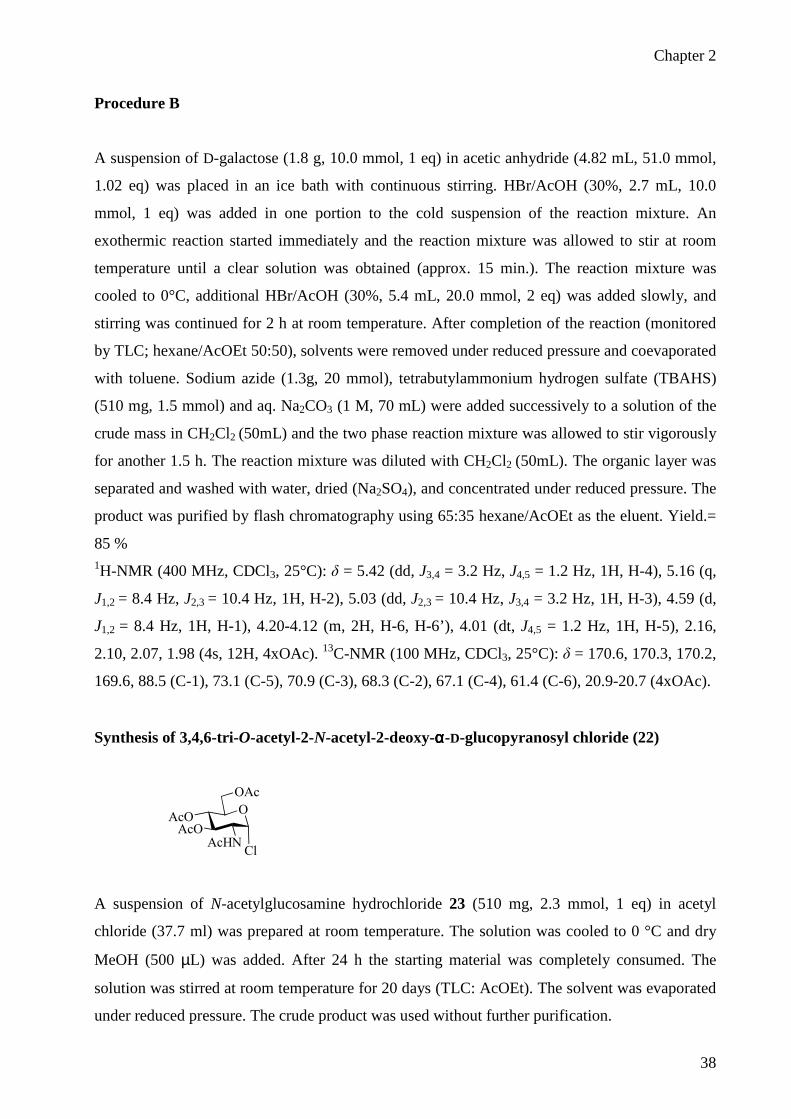
Synthesis of 3,4,6-tri-O-acetyl-2-N-acetyl-2-deoxy-αααα-D-glucopyranosyl chloride (22)
O
AcHNAcO
AcO
OAc
Cl
A suspension of N-acetylglucosamine hydrochloride 23 (510 mg, 2.3 mmol, 1 eq) in acetyl
chloride (37.7 ml) was prepared at room temperature. The solution was cooled to 0 °C and dry
MeOH (500 µL) was added. After 24 h the starting material was completely consumed. The
solution was stirred at room temperature for 20 days (TLC: AcOEt). The solvent was evaporated
under reduced pressure. The crude product was used without further purification.

Chapter 2
39
1H-NMR (400 MHz, CDCl3, 25°C): δ = 6.20 (d, J1,2 = 3.6 Hz, 1H, H-1), 5.89 (d, J2-NH = 7.3 Hz,
1H, NH), 5.34 (dd, J2,3 = J3,4 = 9.5 Hz, 1H, H-3), 5.23 (dd, J3,4 = J4,5 = 9.5 Hz, 1H, H-4), 4.55 (m,
1H, H-2), 4.32-4.25 (m, 2H, H-5, H-6), 4.14 (m, 1H, H-6’), 2.11, 2.06 (2), 1.99 (4s, 12H,
4xOAc). 13C-NMR (100 MHz, CDCl3, 25°C): δ = 171.4, 170.5, 170.1, 169.1, 93.6, 70.9, 70.1,
66.9, 61.2, 53.5, 23.1, 20.7, 20.5.
Synthesis of 3,4,6-tri-O-acetyl -2-N-acetyl-2-deoxy-ββββ-D-glucopyranosyl azide (21)
Procedure A
A solution of 3,4,6-tri-O-acetyl-2-N-acetyl-2-deoxy-α-D-glucopyranosyl chloride 22 (25.6 mg,
0.07 mmol, 1 eq) containing sodium azide (13.4 mg, 0.21 mmol, 3 eq) and tetrabutylammonium
hydrogensulfate (23.8 mg, 0.07 mmol, 1 eq) in CH2Cl2 (256 µL) was prepared at room
temperature. Saturated NaHCO3 (256 µL) was added and the mixture was stirred vigorously for
1 h; the reaction was monitored by TLC (AcOEt). The mixture was diluted with AcOEt and
washed with water, saturated NaHCO3 and brine. The organic layer was dried over Na2SO4,
filtered and the solvent was evaporated under reduced pressure. Yield.= 91 %
Procedure B
At room temperature and under nitrogen, a solution of 3,4,6-tri-O-acetyl-2-N-acetyl-2-deoxy-α-
D-glucopyranosyl chloride 22 (18.7 mg, 0.051 mmol, 1 eq) containing sodium azide (10.0 mg,
0.153 mmol, 3 eq) in dry dimethylformamide (510 µL) was prepared. The mixture was heated to
70 °C and stirred for 3 h. The reaction was monitored by TLC (AcOEt). The solvent was then
evaporated under reduced pressure. The residue was taken up with AcOEt and washed with
water. The organic layer was dried over Na2SO4, filtered and the solvent was evaporated under
reduced pressure. Yield = 90 % 1H-NMR (400 MHz, CDCl3, 25°C): δ = 5.86 (d, J2-NH = 8.8 Hz, 1H, NH), 5.27 (dd, J2,3 = J3,4 =
9.8 Hz, 1H, H-3), 5.11 (dd, J3,4 = J4,5 = 9.8 Hz, 1H, H-4), 4.79 (d, J1,2 = 9.2 Hz, 1H, H-1), 4.28

Chapter 2
40
(dd, J5,6 = 4.7 Hz, J6,6’ = 12.4 Hz, 1H, H-6), 3.75 (dd, J5,6’ = 1.5 Hz, J6,6’ = 12.4 Hz, 1H, H-6’),
3.94 (m, 1H, H-2), 4.32-3.82 (m, 1H, H-5), 2.11, 2.05, 2.04, 1.99 (4s, 12H, 4xOAc). 13C-NMR (100 MHz, CDCl3, 25°C): δ = 170.9, 170.7, 170.5, 169.3, 88.4, 73.9, 72.2, 68.1, 61.9,
54.2, 23.2, 20.7, 20.6, 20.5.
General procedure for the synthesis of 1,2,3,5-tetra-O-acetyl-ββββ-L-arabinopyranose (26) and
1,2,3,5-tetra-O-acetyl-ββββ-D-ribopyranose (27)
Acetic anhydride (10 eq) and a catalytic amount of N,N-dimethylaminopyridine were added, at
room temperature, to a solution of substrate (1 eq) in pyridine dried on molecular sieves (0.1 M).
The solution was stirred for 24 h and then was concentrated in vacuo. The residue was dissolved
in AcOEt and washed with aqueous 5 % HCl, aqueous 5 % NaHCO3 and water. The organic
layer was dried over Na2SO4 and concentrated to give the product in quantitative yield. The
crude product was used without further purification.
General procedure for the synthesis of 2,3,5-tri-O-acetyl-ββββ-L-arabinopyranosyl azide (28)
and 2,3,5-tri-O-acetyl-ββββ-D-ribopyranosyl azide (29)
Trimethylsilyl azide (1.4 eq) and tin tetrachloride (0.3 eq) were added, at room temperature and
under nitrogen, to a solution of substrate 26 or 27 (1 eq) in dry CH2Cl2 (0.5 M). The reaction
mixture was stirred at room temperature and the reaction was monitored by TLC (60:40
hexane/AcOEt). After 24 h CH2Cl2 was added and the solution was washed with saturated
Na2CO3 and then with water. The organic layer was dried over Na2SO4, filtered and evaporated
under reduced pressure.
2,3,4-tri-O-acetyl-ββββ-L-arabinopyranosyl azide (28)
The compound was purified by flash chromatography (hexane/acetone 80:20) yield = 75 %. 1H-NMR (400 MHz, CDCl3, 25°C): δ = 5.29 (ddd, J3,4 = 3.6 Hz, J4,5 = 1.6 Hz, J4,5’ = 2.8 Hz, 1H,
H-4), 5.15 (dd, J1,2= 8 Hz, J2,3 = 9.6 Hz, 1H, H-2), 5.05 (dd, J2,3 = 9.6 Hz, J3,4 = 3.6 Hz, 1H, H-3),

Chapter 2
41
4.58 (d, J1,2= 8 Hz, 1H, H-1), 4.11 (dd, J4,5 = 2.8 Hz, J5,5’ = 13.2 Hz, 1H, H-5), 3.73 (dd, J4,5’ =
1.6 Hz, J5,5’ = 13.2 Hz, 1H, H-5’), 2.16, 2.09, 2.02 (3s, 9H, 3xOAc). 13C-NMR (100 MHz,
CDCl3, 25°C): δ = 170.4, 170.2, 169.6, 88.7 (C-1), 70.4 (C-3), 68.6 (C-2), 67.8 (C-4), 65.8 (C-5),
21.1-20.8 (3xOAc). NOESY (400 MHz, CDCl3, 25°C): contact between H-1/H-3. ESI-MS: m/z
324.1 (M+Na ).
2,3,4-tri-O-acetyl-ββββ-D-ribopyranosyl azide (29)
The compound was purified by flash chromatography (hexane/acetone 80:20) yield = 65 %. 1H-NMR (400 MHz, CDCl3, 25°C): δ = 5.48 (t, J2,3 = 3.2 Hz, 1H, H-3), 5.15-5.06 (m, 2H, H-1,
H-4), 4.86 (dd, J1,2 = 6.4 Hz, J2,3 = 3.2 Hz, 1H, H-2), 4.07 (dd, J4,5’ = 4 Hz, J5,5’ = 12 Hz,1H, H-5),
3.85 (dd, J4,5’ = 3.2 Hz, J5,5’ = 12 Hz, 1H, H-5’), 2.11, 2.08, 2.06 (3s, 9H, 3xOAc). ESI-MS: m/z
324.1 (M+Na ).
2,3,4-tri-O-acetyl-β-L-arabinopyranosyl azide 1H-NMR (400 MHz, CDCl3, 25°C)

Chapter 2
42
Synthesis of 2,3,4,6-tetra-O-acetyl-ββββ-D-glucopyranosyl chloride (30)
Aluminium trichloride (85.4 mg, 0.64 mmol, 0.5 eq) was added, at room temperature and under
argon, to a solution of 1,2,3,4,6-penta-O-acetyl-β-D-glucopyranose 15 (500 mg, 1.28 mmol, 1
eq) in dry CH2Cl2 (2.56 mL, 0.5 M). Aluminium trichloride gradually disappeared and was
replaced by a fine white precipitate. After 2 h, the mixture was filtered into a large volume (50
mL) of dry hexane. The resulting white precipitate was filtered on celite and washed with dry
CH2Cl2 (2.5 mL) and 60:40 hexane/AcOEt (50 mL). The solvent was evaporated under reduced
pressure and the crude product was used without further purification.
Synthesis of 2,3,4,6-tetra-O-acetyl-αααα-D-glucopyranosyl azide (2)
2,3,4-tri-O-acetyl-β-L-ribopyranosyl azide 1H-NMR (400 MHz, CDCl3, 25°C)

Chapter 2
43
At room temperature and under nitrogen, trimethylsilyl azide (108 µL, 0.820 mmol, 1.4 eq) and
tetrabutylammonium fluoride (1 M in THF, 820 µL, 1.4 eq) were added to a solution of 2,3,4,6-
tetra-O-acetyl-β-D-glucopyranosyl chloride 30 (215 mg, 0.586 mmol, 1 eq) in dry THF (8.2 mL,
0.1 M). The solution was heated to 65 °C and stirred for 30 h. The reaction was monitored by
TLC (60:40 hexane/AcOEt). The solvent was evaporated under reduced pressure and the crude
was purified by flash chromatography using 65:35 hexane/AcOEt as the eluent.
Yield = 69 % (over 2 steps), α/β ratio 89:11 1H-NMR (400 MHz, CDCl3, 25°C): δ = 5.62 (d, J1,2 = 4.3 Hz, 1H, H-1), 5.40 (dd, J2,3 = 10 Hz,
J3,4 = 9.8 Hz, 1H, H-3), 5.06 (dd, J3,4 = J4,5 = 9.8 Hz, 1H, H-4), 4.96 (dd, J1,2 = 4.3 Hz, J2,3 = 10
Hz, 1H, H-2), 4.29 (dd, J5,6 = 4.7 Hz, J6,6’ = 12.4 Hz, 1H, H-6), 4.19-4.12 (m, 2H, H-5 and H-6’),
2.11, 2.10, 2.05, 2.03 (4s, 12H, 4xOAc). 13C-NMR (100 MHz, CDCl3, 25°C): δ = 170.5, 169.9, 169.4, 86.2 (C-1), 70.1 (C-5), 69.6 (C-4),
69.5 (C-2), 67.9 (C-3), 61.5 (C-6), 20.6, 20.6, 20.5, 20.5 (4xOAc).
Synthesis of 2,3,4,6-tetra-O-acetyl-αααα-D-galactopyranosyl azide (31)
See procedure for the synthesis of 2,3,4,6-tetra-O-acetyl-α-D-glucopyranosyl azide 2. The
compound was purified by flash chromatography (hexane/AcOEt 60:40) Yield = 62 % (over 2
steps), α/β ratio 75:25 1H-NMR (400 MHz, CDCl3, 25°C): δ = 5.65 (d, J1,2 = 3.6 Hz, 1H, H-1), 5.45 (dd, J3,4 = 3 Hz,
J4,5 = 1.2 Hz, 1H, H-4), 5.24 (dd, J2,3 = 10.8 Hz, J3,4 = 3 Hz, 1H, H-3), 5.19 (dd, J1,2 = 3.6 Hz, J2,3
= 10.8 Hz, 1H, H-2), 4.35 (dt, J4,5 = 1.2 Hz, 1H, H-5), 4.15-4.07 (m, 2H, H-6, H-6’), 2.14, 2.10,
2.06, 1.99 (4s, 12H, 4xOAc). 13C-NMR (100 MHz, CDCl3, 25°C): δ = 170.5, 170.3, 170.1,
169.9, 86.9 (C-1), 68.7 (C-5), 67.8 (C-4), 67.5 (C-2), 67.4 (C-3), 61.6 (C-6), 21.2, 20.8, 20.7
(4xOAc).

Chapter 2
44
2,3,4-tri-O-acetyl-αααα-L-arabinopyranosyl azide (32)
See procedure for the synthesis of 2,3,4,6-tetra-O-acetyl-α-D-glucopyranosyl azide 2. The
compound was purified by flash chromatography (hexane/AcOEt 70:30). 1H-NMR (400 MHz, CDCl3, 25°C): δ = 5.59 (d, J1,2 = 3.2 Hz, 1H, H-1), 5.33 (q, J4,5 = 1.4 Hz,
J4,5’ = 2.4 Hz, 1H, H-4), 5.27-5.20 (m, 2H, H-2, H-3), 4.08 (dd, J4,5 = 1.4 Hz, J5,5’ = 13 Hz, 1H,
H-5), 3.80 (dd, J4,5’ = 2.4 Hz, J5,5’ = 13 Hz, 1H, H-5’), 2.14, 2.11, 2.02 (3xOAc). 13C-NMR (100
MHz, CDCl3, 25°C): δ = 170.4, 170.3, 170, 87.4 (C-1), 68.7 (C-4), 68, 67.1, 62.7 (C-5), 21.1-
20.8 (3xOAc).
2,3,4-tri-O-acetyl-α-L-arabino-pyranosyl azide 1H-NMR (400 MHz, CDCl3, 25°C)

Chapter 2
45
2,3,4-tri-O-acetyl-αααα-D-fucosyl azide (18)
Obtained as by-prodouct from the synthesis of the β-fucosyl azide. Yield = 10% 1H-NMR (400 MHz, CDCl3, 25°C): δ = 5.59 (d, J1,2 = 4 Hz, 1H, H-1), 5.29 (dd, J3,4 = 3.2 Hz,
J4,5 = 1.2 Hz, 1H, H-4), 5.24 (dd, J2,3 = 10.8 Hz, J3,4 = 3.2 Hz, 1H, H-3), 5.18 (dd, J1,2 = 4 Hz, J2,3
= 10.8 Hz, 1H, H-2), 4.27 (q, J5,6 = 6.4 Hz, 1H, H-5), 2.15, 2.01, 1.98 (3s, 9H, 3xOAc), 1.17 (d,
J5,6 = 6.4 Hz, 3H, H-6). 13C-NMR (100 MHz, CDCl3, 25°C): δ = 170.6, 170.4, 170, 87.2 (C-1),
70.8 (C-4), 67.8, 67.7, 67.1 (C-5), 20.9-20.8 (3xOAc), 16.1 (C-6).
Synthesis of 1,2,3,5,6-penta-O-acetyl-ββββ-D-galactofuranose (35).
2,3,4-tri-O-acetyl-α-D-fucopyranosyl azide 1H-NMR (400 MHz, CDCl3, 25°C)

Chapter 2
46
D-Galactose (10g, 5,5 mmol, 1 eq) was heated with pyridine (150 mL) for 1 h at 100°C. The
temperature was lowered to 60°C, and acetic anhydride (33.12 mL, 35.2 mmol, 6.4 eq) was
added dropwise. The mixture was kept for 1.5 h at 60°C, and, after 24 h at room temperature, it
was quenched into ice-water (precipitation of penta-O-acetyl-β-D-galactopyranose). The solution
was extracted with AcOEt (2x250 mL) and washed with aqueous 5 % HCl, aqueous 5 %
NaHCO3 and water. The organic layer was dried over Na2SO4, filtered and evaporate under
reduced pressure to give an orange oil. The oil was dissolved in MeOH, then water was added
until the cloud point was reached. The solution was cooling at 4°C overnight, and the resulting
white solid was filtered (penta-O-acetyl-β-D-galactopyranose); the solvent was evaporated under
reduced pressure, the oil was dissolved in isopropanol, then water was added until the cloud
point was reached, and the resulting white solid was filtered: yield = 36 % 1H-NMR (400 MHz, CDCl3, 25°C): δ = 6.18 (s, 1H, H-1), 5.36 (m, 1H, H-5), 5.18 (s, 1H, H-4),
5.08 (d, J = 5.6 Hz, 1H), 4.38-4.31 (m, 2H, H-6), 4.24-4.19 (m, 1H, H-6’), 2.12-2.03 (m, 15H,
5xOAc). 13C-NMR (100 MHz, CDCl3, 25°C): δ = 170.7, 170.2, 169.9, 169.6, 169.2, 99.3 (C-1),
82.3, 80.7 (C-4), 76.5, 69.4 (C-5), 62.7 (C-6), 21.1-20.8 (5xOAc).
OAc
H OAc
AcO HO
AcOAcO
H
H
1,2,3,5,6-penta-O-acetyl-β-D-galactofuranose 1H-NMR (400 MHz, CDCl3, 25°C)

Chapter 2
47
Synthesis of 2,3,5,6-tetra-O-acetyl-ββββ-D-galactofuranosyl azide (36).
Trimethylsilyl azide (154 µL, 1.16 mmol, 1.4 eq) and tin tetrachloride (29.1 µL, 0.25 mmol, 0.3
eq) were added, at room temperature and under nitrogen, to a solution of 1,2,3,4,6-penta-O-
acetyl-D-galactofuranose 33 (324 mg, 0.83 mmol, 1 eq) in dry CH2Cl2 (3.3 mL, 0.5 M). The
reaction mixture was stirred at room temperature and the reaction was monitored by TLC (60:40
hexane/AcOEt). After 24 h CH2Cl2 was added and the solution was washed with saturated
Na2CO3 and then with water. The organic layer was dried over Na2SO4, filtered and evaporated
under reduced pressure. The product was purified by flash chromatography using 60:40
hexane/AcOEt as the eluent. Yield = 90 % 1H-NMR (400 MHz, CDCl3, 25°C): δ = 5.42 (s, 1H, H-1), 5.40-5.35 (m, 1H, H-5), 5.05 (dd, J2,3
= 5.2 Hz, J3,4 = 2.8 Hz, 1H, H-3), 4.95 (t, J3,4 = 2.8 Hz, 1H, H-4), 4.39-4.32 (m, 2H, H-2, H-6),
4.19 (dd, J5,6’ = 6.8 Hz, J6,6’ = 11.6 Hz, 1H, H-6’), 2.16, 2.14, 2.12, 2.06 (4s, 12H, 4xOAc). 13C-
NMR (100 MHz, CDCl3, 25°C): δ = 170.6, 170.1, 169.9, 169.7, 94.3 (C-1), 82.2 (C-2), 81.1 (C-
4), 76.5 (C-3), 69.4 (C-5), 62.5 (C-6), 20.9-20.7 (4xOAc). ESI-MS: m/z 396.2 (M+Na ).
N3
H OAc
AcO HO
AcOAcO
H
H
2,3,5,6-tetra-O-acetyl-β-D-galactofuranosyl azide 1H-NMR (400 MHz, CD3OD, 25°C)

Chapter 2
48
2,3,5-tri-O-acetyl-αααα-L-arabinofuranosyl azide (40)
See procedure for the synthesis of 2,3,5,6-tetra-O-acetyl-β-D-galactofuranosyl azide 36. The
compound was purified by flash chromatography (hexane/acetone 80:20 or crystallization in
hexane) Yield = 80 % 1H-NMR (400 MHz, CDCl3, 25°C): δ = 5.45 (s, 1H, H-1), 5.02 (dd, J = 2.8 Hz, J = 1.2 Hz, 1H,
H-3), 4.99 (s, 1H, H-2), 4.45-4.38 (m, 2H, H-4, H-5), 4.27-4.20 (m, 1H, H-5’), 2.13, 2.12, 2.10
(3s, 9H, 3xOAc). 13C-NMR (100 MHz, CDCl3, 25°C): δ = 170.7, 170.2, 169.7, 94.5 (C-1), 82.6
(C-4), 78.3 (C-3), 81.1 (C-2), 77.1 (C-3), 63.2 (C-5), 21-20.9 (3xOAc).
N3AcO H
H OAcOAcO
2,3,5-tri-O-acetyl-α-L-arabinofuranosyl azide 1H-NMR (400 MHz, CD3OD, 25°C)

Chapter 2
49
Synthesis of 2,3,5-tri-O-acetyl-ββββ-D-ribofuranosyl azide (41)
See procedure for the synthesis of 2,3,5,6-tetra-O-acetyl-β-D-galactofuranosyl azide 36. The
compound was purified by flash chromatography (hexane/acetone 80:20) Yield = 94 % 1H-NMR (400 MHz, CDCl3, 25°C): δ = 5.35 (d, J1,2 = 2.0 Hz, 1H, H-1), 5.32 (dd, J2,3 = 4.8 Hz,
J3,4 = 6.8 Hz, 1H, H-3), 5.13 (dd, J1,2 = 2 Hz, J2,3 = 4.8 Hz, 1H, H-2), 4.41 (dd, J4,5 = 3.2 Hz, J5,5’
= 12 Hz, 1H, H-5), 4.35 (ddd, J3,4 = 6.8, Hz, J4,5 = 3.2 Hz, J4,5’ = 4.4 Hz, 1H, H-4), 4.14 (dd, J4,5’
= 4.4 Hz, J5,5’ = 12 Hz, 1H, H-5’),2.12, 2.11, 2.07 (3s, 9H, 3xOAc). 13C-NMR (100 MHz, CDCl3,
25°C): δ = 170.7, 169.7, 169.5, 92.8 (C-1), 79.5 (C-4), 74.6 (C-2), 70.6 (C-3), 63.2 (C-5), 20.8,
20.7, 20.6 (3xOAc). NOESY (400 MHz, CDCl3, 25°C): contact between H-1/H-4.
General procedure for the deprotection of peracetylated glycosyl azides.
A solution of NaOMe 0.1M in dry methanol (0.5 eq) was added, at room temperature and under
nitrogen, to a solution of peracetylated glycosyl azides (1eq) in dry MeOH (0.1 M). The mixture
2,3,5-tri-O-acetyl-β-D-ribofuranosyl azide 1H-NMR (400 MHz, CD3OD, 25°C)
N3
AcO OAc
H HOAcO

Chapter 2
50
was stirred at room temperature. After 45 minutes TLC monitoring (eluents: hexane/AcOEt
50:50 and CHCl3:MeOH 80:20) showed total consumption of the starting material, the acid resin
Amberlyst IRA 120 H+ was added. The mixture was stirred for 30 minutes (pH = 3). The resin
was filtered and washed with MeOH, the solvent was removed under reduced pressure. The
product, isolated in quantitative yield, was used without further purification.
ββββ-D-glucopyranosyl azide (10)
1H-NMR (400 MHz, CD3OD, 25°C): δ = 4.48 (d, J1,2 = 8.8 Hz, 1H, H-1), 3.87 (dd, J5,6 = 2 Hz,
J6,6’ = 12 Hz, 1H, H-6), 3.67 (dd, J5,6’ = 5.6 Hz, J6,6’ = 12 Hz, 1H, H-6’), 3.38-3.31 (m, 2H, H-3,
H-5), 3.30-3.25 (m, 1H, H-4), 3.13 (t, J1,2 = 8.8 Hz, 1H, H-2). 13C-NMR (100 MHz, CD3OD,
25°C): δ = 92.2 (C-1), 80.3 (C-5), 78.3 (C-3), 74.9 (C-2), 71.3 (C-4), 62.7 (C-6).
ββββ-D-fucosyl azide (50)
1H-NMR (400 MHz, CD3OD, 25°C): δ = 4.41 (d, J1,2 = 8 Hz, 1H, H-1), 3.73 (q, J5,6 = 6.4 Hz,
1H, H-5), 3.64 (d, J4,5 = 1.6 Hz, 1H, H-4), 3.47-3.41 (m, 2H, H-2, H-3), 1.29 (d, J5,6 = 6.4 Hz,
3H, H-6). 13C-NMR (100 MHz, CD3OD, 25°C): δ = 92.5 (C-1), 75.3, 74.4 (C-5), 73 (C-4), 71.9,
16.9 (C-6).
ββββ-D-galactopyranosyl azide (43)

Chapter 2
51
1H-NMR (400 MHz, CD3OD, 25°C): δ = 4.42 (d, J1,2 = 8 Hz, 1H, H-1), 3.86 (dd, J4,5 = 2 Hz, 1H,
H-4), 3.77 (dd, J5,6 = 6.4 Hz, J6,6’ = 11.6 Hz, 1H, H-6), 3.70 (dd, J5,6’ = 5.2 Hz, J6,6’ = 11.6 Hz,
1H, H-6’), 3.61 (dt, J4,5 = 2 Hz, J5,6 = 6.4 Hz, J5,6’ = 5.2 Hz, 1H, H-5), 3.52-3.46 (m, 2H, H-2, H-
3). 13C-NMR (100 MHz, CD3OD, 25°C): δ = 92.8 (C-1), 79 (C-5), 75.1, 72.1, 70.4 (C-4), 62.6
(C-6).
2-N-acetyl-2-deoxy-ββββ-D-glucopyranosyl azide (42)
1H-NMR (400 MHz, CD3OD, 25°C): δ = 4.48 (d, J1,2 = 9.2 Hz, 1H, H-1), 3.88 (dd, J5,6 = 1.6 Hz,
J6,6’ = 12 Hz, 1H, H-6), 3.72-3.64 (m, 2H, H-2, H-6), 3.45 (t, J = 10 Hz, 1H, H-3), 3.37-3.32 (m,
2H, H-4, H-5), 1.98 (s, 3H, OAc). 13C-NMR (100 MHz, CD3OD, 25°C): δ = 173.9, 90.3 (C-1),
80.5 (C-5), 75.9 (C-3), 71.8 (C-4), 62.8 (C-6), 56.9 (C-2), 23 (CH3).
2-N-acetyl-2-deoxy-β-D-glucopyranosyl azide
1H-NMR (400 MHz, CD3OD, 25°C)

Chapter 2
52
ββββ-L-arabinopyranosyl azide (48)
1H-NMR (400 MHz, CD3OD, 25°C): δ = 4.44 (t, J1,2 = 8 Hz, 1H, H-1), 3.95 (dd, J4,5 = 2.4 Hz,
J5,5’ = 12.4 Hz, 1H, H-5), 3.88 (d, J4,5’ = 1.6 Hz, 1H, H-4), 3.67 (dd, J4,5’ = 1.6 Hz, J5,5’ = 12.4 Hz,
1H, H-5’), 3.59-3.54 (m, 2H, H-2, H-3). 13C-NMR (100 MHz, CD3OD, 25°C): δ = 92.8 (C-1),
74.5, 72.1, 70 (C-4), 69.7 (C-5).
ββββ-D-ribopyranosyl azide (45)
1H-NMR (400 MHz, CD3OD, 25°C): δ = 5.04 (d, J1,2 = 6 Hz, 1H, H-1), 3.99 (t, J = 2.4 Hz, 1H,
H-4), 3.93-3.79 (m, 3H, H-3, H-5, H-5’), 3.48 (dd, J1,2 = 6 Hz, J = 2.8 Hz, 1H, H-2). 13C-NMR
(100 MHz, CD3OD, 25°C): δ = 90.8 (C-1), 72.2 (C-2), 70 (C-4), 69.4 (C-3), 66.5 (C-5).
β-L-arabinopyranosyl azide 1H-NMR (400 MHz, CD3OD, 25°C)
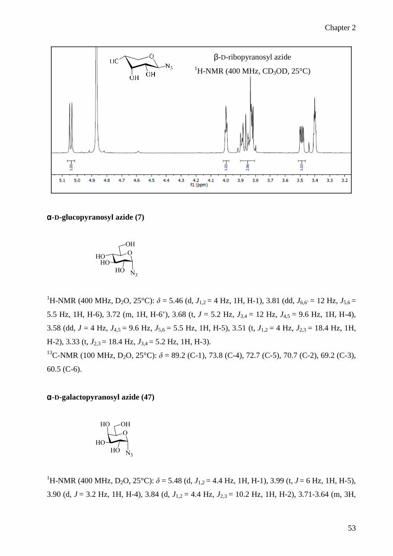
Chapter 2
53
αααα-D-glucopyranosyl azide (7)
O
HO
HOHO
OH
N3
1H-NMR (400 MHz, D2O, 25°C): δ = 5.46 (d, J1,2 = 4 Hz, 1H, H-1), 3.81 (dd, J6,6’ = 12 Hz, J5,6 =
5.5 Hz, 1H, H-6), 3.72 (m, 1H, H-6’), 3.68 (t, J = 5.2 Hz, J3,4 = 12 Hz, J4,5 = 9.6 Hz, 1H, H-4),
3.58 (dd, J = 4 Hz, J4,5 = 9.6 Hz, J5,6 = 5.5 Hz, 1H, H-5), 3.51 (t, J1,2 = 4 Hz, J2,3 = 18.4 Hz, 1H,
H-2), 3.33 (t, J2,3 = 18.4 Hz, J3,4 = 5.2 Hz, 1H, H-3). 13C-NMR (100 MHz, D2O, 25°C): δ = 89.2 (C-1), 73.8 (C-4), 72.7 (C-5), 70.7 (C-2), 69.2 (C-3),
60.5 (C-6).
αααα-D-galactopyranosyl azide (47)
O
HO
HO
HO OH
N3
1H-NMR (400 MHz, D2O, 25°C): δ = 5.48 (d, J1,2 = 4.4 Hz, 1H, H-1), 3.99 (t, J = 6 Hz, 1H, H-5),
3.90 (d, J = 3.2 Hz, 1H, H-4), 3.84 (d, J1,2 = 4.4 Hz, J2,3 = 10.2 Hz, 1H, H-2), 3.71-3.64 (m, 3H,
β-D-ribopyranosyl azide 1H-NMR (400 MHz, CD3OD, 25°C)

Chapter 2
54
H-3, H-6, H-6’). 13C-NMR (100 MHz, D2O, 25°C): δ = 89.4 (C-1), 73.1 (C-5), 69.2 (C-3), 69
(C-4), 67.6 (C-2), 61.2 (C-6).
αααα-L-arabinopyranosyl azide (51)
1H-NMR (400 MHz, CD3OD, 25°C): δ = 5.32 (d, J1,2 = 4 Hz, 1H, H-1), 4.02-3.93 (m, 3H, H-2,
H-5), 3.81-3.77 (m, 2H, H-5’). 13C-NMR (100 MHz, CD3OD, 25°C): δ = 91.5 (C-1), 70.9, 70.5,
69.5, 66.4 (C-5).
αααα-D-fucosyl azide (44)
α-L-arabinopyranosyl azide 1H-NMR (400 MHz, CD3OD, 25°C)

Chapter 2
55
1H-NMR (400 MHz, CD3OD, 25°C): δ = 5.32 (d, J1,2 = 4.4 Hz, 1H, H-1), 4.03 (q, J5,6 = 6.8 Hz,
1H, H-5), 3.86 (dd, J1,2 = 4.4 Hz, J2,3 = 10 Hz, 1H, H-2), 3.66 (d, J3,4 = 3.2 Hz, 1H, H-4), 3.62
(dd, J2,3 = 10 Hz, J3,4 = 3.2 Hz, 1H, H-3), 1.24 (d, J5,6 = 6.8 Hz, 3H, H-6). 13C-NMR (100 MHz,
CD3OD, 25°C): δ = 92.4 (C-1), 73.4 (C-4), 71.5 (C-3), 70.2 (C-5), 69.5 (C-2), 16.8 (C-6).
ββββ-D-galactofuranosyl azide (37)
1H-NMR (400 MHz, D2O, 25°C): δ = 5.31 (d, J1,2 = 2.8 Hz, 1H, H-1), 4.06 (ABX system, part A
dd, J2,3 = 4 Hz, J3,4 = 10 Hz 1H, H-3), 4.01 (ABX system, part B, dd, J3,4 = 10.4 Hz, J4,5 = 4.4 Hz,
1H, H-4), 3.94 (ABX system, part X, dd, J1,2 = 2.8 Hz, J2,3 = 3.6 Hz, 1H, H-2), 3.80-3.73 (ddd,
J4,5 = 4.4 Hz, J5,6’ = 7.2 Hz, J5,6 = 4.8 Hz, 1H, H-5), 3.64 (dd, J5,6 = 4.8 Hz, J6,6’ = 11.6 Hz, 1H, H-
6), 3.57 (dd, J5,6’ = 7.2 Hz, J6,6’ = 11.6 Hz, 1H, H-6’). 13C-NMR (100 MHz, D2O, 25°C): δ = 95.3
(C-1), 84.2 (C-4), 80.6 (C-2), 76.4 (C-3), 70.7 (C-5), 62.6 (C-6). [α]D25 = -153 (c 1, MeOH).
NOESY (400 MHz, D2O, 25°C): contact between H-1/H-3.
α-D-fucopyranosyl azide 1H-NMR (400 MHz, CD3OD, 25°C)

Chapter 2
56
αααα-L-arabinofuranosyl azide (49)
1H-NMR (400 MHz, CD3OD, 25°C): δ = 5.25 (d, J1,2 = 2 Hz, 1H, H-1), 4.11 (ddd, J3,4 = 5.6 Hz,
J4,5 = 3.6 Hz, J4,5’ = 5.2 Hz, 1H, H-4), 3.99 (dd, J2,3 = 3.6 Hz, J3,4 = 5.6 Hz, 1H, H-3), 3.93 (dd,
J1,2 = 2 Hz, J2,3 = 3.6 Hz, 1H, H-2), 3.80 (dd, J4,5 = 3.6 Hz, J5,5’ = 12 Hz, 1H, H-5), 3.70 (dd, J4,5’
= 5.2 Hz, J5,5’ = 12 Hz, 1H, H-5’). 13C-NMR (100 MHz, CD3OD, 25°C): δ = 97.9 (C-1), 87.8 (C-
4), 83.2 (C-2), 78.4 (C-3), 63 (C-5).
β-D-galactofuranosyl azide 1H-NMR (400 MHz, D2O, 25°C)

Chapter 2
57
ββββ-D-ribofuranosyl azide (53)
1H-NMR (400 MHz, CD3OD, 25°C): δ = 5.19 (d, J1,2 = 1.6 Hz, 1H, H-1), 4.04 (dd, J2,3 = 4.8 Hz,
J3,4 = 6.8 Hz, 1H, H-3), 3.97 (ddd, J3,4 = 6.8 Hz, J4,5 = 3.2 Hz, J4,5’ = 6 Hz, 1H, H-4), 3.81 (dd, J1,2
= 1.6 Hz, J2,3 = 4.8 Hz, 1H, H-2), 3.76 (dd, J4,5 = 3.2 Hz, J5,5’ = 12 Hz, 1H, H-5), 3.60 (dd, J4,5’ =
6 Hz, J5,5’ = 12 Hz, 1H, H-5’). 13C-NMR (100 MHz, CD3OD, 25°C): δ = 96.7 (C-1), 85.7 (C-4),
76.6 (C-2), 72.2 (C-3), 63.8 (C-5).
α-L-arabinofuranosyl azide 1H-NMR (400 MHz, CD3OD, 25°C)

Chapter 2
58
β-D-ribofuranosyl azide 1H-NMR (400 MHz, CD3OD, 25°C)

Chapter 2
59
2.7 Reference
1 Paulsen H. Adv. Carbohydr. Chem. 1971, 26, 127. 2 Martichonok, V.; Whitesides, G. M. J. Org. Chem. 1996, 61, 1702-1706. 3 Györgydeák, Z.; Szitagyi, L.; Paulsen H. J. Carb. Chem. 1993, 12, 139-163. 4 Kumar, R.; Tiwari, P.; Maulik, P. R.; Misra, A. K. Eur. J. Org. Chem. 2006, 74-79. 5 Tanaka, T.; Nagai, H.; Noguchi, M.; Kobayashi, A.; Shoda, S. Chem. Commun. 2009, 3378-3379. 6 Thamsen, J.; Acta Chem. Scand. 1952, 6, 270. 7 Gouin, S. G.; Kovensky, J. Tetrahendron. Lett. 2007, 48, 2875-2879. 8 Besset, C.; Chambert, S.; Fenet, B.; Queneau, Y. Tetrahendron Lett. 2009, 50, 7043-7047. 9 Tropper, F. D.; Andersson, F. O.; Braun, S.; Roy, R. Synthesis 1992, 618-620. 10 Gudmundsdottir, A. V.; Nitz, M. Org. Lett. 2008, 10, 3461-3463. 11 Lemieux, R. U.; Stevens J. D. Can. J. Chem. 1965, 43, 2059-2070. 12 Lynch, B. M.; Sharma, S. C. Can. J. Chem. 1976, 54, 1029-1038. 13 Korytnyk, W.; Mills J. A. J. Chem. Soc. 1959, 636-649. 14 Soli, E. D.; Manoso, A. S.; Patterson, M. C.; DeShong, P. J. Org. Chem. 1999, 64, 3171-3177. 15 Dedola, S.; Nepogodiev, S. A.; Hughes, D.L.; Field, R. A. Acta Cryst. 2008, C64, 445-446. 16 Bianchi, A.; Bernardi, A. J. Org. Chem. 2006, 71, 4565-4577. 17 D’Accorso, N. B.; Thiel, I. M. E. Carbohydr. Res. 1983, 124, 177-184. 18 Chittenden, G. J. F. Carbohydr. Res. 1972, 25, 35-41. 19 Kam, B.L.; Barascut, J. L.; Imbach, J. L. Carb. Res. 1979, 69, 135-142. 20 Stimac, A.; Kobe, J. Carb. Res. 1992, 232, 359-365.

Chapter 3
Synthesis of different phosphines

Chapter 3
61
In order to develop a stereoselective Staudinger ligation for the synthesis of glycosyl amides, it
was decided to employ the procedure based on the Staudinger reduction-acylation process of the
corresponding glycosyl azides. Our methods to achieve a stereoselective synthesis of glycosyl
amides with traceless Staudinger ligation are described in Chapters 4-6. Before that, the
procedures employed for the synthesis of phosphines are described in the following sections.
3.1 Synthesised phosphines
The traceless Staudinger ligation affords one approach to the synthesis of unnatural α-glycosyl
amides, starting from the corresponding α-azides, because it allows for reduction of the starting
material and immediate trapping of the Staudinger aza-ylide intermediate in an intramolecular
fashion, thus resulting in the direct formation of an amide link. Fast, intramolecular acylation of
the anomeric nitrogen can in many cases prevent epimerisation and give retention of
configuration at the anomeric carbon. In 2006 1 and 2008 2 our laboratory has reported that the
traceless Staudinger ligation of α-glycosyl azides 2, 7 and 8 with diphenylphosphanyl-phenyl
esters 9 in polar aprotic solvents yields α-glycosyl amides with good yields and selectivity
(Scheme 1).
ON3
RORO
RO
OR O
PPh2
O
R'
O
N3
RORO
RO
ORO
PPh2
O
R'A
B
O HN
RORO
RO
OR
O
R'
O
HNRO
RORO
OR
O
R'
O HN
RORO
RO
OR
O
R'
+
R = H, -Bn
R = -Ac
9
+
9
R = H, -Bn, -Ac
2 R = Ac7 R = H8 R = Bn
1 R = Ac10 R = H11 R = Bn
Scheme 1. Traceless Staudinger ligation of glycosyl azides with functionalized phosphine 1, 2

Chapter 3
62
The steric course of the reaction is controlled both by configuration of the starting azide and by
the presence and nature of protecting groups on the pyranose ring. In fact, the O-acetyl azide 2
allows full inversion of the anomeric center and yields the β-amide only. The phosphines 9
employed are air stable and can be easily isolated and purified by flash chromatography.
The process described, however, left various synthetic problems unresolved. The reactivity of α-
glycosyl azides was uniformely low, and the corresponding amides were obtained generally in
modest yields. Moreover, the yields of the ligation appeared to depend critically also on the
nature of the acyl group to be transferred and were specially disappointing for the transfer of
amino acids to α-azides. Thus, in order to further explore the reactivity and stereoselectivity of
ligation in the synthesis of α-glycosyl amides, different acyl phosphines were prepared, trying to
vary the basicity of the P atom and the nature of the phenyl ester leaving group.
3.2 Synthesis of diphenylphosphanyl-phenyl esters
Phosphines 9 had been obtained in the course of a previous thesis3 starting from o-
diphenylphosphinophenol 57. This was prepared in three steps starting from phenol and using a
procedure introduced by Rauchfuss with small modifications.4 Firstly, the hydroxyl group was
protected as the methoxymethyl ether:5 the presence of the MOM-ether both protects and
activates the phenol ring. Secondly, the diphenylphosphino group was attached by ortho
lithiation of the ether 55 (BuLi, hexane, TMEDA) followed by reaction with
chlorodiphenylphosphine (Ph2PCl). Finally, the hydroxyl group was deprotected with HCl in
MeOH to afford 57 (Scheme 2).
Scheme 2. Synthesis of o-diphenylphosphinophenyl ester 57
Interestingly Luo et al. found that treatment of other methoxymethyl-protected phenols bearing
phosphine groups with HCl led to phosphonium salts because of the basicity of the phosphine

Chapter 3
63
moiety.6 As the authors noted, in the previously reported preparation of 57, a base-neutralisation
step was omitted but the crude product was sublimed, which is consistent with known loss of
HCl from phosphonium salts at elevated temperatures. In fact, 57 in our hands exhibited a single 31P resonance at δ = -26.6 ppm, whereas omission of the base-neutralisation step led to the
hydrochloride salt 58 with a 31P resonance at + 21.6 ppm (Figure 1).7
Figure 1. 31P resonance of hydrochloride salt of o-diphenylphosphinophenol 58 (up) and o-
diphenylphosphinophenol 57 (down)
o-Diphenylphosphinophenol 57 was then acylated to give the corresponding functionalised
phosphines which were used for the Staudinger ligation reactions.

Chapter 3
64
3.3 Synthesis of the diphenyl- and dialkyl-phosphanylphenols
Following a similar approach, in the course of this thesis a group of other functionalized
diphenyl- and dialkyl-phosphanylphenols was prepared. This include the diphenylphosphanyl-4-
methoxyphenol 59 (Figure 2),8 diphenylphosphanyl-4-fluorophenol 608 and several novel
phoshines (61, 62, 63 and 64 Figure 2) with various substituents at phosphorus and characterized
by an electronpoor phenol ring.
Figure 2. Set of diphenylphosphanyl-phenols (59 to 64) synthesized in this work
The diphenylphosphanyl-4-methoxyphenol 59 (Scheme 3) is a known compound:8 it was
prepared following exactly the same procedure described for 57 (Scheme 3) 3,4 and the analytical
data were consistent with the reported structure.
Scheme 3. Synthesis of the diphenylphosphanyl-4-methoxyphenol 59

Chapter 3
65
For the synthesis of 60-62 the required fluorinated-methoxymethyl phenyl ethers 68-70 (Scheme
4) were synthesised as usual by the reaction of phenols 71-73 with sodium hydride followed by
treatment with chloromethyl methyl ether (MOM-Cl) (Scheme 4).4 Metalation and reaction with
the appropriate chlorophosphine (Scheme 4) was performed according to Fink8, using diethyl
ether rather than hexane, in the absence of TMEDA and at low temperature -50°C, probably to
avoid ortho metalation respect to the flourus atom.9 Finally, HCl deprotection afforded the
required phosphines 60, 61 and 62.
OMOM
PPh2
71 68 74
y = 94 % y = 47 %
60
OH
PPh2
y = 90 %
OH
F
OMOM
F F F
Ph2PCl, -50°C to r.t.
OMOM
PPh2n-BuLi, Et2O dryNaH, MOM-Cl
0°C to r.t., DMF
72 69 75
y = 96 % y = 34 %
MeOH
61
OH
PPh2
HCl (g)
y = 85 %
OH
CF3
OMOM
CF3 CF3 CF3
OMOM
PPh2
73 70 76
y = 93 % y = 46 %
62
OH
PPh2
y = 70 %
OH OMOM
F3C CF3 F3C CF3 CF3 F3C CF3F3C
NaH, MOM-Cl
0°C to r.t., DMF Ph2PCl, -50°C to r.t.
n-BuLi, Et2O dry MeOH
HCl (g)
MeOH
HCl (g)Ph2PCl, -50°C to r.t.
n-BuLi, Et2O dryNaH, MOM-Cl
0°C to r.t., DMF
Scheme 4. Synthesis of fluorinated-o-diphenylphosphinophenol
Similarly, Fink 8 metalation of fluorinated-methoxymethyl phenyl ether 68 followed by reaction
with commercially available chlorodicyclohexylphosphine or chlorodicyclopentylphosphine,
gave the methoxymethyl o-dicyclohexylphosphinophenyl ether 77 and methoxymethyl o-
dicyclopentylphosphinophenyl ether 78 (Scheme 5), which were deprotected with HCl, as usual,

Chapter 3
66
to yield 79 and 80. Both these phosphines are air stable and can be easily isolated and purified by
flash chromatography.
Scheme 5. Synthesis of the dialkyl-phosphanylphenols 79 and 80
3.4 Acylation of the phosphanylphenols
The phosphanylphenols described above were acylated with standard methods, which vary
depending on the nature of the acyl chain employed. In general, the corresponding acyl chlorides
were used, when commercially available. Benzoates were obtained from benzoic anhydride.
Condensing agents (DCC or EDC) were employed for amino acids.
All the compounds prepared are collected in Table 1, where the method of acylation is indicated.
All other details are reported in the experimental section of this chapter.
Table 1. O-acylphosphanylphenols prepared
Compound Starting
phosphanylphenol R-C=O
Acylation
method
Y
(%)
PPh2O CH3
O
81a
57 CH3CO
Ac2O,
Pyridine,
DMAP
Quant

Chapter 3
67
PPh2O
O
81b
57 CH3(CH2)3CO RCOCl/TEA 96 %
PPh2O
O
81c
57 (CH3)2CHCH2CO RCOCl/TEA 97 %
PPh2O
O
81d
57 (CH3)2CHCO RCOCl/TEA 87 %
PPh2O
O
81e
57 (CH3)2C=CHCO RCOCl/TEA 84 %
PPh2O
O13
81f
57 CH3(CH2)14CO RCOCl/TEA 85 %
PPh2O
O6
81g
57 CH3(CH2)6CO RCOOH/DCC
DMAP 90 %
PPh2
O
O
81h
57 citronellic acid RCOOH/DCC
DMAP 75 %
PPh2O Ph
O
81i
57 PhCO
(PhCO)2O,
Pyridine,
DMAP
Quant

Chapter 3
68
PPh2
O
O
COOMe
NHCbz
81j
57
RCOOH/EDC,
DIPEA,
DMAP
85 %
PPh2O
O
NHCbz
COOMe
81k
57
RCOOH/EDC,
DIPEA,
DMAP
75 %
PPh2O
O
NHCbz
81l
57
RCOOH/DCC
DMAP 82 %
PPh2O CH3
OMeO
82a
59 CH3CO
Ac2O,
Pyridine,
DMAP
Quant
PPh2O
OMeO
82b
59 CH3(CH2)3CO RCOCl/TEA 87 %
PPh2O
OMeO
82c
59 (CH3)3CCO RCOCl/TEA 83 %
PPh2O
OMeO
82d
59 (CH3)2CHCO RCOCl/TEA 82 %
PPh2O
OMeO
82e
59 (CH3)2C=CHCO RCOCl/TEA 84 %

Chapter 3
69
PPh2O
O
COOMe
NHCbzMeO
82j
59
RCOOH/EDC,
DIPEA,
DMAP
75 %
PPh2O CH3
OF
83a
60 CH3CO
Ac2O,
Pyridine,
DMAP
Quant
PPh2O
OF
83b
60 CH3(CH2)3CO RCOCl/TEA 95 %
PPh2O
OF
83e
60 (CH3)2C=CHCO RCOCl/TEA 91 %
PPh2O
O
COOMe
NHCbzF
83j
60
RCOOH/EDC,
DIPEA,
DMAP
85 %
PPh2O
O
NHCbz
COOMe
F83k
60
RCOOH/EDC,
DIPEA,
DMAP
85 %
PPh2O
O
NHCbz
F
83l
60
RCOOH/DCC
DMAP 92 %
PPh2O
O
NHCbz
F
83m
60
RCOOH/DCC
DMAP 90 %

Chapter 3
70
PPh2O
OF
BocN
83l
60
RCOOH/DCC
DMAP 94 %
PPh2O
O
COOMe
NHCbzF3C
84j
61
RCOOH/EDC,
DIPEA,
DMAP
90 %
PPh2O
O
CF3
F3C COOMe
NHCbz
85k
62
RCOOH/EDC,
DIPEA,
DMAP
95 %
P(cyclohexil)2O
O
COOMe
NHCbzF
86j
79
RCOOH/EDC,
DIPEA,
DMAP
80 %
P(cyclopentyl)2O
O
COOMe
NHCbzF
87j
80
RCOOH/EDC,
DIPEA,
DMAP
80 %
3.5 Synthesis of diphenylphosphinothiol ester
The Staudinger ligation has also been proposed for the ligation of peptides en route to the total
chemical synthesis of proteins.10 (Diphenylphosphino)methanethiol is the most efficacious of
known reagents, in this case providing high isolated yields for equimolar couplings in which a
glycine residue is at the nascent junction.
This reagent has been used in the orthogonal assembly of a protein,11 site-specific
immobilization
of peptides and proteins to a surface,12, 13 and synthesis of glycopeptides.14 We prepared it to test
it in the ligation of α-glycosyl azides. The diphenylphosphinothiol ester 91 was synthesised by
treating the chloromethylphosphonic dichloride 88 with 4-methoxyphenylmagnesium bromide

Chapter 3
71
and potassium thioacetate to give phosphine oxide 88. Thus the desired compound 91 was
prepared for reduction with trichlorosilane from corresponding oxide 89 (Scheme 6).10
Scheme 6. Synthesis of phosphinothioester 91
Also in this case, the diphenylphosphinothiol ester 91 was functionalized with valeroyl chloride
to give phosphine 92 (Scheme 7).
Scheme 7. Synthesis of phosphine 92

Chapter 3
72
3.6 Experimental Section
Solvents were dried by standard procedures: dichloromethane, methanol, N,N-
diisopropylethylamine and triethylamine were dried over calcium hydride; N,N-
dimethylformamide, N,N,N’,N’-tetramethylethylenediamine, diethyl ether, chloroform and
pyridine were dried over activated molecular sieves. Reactions requiring anhydrous conditions
were performed under nitrogen. 1H, 13C and 31P-NMR spectra were recorded at 400 MHz on a
Bruker AVANCE-400 instrument. Chemical shifts (δ) for 1H and 13C spectra are expressed in
ppm relative to internal Me4Si as standard. Chemical shifts (δ) for 31P are expressed in ppm
relative to internal H3PO4 as standard. Signals were abbreviated as s, singlet; bs, broad singlet; d,
doublet; t, triplet; q, quartet; m, multiplet. Mass spectra were obtained with a Bruker ion-trap
Esquire 3000 apparatus (ESI ionization). Thin layer chromatography (TLC) was carried out with
pre-coated Merck F254 silica gel plates. Flash chromatography (FC) was carried out with
Macherey-Nagel silica gel 60 (230-400 mesh).
Synthesis of methoxymethyl phenyl ether (55)
O O
At 0 °C and with stirring, phenol 54 (5 g, 53.1 mmol, 1 eq) was added to a slurry of NaH (3.19 g,
79.7 mmol, 1.5 eq 60 % in oil) in dimethylformamide (26.5 mL, 2 M). After 30 minutes,
chloromethyl methyl ether (6.05 mL, 79.7 mmol, 1.5 eq) was added, then the temperature was
allowed to warm up to room temperature. The mixture was stirred and the reaction was
monitored by TLC (8:2 hexane/AcOEt). After 30 minutes, water (106 mL) was added. The
product was exctracted with hexane (3 x 52 mL). The organic layer was dried over Na2SO4 and
concentrated. Yield = 74 %. 1H-NMR (400 MHz, CDCl3, 25°C): δ = 7.38-7.31 (m, 11H, Ph), 7.13 (m, 1H), 6.93 (m, 1H),
6.72 (m, 1H), 5.19 (s, 2H, CH2O), 3.49 (s, 3H, OCH3). 13C-NMR (100 MHz, CDCl3, 25°C): δ =
158.6, 136.9, 134.4, 134.0, 133.7, 130.4, 128.9, 128.7, 128.5, 122.2, 113.6, 113.6, 94.3, 56.1.

Chapter 3
73
Synthesis of methoxymethyl-4-methoxyphenyl ether (66)
O O
MeO
At 0 °C and with stirring, phenol 65 (2.98 g, 24 mmol, 1 eq) was added to a slurry of NaH (90.9
mg, 36 mmol, 1.5 eq) in dimethylformamide (12 mL, 2 M). After 30 minutes, chloromethyl
methyl ether (2.72 mL, 36 mmol, 1.5 eq) was added, then the temperature was allowed to warm
up to room temperature. The mixture was stirred and the reaction was monitored by TLC (80:20
hexane/AcOEt). After 30 minutes, water (60 mL) was added. The product was exctracted with
hexane (3 x 25 mL). The organic layer was dried over Na2SO4 and concentrated. yield = 94 %. 1H-NMR (400 MHz, CDCl3, 25°C): δ = 7.01 (dd, J = 2.4 Hz, J = 7.2 Hz, 2H, ArH), 6.85 (dd, J =
2 Hz, J = 6.8 Hz, 2H, ArH), 5.13 (s, 2H, CH2O), 3.78 (s, 3H, OCH3), 3.49 (s, 3H, CH3).
General procedure for the synthesis of the methoxymethyl-fluorinated phenyl ether
At 0 °C and with stirring, fluorinated phenol 71, 72 and 73 (1 eq) was added to a slurry of NaH
(1.5 eq) in dimethylformamide (2 M). After 30 minutes, chloromethyl methyl ether (1.5 eq) was
added, then the temperature was allowed to warm up to room temperature. The mixture was
stirred and the reaction was monitored by TLC (80:20 hexane/AcOEt). After 30 minutes, water
was added. The product was exctracted with hexane (3 x 50 mL). The organic layer was dried
over Na2SO4 and concentrated.
4-fluorophenyl methoxymethyl ether (68)
O O
F
1H-NMR (400 MHz, CDCl3, 25°C): δ = 7.04-6.98 (m, 4H, Ph), 5.16 (s, 2H, CH2O), 3.51 (s, 3H,
OCH3). Yield = 94 %.

Chapter 3
74
4-trifuoromethylphenyl methoxymethyl ether (69)
O O
F3C
1H-NMR (400 MHz, CDCl3, 25°C): δ = 7.54 (d, J = 8 Hz, 2H, ArH), 7.10 (d, J = 8 Hz, 2H,
ArH), 5.22 (s, 2H, CH2O), 3.48 (s, 3H, OCH3). Yield = 96 %.
3,5-ditrifuoromethylphenyl methoxymethyl ether (70)
O OF3C
CF3
1H-NMR (400 MHz, CDCl3, 25°C): δ = 7.52 (s, 1H, ArH), 7.49 (s, 2H, ArH), 5.26 (s, 2H,
CH2O), 3.50 (s, 3H, OCH3). Yield = 93 %.
Synthesis of methoxymethyl o-diphenylphosphinophenyl ether (56)
PPh2
O O
A solution containing 1.6 M n-BuLi (10 mL, 16 mmol, 1.1 eq) and N,N,N’,N’-
tetramethylendiamine (2.24 mL, 14.9 mmol, 1.03 eq) in dry hexane was added, under nitrogen,
to an ice-cooled solution of methoxymethyl phenyl ether 55 (2 g, 14.5 mmol, 1 eq) in dry hexane
(final concentration: 0.5 M). The temperature was allowed to warm up to room temperature. The
solution became yellow and a pale precipitate formed after 1.5 h. The solution was cooled in ice
and chlorodiphenylphosphine (2.74 mL, 14.5 mmol eq) was added slowly. The resultant solution
was stirred for 16 h at room temperature and the solvent evaporated in vacuo. The residue was
diluted with Et2O and washed with 1 M aqueous Na2HPO4. The organic layer was dried over
Na2SO4 and concentrated to give a white solid. The solid was dissolved in MeOH. After cooling
for 30 minutes, the crystals were collected and recrystallized from CH2Cl2-MeOH; yield = 61 %.

Chapter 3
75
1H-NMR (400 MHz, CDCl3, 25°C): δ = 7.38-7.31 (m, 11H, Ph), 7.13 (m, 1H), 6.93 (m, 1H),
6.72 (m, 1H), 5.06 (s, 2H, CH2O), 3.16 (s, 3H, OCH3). 13C-NMR (100 MHz, CDCl3, 25°C): δ =
158.6, 136.9, 134.4, 134.0, 133.7, 130.4, 128.9, 128.7, 128.5, 122.2, 113.6, 113.6, 94.3, 56.1. 31P-
NMR (161 MHz, CDCl3, 25°C): δ = - 14.8.ppm
Synthesis of methoxymethyl 2-(diphenylphosphino)-4-methoxyphenyl ether (67)
PPh2O O
MeO
A solution containing 1.6 M n-BuLi (8.1 mL, 13 mmol, 1.05 eq) and N,N,N’,N’-
tetramethylendiamine (1.74 mL, 12.2 mmol, 1.01 eq) in dry hexane was added, under nitrogen,
to an ice-cooled solution of methoxymethyl phenyl ether 66 (2.02 g, 12 mmol, 1 eq) in dry
hexane (final concentration: 0.5 M). The temperature was allowed to warm up to room
temperature. The solution became yellow and a pale precipitate formed after 1.5 h. The solution
was cooled in ice and chlorodiphenylphosphine (2.19 mL, 12 mmol, 1 eq) was added slowly.
The resultant solution was stirred for 16 h at room temperature and the solvent evaporated in
vacuo. The residue was diluted with DCM and washed with 1 M aqueous NaH2PO4. The organic
layer was dried over Na2SO4 and concentrated to give a orange oil. The oil was dissolved in
MeOH. After cooling for over-night, the white crystals were collected and dried in vacuo; yield
= 61 %. 1H-NMR (400 MHz, C6D6, 25°C): δ = 7.50-7.40 (m, 4H, Ph), 7.10 (dd, J = 8.9 Hz, J = 3.1 Hz,
1H, H-6), 7.07-7.01 (m, 6H, Ph), 6.71 (dd, J = 8.9, J = 3.1 Hz, 1H, H-5), 6.60 (dd, J = 4.5 Hz, J
= 3.1 Hz, 1H, H-3), 4.78 (s, 2H, CH2O), 3.18 (s, 3H, OCH3),3.04 (s, 3H, OCH3). 13C-NMR (100
MHz, C6D6, 25°C): δ = 156.0, 154.4, 138.3, 135.1, 129.6, 129.5, 129.4, 128.5, 120.3, 116.2,
115.7, 95.7, 56.3, 55.6. 31P-NMR (161 MHz, C6D6, 25°C): δ = - 14.1 ppm
General procedure for the synthesis of the 2-(diphenylphosphanyl)-fuorinated-phenyl
methoxymethyl ether
A solution of n-BuLi in hexane (1.6 M, 1.1 eq) was added dropwise with stirring to a solution of
methoxymethyl-fluorinated phenol 68, 69 and 70 (1 eq) in diethyl ether at -50°C and then
allowed to warm to room temperature. After 3 h the solution was cooled again to -50°C, a

Chapter 3
76
solution of chlorodiphenylphosphane (1.05 eq) in diethyl ether was added dropwise, and the
suspension was stirred overnight at room temperature. The precipitate was removed, the solvent
was replaced by dichloromethane, and methanol was added. At 0°C white crystals separated and
dried in vacuo.
2-(Diphenylphosphanyl)-4-fuorophenyl methoxymethyl ether (74)
O O
F
PPh2
1H-NMR (400 MHz, C6D6, 25°C): δ = 7.40-7.32 (m, 4H, Ph), 7.05-7.00 (m,6H, Ph), 6.91-6.84
(m, 1H, H-6), 6.78-6.70 (m, 2H, H-2, H-3), 4.65 (s, 2H, OCH2) 2.94 (s, 3H, OCH3). 13C-NMR
(100 MHz, C6D6, 25°C): δ = 158.5, 155.4, 136.8, 134.4, 130.3, 129.1, 128.8, 120.4, 116.5, 115.3,
94.7, 55.7. 31P-NMR (161 MHz, C6D6, 25°C): δ = -14.6 ppm. Yield = 47 %
2-(Diphenylphosphanyl)-4-trifuoromethylphenyl methoxymethyl ether (75)
O O
F3C
PPh2
1H-NMR (400 MHz, CDCl3, 25°C): δ = 7.54 (m, 1H), 7.36-7.7.27 (m, 10H, Ph), 7.16 (dd, J = 4
Hz, J = 8.8 Hz, 1H, Ar-H), 6.93 (t, J = 3.2 Hz, 1H), 5.11 (s, 1H, CH2O), 3.15 (s, 3H, OCH3). 31P-
NMR (161 MHz, CDCl3, 25°C): δ = -14.4 ppm [ oxide 31P = + 26.8 ppm]. Yield = 34 %.
2-(Diphenylphosphanyl)-3,5-ditrifuoromethylphenyl methoxymethyl ether (76)
O O
PPh2F3C
CF3

Chapter 3
77
1H-NMR (400 MHz, CDCl3, 25°C): δ = 7.52-7.7.28 (m, 12H, Ph), 4.71(s, 1H, CH2O), 2.96 (s,
3H, OCH3). Yield = 46 %
Synthesis of the 2-(dicyclohexylphosphanyl)-4-fluorophenyl methoxymethyl ether (77)
O O
F
P(cyclohexyl)2
A solution of n-BuLi in hexane 1.6 M, (8.27 mL, 13.2 mmol, 1.05 eq) was added dropwise with
stirring to a solution of 4-fluorophenyl methoxymethyl ether 68 (1.97 g, 12.6 mmol, 1 eq) in
diethyl ether at -50°C and then allowed to warm to room temperature. After 3 h the solution was
cooled again to -50°C, a solution of chlorodicyclohexylphosphane (3.02 g, 12.6 mmol, 1 eq) in
diethyl ether was added dropwise, and the suspension was stirred overnight at room temperature.
The precipitate was removed, then the solvent was evaporated giving a yellow oil. The crude
product was purified by flash chromatography using 90:10 hexane/AcOEt as eluent to afford 77
in 61 % yield. 1H-NMR (400 MHz, CDCl3, 25°C): δ = 7.13-7.07 (m, 2H, ArH), 7.02-6.92 (m, 1H, ArH), 5.18
(s, 2H, CH2O), 3.52 (s, 3H, CH3), 2.01-1.53 (m, 14H, Cyhex), 1.41-1.13 (m, 8H, Cyhex). 31P-
NMR (161 MHz, CDCl3, 25°C): δ = - 8.6 ppm [ oxide 31P = + 48.9 ppm].
Synthesis of the 2-(dicyclopentylphosphanyl)-4-fluorophenyl methoxymethyl ether (78)
O O
F
P(cyclopentyl)2
A solution of n-BuLi in hexane (2.13 mL, 3.41 mmol, 1.1 eq) was added dropwise with stirring
to a solution of 4-fluorophenyl methoxymethyl ether 68 (506.4 mg, 3.24 mmol, 1 eq) in diethyl
ether at -50°C and then allowed to warm to room temperature. After 3 h the solution was cooled
again to -50°C, a solution of chlorodicyclopentylphosphane (639 µL, 3.24 mmol, 1.05 eq) in
diethyl ether was added dropwise, and the suspension was stirred overnight at room temperature.
The precipitate was removed and the crude without further purification was used in the
deprotection step.

Chapter 3
78
1H-NMR (400 MHz, CDCl3, 25°C): δ = 7.20-7.02 (m, 2H), 7.01-6.91 (m, 1H), 5.22 (s, 2H,
CH2O), 3.48 (s, 3H, CH3), 2.26-2.19 (m, 2H), 2.14-2.03 (m, 2H), 2.01-1.32 (m, 14H, Cypent.).
Synthesis of o-diphenylphosphinophenol (57)
PPh2
OH
5 mL of dry methanol were saturated with HCl(g). Methoxymethyl o-diphenylphosphinophenyl
ether 56 (1 g, 3.1 mmol, 1 eq) was added at room temperature and under argon. The resulting
solution was stirred for 1 h then the solvent was evaporated giving a yellow oil. The residue was
dissolved in dry MeOH (3 mL), then water was added until the cloud point was reached. The
solution was stirred under argon while cooling, and the resulting white solid was filtered; yield =
77 %.
1H-NMR (400 MHz, CDCl3, 25°C): δ = 7.43-7.26 (m, 11H, Ph), 7.05-6.86 (m, 3H), 6.42 (bs, 1H,
OH). 31P-NMR (161 MHz, CDCl3, 25°C): δ = - 26.6 ppm [ oxide 31P = + 40.6 ppm].
Synthesis of 2-(diphenylphosphino)-4-methoxyphenol (59)
PPh2OH
MeO
5 mL of dry methanol were saturated with HCl(g). Methoxymethyl o-diphenylphosphinophenyl
ether 67 (2.48 g, 7 mmol, 1 eq) was added at room temperature and under nitrogen. The resulting
solution was stirred for 1 h then the solvent was evaporated giving a orange oil. The residue was
diluted with AcOEt (100 mL) and washed with NaHCO3 saturated. The organic layer was dried
over Na2SO4 and concentrated to give a yellow solid. The crude product was purified by flash
chromatography using 80:20 hexane/AcOEt as the eluent to afford 59 in yield = 57 %.
1H-NMR (400 MHz, CDCl3, 25°C): δ = 7.72-7.40 (m, 10H, Ph), 7.03-6.86 (m, 1H, H-6), 6.88
(bd, 1H, H-5), 6.50 (bd, 1H, H-3), 5.9 (bs, 1H, OH), 3.61 (s, 3H, OCH3). 13C-NMR (100 MHz,
C6D6, 25°C): δ = 153.7, 153.5, 133.5, 132.0, 130.0, 128.9, 128.3, 118.8, 118.2, 116.9, 55.7. 31P-
NMR (161 MHz, CDCl3, 25°C): δ = - 21 ppm

Chapter 3
79
General procedure for the synthesis of the 2-(diphenylphosphanyl)-fuorinated-phenol
Dry methanol were saturated with HCl(g). Methoxymethyl o-diphenylphosphino-fluorinated-
phenyl ether (1 eq) was added at room temperature and under nitrogen. The resulting solution
was stirred for 1 h then the solvent was evaporated giving a brown oil. The reaction mixture was
diluted with AcOEt and extracted with sat. NaHCO3 and water: the organic layer was dried over
Na2SO4 and concentrated. The crude product was purified by flash chromatography, as indicated
in each individual case.
2-(Diphenylphosphanyl)-4-fuorophenol (60)
OH
F
PPh2
The compound was purified by flash chromatography (hexane/AcOEt 80:20) yield = 90 %. 1H-NMR (400 MHz, C6D6, 25°C): δ = 7.30-7.21 (m, 4H,Ph), 7.03-6.93 (m, 6H, Ph), 6.84 (m,
1H, H-3), 6.66 (m, 1H, H-3), 6.51 (m, 1H, H-6) 6.10 (bs, 1H, OH). 13C-NMR (100 MHz, C6D6,
25°C): δ = 157.6, 155.7, 135.4, 133.9, 129.3, 129.0, 124.3, 120.1, 118.1, 116.9. 31P-NMR (161
MHz, C6D6, 25°C): δ = - 24.8 ppm.
2-(Diphenylphosphanyl)-4-trifuoromethylphenol (61)
OH
F3C
PPh2
The compound was purified by flash chromatography (hexane/AcOEt 80:20) yield = 85 %. 1H-NMR (400 MHz, CDCl3, 25°C): δ = 7.42 (d, J = 8.8 Hz, 1H), 7.27-7.7.15 (m, 10H, Ph 7.12
(s, 1H), 6.86 (dd, J = 4.8 Hz, J = 8.4 Hz, 1H), 6.42 (bs, 1H, OH). 31P-NMR (161 MHz, CDCl3,
25°C): δ = - 26.8 ppm [ oxide 31P = + 41.3 ppm].

Chapter 3
80
2-(Diphenylphosphanyl)-3,5-ditrifuoromethylphenol (62)
OH
PPh2F3C
CF3
The compound was purified by flash chromatography (hexane/AcOEt 90:10) yield = 70 %. 1H-NMR (400 MHz, C6D6, 25°C): δ = 7.71 (s, 1H), 7.52-7.24 (m, 11H, Ph). ESI-MS: m/z 437.28
(M+Na ).
Synthesis of the 2-(dicyclohexylphosphanyl)-4-fluorophenol (79)
OH
F
P(cyclohexyl)2
The compound was purified by flash chromatography (hexane/AcOEt 90:10) yield = 67 %. 1H-NMR (400 MHz, CDCl3, 25°C): δ = 7.01-6.97 (m, 2H, Ar-H), 6.91-6.84 (m ,1H, Ar-H),
2.01-1.87 (m, 4H, Cyhex), 1.84-1.56 (m, 8H, Cyhex), 1.41-1.02 (m, 10H, Cyhex).
Synthesis of the 2-(dicyclopentylphosphanyl)-4-fluorophenol (80)
OH
F
P(cyclopentyl)2
The compound was purified by flash chromatography (hexane/AcOEt 90:10) yield = 41 %. 1H-NMR (400 MHz, CDCl3, 25°C): δ = 7.02-6.99 (m, 1H), 6.96-6.92 (m, 1H), 6.87-6.82 (m,
1H), 6.76-6.68 (bs, 1H, OH), 2.19-2.04 (m, 2H), 2.02-1.94 (m, 2H), 1.75-1.32 (m, 14H, Cypent).
General procedure for the synthesis of the o-diphenylphosphanyl-phenyl acetate
Acetic anhydride (1.2 eq) and N,N-dimethylaminopyridine (0.1 eq) were added, at room
temperature and under nitrogen, to a solution of o-diphenylphosphinophenol (1 eq) in dry

Chapter 3
81
pyridine (0.15 M). The solution was stirred for 2 h and then concentrated in vacuo. The residue
was dissolved in CH2Cl2 and washed with 10 % aqueous HCl, 5 % aqueous NaHCO3 and water.
The organic layer was dried over Na2SO4 and concentrated to give the product.
o-Diphenylphosphanyl-phenyl acetate (81a)
PPh2
O CH3
O
1H-NMR (400 MHz, CDCl3, 25°C): δ = 7.40-7.32 (m, 11H, Ph), 7.16 (m, 2H), 6.87 (m, 1H), 2.0
(s, 3H, CH3). 13C-NMR (100 MHz, CDCl3, 25°C): δ = 169.1, 153.1 (d, J = 17 Hz), 149.6, 136.6,
135.8, 135.6, 134.4, 134.0, 133.9, 131.8, 130.1, 129.3, 128.9, 128.7, 128.7, 126.3, 124.1, 122.8,
20.7. 31P-NMR (161 MHz, CDCl3, 25°C): δ = - 14.7 ppm. Quantitative yield.
2-(Diphenylphosphanyl)-4-methoxyphenyl acetate (82a)
PPh2O CH3
OMeO
1H-NMR (400 MHz, CDCl3, 25°C): δ = 7.42-7.29 (m, 10H, Ph), 7.11 (dd, J = 4.4 Hz, J = 8.2
Hz, 1H, H-6), 6.91 (dd, J = 3.2 Hz, J = 8.8 Hz, 1H, H-5), 6.42-6.38 (m, 1H, H-3), 3.16 (s, 3H,
OCH3), 1.99 (s, 3H, CH3). Quantitative yield.
2-(Diphenylphosphanyl)-4-fluorophenyl acetate (83a)
PPh2O CH3
OF
1H-NMR (400 MHz, CDCl3, 25°C): δ = 7.45-7.36 (m, 10H, Ph), 7.16-7.09 (m, 1H), 7.07-7.04
(m, 1H), 6.57-6.52 (m, 1H), 1.97 (s, 3H, CH3). Quantitative yield.

Chapter 3
82
General procedure for the synthesis of the o-diphenylphosphanyl-phenyl esters
Dry triethylamine (1.1 eq) and the acyl chloride (1.2 eq) were added, at room temperature and
under argon, to a solution of the o-diphenylphosphinophenol (1 eq) in dry CH2Cl2 (0.1 M). The
reaction mixture was stirred at room temperature and monitored by TLC (90:10 hexane/AcOEt)
until disappearance of the o-diphenylphosphinophenol (ca. 1 h). The solvent was then
evaporated under reduced pressure and the residue was diluted with AcOEt and washed with 5 %
aqueous NaHCO3 and water. The organic layer was dried over Na2SO4 and concentrated. The
crude product obtained was purified by flash chromatography, as indicated in each individual
case.
o-Diphenylphosphanyl-phenyl pentanoate (81b).
PPh2
O
O
The compound was purified by flash chromatography (hexane/AcOEt 90:10) yield = 96 %. 1H-NMR (400 MHz, CDCl3, 25°C): δ = 7.40-7.31 (m, 11H, Ph), 7.19-7.12 (m, 2H), 6.86-6.82
(m, 1H), 2.28 (t, J = 7.5 Hz, 2H, CH2), 1.51 (m, 2H, CH2), 1.29 (m, 2H, CH2), 0.89 (t, J = 7.5
Hz, 3H, CH3). 13C-NMR (100 MHz, CDCl3, 25°C): δ = 171.8, 153.2 (d, J = 17 Hz), 134.7,
134.5, 134.0, 133.9, 133.8, 132.2, 131.8, 130.1, 129.2, 128.9, 128.8, 128.7, 126.2, 125.9, 125.7,
122.8, 33.9, 26.7, 22.3, 13.9. 31P-NMR (161 MHz, CDCl3, 25°C): δ = - 14.8 ppm [ oxide 31P = +
27.2 ppm].
o-Diphenylphosphanyl-phenyl 3-methyl-butanoate (81c)
PPh2
O
O
The compound was purified by flash chromatography (hexane/AcOEt 90:10) yield = 97 %. 1H-NMR (400 MHz, CDCl3, 25°C): δ = 7.42-7.32 (m, 11H, Ph), 7.20-7.12 (m, 2H), 6.88-6.83
(m, 1H), 2.17 (d, J = 7.0 Hz, 2H, CH2), 2.05 (m, 1H, CH), 0.94 (d, J = 7.0 Hz, 6H, 2xCH3).13C-

Chapter 3
83
NMR (100 MHz, CDCl3, 25°C): δ = 170.9, 152.9 (d, J = 17 Hz), 135.8 (d, J = 16 Hz), 134.5,
134.4, 134.1, 133.9, 133.8, 133.5, 131.7 (d, J = 9 Hz), 130.2 (d, J = 14 Hz), 130.2, 129.9, 129.4,
129, 128.6, 128.6, 126.0, 125.6, 125.5, 123.9, 123.9, 122.6, 42.9, 25.3, 22.4. 31P-NMR (161
MHz, CDCl3, 25°C): δ = - 15.0 ppm [ oxide 31P = + 27.1 ppm].
o-Diphenylphosphanyl-phenyl i-butanoate (81d)
PPh2
O
O
The compound was purified by flash chromatography (hexane/AcOEt 90:10) yield = 87 %. 1H-NMR (400 MHz, CDCl3, 25°C): δ = 7.42-7.26 (m, 11H, Ph), 7.18-7.12 (m, 2H), 6.83-6.78
(m, 1H), 2.55 (m, J = 7.0 Hz, 1H, CH), 1.10 (d, J = 7.0 Hz, 6H, 2xCH3). 13C-NMR (100 MHz,
CDCl3, 25°C): δ = 174.7, 153 (d, J = 17 Hz), 135.7 (d, J = 10 Hz), 134.1, 133.9, 133.7, 131.8
(dd, J = 10 Hz), 130.17 (d, J = 15 Hz), 129.9, 128.9, 128.6, 128.6, 126, 122.5, 34.1, 18.6. 31P-
NMR (161 MHz, CDCl3, 25°C): δ = - 15.1 [ oxide 31P = + 28.9 ppm].
o-Diphenylphosphanyl-phenyl 3-methyl-but-2-enoate (81e)
PPh2
O
O
The compound was purified by flash chromatography (hexane/AcOEt 90:10) yield = 84 %. 1H-NMR (400 MHz, CDCl3, 25°C): δ = 7.43-7.32 (m, 11H, Ph), 7.22-7.12 (m, 2H), 6.93-6.86
(m, 1H), 5.69 (m, 1H, CH), 2.09 (d, J = 1.2 Hz, 3H, CH3), 1.90 (d, J = 1.2 Hz, 3H, CH3). 13C-
NMR (100 MHz, CDCl3, 25°C): δ = 164.1, 159.6, 152.9 (d, J = 17 Hz), 135.9 (d, J = 10 Hz),
134.1, 133.9, 133.7, 131.7 (d, J = 10 Hz), 130.5 (d, J = 14 Hz), 129.8, 129.0, 128.8, 128.4, 125.8,
122.8, 114.9, 27.5, 20.4. 31P-NMR (161 MHz, CDCl3, 25°C): δ = - 14.8 ppm [ oxide 31P = + 26.8
ppm].

Chapter 3
84
o-Diphenylphosphanyl-phenyl palmitate (81f)
PPh2O
O13
The compound was purified by flash chromatography (hexane/AcOEt 95:5) yield = 85 %. 1H-NMR (400 MHz, CDCl3, 25°C): δ = 7.42-7.28 (m, 11H, Ph), 7.18-7.08 (m, 2H), 6.82-6.76
(m, 1H), 2.23 (t, J = 7.2 Hz, J = 8 Hz, 2H, CH2), 1.48 (t, J = 7.2 Hz, J = 6.8 Hz, 2H, CH2), 1.41-
1.17 (m, 24H, 12xCH2), 0.88 (t, J = 6.8 Hz, 3H, CH3). 13C-NMR (100 MHz, CDCl3, 25°C): δ =
171.8, 134.3, 134.1, 133.9, 129.2, 126.2, 122.8, 35.2, 34.3, 32.2, 29.9, 29.8, 29.6, 29.4, 29.3,
29.1, 24.7, 22.9, 14.3. 31P-NMR (161 MHz, CDCl3, 25°C): δ = - 14.3 ppm [ oxide 31P = + 27.3
ppm].
2-(Diphenylphosphanyl)-4-methoxyphenyl pentanoate (82b)
PPh2O
OMeO
The compound was purified by flash chromatography (hexane/AcOEt 90:10) yield = 87 % 1H-NMR (400 MHz, CDCl3, 25°C): δ = 7.40-7.31 (m, 10H, Ph), 7.10 (dd, J = 4.4 Hz, J = 8.8 Hz,
1H, H-6), 6.91 (dd, J = 3.2 Hz, J = 8.8 Hz, 1H, H-5), 6.36 (dd, J = 3.2 Hz, J = 4.4 Hz, 1H, H-3),
3.53 (s, 3H, OCH3), 2.26 (t, J = 8 Hz, 2H, CH2), 1.51 (m, CH2), 1.31 (m, CH2), 0.89 (t, J = 7.2
Hz, 3H, CH3). 13C-NMR (100 MHz, CDCl3, 25°C): δ = 171.9, 157.1, 146.4, 146.3, 135.6, 135.5,
134.1, 131.5, 131.4, 128.5, 128.3, 123.3, 123.2, 122.3, 118.9, 118.8, 114.5, 55.4, 33.7, 26.6,
22.2, 13.7. 31P-NMR (161 MHz, CDCl3, 25°C): δ = - 14.5 ppm [ oxide 31P = + 26.9 ppm].
2-(Diphenylphosphanyl)-4-methoxyphenyl tert-butanoate (82c)
PPh2O
OMeO

Chapter 3
85
The compound was purified by flash chromatography (hexane/AcOEt 90:10) yield = 83 % 1H-NMR (400 MHz, CDCl3, 25°C): δ = 7.37-7.23 (m, 10H, Ph), 7.04 (dd, J = 4.2 Hz, J = 8.8 Hz,
1H, H-6), 6.91 (dd, J = 3.2 Hz, J = 8.8 Hz, 1H, H-5), 6.38 (dd, J = 3.2 Hz, J = 4.2 Hz, 1H, H-3),
3.61 (s, 3H, OCH3), 1.12 (s, 9H, 3xCH3).
2-(Diphenylphosphanyl)-4-methoxyphenyl i-butanoate (82d)
PPh2O
OMeO
The compound was purified by flash chromatography (hexane/AcOEt 90:10) yield = 82 %. 1H-NMR (400 MHz, CDCl3, 25°C): δ = 7.37-7.28 (m, 10H, Ph), 7.06 (dd, J = 4.2 Hz, J = 8.8 Hz,
1H, H-6), 6.87 (dd, J = 3.2 Hz, J = 8.8 Hz, 1H, H-5), 6.28 (dd, J = 3.2 Hz, J = 4.4 Hz, 1H, H-3),
3.61 (s, 3H, OCH3), 2.53-2.47 (m, 1H, CH), 1.07 (s, 3H, CH3), 1.05 (s, 3H, CH3).
2-(Diphenylphosphanyl)-4-methoxyphenyl 3-methyl-but-2-enoate (82e)
PPh2O
OMeO
The compound was purified by flash chromatography (hexane/AcOEt 90:10) yield = 84 %. 1H-NMR (400 MHz, CDCl3, 25°C): δ = 7.42-7.29 (m, 10H, Ph), 7.10 (dd, J = 4.4 Hz, J = 8.8 Hz,
1H, H-6), 6.91 (dd, J = 3.2 Hz, J = 8.8 Hz, 1H, H-5), 6.38 (dd, J = 3.2 Hz, J = 4.4 Hz, 1H, H-3),
5.62 (s, 1H, CH), 3.62 (s, 3H, OCH3), 2.03 (s, 3H, CH3), 1.85 (s, 3H, CH3). 13C-NMR (100 MHz,
CDCl3, 25°C): δ = 159.4, 157, 135.7, 135.6, 134.1, 133.9, 129.1, 128.6, 128.5, 128.4, 123.5,
123.3, 123.2, 119, 118.9, 114.9, 114.4, 55.4, 27.5, 20.4. 31P-NMR (161 MHz, CDCl3, 25°C): δ =
- 14.1 ppm [ oxide 31P = + 26.4 ppm].

Chapter 3
86
2-(Diphenylphosphanyl)-4-fluorophenyl pentanoate (83b).
PPh2O
OF
The compound was purified by flash chromatography (hexane/AcOEt 90:10) yield = 95 %. 1H-NMR (400 MHz, CDCl3, 25°C): δ = 7.44-7.33 (m, 10H, Ph), 7.18-7.12 (m, 1H), 7.11-7.04
(m, 1H), 6.56-6.51 (m, 1H), 2.30-2.24 (t, J = 7.6 Hz, 2H, CH2), 1.52 (m, CH2), 1.32 (m, CH2),
0.91 (t, J = 7.6 Hz, 3H, CH3).
2-(Diphenylphosphanyl)-4-fluorophenyl 3-methyl-but-2-enoate (83e).
PPh2O
OF
The compound was purified by flash chromatography (hexane/AcOEt 90:10) yield = 91 % 1H-NMR (400 MHz, CDCl3, 25°C): δ = 7.42-7.32 (m, 10H, Ph), 7.18-7.12 (m, 2H), 7.11-7.02
(m, 2H), 5.65 (s, 1H, CH), 2.13 (s, 3H, CH3), 1.91 (s, 3H, CH3).
Synthesis of o-diphenylphosphanyl-phenyl caprylate (81g)
PPh2O
O
A solution of the o-diphenylphosphinophenol 57 (126.2 mg, 0.45 mmol, 1 eq), the caprylic acid
(77.9 mg, 0.54 mmol, 1.2 eq) and N,N-dimethylaminopyridine (5.5 mg, 0.045 mmol, 0.1 eq) in
dry CH2Cl2 was added, at room temperature and under nitrogen, to a solution of N,N’-
dicyclohexylcarbodiimide (130.1 mg, 0.63 mmol, 1.4 eq) in dry CH2Cl2 (total volume: 4.5 mL,
0.1 M). The mixture was stirred at room temperature for 1 h, monitoring by TLC (90:10
hexane/AcOEt). The reaction mixture was filtered and washed with CH2Cl2. The crude product

Chapter 3
87
was purified by flash chromatography using 90:10 hexane/AcOEt as the eluent to afford the
product in 90 % yield. 1H-NMR (400 MHz, CDCl3, 25°C): δ = 7.45-7.26 (m, 11H, Ph), 7.20-7.07 (m, 2H), 6.83-6.78
(m, 1H), 2.25 (t, J = 7.6 Hz, 2H, CH2), 1.49 (t, J = 7.2 Hz, 2H, CH2), 1.36-1.19 (m, 8H, 4xCH2),
0.89 (t, J = 6.8 Hz, CH3). 13C-NMR (100 MHz, CDCl3, 25°C): δ = 171.8, 135.9, 134.7, 134.3,
134.1, 130.1, 129.2, 128.8, 128.7, 126.2, 122.8, 34.2, 33.8, 31.8, 29.2, 24.7, 22.8, 14.3 (CH3).
Synthesis of o-diphenylphosphanyl-phenyl citronellate (81h)
PPh2O
O
A solution of the o-diphenylphosphinophenol 57 (127.8 mg, 0.46 mmol, 1 eq), the citronellic
acid (93.6 mg, 0.55 mmol, 1.2 eq) and N,N-dimethylaminopyridine (5.5 mg, 0.046 mmol, 0.1
eq) in dry CH2Cl2 was added, at room temperature and under nitrogen, to a solution of N,N’-
dicyclohexylcarbodiimide (132.1 mg, 0.64 mmol, 1.4 eq) in dry CH2Cl2 (total volume: 4.6 mL,
0.1 M). The mixture was stirred at room temperature for 1 h, monitoring by TLC (90:10
hexane/AcOEt). The reaction mixture was filtered and washed with CH2Cl2. The crude product
was purified by flash chromatography using 95:5 hexane/AcOEt as the eluent to afford the
product in 75 % yield. 1H-NMR (400 MHz, CDCl3, 25°C): δ = 7.45-7.27 (m, 11H, Ph), 7.21-7.08 (m, 2H), 6.84-6.78
(m, 1H), 5.07 (t, J = 7.2 Hz, J = 6.8 Hz, 1H, CH), 2.25 (dd, J = 5.6 Hz, JHa-Hb = 15.3 Hz, 1H, Ha,
CH2), 2.06 (dd, J = 8.4 Hz, JHa-Hb = 15.3 Hz, 1H, Hb, CH2), 1.99-1.79 (m, 3H, CHCH2), 1.68 (s,
3H, CH3), 1.60 (s, 3H, CH3), 1.27-1.12 (m, 2H, CH2), 0.89 (s, 3H, CH3). 13C-NMR (100 MHz,
CDCl3, 25°C): δ = 171.2, 153.2, 153.1, 136, 135.9, 134.3, 134.1, 133.9, 131.9, 131.8, 131.7,
130.1, 129.2, 128.8, 128.7, 126.2, 124.5, 122.7, 41.5, 36.9, 29.9, 25.6, 19.8, 17.9. 31P-NMR (161
MHz, CDCl3, 25°C): δ = - 14.9 ppm [ oxide 31P = + 27.3 ppm].
Synthesis of o-diphenylphosphanyl-phenyl benzoate (57i)
PPh2O Ph
O
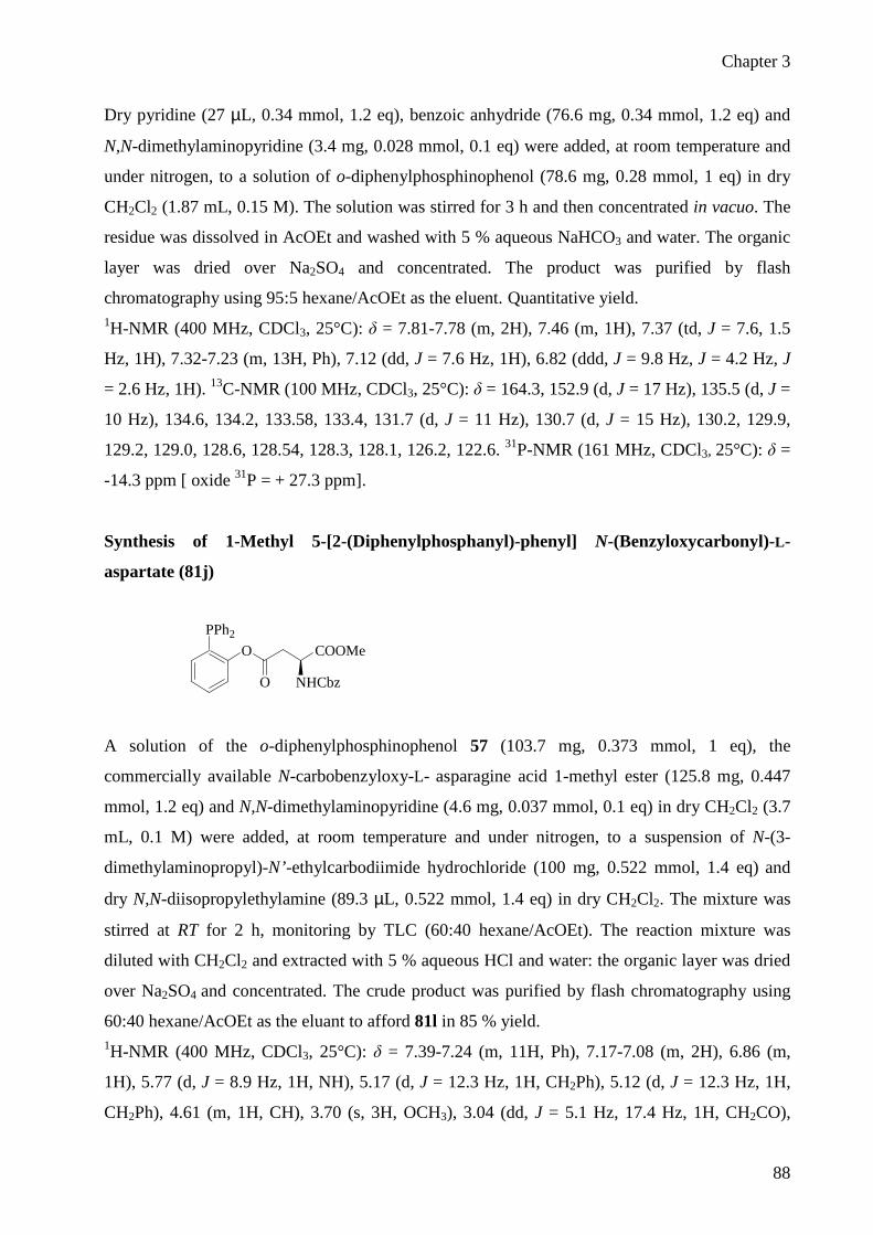
Chapter 3
88
Dry pyridine (27 µL, 0.34 mmol, 1.2 eq), benzoic anhydride (76.6 mg, 0.34 mmol, 1.2 eq) and
N,N-dimethylaminopyridine (3.4 mg, 0.028 mmol, 0.1 eq) were added, at room temperature and
under nitrogen, to a solution of o-diphenylphosphinophenol (78.6 mg, 0.28 mmol, 1 eq) in dry
CH2Cl2 (1.87 mL, 0.15 M). The solution was stirred for 3 h and then concentrated in vacuo. The
residue was dissolved in AcOEt and washed with 5 % aqueous NaHCO3 and water. The organic
layer was dried over Na2SO4 and concentrated. The product was purified by flash
chromatography using 95:5 hexane/AcOEt as the eluent. Quantitative yield. 1H-NMR (400 MHz, CDCl3, 25°C): δ = 7.81-7.78 (m, 2H), 7.46 (m, 1H), 7.37 (td, J = 7.6, 1.5
Hz, 1H), 7.32-7.23 (m, 13H, Ph), 7.12 (dd, J = 7.6 Hz, 1H), 6.82 (ddd, J = 9.8 Hz, J = 4.2 Hz, J
= 2.6 Hz, 1H). 13C-NMR (100 MHz, CDCl3, 25°C): δ = 164.3, 152.9 (d, J = 17 Hz), 135.5 (d, J =
10 Hz), 134.6, 134.2, 133.58, 133.4, 131.7 (d, J = 11 Hz), 130.7 (d, J = 15 Hz), 130.2, 129.9,
129.2, 129.0, 128.6, 128.54, 128.3, 128.1, 126.2, 122.6. 31P-NMR (161 MHz, CDCl3, 25°C): δ =
-14.3 ppm [ oxide 31P = + 27.3 ppm].
Synthesis of 1-Methyl 5-[2-(Diphenylphosphanyl)-phenyl] N-(Benzyloxycarbonyl)-L-
aspartate (81j)
PPh2
O
O
COOMe
NHCbz
A solution of the o-diphenylphosphinophenol 57 (103.7 mg, 0.373 mmol, 1 eq), the
commercially available N-carbobenzyloxy-L- asparagine acid 1-methyl ester (125.8 mg, 0.447
mmol, 1.2 eq) and N,N-dimethylaminopyridine (4.6 mg, 0.037 mmol, 0.1 eq) in dry CH2Cl2 (3.7
mL, 0.1 M) were added, at room temperature and under nitrogen, to a suspension of N-(3-
dimethylaminopropyl)-N’-ethylcarbodiimide hydrochloride (100 mg, 0.522 mmol, 1.4 eq) and
dry N,N-diisopropylethylamine (89.3 µL, 0.522 mmol, 1.4 eq) in dry CH2Cl2. The mixture was
stirred at RT for 2 h, monitoring by TLC (60:40 hexane/AcOEt). The reaction mixture was
diluted with CH2Cl2 and extracted with 5 % aqueous HCl and water: the organic layer was dried
over Na2SO4 and concentrated. The crude product was purified by flash chromatography using
60:40 hexane/AcOEt as the eluant to afford 81l in 85 % yield. 1H-NMR (400 MHz, CDCl3, 25°C): δ = 7.39-7.24 (m, 11H, Ph), 7.17-7.08 (m, 2H), 6.86 (m,
1H), 5.77 (d, J = 8.9 Hz, 1H, NH), 5.17 (d, J = 12.3 Hz, 1H, CH2Ph), 5.12 (d, J = 12.3 Hz, 1H,
CH2Ph), 4.61 (m, 1H, CH), 3.70 (s, 3H, OCH3), 3.04 (dd, J = 5.1 Hz, 17.4 Hz, 1H, CH2CO),

Chapter 3
89
2.75 (dd, J = 4.4 Hz, 17.4 Hz, 1H, CH2CO). 13C-NMR (100 MHz, CDCl3, 25°C): δ = 171.1,
169.3, 156.2, 152.6 (d, J = 17 Hz), 136.4, 135.6, 135.5, 135.4, 135.3, 134.3, 134.2, 133.9, 133.8,
130.5, 130.3, 130.2, 129.4, 129.3, 128.9, 128.8, 128.7, 128.4, 128.3, 126.7, 122.6, 122.5, 67.3,
53.0, 50.4, 36.6. 31P-NMR (161 MHz, CDCl3, 25°C): δ = - 15.6 ppm [ oxide 31P = + 28.2 ppm].
1-Methyl 5-[2-(Diphenylphosphanyl)-4-methoxyphenyl] N-(Benzyloxy-carbonyl)-L-
aspartate (82j)
PPh2O
O
COOMe
NHCbzMeO
See procedure for the synthesis of Synthesis of 1-Methyl 5-[2-(Diphenylphosphanyl)-phenyl] N-
(Benzyloxycarbonyl)-L-aspartate (81j)
The compound was purified by flash chromatography (hexane/AcOEt 65:35) yield = 75 %. 1H-NMR (400 MHz, CDCl3, 25°C): δ = 7.42-7.24 (m, 15H, Ph), 7.06 (dd, J = 4 Hz, J = 8.4 Hz,
1H, H-6), 6.86 (dd, J = 3.2 Hz, J = 8.4 Hz, 1H, H-5), 6.37 (t, J = 3.6 Hz, 1H, H-3), 5.75 (d, JNH-
CH = 8.2 Hz, 1H, NH), 5.19 (d, J = 6 Hz, 2H, CH2-O), 4.60 (q, JCH-CH2 = 4.4 Hz, 1H, CH), 3.72 (s,
3H, OCH3), 3.62 (s, 3H, OCH3), 3.01 (dd, J = 4.4 Hz, J = 17.2 Hz, 1H, Ha, CH2), 2.72 (dd, 1H, J
= 4.4 Hz, J = 17.2 Hz, Hb, CH2).
1-Methyl 5-[2-(Diphenylphosphanyl)-4-fluorophenyl] N-(Benzyloxy-carbonyl)-L-aspartate
(83j)
PPh2O
O
COOMe
NHCbzF
See procedure for the synthesis of Synthesis of 1-Methyl 5-[2-(Diphenylphosphanyl)-phenyl] N-
(Benzyloxycarbonyl)-L-aspartate (81j)
The compound was purified by flash chromatography (hexane/AcOEt 60:40) yield = 85 %. 1H-NMR (400 MHz, CDCl3, 25°C): δ = 7.48-7.27 (m, 15H, Ph), 7.12-7.02 (m, 2H, H-2, H-3),
6.55 (m, 1H, H-1), 5.70 (d, JNH-CH = 8.6 Hz, 1H, NH), 5.18 (d, J = 6 Hz, 2H, CH2O), 4.61 (q, JCH-

Chapter 3
90
CH2 = 4.4 Hz, 1H, CH), 3.72 (s, 3H, OCH3), 3.01 (dd, J = 4.6 Hz, J = 17.2 Hz, 1H, Ha, CH2), 2.75
(dd, J = 4.6 Hz, J = 17.2 Hz, 1H, Hb, CH2). 13C-NMR (100 MHz, CDCl3, 25°C): δ = 170.9,
169.2, 161.8, 159.4 (CO(Cbz)), 134.2, 134.1, 133.9, 133.8, 129.7, 129.6, 129.0, 128.9, 128.7,
128.6, 128.4, 128.3, 128.1 (5xCAr), 123.9, 123.8, 120.2, 120, 116.9, 116.6, 67.2 (CH2O), 52.9
(OCH3), 50.3 (CH), 36.5 (CH2). 31P-NMR (161 MHz, CDCl3
, 25°C): δ = - 15.3 ppm [ oxide 31P =
+ 26.7 ppm]. ESI-MS: m/z 582.2 (M+Na ).
1-Methyl 5-[2-(Diphenylphosphanyl)-4-trifluoromethylphenyl] N-(Benzyloxy-carbonyl)-L-
aspartate (84j)
PPh2O
O
COOMe
NHCbzF3C
See procedure for the synthesis of Synthesis of 1-Methyl 5-[2-(Diphenylphosphanyl)-phenyl] N-
(Benzyloxycarbonyl)-L-aspartate (81j)
The compound was purified by flash chromatography (hexane/AcOEt 70:30) yield = 90 %. 1H-NMR (400 MHz, CDCl3, 25°C): δ = 7.48 (d, J = 8.8 Hz, 1H), 7.37-7.21 (m, 16H, Ph), 7.07
(m, 1H), 5.64 (d, JNH-CH = 8.4 Hz, 1H, NH), 5.14 (d, J = 5.6 Hz, 2H, CH2O), 4.59 (q, JCH-CH2 =
4.4 Hz, 1H, CH), 3.68 (s, 3H, OCH3), 2.94 (dd, J = 4.4 Hz, J = 17.2 Hz, 1H, Ha, CH2), 2.72 (dd,
J = 4.4 Hz, J = 17.2 Hz, 1H, Hb, CH2).
1-Methyl 5-[2-(Dicyclohexylphosphanyl)-4-fluorophenyl] N-(Benzyloxy-carbonyl)-L-
aspartate (86j)
P(cyclohexil)2O
O
COOMe
NHCbzF
See procedure for the synthesis of Synthesis of 1-Methyl 5-[2-(Diphenylphosphanyl)-phenyl] N-
(Benzyloxycarbonyl)-L-aspartate (81j)
The compound was purified by flash chromatography (hexane/AcOEt 80:20) yield = 80 %.

Chapter 3
91
1H-NMR (400 MHz, CDCl3, 25°C): δ = 7.42-7.31 (m, 5H, Ph), 7.18-7.12 (m, 1H, H-3), 7.11-
7.01 (m, 2H, H-1, H-2), 6.57 (dd, JNH-CH = 8.4 Hz, J = 3.2 Hz, 1H, NH), 5.18 (s, 2H, CH2O), 4.79
(m, 1H, CH), 3.79 (s, 3H, OCH3), 3.39 (dd, J = 4.8 Hz, J = 16.8 Hz, 1H, Ha, CH2), 3.14 (dd, J =
4.4 Hz, J = 16.8 Hz, 1H, Hb, CH2), 1.93-1.48 (m, 14H, Cyhex), 1.47-0.97 (m, 8H, Cyhex).
1-Methyl 5-[2-(Dicyclopentylphosphanyl)-4-fluorophenyl] N-(Benzyloxy-carbonyl)-L-
aspartate (87j)
P(cyclopentyl)2O
O
COOMe
NHCbzF
See procedure for the synthesis of Synthesis of 1-Methyl 5-[2-(Diphenylphosphanyl)-phenyl] N-
(Benzyloxycarbonyl)-L-aspartate (81j)
The compound was purified by flash chromatography (hexane/AcOEt 70:30) yield = 80 %. 1H-NMR (400 MHz, CDCl3, 25°C): δ = 7.32-7.27 (m, 5H, Ph), 7.18-7.15 (m, 1H), 7.04-6.94 (m,
2H), 6.55-6.50 (m, 1H, NH), 5.13 (s, 2H, CH2O), 4.78-4.73 (m, 1H, CH), 3.75 (s, 3H, OCH3),
3.36 (dd, J = 4.4 Hz, J = 16.8 Hz, 1H, Ha, CH2), 3.01 (dd, J = 4.4 Hz, J = 16.8 Hz, 1H, Hb, CH2),
2.07-1.98 (m, 2H), 1.86-1.76 (m, 2H), 1.70-1.34 (m, 14H).
Synthesis of 1-Methyl 5-[2-(Diphenylphosphanyl)-phenyl] N-(Benzyloxycarbonyl)-L-
glutamate (81k)
PPh2O
O
NHCbz
COOMe
A solution of the o-diphenylphosphinophenol 57 (200 mg, 0.715 mmol, 1 eq), the commercially
available N-carbobenzyloxy-L-glutamic acid 1-methyl ester (255 mg, 0.86 mmol, 1.2 eq) and
N,N-dimethylaminopyridine (8.8 mg, 0.072 mmol, 0.1 eq) in dry CH2Cl2 (0.1 M) were added, at
room temperature and under nitrogen, to a suspension of N-(3-dimethylaminopropyl)-N’-
ethylcarbodiimide hydrochloride (193.3 mg, 1 mmol, 1.4 eq) and dry N,N-diisopropylethylamine
(172.6 µL, 1 mmol, 1.4 eq) in dry CH2Cl2. The mixture was stirred at RT for 2 h, monitoring by
TLC (50:50 hexane/AcOEt). The reaction mixture was diluted with CH2Cl2 and extracted with

Chapter 3
92
10 % aqueous HCl and water: the organic layer was dried over Na2SO4 and concentrated. The
crude product was purified by flash chromatography using 70:30 hexane/AcOEt as the eluant to
afford 81k in 75 % yield. 1H-NMR (400 MHz, CDCl3, 25°C): δ = 7.42-7.29 (m, 11H, Ph), 7.17-7.08 (m, 2H), 6.86 (m,
1H), 5.40 (d, J = 7.2 Hz, 1H, NH), 5.12 (s, J = 12.3 Hz, 1H, CH2Ph), 4.35 (q, J = 4.8 Hz, 1H,
CH), 2.35 (m, 2H, CH2CO), 2.07 (m, 1H, Ha, CH2CH), 1.86 (m, 1H, Hb, CH2CH).
1-Methyl 5-[2-(Diphenylphosphanyl)-4-fluorophenyl] N-(Benzyloxy-carbonyl)-L-glutamate
(83k)
PPh2O
O
NHCbz
COOMe
F
See procedure for the synthesis of 1-Methyl 5-[2-(Diphenylphosphanyl)-phenyl] N-
(Benzyloxycarbonyl)-L-glutamate (81k)
The compound was purified by flash chromatography (hexane/AcOEt 60:40) yield = 85 %. 1H-NMR (400 MHz, CDCl3, 25°C): δ = 7.40-7.27 (m, 15 H, Ph), 7.09 (m, 1H, H-2), 7.00 (m,
1H, H-3), 6.47 (m, 1H, H-1), 5.34 (d, JNH-CH = 7.9 Hz, 1H, NH), 5.11 (d, J = 3.8 Hz, 2H, CH2-
O), 4.35 (m, 1H, CH), 3.74 (s, 3H, O-CH3), 2.28 (m, 2H, CH2-CO), 2.01 (m, 1H, Ha, CH2-CH),
1.80 (m, 1H, Hb, CH2-CH). 13C-NMR (100 MHz, CDCl3, 25°C): δ = 170.8, 161.9, 159.4
(CO(Cbz)), 134.4, 134.4, 134.2, 134.2, 129.8, 129.2, 129.1, 128.9, 128.6, 128.5 (5xCAr), 124.3,
124.2, 120.2, 119.9, 116.9, 116.7, 67.5 (CH2-O), 53.5 (CH), 52.8 (O-CH3), 30.3 (CH2-CO), 27.6
(CH2). 31P-NMR (161 MHz, CDCl3,
25°C): δ = - 14.2 ppm [ oxide 31P = + 26.8 ppm]. ESI-MS:
m/z 596.3 (M+Na ).
1-Methyl 5-[2-(Diphenylphosphanyl)-3,5-ditrifluoromethylphenyl] N-(Benzyloxy-
carbonyl)-L-glutamate (85k)
PPh2O
O
CF3
F3C COOMe
NHCbz

Chapter 3
93
See procedure for the synthesis of 1-Methyl 5-[2-(Diphenylphosphanyl)-phenyl] N-
(Benzyloxycarbonyl)-L-glutamate (81k)
The compound was purified by flash chromatography (hexane/AcOEt 70:30) yield = 95 %. 1H-NMR (400 MHz, CDCl3, 25°C): δ = 7.94 (s, 1H), 7.61 (s, 1H), 7.38-7.31 (m, 15H, Ph), 5.20
(d, J = 7.6 Hz, 1H, NH), 4.28-4.23 (m, 1H, CH), 3.73 (s, 3H, OCH3), 1.82-1.58 (m, 4H, 2xCH2).
Synthesis of N-(Benzyloxycarbonyl)-ββββ-alanine 2-(Diphenylphosphanyl)-phenyl Ester (81l)
PPh2O
O
NHCbz
A solution of the o-diphenylphosphinophenol 57 (90 mg, 0.32 mmoli, 1 eq.), the protected amino
acid N-carbobenzyloxy-L-β-alanine (87 mg, 0.39 mmoli, 1.2 eq) and N,N-
dimethylaminopyridine (3.9 mg, 0.032 mmol, 0.1 eq) in dry CH2Cl2 (0.1 M) was added, at room
temperature and under nitrogen, to a solution of N,N’-dicyclohexylcarbodiimide (94 mg, 0.45
mmol, 1.4 eq) in dry CH2Cl2. The mixture was stirred at room temperature for 1.5 h, monitoring
by TLC (80:20 hexane/AcOEt). The reaction mixture was filtered and washed with CH2Cl2. The
crude product was purified by flash chromatography using 70:30 hexane/AcOEt as the eluent to
afford 81l in 82 % yield. 1H-NMR (400 MHz, CDCl3, 25°C): δ = 7.39-7.24 (m, 11H, Ph), 7.17-7.08 (m, 2H), 6.86 (m,
1H), 5.38 (s, J = 8.9 Hz, 1H, NH), 5.13 (s, J = 12.3 Hz, 1H, CH2Ph), 3.4 (q, J = 6 Hz, 2H,
NHCH2), 2.55 (t, J = 6 Hz, 2H, CH2). 13C-NMR (100 MHz, CDCl3, 25°C): δ = 170.1, 169.3,
156.2, 152.6 (d, J = 17 Hz), 136.4, 135.6, 135.5, 135.4, 135.3, 134.3, 134.2, 133.9, 133.9, 130.5,
130.2, 130.1, 130.0, 129.2, 128.7, 128.6, 128.5, 128.4, 128.0, 126.7, 122.5, 122.4, 66.7, 36.6,
34.6.
N-(Benzyloxy-carbonyl)-ββββ-alanine 2-(Diphenylphosphanyl)-4-fluorophenyl Ester (83l)
PPh2O
O
NHCbz
F

Chapter 3
94
A solution of the 2-diphenylphosphino-p-fluorophenol 60 (283.3 mg, 0.96 mmol, 1 eq), the
protected amino acid N-carbobenzyloxy-L-β-alanine (257.1 mg, 1.15 mmol, 1.2 eq) and N,N-
dimethylaminopyridine (11.7 mg, 0.096 mmol, 0.1 eq) in dry CH2Cl2 (9.6 ml, 0.1 M) was added,
at room temperature and under argon, to a solution of N,N’-dicyclohexylcarbodiimide (277.3 mg,
1.34 mmol, 1.4 eq) in dry CH2Cl2. The mixture was stirred at room temperature for 2 h,
monitoring by TLC (70:30 hexane/AcOEt). The reaction mixture was filtered and washed with
CH2Cl2. The crude product was purified by flash chromatography using 70:30 hexane/AcOEt as
the eluent to afford 83l in 92 % yield.
1H-NMR (400 MHz, CDCl3, 25°C): δ = 7.42-7.27 (m, 15H, Ph), 7.15-7.05 (m, 2H, H-2, H-3),
6.52 (m, 1H, H-1), 5.27 (m, 1H, NH), 5.12 (s, 2H, CH2O), 3.48 (q, JCH2-NH = 12 Hz, 2H,
CH2NH), 2.50 (t, JCH2-CH2 = 6 Hz, 2H, CH2CO). 13C-NMR (100 MHz, CDCl3, 25°C): δ = 170.8,
161.9, 159.5 (CO(Cbz)), 156.5, 134.8, 134.7, 134.4, 134.2, 132.1, 132, 129.8, 129.3, 129.2, 128.8,
128.5, 128.4 (5xCAr), 124.3, 124.2, 120.4, 120.2, 117.1, 116.8, 67 (CH2O), 36.6 (CH2), 34.8
(CH2CO). 31P-NMR (161 MHz, CDCl3, 25°C): δ = - 15.2 ppm [ oxide 31P = + 27.9 ppm]. ESI-
MS: m/z 524.2 (M+Na ).
N-(Benzyloxy-carbonyl)glycine 2-(Diphenylphosphanyl)-4-fluorophenyl Ester (83m)
PPh2O
O
NHCbz
F
A solution of the 2-diphenylphosphino-p-fluorophenol 60 (158,3 mg, 0.53 mmol, 1 eq), the
protected amino acid N-carbobenzyloxy-L-β-glycine (134 mg, 0.64 mmol, 1.2 eq) and N,N-
dimethylaminopyridine (6.5 mg, 0.053 mmol, 0.1 eq) in dry CH2Cl2 (5.3 ml, 0.1 M) was added,
at room temperature and under argon, to a solution of N,N’-dicyclohexylcarbodiimide (152.7 mg,
0.74 mmol, 1.4 eq) in dry CH2Cl2. The mixture was stirred at room temperature for 2 h,
monitoring by TLC (70:30 hexane/AcOEt). The reaction mixture was filtered and washed with
CH2Cl2. The crude product was purified by flash chromatography using 70:30 hexane/AcOEt as
the eluent to afford 83m in 90 % yield. 1H-NMR (400 MHz, CDCl3, 25°C): δ = 7.48-7.30 (m, 15H, Ph), 7.15 (m, 1H, H-2), 7.11 (m, 1H,
H-3), 6.53 (m, 1H, H-1), 5.12 (s, 2H, CH2O), 4.99 (m, 1H, NH), 3.85 (d, JNH-CH2 = 5.2 Hz, 2H,
CH2). 13C-NMR (100 MHz, CDCl3, 25°C): δ = 168.4, 159.6 (CO(Cbz)), 156.4, 162.1, 136.4
(Cipso), 134.8, 134.7, 134.4, 134.2, 129.9, 129.2, 129.1, 128.9, 128.6, 128.4 (5xCAr), 124.2,

Chapter 3
95
124.1, 120.4, 120.3, 120.1, 120.1, 117, 116.8, 67.5 (CH2O), 42.8 (CH2). 31P-NMR (161 MHz,
CDCl3, 25°C): δ = - 14.0 ppm [ oxide 31P = + 27.5 ppm]. ESI-MS: m/z 510.4 (M+Na ).
N-(tert-Butoxycarbonyl)-L-proline 2-(Diphenylphosphanyl)-4-fluorophenyl Ester (83n)
PPh2O
OF
BocN
A solution of the 2-diphenylphosphino-p-fluorophenol 60 (119.6 mg, 0.40 mmol, 1 eq), the
protected amino acid N-Boc-L-β-proline (103.3mg, 0.48 mmol, 1.2 eq) and N,N-
dimethylaminopyridine (4.9 mg, 0.04 mmol, 0.1 eq) in dry CH2Cl2 (4 ml, 0.1 M) was added, at
room temperature and under argon, to a solution of N,N’-dicyclohexylcarbodiimide (115,5 mg,
0.56 mmol, 1.4 eq) in dry CH2Cl2. The mixture was stirred at room temperature for 2 h,
monitoring by TLC (80:20 hexane/AcOEt). The reaction mixture was filtered and washed with
CH2Cl2. The crude product was purified by flash chromatography using 80:20 hexane/AcOEt as
the eluent to afford 83n in 94 % yield. 1H-NMR (400 MHz, CDCl3, 25°C): δ = 7.30-7.25 (m, 5H, Ph), 7.24-7.09 (m, 5H, Ph), 7.06-6.92
(m, 2H, H-2, H-3), 6.36 (m, 1H, H-1), 4.24 (m, 1H, CH), 3.26 (t, J Hc-Hd = 6.8 Hz, Hc, 1H, CH2),
3.18 (t, 1H, Hd, CH2), 2.03-1.95 (m, 1H, Ha), 1.78-1.62 (m, 3H, Hb, CH2), 1.45-1.30 (d, 9H,
Boc). 13C-NMR (100 MHz, CDCl3, 25°C): δ = 171.2, 161.9, 159.4, 154.8, 153.9, 148.9, 135.1,
134.4, 134.2, 133.9, 132.1, 129.7, 129, 124.3, 123.5, 119.9, 117.2, 116.9, 80.4, 80.1, 59.4, 46.8,
46.6, 30.6, 29.6, 28.8 (Boc), 24.9, 23.8. 31P-NMR (161 MHz, CDCl3, 25°C): δ = - 15.5, - 15.8
ppm [ oxide 31P = + 27.7, + 28.0 ppm].
Synthesis of the compound (89)

Chapter 3
96
Chloromethylphosphonic dichloride (5g, 30 mmol, 1eq) was dissolved in anyhdrous THF (30
mL). A solution of 4-methoxyphenylmagnesium bromide (0.5M) in THF (120 mL, 240 mmol, 2
eq.) was added dropwise over 1 h. The resulting mixture was stirred at reflux for 24 h. The
reaction was then quenched by the addition of water (5 mL), and the solventwas removed under
reduced pressure. The residue was dissolved in CH2Cl2, and the resulting solution was washed
once with water (12 mL). The organic layer was dried over Na2SO4 and filtered, and the solvent
was removed under reduced pressure. The residue was purified by flash chromatography
(CH2Cl2/MeOH 97:3). Phosphine oxide was isolated as a white solid in 63 % yield. 1H-NMR (400 MHz, CDCl3, 25°C): δ = 7.77-7.72 (m, 4H, Ph), 7.04-7.00 (m, 4H, Ph), 4.00 (d, J
= 7 Hz, 2H), 3.88 (s, 6H, 2xCH3). 13C-NMR (100 MHz, CDCl3, 25°C): δ = 162.9, 133.5, 133.4,
121.5, 120.4, 114.4, 114.2, 55.4, 38.1. 31P-NMR (161 MHz, CDCl3, 25°C): δ = + 27.4 ppm. ESI-
MS: m/z 310.05 (M+Na ).
Synthesis of the compound (90)
Phosphine oxide 89 (2.9 g, 9.1 mmol, 1 eq) was dissolved in DMF (25 mL). Potassium
thioacetate (1.25 g, 10.9 mmol) was then added, and the reaction mixture was stirred under
nitrogen for 18 h. the solvent was then removed under reduced pressure. The resulting oil was
purified by flash chromatography (CH2Cl2/MeOH 97:3). Phosphine oxide 90 was isolated as a
clear, colorless oil in 87 % yield. 1H-NMR (400 MHz, CDCl3, 25°C): δ = 7.70-7.65 (m,4H, Ph), 6.99-6.97 (m,4H, Ph), 3.85 (s,
6H, 2xOCH3), 3.70 (d, J =8.2 Hz, 2H), 2.28 (s, 3H, CH3). 13C-NMR (100 MHz, CDCl3, 25°C): δ
= 162.9, 133.2, 123.4, 123.3, 114.4, 55.6, 30.4, 28.0. 31P-NMR (161 MHz, CDCl3, 25°C): δ = +
29.2 ppm. ESI-MS: m/z 373.06 (M+Na ).

Chapter 3
97
Synthesis of the compound (91)
Phosphine oxide 90 (1.06 g, 2.95 mmol, 1 eq) was dissolved in ahnydrous chloroform (10 mL).
Trichlorosilane (8 mL, 3.87 mmol, 26.5 eq) was added, and the resulting solution was stirred
under nitrogen for 72 h. The solvent was then removed under reduced pressure. The residue was
purified by flash chromatography (CH2Cl2/MeOH 97:3). Phosphine 91 was isolated as a white
solid in 92 % yield. 1H-NMR (400 MHz, CDCl3, 25°C): δ = 7.38-7.36 (m, 4H, Ph), 6.93-6.89 (m, 4H, Ph), 3.85 (s,
6H, 2xOCH3), 3.47 (bs, 2H), 2.32 (s, 3H, CH3). 13C-NMR (100 MHz, CDCl3, 25°C): δ = 194.4,
160.5, 134.2, 134.0, 127.9, 127.8, 114.2, 55.2, 30.3, 26.4. 31P-NMR (161 MHz, CDCl3, 25°C): δ
= -19.7 ppm. ESI-MS: m/z 357.06 (M+Na ).
Synthesis of the compound (92)
Phosphine 91 (200 mg, 0.56 mmol, 1 eq) was dissolved in ahnydrous DMF (1mL). A solution of
MeONa (34 mg, 0.62 mmol, 1.1 eq) was added dropwise at 0°C, under nitrogen. The resulting
mixture was stirred at 0°C for 2 h and then valeroyl chloride ( 67.4 mg, 0.56 mmol, 1eq) was
added. The solution was stirred for 1 h at 0°C, dissolved in CH2Cl2 and washed with 5 %
aqueous HCl, 5 % satured NaHCO3 and water. The organic layer was dried over Na2SO4 and
concentrated to give the crude product. The resulting oil was purified by flash chromatography
(cyclohexane/AcOEt 80:20 to cyclohexane/AcOEt/MeOH 80:20:2). Phosphine 92 was isolated
as a clear, colorless oil in 57 % yield. 1H-NMR (400 MHz, CDCl3, 25°C): δ = 7.44 (t, J = 8 Hz, 4H, Ph), 6.94 (d, J = 8 Hz, 4H, Ph),
3.68 (s, 6H, 2xOCH3), 3.56 (d, J = 2 Hz, 2H, CH2S), 2.53 (t, J = 7.6 Hz, 2H, CH2), 1.59 (m, 2H,

Chapter 3
98
CH2), 1.29 (m, 2H, CH2), 0.89 (t, J = 7.2 Hz, 3H, CH3). 31P-NMR (161 MHz, CDCl3
, 25°C): δ = -
17.6 ppm [ oxide 31P = + 29.7 ppm].
Chapter 3
99
3.7 Reference
1 Bianchi, A.; Bernardi, A. J. Org. Chem., 2006, 71, 4565-4577. 2 Nisic, F.; Bernardi, A. Carb. Res. 2008, 343, 1636-1643. 3 Bianchi, A. PhD Thesis, Università di Milano, 2004-2005. 4 Rauchfuss, T. B. Inorg. Chem. 1977, 16, 2966-2968. 5 Jeganathan, S.; Tsukarmoto, M.; Schlosser, M. Synthesis 1990, 109-111. 6 Luo, H.; Setyawati, I.; Rettig, S. J.; Orvig, C. Inorg. Chem. 1995, 34, 2287-2299. 7 Grotjahn, D. B.; Joubran, C.; Combs, D. J. Organomet. Chem. 1999, 589, 115-121. 8 Heinicke, J.; Köhler, M.; Peulecke, M.; He, M.; Kindermann, K.; Keim, W.; Fink, G. Chem. Eur. J. 2003, 9,
6093-6107. 9 Furlano, D. C.; Calderon, S.N.; Chen, G.; Kirk, K.L. J. Org. Chem. 1988, 53, 3145-3147. 10 Soellner, M. B.; Tam, A.;Raines, R.T. J. Org. Chem., 2006, 28, 9824-9830. 11 Nilsson, B. L.; Hondal, R. J.; Soellner, M. B.; Raines, R. T. J. Am. Chem. Soc. 2003, 125, 5268-5269. 12 Soellner, M. B.; Dickson, K. A.; Nilsson, B. L.; Raines, R. T. J. Am. Chem. Soc. 2003, 125, 11790-11791. 13 Gauchet, C.; Labadie, G. R.; Poulter, C. D. J. Am. Chem. Soc. 2006, 128, 9274-9275. 14 For examples, see: (a) He, Y.; Hinklin, R. J.; Chang, J. Y.; Kiessling, L. L. Org. Lett. 2004, 6, 4479-4482. (b)
Bianchi, A.; Bernardi, A. Tetrahedron Lett. 2004, 45, 2231-2234. (c) Bianchi, A.; Russo, A.; Bernardi, A.
Tetrahedron: Asymmetry 2005, 16, 381-386. (d) Liu, L.; Hong, Z.-Y.; Wong, C.-H. ChemBioChem 2006, 2006,
429-432.

Chapter 4
Staudinger ligation of unprotected azides:
a reagent screening

Chapter 4
101
4.1 Background Our previous studies on the use of Staudinger ligation for the synthesis of glycosyl amides have
been summarized in the introduction (Chapter 1). Among them, ligation of unprotected α- and β-
glucosyl azides1 7 and 10 was particularly remarkable because in both cases it occurred
stereoconservatively (Scheme 1) and allowed simple isolation of the resulting glucosyl amides
from the phosphane oxide by-product by water extraction. The anomeric ratios of the products
were consistently good and the yields moderate-to-good for most of the alkyl chains R,
particularly under MW irradiation.1
ON3
OHHO
HO
OH
O
N3HO
HOHO
OH
O
PPh2
O
R
O
HNHO
HOHO
OH
O
R
O HN
HOHO
HO
OH
O
R81a-l
7
10
93a-l
94a-l
Scheme 1. Stereoconservative Staudinger ligation of 7 and 10 with phosphine 81a-l
For its novelty and stereoselectivity this method deserved further consideration, but it was far
from perfect and left many problems open. As noted above, yields were generally modest and
dropped to poor for some α-glycosylamides. Most notably the aspartic and glutamic acid
derivativesm 81j and 81k (Scheme 2) gave poor yields of the corresponding glucosyl amino
acids for both the α and β series. This is particularly disappointing because these amides are
important glycoconjugates, mimicking the conserved core of natural N-asparagine-linked
glycopeptides and glycoproteins.2, 3

Chapter 4
102
Scheme 2. Phosphines 81j,k give poor yields of N-glycosyl amino acids
Furthermore, the reaction often yielded mixtures of the desired pyranosylamide and the isomeric
furanosylamide. For instance, reaction of 7 with the pentanoyl phosphine 81b (Scheme 3) in
DMA at 70°C yielded a complex mixture of compounds in the water fraction that were separated
by reverse phase HPLC and characterized as the expected pentanamide 93b (50 % yield), the α-
N-pentanoyl-glucofuranosylamine isomer 95b (15 % yield) and α and β-glucose (equilibrium
mixture of anomers, 30% yield). The configuration of 93b was assigned on the basis of the
anomeric proton signal (5.45 ppm, J1-2 = 5.4 Hz). The structure of 95b was assigned on the basis
of ESI-MS and of the 1H-NMR (D2O), which is consistent with the spectrum of the known α-D-
glucofuranoside (Scheme3).4
Scheme 3. Staudinger ligation of 7 with phosphine 81b in DMA

Chapter 4
103
The anomeric configuration of 95b was further confirmed to be α by the NOESY spectrum
(D2O), which shows a strong crosspeak for the H1 and H2 protons and no crosspeak for the H1
and H3 protons (Scheme 3). The presence of glucose in the reaction crude was identified by 1H-
NMR spectroscopy.
The furanoside 95b must clearly derive from a ring-opening process occurring after the azide
reduction step, presumably from the iminophosphorane 96 (Scheme 4) to afford the
phosphinimine 97, which can undergo ring-closure to yield 95b. Hydrolysis of the same
intermediate 97 accounts for the formation of α and β-D-glucose in the reaction mixture. The
alternative hypothesis that glucose could be formed by direct hydrolysis of the starting azide was
discarded because 7 was recovered unaltered after treatment with water in DMA at 70°C for 24
h.
PPh2
O
O
O
HOH
HO
HO
OH
NN2
O
HO
HO
N
H
HOOH
PPh2
O
O
NH
H
H OH
OH HO
HHO
HO
O
H
PPh2
O
O
O
HO
HOHO
OH
N
H
O
HOH
HO
HO
OH
HN
O
H
O
O
HO
HO
N
HOOH
PPh2
O
H2O
95
96 97
-glucose
acyl transfer
93b
O
HO
HOHO
OH
N3
PPh2
O
O
+
81b
7
Scheme 4. Mechanism for the formation of the furanosylamide 95b and glucose in the ligation
of 7 with 81b
This drawback was partly resolved in our previous work1, 5 by reducing the temperature of the
reaction (high temperatures favor formation of the furanosylamides) while using microwaves to

Chapter 4
104
avoid increasing the reaction time. Adding 2 % DMPU to the solvent (DMA) was also found to
be beneficial, since a high polarity of the solvent appears to accelerate the acyl transfer step.6
Under these conditions, formation of the undesired furanosyl amide could be contained in most
cases below 5-10 % and both α and β unprotected glycosyl azides could be transformed
stereoselectively in the corresponding amides and easily isolated from reaction mixtures.
However, clearly the process called for further optimization.
4.2 Staudinger ligation with a new group of phosphines
To address the set of problems described above we turned to examining the performance of
different phosphines in the reaction using as model substrates the α and β-glucopyranosyl azides
7 and 10.
The Staudinger ligation of phosphines 81 can be envisaged as taking place in two steps, as
described in Scheme 5 for the reaction of 81j with α-glucosyl azide 7. In the first step,
nucleophilic addition of the phosphine to the γ nitrogen of the azide followed by nitrogen
extrusion leads to the iminophosphorane 98. In the second step, 98 undergoes an intramolecular
reaction in which the acyl chain transferred from the phenolic ester to the imminophosphorane
nitrogen and an amide bond is generated.1 Our previous observations indicated that the poor
amide yields observed upon reaction of glucosylazides with some phosphines were caused by
inefficient acyl chain transfer in the iminophosphorane intermediate 98 (Scheme 5).1, 7
Scheme 5. Two-step mechanism for the Staudinger ligation.
Using the syntheses shown in Chapter 3, we prepared the group of new phosphines collected in
Figure 1 ( 59-62, 79, 80 and 91) for further studies on the Staudinger ligation process.

Chapter 4
105
PPh2
OH
MeO
PPh2
OH
F
P
OH
F
PMeO S
O
2
PPh2
OH
F3C
PPh2
OH
CF3
F3C
P
OH
F
59 60 61 62
9179 80
Figure 1. Set of new phosphines
The two phosphines 59 and 60 were studied in more detail and were meant to address two
different and opposite features of the process:
1. The azide reduction step (formation of the imminophosphorane, Scheme 6) is facilitated
by a strong basicity of the P atom.
2. The acyl transfer step (from phenol oxygen to iminophosphorane N, in Scheme 6) would
be accelerated by EWG groups on the aromatic ring, which, however, will reduce the
basicity of P.
Thus, we expected that increasing the leaving group ability of the phenol, such as with
phosphines 60-62 could improve the overall-yield of the reaction by accelerating the transfer
step. A possible decrease in reactivity due to the reduced basicity of the P atom induced by the
electronpoor phenol could be offset by increasing the electonrichness of the phosphines, for
instance with aliphatic substituents, such as cyclohexyl or cyclopentyl rings, as in 79 and 80.
This should result in an increase of the reduction rate, leading to a diminished amount of by-
products related to incomplete azide reduction.
More generally, we expected that a change in the structure of the phoshine reagent may be
reflected in a different composition of the reaction mixtures and in particular may affect the
formation of the furanose isomers in ways that would be difficult to anticipate at this point.

Chapter 4
106
PPh2
O R
O
HOO
NHO
HO
OH
Ph2P
N2
iminophosphorane
HOO
NHO
HO
OH
Ph2P
O
ROC
H2OPh2P
HO
O
OO
R
HOO
NHCORHO
HO
OH
+
7
HOO
N3
HOHO
OH
X
X = H, F, OMe
X
X
X
Reduction
Acyl transfer
93
Scheme 6. Mechanism of the traceless Staudinger ligation
As a first step we identified a rapid and diagnostic analytical method to evaluate the course of the
reactions by analysis of the crude reaction mixtures. Since the reaction is performed on
unprotected sugars, Thin Layer Chromatography cannot be used to monitor its course. Rather,
after completion of the reactions under a predefined set of conditions (time, temperature etc), the
crude mixtures were diluted with water and extracted with CH2Cl2 to eliminate the phosphine by-
products. The aqueous layers were analyzed by 1H-NMR (typically in D2O) and the product
ratios established, based on the integration of the anomeric protons (Figure 2). Typical reaction
crudes contained both the expected α- or β-pyranosyl amides (93 or 94) and variable amounts of
the corresponding α-furanosyl amides 95. As we have previously shown (Scheme 4),1 the latter
are formed by ring-opening/ring-closure of the iminophosphoranes intermediate. Other by-
products consist in free glucose (equilibrium anomeric mixtures) formed by hydrolysis of the
same intermediates. They were all identified and characterized by isolation and/or comparison
with known samples.

Chapter 4
107
Figure 2. 1H-NMR (D2O) spectra of the crude water extract
As mentioned above, a first set of experiments was perfomed with phoshines 82 and 83 obtained
by acylation of the corresponding phenols 59 and 60. Phosphine 82 was chosen because
fluorophenyl esters are well-known as good activating groups in acylic nucleophilic substitution
and increase the acylating ability of the reagent. The methoxy group on the aromatic ring of 83
increases the basicitity of the phosphorous atom, allowing fast reduction of the azide, but reduces
the acylating ability.
A set of experiments was designed on the unprotected α-glucosyl azide 7 chosen as the standard
substrate, changing reaction temperature, nature of the R group (R = Me, CH3CH2CH2CH2-,
(CH3)2C=CH-, Z-L-aspartic acid 1-methyl ester) and type of process (thermal or microwave
assisted) (Scheme 7 and Tables A, B and C in the appendix).
Yields and diasteromeric ratios obtained from the ligation were estimated on the basis of the
weight and on the 1H-NMR spectra of the crude water extract, and all reactions were run in the
same solvent: 98:2 DMA:DMPU (the results are collected in Tables A and B in the appendix).
For comparison, the data previously obtained1 in similar conditions for 81 are collected in Table
C.

Chapter 4
108
Scheme 7. Screening for the ligation of α-glucosyl azide 7
Evaluation of results obtained from this screening show that:
The methoxy-substituted phosphines 82 displayed lower levels of α selectivity, proved by the
presence of both α- and β-glucosyl amides in the crude water extract, and also in general showed
a higher tendency to give the formation of α-glucofuranosyl amides and of other hydrolysis by-
products.
Microwave assisted process with compounds 82 afforded low yield, low stereoselectivity and the
azide 7 was not completely reduced (see Table A in appendix, entries 5, 6 and 11). In the end,
the phosphine 82 presented a reduced reactivity and minor α-stereocontrol in comparison with
compound 81 for the Staudinger ligation. Compound 82 afforded high conversion to α-
glucofuranosyl amides 95, if the reaction was performed above 70°C (Table A, entries 3, 4 and
6). However, after water extraction we obtained low yield, probably for an inefficient acyl chain
transfer during the ligation, that stop the reaction to iminophosphorane intermediate 98 step
(Scheme 5).
Similarly, the fluorinated compounds 83 showed lower reduction strength and lower acylating
ability in the ligation of alkyl chains, but were found to perform dependably in the transfer of
Aspartic acid side-chain to 7. (see Table B in appendix, entries 14 and 15).
Some general observations were extrapolated from this set of experiments for the Staudinger
ligation between α-glucosyl azide 7 and functionalized phosphines 82 and 83:
1. The results showed a strong and somewhat unpredictable dependence of the process on the
nature of the R group and the type of phosphine.

Chapter 4
109
2. If the ligation for the thermal process was performed above 40 °C, the quantity of α-
glucofuranosyl amide increased in each case observed (Table A and B in appendix,
experiment perform at 70°C and 100°C).
3. The methoxy phosphines 82 gave large amount of pyranose-furanose conversion.
4. The fluorinated phosphines 83 gave the best results with amino acids.
The best conditions identified for the synthesis of 93j, consisted in treating the unprotected α-
glucosyl azide with 2 equivalents of phosphines 83j in DMA/DMPU (98:2) at 40°C during 18 h
(Scheme 8).
Scheme 8. Staudinger ligation of the unprotected α-glucosyl azide 7 with phosphine 83j
Under these conditions the α-glucopyranosylamide was isolated in good yield and excellent
anomeric ratio (55 % isolated yield and 98:2 α/β ratio). The presence of the F atom in the
aromatic-ring appears to play a key role for the reactivity of the phosphine 83j in the acyl
transfer of aspartic acid.
As a matter of fact, the reaction occurred with over 55% yield, compared to 25% afforded by the
non-fluorinated reagent 81j. However, the ligation required more equivalents of phosphines (1.2
eq. to 2 eq.) and the reaction time became much longer (4 h to 18 h). This proves the low
reactivity of fluorinated phosphine 83j respect to compound 81j. Microwave assisted process
also in this case afforded low yield and high presence of by-products (Table B in appendix,
entries 6, 7, 9 and 13).
To further explore the ability of electron poor phosphines in the Staudinger ligation of aspartic
acid residue, azide 7 was treated with 84j and 85k (Figure 3) in the same conditions optimized
for the precedent ligation (40°C, 18 h and DMA/DMPU 98:2 as mixture solvent).

Chapter 4
110
Figure 3. Phosphines 84j and 85k
Unfortunately phoshines 84j and 85k were found to reduce the azide, but not to transfer the acyl
chain and afforded only hydrolysis by-products, such as α and β-glucose and other not identified
compounds, whit complet absence of α-glucosyl amide as a product.
This suggested that the azide reduction step could not be induced in the reaction for the low
basicity of the phosphorus atom, also increased temperature and time of the reaction.
For this purpose compound 86j (Figure 4) was synthesized, replacing two phenyl rings with
cyclohexyl rings to increase the reduction strength of the phosphines. These became able to
reduce the azide 7 (major reaction product was β-glucosylamine as show by anomeric signal at
4.18 ppm J1-2 = 8.8 Hz to prove that the reduction step happened), but didn’t have acylating
ability at all, probably for the major steric hindrance of the alkyl substituent.
Figure 4. Phosphines 86j and 87j
Compound 87j was synthesized to decrease the steric hindrance of the cyclohexyl ring and tested
with different reaction conditions, but also in this case the ligation afforded only hydrolysis by-
prodouct with total absence of α-glucosyl amide 93j.
A final set of experiments were performed treating the unprotected α-glucosyl azides 7 with the
functionalized (diphenylphosphino)methanethiol 92 (Scheme 9). 8 This phosphine was largely
used in the orthogonal assembly of a protein,9 site-specific immobilization of peptides and
proteins to a surface,10, 11 and synthesis of glycopeptides. 12

Chapter 4
111
The same procedure employed for the Staudinger ligation between phosphine and protected or
unprotected glucosyl azides (70 °C/18 h and 40°C/4h, in DMA:DMPU 98:2 as a solvet
mixture)1, 7 were used, but we observed that 92, is not able to react with the compound 7.
Scheme 9. Staudinger ligation of 7 with the functionalized (diphenylphosphino)methanethiol 92
The mechanism proposed by Raines and co-workers (Scheme 10)8 for the ligation mediated by
(diphenylphosphino)methanethiol 99, show that the imminophosphorane 100 can form a
tetrahedral intermediate 101, which collapses to give an amidophosphonium salt 102 that is
hydrolyzed in aqueous solution, furnish the final amide. Probably in our case the steric hindrance
of the tetrahedral intermediate 101, that occour during the ligation, avoid the complete reduction
of α-glucosyl azide 7 and the acyl transfer of the acyl chain.
Scheme 10. Mechanism for the Traceless Staudinger ligation mediated by
(diphenylphosphino)methanthiol 7

Chapter 4
112
4.3 Appendix
Table A. Screening for the synthesis of α-glucosylamides 93
Entry Reagent Process Glucose Azide β-
pyranosyl amide 94
α-pyranosyl amide 93
α-furanosyl amide 95
1 82a 70 °C, 18 h 6 / 10 48 36
2 82a 100 °C, 18 h / / 13 37 50
3 82b 70 °C, 18 h / / 16 24 60
4 82b 100 °C, 18 h / / 17 18 65
5 82b M.W.120°C,20’ / 24 15.5 6.5 54
6 82b M.W.120°C,40’ 5 8 17 8 59
7 82e 100 °C, 18 h 18 / 51 / 31
8 82j 40 °C, 18 h ? 43 ? 23 27
9 82j 70 °C, 18 h 45 / ? 17 38
10 82j 70 °C, 4 h 12 26 ? 17 26
11 82j M.W.120°C, 1h 12.5 38 ? 20 29
Ratio estimated on the basis of the 1H-NMR (D2O)spectra of the crude water extract (?) 1H-NMR integration not possible

Chapter 4
113
Table B. Screening for the synthesis of α-glucosylamides 93
Entry Phosphin. Process Glucose Azide β-
pyranosyl amide 94
α-pyranosyl amide 93
α-furanosyl amide 95
1 83a 70 °C, 4 h 16 5 9.5 54 15
2 83a 70 °C, 18 h 12 7 8 60 13
3 83b 40 °C, 4 h 15 31 7.5 29.5 16
4 83b 70 °C, 4 h 15.5 / 10.5 55 19
5 83b 70 °C, 18 h 20.5 / 24 8 47
6 83b M.W.50°C,50’ 17 8 17 24.5 33
7 83b M.W.120°C,20’ 12 7 17 14 50
8 83e 70 °C, 4 h 22 / 31 6.5 40
9 83e M.W.120°C,20’ 21 / 33.5 32 13.5
10 83j 40 °C, 18 h 5 25 ? 44 15
11 83j 70 °C, 18 h 27 / ? 45 24
12 83j 70 °C, 4 h 9 19.5 ? 30 19
13 83j M.W.120°C,40’ 6 28 7 39 19
14 83j 45 °C, 18 h 20 / / 61 22
15 83j 40 °C, 18 h 15 / / 65(55a) 20
Ratio estimated on the basis of the 1H-NMR (D2O)spectra of the crude water extract (?) 1H-NMR integration not possible. a) Isolated yield

Chapter 4
114
Table C. Screening for the synthesis of α-glucosylamides 93
Entry Phosphin. Process Glucose Azide β-
pyranosyl amide 94
α-pyranosyl amide 93
α-furanosyl amide 95
1 81a 70 °C, 18 h 20 / / 77 3
2 81b 40 °C, 4 h 20 / / 75(65a) 5
3 81b 70 °C, 18 h 20 / / 65(50a) 15
4 81b M.W.50°C,50’ 20 / / 75(65a) 2
5 81b M.W.120°C,10’ 20 / / 75(65a) 2
6 81e 40 °C, 4 h 20 / / 40 40
7 81e M.W.50°C,50’ / / / 10a 52a
8 81j 40 °C, 4 h 35 / / 25a 15
9 81j M.W.50°C,50’ 35 / / 25a 10
10 81j M.W.120°C,10’ / / / 25a 10
Ratio estimated on the basis of the 1H-NMR (D2O)spectra of the crude water extract (?) 1H-NMR integration not possible. a) Isolated yield

Chapter 4
115
4.4 References
1 Nisic, F.; Bernardi, A. Carbohydr. Res. 2008, 343, 1636-1643. 2 Lis, H.; Sharon, N. Eur. J. Biochem. 1993, 218, 1–27; Taylor, C. M. Tetrahedron 1998, 54, 11317–11362. 3 β-Linked glucosyl amides of glutamic acid are found in natural glyco proteins: Ecker, M.; Deutzmann, R.; Lehle,
L.; Mrsa, V.; Tanner, W. J. Biol. Chem. 2006, 281, 11523–11529. 4 Norrild, J.C.; Eggert, H. J. Am. Chem.Soc. 1995, 117, 1479-1484 5 Nisic F. Master Thesis, Università degli studi di Milano, 2007. 6 Saxon, E.; Armstrong, J. I.; Bertozzi, C. R. Org. Lett. 2000, 2, 2141-2143. (b) Saxon, E.; Bertozzi, C. R. Science
2000, 287, 2007-2010. 7 Bianchi, A.; Bernardi, A. J. Org. Chem. 2006, 71, 4565-4577. 8 Soellner, M. B.; Tam, A.; Raines, R. T. J. Org. Chem. 2006, 71, 9824-9830. 9 Nilsson, B. L.; Hondal, R. J.; Soellner, M. B.; Raines, R. T. J. Am. Chem. Soc. 2003, 125, 5268-5269. 10 Soellner, M. B.; Dickson, K. A.; Nilsson, B. L.; Raines, R. T. J. Am. Chem. Soc. 2003, 125, 11790-11791. 11 Gauchet, C.; Labadie, G. R.; Poulter, C. D. J. Am. Chem. Soc. 2006, 128, 9274-9275. 12 For examples, see: (a) He, Y.; Hinklin, R. J.; Chang, J. Y.; Kiessling, L. L. Org. Lett. 2004, 6, 4479-4482. (b)
Bianchi, A.; Bernardi, A. Tetrahedron Lett. 2004, 45, 2231-2234. (c) Bianchi, A.; Russo, A.; Bernardi, A.
Tetrahedron Asymmetry 2005, 16, 381-386. (d) Liu, L.; Hong, Z.-Y.; Wong, C.-H. ChemBioChem 2006, 2006,
429-432.

Chapter 5
Stereoselective synthesis of N-glycosyl amino acids by
traceless Staudinger ligation of unprotected glycosyl
azides

Chapter 5
117
In chapter 4 we showed that monofluorophosphine 83j improve the yields of aspartic acid
transfer, which occurred with over 55 % yield, compared to 25 % afforded by the non-
fluorinated analogue 81j (Scheme 1). Hence, improving the leaving group ability of the activated
ester appears to result in higher yields for this difficult transfer step and the novel functionalized
phosphanis 83j affords the corresponding N-glucosyl aspartic acid 93j in a stereoconservative
and synthetically pratical manner. This preliminary result prompted us to further explore the
scope of phosphines derived from monofluorophosphino phenol 60 in the ligation of amino acids
and glycosyl azides.
O
N3HO
HOHO
OH
O
PPh2
O
7
O
HO
HOHO
OH
HNO
O NHCbz
O
y = 55 %
y = 25 %
COOMe
NHCbz
O
PPh2
O
COOMe
NHCbz
F
81j
83j
93j
93j
O
HO
HOHO
OH
HNO
O NHCbz
O
Scheme1. Ligation of α-glucosyl azide 7 with functionalized phosphines 81j and 83j
5.1 Synthesis of αααα- and ββββ-glucosyl amino acids
The monofluoro phosphines 83j-n, could be obtained in high yields from the known phosphine
601 by reaction with the appropriate amino acids and condensing agents as show in Scheme 2,
(See chapter 3 experimental ct for the procedures). The functionalized phosphines 83j-n were
synthesized to examine the Staudinger ligation of the aspartic and glutamic acid side-chains (83j
and 83k, respectively) as well as the reactions of simple α- and β-amino acids such as β-alanine

Chapter 5
118
(83l), glycine (83m) and proline (83n). Optimal yields were obtained by using either N-[3-
(dimethylamino) propyl]-N-ethylcarbodiimide (EDC) (83j and 83k) or DCC (83l-n) as the
condensing agent. The ligation reagents could be isolated by simple filtration through a silica gel
plug. Analytical samples were purified by flash chromatography. In general, and like the parent
non-fluorinated compounds, phosphines 83j-n were found to be air-stable compounds that could
be handled under standard conditions with minimal precautions.
Scheme 2. Synthesis of the fluorinared phosphines 83j-n
The ligation experiments were performed by treating the unprotected α- and β-glucosyl azides 7
and 10 with 2 equivalents of phosphine 83j-n in DMA/DMPU (98:2) at 40 °C for 18 h. After
completion of the reaction, the crude mixtures were stirred with water for an additional 2 h, then
diluted with water and extracted with CH2Cl2 to eliminate the phosphine by-products. The
aqueous layers were analyzed by 1H NMR (typically in D2O) and the product ratios established
on the basis of the integration of the anomeric protons. Typical reaction crudes contained both
the expected α- or β-pyranosyl amides 93 or 94 and variable amounts of the corresponding
furanosyl amides 95 (Scheme 3). The anomeric configuration of 95j-n was consistent with the
previously data reported for the α-glucofuranosyl amides.2 As we have previously shown
(Scheme 4, chapter 4),2 the latter are formed by ring-opening/ring-closure of the intermediate
iminophosphoranes.3 Free glucose (equilibrium anomeric mixtures) is also formed by hydrolysis
of the same intermediates.

Chapter 5
119
Scheme 3. Synthesis of α- and β- glucosyl aminoacids 93j-m and 94 j-n.
The glucosyl amides were purified by flash chromatography on silica gel (CHCl3/MeOH/H2O
80:20:2) and fully characterized as their acetylated counterparts 103j-m and 104j-n (Scheme 4).
Scheme 4. Acetylated compounds 103j-m and 104j-n
Ligation of the α-azide was generally accompanied by ring contraction and sizeable quantities of
the glucofuranosyl amides 95 were formed (Table 1, entries 1-4). Strict control of anhydrous

Chapter 5
120
conditions and temperature were found to minimize the formation of ring-contraction by-
products. In our previous studies with phosphines 81j,2 the use of MW irradiation proved
beneficial for reducing the amount of furanose formation, but this was not the case with
phosphines 83j as MW irradiation (50 °C, 50 min) gave lower yields of the desired pyranosyl
amides and more by-products.
The reactions with β-glucosyl azide 10 (Table 1, entries 5-9) gave good-to-excellent yields of the
corresponding pyranosyl amides 94j-n, which were essentially pure after water extraction. The 1H NMR spectra of the reaction crudes showed no trace of the anomeric α-pyranosyl epimer and
very minor quantities (3 %) of a by-product to which the furanosyl amide structure 95 was
tentatively assigned; α-, β- and γ-amino acids were transferred with equal efficiency. The
aspartic acid amide 94j (Table 1, entry 5) was the most difficult to obtain, but could still be
isolated in 77 % yield, as opposed to the yield of 32 % obtained under the same conditions with
phosphane 81j.2
Table 1. Synthesis of α- and β-glucosylaminoacids 93 and 94 by ligation of α- and β-
glucosylazides 7 and 10 with 83j-n
Entry Phosphine Azide Product Isolated yieldsa (%)
αααα/ββββ ratiob
Pyranose/Furanose ratioc
1 83j 7 93j 56 98:2 85:15
2 83k 7 93k 59 98:2 87:13
3 83l 7 93l 60 98:2 90:10
4 83m 7 93m 36 (12)d 98:2 67:33
5 83j 10 94j 77 < 1:99 98:2
6 83k 10 94k 87 < 1:99 98:2
7 83l 10 94l 85 < 1:99 98:2
8 83m 10 94m 75 < 1:99 97:3
9 83n 10 94n 70 < 1:99 98:2
a) Yields based on glucopyranosylamide b) Ratio of the anomeric proton signals of the α- and β-pyranosylamides
93 and 94 in the crude 1H-NMR spectrum c) Ratio of the anomeric proton signals in the crude 1H-NMR spectrum.
d) Isolated yield of furanosyl amide 95m.

Chapter 5
121
A significant difference in the reactivity of α- and β-glucopyranosyl azides towards the traceless
Staudinger ligation was noted, in particular reaction of α-glucosyl azide 7 (Table 1, entries 1-4)
with 83j-m afforded the corresponding α-amide 93j-m in lower yields compared to the β-
anomers 94j-n formed from 10 (Table 1, entries 5-9).
In 2010 Field and co-worker4 reported a comparison of X-ray crystallographic data for both the
α- and β-peracetylated glucopyranosyl azides 1 and 2 highlighted the impact of anomeric effect
on the dipolar character of the anomeric azides, which likely accounts for the observed reactivity
difference. This comparison revealed that the acetylated glucopyranose rings of 1 and 2 are very
similar, except for the anomeric azido groups having different configurations (axial vs
equatorial). In addition, the interatomic distances within the C(1)-N(A)-N(B)-N(C) fragment
(Table 2 and Figure1) are in keeping with the expected influence of the anomeric effect. The
C(1)-N(A) distance in 1 is shorter than the corresponding distance in 2; the terminal N(B)-N(C)
bond is shorter than the inner N(A)-N(B) bond in β-isomer 1, whereas in α-isomer 2 these bonds
are more similar value.
Figure 1. Resonance structures for α- and β-glucopyranosyl azides
Table 2. Interatomic distance (Å) for CN3 fragment in tetra-O-acetyl-glucosyl azides 1 and 2
ββββ-azide 1 αααα-azide 2
C(1)-N(A) 1.460 1.510
N(A)-N(B) 1.243 1.165
N(B)-N(C) 1.119 1.195

Chapter 5
122
This particular difference between the azido group in 1 and 2 indicates a significant difference in
electron distribution within this group for two isomeric compounds, which manifest itself as a
difference in dipolar charge character and hence the different reaction rates.
The higher reactivity of β-azides 1 can be interpreted as a result of a partial negative charge on
atom N(A).
5.2 Synthesis of other ββββ-glycosyl amino acids To probe the scope of the fluorinated phosphines 83, other β-unprotected glycosyl azides were
tested in the Staudinger ligation (Scheme 5, Table 3). The reactions of phosphines 83j-n with β-
galactosyl azide 43, β-fucosyl azide 50 and β-glucosyl-N-acetyl azide 42 were performed and
afforded the corresponding glycosyl amides 105, 106 and 107 in good yields.
O
NHAcHO
HO
OH
N3
O
HOHO
HO OH
N3
O
HOHO
HO OH
HN R
O
O
NHAcHO
HO
OHHN R
O
+
42 107j-l
43
+
105j-m
+
106j-n
N3OH3C
OHOH
OHOH3C
OHOH
OH
50
NH R
O
NHCbz
NHCbz
83j, 105j - 107j R= CH2CHCO2Me
R= CH2CH2CHCO2Me
R= CH2CH2NHCbz
R= CH2NHCbz
NBoc
R=83k, 105k - 107k
83l, 105l - 107l
83m, 105m, 106m
83n, 106n
83j-m
PPh2
F
O
R
83j-n
PPh2
F
O
R
83j-l
PPh2
F
O
R
Scheme 5. Synthesis of β- amides 105-107 with 83j-n

Chapter 5
123
1H NMR analysis of the crude reaction mixtures showed no trace of the anomeric α-pyranosyl
epimer. Variable quantities (5-13 %) of the isomeric furanosyl amides were formed, depending
both on the nature of the azide and the amino acid transferred (see Table 3). In all cases, this by-
product could be separated from the pyranose isomer by flash chromatography. With the β-
galactosyl azide 43, β- and γ-amino acids were transferred with an average yield of 70 % (Table
3, entries 1-4). Similar results were obtained with β-fucosyl azide 50 (Table 3, entries 5-9). β-
Glucosyl-N-acetyl azide 105 was found to be less reactive and the corresponding amides were
formed in an average yield of 55 % (Table 3, entries 10-12).5 Comparison with the yields
obtained with the corresponding β-glucosyl azide 10 (Table 1, entries 5-7), which on average are
higher by 30 %, suggests a deactivating role of the acetamido group in 42.
Table 3. Synthesis of β-amides 105-107 with 83j-n.
Entry Phosphine Azide Product Isolated yieldsa (%) Pyranose/Furanose ratiob
1 83j 43 105j 74 -
2 8k 43 105k 70 5 %
3 83l 43 105l 71 10 %
4 83m 43 105m 73 5 %
5 83j 50 106j 60c 9 %
6 83k 50 106k 68 c 9 %
7 83l 50 106l 70 c 13 %
8 83m 50 106m 75 c 6 %
9 83n 50 106n 87 c -
10 83j 42 107j 56 -
11 83k 42 107k 54 -
12 83l 42 107l 50 -
a) Yields based on β-pyranosylamide . b) Ratio of the anomeric proton signals estimated from 1H-NMR spectra of
the crude reaction mixtures. c. ca. 10 % of hydrolysis product (fucose) also observed

Chapter 5
124
The propensity of galactopyranose and fucopyranose (Table 3, entries 2-8) to gives the formation
of furanose configuration, in non aqueous media was already known in literature.6
The β-galactosyl, β-fucosyl and β-Glucosyl-N-acetyl amides were purified by flash
chromatography on silica gel (CHCl3/MeOH/H2O 80:20:2 or CHCl3/MeOH 80:20) and fully
characterized as their acetylated counterparts 108j-l, 109j-n and 110j-k (Scheme 6).
O
OAcAcO
AcO OAc
HN R
O
O
NHAcAcO
AcO
OAcHN R
O
107j-k
105j-l
106j-n
OH3C
OAcOAc
OAcNH R
O
Ac2O, DMAP
Pyridine
Ac2O, DMAP
Pyridine
Ac2O, DMAP
Pyridine
110j-k
108j-l
109j-n
O
OHHO
HO OH
HN R
O
O
NHAcHO
HO
OHHN R
O
OH3C
OHOH
OHNH R
O
NHCbz
NHCbz
83j, 105j - 110j R= CH2CHCO2Me
R= CH2CH2CHCO2Me
R= CH2CH2NHCbz
R= CH2NHCbz
NBoc
R=83k, 105k - 110k
83l, 105l - 109l
83m, 105m, 109m
83n, 106n - 109n
Scheme 6. Acetylated compounds 108j-l, 109j-n and 110j-k

Chapter 5
125
5.3 Conclusions
The results reported shown that unprotected α- and β-glycosyl azides can be stereoselectively
ligated to α-, β- and γ-amino acids by traceless Staudinger ligation using fluorinated phosphines
83. All the N-glycosyl amino acid products could be isolated from the crude reaction mixtures by
simple water extraction and were further purified by flash chromatography on silica gel.
The starting phosphine 60 is not commercially available, but it can be easily prepared on the
gram scale by following the reported procedure (Chapter 3).1 It is stable to air at room
temperature for weeks and can be handled with no special precautions.
Acylation followed by silica gel filtration affords the ligation agents 83j-n, which can be used
without further purification. The stereoselective synthesis of the required azide precursors has
been reported in the Chapter 2. The ligation method described herein works reliably well for
unprotected β-azides of the gluco, galacto and fuco series. Lower yields (ca. 55 %) were
obtained with β-glucosyl-2-N-acetyl and α-glycosyl azides. Only one method for the synthesis of
β-D-2-deoxy-2-N-acetylglucopyranosyl-asparagine from unprotected N-acetylglucosamine has
been described.7
There are only a handful of methods that can be used to synthesize α-N-glycosyl amides,2, 8, 9, 10
among them, the traceless Staudinger ligation of α-glycosyl azides with functionalized
phosphines was found to perform poorly with amino acids. The fluorophenylphosphines 83j-n
described allow the synthesis of α-N-glucosyl amino acids in reasonable yields and in a
synthetically practical manner.
The elaboration of unprotected carbohydrates has received considerable attention in recent
years.11, 12, 13 Unprotected glycosyl amino acids can be used directly for the linear synthesis of
glycopeptides (direct insertion of the glycosyl amino acid into a growing peptide chain).10, 14
Methods of glycopeptide synthesis that do not involve protected sugars do not require further
manipulation of the sugar, which can lead to racemization of the amino acid.15
Beyond affording N-glycosylated Asn and Gln building blocks, the functionalized phosphines
described in this chapter could also be used to achieve traceless ligation of glycosyl azides to
glycine, proline and to the non-proteinogenic amino acid β-Ala.
The resulting compound are not meant to be incorporated into N-glycopeptides sequences, but
can be used for the bioconjugation of carbohydrates to aglycons using chemically stable bonds
and for the design of carbohydrate mimics which may resist in vivo to hydrolytic enzymes.16, 17

Chapter 5
126
5.4 Experimental Section
Solvents were dried by standard procedures: dichloromethane, methanol, N,N-
diisopropylethylamine and triethylamine were dried over calcium hydride; N,N-
dimethylacetamide (DMA), 1,3-dimethyltetrahydro-2(1H)pyrimidinone (DMPU), chloroform
and pyridine were dried over activated molecular sieves. Reactions requiring anhydrous
conditions were performed under nitrogen. 1H, 13C and 31P-NMR spectra were recorded at 400
MHz on a Bruker AVANCE-400 instrument. Chemical shifts (δ) for 1H and 13C spectra are
expressed in ppm relative to internal Me4Si as standard. Signals were abbreviated as s, singlet;
bs, broad singlet; d, doublet; t, triplet; q, quartet; m, multiplet. Mass spectra were obtained with a
Bruker ion-trap Esquire 3000 apparatus (ESI ionization) or an Autospec Fission Instrument
(FAB ionization) and FT-ICR Mass Spectrometer APEX II & Xmass software (Bruker
Daltonics) - 4.7 Magnet. Thin layer chromatography (TLC) was carried out with pre-coated
Merck F254 silica gel plates. Flash chromatography (FC) was carried out with Macherey-Nagel
silica gel 60 (230-400 mesh).
General procedure for stereoselective ligation of 3 in DMA:DMPU mixtures.
The phosphine (2 eq) was added, at room temperature, to a 0.1 M solution of azide (1 eq) in 98:2
N,N-dimethylacetamide and DMPU. The solution was stirred for 16 h at 40 °C, then water was
added and the mixture was stirred for an additional 2H at the same temperature. The solvent was
evaporated under reduced pressure, and the residue was diluted with water and extracted with
CH2Cl2. The water layer was evaporated under reduced pressure and the crude was purified as
indicated below for each compound.

Chapter 5
127
Nα-(Benzyloxycarbonyl)-Nγ-(αααα-D-glucopyranosyl)-L-asparagine O-Methyl Ester (93j)
OHO
OH
HOHO
O
HN COOMe
NHCbz
The compound was purified by flash chromatography (CHCl3:MeOH:H2O 80:20:2, Rf = 0.35) or
automated reverse phase chromatography (H2O:CH3CN gradient); 1H-NMR (400 MHz, D2O, 25°C): δ = 7.42-7.28 (m, 5H, Ph), 5.48 (d, J1,2 = 5.6 Hz, 1H, H-1),
5.06 (bs, 2H, CH2O), 4.55 (t, J = 6.4 Hz, 1H, CH), 3.72 (dd, J2,3 = 5.6 Hz, 1H, H-2), 3.54-3.76
(m, 4H, H-3, H-4, H-5, H-6), 3.68 (s, 3H, OCH3), 3.34 (m, 1H, H-6’), 2.88 (d, J = 6.4 Hz, 2H,
CH2). 13C-NMR (100 MHz, D2O):δ 179.4, 128.8, 128.4, 127.6, 76.6 (C-1), 72.9 (C-3), 72.6 (C-
5), 69.2 (C-2), 69.1 (C-4), 67.3 (CH2O), 60.5 (C-6), 53.14 (OCH3), 50.8 (CH). MS (ESI): m/z =
465.2 [M + Na]+. [α]D25 = + 57.2 (c 0.2, MeOH). Yield = 55 %.
Nα-(Benzyloxycarbonyl)-Nδδδδ-(αααα-D-glucopyranosyl)-L-glutamine O-Methyl Ester (93k)
OHO
OH
HOHO
O
NHCbz
COOMeHN
The compound was purified by flash chromatography (CHCl3:MeOH:H2O 80:20:2, Rf = 0.33); 1H-NMR (400 MHz, D2O, 25°C): δ = 7.42-7.28 (m, 5H, Ph), 5.52 (d, J1,2 = 5.5 Hz, 1H, H-1),
5.10 (s, 2H, CH2O), 4.20 (q, J = 5 Hz, JCH,CH2 = 9 Hz, 1H, CH), 3.72 (m, 4H, H-2, OCH3), 3.54-
3.76 (m, 6H, H-3, H-4, H-5, H-6, H-6’), 2.47 (t, J = 7 Hz, 2H, COCH2), 2.18 (m, 1H, Ha, CH2),
1.95 (m, 1H, Hb, CH2). MS (ESI): m/z = 457.3 [M + H]+. [α]D25 = + 50.4 (c 0.3, MeOH). Yield =
59 %.

Chapter 5
128
N-(N-Benzyloxycarbonyl-β-alanyl)-αααα-D-glucopyranosylamine (93l)
The compound was purified by flash chromatography (CHCl3:MeOH:H2O 80:20:2, Rf = 0.38); 1H-NMR (400 MHz, D2O, 25°C): δ = 7.41-7.33 (m, 5H, Ph), 5.52 (d, 1H, J1,2 = 5.4 Hz, H-1) 5.05
(s, 2H, CH2O), 3.78-3.58 (m, 4H, H-2, H-6, H-6’), 3.50-3.27 (m, 4H, CH2), 2.53-2.48 (m, 2H,
CH2). 13C-NMR (100 MHz, D2O, 25°C): δ = 175.9, 158.2 (CO(Cbz)), 136.4 (Cipso), 128.8, 128.5,
127.7 (5xCAr), 76.6 (C-1), 73.0, 72.6, 69.3, 69.2, 66.9 (CH2O), 60.4 (C-6), 38.1 (CH2), 36.1
(CH2). [α]D25 = + 46.4 (c 1, MeOH). Yield = 60 %.
N-(N-Benzyloxycarbonylglycyl)-αααα-D-glucopyranosylamine (93m)
The compound was purified by flash chromatography (CHCl3:MeOH:H2O 80:20:2, Rf = 0.23); 1H-NMR (400 MHz, D2O, 25°C): δ = 7.43-7.29 (m, 5H, Ph), 5.53 (d, J1,2 = 5.6 Hz, 1H, H-1),
5.09 (s, 2H, CH2O), 3.89 (bs, 2H, CH2), 3.87-3.34 (m, 6 H, H-2, H-3, H-4, H-5, H-6, H-6’). 13C-
NMR (100 MHz, D2O, 25°C): δ = 170.3, 128.8, 128.4, 127.8 (5xCAr), 79.4 (C-1), 76.7, 72.7,
72.4, 69.4, 67.3 (CH2O), 60.6 (C-6), 43.4 (CH2). [α]D25 = + 16.3 (c 0.5, MeOH). Yield = 36 %.

Chapter 5
129
Nα-(Benzyloxycarbonyl)-Nγ-(ββββ-D-glucopyranosyl)-L-asparagine O-Methyl Ester (94j)
OHO
OH
HOHO
O
HN COOMe
NHCbz
The compound was purified by flash chromatography (CHCl3:MeOH:H2O 80:20:2, Rf = 0.43); 1H-NMR (400 MHz, D2O, 25°C): δ = 7.45-7.35 (m, 5H, Ph), 5.10 (s, 2H, CH2O), 4.96 (d, J1,2 =
9.2 Hz, 1H, H-1), 4.65 (t, J = 6.4 Hz, 1H, CH-N), 3.88 (dd, J6,6’ = 12.4 Hz, J5,6 = 2.4 Hz, 1H, H-
6), 3.80-3.69 (m, 4H, H-6’, OCH3), 3.60-3.51 (m, 2H, H-4, H-5), 3.46-3.37 (m, 2H, H-3, H-2),
2.95-2.89 (m, 2H, CH2). 13C-NMR (100 MHz, D2O, 25°C): δ = 173.4, 173.1, 157.7 (CO(Cbz)),
136.2 (Cipso), 128.8, 128.4, 127.6 (5xCAr), 79.2 (C-1), 77.5 (C-5), 76.4 (C-4), 71.7 (C-2), 69.2
(C-3), 67.2 (CH2O), 53.1 (OCH3), 50.6 (CH), 37.1 (CH2). MS (FAB): m/z = 465 [M + Na]+.
[α]D25 = - 0.6 (c 1, MeOH). Yield = 77 %.
Nα-(Benzyloxycarbonyl)-Nδδδδ-(ββββ-D-glucopyranosyl)-L-glutamine O-Methyl Ester (94k)
OHO
OH
HOHO
O
NHCbz
COOMe
HN
The compound was purified by flash chromatography (CHCl3:MeOH:H2O 80:20:2, Rf = 0.47); 1H-NMR (400 MHz, D2O, 25°C): δ = 7.49-7.38 (m, 5H, Ph), 5.14 (s, 2H, CH2O), 4.95 (d, J1,2 =
9.2 Hz, 1H, H-1), 4.25 (m, 1H, CH-N), 3.86 (dd, J5,6 = 2 Hz, J6,6’ = 12.4 Hz, 1H, H-6), 3.72 (s,
3H, OCH3), 3.67 (dd, J5,6’ = 5.2 Hz, 1H, H-6’), 3.54-3.47 (m, 2H, H-4, H-5), 3.44-3.35 (m, 2H,
H-3, H-2), 2.44 (t, J = 7.6 Hz, 2H, CH2CO), 2.27-2.18 (m, 1H, Ha, CH2CH), 2.04-1.95 (m, 1H,
Hb, CH2CH). 13C-NMR (100 MHz, D2O, 25°C): δ = 176.2, 174.2, 158 (CO(Cbz)), 136.3 (Cipso),
128.8, 128.4, 127.6 (5xCAr), 79.2 (C-1), 77.5 (C-5), 76.4 (C-4), 71.8 (C-2), 69.2 (C-3), 67.2
(CH2O), 60.5 (C-6), 53.5 (CH), 52.9 (OCH3), 31.7 (CH2CO), 26.2 (CH2). MS (FAB): m/z = 479
[M + Na]+. [α]D25 = - 10 (c 1, MeOH). Yield = 87 %.

Chapter 5
130
N-(N-Benzyloxycarbonyl-β-alanyl)-ββββ-D-glucopyranosylamine (94l)
The compound was purified by flash chromatography (CHCl3:MeOH:H2O 80:20:2, Rf = 0.34); 1H-NMR (400 MHz, D2O, 25°C): δ = 7.50-7.38 (m, 5H, Ph), 5.11 (s, 2H, CH2O), 4.97 (d J1,2 =
8.8 Hz, 1H, H-1), 3.88 (d, J6,6’ = 12.4 Hz, 1H, H-6), 3.72 (dd, J6,6’ = 12.4 Hz, 1H, H-6’), 3.60-
3.30 (m, 6H, H-5, H-4, H-2, H-3, CH2NHCbz), 2.54 (t, 2H, CH2). 13C-NMR (100 MHz, D2O,
25°C): δ = 175.3, 158.2 (CO(Cbz)), 136.4 (Cipso), 128.8, 128.3, 127.6 (5xCAr), 79.2 (C-1), 77.5 (C-
5), 76.4 (C-4), 71.7 (C-2), 69.2 (C-3), 66.9 (CH2O), 60.5 (C-6), 36.7 (CH2), 35.8 (CH2). MS
(FAB): m/z = 407 [M + Na]+. [α]D25 = + 3.5 (c 0.7, MeOH). Yield = 85 %.
N-(N-Benzyloxycarbonylglycyl)-ββββ-D-glucopyranosylamine (94m)
The compound was purified by flash chromatography (CHCl3:MeOH:H2O 80:20:2, Rf = 0.32); 1H-NMR (400 MHz, D2O, 25°C): δ = 7.45-7.30 (m, 5H, Ph), 5.09 (s, 2H, CH2O), 4.94 (d, J1,2 =
9.2 Hz, 1H, H-1), 3.88-3.77 (m, 3H, CH2, H-6), 3.64 (dd, J5,6’ = 5.2 Hz, J6,6’ = 12.4 Hz, 1H, H-
6’), 3.53-3.44 (m, 2H, H-5, H-3), 3.39-3.28 (m, 2H, H-2, H-4). 13C-NMR (100 MHz, D2O,
25°C): δ = 173.4, 158.6 (CO(Cbz)), 136.2 (Cipso), 128.8, 128.2, 127.7 (5xCAr), 79.3 (C-1), 77.6,
76.4 , 71.7, 68.9, 66.4 (CH2O), 60.5 (C-6), 43.7 (CH2). [α]D25 = + 4.1 (c 1, MeOH). Yield = 75
%.

Chapter 5
131
N-(N-tert-Butoxycarbonyl-L-prolyl)-β-D-glucopyranosylamine (94n)
ONHHO
OH
HOHO
O
BocN
The compound was purified by flash chromatography (CHCl3:MeOH:H2O 80:20:2, Rf = 0.36); 1H-NMR (400 MHz, MeOD, 25°C): δ = 4.95 (d, J1,2 = 8.8 Hz, 1H, H-1), 4.26 (dd, J = 3.6 Hz, J =
8.4 Hz, 1H, CH), 3.87 (bdd, J6,6’ = 11.6 Hz, 1H, H-6), 3.75-3.68 (m, 1H, H-6’), 3.63-3.52 (m,
1H, CH2CH, Ha), 3.51-3.42 (m, 2H, H-3, CH2CH, Hb), 3.41-3.30 (m, 3H, H-2, H-4, H-5), 2.37-
2.18 (m,1H, Hc), 2.08-1.88 (m, 3H, Hd, CH2), 1.50 (m, 9 H, Boc). 13C-NMR (100 MHz, MeOD,
25°C): δ = 81.4 (C-1), 79.8, 79.1, 74.2, 71.6, 62.8 (C-6), 61.8 (CH), 48 (CH2), 32 (CH2), 28.8
(Boc), 25.1 (CH2). FT-ICR MS (ESI): calcd. for C16H28N2O8 Na [M +Na]+ 399.17379; found
399.17314. [α]D25 = - 39.2 (c 0.6, MeOH). Yield = 70 %.
Nα-(Benzyloxycarbonyl)-Nγ-(ββββ-D-galactopyranosyl)-L-asparagine O-Methyl Ester (105j)
The compound was purified by flash chromatography (CHCl3:MeOH:H2O 80:20:2, Rf = 0.18); 1H-NMR (400 MHz, MeOD, 25°C): δ = 7.42-7.28 (m, 5H, Ph), 5.11 (s, 2H, CH2O), 4.58 (m, 1H,
CH), 3.89 (d, J = 2.4 Hz, 1H, H-4), 3.77-3.64 (m, 5H, H-6, H-6’, OCH3), 3.63-3.47 (m, 3H, H-2,
H-3, H-5), 2.89-2.78 (m, 2H, CH2). 13C-NMR (100 MHz, MeOD, 25°C): δ = 129.6, 129.1, 129
(5xCAr), 81.6 (C-1), 78.4, 75.9, 71.4, 70.6 (C-4), 67.9 (C-6), 62.7 (CH2O), 53.1 (OCH3), 52.1
(CH), 38.2 (CH2). Yield = 74 %.

Chapter 5
132
Nα-(Benzyloxycarbonyl)-Nδδδδ-(ββββ-D-galactopyranosyl)-L-glutamine O-Methyl Ester (105k)
1H-NMR (400 MHz, DMSO, 25°C): δ = 7.47-7.7.26 (m, 5H, Ph), 5.04 (s, 2H, CH2O), 4.63 (d,
J1,2 = 5.2 Hz, 1H, H-1), 4.10-4.01 (m, 1H, CH), 3.70-3.67 (m, 1H, H-5), 3.64 (s, 3H, OCH3),
3.52-3.31 (m, 5H, H-2, H-3, H-4, H-6, H-6’), 2.25-2.20 (t, J = 7.6 Hz, 2H, CH2), 2.03-1.95 (m,
1H, Ha, CH2CH), 1.78-1.73 (m, 1H, Hb, CH2CH). 13C-NMR (100 MHz, DMSO, 25°C): δ =
172.6, 171.6, 156.1 (CO(Cbz)), 136.8 (Cipso), 128.3, 127.7, 127.1 (5xCAr), 79.9 (C-1), 76.7, 74.1,
69.7, 68.2, 65.5 (CH2O), 60.4 (C-6), 53.5 (CH), 51.8 (OCH3), 31.6 (CH2CO), 26.3 (CH2). Yield
= 70 %.
N-(N-Benzyloxycarbonyl-β-alanyl)-ββββ-D-galactopyranosylamine (105l)
1H-NMR (400 MHz, DMSO, 25°C): δ = 7.46-7.29 (m, 5H, Ph), 5.04 (s, 2H, CH2O), 4.70 (d, J1,2
= 8.8 Hz, 1H, H-1), 3.71 (d, J = 2.4 Hz, 1H, H-4), 3.54-3.29 (m, 5H, H-2, H-3, H-5, H-6, H-6’),
3.27-3.23 (m, 2H, CH2), 2.41-2.28 (m, 2H, CH2). 13C-NMR (100 MHz, DMSO, 25°C): δ =
170.6, 155.9 (CO(Cbz)), 137.2 (Cipso), 128.4, 127.8, 127.7 (5xCAr), 79.8 (C-1), 76.6, 73.9, 69.5,
68.1 (C-4), 62.2 (CH2O), 60.4 (C-6), 36.6 (CH2), 35.5 (CH2). Yield = 71 %.
N-(N-Benzyloxycarbonylglycyl)-ββββ-D-galactopyranosylamine (105m)
The compound was purified by flash chromatography (CHCl3:MeOH:H2O 80:20:2, Rf = 0.35);

Chapter 5
133
1H-NMR (400 MHz, MeOD, 25°C): δ = 7.42-7.28 (m, 5H, Ph), 5.14 (s, 2H, CH2O), 4.92 (d, J1,2
= 8.8 Hz, 1H, H-1), 3.95-3.83 (m, 3H, H-5, CH2), 3.78-3.70 (m, 2H), 3.60-3.58 (m, 2H, H-2),
3.55-3.52 (dd, J5,6’ = 3.2 Hz, J6,6’ = 9.6 Hz, 1H, H-6’). 13C-NMR (100 MHz, MeOD, 25°C): δ =
172.9, 159.1 (CO(Cbz)), 138.1 (Cipso), 129.5, 129.0, 128.9 (5xCAr), 81.6 (C-1), 78.3, 75.7, 71.4,
70.4, 67.9 (CH2O), 62.6 (C-6), 46.1 (CH2). Yield = 73 %.
Nα-(Benzyloxycarbonyl)-Nγ-(ββββ-D-fucopyranosyl)-L-asparagine O-Methyl Ester (106j)
The compound was purified by flash chromatography (CHCl3:MeOH 80:20, Rf = 0.45); 1H-NMR (400 MHz, MeOD, 25°C): δ = 7.41-7.31 (m, 5H, Ph), 5.12 (s, 2H, CH2O), 4.60 (m, 1H,
Ha, CH-N), 3.68 (s, 3H, OCH3), 3.67-3.56 (m, 3H, H-2, H-3, H-5), 3.52 (d, J = 6.4 Hz, 1H, H-4),
2.84 (m, 2H, CH2CH), 1.25 (d, J5,6 = 8 Hz, 3H, H-6). 13C-NMR (100 MHz, MeOD, 25°C) : δ =
173.5, 172.8, 158.4 (CO(Cbz)), 138.1 (Cipso), 129.5, 128.8 (5xCAr), 81.3 (C-1), 75.9, 73.7, 73.1,
71.1, 67.1 (CH2O), 53.0 (OCH3), 52.0 (CH), 38.5 (CH2), 16.9 (C-6). FT-ICR MS (ESI): calcd.
for C19H26N2O9Na [M +Na]+ 449.15305; found 449.15235. [α]D25 = -0.04 (c 0.6, MeOH). Yield
= 60 %
Nα-(Benzyloxycarbonyl)-Nδδδδ-(ββββ-D-fucopyranosyl)-L-glutamine O-Methyl Ester (106k)
The compound was purified by flash chromatography (CHCl3:MeOH 80:20, Rf = 0.68);
1H-NMR (400 MHz, D2O, 25°C) : δ = 7.52-7.43 (m, 5H, Ph), 5.18 (s, 2H, CH2O), 4.92 (d, 1H,
J1,2 = 9.2 Hz, H-1), 4.30 (m, 1H, CH), 3.89-3.67 (m, 6 H, H-3, H-4, H-5, OCH3), 3.59 (t, 1H, J2,3
= 9.2 Hz, H-2), 2.47 (m, 2H, CH2CO), 2.24 (m, 1H, Ha, CH2), 2.06 (m, 1H, Hb, CH2), 1.25 (d,
3H, J5,6 = 6.4 Hz, H-6). 13C-NMR (100 MHz, D2O, 25°C) : δ = 175.8, 174.0, 157.8 (CO(Cbz)),
136.0 (Cipso), 128.6, 128.2, 127.4 (5xCAr), 79.3 (C-1), 73.3 (C-2), 72.4, 71.2, 68.9, 67.0 (CH2O),

Chapter 5
134
53.4 (CH), 52.7 (OCH3), 31.5 (CH2CO), 26.0 (CH2CH), 15.3 (C-6). FT-ICR MS (ESI): calcd. for
C20H28N2O9 Na [M +Na]+ 463.16870; found 463.16891. [α]D25 = -23.4 (c 0.2, MeOH). Yield =
68 %.
N-(N-Benzyloxycarbonyl-β-alanyl)-ββββ-D-fucopyranosylamine (106l)
The compound was purified by flash chromatography (CHCl3:MeOH:H2O 80:20:2, Rf = 0.35); 1H-NMR (400 MHz, D2O, 25°C): δ = 7.49-7.39 (m, 5H, Ph), 5.11 (s, 2H, CH2O), 4.91 (d, 1H,
J1,2 = 9.2 Hz, H-1), 3.85 (q, 1H, J5,6 = 6.4 Hz, H-5), 3.79 (d, 1H, J3,4 = 3.2 Hz, H-4), 3.71 (dd, 1H,
J2,3 = 9.4 Hz, H-3), 3.59 (t, 1H, H-2), 3.43 (t, 2H, CH2) 2.53 (m, 2H, CH2CO), 1.24 (d, 3H, H-6). 13C-NMR (100 MHz, D2O, 25°C): δ = 175.1, 158.2 (CO(Cbz)), 136.4 (Cipso), 128.7, 128.3, 127.7
(5xCAr), 79.4 (C-1), 73.5 (C-3), 72.5 (C-5), 71.4 (C-4), 69.1 (C-2), 66.9 (CH2O), 36.7 (CH2),
35.7 (CH2CO), 15.6 (C-6). FT-ICR MS (ESI): calcd. for C17H24N2O7Na [M +Na]+ 391.14757;
found 391.14714. [α]D25 = - 14.1 (c 1, MeOH). Yield = 70 %.
N-(N-Benzyloxycarbonylglycyl)-ββββ-D-fucopyranosylamine (106m)
The compound was purified by flash chromatography (CHCl3:MeOH:H2O 80:20:2, Rf = 0.34); 1H-NMR (400 MHz, D2O, 25°C): δ = 7.52-7.37 (m, 5H, Ph), 5.19 (s, 2H, CH2O), 4.96 (d, 1H,
J1,2 = 9 Hz, H-1), 3.95 (d, 2H, J = 3.4 Hz, CH2), 3.89 (q, 1H, J5,6 = 6.4 Hz, H-5), 3.82 (dd, 1H,
J3,4 = 3.4 Hz, H-4), 3.74 (dd, 1H, J2-3 = 9.4Hz, H-3), 3.64 (t, 1H, H-2), 1.26 (d, 3H, H-6). 13C-
NMR (100 MHz, D2O, 25°C): δ = 173.2, 158.6 (CO(Cbz)), 136.2 (Cipso), 128.8, 128.5, 127.8

Chapter 5
135
(5xCAr), 79.6 (C-1), 73.5 (C-3), 72.7 (C-5), 71.4 (C-4), 69.1 (C-2), 67.4 (CH2O), 43.5 (CH2),
15.5 (C-6). FT-ICR MS (ESI): calcd. for C16H22N2O7Na [M +Na]+ 377.13192; found 377.13165.
[α]D25 = - 12.1 (c 1, MeOH). Yield = 75 %.
N-(N-tert-Butoxycarbonyl-L-prolyl)-β-D-fucopyranosylamine (106n)
The compound was purified by flash chromatography (CHCl3:MeOH:H2O 80:20:2, Rf = 0.48); 1H-NMR (400 MHz, MeOD, 25°C): δ = 4.21-4.13 (m, 1H, CH), 3.70 (q, J5,6 = 6.4 Hz, 1H, H-5),
3.64 (s, 1H, H-4), 3.56-3.47 (m, 3H, H-2, H-3, Ha), 3.43-3.37 (m, 1H, Hb), 2.31-2.17 (m, 1H, Hc,
CH2), 2.15-1.78 (m, 3H, CH2, Hd), 1.52-1.39 (bs, 9 H, Boc), 1.21 (d, 3H, H-6). 13C-NMR (100
MHz, MeOD, 25°C): δ = 176.8, 156.3 (COBoc), 81.5 (C-1), 76.3, 73.7 (C-5), 73,4 (C-4), 71.3,
61.9 (CH), 48.0 (CH2), 32.6 (CH2), 28.8 (Boc), 24.8 (CH2), 17.1 (H-6). FT-ICR MS (ESI): calcd.
for C16H28N2O7Na [M +Na]+ 383.17887; found 383.17848. [α]D25 = - 49.7 (c 1, MeOH). Yield =
87 %.
Nα-(Benzyloxycarbonyl)-Nγ-(2-acetamido-2-deoxy- β β β β-D-glucopyranosyl)-L-asparagine O-
Methyl Ester (107j)
The compound was purified by flash chromatography (CHCl3:MeOH 80:20, Rf = 0.20); 1H-NMR (400 MHz, MeOD, 25°C) : δ = 7.39-7.31 (m, 5H, Ph), 5.11 (s, 2H, CH2O), 4.96 (d, J1,2
= 9.7 Hz, 1H, H-1), 4.60 (t, JCH-CH2 = 6.8 Hz, 1H, CH-N), 3.85 (bd, J6,6’ = 11.8 Hz, 1H, H-6),
3.77-3.70 (m, 4H, H-2, OCH3), 3.66 (dd, J5,6’ = 3.8 Hz, 1H, H-6’), 3.47 (m, 1H, H-3), 3.40-3.32
(m, 2H, H-4, H-5), 2.73 (t, 2H, CH2), 1.94 (s, 3H, NHAc). 13C-NMR (100 MHz, CDCl3, 25°C): δ

Chapter 5
136
= 174.6, 173.5, 172.6, 129.6, 129.2, 129.0 (5xCAr), 80.4 (C-1), 79.9, 76.5 (C-3), 72.0, 67.9
(CH2O), 62.8 (C-6), 56.2 (C-2), 53.1 (OCH3), 52.2 (CH), 38.7 (CH2), 23.0 (CH3). MS (FAB):
m/z = 484 [M +1]+. [α]D25 = + 19.2 (c 1, MeOH). Yield = 56 %.
Nα-(Benzyloxycarbonyl)-Nγ-(2-acetamido-2-deoxy- ββββ-D-glucopyranosyl)-L-glutamine-O-
Methyl Ester (107k)
The compound was purified by flash chromatography (CHCl3:MeOH 80:20, Rf = 0.42); 1H-NMR (400 MHz, MeOD, 25°C): δ = 7.45 (m, 5H, Ph), 5.15 (s, 2H, CH2O), 5.05 (d, J1,2 = 9.7
Hz, 1H, H-1), 4.20 (m, 1H, CH), 3.47-3.81 (m, 6 H, H-2, H-3, H-4, H-5, H-6, H-6’), 3.75 (s, 3H,
OCH3), 2.38 (t, J = 7.4 Hz, 2H, CH2CO), 2.15-2.05 (m, 1H, Ha, CH2), 2.03-1.92 (m, 4H, Hb,
NHAc). 13C-NMR (100 MHz, MeOD, 25°C) : δ = 175.7, 174.8, 152.6 (CO(Cbz)), 136.4 (Cipso),
128.9, 128.5, 127.7 (5xCAr), 78.4 (C-1), 77.6, 74.2, 69.6, 67.3 (CH2O), 60.6 (C-6), 54.4 (CH),
53.6 (C-2), 53.0 (OCH3), 31.8 (CH2CO), 26.3 (CH2), 22.0 (OCH3). FT-ICR MS (ESI): calcd. for
C22H31N3O10Na [M +Na]+ 520.19017; found 520.19006. [α]D25 = + 3.5 (c 0.6, MeOH). Yield =
54 %.
N-(N-Benzyloxycarbonyl-β-alanyl)-2-acetamido-2-deoxy-β-D-glucopyranosylamine (107l)
The compound was purified by flash chromatography (CHCl3:MeOH:H2O 80:20:2, Rf = 0.24); 1H-NMR (400 MHz, MeOD, 25°C): δ = 7.31-7.24 (m, 5H, Ph), 5.01 (s, 2H, CH2O), 4.91 (d, J1,2
= 9.6 Hz, 1H, H-1) 3.78 (d, J6,6’ = 11.6 Hz, 1H, H-6), 3.69 (t, J2,3 = 10 Hz, 1H, H-2), 3.60 (dd,
J5,6’ = 2.4 Hz, 1H, H-6’), 3.39 (m, 1H, H-3), 3.31-3.24 (m, 4H, H-4, H-5, CH2), 2.34 (t, J = 6.7
Hz, 2H, CH2), 1.88 (s, 3H, NHAc). 13C-NMR (100 MHz, D2O, 25°C): δ = 173.0, 172.8, 157.2
(CO(Cbz)), 136.9 (Cipso), 128.1, 127.6, 127.4 (5xCAr), 78.9 (C-1), 78.1, 74.9 (C-3), 71.1, 66.1

Chapter 5
137
(CH2O), 61.3 (C-6), 54.8 (C-2), 37.9 (CH2), 35.8 (CH2), 21.4 (CH3). MS (FAB): m/z = 448 [M +
Na]+. [α]D25 = + 18.6 (c 1, MeOH). Yield = 60 %.
General procedure for acetylation of unprotected glycosyl amides.
Acetic anhydride (10 eq) and a catalytic amount of N,N-dimethylaminopyridine were added, at
room temperature, to a solution of substrate (1 eq) in pyridine dried on molecular sieves (0.1 M).
The solution was stirred for 24H and then was concentrated in vacuo. The residue was dissolved
in AcOEt and washed with aqueous 5 % HCl, aqueous 5 % NaHCO3 and water. The organic
layer was dried over Na2SO4 and concentrated to give the product in quantitative yield.
Nα-(benzyloxycarbonyl)-Nγ-(2,3,4,6-tetra-O-acetyl-α-D-glucopyranosyl)-L-asparagine-O-
Methyl Ester (103j)
OAcO
OAc
AcOAcO
NH
NHCbzO
COOMe
1H-NMR (400 MHz, CDCl3, 25°C): δ = 7.41-7.32 (m, 5H, Cbz), 6.78 (s,1H, NH), 5.95 (m, 1H,
NH-Cbz), 5.85 (t, J1,2 = 6.4Hz, JN-H = 6.4 Hz, 1H, H-1), 5.48 (t, J3,4 = 9.6 Hz, J2,3 = 9.6 Hz, 1H,
H-3), 5.17 (m, 1H, H-2), 5.17 (m, 2H, CH2O), 5.08 (m, 1H, H-4) 4.65 (m, 1H, CH), 4.30 (dd, J5,6
= 2.4 Hz, J6,6’ = 12.4Hz, 1H, H-6), 4.08 (m, 1H, H-6’), 3.92 (m, 1H, H-5), 3.75 (s, 3H, OCH3),
3.03 (m, 1H, Ha, CH2), 2.85 (m, 1H, Hb, CH2).
Nα-(benzyloxycarbonyl)-Nδδδδ-(2,3,4,6-tetra-O-acetyl-α-D-glucopyranosyl)-L-glutamine-O-
Methyl Ester (103k)
OAcO
OAc
AcOAcO
O
NHCbz
COOMeHN

Chapter 5
138
1H-NMR (400 MHz, CDCl3, 25°C): δ = 7.50-7.48 (m, 1H, NH), 7.42-7.32 (m, 5H, Cbz), 5.90 (t,
J1,2 = 6 Hz, JN-H = 6 Hz, 1H, H-1), 5.62 (d, J1,NH-Cbz = 8.6 Hz, 1H, NHCbz,), 5.51 (m,1H, H3),
5.20 (m, 2H, CH2O), 5.18 (m, 1H, H-2), 5.09 (m, 1H, H-4), 4.34 (m, 1H, CH), 4.27 (m, 1H, H-
6), 4.06 (m, 1H, H-6’), 4.04 (m, 1H, H-5), 3.75 (s, 3H, OCH3), 2.35 (m, 2H, CH2CO), 2.30 (m,
1H, Ha, CH2), 2.05 (m, 12H, 4xOAc), 1.91 (m, 1H, Hb, CH2).
HETCOR-NMR (400 MHz, CDCl3, 25°C): δ = 128.8, 128.5, 127.7, 73 (C-1), 72 (C-3), 71 (C-4),
70 (C-5), 69 (C-2), 68 (CH), 64 (C-6), 54 (OCH3), 37-35 (2xCH2), 29 (4xOAc).
N-(N-Benzyloxycarbonyl-β-alanyl)-2,3,4,6-tetra-O-acetyl-αααα-D-glucopyranosyl amine (103l)
O
NH
AcO
OAc
AcOAcO
O
NHCbz
1H-NMR (400 MHz, CDCl3, 25°C): δ = 7.45-7.26 (m, 5H, Ph), 6.95 (m, 1H, NH), 5.87 (m, 1H,
H-1), 5.48-5.30 (m, 2H, NHCbz, H-3), 5.23-5-03 (m, 4H, CH2O, H-2, H-4), 4.26 (dd, J5,6 = 3.6
Hz, J6,6’ = 12 Hz, 1H, H-6), 4.10-4.01 (bd, 1H, H-6’), 3.97-3.82 (m, 1H, H-5), 3.51 (m, 2H,
CH2), 2.55 (m, 2H, CH2CO), 2.14-1.96 (m, 12H, 4xOAc). 13C-NMR (100 MHz, CDCl3, 25°C): δ
= 176.4, 172.4, 170.8, 169.5, 169.2, 156.9 (CO(Cbz)), 136.4 (Cipso), 128.7, 128.3, 128.2 (5xCAr),
74.4 (C-1), 70.3 (C-3), 68.6 (C-2), 68.5 (C-4), 68.3 (C-5), 67.1 (CH2O), 61.9 (C-6), 37 (CH2),
36.9 (COCH2), 20.7, 20.6, 19.7 (4xOAc). MS (FAB): m/z = 575 [M + Na]+. [α]D25 = + 44.0 (c
0.5, CHCl3). Rf = 0.25 (hexane:AcOEt 40:60).
N-(N-Benzyloxycarbonylglycyl)-2,3,4,6-tetra-O-acetyl-αααα-D-glucopyranosyl amine (103m)
O
NH
AcO
OAc
AcOAcO
O
NHCbz
1H-NMR (400 MHz, CDCl3, 25°C): δ = 7.32-7.20 (m, 5H, Ph), 6.78 (m, 1H, NH), 5.77 (bd, 1H,
H-1),5.48-5.27 (m, 2H, H-3, NHCbz), 5.17-4.98 (m, 4H, H-2, H-4, CH2O), 4.75 (bdd, 1H, H-6),

Chapter 5
139
4.27 (bdd, 1H, H-6’), 4.05-3.88 (m, 3H, H-5, CH2), 2.05-1.87 (m, 12H, 4xOAc). 13C-NMR (100
MHz, CDCl3, 25°C): δ = 170.8, 170.3, 169.7, 169.3, 169.1, 158.2 (CO(Cbz)), 136.0 (Cipso), 128.8,
128.5, 128.3 (5xCAr), 80.1 (C-1), 76.5 (C-3), 75.1, 73.9, 72.7, 68.4, 67.7(CH2O), 61.9 (C-6), 45.2
(CH2), 20.8, 20.7, 20.6 (4xOAc). MS (FAB): m/z = 561 [M + Na]+. [α]D25 = + 14.7 (c 0.6,
CHCl3). Rf = 0.24 (hexane:AcOEt 40:60).
Nα-(benzyloxycarbonyl)-Nγ-(2,3,4,6-tetra-O-acetyl-ββββ-D-glucopyranosyl)-L-asparagine-O-
Methyl Ester(104j)
ONHAcO
OAc
AcOAcO
NHCbzO
COOMe
1H-NMR (400 MHz, CDCl3, 25°C): δ = 7.45-7.30 (m, 5H, Ph), 6.37 (d, JNH,1 = 9.2 Hz, 1H, NH),
5.96 (d, J = 8.8 Hz, 1H, NHCbz), 5.29 (t, J2,3 = 9.6 Hz, J3,4 = 9.6 Hz, 1H, H-3), 5.20 (t, J1,2 = 9.6
Hz, 1H, H-1), 5.12 (s, 2H, CH2O), 5.05 (t, 1H, H-4), 4.90 (t, 1H, H-2), 4.60 (m, 1H, CH), 4.29
(dd, J5,6 = 4.4 Hz, J6,6’ = 12.6 Hz, 1H, H-6), 4.06 (m, J5,6’ = 2 Hz, J6,6’ = 12.6 Hz, 1H, H-6’), 3.77
(m, 1H, H-5), 3.73 (s, 3H, OCH3), 2.89 (dd, JCH,Ha = 4.4 Hz, JHa,Hb = 16.4 Hz, Ha, 1H, CH2CO),
2.70 (dd, JCH,Hb = 4 Hz, Hb, 1H, CH2CO), 2.10-1.98 (m, 12H, 4xOAc). 13C-NMR (100 MHz,
CDCl3, 25°C): δ = 171.7, 171.6, 170.9, 170.2, 169.9, 156.4 (CO(Cbz)), 136.5 (Cipso), 128.9, 128.5,
128.4 (5xCAr), 78.5 (C-1), 74.1 (C-5), 72.8 (C-3), 70.8 (C-2), 68.4 (C-4), 67.5 (CH2O), 61.9 (C-
6), 53.1 (OCH3), 50.7 (CH), 38.1 (CH2), 21.1, 20.9, 20.9 (4xOAc). MS (FAB): m/z = 611 [M +
H]+. [α]D25 = + 11.4 (c 0.7, CHCl3). Rf = 0.31 (hexane:AcOEt 40:60).
Nα-(benzyloxycarbonyl)-Nδδδδ-(2,3,4,6-tetra-O-acetyl-ββββ-D-glucopyranosyl)-L-glutamine-O-
Methyl Ester (104k)
ONHAcO
OAc
AcOAcO
O
NHCbz
COOMe
1H-NMR (400 MHz, CDCl3, 25°C): δ = 7.40-7.32 (m, 5H, Ph), 6.58 (d, 1H, JNH,1 = 9.2 Hz, NH),
5.55 (d, J = 8 Hz, 1H, NHCbz), 5.29 (t, J2,3 = 9.6 Hz, J3,4 = 9.6 Hz, 1H, H-3), 5.25 (m, 1H, H-1),

Chapter 5
140
5.10 (s, 2H, CH2O), 5.07 (t, J3,4 = 9.6 Hz, J4,5 = 10 Hz, 1H, H-4), 4.92 (t, J1,2 = 9.2 Hz, J2,3 = 9.6
Hz, 1H, H-2), 4.34 (m, 1H, CH-N), 4.28 (dd, J5,6 = 4.4 Hz, J6,6’ = 12.4 Hz, 1H, H-6), 4.07 (dd,
J5,6’ = 2 Hz, J6,6’ = 12.4 Hz, 1H, H-6’), 3.80 (m, 1H, H-5), 3.74 (s, 3H, OCH3), 2.30-2.20 (m, 2H,
CH2CO), 2.08-1.98 (m, 12H, 4xOAc), 1.90-1.80 (m, 2H, CH2). 13C-NMR (100 MHz, CDCl3,
25°C): δ = 172.5, 172.5, 171.3, 170.9, 170.2, 169.9, 156.5 (CO(Cbz)), 136.4 (Cipso), 128.9, 128.6,
128.5 (5xCAr), 78.5 (C-1), 73.9 (C-5), 73.2 (C-3), 70.9 (C-2), 68.4 (CH2O), 61.9 (C-6), 53.5
(CH), 52.9 (OCH3), 32.4 (CH2CO), 28.4 (CH2), 20.9 (4xOAc). MS (FAB): m/z = 625 [M + H]+.
[α]D25 = + 44.1 (c 1, CHCl3). Rf = 0.27 (hexane:AcOEt 40:60).
N-(N-Benzyloxycarbonyl-β-alanyl)-2,3,4,6-tetra-O-acetyl-ββββ-D-glucopyranosyl amine (104l)
ONHAcO
OAc
AcOAcO
O
NHCbz
1H-NMR (400 MHz, CDCl3, 25°C): δ = 7.45-7.28 (m, 5H, Ph), 6.40 (d, JNH-1 = 9.2 Hz, 1H, NH),
5.30 (bd, 1H, NHCbz), 5.27 (t, J2,3 = 9.6 Hz, J3,4 = 9.6 Hz, 1H, H-3), 5.24 (t, J1,2 = 9.4 Hz, JNH-1 =
9.2 Hz, 1H, H-1), 5.05 (s, 2H, CH2O), 5.04 (m, 1H, H-4), 4.90 (t, J1,2 = 9.4 Hz, J2,3 = 9.6 Hz, 1H,
H-2), 4.30 (dd, J5,6 = 4.4 Hz, J6,6’ = 12.6 Hz, 1H, H-6), 4.10 (dd, J5,6’ = 2Hz, J6,6’ = 12.6 Hz, 1H,
H-6’), 3.81 (m, 1H, H-5), 3.46 (q, 2H, CH2), 2.41 (m, 2H, CH2CO), 2.10-1.98 (m, 12H, 4xOAc). 13C-NMR (100 MHz, CDCl3, 25°C): δ = 172.1, 171.4, 170.9, 170.2, 169.9, 156.4 (CO(Cbz)),
136.7 (Cipso), 128.9, 128.5, 128.4 (5xCAr), 78.4 (C-1), 73.9 (C-5), 72.9 (C-3), 70.9 (C-2), 68.4
(C-4), 67.1 (CH2O), 61.9 (C-6), 36.9 (CH2), 36.4 (CH2-CO), 21.1, 20.9 (4xOAc). MS (FAB): m/z
= 553 [M + H]+. [α]D25 = + 45.4 (c 1, CHCl3). Rf = 0.24 (hexane:AcOEt 40:60).
N-(N-Benzyloxycarbonylglycyl)-2,3,4,6-tetra-O-acetyl-ββββ-D-glucopyranosyl amine (104m)
ONHAcO
OAc
AcOAcO
O
NHCbz
1H-NMR (400 MHz, CDCl3, 25°C): δ = 7.47-7.30 (m, 5H, Ph), 6.93 (d, 1H, JNH,1 = 8.4 Hz, NH),
5.33-5.23 (m, 3H, H-3, H-1, NHCbz), 5.14 (s, 2H, CH2O), 5.06 (t, J3,4 = 9.6 Hz, 1H, H-4), 4.94

Chapter 5
141
(t, J1,2 = 9.6 Hz, J2,3 = 9.6 Hz, 1H, H-2), 4.28 (dd, J5,6 = 4.4 Hz, J6,6’ = 12.6 Hz, 1H, H-6), 4.15-
4.05 (m, 1H, H-6’), 3.97-3.75 (m, 3H, H-5, CH2), 2.15-1.97 (m, 12H, 4xOAc). 13C-NMR (100
MHz, CDCl3, 25°C): δ = 171.6, 170.9, 170.2, 169.9, 169.8, 156.8 (CO(Cbz)), 136.2 (Cipso), 128.9,
128.7, 128.6 (5xCAr), 78.6 (C-1), 74 (C-5), 72.9 (C-3), 70.7 (C-2), 68.5 (C-4), 67.9 (CH2O), 61.9
(C-6), 45 (CH2), 21, 20.9 (4xOAc). MS (FAB): m/z = 539 [M + H]+. [α]D25 = + 6.3 (c 1, CHCl3).
Rf = 0.24 (hexane:AcOEt 40:60).
N-(N-tert-Butoxycarbonyl-L-prolyl)-2,3,4,6-tetra-O-acetyl-ββββ-D-glucopyranosyl amine (104n)
ONHAcO
OAc
AcOAcO
O
BocN
1H-NMR (400 MHz, CDCl3, 25°C): δ = 5.31-5.21 (m, 2H, H-3, H-1), 5.03 (t, 1H, H-4), 4.89 (m,
1H, H-2), 4.25-4.05 (m, 3H, CH, H-6, H-6’), 3.82 (m, 1H, H-5), 3.51-3.25 (m, 2H, CH2), 2.30-
1.75 (m, 16 H, 2CH2, 4xOAc), 1.43 (bs, 9 H, Boc). 13C-NMR (100 MHz, CDCl3, 25°C): δ =
170.7, 170.6, 170, 169.6, 78.1 (C-1), 73.7 (C-5), 72.8 (C-3), 70.8 (C-2), 68.4 (C-4), 61.9 (C-6),
47.1 (CH2), 29.8 (CH2), 28.2 (Boc), 20.8, 20.7, 20.6 (4xOAc). MS (FAB): m/z = 545 [M + H]+.
[α]D25 = - 20.1 (c 1, CHCl3). Rf = 0.25 (hexane:AcOEt 40:60).
Nα-(benzyloxycarbonyl)-Nγ-(2,3,4,6-tetra-O-acetyl-ββββ-D-galactopyranosyl)-L-asparagine-O-
Methyl Ester (108j)
ONH
AcO OAc
AcOAcO
NHCbzO
COOMe
1H-NMR (400 MHz, CDCl3, 25°C): δ = 7.41-7.28 (m, 5H, Ph), 6.39 (d, JNH,1 = 8.8 Hz, 1H, NH),
5.97 (d, J = 8.8 Hz, 1H, NHCbz), 5.42 (bs, 1H, H-4), 5.18 (t, 1H, H-1), 5.13-5.06 (m, 4H, H-2,
H-3, CH2O), 4.60 (m, 1H, CH), 4.17-3.96 (m, 3H, H-5, H-6, H-6’), 3.73 (s, 3H, OCH3), 2.88 (dd,
1H, JCH,Ha = 4.4 Hz, JHa,Hb = 16.4 Hz, Ha, CH2), 2.70 (dd, 1H, JCH,Hb = 4 Hz, JHa,Hb = 16.4 Hz, Hb,
CH2) 2.08-1.98 (m, 12H, 4xOAc). 13C-NMR (100 MHz, CDCl3, 25°C): δ = 171.6, 171.4, 170.6,
170.4, 169.9, 169.8, 156.1 (CO(Cbz)), 136.2 (Cipso), 128.6, 128. 2, 128.1 (5xCAr), 78.4 (C-1), 72.5

Chapter 5
142
(C-5), 70.7, 68.3, 67.2 (CH2O), 67.1, 61.1 (C-6), 52.8 (OCH3), 50.4 (CH), 37.8 (CH2), 20.7-20.6
(4xOAc). MS (FAB): m/z = 611 [M + H]+. [α]D25 = + 17.4 (c 1, CHCl3). Rf = 0.36
(hexane:AcOEt 40:60).
Nα-(benzyloxycarbonyl)-Nδδδδ-(2,3,4,6-tetra-O-acetyl-ββββ-D-galactopyranosyl)-L-glutamine-O-
Methyl Ester (108k)
ONH
AcO OAc
AcOAcO
O
NHCbz
COOMe
1H-NMR (400 MHz, CDCl3, 25°C): δ = 7.42-7.28 (m, 5H, Ph), 6.47 (d, J NH,1 = 8.8 Hz, JHa,Hb =
16.4 Hz, NH), 5.56 (d, J = 7.6 Hz, JHa,Hb = 16.4 Hz, NHCbz), 5.43 (bs, 1H, H-4), 5.22 (t, 1H, H-
1), 5.20-5.03 (m, 4H, H-2, H-3, CH2O), 4.36 (m, 1H, CH), 4.20-3.97 (m, 3H, H-5, H-6, H-6’),
3.74 (s, 3H, OCH3), 2.33-1.90 (m, 16 H, 2xCH2, 4xOAc). 13C-NMR (100 MHz, CDCl3, 25°C): δ
= 172.4, 171.7, 170.7, 170.3, 170.1, 156 (CO(Cbz)), 136.5 (Cipso), 128.9, 128.6, 128.5 (5xCAr),
78.8 (C-1), 72,6 (C-5), 71.2 , 68.6, 67.5, 67,4 (CH2O), 61.4 (C-6), 53.6 (CH), 52.9 (OCH3), 32.4
(CH2CO), 28.2 (CH2), 21.1, 20.9, 20.9 (4xOAc). MS (FAB): m/z = 625 [M + H]+. [α]D25 = + 15.7
(c 1, CHCl3). Rf = 0.36 (hexane:AcOEt 40:60).
N-(N-Benzyloxycarbonyl-β-alanyl)-2,3,4,6-tetra-O-acetyl-αααα-D-galactopyranosyl amine
(108l)
ONH
AcO OAc
AcOAcO
O
NHCbz
1H-NMR (400 MHz, CDCl3, 25°C): δ = 7.47-7.28 (m, 5H, Ph), 6.36 (d, JNH,1 = 9.2Hz, 1H, NH),
5.43 (d, J3,4 = 3.2 Hz, 1H, H-4), 5.35 (m, 1H, NHCbz), 5.21 (t, JNH,1 = 9.2Hz, 1H, H-1), 5.18-
5.05 (m, 4H, H-2, H-3, CH2O), 4.15-3.98 (m, 3H, H-5, H-6, H-6’), 3.46 (m, 2H, CH2), 2.42 (m,
2H, CH2CO), 2.13 (s, 3H, OAc), 2.04-1.98 (m, 9 H, 3xOAc). 13C-NMR (100 MHz, CDCl3,
25°C): δ = 172, 171.7, 170.7, 170.3, 170.1, 156.6 (CO(Cbz)), 136.7 (Cipso), 128.9, 128.5, 128.4
(5xCAr), 78.7 (C-1), 72.7 (C-5), 71.1, 68.7, 67.4, 67.1 (CH2O), 61.4 (C-6), 36.9 (CH2), 36.4

Chapter 5
143
(CH2CO), 21.0, 20.9, 20.9 (4xOAc). MS (FAB): m/z = 553 [M + H]+. [α]D25 = + 13.9 (c 1,
CHCl3). Rf = 0.21 (hexane:AcOEt 40:60).
Nα-(benzyloxycarbonyl)-Nδδδδ-(2,3,4,6-tetra-O-acetyl-ββββ-D-fucopyranosyl)-L-asparagine-O-
Methyl Ester (109j)
NHOH3C
OAcOAc
OAc
O
COOMe
NHCbz
1H-NMR (400 MHz, CDCl3, 25°C): δ = 7.45-7.27 (m, 5H, Ph), 6.30 (dd, JNH,1 = 8.8 Hz, 1H,
NH), 5-78 (d, J = 7.2 Hz, 1H, NHCbz), 5.29 (s, 1H, H-4), 5,26 (bs, 1H, H-2), 5.20-5.02 (m, 4H,
H-1, H-3, CH2O), 4.58 (m, 1H, CH), 3.88 (q, J5,6 = 6.4 Hz, 1H, H-5), 3.75 (s, 3H, OCH3), 2.94
(dd, JHa,Hb = 16.2 Hz, JHa,CH = 3.6 Hz, 1H, Ha, CH2), 2.71 (dd, JHa,Hb = 16.2 Hz, JHb,CH = 4 Hz,
1H, Hb, CH2), 2.16 (s, 3H, OAc), 2.02-1.97 (m, 6 H, 2xOAc), 1.17 (d, J5,6 = 6.4 Hz, 3H, H-6). 13C-NMR (100 MHz, CDCl3, 25°C): δ = 171.4, 170.7, 170.1, 128.9, 128.6, 128.4 (5xCAr), 78.6
C-1, 71.5, 71.3 (C-5), 70.6 (C-4), 68.8, 67.5 (CH2O), 53.1 (OCH3), 50.8 (CH), 38.5 (CH2), 20.9,
20.9, 16.4 (C-6). MS (FAB): m/z = 553 [M + H]+. [α]D25 = -3.1(c 0.4, CHCl3). Rf = 0.33
(hexane:AcOEt 40:60).
Nα-(benzyloxycarbonyl)-Nδδδδ-(2,3,4,6-tetra-O-acetyl-ββββ-D-fucopyranosyl)-L-glutamine-O-
Methyl Ester (109k)
NHOH3C
OAcOAc
OAc
O
COOMe
NHCbz
1H-NMR (400 MHz, CDCl3, 25°C): δ = 7.44-7.28 (m, 5H, Ph), 6.35 (d, JNH,1 = 8.8 Hz, 1H, NH),
5.52 (d, J = 7.2 Hz, 1H, NHCbz), 5.26 (bs, 1H, H-4), 5.20-5.02 (m, 5H, H-1, H-2, H-3, CH2O),
4.35 (m, 1H, CH), 3.89 (bq, 1H, H-5), 3.74 (s, 3H, OCH3), 2.38-1.94 (m, 11H, CH2, 3xOAc),
1.17 (d, J5,6 = 6.4 Hz, 3H, H-6). 13C-NMR (100 MHz, CDCl3, 25°C): δ = 172.5, 172.3, 171.9,
170.7, 170.2, 156.3 (CO(Cbz)), 136.5 (Cipso), 128.9, 128.6, 128.5 (5xCAr), 78.7 (C-1), 71.6, 71.2
(C-5), 70.7 (C-4), 68.7, 67.4 (CH2O), 53.7 (CH), 52.9 (OCH3), 32.5 (CH2CO), 27.9 (CH2), 21.1,

Chapter 5
144
20.9, 20.8 (3xOAc), 16.4 (C-6). MS (FAB): m/z = 567 [M + H]+. [α]D25 = - 7.1 (c 0.7, CHCl3). Rf
= 0.30 (hexane:AcOEt 40:60).
N-(N-Benzyloxycarbonyl-β-alanyl)-2,3,4-tri-O-acetyl-ββββ-L-fucopyranosyl amine (109l)
NHOH3C
OAcOAc
OAc
O
NHCbz
1H-NMR (400 MHz, CDCl3, 25°C): δ = 7.40-7.27 (m,5H, Ph), 6.34 (d, JNH,1 = 8.8 Hz, 1H, NH),
5.36 (m, 1H, NHCbz), 5.27 (bd, J3,4 = 2.4 Hz, 1H, H-4), 5.18 (t, JNH,1 = 8.8 Hz, 1H, H-1), 5.15-
5.02 (m, 4H, H-2, H-3, CH2O), 3.92 (q, J5,6 = 6.4 Hz, 1H, H-5), 3.46 (bq, 2H, CH2), 2.49-2.32
(m, 2H, CH2CO), 2.17 (s, 3H, OAc), 2.04-1.98 (m, 6 H, 2xOAc), 1.18 (d, J5,6 = 6.4 Hz, 3H, H-
6). 13C-NMR (100 MHz, CDCl3, 25°C): δ = 172.0, 171.8, 170.7, 170.2, 156.7(CO(Cbz)), 136.8
(Cipso), 128.8, 128.4, 128.4 (5xCAr), 78.6 (C-1), 71.5 (C-5), 71.2, 70.6 (C-4), 68.8, 67.1 (CH2O),
36.9 (CH2), 36.3 (CH2CO), 21.0, 20.9, 20.9 (3xOAc), 16.4 (C-6). MS (FAB): m/z = 495 [M +
H]+. [α]D25 = - 19.6 (c 1, CHCl3). Rf = 0.23 (hexane:AcOEt 40:60).
N-(N-Benzyloxycarbonylglycyl)-2,3,4-tri-O-acetyl-ββββ-L-fucopyranosyl amine (109m)
NHOH3C
OAcOAc
OAc
O
NHCbz
1H-NMR (400 MHz, CDCl3, 25°C): δ = 7.45-7.28 (m, 5H, Ph), 6.90 (d, J = 8.8 Hz, 1H, NH),
6.43 (d, J = 8.8 Hz, 1H, NH), 5.27-5.11 (m, 6 H, H-1, H-2, H-3, H-4, CH2O), 3.96-3.88 (m, 2H,
H-5, Ha, CH2), 3.80 (dd, J = 6 Hz, JHa,Hb = 17.2 Hz, 1H, Hb, CH2), 2.22 (s, 3H, OAc), 2.17 (s,
3H, OAc), 1.99 (s, 3H, OAc), 1.18 (d, 3H, J5,6 = 6.4 Hz, H-6). 13C-NMR (100 MHz, CDCl3,
25°C): δ = 172.9, 170.1, 169.9, 169.6, 156.4 (CO(Cbz)), 136.1 (Cipso), 128.8, 128.7, 128.4 (5xCAr),
78.6 (C-1), 71.2 (C-5), 72.0, 70.5, 68.5, 67.6 (CH2O), 44.8 (CH2), 20.9, 20.8, 20.7 (3xOAc), 16.2

Chapter 5
145
(C-6). MS (ESI): m/z = 503.5 [M + Na]+. [α]D25 = - 16.1 (c 0.7, CHCl3). Rf = 0.28
(hexane:AcOEt 40:60).
N-(N-tert-Butoxycarbonyl-L-prolyl)-2,3,4-tri-O-acetyl-ββββ-L-fucopyranosyl amine (109n)
NHOH3C
OAcOAc
OAc
O
BocN
1H-NMR (400 MHz, CDCl3, 25°C): δ = 5.27 (s, 1H, H-4), 5.25-5.05 (m, 3H, H-1, H-2, H-3),
4.30-4.23 (m, 1H, CH), 3.91 (q, J5,6 = 6.4 Hz, 1H, H-5), 3.53-3.30 (m, 2H, CH2), 2.24-1.81 (m,
13H, 2xCH2, 3xOAc), 1.16 (d, 3H, H-6). 13C-NMR (100 MHz, CDCl3, 25°C): δ = 173.3, 171,
170.6, 170.1, 78.5 (C-1), 71.6, 71.1 (C-5), 70.6 (C-4), 60.9 (CH), 48.0 (CH2), 29.8 (CH2), 28.5
(CH2), 20.8, 20.7 (3xOAc), 16.2 (C-6). MS (ESI): m/z = 487.2 [M + H]+. [α]D25 = - 61.5 (c 1,
CHCl3). Rf = 0.38 (hexane:AcOEt 40:60).
Nα-(Benzyloxycarbonyl)-Nγ-(2-acetamido-3,4,6-tri-O-acetyl-2-deoxy)-ββββ-D-glucopyranosyl-
L-asparagine-O-Methyl Ester (110j)
ONHAcO
OAc
AcHNAcO
NHCbzO
COOMe
1H-NMR (400 MHz, CDCl3, 25°C): δ = 7.42-7.28 (m, 5H, Ph), 7.08 (d, JNH,1 = 3.6 Hz, 1H, NH),
5.87 (bd, 2H, NH), 5.05 (m, 1H, H-4), 5.05 (s, 2H, CH2O), 4.96-4.91 (bt, 2H, H-1, H-3), 4.52
(m, 1H, CH), 4.22 (dd, J5,6 = 4.0 Hz, J6,6’ = 12.4 Hz, 1H, H-6), 4.06-3.99 (m, 2H, H-2, H-6’),
3.66 (s, 3H, OCH3), 3.67-3.62 (m, 1H, H-5), 2.65 (bd, 1H, Ha, CH2), 2.05 (dd, JHa,Hb = 16.5 Hz,
JHb,CH = 4.3 Hz, 1H, Hb, CH2), 2.01 (s, 3H, OAc), 2.00 (s, 3H, OAc), 1.97 (s, 3H, OAc), 1.90 (s,
3H, NHAc). 13C-NMR (100 MHz, CDCl3, 25°C): δ = 172.4, 170.1, 169.5, 128.9, 128.4 (5xCAr),
80.8 (C-1), 74.5 (C-3), 73.1 (C-5), 67.8 (C-4), 67.4 (CH2O), 61.9 (C-6), 53.9 (C-2), 53.0 (OCH3),

Chapter 5
146
50.7 (CH), 38.0, 30.0 (CH2), 23.4 (NHAc), 21.0, 20.9 (3xOAc). MS (FAB): m/z = 610 [M + H]+.
[α]D25 = + 6.4 (c 0.6, CHCl3). Rf = 0.55 (hexane:AcOEt 40:60).
N-(N-Benzyloxycarbonyl-β-alanyl)-2-acetamido-3,4,6-tri-O-acetyl-2-deoxy-ββββ-D-
glucopyranosyl amine (110l)
ONHAcO
OAc
AcHNAcO
O
NHCbz
1H-NMR (400 MHz, CDCl3, 25°C): δ = 7.41-7.28 (m, 5H, Ph), 7.07 (d, JNH,1 = 8.4 Hz, 1H, NH),
6.06 (d, J = 7.6 Hz, 1H, NHAc), 5.42 (bs, 1H, NHCbz), 5.09-4.98 (m, 5H, H-1, H-3, H-4,
CH2O), 4.28 (dd, J5,6 = 4.2 Hz, J6,6’ = 12.6 Hz, 1H, H-6), 4.1-4.03 (m, 2H, H-2, H-6’), 3.74 (m,
1H, H-5), 3.44 (q, 2H, CH2), 2.51-2.31 (m, 2H, CH2), 2.09-1.98 (m, 9 H, 3xOAc), 1.91 (s, 3H,
NHAc). 13C-NMR (100 MHz, CDCl3, 25°C): δ = 172.3, 172.1, 170.8, 169.4, 156.4 (CO(Cbz)),
136.7 (Cipso), 128.6, 128.3, 128.2 (5xCAr), 80.4 (C-1), 73.7 (C-5), 73.1, 67.8, 66.8 (CH2O), 61.8
(C-6), 53.7 (C-2), 36.8 (CH2), 36.2 (CH2CO), 23.2 (NHAc), 20.9, 20.8, 20.7 (3xOAc). MS (ESI):
m/z = 551.2 [M + H]+. [α]D25 = + 2.75 (c 0.8, CHCl3). Rf = 0.23 (hexane:AcOEt 40:60).

Chapter 5
147
5.5 References
1 Heinicke, J.; Köhler, M.; Peulecke, N.; He, M.; Kindermann, M. K.; Keim, W.; Fink, G. Chem. Eur. J. 2003, 9,
6093–6107. 2 Nisic, F.; Bernardi, A. Carbohydr. Res. 2008, 343, 1636–1643. 3 Paulsen, H.; Györgydéak, Z.; Friedmann, M. Chem. Ber. 1974, 107, 1590–1613. 4 Dedola, S.; Hughes, D.L.; Nepogodiev, S.A.; Rejzek, M.; Field, R.A. Carbohydr. Res. 2010, 345, 1123-1134. 5 Similar yields have been reported for the same substrate with a different functionalized phosphane: He, Y.;
Hinklin, R. J.; Chang, J. Y.; Kiessling, L. L. Org. Lett. 2004, 6, 4479. 6 Angyal, S. J. Carbohydr. Res. 1994, 263, 1-11. 7 Doores, K. J.; Mimura, Y.; Dwek, R. A.; Rudd, P. M.; Elliott, T.; Davis, B. G. Chem. Commun. 2006, 1401–
1403. 8 Bianchi, A.; Bernardi, A. J. Org. Chem. 2006, 71, 4565–4577, and references cited therein. 9 Ratcliffe, A. J.; Fraser-Reid, B. J. Chem. Soc. Perkin Trans. 1 1989, 1805–1810; Ratcliffe, A. J.; Konradsson,
P.; Fraser- Reid, B. Carbohydr. Res. 1991, 216, 323–335. 10 Damkaci, F.; DeShong, P. J. Am. Chem. Soc. 2003, 125, 4408–4409. 11 Wang, C. C.; Lee, J. C.; Luo, S.-Y.; Kulkarni, S. S.; Huang, Y.W.; Lee, C. C.; Chang, K. L.; Hung, S.C. Nature
2007, 446, 896–899. 12 Cardona, F.; La Ferla, B. J. Carbohydr. Chem. 2008, 27, 203–213. 13 Saloranta, T.; Muller, C.; Vogt, D.; Leino, R. Chem. Eur. J. 2008, 14, 10539–10542. 14 Davis, B. G. Chem. Rev. 2002, 102, 579–602. 15 Sjolin, P.; Elofsson, M.; Kihlberg, J. J. Org. Chem. 1996, 61, 560–565; Kunz, H. Angew. Chem. Int. Ed. Engl.
1987, 26, 294–308. 16 Norris, P. Curr. Top. Med. Chem. 2008, 8, 101–113. 17 Timpano, G.; Tabarani, G.; Anderluh, M.; Invernizzi, D.; Vasile, F.; Potenza, D.; Nieto, P. M.; Rojo, J.; Fieschi,
F.; Bernardi, A. ChemBioChem 2008, 9, 1921–1930.

Chapter 6
Stereoselective synthesis of glycofuranosyl amides

Chapter 6
149
6.1 Glycofuranosyl amides: preliminary studies
The formation of glucofuranosides 95 in Staudinger ligation reactions of glucosyl azide 7
(Chapter 4) drew our attention towards this atypical class of compounds ( Scheme 1). In
particular the furanose form of D-glucose is known to be unstable1 and rarely isolated only as
bis-acetonides 2 or esters of boronic acid.3
Scheme 1. Formation of α-gluco-furanosyl amide in the Staudinger ligation of 7
Usually, at 40°C the reaction of 7 with functionalized phosphines 81 afforded the α-
glucofuranosyl amides 95 in average yield of 5-15 % depending on the acyl chain. The amount
of furanose increases at higher temperatures. During the ligation of particular acyl chains (for
instance 81e), up to 50 % of furanose was obtained (95e, Scheme 2 and entries 6 and 7 of Table
C, Chapter 4).4
Scheme 2. Formation of N-3-methyl-2-butenoyl-α-D-glucofuranosylamide 95e
Other interesting observations were obtained during the screening performed on α-
glucopyranosyl azides 7 and 10 with fluorinated phosphines 83 for the transfer of amino acids

Chapter 6
150
residues (Chapter 4 and 5). With the α-glucosyl azide 7 variable amounts of furanosylamide
were formed, depending on the nature of the acyl chain transferred during the ligation (entries 1-
4, Table1, Chapter 5). When the same reaction conditions were extended to unprotected β-
glucosyl azides 10, formations of 95 was not observed and only β-pyranosyl amides 94 were
obtained (Scheme 3 and entries 5-9 in Table1, Chapter 5).5
O
HO
HOHO
OH
HN R
O
O
HOHOHO
OHHN R
O
94j-m
93j-m 95j-m
NH
H
H OH
OH HO
H5HO
HO
O
H4 R
+
O
N3HO
HOHO
OH
O
PPh2
O
R
7
83j-n
ON3
OHHOHO
OH
10 83k83j R= CH2CHCO2Me R= CH2CH2CHCO2Me
83l R= CH2CH2NHCbz 83m R= CH2NHCbz
F
NHCbzNHCbz
Scheme 3. Synthesis of α- and β- glucosyl aminoacids 93j-m and 94 j-m
On the other hand, ligation of unprotected β-galactopyranosyl azide 43 with fluorinated
phosphines 83j-m under similar conditions afforded α-furanosyl amides 111 in average yield of
5-15 % (Scheme 4, and entries 1-9 in Table 3, Chapter 5).
Scheme 4. Formation of α-galactofuranosyl amides 111j-m as by-products in the ligation of 43

Chapter 6
151
The propensity of glucose and galactose to give furanosides in non-aqueous media was described
in the literature.1 Data show (Table 1) that no furanose is observed for glucose. Galactose forms
ca. 30 % of furanose, and the β-furanose configuration (1,2-trans) is more stable than the α (1,2-
cis) one. So, the Staudinger ligation of 7 and 43 appears to promote only the formation of the
less stable α furanosyl compounds.
Table 1. Furanose configuration of monosaccharide, in DMSO mediaa
Entry Monosaccharide ββββ-pyranose (%)
αααα-pyranose (%)
ββββ-furanose (%)
αααα-furanose (%)
1 Glucose 63.1 36.9 - -
2 Galactose 39 28 24 9
a) From ref 1
This series of experiments allowed us to draw the following general conclusion:
• The formation of α-furanosyl amides 95 and 111 is favored by a temperature increase
and can be increased by running the ligation at temperatures above 40°C.
• The nature of the acyl chains transferred plays a fundamental and still unpredictable role
for the furanosyl ring formation.
• In each of the reactions evaluated the glycofuranosyl amide was formed as a unique α-
furanose isomer.
• Strikingly, despite the pyranose-furanose isomerization must involve a ring opening step,
formation of the β-pyranosyl amide, a more obvious candidate product of an equilibration
process, is not observed.
To clarify the mechanism of this intriguing reaction and to analyze the potential activity of
furanosyl compounds as sugar mimics, a new set of experiments was performed starting from the
β-D-galactofuranosyl azide (β-D-Galf azide, Scheme 5) as a model substrate. The synthesis of
penta-O-acetyl-galactofuranose (D-AcGalf) 35 was described.6 We reasoned that the reactivity of
the corresponding azide 36 could shed light on the formation of the unusual and alternative α-
furanosyl by-products obtained in the ligation of unprotected α-pyranosyl azides. Furthermore,
D-Galf derivatives have been reported to posses biological activity against Mycobacterium
tubercolosis,7, 8 and thus the corresponding amides constitute by themselves an interesting
synthetic target.

Chapter 6
152
6.2 Synthesis of αααα- and ββββ-galactofuranosyl amides
The starting galactofuranosyl azide 36 was prepared in 90 % yields from known penta-O-acetyl-
galactofuranose 35 5 using TMSN3/SnCl4 (Scheme 5). Zemplen’s deacetylation afforded the
unprotected Galf azide 37, whose anomeric configuration was established by NOE difference
experiments, showing a clear correlation between protons H1 and H3.
Scheme 5. Synthesis of Galf azide
Initial studies focused on the functionalization of 36 using standard methodology, such as
catalytic hydrogenation followed by in situ acetylation or intermolecular Staudinger reaction
with Ph3P/AcOH (Scheme 6). However, both methods led to a mixture of epimers at the
anomeric carbon. Similar results were obtained upon treatment of 37 with H2 and Pd/C.
Scheme 6. Derivatization of 36 with conventional methods leads to mixtures of epimers
The reaction mixtures could be analyzed by 1H-NMR spectrscopy. As shown in Figure 1, the
anomeric protons of all four isomeric galactosyl amides are clearly observed in the 1H-NMR of
the reaction mixture (Figure 1, peaks 1-4). They are well separated from one another, as well as
from the starting azide (doublet at 5.22 ppm, not shown in Figure 1) and from the hydrolysis
products (α- and β-galactose, peaks 5 and 6 in Figure 1, respectively) which originate from
incomplete acyl transfer in the intermediate iminophosphorane.

Chapter 6
153
Figure 1. 1H-NMR (D2O) spectra of the anomeric proton of the four isomeric galactosyl amides
(peaks 1-4). Doublets 5 and 6 correspond to α- and β-glucose, respectively
On the contrary, traceless Staudinger ligation of 36 with 2-diphenylphosphanyl-phenyl valeroate
81b in a 98:2 DMA:DMPU solvent mixture for 4h at 70°C 4, 5, 9 afforded the corresponding tetra-
O-acetyl-galactofuranosylamide 112b in 60% yield as a single anomer (Scheme 7). The structure
and anomeric configuration of 112b were confirmed by NMR after deacetylation (cat. MeONa in
MeOH) to yield the unprotected amide 113b. The chemical shift of C4 in 113b at 83.6 ppm is
diagnostic for the furanose form and NOESY experiments (D2O) showed the expected crosspeak
between protons H1 and H3. The deprotection step was found to be critical to preserve the
configurational integrity of the anomeric carbon. While 0.05 M MeONa solutions and short
contact times (1h) were used successfully, exposure of 112b to more concentrated (0.1 M)
MeONa in MeOH caused partial anomeric epimerization to 111b, presumably via MeONa
induced deprotonation of the amide nitrogen.
5
6
1
2
3
4

Chapter 6
154
N3
H OAc
AcO HO
AcOAcO
H
PPh2
O
O
+
NHCOC4H9
H OAc
AcO HO
AcOAcO
H
112b
81b
H
H
NHCOC4H9
H3 OH
HO HO
HOHO
H
113b
H1
a) 70°C, 4h, DMA/DMPU; b) H2O, 2 h, 70°C; c) 0.05M MeONa, MeOH
a, b c
y = 60 %, 100 %
NOESYcontact
36
Scheme 7. Synthesis of β-furanosyl amides 112b by ligation of 36
Surprisingly ligation of unprotected β-Gal-f-azide 37 with 81b under the same conditions used
for 36 afforded only amide 111b with complete inversion of anomeric configuration (Scheme 8).
Products from the Staudinger ligation of 37 were isolated by water extraction from the crude
reaction mixtures and purified by silica gel chromatography. Anomeric ratio were evaluated on
the crude by 1H-NMR spectroscopy. The structure of 111b was confirmed by the C4 chemical
shift (84.4 ppm) and by the absence of crosspeaks between protons H1 and H3 in the NOESY
spectrum (CD3OD; H1 and H3 of 111b are isochronous in D2O at 400 MHz).
Scheme 8. Synthesis of α-furanosyl amides 111b by ligation of 37
Compound 111b was fully characterized as the 1,2-cis isomer after acetylation to 114b (Scheme
9), by the presence of crosspeaks between protons H1 and H4 in the NOESY spectrum (CDCl3 at
400 MHz).

Chapter 6
155
Scheme 9. Acetylation of 111b and NOESY characterization of 114b
6.3 Synthesis of arabinofuranosyl and ribofuranosyl amides
In Chapter 1 we showed that the reaction of phosphines 9 with tetra-O-acetyl-α-glucopyranosyl
azides 2 was non-stereoconservative 9 (Scheme 10), and that β-glycosyl amides are obtained in
good yield and complete stereoselectivity starting from both α- and β-azides.
Scheme 10. Staudinger ligation of tetra-O-acetyl azides is non-stereoconservative
Usually tetra-O-acetyl-pyranosyl azides give β-anomerization, while unprotected pyranosyl
azides give complete retention of the anomeric configuration (Scheme 11).
Scheme 11. β-anomerization of the tetra-O-acetyl-α-pyranosyl azides

Chapter 6
156
It was surprising, therefore, to establish that tetra-O-acetyl-β-furanosyl azides 36 do not
anomerize, but unprotected furanosyl azides 37 do, and afford 1,2-cis compounds with total
inversion on the anomeric carbon. The inversion of configuration during the reaction of
unprotected galactofuranosyl azides suggests that an unexpected mechanism may be involved in
the formation of α-galactofuranosyl amides.
To further explore the scope of this reaction and to investigate the reasons of this unexpected
result, a new set of experiments were designed using phosphine 81b as the standard substrate.
The ribofuranosyl azides 41 and 53 (Scheme 12) and the arabinofuranosyl azides 40 and 49
(Scheme 14) were subjected to traceless ligation with compound 81b in the same reaction
conditions used above (70°C for 4h in 98:2 DMA:DMPU). Both ribofuranosyl and
arabinofuranosyl azides present a 1,2-trans configuration but with the opposite absolute
configuration at C2.
N3
HO OH
H HOHO
NHCOC4H9HO OH
H HOHO
NHCOC4H9
HO OH
H HOHO
1,2-trans
1,2-cis
yield = 59 %
yield = 55 %
b) H2O, 2 h, 70°C
a) 70°C, 4h, DMA/DMPU
b) H2O, 2 h, 70°C
a) 70°C, 4h, DMA/DMPU
PPh2
O
O
c) MeONa 0.05M, MeOH
41
53
81b
116b
115b
N3
AcO OAc
H HOAcO
Scheme 12. Staudinger ligation of 81b with β-ribofuranosyl azides 41 and 53
Reaction of the β-tetra-O-acetyl ribofuranosyl azide 41 with 81b yielded a β-amide. Its anomeric
configuration of was characterized after Zemplen’s deacetylation (Scheme 12), which afforded
the unprotected 1,2-trans ribofuranosyl amide 116b. NOESY correlation between protons H1

Chapter 6
157
and H4, and the high value of C4 (83.5 ppm) in 116b are diagnostic for a β-furanosyl ring.
Reaction of the unprotected β-ribofuranosyl azide 53 gave the α-anomer 115b. After acetylation
(Scheme 13) of 115b the structure of 117b was assigned on the basis of strong crosspeak for the
H1 and H3 protons in the NOESY spectrum (CD3OD).
Scheme 13. Acetylation of the N-pentanoyl-α-D-ribofuranosylamide 115b
HO H
H OHOHO
NHCOC4H9
HO H
H OHOHO NHCOC4H9
1,2-trans
1,2-cis
yield = 57 %
yield = 53 %
b) H2O, 2 h, 70°C
a) 70°C, 4h, DMA/DMPU
b) H2O, 2 h, 70°C
a) 70°C, 4h, DMA/DMPU
PPh2
O
O
c) MeONa 0.05M, MeOH
40
49
AcO H
H OAcOAcO
N3
HO H
H OHOHO
N3
81b
119b
118b
Scheme 14. Staudinger ligation of 81b with α-arabinofuranosyl azides 40 and 49
Similarly inversion of configuration was observed upon reaction of the unprotected
arabinofuranosyl azide 49 to give 118b. NOESY experiments on 118b, showed a clear

Chapter 6
158
correlation between protons H1 and H4, and also the chemical shift of C4 in 118b at 85.4 ppm is
diagnostic for the furanose form. On the contrary no inversion was observed upon ligation of the
tetra-O-acetyl azide 40, which gave 119b.
Thus, under the conditions developed all reactions occurred with selective inversion on the
anomeric carbon to afford 1,2-cis compounds for the unprotected azides 53, 49 and 37 while they
were stereoconservative for the O-acetyl azides 41, 40 and 36, leading to 1,2-trans amides.
Products from the Staudinger ligation of the unprotected ribofuranosyl azides 53 and
arabinofuranosyl 49 were isolated by water extraction from the crude reaction mixtures and
purified by silica gel chromatography (CHCl3/MeOH 85:15). Also in this case to facilitate
products isolation from phosphine oxide, the tetra-O-acetyl ribofuranosyl and tetra-O-acetyl
arabinofuranosyl amides were not directly isolated, but first deacetylated to afford the
corresponding deprotected amides 116b and 119b. Finally the compounds were isolated by water
extraction and purified by flash chromatography on silica gel. Under this conditions the 1,2-cis
(115b and 118b) and 1,2-trans (116b and 119b) furanosyl amides were generally obtained in
moderate yields and excellent anomeric ratio.
For comparison purpose and to fully characterize the ligation products, the corresponding
pyranosyl azides β-ribopyranosyl azide 29 (tri-O-acetyl) and 45 (unprotected) (Scheme 15) and
β-arabinopyranosyl azide 28 (tri-O-acetyl) and 48 (unprotected) (Scheme 16) were subjected to
Staudinger ligation with 81b. They afforded the corresponding 1,2-trans glycopyranosyl amides
120b and 121b in good yields and without anomeric inversion. 1H NMR analysis of the crude
reaction mixtures showed no trace of isomeric azides also starting from unprotected 1,2-trans
azides 45 and 48. The anomeric configuration of 120b was confirmed by the typical high value
of the trans-diaxial coupling constant (J1,2 = 8 Hz) for the β compounds and also the chemical
shift of C4 (68.8 ppm) is diagnostic for the pyranose form.

Chapter 6
159
Scheme 15. Staudinger ligation of 81b with β-ribopyranosyl azides 29 and 45
The structure of 121b was established on the basis of the chemical shift of the C4 carbon at 68.8
ppm, which is consistent with the pyranose form and of the J1,2 of 8.8 Hz consistent with a trans
configuration of C1-C2.
Scheme 16. Staudinger ligation of 81b with β-arabinopyranosyl azides 28 and 48

Chapter 6
160
In conclusion, the ligation of 81b with α-arabinopyranosyl and β-ribopyranosyl azides confirm
the previous observation on the stereoselectivity for the intramolecular Staudinger ligation of
glucopyranosyl azide:9
6.4 Proposed mechanism for the αααα-furanosylamide formation
The ligation of unprotected 1,2-trans furanosyl azides 37, 53 and 49 with phosphines 81b
afforded glycofuranosyl amides with the opposite 1,2-cis anomeric configuration respect to the
starting azide precursor. This anomeric epimerization must derive from a ring-opening process
occurring after the azide reduction step. A likely mechanism is shown in Scheme 17 for the
reaction of 81b with β-galactofuranosyl azide 37. Upon azide reduction, the iminophosphorane
122 (Scheme 17) is formed and ring opening of 122 affords phosphinimine 123. From this open-
chain intermediate, the ring clousure step could be biased by the unprotected hydroxyl group at
position 2, which can trap the phosphorous atom of 123 as the cyclic oxazaphospholane
intermediate 124, thus enforcing, after ring clousure, the formation of the α-iminophosphorane
125. In other words, the anomeric carbon of the sugar is blocked by chelation of phosphorous in
an intermediate 124 which can only afford α-amides upon ring closure. Finally, intramolecular
acyl transfer and subsequent hydrolysis give α-galactofuranosyl amide 111b with total inversion
on the anomeric carbon.
This mechanism implies that ligation of the unprotected furanosyl azides will enforce a 1,2-cis
configuration in the resulting amides by coordination of the free OH in positon 2 with the
phosphorous atom of the iminophosphorane and can be used to clarify also the epimerizations
observed during the ligation of 81b with unprotected arabinofuranosyl 49 and ribofuranosyl
azides 53.

Chapter 6
161
Scheme 17. Proposed mechanism for the anomeric epimerization of 37
To support this mechanistic picture, we attempted to examine the ligation of the isomeric 1,2-cis
α-galactofuranosyl azide 46 (Scheme 20), with the expectation that anomeric inversion should
be no longer observed on this isomer. This compound has never been described. However, the
conversion of free galactopyranose 19 into 2,3,5,6-tetra-O-tert-butyldimethylsilyl-β-
galactofuranosyl iodide10 127 (Scheme 19) has been recently reported by Baldoni and Marino.
The authors treated the free hexose sugar with an excess of tert-butyldimethylsilyl chloride in
dimethylformamide and in the presence of imidazole at room temperature obtaining 1,2,3,5,6-
penta-O-tert-butyldimethylsilyl-β-galactofuranose 126 which was isolated from the reaction
mixture in 75 % yield by crystallization from methanol (Scheme 18).

Chapter 6
162
Scheme 18. Stereoselective synthesis of 1,2,3,5,6-penta-O-tert-butyldimethylsilyl-β-
galactofuranose 126
Treating 126 with 1.2 equiv. of tri-methylsilyliodide (TMSI) in anhydrous 1,2-dichloromethane
at 0°C for 30 minutes, the anomeric iodide is formed.10 When TLC examination showed a total
consumption of the starting material 126, tetra-butylammonium azide 11 (nBu4NN3) was added
and the mixture was stirred overnight at room temperature. The reaction afforded good yields
and an optimal stereoselectivity for the 2,3,4,6-tetra-O-tert-butyldimethylsilyl-α-galactofuranose
azide 128, the structure was confirmed by NOESY correlation between H1-H4 (Scheme 19).
Scheme 19. Synthesis of α-galactofuranosyl azide 128 via in situ formed galactofuranosyl iodide
Removal of the TBS groups from 128 was accomplished with tetra-butylammonium floride
(nBu4NF) in THF 12 and afforded unprotected α-galactofuranose azide 46 (Scheme 20). The
anomeric configuration of 46 was confirmed by NOESY experiments that showed a clear
correlation between protons H1 and H4 (Scheme 20, Figure 2), and also the chemical shift of C4
in 46 at 84.5 ppm is diagnostic for the furanose form.
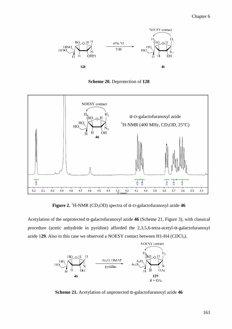
Chapter 6
163
Scheme 20. Deprotection of 128
Figure 2. 1H-NMR (CD3OD) spectra of α-D-galactofuranosyl azide 46
Acetylation of the unprotected α-galactofuranosyl azide 46 (Scheme 21, Figure 3), with classical
procedure (acetic anhydride in pyridine) afforded the 2,3,5,6-tetra-acetyl-α-galactofuranosyl
azide 129. Also in this case we observed a NOESY contact between H1-H4 (CDCl3).
Scheme 21. Acetylation of unprotected α-galactofuranosyl azide 46
α-D-galactofuranosyl azide 1H-NMR (400 MHz, CD3OD, 25°C)
H1
H OH
HO HO
HOHO
H
N3
H4
NOESY contact
46

Chapter 6
164
Figure 3. 1H-NMR (CDCl3) spectra of tetra-O-acetyl-α-D-galactofuranosyl azide 129
The unprotected and tetra-O-acetyl-α-galactofuranosyl azides 46 and 129 (Scheme 22) were
subjected to traceless ligation with compound 81b in the same reaction conditions used for the
ligation of protected and unprotected β-galactofuranosyl azides 36 and 37 (70°C for 4h in
solvent mixture DMA:DMPU 98:2).
The ligation of unprotected α-galactofuranosyl azide 46 with phosphines 81b afforded the amide
130b with the same anomeric configuration of the starting azide precursor, in a
stereoconservative process. Unprotected α-furanosyl azides 46, thus, did not anomerize but
afforded the 1,2-cis azide, as it would be expected based on the proposed mechanism.
Surprisingly, ligation of the of tetra-O-acetyl-α-furanosyl azides 129 afforded a 4:1 β:α ratio,
thus revealing that an equilibration process is taking place for this acetylated azide, which favors
the 1,2-trans amide 113b with inversion on the anomeric carbon (Scheme 22). The inversion of
configuration during the reaction of protected galactofuranosyl azide 129 suggests that this
configuration may be more stable for the tetra-acetylated compound.
H1
H OAc
RO HO
AcOAcO
H
N3
H4
NOESY contact
129
Tetra-O-acetyl-α-D-galactofuranosyl azide 1H-NMR (400 MHz, CDCl3, 25°C)

Chapter 6
165
1,2-trans
1,2-cis
b) H2O, 2 h, 70°C
a) 70°C, 4h, DMA/DMPU
b) H2O, 2 h, 70°C
a) 70°C, 4h, DMA/DMPU
PPh2
O
O
c) MeONa 0.05M, MeOH
129
46
81b
113b
111b
H OH
HO HO
HOHO
H
N3
H
H OAc
AcO HO
AcOAcO
H
N3
H
H OH
HO HO
HOHO
H
NHCOC4H9
H
H OH
HO HO
HOHO
H
H
NHCOC4H9
4:1113b:111b
Scheme 22. Staudinger ligation of 81b with α-galactofuranosyl azide 46 and 129
After these observations the mechanism proposed in Scheme 17 for α-furanosylamide formation
appears totally confirmed. The full implications of this proposed mechanism relative to the
ligation of furanosyl and pyranosyl azides will be discussed in Chapter 7.
6.5 Synthesis of small library of αααα and ββββ galactofuranosylamides as inhibitors
of Mycobacteria
The arabinogalactan constituent of mycobacterial cell walls contains a galactan polymer made of
galactose residues in their furanose form. In particular galactofuranose (D-Galf ) was found to be
an important coustituent of the cell wall of mycobacteria the causative agent of tuberculosis
(TB).7, 8 The development of new drugs for tuberculosis has taken on some urgency in the recent
years. The World Health Organization reports that the global incidence rate ot TB is rising, and
more than 4000 people die of TB everyday. 13 More new cases are expected since TB is also the
main cause of death in AIDS patients. Inhibition of the incorporation of D-galactofuranose (D-
Galf ) has become a potential therapeutic strategy since Galf-containing oligosaccharides are
critical for survival and infectivity 14, 15 of mycobacteria. Incorporation of Galf into

Chapter 6
166
oligosaccharides involves two steps. First, the activated precursor UDP-galactofuranose (UDP-
Galf 1) is synthesized from UDP-galactopyranose (UDP-Galp 2) in a reaction catalyzed by UDP-
galactopyranose mutase (UGM). Subsequently, Galf is transferred from UDP-Galf 1 onto
acceptors to form various Galf-containing oligosaccharides by UDP-galactofuranosyltransferase
(GalfT).16, 17
The furanose form of D-galactose, Galf, is uknown in mammals and UDP-galactopyranose
(UDP-Galp 2) mutase has become an interesting target for the development of drugs against the
many pathogens which contain Galf, most prominently Mycobacterium tuberculosis. UDP-Galp
2 mutase inhibitors have been reported and shown to block mycobacterial growth.18, 19, 20, 21, 22, 23
Various molecules containing a structural unit mimicking the Galf monosaccharide have been
synthesised, including iminosugars, thiosugars, galactofuranosyl thioglycosides and C-
glycosides (Figure 4), but no galactofuranosylamides have been described so far. The ability of
some of these compounds to inhibit bacterial growth makes galactofuranose mimics interesting
biological targets with potential therapeutic value.7, 24, 25, 26
Figure 4. Some analogues of β-galactofuranose which have shown activity against
Mycobacterium tubercolosis
To explore the scope of galactofuranosyl amides as bacterial growth inhibitors, we used the
stereodivergent ligation of furanosyl azides just discovered and applied it to a set of phosphines

Chapter 6
167
variously functionalized, to obtain a small library of α- and β-galactofuranosyl amides (Scheme
23).
Scheme 23. Traceless Staudinger ligation of 36 and 37 with phosphines 81b-i
The results of this screening are collecetd in Table 2. Transfer of linear aliphatic (81b, f, g),
branched aliphatic (81c, e), unsaturated (81e, h) and aromatic (81i) amide chains was performed
with uniform results. All reactions gave the expected products in moderate yields. As previously
reported for unprotected glycopyranosyl azides,3 microwaves were found to accelerate the
ligation of 37 (entries 7 and 11). The stereoselectivity was complete, as determined by 1H-NMR
of the crudes. In all cases, β-anomers were obtained from the tetra-O-acetyl azide 36 and α-
anomers from the unprotected azide 37. Experimental procedures, purification and
characterization data were performed as described before in par. 6.2.

Chapter 6
168
Table 2. Ligation of 36 and 37 with phosphines 81b-i.a
Entr
y
Azid
e Phosphine R Product α/β b Yields
1 36 81b -(CH2)3CH3 113b 2:98 60
2 36 81c -CH2CH(CH3)2 113c > 1:99 53
3 36 81e -CH=C(CH3)2 113e 2:98 48
4 36 81f -(CH2)14CH3 113f > 1:99 60
5 36 81g -(CH2)6CH3 113g > 1:99 56
6 36 81h -CH2CH(CH3)(CH2)2CH=C(CH3)2 113h > 1:99 51
7 37 81b -(CH2)3CH3 111b > 99:1c 60
8 37 81c -CH2CH(CH3)2 111c > 99:1 57
9 37 81e -CH=C(CH3)2 111e > 99:1 51
10 37 81f -(CH2)14CH3 111f > 99:1 55
11 37 81g -(CH2)6CH3 111g > 99:1 c 63
12 37 81h -CH2CH(CH3)(CH2)2CH=C(CH3)2 111h > 99:1 56
13 37 81i -Ph 111i > 99:1 62
a) Unless otherwise stated, all reactions were performed in 98:2 DMA:DMPU mixtures for 4h at 70°C. Then water
was added and the solution stirred for additional 2h at 70°C before work up. b)1H-NMR ratio of the crude. c)
Reaction performed under microwave: irradiation at 30 min, 70°C.
All these compounds were tested in minimal inhibition concentration (MIC) tests against a panel
of bacteria. One compound, the α-galactofuranosyl amides 111f displayed a modest activity
against Mycobacterium bovis with a MIC value of 64 µg/ml. The β-anomer 113f showed no
activity.
This finding may represent the discovery of a new potential class of antibacterial agents, because
the totality of the molecules reported so far which contain a structural unit mimicking Galf are all
in the β-configuration. More work is required to verify this finding and to optimize the structure
and activity of these compounds.

Chapter 6
169
6.6 Experimental Section
Solvents were dried by standard procedures: dichloromethane, methanol, N,N-
diisopropylethylamine and triethylamine were dried over calcium hydride; N,N-
dimethylacetamide (DMA), 1,3-dimethyltetrahydro-2(1H)pyrimidinone (DMPU), chloroform
and pyridine were dried over activated molecular sieves. Reactions requiring anhydrous
conditions were performed under nitrogen. 1H, 13C and 31P-NMR spectra were recorded at 400
MHz on a Bruker AVANCE-400 instrument. Chemical shifts (δ) for 1H and 13C spectra are
expressed in ppm relative to internal Me4Si as standard. Signals were abbreviated as s, singlet;
bs, broad singlet; d, doublet; t, triplet; q, quartet; m, multiplet. Mass spectra were obtained with a
Bruker ion-trap Esquire 3000 apparatus (ESI ionization) and FT-ICR Mass Spectrometer APEX
II & Xmass software (Bruker Daltonics) - 4.7 Magnet. Thin layer chromatography (TLC) was
carried out with pre-coated Merck F254 silica gel plates. Flash chromatography (FC) was carried
out with Macherey-Nagel silica gel 60 (230-400 mesh).
General procedure for stereoselective ligation of protected and unprotected glycosyl azides
in DMA:DMPU mixtures.
The phosphine (1.2 eq) was added, at room temperature, to a 0.1 M solution of glycosyl azide (1
eq) in 98:2 N,N-dimethylacetamide and DMPU. The solution was stirred for 4 h at 70 °C, then
water was added and the mixture was stirred for an additional 2 h at the same temperature. The
solvent was evaporated under reduced pressure, and the residue was purified as indicated below
for each compound.
General procedure for acetylation of unprotected glycosyl amides.
Acetic anhydride (10 eq) and a catalytic amount of N,N-dimethylaminopyridine were added, at
room temperature, to a solution of substrate (1 eq) in pyridine dried on molecular sieves (0.1 M).
The solution was stirred for 24 h and then was concentrated in vacuo. The residue was dissolved
in AcOEt and washed with aqueous 5 % HCl, aqueous 5 % NaHCO3 and water. The organic
layer was dried over Na2SO4 and concentrated to give the product in quantitative yield.

Chapter 6
170
N-pentanoyl-α-D-galactofuranosyl amide (111b)
The compound was purified by flash chromatography (CHCl3:MeOH 80:20) yield = 60 %. 1H-NMR (400 MHz, CD3OD, 25°C): δ = 5.69 (d, J1,2 = 4.4 Hz, 1H, H-1), 4.13 (t, J2,3 = 3.4, J3,4 =
3.6 Hz, 1H, H-3), 3.88 (dd, J1,2 = 4.4, J2,3 = 3.4 Hz, 1H, H-2), 3.80 (dd, J3,4 = 3.6, J4,5 = 3 Hz, 1H,
H-4), 3.74 (ddd, J4,5 = 3, J5,6 = 6, J5,6’ = 6.8 Hz, 1H, H-5), 3.62 (dd, J5,6 = 6, J6,6’ = 11.6 Hz, 1H,
H-6), 3.57 (dd, J5,6’ = 6.8, J6,6’ = 11.6 Hz, 1H, H-6’), 2.29 (t, J = 7.6 Hz, 2H, CH2), 1.64-1.56 (m,
2H, CH2), 1.43-1.32 (m, 2H, CH2), 0.95 (t, J = 7.4 Hz, 3H, CH3). 13C-NMR (100 MHz, CD3OD,
25°C): δ = 176.7, 84.4 (C-4), 82.2 (C-1), 78.5 (C-3), 77.4 (C-2), 73 (C-5), 64.3 (C-6), 37 (CH2),
28.9 (CH2), 23.5 (CH2), 14.3 (CH3). [α]D25 = +18.1 (c 1, MeOH). FT-ICR (ESI) calcd. for
C11H21N1O6 [M+Na]+ 286.12611; found 286.12597.
N-pentanoyl-α-D-galactofuranosyl amide 1H-NMR (400 MHz, CD3OD, 25°C)

Chapter 6
171
2,3,5,6-tetra-O-acetyl-N-pentanoyl-α-D-galactofuranosyl amide (114b)
1H-NMR (400 MHz, CDCl3, 25°C): δ = 6.14 (d, JNH,1 = 10 Hz, 1H, NH), 6.06 (d, J1,2 = 4.4 Hz,
JNH,1 = 10, 1H, H-1), 5.34 (ddd, J4,5 = 6.8, J5,6 = 3.6, J5,6’ = 6.8 Hz, 1H, H-5), 5.21 (dd, J1,2 = 4.4,
J2,3 = 1.8 Hz, 1H, H-2),5.07 (dd, J2,3 = 1.8, J3,4 = 3.2 Hz, 1H, H-3), 4.38 (dd, J5,6 = 3.6, J6,6’ = 12
Hz, 1H, H-6), 4.09 (dd, J5,6’ = 6.8, J6,6’ = 12 Hz, 1H, H-6’), 3.92 (dd, J3,4 = 3.2, J4,5 = 6.8 Hz, 1H,
H-4), 2.23 (t, J = 7.6 Hz, 2H, CH2), 2.18 (s, 3H, OAc), 2.12 (s, 3H, OAc), 2.10 (s, 3H, OAc),
2.05 (s, 3H, OAc), 1.61-1.56 (m, 2H, CH2), 1.41-1.32 (m, 2H, CH2), 0.92 (t, J = 7.6 Hz, 3H,
CH3). 13C-NMR (100 MHz, CDCl3, 25°C): δ = 176.7, 173, 170.7, 169.8, 169.1, 79.9 (C-1), 79.7
(C-4), 76.7 (C-3), 75.1 (C-2), 70.3 (C-5), 62.9 (C-6), 36.7 (CH2), 29.9 (CH2), 22.5 (CH2), 21.1-
20.8 (4xOAc), 14.3 (CH3). NOESY (400 MHz, CDCl3, 25°C): contact between H-1/H-4.
2,3,5,6-tetra-O-acetyl-N-pentanoyl-α-D-galactofuranosyl amide 1H-NMR (400 MHz, CDCl3, 25°C)

Chapter 6
172
N-(3-Methylbutanoyl)-α-D-galactofuranosyl amide (111c)
The compound was purified by flash chromatography (CHCl3:MeOH 80:20) yield = 57 %. 1H-NMR (400 MHz, CD3OD, 25°C): δ = 5.72 (d, J1,2 = 4.4 Hz, 1H, H-1), 4.16 (t, J2,3 = 3.2, J3,4 =
3.6 Hz, 1H, H-3), 3.92 (dd, J1,2 = 4.4, J2,3 = 3.2 Hz, 1H, H-2), 3.84 (dd, J3,4 = 3.6, J4,5 = 3.2 Hz,
1H, H-4), 3.78 (ddd, J4,5 = 3.2, J5,6 = 5.6, J5,6’ = 6.8 Hz, 1H, H-5), 3.65 (dd, J5,6 = 5.6, J6,6’ = 11.2
Hz, 1H, H-6), 3.61 (dd, J5,6’ = 6.8, J6,6’ = 11.2 Hz, 1H, H-6’), 2.18 (d, J = 6.4 Hz, 2H, CH2), 2.16-
2.08 (m, 1H, CH), 1.01 (d, J = 2.4 Hz, 3H, CH3), 0.99 (d, J = 2.4 Hz, 3H, CH3). 13C-NMR (100
MHz, CD3OD, 25°C): δ = 176, 85.6 (C-4), 84.5 (C-1), 82.2 (C-3), 77.4 (C-2), 73 (C-5), 64.3 (C-
6), 46.5 (CH2), 27.3 (CH), 22.9 (CH3). [α]D
25 = +33.4 (c 1, MeOH). FT-ICR (ESI) calcd. for
C11H21N1O6 [M+Na]+ 286.12611; found 286.12591.
N-3-methyl-2-butenoyl-α-D-galactofuranosyl amide (111e)
The compound was purified by flash chromatography (CHCl3:MeOH 85:15) yield = 51 %. 1H-NMR (400 MHz, CD3OD, 25°C): δ = 5.86 (s, 1H, CH), 5.77 (d, J1,2 = 4 Hz, 1H, H-1), 4.17 (t,
J2,3 = 3.2, J3,4 = 3.6 Hz, 1H, H-3), 3.94 (dd, J1,2 = 4, J2,3 = 3.2 Hz, 1H, H-2), 3.85 (bt, J3,4 = 3.6,
J4,5 = 3.2 Hz, 1H, H-4), 3.79 (ddd, J4,5 = 3.2, J5,6 = 6, J5,6’ = 6.8 Hz, 1H, H-5), 3.68 (dd, J5,6 = 6,
J6,6’ = 11.2 Hz, 1H, H-6), 3.62 (dd, J5,6’ = 6.8, J6,6’ = 11.2 Hz, 1H, H-6’), 2.20 (s, 3H, CH3), 1.94
(s, 3H, CH3). 13C-NMR (100 MHz, CD3OD, 25°C): δ = 169.5, 154.2, 119.4 (CH), 84.4 (C-4),
82.1 (C-1), 78.7 (C-3), 77.5 (C-2), 73.1 (C-5), 64.4 (C-6), 27.4 (CH3), 20.3 (CH3). [α]D
25 = +30.2
(c 0.8, MeOH). FT-ICR (ESI) calcd. for C11H21N1O6 [M+Na]+ 284.11046; found 284.11044.

Chapter 6
173
N-palmitoyl-αααα-D-galactofuranosyl amide (111f)
The compound was purified by flash chromatography (CHCl3:MeOH 90:10) yield = 55 %. 1H-NMR (400 MHz, CD3OD, 25°C): δ = 5.74 (d, J1,2 = 4 Hz, 1H, H-1), 4.18 (t, J2,3 = 3.2, J3,4 =
3.6 Hz, 1H, H-3), 3.94 (dd, J1,2 = 4, J2,3 = 3.2 Hz, 1H, H-2), 3.85 (bt, J3,4 = 3.6, J4,5 = 2.8 Hz, 1H,
H-4), 3.79 (ddd, J4,5 = 2.8, J5,6 = 6, J5,6’ = 6.8 Hz, 1H, H-5), 3.67 (dd, J5,6 = 6, J6,6’ = 11.2 Hz, 1H,
H-6), 3.63 (dd, J5,6’ = 6.8, J6,6’ = 11.2 Hz, 1H, H-6’), 2.33 (t, J = 7.2 Hz, 2H, CH2), 1.68 (bt, J =
7.2 Hz, 2H, CH2), 1.44-1.28 (m, 24H), 0.96 (t, J = 6.8 Hz, 3H, CH3). 13C-NMR (100 MHz,
CD3OD, 25°C): δ = 176.7, 84.4 (C-4), 82.2 (C-1), 78.5 (C-3), 77.4 (C-2), 73 (C-5), 64.4 (C-6),
37.3, 33.2, 30.9, 30.8, 30.6, 30.4, 26.8, 23.9, 14.6 (CH3). [α]D
25 = +9 (c 1, MeOH). FT-ICR (ESI)
calcd. for C22H43N1O6 [M+Na]+ 440.29826; found 440.29797.
N-capryloyl-αααα-D-galactofuranosyl amide (111g)
The compound was purified by flash chromatography (CHCl3:MeOH 85:15) yield = 63 %. 1H-NMR (400 MHz, CD3OD, 25°C): δ = 5.68 (d, J1,2 = 4.4 Hz, 1H, H-1), 4.12 (t, J2,3 = 3.6, J3,4 =
3.2 Hz, 1H, H-3), 3.87 (dd, J1,2 = 4.4, J2,3 = 3.6 Hz, 1H, H-2), 3.79 (t, J3,4 = 3.2, J4,5 = 2.8 Hz, 1H,
H-4), 3.73 (ddd, J4,5 = 2.8, J5,6 = 6, J5,6’ = 6.8 Hz, 1H, H-5), 3.61 (dd, J5,6 = 6, J6,6’ = 11.2 Hz, 1H,
H-6), 3.57 (dd, J5,6’ = 6.8, J6,6’ = 11.2 Hz, 1H, H-6’), 2.27 (t, J = 7.2 Hz, 2H, CH2), 1.62 (bt, J =
7.2 Hz, 2H, CH2), 1.40-1.24 (m, 8H, 4xCH2), 0.91 (t, J = 6.4, J = 7.2 Hz, 3H, CH3). 13C-NMR
(100 MHz, CD3OD, 25°C): δ = 176.7, 84.4 (C-4), 82.2 (C-1), 78.5 (C-3), 77.3 (C-2), 72.9 (C-5),
64.3 (C-6), 37.3 (CH2), 33.0, 30.4, 30.3 (3xCH2), 26.8 (CH2), 23.8 (1xCH2), 14.6 (CH3). [α]D
25 =
+12.2 (c 1, MeOH). FT-ICR (ESI) calcd. for C14H27N1O6 [M+Na]+ 328.17306; found 328.17314.

Chapter 6
174
N-citronelloyl-αααα-D-galactofuranosyl amide (111h)
The compound was purified by flash chromatography (CHCl3:MeOH 90:10) yield = 56 %. 1H-NMR (400 MHz, CD3OD, 25°C): δ = 5.74 (d, J1,2 = 4 Hz, 1H, H-1), 5.17 (t, J = 5.8 Hz, 1H,
CH) 4.18 (t, J2,3 = 3.4, J3,4 = 3.6 Hz, 1H, H-3), 3.93 (dd, J1,2 = 4, J2,3 = 3.4 Hz, 1H, H-2), 3.85 (dd,
J3,4 = 3.6, J4,5 = 3.2 Hz, 1H, H-4), 3.79 (ddd, J4,5 = 3.2, J5,6 = 5.6, J5,6’ = 6.8 Hz, 1H, H-5), 3.68
(dd, J5,6 = 5.6, J6,6’ = 11.2 Hz, 1H, H-6), 3.63 (dd, J5,6’ = 6.8, J6,6’ = 11.2 Hz, 1H, H-6’), 2.34 (dd, J
= 13.6, J = 6 Hz, 1H, Ha, CH2), 2.18-1.98 (m, 4H), 1.73 (s, 3H, CH3), 1.67 (s, 3H, CH3), 1.48-
1.40 (m, 1H), 1.33-1.22 (m, 1H), 0.95 (d, J = 6.8 Hz, 3H, CH3). 13C-NMR (100 MHz, CD3OD,
25°C): δ = 175.9, 132.4, 125.7 (CH), 84.5 (C-4), 82.3 (C-1), 78.5 (C-3), 77.4 (C-2), 73 (C-5),
64.4 (C-6), 44.9, 38.1, 31.7, 26.6, 26, 19.9, 17.9. [α]D25 = +13.2 (c 1, MeOH). FT-ICR (ESI)
calcd. for C16H29N1O6 [M+Na]+ 354.18871; found 354.18864.
N-benzoyl-α-D-galactofuranosyl amide (111i)
The compound was purified by flash chromatography (CHCl3:MeOH 85:15) yield = 62 %. 1H-NMR (400 MHz, CD3OD, 25°C): δ = 7.91 (m, 2H, Horto), 7.60 (m, 1H, Hpara), 7.53 (m, 2H,
Hmeta), 5.94 (d, J1,2 = 4.8 Hz, 1H, H-1), 4.27 (t, J2,3 = 4.2, J3,4 = 4.4 Hz, 1H, H-3), 4.13 (t, J1,2 =
4.8, J2,3 = 4.2 Hz, 1H, H-2), 3.93 (dd, J3,4 = 3.6, J4,5 = 2.8 Hz, 1H, H-4), 3.84 (ddd, J4,5 = 2.8, J5,6
= 5.4, J5,6’ = 6.8 Hz, 1H, H-5), 3.70 (dd, J5,6 = 5.6, J6,6’ = 11.2 Hz, 1H, H-6), 3.66 (dd, J5,6’ = 6.8,
J6,6’ = 11.2 Hz, 1H, H-6’). 13C-NMR (100 MHz, CD3OD, 25°C): δ = 169.8, 135.4, 133.2, 129.8,
128.6, 84.5 (C-4), 82.7 (C-1), 78.2 (C-3), 77.9 (C-2), 72.7 (C-5), 64.4 (C-6). [α]D25 = +7.9 (c 1,
MeOH). FT-ICR (ESI) calcd. for C13H17N1O6 [M+Na]+ 306.09481; found 306.09465.

Chapter 6
175
N-pentanoyl-ββββ-D-galactofuranosyl amide (113b)
After Zemplen deacetylation the compound was purified by flash chromatography
(CHCl3:MeOH 80:20) yield = 60 %. 1H-NMR (400 MHz, CD3OD, 25°C): δ = 5.38 (d, J1,2 = 5.2 Hz, 1H, H-1), 4.14 (t, J2,3 = 5.8, J3,4 =
6.4 Hz, 1H, H-3), 3.95 (t, J1,2 = 5.2, J2,3 = 5.8 Hz, 1H, H-2), 3.90 (dd, J3,4 = 6.4, J4,5 = 2.8 Hz, 1H,
H-4), 3.68-3.63 (m,1H, H-5), 3.62-3.57 (m, 2H, H-6, H-6’), 2.22 (t, J = 7.4 Hz ,2H, CH2), 1.71-
1.54 (m, 2H, CH2), 1.48-1.32 (m, 2H, CH2), 0.95 (t, J = 7.2 Hz, 3H, CH3). 13C-NMR (100 MHz,
CD3OD, 25°C): δ = 177.4, 85.5 (C-1), 83.6 (C-4), 81.1 (C-2), 77.5 (C-3), 72.7 (C-5), 64.4 (C-6),
37.1 (CH2), 28.9 (CH2), 23.5 (CH2), 14.3 (CH3). NOESY (400 MHz, CD3OD, 25°C): contact
between H-1/H-3.
[α]D25 = -17.9 (c 0.2, MeOH). FT-ICR (ESI) calcd. for C11H21N1O6 [M+Na]+ 286.12611; found
286.12586.
N-pentanoyl-β-D-galactofuranosyl amide 1H-NMR (400 MHz, CDCl3, 25°C)

Chapter 6
176
N-(3-Methylbutanoyl)-ββββ-D-galactofuranosyl amide (113c)
After Zemplen deacetylation the compound was purified by flash chromatography
(CHCl3:MeOH 85:15) yield = 53 %. 1H-NMR (400 MHz, CD3OD, 25°C): δ = 5.40 (d, J1,2 = 5.2 Hz, 1H, H-1), 4.15 (t, J2,3 = 5.8, J3,4 =
6.4 Hz, 1H, H-3), 3.97 (t, J1,2 = 5.2, J2,3 = 5.8 Hz, 1H, H-2), 3.92 (dd, J3,4 = 6.4, J4,5 = 2.8 Hz, 1H,
H-4), 3.71-3.65 (m,1H, H-5), 3.63-3.58 (m, 2H, H-6, H-6’), 2.35 (d, J = 7.2 Hz, 2H, CH2), 1.01
(d, J = 6.4 Hz , 3H, CH3), 0.97 (d, J = 6 Hz, 3H, CH3). 13C-NMR (100 MHz, CD3OD, 25°C): δ =
176.6, 85.6 (C-1), 83.7 (C-4), 81.2 (C-2), 77.5 (C-3), 72.7 (C-5), 64.4 (C-6), 46.5 (CH2), 22.9
(CH3), 22.7 (CH3). [α]D
25 = -24.5 (c 1, MeOH). FT-ICR (ESI) calcd. for C11H21N1O6 [M+Na]+
286.12611; found 286.12584.
N-3-methyl-2-butenoyl-ββββ-D-galactofuranosyl amide (113e).
After Zemplen deacetylation the compound was purified by flash chromatography
(CHCl3:MeOH 85:15) yield = 48 %. 1H-NMR (400 MHz, CD3OD, 25°C): δ = 5.74 (s, 1H, CH), 5.41 (d, J1,2 = 5.6 Hz, 1H, H-1), 4.15
(t, J2,3 = 5.8, J3,4 = 6.4 Hz, 1H, H-3), 3.96 (t, J1,2 = 5.6, J2,3 = 5.8 Hz, 1H, H-2), 3.90 (dd, J3,4 = 6.4,
J4,5 = 2.8 Hz, 1H, H-4), 3.68-3.63 (m,1H, H-5), 3.61-3.57 (m, 2H, H-6, H-6’), 2.15 (s, 3H, CH3),
1.89 (s, 3H, CH3). 13C-NMR (100 MHz, CD3OD, 25°C): δ = 170.2, 154.3, 119.3 (CH), 85.4 (C-
1), 83.4 (C-4), 81.1 (C-2), 77.5 (C-3), 72.8 (C-5), 64.4 (C-6), 27.5 (CH3), 20.3 (CH3). [α]D
25 = -
27.8 (c 0.2, MeOH). FT-ICR (ESI) calcd. for C11H21N1O6 [M+Na]+ 284.11046; found
284.11027.

Chapter 6
177
N-palmitoyl-ββββ-D-galactofuranosyl amide (113f).
After Zemplen deacetylation the compound was purified by flash chromatography
(CHCl3:MeOH 90:10) yield = 60 %. 1H-NMR (400 MHz, CD3OD, 25°C): δ = 5.40 (d, J1,2 = 5.2 Hz, 1H, H-1), 4.15 (t, J2,3 = 6, J3,4 = 6
Hz, 1H, H-3), 3.96 (t, J1,2 = 5.2, J2,3 = 5.6 Hz, 1H, H-2), 3.93 (dd, J3,4 = 6.4, J4,5 = 2.8 Hz, 1H, H-
4), 3.70-3.65 (m, 1H, H-5), 3.63-3.58 (m, 2H, H-6, H-6’), 2.24 (t, J = 8 Hz, 2H, CH2), 1.63 (m,
2H, CH2), 1.41-1.25 (m, 24 H, 12x CH2), 0.92 (t, J = 6.8 Hz, 3H, CH3). 13C-NMR (100 MHz,
CD3OD, 25°C): δ = 177.1, 85.7 (C-1), 83.8 (C-4), 81.2 (C-2), 77.6 (C-3), 72.8 (C-5), 64.4 (C-6),
37.4, 33.2, 30.9, 30.7, 30.6, 30.4, 26.8, 23.8, 14.6 (CH3). [α]D
25 = -37.3 (c 1, MeOH). FT-ICR
(ESI) calcd. for C22H43N1O6 [M+Na]+ 440.29826; found 440.29844.
N-capryloyl-ββββ-D-galactofuranosyl amide (113g)
After Zemplen deacetylation the compound was purified by flash chromatography
(CHCl3:MeOH 85:15) yield = 56 %. 1H-NMR (400 MHz, CD3OD, 25°C): δ = 5.42 (d, J1,2 = 5.2 Hz, 1H, H-1), 4.17 (t, J2,3 = 5.8, J3,4 =
6.4 Hz, 1H, H-3), 3.98 (t, J1,2 = 5.2, J2,3 = 5.8 Hz, 1H, H-2), 3.95 (dd, J3,4 = 6.4, J4,5 = 2.8 Hz, 1H,
H-4), 3.73-3.68 (m, 1H, H-5), 3.65-3.59 (m, 2H, H-6, H-6’), 2.28 (t, J = 7.2, J = 8 Hz, 2H, CH2),
1.65 (m, 2H, CH2), 1.47-1.27 (m, 8H, 4xCH2), 0.94 (t, J = 6.8 Hz, 3H, CH3). 13C-NMR (100
MHz, CD3OD, 25°C): δ = 177.1, 85.4 (C-1), 83.6 (C-4), 81 (C-2), 77.3 (C-3), 72.6 (C-5), 64.2
(C-6), 37.2, 32.8, 30.2, 30.1, 26.7, 23.6, 14.4 (CH3). [α]D
25 = -50.4 (c 0.9, MeOH). FT-ICR (ESI)
calcd. for C14H27N1O6 [M+Na]+ 328.17306; found 328.17317.

Chapter 6
178
N-citronelloyl-ββββ-D-galactofuranosyl amide (113h)
After Zemplen deacetylation the compound was purified by flash chromatography
(CHCl3:MeOH 90:10) yield = 51 %. 1H-NMR (400 MHz, CD3OD, 25°C): δ = 5.45 (d, J1,2 = 5.2 Hz, 1H, H-1), 5.16 (t, J = 6.8 Hz, 1H,
CH) 4.20 (t, J2,3 = 5.6, J3,4 = 6.2 Hz, 1H, H-3), 4.01 (t, J1,2 = 5.2, J2,3 = 5.6 Hz, 1H, H-2), 3.97 (dd,
J3,4 = 6.2, J4,5 = 2.8 Hz, 1H, H-4), 3.71 (ddd, J4,5 = 2.8, J = 6, J = 6.8 Hz, 1H, H-5), 3.68-3.63 (m,
2H, H-6, H-6’), 2.29 (dd, J = 13.6, J = 6 Hz, 1H, Ha, CH2), 2.18-1.98 (m, 4H), 1.73 (s, 3H, CH3),
1.67 (s, 3H, CH3), 1.48-1.40 (m, 1H), 1.33-1.22 (m, 1H), 1.01 (d, J = 6.4 Hz, 3H, CH3). 13C-
NMR (100 MHz, CD3OD, 25°C): δ =176.6, 132.4, 125.7, 85.7 (C-1), 83.8 (C-4), 81.2 (C-2),
77.6 (C-3), 72.8 (C-5), 64.4 (C-6), 44.9, 38.1, 30.9, 26.6, 26, 19.9, 17.9. [α]D25 = -55.8 (c 0.9,
MeOH). FT-ICR (ESI) calcd. for C16H29N1O6 [M+Na]+ 354.18871; found 354.18866.
N-pentanoyl-αααα-D-ribofuranosyl amide (115b)
The compound was purified by flash chromatography (CHCl3:MeOH 80:20) yield = 59 %. 1H-NMR (400 MHz, CD3OD, 25°C): δ = 5.67 (d, J1,2 = 4.8 Hz, 1H, H-1), 4.12-4.06 (m, 2H, H-2,
H-3), 3.93-3.87 (q, J4,5 = 3.2 Hz, J4,5’= 4.4 Hz, 1H, H-4), 3.69 (dd, J4,5 = 3.2 Hz, J5,5’= 12 Hz, 1H,
H-5), 3.55 (dd, J4,5’ = 4.4 Hz, J5,5’= 12 Hz, 1H, H-5’) 2.24 (t, J = 7.2 Hz ,2H, CH2), 1.65-1.57 (m,
2H, CH2), 1.43-1.33 (m, 2H, CH2), 0.95 (t, J = 7.2 Hz, 3H, CH3). 13C-NMR (100 MHz, CD3OD,
25°C): δ = 176.6, 84.2 (C-4), 81.5 (C-1), 72.6, 71.8, 62.9 (C-5), 36.9 (CH2), 28.8 (CH2), 23.3
(CH2), 14.1 (CH3).

Chapter 6
179
N-pentanoyl-ββββ-D-ribofuranosyl amide (116b)
After Zemplen deacetylation the compound was purified by flash chromatography
(CHCl3:MeOH 80:20) yield = 55 %. 1H-NMR (400 MHz, CD3OD, 25°C): δ = 5.38 (d, J1,2 = 4.8 Hz, 1H, H-1), 4.06 (t, J2,3 = 5.2, J3,4 =
5.2 Hz, 1H, H-3), 3.91 (t, J1,2 = 4.8, J2,3 = 5.2 Hz, 1H, H-2), 3.88-3.83 (q, J4,5 = 3.6 Hz, J4,5’= 4.4
Hz, 1H, H-4), 3.70 (dd, J4,5 = 3.6 Hz, J5,5’= 12 Hz, 1H, H-5), 3.61 (dd, J4,5’ = 4.4 Hz, J5,5’= 12 Hz,
1H, H-5’) 2.23 (t, J = 7.2 Hz ,2H, CH2), 1.65-1.57 (m, 2H, CH2), 1.43-1.33 (m, 2H, CH2), 0.95
(t, J = 7.2 Hz, 3H, CH3). 13C-NMR (100 MHz, CD3OD, 25°C): δ = 176.8, 85.8 (C-1), 85.1 (C-4),
75.9 (C-2), 71.9 (C-3), 63.3 (C-5), 37.1 (CH2), 28.9 (CH2), 23.5 (CH2), 14.3 (CH3). NOESY (400
MHz, CD3OD, 25°C): contact between H-1/H-4.
N-pentanoyl-α-D-ribofuranosyl amide 1H-NMR (400 MHz, CDCl3, 25°C)

Chapter 6
180
Tetra-O-acetyl N-pentanoyl-ββββ-D-ribofuranosyl amide (117b)
1H-NMR (400 MHz, CD3OD, 25°C): δ = 6.06 (d, J1,2 = 4.8 Hz, 1H, H-1), 5.45 (t, J1,2 = 4.8, J2,3 =
5.6 Hz, 1H, H-2), 5.36 (t, J2,3 = 5.6 Hz, 1H, H-3), 4.36-4.31 (m, 2H, H-4, H-5), 4.21 (dd, J4,5’ =
4.8, J5,5’= 12 Hz, 1H, H-5’) 2.23 (t, J = 7.2 Hz ,2H, CH2), 2.19 (s, 3H, OAc), 2.15 (s, 3H, OAc),
2.12 (s, 3H, OAc), 1.67-1.59 (m, 2H, CH2), 1.44-1.35 (m, 2H, CH2), 0.99 (t, J = 7.2 Hz, 3H,
CH3). 13C-NMR (100 MHz, CD3OD, 25°C): δ = 176.1, 172.2, 171.5, 171.3, 80.8 (C-1), 79.2 (C-
4), 72.9 (C-3), 71.9 (C-2), 64.8 (C-5), 36.8 (CH2), 29 (CH2), 23.4 (CH2),20.7-20.5 (3xOAc), 14.2
(CH3). NOESY (400 MHz, CD3OD, 25°C): contact between H-1/H-3 and H-1/H-5.
N-pentanoyl-β-D-ribofuranosyl amide 1H-NMR (400 MHz, CDCl3, 25°C)

Chapter 6
181
N-pentanoyl-ββββ-D-arabinobofuranosyl amide (118b)
The compound was purified by flash chromatography (CHCl3:MeOH 80:20) yield = 57 %. 1H-NMR (400 MHz, CD3OD, 25°C): δ = 5.72 (d, J1,2 = 4.4 Hz, 1H, H-1), 4.01 (t, J2,3 = 3.6 Hz,
J3,4= 3.6 Hz, 1H, H-3), 3.90 (dd, J1,2= 4.4 Hz, J2,3 = 3.6 Hz, 1H, H-2), 3.76 (ddd, J3,4= 3.6 Hz, J4,5
= 3.6 Hz, J4,5’= 5.2 Hz, 1H, H-4), 3.70 (dd, J4,5 = 3.6 Hz, J5,5’= 11.6 Hz, 1H, H-5), 3.64 (dd, J4,5’
= 5.2 Hz, J5,5’= 11.6 Hz, 1H, H-5’) 2.28 (t, J = 7.6 Hz ,2H, CH2), 1.65-1.57 (m, 2H, CH2), 1.43-
1.33 (m, 2H, CH2), 0.94 (t, J = 7.6 Hz, 3H, CH3). 13C-NMR (100 MHz, CD3OD, 25°C): δ =
176.8, 84.4 (C-4), 82.2 (C-1), 78.3 (C-3), 77.3 (C-2), 63.5 (C-5), 36.9 (CH2), 28.9 (CH2), 23.5
(CH2), 14.3 (CH3). NOESY (400 MHz, CD3OD, 25°C): contact between H-1/H-4.
N-pentanoyl-β-D-arabinobofuranosyl amide 1H-NMR (400 MHz, CDCl3, 25°C)

Chapter 6
182
N-pentanoyl-αααα-D-arabinobofuranosyl amide (119b)
After Zemplen deacetylation the compound was purified by flash chromatography
(CHCl3:MeOH 80:20) yield = 53 %. 1H-NMR (400 MHz, CD3OD, 25°C): δ = 5.39 (d, J1,2 = 4.8 Hz, 1H, H-1), 4.03 (t, J2,3 = 5.2 Hz,
1H, H-3), 3.88 (t, J1,2 = 4.8, J2,3 = 5.2 Hz, 1H, H-2), 3.84 (q, J4,5 = 3.6 Hz, J4,5’= 4.4 Hz, 1H, H-4),
3.67 (dd, J4,5 = 3.6, J5,5’= 12 Hz, 1H, H-5), 3.55 (dd, J4,5’ = 4.4 Hz, J5,5’= 12 Hz, 1H, H-5’) 2.20 (t,
J = 7.6 Hz ,2H, CH2), 1.60-1.54 (m, 2H, CH2), 1.40-1.29 (m, 2H, CH2), 0.92 (t, J = 7.2 Hz, 3H,
CH3).
N-pentanoyl-α-D-arabinobofuranosyl amide 1H-NMR (400 MHz, CDCl3, 25°C)

Chapter 6
183
N-pentanoyl-ββββ-D-ribopyranosyl amide (120b)
The compound was purified by flash chromatography (CHCl3:MeOH 80:20) yield = 68 %. 1H-NMR (400 MHz, CD3OD, 25°C): δ = 5.15 (d, J1,2 = 9.2 Hz, 1H, H-1), 4.07 (d, J2,3 = 2.8 Hz,
1H, H-3), 3.73-3.57 (m, 3H, H-4, H-5, H-5’), 3.39 (dd, J1,2 = 9.2, J2,3 = 2.8 Hz, 1H, H-2), 2.22 (t,
J = 7.6 Hz ,2H, CH2), 1.64-1.56 (m, 2H, CH2), 1.43-1.32 (m, 2H, CH2), 0.94 (t, J = 7.6 Hz, 3H,
CH3). 13C-NMR (100 MHz, CD3OD, 25°C): δ = 177.5, 78 (C-1), 72.4 (C-3), 71.3 (C-2), 68.8 (C-
4), 65.7 (C-5), 37 (CH2), 28.9 (CH2), 23.5 (CH2), 14.3 (CH3).
N-pentanoyl-β-D-ribopyranosyl amide 1H-NMR (400 MHz, CDCl3, 25°C)

Chapter 6
184
N-pentanoyl-ββββ-D-arabinopyranosyl amide (121b)
The compound was purified by flash chromatography (CHCl3:MeOH 80:20) yield = 70 %. 1H-NMR (400 MHz, CD3OD, 25°C): δ = 4.90 (d, J1,2 = 8 Hz, 1H, H-1), 3.95-3.88 (m, 2H, H-3,
H-5), 3.73-3.66 (m, 3H, H-2, H-4, H-5’), 2.34 (t, J = 7.6 Hz ,2H, CH2), 1.74-1.67 (m, 2H, CH2),
1.53-1.42 (m, 2H, CH2), 1.03 (t, J = 7.6 Hz, 3H, CH3). 13C-NMR (100 MHz, CD3OD, 25°C): δ =
177.2, 81.8 (C-1), 75.3 (C-2), 71.4 (C-4), 70.3 (C-3), 68.9 (C-5), 37.1 (CH2), 29 (CH2), 23.5
(CH2), 14.2 (CH3).
N-pentanoyl-β-D-arabinopyranosyl amide 1H-NMR (400 MHz, CDCl3, 25°C)

Chapter 6
185
Synthesis of 1,2,3,5,6-penta-O-tert-butyldimethylsislyl-ββββ-D-galactofuranose (126)
To a solution of D-galactose (0.5 g, 2.77 mmol) in dry DMF (14mL), imidazole (2.74 g, 18.25
mmol) and TBSCl (3 g, 20.08 mmol) were added, and the reaction mixture was stirred at room
temperature. After 48h TLC (Hexane/AcOEt 10:1) monitoring showed total consumption of the
starting material. The solution was poured into ice/water and was diluted with CH2Cl2. The
organic layer was washed with HCl (5 %), water, NaHCO3 (ss), and water, dried (Na2SO4), and
concentrated. Addition of MeOH (5 mL) to the syrup afforded crystalline compound 126. 1H-NMR (400 MHz, CDCl3, 25°C): δ = 4.41 (d, J = 7.1 Hz, 1H, H-1), 4.00 (d, J = 2.3 Hz, 1H,
H-4), 3.85 (dd, J = 10.0, 5.8, 2.9 Hz, 1H, H-6), 3.71 (dd, J = 10.0, 5.8 Hz, 1H, H-6’), 3.57 (m,
2H, H-2, H-3), 3.38 (ddd, J = 9.3, 5.4, 2.9 Hz, 1H, H-5), 0.93-0.87 (m, 45H, SiC(CH3)3), 0.14-
0.04 (m, 30H, Si(CH3)2). 13C-NMR (100 MHz, CDCl3, 25°C): δ = 98.7, 76.2, 75.2, 74.1, 69.2,
61.6, 26.3-18.0, -3.2 to -4.45.
Synthesis of 2,3,5,6-tetra-O-tert-butyldimethylsislyl-αααα-D-galactofuranosyl azide (128)
A solution of 126 (200 mg, 0.26 mmol) in anhydrous CH2Cl2 (10 mL) was cooled to 0°C and
stirred for 10 min. under nitrogen. Then, iodotrimethylsilane (1.2 eq.,0.042 mL, 0.32 mmol) was
added, and the solution was stirred at 0°C until TLC (Hexane/AcOEt 10:1) monitoring showed
complete transformation of 129 in two products Rf = 0.70 and Rf = 0.54. In parallel a solution of
tetrabutylammonium hydroxide was prepared by addition 280 µL of 10M sodium hydroxide
solution (an excess) to 383.3 mg (0.2 mol) of tetrabutylammonium hydrogene sulphate in 560 µL
of water.
A solution of 147 mg (0.4 mol) of sodium azide in 280 µL of water was added and
tetrabutylammonium azide extracted with 1 mL of dichloromethane. The organic layer, was
separated off and the aqueous phase again extracted with 1 mL of dichloromethane. The

Chapter 6
186
combined organic phases were evaporated in vacuo at 40°C to yield a crude tetrabutylammonium
azide as a colourless oil. The solutions thus obtained were used directly in the following step.
Then EtN(iPr)2 (0.054 mL, 0.032 mmol) and a solution of the azide were added, and the stirring
was continued until consumption of the components of Rf = 0.70 and Rf = 0.54. The solution was
diluted with CH2Cl2 washed with HCl 5 %, NaHCO3 (ss) and water, dried (Na2SO4), and
concentrated. The syrup obtained was purified by flash column chromatography (Hexane:AcOEt
98:2). Yield = 75 % 1H-NMR (400 MHz, CDCl3, 25°C): δ = 5.11 (d, J1,2 = 4 Hz, 1H, H-1), 4.24 (t, J2,3 = 4.4, J3,4 = 4.6
Hz, 1H, H-3), 3.99 (t, J1,2 = 4, J2,3 = 4.4 Hz, 1H, H-2), 3.87 (t, J3,4 = 4.6, J4,5 = 4.4 Hz, 1H, H-4),
3.79 (ddd, J4,5 = 4.4, J5,6 = 6, J5,6’ = 5.6 Hz, 1H, H-5), 3.66 (dd, J5,6 = 6, J6,6’ = 10 Hz, 1H, H-6),
3.60 (dd, J5,6’ = 5.6, J6,6’ = 10 Hz, 1H, H-6’), 0.92-0.84 (m, 36H, SiC(CH3)3), 0.18-0.06 (m, 24 H,
Si(CH3)2). 13C-NMR (100 MHz, CDCl3, 25°C): δ = 90.9 (C-1), 85.5 (C-4), 79.5 (C-2), 76.3 (C-
3), 73 (C-5), 65 (C-6), 26.2, 26.1, 26, 25.9, - 0.25 to -0.5. NOESY (400 MHz, CDCl3, 25°C):
contact between H-1/H-4.
Synthesis of αααα-D-galactofuranosyl azide (46)
A solution of 128 (60 mg, 0.09 mmol, 1 eq.) in anhydrous THF dry (180 µL,) was cooled to 0°C
and stirred for 10 min. under nitrogen. Then, tetrabutylammonium fluoride 1M in THF (157 µL,
0.54 mmol, 5 eq.) was added, and the solution was stirred at room temperature until TLC
(Hexane/AcOEt 60:40 and CHCl3/MeOH 80.20) monitoring showed complete transformation of
128 in 46.
The solution was diluted with water washed with dichloromethane and AcOEt and then
concentrated.The syrup obtained was purified by flash column chromatography (CHCl3:MeOH
90:10). Quantitative yield 1H-NMR (400 MHz, CD3OD, 25°C): δ = 5.17 (d, J1,2 = 4.8 Hz, 1H, H-1), 4.08 (t, J2,3 = 6.4, J3,4 =
6 Hz, 1H, H-3), 4.04 (t, J1,2 = 4.8, J2,3 = 6.4 Hz, 1H, H-2), 3.77 (dd, J3,4 = 6, J4,5 = 4.4 Hz, 1H, H-
4), 3.69 (ddd, J4,5 = 4.4, J5,6 = 5.2, J5,6’ = 6.8 Hz, 1H, H-5), 3.66 (dd, J5,6 = 5.2, J6,6’ = 11.2 Hz, 1H,
H-6), 3.60 (dd, J5,6’ = 6.8, J6,6’ = 11.2 Hz, 1H, H-6’). 13C-NMR (100 MHz, CD3OD, 25°C): δ =

Chapter 6
187
92.8 (C-1), 84.5 (C-4), 79 (C-2), 76.2 (C-3), 73.5 (C-5), 64.1 (C-6). NOESY (400 MHz, CD3OD,
25°C): contact between H-1/H-4.
Synthesis of 2,3,4,6-tetra-O-acetyl-αααα-D-galactofuranosyl azide (129)
Acetic anhydride (10 eq) and a catalytic amount of N,N-dimethylaminopyridine were added, at
room temperature, to a solution of substrate 46 (1 eq) in pyridine dried on molecular sieves (0.1
M). The solution was stirred for 24 h and then was concentrated in vacuo. The residue was
dissolved in AcOEt and washed with aqueous 5 % HCl, aqueous 5 % NaHCO3 and water. The
organic layer was dried over Na2SO4 and concentrated to give the product 129 in quantitative
yield. 1H-NMR (400 MHz, CDCl3, 25°C): δ = 5.55 (d, J1,2 = 5.6 Hz, 1H, H-1), 5.41 (t, J2,3 = 6.8, J3,4 =
6.4 Hz, 1H, H-3), 5.28 (ddd, J4,5 = 2, J5,6 = 4.8, J5,6’ = 6.4 Hz, 1H, H-5), 5.14 (t, J1,2 = 5.6, J2,3 =
6.8 Hz, 1H, H-2), 4.35 (dd, J5,6 = 4.8, J6,6’ = 12 Hz, 1H, H-6), 4.17 (dd, J5,6’ = 6.4, J6,6’ = 12 Hz,
1H, H-6’), 4.13 (dd, J3,4 = 6, J4,5 = 4.4 Hz, 1H, H-4), 2.13 (s, 3H, OAc), 2.10 (s, 3H, OAc), 2.08
α-D-galactofuranosyl azide 1H-NMR (400 MHz, CD3OD, 25°C)

Chapter 6
188
(s, 3H, OAc), 2.05 (s, 3H, OAc). 13C-NMR (100 MHz, CDCl3, 25°C): δ = 170.6, 170.2, 170.1,
169.9, 88.8 (C-1), 79.1 (C-4), 75.9 (C-2), 73.6 (C-3), 69.6 (C-5), 62.4 (C-6) 20.9-20.6 (4xOAc).
NOESY (400 MHz, CDCl3, 25°C): contact between H-1/H-4.
Tetra-O-acetyl-α-D-galactofuranosyl azide 1H-NMR (400 MHz, CDCl3, 25°C)

Chapter 6
189
6.7 References
1 Angyal, S. J. Carbohydr. Res. 1994, 263, 1-11. 2 Györgydeák, Z.; Szitagyi, L.; Paulsen H. J. Carb. Chem. 1993, 12, 139-163. 3 Norrild, J. Chr.; Eggert, H. J. Am. Chem. Soc. 1995, 117, 1479-1484. 4 Nisic, F.; Bernardi, A. Carbohydr. Res 2008, 343, 1636-1643. 5 Nisic, F.; Andreini, M.; Bernardi, A. Eur. J. Org.Chem, 2009, 5744-5751. 6 Chittenden, G. J. F. Carbohydr. Res 1972, 25, 35-41. 7 Owen, D. J.; Davis, C. B.; Hartnell, R. D.; Madge, P. D.; Thomson, R. J.; Chong, A. K. J.; Coppel, R. L.; M. von
Itzstein Carbohydr. Res. 2007, 342, 1773-1780. 8 Owen, D. J.; Davis, C. B.; Hartnell, R. D.; Madge, P. D.; Thomson, R. J.; Chong, A. K. J.; Coppel, R. L.; M. von
Itzstein Bioorg. Med. Chem. Lett. 2007, 17, 2274-2277. 9 Bianchi, A.; Bernardi, A. J. Org. Chem. 2006, 71, 4565-4577. 10 Baldoni, L.; Marino, C. J. Org. Chem. 2009, 74, 1994-2003. 11 Brandstrom, A.; Lamm, B.; Palmertz, I. Acta Chem. Scand. (B), 1974, 28, 699-701. 12 Corey, E. J.; Venkateswarlu, A. J. Am. Chem. Soc. 1972, 94, 6190-6191. 13 World Health Organization. Fact Sheet No. 104; World Health Organization: Geneva, 2002 14 Duncan, K. Curr. Pharm. Des. 2004, 10, 3185-3194. 15 Scherman, M. S.; Winans, K. A.; Bertozzi, C. R. Antimicrob. Agents Chemother. 2003, 378-382. 16 Kremer, L.; Dover, L.G.; Morehouse, C.; Hitchin, P.; Everett, M.; Morris, H. R.; Dell, A.;Brennan, P. J.;Besra,
G. S. J. Biol. Chem. 2001, 276, 26430-26440. 17 Rose, N. L.; Completo, G. C.; Lin, S. J.; McNeil, M.; Palcic, M. M.; Lowary, T. L. J. Am. Chem. Soc. 2006, 128,
6721-6729. 18 Richards, R. M.; Lowary, T. L. ChemBioChem 2009, 10, 1920-1938. 19 Lee, R. E.; Smith, M. D.; Nash, R. J.; Griffiths, R. C.; McNeil, M.; Grewal, R. K.; Yan, W. X.; Besra, G. S.;
Brennan, P. J.; Fleet, G. W. J. Tethrahedron Lett. 1997, 38, 6733-6736. 20 Itoh, K.; Huang, Z. S.; Liu, H. W. Org. Lett. 2007, 9, 879-882. 21 Kovensky, J.; McNeil, M.; Sinaÿ, P. J. Org. Chem. 1999, 64, 6202-6205; (b) Caravano, A.; Vincent, S. P.; Sinaÿ,
P. Chem. Commun. 2004, 1216-1217. (c) Caravano, A.; Mengin-Lecreuix, D.; Brondello, J. M.; Vincent, S. P.;
Sinaÿ, P. Chemistry-Eur. J. 2003, 9, 5888-5898. (d) Pan, W. D.; Ansiaux, C.; Vincent, S. P. Tetrahedron Lett.
2007, 48, 4353-4356. 22 Tangallapally, R. P.; Yendapally, R.; Lee, R. E.; Hevener, K.; Jones, V. C.;Lenaerts, A. J. M.; McNeil, M. R.;
Wang, Y. H.; Franzblau, S.; Lee, R. E. J. Med. Chem. 2004, 47, 5276-5283. 23 Soltero-Higgin, M.; Carlson, E. E.; Phillips, J. H.; Kiessling, L. L. J. Am. Chem. Soc. 2004, 126, 10532-10533. 24 Carlson, E. E.; May, J. F.; Kiessling, L. L. Chem. Biol. 2006, 13, 825-837; Dykhuizen, E. C.; May, J. F.;
Tongpenay, A.; Kiessling, L. L. J. Am. Chem. Soc. 2008, 130, 6706-6707; Dykhuizen, E. C.; Kiessling, L. L.
Org. Lett. 2009, 11, 193-196. 25 Veerapen, N.; Yuan, Y.; Sanders, D. A. R.; Pinto, B. M. Carbohydr. Res. 2004, 339, 2205-2217. 26 Frigell, J.; Cumpstey, I. Tethrahedron Lett. 2007, 48, 9073-9076.

Chapter 7
Summary, discussion, perspectives

Chapter 7
191
7.1 Mechanistic considerations
Although the Staudinger reaction has been known from 1919, and was actually described before
the Wittig counterpart, the mechanism of this reaction is still poorly understood. The number of
the intermediates and of the possible reaction pathways connecting them to each other and to the
starting and final products is high and the potential energy surface extremely complex.
The mechanism of the Staudinger reaction has been extensively studied by using computational1,
2, 3 and experimental methods. 4, 5
Kinetic studies of the classical Staudinger reaction have shown that the process can be first- or
second-order overall depending on the exact nature of the reactants. For example, the rate-
limiting step in the reaction between triphenylphosphine and benzenesulfonyl azide (R’ = -
SO2Ph) is the unimolecular decomposition (k2, Scheme 1) of the phosphazide complex 130.6
In contrast, the reaction of benzoyl azide (R’ = -COPh) with triphenylphosphine follows second
order kinetics, indicating that the bimolecular step leading to the formation of the phosphazide
130 (k1, Scheme 1) is rate-limiting.7 In reactions between substituted phenyl azides and
substituted triphenylphosphines, deviations from second-order kinetics are observed, presumably
due to the reversibility of formation of the phosphazide intermediate 130 (k-1, Scheme 1).7
Scheme 1. Schematic mechanism of the classical Staudinger reaction
Phosphotriazadienes have been isolated from Staudinger azide reductions: these intermediates
are stabilised by electron-donating groups on the phosphine, or electron-withdrawing groups on
the azide, or sterically hindering groups on both phosphine and azide.1 Some

Chapter 7
192
phosphotriazadienes have been characterised by X-ray crystallography and other techniques:1, 4, 8,
9, 10 they all have been found to be consistent with a zwitterionic structure corresponding to 130
(Scheme 1) displaying a partial double bond character of the central N-N linkage. The E
configuration of the N-N double bond (Scheme 1, 130-E) is generally observed,8 while the cis-
configuration has rarely been isolated.8a
In the reaction mixture 130-E can isomerise to the Z isomer 130-Z, which spontaneously
decomposes to afford N2 and the aza-ylide 131.
Iminophosphoranes 131 are relatively stable species and have been isolated in a number of
instances.1 In particular, glycosyl iminophosphoranes from O-acetyl protected sugars are rather
stable, and many have been isolated and characterised by NMR spectroscopy.9, 11, 12
Mechanistic investigations of the traceless Staudinger ligation with phosphine 132 (Scheme 2)
were recently reported by the Bertozzi group.13 They determined the kinetic parameters of the
Staudinger ligation of phosphine 132 and benzyl azide 133 using 31P-NMR spectroscopy. When
132 (0.041 M) and 133 (0.41 M) were combined in CD3CN with 5 % water (v/v) (2.78 M),
consumption of 132 followed pseudo-first-order kinetics. Under these conditions, the only
species detectable by 31P-NMR were the starting material 132 and the ligation product 134.
Scheme 2. Mechanistic investigations of the traceless Staudinger ligation 13

Chapter 7
193
For this reaction the results can be summarised as follows:
1. The reaction proceeded faster in solvents with higher dielectric constants. These
observations suggest that the rate-limiting step involves a polar transition state that can be
stabilised by polar solvents.
2. The kinetic studies suggest that the rate-limiting step of the reaction occurs prior to the
intramolecular reaction of the aza-ylide group with the ester functionality and consists of
the initial bimolecular reaction between the phosphine and the benzyl azide. The acyl
transfer step, however, competes with the hydrolysis of the aza-ylide if the ester leaving
group is bulky (R = t-Bu).
3. The overall rate of the reaction increases when the phosphine is substituted with electron-
donating groups.
Different conclusions can be drawn from the reaction between 132 and phenyl azide. In this
instance, a stable aza-ylide intermediate (31P-NMR signal +13 ppm) is formed and only slowly
converted to the ligation product. Thus, the rate-determining step in the Staudinger ligation of
aryl azides appears to be the intramolecular amide bond formation. This difference is likely
associated to the lower basicity / nucleophilicity of the N atom in the iminophosphorane
intermediate.13
More recently, Raines and coworkers discussed the mechanistic aspects of the Staudinger
ligation of peptides mediated by (diphenylphosphino)methanethiol.14, 15
The mechanism proposed by Raines and co-workers (Scheme 3)15 for the ligation mediated by
(diphenylphosphino)methanethiol 99, show that the imminophosphorane 100 can form a
tetrahedral intermediate 101, which collapses to give an amidophosphonium salt 102 that is
hydrolyzed in aqueous solution, to afford the final amide.

Chapter 7
194
Scheme 3. Mechanism for the Traceless Staudinger ligation mediated by
(diphenylphosphino)methanthiol
To obtain insight on the ability of nonpolar solvents to provide higher yields of the desired
peptide product, the author performed DFT calculations on the effect of solvent on the electron
density on phosphorus in tetrahedral intermediate 101 (R = R’ = CH3). The calculation indicated
that this electron density increases in nonpolar solvents (Table 1), presumably due to the
dispersal of charge into the phenyl groups.
Table 1. Effect of solvent polarity on the calculated electron density on P and N in 101
Parameter H2O ΤΗΤΗΤΗΤΗF
P, charge + 0.082 + 0.028
N, charge - 0.348 - 0.335
All of the experimental and theoretical results from reactions with different phenyl substituents
and in solvents of different polarity are consistent. These results indicate that increasing the
electron density on phosphorus leads to a higher yield of amide products. This finding can be
depicted by the dissection of tetrahedral intermediate 101 into resonance forms 101p+ (which has
less charge dispersal) and 101pδδδδ+. To emphasize the importance of 101p+ and 101pδδδδ+ to the

Chapter 7
195
product distribution, Raines refined the mechanism of the Staudinger ligation as reported in
Scheme 4.
Scheme 4. Refined mechanism including the resonance form 101pδδδδ+
Previous studies from our laboratory 16 examined the mechanism of the reaction of the α-tetra-
O-benzyl glucosyl azide 8 with phosphines 81 (Scheme 5). The accepted mechanism for the
Staudinger reduction of azides with phosphines 81 requires the formation of two sequential
intermediates. The initial nucleophilic attack of the phosphine on the azide is expected to
proceed through a phosphotriazadiene 135 (often called phosphotriazene or phosphazene or
phosphazide), which should be present as an equilibrating Z-E mixture of zwitterions (136-Z and
136-E). This intermediate is stabilized by electron-donating groups on the phosphine, electron-
withdrawing groups on the azide, or sterically hindering groups on both the phosphine and the
azide.17
In the course of reactions performed in solvents of low polarity (toluene, CCl4, CHCl3) and
starting from α-tetra-O-benzyl glucosyl azide 8, intermediates that are consistent with the
structures 136 were indeed isolated,16 with a ratio that probably depends on the steric hindrance
of the acyl group that is transferred (86:14 ratio from 81b, and 50:50 from 81j).

Chapter 7
196
BnOO
N3
BnOBnO
OBn PPh2
O R
O
BnOO
NBnO
BnO
OBn
NN
Ph2P
ROCO
BnOO
NBnO
BnO
OBn
Ph2P
O
COR
N2
137
BnOO
NBnO
BnO
OBn
Ph2P
O
ROC
138
135
BnOO
NBnO
BnO
OBn
NN
PPh2
ROCO
BnOO
NBnO
BnO
OBn
NN
PPh2
ROCO
H2O
PPh2
OH
O
BnOO
NHCORBnO
BnO
OBn
+
136-E136-Z
818
Scheme 5. Proposed mechanism for the traceless Staudinger ligation of 81 with α-tetra-O-
benzyl-glucosyl azide 8
The configuration of the two intermediates was not fully assigned, but it could be clearly
differentiated analysing the coupling constant between the anomeric proton and the phosphorous
atom. Unfortunately, these compounds, once isolated from the reaction mixture by
chromatography, rapidly undergo nitrogen extrusion and are not stable enough to allow a
complete characterisation. Furthermore, in apolar solvents, sunlamp irradiation appeared to
accelerate the decomposition of some, but not all, of the intermediates. In any case, acyl transfer
appeared to occur with retention of configuration at the anomeric carbon, leading to the
formation of the corresponding α-tetra-O-benzyl-glucosyl amides.
Acceleration of the acyl transfer was obtained more reliably using aprotic dipolar solvents. The
intermediates were isolated only for the reaction of 8 with 81j, and found to be identical to those

Chapter 7
197
isolated from toluene. The α/β selectivity was also improved in these solvents, but phosphine 81j
represents one exception, because the reaction became non-stereoconservative.
Finally, upon reaction of the α-tetra-O-acetyl glucosyl azide 2 with 81j in N,N-
dimethylacetamide at 40 °C formation of the phosphotriazadiene could not be characterized, but
the corresponding aza-ylide (iminophosphorane) intermediate 139 was directly isolated and fully
characterised by NMR spectroscopy (1H NMR, CDCl3, 400 MHz: H1, 5.29 ppm, dd, J1-2 = 3.7
Hz, J1-P = 22.6 Hz, Scheme 6).17 This is in agreement with previous reports on the Staudinger
reaction of O-acetyl glycosyl azides.9, 18, 19
Scheme 6. Staudinger ligation of azide 2 with phosphine 81j
Interestingly, upon water work up at 40 °C or 70 °C, this intermediate afforded the β-glucosyl
amide 104j only. This behavior is not limited to phosphine 81j, but is a general feature of the
ligation reaction of α-tetra-O-acetl-glycopyranosyl azides.
In general, the anomeric iminophosphoranes of O-acetyl pyranoses appear to react more
sluggishly than the corresponding O-benzyl derivatives with acylating agents and to allow α to β
anomerization to occur prior to acyl transfer. Unlike the ligation of tetra-O-benzyl-
glycopyranosyl azides, the reduction-acylation of tetra-O-acetyl-glycopyranosyl azides is a
nonstereoconservative process, and acyl transfer occurs more slowly than iminophosphorane
anomerisation.
These results are in agreement with recent literature reports obtained by the Kiessling group
using dialkylphosphino-(borane)methanethioesters. 20

Chapter 7
198
We speculated that tetra-O-acetyl-glycopyranosyl iminophosphoranes may be deactivated
toward acyl transfer by the electron-withdrawing effect of the acetates, which delocalize the
negative charge on the nitrogen atom and allow anomeric equilibration to occur before the acyl
transfer step. On the contrary, the stereoconservative process obtained with tetra-O-benzyl and
unprotected glycopyranoses was interpreted as the result of a stronger localization of the
negative charge on the anomeric nitrogen, leading to faster acyl transfer and blocking the ring-
opening anomerization process (Scheme 7).
Scheme 7. Stereochemical course of the staudinger ligation of tetra-O-acetyl-, tetra-O-benzyl-
and unprotected α-glucopyranosyl azides 2, 8, and 7 with phosphines 81
However, closer inspection of the results from unprotected α-glycopiranosyl azides and
furanosyl azides revealed a more complex picture.
Indeed, formation of variable quantities of glucofuranosyl amide by-products from unprotected
α-glucopyranosyl azide 7 both in the reaction with 81 and 83 (Scheme 8) suggests that the
Staudinger intermediates must, also in these cases, undergo a ring-opening process, at least to
some extent. This however, is not leading to α-β equilibration, but rather to a ring-contraction,
which is most unusual for pyranoses particularly in the gluco configuration.

Chapter 7
199
Scheme 8. Formation of α-glucofuranosyl amide 95 from α-glucopyranosyl azide 7
Other elements are gathered from the ligation reactions of β-glucopyranosyl azide 10 and β-
galactopyranosyl azide 43 with phosphines 83j-m (Scheme 9). β-glucopyranosyl azide 10 reacts
with 83 to afford β-pyranosyl amides 94 only and no ring contraction products are identified in
the crude reaction mixtures. However, α-galactofuranosyl amides 111 are formed as by-products
in the ligation of the β-galactopyranosyl amide 43 with 83. Again, the unusual and unstable α-
anomer is the only furanose form isolated.
O
HOHO
HO OH
N3
O
HOHO
HO OH
HN R
O
43
+
105j-m83j-m
PPh2
F
O
RNHCOR
H OH
HO HO
HOHO
H
H
+
111j-m
O
HOHO
HO
OH
N3
O
HOHO
HO
OH
HN R
O
10
+
94j-m83j-m
PPh2
F
O
R
10-15%
Scheme 9. β-glucopyranosyl amide 10 does not afford ring contraction products in the reaction
with 83; β-galactopyranosyl azide 43 does
Finally, and perhaps more strikingly, the reaction of pyranosyl and furanosyl azides appear, at
first sight, to follow opposite stereochemical pathways. In fact 1,2-trans O-acetyl-furanosyl

Chapter 7
200
azides of the galacto-, ribo- and arabino- series (36, 41 and 40) do not isomerize upon ligation
with 81b, whereas the same 1,2-trans furanosyl azides do when they are unprotected
(compounds 37, 53 and 49, respectively). This behaviour, exemplified in Scheme 10 for the β-
galactofuranosyl azides 36 and 37, is exactly the opposite as that described for 2 and 7 in
Scheme 7.
N3
H OAc
AcO HO
AcOAcO
H
PPh2
O
O
+
NHCOC4H9
H OAc
AcO HO
AcOAcO
H
112b
81b
H
H
NHCOC4H9
H OH
HO HO
HOHO
H
113b
H
a) 70°C, 4h, DMA/DMPU; b) H2O, 2 h, 70°C; c) 0.05M MeONa, MeOH
a, b c
y = 60%, 100%
N3
H OH
HO HO
HOHO
H
PPh2
O
O+
81b
H
a, b
y = 60%, 100%
NHCOC4H9
H OH
HO HO
HOHO
H
H
37 111b
stereoconservativeprocess
completeinversion
36
Scheme 10. Stereochemical course of the Staudinger ligation of tetra-O-acetyl and unprotected
β-glycofuranosyl azides 36 and 37 with phopshines 81b
The remaining sections of this chapter will be dedicated to a full discussion of the reaction
selectivity for unprotected and O-acetyl-glycosyl azides in the context of a common mechanistic
framework.
7.2 Mechanism of the ligation of unprotected glycosyl azides
We have shown in this thesis that the behavior of the unprotected furanosyl azides may be traced

Chapter 7
201
to ring opening of the intermediate iminophosphorane, leading to a phosphinimine 123 which
allows coordination of the phosphorous atom by the hydroxy group in position 2 of the sugar to
afford the oxaphospholane 124, which leads to the 1,2-cis isomer after closure of the furanose
ring (Scheme 11).
Scheme 11. Proposed mechanism for the anomeric epimerization of 37
This hypothesis was confirmed by the observation that the unprotected 1,2-cis galactofuranosyl
azide 46 reacted with 81b with total retention of configuration (Scheme 12), thus showing that
1,2-cis compounds are formed from furanosyl azides regardless of the configuration of the
starting material.

Chapter 7
202
Scheme 12. Synthesis of α-galactofuranosyl amide 111b by ligation of 46 with 81b
The mechanistic path shown in Scheme 11 can be used to reconcile the apparently opposite
behaviors of pyranosyl and furanosyl azides in the ligation process. The full mechanistic picture
is shown in Scheme 13, using unprotected galactosyl azides as substrates.
The reaction course is dominated by the competition between acyl transfer and ring opening
process, as discussed in the opening sections of this thesis. However, for unprotected sugars the
behavior of the intermediated phosphinimine is more complex than previously discussed. In fact,
once the phosphinimine is formed, the phosphorous atom is probably trapped by the 2 hydroxy
group of the sugar in a 5-membered oxazaphospholane ring, exemplified in 124. Once this cycle
is formed, the ring closure process can no longer revert to β-isomers of the iminophosphorane
and only the left half side of the Scheme 13 is still available. In particular 124 can cyclize by
attack of O4 to the anomeric carbon and form the α-furanosyl iminophosphorane 125, which,
upon acyl transfer leads to the α-furanosyl amides 111. An alternative pathway for 124 is
represented by O5 attack to the anomeric carbon, leading to the α-pyranosyl iminophosphorane
140, from which acyl transfer would generate the α-galactopyranosyl amides 142. This pathway
does not appear to be followed, since no α-pyranosyl azide is observed in the ligation of the
furanosyl azides 46 and 37. But the reverse pathway, leading from 140 to 124, is the source of
α-furanosyl amide formation in the ligation of the α-pyranosyl azide 47 and of the corresponding
azide in the gluco-series (7). Acyl transfer in the α-pyranosyl iminophosphoranes 140 is
relatively slow, allowing for ring opening and phosphorous coordination by the 2 hydroxy group.
The latter event prevents α-β isomerization of the pyranose ring to β-pyranosyl
iminophosphorane 141, and rather redirects the open sugar through the furanose pathway. Hence,
O4 attack on the anomeric carbon, leads to 125 and thus to the α-furanosyl amide by-products
both in the galacto- (111) and gluco- series.

Chapter 7
203

Chapter 7
204
When the ligation is performed starting from β-pyranosyl azides, a more facile acyl transfer is
expected. Thus, in the gluco series, β-pyranosyl amides are the only reaction product. However,
if ring opening of the β-pyranosyl iminophosphorane 141 can occur, the reaction can again be
funnelled through the cyclic oxazaphospholane 124, thus providing a path for pyranose to α-
furanose isomerization. This appears to be the case with β-galactopyranosyl azide 43 (Scheme
13). Finally, the β-furanosyl iminophosphorane 122 must initially be formed upon reduction of
β-galacto furanosyl azide 37. This, however, appears to be unable of direct acyl transfer but
undergoes fast ring opening leading through 124 and 125 to the α-furanosyl amides 111.
In this rather complex framework, the mechanistic scheme shown in Chapter 4 for the reaction of
α-gluco pyranosyl azide 7 with 9 (Scheme 4, Chapter 4) can be riformulated as shown in
Scheme 14. The iminophosphorane 96, formed in the first steps of the ligation can either
transfer the acyl group to the anomeric N leading to α-glucopyranosyl amide 93, or can undergo
ring opening to 97. This is probably not an equilibrium reaction, because the resulting
phosphinimine 97 is blocked in the oxazaphospholane which strongly favors the formation of 5-
5-fused bicyclic systems and hence of the furanosyl amide 95.
Scheme 14. Mechanism for the formation of furanosyl amide 95 in the ligation of 7 with 81

Chapter 7
205
Indirect proof of this is the absence of pyranosyl amides in the ligation products of furanosyl
azides. The amount of 95 formed in this reaction increases with the reaction temperature because
higher temperatures favour ring opening of 96 and reduce the acyl transfer selectivity.
7.3 Mechanism of the ligation of O-acetyl-glycosyl azides
The results obtained in the ligation of tetra-O-acetyl-glycosyl azides 1, 2, 31, 36 and 129 with the
pentanoyl phosphine 81b are summarized in Scheme 15.
Ligation of tetra-O-acetyl-β-glucopyranosyl azide 1 procedes uneventfully to afford the β-amide
104b (Scheme 15, eq. 1). The same product is form by ligation of α-tetra-O-acetyl-
glucopyranosyl azide 2 (Scheme 15, eq 2) As seen in Chapter 1 and previously reported 17 the
complete inversion of configuration observed in this reaction can be explained by ring opening
of the intermediate iminophosphorane, facilitated by the electron-withdrawing effect of the
acetyl groups, which favors phosphinimine formation. (Ring contraction paths are not available
to these O-acetyl substrates). A similar behaviour is observed for the α-tetra-O-acetyl-
galactopyranosyl azide 31 (Scheme 15, eq 3), although the anomeric inversion is not complete
and a 4:1 β:α mixture of anomeric amides 108b and 143b is formed. This suggests that either the
acyl transfer is faster for the α-galactopyranosyl iminophosphorane than for the corresponding
gluco- derivative, or that the anomeric equilibrium in the galactopyranose series is less shifted
toward the β-isomer.
Given the propensity of acetylated glycosyl iminophosphoranes to undergo ring opening upon
formation of the iminophosphorane, it is rather surprising to observe a totally stereoconservative
process in the ligation of β-tetra-O-acetyl-galactofuranosyl azide 36 (Scheme 15, eq 4). Indeed,
all literature data support small energetic differences for the α and β isomers of furanoses.21 Ring
opening would be expected to produce a mixture of isomer iminophosphoranes and there is no
reason why 36 should be stabilized against it. Indeed, ligation of the α-tetra-O-acetyl-
galactofuranosyl azide 129 (Scheme 15, eq 5) affords a 4:1 mixture of β- and α- isomers 112b
and 114b, which must originate from a ring opening event. The partial inversion of the anomeric
configuration observed in this reaction suggests that when ring opening occurs for these
galactofuranose substrates, the subsequent ring closure is biased towards the formation of 1,2-
trans-amides, presumably for steric reasons.

Chapter 7
206
Scheme 15. Ligation of tetra-O-acetyl-glycosyl azides 1, 2, 31, 36 and 129
With these observations we think we have now begun to clarify the mechanistic picture of these
ligation processes to the point where predictions can be made concerning the stereochemistry of
the reaction on new substrates and with new combinations of reagents. Work is in progress in our
laboratory to test hypotheses generated by the models described above.

Chapter 7
207
7.4 Reference
1 Widauer, C.; Grützmacher, H.; Shevchenko, I.; Gramlich, V. Eur. J. Inorg. Chem. 1999, 1659-1664 and
references therein. 2 Alajarín, M.; Conesa, C.; Rzepa, H. S. J. Chem. Soc. Perkin Trans 2 1999, 1811. 3 Tian, W.Q.; Wang, Y.A. J. Org. Chem. 2004, 69, 4299-4308. 4 Gololobov, Y. G.; Kasukhin, L. F. Tetrahedron 1992, 48, 1353-1406 and references therein. 5 Gololobov, Y.G.; Zhmurova, I. N.; Kasukhin, L. F. Tetrahedron 1981, 37, 437-472. 6 Leffler, J. E.; Tsuno, Y. J. Org. Chem. 1963, 28, 902-906; b) Leffler, J. E.; Temple, R. D. J. Am. Chem. Soc.
1967, 89, 5235-5246. 7 Leffler, J. E.; Temple, R. D. J. Am. Chem. Soc. 1967, 89, 5235-5246. 8 a) Molina, P.; López-Leonardo, C.; Llamas-Botía, J.; Foces-Foces, C.; Fernández-Castaño, C. Tetrahedron
1996, 52, 9629-9642; b) Alajarín, M.; Molina, P.; López-Lazaro, A.; Foces-Foces, C. Angew. Chem. Int. Ed.
1997, 36 ̧67-70. 9 Kovács, L.; Ösz, E.; Domokos, V.; Holzer, W.; Györgydeák, Z. Tetrahedron 2001, 57, 4609–4621. 10 a) Hillhouse, G. L.; Goeden, G.V.; Haymore, B. L. Inorg. Chem. 1982, 21, 2064; b) Chernega, A. N.; Antipin,
M.Y.; Struchkov, Y. T.; Boldeskul, I. E.; Ponomarchuk, M. P.; Kasukhin, L. F.; Kukhar, V. P. Zh. Obshch.
Khim. 1984, 54, 1979; c) Chidester, C. G.; Szmuszkovicz, J.; Duchamp, D. J.; Laurian, L. G.; Freeman, J. P.
Acta Cryst. 1988, C44, 1080; d) Chernega, A. N.; Antipin, M. Y.; Struchkov, Y. T.; Ponomarchuk, M. P.;
Kasukhin, L. F.; Kukhar, V. P. Zh. Obshch. Khim. 1989, 59, 1256; e) Tolmachev, A. A.; Kostyuk, A. N.;
Kozlov, E. S.; Polishchuk, A. P.; Chernega, A. N. Zh. Obshch. Khim. 1992, 62, 2675; f) Goerlich, J. R.;
Farkens, M.; Fischer, A.; Jones, P.; Schmutzler, R. Z. Anorg. Allg. Chem. 1994, 620, 707; g) Bieger, K.;
Bouhadir, G.; Reau, R.; Dahan, F.; Bertrand, G. J. Am. Chem. Soc. 1994, 118, 8087-8094. 11 a) Kovács, L.; Pintér, I.; Messmer, A. Carbohydr. Res. 1985, 141, 57-65; b) Kovács, L.; Pintér, I.; Messmer, A.
Carbohydr. Res. 1987, 166, 101-111. 12 Johnson, A. W. Ylides and Imines of Phosphorous; Wiley: New York, 1993. 13 Lin, F. L; Hoyt, H. M.; van Halbeek, H.; Bergman, R. G.; Bertozzi C. J. Am. Chem. Soc. 2005, 127, 2686-2695. 14 Soellner, M. B.; Dickson, K. A.; Nilsson, B. L.; Raines, R. T. J. Am. Chem. Soc. 2006, 128, 8820-8828. 15 Soellner, M. B.; Tam, A.; Raines, R. T. J. Org. Chem. 2006, 71, 9824-9830. 16 Bianchi, A.; Russo, A.; Bernardi, A. Tetrahedron: Asymmetry 2005, 16, 381-386. 17 Bianchi, A.; Bernardi, A. J. Org. Chem. 2006, 71, 4565-4577. 18 a) Kovács, L.; Pintér, I.; Messmer, A. Carbohydr. Res. 1985, 141, 57-65; b) Kovács, L.; Pintér, I.; Messmer, A.
Carbohydr. Res. 1987, 166, 101-111. 19 Johnson, A. W. Ylides and Imines of Phosphorous; Wiley: New York, 1993. 20 He, Y.; Hinklin, R. J.; Chang, J.; Kiessling, L. L. Org. Lett. 2004, 6, 4479-4482) 21 Angyal, S. J. Carbohydr. Res. 1994, 263, 1-11.