Towards generating broad-spectrum resistance to pathogens ...
Upload
independentCategory
view
1download
0
Progr Colloid Polym Sci (2000) 115 :287—294 © Springer-Verlag 2000
METHODS
E. Kudryashov C. Smyth G. Duffy V. Buckin
Ultrasonic high-resolution longitudinal and shear wave measurements in food colloids: monitoring of gelation processes and detection of pathogens
E. Kudryashov C. Smyth V.
Buckin (�) Department of Chemistry University College Dublin Belfield, Dublin 4, Ireland e-mail: [email protected] Tel.: +353-1-7062436 Fax: +353-1-7062127 G. Duffy The National Food Centre Teagasc, Castleknock Dublin 15, Ireland Abstract In the present paper we describe the applications of the ultrasonic high-resolution longitudinal and shear wave measurements for food and bio-colloids. In the first example, both ultrasonic methods were used for the monitoring of the acidified milk gelation induced by glucono-δ-lactone (formation of gel network in yoghurt). Ultrasonic measurements demonstrated a high sensitivity to pre-gelation
and gelation processes during the formation of acid milk gels. The hydration of colloidal calcium phosphate released into serum and the swelling of casein in micelles at pH 5.6—5.0 are suggested as the main contributors to the ultrasonic velocity and attenuation changes during the pre-gelation. The increase in shear loss modulus of acidified milk at pH 5.0—4.85 can be explained by the aggregation of the casein micelles into clusters. Subsequent reformation of these clusters into a gel network at pH 4.85—4.6 is observed as a sharp rise in the storage moduli of acid milk gels and an increase in the ultrasonic velocity. The second example is the application of the ultrasonic shear wave measurements for the detection of Salmonella in liquids. The antigen-antibody binding monitored by
impedance measurements of a quartz crystal at 5, 15 and 25 MHz results in both the decrease in resonant frequency and an increase in the imaginary part of the quartz impedance. The analysis of the data indicates that the bacteria cells on the sensor surface do not exhibit pure mass-load behaviour, and the viscoelastic properties of the inter-facial layer must be taken into account for quantitative analysis. Overall, our ultrasonic measurements demonstrate their high po-tential as non-destructive methods of analysis of complex foods and bio-colloids. Key words Ultrasonic waves Acid milk gelation Viscoelastic moduli Piezosensor Bacteria detection

Introduction The development of different technological and academic research concerned with complex colloids, such as food and bio-colloids, has emphasised the need for sensitive and highly efficient methods of analysis. Techniques such as different kinds of optical spectroscopy and rheology methods, which play an important role in many analytical applications, often fail either because of optically opaque samples (spectroscopy) or because of their fracture behaviour during testing (e.g. rheology). Therefore, the development and evaluation of new experimental techniques is required. High-resolution ultrasonic spectroscopy is one of the potential techniques for the analysis of complex foods and bio-colloids. In this technique, two kinds of ultrasonic waves (of megahertz frequency range) can be generated by the vibrating surface of a piezotransducer into an analysed medium: (1) longitudinal, where the particle oscillations are in the direction of the wave, (2) shear, where these oscillations are perpendicular to the direction of the wave [1]. The shear and volume viscoelastic moduli of the medium determines the propagation of the longitudinal wave, while the shear wave is solely determined by the shear moduli. A major advantage of ultrasonic techniques is the ability of the ultrasonic waves to propagate through optically non-transparent materials. The low amplitude of mechanical displacements in ultrasonic waves makes them ideal for non-destructive characterization of complex colloids. Longitudinal waves dominated the ultrasonic analysis of liquids and colloids in the last two decades. The applications of ultrasonic measurements for the study of milk (milk fat, milk proteins) [2, 3], monitoring of rennet and acidified milk coagulation [4, 5] were previously reported. The majority of these studies were based on ultrasonic attenuation and velocity measurements using pulse ultrasound spectrometry. Although this method
provides a reasonable resolution in measurements of attenuation, it has a low resolution in ultrasonic velocity measurements and requires a large volume of samples for analysis. The development of a new high-resolution resonator technique [6] has allowed for the expansion of the applications of this technique for complex colloids. The modern ultrasonic resonator technique provides a combination of high-resolution, automatic control with easy-to-use facilities [6]. Applications of the technique for the precise determination of the heat coagulation temperature of calcium fortified milk were recently presented [7]. The shear wave propagation measurements were used for analysis of various systems [8]; however, their application in liquid media and weak gels was limited. This is particularly due to the high attenuation of shear waves in these systems. An alternative experimental approach has been developed, which is based on the thickness-shear mode resonator technique [9 11]. In this technique a piezoelectric crystal resonator launches ultrasonic shear waves into the substrate attached to the crystal surface. The mechanical resonance of the crystal is dependent on the crystal parameters, and viscoelastic properties of the attached medium. This can be represented in terms of a mechanical equivalent diagram, which includes mass, elastic and damping (friction) elements (Fig. 1). A simple physical interpretation of it is a mass on a spring with a friction force. Surface loading of the crystal gives an extra contribution to all these elements. The mechanical vibrations of the quartz crystal are coupled to its electrical properties via the piezoelectric effect and therefore can be evaluated from the measurement of the electrical impedance of the quartz crystal. This method was successfully applied to different colloid systems [9, 12]. The expansion of this method resulted in the impressive development of new (relatively cheap) piezo

erbsc(cssg( wtiicl Th(paacfampab
In this paper we describe the application of ultrasonic longitudinal and shear wave techniques for food and bio-colloids. Firstly, we report the application of both ultrasonic methods for the monitoring of the acid milk gel formation, which is the main constituent of various dairy products, including yoghurts, soft cheeses and caseinates. The velocity and attenuation of longitudinal ultrasonic wave measurements were used to analyse the pre-gelation and gelation processes in acidified milk (colloidal calcium phosphate solubilisation, casein micelle aggregation, gel formation). The evolution of the viscoelastic moduli of acidified milk gel was monitored using ultrasonic shear wave measurements.
Fig. 1 The mechanical model (left) and equivalent circuit (right) fora shear wave quartz crystal under liquid loading. The motional armvalues (RQ, LQ, CQ) correspond to the unloaded crystal, and thevalues of RL, LL characterise the effect of media in direct contactwith one quartz electrode
lectrical biosensors for the detection of a wide ange of species from small molecules to acteria cells [13 17]. The majority of these ensors were based on the measurement of the hanges of the crystal resonant frequency, ∆fs e.g. quartz crystal microbalance) as a result of hanges of surface mass loading, caused by the pecific adsorption of molecules onto a pecially modified surface. The value of ∆fs is iven by the modified Sauerbrey’s equation see [9] and refs therein):
( ) )ρLηL/4πf/N[ργ2f∆f 1/2ss
2ss +≈ (1)
here γ is constant for a given resonance, N is he number of resonance harmonics, and ρLηL s the product of density x viscosity of the layer n contact with the resonator. A value of ρs orresponds to the surface density of the solid ayer attached to the crystal surface.
he quartz crystal microbalance demonstrated a igh sensitivity similar to immunoassays [15] i.e. ELISA [18]). However, the response of the iezocrystal to bacteria was found complex, nd depends on viscoelastic properties of the ttached layer, ρLηL [16]. This leads to a omplicated relationship between the resonant requency of the piezocrystal and the mass of ttached bacterial cells [Eq. (1)]. Therefore a ore complete analysis of the resonator
arameters (e.g. impedance analysis) must be pplied for quantitative detection of iomaterials.
Secondly, we describe the design of the ultrasonic piezoelectric biosensor for Salmonella and its application for the detection of bacteria cells, as well as factors important for the quantitative applications of the sensor. Materials and methods Materials Low-heat pasteurised skim milk (Dale Farm, Ballymena) and the acid precursor glucono-δ-lactone (GDL, Sigma) were used to produce acid milk gels. Protein A (Sigma) and rabbit anti-Salmonella (Biogenesis) were used for the construction of the sensitive layer. Dulbecco’s phosphate buffered saline (DPBS) is composed of 137 mM NaC1, 2.7 mM KC1, 8 mM Na2HPO4 and 1.5 mM KH2PO4, pH 7.42. Heat inactivated Salmonella antigen (stock solution of 1010 cells m1-1) was supplied by National Food Centre (Teagasc). Acid ification of milk The samples of skim milk (about 20 ml) were degassed and then the GDL was added (giving a final concentration of GDL of 2% w/w), at which time we started to record the gelation time. The samples were kept under constant stirring for 5 mm before being quickly loaded into the testing cells of the ultrasonic device.

The kinetics of acidification were monitored by the measurement of pH (using an Orion 710A pH meter with a Gelplas combination pH electrode) and the ultrasonic technique simultaneously for a period of 16 h at 20°C. Ultrasonic piczosensor for Salmonella A piezoelectric sensor for Salmonella has been constructed by immobilizing the anti-Salmonella antibody on the crystal electrode of 5 MHz AT-cut polished quartz crystal. Firstly, 3 µl of Protein A (1 mg/ml in a 1:1 solution of DPBS and 0.1 M sodium acetate, pH 5.5) were spread onto a cleaned and dried sensor electrode, and allowed to dry. The crystal was washed several times with DPBS and distilled water. Secondly, 5 µl of rabbit anti-Salmonella (Biogenesis) in DPBS was spread onto the crystal coated with protein A. Then the crystal was washed and dried again, and exposed to 400 µL of DPBS, before addition of the bacteria suspension. A drop of Salmonella antigen solution either from stock solution or after us dilution in DPBS was added to the cell to obtain the final concentration of the bacteria in the sensor cell. The antigen-antibody binding was monitored by impedance measurements (at 5, 15 and 25 MHz) until it had reached saturation. Then the sensor was washed out with DPBS, and the next portion of bacteria was added. Finally, the crystal was washed and allowed to dry. All subsequent steps of sensor assembly, as well as antigen-antibody binding reaction, were investigated using impedance analysis of the quartz piezosensor. Methods Longitudinal acoustical wave measurements The velocity (u) and attenuation coefficient (α) of longitudinal ultrasonic wave propagation in acidified milk was measured by a high-resolution resonator method. The details of cell construction and measurement procedures are described elsewhere [6, 7]. All measurements were done differentially with two identical resonator cells of 1 cm3 volume, at a frequency of about 7.2 MHz. The resonance frequencies and half bandwidth of selected resonances in
the reference and measuring cells were automatically measured by a PC-controlled Ultrasonic Scientific frequency synthesiser/analyser. The resolution of our measurements was about l0-5% for ultrasonic velocity and better than 0.1 % for ultrasonic attenuation. The measurements were carried out by the following procedure. First the reference cell was filled with non-acidified skim milk and the measuring cell was filled with acidified skim milk. The resonant frequencies, fn (n is the harmonic number), and bandwidth of the resonant peaks at the —3 dB level in the reference and measuring cells, ∆fn were measured continuously over 16 h. The changes of the ultrasonic velocity relative to the corresponding values for skim milk (δu/u) and the changes in the ultrasonic attenuation coefficient (α) of the acidified milk are calculated from the following equations [6, 7]: δu/u = δfn/fn (2) α = πfn(1/Qn – 1/Qcell)/u (3) Qn = fn/∆fn (3a) where δfn is the change info relative to the corresponding values for non-acidified skim milk and Qn is the Q factor of the resonance determined by the total energy losses in the resonator cell. There are two contributions to Qn, one from the sample and the other from the cell. The value of Qcell is obtained from calibration of the cell with standard liquids. Shear wave measurements The experimental set-up consisting of the quartz crystal resonator and a temperature-controlled quartz crystal holder was described previously [19]. The AT-cut, 5 MHz quartz crystals (Roditi International, UK, and International Crystal Manufacturing, USA) were used in this work. The working diameter of the gold electrodes on both sides of the quartz plate was 6 mm. The quartz crystal was placed between two Viton O-rings, and a glass

tube was fixed onto the crystal in order to form a liquid sample container. One side of the crystal was immersed in the liquid, while the other face was kept in air. The volume of the glass liquid cell was approximately 1 cm3. Gold foil electrodes were attached to both sides of the crystal, providing electrical connection with the HP 4194A Impedance Analyser (Hewlett Packard, Japan) via very short shielded cables. Impedance analysis involving the measurement of current at a known applied voltage was carried out near frequencies of 5, 15 and 25 MHz. A programme based on HP VEE software was designed to measure the magnitude, |Z|, and phase angle of impedance, θ (Zθ spectra), as well as conductance, G, and susceptancc. B (G-B spectra), and to collect the characteristic parameters continuously. Quantitative analysis of data The Butterworth-van Dyke equivalent circuit (Fig. 1) simulates the electrical characteristics of the shear resonator over a range of frequencies close to the resonance region [9, 11]. According to the model, the electrical impedance of the loaded quartz resonator can be described as the motional impedance of the resonator, Z, in parallel with the static capacitance, C0. Two components of the motional impedance, the motional impedance of the unperturbed resonator, ZQ, and the motional impedance created by the surface load, ZL, have real and imaginary parts, which are related to the parameters of the equivalent circuit by: Re(ZQ) = RQ, Re(ZL) = RL, Im(ZQ) = 2πfLQ – 1/(2πfCQ), Im(ZL) = XL = 2πfLL. The parameters CO, CQ, and LQ depend only on the quartz physical properties and dimensions relative to the electrode area [9, 11]. The inductance, LL, corresponds to the effect of the surface loading on the inertia mass of the crystal, and the resistance represents the dissipation of electrical energy due to the damping of quartz vibrations in the attached medium. The values of RL and XL were determined by measuring the frequency dependence of the electrical impedance of the quartz near the
frequencies of 5, 15 and 25 MHz, which is then fitted using the Butterworth-van Dyke equivalent circuit. They were used for calculations of the storage, G’, and the loss, G”, moduli of milk and acid milk gel according to the following equations [1, 11]:
( ) ( )2L2
L2 ρA/XRG' −=
( ) ( )2LL ρA/XRG" 2= (4)
where ρ is the density of the medium, A is the constant determined by the physical properties and the geometry of the quartz resonator, and the resonant frequency at which the measurements are performed. The coefficient A was determined by calibration of our quartz with liquids (water-glycerol mixtures) of known viscosity. Results and discussion Ultrasonic measurements of the kinetics of acidified milk gelation Shear wave measurements Figure 2 illustrates the evolution of the storage, G’, and the loss, G”, moduli of milk in the process of formation
om
Fig. 2a The evolution in the high-frequency viscoelastic moduli of milk (G’, G”) during acidification of milk by GDL. b The changes in viscoelastic moduli around the sol-gel transition point
f the acidified milk gel. Three stages of the ilk acidification can be distinguished clearly.
The first stage. At a pH higher than 5.1, no changes in rheological parameters were observed. The loss modulus remains the same as those for skim milk. The value of the skim milk viscosity was calculated as η = G”/2πf. A value of 1.7 ± 0.1 mPa s was obtained. This was found to be the same for all measured frequencies (5, 15 and 25 MHz), and is in good agreement with the value of 1.68 mPa s determined previously at low frequencies [20]. The second stage. In the pH range between 5.1 and 4.85 the loss modulus, G”, rises with pH (Fig. 2). This observation is in agreement with previous rheological studies. Low-frequency dynamic viscosity measurements in acidified milk indicated a growth in the viscosity of milk starting at approximately pH 5.1 [19, 21]. The increase in the loss modulus of the milk in this pH range, 5.1—4.8, was suggested to be due to the formation of large aggregates of casein particles, which precluded the formation of the gel network [21, 22]. According to electron microscopy studies, starting from pH 5.2 the casein micelles lose their integrity. At the same pH, particles start growing in size, as has been seen by turbidity and light scattering studies at a pH of about 5.1[22]. The storage modulus remains at a very low value, at all measured frequencies, which indicates the absence of gel network formation in acidified milk in this pH range. The third stage of acid milk gel formation starts at pH 4.8. The storage modulus, G’, starts rising quickly with decrease of pH, which indicates the formation of the three-dimensional gel network of casein particles in milk. Previously this sol-gel transition was observed by both a sharp increase in stiffness and a decrease in loss tangent (G”/G’) of the acid milk gel at pH 4.8—4.7 [21]. At pH below 4.2 the high-frequency rheological parameters show a tendency to level off. In contrast, according to our and previously reported dynamic rheology measurements of the acid milk gel, the storage modulus, G’, rises with time even when pH had reached its saturated value of 4.0 [19, 23]. This indicated the long-range rearrangements in the gel structure (observed at low frequency),
which exceeds the structural scale of the high-frequency measurements (~1 µm) [19]. Longitudinal wave measurements Ultrasonic velocity in dispersed systems is determined by the density and viscoelastic moduli of the individual component phases, the size of disperse particles and their volume fraction [24]. The overall attenuation in a colloidal dispersion is composed of absorption and scattering losses [25]. The absolute value of the ultrasonic attenuation and the relative changes of ultrasonic
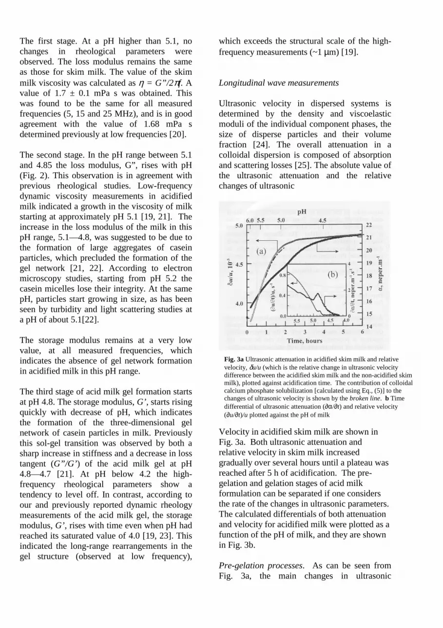
Velocity in acidified skim milk are shown in Fig. 3a. Both ultrasonic attenuation and relative velocity in skim milk increased gradually over several hours until a plateau was reached after 5 h of acidification. The pre-gelation and gelation stages of acid milk formulation can be separated if one considers the rate of the changes in ultrasonic parameters. The calculated differentials of both attenuation and velocity for acidified milk were plotted as a function of the pH of milk, and they are shown in Fig. 3b. Pre-gelation processes. As can be seen from Fig. 3a, the main changes in ultrasonic
Fig. 3a Ultrasonic attenuation in acidified skim milk and relative velocity, δu/u (which is the relative change in ultrasonic velocity difference between the acidified skim milk and the non-acidified skim milk), plotted against acidification time. The contribution of colloidal calcium phosphate solubilization [calculated using Eq., (5)] to the changes of ultrasonic velocity is shown by the broken line. b Time differential of ultrasonic attenuation (∂α/∂t) and relative velocity (∂u/∂t)/u plotted against the pH of milk
parameters occur in the pre-gelation stage between pH 5.6 and 5. Pre-gelation processes such as the release of colloidal calcium phosphate from micelles and partial liberation of casein protein result in the modification of the chemical composition of casein micelles. These processes cause the swelling of casein micelles and the aggregation of casein micelles. The apparent/partial effects of the changes in the micelles physicochemical properties on the ultrasonic parameters are discussed below. The effect of casein liberation from the micelles into serum during acidification seems to be small. This conclusion follows from our analysis of casein hydration within micelles at pH 6.7 (initial pH of milk). According to our results, the specific apparent volume and adiabatic compressibility of total casein proteins inside micelles are nearly the same as the corresponding values for α-casein in solution, indicating a similar level of hydration of proteins in both states. A significant contribution to the increase of the ultrasonic velocity is expected owing to the dissociation of colloidal minerals from the casein micelles. It is well known that multivalent ions such as calcium and magnesium are highly hydrated in water solutions, and these ions have a significant effect on ultrasonic velocity in aqueous solution, because the compressibility of water in the hydration shell is different (generally less) from bulk water [26]. Therefore the release of colloidal calcium phosphare into serum during acidification is supposed to have a significant increase on the ultrasonic velocity. In our calculations we have used data on the dissociation of micellar calcium and inorganic phosphate in GDL acidified skim milk reported by Gastaldi et al [21] to calculate the relative change in ultrasonic velocity, δ(u/u)hydr, according to the equation: δ(u/u)hydr = δ(u/u)(CA2+)hydr + δ(u/u)(H2PO-
4)hydr (5) The values of δ(u/u)(CA2+)hydr and δ(u/u)(H2PO-
4)hydr were calculated from the apparent molar volume and apparent adiabatic compressibility for the hydrated calcium and phosphate ions [27].
There is quantitative consistence between the experimental and theoretical dependencies of relative ultrasonic velocity over the pH range 5.6 5.0 (Fig. 3a). This supposes that the dissociation of colloidal calcium phosphate into serum makes an important contribution to the change of ultrasonic velocity in acidified skim milk. An increase in particle size can have a significant effect on both ultrasonic velocity and ultrasonic attenuation. We calculated the contributions of the aggregation and the increase in particle size into the ultrasonic parameters in skim milk using the Truell-Waterman theory ([25] and refs therein). The effect of aggregation or change in casein particle size on ultrasonic parameters of skim milk during acidification is small. The changes in ultrasonic attenuation and relative ultrasonic velocity corresponding to the increase of particle size (from 0.1 to 1 µm) resulting from the aggregation are only 10% of the total changes in ultrasonic parameters during milk acidification. Next we consider the effect of water transfer from the milk serum to the casein micelle. We calculated the effect of the change in voluminosity and water content of casein micelles on both ultrasonic parameters. It was found that the increase in the volume of the casein particles results in negligible changes in the ultrasonic velocity in comparison with the experimental value for milk acidification. We would expect this, as there is little change in the overall density and compressibility of the system. On the other hand, the ultrasonic attenuation was found to be highly sensitive to changes in the voluminosity and water content of the casein micelle. The total changes in ultrasonic attenuation during pre-gelation can be explained by a 25% rise of casein volume fraction compared to its initial value of 0.1. Gelation process. The sharp peaks in both differentials of ultrasonic velocity and attenuation around pH 4.8 may correspond to the point of gelation in acidified milk gel, which is in agreement with our shear wave measurements. From the deconvolution of the curves, we established the contribution from

gelation to relative ultrasonic velocity as 1 x l0-
5 and attenuation as 0.27 neper m-1, which consists approximately of 1% and 4% of the total changes of the corresponding ultrasonic parameters during acidification of skim milk. The contribution of the changes in elasticity and viscosity of acid milk gel into ultrasonic parameters can be estimated from the magnitudes of the changes in ultrasonic velocity and attenuation upon the sol-gel transition using the following expressions [1]:
( ) ( )sv
22
G uρf2π ηηα ∆+∆≅∆ 3
43
''43
')(2 MGKuu G ∆=∆+∆≅∆ρ (6)
Here ∆(u)G and ∆(α)G are the changes in ultrasonic velocity and attenuation upon sol-gel transition (between pH 5.0 and 4.6). The values of ∆M’, ∆K’, ∆G’ and ηv, ηs correspond to the changes in real parts of complex, bulk and shear moduli, respectively, ηv and ηs are the changes in the volume and shear viscosities upon gelation. By using the above Eqs. (6) and the values of the changes in ultrasonic velocity and attenuation (integral area of seconds peaks in Fig. 3b), we have obtained the value of the changes in viscoelastic parameters of the acidified milk during the sol-gel transition. The change in complex elastic modulii (∆K’ + 34 ∆G’) is estimated to be about 100 kPa at the
frequency of 7.2 MHz used in longitudinal wave measurements. This value is of the same order as the value of the shear storage modulus of acid milk gel determined from ultrasonic shear wave measurements (G’ ≅ 27 kPa at 5 MHz, and G’ ≅ 130 kPa at 25 MHz). The change in effective viscosity (volume and shear) of acidified skim milk during gelation
was estimated to be about 3.9 mPa. In comparison, the magnitude of shear viscosity changes in acidified skim milk is about 2.5 mPa determined by the ultrasonic shear wave technique. Impedance analysis of ultrasonic piezosensor for Salmonella A summary of the results of the impedance measurements (at 5, 15 and 25 MHz) for the subsequent steps of the piezosensor assembly as well as the antigen-binding reaction is presented in Table 1. As can be seen, the deposition of both protein A and antibody are accompanied by the positive values of XL, and by zero values of RL, which indicate the formation of solid-like layers on the crystal surface. The magnitude of the changes of resonant frequencies corresponds to the protein monolayer thickness. This is confirmed by our analysis of the surface profile of the deposed layers using atomic force microscopy. The observed changes in the resonant frequency, ∆fr, and imaginary part of impedance, XL, after inoculation of Salmonella antigen into the sensor (giving a final concentration of 106 cell per ml) are shown in Fig. 4. The resonant frequency of the sensor drops continuously over 3 h after the addition of the bacteria, indicating the adsorption of the Salmonella antigen on the antibody-modified sensor surface. These changes in resonant frequency correlate well with the changes in the value of XL. Since the rise in the value of XL corresponds to the attachment of an additional mass to the sensor (Fig. 1), the adsorbed bacteria appear to be strongly bound to the crystal surface. The decrease in the value of RL (Table 1) can also be explained by the lowering of the viscosity of the interfacial antibody-liquid layer (due to the restriction of antibody molecule movements on the surface) upon bacteria binding.
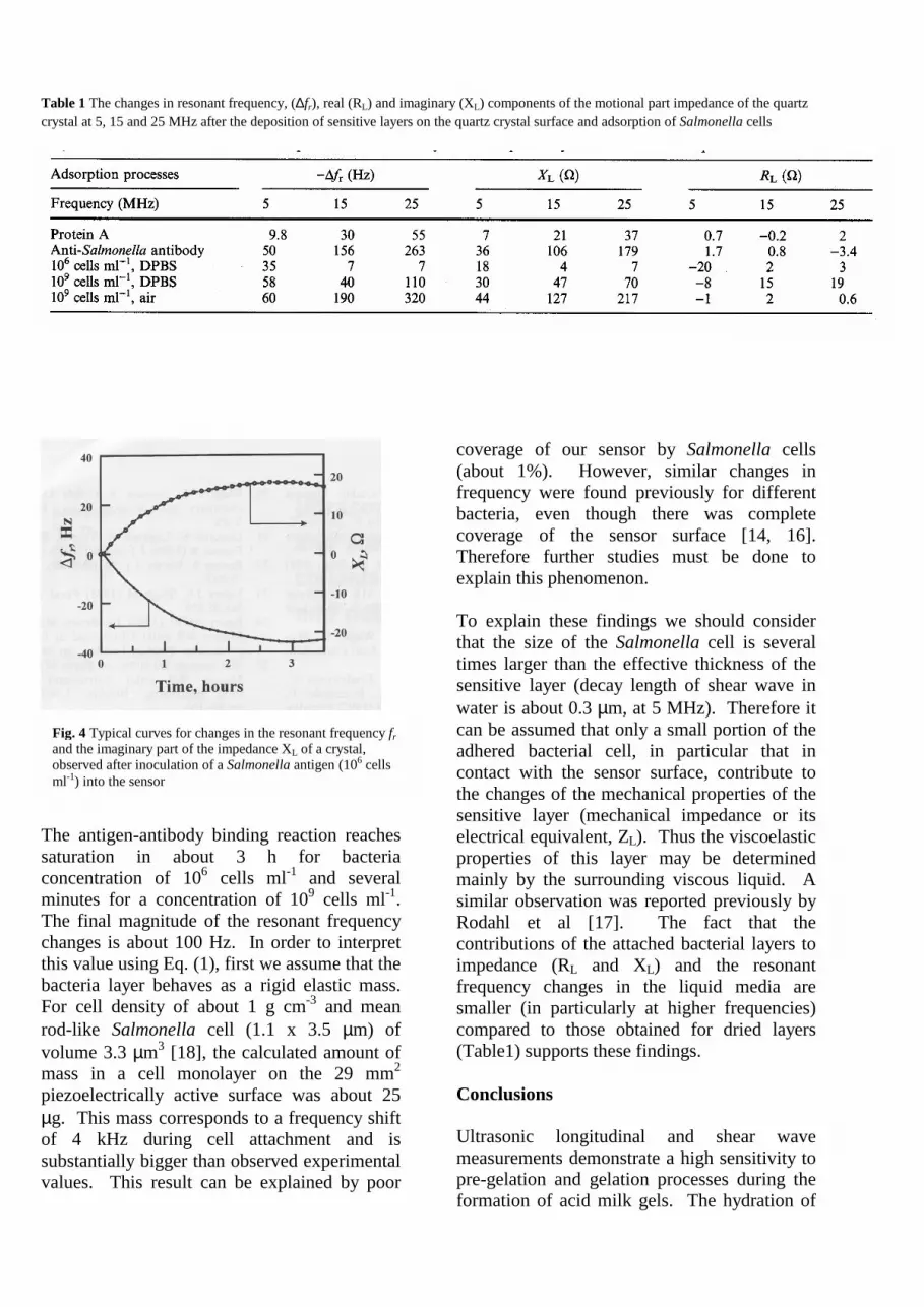
Table 1 The changes in resonant frequency, (∆fr), real (RL) and imaginary (XL) components of the motional part impedance of the quartz crystal at 5, 15 and 25 MHz after the deposition of sensitive layers on the quartz crystal surface and adsorption of Salmonella cells
The antigen-antibody binding reaction reaches saturation in about 3 h for bacteria concentration of 106 cells ml-1 and several minutes for a concentration of 109 cells ml-1. The final magnitude of the resonant frequency changes is about 100 Hz. In order to interpret this value using Eq. (1), first we assume that the bacteria layer behaves as a rigid elastic mass. For cell density of about 1 g cm-3 and mean rod-like Salmonella cell (1.1 x 3.5 µm) of volume 3.3 µm3 [18], the calculated amount of mass in a cell monolayer on the 29 mm2 piezoelectrically active surface was about 25 µg. This mass corresponds to a frequency shift of 4 kHz during cell attachment and is substantially bigger than observed experimental values. This result can be explained by poor
coverage of our sensor by Salmonella cells (about 1%). However, similar changes in frequency were found previously for different bacteria, even though there was complete coverage of the sensor surface [14, 16]. Therefore further studies must be done to explain this phenomenon. To explain these findings we should consider that the size of the Salmonella cell is several times larger than the effective thickness of the sensitive layer (decay length of shear wave in water is about 0.3 µm, at 5 MHz). Therefore it can be assumed that only a small portion of the adhered bacterial cell, in particular that in contact with the sensor surface, contribute to the changes of the mechanical properties of the sensitive layer (mechanical impedance or its electrical equivalent, ZL). Thus the viscoelastic properties of this layer may be determined mainly by the surrounding viscous liquid. A similar observation was reported previously by Rodahl et al [17]. The fact that the contributions of the attached bacterial layers to impedance (RL and XL) and the resonant frequency changes in the liquid media are smaller (in particularly at higher frequencies) compared to those obtained for dried layers (Table1) supports these findings. Conclusions Ultrasonic longitudinal and shear wave measurements demonstrate a high sensitivity to pre-gelation and gelation processes during the formation of acid milk gels. The hydration of
Fig. 4 Typical curves for changes in the resonant frequency fr and the imaginary part of the impedance XL of a crystal, observed after inoculation of a Salmonella antigen (106 cells ml-1) into the sensor

colloidal calcium phosphate released into serum and the swelling of casein in micelles at pH 5.6—5.0 are suggested as the main contributors to the ultrasonic velocity and attenuation changes during the pre-gelation process. The increase in the attenuation of the longitudinal wave and loss moduli of acidified milk indicates the aggregation of the casein micelles into clusters. Subsequent reformation of these clusters into a continuous gel network at pH 4.8—4.6 is observed as a sharp rise in the storage moduli of acidified milk gel and an additional increase in ultrasonic velocity and attenuation. The ability of the ultrasonic shear wave biosensor to monitor bacteria in liquids was
demonstrated. However, impedance measurements of the antigen-binding reaction indicates that the attachment of the bacteria to the sensor does not exhibit pure mass-load behaviour, and the viscoelastic properties of the interfacial layer must be taken into account. Overall our ultrasonic measurements show a high potential as non-destructive methods of analysis of complex foods and bio-colloids. Acknowledgements This work was supported by grant 97/R&D/T178 from the Department of Agriculture Food and Forestry of Ireland and EU grant FAIR-CT97-3096. We are grateful to Mr James Mahon, an undergraduate student, for his help in ultrasonic sensor construction. We gratefully acknowledge the support of this work by Ultrasonic Scientific Ltd, who provided the equipment for high resolution longitudinal ultrasonic measurements.
References 1. Herzfeld KF, Litovitz TA (1959)
Absorption and dispersion of ultrasonic waves. Academic Press, New York
2. Miles CA, Shore D, Langley KR (1990) Ultrasonics 28:394
3. Griffin WG, Griffin MCA (1990) J Acoust Soc Am 87:2541
4. Gunasekaran 5, Ay C (1994) Trans ASEA 37:857
5. Benguigi L. Emery J, Durand D, Busnel JP (1994) Lait 74:197
6. Buckin V. Smyth C (1999) Semin Food Anal 4:89
7. Smyth C, Dawson K, Buckin V (1999) Prog Colloid Polym Sci 112:221
8. Ferry JD (1980) Viscoelastic properties of polymers. Wiley, New York
9. Buttry DA, Ward MD (1992) Chem Rev 92:1355
10. Johannsmann D (1999) Macromol Chem Phys 200:501
11. Bandey HL, Martin SJ, Cernosek RW, Hillman AR (1999) Anal Chem 7 1:2205
12. Martin SJ (1997) Faraday Discuss Chem Soc 107:463
13. Muramatsu H, Kajiwara K, Tamiya E, Karube I (1986) Anal Chim Acta 188:257
14. Gryte DM, Ward MD. Hu WS (1993) Biotechnol Prog 9:105
15. Bovenizer JS, Jacobs MB, O’Sillivan CK, Guilbault GG (1998) Anal Lett 31: 1287
16. Tessier L, Schmitt N, Watier H, Brumas V, Patat F (1997) Anal Chim Acta 347:207
17. Rodahl M, Höök F, Fredriksson C, Keller C, Krozer A, Brzezinski P, Voinova M, Kasemo B (1997) Faraday Discuss Chem Soc 107:229
18. Prescot LM, Harley JP, Klein DA (1996) Microbiology. WCB, Dubuque
19. Kudryashov ED, Hunt N, Arikainen EO, Buckin VA (2000) J Dairy Sci, (to be published)
20. Walstra P, Jenness R (1984) Dairy chemistry and physics. Wiley, New York
21. Gastaldi E. Lagaude A, Tarodo de la Fuente B (1996) J Food Sci 61:59
22. Banon S, Hardy J (1992) J Dairy Sci 75:935
23. Lucey JA, Singh H (1998) Food Res Tnt 30:529
24. Povey MJW (1998) In: Povey MJW, Mason WP (eds) Ultrasound in food processing. Blackie, London, pp 30—65
25. McClements DJ (1998) In: Povey MJW, Mason WP (eds) Ultrasound in food processing. Blackie, London, pp 85—103
26. Sarvazyan AP (1991) Annu Rev Biophys Chem 20:321
27. Millero FJ, Ward GK, Chetirkin PV (1977) J Acoust Soc Am 61:1492
Copyright © 2022 FDOKUMEN