UEG Week 2013 Oral Presentations - CiteSeerX
134
UEG Week 2013 Oral Presentations MONDAY, OCTOBER 14, 2013 8:00–10:30 Opening Plenary Session – Hall 1_____________________ OP001 RANDOMISED CONTROLLED TRIAL OF DRUG LEVEL VERSUS CLINICALLY BASED DOSING OF INFLIXIMAB MAINTENANCE THERAPY IN IBD: FINAL RESULTS OF THE TAXIT STUDY N. Vande Casteele 1,* , A. Gils 1 , V. Ballet 2 , G. Compernolle 1 , M. Peeters 1 , K. Van Steen 3 , S. Simoens 4 , M. Ferrante 2 , G. Van Assche 2 , S. Vermeire 2 , P. Rutgeerts 2 . 1 KU Leuven Laboratory for Therapeutic and Diagnostic Antibodies, 2 UZ Leuven Department of Gastroenterology, Leuven, 3 University of Lie`ge Systems and Modeling Unit, Lie`ge, 4 KU Leuven Research Centre for Pharmaceutical Care & Pharmaco-economics, Leuven, Belgium INTRODUCTION: Treat-to-target dosing based on infliximab (IFX) trough levels (TLI) has been suggested to increase efficacy, safety and cost-effectiveness of IFX treatment, although prospective data are lacking. In the optimisation phase of the TAXIT study we showed that dose intensification of IFX in CD patients with TLI 53 mg/ml resulted in a better disease control, whereas dose reduction in CD and UC patients with TLI 47 mg/ml resulted in lower drug exposure and lower IFX cost whilst maintaining disease control. 1 AIMS&METHODS: Here we report on the subsequent long-term randomised part of this study. 251 patients entered the maintenance phase (MP) of which 123 patients (82 CD/41 UC) were randomised to the clinically based (CB) arm and 128 patients (91 CD/37 UC) to the trough level based (LB) arm. Primary end point of the MP was the rate of both clinical (Harvey-Bradshaw/partial MAYO score) and biological (CRP 5 mg/l) remission one year after randomisation in each group. Secondary end points included number of patients with TLI between 3-7 mg/ml and antibody to IFX (ATI) rate. RESULTS: After optimisation, median TLI and CRP were equal in the CB and LB arm (respectively 4.9 mg/ml [3.8-5.9] vs 5.0 mg/ml [4.0-5.7] and 1.3 mg/l [0.6- 4.5] vs 1.5 mg/l [0.7-4.1]). 226/251 patients (90%) completed the MP of whom 69% of the CB vs 72% of the LB arm achieved the primary end point (P¼0.7). During MP, 11 patients (4.4%) (5 CB vs 6 LB) were failures and 14 patients (5.6%) (7 CB vs 7 LB) were excluded (e.g. due to pregnancy or lost to follow-up). After one year, 56% in the CB vs 78% in the LB arm had TLI between 3-7 mg/ml (P50.001). During MP, undetectable TLI were more frequently observed in the CB than in the LB arm (RR 3.7; 95% CI 1.7-8.0; P50.001) which may have influenced the higher frequency rate of ATI observed in the CB vs the LB arm (RR 3.3; 95% CI 1.4-7.7; P50.01). At the end of MP, three patients were ATI positive in the CB vs none in the LB arm (P¼0.1). All three patients were ATI negative at screening and developed ATI during MP. CONCLUSION: Dose-to-target optimisation of IFX allowed to achieve TLI within the interval of 3-7 mg/ml which resulted in a more efficient use of drug. The maintenance phase did not show superiority for continued level based drug adjustment over clinically based adjustment. Treatment guided by levels resulted in less ATI formation but the proportion of patients in clinical and biological remission was similar for both groups. REFERENCES: 1. Vande Casteele N, et al. Gastroenterology 2012;142(5):S211-S212. Contact E-mail Address: [email protected] Disclosure of Interest: N. Vande Casteele: None Declared, A. Gils Financial support for research from: Pfizer, Lecture fee(s) from: MSD, Pfizer, V. Ballet: None Declared, G. Compernolle: None Declared, M. Peeters: None Declared, K. Van Steen: None Declared, S. Simoens: None Declared, M. Ferrante Financial support for research from: Janssen Biologics, Lecture fee(s) from: Merck, Tillotts, Ferring, Abbvie, Consultancy for: Abbvie, Merck, Janssen Biologics, G. Van Assche Financial support for research from: Ferring, Abbvie, Lecture fee(s) from: Merck, Abbvie, Janssen-Cilag, Consultancy for: PDL BioPharma, UCB Pharma, Sanofi-Aventis, Abbvie, Ferring, Novartis, Biogen Idec, Janssen Biologics, NovoNordisk, Zealand Pharma A/S, Millenium/Takeda, Shire, Novartis, BMS, S. Vermeire Financial support for research from: UCB Pharma, MSD, Abbvie, Lecture fee(s) from: Abbvie, Merck, Ferring, UCB Pharma, Centocor, Consultancy for: UCB Pharma, AstraZeneca, Ferring, Abbvie, Merck, Ferring, Shire, Pfizer, P. Rutgeerts Financial support for research from: UCB Pharma, Abbvie, Janssen Biologics, Merck, Prometheus, Lecture fee(s) from: Abbvie, Merck, Consultancy for: Amgen, Merck, UCB Pharma, Genentech, BMS, Abbvie, Janssen Biologics, Millenium, Neovacs, Actogenics, Prometheus. Keywords: anti infliximab antibodies measurement, biologic therapy, inflamatory bowel disease, Personalized therapy, pharmacokinetics/Pharmacodynamic rela- tionship, trough levels OP002 EARLY VERSUS ON-DEMAND NASOENTERAL FEEDING IN SEVERE PANCREATITIS: A MULTICENTER RANDOMISED CONTROLLED TRIAL O. J. Bakker 1,* and The Dutch Pancreatitis Study Group: . 1 Surgery, University Medical Center Utrecht, Utrecht, Netherlands INTRODUCTION: In patients with severe pancreatitis, a routine start of nasoenteral feeding within 24 hours after admission is believed to reduce com- plications. However, evidence is based on observational and retrospective data. AIMS&METHODS: Between August 2008 and July 2012, 867 patients were screened for enrollment in 19 hospitals. Eventually, 208 adults with predicted severe pancreatitis were randomised to early nasoenteral feeding through a naso- jejunal tube started within 24 hours of admission (n¼102), or starvation for 72 hours followed by an oral diet with on-demand nasoenteral feeding whenever oral intake was insufficient (n¼106). Predicted severe pancreatitis on admission was defined as an Acute Physiology and Chronic Health Evaluation II score 8, Imrie score 3, or CRP 150 mg/L. The primary endpoint consisted of a com- posite of mortality or infection (infected pancreatic necrosis, bacteremia, or pneu- monia). The protocol of this trial has been published. 1 RESULTS: One patient from the early feeding group and 2 patients from the on- demand group were excluded because of incorrect diagnosis of acute pancreatitis; thus 101 patients were analysed in the early feeding group and 104 patients in the on-demand group. Baseline characteristics did not differ. Median APACHE II scores at randomisation were 11 in the early feeding group and 9 in the on-demand group (p ¼ 0.24). In the early feeding group, nutrition was initiated a median of 23 hours after admission. The primary composite endpoint occurred in 30 patients in the early feeding group compared to in 28 patients in the on-demand group (30% versus 27%; p ¼ 0.76). Mortality was 11% in the early feeding group compared to 7% in the on-demand group (p ¼ 0.33). There were no differences in infections (25% versus 26%, p ¼ 0,87), persistent single-organ failure (13% versus 8%, p ¼ 0.65) or persistent multi-organ failure (5% versus 7%, p ¼ 0.77). In the on-demand group, 72 patients (69%) tolerated an oral diet and only 32 patients (31%) required nasoenteral tube feeding. CONCLUSION: Starting nasoenteral tube feeding within 24 hours after admis- sion in patients with predicted severe pancreatitis does not reduce complications as compared to on-demand nasoenteral tube feeding started after 72 hours. Therefore, in contrast to what is advised in recent guidelines, routine early nasoenteral tube feeding is not necessary. Nasoenteral tube feeding can be started after 72 hours when oral intake is insufficient. REFERENCES: 1. Bakker OJ , van Brunschot S, van Santvoort HC, et al. and Gooszen HG for the Dutch Pancreatitis Study Group. Pancreatitis, very early compared with normal start of enteral feeding (PYTHON trial): design and rationale of a randomised controlled multicenter trial. Trials. 2011 Mar 10;12:73. doi: 10.1186/1745-6215-12-73. Contact E-mail Address: [email protected] Disclosure of Interest: None Declared Keywords: Acute Pancreatitis, Complications, Nutrition, pancreas, randomised controlled trial OP003 SUSTAINED VIROLOGIC RESPONSE 12 WEEKS POST-TREATMENT WITH INTERFERON-FREE REGIMENS OF ABT-450/R, ABT-267, ABT-333, AND RIBAVIRIN IN CHRONIC HCV GENOTYPE 1 DIFFICULT-TO-TREAT PATIENT SUBGROUPS M. Rodriguez-Torres 1 , F. Nunes 2,* , D. Cohen 3 , W. Xie 3 , C. Marincic 3 , A. Khatri 3 , T. Pilot-Matias 3 , T. Podsadecki 3 , B. Bernstein 3 . 1 Fundacio ´n de Investigacio ´n, San Juan, Puerto Rico, 2 PHGI Assoc. Ltd., Philadelphia, 3 AbbVie, North Chicago, United States INTRODUCTION: ABT-450 is a potent hepatitis C virus (HCV) protease inhi- bitor (dosed with ritonavir 100mg, ABT-450/r) identified as a lead compound by AbbVie and Enanta; ABT-267 is an NS5A inhibitor and ABT-333 is a non- nucleoside polymerase inhibitor. We examined the safety and efficacy of 12- and 24-week (wk) interferon (IFN)-free 3DAAþribavirin (RBV) regimens (ABT-450/r, 100/100-150/100mg QD; ABT-267, 25mg QD; ABT-333, 400mg BID; and weight-based RBV) in treatment-naı¨ve (TN) patients (pts) and prior pegIFN/RBV null responders (Null) with chronic HCV genotype 1 (GT1) infec- tion, overall and in pt subgroups with baseline characteristics associated with poorer response to pegIFN/RBV. AIMS&METHODS: Non-cirrhotic TN and Null pts received 3DAAþRBV for 12 or 24 wks. SVR 12 rates (HCV RNA 525 IU/mL at post-treatment wk12) were analyzed in the TN and Null cohorts overall and according to age, race, ethnicity, BMI, HOMA-IR, IL28B genotype, and baseline viral load (ITT, non- completer¼failure). RESULTS: 247 pts (159 TN, 88 Null) assigned to 3DAAþRBV regimens received 1 dose of study drug (male: 49% and 63%; GT1a infection: 68% and 63%). Overall SVR 12 rates for TN and Null cohorts were 98.7% and 93.3% (12-wk) and 92.5% and 97.7% (24-wk). SVR 12 rates 90% were achieved regardless of age, BMI, race, ethnicity (Table), HOMA-IR, IL28B genotype, or baseline viral load, with no statistically significant differences in any comparisons made in the TN or Null cohorts (P40.05, Fisher’s exact test). Most common AEs in TN and Null cohorts were fatigue (32.7% and 23.9%) and headache (31.4% and 30.7%). 4 of 247 pts (1.6%) discontinued treatment due to study drug-related AEs. 1 pt (0.4%) had a serious AE (arthralgia) possibly related to study drug. United European Gastroenterology Journal 1(1S) A1–A134 ! Author(s) 2013 Reprints and permissions: sagepub.co.uk/journalsPermissions.nav DOI: 10.1177/2050640613502899 ueg.sagepub.com
-
Upload
khangminh22 -
Category
Documents
-
view
2 -
download
0
Transcript of UEG Week 2013 Oral Presentations - CiteSeerX

UEG Week 2013 Oral Presentations
MONDAY, OCTOBER 14, 2013 8:00–10:30Opening Plenary Session – Hall 1_____________________
OP001 RANDOMISED CONTROLLED TRIAL OF DRUG LEVEL
VERSUS CLINICALLY BASED DOSING OF INFLIXIMAB
MAINTENANCE THERAPY IN IBD: FINAL RESULTS OF THE TAXIT
STUDY
N. Vande Casteele1,*, A. Gils1, V. Ballet2, G. Compernolle1, M. Peeters1, K. Van
Steen3, S. Simoens4, M. Ferrante2, G. Van Assche2, S. Vermeire2, P. Rutgeerts2.1KU Leuven Laboratory for Therapeutic and Diagnostic Antibodies, 2UZ LeuvenDepartment of Gastroenterology, Leuven, 3University of Liege Systems andModeling Unit, Liege, 4KU Leuven Research Centre for Pharmaceutical Care &Pharmaco-economics, Leuven, Belgium
INTRODUCTION: Treat-to-target dosing based on infliximab (IFX) troughlevels (TLI) has been suggested to increase efficacy, safety and cost-effectivenessof IFX treatment, although prospective data are lacking. In the optimisationphase of the TAXIT study we showed that dose intensification of IFX in CDpatients with TLI 53 mg/ml resulted in a better disease control, whereas dosereduction in CD and UC patients with TLI 47 mg/ml resulted in lower drugexposure and lower IFX cost whilst maintaining disease control.1
AIMS&METHODS: Here we report on the subsequent long-term randomisedpart of this study. 251 patients entered the maintenance phase (MP) of which 123patients (82 CD/41 UC) were randomised to the clinically based (CB) arm and128 patients (91 CD/37 UC) to the trough level based (LB) arm. Primary endpoint of the MP was the rate of both clinical (Harvey-Bradshaw/partial MAYOscore) and biological (CRP �5 mg/l) remission one year after randomisation ineach group. Secondary end points included number of patients with TLI between3-7 mg/ml and antibody to IFX (ATI) rate.RESULTS: After optimisation, median TLI and CRP were equal in the CB andLB arm (respectively 4.9 mg/ml [3.8-5.9] vs 5.0 mg/ml [4.0-5.7] and 1.3 mg/l [0.6-4.5] vs 1.5 mg/l [0.7-4.1]). 226/251 patients (90%) completed the MP of whom69% of the CB vs 72% of the LB arm achieved the primary end point (P¼0.7).During MP, 11 patients (4.4%) (5 CB vs 6 LB) were failures and 14 patients(5.6%) (7 CB vs 7 LB) were excluded (e.g. due to pregnancy or lost to follow-up).After one year, 56% in the CB vs 78% in the LB arm had TLI between 3-7 mg/ml(P50.001). During MP, undetectable TLI were more frequently observed in theCB than in the LB arm (RR 3.7; 95% CI 1.7-8.0; P50.001) which may haveinfluenced the higher frequency rate of ATI observed in the CB vs the LB arm(RR 3.3; 95% CI 1.4-7.7; P50.01). At the end of MP, three patients were ATIpositive in the CB vs none in the LB arm (P¼0.1). All three patients were ATInegative at screening and developed ATI during MP.CONCLUSION: Dose-to-target optimisation of IFX allowed to achieve TLIwithin the interval of 3-7 mg/ml which resulted in a more efficient use of drug.The maintenance phase did not show superiority for continued level based drugadjustment over clinically based adjustment. Treatment guided by levels resultedin less ATI formation but the proportion of patients in clinical and biologicalremission was similar for both groups.REFERENCES:
1. Vande Casteele N, et al. Gastroenterology 2012;142(5):S211-S212.Contact E-mail Address: [email protected] of Interest: N. VandeCasteele: NoneDeclared,A.Gils Financial supportfor research from: Pfizer, Lecture fee(s) from:MSD, Pfizer, V. Ballet: NoneDeclared,G. Compernolle: None Declared, M. Peeters: None Declared, K. Van Steen: NoneDeclared, S. Simoens: None Declared, M. Ferrante Financial support for researchfrom: Janssen Biologics, Lecture fee(s) from: Merck, Tillotts, Ferring, Abbvie,Consultancy for: Abbvie,Merck, Janssen Biologics, G.VanAssche Financial supportfor research from:Ferring,Abbvie,Lecture fee(s) from:Merck,Abbvie, Janssen-Cilag,Consultancy for: PDL BioPharma, UCB Pharma, Sanofi-Aventis, Abbvie, Ferring,Novartis, Biogen Idec, Janssen Biologics, NovoNordisk, Zealand Pharma A/S,Millenium/Takeda, Shire, Novartis, BMS, S. Vermeire Financial support for researchfrom: UCB Pharma, MSD, Abbvie, Lecture fee(s) from: Abbvie, Merck, Ferring,UCB Pharma, Centocor, Consultancy for: UCB Pharma, AstraZeneca, Ferring,Abbvie, Merck, Ferring, Shire, Pfizer, P. Rutgeerts Financial support forresearch from: UCB Pharma, Abbvie, Janssen Biologics, Merck, Prometheus,Lecture fee(s) from: Abbvie, Merck, Consultancy for: Amgen, Merck, UCBPharma, Genentech, BMS, Abbvie, Janssen Biologics, Millenium, Neovacs,Actogenics, Prometheus.Keywords: anti infliximab antibodies measurement, biologic therapy, inflamatorybowel disease, Personalized therapy, pharmacokinetics/Pharmacodynamic rela-tionship, trough levels
OP002 EARLY VERSUS ON-DEMAND NASOENTERAL FEEDING IN
SEVERE PANCREATITIS: A MULTICENTER RANDOMISED
CONTROLLED TRIAL
O. J. Bakker1,* and The Dutch Pancreatitis Study Group: . 1Surgery, University
Medical Center Utrecht, Utrecht, Netherlands
INTRODUCTION: In patients with severe pancreatitis, a routine start ofnasoenteral feeding within 24 hours after admission is believed to reduce com-plications. However, evidence is based on observational and retrospective data.AIMS&METHODS: Between August 2008 and July 2012, 867 patients werescreened for enrollment in 19 hospitals. Eventually, 208 adults with predicted
severe pancreatitis were randomised to early nasoenteral feeding through a naso-jejunal tube started within 24 hours of admission (n¼102), or starvation for 72hours followed by an oral diet with on-demand nasoenteral feeding whenever oralintake was insufficient (n¼106). Predicted severe pancreatitis on admission wasdefined as an Acute Physiology and Chronic Health Evaluation II score � 8,Imrie score � 3, or CRP � 150 mg/L. The primary endpoint consisted of a com-posite of mortality or infection (infected pancreatic necrosis, bacteremia, or pneu-monia). The protocol of this trial has been published.1
RESULTS: One patient from the early feeding group and 2 patients from the on-demand group were excluded because of incorrect diagnosis of acute pancreatitis;thus 101 patients were analysed in the early feeding group and 104 patients in theon-demand group. Baseline characteristics did not differ. Median APACHE IIscores at randomisation were 11 in the early feeding group and 9 in the on-demandgroup (p¼ 0.24). In the early feeding group, nutrition was initiated a median of 23hours after admission. The primary composite endpoint occurred in 30 patients inthe early feeding group compared to in 28 patients in the on-demand group (30%versus 27%; p¼ 0.76). Mortality was 11% in the early feeding group compared to7% in the on-demand group (p ¼ 0.33). There were no differences in infections(25% versus 26%, p ¼ 0,87), persistent single-organ failure (13% versus 8%, p ¼0.65) or persistent multi-organ failure (5% versus 7%, p¼ 0.77). In the on-demandgroup, 72 patients (69%) tolerated an oral diet and only 32 patients (31%) requirednasoenteral tube feeding.CONCLUSION: Starting nasoenteral tube feeding within 24 hours after admis-sion in patients with predicted severe pancreatitis does not reduce complicationsas compared to on-demand nasoenteral tube feeding started after 72 hours.Therefore, in contrast to what is advised in recent guidelines, routine earlynasoenteral tube feeding is not necessary. Nasoenteral tube feeding can be startedafter 72 hours when oral intake is insufficient.REFERENCES:
1. Bakker OJ , van Brunschot S, van Santvoort HC, et al. and Gooszen HG forthe Dutch Pancreatitis Study Group. Pancreatitis, very early compared withnormal start of enteral feeding (PYTHON trial): design and rationale of arandomised controlled multicenter trial. Trials. 2011 Mar 10;12:73. doi:10.1186/1745-6215-12-73.
Contact E-mail Address: [email protected] of Interest: None DeclaredKeywords: Acute Pancreatitis, Complications, Nutrition, pancreas, randomisedcontrolled trial
OP003 SUSTAINED VIROLOGIC RESPONSE 12 WEEKS
POST-TREATMENT WITH INTERFERON-FREE
REGIMENS OF ABT-450/R, ABT-267, ABT-333, AND RIBAVIRIN IN
CHRONIC HCV GENOTYPE 1 DIFFICULT-TO-TREAT PATIENT
SUBGROUPS
M. Rodriguez-Torres1, F. Nunes2,*, D. Cohen3, W. Xie3, C. Marincic3,
A. Khatri3, T. Pilot-Matias3, T. Podsadecki3, B. Bernstein3. 1Fundacion deInvestigacion, San Juan, Puerto Rico, 2PHGI Assoc. Ltd., Philadelphia, 3AbbVie,North Chicago, United States
INTRODUCTION: ABT-450 is a potent hepatitis C virus (HCV) protease inhi-bitor (dosed with ritonavir 100mg, ABT-450/r) identified as a lead compound byAbbVie and Enanta; ABT-267 is an NS5A inhibitor and ABT-333 is a non-nucleoside polymerase inhibitor. We examined the safety and efficacy of 12-and 24-week (wk) interferon (IFN)-free 3DAAþribavirin (RBV) regimens(ABT-450/r, 100/100-150/100mg QD; ABT-267, 25mg QD; ABT-333, 400mgBID; and weight-based RBV) in treatment-naıve (TN) patients (pts) and priorpegIFN/RBV null responders (Null) with chronic HCV genotype 1 (GT1) infec-tion, overall and in pt subgroups with baseline characteristics associated withpoorer response to pegIFN/RBV.AIMS&METHODS: Non-cirrhotic TN and Null pts received 3DAAþRBV for12 or 24 wks. SVR12 rates (HCV RNA525 IU/mL at post-treatment wk12) wereanalyzed in the TN and Null cohorts overall and according to age, race, ethnicity,BMI, HOMA-IR, IL28B genotype, and baseline viral load (ITT, non-completer¼failure).RESULTS: 247 pts (159 TN, 88 Null) assigned to 3DAAþRBV regimens received�1 dose of study drug (male: 49% and 63%; GT1a infection: 68% and 63%).Overall SVR12 rates for TN and Null cohorts were 98.7% and 93.3% (12-wk)and 92.5% and 97.7% (24-wk). SVR12 rates �90% were achieved regardless ofage, BMI, race, ethnicity (Table), HOMA-IR, IL28B genotype, or baseline viralload, with no statistically significant differences in any comparisons made in the TNor Null cohorts (P40.05, Fisher’s exact test). Most common AEs in TN and Nullcohorts were fatigue (32.7% and 23.9%) and headache (31.4% and 30.7%). 4 of 247pts (1.6%) discontinued treatment due to study drug-related AEs. 1 pt (0.4%) had aserious AE (arthralgia) possibly related to study drug.
United European Gastroenterology Journal1(1S) A1–A134! Author(s) 2013Reprints and permissions:sagepub.co.uk/journalsPermissions.navDOI: 10.1177/2050640613502899ueg.sagepub.com

TN, n/N (%) Null, n/N (%)
Age �50 y 92/97 (95) 52/56 (93)
Age 550 y 60/62 (97) 32/32 (100)
BMI �30 kg/m2 36/40 (90) 18/18 (100)
BMI 530 kg/m2 116/119 (98) 66/70 (94)
Black 16/17 (94) 9/9 (100)
Non-black 136/142 (96) 75/79 (95)
Hispanic/Latino 10/11 (91) 8/8 (100)
Non-Hispanic/Latino 142/148 (96) 76/80 (95)
CONCLUSION: In this study, treatment with 3 DAAs (ABT-450/r, ABT-267,and ABT-333) plus RBV for 12 or 24 wks was well-tolerated, with high SVR12
rates achieved in non-cirrhotic HCV GT1 treatment-naıve patients and previousnull responders, even with baseline characteristics associated with a poorerresponse to pegIFN/RBV.Contact E-mail Address: [email protected] of Interest: M. Rodriguez-Torres Financial support for research from:AbbVie, Akros, Boehringer Ingelheim, BMS, Gilead, GSK, IdenixPharmaceuticals, Idera Pharmaceuticals, Inhibitex Pharmaceuticals, Medtronic,Merck, Novartis, Pharmasset, Roche, Santaris Pharmaceuticals, ScynexisPharmaceuticals, Tibotec, Vertex Pharmaceuticals, ViroChem Pharma,ZymoGenetics, Consultancy for: BMS, Genentech/Roche, Gilead, InhibitexPharmaceuticals, Merck, Santaris Pharmaceuticals, Tibotec, Vertex, F. NunesFinancial support for research from: AbbVie, Merck, BMS, D. CohenShareholder of: AbbVie, Other: Employee of AbbVie, W. Xie Shareholder of:AbbVie, Other: Employee of AbbVie, C. Marincic Shareholder of: AbbVie,Other: Employee of AbbVie, A. Khatri Shareholder of: AbbVie, Other:Employee of AbbVie, T. Pilot-Matias Shareholder of: AbbVie, Other:Employee of AbbVie, T. Podsadecki Shareholder of: AbbVie, Other: Employeeof AbbVie, B. Bernstein Shareholder of: AbbVie, Other: Employee of AbbVieKeywords: clinical trial, direct-acting agents, Hepatitis C virus (HCV)
OP004 KLEBSIELLA OXYTOCA OVERGROWTH AND TILIVALLINE
PRODUCTION INDUCE ANTIBIOTIC ASSOCIATED
HEMORRHAGIC COLITIS
G. Schneditz1,*, J. Rentner2, K. A. T. Herzog1,3, G. Gorkiewicz4, S. Roier1,S. Schild1, R. Breinbauer2, E. L. Zechner1, C. Hogenauer3. 1Institute of MolecularBiosciences, University of Graz, 2Institute of Organic Chemistry, Graz Universityof Technology, 3Department of Internal Medicine, 4Institute of Pathology, MedicalUniversity of Graz, Graz, Austria
INTRODUCTION: Antibiotic-associated hemorrhagic colitis (AAHC) causedby Klebsiella oxytoca is exemplary for antibiotic-driven enterobacterial dysbiosis.Alterations of the intestinal microbiota following penicillin therapy results inexclusive overgrowth of �-lactamase producing K. oxytoca, which is found inintestine of about 5% of the healthy population. AAHC patients usually report asudden onset of severe bloody diarrhea but recover spontaneously after cessationof antibiotic prescription. To understand the virulence of K. oxytoca our workfocused on an unknown secreted substance with strong toxicity towards humanintestinal epithelial cell lines in vitro.AIMS&METHODS: We conducted transposon- and site-directed mutagenesisof a toxin-producing K. oxytoca strain, combined with an in vitro screen forcytotoxic effects on epithelial cells to identify toxin negative mutant strains. Inparallel, the genome of a toxin-producing clinical isolate of K. oxytoca wassequenced and annotated. The toxic product was isolated from K. oxytoca con-ditioned medium. Preparative HPLC followed by HiRes-MS and NMR analysisof purified toxin were used to identify the toxin structure. We evaluated the toxinas a virulence factor in a mouse model for AAHC. Following inoculation per os,AAHC was triggered via administration of amoxicillin/clavulanate and indome-tacin. Colon tissue was subjected to histological analysis for assessment ofAAHC pathologies. Toxin-induced host cell pathophysiology was investigatedusing an epithelial barrier model in vitro.RESULTS: We identified a cytotoxin secreted by K. oxytoca as a pentacyclicpyrrolobenzodiazepine (PBD) named tilivalline (TLV). PBDs are known asDNA-modifying metabolites of Actinobacteria, whereas TLV is the onlyknown PBD produced by the human microbiota and by gram-negative bacteria.We showed that TLV producing strains caused AAHC, however virulence ofTLV knock-out strains was strongly attenuated in the murine model.Induction of epithelial apoptosis is the dominant effect of TLV-positive strainsin vivo. In vitro evidence indicates that apoptotic death progresses to disruptionof the epithelial barrier.CONCLUSION: We conclude that TLV is the main virulence factor ofK. oxytoca in AAHC development. TLV is the first reported PBD causinghuman disease - an example of a novel class of intestinal bacterial cytotoxins.Contact E-mail Address: [email protected] of Interest: None DeclaredKeywords: colitis, dysbiosis, klebsiella oxytoca, Microbiota, pyrrolobenzodiaze-pine, toxin
MONDAY, OCTOBER 14, 2013 11:00–12:30Today’s science; tomorrow’s medicine: From genes to disease in IBD –Hall 2_____________________
OP005 GENETIC AND CLINICAL CHARACTERIZATION OF
47 MULTIPLE-AFFECTED IBD FAMILIES
A. Settesoldi1,*, T. Billiet1, E. Hoefkens1, V. Ballet2, P. Rutgeerts2, S. Vermeire2,I. Cleynen1. 1KU Leuven, 2UZ Leuven, Leuven, Belgium
INTRODUCTION: Although the exact pathogenesis of inflammatory boweldisease (IBD) is not known, our current understanding suggests an etiologybased on an interaction among genetic, environmental and immunological fac-tors. The genetic progress has been outstanding with 163 genetic loci identified. Itis believed that multiple-affected families have a high burden of genetic riskvariants although this has not been studied yet in detail.AIMS&METHODS: We collected 47 families with 3 first-degree membersaffected by IBD (Crohn’s disease (CD), ulcerative colitis (UC) or colitis-typeunclassified (IBD-U). Our aim was to characterize these families and to find towhat extent the 163 confirmed genetic loci are associated with disease in thesefamilies. Clinical data (gender, diagnosis, age at diagnosis, CD location andbehavior, UC extent) were obtained. Both affected and unaffected memberswere genotyped using immunochip and 192 independent SNPs from the 163loci were included in plink.RESULTS: Of the 47 families, 32 were CD only, 1 was UC only, 12 were mixedCD/UC, and 2 also contained IBD-U. These families comprised 81 nuclear rela-tions (offspring with both parents), 165 affected (138 CD, 24 UC, 3 IC) and 132unaffected members, 132 of which were male and 165 female. The median age atdiagnosis was 29 years for CD (IQR 10-70) and 32 for UC (IQR 35-54). 17 SNPswere nominally significant (puncorrect50.05) for the combined TDT and parentTDT test statistic. The most significant association was seen for rs3172494(IP6K2, p¼5.71e-05), located in the 3p21 region close to MST1. Althoughrs3172494 is differentially transmitted from both heterozygous fathers andmothers to affected offspring (T:Upat¼2.5:14.5, T:Umat¼6.5:11.5), paternal trans-mission bias is most pronounced (p¼3.61e-03). The next most significant SNPswere rs2227551 (p¼4.91e-03) and rs4256159 (p¼6.49e-03). Interestingly, theNOD2 frameshift mutation rs2066847 was not significantly associated with dis-ease, but there was a strong paternal transmission bias of the risk allele(T:Upat¼13:4, T:Umat¼4:4, ppat¼2.91e-02).CONCLUSION: This is the largest cohort of multiple-affected IBD familiesstudied for genetic association to date. We found nominal significance for diseaseassociation with some of the known IBD susceptibility loci, as well as paternal ormaternal transmission bias. However, most risk alleles were not specifically asso-ciated with disease and many risk alleles were also carried by unaffected relatives.It will be important to also incorporate environmental factors in the family-basedassociation studies in order to try and predict individuals at high risk for devel-oping disease.Contact E-mail Address: [email protected] of Interest: A. Settesoldi: None Declared, T. Billiet: None Declared, E.Hoefkens: None Declared, V. Ballet: None Declared, P. Rutgeerts Financialsupport for research from: UCB Pharma, Abbvie, Janssen Biologics, Merck,Prometheus, Lecture fee(s) from: Abbvie, Merck, Consultancy for: Amgen,Merck, UCB Pharma, Genentech, BMS, Abbvie, Janssen Biologics, Millenium,Neovacs, Actogenics, Prometheus, , S. Vermeire Financial support for researchfrom: UCB Pharma, MSD, Abbvie, Lecture fee(s) from: Abbvie, Merck, Ferring,UCB Pharma, Centocor, Consultancy for: UCB Pharma, AstraZeneca, Ferring,Abbvie, Merck, Ferring, Shire, Pfizer, I. Cleynen: None DeclaredKeywords: genetics, IBD
MONDAY, OCTOBER 14, 2013 11:00–12:30TSTM: From genes to disease in IBD – Hall 2_____________________
OP006 TRANSCRIPTOMIC PROFILES FOR HUMAN INTESTINAL
T CELLS REVEAL NOVEL PATHWAYS OF REGULATION AND
INTERACTIONS WITH GENETIC RISK FOR BOTH
GASTROINTESTINAL AND EXTRAINTESTINAL INFLAMMATORY
DISEASE.
T. Raine1,2,*, J. Liu3, C. Anderson3, M. Parkes2, A. Kaser1,2. 1University of
Cambridge, 2Cambridge University Hospitals NHS Foundation Trust,3Wellcome Trust Sanger Institute, Cambridge, United Kingdom
INTRODUCTION: The intestinal mucosa harbours the largest accumulation ofT lymphocytes in the human body. These T cells play an important role in thetriggering and maintenance of intestinal inflammation but are poorly charac-terised in humans, whilst in the mouse there are only a handful of reports oftranscriptomes generated for individual cell types, many of which lack directhuman equivalents.AIMS&METHODS: CD4þ and CD8þ effector T cells purified from the intrae-pithelial (IEL) and lamina propria (LPL) layers of terminal ileal biopsies from6 healthy female volunteers were used for expression microarrays analysis along-side reference peripheral blood populations. In silico pathway analysis, protein-protein interaction modelling, transcription factor and kinase prediction model-ling were used to predict active pathways in the gut and results confirmed by exvivo flow cytometric analysis. Genetic risk loci for a range of gastrointestinal andextraintestinal inflammatory pathologies were tested for enrichment of key gutexpressed genes.RESULTS: Key pathways upregulated in gut T cells include TNF-receptor sig-nalling, IL-17A and aryl hydrocarbon receptor signalling and 4-1BB costimula-tory signalling. Genetic risk loci for gastrointestinal pathologies weresignificantly enriched for genes upregulated in gut T cell populations, includingCrohn’s disease (CD4þ and CD8þ LPL), Ulcerative colitis (CD8þ LPL) and
A2 United European Gastroenterology Journal 1(1S)

coeliac disease (CD4þ and CD8þ IEL and LPL). Risk loci for extraintestinalpathologies with a significant microbial trigger were also enriched for gutexpressed genes (type 1 diabetes, MS), whilst other inflammatory pathologiesand non-inflammatory pathologies showed no such enrichment (SLE, rheuma-toid arthritis, BMI, Height). Transcription factor prediction and interrogation ofrisk loci using published ChIP-Seq data converge on HNF4a as a key point ofgenetic regulation of gut inflammation.CONCLUSION: We report the first complete transcriptional analysis of all 4major T cell populations in the terminal ileum of healthy human volunteers. Weprovide evidence for several gut-specific T cell signalling pathways. Risk loci forgastrointestinal inflammatory diseases are enriched for gut expressed T cell genes;this approach informs our understanding of candidate genes and causal cellpopulations at risk loci. We also show a surprising role of gut T cell genes in asubset of extraintestinal inflammatory pathologies with strong epidemiologicallinks to microbial influences.Disclosure of Interest: None DeclaredKeywords: Genetic susceptibility, inflamatory pathways, mucosal inflammation,T cell
MONDAY, OCTOBER 14, 2013 11:00–12:30Paediatric IBD – Hall Helsinki_____________________
OP010 LONG-TERM OUTCOME OF PAEDIATRIC-ONSET
ULCERATIVE COLITIS: EARLY YEARS ARE SHAPING THE
FUTURE.
C. Gower-Rousseau1,*, J. Salleron2, D. Turck3, M. Fumery4, A. Peneau5,
C. Spyckerelle6, E. Laberenne7, F. Vasseur2, L. Peyrin-Biroulet8, J.-F. Colombel9, G. Savoye10 and EPIMAD Group: . 1Epidemiology, 2BiostatisticsEA2694, 3Pediatrics, UNIVERSITY AND HOSPITAL LILLE , Lille,4Gastroenterology, University and Hospital, Amiens, 5Gastroenterology, SaintPhilibert Hospital Catholic University, 6Pediatrics, St Antoine Hospital CatholicUniversity, Lille, 7Gastroenterology, General Hospital, Seclin, 8Gastroenterology,University Hospital Inserm U954, Nancy, France, 9Gastroenterology, HenryD.Janowitz Division of Gastroenterology, New-York, United States,10Gastroenterology, University Hospital, Rouen, France
INTRODUCTION: Data on long-term outcome of paediatric-onset ulcerativecolitis (UC) are scarce.AIMS&METHODS: All patients recorded by the EPIMAD Registry between1988 and 2004 with a diagnosis of UC before the age of 17 years were included.The cumulative risks of receiving immunosuppressants (IS, including azathiopr-ine and/or methotrexate and/or cyclosporine) and anti-TNF� therapy, as well asundergoing colectomy were estimated via the Kaplan-Meier method.RESULTS: 159 paediatric-onset UC patients with a follow-up � 2 years wereidentified (5% of all cases of UC), including 92 females. Median age at diagnosiswas 14.5 years [IQR: 11.4-16.1] and median duration of follow-up was 11.5 years[8.2-15.8]. At diagnosis 25% of children had proctitis (E1), 38% left-sided colitis(E2) and 37% extensive colitis (E3). Disease course was characterised by diseaseextension in 50% of patients (50 among 101 E1 and E2). Cumulative risks ofcolonic extension were 11% at 1 year, 48% at 5 years, 54% at 10 years and 57%at 15 years. At diagnosis 12 (7.6%) patients had extra intestinal manifestationsand 40 (25%) at maximal follow-up including articular manifestations (n¼27).Cumulative probabilities of receiving IS and anti-TNF� therapy were respec-tively 20% and 0.5% at 2 years, 28% and 4% at 5 years, 32% and 7% at 10years and 35% and 13% at 15 years. Cumulative probabilities of colectomy were6% at 1 year, 20% at 5 years, 21% at 10 years and 24% at 15 years.CONCLUSION: In this large population-based cohort of paediatric-onset UCdisease the rate of disease extension and colectomy rapidly increased within thefirst 6 years after diagnosis and then remained stable. These data emphasize theneed for early intervention to modify the natural history of paediatric UC.Contact E-mail Address: [email protected] of Interest: C. Gower-Rousseau Financial support for research from:Ferring Company, J. Salleron: None Declared, D. Turck: None Declared, M.Fumery: None Declared, A. Peneau: None Declared, C. Spyckerelle: NoneDeclared, E. Laberenne: None Declared, F. Vasseur: None Declared, L. Peyrin-Biroulet: NoneDeclared, J.-F. Colombel: NoneDeclared, G. Savoye:NoneDeclaredKeywords: inflammatory bowel disease, natural history, paediatrics, population-based
OP011 NATURAL HISTORY CONFIRMS THE VALIDITY OF
SEPARATING PAEDIATRIC IBD AT 10 YEARS OF AGE IN THE
PARIS CLASSIFICATION
J. Van Limbergen1,*, P. Henderson2, H. E. Drummond3, R. K. Russell4,
J. Satsangi3, D. C. Wilson1. 1Child Life and Health, University of Edinburgh,2Dept of Paediatric Gastroenterology and Nutrition, Royal Hospital for SickChildren, 3Gastrointestinal Unit, University of Edinburgh, Edinburgh, 4Dept ofPaediatric Gastroenterology, Yorkhill Hospital, Glasgow, United Kingdom
INTRODUCTION: Compared with adult IBD, paediatric IBD (PIBD) has beenshown to be more dynamic and extensive at diagnosis using the Montreal classi-fication(1). An updated and modified classification of paediatric IBD (Paris)(2)has recently been published; a key feature is an age division at 10 years. Data onthe natural history of PIBD using the Paris classification are scarce.AIMS&METHODS: Our aims were to characterise the phenotype of PIBD indifferent age groups (A1a510 years;A1b�10517 years), at diagnosis and after 2& 4 years of follow-up, using the Paris classification. 713 patients (517 years atdiagnosis, male/female 407/306, median age (Q1-Q3) 11.5y (8.7-13.3): 473 CD /173 UC / 67 IBDU) were analysed at diagnosis, 2 (295CD / 100UC) & 4-year(103CD / 56UC) follow-up and Chi-squared analyses were performed.
RESULTS: The A1a-location of paediatric CD differs from A1b (p¼0.0002).A1a CD is characterised by less ileal disease (L1þ/- L4: 6/154 (38.9%) vs. 32/313 (10.2%); p¼0.01 OR 0.36 (0.15-0.87)) and more limited oralþ/-perianal CDat presentation (14/154 (9.1%) vs. 3/313 (0.9%); p50.0001 OR 10.33 (2.92-36.53)). These differences persist during follow-up of A1a and A1b (p¼0.0007and p¼0.02 at 2y and 4y, respectively). By 4 years after diagnosis, A1a CD ischaracterised by less panenteric CD (L3þL4ab: 4/67 (5.9%) vs. 18/103 (17.5%);p¼0.03 OR 0.30 (0.10-0.93)).By 2y after diagnosis, Paris location (of patients not L3þL4ab at diagnosis),changed in 20.8% (59/283). This change of location was due to ileal (from colonicto ileo-colonic disease) in 24/59 (40.7%) vs. colonic (from ileal to ileo-colonicdisease (n¼8) or isolated colonic extension (n¼5), 13/59 (22%)) in the patientswho extend (p¼0.02 OR 2.43 (1.08-5.43)). 28/59 (47.4%) had extension into theupper gastrointestinal tract (L4a/b).Whereas A1a CD remains largely inflammatory during follow-up, A1b CD beha-viour progresses to intestinal penetrating complications (p50.0001).UC presented as pancolitis (E4) in 60% of A1a (36/60) vs 74.8% of A1b (77/103),p¼0.04 OR 0.51 (0.26-1.00). 4 years after diagnosis, E4 UC was less commonin A1a compared with A1b (15/30 (50%) vs 20/26 (76.9%), p¼0.03 OR 0.30(0.09-0.96)).CONCLUSION: We have demonstrated that A1a CD phenotype is charac-terised by less ileal disease with more oralþ/- perianal disease and inflammatorydisease compared with A1b CD. Childhood UC is extensive (E3/E4) at diagnosisbut pancolitis is rarer in A1a than A1b UC; these differences persist duringfollow up.REFERENCES:
1. Van Limbergen J. & Russell R.K. et al. Gastroenterology 2008;135(4):1114-22.2. Levine A. et al. Inflamm Bowel Dis. 2011;17(6):1314-21.Contact E-mail Address: [email protected] of Interest: None DeclaredKeywords: inflammatory bowel disease, paediatrics, phenotype
OP012 PRIMARY SCLEROSING CHOLANGITIS IN PEDIATRICS
NEWLY-ONEST CROHN’S DISEASE IS ASSOCIATED WITH MALE
PREDOMINANCE, PERIANAL LESIONS AND ABBERANT
SEROLOGICAL RESPONSE
M. K. Sladek1,*, R. B. Herman1, I. Herman-Sucharska2, K. Fyderek1.
1Department of Pediatrics, Gastroenterology and Nutrition, 2Departemnt ofElectroradiology, Faculty of Sciences, JAGIELLONIAN UNIVERSITYMEDICAL COLLEGE, Krakow, Poland
INTRODUCTION: Introduction: Primary sclerosing cholangitis (PSC) isa chronic fibroobliterative disorder of the biliary ducts strongly associatedwith inflammatory bowel disease (IBD) with reported predominant associationwith ulcerative colitis (UC). A distinct clinical phenotype has been reported inUC-PSC but for Crohn’s disease (CD) is poorly defined, especially in children.AIMS&METHODS: The aim of the study was to evaluate the incidence of PSCin IBD children and to define the clinical characteristic of pediatric-onset CD-PSC. Patients/ Methods: The study involved 585 newly–onset IBD children; 368CD patients, 140 UC patients and 76 IBD-U patients at the ages 2 to 18 years,prospectively diagnosed in our institution according to the Porto criteria between2004-2012. Of this cohort 16 patients were identified with concomitant PSCaccording to the conventional cholangio-MR criteria, results of liver tests andultrasound imaging; 14/16 were diagnosed with CD. Therefore 2 UC patientswere excluded from the analysis and CD patients were matched 1:2 to CD-PSCpatients with sex and age and served as a control group. CD activity was eval-uated by the Pediatric Crohn’s Disease Activity Index (PCDAI) and diseasephenotype was determinate with the Paris classification. The following testswere performed: pANCA, IgA and IgG ASCA, ANA, PAB and liver tests(ALT, GGTP). For statistic analysis the chi-square and t-test were performedusing Statistica 9 software.RESULTS: Results: In our IBD cohort PSC was predominantly diagnosed inCD patients (87%) and more frequently in boys than girls (71% vs. 29%;p50.005). MRCP showed mainly extra and intra hepatic involvement(71%). CD-PSC patients more frequent presented with perianal lesion (29%vs. 19%; p50.005). At the time of diagnosis there were no differences inPCDAI, location and behavior of the disease (p40.05), however CD-PSCpatients received more frequently steroids treatment to induce remission(p¼0.001). Altered immunological response with significant higher rate ofpANCA (71% vs. 21%, p50.0002), ANA (29% vs. 4%; p50.04) and PAB(43% vs. 21%; p¼0.0003) was demonstrated in CD-PSC patients. No differencewas found in ASCA rate in both groups (CD-PSC 29%, CD-32%, p40.05), and21% CD-PSC patients expressed overlap ASCA with pANCA. Significant higherGGTP level (92,7 vs. 18,4 U/l, p¼0,0001) but not ALT was present in CD-PSCpatients.CONCLUSION: Pediatric newly-onset CD-PSC patients have a unique clinicalpresentation with higher rate of perianal disease and male predominance. Analtered serological response to microbial and autoantigens with UC-like profilewas present.REFERENCES:
1. Noble-Jamieson G, Heuschkel, Torrente F et all. Colitis-associated sclerosingcholangitis in children: A single center experience. JCC 2013. dx.doi.org/10.1016/j.crohn’s.2013.01.016 2. Halliday JS, Djordjevic J, Lust et all. Aunique clinical phenotype of primary sclerosing cholangitis associated withCrohn’s disease. JCC 2012; 6:174-181
Contact E-mail Address: [email protected] of Interest: None DeclaredKeywords: Children, Crohn‘s disease, Primary sclerosing cholangitis
United European Gastroenterology Journal 1(1S) A3

OP013 REPEATED EXCLUSIVE ENTERAL NUTRITION IN THE
TREATMENT OF PEDIATRIC CROHN’S DISEASE: EVALUATION
OF EFFICACY AND LONG-TERM OUTCOME
K. Frivolt1,2,*, T. Schwerd1, S. Schatz1, K. Werkstetter1, A. Schwarzer1,
P. Bufler1, S. Koletzko1. 1Dr. v. Hauner Children’s Hospital, University MunichMedical Center, Munich, Germany, 22nd Department of Pediatrics, ComeniusUniversity Medical School, Bratislava, Slovakia
INTRODUCTION: Exclusive enteral nutrition (EEN) induces mucosal healingin children with active Crohn’s Disease supporting its role as induction therapyand suggesting EEN as treatment for disease exacerbation.AIMS&METHODS: We aimed to compare short- and long-term outcome of asecond course of EEN in a single center cohort. Patients with active CD treatedwith EEN were retrospectively evaluated for induction of remission and relapse-free duration in the study period 01/2004 to 06/2011. Patients with anti-TNF-therapy or corticosteroids 3 months prior to EEN were excluded. Analysisincluded changes in mathematically weighted Pediatric Crohn’s DiseaseActivity Index (wPCDAI), laboratory and anthropometric data. Remissionwas defined as wPCDAI of 512.5 and relapse as wPCDAI of �12.5 and/orneed for anti-TNF-treatment, steroids, surgery or EEN-reintroduction.RESULTS: During the study period, 181 CD patients were seen, 94 (52%)received at least one course of EEN, but 42 children did not meet inclusioncriteria (33 prior CS, 2 IFX, 7 less than 12 months follow up). Of the remaining52 children (31 male, mean age 13.2 years) 26 (50%) received a 2nd course ofEEN. Azathioprin was started in 33/52 patients during first month of EEN. Themean wPCDAI at 1st-EEN was significantly higher compared to 2nd-EEN.Remission rate 3 months after starting 1st or 2nd-EEN was 92% (48/52) and77% (20/26), respectively. Within one year follow-up, 32/48 (67%) and 14/20(70%) patients relapsed, respectively (median time 231 vs. 145 days). Diseaseseverity, anthropometric data, blood and fecal inflammatory markers showedbetter improvement with 1st compared to 2nd-EEN. Normalized fecal calprotec-tin during EEN did not reduce the risk for relapse.CONCLUSION: EEN is highly effective for induction of remission in active CDbut a slightly lower efficacy is observed with second course. Despite early intro-duction of immunomodulatory therapy with azathioprine the one-year relapserate is high.Disclosure of Interest: None DeclaredKeywords: exclusive enteral nutrition, pediatric IBD
OP014 INFLIXIMAB REGIMENS OF MAINTENANCE THERAPY IN
CHILDREN WITH CROHN DISEASE - MULTICENTER
RANDOMIZED STUDY
J. Kierkus1,*, B. Iwanczak2, U. Grzybowska Chlebowczyk3, I. Lazowska
Przeorek4, E. Toporowska Kowalska5, G. Czaja Bulsa6, B. Korczowski7,M. Sladek8. 1Gastroenterology, Hepatology and Feeding Disorders, IPCZD,warsaw, 2Department of Pediatrics, Gastroenterology and Nutrition, MedicalUniversity of Wroclaw, Wroclaw, 3The Department of Paediatrics, MedicalUniversity of Silesia, Katowice, 4Department of Pediatric Gastroneterology andNutrition, Medical University of Warsaw, Warsaw, 5Department of PaediatricAllergology, Gastroenterology and Nutrition, Medical University of Lodz, Lodz,6Paediatrics, Gastroenterology and Rheumatology, Zdroje Hospital in Szczecin,Szczecin, 7Department of Pediatrics, State Hospital, Rzeszow, 8Department ofPediatrics, Gastroenterology and Nutrition, Jagiellonian University School ofMedici, Krakow, Poland
INTRODUCTION: Optimal use of biologics in Crohn’s disease (CD) requiresbalance between benefits and risk, and concern of serious side effects may ques-tion the concomitant immunomodulators use in pediatric patients. The study wasconducted to compare the efficacy of two infliximab maintenance regiments: (1)infliximab alone and (2) infliximab with immunomodulator in pediatric patientswith moderate to severe active CD.AIMS&METHODS: Patients (n¼99; 14,5 mean age, 62% males) withPCDAI430 were involved to the study and received induction therapy with inflix-imab 5 mg/kg at weeks 0, 2, and 6. Clinical (PCDAI score) evaluation were per-formed at week 10 and patients with clinical response (decrease of PCDAI�15AND PCDAI530) and/or remission (PCDAI�10) were randomized to Group lreceiving infliximab 5 mg/kg every 8 weeks with immunomodulator until Week 54(n¼45) or Group II receiving infliximab 5 mg/kg every 8 weeks with immunomo-dulator stopped at Week 26 (n¼39). Clinical assessment (PCDAI score) was per-formed at week 54 in both groups. Primary endpoint was loss of clinical responsedefined as increase of PCDAI415 points or PCDAI430. Secondary endpointwas: necessity to increase/change maintenance therapy.RESULTS: 84 out of 99 (85%) pts had response, and 58 (59%) clinical remissionafter induction therapy at Week 10. 2 out of 45 (4%) pts in Group I and 2 out of39 pts (5%) in Group II had loss of clinical response at Week 54 (non significant-NS). The increase/change of maintenance therapy was necessary in 13 out of 45patients (29%) in Group I and 11 out of 39 (28%) in Group II (NS).CONCLUSION: Both regimens of maintenance therapy (1.Infliximab withimmunomodulator and 2. Infliximab alone) are clinically equally efficient inchildren aged 7-17 years with moderate to severe CD.Contact E-mail Address: [email protected] of Interest: None DeclaredKeywords: children, Crohn, infliximab
OP015 ENDOSCOPIC AND CLINICAL PREDICTORS OF 1-YEAR
OUTCOME IN CHILDREN WITH ULCERATIVE COLITIS
TREATED WITH INFLIXIMAB: RESULTS FROM THE T72 TRIAL
D. Turner1,*, A. M. Griffiths2, G. Veerman3, J. Johanns4, L. Damaraju5,
M. Blank5, J. Hyams6. 1Shaare Zedek Medical Center, Jerusalem, Israel,Jerusalem, Israel, 2HSC, Toronto, Canada, 3UZ , Brussels, Belgium, 44JanssenResearch & Development, LLC , 5Janssen Research & Development, LLC , SpringHouse, Pennsylvania, 6Connecticut Children’s Medical Center, Hartford, UnitedStates
INTRODUCTION: Infliximab is increasingly used to treat moderate to severeulcerative colitis (UC) in children. We aimed to identify early clinical, laboratory,and endoscopic predictors of sustained steroid-free remission in this population.AIMS&METHODS: A post-hoc analysis was conducted in 51 children (6-17years) with moderate to severe pediatric UC treated with infliximab in the1-year T72 randomized controlled trial. The primary outcome of this analysiswas steroid-free remission at both Weeks 30 and 54, determined by patient andphysician global assessments. Predictors were mucosal healing, The Pediatric UCActivity Index (PUCAI) and CRP (all measured concurrently at Week 8).RESULTS: The PUCAI at Week 8 performed best in predicting 1-year sustainedsteroid-free remission (9/17 (53%) who had PUCAI510 points were in sustainedremission vs. 4/20 (20%)who had PUCAI4¼10; P¼0.036).Week-8mucosal healing(MH) was less predictive (7/16 (44%) with MH vs. 6/21 (29%) without; P¼0.34).Area under the ROC curve to predict steroid-free sustained remission was 0.70(95%CI 0.53-0.88) for the PUCAI, 0.56 (0.36-0.76) for MH and 0.44 (0.24-0.65)for CRP.Using a multivariable logistic regression model, the Week 8 PUCAI was the onlypredictor of sustained 1-year steroid-free remission (P¼0.038) while MH andCRP were not (P40.2). The PUCAI had a high concordance with endoscopicscore (33% remission rate by the PUCAI vs. 31% complete MH at week 8).CONCLUSION: We present the first data on the utility of MH to predict diseasecourse in pediatric UC. The PUCAI, which is a non-invasive clinical index, has atleast similar power as MH in predicting sustained remission and was superior toCRP. Routine endoscopic evaluation in children with UC who are in completeclinical remission by the PUCAI may not be necessary.REFERENCES:
1. COI- the PUCAI is copyrighted to the Hospital for Sick ChildrenContact E-mail Address: [email protected] of Interest: None DeclaredKeywords: mucosal healing, prediction, PUCAI, ulcerative colitis
MONDAY, OCTOBER 14, 2013 11:00–12:30H. pylori treatment – Hall 7_____________________
OP016 MULTICENTRIC RANDOMIZED STUDY OF H.PYLORIERADICATION AND PEPSINOGEN TESTING FOR PREVENTION
OF GASTRIC CANCER MORTALITY (GISTAR). THE STUDY
DESIGN
M. Leja1,*, I. Upmace2, J. Y. Park3, M. Plummer3, R. Herrero3. 1Faculty ofMedicine, 2University of Latvia, Riga, Latvia, 3International Agency for Researchon Cancer, Lyon, France
INTRODUCTION: Currently no ideal preventive modalities are available forreducing gastric-cancer caused mortality in organized population-based applica-tion. The primary objective of the study is to determine if H.pylori screeningfollowed by eradication of positive subjects and endoscopic follow-up of thosewith serological evidence of atrophic gastritis reduces mortality from gastriccancer in middle-aged people in high-risk areas.AIMS&METHODS: The GISTAR study is a multicentre randomized study ofH.pylori eradication and pepsinogen testing for prevention of gastric cancermortality. Here, we report the study design.RESULTS: Altogether 30.000 individuals aged 40-64 years will be enrolled,providing 90% study power to detect at least 35% reduction in gastric cancermortality at 15 years of follow-up; initially a pilot study by enrolment approx.3000 individuals is planned. Participants will be randomly allocated to one of twogroups. In the active investigation/management group those positive for H.pyloriwill be offered eradication therapy, individuals with decreased pepsinogen will beinvited for endoscopy. The control group will receive standard health care. Theprimary endpoint for our trial will be the mortality difference from gastric cancerbetween the groups at 15 years or when enough cases accumulate to demonstratea statistical difference.CONCLUSION: The study is expected to provide valuable information on theutility for reduction in gastric cancer mortality of: 1)H.pylori eradication inadults on a population-basis, including among subjects who may already havepre-malignant lesions; 2)pepsinogen testing in screening settings.Contact E-mail Address: [email protected] of Interest: None DeclaredKeywords: gastric cancer, prevention, study
OP017 SEQUENTIAL THERAPY FOR 10 DAYS VERSUS TRIPLE
THERAPY FOR 14 DAYS IN THE FIRST LINE TREATMENT OF
HELICOBACTER PYLORI INFECTION- A MULTICENTER,
OPEN-LABEL, RANDOMIZED TRIAL
J.-M. Liou1,*, J.-T. Lin1,2, C.-C. Chen3, C.-Y. Chang4, J.-Y. Wu5, W.-H. Chang6,
Y.-C. Lee1, M.-S. Wu1 and Taiwan Helicobacter Consortium: . 1InternalMedicine , NATIONAL TAIWAN UNIVERSITY HOSPITAL, 2Fu Jen CatholicUniversity, Taipei, 3Internal Medicine , NATIONAL TAIWAN UNIVERSITYHOSPITAL, Yun-Lin , 4Internal Medicine , E-DA Hospital , 5Internal Medicine ,
A4 United European Gastroenterology Journal 1(1S)

Kaohsiung Medical Universisty Hospital , Kaohsiung , 6Internal Medicine ,Mackay Memorial Hospital, Taipei, Taiwan, Province of China
INTRODUCTION: Our recent trial showed that sequential therapy for 14 dayswas superior to triple therapy for 14 days in the first line treatment ofHelicobacter pylori (H. pylori) infection. However, whether sequential therapyfor 10 days (S-10) is superior to triple therapy for 14 days (T-14) remains con-troversial. Besides, whether the efficacies of sequential therapy and triple therapyvary in hospitals-based and in community-based subjects remain unknown.AIMS&METHODS: Therefore, we aimed to further compare the efficacyand tolerability of S-10 and T-14 in the first line treatment in the hospitalsand in the community. We recruited adult patients with H. pylori infection naıveto eradication therapies from eight medical centers and from the community.Using a computer-generated randomization sequence, we randomly allocatedpatients to either sequential therapy for 10 days (S-10) or triple therapy for 14days (T-14). Our primary outcome was the eradication rate in first-line treatmentby intention-to-treat (ITT) and per-protocol (PP) analyses. Minimum inhibitionconcentrations were determined by agar dilution test. 23S rRNA mutation wasdetected by polymerase chain reaction followed by direct sequencing.RESULTS: Between Feb, 2012 and April, 2013, we enrolled 1118 patients and 651strains were collected. The eradication rates in the S-10 and T-14 groups were 87.6%(383/437) and 87.1% (385/442) in ITT analysis (p¼0.810) and were 91.6% (383/418)and 90.6% (385/425) in the PP analysis (p¼0.596), respectively. The eradicationrates of T-14 appeared to be lower in the community (86.6%, 149/172) than inthe hospitals (93.3%, 236/253) in the PP analysis (p¼0.021). However, the eradica-tion rates of S-10 were similar in the community and in the hospitals. The prevalenceof 23S rRNA mutation were 9.9% (26/263) and 7.5% (20/268) in the S-10 and T-14groups, respectively. The eradication rates of S-10 and T-14 were both significantlyaffected in the presence of clarithromycin resistance and 23S rRNA mutations.CONCLUSION: Sequential therapy for 10 days was as effective as triple therapyfor 14 days in the first line treatment of H. pylori infection, but the eradicationrate of triple therapy appeared to lower in the community than in the hospitals.(ClinicalTrials.gov: NCT01607918)Disclosure of Interest: None DeclaredKeywords: community , H pylori , resistance , Sequential therapy, triple therapy
OP018 THE EFFICACY OF SECOND-LINE ANTI-HELICOBACTER
PYLORI ERADICATION USING 10-DAY AND 14-DAY
LEVOFLOXACIN-CONTAINING TRIPLE THERAPY
W.-C. Tai1,*, S.-K. Chuah1, K.-L. Wu1. 1Division of Hepatogastroenterology,Chang Gung Memorial Hospital, Kaohsiung, Kaohsiung, Taiwan, Province ofChina
INTRODUCTION: Large meta-analyses recommended that second lineHelicobacter pylori eradication with 10-day fluoroquinolone triple therapy asone of the treatment options. Reports describing second line Helicobacter pylorieradication using 14-day of fluoroquinolone-containing triple therapy are few.AIMS&METHODS: Current study aimed to compare the efficacy of 10-day and14-day levofloxacin-containing triple therapy as second line regimen and theclinical factors influencing the outcome.148 patients who failed Helicobacter pylori eradication using the standard tripletherapy for 7 days were enrolled and divided to a 10-day levofloxacin-containingtriple therapy group (levofloxacin 500 mg once daily, amoxicillin 1 g twice daily,and esomeprazole 40 mg twice daily for 10 days, EAL-10) or a 14-day levoflox-acin-containing triple therapy group (levofloxacin 500 mg once daily, amoxicillin1 g twice daily, and esomeprazole 40 mg twice daily for 14 days, EAL-14).Follow-up to assess treatment response consisted of either endoscopy or a ureabreath test, which were carried out 8 weeks later.RESULTS: Eradication rates attained by 10-day levofloxacin-containing tripletherapy (EAL-10) and 14-day levofloxacin-containing triple therapy (EAL-14)treatments in the per-protocol analysis were 50/67(74.6%; 95% CI¼60-84.3) and63/69 (90.0%; 95% CI¼82.2-97.8). In the intention-to-treat analysis, these were50/74 (67.6%; 95% CI¼53.5-80.4) in the EAL-10 group and 63/74 (85.1%; 95%CI¼73.7-92.4) in the EAL-14 groups. The observed adverse events were 14.9 %and 21.6 % among the two groups. The drug compliance of EAL-10 vs EAL-14groups was 100% vs. 98.6%. Levofloxacin-resistant strains occurred at a fre-quency of 25.7%. H. pylori eradication rates for the levofloxacin-susceptiblestrains and levofloxacin-resistant strains were 89.3% (25/28) and 22.2% (2/9)in the per-protocol analysis. Univariate analysis showed that previous pepticulcer history, duration of H. pylori eradication and levofloxacin resistance arethe clinical factors influencing the efficacy of H. pylori eradication therapy.CONCLUSION: A 14-day levofloxacin-containing triple therapy was more effi-cient than 10-day therapy. A 14-day levofloxacin-containing triple therapyapproach provides a 4 90% per-protocol report card with the caveat that thisapproach is markedly less effective in the presence of fluoroquinolone resistance.Contact E-mail Address: [email protected] of Interest: None DeclaredKeywords: Antibiotics resistent, Levofloxacin-containing triple therapy, Second-line anti-Helicobacter pylori eradication
OP019 ERADICATION RATES FOR HELICOBACTER PYLORI
INFECTION: LESSONS FROM A SYSTEMATIC REVIEW AND
META-ANALYSIS OF SEQUENTIAL THERAPY
L. Gatta1,2,*, N. Vakil3, C. Scarpignato2, D. Vaira4. 1Gastroenterology and
Digerstive Endoscopy Unit, Versilia Hospital, Lido di Camaiore, 2Department ofClinical and Experimental Medicine, University of Parma, Parma, Italy,3Department of Medicine, University of Wisconsin School of Medicine and Public
Health, Madison, Wisconsin, United States, 4Department of Medical and SurgicalSciences, University of Bologna, Bologna, Italy
INTRODUCTION: The increasing prevalence of antimicrobial resistant strainsof H. pylori has markedly reduced the eradication rate of the current regimens.There has been world-wide interest in sequential therapy, a new regimen admin-istering antimicrobials in a given sequence rather than all simultaneously.AIMS&METHODS: To perform a systematic review and meta-analysis of stu-dies comparing sequential therapy to pre-existing and new therapies. Wesearched Medline, Embase, the Cochrane Central Register of Controlled Trialsup to March 2013, and abstract books of the major European, American andAsian gastroenterological meetings for RCTs in adults, never treated before,where sequential therapy was compared to a pre-existing or new therapies.Two reviewers independently abstracted data and appraised risk of bias.RESULTS: 40 RCTs were reviewed and analysed: 4873 patients were rando-mized to sequential therapy and 6749 to other (established and new) treatments.The overall eradication rate of sequential therapy was 84.1% (95% CI: 81.6 to86.4). Sequential was superior to 7-day triple therapy (Relative Risks, RR 1.20;95% CI: 1.16 to 1.25; I2: 18.4%), marginally superior to 10-day triple therapy(RR: 1.13; 95% CI: 1.05 to 1.22; I2: 63.2%), but not superior to 14-day tripletherapy (RR: 1.00; 95% CI: 0.94 to 1.06; I2: 54.3%), bismuth (RR: 1.04; 95% CI:0.97 to 1.10; I2: 0%) and non-bismuth therapy (RR: 1.01; 95% CI: 0.94 to 1.08;I2: 41.9%).Data on eradication according to pre-treatment anti-microbialsusceptibility testing were available in seven studies, and sequential therapy era-dicated the 72.8% (95%CI: 61.6 to 82.8) of strains resistant to clarithromycin.CONCLUSION: Eradication rates with pre-existing and new therapies for H.pylori are currently sub-optimal. Regional monitoring of resistance rates shouldhelp guide therapy, while new agents for treatment need to be developed.Contact E-mail Address: [email protected] of Interest: L. Gatta: None Declared, N. Vakil Lecture fee(s) from:AstraZeneca Pharmaceuticals; Takeda Pharmaceutical, Consultancy for:AstraZeneca Pharmaceuticals; Orexo, Takeda Pharmaceutical; IronwoodPharmaceuticals; Otsuka , Shareholder of: Meridian Diagnostics; Orexo, C.Scarpignato Lecture fee(s) from: AstraZeneca, Consultancy for: AstraZeneca;Pfizer; Janssen-Cilag; Sidem; , D. Vaira: None DeclaredKeywords: Dyspepsia, Helicobacter pylori eradication, Sequential therapy
OP020 CULTURE BASED TRIPLE THERAPY USING LEVOFLOXACIN
OR RIFABUTIN IN PATIENTS WHO FAIL INITIAL THERAPY
G. Fiorini1,*, N. Vakil2, A. Zullo3, I. M. Saracino1, V. Castelli1, C. Ricci4,C. Zaccaro1, L. Gatta5, D. Vaira1. 1Department of Clinical Medicine,University of Bologna, Italy, Bologna, Italy, 2University of Wisconsin,School of Medicine & Public Health, Madison, Wisconsin, USA., Madison, UnitedStates, 3Gastroenterology Unit, ‘‘Nuovo Regina Margherita’’ Hospital, Rome,Italy , Rome, 4Gastroenterology Unit, University of Brescia, Italy, Brescia,5Gastroenterology and Digestive Endoscopy Unit, Versilia Hospital, Lido diCamaiore, Italy;, Lido di Camaiore, Italy
INTRODUCTION: Eradication of h pylori is extremely difficult when resistantstrains are present.AIMS&METHODS: Aim To evaluate the success rate of targeted therapy direc-ted by antimicrobial susceptibility testing in patients infected with resistantstrains of H pylori.Methods
Design: Single centre, prospective study. Patients: consecutive cases with H pyloriinfection persistent despite 1 or more treatment attempts in which bacterialculture documented resistance to at least one anti-microbial agent.Intervention: Therapy with either a 10-day levofloxacin-triple (250 mg twicedaily) or a 12-day rifabutin-triple (150 mg once daily), in addition to amoxicillin(1 g twice daily) and esomeprazole (40 mg twice daily).Outcome: Eradication success was determined by 13C-urea breath test 6-8 weeksafter therapy. Compliance and tolerance: Compliance and side-effects were deter-mined by using a personal interview at the end of therapy. Rifabutin toxicity wasmonitored by blood counts.RESULTS: A total of 290 patients were enrolled; 149 were levofloxacin suscep-tible and 141 had levofloxacin resistance.Hpylori infection was cured in 128 (86%;95% CI¼ 0.79 to 0.90; P5 0, 0001) out of the 149 patients following levofloxacintriple therapy and 129 (91.5%; 95% CI¼ 0.85 to 0.95; P¼ 0, 0565) out of the 141patients with rifabutin triple regimen. In both groups, the cure rate did not sig-nificantly differ between patients infected withHpylori strains resistant to single ormultiple antibiotics. Mild side-effects occurred in 15.5% and 14.9% of patients,respectively, and self-limiting neutropenia was observed in 1 (0.7%) case. Table 1shows H pylori eradication according to antibiotic resistance.
United European Gastroenterology Journal 1(1S) A5

Table 1. H. pylori eradication rates according to antibiotic resistance.
Antibiotic
resistance
Levofloxacin-
amoxicillin
therapy
(%; 95% CI)
(N ¼ 149)
Rifabutin-
amoxicillin
therapy
(%; 95% CI)
(N ¼ 141)
Levofloxacin þClarithromycin þMetronidazole
- 107/119 (89.9%; 95 %CI¼ 0.83 TO 0.94)
Levofloxacin þClarithromycin
- 13/13 (100%; 95%CI ¼ 0,77 TO 1,00)
Levofloxacin þMetronidazole
- 8/9 (88.9%CI¼0.56 to 0.98)
Clarithromycin þMetronidazole
71/76 (93.4%;CI¼0.85 to 0.97)
-
Clarithromycin 54/62 (85.7%;CI¼0.74 to 0.92)
-
Metronidazole 10/11 (91%; 95%CI ¼ 0.62 to 0.98)
-
CONCLUSION: A culture-based triple therapy with either levofloxacin or low-dose rifabutin is highly effective in curing infection in patient with resistantstrains to H pylori.Contact E-mail Address: [email protected] of Interest: None DeclaredKeywords: culture based therapy, H Pylori
OP021 EFFICACY AND SAFETY OF PYLERA� AND OMEPRAZOLE
GIVEN FOR 10 DAYS IN PATIENTS WHO FAILED PRIOR
HELICOBACTER PYLORI TREATMENT WITH PROTON PUMP
INHIBITOR, AMOXICILLIN AND CLARITHROMYCIN TRIPLE
THERAPY
J.-C. Delchier1,*, P. Malfertheiner2, R. Thieroff-Ekerdt3. 1gastroenterology,CHU Henri Mondor, Creteil, France, 2gastroenterology, . Otto-von-GuerickeUniversitat, Magdeburg, Germany, 3Aptalis Pharma US, Inc, bridgewater,United States
INTRODUCTION: Omeprazole, amoxicillin and clarithromycin (OAC) tripletherapy remains the standard of care for Helicobacter pylori eradication despiteincreasing clarithromycin resistance (CLA-R) and reduced efficacy. Guidelinesrecommend bismuth-based quadruple therapy second-line for patients who failedto eradicate H pylori with prior treatment. We evaluated efficacy, safety andtolerability of a 10 day(d) course of PYLERA (bismuth subcitrate, metronidazoleand tetracycline) plus omeprazole (O) in patients who failed to achieve H pylorieradication using OAC.AIMS&METHODS: Patients with H pylori who failed eradication using 1 OACcourse, with and without additional non-bismuth eradication treatment courses,where courses were completed �12 months before screening, were enrolled intoan open-label, single-arm study and treated with 10 d of PYLERA þ O.Eradication (primary end point) was defined as 1 negative C13 urea breath testperformed 28–56 d after treatment.RESULTS: Forty-nine patients received �1 dose of PYLERA þ O (ITT); 40(82%) completed 10 d of treatment and were included in the PP analysis. ITTpatients had mean (� SD) disease duration of 6.3�3.6 months since the firsteradication attempt; 14.3% of ITT and 17.5% of PP patients had history of oractive peptic ulcer disease. In both populations, 84% of patients had CLA-R,42% had metronidazole resistance (MET-R) and 38% had both CLA-R andMET-R. H pylori eradication rates exceeded 90% in both the ITT and PP popu-lations (93.2% for ITT and 94.7% for PP population, respectively). One patientdiscontinued participation in the study due to an adverse event (AE). Thirty-three patients experienced 87 treatment-emergent AEs (TEAEs); the majority(57%) of which were gastrointestinal (dyspepsia, diarrhea, discolored feces) orneurologic (dysgeusia, headache, dizziness) AEs. Transient increases in liverfunction tests were also observed. The majority of TEAEs were mild and resolvedby the end of treatment. Severe TEAEs occurred in 10.2% of patients. No deathsor serious AEs occurred.CONCLUSION: PYLERA þ O was safe and highly efficacious after 10 d oftreatment for H pylori eradication in patients who previously failed to demon-strate eradication after being treated with at least 1 OAC course. The eradicationrate observed in this second-line study is similar to those previously observed infirst-line H pylori eradication studies with PYLERA patients who were not com-promised by preexisting MET-R.Contact E-mail Address: [email protected] of Interest: J.-C. Delchier Financial support for research from: AptalisPharma, P. Malfertheiner Financial support for research from: Aptalis Pharma,R. Thieroff-Ekerdt Other: employee of AptalisKeywords: H pylori, PYLERA, quadruple therapy
MONDAY, OCTOBER 14, 2013 11:00–12:30
Endotherapy via the submucosal space – Hall 6_____________________
OP022 PERORAL ENDOSCOPIC MYOTOMY (POEM) COMPARED TO
PNEUMATIC DILATATION FOR MANAGEMENT OF ACHALASIA –
A RETROSPECTIVE COHORT STUDY
P. Chiu1,*, J. C. Wu2, A. Y. Teoh1, S. Chan1, E. K. Ng1, F. K. Chan2, J. J. Sung2,
J. Y. Lau1. 1Department of Surgery, 2Department of Medicine and Therapeutics,Institute of Digestive Disease, The Chinese University of Hong Kong, Hong Kong,Hong Kong
INTRODUCTION: Endoscopic pneumatic dilatation is a conventional treat-ment for Achalasia. Peroral endoscopic myotomy emerged as a novel approachto perform myotomy through a submucosal tunnel. This study retrospectivelycompared the clinical outcomes of pneumatic dilatation against POEM for treat-ment of Achalasia.AIMS&METHODS: This is a retrospective cohort study comparing the clinicaloutcomes of patients with Achalasia treated by endoscopic pneumatic dilatationagainst POEM. From 1998 to 2007, patients who had diagnosis of achalasiareceived pneumatic dilatation (PD) as primary treatment. All the PDs were per-formed under intravenous sedation. The size of pneumatic balloon was determinedaccording to the preference of endoscopists. After 2010, patients with Achalasiawere treated by POEM under general anesthesia with CO2 insufflation. Themethod of POEM was described in our previous study [1].RESULTS: From 1998 to 2012, 49 patients with Achalasia were treated withendoscopic pneumatic dilatation while 25 patients received POEM. The meanage of patients was similar between the two groups. All patients presented withsymptoms of dysphagia. The operative time for POEM was significantly longerthan PD (95.0 vs 21.1minutes; p5 0.001). Themean hospital staywas significantlylonger for POEM (2.28 vs 1.58 days; p ¼ 0.018). Although complication rate wassimilar between the two groups, 2 patients had perforation after dilatationwhile nopatient sustained leakage from POEM. The PD group had significantly longerfollow-up period than POEM. Patients after POEM had significantly lower dys-phagia score compared to PD group, whom required a significantly higher re-intervention (32% vs 0%; p ¼ 0.001). There was a significantly higher rate ofGERD in the PD group when compared to POEM group.
Pneumatic
dilatation (49) POEM (25) P value
Mean age (years);gender (M:F)
50.93�19.11; 32:18 50.04�15.02; 10:15 N.S.
Preoperative LESpressure
37.93�27.13 55.65�26.46 0.027y
Operative time 21.10�5.18 95.00�42.33 5 0.001y
Mean Hospitalday (days)
1.58�1.03 2.28�1.75 0.018y
Complication (%) 4 (8.0%) 2 (8.0%) 0.685
Bleeding 1 0
Pneumomediastinum 0 1
Perforation / leakage 2 0
Pneumonia 0 1
Persistent pain 1 0
Follow-up (Mean) 131.6�34.5 months 20.2�7.5 months 5 0.001y
Dysphagia score at6 months (Mean)
0.6�0.73 0 5 0.001y
Need of re-intervention 16 (32%) 0 (0%) 0.001y
GERD after operation 20 (40%) 4 (16%) 0.04y
CONCLUSION: Compared to pneumatic dilatation, POEM achieved signifi-cantly better relief of dysphagia with less need of re-intervention as well asgastroesophageal reflux after treatment. This study is limited by its retrospectivenature.REFERENCES:
1. Chiu PW, Wu JC, Teoh AY, et al. Peroral endoscopic myotomy for treatmentof achalasia: from bench to bedside. Gastrointest Endosc 2013; 77(1):29-38
Contact E-mail Address: [email protected] of Interest: None DeclaredKeywords: Achalasia, Peroral Endoscopic Myotomy (POEM), pneumaticdilatation
OP023 A CLINICAL COMPARATIVE STUDY OF PERORAL
ENDOSCOPIC FULL-THICKNESS AND CIRCULAR MUSCLE
MYOTOMY FOR THE TREATMENT OF ACHALASIA
Q.-L. Li1, P.-H. Zhou1,*, L.-Q. Yao1, W.-F. Chen1, M.-D. Xu1, Y.-Q. Zhang1,J.-W. Hu1, M.-Y. Cai1, W.-Z. Qin1. 1Endoscopy Center and Endoscopy ResearchInstitute, Zhongshan Hospital, Fudan University, Shanghai, China
INTRODUCTION: A circular muscle myotomy preserving the longitudinalouter esophageal muscular layer is often recommended during peroral endo-scopic myotomy (POEM) for achalasia. However, because the longitudinalmuscle fibers of the esophagus are extremely thin and fragile, and completenessof myotomy is the basis for the excellent result of conventional surgical myot-omy, this modification needs to be further debated.
A6 United European Gastroenterology Journal 1(1S)

AIMS&METHODS: Here, we retrospectively analyzed our prospectively main-tained POEM database to compare the outcomes of endoscopic full-thicknessand circular muscle myotomy. According to the myotomy depth, 103 patientswith full-thickness myotomy were assigned to group A, while 131 patients withcircular muscle myotomy were assigned to group B. Symptom relief, procedure-related parameters and adverse events, manometry outcomes, and reflux compli-cations were compared between groups.RESULTS: The mean operation times were significantly shorter in group Acompared with group B (P ¼ 0.02). There was no increase in any procedure-related adverse event after full-thickness myotomy (all P 5 0.05). During follow-up, treatment success (Eckardt score � 3) persisted for 96.0% (95/99) of cases ingroup A and for 95.0% (115/121) of cases in group B (P ¼ 0.75). There were nostatistical significant differences of pre/post-treatment D-value of symptomscores and LES pressures between groups (both P 4 0.05). The overall clinicalreflux complication rates were also similar (21.2% vs. 16.5%, P ¼ 0.38).CONCLUSION: Short-term symptom relief and manometry outcomes of eachmethod were comparable. Full-thickness myotomy significantly reduced the pro-cedure time but did not increase the procedure-related adverse events or clinicalreflux complications.Contact E-mail Address: [email protected] of Interest: None DeclaredKeywords: Achalasia, Full-thickness myotomy, Peroral endoscopic myotomy
OP024 SUBMUCOSAL TUNNELING ENDOSCOPIC RESECTION FOR
UPPER GASTROINTESTINAL SUBEPITHELIAL TUMORS
ORIGINATING FROM THE MUSCULARIS PROPRIA LAYER
Y. Liping1,*, Z. Yu1, M. Xin-Li1, Z. Lin-hong1, Z. Xian-Bin1, H. Sai-Qin1,
C. Ji-Ya1. 1Department of Gastroenterology, Taizhou Hospital of ZhejiangProvince, Wenzhou Medical College,, Linhai, China
INTRODUCTION: Submucosal tunneling endoscopic resection (STER) hasemerged as a new technique for resecting upper gastrointestinal SETs. Thisnew endoscopic technique has advantages over ESD in terms of maintainingthe normal anatomic structure of digestive tract, reducing the risk of pleural/abdominal infection and promoting wound rapid healing,AIMS&METHODS: STER was performed as follows: (1) Lesions werefirst located by injection of methylene blue or indigo carmine. A fluid cushionwas made after injection of several milliliters of submucosal injection solution5cm proximal to the SET, and a 2cm longitudinal mucosal incision was madewith a hook knife at the gastric or esophageal mucosa as the entry point.(2) A submucosal tunnel to the lesion was created with a hook knife or hybridknife between the submucosal and muscular layers. Endoscopic resection of theSET was then performed by ESD technique with an insulated-tip knife, hookknife, or hybrid knife. (3) Complete resection of the lesion withoutinterruption of the lesion capsule is important for these patients. In this study,several ml of submucosal injection solution was injected into the tissue surround-ing the lesion before making a circular excavation around the lesion, whichwould help differentiate the MP layer from the tumor mass, facilitate excavationof the tumor from the MP layer and avoid the tumor capsule rupture during theendoscopic resection. If gastric SETs connect to the underlying MP or serosallayer tightly, the lesion, including its underlying muscularis propria and serosa,was resected with a hook knife, insulated-tip knife or snare. In this situation, adual-channel gastroscope was used with forceps grasping the tumor into gastriccavity to prevent the tumor falling into the peritoneal. (4) After the lesion wascompletely resected from esophagus or stomach, it was removed with a snare orforceps. All visible blood vessels of the resection edge were coagulated with hotbiopsy forceps or argon plasma coagulation to prevent delayed bleeding.Subsequently, the mucosal incision site was closed with several clips. A20-gauge needle was used to relieve the pneumoperitoneum during and afterthe procedure.RESULTS: Successful complete resection by STER was achieved in all 71 cases(success rate: 100%). And all seventy-one tumors were evaluated by histopathol-ogy out of which 53 were leiomyoma, 17 were GIST and one was calcifyingfibrous tumor.CONCLUSION: In conclusion, STER is a safe, effective and feasible procedurefor treatment of upper gastrointestinal SETs originating from the MP layer.Contact E-mail Address: [email protected] of Interest: None DeclaredKeywords: ESD, Muscularis Propria Layer, STER, Upper Gastrointestinal
OP025 EARLY DIAGNOSIS AND MANAGEMENT OF ESOPHAGEAL
LEAKAGE AFTER PERORAL ENDOSCOPIC MYOTOMY FOR
ACHALASIA
Y.-Q. Zhang1, P.-H. Zhou1,*, L.-Q. Yao1, M.-D. Xu1, Q.-L. Li1, W.-F. Chen1, J.-
W. Hu1, M.-Y. Cai1, W.-Z. Qin1. 1Endoscopy Center and Endoscopy ResearchInstitute, Zhongshan Hospital, Fudan University, Shanghai, China
INTRODUCTION: Peroral endoscopic myotomy (POEM) has been described asa less invasive surgical myotomy option for treating achalasia. However, like anyother surgical procedure, there is the possibility of complications occurring afterPOEM. Despite its low incidence, esophageal leakage can result in serious con-ditions, such as pleural infection, pneumonia and segmental atelectasis of thelungs. Delay in diagnosis and treatment can result in a significant increase inmorbidity and mortality.AIMS&METHODS: A total of 679 consecutive achalasia patients underwentsuccessful POEM at the Endoscopy Center of Zhongshan Hospital, FudanUniversity, between August 2010 and April 2013. Patients with postoperativeesophageal leakage in the submucosal tunnel were identified from the database,and their medical records were thoroughly investigated.
RESULTS: Three patients (0.4%, 3/679) suffered from esophageal leakage afterPOEM. When leakage is suspected, gastroscopy should be performed as early aspossible, because small leakage is difficult to diagnose by esophagography.Esophageal leakage was confirmed by gastroscopy and two cases were suturedby metal clips immediately. Those cases were treated with thoracic drainage. Theenteric feeding tube was placed under direct vision, which might contribute to thegeneral nutritional correction from the postsurgical status, and this might con-tribute to the closure of leakage. Successful closure of leakage was achieved in allcases without the need for surgical interventions.CONCLUSION: Esophageal leakage was a rare and severe complication afterPOEM. Early diagnosis and treatment can improve the status of patients andshorten the time of recovery. It should be worth mentioning that suture by metalclips is particularly effective for esophageal leakage after POEM.Contact E-mail Address: [email protected] of Interest: None DeclaredKeywords: achalasia, esophageal leakage, Peroral endoscopic myotomy
OP026 TUNNEL METHOD FOR ESOPHAGEAL ESD: COMPARATIVE
STUDY WITH THE STANDARD ESD TECHNIQUE.
V. Lepilliez1, M. Pioche1,2,*, L. Mais1, O. Guillaud1, V. Hervieu3, J.-C. Saurin1,
T. Ponchon1. 1Gastroenterology, Hopital Edouard Herriot, 2U1032, INSERM,3Pathology, Hopital Edouard Herriot, Lyon, France
INTRODUCTION: Endoscopic Sub-mucosal Dissection (ESD) allows ‘‘en bloc’’R0 resection for esophageal neoplasms larger than 15 mm but it is a high risk andtime consuming method. Tunnel technique consists first to perform an incision atthe lower and upper edges of the lesion then to create a tunnel in the sub-mucosalspace to join both incisions to finish by lateral incisions. Main advantage of thetunnel is to easily separate mucosa from muscular layer by avoiding rapid dis-appearance of liquid lifting and by stretching mucosa. We report our experienceof esophageal ESD comparing tunnel method versus a standard approach (cir-cumferential incision first).AIMS&METHODS: We reviewed all the consecutive esophageal ESD per-formed in the unit between January 1, 2010 and January 11, 2013. These patientspresented with a superficial esophageal neoplasms, UT1N0 at EUS. The twotechniques were compared.RESULTS: Standard ESD was performed in 24 patients and tunnel technique in11 patients. In the standard group, mean dissected surface was 5.30cm2 versus13.25 cm2 in the group tunnel. Mean speed of dissection was faster for the tunnelgroup with 16.9 mm2/min versus 5.5 mm2/min in the standard group (p 50.001). Esophageal ESD was en bloc in 100% of cases in the tunnel groupversus 91.7 % (2 piece meal resections) in the standard approach. R0 resectionwas obtained in 81.8 % of the tunnel group versus 62.5% with the standardtechnique. Complications resolved conservatively: 2 bleedings in the standardgroup, and one sub cutaneous emphysema in the tunnel group.CONCLUSION: Tunnel method is an interesting option for ESD of superficialesophageal neoplasms. It is faster and more effective than usual technique with-out higher morbidity.Contact E-mail Address: [email protected] of Interest: None DeclaredKeywords: endoscopic submucosal dissection, esophageal cancer, new technique
OP027 TUBULAR ENDOSCOPIC SUBMUCOSAL DISSECTION (T-ESD)
FOR ‘EN BLOC RESECTION’ OF SUPERFICIAL NEOPLASTIC
LESIONS OF THE OESOPHAGUS – EXPERIENCE IN 28 PATIENTS
J. Hochberger1,*, E. Kruse2, M. Delvaux1, J. Huppertz1, E. Wedi1.
1Hepatogastroenterology, NHC, UNIVERSITY HOSPITAL OF Strasbourg,Strasbourg, France, 2Hepatogastroenterology, St Bernward Hospital, Hildesheim,Germany
INTRODUCTION: ESD, an advanced endoscopic ‘free hand’ resectiontechnique, has initially been developed for the treatment of gastric neoplasticlesions. Experience in the esophagus remains limited so far. We report the initialexperience of long tubular ESD for treatment of superficial oesophagealneoplasms.AIMS&METHODS: From November. 2009 to July 2012 all patients referred fortreatment of an early oesophageal cancer –squamous cell carcinoma or adeno-carcinoma on Barrett– were included in a prospective cohort. Data were obtainedfor demographics of the patient, technical conditions of the procedure, follow-upand outcome.RESULTS: Twenty-nine ESDs were performed in 28 patients (7 women, 21 men,median age 64 years, range 42-82 years). 18 lesions were Barrett early adenocar-cinomas and 11, squamous cell carcinomas. ESD was performed in all patients,starting from the upper limit of the mucosal segment to be resected. In 12 cases,the ESD was not circumferential, allowing the resection of an area of mucosameasured on the formalin-fixed specimen pinned on cork and ranging from 4.1x2.5 to 16x10 cm. In 17 cases, a circular, tubular (T-ESD) was performed thelength of the mucosal segment ranging from 8 to 22 cm. Pathological examina-tion of the resected specimen showed lateral R0 resection in all cases and basalR0 resection in 26 cases (86%). Three patients underwent surgery after ESD (2pT1b-sm2 with basal R1 and 1 sm2 L1). Surgical specimen showed no tumoraltissue and only one positive lymph node. Median follow-up is 187 days (25-989)without tumour recurrences. Oesophageal stricture developed in 10/13(77%) patients with circular ESD resection and was managed by bougienage/balloon dilatation. In 4 cases with rapid stricture development a temporary self-expanding covered metal stent was inserted. From August 2011, 16 patientswere administred high dose steroid therapy (150-2,5 mg/d//�8 weeks)and 13out of them (81%) did not develop oesophageal stricture (follow-up25-324 days).
United European Gastroenterology Journal 1(1S) A7
CONCLUSION: Widespread-esophageal ESD and tubular ESD are technicallyfeasible, but experience in western European centers remains limited so far. Mid-term follow-up of the patients shows encouraging results. Secondary stricturescan be managed conservatively or with high dose steroid therapy, but are cur-rently the major subject of intense basic research and animal studies.Disclosure of Interest: None DeclaredKeywords: Barrett’s esophageal cancer, Endoscopic submucocsal resection,Oesophageal carcinoma
MONDAY, OCTOBER 14, 2013 11:00–12:30Improving colonoscopic practice – Hall 9_____________________
OP028 DO EUROPEAN PANEL ON APPROPRIATENESS OF
GASTROINTESTINAL ENDOSCOPY(EPAGE) II GUIDELINES HELP
TO SELECT AND PRIORITIZE PATIENTS REFERRED TO
COLONOSCOPY?
S. L. Eskeland1,*, E. Dalen1, J. Sponheim2, E. Lind2, C. Brunborg3, T. de Lange2.
1Department of Medical Research, 2Department of Medicine, Bærum Hospital,Vestre Viken Hospital Trust, Bærum, 33Unit for Biostatistics, Epidemiology andHealth Economics, Oslo University Hospital, Oslo, Norway
INTRODUCTION: The capacity for colonoscopy is limited; therefore it’s impor-tant to prioritize the examinations with the highest diagnostic yield.AIMS&METHODS: We wanted to assess whether appropriate indicationsaccording to EPAGE II guidelines affected the diagnostic yield of the examina-tions in an open-access endoscopy unit. We also wanted to evaluate if otherpatient- or doctor related factors affected the diagnostic yield. All referrals forcolonoscopy during the inclusion period were prospectively assessed, using theEPAGE II guidelines to determine the appropriateness of the colonoscopy andthe corresponding diagnostic yield of the examination. The symptoms listed inthe referral letters were recorded, as well as the workplace of the referring doctor.The final diagnosis was recorded just after the endoscopy. Patients with anincomplete endoscopic or radiological examination of the colon were excludedfrom the study.RESULTS: From 26th of January to 6th of October 2004, 306 referrals wereincluded in the study. EPAGE II criteria were applicable in 299 (97.7%) of thereferrals. Of these, 246 (82.2%) were considered appropriate, 21 (7.0%) inap-propriate and 32 (10.7%) uncertain. The overall diagnostic yield in the cohortwas 29.8%. The diagnostic yield increased to 34.1% for the appropriate referrals,and decreased to 9.4% and 9.5% for the uncertain and inappropriate referrals,respectively. The Odds Ratio for finding a significant pathology in the colon withan appropriate indication compared to an inappropriate referral was 4.98(95%CI 1.87- 13.24, p¼0.0011). The factors influencing the likelihood of having asignificant pathological finding are listed in table 1.Table 1: Logistic regression of likelihood of having a significant pathology in thecolon
Regr.
coeff. OR CI (95%) p-valueRegr.
coeff. OR CI (95%) p-value
Age(continuous) 0.01 1.01 1.00-1.03 0.212 0.003 1.003 0.99-1.02 0.702
Male sex 0.62 1.86 1.13-3.08 0.0152 0.64 1.90 1.14-3.17 0.0142
EPAGE II
appropriateness
-1.61 0.20 0.08-0.52 0.0012 -1.58 0.21 0.08-0.55 0.0022
1Chi – square test, 2Logistic regressionCONCLUSION: There was a clear association between a high appropriatenessof the indication for colonoscopy according the EPAGE II guidelines and a highdiagnostic yield. These guidelines can be useful for both the referring physicianand the consultant gastroenterologist when deciding whether or not the patientwill benefit from a colonoscopy.Contact E-mail Address: [email protected] of Interest: None DeclaredKeywords: appropriateness, diagnostic yield, EPAGE II, guidelines
OP029 A MULTI-CENTRE PRAGMATIC STUDY OF AN EDUCATIONAL
INTERVENTION TO IMPROVE ADENOMA DETECTION AT
COLONOSCOPY
P. T. Rajasekhar1,*, C. J. Rees2, M. D. Rutter1, B. P. Saunders3, M. G. Bramble4,
P. Hungin4, D. W. Wilson4, J. E. East5 and The Quality Improvement inColonoscopy Study Group: . 1Gastroenterology, 2Northern Region EndoscopyGroup, South Shields, 3Gastroenterology, St Marks Hospital, London, 4School ofMedicine and Health Sciences, Durham University, Stockton-on-Tees,5Gastroenterology, John Radcliffe Hospital, Oxford, United Kingdom
INTRODUCTION: High quality colonoscopy prevents colorectal cancers. Lowadenomadetection rates (ADR) are linked to subsequent high interval cancer rates.Variability in ADR exists between practitioners. Withdrawal time of4 6 minutes;Buscopan use; position changes; and rectal retroflexion have some evidence toimprove lesion detection. Implementation of evidence based ’bundles’ of care hasshown to be effective in improving outcomes in other healthcare settings1.AIMS&METHODS: We aimed to evaluate both the feasibility of implementinga ‘bundle’ comprising the above measures into routine practice and the effect onADR. Twelve English endoscopy units participated. All nominated a lead endos-copist and nurse. A model combining central training, locally led
implementation, feedback and ongoing study team support was used.Colonoscopists’ ADRs were measured for 3 months prior to and for a 9month period following implementation. Colonoscopists performing � 25 pro-cedures during the period before were ranked according to ADR and quartileswere constructed. Change in Buscopan use was used as a surrogate marker forintervention uptake. A corrected Chi Squared test was used to check for signifi-cant change.RESULTS: One hundred and eighteen colonoscopists were included in the globalanalyses and 68 in analyses of quartiles. The study included 17411 colonoscopies,4351 and 13060 in the pre and post intervention periods respectively. There was asignificant global increase in Buscopan use (15.8% vs. 54.8%, p50.001), also seenin each quartile, and ADR (16.0% vs. 17.7%, p¼0.009), Table 1.Table 1. Summary of change in ADR per quartile.
Before After
Quartile N ADR (%) N ADR (%) Difference ratio p value ADR
Upper 785 27.4 2486 20.7 0.76 50.001
Upper Middle 1116 17.5 3097 19.1 1.09 0.24
Lower Middle 785 13.3 2484 18.7 1.41 50.001
Lower 936 7.3 2405 13.9 1.91 50.001
N ¼ number of colonoscopiesCONCLUSION: Our evidence based educational intervention resulted in a sig-nificant change in behaviour, evidenced by increased Buscopan use. A significantincrease in the global ADR occurred, and ADR also improved significantly in thetwo lower quartiles. A fall was seen in the upper quartile, but the ADR in thisgroup remained above that in the other groups and the global mean of 17.7%.This study demonstrates that simple evidence based educational interventionswith support can significantly change practice and ADR, particularly amongstthe poorest performers.REFERENCES:
1. Pronovost P et al. An intervention to decrease catheter-related bloodstreaminfections in the ICU. N Engl J Med 2006;355:2725-32
Contact E-mail Address: [email protected] of Interest: None DeclaredKeywords: adenoma detection rate, implementation, Quality of colonoscopy
OP030 THE IMPACT OF HYOSCINE-N-BUTYLBROMIDE ON
ADENOMA DETECTION DURING COLONOSCOPY: A
METANALYSIS OF RCTS.
E. Rondonotti1,*, O. Zolk2, A. Amato1, S. Paggi1, A. Baccarin1, G. Spinzi1,
F. Radaelli1. 1Gastroenterology Unit, Ospedale Valduce, Como, Italy, 2Institute ofExperimental and Clinical Pharmacology and Toxicology, University of Erlangen-Nuremberg , Erlangen, Germany
INTRODUCTION: Hyoscine-N-butyl Bromide (HBB) can induce flattening ofcolonic folds through inhibition of smooth muscles activity, thus improvingmucosal visualisation. Whether this effect influences polyp detection iscontroversial.AIMS&METHODS: Aim: we conducted a meta-analysis to evaluate whetherHBB, administered during colonoscopy, improves polyp and adenoma detection.Material and Methods:Publications in English (MEDLINE and EMBASE) weresearched for RCTs in which, during colonoscopy, patients were randomized toreceive HBB or placebo. Outcome measures included: the number of patientswith at least one polyp (PDR), the number of patients with at least one adenoma(ADR), the number of patients with at least one advanced adenoma (adv-ADR),the mean number of polyps (PPP) and adenomas (APP) per-patient. Data aboutsafety and tolerability were also extracted.RESULTS: From 510 papers initially retrieved, 5 RCT consisting of 1998patients (patients receiving HBB: 1006), were included. In all these studies 20mg of HBB was administered i.v. at time of caecal intubation. Analysis of thepooled data showed that PDR, ADR and adv-ADR do not significantly differamong the two study groups; the odds ratio (OR) with 95% confidence interval(CI) were: OR 1.09; 95% CI: 0.91 – 1.31; OR 1.13, 95% CI: 0.92 – 1.38 and OR:0.9; 95 % CI: 0.63 – 1.30 respectively). Similar results were also observed as far asPPP and APP are concerned: mean difference: 0.05, 95 % CI: -0.03-0.14 andmean difference: 0.05, 95 % CI: -0.02 – 0.13 respectively. Administration of HBBdid not result in a significant increase of drug-related side effects.CONCLUSION: The administration of HBB at time of caecal intubation,although well tolerated, did not improve either polyp or adenoma detection.Contact E-mail Address: [email protected] of Interest: None DeclaredKeywords: adenoma detection rate, colonoscopy performance, hyoscine NButylbromide, polyp detection rate
OP031 CAN REAL-TIME OPTICAL DIAGNOSIS OF ALL COLORECTAL
POLYPS IMPROVE THE EFFICIENCY OF COLONOSCOPY
PRACTICE? RESULTS FROM THE VETERANS AFFAIRS
COLORECTAL LESION IDENTIFICATION AND DIAGNOSIS (VALID
-FOR ALL) TRIAL
T. Kaltenbach1,*, A. Rastogi2, R. V. Rouse1, K. McQuaid3, T. Sato1, A. Bansal2,
J. Kosek1, R. Soetikno1. 1Veterans Affairs Palo Alto, Stanford University, PaloAlto, 2Veterans Affairs Kansas City, University of Kansas, Kansas City, 3VeteransAffairs, San Francisco, University of San Francisco, San Francisco, United States
A8 United European Gastroenterology Journal 1(1S)

INTRODUCTION:Real-time Optical Diagnosis (OD) of all colorectal polyps hasthe potential to improve the practice of colonoscopy: patients can be informed ofthe results and surveillance timing at discharge. The criteria for OD of colorectalpolyps using the Narrow Band Imaging have been validated 1, 2. The society guide-lines for OD have been established. We hypothesized that the use of close viewcolonoscope technology can improve the efficiency of practice.AIMS&METHODS: 5 endoscopists made an OD (neoplastic vs non-neoplastic)of the polyp histology using two randomly assigned colonoscopes (close view,CFHQ190 vs standard view, CFH180). They rated the confidence level (high vslow) of each diagnosis using the NBI International Colorectal Endoscopic(NICE) classification. Central, blinded pathology was the reference standard.We compared the feasibility and the diagnostic performance of close andstandard view OD; and the agreement with pathology based surveillanceintervals.RESULTS:We detected 1309 polyps in 558 subjects in well-balanced study arms;with 76.9% polyp and 60.0% adenoma prevalence. The polyps were predomi-nantly �5mm (74.5%); median 4mm, range 1-60mm. The majority was neoplas-tic (61.9%). Endoscopists were over twice as likely to make an OD with highconfidence, using the close (85.9%) as compared to standard view (74.3%), (OR2.3; 95%CI,1.6-3.2; p¼0.003). The high confidence OD had high npv and sensi-tivity (Table). The median diagnosis time per polyp was 15 seconds using close,and 12 using standard views (p¼NS). Correct surveillance recommendationswere made in 518 patients, 92.8% of the cohort, as compared with pathologybased recommendations.Table 1. Diagnostic Operating Characteristics of Optical Diagnosis
a
All Polyps,
n¼1309
Close View
Optical
Diagnosis
n¼710
Standard
View
Optical
Diagnosis
n¼599
P-value
Odds Ratio
(95% CI)b,c,d
High
Confidence
Endoscopic
Diagnosis
610 (85.9%) 445 (74.3%) 0.0032.26 (1.61-3.18)
Accuracy 90.2% (87.5-92.4) 89.2% (86.0-91.9)
Sensitivity 98.8% (97.1-99.6) 96.5% (93.6-98.3)
NegativePredictiveValue
96.8% (92.6-98.9) 92.5% (86.6-96.3)
CONCLUSION: We observed that real-time OD of all colorectal polyps can beapplied in patient care. Endoscopists were twice as likely to make an OD of color-ectal polyps with high confidence using colonoscopy with close view, as comparedto the conventional standard view. The diagnoses were highly accurate and led tosimilar surveillance intervals as compared to those made based on pathology.REFERENCES:
1. NCT01288833Contact E-mail Address: [email protected] of Interest: T. Kaltenbach Financial support for research from:Olympus America, Inc, Consultancy for: Olympus America, Inc, A. RastogiFinancial support for research from: Olympus America, Inc, R. Rouse: NoneDeclared, K. McQuaid: None Declared, T. Sato: None Declared, A. Bansal:None Declared, J. Kosek: None Declared, R. Soetikno Financial support forresearch from: Olympus America, Inc, Consultancy for: Olympus America, IncKeywords: characteristics and distribution of polyps, colon cancer, narrow bandimaging, optical diagnosis, Polyp, randomised controlled trial
OP032 IMPACT OF UPGRADE FROM WHITE LIGHT TO HIGH
DEFINITION ENDOSCOPY. IS IT WORTH THE INVESTMENT?
E. Waldmann1,2,*, M. Britto-Arias1,2, I. Gessl1,2, G. Heinze3, P. Salzl1,2,
M. Trauner1, W. Weiss2, A. Ferlitsch1, M. Ferlitsch1,2. 1Department of InternalMedicine III, Div of Gastroenterology and Hepatology, Medical University ofVienna, 2Quality Assurance Working Group, Austrian Society of Gastroenterologyand Hepatology (OEGGH), 3Div of Clinical Biometrics, Medical University ofVienna, Vienna, Austria
INTRODUCTION: The impact of high resolution (high density, HD) imaging incolonoscopy is discussed controversially.AIMS&METHODS: The aim of this study was to investigate whether the switchto HD function in coloscopes increases the detection rate of premalignant lesions.Screening colonoscopy data performed between November 2007 and March 2013within a nationwide quality assurance program in Austria were included. Wecompared adenoma detection rate, advanced adenoma detection rate, polypdetection rate, flat polyp detection rate and proximal polyp detection rate ofendoscopists who switched from the use of regular white light (low density,LD) to HD endoscopes during our observation period. The observation periodwas restricted for each investigator to the time range starting one year before theinvestigator’s upgrade to HD until one year after the upgrade to avoid a bias ofparticipation in a quality improvement program.RESULTS: 6.330 colonoscopies performed by 42 endoscopists were included inthe study. The median number of colonoscopies performed per endoscopist was105 (IQR 54-162), 50 (IQR 28-99, n¼3.362) using HD and 54 (IQR 22-102,n¼2.968) LD. The HD group included 48.5% women, the LD group 47.5%;mean age was 59.9 (53.3-67.6) vs 60 (54-67.4) years; sedation rate was 88.2%vs 84.2%; cecal intubation rate was 97.7% vs 97%. We found a slightly increased
detection rate for adenomas (23.7% vs 20.2%; p¼0.0886), advanced adenomas(5.5% vs 4.7%; p¼0.479), polyps (41.5% vs 38.8%; p¼0.305) and proximalpolyps (20.0% vs 18.5%; p¼0.469) of endoscopists using HD coloscopes com-pared to those using LD coloscopes; neither achieved statistical significance.Notably, there was no difference in flat polyp detection rate (5.5% vs 5.5%;p¼0.987). Detection rate of adenomas and polyps divided according to sizedid not reveal significant difference 50.5 cm (ADR 9.9% vs 10.3%; p¼0.785,PDR 22.6% vs 23.5%; p¼0.673), 0.5-1 cm (ADR 6.8% vs 7.4%; p¼0.601, PDR9.5% vs 10.1%; p¼0.673), 1-2 cm (ADR 1.9% vs 2.4%; p¼0.503, PDR 2.4% vs2.6%; p¼0.752), 42cm (ADR 0.4% vs 0.7%; p¼0.238, PDR 0.6% vs 0.8%;p¼0.366).CONCLUSION: The use of HD endoscopes does not increase the quality ofcolonoscopies in terms of detection rate of premalignant lesions- independent ofsize of the lesion.Contact E-mail Address: [email protected] of Interest: None DeclaredKeywords: adenoma detection rate, high density endoscopy, polyp detection rate,Screening colonoscopy
OP033 NBI, FICE, ISCAN, AFI OR CONFOCAL ENDOMICROSCOPY
FOR DISCARD OPTICAL BIOPSY STRATEGY: A META ANALYSIS
OF 97 STUDIES
L. K. Wanders1,*, J. East2, S. Uitentuis1, M. M. Leeflang3, E. Dekker1.1Gastroenterology, Academical Medical Center Amsterdam, Amsterdam,Netherlands, 2Gastroenterology, Translational Gastroenterology Unit, JohnRadcliffe Hospital, Oxford, United Kingdom, 3Clinical Epidemiology, AcademicalMedical Center Amsterdam, Amsterdam, Netherlands
INTRODUCTION: In the last two decades several new imaging techniques,such as NBI, iSCAN, FICE, AFI and CLE, have been developed in orderto improve endoscopic differentiation between neoplastic and non-neoplastic colonic lesions. In 2011 the PIVI (Preservation and Incorporationof Valuable Endoscopic Innovations) was introduced to set a required thresh-old of a NPV 490% for a technique or device to become appropriate forincorporation into clinical practice. Diagnostic performance has been exten-sively studied for these techniques; however, an overall overview of the accu-racy and precision has never been shown in a meta-analysis.AIMS&METHODS: The aim of this meta-analysis is to determine the sensitiv-ity, specificity and NPV of the differentiation of NBI, iSCAN, FICE, AFI andCLE with histopathology as reference standard. Additional analyses on real-time, polyp size, magnification, high definition and Lucera/Exera were per-formed if data were available. Medline, Embase, PubMed and Cochrane librarywere searched. Outcomes were plotted in HSROC-curves with summary pointand their 95% confidence region. Quality of the studies was assessed using theQUADAS-2 tool.RESULTS: In total 97 studies were included where sensitivity and specificitywere assessed.
Technique
Number of
studies Sensitivity (95% CI) Specificity (95% CI)
NBI 54 91.0 (88.6-93.0) 85.6 (81.3-89.0)
iSCAN 10 89.3 (83.3-93.3) 88.2 (80.3-93.2)
FICE 16 91.8 (87.1-94.9) 83.5 (77.2-88.3)
AFI 11 86.7 (79.5-91.6) 65.9 (50.9 - 78.2)
CLE 12 92.6 (88.2 - 95.5) 88.6 (80.3 - 93.7)
CONCLUSION: Of all advanced imaging techniques NBI is mostextensively studied and, when combined with magnification, a NPV of 490%could be reached for the differentiation of diminutive colorectal polyps.Other features did not improve the accuracy. FICE and iSCAN showsimilar results, but wider confidence intervals. The accuracy of AFI is consider-ably lower than the other techniques. Although CLE shows a high sensitivityand specificity, due to the highly specialized technique it is not applicable in dailypractice.REFERENCES:
1. Rex DK et al. The American Society for Gastrointestinal Endoscopy PIVI(Preservation and Incorporation of Valuable)
Contact E-mail Address: [email protected] of Interest: L. Wanders: None Declared, J. East: None Declared, S.Uitentuis: None Declared, M. Leeflang: None Declared, E. Dekker Financialsupport for research from: Olympus, Other: Equiment on loan OlympusKeywords: COLONIC CANCER, Colonoscopy, Neoplasm
MONDAY, OCTOBER 14, 2013 11:00–12:30Advances in biliopancreatic imaging – Hall 10_____________________
OP034 EX VIVO MAGNIFYING ENDOSCOPIC OBSERVATION OF
NON-NEOPLASTIC BILE DUCT MUCOSA USING NARROW BAND
IMAGING
Y. Ishida1,*, Y. Okabe1, M. Yasumoto1,2, G. Sugiyama1, K. Kuraoka1,
Y. Sasaki1, T. Ushijima1, R. Kaji1, Y. Kitasato3, Y. Nakama3, H. Horiuchi3,H. Kinoshita3, O. Tsuruta1, M. Sata1. 1Division of Gastroenterology, Departmentof Medicine, 2Department of Pathology, 3Department of Surgery, KurumeUniversity School of Medicine, Kurume, Japan
United European Gastroenterology Journal 1(1S) A9

INTRODUCTION: The utility of peroral cholangioscopy (POCS) has beenreported since a new video POCS was developed. However, endoscopic findingsof non-neoplastic bile duct mucosa using POCS with narrow band imaging (NBI)have not yet been established. This is a fundamental study ex vivo to comparemagnifying endoscopic findings and histopathological findings.AIMS&METHODS: 39 common bile ducts which were surgically resected wereenrolled in this study. These specimens included non-neoplastic bile duct mucosaobtained from 39 patients who underwent pancreatoduodenectomy, bile ductresection, or hepatectomy. We cut each common bile duct open for ex vivoendoscopic observation of its mucosa. We used a magnifying endoscope (FH-260AZI or H-260Z; Olympus Medical Systems, Tokyo, Japan) commonly usedfor the gastrointestinal tract, and we utilized both conventional white light ima-ging (WLI) and NBI (CV-260SL processor, CVL-260SL light source; Olympus).After histological diagnosis, the 39 specimens were classified into two categoriesbased on the absence or presence of histological inflammation. Normal to mildinflammatory mucosa was assigned to group A and moderate to severe inflam-matory mucosa was to group B. Then we examined the relationship betweenthe magnifying endoscopic findings (using WLI and NBI) and microscopichistopathology.RESULTS: 17 specimens of bile duct mucosa were classified as group A and 22specimens as group B. In the 16 cases of group A, many oval-shaped, depressedareas and a fine, regular network of microvessels were observed using magnifyingendoscopy. In the 22 cases of group B, we could not clearly see these oval-shaped,depressed areas and the network. In eight cases of histologically confirmed severeinflammatory mucosa in group B, the depressed areas and network could not befound and only irregular mucosa and microvessels were observed. Additionally,these bile duct mucosa cases looked like petechial hemorrhaging, scales, or vil-lous structures. In only one case with mild inflammation but with regenerativechange, irregular mucosal structure was observed and likely to be mistaken forneoplastic mucosa. In all cases, we could see these findings more clearly whenmagnifying endoscopy with NBI was used.CONCLUSION: Oval-shaped, depressed areas and a fine, regular network ofmicrovessels are the characteristic features of normal bile duct mucosa.Inflammation obscures these features. NBI can depict mucosal architecture ofthe bile duct in minute detail.Contact E-mail Address: [email protected] of Interest: None DeclaredKeywords: Bile duct, cholangioscopy, magnifying endoscopy with narrow-bandimaging
OP035 NEEDLE-BASED CONFOCAL LASER ENDOMICROSCOPY
(NCLE) ALLOWS IN VIVO CHARACTERIZATION OF
PANCREATIC SEROUS CYSTADENOMAS (SCA)
B. Napoleon1,*, A.-I. Lemaistre2, M. Giovannini3, F. Caillol3, B. Pujol1,
V. Lepilliez1, F. Fumex1. 1Hopital prive Jean Mermoz, 2Centre Leon Berard,Lyon, 3Institut Paoli Calmettes, Marseille, France
INTRODUCTION: nCLE enables microscopic observation, in vivo and in real-time, during an EUS-FNA procedure. A prospective multicenter french studyaims at assessing the yields of nCLE for the diagnosis of pancreatic cysts. Asinterpretation criteria for SCA have not been defined during a previous study(INSPECT), 2 investigators and 2 pathologists reviewed the 7 first cases, includ-ing 5 SCA, and identified a new aspect: the superficial vascular network (SVN). Itmatches SCA histology.AIMS&METHODS: The aim of this preliminary study is to assess the yields ofthis criteria for the diagnosis of SCA, as well as the inter (IOA) and intra (ioa)observer agreements.Over 6 months, 20 patients were enrolled (lonely pancreatic cyst4 2 cm large, nopancreatic duct communication, no chronic pancreatitis). Following EUS exam-ination, the AQ-Flex 19 miniprobe was introduced in a 19G needle and real-timevideo sequences of the cyst wall were recorded after puncture. Then intracysticfluid was analyzed. Final diagnosis of SCA (n¼9) was based on: - absence ofsolid mass on EUS, -absence of cytologic criteria suggesting a mucinous lesionþ/- SCA cells, -intracystic CEA 5 5 ; amylase 5 300. The other lesions wereconsidered as mucinous cystadenoma (n¼5, proven by surgery), IPMN (n¼5),pseudocyst (n¼1). After a basic training on 2 patients’ sequences, with and with-out SVN sign, IOA was evaluated by 6 investigators (4 experts, 2 beginners).They reviewed a set of 18 video sequences (8 SCA and 10 non SCA) in a rando-mized order. The ioa was evaluated, by one beginner and one expert whoreviewed twice the sequences in a different order. For the cases with a discre-pancy of interpretation a joint review was done and a final consensus proposed ifpossible.RESULTS: The ioa was excellent and identical for the beginner and the expert(kappa 0.87). The IOA was good to excellent (kappa 0.69). In 72% of cases theagreement between the 6 observers was complete. A final consensus was obtainedin the 28% remaining cases. Compared to the final diagnosis, the accuracy,sensitivity, specificity, PPV and NPV of SVN sign for the diagnosis of SCAwere respectively 83%, 62.5%, 100%, 100% and 77%.CONCLUSION: The presence of SVN is a histological particularity of SCA,which can be highlighted by nCLE. The yields of this sign are high with 100%specificity and PPV and a good to excellent intra and interobserver agreement. Itspresence could avoid unnecessary surgery. The second phase of the study shouldenable to prospectively validate the sign and confirm the interest of nCLE for thediagnosis of pancreatic cystic tumors.Contact E-mail Address: [email protected] of Interest: None DeclaredKeywords: cyst, endomicroscopy, serous, vascular
OP036 EMID STUDY : FINAL RESULTS OF A PROSPECTIVE
BICENTRIC STUDY ASSESSING PROBE-BASED CONFOCAL
LASER ENDOMICROSCOPY (PCLE). IMPACT IN THE
MANAGEMENT OF BILIARY STRICTURES
F. Caillol1,*, E. bories1, F. poizat2, C. pesenti1, R. dumas3, J.-F. demarquay3,
G. Monges2, M. Giovannini1. 1endoscopies, 2pathology, INSTITUT PAOLICALMETTE, marseille, France, 3endoscopies, Hopital Grace de Monaco,Monaco, Monaco
INTRODUCTION: pCLE is an imaging technology, enabling optical biopsy, orin vivo histology. The definitive results of the EMID study are presented here,comparing optical biopsies with definitive histology.AIMS&METHODS: From July 2007 to May 2012, 61 patients with a biliarystricture without any previous histology were included (mean age 67 years old, 26women, 33 men). Pre-operating imaging findings were available (MRI or scan-ner). An endoscopic ultrasound (EUS) had to be conducted before the ERCPprocedure. The pCLE imaging was done during the ERCP procedure. The pCLEminiprobe was the CholangioFlex (Cellvizio, Mauna Kea Technologies, France).This probe has a 0.96mm diameter, a 55 microns imaging depth, and a 400-foldmagnification. It was introduced into a 8.5F double lumen catheter (CookEndoscopy, or MTW), positioned in the bile duct on a 0.035 inches guidewire. A 2.5ml injection of fluorescein 10 % was necessary to obtain interpretableimages.The pCLE criteria used were the Miami classification criteria (large ves-sels with double circulation, dark cells aggregates, thick dark bands with irregularbranches, epithelium).Results were compared to definitive histology obtained bybiopsy or surgery in case of malignant lesions, and by surgery or 1-year follow-upin case of benign lesions.RESULTS: : Six patients were excluded from the study because no definitivehistology was available. There were 41 malignant lesions, 14 benign lesions.
sens spec PPV NPV reliability
pCLE 88% 79% 92% 69% 85%
brushingþ biopsies 76% 79% 91% 52% 76%*
pCLEþ brushingþ biopsies 100% 71% 91% 100% 93%*
* The diagnostic difference regarding the diagnostic reliability is statisticallysignificant (p¼0.03). The biopsies are the tissue samplings obtained by ERCPor EUS.19 patients had a biliary stricture without individualized mass on pre-operatingimaging findings (6 malignant lesions, 13 benign lesions).Sens Spec PPV NPV Reliability pCLE 83 % 77 % 62 % 91 % 79 % brush-ingsþbiopsies 50 % 77 % 50 % 77 % 68 % pCLE þ brushings þ biospies 100 %69 % 60 % 100% 79 %CONCLUSION: The addition of a pCLE procedure in the diagnostic histologi-cal examination of a biliary stricture enables to significantly increase the diag-nostic reliability.Contact E-mail Address: [email protected] of Interest: None DeclaredKeywords: biliary strictures, ERCP, pCLE
OP037 PROSPECTIVE DOUBLE BLINDED COMPARISON
OF THE DIAGNOSTIC YIELD OF ENDOSCOPIC
ULTRASONOGRAPHY VS SECRETIN ENHANCED CHOLANGIO-
MRI IN THE ETIOLOGICAL STUDY OF IDIOPATHIC ACUTE
PANCREATITIS.
J. J. Vila1,*, E. Rubio1, M. Garaigorta1, M. Gomez1, M. Basterra1, S. Goni2,I. Fernandez-Urien1, F. J. Jimenez1. 1Endoscopy Unit. Gastroenterology Dpt.,Complejo Hospitalario de Navarra, Pamplona, 2Endoscopy Unit. GastroenterologyDpt., Hospital de Zumarraga, Zumarraga, Spain
INTRODUCTION: Double blinded comparison of the diagnostic yield of endo-scopic ultrasonography (EUS) and Secretin enhanced Cholangio-MRI (S-CMRI)in patients with idiopathic acute pancreatitis (IAP) has never been published.AIMS&METHODS: We wanted to compare the diagnostic yield of EUS andS-CMRI in the etiological work-up of IAP. Prospective double-blinded studyincluding consecutive patients with IAP diagnosed in june/2009-october/2012.A minimum of 6 months follow up after diagnosis was made (april 2013). IAPwas defined: acute pancreatitis bout without etiological findings after anamnesis,laboratory and radiology (2 transabdominal ultrasonography or 1 plus a CT).EUS and S-CMRI were performed 44 weeks after the acute bout.Two radiologists and 2 endosonographers participated in the study and wereblinded to the outcome of the other exploration. Chi-square was used to makethe statistical analysis.RESULTS: 32 consecutive patients with IAP were proposed to participate.Six patients refused: 4 did not accept EUS and 2 S-CMRI. In the remaining26, EUS found a possible cause of IAP in 20 (76.9%) and S-CRMI in 9(34.6%) (p ¼ 0.34). In 7 out of these 9 patients EUS was also positive. Theother two patients had a Pancreas Divisum (PD) and an intraductal papillarymucinous tumor (IPMN) not detected with EUS. Both examinations were per-formed with a median difference of 11 days (2-49). EUS diagnosed 11 cholelithia-sis, 1 choledocholithiasis, 2 PD with Santorinicele, 3 chronic pancreatitis and 3IPMN (1 with malignant citology after FNA). The S-CRMI found 1 choledo-cholithiasis, 3 PD with Santorinicele, 1 chronic pancreatitis and 4 IPMN. 10patients with cholelithiasis and two patients with IPMN underwent surgery con-firming the diagnosis. Two patients with PD and the patient with choledoco-lithiasis underwent ERCP confirming the diagnosis. Chronic pancreatitis wasconfirmed by pancreatic function tests. The other 4 patients refused surgery or
A10 United European Gastroenterology Journal 1(1S)

ERCP. After a mean follow up of 26.81�13.30 months: 20 patients remainasymptomatic, 2 patients had recurrent abdominal pancreatic type pain and 2patients had recurrent bouts of pancreatitis. 1 patient was lost from follow upand 1 patient died because of unrelated cause.CONCLUSION: We found no significant differences between the diagnosticyield of EUS and S-CMRI in patients with IAP, although EUS provides superiordiagnostic yield due to better detection of cholelithiasis which is the most fre-quent finding in these patients, and the ability to perform FNA.Contact E-mail Address: [email protected] of Interest: None DeclaredKeywords: Endosonography, Idiopathic Acute Pancreatitis, MRI secretinenhanced
OP038 SINGLE-OPERATOR CHOLANGIOPANCREATOSCOPY
(SOCPS) IN PATIENTS REQUIRING DIAGNOSIS AND THERAPY
FOR BILIARY AND PANCREATIC DISEASES: PROSPECTIVE
MULTI-CENTER STUDY IN JAPAN
T. Kurihara1,*, T. Itoi2, I. Yasuda3, T. Tsuyuguchi4, H. Isayama5. 1Department ofGastroenterology and Hepatology, Tokyo Medical University, 6-7-1 Nishishinjuku,Shinjuku-ku, Tokyo, 2Department of Gastroenterology and Hepatology, TokyoMedical University, 6-7-1 Nishishinjuku Shinjuku-ku Tokyo, 3First Department ofInternal Medicine, Gifu University Hospital, 1-1 Yanagido, Gifu 501-1194,4Department of Medicine and Clinical Oncology, Graduate school of MedicineChiba University, 1-8-1 Inohana, chuo-ku, Chiba, 5Department ofGastroenterology, The University of Tokyo, 7-3-1, Hongo, Bunkyo-ku, Tokyo ,Japan
INTRODUCTION: The usefulness of single-operator cholangioscopy for biliarydiagnosis and treatment was previously reported.AIMS&METHODS: Objective: To assess the utility and safety of single-operatorcholangiopancreatoscopy (SOCPS) using Spyglass system in widespread clinicalapplication for biliary and pancreatic diseases.Methods: Design, setting: Prospective case series, twenty-two referral centers inJapan. Patients: One hundred sixty-two patients requiring diagnosis of undeter-mined biliary and pancreatic lesions or treatment of biliary and pancreatic dis-ease. Interventions: SOCPS examination, SOCPS-directed tissue sampling andstone therapy. Main outcome: Procedural success rate of visualizing targetlesions, SOCPS-directed adequate tissue sampling and removal of stone. Thesensitivity and specificity of SOCP examination and histological diagnosis ofSOCPS-direct biopsies.RESULTS: Results: Diagnosis: The success rate of visualizing target lesions was95.8% of 119 patients who underwent SOCPS. The overall adequate tissue forhistological examination was secured in 76.2% of 105 patients who underwentbiopsy under SOCPS (bile duct; 71/90¼78.9%, pancreatic duct; 9/15¼60.0%).The sensitivity and specificity of SOCPS visual impression of bile ductwere 88.1% (59/61) and 80.0% (28/35), respectively. The sensitivity and specifi-city of SOCPS directed-biopsy for bile duct lesions were 65.1% (41/63) and89.3% (25/28), respectively. The sensitivity of SOCPS visual impression anddirected-biopsy to detect intraductal papillary mucinous neoplasm of main pan-creatic duct were 100% (15/15) and 73.3% (9/11), respectively. Stone therapy:Complete biliary and pancreatic stone clearance combined SOCPS-directedstone therapy using EHL or laser lithotripsy achieved in 74.2% (23/31), 28.6%(2/7), respectively. Others: SOCPS using Spyglass system used in cannulationof the cystic duct in two patients and passing across the obstructed self-expandable metallic stent for malignant biliary stricture in two patients. Allprocedures were successful.The incidence of procedure-related adverse eventswas 5.6%.CONCLUSION: SOCPS with directed-biopsy and SOCPS directed therapy forbiliary or pancreatic disease can be safely performed with high success rate byusing Spyglass system.Contact E-mail Address: [email protected] of Interest: None DeclaredKeywords: cholangioscopy, single operator method
OP039 A NEWLY DESIGNED PROTOTYPE OF SHORT SBE FOR ERCP
IN PATIENTS WITH SURGICALLY ALTERED ANATOMY
M. Kida1,*, H. Yamauchi1, K. Okuwaki1, S. Miyazawa1, T. Iwai1, M. Takezawa1,
H. Kikuchi1, M. Watanabe1, H. Imaizumi1, W. Koizumi1. 1Endoscopy &Gastroenterology, Kitasato University Hospital, Sagamihara, Japan
INTRODUCTION: ERCP is often difficult to perform in patients with surgicallyaltered anatomy. However, the recent advent of balloon enteroscopy hasimproved the outcome of ERCP in these patients. Because of the long lengthof endoscope, we have to use special long devices and can’t use wire-guideddevices. Then we have designed newly developed short-type prototype of singleballoon enteroscope (SBE) and investigated its usefulness.AIMS&METHODS: From 2009 up to now, we have performed 62 ERCP in 45patients (R-Y: 37, B- II: 8) with a new prototype of short enteroscope,which is 152 cmin length, 9.2 mm in diameter, andwith 3.2 mmworking channel. And we historicallycompared with a standard enteroscope which is used in 26 patients (R-Y: 18, B- II: 8)from 2007 to 2009, 200 cm, 9.2 mm, and with 2.8 mm working channel.RESULTS: Among the all sessions of ERCP, the rate of prototype and standardone of reaching the blind end was 92% (48/52), 84% (16/19) in R-Y, and 90 %(9/10), 100% (9/9) in B-II. The diagnostic success rates were 88%(42/48), 68%(11/16), p¼0.09, in R-Y, and 89% (8/9), 89% (8/9) in B- II. The therapeuticsuccess rates were 94% (40/42), 90% (9/10) in R-Y, and 100% (8/8), 100%(8/8) in B-II.The mean procedure time were 41 min., 50.6 min. in R-Y, p¼0.14, and 36 min.,38.4 min. in B- II. Because of the short length of endoscope, most conventional
devices of ERCP could be used. And because the channel diameter was 3.2 mm,we could perform with wire guided devices. Hyperamylasemia and post-ERCPpancreatitis occurred 14.6% (7/48), 18.8% (3/16) and 6.2% (3/48), 12.5% (2/16)in R-Y, and 22.1% (2/9), 11.1% (1/9) and 0% (0/9), 0% (0/9) in B- II.Perforation occurred 3.8% (2/52), 0% (0/18) in R-Y, and 0% (0/9), 0% (0/9)in B- II.CONCLUSION: Short-type SBE is effective for ERCP in patients with surgicallyaltered anatomy and allows most conventional ERCP devices to be used.Disclosure of Interest: None DeclaredKeywords: ERCP, single-balloon enteroscopy
MONDAY, OCTOBER 14, 2013 11:00–12:30Oesophageal cancer: Treatment, prognosis and prognosticators –Hall 8_____________________
OP040 RADIOFREQUENCY ABLATION IN PATIENTS WITH EARLY
ESOPHAGEAL NEOPLASIA: A PROSPECTIVE EVALUATION OF
52 CONSECUTIVE CASES
J. Martinek1,*, M. Stefanova2, J. Maluskova3, J. Krajciova1, J. Spicak1.
1HEPATO-GASTROENTEROLOGY, INSTITUTE FOR CLINICAL ANDEXPERIMENTAL MEDICINE, Praha 4, 2Internal Medicine, Hospital NaFrantisku, Praha 1, 3Pathology, INSTITUTE FOR CLINICAL ANDEXPERIMENTAL MEDICINE, Praha 4, Czech Republic
INTRODUCTION: Radiofrequency ablation (RFA) in combination with endo-scopic resection (ER) is method of choice for treatment of early esophagealneoplasia. The aim of this prospective study was to assess the long-term efficacyof RFA.Main outcome measurements: Complete remission defined as an absence of intest-inal metaplasia (CR-IM) or neoplasia (CR-neoplasia). A special attention wasdirected towards a detection of buried glands beneath the neosquamousepithelium.AIMS&METHODS: The aim of this prospective, single center study was toassess the long-term efficacy of RFA. Main outcome measurements were com-plete remission defined as an absence of intestinal metaplasia (CR-IM) or neo-plasia (CR-neoplasia). A special attention was directed towards a detection ofburied glands beneath the neosquamous epithelium.RESULTS: The study involved 52 (mean age 62, range 25-86) consecutive patientsundergoing endoscopic treatment for esophageal neoplasia in our center during2009-2013. Fifty patients were diagnosed with Barrett esophagus related neoplasia(BORN), the remaining 2 patients had squamous neoplasia. The median followup was 24 months (range 0,5-4). In 11 patients (21%), RFA was a singletreatment modality while in 41 patients (79%), RFA was combined with endo-scopic resection or dissection of a visible lesion (both pre- or post- RFAtherapy). The indications for endoscopic treatment were as follows: earlyadenocarcinoma (EAC): 21 (40%), early squamous carcinoma (ESC): 2 (3%),high grade intraepithelial neoplasia: 19 (36%), low grade intraepithelial neoplasia:11 (21%).A total 99 RFA treatment session was performed (31 with HALO 360, 68 withHALO 90). The total number (median) of treatment sessions per patient (ER þRFA) was 3 (range 3-6) and the total number of RFA sessions per patient was 2(1-6). In one patient with EAC, RFA with HALO 360 was not successful sinceeven the smallest balloon could not be fully inflated.CR-IM and CR-neoplasia were achieved in 69% (95% CI 61-81%) and 91%(95% CI 86-99%), respectively. During the follow-up, there were 3 recurrences ofintestinal neoplasia (6%) and no neoplasia recurred. We did not detect buriedglands beneath the new squamous epithelium.There were no serious complications or adverse events. One patient (2%) devel-oped esophageal stenosis which was managed by esophageal dilatation.CONCLUSION: RFA is effective and safe in treatment of early esophagealneoplasia. Intestinal metaplasia recurs infrequently. In most patients, RFA iscombined with endoscopic resection.Contact E-mail Address: [email protected] of Interest: None DeclaredKeywords: Barrett’s neoplasia, radio frequency ablation
OP041 LONG-TERM OUTCOMES OF ENDOSCOPIC RESECTION FOR
SUPERFICIAL PHARYNGEAL SQUAMOUS CELL CARCINOMA
INVADING THE SUBEPITHELIAL LAYER
H. Satake1,2,*, T. Yano1, K. Minashi1, Y. Yoda1, T. Kojima1, Y. Oono1,H. Ikematsu1, I. Aoyama3, S. Morita3, S. Miyamoto3, R. Hayashi4, K. Kaneko1,M. Muto3. 1Gastroenterology and GI Oncology, National Cancer Center HospitalEast, Kashiwa, 2Gastroenterology, Kobe City Medical Center General Hospital,Kobe, 3Gastroenterology and Hepatology, Kyoto University Graduate School ofMedicine, Kyoto, 4Head and Neck Surgeon, National Cancer Center HospitalEast, Kashiwa, Japan
INTRODUCTION: Recently, endoscopic resection (ER) is indicated for super-ficial pharyngeal squamous cell carcinoma (SPSCC). However, the indication hasnot been established. Especially, while the SPSCC invading the subepitheliallayer would have a risk of lymph node metastasis, the curability of ER for thelesions is unclear.AIMS&METHODS: The aim of this retrospective two-center cohort study is toevaluate the curative potential of ER for the SPSCC invading the subepitheliallayer. Selection criteria for this study were as follows: 1) initial ER, 2) histologi-cally proven squamous cell carcinoma (SCC) invading the subepithelial layer, 3)no lymph node nor distant metastasis was not detected by neck/chest/upperabdomen CT before ER, and 4) no prior treatment for head and neck cancer.ER was indicated under general anesthesia.
United European Gastroenterology Journal 1(1S) A11

RESULTS: Among the 198 SPSCCs in 176 consecutive patients treated by ER attwo tertiary referral centers from June 2002 to July 2010, 50 lesions in 47 patients(male / female: 47 / 0, median age 64 years) were histologically diagnosed as havingsubepithelial invasion. Median tumor thickness was 1000 (range; 200 to 10000)�m. While six patients developed local recurrence, all of them were curedwith organ-preserved intervention: salvage ER, partial resection andchemoradiotherapy. With a median follow-up period of 71 months (range27-116), one patient had neck lymph node metastasis two months after ER, andwas performed a neck lymph node dissection. The 5-year overall survival andcause-specific survival rate were 84.5% (95%CI, 73–96) and 100%, respectively.CONCLUSION: ER could be an alternative, less invasive curative-intent treat-ment for SPSCC invading the subepithelial layer.Contact E-mail Address: [email protected] of Interest: None DeclaredKeywords: endoscopic resection, invading the subepithelial layer, Long-term out-comes, superficial pharyngeal squamous cell carcinoma
OP042 LONG-TERM OUTCOMES OF ENDOSCOPIC SUBMUCOSAL
DISSECTION FOR SUPERFICIAL SQUAMOUS CELL CARCINOMAS
M. Takeuchi1,*, M. Kobayashi2, S. Hashimoto2, K.-I. Mizuno2, Y. Aoyagi1.
1Department of Gastroenterology, 2Department of Endoscopy, NIIGATAUNIVERSITY HOSPITAL, Niigata, Japan
INTRODUCTION: Endoscopic submucosal dissection (ESD) is currentlyaccepted as the major therapeutic modality for superficial esophageal neoplasms.However, reports about the long-term outcomes of ESD for superficial esopha-geal squamous cell carcinomas (ESCC) have been limited.AIMS&METHODS: This study aimed to clarify both the advantages and dis-advantages of ESD for superficial ESCC based on assessment of long-term out-comes. We analyzed 192 patients with 224 lesions who were treated by ESD fromFebruary 2003 to December 2008, and subsequently followed them for more thanthree years thereafter. Patients with previous esophageal endoscopic resectionand either local recurrence after CRT or endoscopic resection were excluded.The patients were categorized into the following groups: group A (n¼132),those with a tumor depth of EP/LPM; group B (n¼40), those with MM/SM1carcinoma; and group C (n¼20), those with massive submucosal invasive cancer.Local recurrence rates, lymph node or distant metastasis, multiple metachronousESCC detected over one year after ESD, and the 5-year overall/cause specificsurvival rates were evaluated.RESULTS: Two patients with vessel infiltration in group B and 9 patients ingroup C underwent additional treatment comprising CRT or surgery. The localrecurrence rate was 0% in all groups during the median follow-up period of 59.5months (range 6-107 months). Lymph node and distant metastasis did not occurin group A, although lymph node metastasis occurred in one (2.5%) and three(15.0%) patients in groups B and C, respectively. Distant metastasis did notoccur in any group. Multiple metachronous carcinomas occurred in 13 (9.8%),three (7.5%) and one (5.0%) of the patients in groups A, B and C, respectively.All metachronous lesions were limited to the muscularis mucosa and were com-pletely removed en-bloc by ESD. The 5-year overall/cause specific survival rateswere 81.8/100%, 85.0/100% and 70.0/100%, in group A, B and C, respectively.Among 36 patients who died of diseases other than esophageal cancer, 52.8%were due to other organ carcinoma, especially of the head and neck, followed byheart and respiratory disease.CONCLUSION: Long-term follow-up revealed that ESD for superficial ESCC ispotentially curative. However, almost all of the patients died of other organcarcinoma, indicating that these patients should be investigated not only formultiple metachronous carcinoma, but also for cancers of other organs.Contact E-mail Address: [email protected] of Interest: None DeclaredKeywords: endoscopic submucosal dissection (ESD), esophageal squamous cellcarcinoma (ESCC), long-term outcomes
OP043 ESOPHAGECTOMY FOR ESOPHAGEAL CANCER IN ELDERLY
PATIENTS: CLINICAL AND FUNCTIONAL OUTCOME
M. Scarpa2,*, B. Filip1, F. Cavallin2, M. Cagol2, R. Alfieri2, C. Castoro2. 1Dept ofSurgery, University of Medicine of Iasi, Iasi, Romania, 2Oncological Surgery Unit,Veneto Institute of Oncology (IOV-IRCCS), Padova, Italy
INTRODUCTION: Worldwide increased life expectancy is leading to anincreased number of elderly patients diagnosed with esophageal cancer whoare referred for surgical treatment. Age-related changes in cardiovascular, pul-monary, nervous, hepatorenal and endocrine physiology, associatd to anincreased frequency of comorbidities lead to a decreased functional reserve inpatients candidate to esophagectomy.AIMS&METHODS: A prospective database of all the patients diagnosedwith esophageal cancer evaluated at Veneto Institute of Oncology betweenJanuary 2008 and December 2012 was collected. A retrospectivecomparison between patients under and over 70 years in term of patient selection,comorbidities, prognostic scores, postoperative mortality, morbidity, functionaloutcome and medium term survival was performed. Non parametric statisticswas used.RESULTS: A total of 278 consecutive patients were evaluated. Resecability ratewas 45 % in patients over 70 years vs. 72.86% for those under 70 (p50.05). Notoperable elderly had higher rate of cardiac comorbidity (92.3% vs. 25.0%,p50.0001) and had a more frequent III-IV ASA score (95.7% vs. 5.0%,p50.0001). The overall 90 day mortality was 1.62% (3 cases), two of themunder 70 years and one in the second group. There was no difference in termof postoperative complications between the two subgroups of patients (31.7% vs.
45%, p¼ 0.52). POSSUM score had a relatively equal discriminative ability forpatients over 70 years, but was better adapted for patients over 70 years, in termsof calibration. Overall 1-year survival was 94.3% in elderly group and 86.4% inyounger group and at 2 years it was, respectively, 63.6% and 61.1% (p¼0.63).The frequence of severe symptomatic anastomotic strictures was similar betweenthe two groups of patients (12.5% vs. 13%, p¼0.91).CONCLUSION: Esophagectomy could be performed safely in patients over 70years with the same morbidity, mortality and functional results than in youngerpatients. The key point to obtain the same postoperative outcomes was theaccurate proprativ selection. Further studies are necessary to evaluate qualityof life in elderly after esophagectomy.Disclosure of Interest: None DeclaredKeywords: elderly patients, esophageal cancer, esophagectomy
OP044 SURVIVAL OF PATIENTS WITH BARRETT-RELATED
ADENOCARCINOMA DETECTED IN A SURVEILLANCE
PROGRAM: RESULTS OF A LARGE MULTICENTER PROSPECTIVE
COHORT STUDY
F. Kastelein1,*, K. Biermann1, E. Steyerberg1, L. Looijenga1, E. Kuipers1,
M. Spaander1, M. Bruno1 and the ProBar study group: . 1Erasmus MC,Rotterdam, Netherlands
INTRODUCTION: The value of Barrett’s esophagus (BE) surveillance is underdiscussion given the overall low incidence of neoplastic progression, large screen-ing base, and lack of discriminative tests for risk stratification. One of the keyquestions in this discussion is whether BE surveillance reduces mortality due toesophageal adenocarcinoma (EAC).AIMS&METHODS: The aim of this study was to evaluate survival of BEpatients diagnosed with high-grade dysplasia (HGD) or EAC in a surveillanceprogram. We conducted a multicenter prospective cohort study in 3 academicand 12 regional hospitals throughout the Netherlands. In this study patients wereincluded with BE of at least 2 cm, confirmed by the presence of intestinal meta-plasia. Surveillance was performed according to the ACG guidelines and withevery endoscopy biopsies were taken according to the Seattle protocol. Biopsieswere graded by at least two independent pathologists. Incident cases of HGD andEAC were identified during follow-up.RESULTS: 720 BE patients were included in this study and followed duringsurveillance for a median duration of 6.6 years (IQR 5.1-7.3). Fifty (7%) patientsshowed neoplastic progression after a median duration of 3.2 years (IQR 2.0-5.3).Thirty-nine (78%) patients were diagnosed with HGD or carcinoma in situ,7 (14%) with a T1 tumor, 2 (4%) with a T2 tumor and 2 (4%) with a T3tumor. The overall incidence rate of HGD and EAC was 1.3 per 100 patient-years (95% CI 0.9-1.7). The incidence rate of invasive EAC was 0.1 per 100patient-years (95% CI 0.0-0.3). The incidence of neoplastic progression andtumor stage did not significantly change over time. Of all patients with neoplasticprogression, 9 (18%) received intensive endoscopic surveillance, 30 (60%)endoscopic treatment, 4 (8%) surgical resection, and 2 (4%) surgical resectionwith neoadjuvant chemoradiotherapy. Four (8%) patients were only recentlydiagnosed and did not yet receive treatment. One (2%) patient died before treat-ment of a cause not related to BE. The 5-year HGD/EAC specific survivalwas 91% (95% CI 0.81-1.00) and the 5-year overall survival was 78% (95%CI 0.61-0.94).CONCLUSION: BE surveillance enables the detection of HGD or EAC at anearly and curable stage when endoscopic treatment is still feasible. The 5-yeardisease specific survival of all patients with neoplastic progression was 91%,which is the most optimal survival rate that can be achieved in these patientsaccording to AJCC TNM system definitions (stage 0 disease, TisN0M0, 5-yearsurvival 4 90%).Contact E-mail Address: [email protected] of Interest: None DeclaredKeywords: Barrett’s esophagus, esophageal adenocarcinoma, high grade dyspla-sia, survival
OP045 PROGNOSTIC VALUE OF VEGFR-3 AND CXCR4 FOR
PALLIATIVE CHEMOTHERAPY IN PATIENTS WITH ADVANCED
ESOPHAGOGASTRIC CANCER: TRANSLATIONAL RESEARCH OF
A RANDOMIZED PHASE III STUDY OF THE
ARBEITSGEMEINSCHAFT INTERNISTISCHE ONKOLOGIE (AIO).
T. Thomaidis1,*, A. Maderer1, J. Kany1, C. Pauligk2, K. Steinmetz2, A. Schad3,
R. Hofheinz4, H. Schmalenberg5, N. Homann6, P. Galle1, S.-E. Al- Batran2,M. Moehler1. 1I. Medical Department, Johannes-Gutenberg University , Mainz,2Krankenhaus Nordwest, UCT University Cancer Center, Frankfurt am Main,3Institute of Pathology, Johannes-Gutenberg University , Mainz, 4III. MedicalClinic , University Clinic , Mannheim, 5Tumor centre, University of Jena, Jena,6II. Medical Clinic, Wolfsburg, Germany
INTRODUCTION: Standard treatment in advanced esophagogastric canceris systemic chemotherapy with fluoropyrimidines and platinum derivatives.However, since individual response to these regimes remains uncertain,prognostic markers are needed. The invasion and stem cell markers VEGFR-3and CXCR4 have been linked with more aggressive esophagogastriccancer types.AIMS&METHODS: Thus, we aimed to assess correlations of VEGFR-3 and/orCXCR4 expression with clinical outcome in a randomized phase-III study ofpatients with oxaliplatin/leucovorin/5-FU (FLO) versus cisplatin/leucovorin/5-FU (FLP).Tumour tissues of 72 patients from the FLO vs FLP phase III AIO trial wereexamined for VEGFR-3 and CXCR4 expression. The specimens were stained via
A12 United European Gastroenterology Journal 1(1S)

immunohistochemistry and results were evaluated by two independent patholo-gists according to the sum of intensity (score:0-3) and extent (score:0-4) of thestained tumour areas. An overall score�6 was considered as strong positive,whereas score56 as weak positive. Furthermore, survival analysis was examinedfor patients receiving FLO vs FLP in correlation to VEGFR-3 and CXCR4expression.RESULTS: Tumour tissues showed a strong positive expression for VEGFR-3and CXCR4 in 54% and 36% of the cases, respectively. In the FLO and FLPsubgroups, patients with strong expression of VEGFR-3 or CXCR4 on theirtumour tissues had better median overall survival (OS) under the treatment ofFLP than under FLO, with 21 vs 16 months (p ¼ 0.227) and 28 vs 15 months, (p¼ 0.05), respectively. Patients with low VEGFR-3 or CXCR4 expression livedlonger when FLO was applied, with 22 vs 9 months (p ¼ 0.099) and 20 vs 10months, (p ¼ 0.073) respectively. A significant benefit under FLO vs FLP treat-ment was observed for patients with double weak staining for VEGFR-3 andCXCR4 (p 5 0.05). Furthermore, VEGFR-3þ or CXCR4þ patients olderthan 60 years (36/72) profited significantly more in terms of OS under treatmentwith FLP than patients with weak VEGFR-3 or CXCR4 expression (p ¼ 0.002,p ¼ 0.021 respectively).CONCLUSION: CXCR4þ patients profited in terms of OS from FLP, whereaspatients with weak CXCR4 and/or VEGFR-3 expression had a trend to livelonger under FLO . VEGFR-3 and CXCR4 might have a predictive value con-cerning chemotherapy with cisplatin or oxaliplatin containing regimes inadvanced esophagogastric cancer.Contact E-mail Address: [email protected] of Interest: None DeclaredKeywords: advanced esophagogastric cancer, CXCR4, FLO, FLP, VEGFR-3
MONDAY, OCTOBER 14, 2013 11:00–12:30Hepatocellular carcinoma – Salon 11/12_____________________
OP046 THE MITOGEN-ACTIVATED PROTEIN KINASE ERK5
REGULATES THE DEVELOPMENT AND GROWTH OF
HEPATOCELLULAR CARCINOMA
G. Di Maira1,*, E. Rovida2, N. Navari1, S. Cannito3, M. Parola3, P. DelloSbarba2, F. Marra1. 1Dip. Medicina Sperimentale e Clinica, 2Dipartimento diOncologia Sperimentale, University of Florence, Florence, 3Dipartimento diMedicina e Oncologia Sperimentali, University of Turin, Turin, Italy
INTRODUCTION: Current treatment options for hepatocellular carcinoma(HCC) are limited. The interference with signaling pathways deregulated inhepatocarcinogenesis could be considered a promising therapeutic strategy.ERK5 is the least studied member of the MAPK family and has been impli-cated in several biologic actions relevant for tumor development. Moreover,deregulation of the ERK5 pathway has been shown to be associated withcancer.AIMS&METHODS: Huh-7 and HepG2 were cultured by standard methods.Liver tissue was obtained from HCC and peritumoral areas. ERK5 wassilenced by siRNA transfection or with shRNA and lentiviral vectors. Thespecific ERK5 inhibitor XMD8-92was also used. In vivo development of HCCwas evaluated using the Huh-7 xenograft model in athymic nude mice.Aimof this study was to understand the role of ERK5 in HCC in vitro andin vivo.RESULTS: Analysis of ERK5 by IHC in human tissues showed more abundantnuclear localization in patients with HCC or cirrhosis than in normal liver,indicating ERK activation. ERK5 silencing in HCC cells or exposure toXMD8-92 blocked the increase in migration and invasion induced by EGFor serum. Similar results were observed in response to hypoxia.Immunofluorescence experiments demonstrated that ERK5 silencing or inhibi-tion caused cytoskeletal remodeling and rearrangement of focal adhesions, con-sistent with a reduction in cell motility. In addition, ERK5 activation wasnecessary for the growth of HCC cells, affecting the G1/S transition. In amouse model of HCC xenograft, administration of XMD8-92 significantlydecreased tumor volume by approximately 40% compared to vehicle, 13 daysafter starting the treatment. Moreover, in mice injected with Huh-7 cellssilenced for ERK5 using a lentiviral shRNA vector, the rate of tumor appearancewas significantly lower (4/16 mice, 25%) than in animals inoculated withcells transduced with non targeting shRNA (9/15, 60%). In addition, at theend of the experiment, tumor volume was smaller in the presence of ERK5silencing.CONCLUSION: The ERK5 pathway plays a critical role in HCC tumor devel-opment and growth in vivo. Blocking the ERK5 pathway should be furtherinvestigated as a novel approach for the treatment of HCC.Contact E-mail Address: [email protected] of Interest: None DeclaredKeywords: Cell signaling, HCC
OP047 THE TEMPORAL DYNAMICS OF THE UNFOLDED PROTEIN
RESPONSE AND MULTIDRUG RESISTANCE IN
HEPATOCELLULAR CARCINOMA.
Y.-P. Vandewynckel1,*, I. Colle1, D. Laukens1, B. Lambrecht2, C. Vanhove3,
H. Van Vlierberghe1. 1Gastroenterology, University of Ghent, 2Pneumology,Flemish institute of biotechnology (VIB), 3Infinity lab for small animal imaging,University of Ghent, Ghent, Belgium
INTRODUCTION: Hepatocellular carcinoma (HCC) ranks as the fifth mostcommon cancer worldwide. HCC is a chemoresistant tumour. Systemic therapywith cytotoxic drugs yields low response rates. Medications influencing the che-mosensitivity profile are therefore needed. The unfolded protein response (UPR)
is implicated in the pathophysiology of several diseases including cancer andendoplasmic reticulum (ER) stress could be a cause of the chemoresistance inHCC.AIMS&METHODS: To investigate these phenomena in HCC, we examined thepresence and time-dependent alterations of ER stress and multidrug resistance(MDR) in a mouse model of HCC in order to have an in vivo method for theevaluation of UPR manipulation strategies as future cancer therapy.The in vivo study design was a diethylnitrosamine (DEN)-induced mouse modelfor HCC. Mice (6 groups of n¼14) where sacrificed after 20, 25 and 30 weeks ofDEN or saline administration. The activation pattern of the UPR and the expres-sion of MDR-related efflux pumps were determined by qRT-PCR, Western blot,ELISA and immunohistochemistry. The histology of the HCC nodules wasexamined by H/E, Sirius red, F4/80 and reticulin staining. Small animal imagingby dynamic contrast enhanced MRI and choline PET was performed.RESULTS: After 25 weeks of DEN this model showed PET-positive hepatocar-cinogenesis occurring in a background of inflammation and fibrosis. The majorMDR-related efflux pumps including mdr1, mrp1, mrp4 and brcp were signifi-cantly upregulated in the HCC nodules from 25 weeks. The master regulatorof the UPR, bip, was significantly induced on 25 and even more on 30weeks. Downstream targets of the perk branch (phosphorylated eif2a part:atf4, chop, gadd34 and nrf2 part: gclc, gsta1 and -2) were significantly upregu-lated after 25 weeks and continued to rise until 30 weeks. Unexpectedly, wecould not show an increase in xbp1 splicing, spliced xbp1 targets or ire1a phos-phorylation in the HCC nodules compared to the normal liver tissue. Only after30 weeks the atf6 pathway and the downstream targets showed significantinduction.CONCLUSION: Here, we provide the first evidence of the presence and thetime-dependent alterations of MDR-related efflux pumps and a specific activa-tion pattern of the UPR in the HCC nodules of a DEN-induced mouse model.Consequently, this model can be used to manipulate the fine-tuning of the UPRin hepatocarcinogenesis and related MDR development.REFERENCES:
1. V. C. B. H. working group; B. steering committee. Van Vlierberghe H,Borbath I, Delwaide J, Henrion J, Michielsen P, ‘‘BASL guidelines for thesurveillance, diagnosis and treatment of hepatocellular carcinoma.,’’ ActaGastroenterol Belg, vol. 67, no. 1, pp. 14–25, 2004.
2. T. Uehara, G. R. Ainslie, K. Kutanzi, I. P. Pogribny, L. Muskhelishvili, T.Izawa, J. Yamate, O. Kosyk, S. Shymonyak, B. U. Bradford, G. A.Boorman, R. Bataller, and I. Rusyn, ‘‘Molecular mechanisms of fibrosis-associated promotion of liver carcinogenesis.,’’ Toxicological sciences: anofficial journal of the Society of Toxicology, vol. 132, no. 1, pp. 53–63,Mar. 2013.
3. F. Heindryckx, K. Mertens, N. Charette, B. Vandeghinste, C. Casteleyn, C.Van Steenkiste, D. Slaets, L. Libbrecht, S. Staelens, P. Starkel, A. Geerts, I.Colle, and H. Van Vlierberghe, ‘‘Kinetics of angiogenic changes in a newmouse model for hepatocellular carcinoma,’’ Molecular Cancer, vol. 9, no. 1,p. 219, 2010.
4. A. Chakrabarti, A. W. Chen, and J. D. Varner, ‘‘A review of the mammalianunfolded protein response.,’’ Biotechnology and bioengineering, vol. 108, no.12, pp. 2777–93, Dec. 2011.
5. A. Stolz and D. H. Wolf, ‘‘Endoplasmic reticulum associated protein degra-dation: a chaperone assisted journey to hell.,’’ Biochimica et biophysica acta,vol. 1803, no. 6, pp. 694–705, Jun. 2010.
6. L. Tagliavacca, A. Caretti, P. Bianciardi, and M. Samaja, ‘‘In vivo up-reg-ulation of the unfolded protein response after hypoxia.,’’ Biochimica et bio-physica acta, Mar. 2012.
7. H.-R. Park, A. Tomida, S. Sato, Y. Tsukumo, J. Yun, T. Yamori, Y.Hayakawa, T. Tsuruo, and K. Shin-ya, ‘‘Effect on tumor cells of blockingsurvival response to glucose deprivation.,’’ Journal of the National CancerInstitute, vol. 96, no. 17, pp. 1300–10, Sep. 2004.
8. R. C. Austin, ‘‘The unfolded protein response in health and disease.,’’Antioxidants & redox signaling, vol. 11, no. 9, pp. 2279–87, Sep. 2009.
9. H. Kraskiewicz and U. Fitzgerald, ‘‘InterfERing with endoplasmic reticulumstress.,’’ Trends in pharmacological sciences, vol. 33, no. 2, pp. 53–63, Feb.2012.
10. D. Ron and P. Walter, ‘‘Signal integration in the endoplasmic reticulumunfolded protein response.,’’ Nature reviews. Molecular cell biology, vol. 8,no. 7, pp. 519–29, Jul. 2007.
11. J. H. Lin, H. Li, D. Yasumura, H. R. Cohen, C. Zhang, B. Panning, K. M.Shokat, M. M. Lavail, and P. Walter, ‘‘IRE1 signaling affects cell fate duringthe unfolded protein response.,’’ Science (New York, N.Y.), vol. 318, no.5852, pp. 944–9, Nov. 2007.
12. E. Bobrovnikova-Marjon, C. Grigoriadou, D. Pytel, F. Zhang, J. Ye, C.Koumenis, D. Cavener, and J. A. Diehl, ‘‘PERK promotes cancer cell pro-liferation and tumor growth by limiting oxidative DNA damage.,’’Oncogene, vol. 29, no. 27, pp. 3881–95, Jul. 2010.
13. J. Wu, D. T. Rutkowski, M. Dubois, J. Swathirajan, T. Saunders, J.Wang, B. Song, G. D.-Y. Yau, and R. J. Kaufman, ‘‘ATF6alpha optimizeslong-term endoplasmic reticulum function to protect cells from chronicstress.,’’ Developmental cell, vol. 13, no. 3, pp. 351–64, Sep. 2007.
14. K. Mori, ‘‘Signalling pathways in the unfolded protein response: develop-ment from yeast to mammals.,’’ Journal of biochemistry, vol. 146, no. 6,pp. 743–50, Dec. 2009.
15. C. Hetz, F. Martinon, D. Rodriguez, and L. H. Glimcher, ‘‘The unfoldedprotein response: integrating stress signals through the stress sensorIRE1�.,’’ Physiological reviews, vol. 91, no. 4, pp. 1219–43, Oct. 2011.
16. B. F. Teske, S. A. Wek, P. Bunpo, J. K. Cundiff, J. N. McClintick, T. G.Anthony, and R. C. Wek, ‘‘The eIF2 kinase PERK and the integratedstress response facilitate activation of ATF6 during endoplasmic reticulumstress.,’’ Molecular biology of the cell, vol. 22, no. 22, pp. 4390–405, Nov.2011.
United European Gastroenterology Journal 1(1S) A13

17. G. Jing, J. J. Wang, and S. X. Zhang, ‘‘ER stress and apoptosis: a newmechanism for retinal cell death.,’’ Experimental diabetes research, vol.2012, p. 589589, Jan. 2012.
18. R. V Rao, A. Peel, A. Logvinova, G. del Rio, E. Hermel, T. Yokota, P. C.Goldsmith, L. M. Ellerby, H. M. Ellerby, and D. E. Bredesen, ‘‘Couplingendoplasmic reticulum stress to the cell death program: role of the ER cha-perone GRP78.,’’ FEBS letters, vol. 514, no. 2–3, pp. 122–8, Mar. 2002.
19. E. Szegezdi, S. E. Logue, A. M. Gorman, and A. Samali, ‘‘Mediators ofendoplasmic reticulum stress-induced apoptosis.,’’ EMBO reports, vol. 7,no. 9, pp. 880–5, Sep. 2006.
20. A. S. Lee, ‘‘GRP78 induction in cancer: therapeutic and prognostic implica-tions.,’’ Cancer research, vol. 67, no. 8, pp. 3496–9, Apr. 2007.
21. F. Y. Al-Rawashdeh, P. Scriven, I. C. Cameron, P. V Vergani, and L. Wyld,‘‘Unfolded protein response activation contributes to chemoresistance inhepatocellular carcinoma.,’’ European journal of gastroenterology & hepatol-ogy, vol. 22, no. 9, pp. 1099–105, Sep. 2010.
22. M. Shuda, ‘‘Activation of the ATF6, XBP1 and grp78 genes in humanhepatocellular carcinoma: a possible involvement of the ER stress pathwayin hepatocarcinogenesis,’’ Journal of Hepatology, vol. 38, no. 5, pp. 605–614,May 2003.
23. M. Arai, N. Kondoh, N. Imazeki, A. Hada, K. Hatsuse, F. Kimura, O.Matsubara, K. Mori, T. Wakatsuki, and M. Yamamoto,‘‘Transformation-associated gene regulation by ATF6alpha during hepato-carcinogenesis.,’’ FEBS letters, vol. 580, no. 1, pp. 184–90, Jan. 2006.
24. F. Borel, R. Han, A. Visser, H. Petry, S. J. H. van Deventer, P. L. M. Jansen,and P. Konstantinova, ‘‘Adenosine triphosphate-binding cassette transportergenes up-regulation in untreated hepatocellular carcinoma is mediated bycellular microRNAs.,’’ Hepatology (Baltimore, Md.), vol. 55, no. 3, pp.821–32, Mar. 2012.
25. M. Liu, D. Li, R. Aneja, H. C. Joshi, S. Xie, C. Zhang, and J. Zhou, ‘‘PO(2)-dependent differential regulation of multidrug resistance 1 gene expressionby the c-Jun NH2-terminal kinase pathway.,’’ The Journal of biological chem-istry, vol. 282, no. 24, pp. 17581–6, Jun. 2007.
26. G. Li, X. Chen, Q. Wang, Z. Xu, W. Zhang, and L. Ye, ‘‘The roles of fourmulti-drug resistance proteins in hepatocellular carcinoma multidrug resis-tance.,’’ Journal of Huazhong University of Science and Technology. Medicalsciences ¼ Hua zhong ke ji da xue xue bao. Yi xue Ying De wen ban ¼Huazhong keji daxue xuebao. Yixue Yingdewen ban, vol. 27, no. 2, pp.173–5, Apr. 2007.
27. Z. Sun, Z. Zhao, G. Li, S. Dong, Z. Huang, L. Ye, H. Liang, J. Qu, X. Ai,W. Zhang, and X. Chen, ‘‘Relevance of two genes in the multidrug resistanceof hepatocellular carcinoma: in vivo and clinical studies.,’’ Tumori, vol. 96,no. 1, pp. 90–6, 2010.
28. M. A. Moustafa, D. Ogino, M. Nishimura, N. Ueda, S. Naito, M.Furukawa, T. Uchida, I. Ikai, H. Sawada, and M. Fukumoto,‘‘Comparative analysis of ATP-binding cassette (ABC) transporter geneexpression levels in peripheral blood leukocytes and in liver with hepatocel-lular carcinoma.,’’ Cancer science, vol. 95, no. 6, pp. 530–6, Jul. 2004.
29. C. H. Sukowati, N. Rosso, D. Pascut, B. Anfuso, G. Torre, P. Francalanci,L. S. Croce, and C. Tiribelli, ‘‘Gene and functional up-regulation of theBCRP/ABCG2 transporter in hepatocellular carcinoma.,’’ BMC gastroenter-ology, vol. 12, p. 160, Jan. 2012.
30. K. Hoffmann, L. Shibo, Z. Xiao, T. Longerich, M. W. Buchler, and P.Schemmer, ‘‘Correlation of gene expression of ATP-binding cassette proteinand tyrosine kinase signaling pathway in patients with hepatocellular carci-noma.,’’ Anticancer research, vol. 31, no. 11, pp. 3883–90, Nov. 2011.
31. P. V Korita, T. Wakai, Y. Shirai, Y. Matsuda, J. Sakata, M. Takamura, M.Yano, A. Sanpei, Y. Aoyagi, K. Hatakeyama, and Y. Ajioka, ‘‘Multidrugresistance-associated protein 2 determines the efficacy of cisplatin in patientswith hepatocellular carcinoma.,’’ Oncology reports, vol. 23, no. 4, pp. 965–72,Apr. 2010.
32. P. Grude, F. Conti, D. Mennecier, A. Louvel, D. Houssin, B. Weill, and Y.Calmus, ‘‘MDR1 gene expression in hepatocellular carcinoma and the peri-tumoral liver of patients with and without cirrhosis.,’’ Cancer letters, vol.186, no. 1, pp. 107–13, Dec. 2002.
33. K. Effendi, T. Mori, M. Komuta, Y. Masugi, W. Du, and M. Sakamoto,‘‘Bmi-1 gene is upregulated in early-stage hepatocellular carcinoma and cor-relates with ATP-binding cassette transporter B1 expression.,’’ Cancerscience, vol. 101, no. 3, pp. 666–72, Mar. 2010.
34. Grude P., Conti F., Mennecier D., Louvel A., Houssin D., Weill B., andCalmus Y., ‘‘MDR1 gene expression in hepatocellular carcinoma and theperitumoral liver of patients with and without cirrhosis,’’ Cancer Letters, vol.186, no. 1, p. 7.
35. E. Mizukoshi, M. Honda, K. Arai, T. Yamashita, Y. Nakamoto, and S.Kaneko, ‘‘Expression of multidrug resistance-associated protein 3 and cyto-toxic T cell responses in patients with hepatocellular carcinoma.,’’ Journal ofhepatology, vol. 49, no. 6, pp. 946–54, Dec. 2008.
36. S. Vander Borght, M. Komuta, L. Libbrecht, A. Katoonizadeh, R. Aerts, S.Dymarkowski, C. Verslype, F. Nevens, and T. Roskams, ‘‘Expression ofmultidrug resistance-associated protein 1 in hepatocellular carcinoma is asso-ciated with a more aggressive tumour phenotype and may reflect a progeni-tor cell origin.,’’ Liver international: official journal of the InternationalAssociation for the Study of the Liver, vol. 28, no. 10, pp. 1370–80, Dec. 2008.
37. Z.-S. Chen and A. K. Tiwari, ‘‘Multidrug resistance proteins (MRPs/ABCCs) in cancer chemotherapy and genetic diseases.,’’ The FEBS journal,vol. 278, no. 18, pp. 3226–45, Sep. 2011.
38. Y. Xu, H. Yu, H. Qin, J. Kang, C. Yu, J. Zhong, J. Su, H. Li, and L. Sun,‘‘Inhibition of autophagy enhances cisplatin cytotoxicity through endoplas-mic reticulum stress in human cervical cancer cells.,’’ Cancer letters, vol. 314,no. 2, pp. 232–43, Jan. 2012.
39. M. J. Mann and L. M. Hendershot, ‘‘UPR activation alters chemosensitivityof tumor cells,’’ Cancer Biology & Therapy, vol. 5, no. 7, pp. 736–740, Jul.2006.
40. Y. Liang, T. Zheng, R. Song, J. Wang, D. Yin, L. Wang, H. Liu, L. Tian, X.Fang, X. Meng, H. Jiang, J. Liu, and L. Liu, ‘‘Hypoxia-mediated sorafenibresistance can be overcome by EF24 through VHL-dependent HIF-1� inhibi-tion in Hepatocellular Carcinoma.,’’ Hepatology (Baltimore, Md.), Jan. 2013.
41. W. E. Naugler, T. Sakurai, S. Kim, S. Maeda, K. Kim, A. M. Elsharkawy,and M. Karin, ‘‘Gender disparity in liver cancer due to sex differences inMyD88-dependent IL-6 production.,’’ Science (New York, N.Y.), vol. 317,no. 5834, pp. 121–4, Jul. 2007.
42. C. Szpirer and J. Szpirer, ‘‘A mouse hepatoma cell line which secretes severalserum proteins including albumin and alpha-foetoprotein.,’’ Differentiation;research in biological diversity, vol. 4, no. 2, pp. 85–91, Oct. 1975.
43. K. Zhang, S. Wang, J. Malhotra, J. R. Hassler, S. H. Back, G. Wang, L.Chang, W. Xu, H. Miao, R. Leonardi, Y. E. Chen, S. Jackowski, and R. J.Kaufman, ‘‘The unfolded protein response transducer IRE1� prevents ERstress-induced hepatic steatosis.,’’ The EMBO journal, vol. 30, no. 7, pp.1357–75, Apr. 2011.
44. Y. Yamamoto, Y. Nishiyama, R. Kameyama, K. Okano, H. Kashiwagi, A.Deguchi, M. Kaji, and M. Ohkawa, ‘‘Detection of hepatocellular carcinomausing 11C-choline PET: comparison with 18F-FDG PET.,’’ Journal ofnuclear medicine: official publication, Society of Nuclear Medicine, vol. 49,no. 8, pp. 1245–8, Aug. 2008.
45. S.-O. Lim, S.-G. Park, J.-H. Yoo, Y.-M. Park, H.-J. Kim, K.-T. Jang, J.-W.Cho, B.-C. Yoo, G.-H. Jung, and C.-K. Park, ‘‘Expression of heat shockproteins (HSP27, HSP60, HSP70, HSP90, GRP78, GRP94) in hepatitis Bvirus-related hepatocellular carcinomas and dysplastic nodules.,’’ Worldjournal of gastroenterology: WJG, vol. 11, no. 14, pp. 2072–9, Apr. 2005.
46. J. M. Luk, C.-T. Lam, A. F. M. Siu, B. Y. Lam, I. O. L. Ng, M.-Y. Hu, C.-M. Che, and S.-T. Fan, ‘‘Proteomic profiling of hepatocellular carcinoma inChinese cohort reveals heat-shock proteins (Hsp27, Hsp70, GRP78) up-reg-ulation and their associated prognostic values.,’’ Proteomics, vol. 6, no. 3, pp.1049–57, Feb. 2006.
47. J. M. Matos, F. A. Witzmann, O. W. Cummings, and C. M. Schmidt, ‘‘Apilot study of proteomic profiles of human hepatocellular carcinoma in theUnited States.,’’ The Journal of surgical research, vol. 155, no. 2, pp. 237–43,Aug. 2009.
48. D. Dong, M. Ni, J. Li, S. Xiong, W. Ye, J. J. Virrey, C. Mao, R. Ye, M.Wang, L. Pen, L. Dubeau, S. Groshen, F. M. Hofman, and A. S. Lee,‘‘Critical role of the stress chaperone GRP78/BiP in tumor proliferation,survival, and tumor angiogenesis in transgene-induced mammary tumordevelopment.,’’ Cancer research, vol. 68, no. 2, pp. 498–505, Jan. 2008.
49. Z. Li and Z. Li, ‘‘Glucose regulated protein 78: A critical link between tumormicroenvironment and cancer hallmarks.,’’ Biochimica et biophysica acta,Mar. 2012.
50. H. Yoshida, T. Matsui, A. Yamamoto, T. Okada, and K. Mori, ‘‘XBP1mRNA is induced by ATF6 and spliced by IRE1 in response to ER stressto produce a highly active transcription factor.,’’ Cell, vol. 107, no. 7, pp.881–91, Dec. 2001.
51. H. Yoshida, M. Oku, M. Suzuki, and K. Mori, ‘‘pXBP1(U) encoded inXBP1 pre-mRNA negatively regulates unfolded protein response activatorpXBP1(S) in mammalian ER stress response.,’’ The Journal of cell biology,vol. 172, no. 4, pp. 565–75, Feb. 2006.
52. Y. Qi, X. Chen, C. Chan, D. Li, C. Yuan, F. Yu, M. C. Lin, D. T. Yew, H.-F. Kung, and L. Lai, ‘‘Two-dimensional differential gel electrophoresis/ana-lysis of diethylnitrosamine induced rat hepatocellular carcinoma.,’’International journal of cancer. Journal international du cancer, vol. 122, no.12, pp. 2682–8, Jun. 2008.
53. M. Valko, C. J. Rhodes, J. Moncol, M. Izakovic, and M. Mazur, ‘‘Freeradicals, metals and antioxidants in oxidative stress-induced cancer.,’’Chemico-biological interactions, vol. 160, no. 1, pp. 1–40, Mar. 2006.
54. A. Jauhiainen, C. Thomsen, L. Strombom, P. Grundevik, C. Andersson, A.Danielsson, M. K. Andersson, O. Nerman, L. Rorkvist, A. Stahlberg, and P.Aman, ‘‘Distinct Cytoplasmic and Nuclear Functions of the Stress InducedProtein DDIT3/CHOP/GADD153.,’’ PloS one, vol. 7, no. 4, p. e33208, Jan.2012.
55. S. J. Marciniak, C. Y. Yun, S. Oyadomari, I. Novoa, Y. Zhang, R. Jungreis,K. Nagata, H. P. Harding, and D. Ron, ‘‘CHOP induces death by promotingprotein synthesis and oxidation in the stressed endoplasmic reticulum.,’’Genes & development, vol. 18, no. 24, pp. 3066–77, Dec. 2004.
56. H. Nishitoh, ‘‘CHOP is a multifunctional transcription factor in the ER stressresponse.,’’ Journal of biochemistry, vol. 151, no. 3, pp. 217–9, Mar. 2012.
57. H. Zinszner, M. Kuroda, X. Wang, N. Batchvarova, R. T. Lightfoot, H.Remotti, J. L. Stevens, and D. Ron, ‘‘CHOP is implicated in programmedcell death in response to impaired function of the endoplasmic reticulum.,’’Genes & development, vol. 12, no. 7, pp. 982–95, Apr. 1998.
58. M. C. Hollander, S. Poola-Kella, and A. J. Fornace, ‘‘Gadd34 functionaldomains involved in growth suppression and apoptosis.,’’ Oncogene, vol. 22,no. 25, pp. 3827–32, Jun. 2003.
59. I. Saeki, S. Terai, K. Fujisawa, T. Takami, N. Yamamoto, T. Matsumoto, Y.Hirose, Y. Murata, T. Yamasaki, and I. Sakaida, ‘‘Bortezomib inducestumor-specific cell death and growth inhibition in hepatocellular carcinomaand improves liver fibrosis.,’’ Journal of gastroenterology, Sep. 2012.
Contact E-mail Address: [email protected] of Interest: None DeclaredKeywords: animal study, chemoresistance, endoplasmic reticulum stress,Hepatocelluar carcinoma, small animal imaging
A14 United European Gastroenterology Journal 1(1S)

OP048 CHEMOTHERAPY-INDUCED APOPTOTIC HEPATOMA CELLS
STIMULATE COMPENSATORY PROLIFERATION
M. Osawa1,*, Y. Matsuda2, S. Fujimaki2, T. Wakai3, M. Kubota1. 1Division of
Pediatric Surgery, Niigata University Graduate School of Medical and DentalSciences, 2Department of Medical Technology, Niigata University Graduate Schoolof Health Sciences, 3Division of Digestive and General Surgery, Niigata UniversityGraduate School of Medical and Dental Sciences, Niigata, Japan
INTRODUCTION: Accumulating evidence suggests that dying apoptotic cellsare involved in various types of extracellular stimuli. We addressed whetherchemotherapy-induced dying hepatoma cells affect the growth of intact livingtumour cells, leading to a paradoxical response to chemotherapy.AIMS&METHODS: HepG2 and PLC/PRF/5 cells were incubated with doxor-ubicin (DOX), 5-fluorouracil (5FU) and cisplatin (CDDP) for 24 hours. Afterculture media were refreshed, cells were maintained for 24 hours and served asdonor cultures. The conditioned media of donor cells was transferred to recipienthepatoma cell cultures, which were harvested 2-48 hours later. Cell growth andapoptosis were analysed by trypan blue and MTT assay; western blotting wasused to analyse cell growth-related signalling. In a xenograft tumour model, nudemice were inoculated with HepG2 (4 � 106 cells) with or without cisplatin-sensi-tized dying HepG2 (4 � 106 cells). Tumour growth rates were followed for 40days.RESULTS: In chemotherapy-treated donor cells, cell viabilities weredecreased by 2-8%. When these cells were maintained for more than 10 days,the numbers of living donor cells remained unchanged and p21/Cip1 was signifi-cantly increased (4-8 fold), suggesting that they were in a senescent state. Cellnumbers of the recipient HepG2 cells increased by 1.4-, 1.6- and 2.1-fold (all P 50.05) by exposure to media from DOX-, 5FU-, and CDDP-treated donor cells,respectively. In recipient PLC/PRF/5 cells, cell numbers increased by 2.1-, 1.4-and 2.8-fold (all P 5 0.05) by exposure to media of DOX-, 5FU-, and CDDP-treated donors, respectively. In recipient cells cultured in media from CDDP-sensitized cells, levels of phosphorylated CDK2, MAP kinases, STAT3, NFkBand p65 significantly increased. In animal models, tumours were invisible in miceinoculated with CDDP-induced dying cells. Mean tumour size was 450 mm3 inmice administered with living HepG2 cells, but 782 mm3 in mice administered themixture of living and dying cells (P 5 0.05).CONCLUSION: Chemotherapy-induced dying hepatoma cells stimulate com-pensatory proliferation of living tumour cells. Compensatory proliferationmight be a useful target for treating hepatocellular carcinoma.Contact E-mail Address: [email protected] of Interest: None DeclaredKeywords: compensatory proliferation, drug resistance, hepatocellular carcinoma
OP049 HIGH UBIQUITOUS MITOCHONDRIAL CREATINE KINASE
EXPRESSION IN HEPATOCELLULAR CARCINOMA DENOTES A
POOR PROGNOSIS WITH HIGHLY-MALIGNANT POTENTIAL.
H. Ikeda1,*, K. Enooku1, B. Uranbileg1, H. Yoshida1, K. Moriya1, K. Koike1,Y. Yatomi1. 1The University of Tokyo, Tokyo, Japan
INTRODUCTION: We previously reported the increase in serum mitochondrialcreatine kinase (MtCK) activity in patients with hepatocellular carcinoma(HCC), which was mostly due to the increase in ubiquitous MtCK (uMtCK)among two MtCK isoenzymes, and the high uMtCK mRNA expression in HCCcell lines.AIMS&METHODS: We explored the mechanism(s) and the significance of highuMtCK expression in HCC. Consecutive 105 HCC patients, who underwentradiofrequency ablation with curative intent, were enrolled to analyze theirprognosis.RESULTS: In hepatitis C virus core gene transgenic mice, known to developmitochondrial injury and subsequently HCC in the liver, uMtCK mRNA expres-sion was increased in HCC tissues but not in liver tissues with mitochondrialinjury alone or non-tumorous tissues of the livers bearing HCC. Transient over-expression of ankyrin repeat and suppressor of cytokine signaling box protein 9(ASB9) reduced uMtCK protein levels in HCC cells. Then, the reduction ofuMtCK expression by siRNA led to increased cell death, and reduced prolifera-tion, migration and invasion in HCC cell lines. Finally, the patients with serumMtCK activity 419.4 U/L prior to the treatment had significantly shorter survi-val time than those with serum MtCK activity �19.4 U/L, where higher serumMtCK activity was retained as an independent risk for HCC-related death onmultivariate analysis.CONCLUSION: High uMtCK expression in HCC may be caused by hepatocar-cinogenesis per se but not by mitochondrial injury, where ASB9 could be one ofthe regulators of uMtCK protein levels, and associated with highly-malignantpotential to suggest a poor prognosis.Disclosure of Interest: None DeclaredKeywords: ankyrin repeat and suppressor of cytokine signaling box protein 9,ubiquitous mitochondrial creatine kinase
OP050 AUTOCRINE VEGF SIGNALING DIRECTLY SUPPORTS CELL
PROLIFERATION IN HEPATOCELLULAR CARCINOMA
S. Peng1,*, M. Chen1. 1Division of Gastroenterology and Hepatology, The First
Affiliated Hospital of Sun-Yat Sen University, Guangzhou, China
INTRODUCTION: Vascular endothelial growth factor (VEGF), well recognizedas a potent inducer of angiogenesis, recently has been shown to exert direct pro-proliferative and pro-survival effects on cancer cells. In hepatocellular carcinoma
(HCC), angiogenesis has been shown to be a significant phenotype associatedwith the process of tumor development. In addition, increased expression ofVEGF receptors are also found in HCC tumor cells as independent prognosticfactors, suggesting that autocrine VEGF signaling loop exists in an angiogenesis-independent fashion. However, the roles of autocrine VEGF signaling in HCCremain unknown.AIMS&METHODS: Immunohistochemistry (IHC) and Western blotting (WB)were used to characterize the expression of VEGF and VEGFR1, VEGFR2, andactivated VEGFR1 (pVEGFR1) and VEGFR2 (pVEGFR2) in normal liver,peritumor, tumor and metastasized tumor tissues from HCC patients (N¼32),Using HCC cell line HepG2, 7703 and Huh7, we measured baseline protein levelsof VEGF and its receptors and studied roles of VEGF and its receptors on cellproliferation.RESULTS: By IHC and WB, VEGF was found located in cytoplasm with gra-dual increase from liver, peritumor, to tumor tissue. The activated VEGF recep-tors (pVEGFR1 and pVEGFR2) were mainly located on cell membrane andcytoplasm in normal liver and peritumor tissues; while in tumor and metastasizedtissues, the pVEGFR1 and pVEGFR2 were mainly in nuclei with a significantlyhigher expression level (p¼0.023). Multivariate analysis identified that the highexpression of VEGF(OR ¼ 3.919, 95%CI 1.33–11.53; P ¼ 0.013) in HCCpatients was associated with tumor diameter larger than 3cm. Treatment withhuman recombinant VEGF (rhVEGF) significantly increased cell proliferation inHepG2 (þ13.4%) and 7703(þ17.4%), and Huh7 (þ14.5%) (P50.05), whiletreatment with neutralization antibody against VEGF significantly decreasedcell proliferation (-12.4%. -13.5%, and -11.5% for HepG2, Huh7, and7703 respectively) (P50.05). Treatment with VEGFR-1 and VEGFR-2 neutra-lization antibody VEGFR1, and VEGFR2 not only significantly decreased cellproliferation but also reduced baseline VEGF secretion in HepG2, 7703 andHuh7 cells.CONCLUSION: In HCC, VEGF promotes cell proliferation andregenerates self-sustainable growth signals (by causing novel VEGF synthesisand secretion) through interaction with VEGFR1 and VEGFR2 on cancercells. The findings of autocrine VEGF signaling support additional molecularmechanism of the VEGF/R-targeting agents for use in chemotherapeutictreatment.Contact E-mail Address: [email protected] of Interest: None DeclaredKeywords: hepatocellular carcinoma, Proliferation, vascular endothelial growthfactor receptor
OP051 VALPROIC ACID CONFERS RESISTANCE TO SORAFENIB IN
MICE WITH HEPATOCELLULAR CARCINOMA
Y. Matsuda1,*, M. Osawa2, S. Fujimaki1, T. Wakai3, M. Kubota2,
M. Takamura4, S. Yamagiwa4, Y. Aoyagi4. 1Department of Medical Technology,Niigata University Graduate School of Health Sciences, 2Division of PediatricSurgery, 3Division of Digestive and General Surgery, 4Division of Gastroenterologyand Hepatology, Niigata University Graduate School of Medical and DentalSciences, Niigata, Japan
INTRODUCTION: Sorafenib, a multi-kinase inhibitor, rarely causes tumourregression in patients with hepatocellular carcinoma (HCC). We addressedwhether cytokine-mediated signalling is involved in this drug resistance, andinvestigated whether combination treatment might defer this mechanism andenhance the efficacy of sorafenib.AIMS&METHODS: HepG2 and Hepa1-6 cells were incubated with EGF, HGFand TGF-� for 48 hours, and were treated with sorafenib (7.5 micro-M) incombination with clinically available agents, including celecoxib, fluvastatin,candesartan, chloroquine or valproic acid, for 48 hours. Kinetics of cell growthand apoptosis was analysed by MTT assay. Western blotting was used to exam-ine sorafenib-targeted signalling. An orthotopic tumour model was set up byintrahepatic injection of Hepa1-6 cells into mice with or without carbon tetra-chloride-induced chronic liver injury. Based on the results of in vitro experiments,mice were administered sorafenib (30 mg/Kg/day) with valproic acid (0.4% indrinking water) for 4 weeks.RESULTS: In cultured cells, sorafenib decreased levels of phosphorylatedMAP kinases and Akt, resulting in decreased cell growth. In TGF-�-pre-treated cells, however, the effect of sorafenib on cell growth was inhibitedby 60-70% (P 5 0.05) and the levels of phosphorylated MAP kinases andAkt were restored. When TGF-�-treated cells were exposed to sorafenib plusvalproic acid, phospho-MAP kinases and Akt were significantly decreased andapoptotic cell numbers were increased to 6-8-fold of those treated with sor-afenib alone (P 5 0.01). In the mouse model, mean TGF-� serum concentra-tions were increased compared to normal mice (75 ng/mL vs. 210 ng/mL; P 50.05). Sorafenib treatment decreased mean tumour size (P 5 0.05), but thiseffect was marginal in mice with chronic liver injury. Sorafenib plus valproicacid resulted in significantly decreased tumour size in mice with liver injury(P 5 0.01).CONCLUSION: TGF-� might play a role in acquired resistance to sorafenibin some types of hepatoma cells. Combination treatment with valproic acidmight improve the efficacy of sorafenib in HCC patients with underlying liverdiseases.Contact E-mail Address: [email protected] of Interest: None DeclaredKeywords: drug resistance, hepatocellular carcinoma, sorafenib
United European Gastroenterology Journal 1(1S) A15

MONDAY, OCTOBER 14, 2013 14:00–15:30Getting more out of anti-TNF therapy in IBD – Hall 2_____________________
OP052 STRATEGIC TIMING OF ANTI-TNF THERAPY IN
POSTOPERATIVE CROHN’S DISEASE: COMPARISON OF
ROUTINE USE IMMEDIATELY POSTOPERATIVELY WITH
SELECTIVE USE AFTER DEMONSTRATED RECURRENCE AT 6
MONTH ENDOSCOPY. RESULTS FROM POCER
P. De Cruz1,*, M. Kamm1, A. Hamilton1, K. Ritchie1, S. Krejany1, A. Gorelik1,D. Liew1, L. Prideaux1, I. Lawrance1, J. Andrews1, P. Bampton1, M. Sparrow1,T. Florin1, P. Gibson1, H. Debinski1, R. Gearry1, F. Macrae1, R. Leong1,I. Kronborg1, G. Radford-Smith1, W. Selby1, M. Johnston1, R. Woods1,R. Elliott1, S. Bell1, S. Brown1, W. Connell1, P. Desmond1. 1St Vincent’s Hospitaland University of Melbourne, Melbourne, Australia
INTRODUCTION: Recent data suggest that anti-TNF therapy prevents post-operative recurrence of Crohn’s disease. Routine postoperative use is costly andmay lead to overtreatment; an alternative may be selective use for early endo-scopic recurrence. These strategies have been compared with mucosal healing asthe goal in this ‘‘treat to target’’ study.AIMS&METHODS: In the Post Operative Crohn’s Endoscopic Recurrence,‘‘POCER’’ study, after resection patients were stratified for risk of recurrenceas high (smoker, perforating disease, �2nd operation) or low risk. All patientsreceived 3 months metronidazole 400mg bd. High risk patients also received dailyazathioprine 2mg/kg or 6 mercaptopurine 1.5mg/kg, or adalimumab 40mg fort-nightly if thiopurine intolerant. Patients were randomised to endoscopy at 6months or no endoscopy; endoscopic remission defined as Rutgeerts score i0or i1 & recurrence as �i2. For endoscopic recurrence at 6 months high riskpatients on thiopurine stepped up to adalimumab 40mg fortnightly, & highrisk thiopurine-intolerant patients stepped up to weekly adalimumab. Allpatients were colonoscoped at 18 months: the primary endpoint endoscopicrecurrence. In this intent-to-treat sub-analysis we compared the 18 month out-comes of high risk patients in the endoscopy arm on immediate postoperativeadalimumab to those initially on thiopurine who stepped-up to combined adali-mumab þ thiopurine for recurrence at 6 months.RESULTS: Endoscopic recurrence at 18 months occurred in 12/28 (43%)patients on adalimumab immediately postoperatively compared to 19/32 (59%)patients initially on thiopurine who stepped up to adalimumab þ thiopurine at 6months; P¼0.20. Complete mucosal normality (i0) occurred in 9/28 (32%) v 7/32(22%); P¼0.37. Severe disease (i3 & i4) occurred in 3/28 (11%) v 3/32 (9%);P¼NS.CONCLUSION: Recurrence did not differ significantly between immediate post-operative adalimumab & step-up adalimumab at 6 months for endoscopic recur-rence, although the former tended to be more effective. There was minimaldifference in rates of severe disease recurrence between the two strategies.Step-up with anti-TNF therapy based on endoscopic findings is a viable post-operative strategy in patients at high risk of recurrence.Contact E-mail Address: [email protected] of Interest: None DeclaredKeywords: Crohn‘s disease, Inflammatory bowel disease (IBD), Postoperativerecurrence
OP053 IMPACT OF CONCOMITANT IMMUNOMODULATOR
TREATMENT ON EFFICACY AND SAFETY OF ANTI-TNF
THERAPY IN CROHN’S DISEASE: A META-ANALYSIS OF
PLACEBO CONTROLLED TRIALS WITH INDIVIDUAL PATIENT-
LEVEL DATA
J. Jones1, G. Kaplan2, L. Peyrin-Biroulet3,*, L. Baidoo4, S. Devlin2, G. Melmed5,
D. Tanyingoh2, L. Raffals6, P. Irving7, P. Kozuch8, M. Sparrow9, F. Velayos10,B. Bressler11, A. Cheifetz12, J.-F. Colombel13, C. Siegel14. 1Hepato Gastro-enterology, University of Saskatchewan, Saskatoon, 2Hepato Gastro-enterology,University Hospital of Calgary, Calgary, Canada, 3Hepato Gastro-enterology,University Hospital of Nancy, Vandoeuvre-les-Nancy, France, 4Hepato Gastro-enterology, University of Pittsburgh, Pittsburgh, 5Hepato Gastro-enterology,Cedars-Sinai Medical Center, Los Angeles, 6Hepato Gastro-enterology, MayoClinic, Rochester, United States, 7Hepato Gastro-enterology, Guy’s and St.Thomas’ Hospitals, London, United Kingdom, 8Hepato Gastro-enterology,Jefferson University, Philadelphia, United States, 9Hepato Gastro-enterology,Alfred Hospital, Melbourne, Australia, 10Hepato Gastro-enterology, University ofCalifornia, San Francisco, United States, 11Hepato Gastro-enterology, Universityof British Columbia, Vancouver, Canada, 12Hepato Gastroenterology, Beth IsraelDeaconess Medical Center, Boston, 13Hepato Gastro enterology, Icahn MedicalSchool at Mount Sinai, New York, 14Hepato Gastro enterology, Dartmouth-Hitchcock Medical Center, Lebanon, United States
INTRODUCTION: Whether patients with Crohn’s disease (CD) starting anti-TNF therapy should continue concomitant immunomodulator treatment isdebated. We conducted a systematic review and meta-analysis of randomized con-trolled trials (RCT) of anti-TNF agents to compare the efficacy and safety of con-comitant immunomodulator therapy versus anti-TNF monotherapy.AIMS&METHODS: A systematic review of Embase, Medline and Web ofScience (1980-2008) was conducted to identify RCTs of anti-TNF therapies inCD. The primary endpoints were clinical response (weeks 4-14 and 24-30), clin-ical remission (weeks 24-30), fistula closure (partial or complete), infusion/injec-tion site reactions, and significant adverse events defined as serious infections,malignancy or death.RESULTS: Eleven RCTs were identified. Overall, combination therapy was nomore efficacious than monotherapy for 6 month remission (OR 1.06; 95% CI0.83-1.35), induction or maintenance of response (OR 1.06; 95% CI 0.81 – 1.40and OR 1.46; 95% CI 0.70- 3.05), and partial or complete fistula closure (OR
1.26; 95% CI 0.84-1.88 and OR 1.1; 95% CI 0.68 – 1.79). In subgroup analyses,combination therapy was more efficacious than monotherapy for 6 month remis-sion in those treated with infliximab (OR 1.79; 95% CI 1.06-3.01), but notadalimumab (OR 0.88; 95% CI: 0.58-1.35) or certolizumab (OR 0.93; 95% CI0.65-1.34:). Combination therapy was not associated with serious adverse eventsin comparison to monotherapy (OR 1.11; 95% CI:0.56-2.20) across all anti-TNFtreated agents. In subgroup analysis, combination therapy with infliximab wasassociated with fewer infusion reactions (OR 0.43; 0.19-0.97). Publication biaswas not observed.CONCLUSION: When initiating infliximab, the continued use of an immuno-modulator is associated with greater clinical remission rates, and fewer infusionreactions. Continued immunomodulator use did not influence the efficacy orsafety of adalimumab and certolizumab.Disclosure of Interest: J. Jones: None Declared, G. Kaplan: None Declared,L. Peyrin-Biroulet Lecture fee(s) from: MSD and Abbvie, Consultancy for: MSDand Abbvie, L. Baidoo: None Declared, S. Devlin: None Declared, G. Melmed:None Declared, D. Tanyingoh: None Declared, L. Raffals: None Declared, P.Irving: None Declared, P. Kozuch: None Declared, M. Sparrow: NoneDeclared, F. Velayos: None Declared, B. Bressler: None Declared, A. Cheifetz:None Declared, J.-F. Colombel: None Declared, C. Siegel: None DeclaredKeywords: Anti-TNF, Crohn’s disease, Immunomodulator, Meta-analysis
OP054 EFFICACY OF ADALIMUMAB FOR TREATMENT
OF PERIANAL FISTULA IN CHILDREN WITH MODERATELY TO
SEVERELY ACTIVE CROHN’S DISEASE: RESULTS FROM IMAGINE
1
F. Ruemmele1,*, J. Rosh2, R. Colletti3, W. Faubion4, J. Markowitz5, S. Eichner6,
Y. Wang6, A. Lazar7, R. Thakkar6, J. Hyams8. 1Universite Sorbonne Paris-CiteHospital Necker-Enfants Malades, Paris, France, 2Goryeb Children’s Hospital/Atlantic Health, Morristown, 3University of Vermont, Burlington, 4Mayo Clinic,Rochester, 5Cohen Children’s Medical Center of NY, New Hyde Park, 6AbbVie, NChicago, United States, 7AbbVie, Ludwigshafen, Germany, 8ConnecticutChildren’s Medical Center, Hartford, United States
INTRODUCTION: Objective: To evaluate adalimumab (ADA) efficacy on fis-tula closure and improvement in paediatric patients (pts) with moderately toseverely active Crohn’s disease (CD) enrolled in the randomised clinical trialIMAGINE 1.AIMS&METHODS: In IMAGINE 11, pts aged 6-17 years with Baseline(BL) PCDAI 430 and CD resistant or intolerant to conventional therapyreceived open-label induction of ADA at wks 0/2 according to body weight(�40kg, 160/80mg; 540kg, 80/40mg). At wk 4, pts were randomised todouble-blind higher-dose (HD) ADA (�40kg, 40mg every other week [eow];540kg, 20mg eow) or lower-dose (LD) ADA (�40kg, 20mg eow; 540kg,10mg eow). Pts experiencing disease-flare or non-response could move to blindedweekly dosing after wk 12. Fistula closure (closure of all BL draining fistulae forat least 2 consecutive visits) and fistula improvement (decrease �50% from BL inthe number of draining fistulae for at least 2 consecutive visits) were measured inpts that had at least 1 draining fistula at screening and at wk 0. Non-responderimputation (NRI) was used for missing data.RESULTS: 19% (36/188) of pts had at least 1 draining fistula at BL. A greaterproportion of pts receiving HD than LD ADA achieved fistula closure from wks12-52, although no statistically significant differences were observed betweentreatment groups (Table). Similar results were seen for fistula improvement.Rates of fistula closure and improvement were stable over time in HD ADA-treated pts (NRI). Detailed safety data for LD and HD ADA have been reportedpreviously1. No new safety signals were detected and no significant differenceswere observed between LD and HD ADA1.Table. Proportion of ADA-treated pts with fistula closure or improvement
Fistula ClosureFistulaImprovement
ADALD20/10mg
ADAHD40/20mg
ADALD20/10mg
ADAHD40/20mg
NRI OC NRI OC NRI OC NRI OC
Wk 12,n/N (%)
9/21(42.9)
9/19(47.4)
7/15(46.7)
7/13(53.8)
12/21(57.1)
12/19(63.2)
7/15(46.7)
7/13(53.8)
Wk 26,n/N (%)
8/21(38.1)
8/18(44.4)
7/15(46.7)
7/11(63.6)
8/21(38.1)
8/18(44.4)
8/15(53.3)
8/11(72.7)
Wk 52,n/N (%)
5/21(23.8)
5/15(33.3)
6/15(40.0)
6/11(54.5)
6/21(28.6)
6/15(40.0)
6/15(40.0)
6/11(54.5)
OC: observed case; All p values 40.05 for LD vs HD ADA (Logistic regression)CONCLUSION: Fistula improvement and closure were seen in children withmoderately to severely active CD treated with ADA. Pts in both dosing groupsachieved these benefits.REFERENCES:
1. Hyams, et al. 2012. Gastroenterology; 143:365.Disclosure of Interest: F. Ruemmele Lecture fee(s) from: Shering-Plough, Nestle,MeadJohnson, Ferring, MSD, Johnson & Johnson, Centocor, Other: Boardmembership: SAC:DEVELOP (Johnson & Johnson), invited to MSD France,Nestle Nutrition Institute, invited to Nestle Health Science, invited to Danane,invited to MeadJohnson, Biocodex, J. Rosh Financial support for research from:AstraZeneca, AbbVie, Janssen, UCB, Lecture fee(s) from: Abbott Nutrition,
A16 United European Gastroenterology Journal 1(1S)

Prometheus, Consultancy for: AbbVie, Janssen, Soligenex, Other: Board mem-bership: GI Health Foundation, R. Colletti Financial support for research from:Janssen , Consultancy for: Janssen OrthoBiotech, W. Faubion Financial supportfor research from: AbbVie, Shire Development Inc, Consultancy for: Genentech,Connecticut Children’s Medical Center - Safety officer on subcontracted awardthrough NIH for clinical trial, Other: Board membership: Shire Development,Inc - Pediatric UC Advisory Board, Janssen Services LLC- DEVELOP RegistryScientific Advisory Committee, UCB Biosciences Advisory Board, J. MarkowitzConsultancy for: AbbVie, Janssen OrthoBiotech, UCB , S. Eichner Shareholderof: AbbVie, Other: AbbVie employee, Y. Wang Shareholder of: AbbVie, Other:AbbVie employee, A. Lazar Shareholder of: AbbVie, Other: AbbVie employee,R. Thakkar Shareholder of: AbbVie, Other: AbbVie employee, J. Hyams Lecturefee(s) from: Janssen Orthobiotech, Consultancy for: Janssen Orthobiotech,AbbVie, Other: Expert testimony: Janssen Orthobiotech; Payment for develop-ment of educational presentations: Janssen OrthobiotechKeywords: adalimumab, Crohn’s disease, fistula
OP055 ADALIMUMAB IS MORE EFFECTIVE THAN AZATHIOPRINE
AND MESALAMINE AT PREVENTING POSTOPERATIVE
RECURRENCE OF CROHN’S DISEASE – A RANDOMIZED TRIAL
E. Savarino1,*, G. Bodini2, P. Dulbecco2, V. Savarino2. 1Department of Surgery,
Oncology and Gastroenterology, University of Padua, Padua, 2Department ofInternal Medicine, University of Genoa, Genoa, Italy
INTRODUCTION: Postsurgical recurrence of Crohn’s disease (CD) is very fre-quent and to date only Infliximab has been shown to be useful at preventing it.The efficacy of Adalimumab (ADA) is poorly known.AIMS&METHODS: We evaluated whether the administration of ADA afterresective intestinal surgery reduces postoperative CD recurrence.We randomly assigned 51 patients with CD who had undergone ileocolonicresection to receive after 2 weeks from surgery ADA at the dose of 160/80/40 mg eow (n¼16; 8F/8M), azathioprine (AZA) at 2mg/kg day-1 (n¼17; 8F/9M) or mesalamine (MESA) at 3g/day (n¼18; 10F/8M) and they were fol-lowed up for 2 years. Patients underwent endoscopy and magnetic resonanceimaging at 12 and 24 months, physical examination and blood tests every 2months. The primary end point was the proportion of patients with endo-scopic and clinical recurrence based on Rutgeerts score (endoscopic remissionwas defined by a score of i0 or i1 and recurrence by a score of i2, i3, or i4)and clinical recurrence grading scale (clinical recurrence was defined as ascore of 2 or greater on a scale where 1 indicates absent, 2 mild, 3 moderateand 4 severe symptoms), respectively. Secondary end point was the assess-ment of quality of life by means of a previously validated questionnaire[inflammatory bowel disease questionnaire, IBD-Q; range 32 (¼very poor)– 224 (¼perfect)].RESULTS: Baseline characteristics of the 3 groups were similar in terms ofmean age (45 vs. 49 vs. 46, p¼0.671), active smokers (9 vs. 4 vs. 6, p¼0.138),mean CD duration (8.4y vs. 7.9y vs. 6.9y, p¼0.637), phenotype (B2 or B3,p¼0.922), prior surgical resection and IFX use (p¼0.785 and p¼0.136, respec-tively), mean CDAI (268 vs. 248 vs. 266, p¼0.74), mean CRP (0.6 vs. 0.7 vs.0.6, p¼0.812), mean ESR (54 vs. 46 vs. 48, p¼0.165) and mean IBD-Q (71 vs.75 vs. 68, p¼0.798). The rate of endoscopic recurrence was significantly lowerin ADA [1/16 (6.3%)] compared with AZA [11/17 (64.7%); OR¼0.036(95%CI 0.004-0.347)] and MESA groups [15/18 (83.3%); OR¼0.013 (95%CI0.001-0.143)]. There was a significant lower proportion of patients in clinicalrecurrence in ADA group [2/16 (12.5%] compared with the AZA [11/17(64.7); OR¼0.078 (95%CI 0.013-0.464)] and MESA ones [9/18 (50%);[OR¼0.143 (95%CI 0.025-0.819)]. The quality of life was higher in ADA(202) than AZA [90 OR¼0.028 (95%CI 0.004-0.196)] and MESA groups 98[OR¼0.015 (95%CI 0.002-0.134)].CONCLUSION: The administration of ADA after intestinal resective surgerywas greatly effective at preventing endoscopic and clinical recurrence of CD.Further larger studies are necessary to confirm the therapeutic advantage andto show the costs of this biologic therapy.Contact E-mail Address: [email protected] of Interest: None DeclaredKeywords: adalimumab, IBD, Postoperative recurrence
OP056 LONG-TERM EFFECTIVENESS OF ADALIMUMAB IN CROHN’S
DISEASE: OBSERVATIONAL DATA FROM THE PYRAMID
REGISTRY
E. V. Loftus1,*, G. D’Haens2, W. Reinisch3, J. Satsangi4, R. Panaccione5,
R. Thakkar6, S. Eichner6, M. Castillo6, N. Kan-Dobrosky7, M. Skup6,P. Mulani6, J. Chao6. 1Mayo Clinic, Rochester, United States, 2Imelda GI ClinicalResearch Center, Bonheiden, Belgium, 3Medical University of Vienna, Vienna,Austria, 4Western General Hospital, Edinburgh, United Kingdom, 5University ofCalgary, Calgary, Canada, 6AbbVie Inc., North Chicago, United States, 7AbbVie,Deutschland GmbH & Co. KG, Ludwigshafen, Germany
INTRODUCTION: Adalimumab (ADA), an anti-tumor necrosis factor agent, isapproved for the treatment of moderately to severely active Crohn’sdisease (CD).AIMS&METHODS: We assessed long-term effectiveness of ADA in patientsenrolled in an observational study initiated in 2007, PYRAMID. ADA wasprescribed according to local product labels, and patients were followed for 6years. Effectiveness measures assessed include Physician’s Global Assessment(PGA; composite of Harvey-Bradshaw Index and a rectal bleeding score),Short Inflammatory Bowel Disease Questionnaire (SIBDQ), and 4 componentsof the Work Productivity and Activity Impairment Questionnaire (WPAI):absenteeism, presenteeism, total work productivity impairment (TWPI), and
total activity impairment (TAI). These effectiveness analyses are interim innature, and results are summarized with descriptive statistics for patients whowere naıve to adalimumab therapy at the time of study enrollment.RESULTS: As of 1 December 2012, 5061 patients have been enrolled.41% (2095/5061) of patients were ADA-naıve at the time of enrollment. Meanage of ADA-naive patients was 37 years, mean CD duration was 10 years, 58%were women, and 96% were white. Mean scores for PGA improved from baselineto years 1, 2, 3, and 4 for new ADA users. Clinically meaningful improvements inmean scores for SIBDQ and WPAI (absenteeism, presenteeism, TWPI, and TAI)were maintained over 4 years (table).Efficacy Measures for ADA-naıve Patients
Measure
Baseline,mean(SD)
Change From Baseline, mean (SD)
1 year 2 years 3 years 4 years
PGA 8.1 (5.6)N¼2007
-3.7 (5.4)N¼1341
-3.7 (5.5)N¼1116
-4.2 (5.1)N¼922
-4.8 (5.1)N¼275
SIBDQ 40.6 (12.7)N¼1455
11.5 (13.1)N¼723
10.8 (14.2)N¼526
11.5 (13.6)N¼399
13.7 (13.7)N¼142
Absenteeism 21.9 (33.3)N¼825
-11.5 (34.5)N¼331
-10.9 (32.8)N¼228
-15.2 (33.1)N¼167
-12.2 (36.0)N¼64
Presenteeism 41.4 (29.9)N¼855
-19.2 (32.2)N¼354
-20.3 (35.1)N¼258
-21.8 (31.8)N¼183
-23.0 (38.4)N¼61
TWPI 50.1 (33.3)N¼823
-23.4 (34.9)N¼327
-23.7 (38.9)N¼225
-26.8 (34.4)N¼163
-23.6 (38.6)N¼63
TAI 50.6 (30.1)N¼1419
-22.8 (31.8)N¼698
-23.4 (34.2)N¼513
-22.9 (32.1)N¼377
-26.1 (34.9)N¼132
CONCLUSION: In this worldwide, observational study of ADA use in routineclinical practice, clinically meaningful improvements in disease activity, workproductivity, and activity impairment were seen in patients with CD 1 yearafter initiation of ADA therapy. These improvements were maintained over a4-year assessment period.Disclosure of Interest: E. Loftus Financial support for research from: AbbVie,Amgen, Braintree, Bristol-Myers Squibb, Genentech, GlaxoSmithKline, JanssenBiotech, Millenium-Takeda, Pfizer, Santarus, Shire, UCB, Consultancy for:AbbVie, Cubist, Elan, Eisai, Given, Hospira, Ironwood, Janssen Biotech,UCB, G. D’Haens Consultancy for: AbbVie, Other: Study Investigator, CMEevents/Educational grants: AbbVie, W. Reinisch Lecture fee(s) from: Ferring,Consultancy for: AbbVie, Aesca, Centocor, Genentech, Novartis, Schering-Plough, UCB, J. Satsangi: None Declared, R. Panaccione Financial supportfor research from: AbbVie, Bristol-Myers Squibb, Centocor, Elan, Ferring,Janssen, Millennium, Schering-Plough, Proctor & Gamble, Lecture fee(s) from:AbbVie, AstraZeneca, Centocor, Elan, Janssen, Prometheus, Schering-Plough,Shire, Warner Chilcott, Consultancy for: AbbVie, Amgen, Aptalis, AstraZeneca,Baxter, Bristol-Myers Squibb, Centocor, Eisai, Elan, Ferring, GlaxoSmithKline,Jansen, Merck, Pfizer, Schering-Plough, Shire, UCB, Warner Chilcott, Other:Advisory board: AbbVie, Amgen, Aptalis, AstraZeneca, Baxter, Bristol-MyersSquibb, Centocor, Eisai, Ferring, Elan, GlaxoSmithKline, Janssen, Merck, Pfizer, Schering-Plough, Shire, UCB, Warner Chilcott, R. Thakkar Shareholder of:AbbVie, Other: Employee: AbbVie, S. Eichner Shareholder of: AbbVie, Other:Employee: AbbVie, M. Castillo Shareholder of: AbbVie, Other: Employee:AbbVie, N. Kan-Dobrosky Shareholder of: AbbVie, Other: Employee:AbbVie, M. Skup Shareholder of: AbbVie, Other: Employee: AbbVie, P.Mulani Shareholder of: AbbVie, Other: Employee: AbbVie, J. ChaoShareholder of: AbbVie, Other: Employee: AbbVieKeywords: adalimumab, Crohn’s disease, disease activity, observational study ,work productivity
OP057 OPTIMISING POST-OPERATIVE CROHN’S DISEASE
MANAGEMENT: BEST DRUG THERAPY ALONE VERSUS
ENDOSCOPIC MONITORING WITH TREATMENT STEP-UP. THE
POCER STUDY
P. De Cruz1,*, M. Kamm1, A. Hamilton1, K. Ritchie1, S. Krejany1, A. Gorelik1,
D. Liew1, L. Prideaux1, I. Lawrance1, J. Andrews1, P. Bampton1, M. Sparrow1,T. Florin1, P. Gibson1, H. Debinski1, R. Gearry1, F. Macrae1, R. Leong1,I. Kronborg1, G. Radford-Smith1, W. Selby1, M. Johnston1, R. Woods1,R. Elliott1, S. Bell1, S. Brown1, W. Connell1, P. Desmond1. 1St Vincent’s Hospitaland University of Melbourne, Melbourne, Australia
INTRODUCTION: Disease recurs in the majority of patients with Crohn’s dis-ease requiring intestinal resection. Given the absence of strategy to preventrecurrence we investigated whether endoscopic monitoring and treatment step-up for early recurrence is superior to optimal drug therapy alone commencedimmediately after surgery. Endoscopic mucosal healing has been shown pre-viously to be predictive of subsequent clinical remission, and was therefore thegoal in this ‘‘treat to target’’ study.AIMS&METHODS: In this Post Operative Crohn’s Endoscopic Recurrence,‘‘POCER’’ study, following intestinal resection patients were stratified for riskof recurrence as high risk (smoker, perforating disease, �2nd operation) or lowrisk. All patients received 3 months metronidazole 400mg bd. High risk patientsalso received daily azathioprine 2mg/kg or 6 mercaptopurine 1.5mg/kg. High riskpatients intolerant of thiopurine received adalimumab induction then 40mg fort-nightly. Patients were randomised 2:1 to colonoscopy at 6 months (‘‘active care’’)or no colonoscopy (‘‘standard care’’). Endoscopic remission was defined asRutgeerts score i0 or i1 and recurrence as �i2. For endoscopic recurrence at
United European Gastroenterology Journal 1(1S) A17
6 months low risk patients stepped up to thiopurine, high risk patients onthiopurine stepped up to adalimumab 40mg fortnightly, and high risk thiopur-ine-intolerant patients stepped up to weekly adalimumab. All patients were colo-noscoped at 18 months, with the primary end-point endoscopic recurrence at18 months.RESULTS: 174 patients (83% high risk) were enrolled. Of the 122 active carepatients 45 (37%) underwent treatment step-up. At 18 months endoscopic recur-rence was observed in 60 / 122 (49%) active care patients versus 35 / 52 (67%)standard care (P¼0.028). Complete mucosal normality (i0) was observed in 22%v 8% (P¼0.029); severe disease (i3 and i4) occurred in 12% v 15% (P¼0.466)respectively.CONCLUSION: Treating according to risk of recurrence, with 6 month colono-scopy and treatment step-up for recurrence, is significantly superior to optimaldrug therapy alone, in preventing postoperative recurrence of Crohn’s disease.Selective potent immune suppression, adjusted if needed based on colonoscopy,rather than its use in all high risk patients, leads to effective disease control in amajority of patients.Contact E-mail Address: [email protected] of Interest: None DeclaredKeywords: Crohn‘s disease, Inflammatory bowel disease (IBD), Postoperativerecurrence
MONDAY, OCTOBER 14, 2013 14:00–15:30Management of GI bleeding: A case based discussion – Hall 3_____________________
OP057-LB1 ENDOSCOPIC TREATMENT OF ACUTE VARICEAL
HEMORRHAGE USING HEMOSTATIC POWDER TC-325: A
PROSPECTIVE PILOT STUDY
M. Ibrahim1,2,*, A. El-Mikkawy1, I. Mostafa1, J. Deviere2. 1Department of
Gastroenterology and Hepatology, Theodor Bilharz Research Institute, Cairo,Egypt, 2Gastroenterology & Hepato-Pancreatology, ERASME HOSPITALUNIVERSITE LIBRE DE BRUXELLES, Brussels, Belgium
INTRODUCTION: Current standard of care of acute variceal bleeding combineshemodynamic stabilization, antibiotic prophylaxis, pharmacological agents andendoscopic treatment. The latter may be challenging in emergency. An hemo-static powder has recently been introduced for management of non varicealupper GI bleeding and was shown effective in preliminary studies for managingpeptic ulcer bleeding, cancer related bleeding or temporizing uncontrollablebleeding in severe situations.AIMS & METHODS: A bi-centric prospective trial to assess the effectiveness ofa pre-established delivery protocol of a hemostatic powder to control acutevariceal bleeding (AVB) originating from the esophagus or the gastroesophageal(GE) junction. Fourteen consecutive patients with known liver cirrhosis andsuspected acute variceal bleeding originating from the esophagus up to the GEjunction consented to be included in the study. The ethical committees of ErasmeUniversity Hospital and Theodor Bilharz Research Institute approved theprotocol.RESULTS: Fourteen patients with cirrhosis (13 post hepatitis C and 1 alcoholic)and suspected first episode of AVB were consented. Five were excluded (3patients without acute bleeding and 2 patients with bleeding originating fromduodenal varices) and 9 had confirmed AVB originating from the esophagusor the GE junction. Endoscopy was performed under sedation without endotra-cheal intubation. Bleeding stopped during the endoscopy performed with appli-cation of 21 g of hemostatic powder from the cardia up to 15 cm above the GEjunction. No rebleeding was observed in all patients within 24 hours. No mor-tality was observed at 15 days follow-up.CONCLUSION: Hemospray appears to be safe in controlling, at least tempo-rally, AVB in this series of patients. Nevertheless, further studies, preferablyrandomized controlled trials are required to determine its role and effectivenessin acute variceal bleeding and its potential impact on patients outcome.Contact E-mail Address: [email protected] of Interest: None DeclaredKeywords: hemospray, Variceal bleeding
OP058 PREDICTIVE FACTOR OF EARLY AND DELAYED BLEEDING
AFTER ENDOSCOPIC SUBMUCOSAL DISSECTION FOR EARLY
GASTRIC NEOPLASM
H. J. Kim1,*, H. Lee1,2, H. J. Park1, J. K. Kim1, J. C. Park1,2, S. K. Shin1,2,
S. K. Lee1,2, Y. C. Lee1,2. 1Department of Internal Medicine, Yonsei UniversityCollege of Medicine, 2Institute of Gastroenterology, Yonsei University college ofMedicine, Seoul, Korea, Republic Of
INTRODUCTION: Postoperative bleeding is a major complication of endo-scopic submucosal dissection(ESD). Although there have been several reportsregarding complications, especially bleeding, of ESD for gastric neoplasm,little is known about the difference of early and delayed bleeding risk.AIMS&METHODS: The aim of this study is to determine the incidence rate ofearly (defined as bleeding occurring within 48 hours after the procedure) anddelayed (defined as bleeding occurring on or after 48 hours after the procedure)postoperative bleeding and analyze clinical factors after ESD in case of gastricneoplasm. We retrospectively investigated 3759 lesions from 3384 consecutivepatients undergoing ESD for gastric neoplasm between January 2007 andDecember 2012. Clinicopathological factors of postoperative bleeding wereanalyzed.RESULTS: Total postoperative bleedings were evident in 156 lesions from 146patients. Early postoperative bleeding occurred in 111 lesions from 103 patientsand delayed postoperative bleeding occurred in 45 lesions from 43 patients.
The mean age of patients was 64.6 years and male/female ratio was 3.9:1.Themean size of the lesions was 16.4mm. There was tendency for the location oflesions to be in the lower third of the stomach, there was significant difference inlocations. Emergency surgery was required in 2 of 111 lesions (1.8%) with earlybleeding, embolization was required in 3 of 111 lesions (2.7%) with early bleeding, whereas embolization was required in 1 of 45 lesions (2.2%) with delayedbleeding. The others underwent either endoscopic hemostasis or endoscopicconfirming.Univariate analysis showed that smoking(P¼0.013), endoscopic lesion
size(P¼0.043), tumor location in the upper third of the stomach (P¼0.034), clip-ping of hemostasis during procedure (P¼0.021) were significantly associated withdifference between early and delayed postoperative bleeding.Multivariate analysis showed that tumor location in the upper third of thestomach (OR 17.673, 95% CI 1.973 to 158.293, P¼0.010) and the endoscopiclesion size (OR 0.941, 95% CI 0.885 to 0.999, P¼0.047) were the factors that werestatistically significantly associated with difference between early and delayedpostoperative bleeding.CONCLUSION: The size of endoscopic lesion was a significant risk factor forearly postoperative bleeding. The tumor location in the upper third of the sto-mach was an important predictive factor for delayed postoperative bleeding.These findings suggest the patients with high risk of early or delayed postopera-tive bleeding need strict follow-up.REFERENCES:
1. Kazuhisa Okada. Risk factors for delayed bleeding after endoscopic submu-cosal dissection for gastric neoplasm. Surg Endosc (2011) 25:98–107
2. 2.MUNETAKA NAKAMURA. Risk factors for delayed bleeding from endo-scopic submucosal dissection of gastric neoplasms. Scandi navian Jou rnal ofGast roenterol ogy. 2012; 47: 1108 – 1114
Contact E-mail Address: [email protected], [email protected] of Interest: None DeclaredKeywords: bleeding, early gastric neoplasm, endoscopic submucosal dissection
MONDAY, OCTOBER 14, 2013 14:00–15:30IBD: Extraintestinal manifestations and complications – Hall Pra-gue_____________________
OP059 IMMUNOLOGICAL PROFILE IN CROHN’S DISEASE PATIENTS
WITH SKIN LESIONS INDUCED BY ANTI-TNF-ALFA THERAPY
M. Wlodarczyk1,*, A. Sobolewska1, M. Wisniewska-Jarosinska1. 1Departemnt of
Gastroenterology, Medical Univeristy of Lodz, L�odz, Poland
INTRODUCTION: Anti tumour necrosis factor � (anti-TNF-�) biological agentsare used in the treatment of Crohn’s disease (CD) with a good clinical response,but they are also related to various side effects, including skin lesions. Inhibitionof CD immune pathway associated with the excessive secretion of TNF-� andinterferon � (IFN-�) by CD4þ Th1 lymphocytes, results in immunologicalchanges leading to an activation of Th-17 lymphocytes dependent on interleu-kin-23 (IL-23). Excessive levels of interleukin-17A (IL-17A) secreted by activatedTh-17 lymphocytes may induce inflammatory pathways in dermis.AIMS&METHODS: The study aimed at evaluating the frequency and variety ofskin lesions in patients with CD induced by anti-TNF-� agents and to investigatethe impact of IL-17A, IL-23 and IFN-� levels on their development. 30 adultpatients (11 women and 19 men; mean age�SD 32.0�8.6 years) with CD duringanti-TNF-� therapy were enrolled to the study. The control group consisted of 12healthy subjects. Diagnostic questionnaire was carried out and in each patientcareful skin examination was performed. In all the study participants blood testswere performed and serum levels of IL-17A, IL-23 and IFN-� were measuredusing an ELISA technique.RESULTS: Skin lesions occurred in 18 of patients (60%) with CD during anti-TNF-� therapy. Skin lesions in patients with CD occurred on the average11.03�10.09 months after the beginning of therapy, which is at the end of 52-week treatment cycle. The studied patients presented with the following skinlesions: furunculosis (33.3%), psoriasiform lesions (22.2%), erythema (22.2%),excessive skin dryness (22.2%), eczema (16.7%), acne (16.7%) and ulcers(11.1%). Analysis of cytokine levels showed a statistically significant increasein serum levels of IL-17A, IL-23 and IFN-� in CD patients on anti-TNF-�biological therapy compared to the controls. The levels of IL-17A and IL-23 inpatients with skin lesions were significantly higher compared to these withoutchanges (IL-17A: 39.01�7.03 pg/ml vs. 25.71�4.90 pg/ml, p5.001; IL-23:408.78�94.13 pg/ml vs. 312.15�76.24 pg/ml, p¼.005). The results of the studyshowed the statistically significant positive correlation between IL-17 and IL-23serum levels(r¼0.482, p¼0.007).CONCLUSION: In patients with CD treated with anti-TNF-� agents, the con-centrations of IL-17A, IL-23 and IFN-� are higher compared to the controls.There is also a significant correlation between IL-17A and IL-23 with the pre-sence of cutaneous lesions in CD patients treated with anti-TNF-� agents. Themeasurement of serum levels of these cytokines may be used as predictors of skinlesions during biological treatment, however this hypothesis should be confirmedin further studies.Contact E-mail Address: [email protected] of Interest: None DeclaredKeywords: ANTI-TNF-ALPHA THERAPY, CROHNS DISEASE,INTERFERON GAMMA, INTERLEUKIN-17A, INTERLEUKIN-23,SKIN LESIONS
A18 United European Gastroenterology Journal 1(1S)

OP060 STEROID PULSE THERAPY IS FAVOURABLE THAN THE
CONVENTIONAL ONE IN PREVENTING THE INCREASE OF
BODY FAT PERCENTAGE IN PATIENTS WITH INFLAMMATORY
BOWEL DISEASE RECEIVING SHORT COURSE OF
CORTICOSTEROID THERAPY
K. Farkas1,*, Z. Valkusz1, Z. Szepes1, F. Nagy1, M. Szucs2, A. Balint1, R. Bor1,
E. Csajbok1, T. Wittmann1, T. Molnar1. 1First Department of Medicine,2Department of Medical Physics and Informatics, University of Szeged, Szeged,Hungary
INTRODUCTION: The effect of a 12-weeks course of corticosteroids on themetabolic processes and bone formation has not been well studied.AIMS&METHODS: The aim of this pilot study was to evaluate the changes inbone and lipid metabolism as well as the body fat percentage in IBD patients treatedwith steroids for 12 weeks and to compare two types of oral tapering of methyl-prednisolone. Nineteen patients with IBD received iv. methylprednisolone of1 mg/kg for 5 days due to an acute exacerbation of the disease tapered by 4 mgper week. Patients were prospectively randomized in two groups. In ‘‘conventional’’group (I) steroids were given daily. In ‘‘pulse’’ group (II) weekly dose of steroidswere given on special days of the week. Finally, both groups received the samemethylprednisolone dose. The body mass index were measured before and afterthe corticosteroid therapy. Blood samples were collected to assess different labora-tory parameters, serum cortisol, serum osteocalcin and serum crosslaps values. Totalbody composition was performed at the beginning and at the end of the steroidtherapy to determine the fat and fat-free component of the body.RESULTS: Serum cholesterol and bilirubin level significantly increased, whereasserum cortisol and thrombocyte level significantly decreased after steroid therapyin both groups. Serum osteocalcin was higher in both groups after vs. beforesteroid therapy and also higher in Group II vs. Group I after the therapy. Serumcrosslaps levels did not show any difference after the steroid therapy in thegroups. Body fat percentage significantly increased at the end of the steroidtherapy in Group I patients. Less side-effect occurred in Group II vs. Group I.CONCLUSION: Our results suggest that even a 12-weeks course systemic steroidtherapy may negatively affect bone metabolism. Steroid pulse therapy seems to befavourable than the conventional taper of steroids in preventing the increase ofbody fat percentage and in the development of steroid specific side-effects.Contact E-mail Address: [email protected] of Interest: None DeclaredKeywords: bone metabolism, corticosteroids, inflammatory bowel disease
OP061 CANCER IN ELDERLY-ONSET INFLAMMATORY BOWEL
DISEASE: A POPULATION-BASED STUDY
H. Cheddani1,*, L. Dauchet2, C. Charpentier1, M. Fumery3, J. Salleron4,
A.-M. Bouvier5, J.-L. Dupas3, F. Vasseur4, E. Lerebours1, E. Laberenne6,L. Peyrin-Biroulet7, J.-F. Colombel8, G. Savoye1, C. Gower-Rousseau2 andEPIMAD Group: . 1Gastroenterology, UNIVERSITY AND HOSPITAL Rouen ,Rouen, 2Epidemiology, UNIVERSITY AND HOSPITAL LILLE , Lille,3Gastroenterology, University and Hospital Amiens, Amiens, 4BiostatisticsEA2694, UNIVERSITY AND HOSPITAL LILLE , Lille, 5Registre Bourguignondes Cancers Digestifs , INSERM U 866 - Registre Associe InVS-INCa , Dijon,6Gastroenterology, General Hospital Seclin, Seclin, 7Gastroenterology, UniversityHospital Nancy Inserm U954, Nancy, France, 8Henry Janowicz Divsion ofGastroenterology, Mount Sinai Hospital, New-York, United States
INTRODUCTION: The ageing of the population makes elderly-onset inflamma-tory bowel diseases (IBD) a rising problem. Risk of cancer is unknown in thispopulation.AIMS&METHODS: We studied this risk in a population-based cohort ofpatients with IBD. Patients and methods: In a French population-based cohort,we identified 841 IBD patients 460 years of age at diagnosis from 1988 to 2006,including 367 Crohn’s disease (CD) and 472 ulcerative colitis (UC)1. We com-pared incidence of cancer among patients with IBD with that observed in theFrench Network of population-based Cancer Registries (FRANCIM). Only can-cers occurring after IBD diagnosis were taken into account. Confidence interval(CI) was estimated assuming a Poisson specific law for rare events. Results wereexpressed using the standardized ratios of incidence (SIR) and their CI 95%.RESULTS: After a median follow-up of 6 years [2-11], 103 (12.3%) patientswith IBD including 42 CD and 61 UC developed a cancer corresponding to aSIR of 1.00 [0.83-1.21]. Eleven patients (1.3%) developed at least 2 cancers.Therewas no increased risk of colorectal cancer in IBD (SIR¼1.06 [0.65-1.72],CD (SIR¼1.15 [0.54-2.40] and UC (SIR¼0.99 [0.52-1.91]. An increased risk ofmalignant lymphoproliferative disorders was found in IBD (SIR¼2.71 [1.41-5.20]and in UC (SIR¼3.05 [1.37-6.79]) but not in CD (SIR¼2.21 [0.71-6.86]). Anincreased risk of extraintestinal tumors was observed only for the liver in CD(SIR¼3.25 [1.04-4.07]). Immunomodulator exposure (n¼26) was not associatedwith an increased risk of cancer (SIR¼0.75 [0.43-1.29]) nor with any specific riskincluding malignant lymphoproliferative disorders (SIR¼1.89 [0.26-13.44]). Only2 patients of this cohort received biotherapy.CONCLUSION: There is no increased risk for developing intestinal canceramong patients with elderly-onset IBD in this population-based cohort. Thereis an increased risk of developing malignant lymphoproliferative disorders in UCand an increased risk for developing liver cancer in CD. These data reinforce thepeculiarity of elderly-onset IBD as compared with younger age at onset IBD1.REFERENCES: 1 Charpentier C et al. Gut 2013Contact E-mail Address: [email protected] of Interest: None DeclaredKeywords: Cancer risk, Elderly, population-based
OP062 DIFFERENT EFFECT OF ANTI-TNF ALPHA DRUGS ON SMALL
BOWEL MACROSCOPIC INFLAMMATION IN PATIENTS WITH
ANKYLOSING SPONDYLITIS
M. Rimba1,*, M. Marinescu1, S. Caraiola2, C. G. Badea2, R. A. Ionescu2,
A. Nicolau2, G. Ticu3, M. Parvu4, T. A. Voiosu1, A. Voiosu5, M. R. Voiosu5, C.R. Baicu2. 1Gastroenterology Department, Colentina Clinical Hospital, 2InternalMedicine Department, Colentina Clinical Hospital, CAROL DAVILAUNIVERSITY OF MEDICINE AND PHARMACY, 3Internal MedicineDepartment, 4Rheumatology Department, 5Gastroenterology Department,Colentina Clinical Hospital, Bucharest, Romania
INTRODUCTION: The similarities in intestinal inflammatory involvementbetween patients with ankylosing spondylitis (AS) and inflammatory bowel dis-ease (IBD) gave rise to the hypothesis that, similarly to their effect in IBD,biologic anti-TNF alpha drugs can also lower the intestinal inflammation inAS patients.AIMS&METHODS: Between January 2008 and April 2013, 38 consecutive ASpatients on stable doses of anti-rheumatic medication and 23 controls were prospec-tively enrolled and submitted to a small bowel videocapsule endoscopy (VCE)examination. After reading each VCE recording, the capsule endoscopy scoringindex for small bowel mucosal inflammatory change (Lewis score) was computedfor the bowel as a whole, as well as separately for its proximal, mid and distal tertiles.RESULTS: 33 patients (86.8%) had evidence of small bowel mucosalinflammation (Lewis score 4 135), statistically significant in comparison withthe control group (mean of Lewis scores of 580.9 vs. 81.0, p50.01). 16 patients(42.1%) received anti-TNF alpha biologic therapy at the time of the VCE exam-ination, and these patients had significantly lower Lewis scores in comparisonwith the other AS patients (306.3 vs. 790.1, p¼0.015). The Lewis scores weremuch lower in the Adalimumab/Infliximab group than in the Etanercept groupfor the bowel as a whole (262.4 vs. 379.6, p¼0.069) and its distal tertile (61.7 vs.272.5, p¼0.060), but not for its proximal (227.2 vs. 257.3, p¼0.22) and midtertiles (128.9 vs. 137.5, p¼0.82). In fact, the marked reduction in intestinalinflammation for the distal intestinal tertile in the Adalimumab/Infliximabgroup reached the intestinal inflammatory level of the control group (mean ofLewis scores of 61.7 and 46.7, respectively).CONCLUSION: These results bring into question the possible role of an earlyintervention with anti-TNF alpha drugs in patients with AS and ileal inflamma-tion, condition that predicts an aggressive course of their rheumatologic disease.Compared to the other anti-TNF alpha agents available, and similarly with IBD,Etanercept seems to be different in its therapeutic action on the intestinal inflam-mation in AS.Acknowledgement: supported by PNII–IDEI, ID_323/2007; registered onClinicalTrials.gov with No. NCT00768950.Contact E-mail Address: [email protected] of Interest: None DeclaredKeywords: ankylosing spondylitis, anti TNF therapy, etanercept, Lewis Score,small bowel inflammatory involvement, video capsule
OP063 COMBINED IMMUNOSUPPRESSION, CORTICOIDS AND
MENOPAUSE HAVE SIGNIFICANT INFLUENCE OF BONE
MINERAL DENSITY OF IBD PATIENTS. A PROSPECTIVE STUDY.
T. Hlavaty1,*, A. Krajcovicova1, Z. Killinger2, E. Miznerova1, J. Toth1,D. Cierny2, T. Koller1, J. Letkovsky1, Z. Zelinkova1, M. Huorka1, J. Payer2.1Dept. Gastroenterology and Hepatology, 2Osteocenter, University hospitalBratislava, Bratislava, Slovakia
INTRODUCTION: There is a high prevalence of osteoporosis and osteopeniaamong patients with inflammatory bowel diseases (IBD). The effect of recom-mended vitamin D (VD) and calcium (Ca) supplementation in IBD patients isnot precisely known. Furthermore there is lack of clinical data on the impact ofcorticosteroid (CS) and immunosuppressive therapy (IS) on the bone mineraldensity (BMD).AIMS&METHODS: The aim was to analyse the effect of VD/Ca, CS and IStherapy on the BMD of IBD patients. The cohort consisted of 52 IBD patients.In every patient demographic, clinical characteristics and medication wererecorded at baseline and on every follow-up visit. Patients with low BMD atbaseline were supplemented with 800 IU VD and 1000 mg Ca bid. The BMD wasdetermined by dual-energy X-ray absorptiometry (DXA) at the lumbar spine andtotal femur at baseline and then every 1-2 years at follow-up. We calculated theaverage change in BMDL per year (�BMDL/yr) and analysed the impact ofclinical factors, medication and VD/Ca supplementation. �BMDL was com-pared with least significant change (LSC50.03) to determine significantimprovement or worsening.RESULTS: The median follow-up was 2.9 years. The average BMDL at baselinewas 0.964�0.113 g/cm2. The average BMDL at the last follow-up visit was0.985�0.133 g/cm2. The average �BMDL/yr was 0,010 g/cm2/year (in averageþ1,0%). We observed significant improvement in BMDL (�BMDL4LSC) in26/52 (50%) of patients, no change in 17/52 (32.7%) and worsening in 9/52(17.3%).On multivariate analysis the �BMDL/yr was positively influenced by combined ISand antiTNFa therapy (COMBO) and negatively by CS and menopause. �BMDL/yr in men was 0.014�0.005 g/cm2/yr, in premenopausal women 0.016�0.006 g/cm2/yr and in postmenopausal women -0.012�0.009 g/cm2/yr; p¼0,01. Amongpatients with no IS the �BMDL/yr was -0.001�0.010 g/cm2/yr, patients withAZA -0.001�0.013 g/cm2/yr, patients with antiTNF� 0.003�0.006 g/cm2/yr andamong patients with COMBO 0.027�0.004 g/cm2/yr; p50.05 COMBO vs anyother subgroup. �BMDL/yr among patients treated with CS was -0.031�0.012g/cm2/yr versus CD free patients 0.013�0.004 g/cm2/yr; p50.001.There was no effect of VD/Ca supplementation on BMDL, neither on the serumVD levels.
United European Gastroenterology Journal 1(1S) A19

CONCLUSION: In our prospective study the bone mineral density of IBDpatients was positively influenced by combined immunosuppressive/antiTNFtherapy and negatively by corticosteroids and menopause. Vitamin D andcalcium supplementation in standard dosing had no effect.Disclosure of Interest: None DeclaredKeywords: corticosteroids, Crohn’s disease, Immunosuppression, menopause,osteoporosis, ulcerative colitis
OP064 DEPRESSION IS ASSOCIATED WITH A WORSE CLINICAL
COURSE IN INFLAMMATORY BOWEL DISEASE
M. Barreiro-de Acosta1,2,*, M. Iglesias-Rey2, R. Ferreiro-Iglesias1,2, A. Lorenzo-Gonzalez1, J. E. Dominguez-Munoz1,2. 1Gastroenterology, University Hospital ofSantiago de Compostela, 2Foundation for Research in Digestive Diseases, Santiagode Compostela, Spain
INTRODUCTION: Anxiety and depression are highly prevalent in IBD patients,but their role of anxiety in the clinical course of the disease is unknown. Wehypothesised that anxiety and depression are predictors of a worse clinical coursein IBDAIMS&METHODS: The aim of the study was to evaluate the influence ofanxiety and depression symptoms in emergency visits and hospitalisations inIBD patients.Methods: A prospective observational cohort study was designed. The cohortconsisted of consecutive patients with IBD (Crohns disease (CD) and ulcera-tive colitis (UC)) who attended our monographic IBD Unit. In order toidentify anxiety or depression, a psychological evaluation was performed atbaseline in all of them by the Hospital Anxiety and Depression scale (HAD).In order to assess the clinical course of IBD, all emergency visits and hos-pitalisations related with IBD over a follow-up period of 18 months wererecorded. Results are shown as RR and 95%CI and analysed by PoissonRegression.RESULTS: 716 patients were included (343 male, mean age 44.50 years, agesranging from 18 to 86 years), 297 (41.8%) patients with CD and 413 (58.2%)with UC. At baseline evaluation, anxiety and depression symptoms were presentin 75 (10.5%) and 144 (20.1%) patients respectively. The mean of emergencyvisits was 1.05 (SD: 1.68, range 0-14) and for hospitalizations it was 0.35 (SD:0.94, range 0-9). After a follow up of 18 months, depression at baseline was a riskfactor for more emergency visits (RR: 1.38, CI95%: 1.14-1.65; p¼0.001) but notanxiety (RR: 1.11, CI95%: 0.87-1.41; p¼0.395). Regarding hospitalizations in thenext 18 months, after controlling by relevant sociodemographic and clinicalvariables, anxiety and depression (RR: 1.18, CI95%: 0.75-1.83, p¼0.472; RR:0.70, CI95%: 0.75-1.83, p¼0.059) were not risk factors for an increased numberof them.CONCLUSION: An important number of IBD patients present anxiety ordepressive symptoms. Depression seems to be a risk factor for more emergencyvisits in the following months. Therefore, these patients would probably benefitfrom psychological support.Disclosure of Interest: None DeclaredKeywords: depression, inflamatory bowel disease
MONDAY, OCTOBER 14, 2013 14:00–15:30Improving survival in colon cancer: Lessons learned from rectal cancer – HallOslo_____________________
OP065 SAFETY AND CURABILITY OF ENDOSCOPIC SUBMUCOSAL
DISSECTION FOR COLORECTAL TUMORS COMPARING WITH
ENDOSCOPIC MUCOSAL RESECTION (EMR) AND PIECEMEAL
EMR
Y. Tamegai1,*, T. Kishihara1, A. Chino1, S. Suzuki1, T. Suganuma1,
M. Igarashi1. 1Endoscopy Division, Cancer Institute Hospital, Tokyo, Japan
INTRODUCTION: The progress of the endoscopic resection technique such asendoscopic mucosal resection (EMR) and endoscopic submucosal dissection(ESD) has led to changes in therapeutic strategies for early colorectal cancers.ESD technique is a very useful endoscopic procedure, making it possible to per-form en bloc resection of colorectal tumors regardless of lesion size. In this study,we evaluated the safety and curability between these endoscopic treatments.AIMS&METHODS: ESD was performed for 565 cases of tumor in 551 patients(male: female ¼ 329:222; mean age, 66.2years). In same periods, we experienced3,920 EMR cases and 352 cases with piecemeal EMR. We compared the inci-dence of perforation and residual/local recurrence between ESD, EMR, andpiecemeal EMR.RESULTS: The incidence of perforation was 0.15% with EMR, 0.57% withEPMR, and 0.18% with ESD. This result revealed that ESD has become avery safe procedure similar to the EMR technique. The incidence of residual/local recurrence was 0.6% with EMR, 7.1% with piecemeal EMR, and 0% withESD. Thus, although there is no significant difference in the incidence ofperforation between these endoscopic procedures, the rate of residual/local recur-rence after EPMR was significantly higher than afterEMR and ESD. The pos-sibility to completing ESD for colorectal tumor sometime depends on theexistence of fibrosis in submucosal layer rather than the size and location.Among 565 ESD cases, 135 cases were accompanied by SM fibrosis. Among135 cases with SM fibrosis, of which 39 cases were considered related tocancer invasion and 96 cases were unrelated. En bloc resection rate of thetumor without fibrosis was 96.7% (416/430), and the tumor was accompaniedwith fibrosis in 83.7% (113/135). We experienced only one case of perforation(0.18%), which was accompanied with fibrosis. From the view point of safetyand curability, use of the ESD procedure is thought to be limited for this group athigh risk of perforation and recurrence. For the above mentioned reasons, we
have developed safe resection procedure using laparoscopic and endoscopiccooperative surgery (LECS) in order to perform en bloc resection of tumorsfor which is difficult to complete ESD due to firm fibrosis in submucosal layer.CONCLUSION: It is important to establish the limitations of EMR and ESD inthe therapeutic strategy for treating early colorectal cancer. We have establishedsafe ESD technique and its applied LECS procedure in order to complete safeand curative en bloc resection for tumors accompanied with firm fibrosis insubmucosal layer.Contact E-mail Address: [email protected] of Interest: None DeclaredKeywords: colorectal cancer, EMR, ESD complications
OP066 LONG-TERM RESULTS OF ESD FOR LARGE
INTESTINE TUMOR
S. Katsuki1,*, T. F. Fujita2, K. Takanashi1, E. W. Waga1, K. Kitaoka1,
Y. Komatsu1, H. Ohota3, Y. S. Sato4. 1Center of Gastroenterology, 2OTARUEKISAIKAI HOSPITAL, Otaru, 3Gastroenterology, Sapporo Orthopedics andcardiovascular Hospital, 4Fourth Department of internal Medicine, SapporoMedical University School of medicine, Sapporo, Japan
INTRODUCTION: Large intestine ESD is considered to be a difficult treatmentdue to the thinness of colon wall, its flexural structure and enterobacterialexistence.Therefore large intestine ESD has not yet become a common treatment.At our hospital, we have performed large intestine ESD treatmentspositively since 2003.AIMS&METHODS: From the results of large intestine ESD performed at ourhospital, we evaluate the usefulness of ESD for large intestine tumor.A retrospective study was carried out.696 lesions on which were performed ESD during this past ten years (from April,2003 to March, 2013) at our hospital were the objects. We divided the ten yearsinto two 5-year periods.We defined the first five years as IP (initial period of introduction), and the latterfive years as SP (stable period).And we divided the size of the tumor into 3 groups: group A (less than 2cm),group B (2cm or more to less than 5cm) and group C (5cm or more).We examined the treatment time, the completing rate, the negative rate of cut end(lateral and vertical), and the perforation rate.Furthermore, we examined 235 cases, for which more than four years had passedsince the ESD treatment.RESULTS: The average treatment time (minutes) of group A,B,C in IP were24.1�11.1, 51.3�18.2, 81.6�31.9, respectively and those in SP were 21.9�12.4,43.1�18.1 , 59.2�34.4The negative rates of lateral cut end in IP were 87.9%, 81.1%, 65.6%, and thosein SP were 100%, 96.3%, 97.1%. The negative rates of vertical cut end in IP were100%, 92.6%, 89.0%, and those in SP were 100%, 99.1%, 100%.The completing rates in IP were 95.4%, 88.4%, 90.6%, and those in SP were100%, 99.1%, 100%. The perforation rates in IP were 0%, 4.2%, 10.9%, those inSP were 0%, 0.9%, 0.6%.Among 293 cases, for which over four years had passed since the ESD treatment,in 235 cases, which were able to receive endoscopic follow-up, any local recur-rences were not detected.CONCLUSION: In IP as a tumor grew bigger, there is a tendency that thecompleting rate and the negative rate of cut end decreased more, and a perfora-tion rate increased more, but on the other hand, in SP, regardless of the size ofthe lesion, stable results were obtained. Skilled technique enables safe large intes-tine ESD treatment.Contact E-mail Address: [email protected] of Interest: S. Katsuki : No conflict of interest to declare, T. F. Fujita :No conflict of interest to declare, K. Takanashi : No conflict of interest todeclare, E. W. Waga : No conflict of interest to declare, K. Kitaoka : No conflictof interest to declare, Y. Komatsu : No conflict of interest to declare, H. Ohota :No conflict of interest to declare, Y. S. Sato : No conflict of interest to declareKeywords: colon cancer, ESD (endoscopic submucosal dossection)
MONDAY, OCTOBER 14, 2013 14:00–15:30Colorectal sensory and motor dysfunction in constipation: Lessons for themanagement of adult and paediatric patients – Roof Gar-den_____________________
OP067 CHRONIC CONSTIPATION: ROME III CRITERIA AND WHAT
PATIENTS THINK. ARE WE TALKING THE SAME LANGUAGE?
D. Gambaccini1,*, C. Racale1, S. Salvadori2, P. Alduini3, G. Bassotti4,
E. Battaglia5, R. Bocchini6, A. Bove7, F. Pucciani8, M. Bellini1.1Gastrointestinal Unit, Cisanello University Hospital Pisa, 2CNR, Institute ofClinical Physiology, Pisa, 3Digestive Endoscopy Unit, Massa Hospital, Massa,4Gastroenterology and Hepatology Section, Department of Clinical andExperimental Medicine, University of Perugia, Perugia, 5Gastroenterology andEndoscopy Unit, Cardinal Massaja Hospital, Asti, 6Gastroenterology andEndoscopy Unit, Malatesta Novello Hospital, Cesena, 7Department ofGastroenterology, AORN A. Cardarelli, Gastroenterology and Endoscopy Unit,Naples, 8Department of Medical and Surgical Critical Care, University ofFlorence, Florence, Italy
INTRODUCTION: Chronic constipation is usually diagnosed using Rome IIICriteria, but in clinical practice many physicians do not use these criteria andmany patients consider themselves constipated while not showing symptomsconsistent with these criteria. They also often require help from their physiciansreporting a so called ‘‘self perceived constipation’’ (SPC). On the other hand
A20 United European Gastroenterology Journal 1(1S)

some people could meet the Rome criteria for constipation while consideringthemselves perfectly ‘‘normal’’.AIMS&METHODS: To evaluate the correlation between SPC, functionalconstipation (FC) and irritable bowel syndrome with constipation (IBS-C) diag-nosed according Rome III criteria.934 patients referred for gastroenterological visit and 980 carers were asked ifthey considered themselves constipated. Afterward they were invited to recordanonymously symptoms in order to verify if they could be diagnosed as FC orIBS-C or SPC. Subjects with known or suspected severe organic diseases poten-tially causing constipation or assuming potentially constipating drugs wereexcluded.RESULTS: Data from 1914 subjects (patients and carers) were obtained(38.2% M; 61.8% F; mean age 50.6 � SD 15.8). Among these 661/1914(34.5%) subjects considered themselves to be constipated and 1253 (65.5%)not to be constipated (p50.001). Among the ‘‘constipated‘‘ group 485/661(73.3%) were patients and 176/661 (26.7%) carers (p50.001); Rome III criteriafor FC and IBS-C were satisfied in 545/661 (82.5%) and in 282/661 (42.7%),respectively (p50.001). In 1253 subjects that not considered themselves to beconstipated, diagnosis of FC and IBS-c according to Rome III criteria werefeasible in 208 (16.6%) and 76 (6%) subjects, respectively (p50.001). As awhole criteria for FC and IBS-C were satisfied in 753/1914 subjects (39.3%)and 358/1914 (18.7%) subjects, respectively (p50.001). Among 753 subjectsdiagnosed as FC 513 (68.1%) were patients and 240 (31.9%) carers; among358 subjects diagnosed as IBS-C 273 (76.3%) were patients and 85 (23.7%)carers (p:0.007).CONCLUSION: People and gastroenterologists have often a different percep-tion about what is constipation. FC, IBS-C and SCP are not easily separableconditions in clinical practice. Patients tend often to define ‘‘constipation’’ thecombination of different symptoms that are not usually included in Rome IIIcriteria for the diagnosis of constipation. Moreover in many cases patients define‘‘constipation’’ what their doctors call ‘‘IBS-C’’. Is it time for a revision of RomeCriteria for constipation?Contact E-mail Address: [email protected] of Interest: None DeclaredKeywords: constipation, Irritable bowel syndrome, Rome III criteria
OP068 EFFICACY OF A CARBON DIOXIDE-RELEASING
SUPPOSITORY (EDUCTYL�) IN DYSCHEZIA: A DOUBLE-BLIND,
RANDOMIZED, PLACEBO-CONTROLLED CLINICAL TRIAL
A.-L. Tarrerias1,*, L. Abramowitz2, P. Coulom3, C. Merlette4, G. Staumont3,V. Berger5, B. Savarieau6, M. Marty5. 1Service de chirurugie generale et digestive,Hopital Foch, SURESNES, 2Cabinet de gastro-enterologie, PARIS, 3Centre desmaladies de l’appareil digestif, Clinique St Jean Languedoc, TOULOUSE,4Cabinet Medical, FRESNES, 5Departement etudes cliniques, Nukleus, PARIS,6Service d’hepato-gastroenterologie, Hopital Intercommunal, CRETEIL, France
INTRODUCTION: Dyschezia, a frequent defecatory disorder, has a detrimentalimpact on quality of life. Strong evidence on which to base the treatment is stilllacking. Biofeedback retraining is a well-established technique and is considered asthe only treatment of defecatory disorders of proven efficacy, but poses severallogistical challenges that limit its use. Eductyl�, a carbon dioxide-releasing suppo-sitory, stimulates rectal mechanoreceptors and triggers defecation by release of gas(around 100 mL) in the rectum. This study was designed to assess the efficacy andsafety of the carbon dioxide-releasing suppository.AIMS&METHODS: EDUCDYS was a randomized, double-blind, placebo-con-trolled study. Patients (18-75 years) were eligible if they had dyschezia definedaccording to modified Rome III criteria. Patients were randomly allocated a once-a-day suppository (carbon dioxide-releasing suppository or placebo) to be used inthe morning at the time of scheduled defecation for 21 days. The primary endpointwas the change from D0 to D21 in intensity of discomfort related to dyschezia self-assessed by the patient on a VAS rated from 0 to 100. The primary endpoint wasanalyzed in the full analysis set population using a model of analysis of covariance.RESULTS: 323 patients were randomized (166 intervention group and 157 pla-cebo group). A greater reduction from baseline to D21 in discomfort VASscore was observed in the intervention group (-34.5; SEM: 1.8) than in theplacebo group (- 26.2; SEM: 1.9). The difference -8.2 [95% CI -12.9; - 3.6] wasstatistically significant (p50.0005). Similar trends were observed in analyses inthe intent-to-treat and per-protocol populations. All secondary efficacy para-meters demonstrated superiority of the carbon dioxide-releasing suppository:percentage of 50% responders at D21 (p¼0.002), changes between baseline andD21 for the four subscales and the overall scale of the Constipation-relatedQuality of Life questionnaire (p¼0.001) and of the Bowel Functional Indexscores (p¼0.003).CONCLUSION: These are the first results to indicate the efficacy of a pharma-cological treatment (carbon dioxide-releasing suppository) based on a well-designed randomized, controlled trial in patients with dyschezia.ClinicalTrial.gov, number NCT01243723Disclosure of Interest: A.-L. Tarrerias Financial support for research from:Techni Pharma Laboratory, L. Abramowitz Financial support for researchfrom: Techni Pharma Laboratory, P. Coulom Financial support for researchfrom: Techni Pharma Laboratory, C. Merlette Financial support for researchfrom: Techni Pharma Laboratory, G. Staumont Financial support forresearch from: Techni Pharma Laboratory, V. Berger Financial supportfor research from: Techni Pharma Laboratory, B. Savarieau Financial supportfor research from: Techni Pharma Laboratory, M. Marty Financial support forresearch from: Techni Pharma LaboratoryKeywords: clinical trial, dioxide-releasing suppository, dyschezia, quality of life
MONDAY, OCTOBER 14, 2013 14:00–15:30Coeliac disease: From the tip of the iceberg to deep water – HallHelsinki_____________________
OP069 THE YOUNG COELIACS OF THE PREVENTCD STUDY - A
PROSPECTIVE COHORT AT HIGH-RISK FOR COELIAC DISEASE
S. L. Vriezinga1,*, R. Auricchio2, E. Bravi3, G. Castillejo4, A. Chmielewska5,P. Crespo6, J. Gyimesi7, C. Hartman8, S. Kolacek9, S. Koletzko10, I. Korponay-Szabo7, E. Martinez-Ojinaga11, A. Mocic Pavic9, E. Mummert12, I. Polanco11,H. Putter13, C. Ribes-Koninckx6, J. Romanos14, R. Shamir8, H. Szajewska5,K. Werkstetter10, C. Wijmenga14, R. Troncone2, M. L. Mearin1 and thePreventCD working group: . 1Pediatrics, Leiden University Medical Center,Leiden, Netherlands, 2Pediatrics, University Federico II, Naples, 3Eurospital SpA,Trieste, Italy, 4Pediatrics, Hospital Universitari Sant Joan, Reus, Spain,5Pediatrics, Medical University of Warsaw, Warsaw, Poland, 6Pediatrics, La FeUniversity Hospital, Valencia, Spain, 7Coeliac Disease Center, Heim PalChildren’s Hospital, Budapest, Hungary, 8Schneider Children’s Medical Center,Sackler faculty of Medicine, Tel-Aviv University, Tel-Aviv, Israel, 9UniversityChildren’s Hospital Zagreb, Zagreb, Croatia, 10Hauner Children’s Hospital,University of Munich, Munich, Germany, 11Pediatrics, La Paz University Hospital,Madrid, Spain, 12Phadia GmbH/ThermoFisher, Freiburg, Germany, 13MedicalStatistics, Leiden University Medical Center, Leiden, 14Genetics, UniversityMedical Center Groningen, Groningen, Netherlands
INTRODUCTION: The EU-PreventCD project (www.preventcd.com) is a pro-spective double-blind intervention study investigating the effect of early glutenintroduction on development of coeliac disease (CD) in high risk children. Theanalysis is still blinded as to the effect of the intervention.AIMS&METHODS: The aim of this study is to characterize thedevelopment of CD in the prospective cohort of PreventCD at the age of 3y.From 2007-2010, 1324 infants with a 1st degree relative with CD were recruitedin 8 countries shortly after birth. 814 children, HLA-DQ2þ and/or DQ8þ,have been followed clinically and serologically until the age of 3y. Small bowelbiopsies (SBBs) were performed based on symptoms suggestive of CD, and/oranti-tissuetransglutaminase antibodies (TG2A) or anti-gliadin antibodies (AGA).RESULTS: Mean age 4.3y; 48.5% girls; 89% of the children were DQ2þ (16%homozygous); 52% were breastfed � 6 months. Seventy-four SBBs were per-formed in 68 children. CD was confirmed by SBBs in 53 children and in another3 without SBBs (new ESPGHAN criteria). The cumulative incidence (CI) of CDat 3y was 5.2%. The mean age at diagnosis was 2.7y, with 64% being diagnosedunder 3y. All 56 children with CD had elevated TG2A, 82% had elevated AGA,and 64% had symptoms. CD was not significantly associated with pregnancyduration, birth weight, duration of breastfeeding, or number/type of relativeswith CD. CD developed more frequently in girls (CI 7.2% v.s. 3.3%; p¼0.03).HLA genotypes were highly related to the CI of CD (p50.0001, see table).
HLA genotypes Frequency (%) CI at 3y (%)
DR3-DQ2/DR3-DQ2 4.9 25.3
DR3-DQ2/DR7-DQ2 7.9 14.3
DR3-DQ2/DR5-DQ7 11.2 6.9
DR7-DQ2/DR7-DQ2 3.2 4.5
DR3-DQ2/DQX 31.6 3.5
DR7-DQ2/DR5-DQ7 8.6 3.5
DR7-DQ2/DQX 12.3 2.4
DR3-DQ2/DR4-DQ8 6.0 2.4
Other 14.4 0
CONCLUSION: These preliminary results show that genetically susceptible chil-dren from high-risk families develop CD at a very young age and that this issignificantly associated with homozygosity for DQ2. Even in very young chil-dren, presence of TG2A is a powerful predictor of CD.Contact E-mail Address: [email protected] of Interest: None DeclaredKeywords: children, Coeliac disease, Prospective Studies
OP070 LARGE VARIATIONS IN INCIDENCE AND PREVALENCE OF
CLINICALLY DIAGNOSED COELIAC DISEASE IN THE UK
J. West1, K. Fleming1, L. Tata1, T. Card1, C. J. Crooks1,*. 1Epidemiology andPublic Health, UNIVERSITY OF NOTTINGHAM, Nottingham, UnitedKingdom
INTRODUCTION: Worldwide, few studies have quantified the incidence andprevalence of coeliac disease (CD) and dermatitis herpetiformis (DH) acrosstime, all age groups and the whole population of a nation. Understanding theepidemiology of the occurrence of these diseases in such a manner is crucial forhypothesising about causes and quantifying the burden of disease.AIMS&METHODS: Patients with coeliac disease or Dermatitis herpetiformiswere identified in the Clinical Practice Research Datalink between 1990 and 2011using Read codes we have previously published. Incidence rates and prevalencewere calculated by age group, sex, year and region of residence.Incidence rate ratios adjusted for age, sex and region (IRR) were calculated withPoisson regression.RESULTS: 9087 incident cases of coeliac disease and 809 incident cases ofDermatitis herpetiformis were identified from the CPRD. Between 1990 and
United European Gastroenterology Journal 1(1S) A21

2011 the incidence rate of coeliac disease increased from 5.2/100,000 (95% CI,3.8-6.8) to 19.1/100,000 (17.8-20.5) (IRR 3.6, 95% CI 2.7-4.8). The highest inci-dence of coeliac disease was seen in Northern Ireland (absolute incidence 22.3 per100,000 person years) and the lowest in the London region (absolute incidence 10per 100,000 person years). The incidence of dermatitis herpetiformis decreasedover the same time period from 1.8/100,000 to 0.8/100,000 (average annual IRR0.96, 95% CI 0.94-0.97). There were large regional variations in prevalence forCD but not DH (selected regions shown in table).
CoeliacDisease(n)
Prevalence% 95% CI
DermatitisHerpetiformis(n)
Prevalence% 95% CI
NorthEast
171 0.22 [0.18,0.25] 22 0.03 [0.02,0.04]
EastMidlands
226 0.28 [0.24,0.32] 23 0.03 [0.02,0.04]
WestMidlands
944 0.25 [0.23,0.27] 91 0.02 [0.02,0.03]
East ofEngland
819 0.23 [0.22,0.25] 86 0.02 [0.02,0.03]
SouthWest
1072 0.28 [0.26,0.30] 106 0.03 [0.02,0.03]
London 905 0.16 [0.15,0.17] 87 0.02 [0.01,0.02]
NorthernIreland
592 0.39 [0.36,0.42] 67 0.04 [0.03,0.06]
Scotland 1203 0.27 [0.26,0.29] 166 0.04 [0.03,0.04]
Wales 970 0.22 [0.21,0.24] 114 0.03 [0.02,0.03]
CONCLUSION: We found that across a 21 year period there was a 4-foldincrease in the incidence of CD in the UK with large regional variations inprevalence. This contrasted starkly with a 4% year on year decrease in theincidence of DH with minimal regional variations in prevalence. This contrastcould reflect differences in diagnosis between CD (serological diagnosis and casefinding) and DH (symptomatic presentation). Our reported prevalence of CDand DH remains lower than the known seroprevalence of undetected CD in theUK (1%)1. As the latter does not vary by region, inequalities in access andimplementation of diagnostic pathways may still exist to explain the variationsin clinical disease that we observed.REFERENCES:
1. Gut 2003;52:960-965Contact E-mail Address: [email protected] of Interest: None DeclaredKeywords: Coeliac disease, Dermatitis herpetiformis, incidence, prevalence
OP071 ESPGHAN 2012 COELIAC DISEASE DIAGNOSTIC CRITERIA
VALIDATION: A RETROSPECTIVE SPANISH MULTICENTRIC
STUDY
E. Donat1,*, J. M. Ramos2, F. Sanchez-Valverde3, A. Moreno4, M. Martinez5,
R. Leis6, L. Pena7, G. Castillejo8, S. Fernandez9, Z. Garcia10, L. Ortigosa11,E. Balmaseda12, J. M. Marugan13, F. J. Eizaguirre14, H. Lorenzo15, J. Barrio16,C. Ribes-Koninckx1, SEGHNP (Spanish Society of Pediatric Gastroenterology,Hepatology and Nutrition). 1Gastroenterology Unit, Hospital La Fe, Valencia,2Gastroenterology Unit, Complejo Hospitalario Universitario de Vigo, Vigo,3Gastroenterology Unit, Hospital Virgen del Camino, Pamplona,4Gastroenterology Unit, Hospital Teresa Herrrera, A coruna, 5GastroenterologyUnit, Hospital Universitario Nino Jesus, Madrid, 6Gastroenterology Unit, HospitalClınico Universitario de Santiago, santiago Compostela, 7Gastroenterology Unit,Hospital General de Las Palmas de Gran Canaria, Gran Canaria,8Gastroenterology Unit, Hospital Universitari de Sant Joan de Reus, Reus,9Gastroenterology Unit, H.severo Ochoa, Madrid, 10Gastroenterology Unit, Hospitalde Txagorritxu, Vitoria, 11Gastroenterology Unit, Hospital Universitario Ntra Srade Candelaria , Tenerife, 12Gastroenterology Unit, Hospital General de Albacete,Albacete, 13Gastroenterology Unit, Hospital Clınico Universitario de Valladolid,Valladolid, 14Gastroenterology Unit, Hospital de Donostia, Donostia,15Gastroenterology Unit, Hospital Universitario de Basurto , Basurto,16Gastroenterology Unit, Hospital Universitario de Fuenlabrada , Madrid, Spain
INTRODUCTION: The main objective is to determine whether in certain cir-cumstances a diagnosis of celiac disease (CD) can be safely established withoutperforming a small bowel biopsy (SBB), as proposed in the new ESPGHANguidelines 2012 for the diagnosis of CD which establishes that the SBB couldbe avoided in HLA DQ2/DQ8 symptomatic patients with positive endomysialantibodies (EMA) and anti-transglutaminase antibodies (TTG) whenever thetitle is 10 times the upper limit of normal (TTG4 10xULN). Besides challengeis not longer recommended to be mandatory in all cases with a SBB performedbefore the age of 2 at the time of diagnosis.AIMS&METHODS: Histological, serologic, clinical and genetic data were retro-spectively collected in paediatric patients with a SBB performed between theyears 2000 to 2009 and a minimum follow-up period of 2 years. Same datawere collected from all children who underwent a gluten challenge.RESULTS: 33 centres and 12 regions were involved, 16 of them contributingwith more than 60 cases each. 2647 patients were included, with 440 (16.7%)children under 2 years at the 1st SBB.In 95.9% of the cases (2537 patients) a CDdiagnosis was firmly established, the remaining patients (4.1 %) are non CD orunclear cases.In 845 patient’s data for TTG, EMA and HLA were available,
729 of them being symptomatic. 383 out of these were HLA DQ2/DQ8 positive,had positive EMA and TTG4 10xULN, and thus in these latter a SBBcould have been avoided applying the ESPGHAN 2012 recommendations i.e52.5%.Four patients also positive for HLA, EMA and with TTG 4 10xULNhad a Marsh 1 lesion but they were asymptomatic. Gluten challengewas carried out in 71 patients younger than 2 at the 1st SBB; all but 8 didrelapse: in 5 no serological data were available at diagnosis or the histologicalfindings were not conclusive, thus CD diagnosis was doubtful. 3 are still onfollow-up.CONCLUSION: Application of the new CD diagnosis 2012 ESPGHAN criteriain our population would have avoided a SBB in a 50% of the cases, without therisk of overdiagnosis ( 100% specificity) as in asymptomatic patients, regardlessof serology, SBB is still mandatory.Contact E-mail Address: [email protected] of Interest: None DeclaredKeywords: celiac disease, epidemilogic, new criteria
OP072 THE POPULATION PREVALENCE OF SELF-REPORTED
GLUTEN SENSITIVITY AND REFERRAL CHARACTERISTICS TO
SECONDARY CARE
I. Aziz1,*, N. R. Lewis1, M. Hadjivassiliou2, S. N. Winfield1, N. Rugg1,
A. Kelsall1, L. Newrick1, D. S. Sanders1. 1Department of Gastroenterology,2Neurology, Sheffield Teaching Hospitals, Sheffield, United Kingdom
INTRODUCTION: Reports suggest gluten sensitivity (GS) exists in the absenceof coeliac disease (CD). This clinical entity has been termed non-coeliac glutensensitivity (NCGS). We aimed to determine the population prevalence of self-reported GS and referral characteristics to secondary care.AIMS&METHODS: A United Kingdom population survey screened for GS andrelated symptoms. Diagnostic outcomes were also analyzed in patients referred tosecondary care with GS. CD diagnosis entailed a positive celiac serology (endo-mysial and/or tissue transglutaminase antibodies) plus Marsh 1-3 on duodenalbiopsies. NCGS diagnosis was based on exclusion of CD.RESULTS: 1002 adults in the population (female 55%, mean-age 39yrs). Theself-reported prevalence for GS was 13% (female 79% [p50.0001], mean-age39yrs). Subjects with GS had an increased prevalence of fulfilling the ROMEIII criteria for irritable bowel syndrome (IBS), in comparison to those withoutGS (20% vs. 3.89%, OR 6.23, p50.0001).In secondary care 160 GS patients (female 84%, mean-age 39yrs) were investi-gated, in whom 10% had CD and 90% NCGS. All CD patients were HLA DQ2or DQ8 positive compared to 46% of NCGS cases (p50.0001). Nutritionaldeficiencies (p50.011), autoimmune disorders (22.3% vs. 10.6%, p¼0.003) anda lower mean body mass index (23.7 vs. 25.5, p¼0.005) were significantly asso-ciated with CD compared to NCGS. There was no difference in family history ofCD between the groups (CD 7.4% vs. NCGS 12.8%, p¼0.058).CONCLUSION: GS is commonly self-reported with symptoms suggesting anassociation with IBS. The majority of patients have NCGS, an entity whichdemonstrates clinical and immunologic difference to CD.REFERENCES:
1. Carroccio A, Mansueto P, Iacono G, et al. Non-celiac wheat sensitivity diag-nosed by double-blind placebo-controlled challenge: exploring a new clinicalentity. Am J Gastroenterol 2012;107(12):1898-906
2. Biesiekierski JR, Newnham ED, Irving PM, et al. Gluten causes gastrointest-inal symptoms in subjects without celiac disease: a double-blind randomizedplacebo-controlled trial. Am J Gastroenterol 2011;106(3):508-14
3. Volta U, De Giorgio R. New understanding of gluten sensitivity. Nat RevGastroenterol Hepatol 2012;9(5):295-9.
4. Aziz I, Hadjivassiliou M, Sanders DS. Does gluten sensitivity in the absence ofcoeliac disease exist? BMJ 2012;345:e7907.
5. Sapone A, Bai JC, Ciacci C, et al. Spectrum of gluten-related disorders: con-sensus on new nomenclature and classification. BMC Med 2012;10:13
Contact E-mail Address: [email protected] of Interest: None DeclaredKeywords: Coeliac disease, Irritable bowel syndrome, Non-coeliac glutensensitivity
OP073 DOES IGA TISSUE TRANSGLUTAMINASE ANTIBODY LEVELS
CORRELATE WITH HISTOLOGICAL FINDINGS OF COELIAC
DISEASE (CD)?
M. Kurien1,*, A. Johnston1, A. Avgerinos1, D. S. Sanders1. 1Department ofGastroenterology, Royal Hallamshire Hospital, Sheffield, United Kingdom
INTRODUCTION: The current gold standard diagnostic test for CD is oeso-phogastroduodenoscopy (OGD) and duodenal biopsies, aiming to demonstratethe presence of villous atrophy. Given the low sensitivity of endoscopic markersand the patchy distribution of CD within the small bowel, a duodenal bulbbiopsy is currently recommended. However, recent studies have raised questionsabout the need for biopsies in CD, particularly within the paedatric literature,with high anti-tissue transglutaminase antibody (TTG) levels believed to be suf-ficient. The hypothesis being that high TTG levels correlate closely with definitiveCD histology (Marsh 3a-c) giving a high positive predicative value (PPV).This study evaluates TTG levels and histology (inclusive of a bulb biopsy) inadult patients suspected of having CD, with the aim of defining a cut off value forTTG when biopsy may be unnecessary.AIMS&METHODS: Between Nov 2008 and July 2012, 523 adult patients (416years) with suspected coeliac disease were prospectively recruited into a studyevaluating bulb biopsies in a university hospital. All patients had a minimum of 5duodenal biopsies taken whilst on a gluten containing diet, with at least one beinga bulb biopsy. Furthermore, all patients were tested for IgA TTG, Endomysial
A22 United European Gastroenterology Journal 1(1S)

Antibody (EMA) and total IgA immunoglobulin at the time of their endoscopy.This study retrospectively reviews the correlation between TTG levels (AeskuDiagnostics, Wendelsheim, Germany) and histological outcomes in thesepatients, with CD being defined as villous atrophy (Marsh 3a-3c) and a clinicaland serological response to a gluten free diet.RESULTS: Of the 523 adult patients (median age 51 years, range 16-91) recruited,212 (41%) had positive TTG serology (415 U/ml). EMA positivity was identifiedin 31% of the cohort (163/523) with CD diagnosed in 32% (169/523). The PPV fordiagnosing CD for differing TTG levels are demonstrated in Table 1. No cut offlevel was associated with a PPV of 100%, with the highest PPV value of 97.1%seen when the TTG level was set at 300U/ml (20 x upper normal limit).
TTG cutoff U/ml x ULN
Number of Patients
PPV (%)Coeliacdisease
Not CoeliacDisease
415 1 152 60 71.7
430 2 146 34 81.1
475 5 132 12 91.7
4105 7 128 9 93.4
4150 10 117 7 94.4
4225 15 105 5 95.5
4300 20 99 3 97.1
TTG; anti-tissue transglutaminase antibody; UNL; Upper Normal Limit;PPV; Positive Predicitve Value
CONCLUSION: Contrary to ESPHGAN guidelines our findings would notsupport a biopsy avoidance strategy in those with high TTG levels, withpatient potentially being wrongly diagnosed. We advocate that duodenal biopsiesincluding a bulb biopsy remain the gold standard diagnostic test in those withsuspected CD.Disclosure of Interest: None DeclaredKeywords: Coeliac disease, Serology
OP074 TO INVESTIGATE OR NOT: ‘IRONING OUT THE EVIDENCE’.
J. L. Hulley1,*, J. Pickard1, J. Robertson1, M. Gunn1. 1Department ofGastroenterology, Royal Victoria Infirmary, Newcastle-upon-Tyne, UnitedKingdom
INTRODUCTION: Investigation of patients who have an isolated iron defi-ciency in the absence of anaemia is of disputed value. The British Society ofGastroenterologists (BSG) recommends that this group of patients are investi-gated based on symptoms or family history.1 We reviewed patients referred toour nurse-led iron deficiency anaemia (IDA) service over a 16 month period tolook at outcomes in patients with an isolated iron deficiency.AIMS&METHODS: Data was collected retrospectively over a 16 month periodon all patients referred to the nurse-led IDA service at the Royal VictoriaInfirmary, Newcastle-upon-Tyne. Patients were defined as having an iron defi-ciency with a ferritin less than 40, but normal haemoglobin (Hb) (Hb 411.0 g/dlin females; Hb 412.0 g/dl in males). The following outcomes were examined:total number of patients investigated, including reasons for not investigating; thepresence of symptoms and/or family history; detection of significant malignantand significant benign pathology including coeliac disease.RESULTS: Between January 2012 and April 2013, 458 patients were referred tothe IDA service; 114 were found to have an isolated iron deficiency. Genderdistribution: 32 males and 82 females, 14 of whom were menstruating females.Across both genders the mean age was 62.5 years and mean ferritin was 14. Eightpatients went straight-to-test and the remaining 106 were seen in clinic by a nursespecialist.Of the 114 patients: 82 (72%) underwent GI investigations: 56/82 (68%) patientswere investigated due to GI symptoms and/or significant family history; 26/82(32%) patients were investigated due to patient choice. The commonestreason for non-investigations was patient choice, but other reasons such as men-struation, recent GI investigations or patients failure to attend were also noted.Abnormal and significant pathology was detected in 28/82 patients (34%): 3malignancies (1 ampulla, 1 colorectal and 1 ovarian), 3 coeliac disease, and 22other significant benign pathology (peptic ulcer disease, oesophagitis, multiplegastric erosions, moderate-severe duodenitis).CONCLUSION: Investigation of isolated iron deficiency in the absence of anae-mia based on a symptom or significant family history approach in our cohort ofpatients demonstrates a significant yield of pathology (34% of patients). Thissupports current recommendations from the latest BSG guidelines.REFERENCES:
1. Goddard A, James M, McIntyre A, Scott B, Gut 2011;60:1309e1316.doi:10.1136/gut.2010.228874
In order to make a comprehensive patient assessment and allow patients to makeinformed decisions about their healthcare they should first be seen in a gastro-enterology outpatient clinic. We recommend referral to an IDA outpatient ser-vice in all patients with iron deficiency in the absence of anaemia or menstrualcause for iron deficiency.Contact E-mail Address: [email protected] of Interest: None DeclaredKeywords: Anaemia, iron deficiency
MONDAY, OCTOBER 14, 2013 14:00–15:30Prevention and early detection of upper GI cancer – Hall 7_____________________
OP075 A COMPARISON OF LONG-TERM OUTCOMES BETWEEN
ENDOSCOPIC RESECTION AND SURGERY FOR
DIFFERENTIATED EARLY GASTRIC CANCER
Y.-I. Kim1,*, J. H. Lee1, C. G. Kim1, S.-J. Cho1, J. Y. Lee1, K. W. Ryu1, B.-H. Nam2, M.-C. Kook1, Y.-W. Kim1, I. J. Choi1. 1Center for Gastric Cancer,2Center for Clinical Trials, National Cancer Center, Goyang, Korea, Republic Of
INTRODUCTION: Endoscopic resection (ER) has been recommended for earlygastric cancer (EGC) confined to mucosa meeting the criteria for no risk oflymph node metastasis.AIMS&METHODS: In this study, we compared the long-term outcomes ofEGC patients who underwent ER with those underwent surgery. We retrospec-tively reviewed data of gastric cancer patients who underwent ER or surgery forEGC confined to mucosal layer, less than 2 cm in size, with well or moderatelydifferentiated histology between 2002 and 2007. Long-term outcomes includingdevelopment of metachronous gastric cancer and overall survival (OS) werecompared between ER and surgery.RESULTS: Among 375 patients enrolled, 261 underwent ER (ER group) and114 underwent surgery (Surgery group). ER group had smaller tumor size (mean,1.2 cm vs. 1.5 cm; P50.001) and showed less moderately differentiated histology(23.4% vs. 34.2%; P¼0.029) than Surgery group. The rate of complications aftertreatment was lower in ER group than in Surgery group (2.7% vs. 7.9%;P¼0.028). During a median follow-up of 76.4 months, metachronous gastriccancer occurred more frequently in ER group (6.1%, 16/261) than in Surgerygroup (0.9%, 1/114) (P¼0.024). However, most of metachronous cancers(93.8%, 15/16) in ER group were curatively treated by repeated ER. The 5-year OS rates were 95.7% in ER group and 93.6% in Surgery group. A multi-variate Cox-proportional hazard regression analysis showed that patients under-went ER showed no significant differences in OS, in compared with thoseunderwent surgery (adjusted hazard ratio, 0.76; 95% confidence interval, 0.32to 1.84; P¼0.543).CONCLUSION: OS of ER for mucosal EGC, which were of differentiated typeand less than 2 cm in size, were comparable to those of surgery.Contact E-mail Address: [email protected] of Interest: None DeclaredKeywords: Early gastric cancer, Endoscopic resection, Surgery
OP076 RISK STRATIFICATION USING OLGIM SYSTEM AND
MODIFIED VRIES RISK SCORE IN A HIGH RISK POPULATION
A. C. Caetano1, S. Rua2, B. Goncalves1,*, A. Ferreira1, J. B. Soares1,
R. Goncalves1, C. Rolanda1. 1Gastroenterology Department, 2Braga Hospital,Braga, Portugal
INTRODUCTION: A cascade of premalignant gastric lesions such as atrophicgastritis (AG), intestinal metaplasia (IM) and dysplasia precedes intestinal-typeadenocarcinoma. Other risk factors seem to be related with an increased risk ofprogression to gastric cancer (GC). It is consensual the need to identify sub-groups of patients with higher risk of progression to GC. The Operative Linkon Gastric Intestinal Metaplasia Assessment (OLGIM) was recently proposedwith this purpose.AIMS&METHODS: Aims: To determine if GC risk stratification by OLGIMsystem can be applied in a high risk population and if its association with otherfactors can influence the selection and follow-up of patients with AG and IM.Methods: A prospective study included 84 patients with premalignant gastriclesions/conditions. An upper GI endoscopy was performed and extensivebiopsy samples were obtained for OLGIM staging. Endoscopy surveillance indi-cation and demographic data were collected. A modified Vries risk score wascreated by using family history of GC, alcohol use and III-IV OLGIM stages.RESULTS: From 84 patients (348/509, median age 59�12 years), 36 were clas-sified as III-IV OLGIM stages and 48 patients were classified as 0-II OLGIMstages. An association between the III-IV OLGIM stages and the occurrence ofdysplasia was not found (p ¼ 0,14) as well as between a high modified Vries riskscore and dysplasia (p ¼ 0,26). In a multivariate analysis, high-risk OLGIMstages (III-IV) were associated with male sex (p¼0,02).CONCLUSION: This population, known to benefit from long-lasting attentiveendoscopic surveillance, should not rely only in OLGIM system to stratify futurerisk of GC. Other factors can have major influence in this area of high GCprevalence. Moreover, the proposed risk score (that incorporated OLGIM clas-sification) can not at this time be recommended. These provocative resultsdemand further efforts to improve the reproducibility of any staging criteria.REFERENCES:
1. Dinis-Ribeiro M, Areia M, Vries A, et al. Guidelines for the Management ofPrecancerous Conditions and Lesions in the Stomach (MAPS). An EuropeanSociety of Gastrointestinal Endoscopy (ESGE), European Helicobacter StudyGroup (EHSG), European Society of Pathology (ESP) and SociedadePortuguesa de Endoscopia Digestiva (SPED) Guideline. Endoscopy (2012);44: 74–94.
2. Rugge M, Fassan M, Pizzi M, et al. Operative link for gastritis assessment vsoperative link on intestinal metaplasia assessment. World J Gastroenterol.(2011); 17(41): 4596-601.
3. Vries A,Haringsma J, VriesR, et al. The use of clinical, histological and serologicalparameters to predict the intragastric extent of intestinal metaplasia: a recommen-dation for routine practice. Gastrointestinal Endoscopy (2009); 70: 18-25.
4. Dixon M, Genta R, Yardley J, Correa P. Classification and grading of gas-tritis. The updated Sydney system. International Workshop on theHistopathology of Gastritis, Houston 1994. American Journal of SurgicalPathology (1996); 20: 1161-81.
United European Gastroenterology Journal 1(1S) A23
Contact E-mail Address: [email protected] of Interest: None DeclaredKeywords: OLGIM system, premalignant gastric lesions/conditions
OP077 GASTRIC CANCER PREVENTION BY 5-AZA-
2’DEOXYCYTIDINE IS ASSOCIATED WITH REACTIVATION OF
GROWTH/DIFFERENTIATION FACTOR 1
A. S. L. Cheng1,*, W. Yang1, M. S. M. Li1, H. Wang2, A. W. Chan3, W. Kang3,E. K. W. Ng1, K. F. To1, J. Yu1, F. K. L. Chan1, J. J. Y. Sung1. 1Institute ofDigestive Disease, 2The Jockey Club School of Public Health and Primary Care,3Department of Anatomical and Cellular Pathology, The Chinese University ofHong Kong, Hong Kong, Hong Kong
INTRODUCTION: Gastric cancer is the fourth most common malignancyworldwide. The limited benefit of H. pylori eradication and nutritional interven-tion in risk reduction underlies the urgent need for new targets for gastric cancerprevention. Epigenetic alterations are fundamental hallmarks of cancer genomes.AIMS&METHODS: Since aberrant DNA methylation occurs early in H. pylori-induced gastric carcinogenesis1, we investigated whether 5-aza-2’-deoxycytidine(5’Aza-dC, 0.5 mg/kg), a FDA-approved potent and specific demethylatingagent, reduces gastric cancer burden in a chemical-induced model, of which 7-week-old C57BL6 mice were received alternate week of N-methyl-N-nitrosourea(MNU, 240 p.p.m.) in drinking water for 10 weeks. We further identified the keyhypermethylated genes in gastric cancer development by Methylated-DNA cap-ture-microarray using 3 normal mucosa, 4 intestinal metaplasias (IMs), 4 pairs oftumors/adjacent tissues and 3 5’Aza-dC-treated normal mucosa, followed byvalidation in 36 pairs of human gastric cancer samples and 8 cell lines.RESULTS: After 52 weeks of MNU exposure, gastric cancer and IM weredeveloped in 8 and 8 out of 19 mice, respectively. Biweekly i.p. injection of5’Aza-dC for 24 weeks significantly reduced cancer incidence (42.1 to 11.1%, p5 0.05). Microarray analysis uncovered 12 significant and recurrent hyper-methylated genes in gastric tumors but not in normal or 5’Aza-dC-treatedmucosa. Quantitative RT-PCR analysis showed that only growth/differentiationfactor 1 (Gdf1), but not the other 11 genes, had significantly lower mRNAexpression in gastric tumors compared to both tumor-adjacent and normal tis-sues (p 5 0.05). Moreover, 5’Aza-dC treatment reactivated Gdf1 expression tonormal level. In human, GDF1 expression was found to be significantly down-regulated in gastric tumors compared to tumor-adjacent tissues (p 5 0.005).Compared with normal human mucosa, 7/8 gastric cancer cell lines exhibitedGDF1 silencing, which could be reactivated by genome demethylation.Treatment with recombinant GDF1 significantly inhibited gastric cancer cellproliferation.CONCLUSION: Epigenetic repression of GDF1, a ligand for the TGF-� signal-ing pathway, may render selective growth advantage to gastric epithelial cells.Our findings suggest a causal relationship between DNA methylation and gastriccarcinogenesis and lend support to demethylating drug trial for gastric cancerprevention. *This study was supported by RFCID (08070172) and CUHK DirectGrant.REFERENCES:
1. Cheng AS, et al. Gastroenterology 2013;144:122-33.e9.Contact E-mail Address: [email protected] of Interest: None DeclaredKeywords: DNA methylation, Gastric cancer
OP078 ASSESSMENT OF GASTRIC PHENOTYPE USINGMAGNIFYING
NARROW-BAND IMAGING FOR DIFFERENTIATION OF GASTRIC
CARCINOMA FROM ADENOMA BY LESS-EXPERIENCED
ENDOSCOPISTS
M. Kobayashi1,*, K.-I. Mizuno1, M. Takeuchi2, S. Hashimoto1, Y. Ajioka3,
Y. Aoyagi2. 1Department of Endoscopy, Niigata University Medical and DentalHospital, 2Division of Gastroenterology, 3Molecular and Diagnostic Pathology,Niigata University Graduate School of Medical and Dental Sciences, Niigata,Japan
INTRODUCTION: Conventional white-light endoscopy and forceps biopsy areinsufficient for a definitive diagnosis and therapeutic planning in patients withgastric adenoma. Immunohistochemical studies have reported an obvious phe-notypic difference between adenomas and carcinomas. We previously describednarrow-band imaging with magnifying endoscopy (NBI-ME) allows visualiza-tion of the papillary and pit appearances of the surface structures, which corre-late with gastric and intestinal phenotypes, respectively1.AIMS&METHODS: The aim of the present study was to investigate the utilityof NBI-ME for phenotypic assessment and discrimination of carcinomas fromadenomas, especially by less-experienced endoscopists (LEEs). As previouslyreported, NBI-ME findings were classified into A, B and AB types, whichreveal papillary, tubular pits and groove microstructures, respectively. B typewas sub-classified according to the presence of clear (B-ca) or faint (B-ad)mesh-form microvessels. To validate the A-B classification retrospectively, 137patients (155 lesions), who were diagnosed pre-therapeutically with adenoma orborderline by biopsy, were enrolled. Mucin phenotype was analyzed immunohis-tochemically in the first 60 lesions. To prospectively evaluate diagnostic improve-ment of 10 LEEs, NBI-ME image catalogs from 30 adenomas and 30 size-matched carcinomas were displayed randomly. All of LEEs were traineeswhose training periods in gastrointestinal endoscopy were less than 2 years.Before and after a training lecture describing the proposed differentiation byNBI-ME, LEEs indicated their diagnoses as either adenoma or carcinoma.RESULTS: NBI-ME characterized lesions as A, AB, or B-ca in 122 (98%) of 125carcinomas, and as B-ad in 27 (90%) of 30 adenomas, as determined histologi-cally after endoscopic submucosal dissection. B-ad correlated to histological
adenoma (sensitivity 90%, specificity 98%, accuracy 96%). Mucin phenotypeswere gastric or gastrointestinal in A and AB type carcinomas (31/37, 84%), andwere intestinal in all 10 adenomas. In the prospective study by LEEs, overalldiagnostic accuracy did not improve after the training lecture. However, in A andAB type lesions, the diagnostic accuracy was 86%, which significantly (P5 0.05)improved, and inter-observer agreements (Fleiss � values) were graded assubstantial.CONCLUSION: The proposed A-B classification under NBI-ME can distin-guish gastric or gastrointestinal-type carcinomas with acceptable accuracy evenby LEEs, allowing stricter selection for endoscopic treatment.REFERENCES:
1. Kobayashi M, et al. Mucin phenotype and narrow-band imaging with magni-fying endoscopy for differentiated-type mucosal gastric cancer. JGastroenterol 2011; 46: 1064-70
Contact E-mail Address: [email protected] of Interest: None DeclaredKeywords: gastric adenoma, less-experienced endoscopist, mucin phenotype,narrow-band imaging
OP079 ACCURACY OF A SCORING SYSTEM FOR THE DIFFERENTIAL
DIAGNOSIS OF COMMON GASTRIC SUBEPITHELIAL TUMORS
BASED ON ENDOSCOPIC ULTRASONOGRAPHY
S. J. Hong1,*, S. W. Seo1, J. P. Han1, M. H. Choi1, J.-Y. Song1, H. K. Kim2,T. H. Lee1, B. M. Ko1, J. Y. Cho1, J. S. Lee1, M. S. Lee1. 1Internal medicine,2Pathology, Soonchunhyang University College of Medicine, Bucheon, Korea,Republic Of
INTRODUCTION: Gastric subepithelial tumor (SET) is difficult to diagnoseusing routine endoscopic examination with conventional biopsies. Simple meth-ods of predicting a tissue diagnosis for gastric SETs have not been introduced.We validate a new scoring system for differential diagnosis of major gastric SETsbefore acquiring tissue.AIMS&METHODS: To evaluate simple scoring system predicting the histologyof common gastric SETs, four variables on EUS (tumor location, originatinglayer, echogenicity, and shape) were included. A database of 226 patients treatedby endoscopic resection or surgery for gastric SETs was used to validate the newscoring system.RESULTS: Among the 226 gastric SETs, 69 (30.5%) were gastrointestinal stromaltumors (GISTs), 68 (30.1%) were ectopic pancreases, and 35 (15.5%) were leio-myomas. Many of the GISTs were located in the fundus and body (79.7%),whereas the majority of leiomyomas were found in the cardia (80%). Most ectopicpancreases were found in the antrum (88.2%). GISTs were mainly irregular andround in shape, ectopic pancreases and lipomas were oval, and leiomyomas wereirregularly shaped on EUS. The sensitivity and specificity values for the scoringsystem were 75.8% and 85.4% for GISTs, 84.6% and 73.1% for ectopic pancreas,and 75.9% and 99.5% for leiomyomas, respectively.CONCLUSION: The new scoring system was simple and relatively useful forpredicting common gastric SET histology without acquiring tissue.Contact E-mail Address: [email protected] of Interest: None DeclaredKeywords: diagnosis, gastric subepithelial tumor, histology, scoring system
OP080 LOW-GRADE DYSPLASIA IN BARRETT’S OESOPHAGUS HAS A
HIGH RISK OF PROGRESSION WHEN CONFIRMED BY A PANEL
OF EXPERT PATHOLOGISTS
L. C. Duits1,*, K. N. Phoa1, W. L. Curvers1, F. J. Ten Kate1, G. A. Meijer2, C.
A. Seldenrijk3, G. J. Offerhaus1, M. Visser1, S. L. Meijer1, K. K. Krishnadath1,R. C. Mallant-Hent1,4, J. J. Bergman1. 1Academic Medical Centre, Amsterdam,2VU University Medical Centre, Amsterdam, 3St. Antonius Hospital, Nieuwegein,4Flevoziekenhuis, Almere, Netherlands
INTRODUCTION: The natural history of low-grade dysplasia (LGD)in Barrett’s oesophagus (BO) is uncertain. Reported progression rates to high-grade dysplasia (HGD) or oesophageal adenocarcinoma (OAC) range from 0.6%to 13.4% per yr. This uncertainty may be explained by reliability of the baselineLGD diagnosis and/or differences in quality of endoscopic follow-up (FU).AIMS&METHODS: We aim to study the natural history of LGD confirmed by apanel of expert pathologists organised in our regional Barrett’s AdvisoryCommittee (BAC) to which all Dutch centres have free access. All BO cases referredto the BAC for pathology revision of LGD diagnosed at community hospitalsbetween 2000 and 2011 were included. Slides were evaluated by two or threepathologists with extensive experience in BO neoplasia. In case of discordance aconsensus diagnosis was reached in a dedicated consensus meeting. All FU endo-scopy and pathology reports were retrieved from referring hospitals. The expertpanel diagnosis was compared to histological outcome during endoscopic FU.Primary endpoint was development of any neoplastic progression (HGD/OAC).RESULTS: 467 LGD patients (76% male; mean 62 yrs �11.8) underwentpathology revision. 70% was downstaged to non-dysplastic BO (NDBO) orindefinite for dysplasia. In 28% the initial LGD diagnosis was confirmed, 9patients (2%) were upstaged to HGD. 61 patients were excluded from endo-scopic FU due to baseline HGD (n¼9), participation in a randomised abla-tion study (n¼17), comorbidity (n¼17) or declined informed consent (n¼13).Of the remaining 411 patients, 5 (1%) were lost to FU. 406 patients wereanalysed for outcome during endoscopic FU (median 51 months FU (IQR25-90)). Based on the community LGD diagnosis, the incidence rate forHGD/OAC during FU was 2.6% per pt yr. In patients with confirmedLGD the incidence rate for HGD/OAC during FU was 10.1% per pt yr,compared to 0.9% per pt yr for patients with a consensus diagnosis ofNDBO.
A24 United European Gastroenterology Journal 1(1S)

CONCLUSION: This is the largest cohort of LGD patients undergoing expertpathology revision described to date. The results indicate that confirmed LGD inBO has a markedly increased risk of malignant progression. However, the vastmajority of patients with a community LGD diagnosis will be downstaged afterexpert revision and have a low risk of progression, similar to reported progres-sion rates for NDBO. Therefore, all BO patients with LGD should undergoexpert revision of the histological diagnosis.Contact E-mail Address: [email protected] of Interest: None DeclaredKeywords: Barrett’s esophagus, Low-grade dysplasia, Neoplastic progression
MONDAY, OCTOBER 14, 2013 14:00–15:30
Endoscopy towards, into and through the stomach – Hall 6_____________________
OP081 RANDOMIZED STUDY COMPARING PERORAL ENDOSCOPIC
MYOTOMY,BOTULINUM TOXIN INJECTION AND BALLOON
DILATION FOR ACHALASIA
E. Linghu1,*, H. Li1. 1Department of Gastroenterology and Hepatology, theChinese PLA General Hospital, Beijing, China
INTRODUCTION: Endoscopic treatments for achalasia include peroral endo-scopic myotomy (POEM),botulinum toxin injection (BTI) and balloon dilation(BD),yet no randomized studies have compared POEM with the other twotreatments.This randomized study was aimed to compare the efficacy andsafety of the three treatments.AIMS&METHODS: Forty-five patients were averagely randomized into threetreatment groups,namely 15 patients in POEM,15 in BTI and 15 inBD.Outcomes at 1 year after treatment were documented and compared amongthe groups.The primary outcome was symptom remission,and the secondary out-come was complication, lower esophageal sphincter pressure (LESP) and maxi-mum esophageal width by barium swallow. Symptom remission was defined as areduction in the Eckardt score to no more than 3.This study was registered onlineat the Chinese Clinical Trial Registry (Registration number:ChiCTR-TRC-12002204).During POEM,only the inner circular muscle was incised.During theprocedure of BTI,a total of 100 units of botulinum toxin was injected into muscu-laris propria at the level of lower esophageal sphincter (LES) at one time byinjecting 25 units of toxin into each of the four quadrants of the LES.BD wasperformed as a single-procedure in the patients with a Rigiflex pneumatic dilationballoon of 30 mm in diameter with its maximum pressure up to 12 PSI,which wasmaintained for 60 seconds after gradually reaching maximum pressure duringdilation under direct endoscopic vision.RESULTS: Endoscopic treatments were successfully performed in all of the 45patients,and 93.3% of patients were successfully followed up at 1 year aftertreatment.Symptom remission rate was 100% in POEM group,5.4% in BTIgroup and 64.3% in BD group,and the difference was statistically significantbetween every two groups.Complication rate was 20.0% in POEM group,0%in BTI group and 6.7% in BD group with no statistical difference found betweenthe three groups. LESP,maximum width of esophagus were similar both beforeand 3 months after treatment among the three groups.CONCLUSION: Symptom remission of POEM was higher than BTI and BD at1-year after treatment,and complications were similar among the 3 groups.Contact E-mail Address: [email protected] of Interest: None DeclaredKeywords: balloon dilation, botulinum toxin injection, peroral endoscopic myot-omy, randomized controlled trial
OP082 RISK FACTORS FOR HISTOLOGICAL POSITIVE HORIZONTAL
TUMOR MARGIN AFTER EN BLOC ENDOSCOPIC SUBMUCOSAL
DISSECTION FOR EARLY GASTRIC CANCER AND ITS
MANAGEMENT
N. Numata1,*, S. Oka1, S. Tanaka1, K. Kagemoto2, Y. Sanomura1, S. Yoshida1,
K. Arihiro3, F. Shimamoto4, K. Chayama2. 1Department of Endoscopy,2Department of Gastroenterology and Metabolism, 3Department of Pathology,Hiroshima University Hospital, 4Faculty of Human Culture and Science,Prefectural University of Hiroshima, Hiroshima-shi, Japan
INTRODUCTION: Because there is no consensus regarding the management ofhitological positive horizontal tumor margin (HMþ) after en bloc endoscopicsubmucosal dissection (ESD) for early gastric cancer (EGC), the aim of this studyis to determine the risk factors for HMþ after en bloc ESD for EGC and assessits management.AIMS&METHODS: We examined 890 consecutive patients with 1053 intramu-cosal EGCs resected by en bloc ESD at Hiroshima University Hospital betweenApril 2005 and June 2011. HMþ rate, local recurrence rate, characteristics oflocal recurrent tumor, risk factors for HMþ and outcomes of treatment for localrecurrent tumor were evaluated. The curative criteria after ESD are to satisfywell- or moderately differentiated adenocarcinoma, � 2 cm in diameter, noulceration, absence of vessel invasion. HMþ was defined as a directly positivetumor margin or the presence of cancerous cells on either end of the section of allsections cut into 2mm thick.RESULTS:HMþ rate was 2.4% (25/1053). The local recurrent rate was 0.3% (3/1053), and all local recurrent lesions were intramucosal caners. These local recur-rent lesion developed in 12% (3/25) of patients with HMþ. Univariate analysisshowed the tumor location (upper-third of the stomach), an ESD specimenwithin the curative criteria and the presence of severe fibrosis were the riskfactors for HMþ. Multivariate analysis with logistic regression showed thetumor location (upper-third of the stomach) and the lesion out of the curativecriteria were the independent risk factors for HMþ. The direct tumor margin was
positive at least 2 section in 2 recurrent cases. Another case with local recurrenceshowed no direct positive tumor margin. All local recurrent lesions were resectedcuratively by additional ESD.CONCLUSION: The risk factors for HMþ after en bloc ESD for EGC are thelocation in the upper-third of the stomach, or the lesion out of the curativecriteria. Local recurrent lesions could be cured by additional ESD.Contact E-mail Address: [email protected] of Interest: None DeclaredKeywords: early gastric cancer, endoscopic submucosal dissection (ESD), hori-zontal margin
OP083 ENDOSCOPIC SUBMUCOSAL DISSECTION (ESD) IN EARLY
GASTRIC CANCER (EGC) – RESECTION BEYOND THE
ÐGUIDELINE CRITERIA’’ IN EUROPE
A. Probst1,*, M. Anthuber2, B. Maerkl3, H. Messmann1. 1Department of
Gastroenterology, 2Department of Visceral Surgery, 3Institute of Pathology,Klinikum Augsburg, Augsburg, Germany
INTRODUCTION: ESD is the treatment of choice in endoscopic resection ofearly gastric cancer. In Japan "expanded resection criteria" have been definedafter introduction of ESD and with Gotodas data on the risk for lymph nodemetastasis in EGC (Soetikno et al, JCO2005 – Gotoda et al, Gastric Cancer2000). The German guideline allows endocopic resection for small, well diferen-tiated lesions without submucosal invasion ("guideline criteria"). For lesionsbeyond these criteria surgery is recommended. However only very few follow-up data are available from Europe after resection of "expanded criteria" lesions.AIMS&METHODS: From 05/2005 to 04/2013 ESD was performed in 120EGCs.RESULTS: In 114 cases ESD was technically possible (94%). In 6 cases ESD hadto be stopped (non-lifting n¼5, one perforation). Only 20 of 114 resections werepeformed within the "guideline criteria" (17.5%) while 94 lesions fulfiled the"expanded criteria" (82.5%). "Expanded criteria" known prior to resectionwere lesions diameter in 77/94 lesions (81.9%) and ulceration in 9 lesions(7.9%). "Expanded criteria" which were seen only after resection were G3 histol-ogy in 16/94 (17%), sm infiltration in 16/94 (17%) and L1 histology in 5/94 (5%).In G3 lesions, L1 lesions, sm infiltration or R1 resection at the basal marginsurgery was recommended. En bloc resection rate was 89.5% (102/114). In 10.5%(12/114) resection had to be performed piecemeal. R0 resection rate was 76.3%(87/114) for all lesions. For "guideline criteria" en bloc resection rate was 100%(20/20) and R0 resection rate was 95% (19/20). For "expanded criteria" en blocresection rate was 87.2 % (82/94) and R0 resection rate was 72,3 % (68/94). 6recurrences so far (5 after piecemeal resection, 1 after en bloc resection). Whenlesions were classified "expanded" only because of their size 420mm and histol-ogy showed no other risk criteria (G1/2, L0, V0, R0, no sm infiltration) norecurrence or metastasis was seen so far (mean follow-up 20 months).CONCLUSION: ESD in EGCs shows high en bloc resection rates and R0 resec-tion rates. The "expanded resection criteria" include heterogenous lesions (dia-meter, grading, sm infiltration). In Europe the majority of lesions fulfill the‘‘expanded criteria’’ due to the lesions diameter 420mm. Endoscopic resectionseems to be an adequate treatment for this subgroup also in Europe when otherrisk factors are excluded. Further European data and especially longer follow-updata are needed to answer this question.Disclosure of Interest: None DeclaredKeywords: Early gastric cancer, ESD (endoscopic submucosal dossection)
OP084 CLINICAL OUTCOMES OF ENDOSCOPIC SUBMUCOSAL
DISSECTION FOR UNDIFFERENTIATED-TYPE OR SUBMUCOSAL
INVASIVE EARLY GASTRIC CANCER
H. Kim1,*, B. Bang1, K. Kwon1, Y. Shin1. 1gastroenterology, Inha UniversityHospital, Incheon, Korea, Republic Of
INTRODUCTION: Endoscopic submucosal dissection (ESD) has become animportant curative option for early gastric cancer (EGC). However, the useful-ness of ESD for undifferentiated EGC (UD-EGC) or submucosal invasive EGC(SM-EGC) still remains controversial because of the lack of long-term follow-updata. The aim of this study was to evaluate long-term clinical outcome of ESDfor UD-EGC, SM-EGC.AIMS&METHODS: From January 2006 to December 2011, 22 lesions in 22patients with undifferentiated EGC (11 poorly differentiated adenocarcinoma,11 signet ring cell carcinoma) and 32 lesions in 32 patients with submucosalinvasive cancer were treated by ESD. En bloc resection rate, complete resectionrate (CR), recurrence rate were assessed.RESULTS: The en bloc resection and CR rate in UD-EGC were 86.3%(19/22) and 77.2% (17/22), respectively. 22 lesions (12 mucosal lesions and 10submucosal lesions) in UD-EGC were resected by ESD. Local recurrence ratewere 8.3% (1/12) and 10% (1/10). Of 2 case recurred, 1 case was underwentsurgery and the other case was lost follow-up. Median follow-up period inUD-EGC was 32 months (range 6-72 months). The en bloc resection rate inSM-EGC was 78.1% (25/32). Local recurrence rate was 6.3% (2/32). 1 of 2cases was underwent gastrectomy. The lesion had lymphatic invasion. Medianfollow-up period in SM-EGC was 19 months (range 6-48 months). Only onepatient was expired by the other disease, there was no mortality related toEGC during follow-up.CONCLUSION: ESD may be feasible to treatment for carefully selected patientsof undifferentiated EGC and submucosal invasive EGC.Contact E-mail Address: [email protected] of Interest: None DeclaredKeywords: endoscopic submucosal dissection (ESD), outcome, submucosal inva-sive adenocarcinoma, undifferentiated adenocarcinoma
United European Gastroenterology Journal 1(1S) A25

OP085 TECHNICAL RESULTS OF ENDOSCOPIC SUBMUCOSAL
DISSECTION FOR EARLY GASTRIC CANCER WITH SCARRING
S. Nonaka1,*, I. Oda1, S. Abe1, H. Suzuki1, S. Yoshinaga1, T. Nakajima1,
Y. Saito1. 1Endoscopy Division, NATIONAL CANCER CENTER HOSPITAL,Tokyo, Japan
INTRODUCTION: There are many reports on endoscopic submucosal dissec-tion (ESD) of early gastric cancer (EGC) although increased technical difficultyinvolved in performing ESDs on EGCs with scarring due to previous ulceration(ULþ) has not been addressed previously.AIMS&METHODS: The aim of this study was to analyze the clinical outcomesof ESDs for EGC with scarring and to evaluate the causes for non-curativeESDs. We retrospectively assessed technical results of ESDs performed onEGC patients with solitary lesions diagnosed as intramucosal differentiated ade-nocarcinoma 53cm in size with scarring (ULþ Group) and EGC patients withsolitary lesions diagnosed as intramucosal cancer 52cm without ulceration (UL-Group) before treatment from January 2000 to February 2012 at our institution.EGCs in the remnant stomach after gastrectomy and reconstructed gastric tubefollowing esophagectomy were excluded as well as local recurrence of previousendoscopic resection.RESULTS: Study subjects included 602 ULþ Group patients (505 males/97females) with a mean age of 65.2 years; and 2,033 UL- Group patients (1,573males/460 females) with a mean age of 65.4 years. Lesion locations (upper/middle/lower) in the ULþ and UL- groups were 107/319/176 and 349/769/915,respectively, with median lesion sizes of 18mm and 10mm (ranges, 3-30 and 3-20),respectively. Technical results for the ULþ/UL- groups were median proceduretimes of 80/60 minutes (range, 15-680/5-480) (p50.0001); en-bloc resectionsof 98.8%/99.7% (595/2,027) (p50.003); en-bloc resections with histologicallytumor-free margins of 86.7%/95.8% (522/1,947) (p50.0001); curative resectionsof 68.6%/87.3% (413/1,774) (p50.0001); perforations of 3.2%/1.7% (19/35)(p50.03); and delayed bleeding of 5.3%/3.3% (32/67) (p50.03). Significantfactors for en-bloc resections with positive/inconclusive tumor margins in theULþ/UL- groups included incidental incision made inside lesion duringESD procedures of 5.1%/0.4% (31/9) (p50.0001); inconclusive tumor marginsresulting from burning effect, 2.0%/0.5% (12/11) (p50.0008); and tumor-positivevertical margins due to submucosal deep invasion, 2.7%/1.2% (16/24) (p50.01).CONCLUSION: Technical results for the ULþ Group were significantlylower for en-bloc resections with histologically tumor-free margins and signif-icantly higher for perforations and incidental incision made inside lesionduring ESD procedures compared to the UL- Group. Such outcomes werethought to have been caused by difficulty in identifying appropriate dissectionlines during ESDs due to severe submucosal layer fibrosis resulting fromprevious ulceration. Cases of ESD for EGC with scarring should be per-formed by experienced endoscopists.Contact E-mail Address: [email protected] of Interest: None DeclaredKeywords: Early gastric cancer, endoscopic submucosal dissection, ulceration
OP086 TRANS-ESOPHAGO-CARDIAL-GASTRIC TUNNELING ACCESS
FOR NOTES: A NEW PERITONEAL ACCESS TECHNIQUE
B. Liu1,*, J. Song1, L. Kong1. 1Department of Gastroenterology and Hepatology,
THE SECOND HOSPITAL OF HARBIN MEDICAL UNIVERSITY, Harbin,China
INTRODUCTION: Peritoneal access techniques are among the most importantconcerns in the clinical application of natural orifice transluminal endoscopysurgery (NOTES). However, an optimal peritoneal access technique remainsunknown.AIMS&METHODS: Aim of this study was to develop a new trans-esophago-car-dial-gastric tunneling (TECGT) peritoneal access and evaluate the feasibility of theapproach to peritoneal cavity in NOTES. Animal survival study was conductedwith 10 Beagle dogs: (1) longitudinal mucosal incision on esophageal right wallapproximately 5 cm proximal to the esophagogastric junction (EGJ); (2) creationof submucosal tunnel advancing into stomach for 3-5 cm distal to the EGJ; (3)the seromuscular layer incision at the end of the tunnel for establishing TECGTperitoneal access; (4) endoscopic closure of esophageal mucosal entry after intraper-itoneal exploration. Main outcome measurements included the rate of successfulTECGT peritoneal access, the time to entering peritoneal cavity, complicationsduring and after the procedure, clinical observation, follow-up endoscopy andnecropsy.RESULTS: The peritoneal cavity was successfully entered without complicationsin all 10 dogs. The mean time to entering peritoneal cavity was 33.8 min (range22-48 min). Esophageal mucosal entry was easily closed by endoclips. All dogsrecovered well and gained weight. Follow-up endoscopy showed healing of eso-phageal mucosal entry in 9 dogs and mucosal tearing in one dog (but submucosahealing well without fistula formation). Necropsy confirmed complete closure ofgastric serosal exit without any intraperitoneal problems.CONCLUSION: The TECGT peritoneal access is feasible technically and safefor NOTES procedures.Contact E-mail Address: [email protected] of Interest: None DeclaredKeywords: natural orifice transluminal endoscopy surgery, submucosal endo-scopy, transluminal technique
MONDAY, OCTOBER 14, 2013 14:00–15:30Today’s science; tomorrow’s medicine: Genetics of functional disorders –Hall 9_____________________
OP087 IDENTIFICATION OF NOVEL MOLECULAR DEFECTS IN
CHRONIC INTESTINAL PSEUDO-OBSTRUCTION (CIPO)
E. Bonora1,*, F. Bianco1, L. Cordeddu2, M. D’Amato2, M. Bamshad3,V. Stanghellini1, G. Lindberg2, Z. Mungan4, K. Cefle4, T. Ozcelik5, S. Palanduz4,S. Ozturk4, A. Gedikbasi6, R. Cogliandro1, E. Boschetti1, C. Graziano1, M. Seri1,G. Romeo1, R. De Giorgio1. 1University of Bologna, Bologna, Italy, 2KarolinskaInstitutet, Stockholm, Sweden, 3University of Washington, Seattle, United States,4University of Istanbul, Istanbul, 5Bilkent University, Bilkent-Ankara, 6BakirkoySadi Konuk Hospital, Istanbul, Turkey
INTRODUCTION: CIPO is characterized by a severe impairment of gut motilitymimicking a mechanical sub-occlusion in the absence of occluding causes.Although most patients are sporadic, some CIPO cases show a familial clusterwith a likely genetic origin.AIMS&METHODS: The aims were to: establish the gene(s) involved in a pre-viously studied Turkish family with syndromic CIPO; test whether the geneticabnormalities found in that family may have implications in sporadic CIPO.Methods used: whole exome sequencing (WES) analysis; western blot on sera;immunofluorescence staining on gut biopsies;electromobility shift assay (EMSA)on Caco-2 cells.RESULTS: In the consanguineous Turkish family where we mapped a linkageinterval on chr8q1, WES analysis revealed a novel homozygous mutation, c.1864G4A, p.622 Ala4Thr in RAD21. RAD21 belongs to the cohesin complexregulating cell replication and gene expression. The reported changecosegregated with the disease status and was not detected in 1000 Turkish controlchromosomes. PolyPhen-2 predicted it as probably damaging (HumDiv score:0.999), since it resides in a conserved position. RAD21 screening in 12 Swedishand 24 Italian well-characterized sporadic CIPO patients did not show codingmutations. However, we established a relationship between RAD21 andsporadic CIPO based on our data on APOB expression. APOB mRNA andprotein levels were increased in CIPO patients vs. controls. In silico analysisof human APOB promoter identified three RAD21-putative binding sites.EMSA with anti-RAD21 antibodies showed that RAD21 binds to theseregions. A significant increase in APOB and RAD21 immunoreactive cellswas also detected in the lamina propria of gut biopsies of CIPO patients vs.controls.CONCLUSION: We identified a novel mutation of RAD21 in a familial syndromicform of CIPO and an altered expression of RAD21 and APOB in sporadicCIPO. These previously uncharacterized pathways contribute to clarify CIPO andbear clinical implications for diagnosis and prognosis of this disabling syndrome.REFERENCES:
1. Degl’Incerti et al Eur J Hum Genet 2007Contact E-mail Address: [email protected] of Interest: None DeclaredKeywords: APOB, chronic intestinal pseudo-obstruction, RAD21, whole exomesequencing
OP088 FAMILIAL ACHALASIA ASSOCIATED OR NOT TO ALLGROVE
SYNDROME: ABOUT 18 FAMILIES
A. Tebaibia1,*, M. A. Boudjella1, F. Benmediouni1, M. Lahcene1, N. Oumnia1.1inernal medecine department, kouba hospital, Algiers, Algeria
INTRODUCTION: familial Achalasia is a very rare condition. Very few studiesare available and tackle usually isolated cases. Aim: to evaluate the prevalence offamilial achalasia and study its clinical profile in a large series.AIMS&METHODS: over a period of 20 years (1990-2010) 817 patients withachalasia (425 were females and 392 males, mean age 38.3 � 18.7 years; extremes:3 months-86 years) referred to our centre (the only centre in our country). Allpatients underwent a standardized symptoms questionnaire, a barium esophago-gram, an upper endoscopy and an oesophageal manometry. Achalasia was diag-nosed according to manometric criteria. Familial achalasia: isolated (calledclassical achalasia) or associated to Allgrove syndrome was systematicallychecked in both parents and siblings.RESULTS: 18 families with 41(5%) patients were identified (mean age: 16 �8 .2years, extremes: 4- 37). In 14 families, 2 members were affected in each of them,in three others, 3 members per family and for the remaining 4 members. Allachalasia were observed in siblings transversal, parent/child vertical associationwas unfound. Achalasia was isolated (classic achalasia: group A) in 3 families (n:7, F: 4, M: 3; mean age:), it was associated to Allgrove syndrome (group B) in 15families (n: 34, F: 20, M: 14; mean age: 16.8þ/-8 ). 3A syndrome (alacrima,achalasia, adrenal insufficiency) was observed in 67%, 2 A syndrome (alacrima,achalasia) in 23.6% and 4 A syndrome (alacrima, achalasia, adrenal insuffi-ciency, autonomic neuropathy) in 8.8%. Consanguineous parents were foundin 89% of patients, and death at a young age in the siblings, probably by adrenalinsufficiency, was recorded in 27% of cases. Alacrima was constant and presentat birth in all cases of Allgrove syndrome. Achalasia was present before the ageof 5 years in 75% of cases. There was no difference between the two groups forage, age at onset, sex and the presence of the cardinal signs of achalasia.CONCLUSION: Familial achalasia is almost exclusively infantile. It is rarelyisolated; most often fall under Allgrove syndrome. It reaches only siblings.Alacrima is the earliest sign that should lead to the diagnosis of the Allgrovesyndrome. Prognosis is related to the acute adrenal insufficiency.Contact E-mail Address: [email protected] of Interest: None DeclaredKeywords: Achalasia, Alacrima, Allgrove’syndrome
A26 United European Gastroenterology Journal 1(1S)

OP089 IMPAIRED MIRNA REGULATION MAY LEAD TO ALTERED
5-HT4 RECEPTOR LEVELS IN IBS-D PATIENTS
C. Wohlfarth1,*, J.-D. Hartle1, P. P. Becker1, S. Pohner1, L. A. Houghton2,3,
H. Monnikes4, E. A. Mayer5, G. S. Sayuk6, G. Boeckxstaens7, M. M. Wouters7,M. D’Amato8, F. Lasitschka9, G. A. Rappold1, B. Niesler1 and GENIEUR -Pan-European interdisciplinary network to identify genetic factors: .1Department of Human Molecular Genetics, Institute of Human Genetics,Heidelberg, Germany, 2University of Manchester, Manchester, United Kingdom,3Mayo Clinic, Jacksonville, United States, 4Martin-Luther Krankenhaus, Berlin-Grunewald, Germany, 5Center for Neurobiology of Stress, Division of DigestiveDiseases, University of California, Los Angeles, 6Washington University School ofMedicine, St. Louis, United States, 7TARGID, University Hospital Leuven, Leuven,Belgium, 8Department of Biosciences and Nutrition, Karolinska Institutet, Stockholm,Sweden, 9Institute of Pathology, University of Heidelberg, Heidelberg, Germany
INTRODUCTION: Irritable bowel syndrome (IBS) is a complex gastrointestinal(GI) disorder in which disturbed motility is thought to play a role. 5-HT4 recep-tors regulate motor function and are targeted therapeutically in the treatments ofIBS with constipation (IBS-C) and functional constipation. We hypothesize thatdisturbed 5-HT4 receptor regulation or functional defects in the 5-HT4 receptorgene (HTR4) may be involved in the motor dysfunction seen in these patients.AIMS&METHODS: As 3’ untranslated regions (3’UTRs) are a major site ofposttranscriptional regulation, we screened different HTR4 isoforms in an IBSpilot cohort for variants in their 3’UTRs. Associated polymorphisms were fol-lowed up in larger case-control cohorts and their functional relevance investi-gated in reporter gene assays.RESULTS: In the pilot study cohort, we identified the rare SNP (rs201253747)HTR4b c.*61T4C to be exclusively present in IBS with diarrhoea (IBS-D)patients (2/98 in IBS-D, 0/100 in IBS-C and 0/92 in controls). In a subsequentreplication study, we confirmed the polymorphism as significantly increased inIBS-D patients compared with healthy controls (p¼0.033; OR¼3.09; 2185healthy controls, 613 IBS-C and 829 IBS-D patients were tested in total).TheSNP locates in a putative miR-16 family binding site. We show that HTR4 isspecifically downregulated by this miRNA family via multiple target sites.Furthermore we describe a novel isoform of HTR4b with an alternatively spliced3’UTR. This isoform escapes miR-16 regulation in reporter assays when theHTR4b c.*61C allele is present, suggesting an increase of 5-HT4 receptor expres-sion in HTR4b c.*61C carriers.CONCLUSION: In conclusion, we have shown for the first time that HTR4 issusceptible to miRNA regulation and that the c.*61T4C polymorphism impairsthe regulation of HTR4 via the miR-16 family. Thus we postulate that peoplecarrying this SNP may have a higher 5-HT4 receptor activity and therefore higherrisk of developing IBS-D.Contact E-mail Address: [email protected] of Interest: None DeclaredKeywords: 5-HT4 receptor, IBS, miRNAs, motility
OP090 A META-ANALYSIS OF IMMUNOGENETIC ASSOCIATION
STUDIES IN IRRITABLE BOWEL SYNDROME
B. Czogalla1,*, S. Schmitteckert1, L. A. Houghton2,3, G. S. Sayuk4, A. Olivo-
Diaz5, M. D Amato6, M. Camilleri7, R. Spiller8, M. M. Wouters9,G. Boeckxstaens9, G. A. Rappold1, J. Lorenzo Bermejo10, B. Niesler1,GENIEUR - Pan-European interdisciplinary network to identify genetic factorscontributing to IBS etiopathogenesis. 1Department of Human Molecular Genetics,Institute of Human Genetics, Heidelberg , Germany, 2University of Manchester,Manchester, United Kingdom, 3Mayo Clinic, Jacksonville, 4Washington UniversitySchool of Medicine, St. Louis, United States, 5Hospital General "Dr. Manuel GeaGonzalez", Mexico City, Mexico, 6Department of Biosciences and Nutrition,Karolinska Institute, Stockholm, Sweden, 7Mayo Clinic, Rochester, United States,8NIHR Biomedical Research Unit , Nottingham Digestive Diseases Centre,Nottingham, United Kingdom, 9TARGID, University Hospital Leuven, Leuven,Belgium, 10Institute of Medical Biometry and Informatics, University HospitalHeidelberg, Heidelberg, Germany
INTRODUCTION: There is much controversial discussion on the role of immu-nological and inflammatory factors in the pathoaetiology of Irritable BowelSyndrome (IBS). Moreover, it remains unclear how immune-response distur-bances relate to its pathophysiology. Although family and twin studies suggestan inherited IBS component, genetic studies are still in their infancy. Only a fewcase-control cohorts exist together with a small number of association studies ofsingle nucleotide polymorphisms (SNP) in selected candidate genes. These havereported various SNPs in cytokine genes to associate with IBS risk, and toinfluence expression levels of mediator cytokines.AIMS&METHODS: In order to validate previous data, we conducted a replica-tion study on 16 SNPs in IL1R1, IL4, IL6, IL8, IL10, IL23R, TNF and TNFSF15which have been found to be associated with IBS risk. We genotyped two case-control cohorts of Caucasian origin: one from the UK (205 patients and 92healthy volunteers) and one from the USA (198 patients and 96 healthy volun-teers). Subsequently, a meta-analysis including 11 earlier studies of Caucasianpeople was performed to examine the relationship between inherited immunolo-gical diversity and IBS risk. Our study included 3020 patients (480 IBS- M, 825IBS- C, 1063 IBS- D) and 3048 healthy volunteers.RESULTS: No previously reported SNP association was replicated in ourstudy cohort, but the association with the susceptibility SNP rs4263839 inthe TNFSF15 gene was confirmed in the meta-analysis. An association withIBS overall was reflected in a summary OR of 1.19 (95% CI 1.08-1.31, fourstudies), and with IBS-C in a combined OR of 1.24 (95% CI 1.08-1.42).CONCLUSION: Our findings emphasize the difficulty in investigating therole of inherited genetic variants in heterogenous conditions like IBS. Thismight be related to a large heterogeneity among recruiting centres, perhaps
based on their major management focus and prevented in the future bybetter characterization of patients and controls according to harmonizedrules.Contact E-mail Address: [email protected] of Interest: None DeclaredKeywords: cytokine polymorphisms, IBS, immune system, meta-analysis
OP091 GENOTYPIC HETEROGENEITY AND CLINICAL FEATURES IN
ALLGROVE SYNDROME: ABOUT 78 CASES
A. Tebaibia1,*, M. A. Boudjella1, M. Lahcene1, F. Benmediouni1, N. Oumnia1.
1internal medicine department, kouba hospital, Algiers, Algeria
INTRODUCTION: Allgrove syndrome is a rare autosomal recessive disordercharacterized by alacrima, achalasia, adrenal insufficiency and autonomic/neu-rological abnormalities. Rarely reported in adult patients, it is usually presentduring the first decade of life. Recent studies have identified mutations in theAAAS, a candidate gene on chromosome 12q13 (16 exons).AIMS&METHODS: to describe clinical finding and genotypic analysis. over aperiod of 20 years (1992-2012), 78 consecutive patients (Females: 41, Males: 37;mean age: 16.55 � 10.68 (6 months - 41 years), were enrolled in this study. Theywere 47 (60%) children and 31 (40%) adult. All patients underwent a standar-dized symptoms questionnaire, a complete clinical check up, an ophthalmologiccheck up with a Schirmer test, an adrenal hormone balance, an esophagealbarium swallow, an upper endoscopy and an esophageal manometry.Achalasia was diagnosed according to manomertric criteria. In 23 families (30patients: F: 16, M: 14) and their unaffected parents underwent a genetic study(RFLP analysis): checking for ESV9 and IVS14 mutation.RESULTS: 55 patients (71%) were consanguineous. The syndrome was famil-ial (siblings) in 35 cases (16 families). All patients had alacrima at birth, theyall developed achalasia later (100%). Whereas, adrenal insufficiency was foundin 42 cases (52%) and autonomic dysautonomia/ neurological abnormalities(thenar and hypothenar muscle atrophy, reduced force of abduction andadduction of fingers, mental retardation, optic atrophy, ataxia, . . .) in 18cases (23% ). It was a 3A syndrome (achalasia, alacrima, Addison) in 46cases and a 4A syndrome in seven cases. The syndrome was incomplete orcalled syndrome 2A (alacrima, achalasia) in the other cases. In familial 3A (08families), 17 cases of probable Allgrove syndrome who had at least alacrimawere identified, they died probably after an acute adrenal insufficiency.Genetic analysis was performed in 30 patients and revealed that: - 10 patients(33%) have the EVS9 mutation of AAAS gene: mutation of the exon 9 AAAS(chromosome 12) e likely, to be confirmed by sequencing. - 20 patients (67%)carry the IVS14 þ 1G4 A mutation of AAAS gene: mutation of the intron14 of the AAAS gene (chromosome 12) e confirmed by RFLP andsequencing.CONCLUSION: Allgrove syndrome is a rare genetic condition. It is more fre-quent in children than in adults. Every alacrima of children or teenagers shouldtrigger suspicion for Allgrove syndrome. Achalasia is the most frequent disorderseen after alacrima. The mutation IVS14 þ 1G4 A in intron 14 of the AAASgene (chromosome 12) seems to be the most common.Disclosure of Interest: None DeclaredKeywords: Achalasia, Addison, Allgrove
OP092 ASSOCIATION OF POLYMORPHISMS IN 5-HT2A AND 5-HT2C
RECEPTORS GENES WITH DEPRESSIVE AND ANXIETY
DISORDERS IN PATIENTS WITH IRRITABLE BOWEL SYNDROME
A. Mulak1,*, E. Waszczuk1, J. A. Beszlej2, M. Szechinski2, D. Frydecka2,
M. Szewczuk-Boguslawska2, M. Grzesiak2. 1Department of Gastroenterology andHepatology, 2Department of Psychiatry, WROCLAW MEDICALUNIVERSITY, Wroclaw, Poland
INTRODUCTION: Serotonin (5-HT) plays a crucial role both in the pathophy-siology of irritable bowel syndrome (IBS) and mental disorders. Serotonin-related genetic polymorphisms have been suggested to be associated with ahigh prevalence of anxiety and depressive disorders in patients with IBS.AIMS&METHODS: To investigate the association of the 1438 G/A and 102 T/Cpolymorphisms in 5-HT2A and 23 G/C polymorphism in 5-HT2C receptorsgenes with depressive and anxiety disorders in IBS patients. Ninety five IBSpatients selected according to the Rome II criteria (mean age 49; 81 F, 14 M)participated in the study. The lifetime prevalence of depressive and anxiety dis-orders in IBS patients was assessed by the Munich version of the CompositeInternational Diagnostic Interview (CIDI). Single nucleotide polymorphismswere detected by minisequencing method. The chi-squared test was used to eval-uate the accordance of the observed genotype frequency distribution with theHardy-Weinberg equilibrium. For frequencies less than 5 Yates’ correction wasapplied.RESULTS: Depressive and anxiety disorders were diagnosed in 54 participants(57%). The IBS patients were divided into 4 subgroups: with no mental disorders(n¼41, 43%), with depressive disorders only (n¼20, 21%), with anxiety disordersonly (n¼25, 26%), and with coexisting anxiety-depressive disorders (n¼9, 10%).In comparison to IBS patients without any mental disorder, IBS patients withdepressive disorders were characterized by a lower frequency of the 1438 A allelein 5-HT2A receptor gene (OR¼0.38, 95% IC¼0.17-0.83, p¼0.024). The fre-quency of the 1438 A/A genotype was also lower in IBS patients with depressivedisorders, but it did not reach statistical significance (chi(2)¼5.7, p¼0.058). Nosignificant association of the 102 T/C polymorphism in 5-HT2A receptor genewith depressive and anxiety disorders in IBS patients was revealed. Interestingly,the presence of comorbid anxiety disorders in IBS was associated with a lowerfrequency of the G allele in 23 G/C polymorphism of 5-HT2C receptor gene(OR¼2.80, 95% IC¼1.13-6.92, p¼0.04), as well as with a lower frequency of
United European Gastroenterology Journal 1(1S) A27
the 23 G/G genotype (chi(2)¼6.1, p¼0.048) in comparison with IBS patients withno mental disorders.CONCLUSION: Our results provide further evidence for the involvement of 5-HT2A receptor in the pathophysiology of depressive disorders in IBS. The newfindings in this study indicate also that the polymorphism of 23 G/C in 5-HT2Creceptor gene may be associated with the susceptibility to anxiety disorders inIBS patients. Supported by KBN No2 P05B07230Contact E-mail Address: [email protected] of Interest: None DeclaredKeywords: anxiety, depression, genetic polymorphisms, IBS, serotonin
OP093 MICRO-INFLAMMATION AND OVEREXPRESSION OF
CACNA1H ION CHANNEL IN THE COLONIC MUCOSA OF
PATIENTS WITH IRRITABLE BOWEL SYNDROME: A CASE-
CONTROL STUDY.
J. Scanzi1,2,*, E. Muller2, F. Carvalho2, A. Accarie2, D. Ardid2, M. Dapoigny1.1Gastroenterology department, CHU Estaing Clermont-Ferrand, 2UMR 1107INSERM Neurodol, Universite d’Auvergne, Clermont-Ferrand, France
INTRODUCTION: Visceral hypersensitivity (VH) is a key point in the patho-physiology of irritable bowel syndrome (IBS). Recent investigations suggest thatimpairment of the intestinal epithelial barrier could lead to persistent low-grademucosal inflammation and to VH. Preclinical studies indicate that VH involvessensitization of the colonic primary afferent fibers, via an overexpression of ionchannels that increase neuronal excitability, including the voltage-gated calciumchannel CACNA1H. In a rodent model mimicking IBS, the genetic or pharma-cological blockade of CACNA1H channels prevents the development of colonichypersensitivity.AIMS&METHODS: The aim of the study was to assess the expression ofCACNA1H channels and the proinflammatory state in the colonic mucosa ofsymptomatic IBS patients (cases) and healthy subjects (controls). Each enrolledpatient was referred for colonoscopy, for IBS symptoms or colonic cancer screen-ing. Colonic biopsies were systematically performed, with histological analysis torule out inflammatory or microscopic colitis. Transcriptional and protein expres-sion levels of CACNA1H channels were studied respectively by real-time poly-merase chain reaction (RT-PCR) and immunohistochemical detection. Thedensity of intra-mucosal mast cells and their proximity to the nerve fibers wasalso carried out by co-immunohistochemical detection.RESULTS: Twenty-nine patients were included, forming two groups of 13 casesand 16 controls, matched for sex and age (mean age¼54). In all cases, the colo-noscopy was normal. Analysis by quantitative RT-PCR of CACNA1H mRNAshows a significant overexpression of these channels in the IBS group comparedto the control group (p¼0.034). The immunofluorescence study of theCACNA1H channel tissue distribution in the colonic mucosa shows that thischannel is particularly expressed in colonic nerve fibers. This is consistent withthe involvement of CACNA1H in the hyperexcitability of colonic nociceptiveneurons causing VH. This overexpression of the CACNA1Hchannelin IBSpatients is associated with a significant increase in the density of intra-mucosalmast cells (p¼0.007).CONCLUSION: This study confirms the involvement of the CACNA1H ionchannel in IBS, which may has a role in the neuronal excitability involved inVH. It opens new therapeutic perspectives through antagonist molecules specifi-cally targeting these CACNA1H channels. This study also confirms the presenceof a low-grade inflammatory state in this disease, with an increased density ofintra-mucosal mast cells in IBS patients.Contact E-mail Address: [email protected] of Interest: None DeclaredKeywords: ion channels, Irritable bowel syndrome, micro-inflammation, visceralhypersensitivity
MONDAY, OCTOBER 14, 2013 14:00–15:30Multimodal therapies in biliopancreatic diseases – Hall 10_____________________
OP094 EXTRA-ANATOMICAL ENDOSCOPIC-RADIOLOGICAL
RECONSTRUCTION OF IATROGENIC COMPLETE
TRANSSECTION OF HEPATICS OR COMMON BILE DUCT
G. Donatelli1,*, B. M. Vergeau1, S. Derhy1, J. Dumont1, T. Tuszynski1,
P. Dhumane2, B. Meduri1. 1Unite d’Endoscopie Therapeutique, Hopital Prive desPeupliers, Paris, France, 2General Surgery, Lilavati Hospital, Mumbai, India
INTRODUCTION: Iatrogenic transsection of Bile Duct (BD) is a troublesomecomplication of open or laparoscopic hepatic-biliary surgery. Difficulty inpromptly recognizing and technically demanding surgery makes the primaryrepair a difficult option. However surgery proposed (hepatico-jejunostomy orend-to-end choledocal anastomosis) carries high morbidity and mortality.AIMS&METHODS: We report our experience of Extra-Anatomical Endoscopic-Radiological reconstruction (EAERr) of iatrogenic injured BD. Since 1/2008 anEAERr of BD was attempted in 19 symptomatic patients (10 F), with median ageof 57 (31-89) years, having iatrogenic complete transsection of hepatics or commonbile duct diagnosed by impossibility to pass a guide wire in the intra-hepatics bileducts during endoscopic retrograde cholangiography. Follow endoscopic sphincter-otomy a dormia basket was passed through the choledocal stump in the sub-hepaticspace for catching a percutaneously inserted thin long transhepatic guide wire. Thenit was pulled out through the scope in order to reestablish the biliary continuity.Over guide wire a biliary dilation was performed, followed by deployment of a longplastic 10 Fr stent. The stents were changed every three months, for maximum 24, tilla good caliber of BD gets reconstructed over the stents as confirmed by cholangio-graphic picture. The stents were then removed and the case was followed up clinicalevaluation and biochemical parameters.
RESULTS: In 18/19 (94.7%) patients, EAERr of BD was possible, in 4 pts it wasinjured during open hepatectomy, and in the other 15 during cholecystectomy. In1 patient (5.26%) EAERr failed because of aberrant anatomy and the patient wassubsequently operated. The median time duration between surgery and EAERrwas of 43 (6 – 180) days. 5 pts (26.31%) needed a double EAERr (right and left),to obtain complete drainage of all liver segments. At Avril 2013 one patient is lostto follow up, 2 (11.7%) pts are still under treatment, 14 (82.35%) patients aredeclared cured and 1 was addressed to surgery seen the inability to achieve agood caliber of reconstructed duct. The median time of stents in place, was of 14months (8-24) and at a median follow up of 13 months (4-47) they are clinicallywell and have normal liver test. The median number of stents delivered was of 8(3-22) and a median of 9 (6-14) endoscopy sessions was necessary per patient.CONCLUSION: EAERr, of iatrogenic complete transsection of BD, seems to bea valid mini-invasive alternative to re-established continuity of transsected ductwith no mortality and low morbidity related, despite multiple endoscopicsessions.Contact E-mail Address: [email protected] of Interest: None DeclaredKeywords: BILE DUCT TRANSSECTION, BILIARY RENDEZ VOUS,BILIARY STENTING, ERCP, EXTRA ANATOMICALRECONSTRUCTION, IATROGENIC BILE DUCT LESION
OP095 PREOPERATIVE BILIARY DRAINAGE FOR HILAR
CHOLANGIOCARCINOMA: ENDOSCOPIC OR PERCUTANEOUS
DRAINAGE.
J. K. Wiggers1,*, E. Rauws2, J. Lameris3, O. Busch1, D. J. Gouma1, T. van
Gulik1. 1Surgery, 2Gastroenterology, 3Radiology, Academic Medical Centre,Amsterdam, Netherlands
INTRODUCTION: Most patients undergoing preoperative biliary drainage forhilar cholangiocarcinoma (HCCA) have previously been treated with endoscopicbiliary drainage (EBD) on referral, while percutaneous transhepatic biliary drai-nage (PTBD) is used to treat recurrent cholangitis or overcome technicaldifficulties.AIMS&METHODS: The aim of this study was to analyze a single-centre cohort of171 patients that underwent EBD and/or PTBD before explorative laparotomy forHCCA between 2001 - 2013. The study’s primary endpoint was therapeutic failure ofEBD, defined as the need for secondary PTBD to achieve adequate preoperativebiliary drainage. The secondary study endpoint was post-PTBD cholangitis.RESULTS: There were 155 patients initially treated with EBD, among whomthere were 98 patients with therapeutic success after EBD (63%), and 57 patients(37%) that needed secondary PTBD. Indications for secondary PTBD weretechnical failure of the EBD procedure in 9 patients (6%), stent dysfunctionwith or without cholangitis in 29 (29%), and inadequate drainage of the futureremnant liver in 19 (12%). Multivariate analysis indicated that factors associatedwith EBD failure include bilirubin level (p¼0.003, OR 1.16), hospital of initialprocedure (academic versus regional hospital; p¼0.02, OR 2.95), and tumorextension. Patients with Bimsuth-Corlette type IV tumors (63%, p50.001, OR37.6) and patients with type III tumors (39%, p¼0.002, OR 11.9) more oftenrequired secondary PTBD treatment than patients with type I/II tumors (7%). Atotal of 73 patients had undergone PTBD, either as primary treatment (16patients) or as secondary treatment after previous EBD (57 patients). Amongthese patients, 26 patients (36%) developed a post-PTBD cholangitis. Univariateanalysis indicated that the pre-PTBD bilirubin level (p¼0.04), and previous EBDtreatment (p¼0.03) were associated with post-PTBD cholangitis.CONCLUSION: Patients with Bismuth-Corlette type III and type IV tumors areoften not adequately treated with EBD as preoperative biliary drainage forHCCA. Preferably, all patients with complex biliary tumors should undergobiliary drainage in high volume centres, but especially patients at high risk forfailure of EBD treatment would benefit from direct referral and PTBD as treat-ment of first choice, as failed EBD treatment is associated with cholangitis afterPTBD.Contact E-mail Address: [email protected] of Interest: None DeclaredKeywords: Biliary drainage, endoscopic complications, percutaneous
OP096 A MULTICENTER STUDY ON EUS- GUIDED EXPANDABLE
BILIARY METAL STENT PLACEMENT: CHOICE OF ACCESS
ROUTE, DIRECTION OF STENT INSERTION, AND DRAINAGE
ROUTE
V. Dhir1, E. L. Artifon2, K. Gupta3, J. J. Vila4,*, M. Frazao2, A. Maydeo1.
1Endoscopy Unit. Gastroenterology Dpt., Baldota Institute of Digestive Science.Global Hospital., Mumbai, India, 2Endoscopy Unit. Gastroenterology Dpt.,Universtiy of Sao Paulo., Sao Paulo, Brazil, 3Endoscopy Unit. GastroenterologyDpt., Cedars-Sinai Medical Centre., Los Angeles, United States, 4Endoscopy Unit.Gastroenterology Dpt., Complejo Hospitalario de Navarra, Pamplona, Spain
INTRODUCTION: EUS-guided biliary drainage (EUS-BD) has emerged as anacceptable alternative when ERCP fails. However there is no consensus over thepreferred access route (trans-hepatic or extra-hepatic), direction of stent insertion(antegrade or retrograde) or drainage route (trans-luminal or trans-papillary).AIMS&METHODS: We designed a multicentre retrospective study to com-pare success and complication rate in patients undergoing EUS-BD via dif-ferent access routes, direction of stent insertion, and drainage routes. Patientswho underwent EUS-BD for malignant obstructive jaundice were included.Data from 4 centres was included. Details were recorded of the access route,direction of stent insertion (antegrade: direction from liver to bile duct andviceversa), and drainage route (transluminal: choledocho-duodenostomy orhepatico-gastrostomy; transpapillary: antegrade and retrograde (rendezvous)
A28 United European Gastroenterology Journal 1(1S)
transpapillary stenting procedures. Comparisons were done using Chi squaretest, Student T test, and stepwise logistic regression.RESULTS: 68 patients (34 males) were analyzed. Median age of the cohort was66 years (34-95 years). Fifty five patients (81%) had a distal block. Sixty fourpatients (94%) had previously failed ERCP. EUS-BD was successful in 65patients ( 95.6%). Complications were seen in 14 patients (20.6%, cholangitis5, bile leak 4, perforations 2, pneumobilia 2, and bleed 1) and mortality in 3patients (4.4%, cholangitis 2, perforation 1). The results of various techniques aresummarized in the table. Logistic regression analysis showed trans-hepatic accessto be the only independent risk factor for complications ( p¼0.031, t¼2.2).
Technique(n) Success(%) P valueComplications(%) P value
Access route Transhepatic(36) 34 (94.4) 0.345 11 (30.5) 0.030
Extrahepatic(32) 31 (96.8) 3 (9.3)
StentDirection
Antegrade (25) 23 (92) 0.303 8 (32) 0.073
Retrograde (43) 42 (97.6) 6 (13.9)
Exit route Transluminal(32) 30 (93.7) 0.455 6 (20) 0.480
Transpapillary(36) 35 (97.2) 8 (22.8)
Direct orrendezvous
Direct (48) 45 (93.7) 0.345 11 (22.9) 0.352
Rendezvous (20) 20 (100) 3 ( 15)
CONCLUSION: EUS-BD can be performed with high success rate regardless ofthe choice of access route, stent direction or drainage route. The complicationsappear to be higher in patients following trans-hepatic access, antegrade stentinsertion, and in those with proximal block. Extra-hepatic route should be pre-ferred for EUS-guided direct and rendezvous stent placements. Patients withproximal blocks need careful evaluation before EUS-BD.Contact E-mail Address: [email protected] of Interest: None DeclaredKeywords: Biliary Drainage, ercp, Stent, Therapeutic endosonography
OP097 ENDOSCOPIC ULTRASOUND GUIDED HEPATOGASTROST-
OMY : LARGE RETROSPECTIVE STUDY
E. Bories1,*, F. caillol1, C. pesenti1, M. giovannini2. 1ENDOSCOPY,2INSTITUT PAOLI CALMETTES, MARSEILLE, France
INTRODUCTION: EUS-guided biliary drainage was purposed as an alternativeof percutaneous transhepatic cholangiography in case of endoscopic retrogradecholangiopancreatography (ERCP) failure. Several EUS-guided procedures werepublished: rendezvous technique, transgastric anterograde transpapillary stent-ing, choledocoduodenal anastomosis and EUS-guided bilio-gastric anastomosisor hepaticogastrostomy (HG).AIMS&METHODS: Ninety two EUS-guided biliary procedures were performedfrom June 2000 to february 2013 in our institution. Among these procedures, 74HG were found. After exclusion of patients included in a randomized prospectivestudy, 61 patients were included in this monocentric retrospective study. Aims ofthis study was to analyse feasibility and outcome of transgastric EUS-guidedbiliary procedures.RESULTS: During the study period, 61 patients (female¼32; mean age¼65yrs,range 38-93) were treated by HG. This technique was chosen due to impossibilityto reach the papilla, ERCP failure or to achieve left biliary ducts drainagerespectively in 33%, 16% and 51%. Biliary stricture was malignant in 85%.Sixty-five procedures were attempted. Technical success rate was 94%. Morbidityrate was 33% including pneumoperitoneum (n¼5), bile leakage (n¼7), haemor-rhage (n¼1) and sepsis (n¼5). Stent migration rate was 11%. All complicationswere treated medically with favourable outcome in all cases except one death.During the study, several type of stents was used: plastic stents (n¼12), covered oruncovered SEMS (n¼11), covered and uncovered SEMS (n¼26) or a half coveredSEMS (P4¼16). Morbidity rate was respectively 25%, 63%, 34% and 19%.CONCLUSION: EUS guided therapeutic biliary procedures are feasible withhigh technical success rate. Procedure related complications after biliary drainagewere elevated, but acceptable for these selected difficult cases. Use of half coveredSEMS seems to reduce morbidity rate.Disclosure of Interest: None DeclaredKeywords: EUS guided biliary drainage, Hepaticogastrostomy (HG)
OP098 RESULTS OF PROGRAMMED EUS-GUIDED NECROSECTOMY
IN PATIENTS WITH INFECTED WALLED-OFF PANCREATIC
NECROSIS AFTER SEVERE ACUTE PANCREATITIS
J. C. Ardengh1, T. Baron2, E. Taglieri1, O. Micelli-Neto1,*, E. T. Orsini1,
G. Motta1, R. Kemp1, J. S. dos Santos1. 1Surgery and Anatomy of HCFMRP-USP, Hospital das Clınicas da Faculdade de Medicina de Ribeirao Preto -Universidade de Sao Paulo, Ribeirao Preto, Brazil, 2Gastroenterology, MayoClinic, Rochester, United States
INTRODUCTION: Acute severe pancreatitis may progress to the developmentof walled-off pancreatic necrosis (WOPN) characterized by a mixture of solidand liquid components. Infected WOPN often leads to severe clinical deteriora-tion necessitating open debridement or endoscopic necrosectomy. ProgrammedEUS-guided necrosectomy allows creation of multiple transluminal gateways toimprove transmural removal of necrosis.
AIMS&METHODS: The aim of this study is evaluate the efficacy of a EUS-guided necrosectomy. Patients with severe acute pancreatitis complicated byinfected WOPN were treated by programmed EUS-guided necrosectomy every7 days. One or more sites of transmural entry via EUS designated as EUS-N 1, 2or 3 were performed. Direct necrosectomy was performed and after debridementand irrigation plastic pigtail stents were placed.RESULTS: 17 patients with infected WOPN were treated by programmedEUS-N. The average sessions per patient was 2.5 (1-6). Successful resolution ofWOPN was achieved in15/17 (88.2%) patients. Plastic pigtail stents were placedin 12 and self-expandable metallic stents (SEMS) in 3 and in 2 patients no stentswere placed. Three patients experienced bleeding (entry side (2), inside cavity (1))and worsening of infection (33.3%) after EUS-N, all of which were successfullytreated endoscopically by another session of EUS-N. The mortality rate was(11.7%). One patient underwent surgery (5.8%) and one patient died dueto poor medical conditions. The mean duration of hospital stay was 19 days(5-48 d).CONCLUSION: Programmed EUS-N is an effective option for the treatment ofinfected WOPN because it eliminates the need for surgery and can be performedin the absence of a visible bulging. Prospective studies are needed to confirmthese preliminary and promising results.REFERENCES:
1. van Brunschot S, van Santvoort HC, Gooszen HG, Fockens P. [Endoscopicversus surgical treatment of infected necrotising pancreatitis: the TENSIONstudy]. Nederlands tijdschrift voor geneeskunde. 2012;156(4):A4329.
2. van Santvoort HC, Besselink MG, Bakker OJ, Vleggaar FP, Timmer R,Weusten BL, et al. Endoscopic necrosectomy in necrotising pancreatitis: indi-cation is the key. Gut. 2010 Nov;59(11):1587.
Contact E-mail Address: [email protected] of Interest: None DeclaredKeywords: Acute necrotising, Debridement, Drainage, Endosonography,Pancreatitis, Treatment response
OP099 EUS-GUIDED DRAINAGE OF PANCREATIC PSEUDOCYSTS
(PP) UTILIZING A NOVEL ANCHORING, COVERED SELF-
EXPANDING METAL STENT (ACSEMS): RESULTS FROM A
PROSPECTIVE, MULTI-CENTER STUDY
R. J. Shah1, J. N. Shah2, I. Waxman3, T. E. Kowalski4, A. Sanchez-Yague5,J. Nieto6, B. C. Brauer1, M. Gaidhane7, M. Kahaleh7,*. 1Gastroenterology &Hepatology, University of Colorado Anschutz Medical Campus, Aurora,2Gastroenterology & Hepatology, California Pacific Medical Center (CPMC),San Francisco, 3Gastroenterology & Hepatology, University of Chicago MedicalCenter (UCMC), Chicago, 4Gastroenterology & Hepatology, Thomas JeffersonUniversity, Philadelphia, United States, 5Gastroenterology & Hepatology, HospitalCosta del Sol, Marbella, Spain, 6Gastroenterology & Hepatology, Borland-GrooverClinic (BGC), Jacksonville, 7Gastroenterology & Hepatology, Weill CornellMedical College, New York, United States
INTRODUCTION: Placement of double pigtail plastic stents, with limited lumensize, for endoscopic pancreatic pseudocyst (PP) drainage requires repeat wireaccess of the cystenterostomy after initial stent deployment. Conventional coveredself-expanding metal stents (CSEMS) are larger in diameter and permit single-stepinsertion but have a stent migration risk. The AXIOS (XLUMENA, Inc) stent(ACSEMS), a fully-covered Nitinol stent, has a dual-flange design allowing ananchoring effect to maintain a cystenterostomy tract. Our objective was to evaluatethe safety & efficacy of ACSEMS for PP drainage.AIMS&METHODS: 7 centers (6 US, 1 EU) included patients with symptomaticPP requiring drainage that was � 6 cm with � 70% fluid content.Cystenterostomy creation technique and AXIOS stent diameter (10 or 15 mm)was per endoscopist preference. Safety outcomes: access site-related bleeding,infection, perforation, tissue injury, and stent migration. Efficacy endpoints:successful insertion and removal of ACSEMS, PP resolution defined as � 50%reduction in size, and lumen patency.RESULTS: From Oct ‘11 to June ‘12, 33 patients (18M; age 53 � 14 yrs) wereenrolled with 28 (85%) having underlying pancreatitis. Mean PP size was 9 � 3.3cm. ACSEMS was successfully placed via EUS guidance in 30/33 (91%) patients,with remaining 3 receiving double pigtail stents. Stent was implanted for 30 daysn¼20) or 60 days (n¼9). Procedure time was 64 � 38 minutes. PP resolution wasachieved in 31/33 (94%); and 28/30 (93%) receiving ACSEMS with 93% lumenpatency at stent removal. In ACSEMS subjects, PP size decreased significantly(6.7 cm, p50.0001) from baseline (10 � 4 cm) to 30 days post-stent placement(3.4 � 3.9 cm). For 10 subjects, the PP size was 1.9 � 1.6 cm at 60 days. 10subjects underwent direct endoscopic necrotic debridement through the indwel-ling ACSEMS to achieve PP resolution in 9/10 subjects. Complications includedabdominal pain (3), left shoulder/back pain (1), spontaneous stent migration (1),and access-site infection and stent dislodgement (1).CONCLUSION: ACSEMS was successfully placed in 91% of subjects. PP reso-lution of 93% is comparable to plastic pigtail stent data with the distinct advan-tage of single-step stent deployment and the ability to perform endoscopicnecrosectomy through the stent. Optimizing the delivery system and improvedoperator experience will increase technical success.Contact E-mail Address: [email protected] of Interest: R. Shah Financial support for research from: XlumenaInc., J. Shah Financial support for research from: Xlumena Inc., I. WaxmanFinancial support for research from: Xlumena Inc., T. Kowalski Financial sup-port for research from: Xlumena Inc., A. Sanchez-Yague Financial support forresearch from: Xlumena Inc., J. Nieto Financial support for research from:Xlumena Inc., B. Brauer Financial support for research from: Xlumena Inc.,M. Gaidhane: None Declared, M. Kahaleh Financial support for researchfrom: Xlumena Inc., Consultancy for: Xlumena Inc.Keywords: Chronic Pancreatitis, EUS, Pancreatic Pseudocyst, Stent
United European Gastroenterology Journal 1(1S) A29

MONDAY, OCTOBER 14, 2013 14:00–15:30Basic Science Workshop 1: The metagenomic approach to GI disease – Hall8_____________________
OP100 WESTERN DIET MODIFIES GUT HOMEOSTASIS, LEADING TO
EXACERBATION OF DSS-INDUCED COLITIS
A. Agus1,*, J. Denizot1, J. Thevenot1, E. Billard1, S. Denis1, A. Darfeuille-Michaud1, N. Barnich1. 1M2iSH, U1071, Universite d’Auvergne, Clermont-Ferrand, France
INTRODUCTION: Western diet is a risk factor for Inflammatory BowelDisease (IBD). Such diet induces changes in gut microbiota composition, altershost homeostasis and favors Crohn’s disease-associated Adherent-InvasiveEscherichia coli (AIEC) gut colonization in genetically predisposed mousemodel. Here, we evaluated the effects of a High-Fat/High-Sugar (HF/HS) dietin C57BL/6 mice on gut micro-inflammation, selection of E. coli population, con-centration of short-chain fatty acids (SCFA) and mouse sensitivity to DSS-inducedcolitis.AIMS&METHODS: C57BL/6 mice were fed a conventional or a HF/HS diet fora 18 week-period. Fecal lipocalin 2 (Lcn-2) was measured by ELISA to detectlow-grade inflammation during the course of treatment. E. coli populationsassociated to colonic and ileal mucosa were quantified by plating ontoDrigalsky agar plate. Concentrations of short-chain fatty acids were measuredby gas chromatography in fecal samples. Colitis was induced using 1% of DSStreatment in drinking water for 10 days and the severity of colitis was assessed bydisease activity index (DAI) measurement, histological score, cytokine produc-tion and myeoloperoxidase (MPO) activity.RESULTS: HF/HS diet increased Lcn-2 level in stools from 5 weeks until 18weeks of treatment, showing that HF/HS diet creates a specific inflammatoryenvironment in the gut. The size and composition of mesenteric lymph nodes andPeyer’s patches were modified by HF/HS diet. Abnormal proportions of E. colibacteria were recovered from colonic and ileal mucosa of mice under HF/HSdiet, compared to mice under conventional diet SCFA concentrations (acetate,propionate, butyrate) were significantly decreased in fecal samples from miceunder HF/HS diet compared to mice fed a conventional diet. Thus, HF/HSdiet led to dysbiosis with an increase of pro-inflammatory bacteria and a decreasein protective SCFA-producing bacteria. In addition, HF/HS diet led to anexacerbation of gut inflammation following DSS-induced colitis, with increasedDAI, histological score and MPO activity.CONCLUSION: Western diet modifies gut microbiota composition and inducesgut micro-inflammation. The particular micro-environment created by HF/HSdiet leads to decreased SCFA concentrations, favors E. coli bacteria associated tomucosa, and increases sensitivity to colitis. These results support the multifactor-ial etiology of IBD and highlight the importance of diet in IBD pathogenesis.Contact E-mail Address: [email protected] of Interest: None DeclaredKeywords: DSS-induced colitis, dysbiosis, high-fat/high-sugar diet, intestinalinflammation, short-chain fatty acids
OP101 CHARACTERIZATION OF THE GASTRIC MICROBIOTA IN
PATIENTS WITH CHRONIC GASTRITIS AND GASTRIC
CARCINOMA
R.M. Ferreira1,*, J. L. Costa1, C. Figueiredo1,2, J. C. Machado1,2. 1IPATIMUP -
Institute of Molecular Pathology and Immunology of the University of Porto,2Dept. Pathology and Oncology, Faculty of Medicine of the University of Porto,Porto, Portugal
INTRODUCTION: Gastric carcinoma is the second cause of cancer deathworldwide. The majority of gastric carcinoma cases are related to Helicobacterpylori infection. This infection induces chronic inflammation that, depending onthe strain virulence, host susceptibility factors, and other environmental factors,increases the risk of progression towards carcinoma. During gastric carcinogen-esis other bacteria may grow in the stomach as a result of the profound changesin gastric physiology induced by H. pylori. The new bacterial growth that com-petes for the gastric niche may have the potential to enhance inflammation,contributing to gastric carcinogenesis.AIMS&METHODS: The aim of this study is to characterize the gastric micro-biota present in individuals with chronic gastritis and gastric carcinoma. To datewe have analysed data from ten chronic gastritis and seven gastric carcinomapatients. The next generation sequencing platform Ion PGM was usedto sequence part of the bacterial 16S rRNA gene. This gene contains highlyvariable regions that provide discriminatory power for the characterization ofmicrobiota. The data resulting from mass parallel sequencing was analysed inQiime pipeline. Clustering of samples was evaluated by Principal CoordinateAnalysis (PCoA).RESULTS: So far we have obtained 1,715,309 high quality sequences represent-ing on average 90,279 reads per individual, which were converted in 17,971operational taxonomic units. A complex and diverse gastric microbiota wasidentified, which varied considerably between individuals. The majority of thegastric microbiota could be distributed in five main phyla, Proteobacteria,Firmicutes, Actinobacteria, Bacteroidetes, and Fusobacteria. In patients withchronic gastritis and with gastric carcinoma the most representative phylumwas the Proteobacteria with 69.5% and 84.2% reads, respectively. The remainingfour phyla were always less represented in patients with gastric carcinoma thanin patients with chronic gastritis. Interestingly, the abundance of Helicobactersp. was significantly lower in patients with gastric carcinoma (15.8%) thanin patients with chronic gastritis (63.9%). Analysis of the diversitybetween groups of individuals by PCoA, revealed clustering based on the typeof disease, meaning that distinct profiles of microbiota distinguish the twopatient groups.
CONCLUSION: Apart from the complex and diverse gastric microbiota foundwithin individuals, we were able to identify gastric microbiota profiles that dis-tinguish between patients with chronic gastritis and gastric carcinoma. Themicrobiota profiles identified may have an important role in H. pylori-associatedgastric carcinogenesis.REFERENCES:
1. Lancet. 2005 Oct 1;366(9492):1210-22.2. Arch Intern Med. 2011 Jul 25;171(14):1294-63. http://www.danielbaumgart.deContact E-mail Address: [email protected] of Interest: None DeclaredKeywords: Gastric adenocarcinoma, microbiota
OP102 ESCALATING INCIDENCE OF IBD IN THE EAST AND WEST –
IS THE MICROBIOTA THE KEY? IMPACT OF ETHNICITY,
GEOGRAPHY, AND DISEASE ON THE MICROBIOTA. THE ENIGMA
STUDY.
L. Prideaux1,*, S. Kang2, J. Wagner3, M. Buckley2, J. E. Mahar3, P. De Cruz1,Z. Wen4, L. Chen5, B. Xia5, D. R. van Langenberg6, T. Lockett2, S. C. Ng7, J.J. Sung7, P. Desmond1, C. McSweeney2, M. Morrison2, C. D. Kirkwood3, M.A. Kamm1. 1Department of Gastroenterology, St Vincent’s Hospital, and TheUniversity of Melbourne, 2Commonwealth Scientific and Industrial ResearchOrganisation (CSIRO), Australia, 3Enteric Virus Group, Murdoch ChildrensResearch Institute, Parkville, Australia, 4West China Hospital, Sichuan University,Chengdu, 5Zhongnan Hospital, Wuhan University School of Medicine, Wuhan,China, 6Department of Gastroenterology, Box Hill Hospital, Melbourne, Australia,7Department of Medicine and Therapeutics, The Chinese University of Hong Kong,Li Ka Shing Institute of Health Sciences, Hong Kong, Hong Kong
INTRODUCTION: The gut microbiota is central to health and plays a centralpathophysiological role in inflammatory bowel disease (IBD). Differences inmicrobiota related to geography and ethnicity may hold the key to recent changesin incidence of inflammatory bowel disease. We therefore studied healthy sub-jects, IBD patients and healthy IBD relatives in the East (Hong Kong) and West(Australia), and surveyed their dietary history.AIMS&METHODS: In this Eastern Inflammatory Bowel Disease GutMicrobiota (‘‘ENIGMA’’) study gut mucosal microbiota (ileum, caecum and/or rectum biopsies) was analysed in 190 samples from 87 Caucasian and Chinesesubjects, from Australia and Hong Kong, comprising: 22 Crohn’s disease (CD)patients, 30 ulcerative colitis (UC) patients, 29 healthy controls, and 6 healthyrelatives of CD patients. Bacterial 16S rRNA microarray and 454 pyrosequen-cing were performed.RESULTS: The microbiota was diverse in health, regardless of ethnicity orgeography [Operational Taxonomic Unit number and Shannon diversityindex]. Ethnicity (p¼0.017) and geography (p¼0.015), however, did significantlyaffect microbial composition. CD resulted in substantially reduced bacterialdiversity, regardless of ethnicity or geography, and was the strongest determinantof composition (p¼0.0001). In UC, diversity was significantly reduced in Chinesesubjects only (p50.0004), suggesting that ethnicity is a determinant of bacterialdiversity, while composition was determined by disease (p¼0.002) and ethnicity(p¼0.003). Specific phylotypes were different between health and disease.Chinese IBD patients more often than healthy Chinese tended to have had aWestern diet in childhood, in the East and West.CONCLUSION: The healthy microbiota is diverse but composition is affectedby geographical and ethnic factors. The microbiota is substantially altered inIBD, but ethnicity may also play an important role. This may be key to thechanging epidemiology in developing countries, and emigrants to the West.Diet may be one of the important factors underlying these differences.Supported by the Broad Foundation.Disclosure of Interest: None DeclaredKeywords: ethnicity, inflammatory bowel disease, microbiota
OP103 DECIPHERING MICROBIOTA-DRIVEN CELL SIGNALING
MODULATION IN THE HUMAN GUT USING A FUNCTIONAL
METAGENOMIC APPROACH
M. Nepelska1, S. Guglietta2, N. Lapaque1, D. Brunelli2, A. Jamet1, T.
de Wouters1, O. Berteau1, A. Benjdia1, A. Cultrone1, M. Rhimi1, F. Ledue1,F. Dumetz1,3, S. D. Ehrlich3, E. Maguin1, J. Dore1, M. Rescigno4, H.M. Blottiere1,3,*. 1UMR 1319 Micalis, INRA, Jouy en Josas, France, 2EuropeanInstitute of Oncology, Milan, Italy, 3US 1367 MetaGenoPolis, INRA, Jouy enJosas, 4European Institute of Oncology, Milan, France
INTRODUCTION: The intestinal microbiota is a complex community, whichexerts functions often associated with beneficial effects for its host, includingcontribution to mucosal homeostasis and maturation of the immune system.The microbiota complexity associated to the inability to culture the vast majorityof these microbes lead to the development of new and powerful approachesnamely metagenomics.AIMS&METHODS: To study the interactions between intestinal epithelial cells(IECs) and commensal bacteria, a high throughput cell-based functional meta-genomic approach was established (Lakhdari et al., PLoS one, 2010).Stably transfected human IECs bearing the luciferase reporter gene under thecontrol of promoter of key genes or binding element of key signaling pathways(NF-�B, PPAR�, AP1) were obtained and used to screen metagenomic librariesbearing large DNA fragments (�40 kb) derived from human fecal microbiota. Todo so, a high throughput screening (HTS) platform was established.RESULTS: HTS led to the identification of bioactive metagenomic clones mod-ulating key pathways in IEC. Sequencing, annotation and transposon mutagen-esis allowed the identification of putative genes implicated. For one stimulatory
A30 United European Gastroenterology Journal 1(1S)

clones derived from a Bacteroides-related strain, we identified 2 loci involved inthe NF-�B stimulatory effect. Another clone, derived from a Firmicutes, wasselected for its stimulation of NF-�B, AP1 and TSLP reporter systems. It alsostimulated IL-8 expression and secretion. Biochemical characterization indicatedthat a small heat resistant compound was secreted and transposon mutagenesis inan ABC transporter system abolished this effect. In a co-culture system, thisclone indirectly activated dendritic cells through IEC stimulation and furthermodulated T cell activity. Furthermore, in a new ex-vivo set up of humanorgan culture (Tsilingiri et al. Gut, 2012), it protected the intestinal mucosafrom the destructive effect of a Salmonella strain. Finally, this clone displayeda protective effect in a preventive set up DSS colitis model.CONCLUSION: Our Functional Metagenomic approach allowed the identifica-tion of new bacterial genes involved in the cross-talk with gut epithelium.REFERENCES:
1. Lakhdari O, Cultrone A, Tap J, Gloux K, Bernard F, Ehrlich SD, Lefevre F,Dore J, Blottiere HM. Functional metagenomics: a high throughput screeningmethod to decipher microbiota-driven NF-�B modulation in the human gut.PLoS One. 2010; 5: e13092.
2. Tsilingiri K, Barbosa T, Penna G, Caprioli F, Sonzogni A, Viale G, RescignoM.Probiotic and postbiotic activity in health and disease: comparison on anovel polarised ex-vivo organ culture model. Gut. 2012; 61:1007-15.
Contact E-mail Address: [email protected] of Interest: None DeclaredKeywords: inflamatory pathways, metagenomic, microbial findings, Microbiota,NF-kappaB
MONDAY, OCTOBER 14, 2013 14:00–15:30Aids to improving endoscopic practice – Salon 11/12_____________________
OP104 TABLET COMPUTER BASED MULTIMEDIA ENHANCED
MEDICAL TRAINING IMPROVES PERFORMANCE IN BOARD
EXAMS COMPARED WITH TRADITIONAL MEDICAL EDUCATION
– RESULTS FROM A PROSPECTIVE, RANDOMIZED,
CONTROLLED TRIAL
I. Wende1, D. C. Baumgart1,*. 1Department of Medicine, Division ofGastroenterology and Hepatology, Charite Medical Center - Medical School of theHumboldt-University of Berlin, Berlin, Germany
INTRODUCTION: Traditional teaching concepts in medical education do nottake full advantage of information technology, despite the fact that moderngastroenterology and endoscopy are packed with digital media resources.Although the use of mobile communication devices by health care professionalshas dramatically increased, scientific data on their impact on endoscopy educa-tion and training is very limited.AIMS&METHODS: Eighty participants were recruited, phenotyped and rando-mized to either the tablet or controls groups, respectively. The test group couldtake advantage of their tablet computer based resources (i.e. eBooks, eJournals,digital animations and videos, access to online course management system(Moodle), educational software programs and podcasts from leading medicalpublishers including the AGA Institute, ASGE and ACP), while the controlgroup had access to all conventional resources (i.e. library, books, journals) forthe entire duration their four month rotation. Their performance was statisticallyanalyzed and compared by administration of random generator selected, thema-tically equally distributed questions from the Gastrointestinal Endoscopy Self-Assessment Program GESAP� (n¼200) at the beginning and the end of theirrotation. The potential impact of confounding variables on test scores and cor-relation between self-rated and objectively assessed knowledge was investigated.RESULTS: Data of 55 participants (female n¼ 35; tablet n¼24, controls, n¼31,median age 28 years) were evaluable. At the beginning and the end oft hier rotationthe following median GESAP� scores were recorded: tablet group (38.10 vs. 52.25)control group (38.1020 vs. 35.64). The Wilcoxon signed rank test demonstrated ahighly significant score difference median (positive delta) for the tablet (� 12.85; p¼ 0.0001, CI 95%) but not the control group (� -1.13; p ¼ 0.146, CI 95%).CONCLUSION: Tablet based multimedia enhanced training is a promising con-cept for gastroenterology trainming programs.1. Lancet. 2005 Oct 1;366(9492):1210-222. Arch Intern Med. 2011 Jul 25;171(14):1294-63. http://www.danielbaumgart.deContact E-mail Address: [email protected] of Interest: None DeclaredKeywords: computer, education, endoscopy, exam, tablet, training
OP105 OPTIMIZATION OF UPPER GASTROINTESTINAL
ENDOSCOPY: VALUE OF REAL-TIME GASTRIC JUICE ANALYSIS
A. Ummarino1, F. A. Tucci1, G. Pezzicoli1,*, D. Parigino1, A. P. Di Virgilio1,P. Tucci2, M. Rugge3, A. Tucci1,4, A. Andriulli4. 1Etromapmacs Pole, BiomedicalSciences School, Lesina (FG), 2Electronics, Computer Engineering and Systems,University of Bologna, Bologna, 3Pathology, University of Padova, Padova ,4Division of Gastroenterology, Casa Sollievo Sofferenza Hospital, IRCCS, SanGiovanni R. (FG), Italy
INTRODUCTION: Conventional EGDS cannot identify microscopic lesions.AIMS&METHODS: Aimof this studywas to determine the diagnostic contributionof a novel and automated method for real-time gastric juice analysis during EGDS.Endoscopy, histology and real-time gastric juice analysis (pH, ammonium) wereperformed in 216 patients. We assessed the following diagnostic strategies: EGDSalone (strategy 1), EGDSwith two antral biopsies [hematoxylin-eosin (H-E) staining]in hypochlorhydric patients (strategy 2) or all patients (strategy 3), EGDS with twoantral and two fundic biopsies (H-E staining) in hypochlorhydric patients (strategy 4)
or all patients (strategy 5) and EGDS with two antral and two fundic biopsies (H-Eand immunohistochemical staining) in hypochlorhydric patients (strategy 6). Wedetermined how many of the pathological conditions identified by the complete his-tological evaluation would have been detected by each strategy.RESULTS: In total, 220 pathological conditions were identified.Hypochlorhydria was strongly correlated (r ¼ 0.67; p 5 0.01) with histologicallesions (85% lesions were detected in hypochlorhydric patients) and high ammo-nium levels, with Helicobacter pylori infection (r ¼ 0.69; p 5 0.01). Strategy 1(EGDS alone) identified 5% conditions, while strategies 3 and 5 (biopsies in allpatients) detected 68.6% and 83.2% conditions, respectively. Strategies 2, 4 and 6(based on gastric juice analysis) yielded detection rates (61.4%, 75.5% and90.9%, respectively) similar to or better than those of strategies 3 and 5.
Pathologicalconditions Strategy 1 Strategy 2 Strategy 3 Strategy 4 Strategy 5 Strategy 6
Antralglandularatrophy
4/1921.1%
11/1957.9%
19/19
100%
11/1957.9%
19/19100%
11/1957.9%
Oxynticglandularatrophy
4/2516%
4/2516%
4/2516%
24/2596%
25/25100%
24/2596%
Antralintestinalmetaplasia
0/200%
15/2075%
20/20100%
15/2075%
20/20100%
15/2075%
Oxynticintestinalmetaplasia
0/90%
0/90%
0/90%
9/9100%
9/9100%
9/9100%
Other 3/3100%
3/3100%
3/3100%
3/3100%
3/3100%
3/3100%
Antral G cellhyperplasia
0/170%
0/170%
0/170%
0/170%
0/170%
15/1788.2%
ECL cellhyperplasia
0/200%
0/200%
0/200%
0/200%
0/200%
19/2095%
Helicobacterpyloricolonization
0/1070%
102/10795.3%
105/10798.1%
104/10797.2%
107/107100%
104/10797.2%
Total
pathological
conditions
11/2205%
135/22061.4% �
151/22068.6% �
166/22075.5% x
183/22083.2% #
200/22090.9% *
CONCLUSION: Real-time gastric juice analysis provided information about thepresence of gastric lesions in an otherwise ‘‘normal’’ stomach at EGDS. Itimproved the diagnostic yield and optimized resource utilization without anyadditional effort by the endoscopist.Contact E-mail Address: [email protected] of Interest: A. Ummarino: None Declared, F. A. Tucci: NoneDeclared, G. Pezzicoli: None Declared, D. Parigino: None Declared, A. P. DiVirgilio: None Declared, P. Tucci: None Declared, M. Rugge: None Declared, A.Tucci Other: He is the inventor and patent owner of the Endofaster 21-42, A.Andriulli: None DeclaredKeywords: Atrophic gastritis, Endocrine cells hyperplasia, Gastric juice,Helicobacter pylori, Hypochlorhydria, pH-monitoring
OP106 DEVELOPMENT OF A PORCINE MODEL OF EARLY CHRONIC
PANCREATITIS FOR EUS EXAMINATION: A PILOT STUDY.
A. Irisawa1,*, T. Ikeda2, T. Takagi2, G. Shibukawa1, R. Suzuki2, H. Ohira2.
1Gastroenterology, Fukushima Medical University Aizu Medical Center,Aizuwakamatsu, 2Gastroenterology and Rheumatology, Fukushima MedicalUniversity School of Medicine, Fukushima, Japan
INTRODUCTION: Endoscopic ultrasonography (EUS) is a well-established andless-invasive modality for chronic pancreatitis (CP) diagnosis. In2009, the JapanPancreas Society proposed the new diagnostic criteria for early CP. In this cri-teria, the characteristic imaging for early CP were set as the following EUSfeatures; lobularity, hyperechoic foci without shadowing, stranding, cysts, dilatedside branches, and hyperechoic ductal margin. Although several researchers haveinvestigated whether EUS features of definite CP are correlated with histology, itis difficult to assess in early CP due to the difficulty in obtaining pancreatic tissueof early CP for histology in humans.AIMS&METHODS: The aim of this study is to develop an animal model ofearly CP for evaluation of the correlation between EUS and histology. Fiveminiature pigs weight 20kg were used in this study (1 in normal control, 4 intreatment for early CP). For developing an animal model of early CP, a guidewire was passed into the pancreatic duct and a 5-Fr pancreatic stent was intro-duced over the wire into the pancreatic duct at laparotomy, according to previousreport (1). The end of the stent, which protruded the duodenum, was suturedonto the inner wall of the duodenum. The observation period was set at 28 days.EUS examination (GF-UCT260 and GF-UE260, Olympus Medical SystemsCorp., Tokyo, Japan) was performed under anesthesia to image the whole pan-creas through the gastric wall before and 28 days after the placement of a pan-creatic stent, and then followed by euthanasia. The pancreatic sections weretaken from the location where was visualized by EUS, and histologicallyevaluated.RESULTS: The histopathological examination suggests that the pancreatic ductstent insertion caused pancreatic disorders, as mild to severe acinar atrophy,slight cellular infiltration, and slight fibrosis. At baseline EUS, the pancreas
United European Gastroenterology Journal 1(1S) A31

appeared homogeneous with only a few hyperechoic foci and strands, and therewere no lobularity in the parenchyma. At 28 days poststenting, EUS imagesshowed the following changes: hyperechoic foci and strands, lobularity, hyper-echoic ductal margin, and inhomogeneous parenchymal echo. A normal controlhad no remarkable changes in EUS findings before and after stenting.CONCLUSION: Our experimental results have indicated that the porcine modelof early CP developed by the placement of the pancreatic stent is useful forstudying the correlation between EUS and histology.REFERENCES:
1. Bhhutani et al. Endoscopy. 2009;41:352-6.Contact E-mail Address: [email protected] of Interest: None DeclaredKeywords: early chronic pancreatitis, EUS, porcine model
OP107 PREDICTIVE FACTORS FOR PAIN RELIEF AFTER
ENDOSCOPIC ULTRASOUND-GUIDED UPPER PLEXUS
NEUROLYSIS
H. Sakamoto1,*, M. Kitano1, M. Kudo1. 1Gastroenterolgy and Hepatology, KinkiUniversity Faculty of Medicine, osakasayama, Japan
INTRODUCTION: Endoscopic ultrasound-guided Celiac plexusneurolysis(EUS-CPN)1)is an established treatment for upper abdominal cancerpain. Recently, two alternative techniques, EUS-guided celiac ganglianeurolysis(CGN) 2)and EUS-guided broad plexus neurolysis(BPN)3) for abdom-inal pain management have been reported. However, these procedures are notalways efficacious. We determined the predictive factors for response to theseEUS upper abdominal neurolysis.AIMS&METHODS: In a retrospective analysis of prospective database, a EUSdatabase tracking patients and complications at Kinki University hospital, datafor consecutive 134 patients (CPN 54, BPN 33, CGN 5,CPNþCGN11,BPNþCGN 31) between April 2003 and March 2013. To evaluate the neuro-lytic spread, contrast was mixed with the neurolytic agent and post-procedurecomputed tomography scanning was performed and neurolytic spread area weredivided into six areas. Pain intensity was measured according to a standardizedvisual 11-pointed continuous analog scale (VAS). If the VAS scores dropped bymore than 3 points after the procedures, this was judged to reflect good painrelief. The efficacy in pain relief was evaluated based on the pain score at day 7after procedure.RESULTS: RESULTS: Pain relief was obtained in 108 patients (81%). Narcoticusing dose (560mg/day), tumor size (530mm), and neurolytic spread area (53areas) were significant factors for a negative response to EUS-upper abdominalneurolysis.CONCLUSION: This study revealed Narcotic using dose, tumor size, and neu-rolytic spread area were predict factors for pain relief after EUS-upper abdom-inal neurolysis. The results suggest upper abdominal neurolysis for themanagement to cancer pain should be considered earlier in the disease.REFERENCES:
1. Sakamoto H, Kitano M, KudoM, et al.Endoscopic ultrasounded neurolysis inpancreatic cancer. Pancreatology 2011;2:52-8.
2. Levy MJ, Topazian MD, Wiersema MJ, et al.Initial evaluation of the efficacyand safety of endoscopic ultrasound-guided direct Ganglia neurolysis andblock. Am J Gastroenterol 2008;103:98-103.
3. Sakamoto H, Kitano M, Kudo M, et al. EUS guided plexus neurolysis overthe superior mesenteric artery using a 25 gauge needle. Am J Gastroenterol2010;105:2599-606.
Contact E-mail Address: [email protected] of Interest: None DeclaredKeywords: EUS., EUS-CPN, Pancreatic Cancer
OP108 LOOKING FOR AN IDEAL INDICATOR OF QUALITY OF
COLONOSCOPY - THE HISTOLOGICALLY COMPLETE
POLYPECTOMY RATE (HCPR) IS EASY AND PRECISE
M. Benes1,*, P. Drastich1, P. Stirand1, T. Hucl1, P. Wohl1, J. Spicak1.1Hepatology and Gastroenterology, Institute of Clinical and ExperimentalMedicine, Prague, Czech Republic
INTRODUCTION: Caecum intubation rate (CR) and adenoma detection rate(ADR) are historically the most commonly used indicators of quality of colono-scopy. More complex and precise criteria such as histologtically complete endo-scopic mucosal resection rate (HCEMR) have been used nowadays. However,HCEMR is no applicable to most colonoscopies where polyps are removed bypolypectomy.AIMS&METHODS: The aim of our study was to evaluate and comparevarious indicators of quality of colonoscopy in a retrospective analysis ofall colonoscopies performed in one center between 2009 and 2012. All colo-noscopies were performed by 6 experts, each of them performing at least250 colonoscopies/year and more than 1000 colonoscopies in their carrier.The data were statistically evaluated using correlation tests (Spearman’scoefficient).RESULTS: Altogether, we evaluated 3852 consecutive colonoscopies Themean caecum intubation rate (CR) was 97% (range 93% - 98%), the meanterminal ileum intubation rate was 90% (range 87% - 93%), the mean ade-noma detection rate (ADR) was 33% (range 21% - 42%), the mean colorectalcancer detection rate was 4% (range 3% - 5%), the rate of flat lesion detec-tion was 8% (range 5% - 11%). In total 342 EMR were carried out in 325patients. The percentage of histologically complete EMR (complete removal oflesion with histologically proven margin free of neoplasia, HCEMR) of sig-nificant polyps and flat lesions was 42% (21%463%). The mean percentageof complications after EMR was 2% (range 1% - 3%). In total 1351
polypecomies of significant polyps (greater than 1cm) were carried out. Themean histologically complete polypectomy rate (HCPR) of significant polypswas 51% (range 32% - 69%). The HCPR significantly correlated with ADR,CR and HCEMR.CONCLUSION: Histologically complete polypectomy rate (HCPR) appears tobe completely comparable with histologically complete EMR rate (HCEMR) andcould an ideal indicator of quality of colonoscopy.Contact E-mail Address: [email protected] of Interest: None DeclaredKeywords: colon cancer, Quality of colonoscopy, Screening colonoscopy
OP109 USEFULNESS OF THE NEW EQUIPMENT ‘‘CONTINUOUS
SUCTION MOUTHPIECE’’ DURING PERCUTANEOUS
ENDOSCOPIC GASTROSTOMY
T. Maekita1,*, J. Kato1, Y. Nakatani2, S. Enomoto1, T. Nakaya2, Y. Maeda1,
K. Moribata1, Y. Muraki1, H. Deguchi1, T. Niwa I1, Inoue1, M. Iguchi1,H. Tamai1, M. Ichinose1. 1Gastroenterology, School of Medicine, WakayamaMedical University, 2Internal Medicine, Nakaya Hospital, Wakayama City , Japan
INTRODUCTION: Percutaneous endoscopic gastrostomy (PEG) is importantas a route for administering enteral nutrition. Salivary flow is often difficult tocontrol during PEG, and therefore, there appears to be considerable risk ofaspiration, because PEG is performed in elderly patients with dysphagia at thesupine position. However, no mouthpiece has been designed for the purpose ofprevention of aspiration.AIMS&METHODS: We made a new continuous suction mouthpiece (CSM) toprevent aspiration and evaluated the usefulness of the equipment in PEG. This isa single center, prospective, randomized, controlled study. The CSM was devel-oped with the following procedures. After cutting the junction part of a suctiontube, the tube was bent double, and each part was connected with two movableshort bands. The three parts divided by the short bands were made into:a change-able loop intraoral part with 6 holes for suction; a binding loop part to fit anymouthpieces; and an extraoral part to connect the suction unit continuouslyperformed at low pressure. Seventy-two patients who underwent or exchangedPEG were assigned to each of the groups: the group using the CSM and thegroup using the conventional mouthpiece. Aspiration during and after the pro-cedure, and extent of salivary flow, frequency of saliva suction and number ofchoking episodes during the procedure were evaluated and compared with twogroups. The extent of salivary flow was defined from grade 1; "no flow out of themouth", to grade 4, "flow extending to the hair or clothing". When a rumblingsound was heard, an assistant suctioned using the suction catheter. Chokingepisodes were counted each time they occurred during the procedure.RESULTS: There were no significant differences between the two groups ingender, age, procedure type, duration of procedure, depth of sedation, and indi-cation for PEG. Aspiration and other significant adverse events were notobserved in either group. The grade of salivary flow was significantly lower(P 5 0.001) in patients with the CSM than in patients with the conventionalmouthpiece. Suctions and choking episodes were significantly less frequent (P ¼0.013 and P ¼ 0.015) in patients with the CSM than in patients with the con-ventional mouthpiece.CONCLUSION: We developed a new equipment ‘‘CSM‘‘. The CSM couldreduce the extent of salivary flow, frequency of suctions and choking episodesin PEG-related procedures. This equipment is also expected to reduce complica-tions in other types of endoscopic procedures.Contact E-mail Address: [email protected] of Interest: None DeclaredKeywords: aspiration, mouthpiece, percutaneous endoscopic gastrostomy
MONDAY, OCTOBER 14, 2013 15:45–17:15Clinical challenges in the anorectal region – Hall 3_____________________
OP110 THE INFLUENCE OF SOMATISATION AND HEALTH LOCUS
OF CONTROL IN PATIENTS UNDERGOING SACRAL NERVE
STIMULATION FOR FAECAL INCONTINENCE
A. Alam1,*, M. Abdel-Halim2, R. Cohen3, A. Emmanuel1, S. Chapman4,R. Horne4. 1Gastroenterology, University College London, 2Gastroenterology,3General Surgery, University College London Hospital, 4Centre of BehaviouralMedicine, University College London, London, United Kingdom
INTRODUCTION: Faecal incontinence is a disabling condition affectingbetween 2.2-25.5% of the population. Failing conservative treatment, includingmaximum medical management and bio-feedback, neuro-modulation in the formof sacral nerve stimulation (SNS) is now a well established treatment option forthese patients. Although SNS is effective it is not successful in all patients. It isnot clear whether a patient’s psychological status has any influence on the out-come with sacral nerve stimulation. We used the multi-dimensional health locusof control to assess whether patient’s health beliefs influence patient outcomefollowing treatment with SNS for faecal incontinence. We also used the 15 pointpatient health questionnaire (PHQ-15) to assess whether somatisation in patientsundergoing SNS for faecal incontinence had an influence on their outcome.AIMS&METHODS: A total of 34 patients were suitable for SNS followinginvestigations and having failed conservative treatment. One patient who wasoffered SNS declined surgery and was thus not included in the study. Failure atany stage of treatment was defined as less than 50% improvement in the wexnerscore from baselineRESULTS: There was a significant negative correlation between the somatisa-tion score and the improvement in wexner score following temporary SNSimplant and at 12 months following permanent SNS implant (correlation coeffi-cient -0.40 p¼0.02 & -0.56 p¼0.006).There was also a significant positive
A32 United European Gastroenterology Journal 1(1S)

correlation between the chance health locus of control and the improvement inwexner score following temporary SNS implant and at 12 months following thepermanent implant (correlation coefficient 0.42 p¼0.01 & 0.43 p¼0.04).Regression analysis confirmed the negative relation between somatisation andoutcome at the temporary SNS (� -0.395 p¼0.042) and 12 month (� -0.698p¼0.015) following permanent SNS stages and showed a negative relation ofthe internal health locus of control and outcome following temporary SNS (� -0.400 p¼0.018).CONCLUSION: A higher somatisation score has a negative influence on patientoutcome following sacral nerve stimulation for faecal incontinence. Having ahigher chance health locus of control appears to have a positive influence onpatient outcome with sacral nerve stimulation following the temporary and per-manent SNS implant with a surprising negative impact of internal locus of con-trol at the temporary SNS stage. The approach to managing these patientsshould therefore be a multidisciplinary oneREFERENCES:
1. Norton C, Whitehead WE, Bliss DZ, Harari D, Lang J. Management of fecalincontinence in adults. Neurourol Urodyn 2010;29(1):199-206.
Contact E-mail Address: [email protected] of Interest: None DeclaredKeywords: fecal incontinence, psychological comorbidity, Sacral nervestimulation
OP111 LEVATOR ANI TENDERNESS IN PATIENTS WITH
DYSSYNERGIC DEFECATION
G. Chiarioni1,2,*, S. Heymen2, S. M. Kim2, I. Vantini3, W. E. Whitehead2.
1Gastroenterology, Azienda Ospedaliera Universitaria Integrata di Verona,Verona, Italy, 2UNC Center for Functional GI & Motility Disorders, University ofNorth Carolina at Chapel Hill, Chapel Hill, NC, United States, 3Division ofGastroenterology, University of Verona, Verona, Italy
INTRODUCTION: Recently we reported the surprising finding that patientswith levator ani syndrome share many pathophysiologic features with dyssyner-gic defecation (DD): Among 94 patients with a primary complaint of chronicproctalgia who had tenderness on digital rectal exam, 86%% exhibited paradox-ical contraction of pelvic floor muscle when straining to defecate and 87% couldnot evacuate a 50-ml water-filled balloon. Moreover, improvement in these para-meters was associated with reductions in rectal pain.AIMS&METHODS: To further explore the hypothesis of shared pathophysiol-ogy by determining whether patients with DD report tenderness when tractionis applied to the levator ani muscles. Methods: 242 patients with chronic con-stipation referred to an ambulatory GI practice in Verona, Italy, were evaluatedby physical examination including palpation of the levator muscles by standardprotocol, questionnaire, balloon evacuation test (BET), and (if they failed torespond to fiber treatment) by anorectal manometry (ARM). Patients werediagnosed DD if they failed to relax anal canal pressures and anal EMG onARM and also failed to evacuate a 50-ml water filled balloon within 2 minutes.The last 148 of 242 referrals had a second physical exam by an independentphysician to assess reliability of tenderness assessment.RESULTS: Three subjects were excluded for technical reasons and 41 due to apositive response to fiber supplements, leaving 198 for analysis. Average age was45 years and 94% were female. Constipation type was DD in 44.4%, normaltransit (NT) in 28%, slow transit (ST) in 11%, and outlet obstruction in 14.1%; 5had dyssynergia on manometry but normal BET. Tenderness on digital rectalexam was reported by 87/198 patients (43.9%). 71.2% of DD reported tendernesscompared to 23.4% of patients with other types of constipation (p5.001). 73.6%of patients reporting tenderness also reported that they had pain with 25% ormore of their defecations vs. 15.3% for those without tenderness (p5.001) .Patients reporting tenderness were also younger (41.8 vs. 47.6 years, p¼.007).Two independent physician examiners agreed on the assessment of tenderness in93.5% of cases.CONCLUSION: These data provide further support that the pathophysiology ofDD is similar to that of levator ani syndrome because 71.2% of patients with DDreport tenderness on palpation of the levator ani muscles and 73.6% of thosewith tenderness during digital rectal exam report pain during defecation.Assessment of levator ani tenderness is reliable (93.5% agreement). [Supportedin part by R01 DK31369]REFERENCES:
1. Chiarioni G, Nardo A, Vantini I, et al. Biofeedback is superior to electrogal-vanic stimulation and massage for treatment of levator ani syndrome.Gastroenterology. 2010;138(4):1321-1329
Contact E-mail Address: [email protected] of Interest: G. Chiarioni : No COI to be declared, S. Heymen : NoCOI to be declared, S. Kim : No COI to be declared, I. Vantini : No COI to bedeclared, W. Whitehead : No COI to be declaredKeywords: chronic proctalgia, constipation, dyschezia
MONDAY, OCTOBER 14, 2013 15:45–17:15Long-term outcomes in IBD – Hall Prague_____________________
OP112 FACTORS ASSOCIATED WITH DURABLE RESPONSE TO
INFLIXIMAB 5 YEARS AND BEYOND: A MULTI CENTER
INTERNATIONAL COHORT
P. Juillerat1,2,*, H. Sokol V3, Yajnik2, F. Froehlich4, L. Beaugerie3, J. Cosnes3,
B. Burnand5, A. Macpherson1, J. R. Korzenik2. 1University Clinic forVisceral Surgery and Medicine, Gastroenterology, Bern University Hospital, Bern,Switzerland, 2Medicine / GI Unit, Massachusetts General Hospital, Boston, UnitedStates, 3Gastroenterology, Saint Antoine Hospital, Paris,
France, 4Gastroenterology, Basle University Hospital, Basle, 5Institute of Social &Preventive Medicine, Lausanne University Hospital, Lausanne, Switzerland
INTRODUCTION: Infliximab (IFX) have been used for about a decade world-wide, but initially not as maintenance regimen for Crohn’s disease. Therefore,little is known about the clinical characteristics of the patients who become longterm treated.AIMS&METHODS: To perform a descriptive analysis of Crohn’sdisease (CD) patients who received infliximab longterm in 3 different coun-tries and to develop prognostic factors for successful long term use of IFX(LTUI).RESULTS: We collected data on 348 CD patients (57% female), of whom 111were identified as LTUI. In the comparison group were 138 CD patients (27primary non responders (PNR), 39 los of response (LOR) and 72 adverseevents (AE)) included. Among LTUI, disease extension was 14% ileum, 30%colon, 56% ileocolonic. Additional locations were 6 % upper GI and 52% peri-anal. Penetrating disease behavior associated with perianal lesions (Montrealclassification B3þP) was significantly higher in the LTUI (34% vs 22 %;p¼0.04). Higher levels of education were observed in LTUI. Among the PNRand AE groups, the prevalence of active smokers was 15% in twice as frequent asamong LTUI (p¼0.04) and more people were older than 40 years (p¼0.02).Thirty-five percent of LTUI required dose escalation to 10 mg/kg (vs 20% incomparison groups, p¼0.1). Concomitant thiopurines were administered in anumerically but not statistically significant higher proportion in LTUI (30% vs20%, p¼0.2) whereas comparison groups included more patients with previousthiopurines AE (31% vs 20%, p¼0.5).
LTUI (N¼250)
PNR/LOR/AE
(N¼290) p value
Gender(female)
55% 58% 0.5
Age (yrs þ/-SD) 41.5 (þ/-13) 44 (þ/- 14) 0.05
Location (Montreal)L1 / L2 / L3 / L4
16% / 35% /42% / 7%
26% / 21% /45% / 8%
0.01
Behavior (Montreal)B1 / B2 / B3B1þP / B2þP / B3þP
20% / 17% / 10%5% / 3% / 46%
22% / 26% / 16%5% / 4% / 29%
0.001
Perianal disease 54% 37% 50.001
IFX duration (mo þ/-SD) 77.2 (þ/- 27) 8 (þ/- 12) 50.001
Age start IFX (yrs þ/-SD) 33.1 (þ/- 13) 38.2 (þ/- 14) 50.001
Dose escalation rate 31% 20% 0.09
Surg. resections 48% 60% 0.03
CONCLUSION: A shorter duration of disease at time of first infusion andconcomitant immunosuppressants seems to be good prognostic factors of longterm success of IFX therapy. Age and smoking are associated with PNR andincreased adverse events. These results need to be interpreted cautiouslydue to potential confounding by indication. Hence, patients with a highlevel of education, perianal and penetrating disease have higher rates oflong term infliximab use.Contact E-mail Address: [email protected] of Interest: None DeclaredKeywords: ANTI-TNF-ALPHA THERAPY, Clinical Epidemiology, Crohn’sdisease, Inflammatory bowel disease (IBD), infliximab, treatment
OP113 OCCURRENCE OF STRICTURING AND PENETRATING
COMPLICATIONS IS DIMINISHED IN CROHN’S DISEASE
PATIENTS TREATED BY IMMUNOMODULATORY AND/OR ANTI-
TNF THERAPY WITHIN THE FIRST TWO YEARS OF DISEASE
DURATION WHEN CORRECTED FOR DIAGNOSTIC DELAY
E. Safroneeva1,*, S. Vavricka2, N. Fournier3, G. Rogler2, A. Straumann4,
A. Schoepfer5, Swiss Inflammatory Bowel Disease Cohort Study Group.1Institute of Social and Preventive Medicine, University of Bern, Bern,2Gastroenterology and Hepatology, University Hospital Zurich, Zurich, 3Instituteof Social and Preventive Medicine, University of Lausanne / CHUV, Lausanne,4Gastroenterology and Hepatology, University Hospital Basel, Basel,5Gastroenterology and Hepatology, University Hospital Lausanne / CHUV ,Lausanne, Switzerland
INTRODUCTION: Treatment with immunomodulatory (IM) and/or anti-TNFtherapy within the first two years of Crohn’s disease diagnosis may be associatedwith better clinical response and remission rates when compared to treatmentlater in the disease course. However, a true disease duration, which is comprisedof the diagnostic delay period (time from the onset of first symptoms to diag-nosis) and a period following the establishment of diagnosis, has never beenexamined.AIMS&METHODS: We aimed to assess if treatment with IM and/or anti-TNFtherapy within the first two years of CD symptom onset (diagnostic delay istaken into account, ‘‘early therapy’’) is associated with the diminished risk indeveloping complications when compared to initiating these therapies 4 2 yearsafter CD onset (‘‘late therapy’’).Data for CD patients of the Swiss IBD Cohort were analyzed.Immunomodulators were defined as azathioprin, 6-mercaptopurine, or metho-trexate, whereas anti-TNF therapy included use of infliximab, adalimumab, or
United European Gastroenterology Journal 1(1S) A33

certolizumab pegol. The following outcomes/interventions were assessed usinglogistic regression modelling: stenosis, internal fistulas, perianal fistulas, intest-inal surgery, and perianal surgery.RESULTS: A group of 181 CD patients on ‘‘early therapy’’ (49.5% females) wascompared to a group of 269 CD patients on ‘‘late therapy’’ (46.1% females).Regression modeling evaluating age at diagnosis, gender, disease duration, andtherapy initiation time revealed that ‘‘early therapy’’ was negatively associatedwith stricture formation (OR 0.431, 95% CI 0.251-0.739, p¼0.002) and develop-ment of perianal fistulas (OR 0.490, 95% CI 0.256-0.935, p¼0.031). The overalldisease duration was positively correlated with stricture formation (OR 1.037,95% CI 10.14-1.060, p¼0.001) and risk of undergoing intestinal surgery (OR1.091, 95% CI 1.061-1.121, p50.001).CONCLUSION: Treatment with IM and/or anti-TNF therapy during the firsttwo years of CD onset is associated with a reduced risk for development of bowelstenosis and perianal fistulas. The disease duration, adjusted for diagnostic delay,is positively correlated with the increased risk of developing bowel stenoses andundergoing intestinal surgery. Future studies evaluating the relationship betweentreatment efficacy and disease duration should consider assessing a true diseaseduration that takes into account the length of diagnostic delay.Contact E-mail Address: [email protected] of Interest: None DeclaredKeywords: ANTI-TNF-ALPHA THERAPY, Crohn’s disease, early treatment,immunomodulators, surgery
OP114 UNCHANGED SURGERY RATES IN AN EAST-WEST
EUROPEAN INCEPTION COHORT DESPITE DIFFERENCES IN
USE OF BIOLOGICALS - AN ECCO-EPICOM STUDY
J. Burisch1,*, S. Odes2, I. Vind3, O. Shonova4, R. Salupere5, P. Munkholm1,ECCO-EpiCom. 1Herlev University Hospital, Copenhagen, Denmark, 2SorokaMedical Center and Ben Gurion University of the Negev, Beer Sheva, Israel,3Bispebjerg Hospital, Copenhagen, Denmark, 4Hospital Ceske Budejovice, CeskeBudejovice, Czech Republic, 5Tartu University Hospital, Tartu, Estonia
INTRODUCTION: The EpiCom-cohort is a European prospective population-based cohort of unselected, uniformly diagnosed patients with inflammatorybowel disease (IBD) diagnosed in 2010 in 31 centres from 14 Western and 8Eastern European countries, covering 10 million in background population1.AIMS&METHODS: Patients were followed each 3rd month from diagnosis for12 � 3 months. Clinical data on surgery, biological treatment, hospitalizationand medical treatment were captured throughout the follow-up period andentered in a web-based database, www.epicom-ecco.eu.RESULTS: A total of 1367 patients aged 15 years or older were eligible forfollow-up of whom 710 (52%) had ulcerative colitis (UC), 509 (37%) hadCrohn’s disease (CD), and 148 (11%) had IBD unclassified (IBDU). At 1-yearfollow up 104 patients had undergone 1st surgery (resection or colectomy) (14from Eastern Europe, 90 from Western Europe), 137 had received biologicaltherapy (7 from Eastern Europe, 130 from Western Europe) and 215 were hos-pitalized (32 from Eastern Europe, 183 from Western Europe). Crude annualrates for surgery, biological treatment and hospitalization are shown in Table 1.The cumulative probability for CD patients receiving treatment with 5-ASAwithin the first year of disease was 91% in Eastern Europe and 53% inWestern Europe, 65% and 72% for prednisolone, 44% and 57% for immuno-modulators, 5% and 21% for biological therapy and 11% and 15% for surgery,respectively. For UC patients the cumulative probability for receiving treatmentwith 5-ASA within the first year of disease was 99% in Eastern Europe and 91%in Western Europe, 38% and 49% for prednisolone, 15% and 22% for immu-nomodulators, 1% and 6% for biological therapy and 2% and 4% for surgery,respectively.Table 1. Annual crude rates for surgery, biological therapy and hospitalization
Eastern Europe Western Europe
CD UC IBDU CD UC IBDU
Surgery 12 (12%) 2 (1%) 0 (0%) 65 (16%) 20 (4%) 5 (4%)
Biologicaltreatment
6 (6%) 1 (1%) 0 (0%) 86 (21%) 32 (6%) 12 (8%)
Hospitalization 20 (19%) 12 (8%) 0 (0%) 90 (22%) 80 (14%) 13 (9%)
CONCLUSION: In an era of more aggressive immunological therapy, includingthe use of biologicals, surgery rates at 1-year are comparable to previous popula-tion-based inception cohorts at the start of the millennium. The cumulativeprobability of receiving prednisolone or immunomodulators is similar inEastern and Western Europe while the use of biological therapy in CD andUC patients and the use of 5-ASA in CD patients differs.REFERENCES:
1. Burisch J, et al. East-West gradient in the incidence of inflammatory boweldisease in Europe: the ECCO-EpiCom inception cohort. Gut 2013;0:1–10
Contact E-mail Address: [email protected] of Interest: None DeclaredKeywords: biological therapy, Cohort study, hospitalization, inflamatory boweldisease, medical treatment, surgery
OP115 LONG-TERM COMBINATION THERAPY WITH INFLIXIMAB
AND AZATHIOPRINE PREDICTS SUSTAINED STEROID-FREE
CLINICAL BENEFIT IN STEROID-DEPENDENT ULCERATIVE
COLITIS
A. Armuzzi1, D. Pugliese1,*, S. Danese2, G. Rizzo1, M. Marzo1, C. Felice1,
G. Andrisani1, G. Fiorino2, O. M. Nardone I1, De Vitis1, G. L. Rapaccini1,A. Papa1, L. Guidi1. 1IBD UNIT, Complesso Integrato Columbus CatholicUniversity , Rome, 2IBD UNIT, Istituto Clinico Humanitas, Rozzano, Italy
INTRODUCTION: Anti-TNF alpha therapy is advised as second-line treatmentafter failure of thiopurines in steroid-dependent ulcerative colitis (UC). Werecently reported more than 30% of steroid-free clinical remission and mucosalhealing for steroid-dependent UC patients treated with infliximab (IFX) at 1year. 1 Aims of our study were to describe the long-term outcome of IFX treat-ment in active steroid-dependent UC and to identify predictors of sustainedclinical response and colectomy.AIMS&METHODS: Consecutive patients with active steroid-dependent UCwere enrolled and treated with IFX. Co-primary endpoints were 1) sustainedclinical response in patients who achieved complete or partial clinical benefitafter induction and 2) colectomy free-survival. Sustained clinical response wasdefined as a persistent clinical improvement during the follow-up, with no needof a course of steroids. Secondary endpoints were to identify predictors of sus-tained clinical response and colectomy.RESULTS: 126 steroid-dependent UC patients were studied; 45% were naıve tothiopurines and 56% were started on concomitant thiopurines. The medianduration of follow-up was 32 months (IQR 22-62), with a median number ofinfusion of 14 (IQR 7-24). 77% (97/126) of patients achieved clinical responseafter IFX induction and 47% (46/97) of them showed sustained clinical responseduring follow up. 46% (41/88) of patients who were on steroids at baseline wereable to stop them during long-term treatment with IFX. Cox regression identifiedcombination therapy (p50.0001) as independent predictor of sustained clinicalresponse. The colectomy rate was 23% (29/126), with a median time to colectomyof 16 months (IQR 9-30). Mayo endoscopic score�2 (p¼0.03) at baseline andhigh CRP after induction (p¼0.001) were identified as independent predictors ofcolectomy, while thiopurine naıve status (p¼0.03) was protective. Adverse eventsleading to IFX withdrawal were recorded in 11.1% of patients.CONCLUSION: Infliximab long-term treatment was effective and safe in ster-oid-dependent UC patients. Combination therapy with IFX and thiopurine pre-dicts long-term sustained clinical benefit in steroid-dependent UC. The severityof endoscopic lesions at baseline and persistent high CRP are associated withhigher rates of colectomy, while thiopurine naıve status is protective fromcolectomy.REFERENCES:
1. Armuzzi A, et al. Inflamm Bowel Dis. 2013;19(5):1065-72Disclosure of Interest: A. Armuzzi Lecture fee(s) from: AbbVie, MSD, Chiesi,Ferring, Nycomed and Otsuka, Consultancy for: AbbVie, MSD, D. Pugliese:None Declared, S. Danese Lecture fee(s) from: Abbott Laboratories, Merck &Co/Schering Plough, UCB Pharma, Ferring, Cellerix, Millenium Takeda,Nycomed, Pharmacosmos, Actelion, Alphawasserman, Genentech, Pfizer,Astra Zeneca, Novo Nordisk, Cosmo Pharmaceuticals, Vifor, Johnson andJohnson., Consultancy for: Abbott Laboratories, Merck & Co/ScheringPlough, UCB Pharma, Ferring, Cellerix, Millenium Takeda, Nycomed,Pharmacosmos, Actelion, Alphawasserman, Genentech, Pfizer, Astra Zeneca,Novo Nordisk, Cosmo Pharmaceuticals, Vifor, Johnson and Johnson., G.Rizzo: None Declared, M. Marzo: None Declared, C. Felice: None Declared,G. Andrisani: None Declared, G. Fiorino: None Declared, O. M. Nardone:None Declared, I. De Vitis: None Declared, G. L. Rapaccini: None Declared,A. Papa: None Declared, L. Guidi Financial support for research from: AbbVie,MSDKeywords: infliximab, long-term outcome, Steroid-dependent ulcerative colitis
OP116 INFLIXIMAB IS EFFECTIVE AND SAFE FOR BOTH STEROID-
RESISTANT AND STEROID-DEPENDENT ULCERATIVE COLITIS: A
‘‘REAL LIFE ‘‘ EXPERIENCE IN A LARGE MULTICENTER SERIES
M. Cappello1,*, M. Mazza1, G. Costantino2, W. Fries2, A. C. Privitera3,
M. Mastronardi4, F. Bossa5, F. Castiglione6, A. Rispo6, A. Lauria7,N. Buccianti8, R. Marasco9, L. Grossi10, P. L. Almasio11. 1Di.Bi.Mis - Universityof Palermo - Italy, UOC GASTROENTEROLOGIA ED EPATOLOGIA,Palermo, 2Dipartimento di Medicina Interna, University of Messina, Messina,3Azienda Ospedaliera per l’Emergenza, Ospedale Cannizzaro, Catania, 4UOCGastroenterologia ed Endoscopia Digestiva, IRCSS, Castellana Grotte (BA),5Gastroenterologia, Casa Sollievo della Sofferenza, S. Giovanni Rotondo (FG),6Gastroenterologia, Universita Federico II, Napoli, 7Fastroenterologia edEndoscopia Digestiva, A.O. Bianchi - Melacrino- Morelli, Reggio Calabria,8UOC Medicina Interna, AOS S. Carlo, Potenza, 9UOC Gastroenterologia,Policlinico Catanzaro, Catanzaro, 10Fisiopatologia Digestiva, Ospedale S. Spirito ,Pescara, 11Di.Bi.Mis, UOC GASTROENTEROLOGIA ED EPATOLOGIA -University of Palermo, Palermo, Italy
INTRODUCTION: Infliximab (IFX) has been shown effective both for induc-tion and maintenance of remission in active ulcerative colitis (UC) in clinicaltrials.AIMS&METHODS: The aim of this retrospective multicenter study is to pro-vide ‘‘real life’’ data on IFX efficacy and safety in UC. Consecutive patients withproven UC who received at least one infusion of IFX from January 2008 to June2012 in ten referral centers from Southern Italy were recruited. Clinico-demo-graphic characteristics, IFX indications, concomitant medications, disease activ-ity (Mayo score), date of colectomy, and adverse events were registered.Outcomes of efficacy were clinical and endoscopic responses at week 14 and52, steroid-free remission, mucosal healing and colectomy rate.
A34 United European Gastroenterology Journal 1(1S)

RESULTS: 288 UC patients (61.1% males, mean age 35.7) were studied. Medianduration of UC was 49.5 months;disease extent was pancolitis in 69.5%,left-sidedcolitis in 25%, proctosigmoiditis in 5.5%. Indication to IFX were steroid depen-dence in 75.1%, steroid-resistance in 17.4%, extra-intestinal complications in2.1%. IFX was used as rescue therapy in 5.5% of patients. 40.7% receivedcombo therapy. Patients received a median of 9 infusions. Median follow-upwas 28 months. IFX optimization was necessary in 20%. Mayo score was 9at enrolment, 4 at 14 weeks and 2 at 52 weeks (p50.001). Remission rateswere 29% at week 14, 43.5% at week 52. 47% of patients were on steroids atweek 14, 26% at 52 weeks. Mucosal healing was obtained in 76.5%. Colectomywas performed in 24 patients (10.5%). Median time to colectomy was 5 months.There were no significant differences when comparing IFX monotherapy versuscombo, long-lasting versus recent disease, steroid-dependent versus steroid-resis-tant patients. Colectomy rate was higher in steroid-resistant (p¼0.002). Adverseevents were observed in 23.6% of patients (25 infusion reactions, 37 opportunis-tic infections). 5 subjects developed ‘‘de novo’’ neoplasia (2 colo-rectalcarcinoma).CONCLUSION: This multicenter survey represents one of the largest case serieson IFX use in UC in ‘‘daily clinical practice’’. IFX effectively induces short andlong-term remission and mucosal healing and avoids early colectomy with a goodsafety profile.Contact E-mail Address: [email protected] of Interest: None DeclaredKeywords: Efficacy, infliximab, safety, ulcerative colitis
OP117 TWO-YEAR EFFICACY AND SAFETY OF
AZATHIOPRINE TREATMENT IN THE MAINTAINANCE OF
STEROID-FREE REMISSION IN INFLAMMATORY BOWEL
DISEASE PATIENTS
C. Claudio1,*, R. Pica1, EV Avallone1, C. Corrado1, M. Zippi1, P. Vernia1,P. Paoluzi1, E. S. Corazziari1. 1Internal Medicine and Medical Specialties,‘‘Sapienza’’ University of Rome, Rome, Italy
INTRODUCTION: Azathioprine (AZA) and thiopurine are widely used forinduction and maintenance of remission in patients steroid-resistant or depen-dent with inflammatory bowel disease (IBD). The treatment must be withdrawnin 5-30% of patients due to the occurrence of adverse events.AIMS&METHODS: Aim of this study has been to investigate its efficacy andsafety in maintaining steroid-free remission in steroid dependent/resistentIBD patients two year after the institution of treatment. Data from consecutiveIBD outpatients referred in our Institution, between 1985-2011, werereviewed and all patients treated with AZA were included in this retrospectivestudy. AZA was administered at the recommended dose of 2–2.5 mg/kg. Bloodchemistry was analysed before administration of the drug, every 10-15 days forthe first 3 months and then every 1-2 months following the institution oftreatment.RESULTS: Out of 2396 consecutive IBD outpatients visited in the index period,AZA was prescribed to 347 patients, 182 (52.4%) were affected by Crohn’sdisease (CD) and 165 (47.6%) by ulcerative colitis (UC). Fifty-five patientswith a follow-up 524 months were excluded from the study. Two hundredand ninety-two patients were evaluated, 158 (54.1%) with CD and 134 (45.9%)with UC. One hundred and sixty-one (55.1%) were male and 131 (44.9%) female(average age of 34.15 � 14.34 SD years, range 12-76 y.). Two year after theinstitution of treatment, 188 (64.4%) patients still were in steroid-free remission(110 CD vs 78 UC, 69.6% and 58.2%, respectively, p¼0.0499), 57 (19.5%) had arelapse requiring retreatment with steroids (35 UC vs 22 CD, 26.1% and 13.9%,respectively, p¼0.0115), 47 (16.1%) discontinued the treatment due to side effects(26 CD vs 21 UC, 16.5% and 15.7%, respectively).CONCLUSION: The study confirms that AZA is an effective therapeutic toolfor maintaining steroid-free remission in IBD patients. Two year after the onsetof treatment about two/thirds of patients did not required further steroid courses.In the present series the maintenance of steroid-free remission was significantlyhigher in CD than in UC patients. The occurrence of side effects leading to thewithdrawal of AZA treatment has been low.Contact E-mail Address: [email protected] of Interest: None DeclaredKeywords: azathioprine, IBD, Steroid-dependent IBD, Steroid-resistent IBD
MONDAY, OCTOBER 14, 2013 15:45–17:15IBD: Top late breaking trials and other major advances – HallHelsinki_____________________
OP117-LB1 CLINICAL AND ENDOSCOPIC IMPROVEMENT
FOLLOWING HEMOPOIETIC STEM CELL TRANSPLANTATION
VS MOBILISATIION ALONE IN CROHN’S DISEASE
C. J. Hawkey1,*, The ASTIC Trialists. 1Nottingham Digestive Diseases Centre,
University of Nottingham, Nottingham, United Kingdom
INTRODUCTION: The Autologous Stem Cell Transplantation InternationalCrohn’s Disease (ASTIC) Trial is a randomised controlled trial co-sponsoredby ECCO and EBMT, funded by the Broad Foundation investigating haemo-poetic stem cell transplantation (HSCT) in patients with resistant Crohn’s disease(CD) causing impaired quality of life (QoL).AIMS & METHODS: Patients with impaired QoL and active CD, as measuredby the EQ5D, McMaster Inflammatory Bowel Disease Questionnaire (IBDQ) orKarnofsky Index, were eligible if they had active disease and had failed torespond adequately to at least 3 immunosuppressive agents. All patients under-went mobilisation (iv cyclophosphamide 4gm/M2 over 2 days followed by recom-binant human granulocyte-colony stimulating factor [GCSF, filgrastim), 10m/kg
daily before randomisation to immediate (1 month) or delayed (1 year) immu-noablation and HSCT. The conditioning regime was iv cyclophosphamide 50mg/kg per day for 4 days, anti-thymocyte globulin 2.5mg/kg/day and methyl pre-dnisolone 1mg/kg on days 3–5. The bone marrow was reconstituted by infusionof an unselected graft of 3-8x10⁶/kg CD34 positive stem cells. Results in the twogroups are compared one year after mobilisation, when those randomised todelayed transplantation form an untreated control group, using blinded adjudi-cation of endoscopic activity (Simple Endoscopic Score, SES-CD), CDAI, QoL(EQ5D and IBDQ), CD Activity Index (CDAI) and serious adverse events(SAEs).RESULTS: Forty five patients were randomised to HSCT (n¼23) or control(n¼22). The groups were well matched for age, sex, duration and extent of disease,previous surgery, disease activity, and QoL. In patients undergoing HSCT TheSES-CD fell by 7 (median, IQR 4-13) from 12 (8-23) to 3(1-8) compare to nochange (-5 to þ5) from 9 (5.5 to 16.5) to 7 (4.5 to 14) in the control group. TheCDAI fell from 325 to 162 following HSCT vs 353 to 298 in the control group. TheIBDQ score rose by 36 (-0.75 to 60.5) from 123 (102-143) to 138 (121 to 202) vs 10 (-6,5 to 21.5) . The EQ5DVAS score was 53 (40-65) at baseline and 80 after 1 year, arise of 25 (-7 to 30) compared to a change of 6 (-15 to 11) from 53 (35-64) to 50 (39to 62). Eight patients had a CDAI 5 150 for 4 3 months prior to the 1 yearassessments, of whom 7 were off corticosteroids and immunosuppressive drugscompared to 2 (2) in the control group. During the year from the start of mobilisa-tion there were 70 SAEs in patients undergoing HSCT vs 44 for the control group.One patient died following HSCT. Full results of this phase of the study will beavailable to be presented at UEGW in October 2013.CONCLUSION: HSCT improves endoscopic evidence of disease, clinical symp-toms and quality of life.Contact E-mail Address: [email protected] of Interest: None DeclaredKeywords: Crohn’s disease, hemopoietic stem cell transplantation, randomisedcontrolled trial
OP117-LB2 FAECAL CALPROTECTIN HELPS DETERMINE THE NEED
FOR POST-OPERATIVE COLONOSCOPY IN CROHN’S DISEASE.
PROSPECTIVE LONGITUDINAL ENDOSCOPIC VALIDATION.
RESULTS FROM THE POCER STUDY.
E. Wright1,2,*, P. De Cruz1,2, M. Kamm1,2, A. Hamilton1,2, K. Ritchie1,2,S. Krejany1,2, S. Leach3, J. Keenan3, A. Gorelik1, L. Prideaux1,2, D. Liew1,J. Andrews1, I. Lawrence1, P. Bampton1, M. Sparrow1, T. Florin1, P. Gibson1,H. Debinski1, F. Macrae1, R. Leong1, I. Kronborg1, G. Radford-Smith1,W. Selby1, M. Johnston2, R. Woods2, P. Elliott2, S. Bell2, S. Brown2,W. Connell2, A. Day3, R. Gearry3, P. Desmond1,2. 1University of Melbourne, 2StVincent’s Hospital, Melbourne, Australia, 3University of Otago, Christchurch, NewZealand
INTRODUCTION: Surgery after ‘‘curative’’ resection for Crohn’s disease (CD)is common. The Post Operative Crohn’s Endoscopic Recurrence (POCER) studydemonstrated that optimal post-operative care entails drug treatment accordingto risk and colonoscopy with treatment step-up for recurrence. However colono-scopy is invasive. We tested whether faecal calprotectin (FC) can substitute forendoscopy to predict disease recurrence.AIMS & METHODS: 318 stool samples from 136 patients were tested for FCusing ELISA (Buhlmann) pre-operatively and 6, 12, & 18 months after resection.Colonoscopy was performed at 6 &/or 18 months. Endoscopic recurrence wasassessed using the Rutgeerts score. CRP and CDAI were assessed longitudinally.RESULTS: FC was elevated pre-operatively (median 1402 �g/g). At 6 months,FC fell (166 �g/g) but was higher in recurrence v remission (330 v 75�g/g,p50.001). At 6 & 18 month observations the overall prevalence of endoscopicrecurrence was 34%. FC4100�g/g indicated endoscopic recurrence (� i2) with asens. of 0.89 and spec. of 0.59. PPV was 53% and NPV was 91%. The AUC was0.77 based on ROC analysis. FC correlated with endoscopic score (r ¼ 0.45,p50.001). FC4100�g/g at 6 and/or 12 months predicted recurrence at 18months with sens. 0.80, spec. 0.53, PPV 55% and NPV 79%. In patients withendoscopic recurrence at 6 months who stepped-up treatment, FC fell from252�g/g at 6 months to 109�g/g at 18 months (p¼0.004). At 18 months, inthis step-up group FC discriminated between those in endoscopic remission(median change -158.5�g/g) and those with persistent recurrence (-79�g/g).CRP and CDAI did not correlate with FC or endoscopic recurrence.CONCLUSION: FC is sensitive to monitor for Crohn’s disease recurrence. FC4100�g/g indicates which patients require colonoscopic assessment. FC5100�g/ghas a high NPV for endoscopic disease allowing avoidance of colonoscopy.Monitoring FC with treatment change provides a guide to response.FC4100�g/g predicts later recurrence and helps identify patients requiring obser-vation and intervention. CRP and CDAI are insufficient for monitoring.Contact E-mail Address: [email protected] of Interest: None DeclaredKeywords: biomarker, calprotectin, Crohn’s disease, post-operative
OP117-LB3 LOW-DOSE BUDESONIDE IS AN EFFECTIVE
MAINTENANCE THERAPY IN COLLAGENOUS COLITIS:
RESULTS FROM THE RANDOMISED, PLACEBO-CONTROLLED
BUC-63/COC-TRIAL
A. Munch1,*, J. Bohr2, S. Miehlke3, C. Benoni4, M. Olesen5, A. Ost6,
L. Strandberg7, P. M. Hellstrom8, E. Hertervig9, P. Armerding10, J. Stehlik11,G. Lindberg12, J. Bjork12, A. Lapidus13, R. Lofberg14, O. Bonderup15,S. Avnstrom16, M. Roessle17, R. Mueller18, R. Greinwald18, C. Tysk2,M. Strom1. 1Gastroenterology, University hospital, Linkoping, 2Gastroenterology,University hospital, Orebro, Sweden, 3Center for Digestive Disease, Hamburg,
United European Gastroenterology Journal 1(1S) A35
Germany, 4Gastroenterology, 5Pathology, University hospital, Malmo, 6Pathology,Medilab, Stockholm, 7Regional hospital, Falun, 8Gastroenterology, Universityhospital, Uppsala, 9Gastroenterology, University hospital, Lund, Sweden,10Gastroenterology, Private practice, Berlin, Germany, 11Gastroenterology,Regional hospital, Usti nad Labem, Czech Republic, 12Gastrocentrum,13Gastroenterology, University hospital, 14IBD Unit, Sofiahemmet, Stockholm,Sweden, 15Gastroenterology, Regional hospital, Silkeborg, 16Gastroenterology,Regional hospital, Copenhagen, Denmark, 17Gastroenterology, Private practice,18Dr Falk Pharma, Freiburg, Germany
INTRODUCTION: No controlled data exist so far showing the efficacyof longer treatment with lower dosages of budesonide in collagenous colitis(CC).AIMS & METHODS: BUC-63/COC is the first RCT comparing the efficacy oflow-dose budesonide (Budenofalk� 3mg capsules; 2 capsules e.o.d, and 1 capsulee.o.d; average 4.5 mg/day) versus placebo for 12-months maintenance therapy.Patients were randomised after an 8-week induction treatment with budesonide 9mg/day in tapering doses. Primary endpoint was the rate of continuous clinicalremission over 52 weeks (intention-to-treat [ITT] population). Clinical remissionwas defined as a mean of 53 stools/day, thereof 51 watery stools/day [1].Quality of Life (QoL) was assessed by Short Health Scale (SHS).RESULTS: Out of 148 screened patients, 110 were enrolled into open-labelinduction phase. 92 patients (78 females), who were in remission at the end ofthe induction phase, were randomised and evaluated in the double-blind main-tenance phase. The mean (SD) age was 58.8 (11.0) years. Median (IQR) diseaseduration was 0.5 (0.1�3.8) years. At baseline, mean (SD) stool frequency was 1.7(0.6)/day (budesonide) and 1.8 (0.9)/day (placebo); mean number of waterystools were 0.1 (0.2)/day (budesonide) and 0.2 (1.0)/day (placebo). After 52weeks, 27 of 44 patients (61.4%) given budesonide and 8 of 48 patients(16.7%) given placebo were in clinical remission (ITT: difference 44.5%, 95%CI [26.9%; 62.7%]; p50.0001 one sided). The QoL was unchanged after one yearin budesonide group but showed clinically relevant deterioration in placebogroup.CONCLUSION: Low-dose budesonide is an effective long-term maintenancetherapy in CC and maintains patients’ QoL over 1-year of treatment.Contact E-mail Address: [email protected] of Interest: A. Munch Financial Support from: Dr Falk Pharma, J.Bohr Financial Support from: Dr Falk Pharma, S. Miehlke Financial Supportfrom: Dr Falk Pharma, C. Benoni: None Declared, M. Olesen: None Declared, A.Ost: None Declared, L. Strandberg: None Declared, P. M. Hellstrom: NoneDeclared, E. Hertervig: None Declared, P. Armerding: None Declared, J.Stehlik: None Declared, G. Lindberg: None Declared, J. Bjork: None Declared,A. Lapidus: None Declared, R. Lofberg: None Declared, O. Bonderup FinancialSupport from: Dr Falk Pharma, S. Avnstrom: None Declared, M. Roessle: NoneDeclared, R. Mueller Other: Employee of Dr Falk Pharma, R. Greinwald Other:Employee of Dr Falk Pharma, C. Tysk Financial Support from: Dr Falk Pharma,Lecture Fee(s) from: Tillots, Ferring,MSD, andAstraZeneca,M. Strom FinancialSupport from: Dr Falk PharmaKeywords: Budesonide, collagenous colitis, maintenance therapy
OP117-LB4 INFLIXIMAB AND ADALIMUMAB IN TNF-ANAIVE
CROHN‘S DISEASE PATIENTS: AUSTRIAN MULTICENTRE
COHORT STUDY
P. Papay1,*, S. Kainz1, W. Petritsch2, T. Haas3, T. Feichtenschlager4,G. Novacek1, A. Eser1, H. Vogelsang1, W. Reinisch1. 1Department of InternalMedicine III, Division of Gastroenterology and Hepatology, Medical universityVienna,, Vienna, 2Department of Internal Medicine, Medical University of Graz,Graz, 3Department of Internal Medicine 1, Paracelsus Private Medical University,Salzburg, 4Department of Internal Medicine 4, Hospital Rudolfstiftung, Vienna,Austria
INTRODUCTION: Tumor necrosis factor-� inhibitors (TNFi), infliximab (IFX)and adalimumab (ADA) revolutionized the treatment of Crohns disease (CD).However, despite their broad clinical use, studies comparing their efficacy inTNF-� naıve patients with CD are missing.AIMS & METHODS: Consecutive TNF-� naıve patients with luminal CD fromfour tertiary centers in Austria were retrospectively analyzed for short- and long-term efficacy of IFX and ADA. Stepwise logistic regression was performed toidentify predictors of remission at week 12 and month 12. Cox-regression ana-lysis was performed to evaluate time to loss of remission.RESULTS: In total 311 patients, 204 (65.6%) starting IFX and 107 (34.4%)ADA, were included. At baseline the median Harvey Bradshaw-Index scorewas 8 (5-29) and 8 (5-36), the mean CRP 2.4 � 3.1 mg/dl and 2.2 � 2.8 mg/dlfor IFX and ADA, respectively. In total 155/311 (49.8%) subjects were on con-comitant immunomodulators, 102/311 (32.8%) were on steroids at start of TNFi.There was no difference regarding clinical remission as defined by HBI 5 5points (IFX 128/204; 62.7% vs. ADA 68/107; 63.6%, p¼0.47) and steroid-freeremission (IFX 110/204; 53.9% vs. ADA 61/107; 57.0%, p¼0.60) at week 12.Duration of disease was identified as predictor of response at week 12, defined asHBI drop � 3 points (OR 1.06; 95%CI 1.026-1.098).After 12 months there were 77/204 (37.7%) patients treated with IFX and 47/107(43.9%) with ADA in remission (p¼0.48). Baseline CRP � 0.7 mg/dl (OR 0.24;95%CI 0.07-0.77, p¼0.01) was the only predictor of remission at month 12 inpatients without TNFi intensification. There was a trend in favour of ADA (OR2.96; 0.87-10.07, p¼0.08), and smoking (OR 2.12; 95%CI 0.89-5.04, p¼0.09).CONCLUSION: IFX and ADA appear comparable regarding short and long-term remission in TNFa naıve luminal CD patients. Shorter disease duration andCRP �0.7 mg/dl are associated with higher rate of short- and long-term remis-sion, respectively.Contact E-mail Address: [email protected]
Disclosure of Interest: P. Papay Lecture Fee(s) from: AbbVie,MSD, Consultancyfor: AbbVie,MSD, S. Kainz: None Declared, W. Petritsch Lecture Fee(s) from:AbbVie,MSD, Consultancy for: AbbVie,MSD, T. Haas Lecture Fee(s) from:Abbvie, MSD, Consultancy for: AbbVie,MSD, T. Feichtenschlager LectureFee(s) from: AbbVie,MSD, Consultancy for: AbbVie,MSD, G. NovacekLecture Fee(s) from: AbbVie,MSD, Consultancy for: AbbVie,MSD, A. EserLecture Fee(s) from: AbbVie,MSD, Consultancy for: AbbVie,MSD, H.Vogelsang Lecture Fee(s) from: AbbVie,MSD, Consultancy for: AbbVie,MSD,W. Reinisch Lecture Fee(s) from: AbbVie,MSD, Consultancy for: AbbVie,MSDKeywords: adalimumab, crohns disease, infliximab
OP117-LB5 VOLATILE ORGANIC COMPOUNDS IN BREATH AS NEW
TEST FOR CROHN’S DISEASE ACTIVITY
A. Bodelier1,2,*, A. Smolinska3, J. Dalinga3, A. Masclee2, D. Jonkers2, M. Pierik2,
F.-J. van Schooten3. 1Internal Medicine & Gastroenterology, Amphia HospitalBreda, Breda, 2Internal Medicine & Gastroenterology, Maastricht UniversityMedical center, 3Health Risk Analysis & Toxicology, Maastricht University,Maastricht, Netherlands
INTRODUCTION: Monitoring of mucosal inflammation is of major impor-tance in IBD. Ileocolonoscopy is gold standard but invasive and expensive.Clinical activity indices do not correlate well with endoscopic findings and avail-able non-invasive markers show low sensitivities. Volatile organic compounds(VOCs) in exhaled breath may be related to inflammation. Therefore we mea-sured total amount of exhaled VOCs (volatome) to select those relevant fordisease. The aim of the study is to find VOC profiles that accurately differentiatebetween patients with active CD and remission.AIMS & METHODS: CD patients visiting the outpatient clinic were included in aprospective 1-year study. At 3 monthly visits or when a flare occurred HarveyBradshaw index (HBI), blood, fecal and breath samples were collected. Patientswere divided in 3 groups; healthy controls, active disease (HBI44 and CRP45mg/l or Fecal Calprotectin (FC)4250mg/g) and inactive disease (HBI54 and CRP55and FC5100). Breath samples were analyzed by gas chromatography combinedwith time-of-flight mass spectrometry (GC-tof-MS) to obtain breathograms.Random Forests (RF) method was used to find the most discriminatory VOCsto classify healthy controls, active CD and remission. Principal ComponentAnalysis was performed on proximity matrix obtained from the RF model.RESULTS: 835 GC-tof-MS spectra were measured (725 CD, 110 healthy con-trols). The dataset contained 110 healthy controls, 135 CD remission and 140active CD after assignement to the 3 groups. First RF model showed a set of 6discriminatory VOCs with an overall correct prediction of healthy controls vsCD remission of 92.3% (sens 0.96, spec 0.97, AUC 0.99). Second RF analysis(healthy controls/active CD) showed overall accuracy of 97.3% for diagnosis ofactive CD (sens 0.96, spec 0.97 and AUC 0.98). Finally, 10 discriminatory VOCsgave correct prediction of 81.5% for active CD and 86.4% for remission (sens0.81, spec 0.80, AUC 0.80). For further validation, the 10 discriminatory VOCswere combined in a single disease activity score. This new score showed signifi-cant differences between active CD and remission (p50.001) and was able toclassify disease activity in more than 60% of previously undetermined indivi-duals, only 22% of patients remained unclassified.CONCLUSION: We showed that it’s possible to separate patients with CD fromhealthy controls and active CD from CD in remission with VOC analyses in areal life cohort of CD patients. The VOCs activity score could classify diseaseactivity better than a combination of HBI, CRP and FC.Contact E-mail Address: [email protected] of Interest: None DeclaredKeywords: breath test, Crohn‘s disease, Disease activity, non-invasive tests
OP117-LB6 CXCR3 DEFICIENCY INCREASES SUSCEPTIBILITY TO
INTESTINAL INFLAMMATION IN EXPERIMENTAL IBD
G. Pickert1,*, I. Tegeder2, D. Schuppan3,4. 1Department of Medicine I, Institute of
Translational Immunology, Mainz, 2Goethe-University, Medical faculty, Instituteof clinical pharmacology, 60590 Frankfurt , 3Dept. of Medicine I, Institute ofTranslational Immunology , Mainz, Germany, 4Division of Gastroenterology, BethIsrael Deaconess Medical Center, Harvard Medical School, Boston, MA, UnitedStates
INTRODUCTION: Crohn’s disease (CD) and ulcerative colitis (UC) are char-acterized by a chronic clinical course of relapse and remission associated withself-destructive inflammation of the gastrointestinal tract. In both UC and CD,leukocyte infiltration into the intestine is fundamental event in disease develop-ment and progression. Chemokines and their receptors are orchestrating tissue-and cell type-specific trafficking of leukocytes in CD and UC. Signaling of thechemokine receptor CXCR3 is central for the chemoattraction of activated Th1cells, CTLs, NK cells, macrophages and DCs to inflammatory sites. CXCR3 andits ligands CXCL9, CXCL10 and CXCL11 are highly expressed in IBD,CXCR3’s role in IBD remains unexplored.AIMS & METHODS: To elucidate the role of CXCR3 in the intestinal inflam-mation, wild type and CXCR3 deficient mice were studied in the model ofdextran sodium sulphate (DSS) induced colitis (2.5% DSS in drinking waterfor 10 days). The inflammation was studied endoskopically, histologically andwith FACS-analysis.RESULTS: CXCR3 deficiency strongly enhanced the development of colonicinflammation, with significantly increased intestinal inflammatory infiltratesaccelerated weight loss and more severe epithelial erosions compared to the wild-type controls. FACS-analysis and immunohistochemistry revealed significantlyincreased infiltration by of CD11bþ inflammatory cells and recruitment of F4/80þ macrophages in the colonic mucosa and in the intraepithelial compartmentof CXCR3-/- mice. Furthermore, the exacerbated inflammation in CXCR3-/-
A36 United European Gastroenterology Journal 1(1S)

mice was associated with elevated levels of certain lipid mediators like C16-cer-amide, LPAs, and prostaglandins like 6-keto-PGF in inflamed tissue and in theplasma.CONCLUSION: Chemokine receptor CXCR3 plays an important role in inflam-matory bowel disease. Deficiency of CXCR3 enhances intestinal inflammation ina mouse model of DSS induced colitis. CXCR3 activation could be an attractivetarget to attenuate inflammation in IBD.Contact E-mail Address: [email protected] of Interest: None DeclaredKeywords: IBD, Inflammation
OP117-LB7 IL-33 TREATMENT ACCELERATES EPITHELIAL REPAIR
AND GUT MUCOSAL WOUND HEALING IN DSS-COLITIC MICE
L. R. Lopetuso1,2,*, H. Senkfor1, C. De Salvo1, X.-M. Wang1, D. W. Abbott3,
F. Scaldaferri4, A. Gasbarrini4, T. T. Pizarro1. 1Pathology, Case Western ReserveUniversity - School of Medicine, Cleveland, United States, 2Internal Medicine,Gastroenterology Division, Catholic University of Sacred Heart, Rome, Italy,3Case Western Reserve University - School of Medicine, Cleveland, United States,4Internal Medicine, Gastroenterology Division, Catholic University of SacredHeart, Rome, Italy
INTRODUCTION: Increasing evidence confirms that IL-33 and its receptor,ST2, are important factors in the pathogenesis of IBD.However, animal studiesto uncover their mechanistic functions during gut mucosal inflammation haveyielded ambiguous results, reporting both pathogenic as well as protectivefunctions.AIMS & METHODS: To characterize the role of the IL-33/ST2 axis followingacute epithelial injury and mucosal repair in DSS-induced colitic mice.3% DSSwas administered for 5d to BL6 wild-type (WT) and IL-33 KO mice.DSS wasreplaced with regular drinking water for 2 wks (recovery period).Another groupof WT mice received DSS for 5d and IL-33 or vehicle (VEH) every other dayduring the recovery period.Mice were sacrificed either after DSS challenge orafter 1 or 2 wks of recovery; control mice (CT) not exposed to DSS were sacri-ficed at similar time points.Disease Activity Index (DAI) was daily calculated,and endoscopic and histological evaluation of colons were performed.IHC,qPCR and Western blots were done on full-thickness colons for IL-33 and ST2localization, mRNA expression, and evaluation of protein isoforms, respectively.RESULTS: DSS administered to WT mice resulted in increased body weight lossand DAI.More severe colitis was observed following DSSþ1wk recovery vs. after5d of DSS, which decreased after DSSþ2wks recovery.IL-33 mRNA transcriptswere dramatically elevated after DSS, and even more so following DSSþrecoveryvs. CT.ST2 mRNA expression was also increased after DSSþrecovery vs. CT,while no difference was found between 5d of DSS challenge and CT.Full-length,bioactive IL33, ST2L and sST2 were expressed in all experimental groups; thecleaved, less active form of IL33 was increased in only DSS exposed mice vs.CT.IHC showed intense IL-33 and ST2 staining within the inflamed and ulcer-ated mucosa of DSS-treated mice.ST2 staining was more evident during therecovery phase following DSS, notably localized to subepithelial myofibroblastsin close proximity to areas of re-epithelialization. Remarkably, IL-33 KO miceshowed increased colonic inflammation even after 2 wks recovery compared toafter 5d DSS, suggesting the importance of IL-33 in mucosal healing.Likewise,IL-33 treatment of WT mice resulted in increased body weight, reduced DAI, anddecreased colonic inflammation after 2 wks recovery vs.VEH.CONCLUSION: Our results suggest that activation of the IL-33/ST2 axis pro-motes epithelial repair and mucosal healing following acute epithelial injuryduring DSS-induced colitis.Contact E-mail Address: Dr. Loris Riccardo [email protected] of Interest: None DeclaredKeywords: DSS-Colitis, Epithelial Repair, IL-33/ST2 axis, Mucosal Healing
MONDAY, OCTOBER 14, 2013 15:45–17:15Clinical management of pancreatic diseases: Important updates – Hall8_____________________
OP118 COMPARISON OF CHEMORADIOTHERAPY (CRT) AND
CHEMOTHERAPY (CT) IN PATIENTS WITH LOCALLY
ADVANCED PANCREATIC CANCER (LAPC) CONTROLLED AFTER
4 MONTHS OF GEMCITABINE WITH OR WITHOUT ERLOTINIB:
FINAL RESULTS OF THE INTERNATIONAL PHASE III LAP 07
STUDY
P. Hammel1,*, F. Huguet2, J.-L. van Laethem3, D. Goldstein4, B. Glimelius5,
P. Artru I6, Borbath7, O. Bouche8, J. Shannon9, T. Andre10, F. Bonnetain11,C. Louvet12. 1Gastroenterologie-Pancreatology, hopital Beaujon, APHP, Clichy,2radiotherapy, hopital Tenon, Paris, France, 3Gastroenterologie-Pancreatology,hospital Erasmus, Bruxelles, Belgium, 4oncology, princess Wales hospital, Sydney,Australia, 5Oncology, University of Upsala, Upsala, Sweden, 6Oncology, hopitalMermoz, Lyon, France, 7Gastroenterologie-Pancreatology, Clinique universitairest Luc, Bruxelles, Belgium, 8Gastroenterologie-Pancreatology, hopital RobertDebre, Reims, France, 9Oncology, Nepean center, Sydney, Australia, 10Oncology,hopital st Antoine, Paris, 11Methodology & Quality of life unit, EA 3181, Besancon,12Oncology, Institut Mutualiste Montsouris, Paris, France
INTRODUCTION: CRT in patients with LAPC controlled after induction CTcould be superior to continue CT alone (Huguet, JCO 2007). The role of erlotinibin LAPC is unknown.AIMS&METHODS: We aimed to define the role of 1) CRT after disease controlwith gemcitabine, 2) erlotinib in LAPC.
LAPC PS 0-2 patients were first randomized to gemcitabine alone or plus erlo-tinib 100 mg/d (R1, stratification: center, PS). Patients with controlled diseaseafter 4 months were then randomized to 2 additional months of CT (Arm 1) orCRT (Arm 2) 54 Gy and capecitabine 1600 mg/m2/d (R2, stratification: center,initial arm). Patients receiving erlotinib at R1 had maintenance therapy afterprotocol completion. Quality control for radiotherapy included dummy runsand assessment of treated patients. Primary objective; overall survival (OS) inR2 patients. Secondary objectives; role of erlotinib on OS (R1), tolerance, pre-dictive markers, and circulating tumor cells. Taking into account a 30% progres-sion rate between R1 and R2, and 5% lost to follow-up, 722 patients wererequired to observe 392 deaths to demonstrate a median OS increase from 9 to12 months (HR¼0.75) in the CRT arm (2 sided a¼5% and b¼20%) with plannedinterim analyses using alpha spending function and OBrien Fleming boundaries(to reject H0 or H1). Kaplan-Meier, log rank and univariate Cox tests were used.RESULTS: From 442 pts included for R1, 269 pts reached R2 (arm1: 136; arm 2:133). Main baseline characteristics in arms 1/2: female 44%/56%, mean age 63/62, head tumor 65%/62%, PS 0 56%/48%. After a median follow-up of 36 m,221 deaths had occurred allowing the planned interim analysis (informationfraction 56.4%). OS in R2 pts was 16.5 m [15.5-18.5] and 15.3 m [13.9–17.3] inarms 1 and 2, respectively (HR¼1.03 [0.79-1.34], p¼0.83). IDMC has confirmedthat the futility boundary for the hypothesis of CRT superiority was crossed andconsidered this as the final analysis of the study.oaoCONCLUSION: Administrating CRT is not superior to continue CT in patientswith controlled LAPC after 4 months of CT.Contact E-mail Address: [email protected] of Interest: P. Hammel Financial support for research from: Roche,Other: travel organisation, F. Huguet: None Declared, J.-L. van Laethem: NoneDeclared, D. Goldstein: None Declared, B. Glimelius: None Declared, P. Artru:None Declared, I. Borbath: None Declared, O. Bouche Consultancy for: Roche,J. Shannon: None Declared, T. Andre: None Declared, F. Bonnetain: NoneDeclared, C. Louvet: None DeclaredKeywords: chemotherapy, erlotinib, pancreatic adenocarcinoma, radiotherapy
OP119 THE IMPACT OF EXTRACORPOREAL SHOCKWAVE
LITHOTRIPSY ON LARGE PANCREATIC CALCULI
M. Tandan1,*, D. Nageshwar Reddy1. 1Gastroenterology, Asian Institute Of
Gastroenterology, Hyderabad, India
INTRODUCTION: Large pancreatic calculi are a sequalae of chronic pancrea-titis (CP) and a challenge to the treating gastroenterologist. They produce dis-abling pain in most patients and are difficult to extract. ESWL is now accepted asthe standard of care for large pancreatic calculi not amenable to the standardtechniquesAIMS&METHODS: A total of 1006 patients with large PD calculi were sub-jected to ESWL at Asian Institute of Gastroenterology, Hyderabad, India a highvolume tertiary referral center. ESWL was performed with III generation elec-tromagnetic lithotripter. A maximum of 5000 shocks were given per session andthe aim was to fragment the calculi to 53mm size. Post ESWL, an EndoscopicRetrograde Cholangio Pancreatography (ERCP) was performed to clear thecalculi. Follow up in terms of pain relief, analgesic use hospitalization and qualityof life was noted. Short term follow up of 6 months, intermediate follow upbetween 24-60months and long term follow up beyond 60months (maximum96months) was noted, which included 830, 364 and 272 patients respectively.RESULTS: Complete clearance was achieved in 76% and partial in another 17%following the initial ESWLandERCP. 84%of patients were relieved of pain on shortterm follow up. An intermediate follow up revealed complete pain relief in 68.7%while 25.4% had mild to moderate pain. In the long term group, complete pain reliefwas seen 60%while 35% had mild to moderate pain. There was significant improve-ment in weight and quality of life in these groups. Though the numbers are small, afew patients also revealed improvement in their endocrine functions.CONCLUSION: ESWL is useful modality for large pancreatic duct calculi andoffers good pain relief on short, intermediate and long term follow up. It shouldbe offered as first line therapy in selected patients with large PD calculi.Contact E-mail Address: [email protected] of Interest: None DeclaredKeywords: extracorporeal shockwave lithotripsy, large pancreatic ductal calculi, ,long term follow up
OP120 PANCREATIC DUCT STENTING THERAPY IN CHILDREN
WITH CHRONIC PANCREATITIS.
G. Oracz1,*, J. Pertkiewicz2, J. Kierkus1, M. Dadalski1, J. Ryzko1. 1Dep. of
Gastroenterology, Hepatology and Feeding Disorders, The Childrens MemorialHealth Institute, 2Endotherapy, Warsaw, Poland
INTRODUCTION: Chronic pancreatitis (CP) is a rare disease in childhood.Because of the infrequent occurrence of this disease in childhood, the clinicianmay be unfamiliar with optimal diagnostic and management strategies. AlthoughEndoscopic Retrograde Cholangiopancreatography (ERCP) is commomnly per-formed in children, the effect of pancreatic duct stenting therapy in children withCP is unknown.AIMS&METHODS: The aim of the study was to elaborate efficiency of endo-scopic treatment (pancreatic duct stenting) in children with chronic pancreatitis.208 children with CP hospitalized since 1988 to 2012 were enrolled into the study.ERCP was performed in 157 patients (75.5%). In 86 cases (55.5%) ERCP wasdone at least 2 times (from 2 to 21 examinations). Clinical and epidemiologicaldata were recorded and analyzed. Results of endoscopic therapy weredocumented.Number of pancreatitis episodes/year before and after treatment was analyzed.
United European Gastroenterology Journal 1(1S) A37

RESULTS: A total of 481 ERCP were performed. ERCP was successful in 475cases (98.7%) with a complication rate 1.9% (9/475 procedures). ERCP showedmean 1.68 grade according to the Cambridge Classification System.The total number of 223 pancreatic duct stenting procedures were performed in72 children (34,6%) (41 girls and 31 boys; mean age 11.5 years, range: 3.5-17.9years). In 26 cases (11,6%) 2 stents at the same time were inserted. Mediannumber of stent replacement was 3 (from 1 to 21). Median interwal betweenstent replacement was 4.5 months (range 1-24 months). Stents obliterationoccurred in 25 cases (11%). 3 of 72 patients still had their stents in place.We observed statistically significant decrease in the number of pancreatitis epi-sodes/year from 1.75 /year to 0.23 /year after endoscopic treatment (p50.05).Pancreatic duct stenting was performed more frequently in the patients withhereditary pancreatitis (61.5%; p50.05) and in the children with CP and ana-tomic anomalies of pancreatic duct (65%; p50.05).CONCLUSION: 1. Pancreatic duct stenting therapy is a safe and effective pro-cedure in children with CP.2. This therapy should be especially recommended for children with hereditarypancreatitis and patients with anatomic anomalies of pancreatic duct.Contact E-mail Address: [email protected] of Interest: None DeclaredKeywords: children, Chronic Pancreatitis, ERCP
OP121 ACUTE PANCREATITIS: IS THE NEW ATLANTA
CLASSIFICATION USEFUL IN CLINICAL PRACTICE?
A. I. Fernandes1,*, N. Almeida1, A. M. Ferreira1, A. Casela1, E. Camacho1,P. Amaro1, M. Ferreira1, J. M. Romaozinho1, C. Sofia1. 1Gastroenterology,Centro Hospitalar e Universitario de Coimbra, Coimbra, Portugal
INTRODUCTION: Acute pancreatitis (AP) is a common condition in clinicalpractice and a classification that standardizes concepts and establishes criteria ofseverity more accurately would be very useful.AIMS&METHODS: To evaluate the applicability of the new Atlanta classifica-tion in clinical practice and determine its correlation with the original one pub-lished in 1992. Retrospective study that included all admissions to thegastroenterology department due to AP during the year 2012 (106 episodes, 99patients; M/F – 60/39, Mean age: 63�17 years). Clinical, analytical and imagingparameters were evaluated and different scores of severity were applied. Organfailure was defined according to the modified Marshall scoring system. Weapplied the original Atlanta classification of AP and its new revision.Statistical analysis was performed with SPSS v20.0.RESULTS: Aetiologies: lithiasis-31.1%; alcohol-17%; post-ERCP-5.7%; hyper-triglyceridemia-4.7%, Other-0.9%, Idiopathic-40.6%. Exacerbation of co-mor-bidities – 19.8%. Presence of Systemic Inflammatory Response Syndrome –36.8%. Organ failure: 31 admissions (29.2%); transient (�48h) – 35.5%, persis-tent (448h) – 64.5%. Types of organ failure: respiratory – 26.4%; renal – 10.4%;cardiovascular – 5.7%. Severity scoring systems: Ranson �3 – 34%; APACHE II�8 – 43.4%; BISAP �3 – 24.5%. Local complications: acute peripancreatic fluidcollection – 29.2%; acute necrotic collection – 13.2%; pseudocyst – 2.8%; walled-off necrosis – 2.8%. Need for surgical treatment – 6.6%. Mortality – 4.7%.Original Atlanta classification: mild AP – 49.1%; severe AP – 50.9%. RevisedAtlanta classification: mild AP – 52.8%; moderately severe AP – 28.3%; severeAP – 18.9%. Concordance of the two classifications: 54.7% considering all admis-sions; 76.3% excluding the moderately severe AP in the revised classification. Thedisagreements occurred because a significant number of patients with mild AP inthe new classification had positive traditional severity scores (Ranson �3 –16.1%; APACHE II �8 – 32.5%). Statistically significant differences betweensevere, moderately severe and mild AP were: length of hospital stay (31.1�29.5 vs14.5�6.1 vs 9.3�4 days), referral to surgery (25% vs 6.7% vs 0%) and mortality(25% vs 0% vs 0%).CONCLUSION: The new Atlanta classification is useful in clinical practice sinceit demonstrated a good correlation with the length of hospital stay, need forsurgical treatment and mortality. However, in some cases, a correct classificationof severity can only be established at the end of hospitalization. Moreover, thedisagreement with the traditionally used severity scores could be problematic.Contact E-mail Address: [email protected] of Interest: None DeclaredKeywords: Acute Pancreatitis, Atlanta Classification, organ failure
OP122 PANCREATIC CYST WALL STUDY WITH CONFOCAL
ENDOMICROSCOPY (NCLE): IS IT POSSIBLE TO DISTINGUISH
MUCINOUS VS NON-MUCINOUS CYST?
H. Bertani1,*, V. G. Mirante1, R. Manta1, M. Manno1, A. Caruso1,
R. Conigliaro1. 1Endoscopy Unit, Nuovo Ospedale Civile S.Agostino Estense,Modena, Italy
INTRODUCTION: Preoperative diagnosis of pancreatic cysts benefits from inte-grating the clinical, radiological, and cytological features. As patient manage-ment algorithms evolve to increasingly nonsurgical options, accuracy indistinguishing mucinous from nonmucinous and benign from malignant muci-nous cysts is important.. EUS-FNA represents one of the step in the work up ofcystic lesions to differentiate between mucinous and non mucinous lesions, how-ever its diagnostic accuracy still remains low. Confocal laser endomicroscopy isnow well established as a valid technique to differentiate neoplastic vs non-neo-plastic tissue in the GI tract. A novel needle based confocal laser endomicropscyprobe (nCLE), introduced through an FNA needle has been presented as able tovisualize epithelial layer of cystic wall and consequently to differentiate betweennon-mucinous, mucinous non- malignant and malignant lesions.AIMS&METHODS: The aim of the study was to identify specific imaging cri-teria to differentiate mucinous vs non mucinous cysts. After nCLE procedure, all
patients underwent aspiration of cystic fluid for CEA, amylase and cytologyanalysis.All nCLE findings were compared to cytology, Cystic fluid analysis and EUSfindings if surgical specimen was not available and discussed with pathologist inorder to recognize typical findings of mucinous vs non mucinous.Aim: In this pilot study 6 patients undergoing EUS for pancreatic cysts wereevaluated using the nCLE.RESULTS: In all six patients nCLE was able to identify the epithelial layer of thecysts. In 5 of 6 patients nCLE images featured finger-like projection suitable withpapillary structures typical for mucinous neoplasm. In this 5 patients CEA levels,amylase and cytology confirmed the suspicion for mucinous lesion.CONCLUSION: In this pilot study, nCLE was able to differentiate mucinous vsnon-mucinous cysts with high accuracy and confidence. Mulitcentric studies onthis topic are ongoing.Contact E-mail Address: [email protected] of Interest: None DeclaredKeywords: Confocal Laser Microscopy, Pancreatic cyst
OP123 THE EFFICACY OF SUPPOSITORY NAPROXAN TO PREVENT
POST ERCP PANCREATITIS: A DOUBLE BIND PLACEBO
CONTROLED TRIAL.
F. Mansour-Ghanaei1,*, Z. Taherzadeh1, F. Joukar1. 1Gastrointestinal and Liver
Diseases Research Cente, GUILAN UNIVERSITY OF MEDICAL SCIENCES,Rasht, Iran, Islamic Republic Of
INTRODUCTION: Acute pancreatitis is the most common complication ofendoscopic retrograde cholangiopancreatography (ERCP). Despite significantadvances in endoscopic technology, equipment, and accessory in ERCP the inci-dence has not changed over the past thirty yea rs .We aimed to evaluate theefficacy of rectally administered naproxen 500mg for the prevention of post-ERCP pancreatitisAIMS&METHODS: In a prospective, single-center, randomized, double-blindcontrolled trial during February 2011 to March 2012, 203 eligible patientswho underwent ERCP were enrroled in this study, then patients withrandom block were divided to drug or placebo group . Of this 105 patientswere in drug and 98 patients were in placebo groups received a suppositorycontaining naproxen, 500 mg, or an inert placebo immediately before ERCPrespectively .At the end of each procedure, the endoscopists recorded the totaltime of the procedure, the number of attempts at cannulation, the number ofpancreatic duct cannulations, pancreatic acinarization on radiography, andwhether a sphincterotomy, a needle knife papillotomy, or stent placementwere performed, and final diagnosis. The main outcome of the study wascomplicated pancreatitis. All patients in clinical protests: abdominal pain typi-cal for pancreatitis, increased more than 3-fold amylase enzyme were moni-tored for 24 hours after procedure.RESULTS: Of 203 patients participated in the project, 28 (13.7%) developedpost ERCP pancreatitis. Of this, 19 patients (19.4%) in the placebo group and 9patients (8.6%) in the Naproxan group developed pancreatitis. Using the chisquare test showed significant reduction of post ERCP pancreatitis after takingNaproxan group in comparetion to placebo groups were observed at p ¼(0.02).In this study, a known risk factor in the incidence of pancreatitis afterERCP were: increas age prolong procedure time and number of pancreatic ductinjection.CONCLUSION: This trial showed that rectal Naproxen given immediatelybefore ERCP can reduce the incidence and severity of post-ERCP pancreatitisas shown in other similar study with other NSAIDSContact E-mail Address: [email protected] of Interest: None DeclaredKeywords: Pancreatitis, post ERCP , Suppository Naproxenz
MONDAY, OCTOBER 14, 2013 15:45–17:15Update on Barrett’s oesophagus – Hall 7_____________________
OP124 THE COMBINATION OF AUTOFLUORESCENCE IMAGING
AND PROBE-BASED CONFOCAL LASER ENDOMICROSCOPY
CAN ACCURATELY DIAGNOSE DYSPLASIA IN BARRETT’S
OESOPHAGUS
M. Di Pietro1,*, E. Bird-Lieberman1, X. Liu1, M. O’Donovan2, R. C. Fitzgerald1.1HUTCHISON MRC Cancer Cell Unit, 2Histopathology, Cambridge UniversityHospitals, Cambridge, United Kingdom
INTRODUCTION: Probe-based confocal laser endomicroscopy (pCLE) andnarrow band imaging with zoom (NBI-Z) allow real time prediction of histologyin Barrett’s oesophagus (BO), but have a narrow field of view. Autofluorescenceimaging (AFI) can locate areas of potential dysplasia.AIMS&METHODS: The aims of the study were to evaluate the diagnosticaccuracy for dysplasia of the combination of AFI and pCLE and to comparepCLE with NBI-Z for the prediction of the dysplasia status. 39 patients with BOwere recruited at a single tertiary referral centre (non-dysplastic BO n¼18, inde-finite for dysplasia (ID) n¼4, BO with low grade dysplasia (LGD) n¼8, BO withhigh grade dysplasia (HGD) n¼3, BO with intramucosal cancer (IMC) n¼6).AFI positive (AFIþ) areas were assessed as regular or irregular by NBI-Z andpCLE, according to published criteria (1, 2). If pCLE irregularity relied on atmost 2/6 criteria on 1 glandular structure only this was recorded as focal irregu-larity. Targeted biopsies were taken from AFIþ areas as well as from randomquadrantic locations (Seattle protocol). Correlation between imaging and histo-logical outcomes was analysed on a per-location (AFIþ areas individually) andper-patient (overall histology) basis. Statistical analyses were performed usingchi-square test and paired t-test.
A38 United European Gastroenterology Journal 1(1S)

RESULTS: 130 AFI-targeted areas were included in the analysis. Both NBI-Zand pCLE irregularity correlated with any grade of dysplasia (p50.0001). AlsopCLE focal irregularity associated with any grade of dysplasia (p¼0.017), butfailed to differentiate lower from higher degrees of dysplasia (ID/LGD vs HGD/EC, p¼0.61). In the per-location analysis, sensitivity and specificity of pCLEwere 100% and 69% respectively for a diagnosis of HGD/IMC; and 71% and80% for a diagnosis of any grade of dysplasia. pCLE had a higher sensitivity fordysplasia (P¼0.0004) than NBI, but a lower specificity (p¼0.004). In the per-patient analysis, pCLE correctly classified all dysplastic patients, except one withID, leading to a higher diagnostic accuracy than NBI-Z (table 1). pCLE analysisrequired shorter time than the Seattle protocol (p¼0.0021).Table 1. Per-patient analysis
HGD/ECp valuepCLE vsNBI-Z
Any dysplasiap valuepCLE vsNBI-Z
AFI þNBI-Z
AFI þpCLE
AFI þNBI-Z
AFI þpCLE
Sensitivity 87% 100% Ns 60% 95% 0.007
Specificity 70% 55% ns 78% 82% ns
Accuracy 74% 64% ns 67% 87% 0.03
CONCLUSION: The combination of AFI and pCLE allows accurate in vivodiagnosis of dysplasia in BO and has the potential to enable clinicians to dispensewith multiple random biopsies currently taken during endoscopic surveillanceREFERENCES:
1. Kara MA, Ennahachi M, Fockens P, ten Kate FJ, Bergman JJ. Detection andclassification of the mucosal and vascular patterns (mucosal morphology) inBarrett’s esophagus by using narrow band imaging. Gastrointest Endosc. 2006Aug;64(2):155-66.
2. Gaddam S, Mathur SC, Singh M, Arora J, Wani SB, Gupta N, et al. Novelprobe-based confocal laser endomicroscopy criteria and interobserver agree-ment for the detection of dysplasia in Barrett’s esophagus. Am JGastroenterol. 2011 Nov;106(11):1961-9.
Disclosure of Interest: None DeclaredKeywords: autofluorescence imaging, Barrett’s oesophagus, confocallaser endomicroscopy, Dysplasia, Narrow-band imaging, oesophagealadenocarcinoma
OP125 ENDOSCOPIC FOLLOW-UP INTERVALS OF INDEFINITE FOR
DYSPLASIA IN BARRETT’S ESOPHAGUS SHOULD BE SIMILAR TO
THOSE RECOMMENDED FOR LOW-GRADE DYSPLASIA: A DUTCH
NATIONWIDE COHORT STUDY
C. Kestens1,*, M. Leenders1, G. J. A. Offerhaus2, L. I. H. Overbeek3, J. W. P. M.
van Baal1, P. D. Siersema1. 1Gastroenterology and Hepatology, 2Pathology,University Medical Center Utrecht, 3Stichting Palga, Utrecht, Netherlands
INTRODUCTION: Histological assessment of Barrett’s esophagus (BE) is theonly diagnostic tool for risk stratification of the progression to esophageal ade-nocarcinoma (EAC). The histological diagnosis indefinite for dysplasia (IND) inBE is used when the decision regarding non-dysplastic or dysplastic cannot bemade. Due to this uncertainty combined with the unknown risk of progression ofIND to low-grade dysplasia (LGD), high-grade dysplasia (HGD) or EAC,follow-up includes repeat endoscopy with biopsies.AIMS&METHODS: The aim of the present study was to assess the neoplasticprogression rate of IND in a nationwide cohort of patients with IND in BE in theNetherlands.Patients with a first diagnosis of IND in BE were selected between2002-2011 using PALGA, a nationwide registry of histopathology diagnoses inthe Netherlands. Exclusion criteria were gastric type BE or intestinal with dys-plasia prior to or simultaneously with initial IND diagnosis. Patients were fol-lowed during surveillance until treatment for HGD, detection of EAC or the lastpathology report in the database.RESULTS: In total, 1258 patients met the in- and exclusion criteria of whom 842(67%) had endoscopic follow-up. Of these, 70% were male, 17.6% were diag-nosed in a university center and in 7%, the diagnosis was based on histologicalrevision. Patients who had endoscopic follow-up were significantly younger thanthose without. Patients were followed-up for a total of 2585 person-years (mean3.01, SD 2.6), during which 189 (22%) patients progressed to LGD (138), HGD(21) or EAC (30) (incidence 7.3(95% CI 6.3-8.4)), while the incidence from INDto HGD/EAC was 2.0(95% CI 1.5-2.6) per 100 person years. After excludingcases with HGD/EAC within 1 year after IND diagnosis (n¼16), the incidencerates were 6.7(95% CI 5.8-7.8) and 1.4(95% CI 1.0-1.9), respectively. Older agewas an independent risk factor for neoplastic progression (HR 1.36, 95%CI 1.2-1.6) in contrast to gender, university setting and histological revision.CONCLUSION: In this large cohort of patients with BE, the incidence rate ofHGD/EAC following a diagnosis of IND was 1.4 per 100 person-years. This isnot different from reported incidence rates of LGD to HGD/EAC in the litera-ture and suggests that surveillance intervals in patients with IND should not bedifferent from those in patients with LGD.Contact E-mail Address: [email protected] of Interest: None DeclaredKeywords: Barrett’s esophagus, Indefinite for dysplasia, Neoplastic progression
OP126 INCREASING INCIDENCE OF BARRETT’S ESOPHAGUS AND
ESOPHAGEAL ADENOCARCINOMA IN THE GENERAL
POPULATION
G. M. Masclee1,2,*, P. Coloma1, E. Kuipers2, M. Sturkenboom1. 1Medical
Informatics, 2Gastroenterology and Hepatology, Erasmus University MedicalCenter, Rotterdam, Netherlands
INTRODUCTION: Barrett’s esophagus (BE) is an established risk factor for thedevelopment of esophageal adenocarcinoma (EAC). Several studies havereported an increasing incidence of BE, but estimates vary substantially. Thereare limited data on the incidence of EAC among BE patients in a populationsetting. Our study aimed to determine trends in age- and sex-stratified incidenceof BE and EAC in the general population and incidence of EAC among patientswith BE.AIMS&METHODS: Data were retrieved from The Health ImprovementNetwork (THIN) database (DB), a primary care DB containing electronic medicalrecords of 11.1 million patients from the United Kingdom (UK). BE and EACcases were identified using disease-specific READ codes. The study period wasfrom January 1st 1995 to December 31st 2012. Analyses using standardized soft-ware provided age-and sex-specific incidence rates (IR) both for BE and for EAC.RESULTS: From a study population of 6,707,493 subjects, we identified 11,616incident BE cases and 1,606 incident EAC cases corresponding to 56,294,380person-years (PYs). IR of BE increased linearly with age: IR was 13.5/100,000PYs for patients 440 years of age up to 79.5/100,000 PYs for 70-74 years, afterwhich IRs remained stable. IR of BE was 2-3 times higher for males than femalesacross all age groups. IR for BE increased seven-fold from 1995 to 2003 andthereafter remained fairly stable up to 2012. Similar increases were observed formales and females. IRs of EAC in the general population increased from 0.53/100,000 PYs for 40-44 years of age to 14.1/100,000 PYs for those aged 80-84years; this increase was more pronounced in males. Among BE patients,49 (0.4%) were diagnosed with EAC at least one year after BE diagnosis. IRof EAC among BE patients increased from 9.9/100,000 PYs for those aged 40-44years to 58.7/100,000 PYs for those aged 75-79 years. This increase was especiallyseen for males 465 years.CONCLUSION: The incidence rate of BE in the UK has increased substantiallyin both males and females from 1995 to 2003, but has remained stable thereafter.Less than 1% of patients with BE are diagnosed with EAC one year after BEdiagnosis. The incidences of both BE and EAC increase with advancing age,particularly in males aged 465 years. These findings may help tailor endoscopicsurveillance strategies among patients with BE.Contact E-mail Address: [email protected] of Interest: None DeclaredKeywords: BARRETTS ESOPHAGUS, Clinical Epidemiology, esophageal ade-nocarcinoma, Incidence
OP127 HIGH DEFINITION MAGNIFICATION ENDOSCOPY WITH
MULTIPLE BAND IMAGING VERSUS STANDARD WHITE LIGHT
ENDOSCOPY IN BARRETTS ESOPHAGUS. A PROSPECTIVE
RANDOMIZED BLINDED CROSS-OVER STUDY.
S. O. Bratlie1,*, A. Edebo1, E. Johnsson1, C. Jonson1. 1Sahlgrenska University
Hospital, Goteborg, SWEDEN, DEP OF CLINICAL RESEARCH, Gothenburg,Sweden
INTRODUCTION: Barretts Esophagus (BE) is a metaplastic mucosal transfor-mation adjacent to the esophagogastric junction (EGJ), due to GastroEsophageal Reflux Disease (GERD). Specialized Intestinal Metaplasia (SIM) isassociated to the development of esophageal adenocarcinoma (EAC). The pre-valence of EAC is rising compared to other malignancies. The poor prognosis ofan invasive cancer in the esophagus advocates surveillance. Traditionally, sur-veillance for BE is standard White Light Endoscopy (WLE) and 4 quadrantbiopsies every 1-2 cm of metaplastic mucosa oral to the EGJ[1]. Technicalimprovements like High Definition Magnification Endoscopy (HDME) andMultiple Band Imaging (MBI) may improve diagnostic outcome of surveillance,but has not yet been thoroughly investigated[2].AIMS&METHODS: The aim was to compare the yield of neoplasia in targetedbiopsies (HDME (Fujinon EG-590ZW) þ MBI) versus 4-quadrant biopsies(WLE (Olympus GIF-Q160)). A prospective single-centre investigator- andpathologist-blinded randomized cross-over trial was performed on patientswith confirmed SIM in a tertiary referral centre. Macroscopic extent of BEaccording to the Prague C&M Criteria, yield of neoplasia, number of biopsiesand time-consumption was measured[3].RESULTS: Of 255 referred patients, 111 patients were enrolled for the study.107 patients completed the study. 59 were randomized to HDME with MBI and51 toWLE as the first investigating procedure. 4 out of 107 patients showed HGNin the BE. 3 in the HDME-group and 1 in theWLE-group. The HGN found in theWLE-group were diagnosed as a type 0-IIa polyp at HDME-MBI, but histologyonly showed LGN. There was no significant difference regarding the yield ofLGN. BE extent according to the Prague C & M Criteria was significantlylonger when measured by WLE. The number of biopsies taken during WLE was3 times as high as HDME-MBI (mean 11,7 vs 3,8). Time-consumption was equal.CONCLUSION: The relevance of endoscopic BE-surveillance has beenquestioned. General recommendations have until now been WLE with4-quadrant biopsies depending on prior surveillance results. In this randomizedcross-over study, we found equal or more neoplasia with HDME-MBIdespite less biopsies taken. The finding of 4 HGN among 107 referred patientswithout prior known neoplasia supports the recommendation of surveillancebeing performed, and we conclude that future BE surveillance should be per-formed with HDME in a BE-specialized setting. Whether an additional 4-quad-rant biopsy regimen is necessary to even further avoid EAC-developmentremains to be investigated.
United European Gastroenterology Journal 1(1S) A39

REFERENCES:
1. Spechler, S.J., et al., American Gastroenterological Association medical positionstatement on the management of Barrett’s esophagus. Gastroenterology, 2011.140(3): p. 1084-91.
2. Sharma, P., et al.,Magnification chromoendoscopy for the detection of intestinalmetaplasia and dysplasia in Barrett’s oesophagus. Gut, 2003. 52(1): p. 24-7.
3. Sharma, P., et al., The development and validation of an endoscopic gradingsystem for Barrett’s esophagus: the Prague C & M criteria. Gastroenterology,2006. 131(5): p. 1392-9.
Contact E-mail Address: [email protected] of Interest: None DeclaredKeywords: Barrett’s esophagus, cancer, Endoscopy, surveillance
OP128 RADIOFREQUENCY ABLATION COMBINED WITH
ENDOSCOPIC MUCOSAL RESECTION IS SAFE AND EFFECTIVE
IN ERADICATING DYSPLASTIC BARRETT’S OESOPHAGUS:
UPDATED OUTCOMES FOR THE LARGEST REPORTED
AUSTRALIAN EXPERIENCE WITH RFA
G. Cameron1, C. Jayasekera2, R. Williams3, F. Amico2, F. Macrae2,
P. Desmond1, A. Taylor1,*. 1Gastroenterology Department, St Vincent’s Hospital,2Gastroenterology Department, Royal Melbourne Hospital, 3PathologyDepartment, St Vincent’s Hospital , Melbourne, Australia
INTRODUCTION: Radiofrequency ablation (RFA) combined with endoscopicmucosal resection (EMR) for visible lesions is effective in eradicating dysplasticBarrett’s oesophagus (BE) providing a credible alternative to surgery for highgrade dysplasia (HGD) and early mucosal cancer (IMC) in BE.1
AIMS&METHODS: To report latest efficacy and safety outcomes of RFAcombined with EMR in patients with dysplastic BE treated at Melbourne’stwo quaternary referral centres. Methods: Patients referred from 2008-April2013 for treatment of BE were entered prospectively into a central database.Patients underwent assessment endoscopy with white light, narrow band þ/-confocal endomicroscopy and had EMR of visible lesions. Subsequent clinicaland histological outcomes were recorded. Patients deemed appropriate for RFA(treatment group) were treated at �3 monthly intervals until remission of dys-plasia (CR-D) and/or Barrett’s (CR-BE) was achieved. Remission rates of CR-D(¼no dysplasia on biopsy) and CR-BE (¼no intestinal metaplasia on biopsy andno visible BE) were assessed. Time to achieve remission and number of RFAtreatments required to reach these endpoints were recorded. Adverse events weredefined as surgery, hospital admission, bleeding requiring blood transfusion,unplanned endoscopic intervention.RESULTS: 209 patients have been assessed to date. 165 were amenable to com-bined endoscopic therapy (24 await RFA, 18 required EMR only). Of 123patients (108M) in the treatment group (RFAþ/-EMR (69)) median age was66 (39-87); median M length 5cm (0.5-18); 86 (70%) had HGD or IMCas worst prior pathology. Kaplan-Meier analysis showed 97% of thetreatment group achieved CR-D within 30m and 74% achieved CR-BEwithin 36m. Median time to CR-D was 7.4m (0.4-41.5), median RFA¼2 (1-6).Median time to CR-BE¼13.2 (2.9-34.4); median RFA¼2(1-6). Of 182 EMRprocedures there was 1 perforation requiring surgery, 13 hospital admissionsfor observation or unplanned endoscopic procedure. Of 262 RFA proceduresthere were 5 complications requiring admission (2 mucosal tears, 3 post-RFAbleeds).CONCLUSION: Our updated data supports that RFA combined with EMR iseffective in achieving CR-D and CR-BE in the majority of patients with dysplas-tic BE with low risk of serious complication. A subgroup of patients with dys-plastic BE have poorer response to RFA. Further studies are needed to determinerisk factors for poor responders.REFERENCES:
1. Shaheen et al. Radiofrequency Ablation in Barrett’s Esophagus withDysplasia. N Engl J Med 2009;360:2277-88.
Contact E-mail Address: [email protected] of Interest: None DeclaredKeywords: Barrett’s oesophagus, Outcomes, Radiofrequency ablation, Treatmentresponse
OP129 ACETIC-ACID GUIDED ENDOSCOPIC RESECTION OF
DYSPLASTIC BARRETT’S EPITHELIUM IN A LARGE UK SERIES –
IS ROUTINE RFA REQUIRED?
P. J. Basford1,*, G. Longcroft-Wheaton2, R. Bhattacharyya1, P. Bhandari1.1Gastroenterology, PORTSMOUTH HOSPITALS NHS TRUST,2Gastroenteroloy, Portsmouth Hospitals NHS Trust, Portsmouth, United Kingdom
INTRODUCTION: Endoscopic resection (ER) is an established effective treat-ment for high grade intraepithelial neoplasia (HGIN) and intramucosal cancer(IMC) arising in Barrett’s oesophagus. ER can lead to recurrence so it is sug-gested that all patients should undergo radiofrequency ablation (RFA) after ERas a complimentary treatment strategy. However no comparative study to sup-port this concept has been performed. Acetic acid chromoendoscopy has beenshown to be an effective method of localising and delineating dysplastic areas ofBarrett’s oesophagus in a high risk population.AIMS&METHODS: All ER procedures between January 2005 and April 2013were recorded in a prospective database which was analysed. Demographic data,histology, procedure success, long-term outcome and complications wereassessed. Acetic-acid guided ER was the predominant treatment strategy withthe aim of removing all neoplasia visible with acetic acid chromoendoscopy.Routine RFA was not used.RESULTS: 103 patients were treated for dysplastic Barrett’s oesophagus orearly Barrett’s cancer by ER. The mean age at first procedure was 69 years
and 85% of the patients were male. Mean initial Barrett’s length was5.3cm. 22 of 103 patients had advanced histological features on the initialER specimen and were referred for surgical or oncological treatment. Ofthe remaining 81 cases the initial ER specimen showed intramucosal cancer(IMC) in 34, high grade dysplasia (HGD) in 45 and low grade dysplasia in 2.67 of these 81 cases have follow-up data with a mean duration of 36.5months. 58 of 67 cases (86.6%) have sustained eradication of HGIN/IMCby ER with follow-up. 53 of 67 (79.1%) cases have complete remission ofall dysplasia. 5 patients were referred for surgery for advanced disease (4) orextensive bulky low grade dysplasia not amenable to ER/ablation (1). ER wassuccessful in a mean of 1.45 procedures per patient (range 1-3). Complicationrate was 4.7% (4 bleeds, 1 microperforation, 2 strictures). Additional RFAwas used in 14 cases.CONCLUSION: Acetic-acid guided ER is an effective and safe treatmentfor HGIN/IMC within Barrett’s oesophagus without the need for routine RFAand can be performed successfully in a UK centre. The results for clearance rateof HGD/IMC and of all dysplasia in this series are almost identical to thosereported in the UK RFA registry in which routine RFA was used.REFERENCES:
1. Haidry et al Gastroenterology 2013 doi: 10.1053/j.gastro.2013.03.045 [Epubahead of print]
Contact E-mail Address: [email protected] of Interest: None DeclaredKeywords: Barrett’s neoplasia, Endoscopic resection, radiofrequency ablation
MONDAY, OCTOBER 14, 2013 15:45–17:15Endoscopic techniques which will change tomorrow’s practice –Hall 6_____________________
OP130 COMPARISON OF BLOC BIOPSY USING SUBMUCOSAL
ENDOSCOPY WITH MUCOSAL FLAP METHOD VERSUS EUS-FNA
FOR GASTROINTESTINAL SUBEPITHELIAL TUMOR TISSUE
SAMPLING: A PROSPECTIVE STUDY WITH CROSSOVER DESIGN
H. Kobara1,*, H. Mori1, S. Fujihara1, N. Nishiyama1, M. Ayaki1, T. Yachida1,
K. Kato1, Y. Aritomo1, M. Ono1, H. Kamada1, M. Oryu1, K. Tsutsui1,R. Haba2, T. Masaki1. 1Gastroenterology and Neurology, 2Diagnotic Pathology,Kagawa University , Kagawa, Japan
INTRODUCTION: The diagnostic procedure acquiring adequate tissue sam-pling for gastrointestinal subepithelial tumors (SETs) is not reliable becauseof small sample size and technical failure. Appropriate new techniquesare required to obtain adequate specimens for immunohistochemicalanalysis in this field. We have demonstrated the usefulness of bloc biopsyusing submucosal endoscopy with mucosal flap method (SEMF) as a novelmethod for collecting tumor tissue under direct vision to assist in the diag-nosis of SETs.AIMS&METHODS: We enrolled a total of 20 patients with SETs presentingprimarily endoluminal growth between November 2011 and April 2013 (ourhospital ethics committee approval, UMIN registration No. 000006754) in thisprospective study with crossover design. This bloc biopsy with SEMF consists offive major procedures; the ESD procedure, the short mucosal flap method, thebloc biopsy acquiring a specimen of sufficient size (�5 mm), and the clip closurefrom the tumor side. Our aim was to investigate the safety and histologicaldiagnosis of bloc biopsy with SEMF compared to EUS-FNA for SETs. Theimmunohistological diagnostic rate and complication rate by both methodswere evaluated .RESULTS: The immunohistological diagnostic rate of all 20 cases were 100% (20/20) in bloc biopsy and 35% (7/20) in EUS-FNAB. (P50.05) The results of theimmunopathological analysis provided 12 GISTs, 3 leiomyomas, 2 aberrant pan-creases, 1 schwannoma, 1 duplication cyst and 1 lipoma among SETs. No com-plications, such as bleeding requiring blood transfusion or perforation, occurredduring or after the procedures (complication rate: 0%) in both methods.CONCLUSION: Bloc biopsy with SEMF exhibited significantly a higher advan-tage over EUS-FNA for determining the definitive pathological diagnosis, guid-ing decisions regarding therapeutic planning.REFERENCES:
1. Hideki Kobara. Bloc biopsy by tunneling method using the endoscopic sub-mucosal dissection for upper gastrointestinal submucosal tumor. Endoscopy2012; 44: E197–E198
2. Hideki Kobara. Bloc biopsy by using submucosal endoscopy with a mucosalflap method for gastric subepithelial tumor tissue sampling (with video).Gastrointest Endosc 2013;77:141-145
Contact E-mail Address: [email protected] of Interest: None DeclaredKeywords: bloc biopsy , immunohistological diagnosis, subepithelial tumors, sub-mucosal endoscopy, tissue sampling method
OP131 IMAGE-GUIDED CELL THERAPY ON A MODEL OF
ENTEROCUTANEOUS FISTULA IN MICE.
G. Rahmi1,2,*, L. Pidial3, A. Andriola4, G. Autret V2, Fitoussi2, F. gazeau5,C. wilhelm5, C. Cellier1, O. Clement2,6. 1Hepatogastroenterology, 2Laboratoire deRecherche en Imagerie. INSERM U970., 3hopital europeen georges pompidou,4Laboratoire Matiere et Systemes Complexes, 5Laboratoire Matiere et SystemesComplexes, Universite Paris Diderot, 6Radiologie, hopital europeen georges pom-pidou, Paris, France
INTRODUCTION: The failure rate in the treatment of digestive fistula eitherpostoperative or related to Crohn’s disease remains high. The aim was to eval-uate the efficacy of an image-guided treatment with dual labeled mesenchymal
A40 United European Gastroenterology Journal 1(1S)

stem cell (MSC) sheets from human bone marrow in the healing process of apost-surgical fistula model (tract connecting the cecum and the skin) in nudemice.AIMS&METHODS: Prospective randomized controlled trial from Marchto August 2012. At D3 after surgery, 3 groups: control mice (CM¼10), micegrafted with unlabeled cell sheets (UCS¼10) and mice grafted with dual-labeled cell sheets (superparamagnetic nanoparticles of iron oxide and PKH67fluorochrome) (LCS¼8). Cell sheets consisted of a MSC double layer obtainedby the detachment of confluent cells from commercial thermo-responsiveculture surfaces . Imaging evaluation at D3, D10, D17. MRI (Bruker4.7T cryop-robe) and a video-confocal microscopy miniprobe (VCF) named Cellvizio wereperformed.RESULTS: Treated mice presented mild or absent inflammatory clinicalresponse compared to control mice (6% vs 60%). 2 cases of complete fistulahealing, both for the treated groups (1 LSC and 1 UCS). No complete healingwas observed for the control group. Punctiform fistula aspect was more fre-quently observed in the treated group (56%vs10%). MRI analysis confirmedthe appropriate positioning of the LCS after the graft. Ratio final/initial fistulaopening surface on MRI-based measurements indicated statistically significantdecrease for the treated group (12%vs59.5%,p50.001). The decrease was com-parable for both treated groups (LCS or ULC) (12.1%vs12%,p¼0.73).According to VCF, the microvasculature density was enhanced for treatedmice compared to control ones (8.9%vs1.6%,p¼0.01). Histology revealed thatthe presence of a fibro-inflammatory infiltrate was more important around thefistula opening of control mice compared to treated mice transplanted. Histologyconfirmed the two cases of complete healing in the treated groups while Pearl’sstaining enabled the detection of iron co-localized with inflammatory infiltrate inthe group of mice treated with LCS.CONCLUSION: The clinical and imaging evaluation demonstrated improvedhealing with higher micro-vasculature density and enhanced fistula closure inthe treated mice group. Further studies are in the pipeline to confirm these resultsin a swine model.Disclosure of Interest: None DeclaredKeywords: Cellular therapy, Enterocutaneous fistula, Mice model
OP132 IN VIVO MOLECULAR IMAGING USING FLUORESCENT ANTI-
TNF ANTIBODIES AND CONFOCAL LASER ENDOMICROSCOPY
PREDICTS RESPONSE TO ANTI-TNF THERAPY IN CROHN’S
DISEASE
R. Atreya1,*, H. Neumann1, C. Neufert1, M. J. Waldner1, U. Billmeier1,Y. Zopf1, M. Willma1, C. App1, T. Munster1, H. Kessler1, S. Maas1,B. Gebhardt1, R. Heimke-Brinck1, E. Reuter1, F. Doerje1, T. T. Rau1, W. Uter1,T. D. Wang2, R. Kiesslich3, M. Vieth4, E. Hannappel1, M. F. Neurath1.1UNIVERSITY OF ERLANGEN, Erlangen, Germany, 2UNIVERSITY OFMICHIGAN, Ann Arbor, United States, 3ST. MARY’S HOSPITAL, Frankfurt,4KLINIKUM BAYREUTH, Bayreuth, Germany
INTRODUCTION: As anti-tumour necrosis factor (TNF) antibodies suppressimmune responses in Crohn’s disease (CD) by binding to membrane-bound TNF(mTNF) expressing mucosal cells, we hypothesized that in vivo detection of suchcells via fluorescent anti-TNF antibodies might be used as a predictor of responseto anti-TNF therapy.AIMS&METHODS: Aim of our clinical study was to visualize mucosal mTNFexpression in humans using confocal laser endomicroscopy (CLE) with topicalapplication of a fluorescent anti-TNF antibody (FITC-Adalimumab). This novelin vivo diagnostic modality was used to predict clinical response to subsequentanti-TNF therapy in 25 CD patients.The GMP-conform fluorescent anti-TNF antibody was topically appliedonto the inflamed mucosa of CD patients during a colonoscopy prior to anti-TNF therapy. Fluorescein expression on a cellular level, indicating mucosalmTNFþ cells, was quantified via CLE. CD patients were then treated with ananti-TNF antibody and changes of the CDAI score was correlated to the amountof mucosal mTNFþ cells. Response to treatment was defined as a decrease in theCDAI � 100 points after 12 weeks of therapy.RESULTS: The binding of the antibody to mTNFþ intestinal cells andvisualization of the FITC moiety during CLE enabled assessment of mTNFexpression in CD patients in vivo. No serious adverse events were observed.Patients with high amounts of mTNFþ cells showed significantly higherresponse rates at week 12 (92%) upon subsequent anti-TNF therapy as comparedto patients with low amounts of mTNFþ cells (15%). This clinical response inthe former patients was sustained over a follow-up period of one year. Thesignificant difference of the mean number of mTNFþ cells per image was 30for anti-TNF responders and 11 for non-responders. Sensitivity for the predictionof therapeutic responses based on a discriminative factor of 20 positive cellswas 92%.CONCLUSION: Our data indicate a significant correlation between the efficacyof anti-TNF therapy and prior in vivo detected expression of mucosal mTNFþcells in CD patients. These data indicate for the first time that molecular imagingwith fluorescent antibodies in vivo has the potential to predict therapeuticresponses and opens new avenues for individualized therapy.Contact E-mail Address: [email protected] of Interest: R. Atreya Financial support for research from: Abbvie,H. Neumann: None Declared, C. Neufert: None Declared, M. Waldner:None Declared, U. Billmeier: None Declared, Y. Zopf: None Declared, M.Willma: None Declared, C. App: None Declared, T. Munster: None Declared,H. Kessler: None Declared, S. Maas: None Declared, B. Gebhardt: NoneDeclared, R. Heimke-Brinck: None Declared, E. Reuter: None Declared, F.Doerje: None Declared, T. Rau: None Declared, W. Uter: None Declared, T.Wang: None Declared, R. Kiesslich: None Declared, M. Vieth: None Declared,E. Hannappel: None Declared, M. Neurath Consultancy for: Abbvie
Keywords: anti TNF therapy, Crohn‘s disease, Endoscopy, Inflammatory boweldisease (IBD), molecular imaging
OP133 THE BEGINNING OF NEW ENDOSCOPIC TREATMENT:
ESTABLISHMENT OF PURE ENDOSCOPIC FULL-THICKNESS
RESECTION AND SUTURING OF GASTRIC WALL WITH
INNOVATIVE NEW ENDOSCOPIC DEVICES
H. Mori1,*, H. Kobara1, S. Fujihara1, N. Nishiyama1, T. Yachida1, T. Masaki1,M. Ayaki1. 1Gastroenterology and Neurology, Kagawa university, Kagawa, Japan
INTRODUCTION: An endoscopic full thickness resection (EFTR)requires a reliable full-thickness suturing device and endoscopic countertraction device against collapse of the digestive tract. This presentation focusedon assessing the reliability of newly developed flexible endoscopy suturingdevices.AIMS&METHODS: A total of thirty EFTRs were performed and allocated into3 groups (n¼10 for each group). The full-thickness sutures were performed withover-the-scope clip (OTSC), hand-sewn suture and Double-arm-bar SuturingSystem (DBSS). Air leak tests were conducted among the three groups. Thetime required for 1 OTSC suture, and for single-stitch simple interrupted sutures(hand-sewn and DBSS) were also compared. Furthermore, we conducted EFTRof 3 Beagles dogs to confirm the safety of these devices.RESULTS: All thirty full thickness suturing were completely and successfullyperformed. With regard to air leak test, Mann-Whitney U test showedsignificant differences between OTSC and hand-sewn sutures (P¼0.003). Therewas also a significant difference between OTSC and DBSS (P¼0.023). There wasno significant difference between hand-sewn sutures and DBSS (P¼0.542). Asignificant difference was found in the suture time for single-stitch simple inter-rupted sutures among the OTSC, hand-sewn, and DBSS. Mann-Whitney U testshowed a significant difference between OTSC and hand-sewn suture(P¼0.0001). There was no significant difference between OTSC and DBSS(P¼0.533). A significant difference was found between hand-sewn sutures andDBSS (P¼0.0001). All dogs experiments were succeeded and alive without anycomplications.CONCLUSION: The high safety of full-thickness resection and full-thicknesssuturing allows for clinical applications of this method.REFERENCES:
1. Hirohito Mori, Innovative non-insufflation EFTR: sufficient endoscopicoperative field by mechanical counter traction device (with video) SurgicalEndoscopy Jan 2013 accept in press
2. Hirohito Mori, New flexible endoscopic full thickness suturing device: ATriple-arm-bar Suturing System (with video)
Contact E-mail Address: [email protected] of Interest: None DeclaredKeywords: air leak test, clinical applications, endoscopic counter traction device,endoscopic full thickness resection (EFTR), full-thickness suturing device, in vivoexperiments
OP134 NOTES GASTROENTERIC BYPASS USING A TISSUE-
APPOSING STENT: A REPRODUCIBLE AND EFFICIENT
PROCEDURE IN PIGS.
G. Vanbiervliet1,*, R. Garces2, S. Berdah2, E. Garnier2, M. Barthet2.
1gastroenterology, CHU Archet 2, Nice, 2gastroenterology, HOPITAL NORD,Marseille cedex 20, France
INTRODUCTION: Natural orifice translumenal endoscopic surgery (NOTES)is a less invasive surgical technique than regular surgical procedures and creates agastrojejunal anastomosis without the need of parietal incisions.AIMS&METHODS: The aim of this study is to determine feasibility, efficacyand safety of a pure endoscopic NOTES gastrojejunal anastomosis procedure(GJA). This prospective animal study was carried out under the Aix-MarseilleUniversite ethical committee approval. All procedures were performed on 20 to45 kg domestic pigs, The NOTES procedure has been previously establishedaccording to our preliminary experiences of performing a pure GJA on 16 ani-mals . For this study, we made the incision of the loop with a Cremer cystostome,introduced a guide wire, deployed the distal stent, transferred the loop into thestomach and finally deployed the proximal stent. The endoscope was passedthrough the stent and into the jejunal lumen to check the permeability of theloop.Animals were reefed after 2 days and euthanasia was performed after 21 days offollow-up. All resected surgical specimens were analyzed for anastomotic patencyand a histological analysis was carried out.RESULTS: In total, six pure NOTES gastrojejunal by-pass procedures wereperformed. All the procedures were successfully achieved. The mean opera-tive time was 26 minutes (CI 6,2). There was only one complication duringthe procedures in one pig with a leakage of the GJA at the checking step.The stent was recovered and replaced successfully with the same procedure.The pigs were reefed after 2 days without any complications or sepsis, and they allsurvived for 21 days. Their mean gain weight during the three weeks follow up was0.85 kg (CI 2,56) instead of 5,2 kg (CI 1,6) in animal controls. There were nocomplications observed during the clinical follow up. At the autopsy the GJAwas always permeable with the stent still in place in all the cases, without evidenceof stricture after the removal of the stent and no signs of peritonitis.Pathological examination showed a complete fusion of both gastric and jejunalwalls without any dehiscence.CONCLUSION: GJA with a lumen-apposing stent is feasible and reproduciblein all the cases using a pure NOTES approach and standard endoscopic equip-ment. This procedure is efficient resulting in a constant patent anastomosis and aweight loss versus the normal growth curve.
United European Gastroenterology Journal 1(1S) A41
REFERENCES:
1. ESGE grant 2012Disclosure of Interest: None DeclaredKeywords: experimental endoscopy, Gastrojejunal anastomosis, NOTES, Stents
OP135 A PROSPECTIVE EVALUATION UTILIZING AN ENDOSCOPIC
APPROACH TO PLICATE TISSUE FOR THE TREATMENT OF
PRIMARY OBESITY: OUR FIRST 100 POSE (PRIMARY OBESITY
SURGERY, ENDOSCOPIC) PATIENTS
R. Turro1,1,*, J. Espinos1, A. Mata1, J. Turro1. 1Unidad Endoscopia Digestiva,
Centro Medico Teknon, Barcelona, Spain
INTRODUCTION: The Incisionless Operating PlatformTM (USGI Medical, SanClemente, CA, USA) utilizes the g-Cath EZ TM suture anchor, which can be usedto plicate gastric tissue endoscopically. We report on use of these suture anchorsfor the treatment of primary obesity.AIMS&METHODS: 100 study subjects were enrolled prospectively after institu-tional approval. Diet was advanced post procedure to full solids over 4-6 weeks toallow time for suture plication healing in all patients. Weights were recorded at 1,2, 3, 4, 6, 9 and 12 months. Patients will be followed through 2 years.RESULTS: Subject demographics: (76%F/24%M). Mean age ¼42.4 (21-64). Atbaseline: mean weight was 101.8�12.5 Kg; mean body mass index (BMI) ¼37.5�3.9 (Range 28-47). A mean of approximately 9.3 plications were placed inthe fundus, 3.5 at the distal gastric body. Mean operative time, 64 minutes (29.0–126). Last 50 cases were done in a mean of less than 50 minutes. There were nomortalities. There was 1 SAE which involved readmission for fever 1 week post-procedure at which time a peri-splenic abcess was discovered and resolved con-servatively with antibiotics. 6 month mean weight loss ¼ 16.9 �7.5 Kg (N¼67);%Total Body Weight Loss was 16.6%. 9 month weight loss (N¼41) revealed%EWL at 58.2% with %TBWL at 17.8%. Early 1 year Data (N¼18) revealed%EWL/%TBWL at 64.1%/19.5% respectively. All patients with baseline and 2/6month data demonstrated reduced capacity and earlier satiety (p50.001).CONCLUSION: At early follow-up, this procedure appears to provide a safeand effective short term weight loss solution, and also may decrease gastriccapacity and produce earlier fullness through either a mechanical and/or physio-logic mechanism. Longer-term follow-up and further study are required.Contact E-mail Address: [email protected] of Interest: R. Turro Lecture fee(s) from: USGI Medical, J. EspinosConsultancy for: USGI Medical, A. Mata: None Declared, J. Turro Financialsupport for research from: USGI MedicalKeywords: Obesity surgery, Therapeutic endoscopy
MONDAY, OCTOBER 14, 2013 15:45–17:15Today’s science; tomorrow’s medicine: Genetics and pathogenesis – Hall9_____________________
OP136 THE IMPACT OF SPRED-2 ON COLONIC MUCOSAL REPAIR
IN MICE WITH DSS-INDUCED COLITIS
S. Takahashi1,2,*, S. Fushimi2, T. Ito2, R. Kimura2, T. Inokuchi1, A. Nakarai1,
M. Akita1, K. Harada3, S. Hiraoka1, H. Okada3, K. Yamamoto1,A. Matsukawa2. 1Department of Gastroenterology and Hepatology, 2Departmentof Pathology and Experimental Medicine, Okayama University Graduate School ofMedicine, Dentistry and Pharmaceutical Sciences, 3Department of Endoscopy,Okayama University Hospital, Okayama, Japan
INTRODUCTION: Intestinal barrier dysfunction is a main feature of the inflam-matory bowel diseases (IBD), Crohn’s disease and ulcerative colitis. Rapid andadequate mucosal healing is one of the important prognostic factors for long-termremission in IBD patients. Ras/Raf/ERK pathway play critical roles in colonepithelial regeneration. Sprouty-related EVH1-domain-containing protein(Spred) is a family of proteins that inhibit Ras/Raf/MEK/ERK pathway. Spred-2 is ubiquitously expressed in various tissues including colon whereas Spred-1 andSpred-3 are preferentially expressed in the brain and cerebellum. Spred-2 is con-sidered as a negative regulator of several growth factors.AIMS&METHODS: We hypothesized that knock down of Spred-2 may rein-force EGF signalling and promote colon epithelial repair. Spred-2-knockout(KO) and wild-type (WT) mice were given 2% DSS dissolved in drinkingwater for five days. The body weight and clinical activity of colitis wereevaluated by day 0, 2, 5, 8, 13 and 20 after DSS administration.Additionally, mice were sacrificed at various time points, and their colonswere removed for histological and mechanistic analyses. To evaluate epithelialcell proliferation, mice were injected with BrdU intraperitoneally two hourbefore sacrifice.RESULTS: Reduction of colitis and rapid epithelium repair was observed inSpred-2 KO mice. No significant differences in the levels of TNF-alpha, IL-6and MPO in the colon were found between WT and Spred-2 KO mice. BrdU-positive cells were significantly higher in the colon epithelium in Spred-2 KOmice than that in WT mice. ERK activity was increased in Spred-2 KO micerelative to the WT mice.CONCLUSION: These results suggest that the colonic epithelial regeneration isdependent on Ras/Raf/ERK pathway and Spred-2 may be a new therapeutictarget for the treatment of IBD.Contact E-mail Address: [email protected] of Interest: None DeclaredKeywords: colitis, colon epithelium, DSS, MAPK signaling, spred-2
MONDAY, OCTOBER 14, 2013 15:45–17:15TSTM: Genetics and pathogenesis – Hall 9_____________________
OP137 MICRORNA MIR-19B TARGETING SOCS3 IN COLONIC
EPITHELIAL CELLS AND IS ASSOCIATED WITH THE
PATHOGENESIS OF CROHN’S DISEASE
H. Zhang1,*, X. Zhang1, W. Jiang1. 1Gastroenterology, First Affiliated Hospital ofNanjing Medical University, Nanjing, China
INTRODUCTION: Cytokine signaling (SOCS)-3 plays an important role inautoimmune disorders. High levels of SOCS-3 were observed in Crohn’sdisease(CD). microRNAs (miRNAs) can regulate gene expression duringimmune responses.AIMS&METHODS: The aim of our study was to investigate the contribution ofSOCS-3 expression-associated miRNA to the regulation of chemokines produc-tion in colonic epithelial cells(CECs) in active CD. Targetscan Human 6.2 wasused to screen miRNAs which may be target the 3’ untranslated region (3’ UTR)of SOCS3, and quantitative PCR was used to detect these miRNAs levels inactive CD and healthy subjects. The luciferase reporter assay was performed toconfirm the target gene. The expressions of SOCS3 mRNA and protein weredetermined by quantitative PCR and western blot. Caco2 cells were stimulatedwith IL-6 for 24h and 72h after over-expression or down-regulation miRNA andSOCS3. The levels of chemokines were measured by enzyme linked immunosor-bent assay (ELISA).RESULTS: miRNA-19b expression was decreased, while SOCS3 protein expres-sion was increased in affected area of colonic mucosa tissue in active CD. Therewas an inverse correlation between the expression of miR-19b and SOCS3protein. Bioinformatic prediction showed that SOCS3 was the target gene ofmiR-19b. Next, we verified that among the targetscan screened miRNAs, onlymiR-19b expression was significantly lower than the controls. The luciferasereporter assay confirmed that miR-19b directly recognized 3’ UTR of SOCS3.In vitro, knockdown of miR-19b increased SOCS3 expression, and over-expres-sion of miR-19b decreased SOCS3 expression. Furthermore, down-regulation ofmiR-19b decreased MIP-3�production induced by IL-6 modulated by SOCS3.MIP-3�can induce restitutive migration of intestinal epithelium. So miR-19b mayalso play a protective role for CD patients, and participate in the repair ofintestinal mucosa.CONCLUSION: miR-19b plays a critical role in regulating production of che-mokines by targeting SOCS3 in colonic epithelial cells.Contact E-mail Address: [email protected] of Interest: None DeclaredKeywords: chemokines, colonic epithelial cells, Crohn’s disease, Cytokine signal-ing (SOCS)-3, MicroRNA miR-19b
OP138 TISSUE SPECIFIC DELETION OF MITOCHONDRIAL HSP60
ACTIVATES UNFOLDED PROTEIN RESPONSES IN THE
INTESTINE ASSOCIATED WITH FOCAL HYPERPROLIFERATION
OF EPITHELIAL STEM CELLS
E. Berger1,*, D. Yuan2, N. Waldschmitt1, E. Rath1, N. Simonavicius2,O. Staszewski3, M. Boekschoten4, M. Prinz3, M. Muller4, A. Weber5, K.-P. Janssen6, M. Heikenwalder2, D. Haller1,7. 1Chair of Immunology and Nutrition,Technische Universitat Munchen, Freising-Weihenstephan, 2Institute of Virology,Technische Universitat Munchen, Munchen, 3Institute of Neuropathology,University of Freiburg, Freiburg, Germany, 4Chair Nutrition,Metabolism&Genomics, Wageningen University, Wageningen, Netherlands,5Institute of Surgical Pathology, University Hospital Zurich, Zurich, Switzerland,6Department of Surgery, Technische Universitat Munchen, Munchen, 7ZIEL –Research Center for Nutrition and Food Science, Technische Universitat Munchen,Freising-Weihenstephan, Germany
INTRODUCTION: Heat shock protein (HSP) 60, a mitochondrial (mt) unfoldedprotein response (UPR)-associated chaperone, is implicated in the pathogenesisof chronic intestinal inflammation.AIMS&METHODS: This study investigates the role of HSP60 in intestinalepithelial cell (IEC) homeostasis using novel tissue-specific knockout mousemodels.RESULTS: Generation of epithelial-specific Hsp60 knockout mice (Hsp60flox/floxXVillinCre) antagonized embryonic development upon day eleven after con-ception and induced embryolethality. Postnatal induction of an epithelial specifichsp60 knockout (Hsp60flox/floxXVillinCreER5SUP4T25/SUP4) caused asevere phenotype with rapid weight loss and mortality. Histological evaluationof intestinal gut sections revealed aberrations in villus-crypt architecture includ-ing focal crypt hyper-regeneration dominantly in the upper small intestine. WhileHSP60-negative crypt epithelial cells showed an abrogated expression of theproliferation marker KI67, hyper-regenerative crypts were positive for HSP60and KI67. These crypt foci originate from few HSP60-positive cells which expressthe stem cell marker olfactomedin (OLFM) 4 and escaped from the genetic knockout in the crypt. Although a loss of HSP60 led to an abrogated KI67 expressionin colonic epithelium as well, this did not cause overall tissue pathology. Absenceof HSP60 both in jejunal and colonic epithelium induced hallmarks of themtUPR in IEC, suggesting the presence of compensatory mechanisms uponloss of HSP60 at early phases of mtUPR. These include induction of mitochon-drial chaperones like mtHSP70, proteases like the ATP dependent caseinolyticpeptidase and transcription factors like C/EBP-homologous-protein.Mitochondria showed spherical deposits in the matrix and a decreased expressionof functionality markers like the mitochondrial encoded cytochrome c oxidase(COX) I.CONCLUSION: Tissue-specific deletion of Hsp60 disrupts epithelial cell home-ostasis both pre- and postnatally leading to crypt aberrations and compartmen-talized pathologies in the small intestine.
A42 United European Gastroenterology Journal 1(1S)
Contact E-mail Address: [email protected] of Interest: None DeclaredKeywords: Hyper-regenerative crypts, Intestinal epithelial cells, MitochondrialHSP60, Stem Cells, Unfolded Protein Response
OP139 GENETIC AND CLINICAL DIFFERENCES IN PRIMARY
SCLEROSING CHOLANGITIS PATIENTS WITH HIGH IGG4
N. L. Berntsen1,*, O. Klingenberg2, K. M. Boberg1, T. H. Karlsen1, J. R. Hov1.1Norwegian PSC Research Center, Department of Transplantation Medicine,Division of Cancer Medicine, Surgery and Transplantation, Oslo UniversityHospital Rikshospitalet, 2Department of Medical Biochemistry and Institute ofClinical Biochemistry, Oslo University Hospital, Oslo, Norway
INTRODUCTION: Elevated serum concentration of IgG4 is reported inup to 10% of patients with primary sclerosing cholangitis (PSC), a hetero-geneous disorder of unknown aetiology. High IgG4 is associated with moresevere disease, yet with some extent of corticosteroid responsiveness. Wehypothesized that these patients represent a distinct subgroup of PSC andaimed to explore clinical and genetic aspects of high IgG4 in a largeNorwegian cohort.AIMS&METHODS:We included 263 PSC patients with stored DNA and serumavailable. Patients with high IgG4 were defined by cut-off levels of a) 1.35g/l (asapplied in previous studies on IgG4 related disease) and b) 2.01g/l (upper refer-ence limit defined for the N IgG4 assay from Siemens Healthcare Diagnostics).Genotypes of the strongest genetic risk factors in PSC, HLA-B and HLA-DRB1were available from the patients and 368 healthy controls.RESULTS: N¼47 (18%) and n¼23 (9%) PSC patients had high IgG4 whenapplying cut-off levels of IgG441.35 and IgG442.01 respectively. The HLA-B*08 allele, consistently observed as the top genetic risk factor in PSC, was lessprevalent in patients with high than low IgG4 (29% vs 42%, P¼0.02, for cut-offIgG441.35 and 26% vs 41%, P¼0.05, for cut-off IgG442.01). In contrast, thePSC-associated alleles HLA-B*07 and DRB1*15 were more prevalent in PSCwith high than low IgG4, but only when applying the IgG442.01 cut-off(HLA-B*07: 24% vs 13%, P¼0.04 and DRB1*15: 26% vs 14%, P¼0.04, forhigh vs low IgG4, respectively). When comparing the patients with healthy con-trols, HLA-DRB1*15 was significantly associated only with PSC withIgG442.01 (26% vs 15%, P¼0.05), while there was no association with HLA-DRB1*15 in this PSC population as a whole (P¼0.90). Clinically, IgG441.35was associated with shorter liver transplantation free survival (P¼0.05) andshorter survival to the end-point of death only (P¼0.007), while there were nodifferences between high and low IgG4 regarding gender (87% vs 75% male,P¼0.09) or inflammatory bowel disease (82% vs 82%).CONCLUSION: We report for the first time on genetic associations for elevatedIgG4 levels in PSC. Our findings suggest that the phenotypic heterogeneityrepresented by the IgG4 response contribute to the complex HLA associationsobserved in PSC. The possibility that elevated IgG4 concentration designates agenetic as well as clinical subgroup should be taken into account in furtherstudies, ultimately aiming to identify patients suitable for immunosuppressivetherapy or other treatment options.Contact E-mail Address: [email protected] of Interest: None DeclaredKeywords: genetics, IgG4, Primary sclerosing cholangitis
OP140 EXOME SEQUENCING IDENTIFIES LRP5 MUTATIONS IN
POLYCYSTIC LIVER DISEASE
W. R. Cnossen1,*, R. H. te Morsche1, A. Hoischen2, C. Gilissen2, M. Chrispijn3,
H. Venselaar4, S. Mehdi5, C. Bergmann6,7, J. A. Veltman2, J. P. Drenth1,Institute for Genetic and Metabolic Disease (IGMD); Genomic Disorders Group(GDG). 1Gastroenterology and Hepatology, 2Human Genetics, 3Radiology,4Center for Molecular and Biomolecular Informatics, Radboud University MedicalCenter, Nijmegen, Netherlands, 5Surgery, University Mohammed First, d’Oujda,Morocco, 6Center for Clinical Research, University Hospital Freiburg, Freiburg,7Center for Human Genetics, Bioscientia, Ingelheim, Germany
INTRODUCTION: Hepatic cystogenesis is characterized by multiple fluid filledcysts originating from biliary epithelium cells causing hepatomegaly. Polycysticlivers are present in isolated polycystic liver disease (PCLD) and are a majorextrarenal manifestation (83%) in autosomal dominant polycystic kidney disease(ADPKD). Mutations in the PRKCSH or SEC63 gene cause autosomal domi-nant PCLD, while mutations in the PKD1 or PKD2 gene cause ADPKD. Thereis considerable genetic heterogeneity and �80% of polycystic liver patients lackmutations in hitherto identified genes.AIMS&METHODS: We hypothesized that another gene is involved in hepaticcystogenesis. We assessed a 40-member family with PCLD. Mutation analysis forthe 4 disease-related genes was followed by whole-exome sequencing. Candidategenes were prioritized according to a dominant model of inheritance (ref.1,2) andvalidated by Sanger sequencing in all members. Next, candidate variants wereexcluded in 1,000 Dutch DNAs from healthy, unrelated individuals; and ingenome-wide sequence data of 6,500 individuals from the NHLBI ExomeSequencing Project, the 1,000 Genomes Project and 1,300 individuals of predo-minantly European ancestry sequenced in-house. Functional studies were con-ducted to elucidate the molecular mechanism.RESULTS: A private variant c.3562C4T at a highly conserved amino acidresidue in the Low density lipoprotein Receptor-related Protein 5 (LRP5) genewas present in all affected relatives. This variant was absent in 1,000 controlsand predicted to be damaging according to PolyPhen2, MutPred and SIFT withprofound structural effects by homology modeling. There was highly significantlinkage with the LRP5 locus (LOD score 4.62).
Screening of all LRP5 exons in a PCLD cohort identified 3 independent muta-tions (2 Dutch and 1 Moroccan family), again all undetected in controls andpredicted to be damaging.Immunohistochemistry of liver cyst tissue showed that the protein is present. Weperformed luciferase activity assays to investigate the effect of LRP5 mutants onthe canonical Wnt signaling. Activation of the Wnt signaling was significantlydecreased compared to the wild type.CONCLUSION: Germline LRP5 mutations are associated with polycystic liverdisease. This study provides new insights for the study of hepatic cystogenesisand a molecular link for canonical Wnt signaling pathway in PCLD.REFERENCES:
1. Hoischen A. et al. De novo mutations of SETBP1 cause Schinzel-Giedionsyndrome. Nat Genet 2010;42:483-485.
2. 2.Gilissen C. et al. Exome sequencing identifies WDR35 variants involved inSensenbrenner syndrome. Am J Hum Genet 2010;87:418-423.
Contact E-mail Address: [email protected] of Interest: None DeclaredKeywords: cystogenesis, exome sequencing, genetics, polycystic liver disease, Wntsignaling pathway
OP141 COMPARATIVE EFFECTIVENESS OF IMMUNE-CELL
DEPLETION IN THE TREATMENT OF AUTOIMMUNE
PANCREATITIS
G. M. Seleznik1,*, T. Reding1, J. Browning2, S. Segerer3, M. Heikenwalder4,
R. Graf1. 1Swiss HPB Center, University Hospital Zurich, Zurich, Switzerland,2Department of Immunology, Biogen-Idec, Boston, United States, 3Division ofNephrology, University Hospital Zurich, Zurich, Switzerland, 4Helmholtz-Zentrum , Institute for Virology, Munchen, Germany
INTRODUCTION: The long-term management of autoimmunepancreatitis (AIP) - a relapsing steroid-responsible disorder - is still elusive. Wepreviously demonstrated that acinar specific Lymphotoxin expression in miceTg(Ela1-Lta,b) induces autoimmunity with features reminiscent of human AIP.This includes formation of tertiary lymphoid organs, increased serum IgGs,anti-nuclear antibodies and immune-complex glomerulonephritis. Mice wereresponsive for corticosteroid treatment but it did not revert the autoimmunerepertoire. In contrast, inhibition of Lymphotoxin beta receptor (LT�R) signal-ing pathway (LT�R-Ig) dampened auto-antibody production, chemokine expres-sion, and renal immune-complex deposition and abrogated AIP.AIMS&METHODS: Tg(Ela1-Lta,b) mice with established AIP were treatedwith anti-CD20 mAb (Rituximab) and anti-CD4 mAb in order to deplete B-and CD4þ T-cells respectively. Histology, autoantibody production, chemokineexpression and renal immune complex formation was tested, and compared toLT�R-Ig treatment.RESULTS: Rituximab and LT�R-Ig treatment led to a significant decrease inautoantibody production and inflammatory cell infiltration. The molecularmechanism of this beneficial effect possibly involves the down regulation of Stat3activation. Additionally, in contrast to Rituximab treatment blocking LT�R- sig-nalling reverted acinar cell proliferation and acinar-to-ductal metaplasia formationand also prevented the activation of the non-canonical NF-�b signalling.CONCLUSION: In this unique genetic mouse model of AIP, we demonstratethat therapy with LT�R-Ig and Rituximab is superior to CD4þ T-cell depletion.We reveal novel mechanisms of anti-inflammatory and anti-autoimmune effectsthroughout repressing Stat3 activation and the non-canonical NF-�b pathwayupon LT�R-Ig treatment. Therefore, inhibition of the LT�R-signalling pathwaycould become an alternative or supplementary approach for AIP treatment.Contact E-mail Address: [email protected] of Interest: None DeclaredKeywords: autoimmune pancreatitis, transgenic mouse model
OP142 EXOCRINE PANCREATIC CELLS DERIVED FROM
PLURIPOTENT STEM CELLS – NEXT GENERATION DISEASE
MODELING
A. Kleger1,*, M. Hohwieler2, A. Illing2, T. Seufferlein2. 1Department of Internal
Medicine 1, 2Ulm University Hospital, Ulm, Germany
INTRODUCTION: Human induced pluripotent stem (iPS) cells can be gener-ated from easily accessible tissues such as skin or plucked hair. In particularkeratinocytes can be reprogrammed faster and more efficient than fibroblasts.In case of applying this tool to patients who are classified into a disease group, itenables the generation of disease-specific iPS cells. iPS cells have proven a sig-nificant tool to elucidate pathophysiological mechanisms in various diseases suchas diabetes, blood disorders, defined neurological disorders and genetic liverdisease. However, differentiation of hiPS cells towards the pancreatic lineageremains inefficient and variable. Moreover, current protocols are mostlyaiming to generate endocrine cells while exocrine differentiation protocolsremain elusive.AIMS&METHODS: For the current studies we utilized keratinocyte culturesfrom plucked human hair. With the use of a lentiviral, multicistronic four-factor reprogramming system (encoding Oct4, Sox2, c-Myc, and Klf4), humankeratinocytes were successfully reprogrammed to human induced pluripotentstem cells displaying all hallmarks of embryonic stem cells.RESULTS: Herein, we report the generation of plucked human hairderived induced pluripotent stem cells from healthy donors and patients sufferingform a series of genetic pancreatic disorders. We successfully applied a novel andstepwise pancreatic differentiation protocol to generate exocrine pancreatic cells:Briefly, this includes the generation of virtually pure populations of definitiveand forgut endoderm as assessed by positivity for Sox17 and Foxa2. Further
United European Gastroenterology Journal 1(1S) A43

differentiation clues induce Pdx1-positive pancreatic progenitor cells, which canbe subsequently matured towards amylase positive exocrine cells.CONCLUSION: This set up will provide the basis for future studies dissectingthe pathophysiology of exocrine pancreatic disease in a dish but patient specificcontext.Contact E-mail Address: [email protected] of Interest: None DeclaredKeywords: Differentiation, Disease model, Induced pluripotent stem cells
MONDAY, OCTOBER 14, 2013 15:45–17:15New developments in upper GI imaging – Hall 10_____________________
OP143 THE DIAGNOSTIC PERFORMANCE OF CONVENTIONAL
ENDOSCOPY FOR MAKING A DIAGNOSIS OF SUBMUCOSAL
INVASION BY EARLY GASTRIC CANCER: A SIMPLE DIAGNOSTIC
CRITERION OF ‘‘NON-EXTENSION SIGNS’’
T. Nagahama1,*, K. Yao1, T. Matsui1, H. Tanabe2, K. Imamura2, A. Iwashita2.
1Gastroenterology, Fukuoka university chikushi hospital, 2Pathology, FukuokaUniversity Chikushi Hospital, Chikushino-city, Japan
INTRODUCTION: Differential diagnosis of mucosal (M) or microinvasivesubmucosal (SM1: 5500 �m) carcinoma and invasive submucosal (SM2:=500 �m) carcinoma as an indicator of invasion depth is an importantmethod for determining how to treat early gastric carcinoma (EGC). The pre-sent study sought to determine the ability of conventional endoscopy to diag-nose SM2 carcinoma.AIMS&METHODS: This study comprised all lesions of successive patients whounderwent conventional endoscopy prior to either endoscopic or surgical resec-tion between 2005 and 2012. EGC detected during endoscopic examination werediagnosed according to invasion depth as M/SM1 or SM2, and were thenrecorded in a database upon completion of endoscopy. SM2 was diagnosedwhen one of two criteria is fulfilled according to the standard endoscopy findings:(i) massive surround elevation; or (ii) mucosal convergence with elevation [1].These non-extension signs (NES) findings relate to the increased thickness andrigidity caused by massive submucosal invasion. In order to obtain reproduciblefindings, the stomach was insufflated with a large volume of air to detect non-extensibility. In terms of the endoscopic diagnostic criterion, lesions that werepositive for NES were diagnosed as SM2, and those without NES were classifiedas M/SM1. Histological findings were used as the gold standard.RESULTS: The study investigated a total of 863 lesions from 704 patients (male/female¼ 494/ 210, average age 69.6 years, average tumor size 21.8 mm, macro-scopic-type; elevated/ depressed or flat¼ 554/ 309, histological depth of invasion;M/ SM1/ SM2¼ 655/ 87/ 121, histological type; differentiated type/ undifferen-tiated type¼ 727/ 136). Accuracy, sensitivity, specificity, positive predictive value,and negative predictive value of SM2 diagnosis based on the criterion of NESwere 96.6% (834/863), 89.7% (104/116), 97.7% (730/747), 86% (104/121), and98.4% (730/742), respectively.CONCLUSION: In the present study, the diagnostic criterion of NES possessedsignificantly high accuracy, specificity, and negative predictive value. NES thatcan be detected in conventional endoscopy are a straightforward and effectivemeans for accurately determining the suitability of minimally-invasive endo-scopic treatment.REFERENCES:
1. Yao T, Tanabe H, Nagahama T et al. Clinicopathological study for accurateendoscopic diagnosis of submucosal invasion by early cancer of depressedtype. Stom. Intest. (Tokyo) 2008; 43: 1109–25.
Contact E-mail Address: [email protected] of Interest: None DeclaredKeywords: Early gastric cancer , Diagnosis, Endoscopy, Invasion depth, Non-extension signs
OP144 INTRA- AND INTEROBSERVER AGREEMENT BETWEEN
ENDOSCOPISTS AND PATHOLOGISTS FOR DETECTION OF
GASTRIC INTESTINAL METAPLASIA BY MEANS OF NARROW
BAND IMAGING WITH MAGNIFYING ENDOSCOPY.
L. Gemignani1,*, P. Dulbecco1, M. Corbo1, E. Giambruno1, G. Bodini1,
M. Furnari1, M. Giacchino1, F. E. Gianiorio1, L. Mastracci2, F. Grillo2,V. Savarino3, E. Savarino3,4. 1Department of Internal Medicine, University ofGenoa, Italy, 2Department of Anatomic Pathology, 3Department of InternalMedicine, University of Genoa, Genoa, 4Gastroenterology Unit, Department ofSurgical, Oncological and Gastroenterological Sciences, University of Padua,Padua, Italy
INTRODUCTION: Narrow Band Imaging with Magnifying Endoscopy (NBI-ME) has been recently shown to be able to accurately detect gastric intestinalmetaplasia (GIM) through the visualization of a light blue crest (LBC) on theepithelial surface. However, limited data are available on the reliability of thistechnique among different observers.AIMS&METHODS: The aim was to define the observer agreement amongdifferent endoscopists in identifying GIM using NBI-ME. We prospectively eval-uated consecutive patients undergoing upper GI endoscopy (UGIE). Patientsunderwent UGIE with NBI-ME performed by experienced endoscopists, and afive biopsy set (2 antrumþ1 angulusþ2 corpus), examined by two pathologistsunaware of endoscopic findings. In all endoscopic examinations, at biopsy sites,five photographs were taken and recorded. A total of 100 pictures with relatedhistological diagnosis were selected. Then, seven endoscopists with different experi-ence with NBI-ME (n¼3 with 41500 UGIEs with NBI-ME, n¼2 with 4500 and51500 and n¼2 with5500) blinded to the histological diagnosis and to the resultsof the other observers, were asked to independently assess the pictures before and
after a short training program (visualization of 160 images). An eight more experi-enced endoscopist (41500 UGIEs with NBI-ME) repeated four consecutive assess-ments of the same images shown in different orders, at weekly interval. The patientwas defined as GIM-positive if LBC was seen in any of the image fields.RESULTS: Comparing to the histological assessment, the NBI-ME observationhad an accuracy of 90% (95% CI, 88-95%), a positive predictive value of 81%(95% CI, 74-94%) and a negative predictive value of 93% (95% CI, 92-99%) indetecting GIM. By using Fleiss’ kappa analysis, a very good intra-observeragreement was found (k¼0.86). The agreement among endoscopists andamong endoscopists and pathologists was moderate (k¼0.60 and 0.67, respec-tively) after the first observation, but it improved significantly after the trainingprogram (k¼0.79 and 0.85, respectively).CONCLUSION: Intra-observer agreement in detecting GIM using NBI-ME wasvery good, while the inter-observer agreement among endoscopists with differentexperienced with NBI-ME without adequate training was moderate. However, avery short imaging training permitted to obtain a good level of concordanceamong different experienced endoscopists in detecting GIM.Contact E-mail Address: [email protected] of Interest: None DeclaredKeywords: agreement, Endoscopy, gastric intestinal metaplasia, light blue crests,Pathology, stomach
OP145 THE LEARNING CURVE OF PROBE-BASED CONFOCAL LASER
ENDOMICROSCOPY FOR THE DIAGNOSIS OF ESOPHAGEAL AND
COLORECTAL LESIONS IS SHORT AND INDEPENDANT OF
PREVIOUS ENDOSCOPIC EXPERIENCE
E. Coron1,*, F. Prat2. 1CHU Nantes, Nantes, 2CHU Cochin, Paris, France
INTRODUCTION: Probe-based Confocal Laser Endomicroscopy (pCLE) is animaging technology enabling in vivomicroscopic evaluation of live tissues, in real-time, during an endoscopic procedure. Few studies have addressed the evaluationof the learning curve for this technology.AIMS&METHODS: Our aims were: 1) to evaluate the learning curve in a largesample of gastroenterologists naive to pCLE, 2) to compare trainees (with limitedendoscopic experience) and confirmed GI specialists (with large endoscopicexperience).A set of 10 pCLE video sequences were used for study purpose. The sequencesincluded typical images of healthy GI tract (oesophagus, n¼2 ; colon, n¼2) andvarious pathological conditions (in the esophagus, Barrett’s esophagus (BE)intestinal metaplasia (n¼2), BE gastric metaplasia (n¼2), BE dysplasia and/orcancer (n¼3) and in the colon, hyperplastic polyp (n¼2), adenomatous polyp(n¼2), adenocarcinoma (n¼2), and ulcerative colitis (n¼2)). During the firstphase of experiments, the participants (81 trainees and 37 GI specialists) reviewed10 sequences without any previous training. For each sequence, the participantswere asked to choose a presumptive diagnosis between multiple choices, givenhere above. Then, they underwent a short training session where elementallesions were described, using an independant set of typical examples. Finally,the same review evaluation was repeated using the first set of videos re-arrangedrandomly. Diagnostic accuracy was assessed for each main diagnosis.RESULTS: The results were analyzed considering the percentage of correctanswers before and after the training session, for each group of participants.Results are indicated in table 1. Before and after training, the diagnostic accuracyincreased from 56 % to 89 % for BE lesions and from 24 % to 68 % for color-ectal lesions (Table 1). Regarding esophageal lesions, the most significantimprovement post teaching was observed for the interpretation of normal squa-mous epithelium (37 % to 95%). Regarding colorectal lesions, the most signifi-cant improvement post teaching was observed for the interpretation ofhyperplastic polyps (7 % to 81 %) and ulcerative colitis (12 % to 73 %).
Esophageal lesions Colorectal lesions
All(n¼64)
Residents(n¼46)
ExperiencedGI specialists(n¼18)
All(n¼54)
Residents(n¼35)
ExperiencedGI specialists(n¼19)
Accuracybeforetraining
56 59 48 24 23 28
Accuracyaftertraining
89 91 84 68 69 57
CONCLUSION: 1) The learning curve for pCLE image interpretation is fast,and interpretation can be learned easily after a short and structured training.2) The learning curve is independant of endoscopic experience.Contact E-mail Address: [email protected] of Interest: None DeclaredKeywords: colon, endomicroscopy, Esophagus, learning curve
OP146 A PRELIMINARY STUDY OF STEREOVISION OF VASCULAR
STRUCTURE IN GASTRIC MUCOSA
R. Yamakawa1,*, M. Iwata1, S. Nyuzuki1, M. Harada1, K. Kawauchi1, S.-
I. Oyama1. 1Gastroenterology, KAETSU HOSPITAL, Niigata, Japan
INTRODUCTION: Magnification endoscopy with narrow band imaging (ME-NBI) enhances the visualization of mucosal glandular and vascular structure. Butit do not provide real-time 3D image.
A44 United European Gastroenterology Journal 1(1S)
We reported previously that a pair of images taken by ME-NBI from a positionslightly away could provide non real-time 3D image (anaglyph image) if the tilt andsize differences of one image to another were corrected by projective transformation.We also reported that makingmultiple images (3D rendering) of gastric mucosa fromdifferent directions was possible, and these images were useful for the simultaneousexamination of gastric mucosal glandular and vascular structure by many person.AIMS&METHODS: The aims of this study were to clarify the possibility and theusefulness of stereoscopic examination of vascular structure in gastric mucosa. Atotal of 22 patients were analyzed between July 2009 and May 2013: two gastritis,two gastric adenoma, 15 well-differentiated early gastric carcinoma, two undif-ferentiated early gastric carcinoma, and one MALT lymphoma. A total of 10,191patients underwent upper GI endoscopy and 140 patients were examined byanaglyph method at our hospital in this period.We took a pair of images by ME-NBI from a position slightly away andspecified four pairs of points in these two images. The tilt and size differences ofone image to another were deformed by projective transformation. We identifiedmultiple same points on vascula in these two images, and calculated the height ofthese points. Then multiple line-drawing of vascula from different directions weremade. We examined the usefulness of these images for the understanding of vas-cular structure in gastric mucosa, and the problems of this method.RESULTS: The stereoscopic observation of vascula in gastric mucosa was pos-sible. The differences in vascular structure of gastric mucosa were more clear in3D image than in 2D. But it was difficult to automatically pick out vascula in animage. Small vascula in an image were sometimes invisible in another imagebecause real-time 3D image could not be obtained.CONCLUSION: The stereoscopic observation of vascula in gastric mucosa wasuseful for the understanding of vascular structure. But to pick out vascula auto-matically in an image was difficult. The examination of all vascula in an imagewas impossible.The development of magnification endoscopy system which provide real-time 3Dimage is desired.Contact E-mail Address: [email protected] of Interest: None DeclaredKeywords: gastric mucosa, magnification endoscopy, NBI, stereovision
OP147 VISUALIZING INTRAGASTRIC STRUCTURING OF FAT
EMULSIONS AND ITS IMPACT ON GASTRIC MOTOR FUNCTION
BY MAGNETIC RESONANCE IMAGING (MRI)
T. Radovic1,*, S. Buetikofer1, T. J. Wooster2, J. Curcic1, O. Goetze1, M. Fried1,
W. Schwizer1, A. Steingoetter1. 1Gastroenterology and Hepatology, UniversityHospital Zurich, Zurich, Switzerland, 2CSIRO Preventative Health Flagship,Werribee, Australia
INTRODUCTION: The most recent approach to counteract nutrition-related dis-eases, e.g. obesity and type 2 diabetes is designing functional foods that will increasesatiety, reduce fat synthesis and absorption. Spatial distribution of fat within thegastric lumen is a critical factor that influences the rate of fat delivery to the smallintestine and subsequent fat digestion process. Studies using marker tracer techni-ques showed that different compositions of fat emulsions (FE) can alter gastricemptying and maximize satiety signals.MRI is a reliable non-invasive imaging mod-ality that has been applied to assess gastric motor function and visualize gastric fat-water distribution. The current study applied MRI to investigate the (de)structuringof different FE in the stomach and its impact on gastric emptying and motility.AIMS&METHODS: The randomized, double-blind, three-way crossover studyassessed the impact of three FE of different acid stability and fat composition(table) on gastric emptying (GE), motility and post-prandial visceral perception.17 healthy volunteers (6 m, age 25 � 7, BMI 22 � 1) orally ingested the FE onseparate occasions. MRI scans (1.5TAchieva, Philips Healthcare, Netherlands)assessing gastric content volume (GCV) and peristaltic frequency (f) were per-formed and hunger/fullness scores were registered at fasted state, directly and at15, 30, 50, 65, 80, 140, 190, 225 min after end of FE intake. Parameters werecompared by linear mixed model.RESULTS: Acid stability and fat composition of FE had an impact on allparameters. The stable FE (sFE) resulted in slowest GE with highestmeanGCV (p 50.001), highest f (p 50.001), lowest hunger (p 50.003) andhighest fullness score (p 50.05). Unstable FE (uFE1 and uFE2) manifestedwith rapid intragastric water-fat separation, resulting in strikingly different intra-gastric fat distributions/structures: uFE1 resulted in large solid fat particles pre-sent in the stomach and surprisingly also the duodenum, uFE2 exhibited adistinct fat layer and water phase forming in fundus and antrum, respectively.
sFE uFE1 uFE2
Acid stable Y N N
Initial droplet size [mm] 0.6 0.6 0.6
Fat composition Liquid oil Solid fat Liquid oil
MRI Differences to sFE
MeanGCV [ml] 310�6 -111�8 # -78�8 #
f [1/min] 0.9�0.04 -0.1�0.05 * -0.2�0.05 #
Table: acid stability, fat composition of FE; values for sFE and differences inmeanGCV and f compared to sFE (estimate�SE). *p 50.01; #p 50.001CONCLUSION: Intragastric structuring of acid unstable emulsions can bevisualized by MRI and has an impact on gastric emptying, motility and visceralperception scores.Contact E-mail Address: [email protected] of Interest: None DeclaredKeywords: Fat emulsion, Gastric function, MRI, Satiation
OP148 SCHEDULED SECOND-LOOK ENDOSCOPY IS NOT
NECESSARY AFTER ENDOSCOPIC SUBMUCOSAL DISSECTION
FOR GASTRIC NEOPLASMS: A MULTICENTER PROSPECTIVE
RANDOMIZED CONTROLLED TRIAL (THE SAFE TRIAL)
T. Yamashina1,*, S. Mochizuki2, I. Oda3, K. Kaneko4, Y. Yamamoto5, N. Uedo1,
H. Suzuki3, T. Yano4, S. Kodashima2, N. Yamamichi2, O. Goto6, M. Fujishiro2,K. Koike2. 1Osaka Medical Center for Cancer and Cardiovascular Diseases,Osaka, 2The University of Tokyo, 3National Cancer Center Hospital, Tokyo,4National Cancer Center Hospital East, Kashiwa, 5Cancer Institute Hospital,6Keio University, Tokyo, Japan
INTRODUCTION: Postoperative bleeding is a major complication after endo-scopic submucosal dissection (ESD) for gastric neoplasms and it accounts up to11.3% of cases. To reduce incidence of the bleeding, second-look endoscopy(SLE) is usually scheduled on the day after ESD. However, the effectiveness ofSLE in prevention of post-ESD bleeding remains controversial.AIMS&METHODS: To clarify the effectiveness of a SLE after gastric ESD, thisopen-label multicenter prospective randomized controlled non-inferiority trialwas undertaken in 5 sites across Japan. Patients with one gastric neoplasmwere enrolled in this study with limited exclusion criteria (no history of esopha-gogastric surgery or radiation therapy, no antithrombotics or steroids use duringthe perioperative period). Immediately after the completion of ESD, all visiblevessels and adherent clots on the mucosal defect were routinely coagulated. Then,the patients were allocated (1:1) to either SLE group or non-SLE group bycomputer-generated random sequence via a web response system. In the SLEgroup, patients received SLE on the day after ESD, and when an endoscopistfound active bleeding, visible vessels or adherent clots on the post-ESD ulcer,prophylactic cauterization was performed. All patients received proton pumpinhibitor from the day before the ESD and continued for 4 weeks. When apatient presented with hematemesis, melena or drop of Hb 42g/dl, emergencyendoscopy was performed. The primary endpoint was non-inferiority of the post-ESD bleeding rate, which was defined as endoscopically proven hemorrhage, inthe non-SLE group to that in the SLE group. (Trail registration number: UMIN-CTR 000007170)RESULTS: A total of 130 patients were assigned to the SLE group and 132 tothe non-SLE group; 126 and 132 were included in analysis of theprimary endpoint. Post-ESD bleeding occurred in 7 (5.5%) patients in the SLEgroup and 5 (3.8%) in the non-SLE group (risk difference, -1.8% [90%CI: -6.10-2.56]; Pnon-inferiority¼0.0003), meeting the predefined non-inferiority criterion of7%. All bleeding cases and one case of postoperative perforation were success-fully managed with conservative treatments without blood transfusion orsurgery.CONCLUSION: SLE after gastric ESD is not recommended because it contri-butes little to the prevention of post-ESD bleeding.Contact E-mail Address: [email protected] of Interest: None DeclaredKeywords: endoscopic submucosal dissection (ESD), gastric neoplasm, Post ESDbleeding, second look endoscopy
MONDAY, OCTOBER 14, 2013 15:45–17:15Basic mechanisms of GI motor and sensory dysfunction – Salon11/12_____________________
OP149 INTEGRATION OF ENTERIC NEURAL CREST CELLS AFTER
COLONIC TRANSPLANTATION.
C. McCann1,*, J. Cooper1, D. Natarajan1, J.-M. Delalande1, W. Boesmans2,
P. Vanden Berghe2, A. Burns1, N. Thapar1,3. 1Birth Defects Research Centre,Institute of Child Health, University College London, London, United Kingdom,2TARGID, KU Leuven, Leuven, Belgium, 3Gastroenterology Unit, Great OrmondStreet Hospital, London, United Kingdom
INTRODUCTION: Recent work has focused on the development of cellulartherapies for severe enteric neuropathies such as Hirschsprung disease. Neuralcrest (NC) derived enteric neural stem cells (ENSC) have been identified as apossible source for neural replenishment. Previous work has shown that afterisolation and transplantation, ENSC can successfully colonise recipient gut bothin vitro and in vivo. However, the integration and functionality of transplantedcells within the host neuromusculature remains unclear.AIMS&METHODS: Our aim was to determine the functional integration ofENSC within recipient mouse colonic tissue.We utilized donor ENSC from transgenic mice (Wnt1-cre;YFP - all NCC expressyellow fluorescent protein) as a way of fate mapping cells. Transplantations wereperformed into both ganglionic and aganglionic colon. Transplanted cells werecharacterised post transplantation to assess integration and functionality, usingimmunolabeling and both cellular and tissue physiology.RESULTS: In ganglionic colon, YFPþ transplanted cells showed differentiationinto enteric neurons (�-III tubulin) and glia (GFAP). Mature neuronal subtypes(nNOS, ChaT, VIP) were also evident. YFPþ cells co-localised within the endo-genous neuronal plexuses and appeared to form synaptic contacts (synapto-physin) with endogenouscells.Functionality was assessed via calcium imaging. Transplanted YFPþ cells dis-played calcium transients upon electrical train stimulation (2s, 20Hz of 300mselectrical pulses) of endogenous neuronal fibres, suggesting functional integrationwithin the endogenous neural circuitry. These responses were blocked in thepresence of tetrodotoxin (TTX; 1�M) confirming neuronal origin.Similar in vivo studies on aganglionic bowel are limited by poor survival ofrecipient animals (4 days post transplant). We therefore developed an ex vivoculture system that enables characterization up to 2 weeks post transplantation.These show similar integration and differentiation, of transplanted cells, intoenteric neurons (�-III tubulin) as with ganglionic colon.
United European Gastroenterology Journal 1(1S) A45

We are currently studying functionality in transplanted aganglionic gut in thecontext of this ex vivo system.CONCLUSION: The experiments described provide key evidence of functionalintegration of transplanted ENSC within recipient gut. Transplanted ENSCshow appropriate migration and differentiation along with displaying functionalneuronal properties. Thus, providing proof of concept for the use of ENSC forenteric neuropathies.Disclosure of Interest: None DeclaredKeywords: Enteric nervous system, Hirschsprung Disease, Stem Cells,Transplantation
OP150 INTERSTITIAL CELLS OF CAJAL INTEGRATE EXCITATORY
AND INHIBITORY NEUROTRANSMISSION WITH INTESTINAL
SLOW WAVE ACTIVITY
S. Klein1,*, B. Seidler1, A. Kettenberger1, A. Sibaev2, M. A. Storr2,
M. Schemann3, R. M. Schmid1, G. Schneider1, D. Saur1. 1Department of InternalMedicine 2, TUM, 2Department of Internal Medicine 2, Ludwig-MaximilianUniversitat Munchen, Munchen, 3Lehrstuhl fur Humanbiologie, TUM, Freising-Weihenstephan, Germany
INTRODUCTION: The enteric nervous system contains excitatory and inhibi-tory neurons which control contraction and relaxation of smooth muscle cells(SMC) and gastrointestinal (GI) motor activity. Interstitial cells of Cajal (ICC)act as pacemaker in the GI tract and orchestrate GI motility by generating wavesof depolarisation which induce rhythmic contraction of SMC. The role of ICC inneurotransmission is highly controversial and hampered by the lack of geneticallydefined models enabling time-specific targeting of ICC in adult animals.AIMS&METHODS: To analyse ICC function in adult mice in vivo, we generateda c-KitCreERT2 knock-in allele at the endogenous c-Kit locus. This mouse lineenables for the first time genetic manipulation of ICC at defined time pointsduring development and in adults in vivo. With the help of this novel model,we analyzed the specific role of ICC in enteric excitatory and inhibitory neuro-transmission by genetic loss of function studies. We crossed c-KitCreERT2/þ micewith conditional LSL-R265SUP4DTA/þ5/SUP4 animals, which carry alatent diphtheria toxin A (DTA) expression cassette to deplete ICC. To testwhether ICC modulate inhibitory neurotransmission, we deleted cGMP-depen-dent protein kinase I (Prkg1), a central mediator of the non-adrenergic, non-cholinergic neurotransmission, in ICC by using floxed Prkg1lox/lox mice.RESULTS: Tamoxifen induced depletion of 450% of the ICC network wasobserved in c-KitCreERT2; LSL-R265SUP4DTA/þ5/SUP4 mice. ICC net-work disruption in healthy adult animals resulted in severely disturbed GI moti-lity with significantly increased GI transit time. Importantly, all parts of the GItract were equally affected by ICC depletion. The disturbed motility is caused bydysrhythmic spontaneous phasic myogenic contractions and lack of slow-wavetype electrical activity in circular small intestinal SMC. Organ bath experimentsshowed a significant reduction of neuronal induced colonic SM contractions afterelectrical field stimulation in tamoxifen treated animals. A loss of Prkg1 expres-sion in �40% of all ICC resulted in disturbed GI motility and abolished speci-fically the NO-dependent component of the inhibitory junction potential incolonic circular SMC.CONCLUSION: Time specific inducible depletion of the ICC network is feasiblein adult animals using the novel c-KitCreERT2 expression model. In addition, wedemonstrate for the first time a role for ICC in excitatory and inhibitory neuro-transmission in phenotypic normal adult animals.Disclosure of Interest: None DeclaredKeywords: Interstitial cells of Cajal, neurotransmission
OP151 ACTIVATION OF THE GUANYLATE CYCLASE-C/CGMP
PATHWAY DECREASES GASTROINTESTINAL PAIN
I. Silos-Santiago1,*, J. Castro2, P. Ge1, H. Jin1, M. M. Kessler1, A.L. Blackshaw3, C. B. Kurtz1, M. G. Currie1, G. Hannig1, S. M. Brierley2.1Ironwood Pharmaceuticals, Inc, Cambridge, United States, 2Nerve-Gut ResearchLaboratory - Dicipline of Medicine, University of Adelaide, Adelaide, Australia,3Barts and The London School of Medicine and Dentistry, Queen Mary Universityof London, London, United Kingdom
INTRODUCTION: The 14-amino acid peptide linaclotide is a guanylate cyclase-C (GC-C) agonist related to the hormones guanylin and uroguanylin. In animalmodels, linaclotide reduced colonic hypersensitivity in a GC-C dependentmanner, a mechanism not previously linked to the GC-C/cGMP pathway. Ithas been hypothesized that the analgesic effects of linaclotide are mediated byextracellular cGMP, following GC-C activation.AIMS&METHODS: In this study, we investigated the effects of the endogenousGC-C/cGMP pathway on modulating gastrointestinal sensory signaling.Analgesic effects of uroguanylin and cGMP were assessed in a rat model ofinflammation-induced colonic hypersensitivity. Linaclotide, uroguanylin andcGMP effects on mouse splanchnic colonic nociceptors were measured usingin vitro single-unit afferent recordings. GC-C expression in mice was determinedby in situ hybridization.RESULTS: During inflammation-induced colonic hypersensitivity, orally admi-nistered uroguanylin elicited significant anti-hyperalgesic effects increasing thepain threshold to colorectal distension. In addition, linaclotide, and uroguanylinin vitro significantly inhibited the mechanical responsiveness of mouse colonicnociceptors, an effect that became particularly pronounced during chronic visc-eral hypersensitivity. These effects were mimicked by cGMP, suggesting a directlink between activation of the GC-C/cGMP pathway and analgesic effects in thismodel. Incubation of colonic afferent preparations with the cGMP efflux inhi-bitor, probenecid, eliminated the inhibitory effect of linaclotide on colonic noci-ceptors. This suggests that extracellular cGMP, released upon activation of GC-
C from intestinal epithelial cells, underlies the anti-hyperalgesic effects of theseGC-C agonists. Since we detected high levels of GC-C expression in the intestinebut not dorsal root ganglion neurons this is consistent with a local, peripheralmechanism linking analgesic effects to activation of the GC-C/cGMP pathway.CONCLUSION: GC-C agonists, such as linaclotide, have pronounced anti-hyper-algesic effects in animalmodels of abdominal pain. These effects have also translatedinto the clinic, where in patients with irritable bowel syndrome with constipation,linaclotide treatment improved abdominal pain. These findings suggest that target-ing the GC-C/cGMP pathway is linked to analgesic effects in these patientsContact E-mail Address: [email protected] of Interest: I. Silos-Santiago Shareholder of: IronwoodPharmaceuticals, Inc., J. Castro Financial support for research from: IronwoodPharmaceuticals, Inc., P. Ge Shareholder of: Ironwood Pharmaceuticals, Inc., H.Jin Shareholder of: Ironwood Pharmaceuticals, Inc., M. Kessler Shareholder of:Ironwood Pharmaceuticals, Inc., A. Blackshaw Financial support for researchfrom: Ironwood Pharmaceuticals, Inc., C. Kurtz Shareholder of: IronwoodPharmaceuticals, Inc., M. Currie Shareholder of: Ironwood Pharmaceuticals,Inc., Directorship(s) for: Ironwood Pharmaceuticals, Inc., G. HannigShareholder of: Ironwood Pharmaceuticals, Inc., S. Brierley Financial supportfor research from: Ironwood Pharmaceuticals, Inc.Keywords: abdominal pain, cGMP, Guanylate Cyclase-C, linaclotide
OP152 THE GPR30-ESTROGEN RECEPTOR CONTROLS
GASTROINTESTINAL CHOLINERGIC NEUROTRANSMISSION IN
THE REGULATION OF MOTILITY AND VISCERAL SENSATION
J. Fichna1,2,*, A. Sibaev3, S. Habibi1, M. Bashashati1, M. Storr3. 1Department of
Medicine, Division of Gastroenterology, University of Calgary, Calgary, Canada,2Department of Biomolecular Chemistry, Medical University of Lodz, Lodz,Poland, 3Department of Medicine, Ludwig Maximilians University of Munich,Munich, Germany
INTRODUCTION: Irritable bowel syndrome (IBS) is more common amongwomen. Among female hormones, estrogen plays a crucial role at transcriptionallevels through stimulating its classical nuclear receptors: ER� and ER�. Wedesigned this study to identify whether estrogen alters GI motility and sensationand whether these effects involve a recently introduced G protein coupled recep-tor GPR30.AIMS&METHODS: CD1 female mice (4-5 weeks old) were used. The effects of�-Estradiol and a selective GPR30 agonist G-1 (both 100 nM-3mM) on the con-tractility of colonic preparations induced by electrical field stimulation (EFS; 4Hz, 10 sec, 24 V, 50 sec intervals) and the muscarinic agonist bethanechol (10mM)were studied in vitro. Intracellular recordings of colonic circular smooth musclecells were performed to evaluate the effects of GPR30 activation on excitatoryand inhibitory junction potentials (EJP; IJP). Colonic propulsion of a glass beadin the presence of �-Estradiol or G-1 was studied in vivo. The effects of eithercompound on visceral hyperalgesia induced by intracolonic (i.c.) mustard oil(MO) were tested by recording spontaneous behaviours.RESULTS: G-1 or �-Estradiol inhibited colonic EFS and bethanechol inducedcontractilities. G-15, a selective GPR30 antagonist, blocked the effects of G-1 butnot the effects of �-Estradiol on colonic contractility. MPP dihydrochloride ahighly selective ER� antagonist partially reversed the effects of G-1 and �-Estradiol; PHTPP, an ER� antagonist, did not alter the effects of G-1 or �-Estradiol. Intracellular recordings showed that G-1 reduced the cholinergicmediated EJP without any influence on the nitrergic or purinergic phases ofIJP. �-Estradiol and G-1 (10-20 mg. i.v.) prolonged colonic propulsion in vivo.The effects of �-Estradiol on colonic propulsion were reversed by G-15 or MPPdihydrochloride. For G-1, G-15 or the ER antagonists reversed the effects oncolonic propulsion. MO induced visceral hyperalgesia was inhibited by G-1 or �-Estradiol (20 mg, i.v.). G-15 and MPP dihydrochloride but not PHTPP reversedthe effects of G-1 on visceral hyperalgesia. The effects of �-Estradiol on visceralhyperalgesia were blocked by G-15 but not by the ER antagonists.CONCLUSION: Our data suggest that GPR30 may become a promising targetfor the treatment of functional GI disorders where motility disturbances andvisceral pain are involved.Contact E-mail Address: [email protected] of Interest: None DeclaredKeywords: Estrogen, G protein-coupled receptor, GPR30, Irritable bowelsyndrome
OP153 EXTRACELLULAR MATRIX PROTEIN TENASCIN-X IN MOUSE
AND HUMAN NERVE-GUT INTERACTIONS
R. Aktar1,*, A. Fikree1, M. Peiris1, Q. Aziz1, L. A. Blackshaw1. 1Digestive
Diseases, QUEEN MARY UNIVERSITY OF LONDON, London, UnitedKingdom
INTRODUCTION: Joint hypermobility syndrome (JHS) affects 1 in 20 people,characterised by joint and skin hyperflexibility, chronic pain, rectal evacuatorydysfunction (Mohammed, NGM 2010, 22, 1085), prolapse and unexplained gas-trointestinal (GI) symptoms (Zarate, NGM 2010, 22, 252). There is alsoincreased prevalence of alternating bowel habit, postprandial fullness, bloatingand dysphagia (Fikree, NGM 2012, 24, S2, 19). A severe form of JHS - EhlersDanlos Syndrome type III - is caused by absence of the extracellular matrix(ECM) glycoprotein Tenascin X (TNX). It is reproduced in TNX knockoutmice (Mao, Nat Genet 2002 30:421). We hypothesise that GI disturbances andJHS may share a common disorder of the extracellular matrix.AIMS&METHODS: Aim: To establish TNX distribution in the gut, andhow it relates to the extrinsic and enteric nervous system. Methods:Fluorescent immunohistochemistry was performed on whole-mount mousetissue (n¼7). Colon and gastric fundus were co-labelled for CGRP (calcitonin
A46 United European Gastroenterology Journal 1(1S)

gene related peptide) - a marker for nociceptive primary extrinsic afferents,or Calretinin – a vagal afferent and enteric neuronal marker, plus TNX.These markers were also used in full thickness sections of human colon.RESULTS: Mouse: TNX-IR was surprisingly not found in connective tissue, butoften in neural and vascular tissue. In stomach, immunoreactivity (IR) for TNXor calretinin was not observed in enteric neurones. However, both were seen inintramuscular arrays (IMA), a specific subtype of vagal afferent endings(Phillips, Brain Res Rev 2000, 34, 1-26). Blood vessels were TNX-IR, withCGRP positive nerve fibres running separately alongside, as also found in thecolon. In colonic enteric neurones, TNX-IR was found on �50% of calretinin-IRneuronal cell bodies, and on calretinin-negative neurones. TNX-IR nerve endingswere common in longitudinal muscle layer, but showed less overlap with calre-tinin. The same pattern of labelling for calretinin and TNX was found in humancolonic enteric neurones, indicating this pattern is conserved amongst species.Observations in both species indicated calretinin was cytoplasmic, whilst TNXwas confined to the membrane, as would be expected for an extracellular anchor-ing role (Kwok, Dev Neurobiol 2011, 71:1073-89).CONCLUSION: Conclusion: We provide new evidence that TNX associates withnerves in the gut. Our data imply that TNX may be involved in colonic entericneural function and gastric vagal afferent function, but not in nociceptor function.Further anatomical work is needed on JHS tissue, and functional studies on TNXknockout mice to help elucidate importance of TNX in sensory and motor function.Contact E-mail Address: [email protected] of Interest: None DeclaredKeywords: extracellular matrix, nerve-gut interactions
OP154 THE NON-DIABETIC BB-RAT: A SPONTANEOUS MODEL FOR
INFLAMMATION-RELATED IMPAIRED GASTRIC
ACCOMMODATION.
C. Vanormelingen1,*, R. Farre1, T. Vanuytsel1, T. Masaoka1, P. Vanden Berghe1,J. Tack1. 1TARGID, Leuven, Belgium
INTRODUCTION: Symptoms in post-infectious functional dyspepsia havebeen attributed to impaired gastric accommodation resulting from dysfunc-tion of nitrergic myenteric neurons.1 To date, a spontaneous animal model tostudy the relationship between these changes is lacking. The Biobreeding(BB) rat consists of a diabetes-resistant (BBDR), control strain, and a dia-betes-prone (BBDP) strain. In our facility, 50% of the BBDP rats becomediabetic after the age of 90 days. BBDP rats develop ganglionic inflamma-tion, loss of neuronal nitric oxide synthase (nNOS) and impaired nitrergicmotor control in the intestine, independently of hyperglycemia.AIMS&METHODS: Our aim was to evaluate neuromuscular function, gas-tric accommodation and presence of inflammation in the gastric fundus ofdifferent BB rat strains. Responses to electrical field stimulation (1-4Hz) wereevaluated in gastric fundus circular muscle strips from rats 30 and 220 daysold (control (BBDR), non-diabetic (BBDP) and diabetic (BBDP-H) (N¼6)).This was done under NANC conditions and in the presence of NO synthaseinhibitor L-NAME (10-4M). Relaxation was evaluated as area under thecurve and corrected for cross-sectional area and weight. Statistics weredone using mixed model analysis. Gastric accommodation was studiedin vivo by measuring changes in intragastric pressure (IGP) during infusionof liquid nutrient meal via a gastric fistula.2 Expression of nNOS was quan-tified by Western blot relative to PGP9.5. Inflammation was evaluated his-tologically and by myeloperoxidase (MPO)-activity for mucosa andmuscularis propria.RESULTS: No differences were seen between BBDP and control rats at 30 days.The nitrergic component of the relaxation under NANC conditions was reducedin BBDP and BBDP-H rats of 220 days compared to age-matched controls(P50.01). This was accompanied by a loss of nNOS proteins (P50.01). MPOactivity increased in the fundic mucosa and muscularis propria of 220 days BBDPand BBDP-H rats. This inflammation was histologically confirmed by significantinfiltration of polymorphonuclear cells, whitout lesions. IGP during nutrientinfusion significantly increased in BBDP rats of 220 days compared to controls.Only in controls, IV administration of L-NAME increased IGP during nutrientinfusion.CONCLUSION: BBDP rats of 220 days display altered fundic motor controland impaired accommodation, which is partially explained by loss of nitrergicfunction in the myenteric plexus, and which may be related to low grade inflam-mation in the neuromuscular layer. Changes develop independently from dia-betes. The non-diabetic BBDP rat may provide a spontaneous model forinflammation-induced impaired gastric accommodation.REFERENCES:
1. Tack J, Demedts I, Dehondt G, et al. Clinical and pathophysiological char-acteristics of acute-onset functional dyspepsia. Gastroenterology. 2002Jun;122(7):1738-47.
2. Janssen P, Nielsen MA, Hirsch I, et al. A novel method to assess gastricaccommodation and peristaltic motility in conscious rats. Scand JGastroenterol 2008;43:34-43.
Disclosure of Interest: None DeclaredKeywords: Biobreeding rat, gastric accomodation, neuromuscular inflammation,Nitrergic function
TUESDAY, OCTOBER 15, 2013 8:30–10:30New treatments for functional GI disorders – Hall Prague_____________________
OP155 ACCELERATION OF TIME TO FIRST BOWEL MOVEMENT IN
CHRONIC IDIOPATHIC CONSTIPATION (CIC) PATIENTS
RECEIVING PLECANATIDE IN A LARGE, MULTICENTER, DOSE-
RANGING STUDY
P. B. Miner1,*, L. Barrow2, R. Fogel3, A. Joslyn2, G. Jacob2. 1Oklahoma
Institution for Digestive Research,, Oklahoma City, 2Synergy Pharmaeuticials, Inc,New York, 3Clinical Research Institute of MI, Chesterfield, United States
INTRODUCTION: Plecanatide, a new guanylate-cyclase C receptor agonist,restores intestinal fluid balance in CIC patients resulting in increased numberof bowel movements (BM). Time to first BM data were examined several ways aspart of the analysis of the study results from a recently completed large multi-center study. The aim was to better understand the potential impact of rescuemedication (RM) on the time to first BM.AIMS&METHODS: This double-blind, placebo-controlled dose-ranging studyenrolled 951 CIC patients at 113 sites in the U.S. Patients who met modified CICRome III criteria, and demonstrated 53 Complete Spontaneous BowelMovements (CSBMs)/week during pre-treatment were randomized to 0.3, 1.0,or 3.0 mg plecanatide, or placebo QD for 12 weeks. Bisacodyl (Dulcolax�) 5 mgwas allowed as RM. Efficacy data was collected daily by an interactive telephonesystem. The four endpoints measured were: Time to first spontaneous bowelmovement (SBM) and CSBM, and % of patients with SBM and CSBM in thefirst 24 hr post-dose.RESULTS: All four endpoints demonstrated a dose response. In a subset of 495patients who had not taken RM for 72 hr before and after first dose of studydrug, time to first CSBM and SBM demonstrated dose-related, even faster, onsetof effect (Table).
MEASURE Placebo 0.3 mg 1.0 mg 3.0 mg
Median Timeto FirstCSBM (hours)
124.5 96.9 82 (p50.05) 54.7 (p50.001)
Median Timeto FirstSBM (hours)
27.3 21 (p50.01) 21.3 (p50.01) 12.5 (p50.001)
Median Timeto FirstCSBM w/outRescue within72 hours (hours)
124.5 93.3 71.1 (p50.05) 51.3 (p50.001)
Median Time toFirst SBM w/outRescue within72 hours (hours)
25.1 20.3 (p50.05) 18.1 (p50.01) 9.6 (p50.001)
% Patients withCSBM within24 hours
11.5 20.7 (p50.01) 23.1 (p50.001) 31.2 (p50.001)
% Patients withSBM within24 hours
41.5 55.7 (p50.01) 53.8 (p50.01) 67.5 (p50.001)
CONCLUSION: Plecanatide produced a dose-related reduction in time to firstSBM and CSBM compared to placebo, and a dose-related increase in the percentof patients achieving first SBM and CSBM within 24 hr following first dose. Inpatients not taking RM 72 hr before or after first dose, the time to first CSBMand SBM continued to show a dose-related decrease in time to first BM , there-fore the ingestion of RM did not dramatically influence the analysis of the firstdose effects of plecanatide on BM.Contact E-mail Address: [email protected] of Interest: P. Miner: None Declared, L. Barrow Shareholder of:Synergy Pharma, Other: Employee Synergy Pharma, R. Fogel: None Declared,A. Joslyn Consultancy for: Synergy Pharma, G. Jacob Shareholder of: SynergyPharma, Other: Employee Synergy PharmaKeywords: chronic idiopathic constipation, constipation, GC-C, plecanatide
OP156 PLACEBO INTERVENTIONS IN STUDIES WITH
PSYCHOLOGICAL TREATMENTS OF IRRITABLE BOWEL
SYNDROME: A SYSTEMATIC REVIEW
C. Flik1,*, L. Bakker1, W. Laan1, N. de Wit1. 1Julius Center, Health Sciences andPrimary Care, UNIVERSITY MEDICAL CENTER, Utrecht, Netherlands
INTRODUCTION: The reported placebo response (PR) in RCT’s on therapeu-tic interventions for Irritable Bowel Syndrome (IBS) ranges from 37.5 to 42.6%.In studies with psychological interventions designing an adequate placebo com-parison is difficult. Due to the subjective perception of the intervention and theimportant role of the therapist the size of the PR may be different as compared todrug interventions.AIMS&METHODS: We aimed to assess the design and the effect size of placebointerventions used in RCTs on psychological treatments for IBS.Randomized Controlled Trials comparing psychological interventions for thetreatment of IBS with an adequate placebo treatment, defined as an interventionthat included consultation of a therapist, had a possible benefit for the patient
United European Gastroenterology Journal 1(1S) A47

and took as much therapeutic time as the psychological intervention itself, weresystematically reviewed.Studies published full text in Dutch and English language in peer-reviewed jour-nals, concerning adult IBS patients diagnosed according to the Manning, RomeI, II or III criteria were selected.RESULTS: Six studies, with a total of 545 patients met the inclusion criteria. ThePR ranged from 25% till 71.4% with a pooled mean of 42.2%.Four studies, three with C(B)T and one with autogenic training, used an educa-tional intervention as placebo control (PR: 56%; 37.3%; 25%; 30%). One studyused imaginative and active visualization as control intervention for stress andcontingency management (PR: 71.4%). One study on mindfulness used a supportgroup as placebo control (PR:68.8%).CONCLUSION: Different placebo interventions are used as comparison in stu-dies on psychological interventions in IBS, but the pooled effect size is compar-able to that of drug studies. Possibly the PR is more determined by patientrelated factors such as expectations and disease perception than by the actualcontent of the placebo intervention.Contact E-mail Address: [email protected] of Interest: None DeclaredKeywords: Irritable bowel syndrome, placebo intervention, psychological treat-ment, systematic review
OP157 EFFICACY OF A CARBON DIOXIDE-RELEASING
SUPPOSITORY (EDUCTYL�) IN PATIENTS WITH DYSCHEZIA
AND TREATED BY ANORECTAL BIOFEEDBACK TRAINING: A
DOUBLE-BLIND, RANDOMIZED, PLACEBO-CONTROLLED
CLINICAL TRIAL
O. Cotelle1, G. Cargill2, M. Marty3, L. Bueno4, M.-C. Cappelletti5, H. Colangeli-Hagege V6, Berger3, B. Savarieau7, P. Ducrotte8,*. 1Service de chirurgie visceraleet gynecologique, Groupe hospitalier des Diaconesses Croix St Simon, 2Cabinet degastro-enterologie, 3Departement etudes cliniques, Nukleus, PARIS, 4Unite deneuro-gastroenterologie, INRA, TOULOUSE, 5Centre d’Exploration et deReeducation Perineale, 6Centre de soins du Marais, PARIS, 7Serviced’hepato-gastroenterologie, Hopital Intercommunal, CRETEIL, 8Serviced’hepato-gastroenterologie, Hopital Charles Nicolle, ROUEN, France
INTRODUCTION: Dyschezia is a defecatory disorder with a heavy burden onquality of life. There is a lack of evidence regarding how dyschezia shouldbe managed. Only anorectal biofeedback training has proven effective. Thisstudy aims to assess the efficacy and safety of a carbon dioxide-releasing suppo-sitory, Eductyl�, in patients with dyschezia treated by anorectal biofeedbacktraining.AIMS&METHODS: A randomized, double-blind, placebo-controlled study wasconducted in patients (18-75 years) with dyschezia defined according to modifiedRome III criteria. Patients were randomly assigned to once-a-day carbon diox-ide-releasing suppository or placebo for 21 days. At least 5 sessions of biofeed-back training was performed during the study. The primary endpoint was thechange from D0 to D21 in intensity of symptoms related to dyschezia self-assessed using a VAS (0 -100). Analyses were performed using intent-to-treatprinciples.RESULTS: 122 patients were randomized (62 intervention group and 60 placebogroup). A greater reduction from baseline to D21 in symptom VAS score wasobserved in the intervention group (- 41.3 mm; SEM: 3.1) than in the controlgroup (- 22.3; SEM: 3.3) (p50.0001). Some secondary efficacy parameters weremore improved in the intervention group: percent of patients who improved �50% (p¼0.0014), symptom intensity over 21 days (p50.001), stools stains onunderwear or pads (p¼0.0178) and need to practice manual maneuvers to facil-itate defecation (p¼0.0028). At D21 rectal sensitivity in the interventiongroup(31.4mL (SD 26.4)) was lower than in the control group (39.1 mL (SD 18.3)(p¼0.0058).CONCLUSION: The present trial provides evidence of the efficacy of a carbondioxide-releasing suppository to treat patients with dyschezia during anorectalbiofeedback training.ClinicalTrial.gov, number NCT00910832Contact E-mail Address: [email protected] of Interest: O. Cotelle Financial support for research from: TechniPharma Laboratory, G. Cargill Financial support for research from: TechniPharma Laboratory, M. Marty Financial support for research from: TechniPharma Laboratory, L. Bueno Financial support for research from: TechniPharma Laboratory, M.-C. Cappelletti Financial support for research from:Techni Pharma Laboratory, H. Colangeli-Hagege Financial support for researchfrom: Techni Pharma Laboratory, V. Berger Financial support for researchfrom: Techni Pharma Laboratory, B. Savarieau Financial support for researchfrom: Techni Pharma Laboratory, P. Ducrotte Financial support for researchfrom: Techni Pharma LaboratoryKeywords: anorectal biofeedback training, dioxide-releasing suppository,dyschezia
OP158 ASSESSING ADEQUATE RELIEF-BASED THRESHOLDS FOR
ABDOMINAL AND BOWEL SYMPTOMS IN TWO PHASE 3 TRIALS
OF LINACLOTIDE FOR IRRITABLE BOWEL SYNDROME WITH
CONSTIPATION
A. Lembo1, B. J. Lavins2, J. E. MacDougall2, S. J. Shiff3, X. D. Jia3, M.
G. Currie2, C. B. Kurtz2, J. M. Johnston2,*. 1Beth Israel Deaconess MedicalCenter, Boston, 2Ironwood Pharmaceuticals Inc., Cambridge, 3Forest ResearchInstitute, Jersey City, United States
INTRODUCTION: To assess patient-reported adequate relief (AR)-basedthresholds for abdominal and bowel symptoms in patients with irritable bowel
syndrome with constipation (IBS-C) and to compare weekly responder rates forAR to FDA IBS-C responder criteria.AIMS&METHODS: Data from two randomised, placebo-controlled Phase 3trials of linaclotide (LIN) in patients with IBS-C were pooled. Patients reportedabdominal symptoms (pain, discomfort, bloating, fullness, cramping) and bowelsymptoms (spontaneous bowel movement [SBM] and complete SBM [CSBM]frequency, stool consistency, straining) daily, and AR of IBS-C symptoms(yes/no) weekly. Clinically meaningful thresholds were estimated using AR asan anchor on the pooled 12-week data and were used to define 12-week respon-ders (LIN vs placebo [PBO]). The distribution of the agreement between weeklyAR and the FDA weekly responder criteria (�30% reduction in worst abdominalpain and �1 increase in CSBMs/week from baseline) was assessed.RESULTS: The table shows AR-based thresholds for clinically meaningfulchanges. LIN-treated patients had significantly greater rates of clinically mean-ingful improvement for all symptoms vs PBO (P50.001 for all). The numbersneeded to treat (NNT) were 5.1–6.4 for abdominal symptoms and 2.4–3.7 forbowel symptoms. Responder rates for AR and FDA criteria were greater for theLIN group vs PBO, with 70% and 76% agreement between AR and FDA weeklyresponder rates for the LIN and PBO groups, respectively.AR-based thresholds and 12-week responder rates
Threshold LIN, % (n/N) PBO, % (n/N) NNT
Abdominalsymptoms(% improvementfrom baseline)
Pain 30.1% 55.3%*
(445/805)37.4%(298/797)
5.6
Discomfort 26.7% 57.5%*
(463/805)39.3%(313/797)
5.5
Bloating 24.5% 54.7%*(440/804)
35.3%(281/796)
5.1
Fullness 21.9% 58.5%*(470/804)
38.8%(309/797)
5.1
Cramping 33.6% 50.7%*(408/804)
35.1%(279/794)
6.4
Bowelsymptoms(changefrombaseline)
SBMs/week 2.1 62.2%*
(501/805)26.5%(211/797)
2.8
CSBMs/week 0.7 58.4%*(470/805)
31.0%(247/797)
3.7
Straining(5-point scale)
-1.0 63.6%*(440/692)
32.1%(215/669)
3.2
Stoolconsistency(7-pointscale)
1.0 76.0%*(526/692)
34.5%(231/669)
2.4
*P50.001 for LIN vs PBO.CONCLUSION: Responder endpoint analyses using AR-based thresholds indi-cate that, compared to PBO, LIN treatment results in more patients with clini-cally meaningful abdominal and bowel symptom improvements and there isreasonable agreement between the FDA and AR endpoints.Contact E-mail Address: [email protected] of Interest: A. Lembo Consultancy for: Ironwood Pharmaceuticalsand Forest Laboratories, B. Lavins Shareholder of: IronwoodPharmaceuticals, Other: Employee Ironwood Pharmaceuticals, J. MacDougallShareholder of: Ironwood Pharmaceuticals, Other: Employee IronwoodPharmaceuticals, S. Shiff Shareholder of: Forest Research Institute, Other:Employee of Forest Research Institute, X. Jia Shareholder of: Forest ResearchInstitute, Other: Employee of Forest Research Institute, M. Currie Shareholderof: Ironwood Pharmaceuticals, Other: Employee Ironwood Pharmaceuticals, C.Kurtz Shareholder of: Ironwood Pharmaceuticals, Other: Employee IronwoodPharmaceuticals, J. Johnston Shareholder of: Ironwood Pharmaceuticals, Other:Employee Ironwood PharmaceuticalsKeywords: Abdominal, Bowel, IBS-C, Symptoms, treatment
OP159 EFFICACY OF AMYTRIPTILINE IN IMPROVING INTESTINAL
PERMEABILITY AND QUALITY OF LIFE IN PATIENTS WITH IBS
G. Gigante1,*, G. Caracciolo1, M. Campanale1, V. Cesario1, M. E. Ainora1,V. Bove1, V. Ojetti1, G. Gasbarrini2, A. Gasbarrini1. 1Internal Medicine andGastroenterology, Catholic University - Gemelli Hospital, Rome, 2FondazioneItaliana Ricerca in Medicina, Bologna, Italy
INTRODUCTION: Irritable bowel syndrome (IBS) is a functional gastrointest-inal disorder ( GFD). In the pathogenesis are implicated alterations of GUTmicrobiota, motility, intestinal immunity and inflammation, and recently wasgiven a major role to changes in intestinal permeability. IBS is a debilitatingdisease with a great socio-economic impact that lowers the quality of life ofpatients. For the treatment of IBS we use probiotics, antibiotics and recentstudies including meta-analysis have demonstrated the efficacy of tricyclic anti-depressant (TCA) in particular amitriptyline.AIMS&METHODS: The aim of our study was to evaluate the efficacy of ami-triptyline not only to improve the quality of life of patients but also in improvingintestinal permeability.We consecutively enrolled, from our Unit, 20 pts (10F, 10M; age mean 38�4yrs)with IBS according to Roma III criteria (negative inflammatory markers, nega-tive stool culture and parasitological, no abdominal surgery, no chronic diseases,no daily drugs, not history of psycotropic drugs, glucose BT negative). Allpatients underwent to intestinal permeability with chromium EDTA test.Quality of life was analyzed by SF36 test. Then, we administered amytriptiline
A48 United European Gastroenterology Journal 1(1S)

10 mg po twice daily for 30 days. At the end of treatment we repeated intestinalpermeability with chromium EDTA test and SF36 test.RESULTS: All items analyzed by SF36 were lower compared to the normalvalues. Mean Mental Health Index (MHI) and Physical Health Index (PHI),the two main scores of SF 36, were both under the normal values: MHI39.1�11.8 (nv 50, mean difference -10.9, 95% CI -15.0, -6.7; P 5 0.001); PHI45.8�9.0 (nv 50, mean difference -4.2, 95% CI -7.4, -1.1; P ¼ 0.010). All patienthad increase of intestinal permeability to chromium EDTA test atbaseliChromium EDTA after therapy was: 18 patients within normal limitsand 2 upper normal limits. In these 2 patients Sf36 test was altered but slightlyimproved (MHI from 39�2 to 45 �2, PHI from 45 �1 to 48�1). In 18 patients inwhich intestinal permeability was normalized, Sf36 test was in normal limits(MHI e PHI 4 50 in all patients).CONCLUSION: Amytriptiline is effective in improving quality of life and intest-inal permeability in patients with IBS. This result may demonstrate the role of theGUT brain axis in the pathogenesis of IBS, and how the use of psycotropic drugscan play a role on the bowel functions. Further studies are necessary for dosefinding, time of recurrence and biochimical effect of amytriptiline on intestinalpermeability.Contact E-mail Address: [email protected] of Interest: None DeclaredKeywords: AMYTRIPTILINE, IBS, intestinal permeability
OP160 TREATMENT RESPONSE TO TEMPORARY PERCUTANEOUS
GASTRIC ELECTRICAL STIMULATION, A DOUBLE BLIND
CROSSOVER STUDY
S. Kilincalp1,2,*, M. Simren1, G. Ringstrom1, H. Abrahamsson1, H. Tornblom1.
1Dept of Internal Medicine, Institute of Medicine, Sahlgrenska Academy,University of Gothenburg, Gothenburg, Sweden, 2Department of gastroenterology,Diskapi Yildirim Beyazit Education Hospital, Ankara, Turkey
INTRODUCTION: Gastric electrical stimulation (GES) is a therapeutic alter-native for patients with drug-refractory chronic nausea and vomiting secondaryto diabetic or idiopathic gastroparesis (GP). A proportion of patients are non-responders to treatment. Temporary percutaneous GES (TPGES) aims to selectpatients who are treatment responders, and thus candidates to receive permanentGES.AIMS&METHODS: The aim of this study was to evaluate the response rate toTPGES in patients with drug-refractory chronic nausea and vomiting secondaryto other diagnoses than diabetic GP in a double blind crossover design. We usedthe TPGES implantation technique as previously described1. In order to beincluded, the stimulation must not have been possible to sense in the 5-10 mArange. Randomisation to 1-2 weeks with stimulation ON followed by stimulationOFF, or the opposite order, was done by an independent person, thereby blind-ing both the patient and the physician to the procedure. Gastrointestinal (GI)symptoms were registered daily (vomiting frequency, nausea hours and a 0-3(none-severe) rating for five other GI symptoms, and a 0-4 (good-worst) ratingfor general well-being). The last 7 days in each stimulation period was used foranalysis. Treatment response was defined as a symptom reduction � 50% duringthe ON period compared to the OFF period for one or both of weekly nauseahours (WNH) and weekly vomiting frequency (WVF).RESULTS: We included 28 patients (21 females; median age 47 years (range 20-70 years)). Twelve patients (43%) were responders to treatment without anyeffect of sex (9/21 females vs. 3/7 males; p¼.67) or coexisting GP (7/16 normalvs. 5/12 GP; p¼.91). The distribution of diagnoses, gastric emptying status andresponse rate are outlined in Table 1. The responder rate was higher in the groupwith TPGES ON during period one compared to those with TPGES ON duringperiod two (9/15 vs. 3/13; p¼.049). In the responder group, apart from thesignificant reduction in WVF (p¼.005) and WNH (p¼.009), reduced rating forfullness (p¼.023) and increased general well-being (p¼.02) were noted, and atrend for reduced early satiety (p¼.08).Table 1.
n Gastroparesis (GP) Responders (GP)
Functional dyspepsia 8 0 4 (0)
Diabetes mellitus 5 0 1 (0)
Postsurgical nausea/vomiting 8 6 4 (3)
Enteric dysmotility 4 3 2 (1)
Idiopathic gastroparesis 3 3 1 (1)
CONCLUSION: This double blind crossover study strengthens our previousresults that GES may be effective in a subset of patients with intractablenausea and vomiting, regardless of gastric emptying status and clinical diagnosis.The importance of TPGES and blinded stimulation as a tool for patient selectionwill be further explored.REFERENCES:
1. Elfvin et al. Neurogastroenterol Motil 2007Contact E-mail Address: [email protected] of Interest: S. Kilincalp: None Declared, M. Simren: None Declared,G. Ringstrom: None Declared, H. Abrahamsson: None Declared, H. TornblomLecture fee(s) from: Allmirall, Shire, Danone, Consultancy for: Almirall, ShireKeywords: double blind crossover study, nausea, temporary gastric electricalstimulation, vomiting
OP161 EFFICACY OF NALOXEGOL IN A SUBPOPULATION OF
PATIENTS WITH OPIOID-INDUCED CONSTIPATION AND AN
INADEQUATE BASELINE RESPONSE TO LAXATIVES: RESULTS
FROM 2 PROSPECTIVE, RANDOMIZED, CONTROLLED TRIALS
J. Tack1,*, W. D. Chey2, L. Webster3, M. Sostek4, P. N. Barker4,
J. Lappalainen4. 1University of Leuven, Leuven, Belgium, 2Univ. Michigan HealthSystem, Ann Arbor, MI, 3CRI Lifetree Research, Salt Lake City, UT,4AstraZeneca, Wilmington, DE, United States
INTRODUCTION: Patients on chronic opioid therapy commonly experienceopioid-induced constipation (OIC) that is inadequately treated by laxatives.The objective of this analysis was to examine the efficacy of naloxegol (NGL),an investigative, oral, peripherally acting, m-opioid receptor antagonist, in OICpatients with an inadequate response to �1 or �2 laxative classes.AIMS&METHODS: Data were from two phase 3 randomized, double-blind, 12-week studies of NGL (KODIAC-04 [NCT01309841] and KODIAC-05[NCT01323790]) conducted in outpatients with noncancer pain and OIC receiv-ing NGL 12.5 mg, NGL 25 mg, or placebo once daily. The primary endpoint wasspontaneous bowel movement (SBM) response over 12 weeks (�3 SBMs/wk with�1 SBM/wk increase over baseline for �9 of the 12 weeks and �3 of the last 4weeks). Both studies were powered for this endpoint in the subgroup of patientsat baseline who were considered laxative inadequate responders (LIR). LIR wasdefined as self-reported moderate, severe, or very severe symptoms in �1 of the 4stool symptom domains (incomplete bowel movement [BM], hard stools, strain-ing, or false alarms) of the Baseline Laxative Response Status Questionnaire inpatients taking �1 laxative class for �4 days prior to screening. LIR patients withinadequate response to �2 laxative classes were considered 2X LIR. Within theindividual studies, response in the LIR subgroup was included in a multiple-testing procedure and was analyzed via chi-squared tests. Pooled analysis wasconducted in the LIR and 2X LIR subgroup.RESULTS: Each of the studies demonstrated statistically significant improve-ments compared with placebo for the 25-mg group in the overall population(P¼0.001, P¼0.021) and LIR subgroup (P¼0.002, P¼0.014). For the 12.5-mgdose in the overall population and LIR subgroup, statistical significance was onlymet in KODIAC-04 (P¼0.021 and P¼0.028, respectively). Pooled analysesrevealed a consistent treatment effect in the 1X LIR and 2X LIR subgroups(see Table).
Table. Pooled Naloxegol Response Rates for Weeks 1–12 in LIR Patients
LIR Category Treatment Group NNumber (%) of PatientsResponding
1X LIR Placebo 239 72 (30.1)
12.5 mg 240 102 (42.5)*
25 mg 241 115 (47.7)y
2X LIR Placebo 90 27 (30.0)
12.5 mg 88 39 (44.3)z
25 mg 99 44 (44.4)z
LIR¼laxative-inadequate responder. *P�0.005; yP50.001; zP�0.05 vs placebo.CONCLUSION: NGL was efficacious in OIC patients who had an inadequateresponse to �1 and �2 classes of laxatives.Contact E-mail Address: [email protected] of Interest: J. Tack Lecture fee(s) from: Abbott; Menarini; Movetis;Novartis; Nycomed, Consultancy for: Almirall; AstraZeneca; Danone; Menarini;Novartis; Shire; Takeda; Theravance; Zeria, W. Chey Consultancy for:AstraZeneca, L. Webster Consultancy for: AstraZeneca; CovidienMallinckrodt; CVS; Jazz Pharmaceuticals; Medtronic; Neura Therapeutik;Theravance, Other: Travel Support; AstraZeneca; Covidien Mallinckrodt;Nektar Therapeutics; Theravance, M. Sostek Shareholder of: AstraZeneca,Other: Employee of AstraZeneca, P. Barker Shareholder of: AstraZeneca,Other: Employee of AstraZeneca, J. Lappalainen Shareholder of: AstraZeneca,Other: employee of AstraZenecaKeywords: laxative response, naloxegol, opioid-induced constipation, opioids,peripherally acting mu-opioid receptor antagonist, responder analysis
OP162 STRUCTURED PATIENT EDUCATION FOR PATIENTS WITH
IRRITABLE BOWEL SYNDROME (IBS-SCHOOL) IMPROVES
SYMPTOMS AND QUALITY OF LIFE ALSO IN CLINICAL
PRACTICE
G. Ringstrom1,*, P. Jerlstad1, S. Storsrud1, H. Tornblom1, M. Simren1. 1Instituteof medicine, SAHLGRENSKA ACADEMY, Gothenburg, Sweden
INTRODUCTION: Patients with irritable bowel syndrome (IBS) often feel insuf-ficiently informed about their condition. Positive effects of structured patienteducation on symptoms, quality of life and perceived knowledge of IBS havepreviously been demonstrated in controlled clinical trials (Ringstrom et al. BMCGastroenterol. 2009, Eur J Gastroenterol Hepatol. 2010 & GastroenterolHepatol. 2012).AIMS&METHODS: The aim was to evaluate the effects of the of educationalclass in clinical practice.We included 371 patients (mean age 39 (17-80) years;308 women) referred from primary and secondary care fulfilling Rome IIIcriteria for IBS. The education consisted of three weekly two-hour lessonsheld by a nurse or a dietician specialized in functional gastrointestinal (GI)disorders with eight to ten patients in each group. All patients completed
United European Gastroenterology Journal 1(1S) A49

validated questionnaires before and three months after the start of the educa-tion to evaluate GI symptom severity (IBS Severity Scoring System, IBS-SSS),GI symptom-specific anxiety (Visceral Sensitivity Index, VSI) and IBS relatedQuality of Life (IBSQOL). Short Health Scale (SHS) was used to evaluatesymptom burden, daily function, disease-related worry and general well-being. The patients rated their perceived IBS knowledge and satisfaction withthat knowledge, as well as the covered topics after the education on a visualanalogue scale (VAS), and their satisfaction with the education in general on aseven graded scale.RESULTS: After the education, improvements were seen for GI symptom sever-ity (IBS-SSS) (304�101 vs. 258�106; p50.001) (mean � SD)), GI symptom-specific anxiety (VSI) (42�16 vs. 37�16; p50.001), and six out of nineIBSQOL dimensions compared to before the education. All SHS subscale ratingsimproved: symptom burden (64�22 vs. 57�23; p50.001), daily function (62�27vs. 53�29; p50.001), disease-related worry (66�26 vs.55�31; p50.001) andwell-being (49�25 vs. 45�25; p¼0.01). The patients experienced increased knowl-edge of IBS (43�24 vs. 74�15; p50.001), were more satisfied with their knowl-edge (28�22 vs. 68�23; p50.001) and expressed satisfaction with the educationin general (median value 6 (IQR 5-7)). The four main topics of the educationwere all rated as highly important issues by the patients; pathophysiology(85�18), treatment options (85�17), dietary advice (87�15) and psychologicalaspects (86�16).CONCLUSION: Structured education for patients with IBS is associated withhigh satisfaction ratings, reduced symptom burden and improved QOL. Thistype of patient care seems to be important in the management of IBS and caneasily be included in clinical practice.Contact E-mail Address: [email protected] of Interest: None DeclaredKeywords: Patient education, Qality of life, Symptoms
OP163 PLECANATIDE PRODUCES A DIARRHEA PLATEAU EFFECT
IN CHRONIC IDIOPATHIC CONSTIPATION PATIENTS
R. Fogel1,*, L. Barrow2, D. Drossman3, P. Miner4, K. Shailubhai2, A. Joslyn2,
G. Jacob2. 1Clinical Research Institute of MI, Chesterfield, 2SynergyPharmaeuticials, Inc, New York, 3Drossman Gastroenterology PLLC, Chapel Hill,4Oklahoma Institution for Digestive Research,, Oklahoma City, United States
INTRODUCTION: Plecanatide, a new guanylate cyclase-C receptor agonist,restores intestinal fluid balance in CIC patients resulting in increased numberof bowel movements (BM). The dose-response relationship of plecanatide on theincidence of diarrhea was explored in a large multicenter study in ChronicIdiopathic Constipation (CIC) patientsAIMS&METHODS: The large multicenter study was a randomized, double-blind, placebo-controlled, dose-ranging study in 951 patients. Patients who metmodified Rome III criteria for CIC, with 5 3 CSBMs/ week were randomized to0.3, 1.0, or 3.0 mg plecanatide, or placebo for 12 weeks. During the study BMs,associated symptoms, safety and tolerability were assessed. The primary efficacyend point was an overall responder analysis. Adverse events were collected atstudy visits by standard methods utilized in other large multicenter trials.Duration, severity, whether study drug was interrupted or discontinued andwhether other interventions to treat the adverse event were also collected.Bristol Stool Form Scores (BSFS) of 6 or 7 were not considered as an AE ofdiarrhea in this study.RESULTS: There was a dose response in the overall incidence of AEs (withpatients counted once for AEs) and in the incidence of diarrhea. For diarrhea,the relative % increase in incidence rates between plecanatide doses occurredwith decreasing increments- ie, the difference between 0.3 mg to 1 mg was35% whereas the difference between 1 mg and 3 mg was 13%. Other measuresdescribing diarrhea including time of onset and duration of event did not estab-lish a dose-response relationship between doses.
PBON¼236n (%)
PLE 0.3 mgN ¼ 237n(%)
PLE1 mgN¼238n (%)
PLE3 mgN¼237n (%)
Treatment-emergent AEs 40.7% 41.8% 43.3% 44.7%
All Diarrhea TEAEs 3 (1.3%) 13 (5.5%) 20 (8.4%) 23 (9.7%)
Severe Diarrhea TEAEs 0 0 4(1.7%) 1 (0.4%)
Days Duration ofDiarrhea (mean)
19 29.9 18.8 21.9
Days from first doseto onset ofdiarrhea (mean)
45 18 21.5 20.4
WD due to Diarrhea 1 (0.4%) 2 (0.8%) 8 (3.4%) 7 (3.0%)
CONCLUSION: Diarrhea incidence increased in a dose-dependent manor, butwith decreasingly small increments suggesting there may be a plateau for thiseffect. Secondary measures such as first dose onset and duration of event suggesta plateau effect beginning at low placanatide doses without a clear dose response.Solicitation of AEs of diarrhea was comparable to methods used in previous CICclinical trials.Contact E-mail Address: [email protected] of Interest: R. Fogel: None Declared, L. Barrow Shareholder of:Synergy Pharma, Other: Employee Synergy Pharma, D. Drossman: NoneDeclared, P. Miner: None Declared, K. Shailubhai Shareholder of: SynergyPharma, Other: Employee Synergy Pharma, A. Joslyn Consultancy for:
Synergy Pharma, G. Jacob Shareholder of: Synergy Pharma, Other: EmployeeSynergy PharmaKeywords: chronic constipation, constipation, diarrhoea, GC-C, plecanatide
TUESDAY, OCTOBER 15, 2013 8:30–10:30New tools for IBD diagnosis and monitoring – Hall Oslo_____________________
OP164 DEVELOPMENT OF A NEW TOOL TO ASSESS
CROHN’S DISEASE MAGNETIC RESONANCE INFLAMMATION
SEVERITY
Y. BOUHNIK1,*, M. ZAPPA2, V. ABITBOL3, M. LEWIN4, M. BOUDIAF5,J. COSNES4, J. Y. MARY6, on behalf of GETAID. 1CHU Beaujon, Clichy,2Radiology, hospital Beaujon, PARIS, 3CHU Cochin, 4CHU St Antoine, 5CHULariboisiere, 6DBIM, CHU St Louis, Paris, France
INTRODUCTION: The degree of inflammation of Crohn’s disease (CD) lesionsis a key component of the therapeutic choice as inflammatory lesions may reverseunder medical treatmentAIMS&METHODS: Aims: to develop a small bowel CD Magnetic Resonance(MR) index of Inflammation Severity (CDMRIS).Thirteen centres participatedto this prospective transversal study and selected MR-DVDs from 6 patients withsmall bowel CD, stratified on inflammation severity, 2 nil-mild, 2 intermediateand 2 severe. The 78 MR-DVDs were sent anonymised to the central unit, andwere allocated through balanced incomplete bloc design per stratum of severityto centres for a combined reading by a pair of gastroenterologist and radiologist.Each DVD was read by 4, 9 and 4 pairs and each pair evaluated 8, 18 and 8DVDs per severity stratum. Globally, each reader-pair had to evaluate 34 MR-DVDs sent in 3 successive sets. Total length and number of 20 cm-diseasedsegments were noted. In each segment, the following data were recorded: locali-sation, maximal wall thickness, minimal luminal diameter, presence of T2 wallhypersignal, deep ulceration without fistula, comb sign, abscess, sclerolipomato-sis, lymph nodes, T1 degree of intensity enhancement (mild or moderate, severe)and pattern (homogeneous, layered), type of fatty proliferation (blurred wall orinflammatory mass) and fistula (blind, internal cutaneous). For each MR exam-ination, global inflammation severity (GIS) was evaluated qualitatively betweenzero and 100 using a visual analog scale.To construct the CDMRIS, multivariatelinear mixed model was used with backward selection through likelihood ratiotest. The dependent variable was the GIS and the independent variables were thenumber of segments in which each sign was observed at a given level or above,the maximal wall thickness and the minimal luminal diameter. Reader pair andexaminations were considered as random factors.RESULTS: Up to now, 421 readings are available. Mean � SD of GIS was 21.6� 19.7. Spearman correlation values between the later and the various predictorsvaried between 0.05 and 0.59. The independent predictors of the GIS were max-imal wall thickness over 8 mm, deep ulceration without fistula, comb sign,abscess 4 5 mm, T1 intensity of enhancement (mild or moderate, severe) andlayered pattern, any fistula. These predictors explained 78% of the GIS variance.CONCLUSION: The CDMRIS is a standardized index to evaluate the severityof inflammation in patients with small bowel CD.Contact E-mail Address: [email protected] of Interest: None DeclaredKeywords: Crohn‘s disease, inflammation-based prognostic score, magnetic reso-nance imaging, scoring system
OP165 ENDOSCOPIC ULTRASOUND OF THE COLON AS A MARKER
FOR EARLY THERAPY RESPONSE IN PATIENTS WITH
ULCERATIVE COLITIS – FIRST RESULTS OF A PILOT STUDY.
M. Ellrichmann1,*, J. Bethge1, A. Arlt1, S. Nikolaus2, T. Kuehbacher2,S. Schreiber2, A. Fritscher-Ravens1. 1Medical Department I, InterdisciplinaryEndoscopy, 2Medical Department I, University Hospital Schleswig-Holstein,Campus Kiel, Kiel, Germany
INTRODUCTION: The activity of ulcerative colitis (UC) is based on a combi-nation of clinical, endoscopic and histological activity indices. The evaluation ofdisease severity solely on the basis of these findings exhibits a high interobservervariability and therefore often fails to evaluate response to medical treatment.Endoscopic ultrasound of the colonic wall allows exact quantification of the totalwall thickness (TWT) and differentiation of the different layers and can thereforequantify the grade of inflammation more accurately.AIMS&METHODS: Aim:To evaluate EUS of the gastrointestinal wall and it’s layers in patients with acuteUC undergoing treatment with the Anti-TNF-alpha antibody adalimumab(TNF-Ab) for possible detection of early therapy response.Methods:
After IRB approval consecutive patients with acute UC were examined using aforward-viewing radial echoendoscope (Pentax, Japan). Mucosal, submucosaland total wall-thickness (TWT) were investigated by EUS in the mid sigmoidcolon before and 1, 4 and 12 weeks after initiation of therapy with TNF-Ab.Results were compared to healthy controls (HC) undergoing screening colono-scopy. The examiners were blinded to the macroscopic inflammation scores. EUSresults were correlated to macroscopic Mayo score. Microscopic inflammationscores served as reference.RESULTS: TWT in HC was 1.71�0.02 mm (n¼10) with clear differentiation ofthe different layers. Patients with an acute UC (n¼11) had a TWT of3.4�0.87mm (p5.0.001). In patients with clinical response to therapy withTNF-Ab (n¼10) TWT was reduced by 27.9% (TWT7d¼2.5�0.2mm; p¼0.01)compared to baseline levels. In contrast, Mayo scores (2.1) did not show areduction in this interval (Mayobaseline¼2.2). After 4 weeks of therapy a furtherreduction of the TWT (TWT28d¼2.2�0.2mm) was observed, accompanied by
A50 United European Gastroenterology Journal 1(1S)

significant reduction of the Mayo score (Mayo28d¼1.2�0.2). Prior to therapythere was a strong correlation of TWT and Mayo score. In contrast, no correla-tion was observed 1 and 4 weeks after TNF-Ab therapy (p40.05). During thewhole course of therapy, there was a positive relation between TWT and histo-logical activity scores (r¼0.65; p50.001).In one patient no reduction of the TWT was observed within 1 week of therapy.This patient turned out to be a non-responder to TNF-Ab therapy 6 weeks later.CONCLUSION: Measurement of TWT had a high positive predictive value toquantify the level of inflammation in patients with acute UC. Therefore, EUS ofthe colonic wall could become an important marker to early predict therapyresponse in patients with acute UC.Contact E-mail Address: [email protected] of Interest: None DeclaredKeywords: endoscopic ultrasound, inflammation, ulcerative colitis
OP166 MAGNETIC RESONANCE ENTEROGRAPHY, SMALL
INTESTINE CONTRAST ULTRASONOGRAPHY AND CAPSULE
ENDOSCOPY TO EVALUATE SMALL BOWEL IN PEDIATRIC
CROHN’S DISEASE: A PROSPECTIVE, BLINDED, COMPARISON
TRIAL.
M. Aloi1,*, G. Di Nardo1, G. Romano1, E. Casciani2, F. Civitelli1, S. Oliva1,
F. Viola1, G. Gualdi2, S. Cucchiara1. 1Pediatric Gastroenterology And Liver Unit,2Radiology DEA, SAPIENZA UNIVERSITY OF ROME, Rome, Italy
INTRODUCTION: Small bowel (SB) disease is a distinct clinical entity withinthe heterogeneous phenotypes of Crohn’s disease (CD), determining a hugeimpact on child’s growth and pubertal development. Despite advances in imagingtools, the role of each method for evaluating the entire SB it is still unsettled.AIMS&METHODS: To assess sensitivity, specificity and accuracy of magneticresonance enterography (MRE), small-bowel contrast ultrasonography (SICUS),and capsule endoscopy (CE) in evaluating involvement of SB in 34 patients (pts)referred for suspected or known CD to our Pediatric Gastroenterology Unit(mean age: 12.24�4.6 years; range 8.3-18.1). Twenty-eight (84%) pts with aprevious CD diagnosis and 6 (16%) with suspected CD underwent ileocolono-scopy, MRE and SICUS in a 4-days period; 25 (77%) without stenosis alsounderwent CE. For imaging evaluation, SB was conventionally divided into 5segments: proximal and distal jejunum, proximal, medium and distal ileum. Foreach of the three methods evaluated (MRE, CE, SICUS) sensitivity, specificity,negative predictive value (NPV), positive predictive value (PPV) and accuracywere determined comparing the results to a Consensus reference standard used asa gold standard for the SB1 and to ileocolonoscopy for terminal ileum.
RESULTS: In both proximal and distal jejunum sensitivity of SICUS in detect-ing active CD was 83%, not significantly higher than CE (83%), and MRE(70%); however, the specificity of CE (58%) was significantly lower than bothimaging techniques (p: 0.002). In the proximal and medium ileum, MRE had thehighest sensitivity and specificity (100% and 95%, respectively), compared toSICUS (80% and 93%, respectively) and CE (70% and 75%, respectively). Atthe terminal ileum, SICUS had the highest sensitivity (100%) compared to ileo-colonoscopy, not significantly higher than MRE (93%) and CE (79%); however,CE had higher specificity (89%) compared to SICUS (60%) and MRE (70%).CE obtained its best performance in the central SB (distal jejunum and proximalileum) with a sensitivity of 100% and a NPV of 100%.CONCLUSION: MRE and SICUS had higher sensitivity and specificity com-pared to CE in detecting active CD of SB, except in distal jejunum and proximalileum. In the evaluation of the terminal ileum, SICUS had the best sensitivity,with a high negative predictive value. An integrated use of different imaging toolscould be required to achieve a complete and accurate assessment of the entire SBin children with suspected or confirmed CD of the SB.REFERENCES:
1. Solem CA, Loftus EV, Fletcher JG, et al. Small-bowel imaging in Crohn’sdisease: a prospective, blinded, 4-way comparison trial. Gastrointest Endosc2008;68:255-66
Contact E-mail Address: [email protected] of Interest: None DeclaredKeywords: capsule endoscopy, crohn’s disease, Diagnosis, magnetic resonanceimaging enterography, Small Bowel, ultrasonography
OP167 CONFOCAL LASER ENDOMICROSCOPY FOR THE
ASSESSMENT OF MUCOSAL HEALING IN CROHN’S COLITIS
AND ULCERATIVE COLITIS.
G. Hundorfean1,*, M. T. Chiriac1, M. Neurath2, J. Mudter1. 1Medical Clinic 1,
University of Erlangen-Nuremberg, 2Medical Clinic 1, University ofErlangen-Nuremberg, Department of Internal Medicine 1, Erlangen, Erlangen,Germany
INTRODUCTION:Mucosal healing (MH) is themain goal in the treatment of IBD.Monitoring ofMH is important in evaluating the therapy response and the course ofdisease.By contrast to themacroscopic scores provided by conventional endoscopy inMH assessment, the analysis of the mucosal microstructure using confocal laserendomicroscopy (CLE) might give new insights into MH during therapy anddefine more precisely MH in Crohn’s colitis (CC) and ulcerative colitis (UC).AIMS&METHODS: The aim was to investigate the utility of CLE for assessingmicroscopic mucosal changes before and after the start of anti-TNF alpha therapyin CC and UC. The morphological criteria of crypt number and architecturalchanges (distortion, crypt erosions/ulcerations) were evaluated for a MH score forIBD (IBD-MH score). Consecutive active UC patients (17; Mayo46) and CCpatients (18; CDAI4220) were prospectively included and underwent colonoscopywith CLE before and after 3 infliximab (IFX) infusions. Based on the Mayo-score
and CDAI score, 2 groups i.e. therapy responders (Mayo53; CDAI5 150) andnonresponders (Mayo43; CDAI4150) were defined. A total of 3.500 randomendomicrographs from 35 patients (17 UC and 18 CC) were analyzed in a blindedfashion by 2 endoscopists (50 randommicrographs/patient taken before and 50 afterIFX therapy)- based on the IBD-MH score ranging from 0 to 9.RESULTS: Post-IFX, responder group IBD patients showed an increase in cryptnumber and a decrease of IBD-MH score. The differences between these twoparameters were statistically significant in the responder group (p50,05). In thenon-responder group, the differences between the pre- and post-treatment scoreswere not statistical significant (P40,05). MH was defined as IBD-MH score¼0.
CONCLUSION: Our new IBD-MH-score showed excellent accuracy with thetherapy response assessed by clinical scores. Furthermore, microscopic aspectsallow a more precise assessment of MH. CLE can accurately assess MH in vivobased on the newly developed and statistically validated IBD-MH-score for UCand CC.REFERENCES:
1. 1Solem CA, Loftus EV, Fletcher JG, et al. Small-bowel imaging in Crohn’sdisease: a prospective, blinded, 4-way comparison trial. Gastrointest Endosc2008;68:255-66
Contact E-mail Address: [email protected] of Interest: G. Hundorfean Financial support for research from:MSD/ Essex, M. Chiriac: None Declared, M. Neurath Consultancy for: MSD/Essex, J. Mudter Financial support for research from: MSD/ EssexKeywords: crohn’s disease, endomicroscopy, mucosal healing, ulcerative colitis
OP168 DEVELOPMENT OF MRE BASED MULTI ITEM MEASURES
OF INFLAMMATION AND INTESTINAL DAMAGE
IN PAEDIATRIC CROHN’S DISEASE: THE IMAGEKIDS
STUDY
P. Church1, M.-L. Greer2, A. Griffiths1, M. M. Amitai3, T. Walters1, R. Cytter2,
G. Focht4, D. Turner4,*. 1Gastroenterology, Hepatology and Nutrition,2Diagnostic Imaging, The Hospital for Sick Children, University of Toronto,Toronto, Canada, 3Diagnostic Imaging, Sheba Medical Center, Tel Aviv,4Pediatric Gastroenterology and Nutrition Unit, The Hebrew University ofJerusalem, Shaare Zedek Medical Center, Jerusalem, Israel
INTRODUCTION: Treatment goals in paediatric Crohn’s disease (CD) havemoved beyond symptom control to intestinal healing. Magnetic resonanceenterography (MRE) has become the modality of choice for sequentiallyimaging small bowel, but description and interpretation of MRE findingsmust be standardized so that impact of therapies can be assessed.AIMS&METHODS: We undertook to develop 2 multi-item MRE-basedmeasures in children with CD; one of inflammation, (Pediatric MRE-basedCrohn’s Activity Index, P-MECAI) and one of structural change, (PediatricCrohn’s Disease Intestinal Damage, PECDID) through a rigorous process ofitem generation and reduction. First, comprehensive lists of MRE parametersreflecting inflammation and structural change were prepared by systematicallyreviewing the literature. In an iterative process over email, a Delphi group of30 expert radiologists contributed items to the lists which were reviewed andconsolidated by the steering committee. Items were scored and rank-orderedby the group according to importance and frequency. The highest rankeditems were retained and judgmentally formatted in a face to face meeting.RESULTS: The literature review yielded 80 relevant manuscripts which contrib-uted 35 items; the Delphi process added a further 15 items totaling 33 itemsreflecting inflammation and 17 reflecting damage. Item reduction trimmed thisto 6 items reflecting inflammation and 7 reflecting structural change. Some vari-ables were common to inflammation and damage, while others were specific(table). Motility (normal/impaired), wall enhancement pattern (mucosal, striated,transmural) and fibrofatty proliferation (present/absent) will be considered sepa-rately by data-driven analysis of the ongoing prospective cohorts.
Common Items Categories
Wallthickness
(Continuous)
Wall T2-hyperintensity
None, Mild, Marked
Wallenhancement
None, Mild, Marked
Wallrestricteddiffusion
None, Mild, Marked
Lumen calibre Normal, Narrow,Narrow withprestenotic dilation
Inflammation
Items
Categories Damage Items Categories
Comb sign None, Mild,Marked
Pseudosacculation Present, Absent
Penetrating lesions None,Sinus/phlegmon,Fistula/abscess
History of surgicalintervention
None,Endoscopic dilation,Stricturoplasty, Resection
United European Gastroenterology Journal 1(1S) A51

CONCLUSION: The formatted indices are being prospectively validated in theongoing multi-centre international ImageKids study. These measures could beused to assess the effect of therapies on the control of inflammation and theprogression of bowel damage in pediatric Crohn’s disease.Contact E-mail Address: [email protected] of Interest: None DeclaredKeywords: crohn’s disease, inflammatory bowel disease, inflammatory index,intestinal damage, MR enterography
OP169 BLOOD BACTERIAL DNA AS AN INDEPENDENT RISK FACTOR
OF FLARE IN THE SHORT-TERM IN PATIENTS WITH CROHN’S
DISEASE. PRELIMINARY RESULTS ON ADATHEC STUDY
A. Gutierrez1, L. Sempere1, P. Zapater1,2, M. Garcıa3, R. Laveda4, I. Almenta1,A. Martınez4, P. Gimenez2, J. M. Gonzalez-Navajas1,2, J. Such1,2,R. Frances1,2,*. 1Hospital General Universitario de Alicante, Alicante, 2CIBERehd,Madrid, 3Hospital General de Elche, Elche, 4Hospital Clınico Universitario de SanJuan, Alicante, Spain
INTRODUCTION: Bacterial DNA (bactDNA) translocation into blood hasbeen reported to increase systemic inflammatory markers in patients withCrohn’s disease and affect the efficacy of anti-TNF therapies, with a suspectedimpact on the risk of relapse in the short-term in these patients. We aimed atevaluating the presence of bactDNA in blood as an independent risk factor offlare at six months in patients with Crohn’s Disease.AIMS&METHODS: Patients with Crohn’s disease who were on remission atinclusion, as determined by CDAI5150, were admitted in the study. Clinical andanalytical characteristics of all patients were recorded. BactDNA translocationinto blood was evaluated by 16SrRNA broad-range PCR and partial sequencinganalysis.RESULTS: 151 patients were included in the study. One hundred and twenty-two patients were bactDNA- (80.8%) vs 29 bactDNAþ (19.2%). Flare at sixmonths (12.3% bactDNA- vs 34.5% bactDNAþ, p¼0.004) and the meannumber of hospitalizations in the following 6 months (1.0 �0.0 bactDNA- vs1.67�0.58 bactDNAþ, p¼0.015) were significantly different between bothgroups. Montreal classification, therapy at inclusion, previous surgery, PCRand the rest of clinical and anlytical variables studied were not different betweenbactDNA-based groups. In a second analysis, patients were distributed by theirtherapy in 4 groups: mesalazine, immunesuppresors (non-biologic), anti-TNF-alpha on monotherapy and anti-TNF-alpha on combined therapy. Number offlares was significantly higher in groups on biologics, either alone or combinedwith immunesupressors. A multivariate analysis revealed that the establishedtherapy and the presence of bactDNA remained as independent risk factors offlare at six months (bactDNA; odds ratio (OR) 8.34 95% CI 2.40-29.0, p¼0.001;therapy, OR 3.50 95% CI 1.30-9.40, p¼0.013).CONCLUSION: The presence of bactDNA in the blood of patients withCrohn’s disease identifies a subgroup of patients with an increased risk of flareat six months independent of their established therapy.REFERENCES:
1. Gutierrez A, Scharl M, Sempera L, Holler E, Zapater P, et al. Genetic suscept-ibility to increased bacterial translocation influences the response to biologicaltherapy in patients with Crohn’s disease. Gut 2013 (in press).
Contact E-mail Address: [email protected] of Interest: A. Gutierrez: None Declared, L. Sempere: None Declared,P. Zapater: None Declared, M. Garcıa: None Declared, R. Laveda: NoneDeclared, I. Almenta: None Declared, A. Martınez: None Declared, P.Gimenez: None Declared, J. Gonzalez-Navajas: None Declared, J. Such: NoneDeclared, R. Frances Financial support for research from: AbbvieKeywords: bacterial translocation, Crohn’ disease , flare up, therapy
OP170 ASYMPTOMATIC WHITE MATTER LESIONS IN BRAIN
SUSPICIOUS OF DEMYELINATION IN PATIENTS WITH CROHNS
DISEASE
J. Stovicek1,*, P. Liskova2, S. Hlava1, R. Keil1. 1Clinic of Internal Medicine,Gastroenterology, 2Neurology, UNIVERSITY HOSPITAL MOTOL PRAGUE,Prague, Czech Republic
INTRODUCTION: Extraintestinal manifestaions of inflamantory bowel disease(IBD) involve several organs. Neurologic complications are not rare, but areunderdiagnosed. Some of neurologic complications are probably immune-mediated. Several studies had suggested higher incidence of demyelinating dis-eases in IBD patients, such as multiple scleroses (MS). Demyelinating disordersin these patients can be rarely caused or worsen by anti-TNF therapy. Somestudies confirmed higher occurance of T2 focal white matter lesions of CNSon MRI in IBD patients.AIMS&METHODS: In our single center prospective observational study wefocused on presence of T2 focal white matter lesions of CNS on magnteticresonance investigation (MRI) in patiens with IBD, that may by suspicious ofdemyelination. We examined 68 patients with Crohns disease before inducinganti-TNF alfa therapy. These patients were treated with azathioprine, mesalazineor both. Patients were examined by neurologist with the concern of possible signsof demyelinating disease and underwent MRI examination of brain (native T1,T2, FLAIR sequences).RESULTS: 68 patients with Crohns disease underwent both – MRI and neuro-logical examination. 38 patients (51%) had some abnormalities in neurologicalexamination. In 26 patients (38%) there were described some abnormalities onMRI. In 7 cases these MRI abnormalities (periventricular lesions) were suspi-cious of demyelination. In all of these patients we provided lumbal punctureincluding polyclonal bands examination. Results were completely negative in 5cases, in one case the finding was evaluated as borderline, but in one case MS was
confirmed. Pathological MRI findings in other 19 patients were found clinicallynonsignificant. Most of these pathologies were nonspecific sporadic lesions inwhite matter or mild atrophy.CONCLUSION: Our results support the previous data that frequency of neuro-logical findings in IBD patients is generally underestimated. With the extensionof biological anti-TNF alfa treatment for IBD, possible influence on higher riskof developing MS should be considered. We suggest that the patients shouldundergo MRI brain examination before initiation of antiTNF treatment.REFERENCES:
1. Liu J.: TNF is a potent anti-inflammatory cytokine in autoimmune-mediateddemyelination, Nature Medicine 1998, 4: 78-83
2. Mohan N.: Demyelination Occurring During Anti–Tumor Necrosis FactorTherapy for Inflammatory Arthritides, ARTHRITIS & RHEUMATISM,2001, 44 (12): 2862–2869
3. Hoejtnen F.: Safety of anti-tumor necrosis factor therapy in inflammatorybowel disease, World J Gastroenterol 2009 May 7; 15 (17): 2067-2073
4. Cohen R.: Autoimmune Disease Concomitance Among Inflammatory Boweldisease Patiens in the United States 2001-2002, Inflamm Bowel Dis, Jun 2008,14: 738-743
5. Elsehety A.: Neurologic and Neuropsychiatric Complications of Crohns dis-ease, SMJJ, June 1997, 90(6): 606-610
6. Henderson R.D.: The Occurrence of Autoimmune Diseases in Patients withMultiple Sclerosis and Their Families, Journal of Clinical Neuroscience 2000,7(5): 434–437
Contact E-mail Address: [email protected] of Interest: None DeclaredKeywords: biologics, Crohns disease, demyelination
OP171 DIAGNOSTIC ACCURACY OF ZINC PROTOPORPHYRIN/
HEME RATIO FOR SCREENING IN IRON DEFICIENCY ANAEMIA
IN IBD PATIENTS
M. Wiesenthal1,2,*, D. Jacobsen1,2, F. Hartmann1, A. Dignass3, J. Stein2,4.
1Marien Hospital, 2interdisziplinary Crohn-Colitis-Center Rhein-Main,3Apaglesion Markus Hospital, 4Hospital of Sachsenhausen, Frankfurt a.M.,Germany
INTRODUCTION: In the absence of a feasible, non-invasive gold standard, irondeficiency anaemia (IDA) is best measured by the use of multiple indicators.However, the choice of an appropriate single iron biomarker to replace themultiple-criteria model for screening for IDA at the population level continuesto be debated.AIMS&METHODS: Zinc protoporphyrin (ZPP) has been shown to be a sensitiveand specific screening marker for functional iron deficiency. This study evaluatedfor the first time the diagnostic value of ZPP in the diagnosis of IDA and differ-ential diagnosis of IDA and anaemia of chronic disease. The study populationconsisted of 116 patients with IBD (age, 36.40 � 13.14 years, 59 % female), whoconsecutively attended the Crohn Colitis Centre Frankfurt for routine evaluationbetween October 2009 and October 2012. Blood count, transferrin saturation(TSAT), serum ferritin (SF), C-reactive protein and ZPP were determined byroutine assays. Anemia was defined, according to the World HealthOrganization criteria, as a hemoglobin concentration of 5 13 g/dl for men and12 g/dl for women. For the multiple-criteria model, ID was considered present ifindividuals had � 2 abnormal values from SF (5 30 mg/l), TSAT (5 20%), andZPP (5 40 mmol/mol Hb)1.Patients with anemia were classified as having irondeficiency anaemia (IDA), anaemia of chronic disease (ACD) or according to2.RESULTS: In contrast to SF, ZPP significantly correlated with hemoglobin. Thereceiver operating characteristic (ROC) curves for the various iron indicatorsused to diagnose IDA as defined by the multiple-criteria model indicated thatthe diagnostic accuracy of ZPP was superior to that of the other iron statusindicators. As the most important finding of our study, ZPP has been shownto be elevated even in the case of ACD, indicating iron-deficient erythropoesis(fig. 1)CONCLUSION: ZPP is not a simple parameter of iron deficiency, but rather ascreening parameter of iron-deficient erythropoesis. These results clearly demon-strate the potential utility of ZPP in the detection of iron deficiency in IBDpatients with inflammation and anaemia, even in the presence of ACD. Ourfindings indicate that ZPP can provide added value in diagnosing iron deficiencyin anaemic IBD patients.REFERENCES:
1. Grant KE et al. Comparison of indicators of iron deficiency in Kenyan chil-dren. Am J Clin Nutr. 2012;95:1231–7
2. Weiss G, Goodnough LT. Anemia of chronic disease. N EnglJ Med2005;352:1011–23
Contact E-mail Address: [email protected] of Interest: None DeclaredKeywords: Iron Deficiency Anaemia, Screening Marker
OP172 THE UKS FIRST INTERNET BASED REMOTE MANAGEMENT
SYSTEM FOR MONITORING STABLE IBD PATIENTS IN THE
COMMUNITY, IBD SSHAMP (SUPPORTED SELF HELP AND
MANAGEMENT PROGRAMME)
T. Price1, K. Lithgow1, M. W. Johnson1,*. 1Gastroenterology, Luton andDunstable University Hospital, Luton, United Kingdom
INTRODUCTION: The Luton & Dunstable University Hospital became thefirst hospital in UK to commence a remote management programme for stableinflammatory bowel disease (IBD) patients. The project,entitled IBD-SSHAMP(Supported, Self Help And Management Programme) was funded by anInnovation Award presented by the East of England Primary Care Trust
A52 United European Gastroenterology Journal 1(1S)

(PCT) Innovation Team. The aim was to discharge patients from routine clinicvisits, whilst maintaining an efficient remote monitoring system that could be co-ordinated through our specialist nurses.AIMS&METHODS: A retrospective 10 year review we identified a total of 2790IBD patients. Using Patient Knows Best we developed individualise websites tooffer a communication portal between patients and specialist care, through whichwe could monitor their symptoms and offer management advice through a trafficlight system. An alert is sent out to the IBD nursesand clinician involved if anypatients symptom indices deteriorate markedly. Periodic faecal calprotectin andinflammatory markers will also be used to support the monitoring process.Virtual clinics will be held for these patients twice a year. If necessary patientscan be seen in hospital clinics usually within 24-48hrsRESULTS: We have successfully discharged 400 onto the first wave of IBD-SSHAMP, with a further 300 due to follow shortly in the second wave. Withconfidence in the system building amongst the relevant clinicians the second wavewill primarily contain patients stable on immunosuppressants eg. azathioprine. Withmost patients being seen at 6 monthly intervals, and follow up clinic appointmentscosting our PCT £85, this project could potentially save them £119,000 per year,whilst still providing a patient friendly and efficient management system.CONCLUSION: IBD-SSHAMP is the UK’s first internet based remote manage-ment system for managing stable IBD patients. It aims to reduce cost and free upNHS outpatient time, whilst providing an efficient monitoring and managementprogramme.Contact E-mail Address: [email protected] of Interest: None DeclaredKeywords: IBD Monitoring, MANAGEMENT SAVINGS
TUESDAY, OCTOBER 15, 2013 8:30–10:30Treatment of rectal cancer in 2013 – Roof Garden_____________________
OP173 SHOULD COLON AND RECTAL T1 CANCER BE CONSIDERED
AS A SINGLE ENTITY?
K. Ichimasa1,*, S.-E. Kudo1, H. Miyachi1, T. Hisayuki1, H. Oikawa1,
S. Matsudaira1, M. Misawa1, Y. Mori1, Y. Sugihara1, T. Kudo1, K. Kodama1,K. Wakamura1, T. Hayashi1, S. Ohkoshi1, S. Hamatani1, S. Mukai1, E. Hidaka1,F. Ishida1. 1Digestive Disease Center, Showa University Northern YokohamaHospital, Yokohama city, Japan
INTRODUCTION: Previous reports suggested that the biology of colorectalcancers differed according to the location. Although the investigation of clinico-pathological diversities between colon and rectal cancers could provide usefulinformation for more effective clinical management, there is little evidence of thedifferences. Furthermore, the issue of whether these cancers should be consideredas a single entity or two distinct entities is still debated.AIMS&METHODS: The aim is to clarify the clinicopathological features ofrectal T1 cancers in comparison to colon ones.A total of 19452 colorectal neoplasms were resected endoscopically or surgicallyat our institution between April 2001 and December 2012. Of these, 806 T1cancers (661 colon and 145 rectal) were eligible for analysis. To evaluate theclinical significance of tumor location, we performed a detailed clinicopatholo-gical review of colon and rectal T1 cancers as follows: age, gender, lesion size,recurrence, invasion rate, wel or mod/ por or muc component, vessel permeation,tumor budding and lymph node metastasis.RESULTS: Rectal T1 cancers were detected at younger age (colon 66.2�11.4 vs.rectum 64.0�12.0, p50.05), in larger size (19.8mm�11.4mm vs.24.3mm�15.6mm, p50.05) and with massive invasion (72.9% vs. 82.5%,p50.05). In addition, rectal T1 cancers showed significantly higher incidenceof venous permeation (22.6% vs. 36.6%, p50.05) and recurrence (0.30% vs.2.82%, p50.05). No significant differences were observed in the other clinico-pathological factors.CONCLUSION: Colon and rectal T1 cancer could be considered as two differ-ent diseases requiring different forms of treatment.Contact E-mail Address: [email protected] of Interest: None DeclaredKeywords: clinical characteristics, pathological characteristics, T1 colorectalcarcinoma
OP174 OPTIMAL TIMING OF SURGERY AFTER NEOADJUVANT
TREATMENT IN LOCALLY ADVANCED RECTAL CANCER
C. Spath1,*, U. Nitsche1, M. Maak1, T. Muller1, J. Kleeff1, F. G. Bader1.1Surgery, Klinikum rechts der Isar, Technical University Munich, Munich,Germany
INTRODUCTION: Preoperative chemoradiotherapy (CRT) in locally advancedrectal cancers reduces the risk of local recurrence and increases the number of R0resections. The optimal interval between end of CRT and time of surgery is stillunclear. Accumulating data suggest that the number of pathologic completeresponses (pCR) increase with a longer time interval between CRT and surgery.AIMS&METHODS: A systematic review and meta analysis was performedaccording to the PRISMA statement. Trials were identified by searchingPubMed, MEDLINE and EMBASE from 1995 to September 2012.Retrospective and prospective articles comparing at least two time intervalswere included in this study. Single-arm studies were excluded. Odds ratios and95% confidence intervals were calculated using random-effects models.RESULTS: Systematic literature search revealed a total of 15 retrospectiveand prospective studies meeting inclusion criteria. Of the identified articlesfive retrospective and two prospective studies (n¼768) compared time periodsof 58 and 48 weeks and were included in the meta analysis. A total of 388
patients (51 %) were operated later than 8 weeks after completion of CRT. Inthese patients a significantly higher rate of pCR was observed (OR 0.52, 95% CI0.34-0.80). Postoperative morbidity (rate of anastomotic leaks, intraabdominalabscesses etc.) was similar in both groups (OR 1.23, 95% CI 0.61-2.46). Alsothe rate of sphincter preserving resections did not differ (OR 1.14 , 95% CI0.75-1.75).CONCLUSION: Although current data are limited due to lack of randomizedcontrolled trials and heterogeneous study designs the rate of pathologic completeresponses seems to be increased in patients with a time interval of more than 8weeks after end of CRT. There were no significant differences of morbidity ratesin both groups. A time interval of more than 8 weeks between CRT and resectionshould be considered. High quality randomized controlled trials are required topredict the optimal time interval.Contact E-mail Address: [email protected] of Interest: None DeclaredKeywords: Neoadjuvant therapy, Rectal cancer
TUESDAY, OCTOBER 15, 2013 8:30–10:30Management of pancreatic cancer – Hall Helsinki_____________________
OP175 DISCOVERY OF NEW METABOLIC BIOMARKERS FOR THE
DIAGNOSIS OF PANCREATIC DUCTAL ADENOCARCINOMA
(PDAC)
J. Mayerle1,*, H. Kalthoff2, R. Reszka3, B. Kamlage4, C. Pilarsky5,
R. Grutzmann5, F.-U. Weiss1, M. M. Lerch1. 1Department of Medicine A,University Medicine Greifswald, Ernst-Moritz-Arndt-University, Greifswald,2Institut fur Experimentelle Tumorforschung (IET), UKSH, Campus Kiel, Kiel,3Metanomics Health GmbH, 4metanomics GmbH, Berlin, 5Clinic and OutpatientClinic for Visceral-, Thorax- and Vascular Surgery, University Hospital CarlGustav Carus, Dresden, Germany
INTRODUCTION: Early symptoms of pancreatic cancer (PDAC) are rare andnonspecific and this burdenes PDCA with a especially dismail prognosis.Although modern imaging techniques have pushed the detection limit combina-tion of diagnostic imaging and lab-based biomarkers can, under the best ofcircumstances, distinguish between pancreatic cancer and chronic pancreatitisin only 67%. We have therefore conducted discovery and confirmation studiesto identify metabolite plasma biomarkers for the detection of pancreatic cancerand the differentiation from chronic pancreatitis.AIMS&METHODS: The study was conducted in three phases: An initial pilotstudy on plasma samples was followed by analysis of a second plasma samplecollection and a serum sample collection from pancreatic cancer, chronic pan-creatitis and liver cirrhosis patients as well as matched healthy blood donors.Metabolomic profiles of plasma and serum samples were generated applying highquality polar and lipid GC-MS and LC-MS/MS technology (MxPTM BroadProfiling). In addition, targeted platforms for steroids and lipids (MxPTM
Steroid, MxPTM Lipids) were applied. Up to 477 metabolites were analyzedsemi-quantitatively or quantitatively. Statistical data analysis was done bylinear models (ANOVA) on log10 transformed data considering age, gender,BMI and sample storage time as fixed effects. A selection of 10 metaboliteswas done via classification by using the Random Forest (RF) algorithm withforward feature selection. The predictive ability of the biomarker was evaluatedthrough the estimation of ROC characteristics and AUC values from Bootstrap-based Cross-Validation.RESULTS: Within the three consecutive studies, a multimarker panel identifiedby Random Forest consisted of 10 metabolites and provided an AUC¼0.85when discriminating between pancreatic cancer and pancreatitis. When CA19-9serum levels (AUC¼0.82) were included in the analysis, an AUC of 0.94 wasreached.CONCLUSION: The results indicate that a plasma metabolite biomarker panelmay be used to distinguish between pancreatic cancer and chronic pancreatitiswith a high degree of accuracy. The most discriminating metabolites showedrobustness with respect to transferability of their diagnostic potential fromplasma to serum. A diagnostic assay based on this biomarker would help inguiding suspected PDCA patients and chronic pancreatitis patients burdenedwith an increased PDCA risk towards more or less invasive diagnosticprocedures.Contact E-mail Address: [email protected] of Interest: None DeclaredKeywords: Metabolom, Pancreatic Cancer, prognostic value
TUESDAY, OCTOBER 15, 2013 8:30–10:30Upper GI interventions and advanced endoscopy: Top late breaking abstracts– Hall 7_____________________
OP175-LB1 RADIOFREQUENCY ABLATION IN BARRETT’S
ESOPHAGUS WITH CONFIRMED LOW-GRADE DYSPLASIA
REDUCES NEOPLASTIC PROGRESSION: RESULTS OF THE SURF
RANDOMIZED TRIAL
K. N. Phoa1,*, F. van Vilsteren1, B. Weusten2, R. Bisschops3, E. Schoon4,K. Ragunath5, G. Fullarton6, M. Di Pietro7, N. Ravi8, M. Visser1, G.J. Offerhaus1, C. Seldenrijk2, S. L. Meijer1, F. J. Ten Kate1, J. Tijssen1, J.J. Bergman1. 1ACADEMIC MEDICAL CENTER AMSTERDAM, Amsterdam,2St Antonius Hospital, Nieuwegein, Netherlands, 3UZ Gasthuisberg, Leuven,Belgium, 4Catharinaziekenhuis, Eindhoven, Netherlands, 5Queens Medical Centre,Nottingham, 6Royal Infirmary, Glasgow, 7Addenbrookes Hospital, Cambridge,United Kingdom, 8St James Hospital, Dublin, Ireland
United European Gastroenterology Journal 1(1S) A53

INTRODUCTION: Barrett’s esophagus (BE) with confirmed low-grade dyspla-sia (LGD) is associated with an increased risk of neoplastic progression to high-grade dysplasia (HGD) and/or esophageal adenocarcinoma (EAC). We assessedwhether radiofrequency ablation (RFA) could decrease the rate of neoplasticprogression.AIMS & METHODS: In a multicenter randomized trial we randomlyassigned 136 patients with BE-LGD to either RFAþsurveillance or surveillancealone (control). 68 patients underwent RFA every 2-3 months (max 5 sessions)and subsequent surveillance with 4Q/2cm biopsy at 12-24-36 mo follow-up (FU).68 controls underwent endoscopic surveillance with 4Q/2cm biopsy at 6-12-24-36mo. A central expert GI pathology panel confirmed baseline pathology and primaryoutcome. The primary outcome was neoplastic progression (HGD or EAC) during 3year FU. After review at 2 years the data safety monitoring board recommendedearly termination of the study protocol due to superiority of RFA.RESULTS: Randomization resulted in similar sex, age and BE length betweenthe two groups. One RFA patient died of unrelated cause during treatment. 63 ofthe remaining 67 RFA patients (94%) reached complete eradication of dysplasia(CE-D), 60/67 patients (90%) reached CE-intestinal metaplasia (CE-IM). CE-Dand CE-IM occurred in 19 (28%) and 0 (0%) controls, respectively.Primary outcome: Patients in the RFA group had less overall neoplasticprogression to HGD/EAC (1.5% vs. 26.5%, p50.001) and less progressionto EAC (1.5% vs. 8.8%,p¼0.026) as compared to controls. The single progressorin RFA (EAC) reached CE-D and CE-IM after endoscopic resection. Of the18 progressors in control (12 HGD, 6 EAC); 1 had esophagectomy(T1bN0M0), 17 underwent endoscopic treatment or surveillance. Treatmentrelated adverse events (AE) occurred in 13 RFA patients (19%) and 4 controls(6%, all progressors who underwent therapy). The most common AE wasstenosis; 12% in RFA (n¼8) and 3% in control (n¼2). There were no studyrelated deaths.CONCLUSION: In patients with Barrett’s esophagus with confirmed LGD,radiofrequency ablation was associated with a reduced risk of disease progressionas compared to control. Preventive RFA treatment is therefore beneficial for BE-LGD patients in whom the histological diagnosis is confirmed.Disclosure of Interest: K. Phoa: None Declared, F. van Vilsteren: None Declared,B. Weusten: None Declared, R. Bisschops: None Declared, E. Schoon: NoneDeclared, K. Ragunath: None Declared, G. Fullarton: None Declared, M. DiPietro: None Declared, N. Ravi: None Declared, M. Visser: None Declared, G.Offerhaus: None Declared, C. Seldenrijk: None Declared, S. Meijer: NoneDeclared, F. Ten Kate: None Declared, J. Tijssen: None Declared, J. BergmanFinancial Support from: AstraZeneca, BARRX medicalKeywords: Barrett’s esophageal cancer, Low-grade dysplasia, radiofrequencyablation, randomized controlled trial
OP175-LB2 LIGATION-ASSISTED ENDOSCOPIC ENUCLEATION FOR
TREATMENT OF ESOPHAGEAL SUBEPITHELIAL LESIONS
ORIGINATING FROM THE MUSCULARIS PROPRIA: A
PRELIMINARY STUDY
J. Guo1,*, S. Sun1, Z. Liu2, Z. Liu1, S. Sun1. 1Endoscopy Center, 2Shengjing
Hospital of China Medical University, Shenyang, China
INTRODUCTION: An innovative ligation-assisted endoscopic enucleation(EEL) technique was developed for the diagnosis and treatment of esophagealsubepithelial lesions originating from muscularis propria, by combing endoscopicband ligation and endoscopic enucleation techniques.AIMS &METHODS: The aim of the study was to evaluate efficacy and safety ofEEL technique in the treatment of esophageal subepithelial lesions originatingfrom muscularis propria.47 esophageal subepithelial lesions originating from the muscularis propria in 44patients were treated with EEL between September 2010 and September 2012.Endoscopic ligation was first performed to force the tumor to assume a polypoidform with a pseudostalk, endoscopic enucleation was then performed until thetumor was completely enucleated from muscularis propria using a Hook knifeand forcep.RESULTS: All tumors were enucleated completely. The mean time of the EELprocedure was 12.5�4.6 minutes. No perforation, massive hemorrhage, or peri-tonitis requiring further endoscopic or surgical intervention occurred. There wereno recurrences during the 6-24 month follow-up period.CONCLUSION: EEL offers the option of localized treatment of esophagealmuscularis propria tumors with relatively few complications and low mortality,and provides the advantage of allowing a histopathological diagnosis.Contact E-mail Address: [email protected] of Interest: None DeclaredKeywords: Enucleation, Ligation, Muscularis Propria Layer, subepithelial lesions
OP175-LB3 PROPHYLACTIC SOMATOSTATIN CAN REDUCE
INCIDENCE OF POST-ERCP PANCREATITIS: A MULTICENTER
RANDOMIZED CONTROLLED TRIAL
B. Yu1,*, X.-F. Zhang2, X.-G. Guo3, X.-J. Wan4, Z.-G. Nie5, S.-T. Han6, P. Bie7,
D.-A. Tian8, M. Ji9, X. Ren10, N.-H. Lv11, Z.-S. Li1. 1Gastroenterology, ChanghaiHospital, Shanghai, 2Gastroenterology, First People’s Hospital of Hangzhou,Hangzhou, 3Gastroenterology, Xijing Hospital of Digestive Diseases, Xi’An,4Gastroenterology, First People’s Hospital, School of Medicine, Shanghai JiaotongUniversity, Shanghai , 5Gastroenterology, Urumchi General Hospital, Urumchi ,6Gastrointestinal Endoscopy Center, Jiangsu Provincial Hospital of TraditionalChinese Medicine, Nanjing, 7Gastroenterology, Southwest Hospital, Chongqing,8Gastroenterology, Tongji Hospital, Wuhan, 9Gastroenterology, Beijing FriendshipHospital, Beijing , 10Gastroenterology, Heilongjiang Province Hospital, Harbin,11Gastroenterology, The First Affiliated Hospital, Nan Chang, China
INTRODUCTION: Post-ERCP pancreatitis is the most common complicationof ERCP. Somatostatin can inhibit pancreatic secretion and has been tested forpost-ERCP pancreatitis prophylaxis, but the results of previous studies areinconsistent or even contradictory. This present study investigated whethersomatostatin could reduce the incidence of post-ERCP pancreatitis.AIMS & METHODS: Nine hundreds patients undergoing ERCP were rando-mized: 445 received a somatostatin 250ug bolus injection started before ERCPand 250ug/h intravenous infusion continued for 11 hours after ERCP, and 455patients randomized to no somatostatin. The frequencies of post-ERCP pancrea-titis and hyperamylasemia in the two groups were compared.RESULTS: In the control group, post-ERCP pancreatitis developed in 34 (7.5%)patients, while post-ERCP pancreatitis developed in 18 (4.0%) patients in thesomatostatin group (p ¼ 0.03). The incidence of hyperamylasemia was 10.1% inthe control group and 6.1% in the somatostatin group (p ¼ 0.026). No perfora-tion or death occurred in this study.CONCLUSION: The results of this study indicate that somatostatin prophylaxiscan reduce the incidence of post-ERCP pancreatitis and hyperamylasemia.Contact E-mail Address: Correspondence to: Professor Zhao-Shen Li,Department of Gastroenterology, Changhai Hospital, Second Military MedicalUniversity, Shanghai, China. Email: [email protected] Xu Ren, Department of Gastroenterology, Heilongjiang ProvinceHospital, Harbin, China. Email: [email protected] Nong-Hua Lv, Department of Gastroenterology, The first affiliatedHospital, Nan Chang University, Jiangxi, China. Email: [email protected] of Interest: None DeclaredKeywords: ERCP complications, pancreatitis
TUESDAY, OCTOBER 15, 2013 8:30–10:30H. pylori and gastric cancer risk – Hall 9_____________________
OP176 HISTOLOGICAL CHARACTERISTICS OF GASTRIC MUCOSA
PRIOR TO HELICOBACTER PYLORI ERADICATION MAY
PREDICT GASTRIC CANCER
K. Murakami1,*, M. Kodama1, T. Okimoto1, Y. Nakagawa1, T. Fujioka1.
1Department of Gastroenterology, OITA UNIVERSITY, Yuhu, Japan
INTRODUCTION: Although Helicobacter pylori eradication has been shown toinhibit gastric cancer, it does not completely suppress it. In our previous study,significant improvement of atrophy was observed during a ten-year follow-upperiod following eradication. However, intestinal metaplasia(IM) did not show asimilar improvement, except for IM at the lesser curvature of the corpus (1).Therefore risk factors associated with gastric cancer development following H.pylori eradication were examined.AIMS&METHODS: A total of 2355 patients (1501 males, 824 females) under-went successful eradication of H. pylori. Endoscopical atrophy, serum pepsino-gen, and histological gastritis were evaluated after eradication.RESULTS: FollowingH. pylori eradication, 33/2355 patients (25 males, 8 females)developed gastric cancer. When a non-gastric cancer group was matched accordingto gender and age of the gastric cancer group, the incidence of endoscopic atrophy(3.52 � 1.45 vs. 4.71 � 1.31, P 5 0.001), histological atrophy (1.42 � 0.80 vs. 2.00� 0.87, P ¼ 0.0028) at the greater curvature of the antrum, inflammation (2.05 �0.59 vs. 2.32 � 0.65, P ¼ 0.036), and intestinal metaplasia (IM) (0.06 � 0.30 vs.0.23� 0.53, P ¼ 0.034) at the greater curvature of the corpus for the gastric cancergroup was significantly higher than for the non-gastric cancer group. Multivariateanalysis also showed that the odds ratio and 95% confidence interval (CI) for thesecategories were 2.20 (1.36–3.55) (P ¼ 0.001), 3.26 (1.45–7.34) (P ¼ 0.004), 2.69(1.01–7.26) (P ¼ 0.05), and 5.98 (1.27–28.10) (P ¼ 0.023), respectively.CONCLUSION: Severe endoscopical atrophy, histological atrophy at theantrum, inflammation, and particularly IM at the corpus, were identified asrisk factors for gastric cancer development following H. pylori eradication.Therefore, eradication should be performed before these predictors develop.REFERENCES:
1. Kodama M, Murakami K,Okimoto T, Sato R, Uchida M, Abe T, et al.Ten-year prospective follow-up of histological changes at 5 points on the gastricmucosa as recommended by the updated Sydney system after Helicobacterpylori eradication. J Gastroenterol. 2012;47:394-403
Contact E-mail Address: [email protected] of Interest: None DeclaredKeywords: updated Sydney system, atrophic gastritis, Gastric cancer,Helicobacter pylori, intestinal metaplasia
OP177 INVESTIGATION ABOUT THE RISK OF GASTRIC CANCER
AFTER SUCCESSFUL ERADICATION OF H. PYLORI
M. Kato1,*, K. Mabe1, M. Ohno2, S. Ohmori2, M. Suzuki2, M. Takahashi2,
T. Yoshida2, S. Ono1, Y. Shimizu2, N. Sakamoto2. 1Endoscopy, HokkaidoUniversity Hospital, 2Gastroenterology, Hokkaido University, Graduate School ofMedicine, Sapporo, Japan
INTRODUCTION: Although successful eradication of H. pylori reduced theincidence of gastric cancer, the risk of post-eradication gastric cancer continuesfor long-term. Therefore, the periodic surveillance of gastric cancer is necessaryafter eradication of H. pylori in Japan. The aim of this study is to clarify theincident risk of post-eradication.AIMS&METHODS: Retrospective multicenter survey about the patients whoreceived periodic follow-up using endoscopic examination after successful eradi-cation of H. pylori was conducted in Japan. Successful eradication was dined ascontinually negative results of more than two diagnostic tests of H. pylori.RESULTS: A total 6225 eradicated patients from 38 institutions (mean age: 56.1male-female ratio: 2.2:1 mean follow up period: 47 months) were registered. 186
A54 United European Gastroenterology Journal 1(1S)

gastric cancers were detected from registered patients. Follow-up period of caseswho detected gastric cancer ranged from 3.2 months to 170.0 month, while 14gastric cancers were detected 10 years or more after eradication. The incidence ofpost-eradication gastric cancer in each pre-existing gastric lesion was 10.0% inearly gastric cancer that were removed by endoscopic resection, 8.3% in ade-noma, 3.1% in hyperplastic polyp, 2.4% in gastric ulcer, 2.2% in MALT lym-phoma, 1.6% in gastroduodenal ulcer, 1.6% in chronic gastritis, 0.3% induodenal ulcer and 0.0% in nodular gastritis.CONCLUSION: Gastric cancer was detected even after 10 years or more aftersuccessful H. pylori eradication. The incident risk of post-eradication gastriccancer depended on background of gastric lesions.Contact E-mail Address: [email protected] of Interest:M.Kato Financial support for research from: Eisai Co. Ltd.,Lecture fee(s) from: TakedaPharmaceutical Co. Ltd,K.Mabe:NoneDeclared,M.Ohno: NoneDeclared, S. Ohmori: NoneDeclared,M. Suzuki: NoneDeclared,M.Takahashi: None Declared, T. Yoshida: None Declared, S. Ono: None Declared,Y. Shimizu: None Declared, N. Sakamoto: None DeclaredKeywords: Gastric cancer, post-eradication, risk of cancer
OP178 EVALUATION OF GASTROPANEL IN GASTRIC CANCER
PATIENTS, FIRST-DEGREE RELATIVES AND AUTOIMMUNE
CHRONIC ATROPHIC GASTRITIS (ACAG)
E. Orzes1, R. Cannizzaro1, S. Maiero1, M. Tabuso1,*, M. Fornasarig V1, De Re2,A. Steffan3, S. Cervo3, V. Canzonieri4. 1SOC Gastroenterology, 2Pharmacology,3Clinical Pathology, 4Pathology, CENTRO RIFERIMENTO ONCOLOGICO,Aviano, Italy
INTRODUCTION: Due to short survival in advanced gastric cancer (GC),the early detection of GC in high-risk selected patients is important also inlow incidence areas. In the absence of well-founded biomarkers for thedetection of GC, a program to identify individuals at high risk would includethe evaluation of markers such as serum pepsinogens and the detection ofH. pylori.AIMS&METHODS: To examine if the serological differences of the gastricmucosa in first-degree relatives (FDRs) of GC patients, patients with autoim-mune chronic atrophic gastritis (ACAG) and GC patients can select very highrisk patients.A total of 182 consecutive patients (85M, 97F) were enrolled in the study since2009. In 66 patients with GC (mean age: 63), 44 with ACAG (mean age: 55) and72 FDRs (mean age: 46) serum levels of PG I, PG II, G-17 and PGI/II ratio wereassessed. GCs have been classified by localisation and Lauren type, while OLGAclassification system has been applied for staging chronic gastritis. In every his-tological sample the presence of intestinal metaplasia and theH. pylori status wasinvestigated and the association of clinicopathologic characteristics with serolo-gical values were statistically analyzed.RESULTS: 16.7% of patients with confirmed GC had a positive family historyof GC. There was a statistically significant difference in PGI median levels incancer group, ACAG group and FDRs group (respectively 179.0, 14.6, 105.9 ng/mL, p ¼ 0.0003); in particular, patients with diffuse type GC had 1.51-fold higherPGI levels compared to those with intestinal type (237.2 vs. 157.3 ng/mL) andalso PGI/II ratio resulted higher (7.6 vs. 6.7). In patients affected with cancer andintestinal metaplasia in the stomach both PGI and PGI/II levels resulted lowerthan patients with cancer without metaplasia (125.2 vs. 214.0 ng/mL).Furthermore, patients with ACAG and intestinal metaplasia had significantlylower PGI (12.8 vs. 51.7 ng/mL) and PGI/II ratio (2.1 vs. 4.9) than patientswithout intestinal metaplasia. 38.9% of FDRs had an active H. pylori infectionand 9.7% demonstrated to have intestinal metaplasia in the stomach.CONCLUSION: Our data showed a high prevalence of positive family history ofGC in cancer group. PGI level was significantly higher in cancer group thanACAG group and it also resulted higher in diffuse type compared to intestinaltype GC. The combinatorial approach of Gastropanel and risk factors may behelpful to identify very high-risk patients for GC.Disclosure of Interest: None DeclaredKeywords: autoimmune atrophic gastritis, gastric cancer, pepsinogens
OP179 CHARACTERIZATION OF THE GASTRIC MICROBIOTA IN
PATIENTS WITH CHRONIC GASTRITIS AND GASTRIC
CARCINOMA
R. M. Ferreira1,*, J. L. Costa1, C. Figueiredo1,2, J. C. Machado1,2. 1IPATIMUP -
Institute of Molecular Pathology and Immunology of the University of Porto,2Dept. Pathology and Oncology, Faculty of Medicine of the University of Porto,Porto, Portugal
INTRODUCTION: Gastric carcinoma is the second cause of cancer deathworldwide. The majority of gastric carcinoma cases are related to Helicobacterpylori infection. This infection induces chronic inflammation that, depending onthe strain virulence, host susceptibility factors, and other environmental factors,increases the risk of progression towards carcinoma. During gastric carcinogen-esis other bacteria may grow in the stomach as a result of the profound changesin gastric physiology induced by H. pylori. The new bacterial growth that com-petes for the gastric niche may have the potential to enhance inflammation,contributing to gastric carcinogenesis.AIMS&METHODS: The aim of this study is to characterize the gastric micro-biota present in individuals with chronic gastritis and gastric carcinoma. To datewe have analysed data from ten chronic gastritis and seven gastric carcinomapatients. The next generation sequencing platform Ion PGM was used tosequence part of the bacterial 16S rRNA gene. This gene contains highly variableregions that provide discriminatory power for the characterization of microbiota.The data resulting from mass parallel sequencing was analysed in Qiime
pipeline. Clustering of samples was evaluated by Principal Coordinate Analysis(PCoA).RESULTS: So far we have obtained 1,715,309 high quality sequences represent-ing on average 90,279 reads per individual, which were converted in 17,971operational taxonomic units. A complex and diverse gastric microbiota wasidentified, which varied considerably between individuals. The majority of thegastric microbiota could be distributed in five main phyla, Proteobacteria,Firmicutes, Actinobacteria, Bacteroidetes, and Fusobacteria. In patients withchronic gastritis and with gastric carcinoma the most representative phylumwas the Proteobacteria with 69.5% and 84.2% reads, respectively. The remainingfour phyla were always less represented in patients with gastric carcinoma than inpatients with chronic gastritis. Interestingly, the abundance of Helicobacter sp.was significantly lower in patients with gastric carcinoma (15.8%) than inpatients with chronic gastritis (63.9%). Analysis of the diversity betweengroups of individuals by PCoA, revealed clustering based on the type of disease,meaning that distinct profiles of microbiota distinguish the two patient groups.CONCLUSION: Apart from the complex and diverse gastric microbiota foundwithin individuals, we were able to identify gastric microbiota profiles that dis-tinguish between patients with chronic gastritis and gastric carcinoma. Themicrobiota profiles identified may have an important role in H. pylori-associatedgastric carcinogenesis.Contact E-mail Address: [email protected] of Interest: None DeclaredKeywords: Gastric adenocarcinoma, microbiota
OP180 GASTROKINE-2 RESTRAINS PRENEOPLASTIC PROGRESSION
BY MODULATING GASTRIC MUCOSAL IMMUNITY TO
HELICOBACTER PYLORI
T. R. Menheniott1,*, Y.-T. Chionh1, L. O’Connor1, J. Dabritz1, P. Sutton1, L.
M. Judd1, A. S. Giraud1. 1Murdoch Childrens Research Institute, Melbourne,Australia
INTRODUCTION: Gastrokine (GKN)2 belongs to a family of proteins exclu-sively expressed in the gastric mucosa. While tumor suppressor and/or mucosalimmune/homeostatic roles are suspected, functions of GKN2 in H. pylori infec-tion and related neoplasias have not been elucidated.AIMS&METHODS: GKN2 expression was assessed in human gastric tissuescollected from patients with H. pylori infection, intestinal metaplasia (IM), gas-tric cancers and disease-free controls by quantitative RT-PCR. Gkn2 expressionwas also investigated in mouse models of gastric pathology. Gkn2 knockout (-/-)mice were created by standard gene targeting. Functional impact of GKN2 losson preneoplastic progression was investigated in Gkn2-/- mice infected withmouse adapted H. pylori SS1 for 1-week and 2-months. Effects of GKN2 losson mucosal immunity were studied in primary gastric epithelial cells derived fromGkn2-/- mice.RESULTS: GKN2 mRNA expression was progressively lost in human H. pyloriinfection, IM and adenocarcinoma. Gkn2 mRNA was also suppressed in mousemodels of H. pylori infection, autoimmune gastritis, mucous metaplasia, fundichypertrophy and gastric tumourigenesis. In functional studies, Gkn2-/- miceshowed perturbed epithelial homeostasis in the gastric fundus characterised byincreased cell proliferation and reduced parietal/zymogenic cell numbers. After 1-weekH. pylori infection, Gkn2 -/- mice showed elevated gastric NF-kB activationand cytokine/chemokine release compared with WT controls. After 2-monthsH. pylori infection Gkn2-/- mice showed accelerated development of atrophicgastritis and mucous metaplasia in the fundus. Molecular studies of 2-monthH. pylori infected Gkn2-/- mice revealed increased fundic expression ofT-helper (Th)-type1 cytokines and chemokines, but Th-17 immunity was notdifferentially activated. 2-month H. pylori infected Gkn2-/- mice showed signifi-cantly reduced H. pylori colonisation levels in the fundus suggesting increasedbacterial clearance. Surprisingly, H. pylori colonisation was also reduced in theantrum and fundus of 1-week infected and in the antrum of 2-month infectedGkn2-/- mice, independently of gastritis. Primary gastric epithelial cells derivedfrom Gkn2-/- mice showed enhanced release of interleukin (IL)-1� and IL-6 afterco-incubation with H. pylori for 24h suggesting that GKN2 may suppress for-mative mucosal innate immune responses.CONCLUSION: We show that GKN2 restrains inflammation and related pre-neoplastic progression in the stomach by attenuating mucosal immune responsesto H. pylori. We further propose that GKN2 regulates H. pylori colonisation viaimmune dependent and independent mechanisms.Disclosure of Interest: None DeclaredKeywords: Gastric cancer, Gastrokine, Helicobacter pylori, Innate Immunity,Stomach Neoplasms
OP181 SERUM TREFOIL FACTOR 3 IS A VALID NON-ENDOSCOPIC
BIOMARKER FOR GASTRIC CANCER RISK –INFLUENCE OF
HELICOBACTER PYLORI STATUS AND ERADICATION
A. Yamada1,*, S. hoteya1, T. iizuka1, D. kikuchi1, T. furuhata1, M. kaise1. 1tor-anomon hospital, tokyo, Japan
INTRODUCTION: We have shown that trefoil factors 3 (TFF3) is a valid pre-dictor of gastric cancer (GCA) at 66.1 % of sensitivity and 91.7 % specificity in acohort of limited numbers of patients and controls. We aimed to confirm thelevel of predictive value using a large number of cases and to evaluate influence ofH. pylori eradication on the serum TFF3 in GCA patients.AIMS&METHODS: Healthy individuals who underwent a thorough medicalcheckup and GCA patients who underwent endoscopic workup were sequentiallyrecruited. Enrolled GCA patients were divided into those with and without thehistory of successful H. pylori eradication. Serum levels of TFF1, TFF2, TFF3,H. pylori antibody, PG 1 and PG2 were examined.
United European Gastroenterology Journal 1(1S) A55

RESULTS: We recruited 1410 healthy individuals aged 52.3�12.4 and 429 GCApatients aged 67.6�10.0. The serum levels of TFF3 were 5.6 � 3.1 ng/ml inhealthy individuals and 9.3 � 6.2 ng/ml in GCA (p50.0001). The sensitivity(60.8%) of TFF3 (=7 ng/ml) for predicting GCA presence was comparable tothat (57.2%) of PG test (PG I550, PG I/II ratio53), whereas the sensitivity(70.9%) of the combination of TFF3 (=7 ng/ml) and PG test (PG I530, PG I/IIratio52) was higher. The areas under the receiver-operation characteristic curvefor predicting GCA presence were comparably high for all factors (0.847) and thecombination of TFF3 and PG test (0.846), but was significantly (p50.0001)lower for PG test alone (0.772).Serum TFF3 levels were 7.8 � 4.3 ng/ml and 9.4 � 6.4 ng/ml in early GCApatients with (n¼49) and without H. pylori eradication history (n¼380),respectively(p50.001). In 49 H. pylori-eradicated patients, the sensitivity(44.9%) of TFF3 for predicting GCA presence was significantly superior tothat of a positive PG test (30.6%). However, the sensitivity of TFF3 was sig-nificantly (p50.05) lower in H. pylori-eradicated patients than in none-eradi-cated patients (62.3 %).CONCLUSION: This study using a large number of GCA cased has confirmedthe validity of serum TFF3 as a GCA predictor in the combination of PG test.Serum TFF3 may be a stable biomarker for GCA risk even after H. pylorieradication in contrast with PG test, but H.pylori eradication may partiallydecrease the sensitivity of serum TFF3.Disclosure of Interest: None DeclaredKeywords: trefoil factor 3, gastric cancer, Helicobacter pylori eradication
OP182 HELICOBACTER PYLORI COLONIZATION RATE IN
CHILDREN IS HIGHLY VARIABLE AMONG DIFFERENT ETHNIC
GROUPS IN WESTERN POPULATIONS: THE GENERATION R
STUDY
W. J. den Hollander I1,1,*, L. Holster1, B. van Gilst1, A. J. van Vuuren V1,W. Jaddoe2,3, G. I. Perez-Perez4, M. J. Blaser4, E. J. Kuipers1. 1Gastroenterologyand Hepatology, 2Pediatrics, 3Generation R, Erasmus MC University MedicalCenter, Rotterdam, Netherlands, 4Medicine and Microbiology, New York LangoneMedicine Center, New York, United States
INTRODUCTION: Helicobacter pylori (H. pylori) is usually acquired duringchildhood. Although colonization rates have been declining in Western countries,less is known aboutH. pylori prevalence and risk factors among children living ina multi-ethnic Western city.AIMS&METHODS: We therefore aimed to identify H. pylori and CagA statusand H. pylori-related risk factors in young children living in Rotterdam, a largeEuropean city. IgG anti-H. pylori and CagA-antibodies were measured in serumof children participating in the Generation R study, a population-based prospec-tive cohort study. Information on demographics and maternal characteristics wascollected by questionnaires. Chi-square tests and logistic regression were used.Odds ratios (OR) for H. pylori colonization were adjusted for children’s age,gender, ethnicity, day care attendance and maternal parity and education level.RESULTS: Serum of 4467 children was analysed (mean age 6.2 years � 0.48SD), of which 2164 were female (48%). Overall, 438 children were H. pyloripositive (10%), and 142 of them were CagA-positive (32%). The highest coloni-zation rate was found in children of Moroccan ethnicity (27%), followed bychildren originating from Cape Verde (23%), Dutch Antilles (15%), Turkey(13%), other non-western countries (12%), Surinam (10%), other western coun-tries (10%), and the Netherlands (6%) respectively (p50.001). The colonizationrate if all non-Dutch children were pooled together was 15% compared with 6%of the Dutch children (p50.001). Multivariate regression analyses revealed thefollowing associations with childhood H. pylori colonization: non-Dutch ethni-city (OR 2.30; 95% confidence interval 1.82-2.90), male gender (0.69; 0.56-0.84),and day care attendance (0.60; 0.41-0.88). H. pylori-positive Dutch children wereCagA-positive in 15% of the cases compared with 39% of the non-Dutch sub-jects (p50.001).CONCLUSION: We identified large differences in colonization rates amongchildren living in a multi-ethnic population. Highest H. pylori and CagA preva-lence was found in children from non-Western countries, implying that in comingdecades H. pylori and related diseases will be prevalent in this multi-ethnicpopulation.Contact E-mail Address: [email protected] of Interest: None DeclaredKeywords: Child, epidemiology, Ethnic Groups, Helicobacter pylori
OP183 HELICOBACTER PYLORI ERADICATION AND SERUM LEVELS
OF GHRELIN AND LEPTIN: IS THERE A CONNECTION?
S. M. D. Giestas1,*, D. Marado1, A. Coelho1, C. Agostinho1, C. Sofia1. 1Centro
Hospitalar e Universitario de Coimbra, Coimbra, Portugal
INTRODUCTION: Obesity is a rising public health problem in industrializedcountries. Recent studies suggest that colonization of the stomach withHelicobacter pylori (Hp) can affect gastric synthesis of hormones related toenergy homeostasis. Some studies have shown that Hp eradication significantlyincreases the body mass index (BMI) due to elevated plasma levels of ghrelin andlower leptin levels. However, the relationship between obesity and Hp eradica-tion is still controversial.AIMS&METHODS: To evaluate the effect of Hp eradication on plasma ghrelinand leptin concentrations and body composition.Patients and methods: included prospectively 89 obese colonized with Hp (con-firmed in gastric biopsy) underwent successful eradication triple therapy(Amoxicillin 1g/2i.d., Clarithromycin 500mg/2i.d., Omeprazole 20mg / 2i.d for14 days). Eradication was determined by urease breath test 34 � 5.4 days aftercompleting treatment regimen. The bioimpedance and plasma levels of leptin and
ghrelin (assessed by radioimmunoassay after 12h overnight fast) were determinedbefore and 3 months after eradication.RESULTS: The serum ghrelin levels increased significantly after 3 months era-dication of Hp (3.1 � 1.3 vs. 4.9 � 1.7 ng / dl, p 50.05). However, leptin levelsdid not decrease significantly after eradication (34.4 � 17.1 vs 33.6 � 14.2 ng / dl,p4 .05). The BMI did not increase significantly after Hp eradication (43.8 � 4.3vs 44.1 � 4.6 kg/m2, p4 0.05), but there was a significant increase in fat massand percentage of fat mass (37.6 � 6.2 vs 41.3 � 7.9 kg, 43.9 � 2.7 vs 47.3 �4.7%, respectively, p 50,05).CONCLUSION: This study showed that Hp eradication is associated with asignificant increase in fat mass presumably due to elevated plasma levels ofghrelin. The results of this study seem to indicate that Hp plays a role in theregulation of energy homeostasis.REFERENCES:
1. Papamichael KX, et all. Helicobacter pylori infection and endocrine disorders:Is there a link? Worl J of Gastroenterology 2009; 15(22): 2701-2707
Contact E-mail Address: [email protected] of Interest: None DeclaredKeywords: ghrelin, Helicobacter pylori eradication, leptin
OP184 SUSCEPTIBILITY TO HELICOBACTER PYLORI INFECTION:
RESULTS OF AN EPIDEMIOLOGICAL INVESTIGATION AMONG
GASTRIC CANCER PATIENTS
N. Panic1,*, E. Mastrostefano1, E. Leoncini1, R. Persiani2, D. Arzani1,
R. Amore1, R. Ricci3, F. Sicoli2, S. Sioletic4, M. Bulajic5, D. D’Ugo6,W. Ricciardi1, S. Boccia1. 1Department of Public Health, 2Department of Surgery,3Pathology Department, Universita Cattolica del Sacro Cuore, Rome, Italy, 4TheLudwig Center, Dana-Farber/Harvard Cancer Center, Boston, United States,5University Clinical Hospital ‘‘Santa Maria della Misericordia’’, Udine,6Universita Cattolica del Sacro Cuore, Rome, Italy
INTRODUCTION: Helicobacter pylori (H. pylori) is proven to be group 1 car-cinogen for gastric cancer. Prevention of H. pylori infection and H. pylori eradi-cation are the most effective way to prevent gastric cancer.AIMS&METHODS: The aim of this study is to identify the clinical, demo-graphic, environmental factors and selected genetic polymorphisms that mightaffect the susceptibility towardsH.pylori infection. Histological confirmed gastricadenocarcinoma cases that underwent curative gastrectomy between 2002-2012were included. Gastric biopsy samples were obtained to determine the H. pyloristatus. If H.pylori was present, cagA and vacA genotypes were identified usingpolymerase chain reaction. Patients were interviewed with structured question-naires. Proportions were compared in univariate analysis, while the relationbetween H. pylori positivity and putative risk factors were measured using logis-tic regression analysis.RESULTS: One hundred forty-nine gastric cancer patients were included in thestudy, of which 78,5% were HPþ, 50% cagAþ, 72,5% vacA m1, 80,7 % vacA s1.The presence of cagAwas determined less frequent among vacAm1 (p¼0.031) andvacA s1 (p¼0.052) subtypes. The presence of father history for any cancer was asignificant risk factor for H.pylori infection [adjusted odds ratio (OR): 8.18, 95%confidence interval (CI): 1.04-64.55]. EPHX1 exon 3 T4C (OR: 0.35, CI 95%:0.13-0.94), IL1B-511 T4C (OR: 0.38, CI 95%: 0.15-0.97) and IL1-RN VNTR(OR: 0.19, CI 95%: 0.06-0.58) polymorphisms were protective for H.pylori infec-tion in the univariate analysis. Wine consumption was associated with higher riskof carrying the H.pylori vacA m1 virulent subtype (p¼0.034). Lastly, cardiovas-cular diseases were less common among cagA positive subjects (p¼0.023).CONCLUSION: Father history of any cancer appears to be a risk factor forH.pylori infection. Additional larger studies are needed to confirm our results.Contact E-mail Address: [email protected] of Interest: None DeclaredKeywords: Family history, Gastric cancer, Gene polymorphism, Helicobacterpylori
TUESDAY, OCTOBER 15, 2013 8:30–10:30Beneficial and detrimental effects of bacteria in the GI tract – Hall10_____________________
OP185 RUMINOCOCCUS GNAVUS, BACTERIODES VULGATES AND
CLOSTRIDIUM PERFRINGENS PREDICT POUCHITIS
FOLLOWING COLECTOMY AND IPAA IN ULCERATIVE
COLITIS
K. Machiels1,*, L. Vandermosten1, M. Joossens2, I. Arijs1, I. Terrasson1,
V. Ballet1, M. Ferrante1, J. Verhaegen1, A. De Buck Van Overstraeten1,A. Wolthuis1, A. D’Hoore1, P. Rutgeerts1, S. Vermeire1. 1University HospitalLeuven, Leuven, 2VIB-Vrije Universiteit Brussel, Brussels, Belgium
INTRODUCTION: Pouchitis is the most common complication after colectomywith ileal pouch anal anastomosis (IPAA) for ulcerative colitis (UC). The cumu-lative risk to develop pouchitis is approximately 50% and is the highest withinthe first year after surgery. The pathogenesis of pouchitis is not completelyunderstood but altered gut microbiota has been proposed to play a dominantrole.AIMS&METHODS: We hypothesized that the risk for pouchitis following IPAAcan be predicted based on differences in the fecal microbial composition beforecolectomy. 17 fecal samples before colectomy were obtained from patients who areincluded in a prospective study with clinical and endoscopic assessment at prede-fined time points: at month 1 (Npouchitis¼2; Nnormal pouch¼15), month 3 (N resp4;12), month 6 (N¼3;9) and month 12 (N¼3;5). After bacterial DNA extraction,the predominant microbiota was analyzed using denaturing gradient gel electro-phoresis (DGGE) analysis. Sequencing of purified bands was done to identify
A56 United European Gastroenterology Journal 1(1S)
species. Kaplan–Meier log-rank testing was performed to study occurrence of pou-chitis with respect to presence and absence of the identified species and Partial LeastSquares Discriminant Analysis (PLS-DA) to cluster microbial profiles.RESULTS: PLS-DA showed clustering of patients with pouchitis and withnormal pouches during the first year of follow up, based on similar microbialprofiles before colectomy. DGGE analysis identified an increase ofRumminococcus gnavus (p¼0.017), Bacteroides vulgates (p¼0.027) andClostridium perfringens (p¼0.012) before colectomy in patients developing pou-chitis compared to patients with normal pouches. The cumulative risk for pou-chitis one year following IPAA was clearly increased in patients where R. gnavus,B. vulgates, C. perfringens were present before colectomy (75%, 67%, and 100%resp) in contrast to absence of the species (0%, 40%, 38% resp; p¼0.011,p¼0.086, p¼0.003). A combination of 2 or more species increased the riskseven to 100%.CONCLUSION: Predominant presence of R. gnavus, B. vulgates andC. perfringens in fecal samples of UC patients before surgery is associated witha higher risk of pouchitis within the first year after IPAA. R. gnavus is a domi-nant commensal species expressing �-glucuronidase activity. Both B. vulgates, aninducer of experimental colitis and C. perfringens have been associated withinflammatory bowel disease. Our findings shed new light on the etiology ofpouchitis and may lead to new predictive and therapeutic strategies.Contact E-mail Address: [email protected] of Interest: K. Machiels: None Declared, L. Vandermosten: NoneDeclared, M. Joossens: None Declared, I. Arijs: None Declared, I. Terrasson:None Declared, V. Ballet: None Declared, M. Ferrante Financial support forresearch from: Janssen Biologics, Lecture fee(s) from: Merck, Tilotts, Ferring,Abbvie, Consultancy for: Abbvie, Merck, Janssen Biologics, J. Verhaegen: NoneDeclared, A. De Buck Van Overstraeten: None Declared, A. Wolthuis: NoneDeclared, A. D’Hoore: None Declared, P. Rutgeerts Financial support forresearch from: UCB Pharma, Abbvie, Janssen Biologics, Merck, Prometheus,Lecture fee(s) from: Abbvie, Merck, Consultancy for: Amgen, Merck, UCBPharma, Genentech, BMS, Abbvie, Janssen Biologics, Millenium, Neovacs,Actogenics, Prometheus, S. Vermeire Financial support for research from:UCB Pharma, MSD, Abbvie, Lecture fee(s) from: Abbvie, Merck, Ferring,UCB Pharma, Centocor, Consultancy for: UCB Pharma, AstraZeneca,Ferring, Abbvie, Merck, Ferring, Shire, PfizerKeywords: Bacteroides vulgates, Clostridium perfringens, Pouchitis,Ruminococcus gnavus
OP186 FECAL MICROBIOTA TRANSPLANTATION (FMT)
ERADICATES CLOSTRIDIUM DIFFICILE INFECTION (CDI) IN
INFLAMMATORY BOWEL DISEASE (IBD)
T. J. Borody1,*, A. Wettstein1, A. Nowak1, S. Finlayson1, S. Leis1. 1Department
of Research and Innovation, Centre for Digestive Diseases, Sydney, Australia
INTRODUCTION: The incidence of CDI has doubled in recent years, with anestimated 500,000 new cases per year in the US1. IBD patients are particularlysusceptible to superimposed CDI and associated complications, with over 50% ofinfections resulting in hospitalisation and 20% in colectomy2. With traditionalantibiotic therapies increasingly recognised as ineffective3, FMT has emerged as asafe and effective therapy for non-IBD CDI, with an eradication rate of 90-100%4, however there are only few reports of FMT use in IBD CDI.AIMS&METHODS: This study aimed to determine the safety and efficacy ofFMT for CDI in IBD patients, and to examine the effect of successful eradicationon IBD disease state.A retrospective review was conducted of 28 patients (11F, m 36�18.1 yrs; 17M,m 31�16 yrs) with diagnosed IBD who had undergone FMT for CDI eradica-tion. At time of FMT, 12 patients had Crohn’s disease (CD) and 16 had ulcera-tive colitis (UC), with two UC patients subsequently deemed CD. Infusionregimes varied between single infusions (36%), two-day infusions (18%) andregimens of more than 2 days duration (46%). Patients were not required tocease immunosuppressive medications. Initial post-FMT ‘remission’ was definedas 43 months improvement in disease state with stable or reducing medication.Prolonged remission was defined as the absence of active disease for �3 years forUC and �5 years for CD.RESULTS: CDI was successfully eradicated in 89.3% (25/28) of cases. Noadverse effects of FMT were recorded and no patients required hospitalisationas a result of any FMT complications. Initial ‘remission’ of IBD was noted in60.7% (17/28) of cases, with prolonged remission achieved in 23.5% of these (4/17). There was no improvement of IBD symptoms in 9/28 patients.CONCLUSION: FMT eradicates CDI in almost 90% of IBD patients, equiva-lent to eradication rates for FMT in non-IBD patients and notably higher thanthose of standard antibiotic therapy. FMT is also safe for use in this vulnerableand frequently immunosuppressed IBD population. CDI eradication was asso-ciated with improved disease state in 60.7% of cases, with the longest cases ofremission lasting up to 3-5 years.REFERENCES:
1. Bakken JS et al. Clin Gastroenterol Hepatol 2011; 9:1044-92. Issa M et al. Inflamm Bowel Dis 2008; 14:1432-423. van Nood E et al. NEJM 2013; 368:407-4154. Brandt LJ et al. J Clin Gastroenterol 45(8):655-657Contact E-mail Address: [email protected] of Interest: T. Borody Directorship(s) for: Professor Borody has apecuniary interest in the Centre for Digestive Diseases, where fecal microbiotatransplantation is a treatment option, Other: Professor Borody holds patents inthe field of fecal microbiota transplantation, A. Wettstein : n/a, A. Nowak : n/a,S. Finlayson : n/a, S. Leis : n/aKeywords: Clostridium difficile infection, crohn’s disease, Fecal microbiota trans-plantation, Inflammatory bowel disease (IBD), ulcerative colitis
OP187 PRELIMINARY CLINICAL RESULTS OF REPEATEDLY FECAL
MICROBIOTA TRANSPLANTATION (FMT) IN CHRONIC ACTIVE
ULCERATIVE COLITIS.
P. K. Kump1,*, H. P. Grochenig2, W. Spindelbock1, C. M. Hoffmann3,
G. Gorkiewicz4, H. Wenzl1, W. Petritsch1, G. Reicht5, C. Hoegenauer1. 1ClinicalDepartment of Gastroenterology, UNIVERSITY HOSPITAL GRAZ, Graz,2Dept. of Internal Medicine, Convent Hospital , St. Veith, 3Dept. of Pediatrics,UNIVERSITY HOSPITAL GRAZ, 4Institute of Pathology, University Graz,5Dept. of Internal Medicine, Convent Hospital, Graz, Austria
INTRODUCTION: As dysbiosis plays a pivotal role in the pathogenesis of IBD,fecal microbiota transplantation (FMT) has been investigated to be a therapeutictool in chronic active ulcerative colitis (UC). In a previous pilot study we demon-strated that FMT by a protocol with a single endoscopic application of donorstool in the right colon without prior antibiotic therapy, revealed disappointingclinical results in patient with therapy refractory UC. The aim of this study wasto investigate if a protocol using repeated FMT after prior antibiotic therapy ismore effective in the treatment of chronic active UC.AIMS&METHODS: 9 patients with chronic active UC were included in thestudy for repeated FMT. After antibiotic triple therapy for 10 days, patientsreceived filtered and diluted stool of a healthy volunteer in the right colon viacolonoscope. FMT was repeated in 14 days intervals for a total of 5 applications.The repeated applications were performed by sigmoidoscopy in the left colon.Clinical efficacy was assesses by the mayo score. Furthermore disease activitywas also measured by fecal calprotectin, and by endoscopy. The follow up of thepatients were 90 days.RESULTS: 5/9 patients showed a reduction of the Mayo score for 4 3 points 90days after the first FMT. One patient demonstrated a sustained mucosal healingat day 90. 4/9 failed a sustained clinical improvement, however, 2 of them missedone of the 5 FMTs. None of the patients suffered any severe adverse events.CONCLUSION: These preliminary clinical data show a positive effect ofrepeated FMT after antibiotic pretreatment in chronic active UC. In contrastto recurrent C. difficile colitis repeated administration of FMT seems to be super-ior to a single FMT in the case of treatment of chronic active UC.Contact E-mail Address: [email protected] of Interest: None DeclaredKeywords: Clinical efficacy, Fecal microbiota transplantation, stool transplanta-tion, ulcerative colitis
OP188 EFFICACY OF PROFERMIN� IN PATIENTS WITH ACTIVE
ULCERATIVE COLITIS – A RANDOMISED CONTROLLED TRIAL
A. Krag1,*, P. Munkholm2, H. Israelsen3, B. V. Ryberg3, K. K. Andersen4,F. Bendtsen5. 1Odense University Hospital, Odense, 2Gastroenterology Unit,Medical Section,, Herlev University Hospital, Copenhagen, 3Nordisk Rebalance,Allerød, 4Danish Cancer Society Research Center, 5Hvidovre University Hospital,Copenhagen, Denmark
INTRODUCTION: Profermin� is developed for the dietary management ofulcerative colitis (UC), it consists of water, fermented oats, barley malt, lecithinand Lactobacillus plantarum 299v. In a previous study (CUPE-1), Profermin�was safe and showed a promising clinical effect in patients with active UC.AIMS&METHODS: The aim of this study was in a randomized trial to assessthe clinical efficacy of Profermin�. The study included 74 patients who wererandomly assigned to Profermin� (n¼32) or Fresubin� (n¼41), a comparatorwith a similar nutritional content. Patients with a flare-up of UC (defined asSCCAI score 45 and 5 11) were followed for 8 weeks with daily Simple ClinicalColitis Activity Index (SCCAI). SSCAI was reported on a daily basis observerblinded via the Internet. The primary end point of the study was to assess,whether addition of Profermin� in UC to patients who were already undertreatment with 5-aminosalicylic acid (ASA) and / or immunosuppressant’s atstable doses or in tapering of systemic corticosteroids, significantly couldreduce SCCAI in comparison with Fresubin�.RESULTS: After 8 weeks treatment, the primary endpoint, the difference inSCCAI score was lower in the Profermin� group, mean difference -1.77SCCAI, 95% confidence interval (CI) (-2.97;-0.55), p50.005, in intention totreat analyses. Remission defined as (SCCAI� 2.5) was achieved in 10/32 (31%) in the Profermin� group and in 6/41 (15 %) in the Fresubin� group,p¼0.048. The decrease in SCCAI scores of 50% or more was higher in theProfermin� group 17/32 (53%) vs. 11/41 (27%), p¼0.04. The risk of droppingout due to treatment failure/lack of effect was higher in the Fresubin� group 18/41 (44%) vs. 2/32 (6%), p50.01. The overall risk of dropping out when treatedwith Fresubin� was higher, Hazard Ratio 4.48 95% CI (1.51-13.28), p50.01.CONCLUSION: Supplementation with Profermin� is safe, palatable and able toreduce SCCAI scores at a clinically significant level in patients with mild-to-moderate UC with a flare-up.Contact E-mail Address: [email protected] of Interest: A. Krag Consultancy for: Nordisk Rebalance, P.Munkholm: None Declared, H. Israelsen Shareholder of: Nordisk Rebalance,Other: Employee, B. Ryberg Shareholder of: Nordisk Rebalance, Other:Employee, K. Andersen: None Declared, F. Bendtsen: None DeclaredKeywords: dietary management, probiotic, Ulcerative colitis
OP189 THE BENEFICIAL EFFECT OF MULTIPROBIOTIC
‘‘SYMBITHER ACIDOPHILIC’’ ON CEFTRIAXONE-INDUCED
INFLAMMATION IN RAT COLON
A. Putnikov1, T. Furzikova1, I. Vareniuk1, M. Rudyk1, V. Pozur1,
V. Svyatetska1, N. Roslova1, T. Dovbynchuk1, L. Zakordonets2, L. Skivka1,M. Dzerzhynsky1, T. Beregova1, L. Ostapchenko1, G. Tolstanova1,*.
United European Gastroenterology Journal 1(1S) A57

1Educational-Scietific Center "Institute of Biology, Kiev National TarasShevchenko University, 2Bogomolets National Medical University, Kiev, Ukraine
INTRODUCTION: Accordingly recent epidemiological studies antibiotic treat-ment can increase susceptibility to inflammatory bowel disease development [1,2].AIMS&METHODS: In present study we tested the hypothesis that antibiotic-treatment may result in intestinal barrier disruption and inflammation develop-ment, while treatment with multiprobiotic may prevent these changes. Study wasdone on male Wistar rats (180-230 g). The broad-spectrum cephalosporin antibioticceftriaxone (Cf) (50 mg/kg, i.m.) were injected daily for 14 days. Symbiter (0.16 ml/kg, per os) was injected daily via 4 hr after Cf administration. Rats were euthanizedin 1 day after Cf withdrawal. The parietal microflora was analyzed by bacteriolo-gical culture methods. Histologic changes of colonic epithelial was assessed bymorthometric analysis, mucus secretion – by Alcian blue staining. Oxidative meta-bolism, arginine metabolism by NO-production (Griess reaction) and arginaseactivity by colorimetric method were measured in peritoneal macrophages.RESULTS: Cf treatment induced appearing of Clostridium spp. (lg 0.5�0.1CFU/cm2) in parietal microflora and increased number of lactose(-) E.coli(from lg 0.5�0.1 to lg 3.0�0.1 CFU/cm2). These changes were accompanied byincrease number (p50.05) and hypertrophy of Goblet cells, edema and leukocyteinfiltration in the colonic mucosa. Moreover, Cf increased the oxidative meta-bolism by 64%, decreased NO production and arginase activity levels in perito-neal macrophages. It resulted in depletion of macrophages functional reserve.Combined administration of Symbiter with Cf prevented the colonization ofcolonic parietal mucosa by Clostridium spp. It was also accompanied by decreasein Goblet cells hypertrophy and hyperplasia, attenuation of edema and preven-tion of leukocyte infiltration in colonic mucosa. The level of spontaneous macro-phages oxidative metabolism was 48%. The cells were characterized by thepresence of functional reserve and recovered arginine metabolism.CONCLUSION: Systemic administration of ceftriaxone induced pro-inflamma-tory changes in rat colon. Administration of multiprobiotic ‘‘Symbiter acidophi-lic’’ along with Cf prevented the antibiotic-induced intestinal barrier disruption,colonic inflammation and peritoneal macrophages activation.REFERENCES:
1. Kronman MP, Zaoutis TE, Haynes K, Feng R, Coffin SE. Antibiotic expo-sure and IBD development among children: a population-based cohort study.Pediatrics. 2012;130(4):e794-803.
2. Shaw SY, Blanchard JF, Bernstein CN. Association between the use of anti-biotics in the first year of life and pediatric inflammatory bowel disease. Am JGastroenterol. 2010;105(12):2687-92.
Contact E-mail Address: [email protected] of Interest: None DeclaredKeywords: ceftriaxone, colon, inflammation, probiotics
OP190 SPECIFIC CLUSTERING OF KLEBSIELLA OXYTOCA
ISOLATES CAUSING ANTIBIOTIC-ASSOCIATED HEMORRHAGIC
COLITIS DETECTED BY A NOVEL MULTILOCUS SEQUENCE
TYPING SCHEME
K. A. T. Herzog1,2,*, G. Schneditz2, G. Gorkiewicz3, R. Krause1, E. Leitner4,
G. Feierl4, K. M. Hoffmann5, E. L. Zechner2, C. Hogenauer1. 1Department ofInternal Medicine, Medical University of Graz, 2Institute of Molecular Biosciences,University of Graz, 3Institute of Pathology, 4Institute of Microbiology andEnvironmental Medicine, 5Department of Pediatrics and Adolescent Medicine,Medical University of Graz, Graz, Austria
INTRODUCTION: Klebsiella oxytoca is an opportunistic pathogen and causesdiseases like antibiotic-associated hemorrhagic colitis (AAHC) and nosocomialpneumonia. The latter is thought to be caused by strains originating from theintestine. A subgroup of K. oxytoca isolates produces a non-ribosomal cytotoxicpeptide, which induces colitis in AAHC and is absent from other Klebsiella spp.Previous attempts to assess the genetic relatedness of K. oxytoca isolates usingtyping methods such as pulsed field gel electrophoresis were not successful indiscriminating different subtypes. To move forward we established a novel multi-locus sequence typing (MLST) for K. oxytoca.AIMS&METHODS: 56 clinical K. oxytoca isolates - obtained predominantlyfrom patients with AAHC and pneumonia - were analyzed with the here devel-oped MLST scheme. The sequences of seven loci were compared and a newsequence type (ST) was assigned to each distinct allelic profile.RESULTS: The MLST scheme successfully typed all clinical isolates yielding42 STs. 6 of the 42 STs were represented by more than one isolate anddifferent toxigenicity within one ST was observed. Consistent withthat finding, no clustering of toxin-positive isolates in the phylogeny couldbe seen. Instead, K. oxytoca isolates clustered according to habitat (intestinalvs. respiratory tracts) and to disease state (AAHC vs. other infections).CONCLUSION: Our findings indicate that the K. oxytoca cytotoxin has poly-phyletic origin. Indeed, organization of the toxin producing genes suggests lateralacquisition. In summary, AAHC isolates share a common genetic background,which could be caused by a niche adaptation to the intestinal tract. Interestingly,K. oxytoca isolates from pneumonia patients showed a different genetic cluster-ing, suggesting that these strains have special adaptation for respiratory tractcolonization or do not originate from the intestine.Contact E-mail Address: [email protected] of Interest: None DeclaredKeywords: colitis, horizontal gene transfer, klebsiella oxytoca, multilocussequence typing, nosocomial infection, phylogenetic
OP191 STAT3 ACTIVATION IN CD4þ T CELLS, BUT NOT CD4þ ILCS
PREVENTS FROM BACTERIAL SPREAD DURING INFECTIOUS
COLITIS
I. Backert1, S. Wirtz1, K. Hildner1, K. Floh1, M. F. Neurath1,
C. Becker1, C. Neufert1,*. 1First Department of Medicine,FRIEDRICH-ALEXANDER-UNIVERSITAT ERLANGEN-NURNBERG,Erlangen, Germany
INTRODUCTION: Compromised intestinal barrier function renders individualssusceptible to enteropathogenic bacteria, and genome wide association studieshave linked STAT3 to intestinal inflammation. The Citrobacter rodentium modelmimics the pathogenesis of colonic inflammation by enteropathogenic bacteriaand the clearance of C. rodentium requires contributions from different immunecell populations including innate lymphoid cells (ILCs, during the early phase)and T cells (during the late phase).AIMS&METHODS: The role of STAT3 activation in CD4þ innate and adap-tive immune cells during host defense against enteropathogenic bacteria has notbeen clarified yet. To address that topic, we have studied the course of infectionwith C. rodentium with different genetically modified mice and serial in vivoanalyses by bioluminescence imaging and mini-endoscopy as well as immunomo-nitoring by flow cytometry. In addition, a novel minicircle-vector based expres-sion system was applied for investigations into differential contributions ofSTAT3 dependent effector functions mediated by CD4þ cells.RESULTS: Defective STAT3 signaling in CD4þ cells caused high susceptibilityto C. rodentium including increased bacterial transmission across the colonmucosa and systemic distribution suggesting a marked loss of intestinal barrierfunction. Interestingly, the molecular mechanisms could be attributed to func-tionally defective T cells which were unable to initiate sufficient crosstalk toIECs. Moreover, mice with constitutive activation of STAT3 in CD4þ cellsinduced high levels of numerous antimicrobial effector molecules in IECs andwere strongly protected from enteropathogenic bacteria. In contrast, STAT3 wasdispensable for the host defense against C. rodentium by CD4þ innate lymphoidcells during the early phase.CONCLUSION: Our work indicates a crucial contribution of STAT3 activationin CD4þ T cells, but not CD4þ ILCs for the host protection and colonic barrierfunction during infectious colitis. Thus, our data suggest approaches specificallyelevating STAT3 activation in CD4þ T cells may serve as future therapeuticoptions for infections with enteropathogenic bacteria.Contact E-mail Address: [email protected] of Interest: None DeclaredKeywords: infectious colitis, STAT3
OP192 MUCOSAL PHOSPHTIDYLCHOLINE AND
LYSOPHOSPHATIDYLCHOLINE CONCENTRATIONS ARE
REDUCED IN SEPSIS INDUCED MUCOSAL BARRIER
DYSFUNCTION
C. Schmidt1,2,*, C. Lautenschlager1, A. Gauss3, O. Huber4, W. Stremmel3,A. Stallmach1. 1Clinic of Internal Medicine IV, 2Integrated Research andTreatment Center, Center for Sepsis Control and Care (CSCC), UNIVERSITYHospital JENA, Jena, 3Clinic of Internal Medicine IV, University ClinicHeidelberg, Heidelberg, 4Institute of biochemistry II, UNIVERSITY HospitalJENA, Jena, Germany
INTRODUCTION: Disturbances of microcirculation of the gastrointestinaltract give rise to bacterial translocation and are one of the main causes ofsepsis. Intestinal mucus overlying epithelial cells is one of the main componentsof intestinal barrier integrity. We speculate that during sepsis a degradation ofthe extracellular protective phospholipid layer is associated with an increased GIpermeability.AIMS&METHODS: Sepsis in mice was induced by peritonealcontamination and infection. Animals were sacrificed 6 and 24 h after inductionof sepsis, resp. Nano-electrospray ionization tandem mass spectrometry (ESI-MS/MS) was used to determine phosphtidylcholine (PC) and lysophosphtidyl-choline (LPC) in lipid extracts of colonic mucus specimens. Mucosal barrierfunction was evaluated in miniaturized Ussing chambers by quantification oftranslocation of nanoparticles and microparticles across the colonic wall.Using Western-Blot analysis we quantified concentrations of different colonictight junctions (TJ)-proteins.RESULTS: Six and 24 hours after induction of sepsis the disease score increasedsignificantly. PC and lyso-PC concentrations were significantly reduced in sepsiscompared to healthy controls (PC: 6094 þ/- 2112 pmol/100 mg protein vs. 17650þ/- 3382 pmol/100 mg protein; p¼0,0125; lyso-PC: 765 þ/- 259 pmol/100 mgprotein vs. 1725 þ/- 337 pmol/100 mg protein; p¼0,0266; combined data). InUssing chamber experiments we observed a significantly increased translocationof FITC dextrane 4kDa and FITC dextrane 70kDa, corresponding to 1 and 4 nmsize, resp., while translocation of nanoparticles (300nm) and microparticles (3mm) was unchanged. Moreover, after 24 h we observed a significantly increasedtranslocation of LPS after sepsis induction. Concentrations of claudin-4 andoccludin were significantly reduced in septic animals (by 50% and 55%, resp.;p50,05).
Time [h] Sham Sepsis p
FITC dextrane 4kDa 6 23.2 þ/- 4.6 44.3 þ/- 10.3 5 0.001
FITC dextrane 4kDa 24 20.4 þ/- 34.9 321.6 þ/- 56.7 5 0.001
FITC dextrane 70kDa 6 0.4 þ/- 0.1 0.5 þ/- 0.2 0.016
FITC dextrane 70kDa 24 0.7 þ/- 0.2 6.4 þ/- 3.4 0.019
A58 United European Gastroenterology Journal 1(1S)

CONCLUSION: Reductions of mucosal PC and lyso-PC content are associatedwith a disturbance of mucosal barrier function in the early phase of sepsis. Wespeculate that these alterations of innate immunity may contribute to bacterialtranslocation. Studies using oral phosphatidylcholine gavage will be performedto evaluate the potential of PC to prevent or restore sepsis-induced mucosaldamage and to potentially reduce translocation of intestinal pathogens.Contact E-mail Address: [email protected] of Interest: None DeclaredKeywords: mucosal barrier dysfunction, phosphatidylcholine, sepsis
OP193 ROLE OF MILK FAT GLOBULE-EPIDERMAL GROWTH
FACTOR 8 DURING CITROBACTER RODENTIUM-INDUCED
COLITIS IN MICE
N. Oshima1,*, S. Ishihara1, H. Sonoyama1, Y. Tada1, A. Oka1, R. Kusunoki1,
N. Fukuba1, I. Moriyama1, T. Yuki2, K. Kawashima1, Y. Kinoshita1. 1SecondDepartment of Internal Medicine, Shimane University School of Medicine,2Division of Gastrointestinal Endoscopy, Shimane University Hospital, Izumo,Japan
INTRODUCTION: Milk fat globule-epidermal growth factor-8 (MFG-E8) pro-motes phagocytic clearance of apoptotic cells to maintain normal tissue home-ostasis and modulate immune responses. We recently reported that MFG-E8 alsohas protective and regenerating roles in colonic inflammation. However, its rolein murine infectious colitis remains to be elucidated.AIMS&METHODS: We investigated the role of MFG-E8 in mice with infec-tious colitis. Citrobacter rodentium is a murine model pathogen for enteropatho-genic Escherichia coli (EPEC), which colonizes the surface of intestinal epithelialcells and causes mucosal inflammation in the colon. C. rodentium was orallyadministered to induce colitis in 8-week-old female C57BL/6N wild type (WT)and MFG-E8 knockout (KO) mice. Colitis activity parameters (body weight,colon length, histological score) were determined in inflamed colon tissuesobtained from the mice, while cytokine expression in colonic tissues was deter-mined using real-time PCR, and levels of IL-17, IFN-�, and IL-22 secreted bylamina propria mononuclear cells (LPMCs) or mesenteric lymph node (MLN)cells were determined with ELISA. Phagocytosis and bacterial killing assays wereperformed using bone marrow derived macrophages (BMMPhs) infected with C.rodentium.RESULTS: MFG-E8 KO mice were not protected against infection by C. roden-tium as compared to the WT controls. MFG-E8 KO mice infected with C.rodentium had a higher bacterial burden in the intestine, and showed bodyweight loss and developed severe intestinal inflammation. Furthermore,LPMCs in those mice secreted higher levels of IL-22, IL-17, and IFN-� as com-pared to LPMCs in the WT mice. MFG-E8-deficient BMMPhs were also lessefficient in phagocytosis of C. rodentium and showed a lower bacterial load at 4hours after infection.CONCLUSION: MFG-E8 protects mice from C. rodentium infection and mayplay an important role in defense against gut bacterial pathogens.Contact E-mail Address: [email protected] of Interest: None DeclaredKeywords: Citrobacter rodentium, milk fat globule-epidermal growth factor 8
TUESDAY, OCTOBER 15, 2013 8:30–10:30Nutrients, gut function and obesity – Hall 8_____________________
OP194 INTRAGASTRIC GLUCOSE AND FRUCTOSE HAVE OPPOSING
EFFECTS ON HUMAN BRAIN ACTIVITY
L. Wasse1, C. Bryant1, S. McKie2, J. McLaughlin3,*. 1GI Centre, 2Neuroscience
and Psychiatry Unit, University of Manchester, Manchester, 3GI Centre,University of Manchester, Salford, United Kingdom
INTRODUCTION: Recent advances in physiological magnetic resonance ima-ging (physMRI) have made it possible to map activity in the areas of the brainthat receive vagal inputs from the gut. We initially characterised the spatiotem-poral responses to intragastric lipid using, showing significant CCK1 receptordependent increases in the blood oxygen level dependent (BOLD) signal inthe anticipated brain regions such as the brain stem and hypothalamus (1,2).We recently found that unlike lipid, intragastric glucose decreases BOLDsignal in the identical brain areas. It is unknown whether fructose, anothermonosaccharide, acts similarly to glucose (inhibition of BOLD) or more likelipid (stimulation of BOLD). This may be of key importance in understandingthe effects of different nutrients on appetite and satiety, particularly since fruc-tose is now a major contributor to the caloric excess driving the obesity epidemic.AIMS&METHODS: The aim was to assess the response of the human brain toglucose or fructose. 8 healthy human volunteers completed visits in randomisedorder. They attended after an overnight fast and underwent 30 minutes ofphysMRI acquisition (3.0T Philips scanner). After a 5 min baseline, participantsreceived 250 mL of test solution (0.5M glucose, 0.5M fructose or a saline control)via nasogastric tube. Data were analysed using the p-block method with 2 mintime-bins. Two one-way repeated measures ANOVAs were used to investigateregions within the brainstem and hypothalamus that showed significant differ-ences for glucose 0.5M vs fructose 0.5M.RESULTS: There was a significant difference between 0.5M glucose and 0.5Mfructose observed in key brain areas including the hypothalamus (P50.05). TheBOLD responses were similar until approximately 8 min. They diverged there-after, with a progressive decrease in BOLD evident with 0.5M glucose, but aprogressive increase following fructose, in a manner very similar to that observedfollowing lipid.CONCLUSION: The results demonstrate that despite glucose and fructose beingchemically similar, administration of a 0.5M dose of glucose or fructose elicits
divergent BOLD responses in the brain areas controlling appetite and satiety.The precise mechanism is unknown but neurons may respond directly to glucoseas the brain’s primary fuel (unlike fructose and lipid). Furthermore, fructose doesnot elicit the same glycaemic and neuroendocrine responses as glucose, so differ-ential changes in blood glucose, insulin or gut hormones could partly explain thedifferent response seen. Further work is required to understand the implicationsfor appetite control in health and disease.REFERENCES:
1. Lassman DJ et al. (2010). Gastroenterology 138, 1514 – 1524.2. Jones RB et al. (2012) Gut 61, 1435 – 1551.This work was funded by BBSRC.Contact E-mail Address: [email protected] of Interest: None DeclaredKeywords: Brain Imaging, Gut-brain, Nutrient
OP195 ADIPONECTIN ANTAGONIZES LEPTIN-INDUCED HEPCIDIN
EXPRESSION IN HUMAN LIVER CELLS. NEW INSIGHTS IN
OBESITY ASSOCIATED IRON DEFICIENCY
B. Fischer1, D. Steinhilber2, S. Ulrich2, J. Stein3,*. 1University of Giessen, Giessen,
2University of Frankfurt, 3INTERDISZIPLINARES CROHN-COLITIS-CENTRUM RHEIN-MAIN, Frankfurt a.M., Germany
INTRODUCTION: Obesity is rapidly becoming a pandemic and is associatedwith increased prevalence of iron deficiency anemia (IDA). Obese populationshave higher circulating levels of leptin in contrast to low concentrations of adi-ponectin. Hence, it is important to evaluate the dynamic role between adiponec-tin and leptin in obesity-related IDA.AIMS&METHODS: Since leptin shares a number of common biologicalfeatures with IL-6, a major factor in the development of the anemia ofchronic disease (ACD), we speculated that leptin and its metabolic counterpart adiponectin might play a role in regulating iron metabolism in theoverweight population.The human hepatoma cells HuH7 and Hep G2 wereexposed to IL-6 [10-50ng/ml], Leptin [0,1-10mg/ml] and adiponectin [5mg/ml]for 16h. mRNA expression was determined using real-time quantitativeRT-PCR, protein levels were detected by western blot analysis. Both celllines were also transfected with a hepcidin promoter–luciferase reportergene construct to investigate transcriptional regulation of hepcidin.RESULTS: Similar to IL-6 (***p50.001), Leptin leads to a significant dose-dependent increase of Hepcidin promoter activity (***p50.001) in both celllines, which further resulted in elevated mRNA-levels. The activation of tran-scription factor STAT3 thereby seems to be a crucial event which was furtherconfirmed with specific STAT3-Inhibitor S3I201 [50mM] (***p50.001).Furthermore we could show for the first time, that adiponectin completely abol-ished leptin effects on hepcidin promoter activation (***p50.001 in HepG2,*p50.05 in Huh7).CONCLUSION: Collectively, these data provide a novel insight into the mole-cular linkage between obesity and IDA and further demonstrate that adiponectinhas the molecular potential to inhibit the leptin induced hepcidin expression.Contact E-mail Address: [email protected] of Interest: None DeclaredKeywords: adiponectin, iron deficiency, OBESITY
OP196 VARIOUS HIGH-FRUCTOSE DIETS AS MODEL OF NASH AND
INSULINO RESISTANCE IN WISTAR RATS: 60% OR 20%
FRUCTOSE INDUCES SOME SIMILAR EARLY CHANGES. A LIVER
STUDY BY SPECTROSCOPIC NMR.
M.-C. Beauvieux1,*, J. Naulin1, A.-K. Bouzier-Sorre1, T. Sumessur1,
D. El Hamrani1, H. Gin2, J.-L. Gallis1. 1RMSB UMR5536, CNRS-UNIVERSITE BORDEAUX2, 2Nutrition, CHU Bordeaux, BORDEAUX, France
INTRODUCTION: High-fructose diet (HF) is used in rodents to induce insulinresistance (IR) and offers a NASH model [1]. Half of the publications reported a60% HF diet in contrast to a 10-20% one in the other half, this later being closerto a Western diet. Much diets inducing obesity (‘‘high-fat’’) include both lipidsand sucrose (glucoseþfructose). The increased human consumption of high-fruc-tose corn syrup is linked with the prevalence of overweight [2]. Non-alcoholicfatty liver disease patients often have high fructose supply (soft drinks) [3]. Somedisparate findings concern the stimulation of liver lipogenesis by fructose [4].Liver metabolic profile was studied by 1H HRMAS NMR in rat fed with 60%or 20% HF, besides morphometric and blood measurements.AIMS&METHODS: 7wks-old male Wistar rats were pair-fed (6wks) with iso-caloric diets. Control (Ct): standard chow; HF60: 60% fructose instead of starch(SAFE) in solid diet þ drinking water; HF20: 20% fructose (Sigma) in drinkingwater þ a standard solid chow. Body weight (BW), consumed food and beverage,were measured. Before sacrifice, blood was withdrawal. A piece for histology wasremoved before livers freeze-clamping (N2). Intraabdominal (IA) fat wasweighted. 1H HRMAS was performed on 20mg liver (Brucker AVANCE50011.7T; 6min acquisition).RESULTS: Pair-feeding was chosen to induce IR without obesity. Surprisingly,for a similar caloric intake, fructose induced a lower BW vs Ct (-14% HF60, -10% HF20), whereas the percentage of IA fat tended to increased. HF60 inducedIR (normoglycaemia þ hyperinsulinemia) from 3wks, and a macro- and micro-steatosis and liver inflammation at 6wks, these impairments being lesser in HF20.In all groups, ALAT remained normal. By HRMAS, the content of fatty acidsincreased both in HF60 (þ42�5.3%) and HF20 (þ44�6%) vs Ct. Choline/Pcholine ratio was higher vs Ct (þ46% in HF60 and þ33% in HF20).CONCLUSION: For a similar caloric intake, a lower BW besides a higherIA adiposity evidenced a different metabolic yield even with only HF20during a short period. The increase in the liver fatty acids content occurred
United European Gastroenterology Journal 1(1S) A59

whatever the dose. Moreover, whereas morphological or histological changesare less marked in HF20, the Chol/Pchol ratio was modified in the sameproportion than in HF60, and should be an early metabolic marker. Choland Pchol measured by HRMAS represent free fractions in dynamicexchange with membranes and their variations depict early membraneschanges which could explain later functional impairments occurring in aWestern diet. IREB Grants.REFERENCES:
1. Basciato H. NutrMetab, 20052. Bray GA. AmJClinNutr, 20043. Abid A. JHepatol, 20094. Rippe JM. AdvNutr, 2013Contact E-mail Address: [email protected] of Interest: None DeclaredKeywords: Choline, Fructose, Liver, NMR, steatosis
OP197 ALKALINE-SPHINGOMYELINASE PROMOTES CHOLES-
TEROL ABSORPTION THROUGH REGULATING SPHINGOMYE-
LIN LEVELS IN THE INTESTINAL TRACT
R.-D. Duan1,*, P. Zhang1, Y. Cheng1, L. Ohlsson1, E. Hertervig1, A. Nilsson1.
1Institution of clinical sciences, , UNIVERSITY HOSPITAL OF LUND, Lund,Sweden
INTRODUCTION: Previous studies have shown that milk sphingomyelin (SM)in the intestinal tract inhibits cholesterol absorption. Under physiological condi-tions, digestion of dietary SM is not complete and the SM levels in the gut ismainly affected by the activities of alkaline sphingomyelinase (alk-SMase), whichis a new member of nucleotide pyrophosphatase /phosphodiesterase (NPP)family, and is therefore also called NPP7. Alk-SMase is highly expressed in thesmall intestine where cholesterol absorption occurs. The physiological role of theenzyme in cholesterol absorption has not been established. The present studyaddresses the question in alk-SMase knockout (KO) mice.AIMS&METHODS: Alk-SMase KO mice were generated by Cre-Loxp recom-bination. Neither alk-SMase activity nor the enzyme protein was identified inthese KO mice. After feeding the KO and the wild type (WT) mice with radiolabelled SM, the non-digested SM was 90% higher in the small intestine in theKO mice than in the WT mice, indicating a fundamental effect of the enzyme onthe levels of SM. Upon experiment, these mice were then fed a mixture of 0.1microCi 14C-cholesterol which can be absorbed and 0.5 microCi 3H-sitosterolwhich cannot be absorbed by gavage, and the feces were collected 4, 8 and 24 hthereafter. The total lipids in the feces collected were extracted and the radioactivities were analyzed by TLC and liquid scintillation. Expression of NPC1L1in the small intestine was examined by qPCR and Western blot.RESULTS: The amounts of the feces collected at each time points did not differsignificantly between the KO and the WT mice. The recovery of 14C-cholesterolin the feces was higher at all time points in the KO mice than in the WT mice,maximal increase being by 107% (p50.05) over that of the WT mice 8 h afteradministration of the cholesterol. The 14C-cholesterol in the serum 4 h afterfeeding was decreased by 35% in the KO mice compared to the WT mice. Nosignificant change was found for the recovery of 3H-sitosterol in the fecesbetween the KO and the WT mice, and the ratio of 14C-cholesterol/3H-sitosterolin the KO mice were 133% and 75% higher than those in the WT mice at 8 and24 h after feeding, respectively (p50.05). Studies on the expression of NPC1L1,the transporter of cholesterol in the intestinal mucosa did not show significantchanges between the KO and WT mice.CONCLUSION: Intestinal alk-SMase is a gut factor that enhances cholesterolabsorption by decreasing SM, without affecting the expression of cholesteroltransporter NPC1L1. Inhibition of alk-SMase may reduce the cholesterolabsorption.Contact E-mail Address: [email protected] of Interest: None DeclaredKeywords: Alkaline sphingomyelinase, Cholesterol absorption, Sphingomyelin
OP198 PHYSICAL PROPERTIES OF LIPID EMULSIONS IMPACT ON
VISCERAL PERCEPTION, BLOOD TRIGLYCERIDES AND GI
PEPTIDES
S. Buetikofer1,*, T. Radovic1, T. J. Wooster2, M. Arnold3, O. Goetze1,
W. Langhans3, M. Fried1, W. Schwizer1, A. Steingoetter1. 1Gastroenterology andHepatology, University Hospital Zurich, Zurich, Switzerland, 2Preventative HealthFlagship, CSIRO, Werribee, Australia, 3Institute of Food, Nutrition and Health,ETH Zurich, Zurich, Switzerland
INTRODUCTION: The rising consumption of processed foods with highamounts of emulsified lipids is frequently cited as a leading cause of the increas-ing prevalence of overweight/obesity. Therefore, understanding how the physicalproperties of lipid emulsions (LE) affect fat digestion and satiation responses is ofcentral interest. Intraduodenal infusion of LE of different physical properties hasbeen shown to affect GI peptide release and appetite, but little is known abouttheir impact after regular oral intake and gastric processing.AIMS&METHODS: This randomized, double-blind, 4-way crossover studyassessed the influence of four LE of different physical properties (Table) onhunger, fullness, GI peptides (active Ghrelin [aGhr] and peptide tyrosinetyrosine [PYY]), and blood triglycerides (TG). 17 healthy volunteers (6 m,age 25 �7, BMI 22 �1) orally ingested the LE on separate occasions.Visceral perception scores (scale from 0-10) and blood samples were acquiredin the fasted state and at 10, 20, 30, 40, 60, 85, 135, 180, and 240 min afterLE ingestion. Hunger/fullness scores and temporal changes in concentrationof TG and GI peptides were compared by ordinal and linear mixed models,respectively.
RESULTS: Emulsion acid and shear stability, droplet size and redispersibilityhad a significant impact on all assessed parameters (Table). LE1 (acid stable, 0.6mm) resulted in lowest hunger (p50.003) and highest fullness scores (p50.05).Plasma profiles of aGhr reflected the differences in hunger with LE1 inducing themost pronounced decrease. Similarly, PYY profiles reflected differences in full-ness with LE1 producing the greatest increases. LE3 (acid unstable/not redisper-sible, 0.6 mm) resulted in lowest TG levels and, in general, showed the mostdistinct differences to LE1.
LE1 LE2 LE3 LE4
Physical properties
Acid-stable Y Y N N
Shear-stable N N Y N
Redispersible Y Y N Y
Initial droplet size [mm] 0.6 30 0.6 0.6
Blood samplesaGhr [pg/ml/min]PYY [pg/ml/min]TG [mmol/L/min]
-0.23�0.030.31�0.031.6�0.3
Difference to LE1
0.07�0.04*-0.13�0.04#
0.2�0.4
0.16�0.04#
-0.23�0.04#
-1.5�0.4#
0.06�0.04-0.25�0.04#
0.5�0.4
Table: Physical properties of LE, reference values of LE1 and correspondingdifferences in TG and GI peptide levels compared to LE1 (estimate�SE).*p50.05; #p50.001.CONCLUSION: These findings confirm that visceral perception and GI peptidescan be modulated by systematic modification of physical properties of LE.Therefore, functional foods including tailored LE are a promising target to mod-ulate energy intake in patients with obesity and related co-morbidities.Disclosure of Interest: None DeclaredKeywords: Food intake, Gut Hormones, Lipid Emulsion, Satiation
OP199 MOSAPRIDE CITRATE INCREASES LEVELS OF
POSTPRANDIAL PLASMA-ACTIVE GLUCAGON-LIKE PEPTIDE-1
AND SERUM INSULIN
D. Maruoka1,*, M. Arai1, A. Oyamada1, S. Minemura1, T. Matsumura1,
T. Nakagawa1, T. Katsuno1, O. Yokosuka1. 1Department of Gastroenterologyand Nephrology, GRADUATE SCHOOL OF MEDICINE, CHIBAUNIVRSITY, Chiba City, Japan
INTRODUCTION: Mosapride citrate is a gastrointestinal prokinetic agent thatis widely used in East Asia. Studies have indicated that mosapride citrate canreduce the levels of haemoglobin A1c (HbA1c) in patients with type 2 diabetes [1,2]. Other reports have demonstrated that it increases sugar utilisation by enhan-cing insulin sensitivity within the muscles [1] and increases the mobilisation ofglucose transporters from intracellular pools in muscles [3]; however, the causa-tive mechanisms for these actions are unclear. L-cells in the gastrointestinal tract,which are enteroendocrine cells, secrete glucagon-like peptide-1 (GLP-1), whichstimulates insulin secretion from beta cells of the pancreas and enhances insulinsensitivity within the muscles. Thus, we hypothesised that the effect of mosapridecitrate on glucose metabolism was mediated through GLP-1 and tested thishypothesis in the present study.AIMS&METHODS: Twelve healthy men (mean age, 31.9 � 7.8) consumed a testmeal (460 kcal). Their levels of serum insulin, plasma glucose, glucagon, andactive GLP-1, as well as the blood chemistry results in the pre- and postprandial(90 min after the test meal) states were measured. After the oral administration of5 mg of mosapride citrate 3 times per day for 14 days, blood samples were againdrawn for analysis before and after subjects consumed the test meal. This studywas approved by the medical ethics board of our institution.RESULTS:None of the subjects developed side effects or demonstrated a changein body weight. Postprandial plasma-active GLP-1 concentrations were signifi-cantly higher after the 14-day administration of mosapride citrate (4.8 � 2.2pmol/L) than those before administration (3.7 � 1.2 pmol/L, p 5 0.05, pairedt-test). In addition, postprandial serum insulin concentrations were significantlyhigher after administration (45.6 � 41.6 �IU/mL) than those before administra-tion (34.1 � 28.4 �IU/mL, p 5 0.05, paired t-test). In contrast, the concentra-tions of preprandial plasma-active GLP-1 and serum insulin did not change afterthe 14-day administration of mosapride citrate.CONCLUSION: Administration of a conventional dose of mosapride citrateincreases postprandial concentrations of plasma-active GLP-1 and serum insulinin healthy men.REFERENCES:
1. Ueno N, et al. Diabetologia. 2002; 45: 792-72. Koshiyama H, et al. Diabetes Care. 2000; 23: 1198-93. Nam JS, et al. Diabetes Res Clin Pract. 2010; 87: 329-34Contact E-mail Address: [email protected] of Interest: None DeclaredKeywords: glucagon-like peptide-1 (GLP-1), incretin, L-cell, mosapride citrate
OP200 ESCHERICHIA COLI NISSLE 1917 PRODUCING PREBIOTIC
(FRUCTOSE OLIGOSACCHARIDE (FOS)) AND SECRETING
PYRROLOQUINOLINE QUINONE ALLEVIATES SUCROSE
INDUCED METABOLIC EFFECT
S. K. Pandey1,*, A. K. Singh1, G. Kumar1. 1Biochemistry, The Maharaja
Sayajirao University Of Baroda, Vadodara, India
A60 United European Gastroenterology Journal 1(1S)

INTRODUCTION: High levels of simple carbohydrates (sucrose) consumptionand presence of high level of reactive oxygen species (ROS) are associated withseveral metabolic disorders including obesity in humans. These metabolic disor-ders have been implicated in cardiovascular risk factors, hypertriglyceridemiaand oxidative stress. Amongst Probiotics, E. coli Nissle 1917 is unique withoutendotoximia and beneficial antimicrobial activity which is considered as the mostsuitable therapeutic agent against enteroathogens.AIMS&METHODS:To developE. coliNissle 1917 for alleviating dietary effect ofsucrose induced metabolic syndrome. Tn7 mediated genomic integration was car-ried out to integrate vgb (Vitreoscilla Hemoglobin) and gfp (Green fluorescentprotein) gene. Pyrroloquinoline quinone (pqq) and inulosucrase (inuJ) was sub-cloned in pMALP2 vector. Charles foster rat weighing 200-250 grams were fedwith 20% sucrose in drinking water for six month. Probiotic treatment (E. coliNissle 1917) was given once per week 109 cells and fecal cell count was monitoredby FACS. Antioxidant enzyme activity, lipid profile, Short chain fatty acid(SCFA) analysis and liver oil red staining was carried out.RESULTS: E. coli Nissle 1917 vgb-gfp genomic integrant were confirmed by lossof ampicillin resistance and PCR. Rat fed with 20% sucrose in drinking waterdeveloped clinical characteristics of the metabolic syndrome including increasedplasma glucose, triglycerides, total cholesterol and oxidative stress (Blood andHepatic) in comparison to control group. In addition, they showed increasedliver lipid accumulation, compared to chow fed controls. E. coli Nissle vgb-gfp-pqq-inuJ treatment restored clinical characteristics of metabolic syndrome toalmost normal levels. In addition, E. coli Nissle vgb-gfp-pqq-inuJ showed signifi-cant increase in colonic SCFA (Acetic acid, propionic acid and butyric acid) oncomparison with all other groups.CONCLUSION: Genetically engineered Probiotic E. coli Nissle (vgb-gfp-pqq-inuJ) showed improved colonization potential and suppressed clinical character-istics of sucrose induced metabolic syndrome, therefore, may provide a naturalalternative for the treatment of diet-induced metabolic syndrome.REFERENCES:
1. McKenzie GJ and Craig NL. Fast, easy and efficient: site-specific insertion oftransgenes into Enterobacterial chromosomes using Tn7 without need forselection of the insertion event. BMC Microbiology 2006, 6:39.
2. Lindqvist A. Effects of sucrose, glucose and fructose on peripheral and centralappetite signals. Regulatory Peptides 2006, 150: 26–32.
3. Backhed F, Ley RE, Sonnenburg JL. Host-bacterial mutualism in the humanintestine. Science 2005, 307: 1915–1920.
4. Sonnenborn U, Schulze J (2009) The non-pathogenic Escherichia coli strainNissle 1917 – features of a versatile probiotic. Microb Ecol Hlth Dis.
Contact E-mail Address: [email protected] of Interest: None DeclaredKeywords: hypertriglyceridemia, oxidative stress, prebiotic, probiotic, pyrrolo-quinoline quinone
OP201 COFFEE RESTORES INTESTINAL TIGHT JUNCTION BARRIER
INTEGRITY IN A RAT MODEL OF NASH
G. Mazzone1,*, G. D’Argenio1,2, V. Lembo1, P. Vitaglione3, C. Ferraiuoli1,
E. Clery1, V. Fogliano3, F. Morisco1, N. Caporaso1. 1Clinical Medicine andSurgery, 2Universita of Naples Federico II, Naples , Italy, 3Agricolture, Universitaof Naples Federico II, Naples , Italy
INTRODUCTION: Exposure to bacterial products of intestinal origin, mostnotably endotoxin, including LPS, leads to liver inflammation, hepatocyteinjury and hepatic fibrosis . Impaired gut epithelial integrity due to alterationsin tight junction (TJ) proteins may be the pathological mechanism underlyingbacterial translocation. It is well known that gut permeability plays a crucial rolein the pathogenesis of non-alcoholic steatohepatitis (NASH) and that some nutri-ents and food components may modulate intestinal TJ. Many epidemiological,experimental and clinical studies demonstrate that coffee beverage reduces liverdamage caused by high fat diet (HFD) .AIMS&METHODS: Aims of the study were to evaluate the effect of coffee onthe HFD-induced damage of intestinal mucosal barrier and on pro-inflammatoryTLR-4 in a rat model of NASH.Methods: Male Wistar rats were fed with a HFD for 5 months and divided in twogroups: one group drunk simple water while the other group had drinking wateradded with 1.2 mL decaffeinated coffee per die starting from the 4th month.Contemporarily, another group fed a standard diet and was used as control.Protein and mRNA expression levels of TLR-4, Occludin and ZO-1 were exam-ined from proximal jejunum of rats fed with standard diet, HFD þ water andHFD þ coffee.RESULTS: Results: A significant reduction of TJ proteins Occludin and Zo-1 inHFD fed rats was observed; it was partially reverted by the addition of coffee tothe HFD (0.83�0.27 vs 0.14�0.07, p50.05 and 0.85�0.12 vs 0.57�0.14, p50.05respectively). Coffee also reduced TLR-4 expression up-regulated in the HFD þwater group.CONCLUSION: Conclusion:These preliminary data confirmed that HFD impairs the intestinal TJ barrierintegrity and that coffee is able to partially revert it by increasing ZO-1 andOccludin and reducing TLR4 expression.Contact E-mail Address: [email protected] of Interest: None DeclaredKeywords: coffee, Intestinal epithelial barrier
OP202 LONG-TERM EFFECTS OF AN INTERDISCIPLINARY 52-WEEK
WEIGHT LOSS PROGRAM ON ADIPOKINES AND
NONALCOHOLIC FATTY LIVER DISEASE IN OBESE PATIENTS – A
PROSPECTIVE EVALUATION.
J. Martin1,*, A. Mueller2, A. Waechtershaeuser3, I. Blumenstein3, J. Stein1.
1INTERDISZIPLINARES CROHN-COLITIS-CENTRUM RHEIN-MAIN,Frankfurt a.M., 2University of Giessen, Giessen, 3Krankenhaus Sachsenhausen,Frankfurt a.M., Germany
INTRODUCTION: Nonalcoholic fatty liver disease (NAFLD) is considered tobe the most common liver disorder in western countries, with a rising prevalence.NAFLD is strongly associated with the presence and severity of obesity, butthere is to date no convincing medical therapy. Thus, weight reduction is stillthe first-line therapy for NAFLD patients.AIMS&METHODS: The aim of this study was to evaluate the efficacy of Optifast52, a commercial interdisciplinary weight loss program, with special emphasis onthe adipokines leptin and adiponectin in NAFLD patients. A total of 72 partici-pants with a BMI of4 30 kg/m2 were included. Participants attended weekly overthe course of 52 weeks for medical assessment, physical activity, dietary counselingand psychological support. Laboratory values were determined and bioelectricalimpedance analyses (BIA) of body composition performed at baseline, and after 26and 49 weeks. Assessment of NAFLD was carried out using non-invasive para-meters: the NAFLD fibrosis score and the BARD score.RESULTS: Of the 72 participants, 42 completed the Optifast program. Initialweight was reduced significantly from 121.1(� 24)kg to 96.3(� 21.9)kg(p50.0005). Thus, BMI fell from 41.3(� 6.9)kg/m2 to 32.8(� 6.5)kg/m2 and fatmass was reduced from an initial 54.5(� 13.3)kg to 35(� 13.2)kg. The AST/ALTratio improved with weight loss from 1.17(� 0.5) to 0.83(� 0.26). At baseline, 69%of patients were found to have a pathological AST/ALT ratio, compared to only24.1%at week 49. TheNAFLD score improved from -0.11(� 1.3) to -1.1(� 1.3) onstudy completion, where a score of -1,455 indicates that liver fibrosis can beexcluded. Thus, at the beginning of the study, 14.3% of participants were at lowrisk of fibrosis, while 28.6%were at high risk. This changed to 39.3% and 3.6% atweek 49, respectively. The BARD score improved significantly from 3.5(� 0.87)(baseline) to 2.6(� 1.1) (week 49), coincidentally with an improvement of theleptin/adiponectin ratio from initial 1.16(�0.96) to 0.31(�0.25) (p50.0005).CONCLUSION: These data clearly demonstrate the Optifast 52 interdisciplinaryweight reduction program to effectively reduce not only liver parameters, butalso scores for estimating NAFLD in obese patients. A combination of diet andactivity has a particularly positive impact on the leptin/adiponectin ratio. As longas no medication is available, weight reduction in the form of an interdisciplinaryweight management program is therefore a suitable therapy for NAFLD.Contact E-mail Address: [email protected] of Interest: None DeclaredKeywords: nonalcoholic fatty liver disease, weight loss
TUESDAY, OCTOBER 15, 2013 8:30–10:30Endoscopic interventions in pancreatic diseases – Salon 11/12_____________________
OP203 THE INCREMENTAL ROLE OF THE CONTRAST ENHANCED
ENDOSCOPIC ULTRASOUND IN GUIDING FINE NEEDLE
ASPIRATION FOR PANCREATIC MASSES
A. Seicean1,*, A. Moldovan-Pop1, S. Vultur1, C. Tefas1, E. Botan1, T. Zaharia1,
R. Seicean1, C. Iancu1, R. Badea1, M. Tantau1. 1Third Medical Clinic,UNIVERSITY OF MEDICINE AND PHARMACY IULIU HATIEGAN,CLUJ-NAPOCA, Romania
INTRODUCTION: The global accuracy of fine needle aspiration endoscopicultrasound (EUS-FNA) for pancreatic adenocarcinoma is about 85%. The con-trast agents during EUS may highlight the vessels and the necrotic parts of thepancreatic masses, but their incremental role in diagnosis is not known.AIMS&METHODS: To evaluate whether the guidance of FNA during harmo-nic contrast-enhanced pancreatic endoscopic ultrasound (CEH-EUS) wouldincrease the diagnostic accuracy of EUS- FNA in the same pancreatic masses.In each of the 54 prospectively examined patients with pancreatic masses on CTscan, EUS- FNA was performed using a 22 G needle, followed by CEH-EUS usingSonovue. A second cluster of EUS-FNA was performed on contrast image, avoid-ing vessels and the regions inside the mass considered as necrosis. The final diag-nosis was based on the results of EUS-FNA and surgery, or 6 months of follow-upin benign lesions. The pairs of samples obtained during conventional EUS-FNAand CEH-EUS-FNA, were assessed blindely by two pathologists. Perfusion ana-lysis of the contrast image was performed by post-processing of the raw data.RESULTS: The final diagnosis was adenocarcinoma (n¼40), chronic pancreatitis(n¼6),pancreatic metastasis (n¼ 3) or another (n¼5).The contrast hypoenhancedhomogenous aspect was seen in 92.5% of pancreatic adenocarcinoma, whilenecrotic area inside mass was seen in 10% of the patients. No complicationsappeared during procedures.Macroscopically , the cell blocks obtained during CEH-EUS-FNA were signifi-cantly larger than those obtained in the conventional EUS-FNA guided group(p50.01). Microscopically, the diagnostic accuracy increased with 10%. Thecombination of the quantitative assessment of the contrast image with pathologicresults significantly improved the diagnostic accuracy.CONCLUSION: Although necrotic regions inside pancreatic mass were seen lessfrequently, the CEH-EUS allows a better orientation of the needle inside thepancreatic lesion and increases the yield of diagnostic, especially in combinationwith the quantitative CEH-EUS analysis.Contact E-mail Address: [email protected] of Interest: None DeclaredKeywords: Contrast enhanced ultrasound, Endoscopic ultrasound , fine needleaspiration biopsy, pancreatic mass
United European Gastroenterology Journal 1(1S) A61

OP204 QUANTITATIVE ENDOSCOPIC ULTRASOUND
ELASTOGRAPHY (Q-EUS-E) FOR THE DIFFERENTIAL
DIAGNOSIS OF SOLID PANCREATIC MASSES: A VALIDATION
STUDY
J. Iglesias-Garcıa1,2,*, J. E. Dominguez-Munoz1,2, J. Larino-Noia1,2.1Gastroenterology, University Hospital of Santiago de Compostela, 2Foundationfor Research in Digestive Diseases, Santiago de Compostela, Spain
INTRODUCTION: Quantitative endoscopic ultrasound elastography (Q-EUS-E) is considered as a useful tool for the differential diagnosis of solid pancreaticmasses (SPM). However there are certain drawbacks concerning the methodol-ogy of performing the elastographic evaluation.AIMS&METHODS: Aim of the study was to validate and determine the accu-racy of Q-EUS-E in the differential diagnosis of SPM, performed according to awell-established protocol, in a large series of patients.Methods: Prospective studyincluding consecutive patients who underwent EUS for the evaluation of SPM.EUS-elastography was performed under conscious sedation with the linearPentax EUS (EG-3870-UTK) and the Hitachi-PREIRUS processor. Two differ-ent areas (A and B) were selected for quantitative elastographic analysis: Area Ais a representative area of the mass and B refers to a soft peripancreatic referencearea. The quotient B/A (strain ratio) was considered as the result of the elasto-graphic evaluation. Final diagnosis was based on surgical histopathology or, innon-operated cases, on imaging assessment; EUS-guided fine needle biopsy andclinical follow-up. Data are shown as median and 95%CI. Diagnostic accuracyof Q-EUS-E for detecting malignancy was calculated after drawing the corre-sponding ROC curves.RESULTS: : 199 patients (mean age 64 years, 17-90 years, 121 male) wereincluded in the study. Size of SPM was 34.88�17.4 mm (mean�SD). Tumorswere located in the head of the pancreas in 133 patients, in the body in 53 patientsand in the tail in 13 patients. Final diagnoses and strain ratio levels in differentpancreatic solid lesions are show in table. The strain ratio was significantly higheramong patients with pancreatic malignant tumors compared with those withbenign lesions.ROC curve showed, for a cut-off point of 9.5 (AUC¼0.951), a sensitivity, spe-cificity, PPV, NPV and overall accuracy in detecting malignancy of 100%, 91%,97%, 100% and 98%, respectively.CONCLUSION: Q-EUS-E is a very useful tool for the differential diagnosis ofSPM, performed under a well-established protocol, confirming previous resultsDisclosure of Interest: J. Iglesias-Garcıa Lecture fee(s) from: Cook-Medical,Consultancy for: Cook-Medical, J. E. Dominguez-Munoz Consultancy for:Pentax Medical, J. Larino-Noia: None DeclaredKeywords: Endoscopic Ultrasound, Pancreatic Cancer, quantitative elastography
OP205 NEEDLE-BASED CONFOCAL LASER ENDOMICROSCOPY
(NCLE) FOR THE DIAGNOSIS OF PANCREATIC MASSES :
PRELIMINARY CRITERIA (CONTACT STUDY)
M. Giovannini1,*, F. Caillol1, B. Napoleon2, B. Pujol2, A.-I. Lemaistre3.1Gastroenterology and Hepatology, Institut Paoli Calmettes, Marseille,2Gastroenterology and Hepatology, Hopital prive Jean Mermoz, 3Pathology,Centre Leon Berard, Lyon, France
INTRODUCTION: Needle-based Confocal Laser Endomicroscopy (nCLE) is animaging technique , which enables microscopic observation of solid organs,in vivo and in real-time, during an EUSFNA procedure.AIMS&METHODS: This study aims at building an image atlas, and defineinterpretation criteria for nCLE images in the pancreatic masses. 3 centres inFrance (7 investigators) took part in this prospective study. Any pancreatic mass(but only one) studied by EUSFNA could be imaged by nCLE. The definition ofthe preliminary interpretation criteria was done by consensus, with 5 investiga-tors, including one pathologist. 35 patients with a pancreatic mass were includedprospectively during the study (June 2012 to March 2013) and the correspondingnCLE recordings were reviewed. For each case, the investigators had the follow-ing data: patient’s clinical history, information on the EUS procedure precedingnCLE imaging, cytology, histology findings, nCLE sequences, and, in certaincases, histological images. When reviewing the video sequences, they wereasked to identify characteristic descriptive criteria, and correlate them with afinal diagnosis if possible. The localization of the pancreatic masses was : head(17 cases), body (12 cases), tail (6 cases). There were 17 men, and 16 women (2na), mean age 66 years, (extreme : 32-87 years old). The puncture of the mass wasdone in all cases with a 19G puncture needle. Mean size is 30mm (þ/- 9mm).RESULTS: No complication occured during the nCLE procedure or the punc-ture. A definitive histological diagnosis was obtained in 31/35 patients. It was thefollowing : adenocarcinoma (21 cases), fibrous stroma adenocarcinoma (1 case),neuroendocrine tumor (4 cases), pseudopapillary tumor (1), chronic pancreatitis(3). During this review, all exocrine adenocarcinomas showed 2 signs, dark cellsaggregates with pseudo-glandular aspects, and straight hyperdense elements
more or less thick corresponding to tumoral fibrosis. This last element was pre-posterous in the fibrous stroma tumor. However, both signs were absent in thetumor with acini cells and neuroendocrine tumor. This one showed a very densenetwork of small vessels on a dark background. Moreover, normal pancreasshows an aspect of coffee beans corresponding to acinis.CONCLUSION: This preliminary classification of nCLE images obtained inpancreatic masses could help in the differentiation of adenocarcinomas and neu-roendocrine tumors, and between malignant tumors from normal pancreatictissue. nCLE could therefore facilitate the diagnosis of these lesions, by bringingin vivo microscopic information, in real-time.Contact E-mail Address: [email protected] of Interest: None DeclaredKeywords: endocrine, endomicroscopy, masses
OP206 EUS-GUIDED TRANSDUODENAL BILIARY DRAINAGE USING
A NOVEL, LUMEN-APPOSING SEMS AFTER FAILED
TRANSPAPILLARY STENTING.
R. Kunda1,*, D. Brenke2, A. Larghi3, M. Perez-Miranda4, M. Faehndrich5,
T. Desimio6, S. Ullrich7, F. Hagenmueller8, K. Binmoeller9. 1SurgicalGastroenterology, Aarhus University Hospital, Aarhus, Denmark,2Gastroenterology, Stadtisches Klinikum Karlsruhe, Karlsruhe, Germany,3Digestive Endoscopy Unit , Universita Cattolica del Sacro Cuore , Rome, Italy,4Department of Endoscopy, Hospital Universiario Rio Hortega, Valladolid, Spain,5Department of Interventional Endoscopy, Klinikum Dortmund, Dortmund,Germany, 6Xlumena, Inc, Mountain View, United States, 7Department of MedicineI (Gastroenterology), , 8Department of Medicine I (Gastroenterology), AsklepiosKlinik Altona, Hamburg, Germany, 9Interventional Endoscopy Service, CaliforniaPacific Medical Center, San Francisco, United States
INTRODUCTION: Endoscopic trans papillary stenting by ERCP is widely prac-ticed as first-line treatment of malignant obstructive jaundice. If ERCP fails,percutaneous drainage is an alternative approach, but often considered less desir-able by the patient. As an alternative, we investigated the efficacy of a newlydeveloped lumen-aopposing stent system for EUS-guided internal biliarydrainage.AIMS&METHODS: 16 patients with biliary obstruction due to malignancy andfailed trans papillary drainage were treated with EUS-guided placement of adual-flanged lumen-opposing stent (AXIOS, Xlumena, Inc. Mountain View,CA) between the duodenal bulb and the extra hepatic bile duct followingneedle puncture and guide wire placement. Two stent sizes were used: 6mm x8mm and 10mm x 10mm (inside diameter x working length). Fluoroscopy wasused for cholangiography.RESULTS: Technical success rate was 94% (15/16). In all cases biliary drainagewas achieved at the first attempt. Drainage was effective on follow up (mean¼48days, range¼ 4-101) in 93% of patients (14/15). Stent-related complications (2/16, 12.5%) were one perforation of the duodenum due to stent maldeploymentthat required stent removal (defect closed with clips) and one case of cholangitisdue to sump syndrome (resolved with duct sweep through the AXIOS). Onepatient had intraductal bleeding from tumour infiltration, treated by coil embo-lization prior to endoscopic intervention, but later died of sepsis due to bile ductobstruction from blood clots. The majority of patients reported an improvementof general condition.CONCLUSION: EUS guided trans duodenal biliary drainage with a novellumen-apposing stent is effective for the treatment of malignant obstructivejaundice in patients who have failed trans papillary drainage. Further study iswarranted to establish safety and long-term patency.Contact E-mail Address: [email protected] of Interest: None DeclaredKeywords: EUS guided biliary drainage, lumen apposing stent, transluminaltechnique
OP207 NATURAL HISTORY OF FLUID COLLECTIONS IN ACUTE
PANCREATITIS CLASSIFIED AS PER 2012 ATLANTA REVISION
M. Manrai1,*, P. K. Siddappa1, J. B. Medarapalem1, S. Appasani1, T. D. Yadav2,N. Khandelwal3, K. Singh1, R. Kochhar1. 1Gastroenterology, 2Surgery,3Radiology, Postgraduate Institute of Medical Education andResearch(PGIMER), Chandigarh, India
INTRODUCTION: Acute pancreatitis (AP) is a significant medical and surgicalproblem. Acute fluid collections in acute pancreatitis are a marker of severity andhave been used in imaging severity scoring systems. There is paucity of data onthe natural history of acute fluid collections in acute pancreatitis.AIMS&METHODS: To study the course and outcome of pancreatic and extrapancreatic acute fluid collections in patients of acute pancreatitis.Consecutive patients of acute pancreatitis 412 yrs of age were subjected tocomplete demographic profile, clinical and laboratory evaluation. Details of
Table: OP204
Diagnosis
Pancreaticcancer(n¼135)
Inflammatorymass(n¼38)
MalignantNET(n¼11)
BenignNET(n¼4)
AIP(n¼2)
Lymphoma(n¼3)
Mesenchy-maltumor(n¼2)
Metastasis(n¼4)
Strainratio(mean;95%CI)
38.05(34.3-41.7)
7.23(5.1-9.3)
99.4(52.9-145.9)
4.29(2.66-5.9)
44.22(31.5-56.9)
41.1(0-98.3)
73.75(16.80-130.70)
55.04(16-94)
A62 United European Gastroenterology Journal 1(1S)

acute fluid collections i.e acute peripancreatic fluid collections (APFCs) andacute necrotic collections (ANCs) based on CECT findings were noted.Patients were followed up for short term (up to 3 months) and long term (43months) for for development of walled off necrosis(WON) or pseudocyst, devel-opment of infection, requirement of intervention and mortality.RESULTS: 189 patients (mean age 38.85(13-90)years, 70%males) of AP werestudied. Alcohol was the major cause(n¼80(42.3%)). Necrotizing pancreatitiswas seen in 153(80.9%) patients with ANC in 143, interstitial edematous pan-creatitis in 36 and APFC in 8 of them. No fluid collections were seen in38(20.1%)patients. Collections were located in pancreas in 5(3.31%), peripan-creatic tissue in 52(34.43%), distant areas in 5(3.31%), peripancreatic and distantin 52(34.43%), and pancreatic, peripancreatic and distant in 38(25.1%). ANCswere associated with pancreatic and extra/peripancreatic necrosis in 135(94.4%)patients. 142(75.13%)patients were followed up for 3 months and 64 beyond 3months. 105 ANCs were followed up, of whom 21(20%) resolved and 83(79%)developed WON and one patient had WONþpseudocyst. All APFCs resolvedexcept one which evolved into pseudocyst. Infections were seen in 56.7% ofANCs and none of APFCs. 54 patients required percutaneous drainage; 30 ofthem resolved, 16 had resolving WON and 8 had persistent WON, expired.Endoscopic drainage was done in 16 patients; 2 resolved, 11 had resolvingWON and 2 had persistent WON. 24 patients required surgery in total and 12patients expired.CONCLUSION: Fluid collections developed in 80% of AP and most were peri-pancreatic or distant or a combination of the two. ANCs occurred in 93.4% ofpatients with necrotizing pancreatitis, of whom 20% resolved and rest developedWON. APFCs developed in 22.2% of interstitial pancreatitis and all of themresolved except 1 who developed a pseudocyst.Contact E-mail Address: [email protected] of Interest: None DeclaredKeywords: Acute Fluid Collection, Acute Necrotic Collection, Acute Pancreatitis
OP208 INFECTED WALLED-OFF PANCREATIC NECROSIS IS
ASSOCIATED WITH ORGAN FAILURE AND MORTALITY
S. Roug1,2,*, S. Novovic1, E. Feldager1, J. D. Knudsen3, P. N. Schmidt1.
1Department of Gastroenterology and Gastrointestinal Surgery, CopenhagenUniversity Hospital Hvidovre, 2Department of Internal Medicine I, CopenhagenUniversity Hospital Bispebjerg, 3Department of Clinical Microbiology,Copenhagen University Hospital Hvidovre, Copenhagen, Denmark
INTRODUCTION: Data on the microbial spectrum in infected pancreaticnecrosis are scarce, and only few studies have addressed this in a larger, con-secutive group of patients. Further, few studies have related the microbial find-ings to the use of antibiotics and external drainage.AIMS&METHODS: To evaluate the microbial spectrum of infected pancreaticnecrosis and possible relationship between the occurrence of infected necrosis,organ failure and in-hospital mortality in patients treated with endoscopic, trans-mural drainage and necrosectomy (ETDN). Furthermore to investigate whetheraetiology, previous external drainage, and previous antibiotic treatment influ-enced the microbial findings.A retrospective analysis of medical charts on 78 patients who underwentETDN in our tertiary referral centre between November 2005 and November2011.RESULTS: The prevailing microbial aetiologies on index endoscopy wereEnterococci (32%), Enterobacteriaceae (29%), and yeasts (15%). When compar-ing findings on second to index endoscopy, there was a significant increase ofpolymicrobial findings at the second endoscopy (74% vs. 51%, p¼0.02), as wasthe case for yeasts (31% vs. 15%, p¼0.04). There was a significant difference inin-hospital mortality, development of organ failure, and the need for treatment inan intensive-care unit between patients with infected necrosis as compared topatients with sterile necrosis (table). Findings of Enterococci and yeasts at theindex endoscopy were significantly associated with negative outcome. Neither theaetiology of pancreatitis, the use of external drainage nor antibiotic treatmentprior to the index endoscopy influenced the occurrence of infected necrosis.However, Enterococci were found more often in the group treated with antibio-tics before the index endoscopy than in the group not treated with antibiotics(41% vs. 13%, p¼0.01).Table. Association between infected necrosis at index endoscopy and outcome.
Infected necrosis Sterile necrosis p-value
In-hospital mortality 9/55 0/23 0.039
Organ failure* 24/54 2/22 0.003
Need for ICU* 28/54 1/22 5 0.001
* In two patients it was not possible to find data on organ failure and need for ICUtreatmentCONCLUSION:
We have documented the influence of infected pancreatic necrosis and fungalinfection on organ failure and mortality in necrotizing pancreatitis, and showedthat Enterococcus spp and yeasts were especially associated with a poor outcome.Antibiotic prophylaxis had no effect on the occurrence of infected necrosis andexternal drainage prior to endoscopy did not increase the rate of infected pan-creatic necrosis.Contact E-mail Address: [email protected] of Interest: None DeclaredKeywords: antibiotics, organ failure, pancreatic necrosis, infection, microbialfindings, mortality
OP209 NONSTEROIDAL ANTIINFLAMMATORY DRUIGS (NSAIDS) IN
THE PREVENTION OF POST-ERCP PANCREATITIS: A META-
ANALYSIS
J. M. Libuit1,*, A. R. B. Lomboy1, M. A. A. de Lusong1, J. P. Ong1. 1Medicine,
Philippine General Hospital, Manila, Philippines
INTRODUCTION: Acute pancreatitis is considered the most common compli-cation of ERCP occurring in approximately 1-10% of cases, sometimes even ashigh as 30% depending on the presence of risk factors1-2. It may cause significantmorbidity and additional health expenditure that amounts to approximately US$150 million annually in the US. Death from post-ERCP pancreatitis occurs in0.2-0.6% of cases3-4.Post-ERCP pancreatitis (PEP) is defined as having typical symptoms of pancrea-tic-type abdominal pain associated with at least a three-fold elevation in serumamylase or lipase occurring within 24 hours of ERCP that is severe enough torequire admission to a hospital or to prolong hospital stay of patients who arealready hospitalized5.Several well-designed studies that used different prophylactic agents have beenconducted to determine their chemopreventive efficacy. To date, no agent hasproven to be consistently effective in minimizing the incidence of acute post-ERCP pancreatitis.NSAIDs are nonselective inhibitors of the cyclooxygenase enzyme thereby pre-venting the formation of chemokines involved in the pathogenesis of acute pan-creatitis. By blocking the formation of these cytokines, it is postulated that theincidence of acute post-ERCP pancreatitis will be decreased with prophylacticadministration of these agents. The use of NSAIDs as prophylaxis against acutepost-ERCP pancreatitis appears to be attractive because of its availability andcheap cost.AIMS&METHODS: The role of prophylactic NSAIDs in the prevention of post-ERCP pancreatitis is still unclear. This meta-analysis was performed to deter-mine the efficacy of NSAIDs in the prevention of post-ERCP pancreatitis. It alsodetermined if the use of NSAIDs for this purpoase was associated with anyadverse effect.Searches were conducted using online database: EMBASE, PubMed, GoogleScholar, Cochrane Library, and clinicaltrials.gov. Nine eligible studies wereincluded in this meta-analysis. Data were extracted by three independent inves-tigators based on predetermined criteria. Data were encoded and tabulated usingRevMan 5.RESULTS: The risk of pancreatitis was lower in the experimental group whichused either Diclofenac or Indomethacin compared to placebo (OR: 0.47, 95% CI:0.35 to 0.62). There were no adverse events reported in all the RCTs.CONCLUSION: Prophylactic administration of either Diclofenac orIndomethacin may reduce the incidence of post-ERCP pancreatitis. Rectaladministration of these NSAIDs showed more consistent effect in preventingthe incidence of post-ERCP pancreatitis. Larger studies must still be undertakenin order to recommend its routine use in clinical practice.REFERENCES:
1. Freeman ML, DiSario JA, Nelson DB, Fennerty MB, Lee JG, Bjorkman DJ,et al. Risk factors for post-ERCP pancreatitis: a prospective, multicenterstudy. Gastrointest Endosc. 2001;54:425–34.
2. Masci E, Toti G, Mariani A, Curioni S, Lomazzi A, Dinelli M, et al.Complications of diagnostic and therapeutic ERCP: a prospective multicen-ter study. Am J Gastroenterol. 2001;96:417–23.
3. Freeman ML, Nelson DB, Sherman S, et al. Complications of endoscopicbiliary sphincterotomy. N Engl J Med 1996;335:909-18.
4. National inpatient sample — Healthcare Cost and Utilization Project.Rockville, MD: Agency for Healthcare Research and Quality (http://hcupnet.ahrq.gov).
5. Cotton PB, Lehman G, Vennes J, Geenen JE, Russell RC, Meyers WC,Liguory C, Nickl N. Endoscopic sphincterotomy complications and theirmanagement: an attempt at consensus. Gastrointest Endosc 1991; 37: 383-393.
6. Cheon Y, Cho K, Watkins J, et al. Efficacy of diclofenac in the prevention ofpost- ERCP pancreatitis in predominantly high risk patients: a randomizeddouble-blind prospective trial. Gastrointest Endosc 2007;66:1126–32.
7. Dobronte Z, Erzsebet T, et al. Az indometacin szerepe az endoszkopos retro-grade cholangiopancreatographiat koveto
00
akut pancreatitis prevencioja-ban. Orv Hetil 153:990-6. 2012.
8. Elmunzer BJ, Scheiman JM, et al. A Randomized Trial of RectalIndomethacin to Prevent Post-ERCP Pancreatitis. N Engl J Med 2012;366(15): 1414-1422.
9. Khoshbaten M, Khorram H, Madad L, et al. Role of diclofenac in reducingpostendoscopic retrograde cholangiopancreatography pancreatitis. JGastroenetrol Hepatol 2007.
10. Montano Loza A, Rodriguez Lomeli X, Garcia Correa J, et al. Effect ofrectal administration of indomethacin on amylase serum levels after endo-scopic retrograde cholangiopancreatography, and its impact on the develop-ment of secondary pancreatitis episodes. Rev Esp Enferm Dig 2007;99:330–6.
11. Murray B, Carter R, Imrie C, et al. Diclofenac reduces the incidence ofacute pancreatitis after endoscopic retrograde cholangiopancreatography.Gastroenterology 2003;124:1786–91.
12. Otsuka T, Kawazoe S, Nakashita S, et al. Low-dose rectal diclofenac forprevention of post-endoscopic retrograde cholangiopancreatography pan-creatitis: a randomized control trial. J Gastroenterol online publication 18February 2012.
13. Senol A, Saritas U, and Demirkan H. Efficacy of intramuscular diclofenacand fluid replacemen in prevention of post-ERCP pancreatitis. World JGastroenterol 2009 August 28; 15(32): 3999-4004.
14. Sotoudehmanesh R, Khatibian M, Kolahdoozan S, et al. Indomethacin mayreduce the incidence and severity of acute pancreatitis after ERCP. Am JGastroenetrol 2007;102:978–83.
United European Gastroenterology Journal 1(1S) A63

Contact E-mail Address: [email protected] of Interest: None DeclaredKeywords: ERCP, NSAIDs, Pancreatitis, Prophylaxis
OP210 A NEW FULLY COVERED LARGE BORE WIDE FLARE METAL
STENT FOR DRAINAGE OF PANCREATIC FLUID COLLECTIONS
(PFC) – RESULTS OF A MULTICENTER STUDY
A. Bapaye1,*, T. Itoi2, P. Kongkam3, N. A. Dubale1, F. Itokawa2. 1Department ofDigestive Diseases and Endoscopy, Deenanath Mangeshkar Hospital and ResearchCenter, Pune, India, 2Department of Gastroenterology and Hepatology, TokyoMedical University, Tokyo, Japan, 3Department of Internal Medicine,Chulalongkorn University, Bangkok, Thailand
INTRODUCTION: Endoscopic ultrasonography guided transmural drainage(EUTMD) of pancreatic fluid collections (PFC’s) is currently the standard treat-ment modality. Conventional multiple double pigtail plastic stents (DPT’s) havelimitations of adequate drainage, whereas conventional fully covered self-expandable metallic stents (FCSEMS), although they can provide optimal drai-nage, are more prone to migrate.AIMS&METHODS: A new specially designed large bore wide flare FCSEMShas been recently developed (Nagi stentTM, Niti-S, Taewoong Medical, SouthKorea) to accomplish this purpose with fewer disadvantages. This study aimed toevaluate the efficacy of this FCSEMS for PFC drainage.During a 22-month period (May 2011 – February 2013), 19 patients with 21PFC’s underwent EUTMD using FCSEMS in 3 centers, 16 males & 3 females.Mean age was 46.9 years (range 23 - 80). Etiology of pancreatitis was alcohol in12 (63%), gall stone in 3 (16%), idiopathic in 3 (16%) and pancreas divisum in 1(5%) patients. Mean longest diameter of PFC was 107mm (range 46 - 206). Typesof PFC’s drained were - acute fluid collection (AFC) - 1, pseudocyst (PPC) - 8,infected pseudocyst - 1, walled off pancreatic necrosis (WOPN) - 11. PFC waslocated in body/tail region - 18, head region - 2 and perihepatic - 1. Solid debriswas seen in 12 / 21 (57%) PFC’s. Indication for drainage was sepsis - 12 (63%)and pain - 7 patients (37%).Assessment parameters included – resolution of symptoms and resolution of PFCon imaging, ability to perform necrosectomy (when required) and incidence ofcomplications.RESULTS:
DetailsN (total ¼ 21) Percent
Puncture site
Esophagus /Stomach /Duodenum 1 / 18 / 2 5 / 86 / 9
Access – 19G FNA needle 21 100
Balloon dilatation of track 4 / 6 / 8 / 15mm 9 / 3 / 8 / 1 43 / 14 / 38 / 5
Concurrent drainage DPT /Nasocysticdrain
10 / 7 48 / 33
Necrosectomy 7 33
Stent removal (14 patients, 67%) –Days after insertion, mean (range)
49.1 (45 – 60) 67
Technical success 21 100
Clinical success 21 100
Complications – stent migration 1 5
CONCLUSION: The Nagi stentTM is effective and safe for EUTMD of PFC’sincluding infected PPC’s and WOPN. It permits rapid resolution of PFC with100% technical and clinical success rates. Necrosectomy can be successfully per-formed through the stent in all needed cases. The stent can be easily extracted at theend of the therapy period. Based on results of this study, stent migration rate waslow. The Nagi stentTM should be considered as the first option for patients under-going EUTMD of PFC’s. Further prospective randomized controlled trials com-paring this stent to multiple plastic stents is recommended.Contact E-mail Address: [email protected] of Interest: A. Bapaye : No conflict of interest, T. Itoi : No conflict ofinterest, P. Kongkam : No conflict of interest, N. Dubale : No conflict of interest,F. Itokawa : No conflict of interestKeywords: EUS-guided, pseudocyst, SEMS, Walled off pancreatic necrosis
OP211 IMPACT OF PANCREATIC DUCT STENTING ON THE
LONG-TERM ENDOCRINE FUNCTION IN CHRONIC ALCOHOLIC
PANCREATITIS
C. Juergensen1,*, U. Gottschalk2. 1Gastroenterology, CCM, Charite university
Berlin (Germany), 2Gastroenterology, Caritas-Klinik Pankow, Berlin, Germany
INTRODUCTION: Chronic pancreatitis is associated with a substantial riskof new onset diabetes secondary to progressive loss of parenchyma. Clinicalfacts are indicative for a central role of pancreatic duct obstruction in thedevelopment of diabetes. However, an impact of endoscopic pancreatic ductstenting on the outcome of endocrine function has not been established yet.AIMS&METHODS: 108 patients with symptomatic chronic alcoholic pancrea-titis treated by endoscopic pancreatic duct stenting (PDS) for a period of 1 yearwere followed as part of a prospective interventional study. 56 patients sufferingfrom symptomatic jaundice secondary to chronic pancreatitis but without
pancreatic duct intervention, who were treated by sole bile duct stenting(BDS), served as control.RESULTS: At inclusion, the incidence of diabetes in both patients group wassimilar (PDS 36 of 108 (33.3%) vs. 19 of 56 patients (33.9%) in control group).After 5 years of follow-up, the incidence of new onset diabetes was significantlylower in the interventional group compared with sole BDS control (5 of 52(9.6%) vs. 8 of 22 (36.4%), p 50.01).CONCLUSION: Pancreatic duct decompression by endoscopic stenting seems tobe favorable for the preservation of endocrine function. A randomized trial onthe impact of pancreatic duct stenting on the prevalence of diabetes is needed.Contact E-mail Address: [email protected] of Interest: None DeclaredKeywords: Chronic Pancreatitis, diabetes mellitus, pancreatic stenting
TUESDAY, OCTOBER 15, 2013 11:00–12:30IBD therapy: Safety issues – Hall 2_____________________
OP212 THIOGUANINE REMAINS AN ACCEPTABLE ALTERNATIVE
IMMUNOSUPPRESSANT IN IBD
K. Patel1,*, R. Goel1, V. Kariyawasam1, P. Blaker1, M. Ward1, S. Ray1,
S. Zaman1, S. Anderson1, P. Irving1, J. Sanderson1. 1Gastroenterology, Guy’s andSt Thomas’ NHS Foundation Trust, London,United Kingdom
INTRODUCTION: A significant proportion of inflammatory bowel disease(IBD) patients do not tolerate conventional thiopurines (azathioprine (AZA)and mercaptopurine (MP)). Thioguanine (TG) remains an alternative immuno-modulator (IM) and has been in regular use in the IBD clinic at Guy’s & St.Thomas’ for more than 10 years. Concerns remain regarding nodular regenera-tive hyperplasia (NRH) in long-term use.AIMS&METHODS: We aimed to describe the indications, efficacy, monitor-ing and sequelae of TG use in IBD patients. Patients prescribed TG from2008-2012, subsequent to a previously published series, were reviewed.RESULTS: 37 patients were identified (15 male, mean age 43 years). 29 patientshad Crohn’s disease with 2 having co-existing orofacial granulomatosis, 6 hadulcerative colitis and 1 had IBD-unclassified. All had previous exposure to AZAand/or MP. The indications for TG were thiopurine intolerance in 23 and thio-purine induced pancreatitis in 13 patients.Median duration of exposure was 11 months (IQR: 3-30). Maintenance doseswere 20mg or 40mg. 20 patients (54%) were in clinical remission at the end offollow-up period, and 16 (41%) of these were steroid free. Mean TGN levels were1177 pmol/8x108 RBC (range 70-3182 pmol/8x108). Differences in TGN concen-trations in clinical responders and non-responders were not statisticallysignificant.Treatment was withdrawn in 7 patients. Withdrawal was for adverse eventsin 3 patients, which resolved on TG withdrawal (neutropenic sepsis, pancy-topenia and AST rise) and treatment failure in further 3 patients. TG wasceased in another after the diagnosis of metastatic breast cancer whilstreceiving Adalimumab and concomitant TG for 12 months. 7 patients dis-continued TG because of side effects occurring within 2 months of initiation.11 patients had long-term exposure (418 months). Screening with liver biopsyand/or MRI-Liver was performed in 9 patients. All patients were screened withregular platelet counts and liver profile. None were diagnosed with NRH duringfollow-up.CONCLUSION: TG is a viable second-line IM and appears to be effective withan acceptable safety profile. Side effects are uncommon, occur early and ceaseupon TG withdrawal. Chief indications are previous failure of immunosuppres-sant therapy or thiopurine-associated pancreatitis. There were no cases of TG-associated pancreatitis. Concerns regarding hepatotoxicity and NRH were notmanifest in this small series of patients in concordance with results from similarrecent European series. Dedicated liver MRI screening along with quarterly liverprofile and platelet counts to monitor for NRH should be offered to patients onlong term therapy.Contact E-mail Address: [email protected] of Interest: None DeclaredKeywords: Immunosuppression, Inflammatory Bowel Diseases
OP213 LONG-TERM INCREASE OF SERUM CHOLESTEROL LEVEL IN
ULCERATIVE COLITIS PATIENTS TREATED WITH
CYCLOSPORINE: AN UNDERDIAGNOSED SIDE EFFECT
FREQUENTLY ASSOCIATED WITH OTHER DRUG-RELATED
COMPLICATIONS
Z. Szepes1, A. Balint1, K. Farkas1, M. Szu
00
cs2, F. Nagy1, T. Wittmann1,T. Molnar1,*. 1First Department of Medicine, 2Department of Medical Physics andInformatics, University of Szeged, Szeged, Hungary
INTRODUCTION: Cyclosporine is one of the recommended therapeutic choicesin severe ulcerative colitis (UC) refractory to steroid therapy, although severalserious side effects may limit the use of the drug. Cyclosporine has been reportedto increase the total cholesterol level; however, the change in the serum choles-terol levels before and after cyclosporine therapy has not been examined in UCpatients.AIMS&METHODS: The aim of this study was to compare the serum cholesterollevels before and after the cyclosporine therapy in patients with refractory UCand to examine the association between serum cholesterol level and othercommon side effects.We prospectively assessed the serum cholesterol levels ofUC patients who had been treated with cyclosporine. Data of 72 patients (39females, 33 males, mean age at diagnosis 31.8 years, mean disease duration: 13.5years) were analyzed statistically.
A64 United European Gastroenterology Journal 1(1S)

RESULTS: The mean age of UC patients at the start of cyclosporine therapy was40.3 years, and the mean disease duration at the beginning of the therapy was 8.6years. The median duration of cyclosporine therapy was 9.6 months, and sideeffects developed in 52 patients. 65% of them developed increased cholesterollevels. The mean levels of serum cholesterol were 4.48, 6.1 and 5.08 mmol/l at thebeginning, during and at the end of the therapy. Elevated serum cholesterol levelswere detected in 47.2% of the patients. The serum cholesterol level was signifi-cantly increased during and after stopping cyclosporine therapy compared to thetime before the use of the drug (p50.001, p50.004). However, cholesterol levelsmeasured during the therapy were significantly higher compared to the time afterstopping cyclosporine (p50.001). A significant association was found betweenthe increased serum cholesterol levels during (p¼0.006) and after the cyclosporinetherapy (p¼0.003) and the frequency of side effects.CONCLUSION: Our findings suggest that cyclosporine therapy may result inincreased serum cholesterol levels even in long-term after stopping the therapy.Considering that increased cholesterol levels were significantly more common inpatients developing drug-related complications, the routine measurement ofserum cholesterol may increase the safety of the drug.Contact E-mail Address: [email protected] of Interest: None DeclaredKeywords: cholesterol, cyclosporine, ulcerative colitis
OP214 PYRAMID REGISTRY: AN OBSERVATIONAL STUDY OF
ADALIMUMAB IN CROHN’S DISEASE: RESULTS AT YEAR 5
G. D’Haens1,2,*, W. Reinisch3, J. Satsangi4, E. Loftus Jr5, R. Panaccione6,M. Castillo7, N. Kan-Dobrosky8, S. Eichner7, R. Thakkar7. 1Imelda GI ClinicalResearch Center, Bonheiden, Belgium, 2Academic Medical Center, Amsterdam,Netherlands, 3Medical University Vienna, Vienna, Austria, 4Western GeneralHospital, Edinburgh, United Kingdom, 5Mayo Clinic, Rochester, United States,6University of Calgary, Calgary, Canada, 7AbbVie Inc, N Chicago, United States,8AbbVie Deutschland, Ludwigshafen, Germany
INTRODUCTION: Objective: To evaluate the long-term safety of adalimumab(ADA), up to 5 years (yrs), as it is used in routine clinical practice in patients (pts)with moderately to severely active Crohn’s disease (CD) from the observationalregistry PYRAMID.AIMS&METHODS: All pts entering the multicenter, non-interventional registryPYRAMID were to be followed for up to 6 yrs. Adverse events (AEs) werecollected from the first dose up to 70 days after the last dose of ADA or throughthe 01 Dec 2012 cut-off. Rates of AEs were assessed per 100 patient-yrs (PY).RESULTS: A total of 5061 pts (57% female, mean age 37.8 yrs, median durationof CD 8.2 yrs) have enrolled in PYRAMID, totaling 13,914.2 PY of exposure,including prior exposure in CD ADA clinical trials. As of 01 Dec 2012, 3197 pts(63.2%) were still participating and 397 pts (7.8%) had up to 5 yrs of ADAexposure. Of the 2600 pts (51%) who received biologic therapy prior to enroll-ment, 98% were infliximab-experienced. Concomitant corticosteroids (CS),immunosuppressants (IMM) and IMM þ CS were used by 30%, 36%, and12% of pts, respectively. More pts receiving ADA combination therapy withIMM and/or CS experienced serious infections than pts receiving ADA mono-therapy (10.7%/10.2% vs 7.3%, p50.02). Standardized mortality ratios, calcu-lated using the most recent country-specific mortality rates in a generalpopulation through 2006, were 0.90 (95% confidence interval [CI] 0.64-1.24)overall, 1.10 (95% CI 0.65-1.74) for females, and 0.77 (95% CI 0.46-1.20) formales. There were 37 treatment-emergent deaths reported; 6 of the 37 wereconsidered possibly related to ADA. The table shows an overview of exposure-adjusted registry treatment-emergent AEs for years 3 and 5.Table. Treatment-emergent Adverse Events (AE)
AE
Year 38174.7a PYEvents (E/100PY)
Year 513351.8a PYEvents (E/100PY)
Any AE 2301 (28.1) 4673 (35.0)
Serious AE 1827 (22.3) 3389 (25.4)
AE leading to discontinuation 266 (3.3) 530 (4.0)
Serious infection 340 (4.2) 596 (4.5)
Opportunistic infection(excluding oral candidiasis and TB)
4 (50.1) 17 (0.1)
TB 5 (50.1) 15 (0.1)
Malignancy 48 (0.6) 90 (0.7)
Lymphoma 3 (50.1) 6 (50.1)
Demyelinating disorder 1 (50.1) 6 (50.1)
aPY refer to exposure in registry onlyCONCLUSION: At the 5-year timepoint, long-term ADA exposure continues tobe well-tolerated in pts with moderately to severely active CD. No new safetysignals have been identified. AE rates have remained stable over time.Disclosure of Interest: G. D’Haens Financial support for research from: AbbVie,Jansen Biologics, Given Imaging, MSD, DrFalk Pharma, Photopill, Lecturefee(s) from: AbbVie, Tillotts, Tramedico, Ferring, MSD, UCB, Norgine, Shire,Consultancy for: AbbVie, Actogenix, Boehringer Ingelheim, Janssen, CosmoTechnologies, Elan, Engene, Ferring Pharmaceuticals, Giuliani,GlaxoSmithKline, Merck, MSD, Neovacs, Novonordisk, Otsuka, PDLBiopharma, Pfizer, SetPoint, Shire, Takeda, Teva, UCB, W. ReinischConsultancy for: AbbVie, Aesca, Amgen, Astellas, Astra Zeneca, BiogenIDEC, Bristol-Myers Squibb, Cellerix, Chemocentryx, Celgene, Janssen,
Danone Austria, Elan, Ferring, Genentech, Grunenthal, Johnson & Johnson,Kyowa Hakko Kirin Pharma, Lipid Therapeutics, Millenium, MitsubishiTanabe Pharma Corporation, MSD, Novartis, Ocera, Otsuka, PDL,Pharmacosmos, Pfizer, Procter & Gamble, Prometheus, Robarts Clinical Trial,Schering-Plough, Setpointmedical, Shire, Takeda, Therakos, Tigenix, UCB,Vifor, Yakult, Zyngenia, Austria and 4SC., J. Satsangi: None Declared, E.Loftus, Jr Financial support for research from: AbbVie, UCB, JanssenBiotech, Millenium (now Takeda), GlaxoSmithKline, Amgen, Pfizer, Santarus,Braintree, Genentech, Bristol-Myers Squibb, Shire, Consultancy for: AbbVie,UCB, Janssen Biotech, Elan, Hospira, Eisai, Given, R. PanaccioneConsultancy for: AbbVie, Amgen, AstraZeneca, Axcan Pharma (now Aptalis),Biogen Idec, Bristol-Myers Squibb, Centocor, ChemoCentryx, Eisai MedicalResearch Inc, Elan Pharmaceuticals, Ferring, Genentch, GlaxoSmithKline,Janssen, Merck Sharp and Dohme Corp, Millenium Pharmaceuticals Inc(now Takeda), Ocera Therapeutics Inc, Otsuka America Pharmaceutical,Pfizer, Shire Pharmaceuticals, Prometheus Labs, Schering-Plough, SyntaPharmaceuticals Corp, Teva, UCB Pharma, and Warner Chilcott, M. CastilloShareholder of: AbbVie, Other: AbbVie employee, N. Kan-DobroskyShareholder of: AbbVie, Other: AbbVie employee, S. Eichner Shareholder of:AbbVie, Other: AbbVie employee, R. Thakkar Shareholder of: AbbVie, Other:AbbVie employeeKeywords: adalimumab, Crohn’s disease, Safety
OP215 PREGNANCY OUTCOMES IN PATIENTS WITH CROHN’S
DISEASE TREATED WITH INFLIXIMAB: RESULTS FROM THE
TREATTM REGISTRY
G. R. Lichtenstein1,*, B. G. Feagan2, R. D. Cohen3, B. A. Salzberg4,
W. Langholff5, J. Morgan6, R. Nissinen6, S. R. Yerram6, F. Taillard6, W.J. Sandborn7. 1U Pennsylvania School of Medicine, Philadelphia, United States,2Robarts Research Institute, London, Canada, 3University of Chicago MedicalCenter, Chicago, 4Atlanta Gastroenterology Associates, Atlanta, 5Janssen R&D,LLC, Spring House, 6Janssen Services, LLC., Horsham, 7University of CaliforniaSan Diego, La Jolla, United States
INTRODUCTION: Knowledge of pregnancy outcomes in pts with CD receivinganti-TNF therapy remains limited.AIMS&METHODS: TREAT is an observational registry which evaluates clinicalsafety outcomes in CD pts treated with IFX and other standards of care in NorthAmerica. Pregnancy outcomes between July 1999-February 2012 are reported.RESULTS: 6273 pts were enrolled, 3440 received IFX. At enrollment, IFX-treated pts had a disease duration of 8.4yrs, had more severe disease, weremore often smokers and were using more concomitant medication(s) vs pts onother CD treatments. A total of 348 pregnancies have been reported. Of these,273 were maternal pregnancies, and 105 occurred in mothers treated with IFX.IFX-treated mothers had higher disease activity during pregnancy: 17.3% hadmoderate-to-severe disease, 48.0% had mild-to-moderate disease, and 34.7%were in remission vs 3.4%, 37.5% and 58% of the mothers on other CD treat-ments, respectively. Of the IFX-treated mothers 33.7% were on AZA/6-MP and26.5% were on prednisone during pregnancy vs 36.4% and 11.4% of the mothersin the other-treatment-only group. Through an average of 6yrs of pt follow-up,81.6% (80/98) of pregnancies with known outcome in IFX-exposed mothers and91.0% (81/89) in other-treatments-only exposed mothers resulted in live births(p¼0.064). Spontaneous abortions were reported in 16.3% of IFX-treated and9.0% of the other-treatment-only treated mothers (p¼0.134). 92.5% of IFXexposed and 87.7% of other–treatment-only live births resulted in healthybabies with no defect or other AE. One (1.3%) congenital malformation (anectrodactyly in a familial context) was reported in the IFX-exposed infants and3 (3.7%) (a heart murmur associated with a suspicion of cystic fibrosis, a Down’ssyndrome, and a cortical vision delay) in the other-treatment-only exposedinfants. 5 (4.8%) IFX-exposed infants were reported to have a prolonged hospi-tal stay vs 12 (11.5%) in the other-treatment-only exposed infants.CONCLUSION: Data from TREAT show that the clinical condition of infantsborn to women with prenatal exposure to IFX is comparable to those exposed toother CD treatments. A trend for a lower rate of live birth was reported amongIFX-exposed mothers as compared to mothers not exposed to IFX. The confir-mation of these results and the role of potential confounding factors such as highdisease activity before or during pregnancy, smoking and concomitant medica-tion use, will require further study.Contact E-mail Address: [email protected] of Interest: G. Lichtenstein Financial support for research from:Janssen , B. Feagan Financial support for research from: Janssen, R. CohenFinancial support for research from: Janssen , B. Salzberg Financial supportfor research from: Janssen , W. Langholff Other: Janssen R&D employee, J.Morgan Other: Janssen Services, LLC. employee, R. Nissinen Other: JanssenServices, LLC. employee, S. Yerram Other: Janssen Services, LLC. employee,F. Taillard Other: Janssen Services, LLC. employee, W. Sandborn Financialsupport for research from: JanssenKeywords: Crohn’s disease, Infliximab, TREAT Registry
OP216 ADALIMUMAB (ADA) AND INFLIXIMAB (IFX) THERAPY
DURING PREGNANCY IN IBD: A PROSPECTIVE ASSESSMENT OF
OUTCOME, SAFETY, ANTI-TNFA CORD BLOOD AND FOLLOW UP
BLOOD LEVELS IN NEWBORNS
S. Traussnigg1,*, A. Eser1, C. Primas1, P. Papay1, C. Gratzer1, A. Mikulits2,
S. Angelberger1, W. Reinisch1, M. Trauner1, H. Vogelsang1, G. Novacek1,C. Dejaco1, Austrian Working Group on IBD. 1Department of Internal MedicineIII - Division of Gastroenterology and Hepatology, 2Gastroenterology andHepatology, Medical University Vienna, Vienna, Austria
United European Gastroenterology Journal 1(1S) A65

INTRODUCTION: Reports on anti-TNF� therapy during pregnancy have notshown serious adverse events, although data are still scare. Cord blood levelshave been assessed in few newborns revealing significant placental transfer ofanti-TNF� antibodies. Therefore discontinuation of treatment prior to the thirdtrimester of pregnancy is recommended. First data about drug levels detectableup to 6 months of age indicate a careful handling of life vaccinations.AIMS&METHODS: We aimed to evaluate the safety and impact of anti-TNF�therapy on fetal development and pregnancy outcome, and to assess cord bloodas well as drug levels in the newborns at three and six months follow-up (FU). Allwomen with Crohn’s disease (CD) or ulcerative colitis (UC) at our tertiaryreferral center treated with anti-TNF� therapy during pregnancy were includedfrom Aug 2003 to April 2013 including drug levels since 2011.RESULTS: A total of 14 pregnancies in 13 women with IBD were included(median age 26 years; 13 CD, 1 UC). Patients received either ADA (n¼9) orIFX (n¼5). At time of conception all women received anti-TNF� treatment.Therapy was discontinued at median gestational week (GW) 24 (2-37). Sevenpatients remained in remission during the whole pregnancy. Three out of fournew flares developed after cessation of anti-TNF�. Four women experienced newflares within one week after birth. All completed pregnancies (n¼14; 5 IFX, 9ADA) ended in live births at median GW 40 (36-42). Median birth weight was3180g (1960-4530g). No perinatal complications or congenital malformationsoccurred. After discontinuation of ADA in median GW 27 (24-30) cord bloodlevels (n¼5) of median 1.10 �g/mL (0.36-3.05) were detectable, which all werehigher than the available maternal levels at median 0.83 �g/ml (0.33-1.25). At 3months FU (n¼3) levels were still detectable (median 0.7; 0.45-1.1) whereas at 6months FU (n¼1) a value of 0.29 mg/ml was interpreted as negative. During FUof median 12 months no clinical signs of immunodeficiency were observed.CONCLUSION: Anti-TNF� therapy during pregnancy in women with IBDappears to be safe. However, data on ADA cord blood levels in newbornsemphasize neonatal antibody exposure, suggesting a similar early cessation ofADA therapy, as recommended for IFX. ADA levels at 3 months FU were stilldetectable supporting a very cautious handling of life vaccines especially in thefirst 12 months of age.Contact E-mail Address: [email protected] of Interest: None DeclaredKeywords: adalimumab, anti TNF therapy, crohn disease, IBD, infliximab,Pregnancy
OP217 EFFICACY AND SAFETY OF ANTI-TNF THERAPY IN ELDERLY
PATIENTS WITH INFLAMMATORY BOWEL DISEASE
T. Lobaton1,*, M. Ferrante1, V. Ballet1, P. Rutgeerts1, S. Vermeire1.
1Gastroenterology, University Hospital Leuven, LEUVEN, Belgium
INTRODUCTION: The general increased life expectancy is reflected in the ageof patients with inflammatory bowel disease (IBD). The knowledge about effi-cacy and safety of anti-tumor necrosis factor (TNF) therapy is scarce andconflicting.AIMS&METHODS: Our objectives were to assess the efficacy and safety of anti-TNF therapy in elderly patients taking into account eventual comorbidity.Methods: We performed a retrospective single-centre study where 64 IBDpatients initiating anti-TNF treatment at age �65 years (cases) were comparedto 104 IBD patients initiating anti-TNF at age565 years (control 1) and 70 anti-TNF naıve IBD patients treated with azathioprine (AZA) and/or corticosteroids(CS) �65 years (control 2). Both control groups were matched to the case-groupfor type of IBD (Crohn’s disease or ulcerative colitis), follow up (FU) and type ofanti-TNF (adalimumab or infliximab) in the case of control 1. Comorbidity wasassessed using the Charlson Comorbidity Index (ChCI). Both efficacy and safetyof treatment were analyzed.RESULTS:
Cases (n¼64) Control 1 (n¼104) Control 2 (n¼70)
Baseline characteristics
Age (mean, (range)) 70 ( 65-92) 38 (15-63 ) 67 ( 65-84)
Disease duration inyears (mean � SD)
13.7 �14.3 8.8 � 8.7 9.7 � 12.7
FU period in months(mean � SD)
54.1 � 41.7 65.6 � 35.3 72.1 � 62.1
Adverse events
Infections 8 (13%) 9 (9%) P¼ 0.44 18 (26%) P¼ 0.44
Malignancy 6 (9%) 5 (5%) P¼ 0.34 13 (19%) P¼ 0.14
Hospitalizations 27 (42%) 29 (28%) P¼ 0.07 34 (49%) P¼ 0.30
Death 5 (8%) 2 (2%) P¼ 0.11 7 (10%) P¼ 1.
Mean age, disease duration, FU and adverse events for each group are collectedin Table 1. The clinical response to anti-TNF at short term (4-10 weeks) and longterm (at least 6 months) was significantly lower in the elderly population than inyounger patients (67% vs. 89%; P¼0.001 and 50% vs. 76%; P50.001 respec-tively). These results were confirmed after adjusting for disease duration in multi-variate analysis (P¼0.001). When considering all patients with a ChCI ¼0, theage of � 65 years remained a significant risk factor for lower short and long termclinical response (67% vs. 88%; P¼0.016 and 52% vs. 76%; P¼0.014). Elderlypatients treated with anti-TNF with a ChCI40 had a higher risk for malignancyand death than patients with a ChCI¼0 (19% vs. 0%; P¼0.01 and 16% vs.0%;P¼0.02 respectively).CONCLUSION: Elderly patients treated with anti-TNF have a lower rate ofclinical response (regardless of their comorbidity). The rate of adverse events is
higher in elderly patients but especially in those with a higher comorbidity.Comorbidity is an important factor to take into account when starting anti-TNF therapy in elderly patients.REFERENCES:
1. Cottone M, Kohn A, Daperno M, et al. Advanced age is an independent riskfactor for severe infections and mortality in patients givenanti-tumor necrosisfactor therapy for inflammatory bowel disease. Clin Gastroenterol Hepatol.2011;9:30–35.
2. Bhushan A, Pardi DS, Loftus EV, et al. Association of age with adverse eventsfrom biologic therapy in patients with inflammatory bowel disease.Gastroenterology. 2010;138:Supplement (Abstract 413).
3. Desai A, Zator ZA, de Silva P et al. Older age is associated with higher rate ofdiscontinuation of anti-TNF therapy in patients with inflammatory boweldisease.Inflamm Bowel Dis. 2013 Feb;19(2):309-15.
Contact E-mail Address: [email protected] of Interest: T. Lobaton: None Declared, M. Ferrante Financial supportfor research from: Janssen Biologics , Lecture fee(s) from: Merck, Tillotts, Ferring,Abbvie, Consultancy for: Abbvie, Merck, Janssen Biologics , V. Ballet: NoneDeclared, P. Rutgeerts Financial support for research from: UCB Pharma, Abbvie,Janssen Biologics, Merck, Prometheus, Lecture fee(s) from: Abbvie, Merck,Consultancy for: Amgen, Merck, UCB Pharma, Genentech, BMS, Abbvie, JanssenBiologics, Millenium, Neovacs, Actogenics, Prometheus,, S. Vermeire Financial sup-port for research from: UCB Pharma, MSD, Abbvie, Lecture fee(s) from: Abbvie,Merck, Ferring, UCB Pharma, Centocor, Consultancy for: UCB Pharma,AstraZeneca, Ferring, Abbvie, Merck, Ferring, Shire, PfizerKeywords: anti TNF therapy, elderly patients
TUESDAY, OCTOBER 15, 2013 11:00–12:30Non-invasive diagnosis and staging of liver disease – Hall Stock-holm_____________________
OP221 META-ANALYSIS OF ACOUSTIC RADIATION FORCE
IMPULSE IMAGING FOR THE STAGING OF LIVER FIBROSIS
M. Friedrich-Rust1,*, J. Nierhoff2, A. Chavez Ortis1, S. Zeuzem1, E. Herrmann2.
1Department of Internal Medicine 1, J.W.Goether University Hospital, 2Institute ofBiostatistics and Mathematical Modelling , Faculty of Medicine, J.W. Goethe-University, Frankfurt, Germany
INTRODUCTION: Acoustic Radiation Force Impulse (ARFI) imaging is anultrasound-based elastography method which is integrated in a conventionalultrasound machine enabling the exact localization of measurement site. Manysingle studies have reported promising results.AIMS&METHODS: The aim of the present meta-analysis based on original andabstract publications was to evaluate the overall performance of ARFI for thediagnosis of liver fibrosis.Literature databases and conference abstracts were searched from January2007 up to February 2012. Studies were included if they evaluate the performanceof ARFI of the liver using liver biopsy as reference standard. A random effectsmeta-analysis of the area under the receiver operating characteristics curve(AUROC) was performed as well as summary receiver operating curve techni-ques. Quality analyses were conducted to assess sources of heterogeneity.RESULTS: The systematic literature search revealed 37 studies with overall 3983patients. The mean diagnostic accuracy of ARFI expressed as AUROC was 0.84for the diagnosis of significant fibrosis (F�2), 0.89 for the diagnosis of severefibrosis (F�3) and 0.92 for the diagnosis of liver cirrhosis (F¼4). Subgroup ana-lyses revealed sources of heterogeneity between the different underlying liver dis-eases for the diagnosis of severe fibrosis (F�3) and liver cirrhosis. In addition,AUROC for the diagnosis of severe fibrosis (F�3) was significantly lower instudies examining patients with chronic hepatitis B only as compared to patientswith mixed chronic liver disease (0.87 vs 0.92, p¼0.017). Further, analysing quan-titative factors showed a significant influence of themean BMI on the AUROC forthe diagnosis of significant fibrosis (F�2) with a reduction of the AUROC withincreasing BMI (p¼0.0062).CONCLUSION: The meta-analysis revealed a good diagnostic accuracy of theARFI-imaging for the staging of significant and severe fibrosis and an excellentdiagnostic accuracy for the diagnosis of liver cirrhosis.Contact E-mail Address: [email protected] of Interest: None DeclaredKeywords: ARFI, elastography, liver fibrosis, metaanalysis
TUESDAY, OCTOBER 15, 2013 11:00–12:30Faecal calprotection as a diagnostic tool in IBS and IBD – HallOslo_____________________
OP222 DIAGNOSTIC ACCURACY AND CLINICAL
APPLICATION OF FAECAL CALPROTECTIN IN ADULT PATIENTS
PRESENTING WITH GASTROINTESTINAL SYMPTOMS IN
PRIMARY CARE
P. Pavlidis1,*, F. Chedgy1, B. Davies1, J. Tibble1. 1Digestive Diseases Centre,
Brighton and Sussex University Hospitals, Brighton, United Kingdom
INTRODUCTION: General practitioners (GPs) are often consulted for symp-toms related to the gastrointestinal tract. Irritable bowel syndrome (IBS) is a nonorganic chronic condition characterised by chronic abdominal pain, diarrhoea,constipation and/ or alternating bowel habit and accounts for almost 12% of GPconsultations and 28% of referrals to gastroenterologists. Endoscopic proceduresdo not reveal any significant abnormalities in 70% of these patients.Faecal calprotectin (FCLP), a protein found in the cytosol of neutrophils, is asensitive and specific marker of intestinal inflammation and is used widely in
A66 United European Gastroenterology Journal 1(1S)

secondary care to differentiate between organic and non organic disease. The useof FCLP in primary care in order to reduce referrals to secondary care andinvasive endoscopic procedures bookings is appealing although direct evidenceto support this practice is limited.AIMS&METHODS: Assessment of FCLP test performance in primary carewithin an irritable bowel syndrome (IBS) diagnostic pathway. Retrospectivestudy based on consecutively collected FCLP data from 962 patients, aged 18-45, presenting to their general practitioner (GP) with persistent gastrointestinalsymptoms (43 months).RESULTS: 686 (71%) had a negative (550 �g/g) FCLP test and 276 (29%)patients had a positive (450 �g/g) FCLP. 28% (77/276) of the patientstesting positive and 3% (17/686) of those testing negative had an organicdiagnosis, only 5 of whom were diagnosed with IBD (proctitis). 181 (26%)of the 686 patients with negative FCLP were referred to secondary care. At50mcg/g the sensitivity of the FCLP test for organic disease was 82%, (95%CI 73-89) whilst the specificity for organic disease was 77% (95% CI 74-80).The odds ratio for organic disease with a positive FCLP is 16 (95% CI 9-26), with NPV and PPV of 98% and 28% respectively. A cut off increase to150�g/g reduces the NPV by 1% whilst increasing the PPV to 71%. Thiswould reduce colonoscopy and flexible sigmoidoscopy bookings by 10% andsecondary care referrals by 35% at the cost of 4 missed cases of inflamma-tory bowel disease.CONCLUSION: This study provides the first evidence on the use of FCLPtesting in primary care. The low prevalence of organic disease in this settinghas a significant impact on test performance. This suggests a need for changein cut off value, to improve PPV whilst accepting a reduction in test sensitivity, ifit is to be used as part of the pathway for management of patients with suspectedIBS.REFERENCES:
1. Hatlebakk JG, et al. Diagnostic approach to suspected irritable bowel syn-drome. Best Practice and Reseaerch in Clin Gastroenterology 2004;18(4):735-746
2. Dolwani S, et al. Diagnostic accuracy of faecal calprotectin estimation inprediction of abnormal small bowel radiology. Alim Pharm & Ther2004;20(6):615-21
3. Tibble JA, et al. Use of Surrogate Markers of Inflammation and RomeCriteria to Distinguish Organic From Nonorganic Intestinal Disease.Gastroenterology 2002;123:450–460
4. Van Rheenen PF, Van de Vijver E, Fidler V, Faecal calprotectin for screeningof patients with suspected inflammatory bowel disease: diagnostic meta-ana-lysis BMJ 2010;341:c3369 doi:10.1136/bmj.c3369
Contact E-mail Address: [email protected] of Interest: P. Pavlidis: None Declared, F. Chedgy: None Declared, B.Davies: None Declared, J. Tibble Financial support for research from: Janssenand Roche, Other: Advisory board member for JanssenKeywords: diagnostic accuracy, faecal calprotectin, irritable bowel syndrome,primary care
OP223 THE IMPACT OF VARIABILITY OF FAECAL
CALPROTECTIN (F-CP) ENZYME-LINKED
IMMUNOSORBENT ASSAY (ELISA) KITS RESULTS IN CLINICAL
PRACTICE.
S. Whitehead1,*, C. Morgese2, P. Banerjee2, A. Ali2, B. McKaig2, C. Ford1,J. French3, R. Gamma1, M. J. Brookes2. 1Clinical Chemistry, 2GastroenterologyUnit, WOLVERHAMPTON HOSPITAL, Wolverhampton, 3University ofBirmingham, Birmingham, United Kingdom
INTRODUCTION: Faecal calprotectin (f-Cp), a marker of intestinal inflamma-tion; (i) distinguishes irritable bowel syndrome (IBS) from inflammatory boweldisease (IBD), (ii) monitors disease activity in IBD and (iii) predicts relapse inIBD. We have reported a greater than threefold between-assay variability in f-Cpvalues with different commercially available ELISA assays. We now report onwhether between-assay variability of f-cp results may influence clinical decisionmaking. We measured f-cp in patients with IBS and IBD using four commerciallyavailable ELISA assays; Immunodiagnostik, Buhlmann Laboratories, Phadiaand Calprest f-Cp ELISA assays.AIMS&METHODS: We prospectively evaluated patients. At the time of stooldonation the following were collected to define clinical activity of IBD: (i) demo-graphic data, (ii) clinical activity indices (Harvey-Bradshaw index or Mayoscore), (iii) serum and whole blood samples for inflammatory markers, (iv) radi-ological imaging (MRI or CT enterography) and/or (v) endoscopic evaluation.F-Cp, following extraction, was measured with Immunodiagnostik, BuhlmannLaboratories, Phadia and Calprest f-Cp ELISA assays. These assays all recom-mend a f-Cp cut-of of 550 ug/g as normalRESULTS: 70 patients were invited to participate; 12.8% (n¼9) failed toprovide a sample and 1 sample was inadequate. The remaining patients(n¼60) had clinically established diagnoses of IBS (28.3%; n¼17), Crohns(25%; n¼ 15) and Ulcerative colitis (UC: 46.7%; n¼28). f-Cp was signifi-cantly higher in patients with severe UC than patients with mild or moderatedisease activity (Mayo activity score). There was no correlation between f-Cpand disease activity in Crohns disease (Harvey-Bradshaw index). Inter-assayvariability of up to 3-fold was seen in the reporting ranges between the threeELISA kits. This variability means that there are obvious differences betweenthe sensitivity and specificity at different cut-off values (Table 1).
Table1
50 ug/g cut-off 100 ug/g cut-off
Sens Spec Sens Spec
Buhlmann 97.7% 82.3% 95.3% 100%
Immunodiagnostik 100% 94.1% 95.3% 100%
Phadia 84.6% 100% 69.2% 100%
Calprest 95.3% 94.1% 81.4% 100%
CONCLUSION: f-Cp is an established technique used to distinguish betweenIBS and IBD. We demonstrate a good correlation between f-cp levels with thedisease activity in UC but not in Crohns disease. In the absence of assay stan-dardization, the reported inter-assay variability indicates that cut-off values for f-cp should be assay specific to maximize diagnostic efficiency.Contact E-mail Address: [email protected] of Interest: S. Whitehead: None Declared, C. Morgese: NoneDeclared, P. Banerjee: None Declared, A. Ali: None Declared, B. McKaig:None Declared, C. Ford: None Declared, J. French: None Declared, R.Gamma: None Declared, M. Brookes Financial support for research from:Vifor InternationalKeywords: calprotectin, inflammatory bowel disease
OP224 FECAL CALPROTECTIN LEVELS IN IBS PATIENTS: RESULTS
FROM PROSPECTIVE STUDY
C. Melchior1,2,*, T. Aubry1, G. Gourcerol2,3, M. Coeffier2, A.-M. Leroi2,3,P. Ducrotte1,2. 1Gastroenterology, Rouen University Hospital, 2INSERM U1073,Rouen University, 3Physiology, Rouen University Hospital, ROUEN, France
INTRODUCTION: Low-grade inflammation is considered one of the underlyingmechanisms of irritable bowel syndrome (IBS). How often stigmata of low-gradeinflammation are present in IBS patients remains unclear. Calprotectin is one ofthe faecal markers for the detection of an intestinal inflammation.AIMS&METHODS:
a) To assess the percentage of Rome III IBS patients with elevated faecal cal-protectin levels, b) to look for a relationship between high calprotectin levels andIBS phenotype, an history of post-infectious IBS (PI-IBS), the existence of asmall intestinal bacterial overgrowth (SIBO), histological stigmata of inflamma-tion on mucosal biopsies or the clinical response to an anti-inflammatorytreatment.Calprotectin levels were determined by an ELISA technique (Buhlmann Labs) onat least 50 mg of recent stools in any new IBS patient. In case of elevated levels (�50mg/g), both normal ANCA/ASCA levels and normal small bowel examinationby CT-scan, MRI or wireless capsule were required to consider that the patientwas an IBS patient. A glucose breath-test was discussed in any patient to detectSIBO.RESULTS: 45 consecutive patients (F/M ratio : 2.5), mean aged 40, were included.In 80 % of the cases, IBS was an IBS-D. Mean pain intensity was scored 6.5 on a0-10 VAS. Among these 45 patients, 15 (31.1%) exhibited faecal calprotectin levelshigher than 50 mg/g (range : 53-155). Calprotectin levels were correlated neitherwith IBS severity nor an history of PI-IBS. An histological mucosal infiltrate bylymphocytes or leukocytes was found in 18/45 patients (40 %) but was associatedwith elevated calprotectin levels in only 6 out of 18 (33 %). SIBO was detected in 3of the 29 patients who accepted to perform the glucose breath test. In these 3patients, calprotectin levels were normal. A 2-month anti-inflammatory treatmentby budesonide or mesalasine was indicated in 16 patients. The treatment wasclinically effective in 100 % des 9 cases with high and in only 57 % of the 7cases with normal calprotectin levels (p¼0.0625).CONCLUSION: In this series mainly of IBS-D, 1/3 of the patients had elevatedcalprotectin levels. Such an increase was correlated neither with histologicalstigmata of inflammation, nor a PI-IBS history nor IBS severity. Therefore,the interpretation of high calprotectin levels remains unclear. However, a trendfor better clinical results with budesonide or mesalasine was observed in patientswith high calprotectin levels.Contact E-mail Address: [email protected] of Interest: None DeclaredKeywords: fecal calprotectin, IBS
OP225 DISEASE LOCALIZATION DETERMINES FECAL
CALPROTECTIN LEVELS IN CROHN’S DISEASE
K. Gecse1,*, S. van Wilpe1, H. Brandse1, M. Lowenberg1, C. Ponsioen1, G. vanden Brink1, G. D’Haens1. 1Department of Gastroenterology and Hepatology,Academic Medical Center, Amsterdam, Netherlands
INTRODUCTION: The correlation between Simple Endoscopic Indexfor Crohn’s Disease (SES-CD) and fecal calprotectin is well established in (ileo)-colonic Crohn’s disease (CD). However, existing data are conflicting with regardto fecal calprotectin and endoscopic mucosal damage in ileal CD.AIMS&METHODS: The objective of this study was to evaluate the correlationbetween mucosal ulcerations and fecal as well as serum biomarkers and to exam-ine possible differences between biomarker profiles of patients with ileal and(ileo)colonic CD. A retrospective search was carried out to identify CD patientsseen between 2011 and 2013, where ileocolonoscopy, fecal calprotectin(CALPRO; Buhlmann Calprotectin ELISA), serum C-reactive protein (CRP)and serum leukocyte (LEU) were measured within four weeks of interval
United European Gastroenterology Journal 1(1S) A67

during which no change in medication occurred. Ileocolonoscopies were scoredfor the presence of ulcers in each segment as none (0), aphthous (1), large (2) andvery large (3), as defined by the SES-CD. The sum of segment scores resulted in apartial SES-CD (pSES-CD). Statistical tests were performed with GraphPadPrism 5.01, including Spearman correlation and unpaired t-test.RESULTS: 44 patients (19 male, age (mean � SEM) 36.5 � 2.0 years) wereidentified, of which 9 patients were characterized as ileal (L1), 20 as colonic(L2) and 15 as ileocolonic (L3) according to the Montreal classification. In thetotal population CALPRO correlated best with pSES-CD (r¼0.76, p50,0001),followed by LEU (r¼0.54, p¼0.0004) and CRP (r¼0.45, p¼0.0026). Patients withL1 CD had a significantly lower CALPRO level than those with L2 and L3disease in the presence of large and/or very large ulcers (mean � SEM: 297 �81�g/g vs. 1523 � 97�g/g, p50,0001). LEU was also significantly lower in thepresence of large and/or very large ulcers in L1 CD compared to those with L2and L3 disease (mean� SEM: 6,7 � 0,9 G/l vs. 10,6 � 0,8G/l, p¼0.02). Similartrend was identified regarding CRP levels (mean � SEM: 5,3 � 2,2 mg/l vs. 39,9� 13,4mg/l, p¼0,17, ns.).CONCLUSION: Patients with ileal CD may have large or very large ulcers in theabsence of markedly elevated fecal calprotectin levels. Consequently, cut-offvalues for ileal CD may differ from those with (ileo)colonic disease. A possibleexplanation may lie in less extensive ulcerated surface in the ileum, resulting inlower mucosal and systematic inflammatory load.Contact E-mail Address: [email protected] of Interest: None DeclaredKeywords: biomarker, calprotectin, C-reactive protein, Crohn’s disease
OP226 THE PATHOPHYSIOLOGY AND SEVERITY OF SYMPTOMS IN
IBS PATIENTS ARE NOT ASSOCIATED WITH MUCOSAL IMMUNE
ACTIVITY AS DETERMINED BY FECAL CALPROTECTIN
L. Ohman1,*, H. Tornblom1, B. Le Neve2, D. Guyonnet2, S. Isaksson1,M. Simren1. 1Sahlgrenska Academy at University of Gothenburg, Gothenburg,Sweden, 2Danone research, PALAISEAU, France
INTRODUCTION: Subgroups of patients with irritable bowel syndrome (IBS)may have low grade mucosal immune activity. Fecal calprotectin (f-calprotectin)is a marker for mucosal neutrophil inflammation. IBS patients are reported tohave normal levels of calprotectin, but the levels may vary within the patientgroup, and it is not known whether this variation within or slightly above thenormal range may reflect pathophysiology or symptoms of the patients.AIMS&METHODS: The aimwas to determine if levels of f-calprotectin reflect theclinical and pathophysiological phenotype of IBS patients. Analyses of f-calprotec-tin in stool samples of 98 IBS patients and 35 healthy controls were performed withBuhlmannELISA (lower detection limit 10 ug/g). All IBS subjects completed Bristolstool form (BSF) scale and IBS symptom severity scale (IBS-SSS) questionnaire.Patients also underwent a rectal barostat test with ballon distentions and colonictransit time measurement with ingestion of radiopaque rings.RESULTS: IBS patients had comparable levels of f-calprotectin to controls (17.5(10-47.0) vs. 11(10-44.0) (median (25-75%) percentiles) ug/g; p¼0.21)). AmongIBS patients, 13 subjects (13.2%) had f-calprotectin levels above the 90th percen-tile in the controls (98.8ug/g). There was no difference in IBS subgroup accordingto Rome III (IBS-C, IBS-D, IBS-M, IBS-U) in patients with high as compared tonormal f-calprotectin. Also the disease duration was comparable in the twogroups (9.5 (2-15) vs. 10 (5-15.3) years; p¼0.46). There was no difference betweenIBS patients with high vs. normal levels of f-calprotectin regarding intensity orfrequency of pain, bloating severity, bowel habit dissatisfaction, or daily life asestimated by IBS-SSS (p4 0.5). Also, total IBS-SSS score was comparable in thetwo groups (306(243-350) vs. 331(250-384); p¼0.36). The average stool formaccording to BSF (3.7 (2.9-4.4) vs. 4.1 (3.3-4.9); p¼0.2) and stool frequency(1.5 (0.75-1.9) vs. 1.6 (1.2-2.3); p¼0.29) were similar in patients with high respec-tive normal calprotectin levels. The pain threshold in rectal barostat test wascomparable in IBS patients with high respective normal levels of f-calprotectin(24 (24-28) vs. 24 (20-32) mmHg; p¼0.65). Oroanal transit time (OATT) wascomparable in the two groups (1.5 (0.5-1.9) vs. 1.2(0.7-1.9) days; p¼0.95)CONCLUSION: Fecal calprotectin within the normal range is not associatedwith the severity of IBS symptoms or key pathophysiological factors. Our datado not support that mucosal neutrophil inflammation is of major importance forsymptom generation in IBS.Disclosure of Interest: None DeclaredKeywords: calprotectin, pathophysiology, symptoms
TUESDAY, OCTOBER 15, 2013 11:00–12:30Role of gut microbiota in GI diseases – Hall Helsinki_____________________
OP227 DECIPHERING MICROBIOTA-DRIVEN CELL SIGNALING
MODULATION IN THE HUMAN GUT USING A FUNCTIONAL
METAGENOMIC APPROACH
M. Nepelska1, S. Guglietta2, N. Lapaque1, D. Brunelli2, A. Jamet1, T. deWouters1, O. Berteau1, A. Benjdia1, A. Cultrone1, M. Rhimi1, F. Ledue1,F. Dumetz1,3, S. D. Ehrlich3, E. Maguin1, J. Dore1, M. Rescigno4, H.M. Blottiere1,3,*. 1UMR 1319 Micalis, INRA, Jouy en Josas, France, 2EuropeanInstitute of Oncology, Milan, Italy, 3US 1367 MetaGenoPolis, INRA, Jouy enJosas, 4European Institute of Oncology, Milan, France
INTRODUCTION: The intestinal microbiota is a complex community, whichexerts functions often associated with beneficial effects for its host, includingcontribution to mucosal homeostasis and maturation of the immune system.The microbiota complexity associated to the inability to culture the vast majority
of these microbes lead to the development of new and powerful approachesnamely metagenomics.AIMS&METHODS: To study the interactions between intestinal epithelial cells(IECs) and commensal bacteria, a high throughput cell-based functional meta-genomic approach was established (Lakhdari et al., PLoS one, 2010).Stably transfected human IECs bearing the luciferase reporter gene under thecontrol of promoter of key genes or binding element of key signaling pathways(NF-�B, PPAR�, AP1) were obtained and used to screen metagenomic librariesbearing large DNA fragments (�40 kb) derived from human fecal microbiota. Todo so, a high throughput screening (HTS) platform was established.RESULTS: HTS led to the identification of bioactive metagenomic clones mod-ulating key pathways in IEC. Sequencing, annotation and transposon mutagen-esis allowed the identification of putative genes implicated. For one stimulatoryclones derived from a Bacteroides-related strain, we identified 2 loci involved inthe NF-�B stimulatory effect. Another clone, derived from a Firmicutes, wasselected for its stimulation of NF-�B, AP1 and TSLP reporter systems. It alsostimulated IL-8 expression and secretion. Biochemical characterization indicatedthat a small heat resistant compound was secreted and transposon mutagenesis inan ABC transporter system abolished this effect. In a co-culture system, thisclone indirectly activated dendritic cells through IEC stimulation and furthermodulated T cell activity. Furthermore, in a new ex-vivo set up of humanorgan culture (Tsilingiri et al. Gut, 2012), it protected the intestinal mucosafrom the destructive effect of a Salmonella strain. Finally, this clone displayeda protective effect in a preventive set up DSS colitis model.CONCLUSION: Our Functional Metagenomic approach allowed the identifica-tion of new bacterial genes involved in the cross-talk with gut epithelium.REFERENCES:
1. Lakhdari O, Cultrone A, Tap J, Gloux K, Bernard F, Ehrlich SD, Lefevre F,Dore J, Blottiere HM. Functional metagenomics: a high throughput screeningmethod to decipher microbiota-driven NF-�B modulation in the human gut.PLoS One. 2010; 5: e13092.
2. Tsilingiri K, Barbosa T, Penna G, Caprioli F, Sonzogni A, Viale G, RescignoM.Probiotic and postbiotic activity in health and disease: comparison on anovel polarised ex-vivo organ culture model. Gut. 2012; 61:1007-15.
Contact E-mail Address: [email protected] of Interest: None DeclaredKeywords: inflamatory pathways, metagenomic, microbial findings, Microbiota,NF-kappaB
OP228 ESCALATING INCIDENCE OF IBD IN THE EAST AND WEST –
IS THE MICROBIOTA THE KEY? IMPACT OF ETHNICITY,
GEOGRAPHY, AND DISEASE ON THE MICROBIOTA. THE ENIGMA
STUDY.
L. Prideaux1,*, S. Kang2, J. Wagner3, M. Buckley2, J. E. Mahar3, P. De Cruz1,
Z. Wen4, L. Chen5, B. Xia5, D. R. van Langenberg6, T. Lockett2, S. C. Ng7, J.J. Sung7, P. Desmond1, C. McSweeney2, M. Morrison2, C. D. Kirkwood3, M.A. Kamm1. 1Department of Gastroenterology, St Vincent’s Hospital, and TheUniversity of Melbourne, 2Commonwealth Scientific and Industrial ResearchOrganisation (CSIRO), Australia, 3Enteric Virus Group, Murdoch ChildrensResearch Institute, Parkville, Australia, 4West China Hospital, Sichuan University,Chengdu, 5Zhongnan Hospital, Wuhan University School of Medicine, Wuhan,China, 6Department of Gastroenterology, Box Hill Hospital, Melbourne, Australia,7Department of Medicine and Therapeutics, The Chinese University of Hong Kong,Li Ka Shing Institute of Health Sciences, Hong Kong, Hong Kong
INTRODUCTION: The gut microbiota is central to health and plays a centralpathophysiological role in inflammatory bowel disease (IBD). Differences inmicrobiota related to geography and ethnicity may hold the key to recent changesin incidence of inflammatory bowel disease. We therefore studied healthy sub-jects, IBD patients and healthy IBD relatives in the East (Hong Kong) and West(Australia), and surveyed their dietary history.AIMS&METHODS: In this Eastern Inflammatory Bowel Disease GutMicrobiota (‘‘ENIGMA’’) study gut mucosal microbiota (ileum, caecum and/or rectum biopsies) was analysed in 190 samples from 87 Caucasian and Chinesesubjects, from Australia and Hong Kong, comprising: 22 Crohn’s disease (CD)patients, 30 ulcerative colitis (UC) patients, 29 healthy controls, and 6 healthyrelatives of CD patients. Bacterial 16S rRNA microarray and 454 pyrosequen-cing were performed.RESULTS: The microbiota was diverse in health, regardless of ethnicity orgeography [Operational Taxonomic Unit number and Shannon diversityindex]. Ethnicity (p¼0.017) and geography (p¼0.015), however, did significantlyaffect microbial composition. CD resulted in substantially reduced bacterialdiversity, regardless of ethnicity or geography, and was the strongest determinantof composition (p¼0.0001). In UC, diversity was significantly reduced in Chinesesubjects only (p50.0004), suggesting that ethnicity is a determinant of bacterialdiversity, while composition was determined by disease (p¼0.002) and ethnicity(p¼0.003). Specific phylotypes were different between health and disease.Chinese IBD patients more often than healthy Chinese tended to have had aWestern diet in childhood, in the East and West.CONCLUSION: The healthy microbiota is diverse but composition is affectedby geographical and ethnic factors. The microbiota is substantially altered inIBD, but ethnicity may also play an important role. This may be key to thechanging epidemiology in developing countries, and emigrants to the West.Diet may be one of the important factors underlying these differences.Supported by the Broad Foundation.Disclosure of Interest: None DeclaredKeywords: ethnicity, inflammatory bowel disease, microbiota
A68 United European Gastroenterology Journal 1(1S)

TUESDAY, OCTOBER 15, 2013 11:00–12:30Colonoscopic screening: Top late breaking abstracts – Hall7_____________________
OP228-LB1 EFFECT OF FLEXIBLE SIGMOIDOSCOPY ON INCIDENCE
AND MORTALITY FROM COLORECTAL CANCER; FIRST LARGE-
SCALE POPULATON-BASED TRIAL
O. Holme1,2,*, M. Loberg2,3, M. Kalager2,4, M. Bretthauer2,3, E. Aas2, G. Hoff4,5,
the NORCCAPworking group. 1Dpt of medicine, Sorlandet Hospital Kristiansand,Kristiansand, 2Insitute of Health and Society, University of Oslo, 3Department ofTransplantation Medicine, Oslo university Hospital, Oslo, 4Dpt of Research,Telemark Hospital Skien, Skien, 5Cancer Registry of Norway, Oslo, Norway
INTRODUCTION: Recently, three randomised controlled trials (PLCO, US;FlexiScope, UK; SCORE, Italy) have investigated the effect of flexible sig-moidoscopy in colorectal cancer screening. However, the generalizability ofthe results from these trials is uncertain because the study populations maynot be representative for the general population (only volunteers were eligiblefor randomisation). We report the results after 11-year follow-up of the firstlarge population-based randomised trial on flexible sigmoidoscopy screening.AIMS & METHODS: The Norwegian Colorectal CAncer Prevention(NORCCAP) trial enrolled individuals aged 55-64 years between January 1999and December 2000. Individuals were randomised directly from the PopulationRegister, mimicking a population-based screening programme. Individualsrandomised to the screening group were offered a once-only flexible sigmoido-scopy. Participants with a positive screening test, defined as identification of anyadenoma or a polyp 10 mm or larger at flexible sigmoidoscopy, were offeredcolonoscopy. The control group did not receive any intervention and were nevercontacted throughout the trial. End-points were acquired from the CancerRegistry and the Cause of Death Registry through December 31st 2011.RESULTS: A total of 13,823 individuals were randomised to the screeninggroup, and 41,913 to the control group. Overall compliance with screening was64.8%. After median 10.9 years of follow up , the incidence rate of colorectalcancer was 141.0 cases per 100,000 person-years in the screening group, com-pared to 170.8 cases per 100,000 person-years in the control group, hazard ratio[HR] 0.83 (95% confidence Interval [CI] 0.71-0.96). The colorectal cancer mor-tality rate was 38.6 deaths per 100,000 person-years in the screening group and54.1 deaths per 100,000 person-years in the control group, HR 0.71 (95% CI0.54-0.95). There was no serious adverse events in the 8,846 screening procedures.CONCLUSION: Once-only flexible sigmoidoscopy reduces incidence and mor-tality from colorectal cancer. The design of the NORCCAP trial makes theresults generalizable to a public screening programme.Contact E-mail Address: [email protected] of Interest: None DeclaredKeywords: colorectal cancer screening, flexible sigmoidoscopy, mortality
OP228-LB2 RATES OF POST COLONOSCOPY COLORECTAL CANCER
ARE SIGNIFICANTLY AFFECTED BY METHODOLOGY, BUT ARE
NEVERTHELESS DECLINING STEADILY IN THE ENGLISH
NATIONAL HEALTH SERVICE (NHS)
R. Valori1,*, M. Rutter2, E. Morris3, J. Thomas3. 1Gloucestershire Hospitals,
Cheltenham, 2University Hospital of North Tees, Stockton, 3Leeds University,Leeds, United Kingdom
INTRODUCTION: It is recognised that post-colonoscopy colorectal cancer(PCCRC) can be due to missed cancer, or cancer arising from missed or incom-pletely removed polyps. Thus the rate of post-colonoscopy colorectal cancer(PCCRC) could become a key quality indicator of colonoscopy. A quality indi-cator should be relevant to patients, clearly defined, standardised, measurableover time and have a target to aim for. This study compares methods for definingPCCRC rates, proposes a method that best meets these criteria and explores ratesover time.AIMS & METHODS: Information on all individuals with a primarycolorectal cancer and prior colonoscopic investigations in England between2001 and 2010 was extracted from the National Cancer Data Repository.Previously published methods (Bressler, Cooper, Singh and leClerc) for derivingPCCRC rates were applied to these data to investigate the effect on the rate. Anew method, based on the year of the colonoscopy, not CRC diagnosis, isproposed.RESULTS: Of 259,741 individuals diagnosed with colorectal cancer in the studyperiod a total of 89,735 underwent a colonoscopy in the 3 years prior to theirdiagnosis. The application of the published methods and exclusion criteria to thedataset produced significantly different PCCRC rates from 2.2 to 7.5%:
Exclusion criteria
Method
Bressler Cooper Singh leClerc
None 4.2 7.5 7.2 2.2
Bressler 3.4 6.2 6.0 6.1
Cooper 4.4 7.5 7.3 2.2
Singh 3.6 6.8 6.6 2.2
leClerc 4.1 7.4 5.2 2.2
The PCCRC rate of 7.2% produced by the Singh method best fulfils the pro-posed criteria for a quality indicator but it is not suitable for annual reporting:the rate reflects colonoscopy performance in the years preceding the year of
reporting. Amending this method to look forward from the time of colonoscopy,rather than backward from the time of diagnosis of cancer, provides a raterelated to the year of reporting. This new method demonstrates that PCCRCrates within 3 years of colonoscopy (without exclusions) decreased in theEnglish NHS over 8 years by 36%: from 10.6% to 6.8% for colonoscopiesperformed in 2001 and 2008 respectively.CONCLUSION: PCCRC rates in England are improving over time and compar-able to those in other countries. The method used to determine rates significantlyaffects findings, thus international benchmarking requires an agreed method fordefining PCCRC. The Singh and suggested new method provide a PCCRC ratemost relevant to patients. It is proposed that on the basis of current evidence, andimprovements evident over time in this study, a reasonable target for a nationalrate of PCCRC up to 3 years following a colonoscopy is 55%.Contact E-mail Address: [email protected] of Interest: None DeclaredKeywords: colon cancer, Colonoscopy, colonoscopy performance, quality assur-ance, quality control
OP228-LB4 A COMPOSITE MEASURE OF COLONIC INTUBATION IS
BETTER ABLE TO DISTINGUISH PERFORMANCE OF
COLONOSCOPY AND IS ASSOCIATED WITH HIGHER POLYP
DETECTION RATES
R. Valori1,*, S. Damery2, D. Gavin1, E. Swarbrick3. 1Gloucestershire NHSFoundation Trust, Cheltenham, 2Primary Care Clinical Sciences, BirminghamUniversity, Birmingham, 3Royal Hospital, Wolverhampton, United Kingdom
INTRODUCTION: Caecal intubation rate (CIR) is a key performance indicatorof colonic intubation. CIR with acceptable tolerance is only achieved in somepatients with more sedation. As most colonoscopists achieve 490% intubation,CIR alone cannot determine excellent performance. This study proposes a newcomposite measure of colonoscopy intubation (CIRC) that combines caecal intu-bation, patient comfort and sedation. Characteristics of patient, unit, equipmentand colonoscopist associated with CIRC and the relation of CIRC to pathologydetection are reported.AIMS & METHODS: Data from 20085 colonoscopies reported in a UKnational audit (Gut 2013;62:242-249) were used for this analysis. The CIRC
was defined as percentage of procedures achieving caecal intubation (adjustedfor obstruction), 5¼ median dose of midazolam (2mg) and nurse assessed com-fort score 1-3 (max 5). Multivariate analysis using binary logistic regressionassessed patient, unit and colonoscopist factors in order to derive Odds Ratios(ORs) and 95% confidence intervals (CIs) for factors independently associatedwith CIRc after controlling for effects of all model variables. ORs have signifi-cance of 50.001 unless stated.RESULTS: Overall achievement of CIRC was 54.1% (n¼10865 procedures). TheCIRC was better able to distinguish differences in performance than single mea-sures (CIR and polyp detection). Older age, male sex (OR 1.40; CI:1.32-1.49),adequate/excellent bowel prep and FOBT screen positivity were all associatedwith a higher chance of achieving CIRC. Unit (JAG) accreditation (OR 1.26; CI1.16-1.35) and the presence of 4¼1 magnetic imagers in the unit (OR 1.29; 1.19-1.40) were associated with higher CIRC. Greater annual volume, fewer years’experience and course participation were associated with higher CIRC. Coursefaculty had higher CIRC (OR 1.74;1.57-1.92). Achieving CIRC was associatedwith significantly higher polyp detection rates (OR 1.12; 1.04-1.20) and highercancer detection rates (OR 1.14; 0.98-1.32, p¼0.10).CONCLUSION: The CIRC provides a richer picture of colonoscopic intubationthan CIR alone and is better able to distinguish factors associated with intuba-tion competence in relation to patients, units and colonoscopists. Unit (JAG)accreditation was associated with higher CIRC. Colonoscopists who performmore procedures, have more experience of training or being trained and havepractised fewer years had significantly higher rates of CIRC. The CIRCwas asso-ciated with a significantly higher polyp detection rate. It is proposed that CIRC
replaces CIR as the key performance indicator for intubation of the colon.Contact E-mail Address: [email protected] of Interest: None DeclaredKeywords: Patient comfort, Quality of colonoscopy, sedation
OP228-LB5 FUSETM
COLONOSCOPY SIGNIFICANTLY IMPACTS
ADENOMA SURVEILLANCE RECOMMENDATIONS – DATA
FROM A PROSPECTIVE MULTICENTER RANDOMIZED TANDEM
COLONOSCOPY STUDY
I. M. Gralnek1,*, P. D. Siersema2, D. K. Rex3, O. Segol4, Z. Halpern5,
R. D’Agostino6, B. S. Lewis7. 1RAMBAM HEALTH CARE CAMPUS, Haifa,Israel, 2Utrecht University Medical Center, Utrecht, Netherlands, 3IndianaUniversity, Indianapolis, United States, 4Carmel Medical Center, Haifa, 5Tel AvivMedical Center, Tel Aviv, Israel, 6Wake Forest University, Winston-Salem,7Mount Sinai Medical Center, New York City, United States
INTRODUCTION: Fuse colonoscopy (EndoChoice, Alpharetta, GA, USA) uti-lizes unique imaging technology providing a 330o field of view while maintainingidentical colonoscope technical features. We previously reported that Fuse colo-noscopy significantly increased the adenoma find rate by 71% (p50.0001) &significantly reduced the adenoma miss rate (p50.0001) [1].AIMS & METHODS: To compare Traditional Forward Viewing (TFV) colono-scopy with Fuse colonoscopy & evaluate the impact of Fuse on colonoscopysurveillance recommendations based on current U.S. guidelines [2]. In a multi-center international study (3 sites Israel, 1 site The Netherlands, 2 sites USA),subjects (ages 18-70 years) were randomly assigned (concealed allocation) toundergo same day, tandem, back-to-back colonoscopy starting with eitherTFV colonoscopy or Fuse colonoscopy. All identified polyps were removed.
United European Gastroenterology Journal 1(1S) A69

Patient level data including adenoma quantity, size, and histology type werecaptured.RESULTS: From 1/12 to 3/13, 197 subjects were enrolled, 12 subjects wereexcluded due to inability to reach the cecum, poor prep, or protocol violation.In total, 185 subjects (101 F (54.6%); mean age 55.8 � 9.7 yrs) completed sameday tandem colonoscopies and are included for analysis. Indications for colono-scopy: CRC screening n¼103 (55.7%), polyp surveillance n¼36 (19.5%),and diagnostic evaluation n¼46 (24.9%). TFV colonoscopy missed 20 adenomasin 15 subjects. The detection of these additional adenomas led to 9/15 (60%)subjects having a change in their surveillance colonoscopy recommendations.In 4/15 (26.7%) the colonoscopy surveillance interval was shortened from 5-10yrs to 3 yrs, in 4/15 (26.7%) it was shortened from 10 yrs to 5-10 yrs due tofinding an adenoma in patients in whom nothing was found on TFV colono-scopy, and in 1/15 (6.7%) the colonoscopy surveillance interval was shortenedfrom 10 yrs to 3 yrs due to finding a large size (410mm) adenoma only at Fusecolonoscopy.CONCLUSION: Fuse colonoscopy shortened adenoma surveillance recommen-dations in 60% of the examinations where TFV colonoscopy missed adenomas.TFV colonoscopy did not alter any adenoma surveillance recommendation. Dueto finding missed adenomas, Fuse colonoscopy significantly impacts colonoscopysurveillance recommendations and therefore could lead to improved protectionagainst interval colorectal cancers.Contact E-mail Address: Prof Ian M. Gralnek, MD, MSHS, [email protected] of Interest: I. Gralnek Consultancy for: EndoChoice, P. SiersemaConsultancy for: EndoChoice, D. Rex Consultancy for: EndoChoice, O. SegolConsultancy for: PeerMedical, Z. Halpern Consultancy for: PeerMedical, R.D’Agostino Consultancy for: EndoChoice, B. Lewis Consultancy for:EndoChoiceKeywords: adenoma detection, adenoma detection rate, colonoscopy,Colonoscopy surveillance, technology
TUESDAY, OCTOBER 15, 2013 11:00–12:30Viral hepatitis B – Hall 6_____________________
OP229 CLINICAL CHARACTERISTICS OF PATIENTS WITH CHRONIC
HEPATITIS B WHO DEVELOPED GENOTYPIC RESISTANCE TO
ENTECAVIR
D. S. Lee1,*, Y. S. Jung1, C. M. Moon1, J. H. Park1, D. I. Park1, Y. K. Cho1, C.
I. Sohn1, W. K. Jeon1, B. I. Kim1, H. J. Kim1. 1Internal Medicine,SUNGKYUNKWAN UNIVERSITY KANGBUK SAMSUNG HOSPITAL,Seoul, Korea, Republic Of
INTRODUCTION: It has been reported that the development of entecavir resis-tance in nucleoside-naıve patients is very rare, even after 5 years of treatment. Mostcases of entecavir resistance were reported in patients with prior use of lamivudine.AIMS&METHODS: Here, the authors present the clinical characteristics ofpatients with CHB who developed genotypic resistance to entecavir comparedto those who did not develop resistance. One hundred twelve patients with CHBwho underwent entecavir treatment in our institution from July 2007 to July 2011were included in the current study. We included the nucleoside-naıve patients(n¼74, 66.1%) as well as those who had prior nucleoside treatment (total n¼38,33.9%; lamivudine n¼33, 29.5%; clevudine n¼4, 3,6%; telbivudine n¼1, 0.9%)who had underwent hepatitis B virus (HBV) mutation test at least once duringthe follow-up period (Drug Resistance Pyrosequencing Assay).RESULTS: Eight (7.1%) patients developed genotypic resistance to entecavirduring the follow-up period. The patterns of genotypic resistance to entecavirwere as follows: L180M þ M204V þ S202G (n¼3); M204I þ V173M (n¼1);I169V þ V173M (n¼1); L180M þM204V þ V173L (n¼1); L180M þM204V þV173L þ M250V (n¼1); M204I þ V214A þ P237H (n¼1). Mean � standarddeviation time to develop genotypic resistance to entecavir was 27.1 � 11.6months. Prior nucleoside treatment and drug compliance were not significantcontributors to the development of entecavir resistance. Older age, higher base-line log10HBV-DNA (copies/ml), non-complete responder (more than 300 copies/ml of HBV DNA at 24 weeks of entecvir treatment by real-time PCR), andnonresponder (less than 2log10 decrease of HBV-DNA at 24 weeks of entecvirtreatment) were significant contributors to the development of genotypic resis-tance to entecavir. By Kaplan-Meier analysis with log rank comparison, negativeconversion of HBeAg was significantly lower in patients with CHB who devel-oped entecavir resistance (P¼0.019).CONCLUSION: Clinical characteristics of patients who developed genotypic resis-tance to entecavir were older age, higher baseline log10HBV-DNA, non-completeresponder and nonresponder during the entecavir treatment. Adding potent anotherantiviral drug, such as tenofovir may be needed for patients with CHB who haveabove-mentioned characteristics before and during the entecavir treatment.Contact E-mail Address: [email protected] of Interest: None DeclaredKeywords: Entecavir, Genotypic resistance, HBeAg, HBV DNA, negativeconversion
OP230 IDENTIFICATION OF HBX TARGET MIRNAS THAT REGULATE
HBV REPLICATION BY CHIP-SEQ
F. Guerrieri1,*, L. Belloni , : , D. D’Andrea , : , A. Tramontano , : , M. Levrero.
1Dept Internal Medicine, Sapienza University of Rome, Rome, Italy
INTRODUCTION: HBx regulatory protein is required for HBV cccDNA tran-scription/viral replication and contributes to HBV oncogenicity. Recent evidenceindicates that HBx affects the epigenetic control of HBV viral chromatin, bypreventing HDACs recruitment onto the cccDNA, as well as of cellular
chromatin, by favouring the recruitment of CBP acetyl-transferase on CREB-activated target genes and of the de novo methyl-transferase DNMT3a onrepressed genes.AIMS&METHODS: Aim of this study was to use a broad chromatin immuno-precipitation approach to define the HBx - miRNAs transcriptome. High-throughput sequencing of anti-HBx ChIP-enriched DNA fragments (ChIPSeq)was performed on an Illumina GAIIx. Chromatin immunoprecipitated frommock, wt and HBx-mt monomeric linear full lenght HBV DNA cells was ana-lysed by TaqMan real-time PCR using gene (promoter) specific primers. HBxtarget miRNAs levels were assessed by real-time RT-PCR.RESULTS: ChIPSeq analysis of HBx chromatin recruitment revealed a specificbinding to a large number of new and known target sequences. In 4 independentChIP-seq experiments �16000 HBx binding sites were identified, 12.8% locatedwithin 10 kb of a transcription start site. Several peaks were validated by quan-titative PCR (qPCR). Sistematic integrative analysis of the � 7000 genes poten-tially regulated by HBx shows an enrichment in gene invoved in cell metabolism,chromatin dynamics and cancer but also HBV replication (Ras, calcium trans-port, endocytosis, MAPK/WNT pathways, Src, the EGF/HGF family). HBxalso binds to 233 potential miRNAs [99 putative miRNA promoters and 133mirtrons], including mir224, mir21 and several miRNA deregulated in cancer. �230 miRNAs are potentially regulated by HBx. Functional analysis shows that:a) HBx can both upregulate and repress the expression of miRNAs that affectHBV replication and define new regulatory loops (i.e, miR224, miR138, miR596and others) and the control of cellular functions (i.e. miR21, miR26b, miR-502);b) multiple transcription factors mediate HBx binding to its target genomicsequences (NFkB, E2F1, b-catenin, . . ..); c) HBx binding to miRNAs regulatoryregions is accompanied reshuffling of chromatin modyfying enzymes binding andhistones epigenetic changes.CONCLUSION: HBx is recruited to several genomic loci to modulate the epi-genetic control of target genes and miRNA transcriptionREFERENCES:
1. Nuclear HBx binds the HBV minichromosome and modifies the epigeneticregulation of cccDNA function. Belloni L, Pollicino T, De Nicola F,Guerrieri F, Raffa G, Fanciulli M, Raimondo G, Levrero M.Proc NatlAcad Sci U S A. 2009
2. Epigenetic modification induced by hepatitis B virus X protein via interactionwith de novo DNA methyltransferase DNMT3A. Zheng DL, Zhang L, ChengN, Xu X, Deng Q, Teng XM, Wang KS, Zhang X, Huang J, Han ZG. JHepatol. 2009
3. Hepatitis B virus X protein sensitizes cells to starvation-inducedautophagy via up-regulation of beclin 1 expression Tang H et al.Hepatology. 2009
4. The hepatitis B virus X protein functionally interacts with CREB-bindingprotein/p300 in the regulation of CREB-mediated transcription. Cougot D,Wu Y, Cairo S, Caramel J, Renard CA, Levy L, Buendia MA, Neuveut C. JBiol Chem. 2007
5. Activation of ERAD pathway by human hepatitis B virus modulates viral andsubviral particle production. Lazar C et al. PLoS One. 2012
Contact E-mail Address: [email protected] of Interest: None DeclaredKeywords: CHIP-SEQ, ENDOCYTOSIS, HBV DNA, HBX, microRNA
OP231 A 10-YEAR PROSPECTIVE STUDY OF 673 PATIENTS WITH
CHRONIC HEPATITIS B (CHB) INFECTION – BASELINE
DETERMINANTS AND RATE OF DEVELOPMENT OF CIRRHOSIS
AND HEPATOCELLULAR CARCINOMA (HCC)
Z. Poh1,*, B. B. G. Goh1, P. E. J. Chang1, C. K. Tan1. 1Gastroenterology &
Hepatology, Singapore General Hospital, Singapore, Singapore
INTRODUCTION: Our department has a physician-driven HCC surveillanceprogramme for CHB outpatients using recommended surveillance parametersof liver biochemistry, serum alfafetoprotein (AFP) level and abdominal ultraso-nography. As cirrhosis and HCC are significant progression events, it is useful toidentify patients at risk using surveillance blood tests and demography at pre-sentation. We have now followed up a cohort of 673 CHB patients prospectivelyfor 10 years in our HCC surveillance programme.AIMS&METHODS:
Aims:To determine the rate and risk factors for development of cirrhosis andHCC in CHB patients using baseline screening parameters.Methods:Singapore General Hospital Department of Gastroenterology &Hepatology outpatients with CHB between 1st March 2003-1st March 2004were enrolled in a physician driven HCC surveillance programme. They werereviewed 3-6 monthly with liver biochemistries and serum alfafetoprotein(AFP). Cirrhosis/HCC surveillance imaging was done 6-12 monthly. Cirrhosiswas diagnosed on histology or imaging with supportive clinical evidence. HCCwas diagnosed on dynamic CT/MRI scan. Census for events (cirrhosis/HCC) wasdone 10 years later on 1st March 2013.RESULTS: Therewere 673 patients with 545 (81%) still on follow-up after 10 years.There were 421 (62.6%)males.Mean age was 56.4 years old (�12.7). UsingKaplan-Meier (KM) analysis, cirrhosis development was 2.1%, 8.7%, 12.9% and 17.8% at1, 5, 8 and 10 years respectively (about 1.8%/year). HCC developed in 43 patients.ByKManalysis, risk ofHCC in cirrhotics was 8.1%, 13.9%, 23.1% and 29.8% at 1,5, 8 and 10 years respectively (about 3.0%/year after the 1st year). Cirrhotics withalanine transaminase (ALT)4upper limit of normal (ULN, 36U/L) had significantrisk of developing HCC (49.5% vs 31.0%, p¼0.04). On univariate analysis, malegender, baseline age, serum AFP, ALT and alkaline phosphatase (ALP) were sig-nificantly higher, and serum albumin was significantly lower in patients who devel-oped events. On Cox regression analysis, male gender (OR 1.68; p¼0.023), age455years old (OR 1.73; p¼0.015), albumin538g/L (OR 1.61;p¼0.039) andAFP44.1ug/L (OR 1.77; p¼0.001) were independent risk factors.
A70 United European Gastroenterology Journal 1(1S)

CONCLUSION: The rate of development of cirrhosis is 1.8%/year, with a 10-year risk of HCC in cirrhotics of 30%. The rate of HCC in cirrhotics is highest inthe 1st year and drops to a consistent annual rate thereafter. Patient demogra-phy, serum albumin and AFP can independently predict the development ofsignificant events of cirrhosis and/or HCC in CHB patients.Contact E-mail Address: [email protected] of Interest: None DeclaredKeywords: Cirrhosis, HCC, Hepatitis B virus
OP232 HIGH TREATMENT MODIFICATION RATES UNDER
LAMIVUDINE THERAPY IN HBV MONOINFECTED PATIENTS
WITH LOW BASELINE VIREMIA AND EARLY VIROLOGIC
RESPONSE: A MULTICENTER STUDY
C. Gonen1,*, F. Gunduz1, L. Doganay2, F. Enc3, E. Gunes4, E. Ahıshalı5,
E. Erdem1, M. Sokmen2, I. Tuncer3, O. Ozdogan4. 1Gastroenterology,Haydarpasa Numune Training and Research Hospital, 2Umraniye Training andResearch Hospital, 3Medeniyet University, 4Marmara University, 5Kartal Trainingand Research Hospital, Istanbul, Turkey
INTRODUCTION: Suppression of hepatitis B virus (HBV) replication withantiviral therapy has been linked to reduced risk of liver failure and hepatocel-lular carcinoma. The HBV roadmap concept proposes that patients with com-plete virological response under low genetic barrier drugs at week 24 shouldremain on treatment with regular monitoring.AIMS&METHODS: According to our countries reimbursement protocol; allpatients with HBV DNA 5 7 log copies/ml must initially receive lamivudine(LAM) or telbivudine. Treatment can be modified if HBV DNA greater than 300copies/ml at week 24, or re-emergence of detectable viremia thereafter. The aimof this study is to assess the effectiveness of this concept in LAM treated patients.HBV monoinfected patients with low initial viral load (HBV DNA 5 7 logcopies/ml) and early virologic response (5 300 copies/ml at week 24) to LAMtreatment were included in this multicenter, retrospective, real-life setting study.HBV DNA levels and laboratory asessments were repeated with an interval of atleast 6 months. Treatment modification was allowed if at least 1 log increase inHBV DNA level or re-emergence of detectable viremia have occured duringfollow-up (treatment failure group –TF).RESULTS: Of 111 patients, 72% were male, mean (�SD) age 50�13 years,median ALT 56,5 IU/ml, median HBV DNA 170130,2 copies/ml and 93%HBeAg (-). 61 patients underwent biopsy (median HAI 7, mean fibrosis2,5�1,7). Treatment failure had occured in 29% of patients during a medianfollow-up period of 35 months (10-156 months). TF rates were 6%, 12%,24%, 32%, and 39% at years 1,2,3,4 and 5. After 33 patients with cirrhosisand advanced fibrosis were excluded; remaining population was %68 male,mean age 48�12 years, median HBV DNA 201500 copies/ml, median ALT 58,and 94% HBeAg (-). Treatment failure had occured in 31% of patients during amedian follow-up period of 33 months (10-156 months). TF rates were 7%, 14%,31%, 40%, and 43% at years 1,2,3,4 and 5.CONCLUSION: Even in patients with low baseline viremia, and early virologicresponse under LAM treatment, follow-up indicate high treatment failure andtreatment modification requirement in a large patient population, under real-lifesetting. Unlike telbivudine, roadmap management strategy with lamivudineshould be cautiously reviewed.Contact E-mail Address: [email protected] of Interest: None DeclaredKeywords: HBV, Lamivudine, Roadmap, Therapy
OP233 ENHANCED HMGB1 EXPRESSION REGULATES DISEASE
PROGRESSION OF CHRONIC HEPATITIS B VIA THE AXIS OF
TLR-4-IL-6-TREG/TH17 BALANCE
J. Li1,*, W. Jiang2, C.-Q. Yang1, F.-P. Wang2. 1gastroenterology, Tongji Hospital,2gastroenterology, Zhongshan Hospital, Shanghai, China
INTRODUCTION:HMGB1, as a potent pro-inflammatory cytokine, is substan-tially up-regulated in chronic hepatitis; howbeit its immunopathogenic roleremains elusive. Accumulating evidence indicate its possible association withTreg/Th17 balance.AIMS&METHODS: This study aimed to determine the involvement ofHMGB1-Treg/Th17 balance in chronic hepatitis B (CHB). Blood and liver speci-mens were collected from 36 CHB patients. Peripheral and intra-hepatic HMGB1expression and Treg/Th17 balance were evaluated by ELISA, flowcytometry andimmunohistochemistry. Sera were freshly prepared from CHB at advanced stageswhile PBMC from those at earlier stages. To determine the effects of HMGB1 onTreg/Th17 balance, CHB-PBMC was incubated with CHB-sera, with or withoutblocking of HMGB1, TLR2, TLR4, IL-6, TGF�1, or IL-1�. Besides, purifiedCHB-CD4þ and non-CD4þ (the residual PBMC after CD4 purification) were co-cultured in both contact and non-contact manners, stimulated by recombinantHMGB1 at different concentrations. The changes of TLR4, IL-6, Foxp3 andROR�t expression in total PBMC, CD4þ cells and non-CD4þ cells after incuba-tions or co-cultures were detected by real-time PCR and western blot. Finally,using ConA-induced mouse liver fibrosis models, we intra-peritoneally blockedHMGB1 signal by neutralizing antibody or exacerbated it using rHMGB1 withor without simultaneous TLR4 or IL-6 blockade, and monitored intra-hepaticTreg/Th17 balance and collagen deposition by flowcytometry andimmuohistochemistry.RESULTS: Serum HMGB1 were enriched in CHB patients and correlated withexacerbated liver injury, inflammation and fibrosis. Notably, HMGB1 increasedwith decreased peripheral and intra-hepatic Treg/Th17 ratios, suggestingHMGB1 might favor Th17 however inhibit Treg response. HMGB1 in CHB-sera significantly enhanced ROR�t whereas inhibited Foxp3 expression of CHB-
PBMC, which could be reversed by TLR4 or IL-6 blocking. Besides, rHMGB1up-regulated ROR�t and down-regulated Foxp3 expression of CHB-CD4þ in adose-dependent manner, which required non-contact interactions between CD4and non-CD4 cells for it only enhanced TLR4 and IL-6 expression in non-CD4þ
rather than CD4þ cells. Moreover, in mouse livers, HMGB1 blocking increasedthe Treg/Th17 ratios; decreased Th17 cytokine profile: IL-17, IL-21 and IL-22;increased CTLA-4 expression on Treg cells; and attenuated fibrosis progression.In addition, rHMGB1 showed the opposite effects to anti-HMGB1, which couldbe reversed by either TLR4 or IL-6 blockade.CONCLUSION: Enhanced HMGB1 expression shifts Treg/Th17 balance toTh17 dominance via the TLR4-IL-6 pathway, which exacerbates fibrosis progres-sion of chronic hepatitis B.Contact E-mail Address: [email protected] of Interest: None DeclaredKeywords: chronic hepatitis B , high-mobility group box 1, interleukin-6, regu-latory T cells (Treg)/T helper 17 (Th17) balance, toll-like receptor-4
OP234 DETERMINANTS OF HEPATOCELLULAR CARCINOMA IN
CHRONIC HEPATITIS B PATIENTS WITH CIRRHOSIS UNDER
CONTINUOUS NUCLEOS(T)IDE ANALOGUES
Y.-C. Hsu1,*, C.-Y. Wu2, C.-Y. Chang1, L.-R. Mo1, J.-T. Lin3. 1Department of
Internal Medicine, E-DA HOSPITAL/I-SHOU UNIVERSITY , Kaohsiung,2School of Medicine, National Yang-Ming University,, Taipei, 3School ofMedicine, Fu Jen Catholic University, New Taipei City, Taiwan, Province of China
INTRODUCTION: Risk stratification for hepatocellular carcinoma (HCC) hasbeen extensively investigated in the natural history of chronic hepatitis B (CHB),but little is known regarding patients who receive antiviral regimens.AIMS&METHODS: We aimed to identify risk factors for HCC in CHB patientswith cirrhosis under nucleos(t)ide analogue therapy. After screening 1,559patients who received nucleos(t)ide analogues for CHB between July, 2007 andFebruary, 2013 in the E-Da Hospital (Kaohsiung, Taiwan), this retrospectivecohort study enrolled 195 subjects with cirrhosis, all of whom were viremicwith a pretreatment viral load greater than 2,000 IU/mL, continuouslytreated, and free of HCC within 3 months of treatment. The primary outcomewas HCC occurrence, for which the incidence and risk factors were analyzedby the Kaplan Meyer method and the Cox proportional hazard model,respectively.RESULTS: Twenty-eight (14.4%) patients developed HCC during the observa-tion period for as long as 52.9 months (median, 19.3 months; mean � standarddeviation, 20.9 � 12.1 months), with an annual incidence of 8.24% (95% con-fidence interval [CI], 5.69-11.93%) and a cumulative incidence of 23.9% (95%CI, 16.5-33.8%) at 4 years. Univariate analyses identified diabetes mellitus,ascites, and model for end-stage liver disease (MELD) score as significant riskfactors. Multivariate analysis revealed that diabetes (adjusted hazard ratio, 2.37;95% CI, 1.12-5.01; P ¼ 0.024) and higher MELD scores (adjusted hazard ratio,1.07 per point; 95% CI, 1.01-1.13; P ¼ 0.027) were significantly and indepen-dently associated with risk of HCC.CONCLUSION: Comorbidity with diabetes mellitus and severity of hepaticdysfunction were major determinants of HCC in CHB patients with cirrhosisunder nucleos(t)ide analogues.Contact E-mail Address: [email protected] of Interest: None DeclaredKeywords: chronic hepatitis b, diabetes mellitus, hepatocellular carcinoma, riskfactor
TUESDAY, OCTOBER 15, 2013 11:00–12:30Gastro-oesophageal cancer: The science behind the medicine – Hall9_____________________
OP235 INVOLVEMENT OF MAST CELL TRYPTASE AND
PROTEINASE-ACTIVATED RECEPTOR-2 IN STRESS-INDUCED
EPITHELIAL BARRIER DYNFUNCTION IN ESOPHAGUS
C.-J. Zhong1,*, K. Wang1, L.-P. Duan1. 1Department of Gastroenterology, Peking
University Third Hospital, Beijing, China
INTRODUCTION: It has been reported that chronic stress can increase esopha-geal mucosa permeability and epithelial intercellular spaces (ICS), which isaccompanied by an increasing number in mucosal mast cells.AIMS&METHODS: Regarding numerous previous researches having been dis-cussed on how mast cells could involve in the process of stress-induced barrierfunction in gut, we try to explore whether mast cell plays a role in stress-inducedesophageal barrier function and the possible underlying mechanism.Mast cell intact wildtype Brown Norway rats (þ/þ) and mast cell deficient gene-mutant rats (Ws/Ws) were both subjected to chronic restraint stress (CRS) for 7days. All rats were sacrificed at the 8th day and tissue/blood was harvested. Stressstate was confirmed by weight loss and analysis of serum stress-related hor-mones. ICSs were measured bytransmission electron microscope (TEM) andtight junction proteins were alsoaccessed to evaluate the epithelial barrier dysfun-tion after stress. Mast cells were counted by alcian blue staining and their activa-tion status was evaluated as well. Alterations of tryptase and proteinase-activatedreceptor-2 (PAR2) were analyzed by western blot.RESULTS: CRS induced significantly changes in several parameters that aretypically affected by stress. The body weight of all stressed rats dropped con-tinuously during this 7-day period. Serum corticotropin-releasing hormone(CRH) increased nearly 45% in stressed þ/þ rats and 25% in stressed Ws/Wsrats. An approximate 50% increase of epithelial ICS with a significant decrease intight junction protein of Occludin and ZO-1 were observedin stressed þ/þ rats,but not in stressed Ws/Ws rats. In these stressed þ/þ rats, there was a mast cell
United European Gastroenterology Journal 1(1S) A71

hyperplasia by about 30% and an obvious mast cell activation compared withrespective controls (38.3% vs 16.7%) confirmed by TEM. Mast cell derivedtryptase increased nearly two folds in parallel with mast cell hyperplasia/activa-tion in these stressed þ/þ rats, and epithelial PAR2 expression also increasedconcordantly with its ligand tryptase.CONCLUSION: Chronic stress can induced a mast cell-dependent esophagealepithelial barrier dysfunction which is characterized by increasing epithelial ICSand decreasing tight junction proteins. Mast cells may regulate epithelial barrierdysfunction through tryptase, which can interact with epithelial PAR2and con-sequently results in tight junction alteration and dilated intercellular spaces.Contact E-mail Address: [email protected] of Interest: None DeclaredKeywords: epithelial barrier dynfunction, mast cell deficient gene-mutant rats ,mast cell tryptase, proteinase-activated receptor-2
OP236 INDUCTION OF ER STRESS IDENTIFIES POTENTIAL
ESOPHAGEAL STEM CELL MARKERS
S. Rosekrans1,*, J. Heijmans1, C. Puylaert1, J. Westerlund1, V. Muncan1, G. vanden Brink1. 1Gastroenterology, Academic Medical Center, Amsterdam,Netherlands
INTRODUCTION: The squamous epithelium of the esophagus is a rapidlyrenewing tissue. The esophageal epithelium is hierarchically organized withrapidly proliferating cells, which maintain cellular renewal, localized in the basallayer. To date, no specific markers have been identified that distinguish stem cellsfrom non stem cell proliferating cells in the basal layer. In the intestinal epithelium,long lived stem cells and transcripts that mark stemness are rapidly depleted uponinduction of endoplasmic reticulum (ER) stress (Heijmans et al.). We hypothesizethat esophageal stem cell markers exhibit a similar sensitivity to ER stress.AIMS&METHODS: We aim to identify stem cell genes that mark a hierarchi-cally distinct population of cells in the basal layer cells by analysis of a genesignature that is lost upon induction of ER stress. Sensitivity of esophagealprecursor cells to ER stress was examined in vivo and in vitro. For proof ofprinciple experiments in vivo, we chemically induced ER stress in mice, by injec-tions with thapsigargin. For in vitro experiments, ER stress was induced in vitroin TE7 and OE21 esophageal squamous cell carcinoma (SCC) cell lines usingSubAB, a cytotoxin that induces ER stress by depleting the major ER chaperoneGRP78. From in vitro experiments, we next performed gene arrays to identifythose transcripts that were significantly down regulated in both cell lines uponinduction of ER stress. Next, we localized mRNA of all these genes in wild typemouse esophagus by Dig labeled in situ hybridization.RESULTS: Induction of ER stress in mice resulted in rapid depletion of esopha-geal precursor cells and accelerated differentiation, thus showing that in analogyto the intestine, esophageal stemness is lost. Using RNA micro-arrays, we iden-tified a gene signature of 47 genes that were lost in both individual cell lines uponinduction of ER stress. Of these genes, we found 29 genes to be restricted to thebasal layer of the mouse esophagus. Out of these 29, nine genes show expressionin only a small proportion of the basal cells, potentially marking stem cells.CONCLUSION: ER stress depletes esophageal precursor cells. Our in vitroscreen combined with in situ hybridization identified nine genes that are specifi-cally expressed in a subset of proliferating genes, thereby potentially markingesophageal stem cells.Contact E-mail Address: [email protected] of Interest: None DeclaredKeywords: ER stress, ESOPHAGUS,STOMACH DUODENUM, stem cell
OP237 TUMOR MICROENVIRONMENT IN ESOPHAGEAL
ADENOCARCINOMA: INNATE AND ADAPTIVE IMMUNITY
ACTIVATION IN NEOPLASTIC MUCOSA
M. Scarpa1, M. Scarpa1,*, A. Kotsafti1, B. Filip1, M. Cagol1, R. Alfieri1,M. Bortolami2, A. Porzionato2, I. Castagliuolo2, C. Castoro1. 1Veneto Institute ofOncology (IOV-IRCCS), 2University of Padova, Padova, Italy
INTRODUCTION: Esophageal adenocarcinoma is an increasingly commoncancer with a poor prognosis. Esophageal adenocarcinoma microenvironmentis characterized by lack of cytokines with anti-cancer effect, such as IFN-gamma, and by high expression of several immuno-suppressive factors, such asCOX-2, VEGF and IL-8 [1]. Moreover, the expression of costimulatory mole-cules such as CD80 and CD86 in he esophageal cancer tissue is significantly lowerthan in the normal mucosa of healthy patients and it is inversely correlated to theexpression of TGF-beta1 and IL-10 [2]. This may be one of the mechanisms ofimmune escape of cancer cells in the esophageal cancer.AIMS&METHODS: The aim of the study is to characterize the immune environ-ment of esophageal adenocarcinoma. Mucosa samples from cancer and fromhealthy esophagus were obtained during esophagectomy from 44 patients affectedby esophageal adenocarcinoma and 13 patients affected by squamous cell cancer.Frozen samples were analysed with Real Time qPCR for TLR4, MyD88, CD80,CD86, CD38 and CD69 mRNA expression. Immunohistochemistry for T cellcytolytic activity (CD107a) of tumor infiltrating lymphocytes and for CD80 wasperformed. Non parametrical statistics was performed.RESULTS: In patients with adenocarcinoma, mRNA levels of TLR4, MyD88,CD80, CD86, CD38 and CD69 resulted significantly more expressed in the neo-plastic mucosa compared to respective healthy mucosa. TLR4, MyD88, CD80mRNA levels and CD80þ epithelial cells, CD80þ lamina propria mononucleatecells and CD107aþ T cells were significantly higher in ulcerated cancer than in noulcerated cancer. CD80 mRNA in cancer mucosa was significantly higher inpatients with nodal invasion (p¼0.03). CD80 expression on neoplastic epitheliumsurface was increased in patients who had taxane chemotherapy (p¼0.04). Inneoplastic mucosa of patient having had radiotherapy CD38, TLR4
and MyD88 mRNA levels were reduced (p¼0.04, p50.01 and p50.01,respectively).CONCLUSION: Differently from what observed by Yang et al [2] CD80 mRNAexpression resulted higher in neoplastic tissue compared to normal tissue.Similarly, innate immunity (TLR4 and MyD88) and adaptive immunity (CD38and CDd69) resulted up regulated in cancer tissue. The activation was peculiarlyobserved in ulcerated cancer. These results suggest that esophageal adenocarci-noma elicit an adaptive immunologic response even though it does not preventnodal invasion. Chemotherapy with taxane seems to enhance neoplastic immu-nogenicity while radiotherapy seems to decrease innate immune response andlymphocytic activation.REFERENCES:
1. Milano F, et al Scand J Immunol. 2008 Dec;68(6):616-23.2. Yang W, et al Eur J Surg Oncol. 2010 May;36(5):501-6.Contact E-mail Address: [email protected] of Interest: None DeclaredKeywords: CD107, CD80, esophageal adenocarcinoma, TLR4
OP238 IDENTIFICATION OF GENES SPECIFICALLY METHYLATED
IN EPSTEIN-BARR VIRUS ASSOCIATED GASTRIC CARCINOMAS
J. Nishikawa1,*, M. Nakamura1, K. Sakai2, A. Goto1, J. Nishimura1,
T. Okamoto1, Y. Suehiro2, I. Sakaida1. 1Department of Gastroenterology andHepatology, 2Department of Oncology and Laboratory Medicine, YamaguchiUniversity Graduate School of Medicine, Ube, Japan
INTRODUCTION: Epstein-Barr virus (EBV) is associated with a variety oftumors such as Burkitt lymphoma, nasopharyngeal carcinoma and gastric carci-noma. The precise role of EBV in the carcinogenic progress is not fully under-stood. It has been reported that hypermethylation of tumor-related genes mightbe involved in the development of EBV-associated gastric carcinomas.AIMS&METHODS: We studied comprehensive DNA methylation status innaturally derived gastric adenocarcinoma cell line SNU-719, which is infectedwith EBV, by methylated CpG island recovery on chip (MIRA-chip) assay. Toidentify genes specifically methylated in EBV-associated gastric carcinomas, wefocused on 7 genes, TP73, BLU, FSD1, BCL7a,MARK-1, SCRN-1, and NKX3.1based on the results of MIRA-chip assay. DNA methylation of the genes wasconfirmed by methylation-specific PCR (MSP) in SNU-719. SNU-719 was trea-ted with DAC and/or TSA, and change of mRNA expression of the 7 genes wasevaluated by quantitative RT-PCR. Then, we verified DNA methylation of thegenes in 75 primary gastric cancer tissues from 25 patients with EBV-associatedgastric carcinomas and 50 EBV-negative gastric carcinoma patients as controls.RESULTS: MIRA-chip analysis of SNU-719 showed that 1071 spots were deter-mined as hypermethylated with log ratio of 40.25. Of genes on the spots, 69genes were known as methylated in cancer, and 29 genes were chosen for furtherexamination. Methylation status of the 29 genes was confirmed by MSP in SNU-719. Coincidence by MIRA-chip analysis and MSP was observed in 22 of 29(78.3%) genes. To identify genes specifically methylated in EBV-associated gas-tric carcinomas, we focused on 7 genes, TP73, BLU, FSD1, BCL7a, MARK-1,SCRN-1, and NKX3.1 because their MSP primers were available in clinical tis-sues. Expression of the genes, except for BCL-7a, was up-regulated by combina-tion of DAC and TSA treatment in SNU-719. After the treatment, unmethylatedDNA became detectable in all 7 genes by MSP in SNU-719. We verified DNAmethylation of the genes in 75 primary gastric cancer tissues. Methylationfrequencies of TP73, BLU, FSD1, BCL7a, MARK1, SCRN1, and NKX3.1were significantly higher in EBV-associated gastric carcinomas than inEBV-negative GC.CONCLUSION: We identified 7 genes whose promoter regions were specificallymethylated in EBV-associated gastric carcinomas. Inactivation of these genesmay suppress their function as tumor suppressor genes or tumor-associated anti-gen to develop and maintain EBV-associated gastric carcinomas.REFERENCES:
1. Role of DNA methylation in the development of Epstein-Barr virus-associatedgastric carcinoma. Saito M, Nishikawa J, Okada T, Morishige A, Sakai K,Nakamura M, Kiyotoki S, Hamabe K, Okamoto T, Oga A, Sasaki K, SuehiroY, Hinoda Y, Sakaida I. J Med Virol. 85(1):121-7. 2013.
Contact E-mail Address: [email protected] of Interest: None DeclaredKeywords: DAN methylation, Epstein-Barr virus, Gastric cancer
OP239 TUMOR-RELATED PROTEIN AND PHENOTYPIC EXPRESSION
IN EARLY GASTRIC NEOPLASMS AND BACKGROUND MUCOSA
S. Kawata1,*, K. Yashima1, S. Sasaki1, K. Kawaguchi1, K. Harada1,
Y. Murawaki1. 1Division of Medicine and Clinical Science, Faculty of Medicine,Tottori University, Yonago, Japan
INTRODUCTION: Recently, several tumor-related genes in association with thedevelopment of gastric cancers have been reported. In this study, the differencesin tumor-related proteins and phenotypic expression among early gastric neo-plasms and their background mucosa were evaluated.AIMS&METHODS: Immunohistochemistry was used to examine activation-induced cytidine deaminase (AID) , P53, and Mlh1 expression in 151 early gastricneoplasias (male109:female42, average age69.7) obtained by endoscopic or sur-gical resection and comprising 22 gastric adenomas, 92 intramucosal carcinomas(MCs) , and 37 submucosal invasive carcinomas (SMCs) , and compared them tocorresponding the surrounding mucosal condition classified by the updatedSydney system. In addition, the cellular phenotypes of neoplasms and back-ground mucosa(intestinal metaplasia;IM) were classified into three categories(gastric-,intestinal- and mixed-phenotypes) based on the combination of expres-sion of CD10, MUC2, and MUC5AC.
A72 United European Gastroenterology Journal 1(1S)

RESULTS: The mean scores of gastritis surrounding adenomas, MCs, andSMCs were as follows: glandular atrophy, 2.23, 1.83, and 1.89; chronic inflam-mation, 1.77, 1.89, and 2.24; and IM, 2.27, 1.90, and 1.78, respectively. The meanscores of glandular atrophy and IM surrounding adenomas was significantlyhigher than those surrounding MCs and SMCs (p50.01), and the mean scoreof chronic inflammation surrounding SMCs was higher than those of surround-ing adenomas and MCs (p50.01). The rates of aberrant AID, P53, and Mlh1expression were 36.3%, 0%, and 0% in the adenomas, 35.9%, 32.6%, and 16.3%in the MCs, and 56.8%, 62.2%, and 21.6% in the SMCs, showing progressiveincreases during tumor progression (p50.01). The mean score of chronic inflam-mation surrounding AID positive neoplasms, was significantly higher than thatsurrounding AID negative neoplasms, especially in SMC cases (p50.05).Moreover, that surrounding Mlh1 negative neoplasms was low (p50.05).Therefore, AID and Mlh1 expression was associated with the severity of mono-nuclear cell activity in the non-cancerous mucosa adjacent to the tumor(p50.01). The rate of gastric phenotype was high in MCs (36%) and SMCs(26.7%), and the rate of intestinal phenotype was high in adenomas (50%)(p50.05). The phenotype of IM surrounding SMCs was highly classified intointestinal type (47.8%), and those surrounding adenomas and MCs were highlyclassified into mixed type (70%, 76%).CONCLUSION: Our data indicate that adenomas and MCs may arise fromsimilar background mucosa, and P53, Mlh1, and gastric phenotypic expressioncould be important for carcinogenesis. In addition, chronic inflammation andAID and P53 expression may affect the submucosal invasion.Contact E-mail Address: [email protected] of Interest: None DeclaredKeywords: adenoma, Early gastric cancer, intestinal metaplasia
TUESDAY, OCTOBER 15, 2013 11:00–12:30Challenges in diagnosis and treatment of colorectal cancer – Hall10_____________________
OP239-LB1 THE ROLE OF MAINTENANCE TREATMENT WITH
CAPECITABINE AND BEVACIZUMAB AFTER INDUCTION
TREATMENT WITH CHEMOTHERAPY AND BEVACIZUMAB IN
METASTATIC COLORECTAL CANCER (MCRC): UPDATED
RESULTS INCLUDING SUBGROUP ANALYSIS AND QUALITY OF
LIFE OF THE PHASE III CAIRO3
C. J. Punt1,*, L. Simkens1, H. van Tinteren2, M. Koopman3, Dutch Colorectal
Cancer Group. 1Medical Oncology, Academic Medical Center, University ofAmsterdam, 2Biostatistics, Netherlands Cancer Institute, Amsterdam, 3MedicalOncology, University Medical Center Utrecht, Utrecht, Netherlands
INTRODUCTION: The optimal duration of chemotherapy and bevacizumab inmCRC is not well established. The CAIRO3 study investigated the efficacy ofmaintenance treatment with capecitabine plus bevacizumab versus observation inmCRC patients (pts) not progressing during induction treatment with capecita-bine, oxaliplatin and bevacizumab (CAPOX-B).AIMS & METHODS: Previously untreated mCRC pts, PS 0-1, with stable dis-ease or better after 6 cycles of CAPOX-B, were randomized between observation(arm A) or maintenance treatment with capecitabine 625 mg/m2 bid daily con-tinuously and bevacizumab 7.5 mg/kg iv q 3 weeks (arm B). Upon first progres-sion (PFS1), pts in both arms were treated with CAPOX-B until secondprogression (PFS2, primary endpoint). For pts not able to receive CAPOX-Bupon PFS1, PFS2 was considered equal to PFS1. Secondary endpoints wereoverall survival (OS) and time to second progression (TTP2), which was definedas the time to progression or death on any treatment following PFS1. All end-points were calculated from the time of randomization. This trial is registeredwith ClinicalTrials.gov with the number NCT00442637.RESULTS: A total of 558 pts were randomized. Median follow-up is 39 months.The median number of maintenance cycles in arm B was 9 (range 1-54). Themedian PFS1 in arm A vs B was 4.1 vs 8.5 months (HR 0.44, 95% CI 0.37-0.53,p50.0001). Upon PFS1, 75% of pts received CAPOX-B in arm A and 47% inarm B. The median PFS2 was 10.5 vs 11.5 months (HR 0.81, 95% CI 0.67-0.98,p¼0.03). The median TTP2 and OS in arm A vs B were 14.1 vs 18.7 months (HR0.67, 95% CI 0.56-0.82, p50.0001), and 18.0 vs 21.7 months (HR 0.87, 95% CI0.71-1.06, p¼0.16), respectively. When adjusted for imbalances in baseline char-acteristics, the median OS was significantly better in the maintenance arm (HR0.80, p¼0.035). The overall quality of life (QoL) was not significantly differentbetween the 2 treatment arms.CONCLUSION: Maintenance treatment with capecitabine plus bevacizumabafter 6 cycles CAPOX-B significantly prolonged PFS2. The small absolute dif-ference in PFS2 may be due to the low percentage of pts in arm B that receivedCAPOX-B following PFS1. Maintenance treatment also significantly prolongedPFS1 and TTP2. Data on OS are still immature but show a significant benefit inthe adjusted analysis. Our data support the use of maintenance treatment withchemotherapy plus bevacizumab until progression or unacceptable toxicity.Updated and subgroup analyses will be presented.Contact E-mail Address: [email protected] of Interest: C. Punt Consultancy for: Roche, Sanofi-Aventis, Bayer,Nordic Pharma, Amgen, L. Simkens: None Declared, H. van Tinteren: NoneDeclared, M. Koopman: None DeclaredKeywords: bevacizumab, chemotherapy, colorectal cancer, maintenance treat-ment, phase 3 randomized trial
OP240 INTERVAL CANCER AFTER NORMAL COLONOSCOPY:
PHYSICIAN’S Z SCORE FOR POLYP DETECTION RATE IS THE
MOST IMPORTANT FACTOR
R. Gingold-Belfer1,*, A. Geller1, A. Vilkin1, I. Liphshiz2, Y. Niv1, Z. Levi1.
1Gastroenterology Department, Rabin Medical Center, Petah Tikva, 2IsraeliNational Cancer Regisrtration, Jerusalem, Israel
INTRODUCTION: Interval cancer after normal colonoscopy gains a lot ofattention as issues of colonoscopies’ quality and health costs are of concern.AIMS&METHODS: Our primary aim was to assess the association betweenquality indicators of colonoscopy and interval cancer after normal colonoscopyin the context of symptomatic and high risk population in an academic center.Out of a total of 22,466 symptomatic and high risk subjects who underwent colono-scopy with an acceptable preparation, included 13,048 subjects with normal colono-scopy. All colonoscopies were performed by 18 expert gastroenterologists. Intervalcancer was defined as cancer that was diagnosed between the time of colonoscopyand the next scheduled colonoscopy and up to 5 years. Z scores for polyp detectionrate (Z score –PDR) as well as cecal intubation rate for individual attending gastro-enterologist were prepared. Data about interval cancer was extracted from the IsraeliNational Cancer Registry. Cox regression analysis was used.RESULTS: During a follow up of 65,073 years (mean 4.9�2.2y) a total of 17interval cancers were identified (1.3/1,000), the mean time to cancer was1.65�1.32 y. Z score –PDR of individual gastroenterologist 5 -1 (PDR�22%) was significantly associated with increased hazard risk (HR) for intervalcancer [HR 5.0; 95% confidence interval (CI) 1.9-13.1, 50.001]. Gender, familyhistory, FOBT, cecal intubation rate and examination hour were not associatedwith interval cancer.CONCLUSION: Interval cancer after normal colonoscopy is definitely existentproblem. Except for an increasing age, lower Z score for PDR is the mostimportant risk factor for interval cancer among symptomatic/high risk popula-tion with normal colonoscopy.REFERENCES:
1. Baxter NN, Sutradhar R, Forbes SS, Paszat LF, Saskin R, Rabeneck L.Analysis of administrative data finds endoscopist quality measures associatedwith postcolonoscopy colorectal cancer. Gastroenterology. 2011Jan;140(1):65-72.
2. Rex DK, Petrini JL, Baron TH, Chak A, Cohen J, Deal SE, Hoffman B,Jacobson BC, Mergener K, Petersen BT, Safdi MA, Faigel DO, Pike IM;ASGE/ACG Taskforce on Quality in Endoscopy. Quality indicators for colo-noscopy. Am J Gastroenterol. 2006 Apr;101(4):873-85.
Contact E-mail Address: [email protected] of Interest: None DeclaredKeywords: Interval Cancer, Normal colonoscopy, Polyp Detection Rate, Z score
OP241 THE LONG-TERM OUTCOMES AND RISK FACTORS FOR
RECURRENCE AND DISTANT METASTASIS OF SUBMUCOSAL-
INVASIVE COLORECTAL CANCERS
Y. Kouyama1,*, S.-E. Kudo1, H. Miyachi1, S. Matsudaira1, K. Icimasa1,
H. Oikawa1, T. Hisayuki1, Y. Mori1, M. Misawa1, T. Kudo1, T. Hayashi1,K. Wakamura1, Y. Kobayashi1, E. Hidaka1, S. Ohkoshi1, F. Ishida1,S. Hamatani1. 1Digestive Disease Center, SHOWA UNIVERSITY NORTHERNYOKOHAMA HOSPITAL, YOKOHAMA, Japan
INTRODUCTION:
There is few evidence for the long-term outcomes of submucosal invasive(SM)colorectal cancers resected endoscopically or surgically.AIMS&METHODS:
The aim is to clarify the long-term outcomes and risk factors for recurrence ordistant metastasis.A total of 19186 colorectal neoplasms excluding advanced carcinomas wereresected endoscopically or surgically at our center from April 2001 toDecember 2012. Of these, 829 SM colorectal cancers have been included(Male: Female; 532: 297, Age; 67�11.6, tumor size; 18�12.5mm). Excludingsynchronous or metachronous advanced cancers, we collected data on 742patients over a median followed-up period of 45.6 months. The criteria forcurative endoscopic resection (defined as following pathological factors: horizontaland lateral margin negative, slightly- invasive SM cancers, well or moderately dif-ferentiated adenocarcinoma, negative vessel permeation and negativetumor budding grade2 or 3) was met in 159 cases (Group A). Eighty-five casesdidn’t meet the curative criteria and were recommended the additional colectomywith lymph node dissection, but were followed up because of their refusal and otherreasons (Group B). And surgical colectomy was done in 447 cases (Group C).We analyzed their long-term outcomes and risk factors for recurrence or distantmetastasis.RESULTS:
The reccurence rate in Group A was 0%(0/159).In Group B, that was 2.35%(2/85). Of the 2 patients with reccurence, one haddistant metastasis to para-aortic lymph node, and the other to pelvic lymph nodeand lung.In Group C, the rate of reccurence or distant metastasis was 1.26%(6/477). Ofthe 6 patients, 3 patients had distant metastasis to lung, and 1 patient to pelviclymph node. The other 2 patients had synchronous liver metastasis. Among the 8patients with reccurence or distant metastasis in Group B and C, 5 cases(62.5%)were depressed-type lesions and 5 cases(62.5%) located in the rectum.CONCLUSION:
There was no reccurence in the curative resection group, so the validity of thecriteria has been confirmed.Out of the curative criteria, more carefull assessment would be recommendedespecially for rectal or depressed-type lesions.Disclosure of Interest: None Declared
United European Gastroenterology Journal 1(1S) A73

Keywords: additional surgery, Colorectal Cancer, Endoscopic resection, long-term outcomes, submucosal invasive colorectal carcinoma
OP242 THE INDICATION FOR ADDITIONAL SURGICAL COLECTOMY
WITH LYMPH NODE DISSECTION IN SUBMUCOSAL-INVASIVE
COLORECTAL CARCINOMAS AFTER ENDOSCOPIC TREATMENT
H. Miyachi1,*, S.-E. Kudo1, S. Hamatani1, K. Ichimasa1, S. Matsudaira1,
H. Oikawa1, T. Hisayuki1, Y. Mori1, M. Misawa1, T. Kudo1, K. Kodama1,S. Mukai1, T. Hayashi1, K. Wakamura1, E. Hidaka1, S. Ohkoshi1, F. Ishida1.1Digestive Disease Center, SHOWA UNIVERSITY NORTHERN YOKOHAMAHOSPITAL, Yokohama, Japan
INTRODUCTION: Recent advance of EMR or ESD technology has enabledeasier and safer endoscopic treatment. A lot of submucosal-invasive (SM) color-ectal carcinomas are resected endoscopically with negative margins. Therefore,additional surgical colectomy with lymph node dissection should be consideredaccording to the pathological analysis. Although it is critical to determine thecriteria for curative endoscopic resection, there is only a few data pursuing a largenumber of samplings in terms of the indication for additional surgical colectomy.AIMS&METHODS: The aim is to clarify pathological risk factors for lymphnode metastasis of SM colorectal carcinomas and to establish the indication foradditional surgical colectomy with nodal dissection after endoscopic treatment.A total of 18048 colorectal neoplasms excluding advanced cancers have beenresected endoscopically or surgically at our unit from April 2001 to June 2012.Of these, 781 SM carcinomas were included. Initial or additional surgical colect-omy with nodal dissection was performed in 514 cases, and of which lymph nodemetastasis was found in 50 cases (9.7%). We analyzed the pathological riskfactors as follows: vessel permeation, tumor budding, poorly-differentiated/muci-nous carcinoma (POR/MUC) component, desmoplastic reaction (DR) on thesuperficial layer, and state of muscularis mucosae (MM grade). MM grade wasevaluated into two conditions using the desmin immunostaining: MM grade 1(complete or almost maintenance) and MM grade 2 (fragmentation ordisappearance).RESULTS: The existence of vessel permeation, tumor budding, POR/MUC component or MM grade 2 was a significant risk factor. No lesionscorresponding to MM grade 1 had lymph node metastases. Among SM carcino-mas with MM grade 2, lesions without vessel permeation, tumor budding orPOR/MUC component showed low incidence (2/146: 1.4%) of nodal metastases,while 48 (15.1%) of 317 lesions with at least one factor had lymph nodeinvolvement.CONCLUSION: The indication for additional surgical colectomy after endo-scopic resection has been more clarified: MM grade 1 was suggested to be ananti-risk factor for nodal metastasis (Ultralow-risk group). SM carcinomas withMM grade 2 and without vessel permeation, tumor budding or POR/MUCcomponent may need follow-up (Low-risk group). For SM carcinomas withMM grade 2 and with at least one factor, additional surgical colectomy withlymph node dissection should be recommended (High-risk group).Disclosure of Interest: None DeclaredKeywords: nodal metastasis, additional surgical colectomy, management, MMgrade, risk factors, SM colorectal carcinomas
OP243 MIR-143 AND MIR-145 OVEREXPRESSION IMPROVE THE
RESPONSE TO CETUXIMAB IN HCT116 KRAS MUTANT COLON
CANCER CELLS
S. E. Gomes1,*, A. E. S. Simoes1, D. M. Pereira1, P. M. Borralho1,2, C. M.
P. Rodrigues1,2. 1iMed.UL, FFUL - Research Institute for Medicines andPharmaceutical Sciences, Faculty of Pharmacy, University of Lisbon, 2Departmentof Biochemistry and Human Biology, Faculty of Pharmacy, University of Lisbon,Lisbon, Portugal
INTRODUCTION: MicroRNAs (miRs) play important roles in canceronset and progression, and have been shown to modulate cell sensitivity tochemotherapeutic agents. miR-143 and miR-145 are widely reported as down-regulated in cancer, including colon cancer (CC). We have previously demon-strated that miR-143 overexpression reduces tumour growth and proliferation,increases apoptosis and sensitizes to 5-fluorouracil (5-FU). Importantly, themutational status of the KRAS gene in colon tumours has been strongly asso-ciated with clinical response to the anti-EGFR monoclonal antibody cetuximabin CC, with KRAS mutations being a strong negative predictor of response tocetuximab treatment.AIMS&METHODS: In this study, we aimed to evaluate the effects of miR-143and/or miR-145 overexpression in the sensitization of KRAS mutant CC cells tocetuximab. We produced miR-overexpressing cell lines by transducing HCT116CC cells with retroviruses containing constructs, expressing miR-143, -145 or -143/145, and empty-vector (control), followed by G418 selection. miR-overex-pression was confirmed by RT-PCR Taqman. Viability and proliferation of cellsexposed to serial dilutions of cetuximab up to 1600 mg/ml (or vehicle) wereassessed in real-time, using the xCELLigence system, and by MTS assay, afterup to 72 h of exposure. Steady-state expression of key miRs targets was deter-mined by immunoblot analysis. In addition, HCT116 cells were also transfectedwith RREB1 siRNA or scramble siRNA, and exposed to cetuximab for 72 h, toevaluate its role in cetuximab response.RESULTS: Our results show that miR-145 and miR-143/145 overexpressionsignificantly decreases cell proliferation, resulting in increased doubling times,as evaluated in xCELLigence. Importantly, miR-143 and/or miR-145 signifi-cantly sensitized KRAS mutant colon cancer cells to cetuximab, translated byup to 40% reduction in cetuximab IC50 for cells overexpressing these miRNAs,compared to control (p 5 0.05). In addition our data showed that miR-143overexpression reduced RREB1, ERK5 and Bcl-2 protein levels (p 5 0.05).
Moreover, RREB1 silencing enhanced the sensitivity of KRAS mutant coloncancer cells to cetuximab.CONCLUSION: Collectively, our data indicates that miR-143 and miR-145overexpression, alone or in combination, may provide a potential novel thera-peutic approach for re-sensitizing KRAS mutant colon cancer cells to cetuximab.However, further studies are required to elucidate the molecular mechanismsinvolved.Contact E-mail Address: [email protected] of Interest: None DeclaredKeywords: Cetuximab, Colon Cancer, miR-143, miR-145, Mutant KRAS
OP244 SIGNIFICANT EFFECT OF VEGF POLYMORPHISMS ON
CLINICAL OUTCOMES OF THE COMBINATION CETUXIMAB-
FOLFIRI FOR METASTATIC COLORECTAL CANCER PATIENTS
P. Audrey1,2,*, J. Rollin1,3, V. Gouilleux1,4, M. Boisdron-Celle5, N. Azzopardi1,
A. Morel5, Y. Gruel1,3, G. Paintaud1,6, E. Gamelin5, H. Watier1,4, T. Lecomte1,2.1CNRS UMR 7292, GICC and University Francois Rabelais,2Hepatogastroenterology, 3Hematology Hemostatis, 4Immunology, Hospital ofTours, Tours, 5Hospital of Angers, Angers, 6Pharmacology Toxicology, Hospitalof Tours, Tours, France
INTRODUCTION: Modulation of vascular endothelial growth factor (VEGF)expression by anti–epidermal growth factor receptor (anti-EGFR) antibodies inmetastatic colorectal cancer (mCRC) has been previously demonstrated. In addi-tion, single nucleotide polymorphisms (SNPs) in the VEGFA gene influenceexpression of VEGF. We evaluated the association of VEGF genotype andcirculating level of VEGF with efficacy in a phase II multicenter study evaluatinga cetuximab-based regimen for metastatic colorectal cancer.AIMS&METHODS: We analysed the effects of 5 polymorphisms of the VEGFAgene (-2549 C/A, -1154 G/A, -460 T/C, þ405 G/C and þ936C/T) on the outcomeof 98 mCRC patients treated with FOLFIRI plus cetuximab. The associations ofVEGF serum level with these polymorphisms and patient outcomes were alsoexamined. KRAS mutation analysis was performed retrospectively on 61 patients.RESULTS: Tumorale response rate was 61,2 % and progression free survival(PFS) was about 6,5 months. Kras mutation was detected in 58,4 % of analysedtumors. Genotype analysis demonstrated that patients homozygous for the -2549D and -460T alleles had higher response rates to treatment (p¼0.02) as wellas longer progression free survival (p¼0.03) than patient carriers of the -2549I or -460C alleles. In addition, patient homozygous for the -1154G allele were signifi-cantly more frequently found in the ‘‘responder’’ group (p¼0.015) and had higherPFS (p¼0.007). Haplotypes analysis demonstrated that DTGGC and ICGGChaplotypes were significantly associated with a lower risk of disease progression,more so than the major haplotype ICAGC (p¼0.003 and p¼0.004, respectively).Association of -1154GG genotype and haplotype DTGGC with better PFS wasconfirmed in a subgroup of patients non-mutated KRAS. On the other hand, norelationship was found between SNPs and circulation concentration of VEGF.However, evolution of serum concentration of VEGF mesured for each patient atday 60 and at the initiation of cetuximab-based regimen was associated with tumorresponse but not with PFS.CONCLUSION: These data support that the VEGF genetic variability, particu-larly polymorphism -1154G/A, could be a key determinant of an increased effi-cacy to cetuximab-based chemotherapy for KRAS wild-type colorectal cancerpatients.Contact E-mail Address: [email protected] of Interest: None DeclaredKeywords: biomarkers, cetuximab, colorectal cancer, VEGF polymorphism
OP245 MEK5/ERK5 SIGNALLING MODULATES CELL
PROLIFERATION AND SURVIVAL AND INFLUENCES
SENSITIVITY TO 5-FLUOROURACIL IN COLON CANCER CELLS
D. M. Pereira1,*, A. E. S. Simoes1, S. E. Gomes1, R. E. Castro1,2, P.
M. Borralho1,2, C. M. P. Rodrigues1,2. 1Research Institute for Medicines andPharmaceutical Sciences (iMed.UL), 2Department of Biochemistry and HumanBiology, Faculty of Pharmacy, University of Lisbon, Lisbon, Portugal
INTRODUCTION: Aberrant MEK5/ERK5-mediated signalling has beendemonstrated as a poor prognostic marker in breast and prostate cancer, increas-ing cell proliferation and chemoresistance. In colon cancer (CC) MEK5 over-activation has been correlated with CC stage progression. Moreover, recent datafrom our group demonstrated that ERK5 and MEK5 expression is increased inhuman colon adenomas and adenocarcinomas, suggesting that MEK5/ERK5overactivation may contribute to CC initiation and progression. Additionally,we have previously shown that ERK5 siRNA-mediated silencing induces apop-tosis, and enhances cell sensitivity to 5-fluorouracil (5-FU), the most widely usedchemotherapeutic for CC treatment.AIMS&METHODS: In the present study, we produced a cell line model withdifferential activation of the MEK5/ERK5 pathway to investigate its role in cellproliferation, cell death and 5-FU sensitivity. For this purpose, HCT116 andSW620 stable cell lines expressing a constitutively active or a dominant negativeform of MEK5, MEK5DD and MEK5AA, respectively, were generated by GFP-lentiviral vector transduction, followed by fluorescence-activated cell sorting(FACS). Cells were treated with 5-FU (8-200 mM) for up to 72 h, and cytotoxicitywas evaluated by LDH release. Steady-state expression of MEK5, ERK5 anddownstream signalling mediators was evaluated by immunoblotting.RESULTS: Constitutive activation of MEK5/ERK5 signalling decreased p53and p21 expression (p50.05) in HCT116 cells, while inducing KRAS expression(p50.05) in both HCT116 and SW620 cells. Importantly, in HCT116 cells,ERK5 overactivation decreased sensitivity to 5-FU, reducing cell death(p50.05). In turn, ERK5 loss of function increased cell death (p50.01),
A74 United European Gastroenterology Journal 1(1S)

enhancing cell sensitivity to 5-FU. Furthermore, 5-FU exposure markedlydecreased the levels of endogenous ERK5 and MEK5 expression (p50.05),while inducing p53 and p21 expression (p50.05).CONCLUSION: Overall, our results indicate that overactivation of MEK5/ERK5 pathway may contribute to CC aggressiveness and chemoresistance, sug-gesting that ERK5-targeted inhibition, via siRNA, miRNA or small-moleculeinhibitors, may provide a promising therapeutic approach for CC treatment,warranting further investigation.(Supported by PTDC/SAU-ORG/119842/2010, PEstOE/SAU/UI4013/2011,Sociedade Portuguesa de Gastrenterologia, SFRH/BD/88619/2012 and SFRH/BD/79356/2011. The authors thank Dr. Robert Doebele for the kind gift ofpWPI-MEK5AA and pWPI-MEK5DD constructs.)Contact E-mail Address: [email protected] of Interest: None DeclaredKeywords: 5-Fluorouracil, Chemosensitization, Colon Cancer, MEK5/ERK5Signalling
TUESDAY, OCTOBER 15, 2013 11:00–12:30Nutrition and gut function – Hall 8_____________________
OP246 PPARGAMMA IS A MASTER REGULATOR OF LACTASE
PRODUCTION BY INTESTINAL EPITHELIAL CELLS
M. Fumery1, A. Langlois1, S. Speca1, C. Dubuquoy1, M. Figeac2, R. Christel3,L. Dubuquoy1, S. Bellinvia4, P. Desreumaux1, B. Bertin1,*. 1Inserm U995,2Genomic platform IFR-114, 3Inserm U995, Universite Lille-Nord de France, Lille,France, 4GiulianiSpA, Milano, Italy
INTRODUCTION: Lactose intolerance is a frequent condition that causesabdominal discomfort and diarrhea, resulting from lactase (LCT) enzyme defi-ciency produced by intestinal epithelial cells (IEC). Except for lactose free diet,no treatment can cure lactose intolerance and the regulation of LCT enzymeexpression remains unknown. Peroxisome proliferator-activated receptor �(PPAR�) is a nuclear receptor highly expressed by IEC playing a key role ingut homeostasis and metabolism regulation.AIMS&METHODS: Aim: To evaluate the roles of PPAR� in the regulation oflactase production in vitro in IEC and in vivo in rodents.Methods: Caco2 cells weretreated 24 hours with Pioglitazone (Pio; 1mM) and with a new PPAR� modulatornamed GED (amino-phenyl-methoxy-propionic acid; 1mM) or 5-aminosalycilate(5ASA; 30mM). Transcriptomic profiling was done using Agilent 2-colors 44KGene Expression Microarrays. LCT mRNA and protein expression was assessedby quantitative RT-PCR and immunostaining. LCT activity was evaluated in vitroby standard method measuring the amount of glucose after lactosedigestion. Involvement of PPAR� was confirmed using RNA interference, antago-nist treatment (GW9662) and intestine-specific PPAR� knock-out C57BL/6 mice(PPAR�IEC). In vivo, LCT expression and activity were determined in the duode-num and jejunum of wild-type rodents orally treated with GED.RESULTS: Both in microarray and qRT-PCR analysis, LCT mRNA expressionwas upregulated by GED, Pio and 5ASA compared to control cells with a meanfold of 5.7 (p50.0001), 14.7 (p50.0001) and 9.5 (p50.0001) respectively. LCTprotein upregulation was also observed by immunostaining of stimulated Caco-2cells. Importantly, GED and Pio treatments significantly increased LCTenzyme activity of Caco-2 cells (2.5 to 3 fold; p50.05). LCT mRNA(p¼0.0022), protein expression and lactase activity (p¼0.0043) were decreasedin PPAR� knock-down Caco2 cells. In PPAR�IEC mice, LCT mRNA expressionwas significantly decreased both in duodenum (p¼0.028) and jejunum (p¼0.05)compared to control mice. Both LCT expression and activity were increased inthe duodenum (p50.05) and jejunum (p50.01) of weaned C57BL/6 mice andSprague-Dawley rats treated one week with GED compared to animals receivingvehicle.CONCLUSION: PPAR� agonists are able to increase lactase expression andactivity in vitro and in vivo. These findings identify PPAR� as a new masterregulator of LCT production by IEC and suggest that modulating PPAR� activ-ity may be a new therapeutic strategy for the management of lactose intolerance.Contact E-mail Address: [email protected] of Interest: M. Fumery Financial support for research from: GiulianiSpA, A. Langlois Financial support for research from: Giuliani SpA, S. SpecaFinancial support for research from: Giuliani SpA, C. Dubuquoy Financialsupport for research from: Giuliani SpA, M. Figeac: None Declared, R.Christel Financial support for research from: Giuliani SpA, L. DubuquoyFinancial support for research from: Giuliani SpA, S. Bellinvia Directorship(s)for: Giuliani SpA, P. Desreumaux Financial support for research from: GiulianiSpA, B. Bertin Financial support for research from: Giuliani SpAKeywords: instestinal epithelial cells, lactase gene, lactose intolerance,PPARgamma, small intestine
OP247 DISTURBED INTESTINAL INTEGRITY IN PATIENTS WITH
COPD; EFFECTS OF ACTIVITIES OF DAILY LIVING
K. Lenaerts1,*, E. Rutten2, W. Buurman1, E. Wouters2. 1Department of Surgery,
Maastricht University Medical Centre, Maastricht, 2Centre of expertise forChronic Organ Failure (Ciro), Horn, Netherlands
INTRODUCTION: Chronic obstructive pulmonary disease (COPD) is acceptedto be a multicomponent disease with various comorbidities. The contribution ofthe gastrointestinal tract to the systemic manifestation of COPD has never beeninvestigated. This metabolically active organ may experience recurring localoxygen deficits during daily life, leading to disturbed intestinal integrity inCOPD patients.AIMS&METHODS: 18 patients with moderate COPD (mean FEV1:55�3%pred) and 14 matched healthy controls were tested on two occasions, a
baseline measurement at rest and, at another day, during the performance ofactivities of daily living (ADLs). To assess enterocyte damage, plasma intestinalfatty acid binding protein (IFABP) levels were determined, whereas urinaryexcretion of orally ingested sugar probes was measured using liquid chromato-graphy and mass spectrometry to assess gastrointestinal permeability.RESULTS: Plasma IFABP concentrations were not different between COPDpatients and healthy controls at rest. In contrast, 0-3h urinary lactulose/rham-nose and sucralose/erythritol ratios and 5-24h urinary sucralose/erythritol ratioswere significantly higher in COPD patients compared to controls, indicatingincreased permeability of the small intestine and colon. Furthermore, the perfor-mance of ADLs led to significantly increased plasma IFABP concentrations inCOPD patients but not in control subjects. In line, the intestinal permeabilitydifference between COPD patients and controls was intensified.CONCLUSION: Besides an altered intestinal permeability in COPD patients atrest, performing ADLs led to enterocyte damage in addition to intestinal hyper-permeability in COPD patients but not in controls, indicating functional altera-tion in the gastrointestinal tract. Hence, intestinal compromise should beconsidered as a new component of the multisystem disorder COPD.Contact E-mail Address: [email protected] of Interest: None DeclaredKeywords: Chronic disease, Intestinal epithelial barrier, Intestinal epithelial cells,Intestinal injury, Physical activity
OP248 FOLATE PRODUCTION IN BIFIDOBACTERIA FROM INFANT
AND ADULT HUMANS
M. R. D’Aimmo1, M. Modesto2, P. Mattarelli2,*, B. Sgorbati2, B. Biavati2,
T. Andlid1. 1DEPARTMENT OF CHEMICAL AND BIOLOGICALENGINEERING, Chalmers University of Technology , Goteborg, Sweden,2DIPSA, BOLOGNA UNIVERSITY, Bologna, Italy
INTRODUCTION: Folates – the natural chemically reduced forms of folic acid(vitamin B9) - are cofactors in essential metabolic pathways such as DNA synth-esis and methylation pathways. Humans cannot synthesize folate and depend onintake both from the diet (green vegetables, cereals, rice, milk, fermented milkproducts, etc.) and from indigenous folate synthesizing bacteria of the intestinalmicrobiota. Low folate levels increase the risk for neural tube defects and mayincrease the risk for e.g. certain cancer forms, cardiovascular disease andAlzheimer’s.AIMS&METHODS: Screening for folate production of the bifidobacteria iso-lates from human adult and infant (1-6 month old) was performed. Strainstypical of infants, such as Bifidobacterium longum subsp. infantis and B. breve,and of adults (B. adolescentis) were selected for characterization. The aim of thepresent work was to investigate bifidobacteria from human host of different agewith different feeding habits in order to establish a possible correlation betweendiet and the folate production. Folate is present in many different forms inhumans. The detectable forms studied in the present work are 5-CH3-H4, H4and total folate content. Bifidobacteria strains were cultivated in folate freesynthetic media. Validated HPLC method was used to analyze deconjugatedfolates extracted from bacterial biomass.RESULTS: All bifidobacteria tested (both from adult and infant) were able toproduce folate. Strains derived from adults were the higher producer of totalfolate (range from 580 to 935 �g/g dry matter) with the predominance of 5-CH3-H4 folate and a low amount of H4 folate. In infants we obtained the oppositeresults with strains typical of infant habitat producing low amounts of totalfolate (range from 35 to 200 �g/g dry matter) and an inverted ratio of 5-CH3-H4/H4 folate in respect to adults.CONCLUSION: In agreement with idea of coevolution of host-gut microbiome(Ley et al., 2008) we find that bifidobacteria present in the adult gut were able toproduce high amount of folate whereas strains derived from infants were less ableto produce folate. These findings correlate with the diet and the folate require-ment of the host: in infants, in fact, milk feeding is able per se to fulfill the folateneeds of the individuals whereas in adults a more complex diet is sometime notable to cover all the folate need. The relevance of the different ratio of 5-CH3-H4/H4 folate production in adults and infants has being studied only in fewstrains and further studies are requested in order to complete this finding andprovide an ecological explanation.REFERENCES:
1. Ley RE, et al. (2008) Evolution of mammals and their gut microbes. Science320:1647–1651
Contact E-mail Address: [email protected] of Interest: None DeclaredKeywords: Bifidobacterium adolescentis, Bifidobacterium breve, Bifidobacteriumlongum subsp. infantis, Folate production, Gut microbiota
OP249 UNDISSOCIATED GELATIN TANNATE REDUCES INTESTINAL
LEAKINESS AND MUCOSA INFLAMMATION BY FORMING A
PROTECTIVE BIOFILM: RESULTS FROM IN-VITRO AND IN-VIVO
STUDIES
L. Bueno1,*, S. Sekkal1, V. Theodoru1, M. Dattilo2. 1INRA, Tolouse, France,
2Biogem, Ariano Irpino, Italy
INTRODUCTION: Gelatine (GEL) stabilised by cross-linking with tannic acid(TA) forms gelatine tannate (GT). GT is approved as medical device for the oraltreatment of diarrhoea as Tasectan�. GT is considered as a protective biofilm onthe gut mucosa and has been shown to cure diarrhoea but the mechanism ofaction needs further investigation.AIMS&METHODS: We aimed at investigating the effect of GT and its compo-nents (GEL and TA) on the intestinal mucosa using both in-vitro and in-vivomodels.
United European Gastroenterology Journal 1(1S) A75

The in-vitro ‘‘filming’’ activity was evaluated by Corrositex�, a standard measureof chemical aggression, and on Caco-Goblet monolayers. The effect of GT (5 or20 mg/ml) on Caco-Goblet cell permeability was assessed by Trans-EpithelialElectrical Resistance (TEER) and Lucifer Yellow (LY) before and after apicalS. typhimurium. The tight-junction (TJ) proteins acquaporin-3 (AQP3) andoccludin (OCL) were assayed by RT-PCR as markers of TJ integrity.In-vivo, Wistar rats received orally either GT (250 mg/kg), Gel (125 mg/kg), orTA (125mg/kg), and 2 h. later were injected IP with LPS from E. coli. Jejunalstrips were collected 6 hours later for in vitro TJ permeability measurement usingFITC-dextran and mucosal myelo-peroxidase (MPO) activity as a marker ofinflammation.RESULTS: GT increased the corrosion time (hydrochloric ac. 37%) from 400 to699 sec (p 50.001) suggesting a chemical biofilm protection. In addition, GT( 5mg/ml) increased TEER of CACO-goblet at 4 hours (from 180.1 to 269.2ohm*cm2, p50.05) and decreased the basal permeability to LY in basal condi-tions at both 2 and 4h. The LY permeability increased from 1.18 to 7.54 after 2hours of exposure to S. typhimurium however a pre-treatment with GT sup-pressed this increase and decreased bacterial invasion by 72%, such been asso-ciated with overexpression of AQP3 and OCL at 4 hours (350 and 200%respectively for GT at 20mg/kg.Six hours after LPS injection in rats, both jejunal TJ permeability and MPOactivity were dramatically increased. Oral pretreatment with GT reduced by78.1% the jejunal increase of permeability whereas GEL and TA did not affectedit and subsequently reduced significantly the LPS-induced increase in MPO.CONCLUSION: Our results confirm that GT acts by mechanical protection ofthe gut mucosa. The protective biofilm formed by GT prevents the leakiness ofthe tight Junctions both in basal conditions and after insult by bacteria (in-vitro)and by LPS (in-vivo). These effects cannot be replicated by either tannic acid orgelatine confirming that GT is the active form to prevent gut leakiness andsubsequent inflammation.Contact E-mail Address: [email protected] of Interest: None DeclaredKeywords: Diarhoea, Gelatin tannate, inflammation, LPS, paracellular perme-ability
OP250 DIABETES AND GASTROINTESTINAL DISORDERS: THE
EFFECT OF INTESTINAL METHANE PRODUCTION ON
GLYCEMIC CONTROL
V. Cesario1, T. A. Di Rienzo1, D. Pitocco2, M. Campanale1, G. D’Angelo1,
S. Pecere1, F. D’Aversa1, A. Tortora1, F. Barbaro1, G. Vitale1, G. Gigante1,G. Caracciolo1, A. Gasbarfrini1, V. Ojetti1,*. 1Internal Medicine, 2Diabetology,POLICLINICO GEMELLI, Rome, Italy
INTRODUCTION: At the state of art it isn’t known the correlation betweendiabetes and lower gastrointestinal disorders. Some studies show a significantlyhigher prevalence of small intestinal bacterial overgrowth (SIBO) in patients withtype I diabetes. No data exists about gastrointestinal methane (CH4) productionin patients with diabetes.AIMS&METHODS: Aim of our study was to evaluate the effect of methaogenicflora eradications on glycemic control and daily insulin requirements in patientswith type 1 diabetes in order to identify a possible role of CH4 production ondiabetes severity.30 consecutive patients (9 males, 21 females; mean age 45 þ/-7yrs) affected bytype 1 diabetes underwent H2/CH4 lactulose breath test to evaluate the presenceof SIBO and CH4 production (CH4 concentration at least 3 ppm over that ofroom air). The glycemic control was evaluated trought glycated hemoglobin anddaily insulin requirement (ratio between total insulin units in a day and bodyweight). CH4 producers were treated with metronidazole (500 mg bid for 10days) and underwent a control breath test 8 weeks after the end of therapy.Data were analyzed using paired-data t-test.RESULTS: 12/30 patients (40%)weremethane-producers (mean baseline value 6þ/-2ppm; mean peak 25þ/-8ppm); the mean glycemic control was 7,6% and the dailyinsulin requirements was 0.68þ/-0.12UI/kg. 9/12 patients (75%) showed a significant(P5 0.001) reductionof their glycemic control (meanHbA1c7.6%vs6.8%)anddailyinsulin requirements (0.68þ/-0.12 vs 0.49þ/-0.08 UI/kg) after metronidazole therapy.CONCLUSION: Our study showed for the first time a possible role of CH4production in diabetes metabolic control. In particular, the most interestingdata is that poorly controlled diabetes seems to be related to a gut CH4 produc-tion as confirmed by its significant improvement after eradication therapy.Contact E-mail Address: [email protected] of Interest: None DeclaredKeywords: diabetes mellitus type 1, glycemic control, methanogenic flora
OP251 MEDIUM-CHAIN TRYGLICERIDE INDUCED LEUKOCYTE
ACTIVATION IS NOT MEDIATED BY TOLL-LIKE RECEPTOR 4
E. D. Olthof1,*, A. F. Guelich1, L. A. Joosten2, H. M. Schaap - Roelofs1, G.
J. Wanten1. 1Department of Gastroenterology and Hepatology, 2Department ofGeneral Internal Medicine, RADBOUD UNIVERSITY NIJMEGEN MEDICALCENTRE, Nijmegen, Netherlands
INTRODUCTION: Lipids, as part of parenteral nutrition formulations modu-late the function of the immune system. For instance, medium-chain triglycerides(MCTs), but not long-chain triglycerides (LCT), as part of parenteral lipid emul-sions activate leukocytes in vitro by mechanisms that are still unknown. It hasbeen shown that saturated fatty acids can activate Toll-like receptor 4 (TLR-4)mediated pro-inflammatory signaling pathways in leukocytes.AIMS&METHODS: Aim of our study was to investigate whether TLR-4 is alsoinvolved in MCT-induced leukocyte activation. We assessed the in vitro effect ofthe parenteral mixed lipid emulsion LCT/MCT, at a clinically relevant
triglyceride concentration of 5 mmol/l, on the expression of leukocyte surfacemembrane activation markers in the presence or absence of the specific TLR-4inhibitors TAK-242 (0.5 and 5 mmol/l) and Bartonella quintana LPS (0.1, 1 and2.5 mg/ml).RESULTS: As expected, LCT/MCT activated leukocytes, with an increase inexpression of adhesion (55% and 41% in granulocytes and monocytes, respec-tively), azurophilic and specific degranulation (19% and 22%, respectively ingranulocytes) markers, and a decrease in L-selectin (14% and 20% in granulo-cytes and monocytes, respectively). Inhibition of TLR-4 by TAK-242 andBartonella quintana LPS did not alter the LCT/MCT-induced decrease in L-selectin and increase in adhesion marker expression in granulocytes and mono-cytes. Furthermore, in granulocytes Bartonella quintana LPS did not change theMCT-induced increased expression of specific and azurophilic degranulationmarkers. The LCT/MCT induced increase in expression of both degranulationmarkers in granulocytes was abolished during TLR-4 inhibition with 5 mmol/lTAK-242. However, a similar decrease in degranulation marker expression wasfound after incubation with 5 mmol/l TAK-242 alone.CONCLUSION: MCT-induced immune activation is not mediated by TLR-4signaling.Contact E-mail Address: [email protected] of Interest: None DeclaredKeywords: immune activation, medium chain triglyceride, parenteral nutrition,toll like receptor-4
TUESDAY, OCTOBER 15, 2013 11:00–12:30Pancreatitis: Lessons from animal models – Salon 11/12_____________________
OP252 IMPROVEMENT OF ENDOPLASTIC RETICULUM STRESS BY
ENHANCED PERK PATHWAY REDUCES MURINE
EXPERIMENTAL ACUTE PANCREATITIS
T. Okazaki1,*, A. Nishio1, T. Masahiro2, T. Inoue2, Y. Sakaguchi1, T. Fukui2,
K. Uchida2, K. Okazaki2. 1Gastroenterology and Hepatology, Kansai MedicalUniversity, Moriguchi, 2Gastroenterology and Hepatology, Kansai MedicalUniversity, hirakata, Japan
INTRODUCTION: Endoplasmic reticulum (ER) stress causes the accumulationof misfolded proteins inside the ER and initiates unfolded proteinresponse (UPR). UPR is activated during pancreatitis to restore ER homeostasis.Although protein kinase RNA-like ER kinase (PERK) is associated withthe UPR through phosphorylation of eukaryotic initiation factor 2-alfa(eIF2-a), the role of PERK signaling pathway in pancreatitis is not fullyclarified. We investigated the significance of PERK signaling pathway in severeacute pancreatitis in mice using an elF2-a dephosphorylation inhibitor,salubrinal.AIMS&METHODS: Severe acute pancreatitis was induced by intraperitonealinjection of cerulein (CER) at a dose of 50 mg/kg six times at 1 hour intervals.Moreover, LPS was administered at a dose of 10mg/kg as the septic challengeimmediately after the completion of CER injections. Salubrinal was administeredintraperitoneally immediately after LPS injection and six hours later. Mice weresacrificed at 24 hours after the first injection of CER and the severity of pan-creatitis was histologically graded with a scoring system. Serum amylase andproinflammatory cytokine levels were measured. Expression of ER stress-relatedproteins was examined by western blotting.RESULTS: The severity of pancreatitis in mice treated with salubrinal was sig-nificantly attenuated compared with control mice. Serum amylase and proinflam-matory cytokine levels were lower in salubrinal-treated mice than those of controlmice. Expression level of 78kDa glucose regulated protein (GRP78), activatingtranscription factor 4 and phosphorylated eIF2-a protein were elevated in micetreated with salubrinal compared with control groups.CONCLUSION: Inhibition of eIF2-a dephosphorylation decreased ER stressand reduced severe acute pancreatitis in mice. Augmentation of PERK signalingpathway could be a potential therapeutic option for the treatment of acutepancreatitis.REFERENCES:
1. Malo A et al. 4-Phenylbutyric acid reduces endoplasmic reticulum stress,trypsin activation, and acinar cell apoptosis while increasing secretion in ratpancreatic acini. Pancreas. 2013;42:92-101.
2. Ye R et al. Grp78 heterozygosity regulates chaperone balance in exocrinepancreas with differential response to cerulein-induced acute pancreatitis.Am J Pathol. 2010;177:2827-2836.
3. Fazio EN et al. Stanniocalcin 2 alters PERK signalling and reduces cellularinjury during cerulein induced pancreatitis in mice. BMC Cell Biology.2011;12:17.
Contact E-mail Address: [email protected] of Interest: None DeclaredKeywords: ER stress, pancreatitis, PERK signaling, Salubrinal
OP253 SEROTONIN REGULATES PROGENITOR CELL-BASED BUT
NOT CLONAL REGENERATION IN THE ADULT PANCREATIC
ACINAR CELL
E. Saponara1,*, S. Sonda1, K. Grabliauskaite1, Y. Tian1, T. Reding1, R. Graf1.
1Swiss HPB Center, University Hospital Zurich, Zurich, Switzerland
INTRODUCTION: Progenitor cell-based regeneration of acinar cells isactivated during cerulein-induced pancreatitis. This process requires a prelimin-ary acinar de-differentiation via secretion of zymogens, followed by expression ofprogenitor cell markers and formation of acinar-to-ductal metaplasia (ADM).Clonal regeneration without loss of zymogens and cell de-differentiation isobserved following 60% pancreatectomy. Previously, we demonstrated that
A76 United European Gastroenterology Journal 1(1S)

serotonin (5-HT) is essential for acinar cell secretion [1]. We now investigate therole of 5-HT in pancreatic regeneration.AIMS&METHODS: Cerulein-induced pancreatitis and 60% pancreatectomywere performed in wild type (WT) and tryptophan hydroxylase-1 knocked-out(TPH1-/-) mice, with strongly reduced peripheral 5-HT levels. The regenerativepotential of pancreatic acinar cells was evaluated in vivo, over two weeks, andin vitro in AR42J cells.RESULTS: After experimental pancreatitis, WT mice showed an early up-reg-ulation of 5-HT2A/5-HT2B receptors, amylase secretion and progenitor cellmarker expression, indicative of acinar de-differentiation. These events werefollowed by the appearance of ADM lesions and acinar cell proliferation.Conversely, in TPH1-/- mice these early parameters were blunted and consequentformation of ADM and proliferation of acinar cells were inhibited. After 60%pancreatectomy, clonal regeneration of differentiated acinar cells was compar-able in the two strains. Similarly to observations in vivo, 5-HT2A receptor ago-nists promoted proliferation of undifferentiated AR42J cells but not ofdexamethasone-differentiated cells. 5-HT2B receptor agonist was not mitogenicbut induced amylase release.CONCLUSION: Our results indicate that 5-HT, likely via 5-HT2A/5-HT2B
receptors, modulates progenitor cell-based but not clonal regeneration. Currentinvestigations aim to elucidate the role of 5-HT in pancreatic proliferationinduced by primary mitogen administration.REFERENCES:
1. Sonda, S., et al., Serotonin regulates amylase secretion and acinar cell damageduring murine pancreatitis. Gut, 2013. 62(6): p. 890-8.
Contact E-mail Address: [email protected] of Interest: None DeclaredKeywords: Mitogen, Pancreatitis, Regeneration, Serotonin
OP254 SERUM INTERFERON-GAMMA AND TNF-ALPHA LEVELS
AFTER CHRONIC PANCREATITIS TREATMENT BY UDCA
I. Grigorieva1,*, A. Yamlichanova1, M. Grishaev2. 1Institute of Internal Medicine,
2LLC "Vector-Best", Novosibirsk, Russian Federation
INTRODUCTION: Ursodeoxycholic acid (UDCA) may inhibit the productionof the pro-inflammatory cytokine, e.g. IL-1beta1. In order to prevent concana-valin A-induced liver injury in mice the treatment with UDCA significantlydecreased the intrahepatic levels of TNF-alpha, whereas this compoundshowed no clear effect on interferon-gamma (IFN-gamma), IL-4, IL-6, or IL-102. We did not find information about the antiinflammatory effect of UDCA inpatients with chronic pancreatitis (CP).AIMS&METHODS: to compare IFN-gamma and tumor necrosis factor-alpha(TNF-alpha) serum concentrations in CP patients within standard therapy, andthe addition of UDCA.The study included 88 CP patients (mean age - 50,8 � 2,3 years). CP was con-firmed by ultrasound, ERCP, CT. IFN-gamma and TNF-alpha serum levelswere determined by ELISA. The standard CP treatment regimen includedanalgesics, antispasmodics, pancreatin, proton pump inhibitors, and in 23patients - adding of UDCA ("Ursosan», PRO.MED.CS. Praha, as at a dose of10-15 mg/kg/day) for 3 - 4 months. Statistical analysis was performed by SPSS(11.0).RESULTS: After 3 or 4 months of treatment IFN-gamma serum concentrationin all CP patients were 4,8 � 1,5 pg/ml, TNF-alpha - 4,3 � 0,6 pg/ml. In the CPpatients without UDCA, the corresponding levels were higher (5,1 � 1,5 and 4,7� 0,6 pg/mL, respectively) than in CP patients adding of UDCA (IFN-gamma2.1 � 0,8 pg/ml, p50,05 and TNF-alpha 3,3 � 0,9 pg/ml, p4 0,05, respectively).CONCLUSION: in CP patients adding of UDCA to standard therapy resulted ina significant decrease of serum IFN-gamma concentration and the slight decreaseof TNF-alpha levels, which may indicate the antiinflammatory effect of UDCA.REFERENCES:
1. Joo SS, Kang HC, Won TJ, Lee DI. Arch Pharm Res. 2003;26(12):1067-73.2. Ishizaki K, Iwaki T, Kinoshita S, et al. Eur J Pharmacol. 2008;578(1):57-64.Contact E-mail Address: [email protected] of Interest: None DeclaredKeywords: Chronic Pancreatitis, interferon-gamma, tnf-alpha, UDCA, treatment
OP255 CIGARETTE SMOKE EXTRACT INHIBITS FLUID SECRETION
BY INHIBITING CFTR ACTIVITY IN GUINEA PIG PANCREATIC
DUCT CELLS
A. Schnur1,*, V. Venglovecz1, E. Gal1, Z. Rakonczay1, P. Hegyi1. 1FirstDepartment of Medicine, University of Szeged, Szeged, Hungary
INTRODUCTION: Smoking represents an independent risk factor for the devel-opment of chronic pancreatitis (CP), however, the pathomechanism remainsunknown. Pancreatic ductal epithelial cells (PDEC) secrete a bicarbonate richfluid mediated mainly by cystic fibrosis transmembrane conductance regulator(CFTR), which is responsible for maintaining the integrity of the gland. Cigarettesmoke extract (CSE) was found to alter anion transport mechanism in humanairway epithelial cells, however, no information is available whether smoking hassuch effects on PDEC.AIMS&METHODS: Our aim was to investigate the effects of CSE on basal andforskolin-stimulated pancreatic ductal fluid secretion and on CFTR channelactivity. Intra/interlobular pancreatic ducts for fluid secretion measurementsand single PDEC for patch clamp recordings were isolated from guinea pigpancreas with enzymatic digestion and microdissection. Basal and forskolin(5mM)-stimulated fluid secretion into the closed luminal space of the ducts wasfollowed with video microscopy. Relative volume was calculated with ScionImage software. We used the patch clamp technique to study the effects ofCSE on forskolin-stimulated whole cell CFTR currents.
RESULTS: Isolated guinea pig pancreatic ducts were capable of secreting fluid inthe presence of bicarbonate, the fluid secretion increased up to 1.62�0.06 (n¼18).The administration of forskolin induced a sustained increase in the relative lumi-nal volume which reached an average value of 2.03�0.08 (n¼23). Parallel admin-istration of 40mg/ml CSE decreased the basal and forskolin stimulated fluidsecretion (the relative luminal volume decreased to 1.45�0.03, n¼25; and1.75�0.15, n¼10, respectively). Administration of forskolin activated CFTRcurrents by 10-15-fold in magnitude. 40 mg/ml CSE blocked the forskolin-stimu-lated currents by 75�9% (n¼5).CONCLUSION: Cigarette smoke extract inhibits basal and stimulated pancrea-tic ductal fluid secretion and also the activity of the CFTR Cl- channel. Theseinhibitory effects may contribute to the pathogenesis of smoke-induced pancrea-tic damage and the development of CP.This study was supported by OTKA, MTA and NFU/TAMOPContact E-mail Address: [email protected] of Interest: None DeclaredKeywords: chronic pancreatitis, fluid secretion, smoking
OP256 METHYLENE BLUE AMELIORATES MITOCHONDRIAL
FUNCTION OF RAT KIDNEY DURING EXPERIMENTAL ACUTE
PANCREATITIS
I. Kuliaviene1,*, S. Virketyte2, M. Kincius3, G. Semeklis1, R. Baniene2,
A. Gulbinas3, L. Kupcinskas3, V. Borutaite2, S. Trumbeckaite2.1Gastroenterology, 2Institute of Neurosciences, 3Institute for Digestive research ,LITHUANIAN UNIVERSITY OF HEALTH SCIENCES, Kaunas, Lithuania
INTRODUCTION: Systemic inflammatory response syndrome in severe acutepancreatitis (AP) leads to multiple organ dysfunction that is the main cause ofdeath. Renal failure is of utmost importance and has a large impact on mortalityrate. Our previous data revealed that mitochondrial energy metabolism in kidneyis disturbed during experimental AP and may play an important role in thedevelopment and progression of renal failure. The effects of methylene blue onmitochondrial respiratory functions of rat kidney during severe experimental APwere investigated in this study.AIMS&METHODS: AP was induced in 10 male Wistar rats by intraductalapplication of sodium taurocholate (5%, 1,75 mL/kg). Animals were dividedinto two groups: experimental group – methylene blue (5mg/kg) was injectedintravenously 10 min prior to AP induction; control group – saline solutionintravenously 10 min prior to AP induction. Animals were sacrificed after 24hours from the induction of AP. Kidney were removed for mitochondrial assayby oxygraphic and spectrophotometric methods.RESULTS: We found that AP-induced kidney mitochondrial dysfunction wasrestored with methylene blue intravenous injection. Thus, our data indicate clearincrease in mitochondrial State 3 respiration rate (by 62%, p50.05) and inrespiratory control index (RCI, by 30%) with mitochondrial complex I linkedsubstrate glutamate/malate in the group after methylene blue treatment as com-pared to untreated group. The Complex I activity was increased by two-fold(p50.05) after methylene blue treatment. Methylene blue slightly increasedRCI with Complex II dependent substrate succinate (by 16%, p50.05),though had no effect on State 3 respiration rate with this substrate.CONCLUSION: Our data show that disturbances of mitochondrial energymetabolism during AP in kidney mitochondria could be ameliorated with methy-lene blue intravenous injection. This may be a promising agent for future ther-apeutic interventions for severe AP treatment.Disclosure of Interest: None DeclaredKeywords: Acute Pancreatitis, Methylene Blue , Mitochondria
OP257 INVESTIGATION OF THE ROLE OF NHERF-1 IN PANCREATIC
DUCTAL SECRETION AND ACUTE PANCREATITIS IN MICE
P. Pallagi1, Z. Balla1, A. K. Singh2, S. Dosa3, B. Ivanyi3, B. Riederer2,K. Jarmay1, V. Venglovecz4, J. Maleth1, T. Wittmann1, P. Hegyi1, U. Seidler2,Z. Rakonczay Jr1,*. 1First Department of Medicine, University of Szeged, Szeged,Hungary, 2Department of Gastroenterology, Hepatology and Endocrinology,Hannover Medical School, Hannover, Germany, 3Department of Pathology,4Department of Pharmacology and Pharmacotherapy, University of Szeged,Szeged, Hungary
INTRODUCTION: Fluid and bicarbonate secretion is a vital function of pan-creatic ductal eptihelia and their role in acute pancreatitis (AP) is poorly char-acterized. NHERF-1 is a cytosolic scaffolding protein mediating the apicaltargeting and retention of ion channels and transporters, such as cystic fibrosistransmembrane conductance regulator (CFTR).AIMS&METHODS: The main aims of this study were to investigate thephysiological and pathophysiological relevance of NHERF-1 expression in thepancreas. Wild-type and NHERF-1 knock-out mice were used. We analyzedthe effects of NHERF-1 deletion on ductal function both in vitro and in vivo.The localization of CFTR was performed by immunohistochemistry. AP wasinduced by administration of intraperitoneal cerulein or by intraductal sodium-taurocholate. The severity of AP was evaluated by measuring histological andlaboratory parameters.RESULTS: We show that pancreatic ductal NHERF-1 expression plays a criticalrole in modulating the apical localization of pancreatic ductal CFTR. The grossmislocalization of CFTR in NHERF-1 mice resulted in significantly lower pancrea-tic ductal bicarbonate and fluid secretion. NHERF-1 expression also influenced thedevelopment of AP in both mouse models of AP; the disease severity, especially thedegree of acinar cell death, was higher in NHERF-1-knock-out vs. wild-type mice.CONCLUSION: NHERF-1 has a crucial role in regulating pancreatic ductalfluid and bicarbonate secretion. We provide the first in vivo data that specificallyimplicates pancreatic ducts in the pathogenesis of AP.
United European Gastroenterology Journal 1(1S) A77

This study was supported by OTKA, MTA/DFG and NFU/TAMOP.Disclosure of Interest: None DeclaredKeywords: acute pancreatitis, NHERF-1, pancreatic ducts, secretion
TUESDAY, OCTOBER 15, 2013 14:00–15:30Difficulties in the diagnosis of colitis – Hall Copenhagen_____________________
OP258 CHRONOLOGY OF EXTRAINTESTINAL MANIFESTATIONS
RELATIVE TO IBD DIAGNOSIS IN THE SWISS INFLAMMATORY
BOWEL DISEASE COHORT
S. Vavricka1,*, G. Rogler1, E. Safroneeva2, N. Fournier3, A. Straumann4,C. Gantenbein5, M. Spoerri5, M. Prinz Vavricka5, A. Navarini5, L. French5,F. Froehlich4, M. Fried1, P. Michetti1, A. Schoepfer6, Swiss Inflammatory BowelDisease Cohort Study. 1Gastroenterology and Hepatology, University HospitalZurich, Zurich, 2Institute of Social and Preventive Medicine, University of Bern,Bern, 3Institute of Social and Preventive Medicine, University of Lausanne /CHUV, Lausanne, 4Gastroenterology and Hepatology, University Hospital Basel,Basel, 5University Hospital Zurich, Zurich, 6Gastroenterology and Hepatology,University Hospital Lausanne / CHUV , Lausanne, Switzerland
INTRODUCTION: There is a lack of data evaluating the chronology betweeninflammatory bowel disease (IBD) diagnosis and the occurrence of extraintestinalmanifestations (EIM).AIMS&METHODS: We aimed to assess the type and frequency of EIM in IBDpatients and to evaluate their chronologic behavior. We analyzed data from theSwiss Inflammatory Bowel Disease Cohort (SIBDCS) which collects data since2006 on a large sample of IBD patients from hospitals and private practicesacross Switzerland.RESULTS: : A total of 1,249 patients were analyzed (49.8% female, median age40 (IQR 30-51 years), 735 (58.8%) with Crohn’s disease (CD), 483 (38.7%) withulcerative colitis (UC), and 31 (2.5%) with indeterminate colitis (IC). Of these,366 (29.3%) presented one to five EIM (67.8% with CD, 28.7% with UC, 3.5%with IC). Of those patients suffering from EIM, 26.5% presented two, 4.9%three, 2.5% four, and 2.7% five EIM during lifetime. The initial EIM presentedwith the following frequencies: peripheral arthritis (PA) 70.0%, aphthous stoma-titis 21.6%, ankylosing spondylitis (AS) 16.4%, uveitis 13.7%, erythema nodo-sum (EN) 12.6%, primary sclerosing cholangitis (PSC) 6.6%, pyodermagangrenosum 4.9%, and psoriasis 2.7%. In 25.8 % of cases, the first EIM man-ifested before IBD diagnosis was made (median time 5 months before IBDdiagnosis, range 0-25 months), in 74.2% the first EIM manifested after estab-lished IBD diagnosis (median 92 months, range 29-183 months).CONCLUSION: In one quarter of patients the EIM appear before IBD is diag-nosed. In this population the occurrence of an EIM should motivate the searchfor underlying IBD.Contact E-mail Address: [email protected] of Interest: None DeclaredKeywords: extraintestinal manifestations, Inflammatory bowel disease (IBD)
OP259 MICROSCOPIC COLITIS (MC) OR IRRITABLE BOWEL
SYNDROM (IBS) WITH DIARRHEA: PROSPECTIVE
MULTICENTER STUDY.
G. Macaigne1, B. Lesgourgues2,*, C. Locher3, S. Nahon2, ANGH. 1CH de Marne
La Vallee, Jossigny, 2GHI Le Raincy Montfermeil, Montfermeil, 3CH de Meaux,Meaux, France
INTRODUCTION: The aims of this prospective and multicenter study were todescribe the characteristics of a cohort of patients with MC [lymphocytic (LC) orcollagen (CC) colitis] and to compare them to patients with IBS with diarrhea.AIMS&METHODS: Between September 2010 and June 2012, patients fulfillingthe following inclusion criteria were prospectively included in 26 centers inFrance: transit with at least 3 bowel movements daily with change in their con-sistency, duration of the disorder for more than 3 weeks and normal or nearnormal colonoscopy. Each patient had a colonoscopy with ileal and colonicbiopsies and possible gastroscopy with gastric and duodenal biopsies. The diag-nosis of LC was defined by a rate of intraepithelial lymphocytes above 20% andCC by a collagen thickening of the basal epithelial membrane more than 10microns. We compared characteristic of patients with CM and those with IBSwith diarrhea using univariate and multivariate analysis.RESULTS: 433 patients were included, 129 had a MC (87 LC and 42 CC),278 an IBS with diarrhea (control group) and 26 another organic disease.The two groups of CC and CL were statistically comparable for all studiedcharacteristics. The diagnostic yield of biopsies was higher in the transversecolon, respectively 96% and 98.5% for CC and CL, and lower in therectum, 83% and 78% for CC and CL. A drug intake was observed in53% of cases and autoimmune disease was associated in 34% of cases. Inunivariate analysis, there was a significant difference between MC and IBSgroups for the following characteristics: average age (61 vs 47 years ;p50.005), nocturnal stools (41.2% vs 24.5% ; p¼0.01), weight loss (47.7%vs 28.8% ; p50.0001), hypokalemia (19.1% vs 5.7% ; p50.0001), absence ofabdominal pain (47% vs 28.8% ; p50.0001), association with autoimmunedisease (34.5% vs 11.3% ; p50.0001) and introduction of a drug during thepreceding three months (52.6% vs 24.2% ; p50.0001). By multivariate ana-lysis, predictors of MC were age 4 50 years (OR 3.4, 95% CI [1.9-6.2]),hemoglobin513g/dl (OR 2.4, 95% [1.4-4.4]), weight loss (OR 2.2, 95% CI[1.2-3.8]), duration of diarrhea512 months (OR 2, 95% [1.1-3.5]), introduc-tion of new drugs53 months (OR 3.7, 95% CI [2.1-6.6]) and autoimmunedisease (OR 3.99, 95% CI [2.1-7.7]).
CONCLUSION: In this large prospective multicenter study, the following pre-dictive factors of MC were identified: age450 years, nocturnal stools, weightloss, introduction of new drugs53 months and autoimmune disease associated.Drugs and autoimmune disorder are frequently associated with CM. The diag-nostic yield of colonic biopsies was maximum at the transverse colon and mini-mum at the rectum.Contact E-mail Address: [email protected] of Interest: None DeclaredKeywords: microscopic colitis, prospective multicenter french study
TUESDAY, OCTOBER 15, 2013 14:00–15:30IBD: Therapy beyond anti-TNFs – Hall Helsinki_____________________
OP260 EARLY RESPONSES IN PATIENT-REPORTED OUTCOMES ARE
PREDICTORS FOR MUCOSAL HEALING IN PATIENTS WITH
MODERATELY TO SEVERELY ACTIVE ULCERATIVE COLITIS
TREATED WITH GOLIMUMAB- RESULTS FROM PURSUIT SC
INDUCTION
J. F. Colombel1,*, W. Reinisch2, B. G. Feagan3, C. Marano4, R. Strauss5,
C. Han6, J. Johanns5, H. Zhang5, P. Gibson7, J. Collins8, G. Jarnerot9,P. Rutgeerts10, W. Sandborn11, on behalf of the PURSUIT Steering Committee.1Centre Hospitalier Universitaire de Lille, Lille, France, 2Universitatsklinik furInnere Medizin IV, Vienna, Austria, 3Robarts Research Institute, London, Canada,4Janssen R&D, LLC., Spring Hosue, 5Janssen R&D, LLC., Spring House,6Janssen Global Services, LLC., Malvern, United States, 7Alfred Hospital,Melbourne, Australia, 8Oregon Health Sciences University, Portland, UnitedStates, 9Orebro University Hospital, Orebro, Sweden, 109.University HospitalGasthuisberg, Leuven, Belgium, 11University of California San Diego, La Jolla,United States
INTRODUCTION: -AIMS&METHODS: To assess association of pt-reported stool frequency andrectal bleeding with mucosal healing(MH) based on endoscopy in pts with activeUC. Pts with active UC defined by Mayo score of 6-12 including an endoscopysubscore of �2 were randomized to PBO/PBO; GLM 200mg/100mg; GLM400mg/200mg at wks0 and 2. Pt reported stool frequency and rectal bleedingfrom wks0 through wk6 were collected from the most recent consecutive 3-dayperiod within the 2wks prior to a visit. Responder was defined as normalizedstool frequency or no rectal bleeding. MH was defined as endoscopy subscore ofthe Mayo score of 0/1. Odds ration (OR), positive predictive value (PPV) andlikelihood ratio positive (LRþ) were estimated for predicting MH using normal-ized stool frequency or no rectal bleeding.RESULTS: A high proportion of pts reported abnormal stool frequency (99%)and rectal bleeding (86.9%) at wk0. In these post-hoc analyses, sig differenceswere observed between grps as early as wk2(all nominal p-values50.001) (Table).At wk6, a greater proportion of pts in the pooled GLM grp vs PBO reportednormalized stool frequency (17.7%, vs 7.0%) or no rectal bleeding (52.8% vs34.0%)(all nominal p-values 50.001). Among pts who reported no rectal bleed-ing consistently from wk2 through wk6, 66.4% achieved MH at wk6, vs 13.2% ofpts with rectal bleeding through wk6 (OR¼13.0, nominal p50.001, PPV¼0.66,LR¼3.7). Among pts with normalized stool frequency consistently from wk2through wk6, 78.3% achieved MH at wk6 vs. 30.2% of pts with abnormalstool frequency through wk6 (OR¼8.3, nominal p50.001, PPV¼0.78, LR¼7.1).Table: Post-hoc analysis of stool frequency and rectal bleeding score (meanþSD)
Visit
Stool frequency Rectal bleeding Score
PBO GLMP-value(nominal) PBO GLM
P-value(nominal)
Wk 0 2.3 �0.8 2.4�0.7 0.12 1.50�0.86 1.55�0.83 0.40
Wk 2 2.1 �0.9 1.6�1 50.001 1.20�0.91 0.83�0.85 50.001
Wk 4 1.9 �0.9 1.5�1.1 50.001 1.04�0.94 0.70�0.86 50.001
Wk 6 2.0 �1 1.6�1.1 50.001 1.04�0.94 0.71�0.87 50.001
CONCLUSION: Treatment with SC GLM led to early improvement in ptreported outcomes of stool frequency and rectal bleeding. The normalizationof stool frequency and improvement in rectal bleeding associated with MH atwk6.Contact E-mail Address: [email protected] of Interest: J. Colombel Financial support for research from: Janssen,W. Reinisch Financial support for research from: Janssen, B. Feagan Financialsupport for research from: Janssen, C. Marano Other: Janssen employee, R.Strauss Other: Janssen employee, C. Han Other: Janssen employee, J. JohannsOther: Janssen employee, H. Zhang Other: Janssen employee, P. GibsonFinancial support for research from: Janssen, J. Collins Financial support forresearch from: Janssen, G. Jarnerot Financial support for research from: Janssen,P. Rutgeerts: None Declared, W. Sandborn Financial support for research from:JanssenKeywords: golimumab, PURSUIT, ulcerative colitis
A78 United European Gastroenterology Journal 1(1S)
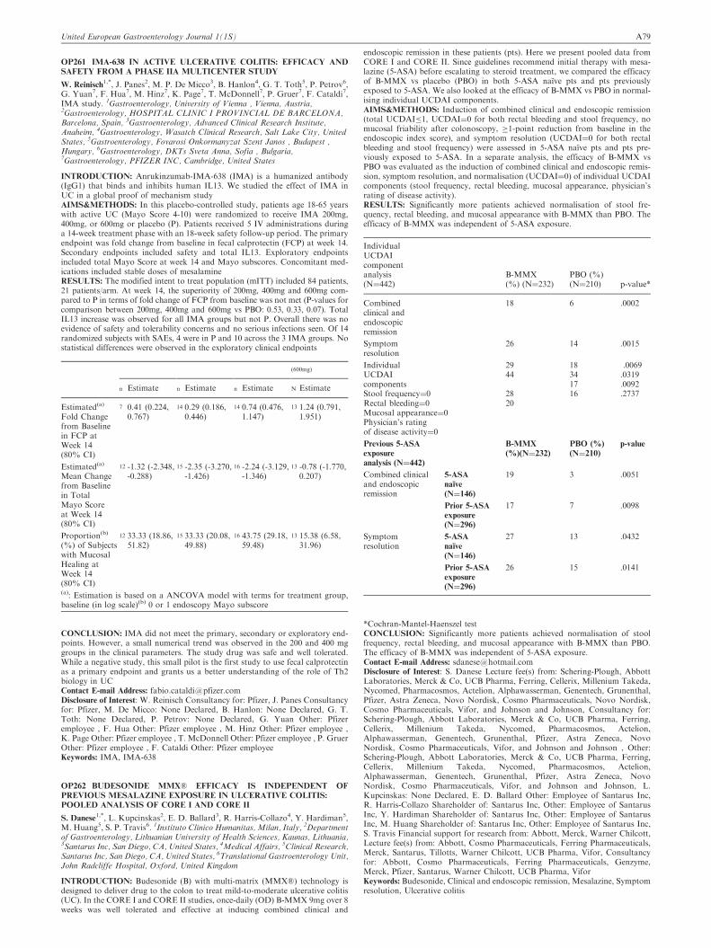
OP261 IMA-638 IN ACTIVE ULCERATIVE COLITIS: EFFICACY AND
SAFETY FROM A PHASE IIA MULTICENTER STUDY
W. Reinisch1,*, J. Panes2, M. P. De Micco3, B. Hanlon4, G. T. Toth5, P. Petrov6,
G. Yuan7, F. Hua7, M. Hinz7, K. Page7, T. McDonnell7, P. Gruer7, F. Cataldi7,IMA study. 1Gastroenterology, University of Vienna , Vienna, Austria,2Gastroenterology, HOSPITAL CLINIC I PROVINCIAL DE BARCELONA,Barcelona, Spain, 3Gastroenterology, Advanced Clinical Research Institute,Anaheim, 4Gastroenterology, Wasatch Clinical Research, Salt Lake City, UnitedStates, 5Gastroenterology, Fovarosi Onkormanyzat Szent Janos , Budapest ,Hungary, 6Gastroenterology, DKTs Sveta Anna, Sofia , Bulgaria,7Gastroenterology, PFIZER INC, Cambridge, United States
INTRODUCTION: Anrukinzumab-IMA-638 (IMA) is a humanized antibody(IgG1) that binds and inhibits human IL13. We studied the effect of IMA inUC in a global proof of mechanism studyAIMS&METHODS: In this placebo-controlled study, patients age 18-65 yearswith active UC (Mayo Score 4-10) were randomized to receive IMA 200mg,400mg, or 600mg or placebo (P). Patients received 5 IV administrations duringa 14-week treatment phase with an 18-week safety follow-up period. The primaryendpoint was fold change from baseline in fecal calprotectin (FCP) at week 14.Secondary endpoints included safety and total IL13. Exploratory endpointsincluded total Mayo Score at week 14 and Mayo subscores. Concomitant med-ications included stable doses of mesalamineRESULTS: The modified intent to treat population (mITT) included 84 patients,21 patients/arm. At week 14, the superiority of 200mg, 400mg and 600mg com-pared to P in terms of fold change of FCP from baseline was not met (P-values forcomparison between 200mg, 400mg and 600mg vs PBO: 0.53, 0.33, 0.07). TotalIL13 increase was observed for all IMA groups but not P. Overall there was noevidence of safety and tolerability concerns and no serious infections seen. Of 14randomized subjects with SAEs, 4 were in P and 10 across the 3 IMA groups. Nostatistical differences were observed in the exploratory clinical endpoints
(600mg)
n Estimate n Estimate n Estimate N Estimate
Estimated(a)
Fold Changefrom Baselinein FCP atWeek 14(80% CI)
7 0.41 (0.224,0.767)
14 0.29 (0.186,0.446)
14 0.74 (0.476,1.147)
13 1.24 (0.791,1.951)
Estimated(a)
Mean Changefrom Baselinein TotalMayo Scoreat Week 14(80% CI)
12 -1.32 (-2.348,-0.288)
15 -2.35 (-3.270,-1.426)
16 -2.24 (-3.129,-1.346)
13 -0.78 (-1.770,0.207)
Proportion(b)
(%) of Subjectswith MucosalHealing atWeek 14(80% CI)
12 33.33 (18.86,51.82)
15 33.33 (20.08,49.88)
16 43.75 (29.18,59.48)
13 15.38 (6.58,31.96)
(a): Estimation is based on a ANCOVA model with terms for treatment group,baseline (in log scale)(b) 0 or 1 endoscopy Mayo subscore
CONCLUSION: IMA did not meet the primary, secondary or exploratory end-points. However, a small numerical trend was observed in the 200 and 400 mggroups in the clinical parameters. The study drug was safe and well tolerated.While a negative study, this small pilot is the first study to use fecal calprotectinas a primary endpoint and grants us a better understanding of the role of Th2biology in UCContact E-mail Address: [email protected] of Interest: W. Reinisch Consultancy for: Pfizer, J. Panes Consultancyfor: Pfizer, M. De Micco: None Declared, B. Hanlon: None Declared, G. T.Toth: None Declared, P. Petrov: None Declared, G. Yuan Other: Pfizeremployee , F. Hua Other: Pfizer employee , M. Hinz Other: Pfizer employee ,K. Page Other: Pfizer employee , T. McDonnell Other: Pfizer employee , P. GruerOther: Pfizer employee , F. Cataldi Other: Pfizer employeeKeywords: IMA, IMA-638
OP262 BUDESONIDE MMX� EFFICACY IS INDEPENDENT OF
PREVIOUS MESALAZINE EXPOSURE IN ULCERATIVE COLITIS:
POOLED ANALYSIS OF CORE I AND CORE II
S. Danese1,*, L. Kupcinskas2, E. D. Ballard3, R. Harris-Collazo4, Y. Hardiman5,
M. Huang5, S. P. Travis6. 1Instituto Clinico Humanitas, Milan, Italy, 2Departmentof Gastroenterology, Lithuanian University of Health Sciences, Kaunas, Lithuania,3Santarus Inc, San Diego, CA, United States, 4Medical Affairs, 5Clinical Research,Santarus Inc, San Diego, CA, United States, 6Translational Gastroenterology Unit,John Radcliffe Hospital, Oxford, United Kingdom
INTRODUCTION: Budesonide (B) with multi-matrix (MMX�) technology isdesigned to deliver drug to the colon to treat mild-to-moderate ulcerative colitis(UC). In the CORE I and CORE II studies, once-daily (OD) B-MMX 9mg over 8weeks was well tolerated and effective at inducing combined clinical and
endoscopic remission in these patients (pts). Here we present pooled data fromCORE I and CORE II. Since guidelines recommend initial therapy with mesa-lazine (5-ASA) before escalating to steroid treatment, we compared the efficacyof B-MMX vs placebo (PBO) in both 5-ASA naıve pts and pts previouslyexposed to 5-ASA. We also looked at the efficacy of B-MMX vs PBO in normal-ising individual UCDAI components.AIMS&METHODS: Induction of combined clinical and endoscopic remission(total UCDAI�1, UCDAI¼0 for both rectal bleeding and stool frequency, nomucosal friability after colonoscopy, �1-point reduction from baseline in theendoscopic index score), and symptom resolution (UCDAI¼0 for both rectalbleeding and stool frequency) were assessed in 5-ASA naıve pts and pts pre-viously exposed to 5-ASA. In a separate analysis, the efficacy of B-MMX vsPBO was evaluated as the induction of combined clinical and endoscopic remis-sion, symptom resolution, and normalisation (UCDAI¼0) of individual UCDAIcomponents (stool frequency, rectal bleeding, mucosal appearance, physician’srating of disease activity).RESULTS: Significantly more patients achieved normalisation of stool fre-quency, rectal bleeding, and mucosal appearance with B-MMX than PBO. Theefficacy of B-MMX was independent of 5-ASA exposure.
IndividualUCDAIcomponentanalysis(N¼442)
B-MMX(%) (N¼232)
PBO (%)(N¼210) p-value*
Combinedclinical andendoscopicremission
18 6 .0002
Symptomresolution
26 14 .0015
IndividualUCDAIcomponentsStool frequency¼0Rectal bleeding¼0Mucosal appearance¼0Physician’s ratingof disease activity¼0
2944
2820
18341716
.0069.0319.0092.2737
Previous 5-ASA
exposure
analysis (N¼442)
B-MMX
(%)(N¼232)
PBO (%)
(N¼210)
p-value
Combined clinicaland endoscopicremission
5-ASA
naıve
(N¼146)
19 3 .0051
Prior 5-ASA
exposure
(N¼296)
17 7 .0098
Symptomresolution
5-ASA
naıve
(N¼146)
27 13 .0432
Prior 5-ASA
exposure
(N¼296)
26 15 .0141
*Cochran-Mantel-Haenszel testCONCLUSION: Significantly more patients achieved normalisation of stoolfrequency, rectal bleeding, and mucosal appearance with B-MMX than PBO.The efficacy of B-MMX was independent of 5-ASA exposure.Contact E-mail Address: [email protected] of Interest: S. Danese Lecture fee(s) from: Schering-Plough, AbbottLaboratories, Merck & Co, UCB Pharma, Ferring, Cellerix, Millenium Takeda,Nycomed, Pharmacosmos, Actelion, Alphawasserman, Genentech, Grunenthal,Pfizer, Astra Zeneca, Novo Nordisk, Cosmo Pharmaceuticals, Novo Nordisk,Cosmo Pharmaceuticals, Vifor, and Johnson and Johnson, Consultancy for:Schering-Plough, Abbott Laboratories, Merck & Co, UCB Pharma, Ferring,Cellerix, Millenium Takeda, Nycomed, Pharmacosmos, Actelion,Alphawasserman, Genentech, Grunenthal, Pfizer, Astra Zeneca, NovoNordisk, Cosmo Pharmaceuticals, Vifor, and Johnson and Johnson , Other:Schering-Plough, Abbott Laboratories, Merck & Co, UCB Pharma, Ferring,Cellerix, Millenium Takeda, Nycomed, Pharmacosmos, Actelion,Alphawasserman, Genentech, Grunenthal, Pfizer, Astra Zeneca, NovoNordisk, Cosmo Pharmaceuticals, Vifor, and Johnson and Johnson, L.Kupcinskas: None Declared, E. D. Ballard Other: Employee of Santarus Inc,R. Harris-Collazo Shareholder of: Santarus Inc, Other: Employee of SantarusInc, Y. Hardiman Shareholder of: Santarus Inc, Other: Employee of SantarusInc, M. Huang Shareholder of: Santarus Inc, Other: Employee of Santarus Inc,S. Travis Financial support for research from: Abbott, Merck, Warner Chilcott,Lecture fee(s) from: Abbott, Cosmo Pharmaceuticals, Ferring Pharmaceuticals,Merck, Santarus, Tillotts, Warner Chilcott, UCB Pharma, Vifor, Consultancyfor: Abbott, Cosmo Pharmaceuticals, Ferring Pharmaceuticals, Genzyme,Merck, Pfizer, Santarus, Warner Chilcott, UCB Pharma, ViforKeywords: Budesonide, Clinical and endoscopic remission, Mesalazine, Symptomresolution, Ulcerative colitis
United European Gastroenterology Journal 1(1S) A79

OP263 EFFECTS OF IMMUNOMODULATORS ON THE
PHARMACOKINETICS AND EFFICACY OF GOLIMUMAB IN
PATIENTS WITH MODERATELY TO SEVERELY ACTIVE
ULCERATIVE COLITIS: RESULTS FROM PHASE 2/3 PURSUIT –SC
INDUCTION AND MAINTENANCE STUDIES
O. J. Adedokun1,*, Z. Xu1, C. Marano1, R. Strauss1, H. Zhang1, H. M. Davis1, J.F. Colombel2, W. Reinisch3, B. Feagan4, P. Rutgeerts5, W. J. Sandborn6, onbehalf of the PURSUIT SC Steering Committee. 1Janssen R&D, LLC. , SpringHouse, United States, 22.Centre Hospitalier Universitaire de Lille, Lille, France,33.Universitatsklinik fur Innere Medizin IV, Vienna, Austria, 4Robarts ResearchInstitute, London, Canada, 55.University Hospital Gasthuisberg, Leuven, Belgium,6University of California San Diego, La Jolla, United States
INTRODUCTION: -AIMS&METHODS: To assess effects of concomitant IM on PK and efficacy ofGLM. PURSUIT-SC induction randomized UC pts with Mayo scores of 6-12inclusive, including endoscopic subscore 42, to PBO/PBO (n¼331);GLM100mg/50mg (before Ph3 dose selection only, n¼72); GLM200 mg/100mg(n¼331) or GLM400 mg/200mg (n¼331) at wks0&2. PURSUIT maintenanceenrolled 1228 pts from PURSUIT-IV & PURSUIT-SC induction studies.Primary analysis for maintenance were pts (n¼464) who responded to GLMinduction and were randomized to PBO, GLM50 mg, or GLM100mg atbsl(wk0) and q4wks thru wk52. Serum GLM concentration (SGC) was measuredthrough wk6 of induction and wk54 of maintenance using validated electroche-miluminescent immunoassay. Antibodies to GLM(ATG) were assessed at wk6 ofinduction, and at wk30 and wk54 of maintenance using an enzyme immunoassay.Efficacy outcomes by IM use were assessed at wk6 (clinical response&remission)for induction; and through wk54 (sustained clinical response) and at wk30 andwk54 (sustained clinical remission) for maintenance.RESULTS: 1227pts had 41 measurable SGC; 32.2% were receiving IMs at wk0of induction(AZA/6-MP[n¼379] and MTX[n¼16]). Among GLM inductionresponders randomized to GLM or PBO,349 remained on randomized dose ofGLM100mg,GLM50mg or PBO thru wk54. Of these,29.5% were receiving IMs.During induction, median SGC levels were similar between pts receiving vs notreceiving IMs. During maintenance, IM use had no substantial impact on medianSGC in those receiving GLM100mg;median SGC was slightly but consistentlyhigher in GLM50mg in combination with IM vs without IM. During inductionand maintenance, there was a positive E-R relationship between SGC and effi-cacy regardless of IM use. IM use didn’t affect efficacy outcomes.Among pts whoreceived 41 GLM dose, ATG incidence was higher (p¼0.02) in pts on PBOduring maintenance vs those on GLM 50mg or GLM100mg (8.8% vs 2.6%).ATG incidence was lower in pts receiving IMs vs those who were not (1.5% vs3.5%, overall 2.8%).Median SGC was lower and there was trend towards lowerclinical response rates through wk54 in pts positive for ATG. Among GLMinduction responders who remained on PBO during maintenance, those positivefor ATG were not receiving IMs.CONCLUSION: Concomitant IM is associated with decreased incidence ofATG, but doesn’t substantially affect SGC, efficacy, or E-R.Contact E-mail Address: [email protected] of Interest: O. Adedokun Other: Janssen employee, Z. Xu Other:Janssen employee, C. Marano Other: Janssen employee, R. Strauss Other:Janssen employee, H. Zhang Other: Janssen employee, H. Davis Other:Janssen employee, J. Colombel Financial support for research from: Janssen,W. Reinisch Financial support for research from: Janssen, B. Feagan Financialsupport for research from: Janssen, P. Rutgeerts Financial support for researchfrom: Janssen, W. Sandborn Financial support for research from: JanssenKeywords: golimumab, pharmacokinetics, PURSUIT
OP264 SAFETY, PHARMACOLOGY, AND EFFECT OF AMG 181, A
HUMAN ANTI-A4B7 ANTIBODY, IN SUBJECTS WITH MILD TO
MODERATE ULCERATIVE COLITIS
W.-J. Pan1,*, G. Radford-Smith2,3, J. Andrews4, H. Schwartz5, W. Rees6,
B. Sullivan7, C. Evangelista7, D. Doherty1, P. Prince1, K. Reynhardt6, K. Zhou8,A. Treacy9, J. Lee7, Z. Yu7, D. Borie10. 1PKDM, Amgen Inc., Seattle, UnitedStates, 2Gastroenterology Unit, Royal Brisbane and Women’s Hospital, Herston,3IBD Group, Queensland Institute of Medical Research, and School of Medicine,University of Queensland, Brisbane St Lucia, 4IBD Service & School of Medicine,University of Adelaide at Royal Adelaide Hospital, Adelaide, Australia, 5MiamiResearch Associates, South Miami, 6Medical Sciences, Amgen Inc., Seattle,7Medical Sciences, Amgen Inc., Thousand Oaks, 8Biostatistics, Amgen Inc., SouthSan Francisco, United States, 9Clinical Operations, Amgen Inc., Kew, Australia,10Clinical Development, Amgen Inc., Thousand Oaks, United States
INTRODUCTION: AMG 181 is currently in Phase 2 clinical trials for treatingCrohn’s disease (CD) and ulcerative colitis (UC).AIMS&METHODS: To evaluate pharmacokinetics/pharmacodynamics (PK/PD), safety, and effect of subcutaneous (SC) AMG 181, we enrolled subjectswith mild to moderate UC (Mayo score 4-10). In one study (completed), 2male and 2 female white subjects (32-43 yr, 57-77 kg) received a single SC doseof 210 mg AMG 181 or placebo (3:1) and were followed for 7 months. In anotherstudy (on-going), 3 male and 1 female subjects (26-51 yr, 73-92 kg) received 21mg AMG 181 or placebo SC (3:1 ratio) monthly for 3 months and were followedfor 5 months. PK, anti-AMG 181 antibodies (ADAs), safety, �4�7 receptoroccupancy (RO), CD4þ central memory T cell counts (Tcm), serum high sensi-tivity C-reactive protein (hsCRP), fecal calprotectin (FC), and Mayo score weremeasured. Remission was defined as: Mayo score �2, with no individual sub-score 41; response: Mayo score decrease from baseline by �3 and �30% þrectal bleeding score decrease of �1 or absolute reading of 0 or 1; mucosalhealing: endoscopy subscore of �1.
RESULTS: Median Mayo score was 7 at screening for the 8 UC subjects (6 onactive; 2 on placebo). PK profiles were within the range of those observed inhealthy subjects. No ADAs were detected. PK and RO profiles were directlycorrelated, without apparent Tcm count elevation. Under AMG 181 active regi-mens, both hsCRP and FC showed decreasing trends in some subjects at somevisits but small sample size and data variability precluded definitive assessment.Blinded evaluation showed 3, 4, and 1 subjects achieved remission, 6, 6, and 2subjects were in response, and 5, 5, and 1 subjects achieved mucosal healing atwks 6, 12, and 28, respectively. No AMG 181-related serious adverse events ordeaths, except for two early withdrawals due to colitis flare, which were assessedby the investigators as not related to the investigational product. No clinicallysignificant changes in electrocardiograms or neurological examinations wereobserved.CONCLUSION: Preliminary PK/PD, immunogenicity, safety, and effect profilesof AMG 181 in UC support its further testing in subjects with UC or CD.Contact E-mail Address: [email protected] of Interest: W.-J. Pan Financial support for research from: Amgen Inc.,Shareholder of: Amgen Inc., Other: Amgen Inc. Employee, G. Radford-SmithFinancial support for research from: Amgen Inc., Consultancy for: Amgen Inc., J.Andrews Financial support for research from: Amgen Inc., Consultancy for: AmgenInc., H. Schwartz Financial support for research from: Amgen Inc., Consultancy for:Amgen Inc., W. Rees Financial support for research from: Amgen Inc., Shareholderof: Amgen Inc., Other: Amgen Inc. Employee, B. Sullivan Financial support forresearch from: Amgen Inc., Shareholder of: Amgen Inc., Other: Amgen Inc.Employee, C. Evangelista Financial support for research from: Amgen Inc.,Shareholder of: Amgen Inc., Other: Amgen Inc. Employee, D. Doherty Financialsupport for research from: Amgen Inc., Shareholder of: Amgen Inc., Other: AmgenInc. Employee, P. Prince Financial support for research from: Amgen Inc.,Shareholder of: Amgen Inc., Other: Amgen Inc. Employee, K. Reynhardt Financialsupport for research from: Amgen Inc., Shareholder of: Amgen Inc., Other: AmgenInc. Employee, K. Zhou Financial support for research from: Amgen Inc.,Shareholder of: Amgen Inc., Other: Amgen Inc. Employee, A. Treacy Financial sup-port for research from: Amgen Inc., Shareholder of: Amgen Inc., Other: Amgen Inc.Employee, J. Lee Financial support for research from: Amgen Inc., Shareholder of:AmgenInc.,Other:AmgenInc.Employee,Z.YuFinancial support for research from:Amgen Inc., Shareholder of: Amgen Inc., Other: Amgen Inc. Employee, D. BorieFinancial support for research from: Amgen Inc., Shareholder of: Amgen Inc.,Other: Amgen Inc. EmployeeKeywords: AMG 181, Anti-�4�7 Antibody, Crohn’s Disease, Inflammatorybowel disease (IBD), MEDI7183, Ulcerative Colitis
OP265 HIGHLY SIGNIFICANT IMPROVEMENT IN DAI SUBSCORES
BY LOCAL APPLICATION OF THE TOLL-LIKE RECEPTOR 9
AGONIST DIMS0150 IN MODERATELY SEVERE ULCERATIVE
COLITIS PATIENTS
T. Knittel1,*, R. Lofberg2, J. Kowalski3, P. von Stein1, O. von Stein1. 1Index
Pharmaceuticals, 2Stockholm Gastro Center, Sophiahemmet , Stockholm, 3JKBiostatistics AB, Jonkoping, Sweden
INTRODUCTION: DIMS0150 is an oligonucleotide that acts as a Toll-likereceptor 9 (TLR-9) agonist and elicits its immunomodulating and anti-inflam-matory effects through specific target cells. Since the phase II clinical study hasshown sustained clinical response and remission but with borderline statisticalsignificance we performed an additional in dept analysis of this study with respectto changes of individual DAI subscores.AIMS&METHODS: A randomized, double-blind, multicenter phase II trial wasconducted in steroid refractory patients with ulcerative colitis of moderatedegree. 34 patients were randomized to a single rectal administration of either30 mg of DIMS0150 or placebo with 2:1 allocation. A paired t –test was used toanalyse the DAI subscore changes in the per protocol (PP) population on apatient by patient basis between baseline and 4 weeks after therapy.RESULTS:DAI subscore analysis indicated robust levels of a statistically signifi-cant mean change at week 4 from baseline in the DIMS0150 treated group in all 4subscores. By contrast DAI sub score analysis in the placebo group demonstratedno significant change at week 4 from baseline in 3 out of 4 of the subscores.The global assessment score showed a statistically significant improvement inDIMS0150 treated patients (p 0,04) and an insignificant change in the placebogroup (p 0,108). By considering stool frequency there was a statistically signifi-cant change (p 0,005) in the mean frequency when compared to baseline in theDIMS0150 treated group with 52% of those patients having a completely normalstool frequency at week 4. By contrast, only 27% of patients given placebo had anormal stool frequency byweek 4 (p 0,02). For the subscore blood in stool there wasa significant reduction (p 0,004) in the mean change from baseline in the DIMS0150treated group with approximately 47% of patients observing no blood in stool atweek 4. By contrast for those patient receiving placebo, there was no statisticalevidence (p 0,09). In terms of endoscopic assessment, patients treated withDIMS0150 demonstrated a statistically significant reduction (p 0,003) in the meanendoscopic score at week 4 when compared to baseline. Placebo patients demon-strated no statistical evidence of an improved endoscopic score (p 0,117).CONCLUSION: The efficacy results for this TLR-9 agonist in patients withmoderately severe ulcerative colitis are encouraging, with highly significantimprovements in DAI subscores in comparison to placebo treated patientsalready 4 weeks after a single dose of a DIMS0150.Contact E-mail Address: [email protected] of Interest: T. Knittel Directorship(s) for: Index Pharmaceuticals,R. Lofberg Shareholder of: Index Pharamceuticals, J. Kowalski: NoneDeclared, P. von Stein Shareholder of: Index Pharmaceuticals, Directorship(s)for: Index Pharmaceuticals, O. von Stein Shareholder of: Index Pharmaceuticals,Directorship(s) for: Index PharmaceuticalsKeywords: therapeutic development, therapy, ulcerative colitis
A80 United European Gastroenterology Journal 1(1S)

TUESDAY, OCTOBER 15, 2013 14:00–15:30Recent developments in upper GI and small bowel bleeding – Hall7_____________________
OP266 RISK FACTORS FOR MORTALITY FROM NON VARICEAL
BLEEDING SOURCE IN PATIENTS WITH LIVER CIRRHOSIS: AN
INDIVIDUAL PATIENT DATA META-ANALYSIS
G. Rotondano1,*, R. Marmo2, M. Del Piano3, L. Cipolletta1, M. Koch4,
E. Grossi5, M. Rea2, C. Bucci2, S. Sansone1, M. A. Bianco1, PNED andPROMETEO Investigators. 1Hospital Maresca, Torre del Greco, 2Hospital Curto,Polla, 3AO Maggiore della Carita, Novara, 4ACO San Filippo Neri, Roma,5Medical Division Bracco, Milano, Italy
INTRODUCTION: Upper GI bleeding in patients with liver cirrhosis is usuallyrelated to esophago-gastric varices or hypertensive gastropathy. Nonetheless, thefrequency of variceal bleeding is decreasing over time, while the number ofcirrhotic patients with non varical UGIB is increasing.AIMS&METHODS: Aim of the study was to assess the risk of death in patientswith liver cirrhosis with bleeding from non-variceal source.Four national prospective multicentric databases (PNED1, PNED 2, PROME-TEO 1 and PROMETEO 2 studies) containing data on consecutivepatients admitted to participating hospitals for acute non-variceal UGIB wereanalysedRESULTS: A total of 3884 pts were included: of these 268 (6.9%) had livercirrhosis. Cirrhotic pts were younger than non cirrhotic (65.2�0.82vs.68.6�0.27,p50.001) and male gender was less frequent (59.8% vs. 66.4%,p50.03). In patients with cirrhosis, the main causes of nonvariceal UGIB weregastric ulcer (25.0%), duodenal ulcer (23.1%) and gastroduodenal erosions(18.6%). Rebleeding and surgery were not different compared to non cirrhotics,while risk of mortality higher (7.8% vs. 4.1%, OR 1.99 [95% CI 1.23-3.20],p¼0.004). Multivariate analysis identified clinical presentation with hematemesis,presence of gastric vascular lesions, chronic renal failure, neoplasia, failure ofendoscopic treatment, concurrent presence of duodenal ulcer and gastroduodenalerosions and recurrent bleeding as independent predictors of bleeding-relateddeath (table). Global prognostic accuracy of the model was 94.9%
Risk factor Odds ratio
95% confidenceinterval P value
Neoplasia 1.73 -.225 to 3.56 0.084
Failure of endoscopic treatment 2.23 .591 to 9.26 0.026
Duodenal ulcer plus GD erosions 2.36 .423 to 4.58 0.018
Renal failure 2.66 .851 to 5.60 0.008
Vascular lesion 2.92 .0147 to .075 0.004
Hematemesis 3.13 1.34 to 5.82 0.002
Recurrent bleeding 3.17 1.36 to 5.75 0.002
CONCLUSION: Concurrent duodenal ulcer and gastro-duodenal erosions,together with bleeding form vascular lesions represent the main determinantsof death in cirrhotic patients with acute nonvariceal UGIB. Co-factors of mor-tality are presentation with hematemesis, failure of endoscopic treatment, pre-dence of neoplasia and recurrent bleeding.Contact E-mail Address: [email protected] of Interest: None DeclaredKeywords: mortality in chirrotics, nonvariceal bleeding, risk factors
OP267 TRANSCATHETER ARTERIAL EMBOLIZATION IS THE
TREATMENT OF CHOICE IN PEPTIC ULCER BLEEDING NOT
RESPONDING TO ENDOSCOPIC THERAPY
M. Jakobsen1, M. M. Nielsen1,*, O. B. Schaffalitzky de Muckadell2, S.
B. Laursen2. 1University of Southern Denmark, 2Department of medical gastro-enterology S, Odense University Hospital, Odense, Denmark
INTRODUCTION: In 5-10% of cases with peptic ulcer bleeding (PUB) it is notpossible to achieve endoscopic hemostasis. In these patients transcatheter arterialembolization (TAE) is frequently used as an alternative to surgical hemostasis.Studies comparing the outcomes of these methods are needed in order to identifythe treatment of choice.AIMS&METHODS: The aim of this study was to examine the efficacy and safetyof TAE compared with surgical hemostasis in patients with PUB not responding toendoscopic treatment. The study was conducted as a retrospective study.Consecutive patients treatedwith TAEor surgery for endoscopy-refractory bleedingin the period June 1997 to June 2012 were included. Patient characteristics, endo-scopic findings, received treatment, and outcome were registered. An adjusted 30-day mortality was calculated using multivariate logistic regression analysis.RESULTS: A total of 41 and 75 patients were treated with TAE and surgery,respectively. Patients treated with TAE had a higher level of comorbidity(median Charlson comorbidity index: 2 versus 1; P¼0.0084), lower rate of com-plications (22% versus 60%; p 5 0.0001), shorter length of stay (median: 9versus 13.5 days; p ¼ 0.0016), but higher rate of rebleeding (39% versus 16%;p ¼ 0.012). There was no difference in unadjusted overall 30-day mortality. Whenadjusting for differences in patient characteristics patients treated with TAE hada lower overall 30-day mortality (OR: 0.30; P ¼ 0.04).CONCLUSION: Transcatheter arterial embolization is associated with lowermortality, lower rate of complications, and lower length of stay compared tosurgical hemostasis. TAE seems to be the best first-line treatment in patients withendoscopy-refractory PUB.
Disclosure of Interest: None DeclaredKeywords: Embolization, peptic ulcer bleeding
OP268 INCREASED CARDIOVASCULAR EVENTS AFTER
DISCONTINUATION OF ANTITHROMBOTICS IN PATIENTS WITH
PEPTIC ULCER BLEEDING
S. Y. Kim1, J. J. Hyun1,*, S. W. Jung1, J. S. Koo1, R. S. Choung1, S. W. Lee1, J.
H. Choi1. 1Korea University Ansan Hospital, Ansan, Korea, Republic Of
INTRODUCTION: Peptic ulcer bleeding associated with antithrombotics hasincreased due to the increase in the proportion of elderly population. Little isknown about the long-term effects of discontinuating antrithrmobotics afterpeptic ulcer bleeding.AIMS&METHODS: The aim of this study was to evaluate whether the risk ofcardiovascular events increases when antiplatelets or anticoagulants are discon-tinued after ulcer bleeding.The medical records of patients diagnosed with ulcer bleeding between January2006 and December 2011 at Korea University Ansan Hospital were retrospec-tively reviewed. We enrolled patients who were taking antiplatelet or anticoagu-lant drugs at the time of ulcer bleeding. We used Cox regression model to adjustpotential confounders, and analyze association between discontinuation ofantithrombotic drugs after ulcer bleeding and thrombotic events such as ischemicheart disease or stroke.RESULTS: Of the 544 patients with ulcer bleeding, 72 patients who were takingantithrombotic drugs and followed-up to more than 2 months were analyzed.Forty patients discontinued antithrombotics after ulcer bleeding (discontinuationgroup) and 32 patients continued antithrombotics with or without transientinterruption (continuation group). Ischemic heart disease developed in 2 patients,stroke or transient ischemic attack developed in 4 patients, deep vein thrombosisin 1 patient and common iliac artery thrombosis in 1 patient during mean follow-up for 26.6 months. Thrombotic event developed more often in discontinuationgroup than in continuation group (p¼0.019). Hazard ratio for thrombotic eventwhen antithrombotics were discontinued continuously was 15.6 (95% CI, 1.8-135.2). There were no significant differences in recurrent bleeding event betweentwo groups.CONCLUSION: Discontinuation of antithrombotic drugs after peptic ulcerbleeding increases risk of cardiovascular events. Therefore, we should be carefulin discontinuating antrithrombotic drugs after ulcer bleeding.Contact E-mail Address: [email protected] of Interest: None DeclaredKeywords: antithrombotics, cardiovascular complication, ulcer bleeding
OP269 SMALL BOWEL ANGIODYSPLASIAS ARE MORE COMMON
AMONG PATIENTS WITH LARGE BOWEL DIVERTICULA.
P. Tsibouris1, P. Apostolopoulos1, C. Kalantzis1, E. Vlachou1,*, A. Koumi1,I. Koumoutsos1, P. Dogantzis1, C. Beltsidou1, G. Alexandrakis1.1GASTROENTEROLOGY, NIMTS GENERAL HOSPITAL, Athens, Greece
INTRODUCTION: Lower gastrointestinal bleeding is a common complicationof large bowel diverticulosis.AIMS&METHODS: Aim: To define frequency of small bowel ulcerative lesionsand angiodysplasias in patients with large bowel diverticulosis, with and withoutanemia.Methods: 984 patients (mean age 60�18 years, 543 men, 256 smokers) undergonesmall bowel capsule endoscopy, gastroscopy and colonoscopy were evaluated.490 were evaluated for iron deficiency anemia, 311 for diarrhea, 60 for unex-plained abdominal pain and for 123 other causes. Stat: X2, logistic regressionanalysis.RESULTS: 140 (29%) anemics and 64 (13%) non-anemics presented large boweldiverticula,without anymacroscopic inflammationandwithout any history of lowergastrointestinal bleeding (p50.0001). Mean small bowel passing time was 380�154min for patients with large bowel diverticula and 332�136 in those without(p50.0001). Excluding patients with IBD, 59 (30%) patients with large bowel diver-ticula and 178 (29%) without presented small bowel ulcerative lesions (p¼0.43).Among anemics, 45 (32%) patients with large bowel diverticula and 112 (32%)without presented small bowel ulcerative lesions (p¼0.98). 117 (57%) patients withlarge bowel diverticula and 278 (36%) without presented small bowel angiodyspla-sias (p50.0001).Amonganemics, 90 (64%)patientswith largebowel diverticula and140 (40%) without presented small bowel angiodysplasias (p50.0001). In logisticregression analysis, age (p50.0001), male gender (p¼0.006), chronic renal failure(p¼0.05) and presence of large bowel diverticula (p¼0.01) were independent riskfactors for the presence of small bowel angiodysplasias.24 (36%) NSAIDs users with large bowel diverticula and 110 (43%) withoutdiverticula presented small bowel ulcerative lesions (p¼0.17). Moreover, all 29patients with large bowel diverticula who received clopidogrel versus 55 (74%)receiving low-dose aspirin presented with anemia (p50.0001).CONCLUSION: Iron deficiency anemia is more common among patients withlarge bowel diverticulosis. Anemia could partially attributed in higher frequencyof angiodysplasias in this patient group, possibly due to connective tissue struc-tural deficiency. Moreover, patients with large bowel diverticulosis are moresusceptible to clopidogrel.Disclosure of Interest: None DeclaredKeywords: anemia, angiodysplasia, capsule endoscopy, large bowel diverticula,small bowel ulcerative lesions
United European Gastroenterology Journal 1(1S) A81

OP270 ELEVATED ANGIOPOIETIN 2 LEVELS IN SMALL BOWEL
ANGIODYSPLASIA; FUTURE BIOMARKER OR THERAPEUTIC
TARGET
G. Holleran1,2,*, B. Hall1,2, S. Smith1, D. McNamara1,2. 1Department of Clinical
Medicine, Trinity College Dublin, Tallaght, 2Department of Gastroenterology,Adelaide and Meath Hospital, Dublin 24, Ireland
INTRODUCTION: Small bowel angiodysplasia (SBA) accounts for up to 50%of cases of obscure gastrointestinal bleeding, which were difficult to diagnoseprior to the advent of capsule endoscopy (CE). Little is known about theirpathophysiology and therapeutic options are limited. Abnormal serum levels ofangiogenic factors have been suggested previously in small cohorts of patientswith sporadic colonic, and genetic - Hereditary Haemorrhagic Telangiectasia(HHT), forms of GI angiodysplasia. Angiogenic factors may represent targetsfor new therapies and act as useful diagnostic and prognostic biomarkers forsporadic small bowel disease.AIMS&METHODS: To assess putative angiogenic factor levels in sporadic SBAcompared with controls.Following informed consent, serum samples were collected and stored at -80oC forbatch analysis from patients with a definite diagnosis of sporadic SBA on CE (P2),and from healthy controls in which GI bleeding had been out-ruled by a negativefaecal immunochemical test. Patients with features consistent with, or knownHHTwere excluded. Serum levels of angiopoietin-2 (Ang-2), soluble endoglin (sEnd),vascular endothelial growth factor (VEGF), and platelet derived growth factor(PDGF) were measured using commercially available enzyme-linked immunosor-bent assay (ELISA) kits (R&DSystems). Samples were prepared in duplicate as permanufacturer’s guidelines and absorbance read at a wavelength of 450nm. Allresults were expressed as a mean and compared between patients and controls, ap value of 50.05 was considered significant.RESULTS: In all, serum samples were analysed from 30 patients, 53% (n¼16)female and 26 controls, 54% (n¼14) female, with mean ages of 68.1 years (34-90)and 55.5 years (26-74) respectively. Levels of Ang-2 were significantly higher inthe angiodysplasia group with a mean of 4883pg/ml (842-11767) vs. a mean of2536pg/ml (792-6243) in controls p50.001. In addition, there was a trendtowards lower levels of sEnd and elevated levels of VEGF and PDGF in thepatient group versus controls although not statistically significant.
Angiogenicfactor
Angiodysplasiamean
Angiodysplasiarange
Controlmean
Controlrange
Ang-2 pg/ml 4883 842-11767 2536 792-6243
sEnd ng/ml 3.69 2.12-6.84 4.65 1.96-9.0
VEGF pg/ml 355.9 45-929 289.6 54-1130
PDGF pg/ml 4229 929-39978 3171 955-6208
CONCLUSION: Serum Ang-2 levels are significantly elevated in patients withsporadic SBA and may represent a useful diagnostic and prognostic biomarker.A trend towards decreased levels of sEnd and elevated levels of VEGF and PDGFmay be useful in further elucidating the pathogenesis of sporadic angiodysplasia.Additional studies with larger patient numbers are needed to confirm our findings.Contact E-mail Address: [email protected] of Interest: None DeclaredKeywords: angiodysplasia, angiogenic factors
OP271 RESCUE THERAPY WITH OVER THROUGH-THE-SCOPE
CLIPS (OTSC) FOR ACUTE GI COMPLEX BLEEDING
UNRESPONSIVE TO CONVENTIONAL ENDOSCOPIC
TREATMENT: RESULTS OF A MULTICENTER ITALIAN STUDY.
R. Manta1,*, G. Galloro2, M. Manno1, V. G. Mirante1, A. Caruso1, H. Bertani1,
B. Mangiavillano3, L. Pasquale4, A. Arezzo5, L. De Luca6, G. Bassotti7,R. Conigliaro1. 1Gastroenterology and Digestive Endoscopy Unit , New CivilS.Agostino Estense Hospital, Modena, 2Clinical Medicine and Surgery , SurgicalDigestive Endoscopy, University of Federico II, Naples, Napoli, 3Gastroenterologyand Digestive Endoscopy Unit , San Paolo Hospital, Milano, 4Gastroenterologyand Digestive Endoscopy Unit , Civil Ariano Irpino Hospital, Ariano Irpino (AV),5Surgical Sciences, University of Torino, Torino, 6Gastroenterology and DigestiveEndoscopy Unit , Civil hospital of Pesaro , Pesaro, 7Clinical and experimentalMedicine, Gastroenterology and Hepatology Section,University of Perugia,Perugia, Italy
INTRODUCTION: BACKGROUND The efficacy of through the scope clips(TTSC) can be sub-optimal in patients with complex bleeding lesions. TheOver-The-Scope Clip (OTSCR) could overcome the TTSC by allowing compres-sion of larger amounts of tissue, allowing a more efficient hemostasis.AIMS&METHODS: AIM To analyze the use of OTSC in a consecutive caseseries of patients with acute GI bleeding unresponsive to conventional endo-scopic treatment modalities. METHODS In a retrospective analysis of prospec-tively collected data in tertiary referral centers, patients undergoing emergencyendoscopy for severe acute non variceal GI bleeding were treated by OTSCplacement after failure of conventional hemostatic therapy. All patients under-went repeat endoscopy 2-4 days after the procedure.Data analysis included pri-mary hemostasis, complications, and one-month follow-up clinical outcome.RESULTS: RESULTS During a 17-month period, 48 patients entered the studyconsecutively. Bleeding lesions unresponsive to conventional endoscopic treat-ment (saline/adrenaline injection and through-the scope clipping) were locatedin the upper and lower GI tract in 40 and 8 cases, respectively. Primary hemostasiswas achieved in 46 out of 48 cases (96%). 1 patient with bleeding from duodenal
bulb ulcer required emergent selective radiological embolization. 1 patient withmassive bleeding from large gastric GIST located in the fundus, needed fibrin glueinjection in the same session to obtain definitive hemostasis. Rebleeding occurredin 2 patients 12h and 24h after the procedure: they were successfully treated withconventional saline/adrenaline endoscopic injection.CONCLUSION: CONCLUSIONS The OTSC is an effective and safe therapeu-tic option for severe acute GI bleeding, when conventional endoscopic treatmentmodalities fail.Contact E-mail Address: [email protected] of Interest: None DeclaredKeywords: Hemostatic clips, ulcer bleeding
TUESDAY, OCTOBER 15, 2013 14:00–15:30
Complications of cirrhosis – Hall 6_____________________
OP272 COMPARISON OF SEVEN PROGNOSTIC MODELS FOR
SHORT-TERM MORTALITY IN BIOPSY-PROVEN ALCOHOLIC
HEPATITIS.
V. Papastergiou1, S. Karatapanis1,*, E. Tsochatzis1, E. Thalassinos1, G. Pieri1,
A. Burroughs1. 1Royal Free Hospital, London, United Kingdom
INTRODUCTION: Assessment of mortality is paramount in alcoholic hepatitis(AH). Although several prognostic scores have been proposed, external valida-tion and model comparisons are required as no one score is used universally.AIMS&METHODS: We aimed to validate seven prognostic models for short-term mortality in biopsy-proven AH: Maddrey discriminant function (DF); Lillemodel; Glascow alcoholic hepatitis score (GAHS); Mayo end-stage liver disease(MELD); age, bilirubin, INR, creatinine score (ABIC); MELD-Na, UK end-stage liver disease (UKELD).Consecutive patients with biopsy-proven AHadmitted between November 2006-September 2011 at the Royal Free Hospital,were evaluated. Biochemical data from day 0 and day 7 from admission was usedto assess prognostic models with respect to 30- and 90-day mortality.RESULTS: Seventy-one patients (47 males, mean age; 48.4 years) were included.The MELD, DF, GASH, ABIC, and the Lille score demonstrated comparablepredictive abilities with C-statistics ranging to 0.71-0.81 for 30-day mortality and0.74-0.84 for 90-day mortality. C-statistics for 30-day/90-day mortality were forMELD-Na 0.68/0.76 and for UKELD 0.56/0.68. Re-scoring on day-7 yielded atrend towards higher C-statistics for both 30-day (0.69-0.84) and 90-day (0.77-0.86) mortality. All models showed excellent NPV, in most cases 490% (range:88%4100%), whereas PPV were low, in most instances 550% (range:21%470%).CONCLUSION: MELD, DF, GASH, ABIC and Lille score, proved to be clini-cally useful for the prediction of short-term mortality when assessed in an exter-nal cohort of patients with biopsy-proven AH. Modifications of MELDincorporating sodium did not confer any prognostic advantage over MELD.Excellent NPV suggest models are best to exclude, rather than to identify high-risk patients.Contact E-mail Address: [email protected] of Interest: None DeclaredKeywords: ALCOHOLIC LIVER DISEASE, prognostic scores
OP273 THE PHOSPHODIESTERASE-5-INHIBITOR UDENAFIL - A NEW
OPTION FOR THERAPY OF PORTAL HYPERTENSION
W. Kreisel1,*, P. Deibert1, L. Kupcinskas2, J. Sumskiene2, B. Appenrodt3,
M. Neagu1, M. Rossle4, A. Zipprich5, K. Caca6, A. Ferlitsch7, R. Mohrbacher8,R. Greinwald8, T. Sauerbruch9. 1University Hospital Freiburg, Freiburg,Germany, 2Kaunas University of Medicine, Kaunas, Lithuania, 3UniversityHospital, Homburg, 4Gastroenterology Centre, Freiburg, 5University Hospital,Halle, 6Klinikum Ludwigsburg, Ludwigsburg, Germany, 7Medical University,Wien, Austria, 8Dr. Falk Pharma, Freiburg, 9University Hospital, Gottingen,Germany
INTRODUCTION:Non-selective beta-blockers (NSBB) are used as standard forprevention of variceal bleeding in cirrhosis. Long-term treatment should decreasethe HVPG by �20% or to �12 mmHg. HVPG decrease by �10% or to �12mmHg in the acute setting predicts clinical response, as well. The response rate toNSBB is about 50%. The efficacy is limited by cardiovascular side effects.Phosphodiesterase-5-inhibitors (PDE-5-I) increased portal venous blood flowand lowered portal pressure in rats with normal liver and in individuals withnormal or cirrhotic liver. The clinical relevance is unknown.AIMS&METHODS: The effect of udenafil, a new PDE-5-I with a half-life of12h, on HVPG in patients with portal hypertension was investigated. Patientswith liver cirrhosis, HVPG �12 mmHg, and low risk of bleeding were eligible forthis open-label, multicenter phase II trial. Patients obtained oral udenafil (12.5mg/day, 25 mg/day, 50 mg/day, 75 mg/day [5 patients for each dose], and 100mg/day [10 patients]) once daily (OD) for one week. Routine laboratory tests andsystemic hemodynamic parameters were checked regularly. On day 0 and 6HVPG was measured prior to and one hour after drug administration.Response 1: Decrease of HVPG by �10% in the acute setting (day 0 or/and6). Response 2: Decrease of HVPG by �20% in the one week OD setting (day 6post-dose compared to day 0 pre-dose) or to �12 mmHg.RESULTS: 30 patients were evaluated. Table 1 shows the mean decrease ofHVPG on day 0 or 6 for the different doses. In the 10 patients treated with100 mg/day the mean decrease was significant on day 0 (p¼0.012) and on day6 (p¼0.045). In 4 of 5 patients treated with 75 mg/day and in 7 of 10 patientstreated with 100 mg/day HVPG was lowered by �10% on day 0 or/and day 6(Response 1). 4 out of the 10 patients in the 100 mg/day group reached response2. There were no cardiovascular side effects due to the drug in any of the groups.Standard liver biochemical tests did not change during the study.
A82 United European Gastroenterology Journal 1(1S)

12.5 mg/day 25 mg/day 50 mg/day 75 mg/day 100 mg/day
day 0 day 6 day 0 day 6 day 0 day 6 day 0 day 6 day 0 day 6
-3% 0% -4% -2% -7% -6% -25% -11% -17%* -13%*
Table 1 Decrease of HVPG in the acute setting on day 0 and day 6. * p50.05
CONCLUSION: The new PDE-5 inhibitor udenafil lowers HVPG in the acutesetting and may be a new option for therapy of portal hypertension.Disclosure of Interest: W. Kreisel: None Declared, P. Deibert: None Declared, L.Kupcinskas: None Declared, J. Sumskiene: None Declared, B. Appenrodt: NoneDeclared, M. Neagu: None Declared, M. Rossle: None Declared, A. Zipprich:None Declared, K. Caca: None Declared, A. Ferlitsch: None Declared, R.Mohrbacher Other: Employee of Dr. Falk Pharma, R. Greinwald Other:Employee of Dr. Falk Pharma, T. Sauerbruch: None DeclaredKeywords: HVPG, liver cirrhosis, Phosphodiesterase-5-Inhibitor, PortalHypertension, Udenafil
OP274 CAUSE-SPECIFIC MORTALITY OF PATIENTS WITH
CIRRHOSIS IN ENGALND: VARIATION BY AETIOLOGY
S. Ratib1,*, K. M. Fleming1, C. J. Crooks1, A. J. Walker1, J. West1.
1Epidemiology & Public Health, UNIVERSITY OF NOTTINGHAM,Nottingham, United Kingdom
INTRODUCTION: While the incidence of and overall death from cirrhosis ofthe liver is inexorably increasing in the UK, we do not know what it is that causespeople with cirrhosis to die. Determining the most important causes of death andhow this varies by aetiology will demonstrate areas where therapeutic interven-tions may be best targeted.AIMS&METHODS:We identified adult patients with cirrhosis from the ClinicalPractice Research Datalink and the Hospital Episode Statistics database between1998 and 2009 in England. Causes of death were obtained from the Office forNational Statistics and categorized according to the International Cause ofDisease classification (ICD10). We examined the cause of death by aetiologyof cirrhosis. We calculated (i) cumulative incidence at 2 years (median follow-up) for each cause and (ii) hazard ratios (HR) to compare risk of death, by 2years, for each cause by aetiology, adjusting for age and sex.RESULTS: 5118 patients with cirrhosis were identified, over a third died by2 years (n¼1934, 37.8%). Of those who died by 2 years, 1019 (52.7%) hada liver-related death. Comparing cryptogenic with alcoholic cirrhosis, thecumulative incidence 2 years from diagnosis was two-fold for circulatorydeaths (12% vs 6%) and four-fold for neoplastic deaths (16% vs 4%).Adjusting for age and sex, risks of circulatory and neoplastic death were stillsignificantly increased among cryptogenic cirrhotics compared to those withalcoholic cirrhosis (Table).Table: Number of deaths, cumulative incidence and adjusted hazard ratios (& 95%
confidence intervals) by aetiology for each cause of death, 2 years from diagnosis*Adjusted for age and sex. yIncludes hepatocellular carcinoma (HCC) deaths.¥Excludes HCC deaths. xIncludes non-liver related digestive, respiratory, andunknown causes.CONCLUSION: Overall, almost half of the underlying causes of death of peoplewith cirrhosis at 2 years following diagnosis are non-liver related. Risk of circu-latory and neoplastic death was substantially higher in people with cryptogeniccirrhosis than those with alcoholic cirrhosis. Strategies for prevention or ameli-oration of circulatory disease may be warranted in people with cryptogeniccirrhosis.Contact E-mail Address: [email protected] of Interest: None DeclaredKeywords: AETIOLOGY, CAUSE OF DEATH, CIRRHOSIS, MORTALITY
OP275 EFFECT’S EVALUATION OF THE TREATMENT WITH
PROPRANOLOL AND LOW DOSE LOSARTAN VERSUS ONLY
PROPRANOLOL ADMINISTRATION ON HEPATIC
HAEMODYNAMIC PARAMETERS IN PATIENTS WITH LIVER
CIRRHOSIS
E. Tcaciuc1,*, O. Corlateanu1, S. Matcovschi1, A. Tcaciuc1, N. Nacu1. 1Internal
Medicine, State Medical and Pharmaceutical University "Nicolae Testemitanu",Chisinau, Moldova, Republic of
INTRODUCTION: Propranolol is indicated for primary prevention of varicealbleeding in patients with liver cirrhosis and portal hypertension for a long period.The effect of low-dose Losartan’s administration on hepatic haemodynamicparameters in patients with liver cirrhosis is well known.AIMS&METHODS: This study examined the efficacy of the treatmentwith propranolol and low dose Losartan versus only Propranolol administrationon hepatic haemodynamic parameters in patients with liver cirrhosis. The studyincluded 62 patients with liver cirrhosis of viral HBV or HCV etiology withdifferent cirrhotic stage. We evaluated the portal system and hepatic arterybefore and after 6 months of treatment using duplex Doppler ultrasonography.30 patients (gr. 1) received Propranolol (30 - 120 mg/daily) plus Losartan (6,25 -12,5 mg/daily) and 32 patients received only Propranolol (30 - 120 mg/daily).RESULTS: The treatment with Propranolol and Losartan by one hand and onlyPropranolol administration by other hand, improves hepatic haemodynamicsparameters in cirrhotic patients. The diameter of portal vein the same decreasedin both groups (15,8�0,7% vs 14,9�0,6%, p40,05). Portal blood flow velocitysignificantly increased in gr. 1 vs gr. 2 (15,3�0,8% vs 9,7�0,7%, p50,05) andhepatic artery resistance index significantly decreased in gr. 1 vs gr. 2 (16,1�0,9%vs 11,7�0,8%, p50,05).CONCLUSION: This study indicates that the administration of Losartan inpatients with liver cirrhosis improve the hepatic haemodynamics, becauseLosartan contributes to decrease the intrahepatic resistance in cirrhotic patients.Contact E-mail Address: [email protected] of Interest: None DeclaredKeywords: liver cirrhosis, portal hypertension, treatment
OP276 THE PROGNOSTIC SIGNIFICANCE OF BACTERIAL DNA IN
ASCITIC FLUID FROM CRITICALLY ILL CIRRHOTIC PATIENTS
T. Bruns1,2,*, P. A. Reuken1,2, L. Gerber3, B. Appenrodt4, J. Peter1,
A. Herrmann1, F. Lammert4, S. Zeuzem3, A. Stallmach1,2. 1Department ofInternal Medicine IV, 2Integrated Research and Treatment Center for SepsisControl and Care (CSCC), Jena University Hospital, Jena, 3Department ofMedicine I, Frankfurt University Hospital, Frankfurt, 4Department of InternalMedicine II, Saarland University Hospital, Homburg, Germany
INTRODUCTION: The presence of bacterial DNA fragments (bactDNA) inascitic fluid (AF) has been interpreted as a surrogate marker of bacterial trans-location in cirrhosis and is associated with an increased risk of mortality in theabsence of overt infections. It is not known, however, whether the identificationof AF bactDNA is of prognostic relevance in the high-risk cohort of cirrhoticpatients presenting with signs of infection or sepsis.AIMS&METHODS: The aims of the study was to investigate whether the presenceof AF bactDNA predicts increased mortality in patients with cirrhosis and signs ofinflammation in a prospective multicenter study. Hospitalized patients with decom-pensated cirrhosis were eligible for this study if they presented with ascites accessibleto diagnostic paracentesis and presented with clinical, microbiological or radiologicalevidence of infection or with two or more positive SIRS criteria. AF and wholeblood was investigated for the presence of bactDNA using multiplex PCR. Theendotoxemia marker lipopolysaccharide-binding protein (LBP) and the cytokinesInterleukin(IL)-6 and IL-10 were quantified in serum and AF using ELISA. 90-days survival was evaluated using Kaplan-Meier estimates.RESULTS: AF BactDNA was detected in 71 (32%) out of 221 patients.BactDNA derived from Gram-negative microorganisms in the majority ofcases with E. coli as the most frequently identified microorganism in 36 (51%)samples. AF bactDNA was more frequently detected in patients presenting withSBP (32% vs 13%; p¼0.001), severe hepatic encephalopathy (32% vs. 20%;p¼0.034) or bacteremia (23% vs. 11%, p¼0.042). Even in the absence of SBP,AF bactDNA was associated with decreased peritoneal IL-6 levels (p50.001)and increased systemic IL-10 levels (p¼0.021) and accompanied by the presenceof circulating bactDNA (p¼0.035). Within 90 days of follow-up 77 patients diedand 11 underwent liver transplantation. In contrast to positive bacterial cultureresults (p¼0.007) and the endotoxemia marker LBP (p¼0.002), which both pre-dicted poor survival, the presence of AF bactDNA was not associated with lower90-days survival (64% vs. 59%; p¼0.71).CONCLUSION: The presence of AF bactDNA is associated with altered localand systemic cytokine responses in cirrhotic patients. Despite its role as a surro-gate marker of subclinical bacterial translocation, the presence of bactDNA didnot predict short-term mortality in a large cohort of critically ill cirrhotic patientswith overt infection.Contact E-mail Address: [email protected] of Interest: None DeclaredKeywords: ascites, bacterial infection, bacterial translocation, Biomarkers,survival
Table: OP274
All deaths Livery Circulatory Neoplasm¥ Other causesx
n Cum.incid.(%)
HR*(95% CI)
Cum.incid.(%)
HR*(95% CI)
Cum.incid. (%)
HR* (95%CI)
Cum.incid.(%)
HR* (95% CI)
All cases 1934 23.9 - 6.6 - 6.8 - 11 -
Alcohol 1065 29.6 1 5.5 1 3.9 1 9.3 1
Viral 167 18.7 0.58 (0.47,0.72)
3 0.56 (0.32,1.0)
4.3 1.15 (0.70,1.87)
11.7 1.33 (0.98,1.79)
Metabolic/Autoimmune 139 17.5 0.49 (0.39,0.62)
3 0.39 (0.21,0.71)
2.3 0.54 (0.28,1.05)
9.8 0.91 (0.65,1.28)
Cryptogenic 653 15.1 0.40 (0.34,0.48)
12.2 1.36 (1.02,1.82)
16.2 3.57 (2.68,4.77)
14.9 1.28 (1.01,1.
United European Gastroenterology Journal 1(1S) A83

OP277 EARLY TRANSJUGULAR INTRAHEPATIC PORTOSYSTEMIC
SHUNT (TIPS) AS COMPARED TO ENDOSCOPIC TREATMENT
REDUCES REBLEEDING BUT NOT MORTALITY IN CIRRHOTIC
PATIENTS WITH A 1ST OR 2ND EPISODE OF VARICEAL
BLEEDING: A MULTICENTRE RANDOMIZED CONTROLLED
TRIAL
I. L. Holster1,1,*, A. Moelker2, E. T. Tjwa1, A. Wils2, E. J. Kuipers1,P. Pattynama2, H. R. van Buuren1. 1Gastroenterology and hepatology,2Radiology, Erasmus MC University Medical Centre, Rotterdam, Netherlands
INTRODUCTION: It has been shown that in unselected patients with gastro-esophageal variceal bleeding (GEVB), TIPS (with conventional, uncoveredstents) compared with endoscopic therapy, is more effective in reducing recurrentvariceal bleeding, but does not improve survival and increases the incidence ofhepatic encephalopathy (HE). Recent data suggest that PTFE-covered TIPSreduce rebleeding and improve survival in selected high-risk patients (Child-Pugh B with active bleeding and Child-Pugh C).AIMS&METHODS: We aimed to compare the efficacy and safety of early TIPSusing PTFE-covered stents vs. endoscopic rubber band ligation (RBL) þ pro-pranolol for the secondary prevention of GEVB in all cirrhotic patients present-ing with a 1st or 2nd variceal bleed. After the index bleeding treatment includedRBL, antibiotics and vasoactive drugs. Following stabilization, patients wererandomly assigned to groups treated with early TIPS or long-term RBL þ pro-pranolol. Randomization was stratified according to Child-Pugh (CP) score andcenter. Kaplan-Meijer (event free) survival analyses with log-rank tests were usedfor the endpoints rebleeding, death, and HE.RESULTS: From 2007-2013, 71 cirrhotic patients (median age 54 yrs, range 30-75,56%male) were randomized (35 to TIPS, 36 to RBL).Mean CP score was 7.4 (35%CP-A, 49% CP-B, and 13%CP-C), and the etiology of underlying liver disease wasalcohol in 42%, viral hepatitis in 12%, alcohol and viral hepatitis in 9%, and auto-immune biliary or liver disease in 25%. The median bleed-to-TIPS time was 6 days.During a median follow-up of 18 (range 1-30) months, rebleeding occurred in 2patients in the TIPS group vs. 11 in theRBL group (p¼0.008). Fifteen patients died:TIPS group n¼8, RBL group n¼7; p¼ 0.675. Deaths were mostly related to multi-organ failure (n¼5), progressive liver failure (n¼4), and hepatobiliary cancer (n¼4).Eighteen patients (25%) experienced a period ofHE (TIPS group n¼13; RBL groupn¼ 5; p¼0.029). This was mild to moderate with good response to medication in allbut two patients in the TIPS group, who required shunt reduction.CONCLUSION: Compared to endoscopic þ B-blocker treatment, early TIPSusing covered stents for secondary prevention of GEVB reduces rebleeding, butnot mortality, and increases the risk of HE, in an - with respect to the severity ofthe underlying disease - unselected population. These results are comparable tothose obtained with TIPS using conventional, uncovered stents.Contact E-mail Address: [email protected] of Interest: None DeclaredKeywords: hepatic encephalopathy, mortality, Rebleeding, Rubber band ligation,TIPS, Variceal bleeding
TUESDAY, OCTOBER 15, 2013 14:00–15:30Prevention, detection and management of gastric tumours – Hall9_____________________
OP278 LONG-TERM METFORMIN USE REDUCES GASTRIC CANCER
RISK IN TYPE 2 DIABETICS WITHOUT INSULIN TREATMENT: A
NATIONWIDE COHORT STUDY
Y.-I. Kim1,*, S.-J. Cho1, S. Y. Kim2, J.-H. Park2, I. J. Choi1, Y. J. Lee3, E.
K. Lee3, M.-C. Kook1, C. G. Kim1, K. W. Ryu1, Y.-W. Kim1. 1Center for GastricCancer, 2National Cancer Control Institute, 3Center for Thyroid Cancer, NationalCancer Center, Goyang, Korea, Republic Of
INTRODUCTION: Metformin use has been associated with a decreased inci-dence and mortality of various cancers.AIMS&METHODS: This study evaluated the association between metformin useand gastric cancer risk. We randomly selected 100,000 type 2 diabetic patients fromthe 2004 Korean National Health Insurance claim database, and assessed gastriccancer incidence among 39,989 patients (aged 30–97 years) who were regularlytreated with anti-diabetic drugs and followed-up from January 2004 to December2010. In total, 26,690 patients had used metformin out of 32,978 diabetics who hadnot regularly used insulin (insulin non-users), and 5,855 patients had used metfor-min out of 7,011 regular insulin users (regular insulin users).RESULTS: Patients who used metformin showed a lower incidence of gastriccancer than those who did not use metformin, in insulin non-users (P¼0.047, log-rank test). However, in patients on regular insulin, there was no difference ofgastric cancer incidence according to metformin use. In insulin non-users, theadjusted hazard ratio (AHR) for metformin use was 0.73 (95% confidentialinterval [CI], 0.53–1.01) with borderline statistical significance (P¼0.059) in amultivariate analysis. Duration of metformin use was associated with the reduc-tion of gastric cancer risk (overall AHR, 0.88; 95% CI 0.81–0.96, P¼0.003),especially in patients that used metformin for more than 3 years (AHR, 0.57;95% CI, 0.37–0.87; P¼0.009).CONCLUSION: Metformin use 43 years in type 2 diabetics who do not useinsulin is associated with significantly reduced gastric cancer risk.Contact E-mail Address: [email protected] of Interest: None DeclaredKeywords: Gastric cancer, Metformin, Type 2 diabetes
OP279 INSULIN RESISTANCE ARE ASSOCIATED WITH EARLY
GASTRIC CANCER : PROSPECTIVE MULTI-CENTER CASE
CONTROL STUDY
H. J. Kwon1, M. I. Park1, W. Moon1, S. J. Park1, S. E. Kim1,*, J. W. Kim2, C.
W. Lee1. 1Department of Internal Medicine, 2Department of Occupational &Environmental Medicine, Kosin University College of Medicine, Busan, Korea,Republic Of
INTRODUCTION: Recently, increased body weight has been associated with anincreased risk of cancers at multiple specific sites, including gastric cancer. Theprecise pathogenetic role of hyperglycemia in gastric carcinogenesis remains obscureso far hyperglycemia and its related conditions may act directly as a carcinogenicfactor. Insulin-resistance is associated with increased risk of cancers but it is not usedas a marker for diseases in clinical practice.AIMS&METHODS: To clarify the factors associated with early gastric cancerand homeostasis model assessment of the insulin resistance(HOMA-IR) index,fasting glucose, lipid proflie as a predictor of early gastric cancer. A total of 63patients with early gastric cancer between Nov 2012 and March 2013. In thisstudy preoperative serum levels of total cholesterol(TC) and triglyceride(TG),low-density lipoprotein(LDL), high-density lipoprotein(HDL), serum fastingglucose, HOMA-IR, HOMA B cell function, Helipobacter pylori infection,body mass index(BMI) were examined prospectively in patients with early gastriccancer. The same number of controls were evaluated and matched to the earlygastric cancer group for age and gender. In these patients, we analyzed theassociation between plasma glucose, insulin, HOMA-IR, lipid profile and earlygastric cancer risk as well as various clinicopathologic characteristics. We per-formed multivariated logistic regression analysis to determine the independentrisk factors for early gastric cancer. HOMA-IR index using the following for-mula: fasting insulin(�IU/L) � fasting plasma glucose (mmol/L) / 22.5.RESULTS: The univariate analysis showed that risk for early gastric cancer wasincrease in subjects with diastolic BP, total cholesterol, fasting glucose and HOMA-IR. However, age, body mss index (BMI), abdominal circumference(AC), high-density lipoprotein, low-density lipoprotein, triglyceride, fasting insulin, HOMA bcell function were not associated with early gastric cancer.In the multivariate-adjusted model, total cholesterol, fasting glucose, body massindex and diastolic BP were strongly associated with an increased risk of earlygastric cancer.CONCLUSION: Hyperglycemia and a lower high density lipoprotein cholesterollevel and low level of HOMA-IR appear to be associated with the risk for earlygastric cancer. Further large controlled studies are needed to clarify the associa-tion of serum glucose, lipid level and gastric cancer.REFERENCES:
1. Kazumi T, Kawaguchi A, Hirano T, Yoshino G. Serum Adiponectin IsAssociated With High-Density Lipoprotein Cholesterol, Triglycerides, andLow-Density Lipoprotein Particle Size in Young Healthy Men. Metabolism2004 May;53(5):589-93.
2. Tiaka EK, Manolakis AC, Kapsoritakis AN, Potamianos SP. The implicationof adiponectin and resistin in gastrointestinal diseases. Cytokine GrowthFactor Rev 2011 Apr;22(2):109-19.
3. Mulholland HG, Murray LJ, Cardwell CR, Cantwell MM. Glycemic index,glycemic load, and risk of digestive tract neoplasms: a systematic review andmeta-analysis. Am J Clin Nutr. 2009 Feb;89(2):568-76.
4. Augustin LS, Gallus S, Negri E, La Vecchia C. Glycemic index, glycemic loadand risk of gastric cancer. Ann Oncol. 2004 Apr;15(4):581-4. 5.Larsson SC,Bergkvist L, Wolk A. Glycemic load, glycemic index and carbohydrate intakein relation to risk of stomach cancer: a prospective study. Int J Cancer. 2006Jun 15;118(12):3167-9.
Contact E-mail Address: [email protected] of Interest: None DeclaredKeywords: Early gastric cancer, HOMA-IR, Hypercholesterolemia,Hyperglycemia
OP280 CAUSES OF MISSED SYNCHRONOUS GASTRIC EPITHELIAL
NEOPLASMS WITH ENDOSCOPIC SUBMUCOSAL DISSECTION: A
MULTICENTER STUDY
H. H. Kim1,2,*, J. H. Kim3, G. H. Kim4, M.-K. Choi1. 1Internal Medicine, TheCatholic University of Korea College of Medicine, Seoul, 2Internal Medicine, KosinUniversity College of Medicine, 3Internal Medicine, Inje University College ofMeidicne, 4Internal Medicine, Pusan National University School of Medicine,Busan, Korea, Republic Of
INTRODUCTION: Unlike surgery, endoscopic submucosal dissection (ESD)removes gastric epithelial neoplasms within a tight margin, leaving mostnormal tissue around the neoplasm intact, thus resulting in a high risk formissed synchronous gastric epithelial neoplasms (mSGENs).AIMS&METHODS: The purpose of this study was to evaluate the characteristicsand risk factors for missed SGENs (mSGENs) compared to simultaneously identi-fied SGENs (siSGENs) in patients who underwent ESD. We retrospectively exam-ined 312 SGENs from 275 patients treated by ESD at 3 hospitals in Korea betweenJanuary 2004 and May 2011. The incidence and clinicopathological features ofSGENs, mSGENs, and siSGENs were investigated. Any second epithelial neoplasmfound within 1 year of the first ESD procedure was defined as an mSGEN and anyneoplasm detected simultaneously with the first neoplasm was defined as a siSGEN.RESULTS: The overall incidence of ESD patients with SGENs was 9.1% (275/3018 patients). Of the SGENs, 45.2% were siSGENs and 54.8% were mSGENs.Independent risk factors for mSGENs were adenoma as the first gastric lesion(Exp (B) ¼ 2.154, 95% CI ¼ 1.282–3.262), and duration of endoscopic examina-tion before the first ESD (Exp (B) ¼ 1.074, 95% CI ¼ 1.001–1.141). The resultssuggest that 33% of mSGENs could have been identified during the endoscopicexamination prior to ESD.
A84 United European Gastroenterology Journal 1(1S)
CONCLUSION: Additional effort needs to be expended in identifying siSGENs,particularly prior to ESD for less serious adenomas. This should include suffi-cient time for endoscopic examination, prior to ESD, to ensure a thoroughexamination for siSGENs.REFERENCES:
1. Gotoda T, Yanagisawa A, Sasako M, et al. Incidence of lymph node metas-tasis from early gastric cancer: estimation with a large number of cases at twolarge centers. Gastric Cancer 2000; 3: 219-225.
2. Yamao T, Shirao K, Ono H, et al. Risk factors for lymph node metastasisfrom intramucosal gastric carcinoma Cancer. 1996; 77: 602-606.
3. Ohkuwa M, Hosokawa K, Boku N, Ohtu A, Tajiri H, Yoshida S. New endo-scopic treatment for intramucosal gastric tumors using an insulated-tip dia-thermic knife. Endoscopy 2001; 33: 221-226.
4. Moertel CG, Bargen JA, Soule EH. Multiple gastric cancers; review of theliterature and study of 42 cases. Gastroenterology 1957; 32: 1095-1103.
5. Kosaka T, Miwa K, Yonemura Y, et al. A clinicopathologic study on multiplegastric cancers with special reference to distal gastrectomy. Cancer 1990; 65:2602-2605.
Contact E-mail Address: [email protected] of Interest: None DeclaredKeywords: gastric cancer, neoplasm, synchronous
OP281 DEEP BIOPSY VIA ESD TECHNIQUE OF SUBEPITHELIAL
TUMOR: PROSPECTIVE STUDY
H. J. Tae1,*, H. L. Lee1, J. O. Kim1, K. N. Lee1, D. W. Jun1, O. Y. Lee1, B.C. Yoon1, H. S. Choi1, J. S. Hahm1. 1Department of Internal Medicine, HanyangUniversity Hospital, Seoul, Korea, Seoul, Korea, Republic Of
INTRODUCTION: Preoperative pathologic diagnosis of a subepithelialtumor(SET) may improve clinical decision making.AIMS&METHODS: The aim of this study was to evaluate diagnostic yield ofDeep biopys via ESD technique and the impact on the clinical management ofpatients with SETs.Forty patients with upper GI SETs underwent endoscopic deep biopsy and EUS.EUS images were examined for mass size, echogenicity, invasion layer.Endoscopic deep biopsy was performed by using the ESD technique. An approxi-mately 15-mm-diameter round incision was made in the overlying mucosa. Nextsubmucosal dissection was performed with the IT knife. When a submucosalmass was present, we performed multiple endoscopic biopsies.RESULTS: The diagnostic yield of deep biopsy for the patients with SETs was90% (36 with diagnostic biopsy; 4 with non diagnostic biopsy). The mean longdiameter of the 36 SMT with diagnostic cytology measured EUS was 21 mm(range 10-40 mm). Treatment plans were changed for 13 of 40 patients (32.5%)after deep biopsy. The changes were avoiding unnecessary operation (6 benignSETs �3 cm in diameter), scheduling for surgical resection (6 GI stromal tumorsand 1 lyphoepithelial carcinoma). Mean procedure time was 13.66�2.91 minutes.There were no complications such as bleeding or perforation during or after theprocedures.CONCLUSION: Diagnostic yield of deep biopsy by ESD technique was rela-tively high in patients with SETs. Because of this, this method also would be ableto reduce the need for unnecessary surgery and to receive proper surgery, orsurveillance.Contact E-mail Address: [email protected] of Interest: None DeclaredKeywords: biopsy, endoscopic submucosal dissection (ESD), subepithelial tumors
OP282 THE INFLUENCE OF ANTIPLATELET THERAPY
WITH ITS TEMPORARY DISCONTINUATION ON BLEEDING
RISK AFTER GASTRIC ENDOSCOPIC SUBMUCOSAL DISSECTION
N. Ueki1,*, K. Miyake1, A. Yamada1, Y. Kodaka1, H. Nagoya1, T. Shindo1,M. Kusunoki2, T. Kawagoe1, S. Futagami1, C. Sakamoto1. 1Gastroenterology,Nippon Medical School, Tokyo, 2Gastroenterology, Nippon Medical school ChibaHokusoh Hospital, Chiba, Japan
INTRODUCTION: The recently updated guidelines of the Japanese Society forGastrointestinal Endoscopy recommends a single dose of aspirin or cilostazol asan anticoagulant and antiplatelet (AP) therapy for endoscopic procedures, espe-cially in cases of endoscopic submucosal dissection (ESD) at a high risk ofthromboembolism. However, even before the guidelines were updated, patientsin whom AP therapies were temporarily discontinued were probably known tohave an increased risk of bleeding after ESD.Therefore, we aimed to evaluate theinfluence of AP therapy and its temporary discontinuation on post-ESD bleedingrisk.AIMS&METHODS: This study was performed before the guidelines wereupdated; we included 364 patients with gastric neoplasms who had undergoneESD at our hospital between October 2005 and December 2012. Patients inwhom AP therapy was replaced with heparin were excluded. Post-ESD bleedingwas considered if the following was observed: overt hematemesis/hematochezia, adecrease in hemoglobin level by 2 g/dL, or active or possible bleeding spot withvisible vessels and clots. Early bleeding was considered if a bleeding episode wasobserved within 48 hours after ESD, and delayed bleeding was considered ifbleeding was observed after 48 hours. To investigate the potential risk factorsthat influence post-ESD bleeding, the following variables were analyzed inpatients who did and did not experience bleeding: age (470), sex, discontinua-tion of AP therapy, the location of ESD (antrum), duration of surgery 4 120min, and platelet count (510 � 104/�L).RESULTS: 340 patients did not experience bleeding (244 men; age, 71.2 years),and 24 experienced bleeding (20 men; age, 71.5 years). The administration of APafter ESD was restarted from the third or subsequent days. The median duration
of postoperative hemorrhage was 5 days from ESD, and of the 27 patients,37.5% experienced early bleeding, and 62.5% experienced bleeding 3–33 dayslater. Univariate analysis indicated that discontinuation of AP therapy, plateletcount, the size of resection, and duration of surgery (P5.100),were related topost-ESD bleeding. Multivariate analysis indicated that discontinuation of APtherapy (OR 4.559; 95%CI, 1.036-20.060; P¼.045) and a platelet count (OR16.186; 95%CI, 2.155-121.589; P¼.007) were significant and independent riskfactors for early bleeding.CONCLUSION: In addition to thrombocytopenia, the discontinuation of APtherapy may increase the risk of bleeding in the early post-ESD period. Patientswho require continuous AP therapy should be assessed more carefully for post-ESD bleeding.Disclosure of Interest: None DeclaredKeywords: antiplatelets, endoscopic submucosal dissection, gastric neoplasm
OP283 THE DECREASING INCIDENCE OF GASTRIC MALT
LYMPHOMA OVER THE LAST 20 YEARS
A. Savio1,*, P. Cesari2, C. Zambelli1, G. Viviani3, F. Donato4, F. Rolfi2,F. Zorzi1. 1Histopathology, 2Endoscopy, Poliambulanza Hospital - Brescia (Italy),Brescia, 3Endoscopy, Manerbio Hospital, Manerbio (Brescia), 4Sperimental andApplied Medicine Department, Brescia University, Brescia, Italy
INTRODUCTION: In our hospital treatment of gastric MALT lymphoma(GML) with eradication of H.pylori (Hp) started in 1992. After the first fewyears a dramatic reduction of newly diagnosed cases was noticed.AIMS&METHODS: The aim of this study is to verify this drop over the last 20years and its hypothetical link to Hp eradication in the general population.The rates of Hp infection and GML in patients undergoing EGD in our hospitalduring three different 3-year periods have been considered and compared withthe data of the local Cancer Registry (CR).RESULTS: The percentage of newly diagnosed cases of GML decreases from0,23% (1992-1994) to 0,08% (2002-2004) and to 0,05% (2010-2012) (Tab 1).Tab 1: Newly diagnosed cases of gastric cancer and MALT lymphoma at
Poliambulanza-HSO hospital in Brescia (Italy) compared with Helicobacterþve patients
YEARS EGDGastriccancer
MALTlymphoma
Hp þvepatients
MALTly/HP þve
1992-1994
15.983 207(1.3%) 38(0.23%) 7.874 (49% EGD) 0.48%
2002-2004
10.628 95(0.9%) 9(0.08%) 3.431 (32% EGD) 0.26%
2012-2014
18.624 204(1.1%) 10(0.05%) 3.766 (20% EGD) 0.26%
The rate of Hpþve patients varies from 49% to 32% and to 20%. The firstinterval drop of incidence of GML is much greater than that expected on thebasis of the Hp rate decline while it remains aligned during the second interval.The drop of GML is in contrast with the data of the non-Hodgkin lymphomasfrom the CR.The subset of GML resistant to antibiotics or with delayed remission is stable, inspite of the reduction of the total number of cases.CONCLUSION: Introduction of proton pump inhibitors during the first intervalmay explain the initial excessive GML decline. Eradication of Hp in all cases withdense lymphoid infiltration could explain both the disproportionate drop ofGML and their greater tendency to persist after antibiotics.Contact E-mail Address: [email protected] of Interest: None DeclaredKeywords: Gastric cancer, Helicobacter pylori eradication, incidence,Lymphoma, MALToma
TUESDAY, OCTOBER 15, 2013 14:00–15:30Optmising colonic polyp detection – Hall 10_____________________
OP284 STANDARD COLONOSCOPY VERSUS CAP-ASSISTED
CHROMO-ENDOSCOPY IN THE PROXIMAL POLYP
DETECTION
H.-S. Kim1, H. W. Hwang1,*, H. J. Park1, D. I. Park2, J. M. Cha3, S.-J. Park4,
H. Choi5, J. E. Shin6, C. S. Eun7, J. O. Kim8, H. G. Kim8, S.-E. Kim9, C.H. Park10, T. I. Kim11, S. N. Hong2, D. H. Yang12. 1Internal medicine, Division ofgastroenterology, YONSEI UNIV. WONJU COLLEGE OFMEDICINE, Wonju,2Internal medicine, Division of gastroenterology, Sungkyunkwan University Collegeof Medicine, Seoul, 3Internal medicine, Division of gastroenterology, Kyung HeeUniversity College of Medicine, Hanam, 4Internal medicine, Division of gastroen-terology, Kosin University College of Medicine, Busan, 5Internal medicine, Divisionof gastroenterology, The Catholic University of Korea College of Medicine, Incheon,6Internal medicine, Division of gastroenterology, Dankook University College ofMedicine, Cheonan, 7Internal medicine, Division of gastroenterology, HanyangUniversity College of Medicine, Guri, 8Internal medicine, Division of gastroenterol-ogy, Soonchunhyang University College of Medicine, 9Internal medicine, Division ofgastroenterology, Ewha Womans University School of Medicine, Seoul, 10Internalmedicine, Division of gastroenterology, Hallym University College of Medicine,Anyang, 11Internal medicine, Division of gastroenterology, Yonsei University Collegeof Medicine, 12Internal medicine, Division of gastroenterology, University of UlsanCollege of Medicine, Seoul, Korea, Republic Of
United European Gastroenterology Journal 1(1S) A85

INTRODUCTION: Recent studies have raised concerns that screening colono-scopy may not prevent colorectal cancer (CRC) incidence and mortality in theproximal colon.AIMS&METHODS: We investigated whether high quality colonoscopy usingcap-assisted chromoendoscopy (CAP-ACE) could enhance the detection of theproximal adenoma and serrated polyp. From March 2010 to March 2012, asymp-tomatic average risk subjects in the aged 45 to 75 who underwent the first screeningcolonoscopy were prospectively enrolled at 13 tertiary hospitals. Thirteen expert(4 5,000 procedures) and 4 non-expert colonoscopists have performed procedures.Subjects were randomly allocated to CAP-ACE group or standard colonoscopy(SC) group. Through per-person analysis, we compared proximal adenoma detec-tion rate (PADR), advanced ADR (AADR) and proximal serrated polyp detectionrate (PSPDR) between two groups. In addition, we conducted per-adenoma ana-lysis to identify the characteristics of polyps found in CAP-ACE group.RESULTS: Among 2,004 subjects enrolled, 1,884 subjects were finally analyzed;CAP-ACE: 935 (49.6%) and SC: 949 (50.4%). Compared to SC, CAP-ACEsignificantly enhanced the ADR (54% vs. 44%, p 5 0.001), PADR (39% vs.31%, p ¼ 0.001) and PSPDR (12% vs. 6%, p 5 0.001). Compared to SC,interestingly, PADR and PSPDR obtained by CAP-ACE were significantlyhigher not in male but in female. This effect was even remarkable in femaleolder (4 60 years) sub-population in which CAP-ACE markedly enhanced theADR (61% vs. 40%, p5 0.001), PADR (46% vs. 25%, p 5 0.001), and PSPDR(17% vs. 4%, p5 0.001) compared to SC. By per-polyp analyses, a total of 2,297adenomas were removed by CAP-ACE (1,273, 55.4%) and by SC (1,024, 44.6%,p 5 0.001). The characteristics of adenomas found and removed by CAP-ACEwere more flat or sessile (p ¼ 0.016) and smaller in size (5 5 mm, p ¼ 0.003) thanthose by SC.CONCLUSION: Compared to SC, CAP-ACE detects even more proximal ade-nomas and serrated polyps in female older population, suggesting the necessity ofhigh quality colonoscopy and individualized screening approach for the betterprevention of the proximal CRC. (Funded by Ministry of Health, Welfare &Family Affairs ClinicalTrials.gov number, NCT01112280.)Contact E-mail Address: [email protected] of Interest: None DeclaredKeywords: cap-assisted chromoendoscopy, proximal colorectal advanced neopla-sia, Quality of colonoscopy, serrated adenoma/polyp
OP285 FULL SPECTRUM ENDOSCOPY VS. TRADITIONAL
FORWARD-VIEWING COLONOSCOPY: FINAL RESULTS OF A
RANDOMIZED, MULTICENTER TANDEM STUDY – THE FUSE
STUDY
P. D. Siersema1,*, I. M. Gralnek2, Z. Halpern3, A. Sloyer4, O. Segol5, A. Suissa2,V. K. Dik1, L. M. Moons1, E. Santo3, R. B. D’Agostino6, D. K. Rex7. 1UMCUtrecht, Utrecht, Netherlands, 2Elisha Medical Center, Haifa, 3Sourasky MedicalCenter, Tel Aviv, Israel, 4North Shore Gastroenterology Associates, New York,United States, 5The Lady Davis Carmel Medical Center, Haifa, Israel, 6WakeForest School of Medicine, Winston-Salem, 7Indiana University School ofMedicine, Indianapolis, United States
INTRODUCTION: Traditional forward-viewing (TFV) colonoscopy is asso-ciated with a significant miss rate for adenomas (up to 31.4%) and colorectalcancer (CRC) due to inadequate visualization behind colonic folds and flexures.Recently, Full Spectrum Endoscopy (FuseTM) colonoscopy (EndoChoice,Alpharetta, GA, USA) was introduced which allows 330 degree viewing whilemaintaining standard colonoscope features.AIMS&METHODS: To determine additional adenoma detection rates com-paring Fuse colonoscopy with TFV colonoscopy. In a multicenter, interna-tional study, patients were randomly assigned to undergo tandemcolonoscopy starting with either TFV or Fuse colonoscopy. Primary endpointwas the number of additional polyps and adenomas detected by Fuse (perlesion analysis). Secondary endpoints included adenoma miss rates, cecalintubation, withdrawal and total procedure times, and adverse events (AE).RESULTS: From 1/2012 – 3/2013, 197 subjects were enrolled. Of these, 185subjects (54.6% female, mean age 55.8 � 9.7 years) completed tandem colonos-copies. Indications for colonoscopy were CRC screening n¼103 (55.7%), polypsurveillance n¼36 (19.5%), and diagnostic work-up n¼46 (24.8%). In 88 subjectsundergoing TFV first, 50 polyps including 28 adenomas were detected while Fuseyielded 39 additional polyps including 20 adenomas, an increase in polyps andadenomas of 78.0% and 71.4%, resp. In 97 subjects undergoing Fuse first, 102polyps including 61 adenomas were detected while TFV yielded 11 additionalpolyps including 5 adenomas, an increase in polyps and adenomas of 10.8% and8.2%, resp. (Fuse vs TFV p50.01). The adenoma miss rate with Fuse was 5/66(7.6%) and with TFV 20/48 (41.7%) (p50.01). Median times to cecum for TFVand Fuse were 5.1 and 4.8 min (p¼NS), withdrawal times 5.6 and 6.2 min(P50.01) and procedure times 12.2 and 14.5 min (p50.01). One patient washospitalized for colitis, while 6 AEs were seen (vomiting, diarrhea, cystitis, gas-troenteritis, minor bleeding and colitis).CONCLUSION: The incremental adenomas found by Fuse after TFV was sig-nificantly higher (71.4%) compared to TFV after Fuse (8.2%), while the ade-noma miss rate for TFV colonoscopy (41.7%) was significantly higher comparedto Fuse (7.6%). These results suggest that Fuse colonoscopy is an importantadvancement in imaging technology and likely will improve the efficacy ofCRC screening.Contact E-mail Address: [email protected] of Interest: P. Siersema Consultancy for: Clinical trial support, I.Gralnek: None Declared, Z. Halpern: None Declared, A. Sloyer: NoneDeclared, O. Segol: None Declared, A. Suissa: None Declared, V. Dik: NoneDeclared, L. Moons: None Declared, E. Santo: None Declared, R. D’Agostino:None Declared, D. Rex: None DeclaredKeywords: adenoma detection, Colonoscopy, polyp detection
OP286 A NOVEL BALLOON-COLONOSCOPE FOR INCREASED
POLYP DETECTION RATE – INTERMEDIATE RESULTS OF A
RANDOMIZED TANDEM STUDY
B. Shpak1, Z. Halpern2,*, R. Kiesslich3, M. Moshkowitz1, E. Santo2,
A. Hoffman3. 1Laniado Hospital, Netanya, 2Tel Aviv Sourasky Medical Center,Tel Aviv, Israel, 3St. Marienkrankenhaus Frankfurt, Frankfurt, Germany
INTRODUCTION: Approximately 30% of polyps are missed during standardcolonoscopy (SC), e.g., due to polyps hidden behind haustral folds. This workexplores a novel device and technique for increasing polyp and adenoma detec-tion rate (PDR/ADR) during colonoscopy. It employs a unique balloon-colono-scope (NaviAidTM G-EYE, Smart Medical Systems Ltd., Ra’anana, Israel),comprising a standard colonoscope having a reprocessable, permanently inte-grated balloon at its distal tip. The balloon-colonoscope does not require pre-procedure preparation, mounting or use of any single-use accessory. Balloonpressure is controlled through a unique inflation system providing pre-deter-mined, user-selectable, anchoring and intermediate (low) pressure levels.AIMS&METHODS: This is a multicenter, randomized, tandem study.Patients referred to colonoscopy for screening, surveillance or diagnosticworkup, were randomized into two groups. Group A underwent SCfollowed by Balloon Colonoscopy (BC); group B underwent BC followed bySC. During the BC, the endoscope is inserted with the balloon deflated till thececum. Then, the balloon is inflated to intermediate pressure and the balloon-colonoscope is withdrawn, thus straightening intestinal folds, smoothening colontopography and improving colon visibility. All polyps detected were removed.RESULTS: Study enrolment plan included 126 patients with interim analysis fol-lowing the first 74. Interim results of 74 patients are presented. In group A, BCperformed following SC detected 23 additional polyps, a 115% additional detectionrate of the BC. In group B, SC performed following BC detected 1 additionalpolyp, a 4.5% additional detection rate of the SC (implying 4.5% miss-rate ofthe balloon-colonoscope). Balloon-colonoscope’s additional detection rate ratio(ADRR), calculated as the ratio between BC 2nd pass additional detection andBC 1st pass miss-rate, is 115/4.5¼25.5. For comparison, ADRR in publishedtandem study of retroscope optics (TER, Avantis Medical) is 2.56. Average proce-dure time of SC and BC was similar. No adverse event occurred.
Group A (n¼37) Group B (n¼37)
1st Pass(SC)
2nd Pass(BC)
AdditionalDetection
1st Pass(BC)
2nd Pass(SC)
AdditionalDetection
All Polyps 20 23 115% 22 1 4.5%
2-5mm 14 16 114% 14 1 7.1%
5-10mm 4 5 125% 3 0 0%
410mm 2 2 100% 5 0 0%
CONCLUSION: BC using the balloon-colonoscope and withdrawal technique issafe, easy to use, exhibits substantial increase in PDR, and presents significantreduction in miss rate during colonoscopy.Contact E-mail Address: [email protected] of Interest: B. Shpak: None Declared, Z. Halpern Consultancy for:Consultant for Smart Medical Systems Ltd., R. Kiesslich: None Declared, M.Moshkowitz: None Declared, E. Santo: None Declared, A. Hoffman: NoneDeclaredKeywords: Adenoma Detection Rate, Balloon Colonoscopy, Colorectal cancer,G-EYE Colonoscope, Polyp Detection Rate, Standard Colonoscopy
OP287 RESECT AND DISCARD STRATEGY CAN BE ACHIEVED WITH
HIGH ACCURACY BY USING HIGH-DEFINITION ENDOSCOPY
WITH VIRTUAL CHROMOENDOSCOPY
H. Neumann1,*, M. Vieth2, C. Gunther1, R. Atreya1, G. E. Tontini1, J. Siebler1,M. F. Neurath1. 1Department of Medicine I, UNIVERSITY OF ERLANGEN-NUREMBERG, Erlangen, 2Institute of Pathology, Klinikum Bayreuth, Bayreuth,Germany
INTRODUCTION: The ASGE PIVI statement proposed that a new technologyshould provide a negative predictive value (NPV) 490% for adenomatous polyphistology to leave distal diminutive colorectal polyps in place without resection.To the best of our knowledge no prior prospective study has evaluated thefeasibility of virtual chromoendoscopy based, real-time endoscopic predictionof polyp histology for leaving distal hyperplastic polyps in place.AIMS&METHODS: Study objective was to prospectively assess the real-timeprediction of diminutive (� 5mm) distal colorectal polyps by using virtual chro-moendoscopy (i-scan). Overall, 224 consecutive patients undergoing screening orsurveillance colonoscopy were included. 121 distal colorectal polyps from 77patients were evaluated in real-time by using high-definition endoscopy and i-scan. Before resection, the endoscopist described each polyp according to size,shape and surface characteristics (e.g. pit and vascular pattern, color, depression)and histology was predicted with a level of confidence (high or low).RESULTS: Histology was predicted with high-confidence in 92% of polyps.Overall accuracy for adenomatous polyp histology was 90% with sensitivity,specificity, positive and negative predictive value of 92%, 89%, 89%, and92%, respectively. When the prediction was made with high-confidence, theaccuracy was 96% and the sensitivity, specificity, positive and negative predictivevalue were 98%, 95, 95%, and 98%, respectively.CONCLUSION: High-definition endoscopy with virtual chromoendoscopyenables prediction of colorectal polyp histology in real-time and is accurate
A86 United European Gastroenterology Journal 1(1S)

enough to leave distal colorectal polyps in place without resection according tothe ASGE PIVI statement. This approach has therefore the potential to reducecosts and risks associated with the redundant removal of diminutive colorectalpolyps.Contact E-mail Address: [email protected] of Interest: None DeclaredKeywords: chromoendoscopy, Detection rate, PIVI, polyps
OP288 A ‘‘RESECT AND DISCARD’’ STRATEGY USING MAGNIFYING
NARROW-BAND IMAGING: A PHASE II PROSPECTIVE STUDY
Y. Takeuchi1,*, M. Hanafusa1, H. Kanzaki1, T. Ohta1, N. Hanaoka1,S. Yamamoto1, K. Higashino1, N. Uedo1, H. Iishi1, R. Ishihara1.1Gastrointestinal Oncology, Osaka Medical Center for Cancer and CardiovascularDiseases, Osaka, Japan
INTRODUCTION: Investigators have proposed ‘‘resect and discard’’ strategiesusing non-magnifying narrow-band imaging (N-NBI) without taking into con-sideration the advanced histology (villous histology, high grade dysplasia oradenocarcinoma)1. We proposed a new ‘‘resect and discard’’ strategy using mag-nifying NBI (M-NBI) taking into consideration the advanced histology in a pilotstudy2.AIMS&METHODS: The aim of this study was to investigate the efficacy andsafety of the new ‘‘resect and discard’’ strategy taking into consideration advancedhistology using M-NBI (compared to N-NBI). This study was carried out by 5colonoscopists in a Japanese tertiary cancer center and was designed as a prospec-tive phase II trial with the participation of 529 patients with 1236 lesions. All of thelesions were observed using N-NBI followed by M-NBI. Polyp histology wasoptically diagnosed using each modality and decisions made as to polyp manage-ment and post-polypectomy surveillance interval. After all polyps were biopsied orresected, optical and histological diagnosis results were compared.RESULTS: 1152 lesions smaller than 10mm in 464 patients were assessed polyphistology and 389 patients were assessed post-polypectomy surveillance interval.The management of 81% of the small polyps could be decided without formalhistopathology using M-NBI. The sensitivity, specificity and accuracy (95% con-fidence interval) of M-NBI in distinguishing lesions with advanced histologyfrom low-grade adenoma and non-neoplastic lesions were 0.77 (0.60-0.88), 0.93(0.92-0.93) and 0.92 (0.91-0.93), respectively. Agreements in determining post-polypectomy surveillance interval (BSG, US) using M-NBI (90%, 89%) werehigher than that using N-NBI (85%, 84%). Negative predictive value for adeno-matous histology for recto-sigmoid polyps using M-NBI (87%) was higher thanthat using N-NBI (83%).CONCLUSION: M-NBI could differentiate most of the lesions with advancedhistology and had a better diagnostic performance compared to N-NBI. M-NBIcan be a promising tool for the ‘‘resect and discard’’ strategy taking into con-sideration the lesions with advanced histology, although further diagnosticimprovement is needed (Clinical trial registration number: UMIN 000003740).REFERENCES:
1. Ignjatovic A, East JE, Suzuki N et al. Optical diagnosis of small colorectalpolyps at routine colonoscopy (Detect InSpect ChAracterise Resect andDiscard; DISCARD trial): a prospective cohort study. Lancet Oncol 2009;10: 1171-1178
2. Takeuchi Y, Hanafusa M, Kanzaki H et al. A proposal for a "resect anddiscard" strategy with optical diagnosis using magnifying narrow-band ima-ging: a pilot study of diagnostic accuracy. Endoscopy 2012; 44 supple 1: A3.
Contact E-mail Address: [email protected] of Interest: None DeclaredKeywords: advanced neoplasm, colonoscopy, Colonoscopy surveillance, magni-fying endoscopy with narrow-band imaging, resect and discard
OP289 EVALUATION OF THE ADOPTION OF COLONOSCOPIC
STRATEGY WITH A "RESECT AND DISCARD" POLICY FOR
DIMINUTIVE COLON POLYPS. AN HISTORICAL COHORT STUDY.
A. Tavernaraki1, E. Voudoukis1, E. Vardas1, A. Theodoropoulou1,G. Tribonias1, G. Georgiou1, E. Giannikaki1, N. Chroniaris1, G. Paspatis1,*.1Department of Gastroenterology, Benizelion General Hospital, Heraklion-Crete,Greece
INTRODUCTION: The prevalence of advanced histological features in diminu-tive polyps is low. Polypectomy costs are also related to the cost of pathologicexamination. Investigators have proposed a colonoscopic strategy with a "resectand discard" policy for diminutive (50.5cm) colon polyps.AIMS&METHODS: The aim of the present study was to evaluate the adoptionof this "resect and discard" colonoscopic strategy for diminutive colon polyps. Tothe best of our knowledge, this is the first reported study that evaluates thisstrategy in a long term basis from a clinical point of view. For a 10 yearsperiod a colonoscopic strategy with "a resect and discard" policy for diminutivecolon polyps was implemented in our unit. A retrospective analysis of the dataregarding colon diminutive polyps was performed. The study populationincluded patients aged 18 years and older undergoing colonoscopy for colorectalcancer screening, post polypectomy surveillance or other indications for whichthe primary goal of the examination was detection of neoplasia. All diminutivepolyps were removed using cold snare technique or biopsy forceps combined withthe application of gold probe or argon plasma coagulation.RESULTS: According to this policy 10.653 diminutive colon polyps from 3433consecutive patients were endoscopically removed and they were not sent forpathologic documentation. Follow up colonoscopy was conducted to approxi-mately half of the patients (1484/3433) for at least one more time. The medianfollow up period was 3.5 years. No case with colon cancer or advanced adenomain the follow up colonoscopies was identified.
CONCLUSION: Our data indicate that the adoption of colonoscopic strategywith a "resect and discard" policy for diminutive colon polyps resulted in anobvious economic benefit without adversely affecting the colonoscopic efficacy.These data may have important implication in the management of diminutivecolon polyps.Contact E-mail Address: [email protected] of Interest: None DeclaredKeywords: diminutive colon polyps, resect and discard
TUESDAY, OCTOBER 15, 2013 14:00–15:30Basic Science Workshop 2: Autophagy a common pathway in GI-inflammation – Hall 8_____________________
OP290 AUTOPHAGY CAUSING CAGA DEGRADATION IS TRIGGERED
BY CYTOPLASMIC ACCUMULATION OF LOW-DENSITY
LIPOPROTEIN RECEPTOR-RELATED PROTEIN-1 (LRP1) IN
GASTRIC EPITHELIAL CELLS INFECTED WITH H. PYLORI
H. Tsugawa1,*, J. Matsuzaki1, S. Okada1, S. Fukuhara1, H. Mori1, T. Masaoka1,A. Sato2, H. Saya3, M. Hatakeyama4, T. Hirayama5, H. Suzuki1. 1InternalMedicine, Keio University School of Medicine, Tokyo, 2Faculty of PharmaceuticalSciences, Okayama University, Okayama, 3Division of Gene Regulation, Institutefor Advanced Medical Research, Keio University School of Medicine,4Microbiology, Graduate School of Medicine, University of Tokyo, Tokyo,5Bacteriology, Institute of Tropical Medicine, Nagasaki University, Nagasaki,Japan
INTRODUCTION: Helicobacter pylori CagA acts as an oncoprotein by promot-ing the early events of gastric cancer development. We previously reported thatalthough intracellular CagA could be degraded by autophagy in host gastricepithelial cells, its autophagic pathway was suppressed in CD44 variant 9-expres-sing gastric cancer stem-like cells due to their resistance to ROS, resulting in thespecific accumulation of CagA in these cells (Cell Host & Microbe, 12:764, 2012).The autophagy causing CagA degradation was activated by VacA via binding tolow-density lipoprotein receptor-related protein-1 (LRP1). We further demon-strated that not m2-type VacA (m2VacA) but m1-type VacA (m1VacA) inducedthe autophagy via binding to LRP1, which is a multifunctional member of theLDL receptor family and undergoes regulated intramembrane (ICD) proteolysisin a �-secretase-dependent process. Although it is known that LRP1 have theability to bind ligand to several signal transduction pathways, the role of theautophagy induction through LRP1 has remained unclear.AIMS&METHODS: The present study was conducted to examine LRP1-signaltransduction to the autophagy. METHODS: The participation of LRP1-ICD inautophagy was assessed using a LRP1-siRNA, and the localization was assessedby western blot following subcellular fractionation and immunocytochemistry.LRP1-ICD binding protein was detected by silver-stained proteins followinganti-LRP1-ICD antibody immunoprecipitation.RESULTS: Although LRP1-ICD was translocated to nucleus in AGS cells,LRP1-ICD was accumulated in cytoplasm by the repression of nuclear translo-cation in AGS cells at 15 hr after m1VacA-H. pylori infection (during the autop-hagy induction). The specific LRP1-knockdown in AGS cells using a LRP1-siRNA did not influence the levels of intracellular glutathione (GSH) and didnot induce the autophagy, indicating that disappearance of nuclear LRP1-ICDwas not required for autophagy. LRP1-ICD in AGS cells at 15 hr after m2VacA-H. pylori infection was not accumulated in cytoplasm. Accumulated LRP1-ICDin the cytoplasm of AGS cells at 15 hr after m1VacA-H. pylori infection bound to35-kDa cytoplasmic protein, suggesting that the specific protein recruitment tocytoplasmic LRP1-ICD was important for this autophagy.CONCLUSION: Our findings indicate that 35-kDa cytoplasmic protein recruit-ment to cytoplasmic LRP1-ICD is important for the induction of autophagy,causing CagA degradation.Contact E-mail Address: [email protected] of Interest: None DeclaredKeywords: autophagy, Nano LC-MS/MS, VacA
OP291 CASPASE 8 MAINTAINS EPITHELIAL CELL ADHESION AND
INTESTINAL HOMEOSTASIS IN VIVO THROUGH REGULATION
OF CLATHRIN-DEPENDENT ENDOCYTOSIS AND AUTOPHAGY
G. Pineton de Chambrun1,2,*, C. Manthey2, C. McAllister2, A. Till3, M. Kagnoff2,P. Desreumaux1, D. Stupack4, J. Wang4, L. Eckmann2. 1Inserm U995, Lille Northof France University, Lille, France, 2Medicine, 3Molecular biology, 4MooresCancer Center, UCSD, La Jolla, United States
INTRODUCTION: Caspase 8 is a key mediator of ligand-activated apoptosis,but has other functions, including regulation of endocytosis in epithelial cells.The physiological importance of these different activities is poorly understood.The aim of this study was to study the physiological functions of caspase 8 inintestinal epithelial cells (IECs).AIMS&METHODS: We generated mice specifically lacking Casp8 in IECs(Casp8�IEC) by crossing Casp8fl/fl mice to Tg(Vil-cre) mice, and examined themfor cell death and endocytosis under constitutive and challenge conditions.The role of TNF-dependent signaling and autophagy was tested by additionalablation of Tnfr1 (Casp8�IEC/Tnfr1-/-) or Atg7 in IECs (Casp8�IEC/Atg7 �IEC).RESULTS: At 6 weeks Casp8�IEC mice did not present any macroscopic ormicroscopic intestinal abnormalities compared with Casp8fl/fl mice. However,IEC from Casp8�IEC mice lacked the normal constitutive degradation ofproteins important for clathrin-dependent endocytosis. To examine the con-sequences of this disturbance in endocytosis, we infected mice with themurine attaching/effacing pathogen, C. rodentium. Casp8�IECmice displayedsignificantly more weight loss and mortality than Casp8fl/fl mice after
United European Gastroenterology Journal 1(1S) A87

infection. Microscopically, Casp8�IECmice presented a complete destructionof small intestinal villi and ileitis. Furthermore, stimulation of Casp8�IEC
mice with LPS for 4 hours caused marked detachment of small intestineIECs and led to villus destruction and mucosal inflammation. LPS-inducedIEC detachment was prevented in Casp8�IEC mice treated with the endocy-tosis inhibitor chlorpromazine. LPS-induced IEC detachment was preventedin Casp8�IEC/Tnfr1-/- mice, demonstrating a role of TNF� in these events.Furthermore, Casp8�IECmice treated with LPS showed abnormal autophagicactivation in IECs compared to Casp8fl/fl mice, and IEC detachment wasabsent in Casp8�IEC/Atg7 �IEC mice. Casp8�IEC/Atg7 �IEC mice were alsoprotected from intestinal inflammation induced by C. rodentium.CONCLUSION: Caspase 8 controls IECs adhesion and maintains intestinalbarrier integrity in response to infectious stimuli by regulating clathrin-dependentendocytosis and autophagy. This work demonstrates a major physiologic role ofcaspase 8 in maintaining intestinal homeostasis and controlling intestinalinflammation.Contact E-mail Address: [email protected] of Interest: None DeclaredKeywords: autophagy, Caspase 8, inflammation
OP292 THE GCN2/EIF2ALPHA/ATF4 SIGNALING PATHWAY IS
NECESSARY FOR AUTOPHAGY RESPONSE TO INFECTION WITH
CROHN’S DISEASE-ASSOCIATED ADHERENT-INVASIVE
ESCHERICHIA COLI
H. T. T. Nguyen1,*, J. Carriere1, G. Dalmasso1, A.-C. Maurin2, A. Bruhat2,A. Darfeuille-Michaud1. 1UMR 1071 Inserm, University of Auvergne, Clermont-Ferrand, 2Human Nutrition Unit, INRA, Theix, France
INTRODUCTION: Crohn0s disease (CD) is a chronic inflammatory disorder ofthe gastrointestinal tract, of which the etiology involves environmental, geneticand microbial factors. Our group and others have shown a high prevalence of theinvasive E. coli strains, designated adherent-invasive E. coli (AIEC), in the intest-inal mucosa of CD patients. Upon AIEC infection, autophagy is induced in hostcells to restrain the replication of the bacteria. The signaling pathway inducingautophagy response to AIEC infection, however, remains unknown.AIMS&METHODS: Here, we investigated the role of the GCN2/eIF2�/ATF4pathway in such mechanism. Intestinal epithelial cells (IECs) were infected withthe AIEC reference strain LF82, a non-pathogenic K12 or the commensal E. coliHS strain. Activation of the GCN2/eIF2�/ATF4 pathway was assessed byWestern blot and qRT-PCR analyses. Autophagy was assessed by Westernblot analysis and immunofluorescent labelling of LC3 and p62. The number ofintracellular bacteria was determined by a bacterial invasion assay and confocalmicroscopy.RESULTS: We found that infection of IECs with AIEC LF82, but not with K12or E. coli HS, increased the levels of phospho-GCN2, phospho-eIF2� andenhanced ATF4 protein expression. The later consequently led to upregulatedmRNA expression levels of target genes of ATF4, such as ASNS, TRB3, GLYT,CHOP, ATF3, and autophagy genes (MAP1LC3B, p62). Intracellular multiplica-tion of AIEC was increased in GCN2-/- and ATF4-/- mouse embryonic fibroblasts(MEFs) compared with that in wild type MEFs as determined by bacterial inva-sion assay and confocal microscopy using a LF82-GFP strain. These results werevalidated in vivo using GCN2 knockout mice and a model of AIEC infection thatwe previously established. AIEC infection induced a stronger pro-inflammatoryresponse in GCN2-/- and ATF4-/- MEFs than in wild type MEFs, and in GCN2knockout mice versus wild type mice. Autophagy induction in response to infec-tion, assessed by Western blot and immunofluorescent analyses of LC3 levels, wasdecreased in GCN2-/- and ATF4-/- MEFs compared with wild type MEFs.CONCLUSION: Our study shows that upon AIEC infection, the GCN2/eIF2�/ATF4 signaling pathway is activated in host cells, which is served as a hostdefense mechanism to induce a functional autophagy-mediated control ofAIEC intracellular replication.Contact E-mail Address: [email protected] of Interest: None Declaredadherent-invasive E. coli, autophagy, Crohn’s disease
OP293 ER STRESS AND AUTOPHAGY, ERMERGING PATHWAYS IN
CROHN’S DISEASE
K. Nys1,*, E. Hoefkens1, I. Cleynen1, P. Rutgeerts1, P. Agostinis2, S. Vermeire1.1Dept. of Clinical and Experimental Medicine, 2Dept. of Cellular and MolecularMedicine, K.U.Leuven, Leuven, Belgium
INTRODUCTION: With the recent advances in treatment of Crohn’s disease(CD), there is an urgent need for efficient diagnostic/prognostic and (persona-lized) therapeutic strategies. Large-scale genetic (GWAS) and transcriptional(microarray) studies have identified several stress and inflammatory signallingpathways to be important during Crohn’s pathogenesis. However, it remainsunclear whether the genetic variants correlate with a functional response and/or a specific disease subphenotype.AIMS&METHODS: We wanted to evaluate whether specific CD-associatedgenetic variants in innate immunity, ER stress and autophagy genes in patient-derived cell types can be functionally translated into a characteristic stressresponse (inflammation, ER stress, autophagy). We also investigated if patientscarrying a low (54 of total 11) or a high number (47 of total 11) of risk allelesbehave differently for these functional readouts, ideally leading to a better andfunctional characterization of patients.We isolated peripheral blood-myeloid cells from 69 healthy controls/Crohn’sdiseased patients (all controls/patients were genotyped for SNPs in genesinvolved in innate immunity (NOD2), ER stress signaling (XBP1, ORMDL3)or autophagy (ATG16L1, IRGM, ULK1, MTMR-3, LRRK2) as part of the
immunochip project) and exposed them to an inflammatory stimulus (LPS),ER stress (thapsigargin) and autophagy modulation (stimulation with rapamy-cin; inhibition with chloroquine).RESULTS: CD patients showed a significant increase of LPS-induced TNF, IL-6/10/1beta/8 secretion (29.4 – 139 % increase) and a decreased upregulation ofBip (32.4 % decrease), an important ER chaperone, during ER stress as com-pared to controls. Moreover, when comparing patients with a ‘high’ or ‘low’number of risk alleles, we found an increased activation of autophagy (36.8 %more accumulation of p62, an autophagy-related tagging protein, after autop-hagy inhibition) and a 4.6 – 45.1 % increased release of TNF, IL-6/10/1beta/8after LPS exposure in ‘high risk’ patients compared to ‘low risk’ patients. Finally,preliminary data show an augmented LPS-induced cytokine release with increas-ing risk alleles in: ATG16L1, IRGM, XBP1 or NOD2.CONCLUSION: Our data suggest, for the first time, that blood-myeloid cellsfrom CD patients typically show a more severe LPS-induced cytokine responsewhich correlates with a decreased capacity to deal with ER stress. Our study alsohighlights that the burden of risk alleles in these pathways or in individualCrohn’s susceptibility genes (ATG16L1, IRGM, XBP1 or NOD2) might posi-tively correlate with the LPS-induced cytokine response and the autophagic rate.Disclosure of Interest: K. Nys: None Declared, E. Hoefkens: None Declared, I.Cleynen: None Declared, P. Rutgeerts Financial support for research from: UCBPharma, Abbvie, Janssen Biologics, Merck, Prometheus, Lecture fee(s) from:Abbvie, Merck, Consultancy for: Amgen, Merck, UCB Pharma, Genentech,BMS, Abbvie, Janssen Biologics, Millenium, Neovacs, Actogenics,Prometheus, P. Agostinis: None Declared, S. Vermeire Financial support forresearch from: UCB Pharma, MSD, Abbvie, Lecture fee(s) from: Abbvie,Merck, Ferring, UCB Pharma, Centocor, Consultancy for: UCB Pharma,AstraZeneca, Ferring, Abbvie, Merck, Ferring, Shire, PfizerKeywords: autophagy, Crohn’s disease, ER stress, inflammation
TUESDAY, OCTOBER 15, 2013 14:00–15:30Pancreatic cancer: Pre-clinical models – Salon 11/12_____________________
OP294 VARIATION IN PRECURSOR LESIONS OF PANCREATIC
CANCER AMONG HIGH-RISK GROUPS
T. P. Potjer1,*, I. Schot2, P. Langer3, J. T. Heverhagen4, M. N. Wasser5, E.
P. Slater3, G. Kloppel6, H. M. Morreau7, B. A. Bonsing8, W. H. de Vos totNederveen Cappel9, M. Bargello3, T. M. Gress10, H. F. Vasen2, D. Bartsch3.1Department of Clinical Genetics, 2Department of Gastroenterology & Hepatology,Leiden University Medical Center, Leiden, Netherlands, 3Department of Surgery,4Department of Radiology, Philipps University Marburg, Marburg, 5Departmentof Radiology, Leiden University Medical Center, Leiden, 6Institute of Pathology,Consultation Center for Pancreatic and Endocrine Tumors, Technical UniversityMunich, Munich, Germany, 7Department of Pathology, 8Department of Surgery,Leiden University Medical Center, Leiden, 9Department of Gastroenterology &Hepatology, Isala Clinics, Zwolle, Netherlands, 10Department of Gastroenterology& Hepatology, Philipps University Marburg, Marburg, Germany
INTRODUCTION: Pancreatic ductal adenocarcinoma (PDAC) surveillanceprograms are currently offered to high-risk individuals aiming to detect precursorlesions or PDAC at an early stage. We assessed differences in frequency andbehavior of precursor lesions and PDAC between two high-risk groups.AIMS&METHODS: Individuals with a p16-Leiden germline mutation (N¼116;median age 54 years) and individuals from familial pancreatic cancer (FPC)families (N¼125; median age 47 years) were offered annual surveillance byMRI and magnetic resonance cholangiopancreatography (MRCP) with or with-out endoscopic ultrasound (EUS) for a median surveillance period of 34 months(0–127 months) or 36 months (0–110 months), respectively. Detailed informationwas collected on pancreatic cystic lesions detected on MRCP and precursorlesions in surgical specimens of patients who underwent pancreatic surgery.RESULTS: Cystic lesions were more common in the FPC cohort (42% vs. 16% inp16-Leiden cohort), whereas PDAC was more common in the p16-Leiden cohort(7% vs. 0.8% in FPC cohort). Intraductal papillary mucinous neoplasm (IPMN)was a common finding in surgical specimens of FPC-individuals, and was onlyfound in two patients of the p16-Leiden cohort. In the p16-Leiden cohort, a sub-stantial proportion of cystic lesions showed growth or malignant transformationduring follow-up, whereas in FPC individuals most cystic lesions remain stable.CONCLUSION: In p16-Leiden mutation carriers, cystic lesions have a highermalignant potential than in FPC-individuals. On the basis of these findings, amore intensive surveillance program may be considered in this high-risk group.Contact E-mail Address: [email protected] of Interest: None DeclaredKeywords: CDKN2A-gene, familial pancreatic carcinoma, magnetic resonancecholangiopancreatography, Pancreatic ductal adenocarcinoma, precursorlesions, surveillance
OP295 SNAIL BYPASSES SENESCENCE AND ACCELERATES TUMOR
PROGRESSION IN A KRAS-DRIVEN MOUSE MODEL OF
PANCREATIC CANCER
M. C. Paul1,*, B. M. Walter1, B. Seidler1, A. Schnieke2, G. Schneider1, R.M. Schmid1, D. Saur1. 1II. Medizinische Klinik, Klinikum rechts der Isar der TUMunchen, Munchen, 2Lehrstuhl fur Biotechnologie der Nutztiere, TU MunchenWZW Weihenstephan, Freising, Germany
INTRODUCTION: Pancreatic ductal adenocarcinoma (PDAC) is a life-threaten-ing disease with 5 year survival rates below 3 percent. The transcription factorSnail plays a crucial role in epithelial-mesenchymal transition (EMT) and is over-expressed in PDAC. We investigated the role of Snail for pancreatic carcinogen-esis, metastasis and EMT in a genetically engineered mouse model of PDAC.
A88 United European Gastroenterology Journal 1(1S)

AIMS&METHODS: We generated a latent Snail allele silenced by a lox-stop-lox(LSL) cassette as a knock-in at the murine Rosa26 locus (LSL-R26Snail/þ).Expression of Snail in the pancreas can be activated in this model using a pancreasspecific Cre driver line (Ptf1aCre/þ). To investigate the role of Snail during pancreaticcarcinogenesis, the LSL-R26Snail/þ line was crossed into the established KrasG12D
dependent PDAC model. Phenotypes were examined by macroscopic and micro-scopic pathological examination and molecular biology methods. Kaplan-Meiersurvival curves were used to compare the different compound mutant mice.RESULTS: Mice with pancreas specific Snail expression from one Rosa26 allele(SnailKI/þ) exhibited normal development. Increasing gene dose by expressing Snailfrom both Rosa26 alleles (SnailKI/KI) resulted in dramatic growth retardation,dedifferentiation of acinar cells and development of acinar cell carcinoma. In thecontext of concomitant KrasG12D expression, Kras;SnailKI/þ mice developed inva-sive PDAC. The median survival of these animals was shortened to 6 monthscompared to 16 months in Kras mice. Biallelic Snail expression in Kras;SnailKI/KI mice led to a dramatic acceleration of PDAC formation and reduced mediansurvival to 2 months. Interestingly, all PDAC from Snail-expressing mice showed awell differentiated morphology without signs of EMT or increased metastasis.Investigation of precursor lesions revealed acceleration of PanIN formation andprogression due to bypass of senescence and increased PanIN proliferation.CONCLUSION: Snail expression accelerates pancreatic tumor progression in agene dose dependent fashion by blocking senescence and inducing proliferation;however, it is not sufficient to induce overt EMT and metastasis of PDAC. Ourfindings provide new insights into Snail gene function as tumor promoter in vivo.Investigation of the underlying mechanisms will open potential novel therapeuticstrategies for this life-threatening disease.Contact E-mail Address: [email protected] of Interest: None DeclaredKeywords: EMT, PDAC, Senescence, Snail
OP296 SUPERPARAMAGNETIC NANOPARTICLES (MNPS) COUPLED
TO PLK-1-SIRNA ARE REPRESENT A NOVEL TREATEMENT
OPTION FOR PANCREATIC DUCTAL ADENOCARCINOMA IN
MICE
U. M. Mahajan1,*, S. Teller1, M. Sendler1, T. Schwaiger1, L. I. Partecke2,G. Glockl3, K. Aurich3, S. Hadlich4, F. U. Weiss1, J. P. Kuhn4, M. M. Lerch1,J. Mayerle1. 1Department of Medicine A, 2Department of visceral, thoracic andvascular surgery, University Medicine Greifswald, Ernst-Moritz-Arndt-University,3Institute of Pharmacy,, Ernst-Moritz-Arndt-University, 4Institute of Radiology,University Medicine Greifswald, Ernst-Moritz-Arndt-University, Greifswald,Germany
INTRODUCTION: Pancreatic ductal adenocarcinoma is one of the most aggres-sive malignancies. Despite considerable knowledge about the cell biology and thegenetic changes of malignant cells, therapeutic options for PDCA remain inef-fective. One plausible explanation for the poor tumor response to therapy is aninsufficient delivery of anticancer drugs to the tumour site. Superparamagneticnanoparticles (MNPs) could serve a dual purpose: the delivery of antiprolifera-tive substances to tumour cells and noninvasive assessment of this deliveryin vivo by MRI. Here, we investigate the efficiency of the PLK-1 inhibitorBI6727 in comparison and in combination to capecitabine as well as the efficiacyand toxicity of nanoparticle-coupled PLK-1 siRNA.AIMS&METHODS: Animals were treated treated with BI6727, and/orCapecitabine in an orthotopic snygenic tumor model. MNPs were designed with amembrane translocationpeptide (MPAP-) andwith tumour selective peptides (EPPT-) to increase intracellular delivery and tumour specificity, respectively. Tumor growthwas detected by small animal MRI. Animal survival as well as tumour proliferation,apoptosis, stroma composition and PLK-1 expression was evaluated.RESULTS: Weekly injections of the PLK-1 inhibitor BI6727 was as effectiveas daily feeding with capecitabine on survival of the animals, tumor size,tumor cell proliferation and apoptosis. BI6727/capecitabine treated tumorsshowed enlarged necrotic fields. Due to the therapy associated side effectsof systemic treatment with the inhibitor, a target directed delivery methodwas established, using a MUC1 binding peptide overexpressed on pancreatictumor cells. In vitro and in vivo experiments revealed significant accumulationof MNPs in pancreatic cancer as well as efficient protein silencing. PLK1silencing resulted in a highly significant reduction in tumour size to less than20% compared to controls in the absence of significant systemic side effects.CONCLUSION: Targeting the activity and expression of the cell cycle relevantprotein PLK-1 is as efficient as treatment with capecitabine. Drug delivery viatumor directed nanoparticles can overcome systemic dose limitations. Therefore,tumor directed nanoparticles coupled to anti-tumorigenic substances are a pro-mising tool for future treatmentContact E-mail Address: [email protected] of Interest: None DeclaredKeywords: pancreatic cancer, PLK-1 inhibitor BI6727, siRNA,Superparamagnetic nanoparticles (MNPs)
OP297 NEXT GENERATION MOUSEMODELS FOR SEQUENTIAL AND
HOST SPECIFIC GENETIC MANIPULATION OF PANCREATIC
CANCER
N. Reiff1,*, B. Seidler1, K. Gottschalk1, R. Rad1, R. M. Schmid1, G. Schneider1,D. Saur1. 1Klinikum rechts der Isar, TUM, Munchen, Germany
INTRODUCTION:
Genetically engineered Cre-loxP based mouse models have dramaticallyimproved our understanding of pancreatic carcinogenesis. However, manipulat-ing pancreatic cancer stem cells, modelling sequential multi-step carcinogenesisand selective targeting of host factors is impossible in these models.
AIMS&METHODS: We aimed to generate a conditional mouse model forsequential gene (in)activation and targeting of different pancreatic compartmentsby combining two different recombination-systems, Flp-frt and Cre-loxP. Toachieve Flp-dependent expression of oncogenic KrasG12D a G12D mutationand a 5’ frt-flanked stop cassette (FSF) were inserted into the endogenousKras locus. Transgenic mice with Pdx1 promoter-driven Flp-expression weregenerated to activate oncogenic KrasG12D in the pancreas. For site and time-specific Cre-activation, two FSF silenced knock-in mouse strains were generated,which express a Tamoxifen-inducible CreERT2 fusion protein in the Flp-lineageunder the control of the ubiquitous CAG promoter or the proliferation markerPCNA.RESULTS: Pancreas-specific expression of Flp-recombinase was shown by aconditional alkaline phosphatase reporter strain (FSF-R26hpAP/þ). Pdx1-Flp;FSF-KrasG12D/þ double-transgenic animals develop PanIN lesions and pan-creatic cancer similar to the Pdx1-Cre;LSL-KrasG12D/þ model. Flp-mediatedrecombination of the FSF cassette results in CreERT2 expression selectively inKrasG12D positive pancreatic cells. Tamoxifen treatment allows sequential andsite specific genetic manipulation of recombined cells in vitro and in vivo asshown by conditional activation of reporter genes (EGFP) or mutant p53.CONCLUSION: By means of this novel model we are for the first time able to 1)sequentially (in)activate genes in the pancreas, 2) manipulate pancreatic cancersubpopulations and, 3) target different pancreatic compartments and the host.Contact E-mail Address: [email protected] of Interest: None DeclaredKeywords: dual recombination mouse model, PDAC
OP298 K-RAS-DEPENDENT SIGNALLING MODULATES THE
ACTIVATION OF INFILTRATING MACROPHAGES ASSOCIATED
WITH A DISTINCT MIRNA PROFILE IN PANCREATIC CANCER
L. Muehlberg1,*, B. Kuehnemuth1, H. Griesmann1, M. Buchholz1, T. M. Gress1,P. Michl1. 1Dept. of Gastroenterology, University of Marburg, Marburg, Germany
INTRODUCTION: Tumour-associated macrophages (TAM) play an importantrole in mediating tumour progression. In pancreatic cancer,infiltrating macrophages have been identified not only in invasive tumours, butalso in early preinvasive pancreatic intraepithelial neoplasias (PanIN lesions)harbouring activating K-Ras mutations. Based on these observations, wehypothesized that, by modulating secreted effectors, K-Ras dependent signallingis able to influence the polarisation and transcriptional program of infiltratingmacrophages, thereby facilitating tumorigenesis and tumour progression.AIMS&METHODS: To study the interaction between preinvasive pancreaticcells and macrophages in vitro, we used human macrophages differentiatedfrom buffy coats which were cultured with conditioned media from the humanpancreatic ductal epithelial cell line HPDE stably expressing constitutively activeK-Ras or wild-type K-Ras. Established markers for classically (M1) or alterna-tively (M2) activated macrophages were investigated by FACS and qRT-PCR.Furthermore, a comprehensive analysis of coculture-dependent changes inmiRNA expression levels was performed by using miRNA PCR profilers com-prising 754 miRNAs.RESULTS: Conditioned media of HPDE cells with constitutively active K-Ras led to a marked upregulation of established M2-markers in human macro-phages such as CD206. In contrast, M1-markers such as CD197 or MHCII werenot significantly altered, as assessed by FACS and qRT-PCR. miRNA profilingrevealed a distinct set of miRNAs up- or downregulated upon incubation withconditioned media of mutant or wild-type K-Ras-expressing HPDE cells.Interestingly, several of the miRNAs upregulated by K-Ras have been implicatedin tumour promotion and angiogenesis, whereas several miRNAs downregulatedby K-Ras have been described as tumour suppressive.CONCLUSION: The data suggest that activating K-Ras mutations in preinva-sive pancreatic precursor lesions are able to trigger an alternatively activatedphenotype in infiltrating macrophages, which is associated with a distinctmiRNA profile facilitating tumour progression.Contact E-mail Address: [email protected] of Interest: None DeclaredKeywords: miRNA, pancreatic cancer, Tumor-associated macrophages
OP299 ANTIDROMIC NFATC1 AND P53 SIGNALING AT THE EDGE OF
DIFFERENTIATION AND STEMNESS IN PANCREATIC CANCER
S. Singh1,*, N. Volker1, S. Vogt1, I. Esposito2, T. M. Gress1, V. Ellenrieder1.
1Division of internal medicine, Dept. of Gastroenterology, Philipps University ofMarburg, Marburg, 2Institut fur Pathologie, Technische Universitat Munchen ,Munich, Germany
INTRODUCTION: The current concept suggests a direct link between EMT andstemness induction in pancreatic cancer, thereby coupling cell motility and de-differentiation with self-renewal capacities and drug resistance. Both key featuresof cellular plasticity are controlled by distinct intracellular signaling and tran-scription pathways. We have shown that activation of the NFATc1 transcriptionfactor promotes pancreatic cancer development and metastasis through its abilityto integrate extrinsic stimuli into coordinated gene regulation.AIMS&METHODS: To assess whether NFATc1 controls transcription of EMTgenes and stemness in PDAC, particularly upon p53 inactivation.We generatedmouse strains with combined pancreas-specific expression of NFATc1, p53R172H
and KrasG12D using Cre-Lox technology. These mice showed a highly aggressivetumor growth (median survival of 550 days). Mouse primary tumour cells wereused to identify NFATc1 targets by gene expression profiling and pathway ana-lyses (ChIP seq, miRNA analyses and GSEA). NFATc1 mediated EMT andstemness were assessed in human and murine pancreatic cancer models usingmigration and spheroid assay as well as xenograft mouse models.
United European Gastroenterology Journal 1(1S) A89
RESULTS: Here, we identified antidromic NFATc1 and p53 signaling path-ways in transcriptional control over EMT and stemness. We show that p53activation prevents cells from EMT in a miR200 dependent manner. However,disruption of the tumor suppressor pathway enables NFATc1/Sox2 chromatincomplex formation and transcription of EMT programmes, resulting in highlyinvasive and metastatic PDACs. Finally, re-expression of miR200c or NFATc1inactivation suppresses EMT/stemness genes and re-sensitizes PDAC tochemotherapy.CONCLUSION: Antidromic NFATc1 and p53 signaling pathways control keyfeatures of cellular plasticity and tumor progression at the level of gene transcrip-tion. These findings implicate key roles for NFATc1 in transcriptional regulationof differentiation and self-renewal in PDACDisclosure of Interest: None DeclaredKeywords: drug resistance, EMT, metastasis, therapeutic development
TUESDAY, OCTOBER 15, 2013 15:45–17:15Video Cases – Hall 1_____________________
OP300 FIRST SUCCESSFUL TRANSPLANTATION OF GASTRIC
MUCOSA INTO THE OESOPHAGUS FOR STRICTURE
PREVENTION AFTER ENDOSCOPIC SUBMUCOSAL DISSECTION
(ESD) FOR EARLY ESOPHAGEAL SQUAMOUS CELL CARCINOMA
J. Hochberger1,*, E. Wedi2, P. Koehler3, M. Delvaux1, S. Gluer4, H. Niemann3,A. Hilfiker5, R. Rothstein6, E. Kruse7. 1Hepatogastroenterology, NHC,2Hepatogastroenterology, UNIVERSITY HOSPITAL OF Strasbourg,Strasbourg, France, 3Animall Health, Friedrich Loeffler Institute, Mariensee,4Paediatric Surgery, St Bernward hospital, Hildesheim, 5Rebirth DFG, LeibnitzResearch Laboratory, Hannover, Germany, 6Hepatogastroenterology, DarmouthCollege, Hannover, United States, 7Hepatogastroenterology, St Bernwardhospital, Hildesheim, Germany
INTRODUCTION: ESD allows en bloc resection of oesophageal superficialcancers with complete pathological examination of the specimen. However,oesophageal stricture is the major complication after resections involving 475% of the circumference, requiring multiple dilations or stent placement.Previous approaches for prevention of such stenoses included transplantationof buccal or dermal cells as well as systemic or local steroid applications withlimited clinical experience and uncertain outcomes. Following experiments ofre-transplantation of gastric mucosal patches in pigs (approval NLVL33-42502-06/1151), we performed a first case of transplantation of gastric mucosa intothe oesophagus in a patient treated for an oesophageal cancer by longcircular ESD.AIMS&METHODS: Video case presentationRESULTS: In a 72 years old male patient admitted for dysphagia, OGDshowed an early squamous cell carcinoma (Paris IIa; EUS UT1a, m, N0)involving 475% of the mucosal circumference in the cervical oesophagus 17-25 cm from teeth and confirmed by biopsies. In April 2011, during an EGDunder general anaesthesia with tracheal intubation, an ESD was performed,removing a 10cm long tubular segment of oesophageal mucosa. After specimenretrieval, a 9�4cm patch of mucosa was removed by ESD from the gastricantrum and cut longitudinally in 3 stripes, which were attached to the denudedoesophageal wall by means of hemoclips and maintained by placement of anon-covered self-expanding metal stent to allow adhesion to the oesophagealwall and vascular nutrition. The oesophageal specimen showed a non-invasivelow horny early squamous cell cancer (pT1a G2 L-,V-) and R0 resection . Thestent was removed at day 20 despite areas of mucosal overgrowth through it.After stent removal, the oesophageal wall showed multiple islets of healthygastric mucosa at the resection site. The patient was discharged on day 24and further endoscopic controls showed progressive growth of the HP-negativeantral mucosa and no stenosis.CONCLUSION: The so far unique case of a gastro-oesophageal endoscopicmucosal transplantation with 2 years follow-up after long ESD of an early oeso-phageal squamous cell cancer opens a new perspective for systematic research inthis field.I declare that I will send the corresponding DVD/CD-ROM: YesContact E-mail address: [email protected] of Interest: None DeclaredKeywords: en bloc resection, mucosa transplantation, Oesophageal carcinoma,oesophageal stenosis
OP301 CLOSING A TRACHEOESOPHAGEAL FISTULA WITH ATRIAL
SEPTAL DEFECT OCCLUDER
J. C. Biasuz1,*, T. A. Couto1, R. B. Villaca2. 1Unit of Gastroenterology, Hospital
de Base do DF, 2Unit of Gastroenterology, Hospital das Forcas Armadas, Brasilia,Brazil
INTRODUCTION: Tracheoesophageal fistula is one of the known late compli-cations of tracheostomy. Injury to the tracheal wall can occur because of highcuff pressure or direct mechanical trauma from the tracheostomy tube.Nowadays we have surgical treatment and endoscopic treatment with endoclips,hystoacril, stents. We report a case that we tried to close the fistula with endoclipsfor 3 times without success and then we used the Atrial Septal Deffect Occluder.AIMS&METHODS: The objective of this case is to show a new use for the AtrialSeptal Deffect Occluder. The patient developed a tracheoesophageal fistula afterlong-period under mechanical ventilation.He was ambulatory treated under conscious sedation. After locating the fistula usingendoscopy, we placed a guidewire thru it. The self-expanding nitinol made occluderwas delivered under direct endoscopic view over a loader wire into the fistula. Wereviewed the Atrial Septal Deffect Occluder position in esophagus and in trachea.
RESULTS: Patient was discharged right after the procedure. He started liquiddiet in the same day and resumed soft diet on the next day. One week after that,the tracheostomy metal tube was removed. Thirty days followed-up he com-plained mild dysphagia for large solids. One year followed-up he stills withsame mild dysphagia but he learned how to avoid it.CONCLUSION: Compared with surgery, this is easier and faster method toclose a tracheoesophageal fistula. Other endoscopic treatments are availableand can achieve the same results. We think it is a new kind of use for thisdevice that is originally design to cardiovascular use.I declare that I will send the corresponding DVD/CD-ROM: YesContact E-mail address: [email protected] of Interest: None DeclaredKeywords: atrial septal defect occluder, endoscopy, tracheoesophageal fistula
OP302 EVIDENCE OF OESOPHAGEAL REGENERATION IN A HUMAN
SUBJECT
K. S. Dua1,*, A. Aadam1, M. Gasparri2, A. Babaei1, W. Hogan1.
1Gastroenterology and Hepatology, 2Surgery, MEDICAL COLLEGE OFWISCONSIN, Milwaukee, United States
INTRODUCTION: In animal models, researchers have successfully regeneratedthe oesophagus using a stent platform. It is not known if the human oesophagushas similar potentials to regrow.AIMS&METHODS: We present a unique case of oesophageal regeneration in ahuman.A 24-year old quadriplegic patient presented with odynophagia, fever and chills.CT scan showed an eroding cervical spine meal plate with a pharyngeal-mediast-inal abscess that completely destroyed a 5 cm long segment of the upper oeso-phagus. This resulted in direct communication between the hypopharynx and themediastinum. At surgery, the metal plate was removed, abscess drained, and apercutaneous gastrostomy tube placed. As he was not a candidate for majorimmediate reconstructive surgery, he was referred to GI.On endoscopy, after traversing the upper oesophageal sphincter (UOS), themediastinum was seen where copious pus was visualized. No oesophagealtissue was identified. The remnant oesophagus was intubated retrogradelyusing a thin endoscope passed via the gastrostomy stoma. The endoscope wasthen retrogradely advanced into the mediastinum and then across the UOS intothe mouth. The distance between the upper end of the remnant oesophagus andthe UOS was 5 cm. Three covered expandable metal stents were deployed in atelescoped fashion with the upper end of the proximal stent positioned in thehypopharynx. Patient started tolerating liquid/soft diet and refused stent removalfor fear of stricture/fistula formation. He presented 3.5 years later with new onsetdysphagia secondary to granulation tissue. Stents were removed by initiallyablating the granulation tissue with a snear and causing pressure necrosis usingthe stent-in-stent technique.RESULTS: After stent removal, the patient continued to tolerate an oral diet.Follow up endoscopy one year later revealed a normal appearing neo-oesopha-gus with stratified squamous epithelium. Endoscopic ultrasound showed normal5-layer neo-oesophageal wall: mucosa, muscularis mucosa, submucosa, muscu-laris propria, and adventitia. On high-resolution impedance-manometry, peristal-tic motility with bolus transit was noted. Patient did not cough during any of the25 swallows evaluated. At 3 year follow up after stent removal (over 6 years afterthe initial presentation), the patient continues to eat per-orally.CONCLUSION: This case illustrates the potentials of the human oesophagus toregenerate. As observed in animals, we postulate that, while the stents maintainedthe morphology, progenitor myoblast from the broken oesophagus, granulationtissue, and recruitment of pluripotent cells may have contributed to thisregeneration.I declare that I will send the corresponding DVD/CD-ROM: YesContact E-mail address: [email protected] of Interest: K. Dua Lecture fee(s) from: Boston Scientific, Other: Anti-reflux valve Cook Medicals, A. Aadam: None Declared, M. Gasparri: NoneDeclared, A. Babaei: None Declared, W. Hogan: None DeclaredKeywords: oesophageal, Regeneration, Stenting
OP303 ENDOSCOPIC RESECTION OF HILAR PAPILLOMATOSIS
AFTER WHIPPLE PROCEDURE FOR AMPULLARY ADENOMA
R. Law1,*, M. D. Topazian1, T. H. Baron1. 1Division of Gastroenterology andHepatology, Mayo Clinic, Rochester, United States
INTRODUCTION: 73-year-old female presented for definitive treatment of bili-ary papillomatosis. Evaluation for acute pancreatitis in 2006 noted an ampullaryadenoma with intraductal extension. She underwent Whipple procedure.Histology demonstrated adenomatous tissue with acute and chronic inflamma-tion. In 2011, she was incidentally found to have abnormal liver chemistries. CTscan revealed intrahepatic bile duct dilatation; however, ERCP was unsuccessfuldue to altered anatomy. Percutaneous transhepatic cholangiography (PTC)showed a possible mass at the hilum but brushings were negative.Cholangioscopic biopsies (via PTC) demonstrated pyloric gland adenoma withacute and chronic inflammation and low grade dysplasia. The patient was offeredsurgery but declined. She was then referred to our institution for endoscopicintervention.AIMS&METHODS: The aim of our case was to provide endoscopic therapy tothis patients biliary papilloma. The PTC catheters were injected with water-solu-ble contrast. Injection of the left catheter revealed a large left hepatic duct with amultilobulated filling defect at the bifurcation. An intraductal ultrasound probeplaced over a guidewire demonstrated an echogenic lobulated polyp within theleft hepatic duct. A single-balloon enteroscope was successfully passed into theafferent limb roughly 10cm distal to the hepaticojejunal anastomosis.The left
A90 United European Gastroenterology Journal 1(1S)

guidewire was grasped with a snare and the snare was pulled from the tip of theendoscope through the anastomosis and out the percutaneous site. It could thenbe grasped on both sides. The scope was advanced through the hepaticojejunost-omy into the common hepatic duct. The lesion was visualized and resected usinga combination of snare polypectomy, retrieval basket, needle knife, and argonplasma coagulation.RESULTS: Following complete resection all guidewires were removed and inter-nal drainage was unnecessary. No obstruction was evident endoscopically orcholangiographically. Histologic analysis was consistent with pyloric gland ade-noma and no evidence of dysplasia was identified.CONCLUSION: -In patients with ampullary adenomas and intraductal exten-sion, preoperative evaluation of the proximal biliary tree may be indicated toevaluate for additional lesions-Direct endoscopic therapy to the proximal bile duct is feasible in patients withsurgically-altered anatomy; however, advancement of the endoscope to the ana-stomosis remains challengingI declare that I will send the corresponding DVD/CD-ROM: YesContact E-mail address: [email protected] of Interest: None DeclaredKeywords: biliary papilloma, cholangioscopy, cholestasis
OP304 PURE TRANSGASTRIC NATURAL ORIFICE TANSLUMINAL
ENDOSCOPIC SURGERY IN ADNEXAL PROCEDURES:THE FIRST
HUMAN CASE REPORT
B. Liu1,*, X. Kong2, G. Cui1, J. Song1. 1Department of Gastroenterology andHepatology, 2Department of Gynaecology and Obstetrics, The Second AffiliatedHospital of Harbin Medical University, Harbin, China
INTRODUCTION: Natural orfice transluminal endoscopy surgery (NOTES) isan innovative procedure that represents a further evolvement of minimally inva-sive surgery. To date, pure transgastric NOTES for adnexal procedures has notbeen reported yet in human being, though some were carried out in animalexperiments.AIMS&METHODS: This study aims to describ the first clinical application ofpure transgastric NOTES for adnexal diseases and evaluate its feasibility, safetyand efficiency.On December 15, 2012, a 36-year-old female patient presented with the symptomsof vaginal bleeding 20 days and left lower abdominal pain 3 days. She has no historyof abdominal surgery. The serum beta-human chorionic gonadotropin (�-hCG) was547.23 mIU/ml(normal less than 5mIU/ml). Transvaginal ultrasonography con-firmed the diagnosis with left fallopian tubal ectopic pregnancy and right simpleovarian cyst. A pure transgastric NOTES procedure was performed after approvedby the hospital ethical committee. The operation process including: (1)Creation ofgastric access by using PEG-like technique; (2)Establishing pneumoperitoneum witha 8 Fr drainage catheter which was placed on the right lower abdomen and con-nected to a laparoscopic insufflator; (3)Detection of bilateral adnexa: a superficialendometriosis lesion was occasionally found on the right ovarian surface and the leftfallopian tubal ectopic pregnancy mass and right ovarian cyst were observed;(4)Cystotomy of the ovarian cyst with injection needle; (5)Electric cautery of theendometriosis lesion with coagrasper; (6)Salpingostomy and dissection of the ecto-pic pregnancy lesion from the tubal wall without laparoscopic assistance;(7)Removal of the lesion and observation of any remnant; (8)Closure of the gastricincision with endoclips and nylon loops under the help of extraction balloon.RESULTS: The ectopic pregnancy lesion was removed successfully with the sizeof 4.0cm�2.5cm. The patient did well postoperatively without any complications.The pain was relieved without any pain-killer. Temporary drainage of peritonealcavity was withdrawn without any evidence of intraabdominal infection or bleed-ing. Serum�-hCG returned to normal 3 day after operation. The histologicalexamination confirmed the presence of chorionic villi in the specimen. Follow-up endoscopy on the 5th postoperative day showed well healing of the gastricincision.CONCLUSION: Pure transgastric NOTES is feasible, safe and effective in per-forming adnexal procedures in selected patients. Our initial case was successfulwith no complications, minimal pain and optimal cosmetic effect.I declare that I will send the corresponding DVD/CD-ROM: YesContact E-mail address: [email protected] of Interest: None DeclaredKeywords: adnexal diseases, Natural orfice transluminal endoscopy surgery, PureNOTES, transgastric NOTES
OP305 RADIOFREQUENCY ABLATION: A NEW OPTION FOR
TREATMENT OF REFRACTORY GASTRIC ANTRAL VASCULAR
ECTASIA
M. Camus1,*, A. Repici2, P. Gonzalez3, S. Kantsevoy4, C. Fristrup5,
D. Wengrower6, A. Carlino2, F. Perez-Roldan3, T. Adar6, P. Rask7, P. Elbe8,S. Lecleire9, P. Marteau1, X. Dray1. 1Gastroenterology, Paris 7 University &APHP Lariboisiere Hospital, Paris, France, 2Istituto Clinico Humanitas, Milano,Italy, 3Hospital General Mancha Centro, Alcazar de San Juan, Spain, 4MercyMedical Center, Baltimore MD, United States, 5Odense University Hospital,Odense, Denmark, 6Shaare Zedek Medical Center and Hebrew University MedicalSchool, Jerusalem, Israel, 7Aalborg Hospital, Aalborg, Denmark, 8KarolinskaUniversitetssjukhus , Stockholm, Sweden, 9Rouen University Hospital, Rouen,France
INTRODUCTION: Gastric antral vascular ectasia (GAVE) is a rare cause ofchronic gastrointestinal bleeding, where endoscopic treatments often fail.AIMS&METHODS: Our aim was to evaluate the feasibility, efficacy and safetyof radiofrequency ablation (RFA) for treatment of GAVE. Data from 8European and 1 US centres were retrospectively collected. The Wilcoxon
matched-pairs signed-ranks test was used to estimate the difference in responsein the 6 months before and after RFA treatment.RESULTS: 18 patients were enrolled (12 female, aged 61 to 85). Underlyingconditions included cirrhosis (7), chronic renal failure (2), diabetes (6), andSjogren (1). 4 patients were treated with aspirin. 14 patients had receivedargon plasma coagulation (APC) treatment (1 to 24 sessions), 2 had bipolarelectrocoagulation, 1 had cryotherapy. 10 patients had endoscopic signs ofrecent hemorrhage at initial endoscopy. RFA was performed with the Halo90
ablation catheter and the HaloFlex system (Covidien), with a power varyingbetween 12 and 15 J/cm2. Treatment was found technically easy by all users.Repeated withdrawal and re-introduction of the endoscope were needed to adjustthe position of the probe and to repeat device applications in a circumferentiallymanner, until all areas of GAVE were treated. Patients received 2.2 � 0.9 RFAsessions (range 1 to 4). 1 patient was referred for additional APC during follow-up. Mean estimated extent of gastric lesions treated was 93 � 8 %. Duration ofsedation for a RFA session was 37 � 17 minutes. No complication was observed.Number of RBC packs significantly decreased from 7.2 � 10.1 (range 0 to 47) inthe 6 months before RFA treatment to 2.3 � 6.1 (range 0 to 27) in the 6 monthsafter RFA treatment (p50.001). Among 16 patients who where tranfusion-dependent, 7 patients (44%) were off RBC transfusions. Lowest hemoglobinrate significantly increased from 7.0 � 1.2 g/dL (range 5.0 to 9.6 g/dL) in the 6months before RFA treatment to 10.2 � 1.6 g/dL (range 6.4 to 14.2 g/dL) in the 6months after RFA treatment (p50.001).CONCLUSION: RFA allowed a complete treatment of GAVE in a safely andtimely manner. RFA significantly decreased the need for RBC transfusions, andlead to a significant increase of the hemoglobin rate. A prospective evaluation ofRFA as a first-line treatment of GAVE should be considered.I declare that I will send the corresponding DVD/CD-ROM: YesContact E-mail address: [email protected] of Interest: None DeclaredKeywords: Gastric antral vascular ectasia, radiofrequency ablation
OP306 SUBMUCOSAL ENDOSCOPIC TUMOR RESECTION FOR
TREATMENT OF GI STROMAL TUMORS AT GASTRIC CARDIA
P. Chiu1,*, A. Teoh1, S. K. Wong1, E. K. Ng1, J. Y. Lau1. 1Department ofSurgery, The Chinese University of Hong Kong, Hong Kong, Hong Kong
INTRODUCTION: Gastric submucosal tumors located at the cardia are notor-iously difficult to treat, sometimes necessitate total gastrectomy. Endoscopicresection utilizing the principles of submucosal endoscopic tunneling can poten-tially treat submucosal tumors without inducing full thickness gastric perforationand avoid major surgical resection. This video will illustrate the techniques ofsubmucosal endoscopic tumor resection at the gastric cardia.AIMS&METHODS: This is a prospective cohort study to examine the feasibil-ity, safety and clinical outcomes of submucosal endoscopic tumor resection fortreatment of gastric submucosal tumors at the cardia. Patients with gastric sub-mucosal tumors of 10 to 40mm in size were recruited. Under general anesthesia,the location of submucosal tumor at cardia was identified upon endoscopy. Theendoscope would then be withdrawn to the distal esophagus 5cm above thetumor with straight alignment to the tumor. A 2cm longitudinal mucosal incisionwas opened after adequate submucosal injection using triangle tip knife. A sub-mucosal tunnel was developed in line to the cardia submucosal tumor. Dissectionwas performed around the submucosal tumor after positive identification. Aftercomplete dissection, the tumor would be retrieved through the mouth and themuscosal entrance would be closed by endoclips.RESULTS: From 2012 to 2013, 5 patients received submucosal endoscopictumor resection for gastric submucosal tumors at cardia. There were 3 femaleand 2 male with a mean age of 50 years. The average operative time was 106.4minutes, and the mean size of the tumors was 3.8cm2. All the submucosal tumorswere successfully resected without conversion, and there was no major postopera-tive complication. The mucosal entrances were closed with an average of 5 endo-clips. The average hospital stay was 2.4 days, and all the patients tolerated fulldiet 2 days after the surgery. The histopathology confirmed 3 cases of GIST, oneleiomyoma and one case of ectopic pancreas with clear resection marginsCONCLUSION: This study showed that submucosal endoscopic tumor resec-tion is a feasible and safe approach for treatment of submucosal tumors less than4cm in size at the gastric cardia. This approach will avoid major surgical resec-tion and difficulties when treating submucosal tumors at the cardia.I declare that I will send the corresponding DVD/CD-ROM: YesContact E-mail address: [email protected] of Interest: None DeclaredKeywords: endoscopic resection, submucosal endoscopy, Submucosal tumor
OP307 HEMOSTATIC POWDER FOR THE TREATMENT OF ACUTE
BLEEDING FROM DUODENAL VARICES.
M. Ibrahim1,2,*, A. El-Mikkawy1, I. Mostafa1, J. Deviere2. 1Department of
Gastroenterology and hepatology, Theodor Bilharz Research Institute, Cairo,Egypt, 2Gastroenterology & Hepato-Pancreatology, ERASME HOSPITALUNIVERSITE LIBRE DE BRUXELLES, Brussels, Belgium
INTRODUCTION: Bleeding from ectopic varices is rare. However, once thebleeding starts, it becomes difficult to control and is sometimes fatal.Moreover, treatment may be technically challenging and treatment failures arereported in 10-15% of the cases.An hemostatic powder (Hemospray - Cook Medical, Winston-Salem, NorthCarolina, USA) has recently been introduced for management of non varicealupper GI bleeding and was shown effective in preliminary studies for managingpeptic ulcer bleeding, cancer related bleeding or temporizing uncontrollablebleeding in severe situations.
United European Gastroenterology Journal 1(1S) A91

AIMS&METHODS: We are presenting two patients with ruptured duodenalvarices was successfully controlled by hemostatic powder. After identificationof a bleeding site, the hemostatic powder was administered, the catheter beinginitially located two cm proximal to site of bleeding and the powder being deliv-ered all over the bleeding site, the bleeding site was observed for 5 minutes underendoscopy. After successful hemostasis had been achieved, standard medicaltherapy for variceal bleeding was continued in the form of continuous infusionof somatostatine and institutional standard of care. Oral food and fluid werewithheld from the patient for 24 hours. Patients were closely monitored for signsof recurrent bleeding for maximum of 24 hours post procedure. A second endo-scopy was performed within 24 hours after initial therapy with hemostaticpowder for control and possible further endoscopic therapy.RESULTS: Patients were discharged 2 days after the procedure with standards offollow up. Patients were contacted by phone at 15 days post procedure to recordthe occurrence of any serious adverse event or complications that might berelated to the use of Hemospray. No major adverse events (e.g. embolization,bowel obstruction, allergic reaction) were observed. No mortality was reportedfor all those patients over 15 days follow-up.CONCLUSION: Hemospray is a safe, easy to use and effective therapeuticoption to control haemorrhage from ectopic varices.I declare that I will send the corresponding DVD/CD-ROM: YesContact E-mail address: [email protected] of Interest: None DeclaredKeywords: DUODENAL VARICES., HEMOSTATIC POWDER
OP308 CLINICAL CASE REVIEW OF A NOVEL COLONOSCOPE: FULL
SPECTRUM ENDOSCOPY
I. M. Gralnek1,2,*, P. D. Siersema3, O. Segol4, A. Suissa5, Z. Halpern5.1Gastroenterology, RAMBAM HEALTH CARE CAMPUS, 2Rapaport Facultyof Medicine, TECHNION-ISRAEL INSTITUTE OF TECHNOLOGY, Haifa,Israel, 3Utrecht University Medical Center, Utrecht, Netherlands, 4CarmelMedical Center, 5Rambam Health Care Campus, Haifa, Israel
INTRODUCTION: This video case series highlights the clincal utilization ofnovel Full Spectrum Endoscope (FUSE). Standard colonoscopy techniqueswere utilized for multicenter tandem study including scope advancement, cecal& terminal ileum intubation, right colon and rectal vault retroflexion and poly-pectomy are shown utilizing the Fuse Colonoscope.AIMS&METHODS: Multi-Center Tandem StudyStudy Aims:
Multicenter tandem study (N¼185)Sites performed back-to-back tandem colonoscopies to compare TraditionalForward View (TFV) colonoscopy to Fuse Colonoscopy: Measured endpointsinclude: 1. The additional number of polyps and adenomas found using Fusecolonoscopy; 2. The polyp and adenoma miss rates for both TFV colonoscopyand Fuse colonoscopy.Randomized (computer generated, block design, concealed allocation); Tandemcolonoscopy design; Same day, back-to- back colonoscopies - same endoscopist;Olympus 160 or 180 series (170� forward view) vs. Fuse 330�; January 2012 -March 2013; Multicenter, 6 sites - Israel (3) The Netherlands (1) USA (2).Inclusion criteria: Subjects: 18 - 70 years old; Colonoscopy performed for: CRCscreening, polyp surveillance, diagnostic evaluation. Exclusion criteria: Historyof colonic resection; History of IBD; History of polyposis syndrome; Suspectedcolonic stricture, diverticulitis or toxic megacolon; History of radiation therapyto abdomen or pelvis; Acute lower GI bleeding.RESULTS: Multicenter Tandem Study Results N¼185:
Adenoma
Miss Rate
Additional
Adenomas
Detected
Traditional ForwardView (TFV)
41.7% 8.2%
FuseTM 7.6% 71.4%
P-Value 5.0001 5.0001
CONCLUSION: - As compared to traditional forward viewing colonoscopy,Fuse Colonoscopy found significantly more polyps and adenomas.- As compared to traditional forward view colonoscopy, Fuse Colonoscopy hada significantly lower adenoma miss rate.I declare that I will send the corresponding DVD/CD-ROM: YesContact E-mail address: [email protected] of Interest: I. Gralnek Consultancy for: EndoChoice, P. SiersemaConsultancy for: EndoChoice, O. Segol: None Declared, A. Suissa: NoneDeclared, Z. Halpern: None DeclaredKeywords: adenoma detection rate, colonoscopy, colonoscopy performance, newtechnique, Polyp Detection Rate
OP309 PERCUTANEOUS TRANSHEPATIC RETRIEVAL OF A
DISPLACED BILIARY STENT
V. Huberty1,*, M. Ibrahim1, A. Vandermeeren1, J. Deviere1. 1Gastroenterology
department, Hopital Erasme, Brussels, Belgium
INTRODUCTION: Percutaneous transhepatic cholangiography is adifficult method to reach the bile tract. It is usually performedwhen patients have surgically altered anatomy (no endoscopic access to thebiliary tract) or Endoscopic Retrograde Cholangio-Pancreatography failure.
It allows therapeutics interventions including stent placement. Biliary stents areprimarily introduced endoscopically whereas the percutaneous transhepatic tech-nique is employed after endoscopic failure.Proximal and distal stent displacement or migration is a rare complication, but thereis a risk of considerable morbidity and mortality, so that the extraction of theprosthesis is recommended in these cases.Again, endoscopic removal is the approachof choice. However, in patients following hepaticojejunostomy or Bilroth II proce-dure an endoscopic retrieval cannot be performed for obvious technical reasons.Consequently, percutaneous transhepatic stent removal could be considered.AIMS&METHODS: We report a case of a successful outcome after retrievablebiliary stent management in a patient with misplaced uncovered biliary expand-able stent. A 68 years old man with a previous Pancreaticoduodenectomy inter-vention for a pancreatic adenocarcinoma was admitted with clinical andlaboratory picture suggesting cholangitis. A Magnetic Resonance Imaging(MRI) shows tumour recurrence at the site of the anastomosis betweencommon bile duct and jejunum.A fully uncovered metal stent was inserted at the site of the stenosis.Unfortunately, the release of the stent was performed in free peritoneum dueto a false path inside the tumour.RESULTS: After placement of an Internal-External biliary drain and three ses-sions of dilation, it was possible to extract the stent. We used a tooth-rat forcepsof 5Fr inside an One Action Stent Introduction System (OASIS) of 10Fr via theepigastric approach accessing left hepatic duct.CONCLUSION: Endoscopic retrieval of biliary stents is the gold standard how-ever ensuring a correct technique trans-hepatic removal is equally successful andsafe, thus eliminating the risks of a surgical procedure.I declare that I will send the corresponding DVD/CD-ROM: YesContact E-mail address: [email protected] of Interest: None DeclaredKeywords: biliary stenting, percutaneous transhepatic cholangiography
OP310 EUS-GUIDED DRAINAGE OF LIVER ABSCESS USING A
LUMEN-APPOSING METAL STENT
N. Alcaide Suarez1, A. L. Vargas Garcia1, I. Penas Herrero1,*, C. de la Serna
Higuera1, P. Gil Simon1, M. Perez Miranda1. 1Hospital Universitario RioHortega, Valladolid, Spain
INTRODUCTION: Percutaneous drainage is the treatment of first choice forhepatic abscess. If drainage is not possible, surgery may be necessary. EUS-guided drainage has been described as an alternative feasible, safe and useful.The Axios stent was designed to provide robust anchorage across nonadherentluminal structures. We describe a case of hepatic abscess managed by EUS-guided drainage with this stent.AIMS&METHODS: Our aim was to assess the feasibility and usefulness ofEUS-guided drainage of hepatic abscess with an Axios stent.RESULTS: A 92-year-old man presented with cholangitis. ERCP was performedwith incomplete removal of multiple choledocholithiasis. Two plastic biliarystents were inserted to attempt extraction at repeat ERCP. Two months later,the patient was admitted with relapsing cholangitis. CT demonstrated a 10-cmabscess in the left lobe of the liver (Fig 1). Intravenous antibiotic therapy wasstarted and the patient underwent ERCP for stone clearance. During this proce-dure, an obvius bulge was observed on the gastric wall (fig 2). The liver abscesswas visualized using a linear-array echoendoscope (fig 3) and it was decided toperform EUS-guided drainage through a transgastric approach. The abscess waspunctured with a 19-gauge needle and a 0.035-inch guidewire was inserted withinthe abscess cavity. The transmural tract was dilated with an 6.5F cystotome andan 8-mm balloon dilation. A lumen-apposing stent was inserted over the guide-wire, a fully-covered 10x10-mm Axios stent (X-lumena). 800 ml of pus drainedand the cavity was irrigated with saline solution after dilation of the fistula. Adouble-pigtail stent was placed through the Axios stent to prevent flange impac-tion. There were no procedure-related complications. CT at 6 days revealedcompleted resolution of the abscess (figu 4). Culture grew Staphylococcusaureus. The transgastric stents were removed after 3 months and the patientremains asymptomatic at 6 months follow-up.CONCLUSION: This EUS-guided approach, similar to pancreatic pseudocystdrainage, might be preferable to percutaneous drainage for left-lobe liverabscesses.Contact E-mail Address: [email protected] of Interest: None DeclaredKeywords: EUS-guided drainage, Liver abscess, Metal stent
TUESDAY, OCTOBER 15, 2013 15:45–17:15Inherited liver diseases – Hall Stockholm_____________________
OP311 IMPACT OF ATP7B DEFICIENCY ON HEPATIC CHOLESTEROL
METABOLISM AND ATHEROSCLEROTIC PLAQUE FORMATION
IN ATP7B-/-MICE, AN ANIMAL MODEL FOR WILSON DISEASE
W. Schirrmeister1,2,*, D. Teupser3, D. Huster2,4. 1Department for
Gastroenterology, Hepatology and Infectious Diseases, Otto-von-GuerickeUniversity Magdeburg, Magdeburg, 2Institute of Medical Physics and Biophysics,University of Leipzig, Leipzig, 3Institute for Laboratory Medicine, Ludwig-Maximilians-University Munich, Munich, 4Department for Gastroenterology andOncology, Deaconess Hospital Leipzig, Leipzig, Germany
INTRODUCTION: Wilson disease (WD) is a severe hereditary metabolic dis-ease, which leads to copper accumulation in the liver and brain. WD is caused bymutations in the copper transporting P1-type ATPase ATP7B. TheAtp7b-/-mouse is a suitable animal model to study hepatic copperaccumulation and toxicity. In this model, copper accumulation leads to hepatic
A92 United European Gastroenterology Journal 1(1S)

nekro-inflammation followed by regeneration and neoplastic proliferation.Recently an unexpected link between hepatic copper overload and cholesterolmetabolism was uncovered. Copper accumulation alters gene expression andcholesterol biosynthesis in hepatocytes resulting in reduced liver and serumcholesterol.AIMS&METHODS: To further analyze these findings, Ldlr/Atp7b-DKO(double knockout) mice were generated and characterized. The Ldlr-/-(lowdensity lipoprotein receptor) mouse is a widely used model for hypercholes-terolemia and atherosclerotic plaque formation. After weaning at 4 weeksDKO and control mice were fed a cholesterol-enriched diet for 16 weeks.At 20 weeks mice were euthanized and blood, liver, heart, brachiocephalicartery (BCA) and aorta were removed. Serum lipids were quantified; histo-logical analysis and microarray-based gene expression analysis were carriedout in liver tissue. Atherosclerotic lesions were quantified in BCA and aorticroot.RESULTS: In 20-week-old Ldlr-/-Atp7b-/-(DKO) mice expression of 9 genesinvolved in cholesterol biosynthesis was reduced, among them, the rate-limitingenzyme HMG-CoA-reductase (3,6fold down). The DKO mice had significantlyreduced serum cholesterol levels (males: 3,79�1,56mmol/l; females:2,74�1,23mmol/l) compared to Ldlr-/-Atp7bþ/þcontrols (5,33�2,6mmol/l vs.4,85�3,15mmol/l; p�0,01). Moreover DKO mice showed significant lower hepa-tic steatosis compared to controls (controls 61,3% steatosis grade IIþIII; DKO76,2% grade 0þI). Atherosclerotic plaque formation was significantly reduced infemale DKO mice (71400�36500�m2) compared to female controls(143300�79000�m2).CONCLUSION: In conclusion, hepatic copper accumulation has direct impacton expression of genes of hepatic cholesterol metabolism accompanied by lowerserum lipids, less liver steatosis and partial reduction of atherosclerosis. Thesenew findings further underline the relevance of the new link between copper andlipid metabolism and are relevant for our understanding of the mechanisms ofWD and may facilitate development of specific therapeutic strategies for hepaticWD.Contact E-mail Address: [email protected] of Interest: None DeclaredKeywords: atherosclerosis, Atp7b knockout mice, copper toxicity, liver, steatosis,Wilson Disease
OP312 DEVELOPMENT OF HEPATOCELLULAR
CARCINOMA IN HEPATIC GLYCOGENOSIS DURING
LONG-TERM FOLLOW-UP
H. J. Jang1,*, H. R. Yang1, J. S. Ko1, J. S. Moon1, J. Y. Jang1, J. K. Seo1.
1Department of Pediatrics, Seoul National University College of Medicine, Seoul,Korea, Republic Of
INTRODUCTION: Hepatic Glycogen Storage Disease(GSD) is an autosomalrecessive disorder that leads to abnormal glucose metabolism and glycogenaccumulation. It can have various complications such as hyperuricemia,hyperlipidemia, hypoglycemia, and failure to thrive. Hepatic adenoma andhepatocellular carcinoma(HCC) are the most severe and fatal complications.AIMS&METHODS: Among 70 patients who were diagnosed as GSD at SeoulNational University Children’s Hospital, 60 were GSD type I, 8 type III, 1 typeIIa, and 1 type IV, respectively. 32 patients out of total developed hepatic ade-noma at age range of 8-25 years(mean 14.0 years). Among the 32 patients ofhepatic adenoma, 4 patients developed hepatocellular carcinoma on averageinterval of 9.0 years between the diagnosis of adenoma and HCC. 50 patientsout of total underwent genetic analysis.RESULTS: Cases of HCC in GSD :CONCLUSION: There is very few study about malignant change of hepaticadenoma in GSD and about an adequate timing of liver transplantation as theultimate curative therapy. The point of this case report is to suggest that earlyliver transplantation is beneficial before malignant change of hepatic adenomaand metastasis to other organs. Regular and meticulous follow-up of imagingstudy is needed for cancer surveillance and MRI is recommended rather than CTfor pediatric patients. The tumor marker such as AFP is not a much help as anindex of suspicion for HCC as shown in our cases. Further study is needed whento perform liver transplantation in GSD patients with hepatic adenoma andabout the risk factors of malignant change.
Contact E-mail Address: [email protected], [email protected] of Interest: None DeclaredKeywords: glycogen storage disease, hepatic adenoma, Hepatocellular cancinoma
TUESDAY, OCTOBER 15, 2013 15:45–17:15Controversies in oesophageal squamous cell cancer – HallPrague_____________________
OP313 FAMILIAL AGGREGATION OF ESOPHAGEAL SQUAMOUS
CELL CARCINOMA – A CASE-CONTROL STUDY IN A HIGH-RISK
AREA OF CHINA
M. Lu1,*, T. Chen1, W. M. Ye2, The Fudan-Taizhou, CMC Institute of health
scicene. 1Clinical epidemiology , Qilu hospital of Shandong University, Jinan,China, 2medical epidemiology and biostatistics, Karolinska Institutete, stockholm,Sweden
INTRODUCTION: The possibility of familial influence on esophageal squa-mous cell carcinoma (ESCC) has not been systematically investigatedAIMS&METHODS: During Oct 2010 – Mar 2012, we conducted a population-based case-control study in Taixing of China, a high-risk area for ESCC, toinvestigate familial aggregation of ESCC through two approaches: 1)whetherpatients with ESCC have an increased likelihood of having a positive familyhistory of cancer, and 2) whether the relatives of an affected individual havean increased risk of the disease. Detailed information of family history ofcancer was collected by in-person interviews using a standard questionnaire.The data were analyzed using the unconditional logistic regression model forcase-control design or Cox proportional hazards regression model for recon-structed cohort design. The cumulative risk of cancer was also estimated in4646 and 5784 first-degree relatives of the cases and controls respectively byKaplan-Meier method.RESULTS: 619 clinical and histopathologically confirmed newly diagnosedESCC cases and 774 controls were finally analyzed.A positive family history ofany cancer was reported by 54.9% of case subjects and 45.5% of control subjects.Excess ESCC risk was associated with a reported family history of any cancer(odds ratio [OR] ¼1.44, 95% confidence interval [CI]: 1.15-1.80, adjusted for age,sex, education, marriage, smoking, alcohol and family size) or digestive tractcancer (adjusted OR ¼ 1.54, 95% CI: 1.23-1.93). Esophageal cancer was signifi-cantly more common in the first degree relatives of the cases than in those of thecontrols, (adjusted OR¼1.83; 95%CI, 1.41-2.37). When the affected relative wasa parent of the proband, the adjusted OR was 1.71 (95% CI, 1.26-2.31), andwhen it was a sibling, the adjusted OR was 2.39 (95% CI, 1.65-3.47). The relativerisks of ESCC increased with increasing number of first-degree relatives withesophageal cancer. In particular, when both of the parents were affected amore than 9-fold excess risk was observed (adjusted OR¼9.32; 95% CI, 2.07-42.06). In the reconstructed cohort study design, the cumulative risk of esopha-geal cancer at age 75 in the first degree relatives of cases was 14.1% versus 8.2%for the controls (hazard ratio, 1.92; 95% CI, 1.58-2.33). A total of 246 (5.3%)and 173 (3.0%) relatives of case and control subjects, respectively, were reportedto have had a diagnosis of esophageal cancer.CONCLUSION: Our study suggests that familial factors are important in theetiology of ESCC. Whether the observed familial aggregation is due to sharedgenetic susceptibility or environmental exposures (or both) needs further studies.Contact E-mail Address: [email protected] of Interest: None DeclaredKeywords: esophageal cancer, Family history, population-based, risk factor
OP314 SCHEDULED ENDOSCOPIC SURVEILLANCE AND
MANAGEMENT OF METACHRONOUS SUPERFICIAL
ESOPHAGEAL SQUAMOUS CELL CARCINOMA AFTER ESD
Y. Nagami1,*, M. Shiba1, Y. Fujiwara1, S. Sugimori1, T. Tanigawa1,
T. Watanabe1, K. Tominaga1, T. Arakawa1. 1Gastroenterology, Osaka CityUniversity Graduate School of Medicine, Osaka, Japan
INTRODUCTION: Recently, endoscopic submucosal dissection (ESD)has become accepted due to its high quality control of the treatment for
Table: OP312
Case 1 Case 2 Case 3 Case 4
Liver biopsy & Enzyme study GSD Ia GSD Ia GSD Ia GSD Ia
Gene sequencing homozygote compound heterozygote not conducted homozygote
Age of diagnosis 6-year-old 4-year-old 9-year-old 8-year-old
Initial presenting symptom Abdominal distension Hepatomegaly Epistaxis Hepatomegaly
Onset age of adenoma 16-year-old 14-year-old 26-year-old 11-year-old
Interval to carcinoma 15 years 8 years 10 years 3 years
AFP(ng/ml) normal normal normal normal
Complication hyperuricemia, hyperlipidemia,lung metastasis of HCC
hyperuricemia,hyperlipidemia,lung & bone metastasisof HCC
hyperuricemia,hyperlipidemia,failure to thrive, HCC
hyperuricemia, hyperlipidemia,hypercalciuria, HCC
Management cornstarch, portocaval shunt,palliative chemotherapy
cornstarch, livertransplantation,palliative chemotherapy
cornstarch, portocaval shunt,palliative chemotherapy
cornstarch, portocaval shunt,liver transplantation
United European Gastroenterology Journal 1(1S) A93

superficial esophageal squamous cell carcinoma (ESCC) without regionallymph node metastasis. However, the residual esophageal mucosa after ESDhas maintained a high potential for the development of metachronous ESCC(MEC).AIMS&METHODS: The aim of this study was to evaluate whether scheduledendoscopic surveillance could control the development of MEC and efficacy of arepeat ESD for MEC.Between May 2004 and December 2011, a total of 196 patients with 269 superficialESCC underwent ESD in our hospital. Fifty-five patients who received chemother-apy, radiotherapy, chemoradiotherapy or esophagectomy were excluded.Consequently, 141 patients were enrolled in this study. Endoscopic surveillanceusing chromoendoscopy with iodine staining was scheduled at 2, 6, 12, and every 6months after the initial ESD, and repeat ESD was performed when MEC wasdetected. We defined a second multiple cancers found at first endoscopic surveil-lance using chromoendoscopy with iodine staining after the initial ESD as syn-chronous ESCC and at after that as MEC. The cumulative incidence rates andlong term outcome of MEC were calculated using Kaplen-Meier method.RESULTS: Of 141 patients, 115 and 26 patients had solitary ESCC and syn-chronous multiple ESCC at the initial ESD. MEC were detected in 16 (11.3%)patients among median follow up period of 824 (range 7- 2984) days. The medianinterval to detection of MEC after the initial ESD was 477 (range, 178-1746)days. The cumulative incidence rate of MEC at 1, 2 and 3 years was 4.9%, 9.9%and 16.2%, respectively. The incidence rate of MEC did not differ betweensolitary and synchronous ESCC at the initial ESD (Log Rank test p¼0.81).All patients underwent ESD with cancer free margin and no serious complica-tions such as delayed bleeding and perforation were encountered. One patientwith muscularis mucosae invasion and no lympho-vascular invasion was receivedadditional chemoradiotherapy. There was no recurrence during the follow-upperiod of 1127 days in median and the survival rate was not significant differencewith patients without MEC (Log Rank test p¼0.28).CONCLUSION: MEC was detected in 11.3% of patients with ESD treatmentfor ESCC. Repeat ESD was effective for the management of MEC in patientswith scheduled endoscopic surveillance.Contact E-mail Address: [email protected] of Interest: None DeclaredKeywords: ESD (endoscopic submucosal dossection), Squamous cell carcinoma
TUESDAY, OCTOBER 15, 2013 15:45–17:15Interact ions between H.pylor i and epithel ial cel ls – RoofGarden_____________________
OP315 AUTOPHAGY CAUSING CAGA DEGRADATION IS TRIGGERED
BY CYTOPLASMIC ACCUMULATION OF LOW-DENSITY
LIPOPROTEIN RECEPTOR-RELATED PROTEIN-1 (LRP1) IN
GASTRIC EPITHELIAL CELLS INFECTED WITH H. PYLORI
H. Tsugawa1,*, J. Matsuzaki1, S. Okada1, S. Fukuhara1, H. Mori1, T. Masaoka1,
A. Sato2, H. Saya3, M. Hatakeyama4, T. Hirayama5, H. Suzuki1. 1InternalMedicine, Keio University School of Medicine, Tokyo, 2Faculty of PharmaceuticalSciences, Okayama University, Okayama, 3Division of Gene Regulation, Institutefor Advanced Medical Research, Keio University School of Medicine, 4Microbiology,Graduate School of Medicine, University of Tokyo, Tokyo, 5Bacteriology, Instituteof Tropical Medicine, Nagasaki University, Nagasaki, Japan
INTRODUCTION: Helicobacter pylori CagA acts as an oncoprotein by promot-ing the early events of gastric cancer development. We previously reported thatalthough intracellular CagA could be degraded by autophagy in host gastricepithelial cells, its autophagic pathway was suppressed in CD44 variant 9-expres-sing gastric cancer stem-like cells due to their resistance to ROS, resulting in thespecific accumulation of CagA in these cells (Cell Host & Microbe, 12:764, 2012).The autophagy causing CagA degradation was activated by VacA via binding tolow-density lipoprotein receptor-related protein-1 (LRP1). We further demon-strated that not m2-type VacA (m2VacA) but m1-type VacA (m1VacA) inducedthe autophagy via binding to LRP1, which is a multifunctional member of theLDL receptor family and undergoes regulated intramembrane (ICD) proteolysisin a �-secretase-dependent process. Although it is known that LRP1 have theability to bind ligand to several signal transduction pathways, the role of theautophagy induction through LRP1 has remained unclear.AIMS&METHODS: The present study was conducted to examine LRP1-signaltransduction to the autophagy. METHODS: The participation of LRP1-ICD inautophagy was assessed using a LRP1-siRNA, and the localization was assessedby western blot following subcellular fractionation and immunocytochemistry.LRP1-ICD binding protein was detected by silver-stained proteins followinganti-LRP1-ICD antibody immunoprecipitation.RESULTS: Although LRP1-ICD was translocated to nucleus in AGS cells,LRP1-ICD was accumulated in cytoplasm by the repression of nuclear translo-cation in AGS cells at 15 hr after m1VacA-H. pylori infection (during the autop-hagy induction). The specific LRP1-knockdown in AGS cells using a LRP1-siRNA did not influence the levels of intracellular glutathione (GSH) and didnot induce the autophagy, indicating that disappearance of nuclear LRP1-ICDwas not required for autophagy. LRP1-ICD in AGS cells at 15 hr after m2VacA-H. pylori infection was not accumulated in cytoplasm. Accumulated LRP1-ICDin the cytoplasm of AGS cells at 15 hr after m1VacA-H. pylori infection bound to35-kDa cytoplasmic protein, suggesting that the specific protein recruitment tocytoplasmic LRP1-ICD was important for this autophagy.CONCLUSION: Our findings indicate that 35-kDa cytoplasmic protein recruit-ment to cytoplasmic LRP1-ICD is important for the induction of autophagy,causing CagA degradation.Contact E-mail Address: [email protected] of Interest: None DeclaredKeywords: autophagy, Nano LC-MS/MS, VacA
OP316 ALTERATION IN THE PATTERN OF DISTRIBUTION OF
GASTRIC PARIETAL CELLS AND CELL PROLIFERATION IN
GASTRIC CORPUS AFTER CHRONIC HELICOBACTER PYLORI
INFECTION IN MONGOLIAN GERBILS
M. Murakami1,*, M. Fukuzawa1, T. Murakami2, K. Amagase2, K. Takeuchi2.
1Pharmacotherapy, Doshisha Women’s College of Liberal Arts, Kyotanabe, 2KyotoPharmaceutical University, Kyoto, Japan
INTRODUCTION: Parietal cell has the central role in the organization of gastricunit. In the atrophic gastritis, loss of parietal cells disorganizes gastric unitsleading to metaplastic gastric mucosa, which has increased risk of gastric malig-nancy. The characteristic form of H pylori induced mucosal injury is the loss ofparietal cells and SPEM followed by the intestinal metaplasia. In H pylori infec-tion, the niche of the organism is in the surface mucus on pit cells or parietal cells,and is close to the isthmus. It is reported that proliferation occurs only in unitslacking parietal cells. However, the pattern and sequences of parietal cell lossafter H pylori infection is unclear in terms of the proliferation of progenitor cellsor stem cell in the isthmus.AIMS&METHODS: We investigated the sequence of parietal cell loss and stu-died proliferation pattern of gastric glandular units at 2, 8, 12 weeks, and at 2years after infection of H pylori in Mongolian gerbils. The gerbils were fasted for24 h before H. pylori inoculation, and drinking water was also withheld after theinoculation. A cagA- and vacA-positive strain of H. pylori, incubated in brain-heart infusion broth containing 10% fetal bovine serum at 37�C overnight undera microaerophilic atmosphere, and H pylori was orally inoculated to eachanimal. Then, both food and water were freely available to the animals from 4h after the inoculation.RESULTS: At early stage of infection, rapid diffuse parietal cell loss wasobserved in the lesser curvature side, but afterwards, atrophy of oxyntic glandprogressed slowly. At the atrophic border zone, parietal cells and mucus cellswere intermingled and staining of Ki67 were markedly increased after parietalcell loss, and the zone moved to the greater curvature side of the gastric corpus.Small amount of parietal cells were still observed at 2 years.CONCLUSION: The increase in the width of proliferative zone, which is repre-sented by Ki67 positive cells, occurred in the area where parietal cells are replacedby non-parietal cell lineages. It is likely that parietal cell lineages are arrested atpre-parietal cell level after H pylori infection.These results shows that the animalmodel of oxyntic atrophy and cell proliferation is important for the study ofparietal cell loss, which is crucial for the premalignant state of gastric mucosa.REFERENCES:
1. Syder AJ et al. Proc Natl Acad Sci 2003:18; 100(6): 3467–3472Disclosure of Interest: None DeclaredKeywords: gastric, gerbil, Helicobacter, pylori
TUESDAY, OCTOBER 15, 2013 15:45–17:15IBD epidemiology: New insights – Hall Helsinki_____________________
OP317 HIGH INCIDENCE OF CROHN’S DISEASE IN UPPSALA,
SWEDEN - RESULTS FROM THE ICURE STUDY
D. Sjoberg1,*, T. Holmstrom2, M. Larsson3, A.-L. Nielsen3, L. Holmquist4,
A. Ekbom5, A. Ronnblom6. 1Department of Medicine, Falu Hospital, Falun,Sweden, 2Department of Medicine, Alands Centralsjukhus, Mariehamn, Finland,3Department of Medicine, Malarsjukhuset, Eskilstuna, 4Department of Pediatrics,Akademiska sjukhuset, Uppsala, 5Clinical epidemiology, Karolinska institute,Stockholm, 6Department of Gastroenterology, Institution of Medical Science,Uppsala, Sweden
INTRODUCTION: Since the Montreal classification was presented, only a fewpopulation based studies regarding the natural history of Crohn’s disease (CD)have been published. Meanwhile, the treatment of CD has changed dramaticallywith widespread and early use of azathioprine and anti-TNF alpha antibodies. Asa result, there is hope that surgery rates for CD patients continue to drop.AIMS&METHODS: The aim of this study was to evaluate the current incidence,treatment and surgery rate of CD patients in the Uppsala region, Sweden, duringthe first year after diagnosis.In Uppsala county data collecting began January 2005 and in the remainingcounties Januari 2007. The study inclusion was closed December 2009. Thepopulation was 305 381 in 2005-2006 and 642 117 in 2007-2009. All CD patientswere included in the study prospectively. Data regarding location, behaviour,medication and surgery was collected.RESULTS: A total of 264 patients were identified. The mean annual incidencewas 9.9/100 000 inhabitants. The incidence for children under the age of 10 was3.8 and for children under the age of 17 it was 10.0.
Location, n (%)
L1: Ileal 73 (27.7)
L2: Colonic 129 (48.9)
L3: Ileocolonic 62 (23.5)
L4: Upper GI 47 (17.8)
Behaviour, n (%)
B1: Inflammatory 204 (77.3)
B2: Stenosing 34 (12.9)
B3: Penetrating 26 (9.8)
Perianal 27 (10.2)
A94 United European Gastroenterology Journal 1(1S)

Patients with a complicated disease (B2 or B3) had significantly longer symtomduration before diagnosis compared to patients with pure inflammatory disease(p¼0.0032). 79% were treated with systemic steroids, 42% with thiopurines and11% with anti-TNF-alpha during the first 12 months.12 patients (4.5%) were diagnosed at the time of surgery. A further 21 patients(8.0%) required surgery during the first 12 months. In a Cox regression analysis,age 40 years or older was associated with an increased risk of surgery (HR:2.03, 95% CI: 1.01-4.08, p¼0.0457). Both stricturating and penetrating diseasewere strongly associated with an increased risk for surgery (HR: 10.59, 95% CI:3.84-29.23 and HR 36.55, 95% CI: 12.82-104.16, p50.0001), whereas locationwas not.CONCLUSION: The incidence of CD in our region is one of the highestreported this far in Europe. The overall surgery rate during the first 12 monthswas 12.5%. Patients with complicated disease had longer symptom duration andof the total number of surgical procedures performed 36% were at the time ofdiagnosis. Old age, stricturing or penetrating disease were independent risk fac-tors for surgery.Contact E-mail Address: [email protected] of Interest: None DeclaredKeywords: CROHNS DISEASE, epidemiology
OP318 SMOKING IS THE KEY RISK FACTOR THAT DOUBLES THE
RISK OF POSTOPERATIVE RECURRENCE OF CROHN’S DISEASE
DESPITE PREVENTIVE DRUG TREATMENT. RESULTS FROM THE
POCER STUDY
P. De Cruz1,*, M. Kamm1, A. Hamilton1, K. Ritchie1, S. Krejany1, A. Gorelik1,
D. Liew1, L. Prideaux1, I. Lawrance1, J. Andrews1, P. Bampton1, M. Sparrow1,T. Florin1, P. Gibson1, H. Debinski1, R. Gearry1, F. Macrae1, R. Leong1,I. Kronborg1, G. Radford-Smith1, W. Selby1, M. Johnston1, R. Woods1,R. Elliott1, S. Bell1, S. Brown1, W. Connell1, P. Desmond1. 1St Vincent’s Hospitaland University of Melbourne, Melbourne, Australia
INTRODUCTION: Smoking, perforating disease and previous resection havebeen identified individually from retrospective cohort studies as factors asso-ciated with increased risk of earlier post-operative Crohn’s disease recurrence.We assessed prospectively whether these factors are associated with increasedrecurrence in the setting of optimal drug therapy.AIMS&METHODS: In this Post Operative Crohn’s Endoscopic Recurrence‘‘POCER’’ study, after resection patients were stratified as high (smoker, perfor-ating disease, �2nd operation) or low risk. All patients received 3 months metro-nidazole 400mg bd. High risk patients also received daily azathioprine 2mg/kg or6 mercaptopurine 1.5mg/kg. High risk patients intolerant of thiopurine receivedadalimumab 40mg fortnightly. Patients were randomised 2:1 to colonoscopy at 6months (‘‘active care’’) or no colonoscopy (‘‘standard care’’). Endoscopic remis-sion was defined as Rutgeerts score i0 or i1 and recurrence as �i2. For endo-scopic recurrence at 6 months low risk patients stepped up to thiopurine, highrisk patients on thiopurine stepped up to adalimumab 40mg fortnightly, and highrisk thiopurine-intolerant patients stepped up to weekly adalimumab. Allpatients were colonoscoped at 18 months, with primary end-point endoscopicrecurrence at 18 months.RESULTS: 145 high risk (median age 38 years) & 29 low risk patients (median 36years) were studied. Of high risk patients: 55 (38%) were current smokers, 101(70%) had perforating disease and 50 (34%) had �1 prior resection. Smokershad an OR of 2.2 (95% CI 1.1-4.2; P¼0.02) for recurrence at 18 months.Recurrence occurred in 24/38 (63%) of smokers in the active and 13/17 (76%)of smokers in the standard care arms (P¼0.33). The OR for recurrence forprevious resection and penetrating disease were 1.9 (1.0-3.8; P¼0.06) and 0.9(0.5-1.7; P¼0.79) respectively. The OR for recurrence if 41 one risk factorswas present was 2.8 (1.2-7.0; P¼0.023) compared to 0 risk factors.CONCLUSION: Despite optimised drug therapy smoking doubles the risk ofCrohn’s disease recurrence short term. The risk increases when more than onerisk factor is present. These prospectively validated risk factors identify patientsin whom more intensive preventive therapy may be warranted. Smoking shouldbe discouraged vigorously.Disclosure of Interest: None DeclaredKeywords: Crohn’s disease, Inflammatory bowel disease (IBD), Postoperativerecurrence
OP319 RISK OF ADVERSE PREGNANCY OUTCOME AMONG WOMEN
WITH INFLAMMATORY BOWEL DISEASE: A POPULATION BASED
CASE-CONTROL STUDY
A. Abdul Sultan1,*, J. West1, T. Card1. 1University of Nottingham, Nottingham,
United Kingdom
INTRODUCTION: Knowledge about adverse birth outcome and pregnancyrelated complications is useful when counselling pregnant women with inflam-matory bowel disease (IBD). Limited population based data exist observing theassociation between IBD and pregnancy related complication and birth outcomehowever. This study therefore attempts to provide estimates of these complica-tions and assess their variation by IBD type.AIMS&METHODS: We calculated the proportion of pregnancies in womenwith and without IBD between 1997 and 2010 throughout England usinglinked primary care Clinical Practice Research Datalink (CPRD) and secondarycare Hospital Episode Statistics (HES) data. Risk of pregnancy complication(postpartum haemorrhage, pre-eclampsia and emergency caesarean section)and adverse birth outcome (pre-term birth, stillbirth and low birth weight) ofwomen with IBD were compared to risks in women without IBD in terms of oddsratio (OR). The analysis was also stratified by ulcerative colitis (UC) and Crohn’sdisease (CD).
RESULTS: Of 250,793 singleton pregnancies resulting in live or stillbirths, 1 in200 (0.5%) were in women with IBD. Pregnancy related complications occurredin 25.9% of pregnancies in women with IBD (of 22.4% of those without). Thelargest contributor to this composite measure was emergency caesarean section(16.6% vs 14.2%). There was also an increased risk of adverse birth outcomeswith one or more occurring in 12.5% of pregnancies of mothers with IBD com-pared to 9.5% of controls. The largest contributor here was pre-term birth (9.4%vs 7%). The corresponding ORs are presented as table 1.Table 1: Pregnancy related complication and adverse birth outcome in women with
IBD
VariableIBD OR1
(95%CI)
CD OR1
(95%CI)UC OR1
(95%CI)
Any one pregnancyrelatedcomplication
1.18 (1.04-1.34) 1.20 (1.01-1.43) 1.14 (0.94-1.39)
Emergency Caesarean delivery 1.29 (1.10-1.52) 1.31 (1.04-1.64) 1.35 (1.06-1.71)
Pre-eclampsia/eclampsia 0.78 (0.51-1.18) 0.83 (0.47-1.47) 0.75 (0.39-1.46)
Postpartum haemorrhage 1.23 (1.03-1.46) 1.27 (0.99-1.62) 1.16 (0.88-1.53)
Any one adverse outcome 1.37 (1.15-1.64) 1.35 (1.06-1.72) 1.36 (1.03-1.81)
Low birth weight(52500 grams)
1.25 (0.97-1.60) 1.41 (1.03-1.94) 1.04 (0.66-1.62)
Pre-term birth 1.37 (1.11-1.67) 1.30 (0.98-1.72) 1.38 (1.00-1.90)
Stillbirth 1.31 (0.70-2.45) 1.79 (0.84-3.74) 0.96 (0.31-2.97)
1Adjusted for smoking status, age and BMICONCLUSION: Our results have demonstrated that women with IBD are morelikely to develop pregnancy related complications and adverse birth outcome. Alarge proportion of these however can be medically determined (caesarean sec-tion and via induction pre-term birth), and so to what extent these associationsare directly caused by IBD remains unclear.Disclosure of Interest: None DeclaredKeywords: inflammatory bowel disease, pregnancy outcome
OP320 FERTILITY RATES IN WOMEN WITH INFLAMMATORY
BOWEL DISEASE (IBD): A UNITED KINGDOM POPULATION-
BASED COHORT STUDY
L. Ban1,*, L. J. Tata1, D. Humes1, L. Fiaschi1, T. Card1. 1Univerisity ofNottingham, Nottingham, United Kingdom
INTRODUCTION: Our objectives were to compare fertility rates in women withIBD to those in the general female population and to assess whether relativefertility was different before and after a clinical IBD diagnosis or bowel surgeryfor IBD.AIMS&METHODS: We included all women aged 15 to 44 years between 1990and 2010 from a large United Kingdom primary care database. Women with IBDwere defined as those with a clinical diagnosis of IBD in their medical records.Each woman was assigned an IBD diagnosis date corresponding to the date ofthe first record of IBD or the date of the first prescription of 5-aminosalicylicacid, whichever was earliest. We estimated overall and age-specific fertility ratesby 5-year age bands as the number of live births per 1000 person-years for allwomen with IBD (and their subtypes Crohn’s disease (CD) and ulcerative colitis(UC)), using Poisson regression to compare these with rates in women withoutIBD adjusting for maternal age, smoking and socioeconomic status. Fertilityrates in women with IBD were then calculated separately during the timebefore and after their diagnosis and also before and after bowel surgery (andspecifically pouch surgery for women with UC).RESULTS: Fertility rates were 46.2 (95% confidence interval [95%CI] 44.6-47.9)and 49.3 (95%CI 49.2-49.5) live births per 1000 person-years in 9,639 womenwith IBD and 2,131,864 without IBD respectively (Adjusted Rate Ratio[ARR]¼0.93, 95%CI 0.89-0.96). Age-specific fertility rates showed that thelower fertility rates in women with IBD were restricted to the 15-19 and 30-44year age groups. Before diagnosis, relative fertility for women with IBD wassimilar to the general population (ARR¼0.98, 95%CI 0.93-1.04), and wasslightly higher for women with UC (ARR¼1.07, 95%CI 0.99-1.16) but lowerfor women with CD (ARR¼0.88, 95%CI 0.81-0.97). After diagnosis, the ARRscompared with women without IBD were 0.87 (95%CI 0.82-0.94) for womenwith CD and 0.92 (95%CI 0.86-1.00) for women with UC. Compared withwomen without IBD, fertility was also lower in women undergoing surgery forIBD (ARR¼0.84, 95%CI 0.77-0.92) especially in women aged 30-39 years. Inwomen with UC, fertility was particularly low after pouch surgery (rate¼23.4 per1,000 live births, 95%CI 12.2-45.0; ARR compared with women withoutIBD¼0.48, 95%CI 0.23-0.99) but not before pouch surgery.CONCLUSION: Compared with women in the general population, women withIBD have decreased fertility. Fertility is further decreased following any surgicalintervention for inflammatory bowel disease with absolute rates being lowest inthose having pouch surgery.Contact E-mail Address: [email protected] of Interest: None DeclaredKeywords: Epidemiology, Fertility, Inflammatory Bowel Diseases, Surgery
United European Gastroenterology Journal 1(1S) A95
OP321 DRUG EXPOSURE AND RISK OF MICROSCOPIC COLITIS - A
NATIONWIDE DANISH CASE-CONTROL STUDY WITH 5751 CASES
O. K. Bonderup1, T. Wigh2, M. Fenger-Grøn3,4, G. L. Nielsen5,*. 1Diagnostic
Center, Regionalhospital Silkeborg, Silkeborg, 2Medical department, RegionalHospital, Randers, 3Research Unit for General Practice, Aarhus University,4Department of Clinical Epidemiology, Aarhus Universityhospital, Aarhus,5Medical department, Regionalhospital Himmerland, Farsø, Denmark
INTRODUCTION: Microscopic colitis includes a group of chronic bowel dis-eases characterized by chronic diarrhea in patients with a normal or near-normalmucosa at colonoscopy. The etiology of the disease is unknown but it has beenreported that drug consumption can increase the risk of microscopic colitis.AIMS&METHODS: The aim was to explore the potential association betweenexposure to proton pump inhibitors (PPI), non-steroid anti-inflammatory drugs(NSAID), selective serotonin reuptake inhibitor (SSRI) and statins and the riskof microscopic colitis in a nationwide case-control study based on Danishnational registries.In a case-control study with randomly selected population based controls weincluded all patients with a recorded diagnosis of either collagenous colitis(CC) or lymphocytic colitis (LC) registered in the Danish Pathology Registerduring the period January 2005 to December 2011. As controls we identified100 persons per case, matched on sex and age, randomly selected from thenationwide Danish Civil Registration System. For both cases and controls weregistered drug exposure from the Danish Prescription Registry in a one-yearperiod preceding the case index date. Crude and adjusted odds ratios of collage-nous and lymphocytic colitis according to drug prescription were calculatedRESULTS: The case group consisted of a total of 5751 patients, hereof 3474(60%) with CC and 2277 (40%) with LC.The observed OR between PPI and both CC (adjusted OR 7.04; 95% CI: 6.55-7.56) and LC (adjusted OR 3.37; 95% CI: 3.08-3.69) were highly significant,whereas the association between SSRI and LC (adjusted OR 2.89; 95% CI:2.61-3.20) is weaker albeit significant. All other recorded associations were alsostatistically significant but with ORs well below 2.Incorporating correction for increased endoscopic surveillance in our final modelattenuated most of the associations between drug exposure and risk of micro-scopic colitis, but they remained significant in case of PPI and CC (adjustedOR 2.03; 95% CI: 1.77-2.33) and SSRI and LC (adjusted OR 1.77; 95% CI:1.42-2.21)CONCLUSION: Our observations confirm the association between intake ofPPI, NSAID, statin and SSRI and the diagnosis of microscopic colitis.However, further evaluation indicates a substantial impact of bias due to moreintense endoscopic surveillance. A possible role of PPI exposure and develop-ment of collagenous colitis and also SSRI exposure and development of lympho-cytic colitis should be further explored.Contact E-mail Address: [email protected] of Interest: None DeclaredKeywords: drugs, etiology, Microscopic colitis
OP322 EPIDEMIOLOGY OF MICROSCOPIC COLITIS – A 10-YEAR
NATIONWIDE DANISH COHORT STUDY.
O. K. Bonderup1,*, T. Wigh2, G. L. Nielsen3, M. Fenger-Grøn4,5. 1Diagnostic
Center, Regionalhospital Silkeborg, Silkeborg, 2Medical department, RegionalHospital, Randers, 3Medical department, Regionalhospital Himmerland, Farsø,4Research Unit for General Practice, Aarhus University, 5Department of ClinicalEpidemiology, Aarhus Universityhospital, Aarhus, Denmark
INTRODUCTION: Microscopic colitis includes two types of colitis: collagenouscolitis (CC) and lymphocytic colitis (LC). European and North American studieshave investigated the epidemiology of microscopic colitis and found an increasingincidence during the period 1990 - 2000 (1,2). A recent study from US indicated acombined annual incidence of microscopic colitis of 19.7/100.000. Previous stu-dies are mainly based on regional registries and we found it of interest to studythe epidemiology based on a comprehensive nationwide cohort.AIMS&METHODS: In a nationwide cohort study based on Danish nationalregistries to estimate the incidence and demographic data of microscopic colitisin Denmark during the period from 2002-2011. In addition, we wanted to studythe trend incidence and demographic data during the ten-year period.This registry study was based on the datasets covering the entire Danish popula-tion of around 5.4 mio. inhabitants. The cohort consisted s of all patients with arecorded diagnosis of either CC or LC registered in the Danish PathologyRegister during the period January 2002 to December 2011. The DanishPathology Register was established in 1990 and in 1997 it became a legal obliga-tion for all departments of pathology in Denmark to report pathology datastatements and the diagnoses were coded after a Danish version of theSystematized Nomenclature of Medicine (SNOMED). The data is used by thepathologists in the daily diagnostic process and covers all pathology data inDenmarkRESULTS: The cohort consisted of a total of 7.777 patients, hereof 4.749(61%) with CC and 3.028 (39%) with LC. In the 10-year period the annualincidence of diagnosed cases of CC increased from 2.9/100.000 to 14.9/100.000 and of LC from 1.7/100.000 to 9.8/100.000. There was no observedchange in the distribution of CC and LC during the period. The female:maleratio was 3:1 for CC and 1.8:1for LC. The mean age at diagnosis was 67years for CC and 64 years for LC. There was no change of female:maleratio or age of diagnosis during the 10-year period. The annual number ofpersons with registered colon-biopsies in the pathology register increasedfrom 21.583 in 2002 to 39.733 in 2011 indicating an increased diagnosticactivity.CONCLUSION: In a nationwide cohort study the incidence of CC and LCwas increasing in a ten-year period from 2002 to 2011. A combined incidence
of microscopic colitis of 24.7/100.000 in 2011was found. There was nochange in the distribution of gender or age of diagnosis in the period. Anincreased diagnostic activity seems to explain the increase of diagnosed cases.REFERENCES:
1. Olesen M, Eriksson S, Bohr J, et al. Microscopic colitis: a common diarrhoealdisease. An epidemiological study in Orebro,Sweden, 1993–1998. Gut2004;53:346–350.
2. Pardi DS, Loftus EV Jr, Smyrk TC, et al. The epidemiology of microscopiccolitis: a population based study in Olmsted County, Minnesota. Gut2007;56:504–508.
Contact E-mail Address: [email protected] of Interest: None DeclaredKeywords: epidemiology, microscopic colitis
TUESDAY, OCTOBER 15, 2013 15:45–17:15Screening for colorectal cancer: The facts and the future – Hall7_____________________
OP323 FAECAL OCCULT BLOOD TEST ANALYSIS WITHIN THE
UNITED KINGDOM BOWEL CANCER SCREENING PROGRAMME
J. Geraghty1,*, J. Snowball2, P. Butler2, S. Sarkar1, R. Blanks3, S. Halloran2,
C. Rees4. 1Gastroenterology, Royal Liverpool University Hospital, Liverpool,2Bowel Cancer Screening Southern Programme Hub, University of Surrey,Guildford, 3Cancer Epidemiology Unit , University of Oxford, Oxford,4Gastroenterology, South Tyneside NHS Foundation Trust, South Shields, UnitedKingdom
INTRODUCTION: Increased vascularity of colorectal neoplasia creates bleed-ing detected by Faecal Occult Blood tests (FOBt). As bleeding is sporadic &unevenly distributed within stools, multiple testing may be required. UK bowelcancer screening programme (BCSP) kits contain 6 windows & returning 5 or 6positive results is termed ‘‘Abnormal’’ & referred to colonoscopy. If 1-4 windowsare positive, the result is initially ‘‘Unclear’’ & 2 further kits are submitted,further positivity leads to colonoscopy (‘‘Weak positive’’). If no further bloodis detected, subjects are deemed ’’Normal’’ & retested in 2 years.AIMS&METHODS: To study FOBt positivity & if particular patterns are asso-ciated with neoplasia rates that indicate the screening algorithm should be chan-ged. We selected all subjects from one area completing 2 screening episodesbetween 2007-9. Each episode included up to 3 kits and 18 windows. 95 possiblecombinations were identified. The number of positive windows compared to thetotal in a given episode was expressed as a ‘‘positivity ratio’’, ranging from 0-100%. Each combination leading to colonoscopy was analysed. Abnormal (83-100% positivity) & Unclear (11-83% positivity) groups were matched to neopla-sia detection rates. Subjects with cancer detected in episode 2 following anUnclear result in episode 1, had their episode 1 pattern analysed.RESULTS: FOBt from 284,387 subjects resulted in 4,000 colonoscopies, diag-nosing 286 (7.1%) cancers. The rate was 21.3% in the Abnormal group and 5.8%in the Weak positive group. Cancer detection increased from 1.9-24.5% in linearcorrelation with increasing positivity of windows, ranging from 11-83% of win-dows positive. Equivalent percentage positivity rates may or may not lead tocolonoscopy depending on the particular pattern. 4 positive windows in kit 1followed by 2 normal kits (4NN) equates to a positivity rate of 22% & is cur-rently categorised in the Normal group & doesn’t lead to colonoscopy. Othercombinations with 22% window positivity lead to colonoscopy & a cancer detec-tion rate of 3%. There were 260 subjects with a 4NN combination in episode 1not leading to colonoscopy & 5 of these subsequently had cancers detectedfollowing different combinations in episode 2.CONCLUSION: This study demonstrates higher ratios of positive windows;detect higher rates of cancer. At present, in the UK some subjects with 11%positive windows proceed to colonoscopy, while others with a rate of 22% (all atkit 1) do not. Based on these findings, further work examining the entire BCSPpopulation, including the costs & benefits of changing the algorithm is inprogress.Contact E-mail Address: [email protected] of Interest: None DeclaredKeywords: colorectal cancer, screening, associated factors, advanced neoplasm,adenoma, Faecal Occult Blood test (FOBt)
OP324 IMPACT ON MORTALITY AND SURVIVAL OF THE FIRST
FOBT MASS COLORECTAL CANCER SCREENING ORGANIZED
IN THE FINISTERE DISTRICT (FRANCE)
C. Agnello1, M. Cariou2, Y. Foll3, O. Pennec1, F. Cholet1, J.-B. Nousbaum1,
M. Robaszkiewicz1,*. 1Gastroenterology, 2Digestive Tumor Registry of Finistere,3ADEC 29, University Hospital, Brest, France
INTRODUCTION: Several trials have shown that screening for colorectalcancer (CRC) by fecal occult blood tests (FOBTs) can reduce CRC mortality.The objective of this study was to estimate the impact of the FOBT screening onthe stage at diagnosis, CRC mortality and 5-years survival in a mass screeningprogram from conducted between September 1st 2004 and September 1st 2006 inthe Finistere district (France).AIMS&METHODS: All CRC diagnosed in individuals aged 50-74 years andregistered in the database of the Finistere registry of digestive cancers werecross-referenced with the database of the Cancer Prevention CoordinationCenter (ADEC 29) to analyze their screening status.During this period, all CRC diagnosed in the screening population were classifiedinto three groups: screen-detected (203 CRC), screen-excluded for medical rea-sons (53 CRC) and individuals who had declined the screening, called non-responder (478 CRC). In each group 5-years survival was estimated using the
A96 United European Gastroenterology Journal 1(1S)

Kaplan-Meier method. The log-rank test was used for univariable comparisonsof time-to-event end points. End of follow-up date due to death or censoring was1st September 2011.RESULTS: The distribution of cancer stages (I,II,III and IV) in the three groupof CRC was respectively :Screen Detected group : 40.9%, 24.1%, 22.6%, 12,4%Screen Excluded group : 34.4%, 31.2%, 15.6%, 18.8%Non responder group : 19.5%, 23.0%, 29.5%, 28.0%CRC mortality was significantly lower in the screen-detected group (mortalityrates : 0.23/1000) than the non responder group group (mortality rates :0.44/1000).The overall and specific cumulative survival rates in the three groups were :
Cumulativesurvival
Screendetected group Screen-excluded group Non responder group
rates Overall / specific Overall / specific Overall / specific
1 year 95.6% 95.8% 94.3% 97.9% 85.9% 87.3%
2 years 92.1% 93.6% 88.7% 91.5% 75.8% 77.8%
3 years 89.1% 92.0% 88.7% 91.5% 70.1% 72.9%
4 years 86.1% 89.3% 84.8% 87.1% 65.2% 69.8%
5 years 85.1% 88.8% 76.9% 80.4% 60.9% 67.1%
The overall and specific survival curves were statistically different between thethree groups (p 5 0.0001).CONCLUSION: The mass CRC screening organized in the Finistere districtallowed diagnosis of CRC at an earlier stage, with for corollary a CRC relatedmortality reduced by 47% and a 5-years specific survival increased by 21,7% inscreen-detected group in comparison with non-responder population.Disclosure of Interest: None DeclaredKeywords: colorectal cancer screening, mortality rate, Survival Rate
OP325 COMPARISON OF FECAL IMMUNOCHEMICAL TEST VS
COLONOSCOPY IN FAMILIAL COLORECTAL CANCER
SCREENING: A CONTROLLED RANDOMIZED TRIAL.
E. Quintero1,*, M. Carrillo Palau1, A. Z. Gimeno Garcia1, I. Alonso Abreu1,
M. Hernandez Guerra1, D. Nicolas Perez1, A. Jimenez1. 1Gastroenterology,University Hospital of Canary Islands, La Laguna, Spain
INTRODUCTION: Colonoscopy is empirically considered the gold standardscreening test in familial colorectal cancer (FCC). However, sensitive fecal immu-nochemical tests (FIT) might be equally efficient for detecting advanced color-ectal neoplasia (ACN: colorectal cancer or advanced adenoma).AIMS&METHODS: Aim: to compare the efficacy of FIT vs colonoscopy todetect ACN in FCC screening. Methods: This randomised controlled trial wascarried out between January 2006 and December 2010. Asymptomatic firstdegree relatives (FDR) of colorectal cancer patients were allocated 1:1 to receiveone-time colonoscopy vs annual FIT (OC-Sensor�, cut-off 50 ng Hb/ml). Wehypothesized that FIT was equivalent to colonoscopy for detecting ACN.Assuming a prevalence of 7.7% for ACN in FCC, a participation rate to FIT20% higher than that to colonoscopy, an ACN detection rate of 56.5% and96.5% for FIT and colonoscopy, respectively with an equivalence margin of0.03, a sample of 1464 FDR (732 in each study group) would provide a powerof 80%.RESULTS: 1918 FDR aged 40-75 years (59% female, median age 51�10 years)of 972 index cases, were randomized to colonoscopy (n¼952) vs FIT (n¼966).Among them, 170 (16.5%) and 182 (19.1%) were excluded in the colonoscopyand FIT groups, respectively. In the FIT group 96.7% of FDR had a valid test,whereas in the colonoscopy group 97.9% had a complete colonoscopy. Overall,61/724 (8.4%), 41/526 (7.8%) and 10/272 (3.7%) had a positive FIT in the first,second and third test, respectively. In the intention-to-screen analysis, ACN wasdetected in 28/784 (3.6%) and 41/782 (5.2%) FDR in the FIT and colonoscopyarms, respectively (OR 0.63, 95% CI [0.38-1.04], p¼0.07). In the per-protocolanalysis, 24/709 (3.4%) and 40/703 (5.7%) had ACN in the FIT and colonoscopygroups, respectively (OR 0.59, 95% CI [0.35-0.99], p¼0.049). Considering theestimated difference in participation rate and the observed ACN detection rate,the absolute difference between both strategies did fall within the equivalencemargin either in the intention-to-screen analysis (0.001, 95% CI [-0.0130 –0.0149]) or in the per-protocol analysis (0.0004: 95% CI [-0.0142-0.0149]).CONCLUSION: Screening with annual FIT is equivalent to one-time colono-scopy to detect ACN in FCC. This strategy should be considered in countrieswith a higher compliance to FIT than colonoscopy. (ClinicalTrials.gov:NCT01075633).Contact E-mail Address: [email protected] of Interest: None DeclaredKeywords: Colonoscopy, Familial colorectal cancer screening, Immunochemicalfaecal occult blood test
OP326 SELECTION OF HIGH RISK SUBJECTS FOR EARLY
COLONOSCOPY IN COLORECTAL CANCER SCREENING USING
ASIA PACIFIC COLORECTAL SCREENING SCORES: A STUDY
FROM 10 ASIAN COUNTRIES
J. J. Y. Sung1,*, on behalf of Asia Pacific Working Group on Colorectal Cancer,
H. M. Chiu2, on behalf of Asia Pacific Working Group on Colorectal Cancer,K.-L. Goh3, on behalf of Asia Pacific Working Group on Colorectal Cancer and
Asia Pacific Working Group on Colorectal Cancer. 1Chinese University of HongKong, Shatin, Hong Kong, 2National Taiwan University, Taipei, Taiwan, Provinceof China, 3University of Malaya, Kuala Lumpur, Malaysia
INTRODUCTION: We have reported the Asia Pacific Colorectal Screening(APCS) score based on age, gender, family history and smoking is a usefultool in selecting asymptomatic Asian subjects for priority of colorectal screening.AIMS&METHODS: We set to conduct a prospective study to test the validity ofAPCS score in selecting high risk subjects for priority colonoscopy.Asymptomatic subjects over the age of 18 years were included. The APCS using 4parameters: 1. Age (score 0-3), 2. Gender (score 0-1), 3. Family history (score 0-2)and 4 Smoking (score 0-1) were applied. Subjects were classified into high-risk(score 4-7), moderate-risk (score 2-3) and low-risk (score 0-1). High-risk subjectswere offered early colonoscopy (within 4 weeks) for colorectal screening. Moderate-risk and low-risk subjects were offered FIT. FIT positive subjects were also offeredearly colonoscopy. Low-risk and moderate-risk subjects with negative FIT wereoffered routine colonoscopy. Primary outcome of the study is to evaluate the effectsof applying APCS in detecting advanced neoplasia in the colon.RESULTS: Up to the time of analysis, 3,915 subjects were enrolled from HongKong, Taiwan, Malaysia, Thailand, Australia, Pakistan, Singapore, Philippines,China and Brunei. There were 2,056 (52.5%) male and the mean age (SD) was54.4 (11) years. Stratified by APCS, there were 940 subjects (24.0%) categorizedas high-risk; 2,173 (55.5%) as moderate-risk and 802 (20.5%) as low-risk. Therewere 142 advanced neoplasia (AN) (excluding invasive cancer) and 14 invasivecancer (CA) found. Comparing the results of high-risk vs moderate or low-risksubjects, the detection of AN/CA was 8.2% vs 2.7% respectively (P50.001).When combining the results of high-risk with FIT positive cases and compareagainst moderate or low-risk subjects with negative FIT, the detection of AN/CAwas 8.9% vs 1.9% respectively (P50.001).Finding of advance neoplasia (AN) or invasive cancer (CA) by APCS and FIT
APCSLow-risk
APCSLow-risk
APCSMod-risk
APCSMod-risk
APCSHigh-risk
FIT negative FIT positive FIT negative FIT positive FIT NA
AN (excludeCA)
2 1 47 25 67
CA 0 0 2 2 10
Other 756 43 1,934 163 863
Total 758 44 1,983 190 940
CONCLUSION: APCS score select majority of subjects that requires early colo-noscopy for colorectal screening. Moderate-risk or low-risk subjects can be offeredFIT for further triaging subjects that might be benefited from early colonoscopy.Contact E-mail Address: [email protected] of Interest: None DeclaredKeywords: asia pacific, colorectal cancer screening, Early Detection, high risk
OP327 A BLOOD GENE EXPRESSION-BASED TEST FOR EARLY
DETECTION OF COLORECTAL CANCER: FINAL REPORT OF AN
INTERNATIONAL MULTI-CENTER CASE-CONTROL STUDY
C. Nichita1,*, S. Monnier-Benoit2, L. Ciarloni2, N. Imaizumi2, S. Hosseinian2,G. Dorta1, DGNP-COL-0310 on behalf of, study. 1Gastroenterology andHepatology Department, Centre Hospitalier Universitaire Vaudois (CHUV),Lausanne, 2DIAGNOPLEX, Lausanne 25, Switzerland
INTRODUCTION: Non-invasive, accurate tests to screen for early stages ofcolorectal cancer (CRC) are not available today. In a previous exploratorystudy (Nichita et al., DDW2010, DDW2011) we demonstrated the feasibilityof detecting pre-cancerous lesions and CRC from a blood sample.AIMS&METHODS: The aim of this study is to further develop and validate atest that differentiates subjects with colorectal cancer and adenomas from healthycontrols.An international multi-center case-control study including three South Koreanand six Swiss centers was conducted from June 2010 to December 2011. 1579patients older than 50 years were prospectively enrolled and 1333 resulted evalu-able subjects. Among them, 680 samples were allocated to the three main studygroups including control subjects (Swiss n¼124, Korean n¼99), patients withadenomas �1cm (Swiss n¼100, Korean n¼154) and patients with CRC (Swissn¼74, Korean n¼129). These subjects were referred for colonoscopy or scheduledfor interventional surgery. The remaining 653 subjects were diagnosed with othertypes of solid cancers or other diseases such as inflammatory bowel diseases ordiverticulitis and were included to test the algorithm specificity. Upon blood col-lection, PBMC were isolated and analyzed for gene expression by RT-PCR with apanel of 29 biomarkers. Subjects belonging to the three main study groups wererandomly assigned to training, validation and test set, at a proportion of 40%,20% and 40%, respectively. The training and validation set were used for algo-rithm development. The independent test set was used for clinical performancedetermination.RESULTS: Statistical analyses revealed gene expression differences betweenKorean and Swiss populations. This prevented the generation of a single classi-fier, therefore two separated Korean and Swiss predictive algorithms have beendeveloped and applied on the independent test set. The Swiss algorithm showed aspecificity of 88% and a sensitivity of 71% for CRC stage I-II (76% CRC I-IV)and of 51% for adenoma detection. The Korean algorithm showed a specificityof 69% and a sensitivity of 63% for CRC stage I-II (70% CRC I-IV) and of 46%for adenoma detection.
United European Gastroenterology Journal 1(1S) A97

CONCLUSION: This study confirmed the blood transcriptome to be a source ofbiomarkers for CRC detection. Moreover, we validated a test for diagnosis ofearly CRC stages and adenomas that has the potential to be a non-invasivecompliant screening test with performances in the same range of tests currentlyused by national programs. The differences observed between Korean and Swisspopulation will require further clinical investigations.Contact E-mail Address: [email protected] of Interest: C. Nichita: None Declared, S. Monnier-Benoit Other:Diagnoplex Employee, L. Ciarloni Other: Diagnoplex Employee, N. ImaizumiOther: Diagnoplex Employee, S. Hosseinian Other: Diagnoplex Employee, G.Dorta Consultancy for: DiagnoplexKeywords: Adenoma, biomarkers, Blood, Colorectal cancer, gene expression,Screening test
OP328 VARIATION IN THE RISK OF VENOUS THROMBOEMBOLISM
IN PEOPLE WITH COLORECTAL CANCER BY STAGE OF DISEASE
AND TREATMENT RECEIVED: A POPULATION BASED COHORT
STUDY FROM ENGLAND, UK
A. J. Walker1,*, J. West1, T. R. Card1, C. Crooks1, D. J. Humes1, M. J. Grainge1.
1Epidemiology and Public Health, The University of Nottingham, Nottingham,United Kingdom
INTRODUCTION: Patients with colorectal cancer are at high risk of venousthromboembolism (VTE) and recent international guidelines have advisedextended prophylaxis for some of these patients following surgery or duringchemotherapy. However, our understanding of which patients are at increasedrisk and what the magnitude is, is limited.AIMS&METHODS: We analysed data from 4 linked databases; the ClinicalPractice Research Datalink, linked to Hospital Episode Statistics, CancerRegistry data and Office for National Statistics cause of death data all fromEngland. Absolute and relative rates of VTE among colorectal cancer patientsaccording to Dukes stage, surgical intervention and chemotherapy received werecalculated using Cox regression.RESULTS: There were 10,309 patients with colorectal cancer and 570 developedVTE (5.5%). The incidence varied by Dukes stage being threefold higher amongDukes D patients compared to Dukes A (HR 3.08, 95% CI 1.95-4.84) and 40%higher for those who received chemotherapy compared to those who did not (HR1.39 CI 1.14-1.69).The risk following surgery varied by stage of disease andchemotherapy with those patients with Dukes A stage having a low incidenceof VTE (0.74% CI 0.28-1.95) at 6 months with all events occurring within 28 daysof surgery compared to Dukes B and C patients whose risk persisted until 6months to around 2-3%.
28 days 180 days
Events % 95% CI Events % 95% CI
All patients 27 0.54 0.37 0.78 23 1.62 1.30 2.01
No chemotherapy Dukes A 4 0.82 0.31 2.16 0 0.82 0.31 2.16
Dukes B 8 0.40 0.20 0.81 4 1.25 0.84 1.85
Dukes C 8 0.61 0.30 1.21 4 1.76 1.16 2.67
Chemotherapy Dukes A 0 0.00 0.00 0.00 0 0.00 0.00 0.00
Dukes B 2 0.54 0.14 2.15 3 2.18 1.10 4.32
Dukes C 5 0.60 0.25 1.44 12 2.55 1.67 3.89
*Patients with no surgery excluded, follow-up starts from surgery dateCONCLUSION: 28 day prophylaxis following surgery for colorectal cancer isappropriate for patients with Dukes A however patients with Dukes B and Cdisease who are having chemotherapy post-operatively may be worth targetingfor a longer duration of prophylaxis.Contact E-mail Address: [email protected] of Interest: A. Walker: None Declared, J. West: None Declared, T.Card Other: The spouse of TRC is an employee of Astrazeneca, C. Crooks: NoneDeclared, D. Humes: None Declared, M. Grainge: None DeclaredKeywords: Chemotherapy, Colorectal cancer, surgery, venous thromboembolism
TUESDAY, OCTOBER 15, 2013 15:45–17:15NAFLD and general hepatology: Important updates – Hall6_____________________
OP329 THE CHARACTERISTICS AND CLINICAL DIFFERENCES
BETWEEN IGG4-RELATED SCLEROSING CHOLANGITIS AND
PRIMARY SCLEROSING CHOLANGITIS
R. Koyama1,*, T. Tamura1, Y. Koizumi1, T. Imamura1, K. Takeuchi1. 1gastro-enterology, TORANOMON HOSPITAL, tokyo, Japan
INTRODUCTION: IgG4-related sclerosing cholangitis (IgG4-SC) is a character-istic sclerosing cholangitis and frequently coexists with autoimmune pancreatitis(AIP). IgG4-SC shows various cholangiographic features similar to those ofprimary sclerosing cholangitis (PSC). As it is not easy to discriminate IgG4-SCfrom PSC, new clinical diagnostic criteria of IgG4-SC 2012 (JJBA 2012;26:59-63)was proposed in Japan in order to avoid the misdiagnosis of PSC and malignantdiseases.AIMS&METHODS: A total of 26 patients were diagnosed as IgG4-SC by theabove criteria at our hospital between 2000 and 2012. On the other hand, 15 PSCpts were treated during the same period. We retrospectively analyzed the clinical
course, imaging findings, mainly US/IDUS, and the discrimination betweenIgG4-SC and PSC.RESULTS: The table shows the characteristics and clinical differences betweenIgG4-SC and PSC pts. The mean age of IgG4-SC pts was significantly older thanPSC pts and males were predominant in IgG4-SC pts. Serum IgG4 levels andpositive rates were significantly higher in IgG4-SC than PSC pts. Coexistence ofAIP was recognized in 96.2% of IgG4-SC pts, and inflammatory bowel disease(IBD) in 40% of PSC pts. All IgG4-SC pts were treated with PSL, with immedi-ate improvement, whereas bile duct stenosis of PSC pts were gradually progres-sive and 2 pts had to receive liver transplantation therapy. We could reveal thecharacteristic bile duct wall thickening on US in 76.9% and on IDUS in 100% ofIgG4-SC pts. When we examined the US imaging of IgG4-SC and PSC cases, thethickening wall of IgG4-SC showed lower echoic than that of PSC (low echoicwall thickening was detected in 80% of IgG4-SC and 16.7% of PSC cases; mostof PSC cases had higher echoic wall thickening), and the mucosal surface ofIgG4-SC was smooth in 80% of cases, whereas PSC was mostly irregular (only16.7% was smooth).
Parameters IgG4-SC (n¼26) PSC (n¼15)
Mean age 66(48-82) 46.5(18-80)
Male/Female 22/4 6/9
Serum IgG4 positive (=135mg/dl) 96.2% 10%
Serum IgG4 level (mg/dl) 549 63.8
Coexistence of AIP 96.2% 0%
Coexistence of IBD 0% 40%
Response to PSL 100% 0%
Liver transplantation 0% 13.3%
CONCLUSION: It is very important to discriminate IgG4-SC from PSC,because the prognosis and the effectiveness of PSL therapy are different. Weconcluded that US and IDUS are useful tools in detecting biliary tract lesionsin IgG4-SC pts. In addition, US findings of thickened bile duct wall couldbecome one way of discriminating between IgG4-SC and PSC.Contact E-mail Address: [email protected] of Interest: None DeclaredKeywords: AIP, IgG4-SC, PSC
OP330 THE NEW ORAL IRON CHELATOR PREVENTS LIVER
FIBROSIS AND PRENEOPLASTIC LESIONS IN LIVER CANCER
AND LIVER CIRRHOSIS MODEL
N. Yamamoto1,2,*, T. Yamasaki2, K. Uchida2, T. Takami2, K. Fujisawa2,
M. Maeda2, I. Saeki2, S. Terai2, I. Sakaida2. 1Health Administration Center, 2
Gastroenterology & Hepatology , Yamaguchi University Graduate School ofMedicine, UBE, Japan
INTRODUCTION: Hepatitis B,C,non-alcoholic steatohepatitis(NASH)can pro-gress to advanced liver fibrosis and ultimately to hepatocellularcarcinoma.Deferasirox(DFX)is a new oral iron chelator which inhibits prolifera-tion of some cancer cells.AIMS&METHODS: The aim of this study is to investigate whether Deferasiroxhas any effects on the development of liver fibrosis as well as preneoplastic lesions.In vitro)We examined cell grouth by MTS assay and apoptosis by Caspase 3activity using human hepatoma cell(HepG2,HuH7,Hep3B).In vivo)The effects ofDFX were examined using the choline-deficient L-amino acid-defined(CDAA)diet-induced rat liver fibrosis model.The total study periods were16,20weeks.One group received CDAA diet with DFX(20mg/kg/adult),the otherreceived CDAA diet only.Liver fibrosis was analyzed by Azan,Sirius–red,aSMAexpression,hydroxyproline level.Development of preneoplastic lesions was assessedby glutathione S-transferase placental form (GST-P)expression.The change oflaboratory data was analyzed.Type I procollagen,TIMP1,TIMP2,TGFb mRNAexpression were analyzed using both RT-PCR and DNA array.We examined thethe effects of DFX using N-nitrosodiethylnitrosamine(DEN)-induced liver cancermice model.One group received with DFX(20mg/kg/adult) from 5 months to 8months after DEN injection(1mg/kg)at 14days of age,the other received DENinjection only.Liver cancer was analyzed by HE,AFP,PCNA,CD44expression.The oxidative stress was analyzed by 4HNE,8OHdG.We comparedmany different gene expressions of cancer and non cancer tissues between DFXgroup and control.RESULTS: In vitro,the cell growth of hepatoma cells was inhibited with DFX ina dose-dependent manner.The caspase3 activity of hepatoma cells was increasedwith DFX in a dose-dependent manner(P50.05).In rat CDAA model,DFX pre-vented liver fibrosis by Azan,Sirius-red,aSMA expression(p50.05),hepatichydroxyproline level was decreased.DFX reduced both the area and numbersof GST-P positive preneoplastic lesions(p50.01).Administration of DFXsignificantly reduced levels of 4HNE (Deferasirox 3.3,CDAA only8.0,p50.01),8OHdG (Deferasirox 1.3,CDAA only 2.0,p50.05).DFX inhibitedType 1 procollagen,TIMP-1,2 mRNA expression(all of p50.01).In mice DENmodel,DFX reduced both the area and numbers of tumor lesions(p50.01).DFXprevented AFP,CD44 expression(p50.05)and reduced levels of 4HNE,8OHdG.CONCLUSION: Our results indicated that Deferasirox inhibited liver fibrosisand preneoplastic lesions.We suggested that Deferasirox will be the new drug forLiver Fibrosis and Hepatocellular Carcinoma.Disclosure of Interest: None DeclaredKeywords: Cancer prevention, drug bioavailability, Hepatocellular cancinoma,liver diseases, liver fibrosis
A98 United European Gastroenterology Journal 1(1S)

OP331 INCRETINS IN NASH/NAFLD: PATHOPHYSIOLOGICAL
IMPLICATIONS
A. C. Meyer-Gerspach1,*, C. Bernsmeier1, L. Blaser1, L. Jeker1, R. E. Steinert1,
M. H. Heim1, C. Beglinger1. 1Department of Biomedicine and Division ofGastroenterology, University Hospital Basel, Basel, Switzerland
INTRODUCTION: Non-alcoholic fatty liver disease (NAFLD) is one of themost frequent chronic liver diseases in Western countries. Non-alcoholic steato-hepatitis (NASH) is a subgroup of NAFLD and carries the risk of disease pro-gression and complications such as cirrhosis, liver failure or hepatocellularcarcinoma. Both NASH and NAFLD are associated with obesity, insulin resis-tance, and type 2 diabetes mellitus (T2DM).Incretins, such as glucose-dependent insulinotropic polypeptide (GIP) and glu-cagon-like peptide-1 (GLP-1), are gastrointestinal peptide hormones regulatingpostprandial insulin release from the pancreatic �-cell. GLP-1 agonism has beenapproved as treatment strategy in T2DM using GLP-1 agonists. The role ofincretins in NAFLD is insufficiently understood. Experimental studies in micesuggest improvement of hepatic steatosis by GLP-1 agonism probably through amechanism involving hepatic GLP-1 receptors. The aims of this study were todetermine the concentrations of incretins (GLP-1 and GIP), glucose and insulinafter a standard glucose tolerance test in NAFLD patients. The results werecompared to a cohort of healthy controls.AIMS&METHODS: The study included a cohort of 52 non-diabetic patientswith NAFLD and 50 matched, healthy, normal-weight, controls. A standardizedoral 75 g glucose tolerance test (oGTT) was performed. Plasma levels of GLP-1,GIP, insulin, glucose and glucagon were measured sequentially for 120 minutesbefore and after the glucose administration using specific assay systems. Insulinresistance was estimated as the homeostasis model of assessment for insulinresistance (HOMA-IR) index.RESULTS: Based on the HOMA-IR, NAFLD patients were insulin resistant (p5 0.0001) with increased fasting and postprandial insulin concentrations; theglucose lowering effect was, however, diminished. Glucose induced GLP-1 secre-tion was markedly decreased in patients with fatty liver disease compared tonormal weight controls (p 5 0.001) with no difference in GLP-1 secretionbetween NAFLD and NASH subgroups. In contrast, GIP secretion wasunchanged in NAFLD/NASH compared to controls. Glucagon secretion wassignificantly higher in NAFLD (p 5 0.001) compared to controls.CONCLUSION: Patients with NAFLD are insulin resistant and show animpaired glucose homeostasis. Incretins show a dissociated response to oralglucose: GLP-1 secretion is impaired in both NAFLD and NASH subgroups,but GIP release is unchanged. Our data support the hypothesis that dysregula-tion of GLP-1 secretion is involved in the pathogenesis of human NAFLD. GLP-1 analogues might therefore be beneficial in the treatment of NAFLD.Contact E-mail Address: [email protected] of Interest: None DeclaredKeywords: glucagon-like peptide-1 , glucose-dependent insulinotropic polypep-tide (GIP), NAFLD, NASH
OP332 ASSOCIATION OF GENETIC VARIANTS WITH NON-
ALCOHOLIC FATTY LIVER DISEASE IN AN URBAN SRI LANKAN
COMMUNITY
A. Kasturiratne1, K. Akiyama2, M. A. Niriella1,*, F. Takeuchi2, M. Isono2,A. Dassanayake1, A. De Silva1, R. Wickramasinghe1, N. Kato2, H. De Silva1.1Faculty of Medicine, University of Kelaniya, Ragama, Sri Lanka, 2NationalCentre for Global Health and Medicine, Tokyo, Japan
INTRODUCTION: Genome-wide association studies (GWAS) have identifiedloci associated with susceptibility to non-alcoholic fatty liver disease (NAFLD) inpopulations of European descent. No large-scale genetic studies have been per-formed in South Asian populations.AIMS&METHODS:We investigated genetic susceptibility to NAFLD and asso-ciated phenotypes in an ongoing, prospective, community-based study amongurban Sri Lankan adults (n¼2992). We selected 10 single nucleotide polymorph-isms (SNPs), previously reported to be associated with NAFLD in populations ofEuropean and South Asian ancestry, for a case-control replication study. Theyincluded loci derived from GWAS [PNPLA3 (rs738409), LYPLAL1(rs12137855), GCKR (rs780094), PPP1R3B (rs4240624), NCAN (rs2228603)][one-tailed P50.05 (two-tailed P50.1) considered significant] plus those fromcandidate gene studies [APOC3 (rs2854117, rs2854116), ADIPOR2 (rs767870),STAT3 (rs6503695, rs9891119)] [P derived by Bonferroni correction for multipletesting]. Genotype data of 2988 participants were analysed.RESULTS: A significant association was observed for PNPLA3 (rs738409) withNAFLD [OR¼1.25, 95% CI 1.08–1.44, P¼0.003)]; rs738409 also showed a trendtowards lower serum triglycerides. APOC3 variants were significantly (P¼7.3-7.5�10-8) associated with higher triglycerides, but not NAFLD. Significant SNP-lipid associations, GCGK (for TG), PPP1R3B (for LDL- and HDL-cholesterol)and NCAN (for TG), were also observed (P¼0.0004-0.09).CONCLUSION: PNPLA3 variant is significantly associated with NAFLD inthis Sri Lankan cohort, the first community based study in a South Asian popu-lation to demonstrate this association. Our failure to demonstrate previouslyreported associations for NAFLD at other loci reinforces the importance offurther large-scale studies on genetic variants in diverse populations to betterunderstand its pathophysiology.Contact E-mail Address: [email protected] of Interest: None DeclaredKeywords: fatty liver, genetic variants, genetics, NAFLD
OP333 ASSOCIATION BETWEEN HELICOBACTER PYLORI
INFECTION DIAGNOSED BY SEROLOGICAL STATUS AND
NONALCOHOLIC FATTY LIVER DISEASE: A CROSS-SECTIONAL
STUDY
Z. Shen1,*, Y. Qin2, Y. Lu1, C. Yu1, Y. Li1. 1Department of Gastroenterology, The
First Affiliated Hospital, College of Medicine, Zhejiang University, Hangzhou310003, Zhejinag Province, China., 2International Health Care Center, The FirstAffiliated Hospital, College of Medicine, Zhejiang University, Hangzhou 310003,Zhejinag Province, China., Hangzhou, China
INTRODUCTION: Helicobacter pylori (H.pylori) infection is associated withmetabolic syndrome, atherosclerosis, cardiovascular disease and diabetes mellitusin previous studies[1-7]. The potential mechanisms are that H.pylori infectionmay have a pathogenic role in the development of oxidative stress, immunologiccross-reactivity, increased blood pressure, modifications of serum lipids levels,impaired glucose tolerance and insulin resistance. Nonalcoholic fatty liver disease(NAFLD) is closely associated with obesity, type 2 diabetes mellitus, dyslipide-mia and hypertension, which form a cluster of metabolic disorders. Insulin resis-tance plays a key role in the pathogenesis of NAFLD. A recent study of smallsample sizes with 28 NAFLD patients and 25 controls found that H.pylori infec-tion could represent one more hit contributing to the pathogenesis of NAFLD,however, the possible link could be not so clear because of small samples.AIMS&METHODS: The aim of this study was to investigate the association ofH.pylori infection with NAFLD. A cross-sectional study was performed amongthe subjects who underwent general health screening including hepatic ultraso-nography and H.pylori immunoglobulin G (IgG) antibody detection in serumfrom The First Affiliated Hospital, College of Medicine, Zhejiang University,China.RESULTS: The study included 9091 subjects (4375 men) with a mean age of 43years. The prevalence rates of anti-H.pylori IgG seropositivity and NAFLD were20.6% and 26.1%, respectively. The NAFLD prevalence was significantly higherin anti- H.pylori IgG seropositivity group than the control group (30.2% vs.25.0%, P 5 0.001). When subjects were assessed relative to their serologicalH.pylori status in the adjusted model which were adjusted for age, gender, sys-tolic and diastolic blood pressure, the anti- H.pylori IgG seropositivity group hadhigher body mass index (BMI), total cholesterol (TC), very low density lipopro-tein cholesterol (VLDL-C), fasting plasma glucose (FPG) and had a higher pre-valence of NAFLD (all P 5 0.05). The prevalence rate of anti- H.pylori IgGseropositivity increased with progressively higher BMI, TC, VLDL-C and FPGlevels, respectively (all Ptrend 5 0.001).CONCLUSION: H.pylori infection is significantly associated with NAFLD andits risk factors including BMI, TC, VLDL-C and FPG.REFERENCES:
1. Gunji T, Matsuhashi N, Sato H, et al. Helicobacter pylori infection is signifi-cantly associated with metabolic syndrome in the Japanese population. Am JGastroenterol 2008;103(12):3005-10.
2. Shin DW, Kwon HT, Kang JM, et al. Association between metabolic syn-drome and Helicobacter pylori infection diagnosed by histologic status andserological status. J Clin Gastroenterol 2012;46(10):840-5.
3. Longo-Mbenza B, Nkondi Nsenga J, Vangu Ngoma D. Prevention of themetabolic syndrome insulin resistance and the atherosclerotic diseases inAfricans infected by Helicobacter pylori infection and treated by antibiotics.Int J Cardiol. 2007 Oct 18;121(3):229-38.
4. Oshima T, Ozono R, Yano Y, et al. Association of Helicobacter pylori infec-tion with systemic inflammation and endothelial dysfunction in healthy malesubjects. J Am Coll Cardiol 2005;45(8):1219-22.
5. de Luis DA, Lahera M, Canton R, et al. Association of Helicobacter pyloriinfection with cardiovascular and cerebrovascular disease in diabetic patients.Diabetes Care 1998;21(7):1129-32.
6. Sung KC, Rhee EJ, Ryu SH, et al. Prevalence of Helicobacter pylori infectionand its association with cardiovascular risk factors in Korean adults. Int JCardiol. 2005 Jul 20;102(3):411-7.
7. Gasbarrini A, Ojetti V, Pitocco D, et al. Helicobacter pylori infection inpatients affected by insulin-dependent diabetes mellitus. Eur J GastroenterolHepatol 1998;10(6):469-72.
Contact E-mail Address: [email protected] of Interest: None DeclaredKeywords: association, Helicobacter pylori, nonalcoholic fatty liver disease
OP334 SKELETAL MUSCLE MIRNAS IN NON-ALCOHOLIC FATTY
LIVER DISEASE AND TARGETING BY
TAUROURSODEOXYCHOLIC ACID IN VITRO
D. M. S. Ferreira1, P. M. Borralho1,2, M. V. Machado3,4, H. Cortez-Pinto3,4, C.
M. P. Rodrigues1,2, R. E. Castro1,2,*. 1Research Institute for Medicines andPharmaceutical Sciences (iMed.UL), 2Department of Biochemistry and HumanBiology, Faculty of Pharmacy, University of Lisbon, 3Institute of MolecularMedicine (IMM), Faculty of Medicine, University of Lisbon, 4Department ofGastroenterology, Hospital de Santa Maria, Lisbon, Portugal
INTRODUCTION: We have recently shown that intramyocellular lipids associ-ate with muscle insulin resistance (IR) and non-alcoholic fatty liver disease(NAFLD) in morbid obese patients. In turn, NAFLD severity is associatedwith activation of the microRNA (miRNA or miR)-34a pro-apoptotic pathway,targeted by tauroursodeoxycholic (TUDCA) acids in primary rat hepatocytes.Recent studies suggest that miRNAs may also regulate IR in skeletal muscle.AIMS&METHODS: The aim of this study was to profile miRNA expression inthe skeletal muscle of patients at different NAFLD stages and ascertain thepotential therapeutic use of TUDCA. Muscle and matching liver biopsies wereobtained from morbid obese patients undergoing bariatric surgery and classifiedas simple steatosis, less severe and more severe non-alcoholic steatohepatitis
United European Gastroenterology Journal 1(1S) A99

(NASH). Muscle RNA was run in TaqMan MicroRNA arrays. C2C12 cells wereincubated with or without palmitic acid (PA), in the presence or absence ofTUDCA. The insulin signalling pathway was evaluated by Western blot.RESULTS: Our results show a progressive and significant increase in the expressionof 45 muscle miRNAs from steatosis to more severe NASH (p 5 0.05). Theseincluded muscle specific miRNAs miR-133a and b, as well as miR-34a, miR-10b,miR-29a, let-7a, and others. Incubation of C2C12 cells with PA also increased miR-34a and let-7a expression (p 5 0.05), as well as apoptosis (p 5 0.05). In addition,PA induced IR in C2C12 cells, as evidenced by the inhibition of the insulin signallingpathway (p 5 0.05), suggesting that miR-34a and let-7a might mediate free fattyacid-induced cytotoxicity and IR in the muscle. Finally, incubation of C2C12 cellswith TUDCA reduced miR-34a induction, IR and apoptosis (p 5 0.05).CONCLUSION: Overall, our results indicate that miRNAs are differentlymodulated with NAFLD severity in the muscle, correlating with changes inliver. In particular, miR-34a is increased in the muscle of NAFLD patientsand targeted by TUDCA in C2C12 cells. TUDCA further inhibits evidence ofIR and apoptosis. A better understanding of the overlapping roles of miRNAs indifferent tissues during the metabolic syndrome, and how to target them, mayhelp in establishing new therapeutic options (PTDC/SAU-OSM/102099/2008,PTDC/SAU-OSM/100878/2008, PTDC/SAU-ORG/111930/2009, Pest-OE/SAU/UI4013/2011 and SFRH/BD/60521/2009)Contact E-mail Address: [email protected] of Interest: None DeclaredKeywords: apoptosis, insulin resistance, microRNA-34a, non-alcoholic fatty liverdisease, skeletal muscle, tauroursodeoxycholic acid
TUESDAY, OCTOBER 15, 2013 15:45–17:15Improving EUS-guided diagnosis – Hall 9_____________________
OP336 DIFFERENTIAL DIAGNOSIS OF SMT AND EVALUATION OF
MALIGNANT POTENTIAL GISTS BY CONTRAST ENHANCED
HARMONIC EUS
H. Sakamoto1,*, M. Kitano1, M. Kudo1. 1Gastroenterolgy and Hepatology, Kinki
University Faculty of Medicine, osakasayama, Japan
INTRODUCTION: Contrast-enhanced harmonic EUS (CH-EUS) is a newsonographic technique that uses US contrast-agents and depicts intratumoralvessels in real time. 1)
AIMS&METHODS: The purpose of present study was to employ this noveltechnique for observation of microvessels in submucosal tumor (SMT) of gastro-intestinal tract and for evaluation whether assessment of tumor vascularity byCH-EUS can predict the preoperative malignancy risk of GI stromal tumors(GISTs).Between June 2008 and May 2012, 121 consecutive patients having aSMT underwent CH-EUS and contrast enhanced multidetector CT (CE-MDCT).Tumors were observed in a real-time fashion of CH-EUS after theinjection, a contrast-agent ,Sonazoid. Final diagnoses based on histological find-ings were cyst (n¼ 5), lipoma (n¼ 7), leiomyoma (n¼ 13), schwannoma (n¼ 7),ectopic pancreas (n¼ 16), glomus tumor (n¼2), inflammatory fibroid polyp (n¼3) and GIST (n¼68). Resected GIST specimens were divided into high-grade andlow-grade malignancy groups based on mitotic activity. The sensitivities withwhich CE-MDCT and CEH-EUS detected intratumoral vessels in high-grademalignancy GISTs were compared.RESULTS: The image patterns of the SMTs by CH-EUS were classified intofour types: type I, no enhancement ; type II, homogeneous enhancement with finevessels; type III, heterogeneous enhancement with abundant irregular vesselsflowing from periphery to center of the tumor; type IV, heterogeneous enhance-ment with fine vessels. The values of diagnostic accuracy in SMT of EUS aloneand EUS plus CH-EUS were 67% and 82%, respectively. EUS plus CH-EUSwas higher than EUS. CH-EUS identified irregular vessels and thereby predictedGIST malignancies with a sensitivity, specificity, and accuracy of 100%, 67%,and 82%, respectively. The sensitivity for detecting intratumorual tumor vesselsin malignant GISTs by CE-MDCT and CH-EUS were 31% and 100%.CONCLUSION: CH-EUS successfully visualized intratumoral vessels in SMTsand may play an important role in predicting the malignancy risk of GISTs.REFERENCES:
1. Sakamoto H, Kitano M, Kudo M, et al. Estimation of malignant potential ofGI stromal tumors by contrast-enhanced harmonic EUS.Gastrointest Endosc2011;73:227-37.
Contact E-mail Address: [email protected] of Interest: None DeclaredKeywords: Contrast-enhanced endoscopic ultrasonography, contrast-enhancedultrasound, EUS, SMTs
OP337 PITFALLS IN THE INTERPRETATION OF PANCREATIC
ENDOSCOPIC ULTRASOUND GUIDED NEEDLE CONFOCAL
LASER ENDOMICROSCOPY
J. G. Karstensen1,*, T. Cart ana2, D. I. Gheonea2, H. Hassan1, J. P. Hasselby3, C.P. Hansen4, C. Popescu5, D. Linnemann6, A. Saftoiu1,2, P. Vilmann1.1Department of Gastroenterology, Copenhagen University Hospital Herlev, Herlev,Denmark, 2Department of Gastroenterology, Research Center of Gastroenterologyand Hepatology, University of Medicine and Pharmacy Craiova, Craiova,Romania, 3Department of Pathology, Rigshospital, University of Copenhagen,4Department of Gastrointestinal Surgery, Rigshopital, University of Copenhagen,Copenhagen, Denmark, 5Department of Cytology, Emergency County ClinicalHospital Craiova, Craiova, Romania, 6Department of Pathology, CopenhagenUniversity Hospital Herlev, Herlev, Denmark
INTRODUCTION: Endoscopic ultrasound (EUS) is an established tool in diag-nosing and staging pancreatic masses and enables guided fine needle aspiration
(FNA). However, EUS-FNA has its drawbacks, due to a relatively low negativepredictive value. Confocal laser endomicroscopy (CLE) has emerged in recentyears as a novel method that enables in vivo microscopic analysis during ongoingendoscopy. Recently, CLE has gone beyond the superficial luminal indicationswith the development of a new microprobe (nCLE). The aim of the presentpreliminary case series was to study the feasibility of EUS guided nCLE andto correlate the findings with microscopy.AIMS&METHODS: A total number of 25 patients with pancreatic masses wereincluded for EUS guided nCLE examinations. During the procedure, an nCLEfibre preloaded into a 19G FNA needle was advanced into the lesion under EUSguidance. Five mL of fluorescein 10% was administered intravenously and ima-ging performed until reproducible movies were obtained. Afterwards EUS-FNAwas performed for cytology smears in the same location. Safety and feasibilitywere evaluated and the nCLE data was digitally stored for post proceduralanalysis, where structures like finger-like projections, thin grey lines, round lob-ular structures, tortuous vessel etc. were registered and correlated to the standardH&E cytopathology specimens, and in some cases histopathology. Moreover,additional topical acriflavine-enhanced ex vivo examinations on fresh pancreaticspecimens were conducted using endoscope based CLE as well as nCLE.RESULTS: EUS-guided nCLE procedures were accomplished in all patients. Noadverse advents were registered. Furthermore, it was feasible to do nCLE insidepathological lesions and relatively easy to visualize organ specific tissue. Despiteselecting predefined structures the diagnostic value was limited mainly due to themissing ability to elucidate the cell nuclei, which is essential to discriminatebetween inflamed and carcinomatous tissue. In the ex vivo examinations, whereacriflavine was administered topically on excised pancreatic tissue, the nucleiwere clearly visualized, thus increasing the diagnostic value.CONCLUSION: EUS-guided nCLE procedures on focal pancreatic masses arefeasible and safe, but the diagnostic value seems limited. Thus, further studiesusing different contrast agents and possibly targeted fluorescent markers arerequired to optimize the diagnostic accuracy.Contact E-mail Address: [email protected] of Interest: None DeclaredKeywords: confocal laser endomicroscopy, needle-based confocal laser endomi-croscopy, pancreas, pancreatic adenocarcinoma
OP338 NEEDLE-BASED CONFOCAL LASER ENDOMICROSCOPY
(NCLE) FOR THE DIAGNOSIS OF LYMPH NODES :
PRELIMINARY CRITERIA (CONTACT STUDY)
F. Caillol1,*, M. Giovannini1, B. Napoleon2, B. Pujol2, A.-I. Lemaistre3.1Gastroenterology and Hepatology, Institut Paoli Calmettes, Marseille,2Gastroenterology and Hepatology, Hopital prive Jean Mermoz, 3Pathology,Centre Leon Berard, Lyon, France
INTRODUCTION: Needle-based Confocal Laser Endomicroscopy (nCLE) is animaging technique , which enables microscopic observation of solid organs,in vivo and in real-time, during an EUSFNA procedure.AIMS&METHODS: This study aims at building an image atlas, and defineinterpretation criteria for nCLE images in the lymph nodes, within the frameof cancer staging.3 centres in France (7 investigators) took part in this prospective study. Any suspi-cious lymph node studied by EUSFNA, with a size superior to 5mm, could beimaged by nCLE, but if a patient had multiple lymph nodes, only one of themcould be imaged. The definition of the preliminary interpretation criteria wasdone by consensus, with 5 investigators, including one pathologist. 17 patientswith suspicious lymph node were included over 8 months (August 2012 to March2013) and the recorded nCLE sequences for each of these patients were reviewed.For each case, the investigators had the following data: patient’s clinical history,information on the EUS procedure preceding nCLE imaging, cytology, histologyfindings, nCLE sequences, and, in certain cases, histological images. When review-ing the video sequences, they were asked to identify characteristic descriptive cri-teria, and correlate them with a final diagnosis if possible. The localization of thelymph nodes was : mediastinal (6 cases), celiac (6 cases), intra-abdominal (3 cases),hepatic hile (1 case) and hepatic pedicule (n¼1). There were 14 men, and 3 women,mean age 59 years old, (extreme : 35-69 years old). The puncture of the lymph nodewas done in all cases with a 19G puncture needle. All had a size superior to 10mm.RESULTS: No complication occured during the nCLE procedure or the punc-ture. A definitive histological diagnosis was obtained in 14/17 patients. It was thefollowing : 7 malignant (metastasis of primary cancer : pancreas, stomach, lung,kidney, prostate and lymphoma), 7 benign. During this review, all normal lymphnodes showed one sign : a reticular background, consisting in lymphocytes. Onthe other hand, tumoral lymph nodes presented dark clumps or aggregates ofdark cells, and tumoral glands. Finally, a few criteria could be observed in allcases : white bands (blood vessels), macrophages, fat cells (bubbles), and thinstraight bands over a regular dark aggregate which corresponds to fibrosis in thecapsula of the lymph node.CONCLUSION: This preliminary classification of nCLE images obtained inlymph nodes could help in the differentiation of malignant and benign lymphnodes. nCLE could therefore facilitate the diagnosis of these lesions, by bringingin vivo microscopic information, in real-time.Contact E-mail Address: [email protected] of Interest: None DeclaredKeywords: cancer staging, endomicroscopy, lymph node metastases
A100 United European Gastroenterology Journal 1(1S)

OP339 CONTRAST-ENHANCED ENDOSCOPIC ULTRASONOGRAPHY
(CH-EUS) IN THE DIFFERENTIAL DIAGNOSIS OF PANCREATIC
CYSTIC LESIONS (PCLS)
P. Fusaroli1,*, M. Serrani1, C. D’Ercole1, L. Ceroni1, S. Guglielmo1, G. Caletti1.
1University of Bologna, Imola, Italy
INTRODUCTION: EUS with fine needle aspiration (FNA) is widely used in thedifferential diagnosis of PCLs. However a definitive diagnosis is obtained bycytopathology only in a third of cases. The detection of biomarkers in thecystic fluid can add to the diagnostic yield but is still far from optimal accuracy.CH-EUS has been reported as an useful adjunct in the differential diagnosis ofpancreatic tumours.AIMS&METHODS: Our aim was to evaluate the potential role of CH-EUS inthe differential diagnosisbetween mucinous and non-mucinous PCLs.We performed a retrospective analysis of a prospective database. Inclusion cri-teria was a definitive diagnosis of either serous cystadenoma or mucinous PCL ina patient who had undergone CH-EUS after injection of 4.8 ml Sonovue�.Patients with simple cysts, pseudocysts and undetermined cysts were excluded.CH-EUS was used to assess the enhancement of cystic wall, septae and internalsolid components. EUS-FNA and/or surgical pathology and/or long term followup constituted our gold standard.RESULTS: In the period 2008-2012, 59 patients matched the inclusion criteria(37 F, 22 M; mean age 65.3 years, range: 39-82). The final diagnosis was asfollows: 22 intraductal papillary mucinous neoplasms (IPMNs) (2 main duct,19 branch type, 1 mixed type), 3 mucinous cystadenomas, 34 serous cystoadeno-mas. The main patterns observed at CH-EUS are shown in the table.
Serous cystadenomas (n¼34) Mucinous PCLs (n¼25)
Hyperenhancing Wall/Septae
29 2
Hypoenhancing 5 23
Chi-square statistics: p50.0001Internal solid components were detected at EUS in 12 patients with IPMN (10/12unenhanced, 2/12 hyperenhanced). Interestingly, the patients with hyperen-hanced solid components were finally diagnosed with malignant IPMN, unlikethe patients with unenhanced solid components who turned out to have benignIPMN.CONCLUSION: Serous and mucinous PCLs exhibited distinctive patterns atCH-EUS, the former being hyperenhanced and the latter being hypoenhanced.Malignant vegetations were clearly shown by CH-EUS as hyperenhanced solidcomponents. These different patterns may allow to use EUS-FNA in selectedcases, avoid further diagnostic testing and possibly influence the clinical outcomeof patients affected from PCLs.Contact E-mail Address: [email protected] of Interest: None DeclaredKeywords: Contrast agents, Contrast harmonic, endoscopic ultrasonography,Pancreatic cysts
OP340 EVALUATION OF PRINCIPAL FACTORS IN PERFORMANCE
OF EUS-GUIDED BIOPSIES FOR HIGH-YIELDS OF DNA AND
MICRO-RNA EXTRACTED FROM PANCREAS
L. Benesova1,*, B. Belsanova1, R. Cuperkova1, B. Bunganic2, T. Dvorakova2,
M. Zavoral2, M. Minarik1. 1Center for applied genomics of solid tumors, GenomacResearch Institute, 2Internal clinic, 1st Faculty of Medicine, Charles University andCentral Military Hospital, Military Faculty Hospital, Prague, Czech Republic
INTRODUCTION: The continuing dismal prognosis of pancreatic cancerprompts sustained effort towards finidng better tools for diagnosis and treat-ment. Combining surgical treatment with chemotherapy if often without anapparent effect. Examination of molecular markers including somatic mutationsand cell-surface proteins has long been adopted in diagnosis of pancreatic lesionsand in estimation of prognosis. Recently, reports demonstrating utility of micro-RNA (miRNA) and DNA hypermethylation have generated a considerable inter-est from clinicians. Although these markers are frequently studied in other solidtumors, research on pancreatic tissue is far more difficult, expecially when itcomes to getting sufficient yields of quality material collected from EUS-guided biopsies.AIMS&METHODS: The aim of the present work was to optimize all stagesleading up to the analysis of levels of miRNA and DNA hypermethylation,both subsequently useful for estimation of prognosis and response to treatment.The EUS-guided biopsies were performed using three types of biopsy needles.Tissue from suspected lesions was acquired from 45 patients. DNA and RNAwere extracted using four different commercial kits. From the extracted RNA acDNA was created using two commercial kits and then miRNA analysis wasperformed by real-time PCR. Ammounts and integrity of the extracted DNAwere tested by evaluating hypermethylation status in a panel 40 cancer-relatedgenes by MLPA technique. Success rates for miRNA and DNA hypermethyla-tion analyses were correlated to the clinical parameters of size, location of thetumor, the aspiration, bloodiness.RESULTS: The highest yields were obtained from bloodless biopsies acquiredwithout aspiration from body and tail of the pancreas. This regardless of the typeof the needle used, tumor size and, most notably, largely independent from thetotal volume panceatic mass collected. From the total levels of extracted RNAand the sucess rates for the DNA methylation analysis, an ideal protocol forextraction and RT-PCT was constructed and subsequently verified by sucesfullyprocessing 20 pancreatic tissue samples.
CONCLUSION: We evaluated the key factors in process of EUS-guided needlebiopsy leading to reliable results of subsequent miRNA and DNA hypermethy-lation analyses. The results show that the sampling should preferably be carriedout without aspiration and that any needle type, appropriate for the localizationof the tumor, is generally applicable to yield genetic testing material.REFERENCES:
1. Zavoral M et al. World J Gastroenterol. 2011, 28, 2897-2908.2. Salek C et al. World J Gastroenterol. 2007, 21, 3714-3720.3. Ali S et al. Br J Cancer. 2012, 107, 1354-1360.4. Kida M et al. J Interv Gastroenterol. 2011, 1, 102–107.Supported by the Czech Ministry of Health grant no. NT13638Contact E-mail Address: [email protected] of Interest: None DeclaredKeywords: DNA methylation, EUS FNA, miRNAs, Pancreatic Cancer,endosonography
TUESDAY, OCTOBER 15, 2013 15:45–17:15Identifying high risk colonic polyps – Hall 10_____________________
OP341 MULTICENTER, PROSPECTIVE, COMPARATIVE TRIAL OF
MAGNIFYING CHROMOENDOSCOPY AND ENDOSCOPIC
ULTRASONOGRAPHY FOR STAGE DIAGNOSIS OF EARLY
COLORECTAL CANCER
M. Ebi1,*, T. Shimura1,2, T. Yamada3, Y. Hirata4, H. Nishiwaki1,
T. Mizushima5, K. Asukai6, S. Togawa6, S. Takahashi7, T. Joh1. 1Department ofGastroenterology and Metabolism, Nagoya City University Graduate School ofMedical Sciences, Nagoya, Japan, 2Boston Children’s Hospital and HarvardMedical School , Boston, United States, 3Nagoya Daini Red Cross Hospital,Nagoya, 4Kasugai Municipal Hospital, Kasugai, 5Gifu Prefectural Tajimi Hospital,Tajimi, 6Social Insurance Chukyo Hospital, 7Department of ExperimentalPathology and Tumor Biology, Nagoya City University Graduate School ofMedical Sciences, Nagoya, Japan
INTRODUCTION: Colorectal cancer (CRC) with intramucosal (M) and slightsubmucosal invasion (invasion depth 51000mm, SMS) has been currently con-sidered as the indication of endoscopic resection, while surgical resection isrecommended for deep submucosal invasion (invasion depth �1000mm, SMD).Magnifying chromoendoscopy (MC) and endoscopic ultrasonography (EUS) hasbeen used to estimate the invasion depth of CRC, and both tools have shown thehigh accuracy in many retrospective studies.There have been two prospectivestudies, which showed the superiority of EUS to MC, but these studies couldnot provide definitive conclusion because definitions of MC were old and severalbiases existed by problems of study design. Thus, a prospective, comparativestudy of MC and EUS was performed.AIMS&METHODS: Patients with early flatCRCswere prospectively enrolled from6 Japanese institutions and randomly allocated to primaryMC followed by EUS (Agroup) and to primary EUS followed by MC (B group) in a back-to-back fashion.Diagnoses of invasion depth by each tool were divided into M-SMs and SMD andthen collated with the final pathological diagnosis by an independent pathologistblinded to the clinical data. All endoscopists attended the consensus meeting andwere trained before the trial to standardize diagnosis among examiners, and thistrial was started after achievement of a mean k value �0.6 among all participatingexaminers. The primary end point was diagnostic accuracy for invasion depth.RESULTS: In total, 70 patients with 70 lesions were enrolled from February2011 to December 2012, and the results of MC and EUS were finally analyzedfor a total of 66 lesions, including 35 lesions in A group and 31 lesions inB group. The accuracy of invasion depth was 71.2% for both MC andEUS, and the sensitivity and specificity for �SMD were 74.2% and 68.6% forMC and 67.7% and 74.3% for EUS, respectively. No significant differenceswere found between MC and EUS. On the other hand, observation time wassignificantly shorter for MC than for EUS (361.7�164.5s vs. 451.2�209.4s;P¼0.002).CONCLUSION: The present study could eliminate several biases which areconcerned in the previous prospective studies by novel study design. MC didnot improve the accuracy of invasion depth compared with EUS, but MCwould be a substitute for EUS due to feasibility.Contact E-mail Address: [email protected] of Interest: None DeclaredKeywords: Colorectal cancer, endoscopic ultrasound, magnifying endoscopy,Prospective Studies, staging
OP342 HIGH-MAGNIFYING COLONOSCOPY USING NBI
INTERNATIONAL COLORECTAL ENDOSCOPIC (NICE)
CLASSIFICATION IMPROVES HIGH CONFIDENCE RATE OF
OPTICAL DIAGNOSIS FOR COLORECTAL POLYPS IN
DIFFERENTING NEOPLASTIC AND NON-NEOPLASTIC LESIONS
M. Iwatate1,*, Y. Sano1, T. Ikumoto1, S. Hattori1, W. Sano1, N. Hasuike1,M. Kotaka1, Y. Murakami2, T. Kaltenbach3, R. Soetikno3, T. Fujimori4.1Gastrointestinal Center, Sano Hospital, Kobe, 2Department of Medical Statistics,Shiga University of Medical Science, Shiga, Japan, 3Division of Gastroenterologyand Hepatology, Veterans Affairs Palo Alto, California, United States,4Department of Surgical and Molecular Pathology, Dokkyo University School ofMedicine, Tochigi, Japan
INTRODUCTION: NBI based real-time optical diagnosis has given us highperformance characteristics to save formal histopathological assessment whenthe prediction was made with high confidence. Increasing high confidence rateof optical diagnosis is clinically important because this leads to the increase infinding the polyp we could apply for real-time optical diagnosis.
United European Gastroenterology Journal 1(1S) A101

AIMS&METHODS: The main purpose is to clarify that high-magnifying colo-noscopy can improve high confidence rate of NBI without magnification usingthe NBI international colorectal endoscopic (NICE) classification. A consecutivepatients who underwent total colonoscopy with a high-magnification (�80, max)colonoscope (CF-H260AZI, Olympus) between April 2012 and August 2012 atSano hospital were eligible for our study. Two specialized colonoscopists (SC)and three general endoscopists (GE) predicted real-time polyp histology withconfidence level followed by three steps: first by white light imaging (WLI),subsequently NBI with non-magnification (NBI-NM), finally NBI with high-magnification (NBI-HM). The high confidence rate of optical diagnosis byNBI-HM were compared with that by NBI-NM in differentiating between neo-plastic and non-neoplastic lesions.RESULTS: 124 patients (56.4�9.2, M/F: 72/52) with 248 polyps resected (n ofdiminutive¼210, n of small ¼38) were included. Of all 248 polyps, 81 were nonneoplastic, 165 were adenoma (LG: 160, HG: 5), and, 2 were invasive cancer. Thehigh confidence rate of NBI-HM was significantly superior to that of NBI-NM inboth diminutive polyp (92.9% vs 79.5%, p50.001, McNemar’s Test) and smallpolyp (94.7% vs 84.2%, p¼0.048, McNemar’s Test). Subgroup analysis of doc-tors’ carrier showed that high diagnostic accuracy of high confidence predictionwas observed in SC group (WLI, NBI-NM, and NBI-HM were 88.9%, 90.7%,and 90.1% in diminutive polyps, and all 100% in small polyps, respectively), butnot in GE group (79.6%, 82.3% and 82.3% in diminutive polyps, and 91.7%,91.7%, and 92.3% in small polyps, respectively).CONCLUSION: The high-magnifying colonoscopy is promising method toapply more diminutive and small polyps for NBI-based optical diagnosis withincreasing high confidence rate. However performance characteristics of highconfidence prediction made by GE group for diminutive polyp were not satisfac-tory. We need to establish efficient training system in vivo for generalendoscopists.Contact E-mail Address: [email protected] of Interest: None DeclaredKeywords: NBI, NICE classification, optical diagnosis, resect and discard
OP343 PREDICTIVE FACTORS OF ADENOMAS WITH HIGH-RISK
CHARACTERISTICS BASED ON TWO PREVIOUS COLONOSCOPY
FINDINGS
J. S. Koo1, J. J. Hyun1,*, S. Y. Kim1, S. W. Jung1, R. S. Choung1, S. W. Lee1, J.H. Choi1. 1Division of Gastroenterology Departmemt of Internal Medicine, KoreaUniviversity Ansan Hospital, Ansan, Korea, Republic Of
INTRODUCTION: It is well known that the suggested intervals for surveillancecolonoscopy are currently based on the findings of most recent examination.However, it was not established whether the previous two colonoscopies havethe effects on third colonoscopy findings.AIMS&METHODS: We evaluated the risk factors of advanced or multiple ade-nomas or cancer on third colonoscopy and investigated the effects of previous twocolonoscopies on third colonoscopy findings. Among the subjects who underwentscreening colonoscopy from Jan. 2002 to Dec. 2009 at Korea University AnsanHospital, the patients with third colonoscopy until Jun. 2012 were enrolled in thisstudy. Baseline clinical characteristics and three colonoscopy and histopathologicfindings of the subjects were reviewed retrospectively. In the study, ‘no adenoma’was defined as hyperplastic polyp(s) or no adenoma. ‘Low-risk findings’ weredefined as one or two small (51 cm) tubular adenomas. ‘High-risk findings’were defined as advanced adenoma or cancer or any sized multiple (�3) adenomas.RESULTS: Among a total 1,066 subjects who underwent successfully the thirdcolonoscopy, 72 (6.8%) had high-risk findings at the third colonoscopy.Multivariate analysis showed that the high-risk findings on third colonoscopywas associated with age (p50.01) and the high-risk findings on first colonoscopy(p 50.01). The high-risk and low-risk findings on second colonoscopy were alsoa significant predictor of the high risk findings on third colonoscopy (respec-tively, p50.01 and p 50.01). In the subjects with high-risk findings on secondcolonoscopy, the results from the first colonoscopy added no significant informa-tion about the probability of detecting high-risk findings on the third colono-scopy. In the subjects with low-risk findings or no adenoma on secondcolonoscopy, the first examination results such as high-risk findings added asignificant information about the probability of detecting high-risk findings onthe third colonoscopy (p50.01).CONCLUSION: The surveillance intervals of third colonoscopy should bedecided with the consideration of two previous colonoscopy findings.Contact E-mail Address: [email protected] of Interest: None DeclaredKeywords: adenoma, surveillance colonoscopy
OP344 CLINICOPATHOLOGICAL FEATURES OF LATERALLY
SPREADING TUMOR OF THE COLORECTUM
N. Toyoshima1,*, S.-E. Kudo1, T. Ishigaki1, Y. Yagawa1, K. Sudo1, Y. Mori1,
T. Kudo1, T. Hayashi1, K. Wakamura1, H. Miyachi1. 1Digestive disease center,SHOWA UNIVERSITY NORTHERN YOKOHAMA HOSPITAL, Yokohama,Japan
INTRODUCTION: Laterally spreading tumors (LSTs) are usually good indica-tion for endoscopic treatment because they are rather benign in spite of theirlarge diameter. There are four subtypes in LSTs; granular type (homogeneoustype (H) / nodular mixed type (M)), and non-granular type (flat-elevated type(F)/pseudo-depressed type (PD)). Their pathological features are different amongsubtypes.AIMS&METHODS: The aim of this study is to evaluate the clinicopathologialfeatures of LSTs focusing on their locations and to clarify indications for EMR/EPMR and ESD.
In a retrospective review of the colonoscopy database (Apr 2001 to Dec 2012) atthe Showa University Northern Yokohama Hospital, we selected cases of color-ectal neoplasms based on the following criteria; endoscopically diagnosed casesof LSTs that underwent subsequent endoscopic or surgical resection. We eval-uated clinicopathological features (gender, age, LSTs subtype, size, rate of sub-mucosal invasion, treatment method) focusing on their locations. For analysis,the locations were divided into three group; right colon, left colon and rectum.We used chi-square test and one way ANOVA post hoc test for statisticalanalysis.RESULTS:
NTotal Right colon Left colon Rectum
P- value1936 1173 507 256
Age 66.9�10.5 67.3�10.1 66.6�10.7 65.3�11.7 P50.001*
Gender 1164/772 721/452 296/211 147/109 N.S
Size(mm) 24.4�15.1 22.8�13.4 23.8�14.2 33.1�20.5 P50.001*
G(H) / G(M) /NG(F) / NG(PD)
514/379/845/198
353/141/557/122
91/122/238/56
70/116/50/20
P50.001y
Rate of submucosalinvasion (%)
12.9 9.9 15.6 21.1 P50.001y
ESD / EMR(EPMR)
1357/485 870/246 360/125 127/114
A total of 1961 LSTs were eligible for inclusion. The main results were shown inthe table.3As the three types of LST(G(M)/NG(F)/NG(PD)) became larger, the ratio ofsubmucosal invasion became higher. But LST-G(H) showed low rate of that evenwhen they were large in diameter.3LST-NG(PD) had higher ratio of submucosal invasion (45.5%) than the othertypes.3In ESD, there was no significant difference of treatment results depend on thelocation(p40.05).3In EMR/EPMR, over 20mm in diameter, residual tumor/recurrence rate washigh at cecum(58.8%) and rectum(20.1%).CONCLUSION: We have to select the treatment method carefully according toLST subtypes and location, because their Clinicopathological features of LSTsare different.Disclosure of Interest: None DeclaredKeywords: Clinicopathological characteristics, colon cancer , ESD, LST
OP345 MOLECULAR FEATURES OF COLORECTAL POLYPS
PRESENTING KUDO’S TYPE II PIT PATTERN
K. Shinmura1,*, K. Konishi1, Y. Kubota1, Y. Yano1, A. Katagiri1,T. Muramoto1, T. Kihara1, M. Tojo1, K. Konda1, T. Tagawa1, T. Yamochi2,H. Yoshida1. 1Division of Gastroenterology, Department of Medicine,2Department of Pathology , Showa university School of Medicine, Tokyo, Japan
INTRODUCTION: Hyperplastic polyp (HP), sessile serrated adenoma (SSA) andtraditional serrated adenoma (TSA) are thought to be precursor lesions of serratedpathway. However, hyperplastic mucosal crypt patterns (so-called Kudo’s type II)are frequently observed in these lesions under chromoendoscopy. We hypothesizedthat there are biological or molecular differences among the serrated polyps (SPs)with hyperplastic crypt pattern and that these features are associated with theirdifferent pathways of progression to CRC. To test this hypothesis, we evaluatedthe molecular features of SPs with hyperplastic crypt pattern.AIMS&METHODS: We examined the clinicopathological and molecular fea-tures of 112 SPs with hyperplastic [stellar or papillary pits] crypt pattern thatwere resected endoscopically at Showa University Hospital from February 2009to August 2012. We investigated the frequency of mutations of KRAS andBRAF; microsatellite instability (MSI); and CpG island methylator phenotype(CIMP), including the methylation of two or more CIMP-related genes (MINT1,2, 31, p16, and MLH1). HPs were classified into microvesicular HP (MVHP),goblet cell–rich HP (GCHP), and mucin-poor HP (MPHP) variants on the basisof WHO classification.RESULTS: 112 SPs with hyperplastic pattern comprised 65 serrated neoplasias(SNs) (5 TSAs, 62 SSAs), 35 MVHPs and 10 GCHPs. TSAs and SSAs werefrequently located in the proximal colon, compared to others (P 5 0.01). Wefound no significant difference in the frequency of BRAF mutation among SPsexcept GCHP (60% for TSAs, 45% for SSAs, 49% for MVHPs and 0% forGCHPs). Furthermore, a significant difference was observed in the frequency ofCIMP between TSAs or SSAs and GCHPs (60% for TSAs, 56% for SSAs, 32%for MVHPs and 10% for GCHPs) (P 5 0.05). When we classified SPs intoproximal and distal lesions, proximal lesions had a macroscopically superficialappearance, whereas distal lesions had a protruding appearance (P 5 0.01).Moreover, the frequency of CIMP was significantly higher in the proximalthan the distal lesions (60% vs. 14%, P 5 0.05).CONCLUSION: We observed distinct molecular features between proximal anddistal SPs with hyperplastic crypt pattern. Proximal SPs with hyperplastic patternmay develop from MVHPs through SSAs/TSAs to colon cancers.Disclosure of Interest: None DeclaredKeywords: colonoscopy, molecular alterlation
A102 United European Gastroenterology Journal 1(1S)

TUESDAY, OCTOBER 15, 2013 15:45–17:15Gastroduodenal and small intestinal tumours – Hall 8_____________________
OP346 NEURONAL ADHESION MOLECULE CHL1 IS EXPRESSED IN
GASTROINTESTINAL STROMA TUMORS (GIST) AND ITS SERUM
LEVELS SERVE AS PROGNOSTIC MARKER
M. Tachezy1,*, H. Zander1, K. von Loga2, F. Gebauer1, M. Schachner3, J.R. Izbicki1, M. Bockhorn1. 1General, Visceral and Thoracic Surgery, 2Pathology,3Center for Molecular Neurobiology, Department of Biosynthesis of NeuralStructures, University Medical Center Hamburg-Eppendorf, Hamburg, Germany
INTRODUCTION: Until now, diagnosis of gastrointestinal stroma tumors(GIST) is based on endoscopic or surgical biopsy or resection. An establishedtumor marker is lacking. Recently, the neural cell adhesion molecul L1 (CD171)was identified as a specific marker for GIST tumors. Elevated L1 expression inthe tumor and L1 serum levels are associated with a shortened recurrence freesurvival and might serve as a prognostic marker for GIST. Another familymember of the so called L1-family is the Close Homologue to L1 (CHL1).AIMS&METHODS: This current study was conducted to evaluate the expressionof CHL1 in GIST and to determine whether or not the ectodomain shedding ofCHL1 could serve as a biomarker in the peripheral blood of GIST patients.Immunohistochemical CHL1 staining of 46 GIST on a tisue microarray (TMA)were performed. A high sensitive and specific sandwich ELISA test was estab-lished. Western blots and quantitative real time PCR experiments (rt-PCR) wereperformed in tumor material and GIST cell line (GIST884). CHL1 levels weremeasured in 101 GIST patients und 92 healthy control sera. Results were statis-tically correlated with clinical-pathological data.RESULTS: CHL1 is expressed in 70% of GIST in immunohistochemicaland Western-blot analysis, also an expression was detected in GIST884 cellline. Quantitative rt-PCR showed an overexpression of one of the knownCHL1 splice variants in GIST specimen. CHL1 serum level are significantlyelevated in GIST patients compared to the control group (p50.01, t-test).Elevated CHL1 levels are significantly associated with larger tumors, thetumor staging according Fletcher and a localization in the small bowel.Moreover, patients with a higher CHL1 serum level showed a significantly shor-tened recurrence free survival (p50.0001, Log-rank Test). Median recurrencefree survival in patients with an elevated CHL1 level is 20 months, comparedto 83 months in the group of a low CHL1 level. Multivariate survival analysisshowed a significantly independent effect on recurrence free survival (HR 3.9,p¼0,011)CONCLUSION: The results of the study indicate a potential role of serumCHL1 as a prognostic marker in GIST, additional experiments with largerpatient cohorts must validate these preliminary data.Further study should investigate the functional role of CHL1 in GIST. Its highrate of expression and its nature as a membrane protein make it an ideal structurefor targeted therapy, potentially with Isoform-selective.Disclosure of Interest: None DeclaredKeywords: GIST, Tumor Markers
OP347 ELEVATED SOLUBLE IL-2 RECEPTOR LEVEL CORRELATED
WITH TUMOR BULK IN FOLLICULAR LYMPHOMA WITH
INTESTINAL INVOLVEMENT
M. Iwamuro1,*, H. Okada2, K. Shinagawa3, K. Takata4, T. Yoshino4,
K. Yamamoto1. 1Department of Gastroenterology and Hepatology, OkayamaUniversity Graduate School of Medicine, Dentistry, and Pharmaceutical Sciences,2Department of Endoscopy, Okayama University Hospital, 3 Department ofHematology and Oncology, 4Department of Pathology, Okayama UniversityGraduate School of Medicine, Dentistry, and Pharmaceutical Sciences, Okayama,Japan
INTRODUCTION: Serum-soluble interleukin 2 receptor (sIL-2R) is widely usedas a tumor marker in lymphoma patients, but its clinical utility in follicularlymphoma patients with gastrointestinal involvement has never been studied.AIMS&METHODS: To reveal the correlation between sIL-2R levels and otherclinical characteristics in follicular lymphoma patients with gastrointestinal invol-vement, a total of 44 patients were retrospectively examined. Patients were sub-divided into two groups according to the serum-soluble interleukin 2 receptor(sIL-2R) levels (normal vs. elevated). Clinical characteristics of the two groupswere compared.RESULTS: Patients with elevated sIL-2R levels likely had systemic involvement(Ann Arbor system staging IIIES/IV or Lugano system staging II-2/IV), 5 ormore nodal areas, and bulky tumors in the gastrointestinal tract. Such patientsalso had a greater risk of an inferior Follicular Lymphoma InternationalPrognostic Index (FLIPI) score, suggesting a poorer prognosis. In addition,though the difference was not statistically significant, more patients with elevatedsIL-2R tended to have lower hemoglobin levels (5 12 g/dL) than patients withnormal sIL-2R. No differences were found in other clinical characteristics such assex, age at diagnosis of lymphoma, histological grade, LDH levels, bone marrowinvolvement, and tracer accumulation in gastrointestinal lesions by PETscanning.CONCLUSION: sIL-2R levels may be an independent prognostic index in thesepatients, because it was correlated with FLIPI. Moreover, since high sIL-2Rlevels reflected large tumor bulk, sIL-2R may be a good tool for monitoring ofdisease relapse or progression.Contact E-mail Address: [email protected] of Interest: None DeclaredKeywords: follicular lymphoma, gastrointestinal lymphoma, soluble interleukin 2receptor
OP348 USEFULNESS OF ULTRASONOGRAPHY FOR DIAGNOSIS OF
SMALL BOWEL TUMORS; CONSIST OF CAPSULE ENDOSCOPY
AND DOUBLE BALLOON ENDOSCOPY.
M. Fujita1,*, N. Manabe2, J. Hata2, K. Honda3, T. Murao1, K.-I. Tarumi1,
T. Akiyama4, A. Shiotani1, K. Haruma1. 1Gastroenterology, 2Endoscopy andUltrasonography, 3General Medicine, 4Pathology, Kawasaki Medical School,Kurashiki, Japan
INTRODUCTION: Recently, many papers showed that an ultrasonography(US) is a conventional and non-invasive modality for the examination of abdom-inal cavity.AIMS&METHODS: The aim of this study was to evaluate the usefulness of USfor the diagnosis of small bowel tumors (SBTs). Three hundreds and eighty fiveconsecutive patients (201 male, 184 female, mean age 70.1) who underwent cap-sule endoscopy (CE) and/or balloon assisted endoscopy (BAE) were enrolled inthis study. All patients underwent US prior to CE and BAE. The sensitivity andspecificity of US in detecting SBTs, size of detected SMTs, detection rate of SBTsby US were evaluated.RESULTS: A total of 75 tumors detected by CE and/or BAE were retrospec-tively analyzed. The sensitivity and specificity of US in the detection of SBTswere 54.7% (41/75) and 99.4% (308/310). The detection rate for tumors was only3.6% (1/28) when the tumor size is less than 10mm in diameter, while the detec-tion rate for tumors which size were 10 mm or larger was 85.1% (40/47). AllSBTs with partial ulcerative and circumferential lesions were detected by US.CONCLUSION: US examination is considered to be a useful modality fordetecting small bowel lesions. We consider that US examination is the firstchoice modality for examining SBTs because it is a conventional and non-inva-sive procedure.Disclosure of Interest: None DeclaredKeywords: balloon-assisted enteroscopy, capsule endoscopy, small bowel tumor,ultrasonography
OP349 SMALL BOWEL ADENOCARCINOMA PHENOTYPING,
RESULTS OF THE AGEO STUDY
T. Aparicio1,*, M. Svrcek2, A. Zaanan3, E. Beohou4, A. Laforest5, P. Afchain6,E. Mitry7, J. Taieb3, P. Laurent-Puig8. 1Gastroenterology, CHU Avicenne,Bobigny, 2Pathology, CHU Saint Antoine, 3Gastroenterology, CHU G. Pompidou,Paris, 4Methodology, CHU Besancon, Besancon, 5UMR-S775 , Paris-Sorbonnecite, 6Oncology, CHU Saint Antoine, 7Gastroenterology, Institut Curie, 8INSERMUMR-S775, Paris-Sorbonne cite, Paris, France
INTRODUCTION: Small bowel adenocarcinoma (SBA) is a rare tumor withpoor prognosis. Data about molecular biology on SBA carcinogenesis arelacking.AIMS&METHODS: Formalin-fixed and paraffin-embedded SBA from 63patients were included. Ampullary tumors were excluded. DNAs were extractedfrom formalin fixed paraffin embedded samples. HER2, �-catenin, TP53 andmismatch repair (MMR) protein expression were assessed by immunohistochem-istry. Overexpression of TP53 was defined as more than 50% of cells with nuclearstaining. Abnormal expression of �-catenin was defined as a nuclear staining.KRAS, V600E BRAF mutations and microsatellite instability were investigated.Patients were enrolled in the study at all stage of disease.RESULTS: We obtained samples from 63 SBA pts (median age, 58 years;tumour stage: I-II, n¼19 (30%); III, n¼22 (35%); IV, n¼20 (32%); locallyadvanced, n¼2 (3%)). HER2 overexpression (3þ) was observed in 2/62 pts.Overexpression of TP53 was observed in 26/62 (42%) pts, abnormal expressionof �-catenin in 12/62 (19%) pts and MMR deficiency (dMMR) in 14/61 (23%)pts, consistent with Lynch syndrome in 7/14 (50%) pts. All of the dMMRtumours were in duodenum or jejunum. Only one of dMMR tumour was stageIV. A KRAS mutation was observed in 21/49 (43%) pts and BRAF V600Emutation in only 1/40 pt. Median overall survival (OS) was 36.6 months (95%confidence interval [CI], 26.9-72.2). For all patients, in univariate analysis, stageI-II (p50.001), WHO PS 0-1 (p¼0.01) and dMMR phenotype (p¼0.02) weresignificantly associated with longer OS. In multivariate analysis, disease stage(p¼0.01) and WHO PS 0-1 (p¼0.001) independently predicted longer OS. Forstage I-III pts median recurrence-free survival (RFS) was 26.7 months. A trendfor a better RFS was observed if tumour was dMMR HR: 0.41 (CI, 0.13 – 1.28),p¼0.12. For stage IV pts, median OS was 17.9 months. In multivariate analysis,WHO PS 0-1 (p¼0.001) and mutated KRAS status (p¼0.04) independently pre-dicted longer OSCONCLUSION: This large study suggests that molecular alterations in SBA arecloser to those in colorectal cancer (CRC) than those in gastric cancer, with lowlevels of HER 2 overexpression and high rates of KRAS mutations.The seemingly higher rate of dMMR than in CRC may be explained by thehigher frequency of Lynch syndrome in SBA. A trend for good prognosis wasassociated with dMMR.Contact E-mail Address: [email protected] of Interest: None DeclaredKeywords: adenocarcinoma, Molecular biomarker, Prognostic factors, SmallBowel
OP350 COMPARISON OF ENDOSCOPIC AND
CLINICOPATHOLOGICAL FEATURES OF GASTRIC AGGRESSIVE
B-CELL LYMPHOMA ACCORDING TO HUMAN
IMMUNODEFICIENCY VIRUS INFECTION STATUS
S. Nakazuru1,*, T. Uehira2, A. Sugimoto1, T. Iwasaki1, R. Iwasaki1,
H. Hasegawa1, T. Yamada1, Y. Sakakibara1, T. Yoshio1, T. Toyama1,H. Ishida1, Y. Kodama3, E. Mita1. 1Department of Gastroenterology and
United European Gastroenterology Journal 1(1S) A103

Hepatology, 2AIDS Medical Center, 3Department of Pathology, Osaka NationalHospital, Osaka, Japan
INTRODUCTION: Human immunodeficiency virus (HIV)-associated non-Hodgkin lymphoma is the most common HIV-associated malignancy despitethe advent of combination antiretroviral therapy. The gastrointestinal (GI)tract is one of the most common extranodal sites, and the stomach is the mostcommon site.AIMS&METHODS: We investigated the endoscopic and clinicopathologicalfeatures of gastric lymphoma patients according to HIV infection status. Weretrospectively analyzed the medical records of GI lymphoma and HIV-asso-ciated lymphoma patients from April 2000 to March 2013. The macroscopictypes of gastric lymphoma were classified into protruded, fold thickening, multi-ple lymphomatous polyposis, ulcerative, superficial, or mixed type.RESULTS: Of 2,314 HIV-positive patients, 16 were diagnosed with GI lym-phoma. The stomach was the most common site of GI involvement (n ¼ 10,63%), followed by the duodenum (n ¼ 5, 31%). One patient was diagnosedupon autopsy with gastric diffuse large B-cell lymphoma (DLBCL). Of the 9HIV-positive gastric lymphoma patients, 6 (67%) had Burkitt lymphoma and 3(33%) had DLBCL. Of the 49 HIV-negative gastric lymphoma patients evaluated,24 (49%) were diagnosed with DLBCL. Burkitt lymphoma was not recognized inthe HIV-negative patients. In total, 9 HIV-positive and 24 HIV-negative patientswith aggressive gastric B-cell lymphoma were evaluated. Age at diagnosis waslower and lactate dehydrogenase levels were higher in HIV-positive patients thanin HIV-negative patients. A variety of endoscopic appearances of gastric lesionswas noted. Mixed type lesions were more frequent in HIV-positive patients than inHIV-negative patients (44% vs 21%), but this difference was not significant. InHIV-positive patients, Burkitt lymphoma presented as the protruded type in 4patients and as the mixed type in 2. DLBCL presented as the mixed type in 2patients and as the superficial type in 1. HIV-positive patients had more lesionsthan HIV-negative patients, and Burkitt lymphoma patients had more lesions thanDLBCL patients. The median follow-up time of DLBCL patients was 22 months(range, 1–138 months). The overall survival rate did not differ significantlybetween HIV-positive and HIV-negative DLBCL patients (P ¼ 0.771).CONCLUSION: Burkitt lymphoma was seen in only HIV-infected gastric lym-phoma patients. Gastric lymphoma patients with HIV-infection or Burkitt lym-phoma had more lesions. The macroscopic type was not associated with HIVinfection status.Disclosure of Interest: None DeclaredKeywords: Endoscopy, HIV, Lymphoma, Stomach
OP351 ANTITUMOR EFFECTS OF NOVEL PHOTODYNAMIC
THERAPY WITH GLUCOSE CONJUGATED CHLORIN FOR GIST
M. Tanaka1,*, H. Kataoka1, S. Yano2, T. Joh1. 1Departments of Gastroenterology
and Metabolism, Nagoya City University Graduate School of Medical Sciences,Nagoya, 2Graduate School of Materials Science, Nara Institute of Science andTechnology, Nara, Japan
INTRODUCTION: Gastrointestinal stromal tumors (GISTs) are the mostcommon mesenchymal tumors of the gastrointestinal tract. Except for surgicalresection, no effective treatment strategies have been established. Photodynamictherapy (PDT) consists of intravenous administration of a photosensitizer, acti-vated by a specific wavelength of light, which produces reactive oxygen speciesthat directly kill tumor cells. GIST cells uptake glucose as shown in PET scansand the long wavelengths of the light spectrum -red, 630-670nm- can penetrate tothe deep layer of the stomach wall. We analyzed the efficacy of PDT using anewly developed photosensitizer, 5, 10, 15, 20-tetrakis [4-[b-D-glucopyrano-sylthio-2, 3, 5, 6-tetrafluorophenyl]-2, 3,[methano[N-methyl] iminomethano]chlorin (H2TFPC-SGlc) for the GIST treatment.AIMS&METHODS: Various photosensitizers were administered in vitro toGIST (GIST-T1) and fibroblast (WI-38) cells, followed by irradiationof 650nm red light, after which cell death was compared. We additionally estab-lished xenograft mouse models with GIST-T1 tumors and examined the accu-mulation by measuring the spectral waveforms and antitumor effects of thesephotosensitizers in vivo.RESULTS: In vitro, the expression of the glucose transporters GLUT1, GLUT3,and GLUT4, the cellular uptake of H2TFPC-SGlc, and apoptosis mediated byPDT with H2TFPC-SGlc were significantly higher in GIST-T1 than in WI-38cells. In vivo, H2TFPC-SGlc accumulation was higher in xenograft tumors ofGIST-T1 cells than in the adjacent normal tissue, and tumor growth was sig-nificantly suppressed following PDT.CONCLUSION: As chemotherapy and/or radiation therapy are not solely effec-tive for GIST treatment, novel treatments for GIST have been explored. In thisstudy, we conclude that H2TFPC-SGlc-mediated PDT had specificity for GIST-T1cells and effectively suppressed the growth of xenograft tumors through apoptosiswithout observable damage to the adjacent normal tissue. Based on the propertiesand characteristics of H2TFPC-SGlc presented in this study, we suggest thatH2TFPC-SGlc is a potential photosensitizer for PDT of GIST.REFERENCES:
1. Tanaka M, Kataoka H, Mabuchi M, et al. Anticancer Effects of NovelPhotodynamic Therapy with Glycoconjugated Chlorin for Gastric andColon Cancer Anticancer Res March 2011 31 (3) 763-769
Contact E-mail Address: [email protected] of Interest: None DeclaredKeywords: apoptosis, photodynamic therapy (PDT), reactive oxygen species(ROS)
TUESDAY, OCTOBER 15, 2013 15:45–17:15Autoimmune pancreatitis: Mechanism and treatment – Salon 11/12_____________________
OP352 IGG4-RELATED AUTOIMMUNE PANCREATITIS AND
CHOLANGITIS IS ASSOCIATED WITH EXTRA-PANCREATIC
ORGAN FAILURE, MALIGNANCY AND MORTALITY IN A
PROSPECTIVE UK COHORT.
M. Huggett1,2, E. Culver3,4,*, M. Kumar2, M. Chapman2, G. Johnson2,S. Pereira1, R. Chapman3,5, G. Webster2, E. Barnes3,6. 1UCL Institute for Liverand Digestive Health, University College London, 2Department ofGastroenterology, University College Hospital, London, 3TranslationalGastroenterology Unit , John Radcliffe Hospital, 4NDM, Oxford University,5NDM , Oxford University, 6Oxford NIHR BRC, Oxford University, Oxford,United Kingdom
INTRODUCTION: Autoimmune pancreatitis (AIP) is now recognised as onecomponent of a multi-system IgG4-related disease (IgG4-RD). Consensus ondiagnostic criteria has been reached, but the course of clinical disease is lesswell defined. We report data from a prospective cohort of 116 UK patientswith IgG4-related autoimmune pancreatitis and/or cholangitis, followed fromdiagnosis for a median of 3 years.AIMS&METHODS: Data were collected from two UK tertiary referralcentres, University College Hospital, London, and The John RadcliffeHospital, Oxford, from 2004-2013, in whom a diagnosis of AIP or IgG4-related cholangiopathy (IRC) was made. All patients were followed prospectivelyand data added to an AIP/IgG4-RD database. Diagnoses were initially madeusing the Japan Pancreas Society criteria and then, since 2007, the HISORtcriteria for AIP and IRC [1,2]. The recent Boston Consensus HistopathologicalCriteria for IgG4-RD (2012) was applied to those with surgical resection speci-mens [3].RESULTS: Presentation with obstructive jaundice occurred in 74% and acutepancreatitis in only 3%. Raised serum IgG4 was found in 76% at some point inthe disease course, and positive IgG4 immunostaining in 74% of tissue samples.Extra-pancreatic involvement occurred in 72% of patients. Whilst there was aninitial response to steroids in 97%, subsequent relapse occurred in 50%. Extra-pancreatic disease was a significant predictor of relapse (P¼0.002).Six patients (5%) developed liver cirrhosis during follow-up and one of theseunderwent a successful liver transplantation. Exocrine insufficiency occurred in53% of patients and diabetes in 37%. Other complications included sialadenitis,renal infiltrates, interstitial lung disease, retroperitoneal fibrosis, inflammatoryarthropathy, and neurological sequelae including a pituitary mass and a rapidlyprogressive autoimmune encephalitis.Malignancy occurred in 11% shortly before, or following, the diagnosis of IgG4-RD, including 3 HPB cancers. In total, eleven patients (9%) in our cohort diedduring follow-up.CONCLUSION: The findings suggest that IgG4-related pancreatic disease isassociated with significant morbidity and mortality due to extra-pancreaticorgan failure and malignancy. Detailed clinical evaluation for associated malig-nancy at first presentation, and careful long-term follow up of patients withIgG4-RD, is required.REFERENCES:
1. Chari ST, Smyrk TC, Levy MJ, et al. Diagnosis of autoimmune pancreatitis:the Mayo Clinic experience. Clin Gastroenterol Hepatol 2006;4:1010-6.
2. Bjornsson E, Chari ST, Smyrk TC, Lindor K. Immunoglobulin G4 associatedcholangitis: description of an emerging clinical entity based on review of theliterature. Hepatology 2007;45(6):1547-54.
3. Deshpande V, Zen Y, Chan JK, et al. Consensus statement on the pathologyof IgG4-related disease. Mod Pathol 2012;25:1181-92.
Contact E-mail Address: [email protected] of Interest: None DeclaredKeywords: autoimmune pancreatitis, IgG4-related cholangiopathy, IgG4-relatedsystemic disease
OP353 THE LONG-TERM OUTCOME OF AUTOIMMUNE
PANCREATITIS: PANCREATIC FUNTION AND QUALITY OF LIFE
J. Buijs1,*, D. Cahen1, M. van Heerde1, E. Rauws2, L. Maillette de Buij
Wenniger2, B. Hansen1, K. Biermann1, J. Verheij2, F. Vleggaar3, M. Brink4,U. Beuers2, H. van Buuren1, M. Bruno1. 1Erasmus University Medical Center,Rotterdam, 2Academic Medical Center, Amsterdam, 3University Medical Center ,Utrecht, 4Meander Medical Center, Amersfoort, Netherlands
INTRODUCTION: Autoimmune pancreatitis (AIP) is the pancreatic manifesta-tion of a systemic IgG4-related fibroinflammatory disorder (IgG4-RD). Thelong-term course of this form of pancreatitis is scarcely described.AIMS&METHODS: The aim of this study was to investigate the long-termoutcome of AIP, in terms of pancreatic function, quality of life (QoL), pancreaticcarcinoma, and mortality. From our AIP database registry, we identified patientswith a minimum follow-up of 2 years. Information was subtracted regardingtreatment (outcome), pancreatic carcinoma, and mortality. If informed consentwas obtained, additional follow-up data was collected prospectively. To evaluatethe pancreatic function, a fecal elasase-1 test was performed (normal value4 200mg/g) and fasting blood glucose (FBG) and glycated haemoglobin were deter-mined. Patients were considered to be endocrine insufficient if they receivedtreatment for glycaemic control, or when FBG was above 7.0 mmol/l and theHbA1c level was more than 48 mmol/mol (6.5%). QoL was assessed using theEuropean Organization for Research and Treatment of Cancer (EORTC)Quality of Life Questionnaire C30 (QLQ-C30) and the Short Form-36 (SF-36).Scores were compared to those obtained from a Dutch reference population,matched for sex and age.
A104 United European Gastroenterology Journal 1(1S)

RESULTS: A total of 111 patients were identified from the database (96 males;median age 72 yrs; IQR 61-78), with a median follow-up of 84 months (IQR 52-120). From 43 patients, only retrospective data were obtained (39%); 21 (19%)had died, 10 (9%) were lost to follow-up, and 12 (11%) did not agree to parti-cipate. Sixty-eight patients were followed prospectively, with a median follow-upof 73 months (IQR 49-103). IgG4-associated cholangitis was present in 36/66(55%) patients. In total, 55/68 (81%) of patients received steroid therapy, with aresponse rate of 100%. At follow-up, exocrine insufficiency was present in 56/67patients (84%) and endocrine insufficiency in 44/64 patients (69%). No signifi-cant differences in QoL scores were found between AIP patients and the refer-ence population. None of the patients developed pancreatic carcinoma duringfollow-up.CONCLUSION: At long-term follow up, most AIP patients are found to haveboth exo- and endocrine pancreatic insufficiency. Therefore, regular evaluationof the pancreatic function is recommended. Long-term QoL is not impaired andwe did not observe any occurrence of pancreatic cancer in our series.Contact E-mail Address: [email protected] of Interest: None DeclaredKeywords: autoimmune pancreatitis, long-term outcome, pancreatic function,quality of life
OP354 UTILITY OF SERUM CARBONIC ANHYDRASE II, IGG AND
IGG4 IN DISTINGUISHING AUTOIMMUNE PANCREATITIS FROM
PANCREATIC CANCER AND IDIOPATIC CHRONIC
PANCREATITIS.
R. Talar-Wojnarowska1,*, A. Gasiorowska1, M. Olakowski2, P. Lampe3,
M. Kujawiak4, J. Grzegorczyk4, E. Malecka-Panas1. 1Department of DigestiveTract Diseases, Medical University, Lodz, 2Department of Digestive TractSurgery, 3Department of Digestive Tract Diseases, Silesian Medical University,Katowice, 4Department of Microbiology and Laboratory Medical Immunology,Medical University, Lodz, Poland
INTRODUCTION: Autoimmune pancreatitis (AIP) can mimic pancreaticcancer in its clinical presentation, imaging features and laboratory parameters.In the present study, sera from patients with AIP, pancreatic adenocarcinoma(PA) and chronic pancreatitis (CP) were evaluated for IgG, IgG4 and carbonicanhydrase II (CAII).AIMS&METHODS: The study included 124 patients: 45 with PA, 24 with AIPand 55 with CP. The serum levels of IgG, IgG4 and CAII were measured and theassociations of the IgG, IgG4, and CAII and clinical data at diagnosis have beenevaluated.RESULTS: Serum levels of IgG, IgG4 and CAII were higher in AIP patientscompared to PA and CP patients (p50,001). In AIP patients the median IgGlevels were 19,7 g/l, IgG4 301,9 mg/dl and CAII 81,82 ng/ml. In PA patientsrespectively 10,61g/l, 123,2 mg/dl and 28,6 ng/ml. There was no statisticallysignificant difference between IgG, IgG4 and CAII serum level in PA and CPpatients. However 17 (37,1%) patients with PA and 15 (27,3%) patients with CPhad IgG4 levels greater than 140 mg/dl, moreover 3 (6,67%) PA patients hadIgG4 greater than 280 mg/dl.CONCLUSION: IgG4 at level 240 mg/dl showed the best sensitivity and speci-ficity in AIP diagnosis compared to IgG and CAII. However elevations of serumIgG4 are seen in up to 30% of subjects without AIP, including pancreatic cancer,and cannot be used alone to distinguish AIP from pancreatic cancer.Contact E-mail Address: [email protected] of Interest: None DeclaredKeywords: autoimmune pancreatitis, Chronic Pancreatitis, pancreaticadenocarcinoma
OP355 P21WAF1/CIP1 DOWN-REGULATION IS CRITICAL FOR
ACINAR-TO-DUCTAL METAPLASIA FORMATION DURING
PANCREATITIS
K. Grabliauskaite1,*, S. Sonda1, E. Saponara1, T. Reding1, R. Graf1. 1Swiss HPB
Center, University Hospital Zurich, Zurich, Switzerland
INTRODUCTION: Transdifferentiation of pancreatic acinar cells into ductal-like lesions, a process defined as acinar-to-ductal metaplasia (ADM), is observedduring organ regeneration following pancreatitis. ADM is found in associationwith pre-malignant PanIN lesions and correlates with an increased risk of pan-creatic cancer. As the regulatory mechanisms governing ADM are not wellunderstood, we investigated whether this transdifferentiation is modulated byp21WAF1/Cip1, a key regulator of cell cycle progression.AIMS&METHODS: Pancreatitis was induced in wild type (WT) and p21 defi-cient (p21-/-) mice by multiple injections of cerulein. Recovery from pancreatitiswas analyzed in mice one week after termination of cerulein treatment. Theexpression of proliferation markers, cell cycle regulators, and the severity oftissue inflammation and fibrosis were analyzed by immunohistochemistry, wes-tern blotting and qRT-PCR.RESULTS: During pancreatitis, we found that p21WAF1/Cip1 was strongly up-regulated in WT acinar cells but absent in cells forming ADM. p21-/- miceshowed a significant increase in the number and size of ADM without affectingADM regression. Surprisingly, the loss of p21 did not increase cell replicationrates but resulted in a compensatory activation of positive and negative cell cycleregulators. In addition, the lack of p21WAF1/Cip1 accelerated the expression ofprogenitor cell markers and the re-localization of �-catenin.CONCLUSION: Our findings reveal that p21WAF1/Cip1 is a gate-keeper of acinarcell de-differentiation and formation of metaplastic epithelium. These resultssuggest an interaction between p21WAF1/Cip1 and �-catenin signalling which isimplicated in the development of malignant lesions.Contact E-mail Address: [email protected]
Disclosure of Interest: None DeclaredKeywords: cell cycle inhibitor, Metaplasia, pancreatitis, regeneration
OP356 ANALYSIS OF ENDOSCOPIC ULTRASOUND (EUS)-GUIDED
FINE NEEDLE ASPIRATION (FNA) SAMPLES FROM PATIENTS
WITH SUSPECTED PANCREATIC DUCTAL ADENOCARCINOMAS
(PDACS) USING PLECTIN-1 (PLEC1) AND KRAS ONCOGENE
MUTATION ANALYSIS
J. Park1,*, B. J. Song1, W. H. Paik1, J. K. Ryu1, J. M. Park1, S. H. Lee1, Y.-
T. Kim1, S. Y. Yang1. 1INTERNAL MEDICINE, GASTROENTEROLOGY,SEOUL NATIONAL UNIVERSITY HOSPITAL, SEOUL, Korea, Republic Of
INTRODUCTION: Pancreatic cancer remains an incurable and rapidly lethalcancer, with a 5-year survival rate of less than 10%. Little to nothing is knownabout advanced pancreatic cancer because of problems with tissue availabilityand usually insufficient quantity of tissue available for diagnosis and study. Oneof the key features of genetic basis of PDAC is the point mutation of KRASoncogene occurred in 95 % of its pathogenesis. Plec1 was also recently identifiedas PDAC specific biomarker.AIMS&METHODS: The aim of this study was to find out the techniques usingminimal specimens in order to achieve accurate diagnosis of patients with sus-pected PDACs. DNA was extracted from EUS-guided FNA samples in 80patients with suspected PDACs. Seven different somatic mutations of KRASoncogene were evaluated using ARMS and Scorpions technology and immuno-histochemical staining of Plec1 antibody was done.RESULTS: The total of 80 study patients with suspected of PDACs underwentEUS-guided FNA and the final diagnosis were as follows; PDAC: 63 patients,pancreas neuroendocrine tumor: 4, autoimmune pancreatitis: 3, chronic pancrea-titis: 1, metastasis to pancreas: 6, others: 3. The sensitivity and specificity ofpathologic diagnosis in EUS-guided FNA samples were 87% and 88% respec-tively. When we combine KRAS oncogene mutational analysis and pathologicreports of FNA samples, we could get the following sensitivities and specificities;95% vs. 97%. Also, adding the results of plectin-1 antibodies of immunohisto-chemical staining could increase the sensitivity of PDAC diagnosis.CONCLUSION: Triple combinations of the techniques (pathologic reports,KRAS oncogene mutational analysis, Plec1 staining) could increase accuracy,however, cost-effectiveness and laborious work should be taken into considera-tion in clinical practice. Further investigation of using minimal specimens tostudy PDACs may enable us to understand biological behavior and to identifybetter tools for the diagnosis and treatment of this deadly cancer.Disclosure of Interest: None DeclaredKeywords: endoscopic ultrasound , fine needle aspiration, KRAS oncogene,Pancreatic ductal adenocarcinoma, Plectin-1
OP357 CLINICAL SIGNIFICANCE OF SERUM AND BILE INSULIN-
LIKE GROWTH FACTOR I, TUMOUR M2-PYRUVATE KINASE,
INTERLEUKIN-6 AND NEUTROPHIL GELATINASE-ASSOCIATED
LIPOCALIN IN DIFFERENTIATION MALIGNANT FROM BENIGN
BILIARY STRICTURES
A. Budzynska1,*, E. Nowakowska-Dulawa1, T. Marek1, A. Nowak1,
M. Hartleb1. 1Dept. of Gastroenterology&Hepatology, SILESIAN MEDICALUNIVERSITY, Katowice, Poland
INTRODUCTION: An early diagnosis is difficult to obtain in patients with bileducts strictures causing substantial delay in possible treatment. Markers to dis-criminate between benign and early stage of malignant diseases of extrahepaticbiliary ducts are urgently needed.AIMS&METHODS: To investigate the value of serum and bile insulin-likegrowth factor I (IGF-I), tumour M2-pyruvate kinase (Tu M2-PK), interleukin-6 (IL-6) and neutrophil gelatinase-associated lipocalin (NGAL) in distinguishingpancreatobiliary cancers from benign biliary strictures.The study was performed prospectively on patients admitted for biliary decom-pression because of obstructive jaundice. Forty patients including 16 cases ofcholangiocarcinoma, 6 cases of pancreatic cancer, and 18 cases of benignbiliary stricture were enrolled. Their sera and bile were collected to measureIGF-I, Tu M2-PK, IL-6 and NGAL by using enzyme-linked immunosorbentassay. Routine biochemistry including measurement of serum levels of carbohy-drate antigens (CA) 19-9 and carcinoembryonic antigen (CEA) was also performed.RESULTS: Serum levels of CA19-9 (5689.9�13000 vs. 38.3�75), CEA (27.7�65vs. 1.7�1), IGF-I (74.4�43 vs. 117.2�72 ng/ml) and IL-6 (37.9�47 vs. 16.0�23pg/ml) as well as bile level of NGAL (1243.9�1185 vs. 674.4�965 ng/ml) weresignificantly different in patients with malignant strictures as compared withpatients with benign biliary diseases. Serum and bile Tu M2-PK as well as bileIGF-I and IL-6 and serum NGAL had no significant value for discriminatingbetween malignant and benign biliary strictures. Serum IGF-I, serum IL-6 andbile NGAL levels had a receiver characteristic area under the curve of 0.34, 0.69and 0.74, respectively, sensitivity of 100, 90.9 and 77.3%, respectively and spe-cificity of 94, 50.0 and 72.2%, respectively for discriminating between pancrea-tobiliary cancer and benign biliary diseases.CONCLUSION: Measurement of serum IGF-I, serum IL-6 and biliary NGALmay differentiate malignant from benign biliary stricture, serving as a comple-mentary biomarker for serum CEA and CA19-9.REFERENCES:
1. Alvaro D, Macarri G, Mancino MG et al. Serum and biliary insulin-likegrowth factor I and vascular endothelial growth factor in determining thecause of obstructive cholestasis. Ann Intern Med 2007; 147: 451-459
2. Li YG, Zhang N. Clinical significance of serum tumour M2-PK and CA19-9detection in the diagnosis of cholangiocarcinoma. Dig Liver Dis. 2009Aug;41(8):605-8. doi: 10.1016/j.dld.2008.11.010. Epub 2009 Jan 24.
United European Gastroenterology Journal 1(1S) A105

3. Braconi C, Huang N, Patel T. MicroRNA-dependent regulation of DNAmethyltransferase-1 and tumor suppressor gene expression by interleukin-6in human malignant cholangiocytes. Hepatology. 2010 Mar;51(3):881-90.
4. Zabron AA, Horneffer-van der Sluis VM, Wadsworth CA et al. Elevatedlevels of neutrophil gelatinase-associated lipocalin in bile from patients withmalignant pancreatobiliary disease. Am J Gastroenterol. 2011;106:1711-1717.
5. Leelawat K, Narong S, Wannaprasert J, Leelawat S. Serum NGAL to clini-cally distinguish cholangiocarcinoma from benign biliary tract diseases. Int JHepatol. 2011:873548.
Contact E-mail Address: [email protected] of Interest: None DeclaredKeywords: cholangiocarcinoma, insulin-like growth factor I, interleukin-6, neu-trophil gelatinase-associated lipocalin, pancreatic cancer, tumour M2-pyruvatekinase
WEDNESDAY, OCTOBER 16, 2013 8:30–10:30Safety profile of immunosuppressive therapy in IBD – Hall2_____________________
OP358 IMPACT OF ANTI-TNFA THERAPY FOR MATERNAL
INFLAMMATORY BOWEL DISEASE DURING PREGNANCY ON
IMMUNE SYSTEM OF EXPOSED CHILDREN
D. Duricova1,*, N. Machkova1, M. Bortlik1, J. Kozeluhova2, P. Kohout3,L. Hrdlicka1, M. Durilova4, K. Mitrova4, O. Hradsky4, J. Bronsky4, M. Lukas1.1IBD CLINICAL AND RESEARCH CENTRE, ISCARE A.S, CharlesUniversity in Prague, Prague, 2Department of Internal Medicine, UniversityHospital Plzen, Plzen, 3Department of Internal Medicine, Thomayer UniversityHospital in Prague, 4Department of Paediatrics , 2nd Faculty of Medicine,University Hospital Motol, Charles University, Prague, Czech Republic
INTRODUCTION: There is increasing evidence on safety of infliximab(IFX) and adalimumab (ADA) on pregnant women and foetal development.However, data on long-term outcome of exposed children are missing.AIMS&METHODS: Our aim was to assess the impact of prenatal exposure ofanti-TNF� on children development with respect to their immune system.Methods: Consecutive children exposed to anti-TNFs in utero for maternalinflammatory bowel disease treated with biologicals in 3 centres in the CzechRepublic were enrolled. Infants �6 months of age were prospectively examined atpaediatric clinic, and following data were obtained: child personal history, phy-sical examination and blood samples for immunological investigation (full bloodcount þ differential count on the leucocytes; lymphocyte subset analysis; serumimmunoglobulins; response to vaccination).RESULTS: Fifteen IFX and 2 ADA exposed children were included (median age21 months, range: 10-64; median birth weight 2900g, range: 2350-4450; pretermbirth n¼3). Cellular immunity was normal in all infants. Immunoglobulin levelswere decreased in seven children: IgA in 5 (of them, 4 had also low IgG), IgGand IgM in 1 each. However, no increased infection rate or antibiotic usewas observed compared to children with normal immunoglobulin levels. Allchildren had adequate response to tetanus, Streptococcus pneumoniae anddiphtheria vaccination. Similarly, all infants already vaccinated to rubella,morbilli and parotitis had normal serology except for 1 child having low antibodytitre to parotitis. Regarding Haemophilus influenzae B (HiB) vaccination, 6 chil-dren failed to develop protective titre of antibodies (41.00 mg/L); theirlevels ranged from 0.09 to 0.88 mg/mL. No infection caused by HiB wasobserved in infants insufficiently responding to vaccination by the end of thefollow-up.CONCLUSION: Exposure to anti-TNF� preparations seems to be safe for post-natal development of children. No significant impact on cellular or humoralimmunity including serologic response to vaccinations was observed.Acknowledgement: This study was supported by IBD-COMFORT foundation.Contact E-mail Address: [email protected] of Interest: D. Duricova Financial support for research from: IBD-COMFORT foundation , Lecture fee(s) from: MSD, AbbVie, N. MachkovaFinancial support for research from: IBD-COMFORT foundation , Lecturefee(s) from: MSD, AbbVie, M. Bortlik Financial support for research from:IBD-COMFORT foundation , Lecture fee(s) from: MSD, AbbVie, J.Kozeluhova: None Declared, P. Kohout: None Declared, L. Hrdlicka Lecturefee(s) from: MSD, AbbVie, M. Durilova: None Declared, K. Mitrova: NoneDeclared, O. Hradsky: None Declared, J. Bronsky: None Declared, M. LukasLecture fee(s) from: MSD, AbbVieKeywords: adalimumab, children , foetal exposure, immune system, inflixmab ,Pregnancy
OP359 LONG-TERM SAFETY OF VEDOLIZUMAB FOR THE
TREATMENT OF ULCERATIVE COLITIS OR CROHN’S DISEASE
J.-F. Colombel1,*, B. Sands1, S. Hanauer2, P. Rutgeerts3, W. Sandborn4,
S. Danese5, G. D’Haens6, R. Panaccione7, S. Sankoh8, I. Fox8, A. Parikh9,C. Milch8, B. Feagan10. 1Icahn School of Medicine at Mount Sinai, New York,2University of Chicago Medical Center, Chicago, United States, 3KatholiekeUniversiteit and University Hospital Gasthuisberg, Leuven, Belgium, 4University ofCalifornia San Diego, La Jolla, United States, 5Istituto Clinico Humanitas, Milan,Italy, 6Academic Medical Center, Amsterdam, Netherlands, 7University ofCalgary, Calgary, Canada, 8Millennium Pharmaceuticals, Inc, Cambridge,9Takeda Pharmaceuticals International, Inc, Deerfield, United States, 10RobartsResearch Institute, University of Western Ontario, London, Canada
INTRODUCTION: Vedolizumab (VDZ) is an investigational, humanized mono-clonal antibody targeting �4�7 integrin for treating ulcerative colitis (UC) orCrohn’s disease (CD).
AIMS&METHODS: An ongoing open-label extension study (NCT00790933)assesses VDZ safety in patients (pts) with moderate/severe UC/CD who were VDZnaive or had participated in either a phase 2 or 3 VDZ trial. Pts receive 300 mg VDZintravenously every 4 weeks in the extension study. Adverse events (AEs) were sum-marized by descriptive statistics for interim data collected through July 2012.RESULTS: Mean (standard deviation) baseline ages were 41.3 (13.30) and 37.7(12.52) y in UC and CD, respectively. Across VDZ studies, VDZ exposure was�6, �12, and �24 months for 1534, 1149, and 502 pts, respectively. AEs aresummarized in the Table. Drug-related AEs were similar for UC and CD, mostcommonly including headache, 6%; nasopharyngitis, 4%; nausea, 4%; arthral-gia, 4%; upper respiratory infection, 3%; and fatigue, 3%. Except for analabscess in CD (2%), all serious infection incidences were 51% both overalland by indication. No cases of systemic candidiasis, disseminated herpeszoster, cytomegalovirus hepatitis or encephalitis, pneumocystis pneumonia, orprogressive multifocal leukoencephalopathy were noted. AEs that most com-monly led to discontinuation were gastrointestinal, with UC and CD exacerba-tions being most common (5% each). Malignancies were observed in51% of pts(2 colon cancers, 2 malignant melanomas).
AE Category, n (%) UC (n¼704) CD (n¼1118)
Drug-related AE 258 (37) 447 (40)
AE leading to discontinuation 61 (9) 108 (10)
Serious AE 127 (18) 285 (25)
Serious infection 30 (4) 74 (7)
Drug related 15 (2) 51 (5)
Leading to discontinuation 23 (3) 65 (6)
Death 3 (51)a 3 (51)b
aRespiratory failure, acute stroke, pulmonary embolism.bSepticemia, traumatic intracranial hemorrhage, suicide.CONCLUSION: Results support the long-term safety of VDZ treatment in UCand CD. The safety profile was similar to that observed in previous 1-year phase3 trials.Disclosure of Interest: J.-F. Colombel Financial support for research from:Ferring, MSD, Abbott Laboratories, Lecture fee(s) from: Abbott Laboratories,Centocor, Ferring, Given Imaging, Shire, MSD, UCB Pharmaceuticals, Tillotts,Consultancy for: Takeda Pharmaceuticals, Abbott Laboratories, Amgen, BiogenIdec Inc, Boehringer-Ingelheim, Bristol-Myers-Squibb, Cellerix SL, ChemocentryxInc, Centocor, Cosmo Technologies, Elan Pharmaceuticals, Genentech, Giuliani,Given Imaging, Glaxo Smith Kline, Hospira, Immune Pharmaceuticals, MSD,Neovacs, Ocerra Therapeutics, Pfizer, Prometheus, Shire Pharmaceuticals, SyntaPharmaceuticals, Teva Pharmaceuticals, Therakos, UCB Pharmaceuticals,TXCell, Wyeth, Shareholder of: Intestinal Biotech Development, Other: TakedaPharmaceuticals, Schering-Plough, Abbott Laboratories, Centocor, MSD, B.Sands Financial support for research from: Takeda Pharmaceuticals, AbbottImmunology, Janssen Biotech, Celgene, Pfizer, UCB, Glaxo SmithKline,Prometheus Laboratories, Lecture fee(s) from: Mechanisms in Medicine,Creative Educational Concepts, Curatio CME, IMEDEX, Consultancy for:Takeda Pharmaceuticals, Abbott Immunology, Amgen, Astellas PharmaceuticalGlobal Development, Avaxia Biologics, Axcan Pharmaceuticals, Biogen Idec,Bristol-Myers Squibb, Janssen Biotech, Chandler Chicco Agency, Cornerstones,Emmi Solutions, Elan Pharmaceuticals, Merrimack Pharmaceuticals, Pfizer,Prometheus Laboratories, Kyowa Hakko Kirin Pharma Inc, VertexPharmaceuticals, Strategic Consultants International, Dyax, GlaxoSmithKline,Puretech Ventures, Novartis, Baxter Healthcare Corporation, DainipponSumitomo Pharm Co, Immune Pharmaceuticals Corporation, TevaPharmaceutical, Shareholder of: Avaxia Biologics, S. Hanauer Financial supportfor research from: Millennium Pharmaceuticals, Takeda Pharmaceuticals,Consultancy for: Takeda Pharmaceuticals, Millennium Pharmaceuticals, Other:Takeda Pharmaceuticals, Millennium Pharmaceuticals, P. Rutgeerts Financialsupport for research from: Takeda Pharmaceuticals, MillenniumPharmaceuticals, Centocor (J&J), Merck, UCB, Abbott, Lecture fee(s) from:Centocor (J&J), Merck, UCB, Abbott, Genentech, Consultancy for: TakedaPharmaceuticals, Centocor (J&J), Merck, UCB, Abbott, Millennium,Genentech, Neovacs, Merck-Serono, BMS, Robarts, Tillotts, Pfizer, FalkPharma, W. Sandborn Financial support for research from: MillenniumPharmaceuticals, Abbott Laboratories, Bristol Meyers Squibb, Genentech,Glaxo Smith Kline, Janssen (previously Centocor), Novartis, Pfizer, Procter andGamble Pharmaceuticals, Shire Pharmaceuticals, UCB Pharma, Lecture fee(s)from: Abbott Laboratories, Bristol Myers Squibb, Janssen (previously Centocor), Consultancy for: Takeda Pharmaceuticals, Millennium Pharmaceuticals, AbbottLaboratories, ActoGeniX NV, AGI Therapeutics Inc, Alba TherapeuticsCorporation, Albireo, Alfa Wasserman, Amgen, AM-Pharma BV, Anaphore,Astellas Pharmaceuticals, Athersys Inc, Atlantic Healthcare Limited, AxcanPharma (now Aptalis), BioBalance Corporation, Boehringer-Ingelheim Inc,Bristol Myers Squibb, Celegene, Celek Pharmaceuticals, Cellerix SL, CerimonPharmaceuticals, ChemoCentryx, CoMentis, Cosmo Technologies, CoronadoBiosciences, Cytokine Pharmasciences, Eagle Pharmaceuticals, Eisai MedicalResearch Inc, Elan Pharmaceuticals, EnGene Inc, Eli Lilly, Enteromedics,Exagen Diagnostics Inc, Ferring Pharmaceuticals, Flexion Therapeutics Inc,Funxional Therapeutics Limited, Genzyme Corporation, Genentech (nowRoche), Gilead Sciences, Given Imaging, GlaxoSmithKline, Human GenomeSciences, Ironwood Pharmaceuticals (previously Microbia Inc), Janssen (pre-viously Centocor), KaloBios Pharmaceuticals Inc, Lexicon Pharmaceuticals,Lycera Corporation, Meda Pharmaceuticals (previously AlavenPharmaceuticals), Merck Research Laboratories, MerckSerono, MillenniumPharmaceuticals (subsequently merged with Takeda), Nisshin Kyorin
A106 United European Gastroenterology Journal 1(1S)

Pharmaceuticals Co Ltd, Novo Nordisk A/S, NPS Pharmaceuticals, OptimerPharmaceuticals, Orexigen Therapeutics Inc, PDL Biopharma, Pfizer, Procterand Gamble, Prometheus Laboratories, ProtAb Limited, PurgenesisTechnologies Inc, Relypsa Inc, Salient Pharmaceuticals, Salix PharmaceuticalsInc, Santarus, Schering Plough Corporation (acquired by Merck), ShirePharmaceuticals, Sigmoid Pharma Limited, Sirtris Pharmaceuticals Inc (a GSKcompany), S.L.A. Pharma (UK) Limited, Targacept, Teva Pharmaceuticals,Therakos, Tillotts Pharma AG (acquired by Zeria Pharmaceutical Co Ltd),TxCell SA, UCB Pharma, Viamet Pharmaceuticals, Vascular Biogenics Limited(VBL), Warner Chilcott UK Limited, Wyeth (now Pfizer), Shareholder of:Enteromedics, Other: Millennium Pharmaceuticals, Dickinson, Prud’Homme,Adams & Ingram, S. Danese Lecture fee(s) from: Vifor Pharma, MSD,Consultancy for: Ferring, Takeda Pharmaceuticals, Abbott, MSD, ViforPharma, Novo Nordisk, AstraZeneca, UCB, Pfizer, Other: MSD, G. D’HaensFinancial support for research from: Abbott Laboratories, Janssen Biologics,Given Imaging, MSD, DrFalk Pharma, Photopill, Lecture fee(s) from: AbbottLaboratories, Tillotts, Tramedico, Ferring, MSD, UCB, Norgine, Shire andVifor Pharma, Consultancy for: Abbott Laboratories, Actogenix, Centocor,Cosmo, Engene, Ferring Pharmaceuticals, GlaxoSmithKline, Janssen Biologics,Millennium Pharmaceuticals, MSD, Novo Nordisk, PDL Biopharma, Pfizer,SetPoint, Shire, Takeda Pharmaceuticals, Teva Pharmaceuticals, UCB,Directorship(s) for: Robarts Clinical Trials, ON, Canada, R. PanaccioneFinancial support for research from: AbbVie, Janssen, GSK, Amgen,Prometheus, BMS, Pfizer, Chemocentryx, Takeda Pharmaceuticals, Lecturefee(s) from: Abbvie, Janssen, Ferring, Warner Chilcott, Takeda Pharmaceuticals,UCB, Merck, Shire, Consultancy for: Abbvie, Janssen, Ferring, Warner Chilcott,Takeda Pharmaceuticals, UCB, Merck, Shire, Aptalis, Salix, Falk Pharma,Prometheus, S. Sankoh Other: Employee of Millennium Pharmaceuticals, I. FoxShareholder of: Millennium Pharmaceuticals, Other: Employee of MillenniumPharmaceuticals, A. Parikh Shareholder of: Takeda Pharmaceuticals, Other:Employee of Takeda Pharmaceuticals, C. Milch Shareholder of: MillenniumPharmaceuticals, Other: Employee of Millennium Pharmaceuticals, B. FeaganFinancial support for research from: Takeda Pharmaceuticals, Lecture fee(s)from: Johnson and Johnson, Abbott Laboratories, UCB, Shire, Consultancy for:Takeda Pharmaceuticals, Johnson and Johnson, Centocor, Elan/Biogen, BristolMyers Squibb, Celgene, Combinatorx, UCB Pharma, Napo Pharma, AbbottLaboratories, Procter and Gamble, Berlex, Ore Pharmaceuticals, CerimonPharma, Genentech, Tillotts, Unity Pharmaceuticals, Albireo Pharma, SalixPharma, Novo Nordisk, GSK, Astra Zeneca, Serono, Given Imaging Inc,Actogenix, Prometheus Therapeutics and Diagnostics, Athersys, Axcan, Gilead,Nektar, Pfizer, Shire, Wyeth, Zealand Pharma, Zyngenia, GICare Pharma,Sigmoid PharmaKeywords: Crohn’s disease, safety, ulcerative colitis, vedolizumab
WEDNESDAY, OCTOBER 16, 2013 8:30–10:30East meets West: Colorectal cancer screening – Hall Stock-holm_____________________
OP360 BEWARE THE CAECUM: COLONOSCOPIC ADVERSE EVENTS
IN THE ENGLISH NHS BOWEL CANCER SCREENING
PROGRAMME
M. D. Rutter1, NHS BCSP on behalf of, E. Evaluation Committee,C. Derbyshire1,*, J. Nickerson2, C. Patnick2, R. Rees3, NHS BCSP Blanks4,Evaluation Committee. 1Gastroenterology, University Hospital of North Tees,Stockton on Tees, 2Cancer Screening, Public Health England, Sheffield,3Gastroenterology, South Tyneside Foundation Trust, South Shields, 4CancerEpidemiology Unit, Oxford University, Oxford, United Kingdom
INTRODUCTION: The English NHS bowel cancer screening programme(NHSBCSP) is one of the world’s largest organised screening programmes.Minimising adverse events (AEs) is essential for any screening programme.AIMS&METHODS: The study examined perforation, bleeding and post-proce-dure pain in all NHSBCSP colonoscopies between August 2006 and January2012. AE rates were calculated and logistic regression used to examine riskfactors including age, gender, polyp size, morphology and location. Furtheranalysis was conducted on single polypectomy procedures, allowing accurateAE attribution.RESULTS: 130,831 colonoscopies (167,208 polypectomies) were analysed,including 30,896 single polypectomy procedures. Caecal location (but not else-where in the proximal colon) and increasing polyp size were the two most impor-tant risk factors for bleeding, post-procedure pain and perforation. Afteradjustment for polyp size the relative risk for bleeding requiring transfusionfor caecal snare polypectomy was 13.5 (95%CI 4.5-40.6) and for perforationafter caecal non-pedunculated polypectomy was 7.7 (95%CI 1.3-46.1) relativeto the distal colon. AE risk after multiple polypectomies was not additive.CONCLUSION: This is the largest study to date focusing on polyp-specific riskfactors. We have confirmed that the greatest risk factor for an AE is polyp size.For the first time we have demonstrated a substantial and significantly increasedrisk for both significant bleeding (requiring transfusion) and perforation specifi-cally from caecal polypectomy per unit polyp size when compared to elsewhere inthe colon. Additional polypectomies carry a lesser risk, suggesting that perform-ing multiple polypectomies during the same procedure should maximise benefitwhilst minimising harm.Contact E-mail Address: [email protected] of Interest: None DeclaredKeywords: Bleeding, colonoscopy performance, Complications, perforation
OP361 FORCEPS VERSUS SNARE USAGE IN POLYPECTOMIES – ARE
WE FOLLOWING THE GUIDELINES?
M. Britto Arias1,2,*, E. Waldmann1,2, P. Salzl1,2, I. Gessl1,2, D. Sallinger1,2,
M. Trauner2, W. Weiss1, M. Ferlitsch1,2. 1Quality Assurance Working Group,Austrian Society of Gastroenterology and Hepatology (OEGGH), 2Department ofInternal Medicine III, Division of Gastroenterology and Hepatology, MedicalUniversity Vienna (Austria) - Department of Internal Medicine III, Vienna,Austria
INTRODUCTION: The European guideline for quality assurance in colorectalcancer screening recommends snare resection for all polyps larger than 5 mm.The aim of this study was to assess whether this recommendation is executed indaily clinical practice.AIMS&METHODS: We analyzed resection techniques used for themost malignant polyp of different sizes. Clinical records of 103.592 cancer screen-ing examinations, submitted for quality assurance purposes to the national‘‘quality certificate for screening colonoscopy’’ were included in the presentstudy.RESULTS: In 36,28% (n¼37.593) of colonoscopies polyps were detected; in34,7% (n¼35.949) polyps were resected. 74,9% (n¼26.927) were resected usingforceps, 16% (n¼5.753) using snare and 9% (n¼2.659) using both methods. 20% (n¼7.208) of polyps 45 mm were resected with snare, 7,4% (n¼1.204) withboth methods and 17,25% (n¼6.202) with forceps.In 36,28% (n¼37.593) of colonoscopies polyps were detected; in 34,7%(n¼35.949) polyps were resected. 74,9% (n¼26.927) were resected using forceps,16% (n¼5.753) using snare and 9% (n¼2.659) using both methods. 20 %(n¼7.208) of polyps 45 mm were resected with snare, 7,4% (n¼1.204) withboth methods and 17,25% (n¼6.202) with forceps.Of all resected polyps between 5-10 mm 8,0% (n¼1.814) were resected usingsnare while in 91,9 % (n¼20.725) only forceps was used; in the group ofpolyps between 10-20 mm 46,8% (n¼4.743) were resected using snare and53,16% (n¼5.384) forceps; of the polyps 420 mm 67,12% (n¼592) were resectedusing a snare and 32,8% (n¼290) just using forceps.CONCLUSION: In the group of polyps45mm 46,37% were resected using onlyforceps. Hence 46,37% of the polypectomies of this group were not resectedaccording to the EU guidelines. This equals 17,25% of all polypectomies. Eventhough most of these inadequate polypectomies were found in the group ofpolyps 5-10mm, we would recommend special attention to the matter – especiallysince 25% of interval carcinomas develop at the site of prior polyp-resection.Contact E-mail Address: [email protected] of Interest: None DeclaredKeywords: Forceps polypectomy, Screening colonoscopy, Snare polypectomy
WEDNESDAY, OCTOBER 16, 2013 8:30–10:30Understanding new technologies: A session for the general gastroenterol-ogist – Hall Oslo_____________________
OP362 FIRST TRIAL OF A WIRELESS ROBOTIC CAPSULE
ENDOSCOPE EQUIPPED WITH THERAPEUTIC TOOLS
H. Ohta1,*, S. Katsuki2, T. Fujita2, Y. Sato3, T. Sagawa3. 1Gastroenterology,
Sapporo Orthopedics and Cardiovascular Hospital, Sapporo, 2Center ofGastroenterology, Otaru Ekisaikai Hospital, Otaru, 3Gastroenterology, HokkaidoCancer Center, Sapporo, Japan
INTRODUCTION: Capsule endoscopy (CE) is a good modality for less invasivegastrointestinal screening. We know, however, it has a number of drawbackssuch as cost, lack of control, and cannot be used for biopsies or treatment.Therefore we are developing an internet linked robotic capsule endoscopy withmulti-functions (iRoboCap).AIMS&METHODS: Aim To overcome the drawbacks mentioned above andbecome an alternate modality of conventional endoscopies.Materials and Methods The present iRoboCap consisted of two separate units;one was an imaging unit similar to a conventional capsule and the other was amovement control (driving and biopsies) unit. All the parts for imaging, trans-mission of endoscopic data and batteries were taken from a capsule endoscope(PillCam SB2; Givenimaging, Yoquem Israel) and the movement control unitconsisted of DC motors, a receiver of wireless control signals and batteries. Whileobserving the real-time viewer, medical staff used a joystick to navigate theiRoboCap in a phantom and a pig’s stomach and colon via the internet(Control route; joystick–PC or portable device–Internet–Wi-Fi router–controlbox–iRoboCap).The clipping and biopsy tools were tested in the pig’s stomachand colon.RESULTS: 1) The reaction of the capsule to remote manipulation was quickenough to explore everywhere in the phantom that was filled with water. 2) It wasdifficult for the capsule to overcome sphincter contraction. 3) The flame rate ofSB2 (2/sec.) was not quick enough to observe the GI wall in real time. 4) Thebattery for propelling the capsule had a lifetime of about 1 hour which was longenough to control the capsule in the stomach and colon. 5) We had to take intoaccount the transmission lag via the internet between images appearing on thereal-time viewer and the capsule’s response to manipulation of the joystick. 7)Biopsy specimens were obtained in only half of biopsy trials. 8) The clipping toolsuccessfully attached a 1mm clip each time it was used.CONCLUSION: With further modifications, it might be possible for IRoboCapto become an alternative modality for not only screening endoscopy but alsotherapeutic endoscopy. At the moment we are trying to add other functions (e.g.injecting or spraying of drugs, cutting and coagulation) to the next generation CEwhich will be equipped with a PIC-microprocessor.Contact E-mail Address: [email protected] of Interest: H. Ohta : None, S. Katsuki : None, T. Fujita : None, Y.Sato : None, T. Sagawa : None
United European Gastroenterology Journal 1(1S) A107

Keywords: Navigation of Capsule Endoscope, Robotic Endoscopy, Via theInternet
OP363 RANDOMIZED CONTROLLED TRIAL: BRIGHT-NBI VS HIGH
DEFINITION WHITE LIGHT ENDOSCOPY FOR THE DETECTION
OF FOCAL GASTRIC LESIONS
T. L. Ang1, I. Hussain1,*, R. Pittayanon2, H. H. Shiaw3, D. Ang1,
R. Rerknimitr2, K. L. Goh4. 1Department of Gastroenterology, Changi GeneralHospital, Singapore, Singapore, 2Department of Gastroenterology, ChulalongkornHospital, Bangkok, Thailand, 3Division of Gastroenterology & Hepatology,4Department of Gastroenterology, University of Malaya, Kuala Lumpur, Malaysia
INTRODUCTION: It is believed that the White light endoscopy (WLE) maymiss significant focal gastric lesions (like intestinal metaplasia (IM), dysplasiaand early gastric cancer). Althought the current Narrow band imaging (NBI)improves visualization of mucosal details but its use is limited by the dark endo-scopic view. The new EXERA III NBI system (Olympus, Tokyo, Japan) withbright illumination and high definition resolution may overcome this problem.AIMS&METHODS: Our aim is to determine whether there was a difference inthe detection rate of focal gastric lesions between WLE and NBI using theEXERA III NBI system.Three study sites were involved (Singapore, Thailand and Malaysia). Subjects ofage 50 years or more who were undergoing upper GI endoscopy were included.Exclusion criteria were 1) active gastrointestinal bleeding; 2) coagulopathy; and/or 3) previous partial gastrectomy. Patients were randomized to either WLE orNBI with permission for interchangeable examination after recording the find-ings. The presence of focal gastric lesions and the morphology based on the Parisclassification were recorded. The histological diagnoses were tracedprospectively.RESULTS: A total 421 subjects (WLE: 211; NBI: 210) were examined over 14months. When compared to WLE, the NBI detected significantly more focalgastric lesions (41.4% vs. 27.5%, p ¼ 0.002) and IM lesions (22.5% vs. 6.6%;p 50.001). With reference to histological diagnoses, the NBI was more sensitiveto pick up IM (90.9% vs 57.1%) These cases of IM presented as subtle mucosalabnormalities (morphology: 0_Is: 3.6%; 0_IIa: 65.5%; 0_IIb: 25.5%; 0_IIc:5.5%). As only 7 cases of gastric cancer were detected, no meaningful analysisconcerning its detection rate could be performed.CONCLUSION: NBI was useful for the detection of subtle focal gastric lesions.NBI increased the detection rate of IM compared to WLE.REFERENCES:
1. Ang TL, Fock KM, Teo EK, et al. Gastrointest Endosc 2010; 71:AB202.2. Ezoe Y, Muto M, Horimatsu T, et al. Gastrointest Endosc 2010; 71:477-843. Yokoyama A, Inoue H, Minami H. Dig Liver Dis 2010; 42:704-8.Contact E-mail Address: [email protected] of Interest: None DeclaredKeywords: Bright NBI, Focal gastric lesion, intestinal metaplasia, NBI, upper GIendoscopy NBI
WEDNESDAY, OCTOBER 16, 2013 8:30–10:30New insights into the pathophysiology of functional GI disorders – Hall6_____________________
OP363-LB1 EXPLORING THE GENETICS OF IRRITABLE BOWEL
SYNDROME: A GWA STUDY IN THE GENERAL POPULATION
AND REPLICATION IN MULTI-NATIONAL CASE-CONTROL
COHORTS
W. Ek1, S. Ripke1, M. Zucchelli1, B. Niesler1, P. Schmidt1, N. Pedersen1,
P. Magnusson1, N. Talley1, L. Holliday1, L. Houghton1, M. Gazouli1,G. Karamanolis1, M. Barbaro1, R. Colucci1, G. Assadi1, P. Karling1, S. Walter1,B. Ohlsson1, H. Tornblom1, F. Bresso1, A. Andreasson1, A. Dlugosz1,M. Simren1, L. Agreus1, G. Boeckxstaens1, M. Bellini1, G. Barbara1,G. Lindberg1, M. Daly1, M. Camilleri1, M. Wouters1, M. D’Amato1,*.1Department of Biosciences and Nutrition, Karolinska Institutet, Stockholm,Sweden
INTRODUCTION: IBS shows genetic predisposition, but no GWAS has yetbeen attempted. This may be due to the small size of existing patients cohortsand/or the difficulty in defining a common reliable study phenotype. Wehypothesize that alternative, powerful approaches may come from the study oflarge general population samples, by taking advantage of existing epidemiologi-cal information to define IBS and related quantitative traits (endophenotypes)based on bowel symptoms/habits data.AIMS &METHODS:We ran a pilot GWAS on Illumina OmniExpress genotypedata from 11326 Swedish twins from the Screening Across the Lifespan Twin(SALT) study in relation to GI symptom data recorded for the same individuals.Cases and controls were identified based on a definition of IBS previouslyreported to show 25% heritability in the very same population. We followedassociation signals at candidate risk loci in 1718 IBS cases and 1793 asympto-matic controls from clinical centers in Sweden, Belgium, Italy, UK, Greece andUSA by targeted Sequenom genotyping. We performed meta-analysis of indexGWAS and replication data.RESULTS: Comparison of 534 unrelated IBS singletons with 4932 independentasymptomatic singletons resulted in the identification of 47 independent signals p5 0.0001 associated with IBS in SALT. Based on regional LD plots inspection,15 of these signals were selected for follow-up, and 2 SNPs from each region weregenotyped in the replication cohorts. One locus on chr 7 showed replication forboth SNPs in the merged dataset, had same direction of genetic effects on IBSrisk in all populations, and reached p ¼ 9.31 x 10⁻⁶ in the meta-analysis (correctedfor gender and country of origin). Among other genes, GRID2IP maps to this
region, and may represent a good causative candidate because of its involvementin glutamate receptor signalling.CONCLUSION: Population-based cohorts with associated genetic and epide-miological data provide good opportunities to study the genetic architecture ofIBS and related GI symptoms. The risk signals detected in this study can now beconsolidated in additional meta-analyses, which may conclusively determinewhether genetic alterations in glutamate receptor signalling pathways impactIBS risk.Contact E-mail Address: [email protected] of Interest: None DeclaredKeywords: genetics, Irritable bowel syndrome
OP364 ORIGIN OF ABDOMINAL DISTENSION: IS IT IN THE CHEST
AFTER ALL?
E. Barba1, E. Burri2, A. Accarino1,*, S. Quiroga3, E. Monclus4, I. Navazo4, J.
R. Malagelada1, F. Azpiroz1. 1Digestive System Research Unit, UniversityHospital Vall d’Hebron, Barcelona, Spain, 2Internal Medicine, Stadspital Triemli,Zurich, Switzerland, 3Radiology, University Hospital Vall d’Hebron, 4ViRVIG-LSI, Universitat Politecnica de Catalunya, Barcelona, Spain
INTRODUCTION: It has been previously shown that abdominal distension inpatients with functional gut disorders is due to a paradoxical diaphragmaticcontraction without major increment in intraabdominal contents (1). Since theabdomen and the thorax form a common cavity (2, 3), we hypothesized thatduring abdominal distension, diaphragmatic descent, and the consequentincrease in lung height, are associated with a compensatory reduction in thoraciccross section, to preserve lung volumes and respiratory function.AIMS&METHODS: To determine whether and in what sense the thorax isimplicated in abdominal distension. In 25 patients (23 women, 2 men; 18-64yrs) with functional intestinal disorders (21 IBS and 4 functional distension)whose predominant complaint was episodic abdominal distension (that devel-oped during daytime and resolved after overnight rest), two abdominal CTscans were performed: one during basal conditions (no distension) and otherduring an episode of severe abdominal distension.RESULTS: As in previous studies, the increase in girth during distension(3.5�0.3 cm; p50.001 vs basal) was associated with diaphragmatic descent(12�2 mm; p50.001 vs basal) and a modest increase of intraabdominal content(444�63 ml; p50.001 vs basal). Contrary to our hypothesis, during episodes ofabdominal distension a marked expansion of the thoracic cavity was observedwith increased volume of lung air (485�103 ml; p50.001 vs basal), which wasdue not only to the increase in lung height consequent to the diaphragmaticdescent, (16�3 mm; p50.001 vs basal), but also to an elevation of the costalwall (6.2�1.6 mm increase in antero-posterior diameter; p50.001 vs basal).CONCLUSION: Functional abdominal distension is produced by a sequence ofevents: a) expansion of the chest with elevation of the costal wall and diaphrag-matic descent, b) caudo-ventral displacement of abdominal content, and c) pro-trusion of the anterior abdominal wall and girth increment; hence, the origin ofabdominal distension may be the thorax, rather than the abdomen.REFERENCES:
1. Villoria A,. Azpiroz F, Burri E, Cisternas D, Soldevilla A, Malagelada J-R.Abdomino-phrenic dyssynergia in patients with abdominal bloating and dis-tension. Am J Gastroenterol 106:815-819, 2011.
2. Burri E, Cisternas D, Villoria A, Accarino A, Soldevilla A, Malagelada J-R,Azpiroz F. Accommodation of the abdomen to its content: integrated abdo-mino-thoracic response. Neurogastroenterol and Mot 24:312-e162, 2012.
3. Barba, E., Quiroga, S., Accarino, A., Monclus, E., Malagelada, C., Burri, E.,Navazo, I., Malagelada, J-R., Azpiroz, F. Mechanisms of abdominal disten-sion in severe intestinal dysmotility: abdomino-thoracic respone to gut reten-tion. Neurogastroenterol and Mot, Neurogastroenterol Motil. 2013 Apr 22.doi: 10.1111/nmo.12128. [Epub ahead of print].
Contact E-mail Address: [email protected] of Interest: E. Barba: None Declared, E. Burri: None Declared, A.Accarino: None Declared, S. Quiroga: None Declared, E. Monclus: NoneDeclared, I. Navazo: None Declared, J. Malagelada Financial support forresearch from: Given, Shire, Almirall, F. Azpiroz Financial support for researchfrom: Danone, Given, Rotta, Beneo, ShireKeywords: abdominal bloating, abdominal distension, abdominal muscular activ-ity, diaphragmatic function, respiratory function
OP365 CONTRASTING CHANGES IN SMALL BOWEL WATER
CONTENT IN PATIENTS WITH DIARRHOEA :COELIAC DISEASE
AND SCLERODERMA VERSUS IBS AND HEALTHY CONTROLS
C. Lam1,*, D. Sanders2, P. Lanyon3, K. Garsed4, S. Foley5, S. Pritchard6,
L. Marciani1, C. Hoad6, C. Costigan7, P. Gowland6, R. Spiller1. 1NIHRNottingham Digestive Diseases Biomedical Research Unit, University ofNottingham, Nottingham, 2Gastroenterology, Royal Hallamshire Hospital andUniversity of Sheffield, Sheffield, 3Rheumatology, Nottingham UniversityHospitals Trust, Nottingham, 4Gastroenterology, Derby Hospitals FoundationTrust, Derby, 5Gastroenterology, Sherwood Forest Hospitals NHS FoundationTrust, Mansfield, 6Sir Peter Mansfield Magnetic Resonance Centre, University ofNottingham, 7Nottingham University Hospitals Trust, Nottingham, UnitedKingdom
INTRODUCTION: We have validated a novel MRI technique to assess smallbowel water content. Normal fasting motility patterns clear small bowel contentsinto the colon thus fasting small bowel water content (SBWC) reflects a balancebetween secretions/absorption and motility. We hypothesised impaired motilityin scleroderma (SCD) and increased secretions in coeliac disease(CD) wouldincrease SBWC while IBS-D would have normal or enhanced motility.
A108 United European Gastroenterology Journal 1(1S)
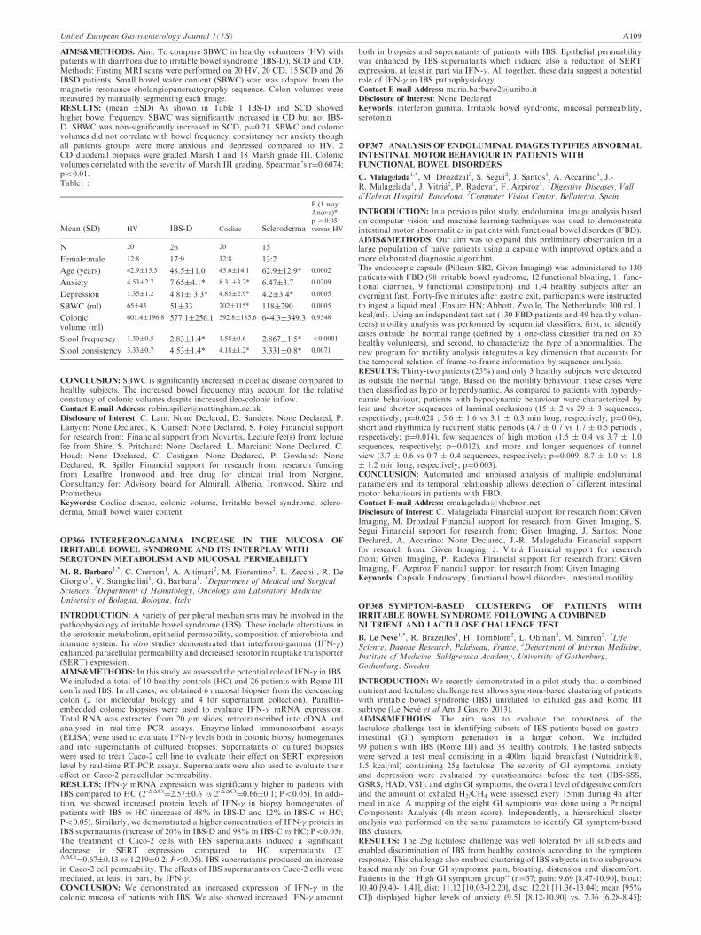
AIMS&METHODS: Aim: To compare SBWC in healthy volunteers (HV) withpatients with diarrhoea due to irritable bowel syndrome (IBS-D), SCD and CD.Methods: Fasting MRI scans were performed on 20 HV, 20 CD, 15 SCD and 26IBSD patients. Small bowel water content (SBWC) scan was adapted from themagnetic resonance cholangiopancreatography sequence. Colon volumes weremeasured by manually segmenting each image.RESULTS: (mean �SD) As shown in Table 1 IBS-D and SCD showedhigher bowel frequency. SBWC was significantly increased in CD but not IBS-D. SBWC was non-significantly increased in SCD, p¼0.21. SBWC and colonicvolumes did not correlate with bowel frequency, consistency nor anxiety thoughall patients groups were more anxious and depressed compared to HV. 2CD duodenal biopsies were graded Marsh I and 18 Marsh grade III. Colonicvolumes correlated with the severity of Marsh III grading, Spearman’s r¼0.6074;p50.01.Table1 :
Mean (SD) HV IBS-D Coeliac Scleroderma
P (1 wayAnova)*p 50.05versus HV
N 20 26 20 15
Female:male 12:8 17:9 12:8 13:2
Age (years) 42.9�15.3 48.5�11.0 45.6�14.1 62.9�12.9* 0.0002
Anxiety 4.53�2.7 7.65�4.1* 8.31�3.7* 6.47�3.7 0.0209
Depression 1.35�1.2 4.81� 3.3* 4.85�2.9* 4.2�3.4* 0.0005
SBWC (ml) 65�43 51�33 202�115* 118�290 0.0005
Colonicvolume (ml)
601.4�196.8 577.1�256.1 592.8�185.6 644.3�349.3 0.9548
Stool frequency 1.30�0.5 2.83�1.4* 1.58�0.6 2.867�1.5* 50.0001
Stool consistency 3.33�0.7 4.53�1.4* 4.18�1.2* 3.331�0.8* 0.0071
CONCLUSION: SBWC is significantly increased in coeliac disease compared tohealthy subjects. The increased bowel frequency may account for the relativeconstancy of colonic volumes despite increased ileo-colonic inflow.Contact E-mail Address: [email protected] of Interest: C. Lam: None Declared, D. Sanders: None Declared, P.Lanyon: None Declared, K. Garsed: None Declared, S. Foley Financial supportfor research from: Financial support from Novartis, Lecture fee(s) from: lecturefee from Shire, S. Pritchard: None Declared, L. Marciani: None Declared, C.Hoad: None Declared, C. Costigan: None Declared, P. Gowland: NoneDeclared, R. Spiller Financial support for research from: research fundingfrom Lesaffre, Ironwood and free drug for clinical trial from Norgine,Consultancy for: Advisory board for Almirall, Alberio, Ironwood, Shire andPrometheusKeywords: Coeliac disease, colonic volume, Irritable bowel syndrome, sclero-derma, Small bowel water content
OP366 INTERFERON-GAMMA INCREASE IN THE MUCOSA OF
IRRITABLE BOWEL SYNDROME AND ITS INTERPLAY WITH
SEROTONIN METABOLISM AND MUCOSAL PERMEABILITY
M. R. Barbaro1,*, C. Cremon1, A. Altimari2, M. Fiorentino2, L. Zecchi1, R. De
Giorgio1, V. Stanghellini1, G. Barbara1. 1Department of Medical and SurgicalSciences, 2Department of Hematology, Oncology and Laboratory Medicine,University of Bologna, Bologna, Italy
INTRODUCTION: A variety of peripheral mechanisms may be involved in thepathophysiology of irritable bowel syndrome (IBS). These include alterations inthe serotonin metabolism, epithelial permeability, composition of microbiota andimmune system. In vitro studies demonstrated that interferon-gamma (IFN-�)enhanced paracellular permeability and decreased serotonin reuptake transporter(SERT) expression.AIMS&METHODS: In this study we assessed the potential role of IFN-� in IBS.We included a total of 10 healthy controls (HC) and 26 patients with Rome IIIconfirmed IBS. In all cases, we obtained 6 mucosal biopsies from the descendingcolon (2 for molecular biology and 4 for supernatant collection). Paraffin-embedded colonic biopsies were used to evaluate IFN-� mRNA expression.Total RNA was extracted from 20 �m slides, retrotranscribed into cDNA andanalysed in real-time PCR assays. Enzyme-linked immunosorbent assays(ELISA) were used to evaluate IFN-� levels both in colonic biopsy homogenatesand into supernatants of cultured biopsies. Supernatants of cultured biopsieswere used to treat Caco-2 cell line to evaluate their effect on SERT expressionlevel by real-time RT-PCR assays. Supernatants were also used to evaluate theireffect on Caco-2 paracellular permeability.RESULTS: IFN-� mRNA expression was significantly higher in patients withIBS compared to HC (2-��Ct
¼2.57�0.6 vs 2-��Ct¼0.66�0.1; P50.05). In addi-
tion, we showed increased protein levels of IFN-� in biopsy homogenates ofpatients with IBS vs HC (increase of 48% in IBS-D and 12% in IBS-C vs HC;P50.05). Similarly, we demonstrated a higher concentration of IFN-� protein inIBS supernatants (increase of 20% in IBS-D and 98% in IBS-C vs HC; P50.05).The treatment of Caco-2 cells with IBS supernatants induced a significantdecrease in SERT expression compared to HC supernatants (2-��Ct¼0.67�0.13 vs 1.219�0.2; P50.05). IBS supernatants produced an increase
in Caco-2 cell permeability. The effects of IBS supernatants on Caco-2 cells weremediated, at least in part, by IFN-�.CONCLUSION: We demonstrated an increased expression of IFN-� in thecolonic mucosa of patients with IBS. We also showed increased IFN-� amount
both in biopsies and supernatants of patients with IBS. Epithelial permeabilitywas enhanced by IBS supernatants which induced also a reduction of SERTexpression, at least in part via IFN-�. All together, these data suggest a potentialrole of IFN-� in IBS pathophysiology.Contact E-mail Address: [email protected] of Interest: None DeclaredKeywords: interferon gamma, Irritable bowel syndrome, mucosal permeability,serotonin
OP367 ANALYSIS OF ENDOLUMINAL IMAGES TYPIFIES ABNORMAL
INTESTINAL MOTOR BEHAVIOUR IN PATIENTS WITH
FUNCTIONAL BOWEL DISORDERS
C. Malagelada1,*, M. Drozdzal2, S. Segui2, J. Santos1, A. Accarino1, J.-R. Malagelada1, J. Vitria2, P. Radeva2, F. Azpiroz1. 1Digestive Diseases, Valld’Hebron Hospital, Barcelona, 2Computer Vision Center, Bellaterra, Spain
INTRODUCTION: In a previous pilot study, endoluminal image analysis basedon computer vision and machine learning techniques was used to demonstrateintestinal motor abnormalities in patients with functional bowel disorders (FBD).AIMS&METHODS: Our aim was to expand this preliminary observation in alarge population of naıve patients using a capsule with improved optics and amore elaborated diagnostic algorithm.The endoscopic capsule (Pillcam SB2, Given Imaging) was administered to 130patients with FBD (98 irritable bowel syndrome, 12 functional bloating, 11 func-tional diarrhea, 9 functional constipation) and 134 healthy subjects after anovernight fast. Forty-five minutes after gastric exit, participants were instructedto ingest a liquid meal (Ensure HN; Abbott, Zwolle, The Netherlands; 300 ml, 1kcal/ml). Using an independent test set (130 FBD patients and 49 healthy volun-teers) motility analysis was performed by sequential classifiers, first, to identifycases outside the normal range (defined by a one-class classifier trained on 85healthy volunteers), and second, to characterize the type of abnormalities. Thenew program for motility analysis integrates a key dimension that accounts forthe temporal relation of frame-to-frame information by sequence analysis.RESULTS: Thirty-two patients (25%) and only 3 healthy subjects were detectedas outside the normal range. Based on the motility behaviour, these cases werethen classified as hypo or hyperdynamic. As compared to patients with hyperdy-namic behaviour, patients with hypodynamic behaviour were characterized byless and shorter sequences of luminal occlusions (15 � 2 vs 29 � 3 sequences,respectively; p¼0.028 ; 5.6 � 1.6 vs 3.1 � 0.3 min long, respectively; p¼0.04),short and rhythmically recurrent static periods (4.7 � 0.7 vs 1.7 � 0.5 periods ,respectively; p¼0.014), few sequences of high motion (1.5 � 0.4 vs 3.7 � 1.0sequences, respectively; p¼0.012), and more and longer sequences of tunnelview (3.7 � 0.6 vs 0.7 � 0.4 sequences, respectively; p¼0.009; 8.7 � 1.0 vs 1.8� 1.2 min long, respectively; p¼0.003).CONCLUSION: Automated and unbiased analysis of multiple endoluminalparameters and its temporal relationship allows detection of different intestinalmotor behaviours in patients with FBD.Contact E-mail Address: [email protected] of Interest: C. Malagelada Financial support for research from: GivenImaging, M. Drozdzal Financial support for research from: Given Imaging, S.Segui Financial support for research from: Given Imaging, J. Santos: NoneDeclared, A. Accarino: None Declared, J.-R. Malagelada Financial supportfor research from: Given Imaging, J. Vitria Financial support for researchfrom: Given Imaging, P. Radeva Financial support for research from: GivenImaging, F. Azpiroz Financial support for research from: Given ImagingKeywords: Capsule Endoscopy, functional bowel disorders, intestinal motility
OP368 SYMPTOM-BASED CLUSTERING OF PATIENTS WITH
IRRITABLE BOWEL SYNDROME FOLLOWING A COMBINED
NUTRIENT AND LACTULOSE CHALLENGE TEST
B. Le Neve1,*, R. Brazeilles1, H. Tornblom2, L. Ohman2, M. Simren2. 1Life
Science, Danone Research, Palaiseau, France, 2Department of Internal Medicine,Institute of Medicine, Sahlgrenska Academy, University of Gothenburg,Gothenburg, Sweden
INTRODUCTION: We recently demonstrated in a pilot study that a combinednutrient and lactulose challenge test allows symptom-based clustering of patientswith irritable bowel syndrome (IBS) unrelated to exhaled gas and Rome IIIsubtype (Le Neve et al Am J Gastro 2013).AIMS&METHODS: The aim was to evaluate the robustness of thelactulose challenge test in identifying subsets of IBS patients based on gastro-intestinal (GI) symptom generation in a larger cohort. We included99 patients with IBS (Rome III) and 38 healthy controls. The fasted subjectswere served a test meal consisting in a 400ml liquid breakfast (Nutridrink�,1.5 kcal/ml) containing 25g lactulose. The severity of GI symptoms, anxietyand depression were evaluated by questionnaires before the test (IBS-SSS,GSRS, HAD, VSI), and eight GI symptoms, the overall level of digestive comfortand the amount of exhaled H2/CH4 were assessed every 15min during 4h aftermeal intake. A mapping of the eight GI symptoms was done using a PrincipalComponents Analysis (4h mean score). Independently, a hierarchical clusteranalysis was performed on the same parameters to identify GI symptom-basedIBS clusters.RESULTS: The 25g lactulose challenge was well tolerated by all subjects andenabled discrimination of IBS from healthy controls according to the symptomresponse. This challenge also enabled clustering of IBS subjects in two subgroupsbased mainly on four GI symptoms: pain, bloating, distension and discomfort.Patients in the ‘‘High GI symptom group’’ (n¼37; pain: 9.69 [8.47-10.90], bloat:10.40 [9.40-11.41], dist: 11.12 [10.03-12.20], disc: 12.21 [11.36-13.04]; mean [95%CI]) displayed higher levels of anxiety (9.51 [8.12-10.90] vs. 7.36 [6.28-8.45];
United European Gastroenterology Journal 1(1S) A109
p50.05) and lower overall digestive comfort (7.71 [6.82-8.60] vs. 12.98 [12.29-13.67] p50.001) than patients in the ‘‘Low GI symptom group’’ (n¼62; pain:2.33 [1.39-3.27], bloat: 2.99 [2.22-3.78], dist: 3.37 [2.54-4.21], disc: 4.72 [4.08-5.36];p50.001). This clustering was independent of the Rome III subtype classificationand the amount of exhaled H2/CH4.CONCLUSION: A test meal containing 25 g of lactulose discriminates IBSpatients from healthy controls, and allows clustering of IBS subjects accordingto their symptom response, independent of Rome III subtypes or the amount ofexhaled gas. As shown by two independent studies, the lactulose challenge testappears to be a promising tool to better define postprandial symptoms in IBSand to evaluate the efficacy of new treatment options.Contact E-mail Address: [email protected] of Interest: B. Le Neve Other: Boris Le Neve is employed by DanoneResearch, R. Brazeilles Other: Remi Brazeilles is employed by Danone Research,H. Tornblom: None Declared, L. Ohman: None Declared, M. Simren Financialsupport for research from: Danone ResearchKeywords: IBS, Pathophysiology
OP369 MECHANISM OF LACTOSE INTOLERANCE IN IRRITABLE
BOWEL SYNDROME: ROLE OF ANXIETY, MUCOSAL IMMUNITY
AND VISCERAL HYPERSENSITIVITY
M. Fox1,2,*, J. Yang3, Y. Zhu3, Y. Long4, Y. Cong3, H. Chu3, N. Dai3, M. Fried1.
1Division of Gastronenterology and Hepatology, University Hospital Zurich,Zurich, Switzerland, 2NIHR Nottingham Digestive Diseases Biomedical ResearchUnit, Nottingham, United Kingdom, 3Department of Gastroenterology, Sir RunRun Shaw Hospital, 4Department of Gastroenterology, Sir Run Run ShawHospital, Hangzhou, China
INTRODUCTION: Many patients with Irritable Bowel Syndrome (IBS) con-sider that certain foods trigger bloating, diarrhea and other digestive symptoms;however the mechanism underlying "food intolerance" is unknown.AIMS&METHODS: We assessed the role of psychology, mucosal immunity andvisceral sensitivity on the development of lactose intolerance (LI) in IBS patientsand healthy controls (HCs) with lactase deficiency.IBS-D patients meeting Rome III criteria (n¼277) and matched HCs (n¼64)underwent a 20g lactose hydrogen breath test (LHBT): a validated model offood intolerance (Yang Clin Gastro Hep 2013). Hospital Anxiety andDepression Score was obtained. Hydrogen (H2) gas production was measuredby breath test. LI symptoms (bloating, borborygmi, nausea, pain and diarrhea)were recorded using a Lickert scale. An increase in symptom score of 41 on ascale of 0-4 defined LI. Rectal sensitivity was measured by barostat with normalvalues defined by HCs.Additionally, 55 IBS-D patients and 18 HCs underwent ileo-colonoscopy withbiopsies obtained from colon and terminal ileum (TI) to quantify mast cells(MCs), T-cells, and enterochromaffin cells (ECC).RESULTS: H2 production was similar in both groups; but LI was morefrequent in IBS than HCs (54% vs. 28%, P50.001) as were individualsymptoms including bloating (39% vs. 14%, P50.001), borborygmi (39%vs. 22%, P¼0.010), pain (31% vs. 11%, P ¼ 0.001) and diarrhea (29%vs. 9%, P¼0.001). IBS patients with LI were more anxious (p50.045) andhad higher rectal sensitivity (P¼0.001) than patients without LI and controls.Multivariate analysis indicated that H2 production in the upper quartile(OR2.2 (95%CI 1.1-4.4), P¼0.028) and the presence of visceral hypersensi-tivity (OR6.6 (1.7-25.0), P¼0.005) were associated with total symptom score.In those that underwent colonoscopy, IBS patients with LI had increased MCs(p50.006), T-cells and ECC (both p50.05) in colonic and TI mucosa comparedto IBS patients without LI and HCs. Multivariate analysis indicated that totalsymptom score was associated with anxiety, mucosal inflammation and hyper-sensitivity (all r40.5 and P50.001).CONCLUSION: Gas production and hypersensitivity to luminal distension bothcontribute to digestive symptoms after lactose ingestion. IBS-D patients with LIare characterized by anxiety, evidence of mucosal inflammation and visceralhypersensitivity. These results provide insight into the mechanism of food intol-erance. The presence of these biomarkers may identify a subgroup of IBS patientslikely to respond to dietary management.Contact E-mail Address: [email protected] of Interest: M. Fox Financial support for research from: Nestle, J.Yang: None Declared, Y. Zhu: None Declared, Y. Long: None Declared, Y.Cong: None Declared, H. Chu: None Declared, N. Dai: None Declared, M.Fried: None DeclaredKeywords: Irritable bowel syndrome, lactose intolerance, mucosal inflammation,visceral hypersensitivity
OP370 GENDER DETERMINES MUCOSAL MACROPHAGE ACTIVITY
IN THE HUMAN JEJUNUM: IMPLICATIONS FOR IRRITABLE
BOWEL SYNDROME
C. Frıas1,*, M. Vicario1, M. Pigrau1, B. Lobo1, E. Salvo-Romero1, A. Gonzalez-Castro1, C. Alonso1, B. Rodino-Janeiro1, M. Moris1, M. D. Castillo1,F. Azpiroz1, J. Santos1. 1Neuro-Immuno-Gastroenterology Group, DigestiveDiseases Research Unit, Gastroenterology Department, Hospital General Valld’Hebron, Vall d’ Hebron Research Institute, CIBERehd, Barcelona, Spain
INTRODUCTION: Altered intestinal barrier function and mucosal microinflam-mation are common findings in irritable bowel syndrome (IBS), a highly preva-lent functional disorder among women. Effector mast cell and T cells areinvolved in IBS pathophysiology; however, the role of phagocytes remainsunknown.AIMS&METHODS: We aimed at identifying macrophage contribution to IBS.Twenty newly diagnosed diarrhea-predominant IBS (IBS-D, female F n¼14)
fulfilling Rome III criteria and age-matched healthy (H) participants (H, F n¼9)were recruited. One mucosal biopsy was obtained from the jejunum, using theWatson’s capsule. Eosinophils (H&E), mast cells (CD117), T lymphocytes(CD3) and macrophages (CD68) were identified by immunohistochemistry.Mucosal RNA was isolated, submitted to microarray analysis and subsequentbiological functions and pathways identification, associated with differentialgene expression.RESULTS: Eosinophils, CD117þ, CD68þ and CD3þ infiltrate was similarbetween H and IBS-D groups. However, in females, the number of CD68þ
was higher in IBS-D compared to H (IBS-D F: 29.0�1.5 n¼14; H F: 20.9�1.6n¼7; P¼0.004). Considering just the H group, females displayed less CD68þ thanmales (H F: 20.9�1.6 n¼7; H M: 27.2�2.4 n¼7; P¼0.051). Differential geneexpression revealed the immunological response as the most altered function inIBS-D, with significant recruitment and activation of monocytes (P50.0001),induction and activation of macrophages (P50.0001), antigen-presenting andcytotoxic T cells (P50.0001). IBS-D group showed significant upregulation ofPerforin 1, Chemokine (C-X-C Motif) Receptor 7, CD20 and MHC Class IPolypeptide-Related Sequence A (PRF1: 1.32; CXCR7: 1.28; MS4A1: 1.23;MICA: 1.23 fold-change ; P50.05).CONCLUSION: Increased immune activity, specifically macrophage response,features the jejunal mucosa of IBS-D patients. The differences in the number ofmacrophages in women comparing to men evidence that further studies areneeded to clarify gender-dependence in phagocytic activity and whether thismechanism explains increased prevalence of IBS in women.Contact E-mail Address: [email protected] of Interest: None DeclaredKeywords: gender , IBS, macrophage
OP371 FODMAP INTOLERANCE IN IBS: VISUALIZATION BY
MAGNETIC RESONANCE IMAGING
R. M. Undseth1,*, A. Berstad2, J. Valeur2. 1Radiology, 2Unger-Vetlesens institute,
Lovisenberg Diakonale Hospital, Oslo, Norway
INTRODUCTION: Postprandial discomfort is commonly reported in patientswith Irritable Bowel Syndrome (IBS), and intake of Fermentable Oligo-, Di- andMonosaccharides, and Polyols (FODMAP) may aggravate the symptoms. Themechanisms are unclear, but may involve increased luminal gas production and/or fluid secretion in response to unabsorbed carbohydrates.AIMS&METHODS: Using MRI, employing a method based on Hoad et al. (1),we aimed to visualize the gastrointestinal response to lactulose, a model sub-stance of FODMAP. Patients with IBS according to Rome III criteria (n ¼52) and healthy controls (n ¼ 16) underwent a lactulose provocation test, asdescribed previously (2). Abdominal and systemic symptoms, graded 0 to 3,and small bowel water content (SBWC), as measured with MRI, were recordedbefore (fasting) and 1 hour after ingestion of lactulose 10 gram in 100 ml ofwater.RESULTS: Lactulose caused significantly more symptoms in IBS patients thanin healthy controls (P ¼ 0.0004). SBWC increased significanly more in the patientgroup compared to the control group (P ¼ 0.0005). However, neither habitualnor post test symptoms were significantly correlated to SBWC (r ¼ -0, 17, P ¼0.34).CONCLUSION: Patients with IBS have increased osmotic and/or secretoryresponse to an indigestible, but fermentable carbohydrate (lactulose) comparedto healthy controls. The clinical significance of the finding is not clear.REFERENCES:
1. Hoad CL, Marciani L, Totman JJ, Wright J, Bush D, Cox EF, Campbell E,Spiller RC, Gowland. Non-invasive quantification of small bowel water con-tent by MRI: a validation study. Phys Med Biol 2007; 52: 6909-22.
2. Valeur J, Morken MH, Norin E, Midtvedt T, Berstad A. Carbohydrate intol-erance in patients with self-reported food hypersensitivity: comparison of lac-tulose and glucose. Scand J Gastroenterol 2009; 44: 1416-23.
Contact E-mail Address: [email protected] of Interest: None DeclaredKeywords: FODMAP, IBS, MRI
OP372 PREVALENCE OF IRRITABLE BOWEL SYNDROME-LIKE
SYMPTOMS IN ULCERATIVE COLITIS PATIENTS WITH
CLINICAL AND ENDOSCOPIC EVIDENCE OF REMISSION:
PROSPECTIVE MULTICENTER STUDY
N. Fukuba1,*, S. Ishihara1, H. Sonoyama1, A. Oka1, R. Kusunoki1, N. Oshima1,I. Moriyama1, T. Yuki2, K. Kawashima1,3, Y. Kushiyama4, H. Fujishiro5,Y. Kinoshita1. 1Department of Internal Medicine II, Shimane university School ofMedicine, 2Division of Gastrointestinal Endoscopy, Shimane University Hospital,Izumo, 3Department of Internal Medicine, Matsue seikyo hospital, 4Department ofgastroenterology, Matsue red cross hospital, Matsue, 5Department of gastroen-terology, Shimane prefectural central hospital, Izumo, Japan
INTRODUCTION: Irritable bowel syndrome (IBS) is a functional intestinaldisorder characterized by abdominal pain or discomfort, along with changes instool frequency and consistency, which reduces quality of life. Previous studieshave reported that IBS-like symptoms are often found in ulcerative colitis (UC)patients in remission. In those studies, remission was mainly defined by evaluat-ing clinical symptoms. However, the prevalence of IBS-like symptoms in UCpatients with endoscopic evidence of remission shown by mucosal healingremains unknown.AIMS&METHODS: We assessed the prevalence of IBS-like symptoms in UCpatients with clinical and endoscopic evidence of remission, and compared theresults to healthy controls. For this study, prospectively conducted at 1 universityand 3 general hospitals in Japan, 208 UC patients noted to have a good general
A110 United European Gastroenterology Journal 1(1S)
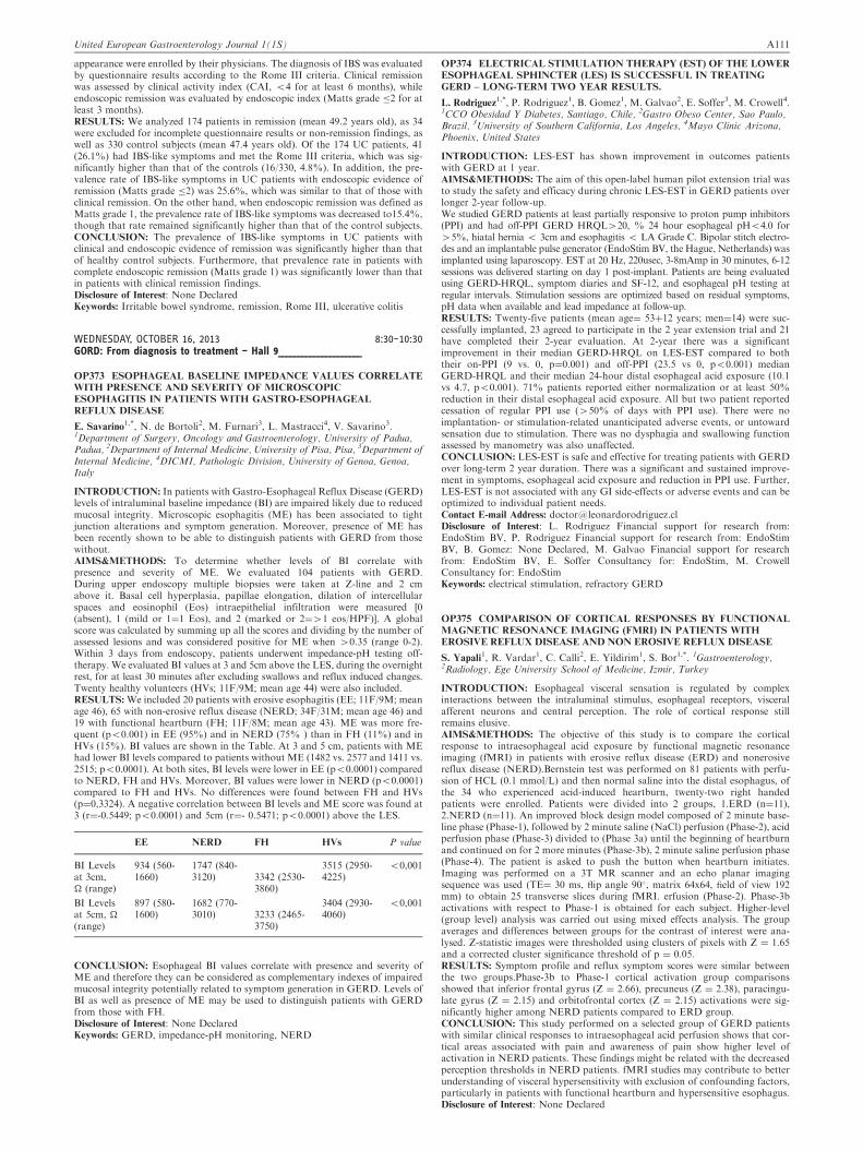
appearance were enrolled by their physicians. The diagnosis of IBS was evaluatedby questionnaire results according to the Rome III criteria. Clinical remissionwas assessed by clinical activity index (CAI, 54 for at least 6 months), whileendoscopic remission was evaluated by endoscopic index (Matts grade �2 for atleast 3 months).RESULTS: We analyzed 174 patients in remission (mean 49.2 years old), as 34were excluded for incomplete questionnaire results or non-remission findings, aswell as 330 control subjects (mean 47.4 years old). Of the 174 UC patients, 41(26.1%) had IBS-like symptoms and met the Rome III criteria, which was sig-nificantly higher than that of the controls (16/330, 4.8%). In addition, the pre-valence rate of IBS-like symptoms in UC patients with endoscopic evidence ofremission (Matts grade �2) was 25.6%, which was similar to that of those withclinical remission. On the other hand, when endoscopic remission was defined asMatts grade 1, the prevalence rate of IBS-like symptoms was decreased to15.4%,though that rate remained significantly higher than that of the control subjects.CONCLUSION: The prevalence of IBS-like symptoms in UC patients withclinical and endoscopic evidence of remission was significantly higher than thatof healthy control subjects. Furthermore, that prevalence rate in patients withcomplete endoscopic remission (Matts grade 1) was significantly lower than thatin patients with clinical remission findings.Disclosure of Interest: None DeclaredKeywords: Irritable bowel syndrome, remission, Rome III, ulcerative colitis
WEDNESDAY, OCTOBER 16, 2013 8:30–10:30GORD: From diagnosis to treatment – Hall 9_____________________
OP373 ESOPHAGEAL BASELINE IMPEDANCE VALUES CORRELATE
WITH PRESENCE AND SEVERITY OF MICROSCOPIC
ESOPHAGITIS IN PATIENTS WITH GASTRO-ESOPHAGEAL
REFLUX DISEASE
E. Savarino1,*, N. de Bortoli2, M. Furnari3, L. Mastracci4, V. Savarino3.
1Department of Surgery, Oncology and Gastroenterology, University of Padua,Padua, 2Department of Internal Medicine, University of Pisa, Pisa, 3Department ofInternal Medicine, 4DICMI, Pathologic Division, University of Genoa, Genoa,Italy
INTRODUCTION: In patients with Gastro-Esophageal Reflux Disease (GERD)levels of intraluminal baseline impedance (BI) are impaired likely due to reducedmucosal integrity. Microscopic esophagitis (ME) has been associated to tightjunction alterations and symptom generation. Moreover, presence of ME hasbeen recently shown to be able to distinguish patients with GERD from thosewithout.AIMS&METHODS: To determine whether levels of BI correlate withpresence and severity of ME. We evaluated 104 patients with GERD.During upper endoscopy multiple biopsies were taken at Z-line and 2 cmabove it. Basal cell hyperplasia, papillae elongation, dilation of intercellularspaces and eosinophil (Eos) intraepithelial infiltration were measured [0(absent), 1 (mild or 1¼1 Eos), and 2 (marked or 2¼41 eos/HPF)]. A globalscore was calculated by summing up all the scores and dividing by the number ofassessed lesions and was considered positive for ME when 40.35 (range 0-2).Within 3 days from endoscopy, patients underwent impedance-pH testing off-therapy. We evaluated BI values at 3 and 5cm above the LES, during the overnightrest, for at least 30 minutes after excluding swallows and reflux induced changes.Twenty healthy volunteers (HVs; 11F/9M; mean age 44) were also included.RESULTS: We included 20 patients with erosive esophagitis (EE; 11F/9M; meanage 46), 65 with non-erosive reflux disease (NERD; 34F/31M; mean age 46) and19 with functional heartburn (FH; 11F/8M; mean age 43). ME was more fre-quent (p50.001) in EE (95%) and in NERD (75% ) than in FH (11%) and inHVs (15%). BI values are shown in the Table. At 3 and 5 cm, patients with MEhad lower BI levels compared to patients without ME (1482 vs. 2577 and 1411 vs.2515; p50.0001). At both sites, BI levels were lower in EE (p50.0001) comparedto NERD, FH and HVs. Moreover, BI values were lower in NERD (p50.0001)compared to FH and HVs. No differences were found between FH and HVs(p¼0,3324). A negative correlation between BI levels and ME score was found at3 (r¼-0.5449; p50.0001) and 5cm (r¼- 0.5471; p50.0001) above the LES.
EE NERD FH HVs P value
BI Levelsat 3cm,� (range)
934 (560-1660)
1747 (840-3120) 3342 (2530-
3860)
3515 (2950-4225)
50,001
BI Levelsat 5cm, �(range)
897 (580-1600)
1682 (770-3010) 3233 (2465-
3750)
3404 (2930-4060)
50,001
CONCLUSION: Esophageal BI values correlate with presence and severity ofME and therefore they can be considered as complementary indexes of impairedmucosal integrity potentially related to symptom generation in GERD. Levels ofBI as well as presence of ME may be used to distinguish patients with GERDfrom those with FH.Disclosure of Interest: None DeclaredKeywords: GERD, impedance-pH monitoring, NERD
OP374 ELECTRICAL STIMULATION THERAPY (EST) OF THE LOWER
ESOPHAGEAL SPHINCTER (LES) IS SUCCESSFUL IN TREATING
GERD – LONG-TERM TWO YEAR RESULTS.
L. Rodriguez1,*, P. Rodriguez1, B. Gomez1, M. Galvao2, E. Soffer3, M. Crowell4.
1CCO Obesidad Y Diabetes, Santiago, Chile, 2Gastro Obeso Center, Sao Paulo,Brazil, 3University of Southern California, Los Angeles, 4Mayo Clinic Arizona,Phoenix, United States
INTRODUCTION: LES-EST has shown improvement in outcomes patientswith GERD at 1 year.AIMS&METHODS: The aim of this open-label human pilot extension trial wasto study the safety and efficacy during chronic LES-EST in GERD patients overlonger 2-year follow-up.We studied GERD patients at least partially responsive to proton pump inhibitors(PPI) and had off-PPI GERD HRQL420, % 24 hour esophageal pH54.0 for45%, hiatal hernia 5 3cm and esophagitis 5 LA Grade C. Bipolar stitch electro-des and an implantable pulse generator (EndoStim BV, the Hague, Netherlands) wasimplanted using laparoscopy. EST at 20 Hz, 220usec, 3-8mAmp in 30 minutes, 6-12sessions was delivered starting on day 1 post-implant. Patients are being evaluatedusing GERD-HRQL, symptom diaries and SF-12, and esophageal pH testing atregular intervals. Stimulation sessions are optimized based on residual symptoms,pH data when available and lead impedance at follow-up.RESULTS: Twenty-five patients (mean age¼ 53þ12 years; men¼14) were suc-cessfully implanted, 23 agreed to participate in the 2 year extension trial and 21have completed their 2-year evaluation. At 2-year there was a significantimprovement in their median GERD-HRQL on LES-EST compared to boththeir on-PPI (9 vs. 0, p¼0.001) and off-PPI (23.5 vs 0, p50.001) medianGERD-HRQL and their median 24-hour distal esophageal acid exposure (10.1vs 4.7, p50.001). 71% patients reported either normalization or at least 50%reduction in their distal esophageal acid exposure. All but two patient reportedcessation of regular PPI use (450% of days with PPI use). There were noimplantation- or stimulation-related unanticipated adverse events, or untowardsensation due to stimulation. There was no dysphagia and swallowing functionassessed by manometry was also unaffected.CONCLUSION: LES-EST is safe and effective for treating patients with GERDover long-term 2 year duration. There was a significant and sustained improve-ment in symptoms, esophageal acid exposure and reduction in PPI use. Further,LES-EST is not associated with any GI side-effects or adverse events and can beoptimized to individual patient needs.Contact E-mail Address: [email protected] of Interest: L. Rodriguez Financial support for research from:EndoStim BV, P. Rodriguez Financial support for research from: EndoStimBV, B. Gomez: None Declared, M. Galvao Financial support for researchfrom: EndoStim BV, E. Soffer Consultancy for: EndoStim, M. CrowellConsultancy for: EndoStimKeywords: electrical stimulation, refractory GERD
OP375 COMPARISON OF CORTICAL RESPONSES BY FUNCTIONAL
MAGNETIC RESONANCE IMAGING (FMRI) IN PATIENTS WITH
EROSIVE REFLUX DISEASE AND NON EROSIVE REFLUX DISEASE
S. Yapali1, R. Vardar1, C. Calli2, E. Yildirim1, S. Bor1,*. 1Gastroenterology,
2Radiology, Ege University School of Medicine, Izmir, Turkey
INTRODUCTION: Esophageal visceral sensation is regulated by complexinteractions between the intraluminal stimulus, esophageal receptors, visceralafferent neurons and central perception. The role of cortical response stillremains elusive.AIMS&METHODS: The objective of this study is to compare the corticalresponse to intraesophageal acid exposure by functional magnetic resonanceimaging (fMRI) in patients with erosive reflux disease (ERD) and nonerosivereflux disease (NERD).Bernstein test was performed on 81 patients with perfu-sion of HCL (0.1 mmol/L) and then normal saline into the distal esophagus, ofthe 34 who experienced acid-induced heartburn, twenty-two right handedpatients were enrolled. Patients were divided into 2 groups, 1.ERD (n¼11),2.NERD (n¼11). An improved block design model composed of 2 minute base-line phase (Phase-1), followed by 2 minute saline (NaCl) perfusion (Phase-2), acidperfusion phase (Phase-3) divided to (Phase 3a) until the beginning of heartburnand continued on for 2 more minutes (Phase-3b), 2 minute saline perfusion phase(Phase-4). The patient is asked to push the button when heartburn initiates.Imaging was performed on a 3T MR scanner and an echo planar imagingsequence was used (TE¼ 30 ms, Fip angle 90�, matrix 64x64, Eeld of view 192mm) to obtain 25 transverse slices during fMRI. erfusion (Phase-2). Phase-3bactivations with respect to Phase-1 is obtained for each subject. Higher-level(group level) analysis was carried out using mixed effects analysis. The groupaverages and differences between groups for the contrast of interest were ana-lysed. Z-statistic images were thresholded using clusters of pixels with Z ¼ 1.65and a corrected cluster signiEcance threshold of p ¼ 0.05.RESULTS: Symptom profile and reflux symptom scores were similar betweenthe two groups.Phase-3b to Phase-1 cortical activation group comparisonsshowed that inferior frontal gyrus (Z ¼ 2.66), precuneus (Z ¼ 2.38), paracingu-late gyrus (Z ¼ 2.15) and orbitofrontal cortex (Z ¼ 2.15) activations were sig-nificantly higher among NERD patients compared to ERD group.CONCLUSION: This study performed on a selected group of GERD patientswith similar clinical responses to intraesophageal acid perfusion shows that cor-tical areas associated with pain and awareness of pain show higher level ofactivation in NERD patients. These findings might be related with the decreasedperception thresholds in NERD patients. fMRI studies may contribute to betterunderstanding of visceral hypersensitivity with exclusion of confounding factors,particularly in patients with functional heartburn and hypersensitive esophagus.Disclosure of Interest: None Declared
United European Gastroenterology Journal 1(1S) A111

Keywords: erosive reflux disease, Functional magnetic resonance imaging(fMRI), Gastroesophageal reflux disease(GERD), non erosive reflux disease
OP376 HISTOLOGY IDENTIFIES PATIENTS WITH
GASTROESOPHAGEAL REFLUX DISEASE WHO ARE NOT
IDENTIFIED BY PH TESTING OR ENDOSCOPY
N. Vakil1,*, M. Vieth2, J. Dent3, L. Mastracci4, J. Wissmar5, B. Wernersson5,
R. Fiocca4. 1University of Wisconsin School of Medicine and Public Health,Madison, United States, 2Klinikum Bayreuth, Bayreuth, Germany, 3Royal AdelaideHospital and University of Adelaide, Adelaide, Australia, 4University of Genoa,Genoa, Italy, 5AstraZeneca R&D, Molndal, Sweden
INTRODUCTION: We have shown previously that total epithelial thickness(TET) in the distal esophagus of �430 mm correlates significantly with investiga-tion-defined gastroesophageal reflux disease (iGERD).1 It is not known whetherTET adds to the diagnostic yield of endoscopy and pH-metry for iGERD.AIMS&METHODS: We performed a post hoc analysis to evaluate the correlationbetween TET �430 mm and individual iGERD markers, and to assess whether thecriterion of TET �430 mm influences the diagnostic yield for GERD in primary carepatients presenting with frequent upper gastrointestinal (GI) symptoms (Diamondstudy; NCT00291746).2 Patients were included if they reported upper GI symptomsof any severity on �2 days/week for �4 weeks and of �mild severity on �3 days inthe week before the study, and had not taken a proton pump inhibitor in the past 2months. Patients with symptom-defined GERD (sGERD) were identified based oninternational heartburn/regurgitation frequency/severity criteria,3 using data fromthe Reflux Disease Questionnaire completed at study inclusion. iGERD was definedas the presence of �1 of: reflux esophagitis (RE; Los Angeles grades A–D); patho-logical esophageal pH (54 for 45.5% of the time); positive symptom associationprobability (SAP �95%). TET �430 mm at 2.0 cm above the Z-line (3 o’clockposition) was added as a novel criterion for GERD.RESULTS: Our analysis included 231 patients from the Diamond study: 37%had pathological esophageal pH, 30% had RE, 16% had a positive SAP and39% had TET �430 mm; 55% had iGERD. TET �430 mm correlated signifi-cantly with RE, pathological pH and iGERD (all p50.005), but not with positiveSAP (p¼1.0). Table 1 gives the specificity/sensitivity of TET �430 mm and 5430mm for iGERD and its component markers. Including TET �430 mm as a fourthiGERD criterion identified an additional 28 patients (25 of whom met sGERDcriteria3), increasing the prevalence of iGERD by 22% (from 55% to 67%).Table 1. Specificity/sensitivity (%) of TET for iGERD and its componentmarkers
TET (mm) iGERD RE Pathological pH Positive SAP
�430 53.5/68.5 79.6/44.9 70.4/48.3 84.5/15.7
5430 31.5/46.5 55.1/20.4 51.7/29.6 84.3/15.5
CONCLUSION: TET �430 mm correlates significantly with RE, pathologicalpH and iGERD, but not with positive SAP; it increases the diagnostic yield foriGERD. TET �430 mm is a clinically useful marker of GERD, particularly inpatients with sGERD, which can be misclassified as functional dyspepsia.REFERENCES:
1. Vieth M et al. Gastroenterology 2011;140(Suppl 1):S-95. 2. Dent J et al. Gut2010;59:714–21. 3. Vakil N et al. Am J Gastroenterol 2006;101:1900–20.
Contact E-mail Address: [email protected] of Interest: N. Vakil Consultancy for: AstraZeneca, IronwoodPharmaceuticals, Orexo, Takeda Pharmaceutical Co. Ltd, Otsuka andXenoport, Shareholder of: Meridian Bioscience and Orexo, M. Vieth Lecturefee(s) from: AstraZeneca, Falk, Pentax, Olympus, Malesci, Covidien and Shire,J. Dent Consultancy for: AstraZeneca R&D, Molndal, Sweden, L. Mastracci:None Declared, J. Wissmar Other: Employee of AstraZeneca R&D, Molndal,Sweden, B. Wernersson Other: Employee of AstraZeneca R&D, Molndal,Sweden, R. Fiocca: None DeclaredKeywords: Gastroesophageal reflux disease, histology, sensitivity, specificity,total epithelial thickness
OP377 PEPSIN IN SALIVA AND GASTROESOPHAGEAL REFLUX
MONITORING IN 100 HEALTHY ASYMPTOMATIC SUBJECTS
AND 65 PATIENTS WITH SIGNIFICANT HEARTBURN/
REGURGITATION
J. O. Hayat1,*, S. Gabieta-Gomez2, E. Yazaki2, J.-Y. Kang1, A. Woodcock3,
P. Dettmar3, J. Mabary4, D. Sifrim2. 1Dept of Gastroenterology, St.George’sUniversity of London, 2Dept of GI Physiology, Barts and the London School ofMedicine and Dentistry, London, 3Technostics Ltd, Hull, United Kingdom,4Sandhill Sci., Colorado, United States
INTRODUCTION: Pepsin is a protease originating from pepsinogensecreted into gastric juice from chief cells, found only in the stomach.Its presence in the oesophagus or more proximally suggests gastro-oesophagealreflux (GOR). Several studies have measured pepsin in saliva to determine itsvalue as marker of GOR. Patients with clinically significant heartburn may haveGORD, hypersensitive oesophagus or functional heartburn.AIMS&METHODS: The aim of this study was to measure pepsin in saliva withobjective assessment of GOR by impedance-pH (MII-pH) in a cohort of asymp-tomatic subjects and consecutive patients with clinically significant heartburn(according to the Montreal definition of GORD).100 healthy subjects, age 30.7 y (range 19-55) and 65 pts with þve Reflux DiseaseQuestionnaire (RDQ) score, age 49.7y (range 25-71) underwent MII-pH
monitoring and simultaneous saliva sampling. Subjects collected expectoratedsaliva on waking, one hour after lunch and dinner. Pepsin was detected usinga lateral flow test with two unique monoclonal antibodies to pepsin (PeptestTM,RDBiomed Ltd). The cut off value to determine pepsin positivity was 25ng/ml.Pts were divided into 3 phenotypes based on MII-pH results. 1) GORD(increased oesophageal acid exposure time (AET) (10.4 % � 1.4) and SAPþve, n¼26). 2) Hypersensitive oesophagus (HO)(normal AET and SAP þve,n¼18) and 3) Functional Heartburn (FH)(normal AET and SAP -ve for acid/non-acid reflux, n¼12).RESULTS: All healthy subjects selected had normal MII-pH testing. 36/100normal subjects had at least 1 sample þve (20% had 1 sample þve, 12% had2 samples þve and 4% had 3 samples þve). In pepsin þve samples, the median(25%475%) pepsin conc. was 118 (64-181)ng/ml. In GORD patients, 21/26 hadat least 1 sample þve (3 patients had 3 samples þve) and pepsin conc. was 152(72-250)ng/ml. In HO patients 15/18 had at least 1 sample þve (4 had 3 samplesþve) and pepsin conc. was 250(74-250)ng/ml. In contrast, only 2/12 FH patientshad at least 1 sample þve and pepsin conc. was 76(67-85)ng/ml. Considering theability to identify patients with heartburn related to reflux (GORD þ HO)Peptest had a sensitivity of 95%, specificity 89%, PPV 97% but a NPV of 57%.CONCLUSION: Pepsin was found in the saliva of a proportion of healthyindividuals with no reflux symptoms. Pepsin is significantly more likely to bedetected in patients with heartburn related to reflux (SAP þve) i.e. GORD andHO, (and in higher conc.) than in normal subjects or patients with functionalheartburn.Disclosure of Interest: None DeclaredKeywords: functional heartburn, gastro-esophageal reflux, hypersensitive esopha-gus, impedance-pH monitoring, pepsin
OP378 PHARYNGEAL PH ALONE IS NOT RELIABLE FOR THE
DETECTION OF PHARYNGEAL REFLUX EVENTS. A STUDY
WITH ESOPHAGEAL AND PHARYNGEAL PH-IMPEDANCE
MONITORING
M. Desjardin1,*, S. Roman2, S. Bruley Des Varannes3, G. Gourcerol4, B. Coffin5,
A. Ropert6, F. Mion2, F. Zerbib1, Groupe Francais de Neurogastroenterologie.1Gastroenterology , CHU Bordeaux, Bordeaux, 2Gastroenterology , CHU Lyon,Lyon, 3IMAD, CHU Nantes, Nantes, 4Gastroenterology , CHU Rouen, Rouen,5Gastroenterology , hopital Louis Mourier, Colombes, 6Gastroenterology , CHURennes, Rennes, France
INTRODUCTION: In clinical practice, the diagnosis of laryngo-pharyngealreflux (LPR) is challenging. Recently, pharyngeal pH probes and pharyngealpH-impedance catheters have been developed. The aim of this study was todetermine the reliability of pharyngeal pH alone for the detection of pharyngealreflux events.AIMS&METHODS: We collected ambulatory, 24-hour, pH-impedance record-ings from 45 healthy subjects (22 women; mean age, 46.3 years; range, 18–78years) off therapy using a bifurcated probe that allowed for detection of phar-yngeal and esophageal reflux events simultaneously. Impedance measurementswere combined with pharyngeal and esophageal pH measurements 1 cm abovethe upper esophageal sphincter and 5 cm above the lower esophageal sphincterrespectively. All pharyngeal pH drops 5 4 or 5 5 and lasting more than 5seconds were analyzed for the simultaneous occurrence of pharyngeal reflux(PR), proximal and distal esophageal reflux and swallows according to impe-dance analysis. Slow pH drifts were deleted from the analysis.RESULTS: Among the 45 recordings, 256 pharyngeal pH drops 55 were iden-tified; 81.3% of them were associated with a simultaneous esophageal pH drop55 . Only 7% of the pharyngeal pH drops 55 corresponded to PR. The major-ity of pharyngeal pH drops 55 were related to swallows (92.6%), and only10.2% and 13.3% were associated with proximal and distal reflux, respectively.There was no significant difference in the duration of pharyngeal pH dropsassociated with PR and the others. Among 126 pharyngeal pH drops 5 4,90.5% were associated with a simultaneous esophageal drop . Only 13.5% ofpH drops 5 4 were related to PR. Most pH drops were related to swallows(89.7%), and 17.5% and 20.6 % were associated with proximal and distalreflux, respectively. There was no significant difference in the duration of pHdrops 54 associated with PR and the othersCONCLUSION: This study demonstrates that pharyngeal pH alone is not reli-able for the detection of pharyngeal reflux and that adding distal esophageal pHanalysis is not helpful. The only reliable analysis should take into account impe-dance patterns demonstrating the presence of a pharyngeal reflux event precededby a distal and proximal reflux event within the esophagus.Contact E-mail Address: [email protected] of Interest: M. Desjardin: None Declared, S. Roman Consultancy for:Given Imaging, S. Bruley Des Varannes Consultancy for: Shire, AlphaWasserman, Janssen Cilag, Given Imaging, Cephalon, ALmirall, G. GourcerolConsultancy for: Reckitt Benckiser, B. Coffin Consultancy for: Shire, Cephalon,ALmirall, Abbott, Mundipharma, A. Ropert: None Declared, F. MionConsultancy for: Shire, ALmirall, Given Imaging, Helioscope, Medtronic,Urogene, F. Zerbib Financial support for research from: Sandhill, GivenImaging, Reckitt Banckiser, , Lecture fee(s) from: Astrazeneca, Sanofi Aventis,Janssen Cilag, Abbott, Consultancy for: Addex, Shire, AlmirallKeywords: impedance-pH monitoring, Laryngopharyngeal reflux(LPR), Refluxsymptoms
A112 United European Gastroenterology Journal 1(1S)

OP379 ASSESSMENT OF PATIENTS REFERRED FOR GERD
EVALUATION: TO CONTINUE OR STOP ACID SUPPRESSANTS
BEFORE FUNCTIONAL TESTS?
D. Ang1,*, J. Tack2. 1Gastroenterology, Changi General Hospital, Singapore,
Singapore, 2Gastroenterology, KUL, Leuven, Belgium
INTRODUCTION: Patients with typical and atypical gastroesophageal refluxsymptoms in the presence of a normal gastroscopy and which persist despiteproton-pump inhibitor (PPI) therapy are increasingly encountered. Objectiveassessment with 24 hour pH monitoring, Bilitec and/or multichannel intralum-inal impedance (MII-pH) monitoring identifies patients with acid reflux (AR)and/or non-acid reflux (NAR). It remains uncertain if proton pump inhibitors(PPI) should be stopped prior to functional tests.AIMS&METHODS: Aim: To determine the diagnostic yield of all studies per-formed on and off PPI therapy.Methods: Systematic review of all studies pub-lished from January 1996 to December 2012. Data were extracted for patientdemographics, symptom profiles, acid exposure times (AET) and symptom index(SI). The prevalence of abnormal oesophageal acid exposure time (AET) andsymptom marker based positive symptom index (SI) was compared by chi-square and student t-testing.RESULTS: Between January 1996 and December 2012, 32 studies involving 2925patients (1130 male, mean [SD] age 50.4[9.9] years) who were evaluated for non-response to PPI therapy whilst on therapy (n¼1841) and after PPI washout(n¼1084) were identified. These studies included 490 subjects who underwent 24hour pH study), 65 subjects withpH-bilitec and 2370 subjects who underwent MII-pH. Information on the nature of reflux symptoms was available in 2646 subjects(n¼1800 typical symptoms; n¼846 atypical symptoms). Thirteen and 11 studiesreported elevated esophageal AET in 381 of 1068 (35.7%) patients and 216 of 1062(20.3%) patients who were studied off and on PPI respectively (p50.05). A positiveSI for AR occurred in 48.5% and 11.9% of patients off and on PPI respectively(p50.05). A positive SI for NAR occurred in 18.8% and 34.2% of patients off andon PPI respectively (p50.05). Improved diagnostic yield was observed whenpatients were studied for AR events off PPI therapy and for NAR events on PPI.CONCLUSION: In GERD patients with insufficient treatment response,improved diagnostic yield was achieved with MII-pH monitoring performed onPPI therapy for evaluation of NAR and off therapy for AR evaluation. Whilstthis approach may help direct appropriate therapy, further outcome studies arerequired.Disclosure of Interest: None DeclaredKeywords: ambulatory pH impedance monitoring, GERD symptoms, protonpump inhibitor
OP380 STW5 REDUCES THE PROINFLAMMATORY SIGNATURE ON
TRANSCRIPTOMIC, PROTEOMIC AND HISTOLOGICAL LEVEL
IN A RAT MODEL FOR GASTROESOPHAGEAL REFLUX DISEASE
H. Abdel-Aziz1,2,*, D. Weiser2, M. T. Khayyal3, G. Ulrich-Merzenich4. 1Dpt. of
Pharmacology, Inst. of Pharm. Chemistry, University of Munster, Munster,2Scientific Department, Steigerwald Arzneimittelwerk, Darmstadt, Germany,3Pharmacology, Faculty of Pharmacy, Cairo University, Cairo, Egypt,4Medizinische Klinik 3, University of Bonn, Bonn, Germany
INTRODUCTION: Gastroesophageal reflux disease (GERD) is one of the mostcommon GI-diagnosis. Proton pump inhibitors (PPIs) present the main treat-ment, but up to 40 % of patients do not achieve adequate symptom control.Therefore the search continues for new therapeutic targets and additional treat-ment options. STW 5, a multi-component herbal preparation, was shown to reliefconcomitant reflux symptoms in patients with functional dyspepsia [1] and toprevent inflammation in an acute model of reflux esophagitis (RE) withoutaffecting the pH of the refluxate [2]. Recently, we showed that it also improvedmacroscopic and histological parameters of esophagitis in a subchronic model ofRE in rats, where it down regulated proinflammatory cytokines in the tissuehomogenates (proteome profiling) [3].AIMS&METHODS: In the present study, we analyzed in our subchronic model ofRE the transcript modulation in the esophageal tissue in an attempt to identifypotential therapeutic targets. Rats were pre-treated for 7d either with STW 5 (0.5 or2 ml/kg) or omeprazole (O). Esophagitis was then induced surgically [4]. Rats weretreated for further 10d with STW 5 or O and sacrificed. The esophagi were excised.RNA was isolated from defined tissue areas and transcripts were analyzed byAgilent whole genome microarray (rat). Selected genes were validated by RT-PCR.RESULTS: Well known markers of inflammation and the immune response likeIL-6, IL-1 and matrixmetallopeptidases (MMPs) were up-regulated in RE. Eventhough STW 5 and O down regulated different sets of genes, they showed acommon pattern in a number of strongly and highly significantly regulatedgenes (p50.0001), including chemokine ligands 4 and 11, G-protein coupledreceptor GPR84, the LOX-1 receptor and resistin.CONCLUSION: Since gastric acid does not appear to be the only causativeagent in GERD, identifying additional mechanisms involved in the pathogenesisor action of effective drugs may help detect novel therapeutic targets in GERDand pave the way for the development of effective treatment options.REFERENCES:
1. Madisch A; Gut 2007; 56:A3362. Abdel-Aziz H et al.; J Pharmacol Sci 2010; 113: 134-1423. Abdel-Aziz H et al.; Gastroenterology 2012; 142: S-593Contact E-mail Address: [email protected] of Interest: H. Abdel-Aziz Other: Employee of SteigerwaldArzneimittelwerk GmbH, D. Weiser Consultancy for: SteigerwaldArzneimittelwerk GmbH, M. Khayyal Financial support for research from:Steigerwald Arzneimittelwerk GmbH, G. Ulrich-Merzenich Financial supportfor research from: Steigerwald Arzneimittelwerk GmbHKeywords: GERD, Herbal treatment, Inflammation, Protomics, Transcriptomics
OP381 PRESENCE OF BILE ACIDS AND PEPSIN IN SALIVA AND
BRONCHO-ALVEOLAR LAVAGE OF PATIENTS WITH
IDIOPATHIC PULMONARY FIBROSIS IS ASSOCIATED WITH
INCREASED SEVERITY OF LUNG DISEASE
E. Savarino1,*, P. Zentilin2, E. Marabotto2, L. Sconfienza3, N. de Bortoli4,
S. Marchi4, V. Savarino2. 1Department of Surgery, Oncology andGastroenterology, University of Padua, Padua, 2Department of Internal Medicine,Division of Gastroenterology, University of Genoa, Genoa, 3Unit of Radiology,IRCCS Policlinico San Donato, Milan, 4Department of Internal Medicine, Divisionof Gastroenterology, University of Pisa, Pisa, Italy
INTRODUCTION: Recent studies investigated the role of pepsin and bile acids(BAs) in saliva and bronchoalveolar lavage (BAL) of patients with respiratorydiseases (i.e. asthma, cystic fibrosis) as specific and sensitive markers of gastro-oesophageal reflux–related pulmonary aspiration. However, no data are availableon the role of these markers in patients with idiopathic pulmonary fibrosis (IPF)who are characterized by increased gastroesophageal reflux (up to 85%) andpoten-tial aspiration of duodenogastric contents into the lungs.AIMS&METHODS: To assess aspiration in IPF patients by measuring pepsinand BAs in saliva/BAL and to investigate whether their presence correlated withdisease severity and pulmonary function tests (PFTs).Saliva andBAL sampleswere collected in 38 and 21 IPFpatients, 36 and 21 patientswith interstitial lung disease other than IPF (non-IPF patients), 50 healthy volun-teers (HVs) and 16 patients undergoing bronchoscopy for other diseases (controls),respectively. All subjects underwent pulmonary high-resolution computed tomo-graphy (HRCT) scan and PFTs on the day of saliva/BAL samples collection. BAsandpepsinweremeasuredusing twocommercial available assays. Thepresence andseverity of pulmonary fibrosis was defined using a previous validatedHRCT-score(Best-score). Forced vital capacity (FVC), forced expiratory volume (FEV) andDLco curve were measured during PFTs.RESULTS: Patients with IPF had more BAs and pepsin (p50.001) in BAL (62%and 67%) and saliva (61% and 68%) than non-IPF patients (40% and 40% inBAL, 33% and 36% in saliva) and controls (0% and 0% in BAL) or HVs (0% and0% in saliva). The concentration of BAs in saliva and BAL was higher (p50.001)in IPF [3.70 mmol/l and 0.90 mmol/l] than in non-IPF patients [1.5 mmol/l and 0.50mmol/l]. A good correlation (p50.001) was found between HRCT score and pre-sence of pepsin and BAs in saliva (r2¼0.5260 and r2¼0.4269, respectively) andBAL (r2¼0.6033 and r2¼0.4605, respectively). A significant correlation wasobserved between HRCT score and the concentration of BAs in saliva(r2¼0.2591, p¼0.013) and in BAL (r2¼0.3843, p¼0.024). We failed to find a sig-nificant association among presence and concentration of BAs/pepsin in salivaand BAL and abnormalities of lung function parameters (p¼ns).CONCLUSION: BAs and pepsin are present in saliva and BAL of more thanone-half of patients with IPF, confirming aspiration of duodenogastric contentsinto the lungs. Their presence as well as their concentration are associated withincreased severity of lung fibrosis.Disclosure of Interest: None DeclaredKeywords: GERD, Idiopathic Pulmonary Fibrosis, impedance-pH monitoring
WEDNESDAY, OCTOBER 16, 2013 8:30–10:30Viral hepatitis C – Hall 10_____________________
OP382 HOW OFTEN DO WE ÐMISS’’ CHRONIC HEPATITIS C
PATIENTS WITH AT LEAST SIGNIFICANT FIBROSIS AND THOSE
WITH COMPENSATED CIRRHOSIS BY USING ACOUSTIC
RADIATION FORCE IMPULSE ELASTOGRAPHY (ARFI) CUT-OFF
VALUES PROPOSED BY META-ANALYSIS ?
S. Bota1,*, I. Sporea1, R. Sirli1, A. Popescu1, M. Danila1, O. Gradinaru-Tascau1.
1Gastroenterology and Hepatology, "Victor Babes" University of Medicine andPharmacy, Timisoara, Romania
INTRODUCTION: ARFI is a ultrasound elastographic method for non-invasiveevaluation of liver fibrosis.AIMS&METHODS: Aim: to evaluate how often we ‘‘miss’’ chronic hepatitis Cpatients with at least significant fibrosis (F�2 - which must be treated withspecific antiviral therapy) and those with compensated cirrhosis (F¼4 - whichmust immediately be treated with specific antiviral therapy and included in ascreening program for hepatocellular carcinoma) by using ARFI cut-off valuesproposed by meta-analysis.METHODS: Our study included 132 patients with chronic hepatitis C evaluatedby means of ARFI and liver biopsy-LB (interpreted according to the METAVIRscore). Ten liver stiffness (LS) measurements were performed in each patient anda median value was calculated, expressed in meters/second (m/s). Reliable mea-surements were defined as: median value of 10 LS measurements with a successrate�60% and an interquartile range interval530%. For predicting F�2 andF¼4 we used the LS cut-offs proposed in published meta-analysis (1): 1.34 m/sand 1.8 m/s, respectively.RESULTS: Reliable LS measurements by means of ARFI were obtained in 117patients (87.9%). The classification of liver fibrosis in LB was: F0-5.9%, F1-9.4%, F2-44.5%, F3-28.2% and F4-11.9%.In our study, 58 patients (49.6%) had LS values 51.34 m/s; from these 75.8%had F�2 in LB. From the 59 patients (50.4%) with LS values�1.34 m/s, only6.8% had F0 or F1 in LB.Also, in our study, 88 patients (75.3%) had LS values 51.8 m/s; from these only2.2 % had F4 in LB. From the 29 patients (24.7%) with LS values�1.8 m/s,41.3% had F4 in LB.CONCLUSION: Similar with Transient Elastography, ARFI had a very goodpositive predictive value (93.2%) for predicting the presence of significant fibro-sis, but the negative predictive value was low (24.2%). So, if the LS valueobtained by means of ARFI elastography is at least 1.34 m/s, we can recommend
United European Gastroenterology Journal 1(1S) A113

directly antiviral therapy, but if the value is lower than 1.34 m/s a LB should beperformed to recover those patients missed by the technique. Also, similar withTransient Elastography, ARFI elastography had a very good negative predictivevalue (97.8%) for excluding the presence of liver cirrhosis, but the positive pre-dictive value was quite low (41.3%).REFERENCES:
1. Friedrich-Rust et al. J Viral Hepat.2012;19:e212-9Contact E-mail Address: [email protected] of Interest: None DeclaredKeywords: ARFI elastography, HCV, liver fibrosis, liver stiffness
OP383 FREQUENCY OF REGULATORY T CELL AND HCV ANTIGEN
SPECIFIC IMMUNE RESPONSE IN RECURRENT HEPATITIS C
AFTER LIVER TRANSPLANTATION
M. Utsumi1,*, A. Takaki2, K. Koike2, Y. Umeda1, D. Nobuoka1, R. Yoshida1,
S. Shinoura1, H. Sadamori1, T. Fujiwara1, T. Yagi1. 1GastroenterologicalSurgery, 2Gastroenterology and Hepatology, Okayama University GraduateSchool of Medicine, Dentistry, and Pharmaceutical Sciences, Okayama, Japan
INTRODUCTION: Regulatory T cells (Treg) and type 1 regulatory T cells (Tr1)could be implicated in HCV recurrence after orthotopic liver transplantation(OLT). These regulatory T cells contribute to hepatitis C virus (HCV) persistenceby suppressing HCV-specific T-cell response. In this study, we have sought toexamine the frequency of Treg or Tr1 among OLT patients in different conditionof post-OLT hepatitis C and evaluate the correlation with frequency of them andHCV specific T cell immune response.AIMS&METHODS: The patients comprised the orthotopic liver transplanta-tion-chronic hepatitis C (CHC) group (n ¼ 14), with active post-transplantationhepatitis C (alanine aminotransferase above the upper limit of normal/positivefor hepatitis C virus RNA); orthotopic liver transplantation-persistently normalalanine aminotransferase (PNALT) group (n ¼ 12), without active post-trans-plantation hepatitis C (persistently normal alanine aminotransferase/withoutinterferon/ positive for hepatitis C virus RNA); and orthotopic liver transplanta-tion-sustained viral response (SVR) group (n ¼ 6), with a sustained post-trans-plantation viral response through interferon treatment (negative for hepatitis Cvirus RNA). Also frequency of HCV specific CD4þ T cells secreting IFN-� wasanalyzed by enzyme linked immune spot.RESULTS: The results revealed that the frequency of Treg tended to be lower inpatients with OLT-SVR than that of patients with OLT-CHC (p¼0.068) and fre-quency of Tr1 in patients with OLT-PNALT was significantly lower than that withOLT-CHC (p¼0.001). The frequency of Treg was correlated with HCV-NS3 anti-gen specific IFN-� response that had positive relation to HCV clearance.CONCLUSION: Conclusively, regulatory T cells increase in number and dimin-ished hepatitis C virus NS3 antigen-specific response recovers after viral eradica-tion in chronic hepatitis C patients after orthotopic liver transplantation.Reduction in type 1 regulatory T cells correlates with hepatitis control, whichmay facilitate the control of chronic hepatitis C patients after orthotopic livertransplantation.REFERENCES:
1. Wright TL, Donegan E, Hsu HH, Ferrell L, Lake JR, Kim M, et al. Recurrentand acquired hepatitis C viral infection in liver transplant recipients.Gastroenterology 1992;103:317-322.
2. Miroux C, Vausselin T, Delhem N. Regulatory T cells in HBV and HCV liverdiseases: implication of regulatory T lymphocytes in the control of immuneresponse. Expert Opin Biol Ther 2010;10:1563-1572.
3. Cabrera R, Tu Z, Xu Y, Firpi RJ, Rosen HR, Liu C, et al. An immunomo-dulatory role for CD4(þ)CD25(þ) regulatory T lymphocytes in hepatitis Cvirus infection. Hepatology 2004;40:1062-1071.
4. Carpentier A, Conti F, Stenard F, Aoudjehane L, Miroux C, Podevin P, et al.Increased expression of regulatory Tr1 cells in recurrent hepatitis C after livertransplantation. Am J Transplant 2009;9:2102-2112.
5. Bolacchi F, Sinistro A, Ciaprini C, Demin F, Capozzi M, Carducci FC, et al.Increased hepatitis C virus (HCV)-specific CD4þCD25þ regulatory T lym-phocytes and reduced HCV-specific CD4þ T cell response in HCV-infectedpatients with normal versus abnormal alanine aminotransferase levels. ClinExp Immunol 2006;144:188-196.
Contact E-mail Address: [email protected] of Interest: None DeclaredKeywords: regulatory T cell, Hepatitis C, liver transplantation, Tr1
OP384 ASSESSMENT OF LIVER STIFFNESS IN PATIENTS WITH HCV-
RELATED MIXED CRYOGLOBULINEMIA UNDERGOING
RITUXIMAB TREATMENT
C. Stasi1,*, E. Triboli1, U. Arena1, T. Urraro1, A. Petrarca1, C. Giannini1,
G. Laffi1, A. L. Zignego1. 1Interdepartmental Center for Systemic Manifestationsof Hepatitis Viruses MASVE and Department of Experimental and ClinicalMedicine, University of Florence, Florence, Italy
INTRODUCTION: Mixed Cryoglobulinemia (MC) is a HCV-related lympho-proliferative disorder generally associated with severe liver disease. Clinical man-ifestations - the so-called MC Syndrome (MCS) - are secondary to a systemicvasculitis of the small vessels. Liver stiffness has been significantly correlated withhistopathological stage of fibrosis, with excellent accuracy in distinguishingabsent/mild and advanced fibrosis. Moreover, it was influenced by necroinflam-matory activity. Rituximab (RTX) is a chimeric monoclonal antibody that binsto the surface of CD-20 B lymphocytes (BL) leading to BL depletion. The BLcompartment generally begins to recover 6 months after therapy. A consistentimprovement of cirrhotic syndrome was previously observed in MCS patientsafter RTX treatment.
AIMS&METHODS: Aim of this study was to evaluate the changes of liverstiffness following RTX treatment in HCV MCS patients. Fourteen consecutivepatients (10 F, 4 M; mean age 60.43�43) with HCV-related chronic hepatitis(n¼10) or cirrhosis (n¼4) and SCM, eligible for RTX treatment, were prospec-tively enrolled. Exclusion criteria were: antiviral or immunosuppressive therapieswithin 6 months; Child-B/C cirrhosis; hepatocellular carcinoma, acute viral hepa-titis, HBV or HIV co-infection, metabolic or vascular liver disease, biliary tractdisorders; alcohol abuse or hepatotoxic drugs. Intravenous injection of 1 g ofRTX was performed at time 0 and 15 days. Assessment of stiffness was carriedout by Fibroscan� (Echosens, Paris-France) before treatment, 15 days after thefirst infusion, and at 1, 3 and 6 months after therapy.RESULTS: All patients were HCV RNA positive. Eleven patients had genotype1, 2 had genotype 2a/2b and 1 patient had genotype 3. MCS significantlyimproved during the study, especially during the first 3 months (table 1). Themean stiffness values observed 3 months after treatment were significantlyreduced when compared with pre-treatment values (p¼0.01). This differencedisappeared after 6 months of follow-up. Cytofluorimetric analysis showed adramatic decrease of CD19þ peripheral blood cells, that started early afteranti-CD20 infusion, with the nadir at 3 months after therapy and reconstitutionafter 6 months.
Parameter Baseline 3 months P value
Platelets (x109 /L) 145.50�71.22 163.4�19.89 p¼0.05
Albumine (g/dL) 3.82�0.35 4.05�0.41 p¼0.01
Mean stiffness (kPa) 20.44�17.29 17.00�16.05 p¼0.01
CD19þ (x106/L) 124.2�56.15 38�16.59 p¼0.01
CONCLUSION: This study, for the first time, shows that RTX-treatment inHCV MCS induces a reduction in liver stiffness that is strictly associated withthe B-cell depletion.Disclosure of Interest: None DeclaredKeywords: chronic hepatitis C virus (HCV), liver stiffness, mixed cryoglobuline-mia , rituximab
OP385 INCREASED BASELINE PROINFLAMMATORY CYTOKINE
PRODUCTION IN CHRONIC HEPATITIS C PATIENTS WITH
RAPID VIROLOGICAL RESPONSE TO PEGINTERFERON PLUS
RIBAVIRIN
G. Par1,*, L. Szereday2,3, T. Berki4, L. Palinkas4, A. Miseta5, A. Vincze1, A. Par1.
1First Department of Medicine, 2Department of Medical Microbiology andImmunology, University of Pecs, 3Janos Szentagothai Research Centre,4Department of Immunology and Biotechnology, 5Department of LaboratoryMedicine, University of Pecs, Pecs, Hungary
INTRODUCTION: Chronic hepatitis C (CHC) patients achieving rapid virolo-gical response (RVR) on PEG-IFN/ribavirin (P/R) therapy have high chance ofsustained virological response (SVR). To analyze host immunological factorsassociated with RVR, viral kinetics, phenotype distribution and Th1/Th2 cyto-kine production by peripheral blood mononuclear cells (PBMC) were studiedprior to and during P/R therapy.AIMS&METHODS: TNF-a, IFN-g, IL-2, IL-6, IL-4 and IL-10 production byPBMC were measured after Toll-like receptor 4 (TLR-4) or phorbol myristateacetate /Ionomycin stimulation in 20 healthy controls and in 50 CHC patientsbefore receiving and during P/R therapy. RVR was achieved by 14, completeearly virological response (cEVR) by 19 patients and 17 patients were null-responders (NR).RESULTS: Patients with RVR showed an increased baseline TNF-a and IL-6production by TLR-4 activated monocytes and increased IFN-g, decreased IL-4and IL-10 production by lymphocytes compared to non-RVR patients. SVR wasalso associated with increased baseline TNF-a production and decreased IL-10levels compared to patients who did not achieve SVR. Baseline IL-2 productionwas higher in cEVR compared to NR patients. Antiviral treatment increasedTNF-a, IL-6 production by monocytes and IFN-g secretion by lymphocytesand decreased IL-4 and IL-10 production by lymphocytes in cEVR comparedto NR patients.CONCLUSION: RVR was associated with increased baseline proinflammatorycytokine production by TLR-4 stimulated monocytes and by activated lympho-cytes. In null-responders and in patients who did not achieve SVR both TLR-4sensing function and proinflammatory cytokine production were impaired, sug-gesting that modulation of TLR activity and controlled induction of inflamma-tory cytokine production may provide further therapeutic strategy for CHCpatients non-responding to P/R treatment.Contact E-mail Address: [email protected] of Interest: None DeclaredKeywords: cytokine profile, Hepatitis C virus (HCV), rapid virological response
OP386 CEREBRAL GLUCOSE METABOLISM AND
NEUROPSYCHIATRIC SYMPTOMS IN CHRONIC HEPATITIS C
PATIENTS WITH ANTIVIRAL THERAPY
K. Sawara1,*, T. Wang1, Y. Yoshida1, A. Miyasaka1, A. Kato1, Y. Takikawa1,
K. Suzuki1. 1gastroenterology and hepatology, IWATE MEDICALUNIVERSITY, Morioka, Japan
INTRODUCTION: Antiviral therapy such as Interferon(IFN) therapy is impor-tant treatment of chronic hepatitis C patients and widely used in the world.
A114 United European Gastroenterology Journal 1(1S)

However, adverse effects of the therapy that depression or neuropsychiatricsymptoms make it difficult to be completed.The aim of study is to evaluate neuropsychiatric symptoms with antiviral therapyand its correlation of effects on cerebral glucose metabolism (CMRglu) inchronic hepatitis C patients and examine how antiviral therapy affects onhuman brain.AIMS&METHODS: 12patients with chronic hepatitis C undergoing antiviraltherapy (Peg IFN �2b,IFN �2b,IFN � plus Ribavirin combination therapy orIFN � monotherapy ) were prospectively evaluated neuropsychiatric symptomsby Digit symbol test(DST),Block design test(BDT),Self-rating DepressionScale(SDS) .We assessed cerebral glucose metabolism (CMRglu) using [18F] deoxyglucosepositron emission tomography (FDG-PET) before and the 8th weeks of therapyand after the therapy.On basic science aspect, We examined human astrocytes were exposed to humanrecombinant IFNs (IFN�2a, IFN�2b, and IFN�), and the proliferation of cellswas measured using the cell count reagent SF and glucose consumption wasmeasured.RESULTS: Compare to before and 8th weeks of therapy, SDS of all patientswere worsened. SDS points were 10.9�6.3 points (mean�SD) increased frombefore the therapy. CMRglu of 6 patients were 1-24% decreased in whole ofthe brain. CMRglu of 5 patients were 2-37% increased in the whole of the brain.There were no trend of result DST and BDT before and 8th weeks of therapy.We examined 7 patients who completed the therapy. Compare to before and afterthe therapy, SDS of all 7 patients after the therapy were recovered same as muchas before the therapy. CMRglu of 5 patients were 11-73% increased in whole ofthe brain from before the therapy. CMRglu of 1 patient were recovered93%�100% in whole of the brain from before the therapy. CMRglu of 1 patientwere decreased in whole of the brain from before the therapy.A physiologically-relevant concentration of IFN�2a and IFN�2b (2IU/ml) sig-nificantly decreased the proliferation of human astrocytes by 23% (p¼0.007) and13% (p¼0.02). Treatment with IFN�2a or IFN�2b decreased the glucose con-sumption in cultured human astrocytes.CONCLUSION: These results suggest that antiviral therapy affects on cerebralglucose metabolism and depression or neuropsychiatric symptoms in chronichepatitis C patients. Antiviral therapy, especially IFNs affects on cerebral glucosemetabolism and that might be involved in proliferation and glucose utilization ofhuman astrocytes. It might be associated with depression or neuropsychiatricsymptoms.Contact E-mail Address: [email protected] of Interest: None DeclaredKeywords: antiviral therapy, cerebral glucose metabolism (CMRglu), humanastrocytes, neuropsychiatric symptoms
OP387 A NEW PROGNOSTIC TEST FOR THE EFFICIENCY OF
ANTIVIRAL THERAPY IN ADULTS AND CHILDREN WITH
HEPATITIS C VIRUS INFECTION
A. R. Reizis1, O. N. Khokhlova2,*, L. V. Serebrovskaya2. 1Central Research
Institute of Epidemiology, Federal Supervision Servise for Consumer RightsProtection and People,s Welfare, Moscow, Russian Federation, 2Russian FederalCenter AIDS, Central Research Institute of Epidemiology, Federal SupervisionServise for Consumer Rights Protection and People,s Welfare, Moscow, RussianFederation
INTRODUCTION: Etiotropic treatment of chronic hepatitis C virus (HCV)infection in adults and children is based on the prolonged administration ofrecombinant type I interferon (IFN). Reliable prognostic indicators of its effi-ciency in individual patients are of paramount importance for the choice ofoptimal therapeutic strategies. Plasmacytoid dendritic cells (pDCs) are majorproducers of IFN in response to viral infections, and have been proposed toplay an important role in the control of HCV infection. Our goal was to testwhether the IFN production capacity of pDCs might correlate with and predictthe efficiency of antiviral therapy of HCV infection.AIMS&METHODS: We have examined 37 patients with HCV infection (13children and 24 adults) at different stages of therapy with recombinant IFNa.The pDC population was enumerated by flow cytometry, and in vitro IFNproduction in the whole blood in response to pDC-specific stimulus unmethy-lated CpG oligonucleotides was determined by ELISA.RESULTS: We found that IFN therapy strongly increased IFN production bypDCs in the whole blood in both adults and children with HCV. Patients whohad eventually achieved a sustained viral response (SVR) showed robust IFNproduction by pDCs at 12 weeks of therapy (1,558�278 pg/ml in our assayconditions), whereas non-responders showed a significantly (P50.005) reducedIFN production (69�54 pg/ml). The increase in IFN production capacity at 12weeks of therapy (4260-fold in adults and 4120-fold in children relative to therespective values prior to the therapy) predicted the likelihood of SVR with96.9% confidence.CONCLUSION: The IFN-producing capacity of pDCs is enhanced by effectiveantiviral therapy with IFN. Higher levels of IFN production by pDCs correlatewith the response to IFN therapy and can serve as a robust early predictor of thesuccessful SVR achievement. These results implicate the stimulation of endogen-ous IFN production by pDCs as an important mechanism of IFN therapy, andestablish a novel test to predict therapy efficiency in HCV patients.Contact E-mail Address: [email protected] of Interest: None DeclaredKeywords: antiviral therapy , chronic hepatitis C virus (HCV) , Plasmacytoiddendritic cells (pDCs) , Prognostic test
OP388 THE PREDICTIVE ROLE OF INTERFERON GAMMA
INDUCTIBLE PROTEIN-10 FOR VIROLOGICAL RESPONSE IN
CHRONIC HEPATITIS C
D. Crisan1,*, C. Radu1,2, M. D. Grigorescu1,2, D. Damian1, P. Szanto1,2,
A. Cavasi1,2, A. Habic1, M. Grigorescu1,2, First two authors equaly contributedat this study. 1Regional Institute of Gastroenterology and Hepatology, 2"IuliuHatieganu"University of Medicine and Pharmacy, Cluj-Napoca, Romania
INTRODUCTION: Multiple factors contribute to virological response inchronic hepatitis C ( CHC). One of them is interferon-gamma inductible pro-tein-10 (IP-10). Its level reflects the status of interferon-stimulated genes (ISGs)which in turn is associated with virological response to antiviral therapy.AIMS&METHODS: The aim of the study was to evaluate the role of serum IP-10 levels on sustained virological response (SVR) and the association of thisparameter with insulin resistance (IR), fibrosis and activity.One hundred seventy two non-diabetic CHC patients with biopsy proven CHC,genotype 1 were studied. Serum levels of IP-10 were assessed by ELISA method.Insulin resistance (HOMA-IR). stages of fibrosis and degrees of activity were eval-uated histologically and by FibroTest and ActiTest(Biopredictive). Area underreceiving operating characteristic curve (AUROC). sensibility, specificity, positivepredictive value(PPV), negative predictive value(NPV) of IP-10 were assessed.RESULTS: We obtained a cut-off value of 392pg/ml to discriminate betweenresponders and nonresponders. SVR was obtained in 94/172 patients ( 54.65%)with IP-10 5 392 pg/ml and in 78/172 patients (45.3%) with IP-10 4392pg/ml(p50.0001). AUROC for SVR was 0.865. with a sensibility of 91.8%. specificity71.4%. PPV 79.5% and NPV 87.0%. IP-10 5 392 pg/ml was associated with lessfibrosis (p50.0001) and also by FibroTest. An association between IP-10 levelsand histologically activity (p¼0.004) and ActiTest (p50.005) was found. Inresponders patients with lower IP-10 levels, HOMA-IR was lower than nonre-sponders (p¼0.022). In multivariate analysis IP-10 levels and fibrosis stages wereindependently predictive for SVR.CONCLUSION: The assessment of serum IP-10 level is a predictive factor forSVR and correlates with fibrosis, activity and IR.Contact E-mail Address: [email protected] of Interest: D. Crisan : none, C. Radu : none, M. D. Grigorescu : none,D. Damian : none, P. Szanto : none, A. Cavasi : none, A. Habic : none, M.Grigorescu : noneKeywords: activity, fibrosis, insulin resistance, IP-10, sustained virologic response(SVR)
OP389 IMPACT OF GENETIC VARIATION IN SR-BI ON HEPATITIS C
VIRUS INFECTION
M. Deest1, S. Westhaus2, S. Pischke1, M. Manns1, C. Sarrazin3, S. Ciesek1, T.
Von Hahn1,2,*. 1Gastroenterology, Hepatology and Endocrinology, 2Institute forMolecular Biology, Medizinische Hochschule Hannover, Hannover, 3MedizinischeKlinik 1, Universitaetsklinikum Frankfurt, Frankfurt, Germany
INTRODUCTION: The scavenger receptor type B class I (SR-BI) is the physio-logical receptor for high density lipoproteins (HDL) on hepatocytes. It is also oneof four essential hepatitis C virus (HCV) cell surface receptors required for viralentry into hepatocytes. Numerous single-nucleotide polymorphisms (SNPs) havebeen described in the SCARB1 gene encoding SR-BI. Some of these are known tohave a sex-dependent phenotype in the form of altered serum HDL, but theirimpact on HCV infection is unknown.AIMS&METHODS: We aimed to investigate SCARB1 SNPs with a high like-lihood to affect HCV biology. All known SNPs that result in an amino acidexchange in the SR-BI protein (5 SNPs) were subjected to in vitro assays testingtheir ability to function as HCV receptors. Another four non-coding SNPs thathave an allele minor frequency of 41% and had been reported to be associatedwith a phenotype in humans were investigated in an association study in a cohortof 218 chronically HCV infected patients.RESULTS: In the genetic association part of the study we found the rs5888 andrs3782287 variants to be associated with sustained virologic response (SVR) to dualtherapy with interferon and ribavirin in female individuals. Moreover, we found asignificant association with altered serum lipids (3 SNPs), transaminase levels (3SNPs) and APRI score (2 SNPs). All identified associations are currently beingretested in a second independent cohort. Among the five coding SNPs, the P297Svariant that has recently been reported to be associated with elevated HDL (1) hasbeen found to be fully functional as an HCV receptor. Conversely, the S112F andT175A variants may have mildly reduced HCV receptor function. We currentlyinvestigate their impact on HCV inhibition by the compound ITX5061 that inhibitsthe interaction between HCV and SR-BI and is currently in clinical development.CONCLUSION: Common non-coding variants in SCARB1 modulate serumlipid levels and possibly the clinical course of HCV infection. The P297S variantis fully functional as an HCV receptor while other rare coding variants may resultin altered HCV entry.REFERENCES:
1. Vergeer et al. N Engl J Med 2011;364:136-45.Disclosure of Interest: None DeclaredKeywords: HDL, Hepatitis C virus, lipid metabolism, Scavenger receptor class Btype I, single nucleotide polymorphism
OP390 LONG-TERM IMPACT OF ANTIVIRAL THERAPY IN THE
NATURAL HISTORY OF CHRONIC HEPATITIS C.
P. Cordero Ruiz1,*, I. Carmona Soria1, A. Caunedo Alvarez1, J. M. Herrerıas
Gutierrez1, A. Vilches Arenas2. 1Gastroenterology, Virgen Macarena UniversityHospital, 2Preventive Medicine and Public Health, , University of Sevilla, Seville,Spain
United European Gastroenterology Journal 1(1S) A115

INTRODUCTION: Many studies have shown that patients with chronic hepa-titis C who achieve a sustained virological response (SVR), following antiviraltherapy, have lower risk of hepatic decompensation, hepatocellular carcinoma(HCC), liver-related deaths and it is associated to favorable changes in liverhistology (1-3)AIMS&METHODS: To compare the rate of clinical events, HCC, all-causemortality, liver-related mortality and fibrosis, after antiviral therapy amongpatients with SVR and those non-sustained virological responders (NR) over along-term follow-up period (16 years). This is an observational, retrospectivehistorical cohort study. One hundred and eighty-two patients with chronic hepa-titis C, who had undergone liver biopsy before treatment, with interferon andribavirina, between 1996 and 2000, from a single Spanish center, were included.All patients were examined with blood test taken for hepatitis C virus detectionand clinical outcomes, defined as hepatic decompensation, HCC and death(global and liver-related). Transient elastography (TE) were performed at theend of follow up (between 2010 and 2012).RESULTS: Of 182 patients, 46.7% (n¼85) had SVR and 53.3% (n¼97) did not.There were no virological relapses during the follow-up period. Twenty sevenpatients developed hepatic decompensation, 1 patient with SVR and 26 (96.3%)without. Fifteen patients developed HCC, 3 patients with SVR and 12 without(80.0%), 29 patients died, 8 patients with SVR and 21 without (72.4%). Of the 29deaths, the cause was liver-related in 12 patients, 2 patients with SVR and 10without (83.3%). Only the adjusted hazard ratio (HR) for time to hepatic decom-pensation (HR 17.4 (95% CI 2.3-129.0) p¼0.005) was significant for NR com-pared to SVR. Pretreatment liver fibrosis in SVR biopsy was F0/F1/F2 in 84patients and F3/F4 in 5 patients and in NR was F0/F1/F2 in 75 and F3/F4 in 18patients. TE was performed in 125 patients (68.7%), 66 patients with SVR and 59without. At the end of follow-up the ET values showed a different distributionbetween SVR (Me¼6.0 (P25;P75)(4.1;8.1) and NR (Me¼10.4 (7.3;16.3) p¼0.001.TE was 5 7.5 kPa for 15 NR (24.6%), from 7.5 to 9.4 kPa for 9 NR (47.4%),from 9.5 to 14.4 kPa for 19 NR (67.9%) and 414.5 kPa for 16 NR (94.1 %).CONCLUSION: In patients with chronic hepatitis C SVR is long lasting, and itis associated to long-term reduction in clinical events. Although the SVR patientshave fewer HCC, all-cause mortality and liver-related mortality than NR, thedifference is not significant. TE values are significantly lower in patients withSVR.REFERENCES:
1. Morgan TR, Ghany MG, Kim HY, Snow KK, Shiffman ML, De Santo JL,et al. Outcome of sustained virological responders with histologicallyadvanced chronic hepatitis C. Hepatology. 2010 Sep;52(3):833-44.
2. George SL, Bacon BR, Brunt EM, Mihindukulasuriya KL, Hoffmann J, DiBisceglie AM. Clinical, virologic, histologic, and biochemical outcomes aftersuccessful HCV therapy: A 5-year follow-up of 150 patients. Hepatology. 2009Mar;49(3):729-38.
3. van der Meer AJ, Veldt BJ, Feld JJ, Wedemeyer H, Dufour JF, Lammert F,et al. Association between sustained virological response and all-cause mor-tality among patients with chronic hepatitis C and advanced hepatic fibrosis.JAMA. 2012 Dec 26;308(24):2584-93.
Contact E-mail Address: [email protected] of Interest: None DeclaredKeywords: Chronic hepatitis C, long-term outcomes, sustained virologic response(SVR), Transient elastography
WEDNESDAY, OCTOBER 16, 2013 8:30–10:30Therapeutic ERCP update – Hall 8_____________________
OP391 ANGLED-TIP GUIDEWIRE ENABLES HIGHER PROCEDURE
COMPLETION THAN STRAIGHT-TIP GUIDEWIRE IN
ENDOSCOPIC RETROGRADE CHOLANGIOPANCREATOGRAPHY:
A RANDOMIZED CONTROLLED TRIAL
O. Inatomi1,*, S. Bamba1, T. Kanda1, Y. Mochizuki2, H. Ban1, M. Shioya1,Y. Saito2, A. Andoh1, Y. Fujiyama1. 1Division of Gastroenterology, 2Division ofEndoscopy, SHIGA UNIVERSITY OF MEDICAL SCIENCE, Otsu, Japan
INTRODUCTION: Wire-guided cannulation technique has been widelyaccepted for endoscopic retrograde cholangiopancreatography (ERCP).Although multiple guidewires are often used based on shape of the papilla orcourse of the bile duct, the entire procedure can ideally be completed using asingle guidewire to reduce cost and avoid complexities of changing guidewires.However, the selection criteria for an optimal guidewire remain controversial. Inthis study, we conducted a randomized controlled trial that evaluated the utilityand safety of using guidewires with different types of tips.AIMS&METHODS: Of the patients who underwent ERCP between June 2011and December 2012, 225 were enrolled. We used guidewire with one of the twodifferent tips (angled or straight) for the initial approach. We assigned 123 casesto the angled-tip group and 102 cases to the straight-tip group. The primaryendpoint was completion rate using a single guidewire. Secondary endpointswere the rates of successful deep cannulation into the bile or pancreatic duct,the incidence of acute pancreatitis and other guidewire-related complications.RESULTS: Completion rate achieved using single guidewire was 85.4%(105/123) in the angled-tip group compared with 73.5% (75/102) inthe straight-tip group (p ¼ 0.027). Most frequent reasons for using multipleguidewires were the use of double-guidewire method (17/18; 94.4%) inthe angled-tip group and to overcome technical difficulties in seeking thecorrect orientation for deep cannulation (15/27; 55.6%) in the straight-tipgroup. There were no significant differences in the final rate of successful deepcannulation, incidence of acute pancreatitis or other complications between thegroups.CONCLUSION: In our study, angled guidewire was found to be safe and super-ior to the straight guidewire for achieving completion of the procedure, without
using multiple guidewires. Angled guidewire is suitable to use as the initial guide-wire in ERCP.Contact E-mail Address: [email protected] of Interest: None DeclaredKeywords: ERCP, Wire guided cannulation
OP392 EMERGING ROLE OF IL-28B GENETICAL POLYMORPHISMS
AS PREDISPOSING FACTORS OF BILIARY ANASTOMOTIC
STRICTURES IN PATIENTS UNDERGOING ERCP AFTER LIVER
TRANSPLANTATION
S. F. Vadala’ di Prampero1,*, D. Bitetto2, M. Bulajic1,3, L. M. Zoratti1,P. Toniutto2, M. Zilli1. 1Gastroenterology unit, University Hospital "Santa Mariadella Misericordia" Udine, 2Medical Sciences Clinical and Experimental, MedicalLiver Transplantation Unit, Internal Medicine, University of Udine, Udine, Italy,3Faculty of Medicine, University of Belgrade, Serbia
INTRODUCTION: The main biliary complication following liver transplanta-tion (LT) is biliary anastomotic stricture (BAS). To define possible predisposingfactors of BAS appearance we analyzed different demographic and clinical fea-tures in patients undergoing ERCP, among which recipient interleukin 28B (IL-28B) rs12979860 C/T polymorphism.AIMS&METHODS: We evaluated 171 consecutive liver transplanted recipientsrecruited in our Centre from 2004 to 2010 (133 males, median age 56 years), withat least one year of follow-up. All patients with clinical or radiologic suspicion ofobstructive jaundice and cholestasis underwent ERCP. The concept of ERCPwas based on biliary sphyncterotomy followed by stricture dilation and place-ment of at least one plastic stent, exchangeable every 3-6 months until the finalstricture resolution. IL-28B rs12979860 C/T polymorphisms were genotyped inall recipients using PCR with restriction fragment length polymorphism in DNAsamples extracted from whole blood.RESULTS: During post-operative follow-up 40 patients presented BAS. Themedian number of ERCP per patient was 3, median number of stents insertedper patient per procedure was 1 and median period until stricture resolution was9 months. Stricture resolution was obtained in 83%. Occurrence of BAS wasstrongly associated with use of Kehr T tube (12/23 Vs 28/148, p50.01), with useof cyclosporine as immunosuppressive therapy (18/54 Vs 22/117, p50.05) andwith presence of the C allele of IL-28B polymorphism (C/C 22/71, C/T 16/83, T/T 2/17, p50.05 for linear trend). The correlation of IL-28B genetic polymorph-ism and development of BAS was confirmed in cyclosporine treated patients (C/C 10/20 Vs T/* 8/34, p50.05) but not in those treated by tacrolimus (C/C 12/51Vs T/* 10/66, p40.05). Independent predictors of BAS development at logisticregression analysis were the interaction between IL-28B C/C genotype andcyclosporine use (O.R. 4.20, p50.01), use of Kehr T tube (O.R. 5.46, p50.01)and donor male gender (O.R. 2.61, p50.01).CONCLUSION: The defining of recipient IL-28B rs12979860 C/T polymorph-isms could represent an emerging role in identifying patients with highly elevatedrisk of BAS development. This fact could also have a strong impact on choice ofmore appropriate regimen of treatment of patients undergoing ERCP for BASpost-LT.Disclosure of Interest: None DeclaredKeywords: ERCP, Gene polymorphism, post liver transplant biliary strictures,Predictive factors
OP393 ENDOSCOPIC TREATMENT OF BENIGN BILIARY
STRICTURES WITH A REMOVABLE FULLY COVERED SELF-
EXPANDABLE METAL STENT: PRELIMINARY DATA FROM A
MULTICENTER EUROPEAN STUDY
A. Schmidt1,*, F. Fanelli2, F. Fiocca3, A. Hoffmeister4, M. Kraft5, M. Lerch5,T. Pickartz5, W. van Steenbergen6, K. Caca1. 1Gastroenterology, KlinikumLudwigsburg, Ludwigsburg, Germany, 2Interventional Radiology,3Gastroenterology, Sapienza University of Rome, Rome, Italy, 4Gastroenterology,University of Leipzig, Ludwigsburg, 5Gastroenterology, University of Greifswald,Greifswald, Germany, 6Gastroenterology, University of Leuven, Leuven, Belgium
INTRODUCTION: Endoscopic treatment of benign biliary strictures by usingballoon dilatation and insertion of (multiple) plastic stents is effective butrequires repeated interventions. Temporary placement of removable fully cov-ered self-expandable metal stents (FCSEMSs) has been reported to be an effec-tive therapy for benign strictures.AIMS&METHODS: The objective of our prospective study at 6 European ter-tiary care centers is to evaluate the efficacy and safety of this endoscopic treat-ment. An FCSEMS (GORE� VIABIL� Biliary Endoprosthesis, WL Gore &Associates, Elkton, MD) was implanted in patients with a benign biliary stric-ture. The devices were scheduled to be removed 9 months later. Patients werefollowed for an additional 15 months.RESULTS: So far, 36 patients have been enrolled in the study. The biliarystricture was due to chronic pancreatitis in 22 patients, anastomotic stricture in6, recurrent bile duct stones in 4, iatrogenic bile duct injuries in 2 and othercauses (2). Twentyseven of the 36 patients had previously undergone endoscopictreatment. All FCSEMSs were placed successfully, either endoscopically (29) orpercutaneously (7) without complications. The stricture resolved immediatelyafter device deployment in 35 patients (97%). Primary stent patency rates were100 % at 1 month after deployment, 80,0% at 6 months, and 66,4% at 9 months.In 15 patients, the device was removed successfully and without complications ata median (� SD) time of 245 � 94 days after implantation. In 8 of these 15patients, removal was done after completion of therapy at 9 months. In the other7, the device was retrieved earlier because of stent occlusion (3 patients), recur-rent cholangitis (1), choledocholithiasis proximal of the stent (1), acute pancrea-titis (1) or before resection of the pancreatic head (1). No stent migration was
A116 United European Gastroenterology Journal 1(1S)

observed. Stricture resolved completely immediately after removal or within amonth of removal in 7 of the 15 patients, another five patients had minimalresidual stenosis not requiring further treatment. Six-month post-removalfollow-up data have so far been obtained in 6 patients; none have had stricturerecurrence.CONCLUSION: These preliminary findings indicate that temporary placementof the FCSEMS is a feasible, safe, and effective treatment for benign biliarystrictures. The design of the device facilitates easy retrieval even after 9months. Patient enrollment and follow-up are continuing.Contact E-mail Address: [email protected] of Interest: None DeclaredKeywords: biliary stenting, biliary strictures, SEMS, VIABIL
OP394 PRELIMINARY RESULTS OF A RANDOMIZED STUDY OF
MULTIPLE PLASTIC STENTS VERSUS COVERED METALLIC
STENT (CSEMS) IN THE TREATMENT OF BILIARY STRICTURE
DUE TO CHRONIC PANCREATITIS.
J. Halttunen1,*, C. Haapamaki1, M. Udd1, O. Lindstrom1, J. Gronroos2,A. Saarela3, L. Kylanpaa1. 1Helsinki University Central Hospital, Helsinki,2Turku University Central Hospita, Turku, 3Oulu University Central Hospital,Oulu, Finland
INTRODUCTION: Background. The use of covered metallic stents (cSEMS) inbenign biliary strictures is not yet established.AIMS&METHODS: Materials and methods. A prospective, multicentre rando-mized study with 60 patients (54 male) suffering from biliary stricture due tochronic pancreatitis (CP) was started in October 2008. In the first ERCP thebiliary stricture was treated with one plastic stent. At randomization 1 to 3months later, either one cSEMS or 3 plastic stents were inserted. After 3months, 3 more plastic stents were added and the position of cSEMS was con-trolled. Six months from randomization all stents were removed. Clinical con-trols were performed 6 months and 2 years from stent removal.RESULTS: Results. 28 patients in the plastic and 24 in the cSEMS group had analcohol related CP. Prior to the 1st ERCP the mean bilirubin (Bil) levels were(plastic vs. cSEMS) 62 and 103 umol/l, the mean alkaline phosphatase (AFOS)levels were 402 and 567 U/l respectively. The mean stricture lengths were 3 cm inboth groups and the mean bile duct diameter above the stricture was 12 and 13mm, respectively; at the stent removal 7 mm in both groups. Stent migration wasseen three times in the plastic group and once in the cSEMS group. The meanfollow up time after stent removal until May 2013 is 29 months in the plastic and27 months in cSEMS groups.59 % of the patients in the plastic and 53 % in the cSEMS group have finishedtheir two-year follow up with a mean Bil 9 and 11 umol/l respectively and themean AFOS levels 85 and 103 U/l. During follow-up 2 (6 %) recurrent strictureshave been found in both groups. During follow-up one patient in the plasticgroup was operated for pancreatic stent induced duodenal perforation. Twopatients in the cSEMS group were operated for suspicion of pancreatic malig-nancy, the final diagnose being CP. Four patients in the cSEMS group had anepisode of cholangitis during the stent treatment. Two patients in the plastic andone in the cSEMS group had cholangitis after stent removal. There has been 6deaths, two in the plastic group. One early death in the cSEMS group was due topancreatic cancer. The difference in deaths between the groups is not significant.CONCLUSION: ConclusionA six months stenting period either with 6 plastic 10 French stents or with one 10mm cSEMS seems to produce a fair long term relief of biliary stricture caused byCP. The recurrent stricture rate has remained low in both groups. The lowmigration rate and the easiness of the insertion would favour the use ofcSEMS in the future.Contact E-mail Address: [email protected] of Interest: None DeclaredKeywords: benign biliary strictures, cSEMS
OP395 ANALYSIS OF RISK FACTORS FOR POST-ERCP
PANCREATITIS IN PATIENTS WHO UNDERWENT
PROPHYLACTIC PANCREATIC DUCT STENTING –
RESULTS OF A PROSPECTIVE, MULTICENTER, CONTROLLED
STUDY
Z. Dubravcsik1,*, A. Szepes1, I. Hritz2, L. Madacsy1. 1Gastroenterology andEndoscopy, BACS-KISKUN COUNTY HOSPITAL, Kecskemet,2Gastroenterology and Endoscopy, Fejer County Szent-Gyorgy Hospital,Szekesfehervar, Hungary
INTRODUCTION: Small caliber pancreatic duct stenting has been accepted andsuggested for prevention of post-ERCP pancreatitis (PEP) in the high risk popu-lation by the american and european gastrointestinal endoscopic societies.Prophylactic pancreatic stents (PPS) reduce the risk of PEP by 50% in highrisk patients, however a subgroup of them still develop PEP.AIMS&METHODS: The aim of the present study was to analyze this subgroup interms of risk factors, severity of PEP and the consequences of attempted butunsuccessful PPS placement. 237 patients high risk of PEP were considered forPPS placement in 2 Hungarian tertiary referral centers during the past 5 years, andpancreatic stents were successfully placed in 213 cases (90%). PEP was definedbased on the Cotton consensus criteria. Cumulative, patient and procedure relatedrisk factors were determined following the guidelines of the ESGE. Univariateanalysis was chosen to evaluate risk factors of PEP in patients with PPS.RESULTS: 30 of 213 patients (14.1%) with successful PPS implantation devel-oped PEP (25 mild; 4 moderate; 1 severe), on the contrary in 9 of 24 patients withattempted but failed PPS placement occurred PEP with a significantly higherincidence of 37.5% (3 mild, 5 moderate and 1 severe). Univariate analysis
revealed that in patients with successful PPS implantation sphincter of Oddidysfunction (SOD) was the only significant risk factor for PEP (OR 2.54, CI:1.1054 to 5.8442, P ¼ 0.0281). Interestingly, in the 24 of 237 patients withattempted but failed PPS placement the procedure related risk factors were sig-nificantly higher compared to the 213 patients with successful pancreatic stenting(2.0 vs. 1.53, p¼0.026, respectively).CONCLUSION: In 14.1% of patients with high cumulative risk factors PEPdeveloped despite PPS, however in the majority of these cases PEP were mild(83%), therefore in these patients PPS application may also contribute to attenu-ate the severity of pancreatitis. SOD is the only significant risk factor for PEPafter PPS insertion. Attempted but failed PPS placement mainly occurred inmore complex ERCP procedures, and it resulted in a significant increase inPEP incidence.Contact E-mail Address: [email protected] of Interest: None DeclaredKeywords: controlled study, post ERCP pancreatitis, preventive pancreas stent,risk factors
OP396 ENDOSCOPIC PAPILLARY LARGE BALLOON DILATATION
VERSUS REPEAT ENDOSCOPIC SPHINCTEROTOMY FOR THE
RECURRENT COMMON BILE DUCT STONES AFTER ENDOSCOPIC
SPHINCTEROTOMY
S. H. Dong1,*, J. W. Jeon1. 1Department of Internal Medicine, College ofMedicine, Kyung Hee University, Seoul, Korea, Republic Of
INTRODUCTION: There are few studies comparing endoscopic papillary largeballoon dilatation (EPLBD: combination of small EST and large balloon dilata-tion) and repeat endoscopic sphincterotomy (EST) for the removal of recurrentcommon bile duct (CBD) stones after prior EST.AIMS&METHODS: we conducted this study to compare EPLBD and repeatEST as endoscopic retreatment for recurrent CBD stones occuring in patientswith prior EST due to choledocholithiasis. In addition, we analyzed the riskfactors for second stone recurrence after endoscopic retreatment. Finally, weinvestigated whether EPLBD could reduce the second stone recurrence comparedwith repeat EST.76 subjects of 787 with the history of EST from 2000 to 2010underwent endoscopic retreatment (EPLBD in 36 and repeat EST in 40) forrecurrent CBD stones. We compared the efficacies between EPLBD and repeatEST. In addition, we investigated the risk factors and cumulative recurrence ratefor second stone recurrence.RESULTS: There were no significant differences in stone clearance rate and theoccurrence of complications between EPLBD and repeat EST. Multivariate ana-lysis identified three risk factors for second stone recurrence; repeat EST(P¼0.033), CBD of � 15mm (P¼0.029), and mechanical lithotripsy (P¼0.029).In cases of CBD of � 15mm, cumulative recurrence rate for second stone recur-rence was significantly lower in EPLBD than repeat EST (P¼0.04).CONCLUSION: EPLBD is considered to be an effective and safe method for theremoval of recurrent CBD stones after EST. Moreover, in subjects with CBD of�15mm, the cumulative recurrence rate for second stone recurrence was signifi-cantly lower in EPLBD than repeat EST.Contact E-mail Address: [email protected] of Interest: None DeclaredKeywords: Endoscopic papillary large balloon dilatation, endoscopic retreat-ment, Endoscopic sphincterotomy, Recurrent common bile duct stone
OP397 CHOLANGIOSCOPY-ASSISTED ELECTROHYDRAULIC
LITHOTRIPSY IS HIGHLY EFFECTIVE IN THE MANAGEMENT
OF DIFFICULT BILE DUCT STONES
V. P. K. Lekharaju1,*, O. Noorullah1, C. Wadsworth1, M. Kumar2, L. Dwyer1,E. Shearer3, G. Webster2, R. Sturgess1. 1Digestive Diseases Unit, AINTREEUNIVERSITY HOSPITAL NHS TRUST, Liverpool, 2Gastroenterology,University College London Hospital, London, 3Anaesthesia, AINTREEUNIVERSITY HOSPITAL NHS TRUST, Liverpool, United Kingdom
INTRODUCTION: Although conventional endoscopic techniques for removalof stones from biliary tree are effective, they fail in 10% of patients. With intro-duction of single operator peroral cholangioscope (POC), stone fragmentationunder direct visual control has proven to be effective.AIMS&METHODS: To describe the characteristics of patients undergoing POCdirected electrohydraulic lithotripsy (POC-EHL) and to evaluate the cost efficacyof POC-EHL in two tertiary Hepatobiliary units in England. Details of patientsundergoing POC-EHL at Aintree University Hospital and University CollegeLondon Hospital were prospectively recorded. Data collected included demo-graphics, number of ERCPs, site of stone, POC-EHL sessions, stone clearanceand complications.RESULTS: A total of 103 patients were referred for POC-EHL. There were 27males (26%) and 76 females (74%). The median age was 62 (20-92) years. 80(78%) patients were tertiary referrals. 72 (70%) patients had at least two or moreendoscopic attempts at stone removal prior to referral for POC-EHL. In sixpatients POC-EHL was not required because at ERCP prior to POC, the ductscould be cleared with conventional techniques. In seven patients EHL was notattempted due to the size, configuration and quantity of stones. With the knowl-edge that these patients were fit for cholecystectomy, they were referred forcholecystectomy and bile duct exploration as a one-stage procedure. All POC-EHL sessions were performed under general anaesthesia. Of the 90 patientsundergoing POC-EHL 65 (72%) patients needed one POC-EHL session and14 (16%) required two sessions and 6 (7%) required three sessions for completestone extraction. In 5/90 (6%) complete stone extraction was not possible despitePOC-EHL and these patients were referred for surgery. The sites of stones werecommon bile duct in 48%, cystic duct and CBD in 20%, cystic duct in 4%,
United European Gastroenterology Journal 1(1S) A117

common hepatic duct in 10% and intra-hepatic ducts in 18%. Three patientsdeveloped cholangitis post POC-EHL, responding to antimicrobial therapy. Twopatients experienced post-procedure bleeding, only one patient required endo-scopic intervention. Initial analysis showed a potential cost savings if POC-EHLwas performed early.CONCLUSION: On an intention to treat basis, 88% of patients referred forPOC-EHL were treated successfully. Successful POC-EHL frequently requirescombination with other stone removal techniques including mechanical litho-tripsy and large balloon ampullary dilatation. POC-EHL is a safe and highlyeffective technique in the clearance of refractory biliary stones.Contact E-mail Address: [email protected] of Interest: None DeclaredKeywords: cholangioscopy, Choledocholithiasis, ERCP, LITHOTRIPSY
OP398 ENDOSCOPIC TREATMENT OF PATIENTS WITH MAJOR BILE
DUCT INJURY AFTER LAPARASCOPIC CHOLECYSTECTOMY:
FACTORS PREDICTING RECURRENCE IN THE LONG TERM
E. Parlak1, S. Dis ibeyaz1,*, B. Odemis 1, A. S. Koksal1, F. Kucukay2,N. Sasmaz1, B. Sahin1. 1Gastroenterology, 2Radiology, Turkiye Yuksek _IhtisasHospital, Ankara, Turkey
INTRODUCTION: Endoscopic treatment is effective in the treatment of patientswith bile duct injury after laparascopic cholecystectomy.AIMS&METHODS: The aim of the study is to investigate the long term resultsof endoscopic treatment, factors predicting the recurrence of the stricture anddetermine the optimal endoscopic treatment. The study was conducted inpatients with major bile duct injury (Strasberg E1-4 and E-5 patients withmain bile duct injury) who could be treated with endoscopic biliary stent inser-tion between January 1995 and September 2011. Patients with minor injury(Luschka and cystic duct leakage), complete transection and aberrant bile ductinjury were excluded. In order to determine recurrence only the patients withmore than one year of follow-up after stent removal were included. Patients withongoing stent treatment or less than 1 year of follow-up were excluded.RESULTS: The study group included 156 patients. The median follow-up periodafter stent removal was 6.5 years (range:1-16.5). Recurrence was seen in 18 patients(11%) after a median duration of 9 months (range:2-96). Multivariant regressionanalysis revealed that the most important two factors predicting the success ofendoscopic treatment were: Rome type treatment (inserting increasing numberof stents every 3-4 months) (Odds ratio: 23.8, 95% CI: 1.46-390.7, p:0.026) insteadof Amsterdam type treatment (exchange of 2 10F biliary stents every 3-4 months)and dilation of the stricture diameter to at least 76% of the choledoche diameter atthe end of stent treatment (Odds ratio: 25.9, 95% CI: 2.46-272.7, p:0.007).CONCLUSION:
Endoscopic treatment is an effective method in the treatment of patients withmajor bile duct injury after laparascopic cholecystectomy. Inserting multiplestents as much as possible without leaving a scar in the bile ducts should beaimed.Contact E-mail Address: [email protected] of Interest: None DeclaredKeywords: biliary injury, cholecystectomy, ERCP, treatment
OP399 COST CONCERNS SHOULD NOT AFFECT THE CHOICE FOR
PLASTIC OR METAL STENT FOR UNRESECTABLE MALIGNANT
COMMON BILE DUCT OBSTRUCTION: A RANDOMIZED
CONTROLLED TRIAL
D. Walter1,*, F. P. Vleggaar1, P. D. Siersema1, on behalf of the PLAMET study
group. 1Departement of Gastroenterology and Hepatology, University MedicalCenter Utrecht, Utrecht, Netherlands
INTRODUCTION: Endoscopic stent placement is the procedure of choice forpalliation of malignant common bile duct (CBD) obstruction. Although self-expandable metals stents (SEMS) are associated with a longer stent patencycompared to plastic stents, they are far more expensive. Until now, no detailedcost analysis comparing treatment with plastic and metal stents have been per-formed. Aim of this study was to perform a full cost comparison of plastic stent,partially covered SEMS (pcSEMS) and uncovered SEMS (uSEMS) placementfor the palliation of CBD obstruction.AIMS&METHODS: Randomized, prospective, multicenter trial in 18 hospitals.In total, 219 patients were randomized to plastic stent (n¼73), pcSEMS (n¼75)or uSEMS (n¼71) placement with stratification for primary stent placement orstent placement after a first recurrent obstruction. Cost comparison includedinitial costs and costs during follow up. Data on health care use was obtainedat 2 weeks and monthly thereafter until death or one year follow-up. Non-para-metric bootstrap techniques were used to derive a p-value for the difference incost distribution.RESULTS: Baseline characteristics were similar between the three groups.Recurrent biliary obstruction occurred in 29 patients (40%) in the plastic stentgroup, in 10 patients (14%) in the pcSEMS group and in 11 patients (15%) in theuSEMS group. Mean stent patency time was 170 days for plastic stents, 303 daysfor pcSEMS and 285 days for uSEMS (p50.05). There was no difference in 3-month (p¼ 0.77) and overall survival between the groups (p¼0.28). Costs for theinitial stent placement procedure were significantly lower for plastic stents com-pared to SEMS (E1,084 vs E1,942; p¼ 0.001). However, total costs per patientwere not significantly different between plastic stents and SEMS (E6,614 vsE7,723, p¼ 0.13). During follow-up, the main costs driver in both groups werecosts for hospitalization (plastic 69% vs SEMS 68%). Total costs for initialmedical procedures and reinterventions were not different between the groups(plastic E2,078 vs SEMS E2,434; p¼ 0.06). In patients with a short survival (� 3months), total costs per patient were also not different between plastic stents and
SEMS (E6,796 vs E6,538; p ¼ 0.81). No differences in costs were found betweenpcSEMS and uSEMS placement.CONCLUSION: Although initial costs are higher for SEMS placement, totalcosts are not different between plastic stents and SEMS, even in patients with ashort survival. Since the clinical outcome with SEMS is favourable and totalcosts are not different, SEMS placement is recommended in patients with unre-sectable malignant CBD obstruction.Contact E-mail Address: [email protected] of Interest: None DeclaredKeywords: costs, malignant biliary obstruction, plastic stent, SEMS
WEDNESDAY, OCTOBER 16, 2013 8:30–10:30Management of early oesophageal tumours – Salon 11/12_____________________
OP400 A SIMPLIFIED CRITERIA OF NBI MAGNIFIED ENDOSCOPY
FINDINGS FOR DIAGNOSING SUPERFICIAL OESOPHAGEAL
SQUAMOUS CELL CARCINOMA.
A. Dobashi1,*, K. Goda1, K. Sumiyama1, H. Toyoizumi1, T. Kato1,M. Matsushima2, H. Tajiri1,3. 1Endoscopy, 2Division of Clinical Epidemiology,3Gastroenterology and Hepatology, THE JIKEI UNIVERSITY OF MEDICINE,Tokyo, Japan
INTRODUCTION: Diagnostic yields for superficial oesophageal squamous cellcarcinoma (SOSCC) including high grade intraepithelial neoplasia by NBI magni-fied endoscopy (NBI-ME) have been reported to be significantly higher than thoseby conventional white light endoscopy (CWE). When diagnosing SOSCC by NBI-ME, several diagnostic criteria of NBI-ME have been assessed. That is one of thereasons why NBI-ME is time-consuming procedure and not in widespread use.AIMS&METHODS: The aim of this study was to evaluate whether or not thediagnostic criteria of SOSCC by NBI-ME can be simplified. We performed NBI-ME for 147 patients with present or past history of head and neck or SOSCCbetween January 2010 and June 2011. We detected 89 lesions (brownish areas 45 mm in diameter by non-magnified NBI or suspicious lesion of neoplasms byCWE). Two expert endoscopists evaluated whether or not the lesion presentedfollowing six findings by NBI-ME in real time, ‘intervascular background colora-tion’, ‘increasing number of intra-epithelial papillary capillary loops (IPCLs) andwell-known four criteria based on morphological changes of IPCL that wereproposed by Inoue et al.; ‘dilation’, ‘tortuous’, ‘caliber change’, and ‘variousforms’. The histology from bite biopsy or endoscopic resection was defined asa gold standard for diagnosis. Minimum criteria of NBI-ME findings for diag-nosing SOSCC were determined by means of statistical analysis.RESULTS: The median size of the 89 lesions was 15mm (range; 4 - 100). Fortyseven lesions (53%) showed flat (0-IIb) type. Fifty four lesions (65%) werehistologically diagnosed as SOSCC and the others as low grade intraepithelialneoplasia or inflammation. The odds ratio of ‘various forms of IPCLs’ forSOSCC was 5.4 (95% CI: 2.0-14.8) by univariate analysis and showed the highestvalue among the 6 NBI-ME findings. ‘Increasing number of IPCLs’ was statis-tically determined as the only finding of NBI-ME which improved the diagnosticperformance when combined with ‘various forms in IPCLs’ by likelihood ratiotest (P50.05). The area under the curve (AUC) by ROC analysis based on‘increasing number of IPCLs’ and ‘various forms in IPCLs’ was 0.70 (95% CI:0.60-0.80). The AUC based on well–known four criteria was 0.73 (95% CI: 0.63-0.84). There were not significant differences between them.CONCLUSION: ‘Increasing number of IPCLs’ and ‘various forms of IPCLs’might be minimum criteria of NBI-ME findings for diagnosing SOSCC by meansof statistical analysis. Further prospective clinical trial will be needed.Disclosure of Interest: None DeclaredKeywords: magnifying endoscopy with narrow-band imaging, Oesophagealcarcinoma
OP401 A RANDOMISED TRIAL COMPARING MULTIBAND
MUCOSECTOMY AND ER-CAP FOR ENDOSCOPIC PIECEMEAL
RESECTION OF EARLY SQUAMOUS NEOPLASIA OF THE
ESOPHAGUS
D. Boerwinkel1,*, Y. Zhang2, X. Qin2, S. He2, L. Xue3, B. Weusten1,4,S. Dawsey5, D. Fleischer6, L. Dou2, Y. Liu2, N. Lu3, J. Bergman1, G. Wang2.1Gastroenterology and hepatology, AMC AMSTERDAM, Amsterdam,Netherlands, 2Endoscopy, Cancer Institute and Hospital, Chinese Academy ofMedical Sciences , 3Pathology, Cancer Institute and Hospital, Chinese Academy ofMedical Sciences, Beijing, China, 4Gastroenterology and hepatology, St. AntoniusZiekenhuis, Nieuwegein, Netherlands, 5Cancer Epidemiology and Genetics,National Cancer Institute, Bethesda, 6Gastroenterology and hepatology, MayoClinic, Scottsdale, United States
INTRODUCTION: Endoscopic Resection (ER) for esophageal highgrade intrae-pithelial neoplasia (HGIN) or esophageal squamous cell carcinoma (ESCC) isusually performed with the ER-cap technique, which is technically difficult.Multi Band Mucosectomy (MBM) is an ER technique that uses a modifiedvariceal-band ligator for piecemeal resection without submucosal lifting. InChina, in certain high-risk areas where ESCC is extremely prevalent and limitedendoscopic expertise is available, MBM may be more easily applicable.AIMS&METHODS: We prospectively compared MBM to ER-cap for piece-meal ER of esophageal squamous neoplasia .Patients with HGIN or ESCC (size �2�6 cm, �2/3 of circumference). Lesionswere delineated with electrocoagulation after 1.25% Lugol staining. Patientswere randomised to MBM or ER-cap followed by piecemeal resection.Endpoints: complete endoscopic resection (ie complete removal of the lesionincluding all coagulation markers), procedure time, costs, adverse events, absenceof HGIN/ESCC at 3 months follow-up. Calculated sample size: 84 patients.
A118 United European Gastroenterology Journal 1(1S)

RESULTS: In 88 patients (62 male, mean age 60 yrs) ER was performed withMBM (n¼46, 12 ESCC) or ER-cap (n¼42, 13 ESCC). There was no difference insize of lesions between groups (5cm vs. 5cm, p¼NS; 42% vs. 33% of circumfer-ence, p¼NS). Endoscopic complete resection was achieved in all lesions. Proceduretime was less with MBM (11 vs. 22 minutes, p50.001), for a median of 5 vs. 4resections (p¼0.03). MBM resulted in smaller (18x12mm vs. 20x15mm; p¼NS),thicker (2200 �m vs. 1700�m; p¼0.04) resection specimens. No difference in sub-mucosal thickness was observed (900 �m vs. 800 �m; p¼NS). Four patients werereferred for surgery, based on the ER histology. Total costs of disposables was lessfor MBM compared to ER-cap ($260 vs. $325, p¼0.04).No clinically significant bleeding episodes occurred. One perforation was seenafter ER-cap, which was treated conservatively. No clinically relevant stenoseswere observed.At 3 months FU none of the patients demonstrated HGIN/ESCC at the resectionsite.CONCLUSION: Piecemeal ER of early esophageal squamous neoplasia withMBM is faster and cheaper compared to ER-cap. Both techniques are highlyeffective and safe. Given its low complexity and costs, MBM may have signifi-cant advantages over the ER-cap technique, especially in countries where ESCCis highly prevalent, yet endoscopic expertise and resources are limited.Contact E-mail Address: [email protected] of Interest: None DeclaredKeywords: endoscopic resection, esophageal cancer, esophageal squamousneoplasia
OP402 CLINICAL EVALUATION OF ENDOSCOPIC SUBMUCOSAL
DISSECTION IN EARLY ESOPHAGEAL CANCERS AND
DYSPLASIA.
F. Xu1, C. yin1, Z. yuan1, C. sun1, R. shi1,*, S. yang1, L. yu1. 1Jiangsu province
hospital, nanjing, China
INTRODUCTION: Today,more attention were paid to endoscopic treatment forearly gastrointestinal cancer,and the endoscopic submucosal dissection (ESD)has been widely accepted by medical workers for the efficacy and less trauma.However , compared with early gastric cancer and dysplasia, ESD in esophagealrequires higher operating techniques, because of the difference of anatomicalstructures and organizational characteristics between them. Therefore, wereported the treatment, efficacy, complications and follow-up of 176 patients,who suffering from early esophageal cancers or dysplasia, in order to discuss theapplication value and safety and to understand exhaustive the advantages, risksand precautions of ESD for the treatment of early esophageal cancers anddysplasia.AIMS&METHODS: 176 patients with early esophageal cancer and dysplasiawho underwent ESD were selected from February 2009 to July 2012, lesionswere confined to the mucous layer and the submucosa by ultrasound, andlymph node metastasis was excluded by Chest CT examination. To observeand compare the circumstance of surgery and treatment, complications, efficacyof postoperative follow-up, and so on.RESULTS: Among the 176 cases, average operation time of ESD for 56 cases oflow-grade intraepithelial neoplasia(LEIGN), 80 cases of High-grade intraepithelialneoplasia(HGIEN) and 40 cases of early esophageal cancer are respectively 62 min,72 min and 86 min, and the average diameter of three groups were respectively4.3cm,5.0cm and 5.7cm. Chest pain in 80 patients (45.5%), bleeding in 2 cases(1.1%) , perforation in 3 cases (1.7%), esophageal stricture in 15 cases(8.5%),bellyache in 17cases (9.6%) and fever in 15 case (8.5%) were observed postopera-tion, None case was observed for other complications. 125 cases completed thefollow-up investion, with a median follow-up time of 14 months (1-39 months),among which residual lesions were occured in 11 patients (6.3%), two of whichLEIGN, six was HEIGN, three was early esophageal cancer and two cases ofrecurrence(4%).101 cases were proceeded for a 2 months postoperative review,with healing rate of 100% (101/101). 79 cases were proceeded for 6 months post-operative review with two cases of local recurrence, wound healing rate of 100%(79/79). 52 cases completed were proceeded for 12 months postoperative reviewwith one cases of local recurrence, wound healing rate of 100% (52/52) .CONCLUSION: ESD could excise early esophageal cancer and precancerouslesions as en bloc, provide complete pathologic data and reduce recurrence andcomplication. ESD was not only a safe and effective therapeutic method but alsoa good diagnostic methodfor early esophageal cancer and dysplasia.REFERENCES:
1. Gotoda T. The estimation using a large number of cases.GastricCancer.2000,3:219-225.
2. Ishihara R, Lishi H, Takeuchi Y, Kato M, Yamamoto S, Yamamoto S, et al.Local recurrence of large squamous-cell carcinoma of the esophagus afterendoscopic resection. Gastrointest Endosc 2008;67:799-804.
3. Takizawa K,Oda I,Gotoda T.Routine coagulation of visible vesels may pre-vent delayed bleeding after endoscopic submucosal dissection—an analysis ofrisk factors.Endoscopy,2008,40:179-183.
4. Higashiyama, M.Oka, S.Tanaka, S.et al.Risk factors for bleeding after endo-scopic submucosal dissection of gastric epithelial neoplasm.DigEndosc.2011,23(4):290-295.
5. Ono S, Fujishiro M, Niimi O, Goto O, Kodashima S, Yamamichi N, et al.Predictors of postoperative stricture after esophageal endoscopic submucosaldissection for superficial squamous cell neoplasms. Endoscopy 2009;41:661-5.
Contact E-mail Address: [email protected] of Interest: None DeclaredKeywords: Diagnosis, Early esophageal cancer and precancerous lesion,Endoscopic submucosal dissection, Treatment
OP403 SUBMUCOSAL TUNNELING ENDOSCOPIC RESECTION FOR
SUBMUCOSAL TUMORS OF THE ESOPHAGOGASTRIC
JUNCTION ORIGINATING FROM THE MUSCULARIS PROPRIA
LAYER: A FEASIBILITY STUDY
M.-D. Xu1,*, X.-Y. wang1, P.-H. Zhou1, L.-Q. Yao1, Q.-L. Li1, Y.-Q. Zhang1,
W.-F. Chen1, Y.-S. Zhong1. 1Endoscopy Center and Endoscopy Research Institute,Zhongshan Hospital, Fudan University, Shanghai, China
INTRODUCTION: The esophagogastric junction (EGJ) is a difficult locationfor endoscopic resection due to its narrow lumen and sharp angle. Potentialincreased risks of perforation and mediastinal infection exist, especially for sub-mucosal tumors (SMTs) originating from the muscularis propria (MP) layer1.Inspired by natural orifice endoscopic transluminal surgery (NOTES) and per-oral endoscopic myotomy (POEM)2, We previously demonstrated the safety andefficacy of submucosal tunneling endoscopic resection (STER) for upper GISMTs3 but the feasibility of STER for the removal of SMTs at the EGJ requiressystematic investigation.AIMS&METHODS: We included 76 consecutive patients with SMT of the EGJoriginating from the MP layer who were treated with STER between July 2009and January 2013. Main outcome measurements of the STER including compli-cations, en bloc resection rate and local recurrence were assessed. We aim toevaluate the clinical impact of STER for the removal of SMTs at the EGJ.RESULTS: Of the 76 SMTs originating from MP layer, The mean diameterof the lesions in treated patients was 21.5 mm (range, 6-35 mm).The meanprocedure time was 47 minutes (range, 15-120 minutes). The en bloc resec-tion rate was 100% (76/76). The pathological diagnoses included leiomyomas(n ¼ 61, 80.3%), GI stromal tumors (GIST) (n ¼ 10, 13.1%), intramuscularlipoma (n ¼ 1, 1.3%), granular cell tumor (n ¼ 1, 1.3%) schwannoma (n ¼3, 4.0%). No delayed hemorrhage or severe adverse events occurred in anyof the 76 patients following STER. No local recurrence and distant metas-tasis occurred during 24 months’ follow-up. Less subcutaneous emphysemaand pneumomediastinum absorption time (p ¼ 0.005) occurred with CO2 vs.air insufflations.CONCLUSION: In our 2-year feasibility study, it showed that STER was safeand effective, provided accurate histopathologic evaluation, and was curative forSMTs of the deep MP layers at the EGJ. Furthermore, it expanded the indica-tions for tunnel endoscopy. CO2 gas insufflation is recommended.REFERENCES:
1. Lee IL, Lin PY, Tung SY, et al. Endoscopic submucosal dissection for thetreatment of intraluminal gastric subepithelial tumors originating from themuscularis propria layer. Endoscopy 2006;38:1024-8.
2. Inoue H, Minami H, Kobayashi Y, et al. Peroral endoscopic myotomy(POEM) for esophageal achalasia. Endoscopy 2010;42:265-71.
3. Xu MD, Cai MY, Zhou PH, et al. Submucosal tunneling endoscopic resection:a new technique for treating upper GI submucosal tumors originating from themuscularis propria layer (with videos). Gastrointest Endosc 2012; 75:195-9.
Contact E-mail Address: [email protected] of Interest: None DeclaredKeywords: submucosal tumors, esophagogastric junction, Submucosal tunnelingendoscopic resection
OP404 THE FREQUENCY OF LYMPH-NODE METASTASIS IN EARLY
ADENOCARCINOMA (EAC) OF THE ESOPHAGUS INVADING THE
SUBMUCOSAL LAYER (PT1B): RESULTS FROM A LARGE
PROSPECTIVE SERIES OF ENDOSCOPICALLY AND SURGICALLY
TREATED PATIENTS
H. Manner1,*, J. Wetzka1, O. Pech2, A. May1, A. Behrens1, M. Pauthner3,
D. Lorenz3, M. Vieth4, M. Stolte5, C. Ell1. 1Department of Internal Medicine II,HSK Hospital Wiesbaden, Wiesbaden, 2St. John of God Hospital, Regensburg,3Dept. of Surgery, HSK Hospital Wiesbaden, Wiesbaden, 4Bayreuth Hospital,Bayreuth, 5Klinikum Kulmbach, Kulmbach, Germany
INTRODUCTION: The rate of lymph-node (LN) metastasis in pT1b EAC hasbeen analyzed in a number of retrospective series from surgical centers. However,data are controversial. Also, data from larger series from patients having beentreated endoscopically and surgically have not yet been reported.AIMS&METHODS: The aim of the present study was to analyze the rate of LNmetastasis in a large prospective series of patients treated by endosopic resectionor eosphagectomy.A total of 230 patients with suspicion of (endoscopic ultrasound, EUS) or his-tologically confirmed diagnosis (endoscopic resection/esophagectomy) of a sub-mucosal EAC (pT1b) were referred to the Department of Internal Medicine II ofthe HSK Hospital Wiesbaden during a 14-yr-period. These patients either under-went endoscopic treatment with a curative or palliative intent or were referred tosurgery (mainly surgical department of HSK Hospital Wiesbaden). Patients wereincluded into a prospective database.The rate of LN metastasis was analyzed retrospectively, depending on the relativedepth of tumor invasion into the submucosa (pT1b sm1-sm2-sm3), and on his-tological risk patterns for LN metastasis (low-risk (LR) patients: G1-2, L0, V0;high-risk (HR) patients: G3, L1, V1; at least one factor required for classificationinto high-risk group). The size of the tumor (�2 cm or42 cm) was not taken intoconsideration during analysis.The diagnosis of LN metastasis was made either by EUS in the endoscopicallytreated patients (follow-up at least 24 months, EUS performed in regular inter-vals), or by histopathological work-up of the surgical resection specimen.RESULTS: In 166/230 patients, the relative depth of submucosal invasion as wellas the histological risk patterns were completely evaluable.The rate of LN metastasis was as follows: sm1 LR 1/72 (1.4%), sm1 HR 2/26(7.7%), sm2 LR 2/12 (16.7%), sm2 HR 5/14 (35.7%), sm3 LR 1/6 (16.7%), sm3HR 13/36 (36.1%).
United European Gastroenterology Journal 1(1S) A119

CONCLUSION: According to this so far largest series on the rate of LN metas-tasis in pT1b EAC, the LN rate in sm1 low-risk lesions was lower than themortality rate of surgery which is known to be 2-5%. Therefore, endoscopictreatment may be used as an alternative to esophageal resection in this subgroupof pT1b patients. Further studies will have to clarify whether sm2 low-risk lesionswith a limited size may also be candidates for endoscopic resection.Contact E-mail Address: [email protected] of Interest: None DeclaredKeywords: BARRETTS ESOPHAGUS, Barrett’s neoplasia, early adenocarci-noma of the esophagus, lymph node metastases
OP405 COMPARISON OF ENDOSCOPIC PIECEMEAL MUCOSAL
RESECTION (EPMR) AND ENDOSCOPIC SUBMUCOSAL
DISSECTION (ESD) FOR SUPERFICIAL ESOPHAGEAL LESION
LARGER THAN 15 MM
Q.-L. Li1,1, P.-H. Zhou1,*, M.-J. He1, M.-D. Xu1, L.-Q. Yao1. 1Endoscopy Centerand Endoscopy Research Institute, Zhongshan Hospital, Fudan University,Shanghai, China
INTRODUCTION: Because neither en bloc resection nor assessment of theresection margin could be obtained in endoscopic piecemeal mucosal resection(EPMR) for large esophageal lesion, the boundary of each snare becomes thepotential recurrence origin theoretically. Endoscopic submucosal dissection(ESD) is now being increasingly used for these tumors because of high curativeresection rate. However, the technical difficulty of ESD repeatedly been shown tobe associated with higher complication rate.AIMS&METHODS: From September 2009 to August 2011, 63 patients withesophageal lesion � 15mm underwent EPMR, while 198 patients underwentESD. Patient characteristics, procedure time, complications (bleeding, perfora-tion, and stricture), local recurrence and distant metastases were comparedbetween ESD and EPMR. Logistic multivariate analysis was used to analyzethe independent factors for en-bloc resection, local recurrence and severe com-plications in ESD group.RESULTS: The tumor size was significant larger in ESD group compared withEPMR group (3.02 � 1.13mm vs. 2.66 � 0.95mm, P ¼ 0.021), and the proceduretime was longer (70.4�32.8min vs. 50.8�28.3min, P 5 0.001). The local recur-rence rate was higher in EPMR group than that in ESD group (11.5% vs. 3.7%,P ¼ 0.023), although both groups obtained the same tumor-free rate of deepmargins (P 4 0.05). The incidence of severe short-term complications, includingsevere bleeding and perforation, were with no significant difference (both P 40.05). Esophageal stricture happened much more frequently in EPMR group(ESD vs. EPMR, 6.2% vs. 23.0%, P 5 0.001), probably due to the largercircumferential mucosal defect in EPMR group (P 5 0.001). The rate of enbloc resection and curative resection were 100% and 96.0% in ESD group,respectively. Local recurrence after ESD was significantly related to tumor size(95%C.I. 1.127-4.388, P ¼ 0.021), while the procedure time (95%C.I. 1.197-21.506, P ¼0.028), tumor size (95%C.I. 1.045-2.748, P ¼ 0.033), and percentageof the circumferential mucosal defect (95%C.I. 1.002-1.041, P ¼ 0.028) wereindependent risk factors for postoperative stricture.CONCLUSION: ESD was found to be superior to EPMR for large superficialesophageal lesions because of lower local recurrence rate and acceptable compli-cations. The present study also provided useful information for predicting risksfor incomplete resection, local recurrence and severe complications in esophagealESD.Contact E-mail Address: [email protected] of Interest: None DeclaredKeywords: endoscopic piecemeal mucosal resection, endoscopic submucosal dis-section (ESD), superficial esophageal lesion
OP406 ENDOSCOPIC RESECTION FOLLOWED BY STEPWISE
ENDOSCOPIC BARRETT’S ABLATION FOR EARLY NEOPLASIA
IN BARRETT’S OESOPHAGUS
N. Fernandopulle1, G. Horgan1, A. A. Bailey1, R. Marshall2, B. Sgromo2,N. Maynard2, B. Braden1,*. 1Translational Gastroenterology Unit, 2Department ofSurgery, OXFORD UNIVERSITY HOSPITALS TRUST, Oxford, UnitedKingdom
INTRODUCTION: Endoscopic mucosal resection (EMR) followed by radiofre-quency ablation (RFA) is now considered standard treatment for high gradedysplasia (HGD) and intramucosal cancer in Barrett’s oesophagus. Completeablation of the Barrett’s epithelium is recommended to reduce the risk of meta-chronous neoplasia. However, RFA is an expensive treatment modality oftenrequiring repeat procedures and further endoscopic surveillance thereafter.RFA might not be necessary in all patients.AIMS&METHODS: The aim of the study is to assess the outcome of EMRwithout RFA in regard to the length of the Barrett’s oesophagus.We analysed our database of oesophageal EMR procedures performed for HGDor early cancer from 2008 to 2012. Patients’ demographics, Barrett’s length,histology, number of procedures, remission and complication rates were assessed.EMR was performed using band ligation mucosectomy with maximal two resec-tions per session. Patients were followed up endoscopically in 3 monthly intervalsand EMR was repeated as required. When no dysplasia was found within a year,follow-up intervals were increased to 6 months.RESULTS: 64 patients underwent EMR for HGD (20) or early oesophagealcancer (44) in Barrett’s oesophagus. 149 EMR procedures were performed. 48patients were male (75%), mean age was 71 years (range 30-87). 20 patients(31%) with submucosal or deeper infiltration or poor differentiation in theEMR specimen underwent surgery/chemotherapy or were conservatively mana-ged depending on patient’s fitness, comorbidities and choice.
Mean follow-up of the remaining 44 patients was 23 month (range 1-55).Remission of dysplasia and neoplasia was achieved in 97.9%. Stepwise EMRduring follow up resulted in complete ablation of Barrett’s epithelium in 18patients (40%) in mean of 4 sessions. All patients with complete endoscopicBarrett’s ablation had an initial maximal Barrett lengths 5 3 cm. 12 patientswith long Barrett’s (45cm) received RFA, 7 others are still awaiting the proce-dure. The overall complication rate was low (6.7 %) including one stricture afterRFA (0.7 %), no perforation, 8 procedural (5.3 %, thereof none Hb relevant)and one delayed bleeding (0.7%). All complications were managedendoscopically.CONCLUSION: EMR is a safe procedure which can achieve remission of earlyoesophageal neoplasia. In short segment Barrett’s oesophagus it can also result incomplete ablation of Barrett’s epithelium. Stepwise endoscopic resection forBarrett’s ablation has a low complication rate when performed with maximaltwo ligation resections in 3 monthly follow-up intervals.Contact E-mail Address: [email protected] of Interest: None DeclaredKeywords: Band ligation, Barrett’s esophageal cancer, EMR, high grade dyspla-sia, radio frequency ablation
OP407 CLINICOPATHOLOGIC FEATURES AND OUTCOME OF
BARRETT’S ADENOCARCINOMAS RESECTED BY ENDOSCOPIC
SUBMUCOSAL DISSECTION
S. Oka1,*, S. Tanaka1, K. Kagemoto2, Y. Urabe2, Y. Sanomura1, S. Yoshida1,
K. Arihiro3, K. Chayama2. 1Endoscopy, 2Gastroenterology and Metabolism,3Pathology, Hiroshima University Hospital, Hiroshima, Japan
INTRODUCTION: Recently in Japan, Barrett’s adenocarcinomas (BAs) arediscovered at an early stage because of advances in diagnostic techniques, andare resected by endoscopic submucosal dissection (ESD). However, there are fewreports on the efficacy and safety of ESD for BA.AIMS&METHODS: The aim of this study is to conduct a retrospective analysisof clinicopathologic features and outcomes of BAs resected by ESD. Twenty BAsin18 patients (16 males, median age 61 years [45-80]) were resected by ESD atHiroshima University Hospital between April 2006 and December 2012. Weinvestigated the clinicopathologic features of BAs and outcome of ESD.RESULTS: Eighteen lesions (90%) developed from SSBE, and 2 lesions (10%)from LSBE (12%). Locations (o’clock direction) of BA were as follows: 0-3o’clock; 13 lesions (65%), 3-6 o’clock; 3 lesions (15%), 6-9 o’clock; 2 lesions(10%) and 9-12 o’clock; 2 lesions (10%). Median tumor size was 15mm (5-60mm). Macroscopic types of BA were as follows: depressed; 11 lesions (55%), flatelevated; 8 lesions (40%), and protruded; 1 lesion (5%). The colors of BA were asfollows: reddish; 18 lesions (90%) and same as background; 2 lesions (10%). Themagnifying NBI findings of BA were irregular of both surface pattern and vas-cular pattern in all lesions. Endoscopic en-bloc resection rate was 100% (20/20),and histological en bloc resection rate was 85% (17/20). Median procedure timewas 85 minutes (range, 30-210 minutes). Delayed bleeding occurred in 1 case, andno perforation occurred. Stenosis after ESD was cured conservatively by balloondilatation in 4 cases. The histology of BE was as follows: well differentiated; 15lesions (75%), moderately differentiated; 4 lesions (20%), and poorly differen-tiated; 1 lesion (5%). The depth of invasion was as follows: lamina propria; 4lesions (20%), muscularis mucosa; 8 lesions (40%) and submucosa; 8 lesions(40%). Additional surgical resection after ESD was performed in 7 cases, andthere were no residual tumors and lymph node metastasis. There was no recur-rent tumor, however, 1 metachronous adenocarcinoma occurred in 11 patientswith follow-up (median period after ESD, 42 months).CONCLUSION: ESD for early stage BA appears to be a safe and effectivetreatment as a total excisional biopsy.Contact E-mail Address: [email protected] of Interest: None DeclaredKeywords: Barrett’s neoplasia, ESD, NBI, outcome
OP408 OUTCOME OF ENDOSCOPIC SUBMUCOSAL DISSECTION
FOR SUBMUCOSAL INVADED ESOPHAGEAL SQUAMOUS CELL
CARCINOMA
A. Takahashi1,*, T. Oyama1. 1Gastroenterology, SAKU CENTRAL HOSPITAL,
Nagano, Japan
INTRODUCTION: Standard therapy of submucosal invaded esophageal squa-mous cell carcinoma (ESCC) is esophagectomy. However, esophagectomy is aninvasive therapy, and it is difficult to treat poor risk patient by esophagectomy.Endoscopic submucosal dissection (ESD) is a minimal invasive therapy, and ESDor ESD þ chemo radio therapy (CRT) could be an alternative therapy. However,those outcomes are unknown.AIMS&METHODS: The aim of this study is to investigate the outcome of SM2ESCC treated by ESD and Esophagectomy.23 patients treated by ESD and 11 patients treated by esophagectomy fromJanuary 2000 to December 2009 were enrolled into this retrospective study.The patients who underwent surgery after neo adjuvant chemotherapy wereexcluded. The median age was 71 (54-82) and 65 (44-91) years, male/femalewas 19/4 and 11/0, respectively. The follow up period was 61 (27-148) and 65(44-91) months. Ce/Ut/Mt/Lt/Ae was 4/3/9/7/1 and 0/0/3/5/3 respectively. 0-I/0-IIa/IIb/IIc was 4/3/1/15 and 2/3/0/6, respectively.RESULTS: 1. The median diameter of lesion was 29 (8-73) mm in ESD and 35(14-50) mm in esophagectomy.2. En-bloc resection rate and R0 resection rate were 96% (22/23) and 91% (21/23).3. Local recurrence rate was 0%.4. Lymph node metastasis (LNM) was found in 4 of 11 (36%) esophagectomy,and 1 of 23 (4%) ESD.
A120 United European Gastroenterology Journal 1(1S)

5. Distant metastasis was 0% in both groups.6. Prognosis: ESD group: 12 patients were treated by additional therapy (AT)(CRT 11 and RT 1), and 11 patients were followed up without AT because ofpatient’s condition. Only 1 patient was died of ESCC at 23 months after ESD.The patient was treated by ESD þ CRT, and LNM was found outside of radia-tion field at 12 months after ESD. Other patients are alive without recurrence. 4patients were died of other disease.Esophagectomy group: No patient was died of ESCC. 1 patient was died ofother disease.CONCLUSION: The outcome of ESD þ CRT is excellent. Therefore, that couldbe an alternative treatment for SM2 ESCC.Contact E-mail Address: [email protected] of Interest: None DeclaredKeywords: endoscopic submucosal dissection, esophageal squamous cellcarcinoma
WEDNESDAY, OCTOBER 16, 2013 8:30–10:30Unravelling new pathways in IBD pathogenesis – Roof Gar-den_____________________
OP409 HIGH FAT DIET ACCELERATES PATHOGENESIS OF MURINE
CROHN’S DISEASE LIKE ILEITIS INDEPENDENTLY OF OBESITY
L. Gruber1,2,*, S. Kisling1, J. Fiamoncini3, S. May1, D. Haller1,2. 1Chair ofNutrition and Immunology, 2Biofunctionality Unit, ZIEL - Research Center forNutrition and Food Sciences, Technische Universitat Munchen, Freising, Germany,3University of Sao Paulo, Sao Paulo, Brazil
INTRODUCTION: Obesity has been associated with a more severe disease coursein inflammatory bowel disease (IBD) and epidemiological data recently identifieddietary fats but not obesity as risk factor for the development of IBD. Crohn’sdisease is one of the two major IBD phenotypes and mostly affects the terminalileum.Despite recent observations that high fat diets (HFD) impair intestinal barrierfunctions and drive pathobiont selection relevant for chronic inflammation in thecolon, mechanisms of HFD in the pathogenesis of Crohn’s disease are not known.The aim of this study was to characterize the effect of HFD on the development ofchronic ileal inflammation in a murine model of Crohn’s disease-like ileitis.AIMS&METHODS: TNF�ARE/WT mice and wildtype C57BL/6 littermates werefed a HFD (palm oil based, 48%kJ from fat) compared to control diet (12%kJ) fordifferent durations. Intestinal pathology and metabolic parameters (glucose toler-ance, mesenteric tissue weight and adipocyte size) were assessed. Intestinal barrierintegrity was characterized at different levels including polyethylene glycol (PEG)translocation, endotoxin in portal vein and cellular markers of barrier function.Inflammatory activation of epithelial cells as well as immune cell infiltration intoileal tissue were determined and related to luminal factors.RESULTS: HFD aggravated ileal inflammation in early phase, but did notinduce significant overweight or typical metabolic disorders in TNF�ARE/WT.Expression of the tight junction protein Occludin was markedly reduced in theileal epithelium of HFD mice independently of inflammation, and translocationof endotoxin was increased. Epithelial cells showed enhanced expression ofinflammation-related activation markers, along with recruitment of CD11cþ den-dritic cells via CCL20, and Th17 biased lymphocyte infiltration into the ileallamina propria. Accordingly, in vitro studies emphasized that both dendriticcell recruitment and Th17-commitment are driven by luminal factors.CONCLUSION: HFD feeding, independently of obesity, accelerated diseaseonset of small intestinal inflammation in the Crohn’s disease-relevant mousemodel TNF�ARE/WT through mechanisms that involve increased intestinal per-meability and altered luminal factors, leading to enhanced dendritic cell recruit-ment and promoted Th17 immune responses.Contact E-mail Address: [email protected] of Interest: None DeclaredKeywords: crohn’s disease, dendritic cells, fat, Inflammatory bowel disease (IBD),Nutrition, Obesity
OP410 BEYOND FIGHT OR FLIGHT: REGULATION OF INTESTINAL
TH17 LYMPHOCYTES BY SYMPATHETIC NERVOUS SYSTEM
S. Dhawan1,*, J. Duarte1, F. Hilbers1, C. Verseijden1, W. de Jonge1. 1Tytgat
Institute for Liver and Intestinal Research, ACADEMIC MEDICAL CENTRE,AMSTERDAM, Amsterdam, Netherlands
INTRODUCTION: The vagal nerve is put forward as a regulator of immunehomeostasis in the gut, however parasympathetic (vagal) and sympathetic (adre-nergic) systems work in tandem to modulate innate immune responses. Manytypes of non-neuronal immune cell populations in the gut can produce neuro-transmitters and function as cells that relay neuro-immune signalling.In thisstudy we characterize the cholinergic T cell populations in the intestine andidentify the factors dictating their cholinergic phenotype.AIMS&METHODS: To identify acetylcholine-producing T cells in theintestine, we used ChAT(BAC)-EGFP mice, which express eGFP under the con-trol of transcriptional regulatory elements for ChAT, and VACht, enzymes thatcatalyze the biosynthesis and secretion of acetylcholine. Bone marrowderived dendritic cells (BMDC) were loaded with ovalbumin and matured byovernight LPS (100ng/ml) stimulation. Adrenergic receptor agonist,Norepinephrine (NE), was tested at concentrations of 1nM - 10mM against vehi-cle. Ovalbumin specific CD4þ CD62Lþ T cells were used in an antigen specificskewing assay.CD45þCD4þ cells were isolated from small intestine and colon and analysed byflow cytometry. Furthermore, we isolated intestinal CD4þ, CCR6þ (IL-17) andCXCR3þ (IFN-g) ChATþcells by enzymatic digestion and cell sorting, andassessed gene transcription by qPCR.
RESULTS: Flow cytometry revealed that ChATþ cells were 31.80�1.78 % and31.20�3.11 % of total CD4þ T cells in the peyer’s patches and MLNs respec-tively, indicating a small subset of memory T cells express ChAT. In addition,(2.1% and 3.8%) of CD4þ T cells were ChAT-positivein the colon and the ileumrespectively.In vitro, ChATþ T cells were induced after adrenergic receptor acti-vation of DCs in a dose dependent fashion (veh: 3.07�2.3%; NE 1mM: 18.37�9.6%). This increase was specific to Th17 cells because it was due to an increase inthe Tcell pool that produced IL-17 (veh: 3.07�2.31 %; 18.37�6.37 %). Nosignificant differences were observed in Th1 or Th2 cells (Th1; veh: 14.01�6.03% NE 1mM: 12.43�4.5 %). Although NE lead to a significant induction of Treg
cells (veh: 9.6�7.6%; NE 1 mM 27.35�7.99%), there was no change in TregChAT (veh: 4.07�2.22 %; NE 1 mM 3.42�2.96 %).In addition, CCR6þ CXCR3þ ChAT positive cells were observed to expressincreased IL-17 (2.2 fold), IL-22 (43 fold) and ROR-� (21 fold) transcripts com-pared to CCR6þ CXCR3þ ChAT negative cells.CONCLUSION: In conclusion, our data provides new evidence of ChATþ Tcells in the intestine, which (1) respond to sympathetic input by upregulatingacetylcholine producing capacity, and (2) seem to be a subset of Th17 Tcellsthat play a key role in regulating the innate immune system.Disclosure of Interest: None DeclaredKeywords: cytokines, Dendritic cell, Innate Immunity, mucosal inflammation, tcells
OP411 IDENTIFICATION OF TSG-6 AS A KEY FACTOR IN MEDIATING
THE THERAPEUTIC EFFECTS OF MESENCHYMAL STEM CELLS
FOR EXPERIMENTAL COLITIS TREATMENT
E. Sala1, M. Genua1, V. Arena2, A. Anselmo3, S. D’Alessio1, A. Gandelli1,
C. Tacconi1, A. Malesci1, S. Danese1, S. Vetrano1,*. 1Division ofGastroenterology, Humanitas Clinical and Research Center, Rozzano, Milan,rozzano, 2Department of Pathology, , Catholic University of Rome, Rome,3Department of Immunology, Humanitas Clinical and Research Center, Rozzano,Milan, rozzano, Italy
INTRODUCTION: Mesenchymal stem cells (MSCs) have been candidate as ther-apeutic treatment for several immune diseases including IBD.Although most ofbeneficial effects are explained by MSC engrafting at the site of tissue injury withmodulation of inflammatory reactions, we have demonstrated that MSC gut-homing is not relevant for exerting their immunomodulatory effects in the treat-ment of colitis, but rather these effects could be mediated by paracrine factorsAIMS&METHODS: Aim of this study was to identify the potential MSC-derived soluble factor involved in the resolution of intestinal inflammation.Theexpression of a panel of candidate genes encoding MSC-derived paracrine factorswas analyzed in murine MSCs by RT-PCR. MSCs were isolated from bonemarrow of wild type (WT) or TSG-6 KO mice. Colitis was induced in mice by3% DSS treatment for 10 days. At day 5 of colitic induction WT mice weretreated intraperitoneally with a single injection of 3x10⁶ WT or TSG-6 MSC-derived cells, or in alternative with 5 daily injections of recombinant murineTNF�-stimulating gene protein 6 (rTSG-6) each of 4 �g.Body weight, diseaseactivity index (DAI) and survival rate were monitored daily and the damage ofcolonic mucosa was evaluated by endoscopic and histological scores. Levels ofTSG-6, IL-6, and IFN-�, were measured in serum and in mucosal extracts byELISA, while mucosal infiltration of leukocytes was examined by FACSRESULTS: The screening of MSC-derived paracrine factors showed an elevatedexpression of both mRNA and protein TSG-6, with 7-fold of increase (p50,01)than other factors. increase of serum TSG-6 was found in colitic mice treatedwith WT MSC after 72 h of injection compared to control mice treated withsaline. rTSG-6 treatment improved survival rate and colitis by reducing markedlyboth systemic and mucosal levels of IL-6, IFN-�, neutrophilinfiltration.Surprisingly, after 10 days of DSS treatment, rTSG-6 injected miceappeared healthy and vital and displayed reduced colon inflammation comparedto saline injected mice, as demonstrated by endoscopic and histological analysis.In contrast, MSCs derived from TSG-6 KO mice did not have any therapeuticeffect in experimental colitisCONCLUSION: Overall, our data demonstrate that MSCs ameliorate colitismediating the release of a multipotent anti-inflammatory protein TSG-6,rather than their engraftment in the colon.The identification of TSG-6 as a keyfactor in the therapeutic efficacy of MSCs could pave the way for an alternativeapproach to MSC-based therapy for the treatment of IBDContact E-mail Address: [email protected] of Interest: None DeclaredKeywords:DSS-induced colitis, Inflammatory bowel disease (IBD), mesenchymalstem cells, , TSG-6
OP412 ANTI-INFLAMMATORY EFFECTS OF ALTERED GP130 STAT3
SIGNALING AND THE ROLE OF MYELOID-DERIVED
SUPPRESSOR CELLS IN DSS INDUCED COLITIS
J. Dabritz1,*, H. V. Chalinor1, T. R. Menheniott1, A. S. Giraud1, L. M. Judd1.1Gastrointestinal Research in Inflammation & Pathology, Murdoch Children’sResearch Institute, Royal Children’s Hospital, Melbourne, Australia
INTRODUCTION: STAT3 regulates the expansion of myeloid-derived suppres-sor cells (MDSC) during inflammation, infection and cancer. The gp130757FF(FF) mouse has an induced single base pair mutation at position 757 of gp130that impairs activation of downstream Ras/MAPK/ERK signalling. This resultsin hyper-activation of oncogene STAT3, with the development of gastric tumoursbut also protection from DSS-induced colitis.AIMS&METHODS: This study determined a mechanism for this protectionand compared this to mice with mutated myeloid derived cells that are unableto express STAT3. gp130757FF (FF), LysMCre/STAT3Flox (MCreSTAT3)
United European Gastroenterology Journal 1(1S) A121

and appropriate wild type (WT) mice were treated with 3% DSS in drinkingwater for 6 days, 3 days recovery without DSS, then euthanized. Colons wereremoved for histological, mRNA and protein analysis as well as isolationof lamina propria mononuclear cells (LPMC) and intraepithelial lymphocytes(IEL). Cell populations from spleen (SPL), mesenteric lymph nodes(MLN), LPMC, and IEL were characterized by flow cytometry for subpopula-tions of MDSC, dendritic cells (DC), monocytes, macrophages, andgranulocytes. Antibodies included anti-mouse CD11b, CD11c, Ly6C, Ly6G,CD49 and F4/80.RESULTS: FF mice were protected from DSS-induced colitis compared to WTmice. Expression of pro-inflammatory cytokines which are induced in WT micewith DSS-induced colitis (e. g. IL-6, IL-17, IL-1�, IFN�) were either unchangedor significantly impaired in the FF mouse. Likewise, expression of cytokines witha cytoprotective role in the colon (IL-19, IL-33) were modestly elevated in WTmice, but markedly increased in FF mice following DSS treatment. DSS-inducedcolitis resulted in a comparable increase in DC, monocytes/ macrophages andgranulocytes in LPMC and IEL of WT mice and FF mice. MDSC are notincreased in SPL and MLN of WT mice during T-cell independent colitis.However, DSS treatment of WT mice resulted in a marked increase in MDSCin IEL and LPMC. Interestingly, the frequency of granulocytic MDSC wasfurther increased in LPMC and IEL of FF mice. MCreSTAT3 mice were notresistant to colitis and showed no change or even a decrease in the frequency ofMDSC in LPMC or IEL compared to wildtype mice.CONCLUSION: Here we show that a mechanism for resistance to DSS-inducedcolitis in gp130FF mice is via increased expression of IL-19 and IL-33, andreduced expression of pro-inflammatory markers, and the marked increase inMDSC in the colon. The protective effects can be blocked, when STAT3 isspecifically deleted from myeloid cells, which underpins the potential role ofMDSC as a new immune regulatory pathway in inflammatory bowel disease.Contact E-mail Address: [email protected] of Interest: None DeclaredKeywords: Experimental colitis, inflammatory bowel disease
OP413 DISTINCT GENE EXPRESSION PROFILES ARE ASSOCIATED
WITH INDUCTION OF FIBROSIS AND WITH ADDITIONAL
RECOVERY IN CHRONIC DSS COLITIS
C. Breynaert1,2,*, I. Arijs1,3, J. Van der Goten1,3, L. Van Lommel3, J. Cremer1,2,F. Schuit3, M. Ferrante1, S. Vermeire1, P. Rutgeerts1, J. Ceuppens2, G. VanAssche1. 1Translational Research Center for Gastrointestinal Disorders(TARGID), 2Laboratory of Clinical Immunology, 3Gene Expression Unit, KULeuven, Leuven, Belgium
INTRODUCTION: Colonic gene expression profiles in different phases ofinflammation and fibrosis might help to elucidate the molecular pathways oftissue remodeling. To date the transcriptomics of colonic gene expression in achronic murine model of IBD have not been studied.AIMS&METHODS: The aim of this study was to study the effect of additionalrecovery on fibrosis and colonic gene expression in a chronic murine DSS model.Chronic relapsing colonic inflammation was induced in C57BL6 mice using 2cycles of 1 week of DSS administration followed by a recovery period of 2 weekswith drinking water (2 cycles). The effect of prolonged recovery was studied byadding an additional 3 weeks recovery period after the 2 cycles of DSS. TotalRNA, extracted from snap frozen colon (n¼5/group), was used to analyze themRNA expression via Affymetrix Mouse Gene 1.0 ST arrays. Data were ana-lyzed with Bioconductor software.RESULTS: Martius-Scarlett-Blue staining showed abundant collagen depositionin the (sub)mucosa after 2 cycles of DSS. After prolonged recovery, there was alower degree of fibrosis (p50.001). In 2-cycles DSS colitis, the top 10 signifi-cantly upregulated genes (fold discovery rate55%, 42-fold change) vs. controlswere REG3G, REG3B, CLCA4, CXCL9, KHDC1, WFDC18, IDO1, PLA2G2A,NOS2 and IL1B. After additional recovery, the top 10 significantly upregulatedgenes vs. controls were PPBP, REG3G, REG3B, KRT5, KRT6A, KRT4, KRT13,DAPL1, PSCA and LTF. To study the impact of recovery on gene expressionunrelated to the degree of chronicity, we identified all significantly upregulatedgenes after additional recovery compared to 2 cycles DSS colitis followed byimmediate sacrifice. As compared to controls, we identified 376 significantlyupregulated genes after 2 cycles of DSS with additional recovery. Of these 376genes, 255 were also upregulated after 2 cycles of DSS while 90 genes wereuniquely upregulated after additional recovery. Eight of these genes coded forkeratins (KRT4, KRT5, KRT6, KRT13, KRT14, KRT16, KRT17, KRT84) and 5of these keratin genes were in the top 15 significantly upregulated genes.CONCLUSION: Prolonged recovery after DSS induced chronic murine colitis isassociated with specific gene expression profiles pointing to activation of mole-cular pathways for healing and repair. This opens perspectives to identify ther-apeutic targets specific for avoiding intestinal fibrosis.Contact E-mail Address: [email protected] of Interest: C. Breynaert Financial support for research from: Agencyfor Innovation by Science and Technology in Flanders (IWT), Broad MedicalResearch Program of the Broad Foundation (IBD-0319R), I. Arijs Financialsupport for research from: Postdoctoral grant of the Foundation for ScientificResearch Flanders, FWO Vlaanderen, J. Van der Goten: None Declared, L. VanLommel: None Declared, J. Cremer: None Declared, F. Schuit: None Declared,M. Ferrante Financial support for research from: Janssen Biologics, Lecturefee(s) from: Merck, Tillotts, Ferring, Abbott, Consultancy for: Abbott, Merck,Janssen Biologics, S. Vermeire Financial support for research from: UCBPharma, MSD, Abbvie, Foundation for Scientific Research Flanders, FWOVlaanderen, Lecture fee(s) from: Abbvie, Merck, Ferring, UCB Pharma,Centocor, Consultancy for: UCB Pharma, AstraZeneca, Ferring, Abbvie,Merck, Ferring, Shire, Pfizer, P. Rutgeerts Financial support for researchfrom: UCB Pharma, Abbvie, Janssen Biologics, Merck, Prometheus, Lecture
fee(s) from: Abbvie, Merck, Consultancy for: Amgen, Merck, UCB Pharma,Genentech, BMS, Abbvie, Janssen Biologics, Millenium, Neovacs, Actogenics,Prometheus, J. Ceuppens: None Declared, G. Van Assche Financial support forresearch from: Abbvie, Ferring, Foundation for Scientific Research Flanders,FWO Vlaanderen, Broad Medical Research Program of the Broad Foundation(IBD-0319R), Lecture fee(s) from: Janssen-Cilag, Merck, Abbvie, Consultancyfor: PDL BioPharma, UCB Pharma, Sanofi-Aventis, Abbvie, Ferring; Novartis, ,Biogen Idec, Janssen Biologics, NovoNordisk, Zealand Pharma A/S, Millenium/Takeda, Shire, Novartis, BMSKeywords: DSS, Fibrosis, Microarray, Tissue repair
OP414 IL-22-MEDIATED ANTIMICROBIAL RESPONSE IS
REGULATED BY HES1 VIA STAT3-DEPENDENT TRANSCRIPTION
IN HUMAN INTESTINAL EPITHELIAL CELLS
R. Okamoto1,*, T. Murano1, H. Shimizu1, G. Ito1, S. Fujii1, T. Nakata1,
K. Tsuchiya1, T. Nakamura1, M. Watanabe1. 1Tokyo Medical and DentalUniversity, Tokyo, Japan
INTRODUCTION: Notch signaling plays an essential role in proliferation anddifferentiation of intestinal epithelial cells (IECs). We have previously shown thatNotch-Hes1 signaling is up-regulated in the inflamed mucosa of ulcerative colitis(UC), and thereby plays an indispensable role in tissue regeneration. However,whether Notch-Hes1 signaling can interact with the inflammatory cytokine sig-nals, and regulate immune responses of IECs, has never been described.AIMS&METHODS: In this study, we investigated whether any interactionbetween Notch-Hes1 and IL-22/IL-22R pathway may exist in IECs, and therebycontribute to regeneration of the intestinal epithelia in IBD. Notch-Hes1 path-way was activated by forced expression of Hes1, in LS174T or DLD1 cells. IL-22/IL-22R pathway was activated in those cells by addition of recombinanthuman IL-22. Under concomitant activation of these two pathways, activity ofRBP-J-dependent or STAT3-dependent transcription was measured by reporterassays. Phosphorylation of STAT3 was examined by immunoblot analysis, andexpressions of downstream target genes were analyzed by quantitative RT-PCRand ELISA.RESULTS: Upon concomitant activation of Notch-Hes1 and IL-22/IL-22Rpathway, no significant interaction was observed in RBP-J-dependent transcrip-tion. In sharp contrast, STAT3-dependent transcription was significantly up-regulated by activation of both Notch-Hes1 and IL-22/IL-22R pathway, com-pared to IL-22/IL-22R pathway alone. Such an additional effect on STAT3-dependent transcription was mediated by Hes1, as forced expression of Hes1in addition to IL-22 up-regulated STAT3-dependent transcription up to 230folds, whereas IL-22 alone up-regulated its activity up to 20 folds.Consistently, in response to IL-22, phosphorylation of STAT3 at Tyr705 couldbe maintained at a high level for a significantly extended period of time by forcedexpression of Hes1. Quantitative RT-PCR analysis showed that such a co-opera-tion between Hes1 and IL-22/IL-22R pathway not only enhanced STAT3 depen-dent transcriptional activity, but also further enhanced expression of its targetgenes, such as REG1A, REG3A and REG3G. Conversely, reduction of Hes1protein level by a �-secretase inhibitor significantly down-regulated the inductionof those genes in IECs, resulting in markedly poor response to IL-22.CONCLUSION: Our present findings add a new aspect in molecular function ofHes1, which can interact with cytokine signals and regulate the immune responseof IECs, and thus further emphasizes the importance of Hes1 expression in theinflamed colonic environment.Contact E-mail Address: [email protected] of Interest: None DeclaredKeywords: Hes1, IL-22, STAT3, ulcerative colitis
OP415 FECAL LIPOCALIN2 REFLECTS COLONIC INFLAMMATION IN
MURINE COLITIS MODEL: A POSSIBLE ROLE OF MEASURING
FECAL LIPOCALIN2 AS A BIOMARKER OF IBD
T. Toyonaga1,*, H. Nakase2, N. Minami2, S. Yamada2, Y. Honzawa2,
T. Yoshino2, M. Matsuura2, K. Okazaki1, T. Chiba2. 1Gastroenterology &Hepatology, Kansai Medical University, Hirakata, 2Gastroenterology &Hepatology, Kyoto University, Kyoto, Japan
INTRODUCTION: Inflammatory bowel diseases (IBD) are chronic and relap-sing-remitting immune-mediated disorders, which are required to keep medicaltreatments throughout life. Therefore, the development of non-invasive modal-ities for monitoring of colonic inflammation such as calprotectin is important forthe management of patients with IBD. Neutrophil gelatinase-associated lipocalin(NGAL), also known as Lipocalin2 (Lcn2) was originally identified as a compo-nent of neutrophil granules, but also expressed in the epithelial cells in responseto inflammatory signals. Although it was reported that Lcn2 protects againstseveral inflammatory diseases, little is known about the role of Lcn2 in thepathogenesis of IBD.AIMS&METHODS: The aim of this study is to evaluate the role of Lcn2 incolonic inflammation of an immune-mediated colitis model, Interleukin-10 knock-out (IL-10 KO) mouse. We examined Lcn2 expressing cells in the colonic tissues ofC57BL/6(WT) and IL-10 KO mice by immunohistochemistry (IHC). Serialchanges of Lcn2 expression in the colonic tissues of both mice were assessed byquantitative real-time PCR, and fecal levels of Lcn2 (fecal Lcn2) were quantifiedby enzyme-linked immunosorbent assay (ELISA). Also, we analyzed the correla-tion between fecal Lcn2 and histologic inflammation of IL-10 KO mice. Lcn2secretion from colonic epithelial cells stimulated with lipopolisaccharides (LPS)was examined by ELISA using a murine colonic epithelial cell line, Colon-26.RESULTS: (1) IHC revealed that Lcn2 was mainly expressed in the intestinalepithelial cells in IL-10 KO mice, whereas Gr-1, CD3, CD11b, CD11c, CD45R,and aSMA-positive cells were negative for Lcn2. (2) Despite no findings of
A122 United European Gastroenterology Journal 1(1S)

obvious colonic inflammation at 4 weeks after birth of IL-10 KO mice, increasedgene expression of Lcn2 was observed in IL-10 KO mice in comparison with WTmice. Of note, the level of fecal Lcn2 in IL-10 KO mice was significantly higherthan that of control mice at 4 weeks (452.4 ng/g vs 4.7 ng/g, p50.05). Moreover,there was a significant correlation between fecal Lcn2 and histological colitisscore of IL-10 KO mice throughout observation period. (3) Stimulation withLPS (100 ng/mL) increased the production of Lcn2 from Colon-26 cells com-pared to that of non-stimulated Colon-26 cells (6639.5 pg/mL vs 243.6 pg/mL,p50.05).CONCLUSION: Our data demonstrated that expression of Lcn2 in epithelialcells, rather than immune cells, could play an important role in the colonicinflammation. In addition, measurement of fecal Lcn2 might a novel biomarkerfor monitoring colonic inflammation in human IBD.Contact E-mail Address: [email protected] of Interest: None DeclaredKeywords: inflammatory bowel disease, Lipocalin2
OP416 HUMAN INTESTINAL DENDRITIC CELLS DRIVE T-CELL
DYSFUNCTION IN ULCERATIVE COLITIS: THE ARYL
HYDROCARBON RECEPTOR AS A THERAPEUTIC TARGET
E. R. Mann1,2,*, D. Bernardo1, H. O. Al-Hassi1, J. Landy3, S. Peake3, R. Man1,
H. Spranger3, T. Elliott3, S. Sarkar1, N. Yassin1, G.-H. Lee1, S. Knight1,A. Hart1. 1Imperial College London, Harrow, United Kingdom, 2Johns HopkinsMedicine, Baltimore, United States, 3St. Mark’s Hospital, Harrow, United Kingdom
INTRODUCTION: Intestinal immune homeostasis is mediated by dendriticcells (DC), which are unique in their ability to drive primary T-cell responses(immunogenic or tolerogenic), and imprint specific homing properties on T-cells.The aryl hydrocarbon receptor (AhR) is a transcription factor whichmodulates intestinal inflammation; in mice, AhR activation at intestinal sitesresults in amelioration of colitis in an IL-22-dependent manner. However,gut DC had yet to be characterised for AhR expression in any species.Furthermore, immunological factors driving pathology in human ulcerative coli-tis (UC) are poorly understood. Gut DC in particular are poorly characterised inhumans.AIMS&METHODS: We aimed to characterise human gut DC for AhR expres-sion and to compare the function of gut DC from UC patients (UC-DC) andhealthy controls (control-DC). Control-DC and UC-DC were isolated from colo-nic biopsies, conditioned þ/- specific AhR agonist 6-formylindolo(3,2-b)car-bazole (FICZ), and characterised by flow cytometry or used to stimulate T-cellresponses in vitro.RESULTS: Human gut DC expressed AhR; expression was restricted on UC-DC. An increased proportion of UC-DC were CD11c- (non-myeloid) comparedto control-DC, whereas there was a loss of CD103þCCR7þ DC (‘‘tolerogenic’’gut DC in mice) from the UC gut. UC-DC exhibited an enhanced capacity togenerate gut-specific T-cells (expressing gut-homing molecules �7 and CCR9, butnegative for skin-homing markers CLA and CCR4). Furthermore, UC-DCreduced IFN� and IL-22 production by T-cells, whilst enhancing T-cell produc-tion of IL-4. Activation of AhR on gut DC via FICZ conditioning enhanced theirability to drive IL-22, IL-17 and TGF� production by T-cells that theystimulated.CONCLUSION: This is the first study to demonstrate that dysregulated T-cell function in human UC is driven by intestinal dendritic cells, and that humangut DC express AhR. UC-DC skew T-cell responses towards Th2 responses with theloss of IL-22 likely to contribute to epithelial barrier damage in UC. In mice, gut DCability to drive intestinal immune tolerance is governed by myeloid CD103þCCR7þ
DC; the loss of CD103þCCR7þ and AhRþ DC in UC is likely to account fordisrupted mucosal homeostasis in the UC gut. Increased generation of gut-homing T-cells in UC is likely to contribute to T-cell infiltration at inflamed sites.Targeting AhR for therapeutic purposes may result in increased epithelial barrierrepair and restoration of intestinal homeostasis.Disclosure of Interest: None DeclaredKeywords: cytokines, dendritic cells, T-cells, ulcerative colitis
OP417 THE GCN2/EIF2ALPHA/ATF4 SIGNALING PATHWAY IS
NECESSARY FOR AUTOPHAGY RESPONSE TO INFECTION WITH
CROHN’S DISEASE-ASSOCIATED ADHERENT-INVASIVE
ESCHERICHIA COLI
H. T. T. Nguyen1,*, J. Carriere1, G. Dalmasso1, A.-C. Maurin2, A. Bruhat2,
A. Darfeuille-Michaud1. 1UMR 1071 Inserm, University of Auvergne, Clermont-Ferrand, 2Human Nutrition Unit, INRA, Theix, France
INTRODUCTION: Crohn0s disease (CD) is a chronic inflammatory disorder ofthe gastrointestinal tract, of which the etiology involves environmental, geneticand microbial factors. Our group and others have shown a high prevalence of theinvasive E. coli strains, designated adherent-invasive E. coli (AIEC), in the intest-inal mucosa of CD patients. Upon AIEC infection, autophagy is induced in hostcells to restrain the replication of the bacteria. The signaling pathway inducingautophagy response to AIEC infection, however, remains unknown.AIMS&METHODS: Here, we investigated the role of the GCN2/eIF2�/ATF4pathway in such mechanism. Intestinal epithelial cells (IECs) were infected withthe AIEC reference strain LF82, a non-pathogenic K12 or the commensal E. coliHS strain. Activation of the GCN2/eIF2�/ATF4 pathway was assessed byWestern blot and qRT-PCR analyses. Autophagy was assessed by Westernblot analysis and immunofluorescent labelling of LC3 and p62. The number ofintracellular bacteria was determined by a bacterial invasion assay and confocalmicroscopy.RESULTS: We found that infection of IECs with AIEC LF82, but not with K12or E. coli HS, increased the levels of phospho-GCN2, phospho-eIF2� and
enhanced ATF4 protein expression. The later consequently led to upregulatedmRNA expression levels of target genes of ATF4, such as ASNS, TRB3, GLYT,CHOP, ATF3, and autophagy genes (MAP1LC3B, p62). Intracellular multiplica-tion of AIEC was increased in GCN2-/- and ATF4-/- mouse embryonic fibroblasts(MEFs) compared with that in wild type MEFs as determined by bacterial invasionassay and confocal microscopy using a LF82-GFP strain. These results were vali-dated in vivo using GCN2 knockout mice and a model of AIEC infection that wepreviously established. AIEC infection induced a stronger pro-inflammatoryresponse in GCN2-/- and ATF4-/- MEFs than in wild type MEFs, and in GCN2knockout mice versus wild type mice. Autophagy induction in response to infection,assessed by Western blot and immunofluorescent analyses of LC3 levels, wasdecreased in GCN2-/- and ATF4-/- MEFs compared with wild type MEFs.CONCLUSION: Our study shows that upon AIEC infection, the GCN2/eIF2�/ATF4 signaling pathway is activated in host cells, which is served as a hostdefense mechanism to induce a functional autophagy-mediated control ofAIEC intracellular replication.Contact E-mail Address: [email protected] of Interest: None DeclaredKeywords: adherent-invasive E. coli, autophagy, Crohn’s disease
WEDNESDAY, OCTOBER 16, 2013 11:00–12:30Oesophageal motility disorders and eosinophilic oesophagitis – Hall9_____________________
OP421 TREATMENT OF EOSINOPHILIC ESOPHAGITIS WITH THE
CRTH2-ANTAGONIST OC000459: A NOVEL THERAPEUTIC
PRINCIPLE
A. Straumann1,*, C. Bussmann2, L. P. Collins3, R. Pettipher3, M. Hunter3,J. Steiner3, H.-U. Simon4. 1Swiss EoE Research Network, Olten, 2PatologyViollier, Basel, Switzerland, 3Oxagen Ldt, Middletown, United Kingdom,4Pharmacology University of Berne, Bern, Switzerland
INTRODUCTION: Eosinophilic esophagitis (EoE) is a Th2-type inflammatorydisease of the esophagus, characterized clinically by symptoms related to esopha-geal dysfunction and histologically by an eosinophil-predominant inflammation.CRTH2 (chemoattractant receptor-homologous molecule expressed on Th2 cells)is a receptor expressed by Th2 cells and eosinophils, which mediates chemotaxisand activation of these cells in response to prostaglandin D2, a key prostanoid inallergic responses. OC000459 is a selective and orally bioavailable CRTH2antagonist, which blocks the ability of PGD2 to recruit and activate Th2 cellsand eosinophils with proven efficacy in asthma. OC000459 is therefore expectedto suppress tissue inflammation associated with EoE. The purpose of this studywas to evaluate the efficacy and safety of an OC000459 monotherapy in adultpatients with active EoE.AIMS&METHODS: In this randomized, double-blind, placebo-controlled trial26 adult patients (m/f ¼ 22/4; mean age 41 yrs, range 22-69 yrs) with active (�20eos/hpf and symptoms), corticosteroid-dependent and/or -resistant EoE weretreated either with 100 mg OC000459 (n¼14) or placebo (n¼12) twice daily for8 weeks. Pre- and post-treatment disease-activity was assessed clinically, endos-copically, histologically and via biomarkers. The primary endpoint was thereduction of the esophageal eosinophil infiltration.RESULTS: After an 8-week treatment of active EoE with OC000459, the meaneosinophil number decreased from 114.7 to 74.2 eos/hpf, whereas under placebo,no reduction was observed (from 102.8 to 99.4 eos/hpf) (p¼0.159). The effect ofOC000459 was more pronounced in the proximal (64% reduction, p¼0.131) thanin the distal esophagus (16% reduction, p¼0.667). The global assessment ofdisease activity decreased under OC000459 from 7.1 to 5.2 pts, whereas thereduction was less in the placebo group (from 6.7 to 5.8, p¼0.424). The endo-scopic appearance decreased under OC000459 from 6.3 to 5.5 and increasedunder placebo from 5.5 to 5.8 pts (p¼0.118). In spite of these differences, thesymptom score decreased in both OC000459 (from to 16.5 to 8.8 pts) and placebo(from 16.7 to 9.8 pts) groups equally (p¼0.989). The treatment was well toleratedand no serious adverse events occurred.CONCLUSION: This study demonstrates that 1) treatment with the CRTH2-antagonist OC000459 exerts a moderate anti-inflammatory effect in adultpatients with active EoE; 2) the effect is more pronounced in the proximalthan in the distal esophagus and; 3) treatment with OC000459 is well tolerated.Disclosure of Interest: A. Straumann Financial support for research from:Oxagen Ldt sponsoring the study, Consultancy for: Oxagen, Pfizer, Actelion,GASK, Novartis, Shareholder of: Roche, Novartis, C. Bussmann Financial sup-port for research from: Study Sponsor, L. P. Collins Other: Employed by StudySponsor, R. Pettipher Other: Employed by Study Sponsor, M. Hunter Other:Employed by Study Sponsor, J. Steiner Consultancy for: for Study Monitoring ,H.-U. Simon: None DeclaredKeywords: CRTH2 Antagonist, RCT, TH2 Type Inflammation
OP422 PROTON PUMP INHIBITOR-RESPONSIVE OESOPHAGEAL
EOSINOPHILIA CORRELATES WITH DOWNREGULATION OF
EOTAXIN-3 AND TH2 CYTOKINES
J. Molina-Infante1,*, M. D. Rivas2, G. Vinagre-Rodriguez1, M. Hernandez-Alonso1, B. Perez-Gallardo1, C. Duenas-Sadornil1, J. M. Mateos-Rodriguez1,M. Fernandez-Bermejo1, P. Robledo-Andres1, R. Banares3, J. Zamorano2.1Gastroenterology, 2Research Unit, Hospital San Pedro de Alcantara, Caceres,3Hepatology, Hospital Gregorio Maranon, Madrid, Spain
INTRODUCTION: Proton pump inhibitor-responsive oesophageal eosinophilia(PPI-REE) is a recently described phenotype distinct from eosinophilic oesopha-gitis (EoE), but not neccesarily a manifestation of gastro-oesophageal refluxdisease. The underlying mechanism remains unknown.
United European Gastroenterology Journal 1(1S) A123

AIMS&METHODS: To address baseline eotaxin-3 and Th2 cytokine expressionprofile in patients with suspected EoE, to characterize the course of cytokinesafter systematic PPI therapy and to evaluate the cytokine profile outcome in PPI-REE after PPI therapy compared to that in EoE patients after topical steroids. Inconsecutive adult patients with dysphagia/food impaction and esophageal eosi-nophilia 4 15 eo/HPF, eotaxin-3, IL-13 and IL-5 were tested in distal andproximal biopsies, at baseline and after PPI therapy for 8 weeks (omeprazole40 mg twice daily). PPI-REE was defined by symptomatic remission and 5 15eo/HPF in both distal and proximal esophagus. EoE patients (4 15 eo/HPF onhigh-dose PPI therapy) were offered rescue therapy with topical steroids (swal-lowed fluticasone nasal drops 400 mg bid) for 6 weeks.RESULTS: : 47 patients were included and reevaluated on PPI therapy. 23/47patients (46%) had PPI-REE (15/44 (30%) complete and 8/47 (16%) partial). Nodifferences were observed between PPI-REE and EoE patients regarding demo-graphics (age 32 vs. 31.5, p 0.98), symptoms (dysphagia/food impaction 89% vs.93%, p 0.7), endoscopic findings (reflux esophagitis 16% vs. 23%, p 0.39 or eso-phageal eosinophilia degree neither at distal (54.9 vs. 68.5, p 0.22) nor at proximalesophagus (44.9 vs. 63.1, p 0.12). Baseline molecular characteristics were indistin-guishable between PPI-REE and EoE at distal [eotaxin-3, 0.72 vs. 0.54 (p 0.44), IL-13, 0,012 vs. 0.017 (p 0.86) and IL-5, 0.021 vs. 0.039 (p 0.22)] and proximal esopha-gus [eotaxin-3, 0.46 vs. 0.78 (p 0.23), IL-13, 0.008 vs. 0.012 (p 0.33) and IL-5, 0.016vs. 0.071 (p 0.08)]. 14/24 EoE patients were given steroids, of whom 11 (78%)achieved complete histological remission. PPI therapy significantly downregulatedeotaxin-3, IL-13 and IL-5 levels in both distal and proximal esophagus in PPI-REEpatients in a similar way to that seen in EoE patients after topical steroids.CONCLUSION: PPI-REE is a cytokine-mediated entity, with baseline overex-pression of eotaxin-3/Th2 cytokines indistinguishable from EoE. PPI therapydownregulates these molecular markers in PPI-REE patients in a similar wayto that seen in EoE after topical steroids.Disclosure of Interest: None DeclaredKeywords: Eosinophilic esophagitis, Gastroesophageal reflux disease(GERD),proton pump inhibitor
OP423 PER-ORAL ENDOSCOPIC MYOTOMY (POEM) IN ELDERLY
PATIENTS WITH ACHALASIA: PECULIARITIES AND OUTCOME
H. Sato1,*, H. Inoue1, H. Ikeda1, E. Grace, R. Santi1, R. Maselli1, A. Yoshida1,
M. Onimaru1, S.-E. Kudo1. 1Digestive Disease Center, Showa University,Northern Yokohama Hospital, Yokohama , Japan
INTRODUCTION: Peroral endoscopic myotomy (POEM) has been developedas one of the best treatment options for esophageal achalasia. The study aim is toevaluate the efficacy and safety of POEM in elderly patients.AIMS&METHODS: From September 2008 to March 2013, a total of 362patients (M/F 154/208, mean age 43 years) underwent POEM at ShowaUniversity Northern Yokohama Hospital. Patients = 65 years old (n¼55) werethe intended subjects of this study. Data regarding demographic profile, perfor-mance status (PS), type of achalasia (sigmoid or non-sigmoid), previous treat-ment for achalasia, clinical course, pre- and post-POEM Eckardt score,manometry findings, and treatment outcomes including duration of hospitaliza-tion and occurrence of complications were collected and analyzed. POEM wasperformed following the technique described earlier.RESULTS: A total of 55 patients were included in this study. Mean duration ofsymptoms was 12.9 years. Twenty-two, 22 (40%) had pneumatic balloon dilationwhile 2 (3.6%) had Heller’s myotomy prior to POEM. Pre-existing comorbidityparticularly cardiovascular disease was seen in 5 (9.1%) of patients. Twelve, 12(21.8%) were taking anticoagulant medications. Although majority (78.2%) ofpatients have non-sigmoid type achalasia, sigmoid type was seen in 12 (21.8%) ofcases. Evidence of pneumonia was seen on baseline CT scan in 35 (63.6%) whichcan be attributed to chronic aspiration due to achalasia. Six, 6 (10.9%) patientshad PS of 2, however, none have deterioration of PS score after POEM. Mucosalperforation occurred in 2 (3.6%) which was successfully closed by endoscopicclipping. No serious life-threatening complications were encountered.A significant reduction in the baseline LES pressure was seen post-operatively(24.8 vs. 14.2 mmHg, p5.001). Significant symptom reduction was achieved in53 (96.4%) of patients (pre-op Eckardt score 5 vs. post-op Eckardt score 0,p5.001) except in two patients: one with severe submucosal fibrosis precludedcomplete myotomy, and the other failed to follow-up.CONCLUSION: POEM is effective and safe even for elderly patients, and con-sidered to be positively recommended to the patients with long term history,
recurrence or persistence of symptoms after other treatments (i.e. balloon dilata-tion, botulinum injection, Heller myotomy). Continuing follow up is needed todetermine long term outcomes.REFERENCES:
1. Inoue H, Minami H, Kobayashi Y, et al. Peroral endoscopic myotomy(POEM) for esophageal achalasia. Endoscopy. 2010 Apr; 42(4):265-71
Disclosure of Interest: None DeclaredKeywords: Achalasia, Elderly, Peroral endoscopic myotomy
OP424 MULTIPLE RAPID SWALLOWING DURING HIGH
RESOLUTION MANOMETRY: LOW OR HIGH VOLUME?
A. Elvevi1,*, A. Mauro1, D. Pugliese1, I. Bravi1, D. Conte1, R. Penagini1.
1Gastrointestinal Unit 2, Universita degli Studi and Fondazione IRCCS Ca GrandaOspedale Maggiore Policlinico., Milan, Italy
INTRODUCTION: Addition of multiple rapid swallowing (MRS) to oesopha-geal manometry studies has been suggested, in order to further assess oesopha-geal function. Different MRS protocols have been proposed.AIMS&METHODS: Aim of our study was to compare low and high volumeMRS. Fifteen healthy subjects (5 men, 28; 22-31 yrs), 20 consecutive patientswith oesophageal symptoms (10 men, 56; 28-73 yrs), all with normal orhypotensive peristalsis and 2 of them having oesophagogastric (OG) junctionoutflow obstruction (4s IRP 415 mmHg) , and 11 achalasia patients aftersuccessful pneumatic dilation (7 men, 59; 54-69 yrs, n¼ 3 type 1, n¼ 4 type2 and n¼ 4 with restored peristalsis), performed eight 5ml single swallows(SS), two 10ml (low volume) MRS and one 200ml (high volume) MRS. Thethree groups had similar basal intraoesophageal basal pressure, -4 (-7 to –3),-4 (-8 to –2) and –5 (-8 to –2) mmHg respectively. Analysis during MRS: 1)presence of motor inhibition, i.e. no pressure wave 4 20mmHg in the distaloesophageal body. 2) OG pressure gradient in the last 5s of MRS. 3) 4sintegrated relaxation pressure of the lower oesophageal sphincter (4s IRP).Analysis after MRS: 1) presence of after contraction, i.e. pressure wave 4 20mmHg in the distal oesophageal body. 2) distal contractile integral (DCI) ofthe after contraction, expressed as % change of DCI after SS.RESULTS: (median; IQR): see table.
y p50.05 vs low volume MRS; * p50.05 vs HS; z p 5 0.01 vs HS and sympto-matic patients. x only 4 patients with peristalsis analysed after MRS.CONCLUSION: Both types of MRS evaluate motor inhibition and its alterationin symptomatic patients. Low volume but not high volume MRS evaluates the‘‘motor reserve’’ of the oesophagus, as assessed by presence and strength of theafter contraction. High volume MRS identifies patients with increased resistanceto outflow, who markedly increase their OG pressure gradient during highvolume MRS only. Both low and high volume MRS should be used in routineclinical practice.Contact E-mail Address: [email protected] of Interest: None DeclaredKeywords: Achalasia, High resolution manometry, Multiple rapid swallowing,Oesophageal motility
OP425 HIGH-RESOLUTION ESOPHAGEAL MANOMETRY: CHICAGO
CLASSIFICATION. . . MADE RIDICULOUSLY SIMPLE
M. Herzig1,*, R. Tutuian1. 1Gastroenterology and Hepatology, Universitiy clinicfor visceral surgery and medicine, Inselspital, Bern, Bern, Switzerland
INTRODUCTION: High-resolution manometry (HRM) is currently thegold standard in evaluating esophageal motility. The most recentlyproposed HRM classification system (Chicago classification) incorporates novelmetrics and defines achalasia (including 3 subtypes), esophago-gastric junction(EGJ) outflow obstruction, motility disoders (including spasm, jackhammer eso-phagus and absent peristalsis) and peristaltic abnormalities (includingrapid contractions, hypertensive peristalsis, weak peristalsis with large/smallbreaks and frequent failed peristalsis) making normal motility a finding of exclu-sion. While widely promoted, the advantages of the Chicago over the conventionalclassification (Spechler and Castell in 2001) haven’t been systematically analyzed.AIMS&METHODS: The aims of the study were to evaluate how conventionalmanometric findings ‘‘translate’’ into the Chicago classification and present asimplified understanding on the HRM classification system.
Table: OP424
Healthy subjects (HS) Symptomatic patients Achalasia patients x
MRS Volume Low High Low High Low High
During MRS Motor inhibition
(n� of pts)14/15 15/15 13/20 13/20 9/11 2/11y
OG pressure
gradient (mmHg)- 0.5;-1.4 to 0.0 1.0;1.0 to 3.0y -3.0;-6.0 to 2.0 1.0;0.5 to 4.0y -0.5;-1.9 to 2.9 9.0;5.5 to 12yz
4s IRP(mmHg) 1.5;0.3 to 2.0 2.0;0.5 to 2.9 2.1;-0.6 to 6.1 0.9;-0.9 to 3.4y 5.9;3.3 to 7.0* 5.3;1.4 to 6.3z
After MRS After
contraction (n� ofpts)
15/15 10/15y 17/20 4/20y 4/4 2/4
Change of
DCI with
reference to
SS (%)
96%;15 to 127 -41%;-100 to 14y -7%;-19 to 57* -100%;-100 to -25y 49%;-22 to 136 -88%;-100 to 305
A124 United European Gastroenterology Journal 1(1S)

In a retrospective analysis of prospectively collected data at our institution from03/2009 to 05/2012 we classified each manometry recording according to theconventional manometric criteria (Spechler & Castell Gut 2001) and HRM cri-teria (Bredenoord et al NGM 2012). We excluded patients who underwent pre-vious treatments in the region of the EGJ (i.e. fundoplication, bariatric surgery,pneumatic dilatation, myotomies) and recordings with less than 7 evaluableswallows.RESULTS: Of 629 investigations 524 (83%) fulfilled inclusion/exclusion criteria.Of 49 patients with achalasia by conventional criteria 13 (26%) fulfilled theHRM criteria for achalasia, 22 (45%) for aperistalsis (IRP 515mmHg), and14 (29%) for esophageal spasms (IRP 515mmHg). Of 146 patients withnormal conventional manometry 2 (1%) had hypertensive peristalsis, 16 (11%)small peristaltic breaks and 128 (88%) normal manometry by HRM criteria.CONCLUSION: Diagnosis of achalasia should be made primarily on failedperistalsis, setting a cut-off for IRP may lead to missing the diagnosis in somepatients. The agreement between the two classification systems regarding normalmanometry is very high. Grouping motility abnormalities as low amplitude, highamplitude and discoordinated peristalsis may help make the HRM classificationsystem easier to use in clinical practice.Disclosure of Interest: None DeclaredKeywords: Classification model, esophageal motility disorders
OP426 THE RESPONSE TO MULTIPLE RAPID SWALLOWS DURING
HIGH-RESOLUTION MANOMETRY PREDICTS OESOPHAGEAL
EMPTYING IN ACHALASIA PATIENTS
F. A. M. Ponds1,*, A. J. Smout1, A. J. Bredenoord1. 1Department of
Gastroenterology and Hepatology, Academic Medical Centre Amsterdam,Amsterdam, Netherlands
INTRODUCTION: The timed barium oesophagography makes it possible tomeasure oesophageal emptying in achalasia patients, which is considered to bea relevant parameter in the management of these patients. However, it exposespatients to ionizing radiation. It has been suggested that the incorporation ofmultiple rapid swallows (MRS) during high-resolution manometry (HRM) mayserve as an additional oesophageal function test.AIMS&METHODS: The aim of our study was to investigate whether theresponse to MRS can predict oesophageal emptying in achalasia patients. 18treated and 10 untreated patients (14 males; mean age 45.4 � 2.7) with achalasiaunderwent HRM in supine position. After 10 regular wet swallows MRS wasperformed by rapidly drinking 200 ml of water with a straw. All patients subse-quently underwent a timed barium oesophagography with radiographs taken at0, 1, 2 and 5 minutes after drinking 200 ml of barium in upright position. Theresponse to MRS was evaluated by measuring the oesophageal body pressuriza-tion during the last 5 seconds of the MRS.RESULTS: Achalasia type I was observed in 12 patients (42.9%), achalasia typeII in another 12 patients (42.9%) and 4 patients were classified as achalasia typeIII (13.4%). The IRP during regular wet swallows was 17.2 mmHg (9.7- 28.3)(median (IQR)) and baseline LOS pressure was 14.5 mmHg (8.5-26.7). Thebarium height at 5 minutes was 2.4 cm (0-5.7). The median oesophageal bodypressurization during MRS was 22.5 mmHg (11.8-32.5). Oesophageal body pres-surization was correlated with oesophageal emptying on timed barium oesopha-gography at all time points (T0: r¼0.495; P 5.01, T1: r¼0.541; P 5.01, T2:r¼0.543; P 5.01, T5: r¼0.681; P 5.01) and also with the IRP during regularwet swallows (r¼0.784; P 5.01). A weak correlation between the oesophagealbody pressurization and the maximum width of the oesophagus during timedbarium oesophagography was found (r¼0.389; P 5.05). Oesophageal body pres-surization was significantly higher in type II achalasia compared to subtype I(28.5 (16.8-40.3) vs 15.5 (11-20.8) mmHg, P 5.05) and higher in the untreatedpatients compared to the treated patients (34 (25-50) vs 15.5 (10.8-24.3) mmHg, P5.05). Of the treated patients, 5 were asymptomatic. The oesophageal bodypressurization of these patients was substantially lower (14 (9-15.5) mmHg) com-pared to untreated patients (34 (25-50) mmHg) and symptomatic treated patients(20 (11-25) mmHg).CONCLUSION: The response to MRS correlates very well with oesophagealemptying as measured with a timed barium oesophagography and provides addi-tional information to regular HRM. We propose to add this simple and inexpen-sive test to each HRM procedure in achalasia patients.Contact E-mail Address: [email protected] of Interest: None DeclaredKeywords: Achalasia, High-resolution manometry, Multiple rapid swallows,Oesophageal emptying, Timed barium oesophagography
WEDNESDAY, OCTOBER 16, 2013 11:00–12:30Capsule endoscopy: From mouth to anus – Hall 10_____________________
OP427 RISK FACTORS FOR SMALL BOWEL MUCOSAL BREAKS IN
LOW-DOSE ASPIRIN USERS: DATA FROM A PROSPECTIVE
MULTICENTER REGISTRY
H. Endo1, E. Sakai1,*, R. Taniguchi2, T. Kessoku3, A. Ezuka4, H. Kawamura5,
T. Higurashi1, H. Ohkubo1, E. Yamada1, H. Takahashi1, T. Sakaguchi3,Y. Hata2, H. Nagase4, A. Nakajima1. 1Yokohama City University Hospital,Yokohama, 2Chigasaki Municipal Hospital, Chigasaki, 3Hiratsuka City Hospital,Hiratsuka, 4Yokohama Rosai Hospital, Yokohama, 5Odawara Municipal Hospital,Odawara, Japan
INTRODUCTION: Identification of the risk factors is important for developingappropriate management strategies to prevent gastrointestinal injury in patients
taking low-dose aspirin. However, few studies have reported on the risk factorsfor small bowel injury among low-dose aspirin users.AIMS&METHODS: Our aim was to evaluate the risk factors for smallbowel mucosal breaks in chronic low-dose aspirin users. Capsule endoscopydata were collected prospectively. A total of 156 low-dose aspirin users whounderwent capsule endoscopy were enrolled in this study from five institu-tions. Any identified small bowel mucosal breaks were classified into erosionsor ulcers. Risk factors for erosions and ulcers were assessed using logisticregression analysis.RESULTS: Data of 152 patients (104 males and 48 females; mean age 70.9years) were included in the analysis. Of these patients, there were 94 (61.8%)patients with at least 1 erosion, and 42 (27.6%) patients with at least 1 ulcer.Univariate analysis identified only one factor associated with the presence ofsmall bowel erosions, namely, the use of thienopyridine (P¼0.01), whereasboth use of enteric-coated aspirin (P¼0.04) and PPI (P¼0.001) were identi-fied as significant risk factors for the presence of small bowel ulcers.Multivariate analysis identified PPI (odds ratio 3.48; 95% confidence inter-val, 1.50-8.09, P¼0.04) as an independent risk factor for the presence ofulcers.CONCLUSION: PPI use can exacerbate the small bowel injury in chronic low-dose aspirin users. It is necessary for clinicians to recognize this dilemma of PPItherapy, and novel means for the treatment of aspirin-induced gastroenteropathyare urgently needed.Disclosure of Interest: None DeclaredKeywords: capsule endoscopy, low-dose aspirin, small bowel ulcerative lesions
OP428 ASSESSMENT OF SMALL BOWEL POLYPS IN PEUTZ-
JEGHERS SYNDROME: SHOULD MR ENTEROGRAPHY BE THE
FIRST LINE SURVEILLANCE MODALITY RATHER THAN
CAPSULE ENDOSCOPY?
R. Rameshshanker1,*, A. O’Rourke1, J. Butcher1, R. Phillips2, S. Clark2,
C. Fraser1. 1Wolfson Endoscopy Unit, 2Polyposis registry, St Mark’s Hospital,Harrow, United Kingdom
INTRODUCTION: Peutz-Jeghers syndrome (PJS) causes multiple hamartoma-tous polyp formation throughout the gastrointestinal tract. Large polyps withinthe small bowel (SB) may cause complications and morbidity including obstruc-tion, bleeding, an increased risk of cancer and post surgical adhesional disease.Regular surveillance and removal of large polyps are important to prevent com-plications from occurring.AIMS&METHODS: The aim of our study was to assess the utility of SB capsuleendoscopy (SBCE) compared with MR enterography (MRE) for the detection ofsmall bowel PJS polyps.We performed a retrospective review of all adult PJS patients under the care ofthe St Mark’s Polyposis Registry between 2006-2012. Participants’ MRE andSBCE findings, enteroscopy reports and case notes were reviewed. Polyps410mm were regarded as clinically relevant. Large polyps (415mm) resectedat push enteroscopy (PE), double balloon enteroscopy (DBE) or intraoperativeenteroscopy (IOE) were correlated in terms of size, location, number and needfor resection with both MRE and SBCE findings.RESULTS: 83 patients (median age 38yrs, 60% female) were included. SBCEwas performed in 76 patients, either alone (n¼29) or prior to MRE (n¼47).Reasons for MRE post SBCE were: previous study involvement (n¼19), post-polypectomy reassessment (n¼10), persistent symptoms (n¼9) and confirmationof significant polyp findings (n¼9). MRE was performed in 54 patients, eitheralone (n¼7) or with SBCE (n¼47). There was no significant difference betweenpatients in whom 410mm polyps were detected (40 vs. 43 for SBCE and MRE,respectively; p ¼ 0.52). In 6 patients, large polyps (415mm) not detected atSBCE, were identified at MRE. Endoscopic removal of large polyps was per-formed in 36 patients. 18 patients did not require polypectomy. DBE’s wereincomplete due to failure of deep intubation in 7 patients (19%) but 4 of thesepatients subsequently underwent laparoscopic assisted DBE and successful poly-pectomy. Concordance with DBE findings for polyp size for SBCE vs. MRE was61% and 79%, respectively (p¼0.18). Concordance with DBE findings for polyplocation for SBCE vs. MRE was 79% and 92%, respectively (p¼0.76).CONCLUSION: MRE appears at least as effective as the current iteration ofSBCE for small-bowel polyp surveillance in adults with PJS. MRE may be lessprone to missing large polyps and more accurate in polyp size assessment andlocalisation and in post -polypectomy reassessment of the SB.Contact E-mail Address: [email protected] of Interest: None DeclaredKeywords: Capsule Endoscopy, MR enterography, Peutz-Jeghers Syndrome
OP429 PROSPECTIVE RANDOMIZED COMPARISON BETWEEN
AXIAL AND LATERAL VIEWING CAPSULE ENDOSCOPY
SYSTEMS IN PATIENTS WITH OBSCURE DIGESTIVE BLEEDING.
M. Pioche1,* on behalf of SFED, : , G. Vanbiervliet2 on behalf of SFED, : ,P. Jacob3, C. Duburque4 on behalf of SFED, : , R. Gincul5 on behalf of SFED, : ,B. Filoche4 on behalf of SFED, : , J. Daudet3 on behalf of SFED, : , J. Filippi2 onbehalf of SFED, : , J.-C. Saurin5 on behalf of SFED and Societe Francaised’endoscopie digestive (SFED): . 1Endoscopy, Hopital Edouard Herriot, Lyon,2Gastroenterology, Hopital de L’archet 2, Nice, 3Gastroenterology, Polyclinique duGrand Sud, Nımes, 4Gastroenterology, Hopital Saint-Philibert, Lille,5Gastroenterology, Hopital Edouard Herriot, Lyon, France
INTRODUCTION: Videocapsule is recommended as the first exploration inobscure digestive bleeding. Given Pillcam SB2O efficiency is widely reported.The CapsovisionO capsule, with four cameras all around, allows to exploresmall bowel with a 360 degrees lateral viewing. This system does not include a
United European Gastroenterology Journal 1(1S) A125
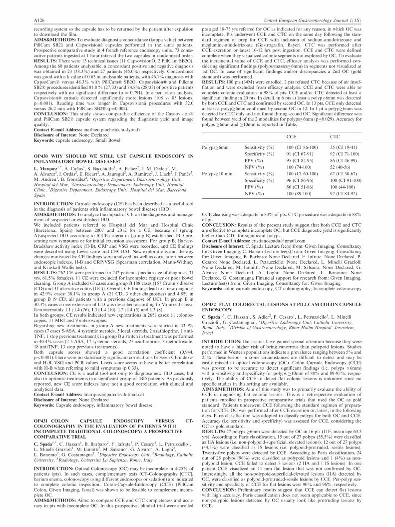
recording system so the capsule has to be returned by the patient after expulsionto download the film.AIMS&METHODS: To evaluate diagnostic concordance (kappa value) betweenPillCam SB2a and Capsovisiona capsules performed in the same patients.Prospective comparative study in 4 french reference endoscopy units. 73 conse-cutive patients ingested at 1 hour interval the two capsules in randomized order.RESULTS: There were 13 technical issues (11 CapsovisionO, 2 PillCam SB2O).Among the 60 patients analysable, a concordant positive and negative diagnosiswas obtained in 23 (38.3%) and 27 patients (45.0%) respectively. Concordancewas good with a k value of 0.63 in analysable patients, with 46.7% diagnosis withCapsoCam� versus 48.3% with PillCam� SB2O. Capsovision� and PillcamSB2� procedures identified 81.8 % (27/33) and 84.8% (28/33) of positive patientsrespectively with no significant difference (p ¼ 0.791). In a per lesion analysis,Capsovision� capsule detected significantly more lesions (108 vs 85 lesions,p¼0.001). Reading time was longer in Capsovisiona procedures with 32.0versus 26.2 min with PillCam SB2� (p¼0.002).CONCLUSION: This study shows comparable efficiency of the Capsovision�and PillCam SB2� capsule system regarding the diagnostic yield and imagequality.Contact E-mail Address: [email protected] of Interest: None DeclaredKeywords: capsule endoscopy, Small Bowel
OP430 WHY SHOULD WE STILL USE CAPSULE ENDOSCOPY IN
INFLAMMATORY BOWEL DISEASES?
L. Marquez1,*, A. Canas1, S. Bacchiddu1, A. Pelaez2, J. M. Dedeu3, M.
A. Alvarez3, I. Ordas2, E. Ricart2, A. Jauregui2, A. Ramirez2, J. Llach2, J. Panes2,M. Andreu1, B. Gonzalez2. 1Digestive Department. Gastroenterology Unit.,Hospital del Mar, 2Gastroenterology Department. Endoscopy Unit, HospitalClinic, 3Digestive Department. Endoscopy Unit., Hospital del Mar, Barcelona,Spain
INTRODUCTION: Capsule endoscopy (CE) has been described as a useful toolin the diagnosis of patients with inflammatory bowel diseases (IBD).AIMS&METHODS: To analyze the impact of CE on the diagnosis and manage-ment of suspected or established IBD.We included patients referred to Hospital del Mar and Hospital Clinic(Barcelona, Spain) between 2007 and 2012 for a CE, because of (groupA)suspected IBD according to ICCE criteria or (group B) established IBD pre-senting new symptoms or for initial extension assessment. For group B, Harvey-Bradshaw activity index (H-B), CRP and VSG were recorded, and CE findingswere described using Lewis score and CECDAI. New explorations and therapychanges motivated by CE findings were analyzed, as well as correlation betweenendoscopic indexes, H-B and CRP/VSG (Spearman correlation, Mann-Withneyand Kruskall Wallis tests).RESULTS: 262 CE were performed in 242 patients (median age of diagnosis 31yrs, 61.5% females). 11 CE were excluded for incomplete register or poor bowelcleaning. Group A included 63 cases and group B 188 cases (157 Crohn’s disease(CD) and 31 ulcerative colitis (UC)). Overall, CE findings lead to a new diagnosein 42.9% cases: 38.1% in group A (21 CD, 3 other diagnostics) and 4.8% ingroup B (9 CD, all patients with a previous diagnose of UC). In group B in30.3% cases a new extension of CD was described according to Montreal classi-fication:mainly L1þL4 (26), L3þL4 (10), L2þL4 (5) and L3 (8).In both groups, CE results indicated new explorations in 26% cases: 11 colonos-copies, 11 MRI and 9 enteroscopies.Regarding new treatments, in group A new treatments were started in 33.9%cases (7 cases 5-ASA, 4 systemic steroids, 5 local steroids, 2 azathioprine, 1 anti-TNF, 1 stop previous treatment); in group B a switch in treatment was performedin 40.4% cases (2 5-ASA, 17 systemic steroids, 25 azathioprine, 3 methotrexate,18 antiTNF, 13 stop previous treatments).Both capsule scores showed a good correlation coefficient (0.944,p50.001).There were no statistically significant correlations between CE indexesand H-B, VSG and PCR values. Lewis score seems to have a better correlationwith H-B when referring to mild symptoms (p 0.33).CONCLUSION: CE is a useful tool not only to diagnose new IBD cases, butalso to optimize treatments in a significant group of IBD patients. As previouslyreported, new CE score indexes have not a good correlation with clinical andanalytical data.Contact E-mail Address: [email protected] of Interest: None DeclaredKeywords: Capsule endoscopy, inflammatory bowel disease
OP431 COLON CAPSULE ENDOSCOPY VERSUS CT-
COLONOGRAPHY IN THE EVALUATION OF PATIENTS WITH
INCOMPLETE TRADITIONAL COLONOSCOPY: A PROSPECTIVE
COMPARATIVE TRIAL
C. Spada1,*, C. Hassan1, B. Barbaro2, F. Iafrate3, P. Cesaro1, L. Petruzziello1,L. Minelli Grazioli1, M. Iannitti3, M. Salsano2, G. Alvaro2, A. Laghi3,L. Bonomo2, G. Costamagna1. 1Digestive Endoscopy Unit, 2Radiology, CatholicUniversity, 3Radiology, Universita La Sapienza, Rome, Italy
INTRODUCTION: Optical Colonoscopy (OC) may be incomplete in 4-25% ofpatients (pts). In such cases, complementary tests (CT-Colonography [CTC],barium enema, colonoscopy using different endoscopes or sedation) are indicatedto complete colonic inspection. Colon-Capsule-Endoscopy (CCE) (PillCamColon, Given Imaging, Israel) was shown to be feasible to complement incom-plete OC.AIMS&METHODS: Aims: to compare CCE and CTC completeness and accu-racy in pts with incomplete OC. In this prospective, blinded trial were enrolled
pts aged 18-75 yrs referred for OC as indicated for any reason, in which OC wasincomplete. Pts underwent CCE and CTC on the same day following the stan-dard regimen of prep for CCE with inclusion of sodium-amidotrizoate andmeglumine-amidotrizoate (Gastrografin, Bayer). CTC was performed afterCCE excretion or latest 10-12 hrs post ingestion. CCE and CTC were definedcomplete when they visualized colonic segments not explored by OC. To evaluatethe incremental value of CCE and CTC, efficacy analysis was performed con-sidering significant findings (polyps/masses�6mm) in segments not visualized at1st OC. In case of significant findings and/or discrepancies a 2nd OC (goldstandard) was performed.RESULTS: 100 pts (34M) were enrolled. 2 pts refused CTC because of air insuf-flation and were excluded from efficacy analysis. CCE and CTC were able tocomplete colonic evaluation in 98% of pts. CCE and/or CTC detected at least asignificant finding in 20 pts. In detail, in 6 pts at least a polyp�6mm was detectedby both CCE and CTC and confirmed by second OC. In 13 pts, CCE only detectedat least a polyp�6mm confirmed by second OC in 12. In 1 pt a polyp�6mm wasdetected by CTC only and not found during second OC. Significant difference wasfound between yield of the 2 modalities for polyps�6mm (p�0.029). Accuracy forpolyps �6mm and �10mm is reported in Table.
CCE CTC
Polyps�6mm Sensitivity (%) 100 (CI 86-100) 35 (CI 19-41)
Specificity (%) 91 (CI 67-91) 92 (CI 71-100)
PPV (%) 95 (CI 82-95) 86 (CI 46-99)
NPV (%) 100 (74-100) 52 (40-56)
Polyps�10 mm Sensitivity (%) 100 (CI 60-100) 67 (CI 30-67)
Specificity (%) 96 (CI 86-96) 100 (CI 91-100)
PPV (%) 86 (CI 51-86) 100 (44-100)
NPV (%) 100 (89-100) 92 (CI 84-92)
CCE cleansing was adequate in 83% of pts. CTC procedure was adequate in 88%of pts.CONCLUSION: Results of the present study suggest that both CCE and CTCare effective to complete incomplete OC, but CCE diagnostic yield is significantlyhigher than CTC for significant polyps.Contact E-mail Address: [email protected] of Interest: C. Spada Lecture fee(s) from: Given Imaging, Consultancyfor: Given Imaging, C. Hassan Lecture fee(s) from: Given Imaging, Consultancyfor: Given Imaging, B. Barbaro: None Declared, F. Iafrate: None Declared, P.Cesaro: None Declared, L. Petruzziello: None Declared, L. Minelli Grazioli:None Declared, M. Iannitti: None Declared, M. Salsano: None Declared, G.Alvaro: None Declared, A. Laghi: None Declared, L. Bonomo: NoneDeclared, G. Costamagna Financial support for research from: Given Imaging,Lecture fee(s) from: Given Imaging, Consultancy for: Given ImagingKeywords: colon capsule endoscopy, CT-colonography, Incomplete colonoscopy
OP432 FLAT COLORECTAL LESIONS AT PILLCAM COLON CAPSULE
ENDOSCOPY
C. Spada1,*, C. Hassan1, S. Adler2, P. Cesaro1, L. Petruzziello1, L. MinelliGrazioli1, G. Costamagna1. 1Digestive Endoscopy Unit, Catholic University,Rome, Italy, 2Division of Gastroenterology, Bikur Holim Hospital, Jerusalem,Israel
INTRODUCTION: flat lesions have gained special attention because they werenoted to have a higher risk of being cancerous than polypoid lesions. Studiesperformed in Western populations indicate a prevalence ranging between 5% and25%. These lesions in some circumstances are difficult to detect and may beeasily missed at optical colonoscopy (OC). Colon Capsule Endoscopy (CCE)was proven to be accurate to detect significant findings (i.e. polyps �6mm)with a sensitivity and specificity for polyps �10mm of 88% and 89-95%, respec-tively. The ability of CCE to detect flat colonic lesions is unknown since nospecific studies in this setting are available.AIMS&METHODS: Aim of this study was to primarily evaluate the ability ofCCE in diagnosing flat colonic lesions. This is a retrospective evaluation ofpatients enrolled in prospective comparative trials that used the OC as goldstandard. Patients underwent CCE following the standard regimen of prepara-tion for CCE. OC was performed after CCE excretion or, latest, in the followingdays. Paris classification was adopted to classify polyps for both OC and CCE.Accuracy (i.e. sensitivity and specificity) was assessed for CCE, considering theOC as gold standard.RESULTS: 27 polyps �6mm were detected by OC in 16 pts (11F, mean age 63,5yrs). According to Paris classification, 15 out of 27 polyps (55,5%) were classifiedas IIA lesions (i.e. non-polypoid-superficial, elevated lesions). 12 out of 27 polyps(44,5%) were classified as IS lesions (i.e. polypoid-protruded, sessile lesions).Twenty-five polyps were detected by CCE. According to Paris classification, 24out of 25 polyps (96%) were classified as polypoid lesions and 1 (4%) as non-polypoid lesion. CCE failed to detect 3 lesions (2 IIA and 1 IS lesions). In onepatient CCE visualized an 11 mm flat lesion that was not confirmed by OC.Interestingly, all the non-polypoid-superficial-elevated lesions (IIA) detected byOC, were classified as polypoid-protruded-sessile lesions by CCE. Per-polyp sen-sitivity and specificity of CCE for flat lesions were 90% and 96%, respectively.CONCLUSION: Preliminary results suggest that CCE can detect flat lesionswith high accuracy. Paris classification does not seem applicable to CCE, sincenon-polypoid lesions detected by OC usually look like protruding lesions byCCE.
A126 United European Gastroenterology Journal 1(1S)

Contact E-mail Address: [email protected] of Interest: C. Spada Lecture fee(s) from: Given Imaging, Consultancyfor: Given Imaging, C. Hassan Lecture fee(s) from: Given Imaging, Consultancyfor: Given Imaging, S. Adler Lecture fee(s) from: Given Imaging, Consultancyfor: Given Imaging, P. Cesaro: None Declared, L. Petruzziello: None Declared,L. Minelli Grazioli: None Declared, G. Costamagna Financial support forresearch from: Given Imaging, Lecture fee(s) from: Given Imaging,Consultancy for: Given ImagingKeywords: colon capsule endoscopy, Flat colonic lesion
WEDNESDAY, OCTOBER 16, 2013 11:00–12:30
Management issues in pancreatic and biliary cancers – Hall8_____________________
OP433 PREOPERATIVE CHARACTERISTICS OF PATIENTS WITH
PRESUMED PANCREATIC CANCER BUT ULTIMATELY BENIGN
DISEASE: A MULTICENTER SERIES OF 344
PANCREATODUODENECTOMIES
A. Gerritsen1,2,*, I. Q. Molenaar1, T. L. Bollen3, C. Y. Nio4, M. G. Dijkgraaf5, H.
C. van Santvoort1, G. J. Offerhaus6, E. Sieders7, K. P. de Jong7, R. M. vanDam8, E. van der Harst9, H. van Goor10, B. van Ramshorst11, B. A. Bonsing12, I.H. de Hingh13, M. F. Gerhards14, C. H. van Eijck15, D. J. Gouma2, I. H. BorelRinkes1, O. R. Busch2, M. G. Besselink2 and the Dutch Pancreatic CancerGroup: . 1Surgery, UMC, Utrecht, 2Surgery, AMC, Amsterdam, 3Radiology, StAntonius Hospital , Nieuwegein, 4Radiology, 5Biostatistics and ClinicalEpidemiology, AMC, Amsterdam, 6Pathology, UMC, Utrecht, 7Surgery, UMCG,Groningen, 8Surgery, MUMC, Maastricht, 9Surgery, Maasstad Ziekenhuis,Rotterdam, 10Surgery, UMCSt Radboud, Nijmegen, 11Surgery, St Antonius Hospital,Nieuwegein, 12Surgery, LUMC, Leiden, 13Surgery, Catherina Hospital, Eindhoven,14Surgery, OLVG, Amsterdam, 15Surgery, Erasmus MC, Rotterdam, Netherlands
INTRODUCTION: Differentiation between malignant and benign pancreatictumours can be difficult. Consequently, a proportion of patients undergoingpancreatoduodenectomy for suspected malignancy will ultimately have benigndisease. A predictive model might prevent unnecessary pancreatoduodenectomiesin a subgroup of these patients.AIMS&METHODS: To compare preoperative clinical and imaging characteris-tics of patients with unexpected benign pathology after pancreatoduodenectomywith those of patients with confirmed (pre)malignant disease.We performed a multicenter retrospective cohort study in 1629 consecutive patientsundergoing pancreatoduodenectomy for suspected malignancy between 2003 and2010. Preoperative characteristics were compared in a 1:3 benign: malignant ratio.Malignant cases were randomly selected from the entire cohort. A multivariablelogistic regression prediction model was constructed to predict benign disease.RESULTS: 107 patients (6.7%) had unexpected benign disease after pancreato-duodenectomy. 86 fulfilled the inclusion criteria and were compared to 258patients with (pre)malignant disease. Patients with benign disease presentedless frequently with jaundice (60% vs. 80%, P50.01), pancreatic mass (54%vs. 70%, P¼0.03), double duct sign on CT (27% vs. 52%, P¼0.01) or EUS(22% vs. 51%, P¼0.02), but more often with pain (56% vs. 38%, P¼0.04). Ina prediction model using these clinical and CT parameters, only 27% of patientswith benign disease were correctly predicted and 6% of patients with malignantdisease were missed.CONCLUSION: Nearly 7% of patients undergoing pancreatoduodenectomy forsuspected malignancy were ultimately diagnosed with benign disease. Althoughsome preoperative clinical and imaging signs might indicate absence of malig-nancy, their discriminatory value is not sufficient for clinical use.Contact E-mail Address: [email protected] of Interest: None DeclaredKeywords: clinical characteristics, Computed tomography, EUS, PancreaticCancer, pancreatitis, pancreatoduodenectomy
OP434 THE DIAGNOSTIC VALUE OF ABDOMINAL DRAINAGE IN THE
INDIVIDUAL RISK ASSESSMENT OF PANCREATIC FISTULA
FOLLOWING PANCREATICODUODENECTOMY
C. Ansorge1,*, E. Rangelova1, J. Blomberg1, M. del Chiaro1, L. Lundell1,
R. Segersvard1. 1Department of Surgical Gastroenterology, Karolinska UniversityHospital, Division of Surgery, Department of Clinical Science, Intervention andTechnology (CLINTEC), Karolinska Institutet, Stockholm, Sweden
INTRODUCTION: Pancreaticoduodenectomy (PD) remains the conceptual fun-dament for curative-intent treatment of malignant diseases confined to the pan-creatic head, distal bile duct and periampullary region of the duodenum. Due touncertain therapeutic value, the use of prophylactic abdominal drainage follow-ing PD is controversial (1). However, the diagnosis of postoperative pancreaticfistula (POPF), the predominate cause of PD-associated morbidity, is based onamylase levels in drain output according to the ISGPF definition (2). The aim ofthe present study was to assess the POPF-predictive value of drain pancreaticamylase (DPA), plasma pancreatic amylase (PPA) and serum C-reactive protein(CRP) in a POPF-risk stratified PD cohort.AIMS&METHODS: Prospective cohort study of 315 standardized PDs withprophylactic drainage 2008-2012. DPA, PPA and CRP were obtained daily.Differences between the study groups with clinically-relevant POPF (ISGPF-B/C) and without (non-POPF/ISGPF-A) were evaluated. ROC analyses were per-formed for the POPF-predictive values of DPA, PPA and CRP. Risk profiles forclinically-relevant POPF were constituted and related to the intraoperative pan-creatic risk assessmen (i.e. the assessment of pancreatic texture and main ductdiameter, 3).
RESULTS: Fifty-nine patients (19%) had clinically-relevant POPF. CRP, PPAand DPA levels for postoperative day (POD) 1-3 differed significantly between thestudy groups. The POPF-predictive value of DPA (POD 1: 1322 U/L; OR 25;POD 2: 314 U/L, OR 35) was superior to that of PPA (OR 14/16), especially whencombined with CRP (POD 3: 202 mg/L; OR 17). A model of DPA and CRPpredicted 90% of the cases correctly (OR 44). Patients with intra-operatively clas-sified low-risk glands had negligible POPF-risks (specificity 99%, OR 65).CONCLUSION: The intraoperative pancreatic risk assessment represents amandatory component of the PD procedure and offers a platform for reliablypredicting the risk of developing postoperative pancreatic fistula. In patients withlow POPF risk, the use of prophylactic abdominal drainage does not offer anyadditional diagnostic value. In patients with intermediate or high POPF risks, adiagnostic model based on cut-off levels of serum C-reactive protein and drainpancreatic amylase could serve as a predictive marker for the subsequent devel-opment of clinically relevant pancreatic fistula formation.REFERENCES:
1. Correa-Gallego C, Brennan MF, D’Angelica M, et al. Operative DrainageFollowing Pancreatic Resection: Analysis of 1122 Patients Resected Over 5Years at a Single Institution. Ann Surg 2013.
2. Bassi C, Dervenis C, Butturini G, et al. Postoperative pancreatic fistula: aninternational study group (ISGPF) definition. Surgery 2005; 138(1):8-13.
3. Ansorge C, Strommer L, Andren-Sandberg A, et al. Structured intraoperativeassessment of pancreatic gland characteristics in predicting complications afterpancreaticoduodenectomy. The British journal of surgery 2012; 99(8):1076-82.
Contact E-mail Address: [email protected] of Interest: None DeclaredKeywords: drain output, pancreatic amylase, pancreaticoduodenectomy, post-operative complications
OP435 INFLUENCE OF MICROSCOPICALLY POSITIVE RESECTION
MARGINS ON �5-YEAR SURVIVAL AFTER RESECTION OF
PANCREATIC DUCTAL ADENOCARCINOMA
Y. Hamada1,*, K. Maeshiro2, Y. Nakayama3. 1Pathology, Fukuoka University,
Fukuoka, 2Chubu Tokushukai Hospital, Okinawa, 3Kyushu Medical Center,Fukuoka, Japan
INTRODUCTION: The impact of microscopically positive resection margins(R1) on surgical outcomes has been vague. Although some researchers haveshown R1 to have important prognostic significance, others have not. Basedon long-term follow-up data, 24% of 5-year survivors had positive marginsafter pancreatic carcinoma resection. However, there are very few articlesdescribing 45-year survival after pancreatic resection. The aim of this studywas to evaluate the prognostic influence of R1 and R0 (negative margins) on�5-year survival after resection of pancreatic ductal adenocarcinoma.AIMS&METHODS: A case-control study of 271 patients with pancreatic ductaladenocarcinoma who underwent pancreatic resection between 1991 and 2007 atFukuoka University (Fukuoka, Japan) was performed. Of the 271 patients, 33were actual 5-year survivors, and the overall 5-year survival rate was 12.2%. Theobservation period was 61 to 288 months (5.1–24.0 years). Clinicopathologicaldata were retrospectively analysed according to the Japan Pancreatic Societyclassification of pancreatic carcinoma. Tissue samples were fixed in 10% forma-lin, and each tumour was then cut into 3- to 4-mm slices and embedded inparaffin. R1 status was defined by the British Royal College of Pathology asthe presence of tumour tissue �1 mm from a circumferential margin surface ofthe resected pancreatic tissue.RESULTS: The R1 and R0 groups comprised 14 and 19 of the 33 patients, respec-tively. A significantly higher number of female patients was present in the R1 groupthan in the R0 group. The R0 group tended to show a significantly weaker relation-ship between the R status and stage than the R1 group. Multivariate analysisshowed that the R status was an independent prognostic marker (p ¼ 0.0071),and Kaplan–Meier curves showed that 45-year survivors in the R1 group hadsignificantly worse prognoses than did those in the R0 group (p ¼ 0.002). Of the14 patients in the R1 group, 12 died of local recurrence.CONCLUSION: Among patients surviving 45 years after pancreatic resection,the prognosis of R1 patients was poor and almost all died of local recurrence.Surgeons and pathologists should be aware of the status of the pancreatic cutmargin.REFERENCES:
1. Ferrone CR, Pieretti-Vanmarcke, R, Bloom, JP, et al. Pancreatic ductal ade-nocarcinoma: Long-term survival does not equal cure. Surgery 152; S43-49:2012
2. Schnelldorfer T, Ware al, Sarr MG, et al. Long-term survival after pancrea-toduodenectomy for pancreatic adenocarcinoma Is cure possible? Ann Surg247; 456-462: 2008
3. Verbeke CS, Leitch D, Menon KV, et al. Pedefining the R1 resection inpancreatic cancer Br J Surg 93; 1232-1237: 2006
Contact E-mail Address: [email protected] of Interest: None DeclaredKeywords: long-term follow up, pancreatic cancer, R1 resection
OP436 AN EARLY ORAL FEEDING STRATEGY AFTER
PANCREATODUODENECTOMY ENHANCES RECOVERY
WITHOUT INCREASING MORBIDITY
A. Gerritsen1,*, R. A. Wennink1, M. G. Besselink1,2, H. C. van Santvoort1, D.
S. Tseng1, E. Steenhagen3, I. H. Borel Rinkes1, I. Q. Molenaar1. 1Department ofSurgery, University Medical Center Utrecht, Utrecht, 2Department of Surgery,Academic Medical Center, Amsterdam, 3Department of Dietetics, Julius Center for
United European Gastroenterology Journal 1(1S) A127

Health Sciences and Primary Care, University Medical Center Utrecht, Utrecht,Netherlands
INTRODUCTION: Delayed gastric emptying (DGE), necessitating nutritionalsupport, is a frequent complication after pancreatoduodenectomy (PD). Data onthe optimal routine feeding strategy after PD are lacking.AIMS&METHODS: The aim of this study was to evaluate whether the intro-duction of an early oral feeding strategy with on demand nasojejunal tube (NJT)feeding after PD improved outcomes as compared to routine NJT feeding.A monocenter cohort study was performed in 102 consecutive patients under-going PD. In period 1 (June 2010-September 2011, n¼51) the routine postopera-tive feeding strategy was NJT feeding (historical controls). This changed to earlyoral feeding with on demand NJT feeding in period 2 (January-December 2012,n¼51; consecutive prospective cohort). The oral feeding strategy consisted ofprotocolized resumption of oral intake starting on the day of surgery, a NJTwas only placed in case of severe preoperative malnutrition (MUST�2) or whenoral intake was insufficient (550% of daily required caloric/protein intake) onpostoperative day 7, supervised by a dietitian. Analysis was by intention-to-treat.RESULTS: Groups were comparable for baseline characteristics. In period 1,98% (n¼50) of patients received NJT feeding versus 53% (n¼27) in period 2(becuase of insufficient intake, n¼20, or preoperative malnutrition, n¼7). Thetime to resumption of adequate oral intake (primary outcome) significantlydecreased with early oral feeding (12 vs. 9 days, P¼0.01) as well as hospitalstay (18 vs. 13 days, P¼0.01), especially in patients with an uncomplicated post-operative course (primary outcome: 11 vs. 6 days, P¼0.01, and hospital stay: 15vs. 9 days, P¼0.01 respectively) whereas no differences were seen in patients whodeveloped DGE. There was no difference in the incidence of Clavien-Dindo�3complications (47% vs. 45%, P¼0.84), DGE (31% vs. 35%, P¼0.67), pancreaticfistula (12% vs. 12%, P¼40.99), postoperative haemorrhage (12% vs. 10%,P¼0.75) and mortality (6% vs. 2%, P¼0.61) between the groups.CONCLUSION: The introduction of an early oral feeding strategy, with ondemand NJT feeding, after PD reduced the time to resumption of adequateoral intake and length of hospital stay, without negative impact on overall mor-bidity, DGE or pancreatic fistula.Contact E-mail Address: [email protected] of Interest: None DeclaredKeywords: Enteral Nutrition, nasojejunal tube feeding, Oral feeding, pancreas,pancreatoduodenectomy, surgery
OP437 IMPACT OF OPTICAL BIOPSY ON MANAGEMENT OF
PATIENTS WITH BILIARY STRICTURES: INTERIM RESULTS OF
A MULTICENTER STUDY
A. Slivka1,*, P. Jamidar2, I. Gan3, M. Giovannini4, F. Caillol4, G. Costamagna5,
P. Cesaro5, M. Kahaleh6. 1University of Pittsburgh Medical Center, Pittsburgh,2Yale New Haven Hospital, Yale, 3Virginia Mason Medical Center, Seattle, UnitedStates, 4Institut Paoli Calmettes, Marseille, France, 5Policlinico Agostino Gemelli /Univ. Cattolica Del Sacro Cuore, Roma, Italy, 6New York Presbyterian WeillCornell Medical Center, New York, United States
INTRODUCTION: Accurate differential diagnosis of indeterminate biliary stric-ture presents a clinical problem owing to the low sensitivity of tissue sampling.Probe based confocal laser endomicroscopy (pCLE) has been shown to accu-rately differentiate benign from malignant strictures in real time during ongoingERCP procedure, with a sensitivity of 98%.AIMS&METHODS: This study presents the interim analysis of an ongoinginternational multicentric trial (FOCUS, NCT01392274) aimed at evaluatingthe impact of pCLE on the management of patients with indeterminate biliarystricture. Initial performance and impact on the subsequent patient workup willbe presented.Patients presenting with indeterminate biliary strictures are currently enrolled in6 international centers (2 european, 4 US) and undergoing pCLE procedureduring standard ERCP. For each patient, the following information is collectedprospectively and evaluated in a sequential fashion: #1: clinical history þ ERCPimpression;#2: pCLE information, #3: histology/cytology results. At each stagethe investigator is asked to provide a presumptive diagnosis as well as a patientmanagement recommendation.RESULTS: 36 cases with a final diagnosis have been accumulated at this time 34malignant proven by surgery or positive tissue sampling; 2 benign proven bysurgery)32 patients (31 malignant, 1 benign) were accurately diagnosed based on theclinical information and ERCP impression, pCLE accurately diagnosed thesame 32 patients, whereas tissue sampling (combination of brush cytology andbiopsy) would have missed 5 malignant patients. In 100% of the cases wherephysicians felt confident to immediately proceed with patient management,patient management based on clinical history and ERCP impression (n¼7) andpCLE impression (n¼10) were identical to decision making once tissue samplereturned.CONCLUSION: This is the first study evaluating the impact of pCLE on man-agement of patients with biliary strictures. A high preponderance of cancer isexpected in this interim analysis based on study design. Clinical impression andpCLE outperform tissue sampling and optical biopsy has the potential toimprove clinical decision making for difficult cases by reducing the work-upalgorithm.Contact E-mail Address: [email protected] of Interest: None DeclaredKeywords: endomicroscopy, MANAGEMENT, strictures
OP438 PATTERNS OF RECURRENCE AFTER R0 RESECTION OF
GALLBLADDER CANCER: ANALYSIS OF PATIENTS
UNDERGOING LYMPH NODE DISSECTION OF THE
HEPATODUODENAL LIGAMENT WITHOUT ROUTINE
EXTRAHEPATIC BILE DUCT RESECTION.
J. K. Wiggers1,*, B. Groot Koerkamp1, O. Busch1, D. Gouma1, T. van Gulik1.
1Surgery, Academic Medical Centre, Amsterdam, Netherlands
INTRODUCTION: The routine use of extrahepatic bile duct resection alongwith lymph node dissection of the hepatoduodenal ligament (LnHDL) in patientswith gallbladder cancer is controversial.(Nishio 2011)AIMS&METHODS: The aim of this study was to analyse patterns of recurrencein patients who underwent radical resection of gallbladder cancer without extra-hepatic bile duct resection. This analysis included 64 patients who had undergoneexplorative laparotomy for gallbladder carcinoma between 1993 – 2012 at asingle centre. Patients with resectable disease and no tumour infiltrationbeyond the cystic duct underwent cholecystectomy in combination with excisionof the gallbladder fossa (segm4/5); LnHDL for tumors stage T1b and higher; andpartial liver or duodenal resection when indicated. Extrahepatic bile duct resec-tion was only used in case of tumor infiltration beyond the cystic duct.RESULTS: Thirty-six patients (56%) underwent resection at laparotomy.Twenty-five patients (39%) underwent R0 resection with LnHDL, but no extra-hepatic bile duct resection (tumor stage T1b in 5 patients; T2 in 16; T3 in 3; andT4 in 1). The median number of lymph nodes harvested from the hepatoduodenalligament in these patients was 3 (range, 1 – 11). Nine patients developed recurrentdisease after a median of 15 months (range, 3 – 25): Isolated disease at the hepaticresection margin in 2 patients, and distant disease in 7 patients. Sixteen patientsremained disease-free at median follow-up of 32 months (range, 10 – 164).CONCLUSION: The majority of patients develop distant metastatic diseaseas initial pattern of recurrence after R0 resection of gallbladder carcinoma.No patients developed isolated recurrent disease in the hepatoduodenal ligament,suggesting that extrahepatic bile duct resection has no additional value overlymph node dissection alone of the hepatoduodenal ligament.REFERENCES:
1. Nishio H, Ebata T, Yokoyama Y, Igami T, Sugawara G, Nagino M.Gallbladder cancer involving the extrahepatic bile duct is worthy of resection.Ann Surg 2011.253(5):953-60.
Contact E-mail Address: [email protected] of Interest: None DeclaredKeywords: gallbladder cancer, lymph node dissection, lymph node metastases
WEDNESDAY, OCTOBER 16, 2013 11:00–12:30Colorectal cancer: Back to the basics – Salon 11/12_____________________
OP439 ER STRESS DEPLETES APC MUTANT INTESTINAL
EPITHELIAL STEM CELLS DOWNSTREAM OF NUCLEAR BETA-
CATENIN
J. F. Van Lidth De Jeude1,*, M. C. Wielenga1, A. S. Lee2, V. Muncan1, G. R. van
den Brink1, J. Heijmans1. 1Tytgat institute for Liver and Intestinal Research,Amsterdam, Netherlands, 2Department of Biochemistry and Molecular Biology,USC/Norris Comprehensive Center, Los Angeles, United States
INTRODUCTION: Wnt signalling drives self-renewal of intestinal epithelialstem cells (ISCs) through activation of �-catenin. Hyperactivation of Wnt signal-ling causes stem cell expansion and constitutes a first step in development ofcolorectal cancer. In contrast, we have previously shown that endoplasmic reti-culum (ER) stress signalling causes loss of self-renewal capacity in ISCs, but howthese two functionally opposite pathways interact remains elusive. By deletionof ER stress repressor gene Grp78, we investigate effects of ER stress on ISCsthat lack the Wnt gatekeeper gene Apc and thus exhibit hyperactive Wntsignalling.AIMS&METHODS: To validate knockout of Grp78 as a model of ER stress wegenerated organoids of intestinal epithelium from Grp78fl/fl mice and analysedgene expression using Illumina arrays. For in vivo studies we used inducibleVillinCreERT2-Apcfl/fl-Grp78fl/fl mice in which simultaneous deletion of Apc andGrp78 resulted in hyperactivation of Wnt signalling and concomitant ER stress.RESULTS: Analysis of Grp78fl/fl organoids showed robust upregulation of apanel of ER stress markers, conforming Grp78 deletion as a bona fide modelfor the study on ER stress. Deletion of Apc from mouse intestinal epitheliumresulted in accumulation of nuclear �-catenin, hyperproliferation, crypt enlarge-ment and crypt fissioning 3 days post induction (p.i.). Induction of ER stress inApc mutant epithelium by combined deletion of Apc and Grp78, resulted inmarked (41%) reduction of BrdUþ cells indicating halted proliferation at day3 p.i. (P 5 0.001). Additionally, crypts were taken over by Grp78 proficient, wildtype cells, indicating loss of self-renewal capacity. While loss of the hyperproli-ferative phenotype and self-renewal capacity occurred, cells in Apc-Grp78 doublemutant crypts maintained nuclear localization of �-catenin.CONCLUSION: Homozygous deletion of Apc results in a known hyperproli-ferative phenotype and ultimately tumor formation. We show that concomitantdepletion of Apc and Grp78 results in abrogation of this phenotype while main-taining nuclear localization of �-catenin, the hallmark of activated Wnt signal-ling. Thus, ER stress mediated stem cell loss is dominant over hyperactivation ofthe Wnt signalling pathway and may interfere with stemness downstream of �-catenin. These results highlight a role for ER stress as a potential target indevelopment of novel cancer therapy.Contact E-mail Address: [email protected] of Interest: None DeclaredKeywords: ER stress, intestinal stem cells, mouse model, Proliferation, UnfoldedProtein Response, Wnt signaling pathway
A128 United European Gastroenterology Journal 1(1S)

OP440 REPORT AND ANALYSIS OF LYNCH SYNDROME
ASSOCIATED GENETIC VARIATIONS REPORTED BY THE
FRENCH UMD MMR –MLH1/MSH2/MSH6 DATABASE
P. Grandval1,*, A. fabre2, P. gaildrat3, P. gaildrat3, S. Baert-Desurmont4, M.
P. buisine5, A. ferrari6, Q. wang7, C. beroud8, S. olschwang2. 1gastroenterology,2CHU TIMONE, marseille, 3Inserm UMR 1079, 4genetic department, rouen,5Centre de biologie et pathologie, lille, 6Centre de biologie et pathologie,7Plateforme de recherche translationnelle, centre leon berard, lyon, 8inserm umr s910, marseille, France
INTRODUCTION: Lynch syndrome is an inherited disease caused by a consti-tutional mutation of MLH1, MSH2 and MSH6 genes, who are involved in DNAmisappariement (MMR). The French laboratories network who perform geneticanalysis has injected a total of 7047 genetic variants in Lynch syndrome sus-pected individuals since 1995. The aim of our study was to propose a classifica-tion and a description of the 707 variants of uncertain significance (VUS).AIMS&METHODS: Three groups of parameters were systematically collected:i) in silico prediction (splicing analyses, conservation level, SIFT and UMD-Predictor estimates; ii) specific clinico-pathological and biological parametersprovided by the network contributors (cosegregation analyses, clinical phenotypeaccording to the enlarged Amsterdam criteria, MMR function status in tumorcells by genotyping and immunohistochemistry analysis, splicing analysis using asplicing reporter minigene and RT-PCR in case of anomaly, transcription-trans-lation assays when VUS involve the Kozak or 5’UTR sequences, concurrentvariations within all analyzed MMR genes); iii) public data such researchpapers and online databases (functional protein assays, allele frequencies).Theinternational 5 class classification was systematically applied after critical analy-sis of each VUS characterization.RESULTS: All the information’s were inserted in the French database and areon line at www.umd.be/MLH1/, www.umd.be/MSH2/ et www.umd.be/MSH6/.A decisional tree was elaborated, and applied for each variant. 370 VUS among707 could be characterized as class 1 (neutral) for 181 and 2 (probably neutral)for 80, which are not responsible for Lynch syndrome. 55 VUS were classified asclass 5 (causal) and 54 as class 4 (probably causal), which are highly suspected tolead Lynch syndrome. The remaining 337 are classified as class 3, as long as noavailable functional assays can demonstrate their pathogenicity.CONCLUSION: Our algorithm allows us a classification of 370 new MMR genevariant, actually available for the scientific community in the French MMRdatabase, which is regularly updated. Only class 5 can be used for geneticcounseling.Contact E-mail Address: [email protected] of Interest: None DeclaredKeywords: classification, LYNCH SYNDROME AND VARIANTS
OP441 BACTERIAL GENOTOXIN COLIBACTIN PROMOTES COLON
TUMOR GROWTH BY MODIFYING TUMOR
MICROENVIRONMENT
A. Cougnoux1, G. Dalmasso1,*, R. Martinez2, E. Buc1,3, J. Delmas1,3,
L. Gibold1,3, P. Sauvanet1, C. Darcha3, P. Dechelotte3, M. Bonnet1, D. Pezet1,3,H. Wodrich2, A. Darfeuille-Michaud1,3, R. Bonnet1,3. 1M2iSH, Inserm UMR1071, INRA Usc 2018, University of Auvergne, Clermont-Ferrand, 2MFP, CNRSUMR 5234, University of Bordeaux, Bordeaux, 3Medicine, Centre HospitalierUniversitaire, Clermont-Ferrand, France
INTRODUCTION: Microbiota is thought to play a critical role in colorectalcancer (CRC). Most of mucosa-associated Escherichia coli isolated from CRCharbor the pks genomic island (pksþ E. coli) responsible for the synthesis ofcolibactin, a genotoxic compound able to induce DNA damage. It was recentlyshowed that pksþ E. coli increased tumor incidence in a colitis-associated cancermouse model.AIMS&METHODS: Our aim was to investigate the mechanisms involved inpksþ E. coli-induced carcinogenesis. Conditioned media derived from infectedHCT116 intestinal epithelial cells were used to stimulate the growth of uninfectedcells. Importance of growth factors was assessed using neutralizing antibodiesin vitro and in a xenograft model. Signaling pathways activated by pksþ E. coliwere investigated by qRT-PCR and Western blot. Results were validated usinghuman biopsies isolated from tumor colonized by pksþ or pks- E. coli.RESULTS: A transient contact of HCT116 cells with pksþ E. coli increasedtumor growth in a mouse xenograft model. Conditioned media derived frompksþ E. coli-infected cells stimulated the growth of uninfected cells. Growthwas sustained by cellular senescence, which is accompanied by production ofHGF. Neutralization of HGF abolished pksþ pro-proliferative effect. Cellularsenescence is known to be driven by post-translational modifications involvingSUMO proteins. We observed that pksþ E. coli-infected cells displayed a mod-ified pattern of SUMO-conjugated proteins correlated with a decrease of SENP1expression, a key protein involved in the control of the SUMOylation process.Over-expression of SENP1 abolished pksþ E. coli-induced alteration of proteinSUMOylation as well as cellular senescence. Using bioinformatics and molecularapproaches, we found that SENP1 expression is directly regulated bymicroRNA-20a-5p. Anti-miR-20a-5p inhibited pksþ E. coli-mediated (i) dropof SENP1 expression, (ii) cellular senescence as well as (iii) cell proliferation.Finally, we found that human tumor colonized by pksþ E. coli expressed sig-nificantly higher level of HFG and miR-20a-5p compared to tumor colonized bypks- E. coli. SENP1 expression was significantly decreased in tumors colonizedwith pksþ E. coli.CONCLUSION: Our results suggest that colibactin-producing bacteria modu-late the tumor microenvironment and favor the emergence of senescent cells,which promote tumor growth by the secretion of growth factors.Contact E-mail Address: [email protected] of Interest: None Declared
Keywords: colibactin, Colorectal Cancer, Escherichia coli, microRNA, pks,Senescence
OP442 INFLAMMATION INCREASES NOTCH1 ACTIVITY IN
COLORECTAL CANCER CELLS AND IS COUNTERACTED BY
EICOSAPENTAENOIC ACID-FREE FATTY ACID.
C. Fazio1,*, G. Piazzi1, P. Vitaglione2, V. Fogliano2, A. Munarini1,
A. Prossomariti1, L. D’Angelo1, M. Napolitano3, A. Belluzzi4, F. Bazzoli1,L. Ricciardiello1. 1Department of Medical and Surgical Sciences, University ofBologna, Bologna, 2Department of Food Science, University of Naples ‘‘FedericoII’’, Portici (Naples), 3Department of Surgical and Medical Sciences, University ofBologna, 4Gastroenterology Unit, S.Orsola-Malpighi Hospital, Bologna, Italy
INTRODUCTION: The link between inflammation and colorectal cancer (CRC)is well established, and inflammatory cells in the lamina propria, includingmacrophages, are critical for cancer initiation and progression. Notch signallinghas a pivotal role in colorectal carcinogenesis. Eicosapentaenoic acid free fattyacid (EPA), a !-3 polyunsaturated fatty acid, has been demonstrated to beprotective toward CRC in preclinical and clinical models.AIMS&METHODS: The aim of this study was to evaluate whether Notch1 canbe activated by inflammatory stimuli through STAT3-Jagged1 in CRC cells, andwhether this mechanism could be counteracted by EPA (SLA Pharma UK). Acytokine-enriched conditioned media (CM) was obtained from THP-1 cells afterdifferentiation into macrophages. CM was applied to untreated and EPA-treatedHT29 cells. Cytokine levels in the CM, fatty acids incorporation and cell adhe-sion of HT29, were tested. Cells were also treated with either IL-6 or IL-8. Levelsof Notch1 intracellular domain (NICD), Jagged1, STAT3 and pSTAT3 werequantified by western blotting. Gene expression of Jagged1 and Notch1 down-stream targets NRARP and HATH1, were evaluated by Real-Time PCR. HT29stably silenced for Notch-1 were tested as well.RESULTS: CM treatment increased cellular adhesion of HT29 up to 90% andthis phenomenon was paired by a strong activation of pSTAT3, increased expres-sions of Jagged1 and NICD. Stimulation of HT29 with either IL-6 or IL-8 led toa lower activation of Jagged1 and NICD compared to the CM-treated counter-parts. The activation of Notch1 caused up-regulation of NRARP and down-regulation of HATH1. This effect was less pronounced in Notch1-silencedHT29, while the expression of Jagged1 remained unchanged. Interestingly, athree-day pre-treatment with non-cytotoxic concentration of EPA counteractedthe CM-driven increases of Jagged1, NICD and pSTAT3. Finally, EPA effectson Notch1 were supported by a concordant modulation of its downstream tar-gets NRARP and HATH1.CONCLUSION: The inflammatory microenvironment is a stronger activator ofNotch1 signalling in CRC cells compared to the effect of each single inflamma-tory cytokine, and pre-treatment with non-cytotoxic concentrations of EPAcounteracts this effect. Our data suggest a possible new mechanism of actionof EPA as chemopreventive agent in CRC through inhibition of Notch1signalling.Disclosure of Interest: C. Fazio: None Declared, G. Piazzi: None Declared, P.Vitaglione: None Declared, V. Fogliano: None Declared, A. Munarini: NoneDeclared, A. Prossomariti: None Declared, L. D’Angelo: None Declared, M.Napolitano: None Declared, A. Belluzzi: None Declared, F. Bazzoli: NoneDeclared, L. Ricciardiello Financial support for research from: SLA PharmaKeywords: chemoprevention, inflammation, notch pathway, Omega-3 fatty acid
OP443 ROLE FOR HEPCIDIN IN OBESITY DRIVEN COLORECTAL
TUMORIGENESIS
E. Shawcross1, A. Chauhan1,*, T. Iqbal1, C. Tselepsis1. 1Gastroenterology,
University Of Birmingham Queen Elizabeth Hospital, Birmingham, UnitedKingdom
INTRODUCTION: While there is a strong association between obesity andcolon cancer, the underlying molecular and cellular mechanisms are unclear.Accumulating evidence suggests a critical role for adipose hormones; leptinand IL-6. Both IL-6 and leptin induce hepcidin expression in obesity. A consti-tutively high hepcidin expression in obesity may result in dysregulated ironmetabolism with high intracellular and low serum iron. High intracellular ironhas been shown via wnt signalling to mediate colorectal cancer (CRC).AIMS&METHODS: We aim to investigate the role of adipose cytokines andhepcidin in mediating CRC both in vivo and in vitro in malignant cell lines.Serum samples from 163 patients attending for colonoscopy as part of thenational colorectal screening programme were obtained and leptin, IL6 andhepcidin levels measured. 73 of these patients were confirmed as having colonicneoplasia. Using tissue extracted from the greater omentum of patients under-going surgery for CRC, we isolated and cultured human adipocytes. The differ-entiated adipocyte secretome (DAS) was collected and used to challengecolorectal cancer cell line SW480 in the presence or absence of leptin, IL-6 andhepcidin inhibitors. Viability and proliferation assays were then conducted on theSW480 cell lines.RESULTS: A positive correlation was found between hepcidin and leptin inpatients with cancer. Antibody arrays and ELISA confirmed the presence ofadipokines in the DAS and elevated levels of leptin (29 fold), IL-6 (12 fold)and hepcidin (3 fold). Co-incubation of SW480 cells with DAS increased cellularviability and proliferation, both of which reverted to control levels with IL-6,leptin or hepcidin blockade. These results were replicated on stimulating SW480cells sequentially with IL6, leptin or hepcidin. Colonocyte challenge with IL6 orLeptin enhanced Hepcidin production. A hepcidin antagonist, blocked IL-6 andLeptin mediated increases in colonocyte viability and proliferation.CONCLUSION: We provide the first demonstration that hepcidin is producedby adipocytes and has a important role in obesity mediated carcinogenesis. The
United European Gastroenterology Journal 1(1S) A129

importance of hepcidin in colonocyte fate is further highlighted by our findingthat a hepcidin antagonist abolished IL6 and leptin mediated increases in cellviability and proliferation. This is the first time the observation has been made intissue that does not endogenously express hepcidin. Taken in conjunction withthe positive correlation between hepcidin and leptin in neoplasia, we predict thatsystemic IL6 and leptin production in obese patients drives local tumoural hep-cidin production, thus driving carcinogenesis.REFERENCES:
1. Brookes et al. A role for iron in Wnt signalling. Oncogene. 2006 Feb7;27(7):966-75.
2. Larsson SC et al. Obesity and colon and rectal cancer risk: a meta-analysis ofprospective studies. Am J Clin Nutr. 2007;86:556-65.
3. Moghaddam A et al. Obesity and risk of colorectal cancer: A meta-analysis.Cancer Epidemiol Biomarkers Prev, 2007. 16(12): p. 2533-47. Considine et al.Serum immunoreactive-leptin concentrations in normal-weight and obesehumans. N Engl J Med. 1996 Feb 1;334(5):292-5.
4. Stattin P et al. Obesity and colon cancer: does leptin provide a link? Int JCancer. 2004;109:149-52.
Contact E-mail Address: [email protected] of Interest: None DeclaredKeywords: Colorectal cancer, hepcidin, IL-6, leptin, obesity
OP444 COLON CANCER STEM CELL PROLIFERATION AND
TUMOUR GROWTH ARE PROMOTED BY NITRIC OXIDE
SYNTHASE-2
M. A. Puglisi1,*, V. Tesori1, L. Ricci-Vitiani2, E. Giorda3, R. Carsetti3,
A. Cittadini4, G. B. Gasbarrini5, G. Pani4, A. Gasbarrini1. 1Internal Medicine andGastroenterology, GEMELLI HOSPITAL, 2Hematology, Oncology andMolecular Medicine, Istituto Superiore di Sanita , 3Cytofluorimetry Laboratory,Bambino Gesu Pediatric Hospital, 4Institute of General Pathology, GEMELLIHOSPITAL, ROME, 5Medical Research Foundation , ONLUS, Bologna, Italy
INTRODUCTION: Chronic inflammation represents a leading cause of neoplas-tic transformation in many human cancers and especially in colon cancer (CC), inpart due to tumor promotion by Nitric Oxide (NO) generated at inflammatorysites. It has also been suggested that high NO synthesis, secondary to inducibileNOS (iNOS) expression, is a distinctive feature of ‘‘cancer stem cells’’ (CSC), asmall subset of cells with self-renewal capacity, that may be identified by theexpression of the CD133 surface marker.AIMS&METHODS: Aim of this study was to explore the contribution of NO inthe definition of CSC features in colon cancer.The production of intracellular NO was assayed using the 4,5-diaminofluoresceindiacetate (DAF-2DA) detection system. Cells were sorted from five human colonCSC lines obtained from tumor tissues and from four established human CC celllines (CaCo-2,HCT-116, Lovo and HT29), on the basis of their level of DAF-2DA (NO‘‘high’’and NO‘‘low’’) by FACS-sorter and aseptically recovered. Wecompared the tumorigenic potential of NO‘‘high’’ cells respect the NO‘‘low’’ cellby in vitro and in vivo assayRESULTS: We observed that NO‘‘high’’cells displayed higher expression level ofiNOS and stem cell markers (CD133, LGR5, BMI, TWIST and SNAIL) thanNO‘‘low’’cells. In vitro assay showed that NO‘‘high’’cells displayed a significantlygreater capacity to give rise to colonies in soft agar and a higher invasive andproliferative capacity than NO‘‘low’’cells. Moreover, we inoculated the two cellfractions in nude mice and we observed that tumors derived from NOhigh cellswere markedly larger and more vascularized than those formed by NOlowcells.We hypothesized that the NO’’high’’ cells are the true CSC, while cells with lowerproduction of NO may be more committed progenitor cells. Interestingly, weobserved that NO‘‘high’’cells treated with the highly selective iNOS inhibitor,1400W, showed a significant reduction of their tumorigenic ability, confirmingthat iNOS is critical for NO production in colon CSC.CONCLUSION: These studies are relevant, since they provide the first demon-stration that NO synthesis, secondary to high iNOS expression, is a distinctivefeature of colon CSC relative to non-CSC, and in the long run, these data willhopefully serve as an impetus for evaluation of iNOS-directed therapies as acomponent of multimodal treatment regimens for human CC.Disclosure of Interest: None DeclaredKeywords: cancer stem cell, colon cancer, inducible NO-synthase, nitric oxide
WEDNESDAY, OCTOBER 16, 2013 11:00–12:30Optimizing clinical outcomes in IBD – Roof Garden_____________________
OP445 RANDOMIZED CONTROLLED TRIAL COMPARING SETON
REMOVAL ALONE OR IN ASSOCIATION WITH ANAL FISTULA
PLUG IN FISTULISING ANO-PERINEAL CROHN’S DISEASE
A. Senejoux1,*, L. Siproudhis2, L. Abramowitz3, N. Munoz-Bongrand4,K. Desseaux5, G. Bouguen2, A. Bourreille6, O. Dewit7, C. Stefanescu1,G. Vernier8, E. Louis9, J. C. Grimaud10, B. Godart11, G. Savoye12,X. Hebuterne13, P. Bauer14, M. Nachury15, D. Laharie16, S. Chevret5,Y. Bouhnik1 on behalf of GETAID: . 1CHU Beaujon, Clichy, 2CHU Rennes,Rennes, 3CHU Bichat, 4CHU Saint Louis, 5Universite Paris VII, Paris, 6CHUNantes, Nantes, France, 7Clinique Universitaire Saint-Luc, Bruxelles, Belgium,8CHU Claude Huriez, Lille, France, 9Hopital Sart Tilman, Liege, Belgium, 10CHUMarseille, Marseille, 11CHU Tours, Tours, 12CHU Rouen, Rouen, 13CHU Nice,Nice, 14Hopital Diaconesses, Paris, 15CHU Lille, Lille, 16CHU Bordeaux,Bordeaux, France
INTRODUCTION: Anal fistula plug (AFP) is a bioabsorbable bioprosthesismade of lyophilized porcine intestinal submucosa used for patients with ano-
perineal fistula. No controlled data is available with AFP in fistulising ano-perineal Crohn’s disease (FAP-CD).AIMS&METHODS: The aim of our study was to assess efficacy and safety ofAFP in FAP-CD (ClinicalTrials.gov No.2008-A00122-53). It was a multicenter,open-label, randomized controlled trial comparing seton removal alone or withAFP insertion in CD patients with non or mildly active luminal disease(CDAI�250) having at least one ano-perineal fistula tract drained for morethan one month. Patients with abscess (collection � 3 mm on magnetic reso-nance) or recto-vaginal fistulas were excluded. Setons were removed at baseline inall patients, and randomization was stratified according to AGA classification insimple or complex fistulas. The primary end point was fistula closure at week 12,defined by the absence of any draining or perianal pain, and abscess. Analysiswas made on an intent-to-treat basis.RESULTS: From June 2008 to December 2011, 106 patients from15 centers have been randomized either to AFP (n¼54) or control group(n¼52); there were 68 women; median age: 34 years [interquartile range: 26-43]; median fistula duration: 23 months [10-53]; median CDAI at baseline: 81[45-135].Fistula closure was achieved in 17/54 (31.5%) patients in the AFP group and in12/52 (23.1%) in the control group at week 12. No interaction in treatment effectwith complexity stratum was found, with fistula closure in 5/15 patients withcomplex fistula and in 12/39 patients with simple fistula in AFPs, as compared to2/13 and 10/39 in controls (P¼ 0.52 by the Breslow-Day homogeneity test). Noevidence of increased prevalence of patients with at least one adverse events wasfound (17/54, 32% vs. 8/52, 15% in the controls; p¼ 0.07).CONCLUSION: Anal fistula plug in combination with seton removal doesn’tseem more effective than seton removal alone to achieve ano-perineal fistulaclosure in patients with CD.Contact E-mail Address: [email protected] of Interest: None DeclaredKeywords: anal fistula plug, anoperineal fistula, Crohn’s disease, randomisedcontrolled trial
OP446 COMPOSITE REMISSION MEASURES IN EARLY CROHN’S
DISEASE: A POST-HOC ANALYSIS OF THE SONIC TRIAL
J. F. Colombel1,*, W. Reinisch2, G. Mantzaris3, A. Kornbluth4, A. Oortwijn5, G.
S. Bevelander6, F. Cornillie5, W. J. Sandborn7. 1Centre Hospitalier Universitairede Lille, Lille, France, 2University Hospital Vienna, Vienna, Austria,3Evangelismos Hospital, Athens, Greece, 4Mount Sinai Medical Center, New York,United States, 5Janssen Biologics BV, Leiden, Netherlands, 6Janssen Biologics BV,Leiden, 7University of California San Diego, La Jolla, United States
INTRODUCTION: Treatment goals in CD are evolving. Treatment targets nowinclude clinical remission (CR; CDAI5150), mucosal healing (MH), and biolo-gical remission such as normalization of CRP1,2.. In addition, early treatmentleads to better outcomes3.AIMS&METHODS: This post-hoc analysis of SONIC evaluated different com-posite remission measures at wk26. SONIC included 508 CD-pts naıve to immu-nomodulators and biologics3. This analysis included 188pts (median diseaseduration of 2.3yrs) who had evidence of mucosal ulcerations at baseline (BL),ileocolonoscopy and evaluable CDAI scores, CRP and ileocolonoscopies at BLand wk26. Composite remission measures were evaluated as follows: CR and MH(defined as absence of mucosal ulceration at wk26), CR and MH and CRPnormalization (CRPnorm; defined as CRP50.8mg/dL), MH and CRPnorm ,CRand CRPnorm.
RESULTS: Overall, 136/188 pts (72.3%) were in clinical remission and 90/188pts (47.9%) achieved MH (0 ulcers) at wk26. The composite measure of clinicalremission and MH was achieved by 72/188 pts(38.3%). Among those pts withelevated CRP(118/188 pts;CRP�0.8mg/dL) at BL, 38/118 pts(32.2%) achievedclinical remission, MH and CRP normalization. The combination of IFXþAZAshowed higher composite remission measures ranging from 52.3% to 63.3% vsIFX (32.3%451.2%) or AZA(12.9%429%) alone.While IFX monotherapyshowed numerically higher composite remission measures vs AZA alone,all com-posite outcomes for IFXþAZA were significantly higher vs AZA alone(Table).Asignificantly greater proportion of IFXþAZA-treated pts achieved the compositeremission measures which included MH vs IFX alone.Table: Clinical remission and composite remission measures at wk26 in SONIC**
* Fisher Exact Test (two-tailed)**This analysis takes into account CR. Note thatthe primary endpoint of SONIC was corticosteroid free clinical remission atwk26CONCLUSION: Combination therapy with IFX þ AZA showed to be moreeffective in achieving various composite remission measures, especially when MHis included, compared to treatment with AZA or IFX alone.REFERENCES:
1. Zallot C. and Peyrin-Biroulet L. Deep Remission in Inflammatory BowelDisease: Looking Beyond Symptoms. Curr. Gasteroenterol. Rep. 2013;15:315
2. Colombel JF., Louis E., Peyrin-Biroulet L., Sanborn WJ. and Panaccione R.Deep Remission: a New Concept? Dig Dis 2012; 30 (suppl 3): 107-111
3. Colombel JF., Sandborn WJ., Reinisch W. et al. Infliximab, Azathioprine, orCombination Therapy for Crohn’s Disease. N Engl J Med 2010;362:1383-95
Contact E-mail Address: [email protected] of Interest: J. Colombel Financial support for research from: Janssen,W. Reinisch Financial support for research from: Janssen, G. MantzarisFinancial support for research from: Janssen, A. Kornbluth Financial supportfor research from: Janssen, A. Oortwijn Other: Janssen employee, G. BevelanderOther: Janssen employee, F. Cornillie Other: Janssen employee, W. SandbornFinancial support for research from: JanssenKeywords: Crohn’s disease, Infliximab, SONIC
A130 United European Gastroenterology Journal 1(1S)

OP447 COMPARISON OF EFFICACY AND TOLERANCE OF TWO
MAINTENANCE REGIMENS WITH ADALIMUMAB IN PATIENTS
WITH MODERATE TO SEVERE CROHN’S DISEASE.
J. Filippi1,*, D. Agrefilo1, E. Fontas2, X. Hebuterne1. 1Gastroenterology and
clinical nutrition, Archet 2 Hospital, 2Clinical research department, CimiezHospital, Nice, France
INTRODUCTION: In adult patients with Crohn’s disease (CD), the induction regi-men commonly used with adalimumab (ADA) is 160 mg at week 0 (w0) and 80mg atw2, followed by amaintenance regimen of 40mg every otherweek (eow). In situationsof partial response or loss of response, the interval between injections can be shortenedto one week, or the dose can be increased to 80 mg eow. It has been shown in theliterature that 30%of patients require such optimization. The aim of this studywas toassess the efficacy and safety of a systematic maintenance optimization of the dose (80mg eow), in comparison with the classical regimen (40 mg eow).AIMS&METHODS: This monocentric, retrospective study was based on theADA prescribing practices of two physicians implicated in the management ofCD patients: one using the classical (CLA) dose (160 mg w0, 80 mg w2 and 40 mgeow), the other using the systematic optimized (OPT) dose (160 mg w0 and 80eow). Outpatients with moderate to severe CD, treated with ADA and followedevery three months between 2007 and 2011, were studied. Baseline characteris-tics, efficacy and tolerance of the two regimens were assessed. Results areexpressed as percentage and median. Comparisons were performed using Chi-square tests for qualitative parameters, exact Fisher tests (for small sample size)and Mann-Whitney tests for quantitative parameters.RESULTS: A total of 84 patients (39 CLA and 45 OPT) were analyzed.Baseline characteristics were similar between the two groups. In the CLA andOPT groups, 30.8% and 28.9% of patients had concomitant immunosuppressivedrugs, 33.3% and 46.7% of patients were on second line of anti TNF alphatherapy and at baseline, the median Harvey Bradshaw Index (HBI) was 11.6and 10.7, respectively (NS). At 3, 6, 9 and 12 months, the steroid-free remission(HBI54) rates were 66.7%, 65.8%, 59% and 40.7% in the CLA group and 84.4%,84.2%, 82% and 81.3% in the OPT group respectively (p¼0.05 at months 3 and 6,p¼0.01 at month 9 and p¼0.001 at month 12). During follow-up, treatment opti-mization was required in 64.1% of CLA patients versus 13.3% of OPT patients(p50.0001); in 31.1% of patients in the OPT group it was possible to taper themaintenance regimen to 40 mg eow. There was no significant difference concerningthe occurrence of side effects (tumor, dermatological, infectious, and rheumatic)between groups.CONCLUSION: In moderate to severe CD patients, maintenance treatment withADA optimized at 80 mg eow appears to be more effective than the standardregimen, with a similar tolerance. These results should be confirmed by a pro-spective controlled study.Contact E-mail Address: [email protected] of Interest: J. Filippi Lecture fee(s) from: Abbott, Ferring, MSD,Norgine, Consultancy for: Abbott, Astellas Pharma, D. Agrefilo: NoneDeclared, E. Fontas: None Declared, X. Hebuterne Lecture fee(s) from:Abbott, Ferring, Fresenius-Kabi, MSD, Norgine, Nutricia, Consultancy for:Abbott, Fresenius-Kabi, NestleKeywords: adalimumab, anti TNF therapy, Crohn’s disease
OP448 RESIDUAL ADALIMUMAB TROUGH LEVELS ARE
ASSOCIATED WITH CLINICAL REMISSION AND MUCOSAL
HEALING IN IBD.
X. Roblin1,*, M. Rinaudo1, H. marotte1, E. Del Tedesco1, J. M. phelip1,
L. Peyrin-Biroulet2, S. paul1. 1university of saint etienne, saint etienne, 2CHUNancy, Nancy, France
INTRODUCTION: Few data have been reported on the impact of residualadalimumab trough levels, antibodies against adalimumab (ADAb) and clinicalresponse in IBD. In addition, there is no data on the impact of these assays onmucosal healing (MH). The aim of our study was to investigate whether therewas a relationship between residual adalimumab trough levels and ADAbregarding the clinical response in IBD patients and also to search for a linkbetween these rates and the presence of MH.AIMS&METHODS: This is a trans sectional study that included any patientwith IBD under ADA maintenance therapy and for who we have the patientrecord clinical activity, endoscopy score, residual adalimumab trough levels andADAb during the same period. Clinical remission was defined for Crohn by aCDAI 5150 and for Ulcerative Colitis (UC) with a Mayo score 54. MH wasdefined as a Mayo endoscopic score mayo 52 for UC and by the disappearanceof ulceration in Crohn patients. Residual adalimumab trough levels and ADAb
were measured with the Premium Lisa-Tracker assay (Theradiag, France) inblind of the clinical datas.RESULTS: 40 patients under ADA maintenance (or 40mg per day during 14days or 40mg / 7j) were included (mean age: 42 þ / - 13 years, sex ratio M / F:1.6). Among the 22 patients with Crohn disease (13 L1), seven had clinicalremission (31%). MH was reported in 8 cases (36%). 18 patients had a UC(E3 14) and were in clinical remission in 33% of cases (N: 6) and mucosal healingin 8 cases (44%). Adalimumab rates observed were 4.9 mg/ml ( 1.9-6 mg/ml) andADAb rate of 4.6 ng/ml (0.9 to 6.8 ng/ml)). Seven patients had ADAb levels 410 ng/ml and none of them had MH. Median of residual adalimumab troughlevels were significantly higher in patients with clinical remission (7.1 vs 4.1 mg/ml, p¼0.012). Similarly, residual adalimumab trough levels were also signifi-cantly higher in patients with mucosal healing (7.4 vs 3.9 mg/ml, p¼0.0053).These results were the same whatever the type of IBD. On multivariate analysis,two independent factors are associated with healing mucosa (trough levels ofADA ( RR : 0.62, 95%CI: 0.40-0.94;p¼0.026) and disease duration (RR :0.82, 95%CI: 0.68-0.97; p¼0.026). The ADAb median were identical regardlessthe presence or the absence of clinical remission or MH.CONCLUSION: Residual adalimumab trough levels were significantly higher incases of clinical remission and mucosal healing presence in IBD patients underadalimumab treatment. Rates of ADAb levels are negative predictors of MH.Disclosure of Interest: X. Roblin Consultancy for: Abbott, Theradiag, M.Rinaudo: None Declared, H. marotte: None Declared, E. Del Tedesco: NoneDeclared, J. M. phelip: None Declared, L. Peyrin-Biroulet: None Declared, S.paul: None DeclaredKeywords: adalimumab, IBD, mucosal healing, trough levels
OP449 THE CLINICAL SIGNIFICANCE OF ANTIBODY TO
INFLIXIMAB FORMATION IN INFLAMATORY BOWEL DISEASE
PATIENTS ON INFLIXIMAB MAINTENANCE
H. Awadie1,*, H. Bar Yoseph1, S. Ben Horin2, Y. Chowers1, M. Waterman3.
1Gastroenterology, Rambam Health Care Campus, Haifa, 2Gastroenterology,Sheba Medical Center, Tel Aviv, 3Gastroenterology, Rambam Health CareCampus&The B. Rappaport Faculty of Medicine – The Technion, Haifa, Israel
INTRODUCTION: Infliximab therapy in patients with inflammatory bowel dis-ease (IBD) is highly effective though up to 40% of patients lose response toinfliximab. Antibodies to infliximab (ATI) formation, occurs in 6-17% ofpatients on scheduled infliximab maintenance therapy and are associated withloss of response. The aim of our study is to assess the clinical significance of ATIformation in IBD patients on maintenance therapy with infliximabAIMS&METHODS: Infliximab (IFX) trough levels and ATI at trough wereprospectively determined in IBD patients on maintenance infliximab (IFX) ther-apy, using anti lambda chain enzyme linked immunosorbent assay (ELISA).Only patients on IFX maintenance therapy with at least 3 ATI tests wereincluded. Patients were divided according to ATI levels into two groups: sus-tained negative (51 mcg/ml) ATI levels (LATI), and fluctuating ATI (FATI)levels (at least one sample with ATI5 1 and at least one41mcg/ml). Meticulousretrospective chart review was performed to assess clinical response - defined asstable dosing of IFX and no change in therapy alongside with physician’s note onpatient’s good clinical status. Statistical analysis comparing FATI to LATI wasperformed using student’s t-test for continuous variables and Chi square forcategorical variables. A two-tailed p-value�0.05 was considered significantRESULTS: Forty-eight IBD patients with available clinical data and serumsamples were included. Twenty five patients had sustained low ATI levels and23 patients had fluctuating ATI levels. Both groups were similar in IBD subtypedistribution (CD in 73.9% and 60% of FATI and LATI groups, respectively),mean age at last follow-up (37 vs. 43 years), mean serum albumin levels (4.2 VS.3.9 gr/dl), rates of concomitant immunomodulator therapy (82% and 80%), andmean trough serum IFX levels (3.3 vs. 4.6 mcg/ml) in FATI and LATI groups,respectively. There were similar rates of clinical response (64% vs. 76%), andmean serum C-reactive protein levels (9.4 vs. 8.5 mg/dl, ULN¼5) in the FATIand LATI groups. Subgroup analysis looking specifically at clinical response inthe FATI and LATI according to IBD subtype, showed similar response rates inUC ( 33% vs. 40%, respectively) and in CD (76% vs. 100%, respectively)CONCLUSION: IBD patients on scheduled infliximab maintenance with fluc-tuating ATI formation pattern have similar rates of clinical response and remis-sion compared with patients without ATIContact E-mail Address: [email protected] of Interest: None DeclaredKeywords: antibodies to infliximab, inflamatory bowel disease, biological therapy
Table: OP446
AZA (n¼54) IFX (n¼62) IFXþAZA (n¼72)P-value*IFXþAZA vs. AZA
P-value*IFX vs. AZA
P-value*IFXþAZA vs. IFX
CR (N¼188) 57.4% (31/54) 75.8% (47/62) 80.6% (58/72) 0.006 0.047 0.534
CR þ MH (N¼188) 20.4% (11/54) 32.3% (20/62) 56.9% (41/72) 50.001 0.207 0.005
CR þ MH þCRPnorm
(N¼118)12.9%(4/31) 25.6%(11/43) 52.3%(23/44) 50.001 0.245 0.015
MH þ CRPnorm
(N¼118)19.4%(6/31) 30.2% (13/43) 56.8%25/44) 0.002 0.419 0.017
CR þ CRPnorm
(N¼118)29.0% (9/31) 51.2%(22/43) 63.6%(28/44) 0.005 0.094 0.282
United European Gastroenterology Journal 1(1S) A131

OP450 ASSOCIATION BETWEEN THIOPURINES METABOLITES
LEVELS AND CLINICAL REMISSION IN IBD PATIENTS : AN
UPDATED META-ANALYSIS
A. moreau1, S. laporte1, E. deltedesco1, S. paul1, J. M. phelip1, L. peyrin-birou-
let2, X. Roblin1,*. 1university of saint etienne, saint etienne, 2CHU Nancy, Nancy,France
INTRODUCTION: A first meta-analysis, published in 2006, support the asso-ciation between thiopurines metabolites levels (6-thioguanine nucleotides or 6-TGN) and clinical remission. This meta-analysis has pooled the results of 6studies (N ¼ 518 patients). Since then, several studies have been published andsome of them present conflicting conclusions. Therefore, an update of this firstmeta-analysis has been performed.AIMS&METHODS: A systematic search was performed up to July 2012. Theprimary goal was to assess the association between thiopurines metabolites levelsand clinical remission in IBDpatients. Secondary objectiveswere to evaluate whetheror not the association was influenced by differences in study design such as thepresence of a selection and/or information bias (misclassification by differences indisease activity scores, metabolites concentration assays). AMantel-Haenszel pooledrisk estimate provide a measure of that association. The Cochran Q test and the I2statistic were used to evaluate heterogeneity between studies. Twodifferent softwareswere used for all statistical analysis (Revman� and R).RESULTS: A total of 20 studies were included in this meta-analysis (N¼2405patients). When studying datas with a cut-off level of 230 pmol / 8.108 RBC (N¼2234 patients), the pooled OR was 2.09 (95% CI, 1.53-2.87, p5.00001). Visualinspection of funnel plot asymmetry and statistical asymmetry tests were alsoperformed. Neither showed indication of publication bias. A sensitivity analysiswas conducted and showed no significant difference in the overall OR withremoval of any individual study.CONCLUSION: This meta-analysis reassert the utility of using cut-off levels ofthiopurines metabolites for the monitoring of IBD patients. The results of thecumulative meta-analysis are univocals and representative: the OR stabilizes withtime and it clearly demonstrates the inutility to perform any more studies toestablish the association between thiopurines metabolites levels and clinicalremission in IBD patients.REFERENCES:
1. Association of 6-thioguanine nucleotide levels and inflammatory bowel diseaseactivity: a meta analysis. OstermanMT, Kundu R, Lichtenstein GR, Lewis JD.Gastroenterology. 2006 Apr;130(4):1047-53.
Contact E-mail Address: [email protected] of Interest: A. moreau: None Declared, S. laporte: None Declared, E.deltedesco: None Declared, S. paul: None Declared, J. M. phelip: NoneDeclared, L. peyrin-biroulet: None Declared, X. Roblin Consultancy for: HACPharmaKeywords: azathioprine, IBD, thioguanine nucleotides
WEDNESDAY, OCTOBER 16, 2013 14:00–15:30GI Surgery: What’s new in 2013? – Hall 6_____________________
OP451 RECURRENCES AFTER ENDOSCOPIC STENTING AS
TREATMENT FOR ACUTE MALIGNANT COLONIC
OBSTRUCTION IN THE DUTCH STENT-IN 2 TRIAL .
D. A. Sloothaak1,*, M. W. van der Berg2, M. G. Dijkgraaf3, P. Fockens2, P.
J. Tanis1, J. W. van Hooft2, W. A. Bemelman1 and collaborative Dutch Stent-Instudy group: . 1Surgery, 2Gastroenterology and Hepatology, 3 ClinicalEpidemiology, Biostatistics and Bioinformatics, Academic Medical CenterAmsterdam, Amsterdam, Netherlands
INTRODUCTION: The Stent-in-2 trial randomised patients with acute malig-nant colonic obstructions between treatment with endoscopic stent as a bridge toelective surgery and emergency surgery. The rate of clinical and subclinical stentrelated tumour perforations was unexpectedly high. Potential oncological con-sequences of these (sub)clinical perforations are not clear.AIMS&METHODS: The aim of our study was to compared the risk of cancerrecurrence after endoscopic stenting for acute malignant colonic obstructions tothat of emergency surgery. Medical charts from eligible patients of the Stent-in-2trial (n¼97), with proven malignancy of the obstructing tumour and potentialcurable disease (n¼58), were reviewed. Data was collected about adjuvant treat-ment, cancer recurrence and survival. We calculated the 5 year overall,and locoregional recurrence rates and used a competing risk analysis toexplore differences in the cumulative incidence of recurrence. Patients in thestent group with a (sub)clinical perforation (n¼6) were identified for a subgroupanalysis.RESULTS: Emergency surgery was performed in 32 (55%) patients and endo-scopic stenting as a bridge to surgery in 26 (45%) patients. Patient, and tumourcharacteristics and the use of adjuvant chemotherapy were comparable betweenthe two groups. The median follow up was 38 months (IQR 18-44) in the emer-gency surgery group and 36 months (IQR 34-49) in the stent group. The 5 yearoverall recurrence rate was 25% (n¼8) and 42% (n¼11), respectively (p¼0.027).The locoregional recurrence rate was 9% (n¼3) and 19% (n¼5), respectively(p¼0.052). The cumulative incidence of overall recurrences in patients with a(sub)clinical stent related perforation was 83% [CI95% 58-100] which was sig-nificantly increased compared to patients who underwent emergency surgery orstenting without perforation (26% [CI95% 14-47], 34% [CI95% 18-65], respec-tively, p ¼ 0.0072). The cumulative incidence of locoregional recurrences within 5years was also increased in patients with a (sub)clinical stent related perforation(50% [CI95% 22-100], 11% [CI95% 3-41], 10 [CI95% 3-28], respectively, p ¼0.053).CONCLUSION: Although the small number of patients in our study can beconsidered as a limitation, our results relate to a prospective cohort with a low
risk of allocation bias. Clinical and subclinical perforations after endoscopicstenting for malignant colonic obstructions seem to be associated with anincreased risk of recurrent disease.Disclosure of Interest: None DeclaredKeywords: colon cancer, Stenting
OP452 SIMPLIFYING ENDOSCOPIC EN-BLOC-RESECTION OF
LARGE LESIONS BY COMBINING A NEW INSTRUMENT AND A
SPECIAL REGARD TO THE IMPEDANCE OF SUBMUCOSAL
INJECTION SOLUTIONS
N. Al-Dayaa1,*, N. Quaas1, M. Losle1,2, K. E. Grund1,2. 1Surgical Endoscopy,Department of General, Visceral and Transplant Surgery, University Hospital,Tubingen, 2supported by: Federal Ministry of Economics and Technology based ona resolution of the Bundestag, Berlin, Germany
INTRODUCTION: Till today the removal of large (Ø 420 mm) and difficultlesions is a great challenge for high-frequency (HF) surgery. New instrumentsand injection solutions have been developed to decrease the complication ratesand to facilitate the resection. However the correlation between HF-parameters(alternating current, alternating voltage and impedance) and the cutting result isnot in focus of these developments. Previous results led to the conclusion that ahigh impedance of the tissue is essential for the removal of large lesions in thegastrointestinal tract.AIMS&METHODS: In large lesions the contact surface of the snare is biggerthan in small lesions which results in a lower impedance. A low impedance cancause problems with the HF-surgical cutting process, so our aim was to increasethe impedance by developing a new instrument and finding a submucosal injec-tion solution with few electrolytes.We compared the impedance (RF-impedance-meter, f. 100 kHz) of snares withwires of varying lengths in a standard solution for submucosal injection (0,9%saline) and during the resection of imitated lesions (Ø 20 mm) in isolated porcinestomachs.With standardized electrodes (gold probes 1mm/15mm) we measured the impe-dance (RF-impedance-meter, f. 100 kHz) of various solutions isolated and afterinjection of 3 ml into the submucosa of isolated porcine stomachs. Followingsolutions were tested: 0,9% saline, 4% gelatine, 6% hydroxyethyl starch, 10%glycerol/5% fructose in 0,9% saline and 10% glucose solution.RESULTS: The measuring of the impedance with varying cutting-wires in 0,9%saline solution showed an exponential increase of the impedance during reduc-tion of the length of the wire. During the resection the impedance showed a meanincrease by 175% by varying the length of the wire from 30 mm to 10 mm.The 10% glucose solution had a significantly (p 5 0,001) higher impedance (plus53-156%) than the other submucosal injection solutions. The other tested solu-tions showed no significant difference in the impedance compared to 0,9% salinesolution.CONCLUSION: For the first time we investigated the differences in theimpedance concerning the length of the cutting-wire and various submucosalinjection solutions. We developed a new instrument with a partially insulatedsnare-wire so the cutting part of the wire is very short (15 mm). This instrumentin combination with an optimal injection solution with fewer electrolytes opensup new possibilities for the expansion and improvement of conventional resectiontechniques (polypectomy and EMR) and as an alternative to ESD.Contact E-mail Address: [email protected] of Interest: None DeclaredKeywords: EMR, HF-Surgery, Large Lesions, Submucosal Injection Solution
WEDNESDAY, OCTOBER 16, 2013 14:00–15:30Nutrition: From the nursery to the nursing home – Hall 10_____________________
OP453 VIOLATIONS TO THE ESPGHAN PEDIATRIC GUIDELINES
FOR ACUTE GASTROENTERITIS IN 11 EUROPEAN COUNTRIES
E. Nicastro1,*, I. Liguoro1, A. Lo Vecchio1, A. Chmielewska2, M. Piescik2, C. De
Bruyn3, J. Dolinsek4, E. Doroshina5, S. Fessatou6, T. L. Pop7, C. Prell8,M. Tabbers9, M. Tavares10, P. Urenden11, A. Guarino12, and, the TutorialEuropean Electronic Network on Acute Gastroenteritis (TEEN-AGE) workinggroup. 1Department of Translational Medical Sciences, Pediatric Area, UniversityFederico II, Naples, Italy, 22nd Department of Pediatrics, The Medical Universityof Warsaw, Warsaw, Poland, 3CDB Universitair Ziekenhuis Brussel Kinderen,Vrije Universiteit Brussel, Brussels, Belgium, 4University Medical Centre, Maribor,Slovenia, 5Russian Medical Postgraduate Academy, Moscow, Russian Federation,6First Department of Pediatrics, Athens Children’s Hospital ‘‘P. & A. Kyriakou’’,Athens, Greece, 7"Iuliu Hatieganu" University of Medicine and Pharmacy,, Cluj-Napoca, Romania, 8Division of Paediatric Gastroenterology & Hepatology,University of Munich Medical Center, Munich, Germany, 9Emma’s Children’sHospital Academic Medical Centre, Amsterdam, Netherlands, 10Department ofPediatrics, Hospital Sao Joao, Porto, Portugal, 11Department of Pediatrics,Marmara University, Istanbul, Turkey, 12Department of Translational MedicalSciences, University Federico II, Naples, Italy
INTRODUCTION: The Tutorial European Electronic Network on Acute gas-troenteritis (TEEN-AGE) is a project of implementation of ESPGHAN/ESPIDpediatric guidelines for acute gastroenteritis (AGE) in Europe through an e-learning course hosted by the UEG platform (1).AIMS&METHODS:We aimed at investigating the adherence to AGE guidelinesin in- and out-patients 5 5 years. Patients’ data were collected by the physiciansparticipating in the network before the e-learning course and loaded in a CRFavailable on the UEG website.RESULTS: We enrolled 278 children (143 boys, mean age 23�16 months),184 in- and 94 out-patients. Only 39.4% of the outpatients and 51% of the
A132 United European Gastroenterology Journal 1(1S)

inpatients received oral rehydration solution (ORS) at home. Among the inpa-tients, 120/184 (65.2%) presented more than 3 episodes of vomiting/24 hors.Physicians reported a dehydration 4 5% in 100/184 (54.3%) of the children.Severe dehydration and ORS failure were the reasons for admission in 39.2% and30.9% of cases, respectively. Only 63.6% of hospitalized children needed IVrehydration and in only 16.8% a weight gain 4 5% was observedat discharge, indicating a low rate of severely dehydrated children amongthose who were hospitalized. Inappropriate changes in diet were done in30.9% of the outpatients and in 33.7% of the inpatients. All the inpatients and46.8% of the outpatients underwent laboratory investigations, while stool culturewere done in 53.3% (inpatients) and 16% (outpatients) of cases, respectively.CONCLUSION: Unnecessary, invasive and expensive interventions were com-monly observed in clinical management of AGE, including an excess of admis-sions with low rate of intravenous rehydration and the negligible difference ofweight between admission and discharge. Unnecessary investigation were alsoprescribed. The second phase of the TEEN-AGE will aim at reducing the viola-tions to the guidelines recommendations through the e-learning implementation.REFERENCES:
1. http://www.e-learning.ueg.eu/courses/course-summary.html?eprs%5Br%5D¼18362
Contact E-mail Address: [email protected] of Interest: None DeclaredKeywords: children, e-learning, ESPGHAN guidelines, gastroenteritis
OP454 EFFICACY AND ADVERSE EVENTS OF LAPAROSCOPIC
GASTROSTOMY PLACEMENTS IN CHILDREN: RESULTS OF A
LARGE COHORT STUDY
J. Franken1, F. A. Mauritz1,2,*, D. C. van der Zee1, M. Y. van Herwaarden-Lindeboom1. 1Pediatric Surgery, Wilhelmina Children’s Hospital, UniversityMedical Center Utrecht, 2Gastroenterology and Hepatology, University MedicalCenter Utrecht, Utrecht, Netherlands
INTRODUCTION: The placement of a gastrostomy tube is an established treat-ment to provide enteral feeding directly via the stomach. Available data on long-term outcomes of a laparoscopic gastrostomy (LAG) with regard to the efficacyand adverse events, specifically gastroesophageal reflux (GER) are limited.Furthermore, some studies advocate the routine use of preoperative 24-hourpH monitoring to predict postoperative GER symptoms; however, this predictivevalue is under debate.AIMS&METHODS: The aim of this study is to evaluate long-term efficacy andadverse events after LAG. A retrospective observational cohort study was per-formed including all 300 patients that underwent LAG between January 2004and December 2011. The median age at the time of operation was 2.66 years(IQR 1.28 – 7.44). The majority of patients (75.0%) were neurologicallyimpaired. Endpoints for efficacy were successful feeding and weight-for-lengthand length-for-age z-scores and endpoints to evaluate adverse events were com-plications, reinterventions and GER symptoms.RESULTS: After a median follow-up of 2.63 years (IQR 1.07 – 4.77), feedingthrough the gastrostomy was successful in 255 patients (95.9%). Weight-for-length z-scores had significantly increased after operation compared to preopera-tive measures (-0,98SDS to -0,26SDS; p50.0005). Length-for-age z-scoreremained similar (p¼0.695). Major complications, such as stomach wall dehis-cence and intraoperative bleeding were seen in 6 patients (2.0%). Minor complica-tions, mainly hypergranulation, leakage, wound infection and early cathererdislodgement were encountered in 221 patients (74.0%). A total number of 48reinterventions was needed to treat complications. In 28.2% of patients with pre-operative GER, reflux symptoms dissolved after operation. However, de novopostoperative GER symptoms were present in 34.2% (p¼0.824). Preoperative24-hour pH monitoring was performed in 180 patients (60%). The sensitivityand specificity of the preoperative 24-hour pH monitoring to identify patientwith postoperative reflux symptoms were respectively 17.5% and 76.9%.CONCLUSION: LAG placement in children leads to successful feeding in 96%of patients and is associated with a low risk of developing serious adverse events;however, minor complications were very frequently encountered. Contrary to theresults of previous studies, the incidence of GER symptoms does not increaseafter LAG and preoperative 24-hour pH monitoring is not a reliable tool toaccurately predict postoperative GER symptoms, since a large percentage ofpatients with postoperative GER symptoms have negative preoperative pHmeasurements.Disclosure of Interest: None DeclaredKeywords: children, enteral feeding, gastroesophageal reflux, laparoscopicgastrostomy
OP455 ETHANOL AND TAUROLIDINE LINE LOCKS FOR THE
REDUCTION AND TREATMENT OF CATHETER RELATED
BLOOD STREAM INFECTIONS IN PAEDIATRIC INTESTINAL
FAILURE: A SYSTEMATIC REVIEW AND META-ANALYSIS
P. Henderson1, R. Tayler2, D. C. Wilson1, A. Barclay2,*. 1Child Life and Health,
University of Edinburgh, Edinburgh, 2Paediatric Gastroenterology , RHSC,Glasgow , United Kingdom
INTRODUCTION: Paediatric intestinal failure (PIF: dependency on parentalnutrition (PN) for �28 days) requires a central venous catheter (CVC) for PNadministration, with significant morbidity and mortality associated with cathe-ter-related blood stream infection (CRBSI). Studies suggest that specialised linelocks containing ethanol or taurolidine may significantly reduce CRSBI for PIF.AIMS&METHODS: Our aim was systematically review the evidence for effec-tiveness of ethanol and taurolidine line locks in the prevention or treatment ofCRBSI in PIF. Systematic retrieval of data from studies of PIF was obtained (PN
428 days, age 518yr). Electronic searches of the Cochrane Library, MEDLINE(1946 –April 2013) and PubMed (to April 2013) were made using keyword andMeSH terms ’Intestinal failure’, ’child’ , ’ethanol locks’ and ’taurolidine locks’.Hand searches were also performed. Two authors independently assessed the levelof evidence (EL) using SIGN methodology (www.sign.ac.uk). Outcome measureswere the reduction in rates of CRBSI or eradication of CRBSI with rates expressedper 1000 CVC days; a meta-analysis was also performed using RevManRESULTS:
Study EL Rx Group InterventionReduction in CRBSIper 1000 CVC days
Jones 2- 23 70% ethanol 9.9 to 2.1
Wales 2- 10 70% ethanol 10.2 to 0.3
Blackwood 3 2 70% ethanol -
Cober 2- 15 70% ethanol 7.9 to 0.5
Onland 3 9 70% ethanol -
Mouw 2- 5 70% ethanol 11.5 to 2.3
McGrath 3 7 70% ethanol -
Chu 2- 19 Taurolidine 8.6 to 1.2
Pieroni 2013 2- 20 70% ethanol 9.8 to 2.7
The search strategy yielded 3142967 hits. Combination searches using ’intestinalfailure’ and ’Child’ reduced this to 2993. 15 studies were read in detail; five wereexcluded due to containing purely adult data or where data on PIF alone couldnot be extracted. Ten studies were included in our review, six of CRBSI preven-tion and four of CRSBI treatment. 8 studies used ethanol alone and 1 reportedtaurolidine use (Table; 2 studies by Pieroni reported duplicate data, only the laterone is shown). Four studies reported success in prevention of recurrent linesepsis. 2 thrombotic episodes were reported. A meta-analysis of the five studiesreported ethanol locks for CRBSI prevention demonstrated a reduction inCRBSI by 7.47 events (95% CI 5.86-9.08) per 1000 catheter days.CONCLUSION: Although data for the use of ethanol line locks are limited andof poor methodological quality (and thus of lower EL), ethanol locks appear tobe an effective therapy in CRBSI prevention and treatment. With only one studyof taurolidine locks, no clear conclusions can be made. Well designed studies arewarranted to compare these two treatments.Contact E-mail Address: [email protected] of Interest: None DeclaredKeywords: central line sepsis , intestinal failure , parenteral nutrition
OP456 DOES INSERTION OF A GASTROSTOMY CONFER ANY
QUALITY OF LIFE BENEFIT TO EITHER PATIENTS OR THEIR
CARERS?
M. Kurien1,*, M. E. McAlindon1, J. Grant1, E. F. Wong1, A. Johnston1,B. Hoeroldt2, K. L. Dear3, D. S. Sanders1. 1Department of Gastroenterology,Royal Hallamshire Hospital, Sheffield, 2Department of Gastroenterology,Rotherham District General Hospital, Rotherham, 3Department ofGastroenterology, Chesterfield Royal Hospital, Chesterfield, United Kingdom
INTRODUCTION: Gastrostomy feeding is an effective means of providing ent-eral nutrition to patients who have functionally normal gastrointestinal tracts butwho cannot meet their nutritional requirements because of an inadequate oralintake. Whilst improvements in outcome measures such as nutritional status andmortality have been demonstrated in certain patients undergoing gastrostomyinsertion, there remains a paucity of work evaluating another important healthoutcome measure, which is health related quality of life (HRQoL). Furthermoreno previous study has looked at the impact on carers’ HRQoL when consideringall referral indications for gastrostomy. This prospective, multicenter study eval-uates HRQoL in both gastrostomy patients and their carers, with comparisonsmade with a population control group.AIMS&METHODS: 61 patients (mean age 68 years) and 58 carers (mean age65 years) were prospectively recruited from 4 hospitals in SouthYorkshire betweenFeb-Dec 2012. All individuals had HRQoL evaluated prior to gastrostomy inser-tion, with repeatedmeasurements undertaken at 3months. Assessment was under-taken using EQ-5D, a validated assessment tool and preferential measure used byNICE, producing scores between 0 for dead and 1 for perfect health. Findings werethen compared with a separate cohort of population controls (n¼419), to deter-mine if differences existed in HRQoL. Non-parametric statistical analysis wasundertaken using a Wilcoxon Rank test to compare longitudinal paired EQ-5Dscores, and a Mann-Whitney test to compare EQ-5D scores between groups, withp values 50.05 considered significant.RESULTS: 61 gastrostomy patients have been assessed to date. Of these, 3 diedprior the 3 month reassessment post insertion. No significant change was shownin mean EQ-5D scores in either the gastrostomy patients (0.74 versus 0.73,p¼0.11) or their carers (0.96 versus 0.97, p¼0.30) at 3 months following gastro-stomy insertion. When compared to population controls, carers had comparablescores to the population controls unlike the gastrostomy patients who had sig-nificantly lower mean EQ-5D scores (0.73 versus 0.94, p50.0001).CONCLUSION: This study demonstrates that HRQoL does not significantlyimprove for patients or their carers following gastrostomy insertion. Given thatgastrostomy feeding has no positive effect on HRQoL, questions must be raisedas to the merits of this intervention if it only serves to improve physiologicaloutcomes.Disclosure of Interest: None DeclaredKeywords: Gastrostomy, Nutrition
United European Gastroenterology Journal 1(1S) A133

OP457 ASSESSING LEVELS OF PAIN AND ANXIETY AFTER PEG
INSERTION IN BOTH COMMUNICATIVE AND NON-
COMMUNICATIVE PATIENTS
P. Oppong1, S. Cochrane1,*, N. Pitts1, V. Chudleigh2, S. Lewis1.
1Gastroenterology, 2Dietetics, Derriford Hospital, Plymouth, United Kingdom
INTRODUCTION: Abdominal pain following percutaneous endoscopic gastro-stomy (PEG) placement is considered to be secondary to a chemical peritonitis.However, the prevalence and degree of severity of pain is poorly characterised.Abdominal pain following liver biopsy is strongly linked to preprocedural anxi-ety levels1. We assessed abdominal pain and anxiety associated with PEGplacement.AIMS&METHODS: A prospective questionnaire assessed patient anxiety andabdominal pain 1 hour (h) pre PEG placement, 1h post and 24h post using a 10-point Likert scale. The questionnaire was completed by the patient where possi-ble or clinician if not. Abdominal pain was assessed by examination at 1h postprocedure. 24h post procedure complications and analgesia requirements wererecorded. Patients’ Mini Mental Score (MMSE, 0-30) and Barthel index (0-20)were completed.RESULTS: 70 consecutive patients (M:F 45:25) median age 61.5 (19-94) wereassessed. The commonest indications were head and neck malignancies (44%)and stroke (11%). First pass rate was 97%, with no clinical complications recorded.Mean (StD), MMSE, Barthel, anxiety and pain scores
MMSE Barthel
Anxiety Pain
Assessment Pre 1h post 24h post Pre 1h post 24h post
Patient n¼49 28.1
(6.4)
17.8(5.0)
3.5(3.2)
1.7(2.3)
2.8(2.9)
0.1(0.3)
2.0(2.3)
3.4(2.9)
Clinician n¼21 6.2(9.6)
3.2(4.3)
0.3(0.7)
0.1(0.2)
0.2(0.7)
0(0)
0(0)
0.1(0.7)
Combined n¼70 21.6(12.6)
13.4(8.3)
2.5(3.0)
1.2(2.0)
2(2.8)
0(0.3)
1.4(2.1)
2.5(2.9)
24 self-reporting patients had a pain score of 1-3 at 1h post placement and 20 at24h. 3 patients reported a pain score of 7-10 at 1h and 7 at 24h. 21/49 self-reporting patients and 0/21 non self-reporting had PEG site and/or generalabdominal tenderness on clinical examination at 1h.Pain post PEG placement was noted in only 1 clinician-assessed patient. This wasat 24h. 50.7% of patients took analgesia at 24 hours post procedure (all self-reporting). Regression showed no relationship between pre placement anxietyand post placement pain.CONCLUSION: Pain at 1h post PEG placement was common in self-reportingpatients and usually mild. By 24h, 41% reported moderate to severe pain, oftentaking analgesia. Preprocedural anxiety did not predict post procedural pain.Clinician assessment at 1h and 24h where patients could not self assess failedto identify pain. After PEG placement patients should be offered advice on painand given access to analgesia. It is likely that pain is not identified in debilitatedpatients and clinicians need to be more alert to its possible presence.REFERENCES:
1. Riley T. Predictors of pain medication use after percutaneous liver biopsy.Digest Dis Sci 2002;47:2151-53
Contact E-mail Address: [email protected] of Interest: None DeclaredKeywords: Pain, Percutaneous endoscopic gastrostomy
OP458 OPEN LABEL, RANDOMIZED INTERNATIONAL
MULTICENTRE STUDY OF THE IMPACT OF DIET ON
‘INFLAMM-AGEING’, OXIDATIVE STRESS AND GUT MICROBIOTA
IN ELDERLY PEOPLE (RISTOMED): BENEFITS OF DIETARY
ADVICE ALONE OR IN CONJUNCTION WITH VSL#3� PROBIOTIC.
L. Valentini1,*, A. Pinto2, I. Bourdel-Marchasson3, R. Ostan4, P. Brigidi5,
S. Turroni5, S. Hrelia6, P. Hrelia7, F. Buccolini8, C. Franceschi4, H. Lochs1,9.1Dept Gastroenterology and Hepatology, Charite-Universitatsmedizin Berlin,CCM, Berlin, Germany, 2Experimental Medicine Department, Sapienza Universityof Rome, Rome, Italy, 3Pole de Gerontologie Clinique, Xavier Arnozan Hospital,Bordeaux, France, 4Department of Experimental, Diagnostic and SpecialtyMedicine, 5Department of Pharmacy and Biotechnology, 6Dept Life QualityStudies, 7Dept Pharmacy and Biotechnologies, Alma mater Studiorum Universityof Bologna, Bologna, 8R&D, VoxNet CEO, Rome, Italy, 9Rectorate, MedicalUniversity of Innsbruck, Innsbruck, Austria
INTRODUCTION: Aging is associated with a phenomenon termed ‘inflamm-aging’, which is characterised by chronic, low-grade pro-inflammatory conditionsleading to long-term tissue damage. Reducing chronic low-grade inflammationmay be a way to prevent or reduce the severity of age-related diseases. It has beenproposed that the age-related changes in gut microbiota, associated with anincrease in systemic inflammation may also contribute to the progression ofdisease and frailty in the elderly. Evidence exists that inflammation, oxidativestress and gut microbiota are influenced by diet or nutraceutical dietarysupplements.AIMS&METHODS: To assess the impact of a personalized diet, with or withoutthe addition of VSL#3� probiotic blend, on markers of inflammation, oxidativestress and gut microbiota in elderly individuals. Open label, randomized multi-centre study. Setting: Community. Primary endpoint: baseline change in hsCRP(high-sensitivity C-reactive protein). As a part of a larger study, 62 participantswere randomized to follow a personalized diet created through a web-basedplatform (RISTOMED) and optimized to reduce inflammation and oxidativestress, either with or without supplementation with VSL#3�, for 8 weeks.RESULTS: There was no significant change in hsCRP amount in either studyarm. However, a subgroup of 17 participants were classified as having highinflammation at baseline, and hsCRP fell significantly in this group (p¼0.022).VSL#3� plus diet, but not diet alone, was associated with a significant reductionin homocysteine levels and increases in folate (p50.01) and vitamin B12(p¼0.03). No significant changes from baseline in total antioxidant activity orgut microbiota composition were observed.CONCLUSION: The RISTOMED diet may reduce inflammation in elderlypeople with basal high inflammatory status. Addition of VSL#3� was associatedwith increased folate and vitamin B12 concentrations and significant reduction ofhomocysteine levels in all participants regardless of inflammatory status.Combined supplementation of VSL#3 with a proper diet may potentiallyreduce aging-related risks associated with high homocysteine and reducedfolate and vitamin B12 levels.REFERENCES:
1. ClinicalTrials.gov: NCT01069445 – NCT01179789Contact E-mail Address: [email protected] of Interest: None DeclaredKeywords: Aging, Diet, Gut microbiota, low grade inflammation, oxidativestress, VSL#3 probiotic
A134 United European Gastroenterology Journal 1(1S)