Two-Winged Cloeodes in Brazil: New Species, Stage Description, and Key to South American Species
20
Journal of Insect Science: Vol. 13 | Article 39 Massariol et al. Journal of Insect Science | http://www.insectscience.org 1 Two-winged Cloeodes in Brazil: New species, stage descrip- tion, and key to South American species Fabiana Criste Massariol 1a* , Lucas Ramos Costa Lima 2b , Ulisses Dos Santos Pinheiro 3c , Luciano Lopes Quieroz 4d , Leandro Gonçalves Oliveira 5e , Frederico Falcão Salles 6f 1 Laboratório de Sistemática e Ecologia de Insetos, Programa de Pós-graduação em Biodiversidade Tropical, Univer- sidade Federal do Espírito Santo, CEP 29.933-415, São Mateus, ES, Brazil 2 Laboratório de Porifera, Programa de Pós-graduação em Biologia Animal, Universidade Federal de Pernambuco, CEP 50.670-420, Recife, PE, Brazil 3 Laboratório de Porifera, Depto. de Zoologia, Universidade Federal de Pernambuco, CEP 50.670-420, Recife, PE, Brazil 4 Laboratório de Análise e Gerenciamento Ambiental de Recursos Hídricos, Universidade Federal de Goiás, CEP: 74.001-970, Goiânia, GO, Brazil 5 Laboratório de Análise e Gerenciamento Ambiental de Recursos Hídricos, Depto. de Ecologia, Universidade Fede- ral de Goiás, CEP: 74.001-970, Goiânia, GO, Brazil 6 Laboratório de Sistemática e Ecologia de Insetos, Depto. de Ciências Agrárias e Biológicas, Universidade Federal do Espírito Santo, CEP 29.933-415, São Mateus, ES, Brazil Abstract The present work, based on material from northern, central-western, and northeastern Brazil, con- tributes to the knowledge of the two-winged Cloeodes Traver (Ephemeroptera: Baetidae) in South America. Two new species, C. maracatu, sp. nov. and C. spaceki, sp. nov., are described, the former based on nymphs and reared adults and the latter only on nymphs; the male and fe- male imago of C. auwe and the female imago of C. redactus are described. Based on these findings, an updated key for South American nymphs and male adults of the two-winged Cloe- odes is provided. Keywords: macroinvertebrate, mayfly, Neotropics, new taxon, taxonomy Abbreviations: CZNC, Coleção Zoológica Norte Capixaba, Universidade Federal do Espírito Santo, São Mateus, Brazil; DZRJ, Coleção Entomológica Prof. José Alfredo Pinheiro Dutra, Departamento de Zoologia, Universidade Fede- ral do Rio de Janeiro, Rio de Janeiro, Brazil; IML, Instituto Miguel Lillo, Tucumán, Argentina, INPA, Invertebrate Collection of the Instituto Nacional de Pesquisas da Amazônia, Manaus, Brazil Correspondence: a [email protected] , b [email protected] , c [email protected] , d [email protected] , e [email protected] , f [email protected] , *Corresponding author Editor: Daniela Takiya was editor of this paper. Received: 24 January 2012 Accepted: 20 June 2012 Copyright : This is an open access paper. We use the Creative Commons Attribution 3.0 license that permits unre- stricted use, provided that the paper is properly attributed. ISSN: 1536-2442 | Vol. 13, Number 39 Cite this paper as: Massariol FC, Lima LRC, Pinheiro UDS, Quieroz LL, Oliveira LG, Salles FF. 2013. Two-winged Cloeodes in Brazil: New species, stage description, and key to South American species. Journal of Insect Science 13:39. Available online: http://www.insectscience.org/13.39
Transcript of Two-Winged Cloeodes in Brazil: New Species, Stage Description, and Key to South American Species
Journal of Insect Science: Vol. 13 | Article 39 Massariol et al.
Journal of Insect Science | http://www.insectscience.org
1
Two-winged Cloeodes in Brazil: New species, stage descrip-tion, and key to South American species Fabiana Criste Massariol1a*, Lucas Ramos Costa Lima2b, Ulisses Dos Santos Pinheiro3c, Luciano Lopes Quieroz4d, Leandro Gonçalves Oliveira5e, Frederico Falcão Salles6f
1Laboratório de Sistemática e Ecologia de Insetos, Programa de Pós-graduação em Biodiversidade Tropical, Univer-sidade Federal do Espírito Santo, CEP 29.933-415, São Mateus, ES, Brazil 2Laboratório de Porifera, Programa de Pós-graduação em Biologia Animal, Universidade Federal de Pernambuco, CEP 50.670-420, Recife, PE, Brazil 3Laboratório de Porifera, Depto. de Zoologia, Universidade Federal de Pernambuco, CEP 50.670-420, Recife, PE, Brazil 4Laboratório de Análise e Gerenciamento Ambiental de Recursos Hídricos, Universidade Federal de Goiás, CEP: 74.001-970, Goiânia, GO, Brazil 5Laboratório de Análise e Gerenciamento Ambiental de Recursos Hídricos, Depto. de Ecologia, Universidade Fede-ral de Goiás, CEP: 74.001-970, Goiânia, GO, Brazil 6Laboratório de Sistemática e Ecologia de Insetos, Depto. de Ciências Agrárias e Biológicas, Universidade Federal do Espírito Santo, CEP 29.933-415, São Mateus, ES, Brazil Abstract The present work, based on material from northern, central-western, and northeastern Brazil, con-tributes to the knowledge of the two-winged Cloeodes Traver (Ephemeroptera: Baetidae) in South America. Two new species, C. maracatu, sp. nov. and C. spaceki, sp. nov., are described, the former based on nymphs and reared adults and the latter only on nymphs; the male and fe-male imago of C. auwe and the female imago of C. redactus are described. Based on these findings, an updated key for South American nymphs and male adults of the two-winged Cloe-odes is provided.
Keywords: macroinvertebrate, mayfly, Neotropics, new taxon, taxonomy Abbreviations: CZNC, Coleção Zoológica Norte Capixaba, Universidade Federal do Espírito Santo, São Mateus, Brazil; DZRJ, Coleção Entomológica Prof. José Alfredo Pinheiro Dutra, Departamento de Zoologia, Universidade Fede-ral do Rio de Janeiro, Rio de Janeiro, Brazil; IML, Instituto Miguel Lillo, Tucumán, Argentina, INPA, Invertebrate Collection of the Instituto Nacional de Pesquisas da Amazônia, Manaus, Brazil Correspondence: a [email protected], b [email protected], c [email protected], d [email protected], e [email protected], f [email protected], *Corresponding author Editor: Daniela Takiya was editor of this paper. Received: 24 January 2012 Accepted: 20 June 2012 Copyright : This is an open access paper. We use the Creative Commons Attribution 3.0 license that permits unre-stricted use, provided that the paper is properly attributed. ISSN: 1536-2442 | Vol. 13, Number 39 Cite this paper as:
Massariol FC, Lima LRC, Pinheiro UDS, Quieroz LL, Oliveira LG, Salles FF. 2013. Two-winged Cloeodes in Brazil: New species, stage description, and key to South American species. Journal of Insect Science 13:39. Available online: http://www.insectscience.org/13.39
Journal of Insect Science: Vol. 13 | Article 39 Massariol et al.
Journal of Insect Science | http://www.insectscience.org 2
Introduction The genus Cloeodes Traver (Ephemeroptera: Baetidae) is represented by approximately 35 species distributed in the Neotropical, Nearc-tic, Afrotropical, and Oriental regions (Traver 1938; Waltz and McCafferty 1994; Lugo-Ortiz et al. 1999; Soldán and Yang 2003; Ja-cobus et al. 2006). The genus is most diverse in the Neotropics, where 22 species have been described so far, including 20 from South America and six from the Antilles, Central America, and southern North America (Waltz and McCafferty 1987a, b; Kluge 1991; Hofman, Sartori, and Tomas 1999; Wiersema and Baumgardner 2000; Nieto and Richard 2008; Nieto and Emmerich 2011; Salles 2011). All species of Cloeodes outside South Amer-ica are two-winged (i.e., their hind wings or hind wings pads are absent), but this rule does not apply to South America. Of the 20 species known prior to this study, only six were two-winged: C. anduzei Traver, C. auwe Salles and Batista, C. barituensis Nieto and Richard, C. binocularis Needham and Murphy, C. re-dactus Waltz and McCaffety, and C. turbinops Needham and Murphy. Cloeodes anduzei, C. binocularis, and C. turbinops were described based only on male imagines, C. barituensis was described based on all stages, and C. auwe and C. redactus are known exclusively at the nymphal stage. The present study, based on material from several localities in Brazil, contributes to the knowledge of the two-winged representatives of Cloeodes: two new species are described, one based on nymphs from central Brazil and the other based on nymphs and imagines from the Northeastern Region; the male and female imago of C. auwe and the female imago of C.
redactus are described; furthermore, new di-agnostic characteristics are presented for nymphs of both species. An updated taxo-nomic key for the South American species of this group is also proposed. Material and Methods Taxonomic descriptions and/or diagnoses pre-sented herein were generated from a DELTA (Dallwitz 1980; Dallwitz et al. 1993) database of South American Baetidae genera and spe-cies under development (e.g., Salles 2010; Massariol and Salles 2011). The free program DIVA-GIS 5.2 (http://www.diva-gis.org/) was used to make the distribution map of the spe-cies. Photographs were taken using an OPTON Q719K-AC microscope with a TA-0124S dig-ital camera, or a Leica M165C (http://www.leica-microsystems.com/) ste-reomicroscope with a DFC420 digital camera. In the latter case, a series of stacked images were processed with the program Leica Ap-plication Suite version 3.4.1 to produce final images with enhanced depth of field. Line drawings were made with the aid of a camera lucida, and/or photographs were prepared ac-cording to Coleman (2003, 2006). The material examined is deposited in the fol-lowing institutions: Invertebrate Collection of the Instituto Nacional de Pesquisas da Ama-zônia (INPA), Manaus, Brazil; Coleção Zoológica Norte Capixaba (CZNC), Universi-dade Federal do Espírito Santo, São Mateus, Brazil; Coleção Entomológica Prof. José Al-fredo Pinheiro Dutra (DZRJ), Departamento de Zoologia, Universidade Federal do Rio de Janeiro, Rio de Janeiro, Brazil; and Instituto Miguel Lillo (IML), Tucumán, Argentina.
Journal of Insect Science: Vol. 13 | Article 39 Massariol et al.
Journal of Insect Science | http://www.insectscience.org 3
Taxonomy Cloeodes auwe Salles and Batista, 2004 (Figure 1, 2, 8−12) Cloeodes auwe Salles and Batista, in Salles et al. 2004: 5.; Domínguez et al. 2006: 148. Diagnoses Nymphs (Salles at al. 2004; Domínguez et al. 2006, adapted): 1) Antenna about 2.0× the length of head capsule; 2) Labrum with dorsal arc of setae composed of 1 + 0 + 2 long, spine-like setae; 3) Segment III of labial palp truncated; 4) Fore-femur with apex without projection, with two blunt setae; 5) Tarsal claw 0.5 to 0.8× length of tarsi; 6) General coloration of abdomen yellowish-white with light brown to dark brown irregular marks, segment I with a circular clear mark at an-teromedial region; 7) Spines on posterior margin of tergum I present; 8) Paraproct with eight marginal spines; 9) Caudal filaments with posterior margin of segments with short spines on each segment and long spines on every four segments of cerci and terminal fil-ament. Adults. Male imago: 1) Turbinate eyes with inner margins divergent, touching each other posteriorly (Figure 8, 9); 2) Marginal interca-laries absent between Sc-R2 and CuP-A (Figure 11); 3) Abdominal terga VI with large macula on anterolateral regions (Figure 8); 4) Segment II of forceps with basal constriction (Figure 12); 5) Segment III of forceps elon-gated (Figure 12); 6) Posterior margin of subgenital plate slightly convex (Figure 12). Female imago: 1) General coloration yel-lowish-white with abdominal marks similar to male imago.
Descriptions Male imago Length. Body: 4.8 mm; antenna: 0.5 mm; fore-wing: 4.0 mm; tibia I: 0.8 mm; tibia II: 0.7 mm; tibia III: 0.7 mm; caudal filament: broken. Head (Figure 8−10). Yellowish-white. Turbi-nate portion of compound eyes yellow dorsally, stalk reddish-brown. Antenna yel-lowish-white. Dorsal portion of turbinate eyes oblong; length 1.5× width; stalk height 0.4× width of dorsal portion; inner margins diver-gent, touching each other posteriorly (Figure 9). Thorax (Figure 8−10). Yellowish-white. Mesonotum more opaque than pronotum and metanotum. Lateral margin of anteronotal and metascutellar protuberances brown. Anterono-tal protuberance rounded (Figure 10). Metascutellar protuberance posteriorly point-ed (Figure 11). Legs yellowish-white; femur with black trans-versal marks on posterior surface. Leg I: tibia 0.9× length of femur; tarsi 0.5× length of fe-mur; and with four segments decreasing on length apically. Leg II: tibia 1.0× length of femur; tarsi 0.9× length of femur. Leg III: tib-ia 0.9× length of femur. Tarsus 0.9× length of femur. Fore-wing (Figure 11) hyaline, but opaque between C and R1. Longitudinal veins light-brown, except C, Sc, and R1 yellowish-brown; cross veins dark brown; stigmatic area with four cross veins touching Sc and two veins not touching Sc; marginal intercalaries paired, except single between veins ICu2−CuP, and absent between veins Sc-R2 and CuP-A; length of each intercalary vein 0.9× distance between adjacent longitudinal
Journal of Insect Science: Vol. 13 | Article 39 Massariol et al.
Journal of Insect Science | http://www.insectscience.org 4
veins; length of fore-wing about 2.5× width. Hind wing absent. Abdomen (Figure 8). Terga yellowish-white. Segment I−VI white, VII−X yellowish-white. Segment III−VIII and X with one red macula on anteromedial region. Segment II, III, VI, and VII with one red macula on postero sub-lateral regions. Segment II with one red macula on antero sublateral regions. Segment III with one red macula on anterolateral re-gions. Segment VI with one large black macula on anterolateral regions. Tracheation partially pigmented. Sterna yellowish-white. Genitalia (Figure 12) yellowish-white. For-ceps segment I sub-rectangular; 0.5× length of segment II; distance between base of forceps 1.2× distance between lateral margins of for-ceps. Forceps segment II with basal constriction. Forceps segment III elongated, 1.8× as long as wide; 0.2× length of segment II. Posterior margin of sub-genital plate slight-ly convex. Female imago Length. Body: 5.2 mm; antenna: 0.5 mm; fore-wing: 4.8 mm; tibia I: 0.8 mm; tibia II: 0.7 mm; tibia III: 0.7 mm; caudal filament: broken. Head. Yellowish-white. Compound eye black. Antenna yellowish-white.
Thorax. Yellowish-white. Anteronotal protuberance rounded. Metascutellar protuberance posteriorly point-ed. Legs yellowish-white; femur with black trans-versal marks on posterior surface. Leg I: tibia 0.9× length of femur; tarsi 1.0× length of fe-
mur; and with 4 segments decreasing in length apically. Leg II tibia 0.9× length of femur; tarsi 1.0× length of femur. Leg III tibia 0.9× length of femur; tarsi 1.0× length of femur. Fore-wing hyaline, but opaque between C and R1. Longitudinal veins light brown, except C, Sc and R1 yellowish-brown; cross veins dark brown; stigmatic area with seven cross veins touching or almost touching Sc; marginal in-tercalaries paired, except single between veins ICu2-CuP, and absent between veins Sc-R2, CuP-A; length of each intercalary vein 0.9× distance between adjacent longitudinal veins; length of fore-wing about 2.6× width. Abdomen. Terga segments I−VI yellowish-white and VII−X white. Markings similar to male imago. Tracheation partially pigmented. Sterna yellowish-white. Comments Cloeodes auwe is a very unusual member of the genus; the nymphs are unique among South American Cloeodes in several charac-teristics, such as shape of the third segment of labial palp and tarsal claws size. The very long marginal intercalary veins of the adults readily distinguish it from other species of Cloeodes. Although adults were not obtained by rearing, they are considered conspecific based on the abdominal color pattern of a ma-ture nymph (Figure 2) collected at the same place where the adult was found. The species is a relatively common member of the mayfly community in Amazonian streams. However, among the material examined there was a small immature nymph from the Espírito San-to State, Southeastern Brazil. Given the unexpected presence of this species outside the Amazonas Basin, and the small size of the single nymph from Espírito Santo, this nymph was tentatively identified as C. auwe.

Journal of Insect Science: Vol. 13 | Article 39 Massariol et al.
Journal of Insect Science | http://www.insectscience.org 5
Distribution (Figure 1) BRAZIL - Amazonas, Mato Grosso, Rondô-nia. Material examined One nymph, Brazil, Rondônia, Rio São Do-mingos, 30 km na estrada de Tarilândia, 29 July 2004, 10° 37’ 46.9” S, 62° 33’ 50.3” W, 157 m a.s.l., Hamada N, col. Nine nymphs, Brazil, Rondônia, Rio Jaci, acima do Rio São Pedro, 25 March 2004, 09° 31’ 21.0” S, 64° 21’ 58.1” W, Hamada N, col. Thirty-four nymphs, Brazil, Amazonas, Presidente Figuei-redo, Igarapé do Km 24, 14 October 2003, 02° 45’ 51.9” S, 60° 02’ 11.9” W, rock, low cur-rent, Salles FF, col. One nymph, Brazil, Amazonas, Manaus, BR 174, Km 18.5, 16 July 2009, 02° 49’ 1.5” S, 60° 02’ 7.5” W, Salles FF, col. One male imago and two fe-male imagines, Brazil, Amazonas, Manaus, BR 174, Km 18.5, 22 January 2009, 02° 49’ 1.5” S, 60° 02’ 7.5” W, light trap, Salles FF, col. (CZNC). Cloeodes cf. auwe. One nymph, Brazil, Espíri-to Santo, Afonso Cláudio, Cachoeira Santa Luzia, 15 February 2011, 20° 09’ 33.2” S, 41° 08’ 53.8” W, 457 m a.s.l., rock, low current, Massariol FC, Bertazo K, col. (CZNC). Life cycle association Although adults were not obtained by rearing, they are considered to be conspecific based on the abdominal color pattern of a mature nymph collected at the same place where the adults were collected. Cloeodes maracatu Lima, Pinheiro and Massariol, sp. nov. (Figure 1, 4, 13−30) Diagnoses Nymphs: 1) Antenna about 2.2× length of head capsule; 2) Labrum with dorsal arc of
setae composed of 1+0+2 long, spine-like se-tae (Figure 13); 3) Segment III of labial palp rounded (Figure 18); 4) Fore femur with apex projected, with 2 blunt setae (Figure 20a); 5) Tarsal claw 0.4× length of tarsi; 6) General coloration of abdomen yellowish-brown, seg-ments I, II, VI, and IX washed with brown and VI and X widely washed with dark-brown (Figure 4); 7) Spines on posterior margin of tergum I present; 8) Paraproct with 15 to 18 marginal spines (Figure 23); 9) Caudal fila-ments with posterior margin of segments with short spines on each segment and long spines on every two segments on cercus and every four segments on terminal filament. Adults: Male imago. 1) Turbinate eyes with inner margins divergent (Figure 25); 2) Mar-ginal intercalaries paired, absent between Sc-R1 and CuP-A (Figure 28); 3) Abdominal ter-ga I−VII with a narrow brown marks on anterolateral margin; 4) Segment II of forceps with basal constriction (Figure 30); 5) Seg-ment III of forceps elongated (Figure 30); 6) Posterior margin of subgenital plate rounded (Figure 30). Female imago. 1) Marginal in-tercalaries single, and absent between Sc-R1, R sector, and CuP-A (Figure 29; 2) Abdomi-nal terga yellowish-white, except for segments VII−X translucent white; posterior margin of segment I with a narrow transverse dark brown (Figure 27); Descriptions Nymph Length. Body: 4.8−5.9 mm; cercus: 1.7−1.9 mm; terminal filament: 1.5−1.6 mm; antenna: 1.6−2.0 mm. Head (Figure 4). Light brown with a narrow, pale-yellow median longitudinal band along the length of vertex; compound eyes and ocel-lus of the male side surrounded in black. Frons with the area between the bases

Journal of Insect Science: Vol. 13 | Article 39 Massariol et al.
Journal of Insect Science | http://www.insectscience.org 6
of antenna yellow. Turbinate portion of com-pound eyes of the male reddish-brown. Antenna light brown; 2.2× length of head cap-sule. Labrum (Figure 13). Rectangular, broader than long, length about 0.7× maximum width; dorsal surface flat; distal margin with medial emargination and small process. Dorsally with many short, fine, simple setae scattered over surface; dorsal arc of setae composed of 1 + 0 + 2 long, spine-like setae; lateral margin bare. Ventrally with submarginal row of setae de-creasing in length toward medial region, composed of lateral and anterolateral bifid and frayed setae, medial setae pectinate and bifid; ventral surface with five to eight short, blunt setae near the lateral and anterolateral mar-gins. Right mandible (Figure 14). Inner and outer set of incisors with three and four denticles respectively. Prostheca slender, bifurcated at middle, inner lobe long, outer short, both frayed. Margin between prostheca and mola slightly convex; tuft of setae between pros-theca and mola absent; tuft of spine-like setae at base of mola present; tuft of setae at apex of mola present, reduced to a bifid setae. Lateral margins almost straight; bare; basal half bare. Left mandible (Figure 15). Inner and outer set of incisors with four denticles respectively. Prostheca robust, apically denticulate and with comb-shape structure at apex. Margin be-tween prostheca and mola slightly convex, without crenulations; tuft of setae absent; tuft of spine-like setae at base of mola present; subtriangular process wide, above level of ar-ea between prostheca and mola; denticles of mola not constricted; tuft of setae at apex of mola absent. Lateral margins almost straight; bare; basal half bare.
Hypopharynx (Figure 16). Lingua subequal in length to superlingua; apex with anteromedial lobe rounded, with short, fine, simple setae; medial tuft of short setae present; distal half laterally expanded. Superlingua not expanded; fine, simple setae scattered over lateral and distal margin and basal half of lateral margin with short, spine-like setae. Maxilla (Figure 17). Crown of galea-lacinia with four denticles, inner denticle opposed to outer denticles; inner dorsal row of setae with three pectinate denti-setae. Medial protuber-ance of galea with one short, spine-like setae and five to six long setae. Maxillary palp reaching apex of galea-lacinia; two-segmented; setae on maxillary palp, short, fine and simple, scattered over surface, a spine-like setae at apex of segment II; palp segment II 1.8× length of segment I; apex of last seg-ment constricted at base. Labium (Figure 18). Glossa basally broad, narrowing apically and subequal in length to paraglossa; inner margin with 18 to 19 spine-like setae increasing in length apically, outer margin with 12 to 14 long, spine-like setae increasing in length apically; ventral surface with a row of seven to eight long and spine-like setae near inner margin and short, fine, simple setae scattered on basal half. Para-glossa sub-rectangular or straight, curved only at apex; apex with two rows of spine-like se-tae; outer margin with a row of 15 to 17 long and spine-like setae; dorsally and ventrally with a curved row of six to eight spine-like setae near to inner margin. Labial palp with segment I 0.8× length of segments II and III combined; segment I covered with short, sim-ple setae and micropores near to outer margin; inner margin of segment II bare; outer margin with short, fine setae; dorsally with row of six to seven spine-like, simple setae; ventrally
Journal of Insect Science: Vol. 13 | Article 39 Massariol et al.
Journal of Insect Science | http://www.insectscience.org 7
with short, fine, simple setae scattered over surface; segment III rounded; length 1.0× width; covered with spine-like simple setae along margins and ventral surface; dorsally with a row of spine-like simple setae near to apex. Thorax (Figure 4). General coloration brown with yellow marks. Fore wing pads brownish. Hind wing pads absent. Fore-leg (Figure 19a). Brown, tarsi and tarsal claw yellowish-brown. Ratio of fore-leg 2.0 : 1.0 : 0.5 : 1.2 : 0.4 mm. Fore-femur. Length about 4.5× maximum width; dorsally with a row of seven to nine blunt setae (in lateral view they look like spine-like setae); length of setae about 0.1× maximum width of femur; apex projected; with two blunt setae (Figure 20a); ventrally bare; anterior surface with abundant scale-bases and scales near to ventral margin. Tibia. Dorsally with row of abundant, long, fine, simple setae; ventrally with row of 10 short, spine-like setae, lanceolate setae su-bapically; anterior surface with abundant scale-bases and scales near to ventral margin; tibio-patelar suture present. Subtending bristle present (Figure 19b). Tarsus. Dorsally with row of abundant long, fine setae; ventrally with row of 15 to 17 spine-like setae and one long lanceolate setae near the apex; anterior surface with scale-bases and scales scattered over surface; tarsal claw bare, 0.4× length of tarsi. Mid- and hind-leg differences. Similar to fore-leg, except for: subapical projections of femur less developed on mid-leg and practically ab-sent on hind-leg (Figure 20a, b, c).
Abdomen (Figure 4). General coloration yel-lowish-brown washed with brown to dark brown. Segments I, II, VI, and IX washed with brown and VI and X widely washed with dark brown. Segments I−VI and X with yel-low spots, segments VII−IX clearer; males showing segments III−IV clearer. Terga. Surface with abundant scale bases and micropores; posterior margin of terga with regular spines, 1.9× as long as wide (Figure 21). Spines present in posterior margin of segments I−X. Sterna. Spines present in posterior margin of segments III−IX. Gill (Figure 22a, b). Opaque, trachea dark gray; inner and outer margins brown. Margin with broad spines and short, fine, simple setae (Figure 22b). Tracheae extending from main trunk to inner and outer margins. Gill I about 1.6× length of segment II; oval. Gill IV as long as the length of the segment V and VI of the combined half; oval. Gill VII about 2.0 × length of segment VIII, oblong. Paraproct (Figure 23). With 15 to 18 mar-ginal spines; surface with abundant scale-bases and micropores; posterolateral extension with blunt marginal spines and scale-bases scattered on surface. Caudal filaments. Yellowish-brown. Posterior margin of segments with short spines on each segment, and long spines on every two seg-ments on cercus and every four segments on terminal filament; inner margin of cercus and inner and outer margin of terminal filament with tufts of long, flat setae.
Journal of Insect Science: Vol. 13 | Article 39 Massariol et al.
Journal of Insect Science | http://www.insectscience.org 8
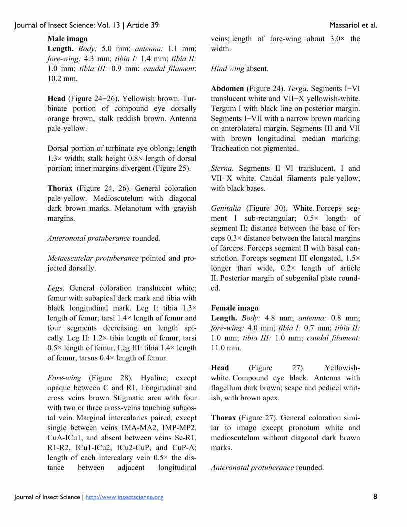
Male imago Length. Body: 5.0 mm; antenna: 1.1 mm; fore-wing: 4.3 mm; tibia I: 1.4 mm; tibia II: 1.0 mm; tibia III: 0.9 mm; caudal filament: 10.2 mm. Head (Figure 24−26). Yellowish brown. Tur-binate portion of compound eye dorsally orange brown, stalk reddish brown. Antenna pale-yellow. Dorsal portion of turbinate eye oblong; length 1.3× width; stalk height 0.8× length of dorsal portion; inner margins divergent (Figure 25). Thorax (Figure 24, 26). General coloration pale-yellow. Medioscutelum with diagonal dark brown marks. Metanotum with grayish margins. Anteronotal protuberance rounded. Metaescutelar protuberance pointed and pro-jected dorsally. Legs. General coloration translucent white; femur with subapical dark mark and tibia with black longitudinal mark. Leg I: tibia 1.3× length of femur; tarsi 1.4× length of femur and four segments decreasing on length api-cally. Leg II: 1.2× tibia length of femur, tarsi 0.5× length of femur. Leg III: tibia 1.4× length of femur, tarsus 0.4× length of femur. Fore-wing (Figure 28). Hyaline, except opaque between C and R1. Longitudinal and cross veins brown. Stigmatic area with four with two or three cross-veins touching subcos-tal vein. Marginal intercalaries paired, except single between veins IMA-MA2, IMP-MP2, CuA-ICu1, and absent between veins Sc-R1, R1-R2, ICu1-ICu2, ICu2-CuP, and CuP-A; length of each intercalary vein 0.5× the dis-tance between adjacent longitudinal
veins; length of fore-wing about 3.0× the width. Hind wing absent. Abdomen (Figure 24). Terga. Segments I−VI translucent white and VII−X yellowish-white. Tergum I with black line on posterior margin. Segments I−VII with a narrow brown marking on anterolateral margin. Segments III and VII with brown longitudinal median marking. Tracheation not pigmented. Sterna. Segments II−VI translucent, I and VII−X white. Caudal filaments pale-yellow, with black bases. Genitalia (Figure 30). White. Forceps seg-ment I sub-rectangular; 0.5× length of segment II; distance between the base of for-ceps 0.3× distance between the lateral margins of forceps. Forceps segment II with basal con-striction. Forceps segment III elongated, 1.5× longer than wide, 0.2× length of article II. Posterior margin of subgenital plate round-ed. Female imago Length. Body: 4.8 mm; antenna: 0.8 mm; fore-wing: 4.0 mm; tibia I: 0.7 mm; tibia II: 1.0 mm; tibia III: 1.0 mm; caudal filament: 11.0 mm. Head (Figure 27). Yellowish-white. Compound eye black. Antenna with flagellum dark brown; scape and pedicel whit-ish, with brown apex. Thorax (Figure 27). General coloration simi-lar to imago except pronotum white and medioscutelum without diagonal dark brown marks. Anteronotal protuberance rounded.

Journal of Insect Science: Vol. 13 | Article 39 Massariol et al.
Journal of Insect Science | http://www.insectscience.org 9
Metaescutelar protuberance pointed and pro-jected dorsally. Legs. Similar to male imago, except by tar-someres of tibia I with longitudinal blackish mark. Leg I: tibia 0.9× length of femur, tarsi 0.7× length of femur. Leg II: tibia 1.1× length of femur, tarsus 0.3× length of femur. Leg III: tibia 1.1× length of femur, tarsus 0.3× the length of femur. Fore-wing (Figure 29) hyaline, except area between C and R1 opaque. Longitudinal and transverse veins brown. Stigmatic area with three Sc veins touching or almost touching subcostal vein. Marginal intercalaries single, except single between Sc-R1, and absent be-tween R sector and CuP-A; length of intercalary vein 0.4× distance between adja-cent longitudinal veins; length of fore-wing about 2.4× width. Hind wing absent. Abdomen (Figure 27). Terga. Yellowish-white, except for segments VII−X translucent white. Posterior margin of segment I with a narrow transverse dark brown. Tracheation darkened. Sterna. Yellowish-white. Etymology The specific epithet is an allusion to a popular musical style of Pernambuco State. Comments Nymphs of C. maracatu are somewhat similar to those of C. spaceki sp. nov., however they can be easily differentiated based on the shape of the third segment of the labial palp (round-ed in C. maracatu, truncated in C. spaceki). The adults of C. maracatu, as those of C. bar-ituensis, present a sexual dimorphism related to the presence of single (female) or double
double (male) marginal intercalary veins. The body color pattern, especially the thorax, read-ily distinguishes C. maracatu from C. barituensis. Distribution (Figure 1) BRAZIL - Pernambuco, Sergipe. Material examined Holotype. Male imago with corresponding nymphal exuvia, Brazil, Pernambuco, Taman-daré, Reserva Biológica Saltinho, Rio Mamucabas, 21 September 2009, 08° 43’ 52.7” S, 35° 10’ 24.8” W, 38 m a.s.l., mar-ginal vegetation, Lima LRC, col. (INPA). Paratypes. One female imago, same data as holotype. One male imago, Brazil, Pernambu-co, Rio Formoso, Riacho da Gameleira, 24 October 2009, 8° 43’ 12.3” S, 35° 10’ 32.9” W, 18 m a.s.l., rock, Lima LRC, col (CZNC). One female imago with corresponding nym-phal exuvia, Brazil, Pernambuco, Tamandaré, Reserva Biológica Saltinho, Riacho da Sede, 21 September 2009, 08° 43’ 52.7” S, 35°1 0’ 24.8” W, 38 m a.s.l., Lima LRC, col (IML). One female imago and one female sub-imago with corresponding nymphal exuviae, same data except 22 September 2009 (DZRJ). Additional material. One nymph, Brazil, Sergipe, Areia Branca, Estação Ecológica Ser-ra de Itabaiana, Riacho Coqueiro, 17 March 2004, 10° 46’ 1.98” S, 37° 20’ 21.6 W, 311 m a.s.l., Francischetti FC, col. Seven nymphs, Brazil, Sergipe, Areia Branca, Estação Ecoló-gica Serra de Itabaiana, Riacho Água Fria, 18 March 2004, 10° 45’ 17.58” S, 37° 20’ 31.5” W, 292 m a.s.l., Francischetti FC, col. One nymph, Brazil, Sergipe, Areia Branca, Estação Ecológica Serra de Itabaiana, Rio dos Negros, 18 March 2004, 10° 44’ 49.2” S, 37° 20’ 24.06” W, 298 m a.s.l., Francischetti FC, col. Three nymphs, Brazil, Sergipe, Areia Branca,

Journal of Insect Science: Vol. 13 | Article 39 Massariol et al.
Journal of Insect Science | http://www.insectscience.org 10
Estação Ecológica Serra de Itabaiana, Rio dos Negros, 18 March 2004, 10° 44’ 49.2” S, 37° 20’ 24.06” W, 298 m a.s.l., leaf litter, Francis-chetti FC, col. Same data, except thirty-three nymphs, marginal vegetation (CZNC). Life cycle association Rearing adults from nymphs. Cloeodes redactus Waltz and McCafferty, 1987 (Figure 1, 5, 6, 31, 32) Genus poss. Cloeodes; Roback 1966: 133. Cloeodes (Cloeodes) redactus Waltz and McCafferty 1987a: 204. Cloeodes redactus, McCafferty and Lugo-Ortiz 1996: 23; Domínguez et al. 2002: 462; Domínguez et al. 2006: 151; Nieto and Rich-ard 2008: 5; Nieto and Emmerich 2011: 58. Diagnoses Nymphs: 1) Labrum with dorsal arc of setae composed of 1 + 0 + 2 long, spine-like setae; 2) Segment III of labial palp rounded; 3) Fore-femur apex projected, with two lanceolate se-tae; 4) Tarsal claw 0.3× length of tarsi; 5) General coloration of abdomen yellowish-brown with brown marks; 6) Spines on poste-rior margin of terga I absent; 7) Paraproct with 12 to 14 marginal spines; 8) Caudal fila-ments with posterior margin of segments with short spines on each segment and long spines on every four segments on cercus and every two segments on terminal filament. Adult: Female imago. 1) Marginal interca-laries single; 2) Abdominal terga yellowish-white, segments III, VI, and X with a red mark on medial region (Figure 31);
Description Female imago Length. Body: 5.0 mm; antenna: 0.8 mm; fore-wing, tibia I, II, III, caudal filament: bro-ken. Head (Figure 31). Yellowish-white. Com-pound eye black. Antenna yellowish-white, scape, and basal ¾ of pedicel white, apex of pedicel and flagellum washed with dark brown. Thorax (Figure 31). Yellowish-white, median longitudinal suture brown. Apex of me-tascutellar protuberance dark brown. Anteronotal protuberance rounded. Metascutellar protuberance posteriorly point-ed. Legs. Yellowish-white. Femur washed with orange, with dark brown subapical transversal marking on anterior surface. Dorsal margin of tibia and tarsus dark brown. Fore-wing hyaline, except opaque between C-R1. Longitudinal and cross veins dark brown. Marginal intercalary veins single; length of each intercalary vein between IMA and IMA2 0.3× distance between adjacent longitudinal veins. Hind wing absent. Abdomen (Figure 31). Terga. Yellowish-white. Segments I–VI with a dark brown nar-row transverse line on posterior margin, continuous only in segment I; segments III, VI and X with a red median marking; segments V, VI almost entirely washed with orange, segment VII washed with orange on anterior margin. Tracheation dark brown.
Journal of Insect Science: Vol. 13 | Article 39 Massariol et al.
Journal of Insect Science | http://www.insectscience.org 11
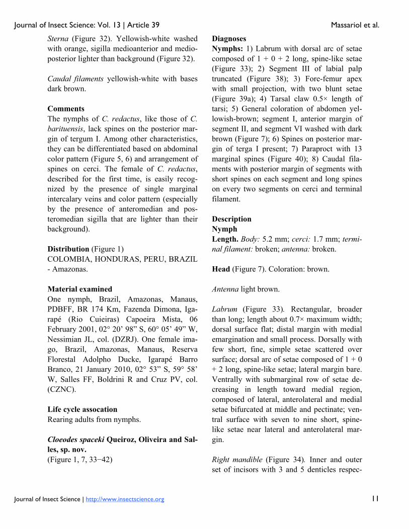
Sterna (Figure 32). Yellowish-white washed with orange, sigilla medioanterior and medio-posterior lighter than background (Figure 32). Caudal filaments yellowish-white with bases dark brown. Comments The nymphs of C. redactus, like those of C. barituensis, lack spines on the posterior mar-gin of tergum I. Among other characteristics, they can be differentiated based on abdominal color pattern (Figure 5, 6) and arrangement of spines on cerci. The female of C. redactus, described for the first time, is easily recog-nized by the presence of single marginal intercalary veins and color pattern (especially by the presence of anteromedian and pos-teromedian sigilla that are lighter than their background). Distribution (Figure 1) COLOMBIA, HONDURAS, PERU, BRAZIL - Amazonas. Material examined One nymph, Brazil, Amazonas, Manaus, PDBFF, BR 174 Km, Fazenda Dimona, Iga-rapé (Rio Cuieiras) Capoeira Mista, 06 February 2001, 02° 20’ 98” S, 60° 05’ 49” W, Nessimian JL, col. (DZRJ). One female ima-go, Brazil, Amazonas, Manaus, Reserva Florestal Adolpho Ducke, Igarapé Barro Branco, 21 January 2010, 02° 53” S, 59° 58’ W, Salles FF, Boldrini R and Cruz PV, col. (CZNC). Life cycle assocation Rearing adults from nymphs. Cloeodes spaceki Queiroz, Oliveira and Sal-les, sp. nov. (Figure 1, 7, 33−42)
Diagnoses Nymphs: 1) Labrum with dorsal arc of setae composed of 1 + 0 + 2 long, spine-like setae (Figure 33); 2) Segment III of labial palp truncated (Figure 38); 3) Fore-femur apex with small projection, with two blunt setae (Figure 39a); 4) Tarsal claw 0.5× length of tarsi; 5) General coloration of abdomen yel-lowish-brown; segment I, anterior margin of segment II, and segment VI washed with dark brown (Figure 7); 6) Spines on posterior mar-gin of terga I present; 7) Paraproct with 13 marginal spines (Figure 40); 8) Caudal fila-ments with posterior margin of segments with short spines on each segment and long spines on every two segments on cerci and terminal filament. Description Nymph Length. Body: 5.2 mm; cerci: 1.7 mm; termi-nal filament: broken; antenna: broken. Head (Figure 7). Coloration: brown. Antenna light brown. Labrum (Figure 33). Rectangular, broader than long; length about 0.7× maximum width; dorsal surface flat; distal margin with medial emargination and small process. Dorsally with few short, fine, simple setae scattered over surface; dorsal arc of setae composed of 1 + 0 + 2 long, spine-like setae; lateral margin bare. Ventrally with submarginal row of setae de-creasing in length toward medial region, composed of lateral, anterolateral and medial setae bifurcated at middle and pectinate; ven-tral surface with seven to nine short, spine-like setae near lateral and anterolateral mar-gin. Right mandible (Figure 34). Inner and outer set of incisors with 3 and 5 denticles respec-
Journal of Insect Science: Vol. 13 | Article 39 Massariol et al.
Journal of Insect Science | http://www.insectscience.org 12
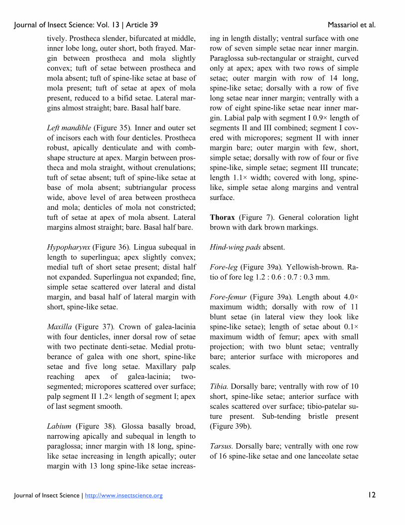
tively. Prostheca slender, bifurcated at middle, inner lobe long, outer short, both frayed. Mar-gin between prostheca and mola slightly convex; tuft of setae between prostheca and mola absent; tuft of spine-like setae at base of mola present; tuft of setae at apex of mola present, reduced to a bifid setae. Lateral mar-gins almost straight; bare. Basal half bare. Left mandible (Figure 35). Inner and outer set of incisors each with four denticles. Prostheca robust, apically denticulate and with comb-shape structure at apex. Margin between pros-theca and mola straight, without crenulations; tuft of setae absent; tuft of spine-like setae at base of mola absent; subtriangular process wide, above level of area between prostheca and mola; denticles of mola not constricted; tuft of setae at apex of mola absent. Lateral margins almost straight; bare. Basal half bare. Hypopharynx (Figure 36). Lingua subequal in length to superlingua; apex slightly convex; medial tuft of short setae present; distal half not expanded. Superlingua not expanded; fine, simple setae scattered over lateral and distal margin, and basal half of lateral margin with short, spine-like setae. Maxilla (Figure 37). Crown of galea-lacinia with four denticles, inner dorsal row of setae with two pectinate denti-setae. Medial protu-berance of galea with one short, spine-like setae and five long setae. Maxillary palp reaching apex of galea-lacinia; two-segmented; micropores scattered over surface; palp segment II 1.2× length of segment I; apex of last segment smooth. Labium (Figure 38). Glossa basally broad, narrowing apically and subequal in length to paraglossa; inner margin with 18 long, spine-like setae increasing in length apically; outer margin with 13 long spine-like setae increas-
ing in length distally; ventral surface with one row of seven simple setae near inner margin. Paraglossa sub-rectangular or straight, curved only at apex; apex with two rows of simple setae; outer margin with row of 14 long, spine-like setae; dorsally with a row of five long setae near inner margin; ventrally with a row of eight spine-like setae near inner mar-gin. Labial palp with segment I 0.9× length of segments II and III combined; segment I cov-ered with micropores; segment II with inner margin bare; outer margin with few, short, simple setae; dorsally with row of four or five spine-like, simple setae; segment III truncate; length 1.1× width; covered with long, spine-like, simple setae along margins and ventral surface. Thorax (Figure 7). General coloration light brown with dark brown markings. Hind-wing pads absent. Fore-leg (Figure 39a). Yellowish-brown. Ra-tio of fore leg 1.2 : 0.6 : 0.7 : 0.3 mm. Fore-femur (Figure 39a). Length about 4.0× maximum width; dorsally with row of 11 blunt setae (in lateral view they look like spine-like setae); length of setae about 0.1× maximum width of femur; apex with small projection; with two blunt setae; ventrally bare; anterior surface with micropores and scales. Tibia. Dorsally bare; ventrally with row of 10 short, spine-like setae; anterior surface with scales scattered over surface; tibio-patelar su-ture present. Sub-tending bristle present (Figure 39b). Tarsus. Dorsally bare; ventrally with one row of 16 spine-like setae and one lanceolate setae

Journal of Insect Science: Vol. 13 | Article 39 Massariol et al.
Journal of Insect Science | http://www.insectscience.org 13
neat apex; tarsal claw bare, 0.5× length of tar-si. Mid- and hind-legs similar to fore-leg. Abdomen (Figure 7). General coloration yel-lowish-brown. Segment I, anterior margin of segment II, and segment VI washed with dark brown, remainder segments washed with brown, becoming darker toward lateral mar-gin; segments I to VI with subtriangular dark brown mark laterally. Terga. Surface with abundant scale-bases and micropores; posterior margin with long spines (2.1× longer than wide) often intercalated by short spines (2.0× longer than wide) (Figure 41); spines present in posterior margin of segments: I−X. Sterna. Spines present in posterior margin of segments V−IX. Gill (Figure 42a, b). Opaque; tracheae dark grey. Margin with narrow spines and short, fine, simple setae (Figure 42b). Tracheae ex-tending from main trunk to inner margins. Paraproct (Figure 40). With 13 marginal spines; surface with abundant scale-bases and micropores; posterolateral extension with blunt marginal spines. Caudal filaments. Brown. Posterior margin of segments with short spines on each segment, and long spines on every two segments on cerci and terminal filament; inner margin of cercus and inner and outer margin of terminal filament with tufts of long, flat simple setae. Adult: Unknown.
Etymology The specific epithet is in honor of Dr. Bruno Spacek Godoy, who collected the holotype. Comments See comments under C. maracatu, sp. nov. Distribution (Figure 1) BRAZIL - Goiás. Material examined Holotype. Nymph (body in alcohol, mouth-parts, legs, tergum IV, and paraprocts on three slides, mounting media Euparal®), Brazil, Goiás, Santa Isabel, Rio das Palmas, 19 No-vember 2008, 15° 17’ 52.8” S, 49° 20’ 39.1” W, 580 m a.s.l., rock, sand, Godoy BS, col. (INPA). Paratypes. Three nymphs, same data as holo-type (DZRJ, IML, CZNC). Key to South American Species of Two-Winged Cloeodes Nymphs 1. Segment III of labial palp truncated (Figure 38); tarsal claw equal or longer than 0.5× length of tarsi………………………………..2 1’. Segment III of labial palp rounded (Figure 18); tarsal claw equal or less than 0.4× length of tarsi ............................................................3 2. Fore-femur without apical projection; body color pattern as in Figure 2…………..C. auwe 2’. Fore femur with a small apical projection (Figure 39a); body color pattern as in Figure 7 ..........................................C. spaceki, sp. nov. 3. Spines on posterior margin of segment I present………………...C. maracatu, sp. nov. 3’. Spines on posterior margin of segment I absent ……………………………………….4 4. Cercus with long spines on every four seg-ments; body color pattern (Figure 5, 6)…………………………………C. redactus
Journal of Insect Science: Vol. 13 | Article 39 Massariol et al.
Journal of Insect Science | http://www.insectscience.org 14
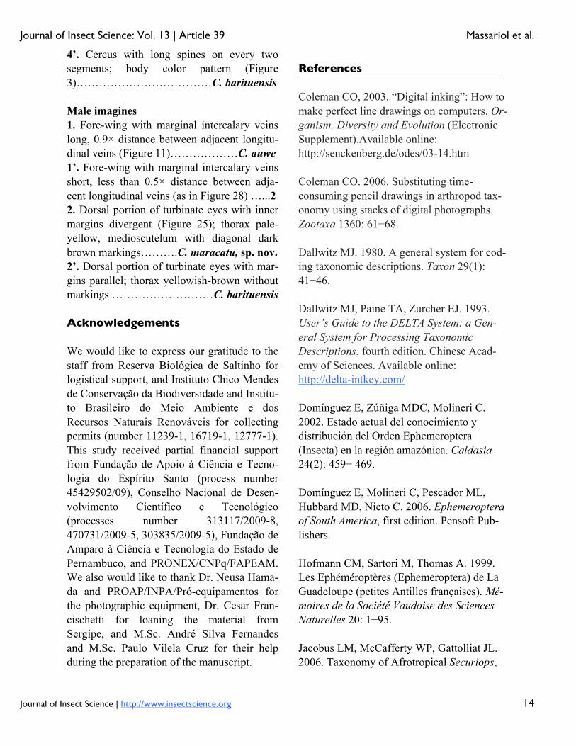
4’. Cercus with long spines on every two segments; body color pattern (Figure 3)………………………………C. barituensis Male imagines 1. Fore-wing with marginal intercalary veins long, 0.9× distance between adjacent longitu-dinal veins (Figure 11)………………C. auwe 1’. Fore-wing with marginal intercalary veins short, less than 0.5× distance between adja-cent longitudinal veins (as in Figure 28) …...2 2. Dorsal portion of turbinate eyes with inner margins divergent (Figure 25); thorax pale-yellow, medioscutelum with diagonal dark brown markings……….C. maracatu, sp. nov. 2’. Dorsal portion of turbinate eyes with mar-gins parallel; thorax yellowish-brown without markings ………………………C. barituensis Acknowledgements We would like to express our gratitude to the staff from Reserva Biológica de Saltinho for logistical support, and Instituto Chico Mendes de Conservação da Biodiversidade and Institu-to Brasileiro do Meio Ambiente e dos Recursos Naturais Renováveis for collecting permits (number 11239-1, 16719-1, 12777-1). This study received partial financial support from Fundação de Apoio à Ciência e Tecno-logia do Espírito Santo (process number 45429502/09), Conselho Nacional de Desen-volvimento Científico e Tecnológico (processes number 313117/2009-8, 470731/2009-5, 303835/2009-5), Fundação de Amparo à Ciência e Tecnologia do Estado de Pernambuco, and PRONEX/CNPq/FAPEAM. We also would like to thank Dr. Neusa Hama-da and PROAP/INPA/Pró-equipamentos for the photographic equipment, Dr. Cesar Fran-cischetti for loaning the material from Sergipe, and M.Sc. André Silva Fernandes and M.Sc. Paulo Vilela Cruz for their help during the preparation of the manuscript.
References Coleman CO, 2003. “Digital inking”: How to make perfect line drawings on computers. Or-ganism, Diversity and Evolution (Electronic Supplement).Available online: http://senckenberg.de/odes/03-14.htm Coleman CO. 2006. Substituting time-consuming pencil drawings in arthropod tax-onomy using stacks of digital photographs. Zootaxa 1360: 61−68. Dallwitz MJ. 1980. A general system for cod-ing taxonomic descriptions. Taxon 29(1): 41−46. Dallwitz MJ, Paine TA, Zurcher EJ. 1993. User’s Guide to the DELTA System: a Gen-eral System for Processing Taxonomic Descriptions, fourth edition. Chinese Acad-emy of Sciences. Available online: http://delta-intkey.com/ Domínguez E, Zúñiga MDC, Molineri C. 2002. Estado actual del conocimiento y distribución del Orden Ephemeroptera (Insecta) en la región amazónica. Caldasia 24(2): 459− 469. Domínguez E, Molineri C, Pescador ML, Hubbard MD, Nieto C. 2006. Ephemeroptera of South America, first edition. Pensoft Pub-lishers. Hofmann CM, Sartori M, Thomas A. 1999. Les Ephéméroptères (Ephemeroptera) de La Guadeloupe (petites Antilles françaises). Mé-moires de la Société Vaudoise des Sciences Naturelles 20: 1−95. Jacobus LM, McCafferty WP, Gattolliat JL. 2006. Taxonomy of Afrotropical Securiops,
Journal of Insect Science: Vol. 13 | Article 39 Massariol et al.
Journal of Insect Science | http://www.insectscience.org 15
new genus, and Cloeodes Traver (Ephemerop-tera: Baetidae). African Entomology 14: 129−140. Kluge NY. 1991. Cuban Mayflies of the fam-ily Baetidae (Ephemeroptera) 1. Genera Callibaetis, Cloeodes and Paracloeodes. Zo-ologicheschii Zhurnal 12: 128−136. Lugo-Ortiz CR, McCafferty WP, Gattolliat JL. 1999. The small minnow mayfly genus Cloeodes Traver (Ephemeroptera: Baetidae) in Madagascar. Proceedings of the Entomo-logical Society of Washington 1: 208−211. Massariol FC, Salles FF. 2011. Two new spe-cies of Cloeodes Traver (Ephemeroptera: Baetidae) from Espírito Santo, Southeastern Brazil. Zootaxa 3058: 1−21. McCafferty WP, Lugo-Ortiz CR. 1996. Los efemerópteros (Ephemeroptera) de América Central. Revista Nicaraguense de Entomolo-gia 35: 19−28. Nieto C, Richard B. 2008. The genus Cloeodes (Ephemeroptera: Baetidae) in Ar-gentina with new generic synonymy and new species. Zootaxa 1727: 1−21. Nieto C, Emmerich D. 2011. Three new spe-cies of the genus Cloeodes Traver (Ephemeroptera: Baetidae) from Uruguay. Zootaxa 2996: 57−65. Roback SS. 1966. The CATHERWOOD Foundation Peruvian-Amazon Expedition. VI - Ephemeroptera nymphs. In: Roback SS, Edi-tor. Monographs of the Academy of Natural Sciences of Philadelphia 14: 129−199. Salles FF, Batista JD, Cabette HRS. 2004. Ba-etidae (Insecta: Ephemeroptera) de Nova
Xavantina, Mato Grosso, Brasil: novos regis-tros e descrição de uma nova espécie de Cloeodes Traver. Biota Neotropica 4(2): 1−8. Salles FF. 2010. Taxonomy of the genus Ade-brotus Lugo-Ortiz & McCafferty (Ephemeroptera: Baetidae). Annales de Lim-nologie 46: 207−215. Salles FF, 2011. Lista das espécies de Ephe-meroptera registradas para o Brasil. Available online: http://ephemeroptera.br.googlepages.com/home Soldán T, Yang JT. 2003. Mayflies (Ephem-eroptera) of Taiwan: Species composition, toxonomic shifts, distribution and bio-geographical analysis. In: Gaino E, Editor. Research Update on Ephemeroptera and Ple-coptera. pp. 413−420. Università di Perugia, Italy. Traver JR. 1938. Mayflies of Puerto Rico. The Journal of Agriculture of the University of Puerto Rico 22: 5−42. Waltz RD, McCafferty WP. 1987a. Revision of the genus Cloeodes (Ephemeroptera: Baetidae). Annals of the Entomological Soci-ety of America 80(2): 191−207. Waltz RD, McCafferty WP. 1987b. Generic revision of Cloeodes and description of two new genera (Ephemeroptera: Baetidae). Pro-ceedings of the Entomological Society of Washington 89: 177−184. Waltz RD, McCafferty WP. 1994. Cloeodes (Ephemeroptera: Baetidae) in Africa. Aquatic Insects 16: 165−169. Wiersema NA, Baumgardner DE. 2000. Dis-tribution and taxonomic contributions to the
Journal of Insect Science: Vol. 13 | Article 39 Massariol et al.
Journal of Insect Science | http://www.insectscience.org 16
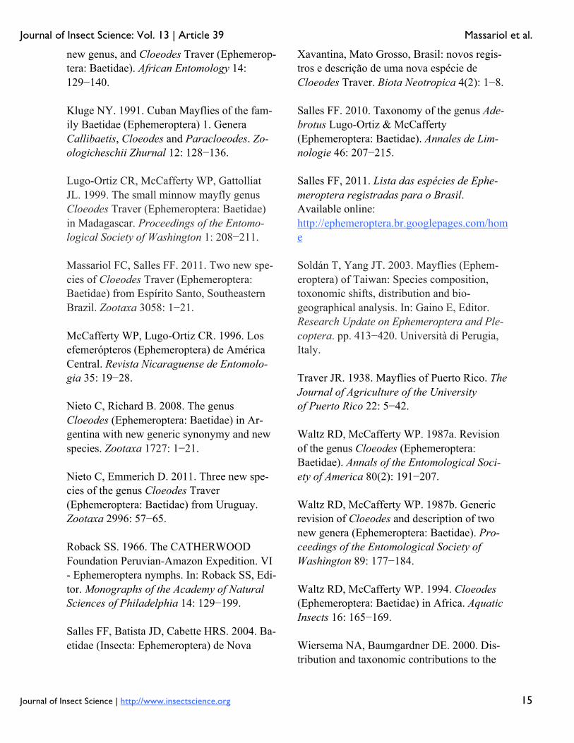
Figure 1. Distribution of the species of two-winged Cloeodes in Brazil. High quality figures are available online.
Figure 2-4. Cloeodes spp., nymphs in dorsal view. 2. C. auwe. 3. C. barituensis. 4. C. maracatu, sp. nov. High quality figures are available online.
Figure 5-7. Cloeodes spp., nymphs in dorsal view. 5. C. redactus, male. 6. C. redactus, female. 7. C. spaceki, sp. nov. High quality figures are available online.
Figure 8-10. Cloeodes auwe, male adult. 8. Dorsal view. 9, 10. Detail of turbinate eyes and thorax, dorsal view and lateral view. High quality figures are available online.
Ephemeroptera fauna of Mexico and Central America. Entomological News 111: 60−66.
Journal of Insect Science: Vol. 13 | Article 39 Massariol et al.
Journal of Insect Science | http://www.insectscience.org 17
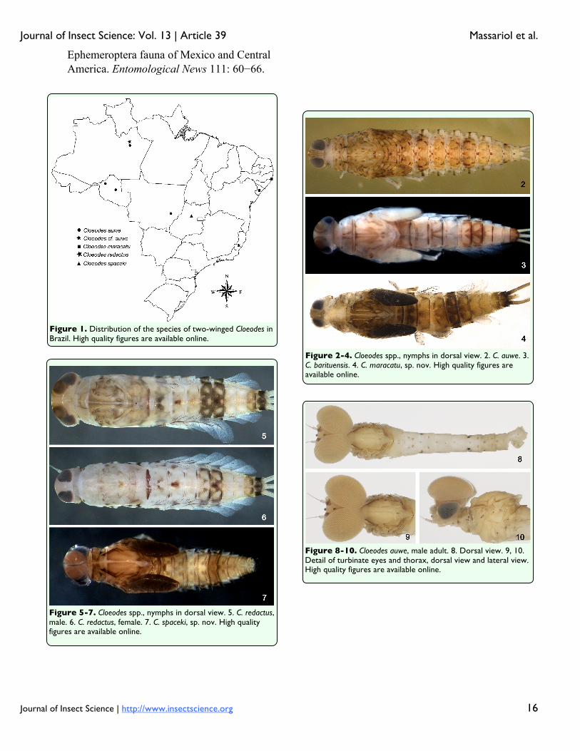
Figure 11-12. Cloeodes auwe, adult. 11. Fore-wing. 12. Male, genitalia. High quality figures are available online.
Figure 13-18. Cloeodes maracatu, sp. nov., nymph. 13. Labrum (left dorsal view, right ventral view). 14. Right mandible, ventral view. 15. Left mandible, dorsal view. 16. Hypopharynx, dorsal view. 17. Maxilla, ventral view. 18. Labium (left, dorsal view; right, ventral view.). High quality figures are available online.
Journal of Insect Science: Vol. 13 | Article 39 Massariol et al.
Journal of Insect Science | http://www.insectscience.org 18
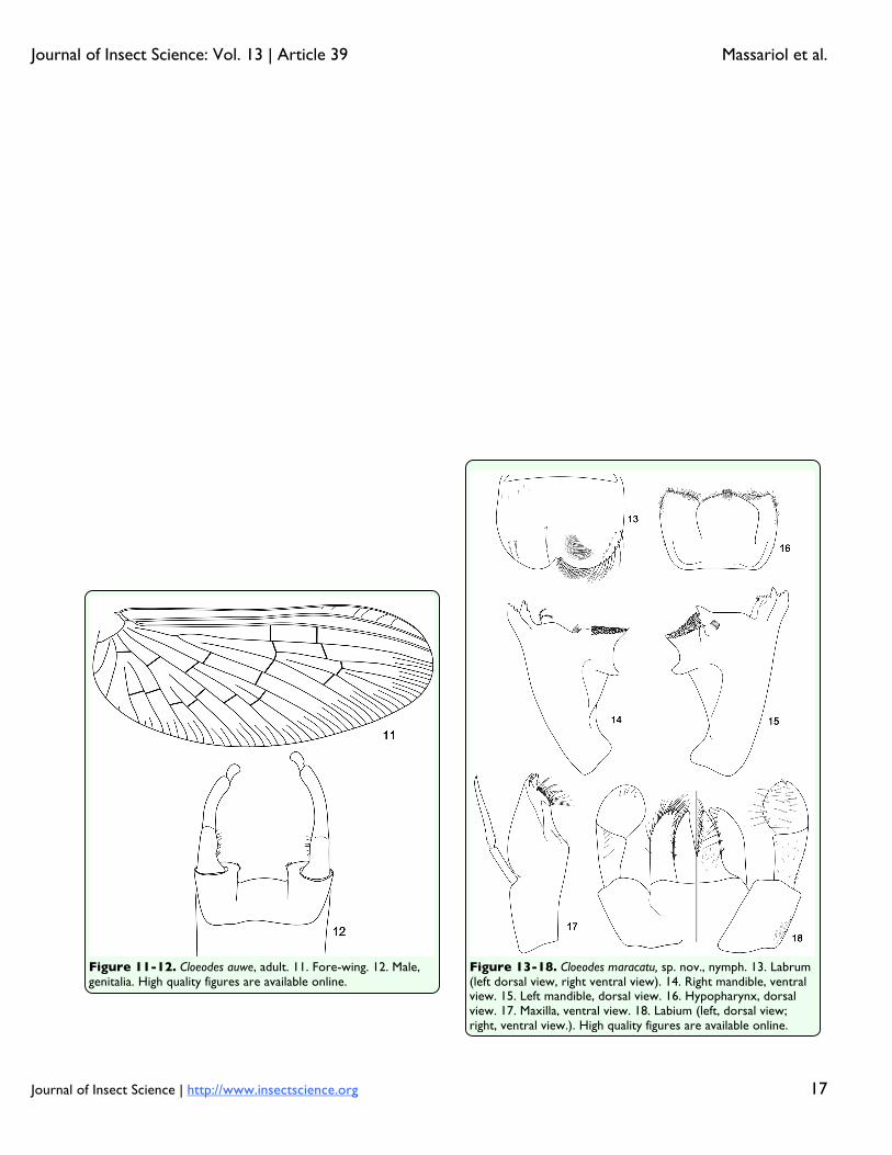
Figure 19-23. Cloeodes maracatu, sp. nov., nymph. 19. Fore-leg, anterior surface (a: general view; b: sub-tending bristle). 20. Projections of femur apex (a: fore-femur; b: mid-femur; c: hind-femur). 21. Posterior margin of tergum IV. 22. Gill IV (a: general view; b: details of gill margin). 23. Paraproct (dorsal view). High quality figures are available online.
Figure 24-27. Cloeodes maracatu, sp. nov., adults. 24. Male, dorsal view. 25, 26. Male, detail of turbinate eyes and thorax, dorsal view, lateral view. 27. Female, dorsal view. High quality figures are available online.
Journal of Insect Science: Vol. 13 | Article 39 Massariol et al.
Journal of Insect Science | http://www.insectscience.org 19
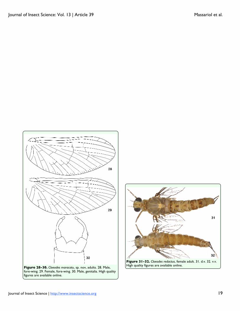
Figure 31-32. Cloeodes redactus, female adult. 31. d.v. 32. v.v. High quality figures are available online.
Figure 28-30. Cloeodes maracatu, sp. nov, adults. 28. Male, fore-wing. 29. Female, fore-wing. 30. Male, genitalia. High quality figures are available online.

Journal of Insect Science: Vol. 13 | Article 39 Massariol et al.
Journal of Insect Science | http://www.insectscience.org 20
Figure 39-42. Cloeodes spaceki, sp. nov., nymph. 39. Fore-leg, anterior surface (a: general view; b: sub-tending bristle). 40. Paraproct (dorsal view). 41. Posterior margin of tergum IV. 42. Gill IV (a: details of gill margin; b: general view). High quality figures are available online.
Figure 33-38. Cloeodes spaceki, sp. nov., nymph. 33. Labrum (left dorsal view, right ventral view). 34. Right mandible, ventral view. 35. Left mandible, dorsal view. 36. Hypopharynx, dorsal view. 37. Maxilla, ventral view. 38. Labium (left dorsal view, right ventral view). High quality figures are available online.